Embed Size (px)
Citation preview

A new genus And species of cAvernicolous pomAtiopsidAe (molluscA, cAenogAstropodA) in BAhiA, BrAzil
luiz ricArdo l. simone1
AbstrAct
Spiripockia punctata is a new genus and species of Pomatiopsidae found in a cave from Serra Ramalho, SW Bahia, Brazil. The taxon is troglobiont (restricted to subterranean realm), and is characterized by the shell weakly elongated, fragile, translucent, normally sculptured by pus‑tules with periostracum hair on tip of pustules; peristome highly expanded; umbilicus opened; radular rachidian with 6 apical and 3 pairs of lateral cusps; osphradium short, arched; gill filaments with rounded tip; prostate flattened, with vas deferens inserting subterminally; penis duct narrow and weakly sinuous; pallial oviduct simple anteriorly, possessing convoluted by‑pass connecting base of bulged portion of transition between visceral and pallial oviducts with base of seminal receptacle; spermathecal duct complete, originated from albumen gland. The description of this endemic species may raise protective environmental actions to that cave and to the Serra Ramalho Karst area.
Key-Words: Pomatiopsidae; Spiripockia punctata gen. nov. et sp. nov.; Brazil; Cave; Tro-globiont; Anatomy.
IntroductIon
The family Pomatiopsidae is represented in the Brazilian region by only two species of the genus Id‑iopyrgus Pilsbry, 1911 (Simone, 2006: 94). However, the taxon is much richer in remaining mainland ar-eas, with both freshwater and semi-terrestrial habits (Ponder & Keyzer, 1998; Kameda & Kato, 2011). Common features of the pomatiopsids are small size (smaller than 15 mm), elongated to short spired, and smooth shell with paucispiral protoconch. The main distinction from other rissooideans is the diaulic pal-lial oviducts of the females; i.e., the typical pallial ovi-duct has two separate openings to the pallial cavity (Davis, 1967, 1979; Ponder, 1988).
An enigmatic tiny gastropod has been collected in caves from the Serra Ramalho Kars area, southwestern Bahia state, Brazil. It has a pretty, fragile, translucent shell in such preliminary gross anatomy, which already reveals troglobiont adaptations, i.e., depigmentation, lack of eyes and small size. The sample has been brought by Maria Elina Bichuette, who is specialized in subter-ranean fauna. Complementary analysis revealed a new pomatiopsid for which no known genus can be attrib-uted. Both genus and species are hereby formally in-troduced, including a detailed anatomical investigation. The description of this new taxon is not only academi-cally urgent, but the cave is also threatened for econom-ic reasons. The description of an endemic species helps in the argumentation for preserving this fragile location.
1. Museu de Zoologia, Universidade de São Paulo. Caixa Postal 42.494, 04218-970, São Paulo, SP, Brasil. E-mails: [email protected]; [email protected]
Volume 52(40):515‑524, 2012

MAterIAl And Methods
All specimens were fixed in 70% Ethanol. The dissections were performed under a stereomicro-scope by standard techniques, with the specimens immersed under fixative. Digital photos of each step of the dissection were obtained, as well as drawings aided by a camera lucida. A scanning electron mi-croscope (SEM) was employed to view details of the shell and radula in the Laboratório de Microscopia Eletrônica of Museu de Zoologia da Universidade de São Paulo.
Anatomical abbreviations in the figures: ag, albumen gland; an, anus; au, auricle; bm, buccal mass; br, sub-radular membrane; ce, cerebral ganglion; cg, capsule gland; cm, columellar muscle; co, bulged region of oviduct; cv, ctenidial vein; dd, duct to digestive gland; df, dorsal folds of buccal mass; dg, digestive gland; es, esophagus; fe, fecal pellets; fp, female pores; ft, foot; gi, gill; in, intestine; ir, insertion of m4 in subradular cartilage; is, insertion of m5 in subradular cartilage; jw, jaws; ki, kidney; m2‑m12, odontophore muscles; mb, mantle border; mj, jaw and peribuccal muscles; mo, mouth; ne, nephrostome; oc, odonto-phore cartilage; od, odontophore; of, Spermathecal oviduct; op, opercular pad; os, osphradium; ov, pal-lial oviduct; oy, ovary; pa, penis aperture; pc, peri-cardium; pd, penis duct; pe, penis; pg, anterior fur-row of pedal glands; pl, pleural ganglion; pn, pedal glanglion; pt, prostate; ra, radula; rm, snout ventral retractor muscle; rn, radular nucleus; rs, radular sac; rt, rectum; sa, salivary gland aperture; sc, subradular cartilage; sd, sperm duct; sg, salivary gland; sn, snout; st, stomach; su, subesophageal ganglion; sv, seminal vesicle; sy, statocyst; te, cephalic tentacle; tg, integu-ment; tn, tentacular nerve; ts, testis; ve, ventricle; vd, vas deferens; vo, visceral oviduct.
Abbreviatons of institutions: MnrJ, Museu Na-cional da Universidade Federal do Rio de Janeiro; MZsP, Museu de Zoologia da Universidade de São Paulo; usnM, National Museum of Natural History, Smithsonian Institution.
systematics
Family Pomatiopsidae Genus Spiripockia new genus
Diagnosis: Turbiform shell with highly expand-ed peristome. Walls translucent, smooth except for
pustules containing hair-like projections of periostra-cum. Freshwater environment.
Description: Outline turbiform to slightly turriform. Protoconch paucispiral, of single whorl. Outer sur-face simple, except for small pustules distributed in somewhat spiral alignment, absent in some specimens (surface totally smooth). Peristome highly expanded and sprayed except for implantation on columellar surface. Umbilicus somewhat opened. Operculum thin. Pair of strong ventral retractor muscles of snout. Salivary gland reduced. Gonoducts closed (tubular); oviduct with bypass in posterior region. Spermathecal duct originated from albumen gland.
Type species: S. punctata new species.
List of included taxa: S. punctata new species.
Etymology: The generic epithet is a contraction of Latin words spiri, from spira, and pocillum a little cup. Meaning something like a spiral small cup. This name is also used by my father, Egydio do Carmo de Sim-one, to designate any female pet in which he does not know the name.
Pronunciation: in English terms the genus must be vo-calized as “speereepowqueeah”.
Spiripockia puncata new species (Figs. 1‑33)
Types: Holotype MZSP 105000. Paratypes: MZSP 104435, 19 specimens, MNRJ 30503, 1 specimen, USNM 2060411, 1 specimen, all from type locality.
Type locality: BRAZIL. Bahia; Serra Ramalho Karst area, Middle São Francisco River basin, Lapa dos Peixes cave, 13°49’21.78”S, 43°57’24.39”W (M.E. Bichuette col., 29/vii/2005).
Diagnosis: Shell weakly elongated; walls thin, fragile, translucent, unpigmented. Sculptured by pustules somewhat spirally aligned, possessing periostracum hair on tip of pustules (sometimes absent). Peristome expanded in plane ~90° in relation to last quarter whorl. Umbilicus opened. Strong pair of ventral snout retractor muscles passing through nerve ring. Radular rachidian with 6 apical and 3 pairs of lateral cusps, ~10 terminal cusps in radular lateral and mar-ginal teeth. Eyes absent. Head-foot unpigmented. Os-phradium short, arched. Gill filaments with rounded
Simone, L.R.L.: A new Pomatiopsidae from Bahia516
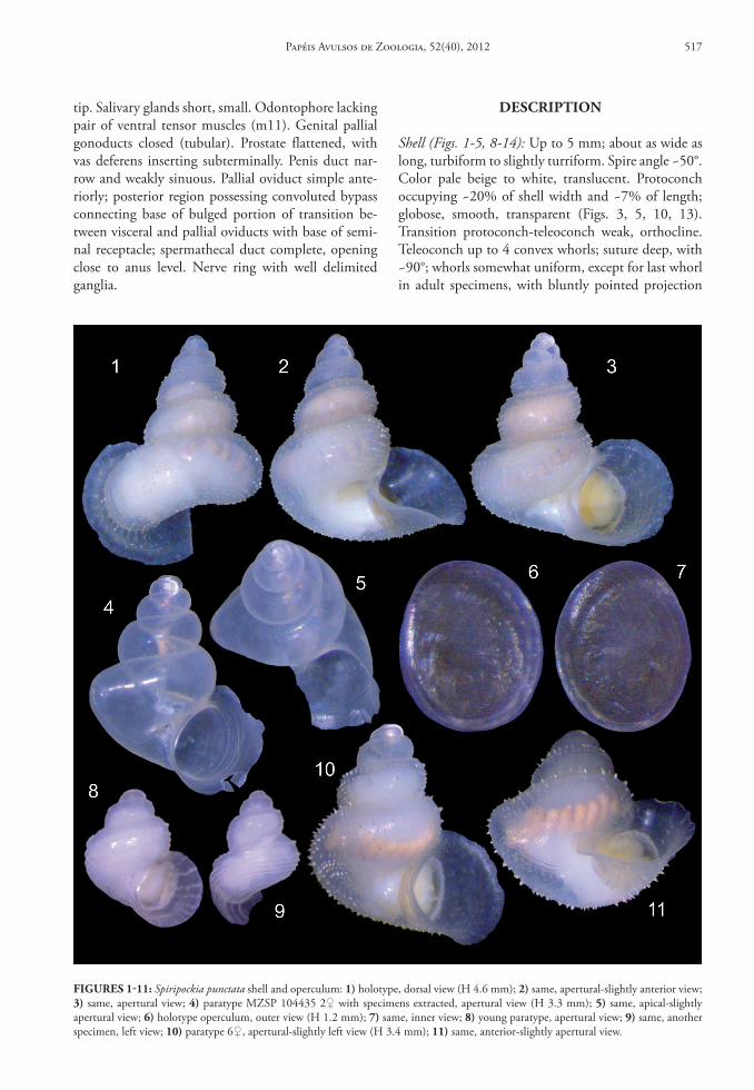
tip. Salivary glands short, small. Odontophore lacking pair of ventral tensor muscles (m11). Genital pallial gonoducts closed (tubular). Prostate flattened, with vas deferens inserting subterminally. Penis duct nar-row and weakly sinuous. Pallial oviduct simple ante-riorly; posterior region possessing convoluted bypass connecting base of bulged portion of transition be-tween visceral and pallial oviducts with base of semi-nal receptacle; spermathecal duct complete, opening close to anus level. Nerve ring with well delimited ganglia.
descrIPtIon
Shell (Figs. 1‑5, 8‑14): Up to 5 mm; about as wide as long, turbiform to slightly turriform. Spire angle ~50°. Color pale beige to white, translucent. Protoconch occupying ~20% of shell width and ~7% of length; globose, smooth, transparent (Figs. 3, 5, 10, 13). Transition protoconch-teleoconch weak, orthocline. Teleoconch up to 4 convex whorls; suture deep, with ~90°; whorls somewhat uniform, except for last whorl in adult specimens, with bluntly pointed projection
FIGures 1‑11: Spiripockia punctata shell and operculum: 1) holotype, dorsal view (H 4.6 mm); 2) same, apertural-slightly anterior view; 3) same, apertural view; 4) paratype MZSP 104435 2♀ with specimens extracted, apertural view (H 3.3 mm); 5) same, apical-slightly apertural view; 6) holotype operculum, outer view (H 1.2 mm); 7) same, inner view; 8) young paratype, apertural view; 9) same, another specimen, left view; 10) paratype 6♀, apertural-slightly left view (H 3.4 mm); 11) same, anterior-slightly apertural view.
Papéis Avulsos de Zoologia, 52(40), 2012 517

located approximately in opposed side from aper-ture (Figs. 2, 5, 11) (more visible in apical or ante-rior views); projection absent in younger specimens (Figs. 8-9); being clearly derived from previous ap-erture. Sculpture minute pustules aligned in spiral lines (Figs. 12, 14), ~6 doted spiral lines, equidis-tantly spaced, in penultimate whorl; each line with ~40 pustules in penultimate whorl disposed some-what alternately each line in relation to neighbor lines (Figs. 1-3, 10-14); each pustule in form of small, cir-cular convexity, separated from neighbor pustules by smooth distance equivalent to twice pustules width (Fig. 12); pustules somewhat uniformly distributed, and sometimes forming wide angulation on shell sur-face (Fig. 14); this dotted sculpture absent in ~15% of specimens, possessing completely smooth outer sur-face (Figs. 4-5); periostracum possessing 5-6 axially aligned hair on pustules tip (Figs. 3, 10-11), looking like flexible scales. Sculpture on younger specimens similar to this above described, but with pustule lo-cated closer to each other and with 4 inferior lines located closer to each other and far from subsutural
line (Figs. 8-9); gradually after 2-3 whorls sculpture changing to adult pattern. Peristome expanding in plane ~90° in relation to terminal region of body whorl (Figs. 1-5, 10-11) in mature specimens; expan-sion edges extremely fragile and somewhat irregular (apparently to fit in substrate), particularly wider in anterior region (Fig. 1) and narrower in columel-lar implantation; expansion extending ~15 % shell width. Aperture oval, superior angulation blunt; re-maining somewhat rounded. Umbilicus opened and relatively wide (Figs. 2, 4, 11), occupying ~15% of shell width (excluding peristome); no callus, except for short and narrow portion of peristome attached to preceding whorl (Figs. 2-4, 10-11).
Head‑foot (Figs. 19‑21): Relatively small, stubby, un-pigmented. Foot thick, as wide as shell aperture. Mes-opodium thick, flanked dorsally by shallow lateral fur-rows (Fig. 21: ft). Anterior furrow of pedal glands (pg) deep, restricted to anterior edge (Figs. 19-20). Oper-cular pad (Fig. 21: op) elliptic, terminal, occupying half of posterior foot dorsal surface. Head bulbous,
FIGures 12‑18: Spiripockia punctata hard parts in SEM: 12) paratype MZSP 104435 3♂, detail of penultimate whorl, dorsal view, scale = 200 µm; 13) same, detail of apical region in profile, scale = 200 µm; 14) same, whole dorsal view (H 3.9 mm); 15) radula, partial whole view, scale = 20 µm; 16) same, detail of central region, scale = 10 µm; 17) same, wider magnification, scale = 10 µm; 18) same, detail of more lateral region, scale = 10 µm.
Simone, L.R.L.: A new Pomatiopsidae from Bahia518

with ~85% of food width; pair of cephalic tentacles lateral positioned (Figs. 19-20, 23: te), each tentacle simple, thick, with ~twice foot length; no detectable eye. Snout with ~twice tentacles width and ~half ten-tacles length (sn); anterior end weakly bilobed, mouth oval, wide, subterminal, ventral (Figs. 23: mo). Colu-mellar muscle thick, ~3/4 whorl. Haemocoel ellipti-cal, on central region of head-foot (Fig. 23).
Operculum (Figs. 6‑7): Corneous, thin, translucent, yellow-beige, flexible, paucispiral. Outline elliptical-almost circular. Edges thin. Nucleus located in middle region of interior half, weakly dislocated internally (Fig. 6); from nucleus, ~2 whorls uniformly growing, appearing; outer surface also sculptured by weak un-dulations parallel to outer edge. Inner surface glossy, scar elliptic, occupying ~half of inner surface; location dislocated to internal edge, but not touching internal edge. Occupies entire shell aperture (Figs. 3, 10-11).
Mantle organs (Figs. 22, 27): Somewhat narrow, with ~1 whorl in length. Mantle edge simple, weakly thick; unpigmented. Osphradium short (os), curved (con-cavity left), simple; length ~6% pallial cavity length; located in anterior-left corner of cavity, close to man-tle border. Gill elongated, narrow, with ~85% of pal-lial cavity length and ~25% of cavity width; anterior end bluntly pointed, close to mantle border; posterior region narrowing gradually, filaments ending at some distance from pericardium; gill filaments somewhat triangular, with distal tip rounded (Fig. 27: gi). Be-tween gill and rectum a narrow distance equivalent to 10% of pallial cavity width. Hypobranchial gland inconspicuous. Rectum wide, reaching up to half of pallial cavity width in some points; bearing aligned series of elliptical fecal pellets (fe) disposed transver-sally or weakly oblique, easily seen by translucence (Figs. 1-3, 10-11); rectum displaced from right man-tle cavity edge by pallial genital ducts. Anus simple, shortly siphoned, distant from mantle border ~10% of cavity length, in anterior-right corner of pallial roof. Genital ducts running along right edge, rela-tively massive, described below.
Visceral mass (Figs. 22, 31): Length ~3 whorls, keep-ing ~2 first shell whorls empty in adult specimens (Figs. 2-3, 10). Color of most structures pale beige to white. Stomach as anterior structure, compressing ventrally small reno-pericardial structures; stomach of ~0.5 whorl and with almost entire adjacent whorl width. Digestive gland of ~2 whorls, mostly posterior to stomach. Gonad running along columellar surface of each whorl when mature, weakly distinct from
digestive gland. Other genital and digestive details below.
Circulatory and excretory systems (Fig. 31): Pericar-dium narrow, located transversely between stomach and pallial cavity (Fig. 22: pc), volume ~1/20 of that of visceral mass; auricle anterior, small, just posterior to posterior end of ctenidial vein; ventricle posterior, simple. Kidney minute, weakly smaller than pericar-dium, located on right side of pallial cavity posterior end; inner tissue totally solid, white. Nephrostome (ne) small, transverse, located close to pericardium.
Digestive system (Figs. 22‑26, 28): Mouth in antero-ventral end of snout (Fig. 23: mo). Pair of strong ventral retractor muscles of snout and mouth (Figs. 23-24: rm); originating in middle level of hae-mocoelic ventral floor; running close to median line towards anterior, flanking ventral surface of buccal mass, passing through nerve ring (Figs. 23, 33); insert-ing along ventral wall of snout, close to ventral border of mouth. Buccal mass bulged, occupying entire snout inner surface and ~1/3 of haemocoelic volume. Pair of jaw plates (Fig. 25: jw) somewhat separated from each other; each jaw oval and very thin, translucent. Odon-tophore spherical, with ~half of buccal mass volume. Odontophore muscles (Figs. 24-26, 28): mj, pair of jaw and peribuccal muscles, working also as odonto-phore protractors, originating in lateral and ventral region of mouth, running towards posterior as part of oral tube along ~half odontophore length (Fig. 24), inserting in latero-ventral mid region of odontophore cartilages (Fig. 26); m2, pair of posterior retractor muscles of odontophore (Figs. 24-25), narrow, origi-nating in lateral region of haemocoel inner surface, in its middle level, running towards anterior a distance slightly longer than buccal mass length, inserting in postero-lateral region of odontophore cartilages (Fig. 28); m4, pair of main dorsal tensor muscle of radula (Fig. 28), broad and thick, surrounding outer lateral surface of odontophore cartilages, originating from their ventro-lateral surface, inserting in lateral edges of subradular cartilage in its region in buccal cavity, and also in radular sac region preceding buccal cavity (Fig. 28: ir); m5, pair of secondary dorsal ten-sor muscles of radula (Figs. 26, 28), narrow and thick, originating in posterior edge of odontophore carti-lages, running towards medial and anterior, between cartilages, inserting in ventral side of radular ribbon in its region exposed in buccal cavity (Figs. 28: is); m6, horizontal muscle, thin and wide, connecting ventral edge of both odontophore cartilages along ~75% of their length (Fig. 28); m10, pair of ventral
Papéis Avulsos de Zoologia, 52(40), 2012 519

protractor muscle of buccal mass (Figs. 24, 26), rela-tively wide and thick, originating in ventral edge of mouth, just ventral to retractor muscle (mr) origin, running towards posterior a distance equivalent to odontophore length, inserting in posterior-ventral
edge of odontophore cartilages. Subradular cartilage relatively wide in region of buccal cavity (Fig. 25: sc). Odontophore cartilages elliptic, somewhat flattened laterally, anterior edge weakly narrower than posterior edge (Fig. 28: oc). Radular sac with length ~3 times
FIGures 19‑27: Spiripockia punctata anatomy: 19) head-foot, male, dorsal view; 20) same, female; 21) same, right view; 22) pallial cavity roof and partially uncoiled visceral mass, female, ventral view, some genital structures displaced, transverse section in indicated region of pallial oviduct also shown; 23) head and haemocoel, ventral view, foot and columellar muscle removed; 24) foregut, right-slightly dorsal view; 25) same, ventral view, odontophore partially removed and deflected, esophagus partially sectioned longitudinally; 26) odontophore, ventral view, radula partially deflected upwards; 27) pallial cavity hoof, transverse section in middle level of gill. Scales = 0.5 mm.
Simone, L.R.L.: A new Pomatiopsidae from Bahia520

that of odontophore (Figs. 24-26: rs), and ~1/4 odon-tophore width; radular nucleus (rn) weakly broader.
Radular tooth (Figs. 15‑18): rachidian tooth triangu-lar, distal tip curved inwards, with ~16% of radular ribbon width, about as tall as wide, base simple, weak-ly arched, 3 pairs of latero-basal cusps (Figs. 16-18), each cusp small, triangular, pointed tip; 5-7 terminal triangular cusps, central cusp larger, cusps weakly di-minishing towards lateral, restricted to distal, curved
edge; lateral tooth with main region rectangular, with ~double rachidian height and its same width, ~15 cusps similar to those of rachidian, cusps smaller in both ends, being gradually larger towards middle-weakly lateral region (Figs. 16-17), basal region wid-ening obliquely externally (Figs. 15, 17) along dis-tance equivalent to ~30% radula width, being covered by marginal teeth (Figs. 17-18); inner and outer lat‑eral tooth similarly shaped, being outer tooth ~30% narrower than inner marginal tooth, ~double length
FIGures 28‑33: Spiripockia punctata anatomy: 28) odontophore, dorsal view, radula, outer layer of muscles and membranes removed, both cartilages deflected; 29) penis, ventral view, adjacent region of integument also shown, penis duct seen by translucence; 30) nerve ring, right view; 31) part of pallial cavity roof and partially uncoiled visceral mass, male, ventral view; 32) posterior end of pallial oviduct, ventral view; 33) nerve ring, dorsal view, topology of adjacent retractor muscle also shown. Scales = 0.5 mm.
Papéis Avulsos de Zoologia, 52(40), 2012 521

as lateral tooth and ~40% narrower, general form as rod curved inwards at tip, tip flattened, bearing ~13 (inner) and ~10 (outer) terminal cusps of similar characters as lateral tooth cusps.
Salivary gland small (Figs. 23-24: sg), white, with maximum length ~half buccal mass length, and sometimes smaller; about 3 times longer than wide, tip rounded; salivary aperture in postero-lateral re-gion of oral cavity (Fig. 25: sa). Dorsal folds of buc-cal mass (Fig. 25: df ) smooth, simple, wide, located close to each other. Esophagus simple and sinuous along haemocoel (Figs. 23-24: es), generally with an-terior glandular enlargement (Fig. 24), but sometimes (~30%) with same width along length; most remain-ing esophageal inner surface simple, lacking folds; posterior esophagus (Fig. 22: es) narrow, inserting in ventral-mid region of stomach. Stomach dimensions and positions above described (visceral mass); pos-sessing main gastric chamber posterior, with rounded posterior surface, and narrow style sac (Figs. 22: ss), with ~half remaining gastric dimensions. Style sac to-tally separated from intestine; esophagus inserting just ventrally to origins of style sac and intestine, on ante-rior stomach side; duct to digestive gland as wide as posterior esophagus, originating at short distance pos-terior to esophageal insertion (Fig. 22: dd), running towards posterior. Intestine originating dorsally and at left from esophageal insertion (Fig. 22: in), with ~double esophagus width, and at left from style sac; running surrounding externally style sac up to dorsal region of kidney, broadening in region preceding pal-lial cavity. Rectum, fecal pellets and anus above de-scribed (pallial cavity). Fecal pellets formed only after renal portion of intestine (Figs. 11, 22).
Genital system: Male (Figs. 19, 29, 31): Testis restrict-ed to posterior half of digestive gland (Fig. 31: ts). Seminal vesicle (sv) weakly convolute, with 3-4 zig-zag loops in columelar region of visceral mass preced-ing stomach, color pale grey-iridescent; vas deferens relatively straight in gastric and renal region, insert-ing in prostate subterminally (Fig. 31: vd). Prostate with ~1/4 pallial cavity width, somewhat dorso-ventrally flattened, white, totally closed (tubular); inner lumen simple, flat (Fig. 27: pt). Vas deferens originating subterminally from prostate anterior re-gion (Fig. 31: vd) in level between anterior and mid thirds of pallial cavity; in short distance penetrating in floor of pallial cavity by side of penis base, running immersed in integument (Figs. 19: vd, 29: pd). Penis (Figs. 19-29: pe) simple, curved, base broad, gradually tapering up to sharp pointed tip. Penis duct seen by translucence, color pale-green-iridescent, performing
weak zigzag coils along outer, almost central penial region, up to penis distal tip (Fig. 29: pd); aperture distal, simple, small (pa).
Female (Figs. 22, 32): Visceral structures similar to those of males; ovary (oy) somewhat broader than testis. Visceral oviduct very narrow, running along middle level of columellar surface of visceral mass ~half whorl (vo). Visceral oviduct inserting terminally in complex posterior region of pallial oviduct (Fig. 32) in bulged region of oviduct (co) with simple glandular walls and hollow inside. This bulged region continu-ing to albumen gland (ag) strongly curved in ~180°. Spermathecal duct (of ) originating in ventral-left side of albumen gland, very narrow but with thick walls, running along right edge of pallial cavity ventral to re-maining pallial oviduct ~90% of pallial cavity length; opening simple by side and ventral to main female pore (Figs. 22: fp). Seminal receptacle (rs) bulging subterminally in posterior region of pallial oviduct, possessing two successive bulbs or sometimes horse-shoe-shaped; inserting by narrow duct in adjacent left side of posterior end of capsule gland. Sperm duct, or bypass (sd) as convoluted duct connecting anterior region of bulged region of oviduct (co) with base of seminal receptacle (rs); posterior half clearly narrower, gradually broadening in anterior half. Capsule gland (cg) occupying ~80% of pallial oviduct length; walls thick glandular, white, lumen flattened (Fig. 22); short terminal atrium with walls weakly thick, taper-ing up to female pore. Female pore shortly siphoned, papilla-like, turned anteriorly, located close to and at right of anus. No special female genital structure in head-foot (Figs. 20-21).
Central nervous system (Figs. 30, 33): Nerve ring lo-cated just posterior to buccal mass, with pedal ganglia weakly more anterior (Fig. 23: nr) than remaining ganglia. Each cerebral ganglion (ce) oval, with size equivalent to esophageal section; cerebral commissure narrow, with ~half-length as each ganglion. Pleural ganglia (pl) with ~half size of cerebral ganglia, located just ventral to them. Each pedal ganglion (pn) weakly larger than cerebral ganglion, somewhat spherical, located very close to each other (pedal commissure extremely short). Cerebro-pedal and pleuro-pedal connectives about as long as pedal ganglia (Fig. 30). Subesophageal ganglion (su) with ~half pleural gan-glion size, located a distance from right pleural gan-glion equivalent to 1.5 times each cerebral ganglion length. Pair of statocysts (sy) located in ventro-poste-rior side of pedal ganglia; with large, single statolith inside. Single pair of tentacular nerve (tn), lacking optical nerve.
Simone, L.R.L.: A new Pomatiopsidae from Bahia522

Measurements (in mm): Holotype: 4.6 by 4.3; para-types: 2♀: 3.3 by 3.1; 4♂: 3.3 by 3.5; 5♀: 3.6 by 3.4.
Distribution: Only known in the type locality (Lapa dos Peixes cave).
Habitat: In a cave surrounded by Caatinga semi-arid environment, attached to rocks inside a small stream.
Material examined: Types.
dIscussIon
Spiripockia punctata is the second troglobiont gastropod described in Brazilian territory. The first one is a relative taxon, Potamolithus troglobius Simone & Moracchioli, 1994, a Hydrobiidae from Ribeira Valley, southern São Paulo state. Both species share the characters to consider them as troglobiont – the total lack of pigmentation and eyes, and small size – and the freshwater environment. In light of the enor-mous quantity of caves in the Brazilian region, it is difficult to believe that only 2 troglobiont gastropod species exist. Bichuette & Trajano (2003) cited at least five more troglobiont species, all belonging to Hydro-biidae and probably to the genus Potamolithus Pilsbry, 1911, occurring in Ribeira Velley, SE Brazil. Though, certainly many more studies on this issue and urgent preservation of cave environments are necessary. I can affirm that the MZSP collection already has some samples of troglobiont rissooideans and even pulmo-nates waiting for study.
Spiripockia punctata has no South American species with which it can be confused. The set of conchological features are so far exclusive and suf-ficiently distinctive to erect a new genus. From the family Pomatiopsidae, the only two species hitherto described belong to the genus Idiopyrgus – I. rudolphi Haas, 1838 and I. souleyetianus Pilsbry, 1911 (Sim-one, 2006), from which S. punctata differs in having a shorter and wider shell, fewer whorls, peristome more expanded, umbilicus wider, and the sculpture of most specimens of pustules with periostracum hair in them (Figs. 1-3, 10-11). The radula and genital part of the anatomy of I. souleyetianus has been investigated (Rey, 1959; Malek, 1983), which permits some extra-shell comparison. The operculum of S. punctata has a more central nucleus. Its osphradium is much shorter and curved. Spiripockia punctata also has the insertion and pallial origin of the vas deferens in the prostate gland in a subterminal manner (Fig. 31: vd), however, that of I. souleyetianus is inserted and originated at
mid-level of that gland. The penis is somewhat simi-lar, with totally closed (tubular) duct, but the penis duct of S. punctata appears to be more convolute. The contrary happens in the seminal vesicle, such as in the case of I. souleyetianus whose seminal vesicle is appar-ently much more coiled. Related to the pallial oviduct, S. punctata lacks a clear separation between albumen and capsule glands (Fig. 32), as happens in I. souleye‑tianus; the bypass duct (sd) is much more coiled in S. punctata, and its seminal receptacle is connected directly to the pallial oviduct, while that of I. souleye‑tianus is connected to the visceral oviduct; S. punctata lacks a small secondary receptacle, which is present in I. souleyetianus; the spermathecal duct (of ) has its origin in the end of the albumen gland, close to inser-tion of the visceral oviduct in S. punctata, while that of I. souleyetianus is originated from the receptacle (also known, in rissooideans, as spermatheca or bursa copulatrix). Actually, the arrangement of the posterior region of the pallial oviduct of I. souleyetianus looks more like the pattern of the family (Davis, 1967, 1979; Ponder, 1988; Davis et al., 1992) than that of S. punctata, with the spermathecal duct connected to the pallial oviduct instead to the receptacle. In this point of view, the name “spermathecal duct” is not totally accurate; however the name was maintained in order to permit further comparisons and to suggest homology. This singularity of S. punctata is another indication of generic separation, beyond those fea-tures of the shell.
With respect to the radula, Spiripockia punctata possesses the normal pattern of the family (Ponder & Keyzer, 1998), which includes the lateral cusps in the rachidian and the basal lateral expansion of the lateral toot base. It differs in number and form of the cusps, as normally found amongst closely related species.
It is expected that with the description of this new endemic taxon, further argumentation for the conservation of the cave type locality for the present species and others can be instituted. Some pomatiop-sids have been considered as endangered species on the IUCN Red Lists. One example is Tomichia caw‑stoni Connolly, 1939, from South Africa (Kristensen et al., 2010). According to the IUCN terms, S. punc‑tata is already considered threatened based on its en-demicity, as well as on its troglomorphic features, such as depigmentation and being eyeless.
resuMo
Spiripockia punctata é gênero e espécie novos de Poma‑tiopsidae encontrado em uma caverna da Serra Ramalho,
Papéis Avulsos de Zoologia, 52(40), 2012 523

SW Bahia, Brasil. O táxon é troglóbio (restrito à vida subterrânea), caracterizado pela concha levemente alon‑gada, frágil, translúcida, normalmente esculturada por pústulas contendo pelos do perióstraco no topo das pús‑tulas; perístoma altamente expandido; umbílico aberto; dente raquidiano da rádula com 6 cúspides apicais e 3 pares de basais; osfrádio curto e curvo; filamentos bran‑quiais com topo arredondado; próstata achatada, com vaso deferente inserindo subterminalmente; ducto do pênis fracamente sinuoso; oviduto palial anteriormen‑te simples, possuindo um by‑pass convoluto da base da porção bulbosa da porção entre oviduto visceral e palial com a base do receptáculo seminal; ducto da espermateca completo, originado da glândula de albume. A descrição dessa espécie endêmica deve contribuir com ações proteti‑vas do ambiente da caverna e à área da Serra Ramalho.
Palavras-Chave: Pomatiopsidae; Spiripockia punc‑tata gen. nov. et sp. nov.; Brasil; Caverna; Troglóbio; Anatomia.
AcknowledGMents
The material was collected under IBAMA li-cense 027/00-DIFAS/DIREC by Maria Elina Bi-chuette, Universidade Federal de São Carlos. I am deeply grateful to her for donation of the specimens, location description, and for reviewing the paper. I am also grateful to Winston Ponder, Australian Muse-um, for suggestions on systematics in the early phase of this study. I further acknowledge FAPESP (Funda-ção de Amparo à Pesquisa do Estado de São Paulo) for financial support in the field activities of E. Trajano’s project (process 2003/0794-5).
reFerences
Bichuette, M.E. & Trajano, E. 2003. A population study of epigean and subterranean Potamolithus snails from southeast Brazil. Hydrobiologia, 505:107-117.
Davis, G.M. 1967. The systematic relationship of Pomatiopsis lapidaria and Oncomelania hupensis formosana (Prosobranchia: Hydrobiidae). Malacologia, 6(1-2):1-143.
Davis, G.M. 1979. The origin and evolution of the gastropod family Pomatiopsidae, with emphasis on the Mekong River Triculinae. Monograph of the Academy of Natural Sciences of Philadelphia, 20:1-120.
Davis, G.M.; Chen, C.E.; Wu, C.; Kuang, T.F.; Xing, G.; Li, L.; Liu, W.J. & Yan, Y.L. 1992. The Pomatiopsidae of Hunan, China (Gastropoda: Rissoacea). Malacologia, 34(1-2):143-342.
Kameda, Y. & Kato, M. 2011. Terrestrial invasion of pomatiopsid gastropods in the heavy-snow region of the Japanese Archipelago. BioMed Central Evolutionary Biology, 11(118):1-14.
Kristensen, T.K.; Stensgaard, A.S. & Appleton, C. 2010. Tomichia cawstoni. The IUCN Red List of Threatened Species. www.iucnredlist.org/apps/redlist/details/63444/0 (extracted in 26/iv/2011).
Malek, E.A. 1983. The South American hydrobioid genus Idiopyrgus Pilsbry, 1911. Nautilus, 97(1):16-20.
Ponder, W.F. 1988. The truncatelloidean (= rissoacean) radiation – a preliminary phylogeny. In: Ponder, W.F. (Ed.). Prosobranch Phylogeny. Malacological Review supplement, 4:129-166.
Ponder, W.F. & Keyzer, R.G. 1998. Superfamily Rissooidea. In: Beesley, P.L.; Ross, G.J.B. & Wells, A. (Eds.). Mollusca: the southern synthesis. CSIRO Publishing, Melbourne. p. 745-766. (Fauna of Australia v. 5, part B, p. 565-1234).
Rey, L. 1959. Molluscs of the genus Oncomelania, in Brazil, and their possible epidemiological significance. Revista do Instituto de Medicina Tropical de São Paulo, 1(2):144-149.
Simone, L.R.L. 2006. Land and freshwater molluscs of Brazil. EGB. FAPESP. São Paulo, 390 pp.
Simone, L.R.L. & Moracchioli, N. 1994. Hydrobiidae (Gastropoda: Hydrobioidea) from the Ribeira Valley, S.E. Brazil, with descriptions of two new cavernicolous species. Journal of Molluscan Studies, 60:445-459.
Aceito em: 26.10.2012 Publicado em: 20.12.2012
Simone, L.R.L.: A new Pomatiopsidae from Bahia524

edItorIAl coMMItteePublisher: Museu de Zoologia da Universidade de São Paulo. Avenida Nazaré, 481, Ipiranga, CEP 04263-000, São Paulo, SP, Brasil.
editor‑in‑chief: Carlos José Einicker Lamas, Serviço de Invertebrados, Museu de Zoologia, Universidade de São Paulo, Caixa Postal 42.494, CEP 04218-970, São Paulo, SP, Brasil. E-mail: [email protected].
Associate editors: Mário César Cardoso de Pinna (Museu de Zoologia, Universidade de São Paulo, Brasil); Luís Fábio Silveira (Museu de Zoologia, Universidade de São Paulo, Brasil); Marcos Domingos Siqueira Tavares (Museu de Zoologia, Universidade de São Paulo, Brasil); Sérgio Antonio Vanin (Instituto de Biociências, Universidade de São Paulo, Brasil); Hussam El Dine Zaher (Museu de Zoologia, Universidade de São Paulo, Brasil).
editorial board: Rüdiger Bieler (Field Museum of Natural History, U.S.A.); Walter Antonio Pereira Boeger (Universidade Federal do Paraná, Brasil); Carlos Roberto Ferreira Brandão
(Universidade de São Paulo, Brasil); James M. Carpenter (American Museum of Natural History, U.S.A.); Ricardo Macedo Corrêa e Castro (Universidade de São Paulo, Brasil); Mario de Vivo (Universidade de São Paulo, Brasil); Marcos André Raposo Ferreira (Museu Nacional, Rio de Janeiro, Brasil); Darrel R. Frost (American Museum of Natural History, U.S.A.); William R. Heyer (National Museum of Natural History, U.S.A.); Ralph W. Holzenthal (University of Minnesota, U.S.A.); Adriano Brilhante Kury (Museu Nacional, Rio de Janeiro, Brasil); Gerardo Lamas (Museo de Historia Natural “Javier Prado”, Lima, Peru); John G. Maisey (American Museum of Natural History, U.S.A.); Naércio Aquino Menezes (Universidade de São Paulo, Brasil); Christian de Muizon (Muséum National d’Histoire Naturelle, Paris, France); Nelson Papavero (Universidade de São Paulo, Brasil); James L. Patton (University of California, Berkeley, U.S.A.); Richard O. Prum (University of Kansas, U.S.A.); Olivier Rieppel (Field Museum of Natural History, U.S.A.); Miguel Trefaut Urbano Rodrigues (Universidade de São Paulo, Brasil); Randall T. Schuh (American Museum of Natural History, U.S.A.); Ubirajara Ribeiro Martins de Souza (Universidade de São Paulo, Brasil); Paulo Emílio Vanzolini (Universidade de São Paulo, Brasil); Richard P. Vari (National Museum of Natural History, U.S.A.).
InstructIons to Authors ‑ (April 2007)General Information: Papéis Avulsos de Zoologia (PAZ) and Arquivos de Zoologia (AZ) cover primarily the fields of Zoology, publishing original contributions in systematics, paleontology, evolutionary biology, ontogeny, faunistic studies, and biogeography. Papéis Avulsos de Zoologia and Arquivos de Zoologia also encourage submission of theoretical and empirical studies that explore principles and methods of systematics.
All contributions must follow the International Code of Zoological Nomenclature. Relevant specimens should be properly curated and deposited in a recognized public or private, non-profit institution. Tissue samples should be referred to their voucher specimens and all nucleotide sequence data (aligned as well as unaligned) should be submitted to GenBank (www.ncbi.nih.gov/Genbank) or EMBL (www.ebi.ac.uk).
Peer review: All submissions to Papéis Avulsos de Zoologia and Arquivos de Zoologia are subject to review by at least two referees and the Editor-in-Chief. All authors will be notified of submission date. Authors may suggest potential reviewers. Communications regarding acceptance or rejection of manuscripts are made through electronic correspondence with the first or corresponding author only. Once a manuscript is accepted providing changes suggested by the referees, the author is requested to return a revised version incorporating those changes (or a detailed explanation of why reviewer’s suggestions were not followed) within fifteen days upon receiving the communication by the editor.
Proofs: Page-proofs with the revised version will be sent to e-mail the first or corresponding author. Page-proofs must be returned to the editor, preferentially within 48 hours. Failure to return the proof promptly may be interpreted as approval with no changes and/or may delay publication. Only necessary corrections in proof will be permitted. Once page proof is sent to the author, further alterations and/or significant additions of text are permitted only at the author’s expense or in the form of a brief appendix (note added in proof ).
submission of Manuscripts: Manuscripts should be sent to the scielo submission (http://submission.scielo.br/index.php/paz/login), along with a submission letter explaining the importance and originality of the study. Address and e-mail of the corresponding author must be always updated since it will be used to send the 50 reprints in titled by the authors. Figures, tables and graphics should not be inserted in the text. Figures and graphics should be sent in separate files with the following formats: “.JPG” and “.TIF” for figures, and “.XLS” and “.CDR” for graphics, with 300 DPI of minimum resolution. Tables should be placed at the end of the manuscript.
Manuscripts are considered on the understanding that they have not been published or will not appear elsewhere in substantially the same or abbreviated form. The criteria for acceptance of articles are: quality and relevance of research, clarity of text, and compliance with the guidelines for manuscript preparation.
Manuscripts should be written preferentially in English, but texts in Portuguese or Spanish will also be considered. Studies with a broad coverage are encouraged to be submitted in English. All manuscripts should include an abstract and key-words in English and a second abstract and key-words in Portuguese or Spanish.
Authors are requested to pay attention to the instructions concerning the preparation of the manuscripts. Close adherence to the guidelines will expedite processing of the manuscript.
Manuscript Form: Manuscripts should not exceed 150 pages of double-spaced, justified text, with size 12 and source Times New Roman (except for symbols). Page format should be A4 (21 by 29.7 cm), with 3 cm of margins. The pages of the manuscript should be numbered consecutively.
The text should be arranged in the following order: title Page, Abstracts with key‑words, body of text, literature cited, tables, Appendices, and Figure captions. Each of these sections should begin on a new page.
(1) title Page: This should include the title, short title, Author(s) name(s) and Institutions. The title should be concise and, where appropriate, should include mention of families and/or higher taxa. Names of new taxa should not be included in titles.
(2) Abstract: All papers should have an abstract in english and another in Portuguese or spanish. The abstract is of great importance as it may be reproduced elsewhere. It should be in a form intelligible if published alone and should summarize the main facts, ideas, and conclusions of the article. Telegraphic abstracts are strongly discouraged. Include all new taxonomic names for referencing purposes. Abbreviations should be avoided. It should not include references. Abstracts and key-words should not exceed 350 and 5 words, respectively.
(3) body of text: The main body of the text should include the following sections: Introduction, Material and Methods, results, discussion, conclusion, Acknowledgments, and references at end. Primary headings in the text should be in capital letters, in bold and centered. Secondary headings should be in capital and lower case letters, in bold and centered. Tertiary headings should be in capital and lower case letters, in bold and indented at left. In all the cases the text should begin in the following line.
(4) literature cited: Citations in the text should be given as: Silva (1998) or Silva (1998:14-20) or Silva (1998: figs. 1, 2) or Silva (1998a, b) or Silva & Oliveira (1998) or (Silva, 1998) or (Rangel, 1890; Silva & Oliveira, 1998a, b; Adams, 2000) or (Silva, pers. com.) or (Silva et al., 1998), the latter when the paper has three or more authors. The reference need not be cited when authors and date are given only as authority for a taxonomic name.
(5) references: The literature cited should be arranged strictly alphabetically and given in the following format:
• Journal Article - Author(s). Year. Article title. Journal name, volume: initial page-final page. Names of journals must be spelled out in full.
•books - Author(s). Year. Book title. Publisher, Place.
•chapters of books - Author(s). Year. Chapter title. In: Author(s) ou Editor(s), Book title. Publisher, Place, volume, initial page-final page.
•dissertations and theses - Author(s). Year. Dissertation title. (Ph.D. Dissertation). University, Place.
•electronic Publications - Author(s). Year. Title. Available at: <electronic address>. Access in: date.
tables: All tables must be numbered in the same sequence in which they appear in text. Authors are encouraged to indicate where the tables should be placed in the text. They should be comprehensible without reference to the text. Tables should be formatted with vertical (portrait), not horizontal (landscape), rules. In the text, tables should be referred as Table 1, Tables 2 and 4, Tables 2-6. Use “TABLE” in the table heading.
Illustrations: Figures should be numbered consecutively, in the same sequence that they appear in the text. Each illustration of a composite figure should be identified by capital letters and referred in the text as: Fig. 1A, Fig. 1B, for example. When possible, letters should be placed in the left lower corner of each illustration of a composite figure. Hand-written lettering on illustrations is unacceptable. Figures should be mounted in order to minimize blank areas between each illustration. Black and white or color photographs should be digitized in high resolution (300 DPI at least). Use “Fig(s).” for referring to figures in the text, but “FIGURE(S)” in the figure captions and “fig(s).” when referring to figures in another paper.
responsability: Scientific content and opinions expressed in this publication are sole responsibility of the respective authors. copyrights: The journals Papéis Avulsos de Zoologia and Arquivos de Zoologia are licensed under a Creative Commons Licence (http://creativecommons.org).
For other details of manuscript preparation of format, consult the CBE Style Manual, available from the Council of Science Editors (www.councilscienceeditors.org/publications/style).
Papéis Avulsos de Zoologia and Arquivos de Zoologia are publications of the Museu de Zoologia da Universidade de São Paulo (www.mz.usp.br). Always consult the Instructions to Authors printed in the last issue or in the electronic home pages: www.scielo.br/paz or www.mz.usp.br/publicacoes.


![Brunner’s Gland Hamartoma of the Duodenum: A Literature …Brunner’s glands appear from the 13th to 14th weeks of embryonic develop-ment [2, 3]. Prevalence of Brunner’s glands](https://img.document.onl/doc/110x75/6147f9f3a830d0442101c8b3/brunneras-gland-hamartoma-of-the-duodenum-a-literature-brunneras-glands-appear.jpg)