Embed Size (px)
Citation preview
INST
ITU
TO
DE C
IÊNC
IAS B
IOM
ÉDIC
AS A
BEL SA
LAZ
AR
Maria C
arlota Pinto da Silva. Effects of dietary Amino acids
supplementation on the European seabass (D
icentrarchus labrax) skin m
ucosal imm
une status.
Effects of dietary Amino acids supplem
entation on the European seabass (D
icentrarchus labrax) skin m
ucosal imm
une status.M
aria Carlota Pinto da Silva
Effects of dietary Amino acids supplementation on the European seabass (Dicentrarchus labrax) skin mucosal immune status.
Maria Carlota Pinto da Silva
M 2017
M.IC
BAS 2017
MESTRADO EM CIÊNCIAS DO MAR - RECURSOS MARINHOS
AQUACULTURA E PESCAS

MARIA CARLOTA PINTO DA SILVA
EFFECTS OF DIETARY AMINO ACIDS SUPPLEMENTATION ON THE EUROPEAN
SEABASS (Dicentrarchus labrax) SKIN MUCOSAL IMMUNE STATUS
Dissertação de Candidatura ao grau de
Mestre em Ciências do Mar – Recursos
Marinhos submetida ao Instituto de
Ciências Biomédicas de Abel Salazar da
Universidade do Porto.
Orientador – Doutor Benjamín Costas
Categoria – Investigador Auxiliar
Afiliação – Centro Interdisciplinar de
Investigação Marinha e Ambiental
Co-Orientador – Professor António Afonso
Categoria – Professor Associado
Afiliação – Instituto de Ciências Biomédicas
Abel Salazar da Universidade do Porto


II
Agradecimentos
This work was supported by Project ALISSA (reference ALG-01-0247-FEDER-3520),
financed by Portugal and the European Union through FEDER, COMPETE 2020 and
CRESC Algarve 2020, in the framework of Portugal 2020.
Este foi um ano de muita aprendizagem. Agradeço em especial ao Professor Afonso
pela grande oportunidade de me ter integrado no seu grupo de investigação. A sua
personalidade absolutamente cativante fez-me procurá-lo na escolha do tema desta
tese.
Agradeço imenso ao Professor Benjamín por ter sido o meu orientador. Muito
obrigada pelas suas correções, conselhos e por ter criado uma excelente
oportunidade para mim.
Professor Eduardo! Foi tão importante! Todo o seu brilho e entusiasmo quer dentro
da sala de aula, quer numa conversa acidental nos corredores do ICBAS, inspiraram-
me verdadeiramente. Obrigada!
O melhor que esta tese me trouxe foi fazer parte da equipa do laboratório
NUTRIMU, sem a qual este trabalho não seria possível! Aprender e trabalhar todos
os dias com este verdadeiro espírito de equipa vai deixar muitas saudades! Cada
um à sua maneira, contribuiu para a realização deste trabalho e, por isso, deixo
aqui um grande abraço de agradecimento a todos: Rita Azeredo, Filipa Fontinha,
Francisco Guardiola, Joana Moura, Paulo e Renata. Obrigada ao meu ilustre colega
Lourenço, pelas gargalhas diárias até sermos separados, pelos truques no
computador e explicações durante os meus pânicos estatísticos (“não estou mesmo
a perceber”). A verdade é que grandes progressos nesta tese foram feitos quando
estavas à minha frente na secretária ou ao meu lado na bancada do laboratório.
Obrigada amigo! Mr. Bruno, obrigada! Para além do teu raciocino químico, que
interveio muitas vezes nas minhas contas e quando os reagentes não dissolviam,
quero agradecer-te pelos teus conselhos dentro do laboratório e por todas as
risotas na varanda lá de cima. Serginho, muito obrigada por todo o cuidado e
preocupação que mostraste desde o meu primeiro protocolo (e pelas tuas colheitas
biológicas deliciosas também)! Mas o maior obrigado de todos, é para a Marina!
III
Estiveste presente em todos os protocolos e P-values, na escolha das cores dos
meus gráficos, até à última frase da conclusão! Muito muito obrigada chefe, por
tudo o que me ensinaste e pela tua infinita paciência, principalmente nos
momentos que eu levei mais desassossego para laboratório e para os dias de
amostragem! Foste essencial e obrigatória, por isso, espero mesmo que gostes
desta tese, porque ela também é um bocadinho tua!
Amigaço Tiago, foste tão tão importante!!! Começamos e acabamos este curso
juntos e não há companheirismo como o nosso! Obrigada!
Pai, Mãe... nem sei bem o que dizer! Com dois heróis como os pais, sempre do
meu lado a orientar-me, tem sido fácil. Mãe, obrigada pela tua preocupação
constante de mãe, e por me fazeres ver, sempre, as oportunidades da maneira mais
realista! Pai, obrigada por me salvares de todos os meus contratempos (que são
muitos), por me apoiares incondicionalmente em todos os meus sonhos, até os
mais impossíveis, e por festejares as minhas conquistas como se fossem as tuas!
Irmãos, queridos e irritantes ao mesmo tempo, obrigada por me fazerem lembrar
de tudo, menos da tese!
Minha querida Baba! És a melhor Avó do mundo e os teus mimos culinários
apareceram sempre nos momentos em que eu mais precisei de inspiração! Mas
muito mais importante, obrigada pelo orgulho que sempre mostraste pela tua neta,
mesmo quando não percebias exatamente o que eu estava a estudar e querias
contar às tuas amigas.
Agradeço profundamente às minhas amigas, que estão e estiveram sempre bem
próximas do meu metro quadrado. Esta tese reflete, com certeza, a vossa amizade
permanente. Muitíssimo obrigada Mimi, Mariana Sá, Margarida e Joana Simões, Inês
Figueiredo, Mariana Veiga e Kika! Fil, Vaninha e Dudu, que quase marcaram voo
antes de mim durante as minhas falsas partidas para as Canárias, obrigada pela
vossa imensa vontade me ir visitar. Isto sim, é amizade!!!!! Pelas abundantes visitas
refundidas no Jardim Botânico e pelas minhas desorganizações mais profundas em
Hollywood, pela intensa comunicação Madrid-Porto... foste e és indispensável,
Sofia Freitas. Obrigada Inês e Matilde pela vossa presença assídua e pontual nos
finais de tarde da Associação a seguir ao trabalho. Deram-me muita forcinha para
o dia seguinte. Às minhas queridas e adoradas amigas Viky e Marta, que mesmo

IV
estando em Espanha, e os meus aparelhos de comunicação sempre estragados,
nunca desistiram de arranjar outras alternativas de me procurar.
Importantíssimo, obrigada Tiago PL! Embora tenhas reduzido significativamente os
meus níveis de concentração e de produtividade, a tua gasolina de avião motivou-
me todos os dias!

V
Resumo
Os maiores custos económicos na indústria de aquicultura devem-se
essencialmente às despesas relativas à alimentação dos animais e às perdas de
stock causadas por doenças infeciosas. O foco da investigação no âmbito da
aquicultura tem vindo a ser o desenvolvimento de dietas funcionais de forma a
evitar o aparecimento de doenças e, consequentemente, o seu tratamento. Os
aminoácidos (AA) são considerados importantes reguladores de vias metabólicas,
crucias para a resposta imune e, por este motivo, representam potenciais
candidatos para integrar estas dietas.
Este estudo teve como objetivo investigar os efeitos da suplementação nutricional
de metionina, da arginina e da citrulina no estado imune do robalo (Dicentrarchus
labrax). Os efeitos da dieta foram avaliados pela medição dos parâmetros imunes
do muco da pele. A hipótese experimental levada a cabo neste trabalho, visa
verificar se estas dietas podem modular o estado imune do muco da pele do robalo,
quer na ausência de doenças, quer em situação de infeção bacteriana.
Foram realizados dois ensaios separadamente, mas de desenho experimental
semelhante, com o intuito de descobrir de que forma cada um dos AA influencia
os parâmetros imunes do muco do robalo. O ensaio 1 foi focado no impacto da
metionina. Para tal, foram formuladas duas dietas com suplementação de 0.5% e
1% de metionina, uma terceira dieta controlo cujos níveis de metionina estavam de
acordo com necessidades nutricionais estabelecidas para robalo e ainda uma
quarta dieta deficiente em metionina.
No ensaio 2 foi avaliada a influência da arginina e da citrulina na imunidade do
muco. Assim, foi formulada uma dieta controlo que cumpria com os requisitos de
arginina e de citrulina estabelecidos para o robalo e três outras dietas
suplementadas com 1% e 2% de arginina e a terceira com 1% de citrulina.
Em ambos os ensaios (1 e 2), os peixes foram alimentados com as dietas
experimentais durante 4 semanas e foram realizadas amostragens de muco às 2 e
4 semanas. No final do período de alimentação, os peixes foram submetidos a uma
infeção bacteriana por injeção peritoneal com Photobacterium damselae subsp.
piscicida (Phdp). No ensaio 1, os peixes foram amostrados às 4 e 24 horas após a
injeção, enquanto que no ensaio 2 os peixes foram amostrados às 4, 24 e 48 horas
após a injeção da bactéria. Os parâmetros humorais do muco da pele do robalo
VI
foram estudados durante o período de alimentação (duas e quatro semanas),e nas
horas seguintes à infeção, nos dois ensaios.
Relativamente à suplementação de metionina, demonstrou-se algum grau de
influência na resposta do muco ´á inflamação no peritoneu. Foi verificado o
decréscimo dos níveis de lisozima e peroxidase no muco, provavelmente devido
ao recrutamento de leucócitos para o local de infeção.
A arginina parece exercer efeitos benéficos sobre o estado imune do muco do
robalo, uma vez que foi observada uma tendência de aumento da atividade
bactericida e peroxidase em todos os grupos suplementados. Quando a bactéria
foi injetada na cavidade peritoneal, o reforço da imunidade observado, traduziu-se
numa diminuição dos parâmetros humorais do muco. Em conjunto, estes
resultados sugerem que, a arginina estimulou o desvio da energia metabólica dos
tecidos periféricos para o foco inflamatório. Acresce ainda que a arginina permitiu
a recuperação dos níveis de lisozima, possivelmente devido à proliferação de
macrófagos na pele, nas 48 horas após a injeção.
Conclui-se, com este estudo que a suplementação com arginina, metionina ou
citrulina na dieta do robalo, não tem uma ação pronunciada e direta sobre a
resposta imune do muco após inflamação peritoneal. No entanto, os resultados
sugerem uma possível migração celular do tecido periférico para o foco
inflamatório, promovida pelos AA estudados.
Palavra-chave: Robalo; Imuno-modulação; inflamação; muco da pele; parâmetros
humorais; aminoácidos; Photobacterium damselae subsp piscicida

VII
Abstract
Major expenses in aquaculture industry are probably attributed to infectious
diseases and fish feeds management. Recently, much attention has been given to
the use of nutritional strategies for preventive health care, aiming to avoid the
occurrence of diseases and its therapeutic procedures. Emerging evidence shows
that many amino acids (AA) regulate key metabolic pathways that are crucial to
immune responses, making them good candidates to be included in functional
feeds.
Having this in mind, the present study aimed to investigate the effects of dietary
methionine, arginine and citruline supplementation on the European seabass
(Dicentrarchus labrax) immune status. The dietary treatments were evaluated by
measuring skin mucus immune parameters. Our experimental hypothesis was to
assess whether those dietary treatments can modulate the skin mucus health
status. Two trials, with a similar experimental design were performed separately.
Trail 1 was focused on dietary methionine surplus, and thus, two diets with 0.5%
and 1% methionine were formulated; plus, a control diet whose methionine levels
met the requirement for seabass; and a fourth diet deficient in methionine. In trial
2, arginine and citruline influence on mucus immune parameters was evaluated.
Three supplemented diets (1% and 2% arginine supplementation; 1% citrulline
supplementation) and a control diet (meeting arginine and citruline requirement
levels for seabass) were formulated. In both trials, fish were fed with experimental
diets for 4 weeks, and mucus sampling was performed after 2 and 4 weeks of
feeding. At the end of the feeding period, fish were subjected to a bacterial
infection by intraperitoneally injecting Photobacterium damselae subsp.
piscicida (Phdp) strain PP3. Fish were sampled following 4 and 24h post-injection in
trial 1; whereas in trial 2, three samplings were performed after 4, 24 and 48h post-
injection. For both trials mucus humoral parameters were studied during the
feeding period, and during the hours following the infection.
Methionine showed some degree of influence in mucus immune response. After
bacterial infection, methionine supplemented groups showed a decrease in
lysozyme and peroxidase levels, probably attributed to the recruitment of immune
cells towards infection site.
Arginine appears to exert beneficial effects on unchallenged European seabass
immune status, since a trend to augment bactericidal and peroxidase activities was

VIII
detected in the skin mucus of fish fed supplemented diets. Interestingly, when the
bacterium was injected in the peritoneal cavity, the enhanced immunity observed,
translated in a decline of mucus humoral parameters. Together, these results
suggest that, upon bacteria stimuli, there is an enhanced deviation of immune
energy from peripheral tissues to the inflammatory focus fueled by arginine.
Moreover, in the particular case of lysozyme activity, arginine allowed a recovery
of the depressed levels following injection, possibly due to macrophages
proliferation.
In summary, it is suggested that neither arginine nor methionine supplementation
have a pronounced and direct influence on the mucus innate immune response to
a peritoneal inflammation. However, the results give us insights of a possible
cellular migration from the peripheral tissue towards inflammatory focus, fostered
by the studied AA.
Key-words: European seabass; immunomodulation; inflammation; skin mucus;
humoral parameters; amino acids; Photobacterium damselae subsp. piscicida

IX
Index
AGRADECIMENTOS.....................................................................................................................II
RESUMO.....................................................................................................................................V
ABSTRACT................................................................................................................................VII
INDEX........................................................................................................................................IX
LISTOFFIGURESANDTABLES....................................................................................................XI
LISTOFABBREVIATIONS.........................................................................................................XIV
INTRODUCTION..........................................................................................................................1
AQUACULTURE...............................................................................................................................1
EUROPEANSEABASSDICENTRARCHUSLABRAX......................................................................................3
AQUACULTURECONSTRAINS.............................................................................................................5
MUCOSA......................................................................................................................................6
SALT-SKINASSOCIATEDLYMPHOIDTISSUE..........................................................................................7
AMINOACIDS(AA)NEEDSUNDERAQUACULTURECONDITIONS.................................................................8
IMMUNONUTRITION........................................................................................................................9
METHIONINE...............................................................................................................................10
ARGININE...................................................................................................................................12
CITRULLINE..................................................................................................................................21
OBJECTIVES..............................................................................................................................22
MATERIALANDMETHODS........................................................................................................23
TRIAL1-EFFECTSOFMETHIONINEAVAILABILITYONEUROPEANSEABASSIMMUNECONDITION
ANDINFLAMATORYRESPONSE.......................................................................................................23
REARINGCONDITIONS�..................................................................................................................23
DIETSCOMPOSITION.....................................................................................................................23
FEEDINGTRIAL.............................................................................................................................25
BACTERIALGROWTHANDINOCULUMPREPARATION�...........................................................................26
TIME-COURSETRIAL......................................................................................................................26
X
TRIAL2-EFFECTSOFARGININEANDCITRULLINEAVAILABILITYONEUROPEANSEABASSIMMUNE
CONDITION......................................................................................................................................28
REARINGCONDITIONS....................................................................................................................28
DIETSCOMPOSITION.....................................................................................................................28
FEEDINGTRIAL.............................................................................................................................30
BACTERIALGROWTHANDINOCULUM................................................................................................30
TIME-COURSETRIAL......................................................................................................................30
HUMORALPARAMETERSANALYTICALPROCEDURES..............................................................................31
DATAANALYSIS...........................................................................................................................35
RESULTS...................................................................................................................................36
TRIAL1-EFFECTSOFMETHIONINEAVAILABILITYONEUROPEANSEABASSIMMUNECONDITION..36
FEEDINGTRIAL.........................................................................................................................36TIME-COURSETRIAL.................................................................................................................38
TRIAL2-EFFECTSOFARGININEANDCITRULLINEAVAILABILITYONEUROPEANSEABASSIMMUNE
CONDITION......................................................................................................................................40
FEEDINGTRIAL.........................................................................................................................40
TIME-COURSETRIAL.................................................................................................................43
.................................................................................................................................................45
DISCUSSION.............................................................................................................................46
TRIAL1-EFFECTSOFMETHIONINEAVAILABILITYONEUROPEANSEABASSIMMUNECONDITION..46
TRIAL2-EFFECTSOFARGININEANDCITRULLINEAVAILABILITYONEUROPEANSEABASSIMMUNE
CONDITION......................................................................................................................................48
CONCLUSIONS..........................................................................................................................51
REFERENCES.............................................................................................................................52

XI
List of figures and tables
Figure1-TotalFishConsumedbyHumansin2014.Adaptedfrom:(FAO,2016) ..........................................1Figure2-GlobalAquacultureProduction.Adaptedfrom:(FAO,2016) .........................................................1Figure3-EuropeanseabassDicentrarchuslabrax.Source:(FAO,2012)...........................................................4Figure4-Schematicrepresentationofthefourteleostmainmucosa-associatedlymphoidtissues(MALT)
describedsofarandtheiranatomicallocalization.GALT:gut-associatedlymphoidtissue;SALT:skin-
associatedlymphoidtissue;GIALT:gill-associatedlymphoidtissue;NALT:nasopharynx-associatedlymphoid
tissue.Source:(Salinas,2015)...........................................................................................................................7Figure5-ImmunonutritionConcept.Adaptedfrom(Kiron,2012)....................................................................9Figure6–Methionineinimmunefunction......................................................................................................11Figure7–Methioninemetabolism.SAM-decarboxylatedS-adenosylmethionine;SAH-S-
adenosylhomocysteine....................................................................................................................................11Figure8–Argininemetabolism.ARG-arginase.NOS-nitricoxidesynthase.ODC-ornithinedescarboxylase.
OAT-ornithineaminotransferase.Adaptedfrom(Wuetal. , 2009) ............................................................12Figure9–Citrullinemetabolism.ASSargininosuccinatesynthase,ASLargininosuccinatelyase,ARGarginase,
OTCornithinecarbamoiltransferase,NOSnitricoxidesynthase,Cit-ArgCyclecitrullineargininecycle.........21Figure10-RepresentativediagramoftheFeedingTrial.................................................................................25Figure11-Mucussampling.............................................................................................................................25Figure12-Representativediagramoftheexperimentalsetupforthetime-coursetrial................................27Figure13-IntraperitonealinjectionwithPhdp...............................................................................................27Figure14-RepresentativediagramoftheFeedingTrial.................................................................................30Figure15-Representativediagramoftheexperimentalsetupforthetime-coursetrial................................31Figure16-96-well microplatesusedinBactericidalactivitytest. ...................................................32Figure17-96-well microplatesusedinBactericidalactivitytest. ...................................................34Figure18-BactericidalactivityinmucusofEuropeanseabassfeddifferentdietsfor2(salmoncolumns)and
4(bordeauxcolumns)weeks.Dataareexpressedasmeans±standarderrorofthemean(SEM)(n=12).
Valeswerecos-transformedbeforebeingtreatedstatistically(one-wayANOVA;P<0.05)..........................36Figure19-AlternativecomplementpathwayinmucusofEuropeanseabassfeddifferentdietsfor2(salmon
columns)and4(bordeauxcolumns)weeks.Dataareexpressedasmeans±SEM(n=12),andanalyzedusing
theKruskal–Wallistest(P<0.05)....................................................................................................................37Figure20-LysozymeinmucusofEuropeanseabassfeddifferentdietsfor2(salmoncolumns)and4
(bordeauxcolumns)weeks.Dataareexpressedasmeans±SEM(n=12),andanalyzedusingtheKruskal–
Wallistest(P<0.05).Differentlettersstandforsignificantdifferenceswithindietarytreatments,while
differentsymbolsindicatesignificantdifferencesattributedtotime.............................................................37
XII
Figure21-PeroxidaseinmucusofEuropeanseabassfeddifferentdietsfor2(salmoncolumns)and4
(bordeauxcolumns)weeks.Dataareexpressedasmeans±SEM(n=12),andanalysedusingtheKruskal–
Wallistest(P<0.05)........................................................................................................................................38Figure22-AlternativecomplementpathwayinmucusofEuropeanseabassfeddietarytreatmentsat24
(salmoncolumns)and48hours(bordeauxcolumns)afterperitonealinflammation.Dataareexpressedas
means±SEM(n=9),andanalyzedusingtheone-wayANOVA;(P<0.05)....................................................39Figure23-LysozymeinmucusofEuropeanseabassfeddietarytreatmentsat24(salmoncolumns)and48
hours(bordeauxcolumns)afterperitonealinflammation.Dataareexpressedasmeans±SEM(n=9).Vales
werearsin-transformedbeforebeingtreatedstatistically(one-wayANOVA;P<0.05).Differentcapitalletters
indicatedifferencesamongdietsregardlesstime...........................................................................................39Figure24-PeroxidaseinmucusofEuropeanseabassfeddietarytreatmentsat24(salmoncolumns)and48
hours(bordeauxcolumns)afterperitonealinflammation.Dataareexpressedasmeans±SEM(n=9),and
analyzedusingtheKruskal–Wallistest(P<0.05).Differentlettersstandforsignificantdifferenceswithin
dietarytreatments..........................................................................................................................................40Figure25-BactericidalactivityinmucusofEuropeanseabassfeddifferentdietsfor2(salmoncolumns)and
4(bordeauxcolumns)weeks.Dataareexpressedasmeans±standarderrorofthemean(SEM)(n=12).
Valeswerecos-transformedbeforebeingtreatedstatistically(one-wayANOVA;P<0.05)..........................41Figure26-AlternativecomplementpathwayinmucusofEuropeanseabassfeddifferentdietsfor2(salmon
columns)and4(bordeauxcolumns)weeks.Dataareexpressedasmeans±SEM(n=12),andanalyzedusing
theKruskal–Wallistest(P<0.05).Differentsymbolsindicatesignificantdifferencesattributedtotime......41Figure27-LysozymeinmucusofEuropeanseabassfeddifferentdietsfor2(salmoncolumns)and4
(bordeauxcolumns)weeks.Dataareexpressedasmeans±SEM(n=12),andanalyzedusingtheKruskal–
Wallistest(P<0.05).Differentsymbolsindicatesignificantdifferencesattributedtotime..........................42Figure28-PeroxidaseinmucusofEuropeanseabassfeddifferentdietsfor2(salmoncolumns)and4
(bordeauxcolumns)weeks.Dataareexpressedasmeans±SEM(n=12),andanalyzedusingtheKruskal–
Wallistest(P<0.05).Differentsymbolsindicatesignificantdifferencesattributedtotime..........................42Figure29-BactericidalactivityinmucusofEuropeanseabassfeddifferentdietsat4(beigecolumns),24
(salmoncolumns)and48hours(bordeauxcolumns)afterperitonealinflammation.Dataareexpressedas
means±SEM(n=9),andcos-transformedbeforebeingtreatedstatistically(one-wayANOVA;P<0.05)...43Figure30-AlternativecomplementpathwayinmucusofEuropeanseabassfeddifferentdietsat4(beige
columns),24(salmoncolumns)and48hours(bordeauxcolumns)afterperitonealinflammation.Dataare
expressedasmeans±SEM(n=9),andanalyzedusingtheKruskal–Wallistest(P<0.05)............................44Figure31-LysozymeinmucusofEuropeanseabassfeddifferentdietsat4(beigecolumns),24(salmon
columns)and48hours(bordeauxcolumns)afterperitonealinflammation.Dataareexpressedasmeans±
SEM(n=9),andanalyzedusingtheKruskal–Wallistest(P<0.05).Differentlettersstandforsignificant
differenceswithindietarytreatments,whiledifferentsymbolsindicatesignificantdifferencesattributedto
time.................................................................................................................................................................44
XIII
Figure32-PeroxidaseinmucusofEuropeanseabassfeddifferentdietsat4(beigecolumns),24(salmon
columns)and48hours(bordeauxcolumns)afterperitonealinflammation.Dataareexpressedasmeans±
SEM(n=9),andLog-transformedbeforebeingtreatedstatistically(one-wayANOVA;P<0.05).................45
Table1-Summaryoffishstudiesshowingtheeffectofarginineonvariousimmuneparameters. 14Table2-Ingredientsoftheexperimentaldiets. 24Table3-Ingredientsoftheexperimentaldiets. 29
1.

XIV
List of Abbreviations
1. AA – Amino acids;
2. Phdp - Photobacterium damselae subsp. piscicida strain PP3;
3. MALT - Mucosa-associated lymphoid tissue;
4. GALT - Gut-associated lymphoid tissue;
5. SALT - Skin-associated lymphoid tissue;
6. GIALT - Gill-associated lymphoid tissue;
7. NALT - Nasopharynx-associated lymphoid tissue;
8. SAM - Decarboxylated S-adenosylmethionine;
9. iNOS - Inducible nitric oxide synthase;
10. NO – Nitric oxide;
11. ACP - Alternative complement pathway;
12. CTRL – Control diet;
13. MET0.5 - Diet supplemented with DL-Methionine at 0.5%;
14. MET 1. - Diet supplemented with DL-Methionine at 01%;
15. NCRT – Diet deficient of methionine;
16. i.p. – Intraperitoneally;
17. ARG1 - Diet supplemented with DL-Arginine at 0.5%;
18. CIT1 – Diet supplemented with DL-citrulline at 0.5 %;
19. ARG2 - Diet supplemented with DL-Arginine at 1%;
20. SD – Standard Deviation;
1
Introduction
Aquaculture
Aquaculture has gain great deal of importance, over the last few decades, with a
production of 73.8 million tonnes in 2014, valued at US$160.2 billion. In 2014,
aquaculture supplied 44.1% of the total fish consumed by humans (Fig.1). This
number is expected to increase in the next times, as all continents apart from
Oceania show a clear tendency to intensify their aquaculture production. However,
China is the major responsible for this growth, contributing with around 60% of
Global fish production from aquaculture, a remarkable value compared with the
European Union´s little contribution (4%) (Fig.2).
Figure 1 - Total Fish Consumed by Humans in 2014.Adapted from: (FAO, 2016)
Figure 2- Global Aquaculture Production. Adapted from: (FAO, 2016)
2
In the last decades, the animal protein intake has raised immensely driven by the
population growth, urbanization and economic development. Fish and its derivates
may have two distinct origins: either captured by wild fisheries or farmed in
aquaculture. Aquaculture hold great promise for meeting global demand for fish
supply, as capture fishery production is no longer a solution. Besides the fact that
fisheries are static since 1980, nowadays, about 31.4 % of fish stocks were
estimated as fished at a biologically unsustainable level.
In developing countries, the annual per capita consumption of fish is still lower
(18.8 kg in 2013) to that of developed regions (26.8 kg in 2013). Yet, this
discrepancy tends to be lower over the time, as fish intake in developing countries
had an impressive growth since 1961. At the moment, Europe’s fish production is
not enough to satisfy the population demand and imports are inevitable. As the
world major producers; China, India, Vietnam, Bangladesh and Egypt provide a
sizeable share of fish to be imported. An increased of fish consumption in
developing countries will negatively affect the Europe´s trading balance, once there
will not be sufficient fish production to cope with both exportations and national
feeding. Thus, imports are likely to become scarcer and more expensive, given the
growing purchasing power on China and Asiatic countries. The solution would be
to augment the national aquaculture sector in developed nations, to keep up with
domestic demand, ensuring a sustainable and independent fish market (FAO,
2016).
Portugal despite being one of countries with high seafood consumption rates, has
a troubling seafood trade deficit. Until 1986, national aquaculture was low and
mainly characterized by the bivalves and freshwater trout. With the integration in
EU, new technological improvements were implemented fostered by EU incentives,
resulting in the growth of national production (Lopes, 2016). However, from 1988
to 2011, a gradual decline on aquaculture production was observed, and seafood
consumption was sustained by increasing dependency in external markets
(Ramalho & Dinis, 2011). According to the Portuguese Association of Fish Farmers
in 2011 Portuguese aquaculture totalled 9,000 tonnes, corresponding to around
1.6% of the total national seafood consumption. Yet this sector is expected to grow
during the next years supported by European Union funding. Moreover, Portuguese
aquaculture will benefit from the extension of Portugal continental shelf beyond
200 nautical miles, as new territory with economical interest will be available.
Portugal exhibits ideal conditions for the offshore aquaculture, offering one of the
3
largest coast lines in Europe associated to productive waters, sustainable
temperature range. Despite the promising geographic location, aquaculture
development in Portugal has a number of constraints (Ramalho et al., 2011). The
difficulties faced by the aquaculture industry in Portugal are mostly due to
bureaucratic impediments with complex licensing procedures and the large
number of governmental institutions involved. Additionally, Portugal lacks on
coastal management planning, attractive market prices, which complicates and
delays the progress of Portuguese aquaculture and discourages the investment on
this sector (Lopes, 2016).
Generally, the aquaculture production worldwide is impaired by various factors
such as diseases, lack of fishmeal alternatives, environmental conditions, scientific
and technological limitations, among others. Aquaculture intensification implies
maximum culture densities, which compromise the animal welfare and favors the
emergency of diseases. Thus, there is an urgent need for effective disease control
measures that may allow greatest productivity with minimum occurrence of
infectious episodes. Another major constraint to the production process is fish
nutrition. Fishmeal is the main constituent of fist diet, representing the most ideal
protein source for fish growth. However, due to its static global production,
fishmeal price is high, with limited availability. Thus, aquaculture is now facing a
difficult challenge of replacing fishmeal and fish oil by more sustainable protein
sources. The scenario is worsened by the economic instability and climate changes
observed nowadays (FAO, 2016).
European Seabass Dicentrarchus labrax
The European seabass Dicentrarchus labrax (Linnaeus, 1758) (Fig.3) is common all
over the Mediterranean Sea, the Black Sea and along the North Eastern Atlantic
coasts, from Norway to Senegal. Being a eurythermic and euryhaline species, it is
able to survive in both fresh and high salinity waters (i.e. 3‰ to full strength sea
water); and tolerate a wide range of temperatures from 2 to 32°C. They are found
in estuarine areas, and coastal lagoons during summer, but migrate to offshore
waters in the winter (Moretti, Fernadez-Criado, Cittolin, & Guidastri, 1999).
Sexual maturity generally occurs at 3 years in males and at 4 years in females in
the Mediterranean Sea, whereas in the Atlantic Ocean it occurs at 4 and 7 years,

4
respectively. Though, under farming conditions puberty is achieved at 2 and 3
years for male (± 200 g) and female (± 700 g). There is only one spawning season
per year, from December to March, which takes place in estuaries and in-shore
areas, where the salinity is high, and temperature is at 12-14ºC. European seabass
spawn 492 000 to 950 000 eggs/ Kg body weight in the Mediterranean Sea, and
the eggs are small, pelagic and start hatching more approximately 27h after
fecundation.
Figure 3- European seabass Dicentrarchus labrax. Source: (FAO, 2012)
In the last decade, seabass industry has grown immensely in Europe, particularly
in Mediterraean areas. Nowadays, it is considered to be one of the most important
commercial fish in EU, with Greece, Turkey, Italy, Spain, Croatia and Egypt being
the biggest producers. Farming may take place in extensive, semi-intensive and
intensive systems; and farm operations may integrate all stages of production
cycle: egg production, larvae rearing, weaning, juvenile production (or pre-
ongrowing) and ongrowing. According to (Basurco, 2000), ongrowing seasbass is
farmed in most cases in net cages (intensive regime). Other farms raise seabass
in land-based methods, such as flow-through and recirculation system.
5
Aquaculture Constrains
Economic losses in aquaculture are mainly due to infectious diseases and its non-
effective treatments such as approved antibiotics and chemotherapeutants (Kiron,
2012). Besides not being eco/consumer-friendly, the treatments often do not show
a beneficial effect on the fish health (Pohlenz & Gatlin, 2014). However, aquaculture
creates favorable conditions to the emergency of diseases.
An intensive aquaculture production is unavoidably a stressful environment for fish
to grow, due to high fish densities at minimum space. Indeed, chronic stress is
known to impair the immune system and immunosuppressed fish display fewer
defences towards a bacterial insult (Conceição et al., 2012). Moreover, under
intensive aquaculture practices, the first line of defence of fish (e.g. mucus,
epidermis, and scales) is often compromised due to physical abrasion, providing
easy access for pathogens. Thus, the is a need to improve the health of fish’s
mucosal surface, for instance by the implementation of good management
practices, which in turn will result in less host susceptibility to any invading agent.
Particularly when handling the fish, farmers must be concern with the maintenance
of fish’s mucosal integrity.
One of the most threatening bacterial diseases, affecting mainly Mediterranean
aquaculture, is the Photobacteriosis (formerly pasteurellosis). The infection is
caused by Photobacterium damselae subsp. piscicida , a gram-negative, halophilic
bacterium (Magarinos, Toranzo, & Romalde, 1997). Generally, Photobacteriosis
does not provoke surface lesions, thus no major external alteration of the infected
fish is perceptible. On the other hand, internal pathological changes may vary
depending on whether the disease is chronic or acute (do Vale et al., 2007). It is
recognized to cause massive mortalities due to its 1) great resistance to antibiotics;
2) ability to infect more than 20 hosts; 3) widespread geographic distribution; 4)
and the lack of efficient vaccines (Do Vale et al., 2005). Its success as a highly
pathogenic agent, relies on the strategy to avoid the host’s phagocytic mechanism.
Do Vale, Marques, & Silva (2003), reported that virulent Phdp strains secrete AIP56,
an exotoxin, which induces extensive apoptosis of host’s macrophages and
neutrophils. For these reasons, Phdp is known to cause a lethal septicemia in
species such as Senegalese sole (Solea senegalensis), gilthead seabream (Sparus
6
aurata) and European seabass, being responsible for huge economic losses in
Mediterranean farms (Romalde, 2002).
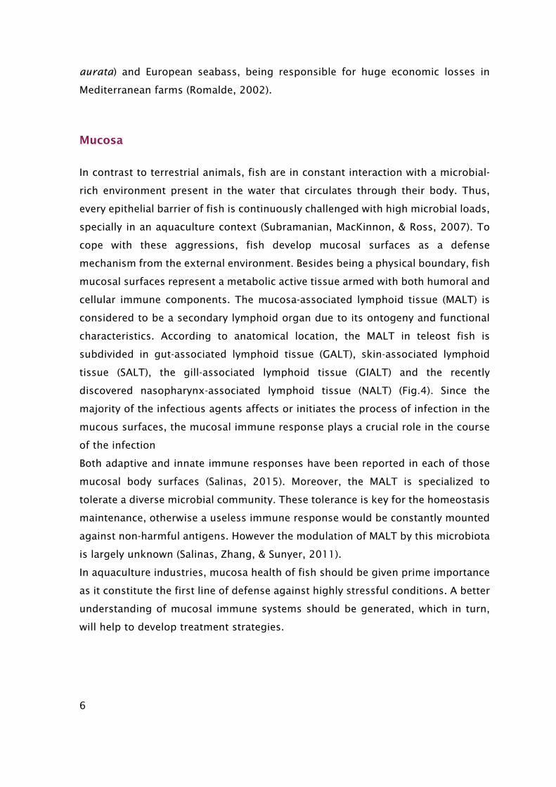
Mucosa
In contrast to terrestrial animals, fish are in constant interaction with a microbial-
rich environment present in the water that circulates through their body. Thus,
every epithelial barrier of fish is continuously challenged with high microbial loads,
specially in an aquaculture context (Subramanian, MacKinnon, & Ross, 2007). To
cope with these aggressions, fish develop mucosal surfaces as a defense
mechanism from the external environment. Besides being a physical boundary, fish
mucosal surfaces represent a metabolic active tissue armed with both humoral and
cellular immune components. The mucosa-associated lymphoid tissue (MALT) is
considered to be a secondary lymphoid organ due to its ontogeny and functional
characteristics. According to anatomical location, the MALT in teleost fish is
subdivided in gut-associated lymphoid tissue (GALT), skin-associated lymphoid
tissue (SALT), the gill-associated lymphoid tissue (GIALT) and the recently
discovered nasopharynx-associated lymphoid tissue (NALT) (Fig.4). Since the
majority of the infectious agents affects or initiates the process of infection in the
mucous surfaces, the mucosal immune response plays a crucial role in the course
of the infection
Both adaptive and innate immune responses have been reported in each of those
mucosal body surfaces (Salinas, 2015). Moreover, the MALT is specialized to
tolerate a diverse microbial community. These tolerance is key for the homeostasis
maintenance, otherwise a useless immune response would be constantly mounted
against non-harmful antigens. However the modulation of MALT by this microbiota
is largely unknown (Salinas, Zhang, & Sunyer, 2011).
In aquaculture industries, mucosa health of fish should be given prime importance
as it constitute the first line of defense against highly stressful conditions. A better
understanding of mucosal immune systems should be generated, which in turn,
will help to develop treatment strategies.
7
Figure 4- Schematic representation of the four teleost main mucosa-associated lymphoid tissues (MALT) described so far and their anatomical localization. GALT: gut-associated lymphoid tissue; SALT: skin-associated lymphoid tissue; GIALT: gill-associated lymphoid tissue; NALT: nasopharynx-associated lymphoid tissue. Source: (Salinas, 2015)
SALT- Skin associated lymphoid tissue
Teleost skin is a large, unique and histological diverse organ. The skin is the
envelope that separates the fish from its environment and it is constitute by three
layers: the mucus (outermost epidermal barrier, with complex composition);
epidermis (a squamous stratified epithelium); dermis (with two layers, the
hypodermis or stratum spongiosum, and the innermost layer or stratum
compactum) (Ángeles Esteban, 2012). In particular, teleost epidermis is very
different to that of mammals because it secretes mucus which is involved in
immune functions (Ángeles Esteban, 2012). The skin associated lymphoid tissue
(SALT) correspond to all immunological defenses provided by fish skin (Ángeles
Esteban, 2012). Unlike mammals, the teleost skin is not keratinized and thus the
epithelium cells are alive and retain the ability to divide (Salinas et al., 2011).
Therefore, the skin epithelium represents an active immune site, coated by mucosa
layer which is produced by four types of secretory cells: goblet cells, sacciform
cells, Malpighian cells and club cells (Zaccone, Kapoor, Fasulo, & Ainis, 2001). The
mucus, besides harboring important immune component has also been reported
to have a key functions including respiration, ionic and osmotic regulation,
reproduction, communication, excretion, feeding and next building (Shephard,
1994).
8
The mucus provides both mechanical and chemical protection to fish. As a
component of the innate and adaptive immune mechanism, it harbors a great
variety of antimicrobial substances include, lysozyme, immunoglobulins,
complement proteins, lectins, C-reactive protein, proteolytic enzymes and
antibacterial peptides and proteins (Shephard, 1994). Moreover, as it is constantly
produced, it prevents the adherence and colonization of parasite, bacteria and
fungi on the fish skin (Pickering, 1974). Although very little information is available,
the main cellular immune constituents of teleost SALT are lymphocytes,
granulocytes, macrophages, and Langerhans-like cells (Salinas et al., 2011).
Moreover, as a component of innate and adaptive immune mechanisms, it harbors
a great variety of antimicrobial substances such us lysozyme, immunoglobulins,
complement proteins, lectins, C-reactive protein, proteolytic enzymes and
antibacterial peptides and proteins (Shephard, 1994).
Hence, as part of a primary defence mechanism of epithelia mucosa, the skin
mucus is expected to adapt itself to pathogenic pressure and external variations.
Indeed, if SALT could accurately discriminate between fish exposed to a pathogenic
agent from healthy ones, mucus sampling methods may be employed during
farming procedures to predict and assist on future disease management. This
technique may constitute a valuable vantage for farmers, to better understand
some abnormal behavior, for instance, without requiring invasive methods.
Amino acids (AA) needs under aquaculture conditions
An acute infection is characterized by a sharp depletion of AA availability, which
are diverted from normal metabolism to be consumed as substrate for the immune
response and synthesis of stress and immune-related proteins (Conceição et al.,
2012). Prolonged infections are also associated with AA imbalances, weight losses
caused by net protein breakdown. Similarly, under stressful conditions, fish AA
requirements increase to cope with the augmented metabolism and energy
demands. The restoration of the AA pool will avoid the lack of recourses, allowing
the fish to cope with increased energy demands. This is the basis of
Immunonutrition concept.
9
Immunonutrition
Immunonutrition aims to provide the right molecules to support immune responses
through fish diet. An additional nutrients supply may assist the immune system to
realize its functions, to finally obtain a higher degree of protection (Kiron, 2012).
An efficient immune defence is of the upmost importance in farms, once fish are
constantly exposed to both stress factors and pathogen invasion (Fig.5).
Moreover, it is widely recognized that the development of aquaculture industry
should be based in eco-friendly and sustainable managements. Thus, the use of
nutritional means for preventive health care had already proved to be a potential
strategy to achieve sustainability in aquaculture. Instead of fighting a certain
disease by using vaccines or antibiotics, immunonutrition aims to prevent
pathogens invasion. For instance, with an increased AA availability, the fish would
be better prepared to mount a proper immune response. However, the link between
nutrition, immune responses and resistance to diseases needs to be clearly
understood, to access the dietary manipulation impact on fish growth and survival.
Figure 5- Immunonutrition Concept. Adapted from (Kiron, 2012).
Nutrients
Activationoftheimmunesystem
Pathogen Stressor
CytokinesMetabolicresponses
Cellularresponses
AcutePhaseProteinSynthesis
Antioxidantdefenses
HostileMilieuforpathogen
Restorationofhomeostasis

10
Methionine
Indeed, AA have a central role in the defense mechanism since they are involved in
the synthesis of important molecules. Methionine is an essential AA, and it is
usually a limiting one in fish diets with high inclusion of plant protein sources
(Wang, Qiao, & Li, 2009). Indeed, this AA is the first limiting step in the initiation
of protein synthesis and proteolytic pathways. Methionine has at least four main
metabolic pathways known for mammals to affect the immune system, which are
likely to be present in fish (Rubin et al., 2007). For instance, methionine is involved
in T cell proliferation and differentiation by fuelling polyamines biosynthesis
(spermine and spermidine) (Fig.6). Moreover, through the generation of
decarboxylated S-adenosylmethionine (SAM), methionine is the most important
methyl donor for methylations reactions of DNA and proteins (Fig.7). Finally,
methionine is also a glutathione precursor (Métayer et al., 2008).
Glutathione affects the immune system either by protecting the body from
oxidative stress, but also in supporting T-cell Proliferation. Glutathione is a key
antioxidant molecule, capable of reducing free radicals and ROS during an
inflammatory response (Grimble, Grimble, Poshoi, al., & Lauterberg, 1996).
Moreover, T-lymphocytes and polymorphonuclear leucocytes are sensitive to
intracellular glutathione concentrations (Machado et al., 2015).
In fact, dietary methionine supplementation appears to exert clear positive effects
on the European seabass (Dicentrarchus labrax) immune status by improving the
peripheral leucocyte response followed by higher complement activity and
bactericidal capacity in response to inflammatory insult with inactivated Phdp
(Machado et al., 2015).
11
Figure 6– Methionine in immune function.
Figure 7 – Methionine metabolism. SAM-decarboxylated S-adenosylmethionine; SAH- S-adenosylhomocysteine
SAM(MethylGroup)
DNAMethylation
PolyamineSynthesis
Immunecellsproliferation
Differentiationoflymphocytes
Methionine
• Antioxidantactivity;
• ActivationofT-lymphocytesand
polymorphonuclear leucocytes;
• Cytokineproduction;
• DNAprontein synthesis
Glutathione (GSH)
SAM
Vitamin B12Methionine
SAH
Homocysteine
Glutathione
Cysteine
Methylacceptor
Methylatedacceptor
Glycine
Glutamate
Betaine
DMG
FolateCycle
12
Arginine
Arginine is a versatile essential AA for all fish species investigated to date, taking
part in several metabolic reactions (Conceição et al., 2012). Besides being an
abundant constituent of body proteins, arginine is also present in the tissue fluids
as phosphoarginine (Li, Mai, Trushenski, & Wu, 2009). There is no knowledge
regarding the synthesis of arginine from another AA apart from citrulline. For these
reasons, arginine de novo synthesis is very limited or complete absent in some
fishes, and may be particularly compromised during stress events. Under unhealthy
conditions, arginine levels may not be enough neither to support optimal growth
or metabolic process.
Similarly to terrestrial animals, arginine plasma levels in fish are controlled by two
main catabolitic enzymes, the inducible nitric oxide synthase (iNOS) and arginase.
The later converts arginine into ornithine and urea, whereas iNOS oxidizes arginine
in two steps that generate nitric oxide (NO) and citrulline. (Conceição et al., 2012)
(Fig. 8).
Figure 8– Arginine metabolism. ARG-arginase. NOS-nitric oxide synthase. ODC-ornithine descarboxylase. OAT-ornithine aminotransferase. Adapted from (Wu et al., 2009)
Arginine NOSARG
Urea
Ornithine
Citrulline
Nitric Oxide
ProlinePolyamines
OATODC
13
Both metabolic pathways are critical to the immunomodulatory actions of arginine.
They directly enhance the immune system responses through 1) polyamine
synthesis and; 2) NO production.
Polyamines are positively related to RNA and DNA polymerase activity and appear
to be indispensable in cell division, DNA replication and regulation of the cell cycle
(Li, Yin, Li, Kim, & Wu, 2007). Besides arginase pathway, arginine can also augment
polyamines synthesis through the stimulation of the release of growth hormone
(Nieves & Langkamp-Henken, 2002).
NO is a powerful oxidant and an anti-microbial agent against parasites, fungi,
bacteria and viruses (J. a Buentello & Gatlin Iii, 1999). In fact, phagocytic efficiency
is closely related to nitrogen reactive species in fish (G. Chen et al., 2015). Upon
infection, NO acts increasing the environmental toxicity, which will compromise
the integrity of the structures of both host and pathogen (Cheng, Buentello, &
Gatlin, 2011).
However, although emerging evidences supporting the arginine promoting effects
on growth, immunity or resistance to environmental stressors and pathogens,
some authors confirmed that fish fed with diet containing excessive arginine shown
immune-depressed symptoms (Ren et al., 2013) (Xie et al., 2012) (Azeredo et al.,
2015).
Arginine is able to mediate immunosuppressive pathways. Jiang et al., 2015
reported that LPS-induced inflammatory response was inhibited when arginine was
added to both primary enterocyte culture media and to the diet of Jian carp
(Cyprinus carpio var. Jian).
In contrast, arginine promoted the synthesis of immune-related proteins and
compounds such as cytokines, complement, lysozyme and antibodies during
bacterial exposure. Moreover, lymphocyte proliferation and differentiation was also
observed upon arginine surplus, as well as increased survival rates after bacteria
exposure (G. Chen et al., 2015). Some of the results are resumed in table 1.
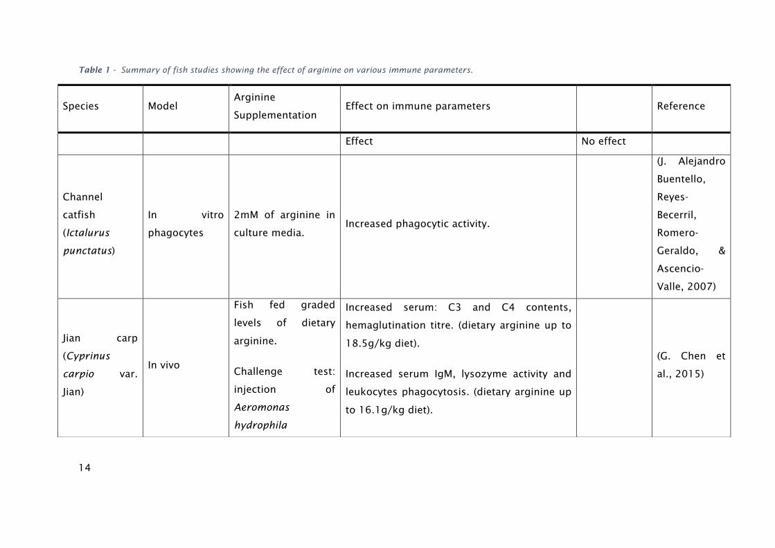
14
Table 1 - Summary of fish studies showing the effect of arginine on various immune parameters.
Species Model Arginine
Supplementation Effect on immune parameters Reference
Effect No effect
Channel
catfish
(Ictalurus
punctatus)
In vitro
phagocytes
2mM of arginine in
culture media. Increased phagocytic activity.
(J. Alejandro
Buentello,
Reyes-
Becerril,
Romero-
Geraldo, &
Ascencio-
Valle, 2007)
Jian carp
(Cyprinus
carpio var.
Jian)
In vivo
Fish fed graded
levels of dietary
arginine.
Challenge test:
injection of
Aeromonas
hydrophila
Increased serum: C3 and C4 contents,
hemaglutination titre. (dietary arginine up to
18.5g/kg diet).
Increased serum IgM, lysozyme activity and
leukocytes phagocytosis. (dietary arginine up
to 16.1g/kg diet).
(G. Chen et
al., 2015)

15
Up-regulation of mRNA expression of
inflammatory cytokines (IL-1b, TNF-a, TGF-b)
due to arginine supplementation.
Increased survival rates following Aeromonas
hydrophila infection (dietary arginine
containing 16.1-21.9g/kg diet).
Yellow catfish
(Pelteobagrus
fulvidraco)
In vivo
Fish fed graded
levels of dietary
arginine. Challenge
test: injection of
Aeromonas
hydrophila
Increased arginase, nitric oxide synthase,
lysozyme activities, phagocytic index and
respiratory burst (diet containing 2.74%
arginine).
Survival rate
was not
affected after
bacterial
challenge.
(Q. Zhou, Jin,
Elmada,
Liang, & Mai,
2014)
Blunt snout
bream
(Megalobrama
amblycephala)
In vivo
Fish fed graded
levels of dietary
arginine.
Increased serum and hepatic total nitric oxide
synthase.
Reduced growth in fish fed diets with
excessive arginine.
No effect on
plasma
superoxide
dismutase.
(Ren et al.,
2013)
Largemouth
bass
(Micropterus
salmoides)
In vivo
Fish fed graded
levels of dietary
arginine.
Increased serum lysozyme activity, serum
protein and respiratory burst of head kidney
leucocytes.
Complement
activity was
not affected.
(H. Zhou,
Chen, Qiu,
Zhao, & Jin,
2012)
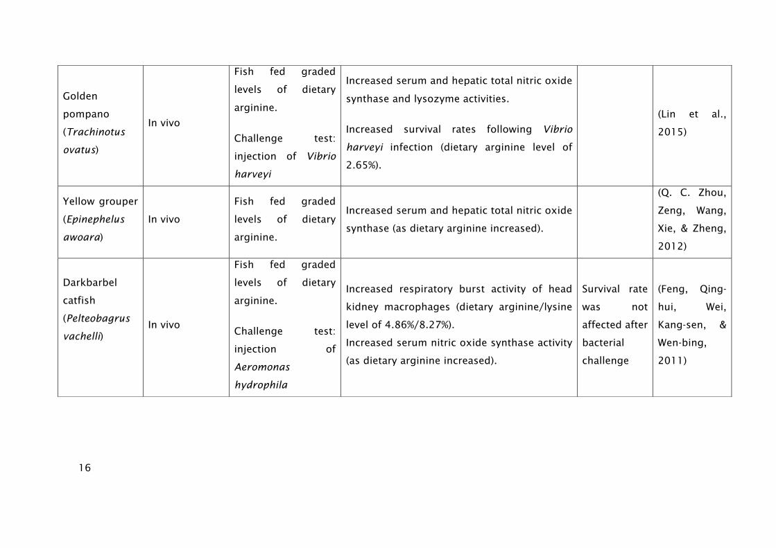
16
Golden
pompano
(Trachinotus
ovatus)
In vivo
Fish fed graded
levels of dietary
arginine.
Challenge test:
injection of Vibrio
harveyi
Increased serum and hepatic total nitric oxide
synthase and lysozyme activities.
Increased survival rates following Vibrio
harveyi infection (dietary arginine level of
2.65%).
(Lin et al.,
2015)
Yellow grouper
(Epinephelus
awoara)
In vivo
Fish fed graded
levels of dietary
arginine.
Increased serum and hepatic total nitric oxide
synthase (as dietary arginine increased).
(Q. C. Zhou,
Zeng, Wang,
Xie, & Zheng,
2012)
Darkbarbel
catfish
(Pelteobagrus
vachelli)
In vivo
Fish fed graded
levels of dietary
arginine.
Challenge test:
injection of
Aeromonas
hydrophila
Increased respiratory burst activity of head
kidney macrophages (dietary arginine/lysine
level of 4.86%/8.27%).
Increased serum nitric oxide synthase activity
(as dietary arginine increased).
Survival rate
was not
affected after
bacterial
challenge
(Feng, Qing-
hui, Wei,
Kang-sen, &
Wen-bing,
2011)

17
Senegalese
sole (Solea
senegalensis)
In vivo
Fish fed graded
levels of dietary
arginine.
Challenge test:
injection of
Photobacterium
damselae subsp.
piscicida (strain
PC566.1)
Increased respiratory burst activity and nitric
oxide production of head kidney leucocytes
with higher arginine supplementations.
Increased lysozyme, alternative complement
pathway, and peroxidase activities (dietary
arginine level of 5.7 and 6.9 g-1 N).
Increased HIF-1, HAMP-1, MIP1-alpha and gLYS
expression values (dietary arginine level of 5.7
and 6.9 g-1 N).
(B. Costas et
al., 2011)
Channel
catfish
(Ictalurus
punctatus)
In vitro:
1-Primary cell
cultures of
head-kidney
macrophages
2- Naïve
peripheral
blood
lymphocytes
Supplementation of
culture media with
and/or glutamine.
Increased macrophage phagocytosis and
killing ability against Edwardsiella ictaluri.
(1mM of arginine in culture media)
Increased proliferation of naïve T- and B-
lymphocytes upon mitogenic exposure (0.5
mM of arginine + glutamine in culture media).
(Pohlenz,
Buentello,
Mwangi, &
Gatlin, 2012)

18
Yellow catfish
(Pelteobagrus
fulvidraco)
In vivo
Fish fed graded
levels of arginine.
Challenged to
ammonia-nitrogen
for 72 h.
Increased anti-ammonia-nitrogen stress ability
(dietary arginine level of 2.81%).
(Q. Chen et
al., 2016)
Red drum,
(Sciaenops
ocellatus)
In vivo
Fish fed graded
levels of dietary
arginine and
glutamine.
Increased neutrophil oxidative radical
production, serum lysozyme, and extracellular
and intracellular superoxide anion production
of kidney macrophages (dietary level of 1%
arginine+1% glutamine).
(Cheng et al.,
2011)
Turbot
(Scophthalmus
maximus)
In vivo
Fish fed graded
levels of dietary
arginine.
Challenged to
repeated handling,
as a chronic stress
factor.
Increased monocytes numbers, nitric oxide
production, plasma lysozyme, superoxide
dismutase and alternative complement
pathway activities (in both control and
stressed fish, with arginine supplements).
(Benjamín
Costas et al.,
2012)
Hybrid striped
bass (Morone
chrysops×Moro
ne saxatilis)
In vivo
Fish fed graded
levels of dietary
arginine.
Increased neutrophil oxidative radical
production, serum lysozyme, extracellular and
intracellular superoxide anion production of
(Cheng,
Gatlin, &
Buentello,
2012)

19
kidney macrophages (dietary level of 1%
arginine).
Channel
catfish
(Ictalurus
punctatus)
In vivo
Fish fed graded
levels of dietary
arginine and/or
glutamine.
Challenged against
Edwardsiella
ictaluri.
Increased survival rates following Edwardsiella
ictaluri infection (dietary arginine level of 2%).
(J. A.
Buentello &
Gatlin, 2001)
Channel
catfish
(Ictalurus
punctatus)
In vivo
Fish fed graded
levels of dietary
arginine and/or
glutamine.
Vaccination against
Edwardsiella
ictaluri.
Increased antibody titers in plasma (dietary
level of 4% arginine; 2% glutamine and a
combination of both).
Increased responsiveness of spleen and head-
kidney lymphocytes against E. ictaluri (dietary
level of 4% arginine; 2% glutamine).
Increased protein content in head-kidney
(dietary level of 4% arginine)
(Pohlenz,
Buentello,
Criscitiello, et
al., 2012)
20
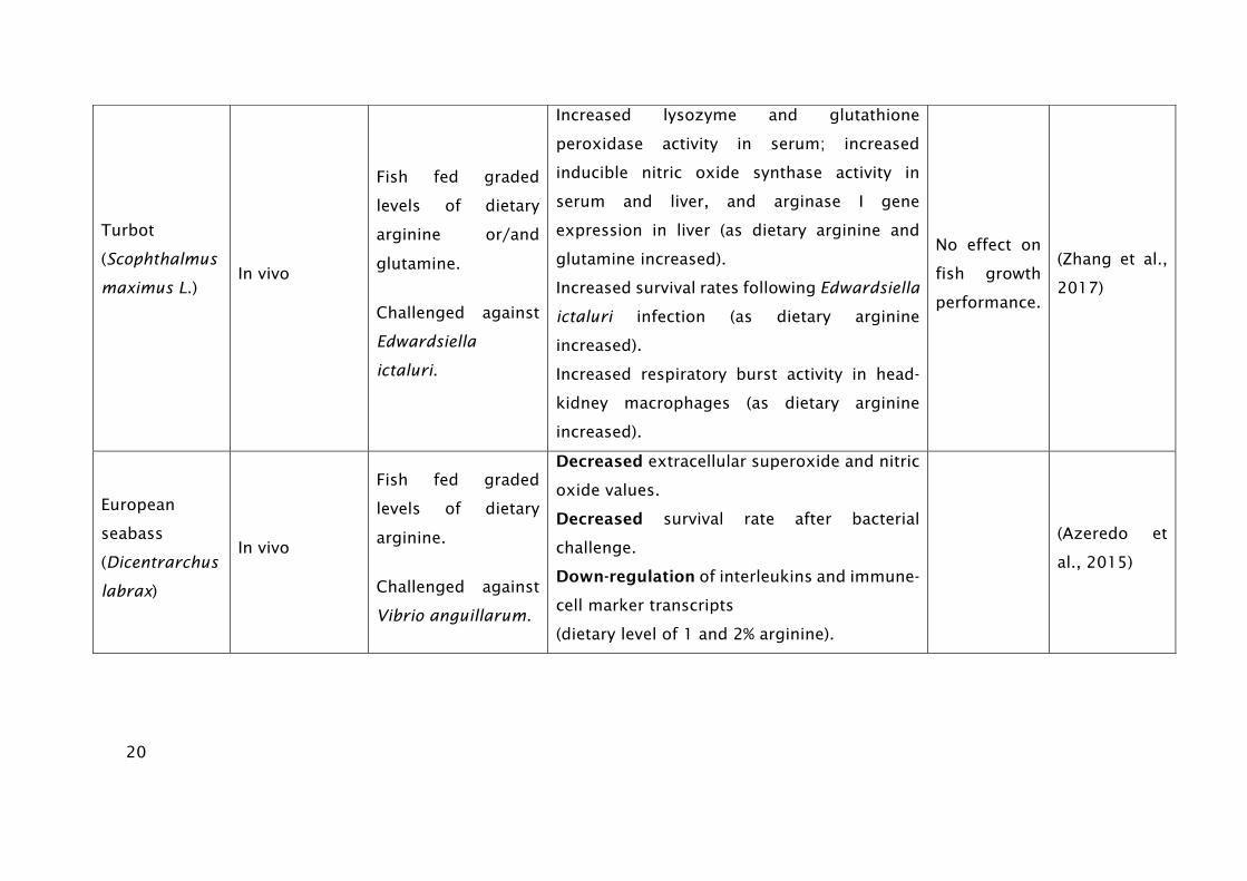
Turbot
(Scophthalmus
maximus L.)
In vivo
Fish fed graded
levels of dietary
arginine or/and
glutamine.
Challenged against
Edwardsiella
ictaluri.
Increased lysozyme and glutathione
peroxidase activity in serum; increased
inducible nitric oxide synthase activity in
serum and liver, and arginase I gene
expression in liver (as dietary arginine and
glutamine increased).
Increased survival rates following Edwardsiella
ictaluri infection (as dietary arginine
increased).
Increased respiratory burst activity in head-
kidney macrophages (as dietary arginine
increased).
No effect on
fish growth
performance.
(Zhang et al.,
2017)
European
seabass
(Dicentrarchus
labrax)
In vivo
Fish fed graded
levels of dietary
arginine.
Challenged against
Vibrio anguillarum.
Decreased extracellular superoxide and nitric
oxide values.
Decreased survival rate after bacterial
challenge.
Down-regulation of interleukins and immune-
cell marker transcripts
(dietary level of 1 and 2% arginine).
(Azeredo et
al., 2015)
21
Citrulline
Citrulline is a non-essential AA, constituent of body proteins. Arginine and citrulline
are linked in several metabolic reactions described in Figure 9. As an essential AA,
the little contribution for arginine’s de novo synthesis in Elasmobranches and
ureogenic teleost, is only through the conversion of citrulline via argininosuccinate
synthase and lyase in the liver (Fig.9) (Mommsen, Moon, & Plisetskaya, 2001).
However, little information is available regarding the efficacy of citrulline to replace
arginine in fish nutrition (Li et al., 2009).
Figure 9– Citrulline metabolism. ASS argininosuccinate synthase, ASL argininosuccinate lyase, ARG arginase, OTC ornithine carbamoiltransferase, NOS nitric oxide synthase, Cit-Arg Cycle citrulline arginine cycle.
In mammals, it has been reported that citrulline might offer a safe alternative to
arginine for improving macrophage function under certain metabolic conditions
(Breuillard, Bonhomme, Couderc, Cynober, & De Bandt, 2014) Moreover, pre-
treatment with citrulline stimulated intestinal production of secretory
immunoglobulin A in mice, which is the first line of host defences against
environmental pathogens (Batista et al., 2011). Indeed, few studies have
approached the effect of citrulline surplus on immune responses in higher
NOS
ARG
Urea
OTC
Citrulline
Arginine
Ornithine Argininosuccinate
ASS
ASL
UreaCarbamoilPhosphate
Urea Cycle Cit-Arg Cycle

22
vertebrates, and to the best of our knowledge there currently are not available data
in fish.
Objectives
Skin mucosal immunity is a key component of the innate immune and its usage for
monitoring fish health status has already been proven. As such, in this study we
hypothesize whether SALT immune parameters are affected by nutritional changes
and immune challenges. The goal is to evaluate the SALT’s capacity to describe fish
condition, and its applicability to be used as a biomarker in fish farms.
The other main goal of this thesis is to provide a better understanding of the
influence of arginine, citrulline and methionine supplementation on mucosal
immune mechanisms and inflammatory response. This knowledge should allow the
development of functional commercial diets, hence getting better farming results
in terms of growth and disease susceptibility of European seabass.

23
Material and Methods
Trial 1 - EFFECTS OF METHIONINE AVAILABILITY ON EUROPEAN SEABASS IMMUNE
CONDITION AND INFLAMATORY RESPONSE
Rearing conditions��
At i3S (Porto, Portugal) fish rearing facilities, European seabass (±8.5g) juveniles
were maintained under standard culture conditions for a quarantine period of two
weeks. In a recirculation seawater system (Temperature: 20 ± 0.5 °C; Salinity: 35
ppt; Photoperiod: 10h dark, 14h light) fish were then distributed into 12 fiberglass
tanks (200 l; n=50) for an acclimatization period of 1 week. Temperature was
maintained by a water heater/cooler system. Oxygen saturation was held at around
7.3 mg/L and photoperiod automatically controlled. Both nitrite and ammonium
levels were daily recorded and its levels controlled by a water ozoniser system.
Water renovations and system cleanings were performed twice a week. Dietary
treatments were randomly assigned to triplicate tanks and fish were fed three times
a day by hand (9.30 am, 1.30 pm and 5.30 pm).
Diets composition
Four diets were formulated and manufactured by Sparos Lda. (Olhão. Portugal). A
control diet (CTRL) was formulated to include an indispensable AA profile meeting
the ideal pattern estimated for European seabass (Kaushik, 1998). Two other diets,
identical to the CTRL were supplemented with DL-Methionine at 0.5% and 1% MET
0.5 and MET 1, respectively at the expenses of wheat gluten. A negative control
diet (NCTRL) was also formulated to be deficient in methionine. Main ingredients
were ground (below 250 μm) in a micropulverizer hammer mill (SH1; Hosokawa
Micron B.V., Doetinchem, The Netherlands). Powder ingredients and oils were then
mixed according to the target formulation in a paddle mixer (RM90; Mainca S.L.,
Granollers, Spain). All diets were manufactured by temperature- controlled
24
extrusion (pellet sizes: 1.5 mm) by means of a low-shear extruder (P55; Italplast
S.r.l., Parma, Italy). Upon extrusion, all feed batches were dried in a convection
oven (OP 750-UF; LTE Scientifics, Oldham, UK) for 4h at 45°C. Formulation of
experimental diets is presented in Table 2. Four diets were randomly assigned to
triplicate tanks and fish were feed 3 times a day. By hand and ab libitum the
experimental diets assigned.
Table 2 - Ingredients of the experimental diets.
25
Feeding trial
The feeding trial lasted for 4 weeks, in order to assess the effect of short -term
dietary supplementation or deficiency of methionine. At the end of each period,
twelve fish per tank were sacrificed by an anesthetic overdose with 2-
phenoxyethanol and individually weighed (Fig. 10). Skin mucus was gently
collected by swabbing from head to tail on both sides of the fish as seen in figure
11, and avoiding contamination from urine, faeces and blood. Mucus samples were
frozen and stored at -80ºC for further analysis.
Figure 10- Representative diagram of the Feeding Trial.
Figure 11- Mucus sampling.
n = 24 n = 24 n = 24 n = 24 n = 24 n = 24 n = 24 n = 24 n = 24 n = 24 n = 24 n = 24
CTRL MET1 MET0.5 NCTRL MET1 CTRL CTRL NCTRL MET0.5 MET1 MET0.5 NCTRL
• n=12/tankà 2weeks• n =12/tankà 4weeks

26
Bacterial growth and inoculum preparation��
Phdp, strain PP3, was kindly provided by Dr. Ana do Vale (Institute for Molecular
and Cell Biology, University of Porto. Portugal) and isolated from yellowtail (Seriola
quinqueradiata; Japan) by Dr. Andrew C. Barnes (Marine Laboratory, Aberdeen, UK).
Bacteria were routinely cultured at 22 ̊C in tryptic soy broth (TSB) or tryptic soy agar
(TSA) (both from Difco Laboratories) supplemented with NaCl to a final
concentration of 2% (w/v) (TSB-2 and TSA-2, respectively) and stored at – 80 ̊C in
TSB-2 supplemented with 15% (v/v) glycerol. To prepare the inoculum for injection
into the fish peritoneal cavities, 100 μL of stocked bacteria were cultured overnight
at 22 ̊C on TSA-2. Exponentially growing bacteria were collected from the TSA-2
and re-suspended in sterile TSB-2. According to the pre-challenge previously
performed, the intended bacterial concentration to kill 50% of the fish (LD50) was
obtained by absorbance reading and adjustment against its growth curve to 5 x
104 colony forming units (cfu) ml-1. Bacteria concentration was confirmed by
plating the resulting cultures on TSA-2 plates and counting of the colony forming
units (cfu) ml-1.
Time-course trial
Immediately after the 4 weeks sampling, the 26-remaining fish were
intraperitoneally (i.p.) injected with 100 μl Phdp (5 x 10⁴ cfu) (Fig. 13). After i.p.
injection, 6 fish from each tank were relocated in a comparable recirculation system
(Temperature: 24 ± 0.5 °C; Salinity: 35 ppt; Photoperiod: 10h dark: 14h light) and
divided in two tanks according to dietary treatment (Fig.12). This experiment was
designed to investigate the immunomodulatory effect of methionine during the
acute inflammatory response against Phdp in fish previously fed the experimental
diets. For that purpose, fish were sampled at 4, 24h after challenge. At each
sampling time, 3 fish per tank were sacrificed by anesthetic overdose with 2-
phenoxyethanol and skin mucus collected.
27
Figure 12- Representative diagram of the experimental setup for the time-course trial.
Figure 13- Intraperitoneal injection with Phdp.
• n=3/tankà 4h• n =3/tankà 24h• n=3/tankà 48h
n = 9 n = 9 n = 9 n = 9 n = 9 n = 9 n = 9 n = 9
CTRL MET1 NCTRL MET1 CTRL MET0.5NCTRL MET0.5
28
Trial 2- EFFECTS OF ARGININE AND CITRULLINE AVAILABILITY ON EUROPEAN
SEABASS IMMUNE CONDITION
Rearing conditions�
At i3S fish rearing facilities, European seabass (8.4 g ± 0.39 g) were maintained
under standard culture conditions for a quarantine period of one weeks. In a
recirculation seawater system (Temperature: 20 ± 0.5 °C; Salinity: 35 ppt;
Photoperiod: 10h dark: 14h light) fish were then distributed into 12 fiberglass
tanks (200 l; n=50) for an acclimatization period of 1 week. Temperature was
maintained by a water heater/cooler system. Oxygen saturation was maintained at
around 7.3 mg/L and photoperiod automatically controlled. Both nitrite and
ammonium levels were daily recorded and its levels controlled by a water ozoniser
system. Water renovations and system cleanings were performed twice a week.
Dietary treatments were randomly assigned to triplicate tanks and fish were fed
three times a day by hand (9.30 am. 1.30 pm and 5.30 pm) until apparent satiety.
Diets composition
Four diets were formulated and manufactured by Sparos Lda, (Olhão. Portugal). A
control diet (CTRL) was formulated to include an indispensable AA profile meeting
the ideal pattern estimated for European seabass (Kaushik, 1998). Two other diets,
identical to the CTRL were supplemented with DL-Arginine and DL-citrulline at 0.5
% dry matter (ARG1 and CIT1, respectively) at the expenses of wheat meal. A third
diet was formulated by supplementing DL-arginine at 1 % dry matter (ARG2). Main
ingredients were ground (below 250 μm) in a micropulverizer hammer mill (SH1;
Hosokawa Micron, B.V., Doetinchem, The Netherlands). Powder ingredients and oils
were then mixed according to the target formulation in a paddle mixer (RM90;
Mainca, S.L., Granollers, Spain). All diets were manufactured by temperature-
controlled extrusion (pellet sizes: 1.5 mm) by means of a low-shear extruder (P55;
Italplast, S.r.l., Parma, Italy). Upon extrusion, all feed batches were dried in a
convection oven (OP 750-UF; LTE Scientifics, Oldham, UK) for 4h at 45 °C.
Formulation of experimental diets is presented in Table 3.

29
Table 3 - Ingredients of the experimental diets.
Ingredients CTRL ARG1 ARG2 CIT % % % %
FishmealLT70(SouthAmerican) 5.000 5.000 5.000 5.000Porcinebloodmeal 2.000 2.000 2.000 2.000Poultrymeal65 5.000 5.000 5.000 5.000Potatoconcentrate 10.000 10.000 10.000 10.000Wheatglúten1 13.500 13.500 13.500 13.500Cornglúten2 30.000 30.000 30.000 30.000Soybeanmeal48 5.000 5.000 5.000 5.000Wheatmeal 8.000 7.500 7.000 7.500Fishoil3 10.000 10.000 10.000 10.000Rapeseedoil4 5.500 5.500 5.500 5.500Vitaminandmineralpremix5 1.000 1.000 1.000 1.000Binder(naturalzeolite) 1.000 1.000 1.000 1.000Antioxidant6 0.200 0.200 0.200 0.200Sodiumpropionate 0.100 0.100 0.100 0.100Monocalciumphosphate7 2.000 2.000 2.000 2.000L-Arginine 0.500 1.000 L-Citrulline 0.500L-Histidine 0.300 0.300 0.300 0.300L-Lysine 0.800 0.800 0.800 0.800L-Threonine 0.200 0.200 0.200 0.200DL-Methionine 0.400 0.400 0.400 0.400Total 100.000 100.000 100.000 100.000
Pelletsize.mm 1.5 1.5 1.5 1.5
30
Feeding trial
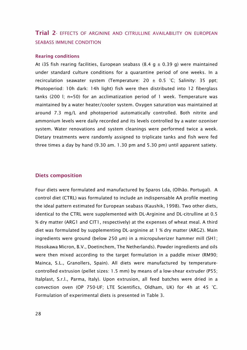
The feeding trial lasted for 4 weeks in order to assess the effect of short -term AA
dietary supplementation. After 2 and 4 weeks of feeding, twelve fish per tank were
sacrificed by anesthetic overdose with 2-phenoxyethanol and individually weighed
(Figure 14). Skin mucus was gently collected by swabbing from gill to tail on both
sides of the fish, and avoiding contamination from urine, feces and blood. Mucus
samples were frozen and stored at -80ºC for further analysis.
Figure 14- Representative diagram of the Feeding Trial.
Bacterial growth and inoculum
Bacteria were grown and harvested as described above. For this trial, the intended
bacterial concentration to kill 50 % of the fish (LD50
) was obtained: 5 x 10³ colony
forming units (CFU) ml-1. Bacteria concentration was confirmed by plating the
resulting cultures on TSA-2 plates and counting of the CFU ml-1.
Time-course trial
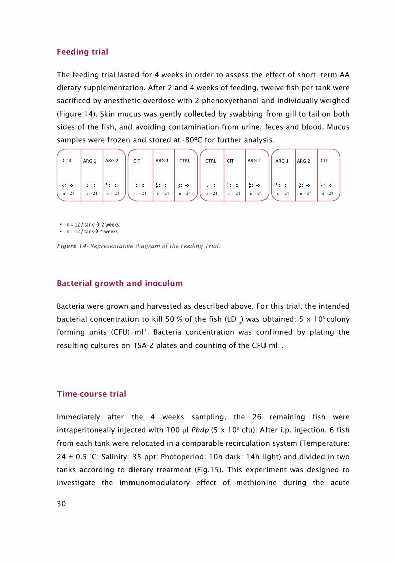
Immediately after the 4 weeks sampling, the 26 remaining fish were
intraperitoneally injected with 100 μl Phdp (5 x 10³ cfu). After i.p. injection, 6 fish
from each tank were relocated in a comparable recirculation system (Temperature:
24 ± 0.5 °C; Salinity: 35 ppt; Photoperiod: 10h dark: 14h light) and divided in two
tanks according to dietary treatment (Fig.15). This experiment was designed to
investigate the immunomodulatory effect of methionine during the acute
n = 24 n = 24 n = 24 n = 24 n = 24 n = 24 n = 24 n = 24 n = 24 n = 24 n = 24 n = 24
CTRL ARG1 ARG2 CIT ARG1 CTRL CTRL CIT ARG2 ARG1 ARG2 CIT
• n=12/tankà 2weeks• n =12/tankà 4weeks
31
inflammatory response against Phdp in fish previously fed the experimental diets.
For that purpose, fish were sampled at 4. 24 and 48h after challenge. At each
sampling time. 3 fish per tank were sacrificed by anesthetic overdose with 2-
phenoxyethanol and skin mucus collected.
Figure 15- Representative diagram of the experimental setup for the time-course trial.
Humoral parameters analytical procedures
Bactericidal Activity
Phdp strain PP3 was used to determine the bactericidal activity of the mucus
sample. After being cultured for 48 h at 25 °C on tryptic soy agar (TSA; Difco
Laboratories), the bacteria were inoculated into tryptic soy broth (TSB; Difco
Laboratories), both supplemented with NaCl to a final concentration of 1% (w/v)
(TSA-1 and TSB-1, respectively). Bacteria in TSB-1 medium were then cultured at the
same temperature for 24h, with continuous shaking (100 rpm). Exponentially
growing bacteria were collected by centrifugation at 3500 × g for 30 min, re-
suspended in sterile HBSS and adjusted to 1 × 10⁶ cfu ml-1. To confirm bacterial
concentration of the inoculum, plating serial dilutions of the suspensions were
performed onto TSA-1 plates and the number of cfu was counted following
incubation at 25 °C.
Mucus bactericidal activity was determined following the method of (Stevens Kehrli
& Canning 1991) with modifications. Briefly, 20 μl of mucus and 20 µl of Phdp (1 ×
• n=3/tankà 4h• n =3/tankà 24h• n=3/tankà 48h
n = 9 n = 9 n = 9 n = 9 n = 9 n = 9 n = 9 n = 9
CTRL ARG1 CIT ARG1 CTRL ARG2CIT ARG2

32
106 cfu ml-1) were added to triplicate wells of a round-bottom 96-well plate,
following incubation 2.5h at 25°C. Hank´s balanced salt solution (HBSS) was added
to some wells instead of mucus as a positive control. To each well. 25 μl of 3-(4.5
dimethyl-2-yl)-2.5-diphenyl tetrazolium bromide (1 mg ml-1; Sigma) were added
and incubated for 10 min at 25 ºC to allow the formation of formazan. Plates were
then centrifuged at 2000 × g for 10 min and the precipitate was dissolved in 200
μl of dimethyl sulfoxide (Sigma). The absorbance of the dissolved formazan was
measured at 560 nm. Bactericidal activity is expressed as percentage, calculated
from the difference between bacteria surviving compared to the number of bacteria
from positive controls (100%).
%#$#%&'()*('+,*-&' = /'01)*2(3. 56772(3. $8,9*-*8*-*#+*3'01)*
%%&'()*('+,*-&' = 677 − %#$#%&'()*('+,*-&'
Figure 16- 96-well microplates used in Bactericidal activity test.
Lysozyme
Lysozyme activity was measured using a turbidimetric assay as described by (B.
Costas et al., 2011). Briefly. a solution of Micrococcus lysodeikticus (0.5 mg ml-1.
33
0.05 M sodium phosphate buffer. pH 6.2) was prepared. In a round-bottom 96-well
plate, 15 μl of mucus and 250 μl of the above suspension were added to give a final
volume of 265 μl. The absorbance (450 nm) was measured after 0.5 and 10 min in
a Synergy HT microplate reader, Biotek, at 25ºC. A standard curve was developed
by using lyophilized hen egg white lysozyme (Sigma), which was serially diluted in
sodium phosphate buffer (0.05 M. pH 6.2). Using the formula of the standard curve
the amount of lysozyme in the sample was calculated. All analysis were conducted
in triplicates.
Alternative Complement Pathway
Alternative complement pathway (ACP) activity was estimated as described by
(Sunyer & Tort, 1995). The following buffers were used: GVB (Isotonic veronal
buffered saline), pH 7.3, containing 0.1% gelatin; EDTA-GVB, as previous one but
containing 20 mM EDTA; and Mg- EGTA-GVB, which is GVB with 10 mM Mg+2 and
10 mM EGTA. Horse red blood cells (RaRBC; Probiologica Lda, Portugal) were used
for ACP determination. RaRBC were washed four times in GVB and re-suspended in
GVB to a concentration of 2.5 × 10⁸ cells ml-1. Ten μl of RaRBC suspension were
then added to 40 μl of serially diluted mucus in Mg-EGTA-GVB buffer. Samples were
incubated at room temperature for 100 min with regular shaking. The reaction was
stopped by adding 150 μl of cold EDTA-GVB. Samples were then centrifuged and
the extent of haemolysis was estimated by measuring the optical density of the
supernatant at 414 nm in a Synergy HT microplate reader Biotek. The ACH50 units
were defined as the concentration of mucus giving 50% haemolysis of RaRBC. All
analysis were conducted by triplicates.
Peroxidase Activity
Total peroxidase activity in mucus was measured following the procedure
described by (Quade & Roth, 1997). Briefly, 15 μl of mucus were diluted with 135
μl of HBSS without Ca+2 and Mg+2 in flat- bottomed 96-well plates. Then, 50 μl of 20
mM 3.3',5.5'-tetramethylbenzidine hydrochloride (TMB; Sigma) and 50 μl of 5 mM
H2O2 were added. The color-change reaction was stopped after 2 min by adding
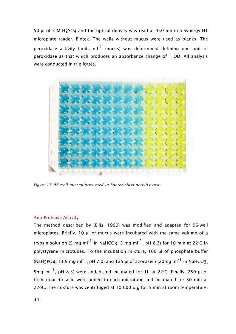
34
50 μl of 2 M H2SO4 and the optical density was read at 450 nm in a Synergy HT
microplate reader, Biotek. The wells without mucus were used as blanks. The
peroxidase activity (units ml-1 mucus) was determined defining one unit of
peroxidase as that which produces an absorbance change of 1 OD. All analysis
were conducted in triplicates.
Figure 17- 96-well microplates used in Bactericidal activity test.
Anti-Protease Activity
The method described by (Ellis, 1990) was modified and adapted for 96-well
microplates. Briefly, 10 μl of mucus were incubated with the same volume of a
trypsin solution (5 mg ml-1 in NaHCO3, 5 mg ml-1, pH 8.3) for 10 min at 22oC in
polystyrene microtubes. To the incubation mixture, 100 μl of phosphate buffer
(NaH2PO4, 13.9 mg ml-1, pH 7.0) and 125 μl of azocasein (20mg ml-1 in NaHCO3,
5mg ml-1, pH 8.3) were added and incubated for 1h at 22oC. Finally, 250 μl of
trichloroacetic acid were added to each microtube and incubated for 30 min at
22oC. The mixture was centrifuged at 10 000 x g for 5 min at room temperature.
35
Afterwards, 100 μl of the supernatant was transferred to a 96 well-plate that
previously contained 100 μl of NaOH (40 mg ml-1) per well. The OD was read at
450nm. Phosphate buffer in place of mucus and trypsin served as blank whereas
the reference sample was phosphate buffer in place of mucus. The percentage of
trypsin activity inhibition was calculated as follows:
%#$#	&(&,*;,-<13&# = /'01)*2(3. 56072(3. $8,9*-*8*-*#+*3'01)*
%	&(&,*;,-<13&# = 677 − %#$#	&(&,*;,-<13&#
Data Analysis
All results are expressed as mean ± standard deviation (mean ± SD). All variables
were checked for normality and homogeneity of variance, by using the Shapiro-Wilk
and the Levene test, respectively. Data was analyzed by two-way ANOVA, with time
and diet as factors and followed by Tukey post-hoc test to identify differences in
the experimental treatments. Variable transformation was applied when
homogeneity and normality were not achieved. A nonparametric test (Kruskal-
Wallis H-test) was performed, if the above assumptions where still not achieved.
And a pairwise comparison to obtain the revealed differences.
All statistical analyses were performed using the computer package SPSS for MAC.
The level of significance used was P≤ 0.05 for al statistical tests.

36
RESULTS
Trial 1 - EFFECTS OF METHIONINE AVAILABILITY ON EUROPEAN SEABASS IMMUNE
CONDITION
FEEDING TRIAL
In methionine feeding trail, no dietary effects were observed on mucus bactericidal
and peroxidase activities (Figs. 18 and 21), whereas peroxidase values augmented
in time regardless of dietary treatment. Furthermore, a slight but significant
increase of ACP levels was observed in fish fed CTRL diet at the end of the feeding
trail compared to those sampled at 2 weeks (Fig. 19). Seabass fed the Met1 diet
presented a decrease in lysozyme levels compared to those fed the CTRL diet at 2
weeks, while after 4 weeks of feeding trail, lysozyme activity decreased significantly
in fish fed diet deficient in methionine (NCRT) compared to those fed CTRL, MET05
and MET1 diets (Fig.20). Moreover, those levels were observed to decrease between
2 and 4 weeks, within the group fed NCRT diet. Finally, there was no anti-trypsin
activity detectable in skin mucus in all experimental diets.
Figure 18- Bactericidal activity in mucus of European sea bass fed different diets for 2 (salmon columns) and 4 (bordeaux columns) weeks. Data are expressed as means ± standard error of the mean (SEM) (n = 12). Vales were cos-transformed before being treated statistically (one-way ANOVA; P < 0.05).
0
5
10
15
20
25
30
35
CTRL MET05 MET1 NCRT
Bac
teri
cidal
act
ivit
y (%
)
2 weeks 4 weeks
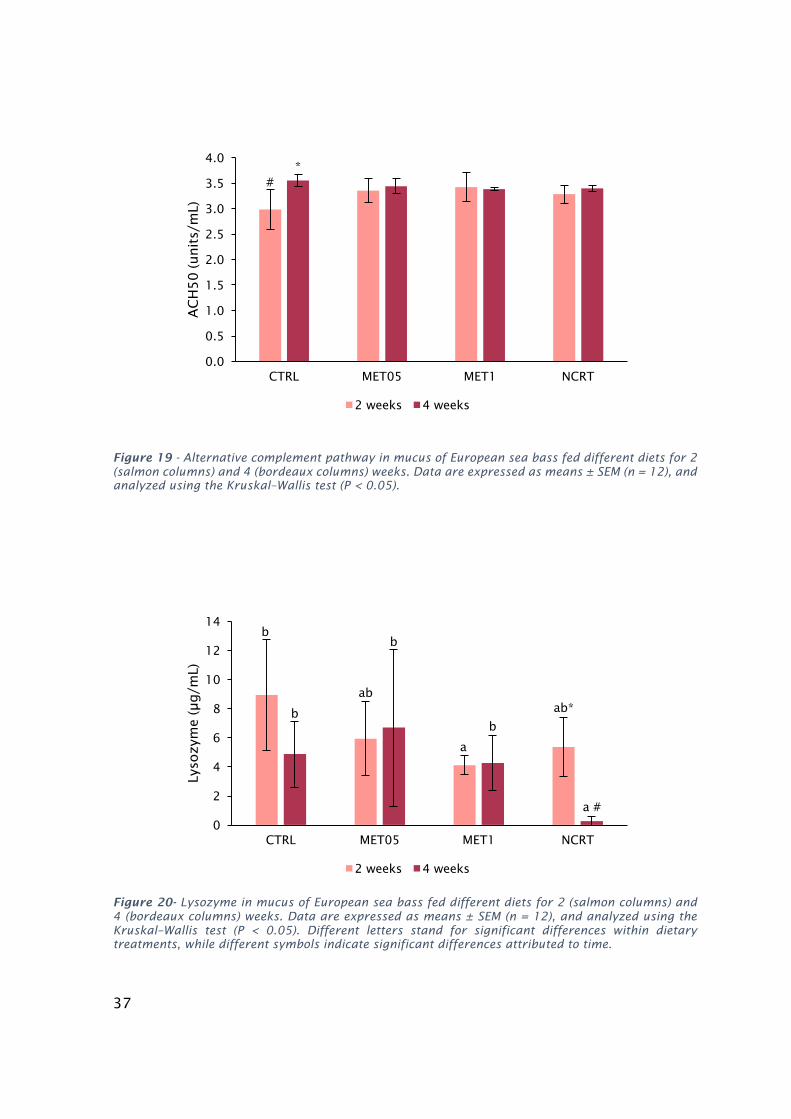
37
Figure 19 - Alternative complement pathway in mucus of European sea bass fed different diets for 2 (salmon columns) and 4 (bordeaux columns) weeks. Data are expressed as means ± SEM (n = 12), and analyzed using the Kruskal–Wallis test (P < 0.05).
Figure 20- Lysozyme in mucus of European sea bass fed different diets for 2 (salmon columns) and 4 (bordeaux columns) weeks. Data are expressed as means ± SEM (n = 12), and analyzed using the Kruskal–Wallis test (P < 0.05). Different letters stand for significant differences within dietary treatments, while different symbols indicate significant differences attributed to time.
b
ab
a
ab*b
b
b
a #
0
2
4
6
8
10
12
14
CTRL MET05 MET1 NCRT
Lyso
zym
e (µ
g/m
L)
2 weeks 4 weeks
#*
0.0
0.5
1.0
1.5
2.0
2.5
3.0
3.5
4.0
CTRL MET05 MET1 NCRT
AC
H5
0 (unit
s/m
L)
2 weeks 4 weeks

38
Figure 21- Peroxidase in mucus of European sea bass fed different diets for 2 (salmon columns) and 4 (bordeaux columns) weeks. Data are expressed as means ± SEM (n = 12), and analysed using the Kruskal–Wallis test (P < 0.05).
TIME-COURSE TRIAL
Dietary methionine resulted in significant effects on lysozyme and peroxidase
levels after a bacterial infection, but not in ACH50 activity (Fig.22). Fish fed diet
MET0.5 presented a significant decrease in skin mucus lysozyme values compared
to fish fed the CTRL diet (Fig.23). Regarding peroxidase activity, fish fed
methionine deficient diet showed higher values than specimens fed MET1 diet
(Fig.24).
0.00
0.10
0.20
0.30
0.40
0.50
0.60
0.70
CTRL MET05 MET1 NCRT
Pero
xid
ase
(unit
s/m
L)
2 weeks 4 weeks
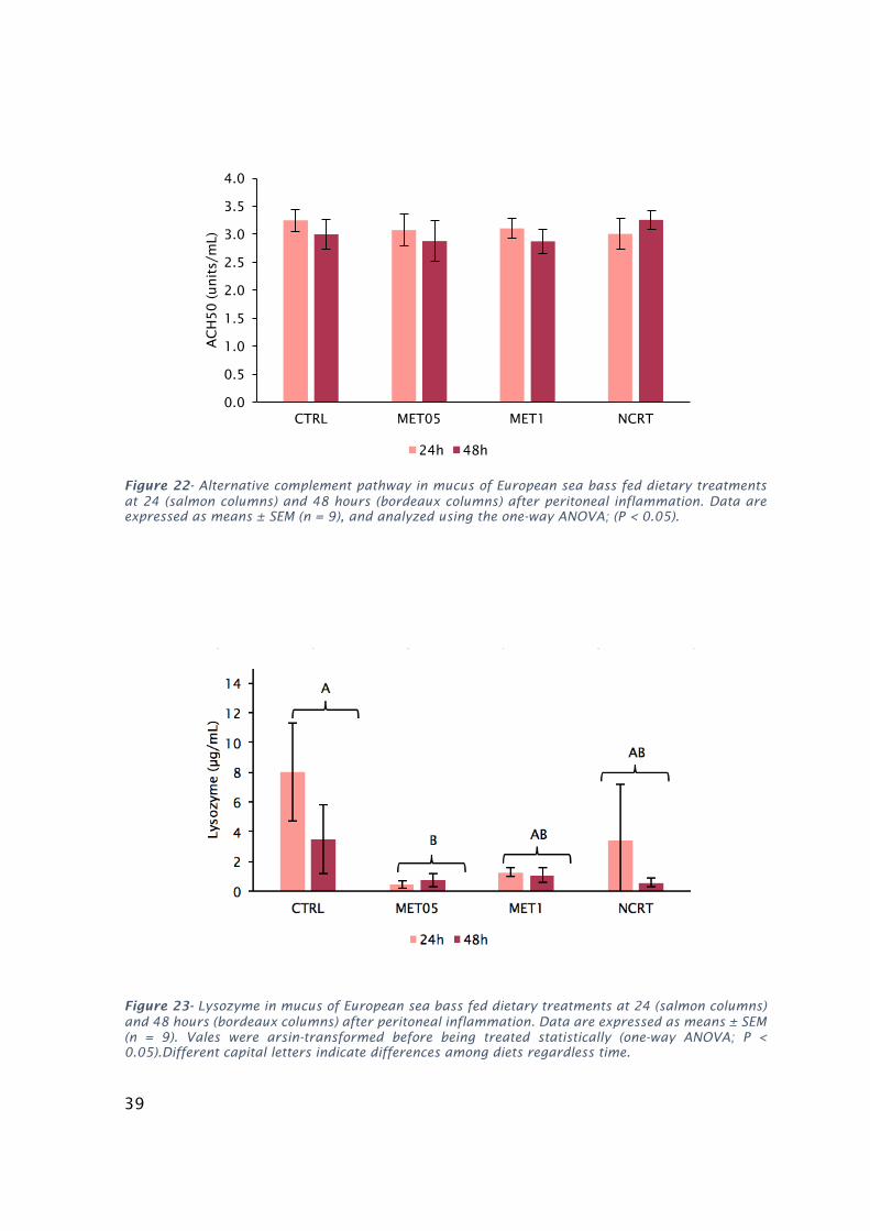
39
Figure 22- Alternative complement pathway in mucus of European sea bass fed dietary treatments at 24 (salmon columns) and 48 hours (bordeaux columns) after peritoneal inflammation. Data are expressed as means ± SEM (n = 9), and analyzed using the one-way ANOVA; (P < 0.05).
Figure 23- Lysozyme in mucus of European sea bass fed dietary treatments at 24 (salmon columns) and 48 hours (bordeaux columns) after peritoneal inflammation. Data are expressed as means ± SEM (n = 9). Vales were arsin-transformed before being treated statistically (one-way ANOVA; P < 0.05).Different capital letters indicate differences among diets regardless time.
0.0
0.5
1.0
1.5
2.0
2.5
3.0
3.5
4.0
CTRL MET05 MET1 NCRT
AC
H50 (unit
s/m
L)
24h 48h
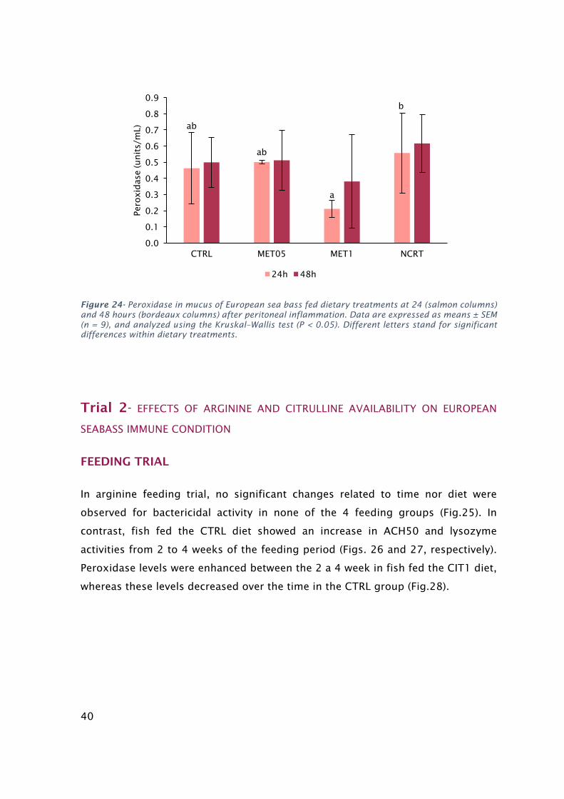
40
Figure 24- Peroxidase in mucus of European sea bass fed dietary treatments at 24 (salmon columns) and 48 hours (bordeaux columns) after peritoneal inflammation. Data are expressed as means ± SEM (n = 9), and analyzed using the Kruskal–Wallis test (P < 0.05). Different letters stand for significant differences within dietary treatments.
Trial 2- EFFECTS OF ARGININE AND CITRULLINE AVAILABILITY ON EUROPEAN
SEABASS IMMUNE CONDITION
FEEDING TRIAL
In arginine feeding trial, no significant changes related to time nor diet were
observed for bactericidal activity in none of the 4 feeding groups (Fig.25). In
contrast, fish fed the CTRL diet showed an increase in ACH50 and lysozyme
activities from 2 to 4 weeks of the feeding period (Figs. 26 and 27, respectively).
Peroxidase levels were enhanced between the 2 a 4 week in fish fed the CIT1 diet,
whereas these levels decreased over the time in the CTRL group (Fig.28).
ab
ab
a
b
0.0
0.1
0.2
0.3
0.4
0.5
0.6
0.7
0.8
0.9
CTRL MET05 MET1 NCRT
Pero
xid
ase
(unit
s/m
L)
24h 48h

41
Figure 25 - Bactericidal activity in mucus of European sea bass fed different diets for 2 (salmon columns) and 4 (bordeaux columns) weeks. Data are expressed as means ± standard error of the mean (SEM) (n = 12). Vales were cos-transformed before being treated statistically (one-way ANOVA; P < 0.05).
Figure 26 - Alternative complement pathway in mucus of European sea bass fed different diets for 2 (salmon columns) and 4 (bordeaux columns) weeks. Data are expressed as means ± SEM (n = 12), and analyzed using the Kruskal–Wallis test (P < 0.05). Different symbols indicate significant differences attributed to time.
0
10
20
30
40
50
60
70
CTRL ARG1 ARG2 CIT1
Bac
teri
cidal
Act
ivit
y (%
)
2 weeks 4 weeks
#*
0
1
2
3
4
5
6
CTRL ARG1 ARG2 CIT1
AC
H50 (unit
s/m
L)
2 weeks 4 weeks

42
Figure 27 - Lysozyme in mucus of European sea bass fed different diets for 2 (salmon columns) and 4 (bordeaux columns) weeks. Data are expressed as means ± SEM (n = 12), and analyzed using the Kruskal–Wallis test (P < 0.05). Different symbols indicate significant differences attributed to time.
Figure 28 - Peroxidase in mucus of European sea bass fed different diets for 2 (salmon columns) and 4 (bordeaux columns) weeks. Data are expressed as means ± SEM (n = 12), and analyzed using the Kruskal–Wallis test (P < 0.05). Different symbols indicate significant differences attributed to time.
# #*
*
0.00
0.20
0.40
0.60
0.80
1.00
1.20
1.40
1.60
CTRL ARG1 ARG2 CIT1
Pero
xid
ase
(unit
s/m
L)
2 weeks 4 weeks
#
*
0.00
10.00
20.00
30.00
40.00
50.00
60.00
70.00
CTRL ARG1 ARG2 CIT1
Lyso
zym
e (μ
g/m
L)
2 weeks 4 weeks
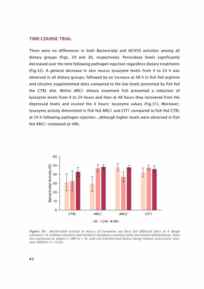
43
TIME-COURSE TRIAL
There were no differences in both Bactericidal and ACH50 activities among all
dietary groups (Figs. 29 and 30, respectively). Peroxidase levels significantly
decreased over the time following pathogen injection regardless dietary treatments
(Fig.32). A general decrease in skin mucus lysozyme levels from 4 to 24 h was
observed in all dietary groups, followed by an increase at 48 h in fish fed arginine
and citruline supplemented diets compared to the low levels presented by fish fed
the CTRL diet. Within ARG1 dietary treatment fish presented a reduction of
lysozyme levels from 4 to 24 hours and then at 48 hours they recovered from the
depressed levels and exceed the 4 hours’ lysozyme values (Fig.31). Moreover,
lysozyme activity diminished in fish fed ARG1 and CIT1 compared to fish fed CTRL
at 24 h following pathogen injection , although higher levels were observed in fish
fed ARG1 compared at 48h.
Figure 29 - Bactericidal activity in mucus of European sea bass fed different diets at 4 (beige columns), 24 (salmon columns) and 48 hours (bordeaux columns) after peritoneal inflammation. Data are expressed as means ± SEM (n = 9), and cos-transformed before being treated statistically (one-way ANOVA; P < 0.05).
0
10
20
30
40
50
60
CTRL ARG1 ARG2 CIT1
Bac
teri
cidal
Act
ivit
y (%
)
4h 24h 48h

44
Figure 30 - Alternative complement pathway in mucus of European sea bass fed different diets at 4 (beige columns), 24 (salmon columns) and 48 hours (bordeaux columns) after peritoneal inflammation. Data are expressed as means ± SEM (n = 9), and analyzed using the Kruskal–Wallis test (P < 0.05).
Figure 31 - Lysozyme in mucus of European sea bass fed different diets at 4 (beige columns), 24 (salmon columns) and 48 hours (bordeaux columns) after peritoneal inflammation. Data are expressed as means ± SEM (n = 9), and analyzed using the Kruskal–Wallis test (P < 0.05). Different letters stand for significant differences within dietary treatments, while different symbols indicate significant differences attributed to time.
0
2
4
6
8
10
12
CTRL ARG1 ARG2 CIT1
AC
H5
0 (unit
s/m
L)
4h 24h 48h
*
**#
b*#
a #ab a #a #
b *
ab
ab*
0
2
4
6
8
10
12
14
16
CTRL ARG1 ARG2 CIT1
Lyso
zym
e (µ
g/m
L)
4h 24h 48h
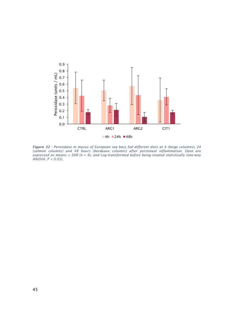
45
Figure 32 - Peroxidase in mucus of European sea bass fed different diets at 4 (beige columns), 24 (salmon columns) and 48 hours (bordeaux columns) after peritoneal inflammation. Data are expressed as means ± SEM (n = 9), and Log-transformed before being treated statistically (one-way ANOVA; P < 0.05).
0.0
0.1
0.2
0.3
0.4
0.5
0.6
0.7
0.8
0.9
CTRL ARG1 ARG2 CIT1
Pero
xid
ase
(unit
s /
mL)
4h 24h 48h

46
DISCUSSION
Trial 1 - EFFECTS OF METHIONINE AVAILABILITY ON EUROPEAN SEABASS IMMUNE
CONDITION
Over the last years, there was a growing interest in the inclusion of AA in fish
functional feeds, aiming an increase of fish health status, which in turn may allow
a higher disease resistance. (Andersen, Waagbø, & Espe, 2015). Apart from being
an indispensable AA, crucial for fish as energy substrate and protein synthesis,
methionine participates in several metabolic pathways with an immune modulatory
action. Moreover, this AA is available as feed grade, thus its inclusion in aquafeeds
may be obtained at reduced costs (Machado et al., 2015). Taking into account this
considerations, several studies were performed, proving methionine’s immune
status enhancing effect in mammals (Rubin et al., 2007) as well as in fish (Tang et
al., 2009). In the present work, fish were fed diets supplemented with methionine
levels above the requirement for normal growth for European seabass during 2 and
4 weeks, and also following stimulation with bacteria. To the best of our
knowledge, no published work showing the modulatory effects of methionine
supplementation on the skin mucus immunity in European seabass is available in
the literature.
Regarding the feeding trial, in the absence of infection, dietary methionine appears
to have no significant effects on the bactericidal, alternative complement pathway
and peroxidase activities. Although not statistically significant, bactericidal activity
presented a generally trend to increase over time, so did peroxidase activity, which
was significantly higher at the end the feeding trial, regardless of the diet. The
enhanced immune effect along time is possibly linked to the fish development and
habituation process to the system. Still, the lack of differences among the innate
immune parameters measured, between dietary treatments, seems to be linked to
effects on skin mucus, since published literature points to a beneficial effect of
methionine supplementation during a systemic immune response (Kuang et al.,
2012). A possible reason to justify the lack of changes in lysozyme and peroxidase
levels may be the absence of an immunological challenge. The presence of a
pathogen induces the immune response involving the activation of effector cells
such as neutrophils, leading to its consequently degranulation and release of
47
peroxidase in the local. In fact, lysozyme levels are known to increase in fish
subjected to an inflammatory agent (Ellis, 2001).
After 4 weeks of feeding trial fish fed diet deficient in methionine, presented a
dramatically drop on the mucosal lysozyme levels. The absence of methionine in
fish nutrition compromised the lysozyme’s production, even without an immune
stimulation. This observation shows the role of methionine as an essential and a
limiting AA in fish.
At the end of the 4 weeks of the feeding period, live bacteria were injected in the
peritoneal cavity, and dietary modulation in response to an inflammatory stimulus
was studied. Methionine is the most common methyl donor in the body, making it
essential for cell division events and polyamines synthesis (Grimble, Poshoi, al., &
Lauterberg, 1996). However, the current study showed a general inhibitory effect
of methionine in mucosal lysozyme and peroxidase values in the time-course trial.
At first sight, this may seem contradictory to several other studies, where
methionine supplementation followed by bacterial insult, resulted in increased
numbers of white blood cells, triggered by the cell proliferation effect of this AA
(Kuang et al., 2012). Increased plasma leucocytes numbers, fueled by methionine
surplus, should be translated in increased plasma peroxidase and lysozyme activity
upon an infection. In fact, Costas et al. 2013 showed that increased lysozyme and
peroxidase levels is directly related with neutrophil numbers in Senegalese sole,
since these enzymes are generated by neutrophils in response to an inflammatory
stimuli (Murray & Fletcher, 1976). In contrast to what has been published for
plasma innate parameters, in this work we observed minimum mucus lysozyme
activity in methionine fed groups, also, peroxidase values were the lowest in fish
fed MET1 diet. The opposite results observed in mucus, may reflect the initial
course of an immune response, where leucocytes are recruited to the inflammatory
focus from the peripheral tissues (Kuang et al., 2012), such as skin. However, to
prove this hypothesis, plasma and peritoneal exudate samples should had been
performed to ensure that the decline of innate immunity in mucus was followed by
an increase of those parameters in the plasma and in peritoneal cavity.
These hypothesis is in agreement with a work conducted with European seabass
fed methionine diet, in which Machado et al. 2015 observed a stronger humoral
and cellular response in both peripheral blood and inflammatory focus, after
injection of Phdp. In the later study, an increased plasma peroxidase and lysozyme
activity was reported, and higher plasma leucocytes counts registered, for fish fed
48
methionine supplemented diet, suggesting a lymphocyte migration to the
inflammatory focus favored by methionine supplementation.
Several lines of evidence have reported the importance of skin as a peripheral
immune organ in teleosts, due to not only the presence of bioactive molecules but
also, due to the process of epidermal migration of leucocytes to the site of
inflammation (Kania, Evensen, Larsen, & Buchmann, 2010). During an infection, a
deviation of metabolic energy from peripheral tissue to the inflammatory focus is
typically observed. Upregulation events normally take place during the initial
response, generating pro-inflammatory molecules which contribute to a fast
neutrophils recruitment (Esteban 2012). The role of fish skin as a source of these
molecules has been confirmed (Gonzalez, Buchmann, & Nielsen, 2007), and in
2012 Lü et al. 2012 showed high expression of genes involved in leucocyte
mobilization in zebra fish skin infected with the bacterium Citrobacter freundii.
Together, these reports support that reduction of mucus immunity factors is being
caused by the influx of neutrophils into the peritoneum via circulation, enhanced
by methionine.
Trial 2- EFFECTS OF ARGININE AND CITRULLINE AVAILABILITY ON EUROPEAN
SEABASS IMMUNE CONDITION
During the last decade, several studies have been focused in arginine application
in functional diets, given its modulatory potential of both innate and adaptive
immune systems in fish. Although efforts were made, the results obtained were
often controversy and the fully mechanism of arginine immune-modulation is not
yet understood. In the present work, fish were fed diets supplemented with
arginine and citrulline (an arginine precursor) levels above the requirement for
normal growth for European seabass in a feeding trial basis (i.e. 2 and 4 weeks),
as well as following stimulation with bacteria. While the effects of arginine-enriched
diet on plasma innate immunity has been reported for this species (Azeredo et al.,
2015), this is the first work showing the modulatory effects of arginine and
citrulline supplementation on the skin mucus immunity in European seabass.
The results of feeding trial revealed a beneficial effect upon arginine increment in
peroxidase and bactericidal activities. Although not in a significant fashion,
49
supplementation of 0.5 and 1 % of ARG appears to improve mucus bactericidal
capacity during the feeding trial, whereas no changes regarding this parameter
were observed for fish fed the control diet during the trial period (4 weeks). The
arginine´s immune stimulating effect suggested, is congruent with the results
obtained for peroxidase values. Mucosal peroxidase values presented a clear
tendency to rise over time in all supplemented groups, in contrast with fish fed the
CTRL diet, which followed the opposite trend with a significant decline of
peroxidase activity between 2 and 4 weeks. Arginine’s positive influence in fish
immune status could be related to polyamines biosynthesis. Polyamines, important
products of arginine’s metabolic pathway, are key molecules for leucocyte
differentiation and proliferation (Andersen et al., 2013). As activated leucocytes
release bioactive molecules, dietary arginine surplus may have benefited fish with
extra humoral defences to better cope with an invading bacterium. This hypothesis
is consistent with previous works where arginine supplementation was reported to
increase leukocyte counts of juvenile Jian carp (Cyprinus carpio var. Jian) (G. Chen
et al., 2015) and to stimulate the proliferation of channel catfish B lymphocytes in
vitro (Pohlenz, Buentello, Mwangi, et al., 2012).
Under an infection condition in the present study, when bacterium was injected
into peritoneal cavity, peroxidase levels were found to decrease over time, in all
dietary groups. The declining peroxidase activity, independently of diet are
probably a reflection of an obligatory neutrophil migration, a characteristic
situation during the acute phase of an inflammatory response. This results suggest
that the mucosal immune parameters are being deviated towards systemic
immunity, regardless of nutritional stimuli.
In contrast, lysozyme levels behaved differently upon dietary administration. In all
supplemented groups, a sharp reduction of these parameter was observed between
4 and 24 hours after infection with bacteria. However, fish fed dietary amino acids
surplus showed a recovery in those levels at 48 hours following injection, as
lysozyme activity improved and exceed values observed at 4 h following infection.
The lysozyme response in supplemented fish stands out by its recuperation
comparing, to what is observed in control group, where the enzyme’s
concentration kept declining alongside the development of the infection. The
differences observed between the concentration of mucosal peroxidase and
lysozyme activities in the time course trial, may rely on the source of these two
enzymes. In fact, fish lysozyme may occur in various leukocytes, such as
50
neutrophils, monocytes and macrophages (Saurabh & Sahoo, 2008); unlike
peroxidase, which is exclusively produced by neutrophils. The minimum lysozyme
values observed at 24 hours, corresponding to the peak of the inflammatory
response, may be partly attributed to neutrophils migration in response to
inflammatory stimuli. However, lysozyme production is reestablished at 48 hours
in fish fed supplemented diets, despite the neutrophils recruitment to
inflammatory focus. This is probably due to activated macrophages present in the
skin mucus, fueled by arginine (Saurabh & Sahoo, 2008). This hypothesis is
consistent with previous works reporting that arginine levels are related to
macrophages activity (Pohlenz, Buentello, Mwangi, et al., 2012) (Q. Zhou et al.,
2014). Being a peripheral immune organ, far from the infection site, it possible that
after 48 hours the skin mucus may have achieved restoration of lysozyme’s activity
more efficiently under arginine’s supplementation. However, more studies are
needed to confirm this pattern. Additionally, in future assays more sampling times
should be performed after a bacterial insult, to follow both systemic and mucosal
immune progression. Especially in the cases where the pathogen is activated, since
fish requires more than 48h to solve the infection.
Simultaneously to mucus sampling, it would had been interesting to monitor
plasma and peritoneal immune parameters to infer about leucocytes migrations,
which would allow a better understanding of the evolution of the infection at a local
level.
The overall results in both trials showed that mucus sampling is not reliable
biomarker to distinguish between normal or disease status, as it does not clearly
translate fish internal condition. The hematological information provided by blood
samples cannot be replaced, however, as peripheral immune organ, great variation
on mucus humoral profile may indicate a depressed immune status. Therefore,
mucus sampling methods can be used as a routine procedure in aquaculture
industries as it is easy, non-evasive and stress-free technique.
Probably, in the presence of an external infection, the mucus response would had
been much more intense, consequently the supplementation effect much more
visible.
51
Conclusions
The supplementation level of the studied AA was not enough to allow a strong
influence in such a far peripheral tissue, as the skin mucus.
In this present study, no major variations in the skin mucus immunity in response
to a peritoneal inflammation are detectable upon both dietary supplementations.
However, given the overall results, the AA studied appear to divert peripheral
metabolic energy towards the infectious site. Likewise, in the case of a bacteria
whose route of infection was external, the previously described recruitment
mechanisms, would be possibly occurring on epidermis tissue. Indeed, it would be
interesting to test whether arginine, citruline or methionine would promote an
enhanced cellular recruitment during a bath infection with the same pathogen. If
this hypothesis is confirmed, fish would benefit from the incorporation of this AA
in functional feeds, for periods previously known to be more susceptible to an
outbreak of any skin infection.

52
References
Andersen, S. M., Holen, E., Aksnes, A., Rønnestad, I., Zerrahn, J.-E., & Espe, M.
(2013). Dietary arginine affects energy metabolism through polyamine
turnover in juvenile Atlantic salmon (Salmo salar). The British Journal of
Nutrition, 110(11), 1968–77. https://doi.org/10.1017/S0007114513001402
Andersen, S. M., Waagbø, R., & Espe, M. (2015). Functional amino acids in fish
nutrition, health and welfare. Frontiers in Bioscience (Elite Edition), 8(1), 143–
169.
Ángeles Esteban, M. (2012). An Overview of the Immunological Defenses in Fish
Skin. ISRN Immunology, 2012, 1–29. https://doi.org/10.5402/2012/853470
Azeredo, R., Pérez-Sánchez, J., Sitjà-Bobadilla, A., Fouz, B., Tort, L., Aragão, C., …
Costas, B. (2015). European sea bass (Dicentrarchus labrax) immune status
and disease resistance are impaired by Arginine dietary supplementation. PLoS
ONE, 10(10), 1–19. https://doi.org/10.1371/journal.pone.0139967
Basurco, B. (2000). Oddshore Mariculture in Mediterranean Contries. Options
Me ́diterrane ́ennes Se ́rie B. Zaragoza, Spain., 9–18.
Batista, M. A., Nicoli, J. R., dos Santos Martins, F., Nogueira Machado, J. A., Esteves
Arantes, R. M., Pacífico Quirino, I. E., … Cardoso, V. N. (2011). Pretreatment
With Citrulline Improves Gut Barrier After Intestinal Obstruction in Mice.
Journal of Parenteral and Enteral Nutrition, 36(1), 69–76.
https://doi.org/10.1177/0148607111414024
Breuillard, C., Bonhomme, S., Couderc, R., Cynober, L., & De Bandt, J.-P. (2014). In
vitro anti-inflammatory effects of citrulline on peritoneal macrophages in
Zucker diabetic fatty rats. The British Journal of Nutrition, (2015), 1–5.
https://doi.org/10.1017/S0007114514002086
Buentello, J. A., & Gatlin, D. M. (2001). Effects of elevated dietary arginine on
resistance of channel catfish to exposure to Edwardsiella ictaluri. Journal of
Aquatic Animal Health, 13(3), 194–201. https://doi.org/10.1577/1548-
8667(2001)013<0194:EOEDAO>2.0.CO;2
Buentello, J. A., Reyes-Becerril, M., Romero-Geraldo, M. D. J., & Ascencio-Valle, F. D.
J. (2007). Effects of dietary arginine on hematological parameters and innate
immune function of channel catfish. Journal of Aquatic Animal Health, 19(3),
53
195–203. https://doi.org/10.1577/H07-004.1
Buentello, J. a, & Gatlin Iii, D. M. (1999). Nitric oxide production in activated
macrophages from channel catfish (Ictalurus punctatus): influence of dietary
arginine and culture media. Aquaculture, 179(1–4), 513–521.
https://doi.org/10.1016/S0044-8486(99)00184-2
Chen, G., Liu, Y., Jiang, J., Jiang, W., Kuang, S., Tang, L., … Feng, L. (2015). Effect
of dietary arginine on the immune response and gene expression in head
kidney and spleen following infection of Jian carp with Aeromonas hydrophila.
Fish and Shellfish Immunology, 44(1), 195–202.
https://doi.org/10.1016/j.fsi.2015.02.027
Chen, Q., Zhao, H., Huang, Y., Cao, J., Wang, G., Sun, Y., & Li, Y. (2016). Effects of
dietary arginine levels on growth performance, body composition, serum
biochemical indices and resistance ability against ammonia-nitrogen stress in
juvenile yellow catfish (Pelteobagrus fulvidraco). Animal Nutrition, 1–6.
https://doi.org/10.1016/j.aninu.2016.07.001
Cheng, Z., Buentello, A., & Gatlin, D. M. (2011). Effects of dietary arginine and
glutamine on growth performance, immune responses and intestinal structure
of red drum, Sciaenops ocellatus. Aquaculture, 319(1–2), 247–252.
https://doi.org/10.1016/j.aquaculture.2011.06.025
Cheng, Z., Gatlin, D. M., & Buentello, A. (2012). Dietary supplementation of arginine
and/or glutamine influences growth performance, immune responses and
intestinal morphology of hybrid striped bass (Morone chrysops??Morone
saxatilis). Aquaculture, 362–363, 39–43.
https://doi.org/10.1016/j.aquaculture.2012.07.015
Conceição, L. E. C., Aragão, C., Dias, J., Costas, B., Terova, G., Martins, C., & Tort,
L. (2012). Dietary nitrogen and fish welfare. Fish Physiology and Biochemistry,
38(1), 119–141. https://doi.org/10.1007/s10695-011-9592-y
Costas, B., Aragão, C., Soengas, J. L., Míguez, J. M., Rema, P., Dias, J., … Conceição,
L. E. C. (2012). Effects of dietary amino acids and repeated handling on stress
response and brain monoaminergic neurotransmitters in Senegalese sole
(Solea senegalensis) juveniles. Comparative Biochemistry and Physiology - A
Molecular and Integrative Physiology, 161(1), 18–26.
https://doi.org/10.1016/j.cbpa.2011.08.014
Costas, B., Conceição, L. E. C., Dias, J., Novoa, B., Figueras, A., & Afonso, A. (2011).
Dietary arginine and repeated handling increase disease resistance and

54
modulate innate immune mechanisms of Senegalese sole (Solea senegalensis
Kaup, 1858). Fish and Shellfish Immunology, 31(6), 838–847.
https://doi.org/10.1016/j.fsi.2011.07.024
Costas, B., Rêgo, P. C. N. P., Conceição, L. E. C., Dias, J., & Afonso, A. (2013). Dietary
arginine supplementation decreases plasma cortisol levels and modulates
immune mechanisms in chronically stressed turbot (Scophthalmus maximus).
Aquaculture Nutrition, 19(SUPL.1), 25–38.
https://doi.org/10.1111/anu.12086
do Vale, A., Costa-Ramos, C., Silva, A., Silva, D. S. P., Gärtner, F., dos Santos, N. M.
S., & Silva, M. T. (2007). Systemic macrophage and neutrophil destruction by
secondary necrosis induced by a bacterial exotoxin in a Gram-negative
septicaemia. Cellular Microbiology, 9(4), 988–1003.
https://doi.org/10.1111/j.1462-5822.2006.00846.x
Do Vale, A., Marques, F., & Silva, M. T. (2003). Apoptosis of sea bass (Dicentrarchus
labrax L.) neutrophils and macrophages induced by experimental infection
with Photobacterium damselae subsp. piscicida. Fish and Shellfish
Immunology, 15(2), 129–144. https://doi.org/10.1016/S1050-
4648(02)00144-4
Do Vale, A., Silva, M. T., Dos Santos, N. M. S., Nascimento, D. S., Reis-Rodrigues,
P., Costa-Ramos, C., … Azevedo, J. E. (2005). AIP56, a novel plasmid-encoded
virulence factor of Photobacterium damselae subsp. piscicida with
apoptogenic activity against sea bass macrophages and neutrophils. Molecular
Microbiology, 58(4), 1025–1038. https://doi.org/10.1111/j.1365-
2958.2005.04893.x
Ellis, A. E. (1990). Serum antiproteases in fish. Techniques in Fish Immunology.
Ellis, A. E. (2001). Innate host defense mechanisms of fish against viruses and
bacteria. Developmental & Comparative Immunology, 25(8), 827–839.
https://doi.org/https://doi.org/10.1016/S0145-305X(01)00038-6
FAO. (2012). Dicentrarchus labrax, 1–12.
FAO. (2016). The State of World Fisheries and Aquaculture 2016. Contributing to
food security and nutrition for all. Rome.
Feng, F., Qing-hui, A., Wei, X., Kang-sen, M., & Wen-bing, Z. (2011). Effects of
dietary arginine and lysine on growth and non-specific immune responses of
juvenile darkbarbel catfish (Pelteobagrus vachelli). Ocean University of China.
Gonzalez, S. F., Buchmann, K., & Nielsen, M. E. (2007). Real-time gene expression
55
analysis in carp (Cyprinus carpio L.) skin: Inflammatory responses caused by
the ectoparasite Ichthyophthirius multifiliis. Fish and Shellfish Immunology,
22(6), 641–650. https://doi.org/10.1016/j.fsi.2006.08.011
Grimble, R. F., Grimble, G. K., Poshoi, L., al., et, & Lauterberg, B. H. (1996).
Immunonutrition: role of sulfur amino acids, related amino acids, and
polyamines. Nutrition (Burbank, Los Angeles County, Calif.), 14(7–8), 605–10.
https://doi.org/10.1016/S0899-9007(98)80041-5
Jiang, J., Shi, D., Zhou, X. Q., Hu, Y., Feng, L., Liu, Y., … Zhao, Y. (2015). Invitro and
invivo protective effect of arginine against lipopolysaccharide induced
inflammatory response in the intestine of juvenile Jian carp (Cyprinus carpio
var. Jian). Fish and Shellfish Immunology, 42(2), 457–464.
https://doi.org/10.1016/j.fsi.2014.11.030
Kania, P. W., Evensen, O., Larsen, T. B., & Buchmann, K. (2010). Molecular and
immunohistochemical studies on epidermal responses in Atlantic salmon
Salmo salar L. induced by Gyrodactylus salaris Malmberg, 1957. Journal of
Helminthology, 84(2), 166–172.
https://doi.org/10.1017/S0022149X09990460
Kaushik, S. (1998). Whole body amino acid composition of European seabass (
Dicentrarchus labrax ), gilthead seabream ( Sparus aurata ) and turbot { Psetta
maxima ) with an estimation of their IAA requirement profiles, 11(5), 355–358.
Kiron, V. (2012). Fish immune system and its nutritional modulation for preventive
health care. Animal Feed Science and Technology, 173(1–2), 111–133.
https://doi.org/10.1016/j.anifeedsci.2011.12.015
Kuang, S. Y., Xiao, W. W., Feng, L., Liu, Y., Jiang, J., Jiang, W. D., … Zhou, X. Q.
(2012). Effects of graded levels of dietary methionine hydroxy analogue on
immune response and antioxidant status of immune organs in juvenile Jian
carp (Cyprinus carpio var. Jian). Fish and Shellfish Immunology, 32(5), 629–
636. https://doi.org/10.1016/j.fsi.2011.12.012
Li, P., Mai, K., Trushenski, J., & Wu, G. (2009). New developments in fish amino acid
nutrition: Towards functional and environmentally oriented aquafeeds. Amino
Acids, 37(1), 43–53. https://doi.org/10.1007/s00726-008-0171-1
Li, P., Yin, Y.-L., Li, D., Kim, S. W., & Wu, G. (2007). Amino acids and immune
function. The British Journal of Nutrition, 98(2), 237–252.
https://doi.org/10.1017/S000711450769936X
Lin, H., Tan, X., Zhou, C., Niu, J., Xia, D., Huang, Z., … Wang, Y. (2015). Effect of
56
dietary arginine levels on the growth performance, feed utilization, non-
specific immune response and disease resistance of juvenile golden pompano
Trachinotus ovatus. Aquaculture, 437, 382–389.
https://doi.org/10.1016/j.aquaculture.2014.12.025
Lopes, A. S. de C. (2016). From consumption to production – development of
fisheries and aquaculture in Portugal. Retrieved from
http://run.unl.pt/handle/10362/19237
Lü, A., Hu, X., Xue, J., Zhu, J., Wang, Y., & Zhou, G. (2012). Gene expression profiling
in the skin of zebrafish infected with Citrobacter freundii. Fish and Shellfish
Immunology, 32(2), 273–283. https://doi.org/10.1016/j.fsi.2011.11.016
Machado, M., Azeredo, R., D??az-Rosales, P., Afonso, A., Peres, H., Oliva-Teles, A.,
& Costas, B. (2015). Dietary tryptophan and methionine as modulators of
European seabass (Dicentrarchus labrax) immune status and inflammatory
response. Fish and Shellfish Immunology, 42(2), 353–362.
https://doi.org/10.1016/j.fsi.2014.11.024
Magarinos, B., Toranzo, A. E., & Romalde, J. L. (1997). Phenotypic and
Pathobiological, 6(1), 41–64.
Métayer, S., Seiliez, I., Collin, A., Duchêne, S., Mercier, Y., Geraert, P.-A., &
Tesseraud, S. (2008). Mechanisms through which sulfur amino acids control
protein metabolism and oxidative status. The Journal of Nutritional
Biochemistry, 19(4), 207–15. https://doi.org/10.1016/j.jnutbio.2007.05.006
Mommsen, T., Moon, T., & Plisetskaya, E. (2001). Effects of arginine on pancreatic
hormones and hepatic metabolism in rainbow trout. Physiol Biochem Zool, 74,
668–678.
Moretti, A., Fernadez-Criado, M. P., Cittolin, G., & Guidastri, R. (1999). Manual on
hatchery production of sea sass and gilthead seabream. Fao, 1, 194.
Murray, C. K., & Fletcher, T. C. (1976). The immunohistochemical localization of
lysozyme in plaice (Pleuronectes platessa L.) tissues. Journal of Fish Biology,
9(4), 329–334. https://doi.org/10.1111/j.1095-8649.1976.tb04681.x
Nieves, C., & Langkamp-Henken, B. (2002). Arginine and immunity: A unique
perspective. Biomedicine and Pharmacotherapy, 56(10), 471–482.
https://doi.org/10.1016/S0753-3322(02)00291-3
Pickering, A. D. (1974). The distribution of mucous cells in the epidermis of the
brown trout Salmo trutta (L.) and the char Salvelinus alpinus (L.). Journal of
Fish Biology, 6(2), 111–118. https://doi.org/10.1111/j.1095-
57
8649.1974.tb04531.x
Pohlenz, C., Buentello, A., Criscitiello, M. F., Mwangi, W., Smith, R., & Gatlin, D. M.
(2012). Synergies between vaccination and dietary arginine and glutamine
supplementation improve the immune response of channel catfish against
Edwardsiella ictaluri. Fish and Shellfish Immunology, 33(3), 543–551.
https://doi.org/10.1016/j.fsi.2012.06.005
Pohlenz, C., Buentello, A., Mwangi, W., & Gatlin, D. M. (2012). Arginine and
glutamine supplementation to culture media improves the performance of
various channel catfish immune cells. Fish & Shellfish Immunology, 32(5), 762–
768. https://doi.org/10.1016/j.fsi.2012.01.029
Pohlenz, C., & Gatlin, D. M. (2014). Interrelationships between fish nutrition and
health. Aquaculture, 431, 111–117.
https://doi.org/10.1016/j.aquaculture.2014.02.008
Quade, M. J., & Roth, J. A. (1997). A rapid, direct assay to measure degranulation
of bovine neutrophil primary granules. Veterinary Immunology and
Immunopathology, 58(3), 239–248.
https://doi.org/http://dx.doi.org/10.1016/S0165-2427(97)00048-2
Ramalho, A., Dinis, M. T., & Ccmar-cimar LA. (2011). Portuguese aquaculture :
Current status and future perspectives. World Aquaculture, 42(1), 26–32.
Ren, M., Liao, Y., Xie, J., Liu, B., Zhou, Q., Ge, X., … Chen, R. (2013). Dietary arginine
requirement of juvenile blunt snout bream, Megalobrama amblycephala.
Aquaculture, 414–415, 229–234.
https://doi.org/10.1016/j.aquaculture.2013.08.021
Romalde, J. L. (2002). Photobacterium damselae subsp. piscicida: An integrated
view of a bacterial fish pathogen. International Microbiology, 5(1), 3–9.
https://doi.org/10.1007/s10123-002-0051-6
Rubin, L. L., Canal, C. W., Ribeiro, A. L. M., Kessler, A., Silva, I., Trevizan, L., … Kras,
R. (2007). Effects of Methionine and Arginine Dietary Levels on the Immunity
of Broiler Chickens Submitted to Immunological Stimuli. Brazilian Journal of
Poultry Science, 9(4), 241–247. https://doi.org/10.1590/S1516-
635X2007000400006
Salinas, I. (2015). The Mucosal Immune System of Teleost Fish. Biology, 4(3), 525–
39. https://doi.org/10.3390/biology4030525
Salinas, I., Zhang, Y. A., & Sunyer, J. O. (2011). Mucosal immunoglobulins and B
cells of teleost fish. Developmental and Comparative Immunology, 35(12),

58
1346–1365. https://doi.org/10.1016/j.dci.2011.11.009
Saurabh, S., & Sahoo, P. K. (2008). Lysozyme: An important defence molecule of
fish innate immune system. Aquaculture Research, 39(3), 223–239.
https://doi.org/10.1111/j.1365-2109.2007.01883.x
Shephard, K. L. (1994). Functions for fish mucus. Reviews in Fish Biology and
Fisheries, 4(4), 401–429. https://doi.org/10.1007/BF00042888
Stevens, M. G., Kehrli, M. E., & Canning, P. C. (1991). A colorimetric assay for
quantitating bovine neutrophil bactericidal activity. Veterinary Immunology
and Immunopathology, 28(1), 45–56. https://doi.org/10.1016/0165-
2427(91)90042-B
Subramanian, S., MacKinnon, S. L., & Ross, N. W. (2007). A comparative study on
innate immune parameters in the epidermal mucus of various fish species.
Comparative Biochemistry and Physiology - B Biochemistry and Molecular
Biology, 148(3), 256–263. https://doi.org/10.1016/j.cbpb.2007.06.003
Sunyer, J. O., & Tort, L. (1995). Natural hemolytic and bactericidal activities of sea
bream Sparus aurata serum are effected by the alternative complement
pathway. Veterinary Immunology and Immunopathology, 45(3–4), 333–345.
Tang, L., Wang, G. X., Jiang, J., Feng, L., Yang, L., Li, S. H., … Zhou, X. Q. (2009).
Effect of methionine on intestinal enzymes activities, microflora and humoral
immune of juvenile Jian carp (cyprinus carpio var. Jian). Aquaculture Nutrition,
15(5), 477–483. https://doi.org/10.1111/j.1365-2095.2008.00613.x
Wang, W. W., Qiao, S. Y., & Li, D. F. (2009). Amino acids and gut function. Amino
Acids, 37(1), 105–110. https://doi.org/10.1007/s00726-008-0152-4
Wu, G., Bazer, F. W., Davis, T. A., Kim, S. W., Li, P., Marc Rhoads, J., … Yin, Y. (2009).
Arginine metabolism and nutrition in growth, health and disease. Amino Acids,
37(1), 153–168. https://doi.org/10.1007/s00726-008-0210-y
Xie, F., Zeng, W., Zhou, Q., Wang, H., Wang, T., Zheng, C., & Wang, Y. (2012).
Dietary lysine requirement of juvenile Pacific white shrimp, Litopenaeus
vannamei. Aquaculture, 358–359, 116–121.
https://doi.org/10.1016/j.aquaculture.2012.06.027
Zaccone, G., Kapoor, B. G., Fasulo, S., & Ainis, L. (2001). Structural, histochemical
and functional aspects of the epidermis of fishes. In B. T.-A. in M. Biology (Ed.)
(Vol. Volume 40, pp. 253–348). Academic Press.
https://doi.org/http://dx.doi.org/10.1016/S0065-2881(01)40004-6
Zhang, K., Mai, K., Xu, W., Liufu, Z., Zhang, Y., Peng, M., … Ai, Q. (2017). Effects of
59
dietary arginine and glutamine on growth performance , nonspeci fi c
immunity , and disease resistance in relation to arginine catabolism in juvenile
turbot ( Scophthalmus maximus L .). Aquaculture, 468, 246–254.
https://doi.org/10.1016/j.aquaculture.2016.10.021
Zhou, H., Chen, N., Qiu, X., Zhao, M., & Jin, L. (2012). Arginine requirement and
effect of arginine intake on immunity in largemouth bass, Micropterus
salmoides. Aquaculture Nutrition, 18(1), 107–116.
https://doi.org/10.1111/j.1365-2095.2011.00886.x
Zhou, Q. C., Zeng, W. P., Wang, H. L., Xie, F. J., & Zheng, C. Q. (2012). Dietary
arginine requirement of juvenile yellow grouper Epinephelus awoara.
Aquaculture, 350–353, 175–182.
Zhou, Q., Jin, M., Elmada, Z. C., Liang, X., & Mai, K. (2014). Growth, immune
response and resistance to Aeromonas hydrophila of juvenile yellow catfish,
Pelteobagrus fulvidraco, fed diets with different arginine levels.
Aquaculturefile:///Users/MariaCarlota/Documents/Mestrado/Tese/artigos
novos/1-s2.0-S0044848614006486-Main.pdf
file:///Users/MariaCarlota/Documents/Mestrado/Tese/artigos novos/1-s2.0-
S0044848616307736-Main.pdf
file:///Users/MariaCarlota/Documents/Mestrado/, 437, 84–91.
https://doi.org/10.1016/j.aquaculture.2014.11.030