Embed Size (px)
Citation preview

UNIVERSIDADE FEDERAL DE SANTA MARIA
CENTRO DE CIÊNCIAS RURAIS
PROGRAMA DE PÓS-GRADUAÇÃO EM MEDICINA VETERINÁRIA
ASSOCIAÇÃO DE ACHADOS
ULTRASSONOGRÁFICOS E
ANATOMOPATOLÓGICOS DE LESÕES DA
ARTICULAÇÃO METACARPOFALANGEANA
EQUINA
DISSERTAÇÃO DE MESTRADO
Grasiela de Bastiani
Santa Maria, RS, Brasil
2014

ASSOCIAÇÃO DE ACHADOS ULTRASSONOGRÁFICOS E
ANATOMOPATOLÓGICOS DE LESÕES DA ARTICULAÇÃO
METACARPOFALANGEANA EQUINA
Por
Grasiela de Bastiani
Dissertação apresentada ao Curso de Mestrado do Programa de Pós-Graduação
em Medicina Veterinária, Área de Concentração em Cirurgia e Clínica
Médica, da Universidade Federal de Santa Maria (UFSM, RS), como requisito
parcial para obtenção do grau de Mestre em Medicina Veterinária
Orientador: Prof. Flávio Desessards de La Côrte
Santa Maria, RS, Brasil
2014

Ficha Catalográfica

Universidade Federal de Santa Maria
Centro de Ciências Rurais
Programa de Pós-Graduação em Medicina Veterinária
A Comissão Examinadora, abaixo assinada, aprova a Dissertação de
Mestrado
ASSOCIAÇÃO DE ACHADOS ULTRASSONOGRÁFICOS E
ANATOMOPATOLÓGICOS DE LESÕES DA ARTICULAÇÃO
METACARPOFALANGEANA EQUINA
elaborada por
Grasiela De Bastiani
como requisito parcial para obtenção do grau de
Mestre em Medicina Veterinária
COMISSÃO EXAMINADORA:
____________________________________
Flávio de La Côrte, Prof. Dr. (UFSM) (Presidente/Orientador)
_____________________________________
Jarbas Castro Junior, Dr. (Clínica Hípica)
_______________________________
Glaucia Kommers, Prof. Dra. (UFSM)
Santa Maria, 29 de janeiro de 2014

AGRADECIMENTOS
Com todo o meu amor e carinho a minha família que embarcou neste projeto junto comigo.
Pelo apoio e ajuda sem medir esforços, pelas palavras de consolo e incentivo nos momentos
de dúvidas em que, muitas vezes custamos a crer que uma simples ideia poderá dar origem à
inúmeras ideias e a várias respostas. São eles nossos pais que incondicionalmente se tornam
nossos maiores incentivadores.
Ao meu namorado Anibal pela paciência.
Ao meu orientador Flavio de La Côrte o meu obrigado não somente por ter acreditado na
ideia e ter feito inúmeros esforços para que a mesma virasse realidade, mas sim pela amizade,
pelo sentimento de acolhida e generosidade dedicado a cada um de seus orientados.
As professoras Karin Brass e Mara Battistela Rubin por todo o apoio.
Ao Laboratório de Patologia Veterinária em especial a Professora Glaucia Kommers e sua
equipe que com trabalho e dedicação exemplares tornaram este projeto real. A Professora
Glaucia Kommers meu muito obrigado serei uma eterna admiradora do teu trabalho.
A família Shafer que me recebeu em Santa Maria de braços abertos, a sua acolhida, amizade,
a sinceridade em seus sorrisos e com certeza as orações da Dona Lucia.
Aos amigos que a idealização deste projeto me proporcionou, Andressa Shafer, Marilia
Oliveira, Verônica Fernandez, Thirsa Grando, Roberta Pereira, Felipe Libardoni, Liomara
Amaral, Vanessa Gass e Dario Cáceres meu muito obrigada.
As eternas margaridas pela amizade que plantamos e seguimos colhendo até hoje.
Aos colegas de trabalho e amigos Marcos, Miguel , Roberta, Liomara e Gabrielle.
Ao frigorífico Foresta pelo material doado ao experimento e em especial a Dra. Neide Severo
e sua equipe que sempre com muito bom humor me abriram as portas.
A equipe de estagiários do Professor Flavio de La Côrte em especial a Camila Cantarelli,
Amanda Bragatto e Mariana Cocco que também fizeram parte deste projeto.
A equipe do Laboratório Embryolab.
A secretária do PPMG, nossa Maria que sempre muito me ajudou com muito carinho meu
muito obrigado.
RESUMO
Dissertação de Mestrado
Programa de Pós-Graduação em Medicina Veterinária
Universidade Federal de Santa Maria, RS, Brasil
ASSOCIAÇÃO DE ACHADOS ULTRASSONOGRÁFICOS E
ANATOMOPATOLÓGICOS DE LESÕES DA ARTICULAÇÃO
METACARPOFALANGEANA EQUINA AUTORA: GRASIELA DE BASTIANI
ORIENTADOR: FLAVIO DE LA CÔRTE
Data e Local da Defesa: Santa Maria, 29 de janeiro de 2013.
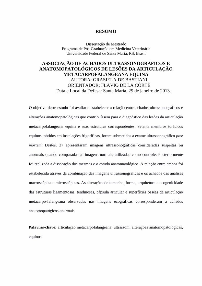
O objetivo deste estudo foi avaliar e estabelecer a relação entre achados ultrassonográficos e
alterações anatomopatológicas que contribuíssem para o diagnóstico das lesões da articulação
metacarpofalangeana equina e suas estruturas correspondentes. Setenta membros torácicos
equinos, obtidos em instalações frigoríficas, foram submetidos a exame ultrassonográfico post
mortem. Destes, 37 apresentaram imagens ultrassonográficas consideradas suspeitas ou
anormais quando comparadas às imagens normais utilizadas como controle. Posteriormente
foi realizada a dissecação dos mesmos e o estudo anatomatológico. A relação entre ambos foi
estabelecida através da combinação das imagens ultrassonográficas e os achados das análises
macroscópica e microscópicas. As alterações de tamanho, forma, arquitetura e ecogenicidade
das estruturas ligamentosas, tendinosas, cápsula articular e superfícies ósseas da articulação
metacarpo-falangeana observadas nas imagens ecográficas corresponderam a achados
anatomopatógicos anormais.
Palavras-chave: articulação metacarpofalangeana, ultrassom, alterações anatomopatológicas,
equinos.

ABSTRACT
Dissertação de Mestrado
Programa de Pós-Graduação em Medicina Veterinária
Universidade Federal de Santa Maria, RS, Brasil
ASSOCIATION OF ULTRASOUND AND ANATOMOPATHOLOGICAL
FINDINGS OF EQUINE METACARPOPHALANGEAL LESIONS AUTHOR: GRASIELA DE BASTIANI
ADVISER: FLAVIO DE LA CÔRTE
Date and Place of Defense: Santa Maria, 29th
january 2013.
In order to assess and establish the association between ultrasonographic and
anatomophatological findings, ultrasonographic examinations were performed, post mortem,
on the fetlock and associated structures of 37 equine forelimb specimens. All this specimens
showed abnormal images or images identified as suspicious on the ultrasound examination.
Subsequently, these limbs were dissected and underwent an anatomopathogical study. The
association between ultrasonographic and anatomopathological findings was established by
comparing data obtained by both methods. Ultrasonographic changes in size, shape,
architecture and echogenicity of ligaments, tendons, joint capsule, articular cartilage and bony
surfaces of the metacarpophalangeal joint were associated with the anatomopathological
findings.
Key words: metacarpophalangeal joint, ultrasound, anatomopathological changes, equine.
SUMÁRIO
1. INTRODUÇÃO.............................................................................................................. 8
2. REVISÃO DE LITERATURA...................................................................................... 9
2.1 Cartilagem e cápsula articular.................................................................................... 9
2.2 Ligamentos colaterais................................................................................................... 10
2.3 Ligamento suspensório................................................................................................. 11
2.4 Ligamento anular palmar............................................................................................ 12
2.5 Tendões flexores............................................................................................................ 13
2.6 Ligamento intersesamoideano ou palmar.................................................................. 15
2.7 Ossos sesamoides proximais......................................................................................... 16
3. ARTIGO 1- ASSOCIATION OF ULTRASOUND AND
ANATOMOPATHOLOGICAL FINDINGS OF EQUINE
METACARPOPHALANGEAL LESIONS………………………………………….…
17
Abstract………………………………………………………………...………………… 18
Introduction……………………………………………………………………...………. 18
Materials and Methods………………………………………………………………….. 19
Results…………………………………………………………………………...………... 21
Joint capsule and articular cartilage..................................................................................... 21
Ligaments and tendons……………………………………………………………………. 22
Sesamoid bones…………………………………………………………………...………. 25
Discussion............................................................................................................................ 25
Conclusion........................................................................................................................... 29
References………………………………………………………...……………………… 29
4. CONSIDERAÇÕES FINAIS......................................................................................... 39
5. REFERÊNCIAS............................................................................................................. 40

8
1. INTRODUÇÃO
A articulação metacarpo/metatarsofalangeana equina pode ser caracterizada como
um sistema de amortecimento de impacto, armazenamento de energia e um estabilizador
do membro distal (CLAYTON et al., 1998; COLBORNE et al., 1998). Funcionalmente a
articulação metacarpo/metatarsofalangeana é especializada na realização de movimentos
de flexão e extensão no plano sagital devido à forma condilar do terceiro
metacarpiano/metatarsiano, a presença de uma proeminente crista sagital e a força
congruente dos ligamentos que a cercam (BARONE, 1989). A articulação do boleto
equino é submetida à alta tensão biomecânica durante a locomoção, especialmente durante
a parte intermediária da fase de apoio, quando ossos e tecidos moles que a compõem
sofrem extrema tensão. Este estresse é responsável pelo aparecimento de lesões que
envolvem as diferentes estruturas desta articulação (DENOIX; BOUSSEAU; CREVIER,
1993).
Lesões da articulação metacarpo/metatarsofalangeana já foram amplamente
documentadas por meio da histologia (DENOIX et al., 1990) e ultrassonografia
(DENOIX; BOUSSEAU; CREVIER, 1993). A ultrassonografia do aspecto palmar/plantar
da articulação metacarpo/metatarso falangeana equina é indicada quando há sinais físicos
como distensão da bainha digital e espessamento da face palmar/plantar, simultaneamente
podendo ser observadas, lesões de tecidos moles (tendinopatias distais do
metacarpo/metatarso, lesões dos ramos distais do ligamento suspensório) ou artropatias
(DENOIX, 1996).
Neste estudo foram documentadas através da ultrassonografia e técnicas
anatomopatológicas as estruturas que compõem a articulação metacarpofalangeana, tais
como cartilagem articular do III osso metacarpiano, cápsula articular, ramos distais do
ligamento suspensório, ligamento anular palmar, bainha digital, tendão flexor digital
superficial, tendão flexor digital profundo, mânica flexora, ligamento intersesamoideano
ou palmar e ossos sesamoides.

9
2. REVISÃO DE LITERATURA
A articulação metacarpofalangeana possui, no aspecto dorsal, uma espessa cápsula
que mede aproximadamente 1 mm e distalmente 0,5mm. As superfícies da cartilagem
articular e do osso sub-condral podem ser visualizadas com o membro na posição vertical,
apoiado sobre o solo mas, a avaliação do aspecto distal destas superfícies exige flexão da
articulação. Exceto pelo aparelho suspensório (músculo interósseo III ou ligamento
suspensório), ossos sesamoides proximais, ligamento intersesamoideano e ligamentos
sesamoideanos, esta articulação apresenta dois ligamentos colaterais simétricos formados
por duas camadas, uma superficial, curta e oblíqua e outra, longa. Ela possui um recesso
dorsal fibroso próximo dorsalmente que contém pouco líquido sinovial em articulações
sadias e outro recesso próximo palmar com inúmeras vilosidades sinoviais (DENOIX,
2009).
O aspecto dorsal da superfície articular proximal da primeira falange em contato
apenas com o III metacarpo/metatarso está sujeito a cargas muito elevadas e sofre altos
picos de pressão (BRAMA et al., 2001). As principais limitações da ultrassonografia da
articulação metacarpofalangeana são a dificuldade de produzir imagens da superfície
articular proximal da primeira falange e da superfície articular palmar/plantar do III
metacarpiano/metatarsiano (DENOIX; AUDIGIE, 2001).
2.1 Cartilagem e cápsula articular
A cartilagem articular aparece como uma linha regular hipoecogênica localizada
entre a membrana ou fluido sinovial e o osso sub-condral que aparece hiperecogênico. A
diminuição na espessura da cartilagem articular é indicativo de fibrilação da mesma. Já a
degeneração cartilaginosa induz a perda local ou difusa de espessura. Em secções
transversais, irregularidades da superfície articular podem ser produzidas por erosões
cartilaginosas lineares dos côndilos metacarpianos (DENOIX, 2009). Histologicamente, a
cartilagem articular de cavalos adultos é dividida em camadas contendo condrócitos de
diferentes formas. A camada superficial ou tangencial contêm condrócitos achatados ou

10
ovóides e as fibras de colágeno são orientadas tangencialmente. Na camada intermediária
ou de transição, os condrócitos possuem dimensões maiores e as fibras de colágeno únicas
ou em conjunto estão orientadas aleatoriamente. Já na camada profunda ou radial, os
condrócitos estão dispostos em colunas verticais separadas por fibras de colágeno com um
arranjo radial (McILWRAITH, 2001).
Em secções ultrassonográficas longitudinais e transversais da face dorsal da
articulação metacarpofalangeana, a cápsula articular está localizada entre os tendões
extensores do dedo e o III metacarpiano, que produzem imagens ecogênicas e
hiperecogênicas, respectivamente. No aspecto dorsal dos côndilos metacarpianos, o seu
limite distal é separado do osso sub-condral hiperecogênico por uma fina membrana
sinovial hipoecogênica e pela cartilagem articular anecóica. Achados ultrassonográficos
anormais de cápsulas articulares incluem modificações na espessura, ecogenicidade e
alterações em suas inserções ósseas (DENOIX et al., 1995). O espessamento da cápsula
articular é um achado comum. Imagens hipoecogênicas podem ser identificadas, além do
aumento de espessura da cápsula. Geralmente, cápsulas articulares possuem uma forma
assimétrica bem como no seu aspecto lateral e medial e são localizadas no aspecto
dorsolateral ou dorsomedial da crista sagital do côndilo metacarpiano (DENOIX, 1996).
Microscopicamente, os sinoviócitos se apresentam hipertróficos e quantidade variável de
células linfoplasmocíticas e macrófagos podem estar presentes no estroma subintimal da
cápsula articular (POOL, 1996).
2.2 Ligamentos colaterais
Os ligamentos colaterais medial e lateral são compostos de uma parte superficial e
uma profunda. A parte superficial se origina proximal, no aspecto distal do metacarpo, e
segue distalmente se inserindo no aspecto proximal lateral/medial da primeira falange. A
parte profunda é triangular e se origina na fossa condilar abaxial correndo oblíqua na
direção palmar distal se inserindo na primeira falange e nos ossos sesamoides
(VANDERPERREN et al., 2008). Os ligamentos colaterais são fáceis de examinar quando
se inicia localizando-os no plano sagital para em seguida avaliá-los no plano transversal.
Ambas as partes superficial (longa) e profunda (curta) dos ligamentos colaterais

11
apresentam fibras paralelas e espessura semelhante entre os aspectos medial e lateral da
articulação na secção transversal. O ligamento colateral longo ou superficial é facilmente
examinado, pois se estende no mesmo plano desde a parte distal do metacarpo até a
primeira falange. A obtenção de imagens do ligamento colateral curto ou profundo se
torna mais difícil devido à sua orientação oblíqua. Esses ligamentos são examinados de
forma independente porque estão em planos diferentes (REEF, 1998). Ligamentos sadios
têm uma aparência ecogênica. Desmopatias de inserção (entesopatias) possuem achados
acompanhados de alterações ósseas, tais como superfície irregular, áreas de osteólise e a
presença de osteófitos (entesófitos) no local da inserção (DENOIX, 2009). Os ligamentos
contêm 85% de colágeno tipo I presente no tecido conectivo e quantidades menores dos
tipos III e V (AMIEL et al., 1984).
2.3 Ligamento suspensório
Os ramos distais lateral e medial do ligamento suspensório ou músculo interósseo
III possuem uma forma arredondada, mas na sua inserção no bordo apical e abaxial dos
sesamoides proximais adquirem forma trapezoide. Ambos os ramos do ligamento
suspensório apresentam aparência heterogênea na inserção dos sesamoides proximais onde
se encontra o recesso sinovial palmar (VANDERPERREN et al., 2008). Na região distal
do III metacarpiano, o ligamento suspensório divide-se em dois ramos distintos adotando a
forma de halteres nas imagens transversais. Devido ao efeito refratário resultante da
sombra criada pelos bordos dos tendões flexores, os ramos podem não serem visualizados
de forma adequada a partir do aspecto palmar do membro e, portanto, o transdutor deve
ser movimentado lateral/medialmente ao longo da localização dos ramos do ligamento
suspensório (SMITH, 2008). Microscopicamente, o ligamento suspensório apresenta um
arranjo linear das fibras, similar ao dos tendões, com entrada vascular entre as fáscias que
circundam os fascículos (DYSON, 2000). Na desmite crônica progressiva do ligamento
suspensório em equinos ocorre falha no suporte da articulação metacarpofalangeana, pois
o processo de reparação altera-se ocorrendo morte dos desmócitos ou sua transformação
em condrócitos devido ao isolamento dos feixes de colágeno a partir do fornecimento
sanguíneo (WHITE; HEWES, 2008).

12
2.4 Ligamento anular palmar
O ligamento anular palmar/plantar se encontra imediatamente abaixo da pele e do
tecido subcutâneo. Ele é uma estrutura fibrosa muito fina (menos de 1mm de espessura)
que circunda o aspecto palmar/plantar do tendão flexor digital superficial. O ligamento
anular palmar/plantar se insere nos bordos dos sesamoides proximais e se estende lateral e
medial ao longo dos bordos do ligamento palmar/plantar (DENOIX, 2000). Na secção
transversal sagital, o ligamento anular palmar/plantar é espesso ficando mais fino na sua
inserção lateral e medial na superfície flexora dos ossos sesamoides proximais; as
superfícies flexoras aparecem neste local como duas linhas curtas, ligeiramente convexas e
hiperecogênicas, produzindo sombras acústicas na parte dorsal. Com orientação oblíqua
dos feixes através de uma imagem negativa do tendão flexor digital superficial mostra uma
arquitetura com três camadas, duas fibrocartilaginosas e ecogênicas localizadas
dorsalmente e no aspecto palmar/plantar separadas por uma espessa camada fibrosa
hipoecogênica (SEIGNOUR et al., 2012). No entanto, o afastamento do transdutor lateral
e medialmente da linha média (onde o ligamento anular está unido por um vínculo ao
tendão flexor digital superficial, chamado de mesotendão) proporciona melhor definição e
diferenciação entre o tendão e o ligamento, devido à hipoecogenicidade sinovial (SMITH,
2008). Ligamentos consistem de tecido conectivo regular denso, onde as fibras de
colágeno possuem um arranjo em ondas paralelas (GARTNER; HIATT, 1997).
A bainha digital flexora é uma cavidade complexa na qual se situam o tendão
flexor digital superficial, o tendão flexor digital profundo e suas estruturas associadas às
pregas sinoviais (sinovial plicae), mesotendão e mânica flexora. Ela se estende a partir da
face distal do metacarpo/metatarso até a segunda falange. No aspecto palmar/plantar da
articulação metacarpo/tarso falangeana, a bainha digital flexora passa por um canal
inelástico criado pelo ligamento anular palmar/plantar, pela superfície palmar/plantar dos
sesamoides proximais e pelo ligamento intersesamoideano ou palmar/plantar (WRIGHT;
McMAHON, 1999). Na região distal do metacarpo, dentro da bolsa proximal da bainha
digital, as pregas sinoviais abaxiais se conectam ao tendão flexor digital profundo na
parede da bainha digital tanto no aspecto lateral como medial. Embora, normalmente a
bainha digital não seja visível é possível identificá-la quando a mesma está distendida. As
pregas sinoviais não deve ser confundidas com aderências, mas são estruturas úteis para
13
avaliar o estado da membrana sinovial (SMITH, 2008). Alterações na ecogenicidade ou
aumento na espessura das pregas sinoviais associada com o tendão flexor digital profundo
na margem proximal da mânica flexora podem refletir patologias da bainha digital flexora
(WRIGHT; McMAHON, 1999).
2.5 Tendões flexores
Na face proximal dos ossos sesamoides proximais, o tendão flexor superficial
digital envolve o tendão flexor digital profundo, formando um anel chamado de mânica
flexora. O aspecto distal da mânica flexora está localizado abaixo do ligamento anular
palmar (WILDERJANS, 2008). No aspecto palmar do boleto (linha média palmar/plantar
do tendão flexor digital superficial), o tendão flexor digital superficial está ligado
sagitalmente à bainha digital flexora pelo mesotendão (DIK; DYSON; VAIL, 1995).
Dorsal ao ligamento anular palmar/plantar, os tendões flexores são circundados pela
bainha digital flexora, que contêm uma pequena quantidade de fluido em animais sadios.
No aspecto palmar/plantar, a bainha digital flexora apresenta um mesotendão fino que liga
sagitalmente o tendão flexor digital superficial ao ligamento anular palmar/plantar. O
tendão flexor digital superficial é plano e se torna progressivamente mais amplo
lateromedialmente (DENOIX, 2000; SCHARAMME; SMITH, 2003). O tendão flexor
digital superficial é composto por feixes de fibras paralelas alongadas que, na vista sagital,
se apresentam como ecos longos e brancos distribuídos uniformemente. Já na vista
transversal, eles aparecem como ecos brancos pontuais (REEF, 1998). O tendão flexor
digital superficial retém um grande volume de fibras musculares funcionais, mas o
comprimento reduzido de suas fibras o torna incapaz de gerar um trabalho mecânico. Ele é
vulnerável à sobrecarga porque é o único componente remanescente que pode responder
ativamente à diminuição na ação do tendão flexor digital profundo (BUTCHER et al.,
2007). O tendão flexor digital superficial em cavalos de corrida adultos possui fibras
musculares extremamente curtas (3–12 mm) dispostas em forma multipenada (BROWN et
al., 2003).
Na altura dos ossos sesamoides proximais, os bordos do tendão flexor digital
superficial são ligeiramente colaterais e estão em contato próximo com os bordos do

14
tendão flexor digital profundo. Na região metacarpo/metatarsiana, o tendão flexor digital
profundo adquire forma oval e se torna mais largo e triangular distalmente no aspecto
palmar/plantar do boleto. Juntamente com a mânica flexora, desliza pelo scutum proximal,
composto pelo ligamento anular palmar e os dois ossos sesamoides proximais (DENOIX,
2000). Todas as estruturas de tecidos moles têm a mesma ecogenicidade, mas os tendões
flexores apresentam um padrão arquitetônico de pontos enquanto o ligamento anular
palmar/plantar e o ligamento intersesamoideano ou palmar/plantar apresentam um padrão
linear. O tendão flexor digital profundo apresenta bordas lisas e bem delimitadas. Sua
ecogenicidade é ligeiramente maior que a do tendão flexor digital superficial com
arquitetura fibrilar mais definida (SEIGNOUR et al., 2012).
Tendões são compostos de água, colágeno, e matriz de proteoglicanos que
compõem a substância fundamental. Além disso, existe uma população esparsa de
tenócitos. Os tendões flexores de animais em crescimento apresentam uma alta
concentração de matriz protéica oligomérica cartilaginosa e são encontradas em níveis
mais elevados em tendões submetidos a grandes tensões (SMITH et al., 2002).
Tropocolágeno é o produto final principal do colágeno tipo I produzido e organizado em
moléculas helicoidais triplas que formam as fibrilas de colágeno. Ligações cruzadas
covalentes estabilizam estas fibrilas, que, por sua vez, se tornam fibras tendinosas. A
população de fibrilas de colágeno é afetada pela idade devido ao aumento de reticulações
que ocorrem com o amadurecimento e envelhecimento (GILLIS et al., 1997). Os tendões
contêm uma fração celular relativamente pequena e uma matriz extracelular
correspondente grande. Eles são compostos por filamentos de colágeno densamente
empacotados e embebidos pela matriz hidrofílica rica em proteoglicanos que proporciona
aos tendões suas propriedades características. Vasos sanguíneos chegam aos tendões por
meio da junção miotendínea, inserção osteotendinosa e via paratendão. Entretanto, o
suprimento sanguíneo é escasso e diminui ainda mais com o amadurecimento e a carga
mecânica (BOSCH, 2010). Os tenócitos são encontrados em alinhamento linear ao longo
das fibras tendíneas, agrupados em subunidades visíveis chamadas de fascículos (SMITH;
GOODSHIP, 2004). Os fascículos são circundados por tecido conectivo contendo vasos
sanguíneos, nervos, vasos linfáticos e fibras elásticas. O tecido conectivo denominado
tenão envolve cada fibra (endotendão), cada fascículo (epitendão) bem como todo o
tendão (peritendão). O tecido conectivo em todo o tendão permite o movimento das fibras
tendíneas e dos fascículos durante o alongamento do tendão quando submetido a

15
sobrecargas (WHITE; HEWES, 2008).
As lesões dos tendões flexores podem ser causadas por tensão intratendinosa com a
ruptura de fibras de colágeno ou por trauma, compressão e laceração extratendinosa
(GENOVESE et al., 1986). Embora ainda não tenham sido descritos em equinos, os danos
iniciais ocasionados nas fibras tendíneas criam uma reação inflamatória que resulta no
fechamento de vasos capilares e na indução e liberação de citocinas catabólicas
(HOSAKA et al., 2005). Em lesões de grau II, a hemorragia se estende do epitendão ao
peritendão. Em lesões de grau III, o peritendão está espessado e o tendão está
significativamente aumentado de tamanho. Não ocorre suprimento vascular da lesão. As
regiões lesionadas são desprovidas de tenócitos e permanecem assim durante a fase de
maturação e cura. Em casos subagudos e crônicos, o número de vasos aumenta na área da
lesão juntamente com a quantidade de células mesenquimais e fibroblastos que formam o
tecido de granulação (STROMBERG, 1971). Ligamentos lesionados sofrem o processo
normal de reparação incluindo inflamação com remoção de tecido lesado, proliferação e
migração de fibroblastos que produzem tecido colágeno e remodelação do ligamento
(SMITH; GOODSHIPG, 2004). A remodelação das fibrilas de colágeno progride com a
cura até que haja um aumento no número de ligações cruzadas entre as moléculas de
colágeno e o realinhamento das fibrilas (FRANK, 1996).
2.6 Ligamento intersesamoideano ou palmar
O ligamento intersesamoideano ou palmar/plantar é uma forte estrutura
fibrocartilaginosa que se insere no plano sagital, no aspecto axial da superfície flexora de
cada osso sesamoide (SEIGNOUR et al., 2012). Proximalmente, o ligamento
palmar/plantar se estende entre os dois ramos distais do ligamento suspensório,
prevenindo o contato entre os côndilos do metacarpo/metatarso e os tendões flexores
durante a hiperextensão do boleto (DENOIX et al., 1997). O ligamento palmar
sagitalmente é ecogênico e preenche o espaço do tendão flexor digital profundo e as
superfícies flexoras hiperecogênicas dos ossos sesamoides proximais bem como, o bordo
sagital do III metacarpiano/metatarsiano. Sua espessura diminui distal e colateralmente. O
espaço entre os dois sesamoides proximais, cuja distância mínima é de 3-6 mm, pode ser
16
avaliado e comparado com o contralateral. As superfícies flexoras são oblíquas, e a parte
lateral/medial do ligamento palmar parece menos ecogênica do que a parte sagital quando
é utilizado um transdutor linear. Leves irregularidades nos sesamoides proximais, sem a
presença de pontos ecogênicos na profundidade do osso sub-condral, são comumente
visualizados e representam variações anatômicas que não devem ser confundidas com
entesiopatias do ligamento palmar/plantar (SEIGNOUR et al., 2012).
2.7 Ossos sesamoides proximais
Os ossos sesamoides proximais têm uma forma piramidal, ápice trifacial, base
proximal ampla e são um pouco ásperos (BARONE, 2000). Eles são compostos de água
(20%), sais minerais (45%) e de substância orgânica (35%). Os componentes orgânicos
são 90% colágeno, 4% glico-aminoglicanos e 6% proteínas. Os sais minerais são
responsáveis pela força e a dureza do osso. O tecido ósseo é composto por uma substância
fundamental inter-fibrilar e por células conjuntivas especializadas (MARCELLI;
SEBERT, 1993). A aparência anatômica e ultrassonográfica do aspecto palmar/plantar do
boleto variam conforme o movimento distal na altura da mânica flexora, proximal ao
ápice, o corpo, superfície flexora e base dos ossos sesamoides proximais (SEIGNOUR et
al., 2012). Alterações detectadas pela ultrassonografia incluem efusão do tendão flexor
digital superficial, alterações na ecogenicidade, redução ou espessamento e ruptura do
ligamento palmar/plantar (usualmente assimétricos) com desprendimento dos sesamoides
proximais e alargamento da distância entre os sesamoides proximais e margem irregular
dos mesmos (DENOIX et al., 1997).

17
3. ARTIGO 1
TRABALHO A SER SUBMETIDO PARA PUBLICAÇÃO
Periódico: Journal of Equine Veterinary Science
ASSOCIATION OF ULTRASOUND AND
ANATOMOPATHOLOGICAL FINDINGS OF EQUINE
METACARPOPHALANGEAL LESIONS
Grasiela De Bastiania, Flávio Desessards de La Côrte
b,*, Karin Érica. Brass
c,
Glaucia Denise. Kommersd, Jean Marie Denoix
e
aPrograma de Pós-graduação em Medicina Veterinária, Universidade Federal de Santa Maria, Santa Maria,
RS, Brazil.
bDepartamento de Clinica de Grandes Animais, Universidade Federal de Santa Maria, Santa Maria, RS,
Brazil.
cDepartamento de Clínica de Grandes Animais, Universidade Federal de Santa Maria, Santa Maria, RS,
Brazil. dDepartamento de Patologia Veterinária, Universidade Federal de Santa Maria, Santa Maria, RS, Brazil.
eÉcole Veterinárie de Maison Alfort, CIRALE, Basse Normandie, France.
*Corresponding author at: Flávio Desessards de La Côrte, PhD, Departamento de Clínica de Grandes
Animais, Universidade Federal de Santa Maria, Santa Maria, RS, Brazil
E-mail address: [email protected] (F.D.L. Côrte)

18
Abstract
In order to assess and establish the association between ultrasonographic and
anatomophatological findings, an ultrasonographic examination was performed, post
mortem, on the fetlock and associated structures of 37 equine forelimb specimens. All this
specimens showed abnormal images or images identified as suspicious on the ultrasound
examination. Subsequently, these limbs were dissected and underwent an
anatomopathogical study. The association between ultrasonographic and
anatomopathological findings was established by comparing data obtained by both
methods. Ultrasonographic changes in size, shape, architecture and echogenicity of
ligaments, tendons, joint capsule, articular cartilage and bony surfaces of the
metacarpophalangeal joint were associated with the anatomopathological findings.
Keywords: metacarpophalangeal joint; ultrasound; anatomopathological changes, equine.
1. Introduction
Functionally, the metacarpophalangeal joint (MP) shows high mobility that is
necessary during locomotion of sport horses and, consequently, a frequent site of lameness
[1]. Its angular design renders it the capacity to support extreme hyperextension, as well as
it is subject of highly compressive, tensile and torsion forces of hard athletic work making
it susceptible to injuries [2]. Its anatomy is relatively simple and the lack of periarticular
muscles makes the region easily accessible by ultrasound imaging, in comparison with the
more complex anatomy of hock or stifle joints [1].
Ultrasonography has shown to be of great value to evaluate and diagnose soft
tissue injury. Its real-time dynamic capabilities offers a major advantage compared with
other imaging techniques, based on criteria such as size, shape, echogenicity, architecture

19
and entheses [3]. The main limitations of ultrasonography of the MP joint are the lack of
imaging of the deep bone and proximopalmar proximal articular surface of the third
metacarpal (McIII) bone [4].
The present study aimed to describe the ultrasonographic changes found on the
MP joint and compare them with their respective anatomopathological findings. Abnormal
images were presented in order to demonstrate the clinical capability of ultrasonography to
point out injuries. It is important to note that no medical history of the horses was
obtained, just the physical changes of the MP joints upon inspection were considered.
2. Materials and Methods
At a slaughterhouse in southern Brazil, 37 forelimbs were collected for this study
which were selected by presenting physical changes at inspection and palpation such as,
deformation of the dorsal profile of the MP joint and digital sheath distension, thickening
of the suspensory ligament branches (SL) and flexor tendons. To enhance water absorption
by the skin tissues and allowing better propagation of the ultrasound waves, the routine
preparation consisted of clipping the hair and soaking the area to be scanned with tepid
water. Acoustic coupling gel was then applied to the skin and with the help of an assistant,
the MP joints were scanned as if the limb was bearing full weight, simulating normal
biomechanic position. Transverse and longitudinal sections were performed using
palmar/dorsal and latero/medial palmar oblique approaches, as described by Denoix et al.
[1]. Ultrasound scans were performed with a portable machine equipped with a 7.5 MHz
linear and 10MHz sector transducers. A hand-held stand-off pad was used to enhance the
contact with the palmar, dorsal, lateral and medial aspects of the MP joint. The ultrasound
images were classified as abnormal or suspicious, as described by Denoix et al. [1], in
20
comparison with the contralateral forelimb always examined. On the dorsal surface of the
MP joint, an ultrasound transverse section was obtained to evaluate the proximal half of
the metacarpal condyle. The transducer was moved from the near-distal surface of the
McIII condyles towards the proximal phalanx, visualizing the insertion of the joint capsule
examined on the flexed fetlock. On the lateral/medial surfaces, transverse and longitudinal
sections were performed to scan the collateral ligaments (CL). At the level of the proximal
sesamoid bones, transverse and longitudinal images of the SL branches were produced. On
the palmar surface of the MP joint, transverse and longitudinal sections were produced. On
the transverse section, the transducer was oriented laterally and medially in order to obtain
images of the insertion the palmar annular ligament (PAL) and the transducer was moved
up and down in order to obtain positive and negative images facilitating the visualization
of scarred areas on the flexor tendons.
Structural changes in size, shape, architecture and echogenicity identified on the
MP joints had their images recorded and identified. Thereafter, the structures visualized on
ultrasound were dissected, and underwent a systematic macroscopic study. Changes in
size, shape, consistency, color and presence of adhesions were observed [5]. Gross lesions
were photographed, collected and fixed in 10% buffered formalin for a period of 14 days.
The soft tissue samples were then routinely processed for histopathology. Sections were
prepared (3 µm) and stained by hematoxylin and eosin and Alcian blue (on selected
section to better demonstrate the cartilaginous tissue). After fixation, bone tissue samples
were decalcified in a formic acid-sodium citrate aqueous solution and routinely processed
for histopathology.
The histological findings were analyzed and then compared with their
ultrasonographic and macroscopic counterparts, establishing their relationship. The
forelimbs were identified from 1 to 37 whereas, if the same forelimb showed more than

21
one abnormal structure, a superscript letter was added to the number, facilitating the
organization of the study. They are indicated in brackets in the article.
3. Results
Out of the 37 forelimbs evaluated, 54 abnormal structures were identified on
ultrasound and confirmed by gross examination. These 54 abnormal structures were: joint
capsule (n=14), articular cartilage of the McIII and sesamoid bones (n=8), superficial and
deep flexor tendons (n=13) and suspensory, collateral, annular palmar and
intersesamoidean ligaments (n=19) (Table 1).
3.1. Joint capsule and articular cartilage
Joint capsule changes were detected on 7 forelimbs (12, 17ª, 22a, 25ª, 27ª, 31ª e
32ª). All of them were thickened and accompanied by changes in the dorsal articular
cartilage and bone surface of the McIII. Only one case (32ª) showed a hypoechogenic
medial zone that corresponded macroscopically to a hardened nodular structure and a
reddish synovial pad. Microscopically, there was multifocal lymphoplasmacytic and
neutrophilic synovitis. Three joints (18ª, 27ª, 31ª) had a yellowish-white or pale yellow,
thickened joint capsule of hard consistency, with a reddish synovial pad. Histologically,
there were multifocal areas of mild hyperplasia of synoviocytes in some cases. Joint 12
showed extensive area of cartilaginous metaplasia of the fibrous capsule. Changes
observed on 8 forelimbs were considered anatomical variations, as they presented
thickening of the joint capsule in comparison to the contralateral joint yet without
echogenicity or histological changes.
22
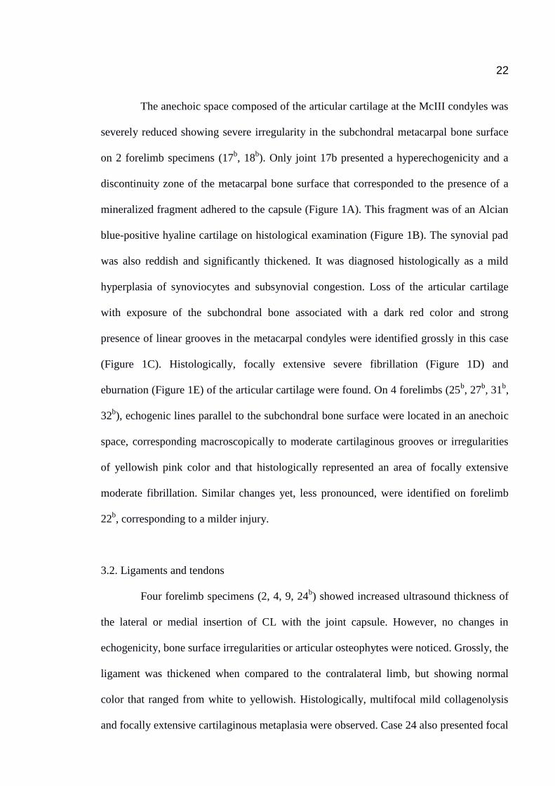
The anechoic space composed of the articular cartilage at the McIII condyles was
severely reduced showing severe irregularity in the subchondral metacarpal bone surface
on 2 forelimb specimens (17b, 18
b). Only joint 17b presented a hyperechogenicity and a
discontinuity zone of the metacarpal bone surface that corresponded to the presence of a
mineralized fragment adhered to the capsule (Figure 1A). This fragment was of an Alcian
blue-positive hyaline cartilage on histological examination (Figure 1B). The synovial pad
was also reddish and significantly thickened. It was diagnosed histologically as a mild
hyperplasia of synoviocytes and subsynovial congestion. Loss of the articular cartilage
with exposure of the subchondral bone associated with a dark red color and strong
presence of linear grooves in the metacarpal condyles were identified grossly in this case
(Figure 1C). Histologically, focally extensive severe fibrillation (Figure 1D) and
eburnation (Figure 1E) of the articular cartilage were found. On 4 forelimbs (25b, 27
b, 31
b,
32b), echogenic lines parallel to the subchondral bone surface were located in an anechoic
space, corresponding macroscopically to moderate cartilaginous grooves or irregularities
of yellowish pink color and that histologically represented an area of focally extensive
moderate fibrillation. Similar changes yet, less pronounced, were identified on forelimb
22b, corresponding to a milder injury.
3.2. Ligaments and tendons
Four forelimb specimens (2, 4, 9, 24b) showed increased ultrasound thickness of
the lateral or medial insertion of CL with the joint capsule. However, no changes in
echogenicity, bone surface irregularities or articular osteophytes were noticed. Grossly, the
ligament was thickened when compared to the contralateral limb, but showing normal
color that ranged from white to yellowish. Histologically, multifocal mild collagenolysis
and focally extensive cartilaginous metaplasia were observed. Case 24 also presented focal
23
mild bone metaplasia. On the remaining examined fetlocks, no histological abnormalities
were found.
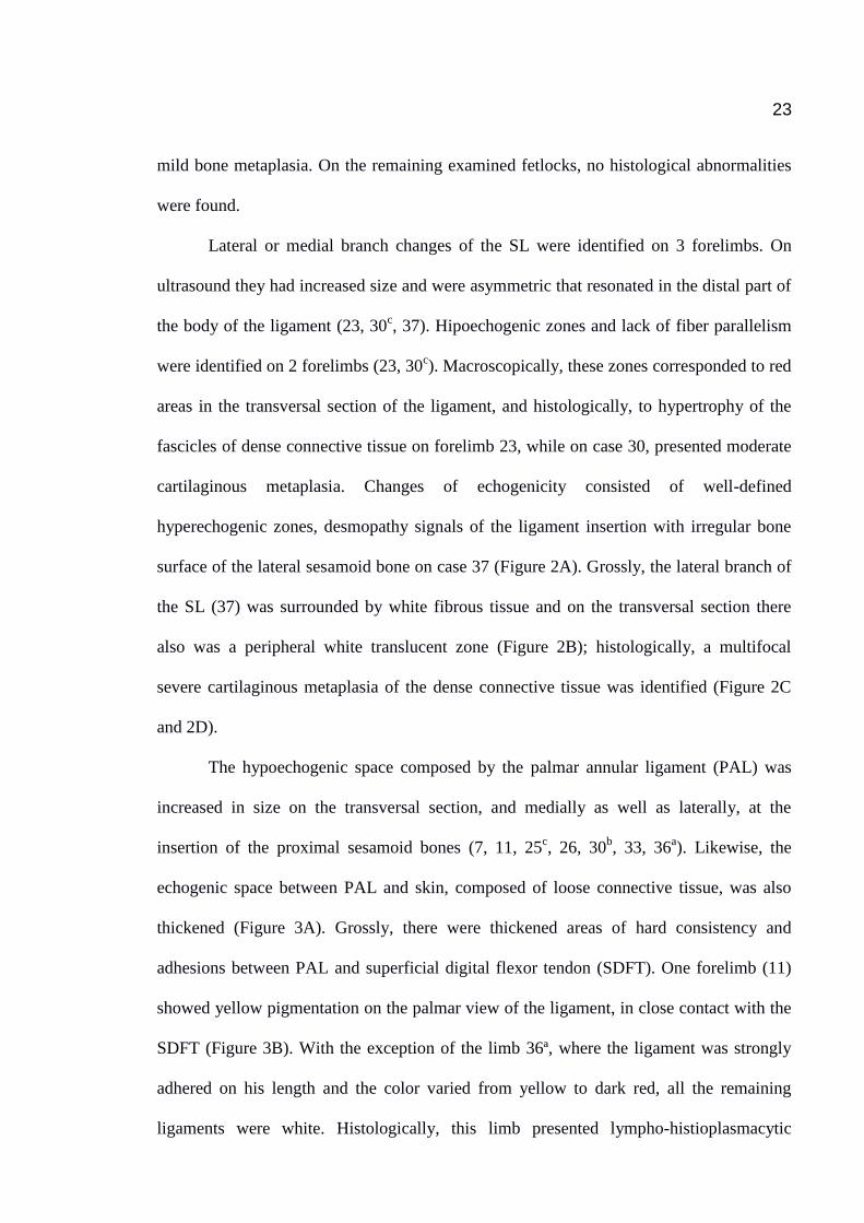
Lateral or medial branch changes of the SL were identified on 3 forelimbs. On
ultrasound they had increased size and were asymmetric that resonated in the distal part of
the body of the ligament (23, 30c, 37). Hipoechogenic zones and lack of fiber parallelism
were identified on 2 forelimbs (23, 30c). Macroscopically, these zones corresponded to red
areas in the transversal section of the ligament, and histologically, to hypertrophy of the
fascicles of dense connective tissue on forelimb 23, while on case 30, presented moderate
cartilaginous metaplasia. Changes of echogenicity consisted of well-defined
hyperechogenic zones, desmopathy signals of the ligament insertion with irregular bone
surface of the lateral sesamoid bone on case 37 (Figure 2A). Grossly, the lateral branch of
the SL (37) was surrounded by white fibrous tissue and on the transversal section there
also was a peripheral white translucent zone (Figure 2B); histologically, a multifocal
severe cartilaginous metaplasia of the dense connective tissue was identified (Figure 2C
and 2D).
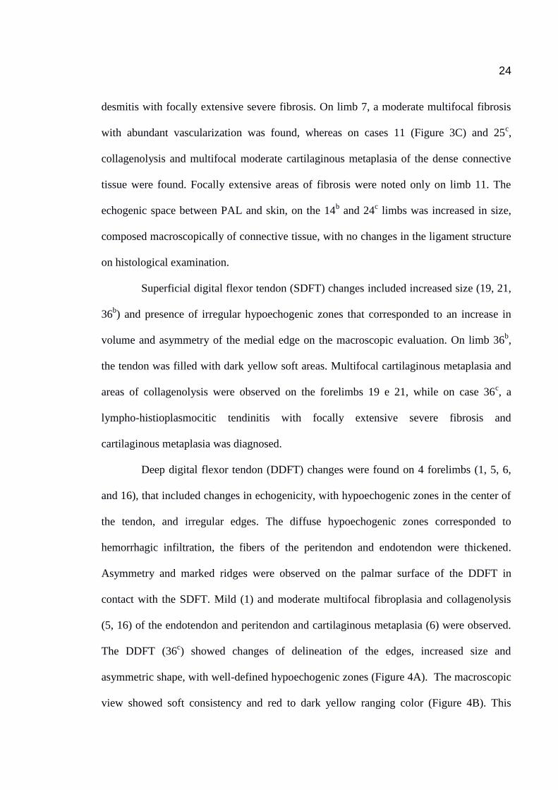
The hypoechogenic space composed by the palmar annular ligament (PAL) was
increased in size on the transversal section, and medially as well as laterally, at the
insertion of the proximal sesamoid bones (7, 11, 25c, 26, 30
b, 33, 36
a). Likewise, the
echogenic space between PAL and skin, composed of loose connective tissue, was also
thickened (Figure 3A). Grossly, there were thickened areas of hard consistency and
adhesions between PAL and superficial digital flexor tendon (SDFT). One forelimb (11)
showed yellow pigmentation on the palmar view of the ligament, in close contact with the
SDFT (Figure 3B). With the exception of the limb 36ª, where the ligament was strongly
adhered on his length and the color varied from yellow to dark red, all the remaining
ligaments were white. Histologically, this limb presented lympho-histioplasmacytic
24
desmitis with focally extensive severe fibrosis. On limb 7, a moderate multifocal fibrosis
with abundant vascularization was found, whereas on cases 11 (Figure 3C) and 25c,
collagenolysis and multifocal moderate cartilaginous metaplasia of the dense connective
tissue were found. Focally extensive areas of fibrosis were noted only on limb 11. The
echogenic space between PAL and skin, on the 14b and 24
c limbs was increased in size,
composed macroscopically of connective tissue, with no changes in the ligament structure
on histological examination.
Superficial digital flexor tendon (SDFT) changes included increased size (19, 21,
36b) and presence of irregular hypoechogenic zones that corresponded to an increase in
volume and asymmetry of the medial edge on the macroscopic evaluation. On limb 36b,
the tendon was filled with dark yellow soft areas. Multifocal cartilaginous metaplasia and
areas of collagenolysis were observed on the forelimbs 19 e 21, while on case 36c, a
lympho-histioplasmocitic tendinitis with focally extensive severe fibrosis and
cartilaginous metaplasia was diagnosed.
Deep digital flexor tendon (DDFT) changes were found on 4 forelimbs (1, 5, 6,
and 16), that included changes in echogenicity, with hypoechogenic zones in the center of
the tendon, and irregular edges. The diffuse hypoechogenic zones corresponded to
hemorrhagic infiltration, the fibers of the peritendon and endotendon were thickened.
Asymmetry and marked ridges were observed on the palmar surface of the DDFT in
contact with the SDFT. Mild (1) and moderate multifocal fibroplasia and collagenolysis
(5, 16) of the endotendon and peritendon and cartilaginous metaplasia (6) were observed.
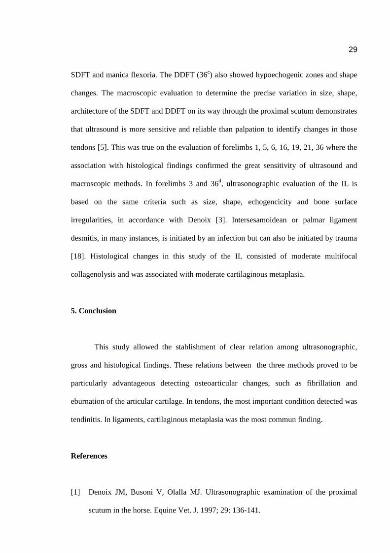
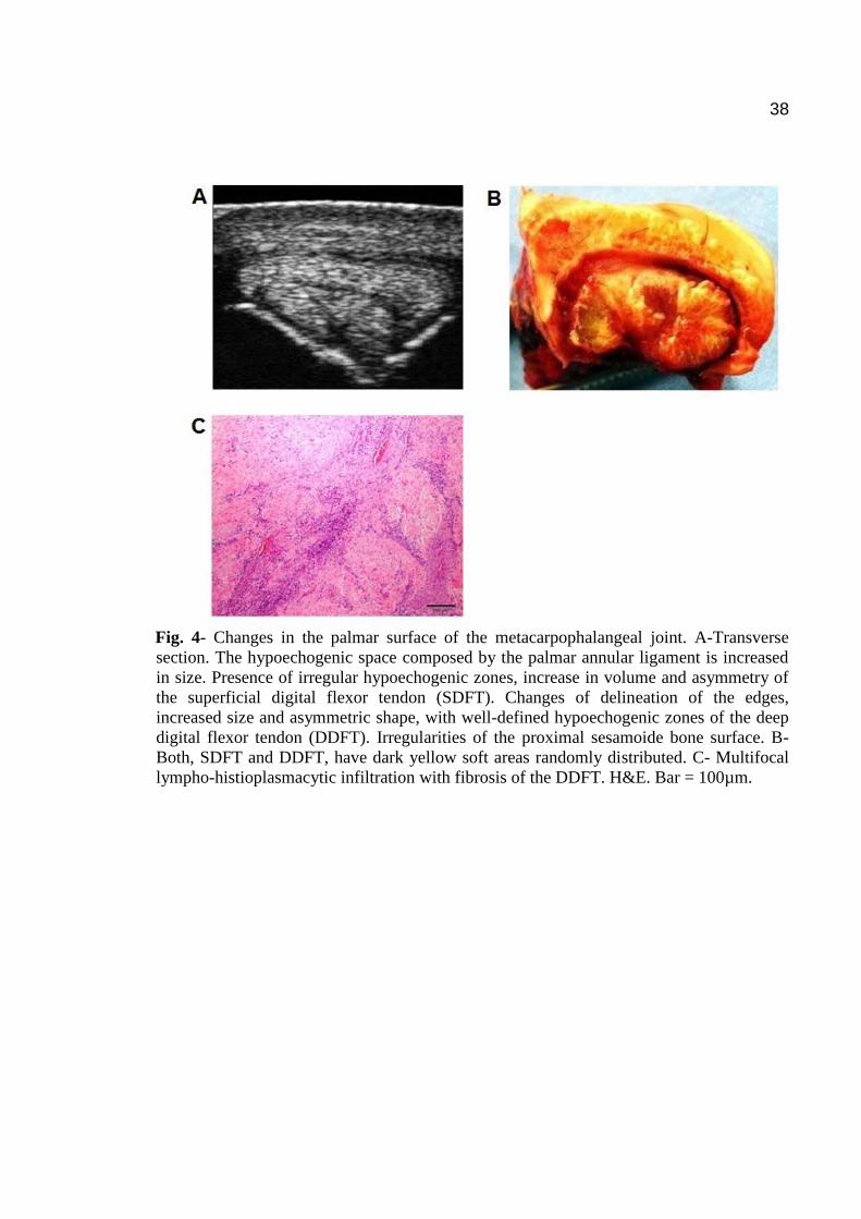
The DDFT (36c) showed changes of delineation of the edges, increased size and
asymmetric shape, with well-defined hypoechogenic zones (Figure 4A). The macroscopic
view showed soft consistency and red to dark yellow ranging color (Figure 4B). This

25
corresponded histologically to multifocal lympho-histioplasmacytic tendinitis with severe
necrosis, hemorrhage and fibrosis (Figure 4C).
Only one forelimb (3) showed intersesamoidean ligament (IL) changes, where the
space between the sesamoid bones was ultrasonographically increased and
hyperechogenic. Grossly, this ligament was white-yellow and with fiber irregularity.
Histologically, moderate multifocal collagenolysis was associated with moderate
cartilaginous metaplasia.
3.3. Proximal sesamoid bones
The proximal sesamoid bones of limb 36d presented irregularities on all their
extension. Macroscopic findings on these structures were white to pink grooves on the
dorsal surface. The palmar surface of the sesamoid bones presented necrotic dark red
zones and microscopically, severe periosteal hemorrhages were found.
4. Discussion
In this study, the selection criteria of the forelimb specimens submitted to
ultrasound examination were different from those applied in clinical routine workup. It
was based on the detection of changes by inspection and palpation of the forelimbs after
slaughter and not by lameness problems. Certainly we might have missed forelimbs
injuries unapparent to palpation and that perhaps could have been responsible for
locomotor problems. Similarly, we may have selected forelimbs with injuries, but without
clinical manifestations. Denoix et al. [5] cites the clinical value and its relationship with
the macroscopic and histological findings demonstrates that necrotic tissues appearance,
angiomatous or edematous tissue and presence of hemorrhagic infiltrates represent a little

26
significant value in the production of musculoskeletal problems. However, fibroplasia has
significant value. Findings such as cartilaginous metaplasia, whitish gelatinous tissue may
determine defects in elasticity of the tendons and ligaments. The anatomopathological
examination in this study was the reference method used to evaluate the ultrasound
sensitivity of the dorsal, lateral/medial and palmar surface of the MP joint, contributing
with consistent morphological and histological information and having the advantage of
being fast, demonstrative and of low cost. Identified lesions were found to be of traumatic,
degenerative, chronic and, in one case, possibly of septic origin. The main interest of this
study is to demonstrate the sensitivity and specificity of ultrasonography to document
injuries of the MP joint, whatever the clinical significance of these lesions (Table 2).
Abnormal ultrasonographic findings of the joint capsule include modifications in
thickness and echogenicity, and bone insertion alterations [6]. On 6 abnormal joint
capsules detected in this study, 5 limbs presented red and thickened synovial pad and all
were accompanied by articular cartilage lesions. Probably, the injuries were the result of
repeated trauma in the joint and may have been accompanied by synovial inflammation as
well [7]. According to Santschi [8] synovial changes resulting from continued
inflammation include hyperplasia and fibrosis. Occasionally, MP synovial pad
proliferation is characterized histologically by fibrous proliferation, collagen necrosis,
increased vascularity and cartilaginous metaplasia [9]. On case 17a, mild synoviocyte
hyperplasia and subsynovial congestion were observed. Pool [10] mentioned that,
microscopically, when synoviocytes appear hypertrophic different numbers of
lymphocytes, plasma cells and macrophages may be present. This was checked in
structure 32a, but the only difference found in this study was the presence of neutrophils
and not of macrophages. Islands of synoviocyte metaplasia may form nodules of cartilage
tissue called synovial chondromas [10]. However, this was not observed in case 12, where

27
cartilaginous metaplasia was observed inside the fibrous capsule and not in the synovial
pad.
Linear cartilaginous erosions, such as the ones observed on the metacarpal
condyle, induce an irregular cartilage surface on transverse section. Cartilage degeneration
induces local or diffuse thinning of the articular cartilage [11]. Histological examination of
the articular cartilage on cases 17b and 18
b showed areas of degeneration (fibrillation) of
the articular cartilage and areas of loss of the cartilage, with exposure of subchondral bone
(eburnation). In 4 cases, the histological cartilage changes were less severe and consisted
of focally extensive fibrillation of the articular cartilage with proliferation of chondrocyte
islands. Ultrasound imaging is very effective for identifying articular cartilage lesions [3],
including the macroscopically discrete abnormalities [1].
Axial deviation of the forelimbs, such as varus and valgus deformation, may
predispose to an overload of compressive forces to the joint and increase tension on the
collateral ligaments, predisposing them to get thickened. It is possible that this occurred on
cases 02, 09, and 24b because, the medial or lateral CL were thickened, but the echogenic
appearance was normal and no osteophytes were observed on the joint. Luxation of the
MP joint is an uncommon disorder caused by trauma. An obvious valgus or varus
deformation can be present [12]. In the above mentioned cases only valgus or varus
deformation were evident and no histological changes were found. On the other hand, on
forelimb 4, a thickened lateral CL was found, also with normal echogenicity. These
findings are accompanied by bone lesions like irregular bone surfaces, osteophyte
production and histological changes, in accordance with Denoix [11].
Hypoechogenic zones visible in the center of the SL and reduced regularity on the
fiber pattern were observed on two forelimbs (23, 30c) that would be compatible with
hemorrhagic infiltration and edema. On another case (37), ultrasonographic findings

28
consisted of enlargement and asymmetry of the hyperechogenic cross-sectional areas, poor
demarcation of the SL borders and focal mineralization, in agreement of Dyson [13].
Specimens from horses with severe desmopathy diagnosed by ultrasound had marked fiber
disruption with focal acellular areas interspersed with hypercellular accumulations of
disorganized fibroblasts [14]. According to White and Hewes [15], there is evidence of
attempted regeneration by fibroplasia, but lack of healing is apparent in distinct focal
areas, with no vascular supply and disorganized collagen, which appears immature. In this
study, histological changes of the hyperechogenic zones of the SL branches consisted of
the chondrocytes islands with severe cartilaginous metaplasia of the dense connective
tissue and hypoechogenic zones corresponded to hypertrophy of the fascicles of dense
connective tissue .
All palmar annular ligaments (PAL) with ultrasonographic findings where
represented by an increased and thickened space occupied by them, where it was found
adhered to the SDFT. Nevertheless, the thickness of PAL can be measured and this
hypoechogenic representation helps identification of this structure [3]. Ultrasonography is
effective in outlining soft tissues abnormalities, such as thickening of the synovial
membrane and adhesions between SDFT and PAL [16]. The healing process results in
fibrous thickening of the SDFT and/or the PAL [2]. This was evident on forelimb 11
where multifocal fibrosis zones of the SDFT and PAL were found on adhesion sites. The
presence of fibrosis in association with PAL thickening may increase the pressure exerted
on the contents of the fetlock canal, and had a significant negative effect on prognosis
[19]. Effusion, thickening of the synovial sheath and adhesions with concurrent lesions of
the PAL are described as ultrasonographic findings indicative of tenosynovitis [17]. These
findings were observed on case 36a. The surface of the proximal sesamoid bones was very
irregular and there was a lack of delineation of the edges and hypoechogenic zones on the
29
SDFT and manica flexoria. The DDFT (36c) also showed hypoechogenic zones and shape
changes. The macroscopic evaluation to determine the precise variation in size, shape,
architecture of the SDFT and DDFT on its way through the proximal scutum demonstrates
that ultrasound is more sensitive and reliable than palpation to identify changes in those
tendons [5]. This was true on the evaluation of forelimbs 1, 5, 6, 16, 19, 21, 36 where the
association with histological findings confirmed the great sensitivity of ultrasound and
macroscopic methods. In forelimbs 3 and 36d, ultrasonographic evaluation of the IL is
based on the same criteria such as size, shape, echogencicity and bone surface
irregularities, in accordance with Denoix [3]. Intersesamoidean or palmar ligament
desmitis, in many instances, is initiated by an infection but can also be initiated by trauma
[18]. Histological changes in this study of the IL consisted of moderate multifocal
collagenolysis and was associated with moderate cartilaginous metaplasia.
5. Conclusion
This study allowed the stablishment of clear relation among ultrasonographic,
gross and histological findings. These relations between the three methods proved to be
particularly advantageous detecting osteoarticular changes, such as fibrillation and
eburnation of the articular cartilage. In tendons, the most important condition detected was
tendinitis. In ligaments, cartilaginous metaplasia was the most commun finding.
References
[1] Denoix JM, Busoni V, Olalla MJ. Ultrasonographic examination of the proximal
scutum in the horse. Equine Vet. J. 1997; 29: 136-141.
30
[2] Wyn-Jones G. Equine Lameness. Oxford, England: Blackwell Scientific
Publications; 1988: 73-94.
[3] Denoix JM. Ultrasonography examination in the diagnosis of joint disease. In: C.W.
McIlwraith, G.W. Trotter, eds. Joint Disease in the horse. Philadelphia,
USA:Saunders; 1996: 165-202.
[4] Denoix JM, Audigie F. Ultrassonographic examination of joint in horse. In:
Proceedings of the 47th
Annual Convention American Association of Equine
Practitioners, San Diego, USA; 2001: 336-375.
[5] Denoix JM, Mialot M, Levy I, Lagadic M. Etude anatomo-pathologique de lésions
associées aux images écrographiques anormales des tendons et ligaments chez le
cheval. Recueil de Médicine Vétérinaire 1990; 166: 45-55.
[6] Denoix JM, Jacot S, Perrot P. Ultrasonografhic examination of the dorsal and abaxial
aspects of the equine fetlock. In : Proceedings of the 41th
Annual Convention
American Association of Equine Practitioners, San Diego, USA; 1995: 138-141.
[7] Caron PJ. Synovial joint, biology and pathobiology. In: J.A. Auer, J.A. Stick, eds,
Equine Surgery. Philadelphia, USA: WB Saunders Company; 1992: 665 p.
[8] Santschi EM. Articular fetlock injuries in exercising horses. Vet. Clin. North. Am.
Equine Pract. 2008; 24: 117-132.

31
[9] White NA. Synovial pad proliferation in the metacarpophalangeal joint. In: N.A.
White, N.J. Moore, eds, Current Practice of Equine Surgery. Philadelphia, USA;
1990: 555-558.
[10] Pool RR. Pathologic manifestations of joint disease in the athletic horse. In: C.W.
McIlwraith, G.W. Trotter, eds, Joint Disease in the horse. Philadelphia, USA:
Saunders; 1996: 87-93.
[11] Denoix JM. Ultrasonographic examination of joints in horses: a live demonstration.
In: Proceedings of the 11th
International Congress of the World Equine Veterinary
Association, Guarujá, Brazil; 2009: 1-10.
[12] Yovich JV, Turner AS, Stashak TS, McIlwraith CW. Luxation of the
metacarpophalangeal and metatarsophalangeal joints in horses. Equine Vet. J. 1987;
19: 295-298.
[13] Dyson S. Proximal suspensory desmitis: clinical, ultrasonographic and radiographic
features. Equine Vet. J. 1991; 23: 25-31.
[14] Dyson S. Proximal suspensory desmitis in the forelimb and the hindlimb. In:
Proceedings of the 46th
Annual Convention American Association of Equine
Practitioners, San Diego, USA; 2000: 137-142.

32
[15] White NA, Hewes CA. Treatment of suspensory ligament desmopathy. In:
Proceedings of the 54th
Annual Convention of the American Association of Equine
Practioners, San Diego, USA; 2008.
[16] Dik KJ, Dyson SJ, Vail TB. Aseptic tenosynovitis of the digital flexor tendon sheath,
fetlock and pastern annular ligament constriction. Vet. Clin. North Am. Equine
Pract. 1995; 11: 151-162.
[17] Smith RKW, Webbon PM. Harnessing the stem cell for treatment of tendon injuries :
heralding a new dawn. Br. J. Sports Med. 2005; 39: 582-584.
[18] Dabareiner RM, Watkins JP, Carter GK, Honnas CM, Eastman T. Osteitis of the axial
border of the proximal sesamoid bones in horses: eight cases (1993-1999). J. Am.
Vet. Med. Assoc. 2001; 219: 82-86.
[19] Owen KR, Dyson SJ, Parkin TDH, Singer ER, Kristoffersen M, Mair TS.
Retrospective study of palmar/plantar annular ligament injury in 71 horses: 2001-
2006. Equine Vet. J. 2008; 40: 237-244.

33
Table 1. Results for the 54 structures analyzed in this study
Structures Analyzed in 37 Forelimbs Number
Joint Capsule 14
Articular Cartilage of the Condyles of the Metacarpal
bone and Proximal Sesamoide Bone
8
Digital Flexor Tendons 13
Ligaments 19

34
Table 2. Association of ultrasonographic images with macroscopic and histological
findings in 37 post-mortem cases analyzed in this study.
SUPENSORY LIGAMENT (SL)
Echogenicity Macroscopically Histology
Diffuse Hypoechogenicity Hemorrhagic Infiltration Fascicles hypertrophy
Hyperechogenicity White to Yellow Nodular Tissue Severe cartilaginous metaplasia
SUPERFICIAL DIGITAL FLEXOR TENDON (SDFT)
Echogenicity Macroscopically Histology
Focal Hypoechogenicity Dark Yellow Zones Lymphohistioplasmacytic
inflammation / Focally extensive
severe fibrosis.
Hyperechogenicity White to Dark Yellow Zones Cartilaginous metaplasia
DEEP DIGITAL FLEXOR TENDON (DDFT)
Echogenicity Macroscopically Histology
Diffuse Hypoechogenicity Hemorrhagic Infiltration Fibroplasia
Focal Hypoechogenicity Red to Dark Yellow Zones Lymphohistioplasmacytic perivascular
inflammation/ Necrotic and
hemorrhagic areas.
PALMAR ANNULAR LIGAMENT (PAL)
Echogenicity Macroscopically Histology
Hyperechogenicity Thickened areas/ Hard consistency/
Adherences to SDFT.
Moderate multifocal fibrosis/
Cartilaginous metaplasia.
Hypoechogenicity Strongly adhered/
Yellow to dark red color.
Lymphohistioplasmacytic and
neutrophilic inflammation/ Focal
extensive severe fibrosis
ARTICULAR CARTILAGE
Echogenicity Macroscopically Histology
Hyperechogenicity/ Severe
Irregular Surface/ Diffuse
Thinning.
Linear Cartilaginous Erosions/
Exposure of Subchondral Bone
Focally extensive severe fibrillation
and eburnation.
Hyperechogenicity/ Moderate
Irregular Surface/ Diffuse
Thickening.
Moderate Cartilaginous Grooves/
Yellowish Pink Color
Focally extensive moderate
fibrillation.

35
Figures
Fig. 1- Articular cartilage changes of the III metacarpal bone (McIII). A- Transverse
section. The anechoic space composed of the articular cartilage at the condyles of McIII is
severely diminished with severe irregularity in the subchondral metacarpal bone surface.
There is a discontinuity zone of the metacarpal bone surface that corresponded to a
mineralized fragment (asterisk). B- A mineralized fragment of an Alcian blue-positive
hyaline cartilage. Bar = 100µm. C- Loss of the articular cartilage with exposure of
subchondral bone associated with a dark red color and strong presence of linear grooves in
the McIII (asterisk). D- Focally extensive severe fibrillation of the articular cartilage
surface. E- Focally extensive severe eburnation of the articular cartilage. Only the
subchondral bone is shown. H&E. Bar = 100µm.

36
Fig. 2- Cartilaginous metaplasia of the suspensory ligament (SL). A- Transversal section.
A round hyperechogenic zone is observed in the distal portion of the ligament. Note the
irregular bone surface of the lateral proximal sesamoid bone and enlarged SL. B- The SL
is surrounded by white fibrous tissue and on transversal section there is a peripheral white
translucent zone. C- Multifocal severe cartilaginous metaplasia of the dense connective
tissue is present. H&E. Bar = 100µm. D- Higher magnification of figure 2C. H&E. Bar =
20µm.

37
Fig. 3- Adhesions between the palmar annular ligament (PAL) and the superficial digital
flexor tendon (SDFT).A- Transverse section. There is a hypoechogenic space composed
by the PAL. The PAL was thickened. B- Multifocal areas of adhesions are observed
between PAL and SDFT. C- On the right side is the PAL and on the left side is the
adhesion with the SDFT (not shown). H&E. Bar = 100µm.
38
Fig. 4- Changes in the palmar surface of the metacarpophalangeal joint. A-Transverse
section. The hypoechogenic space composed by the palmar annular ligament is increased
in size. Presence of irregular hypoechogenic zones, increase in volume and asymmetry of
the superficial digital flexor tendon (SDFT). Changes of delineation of the edges,
increased size and asymmetric shape, with well-defined hypoechogenic zones of the deep
digital flexor tendon (DDFT). Irregularities of the proximal sesamoide bone surface. B-
Both, SDFT and DDFT, have dark yellow soft areas randomly distributed. C- Multifocal
lympho-histioplasmacytic infiltration with fibrosis of the DDFT. H&E. Bar = 100µm.

39
4. CONSIDERAÇÕES FINAIS
Este estudo ecográfico e anatomopatológico de lesões presentes na articulação
metacarpofalangeana e seus componentes demonstra a sensibilidade da técnica
ultrassonográfica em tecidos ligamentosos e tendinosos bem como, em tecidos ósseos.
Imagens anaecóicas, hiperecogênicas, hipoecogênicas histologicamente podem representar
diferentes tipos de tecidos. Metaplasias cartilaginosas foram encontradas nas estruturas
ligamentosas produzindo imagens hiperecogênicas; por outro lado, infiltrados
inflamatórios observados nas estruturas tendinosas produziram imagens hipoecogênicas.

40
5. REFERÊNCIAS
AMIEL, D., et al. Tendons and ligaments: A morphological and biochemical comparison.
Journal Orthopedic Research, New York, v. 1, p. 257-265, 1984.
BARONE, R. Articulations metacarpal-phalangiennea. In: Anatomies des Mamiferes
Domestiques - Tome 2: Arthrologie et Myologie. Paris: Vigot, 1989. p.187-204.
BARONE, R. Le os grands sésamoides. Anatomie comparée des mammifères domestique-
Tome 1: Ostéologie. Paris : Vigot, 2000. p. 573.
BOSCH, G. New insights in the pathogenesis of tendon injuries. In : PROCEEDINGS OF
THE EUROPEAN VETERINARY CONFERENCE, 2010, Amsterdan. Anais...,
Amsterdan: 2010. p. 272-327.
BRAMA, P. A. J., et al. Contact areas and pressure distribution on the proximal articular
surface of the proximal phalanx under sagittal plane loading. Equine Veterinary Journal,
Newmarket, v. 33, p. 26-32, 2001.
BROWN, N. A. T., et al. Architectural properties of distal forelimb muscles in horses,
Equus caballus. Journal of Morphology, Philadelphia, v. 258, p.106-114, 2003.
BUTCHER, M. T., et al.Superficial digital flexor tendon lesions in racehorses as a sequela
to muscle fatigue: a preliminary study. Equine Veterinary Journal, Newmarket, v. 39, p.
540-545, 2007.
GARTNER, L. P.; HIATT, J. L. Color Textbook of Histology. Philadelphia: Saunders,
1997. p. 92-108.
CLAYTON, H. M., et al. Net joint moments and powers in the equine forelimb during the
stance phase of the trot. Equine Veterinary Journal, Newmarket, v. 30, p. 384–389,
1998.
COLBORNE, G.R., et al. Forelimb joint moments and power during the walking stance
phase of horses. American Journal of Veterinary Research, Chicago, v. 59, p. 609–614,
1998.
41
DENOIX, J.M. et al. Etude anatomo-pathologique de lésions associées aux images
écrographiques anormales des tendons et ligaments chez le cheval. Recueil de Médicine
Vétérinaire, Paris, v. 166, p. 45-55, 1990.
DENOIX, J. M.; BOUSSEAU, B.; CREVIER, N. Ultrasound examination of the fetlock in
the horse. In: PROCEEDINGS OF THE 3rd CONGRESS WORLD EQUINE
VETERINARY ASSOCIATION SWISS REV VET MED, 1993. p. 103-108.
DENOIX J. M., et al. Ultrasonographic examination of the dorsal and abaxial aspects of
the equine fetlock. In : PROCEEDINGS OF THE 41th
ANNUAL CONVENTION
AMERICAN ASSOCIATION OF EQUINE PRACTITIONERS, 41, San Diego. Anais…,
San Diego, 1995. p.138-141.
DENOIX, J. M. Ultrasonography examination in the diagnosis of joint disease. In:
MACILWRAITH, C.W.; TROTTER, G.W. Joint Disease in the horse. Philadelphia:
Saunders, 1996. Cap. 10, p.165-202.
DENOIX, J. M. et al. Ultrasonographic examination of the proximal scutum in the horse.
Equine Veterinarie Journal, Newmarket, v. 29, p. 136-141, 1997.
DENOIX, J. M. The equine distal limb. In: Atlas of Clinical Anatomy and Comparative
Imaging. London: Manson Publishing, 2000. Cap. 1, p. 243-376.
DENOIX, J. M.; AUDIGIE, F. Ultrassonographic examination of joint in horse. In:
PROCEEDINGS OF THE 47th
ANNUAL CONVENTION AMERICAN ASSOCIATION
OF EQUINE PRACTITIONERS, 47, 2001, San Diego. Anais… San Diego, 2001. p.
336-375.
DENOIX, J. M. Ultrasonographic examination of joints in horses: a live demonstration.
In: PROCEEDINGS OF THE 11th
INTERNATIONAL CONGRESS OF THE WORLD
EQUINE VETERINARY ASSOCIATION,11., 2009, Guarujá. Anais… Guarujá, 2009.
p. 1-10.
DIK, K. J.; DYSON, S. J.; VAIL, T. B. Aseptic tenosynovitis of the digital flexor tendon
sheath, fetlock and pastern annular ligament constriction. The Veterinary Clinics of
North America. Equine Practioners, Philadelphia, v. 11, p. 151-162, 1995.
DYSON, S. J. Proximal suspensory desmitis in the forelimb and the hindlimb. In :

42
PROCEEDINGS OF THE 46th ANNUAL MEETING OF THE AMERICAN
ASSOCIATION OF EQUINE PRACTIONERS, San Diego. Anais... San Diego, 2000.
p.137-142.
FRANK, C.B. Ligament injuries: pathophysiology and healing. In: ZACKAZEWSKI, J.
E.; QUILLEN, W. S. Philadelphia: W. B. Saunders, 1996. p. 9-25.
GENOVESE, R. L., et al. Diagnostic ultrasonography of equine limbs. The Veterinary
Clinics of North America. Equine Practioners, Philadelphia, v. 2, p. 145-226, 1986.
GILLIS, C., et al. Effect of maturation and aging on the histomorphometric and
biochemical characteristics of equine superficial digital flexor tendon. American Journal
Veterinary Research, Chicago, v. 58, p. 425-430, 1997.
HOSAKA, Y., et al. Differences in tumor necrosis factor (TNF) alpha and TNF receptor-
1-mediated intracellular signaling factors in normal, inflamed and scarformed horse
tendons. The Journal of Veterinary Medicine Science, Tokyo, v. 67, p. 985-991, 2005.
MARCELLI, C., SEBERT, J. L. Architecture et resistance mecanique osseuses. Paris:
Masson, 1993. Cap. 1, p. 2-5.
McLIWRAITH, C.W. Disease Processes of Synovial Membrane, Fibrous Capsule,
Ligaments, and Articular Cartilage. In: PROCEEDINGS OF THE 47th ANNUAL
MEETING OF THE AMERICAN ASSOCIATION OF EQUINE PRACTIONERS, San
Diego, Anais... San Diego, 2001. p.142-157.
POOL, R. R. Pathologic manifestations of joint disease in the athletic horse. In:
MacIlwraith C.W.; Trotter G.W. Joint Disease in the Horse. Philadelphia:
Saunders, 1996. Cap 1, p.87-93.
REEF, V. B. Musculoskeletal ultrasonography. In: Equine Diagnostic Ultrasound.
Philadelphia: Saunders, 1998. Cap 1, p. 39-79.
SCHRAMME, M.; SMITH, R. Diseases of the digital synovial sheath, palmar annular
ligament, and digital annular ligaments. In: Ross, M.W.; Dyson, S.J. Diagnosis and
Management of Lamenees in the Horse. Philadelphia: Saunders, 2003. p. 674-684.
43
SEIGNOUR, M. et al. Ultrasonographic examination of the palmar aspect of the fetlock in
the horse: Technique and normal images. Equine Veterinary Education, Newmarket, v.
24, p. 19-29, 2012.
SMITH, R.; BIRCH, H. L,; GOODMAN, S. et al. The influence of ageing and exercice on
tendon growth and degeneration-hypotheses for the initiation and prevention of strain-
induced tendinopathies. Comparative Biochemistry and Physiology, New York, v. 133,
p. 1039-1050, 2002.
SMITH, K. W; GOODSHIP, A. E. Tendon and ligament physiology. In: Hinchcliff, K.Wl.
Equine Sports Medicine and Surgery. Philadelphia: Saunders, 2004. p. 130-151.
SMITH, R. Tendon and ligament injury. In: PROCEEDINGS OF THE 54th
ANNUAL
CONVENTION AMERICAN ASSOCIATION OF EQUINE PRACTITIONERS, 2008,
San Diego, Anais… San Diego, 2008, p. 475-501.
STROMBERG, B. The normal and disease superficial flexor tendon in race horses. A
morphologic and physiologic investigation. Acta Radiologica Supplementum,
Stockholm. v. 305, p. 1-94, 1971.
WHITE, N. A.; HEWES, C. A. Treatment of suspensory ligament desmopathy. In:
PROCEEDINGS OF THE 54TH ANNUAL CONVENTION OF THE AMERICAN
ASSOCIATION OF EQUINE PRACTIONERS, San Diego. Anais… San Diego, 2008.
WRIGHT, I. M.; MCMAHON , P. J. Tenosynovitis associated with longitudinal tears of
the digital flexor tendons in horses: A report of 20 cases. Equine Veterinary Journal,
Newmarket. v. 31, p. 12-18, 1999.
WILDERJANS, H. Tenoscopy of the digital flexor tendon sheath. PROCEEDINGS OF
THE 10th INTERNATIONAL CONGRESS OF WORLD EQUINE VETERINARY
ASSOCIATION, 2008, Moscow, Anais… Moscow. p.182-187.
VANDERPERREN, K., et al. Evaluation of computed tomographic anatomy of the equine
metacarpophalangeal joint. American Journal of Veterinary Research, Chicago. v. 69,
p. 631-8, 2008.