Embed Size (px)
Citation preview
UNIVERSIDADE FEDERAL DE SANTA MARIACENTRO DE CIÊNCIAS NATURAIS E EXATAS
PROGRAMA DE PÓS-GRADUAÇÃO EM CIÊNCIAS BIOLÓGICAS: BIOQUÍMICA TOXICOLÓGICA
ATIVIDADE DAS ECTONUCLEOTIDASES, COLINESTERASE SÉRICA E PERFIL OXIDATIVONO DIABETES MELITO TIPO 2 E HIPERTENSÃO
EM HUMANOS
TESE DE DOUTORADO
Gilberto Inácio Lunkes
Santa Maria, RS, Brasil
2008
1
ATIVIDADE DAS ECTONUCLEOTIDASES, COLINESTERASE SÉRICA E PERFIL OXIDATIVONO DIABETES MELITO TIPO 2 E HIPERTENSÃO
EM HUMANOS
por
Gilberto Inácio Lunkes
Tese apresentada ao Curso de Doutorado do Programade Pós-Graduação em Ciências Biológicas: Bioquímica Toxicológica,
da Universidade Federal de Santa Maria (UFSM, RS),como requisito parcial para obtenção do grau de
Doutor em Bioquímica Toxicológica.
Orientador: Profa. Dra. Maria Rosa Chitolina Schetinger
Santa Maria, RS, Brasil
2008
2
Universidade Federal de Santa MariaCentro de Ciências Naturais e Exatas
Programa de Pós-Graduação em Ciências Biológicas:Bioquímica Toxicológica
A Comissão Examinadora, abaixo assinada,aprova a Tese de Doutorado
ATIVIDADE DAS ECTONUCLEOTIDASES, COLINESTERASE SÉRICA E PERFIL OXIDATIVONO DIABETES MELITO TIPO 2 E HIPERTENSÃO
EM HUMANOS
elaborada porGilberto Inácio Lunkes
como requisito parcial para obtenção do grau deDoutor em Bioquímica Toxicológica
BANCA EXAMINADORA:
Maria Rosa Chitolina Schetinger, Dra.(Presidente/Orientador)
Carla Denise Bonan, Dra. (PUC/RS)
Nilda Vargas Barbosa, Dra. (Unipampa)
Maribel Antonello Rubin, Dra. (UFSM)
Cinthia Melazzo Mazzanti, Dra.
Santa Maria, março de 2008.
3
DEDICATÓRIA
À Irma, Marlice e Daniéle
A minha mãe Irma Ferst Lunkes, muito obrigado pelas suas orações,
ensinamentos e palavras de incentivo. A minha irmã Marlice Lunkes, muito obrigado
por suas palavras de apoio e incentivo na incessante busca do equilíbrio e do
conhecimento. A minha esposa Daniéle Lunkes, por sua tenacidade e colaboração
em todas as etapas da jornada do doutorado.
4
AGRADECIMENTOS
À minha orientadora professora Dra. Maria Rosa Chitolina Schetinger por seu
persistente apoio, contínuo ensinamento e incentivo na busca do conhecimento. À
professora Dra. Vera Morsch pela sua constante participação e argüição nas
diretrizes dos nossos trabalhos de pesquisa.
Ao corpo docente do Pós-Graduação, que permitiu um crescimento não
apenas científico, mas também como cidadão. A funcionária Angélica pela sua
dedicação e presteza no atendimento.
Aos colegas e incentivadores do Laboratório de Enzimologia, em especial a
Paula, Maísa, Roberta e Jamile, muito obrigado, pois a colaboração de vocês foi
imprescindível em todos os momentos do curso. Ao meu amigo Mushtaq Ahmed,
que Deus ilumine e continue abençoando seu caminho.
5
RESUMO
Tese de DoutoradoPrograma de Pós-Graduação em Ciências Biológicas:
Bioquímica ToxicológicaUniversidade Federal de Santa Maria
ATIVIDADE DAS ECTONUCLEOTIDASES, COLINESTERASE SÉRICA E PERFIL OXIDATIVONO DIABETES MELITO TIPO 2 E HIPERTENSÃO
EM HUMANOSAUTOR: GILBERTO INÁCIO LUNKES
ORIENTADOR: MARIA ROSA CHITOLINA SCHETINGER Data e Local de Defesa: Santa Maria, 5 de março de 2008
O aumento na atividade enzimática da NTPDase e 5'-nucleotidase, em
pacientes com diabetes e hipertensão, desencadeou a investigação dos possíveis
mecanismos envolvidos nas alterações na atividade das ectonucleotidases. O nível
de estresse oxidativo e também a resposta dos sistemas antioxidantes foram
avaliados em pacientes com diabetes tipo 2, hipertensão e diabetes tipo 2 com
hipertensão associada. A interferência da concentração de glicose foi avaliada na
atividade enzimática das ectonucleotidases, colinesterase sérica e também das
enzimas antioxidantes. As curvas in vitro demonstraram que o aumento na atividade
dos sistemas enzimáticos foi proporcional à elevação nas concentrações de glicose,
demonstrando uma interferência direta da hiperglicemia. O aumento na expressão
da enzima NTPDase demonstrou uma importante correlação com a hidrólise dos
nucleotídeos de adenina ATP e ADP em pacientes com diabetes e hipertensão
associada. O incremento nos níveis de marcadores de estresse oxidativo e dos
sistemas antioxidantes, em pacientes com diabetes e hipertensão associada,
parecem estar relacionados com um mecanismo compensatório para prevenir o
dano oxidativo. Os baixos níveis de ácido ascórbico sérico aumentam a exposição
dos pacientes, com diabetes e hipertensão associada, aos danos oxidativos
resultantes do aumento na geração de espécies reativas de oxigênio. O aumento na
6
atividade da enzima colinesterase sérica, em pacientes com diabetes e hipertensão
associada, pode estar potencialmente relacionado com os níveis de glicemia e com
o metabolismo dos lipídios. Os medicamentos administrados aos pacientes não
alteraram as respostas enzimáticas nos grupos analisados. Portanto, houve uma
possível interferência do diabetes e da hipertensão no mecanismo catalítico da
colinesterase sérica. Os dados obtidos nos estudos permitem sugerir que os
elevados níveis de glicose sangüínea constituem um dos principais fatores capazes
de promover alterações nas respostas enzimáticas em pacientes diabéticos e com
hipertensão associada.
Palavras-chave: Ectonucleotidases, estresse oxidativo, colinesterase sérica,
Diabetes melito tipo 2, hipertensão, humanos, plaquetas.
7
ABSTRACT
Thesis of Doctor’s DegreePost-Graduation Program on Biological Sciences:
Toxicological BiochemistryFederal University of Santa Maria,RS, Brazil
ACTIVITY OF THE ECTONUCLEOTIDASES,SERUM CHOLINESTERASE AND OXIDATIVE PROFILE
IN TYPE 2 DIABETES MELITO AND HYPERTENSIONIN HUMANS
AUTHOR: GILBERTO INÁCIO LUNKESADVISER: MARIA ROSA CHITOLINA SCHETINGER
Date and Place of the defense: Santa Maria, March 5th, 2008
The increase in the NTPDase and 5'-nucleotidase enzymatic activities, in
patients with diabetes and hypertension, unchained the investigation of the possible
mechanisms involved in the alterations in ectonucleotidases activities. The oxidative
stress level and also the answer of the antioxidant systems were evaluated in
patients with type 2 diabetes, hypertension and type 2 diabetes with associated
hypertension. The interference of the glucose concentration was evaluated in the
enzymatic activity of the ectonucleotidases, serum cholinesterase and also
antioxidant enzymes. The curves in vitro demonstrated that the increase in enzymatic
activity was proportional to the elevation in the glucose concentrations,
demonstrating a direct interference of the hyperglycemia. The increase in the
NTPDase expression demonstrated an important correlation with adenine nucleotide
ATP and ADP hydrolyze in patients with diabetes and associated hypertension. The
increment in markers of oxidative stress and antioxidant systems levels in patients
with diabetes and associated hypertension seems to be related with a compensatory
mechanism to prevent oxidative damages. Low serum acid ascorbic levels increase
exposes to oxidative damages in patients with diabetes and associated hypertension,
resultant of the increase in reactive oxygen species generation. The increment in the
serum cholinesterase activity can be potentially related with glycemia levels and lipid
8
metabolism in patients with diabetes and associated hypertension. The medicines
administered to the patients did not alter the enzymatic responses in the analyzed
groups. Therefore, there were a possible interference of the diabetes and
hypertension in the catalytic mechanism of the serum cholinesterase enzyme. Data
obtained in the studies permit to suggest that high blood glucose levels constitute
one of the principal factors capable to promote alterations in the enzymatic
responses in patients with diabetes and associated hypertension.
Key words: Ectonucleotidases, oxidative profile, serum cholinesterase, type 2
Diabetes melito, hypertension, platelets.
9
LISTA DE FIGURAS
FIGURA 1: Topologia da enzima NTPDase localizada na superfície da
membrana com dois domínios transmembrana................................................ 22FIGURA 2: Catabolismo de nucleotídeos extracelulares e ativação dos
receptores para nucleotídeos (receptores P2) e adenosina (receptores P1)... 24
10
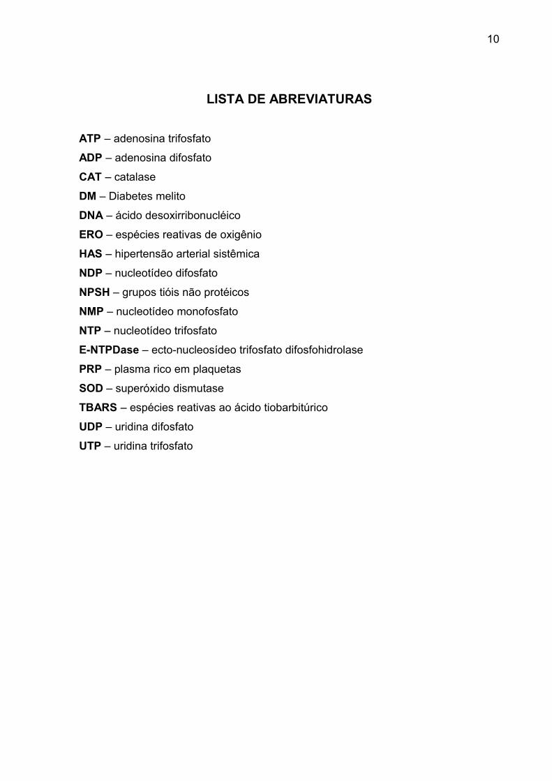
LISTA DE ABREVIATURAS
ATP – adenosina trifosfato
ADP – adenosina difosfato
CAT – catalase
DM – Diabetes melito
DNA – ácido desoxirribonucléico
ERO – espécies reativas de oxigênio
HAS – hipertensão arterial sistêmica
NDP – nucleotídeo difosfato
NPSH – grupos tióis não protéicos
NMP – nucleotídeo monofosfato
NTP – nucleotídeo trifosfato
E-NTPDase – ecto-nucleosídeo trifosfato difosfohidrolase
PRP – plasma rico em plaquetas
SOD – superóxido dismutase
TBARS – espécies reativas ao ácido tiobarbitúrico
UDP – uridina difosfato
UTP – uridina trifosfato
11
LISTA DE ANEXOS
ANEXO A: Termo de Consentimento Livre e Esclarecido ............................... 140ANEXO B: Confirmação de submissão do artigo “Effect of high glucose
levels in human platelet NTPDase and 5'-nucleotidase activities” no
periódico “Diabetes Research and Clinical Practice”……................................. 142ANEXO C: Confirmação de submissão do artigo “Antioxidant status in
platelet from patients with diabetes and hypertension” no periódico
“Molecular and Cellular Biochemistry” ………………….................................... 143

12
SUMÁRIO
DEDICATÓRIA ............................................................................................... 3
AGRADECIMENTOS ..................................................................................... 4
RESUM .......................................................................................................... 5
ABSTRACT .................................................................................................... 7
LISTA DE FIGURAS ...................................................................................... 9
LISTA DE ABREVIATURAS .......................................................................... 10
LISTA DE ANEXOS ....................................................................................... 11
1 INTRODUÇÃO ............................................................................................ 14
2 REVISÃO DA LITERATURA ...................................................................... 17
2.1 Diabetes melito ....................................................................................... 17
2.2 Hipertensão Arterial Sistêmica .............................................................. 18
2.3 Colinesterase sérica ............................................................................... 19
2.4 Plaquetas ................................................................................................. 20
2.5 Nucleotídeos e nucleosídeos ................................................................ 21
2.6 NTPDase e 5'-nucleotidase .................................................................... 21
2.6.1 Ectonucleotidases em patologias humanas .......................................... 24
2.7 Estresse oxidativo .................................................................................. 26
3 ARTIGO CIENTÍFICO E MANUSCRITOS .................................................. 283.1 Artigo 1: Serum cholinestease activity in diabetes and associated
pathologies ..................................................................................................... 293.2 Manuscrito 1: Effect of high glucose levels in human platelet NTPDase
and 5'-nucleotidase activities ………………………………………………..…... 353.3 Manuscrito 2: Antioxidant status in platelet from patients with diabetes
diabetes and hypertension ………………………………………………….……. 643.4 Manuscrito 3: Oxidative stress and antioxidant profile in serum from
patients with type 2 diabetes and hypertension ……………….……………….. 944 DISCUSSÃO DOS RESULTADOS ............................................................. 122
5 CONCLUSÃO .............................................................................................. 126
REFERENCIAL .............................................................................................. 127
13
ANEXOS.......................................................................................................... 139
ANEXO A: Termo de Consentimento Livre e Esclarecido .............................. 140ANEXO B: Confirmação de submissão do artigo “Effect of high glucose
levels in human platelet NTPDase and 5'-nucleotidase activities” no
periódico “Diabetes Research and Clinical Practice”....................................... 142ANEXO C: Confirmação de submissão do artigo “Antioxidant status in
platelet from patients with diabetes and hypertension” no periódico
“Molecular and Cellular Biochemistry” …………………................................... 143
14
1 INTRODUÇÃO
A complexidade epidemiológica de doenças crônicas como diabetes e
hipertensão têm atingido níveis alarmantes (GANNE et al., 2007). Estudos recentes
demonstram que o Diabetes melito está se consagrando como uma das maiores
catástrofes de saúde pública (MEETOO et al., 2007). Atualmente, cerca de 6% da
população adulta mundial tem diabetes diagnosticada e há uma previsão de 366
milhões de pessoas com diabetes até 2030 (WILD et al., 2004). A estimativa de
pacientes hipertensos no Brasil é de aproximadamente 30 milhões de pessoas e no
mundo de 600 milhões (SOCIEDADE BRASILEIRA DE HIPERTENSÃO, 2006). Por
constituírem doenças de extrema relevância em saúde pública requerem contínuos
estudos.
A perda funcional do endotélio vascular no diabetes está intrinsecamente
ligada ao desenvolvimento de doença cardiovascular e é responsável pela
aceleração de processos aterotrombóticos (HAMILTON et al., 2007). Estudos têm
demonstrado que cerca de 80% dos pacientes com Diabetes melito tipo 2
desenvolvem hipertensão (SAVOIA & SCHIFFRIN, 2007). O comprometimento
bioquímico do paciente diabético hipertenso deve ser amplamente investigado na
tentativa de buscar explicações para as alterações desenvolvidas.
O presente estudo tem como propósito avaliar os fatores envolvidos na
alteração da atividade das enzimas NTPDase e 5'-nucleotidase em plaquetas de
humanos com diabetes e hipertensão. Estudos anteriores, em humanos e modelo
experimental, demonstraram um aumento na hidrólise de ATP e ADP pela enzima
NTPDase e AMP pela 5'-nucleotidase em pacientes hipertensos, diabéticos tipo 2 e
diabéticos tipo 2 hipertensos (LUNKES et al., 2003; LUNKES et al., 2004). A
investigação em humanos também revelou que os medicamentos, administrados
aos pacientes diabéticos e hipertensos, não foram capazes de alterar a atividade
enzimática das ectonucleotidases (LUNKES et al., 2003). Então, com a finalidade de
investigar os fatores capazes de interferir na atividade enzimática foram realizadas
15
curvas de glicose e frutose, assim como, foi avaliada a expressão da enzima
NTPDase (CD39) em plaquetas de pacientes com diabetes tipo 2 e hipertensão.
O incremento na produção de radicais livres, proveniente do aumento de dano
oxidativo em lipídios e proteínas, está sendo associado com complicações micro e
macrovasculares em pacientes diabéticos (PENNATHUR & HEINECKE, 2007). Com
o propósito de avaliar o nível de estresse oxidativo, foram investigados os sistemas
antioxidantes enzimáticos e não-enzimáticos em pacientes com diabetes tipo 2 e
hipertensão.
A colinesterase sérica está envolvida na detoxicação de xenobióticos
circulantes e é capaz de hidrolisar acetilcolina (LOCKRIDGE, 1988). A acetilcolina
além das funções cognitivas está relacionada com ações antiinflamatórias
(RAO et al., 2007). O diabetes tipo 2 e a hipertensão podem apresentar baixos
níveis de inflamação sistêmica. Portanto, a colinesterase sérica, que está elevada
em baixos níveis de inflamação sistêmica, pode constituir um marcador para
predisposição ao desenvolvimento de diabetes tipo 2 (DAS, 2007).
Com a finalidade de investigar as alterações pertinentes aos pacientes com
diabetes tipo 2 e hipertensão associada foram elaborados os seguintes objetivos:
Objetivo Geral
Avaliar os fatores que possam estar promovendo aumento na atividade das
enzimas que degradam nucleotídeos da adenina em plaquetas de pacientes
diabéticos, hipertensos e diabéticos hipertensos, bem como a geração de estresse
oxidativo e as alterações na atividade da colinesterase sérica.
Objetivos específicos
a. Verificar atividade enzimática in vitro da colinesterase sérica frente a diferentes
concentrações de glicose em voluntários humanos.
b. Verificar a atividade enzimática in vitro das ectonucleotidases frente a diferentes
concentrações de glicose e frutose em plaquetas de voluntários humanos.
16
c. Verificar se há alteração na expressão da enzima NTPDase em plaquetas de
pacientes com diabetes tipo 2, hipertensão e diabéticos tipo 2 - hipertensos.
d. Avaliar os sistemas anti-oxidantes enzimáticos e não-enzimáticos em pacientes
com diabetes tipo 2, hipertensão e diabéticos tipo 2 - hipertensos, bem como o efeito
da glicose e de micronutrientes.
17
2 REVISÃO DA LITERATURA
2.1 Diabetes melito
O diabetes melito (DM) constitui uma síndrome de etiologia múltipla,
caracterizada por hiperglicemia crônica e elevado risco de alterações
aterotrombóticas afetando o sistema coronário, o cerebral e o arterial (MINISTÉRIO
DA SAÚDE, 2006).
Esta síndrome apresenta diferentes etiologias para os distúrbios de glicemia.
O DM tipo 1 apresenta uma restrição total no fornecimento de insulina, com
tendência a cetoacidose e necessidade de tratamento com insulina. O DM tipo 2
resulta de graus variáveis de resistência à insulina e deficiência relativa de secreção
de insulina. Neste caso, os pacientes apresentam suscetibilidade à obesidade e
complicações micro e macrovasculares (MINISTÉRIO DA SAÚDE, 2006).
A hiperglicemia tem um marcado efeito na estrutura funcional da fibrina,
gerando coágulos com estrutura mais densa, resistente a fibrinólise. A combinação
desses fatores com alteração na reatividade plaquetária cria um risco trombótico
para o desenvolvimento de doença cardiovascular (GRANT, 2007). A presença de
disfunção endotelial, aumento da geração de processo trombótico e resposta
inflamatória anormal são características de DM tipo 2. Os pacientes com DM tipo 2 e
doença arterial coronariana têm hiperatividade de fatores trombóticos vasculares
enquanto que os fatores anticoagulatórios ficam suprimidos (BONDAR et al., 2007).
A homeostasia da glicemia é obtida por meio da secreção de insulina. O
tratamento com insulina tem efeito benéfico nas funções vasculares. Esse efeito
resulta provavelmente do controle da glicemia, como um mecanismo secundário
(GAENZER et al., 2002). O tratamento com insulina em DM tipo 2 interfere na
expressão de citocinas inflamatórias. Subseqüentemente, aumenta os processos
trombóticos em pacientes com aterosclerose, independentemente do tempo de
duração do diabetes e da extensão da doença arterial coronariana
(ANTONIADES et al., 2007).
18
No Brasil, o DM tipo 2 associado com a hipertensão arterial sistêmica constitui
a principal causa de mortalidade, hospitalizações, amputações de membros
inferiores e representa ainda 62,1% dos diagnósticos primários em pacientes com
insuficiência renal crônica submetidos à diálise (MINISTÉRIO DA SAÚDE, 2006).
2.2 Hipertensão Arterial Sistêmica
A hipertensão arterial sistêmica (HAS) constitui um dos principais fatores de
risco para o desenvolvimento de doenças cardiovasculares, cerebrovasculares e
renais. Esta alteração da pressão arterial é responsável por aproximadamente 40%
das mortes por acidente vascular cerebral, por 25% das mortes por doença arterial
coronariana e, em combinação com o diabetes, por 50% dos casos de insuficiência
renal terminal. A prevalência na população urbana adulta brasileira varia entre 22 e
44% (MINISTÉRIO DA SAÚDE, 2006).
No DM tipo 1, a HAS pode estar associada a nefropatia diabética. Nestes
casos, o controle da pressão arterial é crucial para retardar a perda da função renal.
No DM tipo 2, a hipertensão pode estar associada à síndrome de resistência à
insulina e ao alto risco cardiovascular. Estudos com pacientes diabéticos hipertensos
ressaltam a importância da redução da pressão arterial como um fator capaz de
diminuir a morbi-mortalidade cardiovascular e as complicações microvasculares
relacionadas ao diabetes (SOCIEDADE BRASILEIRA DE HIPERTENSÃO, 2006). O
controle adequado da hiperglicemia previne a progressão de distúrbios
microcirculatórios coronarianos. Porém, a presença concomitantemente de
hipertensão retarda o efeito no sistema circulatório coronariano resultante do
controle da glicemia (TAKIUCHI et al., 2002).
As doenças cardiovasculares são diretamente afetadas pela hipertensão
arterial sistêmica. A hipertensão quando associada com o diabetes pode exacerbar
as complicações no sistema cardiovascular. Portanto, a combinação dessas duas
doenças é responsável pelo desenvolvimento mais precoce de doenças coronárias.
A prevalência de hipertensão é muito maior em diabéticos que em pacientes não
diabéticos (GARCÍA DONAIRE & RUILOPE, 2007).
Em normotensos, a insulina que tem propriedade vasodilatadora, pode
estimular a atividade nos receptores neuronais simpaticomiméticos sem elevar a
19
pressão arterial sistêmica. Estudos sugerem que quadros de resistência e/ou
hiperinsulinemia podem causar um aumento na pressão arterial em pacientes com
diabetes (AGATA et al., 1998; MATAYOSHI et al., 2007).
2.3 Colinesterase sérica
A colinesterase sérica está envolvida na detoxicação de xenobióticos
circulantes e é responsável pela hidrólise da acetilcolina. O neurotransmissor
acetilcolina está envolvido com funções cognitivas e também com ações
antiinflamatórias (RAO et al., 2007). Portanto, a colinesterase, pelo aumento na
hidrólise de acetilcolina, pode realçar a inflamação (DAS, 2007).
O DM tipo 2 e a hipertensão são doenças que podem apresentar baixos
níveis de inflamação sistêmica. A elevação da atividade enzimática da colinesterase
sérica, observada em diferentes condições clínicas, poderia servir como um
marcador de baixos níveis de inflamação sistêmica (DAS, 2007). Neste contexto, a
colinesterase sérica pode constituir um marcador para predisposição ao
desenvolvimento de diabetes tipo 2 (RAO et al., 2007).
Estudos prévios têm demonstrado uma elevação na atividade da
colinesterase sérica em pacientes com diabetes, hipertensão e resistência à insulina
(RUSTEMEIJER et al., 2001; DAVE & KATYARE, 2002). O aumento da
colinesterase sérica pode ser proveniente do aumento de fluxo de ácidos graxos
livres, que estimula a síntese hepática desta enzima em pacientes diabéticos
(CUCUIANU et al., 2002). A elevação da atividade enzimática da colinesterase
parece estar relacionada com a hipertensão e com os distúrbios provenientes do
diabetes (ALCANTARA et al., 2002).
Pacientes com hiperlipidemia tipo IIb apresentaram um aumento na atividade
da colinesterase sérica, quando comparados com pacientes hígidos para
dislipidemia (KÁLMÁN et al., 2004). A colinesterase sérica tem sido relacionada com
parâmetros de adiposidade e perfil de lipídios séricos (IWASAKI et al., 2007). Esta
influência do metabolismo dos lipídios foi observada em pacientes com
hiperlipoproteinemia tipo IIa e IIb tratados com sinvastatina, que tiveram uma
diminuição na atividade da colinesterase sérica (MUACEVIC-KATANECA et al.,
2005).
20
2.4 Plaquetas
As plaquetas estão envolvidas na hemostasia sangüínea, onde
desempenham atividade mecânica e bioquímica. Dentre as funções das plaquetas
destacam-se a ativação e a agregação. As plaquetas são ativadas quando entram
em contato com colágeno, trombina, ADP (DANIEL et al., 1998).
No DM tipo 2 há um incremento na reatividade plaquetária e em
conseqüência um risco maior de complicações cardiovasculares
(ANGIOLILLO et al., 2007). Estudo prévio demonstrou que a reatividade plaquetária
em pacientes diabéticos se mantém elevada, mesmo frente à terapia antiplaquetária.
Este tipo de resposta demonstra que o paciente diabético esta mais exposto ao
processo aterotrombótico (EVANGELISTA et al., 2007). Além disso, níveis elevados
de insulina, em jejum, estão associados com uma redução da fibrinólise e com a
hipercoagulabilidade em pacientes com tolerância normal à glicose. A
hiperinsulinemia aumenta o risco de doenças cardiovasculares (MEIGS et al., 2000).
A hiperinsulinemia e a hiperglicemia, mas particularmente a combinação de ambos
proporciona um estado pró-trombótico e pode em adição, ser pró-inflamatório e
pró-aterogênico (BODEN & RAO, 2007).
A presença de hiperatividade plaquetária em pacientes diabéticos com
glicemia controlada e sem complicações está associada ao aumento no estresse
oxidativo e com um deficiente sistema antioxidante em pacientes com DM tipo 2. A
associação dessas alterações proporciona um risco maior de ocorrência de doenças
vasculares em pacientes DM tipo 2 (VÉRICEL et al., 2004).
Em pacientes diabéticos o aumento da atividade de plaquetas sangüíneas
contribui para as complicações vasculares. Nestes pacientes, a estimulação da
plaqueta, com a trombina, promove uma liberação de nucleotídeos de adenina. A
hiperglicemia crônica promove a liberação aumentada de ATP/ADP das plaquetas,
que pode constituir um importante fator para hiperatividade plaquetária
(MICHNO et al., 2007).
2.5 Nucleotídeos e nucleosídeos
Os nucleotídeos extracelulares constituem importantes moléculas
sinalizadoras (ERB et al., 2006; INOUE et al., 2007). Os nucleotídeos modulam uma
21
grande variedade de funções nos tecidos onde interferem em efeitos inflamatórios,
na agregação plaquetária e em reações imunes (ATKINSON et al., 2006;
BOURS et al., 2006; BELDI et al., 2008).
O ATP quando secretado para o meio extracelular de plaquetas é capaz de
mediar a reatividade plaquetária (BIRK et al., 2002). A hidrólise subseqüente de ATP
e ADP até AMP e adenosina inibem a agregação plaquetária (MARCUS et al.,
2005). O ATP atua como um inibidor das ações do ADP (STAFFORD et al., 2003).
O ADP constitui um importante agonista fisiológico para a hemostasia
(MURUGAPPA & KUNAPULI, 2006). A interação do ADP com os receptores P2X e
P2Y em plaquetas tem uma importante função na trombogênese, pois o ADP
extracelular ativa a agregação plaquetária (CATTANEO, 2007). A hidrólise do ADP
até adenosina, através das ectonucleotidases, estimula o processo de inibição da
agregação plaquetária (ROBSON et al., 2005). Portanto, há uma conseqüente
inibição da agregação plaquetária através dos receptores de adenosina
(CRISTALLI et al., 2003; KAHNER et al., 2006). A adenosina também pode ser um
agente vasodilatador (ABBINK-ZANDBERGEN et al., 1999; BIJLSTRA et al., 2004).
Os purinoceptores constituem receptores para ATP, ADP e adenosina.
Normalmente, o receptor purinérgico P1 responde a adenosina, enquanto o receptor
ionotrópico P2X responde ao ATP e o receptor metabotrópico P2Y pode ser ativado
pelo ATP, ADP, UTP e UDP (BURNSTOCK & KNIGHT, 2004). O receptor P2X1 não
é ativado pelo ADP e portanto não induz a agregação plaquetáriaa, via plasmina
(ISHII-WATABE et al., 2000). A ativação do receptor P2Y(12) em presença de altas
concentrações de ADP induz a um parcial agregação plaquetária
(KAUFFENSTEIN et al., 2001).
2.6 NTPDase e 5'-nucleotidase
NTPDase (nucleosídeo trifosfato difosfohidrolase, CD39, EC 3.6.1.5) é o
termo genérico para designar uma família de enzimas presentes na membrana
plasmática de diversos tecidos. As NTPDases catalisam a hidrólise de nucleotídeos
difosfatados (NDP) e trifosfatados (NTP), com diferentes graus de preferência por
um tipo individual (ROBSON et al., 2006).
22
A família das E-NTPDases (ecto-nucleosídeo trifosfato difosfohidrolase) é
composta por 8 membros. Embora possam ser divididos em grupos, de acordo com
suas características topográficas, todos os membros apresentam cinco regiões
conservadas de NTPDase, as quais estão envolvidas na atividade catalítica da
enzima e/ou integridade estrutural das E-NTPDases (ROBSON et al., 2006).
A NTPDase 1 hidrolisa ATP e ADP de forma igualitária, a NTPDase 3 e a
NTPDase 8 hidrolisam preferencialmente ATP como substrato e NTPDase 2 tem
uma elevada preferência por nucleotídeo trifosfatado (ZIMERMMANN, 2001;
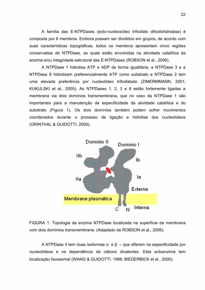
KUKULSKI et al., 2005). As NTPDases 1, 2, 3 e 8 estão fortemente ligadas a
membrana via dois domínios transmembrana, que no caso da NTPDase 1 são
importantes para a manutenção da especificidade da atividade catalítica e do
substrato (Figura 1). Os dois domínios também podem sofrer movimentos
coordenados durante o processo de ligação e hidrólise dos nucleotídeos
(GRINTHAL & GUIDOTTI, 2006).
FIGURA 1: Topologia da enzima NTPDase localizada na superfície da membrana
com dois domínios transmembrana. (Adaptado de ROBSON et al., 2006).
A NTPDase 4 tem duas isoformas α e β – que diferem na especificidade por
nucleotídeos e na dependência de cátions divalentes. Esta ectoenzima tem
localização lisossomal (WANG & GUIDOTTI, 1998; BIEDERBICK et al., 2000).
23
A NTPDase 5 apresenta um alto grau de especificidade para nucleotídeos
difosfatos (NTPs) e possui as porções C e N-terminal não hidrofóbicas (PÁEZ et al.,
2001).
A NTPDase 6 hidrolisa preferencialmente nucleosídeos 5’-difosfatos. A
análise imunohistoquímica sugere que a NTPDase 6 é associada ao Complexo de
Golgi e a pequenas extensões da membrana plasmática (BRAUN et al., 2000).
A NTPDase 7 possui localização subcelular e tem preferência por
nucleosídeos trifosfatos (SHI et al., 2001). A NTPDase 8 possui dois domínios
transmembrana, uma porção C-terminal e outra N-terminal. Essa NTPDase possui
poucos sítios de N-glicosilação e dois resíduos de aminoácidos na porção C-terminal
(SÉVIGNY et al., 2000; BIGONNESSE et al., 2004).
A atividade da NTPDase já foi caracterizada inicialmente em plaquetas de
ratos e posteriormente em plaquetas de humanos (FRASSETTO et al., 1993;
PILLA et al., 1996). As NTPDases em plaquetas intactas de humanos podem estar
envolvidas com a inibição da agregação plaquetária e regulação do tônus vascular
(SÉVIGNY et al., 2002). A CD39 solúvel bloqueou in vitro a agregação plaquetária
induzida por ADP e inibiu a reatividade plaquetária induzida por colágeno,
demonstrando uma importante função na tromboregulação (GAYLE III et al., 1998;
ENJYOJI et al., 1999; MARCUS et al., 2005). As respostas tromboregulatórias da
NTPDase podem ser observadas em estudos in vivo e in vitro que demonstraram
sua participação na homeostasia através de um potente efeito anti-trombótico
(MARCUS et al., 2005; COSTA et al., 2004).
A enzima 5'-nucleotidase (CD73, EC 3.1.3.5), catalisa especificamente a
hidrólise de NMP a adenosina (BARMAN, 1969; SARKIS et al.,1995). A
5'-nucleotidase é uma glicoproteína intrínseca da membrana plasmática de
diferentes tipos celulares como as plaquetas e também pode ser encontrada em
tecidos como nervoso, renal e hepático (ZIMMERMANN et al., 1993). A ativação da
enzima 5'-nucleotidase contribui para a inibição da agregação plaquetária por células
endoteliais humanas (KAWASHIMA et al., 2000). Na cascata de coagulação as
enzimas NTPDase e 5'-nucleotidase têm importante função na regulação da
agregação plaquetária (ENJYOJI et al., 1999).
As ecto-nucleotideo pirofosfatase/fosfodiesterase (E-NPPs) são encontradas
na superfície das células como proteínas transmembrana. As NPPs hidrolisam
pirofosfato ou fosfodiesterase em uma variedade de compostos extracelulares
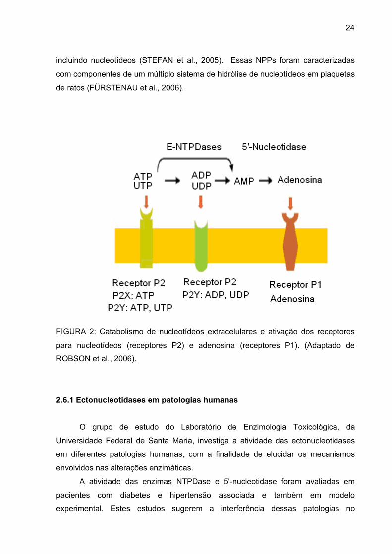
24
incluindo nucleotídeos (STEFAN et al., 2005). Essas NPPs foram caracterizadas
com componentes de um múltiplo sistema de hidrólise de nucleotídeos em plaquetas
de ratos (FÜRSTENAU et al., 2006).
FIGURA 2: Catabolismo de nucleotídeos extracelulares e ativação dos receptores
para nucleotídeos (receptores P2) e adenosina (receptores P1). (Adaptado de
ROBSON et al., 2006).
2.6.1 Ectonucleotidases em patologias humanas
O grupo de estudo do Laboratório de Enzimologia Toxicológica, da
Universidade Federal de Santa Maria, investiga a atividade das ectonucleotidases
em diferentes patologias humanas, com a finalidade de elucidar os mecanismos
envolvidos nas alterações enzimáticas.
A atividade das enzimas NTPDase e 5'-nucleotidase foram avaliadas em
pacientes com diabetes e hipertensão associada e também em modelo
experimental. Estes estudos sugerem a interferência dessas patologias no
25
mecanismo catalítico das ectonucleotidases (LUNKES et al., 2003, LUNKES et al.,
2004). Foi observado que em pacientes diabéticos sobrecarga com ferro promove
aumento na hidrólise de nucleotídeos de adenina (MIRON et al., 2007). A
investigação em gestantes com elevado risco de trombose sugere que as
ectonucleotidases estão envolvidas na tromboregulação (LEAL et al., 2007). O
aumento na expressão de CD39, em pacientes com hipercolesterolemia, foi uma
resposta compensatória ao processo inflamatório e pró-oxidativo associado com a
hipercolesterolemia (DUARTE et al., 2007).
Compostos de pirimidina foram observados como inibidores da atividade da
NTPDase em córtex cerebral de ratos (CECHIN et al., 2003). A interferência de
tratamento sub crônico de HgCl2 foi avaliada na atividade das enzimas NTPDase e
5´-nucleotidase em córtex cerebral de ratos tratados com este metal
(MORETTO et al., 2004). A interferência da exposição crônica de alumínio na
atividade das enzimas NTPDase e 5'-nucleotidase foi avaliada em plaquetas, córtex
cerebral e hipocampo de modelo experimental, indicando que as plaquetas podem
servir como marcadores da toxicidade do alumínio no sistema nervoso central
(KAIZER et al., 2007).
A atividade da enzima NTPDase foi inicialmente caracterizada em linfócitos
humanos (LEAL et al., 2005). Posteriormente, foi observado um aumento na
atividade da NTPDase em pacientes com a infecção por HIV e sua correlação
positiva com CD39 em linfócitos (LEAL et al., 2005). A hidrólise de nucleotídeos de
adenina em pacientes com câncer de mama demonstrou que a atividade da
NTPDase depende do estágio do câncer (ARAÚJO et al., 2005).
A atividade das ectonucleotidases também foi avaliada em modelos
experimentais de desmielinização pelo brometo de etídio. No tratamento com
interferon beta pode se observar que a hidrólise dos nucleotídeos de adenina está
modificada em plaquetas de ratos desmielinizados (SPAVANELLO et al., 2007). Por
outro lado, o tratamento com ebselen e vitamina E não modificou a atividade da
enzima 5´-nucleotidase. Porém, a atividade da enzima NTPDase ficou diminuída em
ratos desmielinizados e o ebselen e a vitamina E interfere na hidrólise dos
nucleotídeos da adenina (MAZZANTI et al., 2007).
A atividade das enzimas NTPDase e 5'-nucleotidase foi avaliada em pacientes
com insuficiência renal. Os dados demonstraram uma alteração na hidrólise de
nucleotídeos em plaquetas de pacientes com alteração renal submetidos à
26
hemodiálise. Possivelmente, as mudanças na atividade das ectonucleotidases
poderiam contribuir para as alterações na homeostasia de pacientes com
insuficiência renal crônica (SILVA et al., 2005).
2.7 Estresse oxidativo
A presença de estresse oxidativo nas células é proveniente essencialmente
da perda de equilíbrio entre os processos oxidantes e antioxidantes, com
conseqüente falência no reparo do dano oxidativo (SCHAFER & BUETTNER, 2001).
Com isso, as células danificadas promovem a produção de espécies reativas de
oxigênio e nitrogênio, que compreendem radicais hidroxil, superóxido, peróxido de
hidrogênio e peroxinitrito (VALKO et al., 2005). As espécies reativas de oxigênio são
altamente reativas e constituem estruturas moleculares que reagem com diversos
componentes celulares como o DNA, as proteínas, os lipídios e os produtos finais da
glicação avançada. Essas reações entre os componentes celulares e as espécies
reativas de oxigênio e nitrogênio promovem danos no DNA, distúrbios na formação
mitocondrial, danos na membrana celular e eventualmente morte celular
(LELLI et al., 1998).
O nível de estresse oxidativo tem sido relacionado com patologias crônicas,
como o diabetes, o Alzheimer e o Parkinson (McGRATH, L.T. et al., 2001; JENNER,
2003). O estresse oxidativo pode ser importante no diabetes, porque a cronicidade
da hiperglicemia e também a resistência à insulina podem induzir ao dano oxidativo
e contribuir para a destruição de células beta pancreáticas (KING & LOEKEN, 2004).
Os sistemas antioxidantes de defesa constituem-se em estruturas
moleculares capazes de capturar os radicais livres e com isso prevenir os danos
oxidativos nas células. A hiperglicemia estimula o aumento na geração de espécies
reativas de oxigênio e induz a um incremento na atividade das enzimas superóxido
dismutase (SOD, EC 1.15.1.1), catalase (CAT, EC 1.11.1.6), nos níveis de grupos
tióis não protéicos (NPSH), proteína carbonil e TBARS em pacientes diabéticos.
Esses dados sugerem que sistemas antioxidantes podem ser considerados como
marcadores de injúria vascular em diabéticos (AHMED et al., 2006; RAMAKRISHNA
& JAILKHANI, 2007). O ácido ascórbico faz parte de um sistema antioxidante não
enzimático responsável pela remoção de radicais livres. A vitamina C, em baixas
27
concentrações em pacientes diabéticos, tem sido relacionada com o incremento dos
níveis de estresse oxidativo (SKRHA et al., 2003).
A produção de espécies reativas de oxigênio constitui um importante
mecanismo de ativação e agregação plaquetária, com extrema relevância no
recrutamento de plaquetas para a formação do trombo (KRÖTZ et al., 2004). O
estresse oxidativo, induzido pela hiperglicemia, é responsável pela ativação da
condição pró-trombose, ativação inicial de plaquetas, adesão e subseqüente
formação de agregação plaquetária. Por isso, o controle metabólico da glicemia é
fundamental para as funções plaquetárias em diabéticos (FERRONI et al., 2004).
Também, a atividade da enzima NTPDase em plaquetas pode ser suscetível a
radicais livres (FRASSETTO et al., 1997).
28
3 ARTIGOS CIENTÍFICOS
Os resultados que fazem parte desta tese estão apresentados sob a forma de
artigos científicos e manuscritos, os quais encontram-se aqui organizados. O artigo
está disposto da mesma forma que foi publicado na edição da revista científica
(Artigo 1). Os manuscritos estão dispostos da mesma forma que foram submetidos
na edição da revista científica (Manuscrito 1 e 2) e na fase de redação
(Manuscrito 3).
29
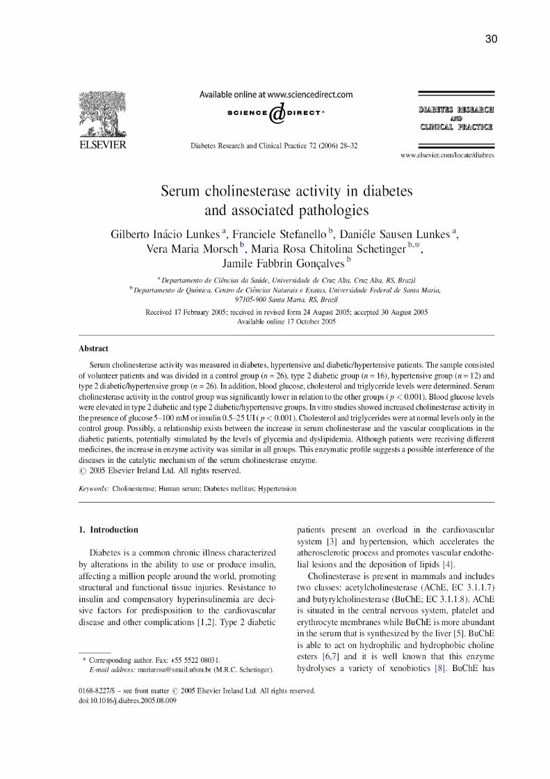
3.1 Artigo 1
O artigo “Serum cholinestease activity in diabetes and associated pathologies”
foi publicado no periódico “Diabetes Research and Clinical Practice”.
30
31
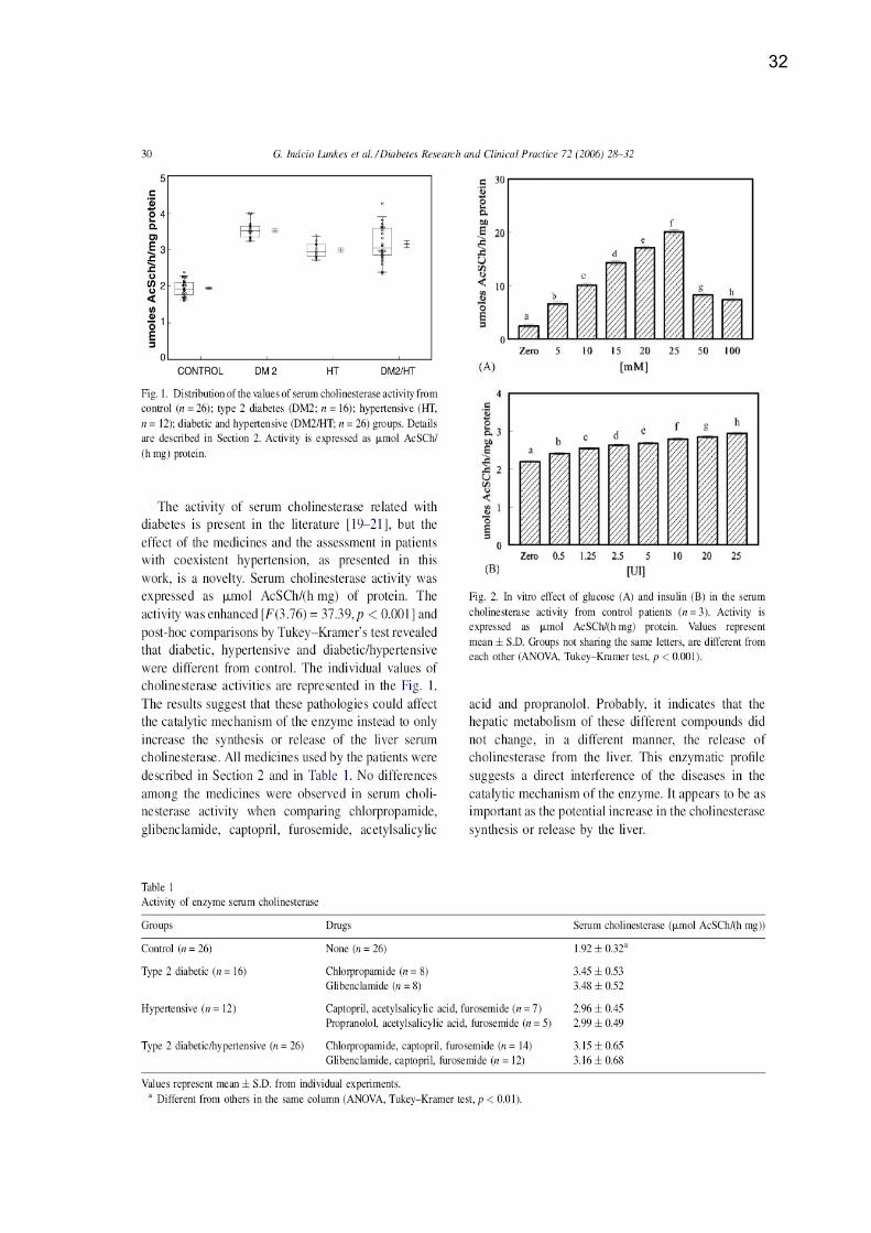
32
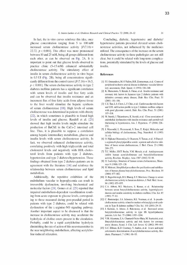
33
34
35
3.2 Manuscrito 1
O manuscrito “Effect of high glucose levels in human platelet NTPDase and
5'-nucleotidase activities” foi submetido ao periódico “Diabetes Research and
Clinical Practice”.
36
Effect of high glucose levels in human platelet
NTPDase and 5'-nucleotidase activities
Gilberto Inácio Lunkesa, Daniéle Sausen Lunkesa, Daniela Lealb, Maria do Carmo
Araújob, Vera Maria Morscha, Maria Rosa Chitolina Schetingera*
a Departamento de Química, Centro de Ciências Naturais e Exatas, Programa de
Pós-Graduação em Bioquímica Toxicológica, Universidade Federal de Santa Maria,
Santa Maria, RS 97105-900, Brazil.
b Hospital Universitário de Santa Maria, Universidade Federal de Santa Maria, Santa
Maria, RS, Brazil.
* Corresponding author. Departamento de Química, CCNE, Universidade Federal de
Santa Maria, Santa Maria, RS 97105-900, Brazil. Fax: +55-5532208978
e-mail addresses: [email protected] or [email protected] (M.R.C.
Schetinger)
37
Abstract
Objectives: We attempt to evaluate the effect of glucose levels in human platelet
ectonucleotidases activities in patients with diabetes or hypertension.
Methods: The activities of the enzymes NTPDase (CD39) and 5'-nucleotidase
(CD73), and CD39 expression were analyzed in human blood platelets of type 2
Diabetes mellitus (DM-2), hypertension (HT) and type 2 Diabetes
mellitus/hypertension (DM-2/HT) goups. The interference of glucose and fructose on
the NTPDase and 5'-nucleotidase in platelets from control patients was also verified.
Results: NTPDase and 5'-nucleotidase activities increased with increasing glucose
and fructose concentrations (p < 0.001) and the different times of pre-incubation did
not interfere in ectonucleotidase activities (p > 0.5). NTPDase and 5'-nucleotidase
activities demonstrated a positive correlation between serum glucose levels and ATP
and ADP hydrolysis in DM-2 and DM-2/HT patients. CD39 expression demonstrated
that DM-2, HT and DM-2/HT groups presented a significant increase (p < 0.004)
when compared to the control group.
Conclusion: The hydrolysis of adenine nucleotides is enhanced in platelets of
patients with diabetes and hypertension. We observed that an increasing glucose
concentration had a direct effect on ectonucleotidases activities. Furthermore, CD39
expression was enhanced in all patients groups. These results suggest that
hyperglycemia interferes in platelet homeostasis and hydrolysis nucleotides are
important facto to thromboregulation.
Keywords: Hyperglycemia; NTPDase; 5'-nucleotidase; Diabetes; Hypertension;
Platelet
38
1. Introduction
Platelets play an important role in hemostasis and thrombosis. These anuclear
cells act via adhesion and aggregation which allow thrombus formation at the site of
vascular injury [1,2,3]. Increased platelet aggregation may result in thromboembolic
events and contribute to acute coronary syndromes [4,5].
Adenine nucleotides are released from dense granules during platelet
activation [6,7]. Adenosine triphosphate (ATP) has been suggested to have a role in
the regulation of platelet aggregation [8,9]. Adenosine diphosphate (ADP) plays a
very important role in thrombogenesis being the main promoter of platelet
aggregation [10,11]. Adenosine is an endogenous inhibitor of platelet aggregation
and interferes in vascular tone [12]. These adenine nucleotides and nucleosides act
via purinoreceptors [6-12].
The family of ectonucleotidases includes enzymes that degrade extracellular
nucleotides. The enzymes NTPDase (nucleoside triphosphate diphosphohydrolase
CD39) and 5'-nucleotidase (CD73) are located in the platelet membrane and
complete the hydrolysis of ATP to adenosine [13,14]. Both enzymes CD39 and
CD73 play an important role in hemostasis and platelet aggregation mainly by
regulating ADP catabolism and adenosine production [15-19]. Recently, studies
observing alterations in NTPDase and 5'-nucleotidase activities in blood platelets
suggested that these ectonucleotidases are involved in the thromboregulation
process in several physiological and pathological conditions [20-22].
Diabetes mellitus is an important risk factor for vascular complications and
thrombus formation [23]. Chronic hyperglycemia promotes platelet activation and can
contribute to vascular events [24,25]. Platelet hyperactivation contributes to
39
increased risk of atherothrombosis in type 2 diabetes [26]. High glucose
concentrations, when chronic, promote alteration in ATP/ADP levels and may be an
important factor involved in platelet hyperactivity in the course of diabetes [27]. The
ectonucleotidases, CD39 and CD73, in platelets, are altered in type 2 diabetic and
hypertensive patients and probably such modifications are compensatory
physiological responses related with the thromboregulation process [21,28].
Previous studies in our laboratory demonstrated an increase in
ectonucleotidase activities in patients with diabetes and associated pathologies [21].
However, the mechanism by which it occurred was not completely understood. Thus,
in the present study we attempt to evaluate the effect glucose levels in human blood
platelets ectonucleotidases activities in these groups of patients.
2. Material and Methods
2.1 Chemicals
Nucleotides, sodium azide, HEPES, and Trizma base were purchased from
Sigma (St. Louis, MO). Antibodies for flow cytometry analysis [R-phycoerythrin-
conjugated mouse monoclonal antibody against human CD39, and fluorescein
isothiocyanate-conjugated mouse monoclonal antibody against human CD61 were
purchased from Serotec Ltd. (Kidlington, Oxford, UK) and BD PharMingen Technical
Data Sheet (San Jose, CA, USA), respectively. The glucose, cholesterol,
HDL-cholesterol, triglycerides and lactate dehydrogenase (LDH) commercial kits
were obtained from Labtest (Lagoa Santa, MG, Brazil). All other reagents used in the
experiments were of analytical grade and of the highest purity.
40
2.2 Patients
The sample consisted of patients from the Assistance Program for diabetic
and hypertensive patients associated with the Municipal Secretary’s Office of Public
Health in Cruz Alta (RS, Brazil) as well as of healthy volunteers. All subjects gave
written informed consent to participate in the study. The protocol was approved by
the Human Ethics Committee of the Health Science Center from the Federal
University of Santa Maria (Protocol number: 013/2004).
The sample was divided into four groups consisting of 50% males and 50%
females. The control group (n=9) consisted of individuals with ages ranging from 28
to 52 years, who did not present any disease and who had not been submitted to any
pharmacological therapy during the last month. Controls were carefully selected by
clinical evaluation and presented sex, age and body mass indices similar to those of
the patients. The type 2 diabetic (DM-2, n=8) group consisted of patients with ages
ranging from 56 to 68 years. The patients of the DM-2 group had type 2 diabetes
mellitus and were treated with glibenclamide (10 mg/day) or mettformin
(850 mg/day). The hypertensive (HT, n=9) group was made up of patients with ages
ranging from 30 to 70 years. The patients of the HT group had different hypertension
levels and were treated with captopril (25 mg/day), furosemide (40 mg/day),
acetylsalicylic acid (100 mg/day) or propranolol (40 mg/day). The type 2
diabetic/hypertensive (DM-2/HT, n=9) group consisted of patients with ages ranging
from 51 to 69 years. All patients of the DM-2/HT had type 2 diabetes mellitus plus
hypertension and received appropriate medication for the associated diseases. Ten
milliliters of blood was obtained from each participant and used for platelet-rich
plasma preparations, biochemical determinations and hematological determinations.
41
2.3 Hematologic determinations
Quantitative determinations of platelets obtained by venipuncture were
performed using a Coulter-STKS analyzer (Miami, USA).
2.4 Biochemical determinations
Serum glucose, cholesterol, triglycerides and lactate dehydrogenase (LDH)
were determined by spectrophotometry, using commercial kits.
2.5 Platelet-rich plasma (PRP) preparation
PRP was prepared from human donors by methods previously published [13].
Briefly, blood was collected into 0.129 mol/L citrate and centrifuged at 160 g for
10 min. The PRP was centrifuged at 1400 g for 15 min and washed twice with
3.5 mmol/L Hepes isosmolar buffer containing 142 mmol/L NaCl, 2.5 mmol/L KCl,
and 5.5 mmol/L glucose. The washed platelets were resuspended in Hepes
isosmolar buffer, and protein was adjusted to 0.3–0.5 mg/mL.
2.6 NTPDase and 5'-nucleotidase assays
Platelet NTPDase was incubated as previously described [13], with
5.0 mmol/L CaCl2, 100 mmol/L NaCl, 4.0 mmol/L KCl, 5.0 mmol/L glucose, and
50 mmol/L Tris–HCl, pH 7.4, at a final volume of 200 µL. The total quantity of 20 µL
of the enzyme preparation (10–15 µg of protein) was added to the reaction mixture
and pre-incubated for 10 min at 37oC. The reaction was started by the addition of
1.0 mmol/L of ATP or ADP. The activity of 5'-nucleotidase was assayed using the
same conditions except that 5.0 mmol/L of MgCl2 and 2.0 mmol/L of AMP were used.
The ectonucleotidases reactions were stopped after one hour of incubation with
42
trichloroacetic acid (TCA 10%), at a final concentration of 5%. The Pi released was
measured by the method of Chan et al. (1984) [29] using Malachite Green as
coloring reagent. The enzymatic activities were described in nmol Pi/min/mg of
protein. All samples were run in triplicate.
2.7 Glucose and fructose curve
To evaluate the glucose and fructose levels in NTPDase and 5'-nucleotidase
activities, we performed experiments with glucose/fructose concentrations ranging
from 5 to 100 mM in platelet-rich plasma (PRP) from control subjects. Pre-incubation
times of 10, 120 minutes and 24 hours were used.
2.8 Flow cytometry analysis
Peripheral blood cells were incubated with anti-CD39 and anti-CD61 (20 µL
per 106 cells) for 25 min, lysed with fluorescence activated cell sorter (FACS)
reagent, and incubated again for 15 min in the dark. Cells were washed twice in
NaCl/Pi buffer (pH 7.4) containing 0.02% (w/v) sodium azide and 0.2% (w/v) BSA.
The cells were then resuspended in NaCl/Pi buffer (pH 7.4) and immediately
analyzed with a FACScalibur flow cytometer using cellquest software (Becton
Dickinson, San Jose, CA, USA), without fixation.
2.9 Protein determination
Proteins were determined by the Coomassie Blue method [30], using bovine
serum albumin (BSA) as standard.
43
2.10 Statistical analysis
Data were analyzed statistically by two-way and one-way ANOVA, followed by
Duncan’s multiple range test. Differences between groups were considered to be
significant when p < 0.05. All data were expressed as mean ± S.D. Correlation was
evaluated with the Pearson test. Linear correlation between variables was also
carried out.
3. Results
The patient’s characteristics are shown in Table 1. Glucose levels were normal
(3.8–6.1 mmol/L) in control and HT groups, and higher in DM-2 (141.5%) and
DM-2/HT (136.6%) groups (p<0.05). The lipid profile of the pathological groups were
different from the control group (p<0.05). Total cholesterol levels (<5.1 mmol/L)
presented an increase in DM-2 (30.1%), HT (33.3%) and DM-2/HT (47.6%).
Triglyceride levels (<2.2 mmol/L) presented an increase of 135% in DM-2, HT and
DM-2/HT groups. Quantitative analysis demonstrated that platelet counts obtained
from all groups were at normal levels (150.000 – 400.000 platelets/mm3). Microscopic
analysis of platelet size and shape revealed a typical pattern (data not shown).
Platelet integrity was determined by lactate dehydrogenase activity from control
patients. The measurements of LDH showed that most cells (more that 90%) were
intact after the isolation procedure and PRP was adequate (data not shown).
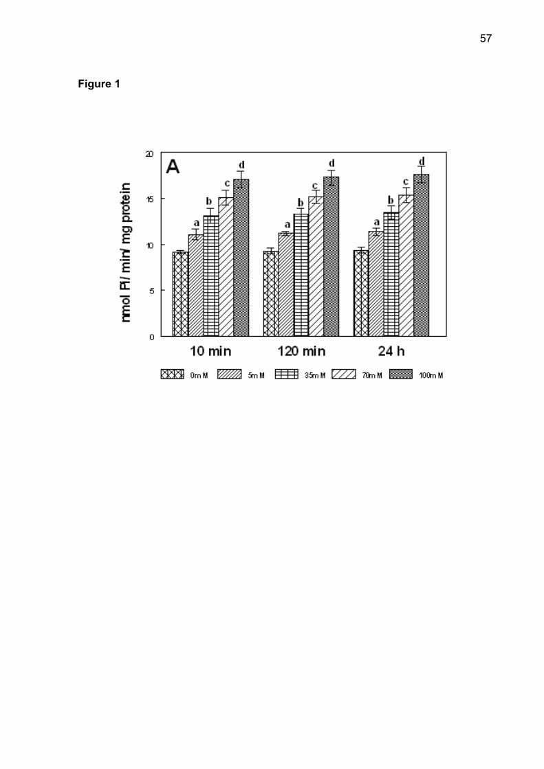
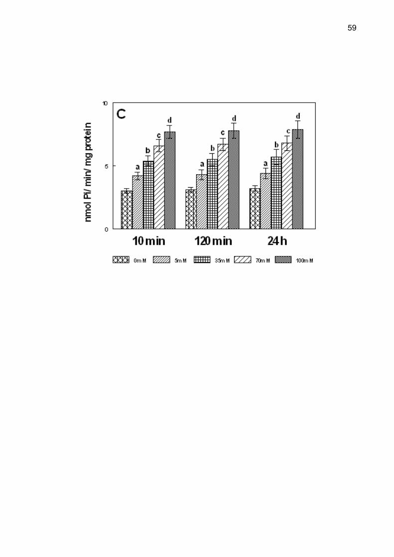
The different times of pre-incubation in the glucose curve in vitro did not
interfere in ectonucleotidases activities (p > 0.5). The effect of glucose on NTPDase
and 5'-nucleotidase is shown in Figure 1. The increase in ATP, ADP and AMP
hydrolysis was observed in all pre-incubation times. Post-hoc comparisons by
Duncan’s test revealed that NTPDase and 5'-nucleotidase activities were significantly
44
higher with increasing glucose concentrations between 5 and 100 mM (p < 0.001).
The effect of fructose on NTPDase and 5'-nucleotidase was similar to that of glucose
(data not shown).
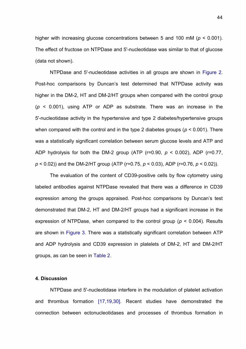
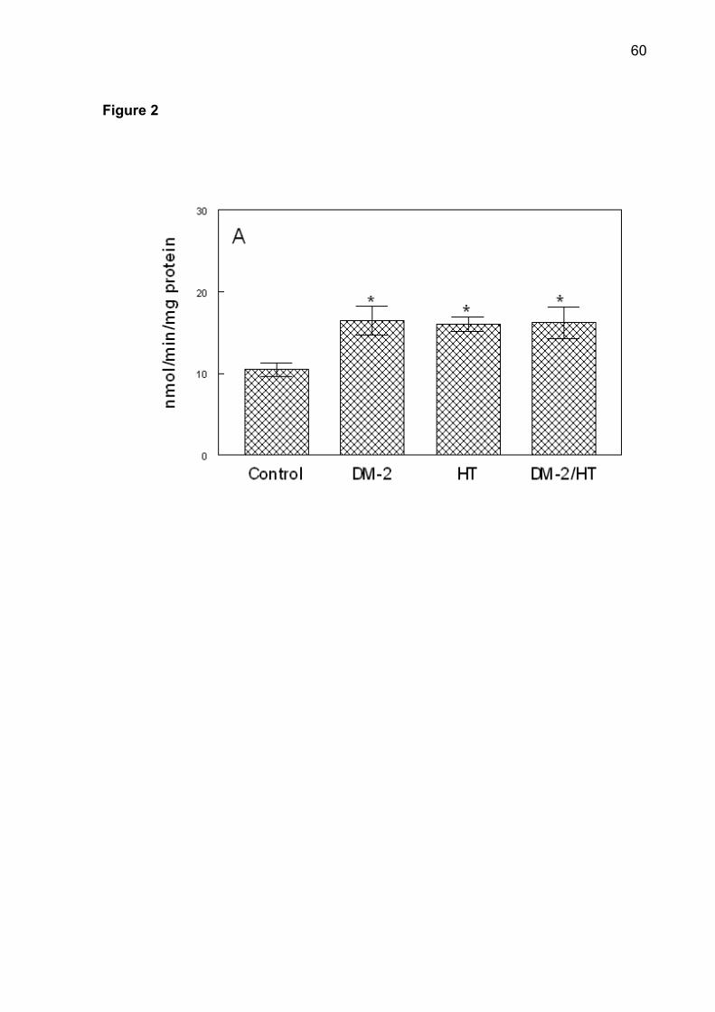
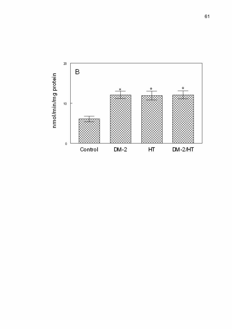
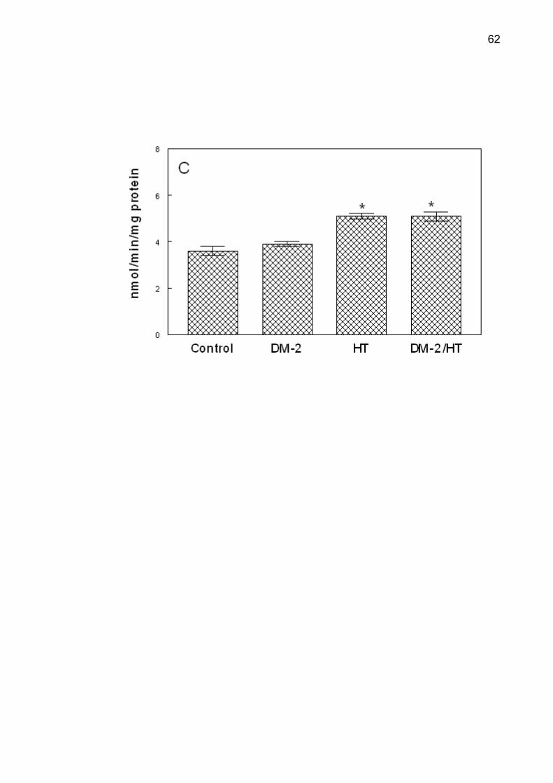
NTPDase and 5'-nucleotidase activities in all groups are shown in Figure 2.
Post-hoc comparisons by Duncan’s test determined that NTPDase activity was
higher in the DM-2, HT and DM-2/HT groups when compared with the control group
(p < 0.001), using ATP or ADP as substrate. There was an increase in the
5'-nucleotidase activity in the hypertensive and type 2 diabetes/hypertensive groups
when compared with the control and in the type 2 diabetes groups (p < 0.001). There
was a statistically significant correlation between serum glucose levels and ATP and
ADP hydrolysis for both the DM-2 group (ATP (r=0.90, p < 0.002), ADP (r=0.77,
p < 0.02)) and the DM-2/HT group (ATP (r=0.75, p < 0.03), ADP (r=0.76, p < 0.02)).
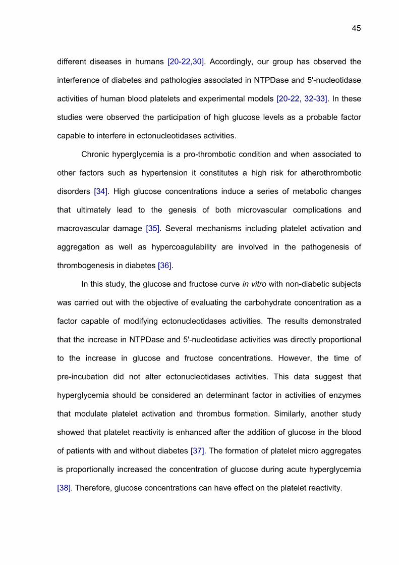
The evaluation of the content of CD39-positive cells by flow cytometry using
labeled antibodies against NTPDase revealed that there was a difference in CD39
expression among the groups appraised. Post-hoc comparisons by Duncan’s test
demonstrated that DM-2, HT and DM-2/HT groups had a significant increase in the
expression of NTPDase, when compared to the control group (p < 0.004). Results
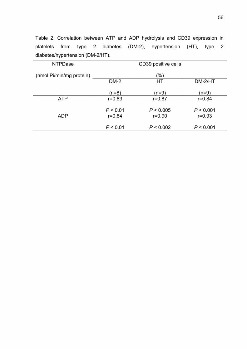
are shown in Figure 3. There was a statistically significant correlation between ATP
and ADP hydrolysis and CD39 expression in platelets of DM-2, HT and DM-2/HT
groups, as can be seen in Table 2.
4. Discussion
NTPDase and 5'-nucleotidase interfere in the modulation of platelet activation
and thrombus formation [17,19,30]. Recent studies have demonstrated the
connection between ectonucleotidases and processes of thrombus formation in
45
different diseases in humans [20-22,30]. Accordingly, our group has observed the
interference of diabetes and pathologies associated in NTPDase and 5'-nucleotidase
activities of human blood platelets and experimental models [20-22, 32-33]. In these
studies were observed the participation of high glucose levels as a probable factor
capable to interfere in ectonucleotidases activities.
Chronic hyperglycemia is a pro-thrombotic condition and when associated to
other factors such as hypertension it constitutes a high risk for atherothrombotic
disorders [34]. High glucose concentrations induce a series of metabolic changes
that ultimately lead to the genesis of both microvascular complications and
macrovascular damage [35]. Several mechanisms including platelet activation and
aggregation as well as hypercoagulability are involved in the pathogenesis of
thrombogenesis in diabetes [36].
In this study, the glucose and fructose curve in vitro with non-diabetic subjects
was carried out with the objective of evaluating the carbohydrate concentration as a
factor capable of modifying ectonucleotidases activities. The results demonstrated
that the increase in NTPDase and 5'-nucleotidase activities was directly proportional
to the increase in glucose and fructose concentrations. However, the time of
pre-incubation did not alter ectonucleotidases activities. This data suggest that
hyperglycemia should be considered an determinant factor in activities of enzymes
that modulate platelet activation and thrombus formation. Similarly, another study
showed that platelet reactivity is enhanced after the addition of glucose in the blood
of patients with and without diabetes [37]. The formation of platelet micro aggregates
is proportionally increased the concentration of glucose during acute hyperglycemia
[38]. Therefore, glucose concentrations can have effect on the platelet reactivity.
46
The ectonucleotidases activities were increased in patients with diabetes and
associated hypertension. Studies have shown that patients with chronic
hyperglycemia frequently have hypercoagulable blood, as evidenced by increased
plasmatic coagulators, reduced endothelial thromboresistance and platelet
hyperactivity [39]. The positive correlation between serum glucose concentration and
ATP and ADP hydrolysis in type 2 diabetic and type 2 diabetic/hypertensive patients
demonstrates that hyperglycemia is an important factor capable of interfering in
ectonucleotidases activities. Hypertension does not generally exist in isolation, but it
occurs in the setting of concomitant risk factors. Platelet activation and fibrinolysis
function are strongly associated with the level of blood pressure, which is associated
with coexisting risk factors such as diabetes mellitus and dyslipidemia [40].
This study showed that NTPDase (CD39) expression in platelet membranes
was great in patients of the pathological groups. This data confirmed the increase
NTPDase activity in platelet membrane of patients with diabetes and hypertension. A
previous study showed that acute hyperglycemia causes platelet hyperactivity to
agonist stimulation [41]. There was a positive correlation between ATP and ADP
hydrolysis and NTPDase expression in platelets of patients with diabetes and
diabetes/hypertension. These data demonstrate that diabetes and hypertension may
have involvement in the catalytic mechanism of the NTPDase in human blood
platelets.
The chronic hyperglycemia presents multiple mechanisms involved in platelet
hyperactivity as non-enzymatic glycation and sorbitol accumulation [42, 43].
Normally, these mechanisms require a long periods of elevated glucose levels.
However, acute hyperglycemia may also alter platelet function. Short exhibition at
elevated levels of glucose can to involve increase protein kinase C, enhances
47
collagen-induced platelet aggregation via increase mitochondrial superoxide
production [44]. Previous study demonstrated that elevated osmolality may changes
of platelet function [45].
Therefore the hyperglycemia may be promoting an excessive liberation of ATP
and ADP of blood platelets. The platelet hyperactivity with increase in the hydrolysis
of adenine nucleotides demonstrates a potential compensatory answer in patients
with diabetes in function of elevated glucose levels. This compensatory mechanism
at hyperglycemia may promote changes of platelet signaling.
In conclusion, our study demonstrated that diabetes and hypertension
interfered in the NTPDase activity increasing the hydrolysis of adenine nucleotides in
human platelets. We observed that an increasing glucose concentration had a direct
effect on ectonucleotidases activities. These results allow us to suggest that
hyperglycemia may be an important factor in platelet homeostasis and ATP, ADP
and AMP hydrolysis are important parameters in the thromboregulation process.
48
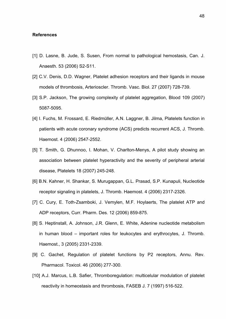
References
[1] D. Lasne, B. Jude, S. Susen, From normal to pathological hemostasis, Can. J.
Anaesth. 53 (2006) S2-S11.
[2] C.V. Denis, D.D. Wagner, Platelet adhesion receptors and their ligands in mouse
models of thrombosis, Arterioscler. Thromb. Vasc. Biol. 27 (2007) 728-739.
[3] S.P. Jackson, The growing complexity of platelet aggregation, Blood 109 (2007)
5087-5095.
[4] I. Fuchs, M. Frossard, E. Riedmüller, A.N. Laggner, B. Jilma, Platelets function in
patients with acute coronary syndrome (ACS) predicts recurrent ACS, J. Thromb.
Haemost. 4 (2006) 2547-2552.
[5] T. Smith, G. Dhunnoo, I. Mohan, V. Charlton-Menys, A pilot study showing an
association between platelet hyperactivity and the severity of peripheral arterial
disease, Platelets 18 (2007) 245-248.
[6] B.N. Kahner, H. Shankar, S. Murugappan, G.L. Prasad, S.P. Kunapuli, Nucleotide
receptor signaling in platelets, J. Thromb. Haemost. 4 (2006) 2317-2326.
[7] C. Cury, E. Toth-Zsamboki, J. Vemylen, M.F. Hoylaerts, The platelet ATP and
ADP receptors, Curr. Pharm. Des. 12 (2006) 859-875.
[8] S. Heptinstall, A. Johnson, J.R. Glenn, E. White, Adenine nucleotide metabolism
in human blood – important roles for leukocytes and erythrocytes, J. Thromb.
Haemost., 3 (2005) 2331-2339.
[9] C. Gachet, Regulation of platelet functions by P2 receptors, Annu. Rev.
Pharmacol. Toxicol. 46 (2006) 277-300.
[10] A.J. Marcus, L.B. Safier, Thromboregulation: multicelular modulation of platelet
reactivity in homeostasis and thrombosis, FASEB J. 7 (1997) 516-522.
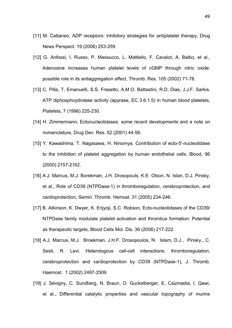
49
[11] M. Cattaneo, ADP receptors: inhibitory strategies for antiplatelet therapy, Drug
News Perspect. 19 (2006) 253-259.
[12] G. Anfossi, I. Russo, P. Massucco, L. Mattiello, F. Cavalot, A. Balbo, et al.,
Adenosine increases human platelet levels of cGMP through nitric oxide:
possible role in its antiaggregation effect, Thromb. Res. 105 (2002) 71-78.
[13] C. Pilla, T. Emanuelli, S.S. Frasetto, A.M.O. Battastini, R.D. Dias, J.J.F. Sarkis,
ATP diphosphoydrolase activity (apyrase, EC 3.6.1.5) in human blood platelets,
Platelets, 7 (1996) 225-230.
[14] H. Zimmermann, Ectonucleotidases: some recent developments and a note on
nomenclature, Drug Dev. Res. 52 (2001) 44-56.
[15] Y. Kawashima, T. Nagasawa, H. Ninomya, Contribution of ecto-5'-nucleotidase
to the inhibition of platelet aggregation by human endothelial cells, Blood, 96
(2000) 2157-2162.
[16] A.J. Marcus, M.J. Borekman, J.H. Drosopouls, K.E. Olson, N. Islan, D.J. Pinsky,
et al., Role of CD39 (NTPDase-1) in thromboregulation, cerebroprotection, and
cardioprotection, Semin. Thromb. Hemost. 31 (2005) 234-246.
[17] B. Atkinson, K. Dwyer, K. Enjyoji, S.C. Robson, Ecto-nucleotidases of the CD39/
NTPDase family modulate platelet activation and thrombus formation: Potential
as therapeutic targets, Blood Cells Mol. Dis. 36 (2006) 217-222.
[18] A.J. Marcus, M.J. Broekman, J.H.F. Drosopoulos, N. Islam, D.J., Pinsky., C.
Sesti, R. Levi. Heterologous cell-cell interactions: thromboregulation,
cerebroprotection and cardioprotection by CD39 (NTPDase-1), J. Thromb.
Haemost. 1 (2002) 2497-2509.
[19] J. Sévigny, C. Sundberg, N. Braun, O. Guckelberger, E. Csizmadia, I. Qawi,
et al., Differential catalytic properties and vascular topography of murine
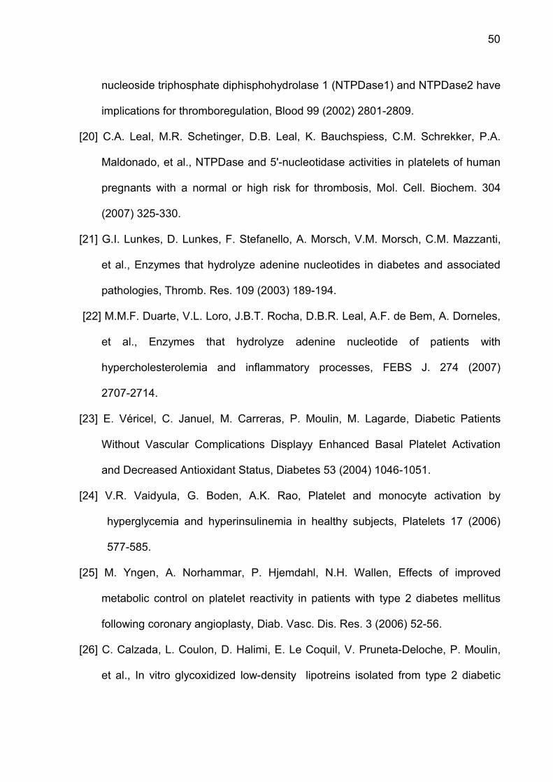
50
nucleoside triphosphate diphisphohydrolase 1 (NTPDase1) and NTPDase2 have
implications for thromboregulation, Blood 99 (2002) 2801-2809.
[20] C.A. Leal, M.R. Schetinger, D.B. Leal, K. Bauchspiess, C.M. Schrekker, P.A.
Maldonado, et al., NTPDase and 5'-nucleotidase activities in platelets of human
pregnants with a normal or high risk for thrombosis, Mol. Cell. Biochem. 304
(2007) 325-330.
[21] G.I. Lunkes, D. Lunkes, F. Stefanello, A. Morsch, V.M. Morsch, C.M. Mazzanti,
et al., Enzymes that hydrolyze adenine nucleotides in diabetes and associated
pathologies, Thromb. Res. 109 (2003) 189-194.
[22] M.M.F. Duarte, V.L. Loro, J.B.T. Rocha, D.B.R. Leal, A.F. de Bem, A. Dorneles,
et al., Enzymes that hydrolyze adenine nucleotide of patients with
hypercholesterolemia and inflammatory processes, FEBS J. 274 (2007)
2707-2714.
[23] E. Véricel, C. Januel, M. Carreras, P. Moulin, M. Lagarde, Diabetic Patients
Without Vascular Complications Displayy Enhanced Basal Platelet Activation
and Decreased Antioxidant Status, Diabetes 53 (2004) 1046-1051.
[24] V.R. Vaidyula, G. Boden, A.K. Rao, Platelet and monocyte activation by
hyperglycemia and hyperinsulinemia in healthy subjects, Platelets 17 (2006)
577-585.
[25] M. Yngen, A. Norhammar, P. Hjemdahl, N.H. Wallen, Effects of improved
metabolic control on platelet reactivity in patients with type 2 diabetes mellitus
following coronary angioplasty, Diab. Vasc. Dis. Res. 3 (2006) 52-56.
[26] C. Calzada, L. Coulon, D. Halimi, E. Le Coquil, V. Pruneta-Deloche, P. Moulin,
et al., In vitro glycoxidized low-density lipotreins isolated from type 2 diabetic
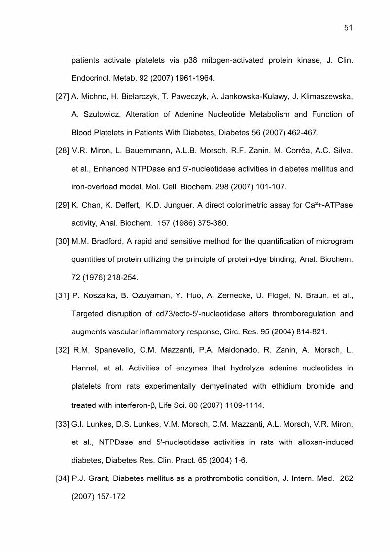
51
patients activate platelets via p38 mitogen-activated protein kinase, J. Clin.
Endocrinol. Metab. 92 (2007) 1961-1964.
[27] A. Michno, H. Bielarczyk, T. Paweczyk, A. Jankowska-Kulawy, J. Klimaszewska,
A. Szutowicz, Alteration of Adenine Nucleotide Metabolism and Function of
Blood Platelets in Patients With Diabetes, Diabetes 56 (2007) 462-467.
[28] V.R. Miron, L. Bauernmann, A.L.B. Morsch, R.F. Zanin, M. Corrêa, A.C. Silva,
et al., Enhanced NTPDase and 5'-nucleotidase activities in diabetes mellitus and
iron-overload model, Mol. Cell. Biochem. 298 (2007) 101-107.
[29] K. Chan, K. Delfert, K.D. Junguer. A direct colorimetric assay for Ca²+-ATPase
activity, Anal. Biochem. 157 (1986) 375-380.
[30] M.M. Bradford, A rapid and sensitive method for the quantification of microgram
quantities of protein utilizing the principle of protein-dye binding, Anal. Biochem.
72 (1976) 218-254.
[31] P. Koszalka, B. Ozuyaman, Y. Huo, A. Zernecke, U. Flogel, N. Braun, et al.,
Targeted disruption of cd73/ecto-5'-nucleotidase alters thromboregulation and
augments vascular inflammatory response, Circ. Res. 95 (2004) 814-821.
[32] R.M. Spanevello, C.M. Mazzanti, P.A. Maldonado, R. Zanin, A. Morsch, L.
Hannel, et al. Activities of enzymes that hydrolyze adenine nucleotides in
platelets from rats experimentally demyelinated with ethidium bromide and
treated with interferon-β, Life Sci. 80 (2007) 1109-1114.
[33] G.I. Lunkes, D.S. Lunkes, V.M. Morsch, C.M. Mazzanti, A.L. Morsch, V.R. Miron,
et al., NTPDase and 5'-nucleotidase activities in rats with alloxan-induced
diabetes, Diabetes Res. Clin. Pract. 65 (2004) 1-6.
[34] P.J. Grant, Diabetes mellitus as a prothrombotic condition, J. Intern. Med. 262
(2007) 157-172
52
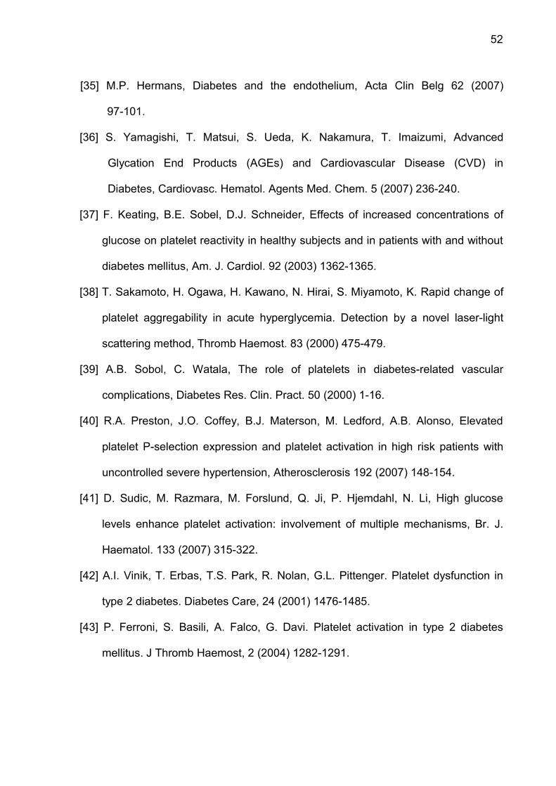
[35] M.P. Hermans, Diabetes and the endothelium, Acta Clin Belg 62 (2007)
97-101.
[36] S. Yamagishi, T. Matsui, S. Ueda, K. Nakamura, T. Imaizumi, Advanced
Glycation End Products (AGEs) and Cardiovascular Disease (CVD) in
Diabetes, Cardiovasc. Hematol. Agents Med. Chem. 5 (2007) 236-240.
[37] F. Keating, B.E. Sobel, D.J. Schneider, Effects of increased concentrations of
glucose on platelet reactivity in healthy subjects and in patients with and without
diabetes mellitus, Am. J. Cardiol. 92 (2003) 1362-1365.
[38] T. Sakamoto, H. Ogawa, H. Kawano, N. Hirai, S. Miyamoto, K. Rapid change of
platelet aggregability in acute hyperglycemia. Detection by a novel laser-light
scattering method, Thromb Haemost. 83 (2000) 475-479.
[39] A.B. Sobol, C. Watala, The role of platelets in diabetes-related vascular
complications, Diabetes Res. Clin. Pract. 50 (2000) 1-16.
[40] R.A. Preston, J.O. Coffey, B.J. Materson, M. Ledford, A.B. Alonso, Elevated
platelet P-selection expression and platelet activation in high risk patients with
uncontrolled severe hypertension, Atherosclerosis 192 (2007) 148-154.
[41] D. Sudic, M. Razmara, M. Forslund, Q. Ji, P. Hjemdahl, N. Li, High glucose
levels enhance platelet activation: involvement of multiple mechanisms, Br. J.
Haematol. 133 (2007) 315-322.
[42] A.I. Vinik, T. Erbas, T.S. Park, R. Nolan, G.L. Pittenger. Platelet dysfunction in
type 2 diabetes. Diabetes Care, 24 (2001) 1476-1485.
[43] P. Ferroni, S. Basili, A. Falco, G. Davi. Platelet activation in type 2 diabetes
mellitus. J Thromb Haemost, 2 (2004) 1282-1291.
53
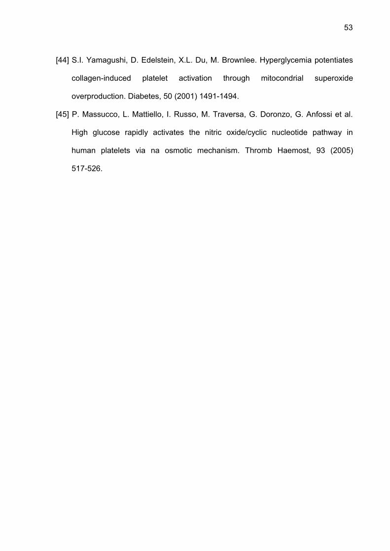
[44] S.I. Yamagushi, D. Edelstein, X.L. Du, M. Brownlee. Hyperglycemia potentiates
collagen-induced platelet activation through mitocondrial superoxide
overproduction. Diabetes, 50 (2001) 1491-1494.
[45] P. Massucco, L. Mattiello, I. Russo, M. Traversa, G. Doronzo, G. Anfossi et al.
High glucose rapidly activates the nitric oxide/cyclic nucleotide pathway in
human platelets via na osmotic mechanism. Thromb Haemost, 93 (2005)
517-526.
54
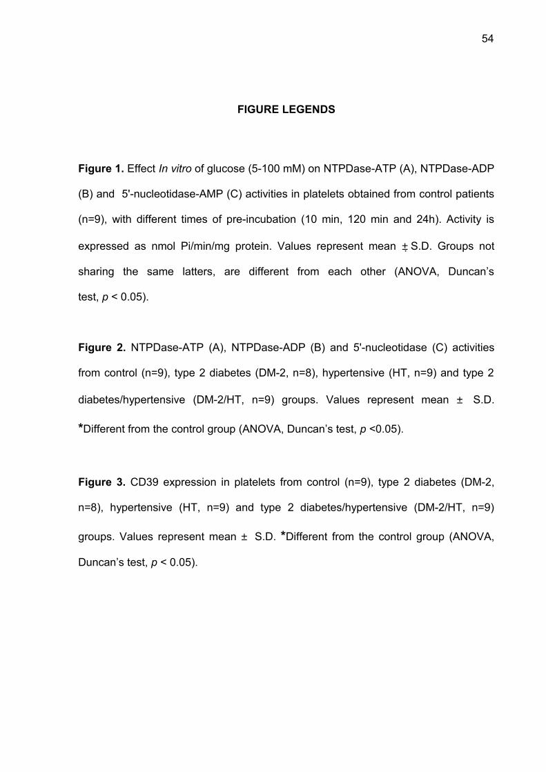
FIGURE LEGENDS
Figure 1. Effect In vitro of glucose (5-100 mM) on NTPDase-ATP (A), NTPDase-ADP
(B) and 5'-nucleotidase-AMP (C) activities in platelets obtained from control patients
(n=9), with different times of pre-incubation (10 min, 120 min and 24h). Activity is
expressed as nmol Pi/min/mg protein. Values represent mean ± S.D. Groups not
sharing the same latters, are different from each other (ANOVA, Duncan’s
test, p < 0.05).
Figure 2. NTPDase-ATP (A), NTPDase-ADP (B) and 5'-nucleotidase (C) activities
from control (n=9), type 2 diabetes (DM-2, n=8), hypertensive (HT, n=9) and type 2
diabetes/hypertensive (DM-2/HT, n=9) groups. Values represent mean ± S.D.
*Different from the control group (ANOVA, Duncan’s test, p <0.05).
Figure 3. CD39 expression in platelets from control (n=9), type 2 diabetes (DM-2,
n=8), hypertensive (HT, n=9) and type 2 diabetes/hypertensive (DM-2/HT, n=9)
groups. Values represent mean ± S.D. *Different from the control group (ANOVA,
Duncan’s test, p < 0.05).
55
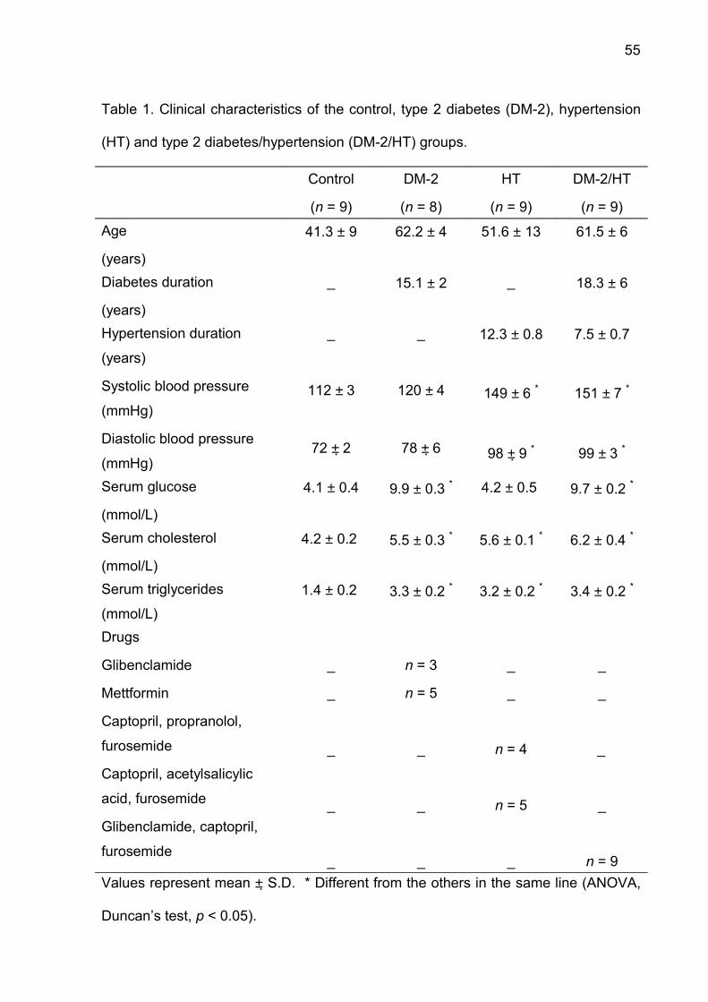
Table 1. Clinical characteristics of the control, type 2 diabetes (DM-2), hypertension
(HT) and type 2 diabetes/hypertension (DM-2/HT) groups.
Control
(n = 9)
DM-2
(n = 8)
HT
(n = 9)
DM-2/HT
(n = 9)
Age
(years)
41.3 ± 9 62.2 ± 4 51.6 ± 13 61.5 ± 6
Diabetes duration
(years)
_ 15.1 ± 2 _ 18.3 ± 6
Hypertension duration
(years)
Systolic blood pressure
(mmHg)
Diastolic blood pressure
(mmHg)
_
112 ± 3
72 ± 2
_
120 ± 4
78 ± 6
12.3 ± 0.8
149 ± 6 *
98 ± 9 *
7.5 ± 0.7
151 ± 7 *
99 ± 3 *
Serum glucose
(mmol/L)
4.1 ± 0.4 9.9 ± 0.3 * 4.2 ± 0.5 9.7 ± 0.2 *
Serum cholesterol
(mmol/L)
4.2 ± 0.2 5.5 ± 0.3 * 5.6 ± 0.1 * 6.2 ± 0.4 *
Serum triglycerides
(mmol/L)1.4 ± 0.2 3.3 ± 0.2 * 3.2 ± 0.2 * 3.4 ± 0.2 *
Drugs
Glibenclamide
Mettformin
Captopril, propranolol,
furosemide
Captopril, acetylsalicylic
acid, furosemide
Glibenclamide, captopril,
furosemide
_
_
_
_
_
n = 3
n = 5
_
_
_
_
_
n = 4
n = 5
_
_
_
_
_
n = 9Values represent mean ± S.D. * Different from the others in the same line (ANOVA,
Duncan’s test, p < 0.05).
56
Table 2. Correlation between ATP and ADP hydrolysis and CD39 expression in
platelets from type 2 diabetes (DM-2), hypertension (HT), type 2
diabetes/hypertension (DM-2/HT).
NTPDase
(nmol Pi/min/mg protein)
CD39 positive cells
(%)DM-2
(n=8)
HT
(n=9)
DM-2/HT
(n=9)ATP r=0.83
P < 0.01
r=0.87
P < 0.005
r=0.84
P < 0.001ADP r=0.84
P < 0.01
r=0.90
P < 0.002
r=0.93
P < 0.001
57
Figure 1

58
59
60
Figure 2
61
62

63
Figure 3
64
3.3 Manuscrito 2
O manuscrito “Antioxidant status in platelet from patients with type 2 diabetes
and hypertension” foi submetido ao periódico “Molecular and Cellular Biochemistry”.
65
Antioxidant status in platelet from patients with
diabetes and hypertension
Gilberto I Lunkes1, Daniéle S Lunkes1, Paula A Maldonado1, Maisa C Correa1, Jamile
F Gonçalves1, Roberta Schmatz1, Cíntia S da Rosa1, Lara V Becker1, Vera M
Morsch1, Maria R C Schetinger1*
1 Departamento de Química, Centro de Ciências Naturais e Exatas, Programa de
Pós-Graduação em Bioquímica Toxicológica, Universidade Federal de Santa Maria,
Santa Maria, RS 97105-900, Brazil.
Departamento de Química, Centro de Ciências Naturais e Exatas, Programa de Pós-
Graduação em Bioquímica Toxicológica, Universidade Federal de Santa Maria,
Santa Maria, RS 97105-900, Brazil. Fax: +55 55 3220 8978.
e-mail addresses: [email protected] or [email protected] (M.R.C.
Schetinger)
66
Abstract
Diabetes and hypertension constitute risks factors that interfere and promote
endothelial dysfunction. This study evaluated the oxidative status on platelets of
patients with diabetes and hypertension alone or associated. The sample consisted
of 90 patients and was divided into six groups, namely: control, hypertensive (HT),
type 2 diabetic using oral hypoglycemic drugs (DM2-OHp), type 2 diabetic using
insulin (DM2-Ins), type 2 diabetic with hypertension using oral hypoglycemic drugs
(DM2/HT-OHp), and type 2 diabetic with hypertension using insulin (DM2/HT-Ins).
The biochemical, lipid peroxidation, and ascorbic acid determinations were estimated
in serum of all groups. There was an increase of TBARS in DM2-OHp, DM2-Ins, HT,
DM2/HT-Ohp, and DM2/HT-Ins groups when compared to the control group
(p< 0.05). The increase of superoxide dismutase (SOD), catalase (CAT), nonprotein
thiols (NPSH) and protein carbonyl was observed in patients of DM2-OHp,
DM2-Ins, HT, DM2/HT-Ohp, and DM2/HT-Ins groups when compared to the control
group (p< 0.05). There was a significant and positive correlation between serum
glucose levels with SOD, CAT, NPSH, and protein carbonyl. Low concentrations of
serum ascorbic acid were observed in DM2-OHp, DM2-Ins, HT, DM2/HT-Ohp, and
DM2/HT-Ins groups when compared to the control group (p< 0.05). In conclusion, the
combination of hypertension with diabetes makes possible the maintenance of
elevated levels of oxidative stress in platelets.
Keywords: Reactive oxygen species, platelet, type 2 diabetes, hypertension, human.
67
Introduction
The oxidative stress contributes to the development of different diseases,
including vascular complications in chronic diseases as diabetes and atherosclerosis
[1]. The reactive oxygen species (ROS) may behave as second messengers and
may regulate platelet functions [2]. Oxidative stress is a factor associated with
platelet activation in diabetic patients [3]. The levels of oxidants and antioxidant
systems on platelets have significant balancing role in the homeostasis of vascular
diseases [4].
Hyperglycemia in patients with type 2 diabetes can be related to many
pathological states that involve disturbances metabolism, like disorders of oxidative-
antioxidative balance [5]. Therefore, high glucose level is an important factor that
may cause intensification of oxidative stress and etiopathogenesis of vascular
complications in diabetes [6, 7].
The elevation of oxidative stress and associated oxidative damages are
mediators of vascular injury in various cardiovascular pathologies, including
hypertension and atherosclerosis [8]. Elevated levels of ROS in cases of
hypertension may cause endothelial dysfunction and increased vascular resistance
[9]. Hypertension increases pro-oxidant generation and can decrease antioxidant
defense, and thereby induces oxidative stress in diabetes [10].
Platelets of diabetic patients are exposed to increased oxidative stress [11].
ROS modify both the adhesive and aggregatory responses of platelets, and free
radical scavengers are therefore important regulators of platelet function [12]. The
antioxidant system includes enzymatic and non-enzymatic components [13]. SOD
and CAT are the enzymatic antioxidants which scavenge the ROS, while nonprotein
thiols (NPSH) and ascorbic acid are non-enzymatic antioxidant systems which play
68
important roles in alleviating tissue damage due to the formation of ROS [14, 15].
Protein carbonyl content may used as marker of oxidative damage of proteins and
correlates with the severity of protein oxidation in diabetes, hypertension, aging [16].
The malondialdehyde (MDA) possible to determinate the lipid peroxidation levels and
it can be used as marker of oxidative stress [17].
Damage to endothelium, either by reactive oxygen species or by its oxidation
products, with simultaneous platelet activation, results in alteration in the vascular
permeability. Oxidative stress promotes alterations in the homeostasia of diabetes
patients. The interactions of the hypertension with diabetes may increase the
interference of the oxidative damage in vascular injury. However, the alterations ROS
induced in platelets of diabetic patients with associated hypertension are few
available in the literature. Hence, the aim of study was evaluated the antioxidant
status in platelets of patients with diabetes and associated hypertension. Therefore,
we also investigated the correlation between hyperglycemia and antioxidant system.
Material and Methods
Materials
Malondialdehyde (MDA), 2,4-dinitrophenylhydrazine (DNPH), 5-5’-dithio-bis-2-
nitrobenzoic acid (DTNB), hydrogen peroxide, and adrenaline were purchased from
Sigma (St. Louis, MO, USA). Glucose, cholesterol, HDL-cholesterol, triglycerides,
glycated hemoglobin, and lactate dehydrogenase (LDH) commercial kits purchased
from Labtest (Lagoa Santa, MG, Brazil). All other reagents used in the experiments
were of analytical grade and of the highest purity.
69
Participants
The sample consisted of patients from the Assistance Program to diabetics
and hypertensive patient assistance program linked with the Municipal Health
Secretariat in Cruz Alta (RS, Brazil) as well as of healthy volunteers. All subjects
gave written informed consent to participate in the study. The protocol was approved
by the Human Ethics Committee of the Health Science Center of the Federal
University of Santa Maria (Protocol number: 013/2004).
The sample was divided into four groups consisting of 50% males and 50%
females. The control group consisted of 15 individuals with ages ranging from 36 to
55, who presented no disease and who had not been submitted to any
pharmacological therapy during one month before the study began. Controls were
carefully selected by clinical evaluation, matched by sex, age, and body mass index
similar to that of the patients. The hypertensive (HT) group was formed by 15
patients with ages ranging from 38 to 58. The components of the HT group had
different hypertension levels and were treated with captopril (25 mg/day), furosemide
(40 mg/day), and acetylsalicylic acid (100 mg/day). Both type 2 diabetic groups, one
of which used oral hypoglycemic drugs (DM2-OHp) and the other of which used
insulin (DM2-Ins), consisted of 15 patients each with ages ranging from 39 to 65. The
patients of DM2-OHp group were treated with metformin (850 mg/day), and the
patients of DM2-Ins were treated with insulin NPH (35 UI/day). Both type 2 diabetic
groups with hypertension, one of which used oral hypoglycemic drugs (DM2/HT-
OHp) and the other of which used insulin (DM2/HT-Ins), were comprised of 15
patients each with ages ranging from 43 to 66. All patients of the type 2 diabetes
mellitus with hypertension received appropriate medication for the associated
diseases.
70
Sample collection
Blood was collected in vacutainer tubes without anticoagulant system. After
the collection, the blood was centrifuged at 1400 X g for 10 min, the precipitate was
discarded, and the serum was used for thiobarbituric acid reactive substances
(TBARS), ascorbic acid (AA), and biochemical determinations. Then, blood was
collected into citrate, centrifuged at 160 X g for 10 min, and the platelet-rich plasma
(PRP) was used for superoxide dismutase, catalase, nonprotein thiols, and protein
carbonyl determination.
The platelet-rich plasma sample was prepared from human donors by the
methods of Pilla et al. [18]. Blood was collected into 0.129 M citrate and centrifuged
at 160 X g for 10 min. The PRP was centrifuged at 1600 X g for 15 min and washed
twice with 3.5 mmol/L Hepes isosmolar buffer containing 142 mmol/L NaCl,
2.5 mmol/L KCl, and 5.5 mmol/L glucose. The washed platelets were resuspended in
Hepes isosmolar buffer and used for the protein carbonyl, NPSH, CAT and SOD
determination.
Lipid peroxidation determination
Lipid peroxidation was estimated by the measurement of thiobarbituric acid
reactive substances in serum samples by modifications of the method of
Jentzsch et al. [19]. Briefly, 0.2 ml of serum was added to the reaction mixture
containing 1 ml of 1% ortho-phosphoric acid, 0.25 mL alkaline solution of
thiobarbituric acid-TBA (final volume 2.0 ml) followed by 45 min heating at 95ºC. After
cooling, samples and standards of malondialdehyde were read at 532 nm against the
blank of the standard curve. The results were expressed as nmol MDA/mL serum.
71
Carbonylation of protein determination
The carbonylation of platelet proteins was determined by modifications of the
Goswami and Koner method [20]. Firstly, from 1 ml of PRP, the proteins were
precipitated using 0.5 ml of 10% trichloroacetic acid (TCA) and centrifuged at
1600 X g rpm for 5 min discarding the supernatant. One half milliliter of 10 mmol/L
2,4-dinitrophenylhydrazine in 2 mol/L HCl was added to this precipitated protein and
incubated at room temperature for 30 min. During the incubation time the samples
were mixed vigorously every 15 min. After the incubation time, 0.5 mL of 10% TCA
was added to the protein precipitated and centrifuged at 1600 X g for 5 min. After
discarding the supernatant, precipitates were washed twice with 1 mL of
ethanol/ethilacetate (1:1), each time centrifuging out the supernatant in order to
remove the free DNPH. The precipitate was dissolved in 1.5 mL of protein dissolving
solution (2 g SDS and 50 mg EDTA in 100 mL 80 mmol/L phosphate buffer, pH 8.0)
and incubated at 37ºC water bath for 10 min. The color intensity of the supernatant
was measured using spectrophotometer at 370 nm against 2 M HCl. Carbonyl
content was calculated by using molar extinction coefficient (21 x 103 1/mol cm) and
results were expressed as nmol carbonyl/mg protein.
Nonprotein thiols (NPSH) content
NPSH were determined in platelets by modifications of Sedlak and Lindsay
method [21]. The PRP was precipitated with 20% trichloracetic acid and then
centrifuged at 3500 rpm for 10 min. The reaction mixture contained 0.5 mL of
supernatant, 2.0 mL of phosphate buffer, pH 8.9, and it was read at 412 nm after the
addition of 0.1 mL of 0.01 mmol/L 5-5’-dithio-bis-2-nitrobenzoic acid (DTNB). The
results were expressed as µmol/mg protein.
72
Catalase activity
CAT activity was determined in platelets by modifications of Luck method [22].
An aliquot (0.05 mL) of PRP was homogenized in potassium phosphate buffer,
pH 7.0. The spectrophotometric determination was started by the addition of 0.07 mL
of aqueous solution of hydrogen peroxide 0.3 mol/L. The change in absorbance at
240 nm was measured for 2 min. The catalase activity was calculated using molar
extinction coefficient and the results were expressed as pmol/mg protein.
Superoxide dismutase activity
SOD activity in platelet was determined by modifications of Misra and
Fridovich method [23]. The reaction mixture constituted of 1.0 mL Tris buffer
0.2 mol/L, pH 10.0, 0.2 mL aliquot of PRP and water to make up the volume to
2.8 mL. The reaction was started by the addition of 0.2 mL of 0.025 mol/L
epinephrine. The change in absorbance was measured at 480 nm for 2 min. One unit
enzyme inhibited the rate of autooxidation of 5 µmol of epinephrine by 50%, and the
results were expressed as U SOD/mg protein.
Ascorbic acid content
Ascorbic acid levels were determined by the method of Jacques-Silva et al.
[24]. An aliquot of 300 µL sample of serum was mixed with
2-4-dinitrophenylhydrazine (4.5 mg/mL), CuSO4 (0.075 mg/mL) and trichloroacetic
acid 13.3% (final volume 1 mL), and incubated for 3 h at 37°C. Then, 1 mL of H2SO4
65% (v/v) was added to the medium. The ascorbic acid levels were measured
spectrophotometrically at 520 nm and calculated using a standard curve
(1.5-4.5 µmol/L ascorbic acid freshly prepared in sulfuric acid).
73
Clinical parameters analysis
Serum glucose, cholesterol, HDL-cholesterol, triglycerides, glycated
hemoglobin, and lactate dehydrogenase (LDH) were determined using
commol/Lercial kits from Labtest (Lagoa Santa, MG, Brazil).
Statistical analysis
Data were analyzed statistically by two-way and one-way ANOVA, followed by
Duncan’s multiple range test. Differences between groups were considered to be
significant when p < 0.05. All data were expressed as mean ± S.D. Correlation was
evaluated with the Pearson test. Linear correlation between variables was also
carried out.
Results
Clinical parameters
The clinical characteristics of the subjects are presented in Table 1. The
patients of the control group had never smoked. However, 40% of the hypertensive
patients had smoked for 22 years, 33% of the diabetes patients had smoked for
25 years, and 15% of the patients with diabetes and hypertension had smoked for
38 years.
The biochemical determinations of glucose, glycated hemoglobin,
triglycerides, total cholesterol, and HDL-cholesterol from all patients are presented in
Table 2. The serum glucose was increased in DM2-OHp (82.3%), DM2-Ins (81.1%),
DM2/HT-OHp (74.5%), and DM2/HT-Ins (75.4%) groups, and these groups were
different from the control and hypertensive groups (p<0.05). Glycated haemoglobin
was increased in DM2-OHp (48.4%), DM2-Ins (47.1%), DM2/HT-OHp (55.7%), and
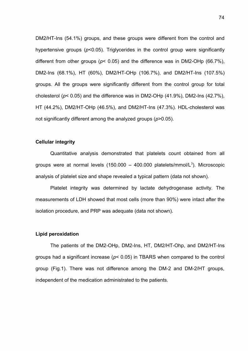
74
DM2/HT-Ins (54.1%) groups, and these groups were different from the control and
hypertensive groups (p<0.05). Triglycerides in the control group were significantly
different from other groups (p< 0.05) and the difference was in DM2-OHp (66.7%),
DM2-Ins (68.1%), HT (60%), DM2/HT-OHp (106.7%), and DM2/HT-Ins (107.5%)
groups. All the groups were significantly different from the control group for total
cholesterol (p< 0.05) and the difference was in DM2-OHp (41.9%), DM2-Ins (42.7%),
HT (44.2%), DM2/HT-OHp (46.5%), and DM2/HT-Ins (47.3%). HDL-cholesterol was
not significantly different among the analyzed groups (p>0.05).
Cellular integrity
Quantitative analysis demonstrated that platelets count obtained from all
groups were at normal levels (150.000 – 400.000 platelets/mmol/L3). Microscopic
analysis of platelet size and shape revealed a typical pattern (data not shown).
Platelet integrity was determined by lactate dehydrogenase activity. The
measurements of LDH showed that most cells (more than 90%) were intact after the
isolation procedure, and PRP was adequate (data not shown).
Lipid peroxidation
The patients of the DM2-OHp, DM2-Ins, HT, DM2/HT-Ohp, and DM2/HT-Ins
groups had a significant increase (p< 0.05) in TBARS when compared to the control
group (Fig.1). There was not difference among the DM-2 and DM-2/HT groups,
independent of the medication administrated to the patients.
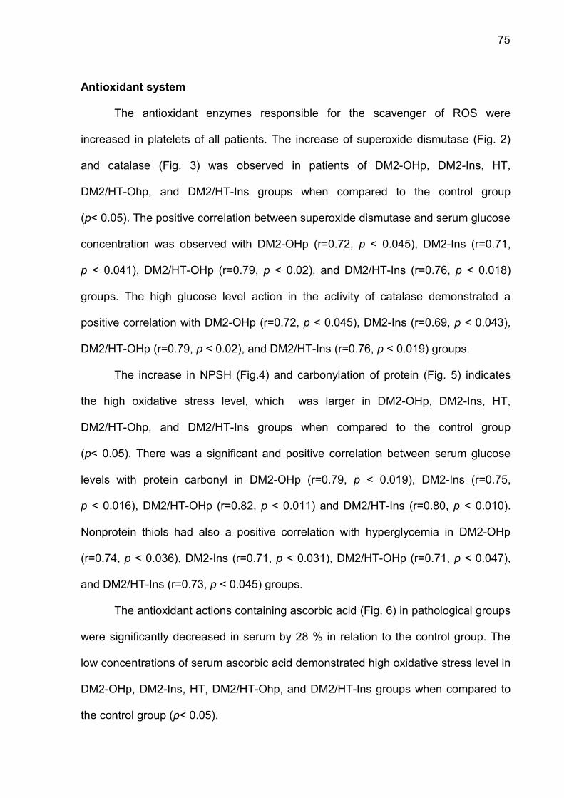
75
Antioxidant system
The antioxidant enzymes responsible for the scavenger of ROS were
increased in platelets of all patients. The increase of superoxide dismutase (Fig. 2)
and catalase (Fig. 3) was observed in patients of DM2-OHp, DM2-Ins, HT,
DM2/HT-Ohp, and DM2/HT-Ins groups when compared to the control group
(p< 0.05). The positive correlation between superoxide dismutase and serum glucose
concentration was observed with DM2-OHp (r=0.72, p < 0.045), DM2-Ins (r=0.71,
p < 0.041), DM2/HT-OHp (r=0.79, p < 0.02), and DM2/HT-Ins (r=0.76, p < 0.018)
groups. The high glucose level action in the activity of catalase demonstrated a
positive correlation with DM2-OHp (r=0.72, p < 0.045), DM2-Ins (r=0.69, p < 0.043),
DM2/HT-OHp (r=0.79, p < 0.02), and DM2/HT-Ins (r=0.76, p < 0.019) groups.
The increase in NPSH (Fig.4) and carbonylation of protein (Fig. 5) indicates
the high oxidative stress level, which was larger in DM2-OHp, DM2-Ins, HT,
DM2/HT-Ohp, and DM2/HT-Ins groups when compared to the control group
(p< 0.05). There was a significant and positive correlation between serum glucose
levels with protein carbonyl in DM2-OHp (r=0.79, p < 0.019), DM2-Ins (r=0.75,
p < 0.016), DM2/HT-OHp (r=0.82, p < 0.011) and DM2/HT-Ins (r=0.80, p < 0.010).
Nonprotein thiols had also a positive correlation with hyperglycemia in DM2-OHp
(r=0.74, p < 0.036), DM2-Ins (r=0.71, p < 0.031), DM2/HT-OHp (r=0.71, p < 0.047),
and DM2/HT-Ins (r=0.73, p < 0.045) groups.
The antioxidant actions containing ascorbic acid (Fig. 6) in pathological groups
were significantly decreased in serum by 28 % in relation to the control group. The
low concentrations of serum ascorbic acid demonstrated high oxidative stress level in
DM2-OHp, DM2-Ins, HT, DM2/HT-Ohp, and DM2/HT-Ins groups when compared to
the control group (p< 0.05).
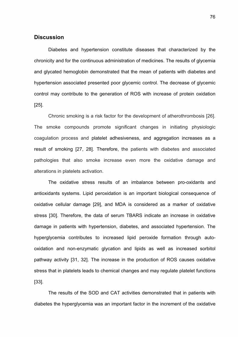
76
Discussion
Diabetes and hypertension constitute diseases that characterized by the
chronicity and for the continuous administration of medicines. The results of glycemia
and glycated hemoglobin demonstrated that the mean of patients with diabetes and
hypertension associated presented poor glycemic control. The decrease of glycemic
control may contribute to the generation of ROS with increase of protein oxidation
[25].
Chronic smoking is a risk factor for the development of atherothrombosis [26].
The smoke compounds promote significant changes in initiating physiologic
coagulation process and platelet adhesiveness, and aggregation increases as a
result of smoking [27, 28]. Therefore, the patients with diabetes and associated
pathologies that also smoke increase even more the oxidative damage and
alterations in platelets activation.
The oxidative stress results of an imbalance between pro-oxidants and
antioxidants systems. Lipid peroxidation is an important biological consequence of
oxidative cellular damage [29], and MDA is considered as a marker of oxidative
stress [30]. Therefore, the data of serum TBARS indicate an increase in oxidative
damage in patients with hypertension, diabetes, and associated hypertension. The
hyperglycemia contributes to increased lipid peroxide formation through auto-
oxidation and non-enzymatic glycation and lipids as well as increased sorbitol
pathway activity [31, 32]. The increase in the production of ROS causes oxidative
stress that in platelets leads to chemical changes and may regulate platelet functions
[33].
The results of the SOD and CAT activities demonstrated that in patients with
diabetes the hyperglycemia was an important factor in the increment of the oxidative
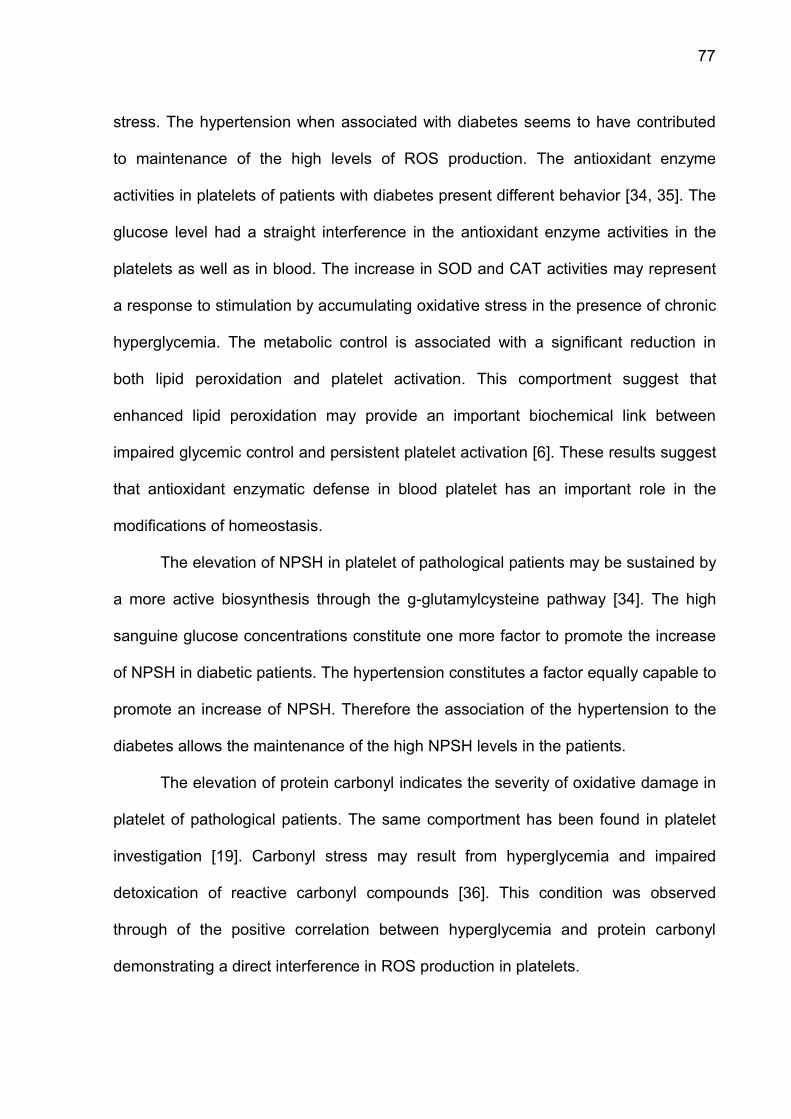
77
stress. The hypertension when associated with diabetes seems to have contributed
to maintenance of the high levels of ROS production. The antioxidant enzyme
activities in platelets of patients with diabetes present different behavior [34, 35]. The
glucose level had a straight interference in the antioxidant enzyme activities in the
platelets as well as in blood. The increase in SOD and CAT activities may represent
a response to stimulation by accumulating oxidative stress in the presence of chronic
hyperglycemia. The metabolic control is associated with a significant reduction in
both lipid peroxidation and platelet activation. This comportment suggest that
enhanced lipid peroxidation may provide an important biochemical link between
impaired glycemic control and persistent platelet activation [6]. These results suggest
that antioxidant enzymatic defense in blood platelet has an important role in the
modifications of homeostasis.
The elevation of NPSH in platelet of pathological patients may be sustained by
a more active biosynthesis through the g-glutamylcysteine pathway [34]. The high
sanguine glucose concentrations constitute one more factor to promote the increase
of NPSH in diabetic patients. The hypertension constitutes a factor equally capable to
promote an increase of NPSH. Therefore the association of the hypertension to the
diabetes allows the maintenance of the high NPSH levels in the patients.
The elevation of protein carbonyl indicates the severity of oxidative damage in
platelet of pathological patients. The same comportment has been found in platelet
investigation [19]. Carbonyl stress may result from hyperglycemia and impaired
detoxication of reactive carbonyl compounds [36]. This condition was observed
through of the positive correlation between hyperglycemia and protein carbonyl
demonstrating a direct interference in ROS production in platelets.

78
The administration of the drugs metformin and insulin did not differentiate the
increase of oxidative stress levels among the groups. The short-term metformin
administration promotes activation of oxidative stress together with alterations of the
antioxidant system [37]. Studies with metformin in vitro and with animals showed
reactive oxygen species production may have been reduced [38].
The low concentrations of ascorbic acid in serum, observed in pathological
patients, suggest an exhibition to oxidative damages. The low concentrations
ascorbic acid has been described as a biomarker in pathogenesis of hypertension
[39, 40] and diabetes [401, when increase the oxidative stress. Therefore, low
concentrations of ascorbic acid in serum can cause pro-oxidative effects [42, 43].
These results suggested that ascorbic acid levels constitute a factor in the control of
oxidative stress.
In conclusion, we can observe that patients with diabetes and hypertension
associated had the oxidative damage increased in platelets. The acid ascorbic was
an important factor capable to modulate oxidative stress. The hyperglycemia
constituted an important factor capable to provide an increase in reactive oxygen
species production and in consequence to modify the antioxidant system status.
Therefore, the combination of Diabetes mellitus and hypertension possible to
maintain an elevated level of oxidative stress in platelets and potentially may promote
more critical alterations in platelet homeostasia.
79
References
1. Medina LO, Veloso CA, Borges EA et al (2007) Determination of the antioxidant
status of plasma from type 2 diabetic patients. Diabetes Res Clin Pract 77:
193–197
2. Olas B, Wachowicz B (2007) Role of reactive nitrogen species in blood platelet
functions. Platelets 23:1-11
3. de Cristofaro R, Rocca B, Vitacolonna E et al (2003) Lipid and
protein oxidation contribute to a prothrombotic state in patients with type 2
diabetes mellitus. J Thromb Haemost 1:250-256
4. Pandey NR, Kaur G, Chandra M et al (2000) Enzymatic oxidant and antioxidants
of human blood platelets in unstable angina and myocardial infarction. Int J
Cardiol 76:33-38
5. Sena CM, Nunes E, Louro T et al (2007) Endothelial dysfunction in type 2
diabetes: effect of antioxidants. Rev Por Cardiol 26: 609-619
6. Ferroni P, Basili S, Falco A et al (2004) Platelet activation in type 2 diabetes
mellitus. J Thromb Haemost 2:1282-1291
7. Błaszczak R, Kidziora-Kornatowska K, Kornatowski T et al (2005) Evaluation of
oxidative stress parameters in diabetes 2 patients in different stage of metabolic
compensation. Pol Merkur Lekarski 18:196-9
8. Rush JW, Ford RJ (2007) Nitric oxide, oxidative stress and vascular endothelium
in health and hypertension. Clin Hemorheol Microcirc 37:185-192
9. Puddu P, Puddu GM, Cravero E et al (2007) The putative role of mitochondrial
dysfunction in hypertension. Clin Exp Hypertens 29:427-434
80
10. Biswas SK, Peixoto E, Souza DS et al (2008) Hypertension increases pro-oxidant
generation and decreases antioxidant defense in the kidney in early diabetes.
Am J Nephrol 28:133-142
11. Seghieri G, Di Simplicio P, Anichini R et al (2001) Platelets antioxidant enzymes
in insulin-dependent diabetes mellitus. Clin Chim Acta 309:19-23
12. Hsiao G, Shen MY, Lin KH et al (2003) Inhibitory activity of kinetin on free radical
formation of activated platelets in vitro and on thrombus formation in vivo. Eur J
Pharmacol 465:281-287
13. Gil-del Valle L, Milian C, Toledo A et al (2005) Altered redox status in patients
with Diabetes Mellitus type I. Pharmacol Res 51:375-380
14. Goswami K, Nanjaiah D, Koner BC et al (2003) Oxidative changes and
desialylation of serum proteins in hyperthyroidism. Clin Chim Acta 337:
163-168
15. Veerappan RM, Senthil S, Rao MR et al (2004) Redox status and lipid
peroxidation in alcoholic hypertensive patients and alcoholic hypertensive
patients with diabetes. Clin Chim Acta 340:207-212
16. Chevion M, Berenshtein E, Stadtman ER (2000) Human studies related to
protein oxidation: protein carbonyl content as a marker of damage. Free Radic
Res 33:S99-S108.
17. Janero, D. R. (1990) Malondialdehydo and thiobarbituric acid-reactivity as
diagnostic indices of lipid peroxidation and peroxidation tissue injury. Free Radic
Biol Med 9:515-540
18. Pilla C, Emanuelli T, Frasetto SS et al (1996) ATP diphosphoydrolase activity
(apyrase, EC 3.6.1.5) in human blood platelets. Platelets 7:225-230
81
19. Jentzsch AM, Bachmann H, Fürst P et al (1996) Improved analysis of
malondialdehyde in human body fluids. Free Radic Biol Med 20:251-256
20. Goswami K, Koner BC (2002) Level of Siliac Acid Residues in Platelets Proteins
in Diabetes, Aging, and Hodgkin’s lymphoma: a potential role of free radicals in
desialylation. Biochem Biophys Res Commol/Lun 297:502-505
21. Sedlak J, Lindsay RHC (1968) Estimation of total, protein bound and non-protein
sulfhydryl groups in tissue with Ellmann’s reagent. Anal Biochem 25:192-205
22. Luck H (1965) Catalase. In: Bergmeyer HU (ed) Methods in Enzymatic Analysis
p.885
23. Misra HP, Fridovich I (1972) The role of superoxide anion in the autooxidation of
epinephrine and a simple assay for superoxide dismutase. J Biol Chem
247:3170-3175
24. Jacques-Silva MC, Nogueira CW, Broch LC et al (2001) Diphenyl diselenide and
ascorbic acid changes deposition of selenium and ascorbic acid in liver and
brain of mice. Pharmacol Toxicol 88:119-127
25. Cakatay U (2005) Protein oxidation parameters in type 2 diabetic patients with
good and poor glycaemic control. Diabetes Metab 31:551-557
26. Takajo Y, Ikeda H, Haramaki N et al (2001) Augmented Oxidative Stress of
Platelets in Chronic Smokers. J Am Coll Cardiol 38:1320-1327
27. Leone A (2007) Smoking, hemostatic factors, and cardiovascular risk. Curr
Pharm Des 13:1661-1667
28. Bloomer RJ, Solis AD, Fischer-Wellman KH (2007) Posptrandial oxidative stress
is exacerbated in cigarette smokers. Br J Nutr 10:1-16
29. Sohal RS (2002) Role of oxidative stress and protein oxidation in the aging
process. Free Radic Biol Med 33:37-44
82
30. Hartnett ME, Startton RD, Browne W et al (2000) Serum markers of oxidative
stress and severity of diabetic retinopathy. Diabetes 23:234-240
31. Komosinska-Vassev K, Olczyk K, Olczyk P et al (2005) Efffects of metabolic
control and vascular complications on indices of oxidative stress in type 2
diabetic patients. Diabetes Res Clin Pract 68:207-216
32. Son, S. M. (2007). Role of vascular reactive oxygen species in development of
vascular abnormalities in diabetes. Diabetes Res Clin Pract 77:S65-S70
33. Begonja AJ, Teichmann L, Geiger J et al (2006) Platelet regulation by NO/cGMP
signaling and NAD(P)H oxidase-generated ROS. Blood Cells Mol Dis 36:
166–170
34. Seghieri G, Di Simplicio P, De Giorgio LA et al (2000) Relationship between
metabolic glyceamic control and platelet content of glutathione and its related
enzymes, in insulin-dependent diabetes mellitus. Clin Chim Acta 299:109-117
35. Véricel E, Januel C, Carreras M et al (2004) Diabetic patients without vascular
complications display enhanced basal platelet activation and decreased
antioxidant status. Diabetes 53:1046-1051
36. Dalle-Donne I, Giustarini D, Colombo R et al (2003) Protein carbonylation in
human diseases. Trends Mol Med 9:169-176
37. Skrha J, Prázný M, Hilgertová J et al (2007) Oxidative stress and endothelium
influenced by metformin in type 2 diabetes mellitus. Eur J Clin Pharmacol 63:
1107-1114
38. Bellin C, de Wizza DH, Wierensperger NF et al ( 2006) Generation of reactive
oxygen species by endothelial as smooth muscle cells: influence of
hyperglycemia and metformin. Horm Metab Res 38:732-739
83
39. Chiplonkar SA, Agte VV, Tarwadi KV et al (2004) Micronutrient deficiencies as
predisposing factors for hypertension in lacto-vegetarian Indian adults. J Am Coll
Nutr 23:239-247
40. Flora SJ (2007) Role of free radicals and antioxidants in health and disease. Cell
Mol Biol 15:1-2
41. Skrha J, Prázný M, Hilgertová J et al (2003) Serum alphap-tocopherol and
ascorbic acid concentrations in Type 1 an Type 2 diabetic patients with and
without angiopathy. Clin Chim Acta 329:103-108
42. Wilkinson IB, Megson IL, MacCallum H et al (1999) Oral vitamin C reduces
arterial stiffness and platelet aggregation in humans. J Cardiovasc Pharmacol
34:690-693
43. Olas B, Wachowicz B (2002) Resveratrol and vitamin C as antioxidants in blood
platelets. Thromb Res 106:143-148
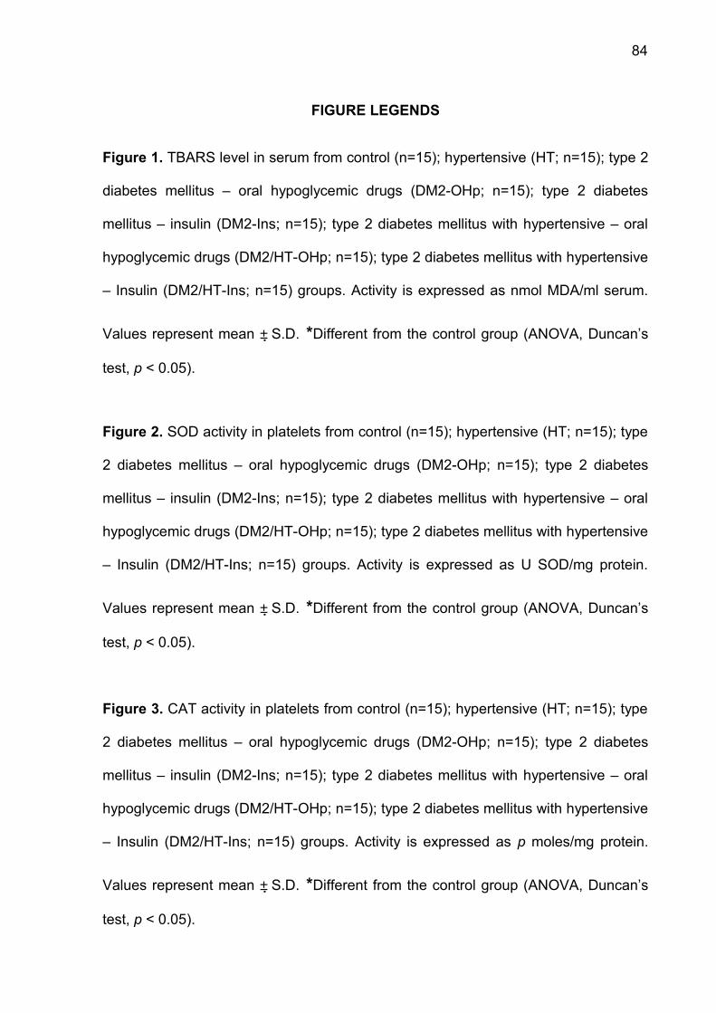
84
FIGURE LEGENDS
Figure 1. TBARS level in serum from control (n=15); hypertensive (HT; n=15); type 2
diabetes mellitus – oral hypoglycemic drugs (DM2-OHp; n=15); type 2 diabetes
mellitus – insulin (DM2-Ins; n=15); type 2 diabetes mellitus with hypertensive – oral
hypoglycemic drugs (DM2/HT-OHp; n=15); type 2 diabetes mellitus with hypertensive
– Insulin (DM2/HT-Ins; n=15) groups. Activity is expressed as nmol MDA/ml serum.
Values represent mean ± S.D. *Different from the control group (ANOVA, Duncan’s
test, p < 0.05).
Figure 2. SOD activity in platelets from control (n=15); hypertensive (HT; n=15); type
2 diabetes mellitus – oral hypoglycemic drugs (DM2-OHp; n=15); type 2 diabetes
mellitus – insulin (DM2-Ins; n=15); type 2 diabetes mellitus with hypertensive – oral
hypoglycemic drugs (DM2/HT-OHp; n=15); type 2 diabetes mellitus with hypertensive
– Insulin (DM2/HT-Ins; n=15) groups. Activity is expressed as U SOD/mg protein.
Values represent mean ± S.D. *Different from the control group (ANOVA, Duncan’s
test, p < 0.05).
Figure 3. CAT activity in platelets from control (n=15); hypertensive (HT; n=15); type
2 diabetes mellitus – oral hypoglycemic drugs (DM2-OHp; n=15); type 2 diabetes
mellitus – insulin (DM2-Ins; n=15); type 2 diabetes mellitus with hypertensive – oral
hypoglycemic drugs (DM2/HT-OHp; n=15); type 2 diabetes mellitus with hypertensive
– Insulin (DM2/HT-Ins; n=15) groups. Activity is expressed as p moles/mg protein.
Values represent mean ± S.D. *Different from the control group (ANOVA, Duncan’s
test, p < 0.05).
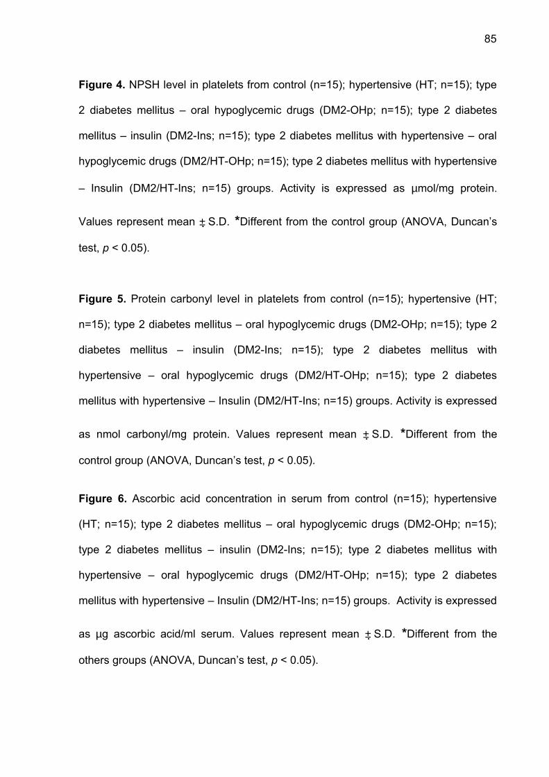
85
Figure 4. NPSH level in platelets from control (n=15); hypertensive (HT; n=15); type
2 diabetes mellitus – oral hypoglycemic drugs (DM2-OHp; n=15); type 2 diabetes
mellitus – insulin (DM2-Ins; n=15); type 2 diabetes mellitus with hypertensive – oral
hypoglycemic drugs (DM2/HT-OHp; n=15); type 2 diabetes mellitus with hypertensive
– Insulin (DM2/HT-Ins; n=15) groups. Activity is expressed as µmol/mg protein.
Values represent mean ± S.D. *Different from the control group (ANOVA, Duncan’s
test, p < 0.05).
Figure 5. Protein carbonyl level in platelets from control (n=15); hypertensive (HT;
n=15); type 2 diabetes mellitus – oral hypoglycemic drugs (DM2-OHp; n=15); type 2
diabetes mellitus – insulin (DM2-Ins; n=15); type 2 diabetes mellitus with
hypertensive – oral hypoglycemic drugs (DM2/HT-OHp; n=15); type 2 diabetes
mellitus with hypertensive – Insulin (DM2/HT-Ins; n=15) groups. Activity is expressed
as nmol carbonyl/mg protein. Values represent mean ± S.D. *Different from the
control group (ANOVA, Duncan’s test, p < 0.05).
Figure 6. Ascorbic acid concentration in serum from control (n=15); hypertensive
(HT; n=15); type 2 diabetes mellitus – oral hypoglycemic drugs (DM2-OHp; n=15);
type 2 diabetes mellitus – insulin (DM2-Ins; n=15); type 2 diabetes mellitus with
hypertensive – oral hypoglycemic drugs (DM2/HT-OHp; n=15); type 2 diabetes
mellitus with hypertensive – Insulin (DM2/HT-Ins; n=15) groups. Activity is expressed
as µg ascorbic acid/ml serum. Values represent mean ± S.D. *Different from the
others groups (ANOVA, Duncan’s test, p < 0.05).
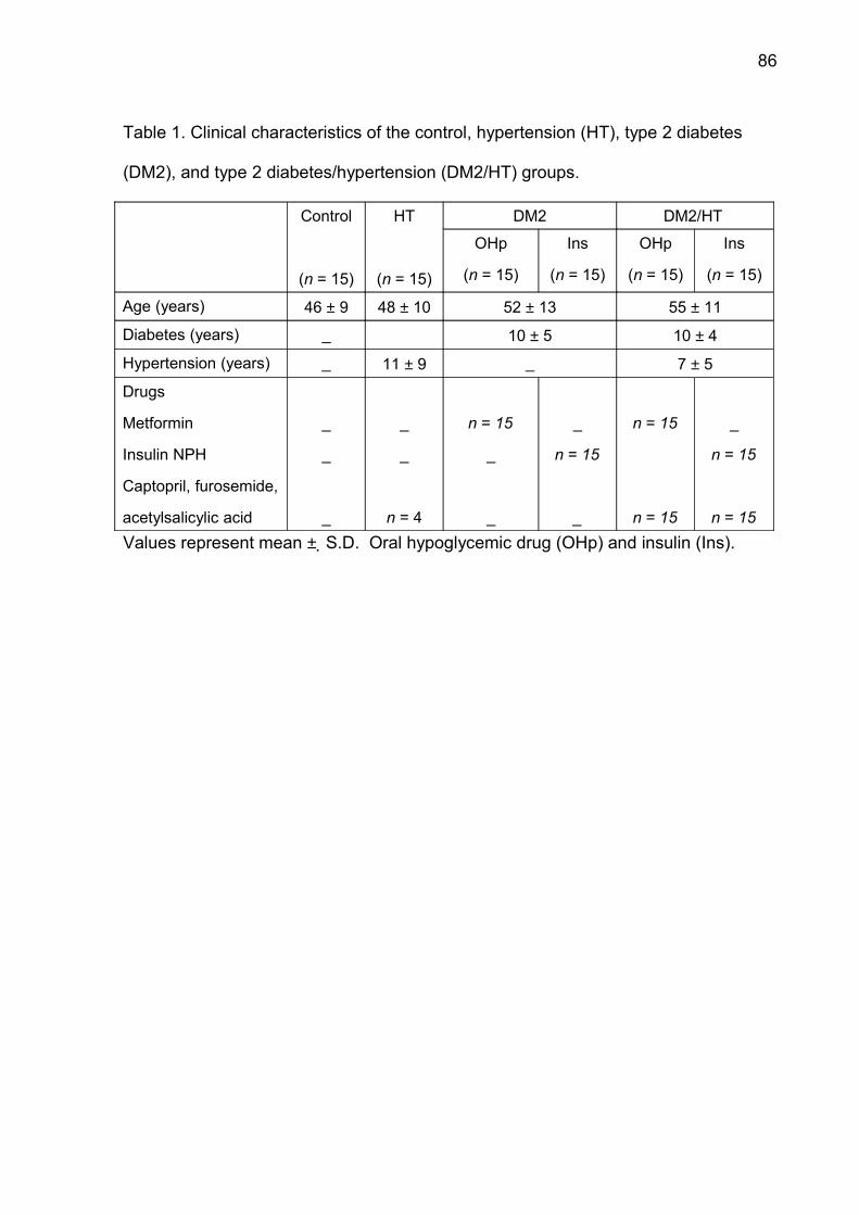
86
Table 1. Clinical characteristics of the control, hypertension (HT), type 2 diabetes
(DM2), and type 2 diabetes/hypertension (DM2/HT) groups.
Control
(n = 15)
HT
(n = 15)
DM2 DM2/HT
OHp
(n = 15)
Ins
(n = 15)
OHp
(n = 15)
Ins
(n = 15)
Age (years) 46 ± 9 48 ± 10 52 ± 13 55 ± 11
Diabetes (years) _ 10 ± 5 10 ± 4
Hypertension (years) _ 11 ± 9 _ 7 ± 5
Drugs
Metformin
Insulin NPH
Captopril, furosemide,
acetylsalicylic acid
_
_
_
_
_
n = 4
n = 15
_
_
_
n = 15
_
n = 15
n = 15
_
n = 15
n = 15Values represent mean ± S.D. Oral hypoglycemic drug (OHp) and insulin (Ins).
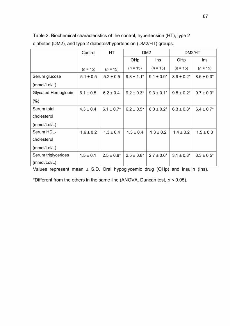
87
Table 2. Biochemical characteristics of the control, hypertension (HT), type 2
diabetes (DM2), and type 2 diabetes/hypertension (DM2/HT) groups.
Control
(n = 15)
HT
(n = 15)
DM2 DM2/HT
OHp
(n = 15)
Ins
(n = 15)
OHp
(n = 15)
Ins
(n = 15)
Serum glucose
(mmol/Lol/L)
5.1 ± 0.5 5.2 ± 0.5 9.3 ± 1.1* 9.1 ± 0.9* 8.9 ± 0.2* 8.6 ± 0.3*
Glycated Hemoglobin
(%)
6.1 ± 0.5 6.2 ± 0.4 9.2 ± 0.3* 9.3 ± 0.1* 9.5 ± 0.2* 9.7 ± 0.3*
Serum total
cholesterol
(mmol/Lol/L)
4.3 ± 0.4 6.1 ± 0.7* 6.2 ± 0.5* 6.0 ± 0.2* 6.3 ± 0.8* 6.4 ± 0.7*
Serum HDL-
cholesterol
(mmol/Lol/L)
1.6 ± 0.2 1.3 ± 0.4 1.3 ± 0.4 1.3 ± 0.2 1.4 ± 0.2 1.5 ± 0.3
Serum triglycerides
(mmol/Lol/L)1.5 ± 0.1 2.5 ± 0.8* 2.5 ± 0.8* 2.7 ± 0.6* 3.1 ± 0.8* 3.3 ± 0.5*
Values represent mean ± S.D. Oral hypoglycemic drug (OHp) and insulin (Ins).
*Different from the others in the same line (ANOVA, Duncan test, p < 0.05).

88
Figure 1
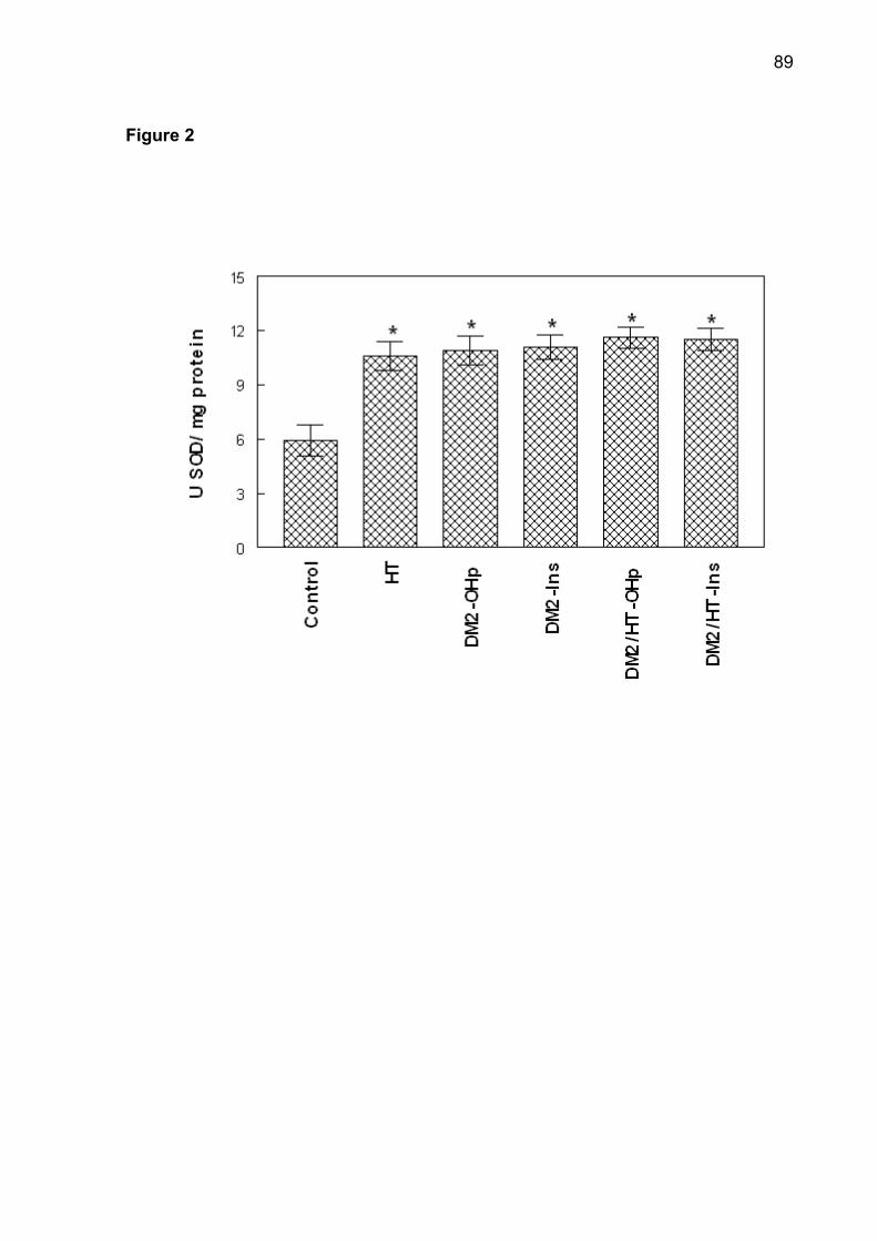
89
Figure 2
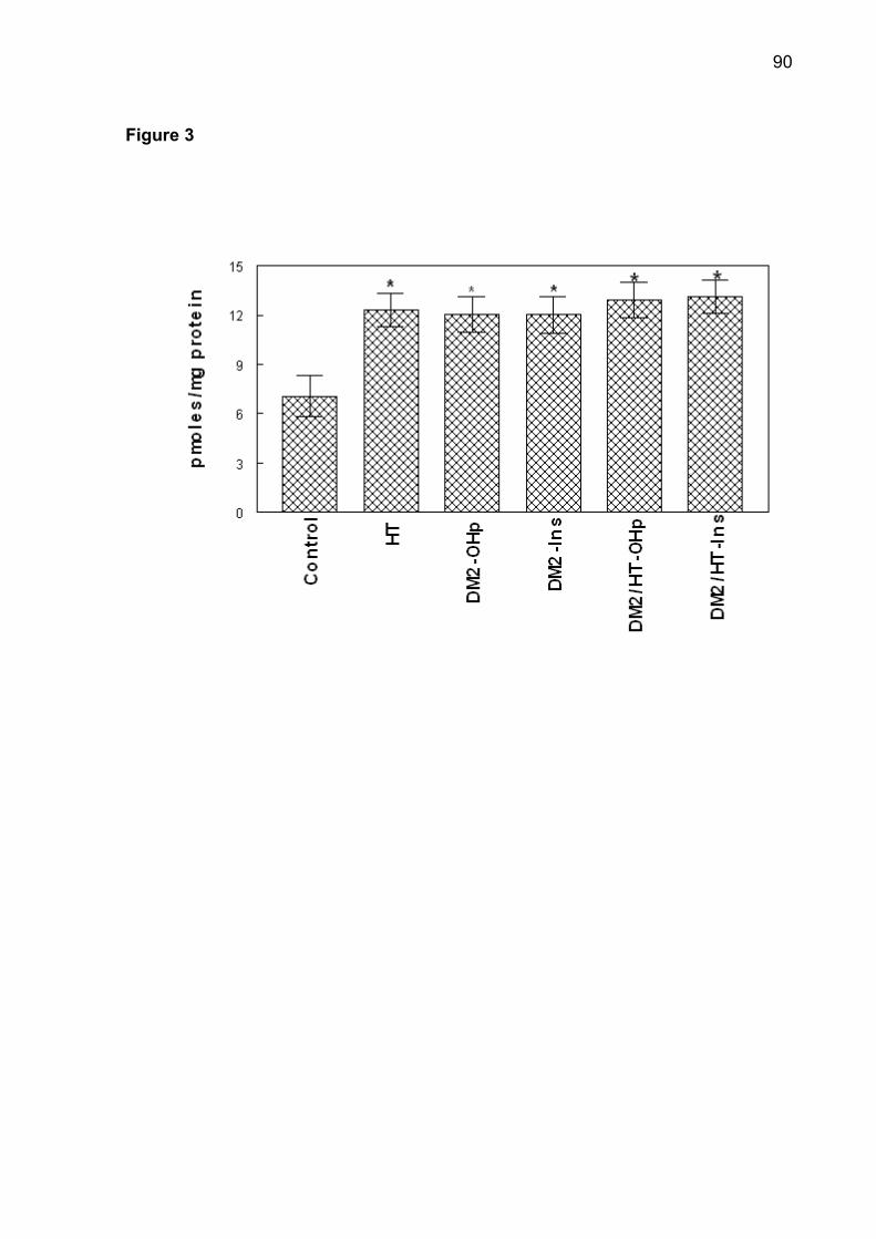
90
Figure 3
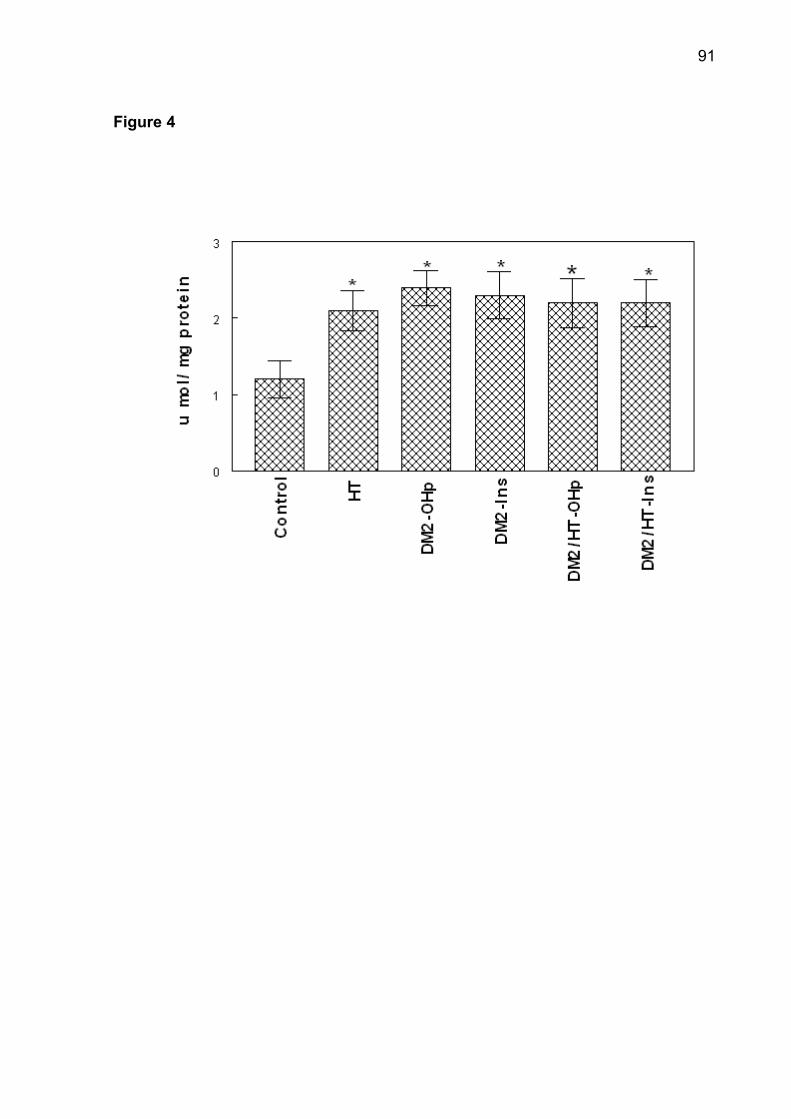
91
Figure 4
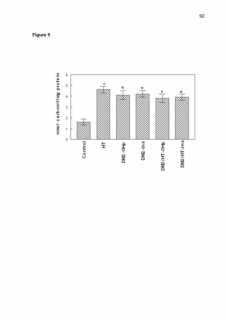
92
Figure 5
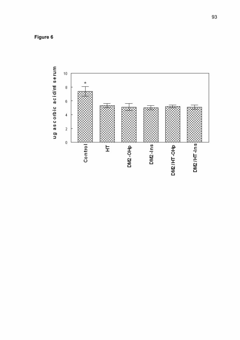
93
Figure 6
94
3.4 Manuscrito 3
O manuscrito “Oxidative stress and antioxidant profile in serum from patients
with type 2 diabetes and hypertension” está na fase de redação.
95
Oxidative stress and antioxidant profile
in patients with diabetes and hypertension
Gilberto I Lunkes, Daniéle S Lunkes, Francieli Stefanello, Roberta Schmatz, Vera M
Morsch, Maria R C Schetinger ∗
Departamento de Química, Centro de Ciências Naturais e Exatas, Programa
de Pós-Graduação em Bioquímica Toxicológica, Universidade Federal de Santa
Maria, Santa Maria, RS 97105-900, Brazil.
Departamento de Química, Centro de Ciências Naturais e Exatas, Programa de Pós-
Graduação em Bioquímica Toxicológica, Universidade Federal de Santa Maria,
Santa Maria, RS 97105-900, Brazil. Fax: +55 55 3220 8978.
*e-mail addresses: [email protected] or [email protected]
(M.R.C. Schetinger)
96
Abstract
This investigation analyzed the action of the hyperglycemia and micronutrient
in antioxidant defense system in patients with diabetes. The sample composed of 80
patients was divided into four groups. The antioxidant enzymatic and non-enzymatic
systems were estimated in patients of control, type 2 diabetes (DM2), hypertension
(HYP) and type 2 diabetes/hypertension (DM2/HYP) groups. There was an increase
in TBARS, CAT, SOD, NPSH and protein carbonyl in patients of pathological groups
when compared to control group (p< 0.05). There was significant and positive
correlation between serum glucose levels with SOD, CAT, NPSH and protein
carbonyl. The serum calcium and magnesium levels were lower while copper and
zinc levels had increased in patients of DM2, HYP and DM2/HYP groups when
compared to the control group (p< 0.05). The micronutrient concentration
demonstrated an exhibition the oxidative damage in the pathological patients. The
hyperglycemia constitutes a factor that promotes the increase of oxidative stress in
patients with diabetes. The hypertension associated with diabetes possible to
maintain elevated levels of oxidative stress and, consequently these patients can
develop vascular alterations.
Keywords: Diabetes mellitus, hypertension, hyperglycemia, antioxidant system.
97
1. Introduction
Diabetes mellitus is a syndrome with chronic hyperglycemia. The diabetic
patients have significant alteration on antioxidant protections and increase in
generation of reactive oxygen species and nitrogen [1]. Elevated free radicals levels
can develop endothelial vascular dysfunction specially in type 2 diabetes [2, 3, 4].
The oxidative stress can promote pro-atherogenic events in diabetic patients and
their relationships with atherosclerosis could potentially identify molecular targets of
therapy [5]. Hyperglycemia may induce reactive oxygen species and nitrogen
production through glucose metabolism, auto-oxidation, and formation of advanced
glycation end product [6].
The production of reactive oxygen species has been linked to certain diseases
of the cardiovascular system including hypertension. The oxidative stress may
constitute a major pathogenic factor in the development of hypertension [7]. The
hypertension increases pro-oxidant generation and the combination of hypertension
with diabetes can exacerbate oxidative stress [8].
Catalase (CAT), superoxide dismutase (SOD), nonprotein thiols (NPSH) are
endogenous antioxidant system defense. The imbalance between pro-oxidants and
antioxidant gives rise to cellular oxidative damage. The ROS in diabetes was greatly
increased due to prolonged exposure to hyperglycemia and impairment of
oxidant/antioxidant equilibrium [9].
The protein carbonyl and lipid peroxidation determination can be used as
markers of oxidative stress. Protein carbonyl content is an indicator and marker of
protein oxidation, and accumulation of protein carbonyl has been observed in several
human diseases including diabetes [10]. TBARS assay let to evaluate the lipid
98
peroxidation level that refers to the oxidative degradation of lipids that result in cell
damage [11].
Some minerals, as copper, zinc, and magnesium can participate in the
reduction of oxidative stress in diabetic patients and can be proposed as adjunctive
therapy [12]. Deficiency in copper levels increases reactive oxygen species and it
can be involved in complications in diabetes [13]. The treatment with zinc decreases
the cardiovascular involvement in type 2 diabetes mellitus patients [14]. Calcium
regulates several mechanisms that generate oxidative or nitrosative stress and the
oxidative stress modifies the calcium level interfering in the vascular reactivity [15].
The magnesium modulates free radicals, malondialdehyde and nitric oxide
production. Thus, this mineral may be involved in the regulation of lipid peroxidation
[16].
Thus, the present studies propose to available the antioxidant defense system
in patients with diabetes and hypertension. Then we investigate the interference of
the hyperglycemia and micronutrient in antioxidant system.
2. Patients and Methods
2.1. Patients
A total of 80 subjects participated in the study. The patients were divided into
four groups with similar age and they were separate with the same proportion
between men and women. The patients participate in the Program of Attendance to
Diabetic, Hypertensive and Diabetic-Hypertensive patients, associated with the
Municipal Secretary’s Office of Health of Cruz Alta (RS, Brazil) and health volunteers.
99
All subjects gave written informed consent to participate in the study. The protocol
was approved by the Human Ethics Committee of the Health Science Center of the
Federal University of Santa Maria (Protocol number: 013/2004).
The sample was divided into four groups consisting of 50% males and 50%
females. The control group (n=26) consisted of individuals with ages ranging from 28
to 52 years, who did not present any disease and who had not been submitted to any
pharmacological therapy during the last month. Controls were carefully selected by
clinical evaluation and presented sex, age and body mass index similar to those of
the patients. The type 2 diabetic (DM2, n=16) group consisted of patients with ages
ranging from 56 to 68 years. The patients of the DM2 group had type 2 diabetes
mellitus and were treated with glibenclamide (10 mg/day) or chlorpropamide
(250 mg/day). The hypertensive (HYP, n=12) group was made up of patients with
ages ranging from 30 to 70 years. The patients of the HYP group had different
hypertension levels and were treated with captopril (25 mg/day), furosemide
(40 mg/day) or propranolol (40 mg/day). The type 2 diabetic/hypertensive (DM2/HYP,
n=26) group consisted of patients with ages ranging from 51 to 69 years. All patients
of the DM2/HYP had type 2 diabetes mellitus plus hypertension and received
appropriate medication for the associated diseases. Ten milliliters of blood was
obtained from each participant and used for platelet-rich plasma preparations,
biochemical determinations and hematological determinations.
2.2. Sample collection
The blood was collected in vacutainer tubes without anticoagulant system.
After the collection, the blood was centrifuged at 1400 X g for 10 min, the precipitated
100
was discarded and the serum was used to make thiobarbituric acid reactive
substances (TBARS), protein carbonyl and biochemical determinations. The second
aliquot was obtained with anticoagulant EDTA. This blood was used for the catalase
and superoxide dismutase assays and the plasma was used for nonprotein thiols and
micronutrient determination. All samples were analyzed in the same day of collection.
2.3. Reagents
The hydrogen peroxide, adrenaline, acid 5,5-ditio-bis-2-nitrobenzoic (DTNB),
malondialdehyede, and 2,4-dinitrophenylhydrazine (DNPH) were purchased from
Sigma (St. Louis, MO, USA). The glucose, cholesterol, HDL-cholesterol and
triglycerides commercial kits purchased from Labtest (Lagoa Santa, MG, Brazil) and
calcium from Bioclin (Belo Horizonte, MG, Brazil). All other reagents used in the
experiments were of analytical grade and highest purity.
2.4. Catalase activity
Catalase (CAT, EC 1.11.1.6) assay involves the change in absorbance at
240 nm due to the catalase dependent decomposition of H2O2 by Nelson & Kiesov
[17]. An aliquot (20 µL) of blood was mixed with potassium phosphate buffer 50 mM,
pH 7.0, and 70 µL of 30 mM H2O2 was added to each sample. The change in
absorbance at 240 nm was measured for 2 min and the slope of the curve at linearity
was calculated. The values were expressed in pmoles/mg protein.
101
2.5. Superoxide dismutase activity
Superoxide dismutase (SOD, EC 1.15.1.1) assay was analyzed from the
inhibition of reaction of the radical superoxide with adrenaline, as described by
Boveris [18]. Aliquots of blood were mixed with glycine buffer 5 mM, and adrenaline
60 mM, pH 2.0 were added to each sample. The change in absorbance at 320 nm
was measured for 5 min and the slope of the curve at linearity was calculated. The
results were expressed in U SOD/mg protein.
2.6. Nonprotein thiols content
Nonprotein thiols (NPSH) were determined by the method of Ellman [19]. An
aliquot of plasma was mixed with potassium phosphate buffer 1 M, pH 7.4, and
5-5’-dithio-bis,2-nitrobenzoic acid 10 mM. NPSH was estimated in absorbance at
412 nm. The results were expressed as µmol/ml plasma.
2.7. Protein carbonyl levels
The carbonylation of serum proteins was determined by modifications method
of Levine [20]. Firstly, from 1 mL of serum, the proteins were precipitated using
0.5 mL of 10% trichloroacetic acid (TCA) and centrifuged at 1600 X g for 5 min
discarding the supernatant. One half milliliter of 10 mmol/L
2,4-dinitrophenylhydrazine in 2 mol/L HCl was added to this precipitated protein and
incubated at room temperature for 30 min. During the incubation time the samples
were mixed vigorously every 15 min. After the incubation time, 0.5mL of 10% TCA
102
was added to the protein precipitated and centrifuged at 1600 X g for 5 min. After
discarding the supernatant, precipitates were washed twice with 1 mL of
ethanol/ethilacetate (1:1), each time centrifuging out the supernatant in order to
remove the free DNPH. The precipitate was dissolved in 1.5 mL of protein dissolving
solution (2 g SDS and 50 mg EDTA in 100 ml 80 mmol/L phosphate buffer, pH 8.0)
and incubated at 37ºC water bath for 10 min. The color intensity of the supernatant
was measured using spectrophotometer at 370 nm against 2 mol/L HCl. Carbonyl
content was calculated by using molar extinction coefficient (21 x 103 1/mol cm) and
results were expressed as nmol carbonyl/mg protein.
2.8. Lipid peroxidation determination
The malondialdehyede (MDA) levels in serum were determined by the method
described by Jentzsch et al. [21]. An aliquot (20 µL) of serum was added to 0.250 mL
of 0.11 mol/L 2-thiobarbituric-acid and 1 mL of 0.2 M phosphoric acid. The mixture
was heated at 90ºC for 45 min and read at 532 nm. The results were expressed
as nmol MDA/ml serum.
2.9. Micronutrients determination
The sample used in determination of copper, zinc and magnesium was
plasma. After sample digestion all determinations of metals were carried out using a
Model 3030 graphite furnace atomic absorption spectrometer (Perkin Elmer,
Norwalk, USA) equipped with an autosampler (Model AS-40), and a deuterium
background correction system. Hollow cathode lamps for lead, copper, zinc and
103
magnesium were operated at 5 mA. The correspondent wavelength and spectral
bandpass were 283.3 nm/0.7 nm, 324.8 nm/0.7 nm, 213.9 nm/0.7 nm, and
285.2 nm/0.7 nm. Pyrolytic coated graphite tubes with platforms were used
throughout the analysis. Chemical modifiers were used whenever necessary [22].
The injection volume was 20 ml and integrated absorbance (peak area) was used for
signal evaluation.
The serum calcium was measured by colorimetric method, using commercial
kit from Bioclin (Belo Horizonte, MG, Brazil). The results were expressed
as mg/dL serum.
2.10. Biochemical determination
The serum glucose, cholesterol, HDL-cholesterol and triglycerides were
measured by enzymatic method, using commercial kits from Labtest (Lagoa Santa,
MG, Brazil). The results were expressed as nmol/mL serum.
2.11. Protein concentration analysis
Protein concentrations were estimated using bovine serum albumin as
standard, as described by Bradford [23].
2.12. Statistical analysis
Data were analyzed statistically by one-way ANOVA, followed by Duncan’s
multiple range test. Differences between groups were considered to be significant
104
when p < 0.05. All data were expressed as mean ± S.D. Correlation was evaluated
with the Pearson test. Linear correlation between variables was also carried out.
3. Results
3.1. Clinical characteristics of the patients
The biochemical determinations of glucose, triglycerides, total cholesterol, and
HDL-cholesterol from all patients are presented in Table 1. The serum glucose was
increased in type 2 diabetes (116.05%) and type 2 diabetes/hypertensive (109.87%)
groups and these groups were different from control and hypertensive groups
(p< 0.05). Triglycerides in the pathological groups were significantly different from
control group (p< 0.05) and the difference was: type 2 diabetes (172.1%),
hypertensive (150.9%), type 2 diabetes/hypertensive (189.4%). The total cholesterol
was not significantly different among the analyzed groups (p> 0.05). HDL-cholesterol
was decreased in type 2 diabetes (43.3%), hypertensive (38.3%), type 2
diabetes/hypertensive (35%) groups when compared to the control group (p< 0.05).
3.2. Micronutrients concentrations
The copper, zinc, magnesium and calcium concentration were expressed in
Table 2. The DM2, HYP and DM2/HYP had an increase in the copper and zinc
concentration when compared to the control group (p< 0.05). However, serum
105
calcium and magnesium levels were lower in the DM2, HYP and DM2/HYP
(p< 0.05) when compared to the control group.
3.3. Lipid peroxidation
There was an increase in the blood levels of lipid peroxidation in serum
(Fig. 1). The patients of the DM2, HYP, DM2/HYP groups had a significant increase
in TBARS, when compared to the control group (p< 0.05).
3.4. Catalase and superoxide dismutase activities
The antioxidant enzymes were change in the activities in pathological patients.
The catalase (Fig. 2) and superoxide dismutase (Fig. 3) activities had an increased in
patients of DM2, HYP, DM2/HYP groups, when compared to the control group
(p< 0.05). The glucose curve in vitro was developed with antioxidant enzymes, using
concentrations ranging from 5 to 100 mM from control subjects. The results
demonstrated an increase in catalase and superoxide dismutase activities. The
elevation in antioxidant enzymes activities was proportional the elevation of glucose
concentration (data not show). The high glycemia had the positive correlation with
catalase activity in DM2 (r=0.58, p < 0.028) and DM2/HYP (r = 0.40, p < 0.039)
groups. The elevated glucose concentrations had positive correlation with superoxide
dismutase activity in DM2 (r=0.57, p < 0.018) and DM2/HYP (r = 0.42, p < 0.028)
groups.
106
3.5. nonprotein thiols content
There was an increase in the blood content of NPSH in serum (Fig. 4). The
patients of the DM2, HYP and DM2/HYP groups had a significant increase in
nonprotein thiols, when compared to the control group (p< 0.05). The hyperglycemia
had positive correlation with nonprotein thiols in DM2 (r=0.56, p < 0.036) and
DM2/HYP (r = 0.610, p < 0.033) groups.
3.6. Protein carbonyl content
Protein oxidation, determined by protein carbonyl content in serum, is shown
in Fig. 5. The elevated protein carbonyl content indicates a high oxidative stress in
patients of the DM2, HYP and DM2/HYP groups when compared to the control group
(p< 0.05). There was significant and positive correlation between serum glucose
levels with protein carbonyl in DM2 (r=0.59, p < 0.027), DM2/HYP (r=0.5182,
p < 0.021).
4. Discussion
Antioxidant defense mechanisms are important in the protection of tissues from
oxidative damage [24]. There are many ways through which hyperglycemia may
increase the generation of oxygen free radicals, such as glycoxidation process,
polyol pathway, and prostanoid biosynthesis and protein glycation [25]. The elevation
in the production of the reactive oxygen species has the potential to initiate changes
in the endothelial function.
107
Previous studies have suggested that cardiovascular complications in diabetes
can be controlled by therapeutic strategies that focus on a good glycemic control and
loosely bound systemic Cu(II) [26]. The reduction in the copper and zinc
concentrations in diabetic patients can promote an elevation in the oxidative stress
levels [13]. The zinc deficiency in rats demonstrated that this state produces low
resistance to oxidant injury, and it produces high vulnerability of lipoproteins to
oxidation [27]. Magnesium deficiency has recently been related with age-related
diseases through free-radical mechanism [28]. The existence of oxidative stress has
been well documented in diabetes and late diabetic complications. The calcium
concentration can be modified by oxidative stress levels. This mechanism of calcium
regulation can interfere in the vascular reactivity [15].
The hyperglycemia appears to play a major role in ROS production and lipid
hydroperoxides to yield lipid-associated free radicals. This condition permits the
propagation of free radical-mediated reactions that produced extensive lipid
peroxidation as a marker of oxidative stress. Our data demonstrated that oxidative
damage in patients of DM2, HYP and DM2/HYP groups was large, because there
was an increase in TBARS concentration in these groups. The increased MDA
concentration has been reported in patients with diabetes [29]. The hypertension
appears to be an important factor in the increase of MDA and investigations showing
increased serum TBARS in hypertensive patients suggest an association between
increased oxidative stress with higher blood pressure [30].
The results obtained in this study showed an increase in CAT, SOD activities
and NPSH content in diabetes patients as it was also demonstrated in other studies
[31, 32]. The development of diabetes complications is associated with an increase in
ROS and alterations on antioxidant enzymes induced by chronic hyperglycemia [33,
108
34]. Conflicting reports on CAT, SOD activities and NPSH content in diabetic patients
have appeared in others studies [35, 36]. Our results suggested that erythrocyte anti-
oxidant enzymes may point to an adaptive reaction to oxidative stress reflecting free
radical overproduction and increased enzyme biosynthesis. A possible explanation
for this phenomenon could be a compensatory mechanism by the body to prevent
tissue damage.
The level of protein oxidation may indicate the oxidative damage and it is
associated with increased in patients with diabetes [37]. Our data demonstrated that
oxidative damage in patients of DM2, HYP and DM2/HYP groups was intense,
because there was an increase in protein carbonylation concentration in these
groups. Therefore, we can observe that hyperglycemia was predominant factor in the
increase of the protein carbonylation in patients with diabetes and associated
pathologies.
109
5. Conclusion
The hyperglycemia was an important factor in the increase of oxidative stress
in patients with type 2 diabetes. The micronutrients levels constituted another factor
able to modify oxidative stress status. The hypertension associated with diabetes
demonstrated an elevated maintenance of the oxidative damage. Therefore, we can
observe that decrease in glycemia control can aggravate oxidative stress and in
consequence vascular alterations.
110
References
[1] Abou-Seif MA, YOUSSEF AA. Evaluation of some biochemical changes in
diabetic patients. Clin Chim Acta 2004;346:161-170.
[2] Lipinski B. Pathophysiology of oxidative stress in diabetes mellitus. J Diabetes
Complicat 2001;15:203-210.
[3] Muzykantov VR. Targeting of superoxide dismutase and catalase to vascular
endothelium. J Control Release 2001;71:1-21.
[4] Weidig P, McMaster D, Bayraktutan U. High glucose mediates pro-oxidant and
antioxidant enzyme activities in coronary endothelial cells. Diabetes Obes Metab
2004;6:432-441.
[5] Jay D, Hitomi H, Griendling KK. Oxidative stress and diabetic cardiovascular
complications. Free Radic Biol Med 2006;40:183-192.
[6] Ha H, Lee HB. Oxidative stress in diabetic nephropathy: basic and clinical
information. Curr Diab Rep 2001;1:282-287.
[7] de Champlain J, Wu R, Girouard H, Karas M, El Midoui A, Laplante MA, et al.
Oxidative stress in hypertension. Clin Exp Hypertens 2004;26:593-601.
[8] Biswas SK, Peixoto FB, Souza DS, de Faria JB. Hypertension increases
pro-oxidant generation and decreases antioxidant defense in the kidney in early
diabetes. Am J Neprol 2008;28:133-142.
[9] Ramakrishna V, Jaulkhani R. Evaluation of oxidative stress in Insulin Dependent
Diabetes Mellitus (IDDM) patients. Diagn Pathol 2007;1:2-22.
[10] Dalle-Donne I, Rossi R, Giustarini D, Milzani A, Colombo R. Protein carbonyl
groups as biomarkers of oxidative stress. Clin Chim Acta 2003;329:23-38.
111
[11] Zwart LL, Meerman JHN, Commandeur JNM. Biomarkers of free radical
damage. Applications in experimental animals and in humans. Free Radic Biol
Med 1999;26:2002-2006.
[12] Bonnefont-Rousselot D. The role of antioxidant micronutrients in the prevention
of diabetic complications. Treat Endocrinol 2004;3:41-52.
[13] Uriu-Adams JY, keen CL. Copper, oxidative stress and human health. Mol
Aspects Med 2005;26:268-298.
[14] Partida-Hernández G, Arreola F, Fenton B, Cabeza M, Román-Ramos R,
Revilla-Monsalve MC. Effect of zinc replacement on lipids and lipoproteins in
type 2-diabetic patients. Biomed Pharmacother 2006;60:161-168.
[15] Davidson SM, Duchen MR. Calcium microdomains and oxidative stress. Cell
Calcium 2006;40:561-574.
[16] Ariza AC, Bobadilla N, Fernández C, Muñoz-Fuentes RM, Larrea F, Halhali A.
Effects of magnesium sulfate on lipid peroxidation and blood pressure regulators
in preeclampsia. Clin Biochem 2005;38:128-133.
[17] Nelson DP, Kiesov LA. Enthalpy of decomposition of hydrogen peroxide by
catalase at 25°C (with molar extinction coefficients of H2O2 solution in the UV).
Anal Biochem 1972;49:474-478.
[18] Boveris A, Fraga G, Varsavsky AI. Increased chemiluminescences and
superoxide production in the liver of chronically ethanol-treated rats. Arch
Biochem Biophys 1983;227:534-541.
[19] Ellmann GL. Tissue sulohydryl groups. Arch Biochem Biophys 1979;82:70-77.
[20] Levini DP, Garland D, Oliver CN. Determination of carbonyl content in oxidatively
modified proteins. Methods Enzymol 1990;186:464-478.
112
[21] Jentzsch AM, Bachmann H, Fürst P. Improved analysis of malondialdehyde in
human body fluids. Free Radic Biol Med 1996;20:251-256.
[22] Welz B, Schlemmer G, Mudakavi J R. Palladium nitrate-magnesium nitrate
modifier for electrothermal atomic absorption spectrometry. Part 5 Performance
for the determination of 21 elements. J Anal Atomic Spectrometry 1992;7:
1257-1271.
[23] Bradford MM. A rapid and sensitive method for the quantification of microgram
quantities of protein utilizing the principle of protein-dye binding. Anal Biochem
1976;72:218-254.
[24] Opara EC. Oxidative stress, micronutrients, diabetes mellitus and its
complications. J R Soc Health 2002;122:28-34.
[25] Jay D, Hitomi H, Griendling KK. Oxidative stress and diabetic cardiovascular
complications. Free Radic Biol Med 2006;40:183-192.
[26] Cooper GJ, Chan YK, Dissanayake AM, Leahy FE, Keogh GF, Frampton CM, et
al. Demonstration of a hyperglycemia-driven pathogenic abnormality of copper
homeostasis in diabetes and its reversibility by selective chelation: quantitative
comparisons between the biology of copper and eight other nutritionally
essential elements in normal and diabetic individuals. Diabetes 2005;54:
1468-1476.
[27] DiSilvestro RA. Zinc in relation to diabetes and oxidative stress. J Nutr
2000;130:1509S-1511S.
[28] Hans CP, Sialy R, Bansal DD. Magnesium deficiency and diabetes mellitus.
Curr Sci 2002; 12:1456-1463.
[29] Yarek MA. The role of oxidative stress in diabetic vascular and neural disease.
Free Radic Res 2003;37:471-480.
113
[30] Armas-Padilla MC, Armas-Hernández MJ, Sosa-Canache B, Cammarata R,
Pacheco B, Guerrero J, et al. Nitric oxide and malondialdehyde in human
hypertension. Am J Ther 2007;12:172-176.
[31] Memiosogullari R, Taysi S, Bakan E. Antioxidant status and lipid peroxidation in
type II diabetes mellitus. Cell Mol Biol 2003;21:291-296.
[32] Ahmed FN, Naqvi FN, Shafiq F. Lipid peroxidation and serum antioxidant
enzymes in patients with type 2 diabetes mellitus. Ann N Y Acad Sci
2006;1084:481-489.
[33] Osawa T, Kato Y. Protective role of antioxidative food factors in oxidative stress
caused by hyperglycemia. Ann N Y Acad Sci 2005;1043:440-451.
[34] Dave GS, Kalia K. Hyperglycemia induced oxidative stress in type-1 and type-2
diabetic patients with and without nephropathy. Cell Mol Biol 2007;53:68-78.
[35] Saha A, Adak S, Chowdhury S. Enhanced oxygen releasing capacity and
oxidative stress in diabetes mellitus and diabetes-associated cardiovascular
disease: A comparative study. Clin Chim Acta 2005;361:141-149.
[36] Kesavulu MM, Rao BK, Giri R. Lipid peroxidation and antioxidant enzyme status
in type 2 diabetics with coronary heart disease. Diab Res Clin Pract 2001;
53:33-39.
[37] Knoukoglu D, Kemerli GD, Sabuncu T. Protein carbonyl content in erythrocyte
membranes in type 2 diabetic patients. Horm Metab Res 2002;34:367-370.
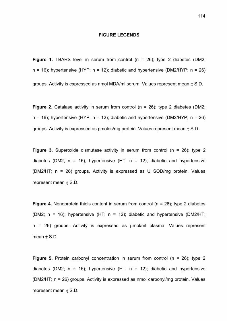
114
FIGURE LEGENDS
Figure 1. TBARS level in serum from control (n = 26); type 2 diabetes (DM2;
n = 16); hypertensive (HYP; n = 12); diabetic and hypertensive (DM2/HYP; n = 26)
groups. Activity is expressed as nmol MDA/ml serum. Values represent mean ± S.D.
Figure 2. Catalase activity in serum from control (n = 26); type 2 diabetes (DM2;
n = 16); hypertensive (HYP; n = 12); diabetic and hypertensive (DM2/HYP; n = 26)
groups. Activity is expressed as pmoles/mg protein. Values represent mean ± S.D.
Figure 3. Superoxide dismutase activity in serum from control (n = 26); type 2
diabetes (DM2; n = 16); hypertensive (HT; n = 12); diabetic and hypertensive
(DM2/HT; n = 26) groups. Activity is expressed as U SOD/mg protein. Values
represent mean ± S.D.
Figure 4. Nonoprotein thiols content in serum from control (n = 26); type 2 diabetes
(DM2; n = 16); hypertensive (HT; n = 12); diabetic and hypertensive (DM2/HT;
n = 26) groups. Activity is expressed as µmol/ml plasma. Values represent
mean ± S.D.
Figure 5. Protein carbonyl concentration in serum from control (n = 26); type 2
diabetes (DM2; n = 16); hypertensive (HT; n = 12); diabetic and hypertensive
(DM2/HT; n = 26) groups. Activity is expressed as nmol carbonyl/mg protein. Values
represent mean ± S.D.
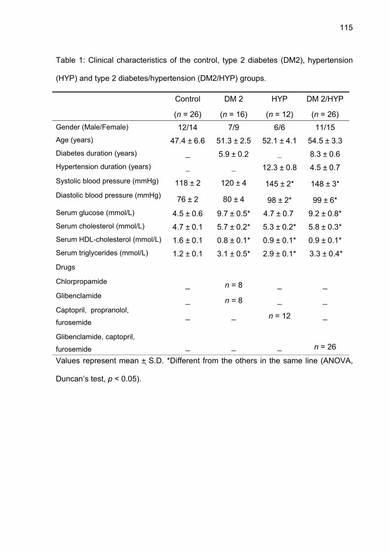
115
Table 1: Clinical characteristics of the control, type 2 diabetes (DM2), hypertension
(HYP) and type 2 diabetes/hypertension (DM2/HYP) groups.
Control
(n = 26)
DM 2
(n = 16)
HYP
(n = 12)
DM 2/HYP
(n = 26)Gender (Male/Female) 12/14 7/9 6/6 11/15Age (years) 47.4 ± 6.6 51.3 ± 2.5 52.1 ± 4.1 54.5 ± 3.3Diabetes duration (years) _ 5.9 ± 0.2 _ 8.3 ± 0.6Hypertension duration (years)
Systolic blood pressure (mmHg)
Diastolic blood pressure (mmHg)
_
118 ± 2
76 ± 2
_
120 ± 4
80 ± 4
12.3 ± 0.8
145 ± 2*
98 ± 2*
4.5 ± 0.7
148 ± 3*
99 ± 6*Serum glucose (mmol/L) 4.5 ± 0.6 9.7 ± 0.5* 4.7 ± 0.7 9.2 ± 0.8*Serum cholesterol (mmol/L) 4.7 ± 0.1 5.7 ± 0.2* 5.3 ± 0.2* 5.8 ± 0.3*Serum HDL-cholesterol (mmol/L) 1.6 ± 0.1 0.8 ± 0.1* 0.9 ± 0.1* 0.9 ± 0.1*Serum triglycerides (mmol/L)
Drugs
Chlorpropamide
Glibenclamide
Captopril, propranolol,
furosemide
Glibenclamide, captopril,
furosemide
1.2 ± 0.1
_
_
_
_
3.1 ± 0.5*
n = 8
n = 8
_
_
2.9 ± 0.1*
_
_
n = 12
_
3.3 ± 0.4*
_
_
_
n = 26
Values represent mean ± S.D. *Different from the others in the same line (ANOVA,
Duncan’s test, p < 0.05).
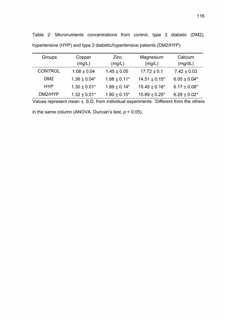
116
Table 2: Micronutrients concentrations from control, type 2 diabetic (DM2),
hypertensive (HYP) and type 2 diabetic/hypertensive patients (DM2/HYP).
Groups Copper(mg/L)
Zinc(mg/L)
Magnesium(mg/L)
Calcium(mg/dL)
CONTROL 1.08 ± 0.04 1.45 ± 0.05 17.72 ± 0.1 7.42 ± 0.03DM2 1.36 ± 0.04* 1.68 ± 0.11* 14.51 ± 0.15* 6.05 ± 0.04*HYP 1.30 ± 0.01* 1.89 ± 0.14* 15.45 ± 0.16* 6.17 ± 0.08*
DM2/HYP 1.32 ± 0.01* 1.80 ± 0.15* 15.89 ± 0.25* 6.29 ± 0.02*Values represent mean ± S.D. from individual experiments. *Different from the others
in the same column (ANOVA, Duncan’s test, p < 0.05).
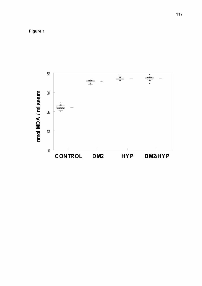
117
Figure 1
CONTROL DM2 HY P DM2/HYP0
13
26
39
52
nmol
MD
A /
ml s
erum
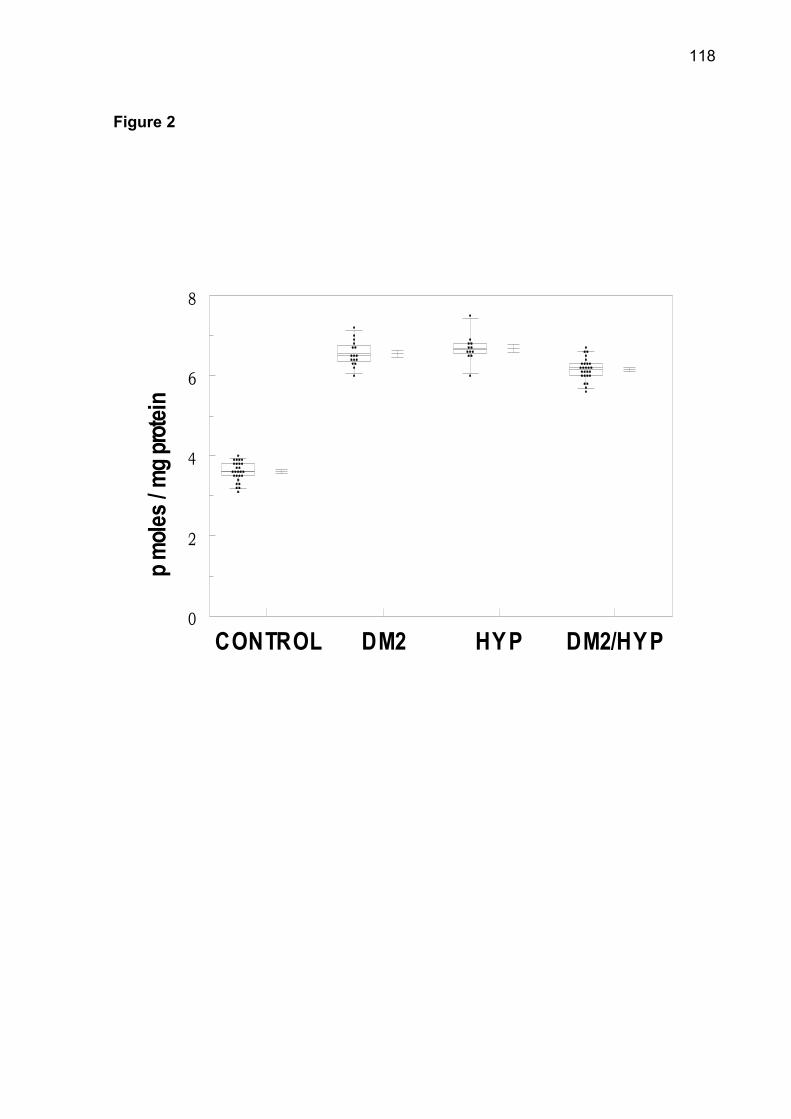
118
Figure 2
CONTROL DM2 HY P DM2/HYP0
2
4
6
8
p mole
s / m
g prot
ein
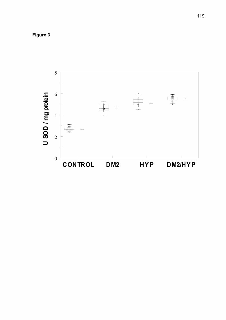
119
Figure 3
CONTROL DM2 HY P DM2/HY P0
2
4
6
8
U S
OD
/ m
g pr
otei
n
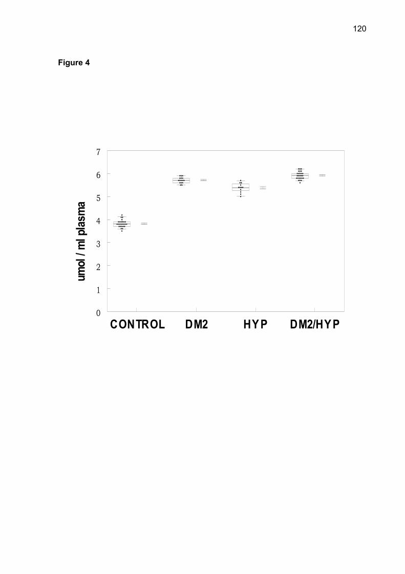
120
Figure 4
CONTROL DM2 HY P DM2/HYP0
1
2
3
4
5
6
7
umol
/ ml
pla
sma
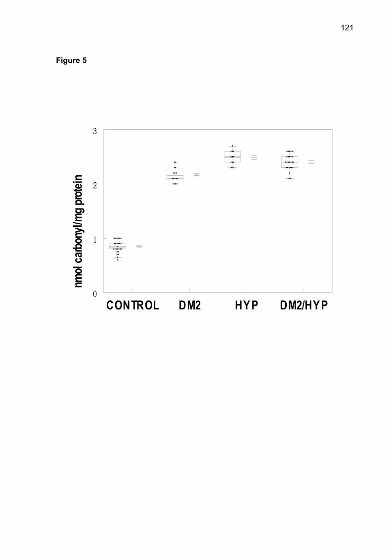
121
Figure 5
CONTROL DM2 HY P DM2/HYP0
1
2
3
nmol
carbo
nyl/m
g prot
ein
122
4 DISCUSSÃO DOS RESULTADOS
O Diabetes melito corresponde a um grupo de doenças metabólicas
caracterizadas por hiperglicemia, que pode resultar na alteração da secreção de
insulina, ação da insulina ou ambas (MINISTÉRIO DA SAÚDE, 2006). A presença
de hiperglicemia persistente em pacientes com diabetes está associada a alterações
macro e microvasculares (YUAN et al., 2007). As alterações macrovasculares em
paciente com diabetes são causa de mortalidade e morbidade, assim como, podem
estar associadas com doenças ateroscleróticas obstrutivas (RAHMAN et al., 2007).
A hiperglicemia tem sido indicada como a responsável por induzir efeitos
pró-coagulantes e antifibrinolíticos no sangue, que podem contribuir para com um
grande risco de trombose arterial (HANSEN et al., 2007). O desenvolvimento de
disfunção endotelial, com supressão da síntese de óxido nítrico e prostaciclinas,
associado com a hiperglicemia promovem um aumento da ativação plaquetária. As
alterações na homeostasia plaquetária criam um risco trombótico que permite o
desenvolvimento de doença cardiovascular (GRANT, 2007). A interferência de
hiperglicemia/hiperinsulinemia, em pessoas saudáveis, é capaz de induzir um
aumento na ativação das plaquetas, promovendo um estado pró-coagulante que
pode contribuir para eventos vasculares agudos e aterogênicos (VAIDYULA et al.,
2006).
Alterações na atividade da enzima colinesterase no soro têm sido
investigadas em diabéticos e um incremento em sua atividade pode estar associado
com complicações vasculares (RAGOOBIRSINGH et al., 1992; ABBOTT et al.,
1993). Outros estudos têm relatado uma associação entre alterações na atividade da
colinesterase sérica com hipertensão e distúrbios encontrados no Diabetes melito
(RUSTEMEIJER et al., 2001; ALCANTARA et al., 2002; KÁLMÁN et al., 2004). O
aumento na atividade da enzima colinesterase sérica, em pacientes com diabetes e
hipertensão associada, sugere que estas doenças poderiam alterar o mecanismo
catalítico da enzima. Estes resultados demonstram que diabetes e hipertensão
podem ser importantes no aumento na síntese de colinesterase ou liberação pelo
tecido hepático. A interferência dos níveis de glicose na atividade da colinesterase
123
sérica foi demonstrada por uma curva in vitro, que indicou um incremento na
atividade enzimática proporcional a elevação nas concentrações de glicose. A
interferência do metabolismo dos lipídios na atividade da colinesterase sérica foi
demonstrada pela positiva correlação entre os níveis de triglicerídios e colesterol
total com o aumento na sua atividade enzimática (IWASAKI et al., 2007). Portanto, o
perfil enzimático da colinesterase sérica, em pacientes com diabetes, pode constituir
um importante fator na etiopatogenia através da influência da resistência à insulina e
metabolismo de lipídios (SRIDHAR et al., 2006).
Estudos prévios de nosso grupo já haviam determinado um aumento na
atividade das ectonucleotidases em pacientes diabéticos e patologias associadas
(LUNKES et al., 2003). Em virtude desses dados, uma curva in vitro de glicose e
frutose frente às enzimas tromboreguladoras, NTPDase e 5'-nucleotidase, foi
realizada em plasma rico em plaquetas de pacientes saudáveis para diabetes e
hipertensão. Este estudo determinou que o aumento na atividade das enzimas foi
proporcional à elevação na concentração de glicose e frutose. Estes resultados
demonstraram que elevadas concentrações de glicose e frutose constituem um
relevante fator capaz de modificar a atividade das ectonucleotidases, que são
enzimas capazes de modular ativação plaquetária e formação de trombo
(MARCUS et al., 2005). Dessa forma, pode-se sugerir que o diabetes per se é capaz
de promover o aumento na atividade da NTPDase e 5'-nucleotidase. As
investigações demonstram que a hiperglicemia e baixo controle glicêmico podem
aumentar a agregabilidade das plaquetas (WATALA et al., 2006). Os nossos dados
demonstram que a hipertensão associada ao diabetes constitui um importante fator
para manutenção do aumento na atividade das ectonucleotidases.
As plaquetas quando expostas a condições hiperosmolar têm apresentado
igualmente um aumento da reatividade das plaquetas. Estudos prévios in vitro com
manitol sugerem que os efeitos osmóticos da glicose constituem um importante
mecanismo pelo qual a hiperglicemia pode aumentar a reatividade plaquetária
(KEATING et al., 2003). Portanto, a exposição das plaquetas a uma elevação de
osmolaridade, aumenta a propensão de agregação plaquetária e em conseqüência
promover complicações tromboembólicas. Dessa forma, pode-se sugerir que um
aumento de osmolaridade associado hiperglicemia pode aumentar a reatividade
plaquetária.
124
O tempo de pré-incubação não constituiu um fator capaz de interferir na
atividade das ectonucleotidases. Esse comportamento enzimático demonstra que a
hiperglicemia, mesmo que em processo agudo, é capaz de influenciar os processos
de tromboregulação. Assim, a supressão da hiperglicemia transitória constitui uma
medida preventiva para diminuir alterações coronárias associadas com quadros de
hiper agregabilidade plaquetária (SAKAMOTO et al., 2000). A hiperglicemia
pós-prandial tem sido demonstrada como um importante preditor de mortalidade em
pacientes com diabetes tipo 2 (DECODE, 1999). Dessa forma, os níveis glicêmicos
pós-prandias são de extrema relevância na patogênese das complicações crônicas
do diabetes (AFFONSO et al., 2003).
Posteriormente, a análise da expressão da enzima NTPDase (CD39), por
citometria de fluxo, demonstrou uma intensa correlação da hidrólise de ATP e ADP
frente a pacientes diabéticos e hipertensão associada. Esses dados indicam que o
incremento na expressão de CD39 pode interferir em processos de ativação
plaquetária. Assim, pode se observar que a proeminente interferência da glicemia na
homeostasia das plaquetas e na hidrólise dos nucleotídeos representa um
importante parâmetro nos processos de tromboregulação (FRIEDMAN et al., 2007).
O desenvolvimento de disfunção vascular, em diabéticos tipo 2, pode estar
associado a uma elevação na geração de espécies reativas de oxigênio
(WEIDIG et al., 2004), assim como, em hipertensos (de CHAMPLAIN et al., 2004).
Os elevados níveis de TBARS, em pacientes com diabetes e hipertensão associada,
indicam que a hiperglicemia está interferindo na peroxidação dos lipídios, que
constitui um biomarcador de estresse oxidativo.
O sistema de defesa antioxidante elevado no soro, em pacientes com
diabetes e hipertensão associada, pode estar relacionado a um mecanismo
compensatório para prevenir o dano oxidativo. A interferência dos níveis de glicose
no sistema enzimático antioxidante foi demonstrada por uma curva in vitro, onde o
aumento na atividade de CAT e SOD foi proporcional ao incremento nas
concentrações de glicose. O sistema não-enzimático antioxidante teve uma
correlação positiva com os elevados níveis de glicose sangüínea em pacientes com
o diabetes e hipertensão associada. Estudos demonstram que a hipertensão
aumenta a formação de pró-oxidantes e a sua combinação com diabetes pode
exacerbar o estresse oxidativo (BISWAS et al., 2008). Portanto, a manifestação de
125
diabetes e hipertensão associados pode aumentar a produção de espécies reativas
de oxigênio e promover alterações vasculares.
A geração de espécies reativas de oxigênio está fortemente relacionada com
ativação das plaquetas em pacientes diabéticos. Portanto, a relação entre os níveis
de sistemas oxidantes e antioxidantes em plaquetas é extremamente relevante no
balanço da homeostasia de doenças vasculares (KRÖTZ et al., 2004). O incremento
na atividade das enzimas catalase e superóxido dismutase e dos níveis de
carbonilação de proteínas e grupos tióis não protéicos representam uma resposta
nas plaquetas à estimulação pela geração de espécies reativas de oxigênio
resultante da hiperglicemia crônica. A presença da hipertensão constitui mais um
fator de estimulação dos níveis de estresse oxidativo e a combinação com quadro de
hiperglicemia persistente podem estimular um aumento na ativação plaquetária
(SUDICA et al., 2006). As baixas concentrações séricas de ácido ascórbico, em
pacientes com diabetes e hipertensão associada, aumentam a exposição aos danos
oxidativos. Tem sido demonstrado que os baixos níveis de ácido ascórbico podem
causar efeitos pró-oxidativos (WILKINSON et al., 1999). A administração dos
medicamentos metformina e insulina não comprometeram o comportamento dos
sistemas enzimático e não-enzimático antioxidantes de defesa.
As enzimas NTPDase e 5'-nucleotidase participam ativamente no processo de
tromboregulação. O diabetes exerceu uma interferência direta na atividade das
ectonucleotidases. Neste estudo, pode se sugerir que as elevadas concentrações de
glicose modularam as enzimas tromboreguladoras. A presença concomitante de
hipertensão, nos pacientes diabéticos, tenha sido um fator de manutenção do
aumento da atividade das ectonucleotidases.
126
5 CONCLUSÃO
a. O incremento na atividade da enzima colinesterase sérica está diretamente
relacionado com as concentrações de glicose e com o metabolismo dos lipídios. Há
igualmente, uma intensa relação com diabetes e hipertensão, podendo estar
associada com as complicações vasculares em pacientes com diabetes e
hipertensão associada.
b. O aumento na atividade das enzimas NTPDase-ATP, NTPDase-ADP e
5'-nucleotidase-AMP foi proporcional ao aumento na concentração de glicose e
frutose, enquanto que os tempos de pré-incubação não modificaram as respostas
enzimáticas, demonstrando que a hiperglicemia pode interferir na atividade
enzimática independente do tempo de exposição.
c. O aumento na expressão da enzima NTPDase-ATP e NTPDase-ADP indica que a
hidrólise dos nucleotídeos de adenina representam um importante parâmetro no
processo de reatividade plaquetária e que a patologia causou o aumento da
expressão da enzima.
d. O aumento nos níveis de biomarcadores de dano oxidativo, como TBARS e
proteína carbonil, indicam um incremento nos níveis de estresse oxidativo. Em
conseqüência, há um estímulo na geração de espécies reativas de oxigênio que
desencadearam de forma compensatória um aumento na atividade dos sistemas
antioxidantes tanto no sangue como nas plaquetas, com exceção do ácido
ascórbico. Essas alterações nos sistemas enzimáticos podem interferir na
homeostasia das plaquetas. Os níveis elevados de glicose constituíram um fator
com uma correlação direta com a elevação dos níveis de estresse oxidativo. A
concentração dos micronutrientes demonstrou uma exposição aos danos oxidativos
e uma tentativa de compensação ao aumento na produção de espécies reativas de
oxigênio.
127
REFERENCIAL
ABBOTT, C. A. et al. Relationship between serum butyrylcholinesterase activity, hypertriglyceridaemia and insulin sensitivity in diabetes mellitus. Clinical Science, v. 85, n. 1, p. 77-81, 1993.
ABBINK-ZANDBERGEN, E. J. et al. The role of adenosine in insulin-induced vasodilation. Journal of Cardiovascular Pharmacology, v. 34, n. 3, p. 373-380, 1999.
AFFONSO, F. S.; CAILLEAUX, S.; PINTO, L. F. C.; GOMES, M. B.; TIBIRIÇÁ, E. Efeitos de altas concentrações de glicose na função endotelial da microcirculação renal de coelhos. Aquivo Brasileiro de Cardiologia, v. 81, n. 1, p. 156-160, 2003.
AGATA, J. et al. Association of insulin resistance and hyperinsulinemia with disturbed lipid metabolism in patients with essential hypertension. Hypertension Research, v. 21, n. 1, p. 57-62, 1998.
AHMED, F. N.; NAQBI, F. N.; SHAFIQ, F. Lipid peroxidation and serum antioxidant enzymes in patients with type 2 diabetes mellitus. Annals of the New York Academy of Sciences, v. 1084, p. 481-489, 2006.
ALCANTARA, V. M. et al. Butyrylcholinesterase activity and risk factors for coronary artery disease, Scandinavian Journal of Clinical and Laboratory Investigation, v. 62, n. 5, p. 399-404, 2002.
ANGIOLILLO, D. J. et al. Impact of platelet reactivity on cardiovascular outcomes in patients with type 2 diabetes mellitus and coronary artery disease. Journal of the American College of Cardiology, v. 50, n. 16, p. 1541-1547, 2007.
ANTONIADES. C. et al. Effects of insulin dependence on inflammatory process, thrombotic mechanisms and endothelial function, in patients with type 2 diabetes mellitus and coronary atherosclerosis. Clinical Cardiology, v. 30, n. 6, p. 295-300, 2007.
128
ARAÚJO, M. et al. Enzymes that hydrolyze adenine nucleotides in platelets from breast cancer patients. Biochimica et Biophysica Acta, v. 1740, v. 3, p. 421-426, 2005.
ATKINSON, B. et al. Ecto-nucleotidases of the CD39/NTPDase family modulate platelet activation and thrombus formation: Potential as therapeutic targets. Blood Cells, Molecules & Diseases, v. 36, n. 2, p. 217-222, 2006.
BARMAN, T. E. Enzyme Handbook. v. 2. New York: Springer Verlag, 1969.
BELDI, G. et al. The role of purinergic signaling in the liver and in transplantation: effects of extracellular nucleotides on hepatic graft vascular injury, rejection and metabolism. Frontiers Bioscience, v. 13, p. 2588-2603, 2008.
BIEDERBICK, A. et al. First apyrase splice variants have different enzymatic properties. The Journal of Biological Chemistry, v. 275, n. 25, p. 19018-19024, 2000.
BIGONNESSE, F. et al. Cloning and characterization of mouse nucleoside triphosphate diphosphohydrolase-8. Biochemistry, v. 43, n. 23, p. 5511–5519, 2004.
BIJLSTRA, P. et al. Glyburide inhibits dipyridamole-induced forearm vasodilation but not adenosine-induced forearm vasodilation. Clinical Pharmacology and Therapeutics, v. 75, n. 3, p. 147-156, 2004.
BIRK, A. V. et al. Role of extracellular AT`P metabolism in regulation of platelet reactivity. The Journal of Laboratory and Clinical Medicine, v. 140, p. 166-175, 2002.
BISWAS, S. K. et al. Hypertension increases pro-oxidant generation and decreases antioxidant defense in the kidney in early diabetes. American Journal of Nephrology, v. 28, n. 1, p. 133-142, 2008.
BODEN, G.; RAO, A. K. Effects of hyperglycemia and hyperinsulinemia on the tissue factor pathway of blood coagulation. Current Diabetes Reports, v. 7, n. 3, p. 223-227, 2007.
BONDAR, I. A. et al. Hyperhomocysteinemia in coronary artery disease and hemostasis disorders in patients with type 2 diabetes mellitus. Klinicheskaia Meditsina, v. 85, n. 5, p. 30-33, 2007.
129
BOURS, M. J. Adenosine 5’-triphosphate and adenosine as endogenous signaling molecules in immunity and inflammation. Pharmacology & Therapeutics, v. 112, n. 2, p. 358-404, 2006.
BRAUN, N. et al. Sequencing, functional expression and characterization of NTPDase 6, a nucleoside diphosphatase and novel member of the ectonucleoside triphosphate diphosphohydrolase family. The Biochemical Journal, v. 351, p. 639–647, 2000.
BURNSTOCK, E. R.; NIGHT G. E. Cellular distribution and functions of P2 receptors subtypes in different system. International Review of Cytology, v. 240, p. 301-34, 2004.
CATTANEO, M. Platelet P2 receptors: old and new targets for antithrombotic drugs. Expert Review of Cardiovascular Therapy, v. 5, n. 1, p. 45-55, 2007.
CECHIN, S. R. et al. Inhibitory effect of novel pyrimidines on ATP and ADP hydrolysis in synaptosomes from rat cerebral cortex. Chemical Research in Toxicology, v. 16, n. 11, p. 1433-1439, 2003.
de CHAMPLAIN, J. et al. Oxidative stress in hypertension. Clinical and Experimental Hypertension, v. 26, n. 7/8, p. 593-601, 2004.
COSTA, A.F. et al. Intravenous apyrase administration reduces arterial thrombosis in a rabbit model of endothelial denudation in vivo. Blood Coagulation & Fibrinolysis, v. 15, n. 7, p. 545-551, 2004.
CRISTALLI, G. et al. Medicinal chemistry of adenosine A2A receptor agonists. Current Topics in Medicinal Chemistry, v. 3, n. 4, p. 387-401, 2003.
CUCUIANU, T. et al. Serum cholinesterase activity correlates with serum insulin, C-peptide and free fatty acids levels in patients with type 2 diabetes. Romanian Journal of Internal Medicine, v. 40, n. 1/4, p. 43-51, 2002.
DANIEL, J.L. et al. Molecular basis for ADP-induced platelet activation. The Journal of Biological Chemistry, v. 273, n. 4, p. 2024-2029, 1998.
DAS, U. N. Acetylcholinestease and butyrylcholinesterase as possible markers of low-grade systemic inflammation. Medical Science Monitor, v. 13, n. 12, p. 214-221, 2007.
130
DAVE, K. R.; KATYARE, S. S. Effect of alloxan-induced diabetes on serum and cardiac butyrylcholinesterases in the rat. The Journal of Endocrinology, v. 175, n. 1, p. 241-250, 2002.
DECODE Study Group. Glucose tolerance and mortality: comparison of WHO and ADA diagnosis criteria. Lancet, v. 354, p. 617-621, 1999.
DUARTE, M. M. et al. Enzymes that hydrolyze adenine nucleotides of patients with hypercholesterolemia and inflamatory processes. The FEBS Journal, v. 274, n. 11, p. 2707-2714, 2007.
ENJYOJI, K. et al. Tergeted disruption of CD39/ATP diphosphohydrolase results in disordered hemostasis and thromboregulation. Nature Medicine, v. 5, n. 9, p. 1010-1017, 1999.
ERB, L. et al. P2 receptors: intracellular signaling. European Journal of Physiology, v.452, n.5, p. 552-562, 2006.
EVANGELISTA, V. et al. Persistent platelet activation in patients with type 2 diabetes treated with low doses of aspirin. Journal of Thrombosis and Haemostasis, v. 5, n. 11, p. 2197-2203, 2007.
FERRONI, P. et al. Platelet activation in type 2 diabetes mellitus. Journal of Thrombosis and Haemostasis, v. 2, n. 8, p.1282-1291, 2004.
FRASSETTO, S. S.; DIAS, R. D.; SARKIS, J. J. Characterization of an ATP diphosphohydrolase activity (apyrase, EC 3.6.1.5) in rat blood platelets. Molecular and Cellular Biochemistry, v. 129, n. 1, p. 47-55, 1993.
FRASSETTO, S. S.; DIAS, R. D.; SARKIS, J. J. Free radical-induced inhibition of ATP diphosphohydrolase activity (EC 3.6.1.5) from rat blood platelets. Biochemistry and Molecular Biology International, v.41, n.1, p. 161-168, 1997.
FRIEDMAN, D. J. et al. The vascular ectonucleotidases ENTPD1 is a novel renoprotective factor in diabetic nephropathy. Diabetes, v. 56, n. 9, p. 2371-2379, 2007.
FÜRSTENAU, C. R.; TRENTIN, S.; BARRETO-CHAVES, M. L., SARKIS, J. J. Ecto-nucleotide pyrophosphatase/phosphodiesterase as part of a multiple system for nucleotide hydrolysis by platelets from rats: kinetic characterization and biochemical properties. Platelets, v. 17, n.2, p. 84-91, 2006.
131
GAENZER, H. et al. Effect of insulin therapy on endothelium-dependent dilation in type 2 diabetes mellitus. The American Journal of Cardiology, v. 89, n. 4, p. 431-434, 2002.
GANNE, S. et al. Hypertension in people with diabetes and the metabolic syndrome: pathophysiologic insights and therapeutic update. Current Diabetes Reports, v. 7, n. 3, p. 208-217, 2007.
GARCÍA DONAIRE, J.A.; RUILOPE, L.M. Trandolapril/verapamil combination in hypertensive diabetic patients. Vascular Health and Risk Management, v. 3, n. 1, p. 77-82, 2007.
GAYLE III, R. B. et al. Inhibition of platelet function by recombinant soluble ecto-ADPase/CD39. The Journal of Clinical Investigation, v. 101, n. 9, p. 1851-1859, 1998.
GRANT, P. J. Diabetes mellitus as a prothrombotic condition. Journal of Internal Medicine, v. 262, n. 2, p. 157-172, 2007.
GRINTHAL, A.; GUIDOTTI, G. CD39, NTPDase1, is attached to the plasma membrane by two transmembrane domains. Why? Purinergic Signalling, v. 2, n. 2, p. 391-398, 2006.
HAMILTON, S. J.; CHEW, G. T.; WATTS, G. F. Therapeutic regulation of endothelial dysfunction in type 2 diabetes mellitus. Diabetes & Vascular Disease Research, v. 4, n. 2, p. 89-102, 2007.
HANSEN, H. R. et al. Hyperglycemia accelerates arterial thrombus formation and attenuates the antithrombotic response to endotoxin in mice. Blood Coagulation & Fibrinolysis, v. 18, n. 7, p. 627-636, 2007.
INOUE, K.; KOIZUMI, S.; TSUDA, M. The role of nucleotides in the neuron-glia communication responsible for the brain functions. Journal of Neurochemistry, v. 102, n. 5, p. 1447-1458, 2007.
ISHII-WATABE, A.; UCHIDA, E.; MIZUGUCHI, H.; HAYAKAWA, T. On the mechanism of plamin-induced platelet aggregation. Implications of the dual role of granule ADP. Biochemistry Pharmacology, v.59, n. 11, p. 1345-1355, 2000.
132
IWASAKI, T. et al. Serum butyrylcholinesterase is strongly associated with adiposity, the serum lipid profile and insulin resistance. Internal Medicine, v. 46, n. 19, p. 1633-1639, 2007.
JENNER, P. Oxidative stress in Parkinson's disease. Annals of Neurology, v. 53, n. 3, p. 26-38, 2003.
KAHNER, B. N. Nucleotide receptor signaling in platelets. Journal of Thrombosis and Haemostasis, v. 4, n. 11, p. 2317-2326, 2006.
KAIZER, R. R. et al. The effect of aluminium on NTPDase and 5’-nucleotidase activities from rat synaptosomes and platelets. International Journal of Developmental Neuroscience, v. 25, n. 6, p. 381-386, 2007.
KÁLMÁN, J. et al. Increased serum butyrylcholinesterase activity in type IIb hyperlipidaemic patients. Life Sciences, v. 75, n. 10, p. 1195–1204, 2004.
KAUFFENSTEIN, G.; BERGMEIER, W.; ECKLEY, A.; OHLMANN, P.; LEON, C.; CAZENAVE, J. P.; NIESWANDR, B.; GACHET, C. The P2Y(12) receptor induces platelet aggregation through weak activation of the alpha(lib)beta(3) integrin-a phosphoinositide 3–kinase-dependent mechanism. FEBS Letters, v.505, n.2, p. 281-290, 2001.
KAWASHIMA, Y.; NAGASAWA, T.; NINOMIYA, H. Contribution of 5’-nucleotidase to the inhibition of platelet aggregation by human endothelial cells. Blood, v. 96, n. 6, p. 2157-2162, 2000.
KEATING, F. G.; SOBEL, B. E.; SCHNEIDER, D. J. Effects of increased concentrations of glucose on platelet reactivity in healthy subjects and in patients with and without diabetes mellitus. The American Journal of Cardiology, v. 92, p. 1362-1365, 2003.
KING, G. L.; LOEKEN, M. R. Hyperglycemia-induced oxidative stress in diabetic complications. Histochemistry and Cell Biology, v. 122, n. 4, p. 333-338, 2004.
KRÖTZ, F.; SOHN, H. Y.; POHL, U. Reactive oxygen species: players in the platelet game. Arteriosclerosis, Thrombosis, and Vascular Biology, v. 24, n. 11, p. 1988-1986, 2004.
KUKULSKI, F. et al. Comparative hydrolysis of P2 receptor agonists by NTPDase 1, 2, 3 and 8. Purinergic Signalling, v. 1, n. 2, p.193-204, 2005.
133
LEAL, D. B. et al. Characterization of NTPDase (NTPDase 1; ecto-apyrase; ecto-diphosphohydrolase; CD39; EC 3.6.1.5) activity in human lymphocytes. Biochimica et Biophysica Acta, v. 1721, n. 1-3, p. 9-15, 2005.
LEAL, D. B. et al. HIV infection is associated with increased NTPDase activity that correlates with CD39-positive lymphocytes. Biochimica et Biophysica Acta, v. 1746, n. 2, p. 129-134, 2005.
LEAL, C. A. NTPDase and 5’-nucleotidase activities in platelets of human pregnants with a normal or high risk for thrombosis. Molecular and Cellular Biochemistry, v. 304, n. 1-2, p. 325-330, 2007.
LELLI, J. et al. ATP converts necrosis to apoptosis in oxidant-injured endothelial cells. Free Radical Biology & Medicine, v. 25, n. 6, p. 684-702, 1998.
LOCKRIDGE, O. Structure of human serum cholinesterase. Bioessays, v. 9, n. 4, p. 125-128, 1988.
LUNKES, G. I. et al. Enzymes that hydrolyze adenine nucleotides in diabetes and associated pathologies. Thrombosis Research, v. 109, n. 4, p. 189-194, 2003.
LUNKES, G. I. et al. NTPDase and 5’-nucleotidase activities in rats with alloxan-induced diabetes. Diabetes Research and Clinical Practice, v. 65, n. 1, p.1-6, 2004.
MARCUS, A. J. Role of CD39 (NTPDase-1) in thromboregulation, cerebroprotection, and cardioprotection. Seminars in Thrombosis and Hemostasis, v. 31, n. 2, p 234-246, 2005.
MATAYOSHI, T. et al. Relationship between insulin resistance and the renin-angiotensin system: analysis for patients with essential and renovascular hypertension. Clinical and Experimental Hypertension, v. 29, n. 7, p. 479-487, 2007.
MAZZANTI, C. M. et al. Previous treatment with ebselen and vitamin E alters adenine nucleotide hydrolysis in platelets from adult rats experimentally demyelinated with ethidium bromide. Life Sciences, v. 81, n.3, p. 241-248, 2007.
134
McGRATH, L.T. et al. Increased oxidative stress in Alzheimer’s disease as assessed with 4-hydroxynonenal but not malondialdhyde. The Quarterly Journal of Medicine, v. 94, p. 485-490, 2001.
MEETOO, D.; McGOVERN, P.; SAFADI, R. An epidemiological overview of diabetes across the world. British Journal of Nursing, v. 16, n. 16, p. 1002-1007, 2007.
MEIGS, J.B. et al. Hyperinsulinemia, hyperglycemia, and impaired hemostasis: the Framingham Offspring Study. The Journal of the American Medical Association, v. 283, n. 2, p. 221-228, 2000.
MICHNO, A. et al. Alterations of adenine nucleotide metabolism and function of blood platelets in patients with diabetes. Diabetes, v. 56, n. 2, p. 462-467, 2007.
MINISTÉRIO DA SAÚDE. Cadernos de Atenção Básica. Diabetes mellitus. Normas e Manuais Técnicos. Brasília, n. 16, 2006, 64 p.
___________________. Cadernos de Atenção Básica. Hipertensão Arterial Sistêmica. Normas e Manuais Técnicos. Brasília, n.15, 2006, 58 p.
MIRON, V. R. et al. Enhanced NTPDase and 5’-nucleotidase activities in diabetes mellitus and iron-overload model. Molecular and Cellular Biochemistry, v. 298, n. 1-2, p. 101-107, 2007.
MORETTO, M. B. et al. Effect of subchronic treatment with mercury chloride on NTPDase, 5’-nucleotidase and acetylcholinesterase from cerebral cortex of rats. Journal of Trace Elements in Medicine and Biology, v. 17, n. 4, p. 255-260, 2004.
MUACEVIC-KATANECA, D. et al. Clinical study on the effect of sinvastatin on butyrylcholinesterase activity. Arzneimittel-Forschung, v. 55, n. 5, p. 271-275, 2005.
MURUGAPPA, S.; KUNAPULI, S. P. The role of ADP receptors in platelet function. Frontiers in Bioscience, v. 11, p. 1977-1986, 2006.
PÁEZ, J. G. et al. Identity between the PCPH proto-oncogene and the CD39L4 (ENTPD5) ectonucleoside triphosphate diphosphohydrolase gene. International Journal of Oncology, v. 9, p. 1249–1254, 2001.
135
PENNATHUR, S.; HEINECKE, J. W. Oxidative stress and endothelial dysfunction in vascular disease. Current Diabetes Reports, v. 7, n. 4, p. 257-264, 2007.
PILLA, C. et al. ATP diphosphohydrolase activity (apyrase, EC 3.6.1.5) in human blood platelets. Platelets, v. 7, p. 225-230, 1996.
RAGOOBIRSINGH, D.; BHARAJ, B. S.; MORRISON, E. Y. Change in serum cholinesterase activity in Jamaican diabetes. Journal of the National Medical Association, v. 84, n. 10, p. 853–855, 1992.
RAHMAN, S. et al. Diabetes-associated macrovasculopathy: pathophysiology and pathogenesis. Diabetes, Obesity & Metabolism, v. 9, n. 6, p. 767-780, 2007.
RAMAKRISHNA, V; JAILKHANI, R. Evaluation of oxidative stress in Insulin Dependent Diabetes Mellitus (IDDM) patients. Annals of Diagnostic Pathology, v. 1, p. 2-22, 2007.
RAO, A. A.; SRIDHAR, G. R.; DAS, U. N. Elevated buryrylcholinesterase and acetylcholinesterase may predict the development of type 2 diabetes mellitus and alzheimer’s disease. Medical Hypotheses, v. 69, n. 6, p. 1272-1276, 2007.
ROBSON, S. C. et al. Ectonucleotidases of CD39 family modulate vascular inflammation and thrombosis in transplantation. Seminars in Thrombosis and Hemostasis, v. 31, n. 2, p 217-233, 2005.
ROBSON, S. C.; SÉVIGNY, J.; ZIMMERMANN, H. The E-NTPDase family of ectonucleotidases: structure function relationships and pathophysiological significance. Purinergic Signalling, v. 2, n. 2, p. 409-430, 2006.
RUSTEMEIJER, C. et al. Is pseudocholinesterase activity related to markers of triacylglycerol synthesis in Type II diabetes mellitus? Clinical Science, v. 101, n. 1, p. 29-35, 2001.
SAKAMOTO, T. et al. Rapid change of platelet aggregability in acute hyperglycemia. Detection by a novel laser-light scattering method. Thrombosis and Haemostasis, v. 83, n. 3, p. 475-479, 2000.
SARKIS, J. J. F. et al. ATP diphosphohydrolases: an overview. Journal of the Brazilian Association for the Advancement of Science, v. 47, n. 3, p. 131-136, 1995.
136
SAVOIA, C.; SCHIFFRIN, E. L. Vascular inflammation in hypertension and diabetes: molecular mechanisms and therapeutic interventions. Clinical Science, v. 112, n. 7, p. 375-384, 2007.
SCHAFER, F.; BUETTNER, G. Redox environment of the cell as viewed through the redox state of the glutathione disulfide/glutathione couple. Free Radical Biology & Medicine, v. 30, n. 11, p. 1191-1212, 2001.
SÉVIGNY, J. et al. Identification and characterization of a novel hepatic canalicular ATP diphosphohydrolase. The Journal of Biological Chemistry, v. 275, n. 8, p. 5640–5647, 2000.
SÉVIGNY, J. et al. Differential catalytic properties and vascular topography of murine nucleoside triphosphate diphosphohydrolase 1 (NTPDase1) and NTPDase2 have implications for thromboregulation. Blood, v. 99, n. 8, p. 2801-2809, 2002.
SHI, J. D. et al. Molecular cloning and characterization of a novel mammalian endo-apyrase (LALP1). The Journal of Biological Chemistry, v. 276, n. 20, p. 17474-17478, 2001.
SILVA, A. C. Enzymes that hydrolyze adenine nucleotides in chronic renal failure: relationship between hemostatic defects and renal failure severity. Biochimica et Biophysica Acta, v. 1741, n. 3, p. 282-288, 2005.
SKRHA, J. et al. Serum alphap-tocopherol and ascorbic acid concentrations in Type 1 an Type 2 diabetic patients with and without angiopathy. Clinica Chimica Acta, v. 329, n. 1/2, p.103-108, 2003.
SOCIEDADE BRASILEIRA DE HIPERTENSÃO. V Diretrizes Brasileiras de Hipertensão. 2006.
SPAVANELLO, R. M. et al. Activties of enzymes tha hydrolyze adenine nucleotides in platelets from rats experimental demyelinated with ethidium bromide and treated with interferon-beta. Life Sciences, v. 80, p. 1109-1114, 2007.
SRIDHAR, G. R. et al. Alzheimer’s disease and type 2 diabetes mellitus: the cholinesterase connection? Lipids in Health and Disease, v. 11, p. 28, 2006.
137
STATFFORD, N.P. et al. Mechanisms involved in adenosine triphosphate–Induced platelet aggregation in whole blood. Arteriosclerosis, Thrombosis, and Vascular Biology, v. 23, p. 1928-1933, 2003.
STEFAN, C; JANSEN, S.; BOLLEN, M. NPP-type ectophosphodiesterases: unity in diversity. Trends in Biochemical Sciences, v. 30, n. 10, p. 542-550, 2005.
SUDICA, D. et al. High glucose levels enhance platelet activation: involvement of multiple mechanisms. British Journal of Haematology, v. 133, n. 3, p. 315-322, 2006.
TAKIUCHI, S. et al. Hypertension attenuates the efficacy of hypoglycemic therapy for preserving coronary flow reserve in patients with type 2 diabetes. Hypertension Research, v. 25, n. 6, p. 893-900, 2002.
VAIDYULA, V. R.; BODEN, G.; RAO, A. K. Platelet and monocyte activation by hyperglycemia and hyperinsulinemia in healthty subjects. Platelets, v. 17, n. 8, p. 577- 585, 2006.
VALKO, M.; MORRIS, H.; CRONIN, M. Metals, toxicity and oxidative stress. Current Medicinal Chemistry, v. 12, n. 10, p. 1161-1208, 2005.
VÉRICEL, E. et al. Diabetic patients without vascular complications display enhanced basal activation and decreased antioxidant status. Diabetes, v. 53, n. 4, p. 1046-1051, 2004.
WANG, T. F.; GUIDOTTI, G. Golgi localization and functional expression of human uridine diphosphatase. The Journal of Biological Chemistry, v. 273, n. 18, p. 11392–11399, 1998.
WATALA, C. et al. High glucose contributes to aspirin insensitivity in streptozotocin-diabetic rats: a multiparametric aggregation study. Blood Coagulation & Fibrinolysis, v. 17, n. 2, p. 113-124, 2006.
WEIDIG, P.; McMASTER, D.; BAYRAKTUTAN, U. High glucose mediates pro-oxidant and antioxidant enzyme activities in coronary endothelial cells. Diabetes, Obesity & Metabolism, v. 6, n. 6, p. 432-441, 2004.
WILD, S. et al. Global Prevalence of Diabetes: estimates for the year 2000 and projections for 2030. Diabetes Care, v. 27, n. 5, p. 1047-1053, 2004.
138
WILKINSON, I. B. et al. Oral vitamin C reduces arterial stiffness and platelet aggregation in humans. Journal of Cardiovascular Pharmacology, v. 34, n. 5, p. 690-693, 1999.
YUAN, S.Y. et al. Microvascular permeability in diabetes and insulin resistance. Microcirculation, v. 14, n. 4/5, p. 363-373, 2007.
ZIMMERMANN, H. Ectonucleotidases: some recent developments and a note on nomenclature. Drug Development Research, v. 52, p. 44-56, 2001.
ZIMMERMANN, H.; VOGEL, M.; LAUBE, U. Hippocampal localization of 5’-nucleotidase as revealeb by immunocytochemystry. Neuroscience, v. 55, n. 1, p. 105-112, 1993.

139
ANEXOS
140
ANEXO A
TERMO DE CONSENTIMENTO LIVRE E ESCLARECIDO
1. TítuloAvaliação do incremento de enzimas que degradam nucleotídeos da adenina,
ésteres de colina e de espécies ativas de oxigênio em diabetes e patologias associadas.
2. Objetivosa. Verificar a interferência de diferentes concentrações de glicose e frutose na atividade das ectonucleotidases em plaquetas de voluntários humanos;b. Verificar se há uma alteração na expressão da enzima NTPDase em plaquetas de pacientes com diabetes, hipertensão e diabéticos hipertensos;c. Avaliar os sistemas anti-oxidantes enzimáticos e não-enzimáticos em pacientes com diabetes, hipertensão e diabéticos hipertensos;d. Verificar a interferência de diferentes concentrações de glicose na atividade enzimática da butirilcolinesterase em pacientes com diabetes, hipertensão e diabéticos hipertensos.e. Observar a atividade das ectonucleotidases frente extrato bruto de Wedelia paludosa em modelo experimental.
3. RegistroO estudo será desenvolvido no Centro de Ciências Naturais e Exatas,
Departamento de Química, Programa de Pós-Graduação em Bioquímica Toxicológica, no Laboratório de Enzimologia Toxicológica, da Universidade Federal de Santa Maria. O presente estudo envolverá pacientes diabéticos, hipertensos e diabéticos hipertensos vinculados ao Programa de Assistência aos Pacientes Diabéticos, Hipertensos e Diabéticos-Hipertensos da Secretaria Municipal de Saúde do município de Cruz Alta, RS. Esse estudo com voluntários humanos obteve a aprovação junto a Comissão de Ética do Centro de Ciências da Saúde da Universidade Federal de Santa Maria, com protocolo n° 013/2004.
4. ProcedimentoOs pacientes serão submetidos a uma punção venosa com sistema
vacutainer. O material biológico, sangue, será destinado para análise de plaquetas, soro e plasma para determinações bioquímicas e de atividade enzimática. As plaquetas serão analisadas no Laboratório de Enzimologia Toxicológica, Centro de Ciências Naturais e Exatas, Departamento de Química, Programa de Pós-Graduação em Bioquímica Toxicológica, da Universidade Federal de Santa Maria, RS.
141
5. Riscos individuaisOs pacientes que voluntariamente se submeterem as punções venosas,
poderão em casos de coleta com procedimento errôneo desenvolver flebite, flebotrombose, hematoma local, petéquias.
6. Identificação do paciente voluntário
Nome:__________________________________________
Identidade:_______________________________________
Assinatura:_______________________________________
7. Cruz Alta, _____ de ____________ de 200__.
142
Anexo B
--------------------------- Mensagem Original ------------------------------------------Assunto: Diabetes Research and Clinical Practice Submission ConfirmationDe: "CLB (ELS)" <[email protected]>Data: Dezembro 12, 2007 Para: mariaschetinger@gmail.com--------------------------------------------------------------------------------------------
Ms. Ref. No.: DIAB-D-07-00782Title: Effect of high glucose levels in human platelet NTPDase and 5’-nucleotidase activitiesDiabetes Research and Clinical Practice
Dear Dr. Schetinger,
Your submission entitled “Effect of high glucose levels in human platelet NTPDase and 5’-nucleotidase activities” assigned the following manuscript number: DIAB-D-07-00782.
You may check on the progress of your paper by logging on to the Elsevier Editorial. The URL is http://ees.elsevier.com/clb/Your username is: mariarosaYour password is: schetinger3372
Thank you for submitting your work to this journal.
Kind regards,
Abi RobinsonEditorial Office
143
ANEXO C
Submitting – Antioxidant status in platelet from patients with diabetes and
hypertension.
---------- Forwarded message ----------From: .Molecular and Cellular Biochemistry <[email protected]>Date: 21 Jan 2008 16:33:55 -0500 Subject: Acknowledgement of ReceiptTo: [email protected]
Dear Maria:
Thank you for submitting your manuscript, "Antioxidant status in platelet from patients with diabetes and hypertension", to Molecular and Cellular Biochemistry.
During the review process, you can keep track of the status of your manuscript by accessing the following web site:
http://mcbi.edmgr.com/
Your username is:mariarosa Your password is:schetinger463
Sincerely,
The Editorial OfficeMolecular and Cellular Biochemistry