Embed Size (px)
Citation preview
903903903903903Mem Inst Oswaldo Cruz, Rio de Janeiro, Vol. 100(8): 903-908, December 2005
Basic biology of Pneumocystis carinii - A Mini ReviewWanderley de Souza/+, Marlene Benchimol*
Laboratório de Ultraestrutura Celular Hertha Meyer, Instituto de Biofísica Carlos Chagas Filho, Universidade Federal do Rio deJaneiro, CCS-Bloco G, 21941-900 Rio de Janeiro, RJ, Brasil *Laboratório de Ultra-estrutura Celular, Universidade Santa Úrsula,
Rio de Janeiro, RJ, Brasil
Basic aspects of cell biology of Pneumocystis carinii are reviewed with major emphasis on its life cycle and thestructural organization of the trophozoites and cyst forms. Initially considered as a protozoan it is now establishedthat Pneumocystis belongs to the Fungi Kingdom. Its life cycle includes two basic forms: (a) trophozoites, which arehaploid cells that divide by binary fission and may conjugate with each other forming an early procyst and (b) cystswhere division takes place through a meiotic process with the formation of eight nuclei followed by cytoplasmicdelimitation and formation of intracystic bodies which are subsequently released and transformed into trophozoi-tes. Basic aspects of the structure of the two developmental stages of P. carinii are reviewed.
Key words: Pneumocystis carinii - life cycle - trophozoites - cyst form - fine structure
At the beginning of the XX century several parasi-tologists dedicated most of their time looking for newparasites in the bloodstream, tissues, and faeces of nor-mal as well as experimentally infected animals. While car-rying out studies on experimentally infected guinea pigsand in humans with a disease later on designated asChagas disease or American trypanosomiasis, Chagas(1909), using a light microscope, observed the presenceof cystic forms in histological sections of lungs. It is im-portant to remember that the description of the schizontsas intracellular dividing forms of malaria parasites hadbeen described a few years before. This fact probablyinfluenced Chagas to consider the cystic forms as tissu-lar schizonts occurring during the life cycle of a new try-panosome species designated as Schizotrypanum cruzi.Five years later, Delanoe and Delanoe (1914) examiningrats collected in Paris described the same forms and con-sidered them as representative of a new protozoan spe-cies which was designated as Pneumocystis carinii as ahonour to Antonio Carini, an Italian biologist who de-scribed the same microorganism in the lungs of rats col-lected in Brazil and which were simultaneously infectedwith Trypanosoma lewisi.
During many years P. carinii was considered as a pro-tozoan without any special medical importance. However,two groups of observations were responsible for the in-clusion of P. carinii among the most important microor-ganisms studied in the past years. The first one, was theconclusion based on comparative analysis of the 16 Sribosomal RNA, mitochondrial genomic gene sequences,aminoacid sequences of peptides and proteins, that P.carinii belongs to the Fungi Kingdom rather than to Pro-tozoa, as considered since its original description (Ypma-Wong et al. 1992, Cushion et al. 1994). According to the
Financial support: CNPq, Faperj, Pronex, Capes, Ausu+Corresponding author. E-mail: [email protected] 13 July 2005Accepted 14 December 2005
molecule used for comparative phylogenetic analysis, P.carinii has been included in different groups such asChytridomycota, Zygomycota, Ascomycetons or Usto-mycetons red yeasts. At present, most authors include P.carinii as a fungus related to ascomycetous yeasts wherethe well known Sacharomyces cerevisae is located. Basedon the analysis of the gene sequences obtained in thegenome project, affinity of P. carinii with Schizosac-charomyces pombe and Neurospora crassa has been sug-gested (review in Cushion et al. 1991, Cushion 2004). How-ever, P. carinii presents unique morphological and lifecycle characteristics as will be described below.
Biochemical and molecular studies have shown thatthere is significant genetic diversity in the natural P. cariniipopulation. In the same infected animal several strainsmay co-exist. P. carinii has been isolated from humans,monkeys, rats, mice, ferrets, sloths, dogs, cats, sheeps,marmosets, and voles. The available information indicatesthat there are distinct species of Pneumocystis. Analysisbased on nucleotide variations of the rRNA genes revealedthe existance of several genotypes (Siripattanapipong etal. 2005). During the international workshop forPneumocystis held in 2001 it was suggested that the spe-cies described in humans should be named P. jirovecii.However, several authors did not follow this recommen-dation. Different host species are infected with geneti-cally different protozoan populations indicating the exist-ence of multiple strains and/or species. Cross infection isalso observed.
The second group was the description of an intersti-tial plasma cell pneumonia found in premature, malnour-ished infants in Central and Eastern Europe during andfollowing World War II. During the 1960s it was recog-nized that P. carinii was a major opportunistic pulmonarypathogen, causing severe pneumonia in children with pri-mary immunodeficiency disorders and in patients usingimmunosuppressive drugs during treatment of cancer andorgan transplantation (Walzer et al. 1974). In all cases ini-tial treatment with pentamidine, a drug used against Afri-can trypanosomiasis, and trimethoprin-sulfamethoxazole,controlled the infection. The importance of P. carinii in-
904904904904904 Basic biology of P. carinii • W de Souza, M Benchimol
fection dramatically increased in the 1980s with the ap-pearance of the acquired immunodeficiency syndrome(Aids) where this organism was the major cause of oppor-tunistic infection and mortality. Indeed, up to 90% of Aidspatients developed pneumocystosis, characterized by in-tense organism proliferation with little or no inflammatoryresponse (reviews in Frenkel et al. 1966, Mills 1986).
Due to all these factors the interest in studying P.carinii increased and the Society of Protozoologists or-ganizes every two years a special scientific meeting onopportunistic organisms where papers dealing with P.carinii and Toxoplasma gondii predominate. The paperspresented in these meetings are published in special is-sues of the Journal of Eukaryotic Microbiology (http://www.blackwellpublishing.com/journals).
In this short review we intend to analyze some basicaspects of the biology of P. carinii emphasizing its lifecycle, and its cell biology, with special emphasis on itsmorphology, as observed by electron microscopy.The life cycle
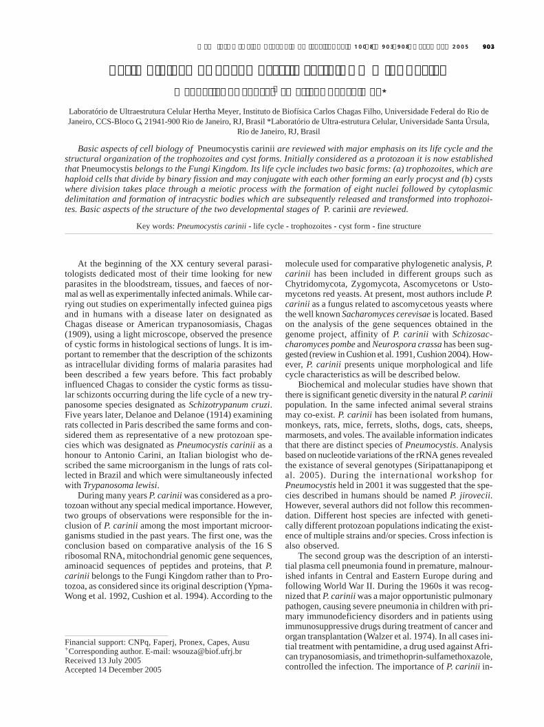
There are several reports on the life cycle of P. carinii,each one presenting different views and various devel-opmental stages. We will consider here a life cycle ac-cepted by most of the authors and which incorporatesdata obtained using electron microscopy (Fig. 1).
It is not yet clear which is the main infective form re-sponsible for the primary infection. However, it is wellestablished that airbone transmission is the most impor-tant one. For instance, corticosteroid-treated rats developP. carinii infection when housed with infected rats. Infec-tion by drinking water or food was excluded in these ex-periments. One way to start working with P. carinii is toadminister corticosteroids to normal laboratory rodents,thus indicating that highly infective forms are present inthe environment.
Fig. 1 shows a schematic view of the life cycle of P.carinii. Two developmental stages are well characterized:the mature cyst and the trophozoite. The trophozoite formis variable in shape, measuring about 0.3 µm in diameter,and they usually form clusters. Some authors considerthem to have ameboid characteristic, with the presence ofcytoplasmic projections similar to filopodia. However, nosuch type of cell motility has been reported in livingsamples.
Trophozoites originate directly from the cyst. Eachmature cyst may contain up to eight spherical intracysticbodies, which give rise to eight trophozoites. It has beenreported that trophozoites may originate from cysts con-taining spherical, banana-shaped or ameboid intracysticbodies. The initial trophozoite is haploid and divides bybinary fission or endogeny. Two trophozoites may conju-gate giving rise to a diploid cell which then divides, asdescribed above for the haploid trophozoites or begin ameiotic process of division with two meiotic cycles wherethree nuclear divisions takes place, forming a large spheri-cal cell with eight nuclei. Subsequently, there is a processof cell delimitation, forming eight intracystic bodies. Im-portant evidence for the presence of meiosis during the P.carinii life cycle is the presence of a synaptonemal com-plex (Matsumoto & Yoshida 1984). Preliminary analysisof the partial genome of P. carinii genes coding for pro-teins similar to those involved in the mating process inother fungi have been detected (Smulian et al. 2001).
It is important to point out that all these forms havebeen identified based on the observation of infections inanimals and, in a few cases, in cell cultures. Further stud-ies are still necessary to reproduce in vitro the completelife cycle of P. carinii.
Attempts have been made to cultivate the protozoanin different cell lines, using an approach typical for proto-zoa. Samples isolated from the lung of infected animalswere inoculated into cell cultures in which they prolifer-ated. Cysts and trophozoites have been observed al-though the latter predominates, growing as clusters inthe supernatant. However, many organisms attached toportions of the cells. In some cases the morphology ofthe attachment resembles that observed in vivo where P.carinii attaches to type I pneumocytes (Bartlett et al. 1994).Attempts to cultivate the organism in cell free media, asusual for fungi, succeeded in ten-fold amplifications ofthe number of cells. However, continuous axenic cultiva-tion of P. carinii is difficult, and has been obtained onlyby few groups (Merali et al. 1999).Cell biology of the trophozoite
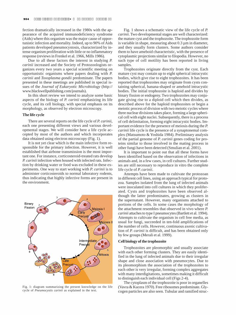
Trophozoites are pleomorphic and usually associatewith each other forming clusters. They are easily identi-fied in the lung of infected animals due to their irregularshape and close association with pneumocytes. Due toits pleomorphism the association of the trophozoites toeach other is very irregular, forming complex aggregateswith many interdigitations, sometimes making it difficultto distinguish each individual cell (Figs 2-4).
The cytoplasm of the trophozoite is poor in organelles(Vavra & Kucera 1970). Free ribosomes predominate. Gly-cogen particles are also seen. Tubular and ramified struc-
Fig. 1: diagram summarizing the present knowledge on the lifecycle of Pneumocystis carinii as explained in the text.
905905905905905Mem Inst Oswaldo Cruz, Rio de Janeiro, Vol. 100(8), December 2005
tures, resembling the endoplasmic reticulum are observed.A small nucleus is seen in variable positions. Usually itappears homogeneous, with an electrondensity similar tothat of the cytoplasm. A nucleolus is evident and locatedcentrally or peripherycally to the nucleus. Dividing nucleihave been observed. The mitotic process occurs withinan intact nuclear membrane and there are mitochondriawith short cristae. Osmiophilic bodies, vacuolar spaces,microtubules and an incipient Golgi complex were alsoreported, although not frequently seen (Dei-Cas et al.1989).
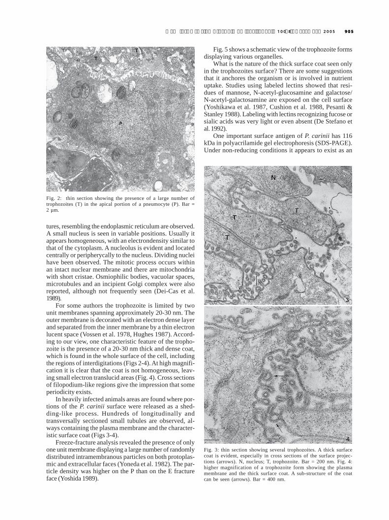
For some authors the trophozoite is limited by twounit membranes spanning approximately 20-30 nm. Theouter membrane is decorated with an electron dense layerand separated from the inner membrane by a thin electronlucent space (Vossen et al. 1978, Hughes 1987). Accord-ing to our view, one characteristic feature of the tropho-zoite is the presence of a 20-30 nm thick and dense coat,which is found in the whole surface of the cell, includingthe regions of interdigitations (Figs 2-4). At high magnifi-cation it is clear that the coat is not homogeneous, leav-ing small electron translucid areas (Fig. 4). Cross sectionsof filopodium-like regions give the impression that someperiodicity exists.
In heavily infected animals areas are found where por-tions of the P. carinii surface were released as a shed-ding-like process. Hundreds of longitudinally andtransversally sectioned small tubules are observed, al-ways containing the plasma membrane and the character-istic surface coat (Figs 3-4).
Freeze-fracture analysis revealed the presence of onlyone unit membrane displaying a large number of randomlydistributed intramembranous particles on both protoplas-mic and extracellular faces (Yoneda et al. 1982). The par-ticle density was higher on the P than on the E fractureface (Yoshida 1989).
Fig. 5 shows a schematic view of the trophozoite formsdisplaying various organelles.
What is the nature of the thick surface coat seen onlyin the trophozoites surface? There are some suggestionsthat it anchores the organism or is involved in nutrientuptake. Studies using labeled lectins showed that resi-dues of mannose, N-acetyl-glucosamine and galactose/N-acetyl-galactosamine are exposed on the cell surface(Yoshikawa et al. 1987, Cushion et al. 1988, Pesanti &Stanley 1988). Labeling with lectins recognizing fucose orsialic acids was very light or even absent (De Stefano etal. 1992).
One important surface antigen of P. carinii has 116kDa in polyacrilamide gel electrophoresis (SDS-PAGE).Under non-reducing conditions it appears to exist as an
Fig. 2: thin section showing the presence of a large number oftrophozoites (T) in the apical portion of a pneumocyte (P). Bar =2 µm.
Fig. 3: thin section showing several trophozoites. A thick surfacecoat is evident, especially in cross sections of the surface projec-tions (arrows). N, nucleus; T, trophozoite. Bar = 200 nm. Fig. 4:higher magnification of a trophozoite form showing the plasmamembrane and the thick surface coat. A sub-structure of the coatcan be seen (arrows). Bar = 400 nm.
906906906906906 Basic biology of P. carinii • W de Souza, M Benchimol
aggregated form with a molecular weight of > 2. 106 kDa.This protein is localized in the surface coat and has simi-larities with mucin-type glycoproteins (Radding et al. 1989).Based on the fact that administration of monoclonal anti-bodies recognizing this protein had a benefitial effect onthe course of the experimental pneumonia, it has beensuggested that it plays some role in pathogenesis(Gigliotti & Hughes 1988). Indeed, the surface of tropho-zoites is covered by the major surface glycoprotein orglycoprotein A which is a family of protein encoded by upto one hundred heterogeneous genes. These genes arelocalized at the ends, upstream of the subtelomeric andtelomeric repeats of all chromosomes. Transcription is lim-ited to a single gene each time and is involved in a pro-cess that resembles the antigenic variation that has beenwell characterized in Trypanosma brucei (Stringer & Keely2001).
Trophozoites interact with the surface of pneumocytes(Fig. 2). In most of the cases such interaction occurredthrough the surface coat that established contact withthe microvilli of the epithelial cells. At some points suchcontact was done through filopodium-like structures.There are evidences that fibronectin is a mediator of the P.carinii attachment to pneumocytes with the participationof fibronectin-binding receptor on the fungus surface anda fibronectin-binding integrin of the host cell surface(Pottratz et al. 1991, 1994, Aliouat et al. 1993).
Part of the P. carinii population is ingested by alveo-lar macrophages in a process mediated by the Dectinn-1β-glucan receptor, with production of hydrogen peroxideand subsequent killing of the organism (Steele et al. 2003).
Cell biology of the cystic formThe cysts are easily identified due to their typical
morphology, being spherical structures with a mean di-ameter of 5-8 µm, containing up to eight intracystic bod-ies (Figs 6-12). Each intracystic body has a mean diameterof 1.2 µm.
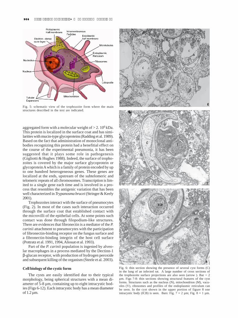
Fig. 5: schematic view of the trophozoite form where the mainstructures described in the text are indicated.
Fig. 6: thin section showing the presence of several cyst forms (C)in the lung of an infected rat. A large number of cross sections ofthe trophozoite surface projections are also seen (arrow ). Bar = 2µm. Figs 7-8: thin sections showing structural features of the cystforms. Structures such as the nucleus (N), mitochondrion (M), vacu-oles (V), ribosomes and profiles of the endoplasmic reticulum canbe seen. In the cyst shown in the upper portion of figure 8 oneintracystic body (ICB) is seen. Bars: Fig. 7 = 2 µm; Fig. 8 = 1 µm.
907907907907907Mem Inst Oswaldo Cruz, Rio de Janeiro, Vol. 100(8), December 2005
The cyst wall has two layers and is approximately 50nm thick (Figs 6-8). The outer layer, with a thickness ofabout 15 nm, is more electrondense. The inner layer, witha thickness of 35 nm, is less dense and is in contact withthe plasma membrane. Some authors described the pres-ence of a membrane-like structure on the outerelectrondense layer (DeStefano et al. 1990a). However,this structure was not seen in most of the published elec-tron micrographs. A general schematic view of the cysticform is shown in Fig. 12.
Freeze-fracture studies show the presence of intramem-branous particles on the fracture faces of the cyst mem-brane (Yoneda et al. 1982). This number, however, wassmaller than that observed in the membrane of the tro-phozoites.
Biochemical analysis has shown that the cyst wall isrich in glucosyl/mannosyl and galactose/N-acetyl-D-ga-lactosamine residues (DeStefano et al. 1990b). The innerportion of the cyst contains two components: a matrixand the intracystic bodies. The matrix contains mitochon-dria, ribosomes, empty vacuoles and membrane debris.Its dimension varies according to the size of the cyst andthe number of intracystic bodies. The intracystic body isa spherical to oval shape, with a centrally located nucleus,endoplasmic reticulum, free ribosomes, mitochondria, andglycogen particles.
It is very common to observe cystic bodies with abanana-like shape. These forms only show the matrix con-tent, but no intracystic bodies. They are considered asremnants of the ruptured cysts which have released theintracystic bodies.
The use of the Thiéry technique, which reveals thepresence of carbohydrates, showed the presence of gly-cogen particles in the trophozoites, the intracystic bodiesand in the cyst matrix. The trophozoite’s plasma mem-brane, the membrane lining the cyst and the intracysticbodies was also labeled, but the trophozoite’s surface coatand the cyst wall were not labeled.Perspectives
It is clear from the results described above that weknow little about the cell biology of Pneumocystis. This isbecause there is no experimental system in which the or-ganisms can be easily maintained in vitro, that would en-able to its life cycle be studied in detail. Therefore, it isimportant that such a system is found to open up newpossibilities for further biochemical and molecular stud-ies.
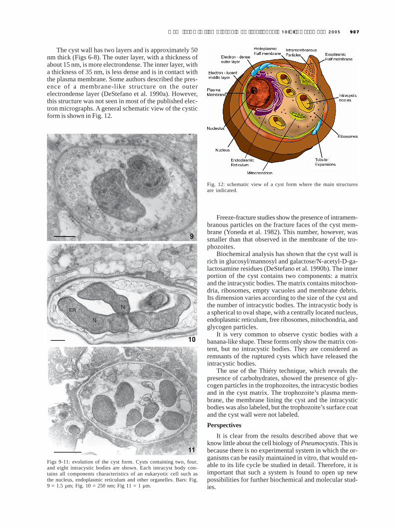
Figs 9-11: evolution of the cyst form. Cysts containing two, four,and eight intracystic bodies are shown. Each intracyst body con-tains all components characteristics of an eukaryotic cell such asthe nucleus, endoplasmic reticulum and other organelles. Bars: Fig.9 = 1.5 µm; Fig. 10 = 250 nm; Fig 11 = 1 µm.
Fig. 12: schematic view of a cyst form where the main structuresare indicated.
908908908908908 Basic biology of P. carinii • W de Souza, M Benchimol
REFERENCESAliouat EM, Dei-Cas E, Ouaissi A, Palluault F, Soulez B, Camus
D 1993. In vitro attachment of Pneumocystis carinii frommouse and rat origin. Biol Cell 77: 209-217.
Bartlett MS, Goheen MP, Lee CH, Shaw MM, Durkin MM,Smith JW 1994. Close association of Pneumocystis cariniifrom infected rat lung with culture cells as shown by lightand electron microscopy. Parasitol Res 80: 208-215.
Chagas C 1909. Nova tripanozomiaze humana. Mem InstOswaldo Cruz 1: 159-181.
Cushion MT 2004. Pneumocystis: unraveling the cloak of ob-scurity. Trends Microbiol 12: 243-249.
Cushion MT, De Stefano JÁ, Walzer PD 1988. Pneumocystiscarinii: surface reactive carbohydrates detected by lectinprobes. Exp Parasito1 67: 137-147.
Cushion MT, Harmsen A, Matsumoto Y 1994. Recent advancesin the biology of Pneumocystis carinii. J Med Vet Mycol32: 217-228.
Cushion MT, Stringer JR, Wa1zer PD 1991. Cellular and mo-lecular biology of Pneumocystis carinii. Int Rev Cytol 131:59-107.
Dei-Cas E, Soulez B, Camus D 1989. Ultrastructural study ofPneumocystis carinii in explants cultures of rabbit lungand cultures with and without feeder cells. J Protozool(Suppl.) 36: 55S-57S.
Delanoe P, Delanoe M 1914. Sur le rapports des kystes dupommon des rats avec le Trypanosoma lewisi. Comp R AcadSci 155: 658-661.
De Stefano JA, Cushion MT, Sleight RG, Walzer PD 1990a.Analysis of Pneumocsytis carinii cyst wall. I. Evidence foran outer surface membrane. J Protozool 37: 428-435.
De Stefano JA, Cushion MT, Puvanesarajah V, Walzer PD 1990b.Analysis of Pnemocystis carinii cyst wall. II. Sugar compo-sition. J Protozool 37: 436-441.
De Stefano JA, Trink1e LS, Wa1zer PD, Cushion MT 1992.Flow cytometrical analysis of lectin binding to Pneumocystiscarinii surface carbohydrates. J Parasitol 78: 271-280.
Frenkel JK, Good JF, Shultz JA 1966. Latent Pneumocystisinfection of rats, relapse and chemotherapy. Lab Invest 15:1559-1577.
Giglioli F, Hughes WT 1988. Passive immunoprophylaxis withspecific monoclonal antibodies confers partial protectionagainst Pneumocystis carinii pneumonitis in animal mod-els. J Clin Invest 81: 1666-1668.
Hughes WT 1987. Pneumocystis carinii Pneumonitis, CRCPress, Boca Raton, FL.
Itatani CA 1994. Ultrastructural demonstration of a pore in thecyst wall of Pneumocystis carinii. J Parasitol 80: 644-648.
Matsumoto Y, Yoshida Y 1984. Sporogony in Pneumocystiscarinii: synaptonemal complexes and meiotic nuclear divi-sions observed in precysts. J Protozool 31: 420-428.
Merali S, Frevert U, Williams JH, Chin K, Bryan R, ClarksonJr AB 1999. Continuous axenic cultivation of P. cariniii.Proc Natl Acad Sci NY 96: 2402-2407.
Mills J 1986. Pneumocystis carinii and Toxoplasma gondii in-fections in patients with AIDS. Rev Infect Dis 8: 1001-1011.
Pesanti EL, Stanley JD 1988. Glycoproteins of Pneumocystiscarinii characterization by electrophoresis and microscopy.J Infect Dis 158: 1353-1359.
Pottratz ST, Paulsrud J, Smith JS, Martin WJ 1991. Pneumocystiscarinii attachment to cultured lung cells by pneumocystisgp120, a fibronectin binding protein. J Clin Invest 88: 403-407.
Pottratz ST, Weir AL, Wisniowski PE 1994. Pneumocystis cariniiattachment increases expression of fibronectin-bindingintegrins on cultured lung cells. Infect Immun 62: 5464-5469.
Radding JA, Armstrong MYK, Ullu E, Richards FF 1989. Iden-tification and isolation of a major cell surface glycoproteinof Pneumocystis carinii. Infect Immun 57: 2149-2157.
Siripattanapipong S, Worapong J, Mungthin M, Leelayoova S,Tan-ariya P 2005. Genotypic study of Pneumocystisjirovecii in human immunodeficiency virus-positive pa-tients in Thailand. J Clin Microbiol 43: 2104-2110
Steele C, Marrero L, Swain S, Harmsen AG, Zheng M, BrownGD, Gordon S, Shellito JE, Kolls JK 2003. Alveolar mac-rophage-mediated killing of Pneumocysts carinii f. sp. murisinvoves molecular recognition by the Dectin-1 β-glucan re-ceptor. J Exp Med 198: 1677-1688.
Smulian AG, Sesterhenn T, Tanaka R, Cushion MT 2001. Theste3 pheromone receptor gene of Pneumocystis carinii issurrounded by a cluster of signal tranduction genes. Genet-ics 157: 991-1002.
Stringer JR, Keely SP 2001. Genetics of surface antigens ex-pression in Pneumocystis carinii. Infect Immun 69: 627-639.
Vavra J, Kucera K 1970. Pneumocystis carinii Delanoe, its ul-trastructure and ultrastructural affinities. J Protozool 17:463-483.
Vossen MEMH, Beckers PJA, Meuwissen JHET, StadhoudersAM 1978. Developmental biology of Pneumocystis carinii:an alternative view on the life cycle of the parasite. ZParazitenkd 55: 101-118.
Yoneda K, Walzer PD, Richey CS, Birk MG 1982. Pneumocystiscarinii: freeze-fracture study of stages of the organism. ExpParasitol 53: 68-76.
Yoshida Y 1989. Ultrastructual studies of Pneumocystis carinii.J Protozool 36: 53-60.
Yoshikawa H, Tegoshi T, Yoshida Y 1987. Detection of surfacecarbohydrates on Pneumocystis carinii by fluorescein-con-jugated lectins. Parasitol Res 74: 43-49.
Ypma-Wong MF, Fonzi WA, Sypherd PS 1992. Fungus-spe-cific translation elongation factor 3 gene present inPneumocystis carinii. Infect Immun 60: 4140-4145.
Walzer PD, Pearl DP, Krogstad DJ, Rawson PG, Schultz MG1974. Pneumocystis carinii pneumonia in the United States:epidemiology, diagnostics and clinical features. Ann InternMed 80: 83-93.