Embed Size (px)
Citation preview
Biologia Genômica
2º Semestre, 2017
Replicação de DNA em Bactérias e no
Núcleo Eucariótico
Prof. Marcos Túlio
Faculdade de Ciências Agrárias e Veterinárias de Jaboticabal
Instituto de Biociências, Letras e Ciências Exatas de S.J.R.P.
Universidade Estadual Paulista “Júlio de Mesquita Filho”
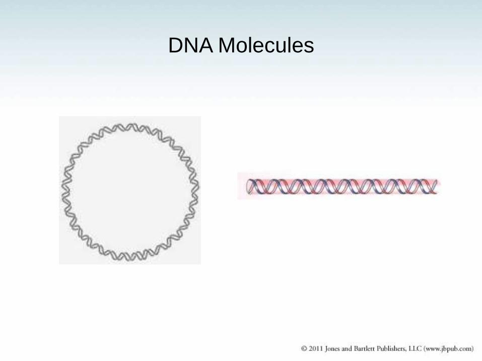
DNA Molecules
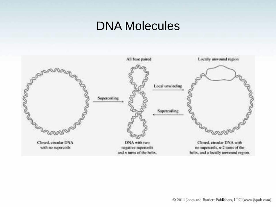
DNA Molecules
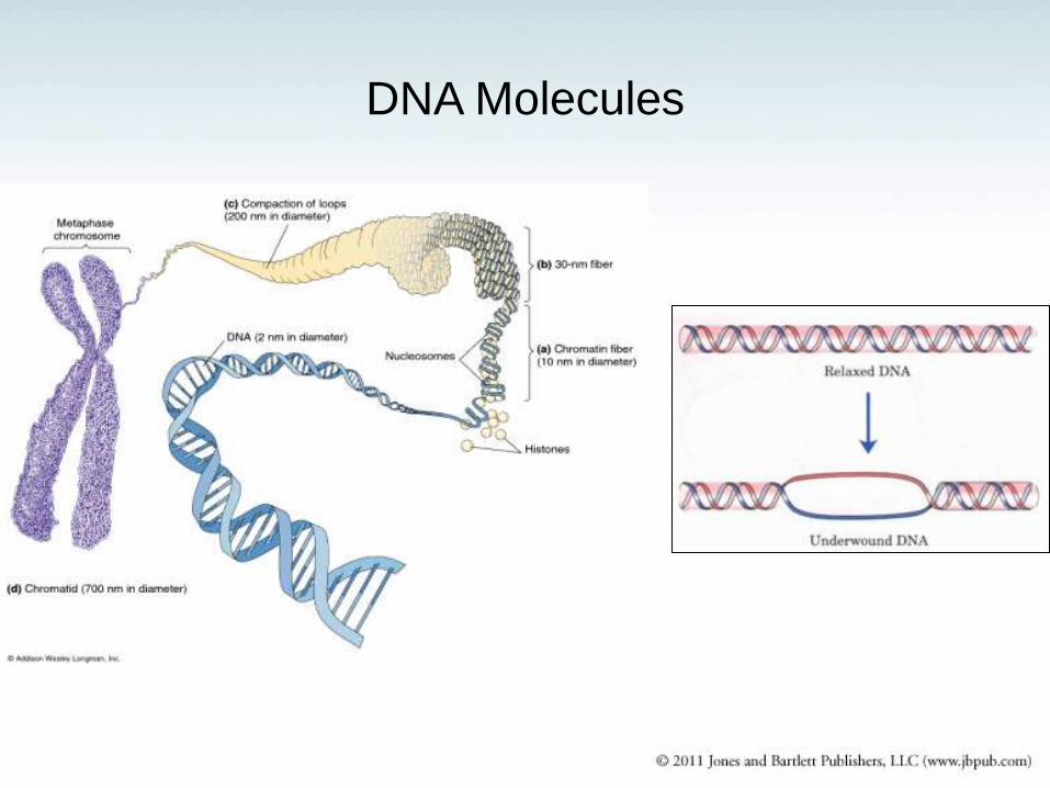
DNA Molecules
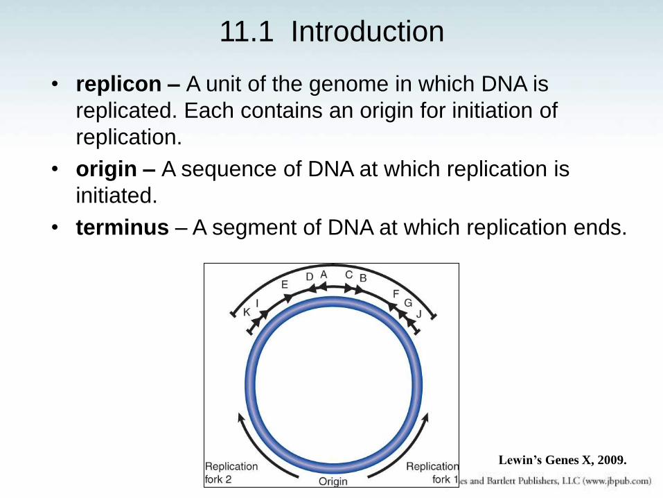
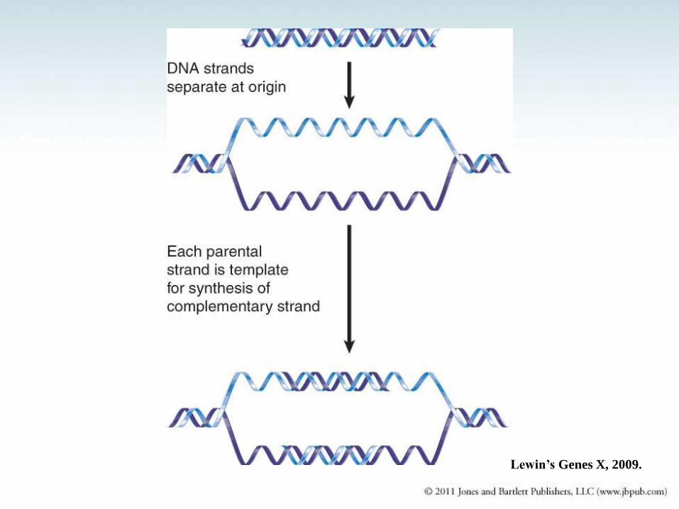
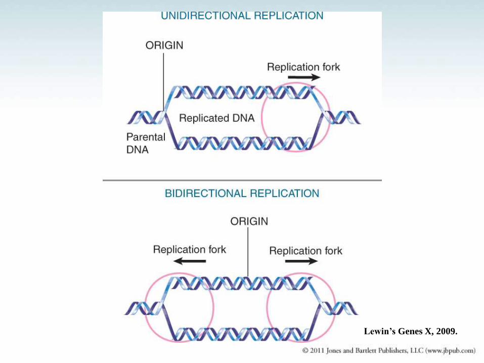
11.1 Introduction
• replicon – A unit of the genome in which DNA is
replicated. Each contains an origin for initiation of
replication.
• origin – A sequence of DNA at which replication is
initiated.
• terminus – A segment of DNA at which replication ends.
Lewin’s Genes X, 2009.
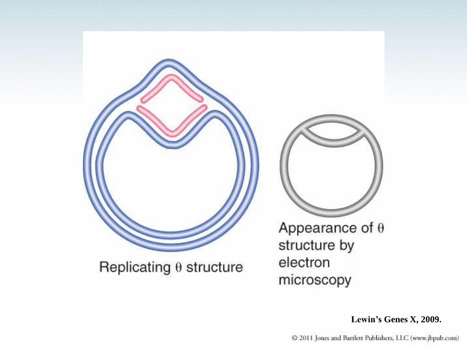
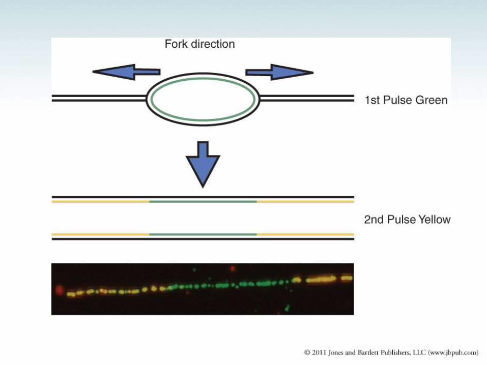
Lewin’s Genes X, 2009.
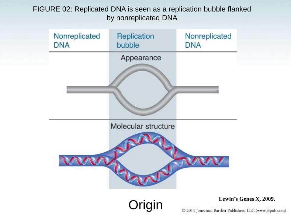
FIGURE 02: Replicated DNA is seen as a replication bubble flanked
by nonreplicated DNA
Origin Lewin’s Genes X, 2009.
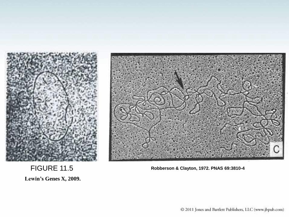
Robberson & Clayton, 1972. PNAS 69:3810-4 FIGURE 11.5
Lewin’s Genes X, 2009.
Lewin’s Genes X, 2009.
Lewin’s Genes X, 2009.
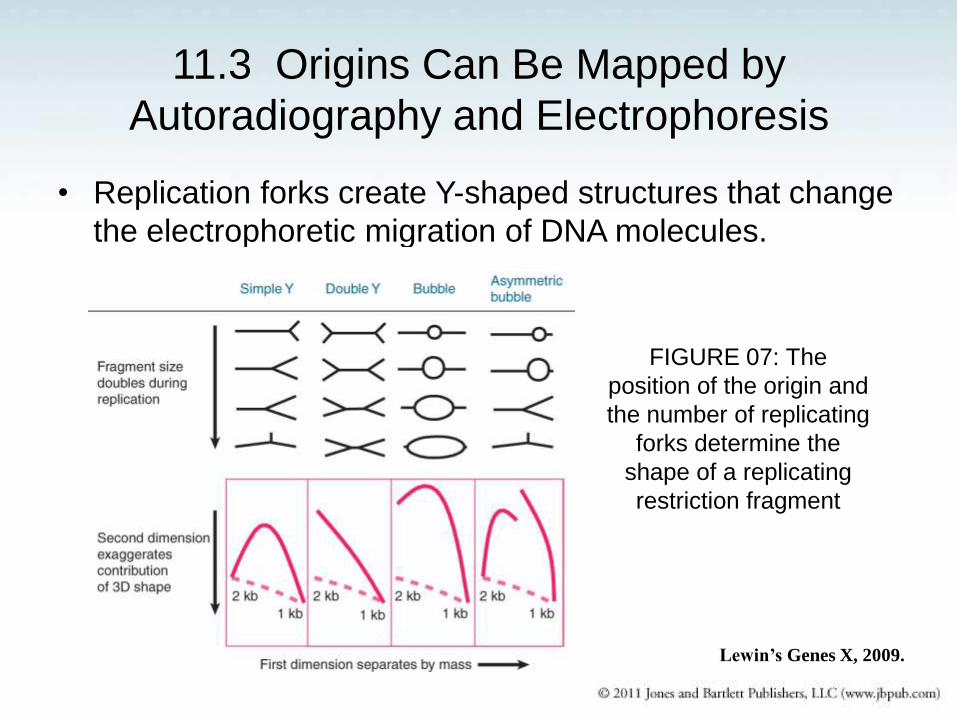
11.3 Origins Can Be Mapped by
Autoradiography and Electrophoresis
• Replication forks create Y-shaped structures that change
the electrophoretic migration of DNA molecules.
FIGURE 07: The
position of the origin and
the number of replicating
forks determine the
shape of a replicating
restriction fragment
Lewin’s Genes X, 2009.
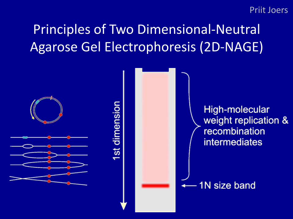
Principles of Two Dimensional-Neutral Agarose Gel Electrophoresis (2D-NAGE)
Priit Joers
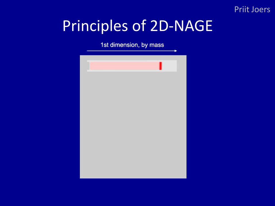
Principles of 2D-NAGE Priit Joers
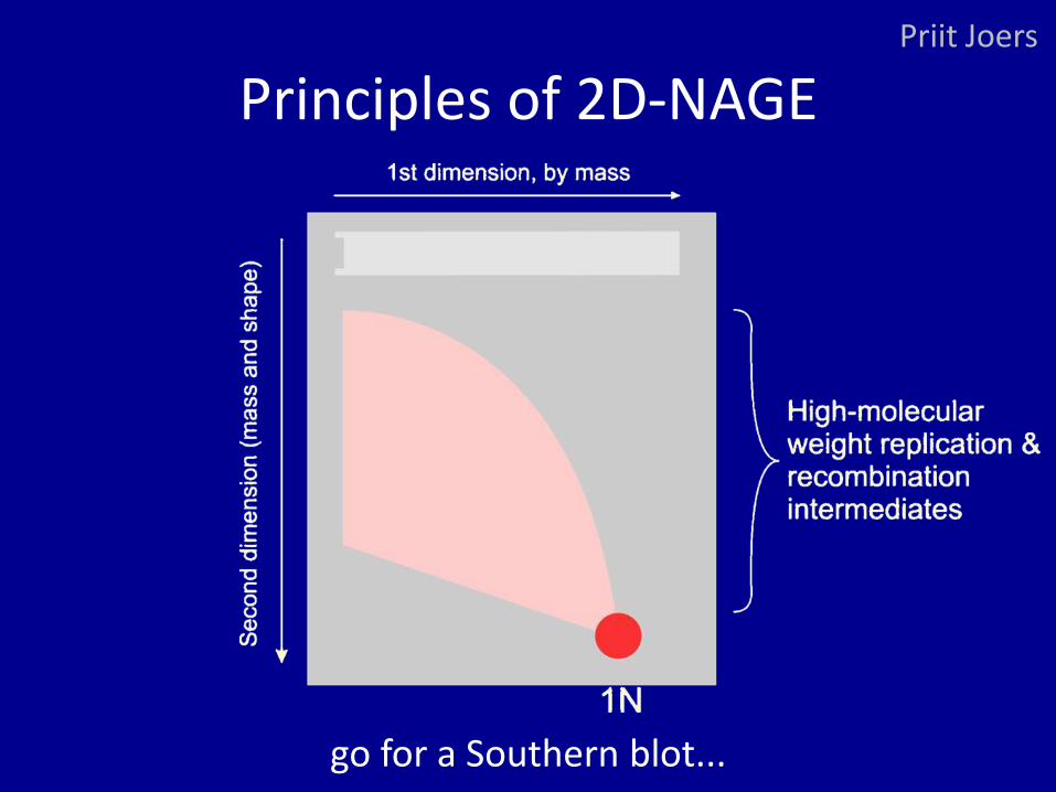
Principles of 2D-NAGE Priit Joers
go for a Southern blot...
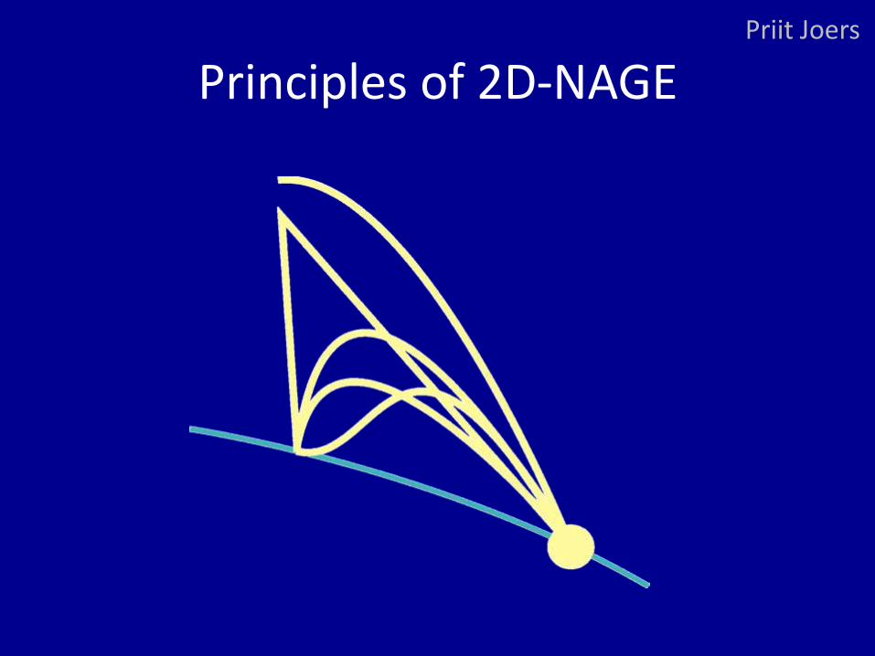
Principles of 2D-NAGE Priit Joers
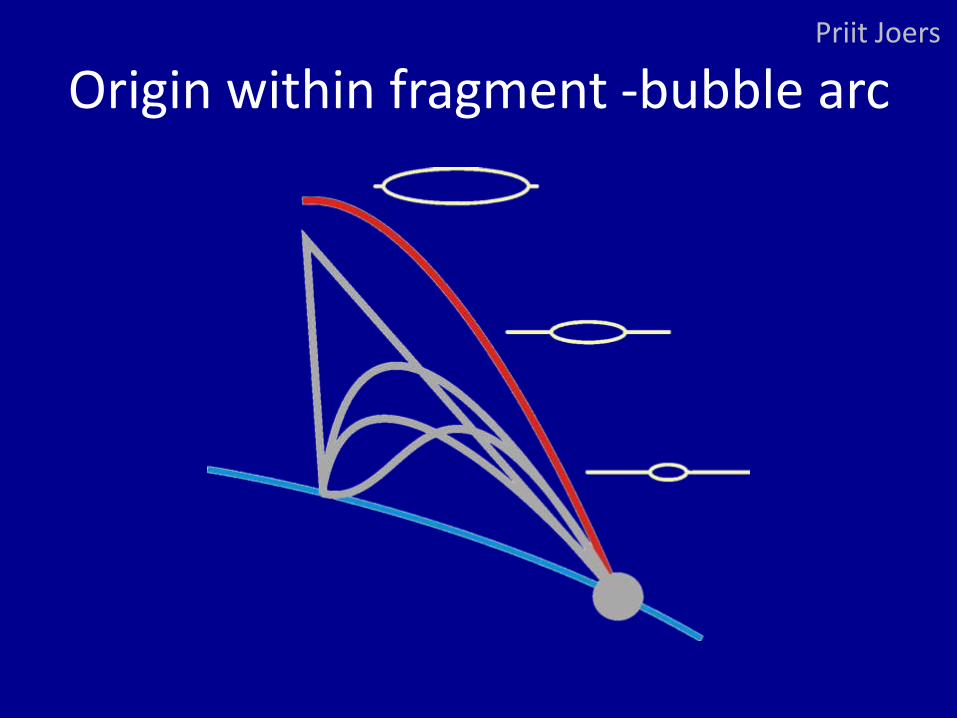
Origin within fragment -bubble arc Priit Joers
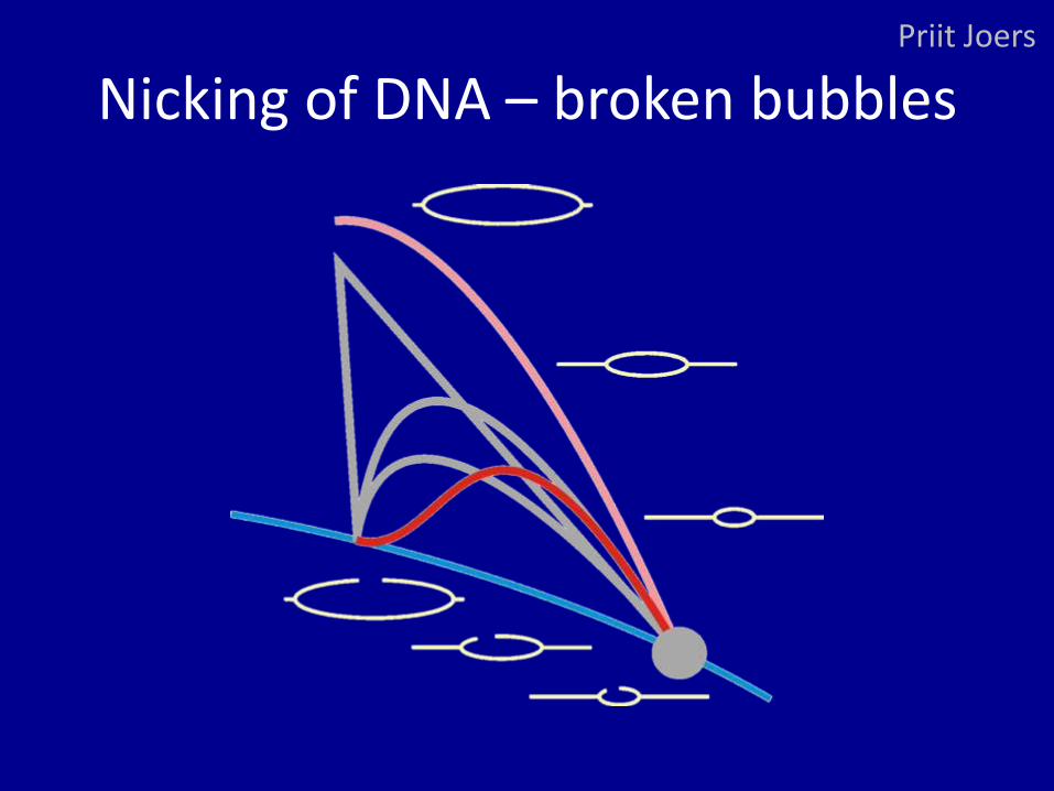
Nicking of DNA – broken bubbles Priit Joers
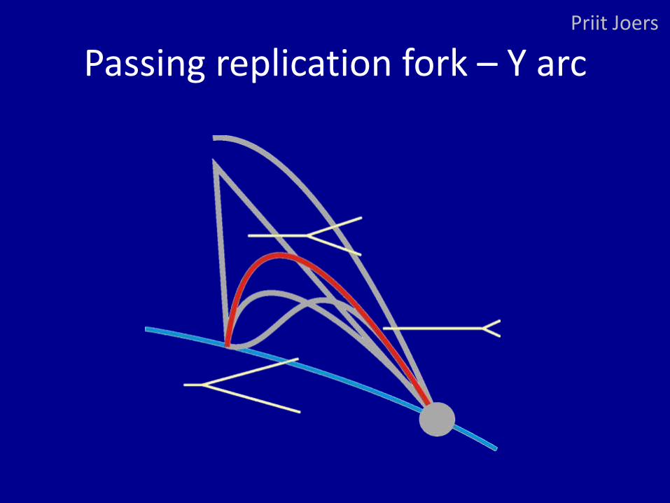
Passing replication fork – Y arc Priit Joers
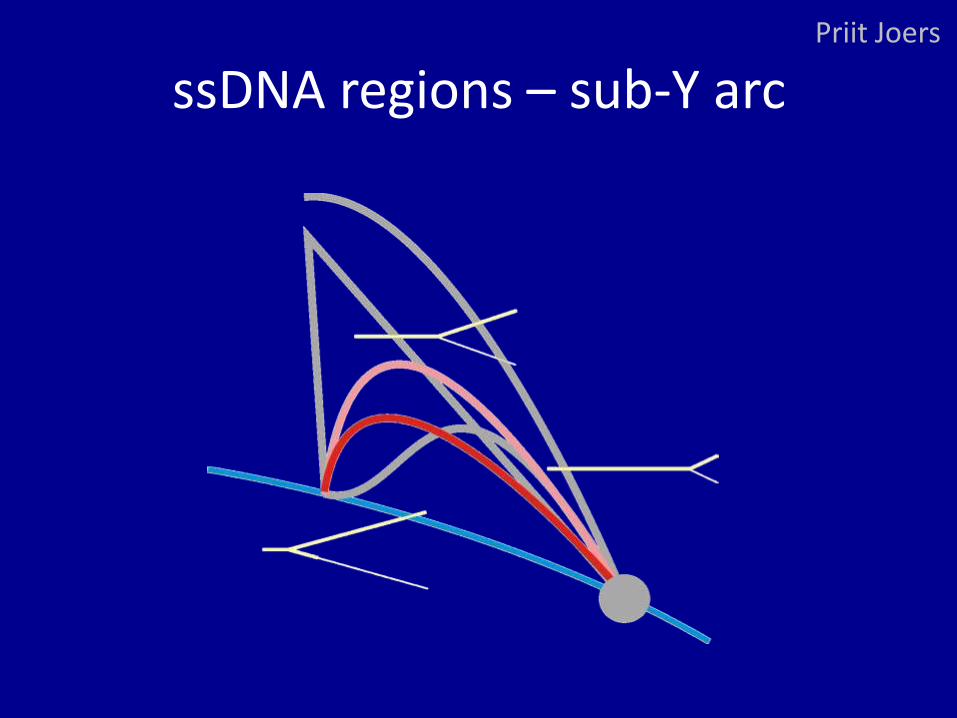
ssDNA regions – sub-Y arc Priit Joers
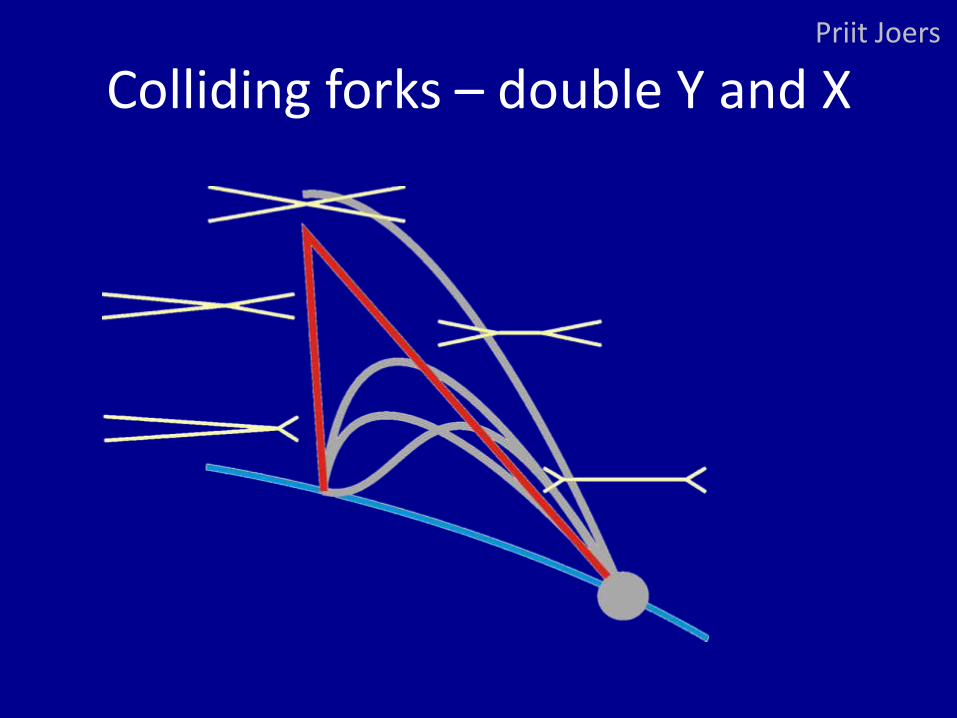
Colliding forks – double Y and X Priit Joers
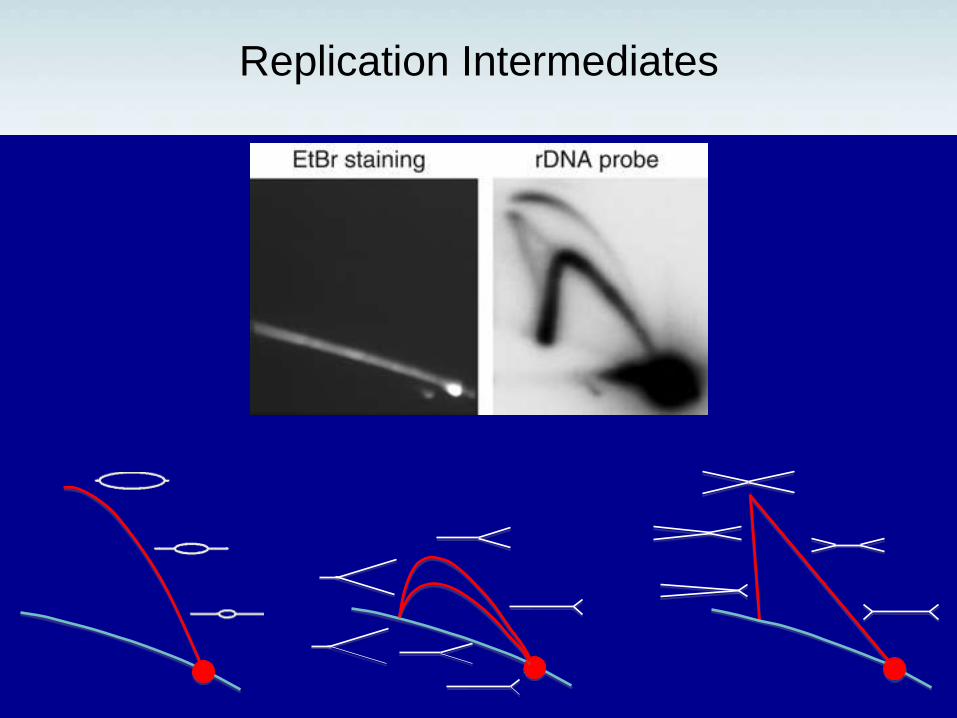
Replication Intermediates
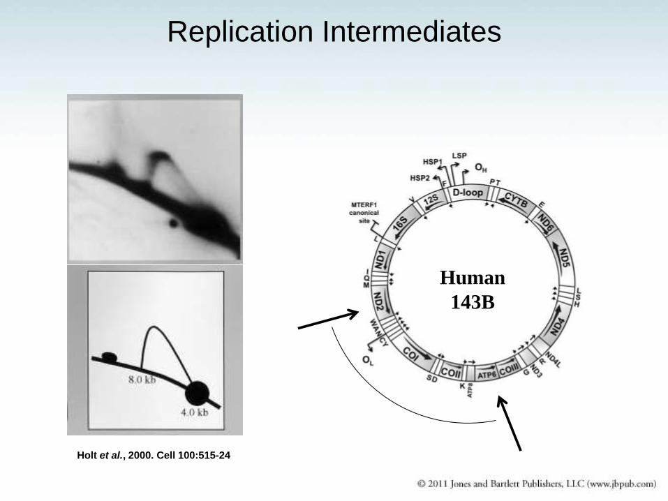
Holt et al., 2000. Cell 100:515-24
Human
143B
Replication Intermediates
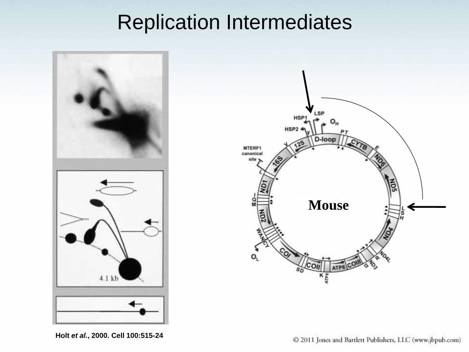
Holt et al., 2000. Cell 100:515-24
Mouse
Replication Intermediates
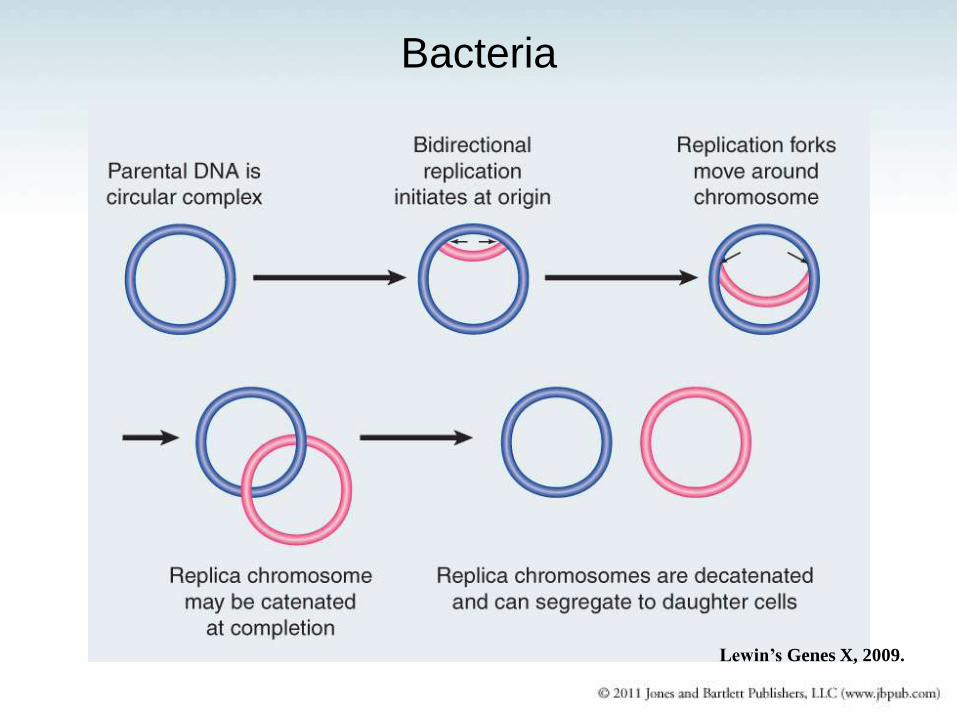
Bacteria
Lewin’s Genes X, 2009.
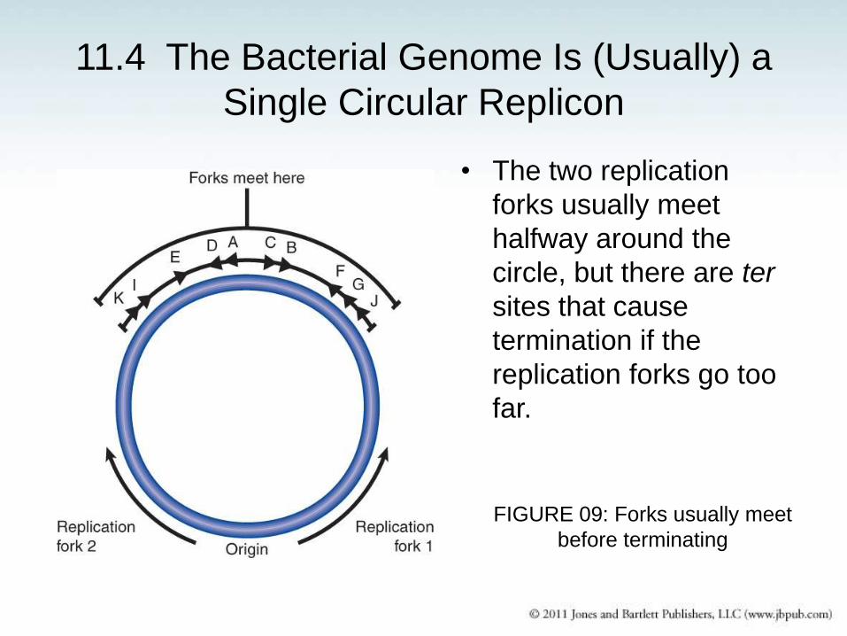
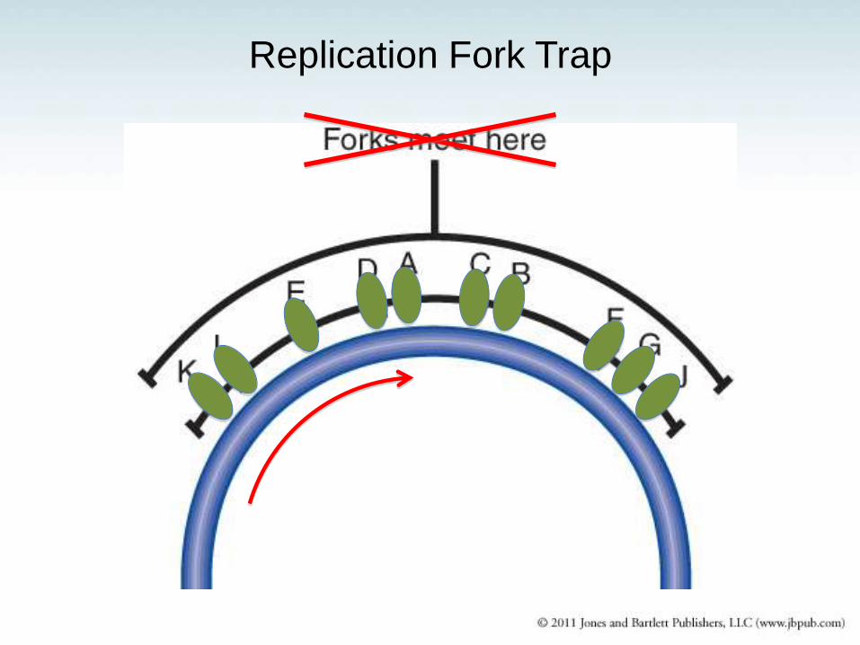
11.4 The Bacterial Genome Is (Usually) a
Single Circular Replicon
• The two replication
forks usually meet
halfway around the
circle, but there are ter
sites that cause
termination if the
replication forks go too
far.
FIGURE 09: Forks usually meet
before terminating
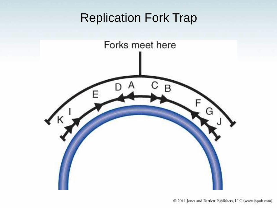
Replication Fork Trap
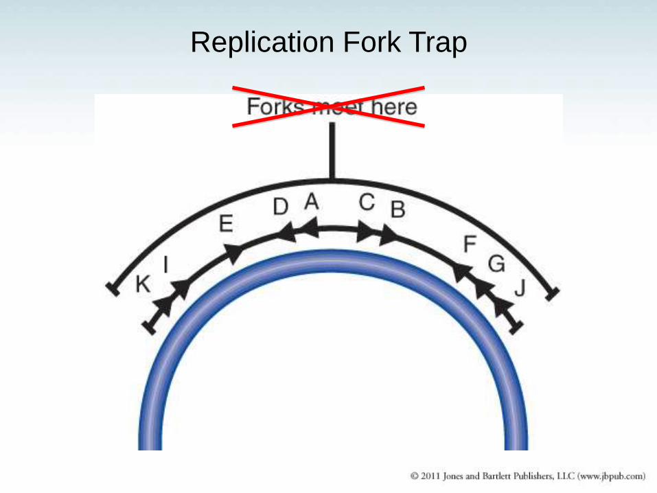
Replication Fork Trap
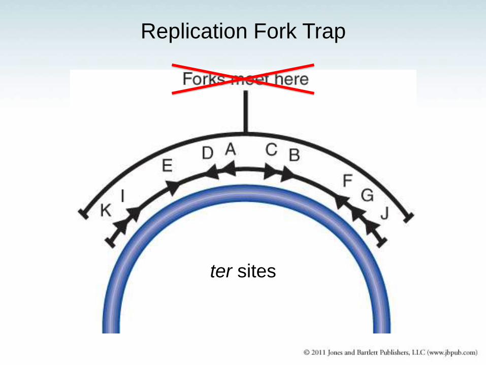
Replication Fork Trap
ter sites
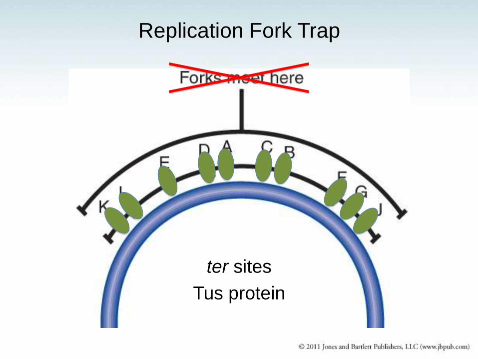
Replication Fork Trap
ter sites
Tus protein
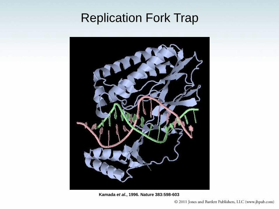
Replication Fork Trap
Kamada et al., 1996. Nature 383:598-603
Replication Fork Trap
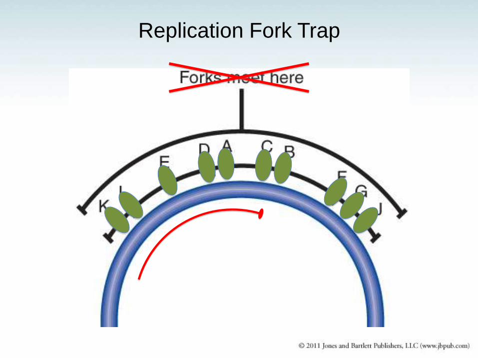
Replication Fork Trap

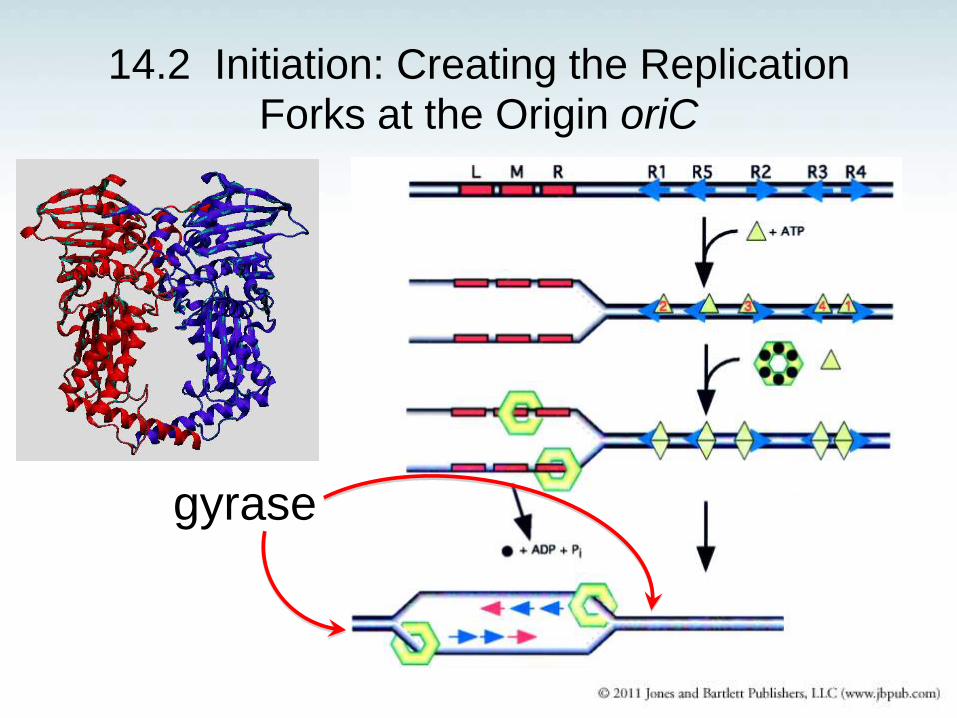
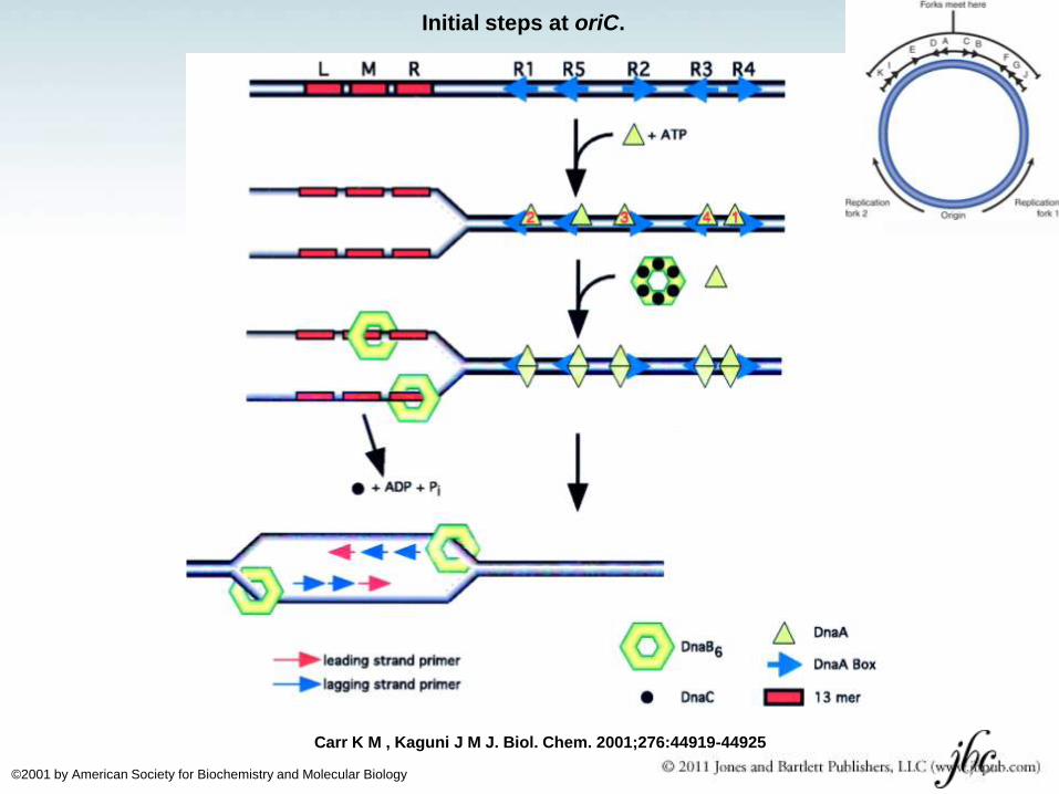
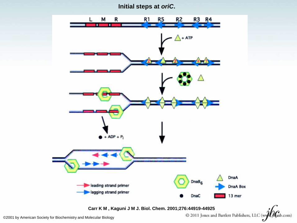
Initial steps at oriC.
Carr K M , Kaguni J M J. Biol. Chem. 2001;276:44919-44925
©2001 by American Society for Biochemistry and Molecular Biology
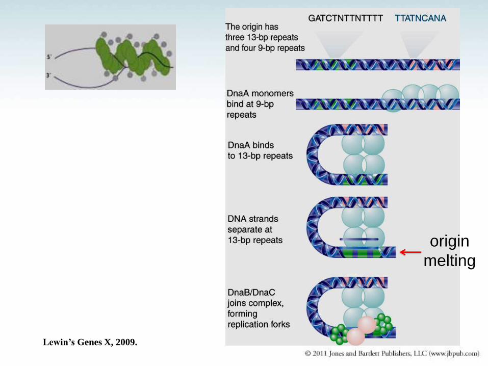
origin
melting
Lewin’s Genes X, 2009.
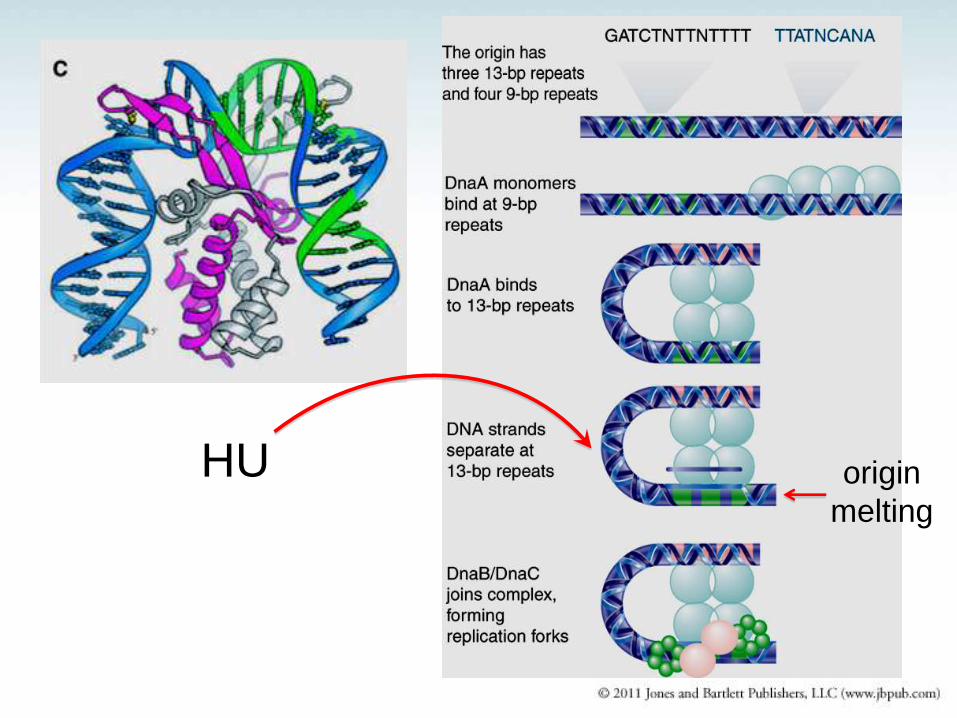
HU origin
melting
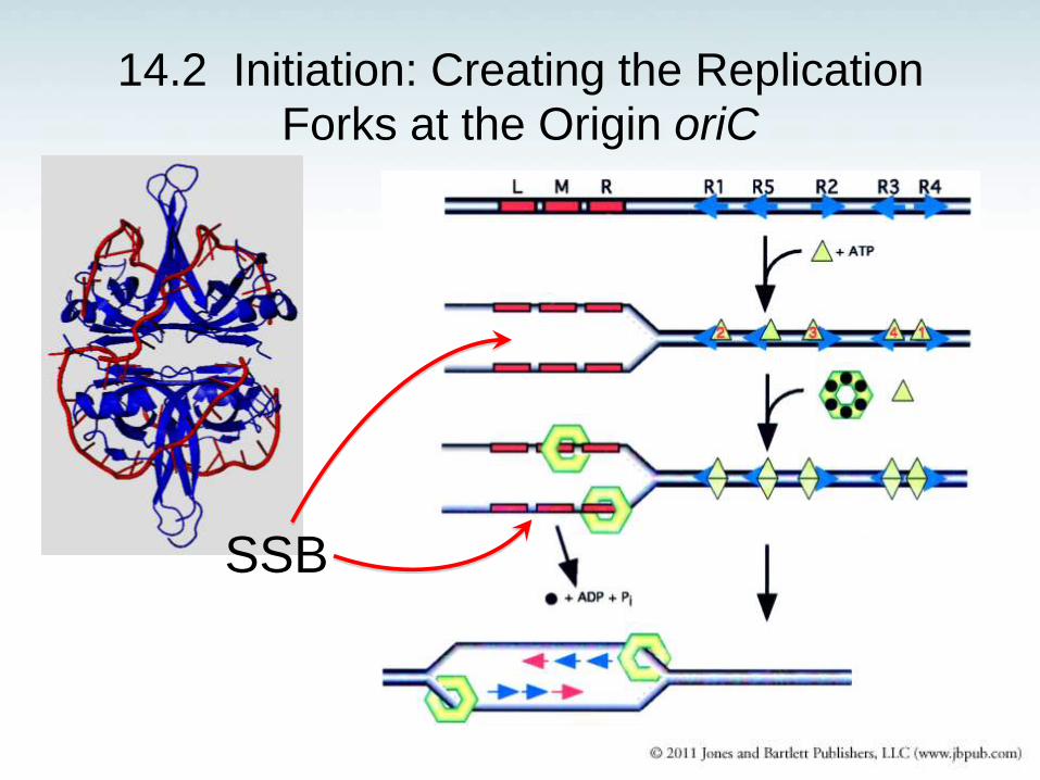
14.2 Initiation: Creating the Replication Forks at the Origin oriC
SSB
14.2 Initiation: Creating the Replication Forks at the Origin oriC
gyrase
Initial steps at oriC.
Carr K M , Kaguni J M J. Biol. Chem. 2001;276:44919-44925
©2001 by American Society for Biochemistry and Molecular Biology
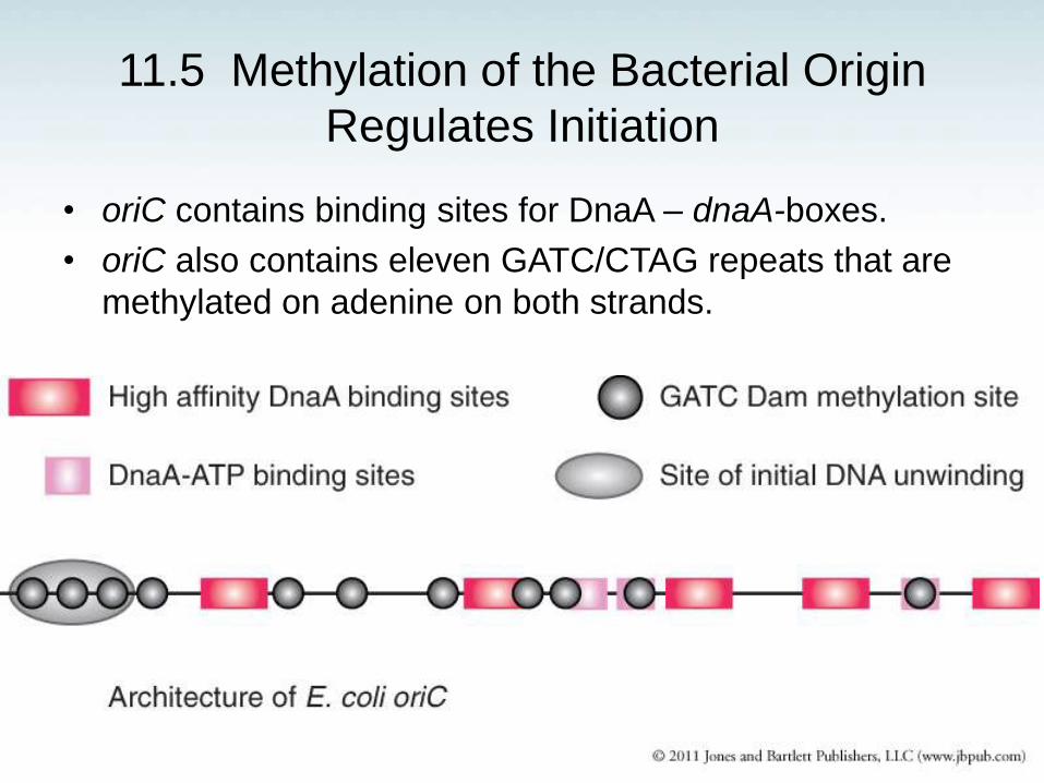
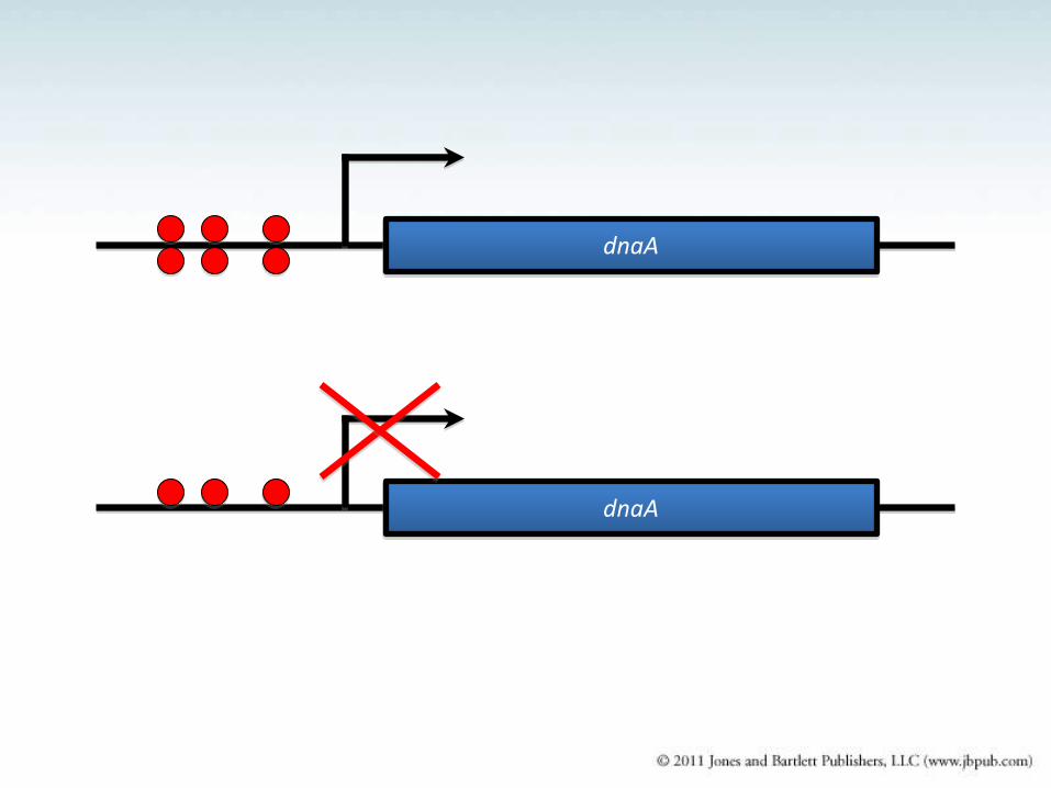
11.5 Methylation of the Bacterial Origin
Regulates Initiation
• oriC contains binding sites for DnaA – dnaA-boxes.
• oriC also contains eleven GATC/CTAG repeats that are
methylated on adenine on both strands.
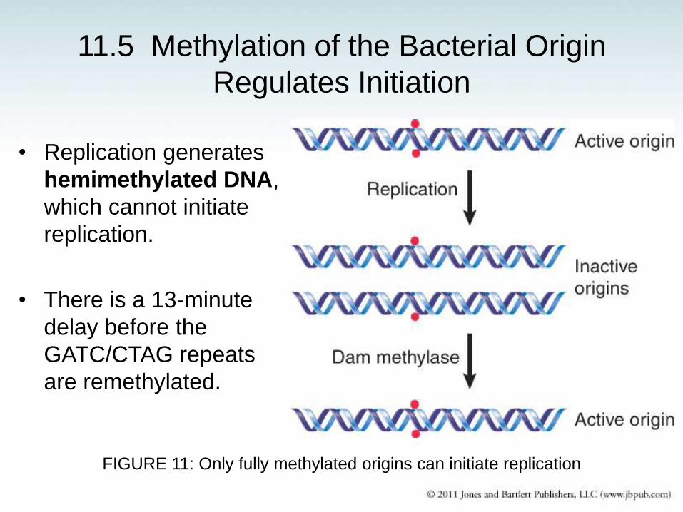
11.5 Methylation of the Bacterial Origin
Regulates Initiation
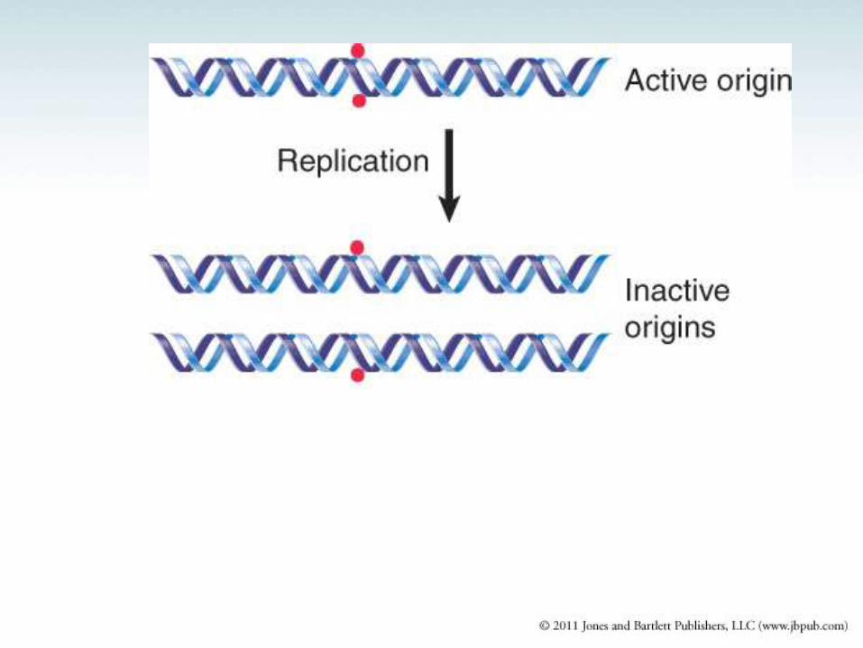
• Replication generates
hemimethylated DNA,
which cannot initiate
replication.
• There is a 13-minute
delay before the
GATC/CTAG repeats
are remethylated.
FIGURE 11: Only fully methylated origins can initiate replication
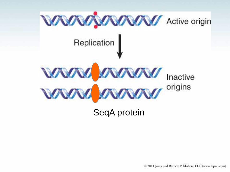
SeqA protein
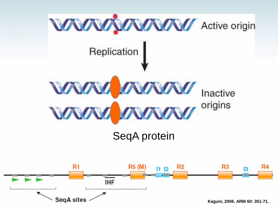
SeqA protein
Kaguni, 2006. ARM 60: 351-71.
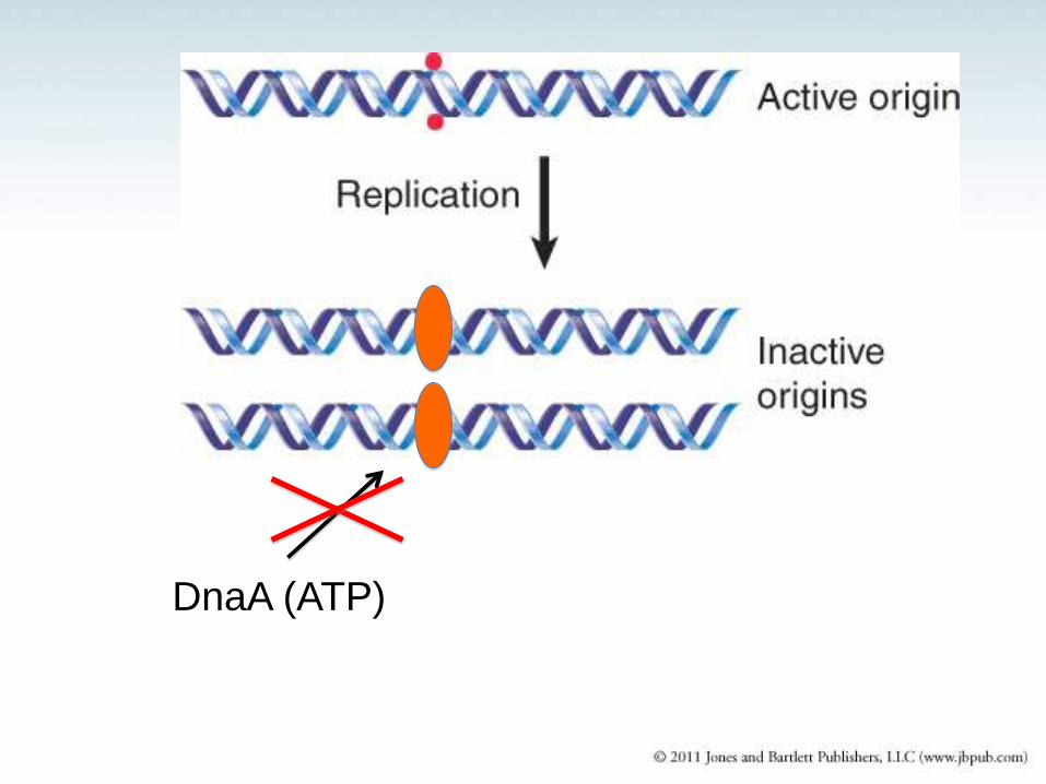
DnaA (ATP)
dnaA
dnaA
Initial steps at oriC.
Carr K M , Kaguni J M J. Biol. Chem. 2001;276:44919-44925
©2001 by American Society for Biochemistry and Molecular Biology
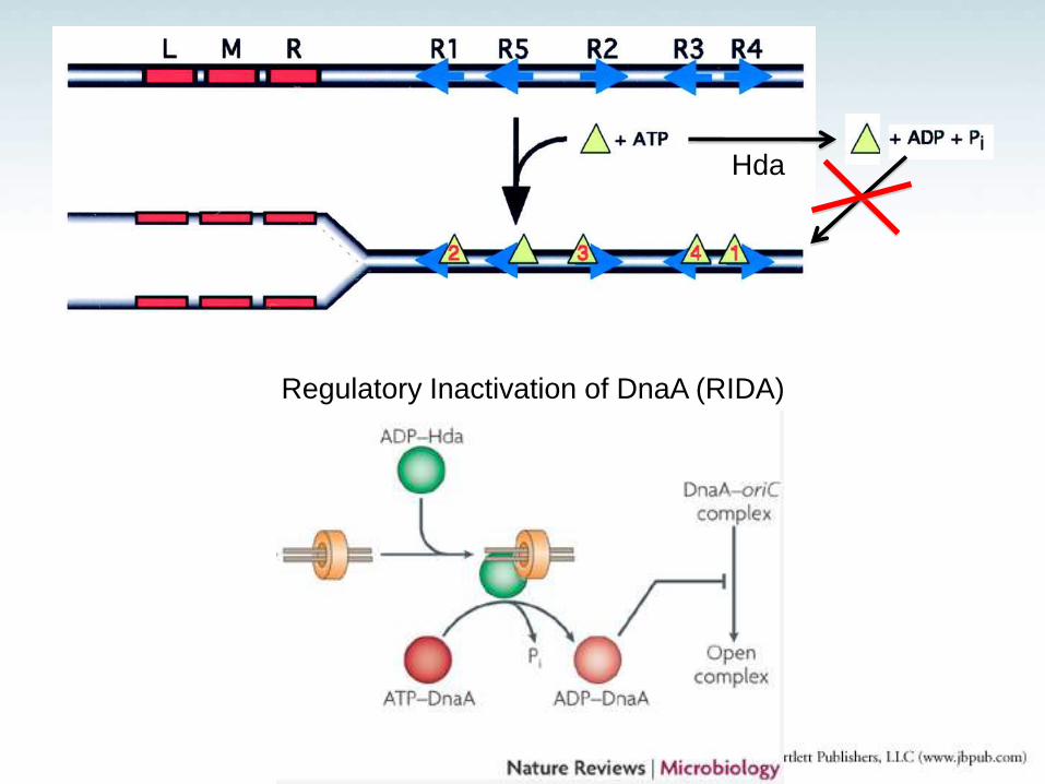
Hda
Regulatory Inactivation of DnaA (RIDA)
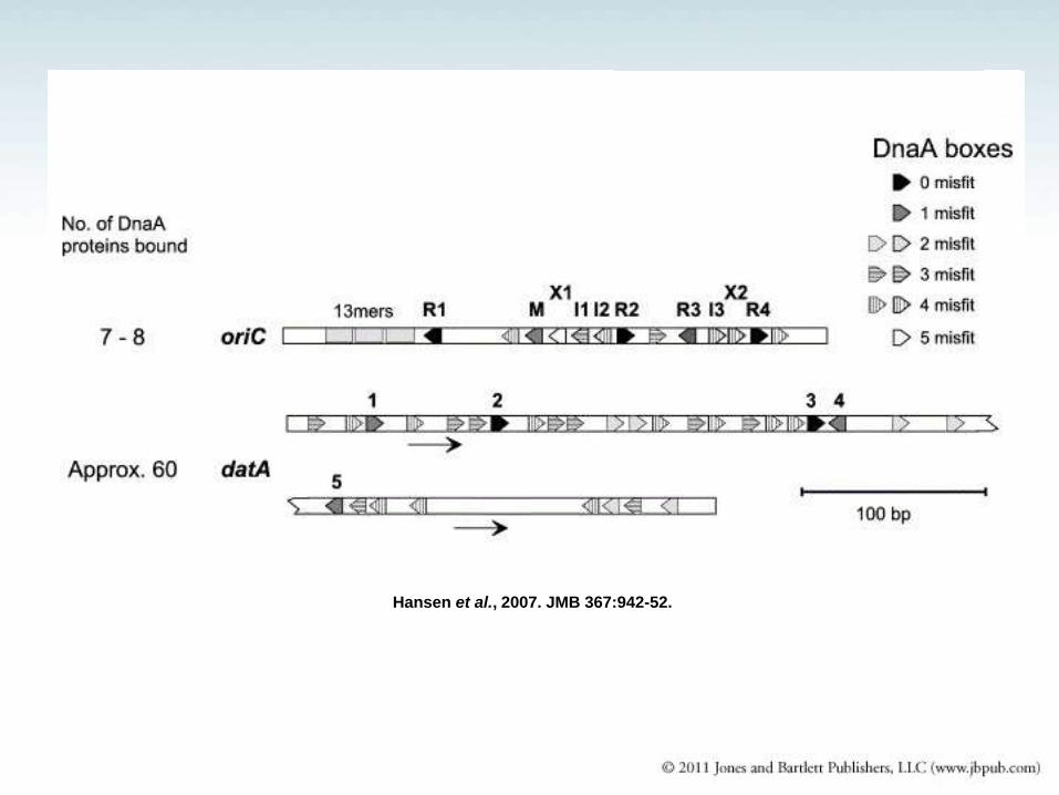
Hansen et al., 2007. JMB 367:942-52.
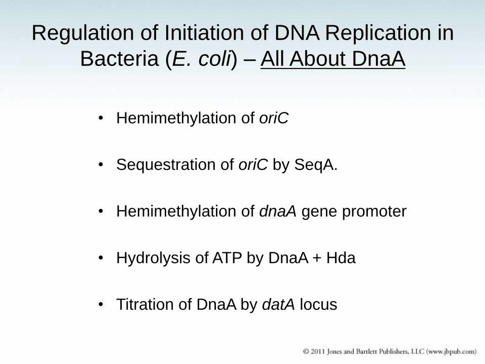
Regulation of Initiation of DNA Replication in
Bacteria (E. coli) – All About DnaA
• Hemimethylation of oriC
• Sequestration of oriC by SeqA.
• Hemimethylation of dnaA gene promoter
• Hydrolysis of ATP by DnaA + Hda
• Titration of DnaA by datA locus
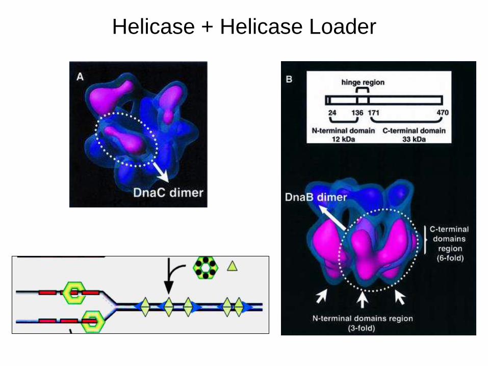
Helicase + Helicase Loader
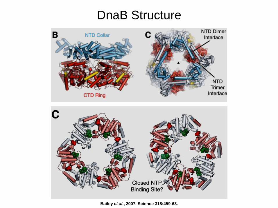
DnaB Structure
Bailey et al., 2007. Science 318:459-63.
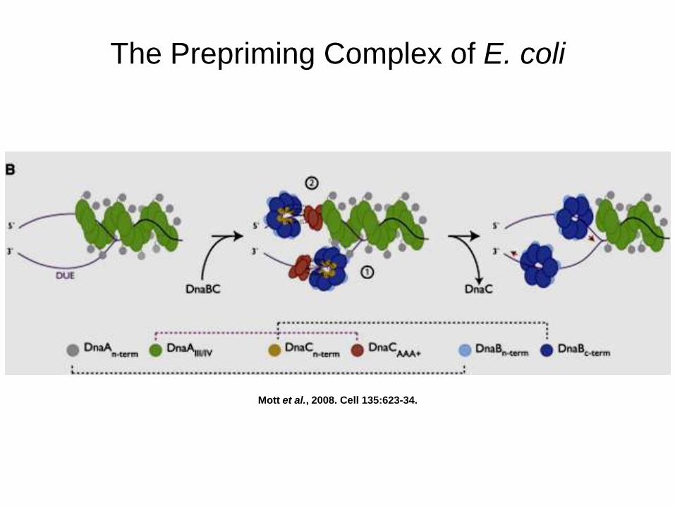
The Prepriming Complex of E. coli
Mott et al., 2008. Cell 135:623-34.

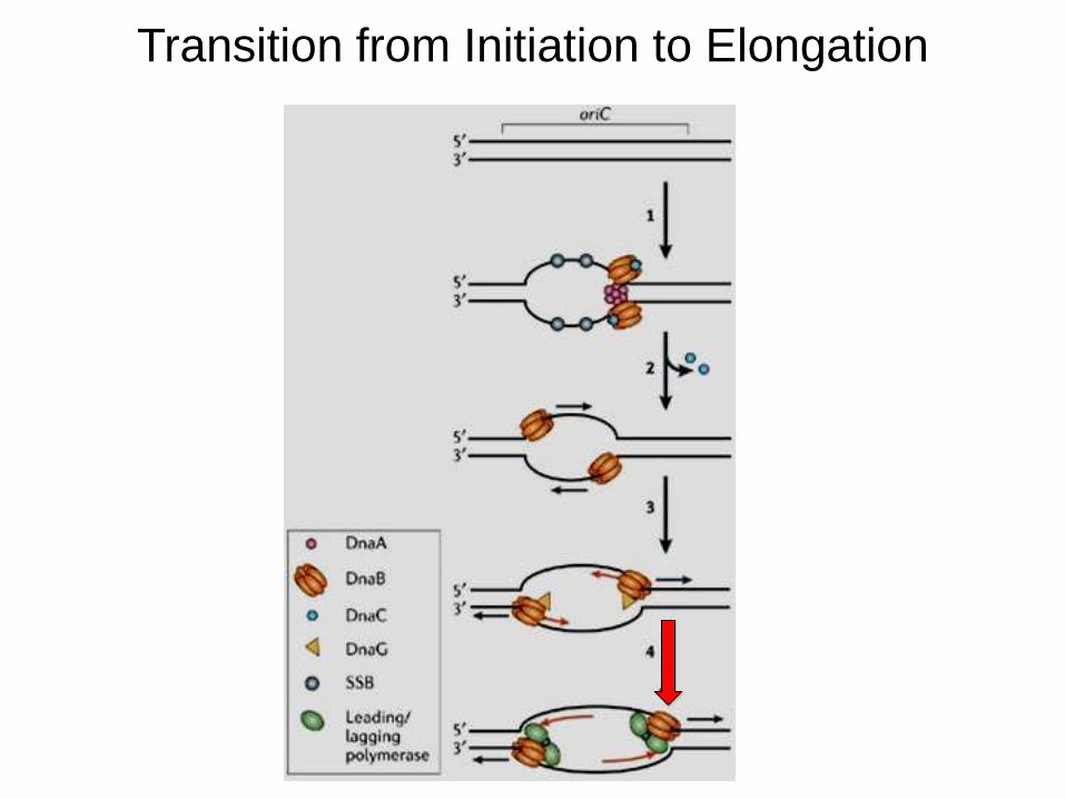
Transition from Initiation to Elongation
Makowska-Grzyska & Kaguni, 2010. Mol Cell 37:90-101.
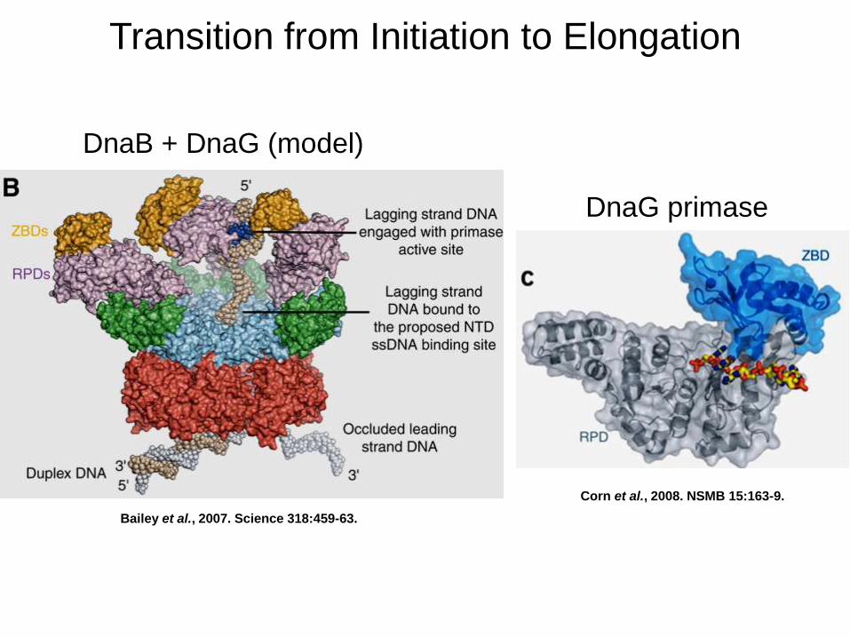
Transition from Initiation to Elongation
Bailey et al., 2007. Science 318:459-63.
Corn et al., 2008. NSMB 15:163-9.
DnaB + DnaG (model)
DnaG primase
Transition from Initiation to Elongation
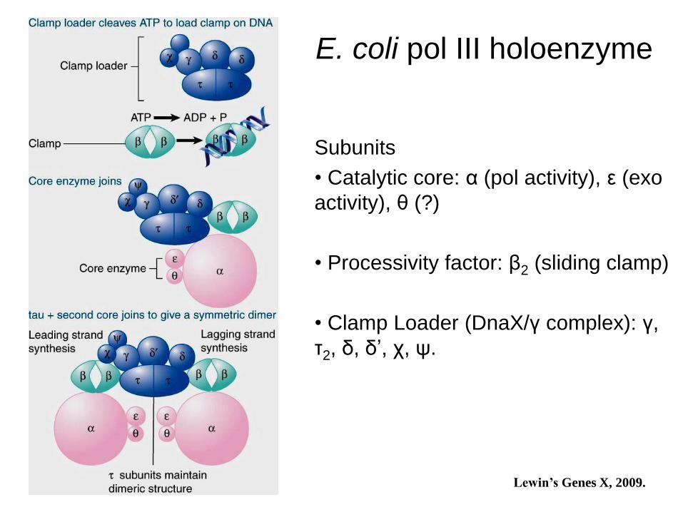
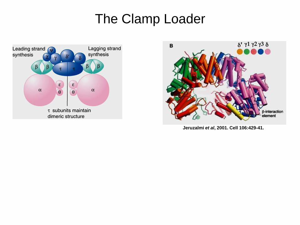
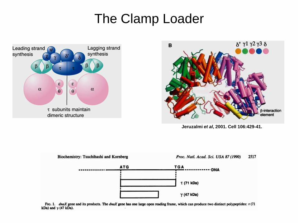
E. coli pol III holoenzyme
Subunits
• Catalytic core: α (pol activity), ε (exo
activity), θ (?)
• Processivity factor: β2 (sliding clamp)
• Clamp Loader (DnaX/γ complex): γ,
τ2, δ, δ’, χ, ψ.
Lewin’s Genes X, 2009.
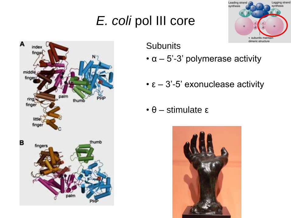
E. coli pol III core
Subunits
• α – 5’-3’ polymerase activity
• ε – 3’-5’ exonuclease activity
• θ – stimulate ε
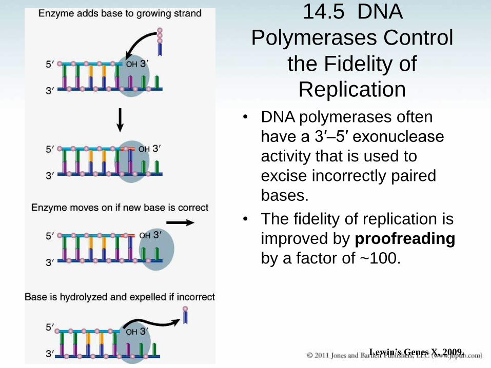
14.5 DNA
Polymerases Control
the Fidelity of
Replication • DNA polymerases often
have a 3′–5′ exonuclease
activity that is used to
excise incorrectly paired
bases.
• The fidelity of replication is
improved by proofreading
by a factor of ~100.
Lewin’s Genes X, 2009.
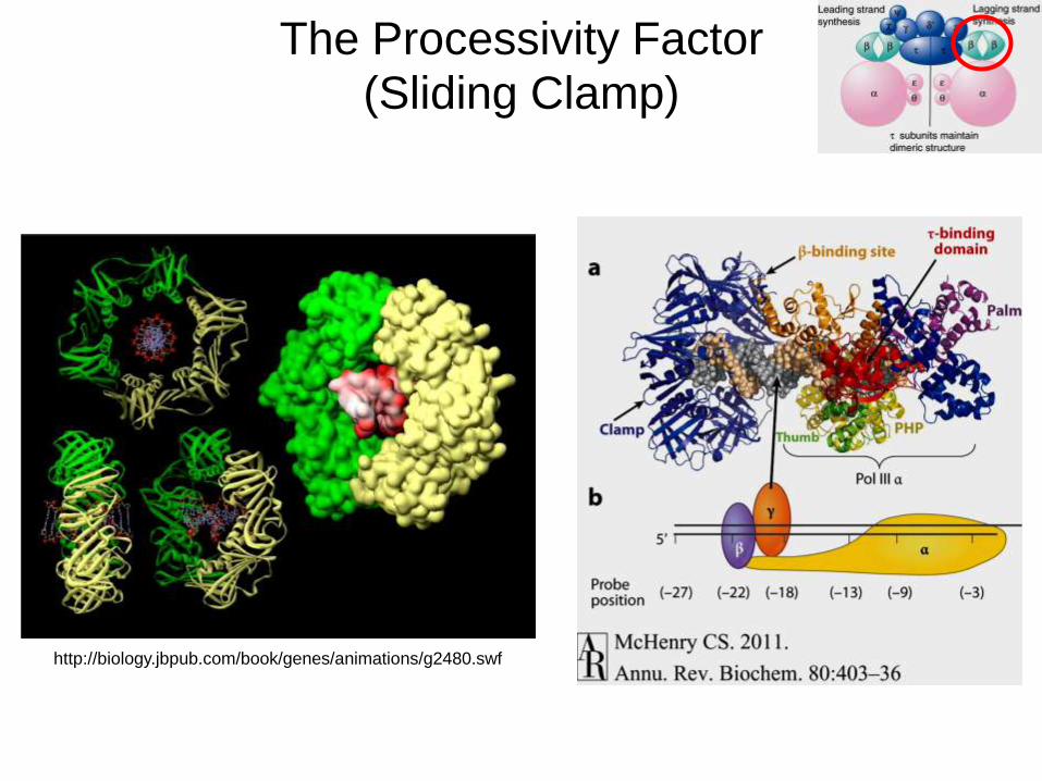
The Processivity Factor
(Sliding Clamp)
http://biology.jbpub.com/book/genes/animations/g2480.swf
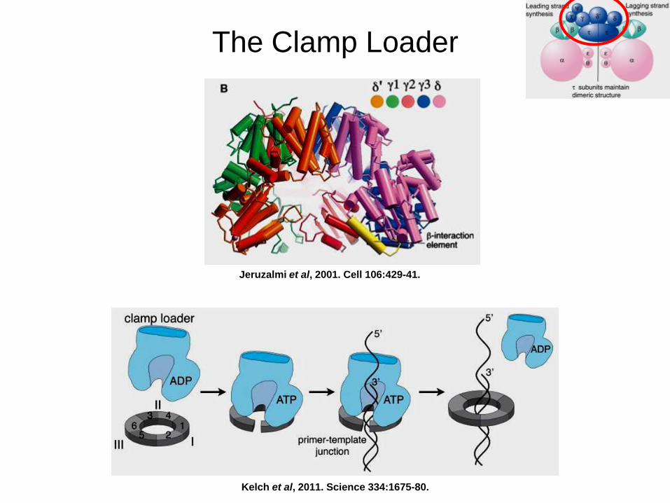
The Clamp Loader
Jeruzalmi et al, 2001. Cell 106:429-41.
Kelch et al, 2011. Science 334:1675-80.
The Clamp Loader
Jeruzalmi et al, 2001. Cell 106:429-41.
The Clamp Loader
Jeruzalmi et al, 2001. Cell 106:429-41.
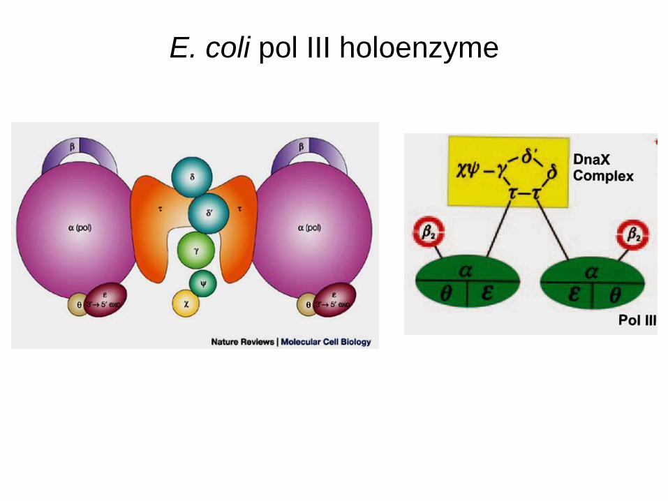
E. coli pol III holoenzyme
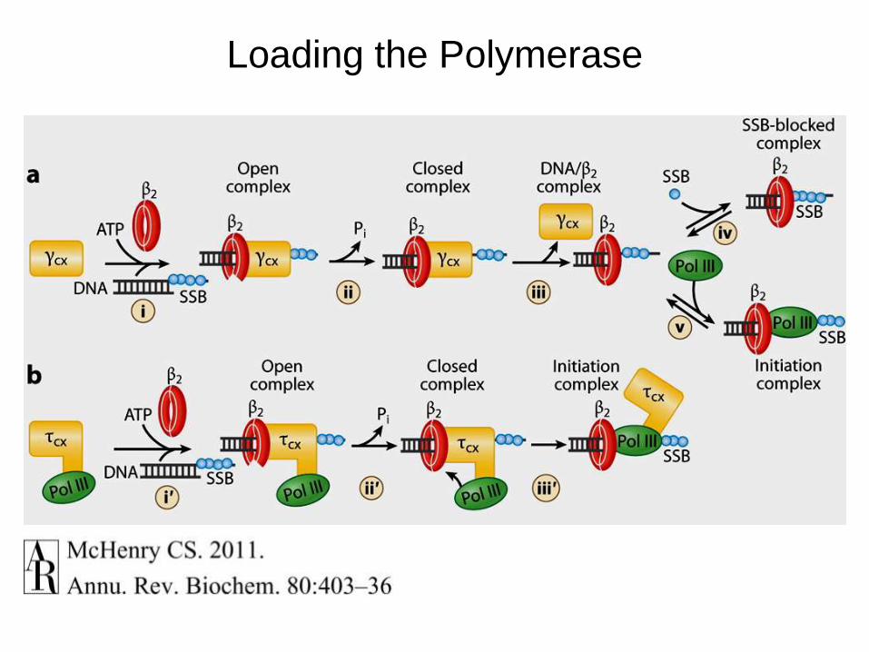
Loading the Polymerase
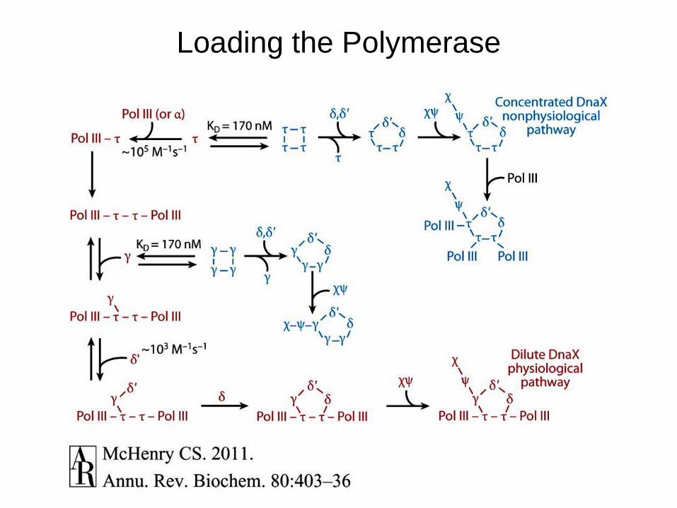
Loading the Polymerase
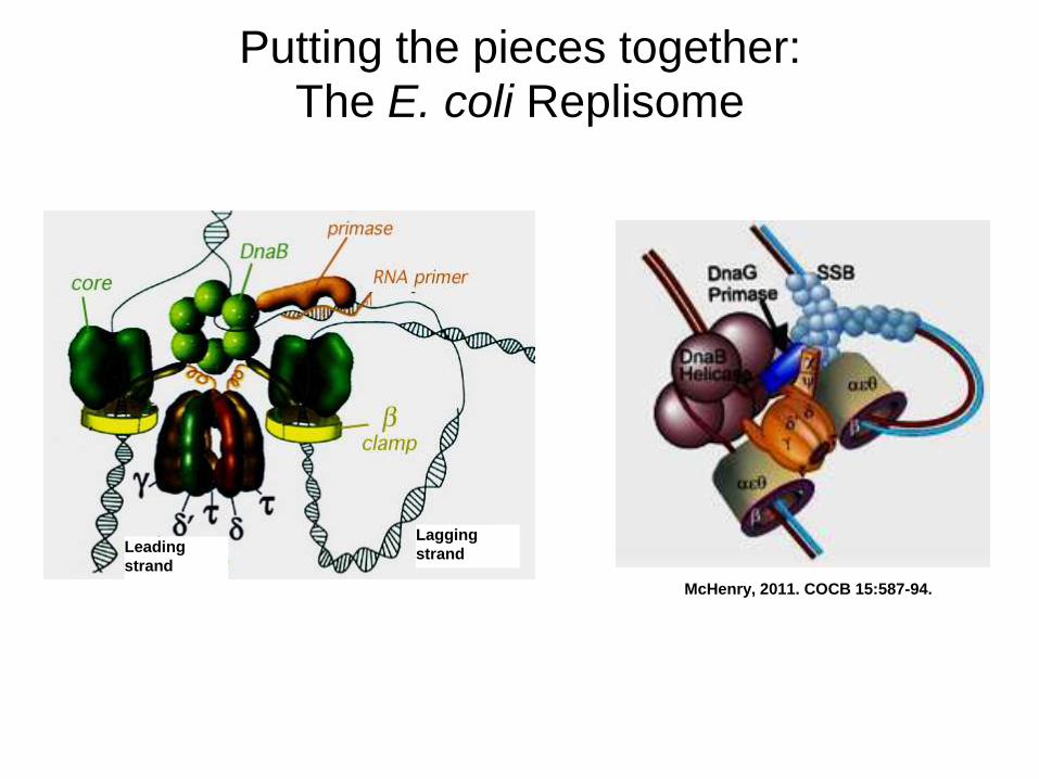
Putting the pieces together:
The E. coli Replisome
McHenry, 2011. COCB 15:587-94.
Leading
strand
Lagging
strand
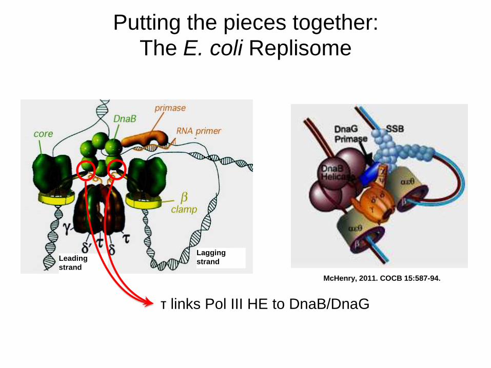
Putting the pieces together:
The E. coli Replisome
McHenry, 2011. COCB 15:587-94.
Leading
strand
Lagging
strand
τ links Pol III HE to DnaB/DnaG
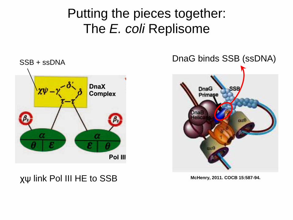
Putting the pieces together:
The E. coli Replisome
McHenry, 2011. COCB 15:587-94. χψ link Pol III HE to SSB
SSB + ssDNA
DnaG binds SSB (ssDNA)
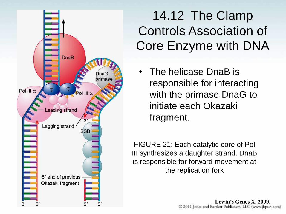
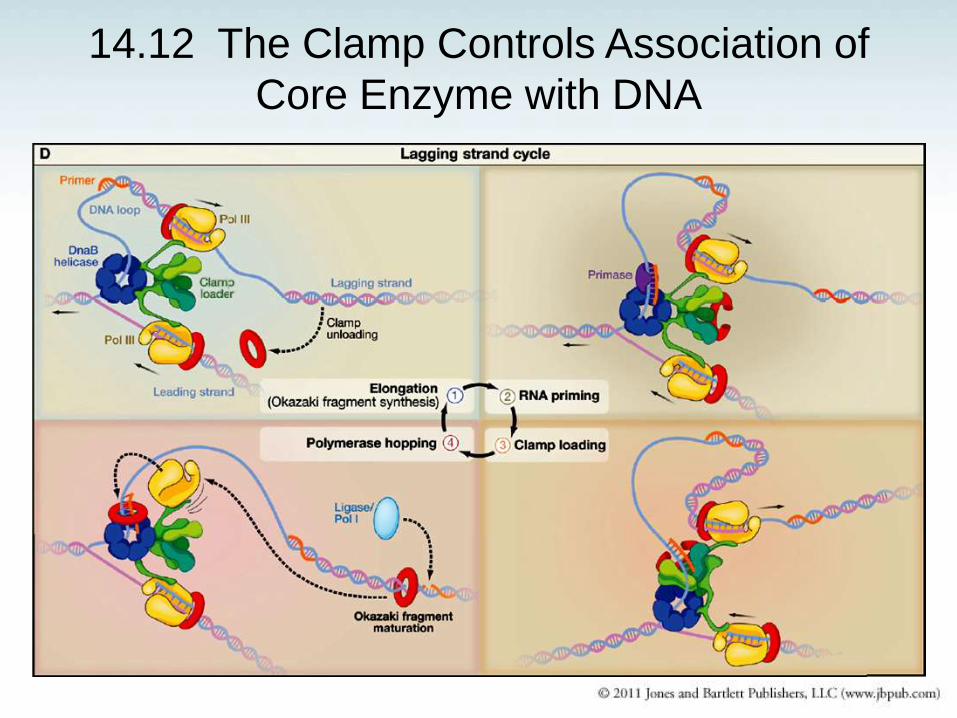
14.12 The Clamp
Controls Association of
Core Enzyme with DNA
• The helicase DnaB is
responsible for interacting
with the primase DnaG to
initiate each Okazaki
fragment.
FIGURE 21: Each catalytic core of Pol
III synthesizes a daughter strand. DnaB
is responsible for forward movement at
the replication fork
Lewin’s Genes X, 2009.
14.12 The Clamp Controls Association of
Core Enzyme with DNA
http://www.wehi.edu.au/education/wehitv/molecular_visualisations_of_dna/
E. coli DNA replication
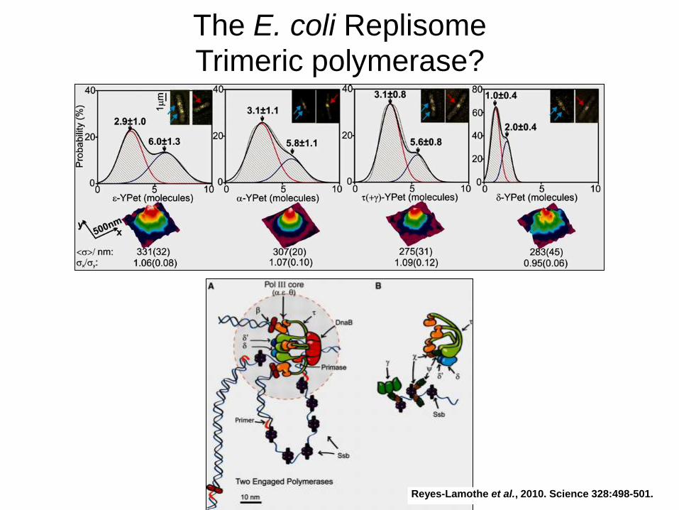
The E. coli Replisome
Trimeric polymerase?
Reyes-Lamothe et al., 2010. Science 328:498-501.
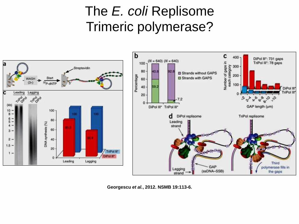
Georgescu et al., 2012. NSMB 19:113-6.
The E. coli Replisome
Trimeric polymerase?
Graham et al., 2017. Cell 169:1201-13.
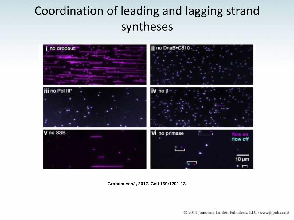
Coordination of leading and lagging strand syntheses
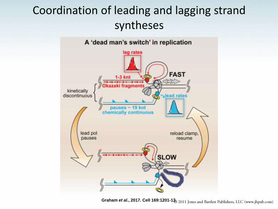
Coordination of leading and lagging strand syntheses
Graham et al., 2017. Cell 169:1201-13.
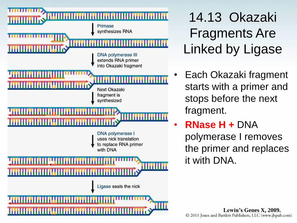
14.13 Okazaki
Fragments Are
Linked by Ligase
• Each Okazaki fragment
starts with a primer and
stops before the next
fragment.
• RNase H + DNA
polymerase I removes
the primer and replaces
it with DNA.
Lewin’s Genes X, 2009.
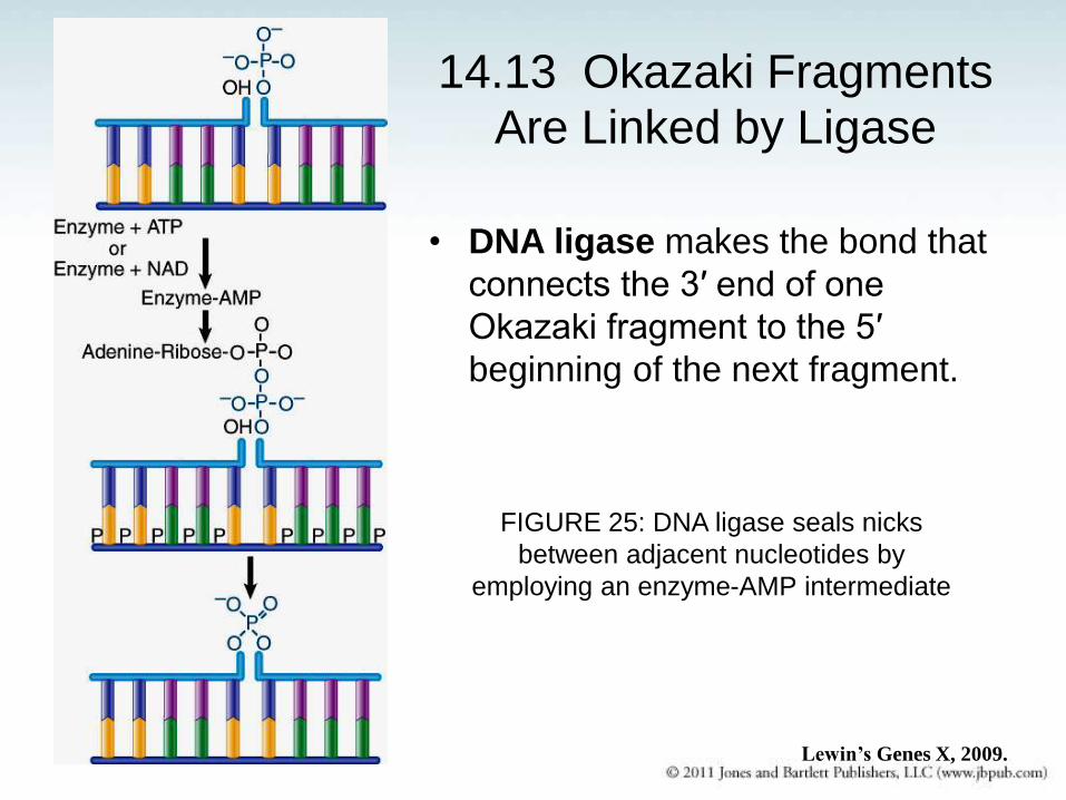
14.13 Okazaki Fragments Are Linked by Ligase
• DNA ligase makes the bond that
connects the 3′ end of one
Okazaki fragment to the 5′
beginning of the next fragment.
FIGURE 25: DNA ligase seals nicks
between adjacent nucleotides by
employing an enzyme-AMP intermediate
Lewin’s Genes X, 2009.
E. coli DNA replication – Summary
• DnaA melts oriC and recruits DnaB helicase/DnaC
helicase loader.
• DnaB helicase recruits DnaG primase. Priming
releases DnaC from prepriming complex.
• DnaB helicase keeps interacting with DnaG primase
transiently throughout lagging-strand synthesis.
• DnaX clamp loader loads β2 clamp on primer-template
(via interactions with δ subunit). Pol III core (α subunit)
interacts with β2 clamp and primer-template.
• Two (Three!) Pol III cores are kept together in the
replisome through the τ subunits of the DnaX clamp
loader.
• In the lagging strand, DnaX clamp loader is constantly
loading β2 clamps onto new primer-templates; it also
promotes Pol III core hopping from the “old” Okazaki
fragment to the “new” primer.
• The τ subunits of DnaX clamp loader are also
important for interacting with DnaB helicase (τ is the
guy!)
E. coli DNA replication – Summary
• The χψ subunits of DnaX clamp loader (τ attaches
them to the ring) interact with SSB transiently, which
interact with DnaG primase transiently.
• RNase H, DNA pol I and DNA ligase are responsible
for the maturation of the Okazaki fragments.
E. coli DNA replication – Summary
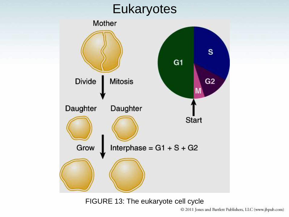
FIGURE 13: The eukaryote cell cycle
Eukaryotes
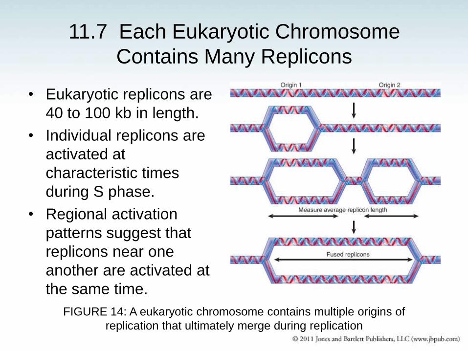
11.7 Each Eukaryotic Chromosome
Contains Many Replicons
• Eukaryotic replicons are
40 to 100 kb in length.
• Individual replicons are
activated at
characteristic times
during S phase.
• Regional activation
patterns suggest that
replicons near one
another are activated at
the same time.
FIGURE 14: A eukaryotic chromosome contains multiple origins of
replication that ultimately merge during replication
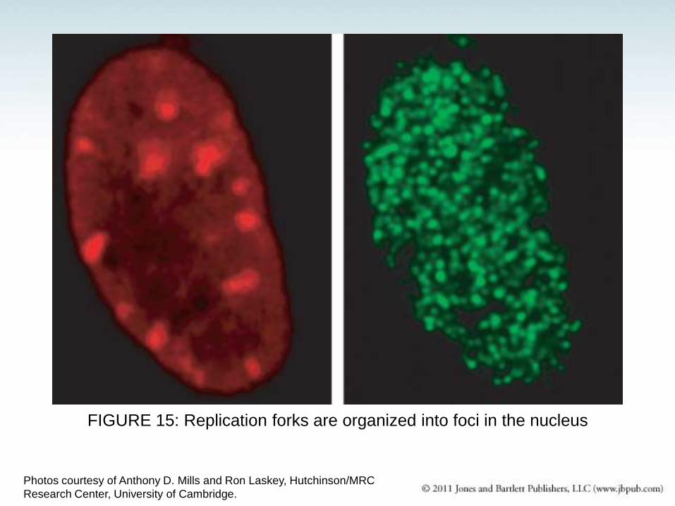
FIGURE 15: Replication forks are organized into foci in the nucleus
Photos courtesy of Anthony D. Mills and Ron Laskey, Hutchinson/MRC
Research Center, University of Cambridge.
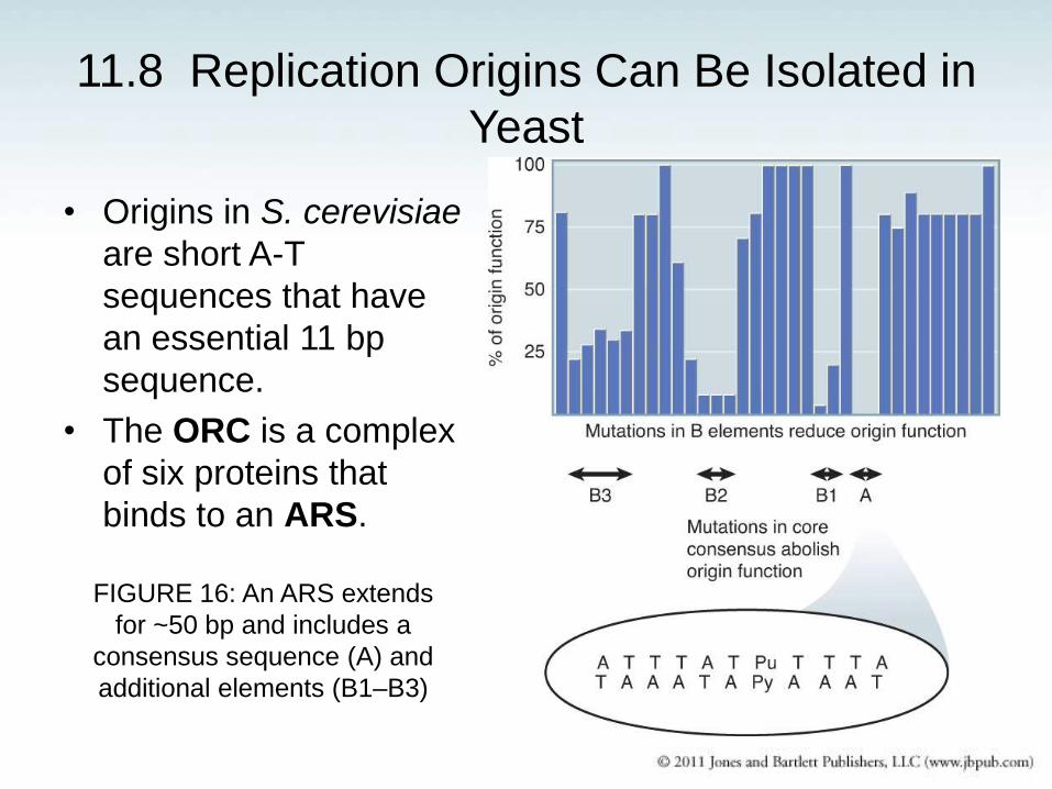
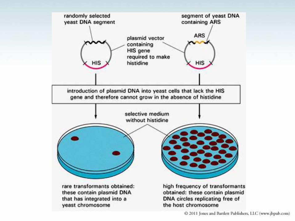
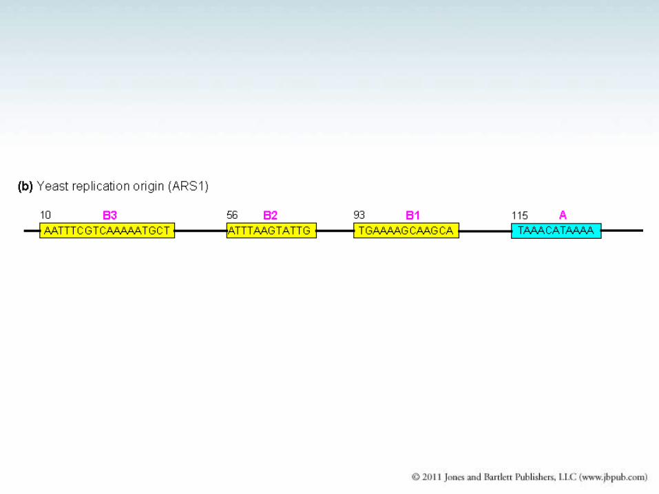
11.8 Replication Origins Can Be Isolated in
Yeast
• Origins in S. cerevisiae
are short A-T
sequences that have
an essential 11 bp
sequence.
• The ORC is a complex
of six proteins that
binds to an ARS.
FIGURE 16: An ARS extends
for ~50 bp and includes a
consensus sequence (A) and
additional elements (B1–B3)
ORC1 ORC2 ORC3
ORC6 ORC5
ORC4
Cdc6
MCM7
MCM2 MCM
3
MCM6 MCM
5
MCM4
MCM7
MCM2 MCM
3
MCM6 MCM
5
MCM4
Cdt1
11.9 Licensing Factor Controls Eukaryotic
Rereplication
• Licensing factor is necessary for initiation of replication
at each origin.
• Licensing factor is present in the nucleus prior to
replication, but is removed, inactivated, or destroyed by
replication.
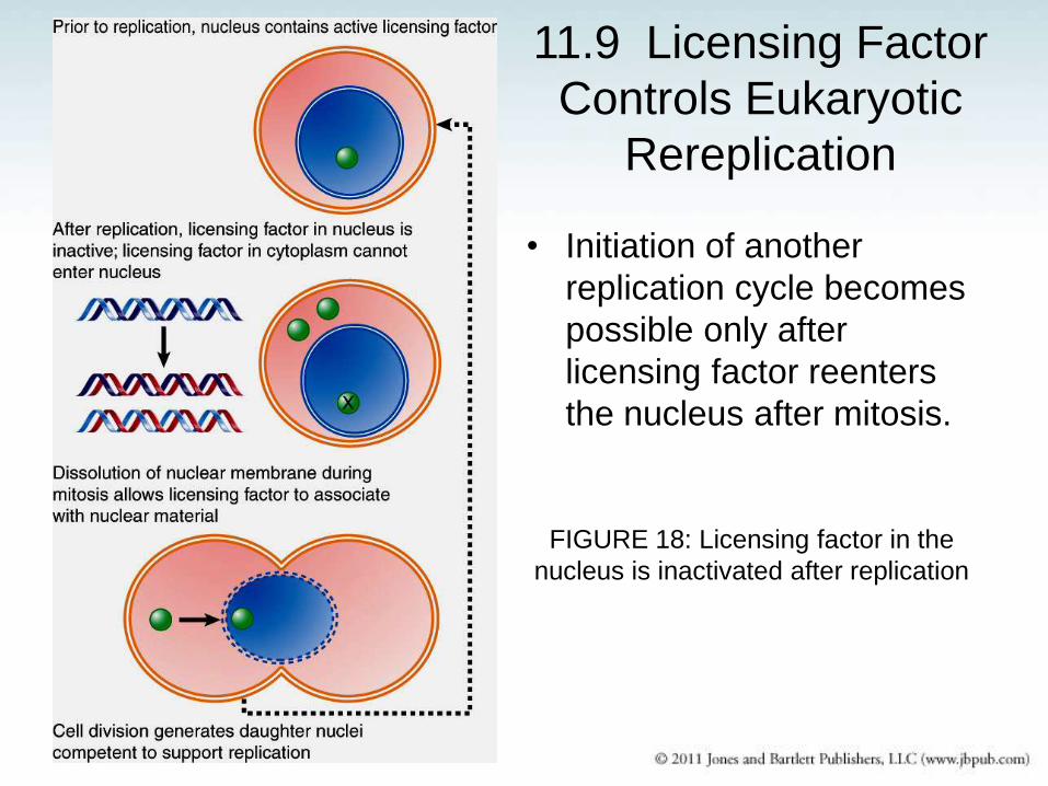
11.9 Licensing Factor
Controls Eukaryotic
Rereplication
• Initiation of another
replication cycle becomes
possible only after
licensing factor reenters
the nucleus after mitosis.
FIGURE 18: Licensing factor in the
nucleus is inactivated after replication

ORC1 ORC2 ORC3
ORC6 ORC5
ORC4
Cdc6
MCM7
MCM2 MCM
3
MCM6 MCM
5
MCM4
MCM7
MCM2 MCM
3
MCM6 MCM
5
MCM4
Cdt1
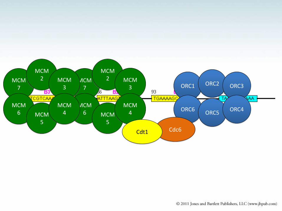
• The ORC is a protein complex that is associated with
yeast origins throughout the cell cycle.
• Cdc6 protein is an unstable protein that is synthesized
only in G1.
• Cdc6 binds to ORC and allows MCM proteins to bind.
• Cdt1 facilitates MCM loading on origins.
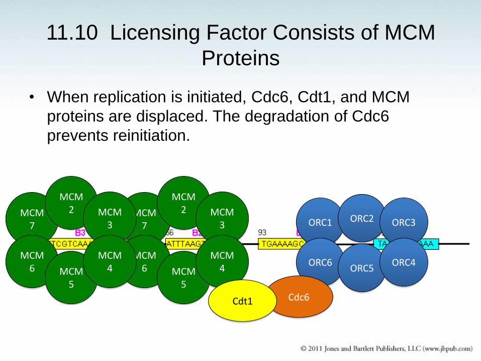
11.10 Licensing Factor Consists of MCM
Proteins
• When replication is initiated, Cdc6, Cdt1, and MCM
proteins are displaced. The degradation of Cdc6
prevents reinitiation.
ORC1 ORC2 ORC3
ORC6 ORC5
ORC4
Cdc6
MCM7
MCM2 MCM
3
MCM6 MCM
5
MCM4
MCM7
MCM2 MCM
3
MCM6 MCM
5
MCM4
Cdt1
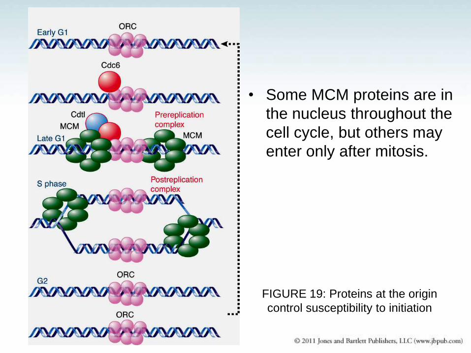
• Some MCM proteins are in
the nucleus throughout the
cell cycle, but others may
enter only after mitosis.
FIGURE 19: Proteins at the origin
control susceptibility to initiation
Regulation of Initiation of DNA Replication in
Eukaryotes (yeast)
• ORC recognizes the origin
• Cdc6 is rapidly degraded
• Some MCM proteins are licensing factors (only enter the
nucleus when the envelope is disrupted during mitosis)
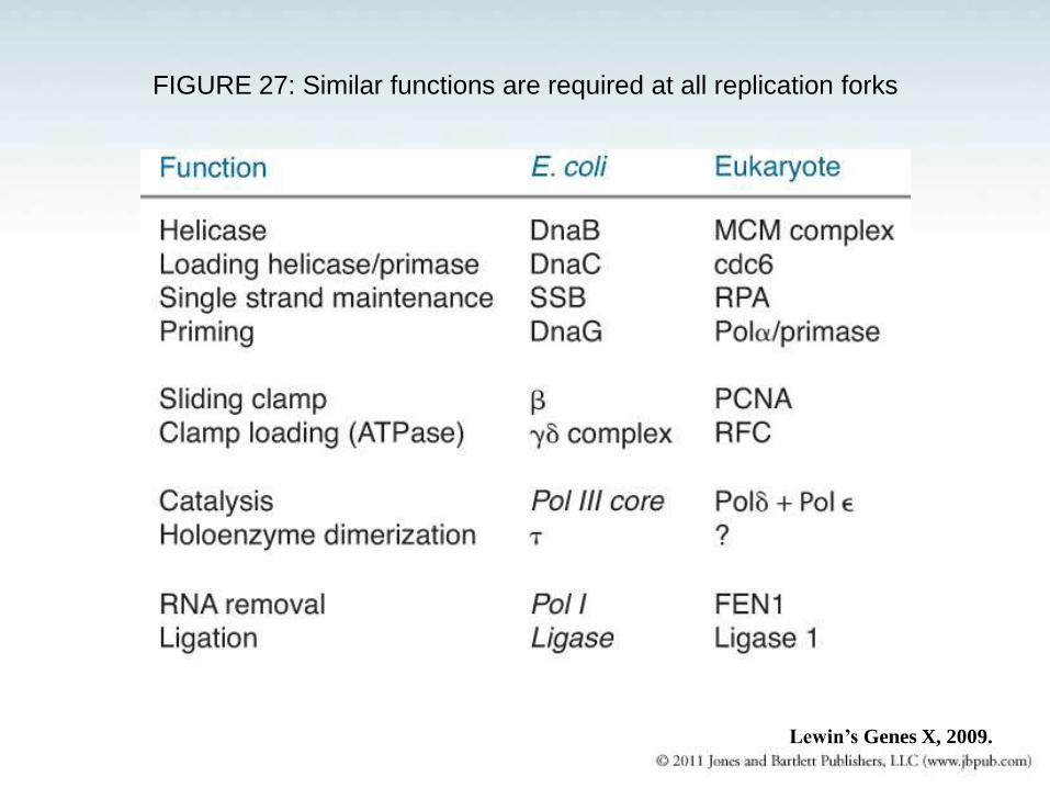
FIGURE 27: Similar functions are required at all replication forks
Lewin’s Genes X, 2009.
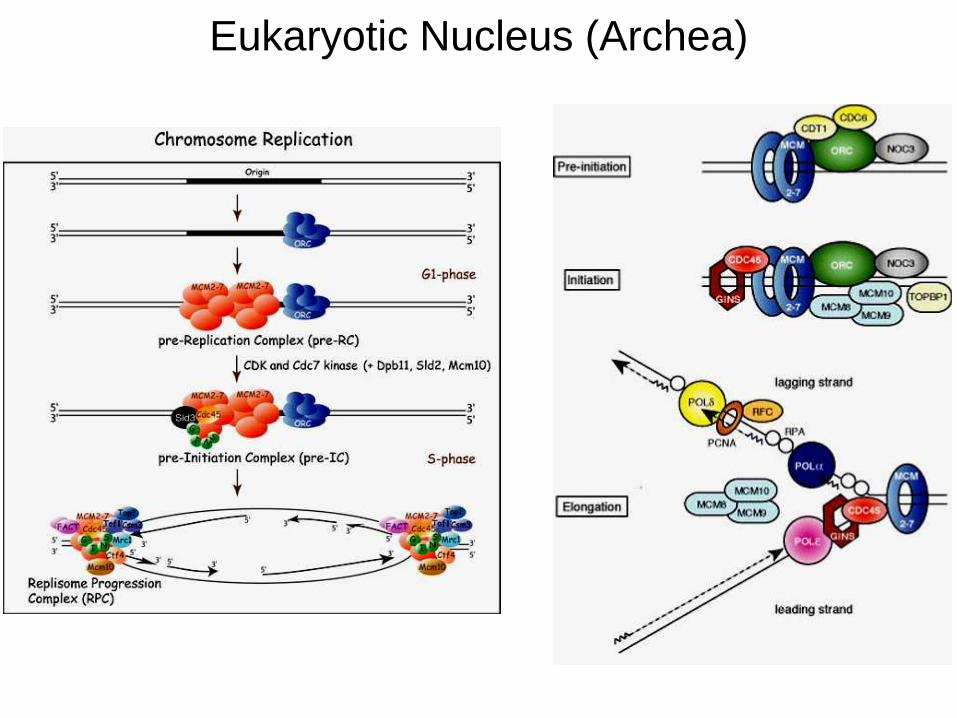
Eukaryotic Nucleus (Archea)
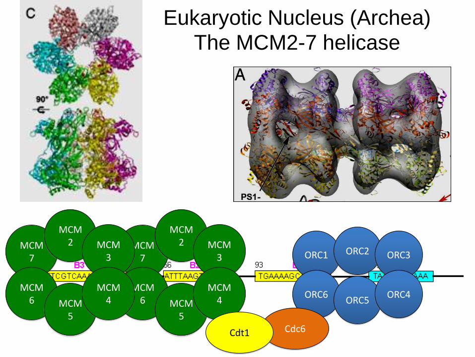
Eukaryotic Nucleus (Archea)
The MCM2-7 helicase
ORC1 ORC2 ORC3
ORC6 ORC5
ORC4
Cdc6
MCM7
MCM2 MCM
3
MCM6 MCM
5
MCM4
MCM7
MCM2 MCM
3
MCM6 MCM
5
MCM4
Cdt1
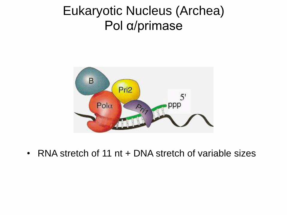
Eukaryotic Nucleus (Archea)
Pol α/primase
• RNA stretch of 11 nt + DNA stretch of variable sizes
Eukaryotic Nucleus (Archea)
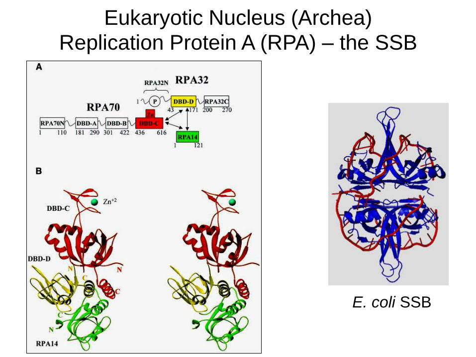
Replication Protein A (RPA) – the SSB
E. coli SSB
Eukaryotic Nucleus (Archea)
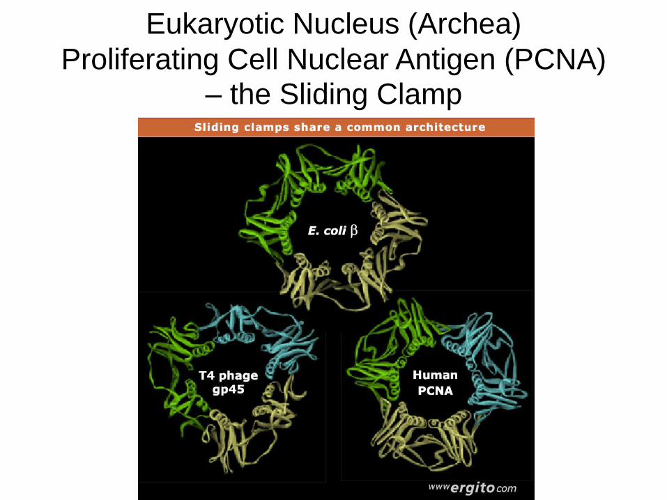
Proliferating Cell Nuclear Antigen (PCNA) – the Sliding Clamp
Eukaryotic Nucleus (Archea)
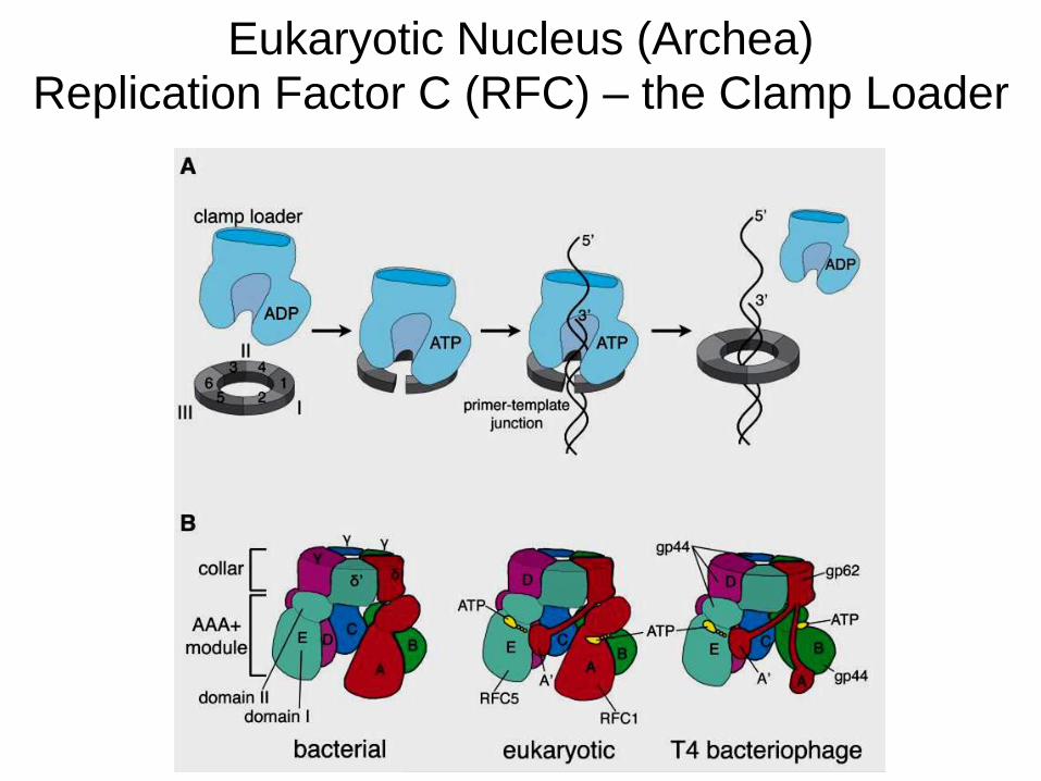
Replication Factor C (RFC) – the Clamp Loader
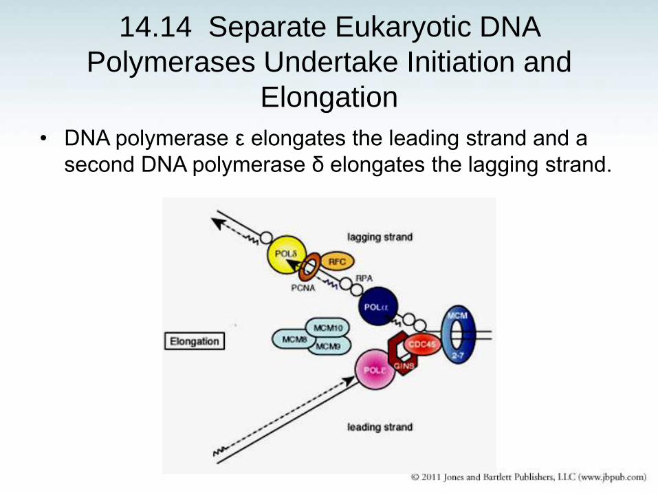
14.14 Separate Eukaryotic DNA
Polymerases Undertake Initiation and
Elongation
• DNA polymerase ε elongates the leading strand and a
second DNA polymerase δ elongates the lagging strand.
Eukaryotic Nucleus (Archea)
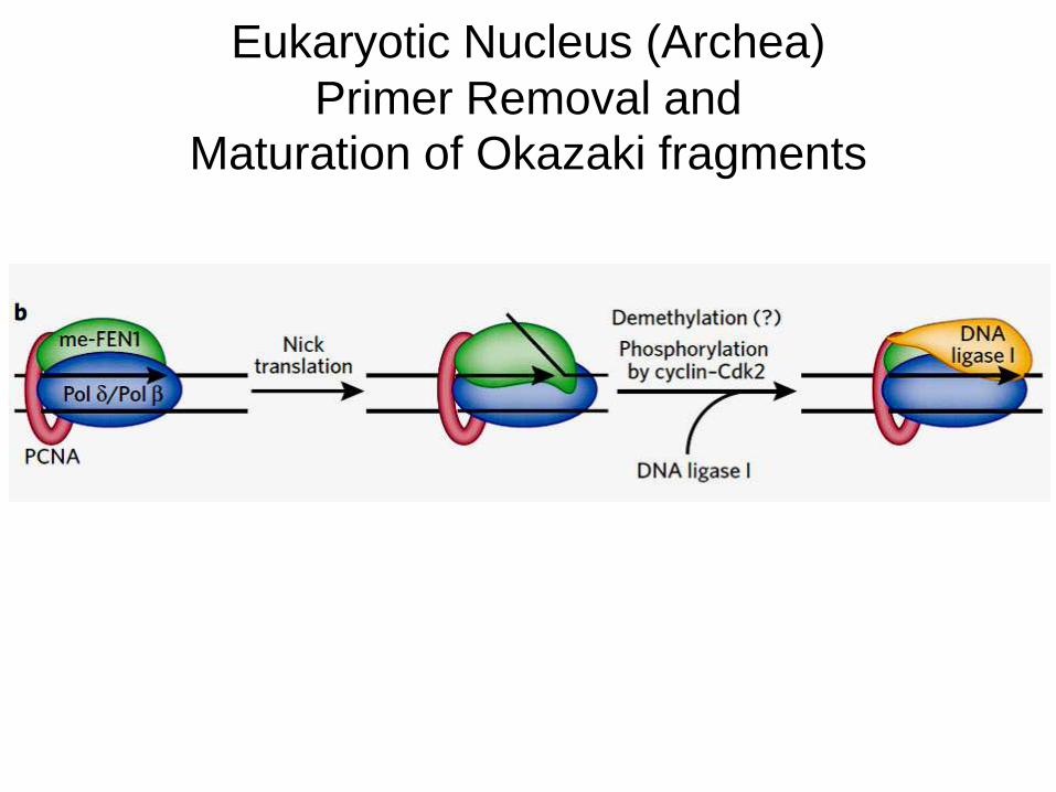
Primer Removal and Maturation of Okazaki fragments
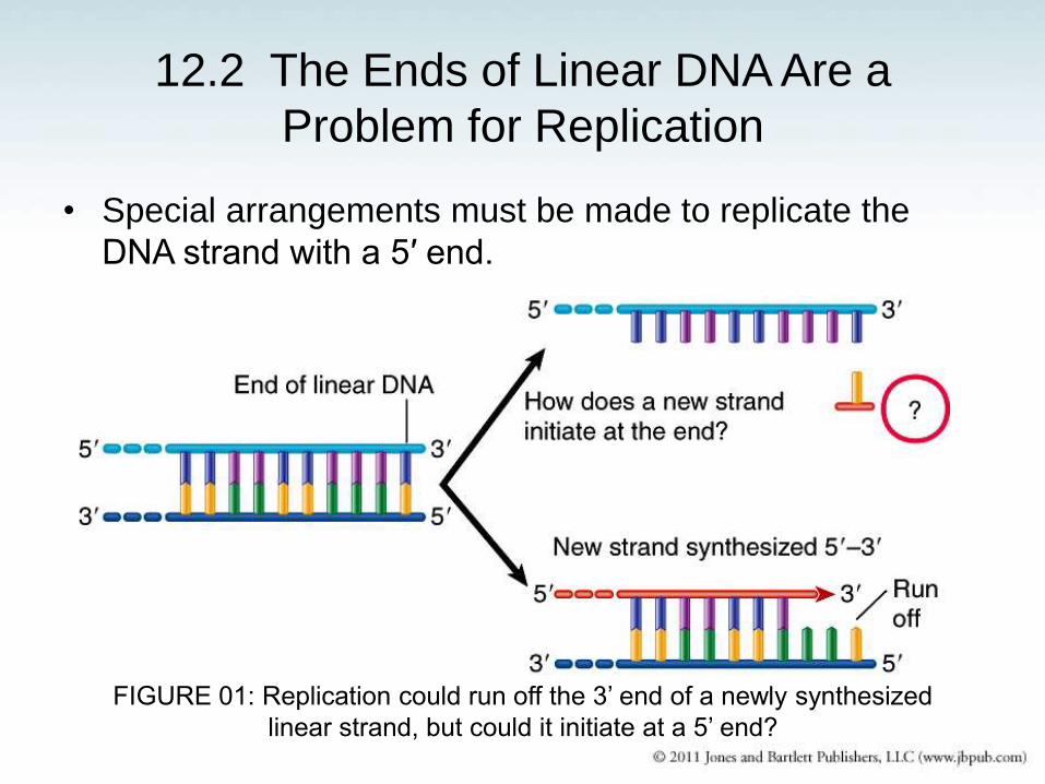
12.2 The Ends of Linear DNA Are a
Problem for Replication
• Special arrangements must be made to replicate the
DNA strand with a 5′ end.
FIGURE 01: Replication could run off the 3’ end of a newly synthesized
linear strand, but could it initiate at a 5’ end?
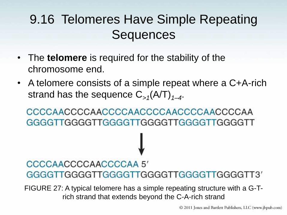
9.16 Telomeres Have Simple Repeating
Sequences
• The telomere is required for the stability of the
chromosome end.
• A telomere consists of a simple repeat where a C+A-rich
strand has the sequence C>1(A/T)1–4.
FIGURE 27: A typical telomere has a simple repeating structure with a G-T-
rich strand that extends beyond the C-A-rich strand
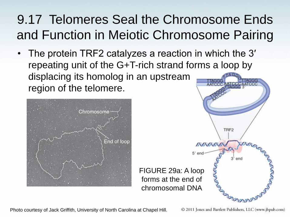
9.17 Telomeres Seal the Chromosome Ends
and Function in Meiotic Chromosome Pairing
• The protein TRF2 catalyzes a reaction in which the 3′
repeating unit of the G+T-rich strand forms a loop by
displacing its homolog in an upstream
region of the telomere.
Photo courtesy of Jack Griffith, University of North Carolina at Chapel Hill.
FIGURE 29a: A loop
forms at the end of
chromosomal DNA
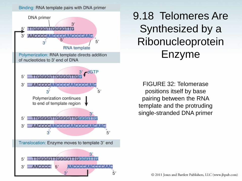
9.18 Telomeres Are
Synthesized by a
Ribonucleoprotein Enzyme
FIGURE 32: Telomerase
positions itself by base
pairing between the RNA
template and the protruding
single-stranded DNA primer
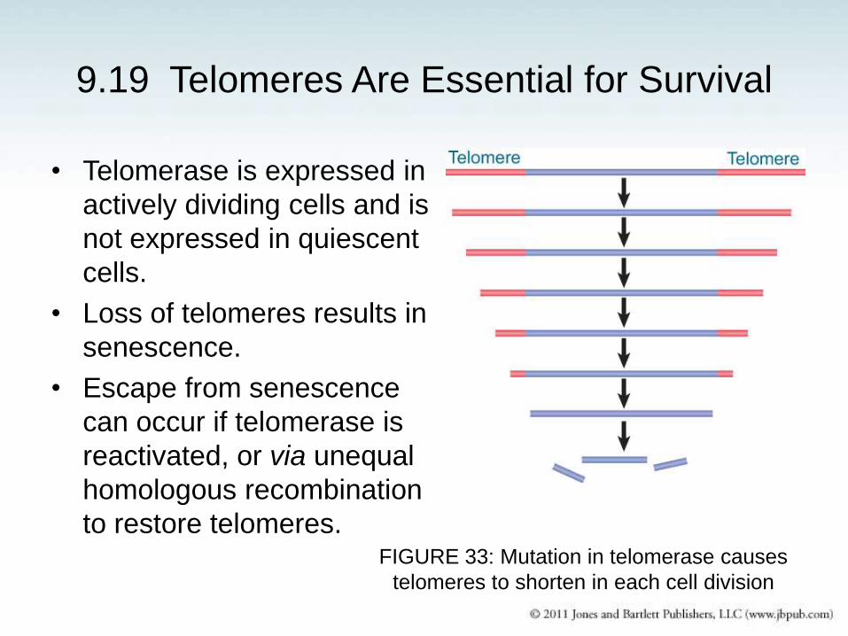
9.19 Telomeres Are Essential for Survival
• Telomerase is expressed in
actively dividing cells and is
not expressed in quiescent
cells.
• Loss of telomeres results in
senescence.
• Escape from senescence
can occur if telomerase is
reactivated, or via unequal
homologous recombination
to restore telomeres.
FIGURE 33: Mutation in telomerase causes
telomeres to shorten in each cell division
• Coordination of leading- and lagging-strand synthesis
in the eukaryotic nucleus is obscure.
• Primers are synthesized by the heterotetrameric Pol
α/primase. They are ~half RNA, ~half DNA.
• Although no sequence homology is found among
nuclear, bacterial and T4 sliding clamps and clamp
loaders, their general structure is very similar (donut-
shape, 3/6fold symmetry).
Systems other than E. coli DNA replication –
Summary
• The heterotrimeric nuclear RPA has no homology with
the homotetrameric bacterial SSB, despite possessing
similar structural folding domains for binding ssDNA.
• Two distinct polymerases (ε and δ) are required to
leading and lagging strand synthesis, respectively, in
the nucleus.
• Okazaki fragments maturation is accomplished by a
complex with PCNA, Pol δ/β, Fen1 and DNA ligase I.
• A specialized polymerase (telomerase) is responsible
for replication of the chromosomal ends.
Systems other than E. coli DNA replication –
Summary