Embed Size (px)
Citation preview
UNIVERSIDADE FEDERAL DE PERNAMBUCO
CENTRO DE CIÊNCIAS BIOLÓGICAS PROGRAMA DE PÓS-GRADUAÇÃO EM BIOQUÍMICA E FISIOLOGIA
CANAIS IÔNICOS NA EXPANSÃO DE CÉLULAS-TRONCO MESENQUIMAIS DE CORDÃO UMBILICAL HUMANO
LINDALVA LAYSE DE LIMA MALAGUETA VIEIRA
Fevereiro, 2011
Lindalva Layse de Lima Malagueta Vieira
CANAIS IÔNICOS NA EXPANSÃO DE CÉLULAS-TRONCO MESENQUIMAIS DE CORDÃO UMBILICAL HUMANO
Dissertação apresentada para o cumprimento parcial das exigências para obtenção do título de Mestre em Bioquímica e Fisiologia pela Universidade Federal de Pernambuco
ORIENTADOR: Prof. Dr. Oleg Vladimirovich Krasilnikov CO-ORIENTADORA: Profa. Dra. Márcia Bezerra da Silva
Fevereiro, 2011
Vieira, Lindalva Layse de Lima Malagueta
Canais iônicos na expansão de células-tronco mesenquimais de cordão umbilical humano/ Lindalva Layse de Lima Malagueta Vieira. – Recife: O Autor, 2011. 71 folhas : il., fig., tab.
Orientador: Oleg Vladimirovich Krasilnikov Co-orientadora: Márcia Bezerra da Silva Dissertação (mestrado) – Universidade Federal de
Pernambuco. Centro de Ciências Biológicas. Bioquímica e Fisiologia, 2011. Inclui bibliografia
1. Células- tronco 2. Proteínas 3. Cordão umbilical I. Título.
580 CDD (22.ed.) UFPE/CCB-2011-143
Lindalva Layse de Lima Malagueta Vieira
CANAIS IÔNICOS NA EXPANSÃO DE CÉLULAS-TRONCO MESENQUIMAIS DE CORDÃO UMBILICAL HUMANO

Primeiramente ao pai do céu por ter me dado sabedoria e discernimento para chegar
até aqui. Aos meus queridos pais Maria Fátima de Lima e Luiz Aurélio Malagueta Vieira por terem me dado princípios, educação e muito amor. Obrigada por toda
dedicação e confiança depositada em mim! Amo vocês!
AGRADECIMENTOS
Ao meu senhor Jesus por ter me presenteado com a vida. A minha família
abençoada, Fátima e Luiz Aurélio (meus pais) vocês são meu exemplo de vida,
meus irmãos Anadrizia e Leo Malagueta por serem verdadeiros amigos, a minha
avó Josefa Farias pelas suas palavras de conforto nas horas e momentos certos.
Aos meus tios em especial, tia Lucia por todo carinho e atenção. Há Kleber Pasini por todo carinho e incentivo, e pelas palavras de conforto nos momentos de
apreensão, você foi essencial em mais essa conquista.
Obrigada ao meu querido orientador professor Oleg Krasilnikov pela
oportunidade e confiança depositadas em mim, como também pela paciência e
tolerância em momentos de dificuldade. A minha querida co-orientadora professora
Márcia Bezerra e ao professor Reginaldo Pereira por toda paciência e dedicação
junto a mim. Vocês foram importantes para o desenvolvimento deste trabalho. Ao
professores Liliya Yuldasheva e Claudi o Gabriel pela atenção, preocupação e
apoio.
Em especial a uma pessoa que gosto muito Darlene Paiva por todo apoio e
noites em claro que passou me ajudando para que conseguisse chegar até aqui.
Aos colaboradores do Centro de pesquisa Aggeu Magalhães Carlos Régis, Valéria do Rego por toda disponibilidade e ensinamentos.
Aos meus queridos amigos Thiago (trem), Marina, Marcela, George e Vilma
por todo apoio, incentivo e preocupação; essa conquista divido com vocês!
A equipe de Cultura de células, Jéssica (minha filha adotiva), Darlene,
Gissely, Alberto, Willamis, Érika, Thauan e Lizandra, vocês foram importante na
minha jornada. A todos do LBM - Laboratório de Biofísica de Membranas, em
especial Dijanah, Janilson e Diego pela paciência e disponibilidade em me ajudar.
A Fredson pela sua atenção e preocupação comigo.
Por fim agradeço às agências de fomento pesquisa, FACEPE e CNPQ pelo
apoio financeiro.
5
SUMÁRIO
RESUMO ............................................................................................................................................................... 6 ABSTRACT ........................................................................................................................................................... 7 LISTA DE ABREVIATURAS .............................................................................................................................. 8 1.INTRODUÇÃO .................................................................................................................................................. 9 2.Revisão Bibliográfica ................................................................................................................................ ...10 2.1Canais catiônicos ....................................................................................................................................... 10
(2.1.1) Canais de potássio ...................................................................................................................... 10 (2.1.2) Canais de sódio ........................................................................................................................... 17 (2.1.3) Canal de Cálcio. ........................................................................................................................... 20
2.2 Canais aniônicos ........................................................................................................................................ 22 (2.2.1) Canal de cloreto dependentes de voltagem .......................................................................... 23 (2.2.2) Canais de cloreto ativados por cálcio (CLCAs) ................................................................... 25 (2.2.3) Canal CFTR (Regulador de Condutância de Membrana na Fibrose Cística) ................ 26 (d) Canal aniônico regulador de volume (VRAC) ............................... Erro! Indicador não definido.
3.1 Localização e classificação..................................................................................................................... 27 3.2.Células-tronco mesenquimais (CTMs) ................................................................................................. 29
(3.2.1).Isolamento ..................................................................................................................................... 29 (3.2.2).Caracterização .............................................................................................................................. 30 (3.2.3).Proliferação ................................................................................................................................... 30 (3.2.4).Diferenciação ................................................................................................................................ 30
3.3 Células-tronco mesenquimais de cordão umbilical Humano ........................................................ 32 4. Ciclo celular ................................................................................................................................................... 33 5. OBJETIVOS .................................................................................................................................................... 36 6 REFERÊNCIAS BIBLIOGRÁFICAS ............................................................................................................ 37 7. ARTIGO CIENTÍFICO.......................................................................................................................48 8. CONCLUSÕES.................................................................................................................................57 9. ANEXOS...........................................................................................................................................72 Anexo I: Parecer do comitê de ética e pesquisa da UFPE..................................................................72 Anexo II: Termo de consentimento livre e esclarecido........................................................................75 Anexo III: Trabalho enviado ao IV simpósio internacional de terapia celular avançadas....................78 Anexo IV: Trabalho enviado ao congresso Brasileiro de células-tronco..............................................79 Anexo V: Instruções da revista aos autores do periódico do trabalho.................................................80 Anexo VI: Trabalho complementar.......................................................................................................81
6
RESUMO
Canais iônicos são proteínas integrais, formadoras de poros na membrana plasmática das células e são de extrema importância para as funções intra e extracelulares. Os poros ajudam no transporte rápido e seletivo dos íons, nutrientes e metabolitos pela membrana plasmática, portanto estão diretamente relacionados a diferentes funções no organismo como, transmissão sináptica, contração muscular, secreção hormonal, regulação do volume celular, proliferação celular e diferenciação.
Podemos classificar os canais iônicos de acordo com seu principal estímulo para ativação: temos os canais ativados por ligantes intracelulares, canais ativados por ligantes extracelulares, canais de potássio e cloreto ativados por cálcio, canais sensíveis a ácidos e canais dependentes de voltagem (Na+, K+, Ca+ e Cl-). Os canais iônicos têm sido extensamente estudados, e novas informações vêm sendo acumuladas nos últimos anos, mostrando a importância da participação destes canais em processos relacionados à homeostase, como também sua contribuição na proliferação e diferenciação celular.
Nesse trabalho procuramos identificar os níveis de RNAm em quatro tipos de canais iônicos, canal de potássio de alta condutância ativado por cálcio (MaxiK), canal aniônico dependente de voltagem (pl-VDAC), Canal de cloreto ativado por cálcio (CLCA1) e Canal de potássio dependente de voltagem (hEAG1) nas células-tronco mesenquimais de cordão umbilical, a partir da geléia de Wharton (hwCTMs), nas diferentes fases do ciclo (G0/G1, S e G2/M) e diferenciação celular (adipo- e osteogênica), visando contribuir no processo de terapia celular.
O RNA total de hwCTMs foi extraído utilizando RNeasy mini kit (QIAGEN). O RNA transcrito em cDNA com superScript reverse (Invitrogen). PCR em Tempo Real foi realizada no ABI prisma 7500 (Applied Biosystems) usando supermix UDG (Invitrogen). Em acordo com os dados publicados, o estímulo osteogênico originou características osteoblásticas das células comprovadas por coloração histoquímica (alizarin red S) onde se observou o aparecimento células alongadas com presença de granulações escuras (cristais de hidroxiapatita) na matriz extracelular. A comprovação da diferenciação adipogênica foi feita por coloração histoquímica com oil red O, mostrando células com morfologia mais arredondada e presença de vacúolos de gordura.
Estabelecemos que entre os quatro canais estudados, dois, CLCA1 e hEAG1, não se expressaram, enquanto a expressão dos MaxiK e pl-VDAC foi consideravelmente alto atingindo ~10% do valor de housekeeping gene Glyceraldehyde 3-phosphate dehydrogenase (GAPDH). Encontramos que a expressão dos MaxiK e pl-VDAC aumentaram durante a progressão do ciclo celular (G0/G1, S e G2/M) nas hwCTMs.
Detectamos que a diferenciação muda a expressão dos canais MaxiK e pl-VDAC de maneira oposta: o canal MaxiK aumenta enquanto que o pl-VDAC diminui.
Os dados ampliam o conhecimento sobre os tipos dos canais iônicos e o nível da expressão nas hwCTMs aumentando a compreensão da biologia de células-tronco mesenquimais.
Palavras-chave: Células-tronco mesenquimais; Geléia de Wharton; canais iônicos; ciclo celular; diferenciação.
7
ABSTRACT
Ion channels are integral proteins, forming pores in the plasma membrane of cells. They determine and regulate the rapid and selective trans-membrane transport of ions, nutrients and metabolites. Ion channels can be classified according to activation stimulus: chemical (intracellular or extracellular), including Ca2+ and pH; mechanical (stretch-activated channels) and electrical (voltage-dependent ion channels, such as well known Na+, K+ , Ca2+ and Cl- channels). The information accumulated during last two decades shows the importance of participation of ion channels in cell homeostasis and indicate their significant contribution in proliferation and differentiation.
In the present study we have investigated mRNA levels of four (MaxiK, pl-VDAC, CLCA1 and hEAG1) ion channels in individual mesenchymal stem cells of Wharton's jelly human umbilical cord (hwMSCs) at the different phases of the cell cycle (G0/G1, S and G2 / M) and under adipodenic and osteogenic differentiations. The differentiation was confirmed by morphological and histochemical analysis. Specifically, the osteogenic stimulus originated the appearance spread-eagle elongated cell and the presence of dark granules (hydroxyapatite crystals; Alizarin Red S staining) in the extracellular matrix. The adipogenic stimulus yields to round-shapes cells with fat vacuoles, which were confirmed by Oil Red O staining. RNA from individual hwMSCs was extracted using RNeasy micro kit (QIAGEN) and transcribed with SuperScript reverse (Invitrogen). Real-Time PCR was performed on the ABI Prism 7500 (Applied Biosystems) using Supermix UDG (Invitrogen).
As a result we have established that in hwMSCs only two (MaxiK and pl-VDAC) of four ion channel genes studied are expressed at significant level. The expression of MaxiK and pl-VDAC channels was increased during the progression of cell cycle (G0/G1, S and G2/M). Differentiation has considerable influence at the ion channel expression level also. The expression of MaxiK was found increased, whereas the expression of pl-VDAC channel was decreased during adipogenic as osteogenic differentiations.
The data extend the knowledge about the types of ion channels and the level of expression in hwCTMs increasing understanding of the biology of mesenchymal stem cells.
Keywords: Mesenchymal stem cells; Wharton Jelly, ion channels, cell cycle, differentiation
8
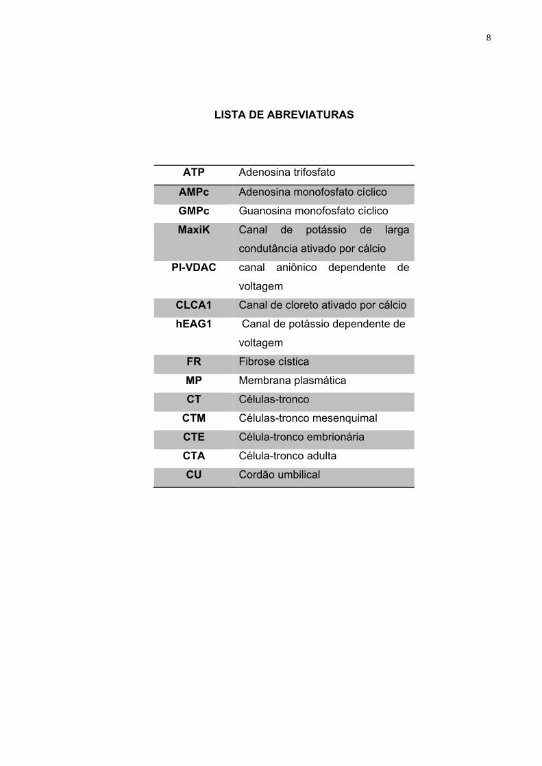
LISTA DE ABREVIATURAS
ATP Adenosina trifosfato
AMPc Adenosina monofosfato cíclico
GMPc Guanosina monofosfato cíclico
MaxiK Canal de potássio de larga
condutância ativado por cálcio
Pl-VDAC canal aniônico dependente de
voltagem
CLCA1 Canal de cloreto ativado por cálcio
hEAG1 Canal de potássio dependente de
voltagem
FR Fibrose cística
MP Membrana plasmática
CT Células-tronco
CTM Células-tronco mesenquimal
CTE Célula-tronco embrionária
CTA Célula-tronco adulta
CU Cordão umbilical
9
1. INTRODUÇÃO
Os canais iônicos são poros constituídos por proteínas integrais presentes
nas membranas celulares e representam elementos fundamentais para o
funcionamento das células (HILLE et al, 1999). Os poros ajudam no transporte rápido e
seletivo dos íons, nutrientes e metabolitos pela membrana plasmática, portanto estão
diretamente relacionados a diferentes funções fisiológicas das células, órgãos e
organismos (TERLAU & STUHMER, 1998). Por exemplo, a participação destes
componentes nas membranas de células germinativas, glóbulos brancos e células
de glândulas endócrinas são importantes no processo de sinalização intracelular
para garantir a homeostase e contribuir para proliferação e diferenciação celular
(PARK et al, 2007).
A permeabilidade seletiva da membrana plasmática mediada por canais,
juntamente com a diferença de concentração iônica, gera um gradiente de potencial
eletroquímico através das membranas biológicas que as células utilizam para
realizar diversas funções, incluindo a geração e propagação de potenciais de ação
(GOLDIN et al, 2000). Os canais iônicos são divididos de acordo com o principal
estímulo para sua ativação. Temos os canais ativados por ligantes intracelulares
(cálcio, adenosina trifosfato (ATP), adenosina monofofasto cíclico (AMPc),
guanosina monofosfato cíclico (GMPc), canais ativados por ligantes extracelulares
(receptores para neurotransmissores), canais de potássio e cloreto ativados por
cálcio, canais sensíveis a ácidos e canais ativados por voltagem ou dependentes de
voltagem (Na+, K+, Ca+ e Cl-) (TERLAU & STUHMER, 1998).
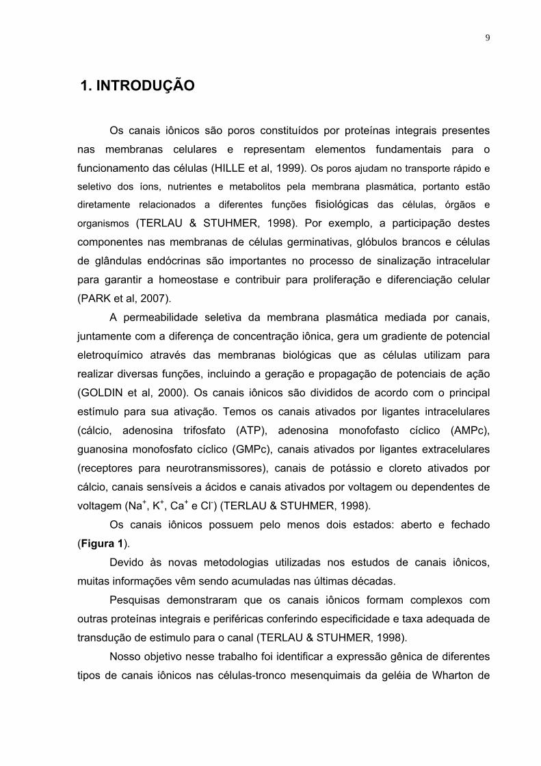
Os canais iônicos possuem pelo menos dois estados: aberto e fechado
(Figura 1).
Devido às novas metodologias utilizadas nos estudos de canais iônicos,
muitas informações vêm sendo acumuladas nas últimas décadas.
Pesquisas demonstraram que os canais iônicos formam complexos com
outras proteínas integrais e periféricas conferindo especificidade e taxa adequada de
transdução de estimulo para o canal (TERLAU & STUHMER, 1998).
Nosso objetivo nesse trabalho foi identificar a expressão gênica de diferentes
tipos de canais iônicos nas células-tronco mesenquimais da geléia de Wharton de
10
cordão umbilical humano (hwCTMs) nas diferentes fases do ciclo (G0/G1, S e G2/M)
e diferenciação celular, visando contribuir no processo de terapia celular.
Figura 1. O esquema demonstra do canal iônico dependente de voltagem na membrana
plasmática. (A) canal iônico fechado com os segmentos S4 (carregado positivamente) perto
do lado intracelular. (B) na resposta da mudança na voltagem transmembrana os segmentos
S4 são direcionadas no sentido extracelular, causando abertura do canal (BORJESSON &
ELINDER, 2008a).
2. Revisão Bibliográfica
2.1 Canais catiônicos 2.1.1. Canais de potássio
Dentre todos os tipos canais iônicos conhecidos, os canais de potássio são os
que apresentam o maior grupo e é o mais diversificado, apresentando cerca de 70
locus conhecidos no genoma de mamíferos (GUTMAN et al, 2005). Este canal
compartilha uma propriedade comum, são seletivos aos íons K+.
2.1.1.1 Canal de potássio dependente de voltagem (Kv)
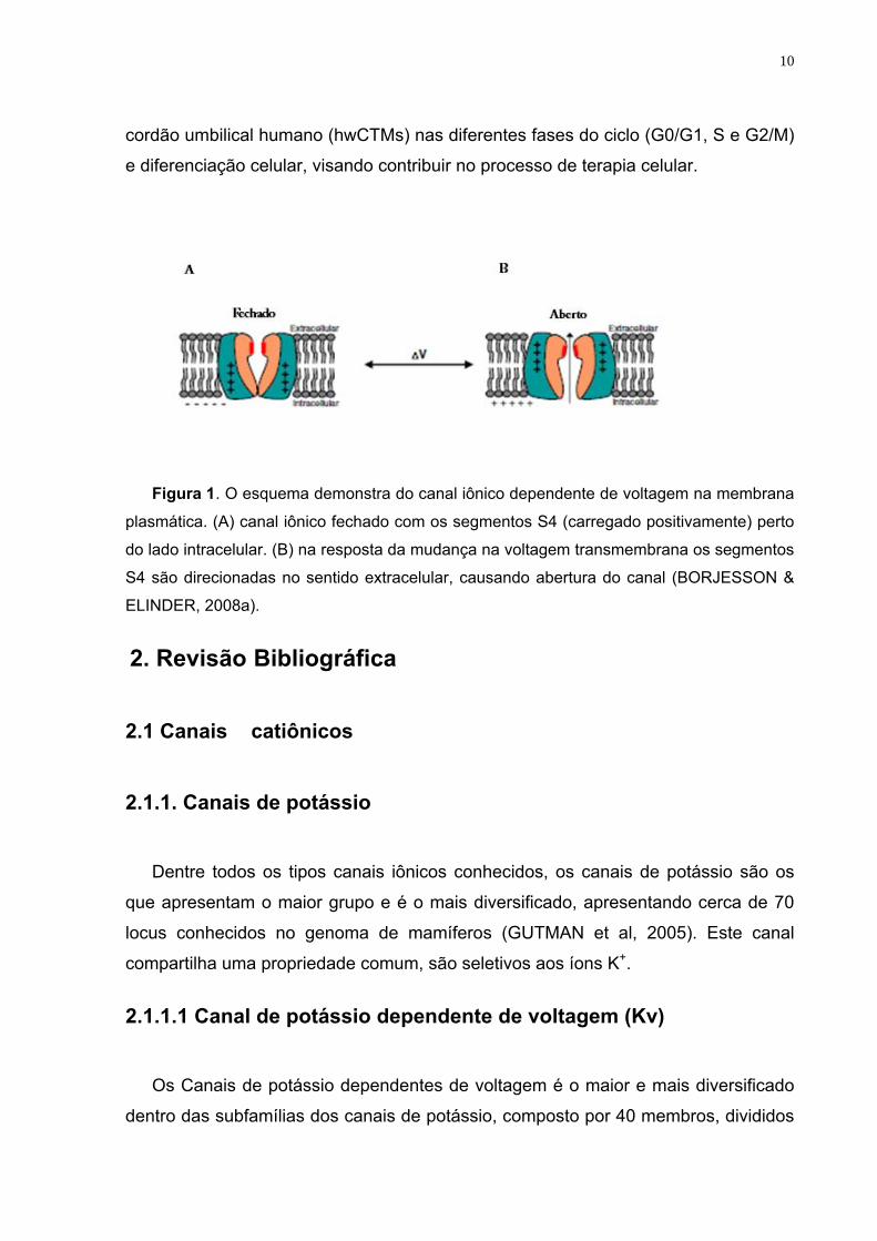
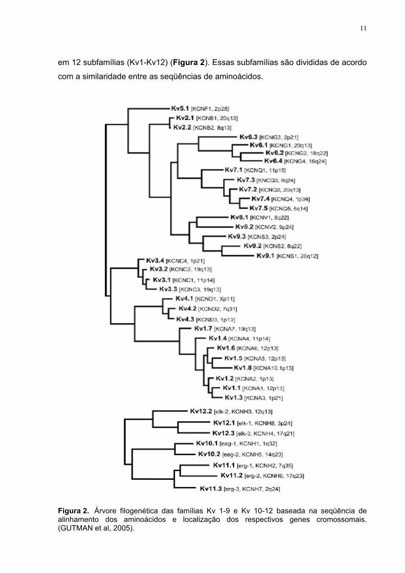
Os Canais de potássio dependentes de voltagem é o maior e mais diversificado
dentro das subfamílias dos canais de potássio, composto por 40 membros, divididos
11
em 12 subfamílias (Kv1-Kv12) (Figura 2). Essas subfamílias são divididas de acordo
com a similaridade entre as seqüências de aminoácidos.
Figura 2. Árvore filogenética das famílias Kv 1-9 e Kv 10-12 baseada na seqüência de alinhamento dos aminoácidos e localização dos respectivos genes cromossomais. (GUTMAN et al, 2005).
12
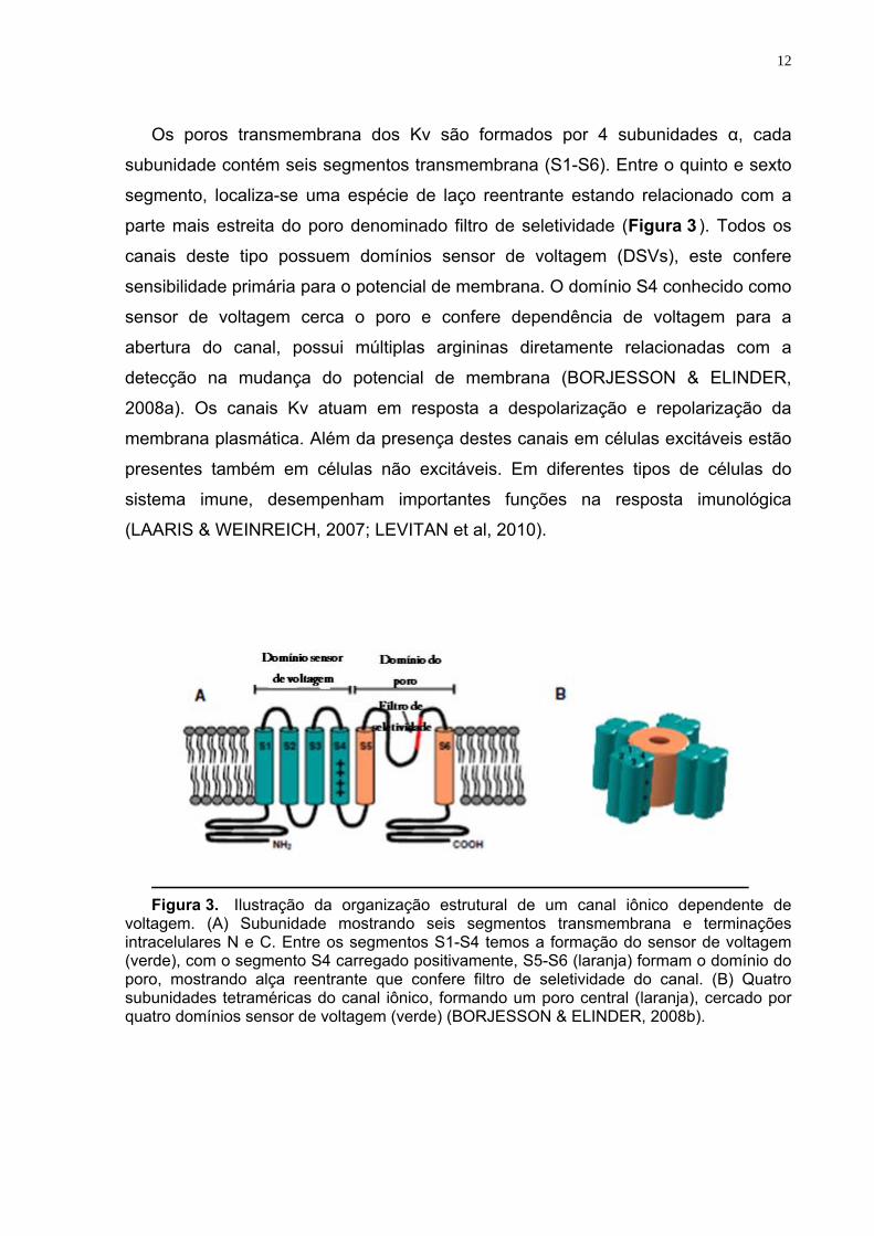
Os poros transmembrana dos Kv são formados por 4 subunidades α, cada
subunidade contém seis segmentos transmembrana (S1-S6). Entre o quinto e sexto
segmento, localiza-se uma espécie de laço reentrante estando relacionado com a
parte mais estreita do poro denominado filtro de seletividade (Figura 3 ). Todos os
canais deste tipo possuem domínios sensor de voltagem (DSVs), este confere
sensibilidade primária para o potencial de membrana. O domínio S4 conhecido como
sensor de voltagem cerca o poro e confere dependência de voltagem para a
abertura do canal, possui múltiplas argininas diretamente relacionadas com a
detecção na mudança do potencial de membrana (BORJESSON & ELINDER,
2008a). Os canais Kv atuam em resposta a despolarização e repolarização da
membrana plasmática. Além da presença destes canais em células excitáveis estão
presentes também em células não excitáveis. Em diferentes tipos de células do
sistema imune, desempenham importantes funções na resposta imunológica
(LAARIS & WEINREICH, 2007; LEVITAN et al, 2010).
Figura 3. Ilustração da organização estrutural de um canal iônico dependente de voltagem. (A) Subunidade mostrando seis segmentos transmembrana e terminações intracelulares N e C. Entre os segmentos S1-S4 temos a formação do sensor de voltagem (verde), com o segmento S4 carregado positivamente, S5-S6 (laranja) formam o domínio do poro, mostrando alça reentrante que confere filtro de seletividade do canal. (B) Quatro subunidades tetraméricas do canal iônico, formando um poro central (laranja), cercado por quatro domínios sensor de voltagem (verde) (BORJESSON & ELINDER, 2008b).
13
2.1.1.2 Canal de potássio ativado pelo cálcio
Canais de potássio dependentes de voltagem e ativados pelo cálcio são também
compostos por quatro subunidades alfa (α) apresentando seis segmentos
transmembrana por cada subunidade (Figura 4 ) (LEVITAN et al, 2010). Esses
canais são subdivididos em dois grupos: canais de potássio de baixa (KCa2.1,
Kca2.2 e Kca2.3) e intermediária (KCa3.1) condutância e canais de potássio de alta
condutância representado por KCa1.1, e seus sinônimos BKca, MaxiK e a.K.a)
(ATKINSON et al, 1991; WEI et al, 2005; WAGNER et al, 2008).
Os quatro subtipos dos canais de potássio de baixa e intermediária condutância
são sensíveis ao cálcio, devido à presença da calmodulina constitutivamente
intracelular, localizada em terminações-carboxil, sendo atuante como ativador da
subunidade β (FANGER et al, 1999; XIA et al, 1998; SCHUMACHER et al, 2001). É
importante ressaltar que estes tipos de canais independem de voltagem, não
apresentando o segmento S4 como sensor de voltagem.
A subunidade α dos canais de potássio de alta condutância possui sete
segmentos transmembrana (Figura 4 ). Estes canais são expressos pelo gene Slo
em animais (ratos e camundongos). No genoma humano é designado como
KCNMA1 (MaxiK) (SALKOFF et al, 2006). Em contraste com os canais de potássio
de pequena e intermediária condutância, canais de alta condutância apresentam
segmento S4 como sensores de voltagem por isso são designados dependentes de
voltagem (DIAZ et al, 1998; HILL et al, 2010). Quatro isoformas da subunidade beta
(β 1-4), cada uma com 2 segmentos transmembranas podem estar associados com
a subunidade α, na proporção 1:1 (TANAKA et al, 2004; KO et al, 2008). Estes
canais são ativados pelo cálcio intracelular e também pela despolarização da
membrana, contribuindo para manutenção do potencial de membrana nos vasos de
pequeno calibre (JACKSON, 2005).
14
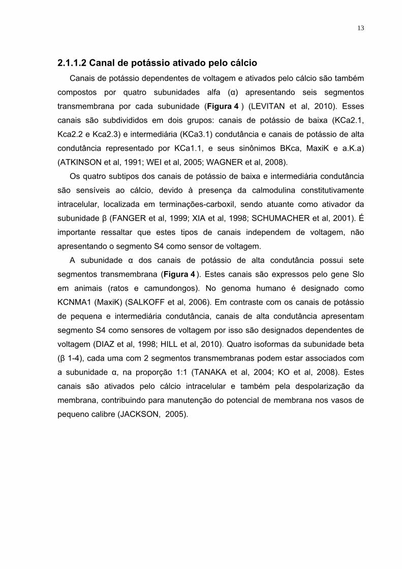
Figura 4. Características estruturais dos canais KCa 3.1, KCa 2.1-3 e KCa 1.1.
(A) Canais KCa 3.1 e KCa 2.1-3, apresentando única subunidade α com seis segmentos transmembrana (S1-S6) e uma laça reentrante entre S5-S6 formadora de poro. A sensibilidade destes canais ao cálcio é conferida pela calmodulina (CAM) ligada a terminações C intracelulares. (B) Subunidade α do KCa 1.1, tendo sete segmentos transmembrana (S0-S6), associada a subunidade β, composta por 2 segmentos transmembrana. (7-10) Terminações C intracelulares bastante longas, contendo segmentos hidrofóbicos, (9-10) são segmentos que confere sensibilidade ao cálcio (GRGIC et al, 2009).
Os canais de potássio ativados pelo cálcio podem ser encontrados em
lugares diversos, com diferentes funções no organismo humano. KCa1.1 regula o
tônus do musculo liso (KHAN et al, 2001; SPROSSMANN et al, 2009), a secreção de
saliva através das glândulas salivares e o nível de eletrólitos ( ROMANENKO et al,
2007; PERRY & SANDLE 2009;). KCa4 encontrado no cérebro e células cardíacas
(BHATTACHARJEE et al, 2005), a expressão do canal KCa5 é restrita aos testículos
(SANTI et al, 2009; SCHREIBER et al, 1998). Os canais de condutância
intermediária KCa3.1 são expressos principalmente em células não excitáveis,
sendo importantes na regulação do volume dos glóbulos vermelhos (BEGENISICH
et al, 2004; VANDORPE et al, 1998) e na ativação dos linfócitos T (BEGENISICH et
al, 2004; GHANSHANI et al, 2000; LOGSDON et al, 1997). Canais de potássio
ativados por cálcio são também expressos no músculo liso e vasos, regulando assim
as funções vasculares (FELETOU, 2009; LEDOUX et al, 2006).
15
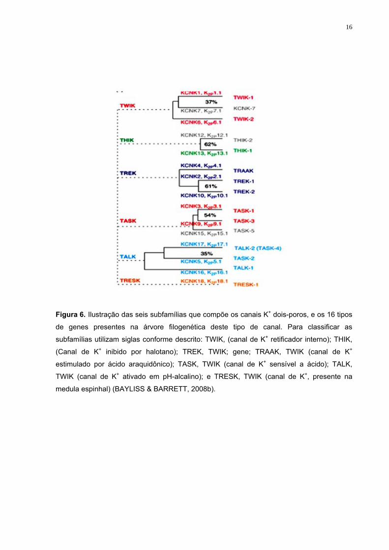
2.1.1.3 Canal de potássio dois poros (K2p)
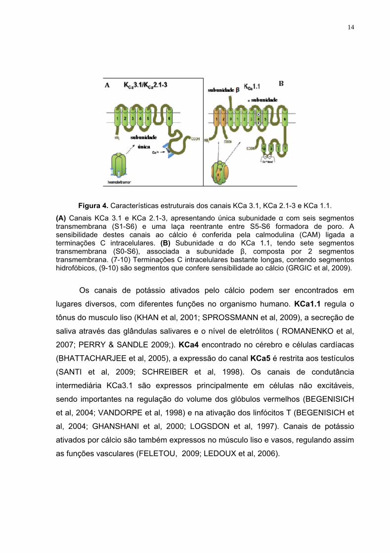
Diferentes famílias de genes amplamente distribuídos modulam o canal do
potássio dois poros, este participa na manutenção do potencial de repouso e
excitabilidade da membrana celular. O canal K2p apresentam 4 segmentos
transmembrana (TM) e duas alças as quais participam na formação do filtro
seletividade deste canal (2P) (Figura 5). Duas subunidades deste canal formam um
canal dimérico, diferentemente de outros tipos de canais de potássio cuja
subunidade alfa possui somente uma alça e precisam quatro subunidades para
formar um poro tetramérico (BAYLISS & BARRETT, 2008a).
Figura 5. Organização estrutural da família dos canais K2P. (A) Subunidade α com dois
laços reentrantes e quatro segmentos transmembrana (BAYLISS & BARRETT, 2008a).
A classificação deste tipo de canal baseia-se nas seqüências homólogas e
características funcionais (Figura 6) (BAYLISS & BARRETT, 2008a). Os canais K2p
estão relacionados com a regulação de fatores físico-químicos, neuroquímico-
endógeno, via de sinalização e drogas de relevância clínica (BAYLISS & BARRETT,
2008a). Na árvore filogenética dos canais de potássio domínio dois-poros, quinze
(15) genes distintos estão presentes. Este tipo de canal é subdividido em seis
grupos: TWIK, THIK, TREK, TASK, TALK e TRESK, conforme ilustrado (BAYLISS &
BARRETT 2008; MILAC et al, 2011).
16
Figura 6. Ilustração das seis subfamílias que compõe os canais K+ dois-poros, e os 16 tipos
de genes presentes na árvore filogenética deste tipo de canal. Para classificar as
subfamílias utilizam siglas conforme descrito: TWIK, (canal de K+ retificador interno); THIK,
(Canal de K+ inibido por halotano); TREK, TWIK; gene; TRAAK, TWIK (canal de K+
estimulado por ácido araquidônico); TASK, TWIK (canal de K+ sensível a ácido); TALK,
TWIK (canal de K+ ativado em pH-alcalino); e TRESK, TWIK (canal de K+, presente na
medula espinhal) (BAYLISS & BARRETT, 2008b).
17
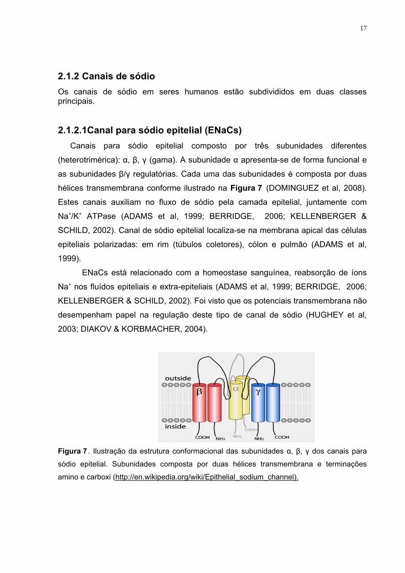
2.1.2 Canais de sódio Os canais de sódio em seres humanos estão subdivididos em duas classes principais. 2.1.2.1Canal para sódio epitelial (ENaCs)
Canais para sódio epitelial composto por três subunidades diferentes
(heterotrimérica): α, β, γ (gama). A subunidade α apresenta-se de forma funcional e
as subunidades β/γ regulatórias. Cada uma das subunidades é composta por duas
hélices transmembrana conforme ilustrado na Figura 7 (DOMINGUEZ et al, 2008).
Estes canais auxiliam no fluxo de sódio pela camada epitelial, juntamente com
Na+/K+ ATPase (ADAMS et al, 1999; BERRIDGE, 2006; KELLENBERGER &
SCHILD, 2002). Canal de sódio epitelial localiza-se na membrana apical das células
epiteliais polarizadas: em rim (túbulos coletores), cólon e pulmão (ADAMS et al,
1999).
ENaCs está relacionado com a homeostase sanguínea, reabsorção de íons
Na+ nos fluídos epiteliais e extra-epiteliais (ADAMS et al, 1999; BERRIDGE, 2006;
KELLENBERGER & SCHILD, 2002). Foi visto que os potenciais transmembrana não
desempenham papel na regulação deste tipo de canal de sódio (HUGHEY et al,
2003; DIAKOV & KORBMACHER, 2004).
Figura 7 . Ilustração da estrutura conformacional das subunidades α, β, γ dos canais para
sódio epitelial. Subunidades composta por duas hélices transmembrana e terminações
amino e carboxi (http://en.wikipedia.org/wiki/Epithelial_sodium_channel).
18
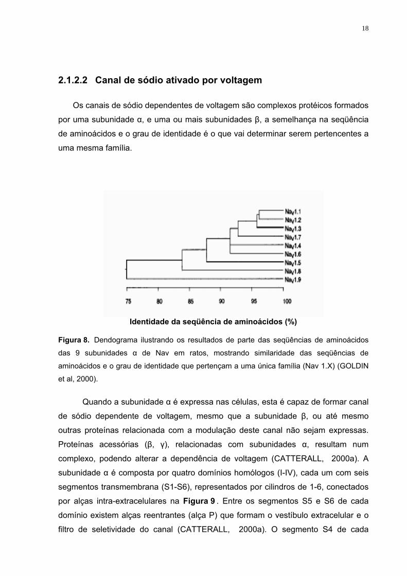
2.1.2.2 Canal de sódio ativado por voltagem
Os canais de sódio dependentes de voltagem são complexos protéicos formados
por uma subunidade α, e uma ou mais subunidades β, a semelhança na seqüência
de aminoácidos e o grau de identidade é o que vai determinar serem pertencentes a
uma mesma família.
Identidade da seqüência de aminoácidos (%)
Figura 8. Dendograma ilustrando os resultados de parte das seqüências de aminoácidos
das 9 subunidades α de Nav em ratos, mostrando similaridade das seqüências de
aminoácidos e o grau de identidade que pertençam a uma única família (Nav 1.X) (GOLDIN
et al, 2000).
Quando a subunidade α é expressa nas células, esta é capaz de formar canal
de sódio dependente de voltagem, mesmo que a subunidade β, ou até mesmo
outras proteínas relacionada com a modulação deste canal não sejam expressas.
Proteínas acessórias (β, γ), relacionadas com subunidades α, resultam num
complexo, podendo alterar a dependência de voltagem (CATTERALL, 2000a). A
subunidade α é composta por quatro domínios homólogos (I-IV), cada um com seis
segmentos transmembrana (S1-S6), representados por cilindros de 1-6, conectados
por alças intra-extracelulares na Figura 9 . Entre os segmentos S5 e S6 de cada
domínio existem alças reentrantes (alça P) que formam o vestíbulo extracelular e o
filtro de seletividade do canal (CATTERALL, 2000a). O segmento S4 de cada
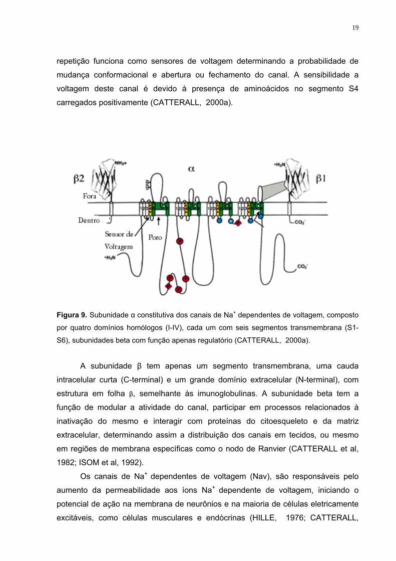
19
repetição funciona como sensores de voltagem determinando a probabilidade de
mudança conformacional e abertura ou fechamento do canal. A sensibilidade a
voltagem deste canal é devido à presença de aminoácidos no segmento S4
carregados positivamente (CATTERALL, 2000a).
Figura 9. Subunidade α constitutiva dos canais de Na+ dependentes de voltagem, composto
por quatro domínios homólogos (I-IV), cada um com seis segmentos transmembrana (S1-
S6), subunidades beta com função apenas regulatório (CATTERALL, 2000a).
A subunidade β tem apenas um segmento transmembrana, uma cauda
intracelular curta (C-terminal) e um grande domínio extracelular (N-terminal), com
estrutura em folha β, semelhante às imunoglobulinas. A subunidade beta tem a
função de modular a atividade do canal, participar em processos relacionados à
inativação do mesmo e interagir com proteínas do citoesqueleto e da matriz
extracelular, determinando assim a distribuição dos canais em tecidos, ou mesmo
em regiões de membrana específicas como o nodo de Ranvier (CATTERALL et al,
1982; ISOM et al, 1992).
Os canais de Na+ dependentes de voltagem (Nav), são responsáveis pelo
aumento da permeabilidade aos íons Na+ dependente de voltagem, iniciando o
potencial de ação na membrana de neurônios e na maioria de células eletricamente
excitáveis, como células musculares e endócrinas (HILLE, 1976; CATTERALL,
20
2000a). Estes canais conduzem os íons em resposta à despolarização da membrana
plasmática, correspondendo ao estado ativado ou condutor. Após a despolarização
da MP e entrada destes íons no meio intracelular, a mesma entra no estado
inativado ou não-condutor (PEARCE & KLEYMAN, 2007).
2.1.3 Canal de Cálcio.
2.1.3.1 Canal de cálcio dependente de voltagem Semelhantemente aos canais de sódio, canais de cálcio dependentes de
voltagem apresentam um complexo de subunidades diferentes α1, α2δ, β1-4, e γ, as
quais são codificadas por múltiplos genes. A subunidade α confere funcionalidade ao
canal e β/γ tem apenas função regulatória (BERRIDGE, 2006). Os canais de cálcio
podem ser regulados por segundo mensageiro, drogas e toxinas (CATTERALL,
2000b).
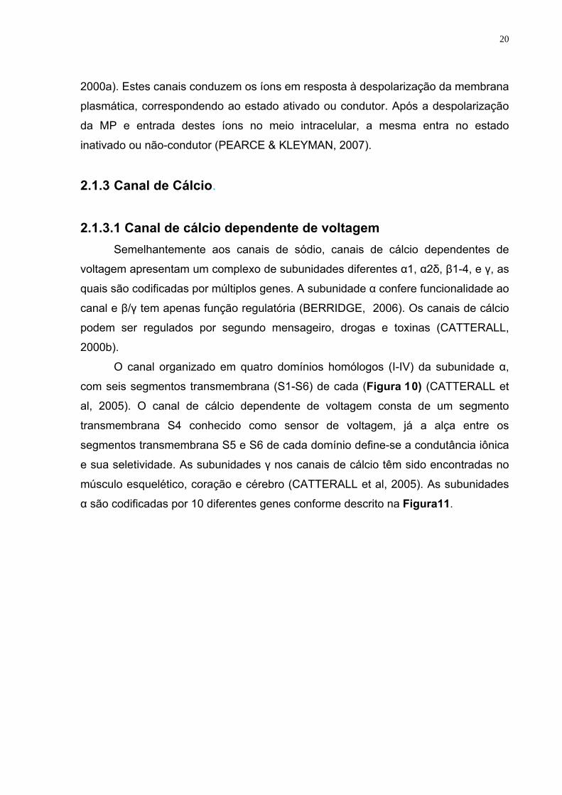
O canal organizado em quatro domínios homólogos (I-IV) da subunidade α,
com seis segmentos transmembrana (S1-S6) de cada (Figura 10) (CATTERALL et
al, 2005). O canal de cálcio dependente de voltagem consta de um segmento
transmembrana S4 conhecido como sensor de voltagem, já a alça entre os
segmentos transmembrana S5 e S6 de cada domínio define-se a condutância iônica
e sua seletividade. As subunidades γ nos canais de cálcio têm sido encontradas no
músculo esquelético, coração e cérebro (CATTERALL et al, 2005). As subunidades
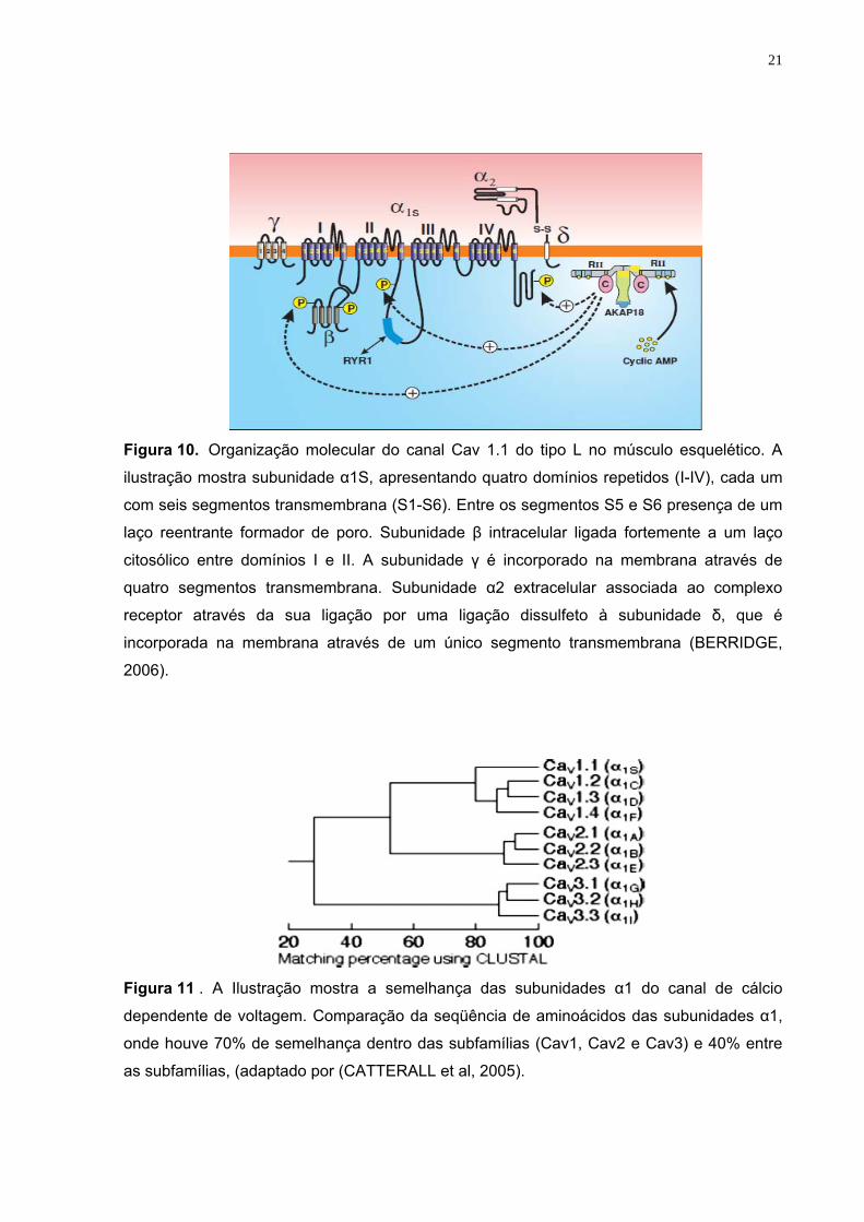
α são codificadas por 10 diferentes genes conforme descrito na Figura11.
21
Figura 10. Organização molecular do canal Cav 1.1 do tipo L no músculo esquelético. A
ilustração mostra subunidade α1S, apresentando quatro domínios repetidos (I-IV), cada um
com seis segmentos transmembrana (S1-S6). Entre os segmentos S5 e S6 presença de um
laço reentrante formador de poro. Subunidade β intracelular ligada fortemente a um laço
citosólico entre domínios I e II. A subunidade γ é incorporado na membrana através de
quatro segmentos transmembrana. Subunidade α2 extracelular associada ao complexo
receptor através da sua ligação por uma ligação dissulfeto à subunidade δ, que é
incorporada na membrana através de um único segmento transmembrana (BERRIDGE,
2006).
Figura 11 . A Ilustração mostra a semelhança das subunidades α1 do canal de cálcio
dependente de voltagem. Comparação da seqüência de aminoácidos das subunidades α1,
onde houve 70% de semelhança dentro das subfamílias (Cav1, Cav2 e Cav3) e 40% entre
as subfamílias, (adaptado por (CATTERALL et al, 2005).
22
Canais de cálcio dependentes de voltagem do tipo L, N, P, Q, R e T são
ativados, após despolarização da membrana plasmática. Canais do tipo L localizam-
se no músculo esquelético, músculo cardíaco, cérebro e retina, e estes estão
relacionados com relaxamento e contração muscular. Canais de cálcio tipo N, P, Q e
R encontram-se nos terminais pré-sinápticos e sua principal função é na liberação de
neurotransmissores. Canais tipo T estão presentes no sistema nervoso, músculo
cardíaco e músculo liso, seu papel é atuar no potencial de ação rítmico das células
musculares, cárdicas e neuronais (PIMENTEL, 2003a).
2.2 Canais aniônicos
Os íon cloreto desempenham um papel relevante na homeostase celular em
condições fisiológicas e patológicas. Estes íons são de maior relevância fisiológica,
já que difunde-se facilmente pelas membranas celulares. Desta forma a variação no
fluxo de Cl- está associado a regulação do volume, processos secretórios e
manutenção do pH celular, essenciais para manterem a atividade enzimática (FOSKETT, 1998; AUZANNEAU et al, 2006). Estudos recentes demonstram que
íons cloreto tem a capacidade de atuar como segundo mensageiro, já que sua
concentração dentro da célula é dinâmica, sendo capaz de modular a atividade de
proteínas transferrina, glicose-6-fosfatase e hemoglobina, dentre outras (CHEN et al,
2010).
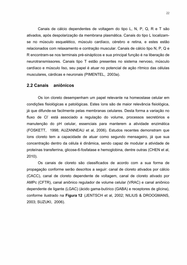
Os canais de cloreto são classificados de acordo com a sua forma de
propagação conforme serão descritos a seguir: canal de cloreto ativados por cálcio
(CACC), canal de cloreto dependente de voltagem, canal de cloreto ativado por
AMPc (CFTR), canal aniônico regulador de volume celular (VRAC) e canal aniônico
dependente de ligante (LGAC) (ácido gama-butírico (GABA) e receptores de glicina),
conforme ilustrado na Figura 12 (JENTSCH et al, 2002; NILIUS & DROOGMANS,
2003; SUZUKI, 2006).
23
Figura 12. Ilustração de diferentes classes de canais aniônicos. Canais de cloreto ativados por cálcio (CACC), canais de cloreto ativados por AMPc (CFTR), canais aniônicos reguladores de volume celular (VRAC) e canais aniônicos dependente de ligante (DURAN et al, 2010a).
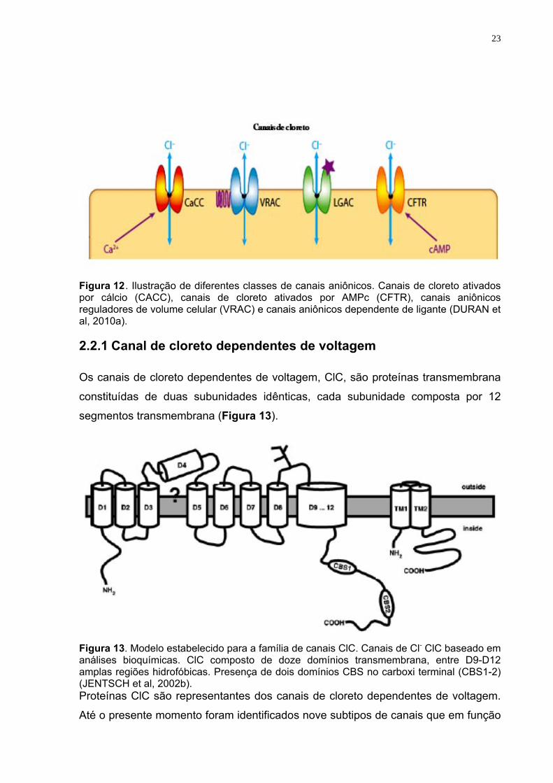
2.2.1 Canal de cloreto dependentes de voltagem Os canais de cloreto dependentes de voltagem, ClC, são proteínas transmembrana
constituídas de duas subunidades idênticas, cada subunidade composta por 12
segmentos transmembrana (Figura 13).
Figura 13. Modelo estabelecido para a família de canais ClC. Canais de Cl- ClC baseado em análises bioquímicas. ClC composto de doze domínios transmembrana, entre D9-D12 amplas regiões hidrofóbicas. Presença de dois domínios CBS no carboxi terminal (CBS1-2) (JENTSCH et al, 2002b). Proteínas ClC são representantes dos canais de cloreto dependentes de voltagem.
Até o presente momento foram identificados nove subtipos de canais que em função
24
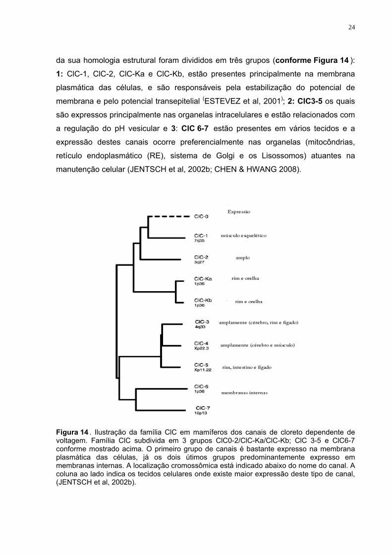
da sua homologia estrutural foram divididos em três grupos (conforme Figura 14 ):
1: ClC-1, ClC-2, ClC-Ka e ClC-Kb, estão presentes principalmente na membrana
plasmática das células, e são responsáveis pela estabilização do potencial de
membrana e pelo potencial transepitelial (ESTEVEZ et al, 2001); 2: ClC3-5 os quais
são expressos principalmente nas organelas intracelulares e estão relacionados com
a regulação do pH vesicular e 3: ClC 6-7 estão presentes em vários tecidos e a
expressão destes canais ocorre preferencialmente nas organelas (mitocôndrias,
retículo endoplasmático (RE), sistema de Golgi e os Lisossomos) atuantes na
manutenção celular (JENTSCH et al, 2002b; CHEN & HWANG 2008).
Figura 14 . Ilustração da família ClC em mamíferos dos canais de cloreto dependente de voltagem. Família ClC subdivida em 3 grupos ClC0-2/ClC-Ka/ClC-Kb; ClC 3-5 e ClC6-7 conforme mostrado acima. O primeiro grupo de canais é bastante expresso na membrana plasmática das células, já os dois útimos grupos predominantemente expresso em membranas internas. A localização cromossômica está indicado abaixo do nome do canal. A coluna ao lado indica os tecidos celulares onde existe maior expressão deste tipo de canal, (JENTSCH et al, 2002b).
25
2.2.2 Canais de cloreto ativados por cálcio (CLCAs)
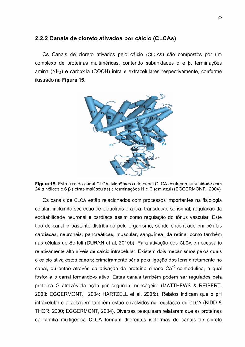
Os Canais de cloreto ativados pelo cálcio (CLCAs) são compostos por um
complexo de proteínas multiméricas, contendo subunidades α e β, terminações
amina (NH2) e carboxila (COOH) intra e extracelulares respectivamente, conforme
ilustrado na Figura 15.
Figura 15. Estrutura do canal CLCA. Monômeros do canal CLCA contendo subunidade com 24 α hélices e 6 β (letras maiúsculas) e terminações N e C (em azul) (EGGERMONT, 2004).
Os canais de CLCA estão relacionados com processos importantes na fisiologia
celular, incluindo secreção de eletrólitos e água, transdução sensorial, regulação da
excitabilidade neuronal e cardíaca assim como regulação do tônus vascular. Este
tipo de canal é bastante distribuído pelo organismo, sendo encontrado em células
cardíacas, neuronais, pancreáticas, muscular, sanguínea, da retina, como também
nas células de Sertoli (DURAN et al, 2010b). Para ativação dos CLCA é necessário
relativamente alto níveis de cálcio intracelular. Existem dois mecanismos pelos quais
o cálcio ativa estes canais; primeiramente séria pela ligação dos íons diretamente no
canal, ou então através da ativação da proteína cinase Ca+2-calmodulina, a qual
fosforila o canal tornando-o ativo. Estes canais também podem ser regulados pela
proteína G através da ação por segundo mensageiro (MATTHEWS & REISERT,
2003; EGGERMONT, 2004; HARTZELL et al, 2005;). Relatos indicam que o pH
intracelular e a voltagem também estão envolvidos na regulação do CLCA (KIDD &
THOR, 2000; EGGERMONT, 2004). Diversas pesquisam relataram que as proteínas
da família multigênica CLCA formam diferentes isoformas de canais de cloreto
26
ativados pelo cálcio nos humanos (hCLCA1-4). Apesar de grandes avanços, até o
presente momento não foi possível elucidar todas as proteínas que compõe a família
dos CLCAs (EGGERMONT, 2004; DURAN et al, 2010b).
2.2.3 Canal CFTR (Regulador de Condutância de Membrana na Fibrose Cística)
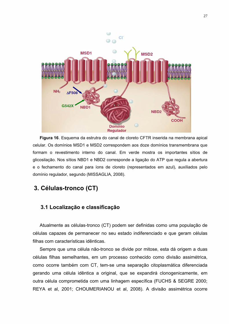
O canal de cloreto CFTR está localizado primariamente na mebrana plasmática
das células sendo este uma via para o movimento de íons Cl- que regula o fluxo
deste através do tecido epitelial. Este canal possui dois domínios transmembrana,
MSD1 e MSD2, com doze segmentos transmembrana cada, dois domínios
citoplasmático de ligação a nucleotídeos, com motivos para ligação do ATP (NBD1 e
NBD2) e um único domínios regulador (R), que controla a atividade do canal de
acordo com seu estado de fosforilação (Figura 16) (OSTEDGAARD et al, 2001).
CFTR participa da regulação do AMP/GMP cíclico, estando diretamente
envolvido no transporte de água e sal em diferentes tecidos, mas é nas células
epiteliais de tecidos com função exócrina como pele, pulmão, pâncreas, glândulas
sudoríparas, fígado, intestino grosso, testículos e ductos deferentes, sua produção é
expressiva, resultando em proteína transmembrana de 1480 aminoácidos, ricamente
glicosilada, que funciona como canal regulador de fluxo do íon cloreto (RIORDAN et
al, 1989; LI et al, 2011).
Alterações no gene CFTR, traz mal funcionamento para o canal, causando uma
doença conhecida como fibrose cística (FC), que pode afetar o pulmão, fígado,
pâncreas e trato gastrointestinal (ROWNTREE & HARRIS, 2003).
27
Figura 16. Esquema da estrutra do canal de cloreto CFTR inserida na membrana apical
celular. Os domínios MSD1 e MSD2 correspondem aos doze domínios transmembrana que
formam o revestimento interno do canal. Em verde mostra os importantes sítios de
glicosilação. Nos sítios NBD1 e NBD2 corresponde a ligação do ATP que regula a abertura
e o fechamento do canal para íons de cloreto (representados em azul), auxíliados pelo
domínio regulador, segundo (MISSAGLIA, 2008).
3. Células-tronco (CT)
3.1 Localização e classificação
Atualmente as células-tronco (CT) podem ser definidas como uma população de
células capazes de permanecer no seu estado indiferenciado e que geram células
filhas com características idênticas.
Sempre que uma célula não-tronco se divide por mitose, esta dá origem a duas
células filhas semelhantes, em um processo conhecido como divisão assimétrica,
como ocorre também com CT, tem-se uma separação citoplasmática diferenciada
gerando uma célula idêntica a original, que se expandirá clonogenicamente, em
outra célula comprometida com uma linhagem específica (FUCHS & SEGRE 2000;
REYA et al, 2001; CHOUMERIANOU et al, 2008). A divisão assimétrica ocorre
28
também no desenvolvimento do zigoto, espermatogênese e oogênese (RAMALHO-
SANTOS & WILLENBRING, 2007).
As CT são classificadas de acordo com sua origem e plasticidade, tabela 1
(AEJAZ et al, 2007; CHOUMERIANOU et al, 2008). As CT embrionárias (CTE)
podem ser: totipotentes (capaz de se diferenciar em qualquer tipo de tecido,
incluindo os anexos extra-embrionários) podendo ser encontradas na fase de zigoto
e primeira clivagem do blastômero; ou pluripotentes (formam todos os tipos de
tecidos exceto os anexos extra-embrionários), encontrada na massa interna do
blastocisto, células do epiblasto (após implantação) e nas células germinativas
primordiais (fase tardia embrionária/início da fetal). As CT adultas têm seu potencial
mais limitado quando comparado com a embrionária, são ditas multipotentes
(originam tipos específicos de tecidos, a partir de uma determinada linhagem), temos
como exemplo as CT hematopoiéticas ou ainda unipotentes (formam apenas um tipo
de tecido celular) (AEJAZ et al, 2007; WATT & DRISKELL 2010).
Tabela 1: Classificação das células-tronco
Quanto à origem Quanto à plasticidade Embrionárias toti ou pluripotentes
Adultas Pluri, multi ou unipotentes
Quanto à origem podem ser embrionárias ou adultas e de acordo com sua plasticidade podem ser totipotentes, capaz de se diferenciar em qualquer tipo de tecido, incluindo os anexos extra-embrionários; pluripotentes formam todos os tipos de tecidos exceto os extra-embrionários, multipotentes originam tipos específicos de tecidos de uma determinada linhagem e unicelulares apenas um tipo de tecido celular.
Células-tronco embrionárias têm a capacidade de se diferenciar em todos
tecidos, além disso, formam derivados dos três folhetos germinativos (ectoderma,
mesoderma e endoderma), mesmo sendo cultivados in vitro (THOMSON et al, 1998;
AEJAZ et al, 2007).Estudos em camundongos e humanos vêm sendo realizados
com CTE, porém questões relacionadas à compatibilidade, segurança e ética, têm
dificultado o uso destas em pesquisas e na terapia celular. Já as células-tronco
adultas são menos indiferenciadas e na maioria das vezes formam tecido-específico,
estas apresentam menor capacidade de rejeição (enxerto versus hospedeiro), porém
ainda existem problemas a serem superados com relação ao seu cultivo e expansão
29
in vitro, limitando-as apenas a função de reparação e homeostase do tecido onde
foram encontradas (CHOUMERIANOU et al, 2008).
Diversos estudos encontraram células-tronco adultas (CTA) em diferentes
órgãos e tecidos, temos como exemplos: cérebro (CLARKE et al, 2000), coração
(MESSINA et al, 2004), pulmões (KIM et al, 2005), fígado (MATTHEWS & YEOH,
2005), pâncreas (KRUSE et al, 2006), rins (AL-AWQATI & OLIVER, 2002), tecido
adiposo (ZUK et al, 2002), músculo esquelético (CHEN & GOLDHAMER, 2003),
decídua dos dentes (MIURA et al, 2003), folículo de cabelos (JAHODA et al, 2003),
pele (JOHNSTON, 2004), sangue periférico (ZHAO et al, 2003), testículos (GUAN et
al, 2006), sangue menstrual (MENG et al, 2007) e líquido amniótico (DE et al, 2007).
CTA ainda podem ser encontradas em fontes menos maduras, tecidos fetais (IN 'T
ANKER et al, 2003), placenta (YEN et al, 2005), sangue de cordão umbilical (KANG
et al, 2006), cordão umbilical, incluindo vasos sanguíneos e geléia de Wharton
(TROYER & WEISS, 2008; SECCO et al, 2009).
3.2 Células-tronco mesenquimais (CTMs) 3.2.1 Isolamento
Células-tronco mesenquimais (CTMs) podem ser isoladas por métodos
enzimáticos ou por cultura em explante. Métodos enzimáticos utilizam enzimas para
digerir a matriz extracelular, e assim facilitar a saída das células para que possam
ser cultivadas (WEISS & TROYER 2006; CAN & KARAHUSEYINOGLU 2007). Já a
cultura em explante, não se faz necessário utilizar nenhum tipo de enzima, apenas
por migração espontânea as células saem dos tecidos, para um ambiente com
condições favoráveis a sua proliferação e crescimento (MITCHELL et al, 2003;
ISHIGE et al, 2009;). As células-tronco mesenquimais obtidas do sangue de cordão
umbilical são separadas por gradiente de densidade por centrifugação. A fração
composta por células mononucleares são cultivadas, e posteriormente as células
não aderentes ao suporte de cultura são desprezadas, pois uma característica das
CTMs é a capacidade de aderir a suportes plásticos (KAWASAKI-OYAMA et al,
2008; TROYER & WEISS 2008).
30
3.2.2 Caracterização As células-tronco mesenquimais não apresentam marcadores específicos.
Devido a esta limitação a Sociedade Internacional de Terapia Celular (SITC),
designou alguns critérios que devem ser seguido para classificar-se CTMs. (a)
ser aderentes ao plástico; (b) ser positivas para diversas proteínas de superfície
(marcadores) incluindo CD73, CD90, CD105, CD44, CD78, CD71, CD271, CD29
e CD44 e negativas para marcadores hematopoiéticos e endoteliais CD31, CD34,
CD45, CD14 ou (CD11b), CD19 ou (CD79-alfa), antígeno leucocitário humano
classe II (HLA-II) e moléculas co-estimulatórias CD80, CD86, CD40 (c) terem
características de auto-renovação e diferenciar-se in vitro em osteoblasto,
adipócitos e condrócitos (DOMINICI et al, 2006; CAN & KARAHUSEYINOGLU,
2007; LA et al, 2009; BEN-AMI et al, 2011).
3.2.3 Proliferação
Células-tronco mesenquimais são multipotentes, não hematopoiéticas, capazes
de se auto-renovar e diferenciar in vitro e in vivo em diferentes linhagens incluindo
adipogênica, condrogênica, osteogênica e miogênica. Embora a principal fonte de
células-tronco mesenquimais seja a medula óssea, pode-se obter este tipo celular de
outros tecidos como músculo esquelético, tecido adiposo, membrana sinovial,
sangue de cordão umbilical e fluídos placentários, entre outros (LA et al, 2009; BEN-
AMI et al, 2011).
3.2.4 Diferenciação
Além da identificação de CTMs com base em características morfológicas e
fenotípicas, outra forma de reconhecer uma suposta população de CTMs é através
da sua capacidade de ser induzida a se diferenciar in vitro em osso, gordura e
cartilagem (CHAMBERLAIN et al, 2007; BEN-AMI et al, 2011).
A metodologia básica para diferenciação de CTMs em osteoblastos envolve
incubar uma monocamada de células mesenquimais, com ácido ascórbico, ß-
glicerofosfato e dexametasona, durante um período de 3 semanas (figura 10 ). As
células diferenciadas apresentam formação de nódulos ou agregados, aumento da
expressão de fosfatase alcalina, podendo com o passar das semanas acumularem
cálcio. Estes nódulos ou agregados ósseos são visualizados pela técnica de
coloração alizarina red ou von Kossa (CHAMBERLAIN et al, 2007, QIAO et al, 2008;
31
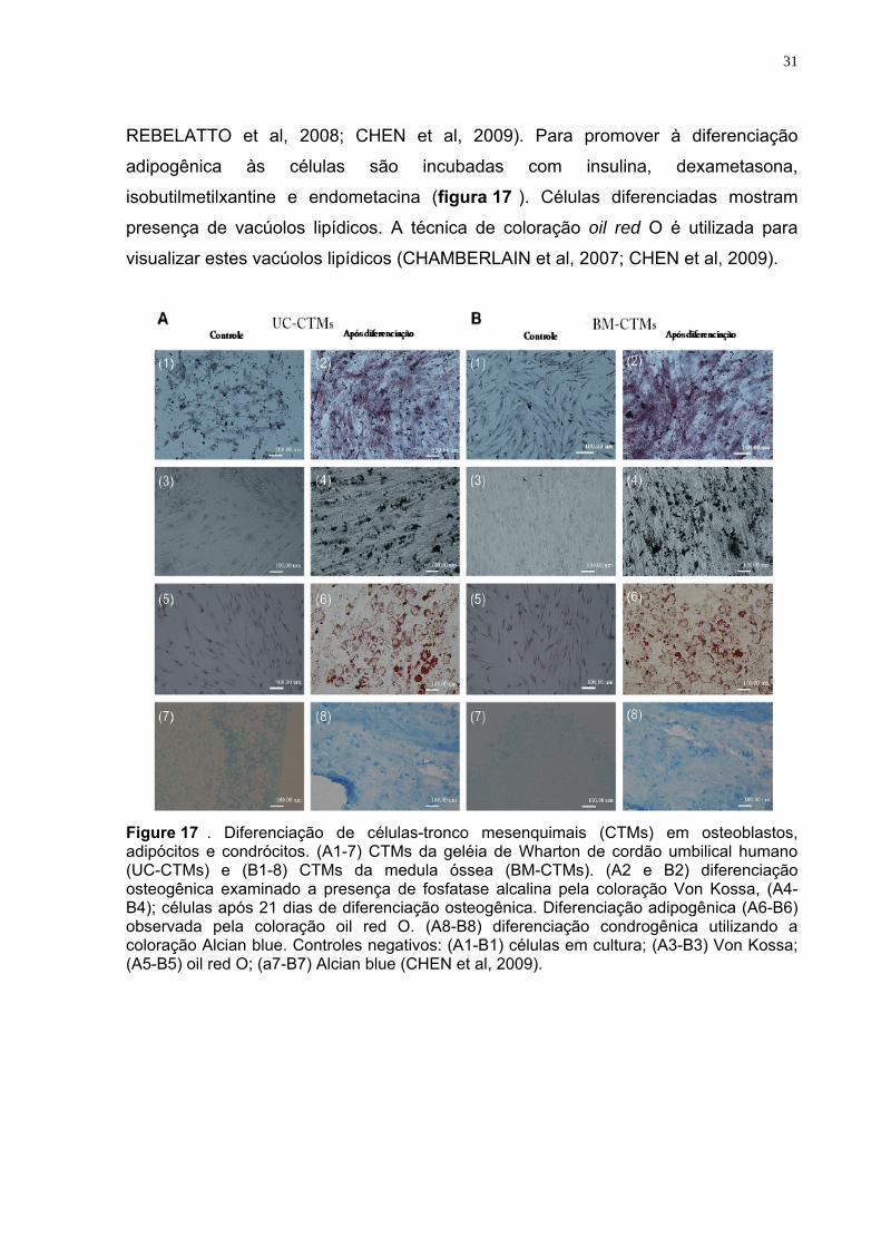
REBELATTO et al, 2008; CHEN et al, 2009). Para promover à diferenciação
adipogênica às células são incubadas com insulina, dexametasona,
isobutilmetilxantine e endometacina (figura 17 ). Células diferenciadas mostram
presença de vacúolos lipídicos. A técnica de coloração oil red O é utilizada para
visualizar estes vacúolos lipídicos (CHAMBERLAIN et al, 2007; CHEN et al, 2009).
Figure 17 . Diferenciação de células-tronco mesenquimais (CTMs) em osteoblastos, adipócitos e condrócitos. (A1-7) CTMs da geléia de Wharton de cordão umbilical humano (UC-CTMs) e (B1-8) CTMs da medula óssea (BM-CTMs). (A2 e B2) diferenciação osteogênica examinado a presença de fosfatase alcalina pela coloração Von Kossa, (A4-B4); células após 21 dias de diferenciação osteogênica. Diferenciação adipogênica (A6-B6) observada pela coloração oil red O. (A8-B8) diferenciação condrogênica utilizando a coloração Alcian blue. Controles negativos: (A1-B1) células em cultura; (A3-B3) Von Kossa; (A5-B5) oil red O; (a7-B7) Alcian blue (CHEN et al, 2009).
32
3.2.4 Células-tronco mesenquimais de cordão umbilical Humano
As células-tronco provenientes de cordão umbilical (CU) apresentam um forte
atrativo para seu uso, quando o interesse for intervir na demanda que existe pela
procura da medula óssea para o uso em terapias celulares, pois de forma parecida o
CU apresenta uma fração hematopoiética e outra mesenquimal (ROCHA &
GLUCKMAN 2006; BAKSH et al, 2007). O CU é um anexo embrionário geralmente
descartado após o parto. Sua coleta é realizada de forma simples, segura e não-
dolorosa; não causando nenhum dano para saúde da mãe nem do recém-nascido
(WEISS & TROYER 2006; CAN & KARAHUSEYINOGLU 2007; SECCO et al, 2008). Estudos relatam comparações entre células-tronco mesenquimais fetais (fCTMs)
e células-tronco adultas (CTAs) quando cultivadas in vitro, estas diferenças serão
descritas a seguir. 1) fCTMs apresentam maior capacidade de expansão e rápido
período de duplicação comparadas com CTAs, estas características podem ser
devido ao tamanho dos telômeros, que nas fCTMs são mais longos (GUILLOT et al,
2007; TROYER & WEISS, 2008). 2) células fetais parecem não ter propriedades de
rejeição, porém estas propriedades são relatadas nas CTAs (GOTHERSTROM et al,
2005); 3) fCTMs não apresentam antígeno leucocitário humano de classe II (HLA),
mas sintetizam HLA-G, em contraste com CTAs (GOTHERSTROM et al,
2003,GOTHERSTROM et al, 2005) e 4) fCTMs expressam diferentes tipos de
citocinas, compatíveis com CTAs. Estas diferenças e similaridades foram
observadas entre cordão umbilical (fCTMs) e sangue periférico de adultos (CTAs)
(TROYER & WEISS, 2008).
CTMs tem sido isoladas de diversos compartimentos do cordão umbilical, como
sangue de cordão umbilical, veia subendotelial e geléia de Wharton.
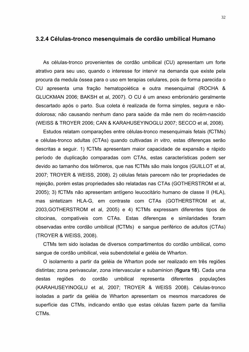
O isolamento a partir da geléia de Wharton pode ser realizado em três regiões
distintas; zona perivascular, zona intervascular e subaminion (figura 18). Cada uma
destas regiões do cordão umbilical representa diferentes populações
(KARAHUSEYINOGLU et al, 2007; TROYER & WEISS 2008). Células-tronco
isoladas a partir da geléia de Wharton apresentam os mesmos marcadores de
superfície das CTMs, indicando então que estas células fazem parte da família
CTMs.
33
Figura 18 . Representação de um corte transversal do cordão umbilical humano, contendo células-tronco mesenquimais. CTMs podem ser isoladas da geléia de Wharton a partir de três compartimentos diferentes; zona perivascular (3), zona intervascular (4) e subaminion (5), conforme ilustrado na figura acima (TROYER & WEISS 2008).
4 Ciclo celular
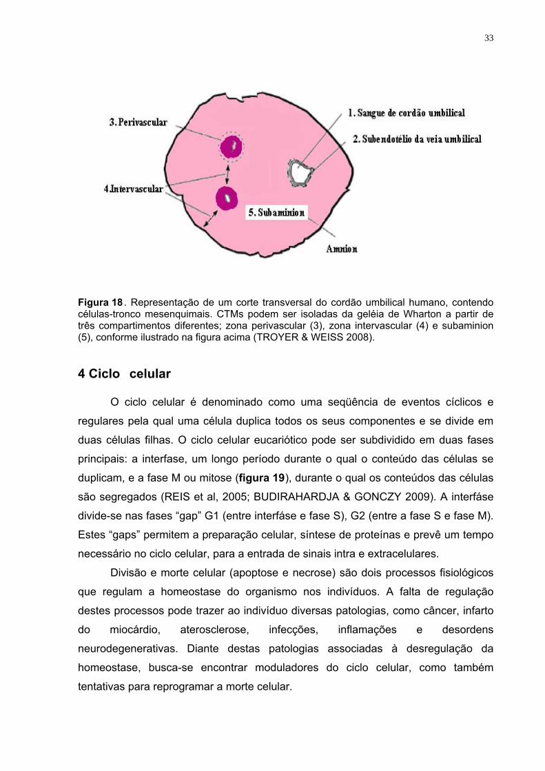
O ciclo celular é denominado como uma seqüência de eventos cíclicos e
regulares pela qual uma célula duplica todos os seus componentes e se divide em
duas células filhas. O ciclo celular eucariótico pode ser subdividido em duas fases
principais: a interfase, um longo período durante o qual o conteúdo das células se
duplicam, e a fase M ou mitose (figura 19), durante o qual os conteúdos das células
são segregados (REIS et al, 2005; BUDIRAHARDJA & GONCZY 2009). A interfáse
divide-se nas fases “gap” G1 (entre interfáse e fase S), G2 (entre a fase S e fase M).
Estes “gaps” permitem a preparação celular, síntese de proteínas e prevê um tempo
necessário no ciclo celular, para a entrada de sinais intra e extracelulares.
Divisão e morte celular (apoptose e necrose) são dois processos fisiológicos
que regulam a homeostase do organismo nos indivíduos. A falta de regulação
destes processos pode trazer ao indivíduo diversas patologias, como câncer, infarto
do miocárdio, aterosclerose, infecções, inflamações e desordens
neurodegenerativas. Diante destas patologias associadas à desregulação da
homeostase, busca-se encontrar moduladores do ciclo celular, como também
tentativas para reprogramar a morte celular.
34
A integridade genômica, proliferação e sobrevivência celular, são reguladas
por “checkpoints” no ciclo celular. A perda no reparo ao DNA e morte celular
programada, podem acarretar em crescimento celular descontrolado.
Como resposta dos danos ao DNA, as células ativam uma rede complexa de
fatores conhecidas como quinases dependentes de ciclinas, estas retardam ou
detêm a progressão do ciclo celular (vias de checkpoint), promovendo assim a
reparação ao DNA ou em casos de danos irreparáveis as células são eliminadas
(ZHIVOTOVSKY & ORRENIUS, 2010).
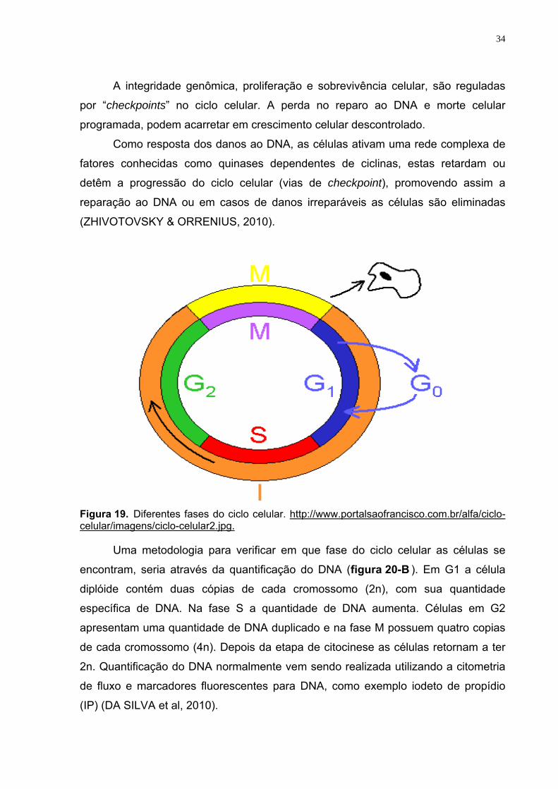
Figura 19. Diferentes fases do ciclo celular. http://www.portalsaofrancisco.com.br/alfa/ciclo-celular/imagens/ciclo-celular2.jpg.
Uma metodologia para verificar em que fase do ciclo celular as células se
encontram, seria através da quantificação do DNA (figura 20-B ). Em G1 a célula
diplóide contém duas cópias de cada cromossomo (2n), com sua quantidade
específica de DNA. Na fase S a quantidade de DNA aumenta. Células em G2
apresentam uma quantidade de DNA duplicado e na fase M possuem quatro copias
de cada cromossomo (4n). Depois da etapa de citocinese as células retornam a ter
2n. Quantificação do DNA normalmente vem sendo realizada utilizando a citometria
de fluxo e marcadores fluorescentes para DNA, como exemplo iodeto de propídio
(IP) (DA SILVA et al, 2010).
35
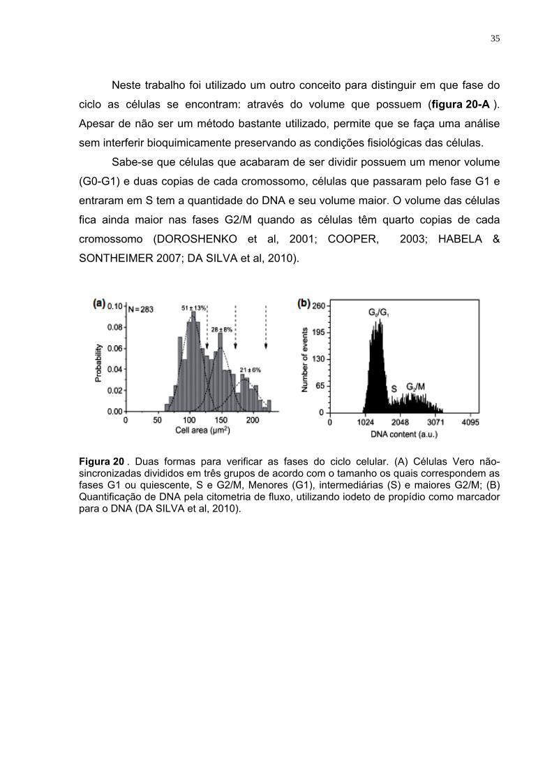
Neste trabalho foi utilizado um outro conceito para distinguir em que fase do
ciclo as células se encontram: através do volume que possuem (figura 20-A ).
Apesar de não ser um método bastante utilizado, permite que se faça uma análise
sem interferir bioquimicamente preservando as condições fisiológicas das células.
Sabe-se que células que acabaram de ser dividir possuem um menor volume
(G0-G1) e duas copias de cada cromossomo, células que passaram pelo fase G1 e
entraram em S tem a quantidade do DNA e seu volume maior. O volume das células
fica ainda maior nas fases G2/M quando as células têm quarto copias de cada
cromossomo (DOROSHENKO et al, 2001; COOPER, 2003; HABELA &
SONTHEIMER 2007; DA SILVA et al, 2010).
Figura 20 . Duas formas para verificar as fases do ciclo celular. (A) Células Vero não-sincronizadas divididos em três grupos de acordo com o tamanho os quais correspondem as fases G1 ou quiescente, S e G2/M, Menores (G1), intermediárias (S) e maiores G2/M; (B) Quantificação de DNA pela citometria de fluxo, utilizando iodeto de propídio como marcador para o DNA (DA SILVA et al, 2010).
36
5. OBJETIVOS Geral
Analisar de forma qualitativa e quantitativa a expressão dos canais de
potássio, MaxiK e Heag1, e os canais aniônicos, pl-VDAC e CLCA1, na
células-tronco mesenquimais da geléia de Wharton de cordão umbilical
humano. Específicos
Isolar e caracterizar as células-tronco mesenquimais da geléia de Wharton de
cordão umbilical humano;
Induzir a diferenciação osteogênica e adipogênica;
Utilizar as células unitárias e RT-PCR em tempo real para determinar a
expressão qualitativa e quantitativa dos canais iônicos MaxiK, Heag1, pl-
VDAC e CLCA1 nas diferentes fases do ciclo celular e durante as
diferenciações osteogênica e adipogênica.
37
6. REFERÊNCIAS BIBLIOGRÁFICAS ADAMS, C. M. Tetraethylammonium block of the BNC1 channel. Biophys.J., 76(3): 1377-1383, 1999.
AEJAZ, H. M. Stem cell therapy-present status. Transplant.Proc., 39(3): 694-699, 2007.
AGRE, P..Aquaporin null phenotypes: the importance of classical physiology. Proc.Natl.Acad.Sci.U.S.A, 95(16): 9061-9063, 1998.
AL-AWQATI, Q. & OLIVER,J.A. Stem cells in the kidney. Kidney Int., 61(2): 387-395, 2002.
ATKINSON, N. S. & ROBERTSON,G.A.. GANETZKY,B. A component of calcium-activated potassium channels encoded by the Drosophila slo locus. Science, 253(5019): 551-555, 1991.
AUZANNEAU, C. Pharmacological profile of inhibition of the chloride channels activated
by extracellular acid in cultured rat Sertoli cells. Reprod.Nutr.Dev., 46(3): 241-255, 2006.
BAKSH, D. & YAO,R.. TUAN,R.S. Comparison of proliferative and multilineage differentiation potential of human mesenchymal stem cells derived from umbilical cord and bone marrow. Stem Cells, 25(6): 1384-1392, 2007.
BAYLISS, D. A. & BARRETT,P.Q. Emerging roles for two-pore-domain potassium channels and their potential therapeutic impact. Trends Pharmacol.Sci., 29(11): 566-575, 2008a.
BEGENISICH, T. Physiological roles of the intermediate conductance, Ca2+-activated potassium channel Kcnn4. J.Biol.Chem., 279(46): 47681-47687, 2004.
BEN-AMI, E. & BERRIH-AKNIN,S.. MILLER,A. Mesenchymal stem cells as an immunomodulatory therapeutic strategy for autoimmune diseases. Autoimmun.Rev., 2011.
BENGA, G..Water channel proteins (later called aquaporins) and relatives: past, present, and future. IUBMB.Life, 61(2): 112-133, 2009
BENGA, G..Water channel proteins: from their discovery in 1985 in Cluj-Napoca, Romania, to the 2003 Nobel Prize in Chemistry. Cell Mol.Biol.(Noisy.-le-grand), 52(7): 10-19, 2006a.
BENGA, I..Priorities in the discovery of the implications of water channels in epilepsy and duchenne muscular dystrophy. Cell Mol.Biol.(Noisy.-le-grand), 52(7): 46-50, 2006b.
BERRIDGE, M. J..Calcium microdomains: organization and function. Cell Calcium, 40(5-6): 405-412, 2006.
BHATTACHARJEE, A. Localization of the Na+-activated K+ channel Slick in the rat central nervous system. J.Comp Neurol., 484(1): 80-92, 2005.
38
BORJESSON, S. I. & ELINDER, F. Structure, function, and modification of the voltage sensor in voltage-gated ion channels. Cell Biochem.Biophys., 52(3): 149-174, 2008a.
BUDIRAHARDJA, Y. & GONCZY, P. Coupling the cell cycle development. Development,
136(17): 2861-2872, 2009.
CAN, A. & KARAHUSEYINOGLU, S. Concise review: human umbilical cord stroma with regard to the source of fetus-derived stem cells. Stem Cells, 25(11): 2886-2895, 2007.
CATTERALL, W. A. & HARTSHORNE, R.P.. BENESKI,D.A. Molecular Properties of neurotoxin receptors sites associated with sodium channels from mammalian brain. Toxicon, 20(1): 27-40, 1982.
CATTERALL, W. A. International Union of Pharmacology. XLVIII. Nomenclature and structure-function relationships of voltage-gated calcium channels. Pharmacol.Rev., 57(4): 411-425, 2005.
CATTERALL, W.A. From ionic currents to molecular mechanisms: the structure and function of voltage-gated sodium channels. Neuron, 26(1): 13-25, 2000a.
CATTERALL, W.A. Structure and regulation of voltage-gated Ca2+ channels. Annu.Rev.Cell Dev.Biol., 16): 521-555, 2000b.
CHAMBERLAIN, G. Concise review: mesenchymal stem cells: their phenotype, differentiation capacity, immunological features, and potential for homing. Stem Cells, 25(11): 2739-2749, 2007.
CHEN, J. C. & GOLDHAMER, D.J. Skeletal muscle stem cells. Reprod.Biol.Endocrinol., 1): 101- 2003.
CHEN, M. Y. Endothelial differentiation of Wharton's jelly-derived mesenchymal stem cells in comparison with bone marrow-derived mesenchymal stem cells. Exp.Hematol., 37(5): 629-640, 2009.
CHEN, T. Y. & HWANG, T.C. CLC-0 and CFTR: chloride channels evolved from transporters. Physiol Rev., 88(2): 351-387, 2008.
CHEN, Y. H. Homologue structure of the SLAC1 anion channel for closing stomata in leaves. Nature, 467(7319): 1074-1080, 2010.
CHOUMERIANOU, D. M. & DIMITRIOU,H.. KALMANTI, M. Stem cells: promises versus limitations. Tissue Eng Part B Rev., 14(1): 53-60, 2008.
CLARKE, D. L. Generalized potential of adult neural stem cells. Science, 288(5471): 1660-1663, 2000.
COOPER, S. Rethinking synchronization of mammalian cells for cell cycle analysis. Cell Mol.Life Sci., 60(6): 1099-1106, 2003.
DA SILVA, M. B et al. Ion channels in volume regulation of clonal kidney cells. Cell Prolif., 43(6): 529-541, 2010.
39
DE, C.P. Isolation of amniotic stem cell lines with potential for therapy. Nat.Biotechnol., 25(1): 100-106, 2007.
DIAKOV, A. & KORBMACHER, C. A novel pathway of epithelial sodium channel activation involves a serum- and glucocorticoid-inducible kinase consensus motif in the C terminus of the channel's alpha-subunit. J.Biol.Chem., 279(37): 38134-38142, 2004.
DIAZ, L. Role of the S4 segment in a voltage-dependent calcium-sensitive potassium (hSlo) channel. J.Biol.Chem., 273(49): 32430-32436, 1998.
DOMINGUEZ, J. N. Tissue distribution and subcellular localization of the cardiac sodium channel during mouse heart development. Cardiovasc.Res., 78(1): 45-52, 2008.
DOMINICI, M. Minimal criteria for defining multipotent mesenchymal stromal cells. The International Society for Cellular Therapy position statement. Cytotherapy., 8(4): 315-317, 2006.
DOROSHENKO, P. & SABANOV,V.. DOROSHENKO,N. Cell cycle-related changes in regulatory volume decrease and volume-sensitive chloride conductance in mouse fibroblasts. J Cell Physiol, 187(1): 65-72, 2001.
DURAN, C et al. Chloride Channels: Often Enigmatic, Rarely Predictable. Annual Review of Physiology, 72): 95-121, 2010a.
EGGERMONT, J. Calcium-activated chloride channels. Proc.Am.Thorac.Soc., 1(1): 22-27, 2004.
ESTEVEZ, R. Barttin is a Cl- channel beta-subunit crucial for renal Cl- reabsorption and inner ear K+ secretion. Nature, 414(6863): 558-561, 2001.
FANGER, C. M. Calmodulin mediates calcium-dependent activation of the intermediate conductance KCa channel, IKCa1. J.Biol.Chem., 274(9): 5746-5754, 1999.
FELETOU, M..Calcium-activated potassium channels and endothelial dysfunction: therapeutic options? Br.J.Pharmacol., 156(4): 545-562, 2009.
FOSKETT, J. K. ClC and CFTR chloride channel gating. Annu.Rev.Physiol, 60): 689-717, 1998.
FUCHS, E. & SEGRE,J.A. Stem cells: a new lease on life. Cell, 100(1): 143-155, 2000.
GHANSHANI, S. Up-regulation of the IKCa1 potassium channel during T-cell activation. Molecular mechanism and functional consequences. J.Biol.Chem., 275(47): 37137-37149, 2000.
GOLDIN, A. L. Nomenclature of voltage-gated sodium channels. Neuron, 28(2): 365-368, 2000.
GOTHERSTROM, C. Difference in gene expression between human fetal liver and adult bone marrow mesenchymal stem cells. Haematologica, 90(8): 1017-1026, 2005.
40
GOTHERSTROM, C. Immunomodulatory effects of human foetal liver-derived mesenchymal stem cells. Bone Marrow Transplant., 32(3): 265-272, 2003.
GRGIC, I. Blockade of T-lymphocyte KCa3.1 and Kv1.3 channels as novel immunosuppression strategy to prevent kidney allograft rejection. Transplant.Proc., 41(6): 2601-2606, 2009.
GUAN, K. Pluripotency of spermatogonial stem cells from adult mouse testis. Nature, 440(7088): 1199-1203, 2006.
GUILLOT, P. V. Human first-trimester fetal MSC express pluripotency markers and grow faster and have longer telomeres than adult MSC. Stem Cells, 25(3): 646-654, 2007.
GUTMAN, G. A. International Union of Pharmacology. LIII. Nomenclature and molecular relationships of voltage-gated potassium channels. Pharmacol.Rev., 57(4): 473-508, 2005.
HABELA, C. W. & SONTHEIMER,H. Cytoplasmic volume condensation is an integral part of mitosis. Cell Cycle, 6(13): 1613-1620, 2007. HARTZELL, C. & PUTZIER,I.. ARREOLA,J. Calcium-activated chloride channels. Annu.Rev.Physiol, 67): 719-758, 2005
HILL, M. A. Large conductance, Ca2+-activated K+ channels (BKCa) and arteriolar myogenic signaling. FEBS Lett., 584(10): 2033-2042, 2010.
HILLE, B. & ARMSTRONG,C.M.. MACKINNON,R. Ion channels: from idea to reality. Nat.Med., 5(10): 1105-1109, 1999.
HILLE, B..Gating in sodium channels of nerve. Annu.Rev.Physiol, 38): 139-152, 1976.
HOFFMANN, E. K. & LAMBERT,I.H.. PEDERSEN,S.F. Physiology of cell volume regulation in vertebrates. Physiol Rev., 89(1): 193-277, 2009.
HUGHEY, R. P. Maturation of the epithelial Na+ channel involves proteolytic processing of the alpha- and gamma-subunits. J.Biol.Chem., 278(39): 37073-37082, 2003.
IN 'T ANKER, P. S. Mesenchymal stem cells in human second-trimester bone marrow, liver, lung, and spleen exhibit a similar immunophenotype but a heterogeneous multilineage differentiation potential. Haematologica, 88(8): 845-852, 2003.
ISHIBASHI, K. & HARA,S.. KONDO,S. Aquaporin water channels in mammals. Clin.Exp.Nephrol., 13(2): 107-117, 2009.
ISHIGE, I. Comparison of mesenchymal stem cells derived from arterial, venous, and Wharton's jelly explants of human umbilical cord. Int.J Hematol., 90(2): 261-269, 2009;
ISOM, L. L. Primary structure and functional expression of the beta 1 subunit of the rat brain sodium channel. Science, 256(5058): 839-842, 1992;
JACKSON, W. F..Potassium channels and proliferation of vascular smooth muscle cells. Circ.Res., 97(12): 1211-1212, 2005.
41
JAHODA, C. A. Hair follicle dermal cells differentiate into adipogenic and osteogenic lineages. Exp.Dermatol., 12(6): 849-859, 20
JENTSCH, T. J. Molecular structure and physiological function of chloride channels. Physiol Rev., 82(2): 503-568, 2002a.
JOHNSTON, N..Skin stem cells. Drug Discov.Today, 9(23): 994- 2004.
KANG, X. Q. Differentiating characterization of human umbilical cord blood-derived mesenchymal stem cells in vitro. Cell Biol.Int., 30(7): 569-575, 2006.
KARAHUSEYINOGLU, S. Biology of stem cells in human umbilical cord stroma: in situ and in vitro surveys. Stem Cells, 25(2): 319-331, 2007.
KAWASAKI-OYAMA, R. S. [Blood mesenchymal stem cell culture from the umbilical cord with and without Ficoll-Paque density gradient method]. Rev.Bras Cir.Cardiovasc., 23(1): 29-34, 2008.
KELLENBERGER, S. & SCHILD,L. Epithelial sodium channel/degenerin family of ion channels: a variety of functions for a shared structure. Physiol Rev., 82(3): 735-767, 2002.
KHAN, R. N. Potassium channels in the human myometrium. Exp.Physiol, 86(2): 255-264, 2001.
KIDD, J. F. & THORN,P. Intracellular Ca2+ and Cl- channel activation in secretory cells. Annu.Rev.Physiol, 62): 493-513, 2000.
KIM, C. F. Identification of bronchioalveolar stem cells in normal lung and lung cancer. Cell, 121(6): 823-835, 2005.
KO, E. A. Physiological roles of K+ channels in vascular smooth muscle cells. J.Smooth Muscle Res., 44(2): 65-81, 2008.
KRUSE, C. Adult pancreatic stem/progenitor cells spontaneously differentiate in vitro into multiple cell lineages and form teratoma-like structures. Ann.Anat., 188(6): 503-517, 2006.
LA, R. G. Isolation and characterization of Oct-4+/HLA-G+ mesenchymal stem cells from human umbilical cord matrix: differentiation potential and detection of new markers. Histochem.Cell Biol., 131(2): 267-282, 2009.
LAARIS, N. & WEINREICH,D. Prostaglandin E2 depresses solitary tract-mediated synaptic transmission in the nucleus tractus solitarius. Neuroscience, 146(2): 792-801, 2007.
LEDOUX, J. Calcium-activated potassium channels and the regulation of vascular tone. Physiology.(Bethesda.), 21): 69-78, 2006.
LESAGE, F. & LAZDUNSKI,M. Molecular and functional properties of two-pore-domain potassium channels. Am.J Physiol Renal Physiol, 279(5): F793-F801, 2000.
LEVITAN, I. Cholesterol and ion channels. Subcell.Biochem., 51): 509-549, 2010.
42
LI, M. S. Regulation of CFTR chloride channel macroscopic conductance by extracellular bicarbonate. Am.J.Physiol Cell Physiol, 300(1): C65-C74, 2011.
LOGSDON, N. J. A novel gene, hKCa4, encodes the calcium-activated potassium channel in human T lymphocytes. J.Biol.Chem., 272(52): 32723-32726, 1997.
MARIANGELA TUZZOLO MISSAGLIA. Pesquisa de mutações no gene CFTR (Cystic Fibrosis Transmembrane Conductance Regulator) em homens brasileiros infertéis portadores de ausência congênita dos ductos deferentes (CAVD).): 1-23, 2008.
MATTHEWS, H. R. & REISERT,J. Calcium, the two-faced messenger of olfactory transduction and adaptation. Curr.Opin.Neurobiol., 13(4): 469-475, 2003.
MATTHEWS, V. B. & YEOH,G.C. Liver stem cells. IUBMB.Life, 57(8): 549-553, 2005.
MENG, X. Endometrial regenerative cells: a novel stem cell population. J.Transl.Med., 5): 57- 2007.
MESSINA, E. Isolation and expansion of adult cardiac stem cells from human and murine heart. Circ.Res., 95(9): 911-921, 2004.
MILAC, A. Structural models of TREK channels and their gating mechanism. Channels (Austin.), 5(1) 2011
MITCHELL, K. E. Matrix cells from Wharton's jelly form neurons and glia. Stem Cells, 21(1): 50-60, 2003.
MIURA, M. SHED: stem cells from human exfoliated deciduous teeth. Proc.Natl.Acad.Sci.U.S.A, 100(10): 5807-5812, 2003.
NILIUS, B. & DROOGMANS,G. Amazing chloride channels: an overview. Acta Physiol Scand., 177(2): 119-147, 2003
NILIUS, B. & EGGERMONT,J.. DROOGMANS,G. The endothelial volume-regulated anion channel, VRAC. Cell Physiol Biochem., 10(5-6): 313-320, 2000.
OSTEDGAARD, L. S. & BALDURSSON,O.. WELSH,M.J. Regulation of the cystic fibrosis transmembrane conductance regulator Cl- channel by its R domain. J.Biol.Chem., 276(11): 7689-7692, 2001.
PARK, K. S. Functional expression of ion channels in mesenchymal stem cells derived from umbilical cord vein. Stem Cells, 25(8): 2044-2052, 2007.
PEARCE, D. & KLEYMAN,T.R. Salt, sodium channels, and SGK1. J.Clin.Invest, 117(3): 592-595, 2007.
PERRY, M. D. & SANDLE,G.I. Regulation of colonic apical potassium (BK) channels by cAMP and somatostatin. Am.J.Physiol Gastrointest.Liver Physiol, 297(1): G159-G167, 2009.
43
PRESTON, C. L. & CALENZO,M.A.. DUBINSKY,W.P. Isolation of a chloride channel-enriched membrane fraction from tracheal and renal epithelia. Am.J.Physiol, 263(4 Pt 1): C879-C887, 1992.
QIAO, C. Human mesenchymal stem cells isolated from the umbilical cord. Cell Biol.Int., 32(1): 8-15, 2008.
RAMALHO-SANTOS, M. & WILLENBRING,H. On the origin of the term "stem cell". Cell Stem Cell, 1(1): 35-38, 2007.
REBELATTO, C. K. Dissimilar differentiation of mesenchymal stem cells from bone marrow, umbilical cord blood, and adipose tissue. Exp.Biol.Med.(Maywood.), 233(7): 901-913, 2008.
REIS, V. C. Cell cycle, DNA replication, repair, and recombination in the dimorphic human pathogenic fungus Paracoccidioides brasiliensis. Genet.Mol.Res., 4(2): 232-250, 2005.
REYA, T. Stem cells, cancer, and cancer stem cells. Nature, 414(6859): 105-111, 2001.
RIORDAN, J. R. Identification of the cystic fibrosis gene: cloning and characterization of complementary DNA. Science, 245(4922): 1066-1073, 1989.
ROCHA, V. & GLUCKMAN,E. Clinical use of umbilical cord blood hematopoietic stem cells. Biol.Blood Marrow Transplant., 12(1 Suppl 1): 34-41, 2006.
ROMANENKO, V. G. Regulation of membrane potential and fluid secretion by Ca2+-activated K+ channels in mouse submandibular glands. J.Physiol, 581(Pt 2): 801-817, 2007.
ROWNTREE, R. K. & HARRIS,A. The phenotypic consequences of CFTR mutations. Ann.Hum.Genet., 67(Pt 5): 471-485, 2003.
SABRINA PIMENTEL.Canais e Transportadores de Cálcio.): 1-13, 2003b.
SALKOFF, L. High-conductance potassium channels of the SLO family. Nat.Rev.Neurosci., 7(12): 921-931, 2006.
SANTI, C. M. Bovine and mouse SLO3 K+ channels: evolutionary divergence points to an RCK1 region of critical function. J.Biol.Chem., 284(32): 21589-21598, 2009.
SCHENK, A. D. Electron crystallography and aquaporins. Methods Enzymol., 483): 91-119, 2010.
SCHREIBER, M. Slo3, a novel pH-sensitive K+ channel from mammalian spermatocytes. J.Biol.Chem., 273(6): 3509-3516, 1998.
SCHUMACHER, M. A. Structure of the gating domain of a Ca2+-activated K+ channel complexed with Ca2+/calmodulin. Nature, 410(6832): 1120-1124, 2001.
SECCO, M. Gene expression profile of mesenchymal stem cells from paired umbilical cord units: cord is different from blood. Stem Cell Rev., 5(4): 387-401, 2009.
44
SECCO, M. Mesenchymal stem cells from umbilical cord: do not discard the cord! Neuromuscul.Disord., 18(1): 17-18, 2008.
SHENNAN, D. B..Swelling-induced taurine transport: relationship with chloride channels, anion-exchangers and other swelling-activated transport pathways. Cell Physiol Biochem., 21(1-3): 15-28, 2008.
SPROSSMANN, F. Inducible knockout mutagenesis reveals compensatory mechanisms elicited by constitutive BK channel deficiency in overactive murine bladder. FEBS J., 276(6): 1680-1697, 2009.
STUTZIN, A. & HOFFMANN,E.K. Swelling-activated ion channels: functional regulation in cell-swelling, proliferation and apoptosis. Acta Physiol (Oxf), 187(1-2): 27-42, 2006.
SUZUKI, M..The Drosophila tweety family: molecular candidates for large-conductance Ca2+-activated Cl- channels. Exp.Physiol, 91(1): 141-147, 2006.
TANAKA, Y. & KOIKE,K.. TORO,L. MaxiK channel roles in blood vessel relaxations induced by endothelium-derived relaxing factors and their molecular mechanisms. J.Smooth Muscle Res., 40(4-5): 125-153, 2004.
TERLAU, H. & STUHMER,W. Structure and function of voltage-gated ion channels. Naturwissenschaften, 85(9): 437-444, 1998.
THOMSON, J. A. Embryonic stem cell lines derived from human blastocysts. Science, 282(5391): 1145-1147, 1998.
TROYER, D. L. & WEISS,M.L. Wharton's jelly-derived cells are a primitive stromal cell population. Stem Cells, 26(3): 591-599, 2008.
TYSON, J. J. & NOVAK,B. Regulation of the eukaryotic cell cycle: molecular antagonism, hysteresis, and irreversible transitions. J Theor.Biol., 210(2): 249-263, 2001.
VANDORPE, D. H. cDNA cloning and functional characterization of the mouse Ca2+-gated K+ channel, mIK1. Roles in regulatory volume decrease and erythroid differentiation. J.Biol.Chem., 273(34): 21542-21553, 1998.
WAGNER, M. & RUDAKOVA,E.. VOLK,T. Aldosterone-induced changes in the cardiac L-type Ca(2+) current can be prevented by antioxidants in vitro and are absent in rats on low salt diet. Pflugers Arch., 457(2): 339-349, 2008.
WALZ, T. & FUJIYOSHI,Y.. ENGEL,A. The AQP structure and functional implications. Handb.Exp.Pharmacol.,(190): 31-56, 2009.
WATT, F. M. & DRISKELL,R.R. The therapeutic potential of stem cells. Philos.Trans.R.Soc.Lond B Biol.Sci., 365(1537): 155-163, 2010.
WEI, Y. Mineralocorticoids decrease the activity of the apical small-conductance K channel in the cortical collecting duct. Am.J.Physiol Renal Physiol, 289(5): F1065-F1071, 2005.
WEISS, M. L. & TROYER,D.L. Stem cells in the umbilical cord. Stem Cell Rev., 2(2): 155-162, 2006.
45
XIA, X. M. Mechanism of calcium gating in small-conductance calcium-activated potassium channels. Nature, 395(6701): 503-507, 1998.
YEN, B. L. Isolation of multipotent cells from human term placenta. Stem Cells, 23(1): 3-9, 2005.
ZARDOYA, R. & VILLALBA,S. A phylogenetic framework for the aquaporin family in eukaryotes. J.Mol.Evol., 52(5): 391-404, 2001.
ZHAO, Y. & GLESNE,D.. HUBERMAN,E. A human peripheral blood monocyte-derived subset acts as pluripotent stem cells. Proc.Natl.Acad.Sci.U.S.A, 100(5): 2426-2431, 2003.
ZHIVOTOVSKY, B. & ORRENIUS,S. Cell cycle and cell death in disease: past, present and future. J Intern.Med., 268(5): 395-409, 2010.
ZUK, P. A. Human adipose tissue is a source of multipotent stem cells. Mol.Biol.Cell, 13(12): 4279-4295, 2002.
46
ARTIGO CIENTÍFICO
Cell cycle dependent expression of MaxiK and pl-VDAC channels in mesenchymal stem cells of human Wharton’s jelly reveled by single-cell
RT-PCR analysis.
Lindalva Layse de Lima Malagueta Vieira1,
Darlene Paiva Bezerra1, Aldenise, Lizandra de Miranda Oliveira1, Gisely Juliane Barbosa
de Albertim1, Reginaldo Pereira da Silva2, Carlos Régis da Silva3, Valeria Rego Alves
Pereira3, Liliya N. Yuldasheva1, Márcia Bezerra da Silva1 e Oleg V. Krasilnikov1
1Departamento de Biofísica e Radiobiologia, Universidade Federal de Pernambuco,
Recife-PE, Brasil; 2 Departamento de Fisiologia e Farmacologia, Universidade Federal de
Pernambuco, Recife-PE, Brasil; 3 Centro de Pesquisa Aggeu Magalhães, Fiocruz, Recife-
PE, Brasil.
CORRESPONDING AUTHOR: Oleg V. Krasilnikov, Department of Biophysics and
Radiobiology, Federal University of Pernambuco, Avenida Prof. Moraes Rego, S/N.
Cidade Universitária, Recife, Pernambuco, Brazil. CEP: 50670-901. Email: [email protected]
ARTIGO CIENTÍFICO A SER SUBMETIDO À REVISTA STEM CELLS
47
ABSTRACT
The ion channels are protein transmembrane structures, which are essential for all cell
functions including synaptic transmission, muscle contraction, hormone secretion, cell
volume regulation, proliferation and cell differentiation. Each type of cells possesses each
own “orchestra” of ion channels adequate for instant cell needs. It is growing
understanding of their importance for cell physiology. However, there are few data on their
expression and roles in Mesenchymal Stem Cells of Wharton Jelly Human Cord
(hwMSCs).
The purpose of the present study was to investigate the levels of mRNA expression of two
types of potassium channels (MaxiK and hEAG1) and two anion channels (pl-VDAC1 and
CLCA1) during the cell cycle progression and under adipogenic and osteogenic
differentiation.
Using RT-PCR analysis of individual not synchronized hwMSCs we for the first time have
shown that the expression cation-.(MaxiK) and anion- (pl-VDAC) ion channels is increased
with cell progression through cell cycle. Moreover, we have established that the
expression MaxiK is increased with differentiation whereas the expression of pl-VDAC
decreased. In addition we have demonstrated that two other ion channels investigated,
CLCA1 and hEAG1, do not expressed in hwMSCs.
The data extend the knowledge about the types of ion channels and the level of
expression in hwMSCs increasing a basic understanding of the biology of mesenchymal
stem cells important for future in vitro experiments and in vivo clinical investigations.
Key w ords: Wharton's jelly; Spontaneous migration; mesenchymal Stem cell; Cell
differentiation; ionic channels ; cell cycle; RT-PCR.
48
INTRODUCTION
Mesenchymal stem cells (MSCs) comprise a population of multipotent progenitors capable
of supporting hematopoieses and are able to differentiate into wide range of cell types
including adipocytes, chondrocytes, osteocytes, cardiomyocytes, neurons and glial cells.
For this reason, MSCs become attractive for tissue engineering and cell therapy. MSCs
could be obtained from practically any tissues of adult organisms, including trabecular
bone, adipose tissue, synovium, skin, muscle, brain, bone marrow, peripheral and cord
blood, amniotic fluid, umbilical cord (UC) and Wharton's jelly (1-3). There are emerging
evidences that MSCs are immunosuppressive (4-7). It makes MSCs an even more
attractive candidate for regenerative medicine as rejections of transplants by the recipient
could be a limiting step for moving the Lab results to real cell therapy.
The fate and commitment of MSCs are regulated by various instructive signals from their
microenvironment, which comprises many biological molecules (soluble and insoluble) and
biomechanical forces. These biochemical and biophysical factors play a pivotal role in
determining the efficacy of MSC differentiation and their contribution to the repair process.
In recent years, the functions of various stem cells have been extensively studied.
However the presence of ion channel in stem cells and their role in the processes of cell
proliferation and differentiation are not well studied although the interest in detecting,
measuring their activity and understanding role is burgeoning. The ion channels are
membrane protein structures responsible for ion exchange between the cytoplasm and the
extracellular environment. Its activity is crucial for such important cellular functions, as
synaptic transmission, muscle contraction, hormone secretion, cell volume regulation, cell
proliferation and even differentiation. Most experimental data about the role of ion
channels in cell physiology were obtained from the study of immortal tumor cells (8-12). It
was established that the concurrent activation of potassium and chloride channels occurs
during cell cycle progression (8, 13-15) while the their block can interrupt the process (12,
16-23). However, there are few data about ion channels and their mission in stem cells
(24-30) and there is no report of ion channels in mesenchymal stem cells isolated from
Wharton's jelly of the human umbilical cord (hwMSCs).
In the present study we aimed to investigate the expression of potassium and anion
channels in different cell cycle phases and during adipogenic and osteogenic
differentiation of mesenchymal stem cells (hwMSCs) isolated from Wharton's jelly of
human umbilical cords.
49
MATERIALS AND METHODS
Isolation and expansion of mesench ymal stem cells fro m Wharton's jelly.
The cells were obtained from the jelly of the umbilical cord of patients with caesarean
deliveries by consent of the term free and clear of the ethics committee number beings of
the Center for Health Sciences, Federal University of Pernambuco, Brazil (n°. 420/2007).
The umbilical cord was collected in a PBS solution (pH 7,2), EDTA (2mM), streptomycin
(150 µg/mL) and amphotericin (5 mg/mL). Processing of the cords was made within 24
hours. At the beginning of processing, the umbilical cord was put in a washing solution
(PBS, EDTA) to try to remove the maximum amount of blood. Subsequently the cord was
cut into pieces about 2 cm. The veins and arteries of each piece was perfused and then
removed, and the jelly (into small pieces) was placed in flask of cultures in DMEM-low-
glucose (Gibco), 20% fetal bovine serum (SFB, LGC, Biotecnologia) 20% Ham’s F12
mixture of nutrients (Gibco), antibiotics penicillin-streptomycin (50 U/mL and 50 µg/mL
respectively; Gibco) at 37ºC, in a humidified (80%) atmosphere of 5% CO2 and 95% air.
The isolation method made no use of proteases to detach cells from the embedding matrix
and based on the “mesenchymal” migratory capability of the mesenchymal cells. The first
subculture of the cells was made between 15 and 21 days, a period where the cells
reached confluence. The cells were detached from flasks with trypsin (0.2%) for
approximately 3-5 minutes, and then centrifuged, 1400 rpm for 5 minutes, and the cells
distributed in other bottles. The cells were subcultured weekly after that.

50
Phenotyping of mesenchymal cells.
Standard flow cytometry techniques were used to determine the typical cell surface
epitope profiles and to characterize them as mesenchymal stem cell. FACS analysis was
performed with freshly harvested cells. At least 50000 cells (in 100 µL PBS/0.5% BSA/2
mMol/L EDTA) were incubated with the respective isotype control (1/1000 diluted, 4°C, 30
min) following with one or two fluorescence-labeled monoclonal antibodies CD90
(eBioscience), CD44 and CD29 (SouthernBiotech); CD45, CD34 and CD31 (FK Biotec)
(1/2000 diluted, 4°C, 60 min). Antibodies associated with different fluorochromes
(fluorescein isothiocyanate (FITC) and phycoerythrin (PE)) were used to simultaneous
analysis of two cell-specific antigens. The cells without incubation with fluorescence-
labeled monoclonal antibodies were used as negative control. 10000 events were
collected for each sample.
Osteogenic or adipogenic differentiation.
hwMSCs were cultured in maintenance medium consisting of low-glucose DMEM
supplemented with 10% F12, 100 U/ml penicillin, 100 _g/ml streptomycin (all from
Invitrogen), and 20% FCS (LGC). Cells were submitted to differentiation when their
confluence was of approximated 70%. To induce differentiation, hwMSCs from passages
2-5 were cultured in corresponding differentiation mediums. The osteogenic differentiation
was achieved by supplementation of the maintenance medium with 10 mM -
glycerophosphate, 0,2 mM ascorbic acid and 10-7 M dexamethasone. The adipogenic
differentiation was achieved by addition of 100 M indomethacin, 1 M dexamethasone,
5g\mL insulin and 0,5 mM 3-isobutyl-1-methylxanthine.
The “differentiation” mediums were changed 80%, every other day for 14-21 days.
Histochemical analysis was made weekly to monitor better the timing of differentiation of
cells.
51
Histochemical staining
The cells were washed with PBS, and fixed with 4% paraformaldehyde for 20 minutes.
Then the sample was washed with deionized water. To recover osteogenic differentiation
the samples were exposed to Alizarin Red S (2,5%) for 5 minutes. To assay adipogenic
differentiation, the fixed and washed cells was treated with Oil Red O (1%) for 5 minutes.
The dyes were removed and the sample was washed several times with deionized water.
The samples were examined under inverted microscope (Leica DMIL; Leica Microsystems
GmbH, Bensheim, Germany) and images were acquired with a CCD video camera (Motic
2000; Quimis, Diadema, SP, Brasil) attached to the microscope.
In some cases to assess cell osteogenesis, the samples were stained by von Kossa.
Briefly, cells were fixed with methanol and stained with 1% silver nitrate (Sigma-Aldrich) for
45 minutes under ultraviolet light, followed by 3% sodium thiosulfate (Sigma-Aldrich) for 5
minutes, and then counterstained with hematoxylin - eosin. Control cells (cultures without
the factors of differentiation) were treated by the same way.
Relation between cell size and cell cycle phase
The knowledge about the correlation between cell size and cell cycle was used to isolate
individual cells at different cell cycle phase. As a result, differently sized cells represent
those at different phases of cell cycle. Cell size was measured using a video imaging
system consisting of a CCD video camera (Moticam 2000; Quimis, Diadema, SP, Brazil)
attached to the Leica DMIL inverted microscope (Leica mycrosystems GmbH). Cell images
were collected and stored directly on to the computer. Each image was then analyzed off-
line using freeware image analysis program (Image J, NIH, Bethesda, Maryland, USA. We
measured cross-sectional area of cells (a directly measured parameter of cell size) of the
hundreds cells in isotonic conditions.
52
Single cell
The single hsMSCs were sucked into a micropipette (tip opening of ~30 µm). The pipette
content was ejected into a 0.5-ml reaction tube. The aspirated volume of the solution
together with each single cell was estimated to be of 5µl. Differently sized cells (which
presumably correspond to cells at different cell cycle phase) were collected into different
refrigerated ml reaction tube. After short centrifugation, a supernatant was discarded and
the collected cells were submitted to RNA extraction, Reverse Transcription and RT-PCR
by essentially the same way as described above. The difference between groups of
individually isolated cells was statistically significant (ANOVA test; p<0,05).
RNA Extraction and RT-PCR
The expression of the genes of MaxiK (Potassium large conductance calcium-activated
channel, subfamily M, beta member 1), pl-VDAC1 (Voltage-dependent anion channel 1),
Heag1 (Potassium voltage-gated channel, subfamily H, member 2) and CLCA1 (Chloride
channel, calcium activated) were studied. The expression of the housekeeping gene
Glyceraldehyde 3-phosphate dehydrogenase (GAPDH) was used as internal control. The
TaqMan probes qPCR were obtained from Applied Biosystems. The codes of probes are
shown in Table 1.
Total RNA of hMSCs (from passage 2) was isolated using RNeasy Micro Kit, according to
the manufacturer’s (QIAGEN). Reverse Transcription (RT) was performed with the
SuperScript III First-Strand Synthesis SuperMix (invitrogen), 5 µl RNA was used in the
reaction and combination random hexamer primers for the initiation of cDNA synthesis,
protocol in a 14 µl reaction mixture. After this RT procedure, the reaction mixture (cDNA)
was used for Real Time PCR. Quantitative PCR (qPCR) was performed by using the ABI
prism 7500 sequence detection system (Applied Biosystems). Amplifications were carried
out in a final reaction volume of 20 µl with UDG platinum quantitative PCR super mix
(Invitrogen). Real time PCR conditions were 50ºC for 2 min and 95ºC for 2 min, and this
initial step was followed by 45 cycles of 95ºC for 3 sec and 60ºC for 30 sec. The RT-PCR
for each mRNA was performed in triplicate on cDNA samples.
53
Quantification
Gene expression was estimated based on cycle threshold (CT) values of diagrams of
fluorescence signals vs. cycle numbers. Assuming the level of housekeeping gene,
GAPDH, expression equal to 1, the relative level of ion channel genes expression for each
sample was achieved using the follow equation:
)(
1
GAPDHCTCTchan .
Statistical Analysis
All data reported are means ±SD obtained in 2-3 independent experiments using different
samples of umbilical cords. Analysis of variance (ANOVA) was used for multiple
comparisons. Values of P ≤0,1 and P ≤0.05 were considered statistically significant. Data
were analyzed using Origin, versions 8.0 (Origin Lab Corporation, Northampton, MA).
RESULTS AND DISCUSSION
Appearance and properties of hwMSCs.
Spontaneous migration methodology was used for isolation of mesenchymal stem cells
from Wharton’s Jelly of human umbilical cord. By this approach, we regularly obtained cell
populations with fibroblastic, elongated, spindle-shaped morphology with a single nucleus.
Images of hwMSCs on the third passage are shown in Fig. 2.
Currently, there is not a consensus about marker set that permits prospective identification
of mesenchymal stem cells from the in vitro MSC population. We have examined the
presence six cell-surface antigens: CD29, CD31, CD34, CD44, CD45 and CD90. The
presence of these antigens at the surface of hwMSCs in third passage (N=7 different
donors) was evaluated by flow cytometry. Cells were positive for the adhesion molecules
CD44, integrin markers CD29 and extracellular matrix protein CD90. The experiments
revealed the presence of two cells populations (Fig. 3 ). The dominant pool of the cells
54 (74.4 ± 0.4%; n=3) possessed high-density of the antigens (the fluorescent intensity
increased almost twenty-folds, 19.3±2.1, in comparison with control level). A quarter of the
cells (25.6±0.4%; n=3) appeared to have a low density of CD90 and CD44 with the
fluorescent intensity of 2.5 ± 0.4 folds above the control level. Integrin marker (CD29) was
also presented and homogeneously distributed among these cells, albeit at low density,
with the fluorescent intensity increase similar to that seen for subsidiary pools in case of
CD90 and CD44. hwMSCs were negative for markers of hematopoietic lineage, including
the leukocyte common antigen CD45, marker hematopoietic progenitor cell CD34 and
endothelial cell marker CD31 (Figure 2). The hwMSCs phenotype has thus been defined
as hwMSCs CD44+, CD90+, CD29+ and CD45-, CD34-, CD31-. The symbol (-) indicates
the negative expression for a marker while the symbol (+) indicates the positive expression
of a marker. Hereby the results suggest isolated cells are of the MSC family (31) .
Differentiation potential is one of important characteristics of mesenchymal stem cells.
Because this, the differentiation of hwMSCs to adipocytes and osteoblast was qualitatively
assessed on the basis of cell morphology and cytochemistry. The presence of lipid-rich
vacuoles stained with Oil Red O was used as a criterion to adipogenic differentiation. The
induced hwMSCs during at 16 day are large, round shaped with lipid-rich vacuoles (Fig. 4
A-B). Osteogenic differentiation obtained at 21 day was assessed by the mineralization of
the extracellular matrix, visualized by Alizarin Red (Fig .4 C-D).
Size of hwMSCs
The major problem with any cell cycle-related study, which is based on measurements of
individual cells, is the lack of a reliable means to identify the cycle status of an individual
living cell. At any given time, an unsynchronized proliferating culture contains cells which
are in all possible phases of the cell cycle. The challenge is in their identification. To
alleviate this difficulty, we have used a known correlation between the size of cells in a
proliferating culture and their position in the cell cycle (32-35). We defined the cell size as
the cross-sectional area (which is a directly measured parameter) of the cell image. The
size distribution of unsynchronized hwMSCs is demonstrated in figure 5. Average size of
cells of untreated, proliferating cultures was established to be of 540±170 µm2 (n = 1180).
The distribution could be arbitrary divided in 3 (three) subgroups based on their size: A
(200-350 µm2), B (351-500 µm2) and C (>501 µm2), which may indeed respectively match
55 cells of G0⁄G1, S and G2⁄M phases of the cell cycle. No attempt was made to identify
these subgroups with the cell cycle phases. The advantage of this approach is in not using
any chemical agents and hence in preserving the normal physiological conditions of the
cells. Differences in average sizes for neighbouring groups are significant (P < 0.05;
ANOVA followed by post-tests).
Expression ionic channels in hwMSCs
The expression pattern of four ion channels genes MaxiK (Potassium large conductance
calcium-activated channel, subfamily M, beta member 1), pl-VDAC1 (Voltage-dependent
anion channel 1), Heag1 (Potassium voltage-gated channel, subfamily H, member 2) and
CLCA1 (Chloride channel, calcium activated) in hwMSCs was studied by Real Time PCR
using specific primers (Table 1). The expression of Heag1 and CLCA1 was not detected,
whereas the expression of MaxiK and pl-VDAC1 was considerable (up to 10% in
comparison with GAPDH).
Expression of MaxiK channels was noted for different cell types, including neurons,
smooth muscle cells, secretory glands, brain and heart. In addition, potassium channels
activated by calcium, were found in osteoblasts, fibroblasts, endothelial and mesenchymal
cells (24, 30, 36-39). So it relatively high expression level in hwMSCs was not a surprise.
Mitochondrial voltage-dependent anion channels (mt-VDACs) translocate a variety of
metabolites, including ATP and ADP, across the mitochondrial outer membrane. VDAC
appears to be a convergence point for a variety of cell survival and cell death signals
mediated by its association with various ligands and proteins. On the other hand, a
plasmalemmal splice variant (pl-VDAC-1) has been proposed to mediate ATP
translocation across the plasma membrane and even in regulatory volume decrease (40-
44). Such our data about the high level expression of pl-VDAC is a first report about the
presence of this channel in hwMSC.
Activity of potassium channel subfamily EAG (hEAG1) appears to be related to the
proliferation of breast cancer cells, early embryonic development i.e. participate in
modulation of cell cycle progression because their inhibition block cells in early G1 phase
(29, 45, 46). Thereby the absence of the expression of hEAG1 in hwMSCs was
unexpected especially because the elevated levels of mRNA for hEAG1 was found in
56 mesenchymal stem cells from adult bone marrow (BMSCs), adipose tissue (hADSCs),
umbilical vein (hu-MSCs), in neurons derived from mesenchymal stem cells derived from
human adipose tissue (NI-hADSCs) and mesenchymal stem cells obtained from bone
marrow of rats and mice (24, 28, 29, 36, 47, 48).
We did not find expression of a putative calcium-activated chloride channel, CLCA1, in
hwCTMs also despite of it expression was noted for wide variety of cell types (49-54).
The physiological implications of the differential expression of ion channels and their role
in hwMSCs physiology are not clear now and require further investigation.
Expression of MaxiK and pl-VDAC1 at different phases of cell cycle of hwMSCs
To study of the cell cycle dependent gene expression one needs cells at the defined
phase. There are at least two ways to obtain such cells: 1) to block cell proliferation at the
designed phase using specific pharmacological agents or 2) utilize individual cells
admitting correlation between cell size and cell cycle position. Because the first
methodology crudely change cell metabolism, the obtained results may have no link with
cell cycle and to be inappropriate for the aim of the study. The second methodology has a
challenge to work at single-cell level at the limit of RT-PCR methodology. On the other
side such experiments may provide qualitative new insights that couldn’t be obtained from
population-level studies (55). We have used three groups of individual hwMSCs, which are
presumably at G0/G1, S and G2/M phase. The size of these cells is shown in Table 2.
Thereby, we opted to estimate the possible change in ion channel gene expression using
individual cells selected from unsynchronized hwMSCs cultured in maintenance medium
according to their size. Results are presented in figure 6. It could be seen that the
expression of both types of the channels (MaxiK and pl-VDAC) appears to increase with
cell progression through the cell cycle. The expression of CLCA1 and hEAG1 ionic
channels continues to be below the detection limit at any phase of hwMSCs cell cycle. The
result that the level of expression of mRNA MaxiK increased during cell cycle progression
is in according this recent observation of Deng (27) obtained rat bone marrow-derived
mesenchymal stem cells. The data for expression of other ion channels during
mesenchymal stem cell progression are shown for the first time.
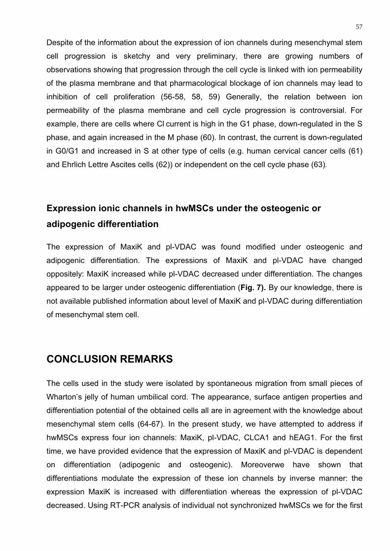
57 Despite of the information about the expression of ion channels during mesenchymal stem
cell progression is sketchy and very preliminary, there are growing numbers of
observations showing that progression through the cell cycle is linked with ion permeability
of the plasma membrane and that pharmacological blockage of ion channels may lead to
inhibition of cell proliferation (56-58, 58, 59) Generally, the relation between ion
permeability of the plasma membrane and cell cycle progression is controversial. For
example, there are cells where Cl current is high in the G1 phase, down-regulated in the S
phase, and again increased in the M phase (60). In contrast, the current is down-regulated
in G0/G1 and increased in S at other type of cells (e.g. human cervical cancer cells (61)
and Ehrlich Lettre Ascites cells (62)) or independent on the cell cycle phase (63).
Expression ionic channels in hwMSCs under the osteogenic or adipogenic differentiation
The expression of MaxiK and pl-VDAC was found modified under osteogenic and
adipogenic differentiation. The expressions of MaxiK and pl-VDAC have changed
oppositely: MaxiK increased while pl-VDAC decreased under differentiation. The changes
appeared to be larger under osteogenic differentiation (Fig. 7). By our knowledge, there is
not available published information about level of MaxiK and pl-VDAC during differentiation
of mesenchymal stem cell.
CONCLUSION REMARKS
The cells used in the study were isolated by spontaneous migration from small pieces of
Wharton’s jelly of human umbilical cord. The appearance, surface antigen properties and
differentiation potential of the obtained cells all are in agreement with the knowledge about
mesenchymal stem cells (64-67). In the present study, we have attempted to address if
hwMSCs express four ion channels: MaxiK, pl-VDAC, CLCA1 and hEAG1. For the first
time, we have provided evidence that the expression of MaxiK and pl-VDAC is dependent
on differentiation (adipogenic and osteogenic). Moreoverwe have shown that
differentiations modulate the expression of these ion channels by inverse manner: the
expression MaxiK is increased with differentiation whereas the expression of pl-VDAC
decreased. Using RT-PCR analysis of individual not synchronized hwMSCs we for the first
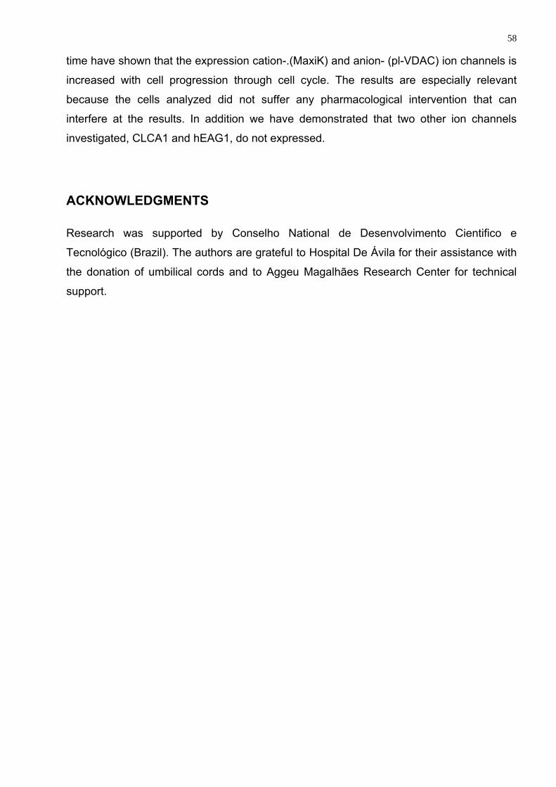
58 time have shown that the expression cation-.(MaxiK) and anion- (pl-VDAC) ion channels is
increased with cell progression through cell cycle. The results are especially relevant
because the cells analyzed did not suffer any pharmacological intervention that can
interfere at the results. In addition we have demonstrated that two other ion channels
investigated, CLCA1 and hEAG1, do not expressed.
ACKNOWLEDGMENTS
Research was supported by Conselho National de Desenvolvimento Cientifico e
Tecnológico (Brazil). The authors are grateful to Hospital De Ávila for their assistance with
the donation of umbilical cords and to Aggeu Magalhães Research Center for technical
support.
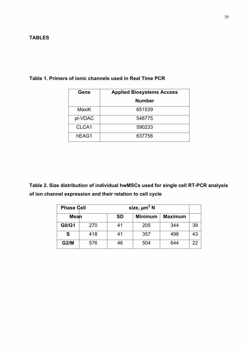
59
TABLES
Table 1. Primers of ionic channels used in Real Time PCR
Gene Applied Biosystems Access Number
MaxiK 651539
pl-VDAC 548775
CLCA1 590233
hEAG1 637756
Table 2. Size distribution of individual hwMSCs used for single cell RT-PCR analysis of ion channel expression and their relation to cell cycle
Phase Cell size, µm2 N
Mean SD Minimum Maximum
G0/G1 270 41 205 344 39
S 418 41 357 498 43
G2/M 576 46 504 644 22

60
FIGURE LEGENDS
Figure 1. Example of single cell collection. Stages of single cell entering into micropipette
are shown in A, B and C.
Figure 2. Morphology of cultured mesenchymal stem cells from Wharton jelly’s of human
umbilical cord. Scale bar is shown in Figure.
Figure 3. Flow cytometry analysis showing the immunophenotype of hwMSCs populations
that were obtained from the homogeneous 70-80%-confluent monolayer at the end of the
third passage. Control is shown in RED.
Figure 4. Phase contrast microscopy hwMSCs stimulated for adipogenic (A, C) and
osteogenic (B, D) differentiation.
Figure 5. The size distribution of the hwMSCs from proliferating cultures. Number in figure
shown position of the maximal value of each Gaussian used to fit the histogram.
Figure 6. Cell cycle dependent expression of MaxiK and pl-VDAC of hwMSCs.
Figure 7. Influence of differentiation of hwMSCs on the expression of MaxiK and pl-VDAC.
The difference between experiment and the respective control is significant at p <0.05.
The expression is shown in comparison to GAPDH gene
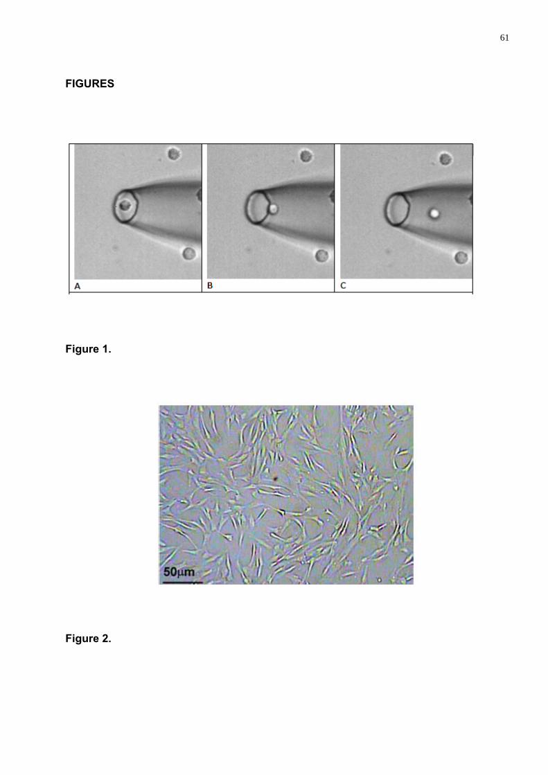
61
FIGURES
Figure 1.
Figure 2.
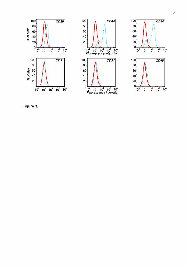
62
Figure 3.
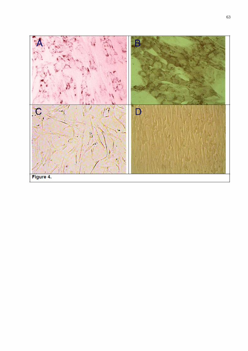
63
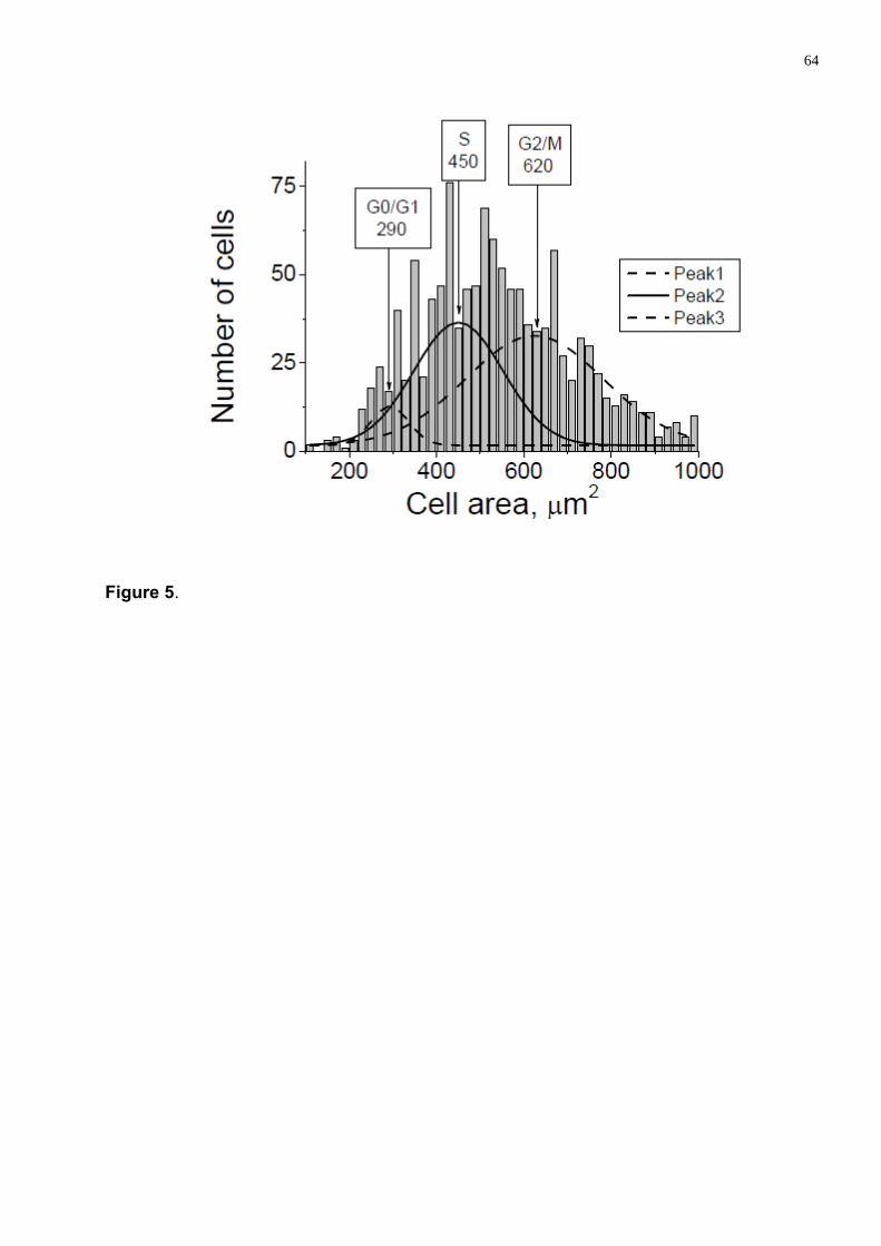
64
Figure 5.
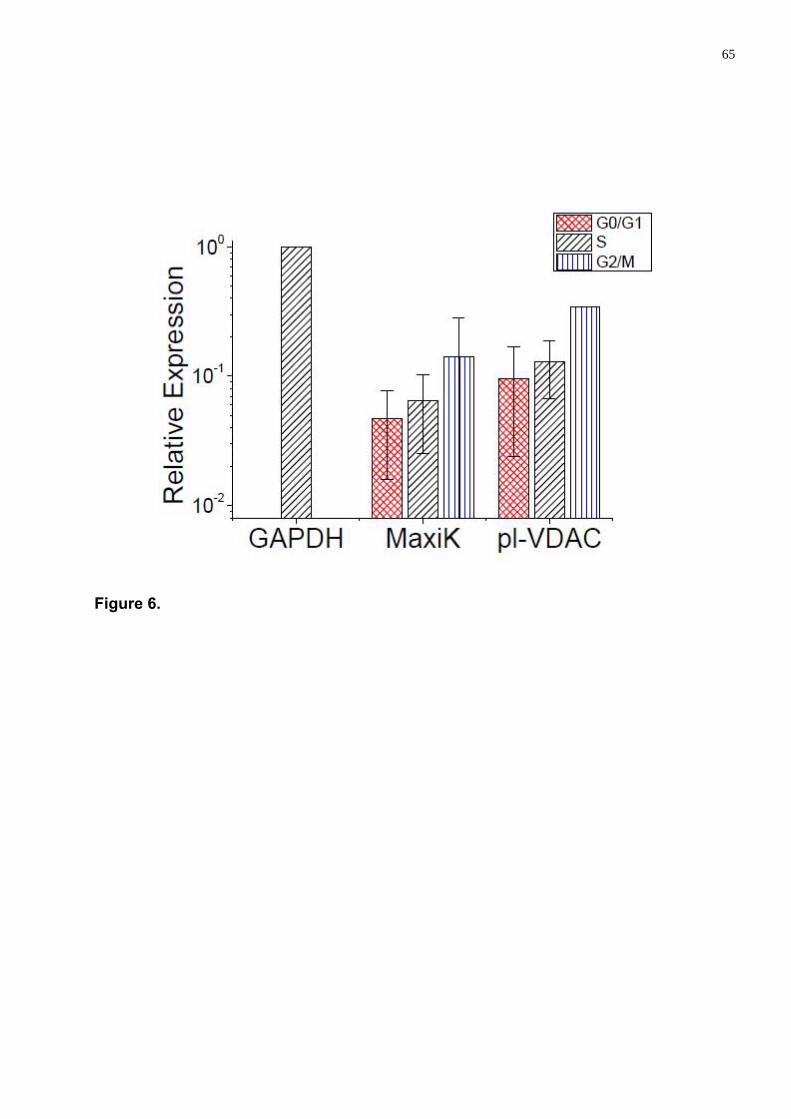
65
Figure 6.
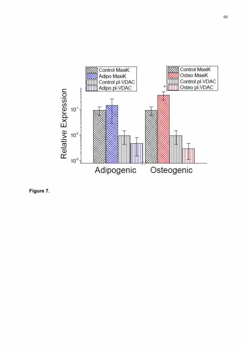
66
Figure 7.
67
Reference List
1. Ben-Ami E, Berrih-Aknin S, Miller A. Mesenchymal stem cells as an immunomodulatory therapeutic strategy for autoimmune diseases. AUTOIMMUN. REV. 2011.
2. Rebelatto CK, Aguiar AM, Moretao MP et al. Dissimilar differentiation of mesenchymal stem cells from bone marrow, umbilical cord blood, and adipose tissue. EXP. BIOL. MED. (MAYWOOD. ) 2008;233(7):901-913.
3. Troyer DL, Weiss ML. Wharton's jelly-derived cells are a primitive stromal cell population. STEM CELLS 2008;26(3):591-599.
4. Weiss ML, Anderson C, Medicetty S et al. Immune Properties of Human Umbilical Cord Wharton's Jelly-Derived Cells. STEM CELLS 2008;26(11):2865-2874.
5. Tyndall A, Gratwohl A. Adult stem cell transplantation in autoimmune disease. CURRENT OPINION IN HEMATOLOGY 2009;16(4):285-291.
6. Chen FH, Tuan RS. Mesenchymal stem cells in arthritic diseases. ARTHRITIS RESEARCH & THERAPY 2008;10(5).
7. Ding YC, Bushell A, Wood KJ. Mesenchymal Stem-Cell Immunosuppressive Capabilities: Therapeutic Implications in Islet Transplantation. TRANSPLANTATION 2010;89(3):270-273.
8. Nilius B. Chloride channels go cell cycling. J. PHYSIOL 2001;532(Pt 3):581.
9. Okada Y. Cell volume-sensitive chloride channels: Phenotypic properties and molecular identity. MECHANISMS AND SIGNIFICANCE OF CELL VOLUME REGULATION. CONTRIB NEPHROL. 2006;152:9-24.
10. Wehner F. Cell volume-regulated cation channels. MECHANISMS AND SIGNIFICANCE OF CELL VOLUME REGULATION 2006;152:25-53.
11. Lang F, Foller M, Lang K et al. Cell volume regulatory ion channels in cell proliferation and cell death. OSMOSENSING AND OSMOSIGNALING 2007;428:209-225.
12. da Silva M.B., Maria Assis Costa VRAP, Gisely Juliane Barbosa da Silva EBBdMDPB et al. Ion Channels In Volume Regulation Of Clonal Kidney Cells. CELL PROLIF. 2010;43:529-541.
13. Lang F, Lepple-Wienhues A, Paulmichl M et al. Ion channels, cell volume, and apoptotic cell death. CELL PHYSIOL BIOCHEM. 1998;8(6):285-292.
14. Lang F, Ritter M, Gamper N et al. Cell volume in the regulation of cell proliferation and apoptotic cell death. CELL PHYSIOL BIOCHEM. 2000;10(5-6):417-428.
68 15. Shen MR, Droogmans G, Eggermont J et al. Differential expression of volume-
regulated anion channels during cell cycle progression of human cervical cancer cells. JOURNAL OF PHYSIOLOGY-LONDON 2000;529(2):385-394.
16. Ullrich N, Sontheimer H. Cell cycle-dependent expression of a glioma-specific chloride current: proposed link to cytoskeletal changes. AM. J. PHYSIOL 1997;273(4 Pt 1):C1290-C1297.
17. Li M, Wang B, Lin W. Cl-channel blockers inhibit cell proliferation and arrest the cell cycle of human ovarian cancer cells. EUROPEAN JOURNAL OF GYNAECOLOGICAL ONCOLOGY 2008;29(3):267-271.
18. Miyazaki H, Shiozaki A, Niisato N et al. Chloride ions control the G(1)/S cell-cycle, checkpoint by regulating the expression of p21 through a p53-independent pathway in human gastric cancer cells. BIOCHEMICAL AND BIOPHYSICAL RESEARCH COMMUNICATIONS 2008;366(2):506-512.
19. Nilius B, Wohlrab W. Potassium channels and regulation of proliferation of human melanoma cells. J. PHYSIOL 1992;445:537-548.
20. Rouzaire-Dubois B, Dubois JM. K+ channel block-induced mammalian neuroblastoma cell swelling: a possible mechanism to influence proliferation. J. PHYSIOL 1998;510 ( Pt 1):93-102.
21. Wang SP, Wang JA, Luo RH et al. Potassium channel currents in rat mesenchymal stem cells and their possible roles in cell proliferation. CLINICAL AND EXPERIMENTAL PHARMACOLOGY AND PHYSIOLOGY 2008;35(9):1077-1084.
22. Beeton C, Smith BJ, Sabo JK et al. The D-diastereomer of ShK toxin selectively blocks voltage-gated K+ channels and inhibits T lymphocyte proliferation. JOURNAL OF BIOLOGICAL CHEMISTRY 2008;283(2):988-997.
23. Ouadid-Ahidouch H, Ahidouch A. K+ channel expression in human breast cancer cells: Involvement in cell cycle regulation and carcinogenesis. JOURNAL OF MEMBRANE BIOLOGY 2008;221(1):1-6.
24. Heubach JF, Graf EM, Leutheuser J et al. Electrophysiological properties of human mesenchymal stem cells. JOURNAL OF PHYSIOLOGY-LONDON 2004;554(3):659-672.
25. Heubach JF, Daniel J, Bock M et al. Electrophysiological properties of mesenchymal stem cells obtained from human bone marrow. CIRCULATION 2002;106(19):68.
26. Li GR, Deng XL, Sun H et al. Ion channels in mesenchymal stem cells from rat bone marrow. STEM CELLS 2006;24(6):1519-1528.
27. Deng XL, Sun HY, Lau CP et al. Properties of ion channels in rabbit mesenchymal stem cells from bone marrow. BIOCHEMICAL AND BIOPHYSICAL RESEARCH COMMUNICATIONS 2006;348(1):301-309.
69 28. Tao R, Lau CP, Tse HF et al. Functional ion channels in mouse bone marrow
mesenchymal stem cells. AMERICAN JOURNAL OF PHYSIOLOGY-CELL PHYSIOLOGY 2007;293(5):C1561-C1567.
29. Park KS, Jung KH, Kim SH et al. Functional expression of ion channels in mesenchymal stem cells derived from umbilical cord vein. STEM CELLS 2007;25(8):2044-2052.
30. Park KS, Choi MR, Jung KH et al. Diversity of Ion Channels in Human Bone Marrow Mesenchymal Stem Cells from Amyotrophic Lateral Sclerosis Patients. KOREAN JOURNAL OF PHYSIOLOGY & PHARMACOLOGY 2008;12(6):337-342.
31. Troyer DL, Weiss ML. Concise review: Wharton's jelly-derived cells are a primitive stromal cell population. STEM CELLS 2008;26(3):591-599.
32. Uhal BD, Ramos C, Joshi I et al. Cell size, cell cycle, and alpha-smooth muscle actin expression by primary human lung fibroblasts. AMERICAN JOURNAL OF PHYSIOLOGY-LUNG CELLULAR AND MOLECULAR PHYSIOLOGY 1998;19(5):L998-L1005.
33. Urbani L, Sherwood SW, Schimke RT. Dissociation of nuclear and cytoplasmic cell cycle progression by drugs employed in cell synchronization. EXP. CELL RES. 1995;219(1):159-168.
34. Anderson EC, Bell GI, Petersen DF et al. Cell Growth and Division .4. Determination of Volume Growth Rate and Division Probability. BIOPHYSICAL JOURNAL 1969;9(2):246-&.
35. Deng XL, Lau CP, Lai K et al. Cell cycle-dependent expression of potassium channels and cell proliferation in rat mesenchymal stem cells from bone marrow. CELL PROLIFERATION 2007;40(5):656-670.
36. Li GR, Sun H, Deng X et al. Characterization of ionic currents in human mesenchymal stem cells from bone marrow. STEM CELLS 2005;23(3):371-382.
37. Nishimaru K, Eghbali M, Rong L et al. Functional and molecular evidence of MaxiK channel beta 1 subunit decrease with coronary artery ageing in the rat. JOURNAL OF PHYSIOLOGY-LONDON 2004;559(3):849-862.
38. Cui J, Yang H, Lee US. Molecular mechanisms of BK channel activation. CELLULAR AND MOLECULAR LIFE SCIENCES 2009;66(5):852-875.
39. Hou SW, Heinemann SH, Hoshi T. Modulation of BKCa Channel Gating by Endogenous Signaling Molecules. PHYSIOLOGY 2009;24(1):26-35.
40. Messina A, Guarino F, Oliva M et al. Characterization of the human porin isoform 1 (HVDAC1) gene by amplification on the whole human genome: A tool for porin deficiency analysis. BIOCHEMICAL AND BIOPHYSICAL RESEARCH COMMUNICATIONS 2000;270(3):787-792.
41. Thinnes FP. Neuroendocrine differentiation of LNCaP cells suggests: VDAC in the cell membrane is involved in the extrinsic apoptotic pathway. MOLECULAR GENETICS AND METABOLISM 2009;97(4):241-243.
70 42. Thinnes FP, Hellmann KP, Hellmann T et al. Studies on human porin XXII: Cell
membrane integrated human porin channels are involved in regulatory volume decrease (RVD) of HeLa cells. MOLECULAR GENETICS AND METABOLISM 2000;69(4):331-337.
43. Okada SF, O'Neal WK, Huang PB et al. Voltage-dependent anion channel-1 (VDAC-1) contributes to ATP release and cell volume regulation in murine cells. JOURNAL OF GENERAL PHYSIOLOGY 2004;124(5):513-526.
44. Shoshan-Barmatz V, De Pinto V, Zweckstetter M et al. VDAC, a multi-functional mitochondrial protein regulating cell life and death. MOLECULAR ASPECTS OF MEDICINE 2010;31(3):227-285.
45. Neusch C, Weishaupt JH, Bahr M. Kir channels in the CNS: emerging new roles and implications for neurological diseases. CELL AND TISSUE RESEARCH 2003;311(2):131-138.
46. Gomez-Varela D, Zwick-Wallasch E, Knotgen H et al. Monoclonal antibody blockade of the human Eag1 potassium channel function exerts antitumor activity. CANCER RESEARCH 2007;67(15):7343-7349.
47. Jang S, Cho HH, Cho YB et al. Functional neural differentiation of human adipose tissue-derived stem cells using bFGF and forskolin. BMC CELL BIOLOGY 2010;11.
48. Bai XW, Ma JY, Pan ZZ et al. Electrophysiological properties of human adipose tissue-derived stem cells. AMERICAN JOURNAL OF PHYSIOLOGY-CELL PHYSIOLOGY 2007;293(5):C1539-C1550.
49. Fuller CM, Ji HL, Tousson A et al. Ca(2+)-activated Cl(-) channels: a newly emerging anion transport family. PFLUGERS ARCH. 2001;443 Suppl 1:S107-S110.
50. Pauli BU, Abdel-Ghany M, Cheng HC et al. Molecular characteristics and functional diversity of CLCA family members. CLINICAL AND EXPERIMENTAL PHARMACOLOGY AND PHYSIOLOGY 2000;27(11):901-905.
51. Jentsch TJ, Neagoe L, Scheel O. CLC chloride channels and transporters. CURRENT OPINION IN NEUROBIOLOGY 2005;15(3):319-325.
52. Jentsch TJ, Stein V, Weinreich F et al. Molecular structure and physiological function of chloride channels. PHYSIOLOGICAL REVIEWS 2002;82(2):503-568.
53. Connon CJ, Kawasaki S, Liles M et al. Gene expression and immunolocalisation of a calcium-activated chloride channel during the stratification of cultivated and developing corneal epithelium. CELL AND TISSUE RESEARCH 2006;323(1):177-182.
54. Wahl AS, Buchthal B, Rode F et al. Hypoxic/Ischemic Conditions Induce Expression of the Putative Pro-Death Gene Clca1 Via Activation of Extrasynaptic N-Methyl-D-Aspartate Receptors. NEUROSCIENCE 2009;158(1):344-352.
55. Leslie M. The Power Of One. SCIENCE 2011;331(6013).
71 56. Lang F. Mechanisms and significance of cell volume regulation. J AM. COLL. NUTR.
2007;26(5 Suppl):613S-623S.
57. Czarnecki A, Vaur S, Dufy-Barbe L et al. Cell cycle-related changes in transient K(+) current density in the GH3 pituitary cell line. AM. J PHYSIOL CELL PHYSIOL 2000;279(6):C1819-C1828.
58. Klausen TK, Bergdahl A, Hougaard C et al. Cell cycle-dependent activity of the volume- and Ca2+-activated anion currents in Ehrlich lettre ascites cells. J CELL PHYSIOL 2007;210(3):831-842.
59. Burg ED, Remillard CV, Yuan JX. Potassium channels in the regulation of pulmonary artery smooth muscle cell proliferation and apoptosis: pharmacotherapeutic implications. BR. J PHARMACOL. 2008;153 Suppl 1:S99-S111.
60. Chen LX, Wang LW, Zhu LY et al. Cell cycle-dependent expression of volume-activated chloride currents in nasopharyngeal carcinoma cells. AMERICAN JOURNAL OF PHYSIOLOGY-CELL PHYSIOLOGY 2002;283(4):C1313-C1323.
61. Shen MR, Droogmans G, Eggermont J et al. Differential expression of volume-regulated anion channels during cell cycle progression of human cervical cancer cells. J. PHYSIOL 2000;529 Pt 2:385-394.
62. Klausen TK, Bergdahl A, Hougaard C et al. Cell cycle-dependent activity of the volume- and Ca2+-activated anion currents in Ehrlich Lettre Ascites cells. JOURNAL OF CELLULAR PHYSIOLOGY 2007;210(3):831-842.
63. Doroshenko P, Sabanov V, Doroshenko N. Cell cycle-related changes in regulatory volume decrease and volume-sensitive chloride conductance in mouse fibroblasts. J. CELL PHYSIOL 2001;187(1):65-72.
64. Monteiro BS, Neto NMA, Del Carlo RJ. Mesenchymal stem cell. CIENCIA RURAL 2010;40(1):238-245.
65. Khatri M, O'Brien TD, Goyal SA et al. Isolation and characterization of chicken lung mesenchymal stromal cells and their susceptibility to avian influenza virus. DEVELOPMENTAL AND COMPARATIVE IMMUNOLOGY 2010;34(4):474-479.
66. Jezierska-Wozniak K, Nosarzewska D, Tutas A et al. Use of adipose tissue as a source of mesenchymal stem cells. POSTEPY HIGIENY I MEDYCYNY DOSWIADCZALNEJ 2010;64:326-332.
67. Fan CG, Zhang QJ, Zhou JR. Therapeutic Potentials of Mesenchymal Stem Cells Derived from Human Umbilical Cord. STEM CELL REVIEWS AND REPORTS 2011;7(1):195-207.