Embed Size (px)
Citation preview
Daniel Albuquerque Pereira
Quorum sensing em cianobactérias
Belo Horizonte
2014
2
Universidade Federal de Minas Gerais
Instituto de Ciências Biológicas
Programa de Pós-graduação em Ecologia, Conservação e Manejo
da Vida Silvestre
Quorum sensing em cianobactérias
Tese apresentada ao Departamento de
Biologia Geral do Instituto de Ciências
Biológicas, Universidade Federal de Minas Gerais, como requisito parcial
para a obtenção do título de Doutor em
Ecologia, Conservação e Manejo da
Vida Silvestre.
Orientadora: Prof. Dra. Alessandra Giani
Departamento de Botânica, ICB, UFMG
Belo Horizonte
2014
3
Agradecimentos
Aos meus pais, Eveline e Roberto, minha irmã Camila e minha avó Marília, por todo o
apoio que me deram durante a minha vida e pelo suporte dado às escolhas que fiz.
A minha orientadora, professora Alessandra Giani, pela contribuição a minha formação
científica, pelos diversos ensinamentos.
A todos os meus amigos pelo apoio e amizade durante todos os anos de minha vida
acadêmica sem os quais eu não teria conseguido finalizar este trabalho.
Ao pessoal do laboratório de Ficologia, pelo apoio e ajuda durante a minha graduação e
mestrado e doutorado.
Ao pessoal da secretaria da pós graduação, Fred e Cris pela boa vontade e ajuda sempre
que foi necessário.
A equipe do Departamento de Bioquímica e Imunologia da UFMG pela ajuda com as
análises de espectrometria de massa
A Capes pela bolsa de doutorado.
4
SUMÁRIO
Resumo..............................................................................................................................6
Abstract..............................................................................................................................8
1 Introdução geral............................................................................................................10
1.1 Quorum sensing.....................................................................................................10
1.2 Cianobactéria.........................................................................................................13
1.3 Quorum sensing em cianobactérias.......................................................................21
2 Hipóteses e justificativa...............................................................................................22
3 Objetivo........................................................................................................................23
4 Referências bibliográficas............................................................................................24
Capítulo 1........................................................................................................................29
Abstract.......................................................................................................................30
Introduction.................................................................................................................31
Methodology...............................................................................................................33
Results.........................................................................................................................35
Discussion...................................................................................................................44
References...................................................................................................................48
Capítulo 2........................................................................................................................55
Abstract.......................................................................................................................56
Introduction.................................................................................................................57
5
Methodology...............................................................................................................59
Results.........................................................................................................................62
Discussion...................................................................................................................68
Supporting Information...............................................................................................75
References...................................................................................................................77
Capítulo 3........................................................................................................................85
Abstract.......................................................................................................................86
Introduction.................................................................................................................87
Methodology...............................................................................................................90
Results.........................................................................................................................93
Discussion...................................................................................................................97
References.................................................................................................................102
5 Conclusões Finais.......................................................................................................109
6
Resumo
O termo “quorum sensing” refere-se a um fenômeno de comunicação celular existente
em bactérias, onde ao se atingir uma certa densidade populacional mudanças no padrão
de expressão gênica, e consequentemente da fisiologia das células são disparadas. No que
diz respeito a cianobactérias, poucos trabalhos com enfoque em quorum sensing foram
realizados. Alguns estudos mostram que substâncias indutoras de quorum sensing podem
alterar algumas características fisiológicas de algumas cepas de cianobactérias.
Evidências indiretas mostraram que a diferença na densidade celular pode afetar a
produção de microcistinas. A capacidade de produzir florações em determinadas
condições é outro aspecto importante das cianobactérias. Nestas situações a densidade
populacional das espécies dominantes atinge valores muito altos, podendo constituir uma
situação ideal para a ocorrência do quorum sensing. Existem diversos estudos sobre
florações, no entanto pouco se sabe sobre o que ocorre com as populações de
cianobactérias em nivel molecular e também fisiológico. Este trabalho teve o objetivo de
caracterizar quatro cepas de cianobactérias quanto à produção de peptídeos e utilizá-las
em experimentos com a finalidade de encontrar evidências da existência de quorum
sensing em cianobactérias e sua relação com a produção de oligopeptídeos. Para cumprir
estes objetivos foram utilizadas técnicas de bioquímica e biologia molecular. Os
resultados encontrados demonstraram que a produção de oligopeptídeos é afetada pela
densidade celular, sendo, na maioria dos casos, maior nas situações com alto número de
células. Foi verificado através de PCR em tempo real que auto indutores do tipo acil-
homoserina lactonas (AHLs), responsáveis pela ativação do quorum sensing em diversos
grupos de bactérias gram-negativas, afetam a transcrição de genes ligados a produção dos

7
peptídeos microcistina, cianopeptolina e microviridina. Através de testes de ELISA foi
visto também que as AHLs afetam a produção de microcistinas da mesma forma que
influenciam a transcrição dos genes liagados à sua síntese. Com este trabalho foi visto
que densidade celular e, possivelmente, quorum sensing são fatores importantes na
síntese de metabólitos secundários em cinobactérias, que devem ser levados em
consideração ao se estudar estes compostos e suas relações com o ambiente.
Palavras chave: Quorum sensing, cianobacterias, microcistina, densidade celular
8
Abstract
The terminology “quorum sensing” is used to identify a cellular communication
phenomenon in the bacterial domain, which happens when a bacteria population reaches
a defined cellular density. During the activation of the phenomenon changes in the
expression of several genes and consequently in the physiology of the cell are triggered.
Concerning cyanobacteria, there is a lack of information about quorum sensing. Some
studies show that quorum sensing inducer compounds may alter physiological
characteristics of certain cyanobacteria strains. Besides that, indirect evidences have
shown that cellular density may influence microcystin production. The capacity to form
blooms in certain conditions is also an important characteristic found in the cyanobacteria
group. In these conditions the cellular density of a population may reach elevated
numbers, consisting of an ideal scenario for quorum sensing. There are a remarkably
number of studies regarding cyanobacterial blooms, but little is known about what
happens with a population at molecular and physiological levels. The aim of this study
was to characterize four strains of cyanobacteria regarding the production of peptides and
to use these strains in experiments with the objective of investigating evidences of
quorum sensing system in cyanobacteria and its connection with the synthesis of
oligopeptides. In order to fulfill these objectives biochemical and molecular techniques
were used. The results obtained showed that the production of oligopeptides is affected
by cellular density, being, in most cases, higher in situations with an elevated number of
cells. It was demonstrated by real time PCR that acylhomoserine lactone autoinducers
(AHLs), which are responsible for the activation of the quorum sensing in various groups
of gram-negative bacteria, affect the transcription of genes linked to the production of
9
microcystins, cyanopeptolins and microviridins. Through ELISA assays it was also seen
that AHLs affect the microcystin production in the same pattern in which they affect the
transcription of genes connected to its synthesis. With this work it was observed that
cellular density and possibly quorum sensing are key factors in secondary metabolite
synthesis in cyanobacteria and should be considered when studying these compounds and
their connection with the environment.
Keywords: Quorum sensing, cyanobacteria, microcystin, cellular density
10
1 – Introdução Geral
1.1 – Quorum sensing
Em populações bacterianas, organismos sem sistema nervoso e hormônios, onde cada
indivíduo é constituído por uma célula que se reproduz por fissão binária e precisa
competir por recursos com outros da mesma espécie, seria esperado que não existissem
mecanismos de reconhecimento e cooperação entre os indivíduos isolados. No entanto,
existem diversas situações nas quais o comportamento cooperativo numa população pode
ser extremente vantajoso, como: conjugação, simbiose, adaptação de nicho, produção de
metabólitos secundários, combate à sistemas de defesa de organismos superiores e até
migrações populacionais com intenção de fugir de locais com condições desfavoráveis
(Williams et al., 2007).
Para que os indivíduos que formam uma população bacteriana expressem o
comportamento cooperativo é necessário que exista alguma forma de comunicação
química entre as células através de moléculas de sinalização. O primeiro trabalho a
identificar este tipo de comunicação foi realizado por Tomasz (1965), que demonstrou
que a bactéria gram-positiva Streptococcus pneumoniae controla fatores de competência
genética através de substâncias produzidas pelo próprio organismo, seguido do trabalho
de Nealson et al. (1970) que mostrou a existência do controle de bioluminescência na
bactéria gram-negativa Vibrio fischeri através de uma substância chamada de auto-
indutor. Os auto-indutores são assim chamados pois parte de sua função é estimular a
própria produção (Williams et al., 2007). Além disso, também permitem que as
populações bacterianas determinem sua densidade numérica, pois, à medida em que a
11
população cresce, os sinalizadores acumulam no meio e, ao atingir um limiar na
concentracão da substância em consequência ao tamanho populacional, mudanças
coordenadas no comportamento bacteriano são disparadas (Fuqua & Greenberg, 2002).
Apesar dos primeiros trabalhos relacionados à comunicação celular em bactérias terem
surgido há mais de 40 anos, foi apenas no começo da década de 1990 que a pesquisa
nessa área se desenvolveu e que o termo “quorum sensing” foi introduzido por Fuqua et
al., (1994) para descrever este fenômeno. Desde então, diversos trabalhos sobre o
funcionamento do quorum sensing em diversos grupos de bactérias foram publicados e
acredita-se que a comunicação celular seja algo comum em bactérias (Miller & Bassler,
2001). No entanto é importante ressaltar que o termo quorum sensing não descreve
adequadamente todas as formas de comunicação celular onde bactérias utilizam sinais
químicos. O tamanho do “quorum” não é fixo e depende da taxa de produção e
degradação dos indutores, fatores que são afetados pelo ambiente no qual os organismos
estão inseridos (Williams et al., 2007). Células solitárias podem tambem mudar do estado
de não quorum sensing para quorum sensing, como observado por Qazi et al. (2001) em
células de Staphylococcus aureus aderidas a endossomos de células endoteliais. Nestes
casos, mesmo em baixa densidade, ocorre acúmulo do auto-indutor em quantidade
suficiente para disparar o fenômeno. Nestas situações o quorum sensing poderia ser na
verdade descrito como “diffusion sensing” ou “compartment sensing”, já que a
informação levada aos indivíduos diz respeito mais ao ambiente onde os indivíduos estão
inseridos do que ao tamanho populacional (Redfield, 2002; Winzer at al., 2002). Outro
aspecto que deve ser levado em consideração é o fato de que o quorum sensing é apenas
um dos componentes ligados à regulação gênica global e que existem vários outros sinais
12
ambientais (ex: temperatura, pH, osmolaridade, concentração de nutrientes) percebidos
por uma população bacteriana, influenciando sua estratégia de sobrevivência e adaptação
à situações de stress (Withers et al., 2001).
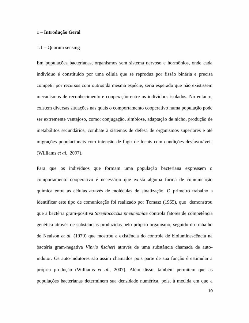
Dentre os diversos auto-indutores responsáveis pela comunicação no quorum sensing,
podem-se destacar as acilhomoserina lactonas (AHLs) (figura 1). Estes compostos são
caracterizados por uma homoserina lactona com um grupo acyl de cadeia carbônica na
posição α. A cadeia de carbono pode ser de tamanho variável (entre C4 e C18), pode
também variar no grau de saturação e oxidação (Williams et al., 2007). O funcionamento
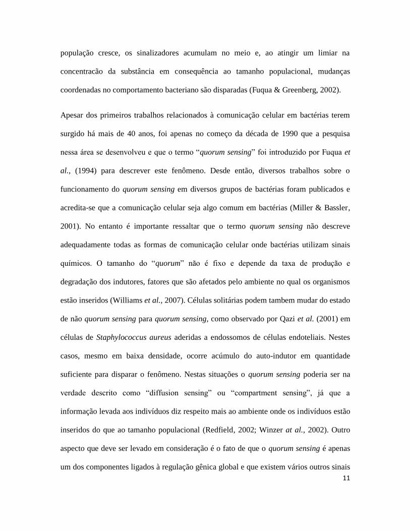
do sistema à partir de AHLs é baseado em duas famílias de proteínas, a LuxI é a estrutura
responsável por sintetizar os AHLs enquanto a LuxR é o receptor dos AHLs (Fuqua et
al., 2001; Swift et al., 2001). O sistema funciona a partir do momento em que o AHL se
liga e ativa a proteína LuxR. O complexo formado pelo AHL e a LuxR é então
responsável pela ativação e repressão de diverssos genes (Fuqua et al., 2001; Swift et al.,
2001). Os AHLs também ativam o gene responsável pela síntese da proteína LuxI,
gerando um “feedback” positivo. A figura 2 apresenta o esquema básico de
funcionamento do quorum sensing.
Figura 1 – Estrutura básica de uma molécula de AHL. R= grupo alquila linear. Fonte
Williams et al., 2007.
13
Figura 2 – Esquema básico do funcionamento do quorum sensing.
1.2 – Cianobactérias
Cianobactérias são organismos fotoautotróficos, que podem ser encontrados em uma
grande variedade de habitats aquáticos, e até terrestres, e são um dos principais
produtores primários em grande parte destes ambientes. Recentemente o aumento do
aporte de nutrientes, levou a formação de grandes biomassas de algas nos ambientes
aquáticos (florações), onde frequentemente as cianobactérias predominam. As florações
geralmente ocorrem em águas férteis e com condições favoráveis ao crescimento destes
microrganismos (Paerl, 1996). O sucesso destes organismos não pode ser explicado
apenas por uma característica, mas por vários fatores intrínsecos e ambientais em
14
conjunto, como por exemplo, o fato de serem os únicos organismos do fitoplâncton
capazes de fixar nitrogênio, controlar a flutuabilidade, acumular grandes quantidades de
fósforo entre outros (Paerl, 1996; Schindler et al., 2008). Durante as floracões é possivel
observar uma queda da diversidade de toda a comunidade aquática, pela dominância de
uma ou poucas espécies (Romo & Miracle, 1995; Giani et al., 2005).
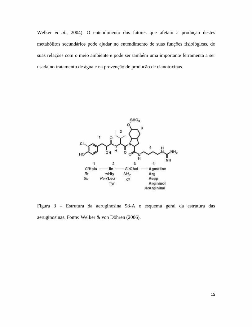
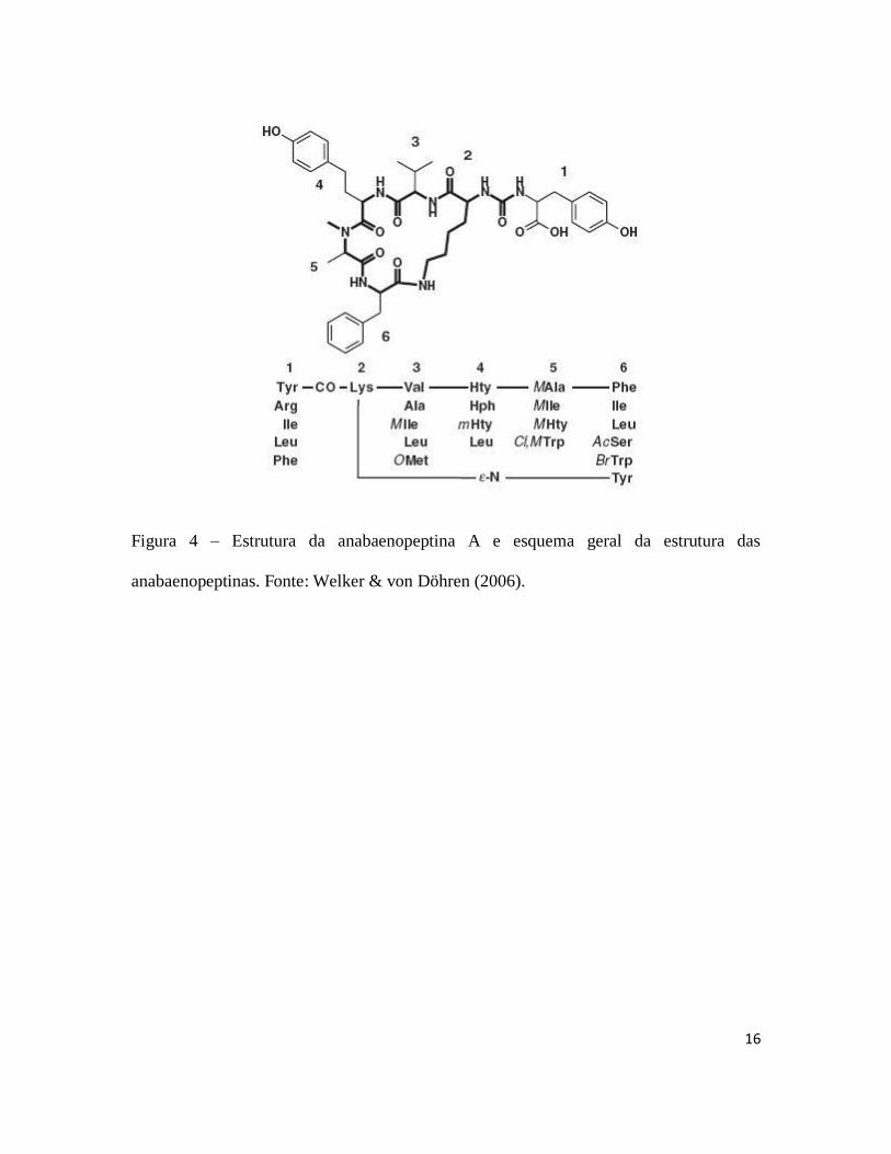
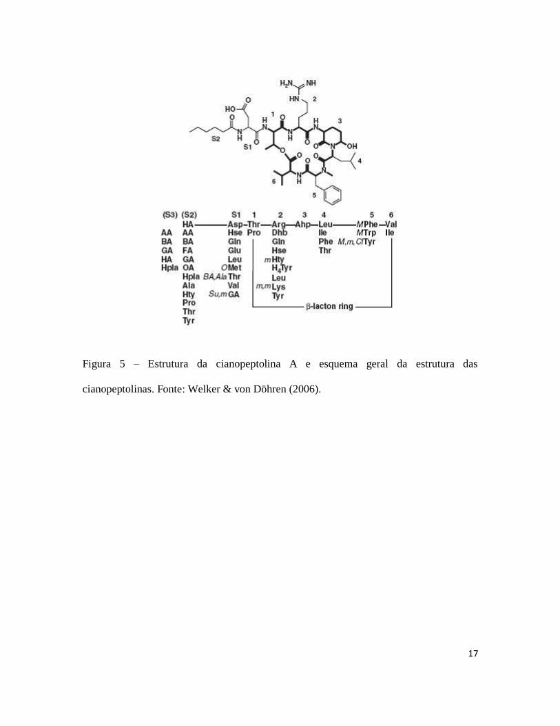
Diversas espécies de cianobactérias podem produzir diferentes tipos de peptídeos
bioativos e as vezes tóxicos (figuras 3, 4, 5, 6, 7 e 8) dentre os quais podem-se destacar as
aeruginosinas (Murakami et al., 1994), anabaenopeptinas (Harada et al., 1995),
cianopeptolinas (Martin et al., 1993), microviridinas (Ishitsuka et al., 1990), microgininas
(Okino et al., 1993) e microcistinas (Carmichael, 1992). A formação destas moléculas
ocorre através de complexos enzimáticos formados por vários módulos, conhecidos como
NRPSs (non-ribossomal peptide synthetases) e PKS (polyketide synthase) (Weber &
Marahiel, 2001; Finking & Marahiel, 2004). Cada módulo é composto de domínios
catalíticos, sendo que um módulo mínimo é composto de um domínio de adenilação,
responsável pela ativação dos aminoácidos, um módulo de tiolação, responsável por
transferir os intermediários ativados, e um modulo de condensação (von Döhren et al.,
1997; Stachelhaus et al., 1998). Com exceção das microviridinas, todos os compostos são
sintetizados por vias não ribossomais, o que pode explicar a sua alta variabilidade
(Welker & von Döhren, 2006).
As funções fisiológicas e ecológicas destes compostos ainda não foram esclarecidas, mas
é sabido que diferentes cepas de uma espécie podem ser ou não produtoras no que diz
respeito a cada tipo de peptídeo e classe (Fastner et al., 2001; Rohrlack et al., 2001;
15
Welker et al., 2004). O entendimento dos fatores que afetam a produção destes
metabólitos secundários pode ajudar no entendimento de suas funções fisiológicas, de
suas relações com o meio ambiente e pode ser também uma importante ferramenta a ser
usada no tratamento de água e na prevenção de producão de cianotoxinas.
Figura 3 – Estrutura da aeruginosina 98-A e esquema geral da estrutura das
aeruginosinas. Fonte: Welker & von Döhren (2006).
16
Figura 4 – Estrutura da anabaenopeptina A e esquema geral da estrutura das
anabaenopeptinas. Fonte: Welker & von Döhren (2006).
17
Figura 5 – Estrutura da cianopeptolina A e esquema geral da estrutura das
cianopeptolinas. Fonte: Welker & von Döhren (2006).
18
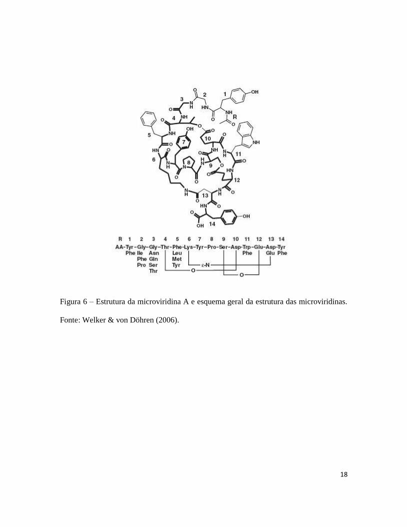
Figura 6 – Estrutura da microviridina A e esquema geral da estrutura das microviridinas.
Fonte: Welker & von Döhren (2006).

19
Figura 7 – Estrutura da microginina e esquema geral da estrutura das microgininas.
Fonte: Welker & von Döhren (2006).
20
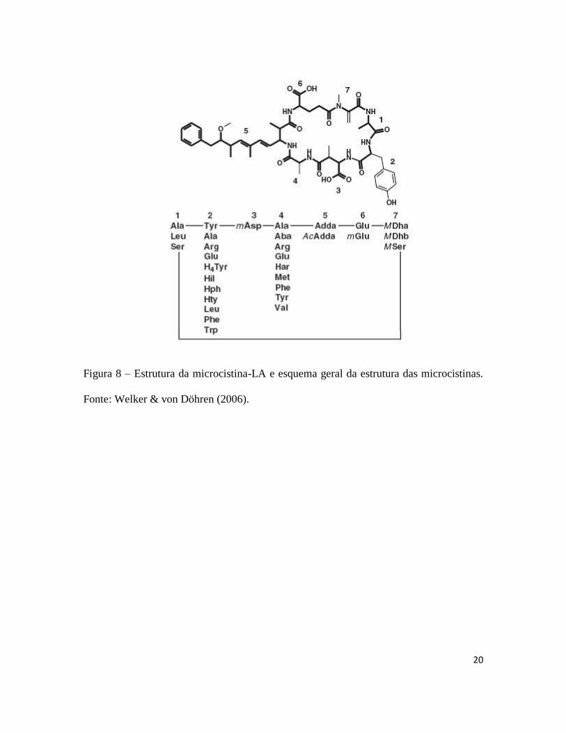
Figura 8 – Estrutura da microcistina-LA e esquema geral da estrutura das microcistinas.
Fonte: Welker & von Döhren (2006).
21
1.3 – Quorum sensing em cianobactérias
Apesar das pesquisas com quorum sensing terem se intensificado a partir da década de
1990, muito pouco foi estudado no grupo das cianobactérias. Existem estudos que
relatam a produção de auto indutores do tipo acil homoserina lactonas (AHLs) em
culturas axênicas de cianobactérias do gênero Gloeothece (Sharif et al., 2008). Trabalhos
realizados com o gênero Anabaena demonstraram que estes organismos podem produzir
enzimas do tipo AHL-acilase que degradam os AHLs (Romero et al., 2008) e que a
fixação de nitrogênio é inibida pela presença de diferentes AHLs com cadeias laterais de
variados tamanhos e graus de saturação (Romero et al., 2011). Van Mooy et al. (2012)
demonstrou que a aquisição de fósforo em cianobactérias marinhas do gênero
Trichodesmium é estimulada na presença de auto-indutores.
Além dos trabalhos já citados, existem evidências indiretas de quorum sensing em
cianobactérias. O trabalho de Gobler et al. (2007), estudando a dinâmica populacional e a
toxicidade de uma floração de cianobactérias em lago eutrófico de Nova Iorque relata um
aumento na expressão do gene mcyE, envolvido na produção de microcistina, em meses
onde a densidade celular era maior. Em estudos realizados em um pequeno lago eutrófico
na Nova Zelândia, Wood et al. (2011) mostraram que a expressão do gene mcyE não é
constitutiva, podendo ser ativada ou não ao longo do dia. O mesmo estudo mostrou que a
expressão do gene mcyE é maior em momentos onde a densidade celular é elevada. Outro
estudo comparando situações de alta e baixa densidade celular mostrou, em experimentos
de mesocosmos, que a quantidade de microcistina por célula era maior em situações onde
a densidade celular era mais elevada (Wood et al., 2012).
22
2 – Hipóteses e justificativa
- Hipóteses:
As cianobactérias, por fazerem parte do grupo das bactérias possuem alguma forma de
comunicação celular através de quorum sensing.
A densidade celular em cianobactérias influencia a produção de metabólitos secundários,
como microcistinas, aeruginosinas, cianopeptolinas, microviridinas e outros.
- Justificativa:
O aumento do processo de eutrofização cultural, associado à alta proliferação de
cianobactérias potencialmente tóxicas tem causado diversos problemas relacionados ao
uso de ambientes de água doce. Apesar de existirem diversos estudos sobre as toxinas
produzidas pelas cianobactérias, a maioria dos trabalhos sobre sua ocorrência no
ambiente é descritiva ou é voltada principalmente para aspectos bioquímicos e
moleculares. Poucos estudos foram feitos relacionando aspectos da ecologia das
cianobactérias com a produção de metabólitos secundários. Pouco se sabe também sobre
a fisiologia das cianobactérias durante situações de floração e como estes episódios
podem afetar a produção de toxinas e outros compostos. O fato das florações
apresentarem alta densidade celular sugere, por exemplo, uma situação favorável ao
aparecimento de fenômenos como o quorum sensing, bastante estudado em diversos
grupos de bactérias, porém, com poucos estudos em cianobactérias.
23
Os trabalhos apresentados nesta tese fornecem dados que permitam uma melhor
compreensão do quorum sensing em cianobactérias e sua relação com alguns metabólitos
secundários, amplamente produzidos por estes organismos.
A tese está divida em três capítulos, que estão apresentados no formato de artigos
científicos. O capitulo 2 foi aceito para publicação na revista FEMS Microbial Ecology
(doi: 10.1111/1574-6941.12281).
3 – Objetivo
3.1 – Objetivo geral
Encontrar evidências da existência de quorum sensing em cianobactérias e verificar sua
relação com a produção de oligopeptídeos.
3.2 – Objetivos específicos
– Caracterizar, de forma bioquímica e molecular, quatro cepas de cianobactérias com
relação à produção de diferentes peptídeos.
– Avaliar os efeitos de diferentes densidades celulares em algumas cepas de
cianobactérias na produção de peptídeos tais como microcistinas, aeruginosinas,
cianopeptolinas e microviridinas.
– Estudar os efeitos de substâncias conhecidas como promotoras de quorum sensing (acil
homoserina lactonas, AHLs) na expressão de genes ligados à produção de microcistinas,
cianopeptolinas e microviridinas, e também na concentração final de microcistina, em
cepas de cianobactérias.
24
4. Referências bibliográficas
Carmichael WW (1992) Cyanobacteria secondary metabolites – the cyanotoxins. J Appl
Bacteriol 72: 445–459.
Fastner J, Erhard M & von Döhren H (2001) Determination of oligopeptides diversity
within a natural population of Microcystis spp. (cyanobacteria) by typing single colonies
by matrix-assisted laser desorption ionization-time of flight mass spectrometry. Appl
Environ Microbiol 67: 5069–5076.
Finking R & Marahiel MA (2004) Biosynthesis of nonribosomal peptides. Annu Rev
Microbiol 58: 453 – 488.
Fuqua WC, Winans SC & Greenberg EP (1994) Quorum sensing in bacteria-the LuxR-
LuxI family of cell density-responsive transcriptional regulators. J Bacteriol 176: 269–
275.
Fuqua WC, Parsek MR & Greenberg EP (2001) Regulation of gene expression by cell-to-
cell communication: acyl-homoserine lactone quorum sensing. Annu Rev Genet 35: 439-
468.
Fuqua WC & Greenberg EP (2002) Listening in on bacteria: acyl-homoserine lactone
signaling. Nature Reviews 3: 685 – 695.
Giani A, Bird D, Praire Y & Lawerence J (2005) Empirical study of cyanobacterial
toxicity along a trophic gradient of lakes. Can J of Fisher and Aquat Sci 62: 1 – 10.
Gobler C J, Davis TW, Coyne KJ & Boyer GL (2007) Interactive influences of nutrient
loading, zooplankton grazing and microcystin sinthetase gene expression on
25
cyanobacterial bloom dynamics in a eutrophic New York lake. Harmful Algae 6: 119–
133.
Harada KI, Fujii K, Shimada T & Suzuki M (1995) Two cyclic peptides, anabenopeptins,
a third group of bioactive compounds from the cyanobacterium Anabaena flos-aqua
NRC, 525-17. Tetrahedron Lett 36: 1511–1514.
Ishitsuka M O, Kusumi T, Kakisawa H, Kaya K & Watanabe MM (1990) Microviridin: a
novel tricyclic depsipeptide from the toxic cyanobacterium Microcystis viridis. J Am
Chem Soc 112: 8180–8182.
Martin C, Oberer L, Ino T, König WA, Busch M & Weckesser J (1993) Cyanopeptolins,
nwe depsipeptides from the cyanobacterium Microcystis PCC 7806. J Antibiot 46 :
1550–1556.
Miller MB & Bassler BL (2001) Quorum sensing in bacteria. Annu Rev Microbiol 55:
165–199.
Murakami M, Okita Y, Matsuda H & Yamaguchi K (1994) Aeruginosin 298-A, a
thrombin and trypsin inhibitor from the blue-green alga Microcystis aeruginosa (NIES-
298). Tetrahedron lett 35: 3129–3132.
Nealson KH, Platt T & Hastings JW (1970) Cellular control of the synthesis and activity
of the bacterial bioluminescent system. J Bacteriol 104: 313 – 322.
Okino T, Matsuda H, Murakami M & Yamaguchi K (1993) Microginin, an angiotensin-
converting enzyme inhibitor from the blue-green alga Microcystis aeruginosa.
Tetrahedron Lett 34: 501– 504.
Pearl HW (1996) A comparison of cyanobacterial bloom dynamics in freshwater,
estuarine and marine environments. Phycologia 35: 25 – 35.
26
Qazi SNA, Counil E, Morrisey J, Rees CED, Chan WC, Williams P & Hill PJ (2001) agr-
expression precedes escape from the endosome of internalised Staphylococcus aureus.
Infect Immun 69: 7074 – 7082.
Redfield R (2002) Is quorum sensing a side effect of diffusion sensing? Trends Microbiol
10: 365 – 370.
Rohrlack T, Henning M & Kohl JG (2001) Isolation and characterization of colony-
forming Microcystis aeruginosa strains. Cyanotoxins – Occurrence, causes and
consequences. (Chorus I, ed), pp. 152-158. Springer, Berlin.
Romero M, Diggle SP, Heeb S, Cámara M & Otero A (2008) Quorum quenching activity
in Anabaena sp.PCC 7120: identification of AiiC, a novel AHL-acylase. FEMS
Microbiol Lett 280: 73 – 80.
Romero M, Muro-Pastor AM & Otero A (2011) Quorum sensing N-acylhomoserine
lactone signals affect nitrogen fixation in the cyanobacterium Anabaena sp. PCC7120.
FEMS Microbiol Lett 315: 101–108
Romo S & Miracle MR (1995) Diversity of phytoplankton assemblages of a polymitic
hypertrophic lake. Arch hydrobiol 132: 363 – 384.
Schindler DW, Hecky RE, Findlay DL, Stainton MP, Parker BR, Paterson MJ, Beaty KG,
Lyng M & Kasian SEM (2008) Eutrophication of lakes cannot be controlled by reducing
nitrogen input: Results of a 37-year whole-ecosystem experiment. PNAS 105: 11254-
11258.
Sharif DI, Gallon J, Smith CJ & Dudley E (2008) Quorum sensing in cyanobacteria: N-
octanoyl-homoserine lactone release and response, by the epilithic colonial
cyanobacterium Gloeothece PCC 6909. ISME J 2: 1171–1182.
27
Stachelhaus T, Mootz HD, Bergendahl V & Marahiel MA (1998) Peptide bond formation
in nonribosomal peptide biosynthesis. Catalytic role of condensation domain. J Biol
Chem 273: 22773 – 22781.
Swift S, Downie JA, Whitehead NA, Barnard AML, Salmond GPC & Willians P (2001)
Quorum sensing as a population-density-dependent determinant of bacterial physiology.
Adv Microb Physiol 45: 199-270.
Tomasz A (1965) Control of the competent state in pneumococcus by a hormone-like cell
product – an example of a new type of regulatory mechanism in bacteria. Nature 208:
155 – 159.
Van Mooy BAS, Hmelo LR, Sofen LE, Campagna SR, May AL, Dyhrman ST, Heithoff
A, Webb EA, Momper L & Mincer TJ (2012) Quorum sensing control phosphorous
acquisition in Trichodesmium consortia. The ISME journal 6: 422–429.
von Döhren H, Keller U, Vater J & Zocher R (1997) Multifunctional peptide syntethases.
Chem Rev 97: 2675 – 2705.
Weber T & Marahiel MA (2001) Exploring the domain structure of modular
nonribosomal peptide synthetase. Structure 9: R3 – R9.
Welker M, Brunke M, Preussel K, Lippert I & von Döhren H (2004) Diversity and
distribution of Microcystis (cyanobacteria) oligopeptides chemotypes from natural
communities studied by single mass spectrometry. Microbiology 150: 1785–1796.
Welker M & von Döhren H (2006) Cyanobacterial peptide: nature’s own combinatorial
biosynthesis. FEMS Microbiol Rev 30: 530–563.
28
Williams P, Winzer K, Chan WC & Cámara M (2007) Look who’s talking:
communication and quorum sensing in the bacterial world. Phil Trans R Soc B 362:
1119–1134.
Winzer K, Hardie KR & Williams P (2002) Bacterial cell to-cell communication: sorry
can’t talk now—out to lunch! Curr Opin Microbiol 5: 216 – 222.
Withers H, Swift S & Williams P (2001) Quorum sensing as an integral component of
gene regulatory networks in Gram-negative bacteria. Curr Opin Microbiol 4: 186 – 193.
Wood SA, Rueckert A, Hamilton DP, Cary SG & Dietrich DR (2011) Switching toxin
production on and off: intermittent microcystin synthesis in a Microcystis bloom. Environ
Microbiol Reports 3: 118–124.
Wood SA, Dietrich DR, Cary SG & Hamilton DP (2012) Increasing Microcystis cell
density enhances microcystin synthesis: a mes mesocosm study. Inland Waters 2: 17–22.
29
Capítulo 1
Peptide profile of four cyanobacteria strains
30
Abstract
Cyanobacteria are known to produce a broad variety of peptides as secondary
metabolites. These oligopeptides are usually synthesized by non-ribosomal units. A
characteristic of these systems is the capacity to combine proteinogenic amino acids with
non-proteinogenic amino acids, carbohydrates, fatty acids and other building blocks,
being this characteristic one of the reasons for the great variability found in these peptides
and creating structures that cannot be achieved by ribosomal synthesis. Aeruginosins,
cyanopeptolins, microcystins and microviridins are some of the main peptides classes
produced by cyanobacteria. The objective of this work was to determine a peptidic profile
based on biochemical methods for four strains of cyanobacteria (two Radiocystis
fernandoii, one Microcystis aeruginosa and one Microcystis panniformis). The strains
were grown for approximately 10 days, then extracted with methanol 75% and the extract
was purified with SPE cartridges. The purified material was analysed in a HPLC
followed by and identification of the peptides in a MALDI-TOF system. A total of 17
peptides were found, among aeruginosins, cyanopeptolins, microcystins and
microviridins. The Microcystis strains produced aeruginosins, cyanopeptolins and
microcystins while the Radiocystis strains produced cyanopeptolins, microcystins and
microviridins. A hierarchical analysis showed that the two Radiocystis strains and the two
Microcystis species form two separate groups according to their peptidic profile. The
technique used in this study showed to be efficient for isolation and identification of
peptides. Besides the compounds already described by other authors, we found some
oligopeptides that are not yet described in the literature.
31
1 – Introduction
Cyanobacterial secondary metabolites are represented by a great variety of structures and
were isolated from several taxa coming from different geographic regions (Welker & von
Dohren, 2006). The majority of these secondary metabolites are small peptides or have
peptidic substructures. Most of these peptides are assumed to be synthesized by NRPS
(non-ribosomal peptide synthetase) or NRPS/PKS (polyketide synthetase), creating
structures that cannot be achieved by ribosomal synthesis (Welker & von Dohren, 2006).
The NRPS system operates at the protein level without the use of nucleic acids (Finking
& Marahiel, 2004). The condensation of amino acids and carboxyl compounds is driven
by protein templates and usually each step requires a different protein module (Weber &
Marahiel, 2001; Schwarzer et al., 2003). The modules are composed of catalytic domains
being formed at least by an adenylation domain for amino acid activation, a thiolation
domain for transfer of activated intermediates, and a condensation domain (von Dohren
et al., 1997; Marahiel et al., 1997; Stachelhaus et al., 1998). An interesting characteristic
of NRPS systems is the capacity to combine proteinogenic amino acids with non-
proteinogenic amino acids, carbohydrates, fatty acids and other building blocks (Welker
& von Dohren, 2006). The ability to synthesize structures with several different types of
building blocks allows the production of a great variety of secondary metabolites, which
are grouped in classes according to their structures.
Aeruginosins (Murakami et al., 1995) are characterized by a linear structure with a
derivative of hydroxy-phenyl lactic acid (Hpla) at the N-terminus, the amino acid 2-
carboxy-6-hydroxyoctahydroindole (Choi) and an arginine derivative at the C-terminus.
32
The amino acids presented in position 2 are usually in D-configuration, although amino
acids in L-configuration can also be found in position 2 (Ishida et al 1999). Chlorination
(Cl) and sulphation (Su) can occur at the Choi (Shin et al., 1997) or Hpla (Ishida et al.,
1999). One aeruginosin variant (aeruginosin 98-C) was also reported to contain a
brominated Hpla (Ishida et al., 1999).
Cyanopeptolins (Martin et al., 1993) are characterized by a cyclic structure formed by an
ester bond between the β-hydroxy group of threonine with the carboxy group of the
terminal amino acid and by the presence of the amino acid 3-amino-6-hydroxy-2-
piperidone (Ahp) in position 3. A side chain of variable length is usually connected to
amino group of a threonine in position 1. The side chain can be formed by one or two
amino acids with an aliphatic fatty acid (Okino et al., 1993) or by a glyceric acid unit at
the N-terminus (Jakobi et al., 1995).
Microcystins (Botes et al., 1984) are characterized by the amino acid Adda
((2S,3S,8S,9S)-3-amino-9-methoxy-2,6,8-trimethyl-10-phenyldeca-4,6-dienoic acid) at
position 5. Positions 2 and 4 show high variability while other positions have only minor
variability. Position 3 is occupied by an aspartate derivative and position 6 is occupied by
a glutamate derivative or by a glutamate in D-configuration. Position 7 is usually
occupied by dehydro alanine (Dha) or by a methyl-dehydro alanine (mDha), in some
variants this position is occupied by a serine (Namikoshi et al., 1992).
Microviridins (Ishitsuka et al., 1990) are characterized by a multicyclic structure formed
by peptide and ester bonds, the peptide also have a side chain of variable length. They are
the largest known cyanobacterial peptides, with molecular weight around 1700 Daltons.
33
Their amino acids are all in L-configuration and the only non-proteinogenic unit is the N-
terminal acetic acid. Variation in this group of peptides occurs primarily due to
modifications in the side chain and in position 5 in the ring (Welker & von Dohren,
2006). In a different way microviridins are synthesized ribossomaly and not trough
NRPS/PKS systems (Philmus et al., 2008).
The structural variety of cyanobacterial peptides can also be explained by the diversity of
NRPS and PKS gene clusters in cyanobacteria. Christiansen et al., (2001) found NRPS
genes in 75% of 146 axenic cultures of all subsections cyanobacteria. NRPS pathways
have been described for aeruginosins (Ishida et al., 2007), cyanopeptolins (Rouhiainen et
al., 2000; Tooming-Klunderud et al., 2007; Rounge et al., 2007), microcystins (Tillet et
al., 2000; Moffitt & Neilan, 2001; Rouhiainen et al., 2004) and other peptides (Welker &
von Dohren, 2006).
In this work, strains of cyanobacteria were characterized according to their peptide
production pattern. The objective was to create a peptide profile of each strain using
biochemical techniques.
2 – Methodology
2.1 – Strains
Four strains were used in the experiments, two Radiocystis fernandoii strains (R28 and
R86), one Microcystis panniformis strain (Mp9) and one Microcystis aeruginosa strain
(Ma26). Microcystis is a well-known bloom forming genera (Oliver & Ganf, 2002),
which is also notorious for its ability to produce secondary metabolites (Welker et al.,
34
2006). Radiocystis is a genus commonly found in tropical regions (Sant´Anna et al.,
2008), which also produces peptides (Vieira et al., 2003; Lombardo et al., 2006; Pereira
et al., 2012). All strains are maintained in the culture collection of the Phycology
Laboratory of the Botany Department in the Federal University of Minas Gerais (Brazil).
2.2 – Biochemical analysis
Cultures were grown in 500 ml of WC media (Guillard & Lorenzen) for approximately
10 days. Growth conditions were 12h light: 12h dark photoperiod at 20°C and 60
µmol.m-2
.s-1
of light intensity (PAR). After growth, cultures were freeze-dried and the
obtained powder used for biochemical analysis. Secondary metabolites were extracted
with methanol 75%, the procedure was undertaken with the addition of the solvent,
followed by sonication on ice and centrifugation (12000 RPM), and for better results the
process was repeated three times. The obtained extract was purified according to Lawton
& Edwards (2001), by reverse phase chromatography using SPE-C18 cartridges (Waters,
Sep-Pak Vac 3cc – 500 mg). The purified material was concentrated with the aid of a
speed-vac system and used for the HPLC analysis.
Chromatography was done in a HPLC (Waters Alliance 2695) coupled with a photodiode
array detector (PDA - Waters 2996) and measurements were made at 225 nm and 238
nm. The column used was a Waters Symmetry C18 (4,6 X 250 mm I.D., 5 µm ODS).
Mobile phase A was acetonitrile, containing 0.1% (v/v) trifluoroacetic acid (TFA), and
mobile phase B was water, containing 0.1% (v/v) trifluoroacetic acid (TFA). The
chromatographic run consisted of a linear gradient from 30% A to 34% in 33.5 minutes
then 40% for 6.5 minutes with a flow-rate of 1ml/min.
35
To identify the peptides produced by each strain, the HPLC fractions were collected and
analysed in a mass spectrometer system. The equipment used was a MALDI-TOF-TOF
Autoflex III (Bruker Daltonics, Billerica, USA). The products were mixed with an α-
cyano-4-hydroxycinnamic acid matrix solution (1:1, v/v) and left to dry at room
temperature in a MALDI target plate Anchorchip 600 (Bruker Daltonics, Billerica, USA).
The peptide masses were obtained using a reflector mode and compared with known
cyanobacterial metabolites. All peptides were then fragmented using the LIFT
fragmentation mode (MS/MS), and the fragment patterns were analysed according to
Erhard et al. (1999) and Welker et al. (2006).
2.3 – Statistical analysis
A dendogram based on hierarchical analysis was done using the software JMP, version 7.
For the construction of the dendogram, it was considered the production of each
individual peptide and class of peptide by each strain.
3 – Results
3.1 – Peptide pattern
A total of 17 different oligopeptides were identified, the strain R28 produced six different
compounds while each of the other three strains produced five different peptides. The
identified compounds include aeruginosins, cyanopeptolins, microcystins and
microviridins. Aeruginosins were detected only in the Microcystis strains, while
microviridins were found only in the Radiocystis strains. Microcystins were produced by
both genera and by all strains tested, Strain R86 produced only one microcystin (MC-
36
RR), both Microcystis strains produced two microcystins (MC-LR and MC-976) and
strain R28 produced four microcystins (MC-RR, MC-YR, MC-FR and MC-WR).
Cyanopeptolins were produced by both Radiocystis strains and by one of the Microcystis
strains. Strain Ma 26 was the only one that did not produce any kind of cyanopeptolin.
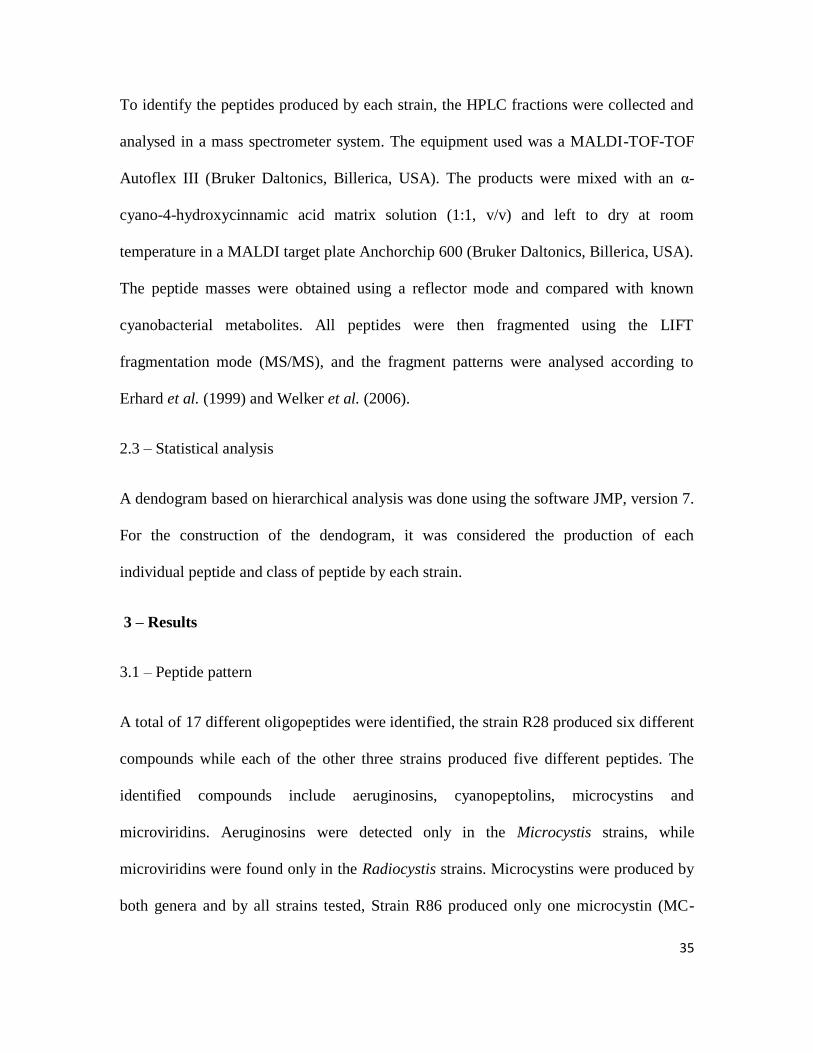
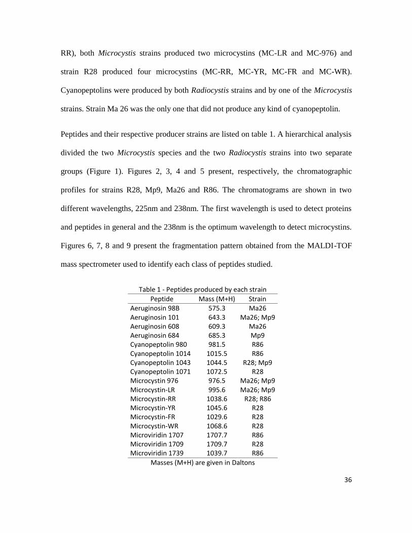
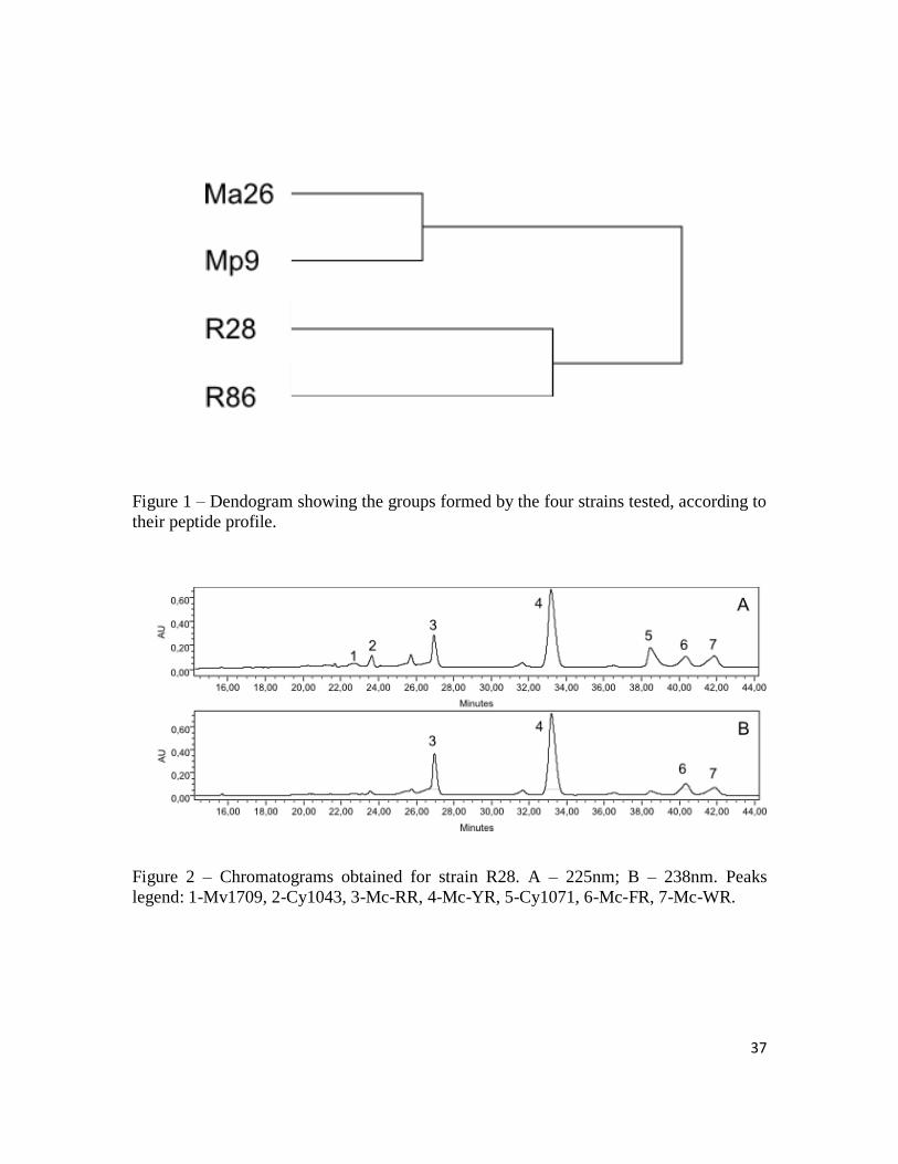
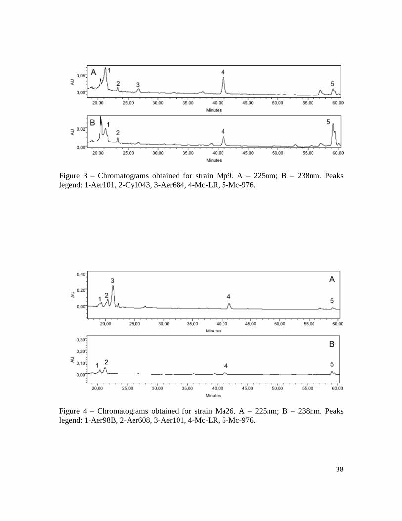
Peptides and their respective producer strains are listed on table 1. A hierarchical analysis
divided the two Microcystis species and the two Radiocystis strains into two separate
groups (Figure 1). Figures 2, 3, 4 and 5 present, respectively, the chromatographic
profiles for strains R28, Mp9, Ma26 and R86. The chromatograms are shown in two
different wavelengths, 225nm and 238nm. The first wavelength is used to detect proteins
and peptides in general and the 238nm is the optimum wavelength to detect microcystins.
Figures 6, 7, 8 and 9 present the fragmentation pattern obtained from the MALDI-TOF
mass spectrometer used to identify each class of peptides studied.
Table 1 - Peptides produced by each strain
Peptide Mass (M+H) Strain
Aeruginosin 98B 575.3 Ma26 Aeruginosin 101 643.3 Ma26; Mp9 Aeruginosin 608 609.3 Ma26 Aeruginosin 684 685.3 Mp9 Cyanopeptolin 980 981.5 R86 Cyanopeptolin 1014 1015.5 R86 Cyanopeptolin 1043 1044.5 R28; Mp9 Cyanopeptolin 1071 1072.5 R28 Microcystin 976 976.5 Ma26; Mp9 Microcystin-LR 995.6 Ma26; Mp9 Microcystin-RR 1038.6 R28; R86 Microcystin-YR 1045.6 R28 Microcystin-FR 1029.6 R28 Microcystin-WR 1068.6 R28 Microviridin 1707 1707.7 R86 Microviridin 1709 1709.7 R28 Microviridin 1739 1039.7 R86
Masses (M+H) are given in Daltons
37
Figure 1 – Dendogram showing the groups formed by the four strains tested, according to
their peptide profile.
Figure 2 – Chromatograms obtained for strain R28. A – 225nm; B – 238nm. Peaks
legend: 1-Mv1709, 2-Cy1043, 3-Mc-RR, 4-Mc-YR, 5-Cy1071, 6-Mc-FR, 7-Mc-WR.
38
Figure 3 – Chromatograms obtained for strain Mp9. A – 225nm; B – 238nm. Peaks
legend: 1-Aer101, 2-Cy1043, 3-Aer684, 4-Mc-LR, 5-Mc-976.
Figure 4 – Chromatograms obtained for strain Ma26. A – 225nm; B – 238nm. Peaks
legend: 1-Aer98B, 2-Aer608, 3-Aer101, 4-Mc-LR, 5-Mc-976.
39
Figure 5 – Chromatograms obtained for strain R86. A – 225nm; B – 238nm. Peaks
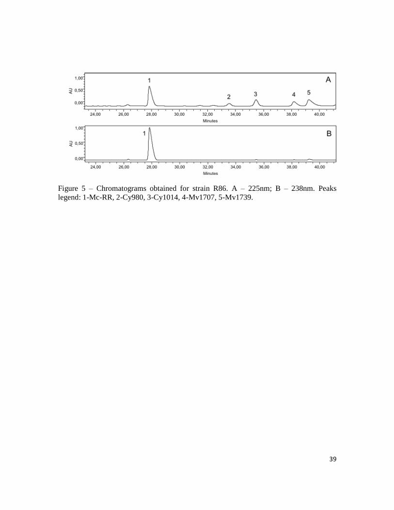
legend: 1-Mc-RR, 2-Cy980, 3-Cy1014, 4-Mv1707, 5-Mv1739.
40
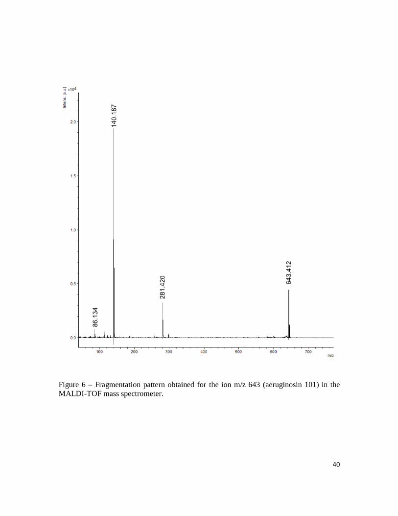
Figure 6 – Fragmentation pattern obtained for the ion m/z 643 (aeruginosin 101) in the
MALDI-TOF mass spectrometer.
41
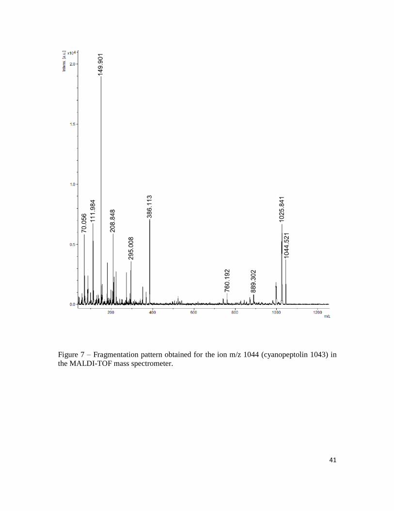
Figure 7 – Fragmentation pattern obtained for the ion m/z 1044 (cyanopeptolin 1043) in
the MALDI-TOF mass spectrometer.
42
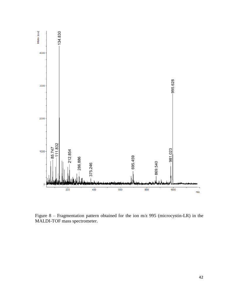
Figure 8 – Fragmentation pattern obtained for the ion m/z 995 (microcystin-LR) in the
MALDI-TOF mass spectrometer.
43
Figure 9 – Fragmentation pattern obtained for the ion m/z 1709 (microviridin 1709) in the
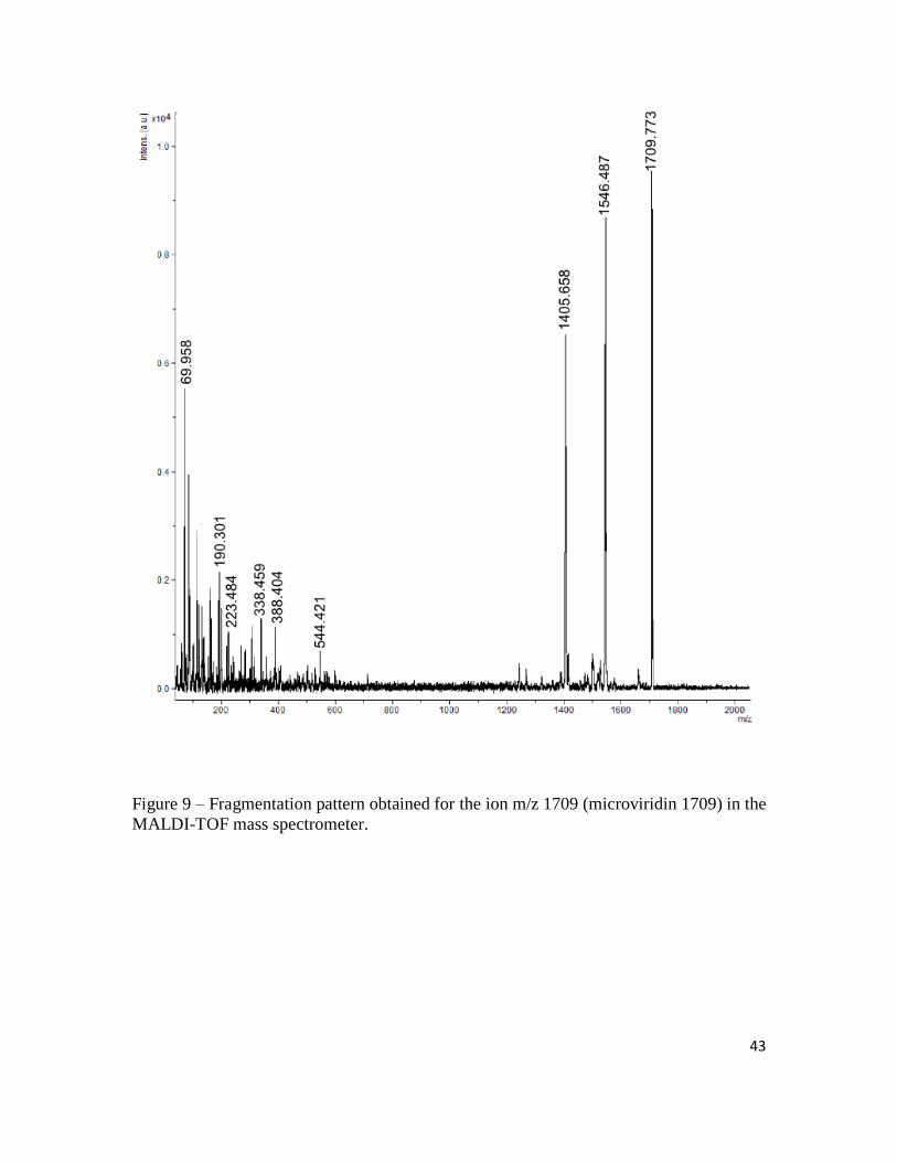
MALDI-TOF mass spectrometer.
44
4 – Discussion
As research progresses new compounds produced by cyanobacteria are discovered, and
some of these substances are already being proved as toxic to humans and other
mammals (Welker & von Döhren, 2006). Other important characteristic of cyanobacteria
is the ability to form blooms in some situations, especially in water bodies suffering from
nutrient enrichment (Paerl et al., 2001). The production of toxic compounds coupled with
the capacity of forming blooms is a growing concern worldwide not only for human
health but also for the entire aquatic trophic chain.
The Microcystis genus is known for its capacity to grow in eutrophic environments,
usually forming blooms (Oliver & Ganf, 2002). Several studies were done concerning the
production of secondary metabolites in this genus (Fastner et al., 2001; Welker et al.,
2004a; Welker et al., 2006), and these findings showed that Microcystis species are able
to produce several kinds of peptides of different classes and that a single strain may
produce several peptides at the same time. Radiocystis (Komárek & Komárková-
Legenerová, 1993) is a genus known to form blooms in tropical regions (Sant´Anna et
al., 2008). It is not as common as Microcystis and as a consequence, studies about this
genus are less frequent. Regarding the production of secondary metabolites, it is known
that it is able to synthesize microcystins and other peptides (Vieira et al., 2003;
Lombardo et al., 2006; Pereira et al., 2012). In this study all strains produced from five to
seven peptides of three different classes: aeruginosins, cyanopeptolins and microcystins
for the Microcystis strains and cyanopeptolins, microcystins and microviridins for the
Radiocystis strains. It is known that cyanobacterial oligopeptides can be found in all
45
sections of cyanobacteria and have a patchy distribution, which is expected for true
secondary metabolites (Christiansen et al., 2001). In this work no aeruginosins were
found in the Radiocystis strains, while no microviridins were found in the Microcystis
strains and cyanopeptolins could not be found in the Microcystis aeruginosa strain. The
lack of certain classes of peptides in some strains might be explained by gene deletion
events. For example, the microcystin gene cluster is a very ancient unit that was probably
present in the common ancestor of modern Anabaena, Microcystis, Nostoc and
Planktothrix (Rantala et al., 2004), and the gene deletion events already proved for
microcystins (Christiansen et al., 2008) might also be expected to occur for other non-
ribossomal peptides.
Some authors (Fastner et al., 2001; Welker et al., 2006) discussed the use of the peptidic
pattern as distinctive chemotypes and the possibility to associate them with known
morphospecies of Microcystis. Because of the wide variety of peptides produced by a
single strain this approach appears to be very complicated. The number of strains used in
this work is not suited for a correlation between chemotypes and morphospecies, but a
simple dendogram based on hierarchical analysis done with the peptide classes and the
individual peptides produced by each strain showed that both species of Microcystis and
the two strains of Radiocystis fernandoii form two separate groups. While the Microcystis
species are the only ones that produce aeruginosins and share the same microcystins, the
Radiocystis are the only ones that produce microviridins and both of them produce the
microcystin-RR. This result suggests that in a study with a wider range of species and
genera it might be possible to correlate the chemotypes with different genera.
46
In the other hand, it may be possible to correlate chemotypes with ecotypes, as suggested
by Welker et al., (2004b), since chemotypes can also be regarded as evolutionary units,
and their interactions would resemble competitive interactions among species more than
co-operative interactions between clonal cells, thus justifying the term “community” to
define their individual colonies in a sample. Therefore, if a strain can be characterized by
its chemical profile as a chemotype and if an ecotype is defined as a group of
microorganisms that shares similar ecological characteristics, these two Radiocystis
strains and Microcystis species would be chemotypes representing different ecotypes,
each one probably adapted to certain environmental conditions.
The majority of studies that focus on identifying cyanobacterial peptides usually use
mass spectrometry. Fastner et al. (2001) and Welker et al. (2004b) findings were
obtained from the use of this approach. Using a MALDI-TOF system, they were able to
analyze several samples of colonies and filaments of cyanobacteria. Although this
method allows a quick examination of a great number of samples it has some
disadvantages. The use of mass spectrometry alone shows only the major peptides
produced by each sample, while compounds that are produced in lower quantities may be
confused with noise signals because of the peaks produced by the major peptides. This
work used liquid chromatography (HPLC) technique coupled with a MALDI-TOF mass
spectrometer, and even if this form of analysis is more laborious and needs a great
amount of material for each sample, it allows a previous separation of the compounds
before the analysis in the MS system. The compound separation permits the isolation and
characterization of minor compounds that would be “invisible” in a situation with no
47
previous separation. The use of HPLC also allows the quantification of the compounds,
which might be very difficult to accomplish with mass spectrometry alone.
So far, more than 600 different peptides were isolated and identified in cyanobacteria
(Welker et al., 2006). Most of these 600 compounds were identified in strains and
environmental samples from North America and Europe, thus there is a lack of
information for other parts of the globe and there are probably a lot of different
compounds still to be found. In this study of only four strains, for example, we found two
aeruginosins (Aer-608 and Aer-684), one cyanopeptolin (Cy-1043) and even one
microcystin (Mc-976) that are not yet been described in the literature.
48
References
Botes DP, Tuinman AA, Wessels PL, Viljoen CC & Kruger H (1984) The structure of
cyanoginosin-LA, a cyclic heptapeptide toxin from the cyanobacterium Microcystis
aeruginosa. J Chem Soc Perkin Trans 1: 2311–2318.
Christiansen G, Dittmann E, Ordorika LV, Rippka R, Herdman M & Borner T (2001)
Nonribosomal peptide synthetase genes occur in most cyanobacterial genera as evidenced
by their distribution in axenic strains of the PCC. Arch Microbiol 178: 452–458.
Christiansen G, Molitor C, Philmus B & Kurmayer R (2008) Nontoxic strains of
cyanobacteria are the result of major gene deletion events induced by transposable
elements. Mol Biol Evol 25: 1695–1704.
Erhard M, von Döhren H & Jungblut PR (1999) Rapid identification of the new
anabaenopeptin G from Planktothrix agardhii HUB 011 using matrix-assisted laser
desorptiom/ionization time-of-flight mass spectrometry. Rapid Commun Mass Spectrom
13: 337–343.
Fastner J, Erhard M & von Döhren H (2001) Determination of oligopeptides diversity
within a natural population of Microcystis spp. (cyanobacteria) by typing single colonies
by matrix-assisted laser desorption ionization-time of flight mass spectrometry. Appl
Environ Microbiol 67: 5069–5076.
Finking R & Marahiel MA (2004) Biosynthesis of nonribosomal peptides. Annu Rev
Microbio 58: 453-488.
49
Guillard RR & Lorenzen CJ (1972) Yellow-green algae with chlorophyllide c. J Phycol
8: 10–14.
Ishida K, Okita Y, Matsuda H, Okino T & Murakami M (1999) Aeruginosins, protease
inhibitors from the cyanobacterium Microcystis aeruginosa. Tetrahedron 55: 10971–
10988.
Ishida K, Christiansen G, Yoshida WY, Kurmayer R, Welker M, Valls N, Bonjoch J,
Hertweck C, Börner T, Hemscheidt T & Dittmann E (2007) Biosynthesis and structure of
aeruginoside 126A and 126B, cyanobacterial peptide glycosides bearing a 2-carboxi-6-
hydroxyoctahydroindole moiety. Chem Biol 14: 565–576.
Ishitsuka M O, Kusumi T, Kakisawa H, Kaya K & Watanabe MM (1990) Microviridin: a
novel tricyclic depsipeptide from the toxic cyanobacterium Microcystis viridis. J Am
Chem Soc 112: 8180–8182.
Jakobi C, Oberer L, Quiquerez C, K¨onigWA &Weckesser J (1995) Cyanopeptolin S, a
sulfate-containing depsipeptide from a water bloom of Microcystis sp. FEMS Microbiol
Lett 129: 129–134.
Komárek J & Komárková-Legenerová J (1993) Radiocystis fernandoii, a new planktic
cyanoprokariotic from tropical freshwater reservoirs. Preslia 65: 355–357.
Lawton LA & Edwards C (2001) Purification of microcystins. J Chrom A 912: 191–209.
Lombardo M, Pinto FCR, Vieira JMS, Honda RY, Pimenta AMC, Bemquerer MP,
Carvalho LR & Kiyota S (2006) Isolation and structural characterization of microcystin-
50
LR and three minor oligopeptides simultaneously produced by Radiocystis fernandoi
(Chroococcales, Cyanobacteriae): A Brazilian toxic cyanobacterium. Toxicon 47: 560–
566.
Marahiel MA, Stachelhaus T & Mootz HD (1997) Modular peptide synthetases involved
in nonribosomal peptide synthesis. Chem Rev 97: 2651–2673.
Martin C, Oberer L, Ino T, König WA, Busch M & Weckesser J (1993) Cyanopeptolins,
new depsipeptides from the cyanobacterium Microcystis PCC 7806. J Antibiot 46 :
1550–1556.
Moffitt MC & Neilan BA (2001) On the presence of peptide synthetase and polyketide
synthetase genes in the cyanobacterial genus Nodularia. FEMS Microbiol Lett 196: 207–
214.
Murakami M, Okita Y, Matsuda H & Yamaguchi K (1994) Aeruginosin 298-A, a
thrombin and trypsin inhibitor from the blue-green alga Microcystis aeruginosa (NIES-
298). Tetrahedron lett 35: 3129–3132.
Namikoshi M, Sivonen K, Evans WR, Carmichael WW, Sun F, Rouhiainen L,
Luukkainen R & Rinehart KL (1992) Two new L-Serine variants of microcystin-LR and
-RR from Anabaena sp. strains 202 A1 and 202 A2. Toxicon 30: 1457–1464.
Oliver RL & Ganf GG (2002) Freshwater blooms. The Ecology of Cyanobacteria – Their
Diversity in Time and Space. (Whitton BA & Potts M, eds), pp. 149-194. Kluwer
Academic Publishers.
51
Okino T, Matsuda H, Murakami M & Yamaguchi K (1993) Microginin, an angiotensin-
converting enzyme inhibitor from the blue-green alga Microcystis aeruginosa.
Tetrahedron Lett 34: 501– 504.
Paerl HW, Fulton RS III, Moisander PH et al (2001) Harmful freshwater algal blooms,
with an emphasis on cyanobacteria. The Scientific World 1: 76–113
Pereira DA, Pimenta AMC & Giani A (2012) Profiles of toxic and non-toxic
oligopeptides of Radiocystis fernandoii (Cyanobacteria) exposed to three different light
intensities. Microbiol Res 167: 413–421.
Philmus B, Christiansen G, Yoshida WY & Hemscheidt TK (2008) Post-translational
modifications in microviridin biosynthesis. Chem Biochem 9: 3066–3073.
Rantala A, Fewer D, Hisbergues M, Rouhiainen L, Vaitomaa J, B¨orner T & Sivonen K
(2004) Phylogenetic evidence for the early evolution of microcystin synthesis. Proc Natl
Acad Sci USA 101: 568–573.
Rouhiainen L, Paulin, L, Suomalainen S, Hyytiainen H, Buikema W, Haselkorn R &
Sivonen K (2000) Genes encoding synthetases of cyclic desipeptides, anabaenopeptilides
in Anabaena strain 90. Mol Microbiol 37: 156–167.
Rouhiainen L, Vakkilainen T, Siemer BL, Buikema W, Haselkorn R & Sivonen K (2004)
Genes coding for hepatotoxic heptapeptides (microcystins) in the cyanobacterium
Anabaena strain 90. Appl Environ Microbiol 70: 686–692.
52
Rounge TB, Rohrlack T, Tooming-Klunderud A, Kristensen T & Jacobsen KS (2007)
Comparison of cyanopeptolin genes in Planktothrix, Microcystis and Anabaena strains:
evidence for independent evolution within each genus. Appl Environ Microbiol 73: 7322–
7330.
Sant'Anna CL, Azevedo MTP, Werner VR, Dogo CR, Rios FR & de Carvalho LR (2008)
Review of toxic species of Cyanobacteria in Brazil. Arch Hydrobiol Suppl Algol Stud
126: 251–265.
Schwarzer D, Finking R & Marahiel MA (2003) Nonribosomal peptides: from genes to
product. Nat Prod Rep 20: 275–287.
Shin HJ, Matsuda H, Murakami M & Yamaguchi K (1997) Aeruginosins 205A and -B,
serine protease inhibitory glycopeptides from the cyanobacterium Oscillatoria agadhii
(NIES-205). J Org Chem 62: 1810–1813.
Stachelhaus T, Mootz HD, Bergendahl V & Marahiel MA (1998) Peptide bond formation
in nonribosomal peptide biosynthesis. Catalytic role of condensation domain. J Biol
Chem 273: 22773-22781.
Tillett D, Dittmann E, Erhard M, von Döhren H, Börner T & Neilan BA (2000) Structural
organization of microcystin biosynthesis in Microcystis aeruginosa PCC 7806: an
integrated peptide-poliketide synthetase system. Chem Biol 7: 753–764.
Tooming-Klunderud A, Rohrlack T, Shalchian-Tabrizi K, Kristensen T & Jacobsen KS
(2007) Structural analysis of a non-ribosomal halogenated cyclic peptide and its puptative
53
operon from Microcystis: implications for evolution of cyanopeptolins. Microbiology
153: 1382–1393.
Vieira JMS, Azevedo MT P, Azevedo SMFO, Honda RY & Corrêa B (2003) Microcystin
production by Radiocystis fernandoii (Chroococales, Cyanobacteria) isolated from a
drinking water reservoir in the city of Belém, PA, Brazilian Amazonia region. Toxicon
42: 709–713.
von Döhren H, Keller U, Vater J & Zocher R (1997) Multifunctional peptide syntethases.
Chem Rev 97: 2675-2705.
Weber T & Marahiel MA (2001) Exploring the domain structure of modular
nonribosomal peptide synthetase. Structure 9: R3-R9.
Welker M, Brunke M, Preussel K, Lippert I & von Döhren H (2004a) Diversity and
distribution of Microcystis (cyanobacteria) oligopeptides chemotypes from natural
communities studied by single mass spectrometry. Microbiology 150: 1785–1796.
Welker M, Christiansen G & von D¨ohren H (2004b) Diversity of coexisting Planktothrix
(Cyanobacteria) chemotypes deduced by mass spectral analysis of microcystins and other
oligopeptides. Arch Microbiol 182: 288–298.
Welker M & von Döhren H (2006) Cyanobacterial peptide: nature’s own combinatorial
biosynthesis. FEMS Microbiol Rev 30: 530–563.

54
Welker M, Maršálek B, Šejnohová L & von Döhren H (2006) Detection and
identification of oligopeptides in Microcystis (cyanobacteria) colonies: Toward an
understanding of metabolic diversity. Peptides 27: 2090–2103.
55
Capítulo 2
CELL DENSITY DEPENDENT
OLIGOPEPTIDE PRODUCTION IN
CYANOBACTERIAL STRAINS
Artigo aceito para publicação no periódico FEMS Microbiology Ecology
(doi: 10.1111/1574-6941.12281)
56
Abstract
Cyanobacteria can form blooms and in these situations they dominate the
phytoplanktonic community, reaching extremely high densities. In the domain Bacteria,
high population densities can stimulate a phenomenon known as quorum sensing, which
may produce several modifications in the cell physiology. Very little is known about
quorum sensing in cyanobacteria. Because of their planktonic way of life, quorum
sensing should be more evident during a bloom event. In this work, we tested whether
cell density could shape the production of bioactive compounds produced by
cyanobacteria. The experiments consisted of two treatments, where cultures of
cyanobacteria were maintained at low and high cellular densities through a semi-
continuous set-up. Analyses were performed by HPLC-PDA and MALDI-TOF MS.
Seventeen peptides were detected and 14 identified, including microcystins, aeruginosins,
cyanopeptolins and microviridins. The results showed that cellular density seems to have
a significant effect on the peptides production. Most of the compounds had significantly
higher cellular quotas in the higher density treatment, although microviridins and an
unknown peptide were produced only at low density. These results may hint at a possible
role for quorum sensing in triggering the production of several cyanobacterial peptides.
57
1-Introduction
Cyanobacteria can form blooms in aquatic environments such as lakes, reservoirs, and
oceans; these situations consist of dominance of the phytoplankton community by one or
a few cyanobacteria species and an exponential increase in the biomass of the dominant
group (Oliver & Ganf, 2002). Blooms have received considerable worldwide attention,
because they are natural events in which populations of cyanobacteria can reach
extremely high densities. Since most blooms are toxic, they represent a severe problem. It
is acknowledged that nutrient enrichment of aquatic ecosystems promotes their
appearance and persistence (Paerl et al., 2001). Phosphorous has been considered the
primary nutrient source responsible for algal biomass accumulation in freshwater systems
(Schindler et al., 2008) and together with nitrogen it may support the development of
blooms when present in excess (Downing et al., 2001). Besides nutrient-rich conditions,
other factors favour intensive cyanobacterial growth, like temperatures above 20°C,
persistent vertical stratification, calm surface waters and low flushing rates (Reynolds,
1987; Paerl, 1988; Shapiro, 1990). After its establishment, a bloom can persist for several
months and in tropical regions they can in some extreme situations become permanent
(McGregor & Fabbro, 2000; Figueredo & Giani, 2009).
Cyanobacteria can produce several kinds of oligopeptides whose synthesis is usually
achieved by non ribosomal peptide synthetase (NRPS) systems (for a full review see
Welker & von Döhren, 2006). The major classes of these peptides are aeruginosins
(Murakami et al., 1994), cyanopeptolins (Martin et al., 1993), microginins (Okino et al.,
1993), microviridins (Ishitsuka et al., 1990), anabaenopeptins (Harada et al., 1995) and
microcystins (Carmichael, 1992). The functions of these compounds are still unclear;
58
however it is known that some peptides are toxic to zooplankton (Agrawal et al., 2001;
Czarnecki et al., 2006) and that microcystins might be related to physiological processes
such as photosynthesis (Long et al., 2001; Young et al., 2005) and iron metabolism
(Martin-Luna et al., 2006). Microcystins are also toxic to humans and other mammals
(Sivonen & Jones, 1999). The increase of cyanobacterial blooms and the toxic nature of
some of their peptides create major public health and water treatment problems (Watson
et al., 2000; Watson, 2004).
There is a substantial amount of information and research on the factors controlling and
affecting the appearance and persistence of cyanobacterial blooms. In addition, the
knowledge about the production of microcystins and other cyanobacterial peptides is
increasing. However, there is not much information about possible changes in the
physiology of the cyanobacterial cells during a bloom event and its potential connection
with the production of secondary metabolites. Some authors have previously observed
changes in the relative amount of toxic genotypes in a bloom (Briand et al., 2008; Okello
et al., 2010; Sabart et al., 2010; Pimentel & Giani, 2013). Furthermore Wood et al.
(2011), found that the expression of the mcyE gene during a bloom event was not
constitutive, but was influenced by cell concentration and it could produce increases of
up to 28 fold in microcystin levels. In mesocosm experiments, Wood et al. (2012) also
observed that microcystin cell quota increased significantly with cell density.
Several bacterial species have the ability to release signalling compounds in their
environment (Waters & Bassler, 2005) and when these molecules reach a threshold, they
trigger a coordinated response able to change the gene expression pattern of the affected

59
cells and, as a consequence, the metabolism and physiology of the bacterial population.
The term quorum sensing is used to describe this density-dependent phenomenon (Fuqua
et al., 1994). It is known that quorum sensing can control different biological functions as
motility, aggregation, swarming, conjugation, luminescence, virulence, symbiosis,
biofilm differentiation, antibiotics biosynthesis and others (Swift et al., 2001; Waters &
Bassler 2005; Williams et al., 2007). Up to date a wide variety of molecules responsible
for quorum sensing in bacteria have been isolated (for a review, see Williams et al.,
2007). The phenomenon of quorum sensing is believed to be widespread in the bacteria
domain (Miller & Bassler, 2001), but concerning cyanobacteria there is still a lack of
information.
In this work, we examined whether low and high cellular densities could affect peptide
concentrations under semi-continuous experimental conditions. The idea was that
experimentally-maintained high density cultures would mimic a situation similar to a
bloom event. The experiments were run using three different species of cyanobacteria,
isolated from Brazilian freshwater systems.
2-Methodology
2.1 – Strains
The strains used in these experiments were Microcystis panniformis (strain Mp9),
Microcystis aeruginosa (strain Ma26) and a Radiocystis fernandoii (strain R28).
Microcystis is one of the most studied genera and is known for its ability to form blooms
(Oliver & Ganf, 2002) and produce secondary metabolites such as microcystins and
others peptides (Welker et al., 2006). Radiocystis (Komárek & Komárková- Legenerová,
60
1993) is less common and less studied than Microcystis, but it is known to form blooms
in tropical regions (Sant´Anna et al., 2008) and produce microcystins and other peptides
(Vieira et al., 2003; Lombardo et al., 2006; Pereira et al., 2012). All strains are
maintained in the culture collection of the Phycology Laboratory of the Botany
Department in the Federal University of Minas Gerais and were isolated from Furnas
reservoir (20o40’S; 46
o 19’W), located in the south-eastern region of Brazil.
2.2-Experiments
Experiments were performed in two treatments, low cell density and high cell density.
Each treatment was prepared in triplicate and received a different amount of inoculum,
varying from 20 to 100 mL, in order to create two different cell densities, and the final
volume was completed to 250 mL with WC medium (Guillard & Lorenzen, 1972).
Growth conditions were 12h light: 12h dark photoperiod, temperature 20±1 °C and 65
µmol.m-2
.s-1
of irradiance. To avoid differences between experiments due to faster
nutrient consumption in the higher density treatment, experiments were conducted in
semi-continuous cultures. Semi-continuous cultures allow the maintenance of a constant
level of biomass, and nutrient-replete growth under controlled conditions. Every other
day, a constant volume of the experimental culture was removed and the same amount of
fresh medium was added, to maintain the low or high cell density conditions within fixed
limits. Under these settings, the experiments ran for 6 days, and cultures were kept at
low and high cell density and exhibited similar growth rates. Samples were taken every
two days to follow growth and a minimum of 400 cells were counted in a Fuchs-
Rosenthal hemocytometer. A 20 mL sample was filtered on the last day and used to
61
measure chlorophyll, according to the methodology described by Nusch (1980). The rest
of the culture was freeze-dried for further biochemical analyses.
2.3-Biochemical analysis
The dry material was extracted three times with methanol 75% (v/v). The procedure was
undertaken with sonication on ice followed by centrifugation (15 min, 8900 g). The
extract was purified by reverse phase chromatography using SPE-C18 cartridges (Waters,
Sep-Pak Vac 3cc – 500 mg) as described by Lawton & Edwards (2001). The purified
extract was dried in Speed-Vac system and diluted in a known volume of methanol 75%
(v/v). The analyses were done using HPLC (Waters Alliance 2695) with a photodiode
array detector (PDA - Waters 2996) at 225 nm and 238 nm wavelength. The column used
was a Waters Symmetry C18 (4.6 X 250 mm I.D., 5 µm ODS). Mobile phase A was
acetonitrile, containing 0.1% (v/v) trifluoroacetic acid (TFA), and mobile phase B was
water, containing 0.1% (v/v) trifluoroacetic acid (TFA). The chromatographic run
consisted of a linear gradient from 30% A to 34% in 33.5 minutes then 40% for 6.5
minutes with a flow-rate of 1 mL. min-1
. The peptide quantification was done dividing the
peak area by the dry weight of the lyophilized material, obtaining a measurement of the
relative change in the peptide concentration; this method was chosen because of the lack
of standards for most peptides. Dry weight was used to standardize the measurements,
since in previous experiments it showed high correlation with cellular biovolume (Pereira
et al., 2012). This relationship biovolume/dry weight was tested statistically by regression
analysis and results showed highly significant correlation for all strains (R2 ≥0.9).
62
Peptides were identified by collecting the HPLC fractions and submitting them to
analysis in a MALDI-TOF-TOF Autoflex III mass spectrometer (Bruker Daltonics,
Billerica, USA). The products were mixed with α-cyano-4-hydroxycinnamic acid matrix
solution (1:1, v/v) and left to dry at room temperature in a MALDI target plate
Anchorchip 600 (Bruker Daltonics, Billerica, USA). The peptide masses were obtained
using a reflector mode and compared with known cyanobacterial metabolites. Known and
unknown peptides were then fragmented using the LIFT fragmentation mode (MS/MS),
and the fragment patterns were analysed according to Erhard et al. (1999) and Welker et
al. (2006).
2.4-Statistics
ANOVA tests were used to compare the means of the two treatments (high and low cell
densities). The parameters analysed were: peptide concentration (area.mg-1
), cell density
(cell. mL-1
), chlorophyll (µg.mg-1
) and growth rate (.day-1
). The statistical analyses were
performed with JMP version 7 software.
3-Results
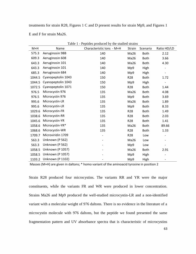
Fourteen peptides were identified according to their fragmentation pattern and UV
absorbance: 4 aeruginosins, 2 cyanopeptolins, 7 microcystins and 1 microviridin. Three
metabolites could not yet be identified. Some peptides were produced at both high and
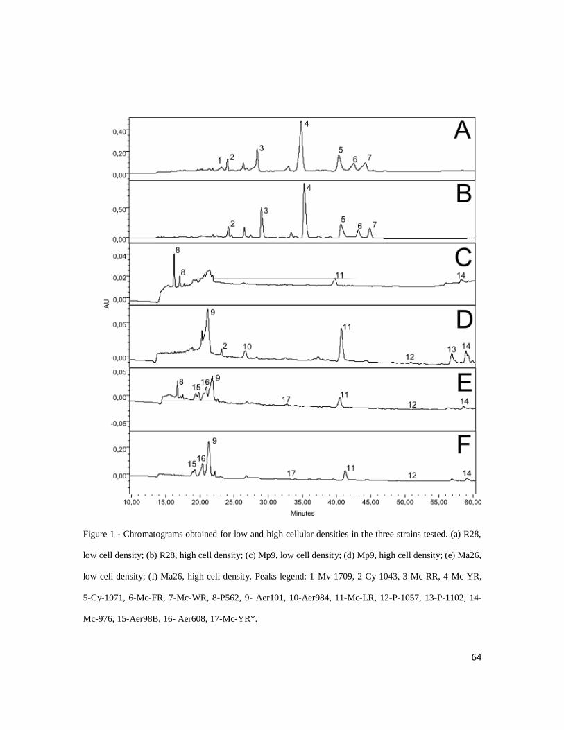
low cell density, while others appeared in only in one of the treatments (Table 1). Figures
1 A and B show the chromatographic pattern found in low and high cell density
63
treatments for strain R28, Figures 1 C and D present results for strain Mp9, and Figures 1
E and F for strain Ma26.
Table 1 - Peptides produced by the studied strains
M+H Name Characteristic Ions - M+H Strain Scenario Ratio HD/LD
575.3 Aeruginosin 98B 140 Ma26 Both 2.12
609.3 Aeruginosin 608 140 Ma26 Both 3.66
643.3 Aeruginosin 101 140 Ma26 Both 4.30
643.3 Aeruginosin 101 140 Mp9 High -
685.3 Aeruginosin 684 140 Mp9 High -
1044.5 Cyanopeptolin 1043 150 R28 Both 1.72
1044.5 Cyanopeptolin 1043 150 Mp9 High -
1072.5 Cyanopeptolin 1071 150 R28 Both 1.44
976.5 Microcystin 976 135 Ma26 Both 4.08
976.5 Microcystin 976 135 Mp9 Both 3.69
995.6 Microcystin-LR 135 Ma26 Both 1.89
995.6 Microcystin-LR 135 Mp9 Both 8.33
1029.6 Microcystin-FR 135 R28 Both 1.49
1038.6 Microcystin-RR 135 R28 Both 2.03
1045.6 Microcystin-YR 135 R28 Both 1.41
1058.6 Microcystin-YR* 135 Ma26 Both 89.66
1068.6 Microcystin-WR 135 R28 Both 1.33
1709.7 Microviridin 1709 - R28 Low -
563.3 Unknown (P 562) - Ma26 Low -
563.3 Unknown (P 562) - Mp9 Low -
1058.5 Unknown (P 1057) - Ma26 Both 2.91
1058.5 Unknown (P 1057) - Mp9 High -
1103.2 Unknown (P 1102) - Mp9 High -
Masses (M+H) are given in daltons; * homo variant of the aminoacid tyrosine in position 2
Strain R28 produced four microcystins. The variants RR and YR were the major
constituents, while the variants FR and WR were produced in lower concentration.
Strains Ma26 and Mp9 produced the well-studied microcystin-LR and a non-identified
variant with a molecular weight of 976 daltons. There is no evidence in the literature of a
microcystin molecule with 976 daltons, but the peptide we found presented the same
fragmentation pattern and UV absorbance spectra that is characteristic of microcystins
64
Figure 1 - Chromatograms obtained for low and high cellular densities in the three strains tested. (a) R28,
low cell density; (b) R28, high cell density; (c) Mp9, low cell density; (d) Mp9, high cell density; (e) Ma26,
low cell density; (f) Ma26, high cell density. Peaks legend: 1-Mv-1709, 2-Cy-1043, 3-Mc-RR, 4-Mc-YR,
5-Cy-1071, 6-Mc-FR, 7-Mc-WR, 8-P562, 9- Aer101, 10-Aer984, 11-Mc-LR, 12-P-1057, 13-P-1102, 14-
Mc-976, 15-Aer98B, 16- Aer608, 17-Mc-YR*.
65
(see supplementary material, supplement 1, Fig. 1). Strain Ma26 also produced a
different microcystin-YR, which seems to be a homovariant showing the amino acid
tyrosine in position 2. This compound had a molecular mass of 1058 daltons, which is the
same mass of a non indentified peptide (peptide 1057) produced by strains Ma26 and
Mp9. However, the microcystin presented the fragmentation pattern and UV absorbance
spectra that are typically found in microcystins, while the peptide 1057 had different
fragmentation pattern and UV absorbance spectra. Furthermore, retention time was
different for each one of the two compounds, characterizing them as different peptides.
Among aeruginosins, we found a total of four variants, aeruginosin 98B, aeruginosin 101
and aeruginosin 608 were produced by strain Ma26 and aeruginosin 684 and aeruginosin
101 were produced by strain Mp9. Aeruginosins were the main peptide class produced by
strain Ma26, with aeruginosin 101 being the major compound, followed by aeruginosin
608. Only two cyanopeptolins were found in the strains used in these experiments,
cyanopeptolin 1071 produced exclusively by R28 strain and cyanopeptolin 1044
produced by strains R28 and Mp9. For strain R28, both cyanopeptolins were produced at
low and high cell densities, but the production was significantly higher in the high
density treatment for cyanopeptolin 1071. In this strain, the production of cyanopeptolin
1043 was higher in the high density treatment, but not significant. However, for the strain
Mp9, the cyanopeptolin 1043 was only found in the high density treatment. It is possible
that the production of this peptide in strain Mp9, growing at low cell density, is kept at
basal levels that could not be measured by the HPLC technique for lack of sensitivity of
the method. Only one microviridin was found in the experiments, the microviridin 1709,
produced by strain R28: no other strains produced microviridins.
66
Three non-identified peptides were detected in the experiments. Their fragmentation
pattern could not be associated with any class of cyanobacterial peptides and additional
biochemical studies are needed to establish whether they belong to an existing class of
peptides or not. Among these peptides there are two compounds (P 1102 and P 1057)
that had increased production at high cellular densities and one compound that was seen
only at low cell density (P 562).
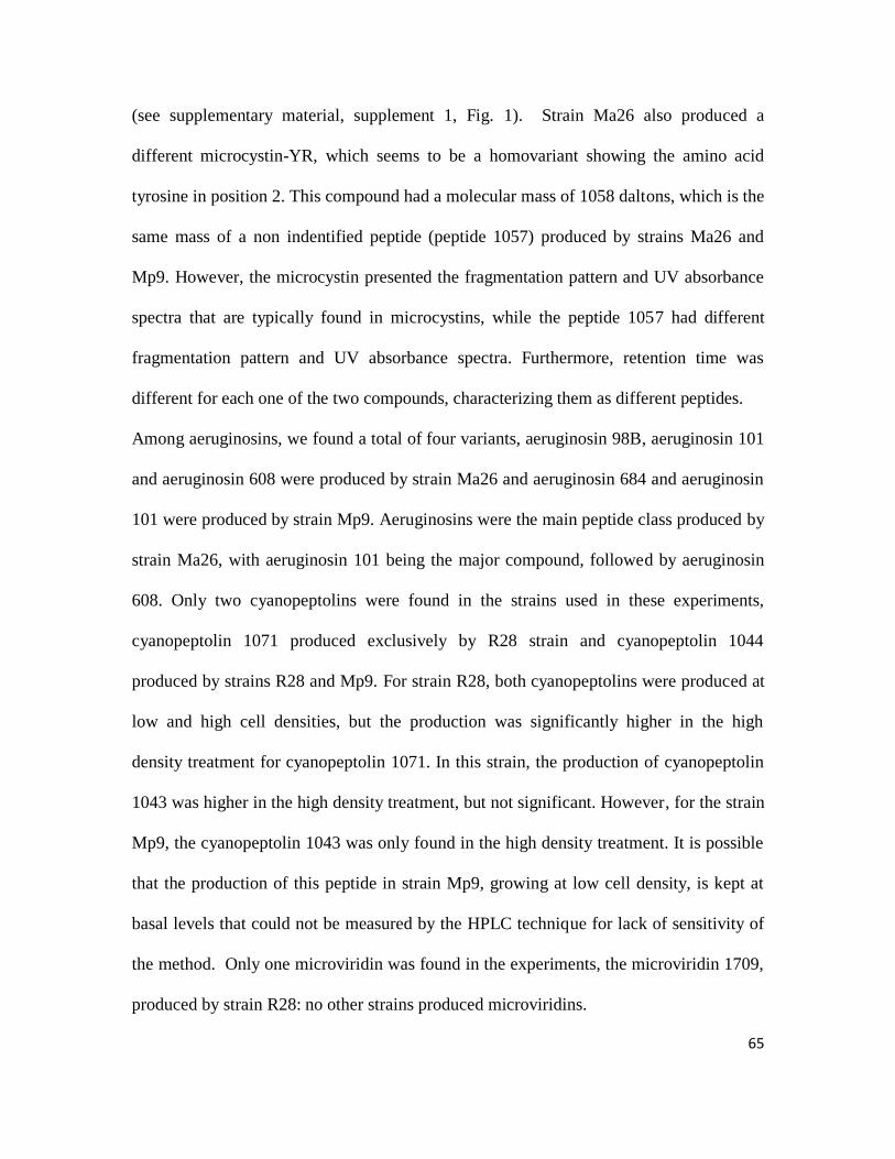
Figure 2 shows the peptide concentrations in all strains and treatments. The amount of all
substances produced by strain 28 was significantly higher in the high density treatment,
with the exception of cyanopeptolin 1043 which showed no significant difference
between treatments and the microviridin 1709 that was detected only in the low density
treatment (Figure 2-A). The amounts of microcystin-LR and microcystin 976 produced
by strain Mp9 were significantly higher in the high density treatment, the other
compounds were only detected in the high density treatment and peptide 562 was
detected in two different peaks only in the low density treatment (Figure 2-B). For all
compounds observed in strain Ma26, a significantly greater amount was produced in the
high density treatment, with the exception of the peptide 562 which was detected only in
the low density treatment (Figure 2-C and D).
The difference between cell density (cell.mL-1
), and chlorophyll concentration (µg.mg-1
)
in the low and high cell density treatments was statistically significant (Table 2). Growth
rates showed no significant difference between treatments, since cultures were
maintained stable in a semi-continuous set up. For strain Ma 26 growth rate was 0.14
(low density) and 0.16 day-1
(high density), for Mp9, 0.16 (LD) and 0.15 day-1
(HD), and
for R28 growth rate was 0.06 (LD) and 0.05 day-1
(HD).
67
Figure 2 - Peptides concentration in all strains tested under low and high cellular densities. (a) Radiocystis fernandoii 28; (b) Microcystis panniformis 9; (c) M.
aeruginosa 26; (d) M. aeruginosa 26. Detail of Mc-YR* and Mc-LR. Error bars denote standard deviations. The symbol * denotes significant differences
between means (P < 0.001).
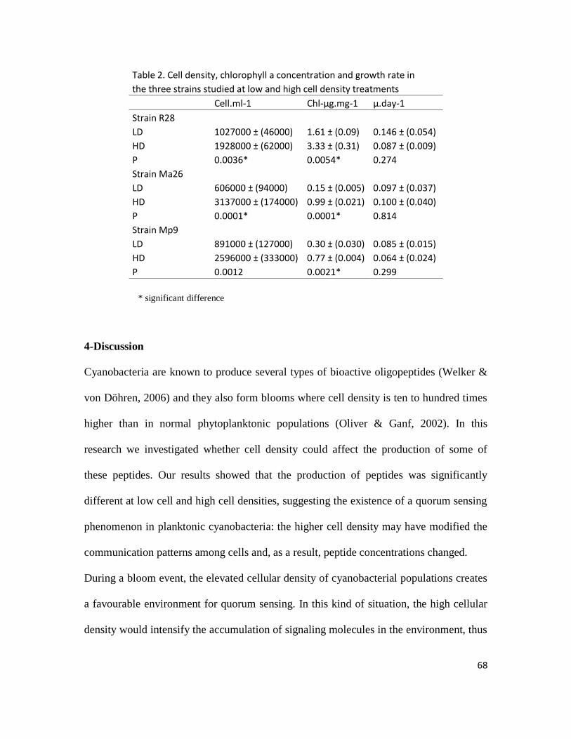
68
Table 2. Cell density, chlorophyll a concentration and growth rate in
the three strains studied at low and high cell density treatments
Cell.ml-1 Chl-µg.mg-1 µ.day-1
Strain R28 LD 1027000 ± (46000) 1.61 ± (0.09) 0.146 ± (0.054)
HD 1928000 ± (62000) 3.33 ± (0.31) 0.087 ± (0.009)
P 0.0036* 0.0054* 0.274
Strain Ma26 LD 606000 ± (94000) 0.15 ± (0.005) 0.097 ± (0.037)
HD 3137000 ± (174000) 0.99 ± (0.021) 0.100 ± (0.040)
P 0.0001* 0.0001* 0.814
Strain Mp9 LD 891000 ± (127000) 0.30 ± (0.030) 0.085 ± (0.015)
HD 2596000 ± (333000) 0.77 ± (0.004) 0.064 ± (0.024)
P 0.0012 0.0021* 0.299
* significant difference
4-Discussion
Cyanobacteria are known to produce several types of bioactive oligopeptides (Welker &
von Döhren, 2006) and they also form blooms where cell density is ten to hundred times
higher than in normal phytoplanktonic populations (Oliver & Ganf, 2002). In this
research we investigated whether cell density could affect the production of some of
these peptides. Our results showed that the production of peptides was significantly
different at low cell and high cell densities, suggesting the existence of a quorum sensing
phenomenon in planktonic cyanobacteria: the higher cell density may have modified the
communication patterns among cells and, as a result, peptide concentrations changed.
During a bloom event, the elevated cellular density of cyanobacterial populations creates
a favourable environment for quorum sensing. In this kind of situation, the high cellular
density would intensify the accumulation of signaling molecules in the environment, thus
69
optimizing the conditions for the occurrence of quorum sensing. Gobler et al. (2007)
investigated the dynamics and toxicity of cyanobacteria in a eutrophic lake in New York
and found that the expression of the mcyE gene, which is part of the microcystin operon,
was higher during periods of high cellular density and declined during months of lower
cell density. Even though the authors did not link their results with quorum sensing, their
findings may hint to a potential correlation between the existence of this phenomenon
and the mcyE gene expression. Similar results were also found by Wood et al. (2011)
who observed that higher levels of microcystin per cell (up to 28 fold) occurred when cell
concentration increased from 70,000 to 4,000,000 cells. mL-1
and corresponded to
changes in the mcyE gene expression. In mesocom experiments, Wood et al. (2012) also
detected a significant up-regulation of microcystin cell quota upon increasing the
concentration of Microcystis cells. Of course caution has to be used when correlating
laboratory experiments with natural environments, since experiments represent
conditions that are simpler than natural scenarios; nevertheless they allow the isolation
and testing of a specific factor and the understanding of single phenomena. In our
experiments, the high cell density cultures represent a bloom situation, and the results
obtained suggest significant differences in the production of secondary metabolites
between bloom and non-bloom events.
The experiments were made in semi-continuous cultures, which is an important
procedure that guarantees that a low cell density culture does not attain high cellular
density and that a high cell density culture does not reach stationary phase. This method
assured that the strains maintained their cell density almost stable during the experimental
70
period, which is evidenced by the non-significantly different growth rates measured in
both situations for all strains tested.
How the quorum sensing modulates metabolic responses in cyanobacteria is still poorly
understood, but it is known that acylhomoserine lactones (AHLs), which is a class of
autoinducers produced by several gram-negative bacteria, are produced and can influence
the physiology of the organisms. Sharif et al. (2008), for example, showed that
cyanobacteria from the genus Gloeothece produce an N-octanoyl-homoserine lactone, the
accumulation of which is able to change the expression of 43 different proteins. Romero
et al. (2011) also showed that AHLs with different side chains can inhibit nitrogen
fixation in Anabaena, while Van Mooy et al. (2012) found that phosphorous acquisition
is increased in marine Trichodesmium consortia in the presence of AHLs.
Our results are an indication of a potential link between quorum sensing and production
of secondary metabolites in cyanobacteria as shown by the different peptide pattern at
low and high cell densities. For most of the compounds measured, the production was
stimulated in the high cell density treatment, with a significant increase in concentration
usually from 1.5 up to 4 times at the high density, and in some cases even reaching 8.3
(Figure 2) and 89.6 times higher values, respectively for microcystin-LR in Mp9 (Figure
2B) and microcystin-YR* in Ma26 strain (Figure 2D). Interestingly, some peptides were
present only in the low density treatment, having their production suppressed when the
cellular density of the culture was higher, as observed for microviridin 1709 in R28 strain
and peptide 562 in Mp9 and Ma26 strains. In a mesocosm experiment, Wood et al.
(2012) found an increase in microcystin quota in situations where the cell number per mL
reached 2.9 million and 7 million. In our experiments the number of cells in the high
71
density treatment varied from 1.9 to 3.1 million per mL and even though the experiments
had different designs and conditions, the results were similar, which corroborates the idea
that microcystin production is stimulated in situations of high cellular density.
Although there is a lot of new information on cyanobacterial peptides, it is still difficult
to understand their functions in the physiology and ecology of cyanobacteria; some of
them can inhibit proteases from zooplanktonic organisms, characteristic of protection
against grazing (Agrawal et al., 2001; Rohrlack et al., 2003; von Elert et al., 2004;
Czarnecki et al., 2006). There are also indications that microcystins may be involved in
light-related photosynthetic processes (Long et al., 2001; Young et al., 2005). It is known
that no specific group of oligopeptide is essential for the growth of cyanobacteria, as
natural populations are composed of producing and non-producing strains, for each
peptide and peptide class (Fastner et al., 2001; Rohrlack et al., 2001; Welker et al., 2004)
and no clear advantage has yet been seen for producing over non producing strains
(Kaebernick et al., 2001). In our experiments we saw that the peptide pattern changes not
only in concentration, but also, in peptide composition according to the cellular density of
the culture. All strains had some peptides that appeared only in the low cell density
treatment and strain Mp9 had several peptides that exclusively appeared in high cellular
density, which is in accordance with the idea that these compounds are not essential for
survival, at least individually, although they may offer some advantage to the individual
strains in particular situations.
Microcystins are characterized by the amino acid (2S,3S,8S,9S)-3-amino-9-methoxy-
2,6,8-trimethyl-10-phenyldeca-4,6-dienoic acid (Adda, position 5) (Carmichael, 1992). In
this study, we found seven different variants of these compounds. Our experiments
72
showed that all microcystin variants were produced at both cell densities, nevertheless
their production was stimulated when the cell density was higher. Because of their
toxicity to humans and other mammals, microcystins are cause of concern for human
health and water treatment plants (Watson, 2004) and, as a consequence, a wide range of
information is already available. Our findings add new knowledge to the ecology and
physiology of this compound, because the fact that quorum sensing may stimulate the
production of microcystins is of importance to the management of lakes, reservoirs and
water treatment plants.
Aeruginosins stand as a class of compounds formed by linear peptides characterized by a
hydroxy-phenyl lactic acid (Hpla) at the N-terminus, the amino acid 2-carboxy-6-
hydroxyoctahydroindole (Choi) and an arginine derivative at the C-terminus (Murakami
et al., 1995). In strain Ma26, aeruginosins were the main peptide class. The production
pattern for aeruginosins was the same as for microcystins, with significantly higher
concentrations observed at the high density treatment. Only two cyanopeptolins were
found in the strains used in these experiments. This class of peptides is characterized by
the amino acid 3-amino-6-hydroxy-2-piperidone (Ahp) and the cyclization of the peptide
ring by an ester bond of the b-hydroxy group of threonine with the carboxy group of the
terminal amino acid (Martin et al., 1993). Cyanopeptolins also presented a production
response similar to the observed for microcystins and aeruginosins.
The resembling patterns found for microcystin, aeruginosin and cyanopeptolin may
reflect their biosynthesis route, which is based on non ribosomal peptide synthetase
systems. We observed similar responses for the three classes of peptides and each strain
had a major peptide belonging to a different class (microcystin for R28, an unknown
73
peptide for Mp9 and aeruginosin for Ma26). Such results point to the fact that these
compounds may have interchangeable functions. These observations, added to the
knowledge that cyanobacterial natural populations consist of a mix of producing and
nonproducing strains for each class of peptide (Fastner et al., 2001; Welker et al., 2004),
are good indicators of the multiplicity of profiles found in nature with potential similar
cellular function.
Microviridin is the largest known cyanobacterial oligopeptide (Ishitsuka et al.,1990), and
this class is characterized by a multicyclic structure established by secondary peptide and
ester bonds and a side chain of variable length; its amino acids are all in L-configuration
and the only non-proteinogenic unit is the N-terminal acetyl group. In our experiments
only one variant was found in strain R28 and this peptide was produced only at low cell
density, opposite to the observed for the other classes of peptides. As previously noted by
some authors (Welker & von Döhren, 2006; Philmus et al., 2008) microviridins are
synthesized ribosomally and their structure is finalized by post-translational
modifications. The different form of synthesis might be one explanation for the opposite
behaviour of this microviridin, and also an indication that its function might be different
when compared to the peptides synthesized by NRPS.
There are still many open questions concerning the complex connections between
cyanobacteria, blooms, quorum sensing and production of secondary metabolites. Genetic
studies and field experiments are needed to expand the knowledge on these interactions.
Our results suggest that quorum sensing may be an important mechanism in regulating
the physiology of bloom forming cyanobacteria and that this phenomenon can play a
significant role in the production of toxic and non-toxic peptides in these organisms.
74
Acknowledgements
This study was supported by a scholarship to D.A.P. from CAPES (Coordenação de
Aperfeiçoamento do Pessoal Docente, Brazil), and by funds provided by Furnas CNPq
(Conselho Nacional de Pesquisa), FAPEMIG (Fundação de Apoio a Pesquisa de Minas
Gerais) and CEMIG (Companhia Elétrica de Minas Gerais) to A.G.
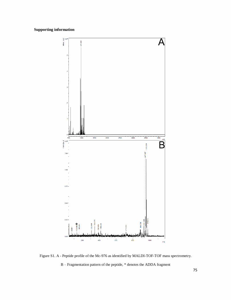
75
Supporting information
Figure S1. A - Peptide profile of the Mc-976 as identified by MALDI-TOF-TOF mass spectrometry.
B – Fragmentation pattern of the peptide, * denotes the ADDA fragment
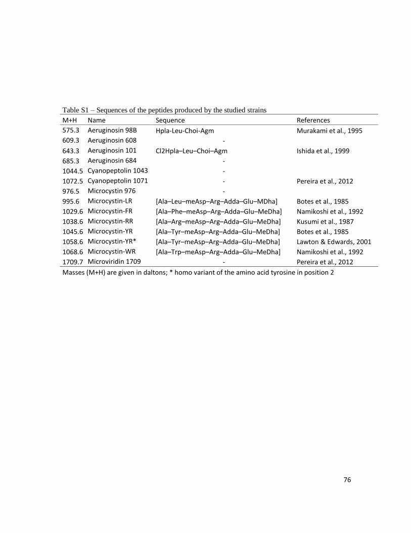
76
Table S1 – Sequences of the peptides produced by the studied strains
M+H Name Sequence References
575.3 Aeruginosin 98B Hpla-Leu-Choi-Agm Murakami et al., 1995
609.3 Aeruginosin 608 - 643.3 Aeruginosin 101 Cl2Hpla–Leu–Choi–Agm Ishida et al., 1999
685.3 Aeruginosin 684 - 1044.5 Cyanopeptolin 1043 - 1072.5 Cyanopeptolin 1071 - Pereira et al., 2012
976.5 Microcystin 976 - 995.6 Microcystin-LR [Ala–Leu–meAsp–Arg–Adda–Glu–MDha] Botes et al., 1985
1029.6 Microcystin-FR [Ala–Phe–meAsp–Arg–Adda–Glu–MeDha] Namikoshi et al., 1992
1038.6 Microcystin-RR [Ala–Arg–meAsp–Arg–Adda–Glu–MeDha] Kusumi et al., 1987
1045.6 Microcystin-YR [Ala–Tyr–meAsp–Arg–Adda–Glu–MeDha] Botes et al., 1985
1058.6 Microcystin-YR* [Ala–Tyr–meAsp–Arg–Adda–Glu–MeDha] Lawton & Edwards, 2001
1068.6 Microcystin-WR [Ala–Trp–meAsp–Arg–Adda–Glu–MeDha] Namikoshi et al., 1992
1709.7 Microviridin 1709 - Pereira et al., 2012
Masses (M+H) are given in daltons; * homo variant of the amino acid tyrosine in position 2
77
References
Agrawal MK, Bagchi D & Bagchi SN (2001) Acute inhibition of protease and
suppression of growth in zooplankter, Moina macrocopa, by Microcystis bloom collected
in central India. Hydrobiologia 464: 37–44.
Briand E, Gugger M, François JC, Bernard C, Humbert JF & Quiblier C (2008) Temporal
variations in the dynamics of potentially microcystin-producing strains in a bloom
forming Planktothrix agardhii (cyanobacterium) population. Appl Environm Microbiol
74: 3839–3894.
Carmichael WW (1992) Cyanobacteria secondary metabolites – the cyanotoxins. J Appl
Bacteriol 72: 445–459.
Czarnecki O, Lippert I, Henning M & Welker M (2006) Identification of peptide
metabolites of Microcystis (cyanobacteria) that inhibit trypsin-like activity planktonic
herbivorous Daphnia (Cladocera). Environ Microbiol 8: 77–87.
Downing JA, Watson SB & McCauley E (2001) Predicting cyanobacteria dominance in
lakes. Can J Fish Aquat Sci 58: 1905–1908.
Erhard M, von Döhren H & Jungblut PR (1999) Rapid identification of the new
anabaenopeptin G from Planktothrix agardhii HUB 011 using matrix-assisted laser
desorptiom/ionization time-of-flight mass spectrometry. Rapid Commun Mass Spectrom
13: 337–343.
Fastner J, Erhard M & von Döhren H (2001) Determination of oligopeptides diversity
within a natural population of Microcystis spp. (cyanobacteria) by typing single colonies
78
by matrix-assisted laser desorption ionization-time of flight mass spectrometry. Appl
Environ Microbiol 67: 5069–5076.
Figueredo CC & Giani A (2009) Phytoplankton community in the tropical lake of Lagoa
Santa (Brazil): Conditions favoring a persistent bloom of Cylindrospermopsis raciborskii.
Limnologica 39: 264–272.
Fuqua WC, Winans SC & Greenberg EP (1994) Quorum sensing in bacteria-the LuxR-
LuxI family of cell density-responsive transcriptional regulators. J Bacteriol 176: 269–
275.
Gobler C J, Davis TW, Coyne KJ & Boyer GL (2007) Interactive influences of nutrient
loading, zooplankton grazing and microcystin sinthetase gene expression on
cyanobacterial bloom dynamics in a eutrophic New York lake. Harmful Algae 6: 119–
133.
Guillard RR & Lorenzen CJ (1972) Yellow-green algae with chlorophyllide c. J Phycol
8: 10–14.
Harada KI, Fujii K, Shimada T & Suzuki M (1995) Two cyclic peptides, anabenopeptins,
a third group of bioactive compounds from the cyanobacterium Anabaena flos-aqua
NRC, 525-17. Tetrahedron Lett 36: 1511–1514.
Ishitsuka M O, Kusumi T, Kakisawa H, Kaya K & Watanabe MM (1990) Microviridin: a
novel tricyclic depsipeptide from the toxic cyanobacterium Microcystis viridis. J Am
Chem Soc 112: 8180–8182.
79
Kaebernick M, Rohrlack T, Christoffersen K & Neilan BA (2001) A spontaneous mutant
of microcystin biosynthesis: genetic characterization and effect on Daphnia. Environ
Microbiol 3: 669–679.
Komárek J & Komárková-Legenerová J (1993) Radiocystis fernandoii, a new planktic
cyanoprokariotic from tropical freshwater reservoirs. Preslia 65: 355–357.
Lawton LA & Edwards C (2001) Purification of microcystins. J Chrom A 912: 191–209.
Lombardo M, Pinto FCR, Vieira JMS, Honda RY, Pimenta AMC, Bemquerer MP,
Carvalho LR & Kiyota S (2006) Isolation and structural characterization of microcystin-
LR and three minor oligopeptides simultaneously produced by Radiocystis fernandoi
(Chroococcales, Cyanobacteriae): A Brazilian toxic cyanobacterium. Toxicon 47: 560–
566.
Long BM, Jones GJ & Orr PT (2001) Cellular microcystin content in N-limited
Microcystis aeruginosa can be predicted from growth rate. Appl Environ Microbiol 67:
278–283.
Martin C, Oberer L, Ino T, König WA, Busch M & Weckesser J (1993) Cyanopeptolins,
nwe depsipeptides from the cyanobacterium Microcystis PCC 7806. J Antibiot 46 :
1550–1556.
Martin-Luna B, Sevilla E, Hernandez JA, Bes MT, Fillat MF & Peleato ML (2006) Fur
from Microcystis aeruginosa binds in vitro promoter regions of the microcystin
biosynthesis gene cluster. Phytochemistry 67: 876–881.
80
McGregor GB & Fabbro LD (2000) Dominance of Cylindrospermopsis raciborskii
(Nostocales, Cyanoptokaryota) in Queensland tropical and subtropical reservoirs:
implications for monitoring and management. Lakes Reserv Res Manag 5: 195–205.
Miller MB & Bassler BL (2001) Quorum sensing in bacteria. Annu Rev Microbiol 55:
165–199.
Murakami M, Okita Y, Matsuda H & Yamaguchi K (1994) Aeruginosin 298-A, a
thrombin and trypsin inhibitor from the blue-green alga Microcystis aeruginosa (NIES-
298). Tetrahedron lett 35: 3129–3132.
Nusch EA (1980) Comparison of different methods for chlorophyll and phaeopigment
determination. Arch Hydrobiol 14: 14–36.
Okello W, Ostermaier V, Portmann C & Gademann K (2010) Spatial isolation favours
the divergence in microcystin net production by Microcystis in Ugandan freshwater lakes.
Water Res 44: 2803–2814.
Okino T, Matsuda H, Murakami M & Yamaguchi K (1993) Microginin, an angiotensin-
converting enzyme inhibitor from the blue-green alga Microcystis aeruginosa.
Tetrahedron Lett 34: 501– 504.
Oliver RL & Ganf GG (2002) Freshwater blooms. The Ecology of Cyanobacteria – Their
Diversity in Time and Space. (Whitton BA & Potts M, eds), pp. 149-194. Kluwer
Academic Publishers.
Paerl HW (1988) Nuisance phytoplankton blooms in coastal, estuarine, and inland
waters. Limnol Oceanogr 33: 823–847
81
Paerl HW, Fulton RS III, Moisander PH et al (2001) Harmful freshwater algal blooms,
with an emphasis on cyanobacteria. The Scientific World 1: 76–113
Pereira DA, Pimenta AMC & Giani A (2012) Profiles of toxic and non-toxic
oligopeptides of Radiocystis fernandoii (Cyanobacteria) exposed to three different light
intensities. Microbiol Res 167: 413–421.
Philmus B, Christiansen G, Yoshida WY & Hemscheidt TK (2008) Post-translational
modifications in microviridin biosynthesis. Chem Biochem 9: 3066–3073.
Pimentel JSM & Giani A (2013) Estimating toxic cyanobacteria in a Brazilian reservoir
by quantitative real-time PCR, based on the microcystin synthetase D gene. J Appl
Phycol 100: 991–999.
Reynolds CS (1987) Cyanobacterial water blooms. Adv Bot Res 13: 67–143
Rohrlack T, Henning M & Kohl JG (2001) Isolation and characterization of colony-
forming Microcystis aeruginosa strains. Cyanotoxins – Occurrence, causes and
consequences. (Chorus I, ed), pp. 152-158. Springer, Berlin.
Rohrlack T, Christoffersen K, Hansen PE, Zhang W, Czarnecki O, Henning M, Fastner J,
Erhard M, Neilan BA & Kaebernick M (2003) Isolation, characterization and quantitative
analysis of microviridin J, a new Microcystis metabolite toxic to Daphnia. J Chem Ecol
29: 1757–1770.
Romero M, Muro-Pastor AM & Otero A (2011) Quorum sensing N-acylhomoserine
lactone signals affect nitrogen fixation in the cyanobacterium Anabaena sp. PCC7120.
FEMS Microbiol Lett 315: 101–108
82
Sabart M, Popel D, Briand E, Combourieu B, Salençon MJ, Humbert JF & Latour D
(2010) Spatiotemporal variations in Microcystis concentrations and in the proportions of
microcystin-producing cells in several Microcystis aeruginosa populations. Appl Environ
Microbiol 76: 4750–4759.
Sant'Anna CL, Azevedo MTP, Werner VR, Dogo CR, Rios FR & de Carvalho LR (2008)
Review of toxic species of Cyanobacteria in Brazil. Arch Hydrobiol Suppl Algol Stud
126: 251–265.
Schindler DW (1975) Whole-lake eutrophication experiments with phosphorus, nitrogen
and carbon. Verh Int Ver Theor Angew Limnol 19: 3221–3231
Shapiro J (1990) Current beliefs regarding dominance of bluegreens: the case for the
importance of CO2 and pH. Int Verein Theor Angew Limnol Verh 24: 38–54
Sharif DI, Gallon J, Smith CJ & Dudley E (2008) Quorum sensing in cyanobacteria: N-
octanoyl-homoserine lactone release and response, by the epilithic colonial
cyanobacterium Gloeothece PCC 6909. ISME J 2: 1171–1182.
Sivonen K & Jones GJ (1999) Cyanobacterial toxins. Toxic cyanobacteria in water.
(Chorus &. Bartram J, eds), pp. 41-111. E&FN Spoon, London.
Swift S, Downie JA, Whitehead NA, Barnard AML, Salmond GPC & Willians P (2001)
Quorum sensing as a population-density-dependent determinant of bacterial physiology.
Adv Microb Physiol 45: 199–270.
83
Van Mooy BAS, Hmelo LR, Sofen LE, Campagna SR, May AL, Dyhrman ST, Heithoff
A, Webb EA, Momper L & Mincer TJ (2012) Quorum sensing control phosphorous
acquisition in Trichodesmium consortia. ISME J 6: 422–429.
Vieira JMS, Azevedo MT P, Azevedo SMFO, Honda RY & Corrêa B (2003) Microcystin
production by Radiocystis fernandoii (Chroococales, Cyanobacteria) isolated from a
drinking water reservoir in the city of Belém, PA, Brazilian Amazonia region. Toxicon
42: 709–713.
Von Elert E, Agrawal MK, Gebauer C, Jaensch H, Bauer U & Zitt A (2004) Protease
activity in gut of Daphnia magna: evidence for trypsin and chymotrypsin enzymes. Comp
Biochem Physiol B 137: 287–296.
Waters CL & Bassler BL (2005) Quorum sensing: cell-to-cell communication in bacteria.
Annu Rev Cell Dev Biol 21: 319–346.
Watson SB, Brownlee B, Satchwill T & Hargesheimer E (2000) Quantitative analysis of
trace levels of geosmin and MIB in source and drinking water using redspace SPME.
Water Research 34: 2818–2828.
Watson SB (2004) Aquatic taste and odour: a primary signal of drinking water integrity.
J Toxicol Environ Health A 67: 1779–1795.
Welker M, Brunke M, Preussel K, Lippert I & von Döhren H (2004) Diversity and
distribution of Microcystis (cyanobacteria) oligopeptides chemotypes from natural
communities studied by single mass spectrometry. Microbiology 150: 1785–1796.
84
Welker M & von Döhren H (2006) Cyanobacterial peptide: nature’s own combinatorial
biosynthesis. FEMS Microbiol Rev 30: 530–563.
Welker M, Maršálek B, Šejnohová L & von Döhren H (2006) Detection and
identification of oligopeptides in Microcystis (cyanobacteria) colonies: Toward an
understanding of metabolic diversity. Peptides 27: 2090–2103.
Williams P, Winzer K, Chan WC & Cámara M (2007) Look who’s talking:
communication and quorum sensing in the bacterial world. Phil Trans R Soc B 362:
1119–1134.
Wood SA, Rueckert A, Hamilton DP, Cary SC & Dietrich DR (2011) Switching toxin
production on and off: intermittent microcystin synthesis in a Microcystis bloom. Environ
Microbiol Reports 3: 118–124.
Wood SA, Dietrich DR, Cary SC & Hamilton DP (2012) Increasing Microcystis cell
density enhances microcystin synthesis: a mesocosm study. Inland Waters 2: 17–22.
Young FM, Thomson C, Metcalf JS, Lucocq JM & Codd GA (2005) Immunogold
localization of microcystins in cryosectioned cells of Microcystis. J Struct Biol 151: 208–
214.
85
Capítulo 3
Quorum sensing signaling affects peptide
production in the cyanobacteria Radiocystis
fernandoii
86
Abstract
Quorum sensing is a bacterial phenomenon that changes gene expression, metabolism
and physiology of bacterial populations according to their density. It occurs when the
bacterial population reaches a certain density and it is controlled by chemical
communication achieved by substances called autoinducers. Acylhomoserine lactones
(AHLs) are a group of autoinducers found in gram-negative bacteria. Concerning
cyanobacteria, there is a lack of information about how the quorum sensing works in this
group. It is known that the genus Gloeothece produces one kind of AHL and that nitrogen
fixation is affected by AHLs in Anabaena strains. It is also known that several species of
cyanobacteria produce a wide variety of peptides as secondary metabolites, including the
toxic and well studied microcystin. In this study, the effects of two AHLs (C8-HSL and
C12-HSL) on the production of cyanobacterial peptides (cyanopeptolins, microcystins
and microviridins) were tested through gene expression (RTqPCR) and ELISA assays
(only for microcystins). The experiments were done with two strains of the cyanobacteria
Radiocystis fernandoii. For the gene expression experiments the C8-HSL compound
showed significant results for microcystins (inhibition) and microviridins (stimulation)
while C12-HSL showed significant results for cyanopeptolins (inhibition), microcystins
and microviridns (stimulation). The ELISA assay showed significant results for both
substances tested and exhibited the same pattern of inhibition or stimulation seen in the
qPCR analysis. The results obtained with these experiments suggest the existence of a
quorum sensing system in Radiocystis fernanadoii that may play an important role in the
production of cyanobacterial peptides.
87
1 – Introduction
Quorum sensing is the term used to describe communication between bacterial cells
through the release of signalling molecules in the environment (Fuqua et al., 1994). This
phenomenon is density dependent and only happens when the bacterial population
reaches a threshold and, as a consequence of the quorum sensing, synchronized responses
induce changes in gene expression, metabolism and physiology of the bacterial
population. It is known that quorum sensing can control different biological functions as
motility, aggregation, swarming, conjugation, luminescence, virulence, symbiosis,
biofilm differentiation, antibiotics biosynthesis and others (Swift et al., 2001; Waters &
Bassler 2005; Williams et al., 2007). Although most of the research concerning quorum
sensing began in 1990 decade and the term appeared in 1994, the idea of bacterial cells
being able to communicate and release “pheromone like” substances is older (Tomasz
1965; Nealson et al., 1970). Until now a wide variety of compounds responsible for
quorum sensing in bacteria were isolated. These substances are called autoinducers and
can be acylhomoserine lactones (AHLs), furanones, fatty acids, peptides and others (for a
review see Williams et al., 2007). In the case of the AHLs, the system is based on two
protein families, LuxI, which is the AHL synthetase and LuxR which is the AHL receptor
(Fuqua et al., 2001; Swift et al., 2001). AHLs diffuse and accumulate in the surrounding
media and after a determined AHL concentration is reached they bind and activate the
LuxR proteins. The complex formed by the AHL and the LuxR protein is then
responsible for the activation and suppression of various target genes (Fuqua et al., 2001;
Swift et al., 2001). The AHLs also activate the LuxI gene, generating a positive
autoinduction where the AHL controls its own synthesis (Williams et al., 2007).
88
The quorum sensing occurrence is believed to be widespread in the bacteria domain and
the AHL system is commonly found in gram-negative bacteria (Miller & Bassler, 2001).
Concerning cyanobacteria there is still a lack of information about the phenomenon. It is
known that AHLs are produced by the cyanobacteria genus Gloeothece (Sharif et al.,
2008), that Anabaena strains can produce an AHL-acylase enzyme (Romero et al., 2008)
and that nitrogen fixation in Anabaena strains is affected by presence of different forms
of AHLs (Romero et al., 2011).
Some species of cyanobacteria have the ability to produce several types of secondary
metabolites, including different kinds of peptides (Welker & von Döhren 2006). The
most studied group of cyanobacterial peptides are the microcystins, an heptapeptide
characterized by the amino acid (2S,3S,8S,9S)-3-amino-9-methoxy-2,6,8-trimethyl-10-
phenyldeca-4,6-dienoic acid (Adda, position 5) (Carmichael, 1992). Besides
microcystins, there are also cyanopeptolins, characterized by the amino acid 3-amino-6-
hydroxy-2-piperidone (Ahp) and the cyclization of the peptide ring by an ester bond of
the b-hydroxy group of threonine with the carboxy group of the terminal amino acid
(Martin et al., 1993) and microviridins the largest known cyanobacterial oligopeptides,
characterized by a multicyclic structure established by secondary peptide and ester bonds
and a side chain of variable length (Ishitsuka et al., 1990).
Although some of these peptides are toxic to humans (Sivonen & Jones 1999), their role
in the cyanobacterial physiology is still unclear. Their possible functions include defence
against grazing (Agrawal et al., 2001, 2005; Czarnecki et al., 2006), protection against
oxidative stress (Zilliges et al. 2011), involvement in iron metabolism (Martin-Luna et
89
al., 2006) and photosynthesis (Long et al., 2001; Young et al., 2005). Their ecological
roles are also not well understood. It is known that cyanobacterial populations are usually
formed by strains that produce and do not produce these metabolites (Welker et al.,
2004), but the ecological advantage for producing over non-producing strains is still
under discussion (Kaebernick et al., 2001; Neilan et al., 2013).
Another characteristic of certain cyanobacteria species is the ability to form blooms in
determined environmental conditions. These blooms are situations where the
cyanobacteria dominate the phytoplankton community and increase their biomass,
reaching extremely high densities (Oliver & Ganf, 2002). A cyanobacterial bloom is a
scenario where the quorum sensing phenomenon would be active. So far, only studies
with indirect evidences have shown correlations between conditions with high cellular
density and production of microcystins (Wood et al., 2011, Wood et al., 2012) and other
peptides (Pereira & Giani 2014).
The aim of this work was to evaluate the effects of exogenous AHLs, molecules known
to be involved in the bacterial quorum sensing signaling, in the expression of genes
related to the production of secondary metabolites (peptides) and in the total amount of
microcystin in different strains of cyanobacteria.
90
2 – Methodology
2.1 – Strains
The study was done using two strains of the cyanobacteria Radiocystis fernandoii (strains
28 and 86) that are maintained in the culture collection of the Phycology Laboratory of
the Botany Department in the Federal University of Minas Gerais. Radiocystis (Komárek
& Komárková-Legenerová, 1993) is a genus known to form blooms in tropical regions
(Sant´Anna et al., 2008) and produce several types of peptides (Vieira et al., 2003,
Lombardo et al., 2006, Pereira et al., 2012). Strain 28 was isolated from Furnas reservoir
(20o40’S; 46
o 19’W), which is a large oligo- to mesotrophic reservoir located in the
south-eastern region of Brazil and receives inputs of nutrients from agricultural activities
and domestic sewages commonly presenting cyanobacterial blooms. Strain 86 was
isolated from Pampulha reservoir (19o55’S; 43
o56’W), which is an eutrophic urban
reservoir located in the city of Belo Horizonte, Brazil, that suffers heavy impact of
pollution from domestic and industrial sewages and shows permanent cyanobacterial
blooms.
2.2 – Experiments
The AHLs used in the experiments were: N-Octanoyl-DL-homoserine lactone (C8-HSL)
and N-Dodecanoyl-DL-homoserine lactone (C12-HSL) both purchased from Sigma-
Aldrich. All substances were diluted in acetonitrile (AHL solvent) to 1mg/ml stock
solution and added to the culture media in a final concentration of 100 µM. Experiments
were run in triplicates, in a final volume of 25 ml of WC medium (Guillard & Lorenzen,
1972), growth conditions were: 12h light: 12h dark photoperiod at 20°C and 65 µmol.m-
91
2.s
-1of irradiance. Two treatments were used, one with the addition of AHL and another
with addition of acetonitrile without AHL. After the addition of the substances, the
growth period was of 48 hours. In the end of the experiments two 10 ml samples were
taken and filtered for molecular analysis and ELISA assay, 2 ml samples were taken for
cell number establishment, counting a minimum of 400 cells in a Fuchs-Rosenthal
hemocytometer.
2.3 – Gene expression analysis
The gene expression analysis was done according to Pimentel, 2013. Briefly, RNA was
obtained after cell lysis by mechanical maceration followed by trizol extraction according
to the manufacturer’s instructions (Invitrogen). RNA was then treated with Dnase
(Promega) for removal of DNA. Approximately 500 ng of RNA was used to generate
cDNA by reverse transcription (RT-PCR), with the aid of a High Capacity Kit (Applied
Biosystems) and RT random primers. Manufacturer’s recommendations were used in the
RT-PCR cycle.
Real time PCR (RTqPCR) was performed using a StepOneTM
System (Applied
Biosystems) with 1 μl of cDNA sample, 0.3 μl of each primer (10pmol/L), 5 μl of
Power SYBR Green I (Applied Biosystems) and sterile milli-Q water for a final volume
of 10 μl. The analysis was done in duplicates and the qPCR cycle followed the
manufacturer’s recommendations. The 16S ribosomal RNA gene was used as a
housekeeping gene. The genes tested in the experiments were mcyD (microcystin), cnp
(cyanopeptolin) and mvd (microviridin). Data analysis was done with the StepOneTM
Software, version 2.0. The primers used in this work are listed in table 1. Previous studies
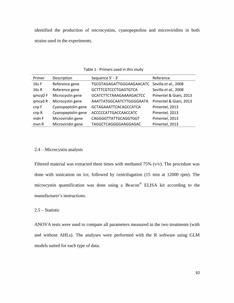
92
identified the production of microcystins, cyanopeptolins and microviridins in both
strains used in the experiments.
Table 1 - Primers used in this study
Primer Description Sequence 5' - 3' Reference
16s F Reference gene TGCGTAGAGATTGGGAAGAACATC Sevilla et al., 2008
16s R Reference gene GCTTTCGTCCCTGAGTGTCA Sevilla et al., 2008
qmcyD F Microcystin gene GCATCTTCTAAAGAAAAGACTCC Pimentel & Giani, 2013
qmcyd R Microcystin gene AAATTATGGCAATCTTGGGGAATA Pimentel & Giani, 2013
cnp F Cyanopeptolin gene GCTAGAAATTCACAGCCATCA Pimentel, 2013
cnp R Cyanopeptolin gene ACCCCCATTGACCAACCATC Pimentel, 2013
mdn F Microviridin gene CAGGGGTTATTGCAGGTGGT Pimentel, 2013
mvn R Microviridin gene TAGGCTCAGGGGAAGGAGAC Pimentel, 2013
2.4 – Microcystin analysis
Filtered material was extracted three times with methanol 75% (v/v). The procedure was
done with sonication on ice, followed by centrifugation (15 min at 12000 rpm). The
microcystin quantification was done using a Beacon® ELISA kit according to the
manufacturer’s instructions.
2.5 – Statistic
ANOVA tests were used to compare all parameters measured in the two treatments (with
and without AHLs). The analyses were performed with the R software using GLM
models suited for each type of data.
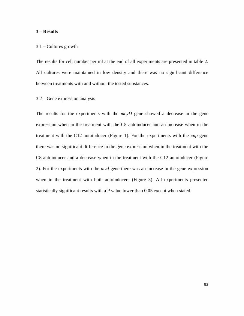
93
3 – Results
3.1 – Cultures growth
The results for cell number per ml at the end of all experiments are presented in table 2.
All cultures were maintained in low density and there was no significant difference
between treatments with and without the tested substances.
3.2 – Gene expression analysis
The results for the experiments with the mcyD gene showed a decrease in the gene
expression when in the treatment with the C8 autoinducer and an increase when in the
treatment with the C12 autoinducer (Figure 1). For the experiments with the cnp gene
there was no significant difference in the gene expression when in the treatment with the
C8 autoinducer and a decrease when in the treatment with the C12 autoinducer (Figure
2). For the experiments with the mvd gene there was an increase in the gene expression
when in the treatment with both autoinducers (Figure 3). All experiments presented
statistically significant results with a P value lower than 0,05 except when stated.
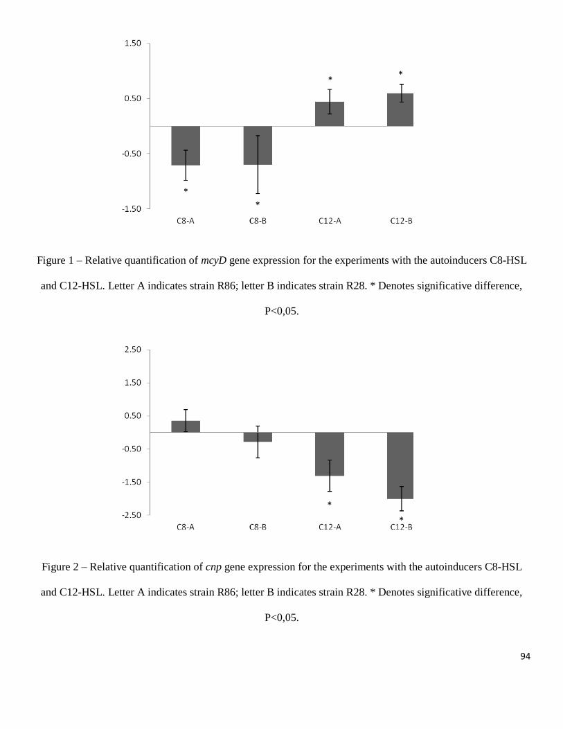
94
Figure 1 – Relative quantification of mcyD gene expression for the experiments with the autoinducers C8-HSL
and C12-HSL. Letter A indicates strain R86; letter B indicates strain R28. * Denotes significative difference,
P<0,05.
Figure 2 – Relative quantification of cnp gene expression for the experiments with the autoinducers C8-HSL
and C12-HSL. Letter A indicates strain R86; letter B indicates strain R28. * Denotes significative difference,
P<0,05.
*
*
* *
*
*
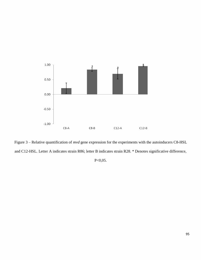
95
Figure 3 – Relative quantification of mvd gene expression for the experiments with the autoinducers C8-HSL
and C12-HSL. Letter A indicates strain R86; letter B indicates strain R28. * Denotes significative difference,
P<0,05.
* * *
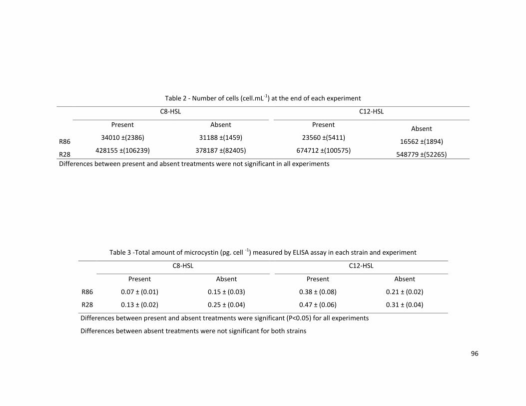
96
Table 3 -Total amount of microcystin (pg. cell -1) measured by ELISA assay in each strain and experiment
C8-HSL
C12-HSL
Present Absent
Present Absent
R86 0.07 ± (0.01) 0.15 ± (0.03)
0.38 ± (0.08) 0.21 ± (0.02)
R28 0.13 ± (0.02) 0.25 ± (0.04)
0.47 ± (0.06) 0.31 ± (0.04)
Differences between present and absent treatments were significant (P<0.05) for all experiments
Differences between absent treatments were not significant for both strains
Table 2 - Number of cells (cell.mL-1) at the end of each experiment
C8-HSL
C12-HSL
Present Absent
Present Absent
R86 34010 ±(2386) 31188 ±(1459)
23560 ±(5411) 16562 ±(1894)
R28 428155 ±(106239) 378187 ±(82405)
674712 ±(100575) 548779 ±(52265)
Differences between present and absent treatments were not significant in all experiments
97
3.3 – ELISA assay
The amount of microcystin per cell in the experiments with the C8-HSL were
significantly lower in the treatments where the AHL was present; 54% lower in the
experiment with strain 86 and 48% with strain 28. For the procedures with the C12-HSL,
the amount of microcystin was significantly higher in the treatment where the AHL was
present, 45% higher in the experiment with strain 86 and 33% with strain 28. The
comparison between the treatments with no AHL showed no significant difference for
both strains. The results of the ELISA analysis are represented in table 3.
4 – Discussion
In gram-negative bacteria, quorum sensing was first described in Vibrio fischeri (Nealson
& Hastings, 1979) as based on autoinducer systems. The molecules are able to diffuse
inside and outside the bacterial cells and their concentration increase with increasing cell
density (Kaplan & Greenberg, 1985), when these molecules reach a threshold
concentration the transcription of several genes is activated. In our experiments, AHLs
were used to induce a quorum sensing response in strains of cyanobacteria. The
comparison between cell number in the treatments (AHL addition) and controls (no AHL
addition) showed no significant difference for all strains, which demonstrates that the
AHLs used in the experiments did not cause any inhibition or stimulation in the growth
of the cyanobacteria. Considering the lack of significant difference in the strains growth,
it is possible to assume that the results found in the RTqPCR analysis and in the ELISA
assays are not a consequence of cell number and are in fact a direct effect of the AHLs.
The cellular density used in the experiments was chosen according to the growth curves
98
obtained in previous experiments which used both strains (Pereira et al., 2012), the
different values in cell number between strains 28 and 86 are a reflect of specific
characteristics that each one of them has.
The microcystin production was measured by ELISA assays and by qPCR through the
expression of the mcyD gene. The cyanopeptolin and microviridn production were
measured only by qPCR through the expression of genes related to their production.
Unfortunately, there are no ELISA kits available for cyanopeptolins, microviridins and
other cyanobacterial peptides, making impossible the direct measurement of these
peptides in the experiment done in this study. The quantification of these peptides could
have been done by HPLC, but this would demand a large scale experiment with a bigger
volume of cultures in order to have more material to make the HPLC measurements
possible. Considering that the AHLs are very expensive and that the amount of each
compound necessary to make a large scale experiment would be very large, a small scale
approach with measurements by qPCR and ELISA was chosen.
Concerning the production of microcystin, strain R28 had a higher amount of toxin per
cell, approximately 0,09 pg.cell-1
more than strain R86. The ELISA results for strain R86
were more intense than the results found in the experiments with the strain R28, but the
differences between the two strains were not statistically significant (for both substances
tested). This shows that the substances effects in inhibition (C8-HSL) and stimulation
(C12-HSL) are the same, independently of the strain and the amount of microcystin
produced by each of them. For the qPCR results, the same pattern found in the ELISA
measurements was verified. The contradictory results for the C8-HSL and C12-HSL may
99
look strange at first, but the fact that the same patterns were found for two different
strains, with different peptide profiles, confirms that they are reliable. Another point that
supports our results is that the similar results were obtained with two different techniques,
measuring two different products. While the qPCR measures the gene expression (RNA
production) the ELISA measures the final product in the synthesis pathway (the
microcystin amount). It also reveals that during the microcystin synthesis the
inhibition/stimulation occurs at the genetic level and not after the RNA transcription,
because if that was the case there would be no difference between controls and treatments
in the qPCR analysis, but a significant difference in the ELISA measurements would still
be observed.
The explanation for the opposite results observed for microcystin production under the
effect of two different AHL compounds might be connected to how the quorum sensing
system works in the Radiocystis. It is known that some species of bacteria have more than
one quorum sensing system and that these systems can work in parallel, in series or in a
competitive way (Waters & Bassler, 2005). It is possible that the Radiocystis, as other
bacteria, has more than one quorum sensing system, which could be activated at different
cellular densities. This would also make sense when considering the development of a
bloom: for example, at the beginning of the phenomenon, at a particular cell density, the
first system would be activated, triggering a series of physiological responses. In a
second moment, after reaching another cellular density, another series of physiological
responses would be activated. It is possible that the C8-HSL and the C12-HSL stimulate
two different quorum sensing systems and this would explain the different responses
produced.
100
For the cyanopeptolins, only C12-HSL had a significant effect, suppressing the gene
expression, while the microviridin production was significantly stimulated in three
experiments out of four. The experiment with the C8-HSL and the strain R86 showed an
increase in the microviridin gene expression but had no significant results while the other
experiments showed an increase in the gene expression with significant results. It is
possible to say that both substances affect the production of microviridins, with the C12-
HSL having a more intense effect. For the cyanopeptolins, it appears that C8-HSL has no
effect on their production.
When comparing the gene expression related to three studied peptides, opposite patterns
are seen for microcystins compared to microviridins for the experiments with the C8-
HSL and microcystins and microviridins compared to cyanopeptolins for the experiments
with the C12-HSL. If the Radiocystis fernandoii has any sort of quorum sensing system
based on AHLs, it is possible to assume that it does not act in the same way with all
peptides, which would not be a surprise considering that the auto inducers usually
stimulate and inhibit several genes at the same time (Williams et al., 2007).
We used two AHLs with different side chain length in the experiments, the C8-HSL
which was shown to be produced by axenic cultures of the genera Gloeothece (Sharif et
al., 2008) and the C12-HSL which was one of the AHLs used by Romero et al., (2011)
and was shown to be related with the inhibition of the enzyme nitrogenase, consequently
reducing nitrogen fixation in a Anabaena strain. The specificity of AHLs receptors is
connected with the side chain length, saturation and substitutions (Gould et al., 2004),
being one of the reasons for the use of AHLs with medium (C8-HSL) and long (C12-
101
HSL) side chains. The different length in their side chains could also be one of the
explanations for the different results found for each one of the AHLs, since C8-HSL and
C12-HSL may not activate the same quorum sensing receptors and, consequently,
promote different physiological changes.
Environmental factors, such as light, temperature, nitrogen, phosphorous and iron were
extensively tested on the production of microcystins and other peptides (Utkilen &
Gjolme 1999; 2000; Wiedner et al., 2003; Repka et al., 2004; Tonk et al., 2005; Sevilla et
al., 2008; Tonk et al., 2009; Alexova et al., 2011; Pereira et al., 2012), but to date, there
are no studies linking quorum sensing signaling molecules and the control of peptide
production on cyanobacteria. The regulatory mechanisms behind peptide production in
cyanobacteria might be an intricate system and in natural scenarios the secondary
metabolites production is probably regulated by a series of features from physical,
chemical and biological origin, which interact together in order to create a production
pattern for these compounds. It is very difficult to study and make statements in
situations with a series of variable factors, which makes laboratory experiments a very
useful tool to understand the role that each parameter has in the peptide production. With
the results obtained in this work, it is possible to conclude that aspects such as quorum
sensing and cellular density may have an important role in the production of
cyanobacterial secondary metabolites.
102
References
Agrawal MK, Bagchi D & Bagchi SN (2001) Acute inhibition of protease and
suppression of growth in zooplankter, Moina macrocopa, by Microcystis bloom collected
in central India. Hydrobiologia 464: 37-44.
Agrawal MK, Bagchi D & Bagchi SN (2005) Cysteine and serine protease-mediated
proteolysis in body homogenate of a zooplankter, Moina macrocopa, is inhibited by the
cyanobacterium Microcystis aeruginosa PCC 7608. Comp Biochem Physiol B 141: 33-
41.
Alexova R, Fujii M, Birch D, Cheng J, Walte D, Ferrari BC, Neilan BA (2011) Iron
uptake and toxin synthesis in the bloom-forming Microcystis aeruginosa under iron
limitation. Environ Microbiol 13: 1064-1077.
Carmichael WW (1992) Cyanobacteria secondary metabolites – the cyanotoxins. J Appl
Bacteriol 72: 445-459.
Czarnecki O, Lippert I, Henning M & Welker M (2006) Identification of peptide
metabolites of Microcystis (cyanobacteria) that inhibit trypsin-like activity planktonic
herbivorous Daphnia (Cladocera). Environ Microbiol 8: 77-87.
Fuqua WC, Winans SC & Greenberg EP (1994) Quorum sensing in bacteria-the LuxR-
LuxI family of cell density-responsive transcriptional regulators. J Bacteriol 176: 269-
275.
Fuqua WC, Parsek MR & Greenberg EP (2001) Regulation of gene expression by cell-to-
cell communication: acyl-homoserine lactone quorum sensing. Annu Rev Genet 35: 439-
468.
103
Gould TA, Schweizer HP, Churchill ME (2004) Structure of the Pseudomonas
aeruginosa acylhomoserinelactone synthase LasI. Mol Microbiol 53: 1135-1146.
Guillard RR & Lorenzen CJ (1972) Yellow-green algae with chlorophyllide c. J Phycol
8: 10-14.
Ishitsuka MO, Kusumi T, Kakisawa H, Kaya K & Watanabe MM (1990) Microviridin: a
novel tricyclic depsipeptide from the toxic cyanobacterium Microcystis viridis. J Am
Chem Soc 112: 8180-8182.
Kaebernick M, Rohrlack T, Christoffersen K & Neilan BA (2001) A spontaneous mutant
of microcystin biosynthesis: genetic characterization and effect on Daphnia. Environ
Microbiol 3: 669-679.
Kaplan HB, Greenberg EP (1985) Diffusion of autoinducer is involved in regulation of
the Vibrio fischeri luminescence system. J Bacteriol 163: 1210-14.
Komárek J & Komárková-Legenerová J (1993) Radiocystis fernandoii, a new planktic
cyanoprokariotic from tropical freshwater reservoirs. Preslia 65: 355-357.
Lombardo M, Pinto FCR, Vieira JMS, Honda RY, Pimenta AMC, Bemquerer MP,
Carvalho LR & Kiyota S (2006) Isolation and structural characterization of microcystin-
LR and three minor oligopeptides simultaneously produced by Radiocystis fernandoi
(Chroococcales, Cyanobacteriae): A Brazilian toxic cyanobacterium. Toxicon 47: 560-
566.
Long BM, Jones GJ & Orr PT (2001) Cellular microcystin content in N-limited
Microcystis aeruginosa can be predicted from growth rate. Appl Environ Microbiol 67:
278-283.
104
Martin C, Oberer L, Ino T, König WA, Busch M & Weckesser J (1993) Cyanopeptolins,
nwe depsipeptides from the cyanobacterium Microcystis PCC 7806. J Antibiot 46: 1550-
1556.
Martin-Luna B, Sevilla E, Hernandez JA, Bes MT, Fillat MF & Peleato ML (2006) Fur
from Microcystis aeruginosa binds in vitro promoter regions of the microcystin
biosynthesis gene cluster. Phytochemistry 67: 876-881.
Miller MB & Bassler BL (2001) Quorum sensing in bacteria. Annu Rev Microbiol 55:
165-199.
Nealson KH, Platt T & Hastings JW (1970) Cellular control of the synthesis and activity
of the bacterial bioluminescent system. J Bacteriol 104: 313-322.
Neilan BA, Pearson LA, Muenchoff J, Moffitt MC & Dittmann E (2013) Environmental
conditions that influence toxin biosynthesis in cyanobacteria. Environ Microbiol 15:
1239-1253.
Oliver RL & Ganf GG (2002) Freshwater blooms. The Ecology of Cyanobacteria – Their
Diversity in Time and Space. (Whitton BA & Potts M, eds), pp. 149-194. Kluwer
Academic Publishers.
Pereira DA, Pimenta AMC & Giani A (2012) Profiles of toxic and non-toxic
oligopeptides of Radiocystis fernandoii (Cyanobacteria) exposed to three different light
intensities. Microbiol Research 167: 413-421.
Pereira DA & Giani A (2014) Cell density oligopeptides production in cyanobacterial
strains. FEMS Microbiol Ecol. Online first.
105
Pimentel JS, (2013) Análises de transcrição gênica em cianobactérias, como ferramenta
para estudar suas relações com o ambiente. Tese de doutorado apresentada no
departamento de biologia geral, UFMG.
Pimentel JS & Giani A (2013) Estimating toxic cyanobacteria in a Brazilian reservoir by
quantitative real-time PCR, based on the microcystin synthetase D gene. J of Appl Phycol
25: 1545-1554.
Repka S, Koivula M, Harjunpa V, Rouhiainen L, Sivonen K (2004) Effects of phosphate
and light on growth of and bioactive peptide production by the cyanobacterium Anabaena
strain 90 and its anabaenopeptilide mutant. Appl Environ Microbiol 70: 4551-4560.
Romero M, Diggle SP, Heeb S, Cámara M & Otero A (2008) Quorum quenching activity
in Anabaena sp.PCC 7120: identification of AiiC, a novel AHL-acylase. FEMS
Microbiol Lett 280: 73-80.
Romero M, Muro-Pastor AM & Otero A (2011) Quorum sensing N-acylhomoserine
lactone signals affect nitrogen fixation in the cyanobacterium Anabaena sp. PCC7120.
FEMS Microbiol Lett 315: 101-108
Sant'Anna C L, Azevedo MTP, Werner VR, Dogo CR, Rios FR & de Carvalho LR
(2008) Review of toxic species of Cyanobacteria in Brazil. Algological Studies 126: 251-
265.
Sevilla E, Martin-Luna B, Vela L, Bes MT, Fillat, ML & Peleato ML (2008) Iron
availability affects mcyD expression and microcystin-LR synthesis in Microcystis
aeruginosa PCC7806. Environ Microbiol 10: 2476-2483.
106
Sharif DI, Gallon J, Smith CJ & Dudley E (2008) Quorum sensing in cyanobacteria: N-
octanoyl-homoserine lactone release and response, by the epilithic colonial
cyanobacterium Gloeothece PCC 6909. ISME J 2: 1171-1182.
Sivonen K & Jones GJ (1999) Cyanobacterial toxins. Toxic cyanobacteria in water.
(Chorus &. Bartram J, eds), pp. 41-111. E&FN Spoon, London.
Swift S, Downie JA, Whitehead NA, Barnard AML, Salmond GPC & Willians P (2001)
Quorum sensing as a population-density-dependent determinant of bacterial physiology.
Adv Microb Physiol 45: 199-270.
Tomasz A (1965) Control of the competent state in pneumococcus by a hormone-like cell
product – an example of a new type of regulatory mechanism in bacteria. Nature 208:
155-159.
Tonk L, Visser PM, Christiansen G, Dittmann E, Snelder EOFM, Wiedner C, et al (2005)
The microcystin composition of the cyanobacterium Planktothrix agardhii changes
towards a more toxic variant with increasing light intensity. Appl Environ Microbiol 71:
5177-5181.
Tonk L, Welker M, Huisman J, Visser PM (2009) Production of cyanopeptolins,
anabaenopeptins and microcystins by the harmful cyanobacteria Anabaena 90 and
Microcystis PCC 7806. Harmful Algae 8: 219-224.
Utkilen H, Gjolme N (1995) Iron-stimulated toxin production in Microcystis aeruginosa.
Appl Environ Microbiol 61: 797-800.
Vieira JMS, Azevedo MTP, Azevedo SMFO, Honda RY & Corrêa B (2003) Microcystin
production by Radiocystis fernandoii (Chroococales, Cyanobacteria) isolated from a
107
drinking water reservoir in the city of Belém, PA, Brazilian Amazonia region. Toxicon
42: 709-713.
Waters CL & Bassler BL (2005) Quorum sensing: cell-to-cell communication in bacteria.
Annu Rev Cell Dev Biol 21: 319-346.
Welker M, Brunke M, Preussel K, Lippert I & von Döhren, H (2004) Diversity and
distribution of Microcystis (cyanobacteria) oligopeptides chemotypes from natural
communities studied by single mass spectrometry. Microbiology 150: 1785-1796.
Welker M & von Döhren, H (2006) Cyanobacterial peptide: nature’s own combinatorial
biosynthesis. FEMS Microbiol Rev 30: 530-563.
Wiedner C, Visser PM, Fastner J, Metcalf JS, Codd GA, Mur LR (2003) Effects of light
on the microcystin content of Microcystis strain PCC 7806. Appl Environ Microbiol 69:
1475-1481.
Williams P, Winzer K, Chan WC & Cámara M (2007) Look who’s talking:
communication and quorum sensing in the bacterial world. Phil Trans R Soc B 362:
1119-1134.
Wood SA, Rueckert A, Hamilton DP, Cary SG & Dietrich DR (2011) Switching toxin
production on and off: intermittent microcystin synthesis in a Microcystis bloom. Environ
Microbiol Reports 3: 118-124.
Wood, S. A., Dietrich, D. R., Cary, S. G., Hamilton, D. P. 2012. Increasing Microcystis
cell density enhances microcystin synthesis: a mesocosm study. Inland Waters. 2, 17-22.
Young FM, Thomson C, Metcalf JS, Lucocq JM & Codd GA (2005) Immunogold
localization of microcystins in cryosectioned cells of Microcystis. J Struct Biol 151: 208-
214.

108
Zilliges Y, Kehr J, Meissner S, Ishida K, Mikkat S, Hagemann M, Kaplan A, Borner T &
Dittmann E (2011) The cyanobacterial hepatotoxin microcystin binds to proteins and
increases the fitness of Microcystis under oxidative stress conditions. PLoS ONE 6: 1-11.

109
5. Conclusões finais
As análises realizadas nesta tese apontaram para uma conexão entre quorum sensing e
produção de peptídeos em cianobactérias. Os experimentos realizados sobre o efeito da
densidade celular permitiram concluir que este parâmetro tem uma influência sobre a
produção da maioria dos peptídeos encontrados nas cepas estudadas. Estes experimentos
também mostraram que em situações de floração existe a possibilidade de que a
densidade celular seja um dos fatores que controlam a produção de metabólitos
secundários.
Os testes feitos com as homoserinas lactonas, substâncias produzidas por bactérias como
sinalizadores celulares intra-populacionais, mostraram que estes compostos afetam a
produção de microcistinas, cianopeptolinas e microviridinas e levantam a possibilidade
da existência de um ou mais sistemas de quorum sensing na cianobactéria Radiocystis
fernandoii.
Os métodos utilizados nos experimentos se mostraram eficientes nas medições. As
técnicas de cromatografia e espectrometria de massa foram adequadas para o isolamento
e identificação dos peptídeos. Os testes de ELISA e a PCR em tempo real permitiram
uma medição eficiente e precisa, mesmo em amostras com baixa concentração de
material. A PCR em tempo real (RTqPCR) permitiu a mensuração da expressão gênica
dos peptídeos alvo, mostrando-se suficientemente sensível para detectar as diferenças
entre os tratamentos realizados.
Os dados apresentados nesta tese abrem novas perspectivas na investigação das relações
entre células de populações de cianobactérias, abrindo-se com isso novas possibilidades

110
para o desenvolvimento de mecanismos de controle de florações em ambientes
eutrofizados.