Embed Size (px)
Citation preview
DEPARTAMENTO DE CIÊNCIAS DA VIDA FACULDADE DE CIÊNCIAS E TECNOLOGIA
UNIVERSIDADE DE COIMBRA
Isabel Maria Lopes de Matos Oliveira
2013
Mitochondrial genome analysis in frontotemporal
lobar degeneration: tRNAs contribution
Análise genómica mitocondrial na degerescência
lobar frontotemporal:contribuição dos tRNAs
DEPARTAMENTO DE CIÊNCIAS DA VIDA FACULDADE DE CIÊNCIAS E TECNOLOGIA
UNIVERSIDADE DE COIMBRA
Mitochondrial genome analysis in frontotemporal
lobar degeneration: tRNAs contribution
Análise genómica mitocondrial na degerescência
lobar frontotemporal:contribuição dos tRNAs
Isabel Maria Lopes de Matos Oliveira
2013
Dissertação apresentada à Universidade de Coimbra para cumprimento dos requisitos necessários à obtenção do grau de Mestre em Evolução e Biologia Humanas, realizada sob a orientação científica da Professora Doutora Manuela Grazina (Universidade de Coimbra, Faculdade de Medicina) e sob a orientação interna da Professora Doutora Eugénia Cunha (Universidade de Coimbra, Faculdade de Ciências e Tecnologia)
Copyright© Isabel Oliveira e Manuela Grazina, 2013
Esta cópia da tese é fornecida na condição de que quem a consulta reconhece que os
direitos de autor são pertença do autor da tese e do orientador científico e que nenhuma
citação ou informação obtida a partir dela pode se publicada sem a referência e
autorização.
This copy of the thesis has been supplied on condition that anyone who consults it is
understood to recognize that its copyright rests with its author and scientific supervisor
and that no quotation from the thesis and no information derived from it may be
published without proper acknowledgment and authorization.
Agradecimentos
A realização desta dissertação só foi possível graças ao apoio e cooperação de várias
pessoas.
Em primeiro lugar queria agradecer à Professora Doutora Manuela Grazina por me ter
aceite no laboratório de Bioquímica Genética e por ter acreditado em mim.
À professora Doutora Eugénia Cunha agradeço toda a ajuda prestada como orientadora
interna.
A todos os membros do laboratório, especialmente à Mestre Maria João Santos por me
ter acompanhado sempre ao longo da realização deste trabalho.
Aos meus colegas do laboratório por me terem recebido tão bem e por fazerem parte do
meu dia-a-dia, proporcionando-me momentos que nunca vou esquecer.
A todos os meus amigos, por todo o apoio e acreditarem em mim nos momentos mais
difíceis.
Por último, queria agradecer à minha mãe e irmã porque sem elas não teria chegado tão
longe no meu percurso académico.

Acknowledgments
This study was financed by Portuguese Foundation for Science and Technology (FCT),
with the Project PTDC/SAL-EPI/121811/2010 (FCT) and partially supported by FCT
project PEst-C/SAU/LA0001/2011.
Isabel Oliveira, 2013 Page i
Index
Index of figures...............................................................................ii
Index of tables............................................................................... iv
Resumo.......................................................................................... v
1. General Introduction.....................................................................1
1.1. Dementia ........................................................................................................ 1 1.2. FTLD History................................................................................................. 1 1.3. FTLD Epidemiology ...................................................................................... 1
1.4. Clinical variants of FTLD .............................................................................. 2
1.5. FTLD Neuropathological variants ................................................................. 2
1.6. Genetic variants of FTLD ..............................................................................4
1.7. Etiological mechanisms of FTLD .................................................................. 4
1.8. Mitochondrial DNA study in FTLD .............................................................. 5
1.9. Human Mitochondrial tRNAs ........................................................................ 7
2. Paper.........................................................................................11
Abbreviations...................................................................................12
Abstract..........................................................................................13
Keywords........................................................................................13
Introduction.....................................................................................14
Objectives.......................................................................................15
Patients and Methods..........................................................................15
Samples..........................................................................................16 PCR amplification ...................................................................................................... 16 Agarose gel electrophoresis........................................................................................ 16 DNA Sanger Sequencing............................................................................................ 17 In silico analysis ......................................................................................................... 18
Results...........................................................................................19
Discussion.......................................................................................29
Conclusions.....................................................................................31
References.......................................................................................33
3. Annex 1: Author information pack (Neurobiology of Disease)...................40
Isabel Oliveira, 2013 Page ii
Index of figures
Figure I: Human mitochondrial DNA (adapted from Greaves et al., 2012)......................6
Figure II: Structure of mt-tRNA (adapted from Yarham et al., 2011)...............................8
Figure III: The tRNA end processing pathway followed by aminoacylation (adapted
from Levinger et al., 2004)................................................................................................9
Figure 1: Number of alteration per gene in which they were identified..........................20
Figure 2: Results from in silico analysis for nucleotide (m.4312C>T mt-tRNAIle
) A-
normal B- and “mutated” structure (RNAfold); C- Location of the sequence variation in
the clover-shaped structure; D- Evolutionary conservation for the nucleotide position
(signed with blue rectangle)............................................................................................25
Figure 3: Results from in silico analysis for nucleotide (m.4435A>G of mt-tRNAMet) A-
normal B- and “mutated” structure (RNAfold); C- Location of the sequence variation in
the clover-shaped structure; D- Evolutionary conservation for the nucleotide position
(signed with blue rectangle)............................................................................................26
Figure 4: Results from in silico analysis for nucleotide (m.5772G>A of mt-tRNACys) A-
normal and B- “mutated” structure (RNAfold); C- Location of the sequence variation in
the clover-shaped structure; D- Evolutionary conservation for the nucleotide position
(signed with blue rectangle)............................................................................................27
Figure 5: Results from in silico analysis for nucleotide (m.12166T>C of mt-tRNAHis) A-
normal and B- “mutated” structure (RNAfold); C- Location of the sequence variation in
the clover-shaped structure; D- Evolutionary conservation for the nucleotide position
(signed with blue rectangle)............................................................................................27
Figure 6: Results from in silico analysis for nucleotide (m.12308A>G of mt-tRNALeu2)
A-normal and B- “mutated” structure (RNAfold); C- Location of the sequence variation
in the clover-shaped structure; D- Evolutionary conservation for the nucleotide position
(signed with blue rectangle)............................................................................................28
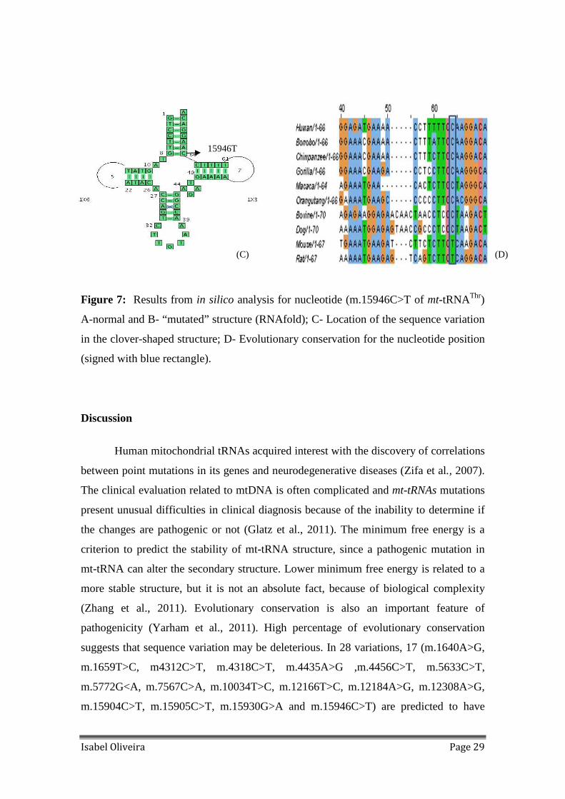
Figure 7: Results from in silico analysis for nucleotide (m.15946C>T of mt-tRNAThr)
A-normal and B- “mutated” structure (RNAfold); C- Location of the sequence variation
Isabel Oliveira, 2013 Page iii
in the clover-shaped structure; D- Evolutionary conservation for the nucleotide position
(signed with blue rectangle)............................................................................................29
Isabel Oliveira, 2013 Page iv
Index of tables
Table I: Neuropathological characterization of FTLD and associated genes (adapted from Mackenzie et al., 2010).............................................................................................3
Table II: Genetic characterization of FTLD (adapted from Schlachetzki, 2011)..............4
Table III: Human mitochondrial genetic code (from Suzuki et al., 2011)........................9
Table 1: The Pathogenicity Scoring System (from Yarham et al., 2011)........................18
Table 2: Patients characterization and data from mtDNA sequences variations.............21
Table 3: In silico analysis of the sequence variations found in mt-tRNA encoding
genes................................................................................................................................23
Isabel Oliveira, 2013 Page v
Resumo
A degenerescência lobar frontotemporal (DLFT) é uma demência
neurodegenerativa heterogénea, incluindo nos aspectos clínicos, neuropatológicos e
genéticos. É caracterizada por mudanças progressivas no comportamento, disfunção
executiva e/ou dificuldades na linguagem, acompanhada por atrofia no lobo frontal e
temporal. Alguns doentes apresentam sobreposição clínica e neuropatológica com a
doença de Alzheimer, o que sugere semelhanças na fisiopatologia, nomeadamente no
envolvimento do DNA mitocondrial (mtDNA).
O objetivo do presente estudo é realizar a sequenciação dos genes no mtDNA
que codificam tRNAs, para identificar alterações nos doentes com DLFT, investigando
o seu envolvimento na DLFT.
Foram analisadas 70 amostras de DNA provenientes de doentes, 39 mulheres e
31 homens, com diagnóstico provável de DLFT (faixa etária: 38-82 anos, média de 63 ±
11), seguidos na Unidade de Neurologia do Centro Hospitalar e Universitário de
Coimbra. O DNA total foi extraído a partir de sangue periférico, e foi efetuada a análise
da sequência dos 22 genes de tRNAs mitocondriais, por sequenciação automática. As
variantes encontradas foram submetidas a análise in silico. Foram encontradas 28
variações diferentes em 32 doentes. Destas, seis variações são provavelmente
patogénicas de acordo com a análise in silico: a m.4312C>T ocorre em heteroplasmia e
apresenta elevada conservação; a m.4435A>G está localizada numa posição
potencialmente crítica e é totalmente conservada em todas as espécies analisadas; a
variação m.5772G>A está localizada no T-stem, levando ao rompimento do
emparelhamento da base (CG) Watson-Crick e é 100% conservada; a alteração
m.12166T>C está localizada no “anticodon loop” e apresenta alta percentagem de
conservação. A variação mais frequente é a m.12308A>G, no mt-tRNALeu2,na região
variável, e é totalmente conservada em todos os mamíferos estudados. A variação
m.15946C>T tem uma elevada taxa de conservação e está localizada no “acceptor
stem”. São necessários estudos adicionais para compreender melhor a relação entre as
alterações do mtDNA identificadas e a FTLD. No entanto, este estudo é original sendo
o primeiro a investigar a sequencia dos genes que codificam os tRNA mitocondrias na
DLFT.
Isabel Oliveira Page 1
1. General Introduction
1.1. Dementia
In the last years, life expectancy has increased at a steady rate, which leads to an
increment the percentage of elderly in the population (Santana and Cunha, 2005). The
decline in mortality and births results in aging of the population in most developed
countries and, consequently, the prevalence of age-related diseases, including
dementias, are increasing. Dementia syndromes are characterized by progressive
impairment in cognitive function and they have become increasingly important in public
health (Fratiglioni et al., 1999). Among the various dementias, Alzheimer's disease
(AD) and Frontotemporal Lobar Degeneration (FTLD) are the most frequent in the
population (Santana and Cunha, 2005).
1.2. FTLD History
In 1892, Arnold Pick described the first clinical case of dementia, in which the
patient presented cognitive impairment, progressive aphasia and changes in social
behaviour. These manifestations are associated with temporal and frontal lobe atrophy
(Kertesz et al., 2005). In 1911, Alois Alzheimer described the histopathology features of
these patients, pointing to the presence of argyrophilic neuronal inclusions, later called
“Pick bodies” (Pan and Chen, 2013). A century later, research groups of Lund (Sweden)
and Manchester (England) published the first clinical and neuropathological criteria set
for the diagnosis of frontotemporal dementia (FTD) (Lund and Manchester Groups,
1994).
1.3. FTLD Epidemiology
FTLD is a heterogeneous neurodegenerative dementia in many aspects,
including clinical, neuropathological and genetic features, characterized by progressive
Isabel Oliveira Page 2
changes in behaviour, executive dysfunction and/or language impairment (Seltman and
Matthews, 2012) with frontal and temporal lobar atrophy (Pan and Chen, 2013). FTLD
occurs most often in the presenile period, with age at onset typically at 45-65 years, and
it has an equal distribution among female and male (Galimberti and Scarpini, 2010).
There is a wide range in duration of illness (2-20 years) partly reflecting different
underlying pathologies (Seelaar et al., 2011).
1.4. Clinical variants of FTLD
Given its heterogeneity, FTLD is classified differently regarding the clinical
characteristics. The site of focal cerebral atrophy, frontal and/or temporal, left and/or
right determines the clinical presentation (Schlachetzki, 2011). It can be differentiated
clinically into three frontotemporal dementia (FTD) syndromes; clinically behavioural
variant (bvFTD), characterized by progressive behavioural impairment and a decline in
executive function with frontal lobe atrophy; progressive nonfluent aphasia (PNFA)
with motor speech deficits and semantic dementia (SD) with loss of object knowledge
with anomia (Seltman and Matthews, 2012). Language variants are subsumed under the
clinical syndrome of primary progressive aphasia (PPA) and show involvement of the
left anterior temporal lobe (Schlachetzki, 2011). Additionally, there is a clinical overlap
between FTD with motor neuron disease (FTD-MND or FTD-ALS), as well as the
parkinsonian syndromes, progressive supranuclear palsy (PSP) and corticobasal
syndrome (CBS) (Pan and Chen, 2013).
1.5. FTLD Neuropathological variants
The neuropathology underlying the FTLD clinical syndromes is also
heterogeneous (Boxer et al., 2011). While clinical phenotype, neuropsychology features
and brain imaging data provide useful information about the FTLD spectrum pathology,
additional information is necessary to define the histopathological abnormality in
patients, since a clinical phenotype can be associated with several different pathologies
(Grossman, 2011). The major pathological hallmark of FTLD is selective atrophy of the
frontal and temporal cortex, with neuronal loss and gliosis (Seelaar et al., 2011). In most
cases, it is possible to find an accumulation of abnormal proteins in neurons and glia
Isabel Oliveira Page 3
(inclusions of aggregates). The identity of the pathological protein is variable (Boxer at
al., 2011) and the classification has been changing in the last few years. The currently
accepted nomenclature for the various FTLD neuropathological subtypes considers into
five groups, FTLD-tau (tau pathology), FTLD-TDP (TAR-DNA binding protein (TDP-
43)), FTLD-UPS (ubiquitin-positive and TDP-43-negative histopathology), FTLD-FUS
(inclusions of the fused in sarcoma protein) and FTLD-ni (without inclusions)
(Mackenzie et al., 2010). It was found that there is a correlation between
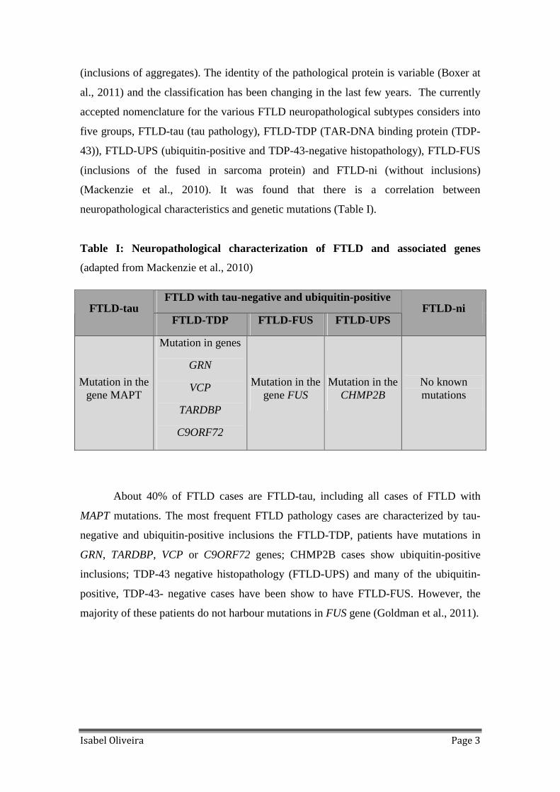
neuropathological characteristics and genetic mutations (Table I).
Table I: Neuropathological characterization of FTLD and associated genes
(adapted from Mackenzie et al., 2010)
FTLD with tau-negative and ubiquitin-positive FTLD-tau
FTLD-TDP FTLD-FUS FTLD-UPS FTLD-ni
Mutation in the gene MAPT
Mutation in genes
GRN
VCP
TARDBP
C9ORF72
Mutation in the gene FUS
Mutation in the CHMP2B
No known mutations
About 40% of FTLD cases are FTLD-tau, including all cases of FTLD with
MAPT mutations. The most frequent FTLD pathology cases are characterized by tau-
negative and ubiquitin-positive inclusions the FTLD-TDP, patients have mutations in
GRN, TARDBP, VCP or C9ORF72 genes; CHMP2B cases show ubiquitin-positive
inclusions; TDP-43 negative histopathology (FTLD-UPS) and many of the ubiquitin-
positive, TDP-43- negative cases have been show to have FTLD-FUS. However, the
majority of these patients do not harbour mutations in FUS gene (Goldman et al., 2011).
Isabel Oliveira Page 4
1.6. Genetic variants of FTLD
Positive family history was observed in 40-50% of the FTLD patients (Sieben, et
al., 2012). The autosomal dominant mode of inheritance has been described in 10-27%
of all FTLD patients with mutations identified. The familial cases are more common as
bvFTD and less frequent in patients with SD and PNFA. The genetic heterogeneity of
FTLD is reflected by the identification of mutations in several nuclear genes. The most
common mutations occur in the GRN and MAPT genes in approximately 50% of the
familial cases, while more rare mutations occur in the CHMP2B, FUS, VCP and
TARDBP genes (Seelaar et al., 2011). Recently, a gene responsible for FTLD has been
discovered on chromosome 9p (C9ORF72). More than 40 pathogenic MAPT mutations
have been described in 134 families and 69 different mutations in GRN gene have been
described in 231 families (Galimberti e Scarpini, 2012) (Table II).
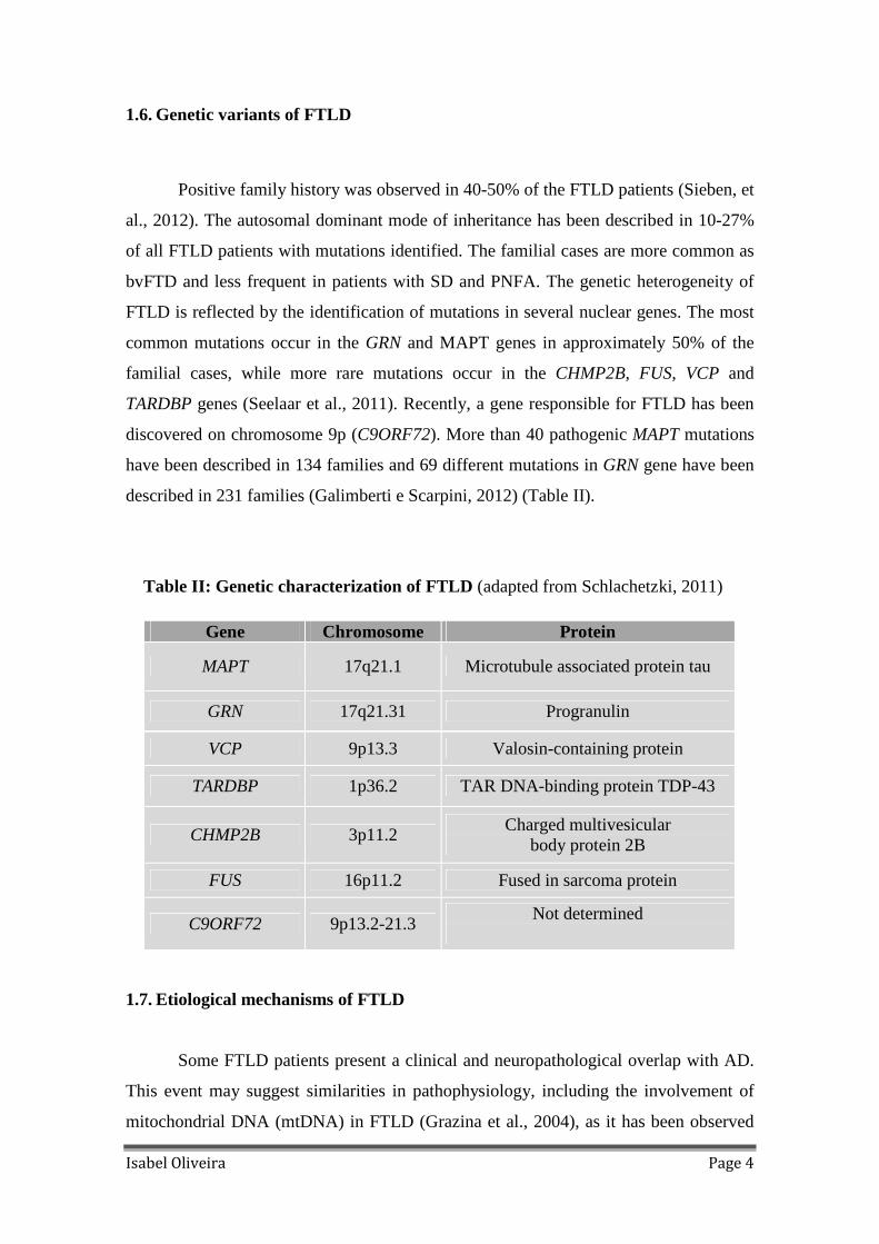
Table II: Genetic characterization of FTLD (adapted from Schlachetzki, 2011)
Gene Chromosome Protein
MAPT 17q21.1 Microtubule associated protein tau
GRN 17q21.31 Progranulin
VCP 9p13.3 Valosin-containing protein
TARDBP 1p36.2 TAR DNA-binding protein TDP-43
CHMP2B 3p11.2 Charged multivesicular
body protein 2B
FUS 16p11.2 Fused in sarcoma protein
C9ORF72 9p13.2-21.3 Not determined
1.7. Etiological mechanisms of FTLD
Some FTLD patients present a clinical and neuropathological overlap with AD.
This event may suggest similarities in pathophysiology, including the involvement of
mitochondrial DNA (mtDNA) in FTLD (Grazina et al., 2004), as it has been observed
Isabel Oliveira Page 5
in AD (Onyango et al., 2006). There are several studies that have identified mtDNA
mutations in AD patients, suggesting the existence of causal factors related to mtDNA.
This points to the involvement and contribution of mitochondrial genome to dementia
(revision Grazina et al., 2006). Accordingly, it is important to study the role of mtDNA
in FTLD (Grazina et al,. 2004).
1.8. Mitochondrial DNA study in FTLD
A number of essential cellular functions take place in the mitochondria.
However, the major mitochondrial event is the production of adenosine -5`-triphosphate
(ATP), the key energy source of the cell (Morán et al., 2012). Mitochondrial ATP is
generated via oxidative phosphorylation (OXPHOS), that occurs in the mitochondrial
respiratory chain (MRC), located within the inner mitochondrial membrane (Reddy,
2008). The process of OXPHOS system comprises five multiprotein complexes. Each
OXPHOS complex consists of polypeptide subunits encoded by nuclear and
mitochondrial DNA, except complex II, which is exclusively encoded by the nuclear
genome (Grazina, 2004). The correct biosynthesis of the OXPHOS complexes is a
highly intricate regulated process that requires the concerted action of the two cellular
genomes (Morán et al., 2012). Human mtDNA consists of a 16,568 nucleotides
organized in a double stranded, circular DNA molecule, containing 37 genes, encoding
for 12S and 16S rRNA, 22 tRNAs and 13 polypeptides , essential components of the
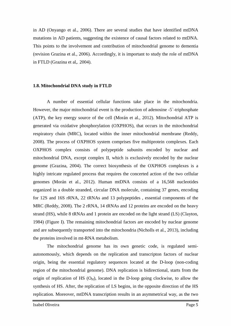
MRC (Reddy, 2008). The 2 rRNA, 14 tRNAs and 12 proteins are encoded on the heavy
strand (HS), while 8 tRNAs and 1 protein are encoded on the light strand (LS) (Clayton,
1984) (Figure I). The remaining mitochondrial factors are encoded by nuclear genome
and are subsequently transported into the mitochondria (Nicholls et al., 2013), including
the proteins involved in mt-RNA metabolism.
The mitochondrial genome has its own genetic code, is regulated semi-
autonomously, which depends on the replication and transcripton factors of nuclear
origin, being the essential regulatory sequences located at the D-loop (non-coding
region of the mitochondrial genome). DNA replication is bidirectional, starts from the
origin of replication of HS (OH), located in the D-loop going clockwise, to allow the
synthesis of HS. After, the replication of LS begins, in the opposite direction of the HS
replication. Moreover, mtDNA transcription results in an asymmetrical way, as the two
Isabel Oliveira Page 6
chains promoters, heavy strand promoter (PH) and light strand promoter (PL) are both
located on the D-loop. The two promoters work in opposite directions, and HS is
transcribed in the opposite to clockwise direction (Grazina, 2004).
Figure I: Human mitochondrial DNA (adapted from Greaves et al., 2012). ND1 –NADH dehydrogenase, subunit 1; ND2 – NADH dehydrogenase, subunit 2; ND3 – NADH dehydrogenase, subunit 3; ND4 – NADH dehydrogenase, subunit 4; ND4L – NADH dehydrogenase, subunit 4L; ND5 – NADH dehydrogenase, subunit 5; ND6 – NADH dehydrogenase, subunit 6; CYTB – Cythocrome B; COI – cytochrome c oxidase I; COII – cytochrome c oxidase II; COIII – cytochrome c oxidase III; ATP6 – ATP synthase F0 subunit 6; ATP8 – ATP synthase F0 subunit 8; RNR1 – Mitochondrially encoded 12S RNA; RNR2 – Mitochondrially encoded 16S RNA; T - Mitochondrially encoded tRNA threonine; P - Mitochondrially encoded tRNA proline; E - Mitochondrially encoded tRNA glutamic acid; L2 - Mitochondrially encoded tRNA leucine 2; S2- Mitochondrially encoded tRNA serine 2; H- Mitochondrially encoded tRNA histidine; R- Mitochondrially encoded tRNA arginine ; G- Mitochondrially encoded tRNA glycine; K- Mitochondrially encoded tRNA lysine; D- Mitochondrially encoded tRNA aspartic acid; S1- Mitochondrially encoded tRNA serine 1; Y- Mitochondrially encoded tRNA tyrosine ; C-Mitochondrially encoded tRNA cysteine; N-Mitochondrially encoded tRNA asparagines; A- Mitochondrially encoded tRNA alanine; W- Mitochondrially encoded tRNA tryptophan; M- Mitochondrially encoded tRNA methionine: I- Mitochondrially encoded tRNA isoleucine; Q- Mitochondrially encoded tRNA glutamine; L1- Mitochondrially encoded tRNA leucine 1; V- Mitochondrially encoded tRNA valine; F- Mitochondrially encoded tRNA phenylalanine.
Most human cells contain hundreds of mitochondria and thousands of mtDNA
copies. This genome is transmitted by maternal inheritance. Due to this fact, maternal
Isabel Oliveira Page 7
and paternal mtDNAs are rarely mixed in the same cytoplasm, not having
recombination between the two types of mtDNA. The only way that mtDNA sequence
may change is through the accumulation of mutations along the maternal lineage. The
high mutation rate of mtDNA results from the lack of protective histones, and
inefficient mtDNA repair systems (Wallace, 1994). In the OXPHOS process, besides
ATP synthesis, there is also the reactive oxygen species (ROS) production and mtDNA
is located close to the main source of ROS formation, being vulnerable to damage
(Reddy and Reddy et al., 2011). The first mtDNA pathogenic mutations were identified
in the late 1980s. Since then, more than 200 mutations in mtDNA were found
(Chinnery, 2006). When there is a mtDNA mutation, a mixture of wild and mutant
molecules could coexist and this situation is called heteroplasmy; on the other hand, the
presence of pure wild or pure mutant molecules are called homoplasmy. In case of
heteroplasmy, as the percentage of mutant molecules increases, oxidative
phosphorylation enzyme activities decrease. When the energy threshold is reached, the
probability of disease manifestation becomes higher (Wallace, 1994). The percentage of
mutated DNA may vary in different patients, from organ to organ and even between
cells within of the same tissue (Chinnery, 2006).
Mitochondrial changes, including MRC dysfunction due to enzymatic defects,
increases ROS production. Morphological changes in the mitochondrial network and
cell death are common features of neurodegenerative diseases of different genetic
origins. Mutations in genes encoding proteins involved in mitochondrial dynamics were
identified in neurodegenerative diseases (Móran et al., 2012).
1.9. Human Mitochondrial tRNAs
Mitochondrial genome encodes 22 tRNAs (tRNAPhe , tRNAVal, tRNALeu1 ,
tRNAIle, tRNAGln, tRNAMet, tRNATrp, tRNAAla, tRNAAsn, tRNACys, tRNATyr, tRNASer1,
tRNAAsp, tRNALys, tRNAGly, tRNAArg, tRNAHis, tRNASer 2, tRNALeu 2, tRNAGln, tRNAThr
and tRNAPro), essential to intramitochondrial protein synthesis. Amino acids are added
to the protein during translation, by transfer RNAs. Each tRNA molecule is encoded by
a different gene and its transcriptional nucleotide sequence results in a pre-tRNA that is
organized into a characteristic secondary structure. This structure, common to the pre-
tRNA and tRNA, is clover-shaped due to the hydrogen bonds established between
Isabel Oliveira Page 8
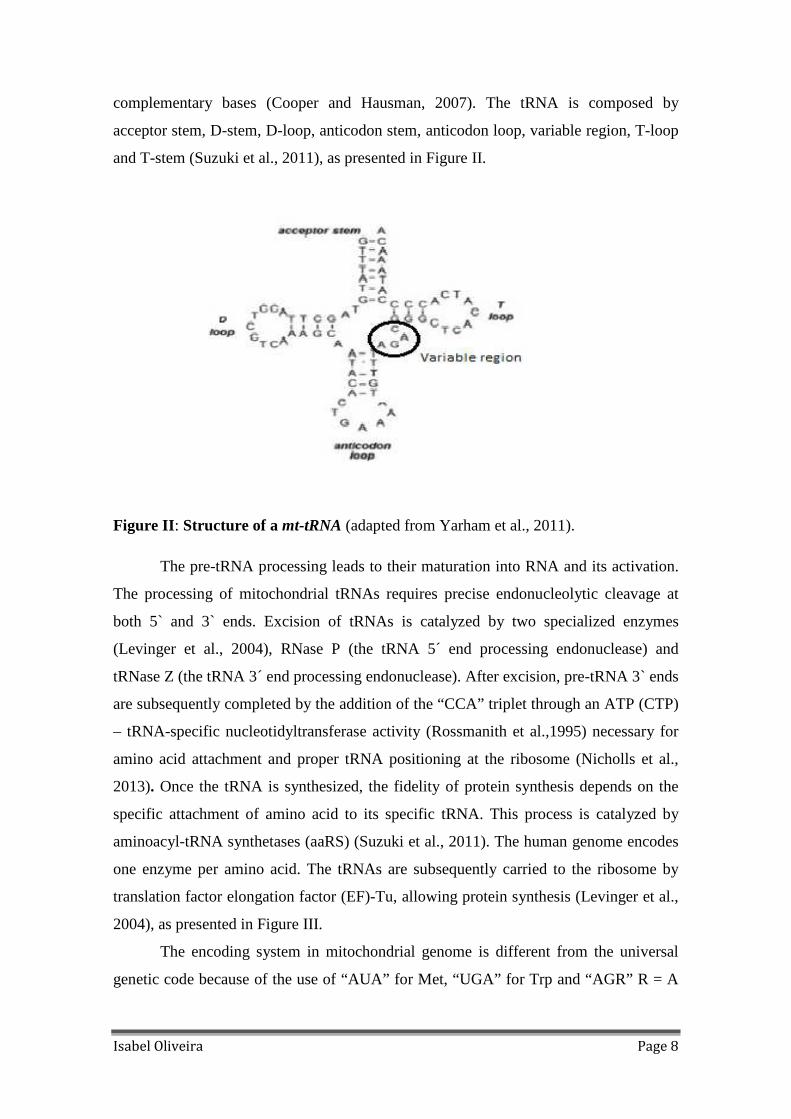
complementary bases (Cooper and Hausman, 2007). The tRNA is composed by
acceptor stem, D-stem, D-loop, anticodon stem, anticodon loop, variable region, T-loop
and T-stem (Suzuki et al., 2011), as presented in Figure II.
Figure II: Structure of a mt-tRNA (adapted from Yarham et al., 2011).
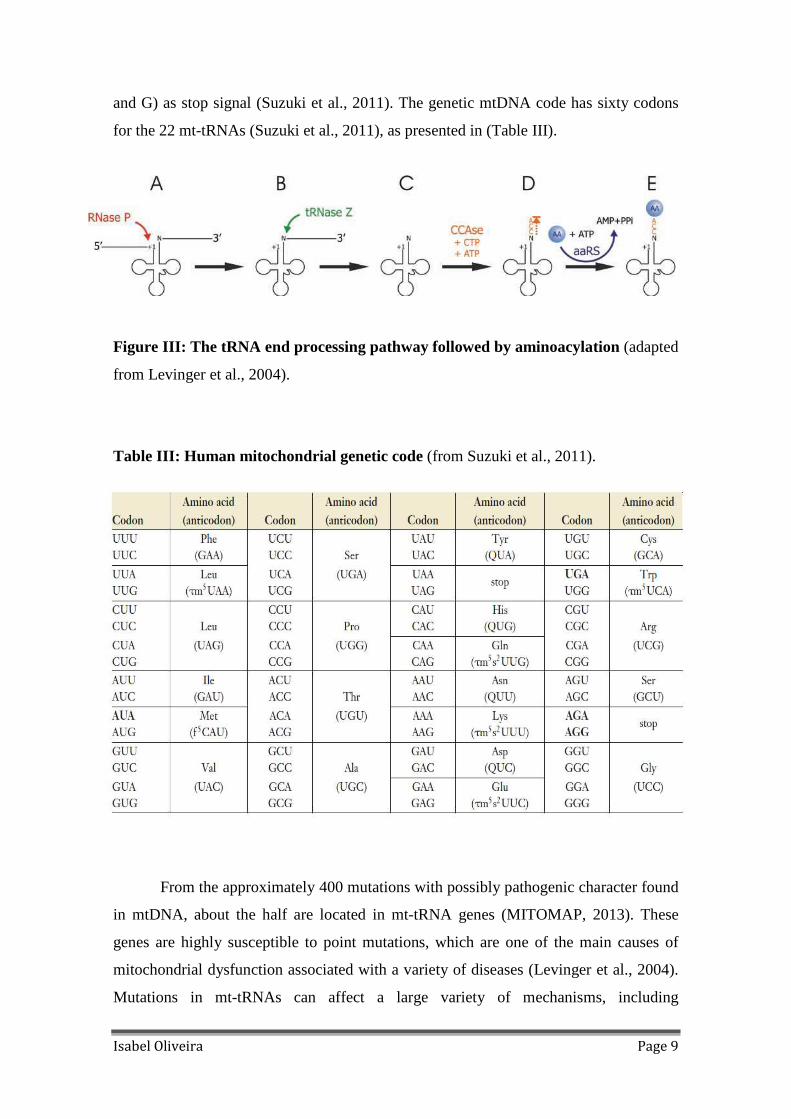
The pre-tRNA processing leads to their maturation into RNA and its activation.
The processing of mitochondrial tRNAs requires precise endonucleolytic cleavage at
both 5` and 3` ends. Excision of tRNAs is catalyzed by two specialized enzymes
(Levinger et al., 2004), RNase P (the tRNA 5´ end processing endonuclease) and
tRNase Z (the tRNA 3´ end processing endonuclease). After excision, pre-tRNA 3` ends
are subsequently completed by the addition of the “CCA” triplet through an ATP (CTP)
– tRNA-specific nucleotidyltransferase activity (Rossmanith et al.,1995) necessary for
amino acid attachment and proper tRNA positioning at the ribosome (Nicholls et al.,
2013). Once the tRNA is synthesized, the fidelity of protein synthesis depends on the
specific attachment of amino acid to its specific tRNA. This process is catalyzed by
aminoacyl-tRNA synthetases (aaRS) (Suzuki et al., 2011). The human genome encodes
one enzyme per amino acid. The tRNAs are subsequently carried to the ribosome by
translation factor elongation factor (EF)-Tu, allowing protein synthesis (Levinger et al.,
2004), as presented in Figure III.
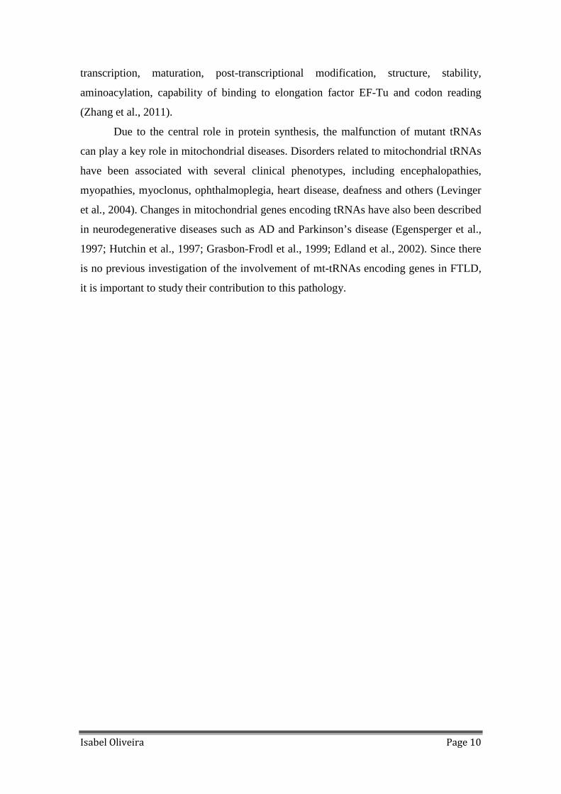
The encoding system in mitochondrial genome is different from the universal
genetic code because of the use of “AUA” for Met, “UGA” for Trp and “AGR” R = A
Isabel Oliveira Page 9
and G) as stop signal (Suzuki et al., 2011). The genetic mtDNA code has sixty codons
for the 22 mt-tRNAs (Suzuki et al., 2011), as presented in (Table III).
Figure III: The tRNA end processing pathway followed by aminoacylation (adapted
from Levinger et al., 2004).
Table III: Human mitochondrial genetic code (from Suzuki et al., 2011).
From the approximately 400 mutations with possibly pathogenic character found
in mtDNA, about the half are located in mt-tRNA genes (MITOMAP, 2013). These
genes are highly susceptible to point mutations, which are one of the main causes of
mitochondrial dysfunction associated with a variety of diseases (Levinger et al., 2004).
Mutations in mt-tRNAs can affect a large variety of mechanisms, including
Isabel Oliveira Page 10
transcription, maturation, post-transcriptional modification, structure, stability,
aminoacylation, capability of binding to elongation factor EF-Tu and codon reading
(Zhang et al., 2011).
Due to the central role in protein synthesis, the malfunction of mutant tRNAs
can play a key role in mitochondrial diseases. Disorders related to mitochondrial tRNAs
have been associated with several clinical phenotypes, including encephalopathies,
myopathies, myoclonus, ophthalmoplegia, heart disease, deafness and others (Levinger
et al., 2004). Changes in mitochondrial genes encoding tRNAs have also been described
in neurodegenerative diseases such as AD and Parkinson’s disease (Egensperger et al.,
1997; Hutchin et al., 1997; Grasbon-Frodl et al., 1999; Edland et al., 2002). Since there
is no previous investigation of the involvement of mt-tRNAs encoding genes in FTLD,
it is important to study their contribution to this pathology.
Isabel Oliveira Page 11
Mitochondrial genome analysis in frontotemporal lobar degeneration: tRNAs contribution
Oliveira I1, Santana I2,3, Santos MJ1, Duro D3, Luís D1,2, Grazina M1,2 1CNC - Center for Neuroscience and Cell Biology – Laboratory of Biochemical
Genetics, University of Coimbra, Portugal; 2Faculty of Medicine, University of
Coimbra, Portugal; 3Centro Hospitalar e Universitário de Coimbra (CHUC), Coimbra,
Portugal.
Corresponding author:
Professor Manuela Grazina, PhD., Faculty of Medicine, University of Coimbra, Pólo III
– Subunit I, Azinhaga de Sta. Comba Celas, 3000-354 Coimbra. Tel: +351 239 480040;
Fax: +351 239 480048; E-mail: [email protected]
Isabel Oliveira Page 12
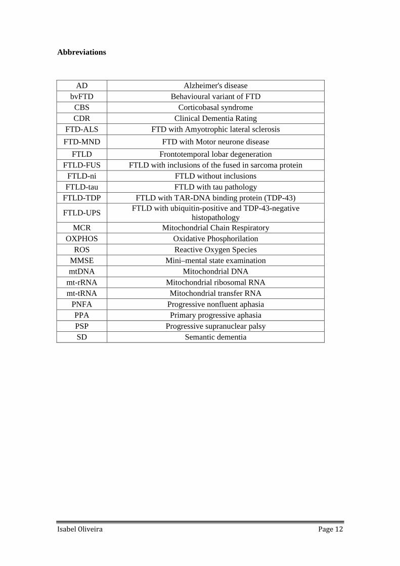
Abbreviations
AD Alzheimer's disease bvFTD Behavioural variant of FTD CBS Corticobasal syndrome CDR Clinical Dementia Rating
FTD-ALS FTD with Amyotrophic lateral sclerosis
FTD-MND FTD with Motor neurone disease
FTLD Frontotemporal lobar degeneration FTLD-FUS FTLD with inclusions of the fused in sarcoma protein FTLD-ni FTLD without inclusions FTLD-tau FTLD with tau pathology
FTLD-TDP FTLD with TAR-DNA binding protein (TDP-43)
FTLD-UPS FTLD with ubiquitin-positive and TDP-43-negative
histopathology MCR Mitochondrial Chain Respiratory
OXPHOS Oxidative Phosphorilation ROS Reactive Oxygen Species
MMSE Mini–mental state examination mtDNA Mitochondrial DNA
mt-rRNA Mitochondrial ribosomal RNA mt-tRNA Mitochondrial transfer RNA
PNFA Progressive nonfluent aphasia PPA Primary progressive aphasia PSP Progressive supranuclear palsy SD Semantic dementia
Isabel Oliveira Page 13
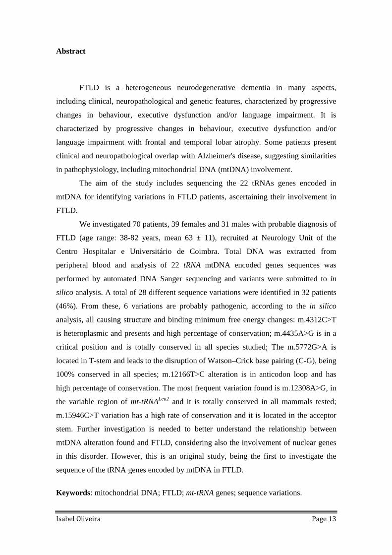
Abstract
FTLD is a heterogeneous neurodegenerative dementia in many aspects,
including clinical, neuropathological and genetic features, characterized by progressive
changes in behaviour, executive dysfunction and/or language impairment. It is
characterized by progressive changes in behaviour, executive dysfunction and/or
language impairment with frontal and temporal lobar atrophy. Some patients present
clinical and neuropathological overlap with Alzheimer's disease, suggesting similarities
in pathophysiology, including mitochondrial DNA (mtDNA) involvement.
The aim of the study includes sequencing the 22 tRNAs genes encoded in
mtDNA for identifying variations in FTLD patients, ascertaining their involvement in
FTLD.
We investigated 70 patients, 39 females and 31 males with probable diagnosis of
FTLD (age range: 38-82 years, mean 63 ± 11), recruited at Neurology Unit of the
Centro Hospitalar e Universitário de Coimbra. Total DNA was extracted from
peripheral blood and analysis of 22 tRNA mtDNA encoded genes sequences was
performed by automated DNA Sanger sequencing and variants were submitted to in
silico analysis. A total of 28 different sequence variations were identified in 32 patients
(46%). From these, 6 variations are probably pathogenic, according to the in silico
analysis, all causing structure and binding minimum free energy changes: m.4312C>T
is heteroplasmic and presents and high percentage of conservation; m.4435A>G is in a
critical position and is totally conserved in all species studied; The m.5772G>A is
located in T-stem and leads to the disruption of Watson–Crick base pairing (C-G), being
100% conserved in all species; m.12166T>C alteration is in anticodon loop and has
high percentage of conservation. The most frequent variation found is m.12308A>G, in
the variable region of mt-tRNALeu2 and it is totally conserved in all mammals tested;
m.15946C>T variation has a high rate of conservation and it is located in the acceptor
stem. Further investigation is needed to better understand the relationship between
mtDNA alteration found and FTLD, considering also the involvement of nuclear genes
in this disorder. However, this is an original study, being the first to investigate the
sequence of the tRNA genes encoded by mtDNA in FTLD.
Keywords: mitochondrial DNA; FTLD; mt-tRNA genes; sequence variations.
Isabel Oliveira Page 14
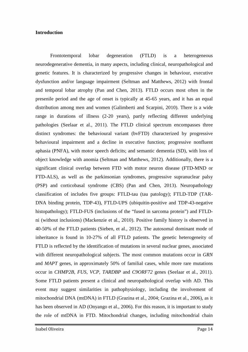
Introduction
Frontotemporal lobar degeneration (FTLD) is a heterogeneous
neurodegenerative dementia, in many aspects, including clinical, neuropathological and
genetic features. It is characterized by progressive changes in behaviour, executive
dysfunction and/or language impairment (Seltman and Matthews, 2012) with frontal
and temporal lobar atrophy (Pan and Chen, 2013). FTLD occurs most often in the
presenile period and the age of onset is typically at 45-65 years, and it has an equal
distribution among men and women (Galimberti and Scarpini, 2010). There is a wide
range in durations of illness (2-20 years), partly reflecting different underlying
pathologies (Seelaar et al., 2011). The FTLD clinical spectrum encompasses three
distinct syndromes: the behavioural variant (bvFTD) characterized by progressive
behavioural impairment and a decline in executive function; progressive nonfluent
aphasia (PNFA), with motor speech deficits; and semantic dementia (SD), with loss of
object knowledge with anomia (Seltman and Matthews, 2012). Additionally, there is a
significant clinical overlap between FTD with motor neuron disease (FTD-MND or
FTD-ALS), as well as the parkinsonian syndromes, progressive supranuclear palsy
(PSP) and corticobasal syndrome (CBS) (Pan and Chen, 2013). Neuropathology
classification of includes five groups: FTLD-tau (tau patology); FTLD-TDP (TAR-
DNA binding protein, TDP-43), FTLD-UPS (ubiquitin-positive and TDP-43-negative
histopathology); FTLD-FUS (inclusions of the “fused in sarcoma protein”) and FTLD-
ni (without inclusions) (Mackenzie et al., 2010). Positive family history is observed in
40-50% of the FTLD patients (Sieben, et al., 2012). The autosomal dominant mode of
inheritance is found in 10-27% of all FTLD patients. The genetic heterogeneity of
FTLD is reflected by the identification of mutations in several nuclear genes, associated
with different neuropathological subjects. The most common mutations occur in GRN
and MAPT genes, in approximately 50% of familial cases, while more rare mutations
occur in CHMP2B, FUS, VCP, TARDBP and C9ORF72 genes (Seelaar et al., 2011).
Some FTLD patients present a clinical and neuropathological overlap with AD. This
event may suggest similarities in pathophysiology, including the involvement of
mitochondrial DNA (mtDNA) in FTLD (Grazina et al., 2004; Grazina et al., 2006), as it
has been observed in AD (Onyango et al., 2006). For this reason, it is important to study
the role of mtDNA in FTD. Mitochondrial changes, including mitochondrial chain
Isabel Oliveira Page 15
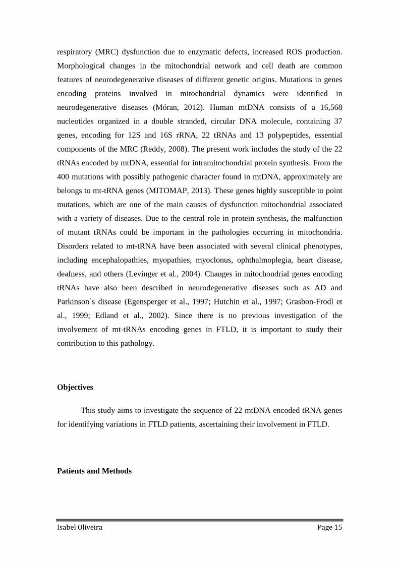
respiratory (MRC) dysfunction due to enzymatic defects, increased ROS production.
Morphological changes in the mitochondrial network and cell death are common
features of neurodegenerative diseases of different genetic origins. Mutations in genes
encoding proteins involved in mitochondrial dynamics were identified in
neurodegenerative diseases (Móran, 2012). Human mtDNA consists of a 16,568
nucleotides organized in a double stranded, circular DNA molecule, containing 37
genes, encoding for 12S and 16S rRNA, 22 tRNAs and 13 polypeptides, essential
components of the MRC (Reddy, 2008). The present work includes the study of the 22
tRNAs encoded by mtDNA, essential for intramitochondrial protein synthesis. From the
400 mutations with possibly pathogenic character found in mtDNA, approximately are
belongs to mt-tRNA genes (MITOMAP, 2013). These genes highly susceptible to point
mutations, which are one of the main causes of dysfunction mitochondrial associated
with a variety of diseases. Due to the central role in protein synthesis, the malfunction
of mutant tRNAs could be important in the pathologies occurring in mitochondria.
Disorders related to mt-tRNA have been associated with several clinical phenotypes,
including encephalopathies, myopathies, myoclonus, ophthalmoplegia, heart disease,
deafness, and others (Levinger et al., 2004). Changes in mitochondrial genes encoding
tRNAs have also been described in neurodegenerative diseases such as AD and
Parkinson`s disease (Egensperger et al., 1997; Hutchin et al., 1997; Grasbon-Frodl et
al., 1999; Edland et al., 2002). Since there is no previous investigation of the
involvement of mt-tRNAs encoding genes in FTLD, it is important to study their
contribution to this pathology.
Objectives
This study aims to investigate the sequence of 22 mtDNA encoded tRNA genes
for identifying variations in FTLD patients, ascertaining their involvement in FTLD.
Patients and Methods
Isabel Oliveira Page 16
Samples
DNA samples of 70 patients (39 females and 31 males; age range: 38-82 years,
mean 63 ± 11) with probable diagnosis of FTLD according to the standard criteria of
DSM-IV ( Brun et al., 1994; McKhann et al.,2001) recruited at Neurology Unit of the
Centro Hospitalar e Universitário de Coimbra were investigated.
Total DNA was extracted from peripheral blood by standard phenol chloroform method
(Treco, 1999).
The scaling of the dementia was obtained by scoring, the CDR (Clinical
Dementia Rating) and MMSE (Mini–mental state examination). In CDR, the scale is
between 0 and 3 and the higher values correspond to higher degree of dementia. In
MMSE, the scale is between 0 and 30 and lower values correspond to higher degree of
dementia (Folstein et al., 1975)
PCR amplification
Amplification of the 22 mitochondrial tRNA enconding genes was performed by
Polymerase Chain Reaction (PCR). This technique allows obtaining multiple copies of a
particular DNA fragment. The amplification conditions included initial denaturation at
95°C for 5 min followed by 35 cycles at 95°C for 45 s, 50-60ºC for 45 s, 72°C for 60 s,
and a final extension step at 72°C for 5 min using a master mix containing 2-10 ng of
DNA, 10X buffer, dNTP (2mM), primer forward (2,5µM), primer reverse (2,5µM), Taq
DNA polymerase, H2O milli Q and MgCl2 (Landsverk et al., 2012).
Agarose gel electrophoresis
PCR procedure was followed by agarose gel electrophoresis for the separation of
DNA fragments, in order to verify the success of amplification. PCR products were
mixed with loading dye (1:1) and then applied to 1% agarose gel for 1 hour, at 100
Volts, using weight marker. After migration, ethidium bromide labeled DNA molecules
were visualized under ultraviolet irradiation (Landsverk et al., 2012).
Isabel Oliveira Page 17
DNA Sanger Sequencing
After PCR product analysis, samples were purified with ExoSAP-IT®, consisting
of exonuclease I (exo I) and alkaline phosphatase (SAP) to degrade the excess of
primers and nucleotides, which are the main factors interfering with PCR sequencing
(Werle et al., 1994).
The sequencing PCR involves the synthesis of single stranded DNA using the
DNA previously amplified in PCR as template. Synthesized chains are terminated
prematurely with various possible sizes. The synthesis begins at the primer binding site
and ending with the incorporation of a terminator nucleotide that lack the hydroxyl
group at the 3' position of the deoxyribose, preventing the establishment of connections
phospho-diester and the incorporation of new nucleotides to DNA strand. When a
terminator nucleotide is incorporated, the synthesis of new chain ends (Buitrago and
Jimenez, 2001). The amplification conditions were an initial denaturation at 96°C for 2
min followed by 45 cycles at 96°C for 10 s, 55ºC for 5 s, 60°C for 4 min, using
BigDye® Terminator Ready Reaction Mix v3.1, 5X sequencing buffer, 2,5µM of
primer forward or reverse and H2O milli Q (Landsverk et al., 2012).
After sequencing PCR, samples were submitted to standard Sephadex®
purification (gel filtration), which removes substances which have not been added
during the reaction. After purification, the samples were loaded in the sequencer ABI
Prism® 3130 (Applied Biosystems).
This is a fast and automated process that allows the determination of nucleotide
sequence comprising the DNA fragment to be studied (Buitrago and Jimenez, 2001).
Automated DNA Sequencing is based on electrophoretic procedures using polymer
gels. Applied Biosystems DNA sequencers detect fluorescence from four different dyes
that are used to identify the A, C, G and T terminators. Each dye has a fluorescence
wavelenght when excited by argon ion laser, allowing detection and distinction of all
four bases.
After the automated sequencing, samples sequences were analysed using
Sequencing Analysis v5.4® and SeqScape v2.5® software, which allow to compare the
obtained data with the reference sequence. Thus, it is possible to detect any variation in
the sequence under study.
Isabel Oliveira Page 18
In silico analysis
After the analysis of all sequences, an in silico study of the detected changes was
performed, using different databases such as MITOMAP where the variations are
reported (MITOMAP, 2013), RNAfold (Hofacker et al., 1994) that predict the RNA
secondary structure based on minimum energy requirements and pair probabilities
(Mezghani et al., 2011). The localization of the sequence variations in mt-tRNA and
sequences from the species were obtained from the Mamit-tRNA database (Putz et al.,
2007). Evolutionary conservation was performed for all alterations (12 mt-tRNAs) in 10
different species (Homo sapiens, Pan paniscus, Pan troglodytes, Gorilla gorilla, Pongo
pygmaeus, Macaca mulatta, Bos taurus, Canis familiaris, Mus musculus, Rattus
norvegicus). The sequence alignment of the mt-tRNAs was performed using the
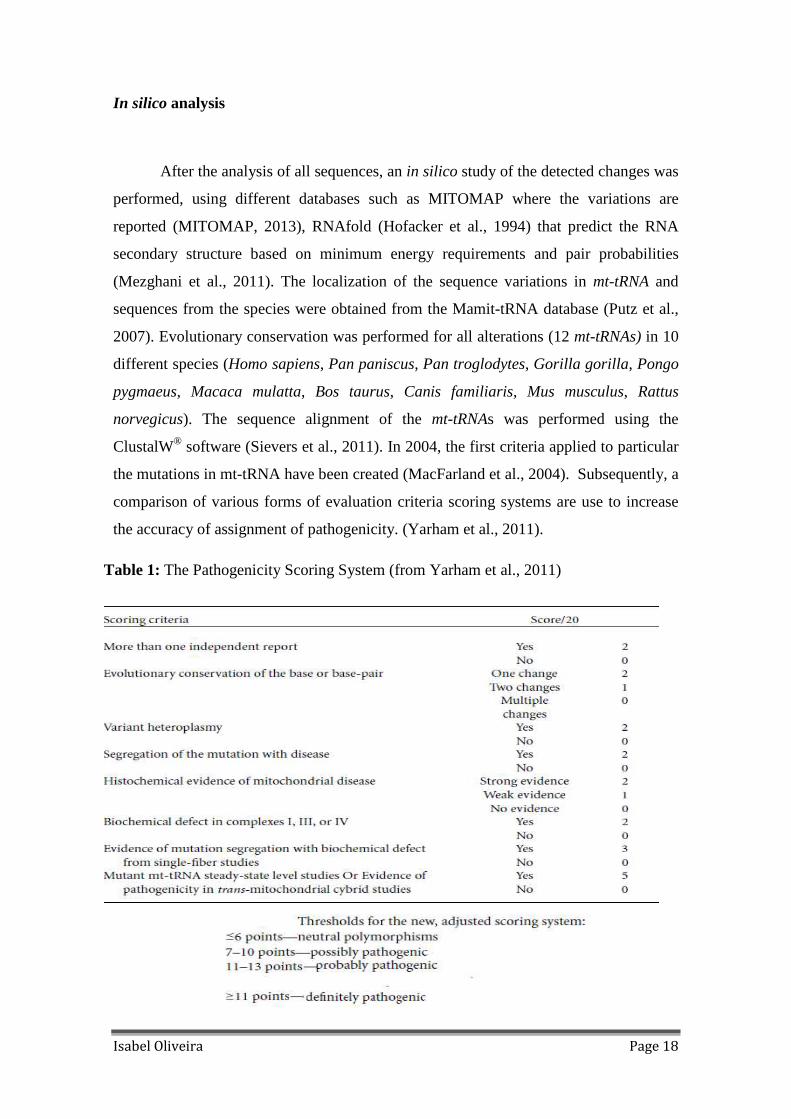
ClustalW® software (Sievers et al., 2011). In 2004, the first criteria applied to particular
the mutations in mt-tRNA have been created (MacFarland et al., 2004). Subsequently, a
comparison of various forms of evaluation criteria scoring systems are use to increase
the accuracy of assignment of pathogenicity. (Yarham et al., 2011).
Table 1: The Pathogenicity Scoring System (from Yarham et al., 2011)
Isabel Oliveira Page 19
Results
A total of 70 patients (39 females and 31 males) with FTLD were included in
this study.
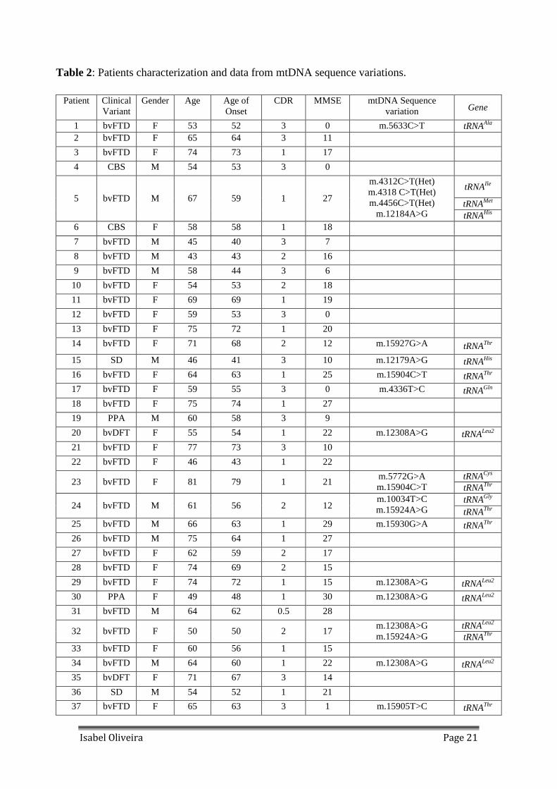
From these, 32 (17 females and 15 males) present 28 different genetic variations
in mt-tRNA genes. We have found 22 patients with only one variation, 9 patients with 2
variations and 3 patients with 4 variations. Patients 5, 43 and 51 have the higher number
of variations (Table 2).
Concerning the CDR, 15 patients presented the maximum degree of dementia
but only 5 of these have mtDNA variations. The remaining patients presented moderate
or mild dementia. For MMSE, 10 patients exhibited maximum degree of dementia but
only 4 of these present variations (Table 3).
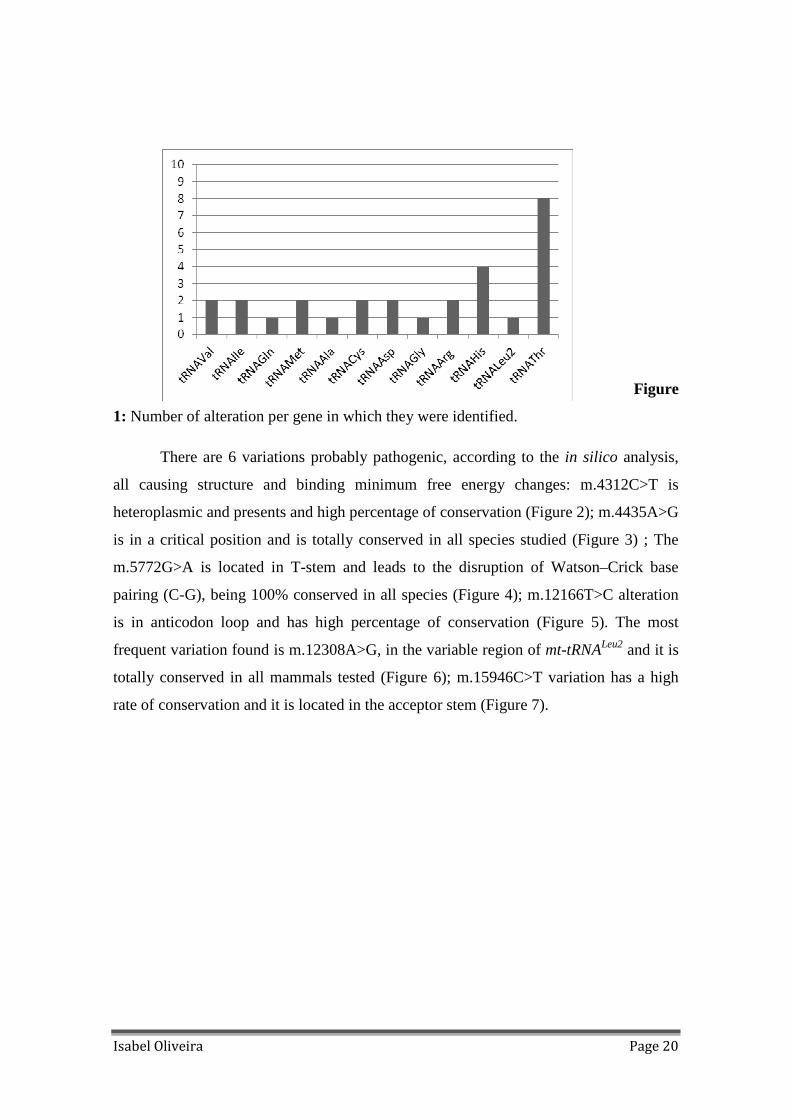
From the 28 different genetic variations, there are 19 variations that were
identified only in 1 patient, 4 variations twice, 3 variations three times, 1 variation four
times and other variation tenfold. Only 12 mt-tRNAs have variations identified
(tRNAAla, tRNAIle, tRNAMet, tRNAHis, tRNAThr, tRNAGln, tRNACys, tRNAGly, tRNALeu2,
tRNAVal, tRNAArg and tRNAAsp), as presented in Figure 1.
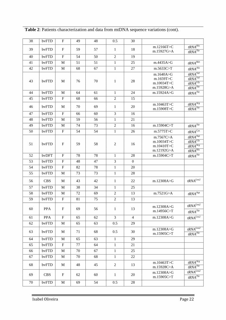
The variations identified were found in MITOMAP database: 14 have been
described as “polymorphism”, 8 as “polymorphism” and “mutation”; 3 variations as
“polymorphism” and “somatic mutation”; 1 as “mutation”; 1 variation as
“polymorphism” and “mutation”; and 1 “novel” (m.7567C>A). The minimum free
energy was changed in 22 of the 28 found sequence variations but 5 (m.5775T>C,
m.7521G>A, m.10410T>C, m.12192G>A and m.15927G>A) of these are not predicted
to alter the tRNA structure. Concerning the location of the variations in mt-tRNAs
structure, 14 were found in the stems, 10 in the loops and 4 in the variable region (Table
3).
Isabel Oliveira Page 20
Figure
1: Number of alteration per gene in which they were identified.
There are 6 variations probably pathogenic, according to the in silico analysis,
all causing structure and binding minimum free energy changes: m.4312C>T is
heteroplasmic and presents and high percentage of conservation (Figure 2); m.4435A>G
is in a critical position and is totally conserved in all species studied (Figure 3) ; The
m.5772G>A is located in T-stem and leads to the disruption of Watson–Crick base
pairing (C-G), being 100% conserved in all species (Figure 4); m.12166T>C alteration
is in anticodon loop and has high percentage of conservation (Figure 5). The most
frequent variation found is m.12308A>G, in the variable region of mt-tRNALeu2 and it is
totally conserved in all mammals tested (Figure 6); m.15946C>T variation has a high
rate of conservation and it is located in the acceptor stem (Figure 7).
Isabel Oliveira Page 21
Table 2: Patients characterization and data from mtDNA sequence variations.
Patient Clinical Variant
Gender Age Age of Onset
CDR MMSE mtDNA Sequence variation Gene
1 bvFTD F 53 52 3 0 m.5633C>T tRNAAla 2 bvFTD F 65 64 3 11 3 bvFTD F 74 73 1 17 4 CBS M 54 53 3 0
tRNAIle
tRNAMet 5 bvFTD M 67 59 1 27
m.4312C>T(Het) m.4318 C>T(Het) m.4456C>T(Het)
m.12184A>G tRNAHis 6 CBS F 58 58 1 18
7 bvFTD M 45 40 3 7 8 bvFTD M 43 43 2 16 9 bvFTD M 58 44 3 6 10 bvFTD F 54 53 2 18 11 bvFTD F 69 69 1 19 12 bvFTD F 59 53 3 0
13 bvFTD F 75 72 1 20 14 bvFTD F 71 68 2 12 m.15927G>A tRNAThr
15 SD M 46 41 3 10 m.12179A>G tRNAHis 16 bvFTD F 64 63 1 25 m.15904C>T tRNAThr 17 bvFTD F 59 55 3 0 m.4336T>C tRNAGln 18 bvFTD F 75 74 1 27 19 PPA M 60 58 3 9 20 bvDFT F 55 54 1 22 m.12308A>G tRNALeu2
21 bvFTD F 77 73 3 10 22 bvFTD F 46 43 1 22
tRNACys 23 bvFTD F 81 79 1 21
m.5772G>A m.15904C>T tRNAThr
tRNAGly 24 bvFTD M 61 56 2 12
m.10034T>C m.15924A>G tRNAThr
25 bvFTD M 66 63 1 29 m.15930G>A tRNAThr 26 bvFTD M 75 64 1 27 27 bvFTD F 62 59 2 17 28 bvFTD F 74 69 2 15
29 bvFTD F 74 72 1 15 m.12308A>G tRNALeu2 30 PPA F 49 48 1 30 m.12308A>G tRNALeu2 31 bvFTD M 64 62 0.5 28
tRNALeu2 32 bvFTD F 50 50 2 17
m.12308A>G m.15924A>G tRNAThr
33 bvFTD F 60 56 1 15 34 bvFTD M 64 60 1 22 m.12308A>G tRNALeu2 35 bvDFT F 71 67 3 14
36 SD M 54 52 1 21 37 bvFTD F 65 63 3 1 m.15905T>C tRNAThr
Isabel Oliveira Page 22
Table 2: Patients characterization and data from mtDNA sequence variations (cont).
38 bvFTD F 49 48 0.5 30
tRNAHis 39 bvFTD F 59 57 1 18
m.12166T>C m.15927G>A tRNAThr
40 bvFTD F 54 50 2 19 41 bvFTD M 51 51 1 25 m.4435A>G tRNAMet 42 bvFTD M 68 67 1 27 m.5633C>T tRNAAla
tRNAVal tRNAVal tRNAGly
43 bvFTD M 76 70 1 28
m.1640A>G m.1659T>C m.10034T>C m.15928G>A tRNAThr
44 bvFTD M 64 61 1 24 m.15924A>G tRNAThr 45 bvFTD F 68 66 2 15
tRNAArg 46 bvFTD M 70 69 1 20
m.10463T>C m.15908T>C tRNAThr
47 bvFTD F 66 60 3 16 48 bvFTD M 59 56 1 21 49 bvFTD M 74 73 2 16 m.15904C>T tRNAThr
50 bvFTD F 54 54 1 26 m.5775T>C tRNACys tRNAAsp tRNAGly tRNAArg
51 bvFTD F 59 58 2 16
m.7567C>A m.10034T>C m.10410T>C m.12192G>A tRNAHis
52 bvDFT F 78 78 1 28 m.15904C>T tRNAThr 53 bvFTD F 48 47 3 0 54 bvFTD F 82 78 1 20
55 bvFTD M 73 73 1 28
56 CBS M 43 42 1 22 m.12308A>G tRNALeu2
57 bvFTD M 38 34 1 25 58 bvFTD M 72 69 2 13 m.7521G>A tRNAAsp 59 bvFTD F 81 75 2 13
tRNALeu2 60 PPA F 69 56 1 13
m.12308A>G m.14956C>T tRNAThr
61 PPA F 65 62 3 4 m.12308A>G tRNALeu2
62 bvFTD M 65 63 0.5 29
tRNALeu2 63 bvFTD M 71 68 0.5 30
m.12308A>G m.15905C>T tRNAThr
64 bvFTD M 65 63 1 29 65 bvFTD F 77 64 1 21
66 bvFTD M 70 67 1 25 67 bvFTD M 70 68 1 22
tRNAArg 68 bvFTD M 48 45 2 13
m.10463T>C m.15928C>A tRNAThr
tRNALeu2 69 CBS F 62 60 1 20
m.12308A>G m.15905C>T tRNAThr
70 bvFTD M 69 54 0.5 28
Isabel Oliveira Page 23
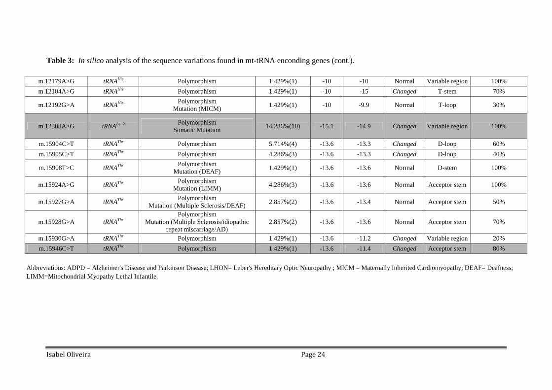
Table 3: In silico analysis of the sequence variations found in mt-tRNA enconding genes.
Minimum free energy (kcal/mol) Change of
nucleotide Locus (tRNA) Reported in MITOMAP Frequency in the
sample (n) Normal Changed
Structure Localization in
tRNA structure
Evolutionary conservation
m.1640A>G tRNAVal Polymorphism 1.429%(1) -12.5 -17.6 Changed Anticodon stem 90% m.1659T>C tRNAVal Mutation (Movement Disorder) 1.429%(1) -12.5 -10.7 Changed T-stem 60%
m.4312 C>T tRNAIle Polymorphism
Somatic Mutation 1.429%(1) -7.8 -8.5 Changed T-loop 80%
m.4318C>T tRNAIle Polymorphism 1.429%(1) -7.8 -9.6 Changed T-loop 60%
m.4336T>C tRNAGln Polymorphism
Mutation (ADPD/Hearing Loss & Migraine)
1.429%(1) -19.5 -19.5 Normal Acceptor stem 70%
m.4435A>G tRNAMet Polymorphism
Mutation (LHON/hypertension) 1.429%(1) -12.8 -12.9 Changed Anticodon loop 100%
m.4456C>T tRNAMet Polymorphism
Mutation (Poss. hypertension factor) 1.429%(1) -12.8 -12.4 Changed T-loop 60%
m.5633C>T tRNAAla Polymorphism
Somatic Mutation 2.857%(2) -17 -16.7 Changed Anticodon stem 40%
m.5772G>A tRNACys Polymorphism 1.429%(1) -18.1 -15.9 Changed T-stem 100%
m.5775T>C tRNACys Polymorphism 1.429%(1) -18.1 -17.8 Normal T-loop 80%
m.7521G>A tRNAAsp Polymorphism 1.429%(1) -9.1 -9.7 Normal Acceptor stem 50%
m.7567C>A tRNAAsp Novel 1.429%(1) -9.1 -10.9 Changed T-loop 40%
m.10034T>C tRNAGly Polymorphism 4.286%(3) -8.5 -8.1 Changed Variable region 90%
m.10410T>C tRNAArg Polymorphism 1.429% (1) -10.1 -11.7 Normal Acceptor stem 20%
m.10463T>C tRNAArg Polymorphism
Somatic Mutation 2.8557%(2) -10.1 -10.1 Normal Acceptor stem 100%
m.12166T>C tRNAHis Polymorphism 1.429%(1) -10 -9.5 Changed Anticodon loop 80%
Isabel Oliveira Page 24
Table 3: In silico analysis of the sequence variations found in mt-tRNA enconding genes (cont.).
m.12179A>G tRNAHis Polymorphism 1.429%(1) -10 -10 Normal Variable region 100%
m.12184A>G tRNAHis Polymorphism 1.429%(1) -10 -15 Changed T-stem 70%
m.12192G>A tRNAHis Polymorphism
Mutation (MICM) 1.429%(1) -10 -9.9 Normal T-loop 30%
m.12308A>G tRNALeu2 Polymorphism
Somatic Mutation 14.286%(10) -15.1 -14.9 Changed Variable region 100%
m.15904C>T tRNAThr Polymorphism 5.714%(4) -13.6 -13.3 Changed D-loop 60%
m.15905C>T tRNAThr Polymorphism 4.286%(3) -13.6 -13.3 Changed D-loop 40%
m.15908T>C tRNAThr Polymorphism
Mutation (DEAF) 1.429%(1) -13.6 -13.6 Normal D-stem 100%
m.15924A>G tRNAThr Polymorphism
Mutation (LIMM) 4.286%(3) -13.6 -13.6 Normal Acceptor stem 100%
m.15927G>A tRNAThr Polymorphism
Mutation (Multiple Sclerosis/DEAF) 2.857%(2) -13.6 -13.4 Normal Acceptor stem 50%
m.15928G>A tRNAThr Polymorphism
Mutation (Multiple Sclerosis/idiopathic repeat miscarriage/AD)
2.857%(2) -13.6 -13.6 Normal Acceptor stem 70%
m.15930G>A tRNAThr Polymorphism 1.429%(1) -13.6 -11.2 Changed Variable region 20%
m.15946C>T tRNAThr Polymorphism 1.429%(1) -13.6 -11.4 Changed Acceptor stem 80%
Abbreviations: ADPD = Alzheimer's Disease and Parkinson Disease; LHON= Leber's Hereditary Optic Neuropathy ; MICM = Maternally Inherited Cardiomyopathy; DEAF= Deafness; LIMM=Mitochondrial Myopathy Lethal Infantile.

Isabel Oliveira Page 25
Figure 2: Results from in silico analysis for nucleotide (m.4312C>T mt-tRNAIle
) A-
normal and B- “mutated” structure (RNAfold); C- Location of the sequence variation in
the clover-shaped structure; D- Evolutionary conservation for the nucleotide position
(signed with blue rectangle).
4312T
∆G=-7,80 kcal/mol ∆G=-8,50 kcal/mol (A) (B)
Normal Changed
(C)
∆G=-12,80 kcal/mol ∆G=-12,90 kcal/mol
Normal Changed
(A)
(D)
(B)
Isabel Oliveira Page 26
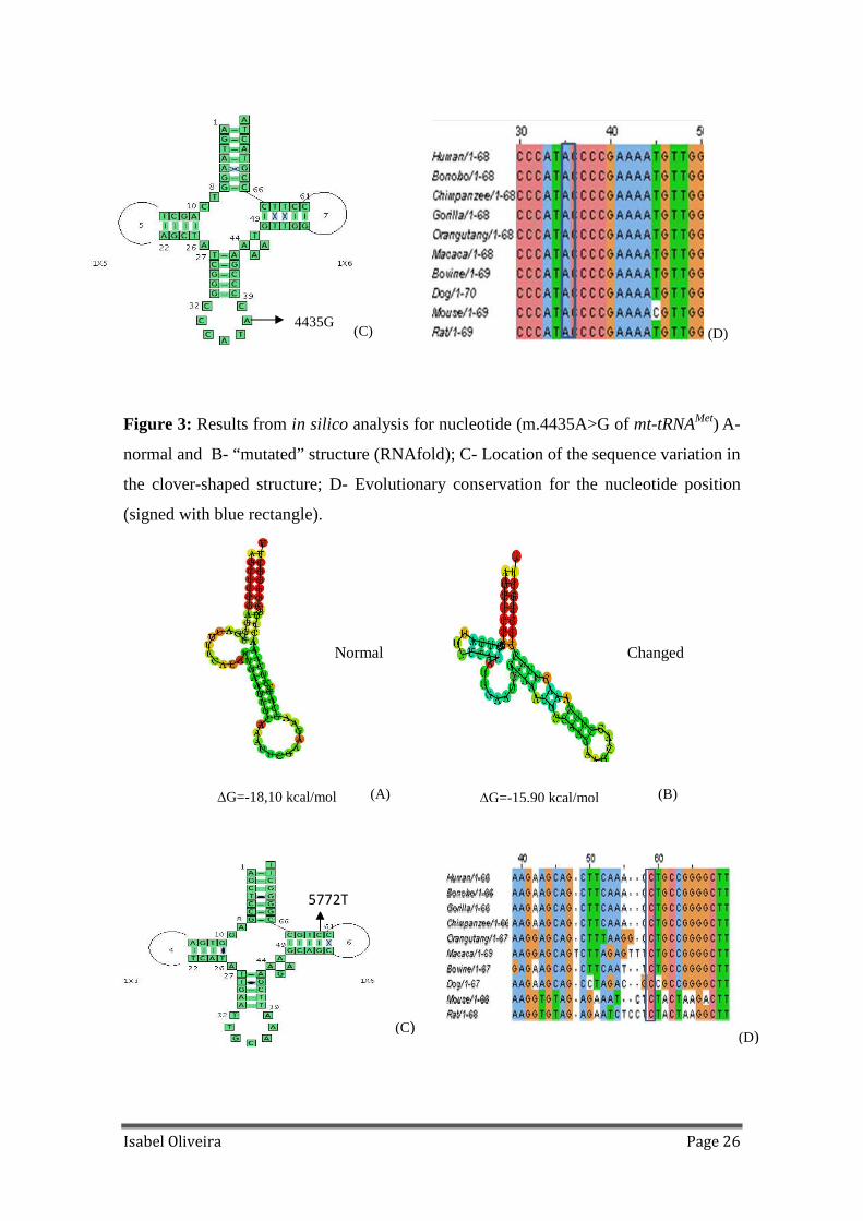
Figure 3: Results from in silico analysis for nucleotide (m.4435A>G of mt-tRNAMet) A-
normal and B- “mutated” structure (RNAfold); C- Location of the sequence variation in
the clover-shaped structure; D- Evolutionary conservation for the nucleotide position
(signed with blue rectangle).
(D) 4435G
(C)
∆G=-18,10 kcal/mol ∆G=-15,90 kcal/mol
Normal Changed
(A)
(D)
5772T
(C)
(B)
Isabel Oliveira Page 27
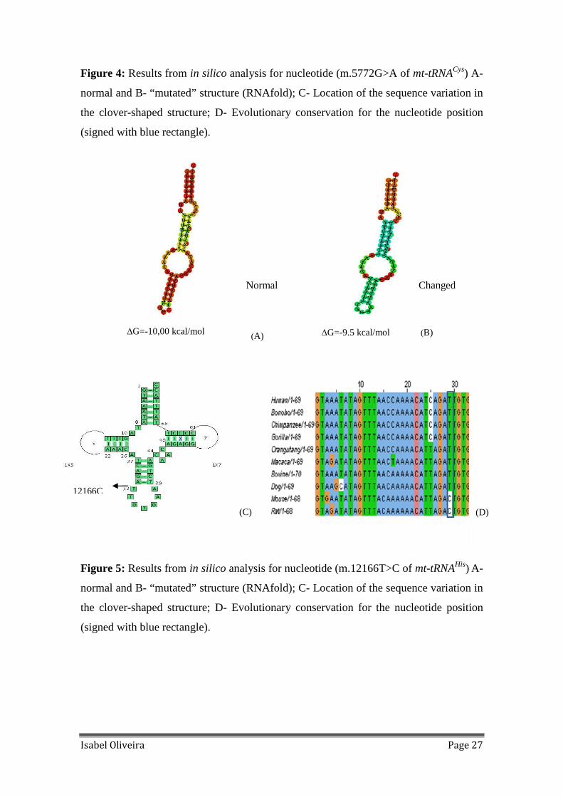
Figure 4: Results from in silico analysis for nucleotide (m.5772G>A of mt-tRNACys) A-
normal and B- “mutated” structure (RNAfold); C- Location of the sequence variation in
the clover-shaped structure; D- Evolutionary conservation for the nucleotide position
(signed with blue rectangle).
Figure 5: Results from in silico analysis for nucleotide (m.12166T>C of mt-tRNAHis) A-
normal and B- “mutated” structure (RNAfold); C- Location of the sequence variation in
the clover-shaped structure; D- Evolutionary conservation for the nucleotide position
(signed with blue rectangle).
∆G=-10,00 kcal/mol ∆G=-9,5 kcal/mol
Normal Changed
(A)
12166 C 12166C
(C) (D)
(B)
Isabel Oliveira Page 28
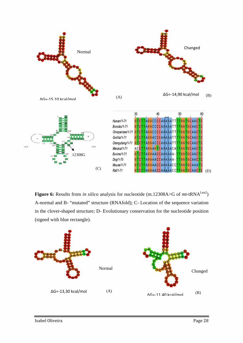
Figure 6: Results from in silico analysis for nucleotide (m.12308A>G of mt-tRNALeu2)
A-normal and B- “mutated” structure (RNAfold); C- Location of the sequence variation
in the clover-shaped structure; D- Evolutionary conservation for the nucleotide position
(signed with blue rectangle).
12308G
∆G=-11,40 kcal/mol ∆G=-13,30 kcal/mol
Normal Changed
∆G=-15,10 kcal/mol
Normal
∆G=-14,90 kcal/mol
Changed
(A)
(C) (D)
(B)
(B)
(A)
Isabel Oliveira Page 29
(2)
Figure 7: Results from in silico analysis for nucleotide (m.15946C>T of mt-tRNAThr)
A-normal and B- “mutated” structure (RNAfold); C- Location of the sequence variation
in the clover-shaped structure; D- Evolutionary conservation for the nucleotide position
(signed with blue rectangle).
Discussion
Human mitochondrial tRNAs acquired interest with the discovery of correlations
between point mutations in its genes and neurodegenerative diseases (Zifa et al., 2007).
The clinical evaluation related to mtDNA is often complicated and mt-tRNAs mutations
present unusual difficulties in clinical diagnosis because of the inability to determine if
the changes are pathogenic or not (Glatz et al., 2011). The minimum free energy is a
criterion to predict the stability of mt-tRNA structure, since a pathogenic mutation in
mt-tRNA can alter the secondary structure. Lower minimum free energy is related to a
more stable structure, but it is not an absolute fact, because of biological complexity
(Zhang et al., 2011). Evolutionary conservation is also an important feature of
pathogenicity (Yarham et al., 2011). High percentage of evolutionary conservation
suggests that sequence variation may be deleterious. In 28 variations, 17 (m.1640A>G,
m.1659T>C, m4312C>T, m.4318C>T, m.4435A>G ,m.4456C>T, m.5633C>T,
m.5772G<A, m.7567C>A, m.10034T>C, m.12166T>C, m.12184A>G, m.12308A>G,
m.15904C>T, m.15905C>T, m.15930G>A and m.15946C>T) are predicted to have
15946T
(D) (C)
Isabel Oliveira Page 30
changes of structure and minimum free energy, but only 8 variations have an elevated
evolutionary conservation (Table 3). In 5 variations, despite of having high percentage
of conservation, its structures do not change (m.5775T>C, m.10463T>C, m.12179A>G,
m.15908T>C and m.15924A>G). There are 7 alterations (m.1640A>G, m.5772G>A,
m.5775T>C, m.10034T>C, m.12166 T>C, m.12179A>G and m.15946C>T) that are
reported in MITOMAP as polymorphisms but the percentage of conservation is high.
Variations previously described as pathogenic mutations, affect mostly high conserved
nucleotides, whereas most polymorphic alterations affect rather nonconserved
nucleotides (Table 3). However, there are exceptions: some pathogenic mutations affect
nonconserved positions and some polymorphic variations affect conserved positions.
This suggests that the rate of conservation of a nucleotide by itself cannot be considered
as a threshold for which the mutation will be effectively pathogenic (Zifa et al., 2007).
Other criteria must be considered.
From the 28 variations detected, m.4312C>T, m.4318C>T and m.4456C>T are
present in heteroplasmy, and were found in the same patient, but the percentage of
heteroplasmy was not determined. On the other hand, heteroplasmy of variants in mt-
tRNA genes has been generally regarded as direct evidence for pathogenicity
(McFarland et al., 2004).
Location of variations in the tRNA structure is also very important. For a
mutation affecting directly the function of mt-tRNA, this should occur in a critical base
to the recognition of the codon and aminoacylation. However, of all the pathogenic
mutations that occur in mt-tRNA genes and that have been previously associated with
mitochondrial diseases, only a few occur in these critical positions (Suzuki et al., 2011).
From 28 variations, there are 14 alterations that are located in the stems, 10 variations
are localized in the loops and 4 are in the variable region. Many mt-tRNA pathological
mutations are mainly located, on the stem portions (Pereira et al., 2008). An elevated
number of variations were found in tRNAThr coding genes, in comparison with other
tRNAs. This is in agreement to the study by Pereira and colleagues (2008), which
indicates that the variability in this gene is much higher, especially in the stem regions.
Other mt-tRNAs genes have also more variations in the stems, such as tRNAVal and
tRNAArg. Other genes have higher numbers of variations in the loops (tRNAIle and
tRNAMet). The tRNAMet presents variations in the loops, namely one substitution,
m.4435A>G, is located in the anticodon loop, which is relevant, since this tRNA
performs an important role as the initiator of all mtDNA proteins.
Isabel Oliveira Page 31
The breaking of Watson–Crick base pairing is an important characteristic to
identify pathogenic mutations that occur in the stem structures. The disruption of C-G
(cytosine-guanine) base pair linking is significantly more common in pathogenic
mutations. The A–T (adenine–thymine) bonding has a lower thermodynamic energy
than C–G bonding and it is possible that breaking A–T bonds has less effect on the
structure of the mt-RNA than an equal break of a C–G pair bases (Figure 4 and 7)
(McFarland et al., 2004). Variation m.5772G>A is in T-stem of tRNACys (within LS of
mtDNA). Variations m.15927G>A, m.15928G>A and m.15946C>T are located in
stems of tRNAThr. Since these variations break the links C-G, these changes are
probably more pathogenic.
After in silico analysis, there are 6 sequence variations that present high
probability of being pathogenic: m.4312C>T, due the heteroplasmy presented, to the
change of minimum free energy and its structure, and high percentage of conservation;
m.4435A>G alteration induces structure and binding minimum free energy changes, it
is localized in a critical position and it is totally conserved in all species analysed;
m.5772G>A modifies the structure and binding minimum free energy, being located a
local of disruption of Watson–Crick base pairing (C-G) of T-stem and it is 100%
conserved in all species; the alteration m.12166T>C presents changes in structure and
binding minimum free energy, it is located in anticodon loop and is highly conserved;
the most frequent variation identified in our cohort is m.12308A>G, in the variable
region of mt-tRNALeu2 gene. This variation leads to structure alteration and it is totally
conserved; the variation m.15946C>T also causes change in the structure and minimum
free energy; it is localized in the acceptor stem and has high conservation rate.
Conclusions
The analysis of the mt-tRNAs variations indicates that there is not sufficient
evidence to classify the variations as pathogenic causative of FTLD. However, through
this study it is possible to gather important data.
Most of the detected variations altered the structure and minimum free energy of
tRNAs. There is a higher number of substitutions in the stems than in the loops, which
is in agreement with the literature, concerning the involvement of mt-tRNA folding
genes in diseases. The evolutionary conservation is not always in agreement with the
Isabel Oliveira Page 32
results obtained for the structure, for prediction of pathogenicity. Therefore, it is
difficult to detect pathogenic mutations due to heterogeneity of results. Nevertheless,
according to all the pathogenicity criteria studied, in 28 variations detected, the more
likely to be pathogenic are m.4312C>T in tRNAIle, m.4435A>G in tRNAMet,
m.5772G>A in tRNACys, m.12166T>C in tRNAHis, m.12308A>C in tRNALeu2 and
m.15946C>T in tRNAThr.
It is yet unclear, at the molecular level, how the mutant mt-tRNAs can cause
mitochondrial dysfunction. There is a current notion that not only mt-tRNA mutations,
but a combination of different mutations present in mitochondrial genes is responsible
for a variety of clinical diseases (McFarland et al., 2004).
Additionally, given the role of mt-tRNAs in MRC function, mutations in these
genes may affect the ability to produce mitochondrial proteins. These protein synthesis
anomalies result in OXPHOS deficiency, since the enzymatic activity of all MRC
complexes could be affected. Furthermore, according to the “Mitochondrial cascade
hypothesis” (Swerdlow and Khan, 2004), polymorphic variations in MCR subunits
encoding genes establish MCR efficiency and basal mitochondrial ROS production, that
correlates with mtDNA damage. Acordingly, somatic mtDNA mutation decreases MCR
efficiency leading to reduced OXPHOS and/or increased ROS production. For this
reason, mtDNA mutations possibly modify age of onset, contributing to
neurodegeneration process, probably due to an impairment of MCR and/or translation
mechanisms.
Moreover, deeper biochemical investigations are needed to better understand the
relationships between mtDNA and FTLD, considering the involvement of nuclear
genes. Genotype/phenotype correlation can involve nuclear and mitochondrial
interactions, but the exact mechanism is still unknown.
In conclusion, more research is needed to determine whether the mt-tRNA
variations play a direct pathogenic role in FTLD. A functional study would certainly
help to prove the possible pathogenicity of these alterations.
Isabel Oliveira Page 33
References
Boxer AL, Gold M, Huey E, Gao FB, Burton EA, Chow T, Kao A, Leavitt BR, Grether
M, Knopman D, Cairns NJ, Mackenzie IR, Mitic L, Roberson ED, Kammen DV,
Cantillon M, Zahs K, Salloway S, Morris J, Tong G, Feldman H, Fillit H, Dickison S,
Khachaturian Z, Sutherland M, Farese R, Miller BL, Cummings J. Frontotemporal
degeneration, the next therapeutic frontier: Molecules and animal models for
frontotemporal degeneration drug development. Alzheimer’s & Dementia 2013; 9(2): 1-
13.
Brun A, Englund B, Gustafson L, Passant U, Mann DMA, Neary D, Snowden JS.
Clinical and neuropathological criteria for frontotemporal dementia:the Lund and
Manchester Groups. Journal Neurol Psychiatry 1994; 57:416-418.
Chinnery PF. Mitochondrial DNA in Homo Sapiens. In: Bandelt HJ, Macaulay V,
Richards M, editors. Human Mitochondrial DNA and the Evolution of Homo Sapiens:
Mitochondrial DNA human in Sapiens. Berlin: Springer; 2006. p.9.
Clayton DA. Transcripton of the mammalian mitochondrial genome. Annu Rev
Biochem 1984; 53: 573-594.
Cooper MG, Hausman RE, 4th ed. The Cell: A Molecular Approach. ASM Press and
Sinauer Associates; 2007.
Edland SD, Tobe VO, Rieder MJ, Bowen JD, McCormick W, Teri L, Schellenberg GD,
Larson EB, Nickerson DA, Kukull WA. Mitochondrial genetic variants and Alzheimer
disease: a case-control study of the T4336C and G5460A variants. Alzheimer Dis Assoc
Disord 2002; 16(1): 1-7.
Egensperger R, Kösel S, Schnopp NM, Mehraein P, Graeber MB. Association of the
mitochondrial tRNA(A4336G) mutation with Alzheimer's and Parkinson's diseases.
Neuropathol Appl Neurobiol 1997; 23(4): 315-21.
Isabel Oliveira Page 34
Folstein M, Folstein S, McHugh PR. “Mini-Mental State”. A practical method for
grading the cognitive state of patients for the clinician. J Psychiatr Res. 1975; 12: 189–
198.
Fratiglioni L, Ronchi DD, Torres HT. Worldwide Prevalence and Incidence of
Dementia. Drugs & Aging 1999; 15(5): 365-375.
Galimberti D, Scarpini E. Genetics and biology of Alzheimer’s disease And
frontotemporal lobar degeneration. International Journal of Clinical and Experimental
Medicine 2010; 3(2): 129-143.
Galimberti D, Scarpini E. Genetics of frontotemporal lobar degeneration. Frontiers in
Neurology 2012; 3: 52.
Glatz C, D`Aco K, Smith S, Sondheimer N. Mutation in the mitochondrial tRNAVal
causes mitochondrial encephalopathy, lactic acidosis and stroke-like episodes.
Mitochondrion 2011; 615-619.
Grasbon-Frodl EM, Kösel S, Sprinzl M, von Eitzen U, Mehraein P, Graeber MB. Two
novel point mutations of mitochondrial tRNA genes in histologically confirmed
Parkinson disease. Neurogenetics 1999; 2(2): 121-7.
Grazina M. Genoma Mitocondrial e Défice Energético no diagnóstico das Doenças da
Cadeia Respiratória Mitocondrial. Dissertação de Doutoramento em Ciências
Biomédicas, Faculdade de Medicina, Universidade de Coimbra; 2004.
Grazina M, Silva F, Santana I, Santiago B, Mendes C, Simões M; Oliveira M, Cunha L,
Oliveira C. Frontotemporal dementia and mitochondrial DNA transitions. Neurobiology
of Disease 2004;15: 306- 311.
Grazina M, Pratas J, Silva F, Oliveira S, Santana I, Oliveira C. Genetic basis of
Alzheimer’s dementia: role of mtDNA mutations. Genes, Brain and Behavior 2006; 5:
92-107.
Isabel Oliveira Page 35
Greaves LC, Reeve AK, Taylor RW, Tumbull DM. Mitochondrial DNA and Disease.
Journal of Pathology 2012; 226:274-286.
Grossman M. Biomarkers to identify the pathological basis for frontotemporal lobar
degeneration. Journal of Molecular Neuroscience 2011; 45(3): 366-371.
Hofacker IL, Fontana W, Stadler PF, Bonhoeffer S, Tacker M, Schuster P.
Fast Folding and Comparison of RNA Secondary Structures. Monatshefte f. Chemie
1994;125:167-188.
Hutchin TP, Heath PR, Pearson RC, Sinclair AJ. Mitochondrial DNA mutations in
Alzheimer's disease. Biochem Biophys Res Commun 1997; 241(2): 221-5.
Kertesz A, McMonagle P, Blair M, Davidson W, Munoz DG. The evolution and
pathology of frontotemporal dementia. Brain 2005; 128(Pt9): 1996-2005.
Landsverk ML, Cornwell ME, Palculict E. Sequence Analysis of the Whole
Mitochondrial Genome and Nuclear Genes Causing Mitochondrial Disorders. In: Wong
LJC, editor.Mitochondrial Disorders: Biochemical and Molecular Analysis. New York:
Springer; 2012. p. 281-299.
Levinger L, Morl M, Florentz C. Mitochondrial tRNA 3’ end metabolism and human
disease. Nucleic Acids Research 2004; 32(18): 5430-5441.
Mackenzie IRA, Neumann M, Bigio EH, Caims NJ, Alafuzoff I, Kril J, Kovacs GG,
Ghetti B, Halliday G, Holm IE, Ince PG, Kamphorst W, Revesz T, Rozemuller AJM,
Kumar-Singh S, Akiyama H, Baborie A, Spina S,Dickson DW, Trojanowski JQ, Mann
DMA. Nomenclature and nosology for neuropathologic subtype of frontotemporal lobar
degeneration: an update. Acta Neuropathologica, 2010; 119:1-4.
McFarland R, Elson JE, Taylor RW, Howell N, Turnbull DM. Assigning pathogenicity
to mitochondrial tRNA mutations: when ‘definitely maybe’ is not good enough. Trends
in Genetics 2004; 20(12) : 591-596.
Isabel Oliveira Page 36
McKnann GM, Albert MS, Grossman M, Miller B, Dickson D, Trojanowski JW.
Clinical and pathological diagnosis of frontotemporal dementia:report of the workgroup
on frontotemporal dementia and Pricks disease 2001; 58:1803-1809.
Mezghani N, Mnif M, Kacem M, Rebai EM, Salem IH, Kallel N, charfi N, Abid M,
fakhfakh. A whole mitochondrial genome screening in a MELAS patient:A novel
mitochondrial tRNAVal mutation. Biochemical and Biophysical Research
Communications 2011; 747-742.
MITOMAP: A Human Mitochondrial Genome database. http://www.mitomap.org, 2013
(last acess 7/2013).
Morán M, Lastres DM, Buerra LM, Arenas J, Martín MA, Ugalde C. Mitochondrial
respiratory chain dysfunction: Implications in neurodegeneration. Free Radical Biology
and Medicine 2012; 53: 595–609.
Nicholls TJ, Rorbach J, Minczuk M. Mitochondria: Mitochondrial RNA metabolism
and human disease. The International Journal of Biochemistry & Cell Biology 2013;
45(4):845-9.
Onyango I, Khan S, Miller B, Swerdlow R, Trimmer P, Bennett P Jr. Mitochondrial
genomic contribution to mitochondrial dysfunction in Alzheimer`s disease. Journal
Alzheimers Disease 2006; 9(2):183-93.
Pan XD, Chen XC. Clinic, neuropathology and molecular genetics of frontotemporal
dementia: a mini-review. Translational Neurodegeneration 2013; 2(1)
Pereira L, Freitas F, Fernandes V, Pereira JB, Costa MD, Costa S, Máximo V,
Macaulay V, Rocha R, Samuels DC. The diversity present in 5140 human
mitochondrial genomes. The American journal of Human Genetics 2009; 84(5):628-
640.
Putz J, Dupuis B, Sissler M, Florentz C. Mamit-tRNA, a database of mammalian
mitochondrial tRNA primary and secondary structures. RNA 2007; 13:1184-1190.
Isabel Oliveira Page 37
Reddy PH. Mitochondrial Medicine for Aging and Neurodegenerative Diseases.
Neuromolecular Medicine 2008; 10(4): 291–315.
Reddy PH, Reddy TP. Mitochondria as a Therapeutic Target for Aging and
Neurodegenerative Diseases. Current Alzheimer Research 2011; 8(4): 393–409.
Rossmanith W, Tullo A, Potuschak T, Karwan R. Human Mitochondrial tRNA
Processing. The Journal of Biological Chemistry 1995; 270(21) : 12885-12891.
Santana I, Cunha L. Demência(s) Manual para Médicos. Coimbra. Faculdade de
Coimbra: Universidade de Medicina; 2005.
Schlachetzki J. Frontotemporal Lobar Degeneration. Advanced Understanding of
Neurodegenerative Diseases. In: Chang RCC, editors Advanced Understanding of
Neurodegenerative Diseases. China: In Tech; 2011.
Seelaar H, Roher JD, Pijnenburg YAL, Fox NC, Swieten Jv. Clinical, genetic and
pathological heterogeneity of frontotemporal dementia: a review. Journal of Neurology,
Neurosurgery & Psychiatry 2011; 82: 476-486.
Seltman RE, Matthews BR. Frontotemporal lobar degeneration: epidemiology,
pathology, diagnosis and management. CNS Drugs 2012; 26(10): 841-70.
Sieben A, Langenhove TV, Engelbborghs S, Martín JJ, Boon P, Cras P, De Deyn PP,
Santens P, Van Broeckhoven C, Cruts M. The genetics and neuropathology of
frontotemporal lobar degeneration. Acta Neuropathologica 2012; 124: 353–372.
Sievers F, Wilm A, Dineen DG, Gibson TJ, Karplus K, Li W, Lopez R, McWilliam H,
Remmert M, Söding J, Thompson JD, Higgins DG. Fast, scalable generation of high-
quality protein multiple sequence alignments using Clustal Omega. Mol Syst Biol 7
2011.
Isabel Oliveira Page 38
Suzuki T, Nagao A, Suzuki T, 2011. tRNAs: Biogenesis, Function, Structural Aspects,
and Diseases. Annual Review of Genetics 2011; 45: 299–329.
Swerdlow RH, Khan SM. A “mitochondrial cascade hypothesis” for sporadic
Alzheimer’s disease. Medical Hypotheses 2004; 63:8-20.
The Lund and Manchester Groups. Consensus statement Clinical and neuropathological
criteria for fronto-temporal dementia. Journal of Neurology, Neurosurgery, and
Psychiatry 1994; 4: 416–8.
Treco DA. Preparation of genomic DNA. In: Ausubel FM., Brent R, Kingston RE,
Moore DD, Seideman JG, Smith, JA, Struh K (Eds.), Current protocols in molecular
biology. New York: Jonh Wiley & Sons Inc; 1999.
Wallace DC. Mitochondrial DNA sequence variation in human evolution and disease.
Proceedings of the National Academy of Sciences 1994; 91: 8739-8746.
Werle E, Schneider C, Renner M, Võlker M, Fiehn W. Convenient single-step, one tube
purification of PCR products for direct sequencing. Nucleic Acids Research 1994;
22(20): 4354-4355.
Yarham JW, Al-Dosary M, Blakely EL, Alston CL, Taylor RW, Elson JL, McFarland
R. A comparative analysis approach to determining the pathogenicity of mitochondrial
tRNA mutations. Human Mutations 2011; 32(11): 1319-1325.
Zhang AM, Bandelt HJ, Jia X, Zhang W, Li S, Yu D, Wang D, Zhuang XY, Zhang Q,
Yao YG. Is mitochondrial tRNA(phe) variant m.593T>C a synergistically pathogenic
mutation in Chinese LHON families with m.11778G>A?. PLoS ONE 2011; 6(10):
e26511.
Isabel Oliveira Page 39
Zifa E, Giannouli S, Theotokis P, Stamatis C, Mamuris Z, Stathopoulos C.
Mitochondrial tRNA Mutations: Clinical and function perturbations. RNA Biol 2007;
4(1): 38-66.
Isabel Oliveira Page 40
3. Annex 1: Author information pack (Neurobiology of Disease)
NEUROBIOLOGY OF DISEASE AUTHOR INFORMATION PACK DESCRIPTION
Neurobiology of Disease is a major international journal at the interface between basic
and clinical neuroscience. The journal provides a forum for the publication of top
quality research papers on: molecular and cellular definitions of disease mechanisms,
the neural systems and underpinning behavioral disorders, the genetics of inherited
neurological and psychiatric diseases, nervous system aging, and findings relevant to
the development of new therapies.
Benefits to authors
We also provide many author benefits, such as free PDFs, a liberal copyright policy,
special discounts on Elsevier publications and much more. Please click here for more
information on our author services.
Please see our Guide for Authors for information on article submission. If you require
any further nformation or help, please visit our support pages:
http://support.elsevier.com
US National Institutes of Health (NIH) voluntary posting ("Public Access") policy
Neurobiology of Disease and Elsevier facilitate the author's response to the NIH Public
Access Policy.For more details please see the Guide for authors
AUDIENCE
Clinicians, neurologists, experimental neurologists Sponsored Articles: Neurobiology of
Disease offers authors or their institutions the option to sponsor non-subscriber access
to their articles on Elsevier's electronic publishing platforms. For more information
please click http://www.elsevier.com/wps/find/ authorshome.authors/ynbdihere.
IMPACT FACTOR
Isabel Oliveira Page 41
2011: 5.403 © Thomson Reuters Journal Citation Reports 2012
ABSTRACTING AND INDEXING
.
Scopus
EDITORIAL BOARD
.
Editor - in -Chief:
T. Greenamyre, Inst. for Neurodegenerative Diseases, University of Pittsburgh, 3501
Fifth Avenue, Suite 7039,Pittsburgh, 15260, USA, Fax: 412 648 9766, Email:
Associate Editors:
E. Bezard, Bordeaux France
E. Cattaneo, Milano, Italy
D.H. Geschwind, Los Angeles, CA, USA
D.A. Lewis, Pittsburgh, PA, USA
S.L. Moshé, Bronx, NY, USA
E.M. Ross, New York, NY, USA
Editorial Board:
P. Aebischer, Lausanne, Switzerland
Y. Agid, Paris, France
H. Akil, Ann Arbor, MI, USA
R.L. Albin, Ann Arbor, MI, USA
M. Baehr, Gottingen, Germany
M.F. Beal, New York, NY, USA
C. Bernard,
O. Berton, Philadelphia, PA, USA
K. Beyreuther, Heidelberg, Germany
A. Björklund, Lund, Sweden
F. Blandini, Pavia, Italy
F.E. Bloom, La Jolla, CA, USA
Isabel Oliveira Page 42
D. Borchelt, Gainsville, FL, USA
D.A. Butterfield, Lexington, KY, USA
J. Chen, Pittsburgh, PA, USA
M.-F. Chesselet, Los Angeles, CA, USA
H. Clark, Minneapolis, MN, USA
J. Collinge, London, UK
J. Conn, Nashville, TN, USA
M.R. Cookson, Bethesda, MD, USA
C.W. Cotman, Irvine, CA, USA
R.G. Dacey Jr.,
V. Dawson, Baltimore, MD, USA
R. Duman, New Haven, CT, USA
E. Feldman, Ann Arbor, MI, USA
P.O. Fernargut, Bordeaux, France
H.L. Fields, San Francisco, CA, USA
G. Fischbach, New York, NY, USA
K.H. Fischbeck, Bethesda, MD, USA
A. Galanopoulou, Bronx, NY, USA
J. Gusella, Charlestown, MA, USA
S.F. Heinemann, San Diego, CA, USA
D. Holtzman, St Louis MO, USA
S. Horvath, Nashville, TN, USA
C.L. Howe, Rochester, MN, USA
E.M. Johnson, St. Louis, MO, USA
J.Y. Koh, Songpa-Gu, Seoul, South Korea
J.H. Kordower, Chicago, IL, USA
D. Kullmann, London, UK
F. LaFerla, Irvine, CA, USA
H. Lassmann, Wien, Austria
S.A. Lipton, La Jolla, CA, USA
J-L. Mandel, Illkirch, France
I.M. Mansuy, Zürich, Switzerland
C.L. Masters, Melbourne, VIC, Australia
P.G. Mastroberardino, Rotterdam, Netherlands
Isabel Oliveira Page 43
K. Mirnics, Nashville, TN, USA
Y. Mizuno, Bunkyo-Ku, Japan
W.C. Mobley, La Jolla, CA, USA
M. Moskowitz, Charlestown, MA, USA
A. Munnich, Paris, France
S. Nakanishi, Suita, Japan
A. Nehlig, Strasbourg, France
M.J. Palladino, Pittsburgh, PA, USA
G. Perry, San Antonio, TX, USA
S. Petrou, Melbourne, VIC, Australia
A. Pisani, Roma, Italy
D.L. Price, Baltimore, MD, USA
S. Prusiner, San Francisco, CA, USA
R. Rademakers, Jacksonville, FL, USA
R.R. Ratan, White Plains, NY, USA
E.M. Ross, New York, NY, USA
F. Rossi, Torino, Italy
S.M. Rothman, Minneapolis, MN, USA
F. Saudou, Paris, cedex 5, France
M. Schwab, Zürich, Switzerland
P. Seeburg, Heidelberg, Germany
D.J. Selkoe, Boston, MA, USA
A. Shapira, London, UK
E. Shooter, Stanford, CA, USA
S. Siegel, Philadelphia, PA, USA
B.K. Siesjö, Honolulu, HI, USA
E. Snyder, La Jolla, CA, USA
H. Soreq, Jerusalem, Israel
L. Steinman, Stanford, CA, USA
C.N. Svendsen, Madison, WI, USA
N.R. Swerdlow, La Jolla, CA, USA
M. Tansey, Atlanta, GA, USA
A.J. Tobin, New York, NY, USA
M.H. Tuszynski, La Jolla, CA, USA
Isabel Oliveira Page 44
C.L. van Broeckhoven, Antwerpen, Belgium
D. Volk, Pittsburgh, PA, USA
S.G. Waxman, New Haven, CT, USA
G. Wenning, Innsbruck, Austria
M. Yenari, Stanford, CA, USA
A.B. Young, Boston, MA, USA
J. Zhang, Seattle, WA, USA
P. Zhou, New York, NY, USA
H.Y. Zoghbi, Houston, TX, USA
C.F. Zorumski, St Louis, MO, USA
R.S. Zukin, Bethesda, MD, USA
Founding Co-Editors
Editors Emeritii:
William Mobley,
GUIDE FOR AUTHORS
.
BEFORE YOU BEGIN
Ethics in publishing
For information on Ethics in publishing and Ethical guidelines for journal publication
seehttp://www.elsevier.com/publishingethics and
http://www.elsevier.com/ethicalguidelines.
Policy and ethics
The work described in your article must have been carried out in accordance with The
Code of Ethics of the World Medical Association (Declaration of Helsinki) for
experimentsinvolving humans
http://www.wma.net/en/30publications/10policies/b3/index.html; EU Directive
2010/63/EU for animal
experimentshttp://ec.europa.eu/environment/chemicals/lab_animals/legislation_en.htm;
Uniform Requirements for manuscripts submitted to Biomedical journals
http://www.icmje.org. This must be stated at an appropriate point in the article.
Isabel Oliveira Page 45
Conflict of interest
All authors are requested to disclose any actual or potential conflict of interest including
any financial, personal or other relationships with other people or organizations within
three years of beginning the submitted work that could inappropriately influence, or be
perceived to influence, their work. See also http://www.elsevier.com/conflictsofinterest.
Further information and an example of a Conflict of Interest form can be found at:
http://elsevier6.custhelp.com/app/answers/detail/a_id/286/p/7923/.
Submission declaration
Submission of an article implies that the work described has not been published
previously (except in the form of an abstract or as part of a published lecture or
academic thesis or as an electronic preprint, see
http://www.elsevier.com/postingpolicy), that it is not under consideration for
publication elsewhere, that its publication is approved by all authors and tacitly or
explicitly by the responsible authorities where the work was carried out, and that, if
accepted, it will not be published elsewhere including electronically in the same form,
in English or in any other language, without the written consent of the copyright-holder.
Changes to authorship
This policy concerns the addition, deletion, or rearrangement of author names in the
authorship of accepted manuscripts: Before the accepted manuscript is published in an
online issue: Requests to add or remove an author, or to rearrange the author names,
must be sent to the Journal Manager from the corresponding author of the accepted
manuscript and must include: (a) the reason the name should be added or removed, or
the author names rearranged and (b) written confirmation (e-mail, fax, letter) from all
authors that they agree with the addition, removal or rearrangement. In the case of
addition or removal of authors, this includes confirmation from the author being added
or removed. Requests that are not sent by the corresponding author will be forwarded by
the Journal Manager to the corresponding author, who must follow the procedure as
described above. Note that: (1) Journal Managers will inform the Journal Editors of any
such requests and (2) publication of the accepted manuscript in an online issue is
suspended until authorship has been agreed. After the accepted manuscript is published
in an online issue: Any requests to add, delete, or rearrange author names in an article
Isabel Oliveira Page 46
published in an online issue will follow the same policies as noted above and result in a
corrigendum.
Copyright
This journal offers authors a choice in publishing their research: Open Access and
Subscription.
For Subscription articles
Upon acceptance of an article, authors will be asked to complete a 'Journal Publishing
Agreement' (for more information on this and copyright, see
http://www.elsevier.com/copyright). An e-mail will be sent to the corresponding author
confirming receipt of the manuscript together with a 'Journal Publishing Agreement'
form or a link to the online version of this agreement. Subscribers may reproduce tables
of contents or prepare lists of articles including abstracts for internal circulation within
their institutions. Permission of the Publisher is required for resale or distribution
outside the institution and for all other derivative works, including compilations and
translations (please consult http://www.elsevier.com/permissions). If excerpts from
other copyrighted works are AUTHOR INFORMATION PACK 11 Jun 2013
www.elsevier.com/locate/ynbdi 5 included, the author(s) must obtain written permission
from the copyright owners and credit the source(s) in the article. Elsevier has preprinted
forms for use by authors in these cases: please consult
http://www.elsevier.com/permissions.
For Open Access articles
Upon acceptance of an article, authors will be asked to complete an 'Exclusive License
Agreement' (for more information see http://www.elsevier.com/OAauthoragreement).
Permitted reuse of open access articles is determined by the author's choice of user
license (seehttp://www.elsevier.com/openaccesslicenses).
Retained author rights
As an author you (or your employer or institution) retain certain rights. For more
information on author rights for: Subscription articles please see
http://www.elsevier.com/authorsrights.Open access articles please see
http://www.elsevier.com/OAauthoragreement.

Isabel Oliveira Page 47
Role of the funding source
You are requested to identify who provided financial support for the conduct of the
research and/or preparation of the article and to briefly describe the role of the
sponsor(s), if any, in study design; in the collection, analysis and interpretation of data;
in the writing of the report; and in the decision to submit the article for publication. If
the funding source(s) had no such involvement then this should be stated. Please see
http://www.elsevier.com/funding.
Funding body agreements and policies
Elsevier has established agreements and developed policies to allow authors whose
articles appear in journals published by Elsevier, to comply with potential manuscript
archiving requirements as specified as conditions of their grant awards. To learn more
about existing agreements and policies please visit
http://www.elsevier.com/fundingbodies.
Open access
This journal offers authors a choice in publishing their research:
Open Access
• Articles are freely available to both subscribers and the wider public with permitted
reuse
• An Open Access publication fee is payable by authors or their research funder
Subscription
• Articles are made available to subscribers as well as developing countries and patient
groups through
our access programs (http://www.elsevier.com/access)
• No Open Access publication fee
All articles published Open Access will be immediately and permanently free for
everyone to read and download. Permitted reuse is defined by your choice of one of the
following Creative Commons user licenses:
Creative Commons Attribution (CC BY): lets others distribute and copy the article, to
create extracts, abstracts, and other revised versions, adaptations or derivative works of
Isabel Oliveira Page 48
or from an article (such as a translation), to include in a collective work (such as an
anthology), to text or data mine the article, even for commercial purposes, as long as
they credit the author(s), do not represent the author as endorsing their adaptation of the
article, and do not modify the article in such a way as to damage the author's honor or
reputation.
Creative Commons Attribution-NonCommercial-ShareAlike (CC BY-NC-SA): for non-
commercial purposes, lets others distribute and copy the article, to create extracts,
abstracts and other revised versions, adaptations or derivative works of or from an
article (such as a translation), to include in a collective work (such as an anthology), to
text and data mine the article, as long as they credit the author(s), do not represent the
author as endorsing their adaptation of the article, do not modify the article in such a
way as to damage the author's honor or reputation, and license their new adaptations or
creations under identical terms (CC BY-NC-SA).
Creative Commons Attribution-NonCommercial-NoDerivs (CC BY-NC-ND): for non-
commercial purposes, lets others distribute and copy the article, and to include in a
collective work (such as an anthology), as long as they credit the author(s) and provided
they do not alter or modify the article.
To provide Open Access, this journal has a publication fee which needs to be met by the
authors or their research funders for each article published Open Access.
Your publication choice will have no effect on the peer review process or acceptance of
submitted articles.
The publication fee for this journal is $2200, excluding taxes. Learn more about
Elsevier's pricing policy: http://www.elsevier.com/openaccesspricing.
Language (usage and editing services)
Please write your text in good English (American or British usage is accepted, but not a
mixture of these). Authors who feel their English language manuscript may require
editing to eliminate possible grammatical or spelling errors and to conform to correct
scientific English may wish to use the English Language Editing service available from
Elsevier's WebShop http://webshop.elsevier.com/languageediting/ or visit our customer
support site http://support.elsevier.com for more information.
Submission
Isabel Oliveira Page 49
Submission to this journal proceeds totally online and you will be guided stepwise
through the creation and uploading of your files. The system automatically converts
source files to a single PDF file of the article, which is used in the peer-review process.
Please note that even though manuscript source files are converted to PDF files at
submission for the review process, these source files are needed for further processing
after acceptance. All correspondence, including notification of the Editor's decision and
requests for revision, takes place by e-mail removing the need for a paper trail.
Referees
Please submit, with the manuscript, the names, addresses and e-mail addresses of three
potential referees. Note that the editor retains the sole right to decide whether or not the
suggested reviewers are used.
PREPARATION
Use of wordprocessing software
It is important that the file be saved in the native format of the wordprocessor used. The
text should be in single-column format. Keep the layout of the text as simple as
possible. Most formatting codes will be removed and replaced on processing the article.
In particular, do not use the wordprocessor's options to justify text or to hyphenate
words. However, do use bold face, italics, subscripts, superscripts etc. When preparing
tables, if you are using a table grid, use only one grid for each individual table and not a
grid for each row. If no grid is used, use tabs, not spaces, to align columns. The
electronic text should be prepared in a way very similar to that of conventional
manuscripts
(see also the Guide to Publishing with Elsevier:
http://www.elsevier.com/guidepublication). Note that source files of figures, tables and
text graphics will be required whether or not you embed your figures in the text. See
also the section on Electronic artwork. To avoid unnecessary errors you are strongly
advised to use the 'spell-check' and 'grammar-check' functions of your wordprocessor.
Article structure
Subdivision - unnumbered sections
Isabel Oliveira Page 50
Divide your article into clearly defined sections. Each subsection is given a brief
heading. Each heading should appear on its own separate line. Subsections should be
used as much as possible when crossreferencing text: refer to the subsection by heading
as opposed to simply 'the text'.
Introduction
State the objectives of the work and provide an adequate background, avoiding a
detailed literaturesurvey or a summary of the results.
Material and methods
Provide sufficient detail to allow the work to be reproduced. Methods already published
should be indicated by a reference: only relevant modifications should be described.
Results
Results should be clear and concise.
AUTHOR INFORMATION PACK 11 Jun 2013 www.elsevier.com/locate/ynbdi 7
Discussion
This should explore the significance of the results of the work, not repeat them. A
combined Results and Discussion section is often appropriate. Avoid extensive citations
and discussion of published literature.
Conclusions
The main conclusions of the study may be presented in a short Conclusions section,
which may standalone or form a subsection of a Discussion or Results and Discussion
section.
Appendices
If there is more than one appendix, they should be identified as A, B, etc. Formulae and
equations in appendices should be given separate numbering: Eq. (A.1), Eq. (A.2), etc.;
in a subsequent appendix, Eq. (B.1) and so on. Similarly for tables and figures: Table
A.1; Fig. A.1,etc.
Essential title page information
Isabel Oliveira Page 51
• Title. Concise and informative. Titles are often used in information-retrieval systems.
Avoid abbreviations and formulae where possible.
• Author names and affiliations. Where the family name may be ambiguous (e.g., a
double name), please indicate this clearly. Present the authors' affiliation addresses
(where the actual work was done) below the names. Indicate all affiliations with a
lower-case superscript letter immediately after the author's name and in front of the
appropriate address. Provide the full postal address of each affiliation, including the
country name and, if available, the e-mail address of each author.
• Corresponding author. Clearly indicate who will handle correspondence at all stages
of refereeing and publication, also post-publication. Ensure that phone numbers (with
country and area code) are provided in addition to the e-mail address and the complete
postal address. Contact details must be kept up to date by the corresponding author.
• Present/permanent address. If an author has moved since the work described in the
article was done, or was visiting at the time, a 'Present address' (or 'Permanent address')
may be indicated as a footnote to that author's name. The address at which the author
actually did the work must be retained as the main, affiliation address. Superscript
Arabic numerals are used for such footnotes.
Abstract
A concise and factual abstract is required. The abstract should state briefly the purpose
of the research, the principal results and major conclusions. An abstract is often
presented separately from the article, so it must be able to stand alone. For this reason,
References should be avoided, but if essential, then cite the author(s) and year(s). Also,
non-standard or uncommon abbreviations should be avoided, but if essential they must
be defined at their first mention in the abstract itself.
Graphical abstract
A Graphical abstract is optional and should summarize the contents of the article in a
concise, pictorial form designed to capture the attention of a wide readership online.
Authors must provide images that clearly represent the work described in the article.
Graphical abstracts should be submitted as a separate file in the online submission
system. Image size: Please provide an image with a minimum of 531 × 1328 pixels (h ×
w) or proportionally more. The image should be readable at a size of 5 × 13 cm using a
regular screen resolution of 96 dpi. Preferred file types: TIFF, EPS, PDF or MS Office
Isabel Oliveira Page 52
files. See http://www.elsevier.com/graphicalabstracts for examples. Authors can make
use of Elsevier's Illustration and Enhancement service to ensure the best presentation of
their images also in accordance with all technical requirements: Illustration Service.
Highlights
Highlights are mandatory for this journal. They consist of a short collection of bullet
points that convey the core findings of the article and should be submitted in a separate
file in the online submission system. Please use 'Highlights' in the file name and include
3 to 5 bullet points (maximum 85 characters, including spaces, per bullet point). See
http://www.elsevier.com/highlights for examples.
Keywords
Immediately after the abstract, provide a maximum of 10 keywords, using American
spelling and avoiding general and plural terms and multiple concepts (avoid, for
example, "and", "of"). Be sparing with abbreviations: only abbreviations firmly
established in the field may be eligible. These keywords will be used for indexing
purposes.
Abbreviations
Define abbreviations that are not standard in this field in a footnote to be placed on the
first page of the article. Such abbreviations that are unavoidable in the abstract must be
defined at their first mention there, as well as in the footnote. Ensure consistency of
abbreviations throughout the article.
Acknowledgements
Collate acknowledgements in a separate section at the end of the article before the
references and do not, therefore, include them on the title page, as a footnote to the title
or otherwise. List here those individuals who provided help during the research (e.g.,
providing language help, writing assistance or proof reading the article, etc.).
Units
Follow internationally accepted rules and conventions: use the international system of
units (SI). If other units are mentioned, please give their equivalent in SI.
Isabel Oliveira Page 53
Database linking
Elsevier encourages authors to connect articles with external databases, giving their
readers one-click access to relevant databases that help to build a better understanding
of the described research.
Please refer to relevant database identifiers using the following format in your article:
Database: xxxx (e.g., TAIR: AT1G01020; CCDC: 734053; PDB: 1XFN). See
http://www.elsevier.com/databaselinking for more information and a full list of
supported databases.
Footnotes
Footnotes should be used sparingly. Number them consecutively throughout the article,
using superscript Arabic numbers. Many wordprocessors build footnotes into the text,
and this feature may be used. Should this not be the case, indicate the position of
footnotes in the text and present the footnotes themselves separately at the end of the
article. Do not include footnotes in the Reference list.
Table footnotes
Indicate each footnote in a table with a superscript lowercase letter.
Artwork
Electronic artwork
General points
• Make sure you use uniform lettering and sizing of your original artwork.
• Embed the used fonts if the application provides that option.
• Aim to use the following fonts in your illustrations: Arial, Courier, Times New
Roman, Symbol, or use fonts that look similar.
• Number the illustrations according to their sequence in the text.
• Use a logical naming convention for your artwork files.
• Provide captions to illustrations separately.
• Size the illustrations close to the desired dimensions of the printed version.
• Submit each illustration as a separate file.
A detailed guide on electronic artwork is available on our website:
http://www.elsevier.com/artworkinstructions
You are urged to visit this site; some excerpts from the detailed information are given
here.
Isabel Oliveira Page 54
Formats
If your electronic artwork is created in a Microsoft Office application (Word,
PowerPoint, Excel) then please supply 'as is' in the native document format.
Regardless of the application used other than Microsoft Office, when your electronic
artwork is finalized, please 'Save as' or convert the images to one of the following
formats (note the resolution requirements for line drawings, halftones, and line/halftone
combinations given below): EPS (or PDF): Vector drawings, embed all used fonts.
TIFF (or JPEG): Color or grayscale photographs (halftones), keep to a minimum of 300
dpi. TIFF (or JPEG): Bitmapped (pure black & white pixels) line drawings, keep to a
minimum of 1000 dpi. TIFF (or JPEG): Combinations bitmapped line/half-tone (color
or grayscale), keep to a minimum of 500 dpi.
Please do not:
• Supply files that are optimized for screen use (e.g., GIF, BMP, PICT, WPG); these
typically have a low number of pixels and limited set of colors;
• Supply files that are too low in resolution;
• Submit graphics that are disproportionately large for the content.
Color artwork
Please make sure that artwork files are in an acceptable format (TIFF (or JPEG), EPS
(or PDF) or MS Office files) and with the correct resolution. If, together with your
accepted article, you submit usable color figures then Elsevier will ensure, at no
additional charge, that these figures will appear in color on the Web (e.g., ScienceDirect
and other sites) in addition to color reproduction in print. For further information on the
preparation of electronic artwork, please see
http://www.elsevier.com/artworkinstructions.
Illustration services
Elsevier's WebShop (http://webshop.elsevier.com/illustrationservices) offers Illustration
Services to authors preparing to submit a manuscript but concerned about the quality of
the images accompanying their article. Elsevier's expert illustrators can produce
scientific, technical and medicalstyle images, as well as a full range of charts, tables and
graphs. Image 'polishing' is also available, where our illustrators take your image(s) and
improve them to a professional standard. Please visit the website to find out more.
Isabel Oliveira Page 55
Figure captions
Ensure that each illustration has a caption. Supply captions separately, not attached to
the figure. A caption should comprise a brief title (not on the figure itself) and a
description of the illustration. Keep text in the illustrations themselves to a minimum
but explain all symbols and abbreviations used.
Tables
Number tables consecutively in accordance with their appearance in the text. Place
footnotes to tables below the table body and indicate them with superscript lowercase
letters. Avoid vertical rules. Besparing in the use of tables and ensure that the data
presented in tables do not duplicate results described elsewhere in the article.
References
Citation in text
Please ensure that every reference cited in the text is also present in the reference list
(and vice versa). Any references cited in the abstract must be given in full. Unpublished
results and personal communications are not recommended in the reference list, but may
be mentioned in the text. If these references are included in the reference list they
should follow the standard reference style of the journal and should include a
substitution of the publication date with either 'Unpublished results' or 'Personal
communication'. Citation of a reference as 'in press' implies that the item has been
accepted for publication.
Web references
As a minimum, the full URL should be given and the date when the reference was last
accessed. Any further information, if known (DOI, author names, dates, reference to a
source publication, etc.), should also be given. Web references can be listed separately
(e.g., after the reference list) under a different heading if desired, or can be included in
the reference list.
References in a special issue
Please ensure that the words 'this issue' are added to any references in the list (and any
citations in the text) to other articles in the same Special Issue.
Isabel Oliveira Page 56
Reference style
Text: All citations in the text should refer to:
1. Single author: the author's name (without initials, unless there is ambiguity) and the
year of publication;
2. Two authors: both authors' names and the year of publication;
3. Three or more authors: first author's name followed by 'et al.' and the year of
publication. Citations may be made directly (or parenthetically). Groups of references
should be listed first alphabetically, then chronologically.
Examples: 'as demonstrated in wheat (Allan, 2000a, 2000b, 1999; Allan and Jones,
1999). Kramer et al. (2010) have recently shown ....'
List: References should be arranged first alphabetically and then further sorted
chronologically if necessary. More than one reference from the same author(s) in the
same year must be identified by the letters 'a', 'b', 'c', etc., placed after the year of
publication.
Examples:
Reference to a journal publication:
Van der Geer J, Hanraads JAJ, Lupton RA. The art of writing a scientific article. J Sci
Commun2010; 163 : 51–9.
Reference to a book:
Strunk Jr W, White EB. The elements of style. 4th ed. New York: Longman; 2000.
Reference to a chapter in an edited book:
Mettam GR, Adams LB. How to prepare an electronic version of your article. In: Jones
BS, Smith RZ,editors. Introduction to the electronic age. New York: E-Publishing Inc;
2009. p. 281–304. Note shortened form for last page number. e.g., 51–9, and that for
more than 6 authors the first 6 should be listed followed by "et al." For further details
you are referred to "Uniform Requirements for Manuscripts submitted to Biomedical
Journals" (J Am Med Assoc 1997; 277 : 927–34) (see also
http://www.nlm.nih.gov/bsd/uniform_requirements.html).
Journal abbreviations source
Journal names should be abbreviated according to:
List of title word abbreviations: http://www.issn.org/2-22661-LTWA-online.php; NLM
Catalog (Journals referenced in the NCBI Databases):
http://www.ncbi.nlm.nih.gov/nlmcatalog/journals;
Isabel Oliveira Page 57
CAS (Chemical Abstracts Service): via
http://www.cas.org/content/references/corejournals.
Video data
Elsevier accepts video material and animation sequences to support and enhance your
scientific research. Authors who have video or animation files that they wish to submit
with their article are strongly encouraged to include links to these within the body of the
article. This can be done in the same way as a figure or table by referring to the video or
animation content and noting in the body text where it should be placed. All submitted
files should be properly labeled so that they directly relate to the video file's content. In
order to ensure that your video or animation material is directly usable, please provide
the files in one of our recommended file formats with a preferred maximum size of 50
MB. Video and animation files supplied will be published online in the electronic
version of your article in Elsevier Web products, including ScienceDirect:
http://www.sciencedirect.com. Please supply 'stills' with your files: you can choose any
frame from the video or animation or make a separate image. These will be used instead
of standard icons and will personalize the link to your video data. For more detailed
instructions please visit our video instruction pages at
http://www.elsevier.com/artworkinstructions. Note: since video and animation cannot
be embedded in the print version of the journal, please provide text for both the
electronic and the print version for the portions of the article that refer to this content.
AudioSlides
The journal encourages authors to create an AudioSlides presentation with their
published article. AudioSlides are brief, webinar-style presentations that are shown next
to the online article on ScienceDirect. This gives authors the opportunity to summarize
their research in their own words and to help readers understand what the paper is about.
More information and examples are available athttp://www.elsevier.com/audioslides.
Authors of this journal will automatically receive an invitation e-mail to create an
AudioSlides presentation after acceptance of their paper.
Supplementary data
Elsevier accepts electronic supplementary material to support and enhance your
scientific research. Supplementary files offer the author additional possibilities to
Isabel Oliveira Page 58
publish supporting applications, high resolution images, background datasets, sound
clips and more. Supplementary files supplied will be published online alongside the
electronic version of your article in Elsevier Web products, including ScienceDirect:
http://www.sciencedirect.com. In order to ensure that your submitted material is directly
usable, please provide the data in one of our recommended file formats. Authors should
submit the material in electronic format together with the article and supply a concise
and descriptive caption for each file. For more detailed instructions please visit our
artwork instruction pages at http://www.elsevier.com/artworkinstructions.
Submission checklist
The following list will be useful during the final checking of an article prior to sending
it to the journal for review. Please consult this Guide for Authors for further details of
any item.
Ensure that the following items are present:
One author has been designated as the corresponding author with contact details:
• E-mail address
• Full postal address
• Phone numbers
All necessary files have been uploaded, and contain:
• Keywords
AUTHOR INFORMATION PACK 11 Jun 2013 www.elsevier.com/locate/ynbdi 11
• All figure captions
• All tables (including title, description, footnotes)
Further considerations
• Manuscript has been 'spell-checked' and 'grammar-checked'
• References are in the correct format for this journal
• All references mentioned in the Reference list are cited in the text, and vice versa
• Permission has been obtained for use of copyrighted material from other sources
(including the Web)
• Color figures are clearly marked as being intended for color reproduction on the Web
(free of charge) and in print, or to be reproduced in color on the Web (free of charge)
and in black-and-white in print

Isabel Oliveira Page 59
• If only color on the Web is required, black-and-white versions of the figures are also
supplied for printing purposes.For any further information please visit our customer
support site at http://support.elsevier.com.
AFTER ACCEPTANCE
Use of the Digital Object Identifier
The Digital Object Identifier (DOI) may be used to cite and link to electronic
documents. The DOI consists of a unique alpha-numeric character string which is
assigned to a document by the publisher upon the initial electronic publication. The
assigned DOI never changes. Therefore, it is an ideal medium for citing a document,
particularly 'Articles in press' because they have not yet received their full bibliographic
information. Example of a correctly given DOI (in URL format; here an article in the
journal Physics Letters B): http://dx.doi.org/10.1016/j.physletb.2010.09.059
When you use a DOI to create links to documents on the web, the DOIs are guaranteed
never to change.
Proofs
One set of page proofs (as PDF files) will be sent by e-mail to the corresponding author
(if we do not have an e-mail address then paper proofs will be sent by post) or, a link
will be provided in the e-mail so that authors can download the files themselves.
Elsevier now provides authors with PDF proofs which can be annotated; for this you
will need to download Adobe Reader version 7 (or higher) available free from
http://get.adobe.com/reader. Instructions on how to annotate PDF files will accompany
the proofs (also given online). The exact system requirements are given at the Adobe
site: http://www.adobe.com/products/reader/tech-specs.html. If you do not wish to use
the PDF annotations function, you may list the corrections (including replies to the
Query Form) and return them to Elsevier in an e-mail. Please list your corrections
quoting line number. If, for any reason, this is not possible, then mark the corrections
and any other comments (including replies to the Query Form) on a printout of your
proof and return by fax, or scan the pages and e-mail, or by post. Please use this proof
only for checking the typesetting, editing, completeness and correctness of the text,
tables and figures. Significant changes to the article as accepted for publication will
only be considered at this stage with permission from the Editor. We will do everything
possible to get your article published quickly and accurately – please let us have all your
Isabel Oliveira Page 60
corrections within 48 hours. It is important to ensure that all corrections are sent back to
us in one communication: please check carefully before replying, as inclusion of any
subsequent corrections cannot be guaranteed. Proofreading is solely your responsibility.
Note that Elsevier may proceed with the publication of your article if no response is
received.
Offprints
The corresponding author, at no cost, will be provided with a PDF file of the article via
email (the PDF file is a watermarked version of the published article and includes a
cover sheet with the journal cover image and a disclaimer outlining the terms and
conditions of use). For an extra charge, paper offprints can be ordered via the offprint
order form which is sent once the article is accepted for publication. Both corresponding
and co-authors may order offprints at any time via Elsevier's WebShop
(http://webshop.elsevier.com/myarticleservices/offprints). Authors requiring printed
copies of multiple articles may use Elsevier WebShop's 'Create Your Own Book' service
to collate multiple articles within a single cover
(http://webshop.elsevier.com/myarticleservices/offprints/myarticlesservices/booklets).
AUTHOR INQUIRIES
For inquiries relating to the submission of articles (including electronic submission)
please visit this journal's homepage. For detailed instructions on the preparation of
electronic artwork, please visit http://www.elsevier.com/artworkinstructions. Contact
details for questions arising after acceptance of an article, especially those relating to
proofs, will be provided by the publisher. You can track accepted articles at
http://www.elsevier.com/trackarticle. You can also check our Author FAQs at
http://www.elsevier.com/authorFAQ and/or contact Customer Support via
http://support.elsevier.com.


![INTRODUÇÃO “Sabe, porém, isto: nos últimos dias, sobrevirão tempos difíceis, pois os homens serão [...] desobedientes aos pais, ingratos, irreverentes,](https://img.document.onl/doc/110x75/552fc161497959413d8e996d/introducao-sabe-porem-isto-nos-ultimos-dias-sobrevirao-tempos-dificeis-pois-os-homens-serao-desobedientes-aos-pais-ingratos-irreverentes.jpg)