Embed Size (px)
Citation preview
UNIVERSIDADE DO ALGARVE
Faculdade de Ciências e Tecnologia
DEVELOPMENT OF FUCOIDAN/CHITOSAN
NANOPARTICULATE SYSTEMS FOR PROTEIN
ADMINISTRATION THROUGH MUCOSAL ROUTES
Sara Ataíde Ferreira
Dissertação
Mestrado Integrado em Engenharia Biológica
Dissertação efetuada sob orientação de:
Professora Doutora Ana Margarida Grenha,
2012

UNIVERSIDADE DO ALGARVE
Faculdade de Ciências e Tecnologia
DEVELOPMENT OF FUCOIDAN/CHITOSAN
NANOPARTICULATE SYSTEMS FOR PROTEIN
ADMINISTRATION THROUGH MUCOSAL ROUTES
Sara Ataíde Ferreira
Dissertação
Mestrado Integrado em Engenharia Biológica
Dissertação efetuada sob orientação de:
Professora Doutora Ana Margarida Grenha
2012

II
DEVELOPMENT OF FUCOIDAN/CHITOSAN
NANOPARTICULATE SYSTEMS FOR PROTEIN
ADMINISTRATION THROUGH MUCOSAL ROUTES
Declaração de autoria de trabalho:
Eu , declaro ser a autora deste trabalho, que é
original e inédito. Autores e trabalhos consultados estão devidamente citados no texto e
constam da listagem de referências incluída
A Universidade do Algarve tem o direito, perpétuo e sem limites geográficos, de arquivar
e publicitar este trabalho através de exemplares impressos reproduzidos em papel ou de forma
digital, ou por qualquer outro meio conhecido ou que venha a ser inventado, de o divulgar
através de repositórios científicos e de admitir a sua cópia e distribuição com objectivos
educacionais ou de investigação, não comerciais, desde que seja dado crédito ao autor e
editor.
Copyrigth © 2012 Universidade do Algarve, Portugal
III
ACKNOWLEDGEMENTS
To Professor Ana Margarida Grenha thanks for the support, interest and constant
availability shown during orientation in this dissertation. By opportunity she gave me, and the
deep knowledge she has conveyed, and specially for believing in my capacities even when I
did not, for that I express also my gratitude.
To Professor Ana Costa I would like to thank, for availability and the help in the BSA
quantification by HPLC.
To all the members of the research group thanks for warmly welcomed me and for the
availability as they always displayed. A special acknowledgment goes to Susana Rodrigues
and Marita Dionísio for all the guidance and patience during this dissertation.
I also acknowledge Fundação para a Ciência e Tecnologia for funding of the project
EncapsDevice (PTDC/SAU-FCF/100291/2008) in which ambit I developed this work.
Other special thanks goes to my family for the patience and support during this
dissertation, specially my father, José De Matos Ataíde Ferreira and my mother, Anatília
Rúivo Guieiro Pereira for providing my education, for the encouragement and love in the
difficult times.
Last but not least I would like to thank all my friends and colleges, namely Catarina
Águas, Inês Teixeira, Susana Dias for the companionship, support and advices. I am deeply
indebted with Ana Luísa Anes for all the attention and affection expressed during this
dissertation, being sometimes a foster sister and mother, to you goes my deeply and sincere
thanks.
IV
ABSTRACT
Presently, the administration of therapeutic proteins through non-parenteral routes poses
a challenge due to stability problems, mainly attributed to pH and high enzymatic content
present in mucosal surfaces. Therefore, the administration of proteins through mucosal routes
requires the development of suitable carriers which confer stability and protection against
harsh environments of the organism and that further facilitate macromolecule permeation.
Polymeric nanoparticles have been proposed as valuable systems to overcome these
biological barriers, showing, in some cases, useful properties of controlled release and cellular
internalization. In this context, there is also a growing tendency towards the use of natural
polymers such as polysaccharides, because of their unique properties and high
biocompatibility and biodegradable profile.
In this work, fucoidan/chitosan (FUC/CS) nanoparticles were prepared by
polyelectrolyte complexation. The aim lying in the development of these carriers is the
expectation that they confer stability and protection to the biomolecules against mucosal
environments, such as pH and enzymatic contents, providing a non-parenteral route for the
administration of protein-based drugs. In this study, bovine albumin serum, insulin and
ovalbumin were used as model proteins. Several FUC/CS mass ratios (4/1 to 1/4) were tested,
resulting in nanoparticles with different sizes (338-676 nm) and zeta potentials (+41 a -49
mV). Nanoparticles FUC/CS = 1/4 and 4/1 were proposed for BSA encapsulation and
variables such as order of polymer addition over each other and the polymeric solution with
which the protein was mixed at first, were tested for their ability to affect the nanoparticles
encapsulation efficiency. Efficiencies as high as 100% were registered (FUC/CS = 4/1) and
the tested variables were found to have a stronger effect on the formulation FUC/CS = 1/4.
The small sizes and high negative and positive charges displayed by the developed
nanoparticles, in addition of their ability to associate macromolecules, were considered to
hold potential for an application in mucosal delivery.
Keywords: Chitosan, Fucoidan, Mucosal routes, Nanoparticles, Proteins

V
RESUMO
Presentemente, a administração de proteínas terapêuticas por vias não-parentéricas
representa um desafio, devido aos problemas de estabilidade, principalmente atribuídos ao pH
e conteúdo enzimáticas em superfícies mucosas. O uso de vias mucosas para a administração
de proteínas exige assim, o desenvolvimento de transportadores adequados que confiram
estabilidade e proteção contra ambientes agressivos encontrados no organismo e que ainda
facilitam a permeação das macromoléculas. As nanopartículas poliméricas surgem então com
o fim de ultrapassar estas barreiras biológicas, evidenciando ainda, em alguns casos,
propriedades úteis de libertação controlada e internalização celular. Neste contexto, sugere
ainda uma tendência crescente para o uso de polímeros naturais, tais como polissacáridos,
devido às suas características únicas e propriedades de elevada biocompatibilidade e perfil
biodegradabilidade.
Neste trabalho, foram preparadas nanopartículas fucoidan/quitosano (FUC/CS) por
complexação polieletrolítica. Ao desenvolver estes sistemas a expectativa é de que eles
confiram estabilidade e proteção para as biomoléculas contra ambientes das mucosas, tais
como o pH e elevados conteúdo enzimáticos, proporcionando uma rota não-parenteral para a
administração de medicamentos à base de proteínas.Neste estudo, a albumina de soro bovino,
insulina e ovalbumina foram utilizadas como proteínas modelos. Foram testados vários rácios
mássicos de FUC/CS (4/1 a 1/4), que resultaram na criação de nanopartículas com diferentes
tamanhos (338-676 nm) e potenciais zeta (+41 a -49 mV). As FUC/CS= 1/4 e 4/1, foram
propostas para o encapsulamento da BSA, onde as variáveis tais como a ordem de adição de
polímeros (protocolo A e B) e a pré-incorporação da proteína, numa das soluções poliméricas,
foram testadas pela capacidade de manipular a eficiência de encapsulação (EE) das
nanopartículas. Eficiências de encapsulação de 100% foram registadas (FUC/CS= 4/1) e a as
variáveis testadas mostraram ter maior influência nas formulações FUC/CS=1/4. Os pequenos
tamanhos e as elevadas cargas negativas e positivas das nanopartículas desenvolvidas, foram
considerados adequados para a aplicação na administração de macromoléculas pela via
mucosa.
Palavras-chave: Fucoidan, Nanopartículas, Proteínas, Quitosano, Vias mucosas

VI
RESUMO ALARGADO
Das inúmeras doenças que normalmente afetam os seres humanos, várias são causadas ou
por uma disfunção fisiológica ou por uma exposição a um fator ambiental. Subjacente a
muitas das condições a nível molecular está uma variação na quantidade, função ou atividade
de uma ou mais proteínas, que desencadeiam alterações a nível celular, tecidular ou na função
de um órgão. Grande parte da atual investigação médica mundial está voltada para a
identificação de proteínas-chave envolvidas em mecanismos molecular subjacentes a muitas
doenças, a fim de selecionar uma destas proteínas como alvo para o desenvolvimento de um
novo medicamento que possa minimizar ou eliminar os sintomas.
Em 1980 a indústria biofarmacêutica tornou-se sinónimo de proteínas terapêuticas
produzidas por tecnologia de ADN recombinante, graças ao progresso no campo da
biotecnologia que culminou com o surgimento do primeiro organismo geneticamente
manipulado. Este progresso permitiu ainda o desenvolvimento de novas terapias bem como a
produção em larga escala de bioprodutos que anteriormente só estavam disponíveis em
quantidades limitadas, fazendo com que a maior parte das moléculas com potencial
terapêutico propostas hoje em dia sejam à base de proteínas.
A formulação de proteínas depende do conhecimento das suas características
físico-químicas e biológicas, incluindo a estabilidade química e física, imunogenicidade e
perfil farmacocinético. A atividade terapêutica das proteínas é altamente dependente da sua
estrutura conformacional. No entanto, a estrutura da proteína é flexível e sensível às
condições externas, o que significa que a sua produção, formulação e manipulação requer uma
atenção especial na otimização da eficácia e segurança, incluindo a minimização da resposta
imune.
Apesar dos progressos da biotecnologia no desenvolvimento de novos medicamentos à
base de proteínas, estes continuam a ter indicação para administração parenteral, devido às
suas incompatibilidades e especificidades da estrutura química. No entanto, a via parenteral
apresenta desvantagens relevantes para o paciente no que respeita o seu conforto e
complacência, especialmente no tratamento crónico, onde se verifica uma diminuição na
aderência ao tratamento, prejudicando assim o resultado desejado. Muitos esforços de
pesquisa estão a ser realizados no sentido de aumentar a complacência do paciente pelo
tratamento, quer através da utilização de vias alternativas de administração, ou na redução da
frequência das injeções. Hoje em dia, várias superfícies mucosas, tais como a nasal,

VII
pulmonar, oral e cavidade bucal estão a ser amplamente exploradas como vias alternativas
para a administração de fármacos e macromoléculas. As nanopartículas emergem então como
potencial aplicação na administração de substâncias terapêuticas, a fim de aumentar a
eficiência do transporte e melhorar o perfil de libertação do fármaco. As vantagens da
utilização de nanopartículas inclui a libertação da substância controlada e/ou prolongada, a
redução de efeitos adversos associados com a substância, proteção contra compostos de
inativação antes de chegar ao local de ação, aumentando assim a penetração intracelular do
principio farmacológico. Alguns destes veículos possuem tamanhos subcelulares que
permitem a internalização das partículas, podendo ocorrer a libertação do fármaco dentro da
célula. Contudo estes veículos também acarretam desvantagens, como, limitação na
capacidade de co associação a outras moléculas igualmente ativas, perfil toxico desconhecido,
forma física indefinida. Para além da limitação no que diz respeito a encapsulação do
fármaco, estes veículos podem também agregar ou degradar prematuramente devido à sua
instabilidade em alguns fluídos biológicos
O sucesso de uma formulação depende da capacidade da proteína em manter a sua
estrutura nativa e atividade durante a preparação e a libertação após administração, bem como
durante o período de armazenamento. Diferentes métodos são utilizados para a preparação de
nanopartículas, que permitem a modulação da sua estrutura, composição e propriedades
físico-químicas. Diferentes perfis de libertação do fármaco podem ser conseguidos usando
diferentes sistemas nanoparticulados, tais como micro ou nano partículas poliméricas.
A escolha do método de preparação das nanopartículas depende do polímero, da
solubilidade do fármaco a ser encapsulado e da função que se quer atribuir ao sistema. A
preparação de nanopartículas poliméricas pode envolver o uso de solventes orgânicos e
métodos agressivos para biomoléculas. A utilização de polímeros naturais na construção
destes veículos de entrega pode ultrapassar estes problemas, pois as nanopartículas podem ser
formadas por simples complexação polielectrolítica. Polímeros naturais oferecem a vantagem
de serem muito semelhantes, frequentemente idênticos, às substâncias macromoleculares, que
os sistemas biológicos estão preparados para reconhecer e metabolizar. Assim os problemas
de toxicidade e de estímulo crónico da reação inflamatória, bem como a falta de
reconhecimento pelas células, provocados por muitos polímeros sintéticos, podem assim ser
suprimidos. Esta semelhança a substâncias naturais ocorrentes, permite a conceção de
sistemas de entrega que funcionam biologicamente a nível molecular, em vez de,
macroscópico, revelando, por vezes, características únicas capazes de ultrapassar alguns dos
problemas inerentes à administração de proteínas por vias não parenterais. Por outro lado, os

VIII
polímeros naturais apresentam frequentemente resposta imunológica e a sua manipulação
tecnológica é mais complicada do que a dos polímeros sintéticos, devido à sua complexidade
estrutural. Industrialmente estas formulações de sistemas de entrega de proteínas não são
muito rentáveis devido à baixa eficiências de encapsulação (EE) que ronda normalmente os
10%.
Neste estudo foi então proposto o uso de polímeros naturais para a produção de
nanopartículas para administração por vias mucosas, não só por esta classe de materiais
apresentar mais possibilidade de cumprir os requisitos de biodegradabilidade e
biocompatibilidade, que são obrigatórios em qualquer aplicação biomédica mas também pelas
características únicas destes polímeros que podem aumentar a eficácia do tratamento por estas
vias. Foram então escolhidos dois polímeros naturais fucoidan e quitosano, respetivamente
extraídos de algas castanhas (Fucus vesiculosus), com 95% de fucose ésteres sulfatados
(polissacárido aniónico), e do exosqueleto de crustáceos através da desacetilação da quitina
(polissacarídeo catiónico). O quitosano destaca-se ainda pelas suas propriedades, muco-
adesiva e de perturbação transiente das junções célula-célula, aumentando assim a absorção
do fármaco.
Embora a técnica de complexação polielectrolítica tenha sido aplicada em outras ocasiões
para a obtenção de nanopartículas à base de quitosano por interação com contra-iões, tais
como o tripolifosfato ou carragenina, o presente trabalho é um dos primeiros a relatar a
produção de nanopartículas resultante da complexação entre o quitosano e fucoidan. Estudos
anteriores a este trabalho que relatam a complexação entre fucoidan e o quitosano, descrevem
a produção de nanopartículas para encapsulação de curcumina um fármaco anti-tumor, ou o
uso de micropartículas vazias (Fucospheres®) para tratamento de queimaduras dérmicas,
tornando este estudo o primeiro a relatar a produção de nanopartículas para a encapsulação
proteínas.
Neste trabalho, foram preparadas nanopartículas fucoidan/quitosano (FUC/CS) por
complexação polielectrólitica. A formação de nanopartículas foi confirmada pela visualização
do efeito de Tyndall, característico da formação de suspensões coloidais. O tamanho e
potencial zeta das nanopartículas foram medidos por espectroscopia de correlação de fotões e
anemometria de laser Doppler, respetivamente, utilizando uma Nanoseries Zetasizer (Malvern
Instruments®, UK). Várias razões FUC/CS de massa (4/1 a 1/4) foram desenvolvidas, que
resultaram na criação de nanopartículas com diferentes tamanhos (338-676 nm) e potenciais
zeta (+41 a -49 mV). Em seguida foram selecionadas as formulações que apresentavam as
IX
características mais apropriadas para a encapsulação de biomoléculas 4/1 e 1/4. As
características chave que fazem com que uma formulação de nanopartículas seja adequada
para encapsulação de biomoléculas são, um tamanho pequeno e carga elevada (+/-) de modo a
promover a interação com as células, bem como um fraco efeito de Tyndall que facilita a
posterior resuspensão. Ao encapsular as biomoléculas as nanopartículas tendem a aumentar de
tamanho e a perder carga (+/-), pois as cargas superficiais dos polímeros que estariam livres
são neutralizadas pelas interações polímero-proteína, resultando num aumento do rendimento
de produção e efeito de floculação. Neste estudo, a albumina de soro bovino, insulina e
ovalbumina foram utilizadas como proteínas modelos. A BSA foi escolhida para testar as
variáveis de encapsulação, pois é muito utilizada em investigação como proteína modelo.
Com pI 4.7 de este modelo proteico permite a manipulação da sua carga superficial e
consequentemente da interação com os polímeros. Tecnicamente esta manipulação traduz-se
na incorporação prévia da BSA numa das soluções poliméricas fucoidan ou quitosano, o que
lhe confere carga positiva ou negativa, ou na alteração na ordem de adição dos polímeros o
que permite a manutenção de um dado pH por mais algum tempo, visando assim um aumento
na EE. A eficiência de encapsulação das nanopartículas foi determinada indiretamente,
mediante a quantificação da biomolécula não encapsulada presente no sobrenadante após o
procedimento de isolamento das nanopartículas. A quantidade de biomolécula livre foi
determinada por Cromatografia Líquida de Alta Pressão HPLC (Agilent ®
série 1100,
Alemanha) com uma coluna 3,6 Aeris u Widepore colum XB-C18 (Phenomenex ®, EUA).
Todas as nanopartículas FUC/CS 4/1 apresentaram um excelente EE de cerca de 100%. A
influência das variáveis foi mais pronunciada na FUC/CS 1/4 em que a pré-incorporação de
BSA no fucoidan aumentou o EE em ambos protocolos A e B (55 e 87%), relativamente à
pré-incorporação no quitosano (11 e 19%). Foi também avaliada a morfologia das
nanopartículas FUC/CS 1/4 e 4/1 por TEM, assim como o rendimento de produção das
partículas vazias e cheias e capacidade de associação da BSA por gravimetria. As
nanopartículas FUC/CS apresentaram algumas variações em termos de tamanho e potencial
zeta após 170 dias de armazenamento a 4 º C, mas as variações não comprometeram a
aplicação pretendida como portadores de proteína para a administração da mucosa.
X
INDEX
ACKNOWLEDGEMENTS ..................................................................................................... III
ABSTRACT ............................................................................................................................. IV
RESUMO .................................................................................................................................. V
RESUMO ALARGADO .......................................................................................................... VI
LIST OF ABBREVIATIONS ............................................................................................... XIII
FIGURE INDEX ................................................................................................................... XIV
GRAPHIC INDEX ................................................................................................................. XV
TABLE INDEX ..................................................................................................................... XVI
1 Introduction ........................................................................................................................ 1
1.1 Biomolecule-based therapies ....................................................................................... 1
1.1.1 Background .......................................................................................................... 1
1.1.2 Historical frame .................................................................................................... 1
1.1.3 Protein formulations ............................................................................................. 2
1.2 Routes of administration for protein-based formulations ............................................ 3
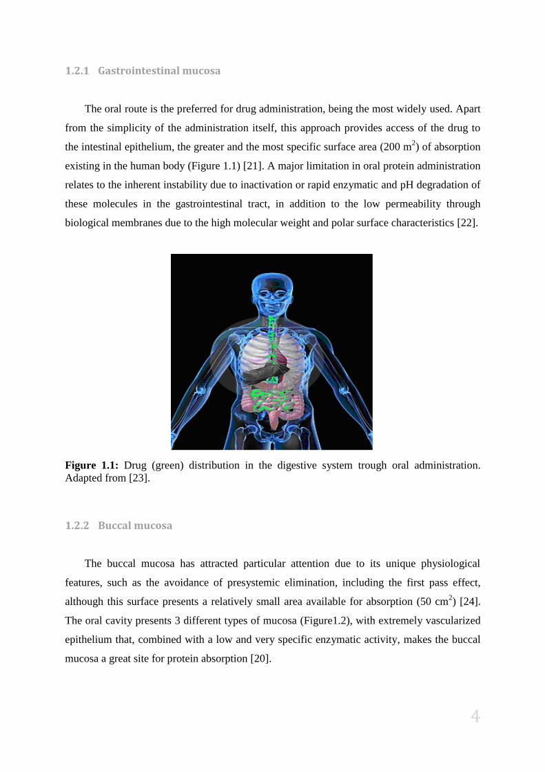
1.2.1 Gastrointestinal mucosa ....................................................................................... 4
1.2.2 Buccal mucosa ...................................................................................................... 4
1.2.3 Pulmonary mucosa ............................................................................................... 5
1.2.4 Nasal mucosa ........................................................................................................ 6
1.3 Transmucosal drug delivery technologies ................................................................... 7
1.4 Polymeric nanoparticles for mucosal administration .................................................. 9
1.4.1 Historical frame .................................................................................................... 9
1.4.2 Definition and structural organization ................................................................ 11
1.4.3 Preparation methods ........................................................................................... 13
1.4.4 Characterization ................................................................................................. 13
1.5 Polymers as nanoparticle matrix-forming materials .................................................. 14
1.5.1 Definition ........................................................................................................... 14

XI
1.5.2 Application of natural polymers in nanopharmaceutics ..................................... 15
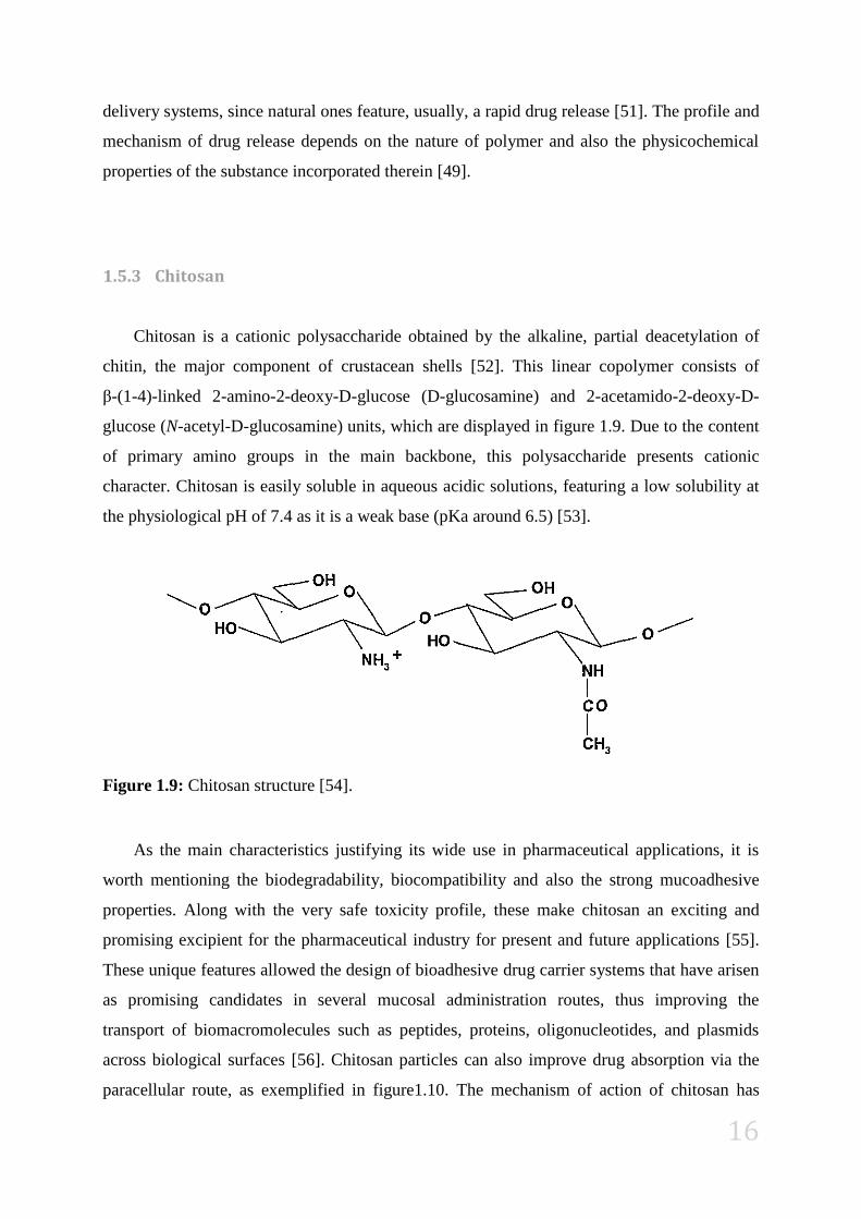
1.5.3 Chitosan .............................................................................................................. 16
1.5.4 Fucoidan ............................................................................................................. 18
1.6 State of the Art ........................................................................................................... 19
2 Objectives ......................................................................................................................... 19
3 Materials and Methods ..................................................................................................... 20
3.1 Reagents..................................................................................................................... 20
3.2 Preparation of fucoidan/ chitosan nanoparticles ........................................................ 20
3.3 Association of biomolecules to FUC/CS nanoparticles ............................................ 21
3.4 Characterization of nanoparticles .............................................................................. 21
3.4.1 Physicochemical properties ................................................................................ 21
3.4.2 Morphology ........................................................................................................ 22
3.4.3 Determination of nanoparticle production yield ................................................ 22
3.4.4 Stability assay ..................................................................................................... 22
3.4.5 Determination of BSA encapsulation efficiency and loading capacity of
nanoparticles ..................................................................................................................... 23
3.5 Statistical analysis...................................................................................................... 23
4 Results and discussion ...................................................................................................... 24
4.1 Characterization of FUC/CS nanoparticles ............................................................... 24
4.1.1 Morphological and physicochemical properties ................................................ 24
4.1.2 Nanoparticle production yield ............................................................................ 27
1.1.1 Stability assay ..................................................................................................... 28
4.2 Association of proteins to FUC/CS nanoparticles ..................................................... 30
4.2.1 Encapsulation efficiency .................................................................................... 31
4.2.2 Size and zeta potential ........................................................................................ 35
4.2.3 Determination of BSA-loaded nanoparticles production yield and loading
capacity 36
5 Conclusions ...................................................................................................................... 37

XII
6 Bibliography ..................................................................................................................... 38
7 Appendix .......................................................................................................................... 45
7.1 Appendix 1 ................................................................................................................ 45
7.2 Appendix 2 ................................................................................................................ 48
7.3 Appendix 3 ................................................................................................................ 49
7.4 Appendix 4 ................................................................................................................ 50
7.5 Appendix 5 ................................................................................................................ 51
7.5.1 Proteins association of FUC/CS nanoparticles ................................................... 51

XIII
LIST OF ABBREVIATIONS
Abs Absorbance
ADN Ácido desoxirribonucleico
BioM Biomolecules
BSA Bovine serum albumin
CkOVM Chicken ovomucoid
CS Chitosan
DkOVM Duck ovomucoid
DNA Dessoxiribonucleic acid
EE Encapsulation efficiency
FDA Food and drug administration
FUC Fucoidan
FUC/CS Fucoidan/Chitosan
HPLC High performance liquid chromatography
KDa Kilo Daltons
LC Loading Capacity
MW Molecular weight
NPs Nanoparticles
P(MAA-g-EG) Poly(methacrylic acid-g-ethylene glycol)
pI Isoelectric Point
PY Production Yield
SD Standard deviation
TEM Transmition Electronic Microscopy
TFA Trifluoroacetic acid
UV Ultra violet
w/w Weight/weight

XIV
FIGURE INDEX
Figure 1.1: Drug (green) distribution in the digestive system trough oral administration.
Adapted from [23]. ..................................................................................................................... 4
Figure 1.2: Buccal mucosa for drug absorption [20]. ............................................................... 5
Figure 1.3: Drug (green) distribution for pulmonary administration. Adapted from [26]. ....... 5
Figure 1.4: Insulin formulation for pulmonary administration. [29] ........................................ 6
Figure 1.5: Drug (green) distribution in nasal administration. Adapted from [32]................... 7
Figure 1.6: Types of terminologies used for nanoparticulate drug delivery systems. (NPs)
Nanoparticles [36]. ................................................................................................................... 11
Figure 1.7: Structural differences for polymeric nanoparticles. (A) nanospheres, (B)
nanocapsules. Adapted from [41]. ............................................................................................ 12
Figure 1.8: Size influence on deposition of inhaled nanoparticles in the human respiratory
tract [46]. .................................................................................................................................. 14
Figure 1.9: Chitosan structure [54]. ........................................................................................ 16
Figure 1.10: Effect of chitosan on the absorption of drugs by the paracellular route. (A)
Normal epithelium. (B) Transient disruption of tight junctions by chitosan with enhancement
of drug absorption. 1: represents the drug, 2: represents the tight junction. Adapted from [57].
.................................................................................................................................................. 17
Figure.1.11: Fucoidan structure [63]. ...................................................................................... 18
Figure 4.1: TEM microphotographs representative of FUC/CS nanoparticles. FUC/CS = 1/4
(A and B) and FUC/CS = 4/1 (C and D). ................................................................................. 24
Figure 4.2: RP-HPLC runs of (A) BSA in acetic acid 1% (w/w), (B) fucoidan and (C)
chitosan and respective spectrums I, II and III. RP-HPLC runs of (D) low encapsulation
efficiency FUC/CS = 1/4, (E) high encapsulation efficiency FUC/CS = 4/1. ......................... 32
XV
GRAPHIC INDEX
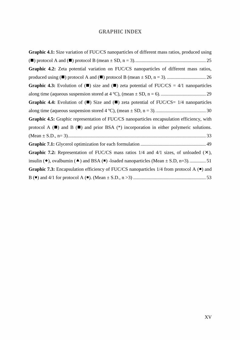
Graphic 4.1: Size variation of FUC/CS nanoparticles of different mass ratios, produced using
() protocol A and () protocol B (mean ± SD, n = 3). .......................................................... 25
Graphic 4.2: Zeta potential variation on FUC/CS nanoparticles of different mass ratios,
produced using () protocol A and () protocol B (mean ± SD, n = 3). ................................ 26
Graphic 4.3: Evolution of () size and () zeta potential of FUC/CS = 4/1 nanoparticles
along time (aqueous suspension stored at 4 ºC), (mean ± SD, n = 6). ..................................... 29
Graphic 4.4: Evolution of () Size and () zeta potential of FUC/CS= 1/4 nanoparticles
along time (aqueous suspension stored 4 ºC), (mean ± SD, n = 3). ......................................... 30
Graphic 4.5: Graphic representation of FUC/CS nanoparticles encapsulation efficiency, with
protocol A () and B () and prior BSA (*) incorporation in either polymeric solutions.
(Mean ± S.D., n= 3). ................................................................................................................. 33
Graphic 7.1: Glycerol optimization for each formulation ...................................................... 49
Graphic 7.2: Representation of FUC/CS mass ratios 1/4 and 4/1 sizes, of unloaded (),
insulin (), ovalbumin () and BSA () -loaded nanoparticles (Mean ± S.D, n=3). ............. 51
Graphic 7.3: Encapsulation efficiency of FUC/CS nanoparticles 1/4 from protocol A () and
B () and 4/1 for protocol A (). (Mean ± S.D., n >3) ............................................................ 53
XVI
TABLE INDEX
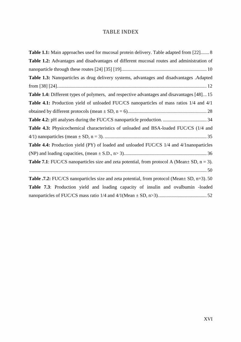
Table 1.1: Main approaches used for mucosal protein delivery. Table adapted from [22]. ...... 8
Table 1.2: Advantages and disadvantages of different mucosal routes and administration of
nanoparticle through these routes [24] [35] [19]. ..................................................................... 10
Table 1.3: Nanoparticles as drug delivery systems, advantages and disadvantages .Adapted
from [38] [24]. .......................................................................................................................... 12
Table 1.4: Different types of polymers, and respective advantages and disavantages [48]. .. 15
Table 4.1: Production yield of unloaded FUC/CS nanoparticles of mass ratios 1/4 and 4/1
obtained by different protocols (mean ± SD, n = 6). ................................................................ 28
Table 4.2: pH analyses during the FUC/CS nanoparticle production. .................................... 34
Table 4.3: Physicochemical characteristics of unloaded and BSA-loaded FUC/CS (1/4 and
4/1) nanoparticles (mean ± SD, n = 3). .................................................................................... 35
Table 4.4: Production yield (PY) of loaded and unloaded FUC/CS 1/4 and 4/1nanoparticles
(NP) and loading capacities, (mean ± S.D., n> 3). ................................................................... 36
Table 7.1: FUC/CS nanoparticles size and zeta potential, from protocol A (Mean± SD, n = 3).
.................................................................................................................................................. 50
Table .7.2: FUC/CS nanoparticles size and zeta potential, from protocol (Mean± SD, n=3) . 50
Table 7.3: Production yield and loading capacity of insulin and ovalbumin -loaded
nanoparticles of FUC/CS mass ratio 1/4 and 4/1(Mean ± SD, n>3). ....................................... 52
1
1 Introduction
1.1 Biomolecule-based therapies
1.1.1 Background
The vastness of diseases that commonly affect humans are caused by either some
physiological dysfunction resulting from a gene mutation, incorrect expression of the related
protein, or to the exposure to an environmental factor, such as pesticides, diet, bacterial,
fungal or viral infection. For many of the conditions at a molecular level underlies a change in
the amount, function or activity of one or more proteins which triggers changes in cellular,
tissue or organ function [1]. An example of common physiological dysfunction is diabetes,
caused by an insufficient activity of insulin or lack of the protein, and often a combination of
these two factors [2]. This disease is common throughout the world and reports reveal that in
2011, 366 million people were affected with diabetes and in 2030 this number is expected to
rise to 552 million. The highest incidence of diabetes is between 40 and 59 years and 78 000
children develop type 1 diabetes per year [3]. A large part of current worldwide medical
research aimed at the identification of key proteins involved in molecular mechanism
subjacent to many diseases such as the various forms of cancer and neurological conditions
such as Parkinson’s disease, motor neuron disease and multiple sclerosis, in order to select
one of these proteins as a target for the development of a new drug that can minimize or
eliminate the symptoms [1].
1.1.2 Historical frame
During the 1980s biopharmaceutical drugs became synonymous of therapeutic proteins,
vaccines, and hormones produced by recombinant DNA technology. A couple of years later,
human insulin, developed by Genentech Company (USA), reached the market, stating the
industrial application of this technology [4] . Since then, hundreds of research centers and
enterprises worldwide have been engaged in research, development and production of
biopharmaceuticals. [5] In the 25 years of existence of the biopharmaceuticals market, many

2
diseases have been and remain the focus of attention both for the development and production
of medicines, mainly for cancer, hepatitis, diabetes, growth disorders and hemophilia [6]. In
this regard, progress in the biopharmaceutical technologies has created an increasing interest
in proteins and peptides due to their role in many pathologies. However, the use of these
molecules in medicine has been limited by their low bioavailability, which results from low
stability against proteolytic enzymes, hydrolytic degradation, low permeability, and the short
half-life in systemic circulation [7]. At the same time, biotechnology appeared as a
multidisciplinary science, gathering basic biological sciences such as genetics, microbiology
and biochemistry, with chemistry, biological engineering and bioinformatics [8]. This
combined expertise led to the development of new therapies and also the large scale
production of bio-products that were previously available only in limited quantities. Thanks to
this biotechnological breakthrough, most of the molecules with therapeutic potential proposed
nowadays are protein-based [9].
1.1.3 Protein formulations
Commercially, most protein-based drugs are formulated as aqueous solutions or
suspensions ready for use or as lyophilized powder for reconstitution of the product. The
formulation of proteins depends on their physicochemical and biological characteristics,
including chemical and physical stability, immunogenicity and pharmacokinetic profile [10].
Therapeutic activity of proteins is highly dependent on their conformational structure.
However, the protein structure is flexible and sensitive to external conditions, which means
that its production, formulation and manipulation require special attention on optimizing the
efficacy and safety, including minimizing the immune response [11]. From a design
perspective, proteins are complex and challenging molecules to develop drug delivery
systems. The success of a formulation depends on the ability of the protein to maintain its
native structure and activity during the preparation and release after administration, as well as
during the storage period [12]. Some proteins require sustained release, while others require a
controlled, immediate or pulsed release. Different release profiles can be achieved using
different particulate systems for drug delivery, such as polymeric micro or nano particles,
hydrogels, liposomes and emulsions [13].
3
In order to develop these new systems several proteins are used as models in research. In
the context of this work, it is important to highlight bovine serum albumin (BSA), ovalbumin
and insulin. BSA has been primarily used in molecular studies and in the formulation of
protein-based drug delivery systems [14]. The extensive use of this protein as model is due to
its easy dissolution in water, yet it is relatively resistant to digestion. Presenting an isoelectric
point of 4.7 this protein might expose negative or positive charges when in basic or acid
environments, respectively. [15]. With a theoretical molecular weight of 69.3 kDa this protein
has a well studied and documented structure that consists of nine loops connected by 17
disulfide bridges that are protected in the core of the protein. Relatively abundant and cheaper
than other proteins, BSA also presents years of stability when stored at 2-8°C. Ovalbumin
shares many of these characteristics and, therefore, is also frequently used as model [16]. In
parallel, insulin is also used as model peptide, having the great advantage of providing a
measurable pharmacological effect and being, thus, used as model therapeutic peptide. After
insulin discovery, researchers engaged in the finding of different modes and routes of
administration for this protein [12].
1.2 Routes of administration for protein-based formulations
Despite the great biotechnology progress, delivering the new protein-based drugs remains
a problem, mainly due to incompatibilities and specific chemical structure [17]. Because of
this, most protein-based formulations have an indication for parenteral administration.
However, the parenteral route has important disadvantages to the patient, especially in chronic
therapy, which decreases therapeutic compliance, thus impairing the expected results [18].
Many research efforts are being made to improve patient compliance, either through the use
of alternative routes of administration or by reducing the frequency of injections [19]. The
demand for better ways for the administration of proteins has resulted in research for the
development of new pharmaceutical technologies. In this regard, the industry interest in
developing alternative methods for drug delivery has been growing for years [6]. Nowadays,
several mucosal surfaces such as the nasal, pulmonary and oral are being extensively explored
as alternative routes for the systemic administration of macromolecular drugs [20].
4
1.2.1 Gastrointestinal mucosa
The oral route is the preferred for drug administration, being the most widely used. Apart
from the simplicity of the administration itself, this approach provides access of the drug to
the intestinal epithelium, the greater and the most specific surface area (200 m2) of absorption
existing in the human body (Figure 1.1) [21]. A major limitation in oral protein administration
relates to the inherent instability due to inactivation or rapid enzymatic and pH degradation of
these molecules in the gastrointestinal tract, in addition to the low permeability through
biological membranes due to the high molecular weight and polar surface characteristics [22].
Figure 1.1: Drug (green) distribution in the digestive system trough oral administration.
Adapted from [23].
1.2.2 Buccal mucosa
The buccal mucosa has attracted particular attention due to its unique physiological
features, such as the avoidance of presystemic elimination, including the first pass effect,
although this surface presents a relatively small area available for absorption (50 cm2) [24].
The oral cavity presents 3 different types of mucosa (Figure1.2), with extremely vascularized
epithelium that, combined with a low and very specific enzymatic activity, makes the buccal
mucosa a great site for protein absorption [20].

5
Figure 1.2: Buccal mucosa for drug absorption [20].
1.2.3 Pulmonary mucosa
The large alveolar surface area (100 m2) suitable for drug absorption (Figure 1.3),
presents a low thickness epithelial barrier, extensive vascularization and relatively low
proteolytic activity compared to other administration routes. Together with the absence of the
first-pass effect, this makes the pulmonary delivery of peptides and proteins an outstanding
possibility [25].
Figure 1.3: Drug (green) distribution for pulmonary administration. Adapted from [26].
Insulin is undoubtedly the biopharmaceutical prototype and Exubera®
(Pfizer) (Figure
1.4) was the first inhaled formulation approved by the United States Food and Drug
Administration (FDA), for pulmonary administration of insulin in a dry powder form. Inhaled
6
insulin showed to be effective, well tolerated and better accepted in patients with type 1 and
type 2 diabetes [27]. However, this technology was removed from the market. The patients
claimed that the increased price of Exubera® relatively to the common injectable insulin was
worthless and that the needles have gotten so fin that they cause virtually no pain. Moreover
the patients also claimed that was not so easy to dose insulin with Exubera® as it was with the
inject one. [28]
Figure 1.4: Insulin formulation for pulmonary administration. [29]
1.2.4 Nasal mucosa
The nasal mucosa (Figure 1.5) is also receiving a great deal of attention due to its
permeability and easy access to the drug absorption site, although it presents low surface area
(160 cm2). This route is already commonly used for delivery of drugs for treatment of local
diseases such as nasal allergy, nasal congestion and nasal infections [30]. A wide range of
products has been developed mostly aiming at the advantage of the rapid onset of action for
the treatment of pain and erectile dysfunction. Recent developments had brought this route as
an alternative for direct administration of drugs in to the brain for the treatment of Alzheimer
and Parkinson [31].

7
Figure 1.5: Drug (green) distribution in nasal administration. Adapted from [32].
1.3 Transmucosal drug delivery technologies
To overcome the limitations created by the mucosal surfaces, several strategies were
developed aiming at improving the bioavailability of therapeutic proteins. The approaches
commonly used in formulating mucosal protein delivery systems include specific excipients,
such as absorption enhancers, enzyme inhibitors, and mucoadhesive polymers. In addition,
formulations are designed in such a manner to provide protection of protein drugs from the
harsh human environment [33]. These strategies have proven to improve protein
bioavailability and researchers believe that addressing the mentioned drawbacks is possible
with the design of a protein formulation that combines all of referred strategies (Table 1.1)
[22].

8
Table 1.1: Main approaches used for mucosal protein delivery. Table adapted from [22].
Approaches Systems Advantages Disavantagens
Absorption
enhancers
Bile salts, fatty acids,
surfactants, salicylates,
chelators, zonular occludens
toxin
Increase membrane
permeation
Transport of both
protein/peptide and
undesirable molecules
Enzyme
Inhibitors
Sodium glycocholate,
camostat mesilate, bacitracin,
soybean trypsin inhibitor,
aprotinin, CkOVM, DkOVM,
polymer–inhibitor conjugates
Resist enzyme
degradation
Induced severe side effects
in chronic therapy
Mucoadhesive
Polymers
P(MAA-g-EG) hydrogel and
lectinconjugated alginate
microparticles, thiolated
polymers,
Mucoadhesive patch system
mucoadhesive polymer–
inhibitor
Site-specific
delivery and
improve membrane
permeation
Site-specific drug
delivery and resist
enzyme degradation
Natural mucus turnover in
intestine
Extensive costs of certain
enzyme inhibitors
Formulation
Vehicles
Emulsions
Liposomes
Microspheres
Nanoparticles
Protect drug from
acid and enhance
permeation through
mucosa
Improve physical
stability and
increase membrane
permeation
Prevent proteolytic
degradation
.
Restrict release of
drug to favourable
area;
Prevent enzymatic
degradation and
increase intestinal
epithelial
absorption
Physicochemical instability
in long-term storage and
requirement for storage at
low Temperatures
Low stability of liposomes
Concerns of protein
stability during processing,
release and storage
Low loading efficiency of
hydrophilic drugs,
difficulty of precise size
control and avoidance of
particle aggregation
Abbreviations: CkOVM, chicken ovomucoid; DkOVM, duck ovomucoid; P(MAA-g-EG),
poly(methacrylic acid-g-ethylene glycol).

9
1.4 Polymeric nanoparticles for mucosal administration
1.4.1 Historical frame
Nanoparticles were first developed in the mid 70s in order to carry vaccines and
anticancer agents to specific tissues or even cells improving therapeutic efficacy and
decreasing the toxic effect of the drugs [34]. Later on, with the growing interest in the
therapeutic potential of labile molecules such as protein and peptides, nanoparticles started
being explored as vehicles to provide protection, being also proposed for administration
through different routes, such as mucosal surfaces (Table 1.2). Nowadays, nanotechnology
allows real progress in the achievement of temporal and spatial site-specific delivery [14].

10
Table 1.2: Advantages and disadvantages of different mucosal routes and administration of
nanoparticle through these routes [24] [35] [19].
Administration
routes
Advantages Disadvantages
Oral Drug protection against pH
and enzymatic damage
Increased permeability
across the epithelial
membrane
First-pass metabolism in the
liver: potential hepatotoxic effect
Potential translocation into
systemic circulation
Requires intact intestinal mucosa
for the uptake
Buccal Short recovery time of
mucosa after stress or
damage
Increased permeability to
molecular weight and
hydrophilic compounds
Limited to potent molecules
Continuous dilution of drug
Involuntary swallowing of drug
Pulmonary Ease of administration
Local action
Rapid absorption and onset
of action
Possibility of administering
lower doses
Local toxicity
Potential for translocation into
systemic circulation
Airway structure acts as a filter
Mucociliary clearance
Alveolar macrophages
Absorption affected by
pathological conditions
Requires complex devices and
particles with specific
aerodynamic properties
Particles can be exhaled
Many factors affecting
reproducibility
Nasal Ease of administration
Rapid absorption and onset
of action
Fewer side effects
Drug protection from
degradation by nasal
mucosa and secretion
enzymes
Large interspecies variation,
leading to difficult
extrapolations of results
Drug diffusion limited by the
mucus barrier and mucociliary
clearance
Administered volume limited to
25-200 μL
Molecular weight cut-off of ~ 1
kDa
Absorption affected by
pathological conditions
Limited to potent molecules
Lack of reproducibility

11
1.4.2 Definition and structural organization
Nanoparticles present variable sizes that range between 10 and 1000 nm, in which the
drug can be dissolved, coated, encapsulated or dispersed. Nanoparticulate drug delivery
systems can have several terminologies, according to structures and materials composing the
systems (Figure 1.6). The use of different production methods can create different and unique
systems, which can be used according to the biological interaction necessary for each purpose
[36]. In the context of this work, the importance of polymeric nanoparticles will be
highlighted, considering their potential for the transmucosal administration of proteins [37].
Figure 1.6: Types of terminologies used for nanoparticulate drug delivery systems. (NPs)
Nanoparticles [36].
Other systems such as microparticles and hydrogel are also being developed with the same
propose of nanoparticles. Table 1.3 describes the advantages and disadvantages of the use of
nanoparticles. .Nanoparticles can actually protect labile drugs from the biological barriers and
enhance their absorption by optimizing their interaction with the absorption site. Some
authors have suggested that nanoparticles may improve the bioavailability of peptide or
protein [7].

12
Table 1.3: Nanoparticles as drug delivery systems, advantages and disadvantages .Adapted
from [38] [24].
Advantages Disadvantages
High surface/volume ratio
Ease of surface modification
Maximized contact with mucosa
High drug concentration in desired
site
Reduction of adverse drug-
associated effects
Intracellular penetration
Protection of encapsulated
molecules
Possibility to provide controlled and
or/ prolonged release
Possibility of targeted delivery
Enhanced drug absorption
Undefined physical shape
Limited capacity to co-associate other
functional molecules
Unknown toxicity profile
Lack of suitable large-scale
production methods
Low stability in some biological
fluids
Tendency for aggregation
Limited loading capacity (unsuitable
for less potent drugs)
Small size can provide access to
unintended environments
Polymeric nanoparticles are spherical systems, formed of one or more polymers
[39].Polymeric nanoparticles are classified in two categories, nanospheres (Figure 1.7 A) and
nanocapsules (Figure 1.7 B), which differ depending on the composition and structural
organization. The nanocapsules are vesicular systems in which the drug is within an aqueous
or oily cavity surrounded by a polymer membrane, or can also be found adsorbed in the
polymer membrane. The nanospheres are formed by a polymeric matrix, where the drug is
dispersed or adsorbed [40].
Figure 1.7: Structural differences for polymeric nanoparticles. (A) nanospheres, (B)
nanocapsules. Adapted from [41].

13
1.4.3 Preparation methods
Different methods are used to prepare polymeric nanoparticles, which allow the
modulation of their structure, composition and physicochemical properties [42]. The choice of
a preparation method depends on the final application of the produced system, type of
polymer and the solubility of the drug to be encapsulated. The preparation of polymeric
nanoparticles often involves the use of organic solvents and aggressive methods, like
ultrasound energy. However, these could affect negatively both the drug/protein to be
encapsulated and the organism that will be administered with the nanosystem. Different
methods are available which explore different polymer interactions, resulting in techniques
such as ionic gelation, polyelectrolyte complexation, emulsification, coacervation and
spontaneous self-assembling, among others [43]. Using natural polymers to prepare the
nanoparticles permits using methodologies that overcome the mentioned problems regarding
aggressive conditions, one of the most used techniques being polyelectrolyte complexation
[44].
1.4.4 Characterization
Complete characterization of nanoparticles requires assessment of several parameters:
polymer type and concentration, morphology, particle size and zeta potential, production
yield, protein encapsulation efficiency, protein loading capacity and type of release profile
[25]. Nanoparticle formulation depends on the choice of suitable polymeric systems with high
encapsulation efficiency, improved bioavailability and retention time. The desired
formulations are generally achieved by trial and error method. Nanoparticle formulations
display improved properties as compared with conventional formulations, namely concerning
controlled release, targeted delivery and therapeutic impact. These targeting capabilities of
nanoparticles are influenced by particle size, surface charge, surface modification, and
hydrophobicity [45]. The size of nanoparticles for crossing different biological barriers is
dependent on the tissue, target site and circulation. Therefore, size and size distribution
determines nanoparticle interaction with the cell membrane and their penetration across the
physiological drug barriers (Figure 1.8). In turn, nanoparticle surface charge is important in

14
determining whether they would cluster in biological fluids or would adhere to, or interact
with oppositely charged cells, predicting cellular internalization [39].
Figure 1.8: Size influence on deposition of inhaled nanoparticles in the human respiratory
tract [46].
1.5 Polymers as nanoparticle matrix-forming materials
1.5.1 Definition
A polymer is a high molecular weight molecule composed of repeating small subunits
called monomers. The polymers may be classified according to their occurrence as natural or
synthetic, as well as for their chain nature, structure, morphology and type of polymerization
reaction. Natural polymers have in general more complex structures than synthetic polymers
[47]. Both natural and synthetic polymers have been extensively investigated as biomaterials,
those with the most frequently reported applications being described in Table 1.4, with their
respective advantages and disanvantages.

15
Table 1.4: Different types of polymers and respective advantages and disadvantages [48].
Occurrence Polymers Advantages Disadvantages
Natural Proteins
Polynucleotides
Polysaccharides
Gums
Resins
Elastomers
Biodegradable
Biocompatible
Nontoxic
Function biologically
at molecular and
macroscopic level.
Degradation via
natural enzymes;
cross-linkers can
make less degradable
Biodeterioration
Immunological
reaction
High natural
variability
Structurally
complexity
Technological
manipulation is more
elaborate
Synthetic Polyamides
Polyamine acids
Polyalkylated
cyanoacrylates
Polyesters
Poly(ortho esters)
Polyurethanes
Polyacrylamides
Predictable properties
Batch-to-batch
uniformity
Easy technological
manipulation
Too expensive
Environmental and
human health concerns
Lack of recognition by
cells
Toxicity
Stimulation of a
chronic inflammatory
reaction
1.5.2 Application of natural polymers in nanopharmaceutics
For a polymer to be used as a biomaterial it should not cause inflammatory or toxic
reactions at the application site, it must provide the drug with adequate half-life , degradation
time should be compatible with the desired application, degradation products cannot be toxic,
and should be able to be metabolized and eliminated from the body [49].
Natural polymers may be regarded as the first clinically used biomaterials and, actually,
polymers have always been classical excipients in pharmacy. More recently, with the
advances in nanotechnology, more sophisticated biodegradable polymers were developed
providing new delivery systems for peptides and proteins [48]. However, the development of
biodegradable systems requires the control of a great number of variables, since the kinetics
of polymer degradation in vivo must remain constant, to obtain a controlled release of the
substance. Therefore, factors such as pH and temperature, which may promote an increase or
a reduction in the rate of degradation of the system, should be evaluated during development
[50]. Synthetic biodegradable polymers have shown growing interest in the application as
16
delivery systems, since natural ones feature, usually, a rapid drug release [51]. The profile and
mechanism of drug release depends on the nature of polymer and also the physicochemical
properties of the substance incorporated therein [49].
1.5.3 Chitosan
Chitosan is a cationic polysaccharide obtained by the alkaline, partial deacetylation of
chitin, the major component of crustacean shells [52]. This linear copolymer consists of
β-(1-4)-linked 2-amino-2-deoxy-D-glucose (D-glucosamine) and 2-acetamido-2-deoxy-D-
glucose (N-acetyl-D-glucosamine) units, which are displayed in figure 1.9. Due to the content
of primary amino groups in the main backbone, this polysaccharide presents cationic
character. Chitosan is easily soluble in aqueous acidic solutions, featuring a low solubility at
the physiological pH of 7.4 as it is a weak base (pKa around 6.5) [53].
Figure 1.9: Chitosan structure [54].
As the main characteristics justifying its wide use in pharmaceutical applications, it is
worth mentioning the biodegradability, biocompatibility and also the strong mucoadhesive
properties. Along with the very safe toxicity profile, these make chitosan an exciting and
promising excipient for the pharmaceutical industry for present and future applications [55].
These unique features allowed the design of bioadhesive drug carrier systems that have arisen
as promising candidates in several mucosal administration routes, thus improving the
transport of biomacromolecules such as peptides, proteins, oligonucleotides, and plasmids
across biological surfaces [56]. Chitosan particles can also improve drug absorption via the
paracellular route, as exemplified in figure1.10. The mechanism of action of chitosan has
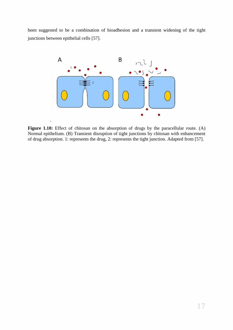
17
been suggested to be a combination of bioadhesion and a transient widening of the tight
junctions between epithelial cells [57].
.
Figure 1.10: Effect of chitosan on the absorption of drugs by the paracellular route. (A)
Normal epithelium. (B) Transient disruption of tight junctions by chitosan with enhancement
of drug absorption. 1: represents the drug, 2: represents the tight junction. Adapted from [57].

18
1.5.4 Fucoidan
Fucoidans are anionic polysaccharides containing substantial percentages of L-fucose and
sulfate ester groups, extracted from brown algae and some marine invertebrates such as
marine cucumber. Commercially available, fucoidan prepared from Fucus vesiculosus
contains 44% fucose and 26% sulfate, being water soluble [58]. A structural model in figure
1.12 shows that the core region of fucoidan is primarily α-L-fucose units linked by (14) and
(13) glycosidic bonds, with sulfate groups substituted at the C-4 position on some of the
fucose residues. (Figure 1.11) [59].
For the past decade, fucoidans isolated from different species have been extensively
studied due to their varied biological activities, including anticoagulant and antithrombotic,
antivirus, antitumor and immunomodulatory, anti-inflammatory, antidislipidemic, antioxidant
and anticomplementary properties. Relevant activity was reported against hepatopathy,
uropathy and renalpathy, as well as providing gastric protective effects and therapeutic
potential in surgery. Fucoidan can showed the ability to sequester toxic heavy metals such as
Cd2+
,Cu2+
, Zn2+
, Pb2+
, Cr3+
, and Hg2+
. This marine biopolymer has shown great properties for
drug delivery and has been reported to bind type A I and II transmembrane glycoprotein
receptors found in macrophages, this way fucoidan can promote specific interactions of a drug
carrier with the macrophages [60].
Figure.1.11: Fucoidan structure [71].
19
1.6 State of the Art
Although the technique of polyelectrolyte complexation was applied in other occasions to
obtain chitosan-based nanoparticles by interaction with counter-anions, such as
tripolyphosphate or carrageenan [61], [52], the present work is one of the first reports on the
production of nanoparticles resulting from the complexation between chitosan and fucoidan.
Previous reports on fucoidan/chitosan complexation describe the production of nanoparticles
to encapsulate the antitumor drug curcumin [62], as well the production of microparticles for
protein encapsulation [63] or the use of unloaded microparticles (Fucospheres®) for dermal
burn treatment [59], making this study the first that reports fucoidan/chitosan nanoparticles
for protein encapsulation. Importantly, the results reported in the present study have already
been presented in panel at the 3rd
Congress of the Portuguese Society of Pharmaceutical
Sciences (Appendix 1) and 9th
Central European Symposium on Pharmaceutical Technology
(Appendix 2).
2 Objectives
The aim of this work was to verify de ability of chitosan and fucoidan to assemble into
nanoparticles which display ability to encapsulate different model proteins, namely BSA,
ovalbumin and insulin. The nanoparticles are aimed at an application in systemic mucosal
protein administration and, therefore, several specific properties should be evidenced, as
follows:
- Size within 50-500 nm to permit a close interaction with the epithelial surface;
- Zeta potential above 30 mV (either negative or positive) to provide adequate stability in
aqueous suspension and to maximize interaction with the epithelial surface;
- Adequate protein encapsulation efficiency, preferably above 50%.

20
3 Materials and Methods
3.1 Reagents
Chitosan (CS) (low molecular weight, deacetylation degree 75–85%), Fucoidan (FUC)
from Fucus vesiculosus, bovine albumin serum (BSA), insulin and ovalbumin sodium,
phosphotungstate dibasic hydrate, sodium hydroxide and glycerol were purchased from
Sigma-Aldrich® (Germany). Bradford reagent was purchased from Bio-rad
® (Germany)
Trifluoroacetic acid (TFA) and glacial acetic acid were supplied, respectively, by Alfa Aesar®
(Germany) and Panreac Synthesis® (Germany). Ultrapure water (Integral 3, Millipore
®,
Portugal) was used throughout. All other chemicals were reagent grade.
3.2 Preparation of fucoidan/ chitosan nanoparticles
Fucoidan/chitosan (FUC/CS) nanoparticles were prepared by polyelectrolyte
complexation, in which the negatively charged groups of fucoidan interact with the cationic
groups of chitosan, creating electrostatic bonds. Briefly, a 2 mg/mL stock solution of FUC
(pH 6.18) was prepared with milliQ water and a 1 mg/mL stock solution of CS (pH 3.17) was
prepared with 1% (w/w) acetic acid. Both solutions were filtered before further using (0.2 µm
filter, Whatman®, Germany). These stock solutions were then diluted to obtain various
concentrations, in order to permit the preparation of nanoparticles with different mass ratios
(4/1 to 1/4, w/w). FUC/CS nanoparticles were spontaneously formed by drop wise addition of
either 1 mL of FUC into 1 mL of CS solution (protocol A), or 1 mL CS into 1 mL of FUC
solution (protocol B), under magnetic stirring for 10 min, at room temperature.
Nanoparticle suspensions were then placed in eppendorf tubes over a layer of 10 µL
glycerol that prevents nanoparticle dehydration and aids the subsequent resuspension step.
Nanoparticles were isolated by centrifugation at 16000 g, for 30 min at 15 ºC (Thermo
Scientific®, Germany). The supernatants were discarded and nanoparticles were resuspended
in 200 µL of purified water.

21
3.3 Association of biomolecules to FUC/CS nanoparticles
Three different proteins were associated to the nanoparticles, which were used as models:
bovine serum albumin (BSA), insulin and ovalbumin. BSA and ovalbumin were dissolved in
milliQ water, while insulin was dissolved in NaOH 0.01 M. The proteins were either
associated with FUC or CS prior to the mixture of the polymers, to test the effect of this
variable. Along this text, when referring to a nanoparticle formulation loaded with proteins,
an asterisk (*) will be placed close to the number representing the polymer with which the
protein was mixed prior to nanoparticle formation (example: FUC/CS = *4/1 means that the
protein was mixed with the FUC solution prior to pouring of this polymer into the CS
solution).
The proteins were associated to the nanoparticles in a concentration of 30% (w/w)
respective to the polymer with the higher content in the formulation. Both protocols A and B
(described above) were used to prepare protein-loaded FUC/CS nanoparticles, in order to test
the effect of the order of addition of polymers on the final characteristics of the nanoparticles.
The isolation of nanoparticles was performed as described above.
3.4 Characterization of nanoparticles
3.4.1 Physicochemical properties
The size and zeta potential of nanoparticles were measured by photon correlation
spectroscopy and laser Doppler anemometry, respectively, using a Zetasizer Nanoseries
(Malvern Instruments®, UK). For the measurements, 20 µL of each sample were diluted with
1 mL purified miliQ water and the suspension placed in an electrophoretic cell. Each analysis
was performed at 25 ºC (n = 3).

22
3.4.2 Morphology
The morphological analysis of FUC/CS nanoparticles was performed by transmission
electron microscopy (TEM; Jeol-JEM® 1011, Germany). Concentrated nanoparticle
suspensions were obtained upon centrifugation, mounted on copper grids coated with a
carbon film (Ted Pella®, USA) and stained with a 2% (w/v) sodium phosphotungstate dibasic
hydrate solution.
3.4.3 Determination of nanoparticle production yield
The nanoparticle production yield was determined by gravimetry. For this procedure,
nanoparticles were prepared, isolated by centrifugation at 16000 g, for 30 min at 15 ºC
(Thermo Scientific®, Germany) and the sediments were freeze-dried over 24 h, using a
Freeze Dryer (Labconco®, USA) (n = 6). The production yield (PY) was calculated as
follows:
Where nanoparticles weight is the sediment weight after freeze-drying and total solids
weight is the total amount of solids added for nanoparticle formation (fucoidan and chitosan
for unloaded nanoparticles and fucoidan, chitosan, and protein for protein-loaded
nanoparticles).
3.4.4 Stability assay
For the assessment of nanoparticle stability, the formulations FUC/CS 1/4 and 4/1 were
prepared according to the procedure described above. Aqueous suspensions of nanoparticles
were stored at 4 ºC and their size and zeta potential were monitored along time (n > 3).
23
3.4.5 Determination of BSA encapsulation efficiency and loading capacity of
nanoparticles
The BSA encapsulation efficiency was determined indirectly, by quantification of the
non-encapsulated protein present in the supernatant after the nanoparticle isolation procedure.
The amount of free protein was primarily determined by High Pressure Liquid
Chromatography (HPLC, Agilent® 1100 series, Germany) with a Aeris Widepore 3.6 u XB-
C18 column (Phenomenex®, USA). The conditions for each run were: temperature 25 ºC;
gradient flow (0.1% TFA in water (A), 0.1% TFA in acetonitrile (B); A/B from 95:5 to 35:65
in 15 min) mobile phase, total run time 20 min, UV detection at 280 nm; flow rate 1.0
mL/min; injection volume 20 µL; BSA retention time 8.7 min. A linear calibration curve for
BSA in 1% (w/w) acetic acid was obtained over the range 5–120 µg/mL (n = 3) (R2
= 0.996).
Absorbance spectrums of pure solutions of polymers and BSA were run in So Bio UV-Visible
spectrophotometer (Varian®, Australia). The nanoparticle protein association efficiency and
loading capacity were calculated from Equations indicated below:
3.5 Statistical analysis
The t-test and the one-way analysis of variance (ANOVA) with the pairwise multiple
comparison procedures (Student–Newman–Keuls method) were performed to compare two or
multiple groups, respectively. All analyses were run using the SigmaStat® statistical program
(Version 3.5, USA) and differences were considered to be significant at a level of P < 0.05.

24
4 Results and discussion
4.1 Characterization of FUC/CS nanoparticles
Fucoidan/chitosan (FUC/CS) nanoparticles were successfully obtained, using several
concentrations of the two polymers, which resulted in FUC/CS mass ratios of 1/4 to 4/1. The
assembly of nanoparticles was mediated by an electrostatic interaction between the negatively
charged sulfate groups of fucoidan and the oppositely charged amino groups of chitosan. The
order of addition of the polymers was analysed as variable, varying according to Protocols A
(fucoidan added over chitosan) and B (chitosan added over fucoidan), as was described in the
methodology. Further optimizations of the procedure of nanoparticle preparation are
described in appendix 3. Apart from the visible Tyndall effect that proves the formation of a
colloidal suspension of FUC/CS nanoparticles, the effect of different mass ratios and order of
addition of polymers was evaluated concerning the resultant size, zeta potential and
production yield, as described in the following sections.
4.1.1 Morphological and physicochemical properties
Figure 4.1 displays the TEM microphotographs of representative FUC/CS nanoparticles
of mass ratios 4/1 and 1/4, which evidence a compact structure and a tendency to a spherical
shape.
Figure 4.1: TEM microphotographs representative of FUC/CS nanoparticles. FUC/CS = 1/4
(A and B) and FUC/CS = 4/1 (C and D).

25
As displayed in Graphic 4.1, the size of FUC/CS nanoparticles varied between 338 and
676 nm. As expected, the variation of mass ratios resulted in different sizes, an observation
valid for both protocols A and B, although a particular trend could not be established.
Generally, it was observed that the presence of greater amounts of polymer (chitosan and
fucoidan) in the formulations resulted in increased particle size of the nanoparticles.
Therefore, the formulation FUC/CS = 1/1 presented the highest sizes (p < 0.05), 564 and 676
nm for protocol A and B, respectively. Accordingly, the opposite happens when the
concentration of the polymers in each formulation reaches the lower limit, with the mass
ratios of 4/1 and 1/4 presenting the lowest sizes in each protocol (varying from 338 to 456
nm). This general behaviour can be observed for both protocols, as described. However, size
differences were more prominent in protocol B, where nanoparticles displayed the lowest and
the highest sizes. As a general trend, the order of addition of one polymer over the other
affected the resulting nanoparticle size and, thus, different results were obtained for protocols
A and B. When comparing both protocols, statistically significant differences were obtained
in all the formulations (p < 0.05), except for mass ratios 1/2 and 1/4. For a more direct
observation of the size values, a table with that information is available in appendix 4.
Graphic 4.1: Size variation of FUC/CS nanoparticles of different mass ratios, produced using
() protocol A and () protocol B (mean ± SD, n = 3).
In what concerns the zeta potentials of the obtained FUC/CS nanoparticles, a complete
shift from strong negative to strong positive charges was observed, depending on the mass
ratios (graphic 4.2). Those formulations with higher amount of fucoidan (4/1 to 2/1) resulted
in a negative charge, with a maximum of -37 mV registered for FUC/CS = 4/1. On the
0
100
200
300
400
500
600
700
800
4/1 3/1 2/1 1/1 1/2 1/3 1/4
Size
(n
m)
FUC/CS (w/w)

26
contrary, formulations with the higher amount of chitosan (1/2 to 1/4) displayed strong
positive charge, reaching a maximum value of +49 mV. Curiously, the formulation with
equal mass of both polymers also registered a strong positive surface charge (+43 mV or +49
mV, depending on used protocol), which indicates that chitosan has a higher charge density.
The specific values obtained for each formulation and protocol are also available on table x on
appendix 4. On the contrary of what was observed for the size, zeta potentials were not
affected by the order of addition of polymers tested in protocols A and B. In addition,
different mass ratios did not have a pronounced effect on zeta potential either, in contrast with
what was observed for nanoparticle size.
Graphic 4.2: Zeta potential variation on FUC/CS nanoparticles of different mass ratios,
produced using () protocol A and () protocol B (mean ± SD, n = 3).
It is a fact that a complete inversion of zeta potential (p < 0.05) is obtained when fucoidan
changes from the most represented polymer, inducing a strong negative charge, to the less or
equal represented polymer, in which cases a strong positive charge is obtained. However,
apart from this evident shift, all formulations with higher amount of fucoidan (4/1 to 2/1)
displayed a charge around -40 mV and all formulations from 1/1 to 1/4 registered a charge of
approximately +45 mV. This effect was reported in several other works concerning chitosan-
based nanoparticles, in which similar variations of polymer mass ratios resulted in very small
changes of zeta potential, in the order of 4 or 5 mV [64], [65].
Nanoparticles based on chitosan and fucoidan were previously proposed for the treatment
of dermal burns [59] and for the encapsulation of stromal cell-derived factor 1 (SDF-1),
-60
-40
-20
0
20
40
60
80
4/1 3/1 2/1 1/1 1/2 1/3 1/4
Zeta
po
ten
tial
(m
V)
FUC/CS (w/w)

27
which is an important chemokine in stem cell mobilization [66]. In the first case, only
positively charged nanoparticles were obtained, inclusive when fucoidan was present in
higher amount (FUC/CS = 5/1). The authors justified this effect with the possible formation
of an outer layer of chitosan that took place during the nanoparticle assembly [59]. The
second work reported very similar results as compared to those described in the present study,
with strong negatively charged nanoparticles being obtained when fucoidan is present in
higher amount in the formulation (FUC/CS = 5/1 has -49.8 mV). Additionally, FUC/CS = 1/1
also presents a positive charge (+ 24.5 mV), as is reported in the present study [66]. The
explanation for the different results found among studies might rely on the use of different
chitosan and fucoidan, which is available with very different characteristics and usual very
few details are given in the papers.
4.1.2 Nanoparticle production yield
Given the displayed size characteristics, FUC/CS mass ratios of 4/1 and 1/4 were selected
to proceed with the studies. At the beginning of this work, only Protocol A was in course for
the preparation of nanoparticles and, when protein-loaded nanoparticles were prepared, the
protein was always mixed with the polymeric solution corresponding to the lower amount in
each formulation (fucoidan in FUC/CS = 1/4 and chitosan in FUC/CS = 4/1). Protocol B was
tested later on in an attempt to improve the encapsulation efficiency of nanoparticles with
mass ratio 1/4. This is why protocol B was not tested for formulation 4/1 concerning the
production yield, as the characteristics of those nanoparticles were satisfactory.
Table 4.1 shows that protocol A tends to provide a higher production yield (PY) for the
formulation 4/1 (32%) as compared with the formulation 1/4 (16%), although the difference is
not statistically significant due to the high standard deviations. The production yield is a
measure of the interactions that take place when both polymers contact with each other. The
fact that, in protocol A, formulation 4/1 has a higher yield than formulation 1/4 might indicate
that chitosan has a higher charge density. When comparing the yields of 1/4 nanoparticles
obtained with different protocols, although protocol B apparently increases the yield, the
differences are not significant.

28
Table 4.1: Production yield of unloaded FUC/CS nanoparticles of mass ratios 1/4 and 4/1
obtained by different protocols (mean ± SD, n = 6).
Protocol FUC/CS Production Yield (%)
A 4/1 32 ± 19
1/4 16 ± 11
B 1/4 26 ± 13
1.1.1 Stability assay
FUC/CS nanoparticles of mass ratios of 1/4 and 4/1 were monitored for their storage
stability along time, concerning size and zeta potential. One of the most common problems of
colloidal particles relies on their tendency for flocculation [24]. This is frequently
accompanied by a decrease in zeta potential to values below 30 mV (in modulus), where the
repulsive forces are not enough to maintain nanoparticles separated from each other, leading
to aggregation.
In this study, aqueous suspensions of FUC/CS nanoparticles were maintained at 4 ºC for
a period of 170 days. Nanoparticles of formulation 4/1 registered a slight size decrease at the
beginning, maintaining stable after that (Graphic 4.3). Nevertheless, as compared with the
initial size (518 nm), differences were only statistically significant at the last time point (170
days; p < 0.05), in which the registered size was 400 nm. Concerning zeta potential, apart for
an apparent instability at the beginning, that was not significant anyway, the zeta potential
maintained relatively stable, being -41 mV for fresh nanoparticles and -38 mV after 170 days.

29
Graphic 4.3: Evolution of () size and () zeta potential of FUC/CS = 4/1 nanoparticles
along time (aqueous suspension stored at 4 ºC), (mean ± SD, n = 6).
FUC/CS = 1/4 nanoparticles exhibited a very similar behavior (Graphic 4.4), with a slight
size decrease from 435 to 331 nm in 170 days. Zeta potential also registered an initial
variation, but the value registered at the end of 170 days (42 mV) was very similar to the
initial (43 mV).
In a general manner, it can be said that nanoparticles register some slight variations in
their physicochemical characteristics upon 170 days of storage at 4 ºC, although the variations
do not compromise the intended application as protein carriers for mucosal administration.
-50
-40
-30
-20
-10
0
0
100
200
300
400
500
600
0 50 100 150 200
Zeta
po
ten
tial
(m
V)
Size
(n
m)
Time (days)

30
Graphic 4.4: Evolution of () Size and () zeta potential of FUC/CS= 1/4 nanoparticles
along time (aqueous suspension stored 4 ºC), (mean ± SD, n = 3).
4.2 Association of proteins to FUC/CS nanoparticles
To choose the most suitable formulation for the encapsulation of biomolecules, key
features were taken into account, namely concerning particle size, which drives the interaction
with mucosal epithelia. FUC/CS nanoparticles 1/4 and 4/1 were selected to undergo protein
encapsulation, as they display the smaller sizes and also opposite charges that might further
affect cell interaction, which study is of interest in the field. Nanoparticles were tested
regarding their ability to associate model proteins of different molecular weights, namely
BSA (67 kDa), ovalbumin (44 kDa) and insulin (5.7 kDa). In addition, protein-loaded
nanoparticles were characterised for their physicochemical properties. Because of time
limitations, nanoparticles encapsulating BSA were the most completely studied and,
therefore, only the results regarding these nanoparticles are displayed and discussed in the
main document. Results regarding the physicochemical proprieties of ovalbumin- and
insulin–loaded nanoparticles are described on appendix 5.
0
10
20
30
40
50
60
0
100
200
300
400
500
600
0 50 100 150 200
Zeta
po
ten
tial
(m
V)
Size
(n
m)
Time (days)

31
4.2.1 Encapsulation efficiency
The determination of BSA encapsulation efficiency was tested for different formulations
of FUC/CS nanoparticles, varying not only the mass ratios (1/4 and 4/1) but also the order of
addition of the polymers (protocols A and B) and also the polymer in which BSA is mixed
prior to nanoparticle formation. RP-HPLC was used to determine the encapsulation
efficiency, a method that is based on the adsorption of hydrophobic molecules onto a
hydrophobic stationary phase in a polar mobile phase. BSA has a great affinity to adsorb on
hydrophobic surfaces [67], which can be reduced by decreasing the mobile phase polarity by
using organic solvents (acetonitrile) resulting in desorption and elution from the reverse phase
column. BSA desorption occurred with a gradient flow of water/acetonitrile from 95:5 to
35:65 in 15 min, both solvents contained 0.1% of TFA ,which helped the BSA to desorb. [68].
BSA was eluted at around 8.5 min, and the peak shape and intensity were similar to those of
pure BSA samples in the presence of all polymers (figure 4.2 D, E). The retention time of
each polymer (figure 4.2 B, C) was determined and revealed to be different from BSA (figure
4.2 A), thus not interfering with BSA determination. To identify each substance, a
simultaneous absorbance spectrum was preformed to each peak and compared with the
absorbance spectrums of pure BSA, fucoidan and chitosan respectively I, II and III in figure
4.2. However, there was an overlap spectrum of the solvent acetonitrile that was overcome by
reading BSA at 280 nm.

32
Figure 4.2: RP-HPLC runs of (A) BSA in acetic acid 1% (w/w), (B) fucoidan and (C)
chitosan and respective spectrums I, II and III. RP-HPLC runs of (D) low encapsulation
efficiency FUC/CS = 1/4, (E) high encapsulation efficiency FUC/CS = 4/1.
The results in graphic 4.5 represent the encapsulation efficiency (EE) of BSA-loaded
FUC/CS nanoparticles of mass ratios 1/4 and 4/1 prepared with protocols A and B and with
prior BSA incorporation in different polymers solutions. FUC/CS nanoparticles effectively
encapsulated BSA. In an initial stage of this study, BSA, insulin and ovalbumin were also
associated to nanoparticles and the respective encapsulation efficiencies were estimated using
the Bradford protein assay, measuring the absorbance by spectrophotometry at 595 nm
(Tecan-Infinite M200, Switzerland). However, it was decided to not proceed with that method
of quantification as there chitosan amino groups were interfering in the quantifications. Data
obtained with this methodology can be found in appendix 5.
Graphic 4.5 shows a rather different profile of protein association between FUC/CS
nanoparticles 1/4 and 4/1 (p < 0.05). FUC/CS *4/1 and 4/1* from protocol A displayed,
respectively, encapsulation efficiencies of 98% and 100%, while protocol B resulted in a
mean of 103% for both formulations. This means that, whatever the variables, the
encapsulation efficiency is very high and remarkable as comparing with the majority of

33
studies regarding protein encapsulation in nanoparticles. In turn, FUC/CS *1/4 and 1/4* from
protocol A displayed, respectively, 55% and 11% encapsulation efficiency, while those
produced with protocol B resulted in 87% and 19%, respectively. As a general trend,
formulations with mass ratio of 4/1 exhibited better ability to encapsulate BSA as compared
with 1/4, whatever the tested variables (p < 0.05). Additionally, for both FUC/CS mass ratios
1/4 and 4/1 higher encapsulation efficiencies were achieved when BSA was mixed with the
polymer with the lowest concentration in each formulation, prior to the addition into the other
polymer solution (p < 0.05). Regarding the effect of changing the order of addition of
polymers, a statistically significant difference was observed for formulations *1/4, 1/4* and
*4/1 (p<0.05).
Graphic 4.5: Graphic representation of FUC/CS nanoparticles encapsulation efficiency, with
protocol A () and B () and prior BSA (*) incorporation in either polymeric solutions.
(Mean ± S.D., n= 3).
A more profound interpretation of the interactions between polymer and BSA in FUC/CS
1/4 and 4/1 nanoparticles, might be achieved by the analyses of the pH of solutions used for
nanoparticle assembly, which are expressed in table 4.2. With a pI of 4.7, BSA exposed
positive charge when the final pH of its solution was lower than the pI, as happens when it
was mixed with chitosan (around 3.3). In turn, when BSA was mixed with fucoidan, the final
pH of the solution was 6.8 and, therefore, a negative charge is overall expressed. When the
polymeric solutions were mixed together, the final pH of the nanoparticle suspensions were
all lower than 4.7, meaning that BSA exposed positive charged at the end of all FUC/CS
0
20
40
60
80
100
120
*1/4 1/4* *4/1 4/1*
Enca
psu
lati
on
eff
icie
ncy
(%
)
FUC/CS mass ratio

34
nanoparticles preparations. A cross reference of this observation with the graphic
representation of the encapsulation efficiency (Graphic 4.5), may explain higher
encapsulation efficiency values for FUC/CS 4/1. Since these nanoparticles contain higher
amount of fucoidan, the negatively charged polymer, and the BSA was always exposing
opposite surface upon polymer mixture, it is quite logical to suggest that these conditions
enhanced BSA-polymer interactions, resulting in greater encapsulations efficiencies. The
lower EE results displayed by FUC/CS 1/4* are a result of the fact that, in this case, BSA is
mixed at first with chitosan and, therefore, it has a positive charge in this solution. When both
polymers are mixed, many positive charges (chitosan and BSA) are competing for the
negatively charged groups of fucoidan, permitting only a limited interaction that results in a
low encapsulation. If the mass ratio remains the same (FUC/CS = 1/4) but BSA is mixed with
fucoidan and only after that there is a mixture with chitosan, there is a very significant
increase in the encapsulation efficiency (p < 0.05), because only many positively charged
groups of chitosan will be available to interact with the negatively charged fucoidan and BSA.
Anyway, the resulting encapsulations are still lower than those provided by the formulation
4/1. A statistically significant increase in the encapsulation efficiency of formulation FUC/CS
*1/4 occurred when protocol was changed from A to B. This difference might be explained by
the fact that adding chitosan solution into fucoidan, it was possible to maintain the pH below
4.7 for a longer time, meaning that the protein would be exposing negative charges, thus
promoting BSA-polymer interaction.
Table 4.2: pH analyses during the FUC/CS nanoparticle production.
Protocol FUC/CS Solutions pH
FUC CS BSA+ polymer (FUC/CS)+BSA
A *1/4 6.38 3.08 6.80 3.17
1/4* 6.26 3.97 3.38 3.46
*4/1 6.28 3.22 6.71 3.48
4/1* 6.30 4.45 3.26 3.44
B *1/4 6.38 3.08 6.78 3.13
1/4* 6.26 3.97 3.34 3.46
*4/1 6.28 3.22 6.79 3.48
4/1* 6.30 4.45 3.26 3.49
*Polymer solution to which BSA was added prior to nanoparticle formation

35
4.2.2 Size and zeta potential
Table 4.3 presents the results corresponding to the unloaded and BSA-loaded
nanoparticles. As can be observed, the incorporation of BSA tends to induce an increase on
nanoparticles size, but in any case the difference is statistically significant. In turn, the zeta
potential of nanoparticles became less negative with the incorporation of the protein in
formulation 4/1, whatever the polymer with which BSA was mixed before nanoparticle
assembly; while formulation 1/4 exhibits different behaviours depending on the polymer with
which the protein is mixed before the formation of nanoparticles. The explanation for these
different effects is probably based on different availability of charges to interact among
polymers and the protein. To facilitate the interpretation of results Table 4.2 gathers the pH of
used polymers, mixtures of polymers with BSA and the final nanoparticle suspension.
The comparison of the zeta potentials of both FUC/CS mass ratios 4/1 and 1/4, of loaded
and unloaded nanoparticles, shows a decrease on surface charge of nanoparticles when loaded
with BSA, this differences were statistically significant (p<0.05). This decrease of the zeta
potential is quite logical, and can be easily justified by the interactions polymers-protein, that
neutralizes some of the available charges of the nanoparticles. This effect was more
pronounced for FUC/CS=1/4 with BSA prior mix in fucoidan, when BSA was exposing
negatively charged groups, maximizing the interaction with the available positive groups of
chitosan. However, this great decrease on zeta potential may be caused by a rearrangement of
nanoparticles structure, where the spatial disposition of the fucoidan chain contributes for this
decline of the surface charge. The absence of a decrease on FUC/CS 1/4 with BSA prior mix
in chitosan can be explained by the low encapsulation efficiency of this formulation.
Table 4.3: Physicochemical characteristics of unloaded and BSA-loaded FUC/CS (1/4 and
4/1) nanoparticles (mean ± SD, n = 3).
*Polymer solution to which BSA was added prior to nanoparticle formation
Protocol FUC/CS Size (nm) Zeta potential (mV)
A
4/1 456 ± 4 -42 ± 1
*4/1 521 ± 55 -37 ± 3
4/1* 525 ± 53 -33 ± 4
B
1/4 372 ± 5 +46 ± 2
*1/4 415 ± 71 +19 ± 5
1/4* 435 ± 49 +53 ± 5

36
4.2.3 Determination of BSA-loaded nanoparticles production yield and loading
capacity
For this study were selected for characterization the FUC/CS mass ratios 4/1 and 1/4
from protocol A and B. As already referred in the above section of unloaded nanoparticles
production yield, Protocol B was not trialled for 4/1 nanoparticles. Prior incorporation BSA in
either polymeric solutions was not tested. Results of this characterization regarding insulin
and ovalbumin can be found in appendix 5 Table 4.4 shows an increase on nanoparticle
productions yield upon BSA incorporation, but this differences were only statistically
significant for FUC/CS nanoparticles 4/1 and 1/4 for protocol A (p< 0.050). The most
notorious increase on production yield was observed by FUC/CS 4/1 upon BSA
incorporation, where nanoparticles gain about 100% of mass relatively to the unloaded
nanoparticles. Showed in table 4.4 are also the loading capacities (LC) of FUC/CS
nanoparticles. This characteristic represents the amount of protein relatively to the total mass
of nanoparticles, which compose each FUC/CS formulation for example, FUC/CS 1/4 had a
49 % LC, means that about half of this particle is compose of BSA and the other of polymer.
Once again the enhancement of the nanoparticle BSA associations was clear in FUC/CS 1/4
when protocol A (36 %) was replaced by B (49%). This shows that protocol B can improve
the profitability of these processes, since although PY of FUC/CS 1/4 did not increase
significantly when protocol change from A (30%) to B (33%), although cross reference with
LC from both protocols (36%) A and (49 %) B shows that the particles can associate more
protein, in fact half of the particles mass content was protein.
Table 4.4: Production yield (PY) of loaded and unloaded FUC/CS 1/4 and 4/1nanoparticles
(NP) and loading capacities, (mean ± S.D., n> 3).
Protocol FUC/CS BSA-loaded NP
PY (%) LC (%)
A 4/1* 65 ± 15 29 ± 7
*1/4 30 ± 7 36 ± 11
B *1/4 33 ± 8 49 ± 11 *Polymer solution to which BSA was added prior to nanoparticle formation

37
5 Conclusions
This work demonstrates that the developed fucoidan/chitosan (FUC/CS) nanoparticles are
suitable to be used as protein carriers systems in the ambit of systemic mucosal
administration. These nanoparticles are produced in complete hydrophilic conditions, by a
very mild procedure of ionic interaction between the negatively charged fucoidan sulphate
groups and the oppositely charged amino groups of chitosan. This procedure avoids the use of
organic solvents and other aggressive conditions that might be detrimental for the integrity of
the drug to be encapsulated. The macromolecules bovine albumin serum (BSA), insulin and
ovalbumin, used in this study as model proteins, were efficiently associated to the developed
drug delivery systems, as demonstrated by the physicochemical characterization of the
systems. The optimization of several variables of the nanoparticle production method allowed
obtaining very high encapsulation efficiencies. The small sizes and high negative and positive
charges displayed by the developed nanoparticles are considered to hold potential for an
application in mucosal delivery of macromolecules. Additionally, FUC/CS nanoparticles
demonstrated to be relatively stable in aqueous suspension, only registering some slight
variations in their physicochemical characteristics upon 170 days of storage at 4 ºC, although
these do not compromise the intended application as protein carriers for mucosal
administration. In order to establish a complete characterization of the developed
nanoparticulate systems, FUC/CS nanoparticles must undergo further studies, namely the
determination of release profile and the toxicity assessment, both in vitro and in vivo.
.

38
6 Bibliography
[1] K. Wilson and J. Walker, Principles and techniques of biochemistry and molecular
biology, Cambrige: Cambridge University Press , 2010.
[2] V. K. Naeayan, D. Williams, E. W. Gregg and C. C. Cowie, Diabetes Public Health,
New York: Oxford University press, 2011.
[3] International Diabetes Fedaration, “Diabetes atlas,” 2011. [Online]. Available:
http://www.idf.org. [Accessed 22 Janeiro 2012].
[4] P. Buckel, “Recombinant proteins for therapy,” Trends in Pharmacological Sciences,
vol. 17, pp. 450-6, 1996.
[5] G. Subramanian, Biopharmaceutical Production Tehcnology, Weinheim: Wiley.VCH
Verlag & Co. KGaA., 2012.
[6] W. G., “Biopharmaceutical benchmarks,” Nature Biotechnology, vol. 24, pp. 769-776,
2006.
[7] C. P. Reis, R. J. Neufeld, A. J. Ribeiro and F. Veiga, “Nanoencapsulation II. Biomedical
applications and current status,” Nanomedicine, vol. 2, p. 53–65, 2006.
[8] R. J. Y. Ho and M. Gibaldi, Biotechnology and Biopharmaceuticals: Transforming
Proteins and genes into drugs, new jersey: John Wiiley & sons, Inc., 2003.
[9] G. Walsh, Biopharmaceuticals Biochemistry and biotechnology, Chichester: John and
Wiley & Sons Ltd., 2003.
[10] S. Cox, Pharmaceutical Manufacturing Handbook: Production and Processes, New
Jersey: John Wiley & Sons, Inc., 2008.
[11] S. Frokjaer and D. E. Otzen, “Protein drug stability: a formulation challenge.,” Nature
Reviews: Drug Discovery, vol. 4, pp. 298-306, 2005.
[12] L. R. Brown, “Commercial challanges of protein drug delivery,” Expert Opinion, vol. 2,
no. Drug Delivery, pp. 29-42, 2005.
[13] L. Jorgensen, E. v. d. W. M. Moeller, H. Nielsen and S. Frokjaer, “Preparing and
evaluating delivery systems for proteins,” European Journal of Pharmaceutical Sciences,
vol. 29, pp. 174-182, 2006.
[14] Y. Pathak and D. Thassu, Drug Delivery Nanoparticles Formulation and
Characterization, New York: Informa Healthcare USA, Inc., 2009.

39
[15] K. Murayama and M. Tomida, “Heat-induced secondary structure and conformation
change of bovine serum albumin investigated by Fourier transform infrared
spectroscopy,” Biochemistry, vol. 43, no. 36, pp. 11526-11532., 2004.
[16] F. Jameel and S. Hershenson, Formulation and Process Development Strategies for
Manufacturing Biopharmaceuticals, New Jersey: John Wiley & Sons, Inc., 2010.
[17] S. E. McNeil, "Nanoparticle therapeutics: a personal perspective," WIREs Nanomed
Nanobiotechnol, vol. 1, pp. 264-271, 2009.
[18] A. Grenha, “Systemic delivery of biopharmaceuticals: Parenteral forever?,” Journal of
Pharmacy and BioAllied Sciences, vol. 2, p. 95, 2012.
[19] J. L. Cleland, A. Daugherty and R. Mrsny, “Emerging protein delivery methods,”
Current Opinion in Biotechnology, vol. 12, p. 212–219, 2001.
[20] V. F. Patel, F. Liu and M. B. Brown, "Advances in oral transmucosal drug delivery,"
Journal of Controlled Release, vol. 153, p. 106–116, 2011.
[21] J. B. Dressman and H. Lennernäs, Oral Drug Absorption. Prediction and Assessment,
volume 105, New York: Marcel Dekker, Inc., 2000.
[22] K. Park, I. C. Kwon and K. Park, "Oral protein delivery: Current status and future
prospect," Reactive & Functional Polymers, vol. 71, p. 280–287, 2011.
[23] Webnode, “Anatomia e Fisiologia,” 2009. [Online]. Available:
http://anatomiaefisiologiahumana.webnode.com.
[24] L. Braz, M. Dionísio and A. Grenha, “Chapter 7: Chitosan-based Nanocarriers: Effective
Vehicles for mucosal Protein Delivery,” in Chitosan: Manufacture, Proprieties and
Usage, New York, Nova Science Publishers, Inc., 2010, pp. 365-413.
[25] A. Grenha, S. Al-Qadi, B. Seijo and C. Remuñán-López, “The potential of chitosan for
pulmonary drug delivery.,” Journal of Drug Delivery Science and Technology, vol. 20,
no. 1, pp. 33-43, 2010.
[26] Sintomas e Dicas, “Sintomas e Dicas,” 2009. [Online]. Available:
http://sintomasedicas.com/.
[27] Nektar Therapeutics, “Pfizer’s Exubera Receives Approval From US FDA; Nektar Co-
Developed the Inhalers & Formulation,” Drug Delivery Technology, vol. 6, no. 2, p. 12,
2006.
[28] A. Weintraub, “Bloomberg Business week Technology,” 18 October 2007. [Online].

40
Available: http://www.businessweek.com.
[29] C. Morrison, “The in vivo blog,” 2007. [Online]. Available:
http://invivoblog.blogspot.pt/.
[30] S. Türker, E. Onur and Y. Ózer, “Nasal route and drug delivery systems,” International
Journal of Clinical Pharmacy, vol. 26, no. 3, pp. 137-142, 2004.
[31] L. Illuma, “Nasal drug delivery- possibilities, problems and solutions,” Journal of
Controlled Release, vol. 87, pp. 187-198, 2003.
[32] Bahrian Doctors, “Bahrian Doctors,” 2012. [Online]. Available:
http://bahriandoctors.com.
[33] J. Shaji and V. Patole, “Protein and Peptide Drug Delivery: Oral Approaches,” Indian
Journal of Pharmaceutical Sciences, vol. 70, no. 3, p. 269–277, 2008.
[34] C. R. Gilbert S. Banker, Drugs and Pharmaceutical Sciences, Modern Pharmaceutics, 4th
edition, Basel: Marcel & Dekker, Inc., 2002.
[35] L. Yildirimer, N. T. Thanhb and M. Loizidoua, “Toxicological considerations of
clinically applicable nanoparticles,” Nano today, vol. 6, p. 585—607, 2011.
[36] F. Re, R. Moresco and M. Masserin, “Nanoparticles for Neuroimaging,” jornal of
Physics D: Applied Physics, vol. 45, no. 7, 2012.
[37] K. Y. Leea and S. H. Yuk, “Polymeric protein delivery systems,” Progress in Polymeric
Science, vol. 32, p. 669–697, 2007.
[38] R. C. Pinto, R. Neufeld, A. Ribeiro and F. Veiga, “Nanoencapsulation I. Methods for
preparation of drugloaded polymeric nanoparticles,” Nanomedicine, vol. 2, no. 1, pp. 8-
21, 2006.
[39] A. Kumari, Y. S. K. and S. C. Yadav, “Biodegradable polymeric nanoparticles based
drug delivery systems,” Journal Colloids and Surfaces B: Biointerfaces, vol. 75, pp. 1-
18, 2010.
[40] W. Abdelwahed, G. Degobert, S. Stainmesse and H. Fessi, “Freeze-drying of
nanoparticles: Formulation, process and storage considerations,” Advanced Drug
Delivery Reviews, vol. 58, p. 1688–1713, 2006.
[41] G. Griffiths, B. Nyström, S. B. Sable and G. K. Khuller, “Nanobead-based interventions
for the treatment ans prevension of tuberculosis,” Nature Reviews, Microbiology, vol. 8,
pp. 827-834, 2010.

41
[42] A. Rieuxa, V. Fieveza, M. Garinota, Y.-J. Schneiderb and V. Préata, “Nanoparticles as
potential oral delivery systems of proteins and vaccines: A mechanistic approach,”
Journal of Controlled Release, vol. 116, no. 1, p. 1–27, 2006.
[43] K. S. Soppimatha, T. M. Aminabhavia, A. R. Kulkamia and W. E. Rudzinski,
“Biodegradable polymeric nanoparticles as drug delivery devices,” Journal of Controlled
Release, vol. 70, pp. 1-20, 2001.
[44] A. Grenha, B. Seijo and C. R. López, “Microencapsulated chitosan nanoparticles for lung
protein delivery,” European Journal of Pharmaceutical Sciences, vol. 25, p. 427–437,
2005.
[45] A. Eifler and C. Thaxton, “Nanoparticle therapeutics: FDA approval, clinical trials,
regulatory pathways, and case study.,” Methods of Molecular Biology, vol. 726, pp. 325-
38, 2011.
[46] G. Oberdorster, “Pulmonary effects of inhaled ultrafine particles,” International Archives
of Occupational and Environmental Health, vol. 74, pp. 1-8, 2001.
[47] M. Elsabahy and K. L. Wooley, “Strategies Toward Well-Defined Polymer
Nanoparticles Inspired by Nature: Chemistry versus Versatility,” Journal of polymer
science, vol. 50, p. 1869–1880, 2012.
[48] D. Labarre, G. Ponchel and C. Vauthier, Biomedical and Pharmaceutical Polymers,
London: Pharmaceutical Press, 2011.
[49] J. H. Park, M. Ye and K. Park, “Review Biodegradable Polymers for Microencapsulation
of Drugs,” Molecules, vol. 10, pp. 146-161, 2005.
[50] T. Hanemann and D. V. Szabó, “Polymer-Nanoparticle Composites: From Synthesis to
Modern,” Journal of Materials, vol. 3, pp. 3468-3517, 2010.
[51] K. E. Uhrich, S. M. Cannizzaro and R. S. Langer, “Polymeric Systems for Controlled
Drug Release,” Chemical Reviews, vol. 99, pp. 3181-3198, 1999.
[52] A. Grenha, M. E. Gomes, M. Rodrigues, V. E. Santo and J. F. Mano, “Development of
new chitosan/carrageenan nanoparticles,” Journal of biomedical materials research. Part
A, vol. 92, no. 4, pp. 1265-72, 2010.
[53] S. A. T. and C. P. Sharma, “Chitosan and Its Derivatives for Drug Delivery,” Journal of
Advanced Polymer Science, vol. 243, p. 23–54, 2011.
[54] G. Dupuis and J. Lehoux, “A simplified method to retrieve chitosan from acidic solutions

42
thereof”. França Patent EP 1701980 A1, 20 setembro 2006.
[55] A. Grenha, “Chitosan nanoparticles: a survey of preparation methods,” Journal of Drug
Targeting, vol. 20, no. 4, pp. 291-300, 2012.
[56] A. B. Schnürch and S. Dünnhaupt, “Chitosan-based drug delivery systems,” European
Journal of Pharmaceutics and Biopharmaceutics, vol. 81, p. 463–469, 2012.
[57] F. Andrade, F. Goycoolea, D. A. Chiappetta, J. Neves, A. Sosnik and B. Sarmento,
“Chitosan-Grafted Copolymers and Chitosan-Ligand Conjugates as Matrices for
Pulmonary Drug Delivery,” International Journal of Carbohydrate Chemistry, vol. 2011,
p. 14, 2011.
[58] B. Li, F. Lu, X. Wei and R. Zhao, “Fucoidan: Structure and Bioactivity,” Mollecules,
vol. 13, pp. 1671-1695, 2008.
[59] A. D. Sezer, E. Cevher, F. Hatipoglu, Z. Ogurtan, A. L. Bas and J. Akbuga, “The use of
fucosphere in treatment of dermal burns in rabbits,” European Journal of Pharmaceutics
and Biopharmaceutics, vol. 69, pp. 189-198, 2008.
[60] M. Liraa, N. Santos-Magalhães, V. Nicolasd, V. Marsaude, M. Silvab, G. Ponchele and
C. Vauthiere, “Cytotoxicity and cellular uptake of newly synthesized fucoidan-coated
nanoparticles,” European Journal of Pharmaceutics and Biopharmaceutics, vol. 79, no.
1, pp. 162-170, 2011.
[61] W. Fana, W. Yanb, Z. Xub and H. Nia, “Formation mechanism of monodisperse, low
molecular weight chitosan,” Colloids and Surfaces B: Biointerfaces, vol. 90, pp. 21-27,
2012.
[62] Y. C. Huang and U. I. Lam, “Chitosan/Fucoidan pH Sensitive Nanoparticles for Oral
Delivery System,” Journal of the Chinese Chemical Society, vol. 58, pp. 779-785, 2011.
[63] A. D. Sezer and J. Akbuga, “Fucosphere—New microsphere carriers for peptide and
proteins delivery,” Journal of Microencapsulation,, vol. 23, no. 5, p. 513–522, 2006.
[64] S. Rodrigues, A. M. R. Costa and A. Grenha, “Chitosan/carrageenan nanoparticles:
Effect of cross-linking with tripolyphosphate and charge ratios,” Journal of
Carbohydrate Polymers , vol. 89, p. 282– 289, 2012.
[65] A. A. Mahmoud, G. S. El-Feky and G. E. A. Awad, “Chitosan/sulfobutylether-β-
cyclodextrin nanoparticles as a potential approach for ocular drug delivery,”
International Journal of Pharmaceutics, vol. 413, no. 1-2, pp. 229-236, 2011.

43
[66] Y. Huang and T. Liu, “Mobilization of mesenchymal stem cells by stromal cell-derived
factor-1 released from chitosan/tripolyphosphate/fucoidan nanoparticles,” Acta of
biomaterialia, vol. 3, pp. 1048-1056, 2012.
[67] Y. U. Yong, P. Ying and J. I. N. Gang, “Competitive adsortion between bovine serum
albumin and collagen observed by atomic force microscope,” Journal of Chinese
Chemical Letters, vol. 15, p. 1465, 2004.
[68] M. Umrethia, V. L. Kett, G. P. Andrews, K. R. Malcom and D. A. Woolfson, “Selectionn
of an analytical method for evaluating bovine serum concentrations in pharmaceutical
polymeric formulations,” Journal of Pharmaceutical and Biomedical Analysis, vol. 51, p.
1175–1179, 2010.
[69] Z. Liu, Y. Jiao, Y. Wang, ,. C. Zhou and Z. Zhang, “Polysaccharides-based nanoparticles
as drug delivery systems,” Advanced Drug Delivery Reviews, vol. 60, p. 1650–1662,
2008.
[70] S. A. Agnihotri, N. N. Mallikarjuna and T. M. Aminabhavi, “Recent advances on
chitosan-based micro-and nanoparticles,” Journal of controlled release, vol. 100, no. 1,
pp. 5-28, 2004.
[71] 1999. [Online]. Available: http://glycomix.co.uk/fucus_vesiculosus_fucoidan.htm . .

44

45
7 Appendix
7.1 Appendix 1
DEVELOPMENT OF FUCOIDAN/CHITOSANNANOPARTICULATE SYSTEMS FOR PROTEIN ADMINISTRATION THROUGH MUCOSAL ROUTES
Sara Ferreira*, Ana Grenha
CBME - Centre for Molecular and Structural Biomedicine / IBB - Institute for
Biotechnology and Bioengineering, University of Algarve, Portugal; *[email protected]
INTRODUCTION
Protein-based biomacromolecules have been used for long in pharmacological therapy, but their administration through non-parenteral routes is a difficult task, due to stability issues mainly attributed to pH and enzymatic contents in mucosal surfaces. It has been a consensus that mucosal routes are valuable alternatives for drug administration, but this demands the development of adequate carriers that confer stability and protection against the mentioned aggressive environments (1). Nanoparticulate systems have proven promising for this end, owing to their high surface-to-volume ratio and capacity for drug encapsulation (2). Using natural polymers for nanocarriers production is advantageous due to the requirements of biocompatibility, biodegradability and absence of toxicity, which are mandatory in any biomedical application. Fucoidan and chitosan are two of these natural polymers, respectively extracted from brown seaweed (Fucus vesiculosus) with 95% of fucose sulfated esters (anionic polysaccharide) (3), and the exoskeleton of crustaceans. In the case of chitosan a further step of partial deacetylation of chitin is necessary to obtain the cationic polysaccharide (4). In this work, fucoidan/chitosan (FUC/CS) nanoparticles were prepared by polyelectrolyte complexation (5). The obtained nanocarriers were optimized in terms of several variables, being evaluated for their capacity to associate proteins.
MATERIAL AND METHODS
Preparation of FUC/CS nanoparticles:
Nanoparticles were prepared by a method of polyelectrolyte complexation (6). Briefly, CS was dissolved in acetic acid 1% (w/w) and FUC was dissolved in purified water. FUC/CS nanoparticles were spontaneously formed at room temperature upon incorporation of FUC solution into CS solution (FUC/CS mass ratios between 4/1 and 1/4) and vice-versa. Different mass ratios and the order of addition of polymeric solutions were studied as variables. Protocol A consisted in the addition of FUC over CS and Protocol B was the opposite.
Nanoparticles were isolated by centrifugation (16000 x g, 30 min, 15 ºC) and resuspended in 100 µL of milli-Q water.
Model biomacromolecules (BioM) were associated to the nanoparticles, including bovine serum albumin (BSA), ovalbumin and insulin. Proteins were dissolved in appropriate solvents (water or sodium hydroxide 0.1 M) and added to either CS or FUC solution, in order to provide the protein with opposite charge in comparison with the polymer presented at the highest concentration in each formulation. The solution of the second polymer is then poured into the previously prepared polymer/protein solution to produce the nanoparticles.
Nanoparticles characterization:
Nanoparticles production yield was calculated by gravimetry, comparing the real weight of nanoparticles with the initial amount of solids used for their production (n = 3). Nanocarriers’ size and zeta potential were measured by photon correlation spectroscopy and laser Doppler anemometry, respectively (Zetasizer
®
Nano ZS, Malvern Instruments) (n = 3).
Nanoparticles supernatant was assessed to determine the amount of free BioM using the Bradford protein assay and measuring the absorbances by spectrophotometry at 595 nm (Tecan-Infinite M200, Switzerland). A calibration curve was made using the supernatant of blank nanoparticles. BioM encapsulation efficiency (E.E.) and loading capacity (L.C.) were calculated comparing the non-associated protein present in the supernatant with the total amount added for nanoparticles production, as follows:
E. E. % = Total BSA Free BSA
Total BSA × 100
E.E. (%)

46
L. C. % = Total BSA Free BSA
Nanoparticle weight × 100
Evaluation of nanoparticles stability:
A study of the nanoparticles (FUC/CS = 4/1 and 1/4) stability in water was performed at 4 ºC. Unloaded nanoparticles were isolated by centrifugation and resuspended in water afterwards. Sizes were monitored during 22 days, using the above mentioned techniques (n = 3).
RESULTS AND DISCUSSION
Nanoparticles characterization:
The two variables studied in the production of FUC/CS nanoparticles, which consist in polymer concentration (CS and FUC) and the order of addition of polymeric solutions, resulted in remarkable effects over nanoparticles characteristics. Sizes ranged between 300 and 700 nm (Figure 1) and the highest size corresponded to the highest amount of materials composing the nanoparticles (FUC/CS = 1/1). Moreover, a complete shift of zeta potentials was observed, varying from -42 to +49 mV (Figure 2), which reflects the highest amount of negative or positive polymer composing the nanoparticles.
Figure 1 - FUC/CS nanoparticles size corresponding to different mass ratios and preparation protocols (mean ± SD, n = 3).
Figure 2 – FUC/CS nanoparticles zeta potential obtained for different mass ratios and preparation protocols (mean ± SD, n = 3).
The most remarkable effect of protocol changes is related with BSA encapsulation. In the formulation FUC/CS 1/4, changing preparation protocol from A to B, it is observed a 60-70% increase in the encapsulation efficiency (from 11-20% in protocol A to 83-85% in protocol B). Formulation 4/1 also shows high BSA encapsulation efficiency, up to 88% with protocol A. Assays regarding ovalbumin and insulin encapsulation show efficiency of 51% and 91%, respectively, in formulation FUC/CS = 4/1 comparatively to the lower efficiency (14%) for both proteins in 1/4 FUC/CS formulation. Release assays are presently being performed.
Evaluation of nanoparticles stability:
The stability assay revealed that, when stored at 4ºC, the nanoparticles (FUC/CS = 1/4 and 4/1) size remains stable for at least 22 days, for formulations obtained by both protocols A and B. Further studies are being performed to assess longer time periods.
CONCLUSIONS
Considering the physicochemical properties, the ability to associate proteins and the demonstrated stability, FUC/CS nanoparticles are considered good candidates for drug delivery purposes. The results indicate that the use of protocol A and B in 4/1 and 1/4 FUC/CS formulations, respectively, provide high BSA encapsulation efficiency.
0
100
200
300
400
500
600
700
800
4/1 2/1 1/1 1/3 1/4
Siz
e (
nm
)
FUC/CS mass ratio
Protocol A
Protocol B
-60
-40
-20
0
20
40
60
80
4\1 3\1 2\1 1\1 1\2 1\3 1\4
Ze
ta p
ote
ncia
l (m
V)
FUC/CS mass ratio
Protocol A
Protocol B

47
REFERENCES
[1] Csaba et al., Adv. Drug Deliv. Rev., 61: 140-157, 2009.
[2] Reis et al., J. Tissue Eng., 1: 4–24, 2007.
[3] Li et al. Molecules, 13: 1671-1695, 2008.
[4] Kurita, Marine Biotechnol., 8: 203–226, 2006.
[5] Berthold et al., J. Control. Release, 39: 17–25, 1996.
[6] Prestwich et al., J. Control. Release, 53: 93– 103, 1998.
ACKNOWLEDGEMENTS
Funding from Fundação para a Ciência e Tecnologia (project PTDC/SAU-FCF/100291/2008) and CBME/IBB, LA is acknowledged. 3rd Congress of the Portuguese Society of Pharmaceutical Sciences/ 9
th Spanish-
Portuguese Conference on Controlled Drug Delivery, Porto/ Portugal, 2011 Published in Revista Portuguesa de Farmácia, volume LII (nº 6), 115-116.

48
7.2 Appendix 2
FUCOIDAN/CHITOSAN NANOPARTICLES AS PROTEIN CARRIERS
Sara Ferreira1, Ana Grenha1
1CBME - Centre for Molecular and Structural Biomedicine / IBB - Institute for Biotechnology and Bioengineering, University of Algarve, Faro, Portugal.
KEYWORDS
chitosan, fucoidan, nanoparticles, protein delivery
INTRODUCTION
Fucoidan and chitosan are natural polymers, respectively extracted from brown seaweed (Fucus vesiculosus) and containing 95% of fucose sulfated esters (anionic polysaccharide) [1], and from the exoskeleton of crustaceans. Displaying opposite charges, these polysaccharides enable the production of nanoparticles by polyelectrolyte complexation, occurring under very mild conditions [2]. Nanoparticles have proven to be promising vehicles in protein delivery, due to their high surface-to-volume ratio and capacity for association of macromolecules. In this work, fucoidan/chitosan (FUC/CS) nanoparticles of various mass ratios (1/4 to 4/1, w/w) were prepared by means of an electrostatic interaction, displaying different size and zeta potential. Several conditions were explored to maximise protein incorporation, either varying 1) the order of addition of polymeric solutions (Protocol A: FUC into CS, or Protocol B: CS into FUC), or 2) the polymeric solution (FUC or CS) to which the protein is added prior to nanoparticle formation. Bovine serum albumin (BSA), ovalbumin and insulin were used as model proteins. The use of natural polymers is expected to provide the basis for biocompatibility and absence of toxicity, which are mandatory in any biomedical application [3]. Importantly, the application of fucoidan in the preparation of nanoparticles for drug delivery purposes was never reported before.
RESULTS & DISCUSSION
FUC/CS nanoparticles display sizes of 300 - 700 nm and zeta potential from -42 to +49 mV. In approach 1), BSA was used as model protein, being always incorporated in FUC solution prior to nanoparticle formation. Higher encapsulation efficiencies were obtained for formulations FUC/CS = 4/1 produced according to protocol A (88%) and FUC/CS = 1/4 prepared by protocol B (85%). The approach 2) was tested for BSA, insulin and ovalbumin, and protocols A and B were used for formulations 4/1 and 1/4, respectively. Higher encapsulation efficiency is always observed when the protein is mixed with in the solution of the polymer present in the lower ratio in the final formulation. Release assays are presently being performed. The performed experiments demonstrate the importance of controlling the protein charge in the moment of providing the interaction with polymers. The size and zeta potential of FUC/CS nanoparticles (1/4 and 4/1, w/w) remain stable for up to 6 months, upon storage at 4 ºC. Considering the physicochemical properties and stability, as well as the ability to associate different proteins, FUC/CS nanoparticles are deemed suitable for drug delivery purposes. National funding from Portuguese Foundation for Science and Technology (project PTDC/SAU-FCF/100291/2008 and PEst-OE/EQB/LA0023/2011) is acknowledged.
REFERENCES
[1] Li B. et al. (2008) Fucoidan: Structure and Bioactivity. Molecules. 13: 1671-1695 [2] Grenha A. (2012) Chitosan nanoparticles: a survey of preparation methods. J Drug Target. 20: 291-300 [3] Chiellini F. (2008) Micro/nanostructured polymeric systems for biomedical and pharmaceutical applications. Nanomedicine. 3: 367-393

49
7.3 Appendix 3
The conditions on which nanoparticles formed were optimized. Temperature reveled to
have great influence on the nanoparticles formation. During this study nanoparticles were
formed at room temperature oscillating between 17-22 ºC, agglomerates were formed above
or under this temperature range (data not shown). The Drop wise incorporation of the
polymers revealed as a crucial step for nanoparticles formation. A small increase in the rate of
addition of polymers causes major changes in terms of size (data not shown). To outcome this
problems with nanoparticle resuspension was adopted a strategy that consisted on finding the
accurate amount of glycerol for each formulation, by phased additions of more 10 µL glycerol
in nanoparticles isolation procedure. The results of this optimization were illustrated in
graphic 1. The graphic 7.1 shows the accurate amount of glycerol for FUC/ CS mass ratio
formulation 1/1, 2/1, 3/1, 4/1 and 5/1 were 20, 50, 20, 10 and 10 µL respectively. The excess
of glycerol usually contributes to nanoparticle aggregation.
Graphic 7.1: Glycerol optimization for each formulation
.
The conditions adopted for the nanoparticle isolation procedure, were centrifuge velocity
of 16000 G, at 15ºC during 30 minutes that correspond to a standardization used by the
research group.
Nanoparticles
Clusters

50
7.4 Appendix 4
FUC/CS nanoparticles size and zeta potential, from protocol A (table 7.1) and B (table
7.2).
Table 7.1: FUC/CS nanoparticles size and zeta potential, from protocol A (Mean± SD, n = 3).
FUC\CS
(w/w)
Size
(nm)
Zeta potential
(mV)
4\1 456±4 -42±1
3\1 547±21 -40±1
2\1 510±27 -41±5
1\1 564±21 49±4
1\2 544±10 45±12
1\3 531±3 49±6
1\4 366±41 46±6
Table 77.2: FUC/CS nanoparticles size and zeta potential, from protocol (Mean± SD, n=3)
FUC\CS
(w/w)
Size
(nm)
Zeta potential
(mV)
4\1 338±44 -37±11
3\1 417±47 -42±1
2\1 420±28 -37±3
1\1 676±34 43±12
1\2 591±58 45±4
1\3 435±33 40±1
1\4 372±5 46±2

51
7.5 Appendix 5
7.5.1 Proteins association of FUC/CS nanoparticles
BSA, ovalbumin and insulin were dissolved in appropriate solvents respectively in water
and sodium hydroxide 0.1 M, and then added to either CS or FUC solution, in order to
provide the protein with opposite charge in comparison with the polymer presented at the
highest concentration in each formulation. Nanocarriers’ size and zeta potential were
measured by photon correlation spectroscopy and laser Doppler anemometry, respectively
(Zetasizer® Nano ZS, Malvern Instruments) (n = 3).Table x contains size and zeta potential of
insulin and ovalbumin –loaded nanoparticles. The graphic 7.2 representation of the tabled size
values, plus BSA-loaded and unloaded nanoparticles, reveals the specificity of the
interactions between these polymers and each protein, whereas displays different behaviors in
terms of sizes.
Graphic 7.2: Representation of FUC/CS mass ratios 1/4 and 4/1 sizes, of unloaded (),
insulin (), ovalbumin () and BSA () -loaded nanoparticles (Mean ± S.D, n=3).
0
100
200
300
400
500
600
700
800
900
*1/4 1/4* *4/1 4/1*
Size
(n
m)
FUC/CS (w/w)

52
Nanoparticles production yield (table 7.3) was calculated by gravimetry, comparing the
real weight of nanoparticles with the initial amount of solids used for their production (n = 3).
Table 7.3: Production yield and loading capacity of insulin and ovalbumin -loaded
nanoparticles of FUC/CS mass ratio 1/4 and 4/1(Mean ± SD, n>3).
Protocol FUC/CS
(w/w)
Insulin-loaded NP Ovalbumin-loaded
NP
P.Y.
(%)
L.C.
(%)
P.Y. (%)
L.C.
(%)
A *4/1 - - 40±9 21±6
4/1* 45±13 37±11 46±8 27±5
B *1/4 33±14 13±6 16±5 13±5
1/4* - - 24±11 0 *Polymer solution to which BSA was added prior to nanoparticle formation
Nanoparticles supernatant was assessed to determine the amount of free BioM using the
Bradford protein assay and measuring the absorbance by spectrophotometry at 595 nm
(Tecan-Infinite M200, Switzerland). A calibration curve was made using the supernatant of
blank nanoparticles. BioM encapsulation efficiency (EE) and loading capacity (LC) were
calculated comparing the non-associated protein present in the supernatant with the total
amount added for nanoparticles production, as described in Materials and Methods.
These assays with ovalbumin and insulin happened in an early stage of this work, where
some characteristic of the polymer were not completely described. An example of this was the
fact that using Bradford assay to measure the proteins encapsulations efficiency, not only the
proteins were being quantify, but also fucoidan, jeopardizing the results. With this work was
evident that each protein has is one unique interaction with this polymers combination. In
graphic 7.3 represents the different encapsulation efficiencies for the 3 model proteins used in
this study, for nanoparticles FUC/CS 1/4 and 4/1 mass ratios. The most remarkable effect
occurs during BSA encapsulation when protocol changes from A to B in FUC/CS 1/4
formulation were observed an increase in 70-80 % the encapsulation ability (EE of 88 %).
Still regarding BSA, the 4/1 formulation shows as well a great encapsulation ability (EE of
81-88 %) with protocol A. In ovalbumin and insulin encapsulation, the FUC/CS mass ratio
4/1 had the highest encapsulation efficiencies respectively 51% and 91% that contrast with
the very low encapsulation efficiencies in FUC/CS mass ratio 1/4 of 14% for both proteins.
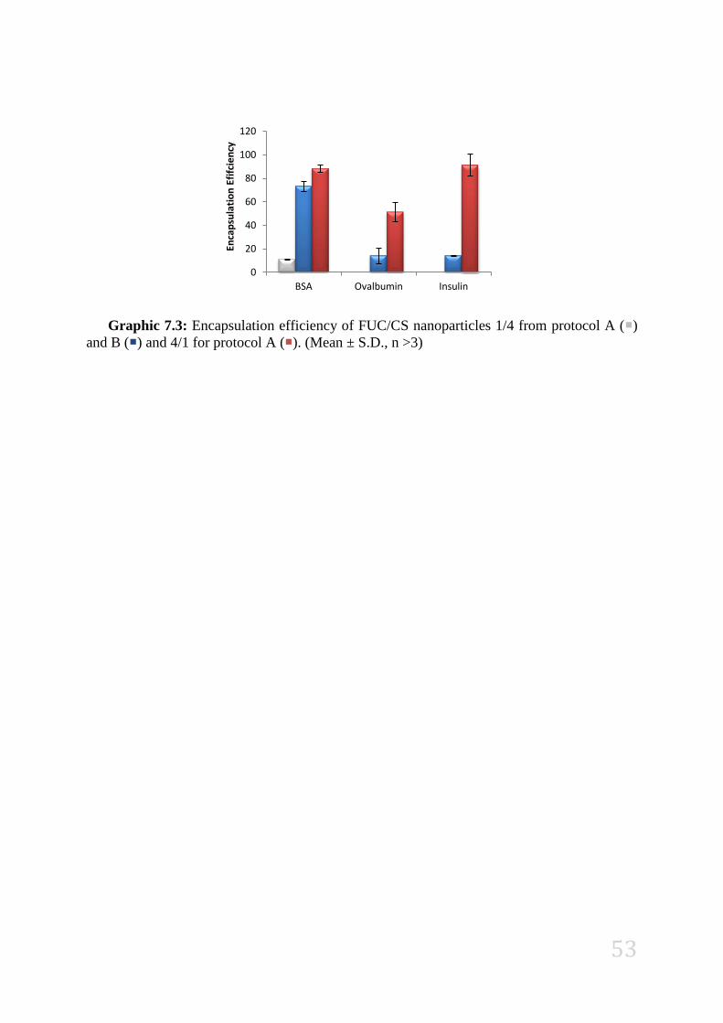
53
Graphic 7.3: Encapsulation efficiency of FUC/CS nanoparticles 1/4 from protocol A ()
and B () and 4/1 for protocol A (). (Mean ± S.D., n >3)
0
20
40
60
80
100
120
BSA Ovalbumin Insulin
Enca
psu
lati
on
Efi
fcie
ncy

![Cytocompatibility of Chitosan and Collagen-Chitosan ...forms the highly porous structure of the scaffolds[13] Two percent (w/v) of chitosan was prepared by dissolving chitosan in 0.2](https://img.document.onl/doc/110x75/5e3f1725786dcc56c068fc16/cytocompatibility-of-chitosan-and-collagen-chitosan-forms-the-highly-porous.jpg)


![[Nome completo do autor] Chitosan Nanoparticles as Drug ... · Chitosan Nanoparticles as Drug Delivery Systems vii “Every person must work for his own improvement, and at the same](https://img.document.onl/doc/110x75/6112aeb2bb03351c1f367767/nome-completo-do-autor-chitosan-nanoparticles-as-drug-chitosan-nanoparticles.jpg)