Embed Size (px)
Citation preview

Universidade de Aveiro
2014
Departamento de Biologia
DIANA RAQUEL SANTOS RIBEIRO
THE ROLE OF UROCORTIN-2 IN PULMONARY ARTERIAL HYPERTENSION O PAPEL DA UROCORTINA-2 NA HIPERTENSÃO ARTERIAL PULMONAR
DECLARAÇÃO
Declaro que este relatório é integralmente da minha autoria, estando
devidamente referenciadas as fontes e obras consultadas, bem como
identificadas de modo claro as citações dessas obras. Não contém, por isso,
qualquer tipo de plágio quer de textos publicados, qualquer que seja o meio
dessa publicação, incluindo meios eletrónicos, quer de trabalhos académicos.

Universidade de Aveiro
2014
Departamento de Biologia
DIANA RAQUEL SANTOS RIBEIRO
THE ROLE OF UROCORTIN-2 IN PULMONARY ARTERIAL HYPERTENSION O PAPEL DA UROCORTINA-2 NA HIPERTENSÃO ARTERIAL PULMONAR
Dissertação apresentada à Universidade de Aveiro para cumprimento dos requisitos necessários à obtenção do grau de Mestre em Biologia Molecular e Celular, realizada sob a orientação científica da Professora Doutora Carmen Dulce da Silveira Brás Silva, Investigadora do Departamento de Fisiologia e Cirurgia Cardiotorácica da Faculdade de Medicina e Professora Auxiliar da Faculdade de Ciências da Nutrição e Alimentação da Universidade do Porto, e coorientação da Professora Doutora Maria Paula Polónia Gonçalves, Professora Associada ao Departamento de Biologia da Universidade de Aveiro.
Este trabalho foi desenvolvido no âmbito do projeto financiado pela Fundação para a Ciência e a Tecnologia (FCOMP-01-0124-FEDER-011051, FEDER, COMPETE, FCT PTDC/DTP-FTO/0130/2012)

À memória da minha avó Margarida da Cruz Silva

“Most people say that it is the intellect which makes a great scientist. They are wrong. It is character.”
Albert Einstein
O júri
Presidente
Prof. Doutora Maria de Lourdes Gomes Pereira Professora Associada com agregação ao Departamento de Biologia da Universidade de Aveiro
Orientadora Prof. Doutora Carmen Dulce da Silveira Brás Silva Investigadora do Departamento de Fisiologia e Cirurgia Cardiotorácica da Faculdade de Medicina e Professora Auxiliar da Faculdade de Ciências da Nutrição e Alimentação da Universidade do Porto
Arguente Prof. Doutora Ana Patrícia Nunes Fontes de Sousa Professora Auxiliar do Instituto de Ciências Biomédicas Abel Salazar da Universidade do Porto
Agradecimentos
À minha orientadora, Professora Doutora Carmen Brás Silva, por me ter acolhido no seu grupo de trabalho e me ter dado a oportunidade de aprender e crescer, tanto como pessoa como cientista. Nunca poderei exprimir por palavras o quanto isso significa para mim e o importante que é na minha vida. Obrigado por apostar em mim e pela amizade, simpatia e compreensão que dedica a todos os que consigo trabalham. Farei das “tripas coração” para nunca a desiludir. À Professora Doutora Paula Gonçalves por me ter apresentado esta oportunidade. Ainda, um obrigado por toda a disponibilidade e simpatia que sempre demonstrou, enquanto professora e orientadora. Ao Professor Doutor Adelino Leite Moreira por me receber no Departamento de Fisiologia e Cirurgia Cardiotorácica da Faculdade de Medicina da Universidade do Porto. Aos meus colegas de grupo, e hoje amigos, Mestre Pedro Ferreira, Mestre Rui Adão, Mestre Carolina Rocha e Dra. Bárbara, por todos os bons momentos passados dentro e fora do laboratório, pela entreajuda e pelos bons conselhos. Vocês são os melhores e os mais malucos que conheço. Nunca deixem morrer a criança que há em vós. Obrigado por fazerem do nosso local de trabalho um sítio espetacular. Aos restantes membros e colegas de departamento, em especial, Glórinha, Fabi, Dudu, Dani, Mizé e Ticha por me terem acolhido no seio da vossa amizade e partilharem o vosso dia-a-dia comigo. Obrigado por fazerem deste departamento uma autêntica casa de família. A todos os meus amigos de longa data por estarem sempre comigo, tanto nos bons como nos maus momentos. Estou certa de que só a morte nos separará. Adoro-vos, obrigado por serem quem são e por partilharem a vossa vida comigo! À minha família e namorado, por todo o apoio e carinho incondicional que sempre me deram, por todas as oportunidades que me proporcionaram e principalmente por sempre terem acreditado em mim e nas minhas capacidades. Obrigado por me incentivarem a querer mais e melhor para o meu futuro. Vocês são tudo para mim. Ainda, a todos que se cruzaram comigo ao longo destes anos, e que de alguma forma contribuíram para a minha formação pessoal, académica e profissional, contribuindo para a concretização desta dissertação de mestrado. Muito Obrigado!

Palavras-chave
Hipertensão arterial pulmonar, disfunção ventricular direita, urocortina-2, recetor tipo 2 para a hormona libertadora de corticotropina.
Resumo
A hipertensão arterial pulmonar (HAP) é uma síndrome caracterizada por um aumento progressivo das resistências vasculares pulmonares e sobrecarga sobre o ventrículo direito que potencialmente levam à insuficiência cardíaca (IC) direita e consequentemente à morte. A urocortina (UCN)-2 é um péptido altamente expresso a nível cardiovascular que tem exibido efeitos terapêuticos benéficos tanto em humanos como em modelos animais de IC. Este estudo tem como principal objetivo explorar os efeitos da UCN-2 num modelo animal de IC ventricular direita (VD), secundário à HAP, e o seu impacto na função miocárdica.
Ratos Wistar machos receberam aleatoriamente uma injeção de monocrotalina (MCT) ou veículo. Após 14 dias, os animais foram novamente sorteados para receber tratamento com UCN-2 ou veículo. Do estudo resultaram 4 grupos experimentais: CTRL, CTRL+UCN-2, MCT e MCT+UCN-2. As avaliações ecocardiográficas, estudos hemodinâmicos e colheita de amostras para análise morfométrica, histológica e molecular foram realizados 24-25 dias após a administração de MCT.
Os animais injetados com MCT desenvolveram HAP e IC VD, demonstrado pelo comprometimento do fluxo pulmonar, dilatação VD e aumento das pressões VD, assim como um débito cardíaco diminuído. A administração de MCT também levou à hipertrofia VD. O tratamento com UCN-2 conseguiu recuperar as alterações induzidas pela HAP na função e estrutura cardíacas. Ainda, os animais MCT+UCN-2 tiveram uma maior taxa de sobrevivência quando comparados com os MCT. Os estudos moleculares revelaram uma expressão genética e uma fosforilação proteica alterada nos animais MCT, de alguns componentes do sistema UCN-2/CRHR2.
Em suma, com este estudo demonstramos que o tratamento crónico com UCN-2 é capaz de restaurar as alterações induzidas pela HAP na função e estrutura cardíacas, assim como reverter as alterações na expressão de marcadores cardíacos de sobrecarga, hipertrofia, hipóxia e apoptose induzidos pela doença. Estes resultados sugerem que a via UCN-2/CRHR2 tem um papel relevante na fisiopatologia da HAP e progressão para IC, representando um potencial alvo terapêutico.

Keywords
Pulmonary arterial hypertension, right ventricular dysfunction, urocortin-2, corticotropin-releasing hormone receptor 2.
Abstract
Pulmonary arterial hypertension (PAH) is a syndrome based on diverse aetiologies, characterized by a persistent increase in pulmonary vascular resistance and overload of the right ventricle (RV), leading to heart failure (HF) and death. Urocortin (UCN)-2 is a peptide highly expressed in the cardiovascular system that has shown promising therapeutic effects in several studies both in humans and animal models of HF. Thus, this study aims to explore the effects of UCN-2 treatment in an animal model of RV HF secondary to PAH and its impact on myocardial function.
Male Wistar rats (180-200g) randomly received monocrotaline (MCT, 60mg/kg) or vehicle. After 14 days, animals were randomly assigned to receive UCN-2
treatment (5μg/kg/day) or vehicle. The study resulted in 4 groups: CTRL (n=9),
CTRL+UCN-2 (n=9), MCT (n=7) and MCT+UCN-2 (n=10). Echocardiographic, hemodynamic studies and sample collection were performed 24-25 days after MCT administration. Only significant results (mean±SEM, p<0.05) are given.
MCT animals developed PAH, demonstrated by impaired pulmonary flow, RV dilation and increased RV pressures, as well as decreased cardiac output. MCT administration also resulted in RV hypertrophy. UCN-2 treatment was able to restore PAH-induced severe abnormalities in cardiac function and structure. Moreover, Kaplan-Meier analysis showed increased survival rate for MCT+UCN-2 rats when compared with the MCT group. The molecular studies revealed an altered genetic expression of the UCN-2/CRHR2 system components in the MCT animals, as shown by the increase in molecular markers of hypertrophy, overload, hypoxia and apoptosis that were reversed with UCN-2 treatment. As well as an impaired protein activation/phosphorylation seen in peptides pertaining to different signaling pathways.
In conclusion, we show that UCN-2 chronic treatment is able to restore PAH-induced severe abnormalities in cardiac function and structure, as well as to reverse the changes in the expression of markers of cardiac overload, hypertrophy, hypoxia and apoptosis induced by the disease. The beneficial effects of UCN-2 seem to be associated with the modulation of numerous signaling pathways, such as survival and proliferation. These findings suggest that the UCN-2/CRHR2 pathway has a relevant role on the pathophysiology of PAH and progression to RV failure, representing a potential therapeutic target.
The Role of Urocortin-2 in Pulmonary Arterial Hypertension Master Thesis on Molecular and Cellular Biology
Index| 1
INDEX Index of Tables ................................................................................................ 3
Index of Figures ............................................................................................... 4
Abbreviations and Acronyms ........................................................................... 5
Introduction .................................................................................................. 12
Overview of Pulmonary Arterial Hypertension ................................................. 13
Definition ....................................................................................................... 13
Clinical Classification ...................................................................................... 14
Pathophysiology ............................................................................................ 15
The Right Ventricle ........................................................................................ 20
Epidemiology and Survival ............................................................................. 21
Symptoms ...................................................................................................... 22
Diagnosis ........................................................................................................ 22
Prognosis ........................................................................................................ 23
Therapy .......................................................................................................... 25
Animal Models of Pulmonary Hypertension ..................................................... 27
Overview of Urocortin-2/CRHR2 System ........................................................... 29
Molecular Structure ....................................................................................... 29
Tissue Distribution ......................................................................................... 30
Intracellular Signaling Pathways .................................................................... 31
UCN-2/CRHR2 Signaling Effects in Cardiac Function ...................................... 34
UCN-2/CRHR2 Signaling Effects in Vascular Function .................................... 35
UCN-2/CRHR2 Signaling Effects in Heart Failure ............................................ 36
UCN-2/CRHR2 Signaling Effects in Myocardial Ischemia ............................... 37
UCN-2/CRHR2 Signaling Effects in Other Cardiovascular Diseases ................ 37
Urocortin-2 as a Therapy for Heart Failure in Humans ................................. 38
Other Biological Effects of Urocortin-2.......................................................... 39
Aims .............................................................................................................. 40
Methods ........................................................................................................ 41
Animal Model .................................................................................................... 42
Functional Studies ............................................................................................. 43
Echocardiography Studies ............................................................................. 43

The Role of Urocortin-2 in Pulmonary Arterial Hypertension Master Thesis on Molecular and Cellular Biology
Index| 2
Invasive Hemodynamic Evaluation ................................................................ 44
Morphometric and Histological Analysis ........................................................... 46
Molecular Studies .............................................................................................. 47
mRNA Expression ........................................................................................... 47
Protein Expression ......................................................................................... 48
Statistical Analysis ............................................................................................. 49
Results .......................................................................................................... 51
Survival Analysis ................................................................................................ 52
Functional Studies ............................................................................................. 52
Echocardiographic Evaluation ....................................................................... 52
Invasive Hemodynamic Analysis .................................................................... 53
Morphometric and Histological Analysis ........................................................... 55
Molecular Studies .............................................................................................. 56
mRNA Expression ........................................................................................... 56
Protein Expression ......................................................................................... 58
Discussion ..................................................................................................... 61
Conclusions and Future Perspectives ............................................................. 70
References .................................................................................................... 73
Appendix ....................................................................................................... 93
Publications as Full Texts: .................................................................................. 94
Publications as Abstracts: .................................................................................. 94
Communications at Scientific Meetings ............................................................ 96
Oral Communications .................................................................................... 96
Poster Communications ................................................................................. 96
Research Projects .............................................................................................. 98
Research Prizes .................................................................................................. 99

The Role of Urocortin-2 in Pulmonary Arterial Hypertension Master Thesis on Molecular and Cellular Biology
Index of Tables| 3
IINNDDEEXX OOFF TTAABBLLEESS
Table 1. Hemodynamic definitions of Pulmonary Hypertension………………………………………………….13
Table 2. Updated clinical classification of Pulmonary Hypertension…………………………………………….14
Table 3. Functional classification of Pulmonary Hypertension……………………………………………………..23
Table 4. Determinants of Pulmonary Arterial Hypertension prognosis………………………………………..24
Table 5. Experimental animal models of Pulmonary Hypertension………………………………………………28
Table 6. Clinical trials with Urocortin-2 as a therapy for Heart Failure…………………………………………38
Table 7. List of used primers............................................................................................................48
Table 8. List of used primary antibodies…………………………………………………………………………………......49
Table 9. Echocardiographic evaluation…………………………………………………………………........................53
Table 10. Invasive hemodynamic evaluation………………………………………………………………………….......54
Table 11. Morphometrical analysis……………………………………………………………………………...................55

The Role of Urocortin-2 in Pulmonary Arterial Hypertension Master Thesis on Molecular and Cellular Biology
Index of Figures| 4
IINNDDEEXX OOFF FFIIGGUURREESS
Figure 1. Suggested UCN-2/CRHR2 signaling pathway in the cardiomyocyte…………………………..33
Figure 2. Preparation of the rat for the hemodynamic evaluation.............................................46
Figure 3. Kaplan-Meier survival curves.......................................................................................52
Figure 4. Representative echocardiographic images……………………………………………………............53
Figure 5. Representative pressure-volume loops…………………………………………………………….........55
Figure 6. Histological analysis of cardiomyocyte structure and cross sectional area……………....56
Figure 7. mRNA quantification of UCN-2 and CRHR2 in the RV………………………………………………..56
Figure 8. mRNA quantification of ET-1, BNP and HIF-1α in the RV………………………………………….57
Figure 9. mRNA quantification of caspase-3 and caspase-8 in the RV……………………………………..58
Figure 10. Level of CRHR2 expression in the RV……………………………………………………………...........58
Figure 11. Activation level of ERKs and p38 in the RV……………………………………………………………..59
Figure 12. Activation level of Akt in the RV……………………………………………………………………………..59
Figure 13. Activation level of STAT3 in the RV…………………………………………………………………………60
The Role of Urocortin-2 in Pulmonary Arterial Hypertension Master Thesis on Molecular and Cellular Biology
Abbreviations and Acronyms| 5
AABBBBRREEVVIIAATTIIOONNSS AANNDD AACCRROONNYYMMSS
5-HT 5-hydroxytryptamine / Serotonin 5-HT1B Serotonin receptor type 1B 5-HTT Serotonin transporter 6MWT 6 minute walk test 6MWD 6 minute walk distance aa Amino acid AC Adenylyl cyclase AKAP A-kinase anchoring protein Akt Protein kinase B ALK1 Activin receptor-like kinase type 1 ANP Atrial natriuretic peptide APAH Associated pulmonary arterial hypertension AU Arbitrary units BMPR2 Bone morphogenetic protein receptor type 2 BNP Brain natriuretic peptide BW Body weight CaMKII Ca
2+/calmodulin-dependent protein kinase II

The Role of Urocortin-2 in Pulmonary Arterial Hypertension Master Thesis on Molecular and Cellular Biology
Abbreviations and Acronyms| 6
cAMP Cyclic adenosine monophosphate CCB Calcium channel blocker cGMP Cyclic guanosine monophosphate CHD Congenital heart disease CI Cardiac index CO Cardiac output CPET Cardiopulmonary exercise testing CREB cAMP response element-binding protein CRH Corticotropin-releasing hormone CRHR1 Corticotropin-releasing hormone receptor type 1 CRHR2 Corticotropin-releasing hormone receptor type 2 CTEPH Chronic thromboembolic pulmonary hypertension EC Endothelial cell ECD Extracellular domain EDPVR k1 End-diastolic pressure volume relationship slope EF Ejection fraction ENG Endoglin eNOS Endothelial nitric oxide synthase EPAC Exchange protein activated by cAMP
The Role of Urocortin-2 in Pulmonary Arterial Hypertension Master Thesis on Molecular and Cellular Biology
Abbreviations and Acronyms| 7
ERA Endothelin receptor antagonist ERK 1/2 Extracellular signal-regulated kinases 1 and 2 ESPVR Ees End-systolic pressure volume relationship slope ET-1 Endothelin-1 ETA Endothelin receptor type A ETB Endothelin receptor type B GcW Gastrocnemius weight GPCR G protein-coupled receptor GSK-3β Glycogen synthase kinase 3 beta HE Hematoxylin-eosin HF Heart failure HR Heart rate HIV Human immunodeficiency virus HPAH Heritable pulmonary arterial hypertension HUVEC Human umbilical vein endothelial cell IL-1 Interleukin-1 IL-6 Interleukin-6 IVC Inferior vena cava IVS Interventricular septum

The Role of Urocortin-2 in Pulmonary Arterial Hypertension Master Thesis on Molecular and Cellular Biology
Abbreviations and Acronyms| 8
iNOS Inducible nitric oxide synthase IPAH Idiopathic pulmonary arterial hypertension KCNK3 Potassium channel subfamily K member 3 LiW Liver weight LV Left ventricle LV+SW Left ventricle + septum weight LVEDP Left ventricular end-diastolic pressure LVEF Left ventricular ejection fraction LW Lung weight MAPK Mitogen-activated protein kinase MCT Monocrotaline mPAP Mean pulmonary arterial pressure NFAT Nuclear factor of activated T cells NF-κB Nuclear factor kappa-light-chain-enhancer of activated B cells NO Nitric oxide NOS Nitric oxide synthase NT-proBNP N-terminal of the prohormone brain natriuretic peptide p38-MAPK p38-mitogen-activated protein kinase PAAT Pulmonary artery acceleration time
The Role of Urocortin-2 in Pulmonary Arterial Hypertension Master Thesis on Molecular and Cellular Biology
Abbreviations and Acronyms| 9
PAD Pulmonary artery diameter PAEC Pulmonary arterial endothelial cell PAET Pulmonary artery ejection time PAH Pulmonary arterial hypertension PAPSV Pulmonary artery peak systolic velocity PASMC Pulmonary arterial smooth muscle cell PAVTI Pulmonary artery velocity time integral PCWP Pulmonary capillary wedge pressure PDE Phosphodiesterase PDE-5 Phosphodiesterase type 5 PGI2 Prostacyclin or prostaglandin 12 PH Pulmonary hypertension PI3K Phosphatidylinositol-3 kinase PKA Protein kinase A PKB Protein kinase B PKC Protein kinase C PLB Phospholamban PVR Pulmonary vascular resistance RAA Right atrium area

The Role of Urocortin-2 in Pulmonary Arterial Hypertension Master Thesis on Molecular and Cellular Biology
Abbreviations and Acronyms| 10
RH Right heart RHC Right heart catheterization RT-PCR Reverse transcription polymerase chain reaction RV Right ventricle RVEDD Right ventricular end-diastolic dimension RVEDV Right ventricular end-diastolic volume RVEDP Right ventricular end-diastolic pressure RVESP Right ventricular end-systolic pressure RVF Right ventricular failure RVH Right ventricular hypertrophy RVW Right ventricle weight SERCA Sarco/endoplasmic reticulum Ca
2+-ATPase
SMAD9 Mothers against decapentaplegia homolog 9 SMC Smooth muscle cell SR Sarcoplasmic reticulum STAT3 Signal transducer and activator of transcription 3 SV Stroke volume τlog Isovolumic relaxation constant TAPSE Tricuspid annular plane systolic excursion

The Role of Urocortin-2 in Pulmonary Arterial Hypertension Master Thesis on Molecular and Cellular Biology
Abbreviations and Acronyms| 11
TGF-β Transforming growth factor-beta TL Tibia length TXA2 Thromboxane A2 UCN-1 Urocortin-1 UCN-2 Urocortin-2 or stresscopin-related peptide UCN-3 Urocortin-3 or stresscopin VEGF Vascular endothelial growth factor VEGFR-2 Vascular endothelial growth factor receptor type 2 VSMC Vascular smooth muscle cell WHO-FC World Health Organization functional class WSPH World Symposia on pulmonary hypertension

IINNTTRROODDUUCCTTIIOONN

The Role of Urocortin-2 in Pulmonary Arterial Hypertension Master Thesis on Molecular and Cellular Biology
Introduction| 13
Overview of Pulmonary Arterial Hypertension
The term Pulmonary Hypertension (PH) encircles several disorders mainly characterized
by the presence of abnormally high pulmonary vascular pressure. Pulmonary arterial
hypertension (PAH), the largest group of PH, is a syndrome based on diverse aetiologies that
results from restricted blood flow through the pulmonary arterial circulation resulting in
increased pulmonary vascular resistance (PVR) and overload of the right ventricle (RV), leading
to heart failure (HF) and death1.
Definition
PAH is defined by a mean pulmonary artery pressure (mPAP) equal to or greater than
25mmHg at rest, and is hemodynamically characterized (Table 1) by the presence of pre-
capillary PH, which implies a normal pulmonary capillary wedge pressure (PCWP) or left
ventricular end-diastolic pressure (LVEDP) of 15mmHg or less with a PVR greater than 3 Wood
Units (mmHg/l•min)1, 2. So far there is no sufficient evidence to add an exercise criterion to this
definition3.
TABLE 1. Hemodynamic definitions of Pulmonary Hypertension*
Definition Characteristics Clinical Group(s)
PH mPAP ≥ 25mmHg All
Pre-capillary PH mPAP ≥ 25mmHg PWP ≤ 15mmHg
CO normal or reduced#
1. PAH 3. PH due to lung diseases 4. Chronic thromboembolic PH 5. PH with unclear and/or multifactorial mechanisms
Post-capillary PH
Passive
Reactive (out of proportion)
mPAP ≥ 25mmHg PWP > 15mmHg
CO normal or reduced#
TPG ≤ 12mmHg TPG > 12mmHg
2. PH due to left heart disease
*All values measured at rest. #High CO can be present in cases of hyperkinetic conditions such as systemic-to-pulmonary shunts (only in the pulmonary circulation), anaemia, hyperthyroidism, etc. Abbreviations: CO, cardiac output; mPAP, mean pulmonary arterial pressure; PH, pulmonary hypertension; PWP, pulmonary wedge pressure; TPG, transpulmonary pressure gradient (mean PAP-mean PWP). Adapted from Galiè et al. (3).

The Role of Urocortin-2 in Pulmonary Arterial Hypertension Master Thesis on Molecular and Cellular Biology
Introduction| 14
Clinical Classification
Since the first World Symposia on Pulmonary Hypertension (WSPH) in 1973 held in
Geneva, Switzerland, the clinical classification of PH has gone through a series of alterations.
Initially, a simple classification divided only in two categories was proposed: primary and
secondary PH, depending on the presence or absence of identifiable causes or risk factors4, 5. In
the following WSPHs, new classifications have been proposed based on emerged knowledge
about PH pathophysiology, clinical features and therapeutic options5, 6.
The latest classification was established during the fifth WSPH in Nice, France, in 2013
(Table 2)7 and individualizes five PH categories according to pathological findings,
hemodynamic characteristics and similar therapy: PAH (Group 1); PH due to left heart diseases
(Group 2); PH due to chronic lung disease and/or hypoxia (Group 3); chronic thromboembolic
PH (CTEPH) (Group 4) and PH due to unclear multifactorial mechanisms (Group 5).
In Group 1, idiopathic PAH corresponds to sporadic disease in which there is no familial
history nor identified risk factors. When PAH occurs in a familial context, it is labeled heritable
PAH and emerges from germline mutations mainly in the gene coding for the bone
morphogenetic protein receptor type 2 (BMPR2) (>70% cases), a member of the transforming
growth factor beta (TGF-β) signaling family. Mutations like this also have been found in 11-40%
of idiopathic cases with no familial record8. Several drugs like aminorex, fenfluramine and
dexfenfluramine (appetite suppressants), and/or toxic rapeseed oil represent a clear risk factor
for PAH development, therefore it represents an isolated PAH category. The last PAH group
encircles several diseases closely related to PAH, such as, connective tissue diseases, human
immunodeficiency virus (HIV) infection, portal hypertension, congenital heart diseases (CHD)
and schistosomiasis7, 9.
TABLE 2. Updated clinical classification of Pulmonary Hypertension (Nice, 2013)
1. PAH 1.1. Idiopathic PAH 1.2. Heritable PAH
1.2.1. BMPR2 1.2.2. ALK1, ENG, SMAD9, CAV1, KCNK3 1.2.3. Unknown
1.3. Drugs and toxins induced 1.4. Associated with (APAH)
1.4.1. Connective tissue diseases 1.4.2. HIV infection 1.4.3. Portal hypertension 1.4.4. Congenital heart diseases 1.4.5. Schistosomiasis

The Role of Urocortin-2 in Pulmonary Arterial Hypertension Master Thesis on Molecular and Cellular Biology
Introduction| 15
1’. Pulmonary veno-occlusive disease and/or pulmonary capillary hemangiomatosis
1’’. Persistent PH of the newborn (PPHN)
2. PH due to left heart disease 2.1. Left ventricular systolic dysfunction 2.2. Left ventricular diastolic dysfunction 2.3. Valvular disease 2.4. Congenital/acquired left heart inflow/outflow tract obstruction and congenital cardiomyopathies
3. PH due to lung diseases and/or hypoxia 3.1. Chronic obstructive pulmonary disease 3.2. Interstitial lung disease 3.3. Other pulmonary diseases with mixed restrictive and obstructive pattern 3.4. Sleep-disordered breathing 3.5. Alveolar hypoventilation disorders 3.6. Chronic exposure to high altitude 3.7. Developmental lung diseases
4. Chronic thromboembolic PH (CTEPH)
5. PH with unclear multifactorial mechanisms 5.1. Hematological disorders: chronic hemolytic anemia, myeloproliferative disorders, splenectomy 5.2. Systemic disorders: sarcoidosis, pulmonary histiocytosis, lymphangioleiomyomatosis,
neurofibromatosis, vasculitis 5.3. Metabolic disorders: glycogen storage disease, Gaucher disease, thyroid disorders 5.4. Others: tumoral obstruction, fibrosing mediastinitis, chronic renal failure, segmental PH
Abbreviations: ALK1, activin receptor-like kinase-1 gene; BMPR2, bone morphogenetic protein receptor type II; CAV1, caveolin-1; ENG, endoglin; HIV, human immunodeficiency virus; KCNK3, potassium channel subfamily K member 3; PH, pulmonary hypertension; PAH, pulmonary arterial hypertension; SMAD9, mothers against decapentaplegic homolog 9. Adapted from Simonneau et al. (7).
Pathophysiology
HISTOPATHOLOGY
PAH is considered a vasculopathy, and in general, all PAH subgroups (Group 1) and other
forms of PH (i.e. PH owing to lung disease and/or hypoxia) exhibit several arterial
abnormalities mainly present in small pulmonary arteries and arterioles1. The most common
pathologic features in PH are medial hypertrophy, dilation and intimal atheromas and because
they are present in all forms of PH, they hold poor diagnostic value. However, PAH is
characterized by constrictive lesions, which include medial hypertrophy, and intimal and
adventitial thickening, and by complex lesions that includes plexiform and dilation lesions, as
well as arteritis10.
MMeeddiiaall hhyyppeerrttrroopphhyy
Is defined by an increase of the diameter of the medial layer, measured between the
internal and the external elastic lamina, exceeding 10% of the arteries cross-sectional
diameter. This abnormality appears in all PAH subgroups and occurs due to pulmonary arterial

The Role of Urocortin-2 in Pulmonary Arterial Hypertension Master Thesis on Molecular and Cellular Biology
Introduction| 16
smooth muscle cell (PASMC) proliferation and/or recruitment to the tunica media. This lesion
is considered an early event in PAH pathogenesis but it is usually regarded as a reversible
one10.
IInnttiimmaall aanndd aaddvveennttiittiiaall tthhiicckkeenniinngg
This occurs due to the proliferation and recruitment of fibroblasts, myofibroblasts and
other connective tissue cells, and consequently by the interstitial deposition of collagen,
leading to fibrosis. This thickening can be uniform (concentric) or focal (eccentric) being the
former often associated with thrombotic events11.
PPlleexxiiffoorrmm lleessiioonnss
This abnormality affects several vascular compartments and it’s very PAH-
characteristic11. The formation of these lesions occurs due to the local and excessive
endothelial cell (EC) proliferation, which leads to the formation of capillary-like channels on a
myofibroblasts, smooth muscle cells (SMC) and connective tissue-rich matrix within the arterial
lumen12. These lesions are responsible for the expansion and partial destruction of the arterial
wall, since they tend to enlarge into the perivascular connective tissue. Fibrin, thrombi and
platelets are frequently encountered in these lesions10, 13.
DDiillaattiioonn lleessiioonnss aanndd aarrtteerriittiiss
The first is usually located near a plexiform lesion and is a thin-walled vein-like vessel,
representing a potential cause for hemorrhages and subsequent fibrosis. In arteritis, necrotic
and fibrotic tissue may accumulate in the artery wall and/or infiltration with inflammatory
cells10.
CELLULAR FACTORS
Several different cell populations are involved and contribute to these types of lesions14.
The main mechanisms responsible for this pulmonary vascular dysfunction are the abnormal
proliferation of SMC and EC, infiltration of inflammatory cells and fibrosis15. However, PAH
cannot only be associated with cell proliferation but also with apoptotic processes, since it is
supposed that an imbalance between these two events is the major responsible for the
narrowing of pulmonary arteries in PAH16.

The Role of Urocortin-2 in Pulmonary Arterial Hypertension Master Thesis on Molecular and Cellular Biology
Introduction| 17
SSmmooootthh mmuussccllee cceellllss aanndd ffiibbrroobbllaassttss
All forms of PAH have in common the migration and proliferation of SMC, which in
general is accompanied by the migration of fibroblasts and formation of an extracellular matrix
layer. This uncontrolled proliferation of SMCs ultimately leads to media hypertrophy, also
contributing to the thickening of the intima and adventitia layers of the pulmonary vessels17.
The formation of an extracellular matrix and myofibroblasts between the endothelium and
internal elastic lamina is termed neointima. Another PAH characteristic feature is the increase
in vasa vasorum neovascularization which mainly affects the adventitia, being able to expand
to the media18.
EEnnddootthheelliiaall cceellllss
In response to shear stress, hypoxia, inflammation and/or other stimuli, ECs proliferate
beyond limit and generate plexiform lesions19. ECs in response to these stimuli may undergo
through changes in proliferative and apoptotic processes, as well as changes at the functional
level. Endothelial dysfunction eventually results in a clear imbalance between the production
and release of vasoconstrictors/vasodilators, activator/inhibitory growth factors,
prothrombotic/antithrombotic mediators and proinflammatory/anti-inflammatory signals20, 21.
IInnffllaammmmaattoorryy cceellllss
In certain forms of PAH (i.e. PAH associated with auto immune diseases) the
inflammatory response plays an important role, since some patients improved both clinically
and hemodynamically when administered immunosuppressant therapy. A fraction of patients
(30-40%) have circulating auto-antibodies and elevated plasma levels of interleukin (IL)-1 and
IL-6. Moreover, some inflammatory cells, such as lymphocytes and macrophages, can also be
found in plexiform lesions20.
TThhrroommbboossiiss aanndd ppllaatteelleett ddyyssffuunnccttiioonn
Some PAH patients exhibit elevated plasma levels of fibrinopeptides, along with von-
Willebrand factor and plasminogen activator inhibitor type 1, reflecting an abnormal
coagulation process and endothelial dysfunction, respectively. Both events are very important
in PAH development because they can generate or aggravate in situ thrombosis. Platelets also
participate in vasoconstriction and vascular remodeling, since they are able to produce
prothrombotic, vasoactive and mitogenic factors22.
The Role of Urocortin-2 in Pulmonary Arterial Hypertension Master Thesis on Molecular and Cellular Biology
Introduction| 18
MOLECULAR ABNORMALITIES
The molecular abnormalities seen in PAH patients are normally associated with
increased endothelin (ET)-1 levels and decreased nitric oxide (NO) and prostacyclin levels,
since these factors influence vascular homeostasis, cell survival and proliferation, among other
processes.
PPrroossttaannooiiddss
Prostacyclin or prostaglandin I2 (PGI2) and thromboxane A2 (TXA2) belong to the
prostanoids family and are produced from arachidonic acid metabolites. The former is a potent
vasodilator and inhibitor of platelet activation and cellular proliferation, while TXA2 is a
vasoconstrictor and promotes these cellular mechanisms1. PGI2 is produced in vascular ECs
and acts on vascular smooth muscle cells (VSMC) as well as circulating platelets and cells, via
the cyclic adenosine monophosphate (cAMP) pathway23. In PAH patients, the expression of
PGI2 synthase in pulmonary arteries is reduced and therefore the production of prostacyclin in
ECs is evidently decreased24.
EEnnddootthheelliinn--11
This 21-amino acid vasoactive peptide is expressed in several mammalian tissues in
different types of cells and is responsible for the regulation of vascular tone. ET-1 exerts its
effects through the interaction with two types of receptors, endothelin receptor-type A (ETA)
and –type B (ETB), which belong to the G-protein-coupled receptors (GPCRs) family and are in
general highly homologous. In PASMCs, when activated, both receptors have a vasoconstrictor
effect, while in pulmonary arterial endothelial cells (PAEC), ETA is not expressed and the
activation of ETB leads to vasodilatation25. EC dysfunction usually leads to ET-1 overexpression,
which results in vasoconstriction and reduced synthesis of NO and prostacyclin, worsening the
vasoconstrictor response. The upregulation of ET-1 is also involved in inflammatory responses
and increased fibrosis15. In PAH patients, ET-1 levels are elevated and it’s clearance in the
pulmonary vasculature is reduced. The plasma levels of this peptide can be correlated with the
severity of PAH and its prognosis26, 27.
EEnnddootthheelliiaall nniittrriicc ooxxiiddee
NO is a 30Da lipophilic gaseous molecule that can be synthesized in mammalian tissues
via activation of either one of the three NO synthase (NOS) isoforms, which have the ability to
catalyze the formation of NO from L-arginine in a two-step reaction. NO is a vasodilator that
modulates several physiological processes, being also capable of inhibiting leukocyte adhesion,
The Role of Urocortin-2 in Pulmonary Arterial Hypertension Master Thesis on Molecular and Cellular Biology
Introduction| 19
platelet aggregation, thrombus formation, and vascular proliferation23. Endothelial NOS (eNOS)
can be activated either by GPCR signal transduction, which increases intracellular Ca2+ levels
and, subsequently, levels of Ca2+-calmodulin; Akt signaling; vascular endothelial growth factor
(VEGF) and hormonal stimuli (e.g. estrogen and insulin)25. In both animal models of PH and
humans with this syndrome, decreased pulmonary vascular eNOS activity is observed, along
with loss of NO bioavailability, which is linked to impaired endothelium-dependent and -
independent vasodilatation, increased PASMC mitogenesis and platelet aggregation25, 28.
PPhhoosspphhooddiieesstteerraassee iinnhhiibbiittiioonn
Phosphodiesterase (PDE) enzymatic activity is implicated in the endogenous degradation
of cAMP29, and currently, eleven PDE isoforms are known in mammalian tissue30. More
specifically, in the setting of PH, PDE type-5 has gaining some interest since it was identified in
elevated concentrations in PASMCs, platelets and myocytes. PDE-5 regulates cyclic guanosine
monophosphate (cGMP) bioactivity via hydrolysis of cGMP to 5’-GMP and allosteric binding of
cGMP to PDE-5, which induces a conformational change to the structure of the latter, that
positively feeds back to promote cGMP metabolism25. In a setting of PAH, expression of PDE-5
is increased in both PASMCs and RV myocytes31,32 being associated with decreased levels of
NO, pulmonary vascular dysfunction and impaired RV lusitropy33. In PASMCs in vitro, PDE-5
inhibition reduces DNA synthesis/cell growth, cellular proliferation, and suppression of
apoptosis32, being also linked to decreased thrombotic burden in CTEPH, presumably by
increasing bioactive cGMP levels in platelets to inhibit platelet aggregation34.
PPoottaassssiiuumm cchhaannnneellss
The inhibition of voltage dependant potassium channels in PASMC results in membrane
depolarization and opening of voltage dependant calcium channels, which leads to an increase
in [Ca2+]i and cellular contraction11. This inhibition can result from a variety of stimuli, such
hypoxia or anorexigens35. Some of these channels are downregulated in PAH patients36.
SSeerroottoonniinn
Serotonin (5-hydroxytryptamine, 5-HT) is a vasoconstrictor agent that is also capable of
promoting PASMC hypertrophy and hyperplasia1. While 5-HT transporter (5-HTT) facilitates the
induction of proliferation since it carries 5-HT into PASMCs, the 5-HT1B receptor mediates
vasoconstriction, both contributing to PAH pathogenesis37. PAH patients usually present
elevated plasmatic concentrations of 5-HT38.
The Role of Urocortin-2 in Pulmonary Arterial Hypertension Master Thesis on Molecular and Cellular Biology
Introduction| 20
RRhhoo pprrootteeiinnss
Several cellular functions such as contraction, migration, proliferation and apoptosis are
regulated by Rho proteins and especially Rho protein A and Rho kinases have been implicated
in PAH vasoconstriction and vascular remodeling39. The signaling pathway involving these
proteins is directly involved in 5-HTT-mediated PASMC proliferation and platelet activation
during PH progression40.
GENETIC MUTATIONS
If not associated with other clinical condition or induced by toxins, PAH can be either
idiopathic or heritable. This disease segregates an autosomal dominant trait with a markedly
reduced penetrance, since only 10-20% of individuals that carry the mutation will develop
PAH41. The BMPR2 is a serine/threonine receptor kinase that belongs to the family of TGF-β. In
58-74% of PAH patients with familial history of the disease, and in 35-40% of idiopathic PAH
patients, germline BMPR2 mutations can be detected42,43. Mutations in BMPR2 cause an
aberrant signal transduction in PASMC, resulting in an imbalance between apoptosis and
proliferation in favor of the latter44. Other two PAH predisposing genes are ALK1 that codes for
Activin-Like Kinase type I receptor, present in ECs, and ENG (endoglin), and are most common
in patients displaying hereditary hemorrhagic telangiectasia45, 46. Most recently, a few studies
described mutations in more than one SMAD genes47. Interestingly, all genes mentioned above
encode proteins involved in the TGF-β signaling pathway, which may be a trigger for
pulmonary vascular remodeling since this signaling pathway controls growth, differentiation
and apoptosis in different cell types48.
The Right Ventricle
Though the development of right heart (RH) failure is secondary to pulmonary vascular
remodeling in PAH, the former is the immediate cause of death in most patients. Therefore,
the integrity of RV function, rather than the degree of vascular injury, is the major determinant
of prognosis in PAH49. The abnormal changes that occur in the pulmonary arteries of PAH
patients, at first leads to vessel narrowing and/or obstruction, which then results in a
progressive increase in PVR and mPAP50.
In a normal heart, the RV, which differs anatomically from the left ventricle (LV), is able
to adapt and respond to an increase in load with an increase in contractility since its thin wall,
The Role of Urocortin-2 in Pulmonary Arterial Hypertension Master Thesis on Molecular and Cellular Biology
Introduction| 21
crescent shape and greater compliance give the RV the ability to adapt rapidly to changes in
volume and pressure load49.
In PAH patients, initially the RV copes with increased afterload, with an enhanced
contraction and a concentric RV remodeling, while the right atrium pressure remains normal.
The rise in ventricular pressures increases diastolic and systolic stretch on the RV wall, which
firstly leads to an increase in muscle mass – adaptive hypertrophy – due to increased protein
synthesis and cardiomyocyte size. However, if the pressure overload is maintained, the RV
cannot sustain the adaptive hypertrophy and eventually dilates, without any increase in RV
contractility, despite further increases in load, reaching a state called uncoupling of the RV51.
The mechanisms involved in further adaptation of the RV and decline of its contractility
are poorly understood, but it is thought to be associated with an imbalance between oxygen
supply and demand52, increased chronic sympathetic activation53, oxidative and nitrosative
stress, immune activation and cardiomyocyte apoptosis51.
The increase in ventricular volume may also lead to tricuspid regurgitation, which results
in RV volume overload and thus further RV decline. The latter is accompanied by an increase in
RV contraction time and ventricular asynchrony together with a decrease in RV stroke volume
(SV), leading to underfilling of the LV54. The impaired LV filling in concert with RV dysfunction
contributes to the evident decline in cardiac output (CO) seen in severe cases of PAH, and if
not interrupted, these circle of events end in RH failure and eventually death50.
Epidemiology and Survival
PAH is a rare and seriously underdiagnosed syndrome, with an estimated prevalence of
15-50 cases per million people and an incidence of 2.4 cases per million people per year,
however the prevalence in certain at-risk groups is substantially higher55. For instance,
according to the French registry, in the associated PAH subgroup, 15.3% PAH patients had
connective tissue diseases, 11.3% had congenital heart diseases, 10.4% had portal
hypertension, 9.5% had anorexigen-associated PAH and 6.2% had HIV infection43. Idiopathic
PAH is 2-4 times more common in women than in men and accounts for at least 40% of PAH
cases, with associated PAH accounting for the majority of the remaining cases55.
The published data regarding Portugal is scarce, but according to the recent Portuguese
nationwide registry, in a cohort of 79 patients, 58.2% were classified as having PAH. This study
showed a clear preponderance of women among PAH patients, with a female/male ratio of

The Role of Urocortin-2 in Pulmonary Arterial Hypertension Master Thesis on Molecular and Cellular Biology
Introduction| 22
1.9:1. The majority of patients were between 21 to 60 years of age. Idiopathic PAH was
present in 37% of the patients, followed by connective tissue disease (26%), congenital heart
disease (22%), portopulmonary hypertension (11%), familial (2%) and other etiologies (2%)56.
In treated PAH patients, the survival rates improved to 96% after 1 year and 89% after 2
years57. While the untreated patients, face an estimated mean survival of 2.8 years, with 1-, 3-
and 5-years survival rate of 68%, 48% and 34%, respectively58.
Symptoms
Due to the non-specific nature of the symptoms, PAH is frequently diagnosed when
patients have reached an advanced stage of disease43. The most common early symptoms
include breathlessness, fatigue, weakness, angina, syncope and abdominal distension.
Regarding physical signs, normally there is a left parasternal lift, an accentuated pulmonary
component of the second heart sound, a systolic murmur of tricuspid regurgitation, a diastolic
murmur of pulmonary insufficiency and a RV third sound. In a more advanced stage, patients
often show a jugular vein distension, hepatomegaly, peripheral edema, ascites and cold
extremities3.
Diagnosis
The evaluation of a patient with suspected PH requires a series of tests and exams
intended to confirm the diagnosis, clarify the clinical group of PH and the specific etiology
within the PAH group, being also important to evaluate the functional and hemodynamic
impairment present3.
Initially, patients with suspected PAH undergo a transthoracic echocardiography, which
is an ultrasound-based technique that provides several variables that can be correlated with
RH hemodynamic parameters59 and it is normally performed in cases of suspected PH, as a first
approach. In order to confirm the diagnosis of PAH, a RH catheterization (RHC) is necessary
and also useful to assess the severity of hemodynamic impairment and to test the
vasoreactivity of the pulmonary circulation60. Patients with idiopathic PAH, who might benefit
from long-term calcium-channel blocker (CCB) therapy are normally subjected to this acute
vasodilator test, which is performed at the time of RHC through the administration of
pharmacologic agents61. Patients with evident RH failure or hemodynamic instability are
excluded from the test3.

The Role of Urocortin-2 in Pulmonary Arterial Hypertension Master Thesis on Molecular and Cellular Biology
Introduction| 23
In patients of suspected PAH, evaluation of other potential etiologies, such as
thromboembolic disease, is recommended, in order to redirect the therapy to the source of
the problem3.
Prognosis
Both clinical and hemodynamic assessments derive from patients cohort’s data and
therefore may not accurately reflect the prognosis of individuals with the disease. However,
they yield important predictive information which may guide clinical management. Despite
large inter-observer variation in the measurement, WHO functional class (WHO-FC), depicted
in Table 3, remains a powerful predictor of survival.
TABLE 3. Functional classification of PH modified after the New York Heart Association functional classification according to the WHO 1998.
Class I Patients with PH but without resulting limitation of physical activity. Ordinary physical activity
does not cause undue dyspnoea or fatigue, chest pain, or near syncope.
Class II Patients with PH resulting in slight limitation of physical activity. They are comfortable at rest.
Ordinary physical activity causes undue dyspnoea or fatigue, chest pain, or near syncope.
Class II Patients with PH resulting in marked limitation of physical activity. They are comfortable at
rest. Less than ordinary activity causes undue dyspnoea or fatigue, chest pain, or near syncope.
Class IV Patients with PH with inability to carry out any physical activity without symptoms. These
patients manifest signs of RH failure. Dyspnoea and/or fatigue may even be present at rest. Discomfort is increased by any physical activity.
Abbreviations: RH, right heart; PH, pulmonary hypertension. Adapted from Galiè et al. (3).
Echocardiographic evaluation remains a good tool to access cardiac structure and
function since it is non-invasive and generates many indices, such as pericardial effusion, right
atrial area (RAA), RV and pulmonary artery dimensions62 and RV Doppler index63, which carry a
high prognostic value. Also, tricuspid annular plane systolic excursion (TAPSE) has been
reported to be of prognostic value64. Amongst the hemodynamic measurements, mPAP has
some prognostic value, but it is less reliable as it may fall towards the end stage of the disease
as the RV fails. Some studies suggest that reduced arterial O2 saturation, low systolic blood
pressure, and increased heart rate (HR) carry a worse prognosis65.
For the assessment of exercise capacity of patients with PAH the 6-minute walk test
(6MWT) and the cardiopulmonary exercise testing (CPET) are used. The former is technically

The Role of Urocortin-2 in Pulmonary Arterial Hypertension Master Thesis on Molecular and Cellular Biology
Introduction| 24
simple, inexpensive, reproducible and well standardized, and in addition to distance walked,
dyspnoea on exertion (Borg scale) and finger O2 saturation are also recorded. However, this
test is not sufficiently validated in PAH subgroups and it can be influenced by body weight
(BW), gender, height, age and patient motivation66. In CPET, gas exchange and ventilation are
continuously recorded during incremental exercise. In PAH patients, O2 uptake at the
anaerobic threshold and peak exercise are reduced in relation to disease severity, as well as
the peak work rate, peak heart rate, O2 pulse and ventilatory efficiency67.
Recently some biomarkers have emerged as an attractive non-invasive tool to monitor
RV dysfunction in patients with PAH and to evaluate the prognosis severity. For instance, brain
natriuretic peptide (BNP) induces vasodilatation and natriuresis as it is released from the
myocardium in response to wall stress. The biologically inactive N-terminal segment (NT-
proBNP), derived from the cleavage of a higher molecular weight precursor of BNP (proBNP),
has an extensive half-life and is very stable even after sampling, providing a useful quantifiable
marker. The baseline median value of BNP which distinguishes a good from a bad prognosis is
150pg/mL68. Low or decreasing BNP/NT-proBNP levels may be a useful marker of successful
disease control in PAH. Increased levels of Troponin T and Troponin I in plasma represents a
marker of myocardial damage and are useful prognostic indicators in acute coronary
syndromes and acute pulmonary embolism. However, the monitoring value of the cardiac
Troponin T levels in plasma still requires confirmation since in some patients they disappear
temporarily or permanently after treatment initiation69. Currently there are other circulating
biomarkers under investigation70, 71, still waiting for clinical validation.
Patients who experience falling exercise capacity, syncope, hemoptysis and have signs of
RV failure carry a poor prognosis. If untreated, PAH patients show a median survival of 6
months for WHO-FC IV, 2.5 years for WHO-FC III, and 6 years for WHO-FC I and II3 (Table 4).
TABLE 4. Determinants of Pulmonary Arterial Hypertension* prognosis
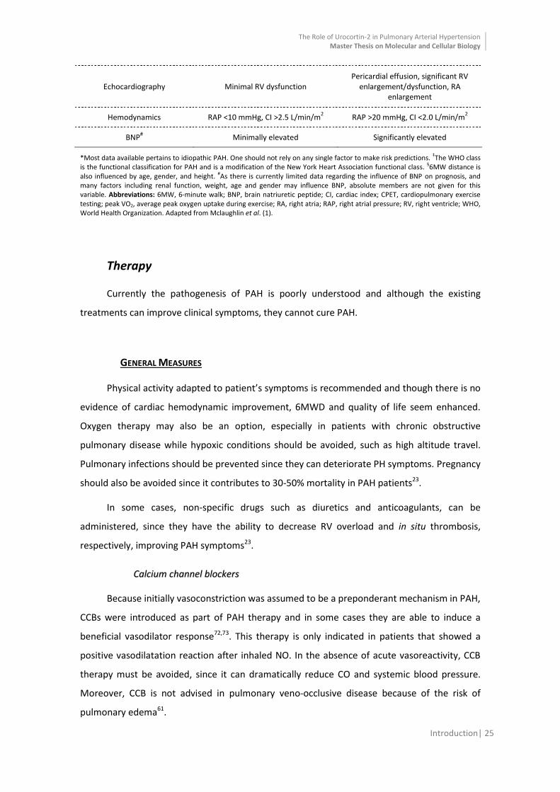
Determinants of Risk Lower Risk (Good Prognosis) Higher Risk (Poor Prognosis)
Clinical evidence of RV failure No Yes
Progression of symptoms Gradual Rapid
WHO class‡ II, III IV
6MW distance§ Longer (>400 m) Shorter (<300 m)
CPET Peak VO2 >10.4 mL/kg/min Peak VO2 <10.4 mL/kg/min
The Role of Urocortin-2 in Pulmonary Arterial Hypertension Master Thesis on Molecular and Cellular Biology
Introduction| 25
Echocardiography Minimal RV dysfunction Pericardial effusion, significant RV
enlargement/dysfunction, RA enlargement
Hemodynamics RAP <10 mmHg, CI >2.5 L/min/m2 RAP >20 mmHg, CI <2.0 L/min/m
2
BNP# Minimally elevated Significantly elevated
*Most data available pertains to idiopathic PAH. One should not rely on any single factor to make risk predictions. ‡The WHO class is the functional classification for PAH and is a modification of the New York Heart Association functional class. §6MW distance is also influenced by age, gender, and height. #As there is currently limited data regarding the influence of BNP on prognosis, and many factors including renal function, weight, age and gender may influence BNP, absolute members are not given for this variable. Abbreviations: 6MW, 6-minute walk; BNP, brain natriuretic peptide; CI, cardiac index; CPET, cardiopulmonary exercise testing; peak VO2, average peak oxygen uptake during exercise; RA, right atria; RAP, right atrial pressure; RV, right ventricle; WHO, World Health Organization. Adapted from Mclaughlin et al. (1).
Therapy
Currently the pathogenesis of PAH is poorly understood and although the existing
treatments can improve clinical symptoms, they cannot cure PAH.
GENERAL MEASURES
Physical activity adapted to patient’s symptoms is recommended and though there is no
evidence of cardiac hemodynamic improvement, 6MWD and quality of life seem enhanced.
Oxygen therapy may also be an option, especially in patients with chronic obstructive
pulmonary disease while hypoxic conditions should be avoided, such as high altitude travel.
Pulmonary infections should be prevented since they can deteriorate PH symptoms. Pregnancy
should also be avoided since it contributes to 30-50% mortality in PAH patients23.
In some cases, non-specific drugs such as diuretics and anticoagulants, can be
administered, since they have the ability to decrease RV overload and in situ thrombosis,
respectively, improving PAH symptoms23.
CCaallcciiuumm cchhaannnneell bblloocckkeerrss
Because initially vasoconstriction was assumed to be a preponderant mechanism in PAH,
CCBs were introduced as part of PAH therapy and in some cases they are able to induce a
beneficial vasodilator response72,73. This therapy is only indicated in patients that showed a
positive vasodilatation reaction after inhaled NO. In the absence of acute vasoreactivity, CCB
therapy must be avoided, since it can dramatically reduce CO and systemic blood pressure.
Moreover, CCB is not advised in pulmonary veno-occlusive disease because of the risk of
pulmonary edema61.

The Role of Urocortin-2 in Pulmonary Arterial Hypertension Master Thesis on Molecular and Cellular Biology
Introduction| 26
SPECIFIC TREATMENT
The growing knowledge about the interplay between signaling pathways in pulmonary
arterial ECs and SMCs, lung fibroblasts, and RV myocytes that occur in response to injury has
led to the development of PAH-specific pharmacotherapies23, already approved for the
treatment of PAH.
PPrroossttaannooiiddss
PGI2 or prostacyclin, is an arachidonic acid produced by ECs that induces relaxation in
both systemic and pulmonary vascular smooth muscle and inhibits platelet aggregation
through the increase in intracellular cAMP levels15. PGI2 also plays an important role in
antiproliferative, antithrombotic, antimitogenic and immunomodulatory activity23. In fact,
patients with PAH have reduced endogenous prostacyclin, which contributes to the
pathogenesis of the disease24.
PPhhoosspphhooddiieesstteerraassee ttyyppee--55 iinnhhiibbiittoorrss
The vasodilator activity of NO in VSMC is achieved through the up-regulation of cGMP,
whose metabolism depends on the activation of PDEs, since the latter is responsible for the
hydrolytic breakdown of cGMP. Three types of PDEs enzymes can be found in pulmonary
arterial contractive cells, but PDE-5 is the most expressed isoform in pulmonary circulation23.
EEnnddootthheelliinn rreecceeppttoorr aannttaaggoonniissttss
ET-1 is a potent vasoconstrictor and a SMC mitogen that contributes to the development
of PAH. Elevated ET-1 levels are frequently correlated with poor prognosis in PAH patients.
Endothelin receptor antagonists (ERAs) act by blocking the binding of ET-1 to its receptors,
inhibiting its downstream effects. So far, several types of ERAs have been identified and differ
in their selectivity to ETA and ETB receptors74.
In short, currently several PAH-specific drugs are available, however they are not able to
cure PAH, and some of them show adverse side effects, such as teratogenic effects, hepatic
function deterioration and anemia during treatment75,76.

The Role of Urocortin-2 in Pulmonary Arterial Hypertension Master Thesis on Molecular and Cellular Biology
Introduction| 27
Animal Models of Pulmonary Hypertension
In order to better understand the pathophysiological mechanism and remodeling
process behind PH, and search for novel therapeutic agents, a variety of animal models have
been developed and characterized.
These experimental in vivo models mimic certain histological and molecular features
seen in PH pathophysiology in humans. These include endothelial dysfunction, muscularization
of previously non-muscular arterioles and increased medial thickness of normally muscularized
arterioles, in situ thrombosis and plexiform lesions appearance77. Chemical agents78,79, genetic
techniques80,81, environmental factors82 and surgical procedures83 can be used to induce PAH-
associated alterations in animals (Table 5).
Currently, monocrotaline (MCT) administration and chronic exposure to hypoxia are the
most widely used models of PH in translational research due to their good reproducibility and
well described histopathology.
MCT is a pyrrolizidine alkaloid extracted from Crotalaria spectabilis seeds and when
administered in rats it is metabolized by several oxidases present in the liver, producing the
reactive bifunctional cross-linking compound MCT pyrrole. Because this compound has a short
half-life and the pulmonary circulation represents the first major vascular bed following liver,
its toxic effect concentrate on pulmonary vessels without affecting the systemic circulation77.
After MCT injection, rats undergo a severe inflammatory reaction, followed by EC death
and loss of small peripheral arteries, as well as an increase of the alveoli/arteries ratio. In the
first two weeks no clinical disorder can be noticed, whereas in the following 2-4 weeks, the
animal’s state begins to deteriorate due to the progressive thickening of the media,
muscularization of non-muscularized arteries, along with an adventitial proliferation. These
abnormalities lead to a progressive increase in mPAP and PVR, ultimately leading to RV
hypertrophy (RVH) and increased RV systolic pressure (from 25 to 80 mmHg). At this stage,
animals show impaired breathing and cyanotic mucus membranes, acquiring also a hunched
posture, being visibly sick. After 4-6 weeks of MCT-administration, animals develop severe PH
with a compensatory RVH, due to the increase in PVR. Following this stage and with a
progressive increase in PVR, the RV function deteriorates and eventually the animals die of RV
failure. However, due to the different pharmacokinetics of MCT among different rat strains,
and even between individuals, differences in time of onset and extent of toxic effects can be
seen84.

The Role of Urocortin-2 in Pulmonary Arterial Hypertension Master Thesis on Molecular and Cellular Biology
Introduction| 28
TABLE 5. Experimental animal models of Pulmonary Hypertension
Experimental model Animal species
Shared pathological findings in human disease
(Patho)physiological stimuli
Acute and chronic hypoxia
Bird, cow, dog, guinea pig,
mouse, pig, rat, sheep
Increased muscularization of resistance vessels in chronic bronchitis, cystic fibrosis, chronic obstructive pulmonary disease, hypoventilation and chronic heart disease.
Increased flow Dog, pig, rat,
sheep Increased muscularization in congenital heart disease
Vascular obstruction (air embolism,
synthetic microspheres)
Dog, pig, rat, sheep
Vascular obliteration and increased muscularization in chronic pulmonary thromboembolism
Chemical and toxic stimuli
Monocrotaline (pyrrole)
Dog, rat, sheep Increased muscularization and vascular inflammation in drug-induced PH
α-Naphthylthiourea Rat Pulmonary edema and increased muscularization induced by chemotherapy
Bleomycin Mouse, rabbit,
rat Fibrosis and increased muscularization in interstitial lung diseases
Group B Streptococcus
Pig, sheep Vasoconstriction in persistent PH of the newborn
Molecular stimuli
VEGFR-2 inhibition + hypoxia
Rat Plexiform lesions in primary PH
Angiopoietin-I overexpression
Rat Muscularization and vascular occlusion in primary and secondary PH
Genetic stimuli
Fawn-hooded rat Rat Genetic predisposition resulting in increased muscularization
Broiler chickens Chicken Genetic predisposition resulting in increased muscularization
BMPR2 knockout Mouse Genetic predisposition resulting in increased muscularization
S100A4 overexpression
Mouse Genetic predisposition resulting in plexiform lesions
Abbreviations: BMPR2, bone morphogenic protein receptor type II; PH, pulmonary hypertension; VEGFR-2, vascular endothelial growth factor receptor type II. Adapted from Marsboom et al. (76).

The Role of Urocortin-2 in Pulmonary Arterial Hypertension Master Thesis on Molecular and Cellular Biology
Introduction| 29
Overview of Urocortin-2/CRHR2 System
Urocortins (UCNs) belong to the corticotropin-releasing hormone (CRH) family which
includes CRH, fish urotensin I, frog sauvagine, UCN-1, UCN-2 (or stresscopin-related peptide)
and UCN-3 (or stresscopin)85. The various actions of the CRH family of peptides are mediated
via CRH receptors (CRHRs) that derive from two distinct genes termed CRHR1 and CRHR286.
These peptides and their receptors are ancient signaling molecules that allow organisms
in development to coordinate physiological responses to a changing environment85. The CRH
family seem to have overlapping roles in different tissues namely the immune, digestive,
central nervous, reproductive and cardiovascular systems, with their relative importance in
each system dependent upon their site of production, plasma distribution and specific
receptor affinity87.
UCN-2 is a peptide highly expressed within the cardiovascular system and has shown
promising effects in multiple studies in both animals88-90 and humans91,92. Therefore, UCN-2
expression and activity in the heart, particularly its therapeutic potential in terms of cardiac
protection has gaining interest in the field of cardiovascular research.
Molecular Structure
UCN-1 is a 40 amino acid (aa) peptide related to CRH (45% sequence identity) and
urotensin (63% sequence identity)93. The parent peptide, composed of 122 aa, has an N-
terminal methionine and a consensus signal peptide sequence, whilst the carboxy terminus of
the precursor contains the C-terminally amidated peptide of UCN-1. The CRH analogue
peptides have a helical conformation and the C-terminal helices are amphipathic, whereas the
N-terminal helices differ in their amphipathicity. The amphipathic N-terminal helices might
play an important role in selectivity of the analogues to CRHR1, but may not be as essential for
CRHR2 binding. The link between N- and C-terminal helices could also play a fundamental role
in ligand-receptor interactions94.
UCN-2, a 38 aa peptide, shows reasonable homology with rat and human CRH (34%),
UCN-1 (43%) and UCN-3 (37–40%)95. Mouse UCN-2, but not human UCN-2, is processed at the
C terminus, resulting in an amidated residue that is further cleaved to a smaller bioactive form.
The prohormones of both UCN-1 and UCN-2 are heavily glycosylated and are capable of
activating CRHR296.

The Role of Urocortin-2 in Pulmonary Arterial Hypertension Master Thesis on Molecular and Cellular Biology
Introduction| 30
The CRHR1 and CRHR2 are membrane-bound proteins that belong to the class B1 of the
family of seven transmembrane GPCRs and are encoded by two distinct genes, at least in
mammals, which are expressed in numerous tissues. Both receptors, bind all members of the
CRH family, albeit with different affinities97.
Concerning receptor structure, they have an aa homology of about 70%, while exhibiting
an approximately 47% divergence at the N-terminal extracellular domain (ECD), a major ligand
binding site, which serves to dock peptide ligands via its C-terminal segment. This is consistent
with their distinct actions and agonist selectivity, which is important for their unique
physiological roles when co-expressed in the same tissue98. Early studies identified a number
of aa within the ECD region crucial for the binding of CRHR1 receptor agonists and antagonists.
Both CRHR1 and CRHR2 ECDs contain a short consensus repeat fold, characteristic of the ECD of
class B1 GPCRs and important in activating the receptor by inducing a helix formation towards
the N-terminus of the ligand to generate a conformational active state99,100.
Tissue Distribution
In the brain, UCN-1 expression is most prominent in the Edinger Westphal nucleus and
lateral superior olive. UCN-1 mRNA or immunoreactivity has also been reported in other brain
regions including the hypothalamus and it seems to be co-localized with dopamine. UCN-1
distribution has also been verified in peripheral tissues such as heart, adrenal gland, skeletal
muscles, placenta, skin, immune system, and gastrointestinal tract101.
UCN-2 has a similar pattern of distribution relative to UCN-1 in the mouse and rat
central nervous systems. In peripheral tissues, high levels of UCN-2 have been detected in the
heart, adrenal gland, placenta, stomach, skin, ovaries, gastrointestinal tract, uterine smooth
muscle, skeletal muscle and peripheral blood vessels101.
In contrast, UCN-3 exhibits a different distribution from UCN-1 and UCN-2, since it’s
found predominantly within the hypothalamus and amygdala. Several major UCN-3 terminal
fields have been recognized, including the lateral septum and the ventromedial hypothalamus
which are known to express high levels of CRHR2, supporting the notion that UCN-3 is an
endogenous ligand101. In human peripheral tissues, immunoreactive UCN-3 is expressed in the
adrenals, heart and kidney (particularly the distal tubules)102.
Regarding the receptors, CRHR1 is not detected in the heart, while CRHR2 is highly
expressed in cardiomyocytes86. In the heart, CRHR2 has two splice variants – CRHR2α which is
The Role of Urocortin-2 in Pulmonary Arterial Hypertension Master Thesis on Molecular and Cellular Biology
Introduction| 31
detected in all chambers of the human heart, and CRHR2β which seems to be restricted to the
left atrium86. This latter isoform is also found in endothelial and SMCs of the systemic
vasculature103. This observation triggered investigation of UCN-2 as an important physiological
peptide in the cardiovascular system.
The half-life of UCN-1 and -2 in humans is approximately 50104,105 and 10 minutes92,
respectively. The exact half-life of UCN-3 in man is not yet known, but it appears to have a
shorter interval of action, at least in healthy sheeps106.
Intracellular Signaling Pathways
UCN-2 appears to exert its effects mainly through interaction with CRHR2 on target cells,
since it shows no appreciable binding affinity for the CRHR1107. Upon agonist binding, CRHRs
undergo a structural conformation alteration, activating the coupled heterotrimeric G protein
which mediates a wide range of intracellular pathways101.
The CRH family of peptides achieves its physiological effects mainly via activation of the
adenylyl cyclase (AC)-cAMP signaling pathway108, which initiates intracellular events resulting
in post-translational modifications of target proteins by protein kinase A (PKA) and/or other
kinases, and alteration of gene transcription regulation by cAMP response element-binding
(CREB) proteins109. However, some studies88,110 have shown that pharmacological inhibition of
the cAMP/PKA pathway failed to abolish the biological effects of CRH and related agonists,
suggesting that these peptides and their receptors are able to induce cellular events through
alternative signaling pathways, as will be further discussed.
Several studies suggest that signaling transduction by UCN-2 and related peptides,
begins by an increase in cAMP-dependent PKA activity103, 111 and downstream PKA/A-kinase
anchoring protein (AKAP) interactions112. UCN-2-binding to CRHR2 promotes an increased
activation of extracellular-signal-regulated kinase (ERK) 1/288, 113 and exchange proteins
activated by cAMP (EPAC), which appears to play a role in cAMP-dependent ERK1/2
activation114, 115. The phosphatidylinositol-3 kinase (PI3K)/protein kinase B (Akt) pathway is also
known to be activated by this peptide108, 112, 116, 117, and it is particularly important in cardiac
and skeletal muscle since it helps regulate phospholamban (PLB) phosphorylation, along with
PKA, controlling the inhibition of sarco/endoplasmatic reticulum calcium-ATPase (SERCA)118.
Another signaling pathway induced by CRHR2 activation is Ca2+/calmodulin-CaMKII,
which along with PKA, is important for intracellular Ca2+ homeostasis given that they have the
The Role of Urocortin-2 in Pulmonary Arterial Hypertension Master Thesis on Molecular and Cellular Biology
Introduction| 32
ability to phosphorylate key Ca2+-regulating proteins like the L-type Ca2+ channel, the
ryanodine receptor and PLB119. The activation of these proteins leads ultimately to increased
Ca2+ influx, sarcoplasmic reticulum (SR) Ca2+ content and accelerated [Ca2+]i transients119, 120.
UCN-2 also interferes with the opening of K+ channels, leading to a hyperpolarized state, which
increases the driving force for Ca2+ entry in the cell121.
Regarding the NO/cGMP pathway, it seems that, in contrast to CRHR1, CRHR2 activation
in human umbilical vein endothelial cells (HUVECs) leads to an increased expression of
inducible NOS (iNOS)122. In pigs, UCN-2 administration induces NO release via activation of
CRHR289, leading also to increased Ca2+ influx since UCN-2 causes eNOS activation through
CRHR2-cAMP and CaMKII-dependent signaling120.
Recently, the mechanism that leads to eNOS phosphorylation and NO release was
established in isolated rabbit ventricular myocytes, where CRHR2 activation through UCN-2
caused an increase in phosphorylation of Akt (Ser473 and Thr308), eNOS (Ser1177) and ERK1/2
(Thr202/Tyr204). It appears that the MEK1/2-ERK1/2 pathway is not required for stimulation
of NO signaling in these cells because eNOS phosphorylation was not suppressed by inhibition
of MEK1/2. The other two pathways, cAMP-PKA and PI3K-Akt, converge on eNOS
phosphorylation and result in pronounced and sustained cellular NO production with
subsequent stimulation of cGMP signaling since, when both of these pathways were inhibited,
the UCN-2-induced increases in [NO]i were attenuated117.
In myocytes, UCN-2 also induces the secretion of both atrial natriuretic peptide (ANP)
and BNP from these cells via CRHR2123. In SMCs, UCN-induced intracellular cAMP accumulation
contributes to increased IL-6 release, and both protein kinase C (PKC) and p38 mitogen-
activated protein kinase (MAPK) signaling cascades are involved downstream of this
pathway124. Furthermore, in neonatal rat cardiomyocytes, this increase in IL-6 release was
induced by CRHR2, in a NF-κB-dependent manner125, indicating that UCNs, specifically UCN-2
and UCN-3, could be important inflammation mediators.
Cells overexpressing both CRHR1 and CRHR2 and treated with sauvagine, a CRH-related
peptide, causes PKA-mediated phosphorylation of the transcription factor CREB, which is an
important intermediary step in transduction pathways arising from activation of these
receptors and leading to modulation of gene transcription of target cells108. In addition,
receptor stimulation by agonist binding increases activation of ERK1/2 independently of cAMP,
revealing the possibility that PKA and MAPK may act in concert to control gene transcription in
CRH-responsive cells. Sauvagine is also able to increase intracellular levels of Ca2+ through both
The Role of Urocortin-2 in Pulmonary Arterial Hypertension Master Thesis on Molecular and Cellular Biology
Introduction| 33
mobilization of intracellular stores and influx across the plasma membrane, which are
important for muscle contraction and relaxation108.
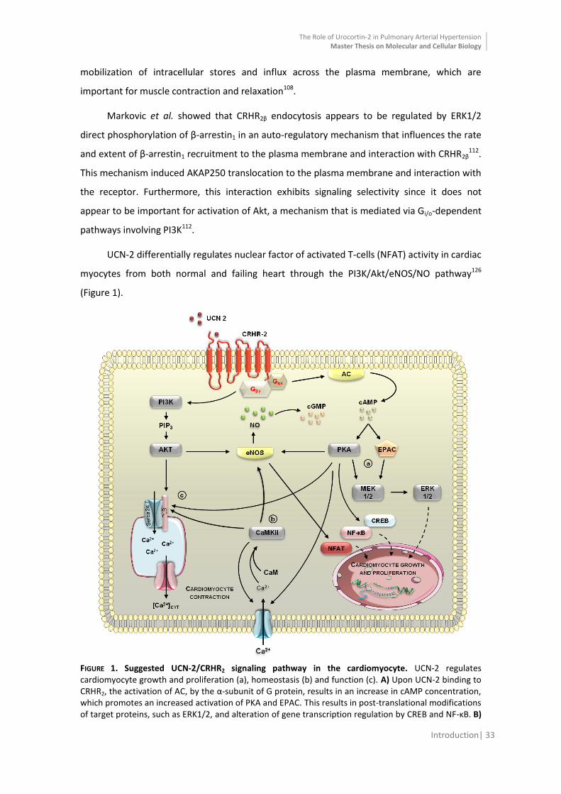
Markovic et al. showed that CRHR2β endocytosis appears to be regulated by ERK1/2
direct phosphorylation of β-arrestin1 in an auto-regulatory mechanism that influences the rate
and extent of β-arrestin1 recruitment to the plasma membrane and interaction with CRHR2β112.
This mechanism induced AKAP250 translocation to the plasma membrane and interaction with
the receptor. Furthermore, this interaction exhibits signaling selectivity since it does not
appear to be important for activation of Akt, a mechanism that is mediated via Gi/o-dependent
pathways involving PI3K112.
UCN-2 differentially regulates nuclear factor of activated T-cells (NFAT) activity in cardiac
myocytes from both normal and failing heart through the PI3K/Akt/eNOS/NO pathway126
(Figure 1).
FIGURE 1. Suggested UCN-2/CRHR2 signaling pathway in the cardiomyocyte. UCN-2 regulates cardiomyocyte growth and proliferation (a), homeostasis (b) and function (c). A) Upon UCN-2 binding to CRHR2, the activation of AC, by the α-subunit of G protein, results in an increase in cAMP concentration, which promotes an increased activation of PKA and EPAC. This results in post-translational modifications of target proteins, such as ERK1/2, and alteration of gene transcription regulation by CREB and NF-κB. B)
The Role of Urocortin-2 in Pulmonary Arterial Hypertension Master Thesis on Molecular and Cellular Biology
Introduction| 34
The Ca2+
/calmodulin-CaMKII, along with PKA, is important for intracellular Ca2+
homeostasis, since they have the ability to phosphorylate key Ca
2+-regulating proteins, leading to increased Ca
2+ influx, SR Ca
2+
content and accelerated [Ca2+
]i transients. The cAMP-PKA and PI3K-Akt signaling pathways converge on eNOS phosphorylation and result in pronounced and sustained cellular NO production with subsequent stimulation of cGMP signaling. C) The activation of PI3K, though the βγ subunit of the G protein, leads to Akt activation which helps regulate PLB phosphorylation, along with PKA, controlling the inhibition of SERCA. NFAT regulation is mediated by PI3K/Akt/eNOS/NO signaling cascade that converges on the activation of several kinases, including GSK-3β, JNK, p38 and PKG, resulting in phosphorylation, deactivation and nuclear export of NFAT. Abbreviations: AC, adenylate cyclase; cAMP, cyclic adenosine monophosphate; PKA, protein kinase A; EPAC, exchange proteins activated by cAMP; MEK1/2, mitogen-activated protein kinase; ERK1/2, extracellular signal-regulated kinase; CREB, cAMP response element-binding protein; NF-κB, nuclear factor kappa-light-chain-enhancer of activated B cells; eNOS, endothelial nitric oxide synthase; NO, nitric oxide; cGMP, cyclic guanosine monophosphate; CaM, calmodulin; CaMKII, Ca2+ /calmodulin-dependent protein kinase II; PI3K, Phosphoinositide 3-kinase; PIP3, Phosphatidylinositol (3,4,5)-trisphosphate; AKT, protein kinase B; Serca 2a, sarco/endoplasmic reticulum Ca2+-ATPase; PLB, phospholamban; UCN-2, Urocortin-2; CRHR2, corticotropin releasing hormone receptor type 2; NFAT, nuclear factor of activated T-cells; GSK-3β, glycogen synthase kinase 3 beta; JNK, c-Jun NH2-terminal kinase; p38, p38 mitogen-activated kinase; PKG, protein kinase G.
UCN-2/CRHR2 Signaling Effects in Cardiac Function
The majority of the described actions of UCNs in the myocardium have been revealed by
observations in cardiomyocytes and in animal models of heart failure. However, it is not
completely clear if the protective effects of UCNs are mediated via a direct effect on cardiac
myocytes or via sympathetic stimulation in reaction to decreased peripheral resistance127.
In both rabbit128 and mouse ventricular myocytes119, as well as in a murine model of
HF129, UCN-2 administration enhances myocardial inotropy and lusitropy in a time- and
concentration-dependent manner. These effects appear to be mediated via activation of
CRHR2 and subsequent stimulation of both cAMP/PKA and Ca2+/calmodulin-CaMKII signaling
pathways since they were abolished by antisauvagine-30, a specific CRHR2 antagonist, leading
to increased SR Ca2+ content and [Ca2+]i transients, as well as an accelerated decay of the
latter119, 128. However, in mouse cardiomyocytes, these effects were accompanied by
arrhythmogenic events, triggered by spontaneous diastolic SR Ca2+ release119. On the other
hand, UCN-2 is reported to increase the ventricular fibrillation threshold130 and reduce the
occurrence of arrhythmias131 in vivo. Evidence of the UCNs actions to improve intracellular
handling132 and perhaps inhibit efferent cardiac sympathetic nerve activity133, might explain
the peptide’s anti-arrhythmic effects in the whole animal. Other beneficial cardiac effects of
UCNs include their ability to improve cardiac bioenergetics (through preservation of high-
energy phosphate stores)134.
In the pig, local intracoronary administration of UCN-2 also has the ability to increase
coronary blood flow as well as myocardial function89. In addition, an immediate improvement
of left ventricular fractional shortening and circumferential fiber shortening velocity after an
The Role of Urocortin-2 in Pulmonary Arterial Hypertension Master Thesis on Molecular and Cellular Biology
Introduction| 35
acute injection of UCN-2 has been described in rats. These positive effects were maintained
even at 5 weeks after treatment135.
Recently, Walther et al. demonstrated that UCN-2 differentially regulates NFAT activity
in cardiac myocytes from both normal and failing hearts, having a crucial role in the regulation
of gene expression in cardiac development, maintenance of an adult differentiated cardiac
phenotype and remodeling processes126.
UCN-2/CRHR2 Signaling Effects in Vascular Function
UCN-2/CRHR2 system effects are not limited to the heart, since in both pulmonary and
systemic vasculature the activation of this receptor and the following pathway seems to exert
a potent and consistent relaxation of the vessels.
In rat thoracic aorta, UCN-2 and -3 wield more potent vasodilator effects than CRH, and
since this relaxation response was abolished in the presence of both AC and PKA inhibitors,
this effect is likely to be mediated by signaling pathways involving these peptides136.
Indeed, in both healthy137 and animal models of HF129, UCN-2 administration causes a
reduction in arterial blood pressure. Furthermore, the selective CRHR2 antagonist, astressin2-B,
abolished UCN-2-induced hypotensive activity while having no effect on basal arterial blood
pressure, indicating that UCN-2 induces hypotension through peripheral CRHR2137. Dieterle et
al. also showed an immediate and sustained lowering of blood pressure in hypertensive rats,
with no reflex rise in HR135. Several mechanisms have been postulated for the blood pressure-
lowering effects of the UCNs, including a direct smooth muscle relaxant effect in combination
with an associated reduction in plasma concentrations of vasoconstrictor hormones. Grossini
et al. showed that UCN-2 administration in the anaesthetized pig primarily increases coronary
blood flow and myocardial function through the release of NO and activation of CRHR2. In this
study, the blockade of NOS abolished only the coronary effects, whereas blockade of subtype 2
of the CRH receptors abolished both cardiac and coronary effects89.
Ex vivo studies in human internal mammary artery138 and in rat coronary artery139
propose both endothelium-dependent and -independent mechanisms for vasorelaxation,
which involves the release of NO that in turn stimulates Ca2+-activated K+ channels in vascular
smooth muscle via cGMP-dependent pathways138.
Additionally, another important role for the UCN-2/CRHR2 system is its contribution to
the angiogenesis process, being a critical component of the pathway necessary for tonic

The Role of Urocortin-2 in Pulmonary Arterial Hypertension Master Thesis on Molecular and Cellular Biology
Introduction| 36
inhibition of adult neo-vascularization. Bale et al. demonstrated that mice deficient in CRHR2
become hypervascularized postnatally. On the other hand, the activation of this receptor in
vitro results in reduced release of VEGF from SMCs, inhibition of SMC proliferation and
inhibition of capillary tube formation in collagen gels140.
Recently, Emeto et al. reported that in human aortic vascular SMCs, UCN-2 significantly
inhibited Akt phosphorylation and proliferation in a dose-dependent manner by promoting a
G1 cell-cycle arrest, reinforcing the role of UCN-2 in the regulation of cell proliferation141.
Moreover, endothelial UCN has potent antioxidative properties and is up-regulated by
inflammatory cytokines and pitavastatin142.
UCN-2/CRHR2 Signaling Effects in Heart Failure
Given the potent vasorelaxant and inotropic effects of UCN-2, interest has grown in its
role in the pathophysiology of HF and its potential therapeutic value in this disease. Several
studies with UCN-2 in different animal models and in human clinical studies have
demonstrated the beneficial effects of the peptide in the setting of cardiac failure.
In both adult rabbit ventricular myocytes128 and in a murine model of HF129, UCN-2
administration enhanced myocardial inotropy and lusitropy, increasing cell shortening and
accelerating relaxation in a time- and concentration-dependent manner. These effects are
induced via CRHR2 receptor-mediated stimulation of PKA128.
In contrast to limited UCN-2 bioactivity in healthy sheep, the therapeutic administration
in animals with pacing-induced HF induced rapid and dose-dependent increases in CO and
reductions in peripheral resistance and left atrial pressure90. The prolonged administration of
UCN-2 in the same animal model also produced the beneficial effects seen above, except in a
sustained way of action143, supporting a role for UCN-2 in pressure/volume homeostasis in HF.
Additionally, UCN-2 co-treatment with an existing proven treatment such as furosemide, a
diuretic agent, resulted in enhancement of beneficial hemodynamic effects compared with
either agent alone, together with augmented renal responses with less rennin activation and
no exacerbation of potassium loss144.

The Role of Urocortin-2 in Pulmonary Arterial Hypertension Master Thesis on Molecular and Cellular Biology
Introduction| 37
UCN-2/CRHR2 Signaling Effects in Myocardial Ischemia
Myocardial ischemia frequently leads to cardiomyocyte apoptosis and necrosis, even
with subsequent restoration of blood flow to the affected area, which has caused research to
focus on trying to prevent cardiomyocyte death after an ischemic event. In this context, UCNs
appear to have a cardioprotective role with increased expression and peptide release observed
following ischemia stimulation145.
On the other hand, neonatal cardiomyocytes treated with either 1-10nM of UCN-2 or -3
results in ERK1/2 phosphorylation, and the inhibition of MEK 1/2 obstructs their
cardioprotective effects88. Brar et al. suggested that UCN-2 and -3 have a cardioprotective role
in situations of ischemia/reperfusion injury acting via the MAPK pathway and reducing infarct
size88.
Moreover, as mentioned above, CRHR2-deficient mice become hypervascularized
postnatally and the activation of this receptor reduces VEGF release from SMCs, suppresses
SMC proliferation and impedes capillary tube formation140. These studies demonstrate a
critical role for CRHR2 in the regulation of angiogenesis and remodeling of both juvenile and
adult vasculature, suggesting this receptor may be a potential target to modulate angiogenesis
in diseases such as cardiovascular ischemia.
UCN-2/CRHR2 Signaling Effects in Other Cardiovascular Diseases
Dieterle et al. reported that chronic UCN-2 treatment was able to improve cardiac and
vascular function through an immediate sustained blood pressure reduction without affecting
the HR. Furthermore, long-term UCN-2 treatment in hypertensive rats diminishes the
development of hypertension-induced LV hypertrophy and the deterioration of its contractile
function135. Moreover, long-term UCN treatment not only had hypotensive effects but may
also inhibited development of vascular remodeling in mesenteric arteries in spontaneously
hypertensive rats146. These beneficial effects of UCN-2 may represent a novel approach for
antihypertensive therapy.
In a recent study, patients who underwent coronary angiography with the pre-diagnosis
of coronary artery disease were examined and distributed according to their ejection fraction
(EF). Those patients with moderate to severe systolic dysfunction (SD) showed decreased
serum UCN-2 levels, while patients with mild to moderate SD showed increased levels of the
peptide when compared to patients without SD. In contrast, the presence of diastolic

The Role of Urocortin-2 in Pulmonary Arterial Hypertension Master Thesis on Molecular and Cellular Biology
Introduction| 38
dysfunction or coronary artery disease appeared to have little effect on circulating UCN-2
concentrations147.
Urocortin-2 as a Therapy for Heart Failure in Humans
Recently, a small number of clinical studies in humans have confirmed some data
obtained from animal studies. In fact, UCN-2 administration in healthy humans induces a dose-
related increase in CO, HR and left ventricular ejection fraction (LVEF) in association with
decreased systemic vascular resistance92. Moreover, UCN-2 and -3 promotes a potent
prolonged arterial vasodilatation without causing tachyphylaxis, with these effects being partly
dependent on endothelial NO and cytochrome P450 metabolites of arachidonic acid148.
In humans with both mild149 and acute decompensated HF91, UCN-2 administration
resulted in a markedly augmented CO without significant reflex tachycardia and an improved
vascular function. Pronounced falls in systemic arterial pressure were also seen with higher
doses of UCN-291. UCN-2 also induced vasorelaxation of coronary arteries isolated from
patients with HF, independently of endothelial integrity150. In addition to positive effects on
cardiovascular parameters when used as a treatment in HF, UCNs may also serve as possible
biomarkers in the identification of early HF151.
Based on these clinical studies, detailed in Table 6, it appears that UCN-2, alone or in
combined therapy, is able to regulate the cardiovascular system which points towards a
therapeutic application for this peptide in HF.
TABLE 6. Clinical trials with Urocortin-2 as a therapy for Heart Failure
Refs Model Treatment Outcome
92
8 Healthy humans
Subjects received placebo, 25μg and 100μg of UCN-2 intravenously over the course of 1h.
↑ Cardiac output; ↑ Heart rate; ↑Left ventricular ejection fraction; ↓ Systemic vascular resistance.
148
18 Healthy humans
Volunteers were subjected to a bilateral forearm venous occlusion plethysmography during intra-arterial UCN-2 (3.6-120pmol/min), UCN-3 (1.2-36nmol/min) and substance P (2-8pmol/min).
Potent and prolonged arterial vasodilatation without tachyphylaxis.
149
8 Male patients with HF
Received placebo and 25μg and 100μg of UCN-2 intravenously over 1h.
↑ Cardiac output; ↑ Left ventricular ejection fraction; ↓ Mean arterial pressure; ↓ Systemic vascular resistance; ↓ Cardiac work.

The Role of Urocortin-2 in Pulmonary Arterial Hypertension Master Thesis on Molecular and Cellular Biology
Introduction| 39
Other Biological Effects of Urocortin-2
UCN-2 is also expressed in the central nervous system and it seems to be involved in the
regulation of the stress response, anxiety and depression152, as well as in the control of
appetite98. Male, but not female, UCN-2-knockout animals demonstrate an altered social
behavior, with more passive social interactions and reduced aggressiveness in comparison to
wild-type animals. Moreover, UCN-2 appears to modulate the aggressive behavior in male
mice153 further evidence that this peptide is involved in the regulation of stress-related
behaviors.
In terms of appetite control, a recent study has reported that CRHR1 agonists, UCN-1 and
stressin1-A, reduced feeding and induced interoceptive stress, while UCN-2 had a more potent
suppressive effect on feeding, via a CRHR2-dependent mechanism, without eliciting malaise154.
UCN-2 also appears to be responsible for a decrease in gastric emptying152 which aids in
appetite inhibition. These differences between UCNs in their regulation of these various
functions seem to be consistent with their pharmacological differences.

The Role of Urocortin-2 in Pulmonary Arterial Hypertension Master Thesis on Molecular and Cellular Biology
Aims| 40
AAIIMMSS
Based on the published findings referenced in the previous chapter, we hypothesize that
by activating CRHR2 in animals with PAH, through UCN-2 administration, we might protect not
only the pulmonary vasculature, but also the RV myocardium and thus increase animal
survival, attenuate PAH severity and improve RV function.
Therefore, we purpose to investigate the role of the UCN-2/CRHR2 system in the
pathophysiology of PAH and progression to HF, while evaluating the efficacy of UCN-2 as a
possible novel therapeutic strategy for this disease.
Our specific goals are:
1. to determine changes in the expression of UCN-2 and its receptor, and of downstream
signaling pathways in the heart tissue from healthy rats and from rats with PAH induced by
monocrotaline (MCT);
2. to assess changes in animal survival, RV myocardial function and the severity of PAH in vivo
(hemodynamic and echocardiographic studies), changes in myocardial structure and
histology and its patterns of gene expression, in healthy animals and in animals with PAH
and HF with or without chronic UCN-2 treatment.

MMEETTHHOODDSS

The Role of Urocortin-2 in Pulmonary Arterial Hypertension Master Thesis on Molecular and Cellular Biology
Methods| 42
All the procedures in this work followed the recommendations of the Guide for the Care
and Use of Laboratory Animals, published by the US National Institutes of Health (NIH
Publication No. 85-23, Revised 1996), and are accredited by the Portuguese Direção Geral de
Alimentação e Veterinária (DGAV) and approved by Fundação para a Ciência e a Tecnologia
(FCT PTDC/DTP-FTO/0130/2012).
Animal Model
Animals were grouped 3 per cage, in a controlled environment with a light-darkness
cycle of 12:12h and an established temperature of 22ºC, with water and food ad libitum.
Before MCT administration an echocardiographic evaluation was performed as a guarantee
that the potentially ill animals (with cardiac disease) did not follow the protocol. Between 24
to 25 days after MCT administration, the animals were submitted again to an
echocardiographic study and to invasive hemodynamic evaluation. After morphometric
assessment, samples were collected and processed for histological and molecular biology
studies.
Seven week-old male Wistar rats (Charles River Laboratories, Barcelona, Spain) weighing
180-200g, were randomly assigned to receive either a subcutaneous (s.c.) injection of MCT
(60mg/kg, Sigma-Aldrich) or an equal volume of vehicle, sodium chloride (NaCl, B. Braun,
0.9%). A freshly aqueous solution of MCT was prepared, wherein 300mg was dissolved in
2.36mL of 1M HCl, diluted with distilled water to about 1.60mL and neutralized with 1.52mL of
1M NaOH. The pH was settled at 7.00.
Fourteen days after MCT/vehicle administration, animals were randomly assigned into 4
subgroups: CTRL, untreated animals; MCT, animals with induced PAH and without
pharmacological treatment; CTRL + UCN-2, animals without PAH and with UCN-2 treatment;
MCT + UCN-2, animals with induced PAH and with UCN-2 treatment. The pharmacological
treatment consisted on a daily intraperitoneal (i.p.) injection of 2.5μg/kg twice a day of hUCN-
2 (Bachem, Bubendorf, Switzerland) during 10 days, while the vehicle treatment consisted on
sodium chloride.
The drug dosages, administration routes and treatment duration were selected based
on previous studies from other research teams135 and on our preliminary dose-finding studies
(unpublished data). Moreover and according with Reagan-Shaw’s equation155 (depicted below
in which animal km=37 and human km=6) the UCN-2 dose administered in our animals is the

The Role of Urocortin-2 in Pulmonary Arterial Hypertension Master Thesis on Molecular and Cellular Biology
Methods| 43
same that has been used in human clinical trials92, 149. Initially an aqueous solution of hUCN-2
was prepared by dissolving 1mg in 1mL of NaCl. Part of the stock was diluted again in NaCl
until we reach a final work concentration of 0.002mg/mL.
Functional Studies
Cardiac function was evaluated both in healthy and MCT-induced PAH animals, with or
without pharmacological treatment, in order to clarify the involvement of UCN-2/CRHR2
system not only in the pathophysiology of PAH, but also in the progression of functional RV
changes that accompany PAH and lead to HF.
The echocardiographic assessment allowed for a non-invasive monitoring and
characterization of PAH progression and ventricular function in the different experimental
groups. Invasive hemodynamic evaluation was performed using high-fidelity pressure-volume
catheters that provided us an accurate determination of systolic and diastolic function
parameters.
Echocardiography Studies
For this purpose, animals from the different experimental groups were sedated with
100μg/kg fentanyl and 5mg/kg midazolam via i.p. injection and anesthetized by inhalation of
8% sevoflurane (Penlon Sigma Delta, UK) in vented containers. Endotracheal intubation was
performed using a 14 gauge catheter and mechanical ventilation controlled by a rodent
ventilator (MouseVent G500, Kent Scientific, Connecticut, USA) with an animal weight-defined
tidal volume. Anesthesia was maintained with 2.5-3.0% vol/vol sevoflurane, titrated according
to the toe pinch reflex.
Animals were placed in left lateral decubitus. After applying warm echocardiography gel,
a 15MHz sensorial probe (Sequoia 15L8W) was gently placed on the thorax. Acquisitions
(Siemens Acuson Sequoia C512) were averaged from three consecutive heartbeats. Bi-
dimensional and M-mode images were obtained for determination of RV structure and
function, respectively. End-diastolic dimension (RVEDD), RAA and TAPSE were obtained in
apical projection of 4 cavities. Pulmonary artery diameter (PAD) was measured in parasternal
short-axis view. Conventional Doppler was used to evaluate pulmonary blood flow, with the

The Role of Urocortin-2 in Pulmonary Arterial Hypertension Master Thesis on Molecular and Cellular Biology
Methods| 44
determination of pulmonary artery acceleration (PAAT) and ejection (PAET) times, peak
systolic velocity (PAPSV) and the velocity time integral (PAVTI). The PAAT/PAET ratio was
determined, and the SV, CO and cardiac index (CI) were calculated as follows:
Invasive Hemodynamic Evaluation
After the echocardiographic evaluation, and as previously described156, the animal
position was adjusted to right lateral decubitus for proper orientation of the heart for
instrumentation. Animal temperature was monitored and regulated with a warming pad and a
rectal temperature sensor, and digitally controlled together with pulse oxymetry measured
through a paw sensor (PhysioSuite, Kent Scientific, Connecticut, USA), and ECG (Animal Bio
Amp, ADinstruments, Oxford, UK) (shown in Figure 5A). Anesthesia was maintained with
sevoflurane (2.5-3.0% vol/vol).
The internal femoral vein was catheterized using a 24G catheter, under surgical
microdissection (Wilde M651, Leica microsystems, Cambridge, UK) for infusion (Multi-Phaser,
NE-100, New Era Pump Systems, NY, USA) of warm lactate Ringer’s solution at a rate of
32mL/kg/h (Figure 5B and 5C). A left thoracotomy was performed, pericardium and pleura
were carefully dissected and the phrenic nerve was severed (Figure 5D). A 3-0 surgical silk was
passed around the inferior vena cava (IVC) for transient occlusion during the protocol.
Pressure-volume catheters were inserted through the apex of the RV and positioned along the
long axis (SPR-869, Millar Instruments, Texas, USA). A flow probe was implanted around the
ascending aorta (MA2.5PSB, 2.5mm, Precision S-Series, Transonic Systems, NY, USA) and
connected to an ultrasonic transit time volume flowmeter (TS420, transit-time perivascular
flowmeter, Transonic Systems, NY, USA). The experimental preparation was allowed to
stabilize for 15 minutes, and during the procedure blood loss was controlled through saline
bolus.
Baseline and IVC occlusions recordings were obtained with ventilation suspended at
end-expiration. Pressure and volume signals were continuously acquired (MVPS 300, Millar
instruments, Texas, USA), digitally recorded at a sampling rate of 1000Hz (ML880 PowerLab

The Role of Urocortin-2 in Pulmonary Arterial Hypertension Master Thesis on Molecular and Cellular Biology
Methods| 45
16/30, ADInstruments, Oxford, UK) and analyzed off-line (LabChart 7 Pro, ADInstruments,
Oxford, UK, and PVAN 3.5, Millar Instruments, Texas, USA). Parallel conductance for the
volume catheter was computed after bolus injection of 50μL hypertonic saline (10% sodium
chloride) and the calibration for factor alpha (field inhomogeneity) was determined through
the CO measured by the aortic flow probe and the ultrasonic transit time volume flowmeter.
Following anesthetic overdose, blood was retrieved for storage and further analysis.
Animals were exsanguinated and heparinized blood was collected and used for volume
calibration with standard cuvettes (P/N 910-1048, Millar Instruments, Texas, USA).
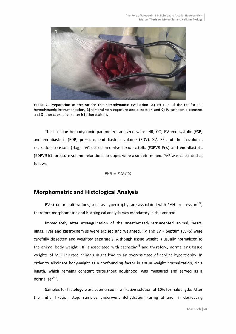
The Role of Urocortin-2 in Pulmonary Arterial Hypertension Master Thesis on Molecular and Cellular Biology
Methods| 46
FIGURE 2. Preparation of the rat for the hemodynamic evaluation. A) Position of the rat for the hemodynamic instrumentation, B) femoral vein exposure and dissection and C) IV catheter placement and D) thorax exposure after left thoracotomy.
The baseline hemodynamic parameters analyzed were: HR, CO, RV end-systolic (ESP)
and end-diastolic (EDP) pressure, end-diastolic volume (EDV), SV, EF and the isovolumic
relaxation constant (τlog). IVC occlusion-derived end-systolic (ESPVR Ees) and end-diastolic
(EDPVR k1) pressure volume relantionship slopes were also determined. PVR was calculated as
follows:
Morphometric and Histological Analysis
RV structural alterations, such as hypertrophy, are associated with PAH-progression157,
therefore morphometric and histological analysis was mandatory in this context.
Immediately after exsanguination of the anesthetized/instrumented animal, heart,
lungs, liver and gastrocnemius were excised and weighted. RV and LV + Septum (LV+S) were
carefully dissected and weighted separately. Although tissue weight is usually normalized to
the animal body weight, HF is associated with cachexia158 and therefore, normalizing tissue
weights of MCT-injected animals might lead to an overestimate of cardiac hypertrophy. In
order to eliminate bodyweight as a confounding factor in tissue weight normalization, tibia
length, which remains constant throughout adulthood, was measured and served as a
normalizer159.
Samples for histology were submersed in a fixative solution of 10% formaldehyde. After
the initial fixation step, samples underwent dehydration (using ethanol in decreasing
The Role of Urocortin-2 in Pulmonary Arterial Hypertension Master Thesis on Molecular and Cellular Biology
Methods| 47
percentages), diaphanisation (using xylene) and impregnation in liquid paraffin (54ºC). Later
they were placed and properly oriented in metal molds and serial sections of 4μm were
obtained from RV samples, in a Minot-type microtome. Hematoxylin and eosin (HE) stained
sections of the RV were used to measure cardiomyocyte cross-sectional area. Sections were
digitally photographed (Olympus XC30 Digital colour Camera, Olympus, PA, USA) and
measured using imaging software (cellB, Olympus, PA, USA). Fifty muscle fibers per animal
were analyzed and only nuclei-centered cardiomyocyte were considered for analysis of
cardiomyocyte dimensions.
Molecular Studies
These studies focused on the gene expression and protein activation analysis of the
main components of UCN-2/CRHR2 system (peptide, receptors and downstream signaling
pathways) as well as of known markers of heart failure in the different experimental groups.
mRNA Expression
In order to allow posterior molecular analysis, RV samples were collected and stored in
RNAlater (Quiagen), frozen in liquid nitrogen and kept at -80ºC until manipulation. RNA was
completely extracted by the silicon membrane and guanidinium-thiocyanate method (RNeasy
Mini Kit, Quiagen, 74104), according to the manufacturer’s instructions. Concentration and
purity of RNA were measured by NanoDrop® ND-1000 spectrophotometer (Thermo Fisher
Scientific), assuming as ideal ratio values of A260/A280 between 1.8 and 2.1. mRNA relative
expression quantification was performed by two-step Real-Time Polymerase Chain Reaction
(RT-PCR). Using animal samples from the CTRL group, standard curves were built to all the
studied genes and tissues, correlating the initial total mRNA quantity and the threshold cycle.
Reverse transcription was performed in a conventional thermocycler (Whatmann Biometra,
050-901) and consisted in 10 minutes at 22ºC, 50 minutes at 50ºC, and 10 minutes at 95ºC.
Ten percent of the obtained cDNA was amplified and detected by RT-PCR (Step-One™ Applied
biosystems) using the probe SYBR Green (PerfeCta® SYBR Green FastMix, Rox, Kit, Quanta
Biosciences) according to the manufacturer’s instructions.
Amplification curves were analyzed with the equipment software (v2.2.2) through
absolute quantification. Melting curves of each PCR reaction were used in order to exclude the
formation of primer-dimers and unspecific products, confirming the purity of the amplified

The Role of Urocortin-2 in Pulmonary Arterial Hypertension Master Thesis on Molecular and Cellular Biology
Methods| 48
product. GAPDH was chosen as reference gene, since no significant changes were observed in
the different groups. Final gene expression results were presented in Arbitrary Units (AU),
being the CTRL group means values after GAPDH normalization correspondent to 1AU. All
assays were performed twice, in order to ensure the security of the obtained results. The
primers used in the molecular analysis (Table 7) were designed in-house with appropriate
software (DNAstar™).
TABLE 7. List of used primers
Gene Primers Sequence
GAPDH P1-
5’TGG CCT TCC GTG TTC CTA CCC
3’
P2- 5’
CCG CCT GCT TCA CCA CCT TCT3’
UCN-2 P1-
5’CGT TGG CAT AAC GCC TCA C
3’
P2- 5’
GGA CAC AGA GCT GGG AGT TG3’
CRHR2 P1-
5’TGC AAC TCA TCG ACC ACG AA
3’
P2- 5’
CAG GTA GCA GCC TTC CAC AA3’
ET-1 P1-
5’CGG GGC TCT GTA GTC AAT GTG
3’
P2- 5’
CCA TGC AGA AAG GCG AAT GTG3’
BNP P1-
5’CAG AGC TGG GGA AAG AAG AG
3’
P2- 5’
GGA CCA AGG CCC TAC AAA AGA3’
HIF-1α P1-
5’TCA TAG GCG GTT TCT TGT AGC
3’
P2- 5’
CTA ACA AGC CGG AGG AC3’
Caspase-3 P1-
5’CGG GTG CGG TAG AGT AAG
3’
P2- 5’
CTG GAC TGC GGT ATT GAG ACA3’
Caspase-8 P1-
5’ACC AAA TGA AGA GCA AAC CTC G
3’
P2- 5’
TTT TCT GTC CCG CAT GTT GC3’
Abbreviation: BNP, brain natriuretic peptide; CRHR2, corticotrophin-releasing hormone receptor type 2; ET-1, endothelin 1; GAPDH, glyceraldehyde 3-phosphate dehydrogenase; HIF-1α, hipoxia inducible factor 1 alpha; UCN-2, urocortin-2.
Protein Expression
Samples from RV of each animal were homogenized on ice in 300μL RIPA lysis buffer
(Cell Signaling Technology) containing protease inhibitors (Protease Inhibitor Cocktail, Sigma-
Aldrich, St. Louis, USA), and phosphatase inhibitors (Phosphatase Inhibitor Cocktail 2 and 3,
Sigma-Aldrich, St. Louis, USA). Tissue was then centrifuged at 12.000 RPMs for 20 min at 4°C.
Supernatants were collected, and total protein concentration was determined using the
Bradford assay (Bio-Rad Laboratories, CA, USA). Samples were treated with Laemmli loading
buffer (Cell Signaling Technology) and boiled for 5 minutes at 95ºC. Samples, with 20μg of
protein, were loaded onto a 10% SDS-PAGE gel (Bio-Rad Laboratories, CA, USA), run, and
electroblotted onto a 0.2μm nitrocellulose membrane (Bio-Rad Laboratories, CA, USA). Pre-

The Role of Urocortin-2 in Pulmonary Arterial Hypertension Master Thesis on Molecular and Cellular Biology
Methods| 49
stained molecular weight marker proteins (Bio-Rad Laboratories, CA, USA) were used as size
standards for the SDS-PAGE. Ponceau staining was performed to verify the quality of the
transfer and to ensure equal protein loading. Blots were blocked in 5% BSA (Bovine Serum
Albumin, Sigma-Aldrich, St. Louis, USA) for 1 hour at room temperature. Incubated overnight
at 4ºC with the antibodies listed in Table 8, in separate experiences, at a dilution of 1:1000.
TABLE 8. List of used primary antibodies
Protein Antibody’s Identification
Akt
p-Akt
Akt (pan) (C67E7), Rabbit mAb, Cell Signaling Technology
Phospho-Akt (Ser473) (587F11), Mouse mAb, Cell Signaling Technology
ERK1/2
p-ERK1/2
p44/42 MAPK (Erk1/2) (3A7), Mouse mAb, Cell Signaling Technology
Phospho-p44/42 MAPK (Erk1/2) (Thr202/Tyr204), Rabbit pAb, Cell Signaling Technology
p38
p-p38
p38α MAPK (L53F8), Mouse mAb, Cell Signaling Technology
Phospho-p38 MAPK (Thr180/Tyr182) (D3F9), Rabbit mAb, Cell Signaling Technology
STAT3
p-STAT3
Stat3 (124H6), Mouse mAb, Cell Signaling Technology
Phospho-Stat3 (Tyr705) (D3A7), Rabbit mAb, Cell Signaling Technology
CRHR2 Anti-Corticotropin Releasing Factor Receptor 2, Rabbit pAb, Abcam, Cambridge, UK
GAPDH GAPDH (0411), Mouse mAb, Santa Cruz Biotechnology, Inc.
Abbreviation: Akt, protein kinase B; CRHR2, corticotropin-releasing hormone receptor type 2; ERK1/2, extracellular-signal regulated kinases 1 and 2; GAPDH, glyceraldehyde 3-phosphate dehydrogenase; GSK-3β, glycogen synthase kinase 3 beta; p38, p38 mitogen-activated protein kinase; STAT3, signal transducer and activator of transcription 3.
After primary antibody removal, membranes were washed with Tris-Buffered Saline (Cell
Signaling Technology) with 0.01% Tween-20 – TBS-T (Sigma-Aldrich, St. Louis, USA) and
incubated with secondary antibodies (IRDye 680LT, Goat-anti-Mouse Ab and 800CW, Goat-
anti-Rabbit Ab, LI-COR Biosciences, Lincoln, USA) in 2% skimmed nonfat milk at a 1:25000
dilution for 1 hour at room temperature. After washing the membranes with TBS-T,
membranes were scanned using an Odyssey scanner (infrared imaging system, LI-COR
Biosciences, Lincoln, USA) and analyzed using the Odyssey provided software (version 3.0).
Statistical Analysis
Statistical analysis was performed using GraphPad Software (vs.6). 2-way ANOVA was
used to statistically analyze all the presented parameters. Holm-Sidak’s method for post hoc
comparisons between groups and Kaplan-Meier survival analysis (log-rank test) were

The Role of Urocortin-2 in Pulmonary Arterial Hypertension Master Thesis on Molecular and Cellular Biology
Methods| 50
performed. Group data are presented as means ± SEM. Differences with p<0.05 were
considered statistically significant.

RREESSUULLTTSS

The Role of Urocortin-2 in Pulmonary Arterial Hypertension Master Thesis on Molecular and Cellular Biology
Results| 52
Survival Analysis
Excluding animals that underwent terminal hemodynamic evaluation, Kaplan-Meier
analysis demonstrated that the survival rate for MCT treated animals was significantly higher
(76%) when compared with MCT untreated animals (44%) as is demonstrated in Figure 3.
0 5 1 0 1 5 2 0 2 5
0
5 0
1 0 0
D a y s e la p s e d
Pe
rc
en
t s
urv
iva
l
M C T
M C T + U C N -2
C T R L
L o g - ra n k te s t , P = 0 .0 0 8
FIGURE 3. Kaplan-Meier survival curves. MCT rats treated with UCN-2 had a significantly higher survival rate than MCT rats (log-rank test, P=0.008). CTRL (n=13); CTRL+UCN-2 (n=10); MCT (n=10) and MCT+UCN-2 (n=15).
Functional Studies
Echocardiographic Evaluation
The full echocardiographic study is presented in Table 9. The bi-dimensional analysis
showed that MCT-treated animals developed RH dilation, as shown by an increase in RV end-
diastolic diameter (RVEDD) and right atria area (RAA), as well as a deviation of the
interventricular septum (IVS) towards the left side of the heart (Figure 4B), while treatment
attenuated RH structural alterations (Figure 4C). Echo-measured RV function was also
improved with UCN-2 treatment, as measured by M-mode through the tricuspid annular plane
systolic excursion (TAPSE), which was decreased in MCT animals.
Doppler imaging analysis revealed a deficit in the pulmonary flow in the MCT group with
decreased peak systolic velocity (PAPSV), pulmonary artery acceleration time (PAAT) and
ejection time (PAET), when compared with healthy animals, which affected the PAAT/PAET
ratio. Treatment with UCN-2 improved blood flow in the pulmonary artery, with normalization
of the PAAT/PAET ratio. Moreover, indexes of overall cardiac function, such as CO and CI, were
also improved with pharmacological UCN-2 intervention.
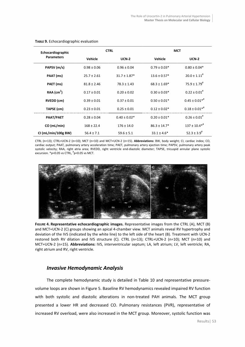
The Role of Urocortin-2 in Pulmonary Arterial Hypertension Master Thesis on Molecular and Cellular Biology
Results| 53
TABLE 9. Echocardiographic evaluation
Echocardiographic Parameters
CTRL MCT
Vehicle UCN-2 Vehicle UCN-2
PAPSV (m/s) 0.98 ± 0.06 0.96 ± 0.04 0.79 ± 0.03* 0.80 ± 0.04*
PAAT (ms) 25.7 ± 2.61 31.7 ± 1.87* 13.6 ± 0.57* 20.0 ± 1.11#
PAET (ms) 81.8 ± 2.46 78.3 ± 1.43 68.3 ± 1.69* 75.9 ± 1.79#
RAA (cm2) 0.17 ± 0.01 0.20 ± 0.02 0.30 ± 0.03* 0.22 ± 0.01
#
RVEDD (cm) 0.39 ± 0.01 0.37 ± 0.01 0.50 ± 0.01* 0.45 ± 0.01*#
TAPSE (cm) 0.23 ± 0.01 0.25 ± 0.01 0.12 ± 0.02* 0.18 ± 0.01*#
PAAT/PAET 0.28 ± 0.04 0.40 ± 0.02* 0.20 ± 0.01* 0.26 ± 0.01#
CO (mL/min) 168 ± 22.4 176 ± 14.0 86.3 ± 14.7* 137 ± 10.4*#
CI (mL/min/100g BW) 56.4 ± 7.1 59.6 ± 5.1 33.1 ± 4.6* 52.3 ± 3.9#
CTRL (n=13); CTRL+UCN-2 (n=10); MCT (n=10) and MCT+UCN-2 (n=15). Abbreviations: BW, body weight; CI, cardiac index; CO, cardiac output; PAAT, pulmonary artery acceleration time; PAET, pulmonary artery ejection time; PAPSV, pulmonary artery peak systolic velocity; RAA, right atria area; RVEDD, right ventricle end-diastolic diameter; TAPSE, tricuspid annular plane systolic excursion. *p<0.05 vs CTRL, #p<0.05 vs MCT.
FIGURE 4. Representative echocardiographic images. Representative images from the CTRL (A), MCT (B) and MCT+UCN-2 (C) groups showing an apical 4-chamber view. MCT animals reveal RV hypertrophy and deviation of the IVS (indicated by the white line) to the left side of the heart (B). Treatment with UCN-2 restored both RV dilation and IVS structure (C). CTRL (n=13); CTRL+UCN-2 (n=10); MCT (n=10) and MCT+UCN-2 (n=15). Abbreviations: IVS, interventricular septum; LA, left atrium; LV, left ventricle; RA, right atrium and RV, right ventricle.
Invasive Hemodynamic Analysis
The complete hemodynamic study is detailed in Table 10 and representative pressure-
volume loops are shown in Figure 5. Baseline RV hemodynamics revealed impaired RV function
with both systolic and diastolic alterations in non-treated PAH animals. The MCT group
presented a lower HR and decreased CO. Pulmonary resistances (PVR), representative of
increased RV overload, were also increased in the MCT group. Moreover, systolic function was
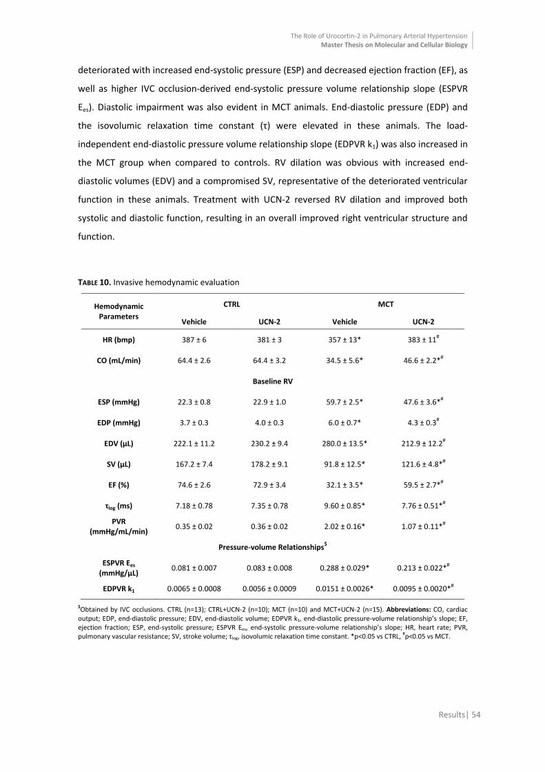
The Role of Urocortin-2 in Pulmonary Arterial Hypertension Master Thesis on Molecular and Cellular Biology
Results| 54
deteriorated with increased end-systolic pressure (ESP) and decreased ejection fraction (EF), as
well as higher IVC occlusion-derived end-systolic pressure volume relationship slope (ESPVR
Ees). Diastolic impairment was also evident in MCT animals. End-diastolic pressure (EDP) and
the isovolumic relaxation time constant (τ) were elevated in these animals. The load-
independent end-diastolic pressure volume relationship slope (EDPVR k1) was also increased in
the MCT group when compared to controls. RV dilation was obvious with increased end-
diastolic volumes (EDV) and a compromised SV, representative of the deteriorated ventricular
function in these animals. Treatment with UCN-2 reversed RV dilation and improved both
systolic and diastolic function, resulting in an overall improved right ventricular structure and
function.
TABLE 10. Invasive hemodynamic evaluation
Hemodynamic Parameters
CTRL MCT
Vehicle UCN-2 Vehicle UCN-2
HR (bmp) 387 ± 6 381 ± 3 357 ± 13* 383 ± 11#
CO (mL/min) 64.4 ± 2.6 64.4 ± 3.2 34.5 ± 5.6* 46.6 ± 2.2*#
Baseline RV
ESP (mmHg) 22.3 ± 0.8 22.9 ± 1.0 59.7 ± 2.5* 47.6 ± 3.6*#
EDP (mmHg) 3.7 ± 0.3 4.0 ± 0.3 6.0 ± 0.7* 4.3 ± 0.3#
EDV (μL) 222.1 ± 11.2 230.2 ± 9.4 280.0 ± 13.5* 212.9 ± 12.2#
SV (μL) 167.2 ± 7.4 178.2 ± 9.1 91.8 ± 12.5* 121.6 ± 4.8*#
EF (%) 74.6 ± 2.6 72.9 ± 3.4 32.1 ± 3.5* 59.5 ± 2.7*#
τlog (ms) 7.18 ± 0.78 7.35 ± 0.78 9.60 ± 0.85* 7.76 ± 0.51*#
PVR (mmHg/mL/min)
0.35 ± 0.02 0.36 ± 0.02 2.02 ± 0.16* 1.07 ± 0.11*#
Pressure-volume Relationships$
ESPVR Ees (mmHg/μL)
0.081 ± 0.007 0.083 ± 0.008 0.288 ± 0.029* 0.213 ± 0.022*#
EDPVR k1 0.0065 ± 0.0008 0.0056 ± 0.0009 0.0151 ± 0.0026* 0.0095 ± 0.0020*#
$Obtained by IVC occlusions. CTRL (n=13); CTRL+UCN-2 (n=10); MCT (n=10) and MCT+UCN-2 (n=15). Abbreviations: CO, cardiac output; EDP, end-diastolic pressure; EDV, end-diastolic volume; EDPVR k1, end-diastolic pressure-volume relationship’s slope; EF, ejection fraction; ESP, end-systolic pressure; ESPVR Ees, end-systolic pressure-volume relationship’s slope; HR, heart rate; PVR, pulmonary vascular resistance; SV, stroke volume; τlog, isovolumic relaxation time constant. *p<0.05 vs CTRL, #p<0.05 vs MCT.

The Role of Urocortin-2 in Pulmonary Arterial Hypertension Master Thesis on Molecular and Cellular Biology
Results| 55
0 1 0 0 2 0 0 3 0 0 4 0 0
0
2 0
4 0
6 0
8 0
V o lu m e ( L )
Pre
ss
ure
(m
mH
g)
C T R L
C T R L + U C N -2
M C T
M C T + U C N -2
FIGURE 5. Representative pressure-volume loops. Representative pressure-volume loops of the different study groups, obtained by transient inferior vena cava (IVC) occlusions. MCT untreated animals revealed higher EDP and EDV while treatment with UCN-2 was able to attenuate both.
Morphometric and Histological Analysis
Animals from the MCT group presented RV hypertrophy as shown by the increase in RV
weight (RVW)/LV + Septum weight (LV+SW) and RVW/tibia length (TL) ratios. Lung weight (LW)
was also increased and gastrocnemius weight (GcW) was decreased (decreased GcW/TL ratio)
in the animals from this group. Liver weight (LiW) showed no difference between groups. UCN-
2 treated animals presented decreased RV hypertrophy and no changes in both lung and liver
weight compared with MCT animals (Table 11). RV hypertrophy in the MCT group, was further
confirmed by histological measurement of cardiomyocyte cross-sectional area (CSA) (MCT: 366
± 25 μm2) when compared to CTRL (255 ± 26 μm2). UCN-2 treatment was capable to reverse
MCT-induced hypertrophy (MCT+UCN-2: 287 ± 26 μm2) (Figure 6).
TABLE 11. Morphometrical analysis
Morphometric Ratios CTRL MCT
Vehicle UCN-2 Vehicle UCN-2
HW/BW (g/kg) 2.48 ± 0.06 2.50 ± 0.04 3.53 ± 0.15* 3.03 ± 0.10*#
RVW//LV+SW (g/g) 0.26 ± 0.01 0.26 ± 0.01 0.48 ± 0.02* 0.41 ± 0.02*#
RVW/TL (g/cm) 0.04 ± 0.00 0.04 ± 0.00 0.08 ± 0.00* 0.06 ± 0.00*#
LW/TL (g/cm) 0.42 ± 0.02 0.41 ± 0.01 0.72 ± 0.04* 0.69 ± 0.02*
LiW/TL (g/cm) 2.97 ± 0.09 2.91 ± 0.10 2.90 ± 0.19 2.69 ± 0.07
GcW/TL (g/cm) 0.53 ± 0.02 0.52 ± 0.02 0.46 ± 0.01* 0.45 ± 0.02*
Abbreviations: BW, body weight; GcW, gastrocnemius weight; HW, heart weight; LiW, liver weight; LV+SW, left ventricle + septum weight; LW, lung weight; RVW, right ventricle weight; TL, tibia length. *p<0.05 vs CTRL, #p<0.05 vs MCT. CTRL (n=13); CTRL+UCN-2 (n=10); MCT (n=10) and MCT+UCN-2 (n=15).
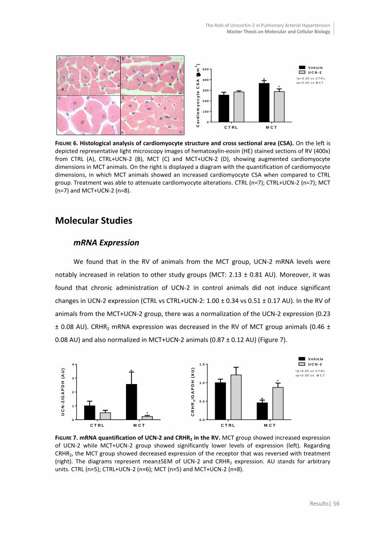
The Role of Urocortin-2 in Pulmonary Arterial Hypertension Master Thesis on Molecular and Cellular Biology
Results| 56
C T R L M C T
0
1 0 0
2 0 0
3 0 0
4 0 0
5 0 0
Ca
rd
iom
yo
cy
te C
SA
(
m2)
V e h ic le
U C N -2
*
* p < 0 .0 5 v s C T R L
# p < 0 .0 5 v s M C T*
#
FIGURE 6. Histological analysis of cardiomyocyte structure and cross sectional area (CSA). On the left is depicted representative light microscopy images of hematoxylin-eosin (HE) stained sections of RV (400x) from CTRL (A), CTRL+UCN-2 (B), MCT (C) and MCT+UCN-2 (D), showing augmented cardiomyocyte dimensions in MCT animals. On the right is displayed a diagram with the quantification of cardiomyocyte dimensions, in which MCT animals showed an increased cardiomyocyte CSA when compared to CTRL group. Treatment was able to attenuate cardiomyocyte alterations. CTRL (n=7); CTRL+UCN-2 (n=7); MCT (n=7) and MCT+UCN-2 (n=8).
Molecular Studies
mRNA Expression
We found that in the RV of animals from the MCT group, UCN-2 mRNA levels were
notably increased in relation to other study groups (MCT: 2.13 ± 0.81 AU). Moreover, it was
found that chronic administration of UCN-2 in control animals did not induce significant
changes in UCN-2 expression (CTRL vs CTRL+UCN-2: 1.00 ± 0.34 vs 0.51 ± 0.17 AU). In the RV of
animals from the MCT+UCN-2 group, there was a normalization of the UCN-2 expression (0.23
± 0.08 AU). CRHR2 mRNA expression was decreased in the RV of MCT group animals (0.46 ±
0.08 AU) and also normalized in MCT+UCN-2 animals (0.87 ± 0.12 AU) (Figure 7).
C T R L M C T
0
1
2
3
4
UC
N-2
/GA
PD
H (
AU
)
#
*
C T R L M C T
0 .0
0 .5
1 .0
1 .5
CR
HR
2/G
AP
DH
(A
U)
V e h ic le
U C N -2
#
*
* p < 0 .0 5 v s C T R L
# p < 0 .0 5 v s M C T
FIGURE 7. mRNA quantification of UCN-2 and CRHR2 in the RV. MCT group showed increased expression of UCN-2 while MCT+UCN-2 group showed significantly lower levels of expression (left). Regarding CRHR2, the MCT group showed decreased expression of the receptor that was reversed with treatment (right). The diagrams represent mean±SEM of UCN-2 and CRHR2 expression. AU stands for arbitrary units. CTRL (n=5); CTRL+UCN-2 (n=6); MCT (n=5) and MCT+UCN-2 (n=8).

The Role of Urocortin-2 in Pulmonary Arterial Hypertension Master Thesis on Molecular and Cellular Biology
Results| 57
Additionally, we determined the gene expression levels of ET-1 and BNP, markers of
ventricular hypertrophy160 and cardiac overload161. In the RV of MCT animals we observed an
increase in the mRNA expression of ET-1 (MCT vs CTRL: 3.39 ± 0.37 vs 1.00 ± 0.18 AU) and BNP
(15.29 ± 2.50 vs 1.00 ± 0.10 AU). Hypoxia-inducible factor 1 alpha (HIF-1α) regulates the
transcription of genes involved in adaptive responses to hypoxia162 and its expression was also
elevated in the MCT group (1.61 ± 0.30 vs 1.00 ± 0.16 AU). ET-1 and HIF-1α expressions were
reversed with UCN-2 treatment (MCT+UCN-2: 1.84 ± 0.57 and 1.01 ± 0.15 AU, respectively)
while BNP expression was attenuated by 7-fold (6.92 ± 2.14 AU) as shown in Figure 8.
C T R L M C T
0
1
2
3
4
ET
-1/G
AP
DH
(A
U)
*
#
C T R L M C T
0
5
1 0
1 5
2 0
BN
P/G
AP
DH
(A
U) *
# *
C T R L M C T
0 .0
0 .5
1 .0
1 .5
2 .0
2 .5
HIF
-1
/GA
PD
H (
AU
)
V e h ic le
U C N -2
#
* * p < 0 .0 5 v s C T R L
# p < 0 .0 5 v s M C T
FIGURE 8. mRNA quantification of ET-1, BNP and HIF-1α in the RV. MCT group showed increased expression of ET-1, BNP and HIF-1α. UCN-2 chronic treatment attenuated BNP and reversed ET-1 and HIF-1α expressions. The diagrams represent mean±SEM of ET-1, BNP and HIF-1α expressions. AU stands for arbitrary units. CTRL (n=5); CTRL+UCN-2 (n=6); MCT (n=5) and MCT+UCN-2 (n=8).
UCN-2, as described above, has been associated with signaling pathways of cell survival.
In this context we analyzed caspase-3 and caspase-8. In apoptosis-associated genes, both
caspase-3 and -8 were altered with increased levels in MCT animals (MCT vs CTRL: caspase-3,
3.90 ± 0.59 vs 1.00 ± 0.13 AU and caspase-8, 2.81 ± 0.35 vs 1.00 ± 0.16 AU). Attenuation of
caspase-3 (MCT+UCN-2: 2.02 ± 0.38 AU) and normalization of caspase-8 expression (1.34 ±
0.21 AU) was seen with UCN-2 treatment (Figure 9).

The Role of Urocortin-2 in Pulmonary Arterial Hypertension Master Thesis on Molecular and Cellular Biology
Results| 58
C T R L M C T
0
1
2
3
4
5
Ca
sp
as
e-3
/GA
PD
H (
AU
)*
# *
C T R L M C T
0
1
2
3
4
Ca
sp
as
e-8
/GA
PD
H (
AU
) V e h ic le
U C N -2
*
#
* p < 0 .0 5 v s C T R L
# p < 0 .0 5 v s M C T
FIGURE 9. mRNA quantification of caspase-3 and caspase-8 in the RV. Both caspases were decreased in MCT group, while UCN-2 treatment was able to reverse caspase-8 and attenuate caspase-3 expression. The diagrams represent mean±SEM of caspase-3 and -8 expressions. AU stands for arbitrary units. CTRL (n=5); CTRL+UCN-2 (n=6); MCT (n=5) and MCT+UCN-2 (n=8).
Protein Expression
CRHR2, a membrane-bound protein that belongs to the family of GPCRs, is the
endogenous receptor with greater affinity for UCN-2 binding107. The expression of this receptor
was particularly high in both MCT groups (MCT: 3.07 ± 0.67 and MCT+UCN-2: 3.34 ± 0.83 AU)
when compared to CTRL group (1.00 ± 0.14 AU) (Figure 10).
C T R L M C T
0
1
2
3
4
5
CR
HR
2/G
AP
DH
(A
U)
V e h ic le
U C N -2
** p < 0 .0 5 v s C T R L
# p < 0 .0 5 v s M C T
*
FIGURE 10. Level of CRHR2 expression in the RV. CRHR2 expression was higher in MCT groups. The presence of CRHR2 was detected by immunoblotting utilizing total protein-specific primary antibodies. The diagrams represent mean±SEM of CRHR2 bands. The CRHR2 bands were normalized to GAPDH bands. Below the diagrams is depicted the representative western blot bands. AU stands for arbitrary units. CTRL (n=9); CTRL+UCN-2 (n=9); MCT (n=7) and MCT+UCN-2 (n=10).
ERK1/2 or p44/42 and p38 belong to the MAPK family and are activated in response to a
wide range of extracellular stimuli including mitogens, growth factors and cytokines163. In MCT
animals we observed a decrease in protein phosphorylation/activation of both ERKs and p38
kinases comparatively to controls (MCT vs CTRL: ERKs, 0.53 ± 0.10 vs 1.00 ± 0.11 AU and p38,
0.52 ± 0.04 vs 1.00 ± 0.14 AU). These levels were reversed with UCN-2 treatment (MCT+UCN-2:
ERKs, 0.92 ± 0.11 AU and p38, 1.14 ± 0.17) (Figure 11).

The Role of Urocortin-2 in Pulmonary Arterial Hypertension Master Thesis on Molecular and Cellular Biology
Results| 59
C T R L M C T
0 .0
0 .5
1 .0
1 .5
p-E
RK
s/E
RK
s (
AU
)
*
#
C T R L M C T
0 .0
0 .5
1 .0
1 .5
p-p
38
/p3
8 (
AU
)
V e h ic le
U C N -2
*
#
* p < 0 .0 5 v s C T R L
# p < 0 .0 5 v s M C T
FIGURE 11. Activation level of ERKs and p38 in the RV. Both ERKs and p38 (left to right) activation was decreased in MCT animals and UCN-2 treatment was able to reverse their activation to control values. Phosphorylation was determined by immunoblotting utilizing total protein- and phosphorylation-specific primary antibodies. The diagrams represent mean±SEM of ERKs and p38 bands. Below is depicted the respective representative western blot bands. Phospho-ERKs and –p38 are denoted as p-ERKs and p-p38. AU stands for arbitrary units. CTRL (n=9); CTRL+UCN-2 (n=9); MCT (n=7) and MCT+UCN-2 (n=10).
Akt is a kinase known by its central regulatory role in several cellular processes such as
cell survival and proliferation, glucose metabolism and cardiovascular homeostasis164. Akt
activation did not show any significant differences between groups. UCN-2 treatment did not
induce any difference in the phosphorylation level of Akt (Figure 12).
C T R L M C T
0 .0
0 .5
1 .0
1 .5
2 .0
p-A
kt/
Ak
t (A
U)
V e h ic le
U C N -2
FIGURE 12. Activation level of Akt in the RV. Akt activation (on the left) did not show any difference between groups. Phosphorylation was determined by immunoblotting utilizing total protein- and phosphorylation-specific primary antibodies. The diagrams represent mean±SEM of Akt bands. Below is depicted the respective representative western blot bands. Phospho-Akt is denoted as p-Akt. AU stands for arbitrary units. CTRL (n=9); CTRL+UCN-2 (n=9); MCT (n=7) and MCT+UCN-2 (n=10).
Signal transducer and activator of transcription 3 (STAT3) is a transcription factor and an
important signaling molecule for several cytokines and growth factor receptors, known for
possessing an oncogenic potential and anti-apoptotic activities165. The MCT group showed a
decreased STAT3 phosphorylation when compared with the CTRL group (MCT vs CTRL: 0.61 ±

The Role of Urocortin-2 in Pulmonary Arterial Hypertension Master Thesis on Molecular and Cellular Biology
Results| 60
0.12 vs 1.00 ± 0.10 AU). Treatment with UCN-2 resulted in restored STAT3 activation
(MCT+UCN-2: 1.01 ± 0.09 AU) as we can see in Figure 13.
C T R L M C T
0 .0
0 .5
1 .0
1 .5
p-S
TA
T3/S
TA
T3 (
AU
)
V e h ic le
U C N -2
*
* p < 0 .0 5 v s C T R L
# p < 0 .0 5 v s M C T#
FIGURE 13. Activation level of STAT3 in the RV. STAT3 activation was decreased in MCT animals and UCN-2 treatment was able to reverse its reduction. Phosphorylation was determined by immunoblotting utilizing total protein- and phosphorylation-specific primary antibodies. The diagrams represent mean±SEM of STAT3 bands. Below the diagrams is depicted the representative western blot bands. Phospho-STAT3 is denoted as p-STAT3 and AU stands for arbitrary units. CTRL (n=9); CTRL+UCN-2 (n=9); MCT (n=7) and MCT+UCN-2 (n=10).

DDIISSCCUUSSSSIIOONN

The Role of Urocortin-2 in Pulmonary Arterial Hypertension Master Thesis on Molecular and Cellular Biology
Discussion| 62
UCN-2 treatment improves survival and RV function in MCT-induced PAH
This study demonstrated that the chronic treatment with UCN-2 in an experimental
model of PAH and secondary RV failure, attenuates structural remodeling and enhances RV
systolic and diastolic function.
Several studies166-168 have demonstrated the progression of MCT-induced PAH using
echo-measured indexes of RV function and structure, proving the importance of non-invasive
methods in the evaluation of PAH severity and RV function, since it allows a comparable
approach to that routinely used in patients62.
In MCT-induced PAH we observed a decrease in PAAT and PAET, which significantly
altered the PAAT/PAET ratio, and in systolic flow which is compromised in PAH, is associated
with pulmonary systolic pressures169 and correlates with decreased survival in PAH patients62.
Pulmonary flow velocity, acceleration and ejection time were also restored with UCN-2
pharmacological intervention, revealing an improved pulmonary arterial function and
decreased RV afterload.
Accordingly to previous studies167, 168 we observed a significant RV and right atria
chamber dilation, shown by the echo-measured RV and RAA. At this time point (4th week after
MCT injection) and with a progressive increase in PVR, the RV cannot sustain the adaptive
hypertrophy and eventually dilates51. This was shown by Hardziyenka et al. that measured
RVEDD echocardiographycally and showed that it seems to start to increase between 20-25
days post MCT administration. Furthermore, in response to progressive pressure overload
there is a shift of the IVS to the left side chamber170, which is in agreement with our
observations (Figure 4B). Moreover, echo-measured TAPSE, an indicator of RV contractile
function170, was also decreased in MCT animals. UCN-2 therapeutic intervention attenuated
the adaptive hypertrophy and improved RV function and structure, as well as correcting IVS
deviation. CO and CI, indexes of overall cardiac function, were also altered in MCT animals and
were normalized with UCN-2 treatment.
Although no studies have invasively evaluated UCN-2’s chronic treatment effect on RV
function in PAH, enough studies have been performed on the LV that allows us to explain our
results. Those studies showed improved cardiac performance in models of HF subjected to
UCN-2 intervention. Bale et al. studied the acute effects of UCN-2 in a murine model of HF and
although LVEDP was not different between wild-type and CRHR2-deficient mice, they noticed
an increase in HR, EF and CO, as well as a decrease in LVEDV and ESV129. In anaesthetized pigs,

The Role of Urocortin-2 in Pulmonary Arterial Hypertension Master Thesis on Molecular and Cellular Biology
Discussion| 63
intracoronary infusion of UCN-2 caused an increase in coronary blood flow and CO,
accompanied by a decrease in coronary vascular resistances89.
Recently, both humans with mild HF149 and acute decompensated HF91 were subjected
to UCN-2 administration. In the latter, UCN-2 treatment was able to decrease systolic blood
pressure and increase CO, associated with a strong reduction in total peripheral resistance.
Administration of UCN-2 in humans with mild HF increased CO and LVEF and decreased mean
arterial pressure, systemic vascular resistance and cardiac work, in a dose-dependent manner.
In both studies UCN-2 was able to induce vasodilation and improve myocardial function.
Similarly, and despite all the structural differences between the LV and the RV, our
functional RV study match what has been previously described for the LV. MCT-injected
animals revealed a severe cardiac dysfunction secondary to PAH and presented RV
hypercontractility, as shown by highly increased systolic pressures, as well by changes in other
parameters of contractile function, namely IVC occlusion-derived ESPV relationship’s slope.
Diastolic dysfunction was also present in the RV of MCT animals, shown by increased stiffness
(rise in RVEDP) and RV dilation (increased RVEDD). These hemodynamic alterations correspond
to decompensated myocardial hypertrophy of the RV in response to pressure overload
resulting from increased pulmonary vascular resistance, as previously described 171.
Treating MCT animals with UCN-2 resulted in the improvement of RV function, given
that both systolic and diastolic dysfunction was enhanced and the ventricle showed improved
contractile and relaxation functions. Cardiac catheterization allowed a more detailed analysis
of ventricular function and corroborated echocardiographic data, nevertheless some caution is
needed when interpreting the results and extrapolating previous knowledge from the LV to
explain RV pathophysiology172.
MCT-induced RV morphohistological changes are attenuated with UCN-2 treatment
UCN-2 treatment after PAH-induction resulted in accentuated decrease of cardiac
hypertrophy, especially RV hypertrophy (Table 11). HW was increased in PAH animals, as
previously mentioned, and this was analyzed using the HW/BW ratio. Nevertheless, in PAH, the
RV is the primary affected ventricle and therefore should be the focus of the analysis, since
due to the augmented PVR is under pressure overload. And in fact, the RVW/BW ratio was also
increased in PAH animals, which revealed the presence of RV hypertrophy.

The Role of Urocortin-2 in Pulmonary Arterial Hypertension Master Thesis on Molecular and Cellular Biology
Discussion| 64
Despite being in agreement with all of our data regarding hypertrophy and hypertrophy-
associated parameters measured, the model we’re using is associated with cachexia158 and
because of that, we alternatively measured the RVW/TL ratio which is accepted as a reliable
parameter for hypertrophy measurement159. The RVW/LV+SW ratio was consistent with the
previously mentioned measurements. Lung edema and congestion is a consequence of PAH173
and using the LW/TL ratio we observed an increase ratio in the MCT group, suggestive of PAH-
associated lung fluid retention.
At the cellular level, our model of PAH developed cardiomyocyte hypertrophy, as
measured by cardiomyocyte CSA. These effects were however reverted by UCN-2
administration. The reversal of hypertrophy in response to treatment underlines the functional
recovery observed with non-invasive and invasive hemodynamic measurements. The RV of
treated animals is under attenuated pressure overload and therefore its adaptive hypertrophic
response is decreased. Less muscle mass, less developed force and consequently, attenuated
developed pressures and hypercontractile state.
Expression of the UCN-2/CRHR2 in PAH and alterations with UCN-2 treatment
Given that the used animal model of PAH is characterized by several structural and
functional cardiac abnormalities over time, expressed by compensated RV hypertrophy at 2-3
weeks and RH failure after 3-5 weeks170, 174 it seems relevant to search, between experimental
groups, for possible differences in the expression of UCN-2 and its receptor, as well as the
expression of genes encoding regulatory proteins relevant for ventricular contractile function.
Moreover, these studies allowed us a better characterization of the variations occurring in the
UCN-2/CRHR2 system and downstream signaling in the context of PAH and progression to HF,
which can provide us an explanation for the alterations detected in the functional studies.
UCN-2 and its receptor, CRHR2, have been implicated in several cardiovascular
diseases91, 141, 149, 175. In spontaneously hypertensive rats, Nishikimi et al. described a higher
UCN mRNA expression in hypertrophied LV than in normal LV, whereas CRHR2 expression was
depressed103. Moreover, Emeto et al. demonstrated augmented expression of UCN-2 in
biopsies from abdominal aortic aneurysm (AAA) and despite not being cardiac tissue, the same
study also showed increased levels of UCN-2 in plasma samples of AAA patients, which are
associated with this cardiovascular disorder141. Additionally, other group analyzed and
compared the prognostic efficacy of both plasma UCN and NT-proBNP levels in acute
myocardial infarction and correlated the high plasmatic UCN levels with increased mortality176.

The Role of Urocortin-2 in Pulmonary Arterial Hypertension Master Thesis on Molecular and Cellular Biology
Discussion| 65
In our study we also observed increased levels of UCN-2 mRNA expression and a sharp
fall in the mRNA expression of the CRHR2 which complies with the studies cited above. In
addition, UCN-2 levels also seem to correlate with increased morbidity and mortality176,
despite its beneficial effects. In our study, the decreased levels of CRHR2 expression may be
related with the increased UCN-2 expression in the RV, wherein by a mechanism of negative
feedback, UCN-2 regulates CRHR2 mRNA expression. UCN-2 treatment was able to reduce
UCN-2 endogenous expression and augment CRHR2 expression, normalizing both expressions
to control levels.
Taken together, these results suggest that increased UCN-2 in the RV from MCT animals
may be associated with an adaptive process of the cardiac muscle. In consequence, one can
predict that UCN-2 may play an important role in the pathophysiology of PAH and progression
to HF.
Increased expression of cardiac overload, hypertrophy and hypoxia markers in PAH was
attenuated with UCN-2 treatment
Neurohumoral activation is associated with RV dysfunction in PAH177 and growing
evidences show that UCN-2 modulates the gene expression and/or the activity of several
neurohumoral agents, such as ET-1 and BNP, involved in PAH and progression to HF90, 143.
ET-1 plasmatic levels are elevated in PAH and are associated with disease severity27. In
addition, its clearance from pulmonary arteries is reduced, promoting further
vasoconstriction179. ET-1 levels are associated with the development of RV hypertrophy in
MCT-induced PAH180 and its RV expression is increased in this experimental model181.
Plasmatic BNP levels are associated with PH182 and increased in MCT-injected rats181. The
production of BNP is stimulated in response to pressure-overload183 and is associated with the
extent of RV dysfunction184. Moreover, NT-proBNP along with UCN-2 plasmatic levels holds an
enhanced prognostic value, since their levels correlate with increased mortality, as previously
cited176.
In our study, both ET-1 and BNP mRNA expressions were augmented in PAH animals up
to 3-fold and 15-fold, respectively. While UCN-2 treatment was able to attenuate ET-1 to 2-fold
and BNP to 7-fold, in accordance with previous studies143, 178.
Hypoxia-inducible factor 1 alpha (HIF-1α) is a heterodimeric subunit of the transcription
factor HIF-1, which regulates the transcription of genes involved in adaptive responses to
The Role of Urocortin-2 in Pulmonary Arterial Hypertension Master Thesis on Molecular and Cellular Biology
Discussion| 66
hypoxia162. Chu et al. showed that HIF-1α overexpression promotes the development of
cardiac hypertrophy via up-regulation of Ca+2-calcineurin under mild hypoxia conditions185.
However, increased [Ca2+]i is not only associated with hypertrophy but also with apoptotic
processes under severe hypoxia situation, given that some reports showed that HIF-1α
overexpression promotes apoptosis of cardiac myocytes in the presence of severe hypoxia,
especially when other cellular energy substrates are lacking186. In our study, RV HIF-1α
expression was also increased in MCT animals and UCN-2 treatment was able to reverse HIF-
1α expression to control levels. The identification of a binding region for HIF-1α in human UCN-
2 gene that regulates transcription in response to hypoxia, explains why both levels of UCN-2
and HIF-1α correlate. Buhler et al. proposed that the induction of hUCN-2 expression by low
oxygen levels may confer to cardiac myocytes an advantage in ischemic conditions187.
UCN-2 treatment reverses changes in markers of apoptosis in PAH
Apoptosis is one of the major mechanisms causing cardiomyocyte loss in failing hearts of
both human patients188 and animal models189. In turn, UCN-2 appears to have cardioprotective
effects by preventing cardiomyocyte death175, 190, 191.
Upon cleavage, caspase-3 is activated in the myocardium of end-stage HF patients192
being also involved in the development of pressure overload-induced LV dysfunction, as well
as hyperglycemia-induced myocardial apoptosis193. Studies that used the MCT-induced PAH
model showed increased caspase-3 activation in those animals194, 195 and in myocardial
infarction, the overexpression of this enzyme induced myofibrillar ultrastructural damage and
when subjected to myocardial ischemia-reperfusion (I/R) injury, caspase-3 transgenic mice
showed increased infarct size and a pronounced susceptibility to die196.
Caspase-8, a cysteine protease, has the ability to activate downstream caspases,
including caspase-3, and therefore it is critically involved in the regulation of cellular apoptosis,
as well as necrotic cell death197. In human cardiomyocytes, iron-induced apoptosis was linked
to increased levels of caspase-8198. In a mouse model of HF, the inhibition of Fas-associated
death domain-containing protein (FADD) had a protective role in myocardial I/R injury and was
correlated with increased caspase-8 activity and downstream caspase-3 activity197.
In our study, the high detected mRNA levels of both caspase-3 and caspase-8 in the RV
of the MCT rats were reverted by UCN-2 treatment which is consistent with other studies134, 199
and confirms the hypothesis that UCN-2/CRHR2 system has anti-apoptotic proprieties.

The Role of Urocortin-2 in Pulmonary Arterial Hypertension Master Thesis on Molecular and Cellular Biology
Discussion| 67
UCN-2 treatment normalized the alterations in some elements of the UCN-2/CRHR2 signaling
pathway seen in MCT animals
In rats, the administration of MCT usually leads to cardiac hypertrophy that develops
due to increasing oxidants, hemodynamic load and by inducing hypoxia. The mechanisms that
lead to cardiac hypertrophy and progression to HF involve the activation G-proteins, calcium
signaling, PI3K, PKCs and MAPKs200. In this study one of our goals was to find which signaling
pathways are deregulated in MCT-induced PAH animals and which are involved in UCN-2
beneficial effects.
ERK1/2 or p44/42 and p38 belong to the MAPK family and are activated in response to a
wide range of extracellular stimuli including mitogens, growth factors and cytokines. When
activated, they have the ability to regulate other kinases and several transcription factors163.
Defects downstream of BMP signaling pathway, involving p38 and ERKs, have been linked to
PAH201 and increased activation of ERK1/2 and p38 was found in lung tissue of MCT-induced
PH model, while a suppression of this activation was observed with treatment202.
On the other hand and for the first time, Kehat and Molkentin proposed that eccentric
hypertrophy is induced by de-activation of the ERK pathway203 and Bartelds et al. confirmed it
by observing a decrease in phosphorylated ERK1/2 in mice subjected to volume and pressure
overload204. Furthermore, in a model of transverse aortic constriction (TAC) that resulted in
late eccentric hypertrophy with eventual development of LV dysfunction, implicated the
importance of ERK1/2 downregulation and enhanced cardiomyocyte apoptosis in the
transition from compensated hypertrophy to HF205.
In relation to p38, Pérez López et al. showed that the disruption of the AKAP/p38
signaling complex inhibits compensatory hypertrophy in response to aortic banding-induced
pressure overload and promotes early cardiac dysfunction associated with increased
myocardial apoptosis and ventricular dilation206.
In consequence, our decreased phosphorylation of both kinases in the RV of MCT
animal’s is in accordance with previous studies, once at the time of sample collection, the MCT
animals already showed an advanced stage of HF with evident RV dilation. In addition, the
reversal of both kinases to its control levels comply with prior studies that showed a boost in
the activation of both ERK 1/288 and p38 MAPK125, at this stage, upon UCN-2-binding to CRHR2,
which accordingly to Bartelds et al. could be a mechanism to prevent the pressure-loaded RV
from dilation204.
The Role of Urocortin-2 in Pulmonary Arterial Hypertension Master Thesis on Molecular and Cellular Biology
Discussion| 68
Akt or PKB is a serine/threonine kinase known by its critical and central regulatory role
in several cellular processes such as cell survival and proliferation, glucose metabolism and
cardiovascular homeostasis164. Akt is also a critical downstream element of the PI3K/Akt cell
survival pathway, whose activity can be inhibited by Akt-mediated phosphorylation164. The
PI3K/Akt pathway is also known to be activated by UCN-2116 and it’s particularly important in
cardiac and skeletal muscle since it helps regulate PLB phosphorylation, along with PKA,
controlling the inhibition of SERCA118.
Although Akt was not significantly affected by any of the administered drugs (MCT and
UCN-2) in resemblance with other studies202, 207.
STAT3 is a transcription factor and an important signaling molecule for several cytokines
and growth factor receptors, known for possessing an oncogenic potential and anti-apoptotic
activities165. In myeloma tumor cells, the blocking of Jak-STAT pathway induces apoptosis,
demonstrating that STAT3 signaling is essential for the survival of this type of cells208.
Akin to cancer, PAH is characterized by an imbalance between a proliferative and an
apoptotic phenotype16. In PASMC proliferation is enhanced due to the activation of pro-
survival transcription factors, such as STAT3209. And in fact, Courboulin et al. demonstrated that
STAT3 phosphorylation was increased in PAH-PASMCs and that treatment with a STAT3 blocker
reversed this state.
However, in the cardiac tissue the opposite happens. Because the RV is not able to
sustain long-term pressure overload, the cardiac contractile force decreases due to apoptosis
and changes in cardiomyocytes, and eventually the RV dilates51. Following this notion, Chen-
Scarabelli et al. verified that cardiac cells incubated with UCN showed increased STAT3
phosphorylation at Tyr705. Moreover, overexpression of STAT3 in cardiac myocytes pretreated
with UCN presented a reduced magnitude of cell death, when compared with UCN treatment
alone. While transfection of cells with a mutant STAT3 (loss of function) enhanced the extent of
cell death in a dose-dependent manner210.
Hereupon, the increased STAT3 phosphorylation in the RV of the MCT treated animal’s
match what has been previously described for the cardiomyocytes, demonstrating the
cardioprotective effect and anti-apoptotic proprieties of UCN-2.
The increased expression of CRHR2 in the RV of MCT animals, obtained by western blot
analysis, contrarily to its mRNA expression, may be related with our decreased activation of
ERK1/2, since according to Markovic et al., the endocytosis of CRHR2 appears to be regulated
by ERK1/2 direct phosphorylation of β-arrestin1 in an auto-regulatory mechanism that

The Role of Urocortin-2 in Pulmonary Arterial Hypertension Master Thesis on Molecular and Cellular Biology
Discussion| 69
influences the rate and extent of β-arrestin1 recruitment to the plasma membrane and
interaction with CRHR2112. Thus, the elevated presence of CRHR2 in the RV from MCT animals
could be associated with the deregulation seen in the pathway that leads to receptor
internalization and degradation.

CCOONNCCLLUUSSIIOONNSS AANNDD FFUUTTUURREE PPEERRSSPPEECCTTIIVVEESS

The Role of Urocortin-2 in Pulmonary Arterial Hypertension Master Thesis on Molecular and Cellular Biology
Conclusions and Future Perspectives| 71
In short, we show for the first time, in an experimental model of MCT-induced PAH, that
UCN-2 treatment is able to restore PAH-induced severe abnormalities in cardiac function and
structure, while attenuating or reversing the expression of markers of cardiac overload,
hypertrophy, hypoxia and apoptosis. We also demonstrated that the UCN-2 beneficial effects
are associated and involve the modulation of several signaling pathways downstream CRHR2
activation.
PAH is a chronic syndrome that is frequently diagnosed in a later stage, where HF is
already settled. Therefore our approach adds the benefit of a more practical and clinical
relevant therapy, since we begin the treatment after PAH was established.
The UCN-2 and its receptor have been studied in the cardiovascular system, but
research so far has failed to determine its effects on both normal and diseased RV physiology.
With this study, we demonstrated the effects of an increase in UCN-2 levels through
exogenous administration of the peptide, which has shown that specific activation of CRHR2
mediates several compensatory mechanisms that ameliorate the dysfunction and structural
abnormalities associated with PAH and RVF.
However, it is also important to take into account that the animal model used in the
present study does not mimic entirety human PH or PAH, nevertheless it has several
pathophysiological alterations in common that allow us to enthusiastically point UCN-2 as a
possible novel therapeutic approach for this incurable and with an uncertain prognosis
disease.
In fact, we hope to have opened the road for further basic and translational research
that could possibly lead to the establishment of UCN-2 as a novel and safe therapeutic agent
for treating patients with PAH and RVF.
In spite of these results, further research is needed in order to determine specifically
where UCN-2 plays its major role. The ventricular function is altered, but since in this model RV
dysfunction and HF is secondary to PAH, we cannot ascertain whether RV function and
structure improvement is secondary to pulmonary vascular functional enhancement and
therefore decreased vascular resistances, or if UCN-2 has a direct effect on the ventricle which
by itself is capable of leading to the changes we observed. In the future, in order to
differentiate indirect from direct myocardial effects of UCN-2 treatment, we predict the use of
an animal model of RV overload.
Beyond RV function and structure, we intent do access pulmonary function and
structure as well, by in vitro (vascular ring preparations) and histological studies. Moreover,

The Role of Urocortin-2 in Pulmonary Arterial Hypertension Master Thesis on Molecular and Cellular Biology
Conclusions and Future Perspectives| 72
our molecular results here described will allow us to design and perform further studies in
order to unravel more signaling pathways underlying the beneficial effects of UCN-2/CRHR2
system activation in PAH and RV HF, in both RV and lung samples.

RREEFFEERREENNCCEESS

The Role of Urocortin-2 in Pulmonary Arterial Hypertension Master Thesis on Molecular and Cellular Biology
References| 74
1. McLaughlin VV, Archer SL, Badesch DB, Barst RJ, Farber HW, Lindner JR, Mathier MA, McGoon MD, Park MH, Rosenson RS, Rubin LJ, Tapson VF, Varga J. Accf/aha 2009 expert consensus document on pulmonary hypertension a report of the american college of cardiology foundation task force on expert consensus documents and the american heart association developed in collaboration with the american college of chest physicians; american thoracic society, inc.; and the pulmonary hypertension association. J Am Coll Cardiol. 2009;53:1573-1619
2. Hoeper MM, Bogaard HJ, Condliffe R, Frantz R, Khanna D, Kurzyna M, Langleben D, Manes A, Satoh T, Torres F, Wilkins MR, Badesch DB. Definitions and diagnosis of pulmonary hypertension. J Am Coll Cardiol. 2013;62:D42-50
3. Galie N, Hoeper MM, Humbert M, Torbicki A, Vachiery JL, Barbera JA, Beghetti M, Corris P, Gaine S, Gibbs JS, Gomez-Sanchez MA, Jondeau G, Klepetko W, Opitz C, Peacock A, Rubin L, Zellweger M, Simonneau G. Guidelines for the diagnosis and treatment of pulmonary hypertension: The task force for the diagnosis and treatment of pulmonary hypertension of the european society of cardiology (esc) and the european respiratory society (ers), endorsed by the international society of heart and lung transplantation (ishlt). Eur Heart J. 2009;30:2493-2537
4. Hatano S ST. Primary pulmonary hypertension. 1975
5. Simonneau G, Galie N, Rubin LJ, Langleben D, Seeger W, Domenighetti G, Gibbs S, Lebrec D, Speich R, Beghetti M, Rich S, Fishman A. Clinical classification of pulmonary hypertension. J Am Coll Cardiol. 2004;43:5S-12S
6. Simonneau G, Robbins IM, Beghetti M, Channick RN, Delcroix M, Denton CP, Elliott CG, Gaine SP, Gladwin MT, Jing ZC, Krowka MJ, Langleben D, Nakanishi N, Souza R. Updated clinical classification of pulmonary hypertension. J Am Coll Cardiol. 2009;54:S43-54
7. Simonneau G, Gatzoulis MA, Adatia I, Celermajer D, Denton C, Ghofrani A, Gomez Sanchez MA, Krishna Kumar R, Landzberg M, Machado RF, Olschewski H, Robbins IM, Souza R. Updated clinical classification of pulmonary hypertension. J Am Coll Cardiol. 2013;62:D34-41
8. Sztrymf B, Coulet F, Girerd B, Yaici A, Jais X, Sitbon O, Montani D, Souza R, Simonneau G, Soubrier F, Humbert M. Clinical outcomes of pulmonary arterial hypertension in carriers of bmpr2 mutation. American journal of respiratory and critical care medicine. 2008;177:1377-1383
9. Humbert M, Montani D, Evgenov OV, Simonneau G. Definition and classification of pulmonary hypertension. Handbook of experimental pharmacology. 2013;218:3-29
10. Pietra GG, Capron F, Stewart S, Leone O, Humbert M, Robbins IM, Reid LM, Tuder RM. Pathologic assessment of vasculopathies in pulmonary hypertension. J Am Coll Cardiol. 2004;43:25S-32S

The Role of Urocortin-2 in Pulmonary Arterial Hypertension Master Thesis on Molecular and Cellular Biology
References| 75
11. Montani D, Gunther S, Dorfmuller P, Perros F, Girerd B, Garcia G, Jais X, Savale L, Artaud-Macari E, Price LC, Humbert M, Simonneau G, Sitbon O. Pulmonary arterial hypertension. Orphanet journal of rare diseases. 2013;8:97
12. Cool CD, Kennedy D, Voelkel NF, Tuder RM. Pathogenesis and evolution of plexiform lesions in pulmonary hypertension associated with scleroderma and human immunodeficiency virus infection. Human pathology. 1997;28:434-442
13. Bjornsson J, Edwards WD. Primary pulmonary hypertension: A histopathologic study of 80 cases. Mayo Clinic proceedings. 1985;60:16-25
14. Dorfmuller P. Pulmonary hypertension: Pathology. Handbook of experimental pharmacology. 2013;218:59-75
15. Waxman AB, Zamanian RT. Pulmonary arterial hypertension: New insights into the optimal role of current and emerging prostacyclin therapies. The American journal of cardiology. 2013;111:1A-16A; quiz 17A-19A
16. Humbert M, Morrell NW, Archer SL, Stenmark KR, MacLean MR, Lang IM, Christman BW, Weir EK, Eickelberg O, Voelkel NF, Rabinovitch M. Cellular and molecular pathobiology of pulmonary arterial hypertension. J Am Coll Cardiol. 2004;43:13S-24S
17. Jeffery TK, Morrell NW. Molecular and cellular basis of pulmonary vascular remodeling in pulmonary hypertension. Progress in cardiovascular diseases. 2002;45:173-202
18. Davie NJ, Crossno JT, Jr., Frid MG, Hofmeister SE, Reeves JT, Hyde DM, Carpenter TC, Brunetti JA, McNiece IK, Stenmark KR. Hypoxia-induced pulmonary artery adventitial remodeling and neovascularization: Contribution of progenitor cells. American journal of physiology. Lung cellular and molecular physiology. 2004;286:L668-678
19. Jonigk D, Golpon H, Bockmeyer CL, Maegel L, Hoeper MM, Gottlieb J, Nickel N, Hussein K, Maus U, Lehmann U, Janciauskiene S, Welte T, Haverich A, Rische J, Kreipe H, Laenger F. Plexiform lesions in pulmonary arterial hypertension composition, architecture, and microenvironment. The American journal of pathology. 2011;179:167-179
20. Kherbeck N, Tamby MC, Bussone G, Dib H, Perros F, Humbert M, Mouthon L. The role of inflammation and autoimmunity in the pathophysiology of pulmonary arterial hypertension. Clinical reviews in allergy & immunology. 2013;44:31-38
21. Morrell NW, Adnot S, Archer SL, Dupuis J, Jones PL, MacLean MR, McMurtry IF, Stenmark KR, Thistlethwaite PA, Weissmann N, Yuan JX, Weir EK. Cellular and molecular basis of pulmonary arterial hypertension. J Am Coll Cardiol. 2009;54:S20-31
22. Herve P, Humbert M, Sitbon O, Parent F, Nunes H, Legal C, Garcia G, Simonneau G. Pathobiology of pulmonary hypertension. The role of platelets and thrombosis. Clinics in chest medicine. 2001;22:451-458

The Role of Urocortin-2 in Pulmonary Arterial Hypertension Master Thesis on Molecular and Cellular Biology
References| 76
23. Montani D, Chaumais MC, Guignabert C, Gunther S, Girerd B, Jais X, Algalarrondo V, Price LC, Savale L, Sitbon O, Simonneau G, Humbert M. Targeted therapies in pulmonary arterial hypertension. Pharmacology & therapeutics. 2014;141:172-191
24. Tuder RM, Cool CD, Geraci MW, Wang J, Abman SH, Wright L, Badesch D, Voelkel NF. Prostacyclin synthase expression is decreased in lungs from patients with severe pulmonary hypertension. American journal of respiratory and critical care medicine. 1999;159:1925-1932
25. Maron BA, Loscalzo J. Pulmonary hypertension: Pathophysiology and signaling pathways. Handbook of experimental pharmacology. 2013;218:31-58
26. Giaid A, Yanagisawa M, Langleben D, Michel RP, Levy R, Shennib H, Kimura S, Masaki T, Duguid WP, Stewart DJ. Expression of endothelin-1 in the lungs of patients with pulmonary hypertension. The New England journal of medicine. 1993;328:1732-1739
27. Rubens C, Ewert R, Halank M, Wensel R, Orzechowski HD, Schultheiss HP, Hoeffken G. Big endothelin-1 and endothelin-1 plasma levels are correlated with the severity of primary pulmonary hypertension. Chest. 2001;120:1562-1569
28. Gangopahyay A, Oran M, Bauer EM, Wertz JW, Comhair SA, Erzurum SC, Bauer PM. Bone morphogenetic protein receptor ii is a novel mediator of endothelial nitric-oxide synthase activation. The Journal of biological chemistry. 2011;286:33134-33140
29. Butcher RW, Sutherland EW. Adenosine 3',5'-phosphate in biological materials. I. Purification and properties of cyclic 3',5'-nucleotide phosphodiesterase and use of this enzyme to characterize adenosine 3',5'-phosphate in human urine. The Journal of biological chemistry. 1962;237:1244-1250
30. Francis SH, Busch JL, Corbin JD, Sibley D. Cgmp-dependent protein kinases and cgmp phosphodiesterases in nitric oxide and cgmp action. Pharmacological reviews. 2010;62:525-563
31. Nagendran J, Archer SL, Soliman D, Gurtu V, Moudgil R, Haromy A, St Aubin C, Webster L, Rebeyka IM, Ross DB, Light PE, Dyck JR, Michelakis ED. Phosphodiesterase type 5 is highly expressed in the hypertrophied human right ventricle, and acute inhibition of phosphodiesterase type 5 improves contractility. Circulation. 2007;116:238-248
32. Wharton J, Strange JW, Moller GM, Growcott EJ, Ren X, Franklyn AP, Phillips SC, Wilkins MR. Antiproliferative effects of phosphodiesterase type 5 inhibition in human pulmonary artery cells. American journal of respiratory and critical care medicine. 2005;172:105-113
33. Waxman AB. Pulmonary hypertension in heart failure with preserved ejection fraction: A target for therapy? Circulation. 2011;124:133-135
34. Suntharalingam J, Hughes RJ, Goldsmith K, Doughty N, George P, Toshner M, Sheares KK, Pepke-Zaba J. Acute haemodynamic responses to inhaled nitric

The Role of Urocortin-2 in Pulmonary Arterial Hypertension Master Thesis on Molecular and Cellular Biology
References| 77
oxide and intravenous sildenafil in distal chronic thromboembolic pulmonary hypertension (cteph). Vascular pharmacology. 2007;46:449-455
35. Weir EK, Reeve HL, Huang JM, Michelakis E, Nelson DP, Hampl V, Archer SL. Anorexic agents aminorex, fenfluramine, and dexfenfluramine inhibit potassium current in rat pulmonary vascular smooth muscle and cause pulmonary vasoconstriction. Circulation. 1996;94:2216-2220
36. Yuan JX, Aldinger AM, Juhaszova M, Wang J, Conte JV, Jr., Gaine SP, Orens JB, Rubin LJ. Dysfunctional voltage-gated k+ channels in pulmonary artery smooth muscle cells of patients with primary pulmonary hypertension. Circulation. 1998;98:1400-1406
37. MacLean MR, Herve P, Eddahibi S, Adnot S. 5-hydroxytryptamine and the pulmonary circulation: Receptors, transporters and relevance to pulmonary arterial hypertension. British journal of pharmacology. 2000;131:161-168
38. Herve P, Launay JM, Scrobohaci ML, Brenot F, Simonneau G, Petitpretz P, Poubeau P, Cerrina J, Duroux P, Drouet L. Increased plasma serotonin in primary pulmonary hypertension. The American journal of medicine. 1995;99:249-254
39. Guilluy C, Sauzeau V, Rolli-Derkinderen M, Guerin P, Sagan C, Pacaud P, Loirand G. Inhibition of rhoa/rho kinase pathway is involved in the beneficial effect of sildenafil on pulmonary hypertension. British journal of pharmacology. 2005;146:1010-1018
40. Guilluy C, Eddahibi S, Agard C, Guignabert C, Izikki M, Tu L, Savale L, Humbert M, Fadel E, Adnot S, Loirand G, Pacaud P. Rhoa and rho kinase activation in human pulmonary hypertension: Role of 5-ht signaling. American journal of respiratory and critical care medicine. 2009;179:1151-1158
41. Loyd JE, Primm RK, Newman JH. Familial primary pulmonary hypertension: Clinical patterns. The American review of respiratory disease. 1984;129:194-197
42. Cogan JD, Pauciulo MW, Batchman AP, Prince MA, Robbins IM, Hedges LK, Stanton KC, Wheeler LA, Phillips JA, 3rd, Loyd JE, Nichols WC. High frequency of bmpr2 exonic deletions/duplications in familial pulmonary arterial hypertension. American journal of respiratory and critical care medicine. 2006;174:590-598
43. Humbert M, Sitbon O, Chaouat A, Bertocchi M, Habib G, Gressin V, Yaici A, Weitzenblum E, Cordier JF, Chabot F, Dromer C, Pison C, Reynaud-Gaubert M, Haloun A, Laurent M, Hachulla E, Simonneau G. Pulmonary arterial hypertension in france: Results from a national registry. American journal of respiratory and critical care medicine. 2006;173:1023-1030
44. Machado RD, Aldred MA, James V, Harrison RE, Patel B, Schwalbe EC, Gruenig E, Janssen B, Koehler R, Seeger W, Eickelberg O, Olschewski H, Elliott CG, Glissmeyer E, Carlquist J, Kim M, Torbicki A, Fijalkowska A, Szewczyk G, Parma J, Abramowicz MJ, Galie N, Morisaki H, Kyotani S, Nakanishi N, Morisaki T, Humbert M, Simonneau G, Sitbon O, Soubrier F, Coulet F, Morrell NW,

The Role of Urocortin-2 in Pulmonary Arterial Hypertension Master Thesis on Molecular and Cellular Biology
References| 78
Trembath RC. Mutations of the tgf-beta type ii receptor bmpr2 in pulmonary arterial hypertension. Human mutation. 2006;27:121-132
45. Girerd B, Montani D, Coulet F, Sztrymf B, Yaici A, Jais X, Tregouet D, Reis A, Drouin-Garraud V, Fraisse A, Sitbon O, O'Callaghan DS, Simonneau G, Soubrier F, Humbert M. Clinical outcomes of pulmonary arterial hypertension in patients carrying an acvrl1 (alk1) mutation. American journal of respiratory and critical care medicine. 2010;181:851-861
46. Harrison RE, Flanagan JA, Sankelo M, Abdalla SA, Rowell J, Machado RD, Elliott CG, Robbins IM, Olschewski H, McLaughlin V, Gruenig E, Kermeen F, Halme M, Raisanen-Sokolowski A, Laitinen T, Morrell NW, Trembath RC. Molecular and functional analysis identifies alk-1 as the predominant cause of pulmonary hypertension related to hereditary haemorrhagic telangiectasia. Journal of medical genetics. 2003;40:865-871
47. Nasim MT, Ogo T, Ahmed M, Randall R, Chowdhury HM, Snape KM, Bradshaw TY, Southgate L, Lee GJ, Jackson I, Lord GM, Gibbs JS, Wilkins MR, Ohta-Ogo K, Nakamura K, Girerd B, Coulet F, Soubrier F, Humbert M, Morrell NW, Trembath RC, Machado RD. Molecular genetic characterization of smad signaling molecules in pulmonary arterial hypertension. Human mutation. 2011;32:1385-1389
48. Morrell NW, Yang X, Upton PD, Jourdan KB, Morgan N, Sheares KK, Trembath RC. Altered growth responses of pulmonary artery smooth muscle cells from patients with primary pulmonary hypertension to transforming growth factor-beta(1) and bone morphogenetic proteins. Circulation. 2001;104:790-795
49. Chin KM, Kim NH, Rubin LJ. The right ventricle in pulmonary hypertension. Coronary artery disease. 2005;16:13-18
50. Vonk Noordegraaf A, Galie N. The role of the right ventricle in pulmonary arterial hypertension. European respiratory review : an official journal of the European Respiratory Society. 2011;20:243-253
51. Bogaard HJ, Abe K, Vonk Noordegraaf A, Voelkel NF. The right ventricle under pressure: Cellular and molecular mechanisms of right-heart failure in pulmonary hypertension. Chest. 2009;135:794-804
52. Sano M, Minamino T, Toko H, Miyauchi H, Orimo M, Qin Y, Akazawa H, Tateno K, Kayama Y, Harada M, Shimizu I, Asahara T, Hamada H, Tomita S, Molkentin JD, Zou Y, Komuro I. P53-induced inhibition of hif-1 causes cardiac dysfunction during pressure overload. Nature. 2007;446:444-448
53. Velez-Roa S, Ciarka A, Najem B, Vachiery JL, Naeije R, van de Borne P. Increased sympathetic nerve activity in pulmonary artery hypertension. Circulation. 2004;110:1308-1312
54. Gan C, Lankhaar JW, Marcus JT, Westerhof N, Marques KM, Bronzwaer JG, Boonstra A, Postmus PE, Vonk-Noordegraaf A. Impaired left ventricular filling due to right-to-left ventricular interaction in patients with pulmonary arterial hypertension. American journal of physiology. Heart and circulatory physiology. 2006;290:H1528-1533
The Role of Urocortin-2 in Pulmonary Arterial Hypertension Master Thesis on Molecular and Cellular Biology
References| 79
55. Peacock AJ, Murphy NF, McMurray JJ, Caballero L, Stewart S. An epidemiological study of pulmonary arterial hypertension. The European respiratory journal. 2007;30:104-109
56. Baptista R, Meireles J, Agapito A, Castro G, da Silva AM, Shiang T, Goncalves F, Robalo-Martins S, Nunes-Diogo A, Reis A. Pulmonary hypertension in portugal: First data from a nationwide registry. BioMed research international. 2013;2013:489574
57. McLaughlin VV. Survival in patients with pulmonary arterial hypertension treated with first-line bosentan. European journal of clinical investigation. 2006;36 Suppl 3:10-15
58. D'Alonzo GE, Barst RJ, Ayres SM, Bergofsky EH, Brundage BH, Detre KM, Fishman AP, Goldring RM, Groves BM, Kernis JT, et al. Survival in patients with primary pulmonary hypertension. Results from a national prospective registry. Annals of internal medicine. 1991;115:343-349
59. Fisher MR, Forfia PR, Chamera E, Housten-Harris T, Champion HC, Girgis RE, Corretti MC, Hassoun PM. Accuracy of doppler echocardiography in the hemodynamic assessment of pulmonary hypertension. American journal of respiratory and critical care medicine. 2009;179:615-621
60. Grignola JC. Hemodynamic assessment of pulmonary hypertension. World journal of cardiology. 2011;3:10-17
61. Tonelli AR, Alnuaimat H, Mubarak K. Pulmonary vasodilator testing and use of calcium channel blockers in pulmonary arterial hypertension. Respiratory medicine. 2010;104:481-496
62. Bossone E, D'Andrea A, D'Alto M, Citro R, Argiento P, Ferrara F, Cittadini A, Rubenfire M, Naeije R. Echocardiography in pulmonary arterial hypertension: From diagnosis to prognosis. Journal of the American Society of Echocardiography : official publication of the American Society of Echocardiography. 2013;26:1-14
63. Tei C, Dujardin KS, Hodge DO, Bailey KR, McGoon MD, Tajik AJ, Seward SB. Doppler echocardiographic index for assessment of global right ventricular function. Journal of the American Society of Echocardiography : official publication of the American Society of Echocardiography. 1996;9:838-847
64. Forfia PR, Fisher MR, Mathai SC, Housten-Harris T, Hemnes AR, Borlaug BA, Chamera E, Corretti MC, Champion HC, Abraham TP, Girgis RE, Hassoun PM. Tricuspid annular displacement predicts survival in pulmonary hypertension. American journal of respiratory and critical care medicine. 2006;174:1034-1041
65. Eysmann SB, Palevsky HI, Reichek N, Hackney K, Douglas PS. Two-dimensional and doppler-echocardiographic and cardiac catheterization correlates of survival in primary pulmonary hypertension. Circulation. 1989;80:353-360
66. Ats statement: Guidelines for the six-minute walk test. American journal of respiratory and critical care medicine. 2002;166:111-117

The Role of Urocortin-2 in Pulmonary Arterial Hypertension Master Thesis on Molecular and Cellular Biology
References| 80
67. Oudiz RJ, Barst RJ, Hansen JE, Sun XG, Garofano R, Wu X, Wasserman K. Cardiopulmonary exercise testing and six-minute walk correlations in pulmonary arterial hypertension. The American journal of cardiology. 2006;97:123-126
68. Nagaya N, Nishikimi T, Uematsu M, Satoh T, Kyotani S, Sakamaki F, Kakishita M, Fukushima K, Okano Y, Nakanishi N, Miyatake K, Kangawa K. Plasma brain natriuretic peptide as a prognostic indicator in patients with primary pulmonary hypertension. Circulation. 2000;102:865-870
69. Torbicki A, Kurzyna M, Kuca P, Fijalkowska A, Sikora J, Florczyk M, Pruszczyk P, Burakowski J, Wawrzynska L. Detectable serum cardiac troponin t as a marker of poor prognosis among patients with chronic precapillary pulmonary hypertension. Circulation. 2003;108:844-848
70. Nickel N, Kempf T, Tapken H, Tongers J, Laenger F, Lehmann U, Golpon H, Olsson K, Wilkins MR, Gibbs JS, Hoeper MM, Wollert KC. Growth differentiation factor-15 in idiopathic pulmonary arterial hypertension. American journal of respiratory and critical care medicine. 2008;178:534-541
71. Lankeit M, Dellas C, Panzenbock A, Skoro-Sajer N, Bonderman D, Olschewski M, Schafer K, Puls M, Konstantinides S, Lang IM. Heart-type fatty acid-binding protein for risk assessment of chronic thromboembolic pulmonary hypertension. The European respiratory journal. 2008;31:1024-1029
72. Sitbon O, Humbert M, Jais X, Ioos V, Hamid AM, Provencher S, Garcia G, Parent F, Herve P, Simonneau G. Long-term response to calcium channel blockers in idiopathic pulmonary arterial hypertension. Circulation. 2005;111:3105-3111
73. Montani D, Savale L, Natali D, Jais X, Herve P, Garcia G, Humbert M, Simonneau G, Sitbon O. Long-term response to calcium-channel blockers in non-idiopathic pulmonary arterial hypertension. Eur Heart J. 2010;31:1898-1907
74. Liu C, Chen J, Gao Y, Deng B, Liu K. Endothelin receptor antagonists for pulmonary arterial hypertension. The Cochrane database of systematic reviews. 2013;2:CD004434
75. Barst RJ, Rubin LJ, Long WA, McGoon MD, Rich S, Badesch DB, Groves BM, Tapson VF, Bourge RC, Brundage BH, Koerner SK, Langleben D, Keller CA, Murali S, Uretsky BF, Clayton LM, Jobsis MM, Blackburn SD, Shortino D, Crow JW. A comparison of continuous intravenous epoprostenol (prostacyclin) with conventional therapy for primary pulmonary hypertension. The New England journal of medicine. 1996;334:296-301
76. Dhillon S, Keating GM. Bosentan: A review of its use in the management of mildly symptomatic pulmonary arterial hypertension. American journal of cardiovascular drugs : drugs, devices, and other interventions. 2009;9:331-350
77. Marsboom GRJ, S. P. Models for pulmonary hypertension. Drug Discovery Today: Disease Models. 2004;Vol. I, No. 3
78. Schroll S, Arzt M, Sebah D, Nuchterlein M, Blumberg F, Pfeifer M. Improvement of bleomycin-induced pulmonary hypertension and pulmonary fibrosis by the
The Role of Urocortin-2 in Pulmonary Arterial Hypertension Master Thesis on Molecular and Cellular Biology
References| 81
endothelin receptor antagonist bosentan. Respiratory physiology & neurobiology. 2010;170:32-36
79. Campbell AI, Zhao Y, Sandhu R, Stewart DJ. Cell-based gene transfer of vascular endothelial growth factor attenuates monocrotaline-induced pulmonary hypertension. Circulation. 2001;104:2242-2248
80. Sato K, Webb S, Tucker A, Rabinovitch M, O'Brien RF, McMurtry IF, Stelzner TJ. Factors influencing the idiopathic development of pulmonary hypertension in the fawn hooded rat. The American review of respiratory disease. 1992;145:793-797
81. West J, Fagan K, Steudel W, Fouty B, Lane K, Harral J, Hoedt-Miller M, Tada Y, Ozimek J, Tuder R, Rodman DM. Pulmonary hypertension in transgenic mice expressing a dominant-negative bmprii gene in smooth muscle. Circulation research. 2004;94:1109-1114
82. Schnader J, Schloo BL, Anderson W, Stephenson LW, Fishman AP. Chronic pulmonary hypertension in sheep: Temporal progression of lesions. The Journal of surgical research. 1996;62:243-250
83. Zagorski J, Debelak J, Gellar M, Watts JA, Kline JA. Chemokines accumulate in the lungs of rats with severe pulmonary embolism induced by polystyrene microspheres. J Immunol. 2003;171:5529-5536
84. Pak OJ, W.; Ghofrani, H. A.; Seeger, W.;Grimminger, F.;Schermuly, R. T.; Weissmann, N. Animal models of pulmonary hypertension: Role in translational research. Drug Discovery Today: Disease Models. 2010;Vol. 7, No. 3-4
85. Hauger RL, Grigoriadis DE, Dallman MF, Plotsky PM, Vale WW, Dautzenberg FM. International union of pharmacology. Xxxvi. Current status of the nomenclature for receptors for corticotropin-releasing factor and their ligands. Pharmacological reviews. 2003;55:21-26
86. Kimura Y, Takahashi K, Totsune K, Muramatsu Y, Kaneko C, Darnel AD, Suzuki T, Ebina M, Nukiwa T, Sasano H. Expression of urocortin and corticotropin-releasing factor receptor subtypes in the human heart. The Journal of clinical endocrinology and metabolism. 2002;87:340-346
87. Oki Y, Sasano H. Localization and physiological roles of urocortin. Peptides. 2004;25:1745-1749
88. Brar BK, Chen A, Perrin MH, Vale W. Specificity and regulation of extracellularly regulated kinase1/2 phosphorylation through corticotropin-releasing factor (crf) receptors 1 and 2beta by the crf/urocortin family of peptides. Endocrinology. 2004;145:1718-1729
89. Grossini E, Molinari C, Mary DA, Marino P, Vacca G. The effect of urocortin ii administration on the coronary circulation and cardiac function in the anaesthetized pig is nitric-oxide-dependent. European journal of pharmacology. 2008;578:242-248

The Role of Urocortin-2 in Pulmonary Arterial Hypertension Master Thesis on Molecular and Cellular Biology
References| 82
90. Rademaker MT, Cameron VA, Charles CJ, Richards AM. Integrated hemodynamic, hormonal, and renal actions of urocortin 2 in normal and paced sheep: Beneficial effects in heart failure. Circulation. 2005;112:3624-3632
91. Chan WY, Frampton CM, Crozier IG, Troughton RW, Richards AM. Urocortin-2 infusion in acute decompensated heart failure: Findings from the unicorn study (urocortin-2 in the treatment of acute heart failure as an adjunct over conventional therapy). JACC. Heart failure. 2013;1:433-441
92. Davis ME, Pemberton CJ, Yandle TG, Fisher SF, Lainchbury JG, Frampton CM, Rademaker MT, Richards AM. Urocortin 2 infusion in healthy humans: Hemodynamic, neurohormonal, and renal responses. J Am Coll Cardiol. 2007;49:461-471
93. Vaughan J, Donaldson C, Bittencourt J, Perrin MH, Lewis K, Sutton S, Chan R, Turnbull AV, Lovejoy D, Rivier C, et al. Urocortin, a mammalian neuropeptide related to fish urotensin i and to corticotropin-releasing factor. Nature. 1995;378:287-292
94. Grace CR, Perrin MH, Cantle JP, Vale WW, Rivier JE, Riek R. Common and divergent structural features of a series of corticotropin releasing factor-related peptides. J Am Chem Soc. 2007;129:16102-16114
95. Karteris E, Hillhouse EW, Grammatopoulos D. Urocortin ii is expressed in human pregnant myometrial cells and regulates myosin light chain phosphorylation: Potential role of the type-2 corticotropin-releasing hormone receptor in the control of myometrial contractility. Endocrinology. 2004;145:890-900
96. Vaughan JM, Donaldson CJ, Fischer WH, Perrin MH, Rivier JE, Sawchenko PE, Vale WW. Posttranslational processing of human and mouse urocortin 2: Characterization and bioactivity of gene products. Endocrinology. 2013;154:1553-1564
97. Liaw CW, Grigoriadis DE, Lorang MT, De Souza EB, Maki RA. Localization of agonist- and antagonist-binding domains of human corticotropin-releasing factor receptors. Mol Endocrinol. 1997;11:2048-2053
98. Lovenberg TW, Liaw CW, Grigoriadis DE, Clevenger W, Chalmers DT, De Souza EB, Oltersdorf T. Cloning and characterization of a functionally distinct corticotropin-releasing factor receptor subtype from rat brain. Proceedings of the National Academy of Sciences of the United States of America. 1995;92:836-840
99. Pal K, Swaminathan K, Xu HE, Pioszak AA. Structural basis for hormone recognition by the human crfr2{alpha} g protein-coupled receptor. The Journal of biological chemistry. 2010;285:40351-40361
100. Wille S, Sydow S, Palchaudhuri MR, Spiess J, Dautzenberg FM. Identification of amino acids in the n-terminal domain of corticotropin-releasing factor receptor 1 that are important determinants of high-affinity ligand binding. Journal of neurochemistry. 1999;72:388-395
The Role of Urocortin-2 in Pulmonary Arterial Hypertension Master Thesis on Molecular and Cellular Biology
References| 83
101. Hillhouse EW, Grammatopoulos DK. The molecular mechanisms underlying the regulation of the biological activity of corticotropin-releasing hormone receptors: Implications for physiology and pathophysiology. Endocrine reviews. 2006;27:260-286
102. Takahashi K, Totsune K, Murakami O, Saruta M, Nakabayashi M, Suzuki T, Sasano H, Shibahara S. Expression of urocortin iii/stresscopin in human heart and kidney. J Clin Endocrinol Metab. 2004;89:1897-1903
103. Nishikimi T, Miyata A, Horio T, Yoshihara F, Nagaya N, Takishita S, Yutani C, Matsuo H, Matsuoka H, Kangawa K. Urocortin, a member of the corticotropin-releasing factor family, in normal and diseased heart. American journal of physiology. Heart and circulatory physiology. 2000;279:H3031-3039
104. Davis ME, Pemberton CJ, Yandle TG, Lainchbury JG, Rademaker MT, Nicholls MG, Frampton CM, Richards AM. Urocortin-1 infusion in normal humans. The Journal of clinical endocrinology and metabolism. 2004;89:1402-1409
105. Davis ME, Pemberton CJ, Yandle TG, Lainchbury JG, Rademaker MT, Nicholls MG, Frampton CM, Richards AM. Effect of urocortin 1 infusion in humans with stable congestive cardiac failure. Clin Sci (Lond). 2005;109:381-388
106. Patel K, Rademaker MT, Kirkpatrick CM, Charles CJ, Fisher S, Yandle TG, Richards AM. Comparative pharmacokinetics and pharmacodynamics of urocortins 1, 2 and 3 in healthy sheep. British journal of pharmacology. 2012;166:1916-1925
107. Hsu SY, Hsueh AJ. Human stresscopin and stresscopin-related peptide are selective ligands for the type 2 corticotropin-releasing hormone receptor. Nature medicine. 2001;7:605-611
108. Rossant CJ, Pinnock RD, Hughes J, Hall MD, McNulty S. Corticotropin-releasing factor type 1 and type 2alpha receptors regulate phosphorylation of calcium/cyclic adenosine 3',5'-monophosphate response element-binding protein and activation of p42/p44 mitogen-activated protein kinase. Endocrinology. 1999;140:1525-1536
109. Shaywitz AJ, Greenberg ME. Creb: A stimulus-induced transcription factor activated by a diverse array of extracellular signals. Annual review of biochemistry. 1999;68:821-861
110. Karalis KP, Venihaki M, Zhao J, van Vlerken LE, Chandras C. Nf-kappab participates in the corticotropin-releasing, hormone-induced regulation of the pituitary proopiomelanocortin gene. The Journal of biological chemistry. 2004;279:10837-10840
111. Brar BK, Jonassen AK, Stephanou A, Santilli G, Railson J, Knight RA, Yellon DM, Latchman DS. Urocortin protects against ischemic and reperfusion injury via a mapk-dependent pathway. The Journal of biological chemistry. 2000;275:8508-8514
112. Markovic D, Punn A, Lehnert H, Grammatopoulos DK. Molecular determinants and feedback circuits regulating type 2 crh receptor signal integration. Biochimica et biophysica acta. 2011;1813:896-907

The Role of Urocortin-2 in Pulmonary Arterial Hypertension Master Thesis on Molecular and Cellular Biology
References| 84
113. Nemoto T, Mano-Otagiri A, Shibasaki T. Urocortin 2 induces tyrosine hydroxylase phosphorylation in pc12 cells. Biochemical and biophysical research communications. 2005;330:821-831
114. Calderon-Sanchez E, Delgado C, Ruiz-Hurtado G, Dominguez-Rodriguez A, Cachofeiro V, Rodriguez-Moyano M, Gomez AM, Ordonez A, Smani T. Urocortin induces positive inotropic effect in rat heart. Cardiovascular research. 2009;83:717-725
115. Van Kolen K, Dautzenberg FM, Verstraeten K, Royaux I, De Hoogt R, Gutknecht E, Peeters PJ. Corticotropin releasing factor-induced erk phosphorylation in att20 cells occurs via a camp-dependent mechanism requiring epac2. Neuropharmacology. 2010;58:135-144
116. Brar BK, Stephanou A, Knight R, Latchman DS. Activation of protein kinase b/akt by urocortin is essential for its ability to protect cardiac cells against hypoxia/reoxygenation-induced cell death. Journal of molecular and cellular cardiology. 2002;34:483-492
117. Walther S, Pluteanu F, Renz S, Nikonova Y, Maxwell JT, Yang LZ, Schmidt K, Edwards JN, Wakula P, Groschner K, Maier LS, Spiess J, Blatter LA, Pieske B, Kockskamper J. Urocortin 2 stimulates nitric oxide production in ventricular myoctytes via akt- and pka-mediated phosphorylation of enos at serine 1177. American journal of physiology. Heart and circulatory physiology. 2014
118. Chen Y, Zhao J, Du J, Xu G, Tang C, Geng B. Hydrogen sulfide regulates cardiac sarcoplasmic reticulum ca(2+) uptake via k(atp) channel and pi3k/akt pathway. Life sciences. 2012;91:271-278
119. Yang LZ, Kockskamper J, Khan S, Suarez J, Walther S, Doleschal B, Unterer G, Khafaga M, Machler H, Heinzel FR, Dillmann WH, Pieske B, Spiess J. Camp- and ca(2)(+) /calmodulin-dependent protein kinases mediate inotropic, lusitropic and arrhythmogenic effects of urocortin 2 in mouse ventricular myocytes. British journal of pharmacology. 2011;162:544-556
120. Grossini E, Molinari C, Mary DA, Uberti F, Ribichini F, Caimmi PP, Vacca G. Urocortin ii induces nitric oxide production through camp and ca2+ related pathways in endothelial cells. Cellular physiology and biochemistry : international journal of experimental cellular physiology, biochemistry, and pharmacology. 2009;23:87-96
121. Grossini E, Caimmi PP, Molinari C, Mary DA, Uberti F, Vacca G. Modulation of calcium movements by urocortin ii in endothelial cells. Cellular physiology and biochemistry : international journal of experimental cellular physiology, biochemistry, and pharmacology. 2010;25:221-232
122. Cantarella G, Lempereur L, Lombardo G, Chiarenza A, Pafumi C, Zappala G, Bernardini R. Divergent effects of corticotropin releasing hormone on endothelial cell nitric oxide synthase are associated with different expression of crh type 1 and 2 receptors. British journal of pharmacology. 2001;134:837-844

The Role of Urocortin-2 in Pulmonary Arterial Hypertension Master Thesis on Molecular and Cellular Biology
References| 85
123. Ikeda K, Tojo K, Otsubo C, Udagawa T, Hosoya T, Tajima N, Nakao K, Kawamura M. Effects of urocortin ii on neonatal rat cardiac myocytes and non-myocytes. Peptides. 2005;26:2473-2481
124. Kageyama K, Suda T. Urocortin-related peptides increase interleukin-6 output via cyclic adenosine 5'-monophosphate-dependent pathways in a7r5 aortic smooth muscle cells. Endocrinology. 2003;144:2234-2241
125. Huang M, Kempuraj D, Papadopoulou N, Kourelis T, Donelan J, Manola A, Theoharides TC. Urocortin induces interleukin-6 release from rat cardiomyocytes through p38 map kinase, erk and nf-kappab activation. Journal of molecular endocrinology. 2009;42:397-405
126. Walther S, Awad S, Lonchyna VA, Blatter LA. Nfat transcription factor regulation by urocortin ii in cardiac myocytes and heart failure. American journal of physiology. Heart and circulatory physiology. 2014;306:H856-866
127. Venkatasubramanian S, Newby DE, Lang NN. Urocortins in heart failure. Biochem Pharmacol. 2010;80:289-296
128. Yang LZ, Kockskamper J, Heinzel FR, Hauber M, Walther S, Spiess J, Pieske B. Urocortin ii enhances contractility in rabbit ventricular myocytes via crf(2) receptor-mediated stimulation of protein kinase a. Cardiovascular research. 2006;69:402-411
129. Bale TL, Hoshijima M, Gu Y, Dalton N, Anderson KR, Lee KF, Rivier J, Chien KR, Vale WW, Peterson KL. The cardiovascular physiologic actions of urocortin ii: Acute effects in murine heart failure. Proceedings of the National Academy of Sciences of the United States of America. 2004;101:3697-3702
130. Meili-Butz S, Buhler K, John D, Buser P, Vale WW, Peterson KL, Brink M, Dieterle T. Acute effects of urocortin 2 on cardiac function and propensity for arrhythmias in an animal model of hypertension-induced left ventricular hypertrophy and heart failure. European journal of heart failure. 2010;12:797-804
131. Liu CN, Yang C, Liu XY, Li S. In vivo protective effects of urocortin on ischemia-reperfusion injury in rat heart via free radical mechanisms. Canadian journal of physiology and pharmacology. 2005;83:459-465
132. Tao J, Xu H, Yang C, Liu CN, Li S. Effect of urocortin on l-type calcium currents in adult rat ventricular myocytes. Pharmacological research : the official journal of the Italian Pharmacological Society. 2004;50:471-476
133. Charles CJ, Jardine DL, Rademaker MT, Richards AM. Urocortin 2 induces potent long-lasting inhibition of cardiac sympathetic drive despite baroreflex activation in conscious sheep. The Journal of endocrinology. 2010;204:181-189
134. Scarabelli TM, Pasini E, Stephanou A, Comini L, Curello S, Raddino R, Ferrari R, Knight R, Latchman DS. Urocortin promotes hemodynamic and bioenergetic recovery and improves cell survival in the isolated rat heart exposed to ischemia/reperfusion. J Am Coll Cardiol. 2002;40:155-161

The Role of Urocortin-2 in Pulmonary Arterial Hypertension Master Thesis on Molecular and Cellular Biology
References| 86
135. Dieterle T, Meili-Butz S, Buhler K, Morandi C, John D, Buser PT, Rivier J, Vale WW, Peterson KL, Brink M. Immediate and sustained blood pressure lowering by urocortin 2: A novel approach to antihypertensive therapy? Hypertension. 2009;53:739-744
136. Kageyama K, Furukawa K, Miki I, Terui K, Motomura S, Suda T. Vasodilative effects of urocortin ii via protein kinase a and a mitogen-activated protein kinase in rat thoracic aorta. Journal of cardiovascular pharmacology. 2003;42:561-565
137. Chen CY, Doong ML, Rivier JE, Tache Y. Intravenous urocortin ii decreases blood pressure through crf(2) receptor in rats. Regul Pept. 2003;113:125-130
138. Chen ZW, Huang Y, Yang Q, Li X, Wei W, He GW. Urocortin-induced relaxation in the human internal mammary artery. Cardiovascular research. 2005;65:913-920
139. Huang Y, Chan FL, Lau CW, Tsang SY, He GW, Chen ZY, Yao X. Urocortin-induced endothelium-dependent relaxation of rat coronary artery: Role of nitric oxide and k+ channels. British journal of pharmacology. 2002;135:1467-1476
140. Bale TL, Giordano FJ, Hickey RP, Huang Y, Nath AK, Peterson KL, Vale WW, Lee KF. Corticotropin-releasing factor receptor 2 is a tonic suppressor of vascularization. Proceedings of the National Academy of Sciences of the United States of America. 2002;99:7734-7739
141. Emeto TI, Moxon JV, Biros E, Rush CM, Clancy P, Woodward L, Moran CS, Jose RJ, Nguyen T, Walker PJ, Golledge J. Urocortin 2 is associated with abdominal aortic aneurysm and mediates anti-proliferative effects on vascular smooth muscle cells via corticotrophin releasing factor receptor 2. Clin Sci (Lond). 2014;126:517-527
142. Honjo T, Inoue N, Shiraki R, Kobayashi S, Otsui K, Takahashi M, Hirata K, Kawashima S, Yokozaki H, Yokoyama M. Endothelial urocortin has potent antioxidative properties and is upregulated by inflammatory cytokines and pitavastatin. Journal of vascular research. 2006;43:131-138
143. Rademaker MT, Charles CJ, Ellmers LJ, Lewis LK, Nicholls MG, Richards AM. Prolonged urocortin 2 administration in experimental heart failure: Sustained hemodynamic, endocrine, and renal effects. Hypertension. 2011;57:1136-1144
144. Rademaker MT, Charles CJ, Nicholls MG, Richards AM. Urocortin 2 inhibits furosemide-induced activation of renin and enhances renal function and diuretic responsiveness in experimental heart failure. Circulation. Heart failure. 2009;2:532-540
145. Brar BK, Stephanou A, Okosi A, Lawrence KM, Knight RA, Marber MS, Latchman DS. Crh-like peptides protect cardiac myocytes from lethal ischaemic injury. Molecular and cellular endocrinology. 1999;158:55-63
146. Chen J, Tao J, Zhang R, Xu Y, Soong T, Li S. Urocortin inhibits mesenteric arterial remodeling in spontaneously hypertensive rats. Peptides. 2009;30:1117-1123
The Role of Urocortin-2 in Pulmonary Arterial Hypertension Master Thesis on Molecular and Cellular Biology
References| 87
147. Topal E, Yagmur J, Otlu B, Atas H, Cansel M, Acikgoz N, Ermis N. Relationship of urocortin-2 with systolic and diastolic functions and coronary artery disease: An observational study. Anadolu kardiyoloji dergisi : AKD = the Anatolian journal of cardiology. 2012;12:115-120
148. Venkatasubramanian S, Griffiths ME, McLean SG, Miller MR, Luo R, Lang NN, Newby DE. Vascular effects of urocortins 2 and 3 in healthy volunteers. Journal of the American Heart Association. 2013;2:e004267
149. Davis ME, Pemberton CJ, Yandle TG, Fisher SF, Lainchbury JG, Frampton CM, Rademaker MT, Richards M. Urocortin 2 infusion in human heart failure. Eur Heart J. 2007;28:2589-2597
150. Smani T, Calderon E, Rodriguez-Moyano M, Dominguez-Rodriguez A, Diaz I, Ordonez A. Urocortin-2 induces vasorelaxation of coronary arteries isolated from patients with heart failure. Clinical and experimental pharmacology & physiology. 2011;38:71-76
151. Wright SP, Doughty RN, Frampton CM, Gamble GD, Yandle TG, Richards AM. Plasma urocortin 1 in human heart failure. Circulation. Heart failure. 2009;2:465-471
152. Dautzenberg FM, Hauger RL. The crf peptide family and their receptors: Yet more partners discovered. Trends in pharmacological sciences. 2002;23:71-77
153. Breu J, Touma C, Holter SM, Knapman A, Wurst W, Deussing JM. Urocortin 2 modulates aspects of social behaviour in mice. Behav Brain Res. 2012;233:331-336
154. Fekete EM, Zhao Y, Szucs A, Sabino V, Cottone P, Rivier J, Vale WW, Koob GF, Zorrilla EP. Systemic urocortin 2, but not urocortin 1 or stressin 1-a, suppresses feeding via crf2 receptors without malaise and stress. British journal of pharmacology. 2011;164:1959-1975
155. Reagan-Shaw S, Nihal M, Ahmad N. Dose translation from animal to human studies revisited. FASEB journal : official publication of the Federation of American Societies for Experimental Biology. 2008;22:659-661
156. Lourenco AP, Vasques-Novoa F, Oliveira-Pinto J, Fontoura D, Roncon-Albuquerque R, Jr., Leite-Moreira AF. Haemodynamic and neuroendocrine effects of tezosentan in chronic experimental pulmonary hypertension. Intensive care medicine. 2012;38:1050-1060
157. Archer SL, Weir EK, Wilkins MR. Basic science of pulmonary arterial hypertension for clinicians: New concepts and experimental therapies. Circulation. 2010;121:2045-2066
158. Lourenco AP, Vasques-Novoa F, Fontoura D, Bras-Silva C, Roncon-Albuquerque R, Jr., Leite-Moreira AF. A western-type diet attenuates pulmonary hypertension with heart failure and cardiac cachexia in rats. The Journal of nutrition. 2011;141:1954-1960
The Role of Urocortin-2 in Pulmonary Arterial Hypertension Master Thesis on Molecular and Cellular Biology
References| 88
159. Yin FC, Spurgeon HA, Rakusan K, Weisfeldt ML, Lakatta EG. Use of tibial length to quantify cardiac hypertrophy: Application in the aging rat. The American journal of physiology. 1982;243:H941-947
160. Bupha-Intr T, Haizlip KM, Janssen PM. Role of endothelin in the induction of cardiac hypertrophy in vitro. PloS one. 2012;7:e43179
161. Booth J, Pinney J, Davenport A. N-terminal probnp--marker of cardiac dysfunction, fluid overload, or malnutrition in hemodialysis patients? Clinical journal of the American Society of Nephrology : CJASN. 2010;5:1036-1040
162. Kido M, Du L, Sullivan CC, Li X, Deutsch R, Jamieson SW, Thistlethwaite PA. Hypoxia-inducible factor 1-alpha reduces infarction and attenuates progression of cardiac dysfunction after myocardial infarction in the mouse. J Am Coll Cardiol. 2005;46:2116-2124
163. Roux PP, Blenis J. Erk and p38 mapk-activated protein kinases: A family of protein kinases with diverse biological functions. Microbiology and molecular biology reviews : MMBR. 2004;68:320-344
164. Burgering BM, Coffer PJ. Protein kinase b (c-akt) in phosphatidylinositol-3-oh kinase signal transduction. Nature. 1995;376:599-602
165. Heim MH. The jak-stat pathway: Cytokine signalling from the receptor to the nucleus. Journal of receptor and signal transduction research. 1999;19:75-120
166. Jones JE, Mendes L, Rudd MA, Russo G, Loscalzo J, Zhang YY. Serial noninvasive assessment of progressive pulmonary hypertension in a rat model. American journal of physiology. Heart and circulatory physiology. 2002;283:H364-371
167. Urboniene D, Haber I, Fang YH, Thenappan T, Archer SL. Validation of high-resolution echocardiography and magnetic resonance imaging vs. High-fidelity catheterization in experimental pulmonary hypertension. American journal of physiology. Lung cellular and molecular physiology. 2010;299:L401-412
168. Koskenvuo JW, Mirsky R, Zhang Y, Angeli FS, Jahn S, Alastalo TP, Schiller NB, Boyle AJ, Chatterjee K, De Marco T, Yeghiazarians Y. A comparison of echocardiography to invasive measurement in the evaluation of pulmonary arterial hypertension in a rat model. The international journal of cardiovascular imaging. 2010;26:509-518
169. Naeije RaH, S. Right ventricular function in pulmonary hypertension: Physiological concepts. European Heart Journal Supplements 9(H). 2007;H5-H9
170. Hardziyenka M, Campian ME, de Bruin-Bon HA, Michel MC, Tan HL. Sequence of echocardiographic changes during development of right ventricular failure in rat. Journal of the American Society of Echocardiography : official publication of the American Society of Echocardiography. 2006;19:1272-1279
171. Chen L, Gan XT, Haist JV, Feng Q, Lu X, Chakrabarti S, Karmazyn M. Attenuation of compensatory right ventricular hypertrophy and heart failure following monocrotaline-induced pulmonary vascular injury by the na+-h+ exchange inhibitor cariporide. J Pharmacol Exp Ther. 2001;298:469-476

The Role of Urocortin-2 in Pulmonary Arterial Hypertension Master Thesis on Molecular and Cellular Biology
References| 89
172. Voelkel NF, Quaife RA, Leinwand LA, Barst RJ, McGoon MD, Meldrum DR, Dupuis J, Long CS, Rubin LJ, Smart FW, Suzuki YJ, Gladwin M, Denholm EM, Gail DB. Right ventricular function and failure: Report of a national heart, lung, and blood institute working group on cellular and molecular mechanisms of right heart failure. Circulation. 2006;114:1883-1891
173. Yamamoto S. Single lung transplantation for the treatment of monocrotaline-induced pulmonary hypertension in the rat. Acta medica Nagasakiensia 40. 1995;(1-4):38-43
174. Campian ME, Hardziyenka M, Michel MC, Tan HL. How valid are animal models to evaluate treatments for pulmonary hypertension? Naunyn-Schmiedeberg's archives of pharmacology. 2006;373:391-400
175. Brar BK, Jonassen AK, Egorina EM, Chen A, Negro A, Perrin MH, Mjos OD, Latchman DS, Lee KF, Vale W. Urocortin-ii and urocortin-iii are cardioprotective against ischemia reperfusion injury: An essential endogenous cardioprotective role for corticotropin releasing factor receptor type 2 in the murine heart. Endocrinology. 2004;145:24-35; discussion 21-23
176. Phrommintikul A, Sivasinprasasn S, Lailerd N, Chattipakorn S, Kuanprasert S, Chattipakorn N. Plasma urocortin in acute myocardial infarction patients. European journal of clinical investigation. 2010;40:874-882
177. Haworth SG. The cell and molecular biology of right ventricular dysfunction in pulmonary hypertension. European Heart Journal Supplements 9(H). 2007:H10-H16
178. Charles CJ, Rademaker MT, Richards AM. Urocortin 1 modulates the neurohumoral response to acute nitroprusside-induced hypotension in sheep. Clin Sci (Lond). 2007;112:485-491
179. Stewart DJ, Levy RD, Cernacek P, Langleben D. Increased plasma endothelin-1 in pulmonary hypertension: Marker or mediator of disease? Annals of internal medicine. 1991;114:464-469
180. Ichikawa KI, Hidai C, Okuda C, Kimata SI, Matsuoka R, Hosoda S, Quertermous T, Kawana M. Endogenous endothelin-1 mediates cardiac hypertrophy and switching of myosin heavy chain gene expression in rat ventricular myocardium. J Am Coll Cardiol. 1996;27:1286-1291
181. Lourenco AP, Roncon-Albuquerque R, Jr., Bras-Silva C, Faria B, Wieland J, Henriques-Coelho T, Correia-Pinto J, Leite-Moreira AF. Myocardial dysfunction and neurohumoral activation without remodeling in left ventricle of monocrotaline-induced pulmonary hypertensive rats. American journal of physiology. Heart and circulatory physiology. 2006;291:H1587-1594
182. Leuchte HH, Holzapfel M, Baumgartner RA, Ding I, Neurohr C, Vogeser M, Kolbe T, Schwaiblmair M, Behr J. Clinical significance of brain natriuretic peptide in primary pulmonary hypertension. J Am Coll Cardiol. 2004;43:764-770
183. King L, Wilkins MR. Natriuretic peptide receptors and the heart. Heart. 2002;87:314-315
The Role of Urocortin-2 in Pulmonary Arterial Hypertension Master Thesis on Molecular and Cellular Biology
References| 90
184. Nagaya N, Nishikimi T, Okano Y, Uematsu M, Satoh T, Kyotani S, Kuribayashi S, Hamada S, Kakishita M, Nakanishi N, Takamiya M, Kunieda T, Matsuo H, Kangawa K. Plasma brain natriuretic peptide levels increase in proportion to the extent of right ventricular dysfunction in pulmonary hypertension. J Am Coll Cardiol. 1998;31:202-208
185. Chu W, Wan L, Zhao D, Qu X, Cai F, Huo R, Wang N, Zhu J, Zhang C, Zheng F, Cai R, Dong D, Lu Y, Yang B. Mild hypoxia-induced cardiomyocyte hypertrophy via up-regulation of hif-1alpha-mediated trpc signalling. Journal of cellular and molecular medicine. 2012;16:2022-2034
186. Zhou YF, Zheng XW, Zhang GH, Zong ZH, Qi GX. The effect of hypoxia-inducible factor 1-alpha on hypoxia-induced apoptosis in primary neonatal rat ventricular myocytes. Cardiovascular journal of Africa. 2010;21:37-41
187. Buhler K, Plaisance I, Dieterle T, Brink M. The human urocortin 2 gene is regulated by hypoxia: Identification of a hypoxia-responsive element in the 3'-flanking region. The Biochemical journal. 2009;424:119-127
188. Saraste A, Pulkki K, Kallajoki M, Henriksen K, Parvinen M, Voipio-Pulkki LM. Apoptosis in human acute myocardial infarction. Circulation. 1997;95:320-323
189. Condorelli G, Morisco C, Stassi G, Notte A, Farina F, Sgaramella G, de Rienzo A, Roncarati R, Trimarco B, Lembo G. Increased cardiomyocyte apoptosis and changes in proapoptotic and antiapoptotic genes bax and bcl-2 during left ventricular adaptations to chronic pressure overload in the rat. Circulation. 1999;99:3071-3078
190. Gao XF, Zhou Y, Wang DY, Lew KS, Richards AM, Wang P. Urocortin-2 suppression of p38-mapk signaling as an additional mechanism for ischemic cardioprotection. Molecular and cellular biochemistry. 2014
191. Tao J, Zhang Y, Soong TW, Li S. Urocortin ii inhibits the apoptosis of mesenteric arterial smooth muscle cells via l-type calcium channels in spontaneously hypertensive rats. Cellular physiology and biochemistry : international journal of experimental cellular physiology, biochemistry, and pharmacology. 2006;17:111-120
192. Narula J, Pandey P, Arbustini E, Haider N, Narula N, Kolodgie FD, Dal Bello B, Semigran MJ, Bielsa-Masdeu A, Dec GW, Israels S, Ballester M, Virmani R, Saxena S, Kharbanda S. Apoptosis in heart failure: Release of cytochrome c from mitochondria and activation of caspase-3 in human cardiomyopathy. Proceedings of the National Academy of Sciences of the United States of America. 1999;96:8144-8149
193. Cai L, Li W, Wang G, Guo L, Jiang Y, Kang YJ. Hyperglycemia-induced apoptosis in mouse myocardium: Mitochondrial cytochrome c-mediated caspase-3 activation pathway. Diabetes. 2002;51:1938-1948
194. Yen CH, Tsai TH, Leu S, Chen YL, Chang LT, Chai HT, Chung SY, Chua S, Tsai CY, Chang HW, Ko SF, Sun CK, Yip HK. Sildenafil improves long-term effect of endothelial progenitor cell-based treatment for monocrotaline-induced rat pulmonary arterial hypertension. Cytotherapy. 2013;15:209-223

The Role of Urocortin-2 in Pulmonary Arterial Hypertension Master Thesis on Molecular and Cellular Biology
References| 91
195. Lee FY, Lu HI, Zhen YY, Leu S, Chen YL, Tsai TH, Chung SY, Chua S, Sheu JJ, Hsu SY, Chang HW, Sun CK, Yip HK. Benefit of combined therapy with nicorandil and colchicine in preventing monocrotaline-induced rat pulmonary arterial hypertension. European journal of pharmaceutical sciences : official journal of the European Federation for Pharmaceutical Sciences. 2013;50:372-384
196. Condorelli G, Roncarati R, Ross J, Jr., Pisani A, Stassi G, Todaro M, Trocha S, Drusco A, Gu Y, Russo MA, Frati G, Jones SP, Lefer DJ, Napoli C, Croce CM. Heart-targeted overexpression of caspase3 in mice increases infarct size and depresses cardiac function. Proceedings of the National Academy of Sciences of the United States of America. 2001;98:9977-9982
197. Fan Q, Huang ZM, Boucher M, Shang X, Zuo L, Brinks H, Lau WB, Zhang J, Chuprun JK, Gao E. Inhibition of fas-associated death domain-containing protein (fadd) protects against myocardial ischemia/reperfusion injury in a heart failure mouse model. PloS one. 2013;8:e73537
198. Hsieh YP, Huang CH, Lee CY, Lin CY, Chang CC. Silencing of hepcidin enforces the apoptosis in iron-induced human cardiomyocytes. J Occup Med Toxicol. 2014;9:11
199. Yuan Z, McCauley R, Chen-Scarabelli C, Abounit K, Stephanou A, Barry SP, Knight R, Saravolatz SF, Saravolatz LD, Ulgen BO, Scarabelli GM, Faggian G, Mazzucco A, Saravolatz L, Scarabelli TM. Activation of src protein tyrosine kinase plays an essential role in urocortin-mediated cardioprotection. Molecular and cellular endocrinology. 2010;325:1-7
200. Chen QM, Tu VC, Purdon S, Wood J, Dilley T. Molecular mechanisms of cardiac hypertrophy induced by toxicants. Cardiovascular toxicology. 2001;1:267-283
201. Nohe A, Hassel S, Ehrlich M, Neubauer F, Sebald W, Henis YI, Knaus P. The mode of bone morphogenetic protein (bmp) receptor oligomerization determines different bmp-2 signaling pathways. The Journal of biological chemistry. 2002;277:5330-5338
202. Kiss T, Kovacs K, Komocsi A, Tornyos A, Zalan P, Sumegi B, Gallyas F, Jr. Novel mechanisms of sildenafil in pulmonary hypertension involving cytokines/chemokines, map kinases and akt. PloS one. 2014;9:e104890
203. Kehat I, Molkentin JD. Extracellular signal-regulated kinase 1/2 (erk1/2) signaling in cardiac hypertrophy. Annals of the New York Academy of Sciences. 2010;1188:96-102
204. Bartelds B, Borgdorff MA, Smit-van Oosten A, Takens J, Boersma B, Nederhoff MG, Elzenga NJ, van Gilst WH, De Windt LJ, Berger RM. Differential responses of the right ventricle to abnormal loading conditions in mice: Pressure vs. Volume load. European journal of heart failure. 2011;13:1275-1282
205. Li XM, Ma YT, Yang YN, Liu F, Chen BD, Han W, Zhang JF, Gao XM. Downregulation of survival signalling pathways and increased apoptosis in the transition of pressure overload-induced cardiac hypertrophy to heart failure. Clinical and experimental pharmacology & physiology. 2009;36:1054-1061

The Role of Urocortin-2 in Pulmonary Arterial Hypertension Master Thesis on Molecular and Cellular Biology
References| 92
206. Perez Lopez I, Cariolato L, Maric D, Gillet L, Abriel H, Diviani D. A-kinase anchoring protein lbc coordinates a p38 activating signaling complex controlling compensatory cardiac hypertrophy. Molecular and cellular biology. 2013;33:2903-2917
207. Mosele F, Tavares AM, Colombo R, Caron-Lienert R, Araujo AS, Ribeiro MF, Bello-Klein A. Effects of purple grape juice in the redox-sensitive modulation of right ventricular remodeling in a pulmonary arterial hypertension model. Journal of cardiovascular pharmacology. 2012;60:15-22
208. Catlett-Falcone R, Landowski TH, Oshiro MM, Turkson J, Levitzki A, Savino R, Ciliberto G, Moscinski L, Fernandez-Luna JL, Nunez G, Dalton WS, Jove R. Constitutive activation of stat3 signaling confers resistance to apoptosis in human u266 myeloma cells. Immunity. 1999;10:105-115
209. Paulin R, Courboulin A, Meloche J, Mainguy V, Dumas de la Roque E, Saksouk N, Cote J, Provencher S, Sussman MA, Bonnet S. Signal transducers and activators of transcription-3/pim1 axis plays a critical role in the pathogenesis of human pulmonary arterial hypertension. Circulation. 2011;123:1205-1215
210. Chen-Scarabelli C, Saravolatz Ii L, McCaukey R, Scarabelli G, Di Rezze J, Mohanty B, Barry S, Latchman D, Georgiadis V, McCormick J, Saravolatz L, Knight R, Faggian G, Narula J, Stephanou A, Scarabelli TM. The cardioprotective effects of urocortin are mediated via activation of the src tyrosine kinase-stat3 pathway. Jak-Stat. 2013;2:e24812

AAPPPPEENNDDIIXX
The Role of Urocortin-2 in Pulmonary Arterial Hypertension Master Thesis on Molecular and Cellular Biology
Appendix| 94
Publications as Full Texts:
Santos-Ribeiro D*, Adão R*, Rademaker M, Leite-Moreira AF, Brás-Silva C. Urocortin-2 in
Cardiovascular Health and Disease. Drug Discovery Today, 2014 (in revision).
Mendes-Ferreira P, Adão R, Maia-Rocha C, Alves BS, Santos-Ribeiro D, Lourenço AP, Mendes
MJ, Cerqueira RJ, Castro-Chaves P, De Keulenaer GW, Leite-Moreira AF, Brás-Silva C.
Neuregulin-1 improves right ventricular function and attenuates monocrotaline-induced
pulmonary arterial hypertension. Cardiovascular Research, 2014 (submitted).
Publications as Abstracts:
Santos-Ribeiro D, Mendes-Ferreira P, Alves BS, Adão R, Maia-Rocha C, Leite-Moreira AF, Brás-
Silva C. Molecular mechanisms underlying the beneficial effects of neuregulin-1 in the
treatment of pulmonary arterial hypertension. 7th Meeting of Young Researchers of University
of Porto 2014, Book of abstracts, page 54.
Alves BS, Santos-Ribeiro D, Adão R, Maia-Rocha C, Mendes-Ferreira P, Leite-Moreira AF, Brás-
Silva C. Modulation of right ventricle function by neuregulin-1 – therapeutic implications in
pulmonary hypertension. 7th Meeting of Young Researchers of University of Porto 2014, Book
of abstracts, page 56.
Mendes-Ferreira P, Santos-Ribeiro D, Alves BS, Adão R, Maia-Rocha C, Leite-Moreira AF, De
Keulenaer GW, Brás-Silva C. Reversão da hipertrofia ventricular direita pela neuregulina-1.
Revista Portuguesa de Cardiologia 2014, vol. 33 (Especial congresso): 69-70.
Adão R, Mendes-Ferreira P, Maia-Rocha C, Alves BS, Santos-Ribeiro D, Falcão-Pires I, De
Keulenaer GW, Leite-Moreira AF, Brás-Silva C. A neuregulina-1 modula a função dos
cardiomiócitos do ventrículo direito na hipertensão arterial pulmonar. Revista Portuguesa de
Cardiologia 2014, vol. 33 (Especial congresso): 164.
Maia-Rocha C, Mendes-Ferreira P, Adão R, Santos-Ribeiro D, Alves BS, De Keulenaer GW,
Leite-Moreira AF, Brás-Silva C. Mecanismos moleculares subjacentes aos efeitos benéficos da
neuregulina-1 no tratamento da hipertensão arterial pulmonar. Revista Portuguesa de
Cardiologia 2014, vol. 33 (Especial congresso): 69.
Mendes-Ferreira P, Santos-Ribeiro D, Alves BS, Adão R, Maia-Rocha C, Leite-Moreira AF, De
Keulenaer GW, Brás-Silva C. Neuregulin-1 attenuates right ventricle hypertrophy in a model of
pulmonary artery banding. European Journal of Heart Failure 2014, 16 S2:112.
The Role of Urocortin-2 in Pulmonary Arterial Hypertension Master Thesis on Molecular and Cellular Biology
Appendix| 95
Maia-Rocha C, Mendes-Ferreira P, Adão R, Santos-Ribeiro D, Alves BS, De Keulenaer GW,
Leite-Moreira AF, Brás-Silva C. Molecular mechanisms underlying the beneficial effects of
neuregulin-1 in pulmonary arterial hypertension. Cardiovascular Research 2014, 103 S1:138.
Maia-Rocha C, Mendes-Ferreira P, Adão R, Santos-Ribeiro D, Alves BS, De Keulenaer GW,
Leite-Moreira AF, Brás-Silva C. The right ventricle molecular changes associated with
pulmonary arterial hypertension are attenuated by neuregulin-1 treatment. European Heart
Journal 2014, 35:1046.
Santos-Ribeiro D, Adão R, Maia-Rocha C, Alves BS, Mendes-Ferreira P, Leite-Moreira AF, Brás-
Silva C. Urocortin-2 improves right ventricular function in pulmonary arterial hypertension.
Circulation 2014 (in press).
Santos-Ribeiro D, Alves BS, Adão R, Maia-Rocha C, Mendes-Ferreira P, Leite-Moreira AF, Brás-
Silva C. Urocortin-2 improves right ventricular function in pulmonar arterial hypertension.
Young European Scientist Meeting 2014 Abstract Book, page 61.
Alves BS, Santos-Ribeiro D, Adão R, Maia-Rocha C, Mendes-Ferreira P, De Keulenaer GW,
Leite-Moreira AF, Brás-Silva C. Right ventricle pressure overload-induced hypertrophy is
attenuated by neuregulin-1. Young European Scientist Meeting 2014 Abstract Book, page 62.
Maia-Rocha C, Santos-Ribeiro D, Mendes-Ferreira P, Adão R, Alves BS, De Keulenaer GW,
Leite-Moreira AF, Brás-Silva C. Molecular mechanisms underlying the beneficial effects of
neuregulin-1 in pulmonary arterial hypertension. Young European Scientist Meeting 2014
Abstract Book, page 63.
Adão R, Alves BS, Santos-Ribeiro D, Mendes-Ferreira P, Maia-Rocha C, Hamdani N, Mendes
MJ, Falcão-Pires I, De Keulenaer GW, Linke W, Leite-Moreira AF, Brás-Silva C. Neuregulin-1
preserves right ventricular diastolic function in animal model of pulmonary arterial
hypertension. Young European Scientist Meeting 2014 Abstract Book, page 50.
Santos-Ribeiro D, Mendes-Ferreira P, Adão R, Maia-Rocha C, Alves BS, Leite-Moreira AF, Brás-
Silva C. Urocortin-2 improves right ventricular function in pulmonar arterial hypertension.
Heart Without Borders – Cardiovascular Development, Disease and Repair – International
Conference 2014. Abstract Book, page 91.
Mendes-Ferreira P, Adão R, Maia-Rocha C, Santos-Ribeiro D, Alves BS, De Keulenaer GW,
Leite-Moreira AF, Brás-Silva C. Neuregulin-1 improves right ventricular function and attenuates
monocrotaline-induced pulmonary arterial hypertension. Heart Without Borders –

The Role of Urocortin-2 in Pulmonary Arterial Hypertension Master Thesis on Molecular and Cellular Biology
Appendix| 96
Cardiovascular Development, Disease and Repair – International Conference 2014. Abstract
Book, page 90.
Santos-Ribeiro D, Mendes-Ferreira P, Adão R, Maia-Rocha C, Alves BS, Rademaker M, Leite-
Moreira AF, Brás-Silva C. Urocortin-2 improves right ventricular function in pulmonary arterial
hypertension. European Journal of Heart Failure 2015 (accepted).
Communications at Scientific Meetings
Oral Communications
Santos-Ribeiro D, Mendes-Ferreira P, Alves BS, Adão R, Maia-Rocha C, Leite-Moreira AF, Brás-
Silva C. Molecular mechanisms underlying the beneficial effects of neuregulin-1 in the
treatment of pulmonary arterial hypertension. 7th Meeting of Young Researchers of University
of Porto (IJUP) 2014 February. Oporto, Portugal.
Alves BS, Santos-Ribeiro D, Adão R, Maia-Rocha C, Mendes-Ferreira P, Leite-Moreira AF, Brás-
Silva C. Modulation of right ventricle function by neuregulin-1 – therapeutic implications in
pulmonary hypertension. 7th Meeting of Young Researchers of University of Porto (IJUP) 2014
February. Oporto, Portugal.
Mendes-Ferreira P, Santos-Ribeiro D, Alves BS, Adão R, Maia-Rocha C, Leite-Moreira AF, De
Keulenaer GW, Brás-Silva C. Neuregulin-1 attenuates right ventricle hypertrophy in a model of
pulmonary artery banding. Heart Failure 2014 May. Athens, Greece.
Adão R, Alves BS, Santos-Ribeiro D, Mendes-Ferreira P, Maia-Rocha C, Hamdani N, Mendes
MJ, Falcão-Pires I, De Keulenaer GW, Linke W, Leite-Moreira AF, Brás-Silva C. Neuregulin-1
preserves right ventricular diastolic function in animal model of pulmonary arterial
hypertension. 9th Young European Scientist Meeting 2014 September. Oporto, Portugal.
Poster Communications
2013
Santos-Ribeiro D, Mendes-Ferreira P, Maia-Rocha C, Adão R, Leite-Moreira AF, Brás-Silva C.
The role of miRNA-146a in pulmonary arterial hypertension. Fundação Astrazeneca Innovate
Competition – 5.0 iMed Conference 2013 October. Lisbon, Portugal.
The Role of Urocortin-2 in Pulmonary Arterial Hypertension Master Thesis on Molecular and Cellular Biology
Appendix| 97
Adão R, Mendes-Ferreira P, Maia-Rocha C, Santos-Ribeiro D, Rademaker M, Leite-Moreira AF,
Brás-Silva C. UCN-2/CRHR2 system role in pulmonary hypertension – pathophysiological and
therapeutic implications. Fundação Astrazeneca Innovate Competition – 5.0 iMed Conference
2013 October. Lisbon, Portugal.
2014
Mendes-Ferreira P, Santos-Ribeiro D, Alves BS, Adão R, Maia-Rocha C, Leite-Moreira AF, De
Keulenaer GW, Brás-Silva C. Reversão da hipertrofia ventricular direita pela neuregulina-1.
XXXV Congresso Português de Cardiologia 2014 Abril. Albufeira, Portugal.
Maia-Rocha C, Adão R, Mendes-Ferreira P, Santos-Ribeiro D, Alves BS, Leite-Moreira AF, De
Keulenaer GW, Brás-Silva C. Mecanismos moleculares associados aos efeitos benéficos da
neuregulina-1 no tratamento da hipertensão arterial pulmonar. XXXV Congresso Português de
Cardiologia 2014 Abril. Albufeira, Portugal.
Adão R, Mendes-Ferreira P, Maia-Rocha C, Alves BS, Santos-Ribeiro D, Falcão-Pires I, De
Keulenaer GW, Leite-Moreira AF, Brás-Silva C. a neuregulina-1 modula a função dos
cardiomiócitos do ventrículo direito na hipertensão arterial pulmonar. XXXV Congresso
Português de Cardiologia 2014 Abril. Albufeira, Portugal.
Maia-Rocha C, Mendes-Ferreira P, Adão R, Santos-Ribeiro D, Alves BS, De Keulenaer GW,
Leite-Moreira AF, Brás-Silva C. Molecular mechanisms underlying the beneficial effects of
neuregulin-1 in pulmonary arterial hypertension. Frontiers in Cardiovascular Biology 2014 July.
Barcelona, Spain.
Maia-Rocha C, Mendes-Ferreira P, Adão R, Santos-Ribeiro D, Alves BS, De Keulenaer GW,
Leite-Moreira AF, Brás-Silva C. the right ventricle molecular changes associated with pulmonar
arterial hypertension are attenuated by neuregulin-1 treatment. European Society of
Cardiology Congress 2014 August/September. Barcelona, Spain.
Santos-Ribeiro D, Alves BS, Adão R, Maia-Rocha C, Mendes-Ferreira P, Leite-Moreira AF, Brás-
Silva C. Urocortin-2 improves right ventricular function in pulmonar arterial hypertension. 9th
Young European Scientist Meeting 2014 September. Oporto, Portugal.
Alves BS, Santos-Ribeiro D, Adão R, Maia-Rocha C, Mendes-Ferreira P, De Keulenaer GW,
Leite-Moreira AF, Brás-Silva C. Right ventricle pressure overload-induced hypertrophy is
attenuated by neuregulin-1. 9th Young European Scientist Meeting 2014 September. Oporto,
Portugal.
The Role of Urocortin-2 in Pulmonary Arterial Hypertension Master Thesis on Molecular and Cellular Biology
Appendix| 98
Maia-Rocha C, Santos-Ribeiro D, Mendes-Ferreira P, Adão R, Alves BS, De Keulenaer GW,
Leite-Moreira AF, Brás-Silva C. Molecular mechanisms underlying the beneficial effects of
neuregulin-1 in pulmonary arterial hypertension. 9th Young European Scientist Meeting 2014
September. Oporto, Portugal.
Santos-Ribeiro D, Adão R, Maia-Rocha C, Alves BS, Mendes-Ferreira P, Leite-Moreira AF, Brás-
Silva C. Urocortin-2 improves right ventricular function in pulmonary arterial hypertension.
American Heart Association Scientific Sessions 2014 November. Chicago, USA.
Santos-Ribeiro D, Mendes-Ferreira P, Adão R, Maia-Rocha C, Alves BS, Leite-Moreira AF, Brás-
Silva C. Urocortin-2 improves right ventricular function in pulmonar arterial hypertension.
Heart Without Borders – Cardiovascular Development, Disease and Repair – International
Conference 2014 November. Porto, Portugal.
Mendes-Ferreira P, Adão R, Maia-Rocha C, Santos-Ribeiro D, Alves BS, De Keulenaer GW,
Leite-Moreira AF, Brás-Silva C. Neuregulin-1 improves right ventricular function and attenuates
monocrotaline-induced pulmonary arterial hypertension. Heart Without Borders –
Cardiovascular Development, Disease and Repair – International Conference 2014 November.
Porto, Portugal.
2015
Santos-Ribeiro D, Mendes-Ferreira P, Adão R, Maia-Rocha C, Alves BS, Rademaker M, Leite-
Moreira AF, Brás-Silva C. Urocortin-2 improves right ventricular function in pulmonary arterial
hypertension. Heart Failure Association Winter Research Meeting 2015 January. Les Diablerets,
Switzerland (accepted).
Research Projects
Role of the neuregulin system in pulmonary arterial hypertension – pathophysiological and
therapeutic implications. Carmen Brás Silva, Faculdade de Medicina da Universidade do Porto,
PTDC/SAU-FCF/100442/2008.
Pathophysiological role and therapeutic potential of urocortin-2 in pulmonary hypertension.
Carmen Brás Silva, Faculdade de Medicina da Universidade do Porto, PTDC/DTP-
FTO/0130/2012.
The role of miRNA-146a in pulmonary arterial hypertension. Pedro Ferreira, Diana Ribeiro,
Carolina Rocha, Rui Adão, Bárbara Alves, Adelino Leite Moreira, Carmen Brás Silva, Faculdade

The Role of Urocortin-2 in Pulmonary Arterial Hypertension Master Thesis on Molecular and Cellular Biology
Appendix| 99
de Medicina da Universidade do Porto, Bolsa de Estudo João Porto da Sociedade Portuguesa
de Cardiologia 2013/2014.
Research Prizes
Bolsa de Estudo João Porto da Sociedade Portuguesa de Cardiologia 2013/2014 – Mendes-
Ferreira P, Santos-Ribeiro D, Maia-Rocha C, Adão R, Alves BS, Leite-Moreira AF, Brás-Silva C. O
Papel do miRNA-146a na hipertensão arterial pulmonar.
Prémio Servier de Investigação Básica da Sociedade Portuguesa de Cardiologia 2014 – Mendes-
Ferreira P, Maia-Rocha C, Adão R, Santos-Ribeiro D, Alves BS, Cerqueira RJ, Mendes MJ, Leite-
Moreira AF, Brás-Silva C. A neuregulina-1 reduz a hipertensão arterial pulmonar e a disfunção
ventricular direita num modelo experimental de hipertensão pulmonar.
Physiology & Immunology Poster Presentation First Prize, 9th Young European Scientist
Meeting 2014 – Maia-Rocha C, Santos-Ribeiro D, Mendes-Ferreira P, Adão R, Alves BS, De
Keulenaer GW, Leite-Moreira AF, Brás-Silva C. Molecular mechanisms underlying the beneficial
effects of neuregulin-1 in pulmonary arterial hypertension.
Physiology & Immunology Parallel Oral Session First Prize, 9th Young European Scientist
Meeting 2014 – Adão R, Alves BS, Santos-Ribeiro D, Mendes-Ferreira P, Maia-Rocha C,
Hamdani N, Mendes MJ, Falcão-Pires I, De Keulenaer GW, Linke W, Leite-Moreira AF, Brás-Silva
C. Neuregulin-1 preserves right ventricular diastolic function in animal model of pulmonary
arterial hypertension.