Embed Size (px)
Citation preview
LUCIANA MARIA VAZ MOREIRA
DISSECTING THE ROLE OF AMYLOID FIBRIL DEPOSITION IN
THE KIDNEY IN FAMILIAL AMYLOIDOTIC POLYNEUROPATHY
Tese de Candidatura ao grau de Doutor em
Ciências Biomédicas submetida ao Instituto de
Ciências Biomédicas Abel Salazar da
Universidade do Porto.
Orientador: Doutor Paulo Pinho e Costa
Categorias: Professor Auxiliar Convidado e
Investigador
Afiliações: Instituto de Ciências Biomédicas Abel
Salazar da Universidade do Porto e Instituto
Nacional de Saúde Dr. Ricardo Jorge
Coorientadora: Doutora Idalina Mª Melo Beirão
Categorias: Professora Auxiliar Convidada
Afiliações: Instituto de Ciências Biomédicas Abel
Salazar da Universidade do Porto e Centro
Hospitalar do Porto
Coorientadora: Doutora Paola Romagnani
Categoria: Professora Associada
Afiliações: Universidade de Florença e Hospital
Pediátrico Meyer de Florença

De acordo com o disposto no nº 1, do artigo 34º do Decreto-Lei nº 74/2006, publicado em
Diario da Républica 1ªserie nº 60 de 24 de Março de 2006 e republicado pelo Decreto Lei
nº 115/2013 publicado no Diario da Républica 1ª serie de 7 de Agosto de 2013,
utilizaram-se nesta tese resultados contidos nos trabalhos já publicados ou em vias de
publicação,
Em revista de circulação internacional com arbitragem científica:
Moreira L, Beirão J, Beirão I, Costa P. Oligomeric TTR V30M aggregates compromise
cell viability, erythropoietin gene expression and promoter activity in the human hepatoma
cell line Hep3B. Amyloid. DOI:10.3109/13506129.2015.1007497 (in press).
Em acta de encontro científico:
Moreira L, Ballerini L, Peired A, Sagrinati C, Parente E, Angelotti ML, Ronconi E, Lazzeri
E, Mazzinghi B, Lacerda P, Beirão I, Lasagni L, Costa PP, Romagnani P. TTRV30M
oligomeric aggregates inhibit proliferation of renal progenitor cells but maintain their
capacity to differentiate into podocytes in vitro. The Proceedings of the XIIIth International
Symposium on Amyloidosis, May 6-10, 2012, Groningen, The Netherlands, GUARD
(Groningen Unit for Amyloidosis Research & Development), UMC Groningen, c2013; 90-
93. ISBN 978-90-821593.
No cumprimento do Decreto-Lei supra mencionado, a autora desta tese declara que
interveio na concepção e execução do trabalho experimental, assim como na
interpretação e discussão dos resultados e na sua redação.
Outros artigos publicados pela autora durante o seu doutoramento que, não tendo sido
usados nos resultados desta tese, estão no âmbito do tema aqui desenvolvido:
Lacerda PC*, Moreira L*, Vitorino R, Costa PP. Use of MALDI-TOF Mass Spectrometry
to Assay the Transthyretin V30M Mutation in Serum From a Liver Transplant Donor: A
Case Report. Transplantation (in press).
* The first two authors should be regarded as joint First Authors.

Beirão JM, Moreira LM, Oliveira JC, Menéres MJ, Pessoa BB, Matos ME, Costa PP,
Torres PA, Beirão IB. Aqueous humor erythropoietin levels in open-angle glaucoma
patients with and without TTR V30M familial amyloid polyneuropathy. Mol Vis. 2014 Jul
2;20:970-6.
Beirão JM, Moreira LV, Lacerda PC, Vitorino RP, Beirão IB, Torres PA, Costa PP.
Inability of mutant transthyretin V30M to cross the blood-eye barrier. Transplantation.
2012 Oct 27;94(8):e54-6.
Este trabalho foi financiado pela Fundação para a Ciência e Tecnologia através de uma
bolsa de doutoramento (SFRH/BD/46441/2008) e co-financiado pelo POPH/FSE.


Aos meus pais.

Agradecimentos
Começo por agradecer aos directores das instituições de acolhimento que tornaram
possível a realização deste trabalho, quer pelo financiamento disponibilizado como pela
utilização das instalações e equipamentos:
Departamento de Genética Humana (DGH) do Instituto Nacional de Saúde Dr. Ricardo
Jorge (INSA), Unidade Multidisciplinar de Investigação Biomédica (UMIB) do Instituto de
Ciências Biomédicas Abel Salazar (ICBAS) da Universidade do Porto e Departamento de
Fisiopatologia Clínica da Universidade de Florença.
Agradeço à Fundação para a Ciência e a Tecnologia pelo apoio financeiro disponibilizado
através da bolsa de doutoramento.
Ao Dr. Paulo Pinho e Costa agradeço, antes de mais, o facto de me ter recebido
novamente no seu laboratório após a minha vinda da Alemana sem qualquer oposição, e
após a defesa do Mestrado ter aceitado ser meu orientador de doutoramento. O meu
percurso no seu laboratório já vai longo, mas tenho sempre que lhe agradecer a
confiança depositada em mim e no meu trabalho. Sempre me deu liberdade para planear
e desenvolver o trabalho de forma autónoma e independente, mas sem as suas
sugestões pertinentes e orientação a realização desta tese não teria sido possível.
À Dra. Idalina Beirão agradeço a dedicação, as discussões científicas tão úteis para o
trabalho e acima de tudo o estímulo constante para continuar face às adversidades. Foi a
“fada-madrinha” das principais experiências e é o meu exemplo de perseverança e força
de vontade.
Alla professoressa Paola Romagnani ringrazio la colaborazione, orientamento e mi avere
ricevuto benissimo nel suo laboratorio per tanti mesi. È stata una grande esperienza e un
apprendimento costante e stimolante in una área scientifica che mi era poco conosciuta.
Alla Laura Lasagni, Elena Lazzeri, Maria Lucia Angelotti, Lara Ballerini, Costanza
Sagrinati, Eliana Parente, Anna Peired, Elisa Ronconi e Benedetta Mazzinghi, ringrazio
prima di tutto, il modo accogliente con che mi hanno accolto e fatto sentire subito
integrata sia in laboratorio che fuori. So che avrò sempre da voi una grande amicizia.
Inoltre, voglio ringraziarvi tutto che mi avete insegnato in laboratorio perché ho sempre
imparato qualcosa da ciascuno di voi.
Ao Dr. João Beirão agradeço a colaboração ao longo deste doutoramento, tornando
possível a realização de vários trabalhos, nomeadamente do estudo com as células RPE.
À Professora Berta Martins agradeço a disponibilidade e apoio, nomeadamente na
compra de reagentes e utilização de vários equipamentos do seu laboratório.
Agradeço à Bárbara, Cláudia, Sandra, Andreia e Oriana a prontidão e simpatia com que
sempre me ajudaram.
À Prof. Mª João Saraiva e Prof. Rosário Almeida do IBMC agradeço a colaboração e
ajuda fundamental que me deram na última fase do trabalho, com a preparação dos
agregados de TTR. Foi um passo muito importante para a finalização do trabalho.
Ao Nelson Ferreira e à Alda Henriques agradeço também a disponibilidade com que me
ajudaram na preparação dos agregados.
À Nádia agradeço o apoio e incentivo para escrever a tese, uma vez que estamos
“juntas” nesta empreitada.
À Dra. Lúcia Lacerda e aos elementos do seu grupo agradeço a disponibilidade com que
sempre me recebram no laboratório, mesmo depois da separação do CGM e do INSA.
À Elisabete, Célia e Eugénia agradeço a simpatia e disponibilidade com a utilização do
Victor3 .
Agradeço à Dra Rosário Santos e aos elementos do seu grupo de genética molecular,
Isabel, Paula, Jorge, Emília, Márcia, Nuno e Ana Rita, pela simpatia com que sempre me
receberam e disponibilizaram a utilização de equipamentos.
À Dra. Luísa Lobato agradeço a colaboração de tantos anos e a confiança em mim e no
meu trabalho. Agradeço ainda o apoio que tornou possível e divertida a estadia no
congresso em Indianápolis e a contribuição para a compra de reagentes para o meu
projecto.
À Dra. Isabel Tavares agradeço a colaboração científica e a confiança que sempre
demonstrou no nosso trabalho.
À Unidade de Rastreio Neonatal do INSA, em especial à Lígia Almeida, Célia Ferreira,
Carla Valongo e Aureliano agradeço a colaboração e simpatia com que me recebem.

Agradeço a todos os membros e amigos do nosso T0 para 8 e pico: Sandra Alves, Olga
Amaral, Joana Duarte, Liliana Matos, Francisca Coutinho, Diogo Ribeiro e Pedro Lacerda.
Tantas discussões e ideias fantásticas foram “congeminadas” nesse espaço!
Ultrapassando todas as expectativas, conseguimos criar um ambiente de camaradagem
e muita criatividade, num espaço tão reduzido. Obrigada pela vossa amizade e por me
terem tão simpaticamente aturado e ajudado durante os últimos anos
Ao longo dos anos fui construindo amizades sólidas e muito especiais. Quero agradecer
aos que, directa ou indirectamente, contribuíram com a vossa amizade, compreensão e
apoio para que esta tese tivesse finalmente um fim: Carla, Filipe, Carlos, Branca, Isabel,
Andreia, Marlène, Otília, Mariana, Rita, Beta, Marisa, Vanessa, Márcia, Gabriela, Dina,
Laurinda, Helena, Paul, D. Teresa, Isabel, D. Lúcia, D. Zulmira, Simonetta e Maria Lucia.
O agradecimento mais importante vai para a minha família, principalmente para os meus
pais porque sem o seu amor, apoio e dedicação constante, nada disto teria sido possível.

i
Table of contents
Abbreviations ................................................................................................................ v
Resumo ....................................................................................................................... vii
Abstract ........................................................................................................................ ix
INTRODUCTION .............................................................................................................. 1
1. Amyloid and amyloidosis: short story of its discovery ................................................ 3
1.1. Historical review ................................................................................................ 3
1.2. Classification of the amyloidosis ....................................................................... 5
1.3. Primary vs secondary and localized vs systemic amyloidosis ........................... 7
1.4. Diagnosis of amyloidosis .................................................................................. 7
2. Familial Amyloidotic Polineuropathy or ATTR amyloidosis ........................................ 9
2.1. ATTRV30M amyloidosis ................................................................................... 9
2.2. Clinical features of ATTRV30M amyloidosis ....................................................10
2.3. Therapeutic strategies for ATTR amyloidosis ...................................................11
2.4. Diagnosis .........................................................................................................14
3. Renal and ocular complications in ATTRV30M Amyloidosis: its association with
low erythropoietin production .................................................................................. 15
3.1. Renal complications: nephrotic syndrome and anemia ....................................15
3.2. Ocular complications: vitreous opacities and glaucoma ...................................16
4. Erythropoietin .......................................................................................................... 18
4.1. Erythropoietin structure ....................................................................................18
4.2. Sites of erythropoietin production .....................................................................18
4.3. Erythropoietin functions: hematopoiesis and cellular protection .......................19
4.4. Use of recombinant EPO to treat anemia .........................................................22
4.5. Regulation of the erythropoietin gene ..............................................................22
5. The transthyretin protein .......................................................................................... 26
5.1. Transthyretin structure .....................................................................................26
5.2. TTR function ....................................................................................................27
5.3. Models of amyloidogenesis ..............................................................................28
ii
5.4. Methods to induce the formation of oligomeric species in vitro ........................ 32
5.5. Cytotoxicity induced by oligomeric and pre-fibrilar TTR species ...................... 32
5.6. EPO deficiency and TTRV30M aggregate toxicity: common mechanisms ....... 35
6. Renal Progenitor Cells ............................................................................................ 36
AIMS ............................................................................................................................... 39
MATERIALS AND METHODS ........................................................................................ 43
1. Cell culture models and human renal biopsies ........................................................ 45
2. Expression and purification of recombinant human TTRV30M ................................ 46
3. Preparation and characterization of TTR amyloidogenic aggregates ...................... 48
3.1. TTR aggregation at mild pH (4.0-5.5) .............................................................. 48
3.2. TTR aggregation by unfolding with HCl and refolding with NaCl ...................... 49
3.3. TTR aggregation at physiological pH followed by magnetic stirring ................. 49
4. Characterization of TTR amyloidogenic aggregates ................................................ 49
4.1. Thioflavin T assays .......................................................................................... 49
4.2. Chemical cross-linking assays ......................................................................... 50
4.3. Dynamic light scattering (DLS) ........................................................................ 50
5. Cell toxicity, apoptosis and caspases 3/7 assays .................................................... 50
5.1. MTT and MTS cell viability assays................................................................... 50
5.2. Annexin V apoptosis assays ............................................................................ 51
5.3. Caspases 3/7 assays ...................................................................................... 51
6. Influence of TTR oligomeric aggregates in the cell cycle and differentiation
capacity of renal progenitor cells ............................................................................ 51
6.1. Cell Cycle analysis .......................................................................................... 51
6.2. Differentiation of RPC into podocytes .............................................................. 52
7. Influence of TTR oligomeric aggregates on the expression of the erythropoietin
gene in Hep3B and RPE cells ................................................................................ 52
8. Influence of TTR oligomeric aggregates on the activity of the EPO promoter .......... 53
8.1. Cloning of the erythropoietin promoter ............................................................. 53
8.2. Transfection with Epo-Prom-pGL3 ................................................................... 55
8.3. Co-transfection with Epo-Prom-pGL3 and pCG-ATF3 ..................................... 55
iii
8.4. Immunofluorescence for NF-kB and GATA-2 on Hep3B cells ..........................55
8.5. Immunohistochemistry for NF-kB and GATA-2 on FAP renal biopsies .............56
9. Statistical analysis ...................................................................................................56
RESULTS AND DISCUSSION ........................................................................................57
1. Production and evaluation of TTRV30M aggregates ...............................................59
2. Evaluation of TTRV30M amyloidogenic aggregates ................................................61
3. TTRV30M oligomeric aggregates compromise cell viability of both immortalized
SH-SY5Y, Hep3B and HEK293T cell lines, as well as of primary RPE and RPC
cells ........................................................................................................................ 66
4. Renal progenitor cell proliferation is inhibited by TTRV30M oligomeric aggregates
but maintain their capacity to differentiate into podocytes in vitro ............................76
5. Oligomeric TTR V30M aggregates reduce erythropoietin mRNA expression ...........79
5.1. Oligomeric TTR V30M aggregates reduce EPO expression in Hep3B cells .....79
5.2. Oligomeric TTR V30M aggregates reduce EPO expression in RPE cells .........81
6. Oligomeric TTR V30M aggregates inhibit EPO promoter activity .............................85
CONCLUSIONS AND FUTURE PERSPECTIVES ..........................................................91
REFERENCES ................................................................................................................97
Annex ........................................................................................................................... 125
Papers published by the author of this thesis during the PhD, that have not been
used on the results section, but are in the context of theme developed here ............. 125

iv
Abbreviations
v
Abbreviations most frequently used in the text
AGEs – advanced glycation end products
AL – immunoglobulin amyloidosis
ATF3 – activating transcription factor 3
ATTR amyloidosis – amyloidosis caused by transthyretin amyloid deposition
ATTRV30M – transthyretin with a methionine-for-valine substitution at position 30
ATTRV30M amyloidosis - amyloidosis caused by the mutant transthyretin with a
methionine-for-valine substitution at position 30
CNS – central nervous system
CSF – cerebrospinal fluid
Ct – threshold cycle
DN – Diabetic nephropathy
DMOG – dimethyloxalylglycine
DLS – Dynamic Light Scattering
EPO – erythropoietin
EPOR – EPO receptor
ER – endoplasmic reticulum
ERK – extracellular signal-regulated kinase
FACS – fluorescence-activated cell sorting
FAP – Familial Amyloidotic Polyneuropathy
FBS – fetal bovine serum
FIH1 – HIF inhibition factor
GAG – sulfated glycosaminoglycans
GATA-2 – GATA binding protein 2
GATA-4 – GATA binding protein 4
HEK293T – human embryonic kidney 293 cell line
Hep3B – human hepatocellular carcinoma
HIF-1 – hypoxia inducible factor 1
HIF-2 – hypoxia inducible factor 1
HNF-4 – hepatocyte nuclear factor 4
HRE – hypoxia response element
HUVECs – primary human umbilical vein endothelial cells
IHC – Immunohistochemistry
IL-1β – interleukin-1β
IMAC – immobilized metal ion affinity chromatography
Abbreviations
vi
iNOS – inducible nitric oxide synthase
IPTG – Isopropyl β-D-1-thiogalactopyranoside
JAK2 – Janus kinase 2
LT – liver transplantation
MAPK – mitogen-activated protein kinase
MTS – 3-(4,5-dimethylthiazol-2-yl)-5-(3-carboxymethoxyphenyl)-2-(4-sulfophenyl)-2H-
tetrazolium
MTT – 3-(4,5-dimethylthiazol-2-yl)-2,5-diphenyltetrazolium bromide
NF-κB – nuclear factor-κB
NO – nitric oxide
PDGF – platelet-derived growth factor
PHDs – prolyl-hydroxylases
qPCR – quantitative PCR
RAGE – receptor of advanged glycation end products
RBP – retinol binding protein
RFLP – restriction fragment length polymorphism
ROS – reactive oxygen species
RPC – renal progenitor cells CD133+CD24+
RPE – human retinal pigment epithelial cells
RT-PCR – real time PCR
SAA – senile systemic amyloidosis
SAP – Serum Amyloid P component
SD – standard deviation
SDS-PAGE – sodium dodecyl sulfate polyacrylamide gel electrophoresis
SH-SY5Y – human neuroblastoma cell line
siRNAs – small interfering RNAs
T4 – thyroxine hormone
TBP – TATA-binding protein
TFA – trifluoroacetic acid
ThT – thioflavin T
TNF-α – tumour necrosis factor-α
TTR – transthyretin
TTRV30M – transthyretin with a methionine-for-valine substitution at position 30
wt-TTR – wild-type TTR
Resumo
vii
Resumo
A Polineuropatia Amiloidótica Familiar (PAF) ou amiloidose ATTRV30M é uma doença
neurodegenerativa, autossómica dominante, causada pela deposição de amilóide
extracelular de transtirretina mutante (TTRV30M), afectando principalmente o sistema
nervoso periférico. É caracterizada por uma polineuropatia periférica sensitivo-motora
progressiva e disfunção autonômica, com manifestações renais, cardíacas e oculares.
A anemia afecta cerca de 25% dos doentes PAF sintomáticos e é caracterizada por uma
produção ineficiente de eritropoietina (EPO), independentemente da presença de
insuficiência renal. Precede por vezes a doença clínica, sugerindo um bloqueio das
células renais produtoras de EPO. Excluímos anteriormente um efeito inibitório dos
depósitos de amiloide fibrilar e da TTRV30M em circulação, mas o papel dos agregados
de TTR não-fibrilares na produção de EPO renal ainda precisava ser explorado. Os
agregados não-fibrilares ou oligómeros de TTR são citotóxicos, induzindo stress oxidativo
e a expressão de moléculas relacionadas com a apoptose, e a secreção de citoquinas
pró-inflamatórias. Alguns destes marcadores também são capazes de inibir a produção
de EPO. A expressão do gene EPO é regulada ao nível da transcrição pelos factores de
transcrição HIF, NF-kB e GATA-2. O HIF induz a expressão de EPO em condições de
hipóxia, por ligação ao enhancer na região 3’, enquanto o GATA-2 e NF-kB inibem a sua
expressão por ligação ao promotor na região 5’ do gene.
Neste trabalho propusémo-nos explorar os mecanismos moleculares envolvidos no
bloqueio da produção de EPO na PAF. Utilisando diferentes modelos de cultura de
células, foi avaliada a influência dos agregados oligoméricos de TTR na viabilidade
celular, capacidade de diferenciação, expressão do gene da EPO e atividade do seu
promotor.
Oligómeros de TTR foram preparados envelhecendo a proteína a pH fisiológico, seguido
de 5 minutos de agitação. As preparações que continham predominantemente espécies
de 300 nm foram usadas para os ensaios celulares.
Uma redução modesta mas estatisticamente significativa da viabilidade celular foi
induzida pela TTRV30M oligomérica após 24 horas de incubação, independentemente do
tipo de célula. Concomitantemente com a redução da viabilidade, foi observado um
aumento da actividade das caspases 3/7 em células Hep3B, SH-SY5Y e RPE expostas
aos agregados oligoméricos de TTRV30M. Estes resultados estão de acordo com
estudos anteriores realizados noutros outros tipos de células, e mostram que a apoptose
está implicada na perda de viabilidade celular.
Resumo
viii
A influência dos agregados oligoméricos sobre a diferenciação celular foi avaliada em
células progenitoras renais (RPC). Estas têm potencial de diferenciação em podócitos e
células tubulares renais. Embora os oligómeros de TTRV30M tenham inibido a
proliferação das RPC, eles não influenciaram a sua capacidade de se diferenciar em
podócitos funcionalmente maduros, e, portanto, não devem comprometer a regeneração
dos tecidos.
A expressão do gene da EPO foi avaliada por PCR em tempo real. Células Hep3B e RPE
tratadas com TTRV30M oligomérica mostraram uma redução significativa
(aproximadamente 50%) da expressão de EPO após 24 horas em condições de normóxia
quando comparado com o controlo e com a exposição à forma tetramérica. Os
oligómeros de TTR normal também reduziram a expressão de EPO em 22% em células
Hep3B em normóxia, quando comparada com a com a exposição à forma tetramérica.
Estes resultados apoiam a nossa hipótese de que espécies oligoméricas citotóxicas
estarão envolvidas na génese da anemia em doentes com PAF. Além disso, mostrámos
recentemente que as concentrações de EPO no humor aquoso de olhos com glaucoma
de doentes não-PAF estão significativamente aumentadas relativamente a olhos sem
glaucoma. No entanto, em olhos com glaucoma de doentes PAF os níveis de EPO não
aumentam e mantêm níveis semelhantes aos dos olhos normais, o que mostra a
incapacidade destes doentes regularem positivamente a produção ocular de EPO.
Um ensaio repórter foi construído com um fragmento de PCR do promotor da EPO, que
contém os locais de reconhecimento para GATA-2 e NF-kB, ligados ao gene da luciferase
para avaliar o papel dos factores de transcrição do promotor. Células Hep3B
transfectadas e expostas durante 24 horas a TTRV30M oligomérica mostraram uma
redução significativa da actividade do promotor de EPO (53%) quando comparado com o
controlo e exposição à forma tetramérica. Estes resultados sugerem que a expressão de
EPO é inibida pelos agregados oligoméricos de TTRV30M, pelo menos em parte através
da inibição da actividade do promotor.
Imunofluorescência e imunohistoquímica foram realizadas para explorar o envolvimento
do NF-kB e GATA-2 na redução da atividade do promotor da EPO, mas não foram
observadas diferenças significativas entre as células tratadas com oligómeros ou com a
forma tetramérica de TTR.
Em conclusão, os agregados de TTR não-fibrilares podem inibir a produção de EPO e
contribuir para o aparecimento precoce da anemia nos doentes PAF. Estudos adicionais
são necessários para elucidar os mecanismos que levam à inibição da EPO, a fim de
proporcionar marcadores úteis para a avaliação do doente e, eventualmente, novos alvos
terapêuticos. As células RPE, sendo produtoras quer de TTR como de EPO, poderão
constituir um bom modelo para estes estudos futuros.
Abstract
ix
Abstract
ATTRV30M amyloidosis or Familial Amyloidotic Polyneuropathy (FAP) is a
neurodegenerative, autosomal dominant disease, caused by the extracelular amyloid
deposition of a mutant transthyretin (TTRV30M), affecting particularly the peripheral
nervous system. It is characterized by progressive sensorimotor peripheral
polyneuropathy and autonomic dysfunction, and renal, cardiac, and ocular manifestations.
Anemia affects about 25% of symptomatic FAP patients and courses with low
erythropoietin (EPO) levels, independently of the presence of renal failure. It sometimes
precedes clinical disease, suggesting a blockage of kidney’s EPO-producing cells. We
had previously excluded an inhibitory effect of the amyloid fibril deposits and of circulating
TTRV30M, but the role of early non-fibrillar TTR aggregates on renal EPO production still
needed to be explored. Early non-fibrillar TTR aggregates are highly cytotoxic, inducing
oxidative stress and the expression of apoptosis-related molecules, and secretion of pro-
inflammatory cytokines. Some of these markers are also capable of inhibiting EPO
production. EPO gene expression is regulated at the transcriptional level by HIF, NF-kB
and GATA-2 transcription factors. HIF induces EPO expression under hypoxic conditions,
by binding to the enhancer in the 3’ region, whereas GATA-2 and NF-kB inhibit its
expression by binding to the promoter in the 5' region of the gene.
In this work, our aim was to explore the molecular mechanisms involved in the blockage of
EPO production in FAP. Using different cell culture models, we assessed the influence of
oligomeric TTR aggregates on cell viability, differentiation capacity, EPO gene expression
and promoter activity.
TTR oligomers were prepared by aging the protein at physiological pH, followed by 5
minutes of stirring. Preparations containing species mainly of 300 nm were used for the
cell culture-based assays.
A modest but statistically significant reduction in cell viability was induced by oligomeric
TTRV30M after 24 hours of incubation, independently of the cell type. Concomitantantly
with the reduction of cell viability, an increase in caspase 3/7 activity was seen in Hep3B,
SH-SY5Y and RPE cells exposed to the oligomeric TTRV30M aggregates. These results
agree with those of previous studies performed with other cell types, and implicate
apoptosis in the loss of viability.
The influence of oligomeric aggregates on cell differentiation was evaluated on renal
progenitor cells (RPC). RPC have self-renewal and multidifferentiation potential into
podocytes and renal tubular cells. Although TTRV30M oligomers inhibited RPC
Abstract
x
proliferation, they did not influence their capacity to differentiate into functionally mature
podocytes, and thus should not compromise tissue regeneration.
EPO mRNA expression was evaluated by real-time PCR. Hep3B and RPE cells treated
with oligomeric TTRV30M showed a significant reduction (about 50%) of EPO mRNA
expression after 24 hours in normoxia, when compared to the control and cells exposed to
the tetrameric form. Oligomers from normal (wild type) TTR also reduced EPO expression
by 22% in normoxic Hep3B cells when compared to exposure to the tetrameric form.
These results support our hypothesis that cytotoxic oligomeric species are involved in the
genesis of anemia in FAP patients. Besides this evidence, we recently showed that EPO
concentrations in the aqueous humor of glaucomatous eyes of non-amyloidotic patients
are significantly increased relatively to normal non-glaucomatous eyes. However, in
glaucomatous eyes of FAP patients the EPO levels did not increase and maintained
similar levels to those of control eyes, showing an inability of these patients to upregulate
ocular EPO production.
A reporter assay was constructed with a PCR fragment of the EPO promoter, containing
the recognition sites for GATA-2 and NF-kB linked to the luciferase gene, to evaluate the
role of transcription factors targeting the promoter. Transfected Hep3B cells exposed for
24 hours to oligomeric TTRV30M showed a significant reduction of the erythropoietin
promoter activity (53%) when compared to the control and exposure to the tetrameric
form. These results suggest that EPO expression is inhibited by oligomeric TTRV30M
aggregates, at least in part through inhibition of promoter activity.
Immunofluorescence and immunohistochemistry were performed to explore the
involvement of NF-kB and GATA-2 in the reduction of EPO promoter activity, but no
significant differences were observed between the oligomeric TTR-treated and tetrameric
TTR-treated cells.
In conclusion, early non-fibrillar TTR aggregates can inhibit EPO production and may
contribute to the early onset of anemia in these patients. Further studies are needed to
elucidate the mechanisms that lead to EPO inhibition, in order to provide useful markers
for patient evaluation, and possibly new targets for therapeutic development. RPE cells,
as producers of both TTR and EPO, could be a good model for these future studies.

INTRODUCTION

Introduction
3
1. Amyloid and amyloidosis: short story of its discovery
Amyloid is an insoluble substance that deposits in tissues and organs, mainly in the
extracellular spaces, leading to progressive organ dysfunction and disease [1-3].
Amyloidosis is the group of diseases associated with amyloid deposition [4]. Different
types of amyloidosis exist, depending on the protein that originates the fibrillar deposits.
Some proteins undergo conformational changes of their structure due to abnormal
polymeric assemblies of its subunits, and form amyloid fibrils. Amyloid deposits are
composed by non-branching fibrils with a β-sheet structure and approximately 10 nm in
diameter [4]. The fibrils bind the dye Congo red and exhibit green birefringence when the
Congo red-stained deposits are viewed with polarized light.
1.1. Historical review
The historical review of the discovery of amyloid presented here had as major
bibliographic sources the detailed accounts of Sipe JD et al. [5] and Kyle RA et al. [6].
The first description of what we now call amyloidosis may have occured in 1639, when
Nicolaus Fontanus reported the autopsy of a young man who had an abscess in the liver
and a large spleen filled with white stones, probably a “sago spleen” amyloidosis.
In 1838, Matthias Schleiden, a German botanist, used the term amyloid to describe a
normal amylaceous constituent of plants. Later, in 1854, Rudolph Virchow used the same
term to describe the corpora amylacea of the nervous system and the substance
implicated in lardaceous degeneration. He found that, using iodine, these stained blue,
turning violet upon the subsequent addition of sulfuric acid. This peculiar reaction made
Virchow consider that these lardaceous deposits were identical to starch.
In 1859, Carl Friedreich and August Kekule saw that this amyloid “mass” had a proteic
nature instead of carbohydrate, unlike amylon or cellulose. From this point, amyloid has
been considered a protein material and, later, as a group of proteins with a propensity to
undergo conformational changes that result in the formation of fibrils. The name amyloid
prevailed nonetheless.
Nowadays, it is known that amyloid deposits in tissues have also other non-fibrillar
components besides the protein, such as proteoglycans (heparan sulfate or chondroitin
sulfate type), basement membrane constituents (laminin, fibronectin and collagen IV),
Introduction
4
serum amyloid P component, sulfated glycosaminoglycans (GAG) and apolipoprotein E
[7-9]. Although the mechanisms by which these components interact with the amyloid
fibrils are not fully understood, evidence suggests that they may influence the amyloid
structure to assume a beta pleated sheet rather than an alpha helical conformation [10-
11]. Also, they may contribute to amyloidogenesis by increasing fibril stability, delaying
their clearance and protecting the amyloid peptide from proteolytic breakdown [12].
In 1922, a new method to detect the presence of amyloid deposits was introduced and is
still used today. Bennhold injected Congo red, a metachromatic cotton wool dye, in
patients with amyloid. He noted the disappearance of the dye from the plasma and its
accumulation in amyloid tissue. Later, in 1927, Divry and Florkin described independently
a particular property of amyloid stained with Congo red: the apple-green birefringence.
Amyloid plaques, when stained with Congo red and visualized under polarized light,
exhibited positive birefringence with respect to the long axis of the deposits, as a result of
Congo red intercalating into the fibrils. This property of congophilia with apple green
birefringence was adopted as the main criterion to define the amyloid substance. The
typical fibrillar morphology is the second criterion.
In 1959, Cohen and Calkins characterized the structure of amyloid [1]. Using electron
microscopy, they recognized that all types of amyloid shared a similar non-branching
fibrillar ultrastructure in fixed tissue sections. Later, X-ray diffraction analyses showed that
amyloid fibrils were ordered with the polypeptide backbone configured as a beta pleated
sheet and oriented perpendicular to the fibril axis [1-3, 13].
In 1967, Shirahama and Cohen saw that the diameter of the isolated amyloid fibril was
approximately 75-80 Å. A pair of amyloid protofibril about 25-35 Å wide were arranged
along the long axis of the fibril in a slow twist [14], with an outer band of 4-75 Å and a
inner band of 8-9 Å [13].
In 1971, Benditt and Glenner characterized the biochemical heterogeneity of amyloid.
Using amino acid sequence determination, they saw that each unique protein was
associated with particular clinical syndromes. They first reported that primary amyloidosis
was the result of the deposition of fragments of immunoglobulin light chains (AL) either of
λ- or κ-type [15-16], and that secondary amyloidosis in patients with chronic and recurrent
acute inflammatory diseases was the result of the deposition of an unknown protein,
named AA [17-18].
Introduction
5
Research on amyloidosis has evolved and nowadays several proteins are known to
undergo conformational changes causing different amyloid diseases, each with its unique
clinical features [19].
1.2. Classification of the amyloidosis
A large number of unrelated proteins are known to form amyloid in vivo. A nomenclature
has been developed to classify the amyloidosis according to the chemical identity of the
amyloid fibril forming protein.
In 1975, Thomas et al. presented a broad classification of the amyloidosis according to
the anatomical system that is predominantly affected [20]. This classification has been
continuously updated and nowadays is managed by the Nomenclature Committee of the
International Society of Amyloidosis (ISA). According to the last data, there are 31 known
extracellular fibril proteins in humans, two of which are iatrogenic in nature (table 1) [4].
The nomenclature is based on the amyloid fibril protein, that is designated with a preffix A,
followed by a suffix that is an abbreviated form of the precursor protein name [4]. The
amyloidosis syndromes are named after the amyloid fibril protein, e.g. AL amyloidosis,
wild-type ATTR amyloidosis or hereditary ATTRV30M (p. TTRV50M) amyloidosis.
Besides this precise molecular classification, amyloidosis are divided into systemic or
localized and in primary, secondary and inherited amyloidosis. This classification is
important for clinical practice as the patient’s therapies and prognoses are different [21].
A list of known amyloid fibril proteins, and their precursors, which form extracellular
deposits in humans is given on table 1. Some proteins, instead, form intracellular amyloid
inclusions (table 2).
Introduction
6
Table 1 - Amyloid fibril proteins and their precursors in human. From Sipe JD et al. [4].
Fibril protein
Precursor protein Systemic
and/or localized
Acquired or
hereditary Target organs
AL Immunoglobulin Light Chain S, L A, H All organs except CNS
AH Immunoglobulin Heavy
Chain S, L A All organs except CNS
AA (Apo) Serum Amyloid A S A All organs except CNS
ATTR Transthyretin, wild type S A Heart mainly in males, ligaments,
tenosynovium
Transthyretin, variants S H PNS, ANS, heart, eye, leptomen
Aβ2M Β2-Microglobulin, wild-type L A Musculoskeletal system
Β2-Microglobulin, variant S H ANS
AApoAI Apolipoprotein A I, variants S H Heart, liver, kidney, PNS, testis, larynx (C
terminal variants), skin (C terminal variants)
AApoAII Apolipoprotein A II, variants S H Kidney
AApoAIV Apolipoprotein A IV, wild
type S A Kidney medulla and systemic
AGel Gelsolin, variants S H PNS, cornea
ALys Lysozyme, variants S H Kidney
ALECT2 Leukocyte Chemotactic
Factor-2 S A Kidney, primarily
AFib Fibrinogen α, variants S H Kidney, primarily
ACys Cystatin C, variants S H PNS, skin
ABri ABriPP, variants S H CNS
ADan* ADanPP, variants L H CNS
Aβ Aβ precursor, wild-type L A CNS
Aβ precursor, variant L H CNS
APrP Prion protein, wild type L A CJD, fatal insomnia
Prion protein, variants L H CJD, GSS syndrome, fatal insomnia
ACal (Pro)calcitonin L A C-cell thyroid tumors
AIAPP Islet Amyloid Peptide† L A Islets of Langerhans, Insulinomas
AANF Atrial Natriuretic Factor L A Cardiac atria
APro Prolactin L A Pituitary prolactinomas, aging pituitary
AIns Insulin L A Iatrogenic, local injection
ASPC‡ Lung Surfactant Protein L A Lung
AGal7 Galectin 7 L A Skin
ACor Corneodesmosin L A Cornified epithelia, hair follicle
AMed Lactadherin L A Senile aortic, media
AKer Kerato-epithelin L A Cornea, hereditary
ALac Lactoferrin L A Cornea
AOAAP Odontogenic Ameloblast-
Associated Protein L A Odontogenic tumors
ASem1 Semenogelin 1 L A Vesicula seminalis
AEnf Enfurvitide L A Iatrogenic
*ADan is the product of the same gene as ABri.; †Also called amylin.; ‡Not proven by amino acid sequence analysis.
Table 2 - Intracellular inclusions with known biochemical composition, with or without
amyloid properties. From Sype JD et al. [4].
Inclusion name Site Protein nature Examples of associated disease
Lewy bodies Neurons
intracytoplasmic α-synuclein* Parkinson’s disease
Huntington bodies Neurons intranuclear Polyq expanded
Huntingtin Huntington’s disease
Hirano bodies Neurons Actin Neurodegenerative disorders
Collins bodies Neurons Neuroserpin Forms of familial presenile dementia
Not specified Neurons, many different cells
Ferritin Form of familial neurodegenerative disorder
Neurofibrillary tangles
Neurons intracytoplasmic
Tau Alzheimer disease, fronto-temporal dementia,
aging, other cerebral conditions
Introduction
7
1.3. Primary vs secondary and localized vs systemic amyloidosis
Amyloidoses may be either idiopathic (primary form) or associated with certain
inflammatory disorders, immunodeficiency states, endocrinopathies or cancer (secondary
forms) [22].
In terms of the location of the amyloid deposits, amyloidosis may be localized or systemic.
While in localized forms the synthesis of the amyloid precursor and deposition of the fibrils
occur within the same organ, in systemic forms of amyloidosis, the fibril precursor protein
is synthesized at a particular site, secreted into circulation and transported to different
sites [23].
1.4. Diagnosis of amyloidosis
Amyloid typing is very important to define the treatment, prognosis and course of the
disease.
In cases of hereditary amyloidosis, in which a family history exists and a specific mutation
is considered, genetic testing may be performed. Techniques such as DNA sequencing,
PCR followed by restriction fragment length polymorphism (RFLP) or high resolution
melting are commonly used to confirm the diagnosis.
In cases of acquired amyloidosis or in the absence of a clear family history, the diagnosis
is usually based on the detection of amyloid deposits in tissue biopsies. Congo red
staining is performed to detect amyloid deposition, revealed by the presence of the
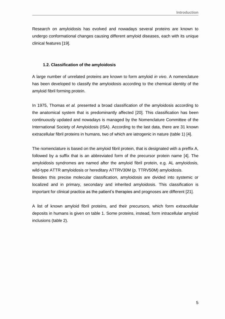
characteristic apple-green birefringence (figure 1). When amyloidosis is confirmed, it is
necessary to type it. Immunohistochemistry on paraffin sections or immunofluorescence of
frozen sections are techniques routinely performed using a panel of antibodies
recognizing different amyloid proteins [21, 24]. Immunoelectron microscopy sometimes is
also used. These antibody based techniques are relatively simple and routinely used in
pathology laboratories. The results are generally reliable but these techniques also have
some drawbacks. Some antibodies have low affinity for the misfolded amyloid proteins.
Also, some mutations may cause loss of epitopes and consequently absence of antibody
binding [25-26].
Introduction
8
Figure 1 – Congo red staining of renal biopsies of ATTR amyloidosis, showing amyloid
deposition in glomeruli, vasculature, and tubulointerstitium. (A) Congo red staining
visualized under light microscope; (B) Congo red staining visualized under polarized light,
showing the apple-green birefringence; (C) Higher magnification of Congo red staining
visualized under polarized light. From Lobato et al. [27].
When doubts persist, amyloid fibril proteins can be extracted and amino acid sequence
analysis performed [28-29]. Proteomic analysis, particularly mass spectrometry, has
become a useful tool for amyloid typing. Fibrils may be characterized by two-dimensional
gel electrophoresis (2D-PAGE). Laser microdissection of Congo red positive areas from
formaline fixed paraffin tissues can also be carried out. Samples are then subjected to
tandem mass spectrometric analysis to identify the amyloid protein [30]. The recent
multidimensional protein identification technology (MudPIT) enables a faster identification
from a peptide mixture of a protein sample. It is a chromatography-based technique that
couples a triphasic microcapillary column (reversed phase, strong cation exchange and
reversed phase high-performance liquid chromatography grade materials) with a tandem
mass spectrometer [31-33]. The peptide mixture is loaded directly onto the triphasic
microcapillary column which is placed directly in-line with the tandem mass spectrometer.
Ideally, these methods should complement each other to allow for an unequivocal result.
Among all the amyloidogenic proteins, in the following sections attention will be given to a
systemic and hereditary form of amyloidosis, the ATTR amyloidosis or Familial
Amyloidotic Polyneuropathy (FAP).
A B C
Introduction
9
2. Familial Amyloidotic Polineuropathy or ATTR amyloidosis
Transthyretin (TTR) is one of the precursor proteins associated with systemic amyloidosis.
More than 100 mutations have been described for TTR [34-35] and most of them, as well
as wild type TTR itself, are amyloidogenic. Only about 10 known TTR mutations show no
propensity to cause amyloidosis.
Deposition of wild-type TTR is associated with an acquired form of amyloidosis, senile
systemic amyloidosis (SAA). It affects particularly the heart, leading to cardiac
complications, and is found mainly in older individuals (>60 years old) and predominantly
in men. SSA affects approximatly 25% of the population aged more than 80 years [36-37].
Amyloidogenic mutations are responsible for hereditary diseases that share some clinical
manifestations, mainly peripheral and autonomic neuropathy, in familial amyloid
polyneuropathy (FAP), and less commonly cardiopathy, in familial amyloid cardiopathy.
2.1. ATTRV30M amyloidosis
Historically named Familial Amyloidotic Polyneuropathy type I (FAP-I) or Portuguese type,
ATTRV30M amyloidosis is a neurodegenerative, autosomal dominant disease,
characterized by extracelular deposition of mutated TTR (V30M) derived amyloid fibrils
[38].
As a matter of simplicity, throughout this document the historical term FAP will be used to
refer to ATTRV30M amyloidosis.
FAP was first described in 1952 by Corino de Andrade. He reported several patients, the
first one observed in 1939 in the Santo Antonio Hospital in Oporto, who had a peculiar
form of peripheral neuropathy with atypical generalized amyloidosis, or paramyloidosis,
with special involvement of the peripheral nerves [39].
In 1978, Costa et al. [40] identified TTR, then known as thyroxin-binding prealbumin, as
the major constituent of the amyloid deposits in these patients. In 1984, Saraiva et al.
identified a substitution of methionine for valine at position 30 of the TTR protein as the
biochemical cause of Portuguese type FAP [38]. Today it is known that Val30Met is the
most frequent disease associated TTR mutation in Portugal, Sweden, Japan and Italy.
Although being considered a rare disease (it affects approximately 1:100.000 persons
worldwide), ATTRV30M amyloidosis is an endemic disease particularly prevalent in the
Introduction
10
northern regions of Portugal, mainly Póvoa do Varzim, where allele frequency is
approximately 1:550 [36, 41].
2.2. Clinical features of ATTRV30M amyloidosis
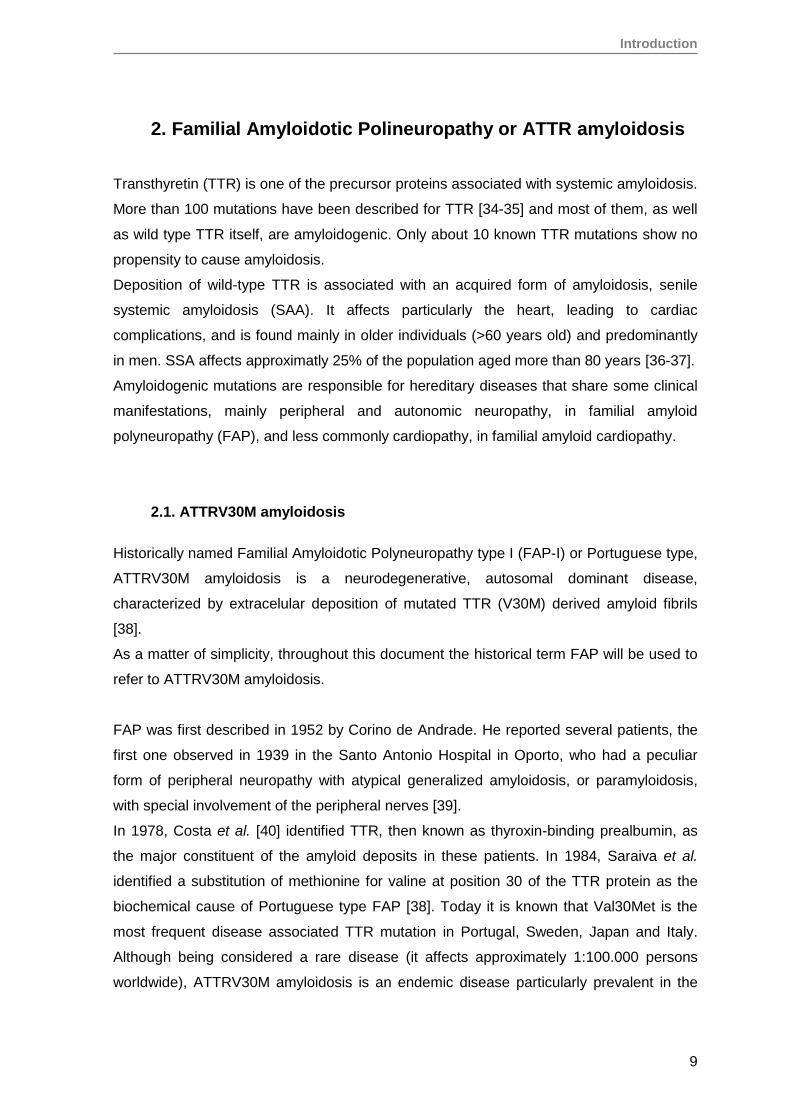
ATTRV30M amyloidosis is a systemic disease characterized by progressive sensorimotor
peripheral polyneuropathy and autonomic dysfunction, with renal, cardiac, and ocular
manifestations, among others [42]. Usually symptoms begin in the third to fourth decade
of life, and develop gradually for 10-20 years, leading to death. Progressive impairment of
thermal sensitivity and pain are common initial symptoms. The disease progresses with
lowering of the general state of health, alimentary and sexual dysfunction, malabsorption,
urinary bladder dysfunction, abnormal glomerular function, cardiac insufficiency and
vitreous opacities [43-46]. Besides the peripheral nerves, where the amyloid is
preferentially deposited causing myelin sheet destruction, other organs, such as kidney,
pancreas, heart, stomach, aorta, skin and eye are affected [47]. Although TTR is
produced mainly by the liver, this organ is not significantly affected.
Figure 2 – Schematic representation of clinical manifestations in FAP patients. From Ueda
et al. [47].
Introduction
11
In this work, more attention will be given to renal and ocular complications, and in
particular, to the expression of erythropoietin by these two organs in FAP patients. This
subject will be discussed later in further detail.
2.3. Therapeutic strategies for ATTR amyloidosis
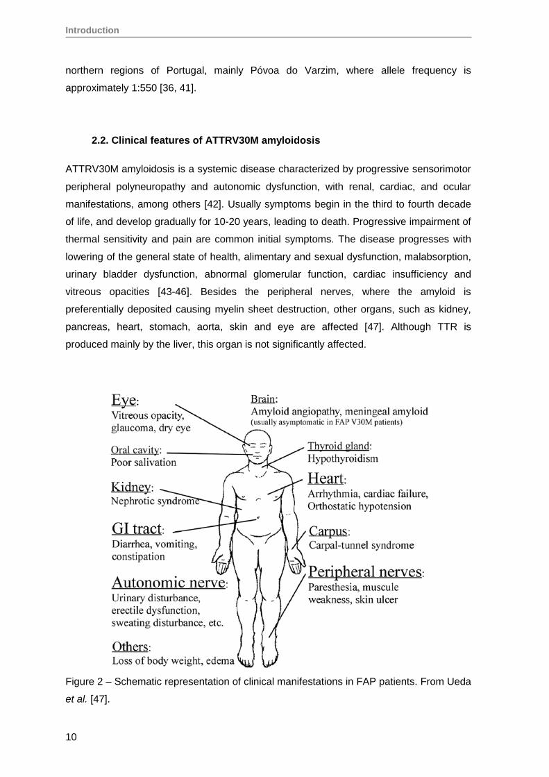
Advances are being made in the treatment of ATTR amyloidosis and many therapeutic
strategies have been proposed, which act on 3 important checkpoints:
A) Blocking the synthesis of TTR protein
B) Stabilization of the TTR tetramer to inhibit TTR disaggregation
C) Disruption and promotion of TTR amyloid fibrils clearance
Figure 3 – Schematic representation of the mechanism proposed for TTR
amyloidogenesis and some therapeutic strategies for FAP. From Hanna 2014 [48].
Block TTR synthesis
Liver transplantation
Since 1990, liver transplantation (LT) has become a therapeutic option for FAP patients,
as it eliminates more than 95% of the abnormal protein from circulation [49-50].
Approximately 120 LTs are performed worldwide each year [http://www.fapwtr.org]. When
LT is performed in the early stage of the disease, it can prolong survival, halt the
Introduction
12
progression of clinical manifestations and even improve autonomic and, to a lesser extent,
peripheral nerve function [51-53].
A strategy to manage the scarcity of healthy livers for transplantation was adopted with
sequential or domino LT, in which FAP patients receive a healthy liver from a deceased
donor and their liver is transplanted into patients with malignant or end-stage liver
diseases. As FAP only manifests after the 2nd or 3rd decade of life, it would be expected
that a FAP liver recipient would not manifest the disease. However, some recipients of
FAP livers started developing TTR amyloid deposits and disease symptoms less than 10
years after the surgery [54-56].
LT has important drawbacks: it is extremely expensive; transplanted patients must be
treated with immunosuppressants lifelong; non-symptomatic carriers of TTR mutations as
well as FAP patients in advanced stage do not undergo liver transplantation. Although
circulating mutant TTR is virtually eliminated, there are reports of continuing formation and
deposition of fibrils from wild-type TTR, causing cardiomyopathy and/or neuropathy after
liver transplantation, findings that seem similar to those observed in senile systemic
amyloidosis (SSA) [57-60]. Also, LT does not prevent the production of mutant TTR in the
cerebrospinal fluid and in the eyes, where the mutant protein is still secreted by choroid
plexus and retinal pigmented epithelium, respectively. Ocular and central nervous system
(CNS) problems may occur, highlighting the importance of non-neural dysfunction in post-
transplant patient management [61-62].
Gene therapy
Antisense oligonucleotides (ASO) and small interfering RNAs (siRNAs) are effective gene-
silencing tools that could be promising for FAP gene therapy [48]. Blocking hepatocyte
synthesis of TTR would prevent the production of both mutant and wild type TTR, which
could be a potential treatment for both hereditary and acquired ATTR amyloidosis. Phase
3 studies are currently ongoing for some ASOs and siRNAs [48, 63].
Antisense oligonucleotides (ASOs) are synthetic single stranded oligomers designed to
be complementary to a specific region in a target mRNA, promoting its degradation, thus
preventing production of the associated protein [64]. The drug ISIS-TTR Rx from Isis
Pharmaceuticals (Carlsbad, CA) targets the TTR mRNA preventing the production of both
mutant and wild-type TTR protein. This ASO suppresses TTR mRNA levels in the liver
and in the choroid plexus of the brain [65-67].
siRNAs are sequence-specific post-transcriptional gene silencing molecules. Patisiran,
also known as ALN-TTR02, from Alnylam Pharmaceuticals (Cambridge, MA) is a lipid
nanoparticle formulation of a synthetic siRNA that selectively silences both mutant and
wild-type TTR gene expression, both in vitro and in vivo [65, 68-69].
Introduction
13
Stabilize the TTR tetramer
TTR binding of its natural ligand thyroxine (T4) stabilizes the tetramer against dissociation.
The same strategy has been tested using small molecules that could bind to the TTR T4
pocket to kinetically stabilize the native tetramer and avoid the conformational changes
that lead to amyloid fibril formation [70].
Diflunisal, a non-steroidal anti-inflammatory drug (NSAID) with a molecular structure
similar to T4, binds to the T4 binding sites on the TTR tetramer, stabilizing it and
preventing acid mediated fibril formation in vitro [71-74], as well as in serum of
amyloidosis patients, without adverse effects [73, 75]. A 2 years long clinical trial showed
that Diflunisal treated patients had substantially less polyneuropathy progression
compared to placebo [76].
Tafamidis (Vyndaqel) or Fx-1006A, like Diflunisal, is a small molecule that selectively
binds to the T4 binding site and stabilizes the TTR tetramer [77], but without NSAID
activity. FAP patients treated with Tafamadis have shown a significant benefit relatively to
an untreated control group. Tafamidis stabilizes plasmatic TTRV30M protein, slowing
disease progression [77-78]. It became the first pharmacological treatment for FAP
patients to be approved in Europe [79].
Clearance of TTR amyloid fibrils
The presence of other compounds in the amyloid fibrils besides the core protein led
several groups to investigate the possibility to target them as a strategy to promote
clearance of amyloid fibrils, regardless of the amyloid type.
Doxycycline, an antibiotic, was shown to be an effective fibril disrupter, disaggregating
TTR amyloid fibris in mice transgenic for human TTRV30M, promoting amyloid deposit
reabsorption [80-81].
TUDCA (Tauroursodeoxycholic acid), a biliary acid with antioxidant and antiapoptotic
activities, significantly decreased the amount of TTR aggregates, as well as oxidative and
apoptotic biomarkers associated with disease, also in a transgenic mouse model [82-83].
A trial of a combination of TUDCA with Doxycyline was conducted in aged FAP mice [82]
and was more effective than either doxydoxycycline or TUDCA alone. It significantly
lowered TTR deposition and associated tissue markers and also disaggregated mature
amyloid deposits in the gastrointestinal tract.
EGCG (Epigallocatechin-3-gallate), the predominant polyphenol in green tea, has shown
in vitro the capacity to both inhibit fibril formation and also disrupt amyloid fibrils by
converting existing fibrils into non-fibril conformers [84-86].
Introduction
14
Anti-SAP antibodies could contribute to clear amyloid fibrils. Serum Amyloid P
component (SAP) is a plasma glycoprotein universally associated with amyloid fibrils.
Encouraging data was seen in animal models as well as in a heterogeneous group of
amyloid patients treated with anti-SAP antibodies [87-88].
2.4. Diagnosis
As for other amyloidosis, the diagnosis is based on histological examination, genetic
testing or mass spectrometry. ATTRV30M amyloidosis results from a single adenine for
guanine nucleotide change in exon 2 [89], which creates a restriction site for the enzyme
NsiI. This facilitates the molecular diagnosis, which can be performed by PCR followed by
RFLP, or by real-time PCR (rtPCR) genotyping based on melting curve analysis. Also,
amino acid substitution results in a known mass shift in the protein molecular weight [90],
making it possible to use mass spectrometry analysis [91-93]. This is particularly useful in
the setting of liver transplantation.
Recently, our group described a case report of a FAP patient who underwent orthotopic
liver transplantation from a cadaveric donor [94]. Continuing deterioration in this patient
raised the suspicion, confirmed in 2012, that the liver donor was also a TTRV30M carrier,
so retransplantation was proposed and carried out. Immunoprecipitation of TTR from the
serum of the cadaveric donor, followed by mass spectrometry analysis [95] enabled to
confirm the absence of TTRV30M, concluding that this second time the transplanted liver
was FAP free.
Introduction
15
3. Renal and ocular complications in ATTRV30M
Amyloidosis: its association with low erythropoietin
production
TTRV30M amyloid deposits are commonly found both in the kidney as well as in the eye
of FAP patients and give rise, respectively, to renal and ocular problems in these patients.
3.1. Renal complications: nephrotic syndrome and anemia
In ATTRV30M amyloidosis, renal amyloid deposition occurs mainly in the glomerular
mesangium and medulla, with a typical involvement of the distal convoluted tubule and
Henle's loop, which may lead to tubular atrophy and loss of tubular function [27]. Clinical
nephropathy manifests initially as a microalbuminuria and then progresses to proteinuria
and renal failure. In FAP patients, nephrotic syndrome may be associated with anemia
with decreased levels of EPO, sometimes below the lower limit of the normal range,
without associated iron deficit [96]. These patients do not respond to iron therapy but
treatment with recombinant EPO is effective [97].
According to World Health Organization criteria, anemia is defined as a concentration of
hemoglobin (Hb) <13 g / dL in men and <12 g / dL in women [98].
3.1.1. Defective EPO production by the kidney in ATTRV30M amyloidosis
Anemia has been described in ATTRV30M amyloidosis patients:
Moderate normocytic normochromic anemia was observed in 39% of the Swedish
FAP patients [99];
macrocytic and hypochromic anemia was reported in a group of 35 Japanese FAP
patients [100].
normocytic and normochromic anemia was present in 25% from a total of 165
symptomatic FAP patients, even in the presence of normal renal function, and is
associated with a defective renal production of EPO, revealed by serum levels lower
than expected [96].
A deficit of EPO in Portuguese FAP patients is an early event that was observed
independently of the presence of renal failure and sometimes preceding clinical disease
Introduction
16
[96]. Serum EPO levels were, on average, 11.2+6.7 mU/mL, lower than the expected
levels (35+13.9 mU/ml) for the degree of anemia, while iron stores, B12 vitamin, and
serum folate levels were normal in these patients. Normalization of iron status was
insufficient for the correction of anemia, but therapy with low doses of recombinant EPO
was effective [97], excluding a defective response of the bone marrow as a cause of
anemia in these patients.
Circulating EPO is mainly produced by the kidney in the adult. The observed low EPO
production suggests a defect of the EPO-producing cells, which could be related to either
the presence of amyloid deposits in the renal interstitium or with other factors, such as
circulating TTRV30M itself.
The amyloid deposits present in renal biopsies were found to have no correlation with
serum EPO levels, independently of the neuropathy score, the amount of amyloid
deposition or the renal clinical manifestations [101].
Anemia in liver transplant recipients is usually due to the side-effects of
immunosuppressive therapies, iron deficiency, renal failure and post-transplant
lymphoproliferative disorders [102]. A study performed in cirrhotic patients showed that
anemia affected 85% of these patients before liver transplantation. After liver
transplantation, anemia decreased to 18% [103]. In contrast, in FAP patients the
prevalence of anemia increased even after liver transplantation, and defective
endogenous EPO production persisted [104], excluding an inhibitory effect of the
circulating TTRV30M on the EPO-producing cells.
Pro-inflammatory cytokines can inhibit EPO gene expression, contributing to the anemia
of chronic disease [105]. In ATTRV30M amyloidosis, inflammation is observed,
particularly with up-regulation of TNF-α, macrophage colony-stimulating factor and IL-1β
[106-107], which could explain the low EPO levels in these patients. However, Beirão et
al. analyzed 24 FAP patients and found no evidence of systemic inflammation, as no
significant differences were found on interleukin-6, transferrin saturation, ferritin and
hepcidin-25 [108].
3.2. Ocular complications: vitreous opacities and glaucoma
ATTRV30M amyloidosis is associated with several ocular manifestations such as lacrimal
dysfunction, pupillary disturbances, changes in the conjunctiva, presbyopia [109], dry eye
[110], vitreous opacities, which may occur before any other systemic manifestation of the
disease [111], and, most seriously, severe glaucoma. Liver transplantation in FAP patients
Introduction
17
proved unable to halt the progression of these ocular manifestations, probably due to the
continued production of the mutated TTR by the retinal pigment epithelial cells [62].
A retrospective study of 477 symptomatic FAP patients was performed by Beirão et al.
which showed that these patients have amyloid deposits in the iris, in the anterior lens
capsule and in the vitreous. Vitrectomy with complete removal of the vitreous is usually
carried out, but when the vitrectomy is incomplete new amyloid deposits are formed due
to continuing deposition in the remaining vitreous [112].
Glaucoma can develop rapidly in FAP patients and, if not treated, may lead to blindness.
A correlation was found between vitrectomy and glaucoma, with vitrectomy favoring the
onset or worsening of glaucoma [113].
3.2.1. Defective EPO production by the eye in ATTRV30M amyloidosis
Glaucoma causes an increase in intraocular pressure, which leads to activation of
neuroprotective mechanisms. Studies have reported upregulation of EPO expression and
an increased intravitreal EPO concentration in some ocular disorders [114], which may
reflect the cytoprotective function of EPO in response to hypoxia, ischemia, and
inflammation [115]. Recently, our group found that EPO concentrations in the aqueous
humor of glaucomatous eyes of non-FAP patients are significantly increased relatively to
normal non-glaucomatous eyes, probably as a protective role. However, in glaucomatous
eyes of FAP patients the EPO levels did not increase and maintained similar levels to
those of control eyes [116]. These results show an inability of FAP patients to upregulate
EPO production both systemically by the kidney and locally by the pigmented epithelium.
Glaucoma is the second leading cause of blindness worldwide [117]. Vascular
abnormalities and altered blood flow at the optic nerve head may lead to local hypoxia,
accelerating neuronal cell death in patients. The hypoxia inducible factor 1 (HIF-1) is
thought to be involved in the pathology of glaucoma, as increased activation of HIF-1 was
found in glaucomatous eyes and localization of this protein was correlated with regions of
visual field defects [118]. HIF is one of the main regulators of EPO expression, by
inducing it in situations of hypoxia. In glaucoma, as a consequence of HIF activation, EPO
levels are strongly elevated, probably as a cytoprotective response.
The mechanisms responsible for the low expression of EPO in FAP patients, whether as a
response to anemia, or as a response to ocular damage, as in the case of glaucoma,
remain unexplained. What is certain is that these patients do not increase EPO levels in
response to stimuli to which a non-PAF patient would respond with an increase in
expression of this cytokine/hormone.
Introduction
18
4. Erythropoietin
Erythropoietin (EPO) is a hormone essential for red blood cell production. A moderate
reduction in hemoglobin concentration is sufficient to increase EPO mRNA expression,
which occurs within minutes of the onset of hypoxia, reaching a maximum after 6 hours
[119-121]. Daily, it stimulates proliferation and differentiation of about 2x10¹¹ erythroid
progenitor cells in the bone marrow, contributing to the control of blood oxygen capacity
throughout the body [122].
EPO is an endocrine, paracrine and autocrine hormone. Besides its hematopoietic
function, EPO has been shown to be a cytoprotective hormone. Among other effects, EPO
antagonizes the activity of pro-inflammatory cytokines, has neuroprotective functions and
promotes healing through stimulation of angiogenesis and capillary growth [123].
4.1. Erythropoietin structure
Human EPO is a glycoprotein of 30.4 kDa encoded by 5 exons located in chromosome 7
as a single copy gene. Translation of the EPO gene results in a polypeptide chain of 193
amino acids that is cleaved posttranslationaly, both at the N- and C-terminal sites. The
secreted protein has 165 amino acids [124].
About 40% of the molecular weight of EPO is due to carbohydrate chains. EPO has 4
glycosilated side chains that are important to its biological function by conferring thermal
and structural stability, protection against free radicals, increased plasma half-life and
selectivity [122, 125-126]. EPO has 4 α-helices and 2 dissulfide bonds with 3 asparagine
N-glycosilation and 1 serine O-glycosilation sites. The sialic acid residues attached to the
4 carbohydrate chains are particularly important for the maintenance of in vivo half-life and
biological activity [126-127].
4.2. Sites of erythropoietin production
During fetal life EPO is produced by the liver, whereas in the adult it is mainly produced by
the kidney [128]. The molecular mechanisms underlying this switch are poorly understood,
but are thought to involve the transcription factor GATA-4 [129], which is highly expressed
by hepatocytes only in the fetal liver. Its inhibition leads to a dramatic reduction in Epo
gene transcription in Hep3B cells.
Introduction
19
Many efforts were done to identify the renal EPO-producing cells. Evidence has been
provided for different locales including: renal glomeruli [130], peritubular interstitial or
endothelial cells in anemic mouse [131-132], peritubular interstitial cells in hypoxic
monkey [133], tubular epithelial cells [134-135] and proximal tubular cells [136-137]. In
2010, our group identified distal tubular cells and cortical collecting tubules as the major
site of EPO production in normal adult human kidneys from patients with ATTRV30M
amyloidosis with or without anemia [138]. In 2013 Bussolati et al. identified a subset of
renal CD133(+)/CD73(+) progenitor cells isolated from the human renal inner medulla,
with a mesenchymal phenotype, as a possible source of EPO under hypoxic conditions,
via the prolyl hydroxylase-HIF-2α axis [139]. CD133+ progenitors have been identified
along the renal nephron [140] which overlaps with the described localization of EPO-
producing cells in different segments of the human nephron by in situ hybridization
studies. In addition, Nagai et al. demonstrated recently in mice that EPO mRNA
expression occurs in proximal convoluted tubules (PCTs), distal convoluted tubules
(DCTs) and cortical collecting ducts (CCDs) under normoxic conditions and in peritubular
cells in severe hypoxia [141]. These dissimilarities may result from inter-species
differences. Additionally, it is likely that EPO production by different populations of renal
cells depend on the varying hypoxic conditions used in the different experimental models.
Apart from the kidney, EPO production has been found also in the brain [142], retina, lung,
spleen, bone marrow, in the male and female reproductive organs [143-144], placenta
[145] and also in numerous cancer cells [124].
Although EPO is mainly produced by the kidney in the adult, an adequate renal EPO-
producing cell line is not available. So, most of the present knowledge of the O2 sensing
mechanism that controls EPO expression has been based on in vitro studies using the
human hepatoma cell lines Hep3B and HepG2, described in 1987 by Goldberg et al. as a
constitutive and inducible EPO producer, in an oxygen-dependent manner [146].
4.3. Erythropoietin functions: hematopoiesis and cellular protection
Recognition that the EPO receptor (EPOR) was expressed in several cells other than the
erythroid progenitor cells led to the discovery of the extra-hematopoietic functions of EPO.
EPO is a member of the cytokine type I superfamily [147], and is both an endocrine,
paracrine and autocrine hormone.
Introduction
20
4.3.1. Hematopoietic function
EPO major function is to promote survival of EPO-dependent colony-forming unit-erythroid
(CFU-E) cells and erythroblasts. EPO synthesis is regulated at the mRNA level, through
mechanisms sensitive to oxygen concentration. In response to hypoxia, EPO production
increases in a few hours [148], and acts on the erythroid progenitor cells in the bone
marrow stimulating their proliferation and differentiation.
EPO binds to the erythropoietin receptor (EpoR) homodimer, which is located on the
surface of erythroid progenitor cells, and triggers a conformational change that brings its
intracellular domains into close proximity, resulting in transphosphorylation and activation
of Janus kinase 2 (JAK2) [124]. Downstream cascades are initiated via different signaling
pathways including signal transducer and activator of transcription 5 (STAT5),
phosphoinositide 3-kinase (PI3K)/AKT, and mitogen-activated protein kinase (MAPK) via
adapter proteins like Src homology containing protein (SHC). These transcription factors
translocate to the nucleus and drive or inactivate transcription of genes involved mainly in
survival and prevention of apoptosis of erythroid progenitors [149].
EpoR is expressed at the highest level on erythroid progenitor cells and plays a critical
role in the regulation of red blood cell production by EPO. However, expression of the
EpoR beyond hematopoietic cells raised the possibility that EPO activity associated with
survival, proliferation and differentiation of erythroid cells might not be restricted to
erythropoiesis.
EPO can also signal via a heterodimeric receptor composed of an EpoR monomer chain
and CD131, the β common cytokine receptor [150]. This heterodimeric complex is found
in nonerythroid cells and is thought to be involved in nonerythroid effects of EPO [124].
4.3.2. Extra-hematopoietic functions: cellular protection
Recent findings have demonstrated that EPO has also a cytoprotective function.
EPO has direct effects on immune cells, endothelial cells, bone marrow stromal cells, as
well as cells of the heart, brain, reproductive system, gastrointestinal tract, muscle, kidney,
pancreas, and nervous system [150-152]. The nonerythropoietic functions of EPO include:
promoting cardiac and central nervous system development, blocking cell death in stroke
models, improving learning and memory, regulating angiogenesis, confer protection in
ischemia/reperfusion injury of the kidney, liver, heart and myocardial infarction and
modulate responses to injuries such as cerebral ischemia, cardiac infarction and retina
degeneration [124, 150]. In vitro recombinant EPO stimulates the proliferation,
mobilization, and differentiation of endothelial progenitor and precursor cells and also
enhances endothelial cell viability and survival by blocking apoptosis [124]. EPO also
Introduction
21
protects against diabetes in mouse models, through direct JAK2 signaling in pancreatic
cells, resulting in cell survival and proliferation, reduced inflammation, and increased
angiogenesis in the islets [150]. EPO may act on the regulation of metabolism and obesity
and have potential benefits in the treatment of neurologic diseases, mood symptoms and
depression.
In the immune system, EPO responds to tissue injury caused by pathogens, trauma and
hypoxia in order to maintain a balance between inflammation and anti-inflammation [153].
To limit an uncontrolled inflammatory response, inflammation or hypoxia can trigger EPO
expression to inhibit proinflammatory cytokine production (TNF-α and interleukin 6), to
increase expression of endothelial nitric oxide synthase (eNOS) and nitric oxide (NO), and
block inducible nitric oxide synthase (iNOS) expression [149, 154], to inhibit macrophage
activity by blocking NF-κB p65 signaling pathway [155], and delimit the volume of injury by
counteracting apoptosis [153].
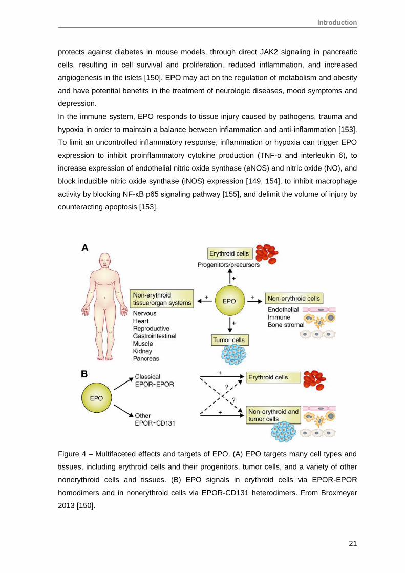
Figure 4 – Multifaceted effects and targets of EPO. (A) EPO targets many cell types and
tissues, including erythroid cells and their progenitors, tumor cells, and a variety of other
nonerythroid cells and tissues. (B) EPO signals in erythroid cells via EPOR-EPOR
homodimers and in nonerythroid cells via EPOR-CD131 heterodimers. From Broxmeyer
2013 [150].
Introduction
22
4.4. Use of recombinant EPO to treat anemia
A variety of EPO derived recombinant forms of erythroid stimulating agents (ESAs) have
been used in research and in clinical practice since 1986 [156]. ESAs are used to treat
anemia in several fields such as end-stage renal disease, malignancies associated with
chemotherapy, AIDS and surgical interventions [157]. ESAs are able to increase red blood
cell count, hemoglobin and hematocrit levels, decreasing the need for red blood cell
transfusion and possibly improving quality of life [156].
Several ESAs have been developed, with the rationale of cost saving and prolonged
survival in circulation. ‘Epoetin’ has an amino acid sequence identical to the endogenous
human EPO. Darbepoetin alfa has a longer survival in circulation and Mircera (methoxy
polyethylene glycol-epoetin beta) has the longest half-life (130-140 h on IV injection)
[157].
However, side effects of EPO treatment have emerged: potentially life-threatening cardiac
complications such as stroke, increase in arterial blood pressure and possibly
hypertension, increase the incidence of thromboembolism [157], especially with higher
doses and rapid increase in hemoglobin levels.
4.5. Regulation of the erythropoietin gene
EPO gene expression is activated in response to many forms of stress, but hypoxia is the
most important stimulus [158]. When O2 concentration drops, EPO gene expression is
activated and an exponential increase in EPO protein plasma levels occurs [159].
Regulation of EPO gene expression occurs at different levels: transcription, mRNA
stabilization and at the translational level by an upstream open reading frame [160-163].
At the transcriptional level, Hypoxia Inducible Factor (HIF), GATA-2 and NF-kB
transcription factors are the main known regulators of EPO gene expression [164-167].
HIF induces EPO expression under hypoxic conditions, while GATA-2 and NF-kB
suppress EPO promoter activity when activated by the pro-inflammatory cytokines IL-1
and TNF-α [168].
4.5.1. Hypoxia inducible factor (HIF)
The EPO gene has a cis-acting hypoxia response element (HRE) located in the enhancer
at the 3’-flanking region. Under hypoxic conditions, the hypoxia-inducible factor HIF binds
to the HRE and induces EPO expression [169, 170].
HIF is a heterodimeric transcription factor composed by an O2-sensitive α-subunit and a
constitutively expressed β-subunit, and is expressed in all tissues of many species [171].
Introduction
23
Three α chain isoforms of HIF were identified (HIF-1α, HIF-2α and HIF-3α). HIF-1α and
HIF-2α have similar regulatory mechanisms and are both involved in O2 delivery and in
cellular adaptation to hypoxia. However, several studies point to differences in the
expression and cellular distribution pattern, which may be indicative of different functions.
Whereas HIF-1α is expressed in all nucleated cells, the expression of HIF-2α is restricted
to specific cell types, including vascular endothelial cells, renal interstitial cells,
hepatocytes, cardiomyocytes, glial cells and astrocytes [172]. Rosenberger et al. found,
inducing hypoxia in rats, that regardless of the type of stimulus, the cell populations
expressing HIF-1α and HIF-2α in the kidney are different. HIF-1α was predominantly
expressed in cells of the proximal and collecting tubules, and HIF-2α was expressed
predominantly in the peritubular interstitial cells and some glomerular cells [173]. Because
HIF-2α accumulates in peritubular cells and these have been described as the site of EPO
production, it was suggested that HIF-2α would be the regulator of EPO expression in the
kidney. However, several studies show that both HIF-1α and HIF-2α contribute to the
regulation of EPO, although differentially, according to the cell type.
The production and activation of HIF is regulated at the level of the α chain, which
increases exponentially as O2 concentration declines. Two oxygen-dependent
mechanisms are involved in this regulation. Under normoxia, in the first mechanism, two
conserved proline residues located in the oxygen-dependent degradation domain (ODDD)
undergo hydroxylation by the action of prolyl-hydroxylases (PHDs) and this allows the
ubiquitination of the α chain by the complex von Hippel-Lindau protein (pVHL)/E3-ubiquitin
ligase, and subsequent degradation by the proteasome [124]. In the second mechanism,
an asparagine residue (Asn803) is hydroxylated by HIF inhibition factor (FIH1) and HIF
activity is suppressed by preventing the binding of the transcription coactivator p300/CBP
(CREB-binding protein), which acts to increase the expression of their target genes. The
PHDs and FIH1 use oxygen as co-substrate. So, under hypoxia, a decrease in the oxygen
tension results in inhibition of both PHDs and FIH1. The α-chain dimerizes with the β
chain, the HIF complex is stabilized and, after recruitment of co-activator p300/CBP, binds
to the HRE of HIF target genes [124]. Additionally, HIF cooperates with hepatocyte
nuclear factor 4 (HNF-4), which binds to the 3′ EPO hypoxia enhancer region [124].
Recently, it was shown that HIF-2α may regulate erythropoiesis by a mechanism
independent of PHD enzymes. HIF-2α is acetylated during hypoxia and deacetylated by
Sirtuin 1, a nicotinamide adenine dinucleotide (NAD)+-dependent protein deacetylase,
which increases HIF-2-dependent EPO synthesis, linking cellular redox and energy state
to systemic hypoxia responses. Sirtuin 1-deficient mice produced significantly lower

Introduction
24
amounts of fetal liver Epo mRNA, and as adults less EPO in response to severe hypoxia
[171].
The hydroxylation of HIFα can be carried out by three prolyl-hydroxylases (PHD1, PHD2
and PHD3), among which PHD2 plays the predominant role at most sites. The prolyl
hydroxylase inhibitor FG-4497 was used in rats to induce HIF-dependent erythropoietin
transcription, and in fact EPO mRNA expression was strongly induced and co-localized
with HIF-2α [174]. It is likely that the PHDs are the primary O2 sensors in the control of
EPO production. Chemical compounds, such as the α-ketoglutarate, Roxadustat
(ASP1517/FG-4592) and dimethyloxalylglycine (DMOG) can inhibit PHDs, providing a
potential oral therapy for stimulating erythropoiesis in patients with chronic kidney disease.
A phase 3 clinical trial using Roxadustat is currently ongoing to evaluate its safety and
efficacy, when compared to epoetin alfa, for the treatment of anemia in patients with
chronic kidney disease on dialysis [175-176]. It has the advantage of offering an oral
treatment and circunventing the need for multiple injections. DMOG was used in this work
to mimick hypoxia conditions and to stimulate EPO expression.
4.5.2. GATA-2 and NF-kB
Besides HIF, EPO expression is also regulated by GATA-2 and NF-kB at the level of the
promoter. There is little information about the mechanisms by which these transcription
factors regulate the EPO promoter, but it is known that both act by inhibiting the promoter
activity.
The EPO promoter is suppressed by GATA-2 in normoxia and GATA-2 levels decrease in
hypoxia [160]. The activation of GATA-2, with consequent inhibition of EPO gene
expression, may be related to a pro-inflammatory stimulus, to increased production of
H2O2 and to the presence of L-NG-monomethylarginine (L-NMMA). Exogenous addiction
or endogenous production of H2O2 enhance the expression of GATA-2 and its binding
activity, suppressing the activity of the EPO promoter and thus inhibiting EPO gene
expression [177-179].
L-NMMA, an endogenous nitric oxide synthase (NOS) inhibitor that is markedly elevated
in uremic patients, increases GATA-2 mRNA expression and its binding to the EPO
promoter, both in normoxia and hypoxia. EPO promoter activity is inhibited by GATA-2
causing a decrease in EPO expression and protein production [179-180]. L-NMMA did not
alter the binding activity of HIF-1, HNF-4 or NF-kB in these experiments.
EPO production may be also suppressed by the pro-inflammatory cytokines IL-1 and TNF-
α, which activate GATA-2 and NF-kB and may contribute to the anemia of chronic disease
in part by suppressing EPO production [168]. NF-kB binds to the EPO promoter in a
Introduction
25
region close to the binding site for GATA-2, and inhibits its activity. Souma T et al.
showed, with a mouse model of adult-onset anaemia caused by erythropoietin deficiency
(ISAM), that inflammatory injury activated NF-kB inducing a pathological phenotypic
switch of the renal EPO-producing cells (REP) to myofibroblasts [181].
4.5.3. ATF3
The activating transcription factor ATF3, although not classically described as a regulator
of EPO expression, is also involved in the activation of the EPO promoter in response to
the platelet-derived growth factor (PDGF) signaling system [182]. ATF3 is rapidly induced
upon exposure of cells to stress signals [183]. Elevated expression of PDGF has been
observed in RPE cells after retinal detachments or retinal laser treatment in murine model
systems [184], in in vitro wounded human RPE cell cultures [185] and in epi-retinal
membranes isolated from proliferative vitreoretinopathy and proliferative diabetic
retinopathy patients [186].
Other transcription factors, such as retinoic X receptor-α (RXR-α), Wilms tumor
suppressor (WT1), SMAD3 and Sp1, may contribute to the regulation of EPO expression.
RXR-α contributes to EPO activation through interaction with EPO enhancer in the fetal
liver during early erythropoiesis [187]. WT1 directly up-regulates EPO expression in
hepatocytes, through binding to the EPO promoter [188]. SMAD3 binding to the 3' EPO
enhancer, and Sp1 binding to the EPO promoter region also cooperate with HIF, HNF4
and p300 in EPO gene transcription in hypoxia [189]. Together these factors act to
stabilize the multifactorial complex interacting with EPO promoter and enhancer to
regulate EPO expression.
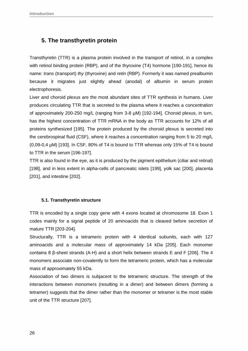
Introduction
26
5. The transthyretin protein
Transthyretin (TTR) is a plasma protein involved in the transport of retinol, in a complex
with retinol binding protein (RBP), and of the thyroxine (T4) hormone [190-191], hence its
name: trans (transport) thy (thyroxine) and retin (RBP). Formerly it was named prealbumin
because it migrates just slightly ahead (anodal) of albumin in serum protein
electrophoresis.
Liver and choroid plexus are the most abundant sites of TTR synthesis in humans. Liver
produces circulating TTR that is secreted to the plasma where it reaches a concentration
of approximately 200-250 mg/L (ranging from 3-8 µM) [192-194]. Choroid plexus, in turn,
has the highest concentration of TTR mRNA in the body as TTR accounts for 12% of all
proteins synthesized [195]. The protein produced by the choroid plexus is secreted into
the cerebrospinal fluid (CSF), where it reaches a concentration ranging from 5 to 20 mg/L
(0,09-0,4 µM) [193]. In CSF, 80% of T4 is bound to TTR whereas only 15% of T4 is bound
to TTR in the serum [196-197].
TTR is also found in the eye, as it is produced by the pigment epithelium (ciliar and retinal)
[198], and in less extent in alpha-cells of pancreatic islets [199], yolk sac [200], placenta
[201], and intestine [202].
5.1. Transthyretin structure
TTR is encoded by a single copy gene with 4 exons located at chromosome 18. Exon 1
codes mainly for a signal peptide of 20 aminoacids that is cleaved before secretion of
mature TTR [203-204].
Structurally, TTR is a tetrameric protein with 4 identical subunits, each with 127
aminoacids and a molecular mass of approximately 14 kDa [205]. Each monomer
contains 8 β-sheet strands (A-H) and a short helix between strands E and F [206]. The 4
monomers associate non-covalently to form the tetrameric protein, which has a molecular
mass of approximately 55 kDa.
Association of two dimers is subjacent to the tetrameric structure. The strength of the
interactions between monomers (resulting in a dimer) and between dimers (forming a
tetramer) suggests that the dimer rather than the monomer or tetramer is the most stable
unit of the TTR structure [207].
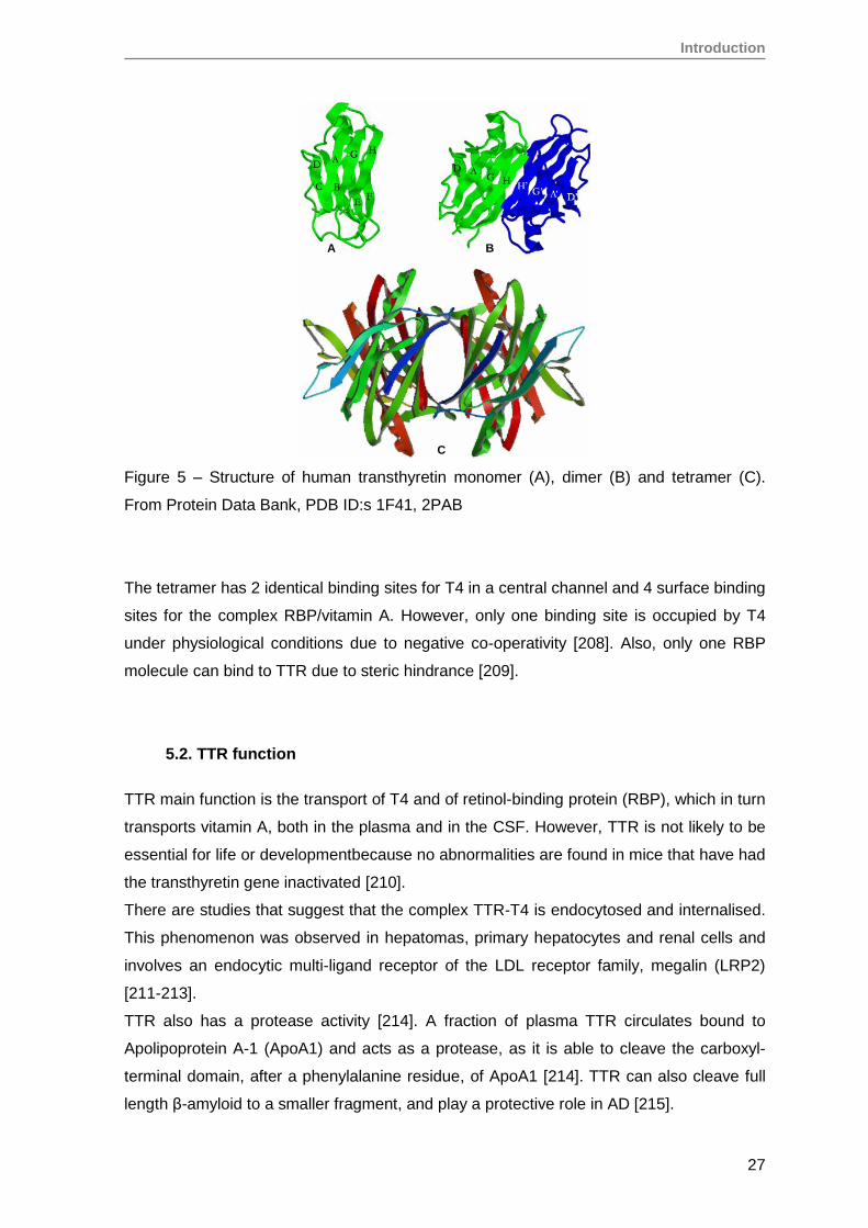
Introduction
27
Figure 5 – Structure of human transthyretin monomer (A), dimer (B) and tetramer (C).
From Protein Data Bank, PDB ID:s 1F41, 2PAB
The tetramer has 2 identical binding sites for T4 in a central channel and 4 surface binding
sites for the complex RBP/vitamin A. However, only one binding site is occupied by T4
under physiological conditions due to negative co-operativity [208]. Also, only one RBP
molecule can bind to TTR due to steric hindrance [209].
5.2. TTR function
TTR main function is the transport of T4 and of retinol-binding protein (RBP), which in turn
transports vitamin A, both in the plasma and in the CSF. However, TTR is not likely to be
essential for life or developmentbecause no abnormalities are found in mice that have had
the transthyretin gene inactivated [210].
There are studies that suggest that the complex TTR-T4 is endocytosed and internalised.
This phenomenon was observed in hepatomas, primary hepatocytes and renal cells and
involves an endocytic multi-ligand receptor of the LDL receptor family, megalin (LRP2)
[211-213].
TTR also has a protease activity [214]. A fraction of plasma TTR circulates bound to
Apolipoprotein A-1 (ApoA1) and acts as a protease, as it is able to cleave the carboxyl-
terminal domain, after a phenylalanine residue, of ApoA1 [214]. TTR can also cleave full
length β-amyloid to a smaller fragment, and play a protective role in AD [215].
A B
C
Introduction
28
5.3. Models of amyloidogenesis
Various mechanisms have been proposed to explain amyloidogenesis.
Mutations in several proteins lead to structural changes that promote unfolding and
predispose for the formation of fibers, resulting in hereditary amyloidosis syndromes. In
the case of TTR, several mutations alter the thermodynamics and kinetics of dissociation
of the tetramer and favor the formation of intermediates that self assemble into amyloid
fibers [216].
For example, the V30M mutation confers a moderate instability to the tetramer, while
L55P is the most unstable and pathogenic variant. However, the presence of mutations is
not essential for amyloidogenesis to be triggered. Examples are some localized
amyloidoses, and again using the example of TTR, senile systemic amyloidosis is caused
by deposition of amyloid fibers from wild-type TTR. Many factors can trigger
destabilization of the protein structure: heat shock, oxidative stress or chemical
modifications, alterations of intracellular macromolecular crowding, presence of suitable
surfaces, absence of stabilizing ligands, impairment of intracellular quality control of
protein folding, pH changes and others [217].
Most in vitro studies regarding the formation of amyloid fibers are based on lowering the
pH, suggesting that mildly acidic pH (present for example in lysosomes) can induce
rearrangement of the tetramer structure and dissociation into partially denatured
monomeric amyloidogenic intermediates, that are then joined again to form amyloid fibrils
[23]. However, it has been shown that normal TTR, as well as the variants V30M, L55P
and the non-amyloidogenic T119M dissociate into monomeric species at physiological pH
and ionic strength [30] and that the most unstable variants (V30M- and L55P-TTR) exist in
a complex equilibrium between monomers, tetramers and aggregates of higher molecular
weight [30].
Nowadays, it is thought that the critical step for amyloidogenesis is the destabilization of
the structure and the formation of non-native intermediate species that have the ability to
self-associate.
These intermediate species seem to play an important role both in the formation of
amyloid fibrils as in the mechanisms of toxicity subjacent to amyloid disease.
Depending on conditions, during the process of amyloidogenesis different types of
species can be formed: amorphous aggregates, soluble oligomers or amyloid fibrils [218-
221], and the process rarely results in a homogeneous product. Usually, heterogeneous
mixtures containing several species of aggregates (amyloid fibrils, amorphous aggregates
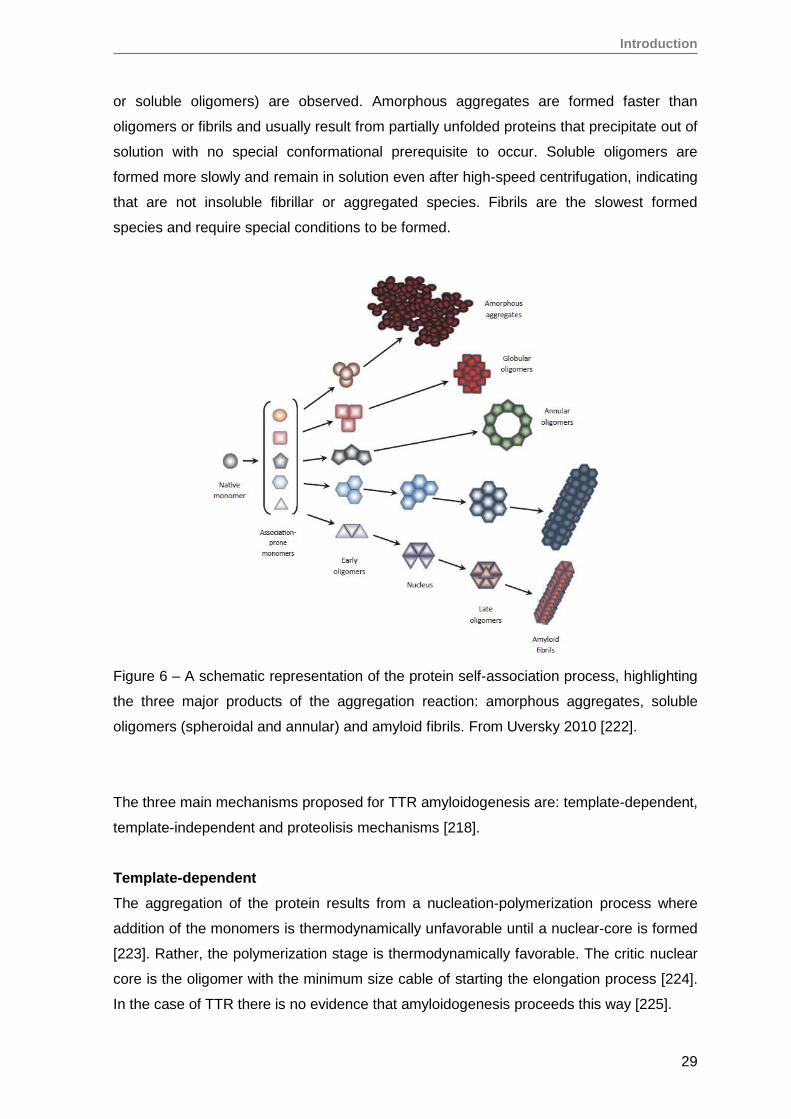
Introduction
29
or soluble oligomers) are observed. Amorphous aggregates are formed faster than
oligomers or fibrils and usually result from partially unfolded proteins that precipitate out of
solution with no special conformational prerequisite to occur. Soluble oligomers are
formed more slowly and remain in solution even after high-speed centrifugation, indicating
that are not insoluble fibrillar or aggregated species. Fibrils are the slowest formed
species and require special conditions to be formed.
Figure 6 – A schematic representation of the protein self-association process, highlighting
the three major products of the aggregation reaction: amorphous aggregates, soluble
oligomers (spheroidal and annular) and amyloid fibrils. From Uversky 2010 [222].
The three main mechanisms proposed for TTR amyloidogenesis are: template-dependent,
template-independent and proteolisis mechanisms [218].
Template-dependent
The aggregation of the protein results from a nucleation-polymerization process where
addition of the monomers is thermodynamically unfavorable until a nuclear-core is formed
[223]. Rather, the polymerization stage is thermodynamically favorable. The critic nuclear
core is the oligomer with the minimum size cable of starting the elongation process [224].
In the case of TTR there is no evidence that amyloidogenesis proceeds this way [225].
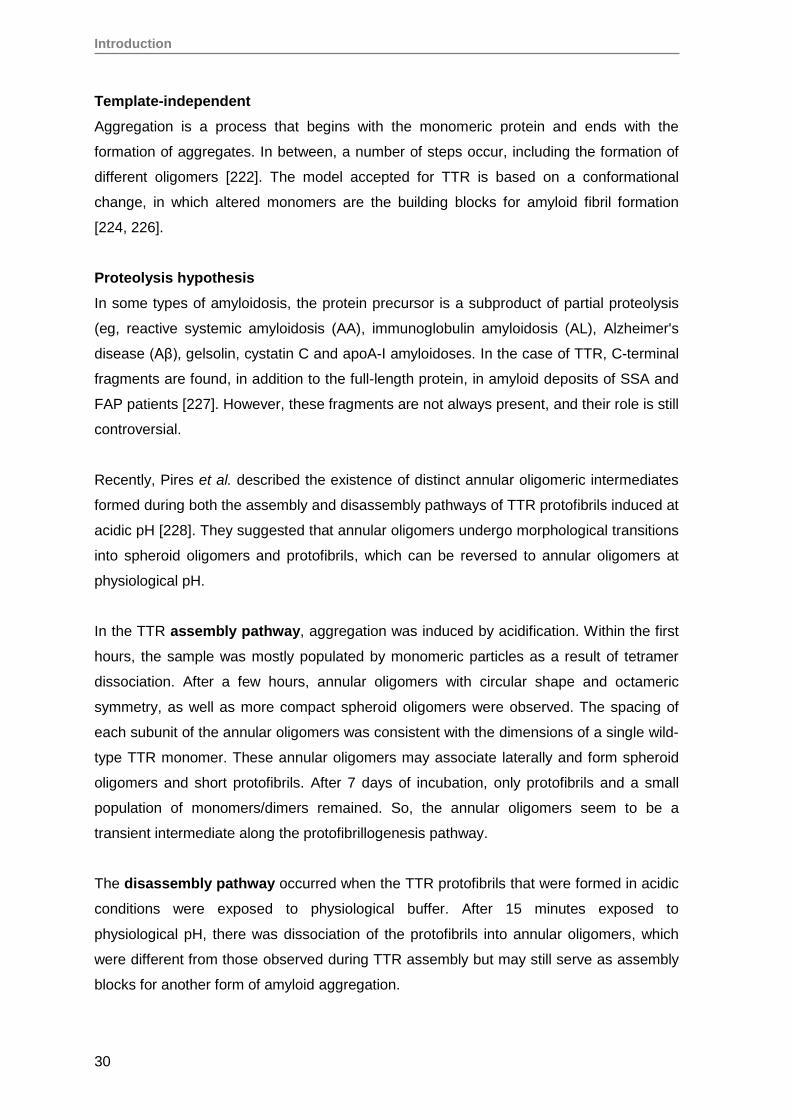
Introduction
30
Template-independent
Aggregation is a process that begins with the monomeric protein and ends with the
formation of aggregates. In between, a number of steps occur, including the formation of
different oligomers [222]. The model accepted for TTR is based on a conformational
change, in which altered monomers are the building blocks for amyloid fibril formation
[224, 226].
Proteolysis hypothesis
In some types of amyloidosis, the protein precursor is a subproduct of partial proteolysis
(eg, reactive systemic amyloidosis (AA), immunoglobulin amyloidosis (AL), Alzheimer's
disease (Aβ), gelsolin, cystatin C and apoA-I amyloidoses. In the case of TTR, C-terminal
fragments are found, in addition to the full-length protein, in amyloid deposits of SSA and
FAP patients [227]. However, these fragments are not always present, and their role is still
controversial.
Recently, Pires et al. described the existence of distinct annular oligomeric intermediates
formed during both the assembly and disassembly pathways of TTR protofibrils induced at
acidic pH [228]. They suggested that annular oligomers undergo morphological transitions
into spheroid oligomers and protofibrils, which can be reversed to annular oligomers at
physiological pH.
In the TTR assembly pathway, aggregation was induced by acidification. Within the first
hours, the sample was mostly populated by monomeric particles as a result of tetramer
dissociation. After a few hours, annular oligomers with circular shape and octameric
symmetry, as well as more compact spheroid oligomers were observed. The spacing of
each subunit of the annular oligomers was consistent with the dimensions of a single wild-
type TTR monomer. These annular oligomers may associate laterally and form spheroid
oligomers and short protofibrils. After 7 days of incubation, only protofibrils and a small
population of monomers/dimers remained. So, the annular oligomers seem to be a
transient intermediate along the protofibrillogenesis pathway.
The disassembly pathway occurred when the TTR protofibrils that were formed in acidic
conditions were exposed to physiological buffer. After 15 minutes exposed to
physiological pH, there was dissociation of the protofibrils into annular oligomers, which
were different from those observed during TTR assembly but may still serve as assembly
blocks for another form of amyloid aggregation.
Introduction
31
Figure 7 – Model of TTR protofibril assembly and disassembly. From Pires et al. [228].
So, in the protofibrillogenesis model proposed by Pires et al. the native TTR undergoes
structural transitions upon acidification, and the tetramers dissociate into amyloidogenic
monomers and, in smaller quantities, dimmers, from which annular oligomers with
octameric symmetry assemble. A single annular oligomer may serve as a scaffold for the
continuous addition of subunits up to the formation of an annular doublet. Spheroid
oligomers coalesce in a dynamic equilibrium with the growing protofibrils which are
abundant in the first week of incubation. Upon adjusting of pH to neutral, protofibril
structure quickly reorganizes, and dissociation into an annular oligomeric species quite
distinct from that seen in the assembly pathway proceeds in a time scale of minutes.
The annular oligomeric species are probably toxic, as it has been seen that oligomers
containing up to eight TTR monomers, but not more, are cytotoxic [229].
Introduction
32
5.4. Methods to induce the formation of oligomeric species in vitro
In vitro amyloidogenesis and formation of oligomeric species can be achieved by several
methodologies, as long as the conditions that promote fibril formation are established. It is
a difficult endeavour because variability exists between protein batchs and has been
plagued by poor reproducibility of experiments both within and among laboratories [230].
Factors such as the purity of the protein sample, the ion-pairing agent used in the protein
purification process, for example HCl and trifluoroacetic acid (TFA) buffers, the existence
of pre-formed aggregates that can behave as fibril seeds and lead to accelerated amyloid
formation, can contribute to this variability by affecting the kinetics of aggregation.
To promote fibril formation, the sample is incubated under a variety of different solution
conditions (pH, temperature, and ionic strength). The most common methods are:
incubating the protein with stirring at room temperature for 7 days [231]
using low pH treatment of purified protein: diluting the protein in sodium acetate
buffer at pH 3.6 and incubating at 37ºC [228]; diluting in acetate buffer, KCl and
EDTA, pH varying from 4.8 to 6.0 [229, 232-233], followed by incubation at 37°C for
a maximum of 7 days [229]
incubating the protein for 48 h at room temperature in trifluoroethanol and sodium
acetate, pH 5.5 [234].
also, different concentrations of the protein and pH solutions, using either acetate or
citrate buffer, may be used [235].
The choice of the method may depend on the protein involved or on the previous
experience of the laboratory.
5.5. Cytotoxicity induced by oligomeric and pre-fibrilar TTR species
Mature amyloid fibrils were for long considered the responsible species for the pathogenic
features of amyloid diseases, as they were the material commonly found in the
pathological deposits. However, in later years, evidence has accumulated that the soluble
oligomers and pre-fibrillar assemblies grown from the amyloidogenic proteins are the main
or even the sole cytotoxic species [229, 236-239]. Most of these evidences come from cell
culture-based experiments, as these soluble species have not been well characterized in
vivo. Other circunstancial evidences, such as the fact that clinical manifestations of
amyloidosis-related neurodegenerative diseases often precede detectable accumulation
of the fibrillar protein aggregates, lend support to this theory. So, soluble oligomers are
Introduction
33
considered very important players both in protein aggregation and in the related
cytotoxicity.
The existence of early non-fibrillar TTR aggregates in pre-symptomatic FAP patients is
supported by studies conducted by Sousa et al. [107]. The presence of these putatively
cytotoxic aggregates is associated with the expression of markers of oxidative stress, and
induction of apoptosis related molecules and pro-inflammatory cytokines [107], processes
that may be common to the toxicity of prefibrillar amyloid protein aggregates in all
amyloidotic diseases [234].
The mechanisms that mediate the cytotoxicity of oligomeric TTR aggregates are not yet
fully understood. However, several pathways, including ER stress, oxidative stress and
inflammation, have been implicated.
5.5.1. ER stress response in FAP
A connection between the endoplasmic reticulum (ER) stress response and FAP was
demonstrated by Teixeira et al. [240]. Increased levels of the ER-resident chaperone BiP,
a member of the heat-shock protein 70 family, were found both in human biopsies of FAP
patients as well as in TTR transgenic mouse model [240]. It was also shown that
extracellular TTR oligomers can induce BiP expression and activation of eIF2α in cell
culture, involving the mobilization of Ca2+ from the ER to the cytosol.
An N-glycosylated TTR fraction was identified in plasma of carriers of the V30M mutation,
which was undetectable in plasma of normal individuals. This glycosylated TTR was
secreted, escaping ER-associated degradation (ERAD) [241].
5.5.2. Disruption of biological membranes
Amyloidogenic species could be toxic by their capacity to interact and permeabilize the
biological membranes, through mechanisms such as detergent, carpeting effects or pore
formation [242]. Studies in vitro suggest that the TTR-mediated calcium permeability may
be driven by binding of the misfolded protein directly to lipids of the plasma membrane,
and that toxicity is correlated with increased membrane binding affinity, destabilisation of
cell membrane fluidity and subsequent decrease in cell viability [243].
5.5.2. Apoptosis
Activation of caspase-3 was described in sciatic nerves of asymptomatic FAP individuals
in which non-fibrillar TTR aggregates were observed [107], as well as in cells treated with
pre-fibrilar aggregates [107]. The death receptor Fas as well as caspase-8 were found to
Introduction
34
be up-regulated in tissues from animal models for FAP with non-fibrillar TTR deposition,
as well as in salivary glands from FAP patients [244].
The TTRV30M protein itself may induce apoptosis and autophagy concomitant with the
accumulation of reactive oxygen species (ROS) [245].
Nunes et al. found that TTRV30M decreases endothelial survival by inducing apoptosis
[246]. In this study it was also shown that TTR could regulate angiogenesis, as endothelial
cells seem to acquire different molecular identities when exposed to either wild-type TTR
or TTRV30M proteins.
5.5.3. Inflammation
The pro-inflammatory cytokines TNF-α, IL-1β and M-CSF (macrophage colony-stimulating
factor) were found to be up-regulated in nerves from FAP patients [106-107]. IL-1β, in
particular, was investigated as a possible therapeutic target in an FAP mouse model.
Treatment with Anakinra, an IL-1 antagonist, prevented TTR extracellular deposition in
sciatic nerve, protecting unmyelinated nerve fibers from aggregate-induced degeneration
[247]. Moreover, Anakinra administration significantly inhibited apoptosis and nitrative
stress, highlighting the relevance of the IL-1 signaling pathway in the pathophysiology of
FAP. On the other hand, up-regulation of the anti-inflammatory cytokine IL-10 in axons
and Schwann cells of FAP nerves, and its correlation with the presence of TTR amyloid
deposits was also observed, suggesting a balance between pro- and anti-inflammatory
mechanisms in FAP [248].
5.5.4. Oxidative stress
Oxidative stress markers were found to be increased in axons of FAP patients even at
pre-symptomatic stages of disease [107], as well as the levels of iNOS [244, 249] and of
3-NT (3-nitrotyrosine), a marker of protein nitration, in FAP nerves, suggesting that
deposited TTR was subjected to nitration [106].
5.5.5. The receptor for advanced glycation end products (RAGE)
It has been postulated that TTR aggregates can bind to the receptor for advanced
glycation end-products (RAGE), which is highly expressed at the surface of different cell
types in amyloid deposition sites, and activate extracellular signal-regulated kinase (ERK)
cascades, leading to downstream increased nuclear transcription factor kB (NF-kB)
activity, and activation of caspases [250-251]. The inflammatory cascades are
consequently activated by NF-kB and in turn will promote NF-kB activation [252-253].
Several evidences suggest that both AGE and RAGE may have a common role in the
progression of TTR amyloidosis, as amyloid proteins may directly bind to RAGE and
Introduction
35
activate signaling pathways that result in cellular perturbations [251, 254]. However,
conflicting results exist regarding the fact that the cytotoxic effects exerted by the
oligomeric TTR may be triggered by activation of NF-kappaB or apoptosis [254].
5.6. EPO deficiency and TTRV30M aggregate toxicity: common mechanisms
As previously stated, TTR aggregates can bind to RAGE, which is highly expressed at the
surface of different cell types in amyloid deposition sites, and activate ERK cascades,
leading to downstream increased NF-kB activity, and activation of caspases. These
mediators, as well as others involved in oxidative stress and apoptosis may influence EPO
production. So, downstream events precipitated by the deposition of cytotoxic oligomeric
species may be involved in the inhibition of EPO expression. Hypoxia Inducible Factor
(HIF), GATA-2 and NF-kB are the main known regulators of EPO gene expression. HIF
induces EPO expression under hypoxic conditions, while GATA-2 and NF-kB transcription
factors suppress EPO promoter activity when up-regulated or activated by the pro-
inflammatory cytokines IL-1 and TNF-α [168].
Introduction
36
6. Renal Progenitor Cells
After acute renal damage, either ischemic or toxic, the kidney is able to regenerate and, in
most cases, to completely recover its function and the integrity of the tubular epithelium.
Recent studies have identified the renal cells that are responsible for this regenerative
response. In man, a population of stem/progenitor cells, selectively localized at the urinary
pole of the Bowman's capsule [255] was identified. It is characterized by the expression of
the markers CD24 and CD133, as well as by expression of transcription factors
characteristic of multipotent stem cells such as Oct-4 and BMI-1. The discovery of these
progenitor cells in a region of the nephron which is contiguous with both the tubular
structures and with podocytes [255], suggests that they represent common progenitors
and can replace damaged cells in both structures. These cells are distributed along the
Bowman's capsule and generate new podocytes, progressively migrating from the urinary
pole to the vascular pole, where complete differentiation into podocyes take place [256-
257]. Studies in mice suffering from glomerulosclerosis showed that these cells have the
ability to integrate the mature structure, reducing proteinuria and improving the chronic
renal damage [256]. When injected in mice with acute renal failure (SCID), the progenitors
have the ability to regenerate different portions of the nephron and reduce renal damage,
both morphological and functionally [255].
Cells expressing the surface markers CD133 and CD24, as well as vimentin, were also
identified as tubular progenitors, close to the proximal tubular epithelium of the adult
kidney [258]. Furthermore, in biopsies from patients with acute tubular necrosis, these
CD133+ vimentin+ cells proliferate, so it may be assumed that they represent a population
of progenitor cells [258]. In agreement with these observations, Sallustio and colleagues
have confirmed the existence of CD133+CD24+ cells in the adult human kidney scattered
along the tubular epithelium, both in proximal and distal tubular segments, and found that
these cells have a gene expression profile not statistically different from that of the
CD133+CD24+ cells of the glomerulus [259].
Studies from Angelotti and colleagues [260] went further and demonstrated that progenitor
cells from the Bowman's capsule express a different marker VCAM1 (Vascular Cell
Adhesion Molecule 1) or CD106, which is absent in the tubular stem cells, allowing to
distinguish and separate them between CD133+CD24+CD106+ or CD133+CD24+CD106-
. These CD106- cells are located in specific segments of the nephron: the proximal and
Introduction
37
distal convoluted tubules. In turn, CD133+CD24+ cells were not identified at the level of
the Henle’s loop or of the collecting duct.
The progenitors from the Bowman’s capsule exhibited a high proliferative potential and
the ability to differentiate and acquire both the phenotype of podocytes and of tubular
cells, while the tubular CD133+CD24+CD106- cells showed a much lower proliferative
capacity and a phenotype already commited to tubular differentiation. Both populations,
however, showed a capacity for greater resistance to toxic agents than already
differentiated renal tubular cells. Moreover, both the glomerular CD133+CD24+CD106+
cells and tubular CD133+CD24+CD106- cells, if inoculated into mice with acute tubular
injury, were integrated at tubular level and showed the ability to reduce kidney damage
from both a morphologically and functionally point of view [260]. This suggests that both
cell populations can potentially participate in the regeneration of cells in the adult human
kidney.
The identification of the factors that regulate cell growth and differentiation properties of
tubular progenitors opens important perspectives in terms of regenerative medicine. In the
case of amyloidosis it is important to know if the misfolded protein influences the
differentiation capacity of the renal progenitor cells. The development of potential
therapeutic agents to prevent and treat tubular damage may depend on this knowledge.


AIMS

Aims
41
Aims
The results obtained over the last few years by our group have shown that erythropoietin
(EPO) production is reduced in ATTRV30M amyloidosis (FAP) patients, leading to anemia
at an early stage of the disease. Our previous studies have excluded a direct effect of
either the presence of amyloid fibril deposits or of soluble circulating TTRV30M on EPO
expression. As oligomeric TTR aggregates are now generally considered as the most
cytotoxic species in ATTR amyloidosis, and suspected mediators of toxicity are likely to
influence EPO expression, we decided to evaluate the effect of oligomeric TTRV30M
aggregates on EPO expression.
Complementary, an assessement of oligomeric TTRV30M aggregate toxicity in the
differentiation potential of the renal progenitor cells (RPC) CD133+CD24+ was carried out.
These cells have the capacity to differentiate either as podocytes or as tubular structures,
and are able to integrate in vivo different portions of the nephron, contributing to its
regenerative potential.
So, in outline, the main objectives of this study are:
To assess the cytotoxicity of oligomeric TTRV30M aggregates in vitro, using
different target cell lines (Hep3B (human hepatocellular carcinoma), SH-SY5Y
(human neuroblastoma cell line), HEK293T (human embryonic kidney cell line),
and RPE (human retinal pigment epithelial cells)
To evaluate the effect of oligomeric TTRV30M aggregates on EPO gene
expression in Hep3B cells, and the role of known regulatory regions of the EPO
gene.
Establish a primary human EPO-producing cell line from retinal pigment epithelium
(RPE) and evaluate the effect of TTR aggregates on these cells.
To evaluate the effect of TTR aggregates on the viability and regenerative
potential of renal progenitor cells (RPC) CD133+CD24+


MATERIALS AND METHODS

Materials and Methods
45
1. Cell culture models and human renal biopsies
In this work we used both immortalized and primary cell lines in experimental models.
Immortalized cell lines:
Hep3B (human hepatocellular carcinoma)
SH-SY5Y (human neuroblastoma cell line)
HEK293T (human embryonic kidney 293 cell line)
Primary cell lines:
RPC (human renal progenitor cells) CD133+CD24+
RPE (human retinal pigment epithelial cells)
Hep3B cells were kindly offered by Dra. Sandra Alves (Department of Human Genetics,
INSA, Portugal).
SH-SY5Y and HEK293T were kindly offered by Prof. Paola Romagnani’s group
(Excellence Centre for Research, Transfer and High Education (DENOTHE), University of
Florence, Italy).
RPC cells were isolated from the glomeruli of adult human kidney, as described by
Sagrinati et al. [255], by the collaborators of Prof. Paola Romagnani.
RPE cells were isolated from the choroid of an eye of an adult human cadaveric donor
from Centro Hospitalar do Porto, in the setting of a project approved by the institutional
ethics committee. The procedure for RPE isolation was adapted from Engelmann et al.
[261] and Chung et al. [262]. Briefly, the RPE/choroid complex was placed in RPMI
supplemented with 10% FBS, 10 µg/mL gentamicin, 0,5 mg/mL colagenase I and 0,5
mg/mL colagenase IV, was triturated with a bistoury and incubated for 4 hours at 37ºC.
After incubation, the cells and media were collected and centrifuged for 5 minutes at 1400
rpm. The pelleted cells were washed with PBS, centrifuged and resuspended in RPMI
supplemented with 10% fetal bovine serum (FBS) and 20 µg/mL gentamicin. The
pigmented cells were counted using a neubauer chamber and plated at a density of
350.000 cells in a 25 cm2 culture flask.
Materials and Methods
46
The RPC cells were maintained in EGM-MV (Cambrex Bio Science) supplemented with
10% FBS (Hyclone). All the other cell types were maintained in RPMI 1640 supplemented
with GlutaMax (Invitrogen, CA, USA) and 10% FBS (Invitrogen). All cells were incubated
at 37ºC in a 5% CO2 atmosphere.
Human renal biopsies
In this study, 14 human formalin-fixed and paraffin embedded renal biopsies from renal
cadaveric donors and ATTRV30M amyloidosis patients were used. Biopsies were
provided by the Pathology Department of Santo António Hospital and informed consent
for research use was obtained. Three renal biopsies from cadaveric donors whose kidney
was successfully transplanted were selected as controls. No donor clinical data was
available due to the anonymity rules imposed in organ collection procedures. Between
1995 and 2001, every FAP patient considered for liver transplantation was submitted to
renal biopsy. Eleven biopsies from FAP patients were randomly selected, taking into
consideration as inclusion criteria the absence of anemia, scarse deposition of amyloid in
the kidney and normal renal function. FAP patients were on average 38 ±7 years old (29 –
56), with 4.5 ± 2.1 years (2 – 12) of symptomatic disease, and serum Hb of 13.2 ± 1.6 (10
– 16.2) g/dl.
2. Expression and purification of recombinant human TTRV30M
The in vitro studies conducted in this work evaluated the effect of TTR aggregates in the
cellular models described in section 1 of this chapter. To do so, it was of crucial
importance to obtain TTR preparations of very good quality, and free of any kind of
contamination, such as endotoxin, to ensure that the effects observed with cells exposed
to these preparations for several days were only due to their presence. To this end, we
produced under stringent conditions recombinant human TTRV30M, using an E. coli
expression system. As a control for some experiments we also used a sample of
commercially available wild-type TTR (Sigma).
Recombinant Human TTRV30M was produced using a strain of chloramphenicol (cam)
resistant BL21-RIL E. coli transformed with the made-to-order synthetic vector
pJexpress401:34985–TTRV30M_v2_opt (pJT, DNA2.0 Inc.). Transformants were

Materials and Methods
47
selected for resistance to cam and kanamycin (kan), and the presence of the correct
vector was confirmed.
Expression and purification of the recombinant protein was optimised. Briefly, a colony of
BL21-RIL/TTRV30M clone (pJT) was grown overnight in 3 mL of LB medium with 50
μg/mL kan and 16 μg/mL cam at 37ºC and 220 rpm. The pre-culture was inoculated in
600 mL TB medium with the same antibiotics and grown for 4 h at 37ºC and 250 rpm. The
expression of the protein was induced with 1 mM Isopropyl β-D-1-thiogalactopyranoside
(IPTG), for 4 h at 30ºC and 250 rpm. The cells were collected by centrifugation and the
pellet was stored at -20ºC.
To extract the protein, the pellet was dissolved in lysis buffer (50 mM Tris.HCl, pH 7.6,
200 mM NaCl, 10 mM imidazole) and treated with 100 μg/mL lysozyme, on ice for 30 min.
The mixture was sonicated 3 x 30 s in pulse mode (0,5 s “on” and 0,5 s “off”) and
centrifuged. The supernatant was filtered, first through a 0,45 µm membrane and then
through a 0,2 µm membrane, and applied to a prepacked Ni2+ sepharose high
performance column (HisTrap HP, GE Heathcare) equilibrated in lysis buffer, an
immobilized metal ion affinity chromatography (IMAC). After washing the column with 7
volumes of lysis buffer, a linear gradient of 10 - 400 mM imidazole was applied to elute the
His-tagged protein. The elution peak fractions were pooled and their content verified by
SDS-PAGE with Coomassie Blue or silver staining. The TTR containing fractions were
dialysed overnight at 4ºC against H2O, quantified by absorbance spectroscopy at 280 nm
(Thermo Scientific NanoDropTM 1000 Spectrophotometer), using the extinction coefficient
of wild-type TTR (wt-TTR), ε280 = 7.76 x 10⁴ M-1 [263], and stored at -20ºC.
To remove the histidine tag, the chromatography product was cleaved with recombinant
TEV protease, produced in our laboratory using the S219V TEV mutant, encoded in the
pRK793 plasmid. This vector overproduces the catalytic domain of TEV protease in the
form of an MBP fusion protein that cleaves itself in vivo to yield a TEV protease catalytic
domain with an N-terminal His-tag and a C-terminal polyarginine tag, and was a kind gift
from David Waugh (Addgene plasmid # 8827) [264]. It was expressed in BL21(DE3)-RIL
cells, and TEV S219V purified by IMAC with a Ni2+ sepharose high performance column,
as described for TTRV30M. The cleavage reaction was performed at room temperature
for at least 4 h in 50 mM Tris.HCl pH 7.6, 0.5 mM EDTA, 1 mM DTT, using a ratio of 1 mg
of TEV for 10 mg of TTRV30M. Following digestion, the reaction was dialysed against 50
mM Tris.HCl pH 7.6, 200 mM NaCl and TEV protease was removed via its own poly
histidine tag by IMAC. These TTRV30M fractions were pooled, concentrated and buffer
exchanged with 50 mM Tris.HCl pH 7.6, by ultrafiltration, using a centrifugal filter device
with a molecular weight cut off of 10 kDa.
Materials and Methods
48
An ion exchange chromatography was performed to further purify the recombinant
TTRV30M. The protein was applied to a Q-Sepharose FF column equilibrated in 50 mM
Tris.HCl pH 7.6. A linear gradient of 0 - 500 mM NaCl was applied to elute the TTR.
Finally, the TTR containing fractions were concentrated by ultrafiltration and applied to a
gel filtration Sephacryl S-200 column equilibrated with 10 mM phosphate buffer pH 7, 100
mM KCl and 1 mM EDTA. The appropriate fractions were pooled and concentrated by
ultrafiltration.
Purified soluble TTRV30M was detoxified using ActiClean Etox (Sterogene). The protein
was eluted with PBS (Invitrogen), concentrated to a final concentration of 2 mg/mL and
filter sterilized by passing through a 2 µm seringe filter. Final concentration was
determined by measuring the absorvance with a NanoDrop 1000 spectrophotometer,
using the extinction coefficient of wt-TTR, ε280 = 7.76 x 10⁴ M-1. A limulus amebocyte
lysate-based assay (E-Toxate Test, Sigma, MO, USA) was used to confirm that the
protein was endotoxin-free.
3. Preparation and characterization of TTR amyloidogenic aggregates
In the literature, several methods have been described for the production of oligomeric
aggregates. Fibrillogenesis depends on various conditions such as temperature, protein
concentration, ionic strength, agitation and pH [265]. The misfolding of the protein is an
essential step. In the case of TTR, the most common method is based on the acidification
of the protein solution to mildly acidic pH (~4-5) to mimic the lysosomal milieu, as it may
be sufficient to initiate the assembly of TTR amyloid fibrils [266]. Complete unfolding of the
protein into monomers with HCl pH 2.0 followed by refoldig with NaCl has also been
described [267]. Another method of achieving oligomerization is by aging the protein, with
or without stirring, at physiological pH [268].
In this work, three different conditions were tested to induce aggregation: moderate pH,
unfolding and refolding (HCl and NaCl) and aging. Regardless the method used, we found
that the fibrillation process is very complex and not always reproducible, whatever the
method used.
3.1. TTR aggregation at mild pH (4.0-5.5)
Aggregation was induced by lowering pH to 4.0 [269], using a sodium acetate solution, or
to pH 5.5 using trifluoroethanol and sodium acetate, methods adapted from Reixach et al.
[229] and Bucciantini et al. [238], respectively. Different aggregation times were tested.
Materials and Methods
49
Briefly, 1,5 mg/mL TTRV30M was incubated with either 100 mM sodium acetate buffer,
pH 4.0, as with a mixture of 10% trifluoroethanol and 50 mM sodium acetate pH 5.5, at
room temperature and at 37ºC. At different time points, the amyloid formation process was
followed by spectrofluorimetry with Thioflavin T (ThT).
3.2. TTR aggregation by unfolding with HCl and refolding with NaCl
A 30 µM TTRV30M protein solution was dialysed for 96 h against 10 mM HCl (pH 2.0) to
unfold the tetramer into monomers and the aggregation was induced with NaCl, as
described by Lindgren et al. [267]. Final concentrations of 100 mM and 50 mM of NaCl,
well as different incubation times, were tested. At different time points, the formation of
amyloid was evaluated by spectrofluorimetry with ThT and cross-linking assays.
3.3. TTR aggregation at physiological pH followed by magnetic stirring
A solution of 36 µM TTR V30M (2 mg/mL) in PBS pH 7.4 (Invitrogen) was filtered through
0,2 µm Anotop syringe filters (Whatman, Kent, UK) and incubated at 37ºC for 72 hours,
followed by vigorous stirring for 5 minutes, using a magnetic stirring bar. The size of the
molecular species in solution was evaluated by Dynamic Light Scattering (DLS).
Oligomers from wild-type TTR (wt-TTR) were prepared by incubating a commercial
protein (Sigma-Aldrich) in PBS, pH 7.4 for 96 hours at 37ºC, followed by vigorous stirring
for 25 minutes.
4. Characterization of TTR amyloidogenic aggregates
The formation of TTR oligomeric aggregates was evaluated by the following methods:
4.1. Thioflavin T assays
ThT assay was adapted from the method described by Nilsson [230]. Briefly, a 12,5 mM
ThT was prepared in 50 mM Tris pH 7.8 and filter sterilized. Aliquots from the TTR
aggregation reaction at different time points were mixed at a final concentration of 0,02
mg/mL with the assay buffer at 25 µM ThT. The fluorescence intensity for each sample
was measured by excitation at 440 nm and emission 482 nm, using a Spectramax Gemini
XS Reader. An aliquot of untreated protein solution and of Tris buffer were also added to
the ThT assay buffer and intensity measured, to serve as control sample and negative
control, respectively. A measured intensity above the control sample is indicative of the
presence of amyloid fibrils.
Materials and Methods
50
4.2. Chemical cross-linking assays
Chemical cross-linking, followed by SDS-PAGE and silver staining, was performed to
visualize the formation of TTR oligomers of different size according to the aggregation
time [229]. Briefly, aliquots from the TTR aggregation reaction at different time points were
mixed at a final concentration of 0,4 mg/mL with a 2.5% solution of glutaraldehyde, and
incubated for 5 min at 37ºC. The reaction was terminated by addition of 1 M Tris-HCl, pH
8.0 to a final concentration of 100 mM. The samples were evaluated by SDS-PAGE and
silver staining.
4.3. Dynamic light scattering (DLS)
DLS measurements were performed at 25ºC in a Malvern Zetasizer Nano ZS (Malvern,
Worcestershire, UK) [268] to characterize tetrameric (time zero) and oligomeric TTR
V30M preparations. Each sample was measured three times; average distributions are
presented.
5. Cell toxicity, apoptosis and caspases 3/7 assays
It has been postulated that oligomeric species, rather than mature fibrils, induce
citotoxicity in vitro. Mainly neuronal cells (neuroblastoma) [106, 229, 250, 270-272], and
primary human umbilical vein endothelial cells (HUVECs) [246] have been used in these
cytotoxicity studies. In this work we used both immortalized and primary cell lines to
evaluate the cytotoxic effect of the oligomeric TTR aggregates in these cells in terms of
cell viability, apoptosis and activity of caspases 3/7. Three distinct aggregation methods
were tested in order to establish the best conditions.
5.1. MTT and MTS cell viability assays
Cells were plated into 96-well plates in complete cell medium (EGM-MV supplemented
with 10% FBS for RPC or RPMI 1640 supplemented with GlutaMax and 10% FBS for all
the other cell types) at a density of 3 x 10³ cells per well and incubated overnight at 37ºC
and 5% CO2. The next day the medium was removed and 100 μL of fresh medium with
0.5% FBS and 2 μM of each protein preparation added. The non-radioactive cell
proliferation assays MTT (Promega, Wisconsin, USA) or MTS (Promega, Wisconsin,
USA) were performed after 24, 48 and 72h of incubation, according to the manufacturer's
instructions. The absorvance was read in a microplate reader at 580 nm for the MTT
assay and 490 nm for the MTS. The result was expressed as “percentage of living cells”
Materials and Methods
51
relative to that seen in control using the expression 100 × (ODsample -
ODblank)/(ODcontrol - ODblank). The ODblank was established from the average of the
wells containing only medium. Average values and SD were calculated from triplicate
determinations.
5.2. Annexin V apoptosis assays
Renal progenitor cells (RPC) were plated at a density of 100.000 cells/well in a 6-well
plate in EGM-MV with 20% FBS and incubated overnight at 37ºC and 5% CO2. The cell
medium was changed to EBM without serum and the cells incubated for 7 h. Protein
preparations (2 μM) diluted in EBM with 0.5% FBS were added to each well and the cells
incubated for 48 and 72 h. The cells were detached, washed with PBS and resuspended
in FACS Buffer (10 mM HEPES, 140 mM NaCl, 2 mM CaCl2, pH 7.4) at 1 x 10⁶ cells/mL.
Cells were stained with Annexin V-APC (eBioscience BMS306APC/100) and propidium
iodide (PI) (Invitrogen) at a final concentration of 5 µg/mL and incubated for 15 min at
room temperature, then kept on ice. Cell were analysed by flow cytometry in a BD LSRII
flow cytometer (BD Biosciences).
5.3. Caspases 3/7 assays
SH-SY5Y, Hep3B and RPE cells were plated as for the MTS assay. 2 μM of each protein
preparation, diluted in RPMI 1640 with 0,5% FBS was added to the respective wells. After
24 h of incubation at 37°C, the Caspase-Glo 3/7 Assay (Promega, Wisconsin, USA) was
performed to determine the activity of caspases 3/7, according to the manufacturer's
instructions. Luminescence was read in a Vitor3 spectrophotometer (Perkin Elmer) and
the activity of caspases 3/7 was expressed in luminescence units. Average values and SD
were calculated from triplicate determinations.
6. Influence of TTR oligomeric aggregates in the cell cycle and differentiation
capacity of renal progenitor cells
6.1. Cell Cycle analysis
RPC were plated at 1 x 10⁵ cells/well in a 6-well plate the day before the experiment.
Solutions of 2 μM TTRV30M diluted in EBM with 0,5% FBS were added to each well. The
cells incubated for 48 and 72 h with the stimuli, then were detached, washed and
resuspended in 100 µL 50% FBS in PBS, then fixed at 4ºC for 1 hour with 300 µL cold
70% ethanol added dropwise, washed, stained with Propidium Iodide (50 µg/mL) in
Materials and Methods
52
presence of RNAse 100 µg/mL and analyzed by flow cytometry in a BD LSRII flow
cytometer (BD Biosciences).
6.2. Differentiation of RPC into podocytes
Following Ronconi et al. [256], RPC were plated at 8 x 10⁴ cells/well in a 6-well plate with
the differentiation medium VRAD: DMEM-F12 supplemented with 0,5% FBS, 100 nM
vitamin D3, and 100 μM ATRA. For the non differentiation control, the medium used was
EBM with 0,5% FBS. The oligomeric protein was diluted in the correspondent media, at a
final concentration of 2 µM. Differentiation was induced for 24 and 48 h. The cells were
detached, counted and centrifuged. The pelleted cells were used for RNA extraction, RT-
PCR and real time PCR. Differentiation was evaluated by quantification of nephrin and
glyceraldehyde-3-phosphate dehydrogenase gene expression in taqman real-time PCR
assays, using standard curves generated with serial dilutions of the same positive sample.
7. Influence of TTR oligomeric aggregates on the expression of the
erythropoietin gene in Hep3B and RPE cells
The human hepatoma Hep3B cell line expresses erythropoietin (EPO) both in a
constitutive and an inducible oxygen-dependent manner [273]. Due to the lack of an
adequate renal EPO-producing cell line, we used a human hepatoma Hep3B culture
model to evaluate the influence of TTR oligomeric aggregates directly on the expression
of the erythropoietin gene, both in normoxia and in simulated hypoxia, using the
panhydroxylase inhibitor dimethyloxalylglycine (DMOG). Retinal pigment epithelial (RPE)
cells are known to be local EPO producers, so primary cell cultures of RPE were also
used.
Hep3B and RPE cells were plated into 12-well plates in RPMI supplemented with 10%
FBS at a density of 8 x 10⁴ cells per well. The next day the medium was removed and
each TTR preparation, diluted in RPMI 1640 supplemented with 0,5% FBS, was added in
quadruplicate at a final concentration of 2 μM. After 16 h of incubation at 37ºC, the
panhydroxylase inhibitor dimethyloxalylglycine (DMOG) (Sigma) was added to 2 wells of
each preparation to a final concentration of 200 μM, to mimic hypoxia, and cells incubated
for another 8 h.
Total RNA was extracted using Trizol Reagent (Invitrogen), according to manufacturer’s
instructions. RNA was quantified by measuring the absorbance at 260 nm, and was
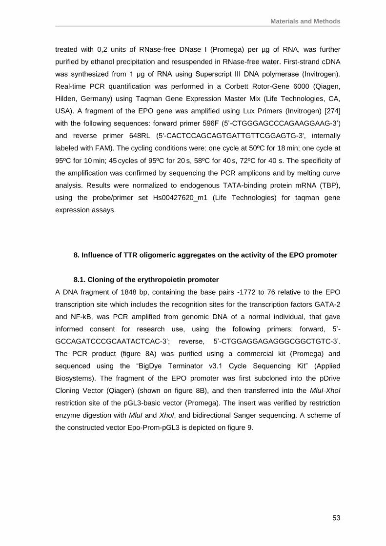
Materials and Methods
53
treated with 0,2 units of RNase-free DNase I (Promega) per µg of RNA, was further
purified by ethanol precipitation and resuspended in RNase-free water. First-strand cDNA
was synthesized from 1 μg of RNA using Superscript III DNA polymerase (Invitrogen).
Real-time PCR quantification was performed in a Corbett Rotor-Gene 6000 (Qiagen,
Hilden, Germany) using Taqman Gene Expression Master Mix (Life Technologies, CA,
USA). A fragment of the EPO gene was amplified using Lux Primers (Invitrogen) [274]
with the following sequences: forward primer 596F (5’-CTGGGAGCCCAGAAGGAAG-3’)
and reverse primer 648RL (5'-CACTCCAGCAGTGATTGTTCGGAGTG-3', internally
labeled with FAM). The cycling conditions were: one cycle at 50ºC for 18 min; one cycle at
95ºC for 10 min; 45 cycles of 95ºC for 20 s, 58ºC for 40 s, 72ºC for 40 s. The specificity of
the amplification was confirmed by sequencing the PCR amplicons and by melting curve
analysis. Results were normalized to endogenous TATA-binding protein mRNA (TBP),
using the probe/primer set Hs00427620_m1 (Life Technologies) for taqman gene
expression assays.
8. Influence of TTR oligomeric aggregates on the activity of the EPO promoter
8.1. Cloning of the erythropoietin promoter
A DNA fragment of 1848 bp, containing the base pairs -1772 to 76 relative to the EPO
transcription site which includes the recognition sites for the transcription factors GATA-2
and NF-kB, was PCR amplified from genomic DNA of a normal individual, that gave
informed consent for research use, using the following primers: forward, 5’-
GCCAGATCCCGCAATACTCAC-3’; reverse, 5’-CTGGAGGAGAGGGCGGCTGTC-3’.
The PCR product (figure 8A) was purified using a commercial kit (Promega) and
sequenced using the “BigDye Terminator v3.1 Cycle Sequencing Kit” (Applied
Biosystems). The fragment of the EPO promoter was first subcloned into the pDrive
Cloning Vector (Qiagen) (shown on figure 8B), and then transferred into the MluI-XhoI
restriction site of the pGL3-basic vector (Promega). The insert was verified by restriction
enzyme digestion with MluI and XhoI, and bidirectional Sanger sequencing. A scheme of
the constructed vector Epo-Prom-pGL3 is depicted on figure 9.
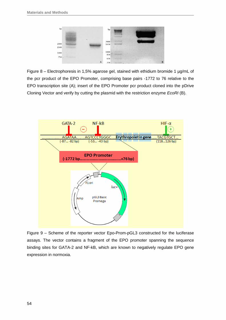
Materials and Methods
54
Figure 8 – Electrophoresis in 1,5% agarose gel, stained with ethidium bromide 1 µg/mL of
the pcr product of the EPO Promoter, comprising base pairs -1772 to 76 relative to the
EPO transcription site (A); insert of the EPO Promoter pcr product cloned into the pDrive
Cloning Vector and verify by cutting the plasmid with the restriction enzyme EcoRI (B).
Figure 9 – Scheme of the reporter vector Epo-Prom-pGL3 constructed for the luciferase
assays. The vector contains a fragment of the EPO promoter spanning the sequence
binding sites for GATA-2 and NF-kB, which are known to negatively regulate EPO gene
expression in normoxia.
Materials and Methods
55
8.2. Transfection with Epo-Prom-pGL3
Hep3B cells were plated into 24-well plates in complete cell medium at a density of 9 x 10⁴
cells per well on the day before the experiment. To evaluate the influence of TTR V30M
on the activity of EPO promoter, Hep3B cells were cotransfected with 7 ng of the control
Renilla luciferase plasmid pTK-RL (Promega) and 350 ng of the Epo-Prom-pGL3 plasmid.
Transfections were done with Lipofectamine 2000 (Invitrogen), using a ratio of 1:3 of DNA
(µg) to lipofectamine (µL) respectively. After 18 hours, the medium was removed and
each TTR preparation was added, at a final concentration of 2 μM. Cells were incubated
for 24 h and luciferase activities were measured with the Dual-Luciferase Assay Kit
(Promega), using a Victor 3 multilabel reader (PerkinElmer). Fold induction was calculated
using Renilla luciferase control normalized values.
8.3. Co-transfection with Epo-Prom-pGL3 and pCG-ATF3
Hep3B cells were plated into 24-well plates in complete cell medium at a density of 9 x 10⁴
cells per well on the day before the experiment. To evaluate the possible influence of the
activating transcription factor ATF3 on the activity of EPO promoter in Hep3B cells
exposed to TTRV30M, cells were cotransfected with 14 ng of the control Renilla luciferase
plasmid pTK-RL (Promega), 350 ng of the Epo-Prom-pGL3 plasmid and 350 ng of pCG-
ATF3 (kindly offered by professor Tsonwin Hai from the Ohio State University).
Transfections, incubations with 2 μM of TTR preparations and measurements of luciferase
activities were done as described in the previous section. Fold induction was calculated
using Renilla luciferase control normalized values.
8.4. Immunofluorescence for NF-kB and GATA-2 on Hep3B cells
Hep3B cells were plated on 4-well chamber slides (Lab-Tek) at a density of 7,5x10³
cells/well and incubated overnight in complete cell media. Solutions of oligomeric or
tetrameric TTRV30M, 2 μM in RPMI with 0,5% FBS, were added to each well and the
cells incubated for 24 hours. The cells were fixed with 4% paraformaldehyde for 10
minutes and permeabilized with cold methanol at -20ºC for 10 min. Cells were washed
with PBS between each incubation. Blocking was performed with 5% BSA in PBS for 1
hour followed by incubation with the primary antibody diluted in 1% BSA for 3 hours at
room temperature. The antibodies used were rabbit anti-human NF-kB p65 polyclonal
antibody (ab7970, Abcam) diluted 1:400 and rabbit anti-human GATA-2 polyclonal
antibody (H-116, Santa Cruz Biotechnology) diluted 1:600. Cells were washed with PBS
and incubated for 1 hour with the secondary antibody (A11008, Alexa Fluor 488 goat anti-
rabbit IgG, Life Technologies) diluted 1:600 in 1% BSA. Slides were mounted in
Fluoroshield with DAPI (Sigma) and analyzed using a fluorescence microscope (Leica).
Materials and Methods
56
8.5. Immunohistochemistry for NF-kB and GATA-2 on FAP renal biopsies
Immunohistochemistry (IHC) was performed on 4 μm thick formalin-fixed paraffin-
embedded sections using standard methods. Treatment with 0.01 M Sodium Citrate
Buffer (pH 6.0) was performed to enhance antigen retrieval. Blocking was performed with
5% BSA in PBS for 1 hour at room temperature, followed by incubation with the antibodies
diluted in 1% BSA in PBS for 3 hours at room temperature. The antibodies used were
rabbit anti-human NF-kB p65 polyclonal antibody (anti-NF-kB, Abcam) diluted 1:400, and
rabbit anti-human GATA-2 polyclonal antibody (Santa Cruz) diluted 1:600. Detection was
carried out with Dako REAL EnVision Detection System, Peroxidase/DAB+, Rabbit/Mouse
(Dako).
9. Statistical analysis
Results of replicated experiments were expressed as mean±SD. Means were compared
using the Student’s t-test. P values <0.05 were considered statistically significant.

RESULTS AND DISCUSSION

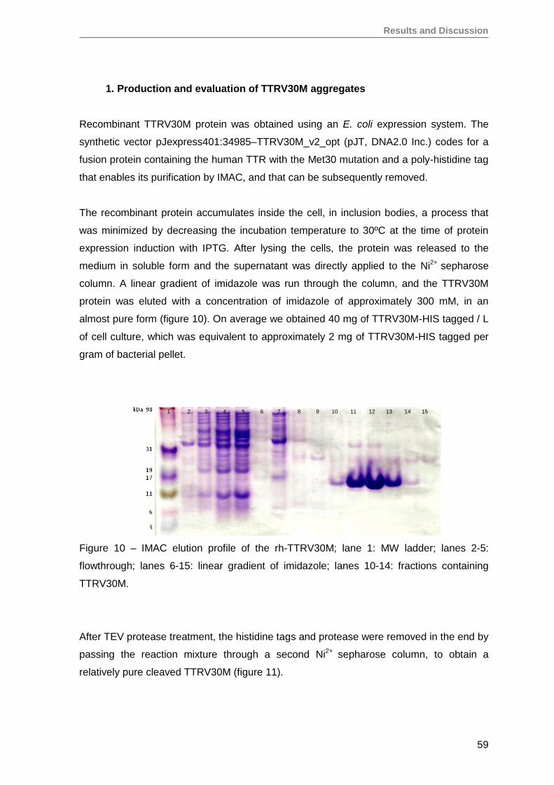
Results and Discussion
59
1. Production and evaluation of TTRV30M aggregates
Recombinant TTRV30M protein was obtained using an E. coli expression system. The
synthetic vector pJexpress401:34985–TTRV30M_v2_opt (pJT, DNA2.0 Inc.) codes for a
fusion protein containing the human TTR with the Met30 mutation and a poly-histidine tag
that enables its purification by IMAC, and that can be subsequently removed.
The recombinant protein accumulates inside the cell, in inclusion bodies, a process that
was minimized by decreasing the incubation temperature to 30ºC at the time of protein
expression induction with IPTG. After lysing the cells, the protein was released to the
medium in soluble form and the supernatant was directly applied to the Ni2+ sepharose
column. A linear gradient of imidazole was run through the column, and the TTRV30M
protein was eluted with a concentration of imidazole of approximately 300 mM, in an
almost pure form (figure 10). On average we obtained 40 mg of TTRV30M-HIS tagged / L
of cell culture, which was equivalent to approximately 2 mg of TTRV30M-HIS tagged per
gram of bacterial pellet.
Figure 10 – IMAC elution profile of the rh-TTRV30M; lane 1: MW ladder; lanes 2-5:
flowthrough; lanes 6-15: linear gradient of imidazole; lanes 10-14: fractions containing
TTRV30M.
After TEV protease treatment, the histidine tags and protease were removed in the end by
passing the reaction mixture through a second Ni2+ sepharose column, to obtain a
relatively pure cleaved TTRV30M (figure 11).
1 2 3 4 5 6 7 8 9 10 11 12 13 14 15
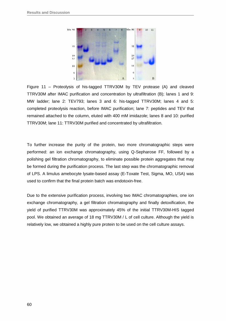
Results and Discussion
60
Figure 11 – Proteolysis of his-tagged TTRV30M by TEV protease (A) and cleaved
TTRV30M after IMAC purification and concentration by ultrafiltration (B); lanes 1 and 9:
MW ladder; lane 2: TEV793; lanes 3 and 6: his-tagged TTRV30M; lanes 4 and 5:
completed proteolysis reaction, before IMAC purification; lane 7: peptides and TEV that
remained attached to the column, eluted with 400 mM imidazole; lanes 8 and 10: purified
TTRV30M; lane 11: TTRV30M purified and concentrated by ultrafiltration.
To further increase the purity of the protein, two more chromatographic steps were
performed: an ion exchange chromatography, using Q-Sepharose FF, followed by a
polishing gel filtration chromatography, to eliminate possible protein aggregates that may
be formed during the purification process. The last step was the chromatographic removal
of LPS. A limulus amebocyte lysate-based assay (E-Toxate Test, Sigma, MO, USA) was
used to confirm that the final protein batch was endotoxin-free.
Due to the extensive purification process, involving two IMAC chromatographies, one ion
exchange chromatography, a gel filtration chromatography and finally detoxification, the
yield of purified TTRV30M was approximately 45% of the initial TTRV30M-HIS tagged
pool. We obtained an average of 18 mg TTRV30M / L of cell culture. Although the yield is
relatively low, we obtained a highly pure protein to be used on the cell culture assays.
A B
1 2 3 4 5 6 7 8 9 10 11
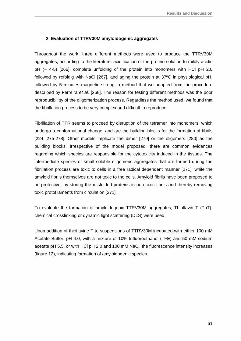
Results and Discussion
61
2. Evaluation of TTRV30M amyloidogenic aggregates
Throughout the work, three different methods were used to produce the TTRV30M
aggregates, according to the literature: acidification of the protein solution to mildly acidic
pH (~ 4-5) [266], complete unfolding of the protein into monomers with HCl pH 2.0
followed by refoldig with NaCl [267], and aging the protein at 37ºC in physiological pH,
followed by 5 minutes magnetic stirring, a method that we adapted from the procedure
described by Ferreira et al. [268]. The reason for testing different methods was the poor
reproducibilitty of the oligomerization process. Regardless the method used, we found that
the fibrillation process to be very complex and difficult to reproduce.
Fibrillation of TTR seems to proceed by disruption of the tetramer into monomers, which
undergo a conformational change, and are the building blocks for the formation of fibrils
[224, 275-278]. Other models implicate the dimer [279] or the oligomers [280] as the
building blocks. Irrespective of the model proposed, there are common evidences
regarding which species are responsible for the cytotoxicity induced in the tissues. The
intermediate species or small soluble oligomeric aggregates that are formed during the
fibrillation process are toxic to cells in a free radical dependent manner [271], while the
amyloid fibrils themselves are not toxic to the cells. Amyloid fibrils have been proposed to
be protective, by storing the misfolded proteins in non-toxic fibrils and thereby removing
toxic protofilaments from circulation [271].
To evaluate the formation of amyloidogenic TTRV30M aggregates, Thioflavin T (ThT),
chemical crosslinking or dynamic light scattering (DLS) were used.
Upon addition of thioflavine T to suspensions of TTRV30M incubated with either 100 mM
Acetate Buffer, pH 4.0, with a mixture of 10% trifluoroethanol (TFE) and 50 mM sodium
acetate pH 5.5, or with HCl pH 2.0 and 100 mM NaCl, the fluorescence intensity increases
(figure 12), indicating formation of amyloidogenic species.
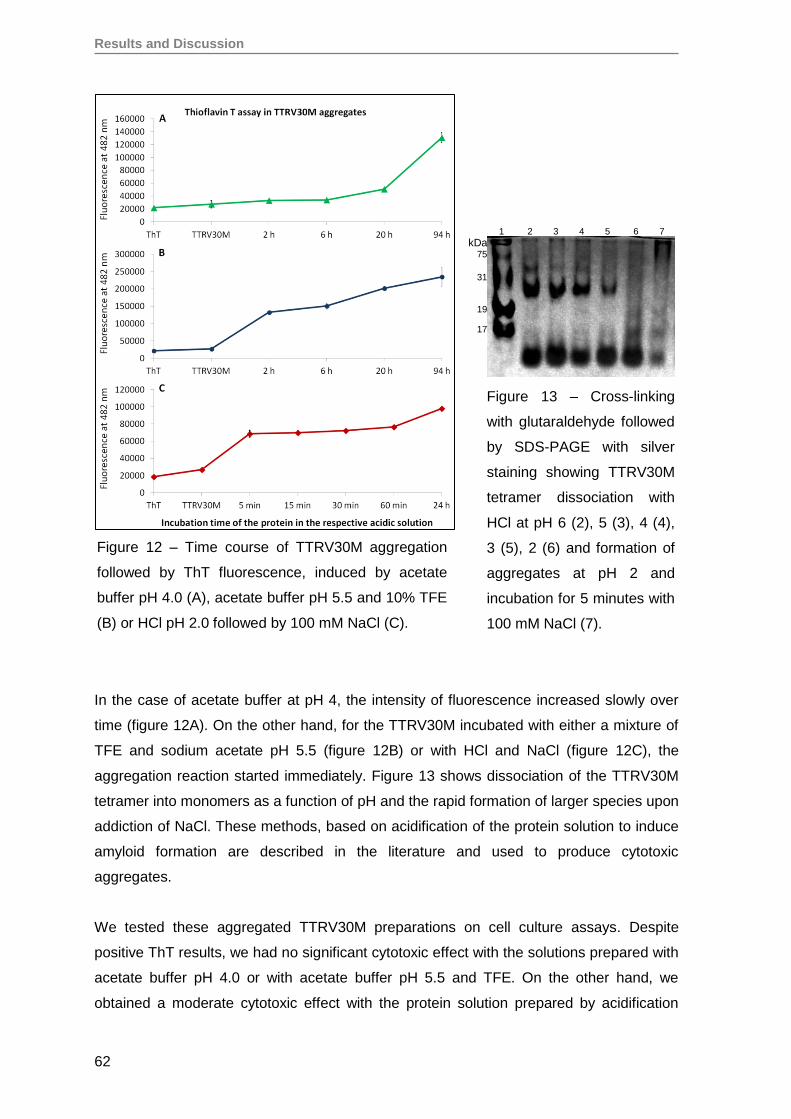
Results and Discussion
62
In the case of acetate buffer at pH 4, the intensity of fluorescence increased slowly over
time (figure 12A). On the other hand, for the TTRV30M incubated with either a mixture of
TFE and sodium acetate pH 5.5 (figure 12B) or with HCl and NaCl (figure 12C), the
aggregation reaction started immediately. Figure 13 shows dissociation of the TTRV30M
tetramer into monomers as a function of pH and the rapid formation of larger species upon
addiction of NaCl. These methods, based on acidification of the protein solution to induce
amyloid formation are described in the literature and used to produce cytotoxic
aggregates.
We tested these aggregated TTRV30M preparations on cell culture assays. Despite
positive ThT results, we had no significant cytotoxic effect with the solutions prepared with
acetate buffer pH 4.0 or with acetate buffer pH 5.5 and TFE. On the other hand, we
obtained a moderate cytotoxic effect with the protein solution prepared by acidification
Figure 12 – Time course of TTRV30M aggregation
followed by ThT fluorescence, induced by acetate
buffer pH 4.0 (A), acetate buffer pH 5.5 and 10% TFE
(B) or HCl pH 2.0 followed by 100 mM NaCl (C).
Figure 13 – Cross-linking
with glutaraldehyde followed
by SDS-PAGE with silver
staining showing TTRV30M
tetramer dissociation with
HCl at pH 6 (2), 5 (3), 4 (4),
3 (5), 2 (6) and formation of
aggregates at pH 2 and
incubation for 5 minutes with
100 mM NaCl (7).
1 2 3 4 5 6 7
kDa 75
31
19
17
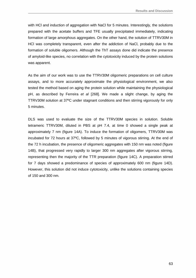
Results and Discussion
63
with HCl and induction of aggregation with NaCl for 5 minutes. Interestingly, the solutions
prepared with the acetate buffers and TFE usually precipitated immediately, indicating
formation of large amorphous aggregates. On the other hand, the solution of TTRV30M in
HCl was completely transparent, even after the addiction of NaCl, probably due to the
formation of soluble oligomers. Although the ThT assays done did indicate the presence
of amyloid-like species, no correlation with the cytotoxicity induced by the protein solutions
was apparent.
As the aim of our work was to use the TTRV30M oligomeric preparations on cell culture
assays, and to more accurately approximate the physiological environment, we also
tested the method based on aging the protein solution while maintaining the physiological
pH, as described by Ferreira et al [268]. We made a slight change, by aging the
TTRV30M solution at 37ºC under stagnant conditions and then stirring vigorously for only
5 minutes.
DLS was used to evaluate the size of the TTRV30M species in solution. Soluble
tetrameric TTRV30M, diluted in PBS at pH 7.4, at time 0 showed a single peak at
approximately 7 nm (figure 14A). To induce the formation of oligomers, TTRV30M was
incubated for 72 hours at 37ºC, followed by 5 minutes of vigorous stirring. At the end of
the 72 h incubation, the presence of oligomeric aggregates with 150 nm was noted (figure
14B), that progressed very rapidly to larger 300 nm aggregates after vigorous stirring,
representing then the majority of the TTR preparation (figure 14C). A preparation stirred
for 7 days showed a predominance of species of approximately 600 nm (figure 14D).
However, this solution did not induce cytotoxicity, unlike the solutions containing species
of 150 and 300 nm.
Results and Discussion
64
Figure 14 - DLS analysis of TTR V30M aggregation for 0 (A), 3 days at 37º C under
stagnant conditions (B), 3 days at 37º C under stagnant conditions followed by 5 minutes
of magnetic stirring (C), 7 days of magnetic stirring (D). Preparation represented in (C)
has a high percentage of small oligomeric species (78%) so was the one used for the cell
culture assays.
TTRV30M is a tetrameric protein with an extensive β-sheet conformation, prone to
dissociation into intermediate molecular species during amyloid fibrillogenesis. Pires et al.
reported that distinct annular oligomeric intermediates formed along both the assembly
and disassembly pathways of TTR, that undergo morphological transitions into spheroid
oligomers and protofibrils [228]. We induced protein fibrillation in vitro by aging a solution
of TTRV30M at physiological pH and temperature, which is evidence for the natural
Results and Discussion
65
instability of the quaternary structure of the protein and its tendency to aggregate and form
amyloidogenic fibers. We saw that after 72 hours of incubation under stagnant conditions,
species of molecular size corresponding to early oligomers are already in solution, albeit
in small amounts relative to the predominant tetrameric form. Stirring this solution for 5
minutes was sufficient to trigger the rapid formation of larger species.
TTRV30M preparations obtained by aging at physiological pH followed by magnetic
stirring for 5 minutes (figure 14C) and preparations obtained by dissociation of the
tetramer into monomers at pH 2.0 followed by refolding with NaCl (figure 12C) were the
final preparations of oligomeric TTRV30M used for the cell culture assays, as we saw that
these were the most cytotoxic species.
Results and Discussion
66
3. TTRV30M oligomeric aggregates compromise cell viability of both
immortalized SH-SY5Y, Hep3B and HEK293T cell lines, as well as of primary
RPE and RPC cells
The immortalized cell lines Hep3B, SH-SY5Y and HEK293T, the primary RPE cells and
the renal progenitor cells RPC were exposed to TTR preparations in order to evaluate cell
viability. Oligomeric preparations were prepared by unfolding with HCl and refolding with
NaCl, or by aging the protein solution at pH 7.4 and 37ºC followed by 5 minutes of stirring.
Hep3B and SH-SY5Y were exposed to oligomers produced by both methods. HEK293T
and RPC were exposed only to oligomers produced by unfolding and refolding. RPE cells
were exposed only to oligomers produced by aging the TTRV30M at physiological pH and
temperature.
3.1. Cell viability assays
A non-radioactive cell proliferation assay (MTT or MTS) was used to evaluate the
cytotoxicity of TTRV30M preparations. The results obtained for each cell type were similar
regardless of the method used, so we calculated the average of all the experiments and
respective replicates in which these oligomers were cytotoxic, for each cell type.
Preliminary experiments were performed with RPC cells exposed to protein
concentrations of 0.5, 1.5 and 3 µM and with an incubation time of 72 hours. A
concentration dependent reduction in cell viability was observed (figure 15).
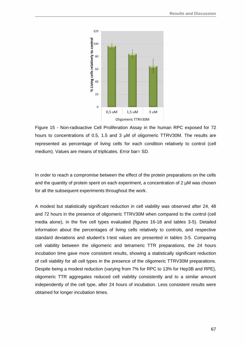
Results and Discussion
67
Figure 15 - Non-radioactive Cell Proliferation Assay in the human RPC exposed for 72
hours to concentrations of 0.5, 1.5 and 3 µM of oligomeric TTRV30M. The results are
represented as percentage of living cells for each condition relatively to control (cell
medium). Values are means of triplicates. Error bar= SD.
In order to reach a compromise between the effect of the protein preparations on the cells
and the quantity of protein spent on each experiment, a concentration of 2 µM was chosen
for all the subsequent experiments throughout the work.
A modest but statistically significant reduction in cell viability was observed after 24, 48
and 72 hours in the presence of oligomeric TTRV30M when compared to the control (cell
media alone), in the five cell types evaluated (figures 16-18 and tables 3-5). Detailed
information about the percentages of living cells relatively to controls, and respective
standard deviations and student’s t-test values are presented in tables 3-5. Comparing
cell viability between the oligomeric and tetrameric TTR preparations, the 24 hours
incubation time gave more consistent results, showing a statistically significant reduction
of cell viability for all cell types in the presence of the oligomeric TTRV30M preparations.
Despite being a modest reduction (varying from 7% for RPC to 13% for Hep3B and RPE),
oligomeric TTR aggregates reduced cell viability consistently and to a similar amount
independently of the cell type, after 24 hours of incubation. Less consistent results were
obtained for longer incubation times.
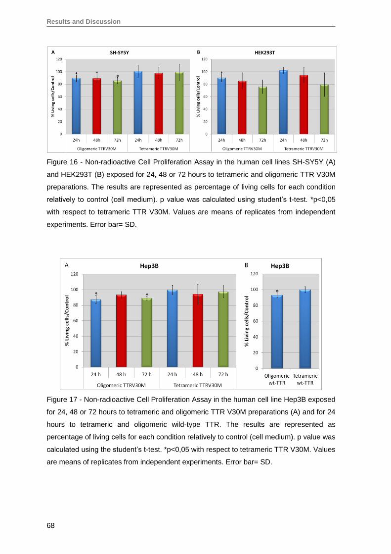
Results and Discussion
68
Figure 16 - Non-radioactive Cell Proliferation Assay in the human cell lines SH-SY5Y (A)
and HEK293T (B) exposed for 24, 48 or 72 hours to tetrameric and oligomeric TTR V30M
preparations. The results are represented as percentage of living cells for each condition
relatively to control (cell medium). p value was calculated using student’s t-test. *p<0,05
with respect to tetrameric TTR V30M. Values are means of replicates from independent
experiments. Error bar= SD.
Figure 17 - Non-radioactive Cell Proliferation Assay in the human cell line Hep3B exposed
for 24, 48 or 72 hours to tetrameric and oligomeric TTR V30M preparations (A) and for 24
hours to tetrameric and oligomeric wild-type TTR. The results are represented as
percentage of living cells for each condition relatively to control (cell medium). p value was
calculated using the student’s t-test. *p<0,05 with respect to tetrameric TTR V30M. Values
are means of replicates from independent experiments. Error bar= SD.
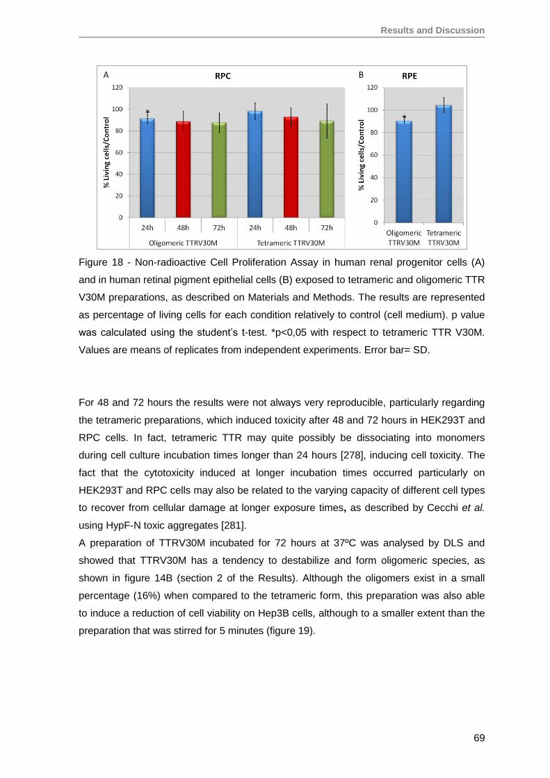
Results and Discussion
69
Figure 18 - Non-radioactive Cell Proliferation Assay in human renal progenitor cells (A)
and in human retinal pigment epithelial cells (B) exposed to tetrameric and oligomeric TTR
V30M preparations, as described on Materials and Methods. The results are represented
as percentage of living cells for each condition relatively to control (cell medium). p value
was calculated using the student’s t-test. *p<0,05 with respect to tetrameric TTR V30M.
Values are means of replicates from independent experiments. Error bar= SD.
For 48 and 72 hours the results were not always very reproducible, particularly regarding
the tetrameric preparations, which induced toxicity after 48 and 72 hours in HEK293T and
RPC cells. In fact, tetrameric TTR may quite possibly be dissociating into monomers
during cell culture incubation times longer than 24 hours [278], inducing cell toxicity. The
fact that the cytotoxicity induced at longer incubation times occurred particularly on
HEK293T and RPC cells may also be related to the varying capacity of different cell types
to recover from cellular damage at longer exposure times, as described by Cecchi et al.
using HypF-N toxic aggregates [281].
A preparation of TTRV30M incubated for 72 hours at 37ºC was analysed by DLS and
showed that TTRV30M has a tendency to destabilize and form oligomeric species, as
shown in figure 14B (section 2 of the Results). Although the oligomers exist in a small
percentage (16%) when compared to the tetrameric form, this preparation was also able
to induce a reduction of cell viability on Hep3B cells, although to a smaller extent than the
preparation that was stirred for 5 minutes (figure 19).
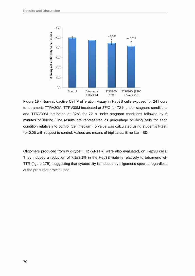
Results and Discussion
70
Figure 19 - Non-radioactive Cell Proliferation Assay in Hep3B cells exposed for 24 hours
to tetrameric TTRV30M, TTRV30M incubated at 37ºC for 72 h under stagnant conditions
and TTRV30M incubated at 37ºC for 72 h under stagnant conditions followed by 5
minutes of stirring. The results are represented as percentage of living cells for each
condition relatively to control (cell medium). p value was calculated using student’s t-test.
*p<0,05 with respect to control. Values are means of triplicates. Error bar= SD.
Oligomers produced from wild-type TTR (wt-TTR) were also evaluated, on Hep3B cells.
They induced a reduction of 7.1±3.1% in the Hep3B viability relatively to tetrameric wt-
TTR (figure 17B), suggesting that cytotoxicity is induced by oligomeric species regardless
of the precursor protein used.
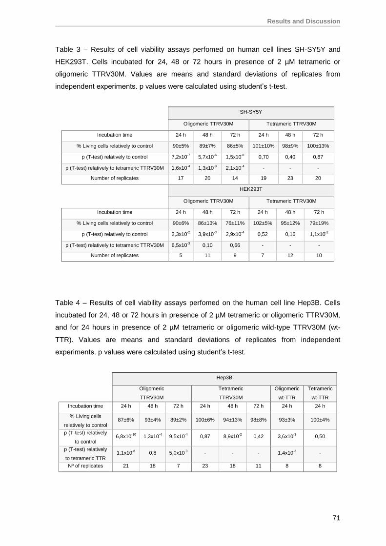
Results and Discussion
71
Table 3 – Results of cell viability assays perfomed on human cell lines SH-SY5Y and
HEK293T. Cells incubated for 24, 48 or 72 hours in presence of 2 µM tetrameric or
oligomeric TTRV30M. Values are means and standard deviations of replicates from
independent experiments. p values were calculated using student’s t-test.
SH-SY5Y
Oligomeric TTRV30M Tetrameric TTRV30M
Incubation time 24 h 48 h 72 h 24 h 48 h 72 h
% Living cells relatively to control 90±5% 89±7% 86±5% 101±10% 98±9% 100±13%
p (T-test) relatively to control 7,2x10⁻⁷
5,7x10⁻⁶
1,5x10⁻⁸
0,70 0,40 0,87
p (T-test) relatively to tetrameric TTRV30M 1,6x10⁻⁴
1,3x10⁻³
2,1x10⁻⁴
- - -
Number of replicates 17 20 14 19 23 20
HEK293T
Oligomeric TTRV30M Tetrameric TTRV30M
Incubation time 24 h 48 h 72 h 24 h 48 h 72 h
% Living cells relatively to control 90±6% 86±13% 76±11% 102±5% 95±12% 79±19%
p (T-test) relatively to control 2,3x10⁻²
3,9x10⁻³
2,9x10⁻⁴
0,52 0,16 1,1x10⁻²
p (T-test) relatively to tetrameric TTRV30M 6,5x10⁻³
0,10 0,66 - - -
Number of replicates 5 11 9 7 12 10
Table 4 – Results of cell viability assays perfomed on the human cell line Hep3B. Cells
incubated for 24, 48 or 72 hours in presence of 2 µM tetrameric or oligomeric TTRV30M,
and for 24 hours in presence of 2 µM tetrameric or oligomeric wild-type TTRV30M (wt-
TTR). Values are means and standard deviations of replicates from independent
experiments. p values were calculated using student’s t-test.
Hep3B
Oligomeric
TTRV30M
Tetrameric
TTRV30M
Oligomeric
wt-TTR
Tetrameric
wt-TTR
24 h
90±2%
8,6E-4
2,0E-2
4
Incubation time 24 h 48 h 72 h 24 h 48 h 72 h 24 h 24 h
% Living cells
relatively to control 87±6% 93±4% 89±2% 100±6% 94±13% 98±8% 93±3% 100±4%
p (T-test) relatively
to control 6,8x10
-10 1,3x10
-4 9,5x10
-4 0,87 8,9x10
-2 0,42 3,6x10
-3 0,50
p (T-test) relatively
to tetrameric TTR 1,1x10
-8 0,8 5,0x10
-3 - - - 1,4x10
-3 -
Nº of replicates 21 18 7 23 18 11 8 8
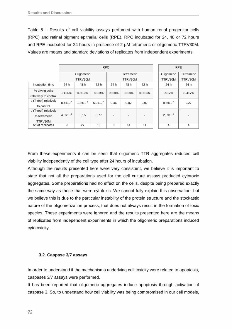
Results and Discussion
72
Table 5 – Results of cell viability assays perfomed with human renal progenitor cells
(RPC) and retinal pigment epithelial cells (RPE). RPC incubated for 24, 48 or 72 hours
and RPE incubated for 24 hours in presence of 2 µM tetrameric or oligomeric TTRV30M.
Values are means and standard deviations of replicates from independent experiments.
RPC RPE
Oligomeric
TTRV30M
Tetrameric
TTRV30M
Oligomeric
TTRV30M
Tetrameric
TTRV30M
24 h
90±2%
8,6E-4
2,0E-2
4
Incubation time 24 h 48 h 72 h 24 h 48 h 72 h 24 h 24 h
% Living cells
relatively to control 91±4% 89±10% 88±9% 98±8% 93±8% 89±16% 90±2% 104±7%
p (T-test) relatively
to control 8,4x10
-4 1,8x10
-5 6,9x10
-4 0,46 0,02 0,07 8,6x10
-4 0,27
p (T-test) relatively
to tetrameric
TTRV30M
4,5x10⁻²
0,15 0,77 - - - 2,0x10⁻²
-
Nº of replicates 9 27 16 8 14 11 4 4
From these experiments it can be seen that oligomeric TTR aggregates reduced cell
viability independently of the cell type after 24 hours of incubation.
Although the results presented here were very consistent, we believe it is important to
state that not all the preparations used for the cell culture assays produced cytotoxic
aggregates. Some preparations had no effect on the cells, despite being prepared exactly
the same way as those that were cytotoxic. We cannot fully explain this observation, but
we believe this is due to the particular instability of the protein structure and the stockastic
nature of the oligomerization process, that does not always result in the formation of toxic
species. These experiments were ignored and the results presented here are the means
of replicates from independent experiments in which the oligomeric preparations induced
cytotoxicity.
3.2. Caspase 3/7 assays
In order to understand if the mechanisms underlying cell toxicity were related to apoptosis,
caspases 3/7 assays were performed.
It has been reported that oligomeric aggregates induce apoptosis through activation of
caspase 3. So, to understand how cell viability was being compromised in our cell models,
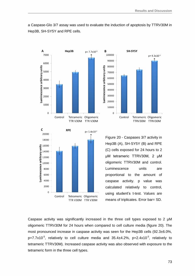
Results and Discussion
73
a Caspase-Glo 3/7 assay was used to evaluate the induction of apoptosis by TTRV30M in
Hep3B, SH-SY5Y and RPE cells.
Caspase activity was significantly increased in the three cell types exposed to 2 µM
oligomeric TTRV30M for 24 hours when compared to cell culture media (figure 20). The
most pronounced increase in caspase activity was seen for the Hep3B cells (92.3±6.0%,
p=7.7x10⁻⁵, relatively to cell culture media and 35.4±4.2%, p=2.4x10⁻⁵, relatively to
tetrameric TTRV30M). Increased caspase activity was also observed with exposure to the
tetrameric form in the three cell types.
Figure 20 - Caspases 3/7 activity in
Hep3B (A), SH-SY5Y (B) and RPE
(C) cells exposed for 24 hours to 2
μM tetrameric TTRV30M, 2 μM
oligomeric TTRV30M and control.
Luminescence units are
proportional to the amount of
caspase activity. p value was
calculated relatively to control,
using student’s t-test. Values are
means of triplicates. Error bar= SD.
Results and Discussion
74
The caspase assay is rather more sensitive than the MTS assay, therefore it is not
surprising that some activity is detected after 24 hours of incubation at 37ºC with
tetrameric TTRV30M in the caspase assay, but not with MTS. As already mentioned for
the viability assays, tetrameric protein solution may begin to form monomers or oligomers
that even in small quantities could be sufficient to activate caspases. Our assumption is
supported by the fact that DLS of TTRV30M after 72 hours incubation at 37ºC showed
that this protein has a tendency form oligomeric species, and was able to induce a
reduction of cell viability (figures 14B and 19).
Early oligomeric aggregates have been shown to be more cytotoxic than mature amyloid
fibrils [229, 270-272]. Similar reduction levels of cell viability induced by the oligomeric
TTRV30M preparations after 24 hours of incubation were observed for the five different
cell types evaluated: human hepatoma cell line Hep3B, human neuroblastoma cell line
SH-SY5Y, human embrionic kidney HEK293T, human renal progenitor cells (RPC) and
primary retinal pigment epithelial cells (RPE). For three of these cell types: Hep3B, SH-
SY5Y and RPE, a caspase assay was performed. Together with the reduction of cell
viability, a concomitant increase in caspase 3/7 activity was seen in the cells exposed to
oligomeric TTRV30M aggregates.
These results agree with previous studies performed using other cell types, mainly
neuronal cells (neuroblastoma) [106, 229, 250, 270-272], and primary human umbilical
vein endothelial cells (HUVECs) [246], and implicate apoptosis in loss of viability.
Interestingly, this cytotoxic effect induced by aggregates seems to be independent of the
cell type, contrary to what was reported by Cecchi et al. Using aggregates of HypF-N, a
prokaryotic peptide domain, that display the same properties of aggregates produced by
disease-associated peptides and proteins, they showed that different cell lines have
different susceptibilities to damage and apoptosis, and this susceptibility was inversely
related to membrane content in cholesterol, to total anti-oxidant capacity of the cell and to
the ability of the cell to maintain the balance of intracellular free Ca2+ [282]. However, the
same group reported later that such differences are more pronounced in longer exposure
times due to different recovering capacity from cellular damage [281]. In fact we saw a
similar cytotoxic effect between the various cell types in the shortest exposure time to the
aggregates. Increasing the incubation time, the results became less reproducible, an
increased toxic effect being observed in some cases but a reduction of cytotoxicity in most
of them. This may be due to continuing fibrillogenesis, leading to the formation of larger
species which may no longer be toxic. According to Cecchi et al, the reduction of
cytotoxicty at longer incubation times may also be due to recovery from cellular damage.
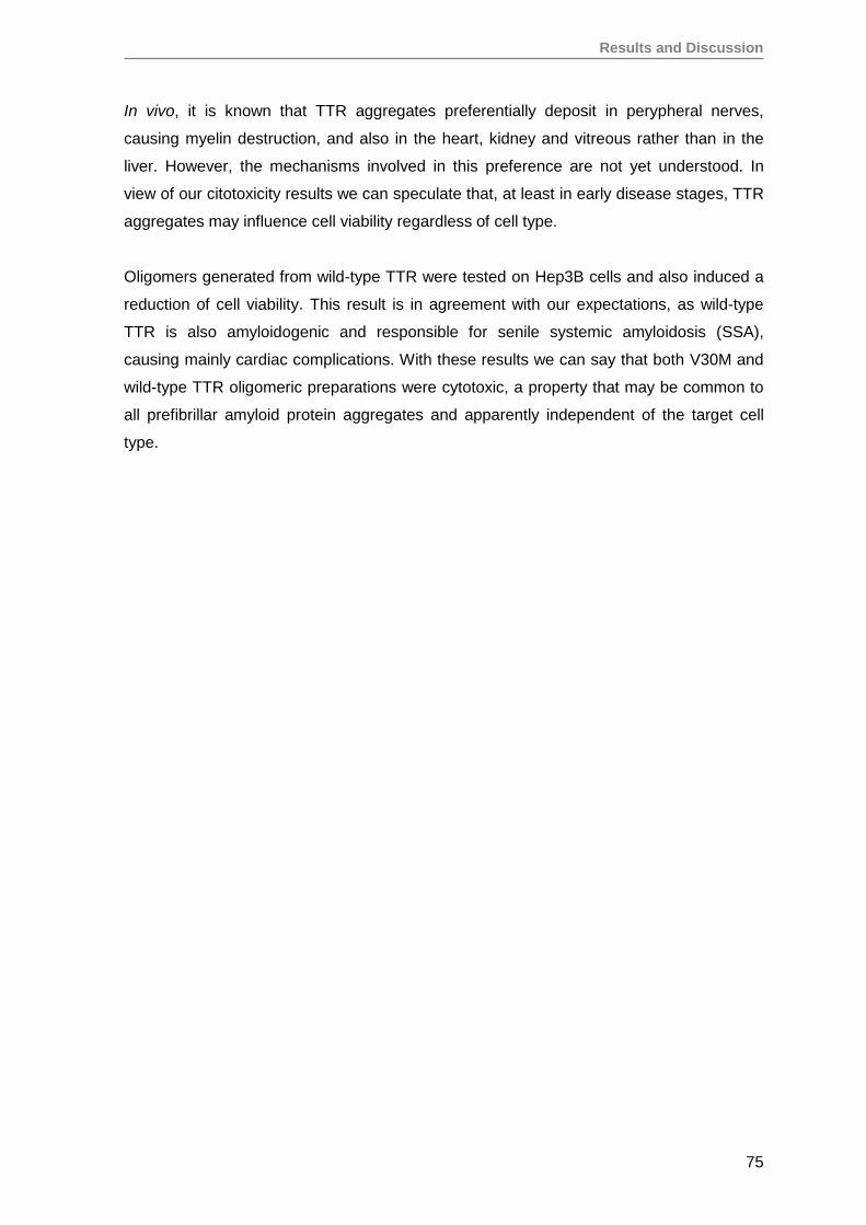
Results and Discussion
75
In vivo, it is known that TTR aggregates preferentially deposit in perypheral nerves,
causing myelin destruction, and also in the heart, kidney and vitreous rather than in the
liver. However, the mechanisms involved in this preference are not yet understood. In
view of our citotoxicity results we can speculate that, at least in early disease stages, TTR
aggregates may influence cell viability regardless of cell type.
Oligomers generated from wild-type TTR were tested on Hep3B cells and also induced a
reduction of cell viability. This result is in agreement with our expectations, as wild-type
TTR is also amyloidogenic and responsible for senile systemic amyloidosis (SSA),
causing mainly cardiac complications. With these results we can say that both V30M and
wild-type TTR oligomeric preparations were cytotoxic, a property that may be common to
all prefibrillar amyloid protein aggregates and apparently independent of the target cell
type.
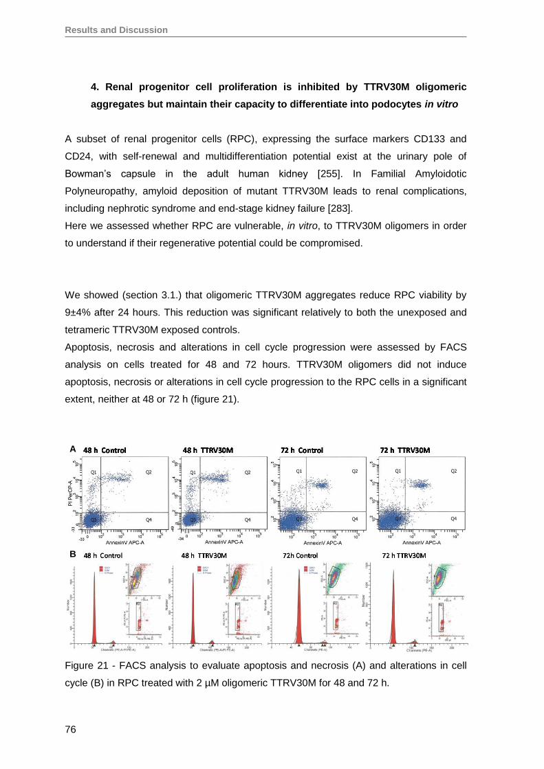
Results and Discussion
76
4. Renal progenitor cell proliferation is inhibited by TTRV30M oligomeric
aggregates but maintain their capacity to differentiate into podocytes in vitro
A subset of renal progenitor cells (RPC), expressing the surface markers CD133 and
CD24, with self-renewal and multidifferentiation potential exist at the urinary pole of
Bowman’s capsule in the adult human kidney [255]. In Familial Amyloidotic
Polyneuropathy, amyloid deposition of mutant TTRV30M leads to renal complications,
including nephrotic syndrome and end-stage kidney failure [283].
Here we assessed whether RPC are vulnerable, in vitro, to TTRV30M oligomers in order
to understand if their regenerative potential could be compromised.
We showed (section 3.1.) that oligomeric TTRV30M aggregates reduce RPC viability by
9±4% after 24 hours. This reduction was significant relatively to both the unexposed and
tetrameric TTRV30M exposed controls.
Apoptosis, necrosis and alterations in cell cycle progression were assessed by FACS
analysis on cells treated for 48 and 72 hours. TTRV30M oligomers did not induce
apoptosis, necrosis or alterations in cell cycle progression to the RPC cells in a significant
extent, neither at 48 or 72 h (figure 21).
Figure 21 - FACS analysis to evaluate apoptosis and necrosis (A) and alterations in cell
cycle (B) in RPC treated with 2 µM oligomeric TTRV30M for 48 and 72 h.
A B
Results and Discussion
77
CD133+CD24+ RPC have an inherent capacity to differentiate into podocytes when
cultivated in VRAD medium. The influence of oligomeric TTRV30M on the regenerative
capacity of these progenitor cells was assessed.
Podocytes express nephrin so the relative expression level of this marker was evaluated
by real-time pcr to confirm differentiation. The results varied between experiments, but on
average we obtained similar results between the controls and the cells exposed to
TTRV30M aggregates, both on differentiation and non-differentiation media (Figure 22).
Figure 22 - Differentiation of renal progenitor cells into podocytes. Assessment by
quantitative RT-PCR of fold increase mRNA levels for the podocyte marker nephrin and
the housekeeping gene GAPDH.
TTRV30M aggregates reduced proliferation of renal progenitor cells. However, in the
apoptosis and cell cycle progression assays no significant alterations were observed.
Also, in the differentiation experiments we saw no alterations in the expression level of the
nephrin gene, a marker for podocytes, relatively to controls, so the inherent capacity of
these progenitor cells to differentiate into podocytes was apparently not affected by the
oligomers. These results are in agreement with a study of Petrakis et al. They saw, with a
transgenic mouse model of TTRV30M-related amyloidosis, that TTRV30M deposition has
deleterious effects on glomerular basement membrane thickness and podocyte foot
process width, but without affecting nephrin and podocin gene expression [284].
A study from Neri et al, using murine embryonic stem (ES) cells and haematopoietic
progenitor (HP) cells exposed to oligomers of Aβ42 showed that ES, but not HP,
displayed some impaired viability but their differentiation was not affected by these
oligomers [285], which is in agreement with our results.
Results and Discussion
78
From this study we can say that TTRV30M oligomers inhibit RPC proliferation but do not
influence their capacity to differentiate into functionally mature podocytes, and thus should
not compromise tissue regeneration. These resident progenitor cells can also induce
regeneration of tubular structures of different portions of the nephron, which can be critical
for preventing irreversible renal failure, and could be useful in cell therapies, particularly in
FAP.
We have previously shown that EPO expression occurs in distal tubular cells, podocytes
and cells of the cortical collecting ducts, in normoxic FAP patients [138]. Although there is
no data regarding the ability of RPC to differentiate into EPO-producing cells, they can
differentiate into podocytes and tubular cells, which could be important to maintain EPO
production.
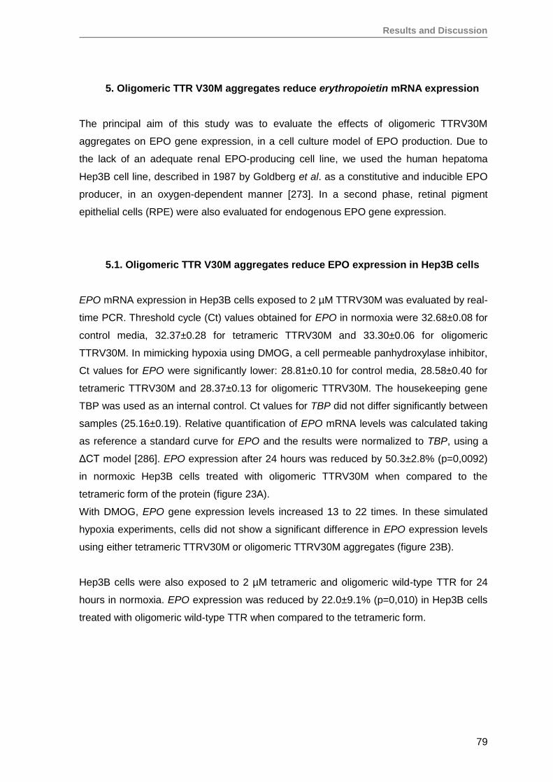
Results and Discussion
79
5. Oligomeric TTR V30M aggregates reduce erythropoietin mRNA expression
The principal aim of this study was to evaluate the effects of oligomeric TTRV30M
aggregates on EPO gene expression, in a cell culture model of EPO production. Due to
the lack of an adequate renal EPO-producing cell line, we used the human hepatoma
Hep3B cell line, described in 1987 by Goldberg et al. as a constitutive and inducible EPO
producer, in an oxygen-dependent manner [273]. In a second phase, retinal pigment
epithelial cells (RPE) were also evaluated for endogenous EPO gene expression.
5.1. Oligomeric TTR V30M aggregates reduce EPO expression in Hep3B cells
EPO mRNA expression in Hep3B cells exposed to 2 µM TTRV30M was evaluated by real-
time PCR. Threshold cycle (Ct) values obtained for EPO in normoxia were 32.68±0.08 for
control media, 32.37±0.28 for tetrameric TTRV30M and 33.30±0.06 for oligomeric
TTRV30M. In mimicking hypoxia using DMOG, a cell permeable panhydroxylase inhibitor,
Ct values for EPO were significantly lower: 28.81±0.10 for control media, 28.58±0.40 for
tetrameric TTRV30M and 28.37±0.13 for oligomeric TTRV30M. The housekeeping gene
TBP was used as an internal control. Ct values for TBP did not differ significantly between
samples (25.16±0.19). Relative quantification of EPO mRNA levels was calculated taking
as reference a standard curve for EPO and the results were normalized to TBP, using a
ΔCT model [286]. EPO expression after 24 hours was reduced by 50.3±2.8% (p=0,0092)
in normoxic Hep3B cells treated with oligomeric TTRV30M when compared to the
tetrameric form of the protein (figure 23A).
With DMOG, EPO gene expression levels increased 13 to 22 times. In these simulated
hypoxia experiments, cells did not show a significant difference in EPO expression levels
using either tetrameric TTRV30M or oligomeric TTRV30M aggregates (figure 23B).
Hep3B cells were also exposed to 2 µM tetrameric and oligomeric wild-type TTR for 24
hours in normoxia. EPO expression was reduced by 22.0±9.1% (p=0,010) in Hep3B cells
treated with oligomeric wild-type TTR when compared to the tetrameric form.
Results and Discussion
80
Figure 23 - Relative expression of erythropoietin in Hep3B cells exposed to TTR V30M for
24 hours. (A) Relative expression levels of EPO, normalized for TBP, in normoxia. (B)
Relative expression levels of EPO both in normoxia and simulated hypoxia (induced by
DMOG). p value was calculated relatively to tetrameric TTR V30M experiments, using
student’s t-test. *p<0,05. Values are means of duplicates. Error Bar= SD.
The EPO gene has a hypoxia responsive element at the EPO 3’ enhancer site to which
hypoxia inducible factor (HIF) can bind [287-289]. In normoxia, the α-chain of HIF is
hydroxylated and degraded by the proteasome. In hypoxia the prolyl hydroxylases (PHD)
and aspaginyl hydroxylase factor inhibiting HIF (FIH) are inhibited, the α-chain of HIF is
not degraded and binds to the β-chain, which is constitutively expressed, and HIF
accumulates, inducing gene expression [290-291]. To mimic hypoxia, we used the cell
permeable panhydroxylase inhibitor dimethyloxalylglycine (DMOG).
We found that exposure of normoxic Hep3B cells to oligomeric TTR V30M significantly
reduces EPO mRNA expression. However, Hep3B cells treated with DMOG did not show
significant differences in EPO mRNA levels when exposed to tetrameric or oligomeric TTR
V30M. Under these simulated hypoxia conditions, Hep3B cells up-regulated EPO
expression 13 to 20 times. It is possible that this large increase in EPO expression masks
any effect of exposure to TTR aggregates on the gene expression level, or, alternatively,
different regulatory mechanisms of EPO production could be involved in normoxia and
hypoxia, altering the cellular response to the presence of oligomeric aggregates.
Our previous work using patient kidney biopsies evaluated EPO expression in normoxia,
and suggested that differences in the regulation of this gene in normoxia and hypoxia are
Results and Discussion
81
possible [138]. Recently, Nagai et al. explored this subject and demonstrated that, in mice,
EPO mRNA expression occurs in renal tubular cells in normoxia, as we have shown for
the human ATTRV30M amyloidosis kidney, while in hypoxia it occurs largely in peritubular
cells, strengthening the case for different regulatory mechanisms for EPO expression in
normoxia and hypoxia [141].
Anemia in FAP patients sometimes precedes overt clinical disease and persists even after
liver transplantation. There is evidence that after liver transplantation, the wild-type TTR
can still form amyloid deposits and that there is progression and maybe even acceleration
of amyloid deposition [59, 292]. In order to understand the persistence of low EPO
production after liver transplantation, we produced oligomeric aggregates from normal
TTR. We saw that Hep3B cells exposed to oligomeric wild-type TTR aggregates are also
capable of reducing EPO mRNA expression when compared to tetrameric TTR controls.
Although these studies were performed in vitro with a hepatoma cell line, they support the
hypothesis that cytotoxic oligomeric species may be involved in the genesis of anemia in
FAP patients. Besides this evidence, we recently showed that EPO concentrations in the
aqueous humor of glaucomatous eyes of non-amyloidotic patients are significantly
increased relatively to normal non-glaucomatous eyes, probably with a protective role
[116]. However, in glaucomatous eyes of FAP ATTRV30M patients the EPO levels did not
increase and maintained similar levels to those of control eyes [116]. These results show
an inability of these patients to upregulate EPO production also locally in the retina.
5.2. Oligomeric TTR V30M aggregates reduce EPO expression in RPE cells
Beyond using Hep3B cells, a tumoral cell line, primary cell cultures of retinal pigment
epithelial cells (RPE) were also evaluated for EPO gene expression. RPE cells when are
cultivated acquire a hexagonal conformation and are pigmented (figure 24). As the cells
divide, they gradually lose their pigment and start differentiating, acquiring a more
elongated and fibroblast-like conformation (figures 25 and 26). Preliminary results showed
that RPE isolated from the eye of an adult human cadaveric donor express EPO mRNA,
but only on early passages (P0 and P1).
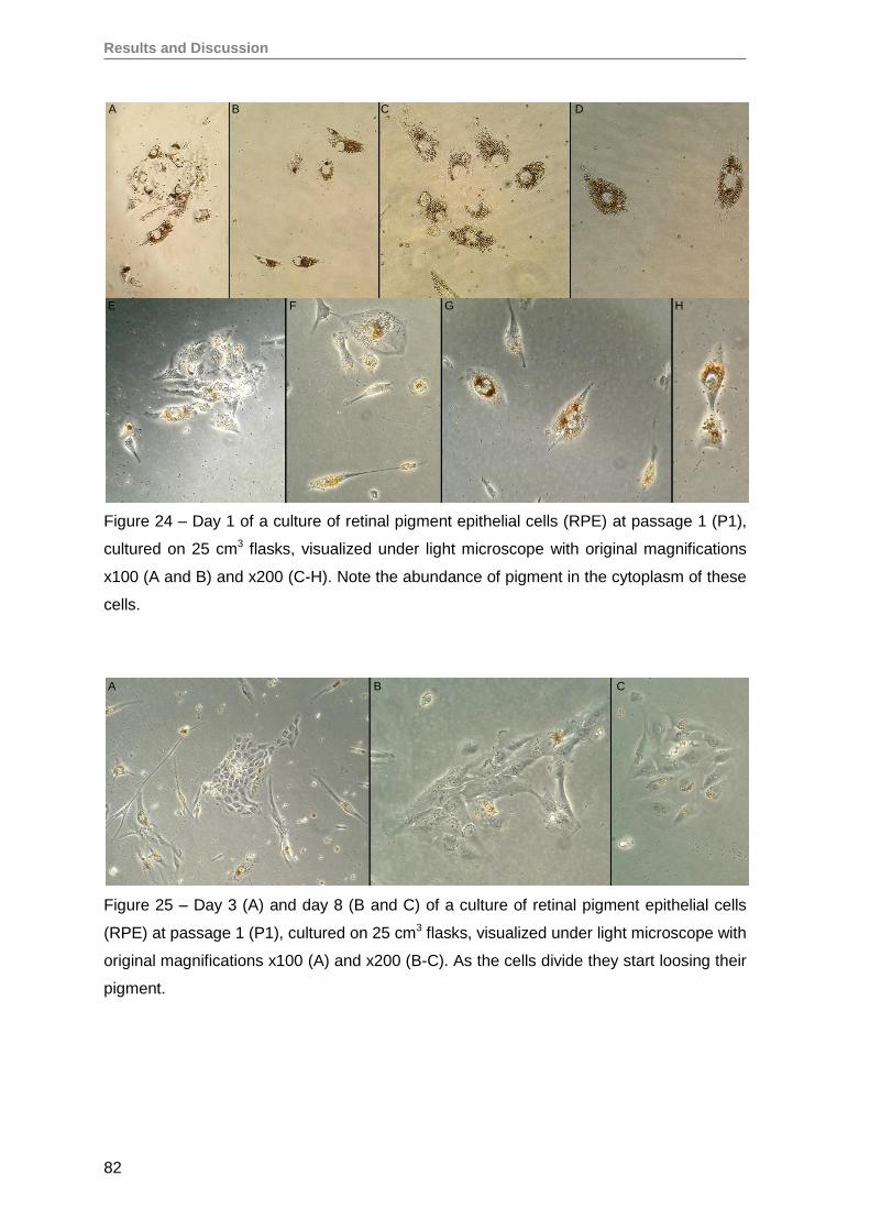
Results and Discussion
82
Figure 24 – Day 1 of a culture of retinal pigment epithelial cells (RPE) at passage 1 (P1),
cultured on 25 cm3 flasks, visualized under light microscope with original magnifications
x100 (A and B) and x200 (C-H). Note the abundance of pigment in the cytoplasm of these
cells.
Figure 25 – Day 3 (A) and day 8 (B and C) of a culture of retinal pigment epithelial cells
(RPE) at passage 1 (P1), cultured on 25 cm3 flasks, visualized under light microscope with
original magnifications x100 (A) and x200 (B-C). As the cells divide they start loosing their
pigment.
A B C D
E F G H
A B C
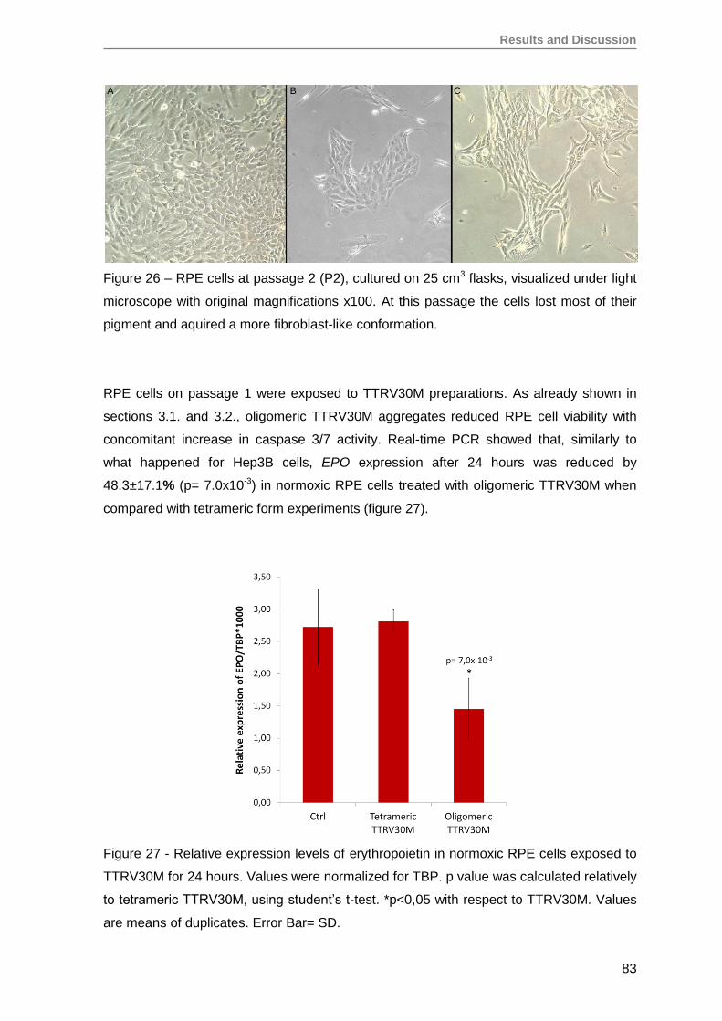
Results and Discussion
83
Figure 26 – RPE cells at passage 2 (P2), cultured on 25 cm3 flasks, visualized under light
microscope with original magnifications x100. At this passage the cells lost most of their
pigment and aquired a more fibroblast-like conformation.
RPE cells on passage 1 were exposed to TTRV30M preparations. As already shown in
sections 3.1. and 3.2., oligomeric TTRV30M aggregates reduced RPE cell viability with
concomitant increase in caspase 3/7 activity. Real-time PCR showed that, similarly to
what happened for Hep3B cells, EPO expression after 24 hours was reduced by
48.3±17.1% (p= 7.0x10⁻³) in normoxic RPE cells treated with oligomeric TTRV30M when
compared with tetrameric form experiments (figure 27).
Figure 27 - Relative expression levels of erythropoietin in normoxic RPE cells exposed to
TTRV30M for 24 hours. Values were normalized for TBP. p value was calculated relatively
to tetrameric TTRV30M, using student’s t-test. *p<0,05 with respect to TTRV30M. Values
are means of duplicates. Error Bar= SD.
A B C

Results and Discussion
84
These are unpublished preliminary results. Further experiments are needed to validate
these results and to better characterize this primary cell culture model, which responded
to DMOG with a significant increase in EPO expression. As RPE cells are producers of
both TTR and EPO in the eye, it may be a good model for a more detailed study of the
mechanisms behind the inhibition of EPO production in FAP patients.
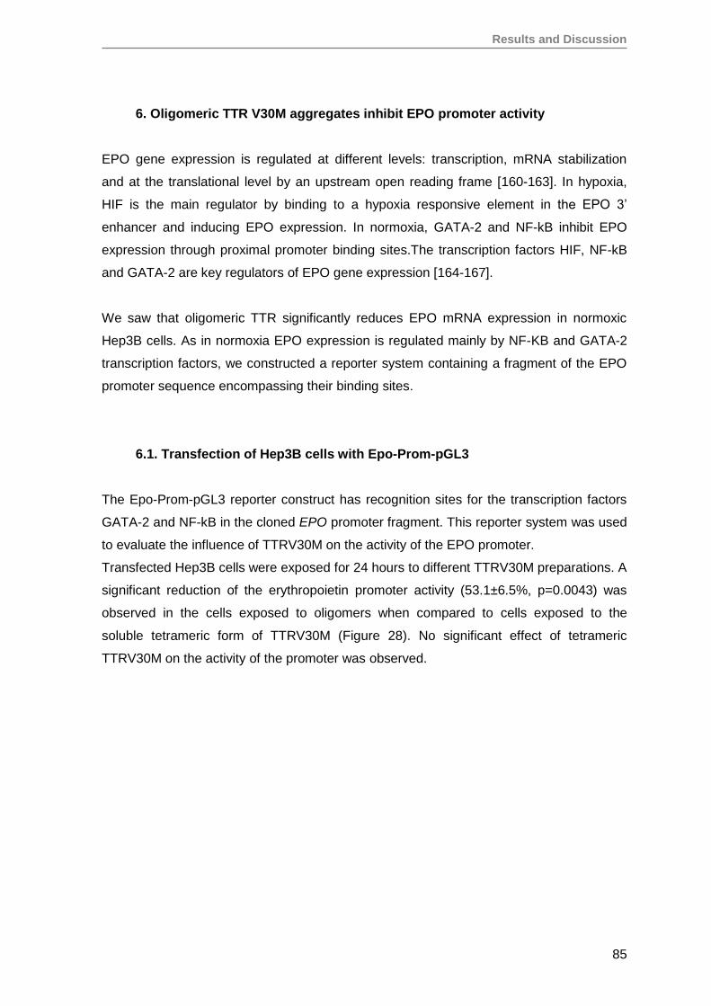
Results and Discussion
85
6. Oligomeric TTR V30M aggregates inhibit EPO promoter activity
EPO gene expression is regulated at different levels: transcription, mRNA stabilization
and at the translational level by an upstream open reading frame [160-163]. In hypoxia,
HIF is the main regulator by binding to a hypoxia responsive element in the EPO 3’
enhancer and inducing EPO expression. In normoxia, GATA-2 and NF-kB inhibit EPO
expression through proximal promoter binding sites.The transcription factors HIF, NF-kB
and GATA-2 are key regulators of EPO gene expression [164-167].
We saw that oligomeric TTR significantly reduces EPO mRNA expression in normoxic
Hep3B cells. As in normoxia EPO expression is regulated mainly by NF-KB and GATA-2
transcription factors, we constructed a reporter system containing a fragment of the EPO
promoter sequence encompassing their binding sites.
6.1. Transfection of Hep3B cells with Epo-Prom-pGL3
The Epo-Prom-pGL3 reporter construct has recognition sites for the transcription factors
GATA-2 and NF-kB in the cloned EPO promoter fragment. This reporter system was used
to evaluate the influence of TTRV30M on the activity of the EPO promoter.
Transfected Hep3B cells were exposed for 24 hours to different TTRV30M preparations. A
significant reduction of the erythropoietin promoter activity (53.1±6.5%, p=0.0043) was
observed in the cells exposed to oligomers when compared to cells exposed to the
soluble tetrameric form of TTRV30M (Figure 28). No significant effect of tetrameric
TTRV30M on the activity of the promoter was observed.
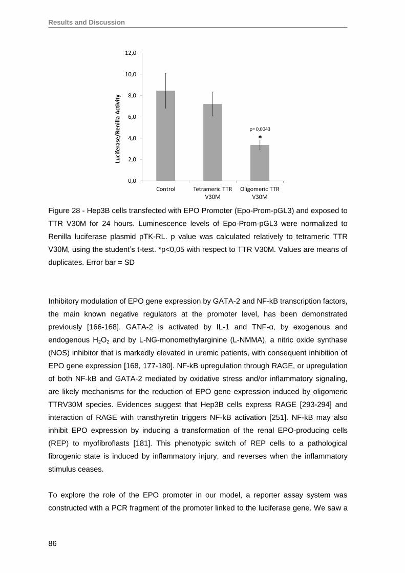
Results and Discussion
86
Figure 28 - Hep3B cells transfected with EPO Promoter (Epo-Prom-pGL3) and exposed to
TTR V30M for 24 hours. Luminescence levels of Epo-Prom-pGL3 were normalized to
Renilla luciferase plasmid pTK-RL. p value was calculated relatively to tetrameric TTR
V30M, using the student’s t-test. *p<0,05 with respect to TTR V30M. Values are means of
duplicates. Error bar = SD
Inhibitory modulation of EPO gene expression by GATA-2 and NF-kB transcription factors,
the main known negative regulators at the promoter level, has been demonstrated
previously [166-168]. GATA-2 is activated by IL-1 and TNF-α, by exogenous and
endogenous H2O2 and by L-NG-monomethylarginine (L-NMMA), a nitric oxide synthase
(NOS) inhibitor that is markedly elevated in uremic patients, with consequent inhibition of
EPO gene expression [168, 177-180]. NF-kB upregulation through RAGE, or upregulation
of both NF-kB and GATA-2 mediated by oxidative stress and/or inflammatory signaling,
are likely mechanisms for the reduction of EPO gene expression induced by oligomeric
TTRV30M species. Evidences suggest that Hep3B cells express RAGE [293-294] and
interaction of RAGE with transthyretin triggers NF-kB activation [251]. NF-kB may also
inhibit EPO expression by inducing a transformation of the renal EPO-producing cells
(REP) to myofibroflasts [181]. This phenotypic switch of REP cells to a pathological
fibrogenic state is induced by inflammatory injury, and reverses when the inflammatory
stimulus ceases.
To explore the role of the EPO promoter in our model, a reporter assay system was
constructed with a PCR fragment of the promoter linked to the luciferase gene. We saw a
Results and Discussion
87
reduction of EPO promoter activity in transfected Hep3B cells exposed to oligomeric
species, parallel to the observed reduction in native EPO mRNA expression. These
results suggest that downregulation of EPO expression in normoxic Hep3B cells exposed
to oligomeric TTRV30M could be explained, at least in part, by a reduction of EPO
promoter activity. As GATA-2 and NF-kB may repress EPO promoter activity, these
transcription factors may be involved in the repression of EPO expression and promoter
activity induced by the oligomeric TTR aggregates.
6.2. Study of NF-kB and GATA-2 protein expression
Immunofluorescence and immunohistochemistry were performed in order to explore the
role of NF-kB and GATA-2. Hep3B cells were exposed to TTRV30M preparations for 24
hours and immunofluorescence was performed for both NF-kB and GATA-2. No
significant differences were observed between the oligomeric-treated and tetrameric-
treated cells.
Immunohistochemistry for NF-KB and GATA-2 was performed on renal biopsies from FAP
patients and from renal donors. As for immunofluorescence, no significant differences
were observed between the renal FAP biopsies and the donors. Interestingly, GATA-2
antibody cross-reacted with the amyloid deposits of the FAP renal biopsies and gave
false-positive results.
The influence of TTR aggregates on the activation of NF-kB and GATA-2 transcription
factors still needs clarification. A technique such as chromatin immunoprecipitation (ChIP),
followed by real-time pcr, may better elucidate if an interaction between one or both of
these transcription factors with the EPO promoter is involved in the inhibition of EPO
expression in FAP.
A similar situation to that found in FAP patients, inappropriately low EPO production, also
occurs in diabetic nephropathy (DN) [295]. Anemia occurs early in the course of DN due
to either impaired production and/or response to EPO, even in the absence of renal
impairment [295].
Diabetic nephropathy (DN) is a chronic disorder present in approximately 25%-40% of
patients with long-standing diabetes. It is characterized by progressive albuminuria and a
decline in renal function [296]. Chronic elevated blood glucose together with glomerular
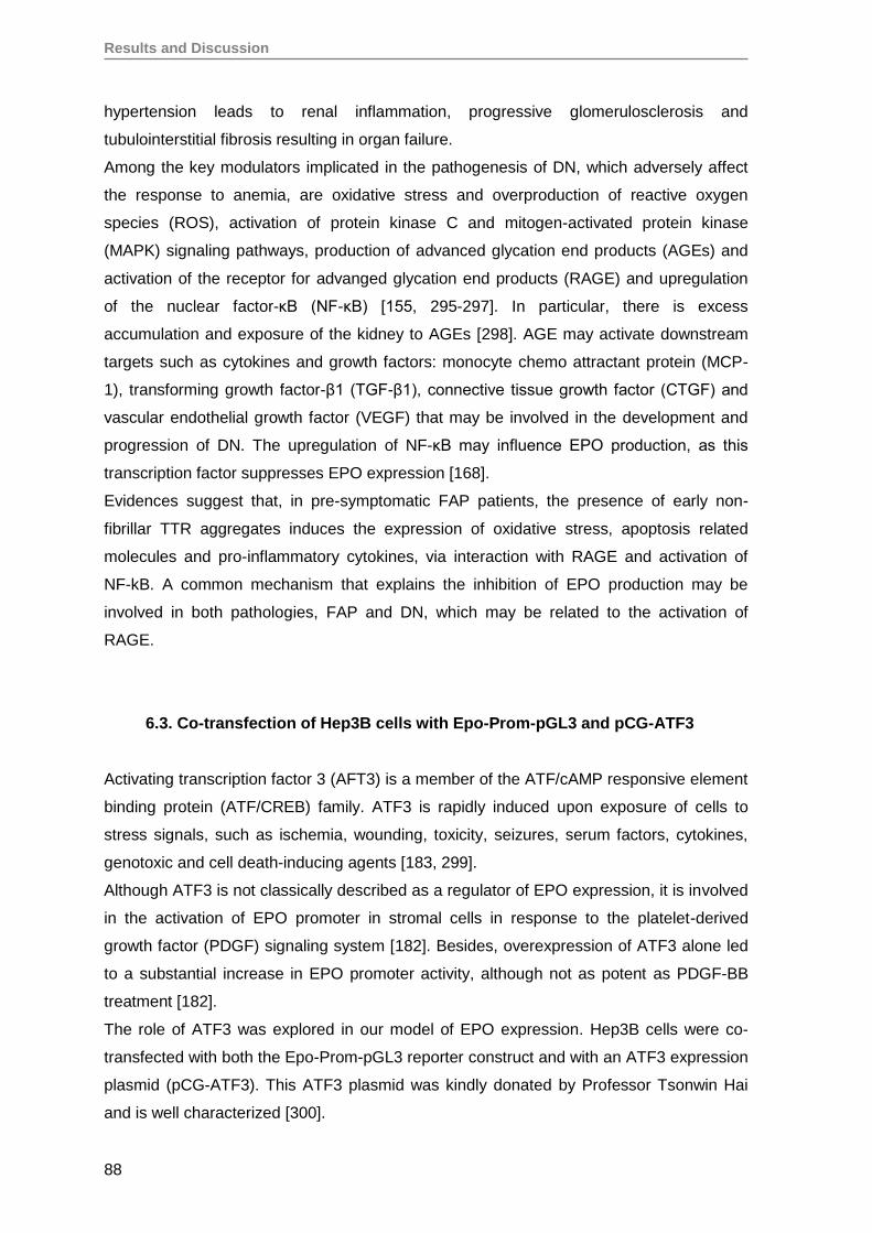
Results and Discussion
88
hypertension leads to renal inflammation, progressive glomerulosclerosis and
tubulointerstitial fibrosis resulting in organ failure.
Among the key modulators implicated in the pathogenesis of DN, which adversely affect
the response to anemia, are oxidative stress and overproduction of reactive oxygen
species (ROS), activation of protein kinase C and mitogen-activated protein kinase
(MAPK) signaling pathways, production of advanced glycation end products (AGEs) and
activation of the receptor for advanged glycation end products (RAGE) and upregulation
of the nuclear factor-κB (NF-κB) [155, 295-297]. In particular, there is excess
accumulation and exposure of the kidney to AGEs [298]. AGE may activate downstream
targets such as cytokines and growth factors: monocyte chemo attractant protein (MCP-
1), transforming growth factor-β1 (TGF-β1), connective tissue growth factor (CTGF) and
vascular endothelial growth factor (VEGF) that may be involved in the development and
progression of DN. The upregulation of NF-κB may influence EPO production, as this
transcription factor suppresses EPO expression [168].
Evidences suggest that, in pre-symptomatic FAP patients, the presence of early non-
fibrillar TTR aggregates induces the expression of oxidative stress, apoptosis related
molecules and pro-inflammatory cytokines, via interaction with RAGE and activation of
NF-kB. A common mechanism that explains the inhibition of EPO production may be
involved in both pathologies, FAP and DN, which may be related to the activation of
RAGE.
6.3. Co-transfection of Hep3B cells with Epo-Prom-pGL3 and pCG-ATF3
Activating transcription factor 3 (AFT3) is a member of the ATF/cAMP responsive element
binding protein (ATF/CREB) family. ATF3 is rapidly induced upon exposure of cells to
stress signals, such as ischemia, wounding, toxicity, seizures, serum factors, cytokines,
genotoxic and cell death-inducing agents [183, 299].
Although ATF3 is not classically described as a regulator of EPO expression, it is involved
in the activation of EPO promoter in stromal cells in response to the platelet-derived
growth factor (PDGF) signaling system [182]. Besides, overexpression of ATF3 alone led
to a substantial increase in EPO promoter activity, although not as potent as PDGF-BB
treatment [182].
The role of ATF3 was explored in our model of EPO expression. Hep3B cells were co-
transfected with both the Epo-Prom-pGL3 reporter construct and with an ATF3 expression
plasmid (pCG-ATF3). This ATF3 plasmid was kindly donated by Professor Tsonwin Hai
and is well characterized [300].
Results and Discussion
89
Using Hep3B cells transfected with the Epo-Prom-pGL3 reporter construct, it was shown
previously that oligomeric TTRV30M aggregates reduced approximately 2-fold the activity
of EPO promoter relatively to the tetrameric-treated cells. The overexpression of ATF3 in
Hep3B cells co-transfected with the Epo-Prom-pGL3 reporter construct and ATF3 vector
led to an increase of the promoter activity in oligomeric-treated cells to a similar value to
that observed in tetrameric TTR-treated cells (figure 29).
Figure 29 - Hep3B cells co-transfected with EPO Promoter (Epo-Prom-pGL3) and pCG-
ATF3, an expression vector containing the ATF3 gene. Cells were exposed to TTR V30M
for 24 hours. Luminescence levels of Epo-Prom-pGL3 were normalized to Renilla
luciferase plasmid pTK-RL. Error bar = SD
The inhibitory effect induced by TTR oligomers on EPO promoter activity seems to be
reversed by the expression of ATF3. These results are preliminary and require further
study, but raise the possibly that ATF3 alone, or together with other transcription factors,
participates in the regulation of EPO gene expression.
It is known that ATF3 is involved in the regulation of several genes, not necessarily via
direct binding to the promoter of the target gene, but forming complexes with other
transcription factors or cofactors [301]. It may stabilize the co-factors at the promoter or
Results and Discussion
90
sequester them away from the transcription machinery, resulting in transcriptional
regulation [182-183, 299-302].
Xue et al propose that the formation of a complex between the ATF3, c-Jun and Sp1
explains the increase of EPO promoter activity induced by PDGF-BB [182]. They showed
that the EPO promoter region does not contain an ATF3 binding site, suggesting that
ATF3 may not act directly on the EPO promoter [182]. Elevated expression of PDGF has
been observed in RPE cells after retinal detachments or retinal laser treatment in murine
model systems [184], in in vitro wounded human RPE cell cultures [185] and in epi-retinal
membranes isolated from proliferative vitreoretinopathy and proliferative diabetic
retinopathy patients [186].
Besides Sp1 and c-Jun, ATF3 may also interact with NF-kB, p53 and p73. It may also
inhibit transcription of pro-inflammatory cytokines, by interacting with NF-κB at the
promoter of these target genes [301].
In our TTRV30M exposure Hep3B cell model, it appears that oligomeric TTRV30M
aggregates inhibit EPO expression by decreasing EPO promoter activity. However, when
the cells over-express ATF3 the inhibitory effect of these aggregates is reverved. This
could be due either to increased activity of promoter inhibitors or to a decreased activity of
ATF3, preventing it from sequestering these inhibitors away from the EPO promoter. This
issue deserves further study.
ATF3 has a basic region-leucine zipper (bZip) DNA binding domain. However, several
isoforms derived from alternative splicing have been described (ATF3ΔZip, ATF3ΔZip2a,
ATF3ΔZip2b, ATF3ΔZip2c, ATF3ΔZip3, and ATF3b), lacking the leucine zipper region
[302]. The role of these alternative splicing forms needs also to be explored.

CONCLUSIONS AND FUTURE PERSPECTIVES

Conclusion and Future Perspectives
93
Conclusions and Future Perspectives
In this project we carried out in vitro studies aiming to explore the role of amyloid
deposition in ATTRV30M amyloidosis, especially regarding the role of early non-fibrillar
aggregates on cell differentiation and erythropoietin production.
First, the production of recombinant human TTRV30M and their oligomeric aggregates
were optimized. Obtaining a pure and LPS-free protein for the cellular assays was crucial
to ensure that the effects on the cells were only due to the protein preparations.
The poor reproducibility of preparation of cytotoxic oligomeric solutions made it necessary
to test several methods described in the literature, for optimization. Mild acidification (pH 4
to 5.5) of the protein solution, acidification to pH 2.0 followed by NaCl or aging under
physiological pH and temperature followed by vigorous stirring were the methods chosen.
In the case of acidification to mild pH we obtained insoluble material, probably amorphous
aggregates that, despite being positive in ThT assays, were not toxic to cells. The method
that induced the formation of most cytotoxic species and with the most reproducible
results was the last one tested, aging the protein at physiological pH and 37ºC for 72
hours followed by 5 minutes of stirring. This TTR preparation contained mainly oligomers
of approximately 300 nm when analysed by dynamic light scattering, and was the one
used for the subsequent cell culture assays.
Different cell culture models were used to evaluate the influence of oligomeric TTR
aggregates on cell viability: using immortalized human hepatoblastoma Hep3B,
neuroblastoma SH-SY5Y and embryonic kidney cells HEK293T, as well as primary retinal
pigment epithelial cells RPE and human renal progenitor cells RPC. The proliferation of
the cells exposed for 24 hours to oligomeric TTRV30M aggregates was moderately
reduced when compared to the cells exposed to the tetrameric protein, independently of
the cell type. Oligomers generated from the wild-type TTR also reduced cell proliferation
in comparison to the soluble tetrameric form. Both oligomeric TTR and TTRV30M
preparations were cytotoxic, a process that may be common to all prefibrillar amyloid
protein aggregates.
Concomitantly to the reduction in cell proliferation, a significant increase in caspases 3/7
activity occurred in cells exposed to oligomeric TTRV30M aggregates, implicating
apoptosis in the loss of viability. These results are in agreement with other studies
performed mainly in neuronal cells (neuroblastoma) [106, 229, 250, 270-272].
Interestingly, the cytotoxic effect induced by the aggregates seems to be independent of
Conclusions and Future Perspectives
94
the cell type, since both immortalized, as well as primary and progenitor cells were shown
to be susceptible to damage and apoptosis driven by aggregate exposure.
Renal progenitor cells (RPC) express the stem cell markers CD133+CD24+ and may
potentially differentiate into podocytes and tubular cells. We evaluated the influence of the
oligomeric aggregates on the viability and differentiation capacity of the RPC, in order to
understand if the misfolded protein could compromise a potential therapeutic use of these
cells to treat renal damage. Proliferation of RPC in vitro was impared by the oligomeric
TTRV30M aggregates comparatively to the tetrameric form, but their differentiation
capacity into mature podocytes was not significantly affected, so renal tissue regeneration
should not be compromised by the presence of early oligomeric species.
ATTRV30M amyloidosis patients develop anemia with low EPO levels. The impaired EPO
production is not related to the extent or pattern of congophilic renal deposition or with the
presence of circulating mutant TTRV30M. The role of oligomeric aggregate cytotoxicity on
EPO production had not been studied yet.
In this work we used the EPO producing cell line Hep3B to evaluate the influence of
oligomeric TTRV30M aggregates on EPO gene expression, as well as on pathways linked
to the regulatory regions of the EPO gene. Normoxic Hep3B exposed for 24 hours to early
oligomeric TTRV30M aggregates showed a 2-fold reduction in EPO mRNA expression. In
accordance to this reduction, there was a 2-fold reduction in EPO promoter activity in
transfected Hep3B cells. These results support involvement of early oligomeric TTR
aggregates in decreased EPO gene expression and could explain the anemia and low
EPO levels in FAP patients, seen even in the pre-symptomatic phase of the disease.
Interestingly, a preparation of wild-type TTR oligomers also reduced EPO mRNA
expression in normoxia, although to a less extent than the TTRV30M oligomers, which
may explain the continuing low EPO production after liver transplantation seen in FAP
patients.
The inhibition of EPO expression caused by exposure of the cells to cytotoxic oligomeric
aggregates seems to be mediated by decreased activity of the EPO promoter.
Transcription factors such as NF-kB and GATA-2 bind to the promoter and inhibit
transcription of the EPO gene, mainly in inflammatory states. Here, we obtained
inconclusive results about the influence of oligomeric aggregates on NF-kB or GATA-2
activation. However, we believe this issue deserves further consideration, as well as other
signaling pathways that may be activated by the oligomeric species and that may
influence EPO expression.

Conclusion and Future Perspectives
95
FAP patients are also unable to up-regulate EPO production in the glaucomatous eye, as
a neuroprotective response. Besides the possible involvement of TTR aggregates in EPO
expression and anemia, a much wider role for these aggregates in disease progression
could be considered. Recent studies point to a potentially major role of EPO in
neurodegenerative diseases, including Alzheimer's disease, Parkinson's disease,
epilepsy, multiple sclerosis and motor neuron diseases [151, 303-311]. It has been
already shown in another model of amyloid-related neurodegeneration, in which PC12
cells are exposed to Aβ25-35 aggregates, that apoptosis is counteracted by EPO signaling
[312-313]. EPO has also been found to increase the resistance of neurons to damage
induced by pro-inflammatory agents [314]. It could be postulated that deposition of TTR
aggregates in the peripheral nervous system down-regulates local EPO expression,
depriving neurons from needed neuroprotective signals, contributing thus for the onset
and/or progression of the disease, an hypothesis that we feel deserves further study.
Retinal pigment epithelial cells (RPE) produce both EPO and TTR in the eye. In order to
find a more suitable cell culture model to explore the direct effect of the amyloidogenic
TTRV30M on EPO production, we isolated RPE cells from an eye of a human cadaveric
donor. Preliminary results showed that, at early passages, these cells express EPO in
normoxia and respond to hypoxia simulated with DMOG.
Besides the reduction of cell proliferation and increased activity of caspases 3/7, EPO
gene expression in normoxic RPE treated for 24 hours with oligomeric TTRV30M
aggregates was reduced by approximately 2-fold comparatively to the cells exposed to
tetrameric TTRV30M, similarly to what was seen in Hep3B cells. This is an important
result as it shows a direct influence of the oligomeric species on a primary cell line that is
physiologically responsible for EPO production in the eye.
As future perspectives, and taking into consideration these preliminary results, we believe
that RPE cells could become a suitable cell culture model to further explore the
mechanisms responsible for EPO inhibition in ATTRV30M amyloidosis, ideally, using RPE
cells from FAP patients.
The blockage of EPO production observed both in the kidney as in the eye of FAP
patients also happens in Diabetic Nephropathy (DN). Oxidative stress, overproduction of
reactive oxygen species (ROS), production of AGEs, activation of the receptor for
advanged glycation end products (RAGE), upregulation of the NF-κB and pro-
inflammatory cytokines seem to be common factors between FAP and DN. ROS may
upregulate GATA-2. In this work we used immunofluorescence to study the activation of
Conclusions and Future Perspectives
96
NF-kB and GATA-2 by exposure to oligomeric aggregates and we could not see
significant differences. However, it would be important to use more sensitive techniques,
such as Chromatin immunoprecipitation or electrophoretic mobility shift assays (EMSA),
which can show protein-DNA interaction, to confirm if the inhibition of EPO expression by
cytotoxic aggregates is indeed related to activation of these two transcription factors.
ATF3 is other transcription factor that may be involved in EPO gene regulation. In our cell
model, the over-expression of ATF3 reversed the inhibitory effect of the oligomeric TTR
aggregates on the EPO promoter activity. This is a preliminary observation that deserves
further study. It would also be interesting to use DN patients for comparison.
Tafamidis is a drug already approved for the treatment of eligible FAP patients. We would
like to know if Tafamidis, a TTR tetramer stabilizer, is able to prevent blockage of EPO
production.
In summary, although we have shown here that exposure to cytotoxic oligomeric TTR
aggregates inhibits EPO expression in vitro, much remains to be discovered about the
mechanisms underlying this inhibition and possible therapeutic targets.

REFERENCES

References
99
References
1 Cohen AS, Calkins E. Electron microscopic observations on a fibrous component in
amyloid of diverse origins. Nature. 1959 Apr 25;183(4669):1202-3.
2 Glenner GG, Eanes ED, Bladen HA, Linke RP, Termine JD. Beta-pleated sheet
fibrils. A comparison of native amyloid with synthetic protein fibrils. J Histochem
Cytochem. 1974 Dec;22(12):1141-58.
3 Cohen AS, Shirahama T, Sipe JD, Skinner M. Amyloid proteins, precursors,
mediator, and enhancer. Lab Invest. 1983 Jan;48(1):1-4.
4 Sipe JD, Benson MD, Buxbaum JN, Ikeda S, Merlini G, Saraiva MJ, Westermark P.
Nomenclature 2014: Amyloid fibril proteins and clinical classification of the
amyloidosis. Amyloid. 2014 Dec;21(4):221-4.
5 Sipe JD, Cohen AS. Review: history of the amyloid fibril. J Struct Biol. 2000
Jun;130(2-3):88-98.
6 Kyle RA. Amyloidosis: a convoluted story. Br J Haematol. 2001 Sep;114(3):529-38.
7 Westermark GT, Norling B, Westermark P. Fibronectin and basement membrane
components in renal amyloid deposits in patients with primary and secondary
amyloidosis. Clin Exp Immunol. 1991 Oct;86(1):150-6.
8 Benditt EP. What role(s) may extracellular matrix, particularly heparan sulfate, play
in amyloid of Alzheimer's disease. Neurobiol Aging. 1989 Sep-Oct;10(5):506;
discussion 510-2.
9 Wisniewski T, Golabek A, Matsubara E, Ghiso J, Frangione B. Apolipoprotein E:
binding to soluble Alzheimer's beta-amyloid. Biochem Biophys Res Commun. 1993
Apr 30;192(2):359-65.
10 de Beer MC, de Beer FC, McCubbin WD, Kay CM, Kindy MS. Structural
prerequisites for serum amyloid A fibril formation. J Biol Chem. 1993 Sep
25;268(27):20606-12.
11 Moss J, Shore I, Woodrow D. AA glomerular amyloid. An ultrastructural immunogold
study of the colocalization of heparan sulphate proteoglycan and P component with
amyloid fibrils together with changes in distribution of type IV collagen and
fibronectin. Histopathology. 1994 May;24(5):427-35.
12 Pepys MB. Pathogenesis, diagnosis and treatment of systemic amyloidosis. Philos
Trans R Soc Lond B Biol Sci. 2001 Feb 28;356(1406):203-10; discussion 210-1.
References
100
13 Eanes ED, Glenner GG. X-ray diffraction studies on amyloid filaments. J Histochem
Cytochem. 1968 Nov;16(11):673-7.
14 Shirahama T, Cohen AS. High-resolution electron microscopic analysis of the
amyloid fibril. J Cell Biol. 1967 Jun;33(3):679-708.
15 Glenner GG, Terry W, Harada M, Isersky C, Page D. Amyloid fibril proteins: proof of
homology with immunoglobulin light chains by sequence analyses. Science. 1971
Jun 11;172(3988):1150-1.
16 Isersky C, Ein D, Page DL, Harada M, Glenner GG. Immunochemical cross-
reactions of human amyloid proteins with human immunoglobulin light polypeptide
chains. J Immunol. 1972 Feb;108(2):486-93.
17 Benditt EP, Eriksen N, Hermodson MA, Ericsson LH. The major proteins of human
and monkey amyloid substance: Common properties including unusual N-terminal
amino acid sequences. FEBS Lett. 1971 Dec 1;19(2):169-173.
18 Benditt EP, Eriksen N. Amyloid protein SAA is associated with high density
lipoprotein from human serum. Proc Natl Acad Sci U S A. 1977 Sep;74(9):4025-8.
19 Benson MD. Partial amino acid sequence homology between an heredofamilial
amyloid protein and human plasma prealbumin. J Clin Invest. 1981 Apr;67(4):1035-
41.
20 Thomas PK. Genetic factors in amyloidosis. J Med Genet. 1975 Dec;12(4):317-26.
21 Gertz MA. The classification and typing of amyloid deposits. Am J Clin Pathol. 2004
Jun;121(6):787-9.
22 Scheinberg MA, Cathcart ES. New concepts in the pathogenesis of primary and
secondary amyloid disease. Clin Exp Immunol. 1978 Jul;33(1):185-90.
23 Westermark P. Aspects on human amyloid forms and their fibril polypeptides. FEBS
J. 2005 Dec;272(23):5942-9.
24 Murphy CL, Eulitz M, Hrncic R, Sletten K, Westermark P, Williams T, Macy SD,
Wooliver C, Wall J, Weiss DT, Solomon A. Chemical typing of amyloid protein
contained in formalin-fixed paraffin-embedded biopsy specimens. Am J Clin Pathol.
2001 Jul;116(1):135-42.
25 Solomon A, Murphy CL, Westermark P. Unreliability of immunohistochemistry for
typing amyloid deposits. Arch Pathol Lab Med. 2008 Jan;132(1):14.
26 Picken MM. New insights into systemic amyloidosis: the importance of diagnosis of
specific type. Curr Opin Nephrol Hypertens. 2007 May;16(3):196-203.
27 Lobato L, Rocha A. Transthyretin amyloidosis and the kidney. Clin J Am Soc
Nephrol. 2012 Aug;7(8):1337-46.
28 Kaplan B, Shtrasburg S, Pras M. Micropurification techniques in the analysis of
amyloid proteins. J Clin Pathol. 2003 Feb;56(2):86-90.
References
101
29 Kaplan B, Martin BM, Livneh A, Pras M, Gallo GR. Biochemical subtyping of amyloid
in formalin-fixed tissue samples confirms and supplements immunohistologic data.
Am J Clin Pathol. 2004 Jun;121(6):794-800.
30 Klein CJ, Vrana JA, Theis JD, Dyck PJ, Dyck PJ, Spinner RJ, Mauermann ML,
Bergen HR 3rd, Zeldenrust SR, Dogan A. Mass spectrometric-based proteomic
analysis of amyloid neuropathy type in nerve tissue. Arch Neurol. 2011
Feb;68(2):195-9.
31 Lavatelli F, Brambilla F, Valentini V, Rognoni P, Casarini S, Di Silvestre D, Perfetti
V, Palladini G, Sarais G, Mauri P, Merlini G. A novel approach for the purification
and proteomic analysis of pathogenic immunoglobulin free light chains from serum.
Biochim Biophys Acta. 2011 Mar;1814(3):409-19.
32 Naidoo K, Jones R, Dmitrovic B, Wijesuriya N, Kocher H, Hart IR, Crnogorac-
Jurcevic T. Proteome of formalin-fixed paraffin-embedded pancreatic ductal
adenocarcinoma and lymph node metastases. J Pathol. 2012 Apr;226(5):756-63.
33 Brambilla F, Lavatelli F, Di Silvestre D, Valentini V, Rossi R, Palladini G, Obici L,
Verga L, Mauri P, Merlini G. Reliable typing of systemic amyloidoses through
proteomic analysis of subcutaneous adipose tissue. Blood. 2012 Feb
23;119(8):1844-7.
34 Data Base on Transthyretin Mutations. http://www.ibmc.up.pt/mjsaraiva/ttrmut.html
35 Mutations in Hereditary Amyloidosis. http://amyloidosismutations.com/
36 Benson MD, Wallace MR. Genetic amyloidosis: recent advances. Adv Nephrol
Necker Hosp. 1989;18:129-37.
37 Westermark P, Sletten K, Johansson B, Cornwell GG 3rd. Fibril in senile systemic
amyloidosis is derived from normal transthyretin. Proc Natl Acad Sci U S A. 1990
Apr;87(7):2843-5.
38 Saraiva MJ, Birken S, Costa PP, Goodman DS. Amyloid fibril protein in familial
amyloidotic polyneuropathy, Portuguese type. Definition of molecular abnormality in
transthyretin (prealbumin). J Clin Invest. 1984 Jul;74(1):104-19.
39 ANDRADE C. A peculiar form of peripheral neuropathy; familiar atypical generalized
amyloidosis with special involvement of the peripheral nerves. Brain. 1952
Sep;75(3):408-27.
40 Costa PP, Figueira AS, Bravo FR. Amyloid fibril protein related to prealbumin in
familial amyloidotic polyneuropathy. Proc Natl Acad Sci U S A. 1978
Sep;75(9):4499-503.
41 Alves IL, Altland K, Almeida MR, Winter P, Saraiva MJ. Screening and biochemical
characterization of transthyretin variants in the Portuguese population. Hum Mutat
1997;9(3):226-233
References
102
42 Ando Y, Araki S, Ando M. Transthyretin and familial amyloidotic polyneuropathy.
Intern Med. 1993; 32(12):920-2.
43 Coimbra A, Andrade C. Familial amyloid polyneuropathy: an electron microscope
study of the peripheral nerve in five cases. I. Interstitial changes. Brain.
1971;94(2):199-206.
44 Hofer PA, Anderson R. Postmortem findings in primary familial amyloidosis with
polyneuropathy. Acta Pathol Microbiol Scand A. 1975 May;83(3):309-22.
45 Kaufman HE, Thomas LB. Vitreous opacities diagnostic of familial primary
amyloidosis. N Engl J Med. 1959 Dec 17;261:1267-71.
46 Paton D, Duke JR. Primary familial amyloidosis. Ocular manifestations with
histopathologic observations. Am J Ophthalmol. 1966 Apr;61(4):736-47.
47 Ueda M, Ando Y. Recent advances in transthyretin amyloidosis therapy. Transl
Neurodegener. 2014 Sep 13;3:19.
48 Hanna M. Novel drugs targeting transthyretin amyloidosis. Curr Heart Fail Rep.
2014 Mar;11(1):50-7.
49 Suhr OB, Friman S, Ericzon BG. Early liver transplantation improves familial
amyloidotic polyneuropathy patients' survival. Amyloid. 2005 Dec;12(4):233-8.
50 Stangou AJ, Hawkins PN. Liver transplantation in transthyretin-related familial
amyloid polyneuropathy. Curr Opin Neurol. 2004 Oct;17(5):615-20.
51 Yamashita T, Ando Y, Okamoto S, Misumi Y, Hirahara T, Ueda M, Obayashi K,
Nakamura M, Jono H, Shono M, Asonuma K, Inomata Y, Uchino M. Long-term
survival after liver transplantation in patients with familial amyloid polyneuropathy.
Neurology. 2012 Feb 28;78(9):637-43.
52 Yamamoto S, Wilczek HE, Nowak G, Larsson M, Oksanen A, Iwata T, Gjertsen H,
Söderdahl G, Wikström L, Ando Y, Suhr OB, Ericzon BG. Liver transplantation for
familial amyloidotic polyneuropathy (FAP): a single-center experience over 16 years.
Am J Transplant. 2007 Nov;7(11):2597-604.
53 Skinner M, Lewis WD, Jones LA, Kasirsky J, Kane K, Ju ST, Jenkins R, Falk RH,
Simms RW, Cohen AS. Liver transplantation as a treatment for familial amyloidotic
polyneuropathy. Ann Intern Med. 1994 Jan 15;120(2):133-4.
54 Stangou AJ, Heaton ND, Hawkins PN. Transmission of systemic transthyretin
amyloidosis by means of domino liver transplantation. N Engl J Med. 2005 Jun
2;352(22):2356.
55 Goto T, Yamashita T, Ueda M, Ohshima S, Yoneyama K, Nakamura M, Nanjo H,
Asonuma K, Inomata Y, Watanabe S, Uchino M, Tanaka K, Ando Y. Iatrogenic
amyloid neuropathy in a Japanese patient after sequential liver transplantation. Am
J Transplant. 2006 Oct;6(10):2512-5.
References
103
56 Takei Y, Gono T, Yazaki M, Ikeda S, Ikegami T, Hashikura Y, Miyagawa S, Hoshii
Y. Transthyretin-derived amyloid deposition on the gastric mucosa in domino
recipients of familial amyloid polyneuropathy liver. Liver Transpl. 2007
Feb;13(2):215-8.
57 Hörnsten R, Wiklund U, Olofsson BO, Jensen SM, Suhr OB. Liver transplantation
does not prevent the development of life-threatening arrhythmia in familial
amyloidotic polyneuropathy, Portuguese-type (ATTR Val30Met) patients.
Transplantation. 2004 Jul 15;78(1):112-6.
58 Olofsson BO, Backman C, Karp K, Suhr OB. Progression of cardiomyopathy after
liver transplantation in patients with familial amyloidotic polyneuropathy, Portuguese
type. Transplantation. 2002 Mar 15;73(5):745-51.
59 Oshima T, Kawahara S, Ueda M, Kawakami Y, Tanaka R, Okazaki T, Misumi Y,
Obayashi K, Yamashita T, Ohya Y, Ihse E, Shinriki S, Tasaki M, Jono H, Asonuma
K, Inomata Y, Westermark P, Ando Y. Changes in pathological and biochemical
findings of systemic tissue sites in familial amyloid polyneuropathy more than 10
years after liver transplantation. J Neurol Neurosurg Psychiatry. 2014 Jul;85(7):740-
6.
60 Liepnieks JJ, Benson MD. Progression of cardiac amyloid deposition in hereditary
transthyretin amyloidosis patients after liver transplantation. Amyloid. 2007
Dec;14(4):277-82.
61 Ando Y, Terazaki H, Nakamura M, Ando E, Haraoka K, Yamashita T, Ueda M,
Okabe H, Sasaki Y, Tanihara H, Uchino M, Inomata Y. A different amyloid formation
mechanism: de novo oculoleptomeningeal amyloid deposits after liver
transplantation. Transplantation. 2004 Feb 15;77(3):345-9.
62 Beirão JM, Malheiro J, Lemos C, Matos E, Beirão I, Pinho-Costa P, Torres P.
Impact of liver transplantation on the natural history of oculopathy in Portuguese
patients with transthyretin (V30M) amyloidosis. Amyloid. 2014 Dec 5:1-5.
63 https://clinicaltrials.gov/
64 Malik R, Roy I. Making sense of therapeutics using antisense technology. Expert
Opin Drug Discov. 2011 May;6(5):507-26.
65 Ackermann EJ, Guo S, Booten S, Alvarado L, Benson M, Hughes S, Monia BP.
Clinical development of an antisense therapy for the treatment of transthyretin-
associated polyneuropathy. Amyloid. 2012 Jun;19 Suppl 1:43-4.
66 Benson MD, Kluve-Beckerman B, Zeldenrust SR, Siesky AM, Bodenmiller DM,
Showalter AD, Sloop KW. Targeted suppression of an amyloidogenic transthyretin
with antisense oligonucleotides. Muscle Nerve. 2006 May;33(5):609-18.
References
104
67 Benson MD, Smith RA, Hung G, Kluve-Beckerman B, Showalter AD, Sloop KW,
Monia BP. Suppression of choroid plexus transthyretin levels by antisense
oligonucleotide treatment. Amyloid. 2010 Jun;17(2):43-9.
68 Kurosawa T, Igarashi S, Nishizawa M, Onodera O. Selective silencing of a mutant
transthyretin allele by small interfering RNAs. Biochem Biophys Res Commun. 2005
Nov 25;337(3):1012-8.
69 Coelho T, Adams D, Silva A, Lozeron P, Hawkins PN, Mant T, Perez J, Chiesa J,
Warrington S, Tranter E, Munisamy M, Falzone R, Harrop J, Cehelsky J,
Bettencourt BR, Geissler M, Butler JS, Sehgal A, Meyers RE, Chen Q, Borland T,
Hutabarat RM, Clausen VA, Alvarez R, Fitzgerald K, Gamba-Vitalo C, Nochur SV,
Vaishnaw AK, Sah DW, Gollob JA, Suhr OB. Safety and efficacy of RNAi therapy for
transthyretin amyloidosis. N Engl J Med. 2013 Aug 29;369(9):819-29.
70 Miroy GJ, Lai Z, Lashuel HA, Peterson SA, Strang C, Kelly JW. Inhibiting
transthyretin amyloid fibril formation via protein stabilization. Proc Natl Acad Sci U S
A. 1996 Dec 24;93(26):15051-6.
71 Adamski-Werner SL, Palaninathan SK, Sacchettini JC, Kelly JW. Diflunisal
analogues stabilize the native state of transthyretin. Potent inhibition of
amyloidogenesis. J Med Chem. 2004 Jan 15;47(2):355-74.
72 Gales L, Macedo-Ribeiro S, Arsequell G, Valencia G, Saraiva MJ, Damas AM.
Human transthyretin in complex with iododiflunisal: structural features associated
with a potent amyloid inhibitor. Biochem J. 2005 Jun 1;388(Pt 2):615-21.
73 Sekijima Y, Dendle MA, Kelly JW. Orally administered diflunisal stabilizes
transthyretin against dissociation required for amyloidogenesis. Amyloid. 2006
Dec;13(4):236-49.
74 Castaño A, Helmke S, Alvarez J, Delisle S, Maurer MS. Diflunisal for ATTR cardiac
amyloidosis. Congest Heart Fail. 2012 Nov-Dec;18(6):315-9.
75 Tojo K, Sekijima Y, Kelly JW, Ikeda S. Diflunisal stabilizes familial amyloid
polyneuropathy-associated transthyretin variant tetramers in serum against
dissociation required for amyloidogenesis. Neurosci Res. 2006 Dec;56(4):441-9.
76 Berk JL, Suhr OB, Obici L, Sekijima Y, Zeldenrust SR, Yamashita T, Heneghan MA,
Gorevic PD, Litchy WJ, Wiesman JF, Nordh E, Corato M, Lozza A, Cortese A,
Robinson-Papp J, Colton T, Rybin DV, Bisbee AB, Ando Y, Ikeda S, Seldin DC,
Merlini G, Skinner M, Kelly JW, Dyck PJ; Diflunisal Trial Consortium. Repurposing
diflunisal for familial amyloid polyneuropathy: a randomized clinical trial. JAMA. 2013
Dec 25;310(24):2658-67.
77 Bulawa CE, Connelly S, Devit M, Wang L, Weigel C, Fleming JA, Packman J,
Powers ET, Wiseman RL, Foss TR, Wilson IA, Kelly JW, Labaudinière R. Tafamidis,
References
105
a potent and selective transthyretin kinetic stabilizer that inhibits the amyloid
cascade. Proc Natl Acad Sci U S A. 2012 Jun 12;109(24):9629-34.
78 Coelho T, Maia LF, Martins da Silva A, Waddington Cruz M, Planté-Bordeneuve V,
Lozeron P, Suhr OB, Campistol JM, Conceição IM, Schmidt HH, Trigo P, Kelly JW,
Labaudinière R, Chan J, Packman J, Wilson A, Grogan DR. Tafamidis for
transthyretin familial amyloid polyneuropathy: a randomized, controlled trial.
Neurology. 2012 Aug 21;79(8):785-92.
79 Said G, Grippon S, Kirkpatrick P. Tafamidis. Nat Rev Drug Discov. 2012 Mar
1;11(3):185-6.
80 Cardoso I, Saraiva MJ. Doxycycline disrupts transthyretin amyloid: evidence from
studies in a FAP transgenic mice model. FASEB J. 2006 Feb;20(2):234-9.
81 Ward JE, Ren R, Toraldo G, Soohoo P, Guan J, O'Hara C, Jasuja R, Trinkaus-
Randall V, Liao R, Connors LH, Seldin DC. Doxycycline reduces fibril formation in a
transgenic mouse model of AL amyloidosis. Blood. 2011 Dec 15;118(25):6610-7.
82 Cardoso I, Martins D, Ribeiro T, Merlini G, Saraiva MJ. Synergy of combined
doxycycline/TUDCA treatment in lowering Transthyretin deposition and associated
biomarkers: studies in FAP mouse models. J Transl Med. 2010 Jul 30;8:74.
83 Macedo B, Batista AR, Ferreira N, Almeida MR, Saraiva MJ. Anti-apoptotic
treatment reduces transthyretin deposition in a transgenic mouse model of Familial
Amyloidotic Polyneuropathy. Biochim Biophys Acta. 2008 Sep;1782(9):517-22.
84 Ferreira N, Cardoso I, Domingues MR, Vitorino R, Bastos M, Bai G, Saraiva MJ,
Almeida MR. Binding of epigallocatechin-3-gallate to transthyretin modulates its
amyloidogenicity. FEBS Lett. 2009 Nov 19;583(22):3569-76.
85 Ferreira N, Saraiva MJ, Almeida MR. Natural polyphenols inhibit different steps of
the process of transthyretin (TTR) amyloid fibril formation. FEBS Lett. 2011 Aug
4;585(15):2424-30.
86 Palhano FL, Lee J, Grimster NP, Kelly JW. Toward the molecular mechanism(s) by
which EGCG treatment remodels mature amyloid fibrils. J Am Chem Soc. 2013 May
22;135(20):7503-10.
87 Botto M, Hawkins PN, Bickerstaff MC, Herbert J, Bygrave AE, McBride A,
Hutchinson WL, Tennent GA, Walport MJ, Pepys MB. Amyloid deposition is delayed
in mice with targeted deletion of the serum amyloid P component gene. Nat Med.
1997 Aug;3(8):855-9.
88 Bodin K, Ellmerich S, Kahan MC, Tennent GA, Loesch A, Gilbertson JA, Hutchinson
WL, Mangione PP, Gallimore JR, Millar DJ, Minogue S, Dhillon AP, Taylor GW,
Bradwell AR, Petrie A, Gillmore JD, Bellotti V, Botto M, Hawkins PN, Pepys MB.
References
106
Antibodies to human serum amyloid P component eliminate visceral amyloid
deposits. Nature. 2010 Nov 4;468(7320):93-7.
89 Sasaki H, Sakaki Y, Matsuo H, Goto I, Kuroiwa Y, Sahashi I, Takahashi A, Shinoda
T, Isobe T, Takagi Y. Diagnosis of familial amyloidotic polyneuropathy by
recombinant DNA techniques. Biochem Biophys Res Commun. 1984 Dec
14;125(2):636-42.
90 Ando Y, Ueda M. Diagnosis and therapeutic approaches to transthyretin
amyloidosis. Curr Med Chem. 2012;19(15):2312-23.
91 Ando Y, Ohlsson PI, Suhr O, Nyhlin N, Yamashita T, Holmgren G, Danielsson A,
Sandgren O, Uchino M, Ando M. A new simple and rapid screening method for
variant transthyretin-related amyloidosis. Biochem Biophys Res Commun. 1996 Nov
12;228(2):480-3.
92 Yamashita T, Ando Y, Bernt Suhr O, Nakamura M, Sakashita N, Ohlsson PI,
Terazaki H, Obayashi K, Uchino M, Ando M. A new diagnostic procedure to detect
unknown transthyretin (TTR) mutations in familial amyloidotic polyneuropathy (FAP).
J Neurol Sci. 2000 Feb 15;173(2):154-9.
93 Bergquist J, Andersen O, Westman A. Rapid method to characterize mutations in
transthyretin in cerebrospinal fluid from familial amyloidotic polyneuropathy patients
by use of matrix-assisted laser desorption/ionization time-of-flight mass
spectrometry. Clin Chem. 2000 Sep;46(9):1293-300.
94 Lacerda PC, Moreira L, Vitorino R, Costa PP. Use of MALDI-TOF Mass
Spectrometry to Assay the Transthyretin V30M Mutation in Serum From a Liver
Transplant Donor: A Case Report. Transplantation (in press).
95 Beirão JM, Moreira LV, Lacerda PC, Vitorino RP, Beirão IB, Torres PA, Costa PP.
Inability of mutant transthyretin V30M to cross the blood-eye barrier.
Transplantation. 2012 Oct 27;94(8):e54-6.
96 Beirão I, Lobato L, Costa PM, Fonseca I, Mendes P, Silva M, Bravo F, Cabrita A,
Porto G. Kidney and anemia in familial amyloidosis type I. Kidney Int. 2004
Nov;66(5):2004-9.
97 Beirão I, Lobato L, Moreira L, Mp Costa P, Fonseca I, Cabrita A, Porto G. Long-term
treatment of anemia with recombinant human erythropoietin in familial amyloidosis
TTR V30M. Amyloid. 2008 Sep;15(3):205-9.
98 World Health Organization. Nutritional anaemias: Report of a WHO scientific group.
Geneva, Switzerland; 1968 Jan 1.
99 Olofsson BO, Grankvist K, Boman K: Evaluation of the anemia in familial amyloidotic
polyneuropahy. Eur J Int Med. 1990; 1: 425-429
References
107
100 Asahara K, Ando Y, Tanaka Y, Yi S, Yamashita T, Ando M. Secondary hypoplastic
anemia in patients with familial amyloidotic polyneuropathy. Acta Haematol.
1993;90(3):130-5.
101 Beirão I, Moreira L, Porto G, Lobato L, Fonseca I, Cabrita A, Costa PM. Low
erythropoietin production in familial amyloidosis TTR V30M is not related with renal
congophilic amyloid deposition. A clinicopathologic study of twelve cases. Nephron
Clin Pract. 2008;109(2):c95-9.
102 Faquin WC, Schneider TJ, Goldberg MA. Effect of inflammatory cytokines on
hypoxia-induced erythropoietin production. Blood. 1992 Apr 15;79(8):1987-94.
103 Vasilopoulos S, Hally R, Caro J, Martin P, Westerberg S, Moritz M, Jarrell B, Muñoz
S. Erythropoietin response to post-liver transplantation anemia. Liver Transpl. 2000
May;6(3):349-55.
104 Beirão I, Lobato L, Costa PM, Fonseca I, Silva M, Bravo F, Cabrita A, Porto G. Liver
transplantation and anemia in familial amyloidosis ATTR V30M. Amyloid. 2007
Mar;14(1):33-7.
105 Means RT Jr, Krantz SB. Progress in understanding the pathogenesis of the anemia
of chronic disease. Blood. 1992 Oct 1;80(7):1639-47.
106 Sousa MM, Du Yan S, Fernandes R, Guimaraes A, Stern D, Saraiva MJ. Familial
amyloid polyneuropathy: receptor for advanced glycation end products-dependent
triggering of neuronal inflammatory and apoptotic pathways. J Neurosci.
2001;21(19):7576-7586.
107 Sousa MM, Cardoso I, Fernandes R, Guimarães A, Saraiva MJ. Deposition of
transthyretin in early stages of familial amyloidotic polyneuropathy: evidence for
toxicity of nonfibrillar aggregates. Am J Pathol. 2001 Dec;159(6):1993-2000.
108 Beirão I, Almeida S, Swinkels D, Costa PM, Moreira L, Fonseca I, Freitas C, Cabrita
A, Porto G. Low serum levels of prohepcidin, but not hepcidin-25, are related to
anemia in familial amyloidosis TTR V30M. Blood Cells Mol Dis. 2008 Sep-
Oct;41(2):175-8.
109 Beirão M, Matos E, Beirâo I, Costa PP, Torres P. Anticipation of presbyopia in
Portuguese familial amyloidosis ATTR V30M. Amyloid. 2011 Sep;18(3):92-7.
110 Beirão JM, Matos ME, Beirão IB, Costa PP, Torres PA. Topical cyclosporine for
severe dry eye disease in liver-transplanted Portuguese patients with familial
amyloidotic polyneuropathy (ATTRV30M). Eur J Ophthalmol. 2013 Mar-
Apr;23(2):156-63.
111 Benson MD. The hereditary amyloidoses. Best Pract Res Clin Rheumatol. 2003
Dec;17(6):909-27.
References
108
112 Beirão NM, Matos E, Beirão I, Costa PP, Torres P. Recurrence of vitreous
amyloidosis and need of surgical reintervention in Portuguese patients with familial
amyloidosis ATTR V30M. Retina. 2011 Jul-Aug;31(7):1373-7.
113 Beirão NM, Matos ME, Meneres MJ, Beirão IM, Costa PP, Torres PA. Vitreous
surgery impact in glaucoma development in liver transplanted familial amyloidosis
ATTR V30M Portuguese patients. Amyloid. 2012 Sep;19(3):146-51.
114 Hernández C, Simó R. Erythropoietin produced by the retina: its role in physiology
and diabetic retinopathy. Endocrine. 2012 Apr;41(2):220-6.
115 Genc S, Koroglu TF, Genc K. Erythropoietin as a novel neuroprotectant. Restor
Neurol Neurosci. 2004;22(2):105-19.
116 Beirão JM, Moreira LM, Oliveira JC, Menéres MJ, Pessoa BB, Matos ME, Costa PP,
Torres PA, Beirão IB. Aqueous humor erythropoietin levels in open-angle glaucoma
patients with and without TTR V30M familial amyloid polyneuropathy. Mol Vis. 2014
Jul 2;20:970-6.
117 Grimm C, Willmann G. Hypoxia in the eye: a two-sided coin. High Alt Med Biol. 2012
Sep;13(3):169-75.
118 Tezel G, Wax MB. Hypoxia-inducible factor 1alpha in the glaucomatous retina and
optic nerve head. Arch Ophthalmol. 2004 Sep;122(9):1348-56.
119 Thomas M, Tsalamandris C, MacIsaac R, Jerums G. Anaemia in diabetes: an
emerging complication of microvascular disease. Curr Diabetes Rev. 2005
Feb;1(1):107-26.
120 Eckardt KU. Biology of erythropoietin production. Nephrol Dial Transplant.
1995;10(9):1572-4.
121 Fandrey J, Bunn HF. In vivo and in vitro regulation of erythropoietin mRNA:
measurement by competitive polymerase chain reaction. Blood. 1993 Feb
1;81(3):617-23.
122 Zhang Y, Wang L, Dey S, Alnaeeli M, Suresh S, Rogers H, Teng R, Noguchi CT.
Erythropoietin action in stress response, tissue maintenance and metabolism. Int J
Mol Sci. 2014 Jun 10;15(6):10296-333.
123 Brines M, Cerami A. Discovering erythropoietin's extra-hematopoietic functions:
biology and clinical promise. Kidney Int. 2006; 70(2):246-250.
124 Bunn HF. Erythropoietin. Cold Spring Harb Perspect Med. 2013 Mar
1;3(3):a011619.
125 Konstantinopoulos PA, Karamouzis MV, Papavassiliou AG. Selective modulation of
the erythropoietic and tissue-protective effects of erythropoietin: Time to reach the
full therapeutic potential of erythropoietin. Biochemica et Biophysica Acta 2007;
1776: 1-9.
References
109
126 Witzig TE, Silberstein PT, Loprinzi CL, Sloan JA Phase III, randomized, double-blind
study of epoietin alpha compared with placebo in anemic patients receiving
chemotherapy. J. Clin. Oncol. 2005; 23: 2606-2617.
127 Lee JS, Ha TK, Lee SJ, Lee GM. Current state and perspectives on erythropoietin
production. Appl Microbiol Biotechnol. 2012 Sep;95(6):1405-16.
128 Dame C, Fahnenstich H, Freitag P, Hofmann D, Abdul-Nour T, Bartmann P,
Fandrey J. Erythropoietin mRNA expression in human fetal and neonatal tissue.
Blood. 1998 Nov 1;92(9):3218-25.
129 Dame C, Sola MC, Lim KC, Leach KM, Fandrey J, Ma Y, Knöpfle G, Engel JD,
Bungert J. Hepatic erythropoietin gene regulation by GATA-4. J Biol Chem. 2004
Jan 23;279(4):2955-61.
130 Burlington H, Cronkite EP, Reincke U, Zanjani ED. Erythropoietin production in
cultures of goat renal glomeruli. Proc Natl Acad Sci U S A. 1972 Dec;69(12):3547-
50.
131 Koury ST, Bondurant MC, Koury MJ. Localization of erythropoietin synthesizing cells
in murine kidneys by in situ hybridization. Blood. 1988 Feb;71(2):524-7.
132 Lacombe C, Da Silva JL, Bruneval P, Fournier JG, Wendling F, Casadevall N,
Camilleri JP, Bariety J, Varet B, Tambourin P. Peritubular cells are the site of
erythropoietin synthesis in the murine hypoxic kidney. J Clin Invest. 1988
Feb;81(2):620-3.
133 Fisher JW, Koury S, Ducey T, Mendel S. Erythropoietin production by interstitial
cells of hypoxic monkey kidneys. Br J Haematol. 1996 Oct;95(1):27-32.
134 Loya F, Yang Y, Lin H, Goldwasser E, Albitar M. Transgenic mice carrying the
erythropoietin gene promoter linked to lacZ express the reporter in proximal
convoluted tubule cells after hypoxia. Blood. 1994 Sep 15;84(6):1831-6.
135 Maxwell AP, Lappin TR, Johnston CF, Bridges JM, McGeown MG. Erythropoietin
production in kidney tubular cells. Br J Haematol. 1990 Apr;74(4):535-9.
136 Horiguchi H, Oguma E, Kayama F. Cadmium and cisplatin damage erythropoietin-
producing proximal renal tubular cells. Arch Toxicol. 2006 Oct;80(10):680-6.
137 Shanks JH, Hill CM, Lappin TR, Maxwell AP. Localization of erythropoietin gene
expression in proximal renal tubular cells detected by digoxigenin-labelled
oligonucleotide probes. J Pathol. 1996 Jul;179(3):283-7.
138 Beirão I, Moreira L, Barandela T, Lobato L, Silva P, Gouveia CM, Carneiro F,
Fonseca I, Porto G, Pinho e Costa P. Erythropoietin production by distal nephron in
normal and familial amyloidotic adult human kidneys. Clin Nephrol. 2010;74(5):327-
335.
References
110
139 Bussolati B, Lauritano C, Moggio A, Collino F, Mazzone M, Camussi G. Renal
CD133(+)/CD73(+) progenitors produce erythropoietin under hypoxia and prolyl
hydroxylase inhibition. J Am Soc Nephrol. 2013 Jul;24(8):1234-41.
140 Bussolati B, Bruno S, Grange C, Buttiglieri S, Deregibus MC, Cantino D, Camussi
G. Isolation of renal progenitor cells from adult human kidney. Am J Pathol. 2005
Feb;166(2):545-55.
141 Nagai T, Yasuoka Y, Izumi Y, Horikawa K, Kimura M, Nakayama Y, Uematsu T,
Fukuyama T, Yamazaki T, Kohda Y, Hasuike Y, Nanami M, Kuragano T, Kobayashi
N, Obinata M, Tomita K, Tanoue A, Nakanishi T, Kawahara K, Nonoguchi H.
Reevaluation of erythropoietin production by the nephron. Biochem Biophys Res
Commun. 2014;449(2):222-228.
142 Masuda S, Okano M, Yamagishi K, Nagao M, Ueda M, Sasaki R. A novel site of
erythropoietin production. Oxygen-dependent production in cultured rat astrocytes. J
Biol Chem. 1994; 269(30): 19488-19493.
143 Yasuda Y, Masuda S, Chikuma M, Inoue K., Nagao M., Sasaki R. Estrogen
dependent production of erythropoietin in uterus and its implication in uterine
angiogenesis. J Biol Chem 1998; 273:25381-25387.
144 Magnanti M, Gandini O, Giuliani L, Gazzaniga P, Marti HH, Gradilone A, Frati L,
Aglianò AM, Gassmann M. Erythropoietin expression in primary rat Sertoli and
peritubular myoid cells. Blood 2001; 98:2872-2874.
145 Conrad KP, Benyo DF, Westerhausen-Larsen A, Miles TM. Expression of
erythropoietin by the human placenta. FASEB J 1996; 10(7): 760-768.
146 Goldberg MA, Glass GA, Cunningham JM, Bunn HF. The regulated expression of
erythropoietin by two human hepatoma cell lines. Proc Natl Acad Sci U S A. 1987
Nov;84(22):7972-6.
147 Ghezzi P, Brines M. Erythropoietin as an antiapoptotic, tissue-protective cytokine.
Cell Death Differ. 2004 Jul;11 Suppl 1:S37-44.
148 Udupa K.B. Functional Significance of Erythropoietin Receptor on Tumor Cells.
World J Gastroenterol 2006; 12(46): 7460-7462.
149 Pregi N, Wenker S, Vittori D, Leirós CP, Nesse A. TNF-alpha-induced apoptosis is
prevented by erythropoietin treatment on SH-SY5Y cells. Exp Cell Res. 2009 Feb
1;315(3):419-31.
150 Broxmeyer HE. Erythropoietin: multiple targets, actions, and modifying influences for
biological and clinical consideration. J Exp Med. 2013 Feb 11;210(2):205-8.
151 Bahcekap.l. N, Akgun-Dar K, Albeniz I, Kapucu A, Kandil A, Ya..z O, Uzum G.
Erythropoietin pretreatment suppresses seizures and prevents the increase in
References
111
inflammatory mediators during pentylenetetrazole-induced generalized seizures. Int
J Neurosci. 2014 Feb 6.
152 Martinez F, Pallet N. When erythropoietin meddles in immune affairs. J Am Soc
Nephrol. 2014 Sep;25(9):1887-9.
153 Brines M, Cerami A. The receptor that tames the innate immune response. Mol
Med. 2012 May 9;18:486-96.
154 Madonna R, Shelat H, Xue Q, Willerson JT, De Caterina R, Geng YJ. Erythropoietin
protects myocardin-expressing cardiac stem cells against cytotoxicity of tumor
necrosis factor-alpha. Exp Cell Res. 2009 Oct 15;315(17):2921-8.
155 Duran-Salgado MB, Rubio-Guerra AF. Diabetic nephropathy and inflammation.
World J Diabetes. 2014 Jun 15;5(3):393-8.
156 Oster HS, Neumann D, Hoffman M, Mittelman M. Erythropoietin: the swinging
pendulum. Leuk Res. 2012 Aug;36(8):939-44.
157 Jelkmann W. Physiology and pharmacology of erythropoietin. Transfus Med
Hemother. 2013 Oct;40(5):302-9.
158 Jelkmann W. Molecular biology of erythropoietin. Intern Med. 2004 Aug;43(8):649-
59.
159 Jelkmann W. Erythropoietin. J Endocrinol Invest. 2003 Sep;26(9):832-7.
160 Jelkmann W. Regulation of erythropoietin production. J Physiol. 2011;589(Pt
6):1251-1258.
161 Storti F, Santambrogio S, Crowther LM, Otto T, Abreu-Rodríguez I, Kaufmann M, Hu
CJ, Dame C, Fandrey J, Wenger RH, Hoogewijs D. A novel distal upstream hypoxia
response element regulating oxygen-dependent erythropoietin gene expression.
Haematologica. 2014 Apr;99(4):e45-48.
162 McGary EC, Rondon IJ, Beckman BS. Post-transcriptional regulation of
erythropoietin mRNA stability by erythropoietin mRNA-binding protein. J Biol Chem.
1997;272(13):8628-8634.
163 Barbosa C, Romão L. Translation of the human erythropoietin transcript is regulated
by an upstream open reading frame in response to hypoxia. RNA. 2014;20(5):594-
608.
164 Stockmann C, Fandrey J. Hypoxia-induced erythropoietin production: a paradigm for
oxygen-regulated gene expression. Clin Exp Pharmacol Physiol. 2006;33(10): 968-
979.
165 Wang GL, Semenza GL. General involvement of hypoxia-inducible factor 1 in
transcriptional response to hypoxia. Proc Natl Acad Sci USA. 1993;90(9): 4304-
4308.
References
112
166 Imagawa S, Yamamoto M, Miura Y. Negative regulation of the erythropoietin gene
expression by the GATA transcription factors. Blood. 1997;89(4):1430-1439.
167 Obara N, Suzuki N, Kim K, Nagasawa T, Imagawa S, Yamamoto M. Repression via
the GATA box is essential for tissue-specific erythropoietin gene expression. Blood.
2008;111(10):5223-5232.
168 La Ferla K, Reimann C, Jelkmann W, Hellwig-Bürgel T. Inhibition of erythropoietin
gene expression signaling involves the transcription factors GATA-2 and NF-
kappaB. FASEB J. 2002;16(13):1811-1813.
169 Semenza GL, Wang GL. A nuclear factor induced by hypoxia via de novo protein
synthesis binds to the human erythropoietin gene enhancer at a site required for
transcriptional activation. Mol Cell Biol. 1992 Dec;12(12):5447-54.
170 Semenza GL. Involvement of oxygen-sensing pathways in physiologic and
pathologic erythropoiesis. Blood. 2009 Sep 3;114(10):2015-9.
171 Haase VH. Regulation of erythropoiesis by hypoxia-inducible factors. Blood Rev.
2013 Jan;27(1):41-53.
172 Kapitsinou PP, Liu Q, Unger TL, Rha J, Davidoff O, Keith B, Epstein JA, Moores SL,
Erickson-Miller CL, Haase VH. Hepatic HIF-2 regulates erythropoietic responses to
hypoxia in renal anemia. Blood. 2010 Oct 21;116(16):3039-48.
173 Rosenberger C, Mandriota S, Jürgensen JS, Wiesener MS, Hörstrup JH, Frei U,
Ratcliffe PJ, Maxwell PH, Bachmann S, Eckardt KU. Expression of hypoxia-
inducible factor-1alpha and -2alpha in hypoxic and ischemic rat kidneys. J Am Soc
Nephrol. 2002 Jul;13(7):1721-32.
174 Paliege A, Rosenberger C, Bondke A, Sciesielski L, Shina A, Heyman SN, Flippin
LA, Arend M, Klaus SJ, Bachmann S. Hypoxia-inducible factor-2alpha-expressing
interstitial fibroblasts are the only renal cells that express erythropoietin under
hypoxia-inducible factor stabilization. Kidney Int. 2010 Feb;77(4):312-8.
175 http://www.astellas.com/en/ir/pdf/4503Astellas_Roxadusutat.pdf
176 http://www.ukmi.nhs.uk/applications/ndo/record_view_open.asp?newDrugID=5878
177 Tabata M, Tarumoto T, Ohmine K, Furukawa Y, Hatake K, Ozawa K, Hasegawa Y,
Mukai H, Yamamoto M, Imagawa S. Stimulation of GATA-2 as a mechanism of
hydrogen peroxide suppression in hypoxia-induced erythropoietin gene expression.
J Cell Physiol. 2001 Feb;186(2):260-7.
178 Imagawa S, Yamamoto M, Ueda M, Miura Y. Erythropoietin gene expression by
hydrogen peroxide. Int J Hematol. 1996 Oct;64(3-4):189-95.
179 Imagawa S, Suzuki N, Ohmine K, Obara N, Mukai HY, Ozawa K, Yamamoto M,
Nagasawa T. GATA suppresses erythropoietin gene expression through GATA site
in mouse erythropoietin gene promoter. Int J Hematol. 2002 May;75(4):376-81.
References
113
180 Tarumoto T, Imagawa S, Ohmine K, Nagai T, Higuchi M, Imai N, Suzuki N,
Yamamoto M, Ozawa K. N(G)-monomethyl-L-arginine inhibits erythropoietin gene
expression by stimulating GATA-2. Blood. 2000 Sep 1;96(5):1716-22.
181 Souma T, Yamazaki S, Moriguchi T, Suzuki N, Hirano I, Pan X, Minegishi N, Abe M,
Kiyomoto H, Ito S, Yamamoto M. Plasticity of renal erythropoietin-producing cells
governs fibrosis. J Am Soc Nephrol. 2013 Oct;24(10):1599-616.
182 Xue Y, Lim S, Yang Y, Wang Z, Jensen LD, Hedlund EM, Andersson P, Sasahara
M, Larsson O, Galter D, Cao R, Hosaka K, Cao Y. PDGF-BB modulates
hematopoiesis and tumor angiogenesis by inducing erythropoietin production in
stromal cells. Nat Med. 2011 Dec 4;18(1):100-10.
183 Hai T, Wolfgang CD, Marsee DK, Allen AE, Sivaprasad U. ATF3 and stress
responses. Gene Expr. 1999;7(4-6):321-35.
184 Seo MS, Okamoto N, Vinores MA, Vinores SA, Hackett SF, et al. Photoreceptor-
specific expression of platelet-derived growth factor-B results in traction retinal
detachment. Am J Pathol. 2000;157:995-1005.
185 Campochiaro PA, Hackett SF, Vinores SA, Freund J, Csaky C, et al. Platelet-
derived growth factor is an autocrine growth stimulator in retinal pigmented epithelial
cells. J Cell Sci. 1994;107(Pt 9):2459-69.
186 Vinores SA, Henderer JD, Mahlow J, Chiu C, Derevjanik NL, et al. Isoforms of
platelet-derived growth factor and its receptors in epiretinal membranes:
immunolocalization to retinal pigmented epithelial cells. Exp Eye Res. 1995;60:607-
19
187 Makita T, Duncan SA, Sucov HM. Retinoic acid, hypoxia, and GATA factors
cooperatively control the onset of fetal liver erythropoietin expression and
erythropoietic differentiation. Dev Biol. 2005 Apr 1;280(1):59-72.
188 Dame C, Kirschner KM, Bartz KV, Wallach T, Hussels CS, Scholz H. Wilms tumor
suppressor, Wt1, is a transcriptional activator of the erythropoietin gene. Blood.
2006 Jun 1;107(11):4282-90.
189 Sánchez-Elsner T, Ramírez JR, Sanz-Rodriguez F, Varela E, Bernabéu C, Botella
LM. A cross-talk between hypoxia and TGF-beta orchestrates erythropoietin gene
regulation through SP1 and Smads. J Mol Biol. 2004 Feb 6;336(1):9-24.
190 Peterson PA. Studies on the interaction between prealbumin, retinol-binding protein,
and vitamin A. J Biol Chem. 1971 Jan 10;246(1):44-9.
191 Oppenheimer JH. Role of plasma proteins in the binding, distribution and
metabolism of the thyroid hormones. N Engl J Med. 1968 May 23;278(21):1153-62.
192 Stabilini R, Vergani C, Agostoni A, Agostoni RP. Influence of age and sex on
prealbumin levels. Clin Chim Acta. 1968 May;20(2):358-9.
References
114
193 Vatassery GT, Quach HT, Smith WE, Benson BA, Eckfeldt JH. A sensitive assay of
transthyretin (prealbumin) in human cerebrospinal fluid in nanogram amounts by
ELISA. Clin Chim Acta. 1991 Feb 28;197(1):19-25.
194 Schussler GC. The thyroxine-binding proteins. Thyroid. 2000 Feb;10(2):141-9.
195 Aleshire SL, Bradley CA, Richardson LD, Parl FF. Localization of human prealbumin
in choroid plexus epithelium. J Histochem Cytochem. 1983 May;31(5):608-12.
196 Schreiber G. The evolutionary and integrative roles of transthyretin in thyroid
hormone homeostasis. J Endocrinol. 2002 Oct;175(1):61-73.
197 Herbert J, Wilcox JN, Pham KT, Fremeau RT Jr, Zeviani M, Dwork A, Soprano DR,
Makover A, Goodman DS, Zimmerman EA, et al. Transthyretin: a choroid plexus-
specific transport protein in human brain. The 1986 S. Weir Mitchell award.
Neurology. 1986 Jul;36(7):900-11.
198 Getz RK, Kennedy BG, Mangini NJ. Transthyretin localization in cultured and native
human retinal pigment epithelium. Exp Eye Res. 1999 May;68(5):629-36.
199 Jacobsson B, Collins VP, Grimelius L, Pettersson T, Sandstedt B, Carlström A.
Transthyretin immunoreactivity in human and porcine liver, choroid plexus, and
pancreatic islets. J Histochem Cytochem. 1989 Jan;37(1):31-7.
200 Gitlin D, Perricelli A. Synthesis of serum albumin, prealbumin, alpha-foetoprotein,
alpha-1-antitrypsin and transferrin by the human yolk sac. Nature. 1970 Dec
5;228(5275):995-7.
201 McKinnon B, Li H, Richard K, Mortimer R. Synthesis of thyroid hormone binding
proteins transthyretin and albumin by human trophoblast. J Clin Endocrinol Metab.
2005 Dec;90(12):6714-20.
202 Loughna S, Bennett P, Moore G. Molecular analysis of the expression of
transthyretin in intestine and liver from trisomy 18 fetuses. Hum Genet. 1995
Jan;95(1):89-95.
203 Sasaki H, Yoshioka N, Takagi Y, Sakaki Y. Structure of the chromosomal gene for
human serum prealbumin. Gene. 1985;37(1-3):191-7.
204 Tsuzuki T, Mita S, Maeda S, Araki S, Shimada K. Structure of the human
prealbumin gene. J Biol Chem. 1985 Oct 5;260(22):12224-7.
205 Kanda Y, Goodman DS, Canfield RE, Morgan FJ. The amino acid sequence of
human plasma prealbumin. J Biol Chem. 1974 Nov 10;249(21):6796-805.
206 Blake CC, Geisow MJ, Oatley SJ, Rérat B, Rérat C. Structure of prealbumin:
secondary, tertiary and quaternary interactions determined by Fourier refinement at
1.8 A. J Mol Biol. 1978 May 25;121(3):339-56.
References
115
207 Robbins J, Cheng SY, Gershengorn MC, Glinoer D, Cahnmann HJ, Edelnoch H.
Thyroxine transport proteins of plasma. Molecular properties and biosynthesis.
Recent Prog Horm Res. 1978;34:477-519.
208 Wojtczak A, Cody V, Luft JR, Pangborn W. Structures of human transthyretin
complexed with thyroxine at 2.0 A resolution and 3',5'-dinitro-N-acetyl-L-thyronine at
2.2 A resolution. Acta Crystallogr D Biol Crystallogr. 1996 Jul 1;52(Pt 4):758-65.
209 Monaco HL, Rizzi M, Coda A. Structure of a complex of two plasma proteins:
transthyretin and retinol-binding protein. Science. 1995 May 19;268(5213):1039-41.
210 Episkopou V, Maeda S, Nishiguchi S, Shimada K, Gaitanaris GA, Gottesman ME,
Robertson EJ. Disruption of the transthyretin gene results in mice with depressed
levels of plasma retinol and thyroid hormone. Proc Natl Acad Sci U S A. 1993 Mar
15;90(6):2375-9.
211 Sousa MM, Saraiva MJ. Internalization of transthyretin. Evidence of a novel yet
unidentified receptor-associated protein (RAP)-sensitive receptor. J Biol Chem.
2001 Apr 27;276(17):14420-5.
212 Sousa MM, Norden AG, Jacobsen C, Willnow TE, Christensen EI, Thakker RV,
Verroust PJ, Moestrup SK, Saraiva MJ. Evidence for the role of megalin in renal
uptake of transthyretin. J Biol Chem. 2000 Dec 8;275(49):38176-81.
213 Divino CM, Schussler GC. Receptor-mediated uptake and internalization of
transthyretin. J Biol Chem. 1990 Jan 25;265(3):1425-9.
214 Liz MA, Faro CJ, Saraiva MJ, Sousa MM. Transthyretin, a new cryptic protease. J
Biol Chem. 2004 May 14;279(20):21431-8.
215 Geneste A, Guillaume YC, Magy-Bertrand N, Lethier L, Gharbi T, André C. The
protease activity of transthyretin reverses the effect of pH on the amyloid-ß
protein/heparan sulfate proteoglycan interaction: a biochromatographic study. J
Pharm Biomed Anal. 2014 Aug;97:88-96.
216 Quintas A, Saraiva MJ, Brito RM. The amyloidogenic potential of transthyretin
variants correlates with their tendency to aggregate in solution. FEBS Lett. 1997
Dec 1;418(3):297-300.
217 Chiti F, Stefani M, Taddei N, Ramponi G, Dobson CM. Rationalization of the effects
of mutations on peptide and protein aggregation rates. Nature. 2003 Aug
14;424(6950):805-8.
218 Bhak G, Choe YJ, Paik SR. Mechanism of amyloidogenesis: nucleation-dependent
fibrillation versus double-concerted fibrillation. BMB Rep. 2009 Sep 30;42(9):541-51.
219 Kodali R, Wetzel R. Polymorphism in the intermediates and products of amyloid
assembly. Curr Opin Struct Biol. 2007 Feb;17(1):48-57.
References
116
220 Goldsbury C, Frey P, Olivieri V, Aebi U, Müller SA. Multiple assembly pathways
underlie amyloid-beta fibril polymorphisms. J Mol Biol. 2005 Sep 16;352(2):282-98.
221 Wetzel R, Shivaprasad S, Williams AD. Plasticity of amyloid fibrils. Biochemistry.
2007 Jan 9;46(1):1-10.
222 Uversky VN. Mysterious oligomerization of the amyloidogenic proteins. FEBS J.
2010 Jul;277(14):2940-53.
223 Morris AM, Watzky MA, Finke RG. Protein aggregation kinetics, mechanism, and
curve-fitting: a review of the literature. Biochim Biophys Acta. 2009
Mar;1794(3):375-97.
224 Quintas A, Vaz DC, Cardoso I, Saraiva MJ, Brito RM. Tetramer dissociation and
monomer partial unfolding precedes protofibril formation in amyloidogenic
transthyretin variants. J Biol Chem. 2001 Jul 20;276(29):27207-13.
225 Wei L, Kawano H, Fu X, Cui D, Ito S, Yamamura K, Ishihara T, Tokuda T, Higuchi K,
Maeda S. Deposition of transthyretin amyloid is not accelerated by the same
amyloid in vivo. Amyloid. 2004 Jun;11(2):113-20.
226 Sebastião MP, Saraiva MJ, Damas AM. The crystal structure of amyloidogenic
Leu55 --> Pro transthyretin variant reveals a possible pathway for transthyretin
polymerization into amyloid fibrils. J Biol Chem. 1998 Sep 18;273(38):24715-22.
227 Mizuguchi M, Hayashi A, Takeuchi M, Dobashi M, Mori Y, Shinoda H, Aizawa T,
Demura M, Kawano K. Unfolding and aggregation of transthyretin by the truncation
of 50 N-terminal amino acids. Proteins. 2008 Jul;72(1):261-9.
228 Pires RH, Karsai Á, Saraiva MJ, Damas AM, Kellermayer MS. Distinct annular
oligomers captured along the assembly and disassembly pathways of transthyretin
amyloid protofibrils. PLoS One. 2012;7(9):e44992.
229 Reixach N, Deechongkit S, Jiang X, Kelly JW, Buxbaum JN. Tissue damage in the
amyloidoses: Transthyretin monomers and nonnative oligomers are the major
cytotoxic species in tissue culture. Proc Natl Acad Sci U S A. 2004 Mar
2;101(9):2817-22.
230 Nilsson MR. Techniques to study amyloid fibril formation in vitro. Methods. 2004
Sep;34(1):151-60.
231 Misumi Y, Ando Y, Gonçalves NP, Saraiva MJ. Fibroblasts endocytose and degrade
transthyretin aggregates in transthyretin-related amyloidosis. Lab Invest. 2013
Aug;93(8):911-20.
232 Fong VH, Vieira A. Pro-oxidative effects of aggregated transthyretin in human
Schwannoma cells. Neurotoxicology. 2013 Dec;39:109-13.
References
117
233 Bourgault S, Solomon JP, Reixach N, Kelly JW. Sulfated glycosaminoglycans
accelerate transthyretin amyloidogenesis by quaternary structural conversion.
Biochemistry. 2011 Feb 15;50(6):1001-15.
234 Bucciantini M, Calloni G, Chiti F, Formigli L, Nosi D, Dobson CM, Stefani M.
Prefibrillar amyloid protein aggregates share common features of cytotoxicity. J Biol
Chem. 2004 Jul 23;279(30):31374-82.
235 Bonifácio MJ, Sakaki Y, Saraiva MJ. 'In vitro' amyloid fibril formation from
transthyretin: the influence of ions and the amyloidogenicity of TTR variants.
Biochim Biophys Acta. 1996 May 24;1316(1):35-42.
236 Stefani M. Protein aggregation diseases: toxicity of soluble prefibrillar aggregates
and their clinical significance. Methods Mol Biol. 2010;648:25-41.
237 Caughey B, Lansbury PT. Protofibrils, pores, fibrils, and neurodegeneration:
separating the responsible protein aggregates from the innocent bystanders. Annu
Rev Neurosci. 2003;26:267-98.
238 Bucciantini M, Giannoni E, Chiti F, Baroni F, Formigli L, Zurdo J, Taddei N, Ramponi
G, Dobson CM, Stefani M. Inherent toxicity of aggregates implies a common
mechanism for protein misfolding diseases. Nature. 2002 Apr 4;416(6880):507-11.
239 Sörgjerd K, Klingstedt T, Lindgren M, Kågedal K, Hammarström P. Prefibrillar
transthyretin oligomers and cold stored native tetrameric transthyretin are cytotoxic
in cell culture. Biochem Biophys Res Commun. 2008 Dec 26;377(4):1072-8.
240 Teixeira PF, Cerca F, Santos SD, Saraiva MJ. Endoplasmic reticulum stress
associated with extracellular aggregates. Evidence from transthyretin deposition in
familial amyloid polyneuropathy. J Biol Chem. 2006 Aug 4;281(31):21998-2003.
241 Teixeira AC, Saraiva MJ. Presence of N-glycosylated transthyretin in plasma of
V30M carriers in familial amyloidotic polyneuropathy: an escape from ERAD. J Cell
Mol Med. 2013 Mar;17(3):429-35.
242 Berthelot K, Cullin C, Lecomte S. What does make an amyloid toxic: morphology,
structure or interaction with membrane? Biochimie. 2013 Jan;95(1):12-9.
243 Gasperini RJ, Small DH. Neurodegeneration in familial amyloidotic polyneuropathy.
Clin Exp Pharmacol Physiol. 2012 Aug;39(8):680-3.
244 Macedo B, Batista AR, do Amaral JB, Saraiva MJ. Biomarkers in the assessment of
therapies for familial amyloidotic polyneuropathy. Mol Med. 2007 Nov-Dec;13(11-
12):584-91.
245 Zhang F, Hu C, Dong Y, Lin MS, Liu J, Jiang X, Ge Y, Guo Y. The impact of V30A
mutation on transthyretin protein structural stability and cytotoxicity against
neuroblastoma cells. Arch Biochem Biophys. 2013 Jul 15;535(2):120-7.
References
118
246 Nunes RJ, de Oliveira P, Lages A, Becker JD, Marcelino P, Barroso E, Perdigoto R,
Kelly JW, Quintas A, Santos SC. Transthyretin proteins regulate angiogenesis by
conferring diferente molecular identities to endothelial cells. J Biol Chem.
2013;288(44):31752-31760.
247 Gonçalves NP, Vieira P, Saraiva MJ. Interleukin-1 signaling pathway as a
therapeutic target in transthyretin amyloidosis. Amyloid. 2014 Sep;21(3):175-84.
248 Sousa MM, do Amaral JB, Guimarães A, Saraiva MJ. Up-regulation of the
extracellular matrix remodeling genes, biglycan, neutrophil gelatinase-associated
lipocalin, and matrix metalloproteinase-9 in familial amyloid polyneuropathy. FASEB
J. 2005 Jan;19(1):124-6.
249 Ando Y, Nyhlin N, Suhr O, Holmgren G, Uchida K, el Sahly M, Yamashita T,
Terasaki H, Nakamura M, Uchino M, Ando M. Oxidative stress is found in amyloid
deposits in systemic amyloidosis. Biochem Biophys Res Commun. 1997 Mar
17;232(2):497-502.
250 Monteiro FA, Sousa MM, Cardoso I, do Amaral JB, Guimarães A, Saraiva MJ.
Activation of ERK1/2 MAP kinases in familial amyloidotic polyneuropathy. J
Neurochem. 2006;97(1):151-161.
251 Sousa MM, Yan SD, Stern D, Saraiva MJ. Interaction of the receptor for advanced
glycation end products (RAGE) with transthyretin triggers nuclear transcription factor
kB (NF-kB) activation. Lab Invest. 2000;80(7):1101-1110.
252 Collins T, Read MA, Neish AS, Whitley MZ, Thanos D, Maniatis T. Transcriptional
regulation of endothelial cell adhesion molecules: NF-kappa B and cytokine-
inducible enhancers. FASEB J. 1995 Jul;9(10):899-909.
253 Hayden MS, Ghosh S. Signaling to NF-kappaB. Genes Dev. 2004 Sep
15;18(18):2195-224.
254 Anan I, Kiuru-Enari S, Obayashi K, Ranløv PJ, Ando Y. Investigation of AGE, their
receptor and NF-kappaB activation and apoptosis in patients with ATTR and
Gelsolin amyloidosis. Histol Histopathol. 2010 Jun;25(6):691-9.
255 Sagrinati C, Netti GS, Mazzinghi B, Lazzeri E, Liotta F, Frosali F, Ronconi E, Meini
C, Gacci M, Squecco R, Carini M, Gesualdo L, Francini F, Maggi E, Annunziato F,
Lasagni L, Serio M, Romagnani S, Romagnani P. Isolation and characterization of
multipotent progenitor cells from the Bowman's capsule of adult human kidneys. J
Am Soc Nephrol. 2006 Sep;17(9):2443-56.
256 Ronconi E, Sagrinati C, Angelotti ML, Lazzeri E, Mazzinghi B, Ballerini L, Parente E,
Becherucci F, Gacci M, Carini M, Maggi E, Serio M, Vannelli GB, Lasagni L,
Romagnani S, Romagnani P. Regeneration of glomerular podocytes by human renal
progenitors. J Am Soc Nephrol. 2009 Feb;20(2):322-32.
References
119
257 Appel D, Kershaw DB, Smeets B, Yuan G, Fuss A, Frye B, Elger M, Kriz W, Floege
J, Moeller MJ. Recruitment of podocytes from glomerular parietal epithelial cells. J
Am Soc Nephrol. 2009 Feb;20(2):333-43.
258 Lindgren D, Boström AK, Nilsson K, Hansson J, Sjölund J, Möller C, Jirström K,
Nilsson E, Landberg G, Axelson H, Johansson ME. Isolation and characterization of
progenitor-like cells from human renal proximal tubules. Am J Pathol. 2011
Feb;178(2):828-37.
259 Sallustio F, De Benedictis L, Castellano G, Zaza G, Loverre A, Costantino V,
Grandaliano G, Schena FP. TLR2 plays a role in the activation of human resident
renal stem/progenitor cells. FASEB J. 2010 Feb;24(2):514-25.
260 Angelotti ML, Ronconi E, Ballerini L, Peired A, Mazzinghi B, Sagrinati C, Parente E,
Gacci M, Carini M, Rotondi M, Fogo AB, Lazzeri E, Lasagni L, Romagnani P.
Characterization of renal progenitors committed toward tubular lineage and their
regenerative potential in renal tubular injury. Stem Cells. 2012 Aug;30(8):1714-25.
261 Engelmann K, Valtink M. RPE cell cultivation. Graefes Arch Clin Exp Ophthalmol.
2004 Jan;242(1):65-7.
262 Chung H, Lee H, Lamoke F, Hrushesky WJ, Wood PA, Jahng WJ. Neuroprotective
role of erythropoietin by antiapoptosis in the retina. J Neurosci Res. 2009 Aug
1;87(10):2365-74.
263 McCutchen SL, Lai Z, Miroy GJ, Kelly JW, Colón W. Comparison of Lethal and Non
lethal Transthyretin Variants and Their Relationship to Amyloid Disease.
Biochemistry. 1995; 34(41):13527-13536.
264 Kapust RB, Tözsér J, Fox JD, Anderson DE, Cherry S, Copeland TD, Waugh DS.
Tobacco etch virus protease: mechanism of autolysis and rational design of stable
mutants with wild-type catalytic proficiency. Protein Eng. 2001 Dec;14(12):993-1000.
265 Pires RH, Saraiva MJ, Damas AM, Kellermayer MS. Structure and assembly-
disassembly properties of wild-type transthyretin amyloid protofibrils observed with
atomic force microscopy. J Mol Recognit. 2011 May-Jun;24(3):467-76.
266 Colon W, Kelly JW. Partial denaturation of transthyretin is sufficient for amyloid fibril
formation in vitro. Biochemistry. 1992 Sep 15;31(36):8654-60.
267 Lindgren M, Sörgjerd K, Hammarström P. Detection and characterization of
aggregates, prefibrillar amyloidogenic oligomers, and protofibrils using fluorescence
spectroscopy. Biophys J. 2005 Jun;88(6):4200-12.
268 Ferreira N, Saraiva MJ, Almeida MR. Natural polyphenols as modulators of TTR
amyloidogenesis: in vitro and in vivo evidences towards therapy. Amyloid. 2012
Jun;19 Suppl 1:39-42.
References
120
269 Bonifácio MJ, Sakaki Y, Saraiva MJ. 'In vitro' amyloid fibril formation from
transthyretin: the influence of ions and the amyloidogenicity of TTR variants.
Biochim Biophys Acta. 1996 May 24;1316(1):35-42.
270 Sörgjerd K, Klingstedt T, Lindgren M, Kågedal K, Hammarström P. Prefibrillar
transthyretin oligomers and cold stored native tetrameric transthyretin are cytotoxic
in cell culture. Biochem Biophys Res Commun. 2008;377(4):1072-1078.
271 Andersson K, Olofsson A, Nielsen EH, Svehag SE, Lundgren E. Only amyloidogenic
intermediates of transthyretin induce apoptosis. Biochem Biophys Res Commun.
2002;294(2):309-314.
272 Hou X. Transthyretin oligomers induce calcium influx via voltage-gated calcium
channels. J Neurochem. 2007;100(2): 446-457.
273 Goldberg MA, Glass GA, Cunningham JM, Bunn HF. The regulated expression of
erythropoietin by two human hepatoma cell lines. Proc Natl Acad Sci U S A.
1987;84(22):7972-7976.
274 Kusser W. Use of self-quenched, fluorogenic LUX primers for gene expression
profiling. Methods Mol Biol. 2006;335:115-133.
275 Lai Z, Colón W, Kelly JW. The acid-mediated denaturation pathway of transthyretin
yields a conformational intermediate that can self-assemble into amyloid.
Biochemistry. 1996 May 21;35(20):6470-82.
276 Kelly JW. The alternative conformations of amyloidogenic proteins and their multi-
step assembly pathways. Curr Opin Struct Biol. 1998 Feb;8(1):101-6.
277 Jiang X, Smith CS, Petrassi HM, Hammarström P, White JT, Sacchettini JC, Kelly
JW. An engineered transthyretin monomer that is nonamyloidogenic, unless it is
partially denatured. Biochemistry. 2001 Sep 25;40(38):11442-52.
278 Quintas A, Saraiva MJ, Brito RM. The tetrameric protein transthyretin dissociates to
a non-native monomer in solution. A novel model for amyloidogenesis. J Biol Chem.
1999 Nov 12;274(46):32943-9.
279 Olofsson A, Ippel HJ, Baranov V, Hörstedt P, Wijmenga S, Lundgren E. Capture of a
dimeric intermediate during transthyretin amyloid formation. J Biol Chem. 2001 Oct
26;276(43):39592-9.
280 Serag AA, Altenbach C, Gingery M, Hubbell WL, Yeates TO. Identification of a
subunit interface in transthyretin amyloid fibrils: evidence for self-assembly from
oligomeric building blocks. Biochemistry. 2001 Aug 7;40(31):9089-96.
281 Cecchi C, Pensalfini A, Baglioni S, Fiorillo C, Caporale R, Formigli L, Liguri G,
Stefani M. Differing molecular mechanisms appear to underlie early toxicity of
prefibrillar HypF-N aggregates to different cell types. FEBS J. 2006
May;273(10):2206-22.
References
121
282 Cecchi C, Baglioni S, Fiorillo C, Pensalfini A, Liguri G, Nosi D, Rigacci S, Bucciantini
M, Stefani M. Insights into the molecular basis of the differing susceptibility of
varying cell types to the toxicity of amyloid aggregates. J Cell Sci. 2005 Aug
1;118(Pt 15):3459-70.
283 Lobato L, Beirão I, Guimarães SM, Droz D, Guimarães S, Grünfeld JP, Noël LH.
Familial amyloid polyneuropathy type I (Portuguese): distribution and
characterization of renal amyloid deposits. Am J Kidney Dis. 1998 Jun;31(6):940-6.
284 Petrakis I, Mavroeidi V, Stylianou K, Andronikidi E, Lioudaki E, Perakis K, Stratigis
S, Vardaki E, Zafeiri M, Giannakakis K, Plaitakis A, Amoiridis G, Saraiva MJ,
Daphnis E. Hsf-1 affects podocyte markers NPHS1, NPHS2 and WT1 in a
transgenic mouse model of TTRVal30Met-related amyloidosis. Amyloid. 2013
Sep;20(3):164-72.
285 Neri T, Bucciantini M, Rosti V, Raimondi S, Relini A, Massa M, Zuccotti M, Donadei
S, Stefani M, Redi CA, Merlini G, Stoppini M, Garagna S, Bellotti V. Embryonic stem
and haematopoietic progenitor cells resist to Aß oligomer toxicity and maintain the
differentiation potency in culture. Amyloid. 2010 Sep;17(3-4):137-45.
286 Pfaffl MW. A new mathematical model for relative quantification in real-time RT-
PCR. Nucleic Acids Res. 2001;29(9):e45.
287 Semenza GL, Nejfelt MK, Chi SM, Antonarakis SE. Hypoxia inducible nuclear
factors bind to an enhancer element located 3’ to the human erythropoietin gene.
Proc Natl Acad Sci U S A. 1991;88(13):5680-5684.
288 Beck I, Ramirez S, Weinmann R, Caro J. Enhancer element at the 3’-flanking region
controls transcriptional response to hypoxia in the human erythropoietin gene. J Biol
Chem. 1991;266(24):15563-15566.
289 Pugh CW, Tan CC, Jones RW, Ratcliffe PJ. Functional analysis of an oxygen-
regulated transcriptional enhancer lying 3’ to the mouse erythropoietin gene. Proc
Natl Acad Sci U S A. 1991;88(23):10553-10557.
290 Blanchard KL, Acquaviva AM, Galson DL, Bunn HF. Hypoxic induction of the human
erythropoietin gene: Cooperation between the promoter and enhancer, each of
which contains steroid receptor response elements. Mol Cell Biol.
1992;12(12):5373-5385.
291 Metzen E, Ratcliffe PJ. HIF hydroxylation and cellular oxygen sensing. Biol Chem
2004;385:223-230.
292 Liepnieks JJ, Zhang LQ, Benson MD. Progression of transthyretin amyloid
neuropathy after liver transplantation.Neurology. 2010;75:324–7.
References
122
293 Takino J, Yamagishi S, Takeuchi M. Glycer-AGEs-RAGE signaling enhances the
angiogenic potential of hepatocellular carcinoma by upregulating VEGF expression.
World J Gastroenterol. 2012 Apr 21;18(15):1781-8.
294 Yoshida T, Yamagishi S, Nakamura K, Matsui T, Imaizumi T, Takeuchi M, Koga H,
Ueno T, Sata M. Telmisartan inhibits AGE-induced C-reactive protein production
through downregulation of the receptor for AGE via peroxisome proliferator-
activated receptor-gamma activation. Diabetologia. 2006 Dec;49(12):3094-9.
295 Stevens PE. Anaemia, diabetes and chronic kidney disease: where are we now? J
Ren Care. 2012 Feb;38 Suppl 1:67-77.
296 Brennan E, McEvoy C, Sadlier D, Godson C, Martin F. The genetics of diabetic
nephropathy. Genes (Basel). 2013 Nov 5;4(4):596-619.
297 Schmid H, Boucherot A, Yasuda Y, Henger A, Brunner B, Eichinger F, Nitsche A,
Kiss E, Bleich M, Gröne HJ, Nelson PJ, Schlöndorff D, Cohen CD, Kretzler M;
European Renal cDNA Bank (ERCB) Consortium. Modular activation of nuclear
factor-kappaB transcriptional programs in human diabetic nephropathy. Diabetes.
2006 Nov;55(11):2993-3003.
298 Forbes JM, Cooper ME. Glycation in diabetic nephropathy. Amino Acids. 2012
Apr;42(4):1185-92.
299 Liang Y, Jiang H, Ratovitski T, Jie C, Nakamura M, Hirschhorn RR, Wang X, Smith
WW, Hai T, Poirier MA, Ross CA. ATF3 plays a protective role against toxicity by N-
terminal fragment of mutant huntingtin in stable PC12 cell line. Brain Res. 2009 Aug
25;1286:221-9.
300 Chen BP, Liang G, Whelan J, Hai T. ATF3 and ATF3 delta Zip. Transcriptional
repression versus activation by alternatively spliced isoforms. J Biol Chem. 1994
Jun 3;269(22):15819-26.
301 Thompson MR, Xu D, Williams BR. ATF3 transcription factor and its emerging roles
in immunity and cancer. J Mol Med (Berl). 2009 Nov;87(11):1053-60.
302 Stephen J McConoughey, Chris C Wolford, Tsonwin Hai. Atf3. UCSD Molecule
Pages. Jun 2011, doi:10.6072/H0.MP.A003217.01
303 Chateauvieux S, Grigorakaki C, Morceau F, Dicato M, Diederich M. Erythropoietin,
erythropoiesis and beyond. Biochem Pharmacol. 2011;82(10):1291-1303.
304 Chong ZZ, Shang YC, Mu Y, Cui S, Yao Q, Maiese K. Targeting erythropoietin for
chronic neurodegenerative diseases. Expert Opin Ther Targets. 2013;17(6):707-
720.
305 Shang YC, Chong ZZ, Wang S, Maiese K. Prevention of β-amyloid degeneration of
microglia by erythropoietin depends on Wnt1, the PI 3-K/mTOR pathway, Bad, and
Bcl-xL. Aging (Albany NY). 2012;4(3):187-201.
References
123
306 Arabpoor Z, Hamidi G, Rashidi B, Shabrang M, Alaei H, Sharifi MR, Salami M,
Dolatabadi HR, Reisi P. Erythropoietin improves neuronal proliferation in dentate
gyrus of hippocampal formation in an animal model of Alzheimer's disease. Adv
Biomed Res. 2012;1:50.
307 Jia Y, Mo SJ, Feng QQ, Zhan ML, Ouyang LS, Chen JC, Ma YX, Wu JJ, Lei WL.
EPO-Dependent Activation of PI3K/Akt/FoxO3a Signalling Mediates
Neuroprotection in In Vitro and In Vivo Models of Parkinson's Disease. J Mol
Neurosci. 2014;53(1):117-124.
308 Merelli A, Czornyj L, Lazarowski A. Erythropoietin: a neuroprotective agent in
cerebral hypoxia, neurodegeneration, and epilepsy. Curr Pharm Des.
2013;19(38):6791-6801.
309 Najmi Varzaneh F, Najmi Varzaneh F, Azimi AR, Rezaei N, Sahraian MA. Efficacy
of combination therapy with erythropoietin and methylprednisolone in clinical
recovery of severe relapse in multiple sclerosis. Acta Neurol Belg. 2014 Mar 7.
310 Cervellini I, Ghezzi P, Mengozzi M. Therapeutic efficacy of erythropoietin in
experimental autoimmune encephalomyelitis in mice, a model of multiple sclerosis.
Methods Mol Biol. 2013;982:163-173.
311 Nagańska E, Taraszewska A, Matyja E, Grieb P, Rafałowska J. Neuroprotective
effect of erythropoietin in amyotrophic lateral sclerosis (ALS) model in vitro.
Ultrastructural study. Folia Neuropathol. 2010;48(1):35-44.
312 Ma R, Huang C, Hu J, Wang M, Xiang J, Li G. JAK2/STAT5/Bcl-xL signaling is
essential for erythropoietin-mediated protection against PC12 cells apoptosis
induced by Aβ25-35. Br J Pharmacol. 2014 Mar 6.
313 Ma R, Xiong N, Huang C, Tang Q, Hu B, Xiang J, Li G. Erythropoietin protects PC12
cells from beta-amyloid(25-35)-induced apoptosis via PI3K/Akt signaling pathway.
Neuropharmacology. 2009;56(6-7):1027-1034.
314 Wenker SD, Chamorro ME, Vittori DC, Nesse AB. Protective action of erythropoietin
on neuronal damage induced by activated microglia. FEBS J. 2013;280(7):1630-
1642.


Annex
Papers published by the author of this thesis during the PhD, that have
not been used on the results section, but are in the context of theme
developed here

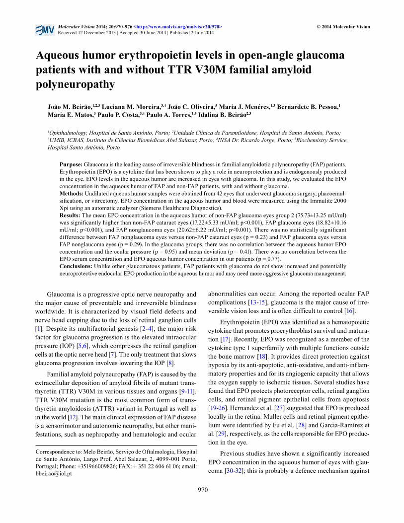
Glaucoma is a progressive optic nerve neuropathy and the major cause of preventable and irreversible blindness worldwide. It is characterized by visual field defects and nerve head cupping due to the loss of retinal ganglion cells [1]. Despite its multifactorial genesis [2-4], the major risk factor for glaucoma progression is the elevated intraocular pressure (IOP) [5,6], which compresses the retinal ganglion cells at the optic nerve head [7]. The only treatment that slows glaucoma progression involves lowering the IOP [8].
Familial amyloid polyneuropathy (FAP) is caused by the extracellular deposition of amyloid fibrils of mutant trans-thyretin (TTR) V30M in various tissues and organs [9-11]. TTR V30M mutation is the most common form of trans-thyretin amyloidosis (ATTR) variant in Portugal as well as in the world [12]. The main clinical expression of FAP disease is a sensorimotor and autonomic neuropathy, but other mani-festations, such as nephropathy and hematologic and ocular
abnormalities can occur. Among the reported ocular FAP complications [13-15], glaucoma is the major cause of irre-versible vision loss and is often difficult to control [16].
Erythropoietin (EPO) was identified as a hematopoietic cytokine that promotes proerythroblast survival and matura-tion [17]. Recently, EPO was recognized as a member of the cytokine type 1 superfamily with multiple functions outside the bone marrow [18]. It provides direct protection against hypoxia by its anti-apoptotic, anti-oxidative, and anti-inflam-matory properties and for its angiogenic capacity that allows the oxygen supply to ischemic tissues. Several studies have found that EPO protects photoreceptor cells, retinal ganglion cells, and retinal pigment epithelial cells from apoptosis [19-26]. Hernandez et al. [27] suggested that EPO is produced locally in the retina. Muller cells and retinal pigment epithe-lium were identified by Fu et al. [28] and Garcia-Ramírez et al. [29], respectively, as the cells responsible for EPO produc-tion in the eye.
Previous studies have shown a significantly increased EPO concentration in the aqueous humor of eyes with glau-coma [30-32]; this is probably a defence mechanism against
Molecular Vision 2014; 20:970-976 <http://www.molvis.org/molvis/v20/970>Received 12 December 2013 | Accepted 30 June 2014 | Published 2 July 2014
© 2014 Molecular Vision
970
Aqueous humor erythropoietin levels in open-angle glaucoma patients with and without TTR V30M familial amyloid polyneuropathy
João M. Beirão,1,2,3 Luciana M. Moreira,3,4 João C. Oliveira,5 Maria J. Menéres,1,3 Bernardete B. Pessoa,1 Maria E. Matos,3 Paulo P. Costa,3,4 Paulo A. Torres,1,3 Idalina B. Beirão2,3
1Ophthalmology, Hospital de Santo António, Porto; 2Unidade Clínica de Paramiloidose, Hospital de Santo António, Porto; 3UMIB, ICBAS, Instituto de Ciências Biomédicas Abel Salazar, Porto; 4INSA Dr. Ricardo Jorge, Porto; 5Biochemistry Service, Hospital Santo António, Porto
Purpose: Glaucoma is the leading cause of irreversible blindness in familial amyloidotic polyneuropathy (FAP) patients. Erythropoietin (EPO) is a cytokine that has been shown to play a role in neuroprotection and is endogenously produced in the eye. EPO levels in the aqueous humor are increased in eyes with glaucoma. In this study, we evaluated the EPO concentration in the aqueous humor of FAP and non-FAP patients, with and without glaucoma.Methods: Undiluted aqueous humor samples were obtained from 42 eyes that underwent glaucoma surgery, phacoemul-sification, or vitrectomy. EPO concentration in the aqueous humor and blood were measured using the Immulite 2000 Xpi using an automatic analyzer (Siemens Healthcare Diagnostics).Results: The mean EPO concentration in the aqueous humor of non-FAP glaucoma eyes group 2 (75.73±13.25 mU/ml) was significantly higher than non-FAP cataract eyes (17.22±5.33 mU/ml; p<0.001), FAP glaucoma eyes (18.82±10.16 mU/ml; p<0.001), and FAP nonglaucoma eyes (20.62±6.22 mU/ml; p<0.001). There was no statistically significant difference between FAP nonglaucoma eyes versus non-FAP cataract eyes (p = 0.23) and FAP glaucoma eyes versus FAP nonglaucoma eyes (p = 0.29). In the glaucoma groups, there was no correlation between the aqueous humor EPO concentration and the ocular pressure (p = 0.95) and mean deviation (p = 0.41). There was no correlation between the EPO serum concentration and EPO aqueous humor concentration in our patients (p = 0.77).Conclusions: Unlike other glaucomatous patients, FAP patients with glaucoma do not show increased and potentially neuroprotective endocular EPO production in the aqueous humor and may need more aggressive glaucoma management.
Correspondence to: Melo Beirão, Serviço de Oftalmologia, Hospital de Santo António, Largo Prof. Abel Salazar, 2, 4099-001 Porto, Portugal; Phone: +351966009826; FAX: + 351 22 606 61 06; email: [email protected]
Molecular Vision 2014; 20:970-976 <http://www.molvis.org/molvis/v20/970> © 2014 Molecular Vision
971
glaucomatous damage [33] caused by hypoxia, ischemia, oxidative stress, and reduced pro-inflammatory cytokine production [34-39]. Although hypoxia/ischemia is the major stimulus for endocular and systemic EPO production [21,40-43], other incompletely understood factors may be involved [27,29].
FAP patients and even presymptomatic carriers have an inappropriately low EPO production [44]. In vitro studies suggest that the dissociated mutant TTR that polymerizes into misfolding amyloidogenic intermediates, protofilaments, and nonfibrillar aggregates of TTR rather than mature amyloid fibrils may induce cellular toxicity [45,46]. We propose that these amyloid precursors may be toxic to EPO-producing cells. This study was performed to evaluate the ocular EPO response in FAP patients with glaucoma.
METHODS
It was recruited 42 eyes of 42 patients (18 females) with a mean age of 56.8±7.4 years. A prospective, controlled, nonrandomized, nonblind comparative study was conducted from January 2008 to December 2011 at the Ophthalmic and Clinical Chemistry Departments from Centro Hospitalar do Porto, Porto. Written informed consent was obtained from all patients. This study was performed in accordance with the Declaration of Helsinki of the World Medical Association and was approved by the Ethics Committee of the Centro Hospitalar do Porto.
Presurgical assessment included Snellen best-corrected visual acuity (Snellen chart, Takagi chart projector CP-30, calibrated for approximately 6 m), slit-lamp biomicroscopy, intraocular pressure (IOP) measurement by Goldman appla-nation tonometry (same person with AT-900 tonometer; Haag-Streit, Koniz, Switzerland), fundoscopy (90 D noncon-tact slit-lamp lens; Volk Optical, Mentor, OH), Humphrey perimetry (Humphrey Field Analyzer; Humphrey Instru-ments, San Leandro, CA), and the cup/disc ratio. All exami-nations were performed within 2 weeks before the surgical procedure.
Exclusion criteria for all groups were: previous laser and/or intraocular surgery; history of systemic (e.g., diabetes mellitus, kidney disease, cardiovascular disorders, anemia, immune disease, except FAP in groups 1 and 3) or any ocular disorders (e.g., age-related macular degeneration); history of medications that could influence the level of EPO (e.g., iron preparations, chemotherapeutic agents, granulocyte colony-stimulating factor, or systemic therapy with EPO), and patients with any type of glaucoma except open-angle glau-coma, such as angle-closure, pigmented, exfoliation, normo-tensive, and neovascular glaucomas, or ocular hypertension.
To clarify the relationship between aqueous EPO produc-tion and circulating blood EPO levels, we compared the aqueous and serum concentrations of EPO. Aqueous humor samples were obtained from each eye before the beginning of surgery (trabeculectomy, phacoemulsification, or vitrec-tomy). The standard procedure involved collecting undiluted aqueous humor samples (50–150 µl) through a paracentesis, using a 30-gauge needle on a tuberculin syringe under an operating microscope. Samples were obtained carefully to avoid touching intraocular tissues or blood contamination. All samples were carefully protected from light and were sent immediately to the laboratory for EPO measurement. At the same time, 9 ml of venous blood samples were collected in EDTA tubes from an antecubital vein immediately before sugery. The blood was immediately centrifuged and the blood serum put on the automatic analyzer.
Serum samples were obtained from the centrifugation of the blood sample. The samples of aqueous humor and serum had the same processing routine analysis. Serum and aqueous humor EPO concentrations were measured by a chemilumi-nescent method in an automatic Xpi Immulite 2000 analyzer (Siemens Healthcare Diagnostics, Siemens AG, Munich, Germany).
Statistical analysis: Statistical analysis was performed using nonparametric tests. The Kruskal–Wallis test was used to compare the groups in relation to age, and the chi-square test was used in relation to gender. The Mann–Whitney U test was used to compare the nonglaucoma, glaucoma, and FAP groups in relation to aqueous humor EPO and serum EPO levels. The relation between EPO and serum was evaluated by Spearman correlation. Values of p<0.05 were considered statistically significant. Data analysis was performed using IBM SPSS Statistics software version 20.
RESULTS
A total of 21 glaucomatous eyes from 21 patients and 21 control eyes (21 patients) were enrolled in the study. The demographic characteristics of the patients are summarized in Table 1. Of the glaucoma eyes, ten were from FAP patients (group 1, mean age 55.4±10.0 years mean and standard devia-tion; five females) and 11 were from non-FAP patients (group 2, mean age 55.8±7.0 years mean and standard deviation; four females). Of the 21 control eyes, nine were from FAP patients with an indication for vitrectomy due to amyloid deposition (group 3, 55.9±8.5 years mean and standard deviation; four females) and 12 were from non-FAP patients awaiting phaco-emulsification and intraocular lens implantation (group 4, 58.8±4.9 years mean and standard deviation; five females). Groups 1 and 2 presented indications for trabeculectomy,
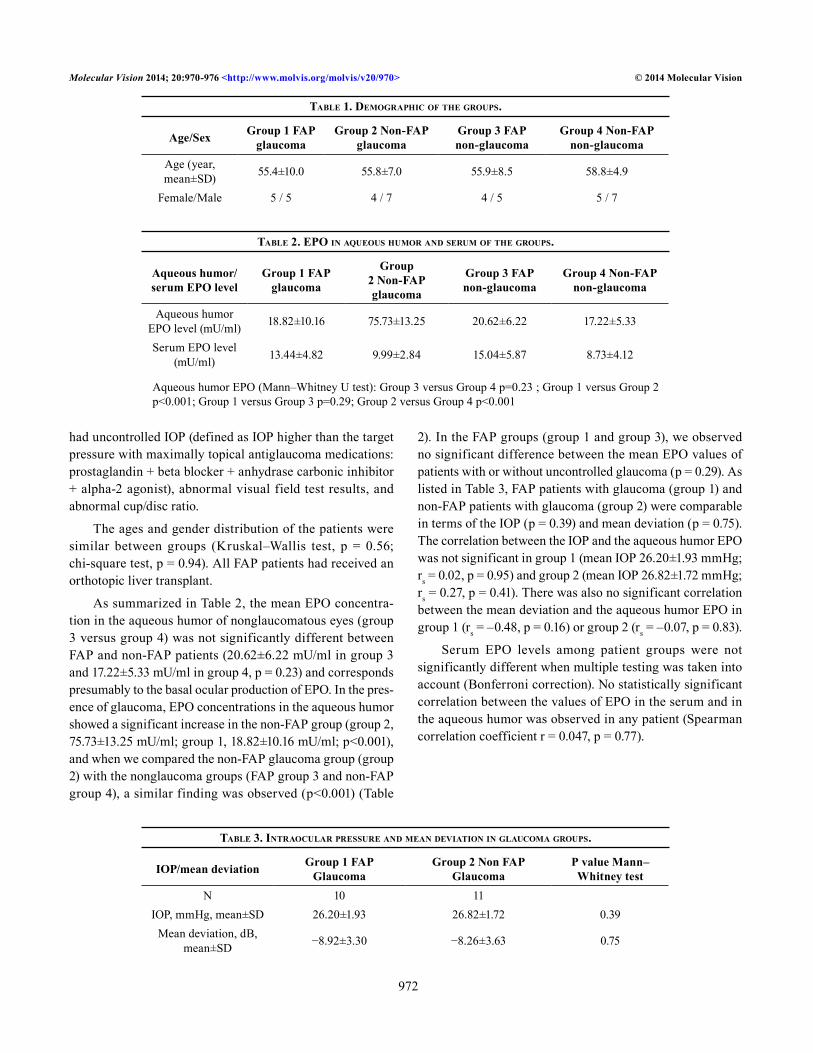
Molecular Vision 2014; 20:970-976 <http://www.molvis.org/molvis/v20/970> © 2014 Molecular Vision
972
had uncontrolled IOP (defined as IOP higher than the target pressure with maximally topical antiglaucoma medications: prostaglandin + beta blocker + anhydrase carbonic inhibitor + alpha-2 agonist), abnormal visual field test results, and abnormal cup/disc ratio.
The ages and gender distribution of the patients were similar between groups (Kruskal–Wallis test, p = 0.56; chi-square test, p = 0.94). All FAP patients had received an orthotopic liver transplant.
As summarized in Table 2, the mean EPO concentra-tion in the aqueous humor of nonglaucomatous eyes (group 3 versus group 4) was not significantly different between FAP and non-FAP patients (20.62±6.22 mU/ml in group 3 and 17.22±5.33 mU/ml in group 4, p = 0.23) and corresponds presumably to the basal ocular production of EPO. In the pres-ence of glaucoma, EPO concentrations in the aqueous humor showed a significant increase in the non-FAP group (group 2, 75.73±13.25 mU/ml; group 1, 18.82±10.16 mU/ml; p<0.001), and when we compared the non-FAP glaucoma group (group 2) with the nonglaucoma groups (FAP group 3 and non-FAP group 4), a similar finding was observed (p<0.001) (Table
2). In the FAP groups (group 1 and group 3), we observed no significant difference between the mean EPO values of patients with or without uncontrolled glaucoma (p = 0.29). As listed in Table 3, FAP patients with glaucoma (group 1) and non-FAP patients with glaucoma (group 2) were comparable in terms of the IOP (p = 0.39) and mean deviation (p = 0.75). The correlation between the IOP and the aqueous humor EPO was not significant in group 1 (mean IOP 26.20±1.93 mmHg; rs = 0.02, p = 0.95) and group 2 (mean IOP 26.82±1.72 mmHg; rs = 0.27, p = 0.41). There was also no significant correlation between the mean deviation and the aqueous humor EPO in group 1 (rs = –0.48, p = 0.16) or group 2 (rs = –0.07, p = 0.83).
Serum EPO levels among patient groups were not significantly different when multiple testing was taken into account (Bonferroni correction). No statistically significant correlation between the values of EPO in the serum and in the aqueous humor was observed in any patient (Spearman correlation coefficient r = 0.047, p = 0.77).
Table 1. Demographic of The groups.
Age/Sex Group 1 FAP glaucoma
Group 2 Non-FAP glaucoma
Group 3 FAP non-glaucoma
Group 4 Non-FAP non-glaucoma
Age (year, mean±SD) 55.4±10.0 55.8±7.0 55.9±8.5 58.8±4.9
Female/Male 5 / 5 4 / 7 4 / 5 5 / 7
Table 2. epo in aqueous humor anD serum of The groups.
Aqueous humor/serum EPO level
Group 1 FAP glaucoma
Group 2 Non-FAP glaucoma
Group 3 FAP non-glaucoma
Group 4 Non-FAP non-glaucoma
Aqueous humor EPO level (mU/ml) 18.82±10.16 75.73±13.25 20.62±6.22 17.22±5.33
Serum EPO level (mU/ml) 13.44±4.82 9.99±2.84 15.04±5.87 8.73±4.12
Aqueous humor EPO (Mann–Whitney U test): Group 3 versus Group 4 p=0.23 ; Group 1 versus Group 2 p<0.001; Group 1 versus Group 3 p=0.29; Group 2 versus Group 4 p<0.001
Table 3. inTraocular pressure anD mean DeviaTion in glaucoma groups.
IOP/mean deviation Group 1 FAP Glaucoma
Group 2 Non FAP Glaucoma
P value Mann–Whitney test
N 10 11IOP, mmHg, mean±SD 26.20±1.93 26.82±1.72 0.39
Mean deviation, dB, mean±SD −8.92±3.30 −8.26±3.63 0.75
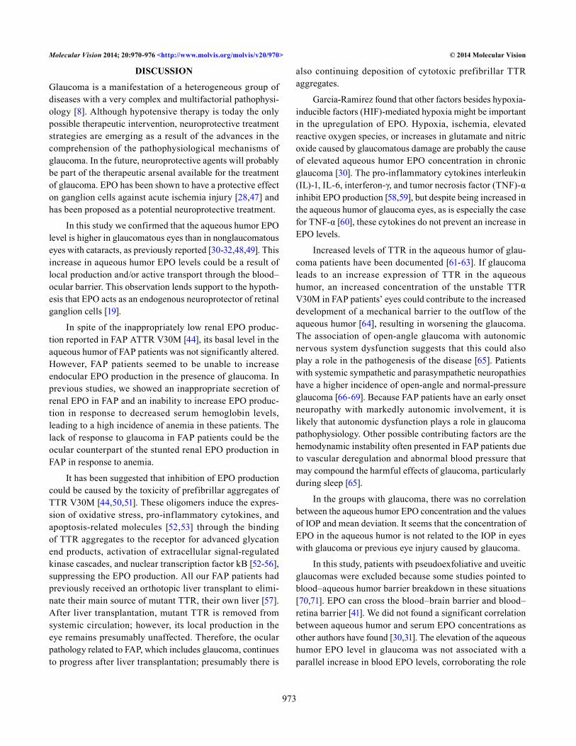
Molecular Vision 2014; 20:970-976 <http://www.molvis.org/molvis/v20/970> © 2014 Molecular Vision
973
DISCUSSION
Glaucoma is a manifestation of a heterogeneous group of diseases with a very complex and multifactorial pathophysi-ology [8]. Although hypotensive therapy is today the only possible therapeutic intervention, neuroprotective treatment strategies are emerging as a result of the advances in the comprehension of the pathophysiological mechanisms of glaucoma. In the future, neuroprotective agents will probably be part of the therapeutic arsenal available for the treatment of glaucoma. EPO has been shown to have a protective effect on ganglion cells against acute ischemia injury [28,47] and has been proposed as a potential neuroprotective treatment.
In this study we confirmed that the aqueous humor EPO level is higher in glaucomatous eyes than in nonglaucomatous eyes with cataracts, as previously reported [30-32,48,49]. This increase in aqueous humor EPO levels could be a result of local production and/or active transport through the blood–ocular barrier. This observation lends support to the hypoth-esis that EPO acts as an endogenous neuroprotector of retinal ganglion cells [19].
In spite of the inappropriately low renal EPO produc-tion reported in FAP ATTR V30M [44], its basal level in the aqueous humor of FAP patients was not significantly altered. However, FAP patients seemed to be unable to increase endocular EPO production in the presence of glaucoma. In previous studies, we showed an inappropriate secretion of renal EPO in FAP and an inability to increase EPO produc-tion in response to decreased serum hemoglobin levels, leading to a high incidence of anemia in these patients. The lack of response to glaucoma in FAP patients could be the ocular counterpart of the stunted renal EPO production in FAP in response to anemia.
It has been suggested that inhibition of EPO production could be caused by the toxicity of prefibrillar aggregates of TTR V30M [44,50,51]. These oligomers induce the expres-sion of oxidative stress, pro-inflammatory cytokines, and apoptosis-related molecules [52,53] through the binding of TTR aggregates to the receptor for advanced glycation end products, activation of extracellular signal-regulated kinase cascades, and nuclear transcription factor kB [52-56], suppressing the EPO production. All our FAP patients had previously received an orthotopic liver transplant to elimi-nate their main source of mutant TTR, their own liver [57]. After liver transplantation, mutant TTR is removed from systemic circulation; however, its local production in the eye remains presumably unaffected. Therefore, the ocular pathology related to FAP, which includes glaucoma, continues to progress after liver transplantation; presumably there is
also continuing deposition of cytotoxic prefibrillar TTR aggregates.
Garcia-Ramirez found that other factors besides hypoxia-inducible factors (HIF)-mediated hypoxia might be important in the upregulation of EPO. Hypoxia, ischemia, elevated reactive oxygen species, or increases in glutamate and nitric oxide caused by glaucomatous damage are probably the cause of elevated aqueous humor EPO concentration in chronic glaucoma [30]. The pro-inflammatory cytokines interleukin (IL)-1, IL-6, interferon-γ, and tumor necrosis factor (TNF)-α inhibit EPO production [58,59], but despite being increased in the aqueous humor of glaucoma eyes, as is especially the case for TNF-α [60], these cytokines do not prevent an increase in EPO levels.
Increased levels of TTR in the aqueous humor of glau-coma patients have been documented [61-63]. If glaucoma leads to an increase expression of TTR in the aqueous humor, an increased concentration of the unstable TTR V30M in FAP patients’ eyes could contribute to the increased development of a mechanical barrier to the outflow of the aqueous humor [64], resulting in worsening the glaucoma. The association of open-angle glaucoma with autonomic nervous system dysfunction suggests that this could also play a role in the pathogenesis of the disease [65]. Patients with systemic sympathetic and parasympathetic neuropathies have a higher incidence of open-angle and normal-pressure glaucoma [66-69]. Because FAP patients have an early onset neuropathy with markedly autonomic involvement, it is likely that autonomic dysfunction plays a role in glaucoma pathophysiology. Other possible contributing factors are the hemodynamic instability often presented in FAP patients due to vascular deregulation and abnormal blood pressure that may compound the harmful effects of glaucoma, particularly during sleep [65].
In the groups with glaucoma, there was no correlation between the aqueous humor EPO concentration and the values of IOP and mean deviation. It seems that the concentration of EPO in the aqueous humor is not related to the IOP in eyes with glaucoma or previous eye injury caused by glaucoma.
In this study, patients with pseudoexfoliative and uveitic glaucomas were excluded because some studies pointed to blood–aqueous humor barrier breakdown in these situations [70,71]. EPO can cross the blood–brain barrier and blood–retina barrier [41]. We did not found a significant correlation between aqueous humor and serum EPO concentrations as other authors have found [30,31]. The elevation of the aqueous humor EPO level in glaucoma was not associated with a parallel increase in blood EPO levels, corroborating the role
Molecular Vision 2014; 20:970-976 <http://www.molvis.org/molvis/v20/970> © 2014 Molecular Vision
974
of local EPO production as already proposed by Fu [28] and Garcia-Ramirez [29].
In conclusion, our study confirmed that the level of EPO is increased in aqueous humor of open-angle glauco-matous eyes, as found by other authors. This increase was not observed in FAP patients. With the increased survival of transplanted FAP patients, glaucoma prevalence is expected to increase dramatically with increased suvival of the trans-planted patients. We showed lower endogenous neuroprotec-tion in glaucomatous eyes of FAP patients, emphasizing the need for more aggressive glaucoma management to maintain vision through life.
REFERENCES1. Agar A, Yip SS, Hill MA, Coroneo MT. Pressure related apop-
tosis in neuronal cell lines. J Neurosci Res 2000; 60:495-503. [PMID: 10797552].
2. Flammer J, Orgül S. Optic nerve blood-flow abnormalities in glaucoma. Prog Retin Eye Res 1998; 17:267-89. [PMID: 9695795].
3. Cioffi GA. Ischemic model of optic nerve injury. Trans Am Ophthalmol Soc 2005; 103:592-613. [PMID: 17057819].
4. Tezel G, Wax MB. Hypoxia-inducible factor 1alpha in the glaucomatous retina and optic nerve head. Arch Ophthalmol 2004; 122:1348-56. [PMID: 15364715].
5. Evans DW, Hosking SL, Gherghel D, Bartlett JD. Contrast sensitivity improves after brimonidine therapy in primary open angle glaucoma: a case for neuroprotection. Br J Ophthalmol 2003; 87:1463-5. [PMID: 14660453].
6. Gordon MO, Beiser JA, Brandt JD, Heuer DK, Higginbotham EJ, Johnson CA, Keltner JL, Miller JP, Parrish RK 2nd, Wilson MR, Kass MA. The Ocular Hypertension Treatment Study: baseline factors that predict the onset of primary open-angle glaucoma. Arch Ophthalmol 2002; 120:714-20. [PMID: 12049575].
7. Izzotti A, Bagnis A, Saccà SC. The role of oxidative stress in glaucoma. Mutat Res 2006; 612:105-14. [PMID: 16413223].
8. Leske MC, Heijl A, Hussein M, Bengtsson B, Hyman L, Komaroff E. Early Manifest Glaucoma Trial Group. Factors for glaucoma progression and the effect of treatment: the early manifest glaucoma trial. Arch Ophthalmol 2003; 121:48-56. [PMID: 12523884].
9. Saraiva MJ, Costa PP, Goodman DS. Studies on plasma transthyretin (prealbumin) in familial amyloidotic polyneu-ropathy, Portuguese type. J Lab Clin Med 1983; 102:590-603. [PMID: 6311926].
10. Saraiva MJ, Costa PP, Birken S, Goodman DS. Presence of an abnormal transthyretin (prealbumin) in Portuguese patients with familial amyloidotic polyneuropathy. Trans Assoc Am Physicians 1983; 96:261-70. [PMID: 6208668].
11. Saraiva MJ, Birken S, Costa PP, Goodman DS. Amyloid fibril protein in familial amyloidotic polyneuropathy, Portuguese type. Definition of molecular abnormality in transthyretin (prealbumin). J Clin Invest 1984; 74:104-19. [PMID: 6736244].
12. Ando Y, Araki S, Ando M. Transthyretin and familial amyloi-dotic polyneuropathy. Intern Med 1993; 32:920-2. [PMID: 8204970].
13. Ando E, Ando Y, Okamura R, Uchino M, Ando M, Negi A. Ocular manifestations of familial amyloidotic polyneu-ropathy type I: long-term follow up. Br J Ophthalmol 1997; 81:295-8. [PMID: 9215058].
14. Sandgren O, Kjellgren D, Suhr OB. Ocular manifestations in liver transplant recipients with familial amyloid polyneu-ropathy. Acta Ophthalmol (Copenh) 2008; 86:520-4. [PMID: 18435819].
15. Beirão M, Matos E, Beirão I, Costa PP, Torres P. Anticipa-tion of presbyopia in Portuguese familial amyloidosis ATTR V30M. Amyloid 2011; 18:92-7. [PMID: 21591979].
16. Kimura A, Ando E, Fukushima M, Koga T, Hirata A, Arimura K, Ando Y, Negi A, Tanihara H. Secondary glaucoma in patients with familial amyloidotic polyneuropathy. Arch Ophthalmol 2003; 121:351-6. [PMID: 12617705].
17. Jelkmann W. Erythropoietin: structure, control of produc-tion, and function. Physiol Rev 1992; 72:449-89. [PMID: 1557429].
18. Ghezzi P, Brines M. Erythropoietin as an antiapoptotic, tissue-protective cytokine. Cell Death Differ 2004; 11:Suppl 1S37-44. [PMID: 15243580].
19. Becerra SP, Amaral J. Erythropoietin - an endogenous retinal survival factor. N Engl J Med 2002; 347:1968-70. [PMID: 12477950].
20. Dreixler JC, Hagevik S, Hemmert JW, Shaikh AR, Rosenbaum DM, Roth S. Involvement of erythropoietin in retinal isch-emic preconditioning. Anesthesiology 2009; 110:774-80. [PMID: 19322943].
21. Inomata Y, Hirata A, Takahashi E, Kawaji T, Fukushima M, Tanihara H. Elevated erythropoietin in vitreous with isch-emic retinal diseases. Neuroreport 2004; 15:877-9. [PMID: 15073535].
22. Sakanaka M, Wen TC, Matsuda S, Masuda S, Morishita E, Nagao M, Sasaki R. In vivo evidence that erythropoietin protects neurons from ischemic damage. Proc Natl Acad Sci USA 1998; 95:4635-40. [PMID: 9539790].
23. Tsai JC, Song BJ, Wu L, Forbes M. Erythropoietin: a candidate neuroprotective agent in the treatment of glaucoma. J Glau-coma 2007; 16:567-71. [PMID: 17873720].
24. Tsai JC, Wu L, Worgul B, Forbes M, Cao J. Intravitreal administration of erythropoietin and preservation of retinal ganglion cells in an experimental rat model of glaucoma. Curr Eye Res 2005; 30:1025-31. [PMID: 16282136].
25. Villa P, Bigini P, Mennini T, Agnello D, Laragione T, Cagnotto A, Viviani B, Marinovich M, Cerami A, Coleman TR, Brines M, Ghezzi P. Erythropoietin selectively attenuates
Molecular Vision 2014; 20:970-976 <http://www.molvis.org/molvis/v20/970> © 2014 Molecular Vision
975
cytokine production and inflammation in cerebral ischemia by targeting neuronal apoptosis. J Exp Med 2003; 198:971-5. [PMID: 12975460].
26. Wang ZY, Shen LJ, Tu L, Hu DN, Liu GY, Zhou ZL, Lin Y, Chen LH, Qu J. Erythropoietin protects retinal pigment epithelial cells from oxidative damage. Free Radic Biol Med 2009; 46:1032-41. [PMID: 19136057].
27. Hernández C, Fonollosa A, García-Ramírez M, Higuera M, Catalán R, Miralles A, García-Arumí J, Simó R. Erythropoi-etin is expressed in the human retina and it is highly elevated in the vitreous fluid of patients with diabetic macular edema. Diabetes Care 2006; 29:2028-33. [PMID: 16936148].
28. Fu QL, Wu W, Wang H, Li X, Lee VW, So KF. Up-regulated endogenous erythropoietin/erythropoietin receptor system and exogenous erythropoietin rescue retinal ganglion cells after chronic ocular hypertension. Cell Mol Neurobiol 2008; 28:317-29. [PMID: 17554621].
29. García-Ramírez M, Hernández C, Simó R. Expression of eryth-ropoietin and its receptor in the human retina: a comparative study of diabetic and nondiabetic subjects. Diabetes Care 2008; 31:1189-94. [PMID: 18332162].
30. Cumurcu T, Bulut Y, Demir HD, Yenisehirli G. Aqueous humor erythropoietin levels in patients with primary open-angle glaucoma. J Glaucoma 2007; 1:645-8. .
31. Wang ZY, Zhao KK, Zhao PQ. Erythropoietin is increased in aqueous humor of glaucomatous eyes. Curr Eye Res 2010; 35:680-4. [PMID: 20673044].
32. Mokbel TH, Ghanem AA, Kishk H, Arafa LF, El-Baiomy AA. Erythropoietin and soluble CD44 levels in patients with primary open-angle glaucoma. Clin Experiment Ophthalmol 2010; 38:560-5. [PMID: 20456444].
33. Dirnagl U, Simon RP, Hallenbeck JM. Ischemic tolerance and endogenous neuroprotection. Trends Neurosci 2003; 26:248-54. [PMID: 12744841].
34. Wenger RH. Cellular adaptation to hypoxia: O2-sensing protein hydroxylases, hypoxia-inducible transcription factors, and O2-regulated gene expression. FASEB J 2002; 16:1151-62. [PMID: 12153983].
35. Arjamaa O, Nikinmaa M. Oxygen-dependent diseases in the retina: role of hypoxia-inducible factors. Exp Eye Res 2006; 83:473-83. [PMID: 16750526].
36. Silva M, Grillot D, Benito A, Richard C, Nuñez G, Fernández-Luna JL. Erythropoietin can promote erythroid progenitor survival by repressing apoptosis through Bcl-XL and Bcl-2. Blood 1996; 88:1576-82. [PMID: 8781412].
37. Sakanaka M, Wen TC, Matsuda S, Masuda S, Morishita E, Nagao M, Sasaki R. In vivo evidence that erythropoietin protects neurons from ischemic damage. Proc Natl Acad Sci USA 1998; 95:4635-40. [PMID: 9539790].
38. Kawakami M, Sekiguchi M, Sato K, Kozaki S, Takahashi M. Erythropoietin receptor-mediated inhibition of exocytotic glutamate release confers neuroprotection during chemical ischemia. J Biol Chem 2001; 276:39469-75. [PMID: 11504731].
39. Genc S, Koroglu TF, Genc K. Erythropoietin as a novel neuro-protectant. Restor Neurol Neurosci 2004; 22:105-19. [PMID: 15272145].
40. Fisher JW. Erythropoietin: physiology and pharmacology update. Exp Biol Med (Maywood) 2003; 228:1-14. [PMID: 12524467].
41. Grimm C, Wenzel A, Groszer M, Mayser H, Seeliger M, Samardzija M, Bauer C, Gassmann M, Remé CE. HIF-1-induced erythropoietin in the hypoxic retina protects against light-induced retinal degeneration. Nat Med 2002; 8:718-24. [PMID: 12068288].
42. Katsura Y, Okano T, Matsuno K, Osako M, Kure M, Watanabe T, Iwaki Y, Noritake M, Kosano H, Nishigori H, Matsuoka T. Erythropoietin is highly elevated in vitreous fluid of patients with proliferative diabetic retinopathy. Diabetes Care 2005; 28:2252-4. [PMID: 16123502].
43. Watanabe D, Suzuma K, Matsui S, Kurimoto M, Kiryu J, Kita M, Suzuma I, Ohashi H, Ojima T, Murakami T, Kobayashi T, Masuda S, Nagao M, Yoshimura N, Takagi H. Erythropoietin as a retinal angiogenic factor in proliferative diabetic retinop-athy. N Engl J Med 2005; 353:782-92. [PMID: 16120858].
44. Beirão I, Lobato L, Costa PM, Fonseca I, Mendes P, Silva M, Bravo F, Cabrita A, Porto G. Kidney and anemia in familial amyloidosis type I. Kidney Int 2004; 66:2004-9. [PMID: 15496172].
45. Bucciantini M, Calloni G, Chiti F, Formigli L, Nosi D, Dobson CM, Stefani M. Prefibrillar amyloid protein aggregates share common features of cytotoxicity. J Biol Chem 2004; 279:31374-82. [PMID: 15133040].
46. Reixach N, Deechongkit S, Jiang X, Kelly JW, Buxbaum JN. Tissue damage in the amyloidoses: Transthyretin monomers and nonnative oligomers are the major cytotoxic species in tissue culture. Proc Natl Acad Sci USA 2004; 101:2817-22. [PMID: 14981241].
47. Junk AK, Mammis A, Savitz SI, Singh M, Roth S, Malhotra S, Rosenbaum PS, Cerami A, Brines M, Rosenbaum DM. Erythropoietin administration protects retinal neurons from acute ischemia-reperfusion injury. Proc Natl Acad Sci USA 2002; 99:10659-64. [PMID: 12130665].
48. Nassiri N, Nassiri N, Majdi M, Mehrjardi HZ, Shakiba Y, Haghnegahdar M, Heidari AB, Djalilian AR, Mirahmadian M. Erythropoietin levels in aqueous humor of patients with glaucoma. Mol Vis 2012; 18:1991-5. [PMID: 22876126].
49. Hamid M, Fahmy I, Moemen L, El-Beltagy T. Role of matrix metalloproteinase-2 and its inhibitor and erythropoietin in the pathogenesis of pseudoexfoliative glaucoma. Australian J Bas Appl Sci 2008; 2:752-6. .
50. Beirão I, Moreira L, Porto G, Lobato L, Fonseca I, Cabrita A, Costa PM. Low erythropoietin production in familial amyloidosis TTR V30M is not related with renal congo-philic amyloid deposition. A clinicopathologic study of twelve cases. Nephron Clin Pract 2008; 109:c95-9. [PMID: 18596378].
Molecular Vision 2014; 20:970-976 <http://www.molvis.org/molvis/v20/970> © 2014 Molecular Vision
976
51. Beirão I, Moreira L, Barandela T, Lobato L, Silva P, Gouveia CM, Carneiro F, Fonseca I, Porto G, Pinho E, Costa P. Erythropoietin production by distal nephron in normal and familial amyloidotic adult human kidneys. Clin Nephrol 2010; 74:327-35. [PMID: 20979939].
52. Sousa MM, Cardoso I, Fernandes R, Guimarães A, Saraiva MJ. Deposition of transthyretin in early stages of familial amyloidotic polyneuropathy: evidence for toxicity of nonfi-brillar aggregates. Am J Pathol 2001; 159:1993-2000. [PMID: 11733349].
53. Fiszman ML, Di Egidio M, Ricart KC, Repetto MG, Boro-dinsky LN, Llesuy SF, Saizar RD, Trigo PL, Riedstra S, Costa PP, Villa AM, Katz N, Lendoire JC, Sica RE. Evidence of oxidative stress in familial amyloidotic polyneuropathy type 1. Arch Neurol 2003; 60:593-7. [PMID: 12707074].
54. Sousa MM, Du Yan S, Fernandes R, Guimaraes A, Stern D, Saraiva MJ. Familial amyloid polyneuropathy: receptor for advanced glycation end products-dependent triggering of neuronal inflammatory and apoptotic pathways. J Neurosci 2001; 21:7576-86. [PMID: 11567048].
55. Sousa MM, Yan SD, Stern D, Saraiva MJ. Interaction of the receptor for advanced glycation end products (RAGE) with transthyretin triggers nuclear transcription factor kB (NF-kB) activation. Lab Invest 2000; 80:1101-10. [PMID: 10908156].
56. Monteiro FA, Sousa MM, Cardoso I, do Amaral JB, Guimarães A, Saraiva MJ. Activation of ERK1/2 MAP kinases in familial amyloidotic polyneuropathy. J Neurochem 2006; 97:151-61. [PMID: 16515552].
57. Benson MD. Liver transplantation and transthyretin amyloi-dosis. Muscle Nerve 2013; 47:157-62. [PMID: 23169427].
58. Jelkmann W. Proinflammatory cytokines lowering erythropoi-etin production. J Interferon Cytokine Res 1998; 18:555-9. [PMID: 9726435].
59. Vannucchi AM, Grossi A, Rafanelli D, Statello M, Cinotti S, Rossi-Ferrini P. Inhibition of erythropoietin production in vitro by human interferon gamma. Br J Haematol 1994; 87:18-23. [PMID: 7947242].
60. Balaiya S, Edwards J, Tillis T, Khetpal V, Chalam KV. Tumor necrosis factor-alpha (TNF-α) levels in aqueous humor of primary open angle glaucoma. Clin Ophthalmol. 2011; 5:553-6. [PMID: 21607023].
61. Prata TS, Navajas EV, Melo LA Jr, Martins JR, Nader HB, Belfort R Jr. Aqueous humor protein concentration in patients with primary open-angle glaucoma under clinical treatment. Arq Bras Oftalmol 2007; 70:217-20. [PMID: 17589689].
62. Grus FH, Joachim SC, Sandmann S, Thiel U, Bruns K, Lackner KJ, Pfeiffer N. Transthyretin and complex protein pattern in aqueous humor of patients with primary open-angle glau-coma. Mol Vis 2008; 14:1437-45. [PMID: 18682810].
63. Inoue T, Kawaji T, Tanihara H. Elevated levels of multiple biomarkers of Alzheimer’s disease in the aqueous humor of eyes with open-angle glaucoma. Invest Ophthalmol Vis Sci 2013; 54:5353-8. [PMID: 23860758].
64. Silva-Araújo AC, Tavares MA, Cotta JS, Castro-Correia JF. Aqueous outflow system in familial amyloidotic polyneurop-athy, Portuguese type. Graefes Arch Clin Exp Ophthalmol 1993; 231:131-5. [PMID: 8385054].
65. Gherghel D, Hosking SL, Orgül S. Autonomic nervous system, circadian rhythms, and primary open-angle glaucoma. Surv Ophthalmol 2004; 49:491-508. [PMID: 15325194].
66. Brown CM, Dütsch M, Michelson G, Neundörfer B, Hilz MJ. Impaired cardiovascular responses to baroreflex stimula-tion in open-angle and normal-pressure glaucoma. Clin Sci (Lond) 2002; 102:623-30. [PMID: 12049615].
67. Clark CV, Mapstone R. Systemic autonomic neuropathy in open-angle glaucoma. Doc Ophthalmol 1986; 64:179-85. [PMID: 3608758].
68. Kumar R, Ahuja VM. A study of changes in the status of autonomic nervous system in primary open angle glaucoma cases. Indian J Med Sci 1999; 53:529-34. [PMID: 10862279].
69. Riccadonna M, Covi G, Pancera P, Presciuttini B, Babig-hian S, Perfetti S, Bonomi L, Lechi A. Autonomic system activity and 24-hour blood pressure variations in subjects with normal- and high-tension glaucoma. J Glaucoma 2003; 12:156-63. [PMID: 12671471].
70. Küchle M, Nguyen NX, Hannappel E, Naumann GO. The blood-aqueous barrier in eyes with pseudoexfoliation syndrome. Ophthalmic Res 1995; 27:Suppl 1136-42. [PMID: 8577452].
71. Nguyen NX, Kuchle M, Naumann GO. Quantification of blood-aqueous barrier breakdown after phacoemulsification in Fuchs’ heterochromic uveitis. Ophthalmologica 2005; 219:21-5. [PMID: 15627823].
Articles are provided courtesy of Emory University and the Zhongshan Ophthalmic Center, Sun Yat-sen University, P.R. China. The print version of this article was created on 2 July 2014. This reflects all typographical corrections and errata to the article through that date. Details of any changes may be found in the online version of the article.
Use of MALDI-TOF Mass Spectrometry to Assaythe Transthyretin V30MMutation in Serum From aLiver Transplant Donor: A Case ReportPedro Castro Lacerda,1 Luciana Moreira,1,2 Rui Vitorino,3 Paulo Pinho Costa1,2
Familial amyloidotic polyneuropathy (FAP), Portuguesetype, or ATTR V30M is an autosomal dominant in-
herited disorder caused by a mutation in the transthyretin gene,with a valine/methionine substitutionat position30 (TTRV30M).1
ATTRV30Mischaracterizedbyaprogressivesensory/autonomicpolyneuropathy and multiple organ dysfunction.2
Liver transplantation is the main therapeutic option,3 asit virtually eliminates the production of circulating TTRV30M, which occurs predominantly in the liver.
CASE REPORT
In 2004, a then 35-year-old female FAP patient underwentorthotopic liver transplantation from a cadaveric donor totreat her condition. Progression of FAP after liver transplan-tation has been documented but usually stabilizes after sometime.4 Continuing deterioration in this patient raised the sus-picion, confirmed in 2012, that the liver donor was also aTTR V30M carrier. Retransplantation was proposed andcarried out, but these unfortunate circumstances made it nec-essary to assure the anxious patient, and the transplantationcenter for that matter, that this second time the transplantedliver was FAP free.
Serum from the second cadaveric transplant donor wassent to us, a few days after the procedure, and absence ofTTRV30M was established by immunoprecipitation ofTTR followed by mass spectrometry analysis.5 The spectrathat comprise the molecular mass of TTRmonomers are rep-resented inF1 Figure 1. In the positive control sample(Figure 1C), we identified 3 peaks that correspond, respec-tively, to the free form of wild type TTR (13.708 ± 10 Da),the cysteine (Cys) conjugated form of the wild type TTR(13.824 ± 10 Da), and the cysteine conjugated form of theTTR V30M variant (13.860 ± 10 Da), with a difference ofapproximately 33 Da from the wild type TTR.5 The livertransplant donor sample (Figure 1A) showed the 2 peaks
attributed to wild type TTR only (13.694 ± 10 and13.816 ± 10 Da) as seen in the negative control (Figure 1B).
DISCUSSION
Domino FAP liver transplantation into non-FAP patientsis common in some centers. In these cases, TTRV30M pro-duction has been found to be followed by FAP symptoms assoon as 7 years after transplantation.6When this happens, re-transplantation is usually proposed. In areas with a highprevalence of FAP, like the north of Portugal (1:550),7 thereis a non-negligible risk of a FAP patient receiving a liver froman untested FAP donor. Portuguese law assumes that everycitizenwho does not declare otherwise is an organ donor. Un-fortunately, reliable individual clinical information is usuallynot available. Familial amyloidotic polyneuropathy is a late-onset disease, and predictive testing, even if done, is not to befound in clinical records, by force of law. So, at the time oftransplantation, up to 12 hours after liver collection, thereis usually no other information on personal or familymedicalhistory of the donor besides the standard information deliv-ered by the histocompatibility center regarding infectiousagents and malign neoplasias.
The risk of transplanting an FAP patient with a TTRV30Mpositive liver is admittedly low. However, in a countrythat has performed more than a thousand of these transplantprocedures, such an event was to be expected, sooner or later.DNA and serum-based assays for carrier status that could beperformed in a timely manner, while not standard, can beeasily implemented. It should be noted, though, that the legaland ethical dimensions of performing what would possiblybe a predictive genetic test in a deceased donor is uncharteredterritory, and would have to be addressed. Namely, the ques-tion of whether it would be justified to communicate, in theabsence of consent, the genetic status of the deceased donorto family members would have to be weighted in terms of
Received 19 August 2014. Revision requested 14 November 2014.
Accepted 14 November 2014.1 Department of Genetics, National Institute of Health Dr. Ricardo Jorge (INSA),Porto, Portugal.
2 Unit for Multidisciplinary Investigation in Biomedicine (UMIB), Institute of BiomedicalSciences Abel Salazar (ICBAS), Porto, Portugal.
3 Research Unit Organic Chemistry; Natural Products and Food Stuffs (QOPNA),Chemistry Department, Aveiro University, Aveiro, Portugal.
Funding: This work was supported by Portuguese Foundation for Science andTechnology (FCT), European Union, QREN, FEDER and COMPETE for funding theQOPNA research unit (project PEst-C/QUI/UI0062/2013), RNEM (PortugueseMassSpectrometry Network) and COST action BM1305. It was also funded by InstitutoNacional de Saúde Dr. Ricardo Jorge, INSA I.P., Portugal.
The authors declare no conflicts of interest.
Pedro Castro Lacerda and LucianaMoreira wish it to be known that, in their opinion,the first two authors should be regarded as joint first authors'.
P.C.L., L.M., and R.V. participated in the research design, carried out theexperiments and data analysis, and contributed to writing of the paper. P.P.C.participated in research design, in the data analysis, and in writing of the paper.
Correspondence: Pedro C Lacerda, Departamento de Genética do InstitutoNacional de Saúde Dr. Ricardo Jorge, INSA I.P. Rua Alexandre Herculano, 321,4000–055 Porto, Portugal. ([email protected]).
Copyright © 2015 Wolters Kluwer Health, Inc. All rights reserved.
ISSN: 0041-1337/15/0000-00
DOI: 10.1097/TP.0000000000000658
Letter
Copyright © 2015 Wolters Kluwer Health, Inc. Unauthorized reproduction of this article is prohibited.
Copyedited by: Josephine C. Gementera
Transplantation ■ Month 2015 ■ Volume 00 ■ Number 00 www.transplantjournal.com 1
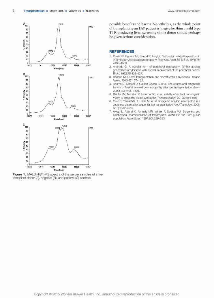
possible benefits and harms. Nonetheless, as the whole pointof transplanting an FAP patient is to give her/him a wild typeTTR producing liver, screening of the donor should perhapsbe given serious consideration.
REFERENCES1. Costa PP, Figueira AS, Bravo FR. Amyloid fibril protein related to prealbumin
in familial amyloidotic polyneuropathy. Proc Natl Acad Sci U S A. 1978;75:4499–4503.
2. Andrade C. A peculiar form of peripheral neuropathy; familiar atypicalgeneralized amyloidosis with special involvement of the peripheral nerves.Brain. 1952;75:408–427.
3. Benson MD. Liver transplantation and transthyretin amyloidosis. MuscleNerve. 2013;47:157–162.
4. Adams D, Samuel D, Goulon-Goeau C, et al. The course and prognosticfactors of familial amyloid polyneuropathy after liver transplantation. Brain.2000;123:1495–1504.
5. Beirão JM, Moreira LV, Lacerda PC, et al. Inability of mutant transthyretinV30M to cross the blood-eye barrier. Transplantation. 2012;9:e54–e56.
6. Goto T, Yamashita T, Ueda M, et al. Iatrogenic amyloid neuropathy in aJapanese patient after sequential liver transplantation.AmJTransplant. 2006;6(10):2512–2515.
7. Alves IL, Altland K, Almeida MR, Winter P, Saraiva MJ. Screening andbiochemical characterization of transthyretin variants in the Portuguesepopulation. Hum Mutat. 1997;9(3):226–233.
Figure 1. MALDI-TOF-MS spectra of the serum samples of a livertransplant donor (A), negative (B), and positive (C) controls.
2 Transplantation ■ Month 2015 ■ Volume 00 ■ Number 00 www.transplantjournal.com
Copyright © 2015 Wolters Kluwer Health, Inc. Unauthorized reproduction of this article is prohibited.
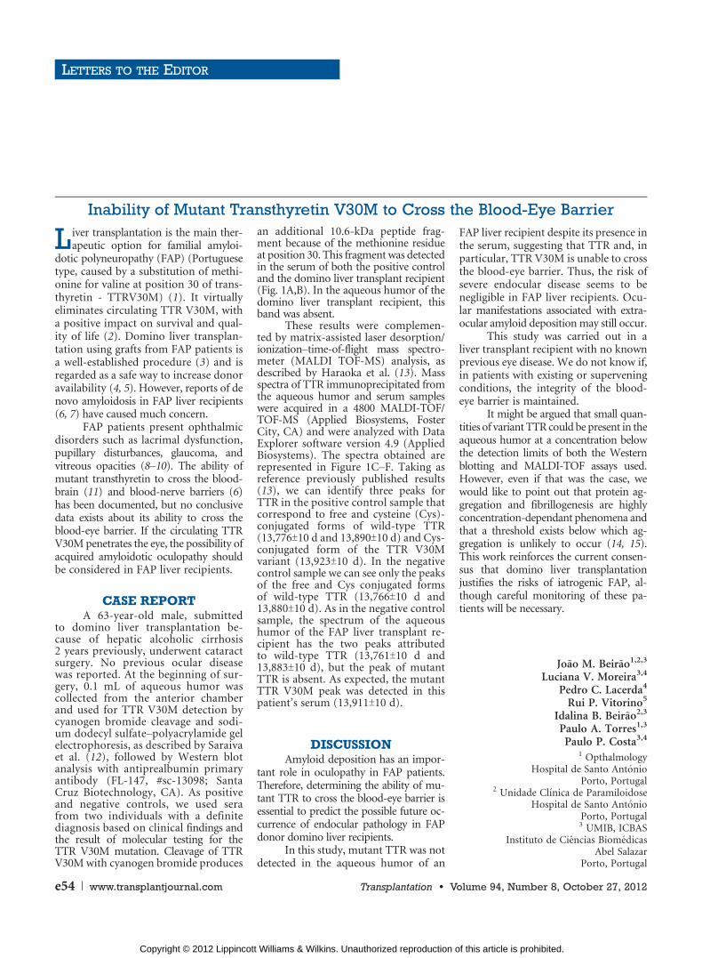
Inability of Mutant Transthyretin V30M to Cross the Blood-Eye Barrier
L iver transplantation is the main ther-apeutic option for familial amyloi-
dotic polyneuropathy (FAP) (Portuguesetype, caused by a substitution of methi-onine for valine at position 30 of trans-thyretin - TTRV30M) (1). It virtuallyeliminates circulating TTR V30M, witha positive impact on survival and qual-ity of life (2). Domino liver transplan-tation using grafts from FAP patients isa well-established procedure (3) and isregarded as a safe way to increase donoravailability (4, 5). However, reports of denovo amyloidosis in FAP liver recipients(6, 7) have caused much concern.
FAP patients present ophthalmicdisorders such as lacrimal dysfunction,pupillary disturbances, glaucoma, andvitreous opacities (8Y10). The ability ofmutant transthyretin to cross the blood-brain (11) and blood-nerve barriers (6)has been documented, but no conclusivedata exists about its ability to cross theblood-eye barrier. If the circulating TTRV30M penetrates the eye, the possibility ofacquired amyloidotic oculopathy shouldbe considered in FAP liver recipients.
CASE REPORTA 63-year-old male, submitted
to domino liver transplantation be-cause of hepatic alcoholic cirrhosis2 years previously, underwent cataractsurgery. No previous ocular diseasewas reported. At the beginning of sur-gery, 0.1 mL of aqueous humor wascollected from the anterior chamberand used for TTR V30M detection bycyanogen bromide cleavage and sodi-um dodecyl sulfateYpolyacrylamide gelelectrophoresis, as described by Saraivaet al. (12), followed by Western blotanalysis with antiprealbumin primaryantibody (FL-147, #sc-13098; SantaCruz Biotechnology, CA). As positiveand negative controls, we used serafrom two individuals with a definitediagnosis based on clinical findings andthe result of molecular testing for theTTR V30M mutation. Cleavage of TTRV30M with cyanogen bromide produces
an additional 10.6-kDa peptide frag-ment because of the methionine residueat position 30. This fragment was detectedin the serum of both the positive controland the domino liver transplant recipient(Fig. 1A,B). In the aqueous humor of thedomino liver transplant recipient, thisband was absent.
These results were complemen-ted by matrix-assisted laser desorption/ionizationYtime-of-flight mass spectro-meter (MALDI TOF-MS) analysis, asdescribed by Haraoka et al. (13). Massspectra of TTR immunoprecipitated fromthe aqueous humor and serum sampleswere acquired in a 4800 MALDI-TOF/TOF-MS (Applied Biosystems, FosterCity, CA) and were analyzed with DataExplorer software version 4.9 (AppliedBiosystems). The spectra obtained arerepresented in Figure 1CYF. Taking asreference previously published results(13), we can identify three peaks forTTR in the positive control sample thatcorrespond to free and cysteine (Cys)-conjugated forms of wild-type TTR(13,776T10 d and 13,890T10 d) and Cys-conjugated form of the TTR V30Mvariant (13,923T10 d). In the negativecontrol sample we can see only the peaksof the free and Cys conjugated formsof wild-type TTR (13,766T10 d and13,880T10 d). As in the negative controlsample, the spectrum of the aqueoushumor of the FAP liver transplant re-cipient has the two peaks attributedto wild-type TTR (13,761T10 d and13,883T10 d), but the peak of mutantTTR is absent. As expected, the mutantTTR V30M peak was detected in thispatient’s serum (13,911T10 d).
DISCUSSIONAmyloid deposition has an impor-
tant role in oculopathy in FAP patients.Therefore, determining the ability of mu-tant TTR to cross the blood-eye barrier isessential to predict the possible future oc-currence of endocular pathology in FAPdonor domino liver recipients.
In this study, mutant TTR was notdetected in the aqueous humor of an
FAP liver recipient despite its presence inthe serum, suggesting that TTR and, inparticular, TTR V30M is unable to crossthe blood-eye barrier. Thus, the risk ofsevere endocular disease seems to benegligible in FAP liver recipients. Ocu-lar manifestations associated with extra-ocular amyloid deposition may still occur.
This study was carried out in aliver transplant recipient with no knownprevious eye disease. We do not know if,in patients with existing or superveningconditions, the integrity of the blood-eye barrier is maintained.
It might be argued that small quan-tities of variant TTR could be present in theaqueous humor at a concentration belowthe detection limits of both the Westernblotting and MALDI-TOF assays used.However, even if that was the case, wewould like to point out that protein ag-gregation and fibrillogenesis are highlyconcentration-dependant phenomena andthat a threshold exists below which ag-gregation is unlikely to occur (14, 15).This work reinforces the current consen-sus that domino liver transplantationjustifies the risks of iatrogenic FAP, al-though careful monitoring of these pa-tients will be necessary.
Joao M. Beirao1,2,3
Luciana V. Moreira3,4
Pedro C. Lacerda4
Rui P. Vitorino5
Idalina B. Beirao2,3
Paulo A. Torres1,3
Paulo P. Costa3,4
1 OpthalmologyHospital de Santo Antonio
Porto, Portugal2 Unidade Clınica de Paramiloidose
Hospital de Santo AntonioPorto, Portugal3 UMIB, ICBAS
Instituto de Ciencias BiomedicasAbel Salazar
Porto, Portugal
LETTERS TO THE EDITOR
e54 www.transplantjournal.com Transplantation & Volume 94, Number 8, October 27, 2012
Copyright © 2012 Lippincott Williams & Wilkins. Unauthorized reproduction of this article is prohibited.
FIGURE 1. Western blot after cyanogen bromide cleavage (A and B) and mass spectra (CYF); fragment (arrow) containingthe residues 30Y127 of the transthyretin with replacement at position 30 methionine for valine (TTR V30M) polypeptidechain, present only in the carriers of the mutation. Lane P represents the positive control serum; N, the negative controlserum; T, the aqueous humor from the transplanted patient (A) and serum from the transplanted patient (B). Lanes Mrepresent the molecular weight marker used (MagicMark; Invitrogen, Carlsbad, CA). Mass spectra of a positive (C) andnegative (D) control serum for the TTR V30Mmutation and the aqueous humor (E) and serum sample (F) of the transplantedpatient. * Cysteine-conjugated form of wild-type TTR; 4 Cysteine-conjugated form of amyloydosis TTR V30M.
* 2012 Lippincott Williams & Wilkins Letters to the Editor e55
Copyright © 2012 Lippincott Williams & Wilkins. Unauthorized reproduction of this article is prohibited.
4 Centro de Genetica MedicaDr. Jacinto Magalhaes
INSA I.P., Porto, Portugal5 Universidade de Aveiro
Aveiro, Portugal
This study was supported by fellowship SFRH/BD/46441/2008 and grant PEst-C/QUI/UI0062/2011 from the Funda0ao para a Ciencia e aTecnologia and by the Laboratoires TheaVPortugal.
The authors declare no conflicts of interest.Address correspondence to: Joao M. Beirao,
M.D., Servi0o de Oftalmologia, Hospital deSanto Antonio, Largo Prof. Abel Salazar, 2,4099-001 Porto, Portugal.
E-mail: [email protected]. and P.P.C. participated in making the re-
search design, analyzing data, and writing thearticle. L.V.M. and P.C.L. participated in an-alyzing laboratory data and writing the article.R.P.V. participated in performing the re-search. I.B.B. and P.A.T. participated in writ-ing the article.
Received 28 June 2012.Accepted 13 July 2012.Copyright * 2012 by Lippincott Williams & WilkinsISSN: 0041-1337/12/9408-e54DOI: 10.1097/TP.0b013e318269e6d1
ACKNOWLEDGMENTThe authors acknowledge Rosario Santos
group (Centro de Genetica Medica Dr. JacintoMagalhaes) for the image acquisition system.
REFERENCES1. Herlenius G, Wilczek HE, Larsson M, et al.
Ten years of international experience withliver transplantation for familial amyloidoticpolyneuropathy: results from the FamilialAmyloidotic Polyneuropathy World Trans-plant Registry. Transplantation 2004; 77: 64.
2. YamashitaT, AndoY, OkamotoS, et al. Ef-fect of liver transplantation on the survivalof patients with ordinary onset familialamyloid polyneuropathy in Japan. Amyloid2011; 18(suppl 1): 180.
3. Furtado A, Tome L, Oliveira FJ, et al. Se-quential liver transplantation. Transplant Proc1997; 29: 467.
4. Monteiro E, Freire A, Barroso E. Familialamyloid polyneuropathy and liver trans-plantation. J Hepatol 2004; 41: 188.
5. Ericzon BG, Larsson M, Wilczek HE.Domino liver transplantation: risks andbenefits. Transplant Proc 2008; 40: 1130.
6. Sousa MM, Ferrao J, Fernandes R, et al. De-position and passage of transthyretin throughthe blood-nerve barrier in recipients of fa-milial amyloid polyneuropathy livers. LabInvest 2004; 84: 865.
7. Stangou AJ, Heaton ND, Hawkins PN.Transmission of systemic transthyretin amy-loidosis by means of domino liver transplan-tation. N Engl J Med 2005; 352: 2356.
8. Ando E, Ando Y, Okamura R, et al. Ocularmanifestations of familial amyloidotic poly-neuropathy type I: long-term follow up. Br JOphthalmol 1997; 81: 295.
9. Beirao NM, Matos E, Beirao I, et al. Re-currence of vitreous amyloidosis and needof surgical reintervention in Portuguesepatients with familial amyloidosis ATTRV30M. Retina 2011; 31: 1373.
10. Beirao M, Matos E, Beirao I, et al. Antici-pation of presbyopia in Portuguese familialamyloidosis ATTR V30M. Amyloid 2011;18: 92.
11. Terazaki H, Ando Y, Nakamura M, et al.Variant transthyretin in blood circulation cantransverse the blood-cerebrospinal barrier:qualitative analyses of transthyretin meta-bolism in sequential liver transplantation.Transplantation 2001; 72: 296.
12. Saraiva MJM, Costa PP, Goodman DS. Bio-chemical marker in familial amyloidotic pol-yneuropathy, Portuguese type. Familystudies on the transthyretin (prealbumin)-methionine 30 variant. J Clin Invest 1985;76: 2171.
13. Haraoka K, Ando Y, Ando E, et al. Pres-ence of variant transthyretin in aqueoushumor of a patient with familial amyloi-dotic polyneuropathy after liver trans-plantation. Amyloid 2002; 9: 247.
14. Lansbury PT Jr. Evolution of amyloid:what normal protein folding may tell usabout fibrillogenesis and disease. Proc NatlAcad Sci U S A 1999; 96: 3342.
15. Esler WP, Stimson ER, Ghilardi JR, et al. Invitro growth of Alzheimer’s disease beta-amyloid plaques displays first-order kinetics.Biochemistry 1996; 35: 749.
New-Onset Parkinson Syndrome After Liver Transplantation
Serious neurologic complications (NCs)may occur in up to 45% of patients
after liver transplantation (LT). Themost common NCs were considered tobe encephalopathy and seizures in manypublished series (1) and were associatedwith increased morbidity and mortality(2). It is often difficult to identify the causeof the neurologic symptoms because of theunspecific nature of their presentation.NCs are frequently attributed to thewell-described adverse effects of calci-neurin inhibitors, tacrolimus (TAC), orcyclosporine A (3). The failure to rec-ognize and adequately treat NCs can leadto a fatal outcome (4).
In this case report, we describe amanifestation of Parkinson syndromeafter LT.
A 47-year-old female underwentdeceased donor LT caused by alcohol-related liver cirrhosis. The surgery was
uneventful. The immunosuppression ther-apy was a calcineurin inhibitorYbasedtriple regimen with TAC (with a targetlevel of 5Y8 ng/mL), mycophenolatemofetil, and prednisone. The liver func-tion tests were in the reference rangewithin 1 week. The patient was able to beweaned from the ventilator 5 hr aftersurgery. At this time, she was fully awakeand had good motor function. Therewere no dopamine-blocking agents used.
On postoperative day (POD) 1,the patient seemed somnolent. She fol-lowed commands but was unable totalk or move voluntarily. Slight rigor inthe patient’s extremities was also noted.On the same day, the patient developedakinesia, which significantly compro-mised her respiratory function. In theevening of POD 1, the patient wasreintubated. Her TAC blood level atthis time was 8.2 ng/mL. A computed
tomographic scan of the head did notreveal any pathologic findings. On POD2, the patient recovered completely andwas extubated. A neurologic consultwas performed, and no deficits werefound; however, 12 hr later, the patient’scondition deteriorated significantly. Thepatient developed the same clinical pic-ture and had to be reintubated butcould be extubated few hours later. OnPOD 3, the same clinical scenario re-peated again. The neurologist was ableto see the patient before reintubationand gave a diagnosis of an atypical Par-kinson syndrome. Levodopa and ben-serazide were administered. Soon aftertreatment was started, the symptomsresolved, and she finally maintainedrespiratory stability. On POD 15, thepatient was discharged from the inten-sive care unit. A follow-up magneticresonance imaging performed on POD
e56 www.transplantjournal.com Transplantation & Volume 94, Number 8, October 27, 2012
Copyright © 2012 Lippincott Williams & Wilkins. Unauthorized reproduction of this article is prohibited.