Embed Size (px)
Citation preview
Effect of alkylphospholipids on Candida albicans biofilmformation and maturation
Taissa V. M. Vila1, Kelly Ishida1, Wanderley de Souza2–4, Kyriakos Prousis5,Theodora Calogeropoulou5 and Sonia Rozental1*
1Laboratorio de Biologia Celular de Fungos, Instituto de Biofısica Carlos Chagas Filho, Universidade Federal do Rio de Janeiro, AvenidaCarlos Chagas Filho 373, Bloco C/sub-solo, Sala C0–026, Cidade Universitaria, 21.941-902, Rio de Janeiro/RJ, Brazil; 2Laboratorio de
Ultraestrutura Celular Hertha Meyer, Instituto de Biofısica Carlos Chagas Filho, Universidade Federal do Rio de Janeiro, Avenida CarlosChagas Filho 373, Bloco C/sub-solo, Cidade Universitaria, 21.941-902, Rio de Janeiro/RJ, Brazil; 3Instituto Nacional de Metrologia,Qualidade e Tecnologia-Inmetro, Avenida Nossa Senhora das Gracas 50, 25250-020, Xerem, Duque de Caxias/RJ, Brazil; 4Instituto
Nacional de Biologia Estrutural e Bioimagens, Rio de Janeiro/RJ, Brazil; 5Institute of Biology, Medicinal Chemistry and Biotechnology,National Hellenic Research Foundation, 48 Vassileos Constantinou Avenue, Athens 11635, Greece
*Corresponding author. Tel: +55-21-25626592; Fax: +55-21-22808193; E-mail: [email protected]
Received 26 April 2012; returned 27 June 2012; revised 10 July 2012; accepted 5 August 2012
Objectives: The aim of this study was to evaluate miltefosine and four synthetic compounds (TCAN26, TC19,TC106 and TC117) for their in vitro inhibitory activity against Candida albicans planktonic and biofilm cellsand investigate whether these compounds are able to inhibit the biofilm formation and to reduce the viabilityof mature C. albicans biofilm cells.
Methods: The XTT reduction assay and transmission and scanning electron microscopy were employed to de-termine the inhibitory effects of the test compounds in comparison with amphotericin B and fluconazoleagainst both planktonic cells and sessile cells in biofilms.
Results: C. albicans planktonic cells were susceptible to miltefosine, TCAN26 and TC19, all alkylphospholipidcompounds. Miltefosine and TCAN26 present a fungicidal activity with similar values of MIC and minimum fun-gicidal concentration (MFC), ranging from 2 to 8 mg/L. Cell treatment with sub-inhibitory concentrations ofalkylphospholipids induced several ultrastructural alterations. In relation to biofilms, miltefosine reduced for-mation (38%–71%) and mature biofilms viability (32%–44%), at concentrations of 64 mg/L. TCAN26 alsoreduced biofilm formation (24%–30%) and mature biofilm viability (15%–20%), at concentrations of 64 mg/L. Although amphotericin B reduced biofilm formation similarly to miltefosine (51%–74%), its activity waslower on mature biofilms (24%–30%). Miltefosine antibiofilm activity was significantly higher than amphoter-icin B, on both formation and mature biofilms (P,0.05 and P,0.0001, respectively). Fluconazole was the leasteffective compound tested.
Conclusion: Promising antibiofilm activity was displayed by miltefosine and other alkylphosphocholine com-pounds, which could be considered a putative option for future treatment of candidaemia associated withbiofilm formation, although further evaluation in in vivo systems is required.
Keywords: yeast, antifungal, miltefosine, synthetic analogues, catheter
IntroductionCandida spp. are pleomorphic fungi that can exist as bothcommensal and opportunistic pathogens, leading to the develop-ment of diseases that can range from superficial to life-threateninginvasive infections. Invasive candidiasis has shown increasing im-portance in cases of nosocomial infections, and Candida spp. con-stitute the fourth most common pathogen isolated from hospital
blood cultures.1 Devices such as stents, shunts, prostheses, endo-tracheal tubes and vascular catheters, have been shown tosupport colonization and biofilm formation by Candida.2 AmongCandida spp., Candida albicans is still the major global pathogen,causing 50%–70% of invasive candidiasis cases.1,3,4
Even with current antifungal therapy, mortality of patientswith candidaemia can be as high as 30%–40%.1,5 One of themajor contributions to Candida virulence is its versatility in
# The Author 2012. Published by Oxford University Press on behalf of the British Society for Antimicrobial Chemotherapy. All rights reserved.For Permissions, please e-mail: [email protected]
J Antimicrob Chemotherdoi:10.1093/jac/dks353
1 of 13
Journal of Antimicrobial Chemotherapy Advance Access published September 19, 2012 at H
ealth Sciences Libraries, U
niversity of Wisconsin-M
adison on October 10, 2012
http://jac.oxfordjournals.org/D
ownloaded from

adapting to a variety of different habitats and to form biofilmsover different surfaces.6 Biofilms are heterogeneous microbialcommunities, composed of cells adhering to an abiotic orbiotic surface, embedded in a polymeric extracellular matrix pro-duced by themselves and with an altered phenotype comparedwith planktonic cells.7
Among the phenotypic alterations displayed by cells in biofilms,the most clinically relevant is their increased resistance to antifun-gal treatments.7 – 9 This enhanced resistance contributes to funguspersistence in the patient despite antifungal therapy. It has beenreported that C. albicans biofilms are resistant to a variety of clinicalantifungal agents, including amphotericin B and fluconazole,10,11
the major antifungal agents recommended for the treatment ofcandidaemia.12 The expression of drug efflux pumps during theearly phase of biofilm formation and alterations in sterol compos-ition of the fungal membrane contribute to the resistance of bio-films to azoles.11 Among the few available antifungal agents forthe treatment of candidaemia, only the lipid formulations ofamphotericin B and the echinocandins showed inhibitory activityagainst biofilms of C. albicans.13
Currently, new antifungal targets are emerging from studieswith lysophospholipid analogues, which constitute a broadclass of metabolically stable compounds.14 Miltefosine (hexade-cylphosphocholine) was initially developed as an antitumouragent, but its anticancer activity was shown to be limited. In par-allel, alkylphosphocholines showed potent antiparasitic activityand selectivity, particularly against trypanosomatid parasitessuch as Leishmania spp. and Trypanosoma cruzi.15 – 17 Miltefosineis effective against both visceral and cutaneous leishmaniasis,displays good bioavailability, and is registered as an oral drugfor the treatment of the disease in India (since 2002) and Colom-bia (since 2005).18
Although many studies have been carried out with protozoa,little is known about the effects of phospholipid analogues infungi. Recently, the inhibitory activity of miltefosine in severalfungal species of medical importance, such as C. albicans, Cryp-tococcus neoformans and Cryptococcus gattii, Aspergillus fumiga-tus, Fusarium solani, Scedosporium apiospermum, Scedosporiumprolificans and some zygomycetes, has been demonstrated.14
However, the mechanisms of action related to this antifungalactivity remain largely unknown.
Some structural analogues of miltefosine have been synthe-sized and tested for antileishmanial activity, and exhibited higheractivity than miltefosine itself.15– 22 Thus, the aim of this workwas to evaluate the antifungal activity of miltefosine and four syn-thetic analogues against both planktonic C. albicans cells andsessile C. albicans cells in biofilms and investigate whether thesecompounds are able to inhibit the biofilm formation and toreduce the viability of mature C. albicans biofilm cells.
Materials and methods
StrainsAntifungal assays were performed using a C. albicans standard strain(ATCC 10231) and a clinical isolate, C. albicans 44A, obtained fromgastric cleavage at the Microbiology/Mycology Laboratory Hemorio, Riode Janeiro, Brazil, and kindly donated by Dr Marcos Ribeiro Dornellas.23
The isolates were maintained in Sabouraud dextrose agar plates at48C, and subcultures were used in each experiment. Candida parapsilosis
ATCC 22019, a quality control strain, was also included in all planktonicsusceptibility tests in order to validate our experiments (MIC values of2 and 0.06 mg/L were obtained for fluconazole and amphotericin B,respectively).
Antifungal agentsMiltefosine (Cayman Chemical Company, MI, USA) and four syntheticanalogues (Figure 1), were evaluated for antifungal activity. CompoundsTCAN26 and TC19 were synthesized as previously described,15,18 whilethe synthesis of compounds TC106 and TC117 will be reported elsewhere.Miltefosine was diluted in distilled water to obtain stock solutions of1.56 mM (635.8 mg/L) and analogues were diluted in DMSO:ethanol(1:1) to obtain stock solutions of 50 mM (23.480 and 23.083 mg/L) forTCAN26 (23.48 mg/L) and TC19 (23.08 mg/L). The final concentration ofDMSO after antifungal dilution was not higher than 0.14% in eachtest well. Fluconazole (Pfizer, Sao Paulo, Brazil) was diluted in water(2 mg/L) and amphotericin B (Sigma Chemical Co., MO, USA) in DMSO(1.6 mg/L) and they were used as reference antifungals. Antifungalagent dilutions were maintained at 2708C.
MICsMICs of antifungal agents were determined for planktonic cells using thebroth microdilution technique described in document M27-A3 publishedby the Clinical and Laboratory Standards Institute.24 Briefly, serial2-fold dilutions of the compounds were prepared in RPMI 1640medium (Sigma Chemical Co.), buffered with 0.16 M MOPS, pH 7.0, in96-well microtitre trays to obtain concentration ranges from 0.03 to16 mg/L for amphotericin B, miltefosine and analogues, and from0.125 to 64 mg/L for fluconazole. Yeasts were then added to each wellat final concentrations of 0.5–2.5×10³ cfu/mL. The microtitre trayswere incubated at 358C for 48 h in a dark, humid chamber. Minimumconcentrations that inhibited 50% and 90% of the fungal yeast growthin relation to control (IC50 and IC90, respectively) were determined byvisual analysis and confirmed by spectrophotometry at 492 nm in amicrotitre plate reader (SpectraMAX 340 Tunable Microplate Reader; Mo-lecular Devices Ltd, Sunnyvale, CA, USA). The percentage of inhibition wascalculated with the following equation: 1002(A×100/C), where A is theoptical density (OD) of wells containing antifungal agent and C is the ODof control wells with fungi only. The document M27-A3 states that theIC50 should be considered as the MIC for all azoles and IC90 should beconsidered as the MIC for all polyenes.24 Here, we also considered theIC90 value to be the MIC for the alkylphospholipids tested.
Minimum fungicidal concentration (MFC)The MFC is defined as the lowest concentration of compound that pro-duces no fungal growth and was determined by transferring an aliquot(5 mL) of each sample treated with concentrations higher than the MICinto an antifungal agent-free Sabouraud dextrose agar plate and incu-bated at 358C for 48 h. A fungicidal effect was considered significantwhen the MFC value was ≤4-fold higher than the MIC value. Above thisvalue, the antifungal effect was considered fungistatic.25
Transmission electron microscopy (TEM)C. albicans planktonic cells (isolate 44A) were treated with subinhibitoryconcentrations (0.25×MIC) of alkylphospholipids and incubated at 358Cfor 48 h. Yeasts were washed in PBS, pH 7.2, and fixed in a solution of2.5% glutaraldehyde and 4% formaldehyde in 0.1 M cacodylate buffer,pH 7.2, for 1 h at room temperature. Yeasts were then post-fixed for 2 hin 1% osmium tetroxide containing 1.25% potassium ferrocyanide and5 mM CaCl2 in cacodylate buffer, pH 7.2, washed in the same buffer,
Vila et al.
2 of 13
at Health Sciences L
ibraries, University of W
isconsin-Madison on O
ctober 10, 2012http://jac.oxfordjournals.org/
Dow
nloaded from

dehydrated in increasing ethanol concentrations (30%, 50%, 70%, 90%,100% and ultra-dry ethanol) for 30 min at each concentration, and em-bedded in Spurr resin. Ultrathin sections were stained with uranylacetate and lead citrate, and images were obtained in a Zeiss 900 electronmicroscope equipped with a CCD camera (MegaView III; Soft ImageSystem, Germany). Images were processed with iTEM software (SoftImage System, Germany).
Biofilm formationC. albicans biofilms were formed as described by Jin and co-workers withsome modifications.26 Briefly, biofilms were grown in commercially avail-able pre-sterilized, polystyrene, flat-bottomed 96-well microtitre plates.Aliquots of 100 mL of standard cell suspensions of yeasts (107 cfu/mL)were transferred into each well and incubated for 1.5 h (adhesion
Compound class Code Structure
Alkylphospholipids
MLTO
P
O
OO–
N+
TC19
OP
O
O
TCAN26O
P
O
O
9
Alkylphospholipid-
dinitroaniline
hybrids
TC106
O
P
O
ON
O2N NO2
O–
N+
CF3
O2N NO2
CF3
4
TC117
O
P
O
ON
O–
N+
O–
N+
N+
O–
Figure 1. Molecular structures and names of all synthetic alkylphospholipid analogues used in this work.
Effect of alkylphospholipids on Candida albicans biofilms
3 of 13
JAC
at Health Sciences L
ibraries, University of W
isconsin-Madison on O
ctober 10, 2012http://jac.oxfordjournals.org/
Dow
nloaded from
phase) under constant agitation. After the adhesion phase, cell suspen-sions were gently aspirated and each well was washed twice with PBSto remove any remaining non-adhering cells. In order to allow thegrowth of biofilm (biofilm formation phase), 100 mL of freshly preparedRPMI 1640 supplemented with 2% glucose and 20% fetal bovineserum (FBS) (Fetal Bovine Serum; Gibco) was added to each well. Theplates were incubated for 48 h at 358C under constant agitation. Biofilmsformed were quantified using the tetrazolium (XTT) reduction assay. Allassays were repeated on three separate occasions.
XTT reduction assayBiofilm formation was quantified using the XTT reduction assay, asdescribed elsewhere.27 Biofilms were first washed with 200 mL of PBS,and then 150 mL of an XTT–menadione solution [0.1 mg/mL XTT,10 mM menadione (both from Sigma Chemical Co.)] was added to eachwell, and the microtitre plates were incubated in the dark for 2 h at358C. Next, 100 mL of solution was transferred to a new microtitreplate and the colour change in the solution was measured with spectro-photometric readings at 490 nm with a microtitre plate reader (Spectra-MAX 340 Tunable Microplate Reader; Molecular Devices Ltd). Theabsorbance values for the controls were then subtracted from thevalues for the test wells to eliminate spurious results due to backgroundinterference.
Effects of antifungals on biofilm formationAntifungal susceptibility tests on biofilms during the developmentalphase were performed based on a protocol previously described byBraga and co-workers, with some modifications.28 First, 100 mL of stan-dardized fungal suspension (1×10⁷ cfu/mL) was dispensed in wells of a96-well microtitre plate, and the plate was incubated at 358C for 1.5 hunder constant agitation. After this adhesion period, plates werewashed with sterile 0.01 M PBS and 100 mL of different antifungalagent concentrations, diluted in RPMI 1640 buffered with MOPS and sup-plemented with 2% glucose and 20% FBS, was added to the wells. Threeantifungal agent concentrations were used based on MIC values obtainedfor planktonic cells (MIC, 4×MIC and 16×MIC). The plates were incubatedin the presence of compounds for 48 h at 358C under agitation to allowbiofilm formation. The supernatant was discarded and the biofilm waswashed in PBS. Biofilm quantification was performed using the XTT reduc-tion assay, as described above. This protocol was used to investigate thepresence of inhibitory activity on initial stages of biofilm formation. Theinhibition percentage was calculated as described above.
Effects of antifungals on mature biofilmsAntifungal susceptibility tests of mature biofilms were performed basedon a protocol previously described, with some modifications.28 Biofilmswere formed for 48 h at 358C on the surfaces of the wells of microtitreplates using the protocol described above, without the antifungalagent present. Then, different antifungal agent concentrations (thesame concentrations as those used in the assay described above) wereadded to the wells and the plates were incubated for another 48 h at358C, under constant agitation, completing the total development timeof 96 h. The effect of compounds on pre-formed biofilms was estimatedusing the XTT reduction assay as described above.
Scanning electron microscopyThese experiments were done using central venous catheters (CVCs)(Intracath VialonTM; BD). For this purpose, CVC sections of 5 mm wereincubated with C. albicans planktonic cells treated or not with alkylpho-spholipids at concentrations 16 times higher than the respective MIC
obtained for the planktonic cells. Biofilm treatments following the proto-col described above were performed during biofilm formation or afterbiofilm maturation. Then, the catheters containing biofilms werewashed in 0.01 M PBS, pH 7.2, fixed in 2.5% glutaraldehyde and 4% for-maldehyde, in 0.1 M cacodylate buffer, for 1 h at room temperature. Sub-sequently, the catheters were washed in the same buffer and post-fixedin 1% osmium tetroxide and 1.25% potassium ferrocyanide for 2 h andthen dehydrated in a series of increasing ethanol concentrations (30%,50%, 70%, 90%, 100% and ultra-dry ethanol) for 30 min at each concen-tration. The samples were critical-point-dried in CO2, coated with goldand observed under a scanning electron microscope. An FEI Quanta250 scanning electron microscope was used to visualize the samplesand evaluate the effect of antifungal agents on the formation of biofilmsand on mature biofilms, compared with control biofilms.
Statistical analysisFor all quantitative assays, statistical analyses were performed withDunnett’s test (one-way analysis of variance) and statistical significancewas accepted at P,0.05.
Results
Antifungal activity
Amphotericin B showed the lowest values of MIC for planktoniccells of both strains (ATCC 10231 and 44A), but interestingly itdid not have fungicidal activity for these two strains. The groupof alkylphospholipids had similar MIC values to fluconazole. Inthis group, miltefosine was the most effective, followed by itssynthetic analogues TCAN26 and TC19. The alkylphospholipidcompounds showed MIC values of 2 and 4 mg/L for miltefosineand 4 mg/L for TCAN26 and TC19. Interestingly, the concentrationthat inhibited 50% of cell growth was close to the concentrationthat inhibited 90% of cell growth and the MFC (Table 1). Thesedata suggested fungicidal activity of miltefosine, TCAN26 andTC19 for both C. albicans strains (Table 1).
Effect on C. albicans planktonic cell ultrastructure
The general morphology of both untreated and treated C. albicans44A planktonic cells was observed using TEM (Figure 2). Planktoniccells of C. albicans 44A were treated for 24 h with subinhibitoryconcentrations of miltefosine (1 mg/L), TCAN26 (1 mg/L) andTC19 (1 mg/L). TEM images of control cells (Figure 2a and b)revealed the presence of cells with a well-defined shape and ahomogeneous and electron-dense cytoplasm (Figure 2a), aregular cell wall (CW) with a compact outermost fibrillar layer (F)juxtaposed to the cell membrane (CM) (Figure 2b). In contrast,cell walls of cells treated with alkylphospholipids (Figure 2c–h)showed a thickening of the cell wall and an increase in the externalfibrillar layer (Figure 2d, f and h). In addition, cell budding seemedto be drastically affected by treatment with these compounds,leading to the appearance of strongly altered buds (Figure 2cand d). After treatment, electron-dense vacuole (DV) accumula-tion (Figure 2g and h) and electron-lucent vacuoles (LV)(Figure 2g and h) were also observed in these cells.
Effect on C. albicans biofilm formation
Fluconazole was the least effective antibiofilm agent and at con-centrations ≤16 times the MIC for planktonic cells did not
Vila et al.
4 of 13
at Health Sciences L
ibraries, University of W
isconsin-Madison on O
ctober 10, 2012http://jac.oxfordjournals.org/
Dow
nloaded from
significantly reduce biofilm formation when compared with bio-films formed in the absence of antifungal agents (Table 2). In con-trast, amphotericin B significantly reduced biofilm formation(P,0.001) at concentrations equal to the MIC for planktoniccells (0.12 mg/L). Furthermore, concentrations of 0.5 and 2 mg/Lamphotericin B (4× and 16×MIC, respectively) induced an inhib-ition in biofilm growth of around 70% (P,0.0001) (Table 2).Among the phospholipid analogues, only miltefosine, TCAN26and TC19 at concentrations up to 16× MIC were able to reduceATCC 10231 biofilm formation significantly (P,0.01) (Table 2).The amounts of inhibition caused by miltefosine and TC19 wereconcentration-dependent and were greater than that caused byTCAN26 (Table 2).
The effects of the antifungal agents on biofilm formation ofthe clinical isolate 44A were different from those observed onATCC 10231 biofilm (Table 2). The addition of amphotericin Bat concentrations from the MIC up to 16×MIC significantlyreduced the biofilm formation ability of the clinical isolate(P,0.001) (Table 2). Interestingly, amphotericin B at its MICshowed almost 50% inhibition of biofilm formation, similar tothe results obtained with 4× and 16×MIC (Table 2).
The phospholipid analogues were also able to inhibit biofilmformation by the clinical isolate of C. albicans (44A). Miltefosineat 16×MIC resulted in a significant reduction (P,0.0001) of71% of the biofilm formed at the end of 48 h (Table 2). Likewise,a slight reduction was observed when TCAN26 was used at16×MIC (Table 2); therefore, reducing biofilm formation signifi-cantly more than amphotericin B (P,0.05).
Effect on C. albicans mature biofilms
The addition of fluconazole at all concentrations to mature bio-films of C. albicans ATCC 10231 and 44A failed to alter themature biofilm susceptibility profile (Table 3). However, ampho-tericin B at concentrations of 0.5 and 2 mg/L (4× and 16×MIC,respectively) led to a significant reduction (around 30%) in themetabolic activity of the cells composing the ATCC biofilm(P,0.05) but had no effect on the clinical 44A isolate biofilm(Table 3). Curiously, only a concentration of 16×MIC of miltefo-sine was able to significantly reduce the metabolic activity of
mature biofilm cells formed by both ATCC and the clinicalisolate 44A, by 32% and 44%, respectively (P,0.05) (Table 3).Remarkably, miltefosine showed a better activity againstmature biofilms than amphotericin B (P,0.0001).
Morphological changes in biofilm formation on centralvenous catheters
The antifungal effect on biofilms was evaluated qualitatively byvisual analysis of the images obtained by scanning electronmicroscopy. Biofilm density and the cell morphology of controlbiofilms were compared with values for biofilms treated with anti-fungal agents, in the different developmental stages (duringbiofilm formation and mature biofilms). To confirm the frequencyof the cell alterations described in the text, extremely careful ob-servation of the entire area of the biofilm was made in eachsample and images were acquired from different locations in thematerial.
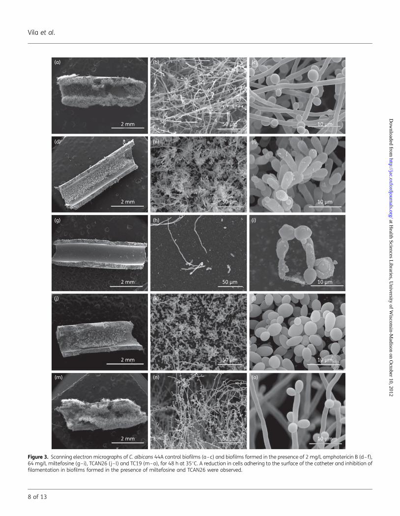
Biofilms of C. albicans clinical isolate 44A formed in theabsence of antifungal agents were highly filamented with alarge degree of colonization of cells adhering to almost theentire catheter surface (Figure 3a–c). Interestingly, biofilmformed by C. albicans ATCC 10231 showed lower cell densitythan biofilms formed by the clinically isolated strain of C. albi-cans, 44A (data not shown). Analysing the effects of standardantifungal agents on the biofilm formation, the addition of flu-conazole (16×MIC) (data not shown) or amphotericin B(16×MIC) (Figure 3d–f) reduced the final mass of adheringcells and inhibited the formation of hyphae in C. albicans 44A.Similar results were observed when fluconazole (16×MIC) oramphotericin B (16×MIC) was added to the catheter duringbiofilm development in the strain ATCC 10231; there was a re-duction in the number of cells adhering to the device, withmost of the cells in yeast form (data not shown). The additionof miltefosine (Figure 3g–i) or TCAN26 (Figure 3j–l) (16×MIC)to the catheter during the formation of biofilms of the clinicalisolate 44A resulted in a reduction of cells adhering, with theelimination of almost all cells on miltefosine-treated catheters(Figure 3g–i) and reduction of filamentation by TCAN26, thecells appearing mostly as yeasts and pseudohyphae (Figure 3j–l).
Table 1. Susceptibility of C. albicans strains to standard antifungal amphotericin B (AMB) and fluconazole (FLC), miltefosine (MLT) and four othersynthetic analogues by the CLSI reference broth microdilution method
C. albicans 44A C. albicans ATCC 10231
Compounds MIC MFC MIC MFC
Standard antifungal AMB 0.12 8 0.25 8FLC 1 .64 1 .64
Alkylphospholipids MLT 4 4 2 2TCAN26 4 8 4 8TC19 4 .16 4 8
Alkylphospholipid-dinitroaniline hybrids TC106 .16 .16 .16 .16TC117 .16 .16 .16 .16
Antifungal concentrations are expressed in mg/L.
Effect of alkylphospholipids on Candida albicans biofilms
5 of 13
JAC
at Health Sciences L
ibraries, University of W
isconsin-Madison on O
ctober 10, 2012http://jac.oxfordjournals.org/
Dow
nloaded from

0.5 µm
0.5 µm
0.5 µm
0.5 µm1 µm
1 µm
1 µm
1 µm
CM
CW
F
CW
LV
DV
CM
CW
(a) (b)
(c) (d)
(e) (f)
(g) (h)
CW
CM
F
Figure 2. Transmission electron micrographs of C. albicans 44A control (a and b) and 44A treated with 1 mg/L miltefosine (c and d), 1 mg/L TCAN26 (eand f) or 1 mg/L TC19 (g and h). Treatment with alkylphospholipids led to an increase in the external fibrillar layer (F), thickening of the cell wall (CW)and disruption of the cell membrane (CM). Treatment also led to the appearance of strongly altered buds and electron-dense vacuoles (DV) andelectron-lucent vacuoles (LV).
Vila et al.
6 of 13
at Health Sciences L
ibraries, University of W
isconsin-Madison on O
ctober 10, 2012http://jac.oxfordjournals.org/
Dow
nloaded from

However, the addition of 64 mg/L TC19 (16×MIC) did not affect44A biofilm formation (Figure 3m–o).
Morphological changes after adding antifungal agents tomature biofilm
Control biofilms of both C. albicans strains, ATCC 10231 and clin-ical isolate 44A, after a total of 96 h of incubation in rich medium
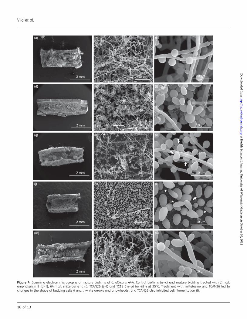
supplemented with glucose (2%) and FBS (20%) were highlydense and had long hyphae and abundant branches and budsover the entire surface of the CVC with which they were incontact (Figure 4a–c; clinical isolate 44A). When the referenceantifungal agents (amphotericin B and fluconazole) wereadded to mature biofilms (after the first 48 h of formation)they did not affect their density or filamentation profile(Figure 4d–f for amphotericin B; data not shown for fluconazole).
Table 2. Evaluation of the effect of antifungal agents during biofilm formation by the XTT reduction method
Absorbance: mean+SD (% inhibition)
Control
Antifungal concentrations (mg/L)
Strains Antifungal MIC 4×MIC 16×MIC
C. albicans ATCC 10231 AMB 0.52+0.1 0.27+0.08** (49.5) 0.15+0.09*** (71.0) 0.14+0.19*** (74.2)FLC 0.52+0.1 0.6+0.18 (0) 0.51+0.11 (4.1) 0.48+0.03 (8.9)MLT 0.52+0.1 0.64+0.17 (0) 0.41+0.12 (3.8) 0.35+0.11** (38.2)TCAN26 0.52+0.1 0.6+0.12 (0) 0.48+0.08 (18.4) 0.43+0.03* (29.5)TC19 0.52+0.1 0.47+0.15 (11.2) 0.42+0.13 (20.6) 0.31+0.09** (41.2)
C. albicans 44A AMB 0.46+0.1 0.23 +0.02** (49.7) 0.21+0.05** (55.0) 0.23+0.06** (51.1)FLC 0.46+0.1 0.43+0.08 (7.2) 0.4+0.1 (12.7) 0.38+0.09 (18.5)MLT 0.46+0.1 0.35+0.1 (23.1) 0.31+0.11* (32.3) 0.13+0.14*** (71.2)TCAN26 0.46+0.1 0.44+0.07 (4.3) 0.39+0.06 (15.8) 0.35+0.06 (24.3)TC19 0.46+0.1 0.51+0.17 (0) 0.53+0.28 (0) 0.44+0.14 (5.0)
AMB, amphotericin B; FLC, fluconazole; MLT, miltefosine; MIC, minimum concentration that inhibits 50% (FLC) or 90% (AMB, MLT, TCAN26 and TC19) ofplanktonic cell growth.*P,0.05.**P,0.001.***P,0.0001.
Table 3. Evaluation of the effect of antifungal agents on mature biofilms by the XTT reduction method
Absorbance: mean+SD (% inhibition)
Control
Antifungal concentrations (mg/L)
Strains Antifungal MIC 4×MIC 16×MIC
C. albicans ATCC 10231 AMB 0.43+0.08 0.37+0.11 (13.5) 0.30+0.09** (30.4) 0.33+0.07** (24.3)FLC 0.43+0.08 0.50+0.05 (0) 0.50+0.10 (0) 0.46+0.07 (0)MLT 0.43+0.08 0.39+0.03 (9.1) 0.45+0.10 (0) 0.29+0.07* (32.4)TCAN26 0.43+0.08 0.36+0.02 (15.8) 0.37+0.02 (12.9) 0.37+0.03 (14.7)TC19 0.43+0.08 0.37+0.21 (14.7) 0.37+0.19 (14.3) 0.41+0.09 (5.01)
C. albicans 44A AMB 0.3+0.07 0.27+0.06 (10.1) 0.28+0.07 (3.7) 0.27+0.07 (7.9)FLC 0.3+0.07 0.30+0.07 (0) 0.30+0.06 (0) 0.29+0.06 (0.7)MLT 0.3+0.07 0.3+0.07 (0) 0.28+0.07 (5.5) 0.16+0.01*** (44.3)TCAN26 0.3+0.07 0.35+0.008 (0) 0.28+0.05 (5.8) 0.24+0.04 (19.5)TC19 0.3+0.07 0.28+0.07 (3.8) 0.25+0.07 (15.5) 0.25+0.05 (15.4)
AMB, amphotericin B; FLC, fluconazole; MLT, miltefosine; MIC, minimal inhibitory concentration that inhibits 50% (FLC) or 90% (AMB, MLT, TCAN26 andTC19) of planktonic growth.*P,0.05.**P,0.001.***P,0.0001.
Effect of alkylphospholipids on Candida albicans biofilms
7 of 13
JAC
at Health Sciences L
ibraries, University of W
isconsin-Madison on O
ctober 10, 2012http://jac.oxfordjournals.org/
Dow
nloaded from
(a)
2 mm 50 µm 10 µm
2 mm 50 µm 10 µm
2 mm 50 µm 10 µm
2 mm 50 µm 10 µm
2 mm 50 µm 10 µm
(b) (c)
(d) (e) (f)
(g) (h) (i)
(j) (k) (l)
(m) (n) (o)
Figure 3. Scanning electron micrographs of C. albicans 44A control biofilms (a–c) and biofilms formed in the presence of 2 mg/L amphotericin B (d–f),64 mg/L miltefosine (g–i), TCAN26 (j– l) and TC19 (m–o), for 48 h at 358C. A reduction in cells adhering to the surface of the catheter and inhibition offilamentation in biofilms formed in the presence of miltefosine and TCAN26 were observed.
Vila et al.
8 of 13
at Health Sciences L
ibraries, University of W
isconsin-Madison on O
ctober 10, 2012http://jac.oxfordjournals.org/
Dow
nloaded from

The analogues of phospholipids showed different behaviour withrespect to mature biofilms of C. albicans 44A. While miltefosineonly slightly reduced the extent of catheter colonization byyeasts (Figure 4g–i), TCAN26 led to greater reduction in boththe extent and the thickness of the biofilm on the catheter(Figure 4j–l). Although the addition of miltefosine to maturebiofilm of C. albicans 44A did not reverse the density of alreadyformed biofilm, it induced alterations in conidia cell shapeprofile (arrows in Figure 4i). TCAN26-treated cells remained ad-herent to the catheter but showed an exclusively yeast formwith many elongated buds and a minor number of pseudohy-phae (arrows and arrowheads in Figure 4k and l), demonstratingcomplete inhibition of filamentation of these cells. Mature bio-films treated with all the TC19 concentrations tested did notinduce alterations in the biofilm profile (Figure 4m–o).
DiscussionThe Candida biofilm lifestyle results in resistance to antifungalagents and protection of the fungus from host defences,which have important clinical repercussions.29,30 Device-relatedinfections are difficult to treat and affected devices often needto be removed, which can be hazardous for some patients.31,32
CVCs appear to be the most common risk factor for candidaemiadevelopment in patients without neutropenia or major immuno-deficiencies.33
In the present work, five phospholipid analogues, includingmiltefosine, were tested to determine whether they had inhibi-tory activity against planktonic and biofilm cells of C. albicans.Planktonic cells of C. albicans were susceptible to treatmentwith miltefosine, TCAN26 and TC19, all alkylphospholipid com-pounds. Although it has been described that both TCAN26 andTC19 show inhibitory activity against Leishmania donovani andLeishmania infantum cells,15,18 the present study is the first todeal with the antifungal effects of TCAN26 and TC19 on C. albi-cans. The MIC values of TCAN26 and TC19 obtained for C. albicansplanktonic cells (2–4 mg/L or 5–10 mM) were quite similar tothose obtained for L. donovani and L. infantum.15,18 Some differ-ences in susceptibility occurred, however, as both alkylphospholipidanalogues were more potent than miltefosine for both Leishmaniaspp., while in the present study they were equal or lesspotent than miltefosine in C. albicans. The antifungal activity of mil-tefosine against C. albicans has been demonstrated previously, theMIC values being similar to those obtained in our work.14,34
Besides, only miltefosine and TCAN26 presented fungicidaleffects against C. albicans. The alkylphospholipid-dinitroanilinehybrid compounds were also tested and present no effect on C.albicans planktonic cells.
Alkylphosphocholines have structural similarity to the naturalsubstrates of fungal phospholipase B1 (PLB1), phosphatidyl-choline and lysophosphatidylcholine. Miltefosine treatment inCryptococcus spp. led to concentration-dependent inhibition oflysophospholipase transacylase (LPTA) and PLB1 activity.14 PLB1appears to be required for Cryptococcus adherence to lung epithe-lial cells and for its haematogenous dissemination, while LPTAseems to be associated with membrane synthesis, remodellingand repair. Therefore, miltefosine may exert an antifungal effectby interfering in the biochemistry of the cell wall and cell mem-brane.14 Little is known about the effects of alkylphospholipids
on fungal growth and the mechanisms involved in this process.In trypanosomatids, previous studies in Leishmania spp. showedthat treatment with miltefosine induced changes in lipidmetabolism, such as the inhibition of phosphatidylcholine andglycosylphosphatidylinositol anchor biosynthesis and cholineuptake.21,35,36
Due to their chemical structure, alkylphospholipids are alsoeasily inserted into lipid membranes and resist catabolic degrad-ation. The level of partitioning into lipid bilayers depends on thedegree of unsaturation of the phospholipid alkyl chains and theamount of cholesterol. Miltefosine interacts with the cell mem-brane and rapidly reaches other subcellular membranes, thusbeing able to affect cell metabolism at different levels.37 Experi-ments with model membranes showed that TCAN26 interactswith both the polar and the hydrophobic regions of membranebilayers, and it was proposed that this compound remains em-bedded in the membrane bilayers, causing significant perturb-ation.15 This behaviour probably also occurs in fungal cells.Insertion of the molecule into the membrane can causechanges in the composition of the membrane, altering its fluidityand permeability, and can interfere with the functioning of mem-brane proteins, affecting proteins involved in cell wall synthesis,such as b-1,3 glucan synthase. These alkylphospholipids alsoappear to exert their effects by interfering directly with the cellmembrane constitution and/or inhibiting biosynthetic pathwaysof membrane lipids within cells.
Thus, it is possible that the changes observed by us in cellshape, the cell wall and the plasma membrane and the increasein electron-dense vacuoles in C. albicans strain 44A treatedwith subinhibitory concentrations of alkylphospholipids may berelated to three mechanisms: (i) imbalance of the biosyntheticpathways of membrane phospholipids, altering cellular lipid me-tabolism; (ii) phospholipase inhibition; and/or (iii) insertion of thealkylphospholipid compounds into the fungal cytoplasmicmembrane.
To study the effect of the alkylphospholipids on C. albicansbiofilm, we used a dual approach, employing two stages ofbiofilm development, seeking to correlate their effects on theinitial and mature biofilm phases in comparison with amphoter-icin B and fluconazole. Previous studies have shown that theresistance of the biofilm to polyenes and azoles changesduring its maturation,10,11 and these data are consistent withthe findings of our study, in which both C. albicans strains werehighly susceptible to amphotericin B during the process ofbiofilm formation and reduced activity was observed in matureC. albicans biofilms, despite the good antifungal activity onplanktonic cells of C. albicans exhibited by fluconazole andamphotericin B. Among the alkylphospholipids compoundstested in this study, only miltefosine was able to reduce biofilmformation and to reduce the viability of mature biofilm C. albi-cans strains, by 32% for the ATCC strain and 44% for the 44Aclinical isolate (at 16×MIC), showing higher activity in the earlieststages of biofilm development than in mature biofilms. TCAN26was also effective against biofilm formation and maturebiofilm but to a lesser extent than miltefosine.
The ability to penetrate the C. albicans biofilm extracellularmatrix (ECM) is one of the existing explanations for the greaterefficacy of lipid formulations of amphotericin B in biofilms com-pared with the traditional formulation of amphotericin B deoxy-cholate and fluconazole.13 Interestingly, the higher activity of
Effect of alkylphospholipids on Candida albicans biofilms
9 of 13
JAC
at Health Sciences L
ibraries, University of W
isconsin-Madison on O
ctober 10, 2012http://jac.oxfordjournals.org/
Dow
nloaded from
(a)
2 mm 50 µm 10 µm
2 mm 50 µm 10 µm
2 mm 50 µm 10 µm
2 mm 50 µm 10 µm
2 mm 50 µm 10 µm
(b) (c)
(d) (e) (f)
(g) (h) (i)
(j) (k) (l)
(m) (n) (o)
Figure 4. Scanning electron micrographs of mature biofilms of C. albicans 44A. Control biofilms (a–c) and mature biofilms treated with 2 mg/Lamphotericin B (d–f), 64 mg/L miltefosine (g–i), TCAN26 (j–l) and TC19 (m–o) for 48 h at 358C. Treatment with miltefosine and TCAN26 led tochanges in the shape of budding cells (i and l, white arrows and arrowheads) and TCAN26 also inhibited cell filamentation (l).
Vila et al.
10 of 13
at Health Sciences L
ibraries, University of W
isconsin-Madison on O
ctober 10, 2012http://jac.oxfordjournals.org/
Dow
nloaded from

alkylphospholipid analogues on biofilms compared with otherantifungal agents tested in this work can be related to thephospholipid nature of these compounds, which must also pene-trate through the ECM of the biofilm to reach the fungal cell.
In the present study, biofilms formed on CVCs were used tocompare the ultrastructural effects of treatment with 16×MICof amphotericin B and the alkylphospholipids. Miltefosineexhibit the most drastic effect on biofilm formation, inducingthe detachment of almost all cells. Although mature biofilmsare less sensitive to miltefosine than forming biofilms, theircells exhibit a peculiar profile of quite altered cells and deformedbuds. When we analysed the morphological aspects ofC. albicans biofilms, amphotericin B and TCAN26 were the mostactive compounds. Despite having shown a low inhibitionprofile (around 20%) in biofilms formed on polystyrene plates,TCAN26 was extremely active on biofilms in CVCs. However,even when this compound was added to pre-formed mature bio-films, there was almost complete inhibition of cell filamentationat the end of the incubation time. This result shows that TCAN26can probably act either on cells that are already filamented or byinhibiting filamentation in yeast. Inhibition of morphogenesismay be important since the lesser complexity of the biofilmmay be related to lower virulence of the cells involved.
The ability of C. albicans to form biofilms can be modified bythe surface properties of the biomaterial over which the biofilmwill be formed.38 Therefore, the difference between the biofilm-forming abilities of cells on polystyrene and CVCs can beexplained by differences in chemical composition between thesurfaces, which may influence the adherence of C. albicans andconsequently biofilm development.38
The antibiofilm activity of alkylphospholipids is an importantfinding as these compounds seem to be active on both plankton-ic and biofilm cells on two different medically important surfaces(polystyrene and catheters) and may be considered as a putativealternative for candidaemia treatment.
The pharmacokinetics, absorption, distribution and metabol-ism of miltefosine have been studied in rats and mice.39 – 41
Oral administration of miltefosine for 5 days following infectionincreased survival and reduced brain and lung cryptococcalburdens. This was achieved with relatively low doses of 7.2 and3.6 mg/kg/day of miltefosine and confirmed the potential ofthis antifungal agent for the treatment of invasive mycoses,including intracerebral infections.14 Serum concentrations of110 mM (44.83 mg/L) were achieved in rats after 2 weeks ofdaily dosing with 10 mg/kg.14 This concentration is 10–20times the MIC for miltefosine against fungi causing invasivemycoses,14 including the C. albicans strains used in this work.Despite being the most potent antifungal agent tested, miltefo-sine has a long half-life (100–200 h) in humans and a lowtherapeutic ratio, characteristics that could encourage the devel-opment of resistance.42 In addition, treatment of pregnantwomen is contraindicated because of its teratogenic propertiesin animals.42 Miltefosine is an orally active fungicidal compoundand although it has significant side effects it is still less toxic thanamphotericin B.14
Previous experiments have shown that TCAN26 and TC19 areless cytotoxic to the human monocytic cell line THP1 than milte-fosine and may be considered as safer alternatives in cases inwhich miltefosine is not recommended.15,18 Miltefosine hashaemolytic activity with an HC50 (concentration that produces
50% of haemolysis) of 38.6 mM (15.7 mg/L), a feature that pre-vents its use in injectable form. However, TCAN26 and TC19 aremuch less haemolytic than miltefosine, showing HC50 values of.60 mM (24.5 mg/L) and .100 mM (40.8 mg/L), respectively.This indicates that these compounds could be administeredintravenously, and thus they may show reduced gastrointestinaltoxicity and higher plasma concentrations in vivo.18,43
Alkylphospholipids are phospholipid derivatives substitutedwith an alkyl chain in the lipid portion. Miltefosine possesses a16-carbon saturated aliphatic chain while TCAN26 and TC19contain cycloalkane rings—adamantane and cyclohexadecane,respectively—in the lipid portion. These chemical differencesmay be related to the differences observed in cytotoxicity andhaemolytic activity. Studies with a series of ring-substitutedether phospholipid derivatives showed that, with regard toTCAN26, the presence or absence of a double bond on the alkylchain does not affect the antileishmanial activity of themolecule.15 On the other hand, studies with a series ofcycloalkylidene-substituted ether phospholipids showed that forTC19 the absence of the double bond increased antileishmanialactivity and reduced the cytotoxicity of the molecule when com-pared with its unsaturated counterpart.18 The length of the alkylchain of the most active ring-substituted compounds variesfrom 5 to 14 carbon atoms, which could be advantageous forthe solubility and/or toxicity of the new compounds, and fortheir metabolic clearance.15,18 Further experiments are thereforerequired in order to gain a better understanding of the structuralfeatures that impart enhanced activity and minimal cytotoxicityfor this class of compounds. However, in general these data indi-cate that ring-substituted ether phospholipids are less cytotoxicto human monocytes and more potent than miltefosine againstL. infantum and L. donovani,15,18 showing that modifications ofthe alkyl chain of alkylphospholipids are effective in reducing cyto-toxicity. Conversely, a hydrophobic chain at least 16 carbon atomslong seems to be important for the maintenance of antifungalactivity.
Conclusions
Taken together, our data demonstrate that alkylphospholipidssuch as miltefosine and TCAN26 are active on both planktonicand biofilm cells of C. albicans. Miltefosine was the most effectivecompound tested by being active against C. albicans biofilms,making this compound particularly interesting. AlthoughTCAN26 was less effective than miltefosine in our model, it isconsidered a less toxic compound than miltefosine by workersin this field of study. Therefore, TCAN26 effectiveness as a treat-ment for biofilms should also be considered in the future, espe-cially since it was extremely active on biofilms in CVCs.
AcknowledgementsThe authors thank Beatriz Bastos for help with material preparation andimage acquisition in the electron microscopy experiments.
FundingThis work was supported by the Conselho Nacional de DesenvolvimentoCientıfico e Tecnologico (CNPq—Brazil), Fundacao Carlos Chagas Filho
Effect of alkylphospholipids on Candida albicans biofilms
11 of 13
JAC
at Health Sciences L
ibraries, University of W
isconsin-Madison on O
ctober 10, 2012http://jac.oxfordjournals.org/
Dow
nloaded from

de Amparo a Pesquisa do Estado do Rio de Janeiro (FAPERJ—Brazil) andCOST Action CM0801–Greek (New Drugs for Neglected Diseases).
Transparency declarationsNone to declare. This article was edited for proper English language byAmerican Journal Experts.
References1 Nucci M, Queiroz Telles F, Tobon AM et al. Epidemiology of opportunisticfungal infections in Latin America. Clin Infect Dis 2010; 51: 561–70.
2 Chandra J, Kuhn DM, Mukherjee PK et al. Biofilm formation by thefungal pathogen Candida albicans: development, architecture, and drugresistance. J Bacteriol 2001; 183: 5385–94.
3 Sifuentes-Osornio J, Corzo-Leon DE, Ponce-de-Leon LA. Epidemiologyof invasive fungal infections in Latin America. Curr Fungal Infect Rep2012; 6: 23–34.
4 Bassetti M, Taramasso L, Nicco E et al. Epidemiology, speciesdistribution, antifungal susceptibility and outcome of nosocomialcandidemia in a tertiary care hospital in Italy. Plos One 2011; 6: e24198.
5 Colombo AL, Guimaraes T. Epidemiologia das infeccoes hematogenicaspor Candida spp. Rev Soc Bras Med Trop 2003; 5: 599–607.
6 Ramage G, VandeWalle K, Wickes BL et al. Characteristics of biofilmformation by Candida albicans. Rev Iberoam Micol 2001; 18: 163–70.
7 Donlan RM, Costerton JW. Biofilms: survival mechanisms of clinicallyrelevant microorganisms. Clin Microbiol Rev 2002; 15: 167–93.
8 Hawser SP, Douglas LJ. Resistance of Candida albicans biofilms toantifungal agents in vitro. Antimicrob Agents Chemother 1995; 39: 2128–31.
9 Mukherjee PK, Chandra J. Candida biofilm resistance. Drug Resist Updat2004; 7: 301–9.
10 Ramage G, Bachmann S, Patterson TF et al. Investigation of multidrugefflux pumps in relation to fluconazole resistance in Candida albicansbiofilms. J Antimicrob Chemother 2002; 49: 973–80.
11 Mukherjee PK, Chandra J, Kuhn DM et al. Mechanism of fluconazoleresistance in Candida albicans biofilms: phase-specific role of effluxpumps and membrane sterols. Infect Immun 2003; 71: 4333–40.
12 Pappas PG, Kauffman CA, Andes D et al. Clinical practice guidelines forthe management of candidiasis: 2009 update by the Infectious DiseasesSociety of America. Clin Infect Dis 2009; 48: 503–35.
13 Kuhn DM, George T, Chandra J et al. Antifungal susceptibility ofCandida biofilms: unique efficacy of amphotericin B lipid formulationsand echinocandins. Antimicrob Agents Chemother 2002; 46: 1773–80.
14 Widmer F, Wright LC, Obando D et al. Hexadecylphosphocholine(miltefosine) has broad-spectrum fungicidal activity and is efficaciousin a mouse model of cryptococcosis. Antimicrob Agents Chemother2006; 50: 414–21.
15 Avlonitis N, Lekka E, Detsi A et al. Antileishmanial ring-substitutedether phospholipids. J Med Chem 2003; 46: 755–67.
16 Urbina JA. Mechanisms of action of lysophospholipid analogues againsttrypanosomatid parasites. Trans R Soc Trop Med Hyg 2006; 100: 9–16.
17 Kapou A, Benetis NP, Avlonitis N et al. 3D-Quantitative structure–activity relationships of synthetic antileishmanial ring-substituted etherphospholipids. Bioorg Med Chem 2007; 15: 1252–65.
18 Calogeropoulou T, Angelou P, Detsi A et al. Design and synthesis ofpotent antileishmanial cycloalkylidene-substituted ether phospholipidderivatives. J Med Chem 2008; 51: 897–908.
19 Chan JH, Neal RA, Pendergast W. The activity of alkylphosphorylcholines and related derivatives against Leishmaniadonovani. Biochem Pharmacol 1987; 36: 2633–6.
20 Unger C, Maniera T, Kaufmann-Kolle P et al. In vivo antileishmanialactivity of hexadecylphosphocholine and other alkylphosphocholines.Drugs Today 1998; 34: 133–40.
21 Croft SL, Seifert K, Duchene M. Antiprotozoal activities of phospholipidanalogues. Mol Biochem Parasitol 2003; 126: 165–72.
22 Croft SL, Barrett MP, Urbina JA. Chemotherapy of trypanosomiasesand leishmaniasis. Trends Parasitol 2005; 21: 508–12.
23 Braga-Silva LA, Mesquita DG, Ribeiro DM et al. Trailing end-pointphenotype antibiotic-sensitive strains of Candida albicans produce differentamounts of aspartyl peptidases. Braz J Med Biol Res 2009; 42: 765–70.
24 Clinical and Laboratory Standards Institute. Reference Method forBroth Dilution Antifungal Susceptibility Testing of Yeasts—Third Edition:Approved Standard M27-A3. CLSI, Wayne, PA, USA, 2008.
25 Pfaller MA, Sheehan DJ, Rex JH. Determination of fungicidal activitiesagainst yeasts and molds: lessons learned from bactericidal testing andthe need for standardization. Clin Microbiol Rev 2004; 17: 268–80.
26 Jin Y, Yip HK, Samaranayake YH et al. Biofilm-forming ability ofCandida albicans is unlikely to contribute to high levels of oral yeastcarriage in cases of human immunodeficiency virus infection. J ClinMicrobiol 2003; 41: 2961–7.
27 Ramage G, VandeWalle K, Wickes BL et al. Standardized method for invitro antifungal susceptibility testing of Candida albicans biofilms.Antimicrob Agents Chemother 2001; 45: 2475–9.
28 Braga PC, Culici M, Alfieri M et al. Thymol inhibits Candida albicansbiofilm formation and mature biofilm. Int J Antimicrob Agents 2008;31: 472–7.
29 Kumamoto CA. Candida biofilms. Curr Opin Microbiol 2002; 5: 608–11.
30 Ramage G, Saville SP, Thomas DP et al. Candida biofilms: an update.Eukaryot Cell 2005; 4: 633–8.
31 Mermel LA, Farr BM, Sherertz RJ et al. Guidelines for the managementof intravascular catheter-related infections. Clin Infect Dis 2001; 32:1249–72.
32 Walsh TJ, Rex JH. All catheter-related candidemia is not the same:assessment of the balance between the risks and benefits of removalof vascular catheters. Clin Infect Dis 2002; 32: 600–2.
33 Tumbarello M, Posteraro B, Trecarichi EM et al. Biofilm productionby Candida species and inadequate antifungal therapy as predictorsof mortality for patients with candidemia. J Clin Microbiol 2007; 45:1843–50.
34 Obando D, Widmer F, Wright LC et al. Synthesis, antifungal andantimicrobial activity of alkylphospholipids. Bioorg Med Chem 2007; 15:5158–65.
35 Lux H, Hart DT, Parker PJ et al. Ether lipid metabolism, GPI anchor bio-synthesis, and signal transduction are putative targets for anti-leishmanialalkylphospholipid analogues. Adv Exp Med Biol 1996; 416: 201–11.
36 Zufferey R, Mamoun CB. Choline transport in Leishmania majorpromastigotes and its inhibition by choline and phosphocholineanalogs. Mol Biochem Parasitol 2002; 125: 127–34.
37 Jimenez-Lopez JM, Rıos-Marco P, Marco C et al. Alterations in thehomeostasis of phospholipids and cholesterol by antitumoralkylphospholipids. Lipids Health Dis 2010; 9: 33.
38 Chandra J, Patel JD, Li J et al. Modification of surface properties ofbiomaterials influences the ability of Candida albicans to form biofilms.Appl Environ Microbiol 2005; 71: 8795–801.
39 Breiser A, Kim DJ, Fleer E et al. Distribution and metabolism ofhexadecylphosphocholine in mice. Lipids 1987; 22: 925–6.
Vila et al.
12 of 13
at Health Sciences L
ibraries, University of W
isconsin-Madison on O
ctober 10, 2012http://jac.oxfordjournals.org/
Dow
nloaded from
40 Marschner N, Kotting J, Eibl H et al. Distribution of hexadecylphosphocholineand octadecyl-methyl-glycero-3-phosphocholine in rat tissues during steady-state treatment. Cancer Chemother Pharmacol 1992; 31: 18–22.
41 Unger C, Fleer E, Damenz E et al. Hexadecylphosphocholine:determination of serum concentrations in rats. J Lipid Mediat 1991; 3: 71–8.
42 Soto J, Soto P. Miltefosine: oral treatment of leishmaniasis. Expert RevAnti-Infect Ther 2006; 4: 177–85.
43 Papanastasiou I, Prousis KS, Georgikopoulou K et al. Design and synthesisof new adamantyl-substituted antileishmanial ether phospholipids. BioorgMed Chem Lett 2010; 20: 5484–7.
Effect of alkylphospholipids on Candida albicans biofilms
13 of 13
JAC
at Health Sciences L
ibraries, University of W
isconsin-Madison on O
ctober 10, 2012http://jac.oxfordjournals.org/
Dow
nloaded from


![Adhesion and biofilm formation by Staphylococcus aureus ... · food processing plants as affected by growth medium, surface type ... and BHI broth plus NaCl (5 g/100 mL)] and incubation](https://img.document.onl/doc/110x75/5acf04277f8b9a1d328c78ba/adhesion-and-biofilm-formation-by-staphylococcus-aureus-processing-plants-as.jpg)


![Candida cinsi mantarlar C. albicans - TavsiyeEdiyorum.com · 2016. 8. 1. · Kandida cinsi mantarlar, CryptococcaceaeIDPLO\DVÕQGDQROXS GDQID]ODW†U](https://img.document.onl/doc/110x75/60bc77ce85fc4238cb2ce9f6/candida-cinsi-mantarlar-c-albicans-2016-8-1-kandida-cinsi-mantarlar-cryptococcaceaeidplodvqgdqroxs.jpg)