Embed Size (px)
Citation preview
0
UNIVERSIDADE FEDERAL DO CEARÁ
FACULDADE DE FARMÁCIA, ODONTOLOGIA E ENFERMAGEM
PROGRAMA DE PÓS-GRADUAÇÃO EM ODONTOLOGIA
ESTUDO CLÍNICO DE TÉCNICA DE REPOSICIONAMENTO DO BLOCO ÓSSEO E IMPLANTES EM REGIÃO ANTERIOR DE MAXILA
RAFAEL LIMA VERDE OSTERNE
FORTALEZA
2016
1
RAFAEL LIMA VERDE OSTERNE
ESTUDO CLÍNICO DE TÉCNICA DE REPOSICIONAMENTO DO BLOCO ÓSSEO E IMPLANTES EM REGIÃO ANTERIOR DE MAXILA
Tese apresentada ao Programa de Pós-Graduação em Odontologia da Faculdade de Farmácia, Odontologia e Enfermagem da Universidade Federal do Ceará, como requisito parcial para obtenção do Título de Doutor em Odontologia.
Área de Concentração: Clínica Odontológica
Orientador: Prof. Dr. Renato Luiz Maia Nogueira
FORTALEZA
2016
2
RAFAEL LIMA VERDE OSTERNE
ESTUDO CLÍNICO DE TÉCNICA DE REPOSICIONAMENTO DO BLOCO ÓSSEO E IMPLANTES EM REGIÃO ANTERIOR DE MAXILA
Tese apresentada ao Programa de Pós-graduação em Odontologia da Universidade Federal do Ceará, como requisito parcial à obtenção do título de Doutor em Odontologia, Área de concentração: Clínica Odontológica.
Aprovada em __/__/_____
BANCA EXAMINADORA
___________________________________________
Prof. Dr. Renato Luiz Maia Nogueira (Orientador) Universidade Federal do Ceará (UFC)
________________________________________
Profa. Dra. Maria Mônica Studart Mendes Moreira
Universidade Federal do Ceará (UFC)
_________________________________________
Profa. Dra. Karina Matthes de Freitas Pontes
Universidade Federal do Ceará (UFC)
__________________________________________
Prof. Dr. Ricardo Teixeira Abreu
São Leopoldo Mandic (SLM)
__________________________________________
Prof. Dr. Abrahão Cavalcante Gomes de Souza Carvalho
Centro Universitário Christus (UNICHRISTUS)
3
AGRADECIMENTOS
Ao meu orientador, Prof. Dr. Renato Luiz Maia Nogueira, por ter contribuído para minha
formação intelectual e científica. Agradeço pelos ensinamentos, conselhos, inteligência,
honestidade, amizade e aos vinhos tomados nessa trajetória. Obrigado pela confiança e apoio
dedicados a mim.
À Profa. Dra. Roberta Barroso Cavalcante, pelos ensinamentos, e apoio desde o meu
Mestrado, sempre incentivadora e disposta à colaborar.
À Profa. Dra. Sílvia Helena Barem Rabenhosrt, pela confiança e disponibilidade de todo o
apoio do Laboratório de Genética Molecular (LABGEM).
Ao Prof. Dr. Ricardo Teixeira Abreu, pela ajuda na confecção desta Tese e o excelente
trabalho na reabilitação destes pacientes.
Às professoras Dra. Mônica Studart e Dra. Karina Matthes, pelas considerações e importantes
sugestões na avaliação deste trabalho.
Às professoras Dra. Renata Cordeiro e Dra. Eveline Turatti, pela disponibilidade a
receptividade no Curso de Odontologia da Universidade de Fortaleza.
Ao Prof. Dr. Abrahão Cavalcante, por aceitar avaliar este trabalho e pela amizade e
disponibilidade sempre que solicitado.
Aos amigos da pós-graduação da área de Cirurgia Bucomaxilofacial, Dr. Phelype Maia, Dr.
Manoel Mello, Dr. Raimundo Thompson, Dr. Ricardo Franklyn, Dra. Nayana Azevedo, Dra.
Luisa de Marilac, bem como às amigas da área de Estomatologia Dra. Carolina Teófilo e Dra.
Malena Freitas, o meu muito obrigado por compartilhar estes anos com vocês e pelos
ensinamentos durante a pós-graduação.
À coordenação do Curso de Medicina da Universidade de Fortaleza, e aos amigos Prof. Rui
Colares e Prof. Márcio Vale Braga, do Laboratório Morfofuncional, pelo apoio e
compreensão em alguns momentos ausentes.
À coordenação, docentes e funcionários do Programa de Pós-Graduação em Odontologia da
Universidade Federal do Ceará, por ter, durante esse período, oferecido todo o apoio
necessário para a realização desta tese.

4
À minha esposa Renata de Matos Brito Lima Verde, pela compreensão de momentos ausentes
e apoio em todos os momentos. Obrigado por compartilhar mais essa etapa da minha vida.
Aos Meus pais Cecília e Eduardo, e minha irmã Ana Beatriz, meu agradecimento à vocês é
maior do que pode ser expresso por simples palavras.
À todos os voluntários que se dispuseram a colaborar com este trabalho.
5
LISTA DE SIGLAS E ABREVIATURAS
3D – Tridimensional
BMPs – Bone morphogenetic proteins (Proteína morfogenética óssea)
CBCT – Cone bean computed tomography (Tomografia computadorizada de feixe cônico)
CT – Computed tomography (Tomografia computadorizada)
rHBMP2 – Recombinant human bone morphogenetic protein 2 (Proteína morfogenética óssea humana recombinante tipo 2)
T1 – Período pré-operatório
T2 – Período pós-operatório imediato
T3 – Período pós-operatório tardio
VAS – Visual analogue scale (Escala visual analógica)
6
RESUMO
Atrofia óssea alveolar vertical representa um desafio para a reconstrução e reabilitação oral,
especialmente quando envolve a zona estética da maxila. Quando a reconstrução óssea não é
realizada, um bom resultado estético dificilmente é obtido. O objetivo deste estudo
retrospectivo foi avaliar o resultado do reposicionamento do bloco osso-implante, após a
osteotomia segmentar em pacientes com atrofia óssea alveolar vertical na região estética de
maxila. Para a realização deste trabalho, foram selecionados pacientes com atrofia óssea
alveolar vertical que se submeteram à reposicionamento do segmento de bloco osso-implante
na região estética maxilar com um mínimo de 6 meses de acompanhamento. As variáveis
avaliadas foram o índice de sucesso do implante, complicações após o procedimento
cirúrgico, quantidade de aumento ósseo vertical, formação da papila, altura da faixa de
mucosa ceratinizada e a análise de satisfação do paciente. Nove pacientes foram incluídos no
estudo, todos com implantes múltiplos, totalizando 25 implantes. A média de aumento ósseo
vertical foi de 4,9 mm (3,0-8,4 mm), e apenas 1 falha de implante ocorreu. Uma melhora
estatisticamente significativa na formação de papila foi observada após a cirurgia, levando a
um bom resultado estético. Mais de 2 mm de altura de mucosa ceratinizada foi observado em
6 pacientes, e também uma alta satisfação e aceitação ao tratamento. Após a avaliação dos
dados concluiu-se que, a técnica apresentada possui a capacidade de reconstruir atrofia óssea
alveolar vertical, com uma elevada taxa de sobrevivência de implantes em período curto de
acompanhamento, melhorando o resultado estético-funcional com uma boa aceitação por
parte dos pacientes.
Palavras-chave: Osteogênese por distração; implantes dentários; prótese dentária;
osteotomia, cirurgia bucal

7
ABSTRACT
Vertical alveolar bone atrophy represents a challenge for reconstruction, especially when the
esthetic zone of the maxilla is involved. When reconstruction is not achieved, a good esthetic
outcome is rarely obtained. The aim of this retrospective study was to asses the outcome of
implant-bone block movement, after segmental osteotomy in the maxillary aesthetic region in
patients with vertical alveolar bone atrophy. Patients with vertical alveolar bone atrophy who
underwent repositioning of the bone-implant block segment in the maxillary aesthetic region
with a minimum of 6 months of follow-up were selected. Outcome measures were the success
rate of the implant, complications after the surgical procedure, amount of vertical bone
augmentation, papilla formation, keratinized mucosal band height and patient satisfaction
analysis. Nine patients were included in the study, all with multiple implants, totaling 25
implants. The mean vertical bone augmentation was 4.9 mm (3.0-8.4 mm), and only 1 implant
failure occurred. A statistically significant improvement in papilla formation was observed
after surgery, leading to a good aesthetic result. More than 2 mm of height of keratinized
mucosa was observed in 6 patients, and also a high satisfaction and acceptance to the
treatment. After data evaluation, it was concluded that the technique presented can reconstruct
vertical alveolar bone atrophy, with a high implant survival rate in a short period of follow-
up, improving the aesthetic-functional result with a good acceptance by the Patients.
Keywords: Osteogenesis distraction, dental implants, dental prosthesis, osteotomy, oral
surgery
8
SUMÁRIO
RESUMO
ABSTRACT 1. INTRODUÇÃO GERAL 9
2. PROPOSIÇÃO GERAL 13
3. CAPÍTULOS 13
CAPÍTULO 1 - "Alternative Osteodistraction Technique after Implant Placement for Alveolar Ridge Augmentation of the maxilla" 15
CAPÍTULO 2 - "A retrospective study of an alternative osteodistraction technique after implant placement in the maxillary esthetic region " 36
4. CONCLUSÃO GERAL 65
5. REFERÊNCIAS 66
6. APÊNDICE A - Questionário aplicado aos participantes da pesquisa para avaliação de satisfação, desconforto e aceitabilidade do tratamento 69
7. ANEXOS 71
ANEXO A - Parecer do Comitê de Ética em Pesquisa 71
ANEXO B - Termo de Consentimento Livre e Esclarecido 74
ANEXO C - Certificado de revisão da língua inglesa 76
9
1 INTRODUÇÃO GERAL
Deficiência óssea vertical em processo alveolar de maxila e mandíbula pode ser
um desafio para reabilitação dental com implantes osseointegrados, podendo gerar próteses
com coroas longas, no sentido cérvico-incisal ou cérvico oclusal, gerando um
comprometimento estético, principalmente em pacientes que necessitam de reabilitação de
dentes anteriores e com linha do sorriso alta (Salama et al, 2009; Kim et al, 2012; Stacchi et al
2013). Diversas técnicas para contornar defeitos ósseos verticais são descritas na literatura,
variando desde próteses com gengiva artificial até reconstruções ósseas com uso de
bioengenharia (Eposito et al, 2009; Gomes-Ferreira et al, 2016; Daga et al, 2015; Yu et al,
2016; Yun et al, 2016).
O uso de gengiva artificial pode ser uma solução em casos de defeitos ósseos
verticais (Coachman et al, 2009; Salama et al, 2009, Enríquez et al, 2016), defendido por
alguns autores para simplificar o tratamento (Coachman et al, 2009); sendo a gengiva
artificial em resina acrílica ou em porcelana, fixa ou removível, e com caracterização para
mimetizar os tecidos moles vizinhos. Embora tenha sido descrita uma aceitação de até 96,8%
do tratamento (Di et al, 2011), alguns autores falam da baixa aceitação e dificuldade de
higienização como possíveis desvantagens (Enríquez et al, 2016).
Técnicas de reconstruções ósseas verticais podem ser empregadas antes da
instalação dos implantes (procedimento em dois estágios) ou simultaneamente à instalação
dos implantes (procedimento de estágio único) (Episoto et al, 2009). Dentre as técnicas de
dois estágios podemos citar a distração osteogênica, enxerto ósseo interposicional (tipo
sanduíche), uso de osso particulado protegido por membranas ou malhas de titânio, com ou
sem o uso de BMP. Em comum à estas técnicas, a reconstrução óssea do defeito vertical
10
realizada previamente à instalação dos implantes gera aumento do tempo de tratamento e
custos financeiros (Pérez-Sayáns et al, 2013; Mavriqi et al, 2015; Gomes-Ferreira et al, 2016).
Procedimentos de estágio único, como a técnica do Bone Ring, e o enxerto em
tenda, apresentam vantagens de redução do tempo total de tratamento (Omara et al, 2016).
Algumas desvantagens podem ser consideradas para estas técnicas, como a necessidade de um
segundo sítio cirúrgico para a remoção do enxerto para a técnica de Bone Ring, ou mesmo
para a técnica do enxerto em tenda, caso seja optado por uso de enxerto autógeno, o padrão
ouro. Caso decida-se pelo uso de biomateriais, eleva-se o custo da técnica do enxerto em
tenda, já que frequentemente é utilizado uma combinação de um substituto ósseo, membranas
de colágeno e malha de titânio (Daga et al., 2015). Outro fator importante para a consideração
em ambas as técnicas, é a necessidade de tecido mole suficiente para o recobrimento do
enxerto sem tensão ao tecido, pois caso a tensão ocorra, o risco de exposição da área
enxertada aumenta (Omara et al, 2016), podendo levar a falha do enxerto.
Diferente das técnicas descritas anteriormente, a reabilitação oral com auxílio da
distração osteogênica clássica é realizada com um procedimento em dois estágios; primeiro a
distração osteogênica propriamente dita, seguida pela instalação dos implantes (Chiapasco et
al, 2004; Kim et al, 2013). Como principais vantagens desta técnica, encontra-se uma taxa
consideravelmente rápida de neoformação óssea que, com ativação diária, pode chegar à 1mm
ao dia, e a vantagem da formação de tecido mole associado à formação óssea (Herford et al,
2015). Como desvantagens da técnica os distratores convencionais usualmente apresentam
custos elevados e costumam ser incômodos para o paciente, o que pode limitar a aceitação do
tratamento (Eposito et al, 2009).
Watzeck et al, 2000, propuseram uma técnica alternativa de distração osteogênica
realizada após a instalação dos implantes em áreas de defeitos ósseos verticais. Esta técnica
apresenta similaridades com a técnica do reposicionamento de implantes mal-posicionados

11
por osteotomias em bloco do processo alveolar (Stacchi et al 2013). Em ambas as técnicas,
duas osteotomias verticais e uma horizontal subapical aos implantes são realizadas, obtendo-
se um bloco ósseo com os implantes. Na técnica do reposicionamento, os implantes são
levados à posição desejada e fixados por meio de parafusos, placas ou mesmo pela união aos
dentes vizinhos. Na técnica de Watzeck et al, 2000, um distrator multidimensional
personalizado é fabricado e utilizado como intermediário do implante; com a ativação do
distrator, o bloco ósseo contendo os implantes é levado à posição proteticamente ideal,
consequentemente melhorando estética final do caso.
Ambas as técnicas de distração osteogênica de Watzeck et al, 2000, e a técnica de
reposicionamento de implantes como relatada por Stacchi et al, 2013, são derivadas da
osteotomia segmentar para cirurgia ortognática. A distração osteogênica de bloco ósseo com
implantes, quando utilizada inicialmente por Watzeck et al, 2000, em 6 pacientes para a
correção de 11 implantes, conseguiu-se um ganho ósseo no sentido vestíbulo-palatino de até
4mm, e de até 11mm no sentido vertical. Dados similares aos de Zechner et al, 2001, que
obtiveram um aumento ósseo vertical de 11 mm e de até 5 mm no sentido vestíbulo-palatino;
[neste estudo 8 pacientes foram tratados, totalizando 14 implantes]. Dentre as principais
desvantagens desta técnica encontram-se a necessidade da confecção do distrator, o que pode
elevar o custo do tratamento, e o desconforto do uso do distrator durante o período de
distração e consolidação óssea.
Ueki et al, 2011, relataram um caso de distração osteogênica após a instalação dos
implantes, no qual foi utilizado o aparelho ortodôntico para que o bloco ósseo fosse levado à
posição desejada. A eliminação da necessidade de uma distrator personalizado apresenta a
vantagem de uma considerável redução de custos para o tratamento, porém pode ser
questionado a previsibilidade do posicionamento final do bloco ósseo.
12
No presente trabalho, são apresentados uma técnica alternativa de distração
osteogênica e um estudo retrospectivo com 9 casos tratados. Nesta técnica, foram realizadas
as mesmas osteotomias utilizadas por Watzeck et al, 2000, para a obtenção do bloco ósseo
com os implantes. Como modificação da técnica, foi realizada uma cirurgia prévia de modelo,
para a obtenção de um guia cirúrgico final em resina acrílica, utilizado como distrator
personalizado. Este foi ativado por meio de fio de aço, apresentando como vantagem a
redução do custo quando comparado com os distratores personalizados descritos da técnica de
Watzeck et al, 2000, e Zechner et al, 2001. Uma outra vantagem a ser considerada é a
previsibilidade do procedimento, já que o guia cirúrgico personalizado em resina acrílica é
confeccionado de acordo com o tamanho esteticamente favorável das coroas protéticas finais
orientando de forma mais precisa a posição do bloco ósseo durante a cirurgia de modelo.
Em comparação com outras técnicas, o reposicionamento do bloco ósseo pode
apresentar menor tempo total de tratamento, já que os implantes são instalados já no início,
sem procedimentos prévios de aumento ósseo vertical; e menor custo, em comparação com
uso de BMPs ou de distratores convencionais. Portanto, o presente estudo apresentou o
objetivo de descrever uma técnica e avaliar retrospectivamente casos consecutivos tratados
com osteotomia em bloco do processo alveolar após a instalação dos implantes em região
anterior da maxila.

13
2 PROPOSIÇÃO GERAL
Avaliar retrospectivamente o índice de sucesso, movimento vertical do bloco
ósseo, formação de papila, altura da faixa de mucosa ceratinizada e satisfação do paciente
após cirurgia para reposicionamento de implantes através de osteotomia em bloco do processo
alveolar em região anterior estética da maxila; assim como descrever uma técnica alternativa
para a realização deste procedimento.
14
3 CAPÍTULOS
Esta tese está baseada no artigo 46 do regimento Interno do Programa de Pós- graduação em
Odontologia da Universidade Federal do Ceará, que regulamenta o formato alternativo para
dissertações de Mestrado e teses de Doutorado, e permite a inserção de artigos científicos de
autoria ou co-autoria do candidato. Dessa forma, esta tese é composta por dois capítulos,
contendo artigos submetidos ou a serem submetidos para publicação em revistas científicas,
conforme descrito abaixo:
Capítulo 1
“Alternative Osteodistraction Technique after Implant Placement for Alveolar Ridge
Augmentation of the maxilla”. Renato Luiz Maia Nogueira, Rafael Lima Verde Osterne,
Ricardo Teixeira Abreu, Phelype Maia Araújo. Este artigo foi submetido para publicação no
periódico “Journal of Oral and Maxillofacial Surgery”.
Capítulo 2
“A retrospective study of an alternative osteodistraction technique after implant placement in
the maxillary esthetic region”. Rafael Lima Verde Osterne, Renato Luiz Maia Nogueira,
Ricardo Teixeira Abreu, Roberta Barroso Cavalcante, Érica Amaral Medeiros Este artigo será
submetido para publicação no periódico “Clinical Oral Implants Research”.

15
CAPÍTULO 1
Title: Alternative Osteodistraction Technique after Implant Placement for Alveolar Ridge
Augmentation of the maxilla
Running title: Alternative osteodistraction technique
Key words: dental implant; osteodistraction; vertical alveolar bone atrophy; osteotomy
Department/Institution: Federal University of Ceara School of Dentistry, Fortaleza, Brazil;
Fortaleza University School of Medicine, Fortaleza, Brazil; Department of Oral and
Maxillofacial Surgery, Memorial Batista Hospital, Fortaleza, Brazil;
Renato Luiz Maia NOGUEIRA1; Rafael Lima Verde OSTERNE2; Ricardo Teixeira ABREU3;
Phelype Maia ARAÚJO4
1. DDS, MSc, PhD, Associate Professor, Department of Dental Clinic, Discipline of Oral and
Maxillofacial Surgery and Stomatology, Federal University of Ceara School of Dentistry,
Fortaleza, Brazil. Oral and Maxillofacial Surgeon, Department of Oral and Maxillofacial
Surgery, Memorial Batista Hospital, Fortaleza, Brazil
2. DDS, MSc, Assistant Professor, Department of Pathology, Fortaleza University School of
Medicine, Fortaleza, Brazil. PhD Student, Federal University of Ceara School of Dentistry,
Fortaleza, Brazil.
3. DDS, MSc, PhD, Associate Professor, São Leopoldo Mandic, Fortaleza, Brazil.
4. DDS, MSc, Assistant Professor, Christus University Center, School of Dentistry, Fortaleza,
Brazil. PhD Student, Federal University of Ceara School of Dentistry, Fortaleza, Brazil.
Correspondence: Rafael Lima Verde OSTERNE, DDS, MSc
16
Assistant Professor, Department of Pathology, Fortaleza University School of Medicine,
Fortaleza, Brazil
Address
Universidade de Fortaleza (Fortaleza University/ Universidad de Fortaleza)
Av. Washington Soares, 1321
Edson Queiroz
60811-905 Fortaleza, Ceará, Brasil P.O. Box 1258
Business: (55-85) 3477 - 3000 Fax: (55-85) 3477 – 305
17
Abstract
An alternative technique to reconstruct atrophic alveolar vertical bone after implant placement
is presented. The technique consists of an osteodistraction or direct surgical repositioning of
an implant-bone block segment after segmental osteotomies that can be used in esthetic or
non-esthetic cases. Initially, casts that transfer the implant position are obtained and the future
ideal prosthetic position is determined to guide the model surgery. After the model surgery, a
new provisional prosthesis is made, and an occlusal splint, which is used as both a surgical
guide and a device for osteodistraction, is custom-fabricated. The surgery is then performed;
for the mobilization of the implant-bone block segment, two vertical osteotomies are
performed and then joined by a horizontal osteotomy. The implant-bone block segment is
then moved to the planned position. If a small movement is planned, the implant-bone
segment is stabilized; in cases that require larger movements, the implant-bone segment may
be gradually moved to the final position by an osteodistraction. This technique has a low cost,
a good predictability of the final implant-bone segment position, and relatively fast esthetic
rehabilitation; it may be considered in cases of dental implants in regions of vertical bone
atrophy.
18
Introduction
Different techniques may be used for oral rehabilitation in cases with vertical alveolar
bone atrophy.1 Some of these techniques may be used before the dental implant installation
(two-stage procedures), such as the tent-pole graft technique, guided bone regeneration, and
grafts stabilized by titanium mesh, which can lead to time-consuming reconstruction and high
treatment costs.2 Other techniques may be used at the same stage of the implant surgery (one-
stage procedures), such as the bone ring technique and the tent-pole technique, which can
reduce the overall time of treatment.3 Osteodistraction is a technique usually used before an
implant is installed, to reconstruct large bone defects, avoiding both bone graft procedures and
the use of bone substitutes, thereby avoiding a second surgery site and reducing the cost of
bone substitutes, and it can present the benefits of recreating both soft and hard tissues.1
Alternatively, the use of multidimensional osteodistraction to move implant-bone
block segments into a prosthetically desirable positions was described initially by Watzek et
al,4 2000, using custom-fabricated distractor abutment to move a dental implant. Zechner et
al,5 2001, described additional cases using the technique proposed by Watzeck et al,4 2000;
and Zauza et al,6 2004, described custom-made traction prostheses; but the use of custom-
made devices, such as distractor abutment and traction prosthesis, may lead to higher cost
treatments. Latter, Marcantonio et al,7 2008, and Carlino et al,8 2016, proposed the use of
tooth-implant supported distractor to move dental implants, but only in vertical direction, as is
the use of a distraction implant described by Gaggl et al,9 2000, and the use of an adhesive
prosthesis with a cylinder to guide the implant repositioning described by Mendonça et al,10
2008. Ueki et al,11 2011, used an alternative method with an orthodontic device to move an
implant-bone block segment into a prosthetically desirable position, although the distraction
obtained is multidimensional, it is not clear how to control the final positioning of the
implant-bone block segment. In the present technical note, we report the use of a custom-

19
made acrylic resin device for osteodistraction or the direct surgical repositioning of the
implant-bone block segment that has a good outcome predictability due to the use of a
surgical guide.

20
Technical note
The technique described herein is used in cases of vertical alveolar bone
reconstruction, after the implant insertion, in the maxilla, in esthetic or non-esthetic regions,
such as the patient shown in figure 1. Initially, an impression is made using the direct
technique (open tray), using a dimensionally stable material such as regular-body polyvinyl
siloxane, and dental stone type IV is poured into this impression to obtain a definitive cast.
The casts are then mounted in a semi-adjustable articulator, and the future ideal prosthetic
position is determined, by a diagnostic wax-up, which will then guide a model surgery. The
model surgery is performed (Figure 2) by simulating the osteotomies, with two vertical and
one subapical implant osteotomy in casts. In cases of multiple implants, whenever necessary,
the obtained block can be segmented if different movements are planned for the implants. The
block containing the implant is positioned in the final desired position and then stabilized with
dental stone type IV.
After the model surgery, a new provisional prosthesis with the correct dimensions is
made. After new provisional crowns are made, an occlusal splint of acrylic resin reinforced
with orthodontic wire is custom-fabricated to obtain a surgical guide, which will guide the
implant-bone block segment to the final planned position during the surgery. In cases of
single implants, a bucopalatinal perforation is done in the provisional crown; in cases of
multiple implants, the crowns must be kept joined at proximal surfaces with acrylic resin. A
perforation, or union of the crowns, is required and passed through an orthodontic wire, which
is then activated by twisting the wire to lead the implant-bone block in the direction of the
surgical guide and the final planned position. The surgical guide must cover all teeth so that
the guide does not move; in cases where there is no good stability, the guide can be fixed to
the posterior teeth, thus increasing the stability.
21
For the surgery, the new provisional prosthesis is screwed to the implant. Under
general or local anesthesia, a horizontal incision is made in the unattached mucosa extending
along the extent of the defect, and a mucoperiosteal flap is elevated (Figure 3). The bone
surrounding the implant is then cut with a saw or a 701 carbide bur; two vertical osteotomies
are performed and then joined by a single horizontal osteotomy. The implant-bone block
segment is then mobilized with a thin chisel. The occlusal splint is positioned, and orthodontic
wire is passed through either the prosthesis perforation (in cases of single implants) or the
interproximal region (in cases of multiple implants) and then passed through the splint. The
implant-bone block segment is moved to the planned position, and the wire is activated by
twisting and sliding the implant-bone block in the direction of the surgical guide and the final
planned position. If a small movement is planned, the implant-bone block segment can be
stabilized with internal rigid fixation; in cases that require longer movements, the implant-
bone block segment may be gradually moved to the final position by osteodistraction. In cases
of osteodistraction, some movement of the implant-bone block segment occurs during surgery
(approximately 3 mm of vertical movement), and it can be activated by the surgeon on the
seventh post-operative day. The time period for distraction is dependent on the amount of
augmentation planned. After the movement is completed, the implant-bone block segment
needs to be immobilized for 12 weeks; this is done via the union of the provisional prosthesis
with adjacent teeth using an orthodontic wire (Figure 4). After this period, prosthetic
procedures for the final prosthesis may be performed (Figure 5).

22
Discussion
In the present article, an alternative treatment to oral rehabilitation in areas with
vertical alveolar bone atrophy is presented. This technique has a lower cost, in comparison
with conventional osteodistraction, because the device is made of acrylic resin reinforced with
orthodontic wire. It also has a good predictability for the final implant-bone segment position
because a model surgery is initially performed and a custom-fabricated device serves as a
surgical guide. Another positive aspect of the technique presented is the relatively fast esthetic
rehabilitation, as a provisional crown with a length that favors the esthetic is installed during
the surgery and osteodistraction. After surgery, early preparation of the definitive prosthesis
takes approximately 14 weeks. Two to three activations on average are performed to reach the
planned position of the bone block, and another 12 weeks are required for bone stabilization
and consolidation. During the immediate postoperative period, the use of systemic antibiotics
is necessary to prevent infections. During the period of distraction, greater hygienic care is
necessary, and the use of chlorhexidine is indicated for oral hygiene.
Another option in cases of vertical bone atrophy is the use of artificial gingiva, which
can successfully reestablish natural crown ratios and significantly improve the esthetic results.
The total length of treatment is also shortened, as this procedure usually reduces the number
of surgical procedures needed.12-14 The disadvantages are mainly related to acceptance and
psychological issues related to patient expectations,12,13 particularly in patients with high lip
lines in which the transition between the natural and artificial gingiva may be visible when
smiling.14 Additionally, the use of artificial gingiva usually requires complex oral hygiene for
maintenance.12-14 Although an additional surgery is necessary for the technique described
here, it appears to be beneficial as it induces the formation of soft tissue along the bone
formation1 and simplifies oral hygiene.

23
Some considerations should be made concerning the selection criteria for these cases.
First, the patient should have sufficient horizontal bone volume for the implant installation, or
horizontal bone grafting should be performed beforehand. Regarding vertical bone height, it
should be at least 10 mm high, so that a minimum of an 8 mm implant can be installed. A
minimum apical height of 2 mm is necessary so that the osteotomy can be performed. The
distance between the implant and the neighboring teeth should allow for an osteotomy that is
2 mm away from the roots of the neighboring teeth, thus reducing the risk of root damage or
heating that may lead to pulpal necrosis. In cases of multiple implants, these should be
designed to allow for a minimum distance of 3 mm between implants, especially for external
hexagon platform implants.15,16 Cases with less than 3 mm may result in bone crest resorption
and the formation of compromised papillae. The neighboring teeth should have good
periodontal health, and the implants must also have good peri-implant health after their
installation and prior to the repositioning surgery.
This technique is contraindicated in the following cases: patients with vertical bone
remnants of less than 10 mm in height, in which implants with a length of less than 8 mm
would be required; in cases where there are reduced distances between the implants (less than
3 mm) and between the tooth/implant (less than 2 mm); and in cases with vertical bone
resorption in the neighboring teeth. Patients with systemic impairments, such as uncontrolled
diabetes mellitus and immunosuppression, as well as patients who use drugs that inhibit bone
metabolism and patients with areas of irradiated bone, should not undergo this technique.
Caution should also be exercised when using this technique on patients who smoke, as
smoking has a negative effect on bone healing.17
Potential complications of this technique such as the loss of implants and the risk of
periodontal defects must be mentioned, especially in cases of osteotomies performed with a
distance of less than 2 mm from the neighboring teeth, which may also increase the risk of
24
pulpal damage. This technique also presents the risks inherent to osteogenic distraction,18
such as the risk of segment necrosis, so segments smaller than 6 mm in length should be
avoided. The risk of bone non-union is associated with a high rate of distractor activation,
which in a conventional distraction is 1 mm per day.18 In the technique presented, the rate of
activation was not completely controlled. In a single activation, it may reach up to 3 mm in
the gap, but this activation does not occur daily and should have intervals of 4 to 7 days.
Although the bone gap at the apex of the implants in a single activation exceeds what is
recommended for daily activation, there is always interproximal bone contact, which reduces
the risk of bone non-union. Larger intervals of activation may generate an early bone union
and limit vertical bone gain.18,19
Other potential complications include the risk of suture dehiscence,18 which can be
minimized with proper horizontal incision placement approximately 5 mm from the
mucogingival junction and the absence of vertical incisions. Some studies have also reported
resorption of the bone crest with the use of osteogenic distraction that can reach
approximately 20% of the movement,18,20 especially in cases where bone movement exceeds
10 mm. Therefore, an overcorrection could be considered for cases with large bony movement
requirements.21
The drawbacks of this method include the necessity for horizontal bone grafts before
the implant installation in cases of horizontal deficiency and the need to use a custom-made
device, similar to an occlusal splint, during the osteodistraction. However, in cases of small
vertical movements, the device is used only during the surgery; in such cases, the implant-
bone segment can be stabilized by internal rigid fixation or with an orthodontic device.
Furthermore, although osteodistraction induces the formation of soft tissue,1 the presence of
keratinized mucosa is necessary for a positive outcome.22 Therefore, it may be necessary to
use keratinized tissue augmentation techniques during the installation of the implant or during

25
the second-stage surgery, as keratinized mucosa may be absent in vertical alveolar bone
atrophy.
A possible improvement for this technique may be the use of computer-aided design
and manufacturing (CAD/CAM) to guide osteotomies, thereby reducing surgical time and the
risk of damage to neighboring teeth.23,24 Osteotomy guides are already used in orthognathic
surgery and orthodontic treatment, and there are reports of its use in osteogenic distraction.23-
25 Kang et al,24 2016, reported on the use of a surgical guide adapted to the maxillary bone
after flap elevation to perform an osteotomy between teeth and posterior osteogenic
distraction. The use of an alternative surgical guide was reported by Casseta et al,23 2015,
which was used from the moment of the incision until the osteotomy. The guide proposed by
Casseta et al,23 2015, would not be the most suitable for the technique described in the present
article because the use of this guide would imply the need for vertical incisions, which could
generate tension at the angles during bone block movement and increase the risk of
dehiscence of the suture and exposure of the bone block. The guide proposed by Kang et al,24
2016, seems to be more suitable for the present technique.
Oral rehabilitation in areas of vertical alveolar bone atrophy is complex and presents a
real clinical challenge that is best treated using a multidisciplinary approach. Although the
outcomes of this technique are encouraging, more clinical cases are necessary to validate the
technique; to date, this procedure was performed in 13 patients, 12 of them in maxillary
esthetic region, some cases with a follow-up as long as 72 months. Although different
treatment options are proposed in the literature, the presented technique may be considered
for dental implants in cases of vertical bone atrophy.

26
Acknowledgements
No conflicts of interest or financial and personal relationships have biased this work.
27
Reference
1. Esposito, M., Grusovin, M.G., Felice, P., Karatzopoulos, G., Worthington, H.V., Coulthard,
P. The efficacy of horizontal and vertical bone augmentation procedures for dental implants -
a Cochrane systematic review. Eur J Oral Implantol. 2009 2:167-84.
2. Herford, A. S., Nguyen, K. Complex bone augmentation in alveolar ridge defects. Oral
Maxillofac Surg Clin North Am. 2015 27:227-44.
3. Omara, M., Abdelwahed, N., Ahmed, M., Hindy, M. Simultaneous implant placement with
ridge augmentation using an autogenous bone ring transplant. Int J Oral Maxillofac Surg.
2016 45:535-44.
4. Watzek, G., Zechner, W., Chrismani, A. & Zauza, K. A distraction abutment system for 3-
dimensional distraction osteogenesis of the alveolar process: technical note. Int J Oral
Maxillofac Implants. 2000 15:731-7.
5. Zechner, W., Bernhart, T., Zauza, K., Celar, A., Watzek, G. Multidimensional
osteodistraction for correction of implant malposition in edentulous segments. Clin Oral
Implants Res. 2001 12:531-8.
6. Zauza, K., Celar, A.G., Zechner, W., Watzek, G. Novel development for intraoral
distraction osteogenesis by individually fabricated traction prostheses. Clin Oral Implants
Res. 2004 15:371-4.
7. Marcantonio, E., Dela Coleta, R., Spin-Neto, R., Marcantonio, E. Jr., Dela Coleta Pizzol,
K.E., Boeck, E.M. Use of a tooth-implant supported bone distractor in oral rehabilitation:
description of a personalized technique. J Oral Maxillofac Surg. 2008 66:2339-44.
28
8. Carlino, F., Villani, G.P., Berti, A., Pantaleo, G., Cortese, A., Claudio, P.P.
Osteodistraction With Dental Implant-Borne Devices for Bone Regeneration in Atrophied
Premaxilla. J Craniofac Surg. 2016 [Epub ahead of print]
9. Gaggl, A., Schultes, G., Kärcher, H. Vertical alveolar ridge distraction with prosthetic
treatable distractors: a clinical investigation. Int J Oral Maxillofac Implants. 2000 15:701-10.
10. Mendonça, G., Mendonça, D.B., Fernandes Neto, A.J., Neves, F.D. Use of distraction
osteogenesis for repositioning of an osseointegrated implant: a case report. Int J Oral
Maxillofac Implants. 2008 23:551-5.
11. Ueki, K., Marukawa, K., Okabe, K., Moroi, A., Nakagawa, K., Yamamoto, E., Niizawa,
S. Esthetic improvement using conventional orthodontics devices after segmental osteotomy
in treatment of malpositioned implants. J Oral Maxillofac Surg. 2011 69:939-43.
12. Enríquez, A., Sánchez, E., Guizar, J. M., Del Campo, C.M., Fandiño, L.A. Esthetic
Restoration with Artificial Gingiva in an Atrophied Alveolar Ridge: Clinical Report. Int J
Periodontics Restorative Dent. 2016 36:567-71.
13. Coachman, C., Salama, M., Garber, D., Calamita, M., Salama, H., Cabral, G. Prosthetic
gingival reconstruction in a fixed partial restoration. Part 1: introduction to artificial gingiva
as an alternative therapy. Int J Periodontics Restorative Dent. 2009 29:471-7.
14. Salama, M., Coachman, C., Garber, D., Calamita, M., Salama, H., Cabral, G. Prosthetic
gingival reconstruction in the fixed partial restoration. Part 2: diagnosis and treatment
planning. Int J Periodontics Restorative Dent. 2009 29:573-81.
15. Chang, M., Wennström, J.L. Peri-implant soft tissue and bone crest alterations at fixed
dental prostheses: A 3-year prospective study. Clin Oral Implants Res. 2010 21:527–34.
29
16. Chang, M., Wennström JL. Bone alterations at implant-supported FDPs in relation to
inter-unit distances: A 5-year radiographic study. Clin Oral Implants Res. 2010 21:735–40.
17. Patel, R.A., Wilson, R.F., Patel, P.A., Palmer, R.M. The effect of smoking on bone
healing: A systematic review. Bone Joint Res. 2013 2:102-11.
18. Saulacic, N., Zix, J., Iizuka, T. Complication rates and associated factors in alveolar
distraction osteogenesis: a comprehensive review. Int J Oral Maxillofac Surg. 2009 38:210-7.
19. Chin, M., Toth, B.A. Distraction osteogenesis in maxillofacial surgery using internal
devices: review of five cases. J Oral Maxillofac Surg. 1996 54:45-53.
20. Reininger, D., Rodriguez-Grandjean, A., López-Quiles, J. Analysis of Resorption and
Need for Overcorrection in Alveolar Distraction Osteogenesis. Int J Oral Maxillofac
Implants. 2016 31:865-9.
21. Saulacic, N., Somoza-Martin, M., Gándara-Vila, P., Garcia-Garcia, A. Relapse in alveolar
distraction osteogenesis: an indication for overcorrection. J Oral Maxillofac Surg. 2005
63:978-81.
22. Bassetti, R. G., Stähli, A., Bassetti, M. A., Sculean, A. Soft tissue augmentation
procedures at second-stage surgery: a systematic review. Clin Oral Investig. 2016 20:1369-
87.
23. Cassetta, M., Pandolfi, S., Giansanti, M. Minimally invasive corticotomy in orthodontics:
a new technique using a CAD/CAM surgical template. Int J Oral Maxillofac Surg. 2015
44:830-3.
24. Kang, S.H., Kim, M.K., Lee, J.Y. Single-tooth dento-osseous osteotomy with a computer-
aided design/computer-aided manufacturing surgical guide. J Korean Assoc Oral Maxillofac
Surg. 2016 42:127-30.

30
25. Li, B., Zhang, L., Sun, H., Yuan, J., Shen, S.G., Wang, X. A novel method of computer
aided orthognathic surgery using individual CAD/CAM templates: a combination of
osteotomy and repositioning guides. Br J Oral Maxillofac Surg. 2013 51:e239-44.
31
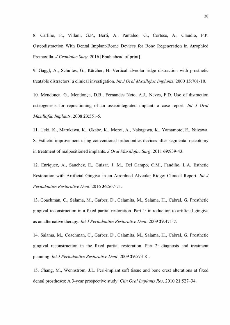
Figure 1: A 40-year-old female patient with vertical alveolar bone atrophy in the anterior region of the maxilla, involving
the region of the four maxillary incisors, was submitted for dental implants. In the frontal clinical view, a provisional
prosthesis with long clinical crowns and an unfavorable esthetics (A); the clinical view shows the vertical alveolar bone
deficiency, and a good width of keratinized mucosa was obtained after the second stage surgery (B).
32
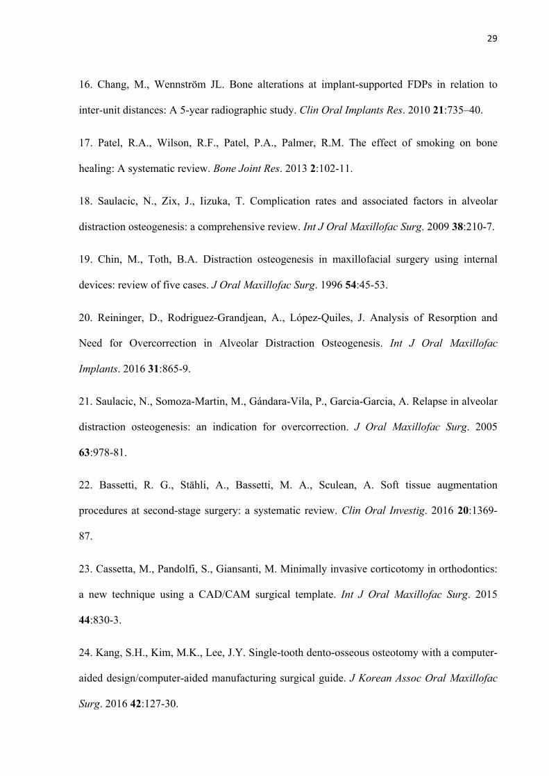
Figu
re 2
: Mod
el su
rger
y. C
asts
wer
e ob
tain
ed a
nd m
ount
ed in
an
artic
ulat
or (A
). Th
e os
teot
omie
s wer
e pe
rfor
med
in c
asts
(B; r
ed a
rrow
s); i
n th
is
case
, afte
r ob
tain
ing
the
bloc
k, it
was
nec
essa
ry to
sep
arat
e th
e im
plan
t in
the
regi
on o
f m
axill
ary
right
late
ral i
ncis
or, w
hich
res
ulte
d in
two
bloc
ks, o
ne c
onta
inin
g 3
impl
ants
and
the
othe
r con
tain
ing
only
one
(C).
The
bloc
ks c
onta
inin
g th
e im
plan
ts w
ere
posi
tione
d in
the
final
des
ired
posi
tion
and
stab
ilize
d w
ith d
enta
l sto
ne ty
pe IV
, and
a n
ew p
rovi
sion
al p
rost
hesi
s w
as m
ade
(D; r
ed a
rrow
sho
win
g th
e or
igin
al p
ositi
on; b
lack
arro
w s
how
ing
the
final
pos
ition
); an
occ
lusa
l sp
lint
of a
cryl
ic r
esin
was
fab
ricat
ed t
o gu
ide
the
impl
ant-b
one
bloc
k se
gmen
t to
the
pla
nned
posi
tion
(E a
nd F
).
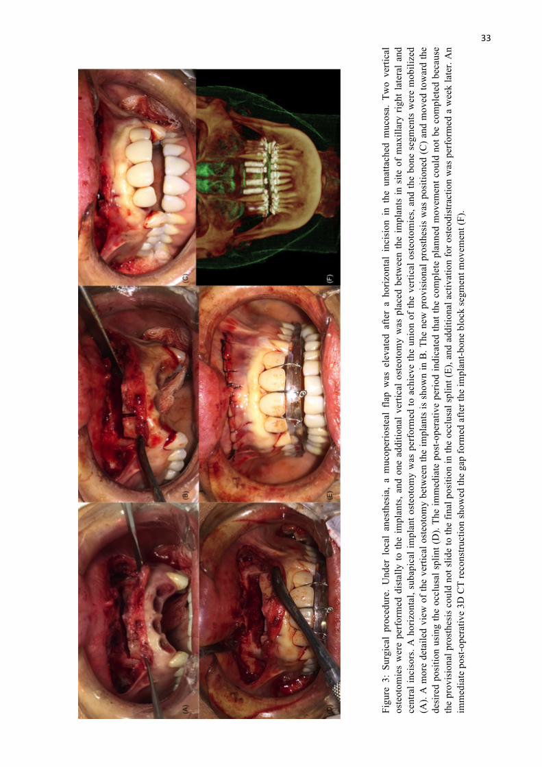
33
Figu
re 3
: Su
rgic
al p
roce
dure
. U
nder
loc
al a
nest
hesi
a, a
muc
oper
iost
eal
flap
was
ele
vate
d af
ter
a ho
rizon
tal
inci
sion
in
the
unat
tach
ed m
ucos
a. T
wo
verti
cal
oste
otom
ies
wer
e pe
rfor
med
dis
tally
to th
e im
plan
ts, a
nd o
ne a
dditi
onal
ver
tical
ost
eoto
my
was
pla
ced
betw
een
the
impl
ants
in s
ite o
f m
axill
ary
right
late
ral a
nd
cent
ral i
ncis
ors.
A h
oriz
onta
l, su
bapi
cal i
mpl
ant o
steo
tom
y w
as p
erfo
rmed
to a
chie
ve th
e un
ion
of th
e ve
rtica
l ost
eoto
mie
s, an
d th
e bo
ne s
egm
ents
wer
e m
obili
zed
(A).
A m
ore
deta
iled
view
of t
he v
ertic
al o
steo
tom
y be
twee
n th
e im
plan
ts is
sho
wn
in B
. The
new
pro
visi
onal
pro
sthe
sis
was
pos
ition
ed (C
) and
mov
ed to
war
d th
e de
sire
d po
sitio
n us
ing
the
occl
usal
spl
int (
D).
The
imm
edia
te p
ost-o
pera
tive
perio
d in
dica
ted
that
the
com
plet
e pl
anne
d m
ovem
ent c
ould
not
be
com
plet
ed b
ecau
se
the
prov
isio
nal p
rost
hesi
s co
uld
not s
lide
to th
e fin
al p
ositi
on in
the
occl
usal
spl
int (
E), a
nd a
dditi
onal
act
ivat
ion
for o
steo
dist
ract
ion
was
per
form
ed a
wee
k la
ter.
An
imm
edia
te p
ost-o
pera
tive
3D C
T re
cons
truct
ion
show
ed th
e ga
p fo
rmed
afte
r the
impl
ant-b
one
bloc
k se
gmen
t mov
emen
t (F)
.
34
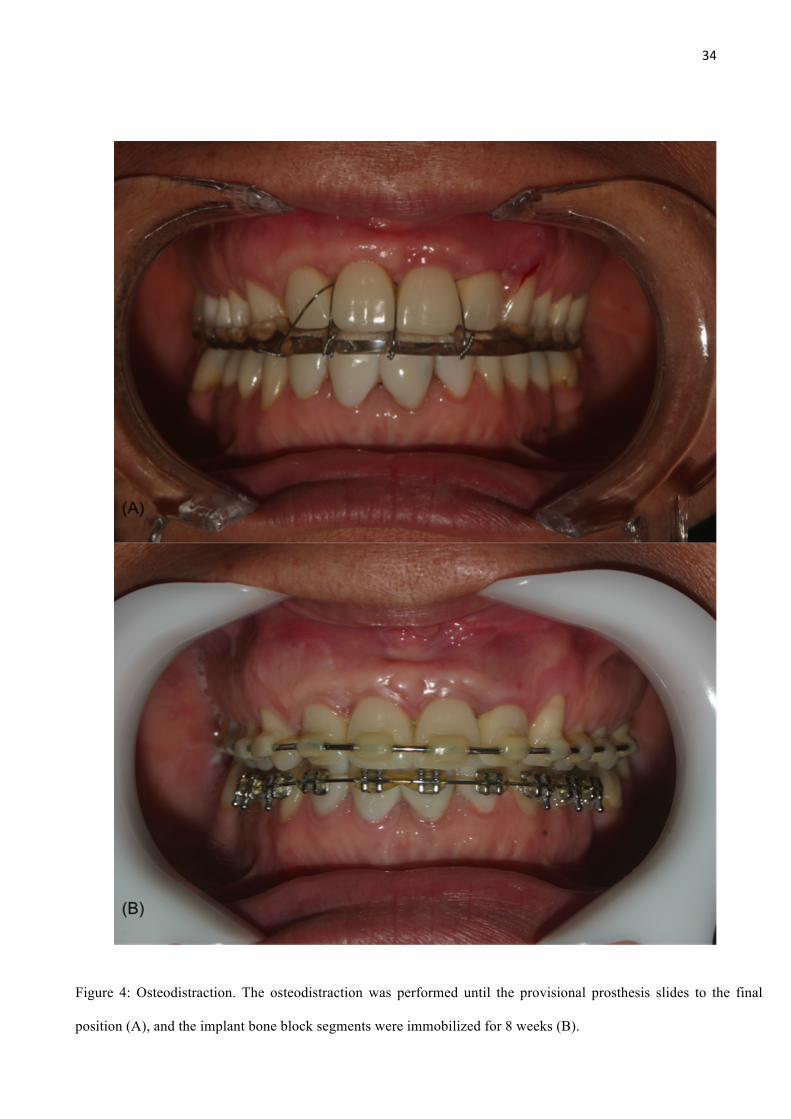
Figure 4: Osteodistraction. The osteodistraction was performed until the provisional prosthesis slides to the final
position (A), and the implant bone block segments were immobilized for 8 weeks (B).
35
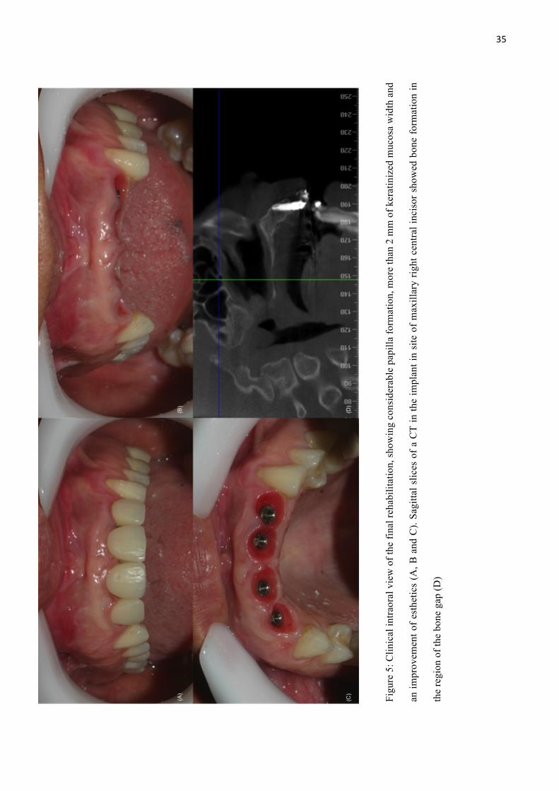
Figu
re 5
: Clin
ical
intra
oral
vie
w o
f the
fina
l reh
abili
tatio
n, sh
owin
g co
nsid
erab
le p
apill
a fo
rmat
ion,
mor
e th
an 2
mm
of k
erat
iniz
ed m
ucos
a w
idth
and
an im
prov
emen
t of e
sthe
tics
(A, B
and
C).
Sagi
ttal s
lices
of a
CT
in th
e im
plan
t in
site
of m
axill
ary
right
cen
tral i
ncis
or s
how
ed b
one
form
atio
n in
the
regi
on o
f the
bon
e ga
p (D
)
36
CAPÍTULO 2
Title: A retrospective study of an alternative osteodistraction technique after implant
placement in the maxillary esthetic region
Running title: Alternative osteodistraction technique
Key words: dental implant; osteodistraction; vertical alveolar bone atrophy; osteotomy
Department/Institution: Federal University of Ceara School of Dentistry, Fortaleza, Brazil;
Fortaleza University School of Medicine, Fortaleza, Brazil; Department of Oral and
Maxillofacial Surgery, Memorial Batista Hospital, Fortaleza, Brazil;
Rafael Lima Verde OSTERNE1; Renato Luiz Maia NOGUEIRA2; Ricardo Teixeira ABREU3;
Roberta Barroso CAVALCANTE4; Érica Amaral MEDEIROS5
1. DDS, MSc, Assistant Professor, Department of Pathology, Fortaleza University School of
Medicine, Fortaleza, Brazil. PhD Student, Federal University of Ceara School of Dentistry,
Fortaleza, Brazil.
2. DDS, MSc, PhD, Associated Professor, Department of Dental Clinic, Discipline of Oral
and Maxillofacial Surgery and Stomatology, Federal University of Ceara School of Dentistry,
Fortaleza, Brazil. Oral and Maxillofacial Surgeon, Department of Oral and Maxillofacial
Surgery, Memorial Batista Hospital, Fortaleza, Brazil.
3. DDS, MSc, PhD, Associated Professor, São Leopoldo Mandic, Fortaleza, Brazil.
4. DDS, MSc, PhD, Associated Professor, Department of Oral Pathology, Fortaleza
University School of Dentistry, Fortaleza, Brazil.
5. DDS, Orthodontics and Dentofacial Orthopedics. Private Practice, Fortaleza, Brazil.
Correspondence: Rafael Lima Verde OSTERNE, DDS, MSc
37
Assistant Professor, Department of Pathology, Fortaleza University School of Medicine,
Fortaleza, Brazil
Address
Universidade de Fortaleza (Fortaleza University/ Universidad de Fortaleza)
Av. Washington Soares, 1321
Edson Queiroz
60811-905 Fortaleza, Ceará, Brasil P.O. Box 1258
Bussines: (55-85) 3477 - 3000 Fax: (55-85) 3477 – 305
38
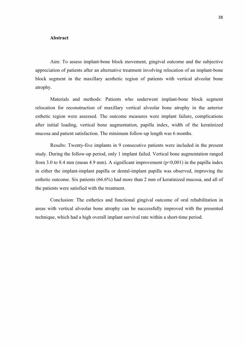
Abstract
Aim: To assess implant-bone block movement, gingival outcome and the subjective
appreciation of patients after an alternative treatment involving relocation of an implant-bone
block segment in the maxillary aesthetic region of patients with vertical alveolar bone
atrophy.
Materials and methods: Patients who underwent implant-bone block segment
relocation for reconstruction of maxillary vertical alveolar bone atrophy in the anterior
esthetic region were assessed. The outcome measures were implant failure, complications
after initial loading, vertical bone augmentation, papilla index, width of the keratinized
mucosa and patient satisfaction. The minimum follow-up length was 6 months.
Results: Twenty-five implants in 9 consecutive patients were included in the present
study. During the follow-up period, only 1 implant failed. Vertical bone augmentation ranged
from 3.0 to 8.4 mm (mean 4.9 mm). A significant improvement (p<0,001) in the papilla index
in either the implant-implant papilla or dental-implant papilla was observed, improving the
esthetic outcome. Six patients (66.6%) had more than 2 mm of keratinized mucosa, and all of
the patients were satisfied with the treatment.
Conclusion: The esthetics and functional gingival outcome of oral rehabilitation in
areas with vertical alveolar bone atrophy can be successfully improved with the presented
technique, which had a high overall implant survival rate within a short-time period.
39
Introduction
Vertical alveolar bone atrophy represents a challenge for reconstruction, especially
when the esthetic zone of the maxilla is involved. When reconstruction is not achieved, a
good esthetic outcome is rarely obtained. Bone grafts for horizontal bone atrophy have high
rates of success, with survival rates ranging from 90.2 to 100% after 3 years of evaluation
when autologous bone grafts were used (Chiapasco et al., 2004; Kim et al., 2013). In cases of
vertical bone atrophy, different techniques may be used including guided bone regeneration
(GBR) with titanium mesh or membranes reinforced with titanium (Urban et al., 2009),
intraoral distractors (Mohanty et al., 2015; Yun et al., 2016), the bilaminar cortical tenting
grafting technique (Yu et al., 2016), the tent-pole technique using screws or implants (Daga et
al., 2015), the sandwich osteotomy (Mavriqi et al., 2015), and others. In all of the previously
cited techniques, bone regeneration is usually realized before the installation of the dental
implant, which requires longer treatment durations and high financial costs (Mavriqi L et al,
2015; Gomes-Ferreira et al, 2016; Pérez-Sayáns et al, 2013).
The use of a non-absorbable and dimensionally stable barrier is usually necessary
(Urban et al., 2009) to prevent the collapse of the grafting material. A limitation is the amount
of soft tissue available for covering the graft material to prevent early exposure of the
membrane. One option is the use of osteodistraction, which can reconstruct both soft and hard
tissues (Epositoe et al. 2009). Traditionally, osteodistraction is used before dental implant
installation. Alternatively, Watzek et al. (2000) has proposed a multidimensional
osteodistraction that can be carried out after implant placement. This procedure involves the
use of a custom-fabricated distractor abutment that can move the dental implant into a
prosthetically desirable position following segmental osteotomy (Watzek et al., 2000).
Watzek et al. (2000) used multidimensional osteodistraction in 6 patients to correct 11
dental implants and achieved success in all cases, with movements of the implant-bone block
segment as long as 11 mm in the vertical direction and 4 mm in buccal/palatal direction.
Following the first description of the technique, Zechner et al., 2001, treated 8 patients with a
total of 14 dental implants and achieved a maximum vertical movement of 11 mm and a
maximum buccal/palatal movement of 5 mm. Later, Zauza et al., 2004, described the
fabrication and function of a custom-made traction prostheses for one-, two- or three-
dimensional osteodistraction. Other case reports and clinical studies of osteodistraction for
40
repositioning dental implants using different techniques have been published (Oduncuoglu et
al., 2011; Ueki et al., 2011; Mendonça et al., 2008; Gaggl et al., 2000), but the literature lacks
clinical investigations that analyzed the gingival esthetic outcomes of this treatment.
Another similar procedure is the direct relocation of the implant-bone block to the
desired position with immediate stabilization by internal rigid fixation (Stacchi et al., 2013;
Tavares et al., 2013). When used in cases of vertical alveolar bone atrophy, this technique
may reconstruct bone atrophy and restore esthetics in the anterior maxillary region. Only a
few cases using this technique have been reported (Tavares et al., 2013; Cunha et al. 2011,
Kassolis et al., 2003; Toscano et al., 2011); only one retrospective study has analyzed the
gingival esthetic outcome, but the cases in this study all involved the repositioning of single
implants (Stacchi et al., 2013). Therefore, the aim of this retrospective clinical study was to
assess the success of dental implants, the implant-bone block movement, the gingival score
and the patients’ subjective appreciation of the final results after an alternative, low-cost
treatment involving relocation of the implant-bone block segment in the maxillary aesthetic
region of patients with vertical alveolar bone atrophy.
41
Methods
The patients selected for this study were all consecutive patients with vertical alveolar
bone atrophy who underwent relocation of the implant-bone block segment in the maxillary
aesthetic region at a private clinic in Fortaleza, Ceará, Brazil, which was operated by a single
surgeon. This study was approved by an ethical committee (Protocol 1.757.767), and written
informed consent was obtained from all patients. The patients were selected on the basis of
the following inclusion criteria: patients with vertical alveolar bone atrophy in the anterior
region of the maxilla with teeth that presented distally to the areas of bone atrophy, allowing
for fitting of the surgical guide; patients who underwent relocation of the implant-bone block
segment in the maxillary aesthetic region; patients who presented with clinical frontal
intraoral photography before and at least 6 months after surgical treatment; and patients who
underwent cone beam computed tomography (CBCT) before and immediately after surgical
treatment. The exclusion criteria for this study were as follows: cases in which the clinical
intraoral photography was of inadequate quality; cases with incomplete clinical records; and
also cases in which the implant-bone blocks were carried out during orthognathic surgery.
Surgical and prosthetic procedures
Initially, an impression was obtained, and the position of the implants was obtained
using a diagnostic cast. The ideal future prosthetic position was then determined using a
diagnostic wax-up. A model surgery was performed, and the implants were positioned in the
final desired location and stabilized with dental stone. With the implants in the final position,
a new provisional implant-fixed prosthesis was made. If it was a single implant, a
buccal/palatal perforation was performed on the prosthesis. If there were multiple implants,
they were kept splinted together at the proximal surfaces. The perforation or the union of the
provisional prosthesis were passed with an orthodontic wire, which led the prosthesis to the
planned position during the surgery. An occlusal tooth-implant-supported splint of acrylic
resin reinforced with orthodontic wire was custom-fabricated for use as a surgical guide.

42
For the surgical procedure, the new provisional implant-fixed prosthesis was
positioned in the implant, and then a horizontal incision was made in the unattached gingiva,
exposing the region of the alveolar process segment to be osteotomized. The bone
surrounding the implant was then cut through with a saw, and two vertical osteotomies were
performed. They were then connected by a single horizontal osteotomy, and the implant-bone
block segment was mobilized with a thin chisel. The occlusal splint was positioned, and the
orthodontic wire was passed through the perforation of the prosthesis (in the case of a single
implant) or through the interproximal region (in the case of multiple implants) and then
passed thru the splint. The implant-bone block segment was then moved to the planned
position. If a small movement occurred, the implant-bone block segment could be stabilized
with internal rigid fixation. In cases of large movements, the implant-bone block segment
could be gradually moved to the final position by an osteodistraction. After the movement
was completed, the implant-bone block segment was immobilized for 12 weeks. This was
performed by connecting the provisional prosthesis to the adjacent teeth with an orthodontics
wire. After this period, the prosthetic procedures for the final prosthesis could be performed.
Data collection
Data from the medical records, clinical photographs and CBCT images were collected
from the following three different periods: before the implant-bone block surgery (T1),
immediately after the repositioning of the implant-bone block (T2), and 6 months after the
surgery (T3).
The outcome measures were the following:
1. Implant failure including mobility of the implants (determined manually) and/or any
infections requiring implant removal that were present at the abutment connection or at the
insertion of the provisional prosthesis. These data were obtained from the medical records.
2. Complications occurring during/after treatment. These data were also obtained from the
medical records.
3. Evaluation of the vertical bone augmentation.
Vertical bone gain was evaluated by linear measurement of the vertical bone gap
found at the apex of each implant involved in the movement, and an average of the
measurements was calculated. This measurement was taken from a sagittal slice of the post-
43
operative CBCT (T2) image using Dolphin 3D imaging software® with the linear
measurement tool.
4. The papilla index according to Jemt 1997 in T1 and T3. This index describes the papillae in
relation to the height of the interproximal space. Score 0: no formation of papillae; Score 1:
less than half of the interproximal space filled with soft tissue; Score 2: formation of papillae
in at least half of the interproximal space; Score 3: papillae filling the entire interproximal
space; Score 4: hyperplastic papillae. This score was determined for all of the papillae
involved in the movement of the implant-bone block based on frontal intraoral clinical
photographs. Intra-examiner agreement was assessed and demonstrated statistically
significant agreement (Kappa, 869).
5. Width of keratinized mucosa
The keratinized mucosa was assessed in clinical pictures from after the surgery in T3.
The measurement was made after calibration of a digital ruler using the actual length of the
distance between the distal surface of the maxillary right central incisor and the distal surface
of the maxillary left central incisor. After, a calibrated ruler was used to measure the width of
the keratinized mucosa from the most apical point of the gingival margin to the mucogingival
junction. The distance was categorized as “no keratinized mucosa”, “between 0-2 mm of
keratinized mucosa”, or “>2 mm of keratinized mucosa”.
6. Evaluation of patient satisfaction
Subjective evaluation of patient satisfaction was assessed during the T3 period based
on the results of the questionnaire of Meijndert et al., 2007, which was modified by Tymstra
et al., 2010. The questionnaire consisted of an overall satisfaction score ranging from 0-10
and two questions regarding the peri-implant mucosa with scores ranging from 0 to 4.
Additionally, two questions about discomfort and compliance were evaluated based on a VAS
score (Stacchi et al., 2013).
Statistical analysis
All data were compiled into a single electronic dataset, and all analyses were
performed using SPSS, version 16.0 (SPSS Inc., Chicago, IL, USA). Age and follow-up time
are expressed as the means and standard deviations. Vertical bone augmentation is expressed
as the mean. The width of the keratinized gingiva and patient satisfaction are expressed as

44
percentages. The Wilcoxon signed-rank test was used to evaluate the papilla scores, and the
level of significance was set at 5%.
45
Results
During a period from 2010 and 2016, 13 consecutive patients underwent repositioning
of implant-bone block segments. Of them, only 9 patients were included in the present study.
Two were excluded because the repositioning was performed during an orthognathic surgery;
1 was excluded because the repositioning was in the posterior region of maxilla; and 1 was
excluded due to a short period of follow-up (less than 6 months). The nine remaining patients
included 2 men and 7 women with ages ranging from 25 to 62 years (mean 44.8 ± 13.1 years).
None of the patients had significant anamnestic remarks. A total of 25 implants were
repositioned, and the mean length of follow-up was 33.8 months (±23.3 months; ranging from
6 to 72 months). In all of the cases, multiple implants were moved, and no cases of single
implant repositioning were performed during this period. In two cases, implant-bone blocks
containing 4 implants were moved (Figure 1); in 3 cases, implant-bone blocks containing 3
implants were moved; and in 4 cases, implant-bone blocks with 2 implants were moved
(Figure 2 and 3). Four of the patients underwent bone grafts prior to implant placement due to
horizontal bone atrophy. The data are presented in Table 1.
During the follow-up period, only one implant failure occurred. The failure was
diagnosed during the abutment connection in a patient who underwent implant-bone block
repositioning with three implants in the block, and the mesial implant (at the maxillary right
central incisor site) failed. All of the other implants are still functional with provisional or
definitive prosthetic rehabilitation. Although an implant apex was sectioned during the
horizontal osteotomy in one case (patient 6; implant in the maxillary central left incisor site;
Figure 4), the implant did not fail. Other complications occurred after surgery in two patients.
Although a minimal distance of 2 mm between the osteotomy and natural teeth was respected,
dental root therapy was necessary for one tooth each in two patients (patient 9, maxillary right
lateral incisor; patient 5, maxillary left lateral incisor), as tooth discoloration occurred, and the
pulp vitality test was non responsive.
The mean vertical bone augmentation measured at T2 was 4.9 mm, ranging from 3.0
mm to 8.4 mm. Internal rigid fixation was used in only case, which involved an implant-bone
block segment with 2 implants (patient 2, implants in the maxillary right lateral and central
incisors site) that were repositioned only 3.0 mm.
The papilla indices are listed in Table 2. In total, 35 papilla were analyzed. Eighteen
46
were implant-natural teeth papilla, and 17 were implant-implant papilla. The scores at T1
were relatively low, indicating poor gingival esthetics before surgery. At this time point, 25
(71.43%) papillae had scores ranging from 0 to 1, with 16 of them at the implant-implant site
and 9 at the implant-natural tooth sites (p 0,004). After surgery (at T3), a statistically
significant improvement in the papilla scores was observed (p< 0,001). Only 7 (20.00%)
papillae had scores ranging from 0 to 1, with one of them at the implant-natural tooth site and
6 at the implant-implant sites (p 0,028) [Graphic 1]. When analyzing the keratinized mucosa
at T3, only one case had at least one implant without keratinized mucosa, two cases had
between 0 to 2 mm of keratinized mucosa, and 6 cases had keratinized mucosa >2.0 mm.
Patient feedback regarding the treatment is presented in Table 3. The worst overall
score was 7, meaning that all patients were satisfied or very satisfied with the treatment
results. Regarding gingival contour, 66.6% were satisfied or very satisfied, and 88.9% were
satisfied or very satisfied with the gingival color. Regarding the discomfort of the surgery, 4
(44.4%) patients considered it to be moderate, and 2 (22.2%) experienced high discomfort.
Only 2 (22.2%) patients stated that they would be reluctant to undergo this procedure again if
needed, and none of the patients stated that they would not undergo the surgery again.
47
Discussion
Reconstruction of alveolar vertical bone atrophy is a complex clinical challenge for
oral rehabilitation, and a multidisciplinary approach is frequently necessary to achieve good
esthetic results. Some decades ago, the criteria for implant success included no clinical
mobility, a lack of symptoms, no radiographic peri-implant radiolucencies, and less than 0.2
mm of annual bone loss after the first year (Albrektsson et al., 1986). Although some cases
may be functional and present with all of the previous criteria of success, as in Figure 2C, it is
unclear if these should be considered cases of successful treatment or successful
osseointegration. Some might consider them to be examples of successful osseointegration
but a failure of the treatment, as good aesthetic results were not achieved.
The options when faced with vertical alveolar bone atrophy vary from prosthetic
compensation of the soft tissue to bone reconstruction using bioengineering (Coachman et al.,
2009; Eposito et al., 2009; Herford et al, 2015). The costs and treatment times are reduced
when only artificial gingiva (ceramic or acrylic resin) is used. However, these treatments can
be complex, time-consuming, and high-cost, requiring bone grafts associated with titanium
mesh and rHBMP2 (Gomes Ferreira 2016). The treatment presented in this study may be
suitable for correcting bone defects in aesthetic areas with satisfactory results and reduced
cost compared with traditional osteodistraction or the use of bioengineering. Because the
device used is made of acrylic resin reinforced by orthodontic wire, it is a low-cost device.
The treatment involves two stages. First, the implant is installed, and then the reconstruction
surgery is performed. Other reconstructions such as tent-pole or bone ring techniques involve
only one stage, which leads to a reduction in total treatment time (Daga et al., 2015; Omara et
al., 2016). Though more time consuming, the presented technique can achieve aesthetic
results in a similar time frame since the provisional prosthesis used in the repositioning
surgery is already the proper size.
This technique was shown to be reproducible in the cases evaluated, with good
predictability due to the use of surgical guides. Another advantage of this technique is that it
can be used for distraction osteogenesis, as in cases that involve repositioning the implant in
one session (patient 2), as well as for malpositioned implants. After mobilization of the
implant-bone block segment, all downward movements can be performed in one step if the
palatal soft tissue allows. If the downward movements cannot be performed precisely due to a
lack of elasticity of the soft tissue, an osteodistraction is performed. Ideally, osteotomies on
48
the buccal bone plate should be initiated with burs or saws and finished using thin chisels,
thus avoiding damage to the periosteum of the palate that could compromise the blood supply
to the implant-bone block segment.
Vertical alveolar bone augmentation was assessed by linear measurement of the bone
gap in a CBCT image. The mean bone augmentation was 4.9 mm, but a case of 8.4 mm of
vertical augmentation was observed in patient 9. Previous authors have reported 11 mm of
vertical alveolar reconstruction by means of osteodistraction of the implant-bone block
segment (Watzeck et al., 2000; Zechner et al., 2001). In the present study, the model surgery
was used to determine all bone augmentations. In cases of multiple implants, different
movements of the implants can be performed whenever necessary by segmenting an implant
to an isolated bone block. However, this should also be planned in advance during the model
surgery, as was the case in one patient from this study (patient 1).
Complications occurred in 4 patients, and one patient’s implant failed. In this patient,
an autogenous bone block graft was performed prior to implant placement. The implant at the
maxillary right central incisor site still had mobility during the provisional prosthesis phase
following the repositioning surgery, so the patient decided not to remake the implant. The
overall implant survival rate was 96% during the follow-up period. Recently, a study by
Chrcanovic et al., 2016, reported a survival rate of 98.36% up to abutment connection,
regardless of the technique used. One patient had the apex of an implant sectioned during
osteotomy, but the implant did not fail. After a follow-up period of 54 months, the implant
had satisfactory esthetics and function. Two of the patients exhibited a loss of pulp vitality
(one tooth each) in the neighboring teeth to the osteotomy site. Although a limit of 2.0 mm of
distance between the osteotomy and the natural tooth was respected, two teeth evolved with
discoloration and a lack of response to the pulp vitality test. Both were lateral incisors, so
perhaps due to the smaller diameter of the root of the tooth, a greater distance of the
osteotomy should be recommended.
To explore the clinical significance of papilla indices, the data were sorted based on
papilla and papilla location (implant-implant papilla or implant-natural tooth papilla). The
scores at T1 were relatively low, indicating the presence of compromised papilla, with
71.43% of the papillae being classified as “no formation of papillae” or “less than half of the
interproximal space filled with soft tissue”. As expected (Tymstra et al., 2010), the inter-
implant papilla had a worse score than the implant-natural tooth papilla, and 64% of the
49
papillae classified as 0 or 1 were in the implant-implant site. After treatment, only 20.0% of
the papillae were classified as 0 or 1, showing a significant improvement of papillae presence
both in the implant-implant site and the implant-natural tooth site. Although more papillae
were observed at T3, the effects of the bone repositioning on the formation of the papillae are
unclear. It can be considered that the bone repositioning reduced the distance from the
interproximal contact point to the inter-implant bone peak, favoring filling of the papilla space
and improvement in the papilla indices. However, it is worth mentioning the importance of
tissue manipulation techniques with provisional crowns for the formation of papillae
(Wittneben et al., 2013), which was also performed in the cases described in this study.
A good width of keratinized mucosa was observed in 6 out of 9 of the patients in the
study. Often in cases of bone atrophy, keratinized mucosa may not be present, and techniques
to increase the amount of keratinized mucosa may be performed during implant installation or
second-stage surgery. The presence of keratinized mucosa has a positive effect on peri-
implant tissue health (Ladwein et al., 2015; Bassetti et al., 2016). In a systematic review,
Bassetti et al., 2016, found that patients with sites with a width of <2 mm of keratinized
mucosa developed discomfort during brushing more easily and also exhibited dental plaque
accumulation and peri-implant soft tissue inflammation.
The overall satisfaction of the patients was high, which was in agreement with the
improvement in papilla formation and the reduction in the length of the tooth crowns
observed in the present study. The procedure was found to have good compliance, as 66.6%
of the subjects would undergo this treatment again, if necessary, and the discomfort was
considered not high (only 2 patients [22.2%] experienced high discomfort). Stacchi et al.,
2012, observed similar results after repositioning single dental implants through segmental
osteotomy, but no osteodistractions were performed in their study.
A number of limitations should be taken into account when interpreting the results of
this study. First, this is retrospective clinical study and not a randomized, controlled clinical
trial. Second, although this procedure was performed on 13 patients, only 9 patients were
analyzed, and such a limited sample size greatly affects the statistical power.

50
Conclusion
The osteodistraction presented here can successfully reconstruct vertical alveolar bone
atrophy in the anterior region of the maxilla without increasing implant loss in a short time
period. Additionally, this procedure can significantly increase the gingival esthetics and is
well accepted by most patients with only moderate discomfort. Clinical trials with larger
numbers of patients comparing vertical bone augmentation, the success of the implant and
complications with other techniques for vertical alveolar bone reconstruction could help to
clarify the results of this study.
51
Reference
Albrektsson, T., Zarb, G., Worthington, P., Eriksson, A.R. (1986) The long-term efficacy of
currently used dental implants: A review and proposed criteria of success. International
Journal of Oral and Maxillofacial Implants 1: 11–25.
Bassetti, R.G., Stähli, A., Bassetti, M.A., Sculean, A. (2016) Soft tissue augmentation
procedures at second-stage surgery: a systematic review. Clinical Oral Investigations. 20:
1369-1387.
Chiapasco, M., Romeo, E., Casentini, P., Rimondini, L. (2004) Alveolar distraction
osteogenesis vs. vertical guided bone regeneration for the correction of vertically deficient
edentulous ridges: A 1-3-year prospective study on humans. Clinical Oral Implants Research
15: 82–95.
Chrcanovic, B.R., Kisch, J., Albrektsson, T., Wennerberg, A. (2016) Factors Influencing
Early Dental Implant Failures Journal of Dental Research 95: 995-1002.
Coachman, C., Salama, M., Garber, D., Calamita, M., Salama, H., Cabral, G. (2009)
Prosthetic gingival reconstruction in a fixed partial restoration. Part 1: introduction to
artificial gingiva as an alternative therapy. International Journal of Periodontics and
Restorative Dentistry 29: 471-477.
da Cunha, H.A., Filho, H.N., Batista, J.G., Matsumoto, M.A. (2011) Segmental osteotomy for
the correction of a malpositioned single implant: an 8-year follow-up. Quintessence
International 42: 817-822.
52
Daga, D., Mehrotra, D., Mohammad, S., Singh, G., Natu, S.M. (2015) Tentpole technique for
bone regeneration in vertically deficient alveolar ridges: A review. Journal of Oral Biology
and Craniofacial Research 5: 92-97.
Esposito, M., Grusovin, M.G., Felice, P., Karatzopoulos, G., Worthington, H.V., Coulthard,
P. (2009) The efficacy of horizontal and vertical bone augmentation procedures for dental
implants - a Cochrane systematic review. European Journal of Oral Implantology 3: 167-184.
Gaggl, A., Schultes, G., Kärcher, H. (2000) Vertical alveolar ridge distraction with prosthetic
treatable distractors: a clinical investigation. International Journal of Oral and Maxillofacial
Implants 15: 701-710.
Gomes-Ferreira, P.H., Okamoto, R., Ferreira, S., De Oliveira, D., Momesso, G.A., Faverani,
L.P. (2016) Scientific evidence on the use of recombinant human bone morphogenetic
protein-2 (rhBMP-2) in oral and maxillofacial surgery. Oral and Maxillofacial Surgery 20:
223-232.
Herford, A. S., Nguyen, K. (2015) Complex bone augmentation in alveolar ridge defects.
Oral and Maxillofacial Surgery Clinics of North America 2: 227 – 244.
Jemt, T. (1997) Regeneration of gingival papillae after single-implant treatment. International
Journal of Periodontics and Restorative Dentistry 17: 326-33.
Kassolis, J.D., Baer, M.L., Reynolds, M.A. (2003) The segmental osteotomy in the
management of malposed implants: a case report and literature review. Journal of
Periodontology 74: 529-536.
53
Kim, J.W., Cho, M.H., Kim, S.J., Kim, M.R. (2013) Alveolar distraction osteogenesis versus
autogenous onlay bone graft for vertical augmentation of severely atrophied alveolar ridges
after 12 years of long-term follow-up. Oral Surgery Oral Medicine Oral Pathology Oral
Radiology 116 : 5 4 0 – 5 4 9
Ladwein, C., Schmelzeisen, R., Nelson, K., Fluegge, T.V., Fretwurst, T. (2015) Is the
presence of keratinized mucosa associated with periimplant tissue health? A clinical cross-
sectional analysis. International Journal of Implant Dentistry 1: 1-5.
Mavriqi, L., Baca, E., Demiraj, A. (2015) Sandwich osteotomy of the atrophic posterior
mandible prior to implant placement. Clinical Case Reports 3: 610-614.
Meijndert, L., Meijer, H.J., Stellingsma, K., Stegenga, B., Raghoebar, G.M. (2007)
Evaluation of aesthetics of implant-supported single-tooth replacements using different bone
augmentation procedures: a prospective randomized clinical study. Clinical Oral Implants
Research 18: 715-719.
Mendonça, G., Mendonça, D.B., Fernandes Neto, A.J., Neves, F.D. (2008) Use of distraction
osteogenesis for repositioning of an osseointegrated implant: a case report. International
Journal of Oral and Maxillofacial Implants 23: 551-555.
Mohanty, R., Kumar, N.N., Ravindran, C. (2015) Vertical Alveolar Ridge Augmentation by
Distraction Osteogenesis. Journal of Clinical and Diagnostic Research 9: ZC43-ZC46.
Oduncuoglu, B.F., Alaaddinoglu, E.E., Oguz, Y., Uckan, S., Erkut, S. (2011) Repositioning a
prosthetically unfavorable implant by vertical distraction osteogenesis. Journal of Oral and
Maxillofacial Surgery 69: 1628-1632.
54
Omara, M., Abdelwahed, N., Ahmed, M., Hindy, M. (2016) Simultaneous implant placement
with ridge augmentation using an autogenous bone ring transplant. International Journal of
Oral and Maxillofacial Surgery 4: 535-544.
Pérez-Sayáns, M., León-Camacho, M.de L., Somoza-Martín, J.M., Fernández-González, B.,
Blanes-Vázquez-Gundín, S., Gándara-Rey, J.M., García-García, A. (2013) Dental implants
placed on bone subjected to vertical alveolar distraction show the same performance as those
placed on primitive bone. Medina Oral, Patologia Oral e Cirurgia Bucal 18: e686-e892.
Stacchi, C., Chen, S.T., Raghoebar, G.M., Rosen, D., Poggio, C.E., Ronda, M., Bacchini, M.,
Di Lenarda, R. (2013) Malpositioned osseointegrated implants relocated with segmental
osteotomies: a retrospective analysis of a multicenter case series with a 1- to 15-year follow-
up. Clinical Implant Dentistry and Related Research 15: 836-846.
Tavares, R.N., da Escóssia, J. Jr., Santos, S.E., Ferraro-Bezerra, M. (2013) Bone graft
sandwich osteotomy to correct a malpositioned dental implant. International Journal of Oral
and Maxillofacial Surgery 42: 901-903.
Toscano, N., Sabol, J., Holtzclaw, D., Scott, T. (2011) Implant repositioning by segmental
osteotomy: a case series and review. International Journal of Periodontics and Restorative
Dentistry 31: e102-e108.
Tymstra, N., Raghoebar, G.M., Vissink, A., Meijer, H.J. (2011) Dental implant treatment for
two adjacent missing teeth in the maxillary aesthetic zone: a comparative pilot study and test
of principle. Clinical Oral Implants Research 22: 207-213.
Ueki, K., Marukawa, K., Okabe, K., Moroi, A., Nakagawa, K., Yamamoto, E., Niizawa, S.
(2011) Esthetic improvement using conventional orthodontic devices after segmental
55
osteotomy in treatment of malpositioned implants. Journal of Oral and Maxillofacial Surgery
69: 939-943.
Urban, I.A., Jovanovic, S.A., Lozada, J.L. (2009) Vertical ridge augmentation using guided
bone regeneration (GBR) in three clinical scenarios prior to implant placement: a
retrospective study of 35 patients 12 to 72 months after loading. International Journal of Oral
and Maxillofacial Implants 24: 502-510.
Watzek, G., Zechner, W., Chrismani, A. & Zauza, K. (2000) A distraction abutment system
for 3-dimensional distraction osteogenesis of the alveolar process: technical note.
International Journal of Oral and Maxillofacial Implants 15: 731–737.
Wittneben, J.G., Buser, D., Belser, U.C., Brägger, U. (2013) Peri-implant soft tissue
conditioning with provisional restorations in the esthetic zone: the dynamic compression
technique. International Journal of Periodontics and Restorative Dentistry 33: 447-455.
Yu, H., Chen, L., Zhu, Y., Qiu, L. (2016) Bilamina cortical tenting grafting technique for
three-dimensional reconstruction of severely atrophic alveolar ridges in anterior maxillae: A
6-year prospective study. Journal of Craniomaxillofacial Surgery 44: 868-875.
Yun, K.I., Choi, H., Wright, R.F., Ahn, H.S., Chang, B.M., Kim, H.J. (2016) Efficacy of
Alveolar Vertical Distraction Osteogenesis and Autogenous Bone Grafting for Dental
Implants: Systematic Review and Meta-Analysis. International Journal of Oral and
Maxillofacial Implants 31: 26-36.
Zauza, K., Celar, A.G., Zechner, W., Watzek, G. (2004) Novel development for intraoral
distraction osteogenesis by individually fabricated traction prostheses. Clinical Oral Implants
Research 15: 371-374.

56
Zechner, W., Bernhart, T., Zauza, K., Celar, A., Watzek, G. (2001) Multidimensional
osteodistraction for correction of implant malposition in edentulous segments. Clinical Oral
Implants Research 12: 531-538.
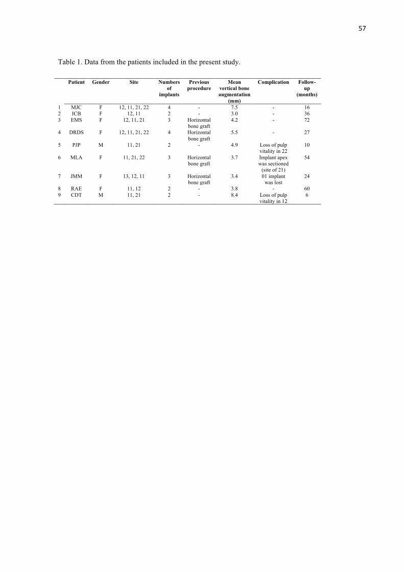
57
Table 1. Data from the patients included in the present study.
Patient Gender Site Numbers of
implants
Previous procedure
Mean vertical bone augmentation
(mm)
Complication Follow-up
(months)
1 MJC F 12, 11, 21, 22 4 - 7.5 - 16 2 ICB F 12, 11 2 - 3.0 - 36 3 EMS F 12, 11, 21 3 Horizontal
bone graft 4.2 - 72
4 DRDS F 12, 11, 21, 22 4 Horizontal bone graft
5.5 - 27
5 PJP M 11, 21 2 - 4.9 Loss of pulp vitality in 22
10
6 MLA F 11, 21, 22 3 Horizontal bone graft
3.7 Implant apex was sectioned
(site of 21)
54
7 JMM F 13, 12, 11 3 Horizontal bone graft
3.4 01 implant was lost
24
8 RAE F 11, 12 2 - 3.8 - 60 9 CDT M 11, 21 2 - 8.4 Loss of pulp
vitality in 12 6
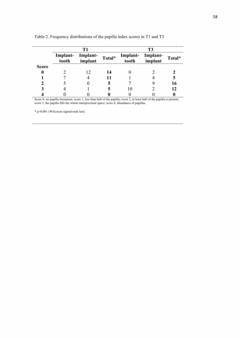
58
Table 2. Frequency distributions of the papilla index scores in T1 and T3
T1 T3
Implant-tooth
Implant-implant Total* Implant-
tooth Implant-implant Total*
Score 0 2 12 14 0 2 2 1 7 4 11 1 4 5 2 5 0 5 7 9 16 3 4 1 5 10 2 12 4 0 0 0 0 0 0
Score 0, no papilla formation; score 1, less than half of the papilla; score 2, at least half of the papilla is present; score 3, the papilla fills the whole interproximal space; score 4, abundance of papillae.
* p>0,001 (Wilcoxon signed-rank test)
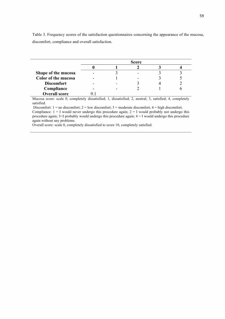
59
Table 3. Frequency scores of the satisfaction questionnaires concerning the appearance of the mucosa,
discomfort, compliance and overall satisfaction.
Score 0 1 2 3 4
Shape of the mucosa - 3 - 3 3 Color of the mucosa - 1 - 3 5
Discomfort - - 3 4 2 Compliance - - 2 1 6
Overall score 9.1 Mucosa score: scale 0, completely dissatisfied; 1, dissatisfied; 2, neutral; 3, satisfied; 4, completely satisfied. Discomfort: 1 = no discomfort; 2 = low discomfort; 3 = moderate discomfort; 4 = high discomfort. Compliance: 1 = I would never undergo this procedure again; 2 = I would probably not undergo this procedure again; 3=I probably would undergo this procedure again; 4 = I would undergo this procedure again without any problems. Overall score: scale 0, completely dissatisfied to score 10, completely satisfied.
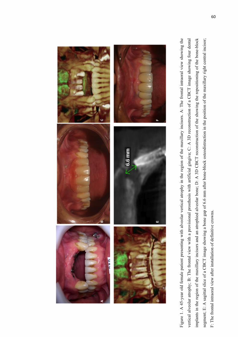
60
Figu
re 1
. A 4
5-ye
ar o
ld fe
mal
e pa
tient
pre
sent
ing
with
alv
eola
r ver
tical
atro
phy
in th
e re
gion
of t
he m
axill
ary
inci
sors
. A: T
he fr
onta
l int
raor
al v
iew
sho
win
g th
e
verti
cal a
lveo
lar a
troph
y; B
: The
fron
tal v
iew
with
a p
rovi
sion
al p
rost
hesi
s w
ith a
rtific
ial g
ingi
va; C
: A 3
D re
cons
truct
ion
of a
CB
CT
imag
e sh
owin
g fo
ur d
enta
l
impl
ants
in th
e re
gion
of t
he m
axill
ary
inci
sors
and
an
atro
phie
d al
veol
ar b
one;
D: A
3D
CB
CT
reco
nstru
ctio
n of
the
show
ing
the
repo
sitio
ning
of t
he b
one-
bloc
k
segm
ent;
E: A
sag
ittal
slic
e of
a C
BC
T im
age
show
ing
a bo
ne g
ap o
f 6.6
mm
afte
r bon
e-bl
ock
oste
odis
tract
ion
in th
e po
sitio
n of
the
max
illar
y rig
ht c
entra
l inc
isor
;
F: T
he fr
onta
l int
raor
al v
iew
afte
r ins
talla
tion
of d
efin
itive
cro
wns
.
61
Figu
re 2
. A 2
7-ye
ar-o
ld m
ale
patie
nt w
ho u
nder
wen
t bon
e-bl
ock
oste
odis
tract
ion
of th
e m
axill
ary
cent
ral i
ncis
ors.
A: T
he o
rthop
anto
mog
ram
x-r
ay a
fter
inst
alla
tion
of th
e im
plan
ts; B
: A 3
D C
BC
T re
cons
truct
ion
of th
e
show
ing
verti
cal a
lveo
lar b
one
atro
phy
at th
e si
te o
f the
max
illar
y ce
ntra
l inc
isor
s; C
: The
clin
ical
fron
tal i
ntra
oral
vie
w s
how
ing
the
lack
of p
apill
a fo
rmat
ion
betw
een
the
cent
ral i
ncis
ors
crow
ns a
nd lo
ng c
row
ns; D
and
E:
The
orth
opan
tom
ogra
m x
-ray
and
3D
CB
CT
reco
nstru
ctio
n of
the
afte
r im
plan
t-bon
e bl
ock
oste
odis
tract
ion.
A g
ap is
obs
erve
d su
bapi
cal t
o th
e im
plan
ts, a
nd th
e m
axill
ary
right
late
ral i
ncis
or w
as s
ubm
itted
to e
ndod
ontic
treat
men
t due
to lo
ss o
f pul
p vi
talit
y; F
: The
clin
ical
fron
tal i
ntra
oral
vie
w 6
mon
ths
post
oper
ativ
ely.
A c
ompl
ete
form
atio
n of
pap
illae
occ
urre
d be
twee
n im
plan
ts (s
core
3);
G a
nd H
: A s
agitt
al C
BC
T sl
ice
of th
e sh
owin
g a
bone
gap
of 8
.6 m
m a
t the
site
of t
he m
axill
ary
left
cent
ral i
ncis
or im
plan
t and
the
bone
form
atio
n in
the
gap
6-m
onth
s pos
tope
rativ
ely.
62
Figure 3: A 62-year-old female patient who underwent implant-bone block osteodistraction at the site of
the maxillary right central and lateral incisors. A: The clinical frontal intraoral view showing the absence
of papillae between the implant and an elongated lateral incisor; B: A 3D reconstruction of a CBCT image
showing vertical alveolar bone atrophy at the site of the implants; C and D: The clinical frontal intraoral
view and 3D CBCT reconstruction of the after bone-block osteodistraction showing partial formation of
papillae between the implants and an improved length of the crowns; E and F: A sagittal slice from the site
of the maxillary right incisor with a bone gap of 4.1 mm that was partially filled with new bone in T3.
63
Figu
re 4
: A
54-
year
-old
fem
ale
patie
nt w
ho u
nder
wen
t im
plan
t-bon
e bl
ock
oste
odis
tract
ion
at t
he s
ite o
f th
e m
axill
ary
cent
ral
inci
sors
and
the
lef
t la
tera
l
inci
sor.
A a
nd B
: The
clin
ical
fron
tal i
ntra
oral
vie
w a
nd a
cor
onal
slic
e of
a C
BC
T im
age
afte
r the
hor
izon
tal b
one
graf
t. Th
e gr
aft a
t the
site
of t
he m
axill
ary
right
late
ral i
ncis
or w
as e
xpos
ed in
the
oral
cav
ity a
nd re
mov
ed, s
o it
was
not
pos
sibl
e to
inse
rt th
e im
plan
t at t
his
site
. Ver
tical
alv
eola
r bon
e at
roph
y w
as a
lso
pres
ent i
n th
is re
gion
; C: A
3D
reco
nstru
ctio
n of
a C
BC
T im
age
afte
r im
plan
t ins
talla
tion
and
bone
-blo
ck o
steo
dist
ract
ion.
The
ape
x of
the
impl
ant a
t the
site
of
the
max
illar
y le
ft ce
ntra
l in
ciso
r w
as s
ectio
ned
durin
g th
e su
bapi
cal
oste
otom
y. D
: Th
e cl
inic
al f
ront
al i
ntra
oral
vie
w a
t T3
sho
win
g pa
rtial
for
mat
ion
of
papi
llae
betw
een
the
impl
ants
and
est
hetic
ally
favo
rabl
e cr
owns
. E a
nd F
: A sa
gitta
l slic
e of
the
CB
CT
imag
e at
the
site
of t
he m
axill
ary
left
cent
ral i
ncis
or w
ith
a se
ctio
ned
apex
with
a g
ap o
f 3.8
mm
that
was
late
r fill
ed w
ith n
ew b
one.
64
Graphic 1. Distribution of the papilla index scores in T1 and T3.
65
4 CONCLUSÃO GERAL
Reabilitação oral em áreas de atrofia óssea alveolar vertical é complexa e representa
um desafio clínico que é melhor tratado através de uma abordagem multidisciplinar. Embora
diferentes opções de tratamento sejam propostos na literatura, a técnica apresentada neste
estudo pode ser considerada como uma opção viável para a reconstrução de áreas com atrofia
óssea alveolar vertical na região anterior da maxila, sem aumentar a perda do implante em um
curto período de tempo. Além disso, o procedimento gera uma melhora significativa de
estética gengival, sendo bem aceito pela maioria dos pacientes com um desconforto
moderado. Dados prospectivos de ensaios clínicos, com um número maior de pacientes
poderiam ajudar a complementar os achados deste estudo.
66
5 REFERÊNCIAS
CHIAPASCO M, ROMEO E, CASENTINI P, RIMONDINI L. Alveolar distraction
osteogenesis vs. vertical guided bone regeneration for the correction of vertically deficient
edentulous ridges: A 1-3-year prospective study on humans. Clinical Oral Implants
Research. v. 15, n. 1, p. 82-95, fev. 2004.
COACHMAN C, SALAMA M, GARBER D, CALAMITA M, SALAMA H, CABRAL G.
Prosthetic gingival reconstruction in a fixed partial restoration. Part 1: introduction to
artificial gingiva as an alternative therapy. International Journal Periodontics and
Restorative Dentistry. v. 29, n. 5, p. 471-477, out. 2009.
DAGA D, MEHROTRA D, MOHAMMAD S, SINGH G, NATU SM. Tentpole technique for
bone regeneration in vertically deficient alveolar ridges: A review. Journal Oral Biology
and Craniofacial Research. v. 5, n. 2, p. 92-97, mai.-ago. 2015.
DI P, LIN Y, LI JH, QIU LX, CUI HY. Clinical study of prosthetic gingival reconstruction in
vertical bone defect patients. Zhonghua Kou Qiang Yi Xue Za Zhi. v. 46, n. 11, p. 655, nov.
2011. [resumo]
ENRÍQUEZ A, SÁNCHEZ E, GUIZAR JM, DEL CAMPO CM, FANDIÑO LA. Esthetic
Restoration with Artificial Gingiva in an Atrophied Alveolar Ridge: Clinical Report.
International Journal Periodontics and Restorative Dentistry. v. 36, n. 4, p. 567-571, jul.-
ago. 2016.
ESPOSITO, M., GRUSOVIN, M.G., FELICE, P., KARATZOPOULOS, G.,
WORTHINGTON, H.V., COULTHARD, P. The efficacy of horizontal and vertical bone
augmentation procedures for dental implants - a Cochrane systematic review. European
Journal of Oral Implantology. v. 2, n. 3, p. 167-184, 2009.
GOMES-FERREIRA PH, OKAMOTO R, FERREIRA S, DE OLIVEIRA D, MOMESSO
GA, FAVERANI LP. Scientific evidence on the use of recombinant human bone
morphogenetic protein-2 (rhBMP-2) in oral and maxillofacial surgery. Oral Maxillofacial
Surgery. v. 20, n.3, p. 223-232, set. 2016.
HERFORD, A. S., NGUYEN, K. Complex bone augmentation in alveolar ridge defects. Oral
and Maxillofacial Surgery Clinics of North America. v. 27, n. 2, p. 227 – 244, mai. 2015.
67
JEMT T. Regeneration of gingival papillae after single-implant treatment. International
Journal of Periodontics and Restorative Dentistry. v. 17, n. 4, p. 326-333, ago. 1997.
KIM JW, CHO MH, KIM SJ, KIM MR. Alveolar distraction osteogenesis versus autogenous
onlay bone grafts for vertical augmentation of severely atrophied alveolar ridges after 12
years of long-term follow-up. Oral Surgery, Oral Medicine, Oral Pathology, Oral
Radiology. v. 116, n. 5, p. 540-549, nov. 2013.
MAVRIQI L, BACA E, DEMIRAJ A. Sandwich osteotomy of the atrophic posterior
mandible prior to implant placement. Clinical Case Reports. v. 3, n. 7, p. 610-614, jul. 2015.
OMARA, M., ABDELWAHED, N., AHMED, M., HINDY, M. Simultaneous implant
placement with ridge augmentation using an autogenous bone ring transplant. International
Journal of Oral and Maxillofacial Surgery. v. 45, n. 4, p. 535-544, abr. 2016.
PÉREZ-SAYÁNS M, LEÓN-CAMACHO MDE L, SOMOZA-MARTÍN JM, FERNÁNDEZ-
GONZÁLEZ B, BLANES-VÁZQUEZ-GUNDÍN S, GÁNDARA-REY JM, GARCÍA-
GARCÍA A. Dental implants placed on bone subjected to vertical alveolar distraction show
the same performance as those placed on primitive bone. Medicina Oral, Patologia Oral,
Cirurgia Bucal. v. 18, n. 4, p. e686-e692, jul. 2013.
SALAMA M, COACHMAN C, GARBER D, CALAMITA M, SALAMA H, CABRAL G.
Prosthetic gingival reconstruction in the fixed partial restoration. Part 2: diagnosis and
treatment planning. International Journal of Periodontics and Restorative Dentistry. v.
29, n. 6, p. 573-581, dez. 2009.
STACCHI C, CHEN ST, RAGHOEBAR GM, ROSEN D, POGGIO CE, RONDA M,
BACCHINI M, DI LENARDA R. Malpositioned osseointegrated implants relocated with
segmental osteotomies: a retrospective analysis of a multicenter case series with a 1- to 15-
year follow-up. Clinical Implant Dentistry and Related Research. v. 15, n. 6, p. 836-846,
dez. 2013.
UEKI K, MARUKAWA K, OKABE K, MOROI A, NAKAGAWA K, YAMAMOTO E,
NIIZAWA S.Esthetic improvement using conventional orthodontic devices after segmental
osteotomy in treatment of malpositioned implants. Journal Oral and Maxillofacial Surgery.
v. 69, n. 3, p. 939-943, mar. 2011.
WATZEK, G., ZECHNER, W., CHRISMANI, A. & ZAUZA, K. A distraction abutment
system for 3-dimensional distraction osteogenesis of the alveolar process: technical note.
68
International Journal of Oral and Maxillofacial Implants. v.15, n. 5, p. 731–737, set.-out.
2000.
YU H, CHEN L, ZHU Y, QIU L. Bilamina cortical tenting grafting technique for three-
dimensional reconstruction of severely atrophic alveolar ridges in anterior maxillae: A 6-year
prospective study. Journal of Craniomaxillofacial Surgery. v. 44, n. 7, p. 868-875, jul.
2016.
YUN KI, CHOI H, WRIGHT RF, AHN HS, CHANG BM, KIM HJ. Efficacy of Alveolar
Vertical Distraction Osteogenesis and Autogenous Bone Grafting for Dental Implants:
Systematic Review and Meta-Analysis. International Journal Oral and Maxillofacial
Implants. v. 31, n. 1, p. 26-36, jan.-fev 2016.
ZECHNER W, BERNHART T, ZAUZA K, CELAR A, WATZEK G. Multidimensional
osteodistraction for correction of implant malposition in edentulous segments. Clinical Oral
Implants Research. v. 12, n. 5, p. 531-538, out. 2001.
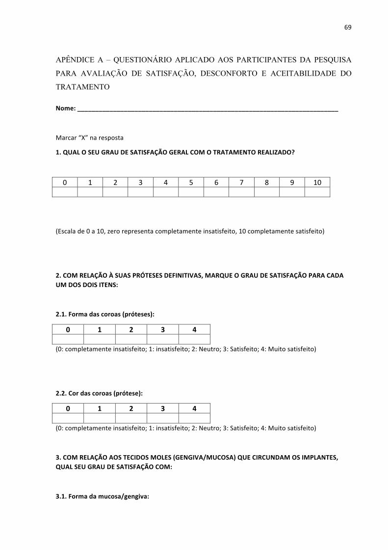
69
APÊNDICE A – QUESTIONÁRIO APLICADO AOS PARTICIPANTES DA PESQUISA
PARA AVALIAÇÃO DE SATISFAÇÃO, DESCONFORTO E ACEITABILIDADE DO
TRATAMENTO
Nome:_________________________________________________________________________
Marcar“X”naresposta
1.QUALOSEUGRAUDESATISFAÇÃOGERALCOMOTRATAMENTOREALIZADO?
0 1 2 3 4 5 6 7 8 9 10
(Escalade0a10,zerorepresentacompletamenteinsatisfeito,10completamentesatisfeito)
2.COMRELAÇÃOÀSUASPRÓTESESDEFINITIVAS,MARQUEOGRAUDESATISFAÇÃOPARACADAUMDOSDOISITENS:
2.1.Formadascoroas(próteses):
0 1 2 3 4
(0:completamenteinsatisfeito;1:insatisfeito;2:Neutro;3:Satisfeito;4:Muitosatisfeito)
2.2.Cordascoroas(prótese):
0 1 2 3 4
(0:completamenteinsatisfeito;1:insatisfeito;2:Neutro;3:Satisfeito;4:Muitosatisfeito)
3.COMRELAÇÃOAOSTECIDOSMOLES(GENGIVA/MUCOSA)QUECIRCUNDAMOSIMPLANTES,QUALSEUGRAUDESATISFAÇÃOCOM:
3.1.Formadamucosa/gengiva:

70
0 1 2 3 4
(0:completamenteinsatisfeito;1:insatisfeito;2:Neutro;3:Satisfeito;4:Muitosatisfeito)
3.2.Cordamucosa/gengiva
0 1 2 3 4
(0:completamenteinsatisfeito;1:insatisfeito;2:Neutro;3:Satisfeito;4:Muitosatisfeito)
4.QUALOGRAUDEDESCONFORTODOPROCEDIMENTO?
Nenhum Leve Moderado Severo
5.ACEITARIAMPASSARNOVAMENTEPELOPROCEDIMENTO?
Sim Talvez Dificilmente Não

71
ANEXO A – PARECER DO COMITÊ DE ÉTICA EM PESQUISA
UNIVERSIDADE FEDERAL DOCEARÁ/ PROPESQ
PARECER CONSUBSTANCIADO DO CEP
Pesquisador:
Título da Pesquisa:
Instituição Proponente:
Versão:CAAE:
AVALIAÇÃO RETROSPECTIVA DE OSTEOTOMIA EM BLOCO DO PROCESSOALVEOLAR APÓS A INSTALAÇÃO DOS IMPLANTES
Rafael Lima Verde Osterne
Departamento de Clínica Odontológica
258778916.0.0000.5054
Área Temática:
DADOS DO PROJETO DE PESQUISA
Número do Parecer: 1.757.767
DADOS DO PARECER
Trata-se de um estudo observacional e retrospectivo, que utiliza como amostra 11 pacientes que foramsubmetidos à reabilitação oral com implantes dentais, seguido de reposicionamento cirúrgico dos implantese prótese sobre implante em uma clínica particular da cidade de Fortaleza. Todos os dados necessários,exceto os dados coletados por questionário, serão coletados através de dados do prontuário, tomografiascomputadorizadas e fotografias clinicas armazenadas na clínica. Os dados serão coletados em um únicomomento de três períodos diferentes, antes do reposicionamento do bloco ósseo (T1); imediatamente apósa cirurgia de reposicionamento do bloco ósseo (T2); e após 6 meses da confecção da prótese definitiva (T3).Serão avaliados para o resultado do tratamento se houve ou não a perda de implantes envolvidos namovimentação e se a prótese final planejada pode ser instalada; e se ocorreram complicações quenecessitem remoção da prótese/implante após carga inicial. Os dados avaliados serão: avaliação doaumento ósseo vertical, escore da papila (papilla index)e avaliação da satisfação do paciente (através dequestionário).
Apresentação do Projeto:
Objetivo Primário:Avaliar retrospectivamente resultado de aumento ósseo vertical, estética gengival e satisfação do
Objetivo da Pesquisa:
Financiamento PróprioPatrocinador Principal:
60.430-275
(85)3366-8344 E-mail: [email protected]
Endereço:Bairro: CEP:
Telefone:
Rua Cel. Nunes de Melo, 1000Rodolfo Teófilo
UF: Município:CE FORTALEZA
Página 01 de 03

72
UNIVERSIDADE FEDERAL DOCEARÁ/ PROPESQ
Continuação do Parecer: 1.757.767
paciente após osteotomia em bloco do processo alveolar para reposicionamento de implantes dentários.Objetivo Secundário:Avaliar aumento ósseo vertical de processo alveolar após osteotomia em bloco do processo alveolar atravésde tomografia computadorizada.Avaliar estabilidade do aumento ósseo vertical.Avaliar resultado estético através de fotografias antes e após o tratamento.Avaliar satisfação do paciente com o resultado final do tratamento.
Riscos: Segundo os autores, o tipo de estudo a ser realizado apresenta um risco mínimo aos seusenvolvidos, como a possibilidade de exposição de informações pessoais e uso inadequado de seus examese resultados de exames. Entretanto, para evitar esse risco somente o pesquisador terá acesso aos dados decada participante. Não é de interesse da pesquisa divulgar os dados individuais dos participantes, e sim osresultados gerais, esperados nos objetivos da pesquisa. Em nenhum momento da pesquisa os participantesserão identificados de forma individual.Benefícios: Os autores informam que os benefícios esperados com a pesquisa são sobre os resultadosclínicos e radiográficos do uso de reposicionamento cirúrgico de implantes dentais por osteotomias emobilização do bloco ósseo.
Avaliação dos Riscos e Benefícios:
A pesquisa é pertinente considerando a importância da avaliação de uma opção terapêutica à casos comdeficiência vertical de processo alveolar.
Comentários e Considerações sobre a Pesquisa:
Os termos de apresentação obrigatória foram apresentados e estão adequados.Considerações sobre os Termos de apresentação obrigatória:
O projeto foi devidamente adequado após correção das pendências existentes, por isso proponho que oprojeto deve ser aprovado.
Conclusões ou Pendências e Lista de Inadequações:
Considerações Finais a critério do CEP:
Este parecer foi elaborado baseado nos documentos abaixo relacionados:Tipo Documento Arquivo Postagem Autor Situação
Informações PB_INFORMAÇÕES_BÁSICAS_DO_P 22/08/2016 Aceito
60.430-275
(85)3366-8344 E-mail: [email protected]
Endereço:Bairro: CEP:
Telefone:
Rua Cel. Nunes de Melo, 1000Rodolfo Teófilo
UF: Município:CE FORTALEZA
Página 02 de 03
73
UNIVERSIDADE FEDERAL DOCEARÁ/ PROPESQ
Continuação do Parecer: 1.757.767
FORTALEZA, 03 de Outubro de 2016
FERNANDO ANTONIO FROTA BEZERRA(Coordenador)
Assinado por:
Básicas do Projeto ETO_775994.pdf 11:41:09 AceitoOutros Respostaprojeto.pdf 22/08/2016
11:40:47Rafael Lima VerdeOsterne
Aceito
Declaração deInstituição eInfraestrutura
AUTORIZACAOdadisciplina.pdf 22/08/201611:38:48
Rafael Lima VerdeOsterne
Aceito
Projeto Detalhado /BrochuraInvestigador
projetocommodificacoes.pdf 22/08/201611:37:33
Rafael Lima VerdeOsterne
Aceito
Cronograma CRONOGRAMAnovo.pdf 22/08/201611:36:07
Rafael Lima VerdeOsterne
Aceito
TCLE / Termos deAssentimento /Justificativa deAusência
TCLEFinal.docx 22/08/201611:35:51
Rafael Lima VerdeOsterne
Aceito
Folha de Rosto folhaderosto.pdf 16/08/201615:38:04
Rafael Lima VerdeOsterne
Aceito
Outros LattesRenato.pdf 15/08/201617:08:52
Rafael Lima VerdeOsterne
Aceito
Outros LattesRAFAEL.pdf 15/08/201617:08:07
Rafael Lima VerdeOsterne
Aceito
Outros CARTACEPMODELOFINAL.pdf 15/08/201617:03:14
Rafael Lima VerdeOsterne
Aceito
Outros TERMO_DE_COMPROMISSO_PARA_UTILIZACAO_DEfinal.pdf
15/08/201617:01:43
Rafael Lima VerdeOsterne
Aceito
Orçamento ORCAMENTOFINAL.pdf 15/08/201616:58:23
Rafael Lima VerdeOsterne
Aceito
Cronograma CRONOGRAMA_MODELOfinal.pdf 15/08/201616:57:05
Rafael Lima VerdeOsterne
Aceito
Declaração dePesquisadores
_DECLARACAO_PESQUISADORES_MODELOFINALfinal.pdf
15/08/201616:56:34
Rafael Lima VerdeOsterne
Aceito
Situação do Parecer:AprovadoNecessita Apreciação da CONEP:Não
60.430-275
(85)3366-8344 E-mail: [email protected]
Endereço:Bairro: CEP:
Telefone:
Rua Cel. Nunes de Melo, 1000Rodolfo Teófilo
UF: Município:CE FORTALEZA
Página 03 de 03
74
ANEXO B – TERMO DE CONSENTIMENTO LIVRE E ESCLARECIDO
TERMO DE CONSENTIMENTO LIVRE E ESCLARECIDO (TCLE)
Você está sendo convidado pelo pesquisador Rafael Lima Verde Osterne como participante da pesquisa intitulada “AVALIAÇÃO RETROSPECTIVA DE OSTEOTOMIA EM BLOCO DO PROCESSO ALVEOLAR APÓS A INSTALAÇÃO DOS IMPLANTES”. Você não deve participar contra a sua vontade. Leia atentamente as informações abaixo e faça qualquer pergunta que desejar, para que todos os procedimentos desta pesquisa sejam esclarecidos.
OBJETIVO: O objetivo que nos leva a estudar o problema é avaliar o resultado geral, estético e satisfação do paciente com o tratamento de reposicionamento de implantes através de osteotomias do processo alveolar.
PARTICIPAÇÃO NA PESQUISA: Ao participar desta pesquisa será aplicado o questionário, não identificado, e será necessário o preenchimento deste termo de consentimento livre e esclarecido.
Lembramos que os dados obtidos deste questionário será utilizado exclusivamente na presente pesquisa. A sua participação é voluntária, você tem a liberdade de não querer participar, e pode desistir, em qualquer momento, mesmo após ter iniciado a avaliação sem nenhum prejuízo para você.
RISCOS E DESCONFORTOS: O tipo de estudo a ser realizado apresenta um risco mínimo aos seus envolvidos, como a possibilidade de exposição de informações pessoais e uso inadequado de seus exames e resultados de exames. Entretanto, para evitar esse risco somente o pesquisador terá acesso aos dados de cada participante. Não é de interesse da pesquisa divulgar os dados individuais dos participantes, e sim os resultados gerais, esperados nos objetivos da pesquisa. Em nenhum momento da pesquisa os participantes serão identificados de forma individual.
BENEFÍCIOS: Os benefícios esperados com a pesquisa são sobre os resultados clínicos e radiográficos do uso de reposicionamento cirúrgico de implantes dentais por osteotomias e mobilização do bloco ósseo.
CONFIDENCIALIDADE: Todas as informações que o(a) Sr.(a) nos fornecer serão utilizadas somente para esta pesquisa. Suas respostas ficarão em segredo e o seu nome não aparecerá em nenhum lugar do questionário nem quando os resultados forem apresentados.
RESSARCIMENTO DAS DESPESAS: A participação no estudo não acarretará custos para você e não será disponível nenhuma compensação financeira adicional.
CONCORDÂNCIA NA PARTICIPAÇÃO: Se o(a) Sr.(a) estiver de acordo em participar deve preencher e assinar o Termo de Consentimento Pós-esclarecido que se segue, e receberá uma via deste Termo e a outra ficará com o pesquisador.
Lembramos que os dados obtidos deste questionário será utilizado exclusivamente na presente pesquisa. A sua participação é voluntária, você tem a liberdade de não querer participar, e pode desistir, em qualquer momento, mesmo após ter iniciado a avaliação sem nenhum prejuízo para você.
75
Garantimos que as informações conseguidas através da sua participação não permitirão a identificação da sua pessoa, exceto aos responsáveis pela pesquisa, e que a divulgação das mencionadas informações só será feita entre os profissionais estudiosos do assunto.
Endereço d(os, as) responsável(is) pela pesquisa:
Nome: Rafael Lima Verde Osterne Instituição: Curso de Odontologia
Endereço: Rua Monsenhor Furtado, 1273, Rodolfo Teófilo, Fortaleza - CE Brasil
CEP 60430-355, Clínica 05, Curso de Odontologia Telefones para contato: 999281718, 33668413 ATENÇÃO: Se você tiver alguma consideração ou dúvida, sobre a sua participação na pesquisa, entre em contato com o Comitê de Ética em Pesquisa da UFC/PROPESQ – Rua Coronel Nunes de Melo, 1000 - Rodolfo Teófilo, fone: 3366-8344. (Horário: 08:00-12:00 horas de segunda a sexta-feira). O CEP/UFC/PROPESQ é a instância da Universidade Federal do Ceará responsável pela avaliação e acompanhamento dos aspectos éticos de todas as pesquisas envolvendo seres humanos. O abaixo assinado _________________________,___anos, RG:________, declara que é de livre e espontânea vontade que está como participante de uma pesquisa. Eu declaro que li cuidadosamente este Termo de Consentimento Livre e Esclarecido e que, após sua leitura, tive a oportunidade de fazer perguntas sobre o seu conteúdo, como também sobre a pesquisa, e recebi explicações que responderam por completo minhas dúvidas. E declaro, ainda, estar recebendo uma via assinada deste termo.
Fortaleza, ____/____/___
Nome do participante da pesquisa Data Assinatura Nome do pesquisador Data Assinatura Nome da testemunha Data Assinatura (se o voluntário não souber ler) Nome do profissional Data Assinatura que aplicou o TCLE
76
ANEXO C – CERTIFICADO DE REVISÃO DA LÍNGUA INGLESA
EDIT
ORI
AL C
ERTI
FICA
TETh
is do
cum
ent c
ertif
ies
that
the
man
uscr
ipt l
isted
bel
ow w
as e
dite
d fo
r pro
per E
nglis
h la
ngua
ge, g
ram
mar
,pu
nctu
atio
n, s
pellin
g, a
nd o
vera
ll sty
le b
y on
e or
mor
e of
the
high
ly qu
alifie
d na
tive
Engl
ish s
peak
ing
edito
rs a
t Am
erica
n Jo
urna
l Exp
erts
.
Man
uscr
ipt t
itle:
Alte
rnat
ive D
istra
ctio
n O
steo
gene
sis T
echn
ique
Afte
r Im
plan
t Pla
cem
ent f
or A
lveol
ar R
idge
Aug
men
tatio
n of
the
Max
illa
Auth
ors:
Rena
to L
uiz
Mai
a NO
GUE
IRA;
Raf
ael L
ima
Verd
e O
STER
NE; R
icard
o Te
ixeira
ABR
EU, P
helyp
e M
aia
ARAÚ
JO
Date
Issu
ed:
Augu
st 1
0, 2
016
Certi
ficat
e Ve
rific
atio
n Ke
y:EB
87-5
E3A-
BDEF
-A8C
A-D2
53
This
certi
ficat
e m
ay b
e ve
rifie
d at
www
.aje
.com
/cer
tifica
te. T
his
docu
men
t cer
tifie
s th
at th
e m
anus
crip
t list
ed a
bove
was
edi
ted
for
prop
er E
nglis
h la
ngua
ge,
gram
mar
, pun
ctua
tion,
spe
lling,
and
ove
rall
style
by
one
or m
ore
of th
e hi
ghly
qual
ified
nativ
e En
glish
spe
akin
g ed
itors
at A
mer
ican
Jour
nal E
xper
ts. N
eith
erth
e re
sear
ch c
onte
nt n
or t
he a
utho
rs'
inte
ntio
ns w
ere
alte
red
in a
ny w
ay d
urin
g th
e ed
iting
proc
ess.
Doc
umen
ts r
ecei
ving
this
certi
ficat
ion
shou
ld b
eEn
glish
-read
y fo
r pu
blica
tion;
how
ever
, the
aut
hor
has
the
abilit
y to
acc
ept o
r re
ject
our
sug
gest
ions
and
cha
nges
. To
verif
y th
e fin
al A
JE e
dite
d ve
rsio
n,pl
ease
visi
t ou
r ve
rifica
tion
page
. If
you
have
any
que
stio
ns o
r co
ncer
ns a
bout
thi
s ed
ited
docu
men
t, pl
ease
con
tact
Am
erica
n Jo
urna
l Ex
perts
at
supp
ort@
aje.
com
.
Amer
ican
Jour
nal E
xper
ts p
rovid
es a
ran
ge o
f ed
iting,
tra
nsla
tion
and
man
uscr
ipt
serv
ices
for
rese
arch
ers
and
publ
isher
s ar
ound
the
wor
ld.
Our
top
-qua
lity P
hD e
dito
rs a
re a
ll na
tive
Engl
ishsp
eake
rs f
rom
Am
erica
's to
p un
ivers
ities.
Our
edi
tors
com
e fro
m n
early
eve
ry r
esea
rch
field
and
pos
sess
the
hig
hest
qua
lifica
tions
to
edit
rese
arch
man
uscr
ipts
writ
ten
by n
on-n
ative
Eng
lish
spea
kers
. For
mor
e in
form
atio
n ab
out o
ur c
ompa
ny, s
ervic
es a
nd p
artn
er d
iscou
nts,
ple
ase
visit
www.
aje.
com
.
77
LANGUAGE EDITING
CERTIFICATEThis document certifies that the manuscript listed below was edited for proper English language, grammar, punctuation,
spelling, and overall style by one or more of the highly qualified native English speaking editors at Wiley Editing Services.
Manuscript title:A retrospective study of an alternative Osteodistraction Technique after implant placement in
maxillary esthetic region
Authors:Rafael Lima Verde OSTERNE; Renato Luiz Maia NOGUEIRA; Ricardo Teixeira ABREU;
Roberta Barroso CAVALCANTE
Date Issued:November 23, 2016
Certificate Verification Key:BA19-AD31-EDFD-307B-527E
This certificate may be verified athttps://secure.wileyeditingservices.com/certificate. This documentcertifies that the manuscript listed above was edited for proper Englishlanguage, grammar, punctuation, spelling, and overall style. Neitherthe research content nor the authors' intentions were altered in any wayduring the editing process. Documents receiving this certificationshould be English-ready for publication; however, the author has theability to accept or reject our suggestions and changes. If you have anyquestions or concerns about this document or certification, pleasecontact [email protected].
Wiley Publishing Services is a service of Wiley Publishing. Wiley's Scientific, Technical, Medical, and Scholarly (STMS) business serves the world's researchand scholarly communities, and is the largest publisher for professional and scholarly societies. Wiley is committed to providing high quality services forresearchers. To find out more about Wiley Editing Services, visit wileyeditingservices.com. To learn more about our other author services provided by WileyPublishing, visit authorservices.wiley.com.