Embed Size (px)
Citation preview

73R E V I S T A P O R T U G U E S A D E I M U N O A L E R G O L O G I A
ARTIGO DE REVISÃO
RESUMO
O mecanismo imunológico subjacente às doencas alérgicas mediadas por IgE é a hipersensibilidade do tipo I, em que os mastócitos e os basófilos são as células efectoras. Esta reacção é reproduzida in vitro no teste de libertação de histamina e outros mediadores e no teste de activação dos basófilos. Estas são ferramentas muito úteis, não só no diagnóstico de diversas doenças alérgicas e seguimento de doentes submetidos a imunoterapia específica, mas também ao nível da investigação dos mecanismos imunológicos de alergia. Ambas as técnicas são discutidas no presente artigo.
Palavras-chave: alergia, alergénio, basófilo, teste de activação dos basófilos, desgranulação, libertação de histamina, teste de desgranulação dos basófilos, citometria de fluxo.
Estudo in vitro dos basófilos é uma ferramenta diagnóstica e de investigação útil em alergologia
Basophil assays are useful diagnostic and research tools in allergology
Alexandra Santos1,2,3, Bernhard Gibbs4, Alick Stephens1, Victor Turcanu1, Gideon Lack1
1 Department of Pediatric Allergy, Division of Asthma, Allergy & Lung Biology, King’s College London – MRC & Asthma UK Centre in Allergic Mechanisms of Asthma, London, United Kingdom
2 Serviço de Imunoalergologia, Hospitais da Universidade de Coimbra, Coimbra, Portugal3 Gulbenkian Programme for Advanced Medical Education 4 Medway School of Pharmacy, University of Kent at Medway, United Kingdom
R e v P o r t I m u n o a l e r g o l o g i a 2 0 1 1 ; 1 9 ( 2 ) : 7 3 - 8 3
Data de recepção / Received in: 31/12/2010
Data de aceitação / Accepted for publication in: 20/06/2011
Imuno (19) 2 - Miolo 2ª PROVA PT.indd 73Imuno (19) 2 - Miolo 2ª PROVA PT.indd 73 15-07-2011 11:45:4615-07-2011 11:45:46

74R E V I S T A P O R T U G U E S A D E I M U N O A L E R G O L O G I A
Alexandra Santos, Bernhard Gibbs, Alick Stephens, Victor Turcanu, Gideon Lack
INTRODUCTION
The immunologic mechanism underlying IgE -mediated allergic diseases is type I hypersensitivity. In sensitised patients, allergen -specifi c IgE antibodies bind to high-
-affi nity IgE receptors (FcεRI) on the surface of mast cells and basophils for relatively long periods of time. On subsequent exposure, allergens bind to IgE on the surface of mast cells and basophils which leads to cross -linking of FcεRI receptors and triggering of complex intracellular signalling cascades. These culminate in the release of both pre -formed mediators (e.g. histamine, proteoglycans, serine proteases) and de novo synthesis of cytokines (e.g. IL -3, IL -4, IL -13) as well as leuko-trienes, all of which contribute to allergic infl ammation1.
The IgE -mediated allergic reaction has been repro-duced in vitro, both as a diagnostic and as a research tool, using mast cells and basophils. Basophils have the advantage of being easily available as they can be readily isolated from peripheral blood. Traditionally, functional in vitro tests based on allergen -induced activation of IgE -bearing basophils have focused on the mediators released by these cells after stimulation with allergen2. However, in parallel with the release of vasoactive mediators, basophils upregulate the expression of different activation markers on their surface, which can be evaluated by flow cytometry – this is the so -called basophil activation test (BAT)3.
This article aims to give an overview of the two main types of functional assays used to study IgE -mediated ba-
sophil activation in vitro: mediator release and basophil activation assays.
MEDIATOR RELEASE ASSAYS
When IgE -receptors on basophils are cross -linked by an allergen, the cells undergo degranulation and release bioactive mediators. Histamine is one of the most impor-tant mediators, as it is responsible for many of the symp-toms in the immediate phase of the allergic response, and can be easily measured in vitro in the supernatants of ba-sophils previously stimulated by allergen.
The primary source of cells in this experimental setting can be whole blood, dextran - or Ficoll -isolated leukocytes and basophils that have been further purified by negative selection using magnetic cell -sorting techniques4. Experi-mental designs using passive sensitisation5, i.e. stripping of native membrane -bound immunoglobulins and preincuba-tion of basophils with patients’ sera before stimulation with allergen, are particularly interesting for mechanistic studies. When collecting the blood for this kind of experiment, it is recommended that the donors have not taken drugs or food a few hours before blood donation and that blood is collected to a syringe or tube containing anticoagulant. The appropriate anticoagulant to be used depends on the cho-sen laboratory protocol. Blood should be processed as soon as possible, preferably within 4 hours of collection.
ABSTRACT
The immunological mechanism of IgE -mediated allergic diseases is type I hypersensitivity, where basophils and mast cells are the main effector cells. This reaction is reproduced in vitro in basophil mediator release and basophil activation assays. These are useful tools not only for the diagnosis of various allergic diseases and follow -up of patients undergoing allergen -specific immuno-therapy, but also in research into the mechanisms of allergy. Both basophil assays are discussed in this article.
Key-words: Allergy, allergen, basophil, basophil activation test, histamine release, basophil degranulation test, flow cytometry.
Imuno (19) 2 - Miolo 2ª PROVA PT.indd 74Imuno (19) 2 - Miolo 2ª PROVA PT.indd 74 15-07-2011 11:45:4615-07-2011 11:45:46
75R E V I S T A P O R T U G U E S A D E I M U N O A L E R G O L O G I A
ESTUDO IN VITRO DOS BASÓFILOS É UMA FERRAMENTA DIAGNÓSTICA E DE INVESTIGAÇÃO ÚTIL EM ALERGOLOGIA / ARTIGO DE REVISÃO
Crude allergen extracts or purified/recombinant al-lergens may be used for cell stimulation. For each donor, different allergen concentrations should be tested, usually in 10 -fold serial dilutions, as the sensitivity of the basophils to specific allergen stimulation varies among patients. As positive controls, anti -IgE should be used to gauge IgE--mediated cell activation and formyl -methionyl -leucyl--phenylalanine (fMLP), a chemotactic stimulus which acti-vates basophils through an IgE -independent mechanism, as a control for functional cell viability. As a negative control, cells are stimulated with buffer alone. Degranulation is optimal at 37˚C and occurs within 30 minutes6 in the presence but not in the absence of extracellular calcium; thus a calcium -containing buffer must be used.
The histamine concentration in the supernatants can be measured using different techniques, namely radio--immunoassay (RIA), enzyme -linked immunosorbent assay (ELISA) or spectrofluorometric assays, which measure the fluorescence of an adduct formed by reacting histamine with o -phthaldehyde6,7. Histamine release is usually ex-pressed as a percentage of the total basophil histamine content, which is determined by the sum of intra and ex-tracellular histamine contents (where intracellular hista-mine contents are liberated by lysis of the cell pellets).
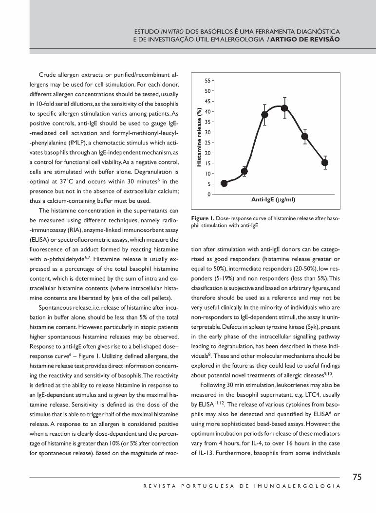
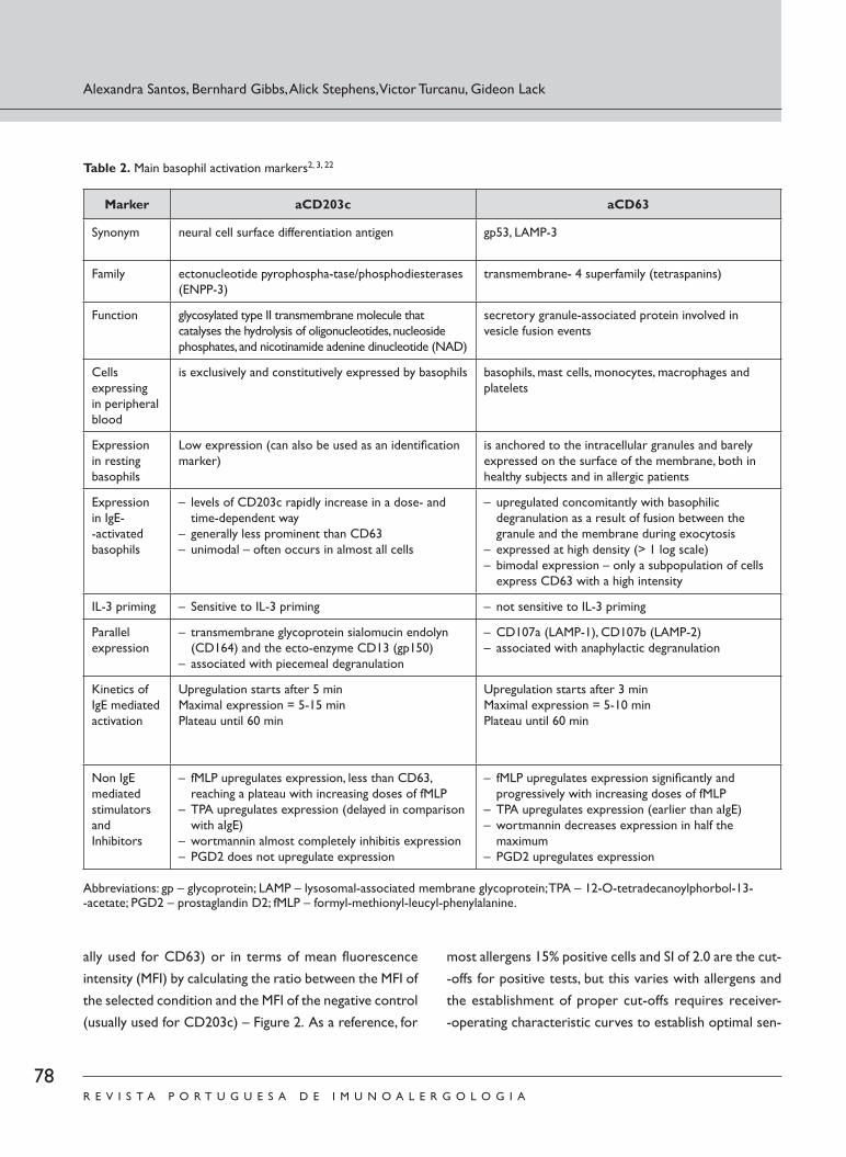
Spontaneous release, i.e. release of histamine after incu-bation in buffer alone, should be less than 5% of the total histamine content. However, particularly in atopic patients higher spontaneous histamine releases may be observed. Response to anti -IgE often gives rise to a bell -shaped dose–response curve6 – Figure 1. Utilizing defined allergens, the histamine release test provides direct information concern-ing the reactivity and sensitivity of basophils. The reactivity is defined as the ability to release histamine in response to an IgE -dependent stimulus and is given by the maximal his-tamine release. Sensitivity is defined as the dose of the stimulus that is able to trigger half of the maximal histamine release. A response to an allergen is considered positive when a reaction is clearly dose -dependent and the percen-tage of histamine is greater than 10% (or 5% after correction for spontaneous release). Based on the magnitude of reac-
tion after stimulation with anti -IgE donors can be catego-rized as good responders (histamine release greater or equal to 50%), intermediate responders (20 -50%), low res-ponders (5 -19%) and non responders (less than 5%). This classification is subjective and based on arbitrary figures, and therefore should be used as a reference and may not be very useful clinically. In the minority of individuals who are non -responders to IgE -dependent stimuli, the assay is unin-terpretable. Defects in spleen tyrosine kinase (Syk), present
in the early phase of the intracellular signalling pathway leading to degranulation, has been described in these indi-viduals8. These and other molecular mechanisms should be explored in the future as they could lead to useful findings about potential novel treatments of allergic diseases9,10.
Following 30 min stimulation, leukotrienes may also be measured in the basophil supernatant, e.g. LTC4, usually by ELISA11,12. The release of various cytokines from baso-phils may also be detected and quantified by ELISA6 or using more sophisticated bead -based assays. However, the optimum incubation periods for release of these mediators vary from 4 hours, for IL -4, to over 16 hours in the case of IL -13. Furthermore, basophils from some individuals
Figure 1. Dose-response curve of histamine release after baso-phil stimulation with anti-IgE
Imuno (19) 2 - Miolo 2ª PROVA PT.indd 75Imuno (19) 2 - Miolo 2ª PROVA PT.indd 75 15-07-2011 11:45:4615-07-2011 11:45:46
76R E V I S T A P O R T U G U E S A D E I M U N O A L E R G O L O G I A
have high constitutive expressions of IL -4 (i.e. preformed and not de novo synthesised) which may also be released within minutes of stimulation13.
Another mediator measured in the supernatant of mast cells and various cell lines, such as LUVA, LAD -2 and RBL cells, to detect degranulation is β -hexosaminidase14 -16. This is a granule -stored enzyme, an exoglycosidase, with optimal activity at low pH, and is secreted in parallel with histamine. The measurement of its activity has been extensively used to monitor mast cell and basophil degranulation by adding fluorogenic β -hexosaminidase substrate at low pH and incubating at 37°C for 60 mins. This reaction is terminated by changing the pH and the colour due to the substrate hydrolysis is measured by fluorometry. The results are ex-pressed as percentages of the total β -hexosaminidase content of the cells, which is determined by summing the extracellular release and the release after cell lysis.
BASOPHIL ACTIVATION TEST
Using a similar experimental setting, whilst the super-natant may be used for measurement of mediator release, the cells may be analysed by flow cytometry to evaluate the expression of basophil activation markers2, 3. This type of experiment may be performed using mixed cell popula-tions (e.g. PBMC, even whole blood) or purified basophils. In any case, identification markers have to be used to gate on basophils and detect the expression of the activation markers in that selected population.
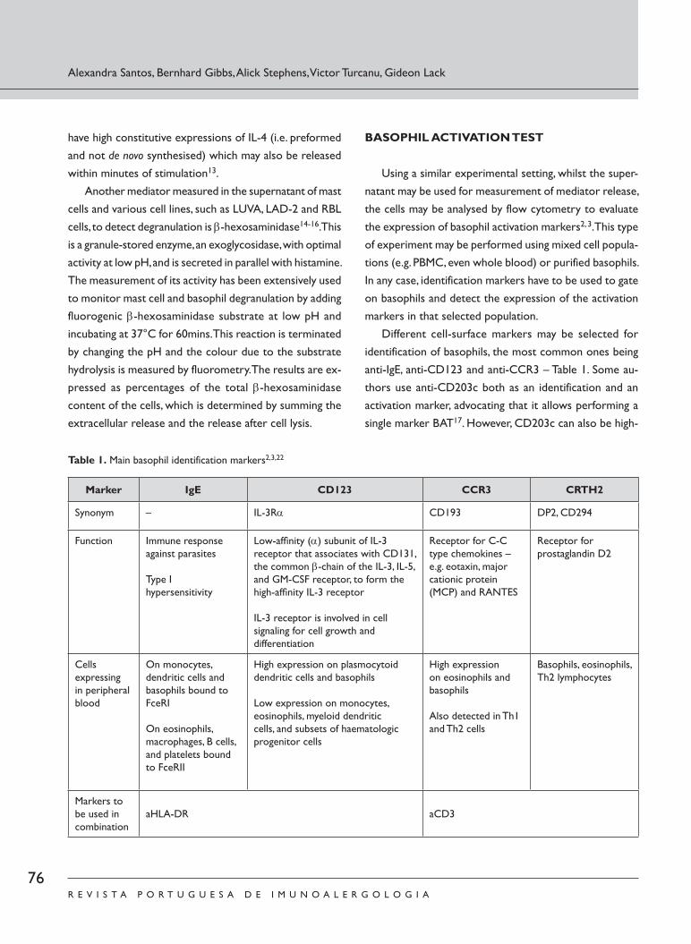
Different cell -surface markers may be selected for identification of basophils, the most common ones being anti -IgE, anti -CD123 and anti -CCR3 – Table 1. Some au-thors use anti -CD203c both as an identification and an activation marker, advocating that it allows performing a single marker BAT17. However, CD203c can also be high-
Alexandra Santos, Bernhard Gibbs, Alick Stephens, Victor Turcanu, Gideon Lack
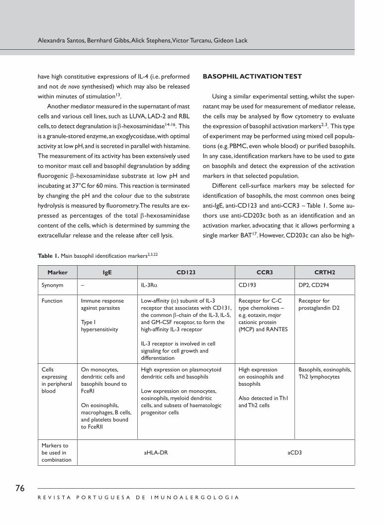
Table 1. Main basophil identification markers2,3,22
Marker IgE CD123 CCR3 CRTH2
Synonym – IL -3Rα CD193 DP2, CD294
Function Immune response against parasites
Type I hypersensitivity
Low -affi nity (α) subunit of IL -3 receptor that associates with CD131, the common β -chain of the IL -3, IL -5, and GM -CSF receptor, to form the high -affi nity IL -3 receptor
IL -3 receptor is involved in cell signaling for cell growth and differentiation
Receptor for C -C type chemokines – e.g. eotaxin, major cationic protein (MCP) and RANTES
Receptor for prostaglandin D2
Cells expressing in peripheral blood
On monocytes, dendritic cells and basophils bound to FceRI
On eosinophils, macrophages, B cells, and platelets bound to FceRII
High expression on plasmocytoid dendritic cells and basophils
Low expression on monocytes, eosinophils, myeloid dendritic cells, and subsets of haematologic progenitor cells
High expression on eosinophils and basophils
Also detected in Th1 and Th2 cells
Basophils, eosinophils, Th2 lymphocytes
Markers to be used in combination
aHLA -DR aCD3
Imuno (19) 2 - Miolo 2ª PROVA PT.indd 76Imuno (19) 2 - Miolo 2ª PROVA PT.indd 76 15-07-2011 11:45:4615-07-2011 11:45:46
77R E V I S T A P O R T U G U E S A D E I M U N O A L E R G O L O G I A
ly expressed in basophils following Ficoll -mediated isola-tion and by priming factors such as IL -3, which by them-selves do not cause substantial degranulation.
In the peripheral blood, IgE is detected on dendritic cells and basophils, which express the high affinity IgE re-ceptor (FcεRI), and also on eosinophils, monocytes, ma-crophages, B cells and platelets, which express the low af-finity IgE receptor (FcεRII or CD23). The expression of IgE on the surface of basophils varies with the atopic status of the patient, increasing in atopic patients. Labelling baso-phils with an anti -IgE antibody can further activate the cells, which can be reduced by fixing, cooling and adding EDTA--containing buffer to the cells before staining. CD123 is the low affinity subunit of the IL -3 receptor, which is ex-pressed in high levels on plasmocytoid dendritic cells and basophils, and in low levels on monocytes, eosinophils, myeloid dendritic cells and subsets of hematologic pro-genitor cells. Additional staining with anti -HLA -DR dis-criminates between HLA -DR negative basophils and HLA--DR positive dendritic cells and monocytes. One of the advantages of identifying basophils with anti -CD123 and anti -HLA -DR is that their expression is not so much in-fluenced by the allergic status of the donor as anti -IgE. CCR3 is the receptor for C -C type chemokines (e.g. eo-taxin, MCP and RANTES). It is highly expressed on baso-phils and eosinophils but also on Th1 and Th2 cells. Thus, an anti -CD3 marker should be used in combination with it to exclude the CD3 positive T cells. Haussmann et al18 have compared the main three basophil identification methods and concluded that CD123/HLA -DR and CCR3 are the most accurate, with CCR3 having the advantages of being most constant with the atopic background of the patient and of identifying basophils with a single marker. However, CCR3 has the disadvantage of being downregu-lated after basophil activation, which does not occur with CD123/HLA -DR.
After stimulation with allergen, the expression of dif-ferent proteins is upregulated on the surface of basophils. Although the intracellular pathways driving the upregula-tion of these markers are not completely understood, they
seem to form two distinct groups of markers that are upregulated concomitantly: one including CD63, CD107a and CD107b and another CD203c, CD13 and CD16419. The most studied and widely used are CD6320 and CD203c17, which are proteins expressed on the membrane of the granules that fuse with the plasmatic membrane of the basophils during degranulation, increasing their expres-sion on the surface of the cell21 – Table 2.
These markers behave differently in their upregulation profiles22, 23. The increase in their expression in response to specific activators and inhibitors follows different kine-tics and seems to be directed through alternative signal transduction pathways. The expression of CD203c is low in resting basophils that have not been primed with IL -3 and increases after activation, whilst CD63 is not expressed in resting cells. The upregulation of CD63 is bimodal, with only a subpopulation of basophils expressing it, whilst CD203c expression is less prominent but often genera-lised to the whole cell population, even to cells that did not express CD63.
Dose -response curves with different agonists and in-hibitors show dissociation between the two activation markers: CD203c is associated with the low -dose events of chemotaxis and CD63 is associated with degranulation19. Different studies have suggested that CD63 may reflect anaphylactic degranulation whilst CD203c reflects piece-meal degranulation. MacGlashan24 hypothesised in a recent published study that this may be the reason why neither CD63 nor CD203c strictly reflect histamine release. His-tamine release measured in the cell supernatant is an ave-rage of what occurs in a heterogeneous population of ba-sophils, being a result of the sum between the two pathways of basophil activation. This highlights the advantage of us-ing flow cytometry to study basophil activation as it gives more complete and detailed information about the behav-iour of individual cells after stimulation with allergen.
The results of BAT may be shown for each condition in dotplots or histograms and differences in comparison with controls may be determined in terms of percentage of basophils expressing the defined activation marker (usu-
ESTUDO IN VITRO DOS BASÓFILOS É UMA FERRAMENTA DIAGNÓSTICA E DE INVESTIGAÇÃO ÚTIL EM ALERGOLOGIA / ARTIGO DE REVISÃO
Imuno (19) 2 - Miolo 2ª PROVA PT.indd 77Imuno (19) 2 - Miolo 2ª PROVA PT.indd 77 15-07-2011 11:45:4615-07-2011 11:45:46
78R E V I S T A P O R T U G U E S A D E I M U N O A L E R G O L O G I A
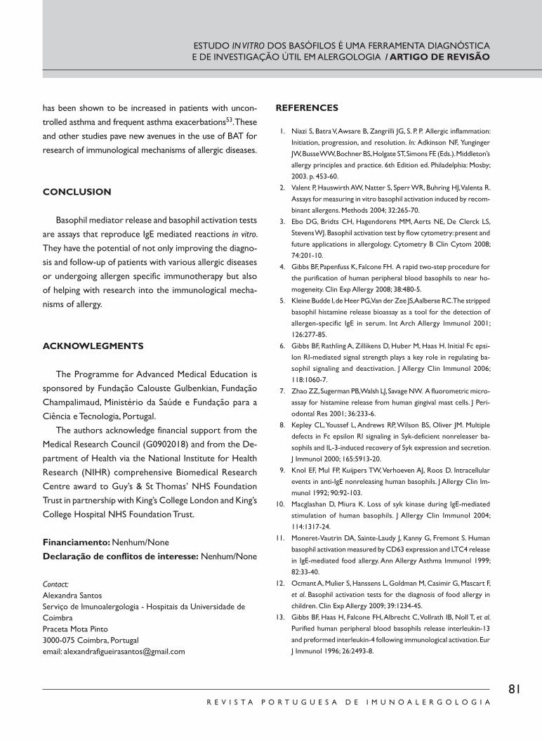
ally used for CD63) or in terms of mean fluorescence intensity (MFI) by calculating the ratio between the MFI of the selected condition and the MFI of the negative control (usually used for CD203c) – Figure 2. As a reference, for
most allergens 15% positive cells and SI of 2.0 are the cut--offs for positive tests, but this varies with allergens and the establishment of proper cut -offs requires receiver--operating characteristic curves to establish optimal sen-
Alexandra Santos, Bernhard Gibbs, Alick Stephens, Victor Turcanu, Gideon Lack
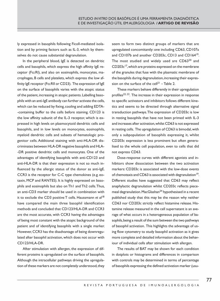
Table 2. Main basophil activation markers2, 3, 22
Marker aCD203c aCD63
Synonym neural cell surface differentiation antigen gp53, LAMP -3
Family ectonucleotide pyrophospha -tase/phosphodiesterases (ENPP -3)
transmembrane - 4 superfamily (tetraspanins)
Function glycosylated type II transmembrane molecule that catalyses the hydrolysis of oligonucleotides, nucleoside phosphates, and nicotinamide adenine dinucleotide (NAD)
secretory granule -associated protein involved in vesicle fusion events
Cells expressing in peripheral blood
is exclusively and constitutively expressed by basophils basophils, mast cells, monocytes, macrophages and platelets
Expression in resting basophils
Low expression (can also be used as an identifi cation marker)
is anchored to the intracellular granules and barely expressed on the surface of the membrane, both in healthy subjects and in allergic patients
Expression in IgE--activated basophils
– levels of CD203c rapidly increase in a dose - and time -dependent way
– generally less prominent than CD63– unimodal – often occurs in almost all cells
– upregulated concomitantly with basophilic degranulation as a result of fusion between the granule and the membrane during exocytosis
– expressed at high density (> 1 log scale)– bimodal expression – only a subpopulation of cells
express CD63 with a high intensity
IL -3 priming – Sensitive to IL -3 priming – not sensitive to IL -3 priming
Parallel expression
– transmembrane glycoprotein sialomucin endolyn (CD164) and the ecto -enzyme CD13 (gp150)
– associated with piecemeal degranulation
– CD107a (LAMP -1), CD107b (LAMP -2)– associated with anaphylactic degranulation
Kinetics of IgE mediated activation
Upregulation starts after 5 minMaximal expression = 5 -15 minPlateau until 60 min
Upregulation starts after 3 minMaximal expression = 5 -10 minPlateau until 60 min
Non IgE mediated stimulators andInhibitors
– fMLP upregulates expression, less than CD63, reaching a plateau with increasing doses of fMLP
– TPA upregulates expression (delayed in comparison with aIgE)
– wortmannin almost completely inhibitis expression– PGD2 does not upregulate expression
– fMLP upregulates expression signifi cantly and progressively with increasing doses of fMLP
– TPA upregulates expression (earlier than aIgE)– wortmannin decreases expression in half the
maximum– PGD2 upregulates expression
Abbreviations: gp – glycoprotein; LAMP – lysosomal -associated membrane glycoprotein; TPA – 12 -O -tetradecanoylphorbol -13--acetate; PGD2 – prostaglandin D2; fMLP – formyl -methionyl -leucyl -phenylalanine.
Imuno (19) 2 - Miolo 2ª PROVA PT.indd 78Imuno (19) 2 - Miolo 2ª PROVA PT.indd 78 15-07-2011 11:45:4715-07-2011 11:45:47

79R E V I S T A P O R T U G U E S A D E I M U N O A L E R G O L O G I A
sitivity and specificity. The interpretation of results should always be tailored to each individual case. The response in a time -course and dose -response manner is an additional
important sign of allergen -mediated basophil activation.Short incubation with IL -3 may increase the sensitivity
of the assay and has been used in some studies25. IL -3 causes nonspecific increase in CD203c expression but not CD63. However, it may be a cause for false positive re-sults26, one reason for that being the concentrations of IL -3 that are used which are much higher than the physi-ological ones.
The molecular mechanisms governing basophil activa-tion are complex and not entirely clarified. Traditionally, analysis of signalling is based on western blot and ELISA techniques, which represent a mean value for the total isolated cell population6. Recently, a proof of concept was
provided that flow cytometry may be used to quantify phosphorilation of p38 -MAPK in basophils27. Similar methods may be used to evaluate consecutive phosphori-
lation of the proteins involved, as has been done for other cells and signalling pathways. Flow cytometry offers various advantages over the traditional techniques. It allows iden-tification of cells with heterogeneity in responsiveness, it combines surface with intracellular staining and integrates immunophenotyping of individual cells. Flow cytometry enables to study the cells in their natural environment, avoiding basophil purification and potential interference from additional manipulations. Furthermore, this novel technique also significantly shortens the time of analysis from days to hours and reduces the sampling volume con-siderably, rendering it more accessible for clinical and re-search applications.
ESTUDO IN VITRO DOS BASÓFILOS É UMA FERRAMENTA DIAGNÓSTICA E DE INVESTIGAÇÃO ÚTIL EM ALERGOLOGIA / ARTIGO DE REVISÃO
Figure 2. Basophil activation after stimulation with 1μg/ml anti-IgE results in expression of CD63 by 25.2% of basophils and in a SI CD203c of 3.3. Basophils were gated as SSClow, CD123+ and HLA-DR- cells
Imuno (19) 2 - Miolo 2ª PROVA PT.indd 79Imuno (19) 2 - Miolo 2ª PROVA PT.indd 79 15-07-2011 11:45:4715-07-2011 11:45:47

80R E V I S T A P O R T U G U E S A D E I M U N O A L E R G O L O G I A
CLINICAL APPLICATIONS
Within certain limits, basophil assays reproduce IgE me-diated allergic reactions in vitro; therefore, they may be use-ful for the diagnosis and monitoring of allergic diseases, namely after interventions such as allergen specific immu-notherapy and anti -IgE treatment. Gober et al28 studied a group of patients allergic to insect venom and collected blood before and after sting challenge to assess the expres-sion of basophil activation markers after stimulation with insect venom and to compare activation marker expression after allergen stimulation in vivo and in vitro. Despite some methodological drawbacks29, patient heterogeneity and the fact that allergen stimulation in vitro resulted in greater basophil activation compared to what happened after in vivo challenge, there was a general agreement between clinical presentation and the results of BAT. Basal CD63 expression and upregulation of CD69 and CD203c expres-sion was greater in patients with a history of systemic reac-tion on immunotherapy. This study suggests that basophil activation markers are useful biomarkers of anaphylaxis.
The interest for BAT in the diagnosis of various al-lergic diseases is growing, namely of pollen, cat, food, drug and venom allergies30 -39. This test is particularly important in cases where skin prick test and serum specific IgE de-termination give equivocal results discordant with the clinical history. Interestingly, Ocmant et al12 showed that BAT discriminated between allergic and non -allergic sub-jects among patients sensitised to egg or peanut, highlight-ing the advantage of BAT over methods that only detect specific IgE antibodies. BAT has shown to be useful also in the diagnosis of chronic urticaria and in the detection of autoantibodies in a subgroup of these patients40.
BAT has proven to be helpful in assessing the acquisition of tolerance to foods in food allergic children. In a recent study by Sampson and colleagues, tolerance to extensively heated milk (HM) was assessed by oral food challenge (OFC) among children with milk allergy41. Patients with negative OFC to extensively HM who reacted to unheated milk were considered to have “HM tolerance”, an intermediate clinical
phenotype between milk allergy and milk tolerance. Baso-phils of HM tolerant patients showed lower reactivity in vitro compared to HM reactive patients42. Basophil reactivity was recovered in the absence of autologous serum and pro-gressively decreased with increasing concentrations of the serum from HM tolerant patients, suggesting that a serum factor was responsible for the inhibition of basophil reactiv-ity to milk allergens42. BAT may also be useful in determining when to safely perform an oral food challenge to assess tolerance and reintroduce the food in the child’s diet. In a recent study by Rubio et al43, BAT showed a sensitivity of 91%, a specificity of 90% and positive and negative predictive values of 81 and 96% in detecting children with persistent cow’s milk allergy. These values are greater than the ones of serum specific IgE and skin prick test usually used in clinical practice. Similar approaches may be used for other foods.
In patients undergoing allergen -specific immunotherapy, loss of allergic reactivity in BAT is observed in parallel to clinical improvement. Similar findings have been reported in patients undergoing immunotherapy to respiratory al-lergens44, 45, food allergens46 and insect venom47. Some stu-dies have suggested that BAT can predict clinical sensitivity and that the expression of CD63 on basophils may be use-ful in deciding when to stop venom immunotherapy48 -50. BAT may also prove to be very useful in monitoring patients undergoing treatment with omalizumab. In a study of seven patients treated with omalizumab and 27 allergic patients not treated, Nopp et al51 showed that the basophil sensitiv-ity, given by a formula based on the allergen concentration that elicited 50% of the basophil maximal reactivity, was a good quantitative measure of efficacy of this treatment.
Recent studies have reported very interesting observa-tions that point out the potential of BAT not only in im-proving the diagnosis of allergic diseases but also in unra-velling some of the unsolved questions about atopic diseases and clinical reactivity in sensitised patients. Baso-phils of atopic when compared with non atopic patients show an activated profile as happens with patients with chronic urticaria52. This in vivo priming reflects ongoing ba-sophil activation. Interestingly, basal expression of CD203c
Alexandra Santos, Bernhard Gibbs, Alick Stephens, Victor Turcanu, Gideon Lack
Imuno (19) 2 - Miolo 2ª PROVA PT.indd 80Imuno (19) 2 - Miolo 2ª PROVA PT.indd 80 15-07-2011 11:45:4815-07-2011 11:45:48
81R E V I S T A P O R T U G U E S A D E I M U N O A L E R G O L O G I A
has been shown to be increased in patients with uncon-trolled asthma and frequent asthma exacerbations53. These and other studies pave new avenues in the use of BAT for research of immunological mechanisms of allergic diseases.
CONCLUSION
Basophil mediator release and basophil activation tests are assays that reproduce IgE mediated reactions in vitro. They have the potential of not only improving the diagno-sis and follow -up of patients with various allergic diseases or undergoing allergen specific immunotherapy but also of helping with research into the immunological mecha-nisms of allergy.
ACKNOWLEGMENTS
The Programme for Advanced Medical Education is sponsored by Fundação Calouste Gulbenkian, Fundação Champalimaud, Ministério da Saúde e Fundação para a Ciência e Tecnologia, Portugal.
The authors acknowledge financial support from the Medical Research Council (G0902018) and from the De-partment of Health via the National Institute for Health Research (NIHR) comprehensive Biomedical Research Centre award to Guy’s & St Thomas’ NHS Foundation Trust in partnership with King’s College London and King’s College Hospital NHS Foundation Trust.
Financiamento: Nenhum/NoneDeclaração de conflitos de interesse: Nenhum/None
Contact:Alexandra SantosServiço de Imunoalergologia - Hospitais da Universidade de CoimbraPraceta Mota Pinto3000-075 Coimbra, Portugalemail: [email protected]
REFERENCES
1. Niazi S, Batra V, Awsare B, Zangrilli JG, S. P. P. Allergic inflammation:
Initiation, progression, and resolution. In: Adkinson NF, Yunginger
JW, Busse WW, Bochner BS, Holgate ST, Simons FE (Eds.). Middleton’s
allergy principles and practice. 6th Edition ed. Philadelphia: Mosby;
2003. p. 453 -60.
2. Valent P, Hauswirth AW, Natter S, Sperr WR, Buhring HJ, Valenta R.
Assays for measuring in vitro basophil activation induced by recom-
binant allergens. Methods 2004; 32:265 -70.
3. Ebo DG, Bridts CH, Hagendorens MM, Aerts NE, De Clerck LS,
Stevens WJ. Basophil activation test by flow cytometry: present and
future applications in allergology. Cytometry B Clin Cytom 2008;
74:201 -10.
4. Gibbs BF, Papenfuss K, Falcone FH. A rapid two -step procedure for
the purification of human peripheral blood basophils to near ho-
mogeneity. Clin Exp Allergy 2008; 38:480 -5.
5. Kleine Budde I, de Heer PG, Van der Zee JS, Aalberse RC. The stripped
basophil histamine release bioassay as a tool for the detection of
allergen -specific IgE in serum. Int Arch Allergy Immunol 2001;
126:277 -85.
6. Gibbs BF, Rathling A, Zillikens D, Huber M, Haas H. Initial Fc epsi-
lon RI -mediated signal strength plays a key role in regulating ba-
sophil signaling and deactivation. J Allergy Clin Immunol 2006;
118:1060 -7.
7. Zhao ZZ, Sugerman PB, Walsh LJ, Savage NW. A fluorometric micro-
assay for histamine release from human gingival mast cells. J Peri-
odontal Res 2001; 36:233 -6.
8. Kepley CL, Youssef L, Andrews RP, Wilson BS, Oliver JM. Multiple
defects in Fc epsilon RI signaling in Syk -deficient nonreleaser ba-
sophils and IL -3 -induced recovery of Syk expression and secretion.
J Immunol 2000; 165:5913 -20.
9. Knol EF, Mul FP, Kuijpers TW, Verhoeven AJ, Roos D. Intracellular
events in anti -IgE nonreleasing human basophils. J Allergy Clin Im-
munol 1992; 90:92 -103.
10. Macglashan D, Miura K. Loss of syk kinase during IgE -mediated
stimulation of human basophils. J Allergy Clin Immunol 2004;
114:1317 -24.
11. Moneret -Vautrin DA, Sainte -Laudy J, Kanny G, Fremont S. Human
basophil activation measured by CD63 expression and LTC4 release
in IgE -mediated food allergy. Ann Allergy Asthma Immunol 1999;
82:33 -40.
12. Ocmant A, Mulier S, Hanssens L, Goldman M, Casimir G, Mascart F,
et al. Basophil activation tests for the diagnosis of food allergy in
children. Clin Exp Allergy 2009; 39:1234 -45.
13. Gibbs BF, Haas H, Falcone FH, Albrecht C, Vollrath IB, Noll T, et al.
Purified human peripheral blood basophils release interleukin -13
and preformed interleukin -4 following immunological activation. Eur
J Immunol 1996; 26:2493 -8.
ESTUDO IN VITRO DOS BASÓFILOS É UMA FERRAMENTA DIAGNÓSTICA E DE INVESTIGAÇÃO ÚTIL EM ALERGOLOGIA / ARTIGO DE REVISÃO
Imuno (19) 2 - Miolo 2ª PROVA PT.indd 81Imuno (19) 2 - Miolo 2ª PROVA PT.indd 81 15-07-2011 11:45:4815-07-2011 11:45:48

82R E V I S T A P O R T U G U E S A D E I M U N O A L E R G O L O G I A
14. Laidlaw TM, Steinke JW, Tinana AM, Feng C, Xing W, Lam BK, et al.
Characterization of a novel human mast cell line that responds to
stem cell factor and expresses functional FcepsilonRI. J Allergy Clin
Immunol 2011; 127:815 -22 e1 -5.
15. Hoffmann A, Jamin A, Foetisch K, May S, Aulepp H, Haustein D, et al.
Determination of the allergenic activity of birch pollen and apple
prick test solutions by measurement of beta -hexosaminidase release
from RBL -2H3 cells. Comparison with classical methods in allergen
standardization. Allergy 1999; 54:446 -54.
16. Passante E, Ehrhardt C, Sheridan H, Frankish N. RBL -2H3 cells are
an imprecise model for mast cell mediator release. Inflamm Res
2009; 58:611 -8.
17. Hauswirth AW, Natter S, Ghannadan M, Majlesi Y, Schernthaner GH,
Sperr WR, et al. Recombinant allergens promote expression of
CD203c on basophils in sensitized individuals. J Allergy Clin Im-
munol 2002; 110:102 -9.
18. Hausmann OV, Gentinetta T, Fux M, Ducrest S, Pichler WJ, Dahinden
CA. Robust expression of CCR3 as a single basophil selection
marker in flow cytometry. Allergy 2011; 66:85 -91.
19. Hennersdorf F, Florian S, Jakob A, Baumgartner K, Sonneck K, Nor-
dheim A, et al. Identification of CD13, CD107a, and CD164 as
novel basophil -activation markers and dissection of two response
patterns in time kinetics of IgE -dependent upregulation. Cell Res
2005; 15:325 -35.
20. Knol EF, Mul FP, Jansen H, Calafat J, Roos D. Monitoring human ba-
sophil activation via CD63 monoclonal antibody 435. J Allergy Clin
Immunol 1991; 88:328 -38.
21. Amano T, Furuno T, Hirashima N, Ohyama N, Nakanishi M. Dyna mics
of intracellular granules with CD63 -GFP in rat basophilic leukemia
cells. J Biochem 2001; 129:739 -44.
22. Chirumbolo S, Vella A, Ortolani R, De Gironcoli M, Solero P, Tri-
dente G, et al. Differential response of human basophil activation
markers: a multi -parameter flow cytometry approach. Clin Mol
Allergy 2008; 6:12.
23. Sturm EM, Kranzelbinder B, Heinemann A, Groselj -Strele A, Aberer
W, Sturm GJ. CD203c -based basophil activation test in allergy
diagnosis: characteristics and differences to CD63 upregulation.
Cytometry B Clin Cytom 2010; 78:308 -18.
24. MacGlashan D, Jr. Expression of CD203c and CD63 in human ba-
sophils: relationship to differential regulation of piecemeal and
anaphylactic degranulation processes. Clin Exp Allergy 2010;
40:1365 -77.
25. Hauswirth AW, Sonneck K, Florian S, Krauth MT, Bohm A, Sperr
WR, et al. Interleukin -3 promotes the expression of E -NPP3/
CD203C on human blood basophils in healthy subjects and in
patients with birch pollen allergy. Int J Immunopathol Pharmacol
2007; 20:267 -78.
26. Chirumbolo S. The use of IL -3 in basophil activation tests is the
real pitfall. Cytometry B Clin Cytom 2011; 80:137 -8.
27. Ebo DG, Dombrecht EJ, Bridts CH, Aerts NE, de Clerck LS, Stevens
WJ. Combined analysis of intracellular signalling and immunophe-
notype of human peripheral blood basophils by flow cytometry: a
proof of concept. Clin Exp Allergy 2007; 37:1668 -75.
28. Gober LM, Eckman JA, Sterba PM, Vasagar K, Schroeder JT, Gol den
DB, et al. Expression of activation markers on basophils in a con-
trolled model of anaphylaxis. J Allergy Clin Immunol 2007; 119:1181-
-8.
29. Ebo DG, Bridts CH, Dombrecht E, De Clerck LS, Stevens WJ. Expres-
sion of activation markers on basophils in a controlled model of
anaphylaxis: General, methodologic, and clinical issues. J Allergy Clin
Immunol 2007; 120:726 -7; author reply 7 -8.
30. Erdmann SM, Heussen N, Moll -Slodowy S, Merk HF, Sachs B. CD63
expression on basophils as a tool for the diagnosis of pollen-
-associated food allergy: sensitivity and specificity. Clin Exp Allergy
2003; 33:607 -14.
31. Ebo DG, Hagendorens MM, Bridts CH, Schuerwegh AJ, De Clerck
LS, Stevens WJ. Flow cytometric analysis of in vitro activated ba-
sophils, specific IgE and skin tests in the diagnosis of pollen -associated
food allergy. Cytometry B Clin Cytom 2005; 64:28 -33.
32. Ocmant A, Peignois Y, Mulier S, Hanssens L, Michils A, Schandene L.
Flow cytometry for basophil activation markers: the measurement
of CD203c up -regulation is as reliable as CD63 expression in the
diagnosis of cat allergy. J Immunol Methods 2007; 320:40 -8.
33. Ebo DG, Hagendorens MM, Bridts CH, De Clerck LS, Stevens WJ.
The basophil activation test in immediate drug allergy. Acta Clin
Belg 2009; 64:129 -35.
34. Kosnik M, Korosec P. Importance of basophil activation testing in
insect venom allergy. Allergy Asthma Clin Immunol 2009; 5:11.
35. Ebo DG, Hagendorens MM, Bridts CH, De Clerck LS, Stevens WJ.
Hymenoptera venom allergy: taking the sting out of difficult cases.
J Investig Allergol Clin Immunol 2007; 17:357 -60.
36. Romano A, Torres MJ, Castells M, Sanz ML, Blanca M. Diagnosis and
management of drug hypersensitivity reactions. J Allergy Clin Im-
munol 2011; 127:S67 -73.
37. Sanz ML, Gamboa PM, Antepara I, Uasuf C, Vila L, Garcia -Aviles C,
et al. Flow cytometric basophil activation test by detection of CD63
expression in patients with immediate -type reactions to betalactam
antibiotics. Clin Exp Allergy 2002; 32:277 -86.
38. Sanz ML, Gamboa PM, Mayorga C. Basophil activation tests in the
evaluation of immediate drug hypersensitivity. Curr Opin Allergy
Clin Immunol 2009; 9:298 -304.
39. Hausmann OV, Gentinetta T, Bridts CH, Ebo DG. The basophil acti-
vation test in immediate -type drug allergy. Immunol Allergy Clin
North Am 2009; 29:555 -66.
40. De Swerdt A, Van Den Keybus C, Kasran A, Cadot P, Neyens K,
Coorevits L, et al. Detection of basophil -activating IgG autoantibod-
ies in chronic idiopathic urticaria by induction of CD 63. J Allergy
Clin Immunol 2005; 116:662 -7.
Alexandra Santos, Bernhard Gibbs, Alick Stephens, Victor Turcanu, Gideon Lack
Imuno (19) 2 - Miolo 2ª PROVA PT.indd 82Imuno (19) 2 - Miolo 2ª PROVA PT.indd 82 15-07-2011 11:45:4815-07-2011 11:45:48

83R E V I S T A P O R T U G U E S A D E I M U N O A L E R G O L O G I A
41. Nowak -Wegrzyn A, Bloom KA, Sicherer SH, Shreffler WG, Noone
S, Wanich N, et al. Tolerance to extensively heated milk in children
with cow’s milk allergy. J Allergy Clin Immunol 2008; 122:342 -7.
42. Wanich N, Nowak -Wegrzyn A, Sampson HA, Shreffler WG.
Allergen -specific basophil suppression associated with clinical
tolerance in patients with milk allergy. J Allergy Clin Immunol 2009;
123:789 -94 e20.
43. Rubio A, Vivinus -Nebot M, Bourrier T, Saggio B, Albertini M, Bernard
A. Benefit of the basophil activation test in deciding when to re-
introduce cow’s milk in allergic children. Allergy 2011; 66:92 -100.
44. Ceuppens JL, Bullens D, Kleinjans H, Van der Werf J. Immunothera-
py with a modified birch pollen extract in allergic rhinoconjuncti-
vitis: clinical and immunological effects. Clin Exp Allergy 2009;
39:1903 -9.
45. Shim JY, Kim BS, Cho SH, Min KU, Hong SJ. Allergen -specific con-
ventional immunotherapy decreases immunoglobulin E -mediated
basophil histamine releasability. Clin Exp Allergy 2003; 33:52 -7.
46. Jones SM, Pons L, Roberts JL, Scurlock AM, Perry TT, Kulis M, et al.
Clinical efficacy and immune regulation with peanut oral immuno-
therapy. J Allergy Clin Immunol 2009; 124:292 -300.
47. Mikkelsen S, Bibby BM, Dolberg MK, Dahl R, Hoffmann HJ. Basophil
sensitivity through CD63 or CD203c is a functional measure for
specific immunotherapy. Clin Mol Allergy 2010; 8:2.
48. Kucera P, Cvackova M, Hulikova K, Juzova O, Pachl J. Basophil activa-
tion can predict clinical sensitivity in patients after venom immuno-
therapy. J Investig Allergol Clin Immunol; 20:110 -6.
49. Ebo DG, Hagendorens MM, Schuerwegh AJ, Beirens LM, Bridts CH,
De Clerck LS, et al. Flow -assisted quantification of in vitro acti-
vated basophils in the diagnosis of wasp venom allergy and follow-
-up of wasp venom immunotherapy. Cytometry B Clin Cytom 2007;
72:196 -203.
50. Verweij MM, Bridts CH, De Clerck LS, Stevens WJ, Ebo DG, De
Knop KJ. P38 mitogen -activated protein kinase signal transduction
in the diagnosis and follow up of immunotherapy of wasp venom
allergy. Cytometry B Clin Cytom 2010; 78:302 -7.
51. Nopp A, Johansson SG, Ankerst J, Bylin G, Cardell LO, Gronneberg
R, et al. Basophil allergen threshold sensitivity: a useful approach to
anti -IgE treatment efficacy evaluation. Allergy 2006; 61:298 -302.
52. Lourenco FD, Azor MH, Santos JC, Prearo E, Maruta CW, Rivitti
EA, et al. Activated status of basophils in chronic urticaria leads to
interleukin -3 hyper -responsiveness and enhancement of histamine
release induced by anti -IgE stimulus. Br J Dermatol 2008; 158:979-
-86.
53. Ono E, Taniguchi M, Higashi N, Mita H, Kajiwara K, Yamaguchi H, et
al. CD203c expression on human basophils is associated with
asthma exacerbation. J Allergy Clin Immunol 2010; 125:483 -9.
ESTUDO IN VITRO DOS BASÓFILOS É UMA FERRAMENTA DIAGNÓSTICA E DE INVESTIGAÇÃO ÚTIL EM ALERGOLOGIA / ARTIGO DE REVISÃO
Imuno (19) 2 - Miolo 2ª PROVA PT.indd 83Imuno (19) 2 - Miolo 2ª PROVA PT.indd 83 15-07-2011 11:45:4815-07-2011 11:45:48

73R E V I S T A P O R T U G U E S A D E I M U N O A L E R G O L O G I A
REVIEW ARTICLE
ABSTRACT
The immunological mechanism of IgE -mediated allergic diseases is type I hypersensitivity, where basophils and mast cells are the main effector cells. This reaction is reproduced in vitro in basophil mediator release and basophil activation assays. These are useful tools not only for the diagnosis of various allergic diseases and follow -up of patients undergoing allergen -specific immunotherapy, but also in research into the mechanisms of allergy. Both basophil assays are discussed in this article.
Key -words: Allergy, allergen, basophil, basophil activation test, histamine release, basophil degranulation test, flow cytometry.
Basophil assays are useful diagnostic and research tools in Allergology
Estudo in vitro dos basófilos é uma ferramenta diagnóstica e de investigação útil em Alergologia
Alexandra Santos1,2,3, Bernhard Gibbs4, Alick Stephens1, Victor Turcanu1, Gideon Lack1
1 Department of Pediatric Allergy, Division of Asthma, Allergy & Lung Biology, King’s College London – MRC & Asthma UK Centre in Allergic Mechanisms of Asthma, London, United Kingdom
2 Serviço de Imunoalergologia, Hospitais da Universidade de Coimbra, Coimbra, Portugal3 Gulbenkian Programme for Advanced Medical Education 4 Medway School of Pharmacy, University of Kent at Medway, United Kingdom
R e v P o r t I m u n o a l e r g o l o g i a 2 0 1 1 ; 1 9 ( 2 ) : 7 3 - 8 3
Data de recepção / Received in: 31/12/2010
Data de aceitação / Accepted for publication in: 20/06/2011
Imuno (19) 2 - Miolo 2ª PROVA EN.indd 73Imuno (19) 2 - Miolo 2ª PROVA EN.indd 73 15-07-2011 11:58:4715-07-2011 11:58:47
74R E V I S T A P O R T U G U E S A D E I M U N O A L E R G O L O G I A
Alexandra Santos, Bernhard Gibbs, Alick Stephens, Victor Turcanu, Gideon Lack
INTRODUCTION
The immunologic mechanism underlying IgE -mediated allergic diseases is type I hypersensitivity. In sensitised patients, allergen -specifi c IgE antibodies bind to high-
-affi nity IgE receptors (FcεRI) on the surface of mast cells and basophils for relatively long periods of time. On subsequent exposure, allergens bind to IgE on the surface of mast cells and basophils which leads to cross -linking of FcεRI receptors and triggering of complex intracellular signalling cascades. These culminate in the release of both pre -formed mediators (e.g. histamine, proteoglycans, serine proteases) and de novo synthesis of cytokines (e.g. IL -3, IL -4, IL -13) as well as leukot-rienes, all of which contribute to allergic infl ammation1.
The IgE -mediated allergic reaction has been repro-duced in vitro, both as a diagnostic and as a research tool, using mast cells and basophils. Basophils have the advantage of being easily available as they can be readily isolated from peripheral blood. Traditionally, functional in vitro tests based on allergen -induced activation of IgE -bearing basophils have focused on the mediators released by these cells after stimulation with allergen2. However, in parallel with the release of vasoactive mediators, basophils upregulate the expression of different activation markers on their surface, which can be evaluated by flow cytometry – this is the so -called basophil activation test (BAT)3.
This article aims to give an overview of the two main types of functional assays used to study IgE -mediated ba-
sophil activation in vitro: mediator release and basophil activation assays.
MEDIATOR RELEASE ASSAYS
When IgE -receptors on basophils are cross -linked by an allergen, the cells undergo degranulation and release bioactive mediators. Histamine is one of the most impor-tant mediators, as it is responsible for many of the symp-toms in the immediate phase of the allergic response, and can be easily measured in vitro in the supernatants of ba-sophils previously stimulated by allergen.
The primary source of cells in this experimental setting can be whole blood, dextran - or Ficoll -isolated leukocytes and basophils that have been further purified by negative selection using magnetic cell -sorting techniques4. Experi-mental designs using passive sensitisation5, i.e. stripping of native membrane -bound immunoglobulins and preincuba-tion of basophils with patients’ sera before stimulation with allergen, are particularly interesting for mechanistic studies. When collecting the blood for this kind of experiment, it is recommended that the donors have not taken drugs or food a few hours before blood donation and that blood is collected to a syringe or tube containing anticoagulant. The appropriate anticoagulant to be used depends on the cho-sen laboratory protocol. Blood should be processed as soon as possible, preferably within 4 hours of collection.
RESUMO
O mecanismo imunológico subjacente às doencas alérgicas mediadas por IgE é a hipersensibilidade do tipo I, em que os mastócitos e os basófilos são as células efectoras. Esta reacção é reproduzida in vitro no teste de libertação de histamina e outros mediadores e no teste de activação dos basófilos. Estas são ferramentas muito úteis não só no diagnóstico de diversas doenças alérgicas e seguimento de doentes submetidos a imunoterapia específica, mas também ao nível da investigação dos mecanismos imunológicos de alergia. Ambas as técnicas são discutidas no presente artigo.
Palavras-chave: alergia, alergeno, basófilo, teste de activação dos basófilos, desgranulação, libertação de histamina, tes-te de desgranulação dos basófilos, citometria de fluxo.
Imuno (19) 2 - Miolo 2ª PROVA EN.indd 74Imuno (19) 2 - Miolo 2ª PROVA EN.indd 74 15-07-2011 12:00:0515-07-2011 12:00:05

75R E V I S T A P O R T U G U E S A D E I M U N O A L E R G O L O G I A
BASOPHIL ASSAYS ARE USEFUL DIAGNOSTIC AND RESEARCH TOOLS IN ALLERGOLOGY / REVIEW ARTICLE
Crude allergen extracts or purified/recombinant al-lergens may be used for cell stimulation. For each donor, different allergen concentrations should be tested, usually in 10 -fold serial dilutions, as the sensitivity of the basophils to specific allergen stimulation varies among patients. As positive controls, anti -IgE should be used to gauge IgE--mediated cell activation and formyl -methionyl -leucyl--phenylalanine (fMLP), a chemotactic stimulus which acti-vates basophils through an IgE -independent mechanism, as a control for functional cell viability. As a negative control, cells are stimulated with buffer alone. Degranulation is optimal at 37˚C and occurs within 30 minutes6 in the presence but not in the absence of extracellular calcium; thus a calcium -containing buffer must be used.
The histamine concentration in the supernatants can be measured using different techniques, namely radio--immunoassay (RIA), enzyme -linked immunosorbent assay (ELISA) or spectrofluorometric assays, which measure the fluorescence of an adduct formed by reacting histamine with o -phthaldehyde6,7. Histamine release is usually ex-pressed as a percentage of the total basophil histamine content, which is determined by the sum of intra and ex-tracellular histamine contents (where intracellular hista-mine contents are liberated by lysis of the cell pellets).
Spontaneous release, i.e. release of histamine after incu-bation in buffer alone, should be less than 5% of the total histamine content. However, particularly in atopic patients higher spontaneous histamine releases may be observed. Response to anti -IgE often gives rise to a bell -shaped dose–response curve6 – Figure 1. Utilizing defined allergens, the histamine release test provides direct information concern-ing the reactivity and sensitivity of basophils. The reactivity is defined as the ability to release histamine in response to an IgE -dependent stimulus and is given by the maximal his-tamine release. Sensitivity is defined as the dose of the stimulus that is able to trigger half of the maximal histamine release. A response to an allergen is considered positive when a reaction is clearly dose -dependent and the percent-age of histamine is greater than 10% (or 5% after correction for spontaneous release). Based on the magnitude of reac-
tion after stimulation with anti -IgE donors can be catego-rized as good responders (histamine release greater or equal to 50%), intermediate responders (20 -50%), low re-sponders (5 -19%) and non responders (less than 5%). This classification is subjective and based on arbitrary figures, and therefore should be used as a reference and may not be very useful clinically. In the minority of individuals who are non -responders to IgE -dependent stimuli, the assay is unin-terpretable. Defects in spleen tyrosine kinase (Syk), present
in the early phase of the intracellular signalling pathway leading to degranulation, has been described in these indi-viduals8. These and other molecular mechanisms should be explored in the future as they could lead to useful findings about potential novel treatments of allergic diseases9,10.
Following 30 min stimulation, leukotrienes may also be measured in the basophil supernatant, e.g. LTC4, usually by ELISA11,12. The release of various cytokines from baso-phils may also be detected and quantified by ELISA6 or using more sophisticated bead -based assays. However, the optimum incubation periods for release of these mediators vary from 4 hours, for IL -4, to over 16 hours in the case of IL -13. Furthermore, basophils from some individuals
Figure 1. Dose-response curve of histamine release after baso-phil stimulation with anti-IgE
Imuno (19) 2 - Miolo 2ª PROVA EN.indd 75Imuno (19) 2 - Miolo 2ª PROVA EN.indd 75 15-07-2011 12:00:0515-07-2011 12:00:05
76R E V I S T A P O R T U G U E S A D E I M U N O A L E R G O L O G I A
have high constitutive expressions of IL -4 (i.e. preformed and not de novo synthesised) which may also be released within minutes of stimulation13.
Another mediator measured in the supernatant of mast cells and various cell lines, such as LUVA, LAD -2 and RBL cells, to detect degranulation is β -hexosaminidase14 -16. This is a granule -stored enzyme, an exoglycosidase, with optimal activity at low pH, and is secreted in parallel with histamine. The measurement of its activity has been extensively used to monitor mast cell and basophil degranulation by adding fluorogenic β -hexosaminidase substrate at low pH and incubating at 37°C for 60mins. This reaction is terminated by changing the pH and the colour due to the substrate hydrolysis is measured by fluorometry. The results are ex-pressed as percentages of the total β -hexosaminidase content of the cells, which is determined by summing the extracellular release and the release after cell lysis.
BASOPHIL ACTIVATION TEST
Using a similar experimental setting, whilst the super-natant may be used for measurement of mediator release, the cells may be analysed by flow cytometry to evaluate the expression of basophil activation markers2, 3. This type of experiment may be performed using mixed cell popula-tions (e.g. PBMC, even whole blood) or purified basophils. In any case, identification markers have to be used to gate on basophils and detect the expression of the activation markers in that selected population.
Different cell -surface markers may be selected for identification of basophils, the most common ones being anti -IgE, anti -CD123 and anti -CCR3 – Table 1. Some au-thors use anti -CD203c both as an identification and an activation marker, advocating that it allows performing a single marker BAT17. However, CD203c can also be high-
Alexandra Santos, Bernhard Gibbs, Alick Stephens, Victor Turcanu, Gideon Lack
Table 1. Main basophil identification markers2,3,22
Marker IgE CD123 CCR3 CRTH2
Synonym – IL -3Rα CD193 DP2, CD294
Function Immune response against parasites
Type I hypersensitivity
Low -affi nity (α) subunit of IL -3 receptor that associates with CD131, the common β -chain of the IL -3, IL -5, and GM -CSF receptor, to form the high -affi nity IL -3 receptor
IL -3 receptor is involved in cell signaling for cell growth and differentiation
Receptor for C -C type chemokines – e.g. eotaxin, major cationic protein (MCP) and RANTES
Receptor for prostaglandin D2
Cells expressing in peripheral blood
On monocytes, dendritic cells and basophils bound to FceRI
On eosinophils, macrophages, B cells, and platelets bound to FceRII
High expression on plasmocytoid dendritic cells and basophils
Low expression on monocytes, eosinophils, myeloid dendritic cells, and subsets of haematologic progenitor cells
High expression on eosinophils and basophils
Also detected in Th1 and Th2 cells
Basophils, eosinophils, Th2 lymphocytes
Markers to be used in combination
aHLA -DR aCD3
Imuno (19) 2 - Miolo 2ª PROVA EN.indd 76Imuno (19) 2 - Miolo 2ª PROVA EN.indd 76 15-07-2011 12:00:0615-07-2011 12:00:06

77R E V I S T A P O R T U G U E S A D E I M U N O A L E R G O L O G I A
ly expressed in basophils following Ficoll -mediated isola-tion and by priming factors such as IL -3, which by them-selves do not cause substantial degranulation.
In the peripheral blood, IgE is detected on dendritic cells and basophils, which express the high affinity IgE re-ceptor (FcεRI), and also on eosinophils, monocytes, mac-rophages, B cells and platelets, which express the low af-finity IgE receptor (FcεRII or CD23). The expression of IgE on the surface of basophils varies with the atopic status of the patient, increasing in atopic patients. Labelling baso-phils with an anti -IgE antibody can further activate the cells, which can be reduced by fixing, cooling and adding EDTA--containing buffer to the cells before staining. CD123 is the low affinity subunit of the IL -3 receptor, which is ex-pressed in high levels on plasmocytoid dendritic cells and basophils, and in low levels on monocytes, eosinophils, myeloid dendritic cells and subsets of hematologic pro-genitor cells. Additional staining with anti -HLA -DR dis-criminates between HLA -DR negative basophils and HLA--DR positive dendritic cells and monocytes. One of the advantages of identifying basophils with anti -CD123 and anti -HLA -DR is that their expression is not so much in-fluenced by the allergic status of the donor as anti -IgE. CCR3 is the receptor for C -C type chemokines (e.g. eo-taxin, MCP and RANTES). It is highly expressed on baso-phils and eosinophils but also on Th1 and Th2 cells. Thus, an anti -CD3 marker should be used in combination with it to exclude the CD3 positive T cells. Haussmann et al18 have compared the main three basophil identification methods and concluded that CD123/HLA -DR and CCR3 are the most accurate, with CCR3 having the advantages of being most constant with the atopic background of the patient and of identifying basophils with a single marker. However, CCR3 has the disadvantage of being downregu-lated after basophil activation, which does not occur with CD123/HLA -DR.
After stimulation with allergen, the expression of dif-ferent proteins is upregulated on the surface of basophils. Although the intracellular pathways driving the upregula-tion of these markers are not completely understood, they
seem to form two distinct groups of markers that are upregulated concomitantly: one including CD63, CD107a and CD107b and another CD203c, CD13 and CD16419. The most studied and widely used are CD6320 and CD203c17, which are proteins expressed on the membrane of the granules that fuse with the plasmatic membrane of the basophils during degranulation, increasing their expres-sion on the surface of the cell21 – Table 2.
These markers behave differently in their upregulation profiles22, 23. The increase in their expression in response to specific activators and inhibitors follows different kinet-ics and seems to be directed through alternative signal transduction pathways. The expression of CD203c is low in resting basophils that have not been primed with IL -3 and increases after activation, whilst CD63 is not expressed in resting cells. The upregulation of CD63 is bimodal, with only a subpopulation of basophils expressing it, whilst CD203c expression is less prominent but often genera-lised to the whole cell population, even to cells that did not express CD63.
Dose -response curves with different agonists and in-hibitors show dissociation between the two activation markers: CD203c is associated with the low -dose events of chemotaxis and CD63 is associated with degranulation19. Different studies have suggested that CD63 may reflect anaphylactic degranulation whilst CD203c reflects piece-meal degranulation. MacGlashan24 hypothesised in a recent published study that this may be the reason why neither CD63 nor CD203c strictly reflect histamine release. His-tamine release measured in the cell supernatant is an aver-age of what occurs in a heterogeneous population of ba-sophils, being a result of the sum between the two pathways of basophil activation. This highlights the advantage of using flow cytometry to study basophil activation as it gives more complete and detailed information about the behaviour of individual cells after stimulation with allergen.
The results of BAT may be shown for each condition in dotplots or histograms and differences in comparison with controls may be determined in terms of percentage of basophils expressing the defined activation marker (usu-
BASOPHIL ASSAYS ARE USEFUL DIAGNOSTIC AND RESEARCH TOOLS IN ALLERGOLOGY / REVIEW ARTICLE
Imuno (19) 2 - Miolo 2ª PROVA EN.indd 77Imuno (19) 2 - Miolo 2ª PROVA EN.indd 77 15-07-2011 12:00:0615-07-2011 12:00:06

78R E V I S T A P O R T U G U E S A D E I M U N O A L E R G O L O G I A
ally used for CD63) or in terms of mean fluorescence intensity (MFI) by calculating the ratio between the MFI of the selected condition and the MFI of the negative control (usually used for CD203c) – Figure 2. As a reference, for
most allergens 15% positive cells and SI of 2.0 are the cut--offs for positive tests, but this varies with allergens and the establishment of proper cut -offs requires receiver--operating characteristic curves to establish optimal sen-
Alexandra Santos, Bernhard Gibbs, Alick Stephens, Victor Turcanu, Gideon Lack
Table 2. Main basophil activation markers2, 3, 22
Marker aCD203c aCD63
Synonym neural cell surface differentiation antigen gp53, LAMP -3
Family ectonucleotide pyrophospha -tase/phosphodiesterases (ENPP -3)
transmembrane - 4 superfamily (tetraspanins)
Function glycosylated type II transmembrane molecule that catalyses the hydrolysis of oligonucleotides, nucleoside phosphates, and nicotinamide adenine dinucleotide (NAD)
secretory granule -associated protein involved in vesicle fusion events
Cells expressing in peripheral blood
is exclusively and constitutively expressed by basophils
basophils, mast cells, monocytes, macrophages and platelets
Expression in resting basophils
Low expression (can also be used as an identifi cation marker)
is anchored to the intracellular granules and barely expressed on the surface of the membrane, both in healthy subjects and in allergic patients
Expression in IgE--activated basophils
– levels of CD203c rapidly increase in a dose - and time -dependent way
– generally less prominent than CD63– unimodal – often occurs in almost all cells
– upregulated concomitantly with basophilic degranulation as a result of fusion between the granule and the membrane during exocytosis
– expressed at high density (> 1 log scale)– bimodal expression – only a subpopulation of cells
express CD63 with a high intensity
IL -3 priming – Sensitive to IL -3 priming – not sensitive to IL -3 priming
Parallel expression
– transmembrane glycoprotein sialomucin endolyn (CD164) and the ecto -enzyme CD13 (gp150)
– associated with piecemeal degranulation
– CD107a (LAMP -1), CD107b (LAMP -2)– associated with anaphylactic degranulation
Kinetics of IgE mediated activation
Upregulation starts after 5 minMaximal expression = 5 -15 minPlateau until 60 min
Upregulation starts after 3 minMaximal expression = 5 -10 minPlateau until 60 min
Non IgE mediated stimulators andInhibitors
– fMLP upregulates expression, less than CD63, reaching a plateau with increasing doses of fMLP
– TPA upregulates expression (delayed in comparison with aIgE)
– wortmannin almost completely inhibitis expression– PGD2 does not upregulate expression
– fMLP upregulates expression signifi cantly and progressively with increasing doses of fMLP
– TPA upregulates expression (earlier than aIgE)– wortmannin decreases expression in half the
maximum– PGD2 upregulates expression
Abbreviations: gp – glycoprotein; LAMP – lysosomal -associated membrane glycoprotein; TPA – 12 -O -tetradecanoylphorbol -13--acetate; PGD2 – prostaglandin D2; fMLP – formyl -methionyl -leucyl -phenylalanine.
Imuno (19) 2 - Miolo 2ª PROVA EN.indd 78Imuno (19) 2 - Miolo 2ª PROVA EN.indd 78 15-07-2011 12:00:0615-07-2011 12:00:06

79R E V I S T A P O R T U G U E S A D E I M U N O A L E R G O L O G I A
sitivity and specificity. The interpretation of results should always be tailored to each individual case. The response in a time -course and dose -response manner is an additional
important sign of allergen -mediated basophil activation.Short incubation with IL -3 may increase the sensitivity
of the assay and has been used in some studies25. IL -3 causes nonspecific increase in CD203c expression but not CD63. However, it may be a cause for false positive re-sults26, one reason for that being the concentrations of IL -3 that are used which are much higher than the physi-ological ones.
The molecular mechanisms governing basophil activa-tion are complex and not entirely clarified. Traditionally, analysis of signalling is based on western blot and ELISA techniques, which represent a mean value for the total isolated cell population6. Recently, a proof of concept was
provided that flow cytometry may be used to quantify phosphorilation of p38 -MAPK in basophils27. Similar meth-ods may be used to evaluate consecutive phosphorilation
of the proteins involved, as has been done for other cells and signalling pathways. Flow cytometry offers various advantages over the traditional techniques. It allows iden-tification of cells with heterogeneity in responsiveness, it combines surface with intracellular staining and integrates immunophenotyping of individual cells. Flow cytometry enables to study the cells in their natural environment, avoiding basophil purification and potential interference from additional manipulations. Furthermore, this novel technique also significantly shortens the time of analysis from days to hours and reduces the sampling volume con-siderably, rendering it more accessible for clinical and re-search applications.
BASOPHIL ASSAYS ARE USEFUL DIAGNOSTIC AND RESEARCH TOOLS IN ALLERGOLOGY / REVIEW ARTICLE
Figure 2. Basophil activation after stimulation with 1μg/ml anti-IgE results in expression of CD63 by 25.2% of basophils and in a SI CD203c of 3.3. Basophils were gated as SSClow, CD123+ and HLA-DR- cells.
Imuno (19) 2 - Miolo 2ª PROVA EN.indd 79Imuno (19) 2 - Miolo 2ª PROVA EN.indd 79 15-07-2011 12:00:0615-07-2011 12:00:06
80R E V I S T A P O R T U G U E S A D E I M U N O A L E R G O L O G I A
CLINICAL APPLICATIONS
Within certain limits, basophil assays reproduce IgE me-diated allergic reactions in vitro; therefore, they may be use-ful for the diagnosis and monitoring of allergic diseases, namely after interventions such as allergen specific immu-notherapy and anti -IgE treatment. Gober et al28 studied a group of patients allergic to insect venom and collected blood before and after sting challenge to assess the expres-sion of basophil activation markers after stimulation with insect venom and to compare activation marker expression after allergen stimulation in vivo and in vitro. Despite some methodological drawbacks29, patient heterogeneity and the fact that allergen stimulation in vitro resulted in greater basophil activation compared to what happened after in vivo challenge, there was a general agreement between clinical presentation and the results of BAT. Basal CD63 expression and upregulation of CD69 and CD203c expres-sion was greater in patients with a history of systemic reac-tion on immunotherapy. This study suggests that basophil activation markers are useful biomarkers of anaphylaxis.
The interest for BAT in the diagnosis of various al-lergic diseases is growing, namely of pollen, cat, food, drug and venom allergies30 -39. This test is particularly important in cases where skin prick test and serum specific IgE de-termination give equivocal results discordant with the clinical history. Interestingly, Ocmant et al12 showed that BAT discriminated between allergic and non -allergic sub-jects among patients sensitised to egg or peanut, highlight-ing the advantage of BAT over methods that only detect specific IgE antibodies. BAT has shown to be useful also in the diagnosis of chronic urticaria and in the detection of autoantibodies in a subgroup of these patients40.
BAT has proven to be helpful in assessing the acquisition of tolerance to foods in food allergic children. In a recent study by Sampson and colleagues, tolerance to extensively heated milk (HM) was assessed by oral food challenge (OFC) among children with milk allergy41. Patients with negative OFC to extensively HM who reacted to unheated milk were considered to have “HM tolerance”, an intermediate clinical
phenotype between milk allergy and milk tolerance. Baso-phils of HM tolerant patients showed lower reactivity in vitro compared to HM reactive patients42. Basophil reactivity was recovered in the absence of autologous serum and pro-gressively decreased with increasing concentrations of the serum from HM tolerant patients, suggesting that a serum factor was responsible for the inhibition of basophil reactiv-ity to milk allergens42. BAT may also be useful in determining when to safely perform an oral food challenge to assess tolerance and reintroduce the food in the child’s diet. In a recent study by Rubio et al43, BAT showed a sensitivity of 91%, a specificity of 90% and positive and negative predictive values of 81 and 96% in detecting children with persistent cow’s milk allergy. These values are greater than the ones of serum specific IgE and skin prick test usually used in clinical practice. Similar approaches may be used for other foods.
In patients undergoing allergen -specific immunotherapy, loss of allergic reactivity in BAT is observed in parallel to clinical improvement. Similar findings have been reported in patients undergoing immunotherapy to respiratory al-lergens44, 45, food allergens46 and insect venom47. Some stud-ies have suggested that BAT can predict clinical sensitivity and that the expression of CD63 on basophils may be use-ful in deciding when to stop venom immunotherapy 48 -50. BAT may also prove to be very useful in monitoring patients undergoing treatment with omalizumab. In a study of seven patients treated with omalizumab and 27 allergic patients not treated, Nopp et al51 showed that the basophil sensitiv-ity, given by a formula based on the allergen concentration that elicited 50% of the basophil maximal reactivity, was a good quantitative measure of efficacy of this treatment.
Recent studies have reported very interesting observa-tions that point out the potential of BAT not only in im-proving the diagnosis of allergic diseases but also in unrav-elling some of the unsolved questions about atopic diseases and clinical reactivity in sensitised patients. Baso-phils of atopic when compared with non atopic patients show an activated profile as happens with patients with chronic urticaria52. This in vivo priming reflects ongoing ba-sophil activation. Interestingly, basal expression of CD203c
Alexandra Santos, Bernhard Gibbs, Alick Stephens, Victor Turcanu, Gideon Lack
Imuno (19) 2 - Miolo 2ª PROVA EN.indd 80Imuno (19) 2 - Miolo 2ª PROVA EN.indd 80 15-07-2011 12:00:0715-07-2011 12:00:07
81R E V I S T A P O R T U G U E S A D E I M U N O A L E R G O L O G I A
has shown to be increased in patients with uncontrolled asthma and frequent asthma exacerbations53. These and other studies pave new avenues in the use of BAT for re-search of immunological mechanisms of allergic diseases.
CONCLUSION
Basophil mediator release and basophil activation tests are assays that reproduce IgE mediated reactions in vitro. They have the potential of not only improving the diagno-sis and follow -up of patients with various allergic diseases or undergoing allergen specific immunotherapy but also of helping with research into the immunological mecha-nisms of allergy.
ACKNOWLEGMENTS
The Programme for Advanced Medical Education is sponsored by Fundação Calouste Gulbenkian, Fundação Champalimaud, Ministério da Saúde e Fundação para a Ciência e Tecnologia, Portugal.
The authors acknowledge financial support from the Medical Research Council (G0902018) and from the De-partment of Health via the National Institute for Health Research (NIHR) comprehensive Biomedical Research Centre award to Guy’s & St Thomas’ NHS Foundation Trust in partnership with King’s College London and King’s College Hospital NHS Foundation Trust.
Funding: NoneConflict of interest disclosure: None
Correspondence:Alexandra SantosServiço de Imunoalergologia - Hospitais da Universidade de CoimbraPraceta Mota Pinto3000-075 Coimbra, PortugalEmail: [email protected]
REFERENCES
1. Niazi S, Batra V, Awsare B, Zangrilli JG, S.P. P. Allergic Inflammation:
Initiation, Progression, and Resolution. In: Adkinson NF, Yunginger
JW, Busse WW, Bochner BS, Holgate ST, Simons FE, editors. Mid-
dleton’s Allergy Principles and Practice. 6th Edition ed. Philadelphia:
Mosby; 2003. p. 453 -60.
2. Valent P, Hauswirth AW, Natter S, Sperr WR, Buhring HJ, Valenta R.
Assays for measuring in vitro basophil activation induced by recom-
binant allergens. Methods 2004; 32:265 -70.
3. Ebo DG, Bridts CH, Hagendorens MM, Aerts NE, De Clerck LS,
Stevens WJ. Basophil activation test by flow cytometry: present and
future applications in allergology. Cytometry B Clin Cytom 2008;
74:201 -10.
4. Gibbs BF, Papenfuss K, Falcone FH. A rapid two -step procedure for
the purification of human peripheral blood basophils to near ho-
mogeneity. Clin Exp Allergy 2008; 38:480 -5.
5. Kleine Budde I, de Heer PG, van der Zee JS, Aalberse RC. The
stripped basophil histamine release bioassay as a tool for the detec-
tion of allergen -specific IgE in serum. Int Arch Allergy Immunol 2001;
126:277 -85.
6. Gibbs BF, Rathling A, Zillikens D, Huber M, Haas H. Initial Fc epsi-
lon RI -mediated signal strength plays a key role in regulating ba-
sophil signaling and deactivation. J Allergy Clin Immunol 2006;
118:1060 -7.
7. Zhao ZZ, Sugerman PB, Walsh LJ, Savage NW. A fluorometric micro-
assay for histamine release from human gingival mast cells. J Peri-
odontal Res 2001; 36:233 -6.
8. Kepley CL, Youssef L, Andrews RP, Wilson BS, Oliver JM. Multiple
defects in Fc epsilon RI signaling in Syk -deficient nonreleaser ba-
sophils and IL -3 -induced recovery of Syk expression and secretion.
J Immunol 2000; 165:5913 -20.
9. Knol EF, Mul FP, Kuijpers TW, Verhoeven AJ, Roos D. Intracellular
events in anti -IgE nonreleasing human basophils. J Allergy Clin Im-
munol 1992; 90:92 -103.
10. Macglashan D, Miura K. Loss of syk kinase during IgE -mediated
stimulation of human basophils. J Allergy Clin Immunol 2004;
114:1317 -24.
11. Moneret -Vautrin DA, Sainte -Laudy J, Kanny G, Fremont S. Human
basophil activation measured by CD63 expression and LTC4 release
in IgE -mediated food allergy. Ann Allergy Asthma Immunol 1999;
82:33 -40.
12. Ocmant A, Mulier S, Hanssens L, Goldman M, Casimir G, Mascart F,
et al. Basophil activation tests for the diagnosis of food allergy in
children. Clin Exp Allergy 2009; 39:1234 -45.
13. Gibbs BF, Haas H, Falcone FH, Albrecht C, Vollrath IB, Noll T, et al.
Purified human peripheral blood basophils release interleukin -13
and preformed interleukin -4 following immunological activation. Eur
J Immunol 1996; 26:2493 -8.
BASOPHIL ASSAYS ARE USEFUL DIAGNOSTIC AND RESEARCH TOOLS IN ALLERGOLOGY / REVIEW ARTICLE
Imuno (19) 2 - Miolo 2ª PROVA EN.indd 81Imuno (19) 2 - Miolo 2ª PROVA EN.indd 81 15-07-2011 12:00:0715-07-2011 12:00:07

82R E V I S T A P O R T U G U E S A D E I M U N O A L E R G O L O G I A
14. Laidlaw TM, Steinke JW, Tinana AM, Feng C, Xing W, Lam BK, et al.
Characterization of a novel human mast cell line that responds to
stem cell factor and expresses functional FcepsilonRI. J Allergy Clin
Immunol 2011; 127:815 -22 e1 -5.
15. Hoffmann A, Jamin A, Foetisch K, May S, Aulepp H, Haustein D, et al.
Determination of the allergenic activity of birch pollen and apple
prick test solutions by measurement of beta -hexosaminidase release
from RBL -2H3 cells. Comparison with classical methods in allergen
standardization. Allergy 1999; 54:446 -54.
16. Passante E, Ehrhardt C, Sheridan H, Frankish N. RBL -2H3 cells are
an imprecise model for mast cell mediator release. Inflamm Res
2009; 58:611 -8.
17. Hauswirth AW, Natter S, Ghannadan M, Majlesi Y, Schernthaner GH,
Sperr WR, et al. Recombinant allergens promote expression of
CD203c on basophils in sensitized individuals. J Allergy Clin Im-
munol 2002; 110:102 -9.
18. Hausmann OV, Gentinetta T, Fux M, Ducrest S, Pichler WJ, Dahinden
CA. Robust expression of CCR3 as a single basophil selection
marker in flow cytometry. Allergy 2011; 66:85 -91.
19. Hennersdorf F, Florian S, Jakob A, Baumgartner K, Sonneck K, Nor-
dheim A, et al. Identification of CD13, CD107a, and CD164 as
novel basophil -activation markers and dissection of two response
patterns in time kinetics of IgE -dependent upregulation. Cell Res
2005; 15:325 -35.
20. Knol EF, Mul FP, Jansen H, Calafat J, Roos D. Monitoring human ba-
sophil activation via CD63 monoclonal antibody 435. J Allergy Clin
Immunol 1991; 88:328 -38.
21. Amano T, Furuno T, Hirashima N, Ohyama N, Nakanishi M. Dynam-
ics of intracellular granules with CD63 -GFP in rat basophilic leuke-
mia cells. J Biochem 2001; 129:739 -44.
22. Chirumbolo S, Vella A, Ortolani R, De Gironcoli M, Solero P, Tridente
G, et al. Differential response of human basophil activation mark-
ers: a multi -parameter flow cytometry approach. Clin Mol Allergy
2008; 6:12.
23. Sturm EM, Kranzelbinder B, Heinemann A, Groselj -Strele A, Aberer
W, Sturm GJ. CD203c -based basophil activation test in allergy di-
agnosis: characteristics and differences to CD63 upregulation. Cy-
tometry B Clin Cytom 2010; 78:308 -18.
24. MacGlashan D, Jr. Expression of CD203c and CD63 in human ba-
sophils: relationship to differential regulation of piecemeal and
anaphylactic degranulation processes. Clin Exp Allergy 2010;
40:1365 -77.
25. Hauswirth AW, Sonneck K, Florian S, Krauth MT, Bohm A, Sperr
WR, et al. Interleukin -3 promotes the expression of E -NPP3/
CD203C on human blood basophils in healthy subjects and in
patients with birch pollen allergy. Int J Immunopathol Pharmacol
2007; 20:267 -78.
26. Chirumbolo S. The use of IL -3 in basophil activation tests is the real
pitfall. Cytometry B Clin Cytom 2011; 80:137 -8.
27. Ebo DG, Dombrecht EJ, Bridts CH, Aerts NE, de Clerck LS, Stevens
WJ. Combined analysis of intracellular signalling and immunophe-
notype of human peripheral blood basophils by flow cytometry: a
proof of concept. Clin Exp Allergy 2007; 37:1668 -75.
28. Gober LM, Eckman JA, Sterba PM, Vasagar K, Schroeder JT, Gold-
en DB, et al. Expression of activation markers on basophils in a
controlled model of anaphylaxis. J Allergy Clin Immunol 2007;
119:1181 -8.
29. Ebo DG, Bridts CH, Dombrecht E, De Clerck LS, Stevens WJ. Expres-
sion of activation markers on basophils in a controlled model of
anaphylaxis: General, methodologic, and clinical issues. J Allergy Clin
Immunol 2007; 120:726 -7; author reply 7 -8.
30. Erdmann SM, Heussen N, Moll -Slodowy S, Merk HF, Sachs B. CD63
expression on basophils as a tool for the diagnosis of pollen-
-associated food allergy: sensitivity and specificity. Clin Exp Allergy
2003; 33:607 -14.
31. Ebo DG, Hagendorens MM, Bridts CH, Schuerwegh AJ, De Clerck
LS, Stevens WJ. Flow cytometric analysis of in vitro activated ba-
sophils, specific IgE and skin tests in the diagnosis of pollen -associated
food allergy. Cytometry B Clin Cytom 2005; 64:28 -33.
32. Ocmant A, Peignois Y, Mulier S, Hanssens L, Michils A, Schandene L.
Flow cytometry for basophil activation markers: the measurement
of CD203c up -regulation is as reliable as CD63 expression in the
diagnosis of cat allergy. J Immunol Methods 2007; 320:40 -8.
33. Ebo DG, Hagendorens MM, Bridts CH, De Clerck LS, Stevens WJ.
The basophil activation test in immediate drug allergy. Acta Clin
Belg 2009; 64:129 -35.
34. Kosnik M, Korosec P. Importance of basophil activation testing in
insect venom allergy. Allergy Asthma Clin Immunol 2009; 5:11.
35. Ebo DG, Hagendorens MM, Bridts CH, De Clerck LS, Stevens WJ.
Hymenoptera venom allergy: taking the sting out of difficult cases.
J Investig Allergol Clin Immunol 2007; 17:357 -60.
36. Romano A, Torres MJ, Castells M, Sanz ML, Blanca M. Diagnosis and
management of drug hypersensitivity reactions. J Allergy Clin Im-
munol 2011; 127:S67 -73.
37. Sanz ML, Gamboa PM, Antepara I, Uasuf C, Vila L, Garcia -Aviles C,
et al. Flow cytometric basophil activation test by detection of CD63
expression in patients with immediate -type reactions to betalactam
antibiotics. Clin Exp Allergy 2002; 32:277 -86.
38. Sanz ML, Gamboa PM, Mayorga C. Basophil activation tests in the
evaluation of immediate drug hypersensitivity. Curr Opin Allergy
Clin Immunol 2009; 9:298 -304.
39. Hausmann OV, Gentinetta T, Bridts CH, Ebo DG. The basophil acti-
vation test in immediate -type drug allergy. Immunol Allergy Clin
North Am 2009; 29:555 -66.
40. De Swerdt A, Van Den Keybus C, Kasran A, Cadot P, Neyens K,
Coorevits L, et al. Detection of basophil -activating IgG autoantibod-
ies in chronic idiopathic urticaria by induction of CD 63. J Allergy
Clin Immunol 2005; 116:662 -7.
Alexandra Santos, Bernhard Gibbs, Alick Stephens, Victor Turcanu, Gideon Lack
Imuno (19) 2 - Miolo 2ª PROVA EN.indd 82Imuno (19) 2 - Miolo 2ª PROVA EN.indd 82 15-07-2011 12:00:0715-07-2011 12:00:07
83R E V I S T A P O R T U G U E S A D E I M U N O A L E R G O L O G I A
41. Nowak -Wegrzyn A, Bloom KA, Sicherer SH, Shreffler WG, Noone
S, Wanich N, et al. Tolerance to extensively heated milk in children
with cow’s milk allergy. J Allergy Clin Immunol 2008; 122:342 -7.
42. Wanich N, Nowak -Wegrzyn A, Sampson HA, Shreffler WG.
Allergen -specific basophil suppression associated with clinical
tolerance in patients with milk allergy. J Allergy Clin Immunol 2009;
123:789 -94 e20.
43. Rubio A, Vivinus -Nebot M, Bourrier T, Saggio B, Albertini M, Bernard
A. Benefit of the basophil activation test in deciding when to re-
introduce cow’s milk in allergic children. Allergy 2011; 66:92 -100.
44. Ceuppens JL, Bullens D, Kleinjans H, van der Werf J. Immunothera-
py with a modified birch pollen extract in allergic rhinoconjuncti-
vitis: clinical and immunological effects. Clin Exp Allergy 2009;
39:1903 -9.
45. Shim JY, Kim BS, Cho SH, Min KU, Hong SJ. Allergen -specific con-
ventional immunotherapy decreases immunoglobulin E -mediated
basophil histamine releasability. Clin Exp Allergy 2003; 33:52 -7.
46. Jones SM, Pons L, Roberts JL, Scurlock AM, Perry TT, Kulis M, et al.
Clinical efficacy and immune regulation with peanut oral immuno-
therapy. J Allergy Clin Immunol 2009; 124:292 -300.
47. Mikkelsen S, Bibby BM, Dolberg MK, Dahl R, Hoffmann HJ. Basophil
sensitivity through CD63 or CD203c is a functional measure for
specific immunotherapy. Clin Mol Allergy 2010; 8:2.
48. Kucera P, Cvackova M, Hulikova K, Juzova O, Pachl J. Basophil activa-
tion can predict clinical sensitivity in patients after venom immuno-
therapy. J Investig Allergol Clin Immunol; 20:110 -6.
49. Ebo DG, Hagendorens MM, Schuerwegh AJ, Beirens LM, Bridts CH,
De Clerck LS, et al. Flow -assisted quantification of in vitro acti-
vated basophils in the diagnosis of wasp venom allergy and follow-
-up of wasp venom immunotherapy. Cytometry B Clin Cytom 2007;
72:196 -203.
50. Verweij MM, Bridts CH, De Clerck LS, Stevens WJ, Ebo DG, De
Knop KJ. P38 mitogen -activated protein kinase signal transduction
in the diagnosis and follow up of immunotherapy of wasp venom
allergy. Cytometry B Clin Cytom 2010; 78:302 -7.
51. Nopp A, Johansson SG, Ankerst J, Bylin G, Cardell LO, Gronneberg
R, et al. Basophil allergen threshold sensitivity: a useful approach to
anti -IgE treatment efficacy evaluation. Allergy 2006; 61:298 -302.
52. Lourenco FD, Azor MH, Santos JC, Prearo E, Maruta CW, Rivitti
EA, et al. Activated status of basophils in chronic urticaria leads
to interleukin -3 hyper -responsiveness and enhancement of hista-
mine release induced by anti -IgE stimulus. Br J Dermatol 2008;
158:979 -86.
53. Ono E, Taniguchi M, Higashi N, Mita H, Kajiwara K, Yamaguchi H, et
al. CD203c expression on human basophils is associated with
asthma exacerbation. J Allergy Clin Immunol 2010; 125:483 -9.
BASOPHIL ASSAYS ARE USEFUL DIAGNOSTIC AND RESEARCH TOOLS IN ALLERGOLOGY / REVIEW ARTICLE
Imuno (19) 2 - Miolo 2ª PROVA EN.indd 83Imuno (19) 2 - Miolo 2ª PROVA EN.indd 83 15-07-2011 12:00:0715-07-2011 12:00:07

Imuno (19) 2 - Miolo 2ª PROVA EN.indd 84Imuno (19) 2 - Miolo 2ª PROVA EN.indd 84 15-07-2011 12:00:0715-07-2011 12:00:07

85R E V I S T A P O R T U G U E S A D E I M U N O A L E R G O L O G I A
ORIGINAL ARTICLE
ABSTRACT
Background: Contact allergy to corticosteroids is rare and requires a high index of suspicion and clinical experience in in-terpreting the results of path testing. Aim: To characterise patients sensitised to corticosteroids on patch testing. Methods: We conducted a ten -year retrospective study (May 1999 -April 2009) on 2323 patients (715 males/1588 females) patch tested for suspected contact allergy. All patients were tested with a Baseline Series including budesonide 0.1%pet (BUD), hydrocortisone -17--butyrate 0.1%pet (HCB) and tixocortol -21 -pivalate 1%pet (TIX) (Chemotechnique Diagnostics). In addition, 136 patients were also tested with 12 other corticosteroid molecules. Readings were performed on second and fourth days, with open referral for late reactions on day 7. Patients reacting to at least one corticosteroid were evaluated regarding demographic and clinical data, and patch test results. Results: 35 patients (1.5%), mean age 53±16 years, 71% female, reacted to at least one corticosteroid within the Baseline Series, 28 to BUD, 14 to HCB and 5 to TIX. No additional patient was detected with the corticosteroid series, most reactions to new molecules occurring to alclometasone, amcinonide and hydrocortisone. Among the 35 reactive patients, 57%, 28%, 9% and 6% reacted respectively to 1, 2, 3 or more corticosteroid molecules, mainly to group B (49%), D2 (25%) and A (16%), with 15 patients reacting to corticosteroids from different groups. Positive results were considered clinically relevant in 55% of cases. Relevance was not found in 47% of patients reacting to BUD. Conclusions: A Baseline Series including BUD, HCB and TIX was efficient in detecting corticosteroid -sensitised patients. Aimed testing revealed sensitisation to new molecules and allowed improving diagnosis and patient counselling. The pattern of sensitisation extended beyond the usually considered groups of corti-costeroids.
Key -words: allergic contact dermatitis, contact dermatitis, topical corticosteroids, patch tests, skin tests
Patch testing with corticosteroids during a ten-year period
Testes epicutâneos a corticosteróides num período de 10 anos
Alexandra Santos1, Pedro Andrade2, Carlos Loureiro1, Margarida Gonçalo2
1 Immunoallergology Department, Hospitais da Universidade de Coimbra2 Dermatology Department, Hospitais da Universidade de Coimbra
R e v P o r t I m u n o a l e r g o l o g i a 2 0 1 1 ; 1 9 ( 2 ) : 8 5 - 9 1
Data de recepção / Received in: 29/12/2010
Data de aceitação / Accepted for publication in: 12/06/2011
Imuno (19) 2 - Miolo 2ª PROVA EN.indd 85Imuno (19) 2 - Miolo 2ª PROVA EN.indd 85 15-07-2011 12:00:1215-07-2011 12:00:12
86R E V I S T A P O R T U G U E S A D E I M U N O A L E R G O L O G I A
INTRODUCTION
Contact allergy to corticosteroids is rare and is
usually a side effect of the topical treatment of
various dermatoses. It can occur either early or
late during the course of the cutaneous disease and cor-
ticosteroid treatment. In the study of contact allergy, cor-
ticosteroids are usually classifi ed, according to their chem-
ical structure, into four groups and two subgroups (Table
I), to make identifi cation of possible cross-reactivity easi-
er1,2. A correct diagnosis of contact allergy to corticoster-
oids demands a high index of suspicion and clinical experi-
ence in interpreting the results of patch tests. Performing
patch tests to corticosteroids should be particularly con-
sidered in cases where a reduced effectiveness of topical
corticosteroids or a worsening of the underlying derma-
tosis is seen with the treatment. However, and to obviate
a low index of suspicion, recommendations are to include
two or three corticosteroids able to detect the greater
part of cases of contact allergy to topical corticosteroids
in both the European Basic Standard Series and the
Portuguese Baseline Series of contact allergens. Clin-
ical experience is important in interpreting the results of
patch tests to corticosteroids. Late readings, that is, a week
later, should not be neglected, as these molecules’ anti-
infl ammatory effect means a contact hypersensitivity reac-
tion tends to have a late onset. Further, reactions present-
ing on the second or third day only with erythema and
which are the result of drug activity on vascular tonicity
should not be taken as positive.
RESUMO
Introdução:A alergia de contacto a corticosteróides é rara, exigindo elevado nível de suspeição e experiência clínica na interpretação dos resultados dos testes epicutâneos no seu diagnóstico. Objectivo:Caracterizar doentes sensibilizados a corticosteróides em testes epicu-tâneos. Métodos:Analisaram -se retrospectivamente resultados dos testes epicutâneos de 2323 doentes com suspeita de dermatite de contacto alérgica, realizados entre Maio 1999 e Abril 2009. Todos os doentes foram testados com uma Série Básica, incluindo budesonido 0,1% vas(BUD), 17 -butirato de hidrocortisona 0,1% vas(BHC) e 21 -pivalato de tixocortol 1% vas(TIX) -Chemotechnique Diagnostics. Em 136 doentes foram testadas outras 12 moléculas de corticosteróides. As leituras realizaram -se em D2 e D4 e em D7 se reacções tardias. Para os doentes que reagiram a pelo menos um corticosteróide, analisaram -se os dados demográficos e clínicos e os resultados dos testes epicutâne-os. Resultados:35 doentes(1,5%), 53±16 anos, 71% sexo feminino, reagiram a pelo menos um corticosteróide na Série Básica: 28 ao BUD, 14 ao BHC e 5 ao TIX. A série de corticosteróides não permitiu identificar mais nenhum doente sensibilizado a corticosteróides, tendo a maioria das reacções a outras moléculas ocorrido a alclometasona, amcinonido e hidrocortisona. Dos 35 doentes, 57%, 28%, 9% e 6% rea-giram respectivamente a 1, 2, 3 ou mais moléculas, maioritariamente do grupo B(49%), D2(25%) e A(16%). Em 55% dos doentes, os resul-tados positivos foram considerados relevantes para a dermatite actual ou passada. Entre estes, as lesões eram maioritariamente de eczema de contacto e as patologias concomitantes mais frequentes eczema crónico, úlcera de perna, asma e rinite. Em 37% dos doentes sensibiliza-dos ao BUD, não se encontrou qualquer relevância clínica. Conclusões:A Série Básica incluindo BUD, BHC e TIX foi adequada na identifi-cação de doentes sensibilizados a corticosteróides. A série de corticosteróides revelou sensibilização a novas moléculas e permitiu melhorar o diagnóstico e aconselhamento dos doentes. O padrão de sensibilização estendeu -se além dos grupos previamente estabelecidos.
Palavras -chave: dermatite de contacto, dermatite de contacto alérgica, corticosteróides tópicos, testes cutâneos, testes epicutâneos
Alexandra Santos, Pedro Andrade, Carlos Loureiro, Margarida Gonçalo
Imuno (19) 2 - Miolo 2ª PROVA EN.indd 86Imuno (19) 2 - Miolo 2ª PROVA EN.indd 86 15-07-2011 12:00:1415-07-2011 12:00:14
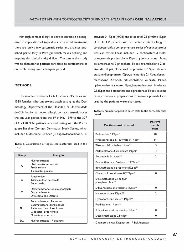
87R E V I S T A P O R T U G U E S A D E I M U N O A L E R G O L O G I A
PATCH TESTING WITH CORTICOSTEROIDS DURING A TEN-YEAR PERIOD / ORIGINAL ARTICLE
Although contact allergy to corticosteroids is a recog-
nised complication of topical corticosteroid treatment,
there are only a few systematic series and analyses pub-
lished, particularly in Portugal, which makes defining and
mapping this clinical entity difficult. Our aim in this study
was to characterise patients sensitised to corticosteroids
on patch testing over a ten-year period.
METHODS
The sample consisted of 2323 patients, 715 males and
1588 females, who underwent patch testing at the Der-
matology Department of the Hospitais da Universidade
de Coimbra for suspected allergic contact dermatitis over
the ten-year period from the 1st of May 1999 to the 30th
of April 2009. All patients received testing with the Portu-
guese Baseline Contact Dermatitis Study Series, which
included budesonide 0.1%pet (BUD), hydrocortisone-17-
butyrate 0.1%pet (HCB) and tixocortol-21-pivalate 1%pet
(TIX). In 136 patients with suspected contact allergy to
corticosteroids, a complementary series of corticosteroids
was also tested. These included 12 corticosteroid mole-
cules, namely: prednisolone 1%pet, hydrocortisone 1%pet,
dexamethasone-2-phosphate 1%pet, triamcinolone-2-ac-
etonide 1% pet, clobetasol propionate 0.25%pet, alclom-
etasone dipropionate 1%pet, amcinonide 0.1%pet, desoxi-
methasone 2.5%pet, diflucortolone valerate 1%pet,
hydrocortisone acetate 1%pet, betamethasone-12-valerate
0.12%pet and betamethasone dipropionate 1%pet. In some
cases, commercial preparations in cream or pomade form
used by the patients were also tested.
Table I. Classification of topical corticosteroids used in this study1,2
Group Allergen
A
HydrocortisoneHydrocortisone acetatePrednisoloneTixocortol pivalate
BAmcinonideTriamcinolone acetonideBudesonide
CDexamethasone sodium phosphateDexamethasone Difl ucortolone valerate
D1
Betamethasone-17-valerateBetamethasone dipropionateAlclometasone dipropionate Clobetasol proprionateMometasone furoate
D2 Hydrocortisone-17-butyrate
Table II. Number of positive patch tests to the corticosteroids tested
Corticosteroids testedPositive patch tests
Budesonide 0.1%pet* 28
Hydrocortisone-17-butyrate 0.1%pet* 14
Tixocortol-21-pivalate 1%pet* 5
Alclometasone dipropionate 1%pet* 4
Amcinonide 0.1%pet** 2
Betamethasone-17-valerate 0.12%pet* 1
Betamethasone dipropionate1%pet** 1
Clobetasol proprionate 0.25%pet* 0
Dexamethasone-21-sodium phosphate1%pet* 1
Difl uorocortolone valerate 1%pet** 0
Hydrocortisone 1%pet** 2
Hydrocortisone acetate 1%pet** 1
Prednisolone 1%pet** 1
Triamcinolone-21-acetonide 1%pet* 0
Desoximethasone 2.5%pet* 0
* Chemotechnique Diagnostics; ** Bial Aristegui.
Imuno (19) 2 - Miolo 2ª PROVA EN.indd 87Imuno (19) 2 - Miolo 2ª PROVA EN.indd 87 15-07-2011 12:00:1415-07-2011 12:00:14

88R E V I S T A P O R T U G U E S A D E I M U N O A L E R G O L O G I A
In the patch tests, the allergens available from
Chemotechnique Diagnostics and Bial Aristegui (as shown
in Table II) were applied during two days in 8 mm Finn
chambers using Scanpor® adhesive tape. Readings were
taken on the second and fourth days and patients were
also advised to return on the seventh day, to detect any
possible late-onset reactions. Test results were evaluated
in line with the International Contact Dermatitis Research
Group (ICDRG) recommendations. Reactions of or over
1+ (at least with erythema and wheals or infiltration cov-
ering the test application area) were taken as positive.
Patients who reacted to at least one corticosteroid in
the patch tests were analysed in terms of demographic
data (sex and age) clinical data (location and characterisa-
tion of the lesions and underlying pathologies) and the
patch test results (number of tests positive to corticos-
teroids per patient, corticosteroids involved and group
they belong to, and clinical importance of the patch tests.
RESULTS
Thirty-five patients, 1.5% of the population studied, re-
acted to at least one corticosteroid in the Contact Derma-
titis Baseline Series. This patient group had a mean age of
53±16 years and 71% were female. The most frequently seen
sensitisations were to the corticosteroids included in the
Baseline Series: 28 patients were sensitised to BUD, 14 to
BHC and five to TIX. Performing testing with the comple-
mentary corticosteroid series did not identify any other
patient sensitised to corticosteroids. The majority of the re-
actions to molecules not integrating the Baseline Series were
to alclometasone (four patients), amcinonide (two patients)
and hydrocortisone (two patients) (Table II). The majority of
patients (94%) were sensitised to other contact allergens.
Of the 35 patients sensitised to corticosteroids, 57%
reacted to only one molecule, 28% to two, 9% to three
and 6% to more than three molecules. Group B molecules
were in the main involved (49%), as were D2 (25%) and A
(16%), with 15 patients (43%) reacting to corticosteroids
of different groups (Figure 1).
In terms of the importance of positive skin tests, 55%
of patients had results considered clinically relevant to the
current or prior dermatitis (Table III). Of these, the lesions
were mostly contact eczema, often complicating a pre-ex-
isting stasis eczema and/or a leg ulcer. There was also a case
of oral pemphigus lesions and another of a late-onset reac-
tion following parenteral corticosteroid administration. The
contact eczema lesions, excluding the cases of leg ulcer and
stasis dermatitis, were on the hands (eight patients), upper
limbs (six patients), face (four patients), feet (four patients),
lower limbs (one) and/or trunk (one). The most frequently
Alexandra Santos, Pedro Andrade, Carlos Loureiro, Margarida Gonçalo
Table III. Clinical relevance of the sensitisations detected to the Baseline Series corticosteroids tested
Corticosteroids
Clinical relevance
Present Past Not relevant or unknown
Budesonide 0.1%pet* 10 5 13
Hydrocortisone-17-butyrate 0.1% pet* 10 3 2
Tixocortol-21-pivalate - 1%pet* 1 0 4
* Chemotechnique Diagnostics.
Imuno (19) 2 - Miolo 2ª PROVA EN.indd 88Imuno (19) 2 - Miolo 2ª PROVA EN.indd 88 15-07-2011 12:00:1415-07-2011 12:00:14
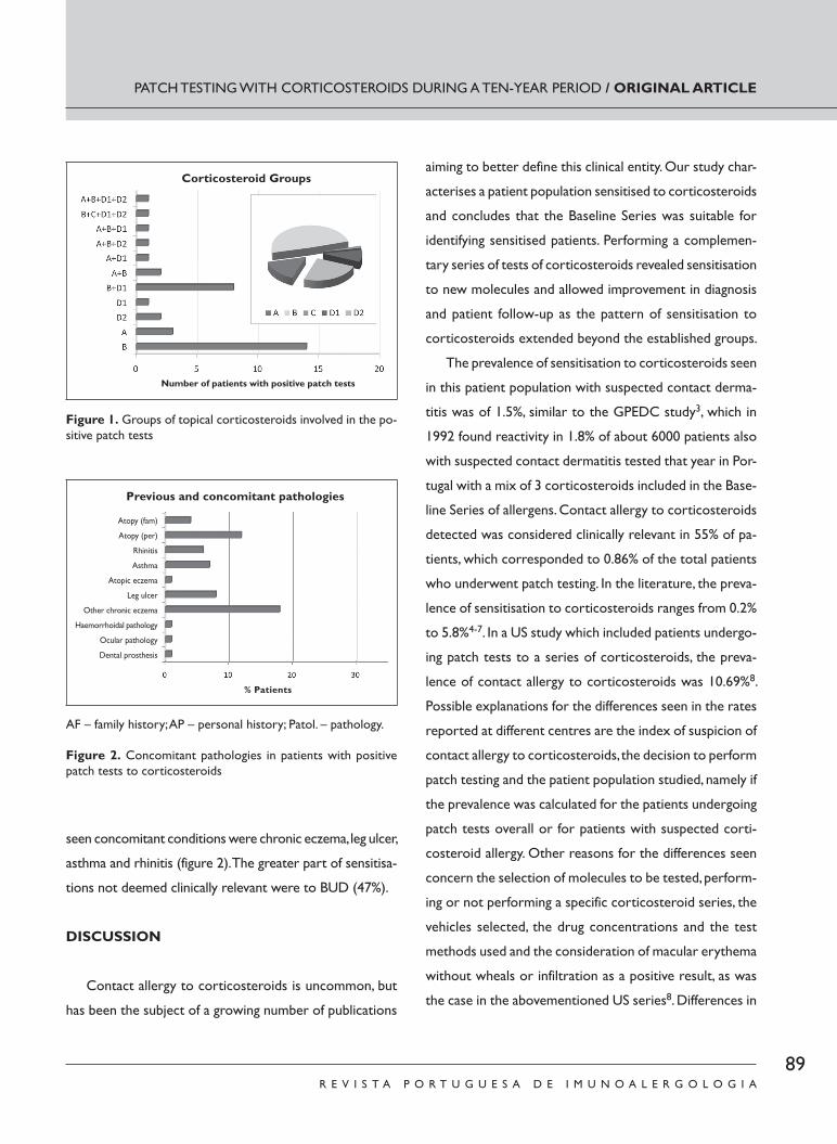
89R E V I S T A P O R T U G U E S A D E I M U N O A L E R G O L O G I A
seen concomitant conditions were chronic eczema, leg ulcer,
asthma and rhinitis (figure 2). The greater part of sensitisa-
tions not deemed clinically relevant were to BUD (47%).
DISCUSSION
Contact allergy to corticosteroids is uncommon, but
has been the subject of a growing number of publications
aiming to better define this clinical entity. Our study char-
acterises a patient population sensitised to corticosteroids
and concludes that the Baseline Series was suitable for
identifying sensitised patients. Performing a complemen-
tary series of tests of corticosteroids revealed sensitisation
to new molecules and allowed improvement in diagnosis
and patient follow-up as the pattern of sensitisation to
corticosteroids extended beyond the established groups.
The prevalence of sensitisation to corticosteroids seen
in this patient population with suspected contact derma-
titis was of 1.5%, similar to the GPEDC study3, which in
1992 found reactivity in 1.8% of about 6000 patients also
with suspected contact dermatitis tested that year in Por-
tugal with a mix of 3 corticosteroids included in the Base-
line Series of allergens. Contact allergy to corticosteroids
detected was considered clinically relevant in 55% of pa-
tients, which corresponded to 0.86% of the total patients
who underwent patch testing. In the literature, the preva-
lence of sensitisation to corticosteroids ranges from 0.2%
to 5.8%4-7. In a US study which included patients undergo-
ing patch tests to a series of corticosteroids, the preva-
lence of contact allergy to corticosteroids was 10.69%8.
Possible explanations for the differences seen in the rates
reported at different centres are the index of suspicion of
contact allergy to corticosteroids, the decision to perform
patch testing and the patient population studied, namely if
the prevalence was calculated for the patients undergoing
patch tests overall or for patients with suspected corti-
costeroid allergy. Other reasons for the differences seen
concern the selection of molecules to be tested, perform-
ing or not performing a specific corticosteroid series, the
vehicles selected, the drug concentrations and the test
methods used and the consideration of macular erythema
without wheals or infiltration as a positive result, as was
the case in the abovementioned US series8. Differences in
PATCH TESTING WITH CORTICOSTEROIDS DURING A TEN-YEAR PERIOD / ORIGINAL ARTICLE
Corticosteroid Groups
Number of patients with positive patch tests
Figure 1. Groups of topical corticosteroids involved in the po-sitive patch tests
Previous and concomitant pathologies
% Patients
Atopy (fam)
Atopy (per)
Rhinitis
Asthma
Atopic eczema
Leg ulcer
Other chronic eczema
Haemorrhoidal pathology
Ocular pathology
Dental prosthesis
AF – family history; AP – personal history; Patol. – pathology.
Figure 2. Concomitant pathologies in patients with positive patch tests to corticosteroids
Imuno (19) 2 - Miolo 2ª PROVA EN.indd 89Imuno (19) 2 - Miolo 2ª PROVA EN.indd 89 15-07-2011 12:00:1415-07-2011 12:00:14
90R E V I S T A P O R T U G U E S A D E I M U N O A L E R G O L O G I A
the prevalence of sensitisation to corticosteroids at dif-
ferent geographical locations may also be connected to
the different molecules available on the market and pre-
scription habits in the region9, in that fluorinated corticos-
teroids seem to be less allergenic10, and the underlying
pathologies in the patient population, namely the preva-
lence of patients with leg ulcer and stasis dermatitis11.
In effect, contact allergy to topical corticosteroids
seems to be more frequent in patients with leg ulcer and
stasis dermatitis, something also seen in the patient group
we studied. Probably due to the chronicity of the inflam-
matory dermo-epidermal lesions and increasing hydration
of the stratum corneum, which increases the penetration
of corticosteroid molecules, their presentation as antigens
by the Langerhans cells is highly facilitated. Other underly-
ing pathologies seen were asthma and rhinitis, particularly
connected to the sensitisation considered clinically irrel-
evant to BUD, which in the majority of cases only signifies
exposure to the drug. However, a possible airborne expo-
sure to BUD drops in aerosols containing this drug in a
workplace environment or via proximity to family mem-
bers who use them was then not investigated12,13.
The Baseline Series, which includes TIX, BUD and BHC
was adequate to identify the patients sensitised to corti-
costeroids. In 1989, Dooms-Goossens identified TIX as a
good marker of sensitisation to corticosteroids14. In a
study by Boffa et al.15 TIX and BUD allowed identification
of over 90% of the patients sensitised to corticosteroids.
Other later studies showed the value of the association
of TIX, BUD and BHC in detecting patients sensitised to
these drugs3, 16, 17. This reflects the fact that TIX is a good
marker of group A corticosteroids, BUD of group B and
BHC of group D and that sensitisation to group C corti-
costeroids is very rare. The mechanism by which this last
group of corticosteroids rarely induces sensitisation re-
mains to be elucidated and might be related to differ-
ences in the allergenicity of the molecules or with the
routes of administration commonly used for drugs of this
group (such as, for example, via the eyes, nose or mouth),
which could promote tolerance induction18. Although it
did not identify new patients sensitised to corticosteroids,
the performance of a series of tests to corticosteroids
revealed sensitisation to new molecules and allowed for
improved diagnosis and recommendations to patients
about the alternatives to the drugs involved in contact
allergy. Also relevant is the use of products used by the
patient him/herself, namely in establishing the clinical rel-
evance of the sensitisations discovered19.
In an attempt to make it easier to identify possible
cross-reactivities with the specific corticosteroid respon-
sible for the contact allergy in each individual case studied,
the topical corticosteroids were classified in line with their
chemical structure based on a literature review and a de-
scriptive study of 15 cases of contact allergy to these
drugs1. However, this classification has been contested,
seeing as cross-reactivities depend on more factors than
the chemical structure of the drug applied to the skin. The
degradation and metabolisation of these steroid molecules
can generate new molecules with different chemical struc-
ture and new potentials in terms of immunologic reactiv-
ity, in so far as the new molecules can present cross-reac-
tivity with other corticosteroids which the molecule
whence they derive did not present. We found the pattern
of sensitisation to corticosteroids extended beyond the
groups previously established. This reflects the fact that
while dividing corticosteroid molecules into groups is use-
ful, it cannot be considered watertight, as cross-reactivity
between drugs of different groups could exist, namely by
isomers, as happens between the B and A groups with the
D2 group. Further, the likelihood of a patient being cosen-
Alexandra Santos, Pedro Andrade, Carlos Loureiro, Margarida Gonçalo
Imuno (19) 2 - Miolo 2ª PROVA EN.indd 90Imuno (19) 2 - Miolo 2ª PROVA EN.indd 90 15-07-2011 12:00:1515-07-2011 12:00:15
91R E V I S T A P O R T U G U E S A D E I M U N O A L E R G O L O G I A
sitised to different drugs must also be considered. It is thus
vital to test molecules of different groups when sensitisa-
tion to corticosteroids is detected to better define which
drugs can be used in the future as an alternative to those
which cause contact allergy.
CONCLUSION
Contact allergy to corticosteroids is infrequent and
demands a high index of suspicion. The possibility of clini-
cally irrelevant sensitisation demands a detailed clinical
history and careful establishing of the clinical importance
of the sensitisations found, meaning that testing with the
patient’s own products can be useful. In patients sensitised
to the corticosteroids in the Baseline Series, it is important
to test a specific series of corticosteroids to improve di-
agnostic rigour and identify alternative drugs that the pa-
tient can use in the future.
Funding: Self-funding
Conflict of interest disclosure: None
ContactAlexandra SantosServiço de Imunoalergologia – Hospitais da Universidade de CoimbraPraceta Mota Pinto3000 -075 Coimbra, PortugalEmail: [email protected]
REFERENCES
1. Coopman S, Degreef H, Dooms -Goossens A. Identification of cross-
-reaction patterns in allergic contact dermatitis from topical corti-
costeroids. Br J Dermatol. 1989; 121:27 -34.
2. Baeck M, Marot L, Nicolas JF, Pilette C, Tennstedt D, Goossens A.
Allergic hypersensitivity to topical and systemic corticosteroids: a
review. Allergy. 2009; 64:978 -94.
3. Pecegueiro M. Contact allergy to topical corticosteroids: a screen-
ing study with a corticosteroid mix. Portuguese Contact Der-
matitis Research Group (GPEDC). Contact Dermatitis. 1995;
33:196 -7.
4. Wilkinson SM. Hypersensitivity to topical corticosteroids. Clin Exp
Dermatol. 1994; 19:1 -11.
5. Burden AD, Beck MH. Contact hypersensitivity to topical corticos-
teroids. Br J Dermatol. 1992; 127:497 -500.
6. Lauerma AI. Screening for corticosteroid contact sensitivity. Com-
parison of tixocortol pivalate, hydrocortisone -17 -butyrate and
hydrocortisone. Contact Dermatitis. 1991; 24:123 -30.
7. Dooms -Goossens A, Morren M. Results of routine patch testing
with corticosteroid series in 2073 patients. Contact Dermatitis.
1992; 26:182 -91.
8. Davis MD, el -Azhary RA, Farmer SA. Results of patch testing
to a corticosteroid series: a retrospective review of 1188 pa-
tients during 6 years at Mayo Clinic. J Am Acad Dermatol. 2007;
56:921 -7.
9. Thomson KF, Wilkinson SM, Powell S, Beck MH. The prevalence of
corticosteroid allergy in two U.K. centres: prescribing implications.
Br J Dermatol. 1999; 141:863 -6.
10. Dooms -Goossens A, Meinardi MM, Bos JD, Degreef H. Contact
allergy to corticosteroids: the results of a two -centre study. Br J
Dermatol. 1994; 130:42 -7.
11. Keegel T, Saunders H, Milne R, Sajjachareonpong P, Fletcher A, Nix-
on R. Topical corticosteroid allergy in an urban Australian centre.
Contact Dermatitis. 2004; 50:6 -14.
12. Baeck M, Pilette C, Drieghe J, Goossens A. Allergic contact der-
matitis to inhalation corticosteroids. Eur J Dermatol. 2010;
20:102 -8.
13. Baeck M, Goossens A. Patients with airborne sensitization/contact
dermatitis from budesonide -containing aerosols ‘by proxy’. Contact
Dermatitis. 2009; 61:1 -8.
14. Dooms -Goossens AE, Degreef HJ, Marien KJ, Coopman SA. Contact
allergy to corticosteroids: a frequently missed diagnosis? J Am Acad
Dermatol. 1989; 21:538 -43.
15. Boffa MJ, Wilkinson SM, Beck MH. Screening for corticosteroid
contact hypersensitivity. Contact Dermatitis. 1995; 33:149 -51.
16. Dooms -Goossens A, Andersen KE, Brandao FM, Bruynzeel D, Bur-
rows D, Camarasa J, et al. Corticosteroid contact allergy: an EECDRG
multicentre study. Contact Dermatitis. 1996; 35:40 -4.
17. Isaksson M, Andersen KE, Brandao FM, Bruynzeel DP, Bruze M,
Camarasa JG, et al. Patch testing with corticosteroid mixes in Europe.
A multicentre study of the EECDRG. Contact Dermatitis. 2000;
42:27 -35.
18. Goossens A, Matura M, Degreef H. Reactions to corticosteroids:
some new aspects regarding cross -sensitivity. Cutis. 2000; 65:43 -5.
19. English JS. Corticosteroid -induced contact dermatitis: a pragmatic
approach. Clin Exp Dermatol. 2000; 25:261 -4.
PATCH TESTING WITH CORTICOSTEROIDS DURING A TEN-YEAR PERIOD / ORIGINAL ARTICLE
Imuno (19) 2 - Miolo 2ª PROVA EN.indd 91Imuno (19) 2 - Miolo 2ª PROVA EN.indd 91 15-07-2011 12:00:1515-07-2011 12:00:15

Imuno (19) 2 - Miolo 2ª PROVA EN.indd 92Imuno (19) 2 - Miolo 2ª PROVA EN.indd 92 15-07-2011 12:00:1515-07-2011 12:00:15
93R E V I S T A P O R T U G U E S A D E I M U N O A L E R G O L O G I A
ORIGINAL ARTICLE
ABSTRACT
Background: The usual recommendation in food allergy treatment is strict eviction until tolerance is established. It is important to know how an eviction diet fails in order to improve the information we give to food -allergic patients. Objective: To identify the frequency and to characterise accidental food exposures in a group of food -allergic patients. Material and methods: Children aged ten years or less, allergic to cow’s milk proteins, egg, fish, peanut or nuts were selected from the files of patients followed up at the Allergology and Clinical Immunology outpatient clinic of
Hospital Dona Estefânia. Parents/caregivers answered a questionnaire regarding the culprit food, symptoms and characterisation of accidental exposures. Results: We selected a group of 65 patients, with a median age of 4.3 years (63% males), corresponding to 69 cases of food allergy – 42 cases of cow’s milk protein allergy, 11 cases of fish al-lergy, 10 cases of egg allergy, five cases of nut allergy and one case of peanut allergy. The first reaction occurred, in most cases, by ingestion (95.6%) and was immediate (78.3%). Symptoms were mucocutaneous in 75.4%, gastrointes-tinal in 33.3% and respiratory in 23.2%. Anaphylaxis occurred in 17%. The eviction diet failed in 68.1% cases, which corresponded to 68 accidental exposure occurrences, most with symptoms (87.1%). Of these 68 accidental exposures, the culprit food was cow’s milk in 69.1% (n = 47), egg in 14.7% (n = 10), fish in 13.2% (n = 9) and nuts in 2.9% (n = 2). Mucocutaneous manifestations were the most frequent (55.9%), followed by respiratory symptoms (25%) and gastrointestinal symptoms (23.5%). Anaphylaxis occurred in 20.5%. Most accidental exposures were at home (36.8%) and at school (29.4%). After the reaction, parents/caregivers administered medication in 41.2% of cases, waited until spontaneous resolution took place in 38.2% and went to Emergency Room Departments in 20.6%. Conclusion: Eviction diet failures were frequent, most of them with symptoms. Most accidental exposures occurred at home and at school. This may indicate gaps in parents’/care givers’ knowledge. The characterisation of accidental exposures in
Accidental exposures in food allergy
Exposições acidentais na alergia alimentar
Filipa Sousa1, João Antunes2, Maria João Paes3, Marta Chambel2, Sara Prates2, Paula Leiria Pinto2
1 Allergology and Clinical Immunology Unit, Hospital Central do Funchal, Madeira, 2 Allergology and Clinical Immunology Clinic, Centro Hospitalar de Lisboa Central, Hospital Dona Estefânia, Lisbon3 Allergology and Clinical Immunology Clinic, Centro Hospitalar de Lisboa Norte, Hospital Pulido Valente, Lisbon
R e v P o r t I m u n o a l e r g o l o g i a 2 0 1 1 ; 1 9 ( 2 ) : 9 3 - 1 0 0
Data de recepção / Received in: 06/01/2011
Data de aceitação / Accepted for publication in: 17/06/2011
Imuno (19) 2 - Miolo 2ª PROVA EN.indd 93Imuno (19) 2 - Miolo 2ª PROVA EN.indd 93 15-07-2011 12:00:1715-07-2011 12:00:17
94R E V I S T A P O R T U G U E S A D E I M U N O A L E R G O L O G I A
INTRODUCTION
The prevalence of food allergy has been increasing
in western countries, namely in paediatric age
individuals1. It is estimated at being around 6 -8%
in childhood2. The foodstuffs most frequently involved are
cow’s milk, egg, fi sh, soya, wheat, shellfi sh, peanut and nuts.
Currently, the only safe recommendation in treating food
allergy is complete eviction until tolerance is aquired3,4.
Accidental ingestion and exposure are a constant source
of worry for patients and their families, given that they can
lead to potentially fatal reactions, which can bring with it a
food -allergic patients may help to improve the information transmitted to parents/caregivers to help them identify risk factors and be aware of avoidance measures.
Key -words: contact, food allergy, exposure, ingestion, accidental reaction
RESUMO
Introdução: A recomendação habitual no tratamento da alergia alimentar é a evicção completa, até à aquisição de tolerância. É importante perceber em que situações ocorrem falhas na evicção, de forma a orientar o melhor possível o doente com alergia alimentar. Objectivo: Conhecer a frequência e caracterizar as exposições acidentais, num grupo de doentes com alergia alimentar. Material e Métodos: A partir dos registos do Serviço de Imunoalergologia do Hospital Dona Estefânia, foram seleccionados doentes com idade ≤ 10 anos, com alergia às proteínas do leite de vaca, ovo, peixe, amendoim ou frutos secos. Os pais/prestadores de cuidados, responderam a um inquérito telefónico, referente ao alimento implicado, falhas na dieta e sintomas. Resultados: Contactou -se um grupo de 65 do-entes com idade média de 4,3 anos (63% do sexo masculino), totalizando 69 casos de alergia alimentar – cerca de 42 casos de alergia ao leite, 11 casos de alergia ao peixe, 10 de alergia ao ovo, 5 de alergia aos frutos secos e 1 de alergia ao amendoim. Na maioria dos casos a 1.ª reacção foi desencadeada por ingestão (95,6%) e foi imediata (78,3%), manifestando -se por sintomas mucocutâneos (MC) em 75,4%, gastrointestinais em 33,3% e respiratórios em 23,2%. Ocorreu anafilaxia em 17%. Houve falhas na dieta em 68,1% dos casos, que contabilizaram um total de 68 eventos de exposição acidental, na maioria (87,1%) com sintomas. Destes 68 eventos de ex-posição acidental, em 69,1% (n=47) o leite foi o alimento implicado, em 14,7% (n=10) foi o ovo, em 13,2% (n=9) o peixe e em 2,9% (n=2) os frutos secos. As manifestações clínicas mais frequentes foram MC (55,9,9%), seguindo -se as do tracto respiratório (25%) e as do tracto gastrointestinal (23,5%). Em 20,5% dos eventos de exposição acidental, ocorreu reacção anafiláctica. A maior parte das inges-tões / exposições acidentais ocorreram em casa (36,8%) e na escola (29,4%). Perante a reacção foi administrada terapêutica em 41,2%, aguardaram resolução espontânea 38,2% e recorreram ao Serviço de Urgência 20,6% dos casos. Conclusões: As falhas na dieta de evicção foram frequentes, a maioria com sintomas. Aconteceram maioritariamente em casa e na escola, o que pode sugerir lacunas no conhecimento dos pais/prestadores de cuidados. A caracterização das exposições acidentais nos doentes com alergia alimentar poderá ajudar a optimizar a transmissão de informação, a estes e aos seus responsáveis, relativamente à prevenção de situações de risco.
Palavras -chave: alergia, alimentar, contacto, exposição, ingestão, reacção acidental
Filipa Sousa, João Antunes, Maria João Paes, Marta Chambel, Sara Prates, Paula Leiria Pinto
Imuno (19) 2 - Miolo 2ª PROVA EN.indd 94Imuno (19) 2 - Miolo 2ª PROVA EN.indd 94 15-07-2011 12:00:1815-07-2011 12:00:18
95R E V I S T A P O R T U G U E S A D E I M U N O A L E R G O L O G I A
heavy emotional burden5,6. Challenges such as reading food-
stuff labels or the care involved in avoiding cross -contamination
occur daily, as do the potential limitations to social activities
which involve food, such as school, eating out, visiting friends
and family. This has a negative impact on these patients’ qual-
ity of life7,8. Further, patients and their parents/caregivers
have to be instructed about what to do in case of accidental
exposure3,9. It is important to understand in which situations
accidental exposure can occur, to better guide the daily life
of a patient with food allergy. Health professionals’ knowl-
edge of patients’ day -to -day reality can improve the informa-
tion transmitted to parents/caregivers to help them identify
risk factors and be aware of avoidance measures.
Our aim was to study the frequency of accidental ex-
posure (ingestion/contact/inhalation) in a group of patients
with food allergy and characterise accidental food expo-
sure, what led to it and reactions to it, in order to improve
the information we give to food -allergic patients and tailor
it to their reality.
MATERIAL AND METHODS
Children aged ten years or less, allergic to cow’s milk
proteins, egg, fish, peanut or nuts were selected from the
files of patients followed up at the Allergology and Clinical
Immunology outpatient clinic in Hospital Dona Estefânia.
Parents/caregivers were contacted by phone and asked to
answer a questionnaire. The questionnaire (Table I) mapped
out demographic data, the trigger foodstuff(s), personal
and family history of allergic disease, characterisation of
the first reaction (how it was triggered, its clinical mani-
festations, time lag between exposure and reaction) and
failure in the diet, characterisation of accidental ingestions
in terms of number, the place and manner in which they
occurred, how the food was presented (obviously or hid-
den) and parents’/caregivers’ attitudes to the reaction.
Some patients were allergic to more than one foodstuff
simultaneously. In order to analyse the results, we defined
as a “case” each food allergy in each patient, meaning the
number of cases is higher than the number of patients.
Unlike demographic data, which is based on the number
of patients, all the remaining analysis of results is based on
the number of cases of food allergy or the number of oc-
currences of accidental exposure.
RESULTS
We contacted the parents of 65 patients, 62 of which
had allergy to only one foodstuff, two to two foodstuffs
and one to three. Hence the analysis deals with a total of
69 cases of food allergy.
Of the demographic data (Table II), we highlight the
predominance of males and that over half the patients had
personal and family histories of another allergic disease,
although only 9.2% had family history of food allergy.
The 69 cases of food allergy consisted of 42 cases of
cow’s milk allergy, 11 cases of fish allergy, 10 of egg allergy,
five of nut allergy and one of peanut allergy. The first reaction
was triggered by ingestion in 95.6% (66/69) of cases and by
contact in 4.4% (3/69) of cases. In the majority of cases
(78.3%), the reaction was immediate, occurring within less
than 30 minutes in 60.9% (42/69) and within 30 minutes to
two hours in 17.4% (12/69). In the remaining 21.7% (15/69)
cases, the reaction manifested after two or more hours. In
terms of the initial clinical manifestation, mucocutaneous
(MC) symptoms were predominant (75.4%; 52/69), followed
by gastrointestinal (GI) (33.3%; 23/69) and respiratory (R)
symptoms (23.2%; 16/69). Mucocutaneous symptoms were
ACCIDENTAL EXPOSURES IN FOOD ALLERGY / ORIGINAL ARTICLE
Imuno (19) 2 - Miolo 2ª PROVA EN.indd 95Imuno (19) 2 - Miolo 2ª PROVA EN.indd 95 15-07-2011 12:00:2015-07-2011 12:00:20
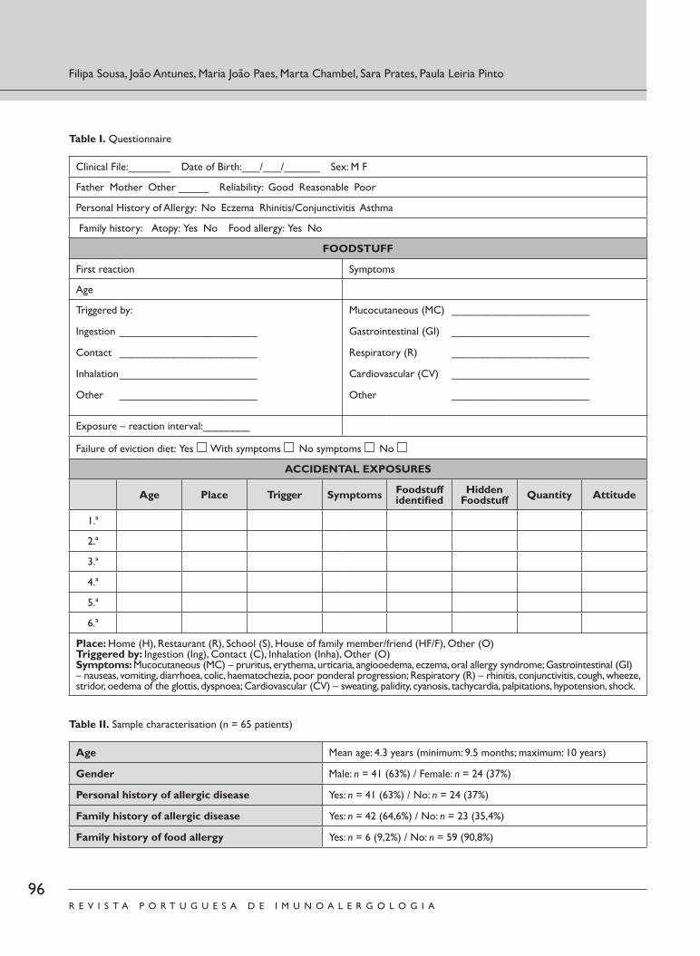
96R E V I S T A P O R T U G U E S A D E I M U N O A L E R G O L O G I A
Filipa Sousa, João Antunes, Maria João Paes, Marta Chambel, Sara Prates, Paula Leiria Pinto
Table I. Questionnaire
Clinical File:_______ Date of Birth:___/___/______ Sex: M F
Father Mother Other _____ Reliability: Good Reasonable Poor
Personal History of Allergy: No Eczema Rhinitis/Conjunctivitis Asthma
Family history: Atopy: Yes No Food allergy: Yes No
FOODSTUFF
First reaction Symptoms
Age
Triggered by:
Ingestion _______________________
Contact _______________________
Inhalation _______________________
Other _______________________
Mucocutaneous (MC) _______________________
Gastrointestinal (GI) _______________________
Respiratory (R) _______________________
Cardiovascular (CV) _______________________
Other _______________________
Exposure – reaction interval:________
Failure of eviction diet: Yes With symptoms No symptoms No
ACCIDENTAL EXPOSURES
Age Place Trigger Symptoms Foodstuff identifi ed
Hidden Foodstuff Quantity Attitude
1.ª
2.ª
3.ª
4.ª
5.ª
6.ª
Place: Home (H), Restaurant (R), School (S), House of family member/friend (HF/F), Other (O)Triggered by: Ingestion (Ing), Contact (C), Inhalation (Inha), Other (O)Symptoms: Mucocutaneous (MC) – pruritus, erythema, urticaria, angiooedema, eczema, oral allergy syndrome; Gastrointestinal (GI) – nauseas, vomiting, diarrhoea, colic, haematochezia, poor ponderal progression; Respiratory (R) – rhinitis, conjunctivitis, cough, wheeze, stridor, oedema of the glottis, dyspnoea; Cardiovascular (CV) – sweating, palidity, cyanosis, tachycardia, palpitations, hypotension, shock.
Table II. Sample characterisation (n = 65 patients)
Age Mean age: 4.3 years (minimum: 9.5 months; maximum: 10 years)
Gender Male: n = 41 (63%) / Female: n = 24 (37%)
Personal history of allergic disease Yes: n = 41 (63%) / No: n = 24 (37%)
Family history of allergic disease Yes: n = 42 (64,6%) / No: n = 23 (35,4%)
Family history of food allergy Yes: n = 6 (9,2%) / No: n = 59 (90,8%)
Imuno (19) 2 - Miolo 2ª PROVA EN.indd 96Imuno (19) 2 - Miolo 2ª PROVA EN.indd 96 15-07-2011 12:00:2015-07-2011 12:00:20
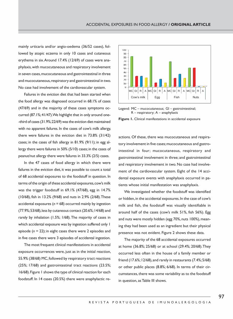
97R E V I S T A P O R T U G U E S A D E I M U N O A L E R G O L O G I A
mainly urticaria and/or angio -oedema (36/52 cases), fol-
lowed by atopic eczema in only 10 cases and cutaneous
erythema in six. Around 17.4% (12/69) of cases were ana-
phylaxis, with mucocutaneous and respiratory involvement
in seven cases, mucocutaneous and gastrointestinal in three
and mucocutaneous, respiratory and gastrointestinal in two.
No case had involvement of the cardiovascular system.
Failures in the eviction diet that had been started when
the food allergy was diagnosed occurred in 68.1% of cases
(47/69) and in the majority of these cases symptoms oc-
curred (87.1%; 41/47). We highlight that in only around one-
-third of cases (31.9%; 22/69) was the eviction diet maintained
with no apparent failures. In the cases of cow’s milk allergy,
there were failures in the eviction diet in 73.8% (31/42)
cases; in the cases of fish allergy in 81.9% (9/11); in egg al-
lergy there were failures in 50% (5/10) cases; in the cases of
peanut/nut allergy there were failures in 33.3% (2/5) cases.
In the 47 cases of food allergy in which there were
failures in the eviction diet, it was possible to count a total
of 68 accidental exposures to the foodstuff in question. In
terms of the origin of these accidental exposures, cow’s milk
was the trigger foodstuff in 69.1% (47/68), egg in 14.7%
(10/68), fish in 13.2% (9/68) and nuts in 2.9% (2/68). These
accidental exposures (n = 68) occurred mainly by ingestion
(77.9%; 53/68), less by cutaneous contact (20.6%; 14/68) and
rarely by inhalation (1.5%; 1/68). The majority of cases in
which accidental exposure was by ingestion suffered only 1
episode (n = 22); in eight cases there were 2 episodes and
in five cases there were 3 episodes of accidental ingestion.
The most frequent clinical manifestations in accidental
exposure occurrences were, just as in the initial reaction,
55.9% (38/68) MC, followed by respiratory tract reactions
(25%; 17/68) and gastrointestinal tract reactions (23.5%;
16/68). Figure 1 shows the type of clinical reaction for each
foodstuff. In 14 cases (20.5%) there were anaphylactic re-
actions. Of these, there was mucocutaneous and respira-
tory involvement in five cases; mucocutaneous and gastro-
intestinal in four; mucocutaneous, respiratory and
gastrointestinal involvement in three; and gastrointestinal
and respiratory involvement in two. No case had involve-
ment of the cardiovascular system. Eight of the 14 acci-
dental exposure events with anaphylaxis occurred in pa-
tients whose initial manifestation was anaphylaxis.
We investigated whether the foodstuff was identified
or hidden, in the accidental exposures. In the case of cow’s
milk and fish, the foodstuff was visually identifiable in
around half of the cases (cow’s milk 51%, fish 56%). Egg
and nuts were mostly hidden (egg 70%, nuts 100%), mean-
ing they had been used as an ingredient but their physical
presence was not evident. Figure 2 shows these data.
The majority of the 68 accidental exposures occurred
at home (36.8%; 25/68) or at school (29.4%; 20/68). They
occurred less often in the house of a family member or
friend (17.6%; 12/68), and rarely in restaurants (7.4%; 5/68)
or other public places (8.8%; 6/68). In terms of their cir-
cumstances, there was some variability as to the foodstuff
in question, as Table III shows.
ACCIDENTAL EXPOSURES IN FOOD ALLERGY / ORIGINAL ARTICLE
Legend: MC – mucocutaneous; GI – gastrointestinal; R – respiratory; A – anaphylaxis
Figure 1. Clinical manifestations in accidental exposure
Imuno (19) 2 - Miolo 2ª PROVA EN.indd 97Imuno (19) 2 - Miolo 2ª PROVA EN.indd 97 15-07-2011 12:00:2115-07-2011 12:00:21
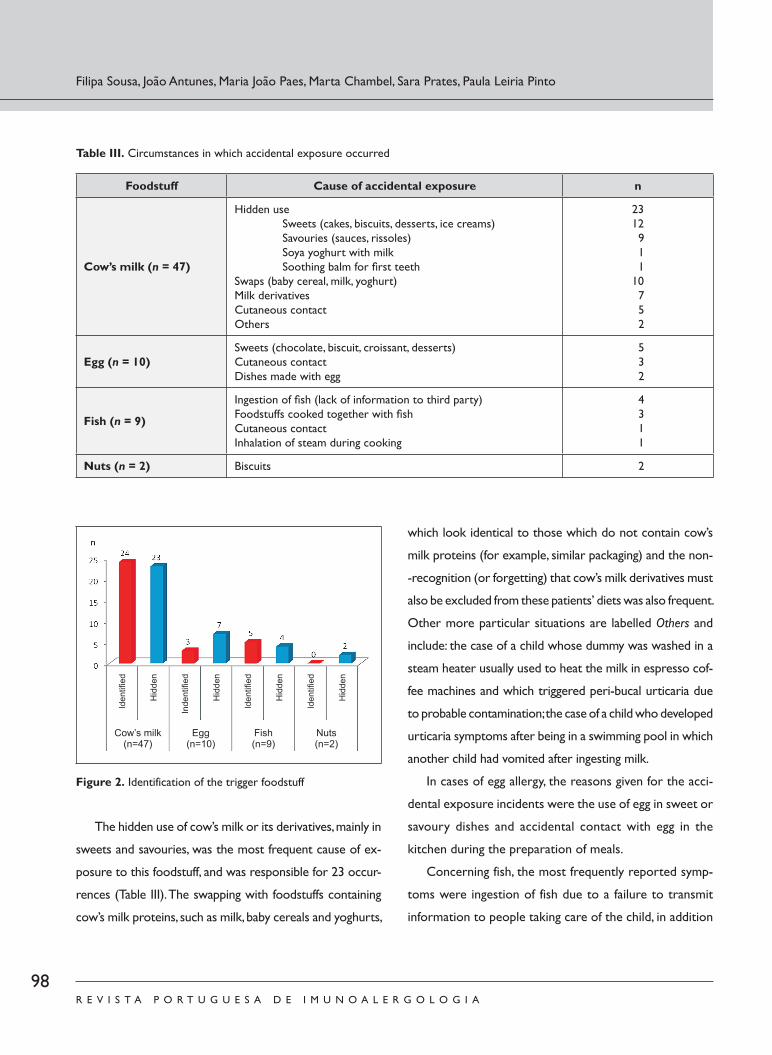
98R E V I S T A P O R T U G U E S A D E I M U N O A L E R G O L O G I A
The hidden use of cow’s milk or its derivatives, mainly in
sweets and savouries, was the most frequent cause of ex-
posure to this foodstuff, and was responsible for 23 occur-
rences (Table III). The swapping with foodstuffs containing
cow’s milk proteins, such as milk, baby cereals and yoghurts,
which look identical to those which do not contain cow’s
milk proteins (for example, similar packaging) and the non-
-recognition (or forgetting) that cow’s milk derivatives must
also be excluded from these patients’ diets was also frequent.
Other more particular situations are labelled Others and
include: the case of a child whose dummy was washed in a
steam heater usually used to heat the milk in espresso cof-
fee machines and which triggered peri -bucal urticaria due
to probable contamination; the case of a child who developed
urticaria symptoms after being in a swimming pool in which
another child had vomited after ingesting milk.
In cases of egg allergy, the reasons given for the acci-
dental exposure incidents were the use of egg in sweet or
savoury dishes and accidental contact with egg in the
kitchen during the preparation of meals.
Concerning fish, the most frequently reported symp-
toms were ingestion of fish due to a failure to transmit
information to people taking care of the child, in addition
Filipa Sousa, João Antunes, Maria João Paes, Marta Chambel, Sara Prates, Paula Leiria Pinto
Figure 2. Identification of the trigger foodstuff
Table III. Circumstances in which accidental exposure occurred
Foodstuff Cause of accidental exposure n
Cow’s milk (n = 47)
Hidden use Sweets (cakes, biscuits, desserts, ice creams) Savouries (sauces, rissoles) Soya yoghurt with milk Soothing balm for fi rst teethSwaps (baby cereal, milk, yoghurt)Milk derivativesCutaneous contactOthers
2312 9 1 110 7 5 2
Egg (n = 10)Sweets (chocolate, biscuit, croissant, desserts)Cutaneous contactDishes made with egg
5 3 2
Fish (n = 9)
Ingestion of fi sh (lack of information to third party)Foodstuffs cooked together with fi shCutaneous contactInhalation of steam during cooking
4 3 1 1
Nuts (n = 2) Biscuits 2
Imuno (19) 2 - Miolo 2ª PROVA EN.indd 98Imuno (19) 2 - Miolo 2ª PROVA EN.indd 98 15-07-2011 12:00:2115-07-2011 12:00:21

99R E V I S T A P O R T U G U E S A D E I M U N O A L E R G O L O G I A
to the ingestion of food cooked together with fish. Table
III shows other more particular cases. The situation trig-
gered by cutaneous contact occurred at Lisbon Zoo and
was caused by a sea lion giving a “kiss” to a child during
the sea lions’ feeding time (the sea lions are fed with fish).
The reaction seen in this case was contact urticaria.
Reactions to nuts were triggered by ingestion of bis-
cuits containing nuts.
The attitude in response to the reaction was to admin-
ister emergency treatment in 41.2% (28/68) of cases, wait
for spontaneous resolution in 38.2% (26/68) and Emergen-
cy Room (ER) visits in 20.6% (14/68). The most frequently
used medicine was H1 -antihistamine (25%; 17/68), followed
by oral corticosteroids (17.6%; 12/68), while there was less
frequent recourse to short -acting bronchodilator (13.2%;
9/68). There was only one case of adrenaline being adminis-
tered (1.5%; 1/68) by healthcare professionals at the ER.
DISCUSSION
This study aimed to identify the frequency of and to char-
acterise accidental food exposures in a group of food -allergic
paediatric patients. The results show that in our patients, ac-
cidental foodstuff ingestion is common and very often ac-
companied by symptoms of greater or lesser severity.
There were failures in the eviction diet in 68.1% of
cases. We found no similar studies in the literature, meaning
it was difficult to compare our data. A Canadian study, how-
ever, specifically analysing the role of foodstuff labels, found
that 47.8% of patients had experienced accidental expo-
sures10. These were mainly patients with peanut allergy and
around half the sample was recruited from food -allergy
patients’ associations, which could explain the lower per-
centage of eviction diet failure. Additionally, the same study
reported that patients allergic to peanut, nuts, fish or shell-
fish had a lower percentage of accidental exposure than
those allergic to other foodstuffs. This difference can be
explained by the fact that those foodstuffs give rise to more
severe reactions, leading to greater care being taken in their
identification by both the food industry and the patient10.
In our patients, the majority of the accidental exposures
led to a clinical reaction (87.2%) and, unlike what is usually
reported9, they occurred more frequently at home or at
school, that is, as part of the child’s daily routine. They occurred
more rarely in restaurants or at friends’ or family members’
houses. It could be that vigilance is greater in situations typi-
cally seen and taught as being those containing risk, thus ef-
fectively reducing the occurrence of accidental exposure
episodes at these places. A day -to -day domestic environment
is probably more conducive to relaxing one’s vigilance.
As regards the form of exposure, in addition to the
situations usually reported in the literature – the hidden
presence of the foodstuff and failures in reading product
labels3,9,10 – we identified some others which often occur.
We highlight the swapping for foodstuffs of the same type
and with similar packaging (baby cereal, milk, yoghurts), the
lack of knowledge (or forgetting) that foodstuffs of the
same family and derivatives contain the trigger allergen
(for example, cow’s milk derivatives) and finally, failures to
pass on the information to third parties (family members,
members of school staff, nannies). The remaining cases,
which illustrate more unusual forms, are also important as
knowledge of these and dissemination of that knowledge
could help prevent similar situations in other patients.
The data we obtained seem to suggest that the infor-
mation transmitted to parents/caregivers might not be
sufficient and that there is a need to pass on more and
better information and duly educate parents/caregivers to
avoid the trigger foodstuff in food -allergy cases.
ACCIDENTAL EXPOSURES IN FOOD ALLERGY / ORIGINAL ARTICLE
Imuno (19) 2 - Miolo 2ª PROVA EN.indd 99Imuno (19) 2 - Miolo 2ª PROVA EN.indd 99 15-07-2011 12:00:2215-07-2011 12:00:22

100R E V I S T A P O R T U G U E S A D E I M U N O A L E R G O L O G I A
A multidisciplinary approach is important in at least some
cases of children with food allergy, with support from a nutrition-
ist/dietician, to improve the information given to parents/care-
givers. In addition, and as is the case in some chronic diseases,
patients and parents/caregivers can benefit from support groups
with whom to share experiences of concrete situations11. There
are as yet no food -allergy support groups in Portugal.
In terms of the attitude taken to the reaction, only a fifth
of patients needed to go to the ER. Of the remaining, around
half had spontaneous resolution – most likely the cases with
less severe reactions – and the other half were resolved
with antihistamines, corticosteroids and/or bronchodilators,
which could mean that the majority of parents/caregivers
are well informed as to how to deal with an acute reaction.
There is some evidence that patients with a history of more
severe reactions take greater care in avoiding accidental
exposures10. This could also contribute to the seemingly
low frequency of severe reactions in the sample studied.
The almost nil recourse to adrenaline, despite 20.5%
of the accidental reactions reported being anaphylactic, is
in line with data seen in other studies, which also found a
lower than desirable use of this drug in emergency situa-
tions12. That said, we admit that there could have been
some use of adrenaline in the children who went to the
ER, with their parents being unaware of this fact.
CONCLUSIONS
There were frequent failures in the eviction diets, the
majority with symptoms. Accidental exposures occurred
mainly as part of a child’s everyday routine, which could
indicate gaps in parents’/caregivers’ knowledge.
A mapping of accidental exposures in a sample of patients
with food allergy can help to improve the information trans-
mitted to patients and their parents/caregivers to help them
identify risk factors and be aware of avoidance measures.
Funding: None
Conflict of interest disclosure: None
Contact:Sónia Filipa de Sousa Jardim FernandesRua Ilha dos Amores, lote 4.11.02 B, 4.º Esq, 1990 -121 LisboaE -mail: [email protected]
REFERENCES
1. Sicherer SH, Munoz -Furlong A, Sampson HA. Prevalence of peanut
and tree nut allergy in United States determined by means of a
random digit dial telephone survey: a five year follow -up study. J
Allergy Clin Immunol 2003; 112: 1203 -7.
2. Bock SA. Retrospective appraisal of complaints of adverse reactions
to foods in children during the first three years of life. Pediatrics
1987; 79: 683 -8.
3. Sicherer SH, Teuber S. Current approach to the diagnosis and man-
agement of adverse reactions to foods. J Allergy Clin Immunol 2004;
114: 1146 -50.
4. Mathew I, Jonathan MS: Management of food allergies. Expert opin-
ion Pharmacother 2003; 4(7): 1025 -1037.
5. Kemp AS, Hu W. Food allergy and anaphylaxis – dealing with uncer-
tainty. Med J Aust 2008; 188: 503 -4.
6. Teufel M, Biedermann T, rapps N, et al. Psychological burden of food
allergy. World J Gastroenterol 2007; 13: 3456 -65.
7. Sicherer SH, Noone SA, Munoz -Furlong A. The impact of childhood
food allergy on quality of life. Ann Allergy Asthma Immunol 2001;
87: 461 -4.
8. Marklund B, Ahlstedt S, Nordström G. Food hypersensitivity and
quality of life. Curr Opin Allergy Clin Immunol 2007; 7: 297 -87.
9. Sampson HA. Food allergy. Part 2: Diagnosis and management. J
Allergy Clin Immunol 1999; 103: 981 -9.
10. Sheth SS, Waserman S, Kagan R, Alizadehfar R, Primeau MN, Elliot S,
et al. Role of food labels in accidental exposures in food -allergic in-
dividuals in Canada. Ann Allergy Asthma Immunol 2010; 104: 60 -5.
11. Wendy Hu, Robert Lo, John Zi. Attributes and views of families with
food allergic children recruited from allergy clinics and from a con-
sumer organization. Pediatr Allergy Immunol 2008; 19: 264 -269.
12. MM Almeida, A Gaspar, C Santa -Marta, S Piedade, P Leiria -Pinto, G
Pires et al. Anafilaxia – da notificação e reconhecimento à abordagem
terapêutica. Rev Port Imunoalergologia 2007; 15 (1): 19 -41.
Filipa Sousa, João Antunes, Maria João Paes, Marta Chambel, Sara Prates, Paula Leiria Pinto
Imuno (19) 2 - Miolo 2ª PROVA EN.indd 100Imuno (19) 2 - Miolo 2ª PROVA EN.indd 100 15-07-2011 12:00:2215-07-2011 12:00:22

101R E V I S T A P O R T U G U E S A D E I M U N O A L E R G O L O G I A
CASE REPORT
ABSTRACT
The prevalence of adverse drug reactions (ADR) in hospitalised patients is estimated at 10 -20% and can be
potentially life -threatening. Toxic epidermal necrolysis (TEN) is one of the most severe forms of ADR, with low
incidence but high mortality. The authors present the case of a 79 -year -old female, with severe haemorrhagic ce-
rebrovascular disease, due to head injury. The patient was admitted to an intensive care unit and in the course of
treatment with meropenem, vancomycin and sodium valproate developed a TEN reaction. Lymphocyte transforma-
tion test (LLT) was performed in order to identify the eliciting drug. Stimulation indices were < 2.0 for meropenem,
7.4 for vancomycin and 6.4 for sodium valproate, with a cut -off value >3. Although still a research tool, LLT was
decisive to prove the immunologic basis of the reaction. Vancomycin and sodium valproate are strictly contraindi-
cated in this patient.
Key -words: Lymphocyte transformation test; sodium valproate; toxic epidermal necrolysis; vancomycin.
Toxic epidermal necrolysis – sodium valproate and vancomycin?
Necrólise epidérmica tóxica – Valproato de sódio e vancomicina?
João Antunes1, Sara Lestre2, Andreia Gonçalves3, André Borges3, Regina Viseu4, Isabel Baptista3
1 Allergology and Clinical Immunology Unit, Hospital de Dona Estefânia, Centro Hospitalar de Lisboa Central.2 Dermatology Unit, Hospital de Santo António dos Capuchos, Centro Hospitalar de Lisboa Central.3 Medical Unit 1.2, Hospital de São José, Centro Hospitalar de Lisboa Central.4 Allergology and Clinical Immunology Unit, Hospital de São Bernardo.
R e v P o r t I m u n o a l e r g o l o g i a 2 0 1 1 ; 1 9 ( 2 ) : 1 0 1 - 1 0 6
Data de recepção / Received in: 26/11/2010
Data de aceitação / Accepted for publication in: 27/12/2010
Imuno (19) 2 - Miolo 2ª PROVA EN.indd 101Imuno (19) 2 - Miolo 2ª PROVA EN.indd 101 15-07-2011 12:00:2215-07-2011 12:00:22

102R E V I S T A P O R T U G U E S A D E I M U N O A L E R G O L O G I A
INTRODUCTION
The majority of allergy-caused adverse drug reactions
(ADR) present with cutaneous involvement and
can manifest as maculopapular exanthema, urti-
caria, erythema multiforme, drug rash or more severe clinical
features —severe adverse cutaneous reactions1. These in-
clude acute exanthematous pustulosis, drug rash with eo-
sinophilia and systemic symptoms (DRESS)/drug-induced
hypersensitivity syndrome, Stevens-Johnson syndrome (SJS)
and toxic epidermal necrolysis (TEN) or Lyell’s syndrome2.
The latter two are considered nosologically close conditions,
with an increasing severity spectrum and mortality ranging from
1-5% to 25-70%, and an annual incidence of 6 and 2 cases per
million people, respectively3. The leading cause of SJS/TEN is
drug related, particularly with prolonged use and in the first few
weeks of treatment. The role concomitant infections or drug
interactions play in the aetiopathogenesis of these reactions
remains to be elucidated, however. The most frequent causes
of death are secondary sepsis and haemodynamic failure4.
Treatment strategy for these patients requires a high
index of clinical suspicion, with immediate suspension of
the drugs used and hospital stay in Intensive Care/Burns
Units with wide-ranging multidisciplinary involvement4,5.
CASE REPORT
Female patient, 79 years old, no relevant personal or
family history, admitted to the neurological intensive care
unit of the Hospital de Santo António dos Capuchos, with
a Glasgow coma scale score of 4 due to post-traumatic
haematoma tetraventricular hydrocephalus and subarach-
noid haemorrhage. There was no indication for surgery,
but the patient needed mechanical ventilation.
There was favourable neurological evolution with mechan-
ical ventilation after two weeks. In terms of nosocomial infec-
tions, the patient developed bacteremia with methicillin-resis-
tant Staphylococcus aureus (MRSA), four weeks after admission,
needing antibiotic therapy with vancomyn. As fever persisted
and there was increased systemic infection/inflammation, mero-
penem was added after 13 days of vancomycin. Four days later
(4th day of meropenem, 17th of vancomycin and 29th of so-
dium valproate, administered to prevent epileptogenic activity),
João Antunes, Sara Lestre, Andreia Gonçalves, André Borges, Regina Viseu, Isabel Baptista
RESUMO
A prevalência de reacções adversas a medicamentos (RAM) em doentes hospitalizados é estimada em 10 -20% e podem ser
potencialmente fatais. A necrólise epidérmica tóxica (NET) é uma das apresentações de RAMs mais severa, com baixa incidência mas
mortalidade elevada. Os autores apresentam o caso de uma mulher de 79 anos, com doença cerebrovascular hemorrágica grave,
pós -traumática, com necessidade de internamento em Cuidados Intensivos que, sob terapêutica com meropenem, vancomicina e
valproato de sódio, desenvolveu um quadro de NET. Para identificação do fármaco responsável realizou -se teste de transformação
linfoblástica (TTL). Os índices de estimulação obtidos foram < 2,0 para o meropenem, 7,4 para vancomicina e 6,4 para o valproato
de sódio; a sua valorização foi efectuada com cut -off >3. Apesar de ser ainda um instrumento de investigação, o TTL foi decisivo na
confirmação da base imunológica da reacção. Vancomicina e valproato de sódio estão totalmente contra -indicados nesta doente.
Palavras -chave: Necrólise epidérmica tóxica; teste de transformação linfoblástica; valproato de sódio; vancomicina.
Imuno (19) 2 - Miolo 2ª PROVA EN.indd 102Imuno (19) 2 - Miolo 2ª PROVA EN.indd 102 15-07-2011 12:00:2215-07-2011 12:00:22
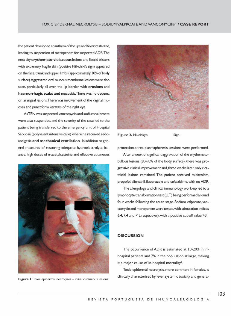
103R E V I S T A P O R T U G U E S A D E I M U N O A L E R G O L O G I A
TOXIC EPIDERMAL NECROLYSIS – SODIUM VALPROATE AND VANCOMYCIN? / CASE REPORT
the patient developed enanthem of the lips and fever restarted,
leading to suspension of meropenem for suspected ADR. The
next day erythemato-violaceous lesions and flaccid blisters
with extremely fragile skin (positive Nilkolski’s sign) appeared
on the face, trunk and upper limbs (approximately 30% of body
surface). Aggravated oral mucous membrane lesions were also
seen, particularly all over the lip border, with erosions and
haemorrhagic scabs and mucositis. There was no oedema
or laryngeal lesions. There was involvement of the vaginal mu-
cosa and punctiform keratitis of the right eye.
As TEN was suspected, vancomycin and sodium valproate
were also suspended, and the severity of the case led to the
patient being transferred to the emergency unit of Hospital
São José (polyvalent intensive care) where he received sedo-
analgesia and mechanical ventilation. In addition to gen-
eral measures of restoring adequate hydroelectrolyte bal-
ance, high doses of n-acetylcysteine and effective cutaneous
protection, three plasmapheresis sessions were performed.
After a week of significant aggravation of the erythemato-
bullous lesions (80-90% of the body surface), there was pro-
gressive clinical improvement and, three weeks later, only cica-
tricial lesions remained. The patient received midazolam,
propofol, alfentanil, fluconazole and ceftazidime, with no ADR.
The allergology and clinical immunology work-up led to a
lymphocyte transformation test (LLT) being performed around
four weeks following the acute stage. Sodium valproate, van-
comycin and meropenem were tested, with stimulation indices
6.4; 7.4 and < 2,respectively, with a positive cut-off value >3.
DISCUSSION
The occurrence of ADR is estimated at 10-20% in in-
hospital patients and 7% in the population at large, making
it a major cause of in-hospital mortality6.
Toxic epidermal necrolysis, more common in females, is
clinically characterised by fever, systemic toxicity and genera-Figure 1. Toxic epidermal necrolyses – initial cutaneous lesions.
Figure 2. Nikolsky’s Sign.
Imuno (19) 2 - Miolo 2ª PROVA EN.indd 103Imuno (19) 2 - Miolo 2ª PROVA EN.indd 103 15-07-2011 12:00:2215-07-2011 12:00:22

104R E V I S T A P O R T U G U E S A D E I M U N O A L E R G O L O G I A
lised epidermal necrosis with cutaneous exfoliation and mu-
cosal erosion, reaching over 30% of the body surface, (more
than 10% in SJS). Differential diagnosis with other conditions
can be difficult , namely with scalded skin syndrome caused
by a staphylococcal toxin (usually with no pain or enanthema)
or toxic shock syndrome caused by Staphylococcus aureus
(with skin desquamation, mainly of the palms of the hands
and the soles of the feet, fever and rapid progress to shock).
Diagnosis is essentially made clinically, but It can be
confirmed by skin biopsy identifying subepidermal phlycte-
na, basal membrane vacuolisation and keratinocyte necro-
sis4,5. A skin biopsy was not performed in our patient as
there were no diagnostic doubts and as polyvalent inten-
sive care was urgently needed.
Several evolution stages are described. The prodromic
stage lasts 2-3 days and is characterised by non-specific
symptoms which begin 1-45 days after exposure. The acute
stage reaches its peak in 2-3 days and the mucosal erosions
usually precede the skin lesions. If there are no complica-
tions, the recovery stage lasts 1-3 weeks4,5.
The aethiopathogenic mechanisms leading to keratinocyte
apoptosis remain to be fully elucidated, but seem to involve
immunologic and metabolic processes, in particular errors in
the xenobiotic detoxification pathways, with accumulation of
immunogenic, metabolites or with a direct cytotoxic effect2.
There are reports of SJS attributed to herpes and My-
coplasma infections, neoplasms, radiotherapy, but mainly
drugs. On the other hand, TEN cases are almost always
drug induced4.
The interaction between drugs and viruses (HIV, EBV
or HHV-6, among others) seems to play a facilitator role
in hypersensitivity phenomena. Viral infections, auto-im-
mune diseases or other xenobiotics can interfere with
drug metabolism or with their recognition by the immune
system and, thus, induce sensitisation2.
Over a hundred TEN-associated drugs have been re-
ported. Sulphonamides are the most frequently implicated
(in around one-third of adults), but also anti-epileptic drugs,
alopurinol, oral penicillins and NSAIDs (namely those derived
from pirazolone or oxicam, with longer half-lives)4. Beta-
lactams can also trigger TEN. Meropenem has been report-
ed in some cases which mention cross-reactivity with cepha-
losporins in patients with TEN, due to probable involvement
of the beta-lactam ring. While this was one of the drugs
initially suspected, this was not confirmed in vitro. We highlight
in addition the reintroduction of beta-lactams (ceftazidime)
during the hospital stay, without recurrence of the lesions.
Vancomycin is also implicated in these cases, although
more rarely4. This is a glycopeptide used in MRSA infec-
tions and coagulase-negative staphylococci. The most com-
mon adverse reaction is “red man syndrome” (erythema
and pruritus, sometimes with hypotension), resulting
from mast cell degranulation by non-immunological mech-
anisms. Other ADRs associated with vancomycin include
neutropenia, oto- and nephrotoxicity, interstitial nephritis
and SJS/TEN. Similar cases of NET have emerged, but these
have been associated with linear IgA deposits. This is an
important differential diagnosis in suspected SJS/TEN7.
As an alternative, some authors have suggested line-
zolid or streptogramins. Teicoplanin and vancomycin be-
long to the same class of drugs, meaning there could be a
higher risk of cross-reactivity. Other alternative drugs
could be suggested after sensitivity tests7.
Hypersensitivity to anti-epileptic drugs could be specific to
the molecule or class. These drugs are divided into phenytoins,
barbiturates (phenobarbital, primidone), iminostilbenes (car-
bamazepine), succinimides (etosuximide), sodium valproate,
oxazolidinediones (trimetadione), benzodiazepines (diazepam,
clonazepam, clorazepate dipotassium and parenteral loraze-
pam) and other (gabapentine, lamotrigine and vigabatrin)8.
João Antunes, Sara Lestre, Andreia Gonçalves, André Borges, Regina Viseu, Isabel Baptista
Imuno (19) 2 - Miolo 2ª PROVA EN.indd 104Imuno (19) 2 - Miolo 2ª PROVA EN.indd 104 15-07-2011 12:00:2415-07-2011 12:00:24

105R E V I S T A P O R T U G U E S A D E I M U N O A L E R G O L O G I A
Rzany and colleagues9 identified 352 cases of SJS/TEN
and 21% of these were attributed to anti-epileptic drugs.
The most frequently implicated were phenobarbital, car-
bamazepine, phenytoins, valproate (13 patients) and, in only
three patients, lamotrigine. The association between val-
proate and SJS/TEN is somewhat biased by the concomi-
tant use of other drugs, namely NSAIDs, in several patients
studied. The majority of ADRs occurred within the first
eight weeks of treatment9.
Reactions to anti-epileptic drugs, namely in patients with
DRESS syndrome, were initially described to aromatic drugs
(i.e., phenytoin, phenobarbital and carbamazepine), with an
estimated 40-80% cross-reactivity between them 8. How-
ever, there are also reports of severe ADR to non-aromatic
anti-epileptic drugs, (i.e.,,valproate, gabapentine or lamotrig-
ine). Thus, in cases of hypersensitivity to valproate, alternative
treatments between classes with lesser risk of cross-reac-
tivity should be sought, namely benzodiazepines8.
The role of in vitro tests in diagnosing ADR, namely LLT
and the basophil activation test, has received a great deal
of attention over the last thirty years, due to their safety
and comfort for the patient, since these can avoid possible
sensitisation with re-exposure to the drug and allow the
mechanisms involved in the reaction to be elucidated2.
Several studies have been developed with this technique
and their results have been encouraging, suggesting a sen-
sitivity of 60-70% and a specificity of 85-100%10. The main
limitations and methodological difficulties of LLTs’ are the
use of stimulation indices (SI) as sensitisation indicators, and
the timing of carrying out the test. This is as the T memory
response can vary depending on the allergen in question
and the type of reaction. An interval of 4-8 weeks to 3-6
months after the acute stage is recommended. In patients
with SJS/TEN, LLT tests are positive in less than 10% of
cases, and an interval less than six weeks is suggested to
increase the test’s sensitivity10. False positive results can
occur, particularly to vancomycin and contrast products10.
However, the increased SI obtained and the severity of the
reaction make this test contraindicated in our patient.
Skin tests in patients with SJS/TEN are not recom-
mended by several authors due to the risk of re-exposure
to the trigger drug. However, these can be considered in
studying hypersensitivity to beta-lactams as there is little
likelihood of a reaction to meropenem.
CONCLUSION
This was a rare and severe case of TEN caused by reaction
to drugs.
Although still a research tool, LLT was decisive to con-
firm the immunological basis of the reaction. Even though
identification of the implicated drugs is not conclusive, in
view of the inherent limitations of the in vitro study, the
use of vancomycin and sodium valproate was contraindi-
cated in this patient.
Finally, the role of concomitant infections, drug interac-
tions and genetic susceptibility factors in the immunop-
athogenic mechanisms remains to be elucidated.
Funding: None
Conflict of interest disclosure: None
Contact:João Antunes – [email protected]
BIBLIOGRAFIA
1. WHO. International drug monitoring: the role of national centres.
Tech Rep Ser 1972;498:1 -25.
TOXIC EPIDERMAL NECROLYSIS – SODIUM VALPROATE AND VANCOMYCIN? / CASE REPORT
Imuno (19) 2 - Miolo 2ª PROVA EN.indd 105Imuno (19) 2 - Miolo 2ª PROVA EN.indd 105 15-07-2011 12:00:2415-07-2011 12:00:24

106R E V I S T A P O R T U G U E S A D E I M U N O A L E R G O L O G I A
2. Torres MJ, Mayorga C, Blanca M. Nonimmediate allergic reactions
induced by drugs: pathogenesis and diagnostic tests. J Investig
Allergol Clin Immunol 2009;19:80 -90.
3. Roujeau JC, Stern RS. Severe adverse cutaneous reactions to
drugs. N Engl J Med 1994;331:1272 -85.
4. Auquier -Dunant A, Mockenhaupt M, Naldi L, Correia O, Schro-
der W, Roujeau JC; SCAR Study Group. Severe Cutaneous
Adverse Reactions. Correlations between clinical patterns
and causes of erythema multiforme majus, Stevens -Johnson
syndrome, and toxic epidermal necrolysis: results of an in-
ternational prospective study. Arch Dermatol 2002;138:1019-
-24.
5. Cabral L, Diogo C, Riobom F, Teles L, Cruzeiro C. Necrólise epidér-
mica tóxica (Síndrome de Lyell): uma patologia para as unidades
de queimados. Acta Med Port 2004;17:129 -40.
6. Guglielmi L, Guglielmi P, Demoly P. Drug hypersensitivity: epidemiol-
ogy and risk factors. Curr Pharm Des 2006;12:3309 -12.
7. Rocha JLL, Kondo W, Baptista MIDK, Cunha CA, Martins LTF. Un-
common vancomycin -induced side effects. Braz J Infect Dis
2002;6:196 -200.
8. Bohan KH, Mansuri TF, Wilson NM. Anticonvulsant hypersensitivity
syndrome: implications for pharmaceutical care. Phamacotherapy
2007;27:1425:39.
9. Rzany B, Correia O, Kelly JP, Naldi L, Auguier A, Stern R. Risk of
Stevens -Johnson syndrome and toxic epidermal necrolysis during
first weeks of antiepileptic therapy: a case -control study. Study Group
of the International Case Control Study on Severe Cutaneous Ad-
verse Reactions. Lancet 1999;353:2190 -4.
10. Pichler WJ, Tilch J. The lymphocyte transformation test in the diag-
nosis of drug hypersensitivity. Allergy 2004;59:809 -20.
João Antunes, Sara Lestre, Andreia Gonçalves, André Borges, Regina Viseu, Isabel Baptista
Imuno (19) 2 - Miolo 2ª PROVA EN.indd 106Imuno (19) 2 - Miolo 2ª PROVA EN.indd 106 15-07-2011 12:00:2415-07-2011 12:00:24
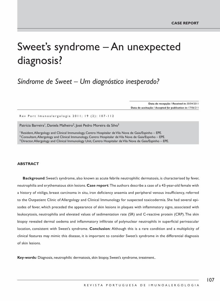
107R E V I S T A P O R T U G U E S A D E I M U N O A L E R G O L O G I A
CASE REPORT
ABSTRACT
Background: Sweet’s syndrome, also known as acute febrile neutrophilic dermatosis, is characterised by fever,
neutrophilia and erythematous skin lesions. Case report: The authors describe a case of a 43 -year -old female with
a history of vitiligo, breast carcinoma in situ, iron deficiency anaemia and peripheral venous insufficiency, referred
to the Outpatient Clinic of Allergology and Clinical Immunology for suspected toxicodermia. She had several epi-
sodes of fever, which preceded the appearance of skin lesions in plaques with inflammatory signs, associated with
leukocytosis, neutrophilia and elevated values of sedimentation rate (SR) and C -reactive protein (CRP). The skin
biopsy revealed dermal oedema and inflammatory infiltrate of polynuclear neutrophils in superficial perivascular
location, consistent with Sweet’s syndrome. Conclusion: Although this is a rare condition and a multiplicity of
clinical features may mimic this disease, it is important to consider Sweet’s syndrome in the differential diagnosis
of skin lesions.
Key -words: Diagnosis, neutrophilic dermatosis, skin biopsy, Sweet’s syndrome, treatment..
Sweet’s syndrome – An unexpected diagnosis?
Síndrome de Sweet – Um diagnóstico inesperado?
Patrícia Barreira1, Daniela Malheiro2, José Pedro Moreira da Silva3
1 Resident, Allergology and Clinical Immunology, Centro Hospitalar de Vila Nova de Gaia/Espinho – EPE.2 Consultant, Allergology and Clinical Immunology, Centro Hospitalar de Vila Nova de Gaia/Espinho – EPE.3 Director, Allergology and Clinical Immunology Unit, Centro Hospitalar de Vila Nova de Gaia/Espinho – EPE.
R e v P o r t I m u n o a l e r g o l o g i a 2 0 1 1 ; 1 9 ( 2 ) : 1 0 7 - 1 1 2
Data de recepção / Received in: 30/04/2011
Data de aceitação / Accepted for publication in: 17/06/211
Imuno (19) 2 - Miolo 2ª PROVA EN.indd 107Imuno (19) 2 - Miolo 2ª PROVA EN.indd 107 15-07-2011 12:00:2415-07-2011 12:00:24

108R E V I S T A P O R T U G U E S A D E I M U N O A L E R G O L O G I A
INTRODUCTION
Sweet’s syndrome was fi rst described, in 1964, by Rob-
ert Douglas Sweet. Since then hundreds of cases of
this disease have been published1,2. Clinically, it is char-
acterised by fever (the most frequent sign) and skin lesions
(erythemato -violaceous papules/nodules/plaques), which
can develop simultaneously with fever or days to weeks
after3. These lesions are characteristically painful, asymmet-
rically distributed (with the most frequent locations being
the upper extremities, face and neck) and clear up with no
residual lesion. Extra -cutaneous manifestations have been
described, such as muscle pain, joint pain, headaches, gen-
eral malaise, conjunctivitis and ulcers of the oral mucosa4.
There are three different forms of this syndrome: classic/
idiopathic (Classical Sweet’s Syndrome – CSS), associated
with neoplastic diseases (Malignancy -Associated Sweet’s
Syndrome – MASS) and drug induced (Drug -Induced Sweet’s
Syndrome – DISS)5. The most frequently involved malignan-
cies are haematological (acute myeloid leukaemia), but
cases associated to solid tumours (breast, gastrointestinal
and genitourinary) have also been described4.
Sweet’s syndrome can precede, accompany or appear af-
ter a diagnosis of malignant disease. It should, thus, raise a red
flag, whether for a malignancy as yet unknown, or for a relapse
in a cancer patient. Granulocyte -colony stimulating factor
(G -CSF) is the drug most frequently associated with Sweet’s
syndrome, but many others can be involved, such as antibiot-
ics, anti -epileptic drugs, antiretroviral drugs, anti -hypertensive
drugs, chemotherapy drugs, anti -psychotic drugs, oral contra-
ceptives, diuretics and non -steroidal anti -inflammatory drugs
– NSAIDs). Diagnosis is made based on the criteria listed in
Table I6. In laboratory terms, the changes seen are increased
inflammatory markers, namely the sedimentation rate and the
presence of leukocytosis with neutrophilia.
Given the plethora of clinical situations that can simulate
this pathology, diagnosis is not always easy. The skin biopsy can
reveal a diffuse infiltrate of mature neutrophils located in the
upper dermis, with no evidence of leukocytoclastic vasculitis.
These histopathological features are not pathognomonic of
the disease. They can be seen in other neutrophilic dermatoses
and in lesions caused by infectious agents. Its pathogenesis is
not yet fully understood, but is probably multifactorial. Sys-
temic corticosteroids, potassium iodate and colchicine are the
Patrícia Barreira, Daniela Malheiro, José Pedro Moreira da Silva
RESUMO
Introdução: O Síndrome de Sweet, também designado por dermatose neutrofílica febril aguda, é caracterizado por febre,
neutrofilia e lesões cutâneas eritematosas. Caso clínico: Os autores descrevem o caso de uma doente de 43 anos, com ante-
cedentes de vitiligo, carcinoma da mama in situ, anemia ferropénica e insuficiência venosa periférica, referenciada à Consulta de
Imunoalergologia por suspeita de toxicodermia. A doente apresentou vários episódios de febre, a preceder o aparecimento de
lesões cutâneas em placa com sinais inflamatórios, associada a leucocitose com neutrofilia e elevação dos valores de velocidade
de sedimentação (VS) e proteína C reactiva (PCR). A biópsia cutânea revelou edema da derme e infiltrado inflamatório de poli-
nucleares neutrófilos em localização perivascular superficial, compatível com Síndrome de Sweet. Conclusão: Apesar de ser
uma patologia rara e de existir uma multiplicidade de situações clínicas que o podem simular, é importante ter sempre presen-
te o Síndrome de Sweet no diagnóstico diferencial de lesões cutâneas.
Palavras -chave: Biópsia cutânea, dermatose neutrofílica, diagnóstico, Síndrome de Sweet, tratamento.
Imuno (19) 2 - Miolo 2ª PROVA EN.indd 108Imuno (19) 2 - Miolo 2ª PROVA EN.indd 108 15-07-2011 12:00:2415-07-2011 12:00:24

109R E V I S T A P O R T U G U E S A D E I M U N O A L E R G O L O G I A
SWEET’S SYNDROME – AN UNEXPECTED DIAGNOSIS? / CASE REPORT
Table I. Diagnostic criteria of Sweet’s syndrome.
Criteria CSS/MASS DISS1 Abrupt onset of painful erythematous lesions (plaques/nodules)
2 Histopathological evidence of dense neutrophilic infi ltrate (without leukocytoclastic vasculitis
3 Pyrexia (> 38 ºC)
4
Association with pregnancy, an underlying haematologi-cal or visceral malignancy, or infl ammatory disease, or preceded by an upper respiratory or gastrointestinal infection, or vaccination
Time relationship between drug administration and onset of clinical picture or recurrence of picture after oral challenge test
5 Excellent response to treatment with systemic corti-costeroids or potassium iodide
Resolution of lesions after suspension of drug or treat-ment with systemic corticosteroids
6 Abnormal laboratory values at presentation (≥ 3): SR > 20 mm/h; positive CRP; leucocytes...; neutrophils...
Diagnosis 2 major criteria (1 – 2) +≥ 2 minor criteria (3 – 6) All 5 criteria
(adapted from Joe EK. Sweet Syndrome. Dermatology Online Journal. 2003;9(4):28).
Table II. Clinical characteristics of the different forms of Sweet’s syndrome.
CharacteristicsClinical forms
CSS Haemataologic malignancy
Solid malignancy DISS
Epidemiology80 50 59 71
Preceding upper airway infection or gastrointestinal infection 75-90 16 20 21
Recurrence 30 69 41 67
Symptoms/SignsPyrexia>38°C 80-90 88 79 100
Musculoskeletal involvement 12-56 26 34 21
Ocular involvement 17-72 7 15 21
Lesion locationUpper extremities 80 89 97 71
Head and neck 50 63 52 43
Trunk 30 42 33 50
Lower extremities Infreq 49 48 36
Oral mucosa 2 12 3 7
Laboratory fi ndingsNeutrophils>6000/μL 80 47 60 38
SR>20mm/h 90 100 95 100
Anaemia (Hb<13g/dL; <12g/dL) Infreq 82 83 100
Plt<150000/μL or >500000/μL Infreq 68 50 50
RF, haematuria, proteinuria 11-50 15 7 0
(adapted from Cohen PR. Sweet’s syndrome – a comprehensive review of an acute febrile neutrophilic dermatosis. Orphanet Journal of Rare Diseases. 2007;2:34).
CSS: Classical Sweet’s Syndrome; DISS: Drug -Induced Sweet’s Syndrome; Plt – platelets; RF – renal failure; SR – sedimentation rate
Imuno (19) 2 - Miolo 2ª PROVA EN.indd 109Imuno (19) 2 - Miolo 2ª PROVA EN.indd 109 15-07-2011 12:00:2515-07-2011 12:00:25

110R E V I S T A P O R T U G U E S A D E I M U N O A L E R G O L O G I A
first -line drugs for treatment7. The prognosis varies, in line
with the form of disease involved. This means that in the case
of MASS and DISS it is related to the evolution of the under-
lying malignancy and the ingestion/suspension of the drug,
respectively. In the case of CSS, it can resolve spontaneously
or take months to weeks to resolve, with recurrence seen in
one -third of cases. Table II summarises the clinical character-
istics of the different forms of Sweet’s syndrome.
CASE REPORT
A 43 -year -old female patient was referred to our De-
partment of Allergology and Clinical Immunology for sus-
pected toxicodermia. She had a history of vitiligo, diag-
nosed when she was 16 years old, breast carcinoma in situ
(for which she had undergone radiotherapy and hormone
therapy), iron deficiency anaemia, since the age of 39, and
superficial venous thrombosis of the great saphenous vein
at the age of 42. She had no known allergy to drugs.
The patient was admitted to the Emergency Room
(ER) with a 48 -hour picture of fever (maximum tempera-
ture of 38.5 .ºC), headaches, nausea and a vomiting episode.
There were no other complaints, namely skin complaints.
Examination revealed the patient to be subfeverish and
with reddened oropharynx (with no discharge or exudate),
with no other abnormalities. The analytical study revealed
hypochromic microcytic anaemia (with no aggravation of
the known status), leukocytosis 15470/uL, with relative
neutrophilia of 89.5%, increased SR (49 mm) and slightly
raised CRP (0.7 mg/dL). The patient was medicated with
10 mg IV metoclopramide and 1000 mg oral paracetamol.
Symptoms improved, and the patient was discharged, with
the recommendation to be vigilant and was medicated
with paracetamol.
The patient returned to the ER around 24hrs afterwards,
for continued headaches and nausea, with recent onset of skin
lesions on the trunk and signs of inflammation, with no associ-
ated pruritus. She had no fever and had papular erythematous
lesions, painful to the touch, located on the abdomen and dor-
sum. Analyses revealed no leukocytosis or neutrophilia, but there
was a CRP increase to 12.07 mg/dL. The patient was medicated
with 1800 mg IV lysine acetylsalicylate, 100 mg oral doxycycline
and 25 mg oral hydroxizine, and admitted to the Internal Med-
icine Unit for a diagnostic work -up. This showed decreased IgA
(< 6.34 mg/dL), with no other immunological changes. Renal,
liver and thyroid function, ionogram, beta 2 -microglobulin and
protein electrophoresis showed no relevant changes. Urinalysis,
chest X -ray, study of viral markers, blood culture panel, urocul-
tures and serology exams for several pathogenic agents did not
reveal infectious foci for the clinical picture described.
During the hospital stay, the patient remained apyretic,
with gradual regression of the lesions. These became more
localized to the right quadrants of the abdomen, maintain-
ing the inflammatory characteristics. The patient was re-
ferred to a dermatology appointment, which considered
the cause to be a viral infection or an NSAID -induced
maculopapular morbilliform toxicoderma. The patient was
discharged with the recommendation to avoid NSAIDs,
was medicated with 100 mg doxycycline, and referred to
the Outpatient Clinic of Allergology and Clinical Immunol-
ogy for suspected toxicodermia.
By the date of examination, the patient had had four
new episodes similar to that already described: fever (with
no apparent cause) with generalized malaise and asthenia
preceding the onset of skin lesions with non -pruriginous
signs of inflammation on the trunk, with no triggering fac-
tor identified. These lasted approximately three to five days
and had spontaneous resolution, leaving no residual lesion.
During one of these episodes the patient resorted to an
Patrícia Barreira, Daniela Malheiro, José Pedro Moreira da Silva
Imuno (19) 2 - Miolo 2ª PROVA EN.indd 110Imuno (19) 2 - Miolo 2ª PROVA EN.indd 110 15-07-2011 12:00:2515-07-2011 12:00:25

111R E V I S T A P O R T U G U E S A D E I M U N O A L E R G O L O G I A
ER, where she was medicated with 1800 mg lysine acetyl-
salicylate, with good tolerance.
The question of this being a case of Sweet’s syndrome
was raised, and a skin biopsy scheduled should the lesions
recur, which happened a month later (Figures 1 and 2). The
biopsy revealed “oedema of the dermis and slight inflam-
matory infiltrate of polynuclear neutrophils with superficial
perivascular location,that is, neutrophilic dermatitis lesions,
compatible with Sweet’s syndrome”.
The patient was medicated with 60mg/day oral pred-
nisolone for six days, at the end of which she was re-
-evaluated. As the lesions had resolved, treatment was
suspended and surveillance continued. The patient has not
experienced further episodes since, wherefore there has
been no need for a new cycle of corticosteroids.
DISCUSSION
When the patient was first observed at our Allergology
and Clinical Immunology Clinic, she had already undergone
extensive tests to identify the causes of her clinical condition.
Given her fever (with no apparent cause) associated with the
analytical inflammatory parameters (leukocytosis with neu-
trophilia, increased SR and CRP), the diagnosis initially sug-
gested was an infectious disease, namely caused by Chlamyd-
ia, Rickettsia, Brucella or Borrelia burgdorferi, which warranted
treatment with doxycycline. The non -identification of a patho-
genic agent and the evolution of the disease in recurrent
episodes (with the patient completely asymptomatic between
those episodes) did not suggest an infectious condition. That
the patient had a drug -triggered (NSAIDs) cutaneous reaction
was also suggested and this led to the patient being referred
to us. However, the skin lesions were already present when
the patient was medicated with lysine acetylsalicylate and, in
addition, the subsequent administration of this drug with good
tolerance ruled out a drug -related hypersensitivity mecha-
nism. Sweet’s syndrome was suggested as the most likely
diagnostic probability and this was confirmed by histology.
A skin biopsy is essential in diagnosing Sweet’s syn-
drome and this and the characteristic skin lesions are ma-
jor criteria. In terms of minor criteria, the patient had fever
SWEET’S SYNDROME – AN UNEXPECTED DIAGNOSIS? / CASE REPORT
Figures 1 and 2. Cutaneous lesions with characteristics of inflammation, spread over back; vitiligo lesions also visible – patient already presented vitiligo.
Imuno (19) 2 - Miolo 2ª PROVA EN.indd 111Imuno (19) 2 - Miolo 2ª PROVA EN.indd 111 15-07-2011 12:00:2515-07-2011 12:00:25

112R E V I S T A P O R T U G U E S A D E I M U N O A L E R G O L O G I A
(temperature > 38 ºC), laboratory abnormalities and good
response to corticotherapy6.
CONCLUSION
Given the severity of the pathologies that can be associ-
ated, Sweet’s syndrome should primarily be considered as a
systemic manifestation of an underlying disease. Histological
confirmation of this syndrome should, thus, not be taken to
be the end of the diagnostic workup8. Given our patient’s
history of breast cancer, a diagnosis of Sweet’s syndrome
could represent the first sign of a recurrence of malignancy.
Since there were no new episodes after administration of
corticotherapy, we can consider this as an idiopathic form of
the disease. However, we stress the importance of a rigorous
follow -up and multidisciplinary approach in these cases.
Although it is a rare condition and a multiplicity of
clinical conditions may mimic this disease, it is important
to consider Sweet’s syndrome in the differential diagnosis
of skin lesions.
Funding: None
Conflict of interest disclosure: None
Contacto:Patrícia Cristina Correia BarreiraServiço de Imunoalergologia, Centro Hospitalar Vila Nova Gaia/Espinho – EPERua Conceição Fernandes 4434 – 502 Vila Nova de Gaia
REFERENCES
1. Gibson LE. Sweet Syndrome. Mayo Clin Proc. 2005;80(4):549.
2. Cohen PR, Kurzrock R. Sweet´s syndrome revisited: a review of
diseases concepts. Int J Dermatol. 2003;42(10):761 -78.
3. Yi S, Bhate C, Scwartz RA. Sweet´s Syndrome: an update and review.
G Ital Dermatol Venereol. 2009;144(5):603 -12.
4. Cohen PR. Sweet´s syndrome – a comprehensive review of an acute
febril neutrophilic dermatosis. Orphanet Journal of Rare Diseases.
2007;2:34.
5. Honigsmann H, Cohen P, Wolff K. Acute febrile neutrophilic derma-
tosis (Sweet´s Syndrome). In: Freedberg IM, Eisen AZ, Wolff K, Austen
KF, Goldsmith LA, Katz S (Eds). Fitzpatrick‘s Dermatology in Gen-
eral Medicine. 6th ed. McGraw -Hill Professional. 2003:1056 –
1062.
6. Joe EK. Sweet Syndrome. Dermatology Online Journal. 2003;
9(4):28.
7. Cohen PR. Neutrophilic Dermatosis: a review of current treatment
options. Am J Clin Dermatol. 2009;10(5):301 -12.
8. Gonçalves P, Miranda JS, Araújo JAM. Síndrome de Sweet e Doença
Inflamatória Intestinal – uma associação pouco frequente. Medicina
Interna. 2010;17(1):44 -47.
Patrícia Barreira, Daniela Malheiro, José Pedro Moreira da Silva
Imuno (19) 2 - Miolo 2ª PROVA EN.indd 112Imuno (19) 2 - Miolo 2ª PROVA EN.indd 112 15-07-2011 12:00:2615-07-2011 12:00:26