Embed Size (px)
Citation preview

1
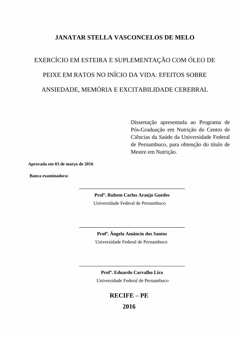
JANATAR STELLA VASCONCELOS DE MELO
EXERCÍCIO EM ESTEIRA E SUPLEMENTAÇÃO COM ÓLEO DE
PEIXE EM RATOS NO INÍCIO DA VIDA: EFEITOS SOBRE
ANSIEDADE, MEMÓRIA E EXCITABILIDADE CEREBRAL
RECIFE/PE
2016

1
JANATAR STELLA VASCONCELOS DE MELO
EXERCÍCIO EM ESTEIRA E SUPLEMENTAÇÃO COM ÓLEO DE
PEIXE EM RATOS NO INÍCIO DA VIDA: EFEITOS SOBRE
ANSIEDADE, MEMÓRIA E EXCITABILIDADE CEREBRAL
RECIFE/PE
2016
Dissertação apresentada ao Programa de
Pós-Graduação em Nutrição do Centro de
Ciências da Saúde da Universidade
Federal de Pernambuco, para obtenção do
título de Mestre em Nutrição.
Orientadora: Profª Drª Manuella Batista-de-
Oliveira Hornsby, professora adjunta do
Departamento de Nutrição da UFPE.

2
3
JANATAR STELLA VASCONCELOS DE MELO
EXERCÍCIO EM ESTEIRA E SUPLEMENTAÇÃO COM ÓLEO DE
PEIXE EM RATOS NO INÍCIO DA VIDA: EFEITOS SOBRE
ANSIEDADE, MEMÓRIA E EXCITABILIDADE CEREBRAL
____________________________________________
Profº. Rubem Carlos Araújo Guedes
Universidade Federal de Pernambuco
____________________________________________
Profª. Ângela Amâncio dos Santos
Universidade Federal de Pernambuco
____________________________________________
Profº. Eduardo Carvalho Lira
Universidade Federal de Pernambuco
RECIFE – PE
2016
Aprovada em 03 de março de 2016
Banca examinadora:
Dissertação apresentada ao Programa de
Pós-Graduação em Nutrição do Centro de
Ciências da Saúde da Universidade Federal
de Pernambuco, para obtenção do título de
Mestre em Nutrição.

4
Dedico este trabalho à minha mãe, DJANIRA, por toda sua dedicação durante esses anos de
minha formação.

5
AGRADECIMENTOS
Agradeço à Deus por todas as bênçãos derramadas em minha vida e pela certeza de que estás
sempre ao meu lado, me guiando em cada etapa de minha vida;
À minha mãe, Djanira. Sempre ao meu lado em todos os momentos. Sem você não teria
conseguido ir tão longe;
À meu pai Severino Targino de Melo (in memoriam). Sei que se estivesses aqui, mesmo
distante, se alegrarias com mais uma vitória de sua filha “Janatinha”;
À meu PAIdastro, José, que me acolheu como sua filha e que se alegra com minhas vitórias;
À toda minha família e amigos;
À minha orientadora Manuella Batista, pela orientação, ensinamentos durante as etapas de
realização desse trabalho;
À minhas amigas “Mosqueteiras”, Patrícia e Laís. Pergunto-me como seriam esses dois anos
sem vocês e chego à conclusão que não seria fácil. Deus preparou tudo de uma forma tão
especial, que olho para trás e vejo o longo caminho que trilhamos sempre confiantes de que
tudo iria acabar bem. Amigas para sempre;
Às minhas companheiras paraibanas de mestrado, Larissa Brito e Bárbara Paulino, quantas
conversas entre estados, sempre nos dando forças;
Aos estagiários, Keyla Torres, Rai Santiago e Roberta Ferrer, pela ajuda durante as longas
horas de experimentos;
Ao professor Rubem Guedes pelos ensinamentos e acolhimento e a todos do LAFINNT, por
toda disponibilidade que foi imprescindível para conclusão deste trabalho;
Às professoras Cláudia Lagranha e Mariana Fernandes, ao professor Eduardo Lira, pelas
parcerias nos diversos trabalhos desenvolvidos;
Ao médico veterinário Edeone França, pelos cuidados aos animais e ensinamentos sobre o
cuidado com os animais de laboratório;

6
Ao Programa de Pós-graduação em Nutrição –UFPE e às secretárias do programa de pós-
graduação, Cecília e Neci, pela disponibilidade;
À CAPES, pela concessão de bolsa de estudos durante os meses de realização deste trabalho;

7
“Porque para Deus nada é impossível.”
(Lucas 1, 37)

8
RESUMO
As fases iniciais da vida representam um período crítico no desenvolvimento do sistema
nervoso. O objetivo desse estudo foi avaliar efeitos da associação entre exercício físico em
esteira e suplementação com óleo de peixe em ratosno início da vida, sobre ansiedade,
memória e excitabilidade cerebral. Ratos Wistar foram divididos em: Óleo de Peixe ou
Veículo e subdivididos em Exercitados ou Sedentários. O período de treinamento ocorreu do
15º ao 45º dias de vida. A partir dos 46 dias de vida, foi realizada avaliação dos efeitos dessa
associação sobre peso corporal, murinometria, respostas comportamentais relacionadas à
ansiedade, memória e eletrofisiologia cortical. Os resultados demonstram que não houve
alteração nos dados murinométricos. No teste de labirinto em cruz elevado, os animais
exercitados apresentaram comportamento menos ansioso a julgar pelo maior número de
entradas nos braços abertos. Além disso, animais suplementados e/ou exercitados
apresentaram memória preservada para reconhecimento da identidade do objeto. Por outro
lado, a análise intragrupo demonstrou prejuízo dependente do tratamento, quando os animais
foram submetidos ao teste de reconhecimento quanto à localização do objeto. Com relação à
análise intergrupo, a suplementação e o exercício aumentaram o índice de discriminação para
o objeto estacionário. Sobre a eletrofisiologia cerebral, houve potencialização da
suplementação sobre o exercício na redução da velocidade de propagação da depressão
alastrante cortical. Neste contexto, os resultados indicam que é necessária cautela no uso da
associação dessas estratégias referentes à modulação comportamental ou neural em períodos
críticos de desenvolvimento do sistema nervoso.
Palavras chaves: Óleos de peixe. Exercício. Sistema nervoso. Período crítico.

9
ABSTRACT
The early stages of life represent a critical period in the development of the nervous system.
The aim of this study was to evaluate how the association between treadmill exercise and
supplementation with fish oil in rats during lactation affect anxiety, memory and brain
excitability. Wistar rats performed two experimental groups as Fish Oil or vehicle, and then
divided into exercised or sedentary. The training period was from the 15th to 45th days of life.
From the 46 days of life, the effects of this combination on body weight, murimetric
parameters, behavioral assessment, and cortical electrophysiology were evaluated. The results
demonstrate that there was no change in murinometric data. In the elevated plus maze,
exercised animals were less anxious as judged by the largest number of entries into the open
arms as compared to the respective controls. Furthermore, supplemented and / or trained
animals showed preserved memory to recognize the identity of the object. On the other hand,
the intra-group analysis showed treatment-dependent impairment when the animals were
subjected to the recognition test on the object location. The inter-group analysis
supplementation and exercise increased the discrimination index for the stationary object. In
addition, supplementation with fish oil enhanced the effect of exercise on brain excitability by
reducing the velocity of propagation of cortical spreading depression. Therefore, the present
data indicate that caution is required in use of the association of these strategies concerning
behavioral or neural modulation.
Key-words: Fish oils. Exercise. Nervous system. Critical period of development.

10
LISTA DE ILUSTRAÇÕES
FIGURA 1 - Desenho experimental/Distribuição dos grupos experimentais (A) e
Cronograma Experimental (B)............................................................................................Pg.23
TABELA 1. Parâmetros do treinamento físico.................................................................Pg. 24
RESULTADOS
FIGURA 2. Peso pós-natal (2A) e dados murinométricos (2B e C) de animais jovens
apresentados como média±EPM........................................................................................Pg. 29
FIGURA 3. Índices de discriminação para os testes de reconhecimento de objeto quanto à
sua identidade (A) e localização espacial (B), em animais jovens sobre diferentes condições
experimentais.....................................................................................................................Pg. 32
FIGURA 4. Velocidade de propagação na DAC (em mm/min) em ratos jovens exercitados
(E) ou sedentários (S), que foram suplementados com óleo de peixe (OP) ou solução veículo
(V)......................................................................................................................................Pg. 33
TABELA 2. Dados murinométricos.................................................................................Pg. 30
TABELA 3. (A) Teste de adaptação ao Campo Aberto (TCA) e (B) Labirinto em Cruz
elevada (LCE)....................................................................................................................Pg. 31

11
LISTA DE SIGLAS E ABREVIATURAS
Ag-AgCl Prata – cloreto de prata
AGI Ácidos graxos insaturados
ANOVA Análise da variância
BDNF Fator neurotrófico derivado do cérebro
CA Circunferência abdominal
CC Comprimento da cauda
CFA Comprimento focinho-ânus
CONCEA Conselho Nacional de Controle de Experimentação Animal
CT Circunferência torácica
DAC Depressão alastrante cortical
DHA Ácido docosaexaenóico
EPA Ácido eicosapentaenóico
EPM Erro padrão da média
IL Índice de Lee
IMC Índice de massa corpórea
KCl Cloreto de potássio
LCE Labirinto em cruz elevado
PCo Peso do coração
PF Peso do fígado
PUFA Ácidos graxos poli-insaturados de cadeia longa
SN Sistema nervoso
SNC Sistema nervoso central
TCA Teste de adaptação ao campo aberto
TRO Teste de reconhecimento de objetos
VLV Variação lenta de voltagem
ω-3 Ômega 3
OP
V
E
S
Grupos Experimentais
Óleo de Peixe
Veículo
Exercitados
Sedentários

12
SUMÁRIO
1. APRESENTAÇÃO 13
2. REVISÃO DA LITERATURA 15
3. OBJETIVOS 20
3.1. Objetivo geral 20
3.2. Objetivos específicos 20
4. HIPÓTESE 21
5. MATERIAIS E MÉTODOS 22
5.1. Aspectos éticos e delineamento experimental 22
5.2. Procedimentos gerais para o exercício físico 23
5.3. Determinações ponderais 24
5.3.1. Evolução ponderal 24
5.3.2. Avaliação murinométrica 24
5.4. Análise comportamental 25
5.4.1. Labirinto em cruz elevado (LCE) 25
5.4.2. Teste de adaptação ao Campo Aberto (TCA) 25
5.4.3. Teste de reconhecimento de objetos (TRO) 26
5.5. Registro eletrofisiológico 27
6. Análise Estatística 28
7. RESULTADOS 29
8. DISCUSSÃO 34
9. CONSIDERAÇÕES FINAIS 37
REFERÊNCIAS 38
APÊNDICE-A 44
APÊNDICE-B 69
ANEXO - A 93
13
1. APRESENTAÇÃO
As fases iniciais da vida representam um período crítico no desenvolvimento do
sistema nervoso (SN). Nesta fase da vida, ocorrem processos de hiperplasia, hipertrofia,
mielinização e migração neuronal com velocidade máxima, em relação a outras etapas da
vida pós-natal, o que torna o cérebro vulnerável às demandas do ambiente, como por
exemplo, as nutricionais (DOBBING, 1968; MORGANE et al., 1993).
Em ratos de laboratório, esse período crítico de desenvolvimento do SN é representado
pela fase pré-natal perdurando até a terceira semana pós-natal, coincidindo com o
desmame (CHEN & SU, 2013; HASHIMOTO et al., 2014).
Neste contexto, um dos principais avanços científicos nos últimos 40 anos foi o
reconhecimento da estreita relação entre o consumo de lipídios dietéticos, a composição
lipídica do encéfalo e sua repercussão sobre o desenvolvimento e funcionamento do
sistema nervoso central (SNC). E devido aos efeitos benéficos dos ácidos graxos poli-
insaturados de cadeia longa (PUFA, polyunsaturated fatty acids) do tipo ômega 3 (ω-3),
comumente presentes no óleo de peixe (OP), boa parte dos estudos tem sido direcionados
para essa classe de lipídeos (YEHUDA et al., 2005; PERINI et al., 2010).
Além dos fatores dietéticos, o exercício físico moderado tem sido investigado pela
comunidade científica devido sua: (1) influência sobre a função e plasticidade cerebral
(RACHETTI et al., 2013), (2) modulação da neurogênese hipocampal (DURING & CAO,
2006; VAN PRAAG et al., 1999) e (3) prevenção da ocorrência de doenças psiquiátricas
(GOMEZ-PINILLA, 2006).
Dessa forma, nos últimos anos o interesse na suplementação de PUFA do tipo ômega
3 (ω-3) e/ou exercício físico aumentou significativamente, sendo demonstrado em muitos
estudos os efeitos benéficos dessas estratégias sobre a maturação cortical, sinaptogênese e
mielinização, redução do risco de déficits cognitivos e inflamação, bem como benefícios
neuroplásticos na juventude e envelhecimento (DIK et al., 2003; BOURRE, 2005;
ENGSTRÖM et al., 2009; VINES et al., 2011; NYBERG et al., 2013).
Além disso, esta associação torna-se importante para a prevenção do desenvolvimento
de distúrbios relacionados à ansiedade (GOMEZ-PINILLA & YING, 2010), uma vez que,
segundo dados epidemiológicos, estes distúrbios estão entre as doenças mentais mais
comuns, acometendo de 3 a 18% da população mundial a cada ano (RAVIDRAN &
SILVA, 2013).
14
Apesar de atualmente a literatura apresentar diversas informações clínicas e
experimentais acerca dos efeitos da suplementação com ácidos graxos insaturados (AGI)
e/ou exercício físico sobre diversas condições, incluindo distúrbios cardiovasculares,
neurológicos e psiquiátricos, há uma preocupação da comunidade científica quanto à
influência dessas estratégias, de forma isolada ou associada, nas fases iniciais da vida,
bem como a duração e dose necessárias para garantir o bom desenvolvimento e
funcionamento dos sistemas orgânicos.
Assim, a pergunta que norteou a realização deste trabalho foi: a associação entre
exercício físico em esteira e suplementação com óleo de peixe no início da vida apresenta
efeitos positivos sobre parâmetros relacionados à ansiedade, memória episódica,
excitabilidade cortical e murinometria em ratos jovens Wistar? A partir desta pergunta
condutora foi hipotetizado que a associação entre a suplementação com óleo de peixe
(dose de 85mg/kg) e o exercício físico em esteira no início da vida mantém a
murinometria adequada para idade. Além de promover efeitos positivos relacionados ao
desempenho dos animais em avaliações comportamentais, melhorando: (1) as respostas
aos testes para reconhecimento de objetos (TRO) e, consequentemente, memória
episódica e (2) reduzindo indicadores ansiogênicos a julgar pelo comportamento durante a
realização da tarefa no teste de adaptação ao campo aberto (TCA) e no labirinto em cruz
elevado (LCE). Bem como promove a diminuição da excitabilidade cerebral através da
redução da velocidade de propagação da depressão alastrante cortical (DAC).
Diante disto, a abordagem proposta neste estudo está distante do uso de tratamentos de
alto custo, uma vez que utiliza a suplementação com o OP e/ou exercício físico em esteira,
que representa uma abordagem natural, de baixo custo e acessível, cuja simplicidade se
traduz em dois aspectos básicos da vida – nutrição e movimento.
Assim, a proposta deste estudo foi avaliar os efeitos da associação da suplementação
com OP e exercício físico em esteira no início da vida de ratos Wistar, sobre parâmetros
murinométricos, ansiedade, memória episódica e eletrofisiologia cerebral.
15
2. REVISÃO DA LITERATURA
2.1. Efeitos do exercício físico e/ou ácidos graxos ômega-3 sobre memória e
ansiedade
Cerca de 50 a 60% do peso seco do cérebro é constituído por lipídios, dos quais
aproximadamente 35% são representados pelos ácidos graxos poli-insaturados de cadeia
longa (PUFA). Estes desempenham um papel fundamental no desenvolvimento do
cérebro, estando sua disponibilidade na dieta associada ao processo de aprendizagem,
humor e habilidades motoras de animais em desenvolvimento (NICULESCU et al., 2011;
BRENNA, 2011; LONG & BENTON, 2013).
Os PUFA do tipo ω-3 (ácidos α-linolênico-ALA, 18:3) e ω-6 (ácido linoléico-LA,
18:2), são considerados ácidos graxos essenciais (AGE) uma vez que não podem ser
sintetizados por mamíferos, e são considerados críticos para o desenvolvimento, estrutura
e função do sistema nervoso central (SNC). O ácido docosahexaenóico (DHA, 22:6 n-3) e
ácido eicosapentaenóico (EPA, 20:5 n-3), metabólitos do ALA, podem ser obtidos a
partir de peixes, sendo essenciais para o crescimento e desenvolvimento em mamíferos,
facilitando a formação de sinapses dendríticas e crescimento neuronal (CHYTROVA et
al., 2010; MIZUNOYA, 2013).
O óleo de peixe (OP), por sua vez, é uma fonte alimentar que apresenta alta
quantidade de EPA e DHA, tipos de PUFA comumente ausentes em outros óleos
(MIZUNOYA et al., 2013).
Um dos PUFA do tipo ω-3 presentes em maior concentração no cérebro é o DHA,
perfazendo até 17% do total de ácidos graxos, o qual apresenta papel importante no
desenvolvimento neuronal nas fases fetal e infantil precoce (FERRAZ et al., 2011),
estando sua suplementação ligada à melhora da aprendizagem e memória relacionada ao
hipocampo em roedores, e à redução da incidência de desordens de humor em seres
humanos (CHYTROVA et al., 2010). Além disso, o DHA está envolvido em diversos
processos neurais, tais como promoção do crescimento dos neurônios na região do
hipocampo, redução da atividade inflamatória e melhora da neurotransmissão de sinais
(CHYTROVA et al., 2010).
O EPA por sua vez, está presente em concentrações menores no SNC, porém
apresenta papel importante no sistema cardiovascular e imunológico (RACHETTI et al.,
2013).

16
Particularmente, os PUFA do tipo ω-3 podem modular a fluidez da membrana
plasmática cerebral através do deslocamento do colesterol (GOMES-PINILLA &
TYAGI, 2013) bem como aumentar o número de receptores, levando a uma melhora da
funcionalidade dos canais iônicos e modulação da expressão gênica de proteínas
envolvidas na transdução de sinais. Juntos esses efeitos parecem otimizar os processos
cognitivos (MURPHY et al., 2014).
Por sua vez, o exercício físico de forma regular, está relacionado a processos
adaptativos que trazem efeitos benéficos ao funcionamento cerebral, incluindo
aprendizagem, potenciação de longo prazo, memória e ansiedade (RADAK et al, 2001;
OGONOVSZKY et al, 2005, FULK et al., 2004). Além disso, o exercício físico parece
manter a integridade cerebrovascular, aumentar o crescimento dos capilares e conexões
dendríticas (DING et al, 2006; LUCAS et al, 2012).
Em humanos, estudo utilizando auto-relatos de ansiedade tem apresentado um efeito
ansiolítico associado ao exercício físico (DISHMAN, 1998). O exercício físico regular
parece induzir efeitos positivos sobre o humor enquanto reduz a ansiedade, incluindo
sentimentos negativos relacionados à frustração e irritabilidade (LONG et al., 1995).
Atualmente acredita-se que ambas as estratégias, exercício físico e suplementação com
PUFA, estejam envolvidas em benefícios para a saúde mental, devido ao suporte dado ao
funcionamento cerebral tanto em condições normais quanto naquelas consideradas
críticas (CHYTROVA et al., 2010; RACHETTI et al., 2013), porém há poucos estudos
que avaliem os efeitos da associação dessas estratégias sobre aspectos comportamentais.
No que diz respeito à memória, a suplementação com OP e o exercício físico em
esteira nas fases iniciais da vida, parecem ser estratégias com efeitos independentes sobre
os prejuízos à memória associados ao envelhecimento. Porém os efeitos positivos
dependem do tipo de tarefa utilizada para avaliar a memória (RACHETTI et al., 2013).
Também tem sido demonstrada a influência do período de suplementação dos ácidos
graxos na dieta bem como o perfil dos ácidos graxos suplementados sobre testes
comportamentais. MESSERI et al. (1975), ao avaliar filhotes de ratas alimentadas durante
a gestação com óleo de milho, oliva, e óleo de cártamo, demonstraram que estes filhotes
apresentaram aversão duas vezes maior a choques elétricos em tarefas de aprendizagem
de esquiva, quando comparados à ratas que tiveram a gordura saturada como fonte
lipídica no mesmo período.
A suplementação perinatal também revela uma melhora consistente da neurogênese
hipocampal em filhotes de ratas submetidas à suplementação de PUFA do tipo ω-3

17
durante o período gestacional (NICULESCU et al., 2011). Em contrapartida a deficiência
de PUFA promove prejuízos permanentes sobre a aprendizagem de filhotes avaliados a
partir do LCE, quando os PUFA foram retirados da dieta das ratas nos últimos 7 dias de
gestação. Porém esses prejuízos não foram observados quando esses ácidos graxos
formam retirados da dieta materna durante o período de lactação (LAMPTEY &
WALKER, 1978).
Apesar de uma variabilidade de desenhos experimentais para avaliar os efeitos dos
componentes dietéticos sobre as habilidades cognitivas, há um consenso de que a
deficiência dos PUFA do tipo ω-3 em roedores resulta em prejuízos à memória e
aprendizagem. Porém os mecanismos pelos quais estes ácidos graxos afetam a
plasticidade cerebral e a cognição estão em processo de elucidação (WU et al., 2008).
No mesmo sentido, a deficiência do DHA tem sido associada a diversas doenças
mentais como depressão, esquizofrenia, bem como ao risco de desenvolvimento da
doença de Alzheimer, estando sua suplementação ligada à efeitos opostos (WU et al.,
2008; WU et al., 2004; CORRIGAN, 1998; DUBNAU et al., 2003).
Sobre o comportamento ansioso, estudos têm demostrado a relação do exercício físico
e os níveis de ansiedade em animais, porém os resultados mostram-se controversos,
demostrando efeitos ansiolíticos (FULK et al., 2004) e ansiogênicos quando avaliados
através do LCE e TCA (BURGHARDT et al., 2004) ou nenhum efeito (CHAOULOFF,
1994).
No entanto, a deficiência de PUFA do tipo ω-3 tem sido associada ao aumento do
fenótipo relacionado à ansiedade, bem como diminuição dos níveis cerebrais de DHA e
de marcadores de plasticidade, como por exemplo, o fator neurotrófico derivado do
cérebro (BDNF) e de sua sinalização através do receptor BDNF TrKB (BHATIA et al.,
2011).

18
2.2. Efeitos dos ácidos graxos ômega-3 e exercício físico sobre a Depressão alastrante
cortical (DAC)
O tecido nervoso apresenta como característica fisiológica principal a capacidade de
produzir atividade elétrica. Por meio desta, o encéfalo é capaz de executar diversas ações
fisiológicas, desde as consideradas mais simples, até as mais complexas. Logo, as técnicas
que permitam o registro e a análise dessa atividade podem fornecer informações relevantes
para a compreensão do funcionamento do sistema nervoso, tanto em condições consideradas
normais, como patológicas (GUEDES, 2005).
Neste contexto, o fenômeno da Depressão Alastrante Cortical (DAC) tem sido
utilizado para avaliação, por exemplo, dos efeitos do exercício físico (BATISTA-DE-
OLIVEIRA et al, 2012) e dieta hiperlipídica (GERMANO et al., 2013), entre outras condições,
sobre a excitabilidade cortical. Dessa forma a proposta do presente estudo é dar continuidade
à linha de pesquisa do qual este projeto faz parte dando subsídios para criação de uma nova
equipe de trabalho nessa área com o objetivo de investigar os efeitos do exercício e da
suplementação com ácidos graxos essenciais.
A DAC foi descrita pela primeira vez como uma depressão da atividade elétrica
cortical espontânea em reposta a uma estimulação elétrica, mecânica ou química de um ponto
da superfície cortical (LEÃO, 1944).
Essa depressão se propaga de forma concêntrica por todo o córtex (com velocidade da
ordem de 2 a 5 mm/min) e ao final, cerca de 10 a 15 min, o tecido cortical se acha recuperado.
À medida que a DAC se propaga para regiões cada vez mais afastadas, a atividade elétrica
começa a se recuperar a partir do ponto estimulado. Acompanhando esta depressão da
atividade elétrica espontânea, foi observada uma variação lenta de voltagem (VLV) na região
cortical onde estava ocorrendo a DAC (LEÃO, 1944; LEÃO 1947).
O fenômeno é caracterizado pela despolarização neuronal (DREIER, 2011) e tem sido
demonstrado no cérebro de diversas espécies de vertebrados (BURES et al., 1974) como
também em humanos (FABRICIUS et al., 2008; GORJI & SPECKMANN, 2004).
Atualmente não há pesquisas que demonstrem os efeitos da suplementação dos PUFA
do tipo ω-3 sobre a eletrofiosologia cerebral. Porém, foi demonstrado que dietas deficientes
em ácidos graxos essencias do tipo ω-3 e ω-6 ofertadas desde o início da vida até a idade
adulta em ratos promove redução da velocidade de propagação da DAC com persistência
desse efeito até a segunda geração de filhotes (BORBA et al., 2010).

19
Por outro lado, no que se refere ao exercício físico e eletrofisiologia cerebral, dados
presentes na literatura demonstram efeitos duradouros a depender da idade, prática de
exercício e estado nutricional prévio (BATISTA-DE-OLIVEIRA et al., 2012).
Diante do exposto, o uso da DAC se mostra como uma maneira simples e interessante
de estudar aspectos nutricionais e do desenvolvimento da eletrofisiologia cerebral (GUEDES,
2005).

20
3. OBJETIVOS
3.1. Objetivo geral
Avaliar os efeitos da associação entre exercício físico em esteira e suplementação com
óleo de peixe no início da vida, sobre ansiedade, memória e excitabilidade cerebral em ratos
Wistar.
3.2. Objetivos específicos
Acompanhar a evolução ponderal durante o período de experimentação;
Obter a avaliação murinométrica, através do índice de Lee, do IMC e da relação entre
circunferência abdominal e torácica;
Averiguar o efeito do exercício físico e da suplementação com óleo de peixe sobre a
memória episódica e ansiedade;
Investigar o efeito do exercício físico e da suplementação com óleo de peixe sobre a
velocidade de propagação da DAC;

21
4. HIPÓTESE
A associação entre exercício físico em esteira e suplementação com óleo de peixe no
início da vida mantém a murinometria adequada para idade, além de promover efeitos
positivos relacionados ao desempenho dos animais em avaliações comportamentais,
melhorando: (1) as respostas aos testes para reconhecimento de objetos (TRO) e,
consequentemente, memória episódica e (2) reduzindo indicadores ansiogênicos a julgar pelo
comportamento durante a realização da tarefa no teste de adaptação ao campo aberto (TCA) e
no labirinto em cruz elevado (LCE). Além disso, haverá diminuição da excitabilidade cerebral
através da redução da velocidade de propagação da depressão alastrante cortical (DAC).

22
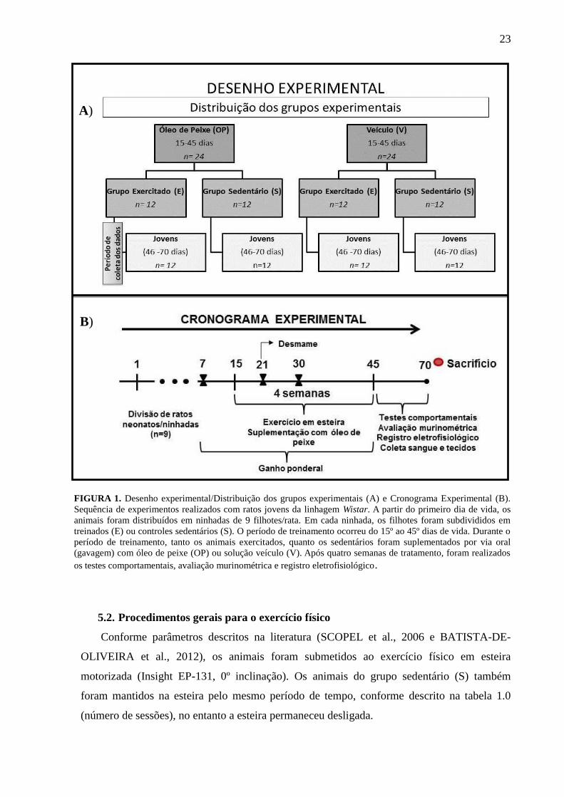
5. MATERIAIS E MÉTODOS
5.1. Aspectos éticos e delineamento experimental
Os métodos utilizados estão de acordo com o disposto na Lei nº 11.794, de 8 de outubro
de 2008 e, especialmente, respeitando as Resoluções Normativas do Conselho Nacional de
Controle de Experimentação Animal – CONCEA. Este projeto de pesquisa foi aprovado pela
Comissão de Ética no Uso de Animais da UFPE (protocolo nº 23076.027072/2014-20).
Foram utilizados ratos machos jovens da linhagem Wistar, provenientes da colônia do
Departamento de Nutrição da Universidade Federal de Pernambuco. Ratos neonatos foram
aleatoriamente distribuídos, 24 horas após o parto, em ninhadas de 9 filhotes por mãe. Destes,
foram separados dois grupos experimentais conforme descrito na Figura 1A. Assim, em
cada ninhada, os filhotes foram subdivididos em exercitados (E) ou controles sedentários (S).
O período de treinamento ocorreu do 15º ao 45º dias de vida. Durante o período de
treinamento, tanto os animais exercitados, quanto os sedentários foram suplementados por via
oral (gavagem) com uma solução contendo óleo de peixe (OP), água destilada e cremophor
0,009% (Sigma®) ou solução veículo (V) contendo apenas água destilada e cremophor
0,009% (Sigma®). Durante todo o período de experimentação, os ratos foram alimentados
com a dieta de manutenção do biotério (“Presence”, Purina do Brasil). A partir dos 46 dias de
vida, foi realizada avaliação dos efeitos da associação do exercício com a suplementação com
óleo de peixe sobre a evolução ponderal, respostas comportamentais, murinometria e
eletrofisiologia cortical (Figura 1B). Para complementar as análises metodológicas, os tecidos
coletados foram avaliados, quanto ao estresse oxidativo, por equipes lideradas pelas Profª
Claúdia Lagranha e Profª Mariana Fernandes. Foram 4 grupos experimentais, cada grupo
contendo 12 animais, com número total de 48 animais.
23
FIGURA 1. Desenho experimental/Distribuição dos grupos experimentais (A) e Cronograma Experimental (B).
Sequência de experimentos realizados com ratos jovens da linhagem Wistar. A partir do primeiro dia de vida, os
animais foram distribuídos em ninhadas de 9 filhotes/rata. Em cada ninhada, os filhotes foram subdivididos em
treinados (E) ou controles sedentários (S). O período de treinamento ocorreu do 15º ao 45º dias de vida. Durante o
período de treinamento, tanto os animais exercitados, quanto os sedentários foram suplementados por via oral
(gavagem) com óleo de peixe (OP) ou solução veículo (V). Após quatro semanas de tratamento, foram realizados
os testes comportamentais, avaliação murinométrica e registro eletrofisiológico.
5.2. Procedimentos gerais para o exercício físico
Conforme parâmetros descritos na literatura (SCOPEL et al., 2006 e BATISTA-DE-
OLIVEIRA et al., 2012), os animais foram submetidos ao exercício físico em esteira
motorizada (Insight EP-131, 0º inclinação). Os animais do grupo sedentário (S) também
foram mantidos na esteira pelo mesmo período de tempo, conforme descrito na tabela 1.0
(número de sessões), no entanto a esteira permaneceu desligada.
B)
A)

24
TABELA 1. Parâmetros do exercício físico.
Grupo
experimental
Parâmetros do
exercício
1ª semana 2ª semana 3ª semana 4ª semana
E
Número de sessões
por semana
5 5 5 5
Velocidade
Tempo
5m/min
20 min
10m/min
20 min
15m/min
20 min
20m/min
20min
Adaptado de Batista-de-Oliveira et al., 2012.
O exercício e a suplementação com os ácidos graxos essenciais foram realizados
concomitantemente, do 15º ao 45ºdias de vida. Doses diárias de 85 mg/kg/dia de ômega-3 nos
grupos OP foram administradas por via oral (técnica de gavagem). Nos grupos controles, a
solução V de cremophor 0,009% (Sigma) foi administrada.
A solução de OP foi preparada a partir de cápsulas (Sundown - Naturalis®) contendo os
seguintes ácidos graxos poliinsaturados: DHA (85 mg/ 1 g) e EPA (128mg/ 1 g) e diluída em
água destilada e cremophor 0,009% (Sigma®). A solução veículo foi preparada utilizando
somente água destilada e cremophor 0,009% (Sigma®).
5.3. Determinações Ponderais
5.3.1. Ganho ponderal
O peso corporal foi obtido aos 7, 14, 21, 30 dias de vida; além disso, os animais foram
pesados também no primeiro e último dia de cada semana de treino. Estes pesos corporais
foram comparados entre os grupos suplementados (OP e V) e de exercício físico (S e E).
5.3.2. Avaliação Murinométrica
No dia do registro eletrofisiológico (procedimentos descritos a seguir), após anestesia
(procedimento descrito no tópico 5.5), os animais tiveram o comprimento da ponta do
focinho até o ânus (CFA), circunferência abdominal (CA), circunferência torácica (CT)
aferidos, para análise dos parâmetros murinométricos:

25
Índice de Massa Corpórea (IMC): peso corporal (g)/ comprimento² (cm²) (NOVELLI et
al., 2007).
Índice de Lee (IL): raiz cúbica do peso corporal (g)/comprimento (cm) (NOVELLI et
al., 2007).
Relação circunferência abdominal/ circunferência torácica (CA/CT) (cm) (NOVELLI et
al., 2007).
5.4. Análise comportamental
5.4.1. Labirinto em cruz elevado (LCE)
O teste consistiu em colocar o animal em um LCE, elevado do solo, formado por dois
braços fechados por paredes e dois abertos (perpendiculares aos primeiros), analisando-se a
frequência de entradas e o tempo gasto em cada tipo de braço, e outros comportamentos como
deslocamentos, levantar-se, esticar-se etc. O animal explora os dois tipos de braço, porém
entra mais e permanece mais tempo nos braços fechados. Considera-se a porcentagem da
preferência (entradas e tempo gasto) pelos braços abertos e pelos fechados um índice
fidedigno de ansiedade: quanto maiores os níveis de ansiedade, menor a porcentagem de
entradas nos braços abertos e de tempo gasto nos mesmos (HANDLEY & MITHANI, 1984;
PELLOW & FILE, 1986).
O procedimento foi realizado entre as 10:00–14:00h, utilizando no mínimo 12 animais de
cada grupo dos 46 a 55 dias de vida. No dia do experimento, os ratos machos foram colocados
na ante-sala experimental, individualmente, em gaiola apropriada e permaneceram 5 minutos
antes do início do teste, para adaptação ao novo ambiente.
O animal foi colocado no centro do aparato cuidadosamente com o focinho voltado para
um dos braços fechados onde foi permitida a exploração livre por 5 minutos. A cada animal
testado, o labirinto era limpo com álcool à 70%, respeitando um intervalo de 5 minutos.
Posteriormente, as seguintes categorias comportamentais foram analisadas:
a) Distância total percorrida;
b) Tempo de imobilidade;
c) Número de entradas no braço aberto: foi considerada uma entrada quando o animal
entrou com as quatro patas no braço;
d) Tempo gasto nos braços abertos;

26
5.4.2. Teste de adaptação ao Campo Aberto (TCA)
Todos os animais foram submetidos ao teste de campo aberto para adaptação à arena. O
aparato consiste em uma arena circular (campo aberto), feito de madeira, com 1 metro de
diâmetro, localizada em um ambiente com iluminação reduzida. Por vez, cada animal foi
colocado, por 5 minutos na arena, para exploração ao ambiente, permitindo a análise da
ansiedade pelo tempo gasto e o número de entradas na área central, a distância percorrida e o
tempo de imobilidade. O teste foi validado segundo PLATEL & PORSOLT, 1982.
5.4.3. Tarefa de reconhecimento de objetos (TRO)
Entre os 46 e 55 dias de idade, os animais foram submetidos a TRO. O aparato consistia
em uma arena (campo aberto) localizada em um ambiente com iluminação reduzida. Os
animais foram colocados, por 5 minutos na arena, para se adaptarem ao ambiente e o
experimento comportamental propriamente dito foi realizado no dia seguinte à adaptação.
Foram avaliadas as diferenças, entre os grupos, na capacidade de identificação de objetos com
base na sua forma e localização no campo aberto.
Em cada uma dessas duas tarefas, os animais, em uma primeira sessão, exploraram por 5
minutos o ambiente. Numa segunda sessão, após 50 minutos, foi avaliado o reconhecimento
das características de forma e localização espacial dos objetos, como descrito adiante. Se,
nessa segunda sessão, diante de dois objetos, um conhecido (da sessão anterior – com
características “familiar”) e outro com características de “novidade” (seja na forma, ou na
posição), o rato reconhecer o objeto apresentado na primeira análise, ele, então, passará mais
tempo explorando o objeto “desconhecido”.
Entre as sessões, os objetos e o campo aberto foram limpos adequadamente com álcool a
70% para eliminar pistas olfativas que pudessem influenciar o ensaio seguinte.
O critério para definir exploração é baseado na “exploração ativa”, ou seja, quando o rato
está tocando os objetos pelo menos com o focinho (O’CALLAGHAN et al., 2007; MELLO et
al., 2008; DERE et al., 2005). ENNACEUR & DELACOUR (1988) (apud DERE et al., 2005)
demonstram esses métodos utilizados para os testes de reconhecimento de objetos,
brevemente descritos a seguir:
Na discriminação das formas: dois objetos idênticos (A e B) foram posicionados na arena
para a primeira análise. Após 50 minutos, os animais foram recolocados no campo aberto
(segunda sessão) com o mesmo objeto A (conhecido), porém, o objeto B foi substituído por
outro, C (desconhecido), da mesma cor, tamanho e cheiro do objeto A, mas com uma forma

27
diferente. O animal, então, diferenciará as formas quando, nessa segunda sessão, passar mais
tempo explorando o objeto com a forma desconhecida.
Para avaliar a distinção de localização espacial: dois objetos idênticos (A e B) foram
colocados em determinadas posições no campo aberto. Passados 50 minutos, os animais
foram novamente colocados no campo aberto (segunda sessão) na presença dos mesmos
objetos (A e B), todavia, neste segundo momento a posição de A se mantém (posição
conhecida), e a localização de B se modificou. Se o animal distinguir uma posição
desconhecida, ele gastará mais tempo explorando o objeto nessa posição.
Para a análise do comportamento no reconhecimento de objeto, foram utilizados dados
referentes ao índice de discriminação (%), fundamentado no tempo de exploração no objeto
novo e no objeto familiar (quanto a identidade ou localização) em relação ao tempo total de
exploração do animal (AKKERMAN et al., 2012).
Todos os testes comportamentais foram filmados através de uma câmera instalada no teto
da sala e conectada a um programa computacional preparado para aquisição de imagens. Para
o processamento e análise dos parâmetros comportamentais gravados em vídeo foi utilizado o
software ANY-maze Video Tracking System version 4.99m.
5.5. Registro eletrofisiológico
Para a realização dos registros eletrofisiológicos, os animais entre 55 e 70 dias de vida
foram anestesiados com uma solução de uretana 10% + cloralose 0,4%, à dose de 1000 mg/kg
de uretana + 40 mg/kg de cloralose, via intra-peritoneal (ambos da Sigma Co., EUA). O
animal foi colocado em decúbito ventral sobre um aquecedor elétrico de temperatura
regulável. Em seguida, a cabeça do animal foi fixada à base de um aparelho estereotáxico. Por
meio de trepanação, foram feitos três orifícios, de cerca de 2 a 4 mm de diâmetro cada,
alinhados paralelamente à linha média.
Os registros da variação lenta de voltagem (VLV) que acompanha a DAC foram feitos
durante 4 horas, por eletrodos de Ag/AgCl (GUEDES & BARRETO, 1992), com um par para
o registro localizados no hemisfério parietal. Um terceiro eletrodo do mesmo tipo foi
colocado sobre o osso nasal e serviu de referência comum aos dois eletrodos de registro. A
DAC foi provocada a cada 20 minutos por estimulação química com KCl a 2%, durante 1
minuto no orifício de estimulação na região frontal.
A velocidade de propagação da DAC foi calculada com base na distância entre os
eletrodos de registro e no tempo gasto pela DAC para percorrer esta distância. Para cada uma
das horas de registro foram calculadas as velocidades médias de propagação do fenômeno.

28
6. Análise Estatística
Todos os dados foram expressos em média±EPM ou mediana com intervalo
interquartílico e analisados estatisticamente utilizando o software GraphPad Prism 6.0
(GraphPad Software Inc., La Jolla, CA, EUA). O teste estatístico foi escolhido mediante a
análise no teste de normalidade (teste de Kolmogorov-Smirnov). Para a distribuição
paramétrica, na análise intergrupo referente aos dados murinométricos, testes para avaliação
do comportamento ansioso (LCE e TCA) e registro eletrofisiológico foram utilizados
ANOVA two-way, seguida de “post hoc” (Tukey) ou do teste “t” de Student não pareado. Para
a avaliação das diferenças significativa intragrupo referente aos dados comportamentais de
reconhecimento de objeto, os índices de discriminação foram analisados através do teste “t”
de Student pareado. As diferenças foram consideradas estatisticamente significativas quando
p<0,05.

29
7. RESULTADOS
7.1. Evolução Ponderal e Dados murinométricos
A associação do exercício físico em esteira e da suplementação com o óleo de peixe
realizada do 15º ao 45º dia de vida pós-natal, não modificou de maneira significativa o
peso corporal e os dados murinométricos, entre os animais dos diferentes grupos
experimentais (ANOVA two-way, p>0,05), conforme demonstrado na FIGURA 2 e
TABELA 2.
D i a s d e v i d a
Ga
nh
o p
on
de
ra
l (g
)
1 5 2 1 2 7 3 4 4 5
0
5 0
1 0 0
1 5 0
V / S
V / E
O P / S
O P / E
FIGURA 2. Ganho ponderal pós-natal apresentado como média±EPM. Estes animais foram previamente
divididos em exercitado (E) (n=20, por grupo) e sedentários (S) (n=20, por grupo). A partir do 15º dia de
vida, peso corporal foi mensurado até o 45º dia de vida. No mesmo período os animais receberam uma dose
diária de óleo de peixe (OP), por 4 semanas. A análise realizada com ANOVA two-way (p>0,05) não
apresentou diferença estatística significativa no peso corporal, entre os grupos experimentais.

30
TABELA 2. Os dados murinométricos são apresentados como média±EPM. No 1º dia de vida pós natal, os animais foram
distribuídos aleatoriamente em ninhadas de 9 filhotes/mãe. Ao 15º dia de vida, os animais foram subdivididos conforme a
suplementação e/ou exercício físico em esteira.
Grupo
Experimental (n) Dados murinométricos
CA CT CA/CT CC CFA IL IMC PCo/PC PF/PC
V&S (n=12) 15,15±0,35 12,50±0,34 15,15±0,35 15,02±0,30 20,50±0,43 0,30±0,003 0,55±0,01 0,45±0,01 1,32±0,06
V&E (n=12) 14,96±0,29 12,63±0,18 14,96±0,29 15,04±0,28 20,64±0,35 0,30±0,003 0,55±0,01 0,49±0,19 1,25±0,01
OP&S (n=12) 14,92±0,38 12,54±0,18 14,92±0,38 15,33±0,50 20,68±0,50 0,29±0,005 0,53±0,02 0,51±0,04 1,30±0,12
OP&E (n=12) 15±0,36 12,64±0.20 15,00±0,36 14,91±0,5 20,60±0,63 0,29±0,14 0,53±0,02 0,50±0,02 2,02±0,44
V&S – solução veículo e sedentários; V&E – solução veículo e exercitados; OP&S óleo de peixe e sedentários; OP&E –óleo de
peixe e exercitados; CA(cm) - circunferência abdominal; CT(cm) -circunferência torácica; CA/CT – Relação circunferência
abdominal/torácica; CC(cm) - comprimento caudal; CFA(cm) - comprimento focinho-ânus; IL (g/cm) – índice de Lee; IMC
(Kg/m²) – índice de massa corporal; %PCo/PC- peso do coração; PF – peso do fígado.
7.2. Avaliação comportamental
7.2.1. Teste de Adaptação ao Campo Aberto (TCA) e Labirinto em Cruz
Elevado (LCE)
O comportamento ansioso avaliado pelo teste de adaptação ao campo aberto (TCA) e
labirinto em cruz elevado (LCE) estão apresentados na TABELA 3. No TCA não foram
observadas diferenças significativas nos parâmetros avaliados, quando comparados os
diferentes grupos experimentais (ANOVA two-way, p>0,05). Por outro lado, no LCE os
animais exercitados apresentaram maior número de entradas no braço aberto, quando
comparados ao grupo controle (V/E 8 (3-9,75); versus V/S 3 (0,5-5,25); (ANOVA two-way,
seguida do teste de Tukey, p=0.01).

31
TABELA 3. (A) Teste de adaptação ao Campo Aberto (TCA) e (B) Labirinto em Cruz elevado (LCE). Os valores dos dados
estão descritos em média±EPM ou mediana (intervalo interquartílico). A letra sobrescrita representa diferença estatística
significativa entre os grupos (ANOVA two-way, seguida do teste de Tukey, p=0.01), V&S: sedentário suplementado com
solução veículo. V&E: exercitado suplementado com solução veículo; OP&S: sedentário suplementado com óleo de peixe;
OP&E: exercitado suplementado com óleo de peixe.
Testes comportamentais Grupos experimentais
V&S (n=7) V&E (n=7) OP&S (n=8) OP&E (n=9)
(A) TCA
Distância total percorrida (m) 21,72±3,64 22,10±2,10 23,58±4,61 26,27±3,61
Tempo de imobilidade (s) 59,41±20,66 61,51±21,39 45,33±17,24 42,12±16,96
Nº de linhas cruzadas 93 (82-102) 86 (80-100) 92 (86-112,8) 96 (91,5-112,5)
Nº de entradas na área central 8 (3-8) 7 (4-10) 7,5 (6-9,5) 7 (6-9)
Tempo na área central (s) 17,87±6,05 17,61±8,89 15,49±5,43 17,87±6,05
V&S (n=8) V&Ex (n=6) OP&S (n=9) OP&Ex (n=8)
(B) LCE
Distância total percorrida (m) 9,17±3,09 11,27±1,29 10,63±3,01 10,63 ±3,01
Tempo de imobilidade (s) 102,99±19,32
78,15±29,92
83,79±25,90 83,79 ±25,90
Nº de entradas no braço aberto 3 (0,5-5,25)a
8 (3-9,75)a
6 (4-6,5) 6 (3,5-8)
Tempo no braço aberto (s) 12,31±12,44 33,92±20,40 33,16±21,72 33,16 ±21,72
7.2.2. Teste de Reconhecimento de Objeto (TRO)
No que se refere ao desempenho dos animais nos testes relacionados à memória
episódica, a análise intragrupo (teste “t” Student pareado) demonstrou que os animais tiveram
a memória preservada para o reconhecimento da identidade do objeto apresentando um índice
de discriminação acima de 60% para o objeto novo versus o objeto familiar (OP&E, 63,3±2,5
versus 36,7±2,5, p< 0,001; OP&S, 71,3±3,8 versus 28.7±3.8, p<0,001; V&E, 66,0±4,0 versus
34,0±4,0, p< 0,005 e V&S, 68,7±3,5 versus 31,3±3,5, p<0,001). A análise intergrupo
(ANOVA two-way, p>0,005) não apresentou nenhuma diferença significante.
Por outro lado, a comparação intragrupo (teste “t” Student pareado) com os dados
obtidos do teste de reconhecimento espacial, demonstrou um prejuízo dependente do
tratamento como julgado pelos valores de índice de discriminação para o deslocado versus o
objeto estacionário (OP&E, 39,6±2,7 versus 60,4±2,7, p< 0,005; OP&S, 39,3±3,1 versus
60,7±3,1, p<0,01). Este prejuízo não foi observado nos ratos controles (V&E, 48,0±3,8 versus
52,0±3,8, p>0,05; V&S, 62,2±2.6 versus 37,8±2,6, p< 0,01). A análise intergrupo (ANOVA
two-way seguida do teste “post hoc” Tukey) para o desempenho no teste de reconhecimento
espacial demonstrou que a suplementação e o exercício físico em esteira aumentam o índice
de discriminação relacionado ao reconhecimento da posição estacionária, e diminuiu os

32
índices relacionado à discriminação do objeto deslocado (OP&S versus V&S, p<0,001, e
V&E versus V&S, p<0,05) (FIGURA 3 A e 3B)
FIGURA 3. Figura do artigo “Fish oil and treadmill exercise have age-dependent effects on episodic
memory and oxidative state of the hippocampus” (APÊNDICE A). Índices de discriminação para os testes
de reconhecimento de objeto quanto à sua identidade (A) e localização espacial (B), em animais sobre
diferentes condições experimentais. *p< 0,05 ou **p< 0,001 indica diferenças significantes intergrupo
(ANOVA two-way seguida do teste a posteriori Tukey), V&S versus OP&S, V&S versus V&E and V&S
versus OP&Ex); #p< 0,001, ##p= 0,001 ou ###p< 0,0001 indica diferenças significativas intragrupo (teste
“t” Student pareado) V&S: sedentário suplementado com solução veículo; V&E: exercitado suplementado
com solução veículo; OP&S: sedentário suplementado com óleo de peixe; OP&E: exercitado suplementado
com óleo de peixe.
7.4. Registro eletrofisiológico
Na avaliação eletrofisiológica dos efeitos da suplementação com óleo de peixe e
exercício físico, utilizando o fenômeno da DAC, a figura 4(A), representa de forma
esquemática o registro eletrofisiológico. Na análise estatística intergrupo, demonstrou que a
associação da suplementação com óleo de peixe e o exercício físico foi capaz de diminuir a
velocidade de propagação do fenômeno da DAC tanto em relação aos animais apenas
suplementados (OP&E, 2,65±0,2 versus OP&S, 3,23±0,3, p<0,001 ANOVA de duas vias) e
os animais controles (OP&E, 2,65±0,2 versus V&S, 3,21±0,2, p<0,001 ANOVA de duas vias,
V&Ex, 3,06±0,2), como apresentado na figura 4 (B).

33
FIGURA 4. (A) Figura do crânio de roedor adaptada de Francisco & Guedes (2015) representando os três
orifícios na superfície ventral do animal sendo o anterior para estímulo com KCl e os dois parietais
posteriores para os registros eletrofisiológico; abaixo, as barras verticais correspondem a 10 mV (alteração
negativa de voltagem). Na DAC foi realizado estímulo químico no cortéx frontal com KCl a 2% durante 1
minuto, como indicado pelas barras horizontais. As linhas tracejadas verticais indicam a latência para uma
onda da DAC atravessar a distância entre os eletrodos. As variações de lenta voltagem (VLV) registradas nos
pontos 1 e 2 em quatro animais, sendo um de cada grupo experimental: a esquerda VLVs de animais
exercitados (E) e a direta de animais sedentários (S), suplementados com óleo de peixe (OP) ou veículo (V);
(B) Velocidade de propagação da DAC (mm/s). Valores expressos em média ± DP. ANOVA de duas vias,
seguido do teste “post hoc” Tukey’s) mostrou diferenças significativas entre os grupos (*p<0,001).

34
8. DISCUSSÃO
No presente estudo, foi investigado os efeitos da suplementação com óleo de peixe
(OP) em uma dose diária de 85 mg/kg/dia concomitante ao exercício físico em esteira durante
4 semanas, dos 15 aos 45 dias de vidas de ratos jovens Wistar.
Durante o período de realização do protocolo de exercício físico, que ocorreu na fase
considerada de crescimento rápido e desenvolvimento, o peso corporal e o índice de massa
corpórea (IMC) foram realizados para determinar se o exercício influenciaria o estado
nutricional do animal e poderia, assim, influenciar o crescimento normal. Como demonstrado,
o protocolo de exercício combinado com a suplementação de OP durante todo o período do
estudo, não modulou de forma negativa a evolução ponderal e demais parâmetros
murinométricos.
Recentemente, PEDROZA et al. (2015) também demonstraram que ratos machos
Wistar adultos e idosos, quando submetidos ao mesmo protocolo de suplementação e
exercício físico utilizado no presente estudo, não apresentaram alterações significativa sobre
peso corporal, IMC e razão CA/CT, porém, neste mesmo estudo também foi observado um
aumento do peso do coração nos animais suplementados e exercitados comparado com os
animais apenas suplementados.
Para este parâmetro, nos dados obtidos neste trabalho, não foram encontradas
alterações significativas, possivelmente devido ao protocolo de exercício físico utilizado.
Porém dados presentes na literatura sugerem que o exercício a longo prazo possa induzir uma
diminuição da razão peso do coração/peso corporal devido a uma diminuição do peso corporal
(RADOVITS et al., 2013).
No que se refere à ansiedade, foi observado que o exercício tem ação ansiolítica, ao
comparar os resultados de animais exercitados com seus respectivos controles. Nesse caso
devido ao aumento do número de entradas nos braços abertos no teste do labirinto em cruz
elevado (LCE). Como descrito na literatura, o percentual de tempo e a frequência de entrada
nos braços abertos no LCE são considerados índices do nível de ansiedade em animais
(FEDEROVA & JR, 2006). De forma semelhante, FULK et al. (2004) também observaram
efeitos ansiolíticos associados a um protocolo de exercício físico crônico e de intensidade
moderada, com maior percentual de entradas nos braços abertos e tempo na área central
durante o LCE.

35
Curiosamente, em estudo realizado com objetivo de avaliar os efeitos de um protocolo
de exercício a curto prazo (4 dias) sobre o comportamento ansioso no LCE, não demonstrou
efeitos ansiolíticos relacionado ao exercício a curto prazo (CHAOULOFF, 1994).
No que se refere aos efeitos dos ácidos graxos dietéticos sobre os níveis de ansiedade
avaliados através do LCE, esses efeitos têm sido relacionados à sua deficiência, demonstrando
maior frequência de entradas e maior tempo gasto nos braços fechados em camundongos
deficientes em ácidos graxos poli-insaturados do tipo ω-3 (NAKASHIMA et al., 1993;
FRANCES et al., 1995; CARRIE et al., 2000). No entanto, os animais deficientes em ácidos
graxos poli-insaturados do tipo ω-3, quando suplementados por um curto período (1 semana)
apresentavam uma melhora significativa da ansiedade em termos de entrada nos braços
abertos (TAKEUCHI et al., 2003). Acredita-se ainda, que esses achados possam ser também
influenciados pelas diferentes condições experimentais (FEDEROVA & JR, 2006).
Outro teste utilizado neste estudo com o objetivo de avaliar o estado de ansiedade foi o
teste do campo aberto (TCA). Porém, os parâmetros avaliados não demonstraram alteração
significativa em relação à ansiedade. Neste teste, os baixos níveis de exploração, aumento do
“freezing” e número de bolo fecal são comportamentos relacionados à ansiedade, enquanto
que o aumento da atividade exploratória, como por exemplo, o tempo na área central e o
comportamento de “levantar” do animal são considerados características de níveis reduzidos
de ansiedade (ROYCE, 1977; WALSH & CUMMINS, 1976). Da mesma forma que o
observado no LCE, o exercício físico em esteira com intensidade moderada também parece
promover efeitos ansiolíticos durante o TCA (FULK et al., 2006).
A associação da suplementação com OP e exercício em esteira também tem sido
associada à melhora do desempenho de ratos submetidos a diferentes tarefas relacionadas à
memória (RACHETTI et al., 2013). Estes mesmos autores observaram que ratos
suplementados com uma dose diária de 85 mg/Kg de óleo de peixe apresentaram efeitos
positivos contra déficits de memória quando submetidos à tarefa de reconhecimento de
objetos.
Similarmente, os animais suplementados e exercitados avaliados no presente estudo,
apresentaram memória preservada a julgar pelos índices de discriminação na tarefa de
reconhecimento de objetos em relação à sua forma. Por outro lado, o mesmo não foi
observado nos animais suplementados com óleo de peixe avaliados através da tarefa de
reconhecimento de objetos quanto à sua localização.

36
Estes dados corroboram os achados descritos na literatura, onde se observa que os
efeitos da suplementação com óleo de peixe e exercício físico sobre a memória dependem do
tipo de tarefa empregada (RACHETTI et al., 2013).
Na literatura há diversos relatos demonstrando uma forte e influente relação entre a
intervenção dietética com os ácidos graxos poli-insaturados de cadeia longa, o exercício e o
funcionamento do cérebro (MURPHY et al., 2014).
Sobre a eletrofisiologia cerebral, estudada através do fenômeno da depressão
alastrante cortical (DAC), diversas condições podem interferir no aumento ou diminuição da
velocidade de propagação do fenômeno. No presente estudo, a suplementação do óleo de
peixe potencializou os efeitos do exercício sobre a redução do grau de excitabilidade do
tecido à passagem do fenômeno da DAC.
Os efeitos de ácidos graxos essencias sobre a eletrofisiologia cerebral foram descritos
por BORBA et al. (2010) demonstrando que a deficiência crônica de ácidos graxos essenciais
reduziu a suscetibilidade à DAC, a julgar pelas baixas velocidades de propagação. Diante
desses efeitos da deficiência de ácidos graxos insaturados é possível supor que a
suplementação de forma isolada, poderia agir de forma contrária aos efeitos observados sobre
a redução da DAC, porém o mesmo não foi observado diante do protocolo de suplementação
utilizado no presente estudo.
Em condições consideradas fisiológicas, acredita-se que os PUFA tenham efeitos
diretos sobre o sistema glutamatérgico e que poderia induzir uma hiperexcitabilidade neuronal
(MILLER et al., 1992; NISHIKAWA et al, 1994). Da mesma forma que a DAC é um
fenômeno relacionado à excitabilidade cerebral, é importante destacar que os efeitos do
exercício físico regular por sua vez, relacionados à melhoria dos processos de aprendizagem,
potenciação à longo prazo e memória, estão intrinsicamente ligados a excitabilidade cerebral
(RADAK et al., 2001; OGONOVSZKY et al., 2005; PASSECKER et al., 2011).
Adicionalmente, respostas comportamentais (envolvendo ansiedade) têm sido
diretamente relacionados à incidência da DAC (DELPRATO, 1965; BOGDANOV et al.,
2013). Nesse sentido, os animais estudados nesse protocolo, quando exercitados,
apresentaram um comportamento ansiolítico significativo em relação aos controles, bem
como houve uma redução da velocidade de propagação da DAC, quando o exercício foi
associado à suplementação com óleo de peixe.
Diante do exposto, é possível supor que a suplementação do óleo de peixe e/ou
exercício físico em esteira no início da vida apresenta efeitos comportamentais, relacionados à
ansiedade e memória, e sobre a eletrofisiologia cerebral.

37
9. CONSIDERAÇÕES FINAIS
Diante dos resultados obtidos no presente estudo, sugere-se que a suplementação com
o óleo de peixe e o exercício físico no início da vida, período considerado crítico para o
desenvolvimento do sistema nervoso, não modula de forma negativa parâmetros
murinométricos, porém é necessário ter cautela quanto ao uso dessas estratégias no que se
refere às ações em nível comportamental e neural.
É importante destacar que este trabalho vem a preencher algumas lacunas de interesse
da comunidade científica, quanto aos efeitos da associação dessas estratégias no início da vida
sobre aspectos orgânicos diferentes.
Apesar de preenchermos de maneira pioneira algumas lacunas, conforme supracitado
há necessidade de se compreender melhor a relação entre: (1) a dose e volume da
suplementação com óleo de peixe, (2) a associação entre óleo de peixe e exercício físico
moderado, e (3), sobretudo o período apropriado para se observar os efeitos dessa associação.
A realização deste trabalho permitiu ainda a elaboração do artigo “Fish oil and
treadmill exercise have age-dependent effects on episodic memory and oxidative state of the
hippocampus” submetido ao periódico “Journal of Nutritional Biochemistry” conforme
objetivo proposto pela presente dissertação, bem como a elaboração do artigo “Treadmill
exercise and Fish oil supplementation during development: A study of cardiac oxidative
metabolism.” submetido ao periódico “Applied Physiology, Nutrition, and Metabolism” em
colaboração com o grupo de pesquisa liderado pela professora Cláudia Jacques Lagranha.

38
REFERÊNCIAS
AKKERMAN, S. PRICKAERTS, J., STEINBUSCH, H. W. M., BLOKLAND, A.Object
recognition testing: statistical considerations. Behavioral Brain Research. v. 232 (2), p. 317-
22, 2012.
BATHIA, H. S.; AGRAWAL, R.; SHARMA, S,; HUO, Y. X.; YING, Z.; GOMEZ-PINILLA,
F. Omega-3 Fatty Acid deficiency during brain maturation reduces neuronal and behavioral
plasticity in adulthood. Plos One, v. 6, n. 12, p. 28451, 2011.
BATISTA-DE-OLIVEIRA, M.; LOPES, A. A. C.; MENDES-DA-SILVA, R. F.; GUEDES, R. C. A.
Aging-dependent brain electrophysiological effects in rats after distinct lactation conditions, and
treadmill exercise: a spreading depression analysis. Experimental Gerontology. v. 6, n. 47, p. 452-
457, 2012.
BOGDANOV, V. B.; BOGDANOVA, O. V.; KOULCHITSKY, S. V.; CHAUVEL, V.;
MULTON, S.; MAKARCHUK, M. Y.; BRENNAN, K. C.; RENSHAW, P. F.; SCHOENEN,
J. Behavior in the open field predicts the number of KCl-induced cortical spreading
depressions in rats. Behavioural Brain Research. v. 236, p. 90-93, 2013.
BORBA, J. M. C.; ROCHA-DE-MELO, A. P.; AMANCIO-DOS-SANTOS, A.; ANDRADE
DA COSTA, B. L. S.; PEREIRA DA SILVA, R.; PASSOS, P. P.; GUEDES, R. C. A.
Essential fatty acid deficiency reduces cortical spreading depression propagation in rats: a
two-generation study. Nutritional Neuroscience, v. 13, n. 3, 2010.
BOURRE, J. M. Where to find omega-3 fatty acids and how feeding animals with diet
enriched in omega-3 fatty acids to increase nutritional value of derived products for human:
what is actually useful? Journal of Nutrition Health Aging. v. 9, n. 4, p. 232-242, 2005.
BRENNA, J. T. Animal studies of the functional consequences of suboptimal polyunsaturated
fatty acid status during pregnancy, lactation and early post-natal life. Maternal and Child
Nutrition. v. 7, p. 59-79, 2011.
BURES, J.; BURESOVA, O.; KRIVANEK, J. The Mechanism and Applications of Leao's
Spreading Depression of Electroencephalographic Activity. Journal of Neurobiology. v. 6, n.
6, p. 619, 1974.
BURGHARDT, P. R. et al. The effects of chronic treadmill and wheel running on behavior in
rats. Brain Research. n. 1019, p. 84-96, 2004.
CARRIE, I.; GUESNET, P.; BOURRE, M.; FRANCES, H. J. Diets containing long-chain n-3
polyunsaturated fatty acids affect behavior differently during development than aging in mice.
British Journal of Nutrition. v. 83, p. 439-447, 2000.
CHAOULOFF, F. Influence of physical exercise on 5-HT1A receptor- and anxiety-related
behaviours. Neuroscience Letters. n. 176, p. 226-230, 1994.
CHEN, H. F.; SU, H. U. Exposure to a maternal n-3 fatty acid-deficient diet during brain
development provokes excessive hypothalamic–pituitary–adrenal axis responses to stress and
behavioral indices of depression and anxiety in male rat offspring later in life. Journal of
Nutritional Biochemistry. v. 24, n. 1, p. 70-80, 2013.
39
CHYTROVA, G.; YING, Z.; GOMEZ-PINILLA, F. Exercise contributes to the effects of
DHA dietary supplementation by acting on membrane-related synaptic systems. Brain
Research. v. 23, p. 32-40, 2010.
CORRIGAN, P. W. The impact of stigma on severe metal illness. Cognitive and Behavioral
Pratice. v. 5, p. 201-222, 1998.
DELPRATO, D. J. Note on the effect of cortical spreading depression on open-field behavior.
Psychological Reports. v. 17, p. 714, 1965.
DERE, E.; HUSTON, J. P.; DA SILVA, M. A. S. Episodic-like memory in mice:
Simultaneous assessment of object, place and temporal order memory. Brain Research
Protocols. v. 16, n. 1-3, p. 10-19, 2005.
DIK, M.; DEEG, D. J.; VISSER, M; JONKER, C. Early life physical activity and cognition at
old age. Journal Clinical Experimental Neuropsychology. v. 25, n. 5, p. 643-653, 2003.
DING, Y. H.; LI, J.; YAO, W. X.; RAFOLS, J. A.; CLARK, J. C.; DING, Y. Exercise preconditioning
upregulates cerebral integrins and enhances cerebrovascular integrity in ischemic rats. Acta
Neuropathology. v. 112, n. 1, p. 74-84, 2006.
DISHMAN, R. K. Physical activity and metal health. Encyclopedia of Mental Health. v. 3,
p. 171-188, 1998.
DOBBING, J. Vulnerable periods in developing brain. In: lipids, malnutrition & the
developing brain. Applied Neurochemistry. p. 287-316, 1968.
DREIER, J.P. The role of spreading depression, spreading depolarization and spreading
ischemia in neurological disease. Nature Medicine. v. 17, n. 4, p. 439-447, 2011.
DUBNAU, J.; CHIANG, A. S.; TULLY, T. Neural Substrates of Memory: From Synapse to
System. Journal of Neurobiology. v. 54, n. 1, p. 238-253, 2003.
DURING, M. J.; CAO, L. VEGF a mediator of the effect of experience on hippocampal
neurogenesis. Current Alzheimer Research. v. 3, n. 1, p. 29-33, 2006.
ENGSTRÖM, K.; SALDEEN, A. S.; YANG, B.; MEHTA, J. L.; SALDEEN, T. Effect of fish
oils containing different amounts of EPA, DHA, and antioxidants on plasma and brain fatty
acids and brain nitric oxide synthase activity in rats. Upsala Journal of Medical Sciences. V.
114, p. 206–213, 2009.
ENNACEUR, A.; DELACOUR, J. A new one-trial test for neurobiological studies of
memory in rats. 1: Behavioral data. Behavioral Brain Research. v. 31, n. 1, p. 47-59, 1988.
FABRICIUS, M.; FUHR, S.; WILLUMSEN, L.; DREIER, J.P.; BHATIA, R.; BOUTELLE,
M.G.; HARTINGS, J.A.; BULLOCK, R.; STRONG, A.J.; LAURITZEN, M. Association of
seizures with cortical spreading depression and peri-infarct depolarisations in the acutely
injured human brain. Clinical Neurophysiology. v. 119, n. 9, p. 1973-1984, 2008.
FEDOROVA, I.; JR, N. S. Omega-3 fatty acids and rodent behavior. Prostaglandins,
Leukotrienes and Essential Fatty Acids. v. 75, n. 4-5, p. 271-289, 2006.

40
FERRAZ, A. C. et al. Chronic ω-3 fatty acids supplementation promotes beneficial effects on
anxiety, cognitive and depressive-like behaviors in rats subjected to a restraint stress protocol.
Behavioural Brain Research, v. 219, n. 1, p. 116–122, 2011.
FRANCES, H.; MONIER, C; BOURRE; M. J. Effects of dietary a-linolenic acid deficiency
on neuromuscular and cognitive functions in mice. Life Science. v. 57, p. 1935-1947, 1995.
FRANCISCO, E. S.; GUEDES, R. C. A. Neonatal taurine and alanine modulate anxiety‑like
behavior and decelerate cortical spreading depression in rats previously suckled under
different litter sizes. Amino Acids. v. 47, n. 11, p. 2437-2445, 2015.
FULK, L. J.; STOCK, H. S.; LYNN, A.; MARSHALL, J.; WILSON, M. A.; HAND, G. A.
Chronic physical exercise reduces anxiety-like behavior in rats. International Journal of
Sports Medicine. v. 1, n.25, p. 78-82, 2004.
GERMANO, P. C. P. S.; SILVA, D. L.; SOARES, G. S. F.; SANTOS, A. M.; GUEDES, R. C.
A. Hypercaloric high-lipid diet and brain development: Effects on cortical spreading
depression in adult rats. Nutritional Neuroscience. v. 16, n. 6, p. 275-281, 2013.
GOMEZ-PINILLA, F. The impact of diet and exercise on brain plasticity and disease.
Nutrition Health. v. 18, n. 3, p. 261-268, 2006.
GOMEZ-PINILLA, F.; TYAGI, E. Diet and cognition: interplay between cell metabolism and
neuronal plasticity. Current Opinion in Clinical Nutrition & Metabolic Care. v. 16, n. 6, p.
726-733, 2013.
GOMEZ-PINILLA, F.; YING, Z. Differential effects of exercise and dietary docosahexaenoic
acid on molecular systems associated with control of allostasis in the hypothalamus and
hippocampus. Neuroscience. v. 168, n. 1, p. 130-137, 2010.
GORJI, A.; SPECKMANN, E. J. Spreading depression enhances the spontaneous
epileptiform activity in human neocortical tissues. European Journal of Neuroscience. v. 19,
n. 12, p. 3371-3374, 2004.
GUEDES R. C. A. Electrophysiological Methods: Application in Nutritional Neuroscience.
In: “Nutritional Neurosciences: Overview of an emerging field, Liebermann, H., Kanarek, T.
and Prasad, C (eds.), CRC Press, New York. Nutrition, Brain and Behavior Series, v. 3,
cap. 4, p. 39-54, 2005.
GUEDES, R. C. A.; BARRETO, J. M. Effect of anestesia on the propagation of cortical
spreading depression. Brazilian Journal of Medical and Biological Research. v. 25, n. 4, p.
393-397, 1992.
HANDLEY, S. L.; MITHANI, S. Effects of alpha-adrenoceptor agonists and antagonists in a
maze-exploration model of 'fear'-motivated behaviour. Naunyn Schmiedeberg’s Arch
Pharmacology. v. 327, n. 1, p. 1-5, 1984.
HASHIMOTO, M.; MAEKAWA, M; KATAKURA, M; HAMAZAKI, K; MATSUOKA Y.
Possibility of Polyunsaturated Fatty Acids for the Prevention and Treatment of
Neuropsychiatric Illnesses. Journal of Pharmacological Sciences. v. 124, n. 3, p. 294- 300,
2014.

41
LAMPTEY, M. S.; WALKER, B. L. Physical and neurological development of the progeny of
female rats fed on essential fatty acid-deficient diet during pregnancy and/or lactation.
Journal of Nutrition. v. 108, n. 3, p. 351-357, 1978.
LEÃO, A. A. P. Further observations on the spreading depression of activity in cerebral
cortex. Journal of Neurophysiology. v. 10, n. 6, p. 409-14, 1947.
LEÃO, A. A. P. Spreading depression of activity in the cerebral cortex. Journal
Neurophysiol, v. 7, p. 359-390, 1944.
LONG, B. C.; VAN STAVEL, R. Effects or exercise training anxiety. Journal of Applied
Sport Psychology. v. 7, p. 167-189, 1995.
LONG, S. J.; BENTON, D. A double-blind trial of the effect of docosahexaenoic acid and
vitamin and mineral supplementation on aggression, impulsivity, and stress. Human
psychopharmacology. v. 28, n. 3, p. 238-247, 2013.
LUCAS, S.J.; AINSLIE, P. N.; MURRELL, C. J.; THOMAS, K. N.; FRANZ, E. A.; COTTER,
J. D. Effect of age on exercise-induced alterations in cognitive executive function: Relationship to
cerebral perfusion. Experiemental Gerontolology. v. 8, n. 47, p. 541-51, 2012.
MELLO, P. B. Effects of acute and chronic physical exercise and stress on different types of
memory in rats. Anais Academia Brasileira de Ciências. v. 80, n. 2, p. 301-309, 2008.
MESSERI, G. P.; OLIVERIO, A.; PAOLETTI, R.; GALLI, C. Deficiency of essential fatty
acids during pregnancy and avoidance learning in the progeny. Pharmacological Research
Communications. V. 7, p. 71–80, 1975.
MILLER, B; SARANTIS, M.; TRAYNELIS, S. F., ATTWELL, D. Potentiation of NMDA
receptor currents by arachidonic acid. Nature. v. 355, p. 722-725, 1992.
MIZUNOYA, W. Effect of dietary fat type on anxiety-like and depression-like behavior in
mice. Springer Plus, v. 2, n. 1, p. 165, 2013.
MORGANE P. J. Prenatal malnutrition and development of the brain. Neuroscience
Biobehavioral Review. v. 17, n. 1, p. 91- 128, 1993.
MURPHY, T.; DIAS, G.P.; THURET, S. Effects of Diet on Brain Plasticity in Animal and
Human Studies: Mind the Gap. Neural Plasticity. v. 2014, p. 32, 2014.
NAKASHIMA, Y; YUASA, S; HUKAMIZU, Y.; OKUYAMA, H.; OHHARA, T.;
KAMEYAMA, T.; NABESHIMA, T. Effect of a high linoleate and a high a-linolenate diet on
general behavior and drug sensitivity in mice. Journal of Lipid Research. v. 34, p. 239-247,
1993.
NICULESCU, M. D.; LUPU, D. S.; CRACIUNESCU, C. N. Maternal α-linolenic acid
availability during gestation and lactation alters the postnatal hippocampal development in the
mouse offspring. International Journal Developmental Neuroscience. v. 29, n. 8, p. 795-
802, 2011.

42
NISHIKAMA, M.; KIMURA, S.; AKAIKE, N. Facilitatory effect of docosahexaenoic acid
on N-methyl-D-aspartate response in pyramidal neurones of rat cerebral cortex. The Journal
of Phusiology. v. 475, n. 1, p. 83-93, 1994.
NOVELLI, E. L.; DINIZ, Y. S.; GALHARDI, C. M.; EBAID, G. M. X.; RODRIGUES, H.
G.; MANI, F.; FERNANDES, A. A. H.; CICOGNA, A. C.; NOVELLI-FILHO, J. L. V. B.
Anthropometrical parameters and markers of obesity in rats. Laboratory Animals, v. 41, n.
1, p. 111-9, 2007.
NYBERG, J.; ABERG, M. A.; TORÉN, K.; NILSSON, M.; BEN-MENACHEM, E.; KUHN,
H. G. Cardiovascular fitness and later risk of epilepsy: a Swedish population-based cohort
study. Neurology. v.81, n. 12, p. 1051-1057, 2013.
O’CALLAGHAN, R. M.; OHLE, R.; KELLY, A. M. The effects of forced exercise on
hippocampal plasticity in the rat: a comparison of LTP, spatial- and non-spatial learning.
Behavioral Brain Research. v. 176, n. 2, p. 362-366, 2007.
OGONOVSZKY, H.; BERKES, I.; KUMAGAI, S.; KANEKO, T.; TAHARA, S.; GOTO, S.;
RADÁK, Z. The effects of moderate-, strenuous- and over-training on oxidative stress
markers, DNA repair, and memory, in rat brain. Neurochemistry International. v. 46, n. 8,
p. 635-640, 2005.
PASSECKER, J.; HOK, V.; DELLA-CHIESA, A.; CHAH, E.; O'MARA, S.M. Dissociation
of dorsal hippocampal regional activation under the influence of stress in freely behaving rats.
Behavioral Neuroscience. v. 66, n. 5, p. 1-7, 2011.
PEDROZA, A. A. S.; LOPES, A.; MENDES-DA-SILVA, R. F.; BRAZ, R. B.; FERREIRA,
D. F.; AMANCIO-DOS-SANTOS, A.; BATISTA-DE-OLIVEIRA-HORNSBY, M.;
LAGRANHA, C. J. Can fish oil supplementation and physical training improve oxidative
metabolism in aged rat hearts? Life Sciences. v. 137, p. 133-141, 2015.
PELLOW, S.; FILE, S. E. Anxiolytic and anxiogenic drug effects on exploratory activity in
an elevated plus-maze: a novel test of anxiety in the rat. Pharmacology Biochemistry and
Behavior, v. 24, n. 3, p. 525-529, 1986.
PERINI, J. A. L. Ácidos graxos poli-insaturados n-3 e n-6: metabolismo em mamíferos e
resposta imune. Revista de Nutrição. v. 23, n. 6, p. 1075-1086, 2010.
PLATEL, A.P., PORSOLT, R.D. Habituation of exploratory activity in mice: a screening test
for memory enhancing drugs. Psychopharmacology. v. 78, p. 7, 1982.
RACHETTI, A. L. F.; ARIDA, R. M.; PATTI, C. L.; ZANIN, K. A.; FERNADES-SANTOS,
L.; FRUSSA-FILHO, R.; GOMES DA SILVA, S; SCORZA, F. A.; CYSNEIROS, R. M. Fish
oil supplementation and physical exercise program: Distinct effects on different memory
tasks. Behavioural Brain Research. v. 237, p. 283-289, 2013.
RADAK, Z.; KANEKO, T.; TAHARA, S.; NAKAMOTO, H.; PUCSOK, J.; SASVÁRI, M.;
NYAKAS, C.; GOTO, S. Regular exercise improves cognitive function and decreases
oxidative damage in rat brain. Neurochemistry International. v. 38, n. 4, p. 17-23, 2001.
RADOVITS, T; OLÁH, A.; LUX, A.; NÉMETH, B. T.; HIDI, L.; BIRTALAN, E.;
KELLERMAYER, D.; MÁTYÁS, C.; SZABÓ, G.; MERKELY, B. Rat model of exercise-
43
induced cardiac hypertrophy: hemodynamic characterization using left ventricular pressure-
volume analysis. American Journal Physiology Heart and Circulatory Physiology. v. 305,
n. 1, p. 124-134, 2013.
RAVIDRAN, A. V.; SILVA, T. L. Complementary and alternative therapies as add-on to
pharmacotherapy for mood and anxiety disorders: A systematic review. Journal of Affective
Disorders. v. 150, n. 3, p. 707-719, 2013.
ROYCE, J. T. On the construct validity of open field measures. Psychological Bulletin. v.
84, p. 1098-1106, 1977.
SCOPEL, D. et al. Exercise intensity influences cell injury in rat hippocampal slices exposed
to oxygen and glucose deprivation. Brain Research Bulletin, v. 71, n. 1-3, p. 155-159, 2006.
TAKEUCHI, T.; IWANAGA, M.; HARADA, E. Possible regulatory mechanism of DHA-
induced anti-stress reaction in rats. Brain Research. v. 964, p. 136-143, 2003.
VAN PRAAG, H.; CHRISTIE, B. R.; SEJNOWSKI, T. J.; GAGE, F. H. Running enhances
neurogenesis, learning, and long-term potentiation in mice. Proceedings of the National
Academy of Sciences of the United States of America. v. 96, n. 23, p. 13427–13431, 1999.
VINES, A.; DELATTRE, A. M.; LIMA, M. M.; RODRIGUES, L. S.; SUCHECKI, D.;
MACHADO, R. B.; TUFIK , S.; PEREIRA, S. I.; ZANATA, S. M.; FERRAZ, A. C. The role
of 5-HT1A receptors in fish oil-mediated increased BDNF expression in the rat hippocampus
and cortex: A possible antidepressant mechanism. Neuropharmacology. v. 62, n. 1, p. 184-
191, 2012.
WALSH, R. N.; CUMMINS, R. A. The open-field test: a critical review. Psychological
Bulletin. v. 83, p. 482-504, 1976.
WU, A.; YING, Z.; GOMEZ-PINILLA, F. Docosahexaenoic acid dietary supplementation
enhances the effects of exercise on synaptic plasticity and cognition. Neuroscience. n. 155, p.
751-759, 2008.
WU, A.; YING, Z.; PINILLA, F. G. Dietary omega-3 fatty acids normalize BDNF levels,
reduce oxidative damage and counteract learning disability after traumatic brain injury in rats.
Journal of Neurotrauma, v. 21, n. 10, p. 1457-1467, 2004.
YEHUDA, S.; RABINOVITZ, S.; MOSTOFSKY, D. I. Essential fatty acids and the brain:
From infancy to aging. Neurobiology of Aging. v. 26, n. 1, p. 98-102, 2005.

44
APÊNDICE A
Artigo original submetido ao periódico “Journal of Nutritional Biochemistry”.
From: Journal of Nutritional Biochemistry <[email protected]>
Date: 2016-03-08 17:08 GMT-03:00
Subject: Journal of Nutritional Biochemistry: Submission Confirmation
To: [email protected], manuella [email protected]
Title: Fish oil and treadmill exercise have age-dependent effects on episodic memory and
oxidative state of the hippocampus
Corresponding Author: Prof. Manuella Batista-de-Oliveira Hornsby
Authors: Patricia F Macêdo; Janatar S Melo; Laís A Costa; Glauber R Braz; Shirley M Sousa;
Claudia J Lagranha;
Dear Prof. Batista-de-Oliveira Hornsby,
Thank you for submitting your manuscript which has been received by the Editorial Office of the
Journal of Nutritional Biochemistry. Each manuscript is internally reviewed and prioritized before
a full external peer review takes place.
You will be able to check on the progress of your manuscript by logging on to the Elsevier
Editorial System for the Journal of Nutritional Biochemistry as an author:
http://ees.elsevier.com/jnb/
Your username is: ******
If you need to retrieve password details, please go
to: http://ees.elsevier.com/jnb/automail_query.asp
Your paper will be given a manuscript number shortly and you will soon receive an e-mail with
this number for your reference. Please let me know if there are any questions or concerns, and
thank you for your submission
Sincerely,
Joseph R. Richardson
Editorial Manager
The Journal of Nutritional Biochemistry
Bernhard Hennig, Editor-in-Chief

45
Title: Fish oil and treadmill exercise have age-dependent effects on episodic memory and
oxidative state of the hippocampus
Patrícia Fortes Cavalcanti de Macêdoa*
; Janatar Stella Vasconcelos de Meloa*
; Laís Alves
Ribeiro Costaa; Glauber Rudá F. Braz
b, Shirley M. de Sousa
b; Cláudia J. Lagranha
b*;
Manuella Batista-de-Oliveira Hornsbya*
#
a
Department of Nutrition / CCS. Federal University of Pernambuco. Campus of Recife.
Recife – PE, Brazil.
b Laboratory of Biochemistry and Exercise Biochemistry. Federal University of Pernambuco.
Campus of Vitoria de Santo Antao. Vitoria de Santo Antao – PE, Brazil.
*These authors contributed equally to this work.
#Corresponding author
Mailing addresses:
Manuella Batista-de-Oliveira Hornsby
Avenida Prof. Moraes Rego, s/n – CEP 50670-901 – Departamento de Nutrição – Cidade
Universitaria – Recife, PE – Brazil.
E-mail: [email protected]
46
Abstract
There is a high interest to better understand how lifestyle choices can improve memory
functions. Exercise (Ex) and long-chain n−3 polyunsaturated fatty acids (PUFAs) found in
fish oil (Fo) are able to reduce inflammation, to stimulate hippocampal antioxidant defenses,
and to improve memory. The aim of this study is to test whether (Fo) and treadmill (Ex) can
improve the rat performance on memory tasks and optimize hippocampal antioxidant state in
an age-dependent manner. Therefore, young and adults Wistar rats were exercised and
received (Fo) during 4 weeks. Afterwards episodic memory was measured by the recognition
of object identity and spatial location tests, and hippocampal oxidative state was investigated
with the levels of malondialdehyde (MDA), carbonyls content, antioxidant enzymatic activity
(superoxide dismutase (SOD), catalase (CAT), glutathione peroxidase (GPx)), and antioxidant
non enzymatic activity (reduced glutathione (GSH) and sulfhydryl content). Data show that
(Fo&Ex) adult rats were able to recognize objects shape and placement, however (Fo&Ex)
young rats had impaired spatial recognition (p<0.05). (Fo&Ex) young rats did not have
reduced MDA or protein content, though, either (Fo) or (Ex) reduced MDA (p<0.05) and
carbonyl levels (p<0.01). (Ex) increased SOD (p<0.001) and CAT activities (p<0.05), and
(Fo) enhanced SOD activity (p<0.05). At adulthood, (Ex) increased MDA levels (p<0.05),
and (Fo&Ex) reduced MDA (p<0.001). Lastly, (Ex) and (Fo) improved non-enzymatic
antioxidant defense (p<0.05) only in adult rats. Results support an age-dependent effects of
the (Fo) and (Ex) on memory functions and oxidative state of the hippocampus during either
neurodevelopment or adulthood.
Keywords: fish oil, EPA, DHA, treadmill exercise, object recognition, hippocampal
oxidative stress.
Chemical compounds studied in this article
Eicosapentaenoic acid (PubChem CID 446284); Docosahexaenoic acid (PubChem CID:
445580); Malondialdehyde (PubChem CID: 10964); Reduced glutathione (PubChem CID:
7048684);

47
1. Introduction
Memory disorders are associated to the malfunctioning of neuroanatomical structures
that are related to the storage, retention and recollection of memories. The prevalence and
complications of memory disorders such as Alzheimer’s disease, Dementia and Parkinson’s
disease represent a worldwide problem. Despite the ongoing research, underlying mechanisms
and preventive lifestyle choices related to these neurodegenerative diseases are still unclear.
Therefore, there is a growing interest to better understand how lifestyle choices and
their underlying mechanisms can improve cognition, memory and brain function to benefit
neurodevelopment, and to prevent or counteract neurodegenerative diseases [1, 2]. The object
recognition task is based on the natural curiosity towards novelty, the test is one-trial learning
task, that allows to separately analyze different memory processes, such as acquisition,
consolidation and retrieval [3]. This one-trial task has proven to be very useful and suitable
for accessing memory functions, and for studying the neurobiology of memory [4, 5].
Studies in humans [2, 6, 7] and animals [2, 8, 9] suggest that the effects of long chain
omega-3 (n-3) fatty acids and physical exercise can act on cognition and improve brain
function. Since the consumption of (n-3) PUFA and exercise can be simultaneously present in
daily life, the association between them could indicate that their effects on the brain are
complementary [10].
From early fetal life to adulthood, the adequate nutrition is an essential requirement for
morphogenesis of the brain, during this initial period, PUFAs play an important role [11]. The
n-3 consumption can benefits brain´s health, these benefits have been investigated by
experimental and epidemiologic studies. Specific features caused by n-3 consumption are
associated to hippocampal BDNF elevation, stimulation of mitochondrial function and
reduced oxidative stress [12].

48
Experimental designs have been focused on the use of different sources of fish oil
supplementation on injury situations and their effects to minimize oxidative stress in aged
mice [13], as well as in young rats [14]. Furthermore, beneficial properties of fish oil can be
extended to a better performance of memory tests in animal models of Alzheimer’s disease
[15] and brain injury [16].
Fish oil intake can lead to differential effects if the animal was exposed to an early life
stress. For example, when adult rats were previously submitted to maternal separation, n-3
PUFAs had little benefit in those animals that were exposed to the early life stress, on the
other hand, the benefits were seen in the neurodevelopment of normal animals [17].
Furthermore, exercise can be useful for both maintaining health and in specific
pathological conditions. In animal studies, exercise can improve cognitive function depending
on which life stage the animal was submitted to the exercise routine [6], [8]. The underlying
mechanisms demonstrated beneficial effects such as preventing oxidative stress in brain
regions of adolescent [18] and aging rats [19].
Considering the association between two interventions, n-3 supplementation and
physical exercise improved cognitive function of healthy animals [9]. Interestingly, the effect
was observed in a long-lasting manner since it was seen in early life stages as well as at
adulthood. It is also known that beneficial effects provided by the association between fish oil
supplementation and exercise are characterized by enhanced antioxidant defenses that can
prevent deleterious effects of aging [20]. However, there is an age-dependent effect for the
fish oil intake and physical exercise, depending on the life stage, effects of fish oil
supplementation or exercise can be contradictory [2, 17, 21].
Notwithstanding, evidence show beneficial effects for the use of fish oil coupled with
exercise on oxidative stress and memory, there is still a lack of information about the
appropriate timings to maximize results of this therapeutic approach, as well as the underlying
49
mechanisms for these effects are still unclear. Therefore, we aimed to investigate whether the
daily supplementation with fish oil coupled with moderate treadmill exercise in both young
and adult rats would influence memory and hippocampal oxidative stress.
2. Materials and methods
2.1 Animals and experimental design
Wistar rats were used from the colony of the Nutrition Department at the Federal
University of Pernambuco (Brazil). The experimental design followed the recommendations
of the Ethics Committee for Research on Animals (23076.027072/2014-20). These
recommendations were in accordance to the "Principles of Laboratory Animal Care"
(National Institutes of Health, Bethesda, USA). Every effort has been made to minimize
animal suffering and to reduce the number of animals per group.
The animals were randomly distributed, 24 hours after birth, in 9 rats per litter. They
were kept in a room with a temperature of 23 ± 1 ° C and raised in a 12h light / dark cycle
(lights on from 7:00 am to 7:00 pm). After weaning, 21 days-old animals were divided into
cages with 3-4 rats with free access to water and food - standard diet of pellets laboratory
(Presence of Brazil Ltd, Present, São Paulo, Brazil) with 23% of protein.
Either 15, or 90 days-old rats were randomly assigned into 4 different experimental
groups: (1) supplemented with fish oil and exercised (Fo&Ex, n=12 (young) and n = 11 (adult
rats)); (2) supplemented with fish oil and sedentary (Fo&S, n= 12 (young) and n = 11 (adult
rats)); (3) supplemented with vehicle solution and exercised (V&Ex, n= 12 (young) and n =
11 (adult rats)) (4) supplemented with vehicle solution and sedentary (V&S, n=12 (young)
and n = 11 (adult rats)). The trial period was 4 weeks.

50
2.2 Supplementation with fish oil
The daily supplementation was performed by gavage. Rats received a single daily dose
of fish oil (Fo, 85mg/kg/d and 1ml/250g/d) or vehicle solution (V, 1 ml/250g/d) as adapted
from [9, 20]. The capsules of fish oil (Sundown®) containing polyunsaturated fatty acids
[docoisahexaenoico (DHA; 85mg/1g) and ecoisapentaenoico (EPA; 128mg/1g)] were
dissolved in Cremophor (Sigma ®) 0.009% then in distilled water and administered via
gavage. To perform the control group, a vehicle solution (V) was given with the same amount
of Cremophor and distilled water that were used to prepare the solution provided to
supplement the fish oil.
2.3 Treadmill exercise
All exercised rats were placed on the treadmill (Insight EP-131, 0° inclination)
following parameters of moderate exercise that were adapted from previous studies [21].
Briefly, animals were placed 30min/day on the treadmill and the speed was increased as it
follows: 5m/min (first week); 10 m/min (second week) and 15m/min (third and fourth weeks).
The rats in the sedentary group (S) were placed at same time in the apparatus but it remained
off.
The treadmill exercise lasted 4 weeks, it was performed at the same timeframe of the
supplementation with fish oil. Therefore, either from 15 to 45 or 90 to 120 days old, rats were
submitted to the treadmill and supplementation to perform the experimental groups with
young and adult animals, respectively.
2.4 Behavioral analysis

51
All the behavioral tests were assessed individually at either 46 or 121 days old to
perform the young and adult groups, respectively. Detailed protocols and the rationale for the
test choices provided are discussed elsewhere [5, 22, 23]. Briefly, the experiment consisted of
a paradigm that utilizes the natural tendency of rodents to discriminate between a familiar and
a novel object, or a novel location, spending more time in exploring the novel objects, or
locations, as compared to the familiar ones.
In the present study, two experiments were designed to test the novelty recognition
paradigm, regarding the object’s novel shape or location. These experiments consist of three
days. In the first day, the rats were placed during five minutes in the open field apparatus to
explore and familiarize itself with the experiment’s environment. The apparatus consisted of a
circular arena with brown walls and an opened top. The floor of the arena is divided into 17
fields which are separated by black lines. In this arena, there are three concentric circles, and
it was located in a sound-attenuated room, with reduced lighting.
24 and 48 hours later, in the second and third days, each rat was returned to the
circular arena, which now contained two equal objects, made of clear glass. These objects
were explored by each rat during five minutes, and this constituted the trial session. After a
fifty-min interval, the animal returned to the arena to perform a five-min test session, in which
the capability of the rats to recognize novel object’s shape or novel object’s location
(dislocated object) was tested in the second and third day, respectively. Each experiment was
recorded by a camera installed in the room roof. The files were analyzed with the ANY-Maze
Software (version 4.99 m) by two previously trained observers, who were “blinded” regarding
the previous treatment of the animal. The videos were analyzed to assess the time spent by the
rat to explore each object. The criteria to define the time spent by the rats were based on the
“active exploration”, when rats were touching the objects with the vibrissae, snout or
forepaws, as previous published [22, 23]. In the trial session, the preference ratio was
52
calculated for each animal as being the time spent by the rat in exploring each object/total
exploration time. The trial sessions ensured that rats employed equal time in exploring each of
the two objects, and therefore there was no preference for one of them. Rats that presented in
the trial session a ratio near 0.5, indicating equal exploration of the two objects, were eligible
to realize the test session; otherwise they were discarded.
In each test session, the rats were expected to recognize the familiar object or the
familiar (stationary) position previously presented (in the trial session). The preference ratios
for the familiar and for the novel shape or position (dislocated) were also calculated for these
animals, which would be expected to spend more time actively exploring the objects
representing novel shape or dislocated position in the arena.
The performance was represented by a discrimination index (%) which consisted of the
exploration time for each analyzed criteria (familiar versus novel shape and stationary versus
dislocated position), expressed as a percentage of the total time of exploration. This paradigm
does not involve the learning of any rule, since it is entirely based on the spontaneous
exploratory behavior of rats towards objects [5]. The objects were devoid of any ethological
meaning, they had never been paired as a reinforce, and were heavy enough to prevent being
moved by the animal. Because the objects were made with the same material, the rats could
not distinguish them by olfactory cues during the trial session. After each session, the objects
and the apparatus were thoroughly cleaned with 70% ethanol solution.
2.5 Drugs and reagents
All drugs and reagents were purchased from Sigma-Aldrich (St. Louis, MO, USA) (Sinc
Pernambuco, Brazil).
2.6 Oxidative Stress Biomarkers

53
2.6.1 Sample preparation for oxidative stress and antioxidant analyses
Hippocampus from young and adult rats were homogenized in 50 mM-TRIS and 1mM-
EDTA (pH 7.4), with the addition of 1 mM-sodium orthovanadate and 200μg/mL
phenylmethanesulfonyl fluoride. Homogenates were centrifuged at 4000 RPM for 10 min at
4ºC and the protein supernatant used for biochemical analyses after the protein content had
been quantified by Bradford method [24] .
2.6.2 Evaluation of Lipid Peroxidation
Lipid peroxidation was evaluated using malondialdehyde (MDA) levels as previously
published [25]. Three hundred µg protein was sequentially mixed to 30% (w/v)
Trichloroacetic acid (TCA) and 10 mM-TRIS buffer (pH 7.4). This mixture was centrifuged
at 2500g for 10 min and the supernatant was boiled for 15 min with 0.73% (w/v)
thiobarbituric acid. The pink pigment yielded then was measured at 535 nm absorption at RT
and expressed as nmol/mg protein.
2.6.3 Evaluation of Protein Oxidation
The protein oxidation was assessed using the procedures highlighted by Reznick and
Packer (1994)[26]. With the samples on ice, 30% (w/v) TCA was added to the sample and
then centrifuged for 14 min at 4000 RPM. The pellet was suspended in 10 mM
2,4dinitrophenylhydrazine and immediately incubated in a dark room for 1h with shake turned
on each 15min. Then the samples were washed and centrifuged three times in an ethyl/acetate
buffer and then final pellet was suspended in 6M guanidine hydrochloride, incubated for 30
protein.

54
2.6.4 Measurement of superoxide dismutase (SOD) activity
SOD determination was performed in accordance with the protocol developed by Misra
and Fridovich (1972)[27]. In brief, 300 µg protein were added to 0.05 M-Carbonate buffer
with 0.1 mM-EDTA (pH 10.2). The reaction was started with 150mM-epinephrine and the
SOD activity was determined by adrenaline auto-oxidation inhibition at 30°C. The decrease in
absorbance was followed for 1.5 min at 480 nm and the results expressed as U/mg protein
[27].
2.6.5 Measurement of Catalase (CAT) activity
The CAT activity was performed as previously described by Aebi (1984)[28]. Briefly,
0.3 M-hydrogen peroxide and 300 µg protein were added to a 50 mM-phosphate buffer (pH
7.0) at 20° C and the absorption decay was monitored for 3 min at 240nm with the results
expressed as U/mg protein [28].
2.6.6 Measurement of Glutathione-S-transferase (GST) activity
GST activity was measured as described previously by Habig, Pabst (1974) [29]. Two
hundred µg of protein was incubated in a 0.1 M-phosphate buffer (pH 6.5) containing 1mM-
EDTA at 30° C and had the assay started with the addition of 1 mM 1-chloro-2.4-
dinitrobenzene and 1 mM-GSH. The formation of 2.4-dinitrophenyl-S-glutathione was
monitored through the absorbance at 340 nm. One unit of enzymatic activity was defined as
the amount of protein required to catalyze the formation of 1 µmol-2.4-dinitrophenyl-S-
glutathione [30].
2.6.7 Measurement of reduced glutathione (GSH)

55
To assess GSH levels, the samples were firstly diluted in a 0.1 M phosphate buffer
containing 5 mM-EDTA (pH 8.0). Then, an aliquot from the diluted sample was incubated
with o-Phthaldialdehyde at room temperature for exactly 15 min. Fluorescence intensities
measured at 420 nm and excitation at 350 nm were compared with a standard curve of known
concentrations of GSH (0.5-10 mM) as previously described [31].
2.6.8 Sulfhydryl content
To evaluates the levels of sulfhydryl we based the assay on the reduction of 5,5′-dithio-
bis(2-nitrobenzoic acid) (DTNB) by thiols, generating a yellow derivative (TNB) that absorve
at 412 nm in spectrophotometry [32]. Briefly, at TRIS buffer, pH7.4, 0.450 mg of
homogenate was added in addition to 1 mM DTNB. This mix was incubated 30 min at room
temperature in a dark room. Absorption was measured at 412 nm. The sulfhydryl content is
inversely correlated to oxidative damage to proteins. Results were reported as mmol TNB/mg
protein.
2.7 Statistical analysis
Results are expressed as means ± the standard error of the mean (SEM). A two-way
ANOVA test was performed to assess significant differences between the groups. The tests of
significance were recommended based on the results of the tests of normality (Kolmogorov-
Smirnov test). Data were considered as statistically significant for p<0.05. To evaluate
intragroup differences regarding behavioral data, paired t test was used to evaluate significant
differences between the discrimination indexes of the object identity recognition (novel versus
familiar) and object placement (dislocated versus stationary) tasks. All data were plotted and
the statistical analysis performed using GraphPad Prism 6.0 software (GraphPad Software
Inc., La Jolla, CA, USA).

56
3.0 Results
3.1 Behavioral assays
Discrimination indexes (MEAN±SEM) regarding the performance of young and adult
rats on the object identity and placement recognition tests are shown in Figure 1. The
intragroup analysis (paired t-test) demonstrated that young animals had preserved memory for
object identity recognition with a discrimination index above 60% for the novel versus
familiar object (Fo&Ex, 63.3±2.5 versus 36.7±2.5, p< 0.001; Fo&S, 71.3±3.8 versus
28.7±3.8, p<0.001; V&Ex, 66.0±4.0 versus 34.0±4.0, p< 0.005 and V&S, 68.7±3.5 versus
31.3±3.5, p<0.001). Intergroup analysis (two-way ANOVA, p>0.05) did not show any
significant differences.
On the other hand, the intragroup comparisons (paired t-test) with the data obtained
from the object placement recognition tests showed a treatment-dependent impairment as
judged by the values of the discrimination index for the dislocated versus stationary object
(Fo&Ex, 39.6±2.7 versus 60.4±2.7, p< 0.005; Fo&S, 39.3±3.1 versus 60.7±3.1, p<0.01). This
impairment was not observed in young rats from the respective control groups (V&Ex,
48.0±3.8 versus 52.0±3.8, p>0.05; V&S, 62.2±2.6 versus 37.8±2.6, p< 0.01). The intragroup
analysis (two-way ANOVA followed by Tukey) for the performance in the object placement
recognition tests showed that fish oil supplementation and treadmill exercise increased the
discrimination indexes related to the recognition of the stationary position, and decreased the
indexes related to the discrimination of the dislocated object (Fo&S versus V&S, p<0.001,
and V&Ex versus V&S, p<0.05). These results are described in Figure 1 A and C.
At adulthood, rats had the ability to recognize the novel and dislocated objects as shown
by the intragroup differences (paired t-tests) of the discrimination indexes for the object
identity recognition (novel versus familiar) and object placement tests (dislocated versus
stationary) (figure 1). In regards of the object identity recognition, adult rats presented higher

57
values of the discrimination indexes (MEAN±SEM) for the novel when compared to the
familiar object (Fo&Ex, 69.3±2.9 versus 30.7±2.9, p< 0.001; Fo&S, 72.9±2.9 versus
27.0±2.9, p<0.001; V&Ex, 76.0±3.6 versus 24.0±3.6, p< 0.001 and V&S, 75.5±4.26 versus
24.5±4.26, p<0.001). Similar results were observed for the object placement tasks as judged
by the increased discrimination indexes (MEAN±SEM) for the dislocated as compared to the
stationary object (Fo&Ex, 61.8±2.7 versus 38.2±2.7, p< 0.01; Fo&S, 64.4±3.1 versus
35.6±3.1, p<0.01; V&Ex, 63.2±3.6 versus 36.8±3.6, p< 0.01 and V&S, 63.9±3.7 versus
36.1±3.7, p<0.01). Figure 1 B and D describe these results.
3.2 Oxidative state
In addition to previous data, we observe that in young rats the exercise induces decrease
in lipid peroxidation evaluated by malondialdehyde (MDA) levels (V&S: 37.7 ± 8.0 nmol/mg
prot, n=6; V&Ex: 16.1 ± 0.6 nmol/mg prot, n=6, p<0.05) and protein oxidation evaluated by
carbonyls content (V&S: 19.4 ± 1.7 µmol/mg prot, n=6; V&Ex: 14.4 ± 0.9 µmol/mg prot,
n=7, p<0.01). Moreover, fish oil supplementation decreases lipid peroxidation (V&S: 37.7 ±
8.0 nmol/mg prot, n=6; Fo&S: 17.0 ± 2.7 nmol/mg prot, n=6, p<0.01) and protein oxidation
(V&S: 19.4 ± 1.7 µmol/mg prot, n=6; Fo&S: 10.5 ± 0.6 µmol/mg prot, n=7, p<0.001);
however the association between exercise and fish oil supplementation did not induce a
significant difference. In adult rats, exercise induces a different response, it causes a
significant increase in MDA levels (V&S: 11.3 ± 2.4 nmol/mg prot, n=6; V&Ex: 55.1 ± 8.0
nmol/mg prot, n=6, p<0.001), on the other hand there was no differences in carbonyl content
(V&S: 1.6 ± 0.2 µmol/mg prot, n=5; V&Ex: 1.4 ± 0.2µmol/mg prot, n=5, p<0.05). However,
when exercise is associated to fish oil supplementation, we observed a significant decrease in
MDA levels (Fo&S: 15.9 ± 2.8 nmol/mg prot, n=6; Fo&Ex: 1.7 ± 0.4 nmol/mg prot, n=6,

58
p<0.001), and in regards of protein oxidation (Fo&S: 1.7 ± 0.1 µmol/mg prot, n=5; Fo&Ex:
1.1 ± 0.1 µmol/mg prot, n=5; p<0.05). These results are described in figure 2.
To better understand effects of the association between exercise and fish oil
supplementation, we evaluated antioxidant defenses, such as enzymatic and non-enzymatic
mechanisms. In young rats, the exercise increases SOD activity (V&S: 31.5 ± 2.9 U/mg prot,
n=8; V&Ex: 46.8 ± 1.7 U/mg prot, n=8, p<0.001); in addition, in supplemented rats, exercise
also induces increase in SOD activity (Fo&S: 46.4 ± 1.7 U/mg prot, n=8; Fo&Ex: 55.9 ± 1.3
U/mg prot, n=8, p<0.05). It was interesting to note that fish oil supplementation per se
increases SOD activity in sedentary as well as in exercised rats (V&S: 31.5 ± 2.9 U/mg prot,
n=8; Fo&S: 46.4 ± 1.7 U/mg prot, n=8, p<0.001 and V&Ex: 46.8 ± 1.7 U/mg prot, n=8;
Fo&Ex: 55.9 ± 1.3 U/mg prot, n=8, p<0.05).
Furthermore, in terms of the capacity to convert H2O2 in H2O by catalase, we observed
that exercise is the only variable that can induce a significant increase. This increase does not
depend on the supplementation with or without fish oil (V&S: 1.0 ± 0.1 U/mg prot, n=6;
V&Ex: 1.7 ± 0.1 U/mg prot, n=7, p<0.01, and Fo&S: 0.9 ± 0.1 U/mg prot, n=6; Fo&Ex: 1.6 ±
0.3 U/mg prot, n=6, p<0.05). However, in regards to glutathione-S-Transferase, neither
exercise, nor fish oil supplementation induce differences in GST activity. In adult rats, the
antioxidant defense had a different response than observed in young rats, either exercise or
fish oil supplementation were able to induces differences in SOD, CAT or GST activity. All
the data of SOD, CAT and GST are described in figure 3.
Since the antioxidant defense is constituted by enzymatic and non-enzymatic
mechanisms, we also investigated non-enzymatic mechanisms. GSH is an important non-
enzymatic antioxidant that can be found in mammalian cells. In the reduced state, the thiol
group of cysteine present in GSH is able to donate a reducing equivalent to unstable
molecules, such as reactive oxygen species, that can decrease the toxicity of the unstable

59
molecules. Young rats did not present any significant differences among the groups.
Moreover, when we evaluate the sulfhydryl content in samples from young rats we also did
not observe any difference.
On the other hand, in adult rats we observed that fish oil supplementation associated to
exercise can increase GSH levels in a significant manner (Fo&S: 4.5 ± 0.5 µM/mg prot, n=5;
Fo&Ex: 8.5 ± 1.1 µM/mg prot, n=6, p<0.05). The same effect was observed in the Sulfhydryl
content (Fo&S: 0.1 ± 0.01 mM/mg prot, n=6; Fo&Ex: 0.26 ± 0.04 mM/mg prot, n=5, p<0.05).
Similar results (figure 4) were also observed when we compared exercised supplemented
with fish oil to exercised animals that received vehicle (GSH levels V&Ex: 4.1 ± 0.2 µM/mg
prot, n=5; Fo&Ex: 8.5 ± 1.1 µM/mg prot, n=6, p<0.01 and Sulfhydryl content V&Ex: 0.1 ±
0.02 mM/mg prot, n=6; Fo&Ex: 0.26 ± 0.04 mM/mg prot, n=5, p<0.05).
4.0 Discussion
Fish oil intake coupled with treadmill exercise has been associated to improvements on
rat performance in different memory tasks. Evidence show that these improvements on brain
function can be long-lasting. The improvements depend on the memory task that is performed
[9].
Our data suggests that fish oil supplementation changes the rat performance on the task
of placement recognition in an age-dependent manner. In fact, young rats that received fish oil
during the brain critical development period had reduced ability to recognize the novel object
placement as compared to the performance of the respective controls. Since, depending on the
life stage, effects of fish oil supplementation can be contradictory, the present data highlight
the need to better understand the relationship between: (1) the dose and volume of fish oil

60
supplementation, (2) the combination between (Fo) and moderate exercise, (3) and most
importantly an appropriate timing for this intervention.
Similarly to the present data, previous findings show that PUFAs and exercise have
been implicated in enhancing cognition and memory functions in healthy, adult rodents [2].
These authors reviewed a variety of data demonstrating a strong and influential relationship
between dietary intervention with PUFAs, exercise, and brain function. Besides these
previous findings in adult rodents, there are published data showing that the perinatal
supplementation with essential fatty acids improved hippocampal development in the
offspring, only when the dam received the enriched diet also during pregnancy [33]. Herein,
we observed an impairment to recognize the novel object placement when young rats received
fish oil during the brain critical development period. Present and previous data highlight the
importance of the appropriate timings for this lifestyle intervention to maximize results on
memory functions [2, 33].
Fish oil is a reliable source of (LC-PUFAs). Theses lipids have important hole in one’s
health state; they are considered essential because mammals are not able to produce them,
therefore LC-PUFAs have to be obtained by dietetic sources [34]. On the other hand, there are
challenges to reach the daily standard LC-PUFA recommendations through dietetic sources,
and to achieve beneficial effects on health care. Therefore, the supplementation with fish oil
as well as with other dietetic sources of LC-PUFA has grown exponentially either prescribed
by several physicians and nutritionists; or by the use from the general population without an
acknowledgement from a health care professional. In fact, the use of LC-PUFA has been
pointed out in a variety of acute and chronic inflammatory settings [35]. However,
experimental and clinical data about LC-PUFA underlying mechanisms and clinical efficacy
are still weak in some settings [2].

61
Experimental data shows that LC-PUFA and treadmill exercise underlying mechanisms
on heart metabolism are related to enhancement of antioxidant defenses [20]. On the other
hand, clinical trials describe LC-PUFA benefits by reducing the time to fatigue, increased grip
strength, and decreased use of nonsteroidal antiinflammatory drugs [35].
Similarly to the age-dependent effects of fish oil and exercise on the rat behavior, we
observed that this intervention affected hippocampal oxidative stress also depending on the
life stage. The present data demonstrated that fish oil supplementation or exercise reduced
lipid peroxidation and protein content as judged by the amount of MDA and carbonyl content
in young rats. On the other hand, if fish oil was associated to exercise, there was a significant
reduction in MDA and protein content in adult rats. In regards of the antioxidant defenses,
while exercise increased SOD and CAT activity in young rats, fish oil supplementation only
increased SOD activity. In adult rats, we observed that the fish oil and exercise improved non-
enzymatic antioxidant defenses because of the increased activity of GSH and sulfhydryl
content.
We believe that there is a timing-related association between the behavioral data and the
changes on hippocampal defenses as judged by the present results. According to recently
published data diet coupled with exercise are key modulators of brain structure and function.
Together, diet and exercise can influence multiple aspects of brain plasticity, such as
neurodevelopment, neurotrophins, neurogenesis, synaptogenesis, and ultimate activity at the
brain network level [2].
In conclusion, we present relevant findings with the effects of fish oil supplementation
and treadmill exercise on memory function and hippocampal oxidative state. These findings
are highlighting a timing-related effect of this low-cost and suitable lifestyle intervention.
5.0 Acknowledgements
The authors are thankful to the CNPq (National Counsel of Technological and Scientific
Development MCTI/CNPq/Universal 2014/APQ 444500/2014-6) and FACEPE (Foundation

62
to Support Science and Research from Pernambuco State—Brazil, APQ 1026.4-09/12). CNPq
and FACEPE provided grants to fund this work. The authors also acknowledge the
scholarships provided by CNPq to PFCM, by FACEPE to GRFB, and by CAPES (Higher
Education Personnel Improvement Coordination) to LARC and JSVM.
6.0 Conflict of Interest statement
The authors declare that there are no conflicts of interests regarding the publication of
this paper.
7.0 Authors contribution
PFCM and JSVM equally contributed to this work.
Figure captions
Figure 1. Object identity (A, B) and placement recognition (C, D) tests. The
discrimination indexes with the performance of young and adult rats under different
experimental conditions are presented in the left (A and C) and right (B and D) panels,
respectively. *p< 0.05 or **p< 0.001 indicate intergroup significant difference (two-way
ANOVA followed by Tukey post-hoc test, V&S versus Fo&S, V&S versus V&Ex and V&S
versus Fo&Ex); #p< 0.001, ##p= 0.001 or ###p< 0.0001 indicate intragroup significant
difference (paired t-test). V&S: sedentary supplemented with vehicle solution; V&Ex:
exercised supplemented with vehicle solution; Fo&S: sedentary supplemented with fish oil;
Fo&Ex: exercised supplemented with fish oil.

63
Figure 2. Oxidative stress biomarkers. Evaluation of lipid peroxidation and protein
oxidation in hippocampus from young and adult rats under different treatment (fish oil and/or
exercise) and control. A) MDA levels in young rats; B) MDA levels in adult rats; C)
Carbonyls content in young rats; D) Carbonyls content in adult rats. Values are presented as
mean ± SEM. Asterisks indicate significant difference (two-way ANOVA, *p <0.05;
**p<0.01; ***p<0.001).

64
Figure 3. Enzymatic antioxidant defense. Evaluation of enzymatic defense in
hippocampus from young and adult rats under different treatment (fish oil and/or exercise)
and control. A) SOD activity in young rats; B) SOD activity in adult rats; C) Catalase activity
in young rats; D) Catalase activity in adult rats; E) GST activity in young rats; D) GST
activity in adult rats. Values are presented as mean ± SEM. Asterisks indicate significant
difference(two-way ANOVA, *p <0.05; **p<0.01; ***p<0.001).

65
Figure 4. Non-enzymatic antioxidant defense. Evaluation of non-enzymatic antioxidant
defense in hippocampus from young and adult rats under different treatment (fish oil and/or
exercise) and control. A) GSH concentration in young rats; B) GSH concentration in adult
rats; C) Sulfhydryl content in young rats; D) Sulfhydryl content in adult rats. Values are
presented as mean ± SEM. Asterisks indicate significant difference intragroup (two-way
ANOVA, *p <0.05; **p<0.01).

66

67
Primary Sources
Secondary Sources
Uncategorized References
1. Reijs, B.L., et al., The Central Biobank and Virtual Biobank of BIOMARKAPD: A
Resource for Studies on Neurodegenerative Diseases. Front Neurol, 2015. 6: p. 216.
2. Murphy, T., G.P. Dias, and S. Thuret, Effects of diet on brain plasticity in animal and
human studies: mind the gap. Neural Plast, 2014. 2014: p. 563160.
3. Prickaerts, J., et al., Dissociable effects of acetylcholinesterase inhibitors and
phosphodiesterase type 5 inhibitors on object recognition memory: acquisition versus
consolidation. Psychopharmacology (Berl), 2005. 177(4): p. 381-90.
4. Akkerman, S., et al., Object recognition testing: statistical considerations. Behav
Brain Res, 2012. 232(2): p. 317-22.
5. Ennaceur, A. and J. Delacour, A new one-trial test for neurobiological studies of
memory in rats. 1: Behavioral data. Behav Brain Res, 1988. 31(1): p. 47-59.
6. Churchill, J.D., et al., Exercise, experience and the aging brain. Neurobiol Aging,
2002. 23(5): p. 941-55.
7. Sachdeva, A., K. Kumar, and K.S. Anand, Non Pharmacological Cognitive Enhancers
- Current Perspectives. J Clin Diagn Res, 2015. 9(7): p. VE01-VE06.
8. Radak, Z., et al., Regular exercise improves cognitive function and decreases
oxidative damage in rat brain. Neurochem Int, 2001. 38(1): p. 17-23.
9. Rachetti, A.L., et al., Fish oil supplementation and physical exercise program: distinct
effects on different memory tasks. Behav Brain Res, 2013. 237: p. 283-9.
10. Wu, A., Z. Ying, and F. Gomez-Pinilla, Docosahexaenoic acid dietary
supplementation enhances the effects of exercise on synaptic plasticity and cognition.
Neuroscience, 2008. 155(3): p. 751-9.
11. Gharami, K., M. Das, and S. Das, Essential role of docosahexaenoic acid towards
development of a smarter brain. Neurochem Int, 2015. 89: p. 51-62.
12. Gomez-Pinilla, F., Brain foods: the effects of nutrients on brain function. Nat Rev
Neurosci, 2008. 9(7): p. 568-78.
13. Cutuli, D., et al., n-3 polyunsaturated fatty acids supplementation enhances
hippocampal functionality in aged mice. Front Aging Neurosci, 2014. 6: p. 220.
14. Zugno, A.I., et al., Omega-3 prevents behavior response and brain oxidative damage
in the ketamine model of schizophrenia. Neuroscience, 2014. 259: p. 223-31.
15. Hashimoto, M., et al., Protective effects of prescription n-3 fatty acids against
impairment of spatial cognitive learning ability in amyloid beta-infused rats. Food
Funct, 2011. 2(7): p. 386-94.
16. Wang, T., et al., Effect of fish oil supplementation in a rat model of multiple mild
traumatic brain injuries. Restor Neurol Neurosci, 2013. 31(5): p. 647-59.
17. Pusceddu, M.M., et al., n-3 PUFAs have beneficial effects on anxiety and cognition in
female rats: Effects of early life stress. Psychoneuroendocrinology, 2015. 58: p. 79-90.
18. Chalimoniuk, M., et al., Diversity of endurance training effects on antioxidant
defenses and oxidative damage in different brain regions of adolescent male rats. J
Physiol Pharmacol, 2015. 66(4): p. 539-47.
19. Flores, M.F., et al., Effects of green tea and physical exercise on memory impairments
associated with aging. Neurochem Int, 2014. 78: p. 53-60.
20. da Silva Pedroza, A.A., et al., Can fish oil supplementation and physical training
improve oxidative metabolism in aged rat hearts? Life Sci, 2015. 137: p. 133-41.

68
21. Batista-de-Oliveira, M., et al., Aging-dependent brain electrophysiological effects in
rats after distinct lactation conditions, and treadmill exercise: a spreading depression
analysis. Exp Gerontol, 2012. 47(6): p. 452-7.
22. Viana, L.C., et al., Litter size, age-related memory impairments, and microglial
changes in rat dentate gyrus: stereological analysis and three dimensional
morphometry. Neuroscience, 2013. 238: p. 280-96.
23. Dere, E., J.P. Huston, and M.A. De Souza Silva, Episodic-like memory in mice:
simultaneous assessment of object, place and temporal order memory. Brain Res
Brain Res Protoc, 2005. 16(1-3): p. 10-9.
24. Bradford, M.M., A rapid and sensitive method for the quantitation of microgram
quantities of protein utilizing the principle of protein-dye binding. Anal Biochem,
1976. 72: p. 248-54.
25. Buege, J.A. and S.D. Aust, Microsomal lipid peroxidation. Methods Enzymol, 1978.
52: p. 302-10.
26. Reznick, A.Z. and L. Packer, Oxidative damage to proteins: spectrophotometric
method for carbonyl assay. Methods Enzymol, 1994. 233: p. 357-63.
27. Misra, H.P. and I. Fridovich, The role of superoxide anion in the autoxidation of
epinephrine and a simple assay for superoxide dismutase. J Biol Chem, 1972.
247(10): p. 3170-5.
28. Aebi, H., Catalase in vitro. Methods Enzymol, 1984. 105: p. 121-6.
29. Habig, W.H., M.J. Pabst, and W.B. Jakoby, Glutathione S-transferases. The first
enzymatic step in mercapturic acid formation. J Biol Chem, 1974. 249(22): p. 7130-9.
30. Habig, W.H., et al., The identity of glutathione S-transferase B with ligandin, a major
binding protein of liver. Proc Natl Acad Sci U S A, 1974. 71(10): p. 3879-82.
31. Hissin, P.J. and R. Hilf, A fluorometric method for determination of oxidized and
reduced glutathione in tissues. Anal Biochem, 1976. 74(1): p. 214-26.
32. Aksenov, M.Y. and W.R. Markesbery, Changes in thiol content and expression of
glutathione redox system genes in the hippocampus and cerebellum in Alzheimer's
disease. Neurosci Lett, 2001. 302(2-3): p. 141-5.
33. Niculescu, M.D., D.S. Lupu, and C.N. Craciunescu, Maternal alpha-linolenic acid
availability during gestation and lactation alters the postnatal hippocampal
development in the mouse offspring. Int J Dev Neurosci, 2011. 29(8): p. 795-802.
34. Yehuda, S., S. Rabinovitz, and D.I. Mostofsky, Essential fatty acids and the brain:
from infancy to aging. Neurobiol Aging, 2005. 26 Suppl 1: p. 98-102.
35. Calder, P.C., n-3 polyunsaturated fatty acids, inflammation, and inflammatory
diseases. Am J Clin Nutr, 2006. 83(6 Suppl): p. 1505S-1519S.

69
APÊNDICE – B
Artigo original submetido ao periódico “Applied Physiology, Nutrition, and
Metabolism”.

70
Title: Treadmill exercise and Fish oil supplementation during development: A study of
cardiac oxidative metabolism.
Janatar Stella Vasconcelos de Meloa; Cristiane de Moura Freitas
b; Patrícia Fortes Cavalcanti
de Macêdoa: Laís Alves Ribeiro Costa
a; Humberto de Moura Barbosa
c; Eduardo Carvalho
Lira c; Manuella Batista-de-Oliveira-Hornsby
a*, Claudia J. Lagranha
b*
#
a Department of Nutrition / CCS. Federal University of Pernambuco. Campus of Recife.
Recife – PE, Brazil.
b Laboratory of Biochemistry and Exercise Biochemistry / CAV. Federal University of
Pernambuco. Vitoria de Santo Antao – PE, Brazil.
c Department of Physiology and Pharmacology/ CCB. Federal University of Pernambuco.
Campus of Recife. Recife – PE, Brazil.
*These authors contributed equally to this work.
#Corresponding author
Claudia J. Lagranha
Rua Alto do Reservatório, s/n – CEP: 55608-680 – Núcleo de Educação Física e Ciências do
Esporte – Bela Vista – Vitória de Santo Antão, PE – Brasil.
Phone/Fax: (00 55 81) 35233351
E-mail: [email protected]
71
Abstract
Studies indicate that lifestyle intervention since childhood may decrease the risk for
cardiovascular disease. Since the utilization of ɷ -3 PUFA and exercise can be present in
daily life, the association between them may improve cardiac health. Here we aimed
investigate the effect of moderate physical training and ɷ -3 present in fish oil (FO) on
oxidative metabolism in hearts of juvenile rats. Rats with fifteen-days of life were submitted
to exercise training and FO supplementation during a period of 4 weeks. At 46 days old, were
evaluated murinometric parameters, fasting glucose, lipid profile and oxidative status. There
were no differences of postnatal body weight, body mass index, and the AC/TC ratio. FO
associated to exercise decreased TG and VLDL-C levels. FO also reduced atherogenic index
and the levels of total cholesterol. On the other hand, FO increased HDL values. Additionally,
exercise and FO supplementation decreases oxidative stress biomarkers, increases antioxidant
defense and oxidative metabolism biomarker. Our findings suggest that intervention in
childhood indeed improve cardiac metabolism.
Key words: Exercise, Fish oil, Heart oxidative metabolism, Developmental period

72
1. Introduction
Heart is the first organ to complete its formation in vertebrates, playing an important
role in the distribution of nutrients and oxygen to the embryo, meeting the body's metabolic
needs in response to the interference of the external environment to maintain
homeostasis(Buckingham et al. 2005). Each system has a physiological critical period of
development that varies with each species (Dobbing et al. 1985). According to Morgane et al.
(Morgane et al. 2002) the susceptible phase occur during the high proliferation and cell
differentiation, that normally comprises gestational and lactation period. Due to the organ
plasticity, heart can be influenced by external factors such as nutritional manipulation or
physical exercise modulating the biochemical milieu, which may induce a number of
adaptations to maintain its internal balance (Porrello et al. 2008; Wang et al. 2012). Studies
indicate that lifestyle intervention since childhood may decrease the severity of many adult
diseases such as heart disease, stroke, obesity among others, that can cause death (Pratt et al.
2007).
In rats, the adequate nutrition during lactation period is essential to contributes to the
maintenance of the structural and functional integrity of cells (Stillwell et al. 1997).
Therefore, polyunsaturated fatty acids (PUFAs) present in fish oil plays an important role for
the maintenance of the cardiac homeostasis (Gharami et al. 2015). A compile evidences
demonstrates that omega(n)-3 enhances the activity of mitochondria, decreases the reactive
oxygen species production and increase antioxidant defense either in human as in rat hearts
(Bourre et al. 1993; Bourre 2005; Garrel et al. 2012; Rodrigo et al. 2013; Anderson et al.
2014; Castillo et al. 2014; Uygur et al. 2014).
Cardiovascular disease remains the leading cause of death in the United States, where
ischemic heart disease and stroke remain the leading causes of premature death in the world
(Roth et al. 2015). A variety of studies have demonstrated that exercise can be useful to

73
maintain health and preventing pathological conditions. When moderate exercise is practiced
regularly, it can decrease the chances of dying from cardiovascular disease by the reduction of
risk factors such as hypertension, obesity and hyperlipidemia (Smith 2010; Fiuza-Luces et al.
2013; Youtz et al. 2014).
Since the consumption of n-3 and exercise can be simultaneously present in daily life,
the association between them may improve health (Nakamura et al. 2008). Corroborating with
these observations, study from our laboratory, using adult and aged rats demonstrate that the
combination of fish oil supplementation and physical training enhances antioxidant defense in
aged rat heart, suggesting that even in a natural process with oxidative stress accumulation the
combined strategy can ameliorate cardiac metabolism and decreases further oxidative stress
damage (da Silva Pedroza et al. 2015).
Previous data demonstrate how fish oil and exercise can be good to maintain heart
health and decrease the risk of dying due to cardiac diseases. However, there is still a lack of
information about the best timing to achieve the optimal effects of this therapeutic approach.
Therefore, we aimed to investigate whether a daily moderate treadmill exercise coupled with
supplementation with fish oil during an early-life period would influence the cardiac
metabolism, lipid profile and BMI.
2. Material and Methods
2.1 Animals and experimental design
All the experimental procedures were performed to minimize animal suffering, and to
reduce the number of animals per group. The present experimental design used offsprings of
an outbred colony strain of Wistar rats obtained from Departamento de Nutrição at
Universidade Federal de Pernambuco (Brazil). Each experimental phase was performed in
accordance with the guidelines of the Institutional Ethics Committee for Animal Research

74
(approval protocol no. 23076.027072/2014-20), which comply with the “Principles of
Laboratory Animal Care” (National Institutes of Health, Bethesda, USA).
Newborn pups from 4 to 6 litters were first joined in a big pool. Male pups from this
pool were randomly assigned to perform the litters of this study. After weaning, all pups were
housed in groups of 3–4 per cage (51×35.5×18.5 cm). Rats were reared in a room with a
temperature of 23±1°C and a 12-h light/dark cycle (lights on from 7:00 am to 7:00 pm), with
free access to water and food – a commercial laboratory chow diet (Presence of Brazil Ltd.,
São Paulo, Brazil) with 23% protein.
Fifteen days-old rats were randomly assigned into 4 different experimental groups: (1)
supplemented with fish oil and exercised (Fo&Ex, n=10); (2) supplemented with fish oil and
sedentary (Fo&S, n= 10); (3) supplemented with vehicle solution and exercised (V&Ex, n=
10); (4) supplemented with vehicle solution and sedentary (V&S, n=10).
2.2 Fish oil supplementation
Rats with Fifteen days-old received one single daily dose of fish oil (Fo, 85 mg/kg/d
and 1 ml/250g/d) or vehicle solution (V, 1 ml/250g/d) in order to obtain, respectively, the
supplemented group (Fo, n=20) and the control group (V, n=20). The capsules of fish oil
(Sundown®) containing polyunsaturated fatty acids [docosahexaenoic (DHA; 85mg/1g) and
eicosapentaenoic (EPA; 128mg/1g)] were dissolved in Cremophor (Sigma ®) 0.009% then in
distilled water, and administered via gavage. To perform the control group, a vehicle solution
(V) was given with the same amount of Cremophor and distilled water that were used to
prepare the solution provided to supplement the fish oil. The groups were supplemented with
daily Fo (85 mg/kg/d) or V (1 mL/250g/d) during a period of 4 weeks, from Monday to
Friday as adapted from (da Silva Pedroza, Lopes et al. 2015) (Rachetti et al. 2013).

75
2.3 Treadmill exercise
Animals that were supplemented with fish oil or vehicle were subdivided into
sedentary (S) and exercised. The exercised groups were subjected to treadmill running during
the same 4 weeks period of supplementation. Rats exercised in a treadmill apparatus (Insight
EP-131, 0° inclination) as adapted from the routine of moderate exercise previously described
by (Batista-de-Oliveira et al. 2012) (da Silva Pedroza, Lopes et al. 2015). Periods of treadmill
exercise lasted 4 weeks. In summary, during the first 3 weeks, the animals were placed in the
treadmill for 30 min/day. The running speed was increased from 5 m/min during the first
week to 10 m/min during the second week, and increased again to 15 m/min during the third
week and fourth week. Rats from the sedentary groups were placed in the treadmill for the
same period as the exercised animals, but the treadmill remained off.
2.4 Postnatal body weight, murinometric evaluations and heart weight
At postnatal days 15, 21, 27, 34 and 45 the body weight (BW) was evaluated to test
whether the fish oil supplementation or the treadmill exercise would influence it. Therefore,
the rats were weighted in a semi-analytical digital electronic scale (Marte®, Minas Gerais
State, Brazil) and their weights were compared to the respective controls.
Abdominal circumference (AC, immediately anterior to the forefoot), thoracic
circumference (TC, immediately behind the foreleg), body weight (BW) and body length (BL,
muzzle-to-anus length) were determined in all rats as described by Novelli et al. (Novelli et al.
2007). The measurements were made in rats under anesthesia with a mixture of 1 g/kg
urethane plus 40 mg/kg chloralose (both from Sigma Co., USA) immediately before blood
and heart collection. The (BW), (BL), (AC) and (TC) were used to determine: body mass
index (BMI) = body weight (g)/length2 (cm
2) and AC/TC ratio.

76
2.5 Blood analysis
Fasted (12–14 h) rats were anesthetized with a mixture of 1 g/kg urethane plus 40
mg/kg chloralose (both from Sigma Co., USA), and blood samples were obtained. The sample
collected from the tail was used to measure glucose levels with a glucometer (G-Tech Free
Sistema NoCode Accumed-Glicomed, Brazil). Then 4ml of blood sample was obtained by
cardiac puncture and immediately placed in separated tubes. After 20 minutes, the sample was
centrifuged at 8 000 rpm for 10 minutes. The serum was frozen at -15ºC until assayed for
content of lipids. The lipid panel analysis was used as an initial broad medical screening tool
for abnormalities in lipids. The analyses of cholesterol (total cholesterol, TC and high-density
lipoprotein cholesterol, HDL-C) and triglycerides (TG) were performed by commercial kits
(Labtest®, Lagoa Santa, MG). Very low-density lipoprotein (VLDL= triglycerides/5) and
atherogenic index were also calculated [log (triglycerides/HDL-cholesterol)] (Dobiasova et al.
2001) (Jurgonski et al. 2012). After the blood samples were obtained, the anesthetized animals
were then sacrificed by the collection of the hearts as described previously (Nascimento et al.
2014).
2.6 Drugs and reagents
All drugs and reagents were purchased from Sigma-Aldrich (St. Louis, MO, USA) (Sinc
Pernambuco, Brazil).
2.7 Heart preparations for biochemical analysis
Homogenate of heart tissues were prepared in 50 mM Tris buffer containing 1 mM
EDTA (pH 7.4), 1 mM sodium orthovanadate, 200 μg/ml phenylmethanesulfonyl fluoride
(PMSF) and centrifuged at 4000 RPM for 10 min at 4°C. The supernatant was collected and
used in the following experiments as described below. Concentration of protein in supernatant
was estimated using crystalline bovine serum albumin (BSA) as standard (Bradford 1976).

77
2.8 Oxidative Stress Biomarkers
I. Evaluation of Malondialdehyde production
A total of 0.3 mg/ml heart homogenates were used to measure malondialdehyde
production following reaction with thiobarbituric acid (TBA) at 100 °C according to the
method of Draper(Draper et al. 1993). In the TBA test reaction, malondialdehyde or MDA-
like substances react to produce a pink pigment with a maximum absorption at 535 nm. The
reaction was developed by the sequential addition to the sample of 30% trichloroacetic acid
and Tris-HCl (3mM) followed by thorough mixing and centrifugation at 2 500 g for 10 min.
Supernatant was transferred to another tube and 0.8% TBA (v/v) was added before mixing
and boiling for 30 min. After cooling, the absorbance of the organic phase was read at 535nm
in a spectrophotometer. Results were expressed as nmol per mg of protein(Nascimento,
Freitas et al. 2014).
II. Evaluation of Carbonyls content
The protein oxidation was assessed using the procedures highlighted by Reznick and
Packer (Reznick et al. 1994). With samples on ice, 30% (w/v) TCA was added to the samples
(0.3 mg/ml heart homogenates) and then centrifuged for 14 min at 4000 RPM. The pellet was
suspended in 10mM 2,4dinitrophenylhydrazine and immediately incubated in a dark room for
1h shake each 15min. Then the samples were washed and centrifuged three times in
ethyl/acetate buffer and then the final pellet was suspended in 6M guanidine hydrochloride,
incubated for 30 min at 37°C and the absorbance read at 370nm.
III. Total and protein-bound sulfhydryl group content
This assay was performed according to Aksenov and Markesbery(Aksenov et al.
2001). Homogenates (0.5 mg/ml) were used to the reduction of 5,5’-dithiobis(2-nitrobenzoic

78
acid) (DTNB) by thiol groups, generating a yellow-stained compound TNB whose absorption
is measured spectrophotometrically at 412 nm. The sulfhydryl content is inversely correlated
to oxidative damage to proteins. Results were calculated as mmol/mg protein.
IV. Superoxide dismutase assay
The determination of total superoxide dismutase enzyme activity (t-SOD) was
performed according to the method of Misra and Fridovich (Misra et al. 1972). Supernatants
(0.2 mg/ml heart) were collected from homogenized tissues following centrifugation were
incubated with 880 µl sodium carbonate (0.05%, pH 10.2, 0.1 mM EDTA) at 37°C before
development of the reaction by the addition of 30 mM epinephrine (in 0.05% acetic acid) and
determined by measuring the kinetic of the inhibition of adrenaline auto-oxidation at 480 nm
expressed at U/mg protein.
V. Catalase assay
Catalase (CAT) activity was measured according to the method described by Aebi
(Aebi 1984). The principle of the assay is based on the determination of the rate constant (k)
of H2O2 decomposition, which in our conditions of temperature and pH was defined as 4.6
x107. The assay content was compound by 50 mM-phosphate buffer (pH 7.0), 300 mM H2O2
and samples (0.3 mg/ml heart homogenates). The rate constant of the enzyme was determined
by measuring change in absorbance (at 240 nm) per minute over a 4 minutes period at 20 oC
and the CAT activity was expressed as U/mg protein.
VI. Glutathione S-transferases activity
Glutathione S Transferase (GST) is an antioxidant enzyme involved in the
detoxification of a wide range of toxic agents including peroxide and alkylating agents present
in the tissues. The activity of GST was measured by the method described by Habig (Habig et

79
al. 1974). The principle of the assay is based on the determination through absorbance
spectroscopy of the conjugation of 1-chloro, 2,4-dinitrobenzene (CDNB) with reduced
glutathione (GSH). Absorbance was measured at 340nm at 30 oC. One unit of enzyme
conjugates 10.0 nmol of CDNB with reduced glutathione per minute.
2.9 Oxidative capacity biomarker
I. Citrate synthase activity
Enzymatic activity was determined as described previously (Le Page et al. 2009).
Briefly, frozen ventricular tissue was homogenized in extraction buffer containing (100 mM-
tris base, 10 mM-EDTA, 2 mM-PMSF, 10 mM sodium orthovanadate) using a sampling
homogenizer (Tecnal, Sao Paulo, Brazil). After homogenization for 30 s, protein contents of
the homogenates were determined with the Bradford protein assay. For citrate synthase
activity 100 µg of protein was incubated in a reaction mixture containing (in mM) 100
Tris·HCl (pH 8.2), 1 MgCl2, 1 EDTA, 0.2 5,5-dithio-bis(2-nitrobenzoic acid) (DTNB)
(ε=13.6 µmol·ml-1
·cm-1
), 3 acetyl-CoA, and 5 oxaloacetate. Citrate synthase activity was
measured by assessing the rate of change in absorbance at 412 nm over 3 min (30-s intervals)
(da Silva et al. 2015).
2.10 Statistical Analyses
Results are expressed as means±S.E.M. Two-way ANOVA test followed by Tukey
test were performed to assess significant differences between the groups. Data were
considered as statistically significant for p < 0.05. All data were plotted and the statistical
analysis performed using GraphPad Prism 6.0 software (GraphPad Software Inc., La Jolla,
CA, USA).

80
3. Results
3.1 Postnatal body weight and Murinometric evaluations
Beside the training and supplementation been conducted during development, there
were no significant differences of body weight (figure 1A), body mass index (figure 1B) and
the AC/TC ratio (figure 1C) among rats from the different experimental groups, suggesting
that neither exercise nor supplementation induces a detrimental effect in the normal growth.
3.2 Fasting glycemia, lipid profile and atherogenic index
There is no significant difference in the values of the fasting glucose (V&S
124.38±4.92 mg/dL; V&Ex 121.33±4.89 mg/dL; Fo&S 114.90±5.35 mg/dL; Fo&Ex
118.56±6.80 mg/dL). However, fish oil associated to exercise reduced the levels of total
cholesterol, triglycerides and very low-density lipid (TC: Fo&S 109.66±4.77 vs. Fo&Ex
81.72±6.18 mg/dL, p<0.05; TG: V&Ex 82.01±4.29 vs. Fo&Ex 60.24±6.25 mg/dL, p<0.05);
VLDL: V&Ex 16.40±0.86 vs. Fo&Ex 12.05±1.25 mg/dL, p<0.05; (table 1). On the other
hand, the fish oil intake increased HDL-C values (V&S 43.90±2.50 vs. Fo&S 59.43±3.15
mg/dL, p<0.05), and reduced atherogenic index (V&S 1.14±0.03 vs. Fo&S 1.01±0.04; V&Ex
1.16±0.02 vs. Fo&Ex 1.04±0.05) (table 1).
3.3 Oxidative capacity biomarkers
3.3.1 Oxidative stress biomarkers
The exercise training significantly decreases MDA and Carbonyls levels in heart of
juvenile rats (MDA: V&S 105.8±10.8 vs. V&Ex 67.3±13.5 nmol/mg prot, p<0.05; Carbonyls:
V&S 7.3±0.6 vs.
supplementation also demonstrate a significant effect against lipid peroxidation and protein
oxidation, decreasing both biomarkers (MDA: V&S 105.8±10.8 vs. Fo&S 56.6±5.5 nmol/mg

81
prot, p<0.01; Carbonyls: V&S 7.3±0.6 vs.
Interesting the association of exercise and fish oil supplementation did not induce an
incremental protective effect against lipid peroxidation and protein oxidation (MDA: Fo&Ex
3.3.2 Antioxidant defenses
Related to enzymatic defense, fish oil supplementation increases superoxide
dismutase-SOD activity either in sedentary as well as in trained exercise, when compared to
non-supplemented sedentary and trained animals (SOD: V&S 5.27±0.28 vs. Fo&S 6.5±0.2
U/mg prot, p<0.05; V&Ex 5.25±0.33 vs. Fo&Ex 6.7±0.4 U/mg prot, p<0.05, figure 3 A).
Related to the H2O2 detoxification, we observed that the association of fish oil
supplementation with exercise training has an increase in catalase-CAT activity either when
compared to exercised vehicle, as well as in comparison to sedentary supplemented (CAT:
V&Ex 1.21±0.04 vs. Fo&Ex 1.51±0.1 U/mg prot, p<0.05; Fo&S 1.09±0.06 vs. Fo&Ex
1.51±0.1 U/mg prot, p<0.01, figure 3B). Beside the tendency of the increment, glutathione-S-
transferase-GST activity doesn’t show a significant difference among groups (figure 3C).
Enzymatic and non-enzymatic molecules compose our antioxidant defense, which
works in association to maintain our oxidative status in equilibrium combating reactive
oxygen species-ROS responsible to the oxidative stress. Glutathione reduced-GSH is a very
important intracellular thiol that combats ROS directly and indirectly acting as a co-factor in
enzymatic reactions. Our results demonstrated that exercise training in juvenile rats increases
GSH in heart (GSH: V&S 1.27±0.1 vs.
fish oil supplementation did not induce alteration in this molecule. In addition, we evaluate
total thiol in our groups; it’s important mention here that protein sulfhydryl groups from
cysteine residues can be oxidized to form disulfide, altering the redox state of proteins. As

82
observed in figure 3E, the association of exercise and fish oil supplementation increases
sulfhydryl-bound groups when compared to vehicle exercised and supplemented sedentary
(Thiol: V&Ex 19.6±1.2 vs. vs. Fo&Ex
3.3.3 Oxidative metabolism biomarker
Citrate synthase is the first enzyme in Krebs cycle responsible for catalyzing the
condensation reaction of the two-carbon acetate residue from acetyl coenzyme A and
molecule of four-carbon, oxaloacetate, to form the six-carbon, citrate and its activity has been
commonly used as oxidative metabolic marker in a number of tissues. In juvenile rats, we
observed that there was an increased activity of citrate synthase in the trained group (85%)
when compare with sedentary (V&S 0.27±0.02 vs.
figure 4); in addition we observe that fish oil supplementation increases the citrate synthase
activity only in sedentary groups (V&S 0.27±0.02 vs Fo&S ,
figure 4).
83
4. Discussion
To our knowledge there have been no published studies examining effects of physical
exercise combined with fish oil supplementation during developmental period on the hearts of
juvenile rats to assess possible relationships of this combination to strengthen antioxidant
defense and metabolic enzyme activity. Our results indicated that moderate exercise coupled
with fish oil supplementation induces an improvement on oxidative metabolism in the heart of
juvenile rats. To the best of our knowledge the majority of the studies associating exercise
with antioxidant and metabolic biomarkers are conducted in adult or aged rats, our data
suggest that early-life intervention influence positively cardiac metabolism, lipid profile
without alteration in BMI.
During the 30-day exercise-training period (15 to 45 days of age), which occurred
during a phase of rapid growth and development, the body weight and BMI was measured to
determine whether exercise interferes with normal growth. As demonstrates in our results the
exercise protocol and the fish oil supplementation, at any time during the study period did not
negatively modulate the body evolution of the juvenile rats.
With respect to lipid profile, we observed that the effect of the exercise coupled with
fish oil supplementation decreases the values of total cholesterol when compared Fo&Ex to
Fo&S. In addition Fo&Ex showed decreases in TG, VLDL and atherogenic index when
compared to exercised not supplemented. Additionally, it’s interesting highlight that
supplemented group showed the highest HDL levels and the lowest atherogenic index,
suggesting that fish oil supplementation even without exercise can help markedly to decreases
the risk of develop cardiovascular diseases in juvenile rats. According to the criteria for
metabolic syndrome diagnosis, high levels of HDL as well as decreased levels of
triglyceridemia are both considered as important factors associated to the lowest risk of
cardiovascular diseases(Alberti et al. 2009). Both data from the literature and our results

84
regarding the lipid profile strongly support the idea that fish oil supplementation combined
with moderate treadmill exercise can decrease the risk of cardiovascular disease in rats.
More than 30 years ago, Holloszy and his colleagues first reported that endurance
exercise training improves oxidative metabolism and it is generally recognized that endurance
exercise training elevates the activities of mitochondrial enzymes. Our data showed that
exercise and fish oil supplementation is able to increase citrate synthase activity in the heart of
juvenile rat. In agreement with our data, a recent study using maternal omega−3
polyunsaturated fatty acid supplementation showed significantly enhanced activities of citrate
synthase, isocitrate dehydrogenase and α-ketoglutarate dehydrogenase (i.e. enzymes of Krebs
cycle) (Kasbi-Chadli et al. 2014). Last year, we have published a manuscript here we showed
that moderate exercise coupled with fish oil supplementation increases the activity of citrate
synthase in adult and aged rats [21]. Since heart needs high level of ATP, situations that may
improve ATP production will be always a positive modulation for heart function. It’s worth
mention here that an increase in the accumulation of Krebs cycle products (i.e. NADH and
FADH2) results in a stronger regulation of mitochondrial function, coupling Krebs cycle
product with ATP production (Chance et al. 1956; Harris et al. 1991).
Taken together, our data suggest that moderate exercise coupled with fish oil
supplementation affect positively lipid profile, antioxidant and metabolic enzymes in juvenile
rats during development. To the best of our knowledge, the present data are the first findings
to demonstrate that there is a reduction of atherogenic risk by direct improvement of lipid
profile in association with an increase of antioxidant and metabolic enzymes in the hearts of
juvenile rats. Therefore our results suggest that an early intervention with moderate exercise
with omega−3 polyunsaturated fatty acid supplementation could help to decrease the
incidence and severity of cardiometabolic diseases, in part by the reduction of the damage that
occurs through oxidative stress.

85
5. Acknowledgements and Funding Information
The authors are thankful to the CNPq (National Counsel of Technological and Scientific
Development MCTI/CNPq/Universal 2014-444500/2014-6) and FACEPE (Foundation to
Support Science and Research from Pernambuco State—Brazil, APQ 1026.4-09/12) that
provided financial fund for this work. The authors also acknowledge the scholarships
provided by CNPq to PFCM, and by CAPES (Higher Education Personnel Improvement
Coordination) to JSVM, CFM and LARC.

86
6. References:
Aebi, H. (1984). Catalase in vitro. Methods Enzymol 105: 121-6.
Aksenov, M.Y. and Markesbery, W.R. (2001). Changes in thiol content and expression of
glutathione redox system genes in the hippocampus and cerebellum in Alzheimer's disease.
Neurosci Lett 302: 141-5.
Alberti, K.G., Eckel, R.H., Grundy, S.M., Zimmet, P.Z., Cleeman, J.I., Donato, K.A.,
Fruchart, J.C., James, W.P., Loria, C.M., Smith, S.C., Jr., International Diabetes Federation
Task Force on, E., Prevention, Hational Heart, L., Blood, I., American Heart, A., World
Heart, F., International Atherosclerosis, S., andInternational Association for the Study of, O.
(2009). Harmonizing the metabolic syndrome: a joint interim statement of the International
Diabetes Federation Task Force on Epidemiology and Prevention; National Heart, Lung, and
Blood Institute; American Heart Association; World Heart Federation; International
Atherosclerosis Society; and International Association for the Study of Obesity. Circulation
120: 1640-5.
Anderson, E.J., Thayne, K.A., Harris, M., Shaikh, S.R., Darden, T.M., Lark, D.S., Williams,
J.M., Chitwood, W.R., Kypson, A.P., andRodriguez, E. (2014). Do fish oil omega-3 fatty
acids enhance antioxidant capacity and mitochondrial fatty acid oxidation in human atrial
myocardium via PPARgamma activation? Antioxid Redox Signal 21: 1156-63.
Batista-de-Oliveira, M., Lopes, A.A., Mendes-da-Silva, R.F., andGuedes, R.C. (2012). Aging-
dependent brain electrophysiological effects in rats after distinct lactation conditions, and
treadmill exercise: a spreading depression analysis. Exp Gerontol 47: 452-7.
Bourre, J.M. (2005). Where to find omega-3 fatty acids and how feeding animals with diet
enriched in omega-3 fatty acids to increase nutritional value of derived products for human:
what is actually useful ? J Nutr Health Aging 9: 232-42.
Bourre, J.M., Dumont, O., Pascal, G., andDurand, G. (1993). Dietary alpha-linolenic acid at
1.3 g/kg maintains maximal docosahexaenoic acid concentration in brain, heart and liver of
adult rats. J Nutr 123: 1313-9.
Bradford, M.M. (1976). A rapid and sensitive method for the quantitation of microgram
quantities of protein utilizing the principle of protein-dye binding. Anal Biochem 72: 248-54.
Buckingham, M., Meilhac, S., andZaffran, S. (2005). Building the mammalian heart from two
sources of myocardial cells. Nat Rev Genet 6: 826-35.
Castillo, R.L., Arias, C., andFarias, J.G. (2014). Omega 3 chronic supplementation attenuates
myocardial ischaemia-reperfusion injury through reinforcement of antioxidant defense system
in rats. Cell Biochem Funct 32: 274-81.
Chance, B. and Williams, G.R. (1956). The respiratory chain and oxidative phosphorylation.
Adv Enzymol Relat Subj Biochem 17: 65-134.
da Silva, A.I., Braz, G.R., Silva-Filho, R., Pedroza, A.A., Ferreira, D.S., Manhaes de Castro,
R., andLagranha, C. (2015). Effect of fluoxetine treatment on mitochondrial bioenergetics in
central and peripheral rat tissues. Appl Physiol Nutr Metab 40: 565-74.
da Silva Pedroza, A.A., Lopes, A., Mendes da Silva, R.F., Braz, G.R., Nascimento, L.P.,
Ferreira, D.S., dos Santos, A.A., Batista-de-Oliveira-Hornsby, M., andLagranha, C.J. (2015).

87
Can fish oil supplementation and physical training improve oxidative metabolism in aged rat
hearts? Life Sci 137: 133-41.
Dobbing, J. and Sands, J. (1985). Cell size and cell number in tissue growth and development.
An old hypothesis reconsidered. Arch Fr Pediatr 42: 199-203.
Dobiasova, M. and Frohlich, J. (2001). The plasma parameter log (TG/HDL-C) as an
atherogenic index: correlation with lipoprotein particle size and esterification rate in apoB-
lipoprotein-depleted plasma (FER(HDL)). Clin Biochem 34: 583-8.
Draper, H.H., Squires, E.J., Mahmoodi, H., Wu, J., Agarwal, S., andHadley, M. (1993). A
comparative evaluation of thiobarbituric acid methods for the determination of
malondialdehyde in biological materials. Free Radic Biol Med 15: 353-63.
Fiuza-Luces, C., Garatachea, N., Berger, N.A., andLucia, A. (2013). Exercise is the real
polypill. Physiology (Bethesda) 28: 330-58.
Garrel, C., Alessandri, J.M., Guesnet, P., andAl-Gubory, K.H. (2012). Omega-3 fatty acids
enhance mitochondrial superoxide dismutase activity in rat organs during post-natal
development. Int J Biochem Cell Biol 44: 123-31.
Gharami, K., Das, M., andDas, S. (2015). Essential role of docosahexaenoic acid towards
development of a smarter brain. Neurochem Int 89: 51-62.
Habig, W.H., Pabst, M.J., Fleischner, G., Gatmaitan, Z., Arias, I.M., andJakoby, W.B. (1974).
The identity of glutathione S-transferase B with ligandin, a major binding protein of liver.
Proc Natl Acad Sci U S A 71: 3879-82.
Harris, D.A. and Das, A.M. (1991). Control of mitochondrial ATP synthesis in the heart.
Biochem J 280 ( Pt 3): 561-73.
Jurgonski, A., Juskiewicz, J., Zdunczyk, Z., andKrol, B. (2012). Caffeoylquinic acid-rich
extract from chicory seeds improves glycemia, atherogenic index, and antioxidant status in
rats. Nutrition 28: 300-6.
Kasbi-Chadli, F., Boquien, C.Y., Simard, G., Ulmann, L., Mimouni, V., Leray, V., Meynier,
A., Ferchaud-Roucher, V., Champ, M., Nguyen, P., andOuguerram, K. (2014). Maternal
supplementation with n-3 long chain polyunsaturated fatty acids during perinatal period
alleviates the metabolic syndrome disturbances in adult hamster pups fed a high-fat diet after
weaning. J Nutr Biochem 25: 726-33.
Le Page, C., Noirez, P., Courty, J., Riou, B., Swynghedauw, B., andBesse, S. (2009). Exercise
training improves functional post-ischemic recovery in senescent heart. Exp Gerontol 44:
177-82.
Misra, H.P. and Fridovich, I. (1972). The role of superoxide anion in the autoxidation of
epinephrine and a simple assay for superoxide dismutase. J Biol Chem 247: 3170-5.
Morgane, P.J., Mokler, D.J., andGaller, J.R. (2002). Effects of prenatal protein malnutrition
on the hippocampal formation. Neurosci Biobehav Rev 26: 471-83.
Nakamura, Y., Ueshima, H., Okuda, N., Higashiyama, A., Kita, Y., Kadowaki, T., Okamura,
T., Murakami, Y., Okayama, A., Choudhury, S.R., Rodriguez, B., Curb, J.D., Stamler, J.,
andGroup, I.R. (2008). Relation of dietary and other lifestyle traits to difference in serum
adiponectin concentration of Japanese in Japan and Hawaii: the INTERLIPID Study. Am J
Clin Nutr 88: 424-30.

88
Nascimento, L., Freitas, C.M., Silva-Filho, R., Leite, A.C., Silva, A.B., da Silva, A.I.,
Ferreira, D.S., Pedroza, A.A., Maia, M.B., Fernandes, M.P., andLagranha, C. (2014). The
effect of maternal low-protein diet on the heart of adult offspring: role of mitochondria and
oxidative stress. Appl Physiol Nutr Metab 39: 880-7.
Novelli, E.L., Diniz, Y.S., Galhardi, C.M., Ebaid, G.M., Rodrigues, H.G., Mani, F.,
Fernandes, A.A., Cicogna, A.C., andNovelli Filho, J.L. (2007). Anthropometrical parameters
and markers of obesity in rats. Lab Anim 41: 111-9.
Porrello, E.R., Widdop, R.E., andDelbridge, L.M. (2008). Early origins of cardiac
hypertrophy: does cardiomyocyte attrition programme for pathological 'catch-up' growth of
the heart? Clin Exp Pharmacol Physiol 35: 1358-64.
Pratt, H.D. and Tsitsika, A.K. (2007). Fetal, childhood, and adolescence interventions leading
to adult disease prevention. Prim Care 34: 203-17; abstract v.
Rachetti, A.L., Arida, R.M., Patti, C.L., Zanin, K.A., Fernades-Santos, L., Frussa-Filho, R.,
Gomes da Silva, S., Scorza, F.A., andCysneiros, R.M. (2013). Fish oil supplementation and
physical exercise program: distinct effects on different memory tasks. Behav Brain Res 237:
283-9.
Reznick, A.Z. and Packer, L. (1994). Oxidative damage to proteins: spectrophotometric
method for carbonyl assay. Methods Enzymol 233: 357-63.
Rodrigo, R., Prieto, J.C., andCastillo, R. (2013). Cardioprotection against
ischaemia/reperfusion by vitamins C and E plus n-3 fatty acids: molecular mechanisms and
potential clinical applications. Clin Sci (Lond) 124: 1-15.
Roth, G.A., Huffman, M.D., Moran, A.E., Feigin, V., Mensah, G.A., Naghavi, M.,
andMurray, C.J. (2015). Global and Regional Patterns in Cardiovascular Mortality From 1990
to 2013. Circulation 132: 1667-78.
Smith, J.K. (2010). Exercise and cardiovascular disease. Cardiovasc Hematol Disord Drug
Targets 10: 269-72.
Stillwell, W., Jenski, L.J., Crump, F.T., andEhringer, W. (1997). Effect of docosahexaenoic
acid on mouse mitochondrial membrane properties. Lipids 32: 497-506.
Uygur, R., Aktas, C., Tulubas, F., Alpsoy, S., Topcu, B., andOzen, O.A. (2014).
Cardioprotective effects of fish omega-3 fatty acids on doxorubicin-induced cardiotoxicity in
rats. Hum Exp Toxicol 33: 435-45.
Wang, K.C., Botting, K.J., Padhee, M., Zhang, S., McMillen, I.C., Suter, C.M., Brooks, D.A.,
andMorrison, J.L. (2012). Early origins of heart disease: low birth weight and the role of the
insulin-like growth factor system in cardiac hypertrophy. Clin Exp Pharmacol Physiol 39:
958-64.
Youtz, D.J., Isfort, M.C., Eichenseer, C.M., Nelin, T.D., andWold, L.E. (2014). In vitro
effects of exercise on the heart. Life Sci 116: 67-73.

89
7. Figure Captions
Table 1. Lipid profile in juvenile rats that was submitted to moderate exercise and fish oil
supplementation during development. V&S – vehicle and sedentary; V&Ex – vehicle and
exercised; Fo&S – fish oil and sedentary; Fo&Ex – fish oil and exercised; FG – fasting
glycaemia; TC – total cholesterol; TG – triglyceride; VLDL – very low density lipid; HDL –
high density lipid and AI – atherogenic index. Values from different groups that are marked
with the same letters were significantly different.
Experimental
Group (n)
Blood data
FG
(mg/dl)
TC
(mg/dl)
TG
(mg/dl)
VLDL
(mg/dl)
HDL
(mg/dl)
AI
(arbitrary
unit)
Fo&Ex (n=10) 118.56±6.80 81.72±6.1a 60.24±6.25a 12.05±1.25a 48.85±5.14 1.04±0.05a
Fo&S (n=10) 114.90±5.35 109.66±4.77a 64.97±5.47 12.99±1.09 59.43±3.15a 1.01±0.04b
V&Ex (n=10) 121.33±4.89 84.69±3.66 82.01±4.29a 16.40±0.86a 51.14±4.51 1.16±0.02a
V&S (n=10) 124.38±4.92 97.66±9.66 79.27±5.75 15.85±1.15 43.90±2.50a 1.14±0.03b

90
Figure 1. Murinometric data of the young rats are presented as mean ± SEM. These rats were
previously subdivided into exercised and sedentary groups, and they received one single daily
dose of fish oil or vehicle solution (for 4 weeks). The body weight (A), body mass index (B)
and abdominal and thoracic circumferences ratio (c) were determined as previously described
(Novelli, 2007). The measurements were made in rats under anesthesia with a mixture of 1
g/kg urethane plus 40 mg/kg chloralose (both from Sigma Co., USA). Two-way ANOVA test
(p>0.05) did not find significant differences of body mass index and the AC/TC ratio among
young rats from distinct experimental groups.

91
Figure 2. Oxidative stress biomarkers evaluated in heart from juvenile rats. All data are
presented as mean ± SEM. These rats were previously subdivided into exercised and
sedentary groups, and they received one single daily dose of fish oil or vehicle solution (for 4
weeks). A) Lipid peroxidation evaluated by malondialdehyde concentration; B) Protein
oxidation evaluated by the concentration of carbonyls. *p<0.05; **p<0.01; ***p<0.001.
Figure 3. Antioxidant defense evaluating enzymatic and non-enzymatic system in heart from
juvenile rats. All data are presented as mean ± SEM. These rats were previously subdivided
into exercised and sedentary groups, and they received one single daily dose of fish oil or
vehicle solution (for 4 weeks). A) Superoxide dismutase activity; B) Catalase activity; C)
Glutathione-S-transferase activity; D) Reduced Glutathione; E) Thiol levels. *p<0.05;
**p<0.01.

92
Figure 4. Oxidative metabolic marker evaluated heart from juvenile rats. All data are
presented as mean ± SEM. These rats were previously subdivided into exercised and
sedentary groups, and they received one single daily dose of fish oil or vehicle solution (for 4
weeks). *p<0.05

93
ANEXO
A – Folha de aprovação da Comissão de Ética no Uso de Animais do CCS/UFPE.