Embed Size (px)
Citation preview
FERNANDA BALEN
EM BUSCA DE NOVOS MÉTODOS DE TRATAMENTO
PARA A RETINOSE PIGMENTAR CAUSADA POR
MUTAÇÕES NA RODOPSINA
Tese apresentada ao Programa de Pós-
Graduacão em Biologia Celular e
Tecidual do Instituto de Ciências
Biomédicas da Universidade de São
Paulo, para obtenção do Título de Doutor
em Ciências.
Área de concentração: Biologia Celular e
Tecidual
Orientadora: Profa. Dr
a. Dânia Hamassaki
Co-orientadora: Profa. Dr
a. Judith Klein-
Seetharaman
Versão corrigida. Versão original
eletrônica encontra-se disponível tanto na
biblioteca do ICB quanto na biblioteca
digital de teses e dissertações da USP (bdtd).
São Paulo 2012

ABSTRACT
Balen F. Finding new approaches to treat retinitis pigmentosa caused by mutations in the
photoreceptor rhodopsin [Ph.D. Thesis (Cell and Developmental Biology)]. São Paulo:
Instituto de Ciências Biomédicas, Universidade de São Paulo; 2012.
Retinitis Pigmentosa (RP) is an inherited disease that progressively leads to blindness. To
date, more than 150 mutations associated with RP are known in rhodopsin. In vitro studies
have shown that most of these mutations cause misfolding of rhodopsin. It has been
hypothesized that molecular instability of the rhodopsin structure is responsible for disease
severity in patients, but there is still no effective therapy to treat RP. Administration of
vitamin A or retinoid derivatives is being used to rescue correctly folded rhodopsin and to
slow down the degeneration, however, this treatment alone cannot cure RP. The focus of
this thesis was to test the hypothesis that molecules other than retinal can help to rescue
folded rhodopsin and/or reduce photoreceptor cell death. For that, the binding of other
ligands to rhodopsin, was investigated in vitro, by studying the effects of the ligands on
WT rhodopsin as well engineered rhodopsin mutants, and in vivo by making use of
different rat models of RP. The major findings of this thesis are: I) The RP mutations,
Asn-15-Ser (N15S) and Pro-23-His (P23H) were studied and characterized in vitro.
Expression of these mutants in the presence of 9-cis and 11-cis retinal reveals that they
differ in characteristics and severity, despite their global classification into the same class.
N15S is slightly defect in structure, stability and cellular localization. P23H, on the other
hand, is severely impaired at the molecular and cellular levels. II) Binding of small
molecules, namely metal ions (Zn2+
, Cu2+
, Fe2+
, Ni2+
, Mg2+
and Mn2+
), the anthocyanin
cyanidin-3-glucoside (C3G) and the chlorophyll-derivative chlorin e6 (Ce6), was tested. It
was shown that these ligands directly interact with rhodopsin in vitro. Biophysical
evidence indicated differential effects of these ligands on rhodopsin function, structure and
dynamics. Ce6 was shown to be the best candidate to confer stability to the rhodopsin
protein in vitro. III) Assessment of the effects of Ce6 on the stability of rhodopsin was
tested in vivo: (a) First, normal rats, Sprague Dawley (SD), were subjected to light-
damage. Treatment with Ce6 appears to have only a minor effect on prevention of retinal
damage. (b) Secondly, the effects of Ce6 on RP progression in vivo were evaluated in the
P23H and Ser334ter (S334ter) rat models. Histological and functional analysis indicated

that Ce6 seems to exert a positive functional effect by slowing the rate of P23H
photoreceptor degeneration in vivo. In contrast, Ce6 increased the photoreceptor
degeneration of the S334ter rat in vivo. Collectively, the studies presented in this thesis
enhance the knowledge related to several RP mutations, namely P23H, N15S and S334ter,
which were found to cause misfolding of the photoreceptor rhodopsin in vitro and/or
mediate the RP disease in vivo. It also conveys a new possibility for treatment of the RP
disease with the identification of molecules (retinals, divalent metal ions, porphyrins and
anthocyanins) that could aid in the stability and folding and modulate photoreceptor
structure and function, thus paving the way to selectively target this receptor and aid
directly in vision-rescue strategies.
Key-words: Retinitis Pigmentosa. Rhodopsin. Photoreceptors.

RESUMO
Balen F. Em busca de novos métodos de tratamento para a retinose pigmentar causada por
mutações na rodopsina [Tese (Doutorado em Biologia Celular e Tecidual)]. São Paulo:
Instituto de Ciências Biomédicas, Universidade de São Paulo; 2012.
Retinose Pigmentar (RP) é uma doença hereditária que progressivamente conduz à
cegueira. Atualmente, são conhecidas mais de 150 mutações da rodopsina. Estudos in
vitro mostraram que a maioria destas mutações altera a conformação da rodopsina
sugerindo que a instabilidade molecular da estrutura da rodopsina é responsável pela
gravidade da RP em pacientes. Todavia, uma terapia efetiva para tratar a RP ainda não foi
estabelecida. Administração de vitamina A ou derivados de retinóides são usados como
tratamento para mediar a correta formação da rodopsina ou reduzir a degeneração.
Contudo, o uso exclusivo desse tratamento não é suficiente para curar a RP. O objetivo
desta tese foi testar a hipótese de que outras moléculas, além dos retinóides, poderiam
influenciar na conformação correta da rodopsina e/ou reduzir a morte dos fotorreceptores.
Com este objetivo, foi investigada in vitro e in vivo a ligação de outros compostos
moleculares à rodopsina. Os efeitos destes compostos foram estudados in vitro na
rodopsina selvagem e mutantes, e in vivo, utilizando diferentes modelos de ratos. Os
principais resultados desta tese são: I) As mutações da RP Asn-15-Ser (N15S) e Pro-23-
His (P23H), foram estudadas e caracterizadas in vitro. A expressão destas mutações na
presença de retinóides 9-cis ou 11-cis revelou que ambas apresentam diferenças em
características e severidade, apesar de serem globalmente classificados dentro da mesma
classe. O N15S apresentou estrutura, estabilidade e localização celular levemente anormal.
O P23H mostrou-se altamente anormal, tanto em nível molecular quanto celular. II) A
ligação de pequenas moléculas à rodopsina foi investigada, utilizando-se os íons metálicos
Zn2+
, Cu2+
, Fe2+
, Ni2+
, Mg2+
e Mn2+
, a antocianina cianidina-3-O-glicosídeo (C3G) e o
derivado da clorofila clorina e6 (Ce6). Verificou-se que estes ligantes interagem
diretamente com a rodopsina in vitro. Evidências biofísicas indicaram efeitos
diferenciados destes ligantes na função, estrutura e dinâmica da rodopsina. Entre todos,
Ce6 conferiu maior estabilidade à rodopsina in vitro. III) A avaliação dos efeitos do Ce6
na estabilização da rodopsina foi testada in vivo: (a) Primeiramente, ratos Sprague Dawley
(SD) normais foram submetidos à degeneração por luz. O tratamento com Ce6 apresentou
um efeito muito pequeno na prevenção da degeneração retiniana induzida por

fototoxicidade. (b) Em seguida, o efeito do Ce6 na progressão da RP foi avaliado nos ratos
modelos P23H e Ser334ter (S334ter). Análises histológicas e funcionais indicaram que o
Ce6 parece exercer um efeito positivo diminuindo a taxa de degeneração dos
fotorreceptores dos ratos P23H. Por outro lado, o Ce6 aumentou a degeneração dos
fotorreceptores do rato S334ter. Coletivamente, os estudos apresentados aumentam o
conhecimento relacionado às diversas mutações da RP, P23H, N15S e S334ter, as quais
causam a má-formação da rodopsina in vitro e/ou medeiam a RP in vivo. Este estudo
também conduz a novas possibilidades de tratamento da RP por meio da identificação de
moléculas (retinóides, íons metálicos divalentes, porfirinas e antocianinas) que podem
ajudar na estabilização e formação da rodopsina, assim como na modulação da estrutura e
função do fotorreceptor, objetivando a seleção específica do receptor e auxiliando
diretamente em estratégias para o resgate da visão comprometida pela RP.
Palavras-chave: Retinose Pigmentar. Rodopsina. Fotorreceptores.
25
1 CHAPTER 1
1.1 INTRODUCTION
Vision is an important sensory model system for vertebrates since it most directly
mediates the interaction with the exterior world. Not surprisingly, the eye has evolved to be an
organ of extreme perfection and complexity. This was elegantly pointed out by Charles
Darwin, who said:
To suppose that the eye, with all its inimitable contrivances for adjusting the focus to
different distances, for admitting different amounts of light, and for the correction of
spherical and chromatic aberration, could have been formed by natural selection
seems, I freely confess, absurd in the highest possible degree. Yet reason tells me, that
if numerous gradations from a perfect and complex eye to one very imperfect and simple, each grade being useful to its possessor, can be shown to exist; if further, the
eye does vary ever so slightly, and the variations be inherited, which is certainly the
case; and if any variation or modification in the organ be ever useful to an animal
under changing conditions of life, then the difficulty of believing that a perfect and
complex eye could be formed by natural selection, though insuperable by our
imagination, can hardly be considered real. How a nerve comes to be sensitive to
light, hardly concerns more than how life itself first originated; but I may remark that
several facts make me suspect that any sensitive nerve may be rendered sensitive to
light, and likewise to those coarser vibrations of the air which produce sounds
(Darwin, 2006). Charles Darwin 1809-1882.
The “perfection” of the eye can be well illustrated by the capability of adaptation that
the eye presents in front of specific needs of the organisms. Vision in humans has evolved to
accommodate both, daylight and night vision. Nocturnal animals have their visual systems
optimized for night activity. Deep sea animal vision is adapted to the limited radiation that
penetrates their habitat.
1.1.1 The vertebrate eye
The retina is a very complex and layered structure that lines the back of the eye
(Fig.1.1 and 1.2). It has a highly ordered anatomical organization, with a few basic classes of
cells located in the outer nuclear layer (rods and cones), inner nuclear layer (bipolar,
horizontal, and amacrine cells) and ganglion cell layer (ganglion cells).

26
Figure 1.1 - Structure of the eye.
SOURCE: Eye diagram is a courtesy of the National Eye Institute, National Institutes of Health. With
permission.
Figure 1.2 - Axial organization of the retina.
SOURCE: Drawing is adapted from (Cajal, 1900). With permission.
Figure 1.3 shows the three layers of neuronal cell bodies (outer nuclear, inner nuclear
and ganglion cell layers) interconnected by synapses in the two plexiform layers (outer and
inner).

27
Light must pass through the entire retinal thickness to reach the outer segments of
photoreceptors (rods and cones), where the phototransduction occurs. To avoid image
distortion and loss, Müller glial cells, whose cell bodies are located in the inner nuclear layer,
appear to act as living fiber optics (Franze et al., 2007).
Photoreceptor axons contact the dendrites of bipolar cells and horizontal cells in the
outer plexiform layer (OPL). In turn, the bipolar cells transmit the signal to ganglion cell
dendrites and amacrine cells in the inner plexiform layer. Last, the ganglion cells send their
axons through the optic fiber layer to the optic disk to make up the optic nerve (Molavi,
1997). Photoreceptors, bipolar, and ganglion cells release glutamate to mediate the
information from retina to the brain.
Figure 1.3 - Light micrograph of a vertical section through the human retina and a scheme identifying
the cells type.
SOURCE: Figure adapted from (Kolb, 2011). With permission.
The photoreceptor cells, rods and cones, are responsible for different parts of visual
perception. Rods, the most abundant photoreceptor cells, are responsible for black and white
vision under dim light conditions. Rod mediated vision is very sensitive to light, but do not
enable color vision. Cone mediated vision are responsible for day light vision. They present
high resolution and are sensitive to color and details.
Besides rods and cones, photosensitive ganglion cells containing melanopsin were also
described in mammals (Foster et al., 1991; Lucas et al., 2001; Provencio et al., 2000). They
28
are involved in various reflexive responses of the brain and body to the presence of light, for
example the regulation of circadian rhythms, the pupillary reflex and other non-visual
responses to light.
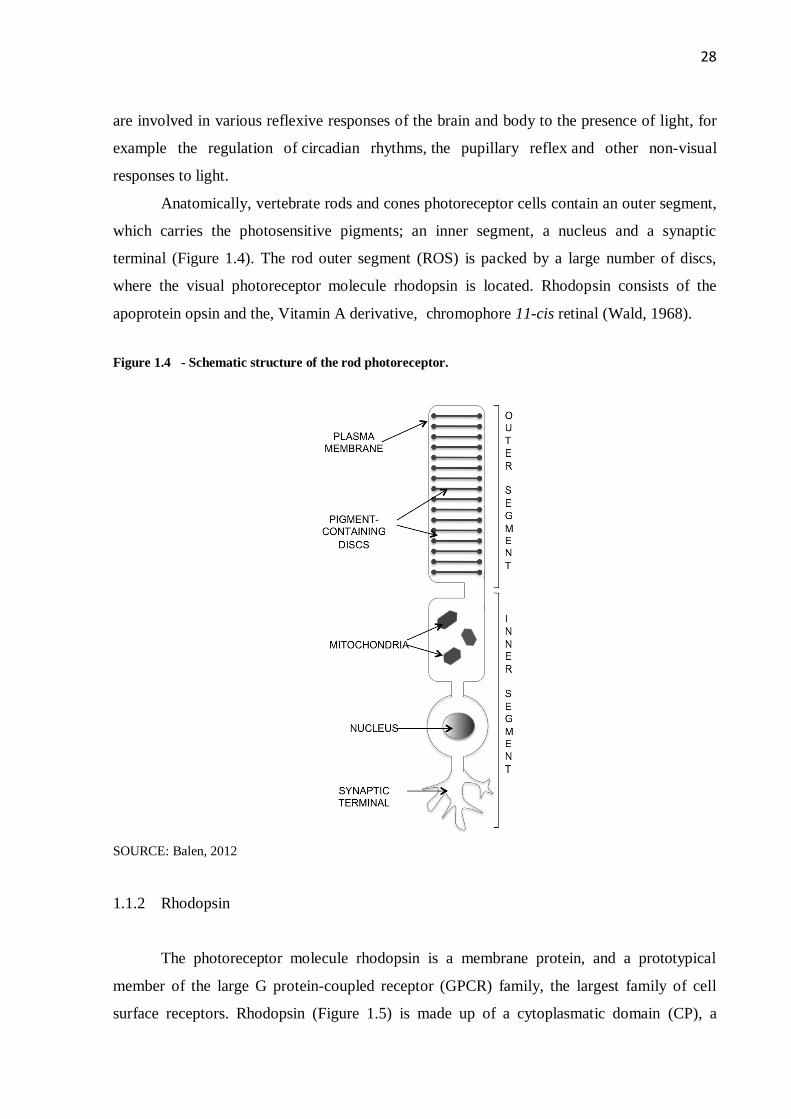
Anatomically, vertebrate rods and cones photoreceptor cells contain an outer segment,
which carries the photosensitive pigments; an inner segment, a nucleus and a synaptic
terminal (Figure 1.4). The rod outer segment (ROS) is packed by a large number of discs,
where the visual photoreceptor molecule rhodopsin is located. Rhodopsin consists of the
apoprotein opsin and the, Vitamin A derivative, chromophore 11-cis retinal (Wald, 1968).
Figure 1.4 - Schematic structure of the rod photoreceptor.
SOURCE: Balen, 2012
1.1.2 Rhodopsin
The photoreceptor molecule rhodopsin is a membrane protein, and a prototypical
member of the large G protein-coupled receptor (GPCR) family, the largest family of cell
surface receptors. Rhodopsin (Figure 1.5) is made up of a cytoplasmatic domain (CP), a

29
seven-transmembrane helical domain (TM) and an extracellular domain (EC). Rhodopsin
contains a covalently linked ligand, 11-cis retinal, a Vitamin A derivative, that stabilizes the
folded receptor (Figure 1.5). Visual signal transduction is initiated when a photon induces
isomerization of 11-cis retinal to all-trans retinal. This event triggers the rearrangement of the
TM domain, resulting in the light-activated, Metarhodopsin II (Meta II) state. The Meta II
state of rhodopsin is the active functionally state of the protein and initiates the visual signal
transduction cascade. As a first step, Meta II binds to the G protein, transducin (Gt). Gt then
activates phosphodiesterase (PDE), which hydrolyzes cyclic GMP (cGMP). This event
ultimately leads to the hyperpolarization of the cell through the closure of ion channels. When
the cell is hyperpolarized, the electrical potential inside the cell is more negative than in
darkness. In contrast, in the dark state, cGMP keeps the channels in the photoreceptor
membrane open, and Na+-Ca
2+ influx depolarizes the membrane. The signal can be shut down
through two mechanisms: (1) through Meta II decay, a process of dissociation of protein and
ligand to opsin and free all-trans retinal, or (2) through phosphorylation of Meta II by
rhodopsin kinase, followed by binding of arrestin to the phosphorylated C-terminal end of
rhodopsin (Chabre et al., 1988). The ligand free opsin formed when Meta II decays can
readily uptake 11-cis-retinal, thus regenerating rhodopsin.

30
Figure 1.5 - Tertiary structure of rhodopsin.
SOURCE: Balen, 2012
The primary structure of rhodopsin (bovine) contains 348 amino acids. This sequence
was determined using amino acid sequencing (Hargrave et al., 1983; Ovchinnikov Yu et al.,
1982). Figure 1.6 shows the secondary structure model of rhodopsin showing the seven TM
helices, the EC domain and the C-terminus domain, which faces the CP, along with the
disulfide bond (Cys110 and Cys187) and palmitoylation sites (Cys322 and Cys323). The
structure is divided in about 50% TM, 25% CP and 25% EC domains. Rhodopsin was the first
protein of the GPCR family to be crystallized (Palczewski et al., 2000). Rhodopsin it the best
studied and the prototypic member of the GPCR family.

31
Figure 1.6 - Secondary structure of rhodopsin.
SOURCE: Balen, 2012
Since the correct folding of the rhodopsin protein is extremely important for its
functionality, many studies were conducted to understand the folding and unfolding
mechanisms of rhodopsin. It has been demonstrated that there is a minimum amount of helix
content necessary to maintain the binding of 11-cis retinal to the protein (Ridge et al., 1995).
Unfolding studies using single molecule force spectroscopic approaches, point at specific
areas of rigidity in the structure of rhodopsin (Sapra et al., 2006, 2008; Tastan et al., 2007).
Furthermore, in silico simulations of thermal unfolding suggested formation of a folding core
between the EC and TM domains (Rader et al., 2004; Tastan et al., 2007). The structure of
rhodopsin is also prone to misfolding when amino acid replacements are made (Anukanth and
Khorana, 1994; Garriga et al., 1996; Kaushal and Khorana, 1994; Liu et al., 1996). Misfolding
here is characterized by the inability of the protein to bind 11-cis retinal, necessary for the
correct function of the protein in the visual system.

32
1.1.3 Allosteric ligands
In addition to retinal isomers, rhodopsin has been shown to bind a number of other
ligands. They all bind in locations other than the retinal binding pocket and are thus by
definition allosteric ligands. These ligands are reviewed below.
Zinc
Zinc, after iron, is the second most abundant trace metal in the human body (Aggett
and Comerford, 1995). It is present in high concentrations in the eye, especially in the retina
(Grahn et al., 2001). Numerous normal retinal functions are dependent on Zn2+
, including
retinal development, synaptic transmission, light response and Vitamin A metabolism (Grahn
et al., 2001). Zn2+
deficiency leads to night blindness, changes in dark-adaptation, ultra-
structural changes in the retina and the retinal pigment epithelium, and age-related macular
degeneration (Ugarte and Osborne, 2001). Furthermore, patients with the retinal degeneration
disease retinitis pigmentosa (see below) exhibit decreased serum Zn2+
levels (Karcioglu et al.,
1984). However, especially for age-related macular degeneration, opposing roles have been
proposed for Zn2+
. While supplementation with Zn2+
is the most widely used intervention
strategy for patients with this disease, its presence in Drusen, deposits of accumulated
extracellular material containing misfolded proteins which especially occur in patients with
age-related macular degeneration, has been linked to increased aggregation of proteins
(Lengyel and Peto, 2008). There are probably multiple proteins responsible for these
physiological effects (Grahn et al., 2001), but one particularly well established target is the
photoreceptor rhodopsin, to which Zn2+
binds directly and specifically with milli- to
micromolar affinities depending on the membrane environment studied (Shuster et al., 1992).
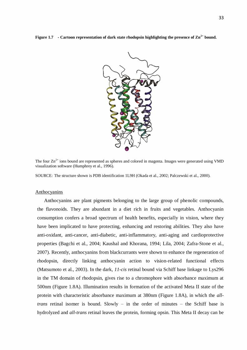
The crystal structure of rhodopsin revealed four putative Zn2+
binding sites (Okada et al.,
2002; Palczewski et al., 2000), shown in Figure 1.7 as purple spheres. The extracellular and
transmembrane Zn2+
binding sites have been proposed to be the physiologically relevant ones
(Park et al., 2007; Stojanovic et al., 2004), but X-ray absorption spectroscopy (Toledo et al.,
2008), NMR spectroscopy (Patel et al., 2005) and proteolytic digestion (Shuster et al., 1992)
have suggested the primary binding sites to be located in the EC domain.
33
Figure 1.7 - Cartoon representation of dark state rhodopsin highlighting the presence of Zn2+
bound.
The four Zn2+ ions bound are represented as spheres and colored in magenta. Images were generated using VMD
visualization software (Humphrey et al., 1996).
SOURCE: The structure shown is PDB identification 1L9H (Okada et al., 2002; Palczewski et al., 2000).
Anthocyanins
Anthocyanins are plant pigments belonging to the large group of phenolic compounds,
the flavonoids. They are abundant in a diet rich in fruits and vegetables. Anthocyanin
consumption confers a broad spectrum of health benefits, especially in vision, where they
have been implicated to have protecting, enhancing and restoring abilities. They also have
anti-oxidant, anti-cancer, anti-diabetic, anti-inflammatory, anti-aging and cardioprotective
properties (Bagchi et al., 2004; Kaushal and Khorana, 1994; Lila, 2004; Zafra-Stone et al.,
2007). Recently, anthocyanins from blackcurrants were shown to enhance the regeneration of
rhodopsin, directly linking anthocyanin action to vision-related functional effects
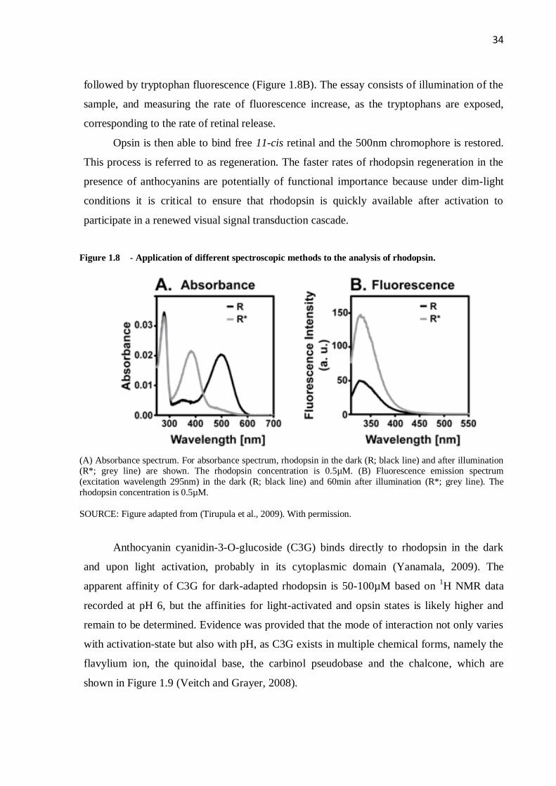
(Matsumoto et al., 2003). In the dark, 11-cis retinal bound via Schiff base linkage to Lys296
in the TM domain of rhodopsin, gives rise to a chromophore with absorbance maximum at
500nm (Figure 1.8A). Illumination results in formation of the activated Meta II state of the
protein with characteristic absorbance maximum at 380nm (Figure 1.8A), in which the all-
trans retinal isomer is bound. Slowly – in the order of minutes – the Schiff base is
hydrolyzed and all-trans retinal leaves the protein, forming opsin. This Meta II decay can be
34
followed by tryptophan fluorescence (Figure 1.8B). The essay consists of illumination of the
sample, and measuring the rate of fluorescence increase, as the tryptophans are exposed,
corresponding to the rate of retinal release.
Opsin is then able to bind free 11-cis retinal and the 500nm chromophore is restored.
This process is referred to as regeneration. The faster rates of rhodopsin regeneration in the
presence of anthocyanins are potentially of functional importance because under dim-light
conditions it is critical to ensure that rhodopsin is quickly available after activation to
participate in a renewed visual signal transduction cascade.
Figure 1.8 - Application of different spectroscopic methods to the analysis of rhodopsin.
(A) Absorbance spectrum. For absorbance spectrum, rhodopsin in the dark (R; black line) and after illumination (R*; grey line) are shown. The rhodopsin concentration is 0.5µM. (B) Fluorescence emission spectrum
(excitation wavelength 295nm) in the dark (R; black line) and 60min after illumination (R*; grey line). The
rhodopsin concentration is 0.5µM.
SOURCE: Figure adapted from (Tirupula et al., 2009). With permission.
Anthocyanin cyanidin-3-O-glucoside (C3G) binds directly to rhodopsin in the dark
and upon light activation, probably in its cytoplasmic domain (Yanamala, 2009). The
apparent affinity of C3G for dark-adapted rhodopsin is 50-100µM based on 1H NMR data
recorded at pH 6, but the affinities for light-activated and opsin states is likely higher and
remain to be determined. Evidence was provided that the mode of interaction not only varies
with activation-state but also with pH, as C3G exists in multiple chemical forms, namely the
flavylium ion, the quinoidal base, the carbinol pseudobase and the chalcone, which are
shown in Figure 1.9 (Veitch and Grayer, 2008).

35
Figure 1.9 - Chemical structures of pH dependent equilibrium species of the cyanidin-3-glucoside.
(A) Flavylium cation. (B) Quinoidal base example. (C) Carbinol pseudobase. (D) Chalcone. „R‟ in the chemical structures of C3G represents the glucoside sidechain.
SOURCE: Figure adapted from (Yanamala et al., 2009). With permission.
Chlorin e6
Porphyrins are an omnipotent class of naturally occurring compounds with many
important biological representatives such as hemes and chlorophylls. The chlorophyll
derivative chlorine e6 (Ce6) (Figure 1.10) is believed to enhance sensitivity of rhodopsin to
red light in deep-sea fish rhodopsin (Douglas et al., 1999; Isayama et al., 2006).
Figure 1.10 - Chemical structure of Ce6.
SOURCE: Balen, 2012
In vivo studies with salamander and mouse models have confirmed that Ce6 can
effectively enhance vision in these animals (Washington et al., 2007). A decrease in the
500nm peak of a UV-Vis spectrum of salamander rhodopsin was observed by illuminating the
sample at 668nm, a wavelength at which Ce6 absorbs while rhodopsin has less response
(Isayama et al., 2006). Additionally, mice administered with Ce6, showed a two-fold increase
in electroretinogram b-wave amplitudes (the mass response to the eye) as a response to red
and blue light (Washington et al., 2007). An energy transfer between Ce6 and 11-cis retinal

36
has been proposed (Isayama et al., 2006). In vitro studies demonstrated that Ce6 physically
binds to rhodopsin and modulates its structure and function (Yanamala, 2009).
1.1.4 Retinitis pigmentosa
Mutations in rhodopsin are associated with the retinal degeneration disease retinitis
pigmentosa (RP). RP reflects a large group of genetically heterogeneous disorders that affect
the photoreceptors and retinal pigment epithelium (RPE) diffusely across the entire fundus but
begin with initial geographic involvement in either the periphery or the macula (Sieving,
2010). It is one of the major causes of blindness in the world, affecting 1 in 4000-5000 people
(Kannabiran, 2008). The disease is marked by early rod photoreceptor dysfunction and
progressive degeneration of rods, which impairs vision in dim light and causes loss of
peripheral vision, that is “tunnel vision” (Sieving, 2010), and later cones (Lam, 2005). This
sequence of events explains why patients initially present night blindness, and only later in
life presents diurnal visual impairment (Hamel, 2006). Typical RP is then described as rod-
cone dystrophy, where rods are more affected than cones. However, some of the allied forms
primarily cause cone photoreceptor loss and initially manifest with a reduction in central
visual acuity (Sieving, 2010).
The pathologic mechanism underlying the observation that at the latest stages in the
disease retinal degeneration is observed is still not clear. Family-related retinal degeneration
with intraneural retinal pigmentation was described as early as 1855 (Sieving, 2010), and
although “retinitis” implies an inflammatory or infectious cause, histopathology studies shows
no evidence of macrophage invasion or other inflammatory response in the photoreceptor
layer or elsewhere in the retina (Sieving, 2010). Studies attributed a genetic basis to the
majority cases of RP and recently concluded that apoptosis is the last stage contributing to
photoreceptor cell death (Chang et al., 1993; Portera-Cailliau et al., 1994; Reme et al., 1998).
Belmonte et al. (2006) showed that Rac1, a member of the GTPase family with very low
molecular weight, may participate in the cell death process when the degeneration is mediated
by light or inherited. Moreover, after degeneration, there was an increase in the activity of
Rac1, and furthermore Rac1 expressing photoreceptors were shown to be TUNEL-positive.
TUNEL is a common method for detecting DNA fragmentation, by labeling the terminal end
of nucleic acids, which results from apoptotic signaling cascades.

37
No evidence of racial or ethnic predisposition has been observed. However, men may
be affected slightly more than women because of X-linked conditions (Sieving, 2010). The
age-at-onset of symptoms and diagnosis is variable and can range from early childhood to
mid-age adults. The diagnostic criteria is assessed through 4 parameters (Hamel, 2006); 1)
Functional signs: which are characterized by night blindness as the first symptom, followed
by photophobia; visual acuity is preserved in early and mid ages. 2) Visual field: marked by
patchy losses of peripheral vision evolving to ring-shaped scotoma, tunnel vision is the last
step. 3) Fundus: presence of pigmentary deposits similar to bone spicules in the peripheral
retina; thinning of the retinal vessels; waxy pallor of the optic nerve; various degrees of retinal
atrophy. 4) Electroretinogram: decrease of the amplitudes of a- and b- waves; scotopic
system, rods, predominates over photopic system, cones.
Methods for clinical diagnosis are based on the presence of night blindness and
peripheral visual defects, lesions in fundus, ERG changes, and progressive deterioration of all
of these signs (Hamel, 2006). The ERG is the best method to detect the disease, since patients
can be asymptomatic, for example presenting only nigh blindness and no other clinical signs.
1.1.5 Types of retinitis pigmentosa
RP is extremely heterogeneous, both clinically and genetically. Cases where there are
no family histories evident are known as isolate, sporadic or simplex. By inheritance, RP can
be classified in autosomal dominant (AD) (30%), autosomal recessive (AR) (20%) and X-
linked recessive (XL) (15%) or sporadic/simplex traits (30%), and be part of the group of
Leber congenital amaurosis (5%). These are among the more common modes of inheritance
in RP and the relative prevalence of each form varies between populations (Kannabiran, 2008;
Musarella and Macdonald, 2011). There are also rare forms of RP that are called X-linked
dominant, mitochondrial and digenic (presents mutations in two different genes) (Musarella
and Macdonald, 2011). Clinically, they are known as non-syndromic RP. Mutations in 16
different genes were found to cause autosomal dominant retinitis pigmentosa (ADRP)
(Kannabiran, 2008). Autosomal dominant forms are usually the mildest forms, some cases
starting after the age of 50, although severe disease can also appear (Hamel, 2006). The most
frequent cause of ADRP is a mutation found in the rhodopsin gene, affecting 20-25% of the
patients, especially in North America and Europe. In North America, a single point mutation,
Pro23His, accounts for 12% of the cases, although not significantly in Europe. Mutations on

38
the RDS gene is the second most predominant set of mutations in RP, 3.5 to 9% were
estimated followed by the RP1, which was estimated to be 4-8% of the cases. Other mutations
appear to be less frequent and are implicated in less than 5% of the cases (Kannabiran, 2008).
Autosomal recessive retinitis pigmentosa (ARRP) is a group of disorders that includes
juvenile and early-onset forms of RP. This group overlaps with another neurodegenerative
disease called Leber‟s congenital amaurosis (LCA). 21 genes were identified to cause ARRP.
However, the frequency of mutations from all of these genes accounts for approximately a
third of ARRP patients, with individual genes contributing to less than 5% of disease
(Kannabiran, 2008). There are two genes that were found to cause X-linked RP, RP2 and RP3
(known as RP GTPase regulator (RPGR)). RP2 account for 10-20% of recessive XLRP and
mutations in RPGR account for 50-80% of recessive XLRP. Another six loci were also
mapped but the genes were not identified yet (Kannabiran, 2008). X-linked forms also start
early and are frequently associated with myopia. Transmission of the disease is recessive in
most cases; however there are some families in which dominant inheritance with affected
females is found (Hamel, 2006).
Many syndromes are associated with different types of pigmentary retinopathies, and
are known as syndromic RP. The most frequent are the Usher and Bardet Biedl syndromes
(BBS). Usher syndrome is the most frequent and is typically associated with neurosensory
deafness. BBS is marked by obesity (already in childhood), mental retardation or mild
psychomotor delay, post axial polydactyly, hypogenitalism and renal problems and RP
associated, frequently as cone-rod type of dystrophy (Hamel, 2006). Other less frequent
syndromes encompass those that cause renal abnormalities such as Senior Loken syndrome
(SLS) and Alport syndrome, dysmorphic syndromes such as Cohen, Jeune and Cockayne
syndromes, metabolic syndromes such as Methylmalonic aciduria with homocystinuria,
Abetalipoproteinemia (Bassen Korntzweig disease), Bietti‟s disease, Cystinosis,
Mucopolysaccharidoses, Zellweger (cerebro-hepato-renal) syndrome, Hyperoxaluria type I
with retinal atrophy in spots, Neonatal adrenoleukodystrophy with leopard spots fundus,
Infantile Refsum disease, Adult Refsum disease, Peroxisomal disorders other than Refsum
disease, Peroxisomal disorders other than Refsum disease and neurological disease, neuronal
ceroid lipofuscinosis, Joubert syndrome, autosomal dominant cerebellar ataxia type II
(SCA7), Myotonic dystrophy and Hallervorden-Spatz syndrome (Hamel, 2006).

39
1.1.6 Current therapies
Although innumerous studies have been performed, there is no effective therapy to
treat RP. Meanwhile, many studies are focusing on alternative therapies to prevent, treat or
slow down the disease.
In order to slow down the degeneration process, light protection and treatment with
vitamins is recommended. Animal studies suggested that light exposure was damaging for the
transgenic photoreceptors (Wang et al., 1997), and it is recommended to patients with
pigmentary retinophaties to avoid direct light exposure, by either wearing dark glasses or
yellow-orange spectacles, which minimizes photophobia. The use of eye shades and lateral
protection is also recommended to protect against light coming from the sides (Hamel, 2006).
Treatment with vitamins A and E are believed to protect the photoreceptors by trophic and
antioxidant effects. A clinical trial of nutritional supplements for adults diagnosed with the
common forms of RP was conducted (Kupfer, 1993). The course of retinal degeneration was
monitored by electroretinogram recordings. On average, patients taking 15,000 IU of vitamin
A daily showed slower, and those taking 400 IU of vitamin E showed faster retinal
degeneration. The molecular mechanism of the Vitamin A effect is the rescue of correctly
folded rhodopsin by retinal. However, 15,000 IU only slowed and could not prevent retinal
degeneration, and a daily vitamin A intake exceeding 25,000 IU over the long-term is toxic.
Later, similar studies were conducted and showed complementary or adverse results (Berson
et al., 1993a, 1993b, 2004, 2010).
Different therapies have therefore been explored (Kannabiran, 2008). Gene therapy
methods have been geared towards correction of the specific gene mutated, by means of gene
replacement for recessive gene mutations or suppression and replacement of mutant mRNAs
for dominant gene mutations. Encapsulated cells releasing neurotrophic factors have been
implemented with the aim of promoting survival and growth of photoreceptors. Anti-
apoptotic factors have been introduced by gene-delivery systems. Finally, stem cell
transplantation has been considered. Each approach is briefly out-lined below.
Gene therapy is a promising therapy for many inherited human diseases. The
technique requires efficient genotyping methods for identification of the implicated gene
(Hamel, 2006). Ocular gene therapy (OGT) has been successfully tried in different animals
and humans. First, identification of the genetic cause of the RP and genotyping of the patients
was performed in order to proceed with the genetic modification of mutant ocular cells to

40
produce therapeutic effects. The strategy of the therapy differs accordingly with the type of
disease or mutation target, and was already applied successfully in mouse (Bennicelli et al.,
2008; Tan et al., 2009), rats (Drenser et al., 1998; Lewin et al., 1998; Weber et al., 2003),
dogs (Acland et al., 2001; Veske et al., 1999; Weber et al., 2003), non-human primates
(Weber et al., 2003), and humans (Bainbridge et al., 2008; Hauswirth et al., 2008; Maguire et
al., 2008, 2009). Due to different outcomes from patients, more tests still need to be done in
order to prove the efficacy for all patients.
Retinal implants using either epiretinal or subretinal implants is also a good therapy
and have shown benefits in many tests with RP patients (Caspi et al., 2009; Humayun and de
Juan, 1998; Yanai et al., 2007) and animals models (Chow and Chow, 1997; Zrenner et al.,
1997). Clinical trials in human patients are already in Phase II, but still being improved.
Neurotrophic factors showed protective effects on photoreceptors degeneration,
including neurotrophic factor (CNTF), Glial cell-line derived neurotrophic factor (GDNF),
cardiotrophin-1, brain-derived neurotrophic factor (BDNF) and basic fibroblast growth factor
(bFGF) (Hamel, 2006; Musarella and Macdonald, 2011) in animals (Bok et al., 2002; Frasson
et al., 1999; Li et al., 2010; Liang et al., 2001; Tao et al., 2002) and are in the trial phase in
humans (Sieving et al., 2006). Phase I and II are currently underway for treatment of atrophic
macular degeneration and RP.
Retinal transplantation consists of the transplantantion of sheets of retinal cells, layers
of photoreceptors or a complete retina (Hamel, 2006; Musarella and Macdonald, 2011).
Alternatively, transplantation of photoreceptor precursors has also been tried (Musarella and
Macdonald, 2011). Transplants in animal models and humans showed that transplanted
material does survive but not all presented evidence of a therapeutic effect (Gal et al., 2000;
MacLaren et al., 2006; Radtke et al., 2008).
Stem cell treatment is being used to regenerate degenerated retinas, based on its
capability to give rise to specialized cells and also its capability of self-renewal (Lund et al.,
2006). The donor cells and the host recipient are two major aspects of the procedure (Rivas
and Vecino, 2009). Hippocampal, embryonic and bone marrow cells have been tested in
models of retinal degenerative disorders and have showed high migratory and differentiation
capacity. In contrast, the use of retinal stem/progenitor cells in suspension or as neurospheres
exhibit poor migration, but were successful in expressing retina specific markers after
transplantation. Although progress is being made, extrapolation to humans is yet to be
demonstrated.

41
Neuroprotection using antiapoptotic factors or inhibitors of apoptosis is also being
explored (Hamel, 2006; Kannabiran, 2008). Studies in mouse and rat models have shown
protection of photoreceptor cells (Bennett et al., 1998; Leonard et al., 2007; Liu et al., 1999;
Petrin et al., 2003).
1.1.7 Rhodopsin as a target in retinitis pigmentosa
The causes for RP are heterogeneous, and mutations in many different genes can lead
to RP. The first of these genes that has been identified as a genetic cause for RP was the rod
photoreceptor rhodopsin. Now more than 150 mutations in rhodopsin have been identified in
different RP patients (Cooper et al., 2007). From these, 92 are single point mutations in 64
amino acid positions (Figure 1.11). This is a remarkable number considering that rhodopsin is
a protein with a total length of only 348 amino acids. 42 of these RP mutations have been
studied in vitro and 31 of them (>70%) were found to cause misfolding of rhodopsin.
Many studies have focused on understanding the molecular mechanism underlying
rhodopsin mutations leading to RP development. The majority of the mutations studied
previously focused on the EC and TM domains of rhodopsin (Dryja et al., 1993, 1995). Most
of these mutations mediate formation of an incorrect disulfide bond, leading to an incorrectly
folded protein that cannot bind 11-cis retinal (Kaushal and Khorana, 1994; Ridge et al., 1995).
This finding suggested a structural coupling between the EC and TM regions, which is
important for the correct folding of rhodopsin.
Since the discovery of misfolding of rhodopsin as one of the leading causes for RP
(Kaushal and Khorana, 1994), much effort has concentrated on rescuing correctly folded
rhodopsin by adding excess Vitamin A or retinoid derivatives. Recently, a clinical trial of
nutritional supplements for adults with common forms of RP was conducted, and
demonstrated that vitamin A treatment alone cannot cure RP (see above).

42
Figure 1.11 - Secondary structure model of rhodopsin showing the positions of the mutations that cause
RP and congenital stationary night blindness.
SOURCE: Balen, 2012
1.1.8 Animal models of retinitis pigmentosa
Hereditary retinal degeneration affects human and other animals. The range of
diseases that affect animals can be due to natural occurrence or obtained through genetic
manipulation. These various mutations are of great importance as model system for the study
of the disease evolution, pathogenesis and strategies for treatment. Specifically for RP, there
are many animal models available and many of them mimic the disease that is found in
humans.
The list of animal models that present mutations that occur naturally includes dogs,
cats and chicken. Natural and induced mutations are studied in mouse, rats and pigs. These
animal models were reviewed recently (Rivas and Vecino, 2009) and are summarized here.
Canine animal models of RP are considered the best models due to the similarity
between dog and human eye disorders. Furthermore, since eye anatomy and size in dogs is
similar to human, the same apparatus for physical studies can be used for dogs as that for
humans. All canine diseases present rod-cone degenerations, where rod disease, dysfunction,
and death precede the loss of cones. These diseases are analogous to human RP and are called
Progressive Retinal Atrophy (PRA). The genes mutated include PDE6A and PDE6B encoding
43
the α and β subunits of the cGMP specific phosphodiesterase (autosomal recessive RP),
RPE65 gene (leber congenital amaurosis), the RP GTPase regulator RPGR (X-linked RP) and
rhodopsin RHO (autosomal dominat RP). A list of available canine models is provided below:
erd - canine early retinal degeneration
rcd-1 - rod-cone dysplasia type 1 - and rcd-2 - rod-cone dysplasia type 2
RHO - rhodopsin, visual pigment of rod photoreceptor
RPE65 – membrane-associated protein
RPGR - mutations in the RP GTPase regulator
RPGRIP1 – mutations in the RP GTPase regulator-interacting protein 1
Cats are good models too since they have a manageable eye size for examination and
manipulation. Their visual neurophysiology is well characterized and their eyes are
physiologically and anatomically similar to human. There are only two forms of feline PRA
that are well studied:
rdAc - autosomal recessive rod-cone degenerative disorder
Rdy model - autosomal dominant early-onset retinal degenerative disease
Chicken eyes are also useful to study since they can be compared with human eyes in
size, facilitating pathological examination. However, chicken eyes are very different from
human eyes from a number of perspectives. In particular, the chicken eye is cone-dominated
rather than rod-dominated. However, the conservation of gene order between chicken and
humans is similar to that between human and mice genome, making this a potential model for
the study of spontaneously occurring inherited blindness disease. There are two models of
chicken that presents retinal degeneration:
rd – retinal degeneration chicken strain
rdd – retinal dysplasia and degeneration
Mice are the most studied animal models, and the rod-less mouse was the first animal
model where retinal degeneration was described. Mouse and human have a genomic similarity
of about 90%, and gene correlation can be precisely achieved. Advantages in the use of mouse

44
include the short life span (about 2 years), availability of protocols that are well established
and the ease at which new single gene diseases can be mapped. Available mouse models are:
Peripherin rds – retinal degeneration slow, rds-peripherin null mutation
Rd – retinal degeneration, which includes rd1, rd4, rd8, rd10, rd12
CEP290: rd16 – novel centrosomal protein
I-255/256 – mutant opsin gene with 3-bp deletion of isoleucine at codon 255/256
Knockout RPE65 (RPE65-/-) - accumulates retinyl esters in the RPE and lacks 11-cis
retinal
P347S - proline-347 to serine mutation in rhodopsin
Peripherin-rds 307 – mice heterozygous for the codon 307 mutation
Q334ter - transgenic mice that express a truncated rhodopsin due to a mutation
resulting in a stop codon at position 344
Knockout rho (rho-/-) – mutations in the rhodopsin gene (have no functional
rhodopsin gene)
VPP – mice express a mouse opsin gene containing three point mutations within a
seven amino acid sequence near the N-terminus of the molecule
o P23H - histidine for proline mutation
o V20G – glycine for valine mutation
o P27L – leucine for proline mutation
Rats exhibit retinal degeneration very similar to the retinal degeneration observed in
humans. The benefits of using rat photoreceptors are that they have been well studied,
therefore extensive knowledge of basic biological mechanisms is known. Morphologically,
the rat eye is several times larger than the mouse, which simplifies surgical manipulations and
electroretinographic evaluation; they also breed as rapidly as mice, generating large litters in a
short gestational time. The following lines are available:
RCS – Royal College of Surgeons. Retinal pigment epithelium (RPE) is unable to
phagocytose shed photoreceptor outer segments.
P23H – histidine at proline 23 position in rhodopsin. Rat carries a mutant mouse opsin
gene in addition to the endogenous native opsin genes

45
S334ter – the opsin transgene contains a termination codon at residue 334, resulting in
the expression of a rhodopsin protein lacking the last 15 C-terminal amino acids
Pigs are genetically engineered animal models. Pig eyes are similar in size to the
human eyes. They have a similar number and distribution of rod and cone cells as humans,
which makes them an excellent model for RP. Among other large mammals, the porcine
retina is even more similar to the human retina in size, and tools applied for diagnostics can
be used without adaptations.
P347L - lysine for proline at position 347 in rhodopsin
P347S - serine for proline at position 347 in rhodopsin
The figure below represents the time of initiation of retinal degeneration in each
animal model (Figure 1.12). Knowledge of the different degeneration times is important in
order to decide which model is more relevant to specific studies such as conducted as part of
this thesis.

46
Figure 1.12 - Schematic representation of the initiation of the retinal degeneration in each animal model.
Continuous lines: normal retina (non-degenerating); discontinuous lines: degeneration of the retina; Red:
morphological states; Blue: electroretinographic state; each square represents a postnatal age of the animal
(months).
SOURCE: Figure is taken from (Rivas and Vecino, 2009). With permission.
47
1.2 OPEN QUESTIONS AND THESIS OBJECTIVES
1.2.1 Open questions
Previous studies by Khorana and coworkers have addressed the identification of the
amino acids stabilizing rhodopsin and have established that there is tight coupling between the
extracellular, transmembrane and cytoplasmic domains in rhodopsin (Khorana, 2000).
Disruption of this coupling causes irreversible misfolding. Recent simulations of misfolding
of the rhodopsin tertiary structure have provided a mechanistic understanding of these
findings (Rader et al., 2004). A major core of residues contributing to rhodopsin stability was
identified at the interface between the extracellular and transmembrane domains. A small
additional region at the conserved DERY motif in the cytoplasmic domain was also
implicated in playing a role in rhodopsin stability. More than 90% of the residues in the large
core cause misfolding upon mutations (Rader et al., 2004). Furthermore, recent findings with
RP patients and molecular studies have correlated these molecular studies with RP patients
studies (Iannaccone et al., 2006). This indicates that it may be possible to develop new
avenues for RP treatments based on stabilization of rhodopsin structure. Such new avenues
are needed because there is, so far, still no cure or treatment for RP.
Here we explore novel approaches for the stabilization of opsin/rhodopsin through
small molecules. Rhodopsin has been shown to be a receptor for anthocyanins (Lila, 2004)
and its spectral range can be extended by interacting with porphyrin compounds (Washington
et al., 2004). Members of the flavonoid group of phytochemicals, anthocyanins are mostly
found in teas, honey, wines, fruits, vegetables, nuts, olive oils, cocoa and cereal.
Anthocyanins pigments and the flavonoids associated are known to protect against many
kinds of diseases. The enhancement of night-time vision by anthocyanins is known (Lila,
2004). Porphyrins are an omnipotent class of naturally occurring compounds with many
important biological representatives such as hemes and chlorophylls. Previous evidence from
our laboratory suggests that porphyrin and anthocyanin compounds both stabilize rhodopsin
and/or opsin (Yanamala, 2009).
In addition, studies have shown that providing patients with more retinal, via
administration of Vitamin A, can only partially slow down RP. So far, many alternative
approaches have been proposed, but none have an entirely positive effect on reversing the

48
disease. The hypothesis that molecules other than retinal can alone or in conjunction with
retinal rescue folded rhodopsin may lead to new treatments of RP.
1.2.2 Objectives and Approaches
This thesis aims to investigate the hypothesis that molecules other than retinal can
rescue folded rhodopsin and/or reduce photoreceptor cell death, which could potentially lead
to new treatments for RP. To address the open questions, the following specific aims are
proposed:
Specific aim 1: In vitro molecular characterization of WT and different rhodopsin
mutants
A study comparing two RP mutations N15S and P23H with WT rhodopsin was
performed. Rhodopsin and mutants were expressed in COS-1 and HEK-293 cells followed by
purification after reconstitution with 9-cis or 11-cis retinal, during or after expression in the
cells. This provided a quantitative understanding of the role of retinal in the rescue of folded
rhodopsin. Using thermal denaturation, Total Reflectance Fourier Transform Infrared
(ATR/FT-IR) and fluorescence spectroscopy, SDS-PAGE mobility, glycosylation studies, and
subcellular localization using confocal microscopy, a quantitative comparison of molecular
properties of the mutants as compared to the WT was assessed.
Specific aim 2: Effects of ligand binding on rhodopsin structure and stability
First, stability studies using the protein rhodopsin from native source was carried out.
Next, the effect of different compounds such as metal ions, porphyrins and anthocyanins were
tested in order to assess their effect on the stability of rhodopsin. UV-visible, fluorescence and
CD spectroscopy were used in order to compare of molecular properties of the WT rhodopsin
alone and in presence of the different ligands tested.
Specific aim 3: In vivo drug treatment of light-damaged and mutant rats with
hereditary RP disease
The aim was to test whether Ce6 is able to prevent apoptosis in Sprague Dawley rats
submitted to light-induced degeneration, and in P23H and S334ter rat models of RP.
Morphological and functional (ERG) analyses were performed.

172
7 CHAPTER 7: INTEGRATION OF IN VITRO, IN VIVO RAT MODEL AND
PATIENT STUDIES IN RETINITIS PIGMENTOSA AND FUTURE STUDIES
7.1 SUMMARY
In parallel to our molecular and in vivo model studies, our collaborator Alexander
Iannaccone at the Retinal Degeneration & Ophthalmic Genetics Service & Lions Visual Function
Diagnostic Laboratory, Hamilton Eye Institute, Department of Ophthalmology, and University of
Tennessee Health Science Center, USA, conducted patient studies of P23H and N15S mutants.
This provides us with the opportunity to compare in vitro, in vivo and patient data directly. Here,
I therefore present a summary of the major findings of my work integrated with the patient
studies. To this end, the manifestations, variability in severity and disease progression rates
obtained from patients pertaining to families carrying either the N15S or P23H mutation is
compared with the in vitro biochemical properties of the corresponding mutant rhodopsin. The
results are integrated into a comprehensive model for the role of rhodopsin as a target for small
molecules in RP degeneration and rescue. Finally, the prospects for novel approaches to prevent
and treat RP in animals and patients are discussed.
7.2 INTRODUCTION
Based on clinical studies, ADRP is divided into two classes – class A and B (Cideciyan et
al., 1998). Class A is characterized by severe rod function abnormalities based on ERG and
perimetry measurement, which results in consistent early night blindness while there is
measurable cone-mediated activity even later in life. On the other hand, Class B shows
measurable rod function (partial ERG preservation) as well as preserved cone function and is
characterized by variable onset of night blindness (some asymptomatic). Class B is further
divided into subclasses B1 and B2. B1 presents regional variation of retinal damage, while B2
indicates a diffuse disease condition with no regional predilection. Therefore, in class B mutants,
cone loss is spatially and temporally related to rod loss and the rate of cone loss is slower than
that of rods.

173
Bridging the gap between the classifications of mutants based on disease phenotype in
patients and the classifications based on in vitro biochemical properties of RHO mutations,
requires a detailed comparison between the clinical manifestations of the mutations and the
molecular properties of the proteins. Many rhodopsin mutations have been studied in vitro and
are classified according to its in vitro behavior (Kaushal and Khorana, 1994; Sung et al., 1991,
1993). In vitro classification of ADRP mutants has been carried out based on their molecular
phenotype, with three classes (Kaushal and Khorana, 1994): Class I, II and III. Class I resembles
WT rhodopsin where the mutants fold correctly, bind the 11-cis retinal chromophore normally,
but are inefficient in the inactivation of transducin, the cognate G protein complex. Class II
mutants do not traffic out of the endoplasmic reticulum and do not bind 11-cis retinal. Class III
mutants are expressed at low levels, poorly form rhodopsin chromophore and are also retained in
the endoplasmic reticulum, presenting high mannose glycosylation.
Previously, several class B ADRP patients, carrying mutations in different domains of
rhodopsin, namely in the extracellular domain, in the second intradiscal loop (P180A and
G188R), and in the cytoplasmic domain, at the end of helix III (R135L and R135W) were studied
(Iannaccone et al., 2006). The degrees of severity and progression rates in patients correlated with
the computationally predicted and experimentally confirmed molecular properties of the
respective rhodopsin protein (Iannaccone et al., 2006).
In this thesis, I investigated two mutants located at the same region of rhodopsin (N-
terminus in the extracellular domain). They are both grouped in the same class, based on clinical
phenotype and molecular properties. This provided an opportunity to test the hypothesis that
clinical phenotype and molecular properties are related at a finer grained level. To this end, an in-
depth comparison of the molecular data with the clinical characteristics studied by our
collaborator is provided in this chapter.
The in vitro data described in this thesis showed that the mutant rhodopsin proteins N15S
and P23H are structurally impaired, but present different characteristics and severity of
impairment. There is to date no rat or mouse model of N15S, so a comparison in an in vivo
animal model is currently not possible. We are therefore fortunate to have access to in vivo
patient data instead through our collaborator, Alex Iannacone, University of Tennessee. His in
vivo patient data, summarized in this chapter, below, confirms the phenotype observed in vitro.
Even though both mutations belong to the same class, the investigated patients presented

174
remarkable differences. P23H affected patient show a greater disease severity than the N15S
patient. This supports the conclusion that the instability of the rhodopsin protein structure is
linked to disease phenotype and severity. Thus, it should be possible to lessen the disease burden
of patients with strategies that stabilize the rhodopsin structure. In order to improve the
conditions of RP affected patients, new strategies for treatment of RP are being explored, and are
at different stages of trial. Here, the use of different ligands was tested, as a new approach for the
stabilization of the structure of rhodopsin. We first demonstrated the success of this strategy in
vitro. Since the results in vivo were promising, especially for Ce6 alone and in conjunction with
Zn2+
, it was also tested if Ce6 would also help to stabilize the mutant protein in vivo. While we
cannot test binding and stabilization effects on rhodopsin in vivo, we can use the end product –
prevention of retinal degeneration as indirect evidence for a success in treatment. To this end, two
studies were performed. First, a light-degeneration model, and secondly two inherited RP rat
models. Ce6 did show some (albeit weak) evidence for a prevention of light-induced retinal
degeneration, but did have a clearly positive effect in one of the models, P23H. Interestingly, a
negative effect was observed in the other rat model, S334ter. These effects are likely attributable
to the type of the mutation and its localization.
7.2.1 Summary of patient data carrying P23H and N15S mutations
Clinical studies were performed by Alexander Iannaccone are briefly summarized here.
Two individuals of each family were investigated, each of them presenting one of the two RP
mutations studied, P23H and N15S. The genealogies of the families are shown in Figure 7.1.

175
Figure 7.1 - Pedigrees of the two families investigated in this study.
Probands from each family are showed by the arrow sign. Horizontal bar on top of the symbol correspond to subjects
that were examined. Filled symbols identify affected individuals.
SOURCE: Not published. With permission.
Clinical studies included full-field flash ERG testing, Goldmann visual fields (GVFs),
monochromatic automated perimetry (MAP) and dark adaptometry (DAPT). In all patients, the
onset of night blindness was delayed (>30 years old) and visual acuity was ≥ 20/30, but disease
expression varied in severity across these RHO changes. Thus, both N-terminal mutations studied
here are associated with a Class B phenotype.
The N15S mutation was associated with mild and sharply demarcated inferior retinal
disease. Where in the inferior fields, both rod sensitivity loss (RSL) and cone sensitivity loss
(CSL) were minimal at age 62, but rod dark adaptation was markedly and unevenly delayed
across the entire visual field even after exposure to moderate amounts of ambient light. At age
66, Goldmann visual fields (GVFs) were virtually unchanged, CSL had slightly increased around
fixation, and RSL had increased pericentrally. At age 62, rod, mixed, and cone ERGs of the N15S
patient were reduced only about 80%, 60% and 75% of the lowest limits of normal, respectively,
configuring a cone>rod pattern of retinal dysfunction. At age 66, rod and mixed ERGs had
declined to about 50% of the normal lowest limits observed, whereas cone ERGs were
unchanged. Formal DAPT testing at age 66 showed markedly delayed recovery kinetics of both
rods and cones.

176
Figure 7.2 - Representative ISCEV-standard ERG response.
Full-field flash ERG testing of a N15S patient.
SOURCE: Not published. With permission.
Similar to the N15S mutation, P23H also had regional (inferior and nasal) retinal disease
predilection. This Class-B1 pattern was observed at age 12 (V:2) clinically and at the GVF as
well as at the MAP level. However, unlike the N15S mutation, by age 41 (IV:6) the disease
pattern transitioned to a widespread RSL>CSL (Class-B2 behaviour), followed by severe RSL
and CSL with macular sparing only at age 74 (III:3). ON GVF testing, a peripheral island of
nasal, inferior and temporal vision was also still present at age 74, but no regional/altitudinal
pattern was evident. Also, in P23H patients, rod-driven ERGs were non-recordable to standard
flashes since age 12 (V:2), but could be clearly elicited with brighter stimuli and b-wave splitting
into separate cone- and a rod-driven peaks was also seen (loss of rod-driven sensitivity), as
previously reported in class-B phenotypes (Iannaccone et al., 2006). This is in line with the
partially preserved dark adaptation rod-mediated function on MAP. At this young age, mixed
responses were fairly well preserved (~60%) and cone ERGs were normal. By age 41, mixed,
transient cone, and flicker cone ERGs were reduced to about 10%, 45% and 75% of the lowest
normal limits, respectively. At age 74, although markedly reduced in amplitude and markedly
delayed, mixed and cone-driven ERGs (recorded with HK-loop electrodes) remained clearly
recordable (~8%, 20% and 27% of the lowest normal limits, respectively). Overall, the P23H-
associated phenotype was significantly more severe than the N15S phenotype.
N15S RHO (IV:1)

177
Visual acuity is well preserved in both mutants, with most patients retaining ≥20/40,
although substantial inter-individual variability is observed, both between and within mutations.
ERG measurements were also performed. Although instances of non-recordable ERGs have been
reported in the literature, most patients retain measurable mixed ERG b-waves. Longitudinal
measurements in the N15S proband (IV:1) with Class-B1 phenotype show a yearly rate of ERG
b-wave loss from baseline of about 6.25%. This is very similar to that reported for of another
Class-B1 mutant (P180A, 7%/yr) (Iannaccone et al., 2006). The ERG is much better preserved in
N15S than in P23H.
It has been suggested that cone loss is spatially and temporally related to rod loss and the
rate of cone loss is slower than that of rods in Class B mutants (Cideciyan et al., 1998). The in
vivo data strongly support this hypothesis. Likewise, coexistence of Class-B1 and B2 features can
occur in the same family in association with the P23H mutation, a transition that appears to be
linked to disease stage. Despite evidence of regional disease and retention of rod function, the
P23H mutation causes greater and earlier rod function loss than N15S, and an overall
significantly greater disease severity.
The N15S phenotype is remarkable for the presence of a ring of pericentral disease,
severely compromised kinetics of rhodopsin regeneration, and a cone > rod pattern of ERG
dysfunction, but later in life rod disease progression continues to precede that of cones as
postulated for Class-B phenotypes.

178
Figure 7.3 - Representative ISCEV-standard ERG response
Full-field flash ERG testing of a P23H patient.
SOURCE: Not published. With permission.
7.2.2 Comparison of P23H and N15S human patient data with in vitro studies
The clinical and functional characterizations of the two mutants are compared here to my
molecular studies (subcellular localization using confocal microscopy, SDS-PAGE mobility,
glycosylation studies, thermal stability and secondary structure content using spectroscopy and
Attenuated Total Reflectance Fourier Transform Infrared (ATR/FT-IR), described in detail in
Chapter 4). Being able to quantitatively compare the characteristics of the two mutants in vitro
and in vivo allowed the study of the extent of severity in molecular and functional terms. The in
vivo functional and clinical studies, expression profiles in cell culture and molecular/biophysical
in vitro studies of the purified proteins all suggest that the phenotype of N15S is significant but is
less than that of P23H. The correlation between the relative severities lends support to the
hypothesis that the molecular properties of the mutant proteins are directly related to the clinical
manifestations in patients. Despite the global classification into the same class, both from a
clinical and a molecular perspective, a quantitative comparison reveals that the two mutations
differ in severity of both, clinical manifestations, and molecular properties. Although not proving
causality, this observation does provide strong support for the hypothesis that the molecular
P23H RHO (III:3)

179
properties of the mutant rhodopsin proteins are quantitatively linked to the phenotype observed in
patients.
7.2.3 Comparison of Ce6 effects on RP progression in vivo versus in vitro model systems
Having established that stability of rhodopsin is related - possibly in a causal manner – to
clinical phenotype of RP mutants P23H and N15S (Chapter 3) and also that stability can be
enhanced by small molecules (Chapter 4), I then tested if the P23H phenotype can be rescued by
the small molecule stability enhancer Ce6 in a rat model of P23H induced RP, described in
Chapter 6. The results of ERG and retinal tissue analysis indicated that Ce6 exerts a positive
functional effect by slowing the rate of photoreceptor degeneration of P23H rats in vivo. The
studies indicated that Ce6 affects RP progression in vivo and the effects are likely linked to the
binding of this molecule to rhodopsin as demonstrated in vitro.
7.3 FUTURE WORK - PROSPECTS FOR NOVEL APPROACHES TO PREVENT AND
TREAT RP IN PATIENTS
This thesis investigated new approaches to treat RP. The emphasis was to find new
treatments of those manifestations of disease caused by mutations in the photoreceptor rhodopsin.
To this end the hypothesis was tested that compounds other than retinal can rescue folded
rhodopsin and/or reduces photoreceptor cell death by means of exerting a direct effect on the
structure and stability of rhodopsin. A conceptual view of the areas where rhodopsin can be
targeted is shown schematically in Figure 7.4.

180
Figure 7.4 - Schematic representation of the in vitro and in vivo experimental steps taken in this work.
SOURCE: Balen, 2012
It was previously found and confirmed in this thesis for the particular mutants under study
that the addition of 9-cis and 11-cis retinal during protein expression in cell culture improves the
folding of rhodopsin mutants, increasing the amount of correctly folded rhodopsin. This in vitro
result is in agreement with in vivo studies where vitamin A was administered to patients and
partially slowed down the RP. However, retinals are able to only partially improve rhodopsin
folding and large amounts of vitamin A are toxic. Therefore, in this thesis, other molecules were
also tested in vitro and in vivo. Divalent metal ions (Zn2+
, Cu2+
, Fe2+
, Ni2+
, Mg2+
and Mn2+
), the
anthocyanin compound C3G and the Chlorophyl derivative Ce6 were tested in vitro as a mean of
stabilizing the structure of rhodopsin. All these molecules exerted an effect on opsin/rhodopsin
structure at different extents and directions (stabilization and destabilization). Metal ions and Ce6

181
conferred a pronounced increase in the thermal stability of the secondary structure of rhodopsin.
The Ce6 molecule was also tested in light-induced degeneration and hereditary rat models in vivo
and has shown an effect on retinal protection. In contrast, C3G exerted a destabilizing effect on
rhodopsin structure while enhancing the regeneration rates and were therefore only tested in
vitro. This thesis work has shown that Ce6 but not C3G can be used to help in the rescue of
folded rhodopsin and/or reducing photoreceptor cell death. This provides a novel avenue and a
complementary one to current approaches to clinically study the disease and help patients that are
affected with hereditary RP.
A number of areas for future exploitation of this finding and further consolidation of its
premises are opened up by the work of this thesis. Shown in Figure 7.5 are my suggested areas of
concentration for future work.
Figure 7.5 - Suggested concentration areas for future work.
SOURCE : Balen, 2012

182
In vivo experiments could be conducted to better evaluate the effect of Ce6 on the
prevention of retinal degeneration caused by light-damage. Factors that could be exploited are the
number of rats, which could be increased, dosage and frequency of treatment, as well age at
which the treatment is performed that could be varied. The parameters should also be extended to
RP affected rats.
In vitro studies need to fill the current gaps opened by the results of my findings. Since
addition of 9-cis and 11-cis during the expression of the rhodopsin mutants, P23H and N15S,
mediated the purification of high amounts of protein, further studies can be pursued where the
addition of different molecules, such as the use of divalent metal ions alone and in conjunction
with Ce6 can be investigated. To facilitate a direct comparison, the corresponding studies I have
done in my thesis with WT rhodopsin need to be carried out with purified mutant proteins. Thus,
all in vivo studies have to be matched with the corresponding in vitro studies. Given the
promising results obtained in this thesis for WT rhodopsin in vitro for Ce6 alone and the
combination of Ce6 and Zn2+, and the observation that Ce6 alone helps the P23H rats in vivo,
strongly suggests, that the proposed approach will be successful.
Finally, to understand the negative results obtained with the in vivo rat model of RP,
S334ter upon treatment of Ce6, in vitro analysis could be performed in order to comprehend the
mechanism behind the effects of these molecules on the S334ter mutation. Specifically, we
propose that Ce6 cannot bind to S334ter. This hypothesis needs to be tested experimentally. The
stability of the mutant also needs to be investigated in vitro to see if stabilization in this case by
binding of small molecules would be beneficial. Given that Ce6 is not a beneficial molecule for
this particular rat model, other compounds, such as C3G and metal ions could be investigated.

183
7.4 IMPACT
Analysis of the interaction of various ligands with rhodopsin, results described in Chapter 3
of this thesis, demonstrated that there are different ligands to rhodopsin, other than 11-retinal, that
bind to rhodopsin. These are allosteric ligands and are believed to bind at the CP domain of
rhodopsin (Yanamala, 2009). The interaction of these accessory ligands exerted different effects
on rhodopsin. Ce6 and metal ions such as Zn2+
, Fe2+
, Cu2+
have shown to stabilize rhodopsin
secondary and tertiary structure. C3G binding to rhodopsin resulted in the destabilization of
retinal-protein interactions, which explains its effect on enhancement of the regeneration rates of
rhodopsin. Yet, it was already known that Zn2+
deficiency causes retinal dysfunctions such as
night blindness and neurodegeneration. Because Zn2+
binds directly to the photoreceptor
rhodopsin and alters its stability, the stabilization of rhodopsin may be key to prevention and
treatment of retinal dysfunctions. These findings can be of great potential importance for the
treatment of retinal degeneration diseases, where mutations in the photoreceptor rhodopsin are
known to be the major disease cause in vivo.
The stabilization obtained by Ce6 and Zn2+
in vitro and the effect of Ce6 in vivo can be
potentially relevant to dysfunctions caused by nutrient deficiencies, but also for inherited
diseases. Retinitis Pigmentosa (RP) is a disease that reflects a large group of genetically
heterogeneous disorders marked by early rod photoreceptor dysfunction and progressive
degeneration of rods and cones (Lam, 2005). The initial causes for RP are heterogeneous, and
mutations in many different genes can lead to RP development (Berson, 1993c; Cooper et al.,
1998). The first of these genes that has been identified as a genetic cause for RP was rhodopsin.
Now more than 150 mutations in rhodopsin have been identified in different RP patients (Cooper
et al., 1998). 42 of these RP mutations have been studied in vitro and 31 of them (>70%) were
found to cause misfolding of rhodopsin (Rader et al., 2004). Since in vitro misfolding and cell-
based retention in the endoplasmic reticulum have been proposed to be one of the molecular
triggering events associated with retinal degeneration in RP in 1994 (Kaushal and Khorana,
1994) much effort has concentrated on rescuing correctly folded rhodopsin with excess Vitamin
A or retinoid derivative. A clinical trial of nutritional supplements for adults with the common
forms of RP (Kupfer, 1993) showed that vitamin A treatment alone cannot cure RP (Kupfer,
1993) but maybe additional means of stabilization of rhodopsin could open the way to a new and
184
more direct therapeutic tool, applicable to many different mutants of rhodopsin as long as their
stabilization contributes to the mechanism of RP prevention/slow-down.
The studies presented here do not only have direct impact on vision. Rhodopsin is a
prototypic member of the GPCR family, which accounts for 2 percent of the human genome and
is the target for approximately 50 percent of current pharmaceutical compounds (Jacoby et al.,
1997). Indeed, Zn2+
has been found to bind also to other members of the GPCR family and has
been shown to have physiological roles in some of these receptors. For β2-adrenergic, dopamine,
and melanocortin receptors, Zn2+
is a positive allosteric modulator of agonist binding (Holst and
Schwartz, 2003; Norregaard et al., 1998; Schetz et al., 1999; Schetz and Sibley, 1997). Other
studies presented experimental evidence showing that Zn2+
also acts as a signaling molecule
(Hershfinkel et al., 2001). Inhibition of G protein activation was observed for rhodopsin (Sheikh
et al., 1996), the β2-adrenergic receptor and the parathyroid hormone receptor (Sheikh et al.,
1999). These are only some GPCRs for which Zn2+
effects have been reported and it is likely that
other GPCRs will be affected also. In this thesis, it has been shown that there can be potential
benefits in combining Zn2+
with other allosteric modulators, such as Ce6, to modulate the
stability of GPCRs as well as interactions of the GPCRs with their signaling ligands. It is of
particular importance to note that Ce6 is believed to bind in the CP domain of rhodopsin
(Yanamala, 2009), an area that is highly conserved in GPCRs in general. Thus, binding of
allosteric CP ligands and stabilization of the respective receptors may be achieved not only in
rhodopsin but also other GPCRs.

185
*In according with: International Committee of Medical Journal Editors. The recommended style for references is
based on the National Information Standards Organization NISO Z39.29-2005 (R2010). Available from:
http://www.nlm.nih.gov/bsd/uniform_requirements.html [2011 July 15].
REFERENCES*
Acland GM, Aguirre GD, Ray J, et al. Gene therapy restores vision in a canine model of
childhood blindness. Nat Genet. 2001;28(1):92-5.
Aggett PJ, Comerford JG. Zinc and human health. Nutr Rev. 1995 Sep;53(9 Pt 2):S16-22.
Anukanth A, Khorana HG. Structure and function in rhodopsin. Requirements of a specific
structure for the intradiscal domain. J Biol Chem. 1994;269(31):19738-44.
Bagchi D, Sen CK, Bagchi M, et al. Anti-angiogenic, antioxidant, and anti-carcinogenic
properties of a novel anthocyanin-rich berry extract formula. Biochemistry. 2004;69(1):75-80.
Bainbridge JW, Smith AJ, Barker SS, et al. Effect of gene therapy on visual function in Leber's
congenital amaurosis. N Engl J Med. 2008;358(21):2231-9.
Balem F, Yanamala N, Klein-Seetharaman J. Additive effects of chlorin E6 and metal ion
binding on the thermal stability of rhodopsin in vitro. Photochem and Photob. 2009:85:471-478
Bennett J, Zeng Y, Bajwa R, et al. Adenovirus-mediated delivery of rhodopsin-promoted bcl-2
results in a delay in photoreceptor cell death in the rd/rd mouse. Gene Ther. 1998;5(9):1156-64.
Bennicelli J, Wright JF, Komaromy A, et al. Reversal of blindness in animal models of leber
congenital amaurosis using optimized AAV2-mediated gene transfer. Mol Ther. 2008;16(3):458-
65.
Berson EL. Retinitis Pigmentosa. The Friedenwald Lecture. Invest Opthalmol Vis Sci.
1993c;34:1659-76.
Berson EL, Rosner B, Sandberg MA, et al. A randomized trial of vitamin A and vitamin E
supplementation for retinitis pigmentosa. Arch Ophthalmol. 1993a;111(6):761-72.
Berson EL, Rosner B, Sandberg MA, et al. Vitamin A supplementation for retinitis pigmentosa.
Arch Ophthalmol. 1993b;111(11):1456-9.
Berson EL, Rosner B, Sandberg MA, et al. Clinical trial of lutein in patients with retinit is
pigmentosa receiving vitamin A. Arch Ophthalmol. 2010;128(4):403-11.
Berson EL, Rosner B, Sandberg MA, et al. Further evaluation of docosahexaenoic acid in
patients with retinitis pigmentosa receiving vitamin A treatment: subgroup analyses. Arch
Ophthalmol. 2004;122(9):1306-14.

186
Bok D, Yasumura D, Matthes MT, et al. Effects of adeno-associated virus-vectored ciliary
neurotrophic factor on retinal structure and function in mice with a P216L rds/peripherin
mutation. Exp Eye Res. 2002;74(6):719-35.
Brouillard R, Dubois JE. Mechanism of Structural Transformations of Anthocyanins in Acidic
Media. J Am Chem Soc. 1977;99(5):1359-64.
Bullock AN, Debreczeni JE, Fedorov OY, et al. Structural basis of inhibitor specificity of the
human protooncogene proviral insertion site in moloney murine leukemia virus (PIM-1) kinase. J
Med Chem. 2005;48(24):7604-14.
Cajal SR. Histologie Du Système Nerveux de l'Homme et Des Vertébrés. 1911. Available from
http://en.wikipedia.org/wiki/Retina. Taken on December 07, 2011.
Caspi A, Dorn JD, McClure KH, et al. Feasibility study of a retinal prosthesis: spatial vision with
a 16-electrode implant. Arch Ophthalmol. 2009;127(4):398-401.
Chabre M, Bigay J, Bruckert F, et al. Visual signal transduction: the cycle of transducin shuttling
between rhodopsin and cGMP phosphodiesterase. Cold Spring Harb Symp Quant Biol. 1988;53
Pt 1:313-24.
Chang G, Hao Y, Wong F. Apoptosis: final common pathway of photoreceptor death in rd, rds,
and rhodopsin mutant mice. Neuron. 1993;11:595-605.
Chen C, Okayama H. High-efficiency transformation of mammalian cells by plasmid DNA. Mol
Cell Biol. 1987;7(8):2745-52.
Chow AY, Chow VY. Subretinal electrical stimulation of the rabbit retina. Neurosci Lett.
1997;225(1):13-6.
Cideciyan AV, Hood DC, Huang Y, et al. Disease sequence from mutant rhodopsin allele to rod
and cone photoreceptor degeneration in man. Proc Natl Acad Sci U S A. 1998;95(12):7103-8.
Cooper DN, Ball EV, Stenson PD, et al. The Human gene mutation database at the institute of
medical genetics in Cardiff. BIOBASE - Biological Database; 2007 [updated May 3, 2012];
Available from: www.hgmd.org.
Cruickshanks KJ, Klein R, Klein BE. Sunlight and age-related macular degeneration. The Beaver
Dam Eye Study. Arch Ophthalmol. 1993 Apr;111(4):514-8.
Darwin C. On the Origin of Species by Means of Natural Selection. Editions DT, editor. Mineola,
New York: Dover Publications, INC; 2006.
Dong CJ, Hare WA. GABAc feedback pathway modulates the amplitude and kinetics of ERG b-
wave in a mammalian retina in vivo. Vision Res. 2002;42(9):1081-7.
Douglas RH, Partridge JC, Dulai K, et al. Dragon fish see using chlorophyll. Nature.
1998;393:423-4.

187
Douglas RH, Partridge JC, Dulai KS, et al. Enhanced retinal longwave sensitivity using a
chlorophyll-derived photosensitiser in Malacosteus niger, a deep-sea dragon fish with far red
bioluminescence. Vision Research. 1999;39(17):2817-32.
Drenser KA, Timmers AM, Hauswirth WW, et al. Ribozyme-targeted destruction of RNA
associated with autosomal-dominant retinitis pigmentosa. Invest Ophthalmol Vis Sci. 1998
Apr;39(5):681-9.
Dryja TP, Berson EL, Rao VR, et al. Heterozygous missense mutation in the rhodopsin gene as a
cause of congenital stationary night blindness. Nat Genet. [10.1038/ng0793-280]. 1993;4(3):280-
3.
Dryja TP, Finn JT, Peng YW, et al. Mutations in the gene encoding the alpha subunit of the rod
cGMP-gated channel in autosomal recessive retinitis pigmentosa. Proc Natl Acad Sci U S A.
1995;92(22):10177-81.
Dryja TP, McGee TL, Reichel E, et al. A point mutation of the rhodopsin gene in one form of
retinitis pigmentosa. Nature. 1990;343(6256):364-6.
Faktorovich E, Steinberg R, Yasumura D, et al. Photoreceptor degeneration in inherited retinal
dystrophy delayed by basic fibroblast growth factor. Nature. 1990;347:83 - 6.
Farrens DL, Khorana HG. Structure and Function in Rhodopsin : Measurement of the rate of
metarhodopsin II decay by Fluorescence Spectroscopy. JBC. 1995;270(10):5073-6.
Fernandez-Sanchez L, Lax P, Pinilla I, et al. Tauroursodeoxycholic acid prevents retinal
degeneration in transgenic P23H rats. Invest Ophthalmol Vis Sci. 2011;52(8):4998-5008.
Fleisch VC, Jametti T, Neuhauss SCF. Electroretinogram (ERG) Measurements in Larval
Zebrafish. CSH Protocols. 2008.
Foster RG, Provencio I, Hudson D, et al. Circadian photoreception in the retinally degenerate
mouse (rd/rd). J Comp Physiol A. 1991;169(1):39-50.
Franze K, Grosche J, Skatchkov SN, et al. Muller cells are living optical fibers in the vertebrate
retina. Proc Natl Acad Sci U S A. 2007;104(20):8287-92.
Frasson M, Picaud S, Leveillard T, et al. Glial cell line-derived neurotrophic factor induces
histologic and functional protection of rod photoreceptors in the rd/rd mouse. Invest Ophthalmol
Vis Sci. 1999;40(11):2724-34.
Frishman D, Argos P. Knowledge-based protein secondary structure assignment. Proteins. 1995
Dec;23(4):566-79.
Gal A, Li Y, Thompson DA, et al. Mutations in MERTK, the human orthologue of the RCS rat
retinal dystrophy gene, cause retinitis pigmentosa. Nat Genet. 2000;26(3):270-1.

188
Garriga P, Liu X, Khorana HG. Structure and function in rhodopsin: correct folding and
misfolding in point mutants at and in proximity to the site of the retinitis pigmentosa mutation
Leu-125-->Arg in the transmembrane helix C. Proc Natl Acad Sci U S A. 1996;93(10):4560-4.
Gearhart PM, Gearhart C, Thompson DA, et al. Improvement of visual performance with
intravitreal administration of 9-cis-retinal in Rpe65-mutant dogs. Arch Ophthalmol.
2010;128(11):1442-8.
Glybina IV, Kennedy A, Ashton P, et al. Intravitreous delivery of the corticosteroid fluocinolone
acetonide attenuates retinal degeneration in S334ter-4 rats. Invest Ophthalmol Vis Sci. 2010
;51(8):4243-52.
Grahn BH, Paterson PG, Gottschall-Pass KT, et al. Zinc and the eye. J Am Coll Nutr. 2001;20(2
Suppl):106-18.
Hamel C. Retinitis pigmentosa. Orphanet J Rare Dis. 2006;1:40.
Harazny J, Scholz M, Buder T, et al. Electrophysiological deficits in the retina of the DBA/2J
mouse. Doc Ophthalmol. 2009;119(3):181-97.
Hargrave PA, McDowell JH, Curtis DR, et al. The structure of bovine rhodopsin. Biophys Struct
Mech. 1983;9(4):235-44.
Hargrave PA. The amino-terminal tryptic peptide of bovine rhodopsin. A glycopeptide containing
two sites of oligosaccharide attachment. Biochim Biophys Acta. 1977;492(1):83-94.
Hauswirth WW, Aleman TS, Kaushal S, et al. Treatment of leber congenital amaurosis due to
RPE65 mutations by ocular subretinal injection of adeno-associated virus gene vector: short-term
results of a phase I trial. Hum Gene Ther. 2008;19(10):979-90.
Hershfinkel M, Moran A, Grossman N, et al. A zinc-sensing receptor triggers the release of
intracellular Ca2+ and regulates ion transport. Proc Natl Acad Sci U S A. 2001;98(20):11749-54.
Holst B, Schwartz TW. Molecular mechanism of agonism and inverse agonism in the
melanocortin receptors: Zn(2+) as a structural and functional probe. Ann N Y Acad Sci.
2003;994:1-11.
Humayun MS, de Juan E, Jr. Artificial vision. Eye (Lond). 1998;12 ( Pt 3b):605-7.
Humphrey W, Dalke A, Schulten K. VMD: visual molecular dynamics. J Mol Graph.
1996;14(1):33-8, 27-8.
Iannaccone A, Man D, Waseem N, et al. Retinitis pigmentosa associated with rhodopsin
mutations: Correlation between phenotypic variability and molecular effects. Vision Res.
2006;46(27):4556-67.
Isayama T, Alexeev D, Makino CL, et al. An accessory chromophore in red vision. Nature.
2006;443(7112):649.

189
Jacoby E, Boudon A, Kucharczyk N, et al. A structural rationale for the design of water soluble
peptide-derived neurokinin-1 antagonists. J Recept Signal Transduct Res. 1997;17(6):855-73.
Kannabiran C. Retinitis pigmentosa: genetics and gene-based approaches to therapy. Expert
Review of Ophthalmology. [Review]. 2008;3(4):417-29.
Karcioglu ZA, Stout R, Hahn HJ. Serum zinc levels in retinitis pigmentosa. Curr Eye Res.
1984;3(8):1043-8.
Kaushal S, Khorana HG. Structure and function in rhodopsin. 7. Point mutations associated with
autosomal dominant retinitis pigmentosa. Biochemistry. 1994a;33(20):6121-8.
Kaushal S, Ridge KD, Khorana HG. Structure and function in rhodopsin: the role of asparagine-
linked glycosylation. Proc Natl Acad Sci U S A. 1994b;91(9):4024-8.
Khorana HG. Molecular biology of light transducin by the mammalian photoreceptor rhodopsin.
J Biomol Structure and Dynamics. 2000;11:1-16.
Kohn J, Wilchek M. A colorimetric method for monitoring activation of Sepharose by cyanogen
bromide. Biochemical and biophysical research communications. 1978;84(1):7-14.
Kolb H. WEBVISION - The organization of the retina and visual system: University of Utah;
2011. Available from: http://webvision.med.utah.edu/book/.
Kraft SP, Parker JA, Matuk Y, et al. The rat electroretinogram in combined zinc and vitamin A
deficiency. Invest Ophthalmol Vis Sci. 1987;28(6):975-84.
Kranich H, Bartkowski S, Denton MJ, et al. Autosomal dominant 'sector' retinitis pigmentosa due
to a point mutation predicting an Asn-15-Ser substitution of rhodopsin. Hum Mol Genet.
1993;2(6):813-4.
Krebs MP, Holden DC, Joshi P, et al. Molecular mechanisms of rhodopsin retinitis pigmentosa
and the efficacy of pharmacological rescue. J Mol Biol. 2010;395(5):1063-78.
Kumel G, Daus H, Mauch H. Improved method for the cyanogen bromide activation of agarose
beads. Journal of Chromatography. 1979;172:221-6.
Kupfer C. Information for Doctors Who Follow Patients with Retinitis Pigmentosa. National Eye
Institute; 1993; Available from: http://www.nei.nih.gov/news/clinicalalerts/alert-rp.asp.
Lam BL. Electrophysiology of vision: Clinical testing and application. Boca Raton: Taylor and
Francis; 2005.
LaVail MM, Battelle BA. Influence of eye pigmentation and light deprivation on inherited retinal
dystrophy in the rat. Exp Eye Res. 1975;21:167-92.
Lengyel I, Peto T. Cure or cause: opposing roles for zinc in age-related macular degeneration.
Expert Rev Ophthalmol. 2008;3(1):1-4.

190
Leonard KC, Petrin D, Coupland SG, et al. XIAP protection of photoreceptors in animal models
of retinitis pigmentosa. PLoS One. 2007;2(3):e314.
Lewin AS, Drenser KA, Hauswirth WW, et al. Ribozyme rescue of photoreceptor cells in a
transgenic rat model of autosomal dominant retinitis pigmentosa. Nat Med. 1998;4(8):967-71.
Li Y, Tao W, Luo L, et al. CNTF induces regeneration of cone outer segments in a rat model of
retinal degeneration. PLoS One. 2010;5(3):e9495.
Liang FQ, Aleman TS, Dejneka NS, et al. Long-term protection of retinal structure but not
function using RAAV.CNTF in animal models of retinitis pigmentosa. Mol Ther. 2001;4(5):461-
72.
Lila MA. Anthocyanins and Human Health: An In Vitro Investigative Approach. J Biomed
Biotechnol. 2004;(5):306-13.
Liu C, Li Y, Peng M, et al. Activation of caspase-3 in the retina of transgenic rats with the
rhodopsin mutation s334ter during photoreceptor degeneration. J Neurosci. 1999;19(12):4778-85.
Liu X, Garriga P, Khorana HG. Structure and function in rhodopsin: correct folding and
misfolding in two point mutants in the intradiscal domain of rhodopsin identified in retinitis
pigmentosa. Proc Natl Acad Sci U S A. 1996;93(10):4554-9.
Lucas RJ, Douglas RH, Foster RG. Characterization of an ocular photopigment capable of
driving pupillary constriction in mice. Nat Neurosci. 2001;4(6):621-6.
Lund RD, Wang S, Klimanskaya I, et al. Human embryonic stem cell-derived cells rescue visual
function in dystrophic RCS rats. Cloning Stem Cells. 2006 Fall;8(3):189-99.
MacLaren RE, Pearson RA, MacNeil A, et al. Retinal repair by transplantation of photoreceptor
precursors. Nature. 2006;444(7116):203-7.
Maeda A, Maeda T, Palczewski K. Improvement in rod and cone function in mouse model of
Fundus albipunctatus after pharmacologic treatment with 9-cis-retinal. Invest Ophthalmol Vis
Sci. 2006;47(10):4540-6.
Maguire AM, Simonelli F, Pierce EA, et al. Safety and efficacy of gene transfer for Leber's
congenital amaurosis. N Engl J Med. 2008 May 22;358(21):2240-8.
Maguire AM, High KA, Auricchio A, et al. Age-dependent effects of RPE65 gene therapy for
Leber's congenital amaurosis: a phase 1 dose-escalation trial. Lancet. 2009;374(9701):1597-605.
Malien-Aubert C, Dangles O, Amiot MJ. Color stability of commercial anthocyanin-based
extracts in relation to the phenolic composition. Protective effects by intra- and intermolecular
copigmentation. J Agric Food Chem. 2001;49(1):170-6.
Marmor M, Arden G, Nilsson S, et al. Standard for clinical electroretinography. Arch
Ophthalmol. 1989;107:816-9.

191
Martinez-Navarrete G, Seiler MJ, Aramant RB, et al. Retinal degeneration in two lines of
transgenic S334ter rats. Exp Eye Res. 2011;92(3):227-37.
Matsumoto H, Inaba H, Kishi M, et al. Orally administered delphinidin 3-rutinoside and cyanidin
3-rutinoside are directly absorbed in rats and humans and appear in the blood as the intact forms.
J Agric Food Chem. 2001;49(3):1546-51.
Matsumoto H, Nakamura Y, Iida H, et al. Comparative assessment of distribution of blackcurrant
anthocyanins in rabbit and rat ocular tissues. Exp Eye Res. 2006;83(2):348-56.
Matsumoto H, Nakamura Y, Tachibanaki S, et al. Stimulatory effect of cyanidin 3-glycosides on
the regeneration of rhodopsin. J Agric Food Chem. 2003;51(12):3560-3.
Molavi DW. Neuroscience Tutorial. Washington University Program in Neuroscience 1997;
Available from: http://thalamus.wustl.edu/course/eyeret.html.
Musarella MA, Macdonald IM. Current concepts in the treatment of retinitis pigmentosa. J
Ophthalmol. 2011;2011:753547.
Muth ER, Laurent JM, Jasper P. The effect of bilberry nutritional supplementation on night visual
acuity and contrast sensitivity. Altern Med Rev. 2000;5(2):164-73.
Nakaishi H, Matsumoto H, Tominaga S, et al. Effects of black current anthocyanoside intake on
dark adaptation and VDT work-induced transient refractive alteration in healthy humans. Altern
Med Rev. 2000 Dec;5(6):553-62.
Noel WK, Walker VS, Kang BSK, et al. Retinal damage by light in rats. Investigative
Ophthalmology and Visual Science 1966 5: 450-473
Noel WK and Albrecht R. Irreversible effects of visible light on the retina: Role of vitamin A.
Science 1971: 172-176
Norregaard L, Frederiksen D, Nielsen EO, et al. Delineation of an endogenous zinc-binding site
in the human dopamine transporter. Embo J. 1998;17(15):4266-73.
Okada T, Fujiyoshi Y, Silow M, et al. Functional role of internal water molecules in rhodopsin
revealed by X-ray crystallography. Proc Natl Acad Sci U S A. 2002;99(9):5982-7.
Ollesch J, Kunnemann E, Glockshuber R, et al. Prion protein alpha-to-beta transition monitored
by time-resolved Fourier transform infrared spectroscopy. Appl Spectrosc. 2007;61(10):1025-31.
O'Mahoney JV, Adams TE. Optimization of experimental variables influencing reporter gene
expression in hepatoma cells following calcium phosphate transfection. DNA Cell Biol.
1994;13(12):1227-32.
Oprian DD, Molday RS, Kaufman RJ, et al. Expression of a synthetic bovine rhodopsin gene in
monkey kidney cells. Proc Natl Acad Sci U S A. 1987;84(24):8874-8.

192
Organisciak DT, Darrow RM, Barsalou L, et al. Susceptibility to retinal light damage in
transgenic rats with rhodopsin mutations. Invest Ophthalmol Vis Sci. 2003;44(2):486-92.
Organisciak DT, Darrow RM, Jiang YI, et al. Protection by dimethylthiourea against retinal light
damage in rats. Invest Ophthalmol Vis Sci. 1992;33(5):1599-609.
Organisciak DT, Wang HM, Li ZY, et al. The protective effect of ascorbate in retinal light
damage of rats. Invest Ophthalmol Vis Sci. 1985;26(11):1580-8.
Ovchinnikov Yu A, Abdulaev N, G, Feigina M, Y, et al. The complete Amino Acid Sequence of
Visual Rhodopsin. Bioorg Khim. 1982;8:1011-4.
Palczewski K, Kumasaka T, Hori T, et al. Crystal structure of rhodopsin: A G protein-coupled
receptor. Science. 2000 Aug 4;289(5480):739-45.
Park PS, Sapra KT, Kolinski M, et al. Stabilizing effect of Zn2+ in native bovine rhodopsin. J
Biol Chem. 2007;282(15):11377-85.
Patel AB, Crocker E, Reeves PJ, et al. Changes in Interhelical Hydrogen Bonding upon
Rhodopsin Activation J Mol Biol. 2005;347(4):803-12.
Perlman I. The Electroretinogram: ERG. 2011 [June 15 2011]; Available from:
http://retina.umh.es/webvision/ERG.html.
Petrin D, Baker A, Coupland SG, et al. Structural and functional protection of photoreceptors
from MNU-induced retinal degeneration by the X-linked inhibitor of apoptosis. Invest
Ophthalmol Vis Sci. 2003;44(6):2757-63.
Pinilla I, Lund RD, Sauve Y. Enhanced cone dysfunction in rats homozygous for the P23H
rhodopsin mutation. Neurosci Lett. 2005;382(1-2):16-21.
Portera-Cailliau C, Sung C, Nathans J, et al. Apoptotic photoreceptor cell death in mouse models
or retinitis pigmentosa. Proc Natl Acad Sci. 1994;91:974-8.
Provencio I, Rodriguez IR, Jiang G, et al. A novel human opsin in the inner retina. J Neurosci.
2000;20(2):600-5.
Rader AJ, Anderson G, Isin B, et al. Identification of core amino acids stabilizing rhodopsin.
Proc Natl Acad Sci U S A. 2004;101(19):7246-51.
Radtke ND, Aramant RB, Petry HM, et al. Vision improvement in retinal degeneration patients
by implantation of retina together with retinal pigment epithelium. Am J Ophthalmol.
2008;146(2):172-82.
Ranchon I, Chen S, Alvarez K, et al. Systemic administration of phenyl-N-tert-butylnitrone
protects the retina from light damage. Invest Ophthalmol Vis Sci. 2001;42(6):1375-9.

193
Ranchon I, LaVail MM, Kotake Y, et al. Free radical trap phenyl-N-tert-butylnitrone protects
against light damage but does not rescue P23H and S334ter rhodopsin transgenic rats from
inherited retinal degeneration. J Neurosci. 2003;23(14):6050-7.
Reeves PJ, Callewaert N, Contreras R, et al. Structure and function in rhodopsin: high-level
expression of rhodopsin with restricted and homogeneous N-glycosylation by a tetracycline-
inducible N-acetylglucosaminyltransferase I-negative HEK293S stable mammalian cell line. Proc
Natl Acad Sci U S A. 2002a;99(21):13419-24.
Reeves PJ, Thurmond RL, Khorana HG. Structure and function in rhodopsin: high level
expression of a synthetic bovine opsin gene and its mutants in stable mammalian cell lines. Proc
Natl Acad Sci U S A. 1996;93(21):11487-92.
Reeves PJ, Kim JM, Khorana HG. Structure and function in rhodopsin: a tetracycline-inducible
system in stable mammalian cell lines for high-level expression of opsin mutants. Proc Natl Acad
Sci U S A. 2002b;99(21):13413-8.
Reme C, Grimm C, Hafezy F, et al. Apoptotic cell death in retinal degenerations. Prog Retin Eye
Res. 1998;17:443-64.
Ridge KD, Lu Z, Liu X, et al. Structure and function in rhodopsin. Separation and
characterization of the correctly folded and misfolded opsins produced on expression of an opsin
mutant gene containing only the native intradiscal cysteine codons. Biochemistry.
1995;34(10):3261-7.
Rivas MA, Vecino E. Animals models and different therapies for treatment of retinitis
pigmentosa. Histology and Histopathology. 2009;24:1295-322.
Saliba RS, Munro PM, Luthert PJ, et al. The cellular fate of mutant rhodopsin: quality control,
degradation and aggresome formation. J Cell Sci. 2002 :115(Pt 14):2907-18.
Sapra KT, Park PS, Filipek S, et al. Detecting molecular interactions that stabilize native bovine
rhodopsin. Journal of Molec Biol. 2006;358:255-69
Sapra KT, Park PS, Palczewski K, et al. Mechanical properties of bovine rhodopsin and
bacteriorhodopsin: possible roles in folding and function. Langmuir. 2008;24(4):1330-7.
Schetz JA, Chu A, Sibley DR. Zinc modulates antagonist interactions with D2-like dopamine
receptors through distinct molecular mechanisms. J Pharmacol Exp Ther. 1999;289(2):956-64.
Schetz JA, Sibley DR. Zinc allosterically modulates antagonist binding to cloned D1 and D2
dopamine receptors. J Neurochem. 1997;68(5):1990-7.
Sekirnjak C, Jepson LH, Hottowy P, et al. Changes in physiological properties of rat ganglion
cells during retinal degeneration. J Neurophysiol. 2011;105(5):2560-71.

194
Sheikh SP, Vilardarga JP, Baranski TJ, et al. Similar structures and shared switch mechanisms of
the beta2-adrenoceptor and the parathyroid hormone receptor. Zn(II) bridges between helices III
and VI block activation. J Biol Chem. 1999;274(24):17033-41.
Sheikh SP, Zvyaga TA, Lichtarge O, et al. Rhodopsin activation blocked by metal-ion-binding
sites linking transmembrane helices C and F. Nature. 1996;383(6598):347-50.
Shuster TA, Martin F, Nagy AK. Zinc causes an apparent increase in rhodopsin phosphorylation.
Curr Eye Res. 1996;15(10):1019-24.
Shuster TA, Nagy AK, Conly DC, et al. Direct zinc binding to purified rhodopsin and disc
membranes. Biochem J. 1992;282 ( Pt 1):123-8.
Sieving PA, Caruso RC, Tao W, et al. Ciliary neurotrophic factor (CNTF) for human retinal
degeneration: phase I trial of CNTF delivered by encapsulated cell intraocular implants. Proc
Natl Acad Sci U S A. 2006;103(10):3896-901.
Sieving PA. Retinitis Pigmentosa and Related Disease. Free Medical Text Book 2010.
Sreerama N, Woody RW. Estimation of protein secondary structure from circular dichroism
spectra: comparison of CONTIN, SELCON, and CDSSTR methods with an expanded reference
set. Anal Biochem. 2000;287(2):252-60.
Steinberg RH, Flannery JG, Naash M, et al. Transgenic rat models of inherited retinal
degeneration caused by mutant opsin genes. Invest Ophthalmol Vis Sci 1996;37.
Stojanovic A, Hwa J. Rhodopsin and retinitis pigmentosa: shedding light on structure and
function. Receptors Channels. 2002;8(1):33-50.
Stojanovic A, Stitham J, Hwa J. Critical role of transmembrane segment zinc binding in the
structure and function of rhodopsin. J Biol Chem. 2004;279(34):35932-41.
Sung CH, Davenport CM, Hennessey JC, et al. Rhodopsin mutations in autosomal dominant
retinitis pigmentosa. Proc Natl Acad Sci U S A. 1991;88(15):6481-5.
Sung CH, Davenport CM, Nathans J. Rhodopsin mutations responsible for autosomal dominant
retinitis pigmentosa. Clustering of functional classes along the polypeptide chain. J Biol Chem.
1993;268(35):26645-9.
Tam BM, Moritz OL. The role of rhodopsin glycosylation in protein folding, trafficking, and
light-sensitive retinal degeneration. J Neurosci. 2009;29(48):15145-54.
Tan MH, Smith AJ, Pawlyk B, et al. Gene therapy for retinitis pigmentosa and Leber congenital
amaurosis caused by defects in AIPL1: effective rescue of mouse models of partial and complete
Aipl1 deficiency using AAV2/2 and AAV2/8 vectors. Hum Mol Genet. 2009;18(12):2099-114.

195
Tanito M, Kaidzu S, Anderson RE. Delayed loss of cone and remaining rod photoreceptor cells
due to impairment of choroidal circulation after acute light exposure in rats. Invest Ophthalmol
Vis Sci. 2007a;48(4):1864-72.
Tanito M, Li F, Elliott MH, et al. Protective effect of TEMPOL derivatives against light-induced
retinal damage in rats. Invest Ophthalmol Vis Sci. 2007b;48(4):1900-5.
Tanito M, Masutani H, Nakamura H, et al. Attenuation of retinal photooxidative damage in
thioredoxin transgenic mice. Neurosci Lett. 2002a Jun 28;326(2):142-6.
Tanito M, Masutani H, Nakamura H, et al. Cytoprotective effect of thioredoxin against retinal
photic injury in mice. Invest Ophthalmol Vis Sci. 2002b;43(4):1162-7.
Tao W, Wen R, Goddard MB, et al. Encapsulated cell-based delivery of CNTF reduces
photoreceptor degeneration in animal models of retinitis pigmentosa. Invest Ophthalmol Vis Sci.
2002;43(10):3292-8.
Tastan O, Yu E, Ganapathiraju M, et al. Comparison of stability predictions and simulated
unfolding of rhodopsin structures. Photochem Photobiol. 2007;83(2):351-62.
Tirupula KC, Balem F, Yanamala N, et al. pH-dependent interaction of rhodopsin with cyanidin-
3-glucoside. 2. Functional aspects. Photochem Photobiol. 2009;85(2):463-70.
Tirupula KC. Structure-function studies of the metabotropic glutamate receptor type 6 (mglur6)
and comparison with rhodopsin. Pittsburgh: University of Pittsburgh; 2011.
Toledo D, Cordomi A, Proietti MG, et al. Structural Characterization of a zinc high-affinity
binding site in rhodopsin. 13th International Conference on Retinal Proteins; June 15-19;
Barcelona, Spain2008.
Tomita H, Kotake Y, Anderson RE. Mechanism of protection from light-induced retinal
degeneration by the synthetic antioxidant phenyl-N-tert-butylnitrone. Invest Ophthalmol Vis Sci.
2005;46(2):427-34.
Ugarte M, Osborne NN. Zinc in the retina. Progress in Neurobiology. 2001;64(3):219-49.
Valle LJd, Ramon E, Cañavate X, et al. Zinc-induced Decrease of the Thermal Stability and
Regeneration of Rhodopsin. J Biol Chem. 2003;278(7):4719-24.
Vedadi M, Niesen FH, Allali-Hassani A, et al. Chemical screening methods to identify ligands
that promote protein stability, protein crystallization, and structure determination. Proc Natl Acad
Sci U S A. 2006;103(43):15835-40.
Veitch NC, Grayer RJ. Flavonoids and their glycosides, including anthocyanins. Nat Prod Rep.
2008;25(3):555-611.
Veske A, Nilsson SE, Narfstrom K, et al. Retinal dystrophy of Swedish briard/briard-beagle dogs
is due to a 4-bp deletion in RPE65. Genomics. 1999;57(1):57-61.
196
Wald G, Brown PK. The molar extinction of rhodopsin. J Gen Physiol. 1953;37(2):189-200.
Wald G. Molecular basis of visual excitation. Science. 1968;162(850):230-9.
Wang M, Lam TT, Tso MO, et al. Expression of a mutant opsin gene increases the susceptibility
of the retina to light damage. Vis Neurosci. 1997;14(1):55-62.
Washington I, Brooks C, Turro NJ, et al. Phorphyrins as photosensitizers to enhance night vision.
J Am Chem Soc. 2004;126(32):9892-3.
Washington I, Zhou J, Jockusch S, et al. Chlorophyll derivatives as visual pigments for super
vision in the red. Photochem Photobiol Sci. 2007;6(7):775-9.
Weber M, Rabinowitz J, Provost N, et al. Recombinant adeno-associated virus serotype 4
mediates unique and exclusive long-term transduction of retinal pigmented epithelium in rat, dog,
and nonhuman primate after subretinal delivery. Mol Ther. 2003;7(6):774-81.
Wenzel A, Grimm C, Samardzija M, et al. Molecular mechanisms of light-induced photoreceptor
apoptosis and neuroprotection for retinal degeneration. Prog Ren Eye Res. 2005 24(2), 275-306.
Yanai D, Weiland JD, Mahadevappa M, et al. Visual performance using a retinal prosthesis in
three subjects with retinitis pigmentosa. Am J Ophthalmol. 2007;143(5):820-7.
Yanamala N, Tirupula KC, Balem F, et al. pH-dependent interaction of rhodopsin with cyanidin-
3-glucoside. 1. Structural aspects. Photochem Photobiol. 2009;85(2):454-62.
Yanamala N. Allosteric Modulation of G Protein Coupled Receptors [Thesis]. Pittsburgh:
University of Pittsburgh; 2009.
Yao F, Svensjo T, Winkler T, et al. Tetracycline repressor, tetR, rather than the tetR-mammalian
cell transcription factor fusion derivatives, regulates inducible gene expression in mammalian
cells. Hum Gene Ther. 1998;9(13):1939-50.
Zafra-Stone S, Yasmin T, Bagchi M, et al. Berry anthocyanins as novel antioxidants in human
health and disease prevention. Molecular nutrition & food research. 2007;51(6):675-83.
Zrenner E, Miliczek KD, Gabel VP, et al. The development of subretinal microphotodiodes for
replacement of degenerated photoreceptors. Ophthalmic Res. 1997;29(5):269-80.


![DE JURE - REVISTA JURÍDICA DO ... - bdjur.stj.jus.br · do pensamento ocidental” confundiu os métodos histórico e jurídico, “[...] através da busca judiciária da verdade”](https://img.document.onl/doc/110x75/5c42802a93f3c338d756233e/de-jure-revista-juridica-do-bdjurstjjusbr-do-pensamento-ocidental.jpg)
![SCC-201 - Capítulo 5 Métodos de Busca pardowiki.icmc.usp.br/images/d/df/ICC2_12.Busca.pdfBusca Outros tipos de Busca Hashing SCC-201 - Capítulo 5 Métodos de Busca [3] João Luís](https://img.document.onl/doc/110x75/6035c78b9298b3175e39d465/scc-201-captulo-5-mtodos-de-busca-busca-outros-tipos-de-busca-hashing-scc-201.jpg)


![tbc aed.ppt [Somente leitura]cos.ufrj.br/~rps/pub/resumos/2005/CARPEDIEM.pdf · Apresentação da ferramenta. Introdução - Motivação Busca de melhores métodos educacionais para](https://img.document.onl/doc/110x75/5f9a1bba253b682ba2383557/tbc-aedppt-somente-leituracosufrjbrrpspubresumos2005-apresentao.jpg)