Embed Size (px)
Citation preview
Gut Microbiota Mediates the ProtectiveEffects of Dietary Capsaicin againstChronic Low-Grade Inflammationand Associated Obesity Induced byHigh-Fat Diet
Chao Kang,a Bin Wang,a Kanakaraju Kaliannan,b Xiaolan Wang,a Hedong Lang,a
Suocheng Hui,a Li Huang,a Yong Zhang,a Ming Zhou,a Mengting Chen,a
Mantian Mia
Research Center for Nutrition and Food Safety, Institute of Military Preventive Medicine, Third Military MedicalUniversity, Chongqing Key Laboratory of Nutrition and Food Safety, Chongqing Medical Nutrition ResearchCenter, Chongqing, People’s Republic of Chinaa; Laboratory for Lipid Medicine and Technology, Departmentof Medicine, Massachusetts General Hospital and Harvard Medical School, Boston, Massachusetts, USAb
ABSTRACT Metabolic endotoxemia originating from dysbiotic gut microbiota has beenidentified as a primary mediator for triggering the chronic low-grade inflammation(CLGI) responsible for the development of obesity. Capsaicin (CAP) is the major pun-gent bioactivator in chili peppers and has potent anti-obesity functions, yet themechanisms linking this effect to gut microbiota remain obscure. Here we show thatmice fed a high-fat diet (HFD) supplemented with CAP exhibit lower levels of meta-bolic endotoxemia and CLGI associated with lower body weight gain. High-resolutionresponses of the microbiota were examined by 16S rRNA sequencing, short-chainfatty acid (SCFA) measurements, and phylogenetic reconstruction of unobservedstates (PICRUSt) analysis. The results showed, among others, that dietary CAP in-duced increased levels of butyrate-producing Ruminococcaceae and Lachnospiraceae,while it caused lower levels of members of the lipopolysaccharide (LPS)-producingfamily S24_7. Predicted function analysis (PICRUSt) showed depletion of genes in-volved in bacterial LPS synthesis in response to CAP. We further identified that inhi-bition of cannabinoid receptor type 1 (CB1) by CAP also contributes to prevention ofHFD-induced gut barrier dysfunction. Importantly, fecal microbiota transplantationexperiments conducted in germfree mice demonstrated that dietary CAP-inducedprotection against HFD-induced obesity is transferrable. Moreover, microbiota deple-tion by a cocktail of antibiotics was sufficient to block the CAP-induced protectivephenotype against obesity, further suggesting the role of microbiota in this context.Together, our findings uncover an interaction between dietary CAP and gut microbi-ota as a novel mechanism for the anti-obesity effect of CAP acting through preven-tion of microbial dysbiosis, gut barrier dysfunction, and chronic low-grade inflamma-tion.
IMPORTANCE Metabolic endotoxemia due to gut microbial dysbiosis is a majorcontributor to the pathogenesis of chronic low-grade inflammation (CLGI), which pri-marily mediates the development of obesity. A dietary strategy to reduce endotox-emia appears to be an effective approach for addressing the issue of obesity. Capsa-icin (CAP) is the major pungent component in red chili (genus Capsicum). Little isknown about the role of gut microbiota in the anti-obesity effect of CAP. High-throughput 16S rRNA gene sequencing revealed that CAP significantly increased bu-tyragenic bacteria and decreased LPS-producing bacteria (e.g., members of the S24-7family) and LPS biosynthesis. By using antibiotics and microbiota transplantation, we
Received 22 March 2017 Accepted 24 April2017 Published 23 May 2017
Citation Kang C, Wang B, Kaliannan K, Wang X,Lang H, Hui S, Huang L, Zhang Y, Zhou M, ChenM, Mi M. 2017. Gut microbiota mediates theprotective effects of dietary capsaicin againstchronic low-grade inflammation andassociated obesity induced by high-fat diet.mBio 8:e00470-17. https://doi.org/10.1128/mBio.00470-17.
Invited Editor Filipa Godoy-Vitorino, InterAmerican University of Puerto RicoMetropolitan Campus
Editor Maria Gloria Dominguez Bello, NewYork University School of Medicine
Copyright © 2017 Kang et al. This is an open-access article distributed under the terms ofthe Creative Commons Attribution 4.0International license.
Address correspondence to Mantian Mi,[email protected].
C.K. and B.W. contributed equally to this article.
RESEARCH ARTICLE
crossm
May/June 2017 Volume 8 Issue 3 e00470-17 ® mbio.asm.org 1
on April 11, 2020 by guest
http://mbio.asm
.org/D
ownloaded from
on A
pril 11, 2020 by guesthttp://m
bio.asm.org/
Dow
nloaded from
on April 11, 2020 by guest
http://mbio.asm
.org/D
ownloaded from
prove that gut microbiota plays a causal role in dietary CAP-induced protective phe-notype against high-fat-diet-induced CLGI and obesity. Moreover, CB1 inhibition waspartially involved in the beneficial effect of CAP. Together, these data suggest thatthe gut microbiome is a critical factor for the anti-obesity effects of CAP.
KEYWORDS capsaicin, chronic low-grade inflammation, gut barrier, gut microbiota,metabolic endotoxemia
Accumulating evidence suggests that chronic low-grade inflammation (CLGI) iscentral to the pathogenesis of obesity (1, 2), which is considered a global public
health problem (3), and is linked with several complications, including insulin resistance(2). Systemic CLGI is identified by elevated circulating levels of inflammatory cytokines,such as tumor necrosis factor alpha (TNF-�), interleukin-1� (IL-1�), and IL-6, which actas molecular mediators and are responsible for the progression of the response to asystemic level encompassing multiple organs (2). It is well known that systemic CLGIdamages pancreatic beta cells, disrupts insulin action, and mediates glucose intoler-ance in obesity (4).
Metabolic endotoxemia has been shown to be the primary contributor to the patho-genesis of CLGI, characterized by increased plasma bacterial lipopolysaccharide (LPS)levels, which are believed to originate from bacteria of the Gram-negative gut micro-biome that spill into the systemic circulation as a direct result of gut barrier dysfunction(5). The binding of LPS with Toll-like receptor-4 (TLR4) leads to the increased expressionof proinflammatory cytokines (6). Diet is the most important factor that determines thegut microbiota composition. The high-fat-diet (HFD)-induced obesity-promoting al-tered microbiome and the resultant gut barrier disintegration have been implicated askey factors facilitating metabolic endotoxemia (5, 6).
Altered gut microbiota composition (e.g., increased abundance of LPS-producingbacteria) (7) and/or function (e.g., changes in LPS biosynthesis and short-chain fattyacid [SCFA] production) (8, 9) are key factors affecting gut barrier integrity. For example,butyrate, propionate, and acetate are SCFAs derived from the bacterial degradation ofcomplex polysaccharides in the gut (9). They have important metabolic roles, withbutyrate acting as a metabolic substrate for colonic epithelial cells. The functionsmediated by butyrate that are pertinent to the pathogenesis of obesity include its rolein the maintenance of intestinal epithelial integrity, thereby preventing the occurrenceof HFD-induced metabolic endotoxemia (10). Thus, many efforts have been made tomodify metabolic endotoxemia through dietary intervention, which could be a prom-ising way to prevent obesity and related diseases.
In addition to altered gut microbiota and function, cannabinoids modulate intestinalpermeability through expression of cannabinoid receptor type 1 (CB1) (11). Althoughthe expression of CB1 is traditionally believed to be limited to the central nervoussystem, recent evidence suggests that gastrointestinal expression also occurs (12).Interestingly, administration of the CB1 antagonist in humans is accompanied bydecreased gut permeability (11) and body weight (13), and treatment with a CB1
agonist (HU-210) enhanced LPS-induced decreases in the expression of mRNA for guttight junction markers such as occludin and zonula occludens-1 (14).
Chili peppers are increasingly being used in food and are very popular worldwide.Capsaicin (CAP) is the major pungent component in red chili (genus Capsicum) thatprovides flavor to food without increasing the number of calories (15). Growingevidence indicates that CAP could improve obesity and related comorbidities, suggest-ing that it could be a new promising therapeutic strategy (16). Previous studies havedemonstrated that dietary CAP can reduce HFD-induced increase in body weight andglucose metabolism abnormalities (17–19). However, the mechanisms that underlie theanti-obesity functions remain obscure. Considering the central role of metabolic endo-toxemia in the development of CLGI, we hypothesize that the anti-obesity effect ofdietary CAP is due to the prevention of microbial dysbiosis-induced gut barrier dys-function and subsequently improved metabolic endotoxemia through altering the gut
Kang et al. ®
May/June 2017 Volume 8 Issue 3 e00470-17 mbio.asm.org 2
on April 11, 2020 by guest
http://mbio.asm
.org/D
ownloaded from
microbiota and inhibiting the expression of gut CB1 receptor. Our findings uncover aninteraction between dietary CAP and gut microbiota and provide a novel mechanismfor the anti-obesity effects of CAP through prevention of metabolic endotoxemia andgut barrier dysfunction.
RESULTSDietary CAP reduces metabolic endotoxemia and systemic low-grade inflam-
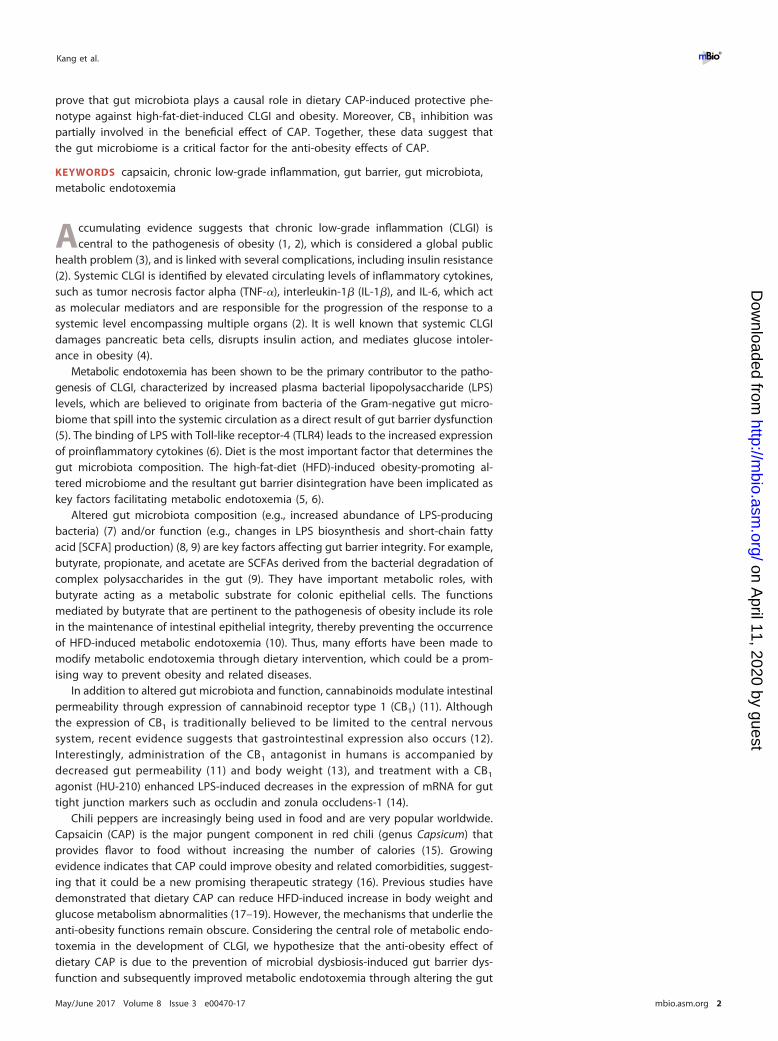
mation. High-fat-diet (HFD)-induced metabolic endotoxemia and systemic CLGI playkey roles in the pathogenesis of obesity (2). To investigate the impact of CAP onmetabolic endotoxemia and systemic CLGI (plasma TNF-�, IL-1�, IL-6, and IL-10), micewere fed either a normal chow diet (NCD) or an HFD with or without CAP supplemen-tation for 12 weeks. As shown in Fig. 1, dietary CAP reduced HFD induced elevation ofthe markers of bacterial translocation (bacterial invasion of epithelium and bacterialDNA in the systemic circulation) and metabolic endotoxemia (intestinal permeability tomacromolecules like fluorescein isothiocyanate [FITC]-dextran, gut tight junction pro-tein expression, and plasma LPS). In detail, bacterial invasion of epithelium was seenby fluorescence in situ hybridization (FISH) with a universal 16S rRNA gene probe. Adramatic increase in the number of bacteria in direct contact with the colon intestinalepithelial surface seen in the HFD group was reduced by CAP (Fig. 1A). Bacterial invasionof epithelium has been associated with bacterial translocation from the intestine andsubsequently impaired metabolic homeostasis (20). Therefore, we determined thelevels of bacterial DNA in blood using quantitative PCR (qPCR). In agreement with FISHresults, we found that the bacterial DNA load was significantly lower in blood samplesfrom mice fed HFD with CAP for 12 weeks (Fig. 1B). In addition, we found that theCAP-treated animals were protected from the increase in intestinal permeability anddownregulation of intestinal tight junction proteins (ZO-1 and occludin) that occurs inresponse to an HFD (Fig. 1C to E). Excessive LPS production due to altered gut
FIG 1 Dietary CAP reduces HFD-induced metabolic endotoxemia and systemic chronic low-grade inflammation (CLGI). Male WT mice (n � 6/group) were fedeither NCD�C or HFD�C for 12 weeks. Blood and different tissue samples were collected and subjected to various analyses. Shown are markers of bacterialtranslocation and metabolic endotoxemia, such as bacterial invasion into colonic mucosa visualized by FISH (16S rRNA genes of all bacteria [red] and nuclei[blue]) (A), total bacterial DNA load (universal 16S rRNA gene copies) in whole-blood samples (B), gut permeability to FITC-dextran (C), mRNA levels of tightjunction proteins ZO-1 (D) and occludin (E), plasma levels of LPS (F) and its receptor (TLR4) in epididymal white adipose tissue at the mRNA level (G), and themarkers of systemic CLGI, such as plasma TNF-� (H), IL-1� (I), IL-6 (J), and IL-10 (K). Data are expressed as means � standard errors of the means (SEM). Datawith different superscript letters are significantly different (P � 0.05) by one-way ANOVA with a post hoc Bonferroni’s multiple-comparison test.
Gut Microbiota Mediates CAP’s Anti-obesity Effects ®
May/June 2017 Volume 8 Issue 3 e00470-17 mbio.asm.org 3
on April 11, 2020 by guest
http://mbio.asm
.org/D
ownloaded from
microbiota leads to metabolic endotoxemia (passage of LPS to systemic circulation)through leaky gut (5), so we next measured the plasma LPS levels by Limulus amebo-cyte lysate (LAL) chromogenic endpoint assay. Consistently, we found significantlylower plasma LPS with CAP treatment (Fig. 1F). Circulating LPS causes CLGI, andadipose tissue (AT) is the major organ that releases markers of CLGI in response to LPS(21). Lower expression of TLR4, which is a receptor for LPS, in the AT also supports thepresence of lower LPS in the group treated with HFD plus CAP (HFD�C) compared tothe group treated with HFD alone (Fig. 1G). Accordingly, the increased levels of markersof systemic CLGI (TNF-�, IL-1�, and IL-6) induced by the HFD were decreased by CAP(Fig. 1H to J), while levels of the anti-inflammatory cytokine IL-10 were significantlyincreased (Fig. 1K). Furthermore, markers of obesity (body weight and excessive visceraladiposity measured by fat pad weight) and insulin resistance (glucose intolerancemeasured by oral glucose tolerance tests [GTTs]) (see Fig. S1 in the supplementalmaterial) were similarly improved by CAP. These results demonstrate that dietary CAPreduces HFD-induced metabolic endotoxemia and CLGI and associated obesity.
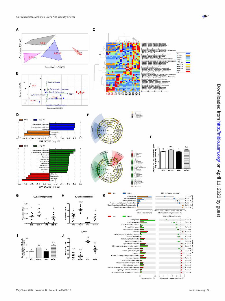
Dietary CAP beneficially alters the gut microbiota. Excessive LPS production dueto elevated levels of LPS-producing bacteria (22) and hyper-intestinal permeability dueto altered gut microbiota (7) and microbiota-derived metabolites (23) have been shownas major causes for the development of metabolic endotoxemia. To assess the impactof dietary CAP on gut microbiota, we analyzed the composition, abundance, andfunction of gut microbiota by high-throughput sequencing of the 16S rRNA gene V3and V4 regions of the cecal contents of these mice. To reveal the relationships of themicrobial communities hosted by individuals differing by CAP treatment, we performedseveral analyses describing the differences in bacterial community compositions andstructures between individuals. To evaluate aspects of bacterial diversity that may beinfluenced by CAP treatment, we first applied measures of � diversity (similarities ordifferences in communities between individuals). To measure community similarity, weused the Jaccard, Bray-Curtis, and UniFrac indices, which are classical ecological �
diversity measures based on the ratio of shared and unique species relative to the totalnumber of species present between two communities, respectively. This analysis revealeda significant impact of CAP treatment overall (permutational multivariate analysis of vari-ance [PERMANOVA], Bray-Curtis, 999 mutations, F � 10.93, P � 0.001; Jaccard, 999 muta-tions, F � 3.9, P � 0.001; unweighted UniFrac, 999 mutations, F � 4.67, P � 0.001) and alsobetween groups (Fig. 2A; see Fig. S2 in the supplemental material). Differences in bacterialcommunity structures are also apparent in principal-coordinate analyses (PCoA)of these � diversity indices. PCoA revealed a distinct clustering of microbiotacomposition between the CAP-supplemented HFD (HFD�C), HFD, and NCD/CAP-supplemented NCD (NCD�C) groups along the primary ordination axis (axis 1),which explains 50.4% of the variation in the data (Fig. 2A). On one end of the axis,the HFD group stand alone, and HFD�C is in the middle, while the NCD/NCD�Ccluster is on the opposite end. Next, taxa that are primarily responsible for an observeddifference between groups were analyzed using the similarity percentage (SIMPER)test, which reveals 7 taxa. Their contributions to groups are analyzed using principalcomponent (variance-covariance type) analysis (PCA) (Fig. 2B). Accordingly, Gram-negative LPS-producing members of the S24_7 and SCFA-producing Ruminococcaceafamilies are primarily responsible for HFD- and CAP-treated groups, respectively. Fur-thermore, the relative abundance of taxa, which showed P value of �0.05 by differentialexpression analysis (nonparametric ANOVA with false discovery rate [FDR] correction),was expressed as a heat map (Fig. 2C) including hierarchical clustering (HCN). HCN is aclustering technique for graphically summarizing the intersample relationships in theform of a dendrogram. HCN also clearly separated the HFD samples as a single clusterfrom the other 3 groups, which form three clusters within a clade. In addition,biomarker analysis using linear discriminant analysis (LDA) effect size (LEfSe) (Fig. 2D)and a cladogram (Fig. 2E) generated from LEfSe analysis indicated that the members ofthe LPS-producing family S24_7 were increased in the HFD group, while SCFA (e.g.,
Kang et al. ®
May/June 2017 Volume 8 Issue 3 e00470-17 mbio.asm.org 4
on April 11, 2020 by guest
http://mbio.asm
.org/D
ownloaded from
Gut Microbiota Mediates CAP’s Anti-obesity Effects ®
May/June 2017 Volume 8 Issue 3 e00470-17 mbio.asm.org 5
on April 11, 2020 by guest
http://mbio.asm
.org/D
ownloaded from
butyrate)-producing Ruminococcaceae and Lachnospiraceae were increased in the CAP-treated group (Fig. 2D and E). Because various high-throughput microbiome analysesemphasize the main effects of CAP on SCFA-producing bacteria, we quantified butyrylcoenzyme A (CoA) transferase (BCoAT) genes using qPCR. Interestingly, we found that thegene copies were higher in the CAP-treated HFD group (Fig. 2F). Consistently, therelative abundances (RAs) of SCFA-producing bacteria measured by both sequencingand qPCR were significantly higher in CAP-treated groups (Fig. 2G and H; see Fig. S3Aand B in the supplemental material). Moreover, the fecal SCFA analysis revealed adramatic increase in butyrate in CAP-treated groups (Fig. 2I; Fig. S3C and D). Butyrate,which is a major SCFA produced by Ruminococcacaea (24), prevents metabolic endo-toxemia (23) by strengthening the gut barrier. Likewise, RA of the LPS-producingS24_7 family was significantly higher in HFD and vice versa in CAP-treated groups(Fig. 2J). Next, to study the potential function of gut microbiota in different groups,LEfSe was applied to the relative abundance of KEGG pathways predicted byphylogenetic reconstruction of unobserved states (PICRUSt) (25). Notably, the biomark-ers with significant discriminative power were the “lipopolysaccharide biosynthesisproteins” and “lipopolysaccharide biosynthesis pathways,” which were significantlylower in CAP-treated groups than in those fed on HFD alone (Fig. 2K).
Although � diversity measures describe aspects of community structure betweengroups, they do not reveal the similarities or differences in communities within agroup (i.e., � diversity). We next applied measures of � diversity, which describespecies composition in one specific habitat of interest and can be informative ofcommunity functioning (26). Because microbial communities are highly diverse andare often poorly amenable to the diversity measures commonly used in communityecology (27), we used several different measures focusing on different aspects ofcommunity assembly, including species richness and abundance. Chao1 (speciesrichness), the abundance-based coverage estimator (ACE), and the Shannon index(species richness) showed a trend of increase when diet was supplemented with CAP(see Fig. S4 in the supplemental material). Together, these data indicate that CAPbeneficially alters the gut microbiota and mainly prevents HFD-induced reduction ofbutyrate-producing bacteria and elevation of Gram-negative LPS-producing bacteriaand associated LPS biosynthesis.
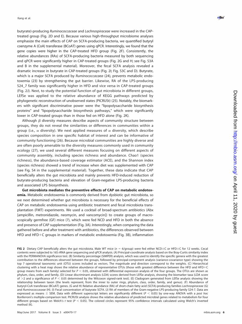
Gut microbiota mediates the preventive effects of CAP on metabolic endotox-emia. Metabolic endotoxemia is commonly derived from dysbiotic gut microbiota, sowe next determined whether gut microbiota is necessary for the beneficial effects ofCAP on metabolic endotoxemia using antibiotic treatment and fecal microbiota trans-plantation (FMT) experiments. We used a cocktail of broad-spectrum antibiotics (Abx[ampicillin, metronidazole, neomycin, and vancomycin]) to create groups of macro-scopically germfree (GF) mice (7), which were fed NCD and HFD in both the absenceand presence of CAP supplementation (Fig. 3A). Interestingly, when comparing the datagathered before and after treatment with antibiotics, the differences observed betweenHFD and HFD�C groups in markers of metabolic endotoxemia (Fig. 3B), inflammation
FIG 2 Dietary CAP beneficially alters the gut microbiota. Male WT mice (n � 6/group) were fed either NCD�C or HFD�C for 12 weeks. Cecalcontents were subjected to 16S rRNA gene sequencing and qPCR analysis. (A) Principal-coordinate analysis based on the Bray-Curtis similarity indexwith the PERMANOVA significance test. (B) Similarity percentage (SIMPER) analysis, which was used to identify the specific genera with the greatestcontribution to the differences observed between the groups, followed by principal-component analysis (variance-covariance type) showing thetop 7 operational taxonomic unit (OTU) scores included as vectors. The magnitude and direction correspond to the weights. (C) Hierarchicalclustering with a heat map shows the relative abundance of representative OTUs (those with greatest difference between the HFD and HFD�Cgroup means from each family) selected for P � 0.05, obtained with differential expression analysis of the four groups. The OTUs are shown asphylum, class, order, and family. (D) Linear discriminant analysis (LDA) scores derived from LEfSe analysis, showing the biomarker taxa (LDA scoreof �2 and a significance of P � 0.05 determined by the Wilcoxon signed-rank test). (E) Cladogram generated from LEfSe analysis showing therelationship between taxon (the levels represent, from the inner to outer rings, phylum, class, order, family, and genus). (F) Abundance ofbutyryl-CoA transferase (BCoAT) genes. (G and H) Relative abundance (RA) of short-chain fatty acid (SCFA)-producing families Lachnospiraceae (G)and Ruminococcaceae (H). (I) Fecal concentration of butyrate SCFA. (J) RA of members of the Gram-negative LPS-producing family S24-7. Data areexpressed as means � SEM. Data with different superscript letters are significantly different (P � 0.05) by one-way ANOVA with a post hocBonferroni’s multiple-comparison test. PICRUSt analysis shows the relative abundance of predicted microbial genes related to metabolism for fourdifferent groups based on Welch’s t test (P � 0.05). The colored circles represent 95% confidence intervals calculated using Welch’s invertedmethod (K).
Kang et al. ®
May/June 2017 Volume 8 Issue 3 e00470-17 mbio.asm.org 6
on April 11, 2020 by guest
http://mbio.asm
.org/D
ownloaded from
(Fig. 3C), and obesity (Fig. 3D; see Fig. S5 in the supplemental material) were eliminated.These findings suggest that the gut microbiota largely mediates the preventive effectsof CAP on metabolic endotoxemia. More specifically, the HFD�C group may carry alower abundance of LPS-producing bacteria.
To further clarify whether the gut microbiota plays a causal role, we transplanted thefecal microbiota of mice fed HFD or HFD�C for 12 weeks into recipient GF mice and
FIG 3 Gut microbiota mediates the preventive effects of CAP on metabolic endotoxemia. (A) Schema showing the animal groups and treatments.Male WT mice (n � 6/group) received either HFD�C (A) or NCD�C (Fig. S5) for 12 weeks and then were treated with a broad-spectrum antibioticcocktail (Abx) consisting of ampicillin, vancomycin, neomycin sulfate, and metronidazole for 6 weeks to introduce microbiota depletion (A). Bloodand tissue samples were subjected to various analyses, and data were analyzed before and after Abx supplementation for markers of metabolicendotoxemia (B), systemic chronic low-grade inflammation (CLGI) (TNF-�, IL-1�, IL-6 and IL-10) (C), and obesity (D). (E) Schema showing thegermfree mice that received microbiota transplanted from mice treated with HFD�C for 12 weeks. Two weeks after transplantation, blood andtissue samples were subjected to various analyses, such as FISH for markers of bacterial translocation, metabolic endotoxemia, and bacterialinvasion of epithelium (F), gut permeability (G), intestinal tight junction proteins (H), plasma levels of LPS (I), and plasma levels of markers ofsystemic CLGI (J). Data are expressed as means � SEM, and statistical analysis was performed using ANOVA with a post hoc Bonferroni’smultiple-comparison test or unpaired two-tailed Student’s t test.
Gut Microbiota Mediates CAP’s Anti-obesity Effects ®
May/June 2017 Volume 8 Issue 3 e00470-17 mbio.asm.org 7
on April 11, 2020 by guest
http://mbio.asm
.org/D
ownloaded from
then fed both groups of recipient mice a high-fat diet for 2 weeks (Fig. 3E). Oncecolonized, the GF mice are maintained as gnotobiotic by housing them in sterileisolators to avoid cross contamination with other bacteria or fungi (see Fig. S6A in thesupplemental material). Strikingly, markers of bacterial translocation (Fig. 3F; Fig. S6B),metabolic endotoxemia (Fig. 3G to I), and systemic CLGI (Fig. 3J) were significantlylower in GF mice that received microbiota from the CAP-treated HFD group than in GFmice that received HFD donor microbiota. As expected, lower TLR4 expression in the AT(Fig. S6C) and higher fecal SCFA levels, especially butyrate and acetate, were found inGF mice treated with HFD�C without microbiota transplant (Fig. S6D). Although therewere no differences in body weights and food intakes between these two groups(Fig. S6E and F), the recipients of microbiota from the mice consuming an HFD with CAPsupplementation exhibited an improvement in glucose tolerance and fat accumulation(Fig. S6G and H). Combined, these findings suggest that the beneficial effects of CAP onmetabolic endotoxemia and obesity were largely mediated by modulation of the gutmicrobiota.
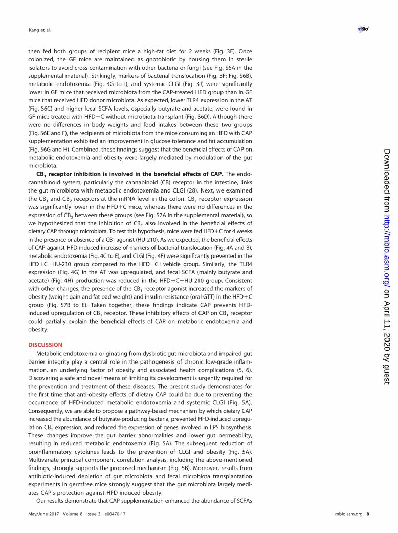
CB1 receptor inhibition is involved in the beneficial effects of CAP. The endo-cannabinoid system, particularly the cannabinoid (CB) receptor in the intestine, linksthe gut microbiota with metabolic endotoxemia and CLGI (28). Next, we examinedthe CB1 and CB2 receptors at the mRNA level in the colon. CB1 receptor expressionwas significantly lower in the HFD�C mice, whereas there were no differences in theexpression of CB2 between these groups (see Fig. S7A in the supplemental material), sowe hypothesized that the inhibition of CB1 also involved in the beneficial effects ofdietary CAP through microbiota. To test this hypothesis, mice were fed HFD�C for 4 weeksin the presence or absence of a CB1 agonist (HU-210). As we expected, the beneficial effectsof CAP against HFD-induced increase of markers of bacterial translocation (Fig. 4A and B),metabolic endotoxemia (Fig. 4C to E), and CLGI (Fig. 4F) were significantly prevented in theHFD�C�HU-210 group compared to the HFD�C�vehicle group. Similarly, the TLR4expression (Fig. 4G) in the AT was upregulated, and fecal SCFA (mainly butyrate andacetate) (Fig. 4H) production was reduced in the HFD�C�HU-210 group. Consistentwith other changes, the presence of the CB1 receptor agonist increased the markers ofobesity (weight gain and fat pad weight) and insulin resistance (oral GTT) in the HFD�Cgroup (Fig. S7B to E). Taken together, these findings indicate CAP prevents HFD-induced upregulation of CB1 receptor. These inhibitory effects of CAP on CB1 receptorcould partially explain the beneficial effects of CAP on metabolic endotoxemia andobesity.
DISCUSSION
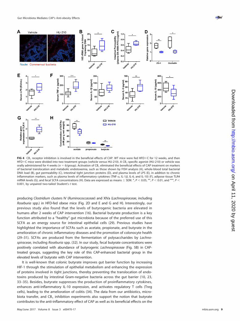
Metabolic endotoxemia originating from dysbiotic gut microbiota and impaired gutbarrier integrity play a central role in the pathogenesis of chronic low-grade inflam-mation, an underlying factor of obesity and associated health complications (5, 6).Discovering a safe and novel means of limiting its development is urgently required forthe prevention and treatment of these diseases. The present study demonstrates forthe first time that anti-obesity effects of dietary CAP could be due to preventing theoccurrence of HFD-induced metabolic endotoxemia and systemic CLGI (Fig. 5A).Consequently, we are able to propose a pathway-based mechanism by which dietary CAPincreased the abundance of butyrate-producing bacteria, prevented HFD-induced upregu-lation CB1 expression, and reduced the expression of genes involved in LPS biosynthesis.These changes improve the gut barrier abnormalities and lower gut permeability,resulting in reduced metabolic endotoxemia (Fig. 5A). The subsequent reduction ofproinflammatory cytokines leads to the prevention of CLGI and obesity (Fig. 5A).Multivariate principal component correlation analysis, including the above-mentionedfindings, strongly supports the proposed mechanism (Fig. 5B). Moreover, results fromantibiotic-induced depletion of gut microbiota and fecal microbiota transplantationexperiments in germfree mice strongly suggest that the gut microbiota largely medi-ates CAP’s protection against HFD-induced obesity.
Our results demonstrate that CAP supplementation enhanced the abundance of SCFAs
Kang et al. ®
May/June 2017 Volume 8 Issue 3 e00470-17 mbio.asm.org 8
on April 11, 2020 by guest
http://mbio.asm
.org/D
ownloaded from
producing Clostridium clusters IV (Ruminococcaceae) and XIVa (Lachnospiraceae, includingRoseburia spp.) in HFD-fed obese mice (Fig. 2D and E and G and H). Interestingly, ourprevious study also found that the levels of butyrogenic bacteria are elevated inhumans after 2 weeks of CAP intervention (16). Bacterial butyrate production is a keyfunction attributed to a “healthy” gut microbiota because of the preferred use of thisSCFA as an energy source for intestinal epithelial cells (29). Previous studies havehighlighted the importance of SCFAs such as acetate, propionate, and butyrate in theamelioration of chronic inflammatory diseases and the promotion of colonocyte health(29–31). SCFAs are produced from the fermentation of polysaccharides by Lachno-spiraceae, including Roseburia spp. (32). In our study, fecal butyrate concentrations werepositively correlated with abundance of butyrogenic Lachnospiraceae (Fig. 5B) in CAP-treated groups, suggesting the key role of this CAP-enhanced bacterial group in theelevated levels of butyrate with CAP intervention.
It is well-known that colonic butyrate improves gut barrier function by increasingHIF-1 through the stimulation of epithelial metabolism and enhancing the expressionof proteins involved in tight junctions, thereby preventing the translocation of endo-toxins produced by intestinal Gram-negative bacteria across the gut barrier (10, 23,33–35). Besides, butyrate suppresses the production of proinflammatory cytokines,enhances anti-inflammatory IL-10 expression, and activates regulatory T cells (Tregcells), leading to the amelioration of colitis (34). The data from our antibiotics, micro-biota transfer, and CB1 inhibition experiments also support the notion that butyratecontributes to the anti-inflammatory effect of CAP as well as its beneficial effects on the
FIG 4 CB1 receptor inhibition is involved in the beneficial effects of CAP. WT mice were fed HFD�C for 12 weeks, and thenHFD�C mice were divided into two treatment groups (vehicle versus HU-210). A CB1-specific agonist (HU-210) or vehicle wasorally administered for 4 weeks (n � 6/group). Activation of CB1 eliminated the beneficial effects of CAP treatment on markersof bacterial translocation and metabolic endotoxemia, such as those shown by FISH analysis (A), whole-blood total bacterialDNA load (B), gut permeability (C), intestinal tight junction proteins (D), and plasma levels of LPS (E), in addition to chronicinflammation markers, such as plasma levels of inflammatory cytokines (TNF-�, IL-1�, IL-6, and IL-10) (F), adipose tissue TLR4mRNA levels (G), and fecal SCFA concentrations (H). Data are expressed as means � SEM. *, P � 0.05, **, P � 0.01, and ***, P �0.001, by unpaired two-tailed Student’s t test.
Gut Microbiota Mediates CAP’s Anti-obesity Effects ®
May/June 2017 Volume 8 Issue 3 e00470-17 mbio.asm.org 9
on April 11, 2020 by guest
http://mbio.asm
.org/D
ownloaded from
gut barrier. Combined, these results indicate that the beneficial effects of CAP againstmetabolic endotoxemia and associated obesity may be largely due to increases in thepopulations of these butyrogenic bacterial species and gut barrier-enhancing effects ofbutyrate.
Variations in the intestinal endocannabinoid system in response to modulation ofthe gut microbiota have been observed in germfree mice and in mouse models ofbacterial-host interactions in colonic tissue (14). The intestinal endocannabinoid systemis thought to control gut barrier function, gut permeability, and metabolic endotoxemiaunder obese and diabetic conditions through a CB1-dependent mechanism, as antag-onists of CB1 decrease gut permeability and act as “gatekeepers” (36). Conversely,treatment with the CB1 receptor agonist HU-210 in mice significantly increased plasmalevels of LPS and augmented the LPS-induced decreases in the expression of guttight junction proteins at the mRNA level (14). In our study, we discovered that CAPprevented the HFD-induced upregulation of CB1 receptor expression because HU-210treatment of the HFD�C group partially abolished the beneficial effects of dietary CAPon gut barrier, metabolic endotoxemia, and CLGI, indicating that CB1 inhibition alsopartially contributes to the anti-obesity effects of CAP.
In addition to the enrichment of butyrogenic bacteria and CB1 inhibition, we foundthat CAP intervention reduced the enrichment of genes involved in LPS biosynthesisand related proteins based on the predicted function by 16S rRNA sequencing andphylogenetic reconstruction of unobserved states (PICRUSt) analysis (25). Interestingly,
FIG 5 Diagram illustrating a proposed mechanism by which the interactions between dietary CAP and gutmicrobiome lead to a decrease in Gram-negative LPS-producing bacteria (e.g., S24-7 family) and LPS biosynthesisand an increase in butyrate-producing bacteria (e.g., Lachnospiraceae and Ruminococcaceae) and butyrate levels.These changes lower LPS production and gut permeability and downregulate CB1 expression, resulting in reducedmarkers of metabolic endotoxemia. The subsequent reduction of inflammatory cytokines leads to the suppressionof chronic low-grade inflammation and obesity (A). (B) Principal-component analysis (biplot) showing the corre-lation between important parameters of the proposed mechanism presented above. Labels indicate the fourgroups. The names of the parameters are plotted as vectors according to their correlation to the first twocomponents.
Kang et al. ®
May/June 2017 Volume 8 Issue 3 e00470-17 mbio.asm.org 10
on April 11, 2020 by guest
http://mbio.asm
.org/D
ownloaded from

our recent study has shown similar results with CAP intervention in human subjects(16). This would indicate the possibility that lower abundance of Gram-negative mi-crobiota must be responsible for the low abundance of KEGG orthology (KO) belongingto the LPS biosynthesis pathway in the HFD�C group. This could be mainly due toprevention of enrichment of members of the Gram-negative S24_7 family with CAPintervention because it was the key bacterial family that largely contributes to the HFDgroup (Fig. 2D and J). These lipopolysaccharides are bacterium-associated molecularpatterns, which act via TLR4 by promoting the inflammatory response (6).
Our study design is unique in its use of fecal microbiota transplantation in the germfree(GF) mouse model. GF mice that lack any exposure to living pathogenic or nonpatho-genic microorganisms provide an attractive model to investigate the role of thecomposition and function of intestinal microbiota on the development of obesity (6).Given the fact that transferring the microbiota of HFD-consuming mice to GF micecaused glucose intolerance in 6 days (37) and significant changes in the luminalbutyrate concentrations of recipient GF mice at day 7 post-FMT (38), it is interestingto note that the acquired changes in microbiota in response to CAP protect againstHFD-induced obesity because germfree mice that received microbiota from the HFD�Cgroup exhibited lower levels of gut permeability, plasma LPS, and markers of CLGI in2 weeks (Fig. 3).
In conclusion, these results demonstrate dietary CAP-microbiome interactions as anovel mechanism underlying the anti-obesity effects of dietary CAP. Our study dem-onstrated that dietary CAP prevents HFD-induced metabolic endotoxemia and systemicCLGI by elevating cecal butyrogenic bacterium and butyrate levels, inhibiting colonicCB1 receptor, and reducing LPS biosynthesis (Fig. 5A). Finally, given the fact that gutdysbiosis and metabolic endotoxemia are often linked to chronic inflammatory dis-eases, the ability of dietary CAP to prevent these conditions confirms the potential ofCAP supplementation as a therapeutic means of treating obesity.
MATERIALS AND METHODSAnimals. Male wild-type (WT) mice of the C57BL/6J genetic lineage were bred in the specific-
pathogen-free animal (SPF) facility of the Third Military Medical University, Chongqing, People’s Republicof China, and maintained in a temperature-controlled room (22 to 24°C) with a strictly followed 12-hlight/12-h dark diurnal cycle with food and water ad libitum. Germfree (GF) mice were created usingcaesarean rederivation from existing SPF mouse lines. A total of 10 1-day-old GF mice of the C57BL/6Jbackground were bred in the Department of Laboratory Animal Science of the Third Military MedicalUniversity and were used as recipients for the fecal microbiota transplantation. Sterile plastic filmisolators, which help to avoid cross contamination with other bacteria or fungi, were used to house themice in a completely germfree environment; this provides an environment that allowed us to conductexperiments without competing background levels of microbiota. They were given ad libitum access tosterilized water during the whole course of the experiment. Food and water and other sterile supplieswere imported into the isolators by docking autoclaved supply cylinders to a double-door port built intothe isolator wall (Fig. S6A). GF foster mice were used to breastfeed 1-day-old GF mice until weaning(which occurred at 3 weeks of age); they were then fed ad libitum with a sterilized normal chow diet for5 weeks postweaning. Culture and PCR analysis of feces amplifying the 16S rRNA gene were used toroutinely test the sterility of GF isolators. The animal experiments were approved by the Animal Care andUse Committee of the Third Military Medical University (Chongqing, China).
Animal diets. Normal chow diet (NCD [D12450B]) had 70% of kilocalories from carbohydrate, 20%of kilocalories from protein, and 10% of kilocalories from fat, for a total energy content of 3.85 kcal/g, andthe high-fat diet (HFD [D12451]) had 35% of kilocalories from carbohydrate, 20% of kilocalories fromprotein, and 45% of kilocalories from fat, for a total energy content of 4.73 kcal/g. These diets, with orwithout capsaicin (Sigma, St. Louis, MO, USA) at 0.01 g CAP/100 g diet, were ordered from Research Diets,Inc. (New Brunswick, NJ, USA).
Animal experiments. Mice were housed in a biosafety level 2 (BSL2) room in hard-top cages withtwo or three mice per cage. Body weight and food intake were measured weekly. Mice were fasted for4 h before being sacrificed, and blood was collected via cardiac puncture in all experiments unlessotherwise specified.
(i) Determination of the CAP effects on metabolic endotoxemia, chronic low-grade inflamma-tion, and gut microbiota. Eight-week old mice were randomly distributed into four groups (n �6/group): (i) normal chow diet (NCD), (ii) NCD with 0.01% CAP (NCD�C), (iii) high-fat diet (HFD), and (iv)HFD with 0.01% CAP (HFD�C). After 12 weeks, GTT and gut permeability assays were done. Then micewere sacrificed, blood samples were aliquoted into EDTA-coated blood collection tubes, cecal contentswere flash-frozen, and body fat pad weights were taken. Blood samples were snap-frozen in liquidnitrogen and stored at �80°C or centrifuged (2,500 � g for 15 min at 4°C) to collect plasma. Cecal
Gut Microbiota Mediates CAP’s Anti-obesity Effects ®
May/June 2017 Volume 8 Issue 3 e00470-17 mbio.asm.org 11
on April 11, 2020 by guest
http://mbio.asm
.org/D
ownloaded from
contents and feces were subjected to microbiome and SCFA analysis, respectively. Analyses of markersof bacterial translocation (bacterial invasion into intestinal epithelium and bacterial DNA in the systemiccirculation), metabolic endotoxemia (hyper-intestinal permeability to macromolecules like FITC-dextran,gut tight junction protein expression, and plasma LPS levels), and systemic CLGI (plasma TNF-�, IL-1�,IL-6, and IL-10) were also conducted.
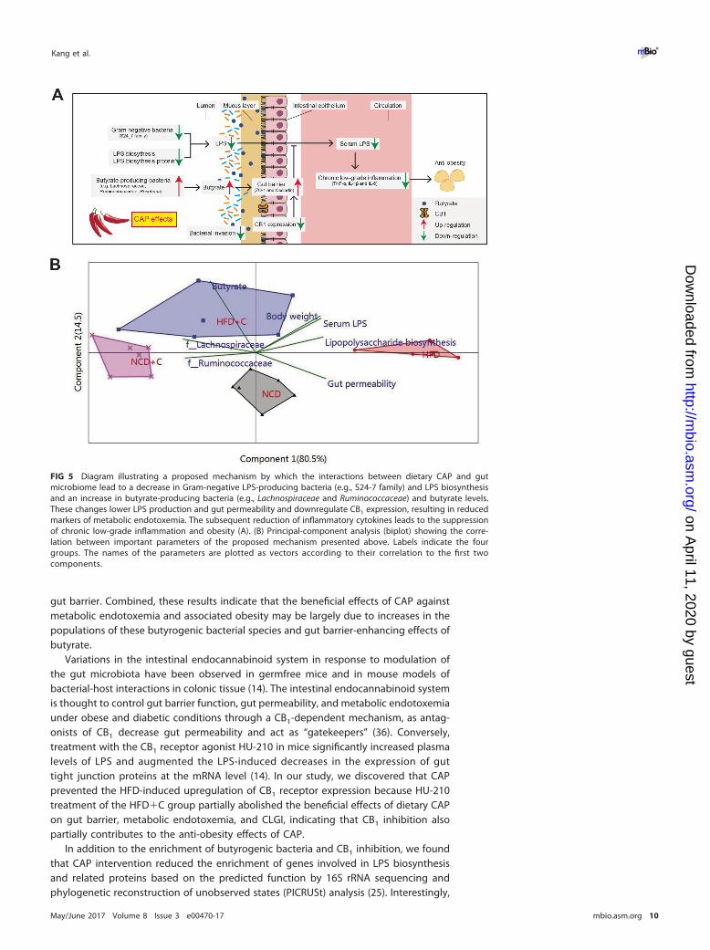
(ii) Determination of causal role of microbiota for the anti-obesity effects of CAP. (a) Antibiotic-induced gut microbiota depletion. A subset of 8-week-old mice were randomly distributed into fourgroups (n � 6/group) and were fed either NCD�C or HFD�C for 12 weeks. Markers of metabolicendotoxemia, CLGI, and obesity were recorded before (at the end of the 12th week) and after treatmentof these mice with a broad-spectrum antibiotic (Abx) cocktail (Sigma, USA) containing ampicillin(1 mg/ml), metronidazole (1 mg/ml), neomycin (1 mg/ml), and vancomycin (0.5 mg/ml) in their drinkingwater for 6 weeks. Successful depletion of gut microbiota after the antibiotic treatment was confirmedusing qPCR analysis of total bacterial 16S rRNA genes (7, 39).
(b) Fecal microbiota transplantation in germfree mice. To colonize the guts of GF mice, fecal sampleswere collected from randomly chosen mice that had received HFD or HFD�C for 12 weeks since the ageof 8 weeks. Fecal samples were stored at �80°C until the time of processing. The procedures forpreparing the fecal samples for microbiota transplantation were performed as described in a previousstudy (40). In detail, fecal samples were homogenized with a mortar and pestle while submerged in liquidnitrogen. A 100-mg aliquot of the pulverized frozen material was then diluted in 1.5 ml of reducedphosphate-buffered saline (PBS) (supplemented with 0.1% resazurin [wt/vol] and 0.05% L-cysteine-HCl)in an anaerobic Coy chamber (atmosphere, 75% N2, 20% CO2, 5% H2) and then vortexed at roomtemperature for 5 min. The suspension was allowed to settle by gravity for 5 min, after which time theclarified supernatant was transferred to an anaerobic crimped tube that was then transported to thegnotobiotic mouse facility. The outer surface of the tube was sterilized by exposure for 20 min to chlorinedioxide in the transfer sleeve attached to the gnotobiotic isolator and then transferred into the isolator.A 1-ml syringe was used to recover a 200-�l aliquot of the suspension; the suspension was subsequentlyintroduced by gavage with a flexible plastic tube into the stomach of each adult 8-week-old GF recipient.The samples were obtained shortly before colonization, immediately (within 5 min) diluted, andintroduced into the GF mice within 2 h after dilution. The recipient mice were separately bred in differentgnotobiotic isolators to prevent normalization of the gut microbiota. Also, the recipient mice weremaintained in separate cages (five mice per cage; n � 5/group) within an isolator dedicated to micecolonized with the same donor microbiota. As mentioned in the previous section, mouse samples weresubjected to analysis for markers of bacterial translocation, metabolic endotoxemia, and systemic CLGIand analysis of fecal SCFA levels after 2 weeks.
(iii) Role of CB1 receptor for the CAP effects on metabolic endotoxemia and obesity. To studythe effects of CAP on HFD-induced upregulation of CB1, HU-210 (CB1 receptor agonist) was dissolved ina mixture of dimethyl sulfoxide (DMSO), Cremophor, and saline solution (41), and then either HU-210 orvehicle was intraperitoneally injected (100 �g/kg/day) into HFD�C (0.01% CAP)-treated mice (n �6/group) for 4 weeks (14). Mice were then subjected to GTT and analysis for markers of obesity, bacterialtranslocation, metabolic endotoxemia, and systemic CLGI.
Statistical analyses. Statistical analysis was performed using GraphPad Prism 6.01 (GraphPadSoftware, Inc., San Diego, CA) unless otherwise specified. Experiments with two groups were analyzedwith Student’s t tests, and those with more than two groups were analyzed with one-way analysis ofvariance (ANOVA) with post hoc Bonferroni’s multiple comparison tests. Data were natural logarithmtransformed to normalize their distributions if the values had a skewed distribution. Correlation-typeprincipal-component analysis (PCA) (XLSTAT software) was applied to the correlation between groupsand findings. Linear discriminant analysis effect size (LEfSe) uses a nonparametric Wilcoxon sum-rank testfollowed by LDA analysis to measure the effect size of each abundant taxon, and two filters (P � 0.05and LDA score of �2) were applied to the present features. A P value of �0.05 was consideredstatistically significant.
Details of the rest of the materials and methods are provided in Text S1 in the supplemental material.Accession number(s). Sequencing data for the 16S rRNA sequences have been deposited in the SRA
database under GenBank accession no. SRP099024.
SUPPLEMENTAL MATERIALSupplemental material for this article may be found at https://doi.org/10.1128/mBio
.00470-17.TEXT S1, DOCX file, 0.1 MB.FIG S1, PDF file, 0.3 MB.FIG S2, PDF file, 0.6 MB.FIG S3, PDF file, 0.2 MB.FIG S4, PDF file, 0.4 MB.FIG S5, PDF file, 0.6 MB.FIG S6, PDF file, 0.5 MB.FIG S7, PDF file, 0.3 MB.TABLE S1, DOCX file, 0.1 MB.TABLE S2, DOCX file, 0.1 MB.
Kang et al. ®
May/June 2017 Volume 8 Issue 3 e00470-17 mbio.asm.org 12
on April 11, 2020 by guest
http://mbio.asm
.org/D
ownloaded from
ACKNOWLEDGMENTSThis research was supported by project 81673157 of the National Natural Science
Foundation of China (NSFC).The authors’ responsibilities were as follows. C.K., B.W., and M.-T.M. designed the
experiments. C.K., X.W., H.L., S.H., L.H., T.Z., M.Z., and M.C. collected samples andperformed the experiments. C.K., B.W., K.K., X.W., and H.L. analyzed the data. C.K., B.W.,K.K., and M.-T.M. prepared the manuscript and had primary responsibility for finalcontent. All authors read and approved the final manuscript. None of the authorsreported a conflict of interest.
REFERENCES1. Ruiz-Núñez B, Pruimboom L, Dijck-Brouwer DA, Muskiet FA. 2013. Life-
style and nutritional imbalances associated with Western diseases:causes and consequences of chronic systemic low-grade inflammationin an evolutionary context. J Nutr Biochem 24:1183–1201. https://doi.org/10.1016/j.jnutbio.2013.02.009.
2. Hotamisligil GS. 2006. Inflammation and metabolic disorders. Nature444:860 – 867. https://doi.org/10.1038/nature05485.
3. Ng M, Fleming T, Robinson M, Thomson B, Graetz N, Margono C, MullanyEC, Biryukov S, Abbafati C, Abera SF, Abraham JP, Abu-Rmeileh NM,Achoki T, AlBuhairan FS, Alemu ZA, Alfonso R, Ali MK, Ali R, Guzman NA,Ammar W, Anwari P, Banerjee A, Barquera S, Basu S, Bennett DA, BhuttaZ, Blore J, Cabral N, Nonato IC, Chang JC, Chowdhury R, Courville KJ,Criqui MH, Cundiff DK, Dabhadkar KC, Dandona L, Davis A, Dayama A,Dharmaratne SD, Ding EL, Durrani AM, Esteghamati A, Farzadfar F, FayDF, Feigin VL, Flaxman A, Forouzanfar MH, Goto A, Green MA, Gupta R.2014. Global, regional, and national prevalence of overweight and obe-sity in children and adults during 1980 –2013: a systematic analysis forthe Global Burden of Disease Study 2013. Lancet 384:766 –781. https://doi.org/10.1016/S0140-6736(14)60460-8.
4. Ewaschuk J, Endersby R, Thiel D, Diaz H, Backer J, Ma M, Churchill T,Madsen K. 2007. Probiotic bacteria prevent hepatic damage andmaintain colonic barrier function in a mouse model of sepsis. Hepa-tology 46:841– 850. https://doi.org/10.1002/hep.21750.
5. Cani PD, Bibiloni R, Knauf C, Waget A, Neyrinck AM, Delzenne NM, BurcelinR. 2008. Changes in gut microbiota control metabolic endotoxemia-induced inflammation in high-fat diet-induced obesity and diabetes inmice. Diabetes 57:1470–1481. https://doi.org/10.2337/db07-1403.
6. Caesar R, Tremaroli V, Kovatcheva-Datchary P, Cani P, Bäckhed F. 2015.Crosstalk between gut microbiota and dietary lipids aggravates WATinflammation through TLR signaling. Cell Metab 22:658 – 668. https://doi.org/10.1016/j.cmet.2015.07.026.
7. Kaliannan K, Wang B, Li XY, Kim KJ, Kang JX. 2015. A host-microbiomeinteraction mediates the opposing effects of omega-6 and omega-3fatty acids on metabolic endotoxemia. Sci Rep 5:11276. https://doi.org/10.1038/srep11276.
8. Vázquez-Castellanos JF, Serrano-Villar S, Latorre A, Artacho A, Ferrús ML,Madrid N, Vallejo A, Sainz T, Martínez-Botas J, Ferrando-Martínez S, VeraM, Dronda F, Leal M, Del Romero J, Moreno S, Estrada V, Gosalbes MJ,Moya A. 2015. Altered metabolism of gut microbiota contributes tochronic immune activation in HIV-infected individuals. Mucosal Immunol8:760 –772. https://doi.org/10.1038/mi.2014.107.
9. Scheppach W. 1994. Effects of short chain fatty acids on gut morphologyand function. Gut 35:S35–S38. https://doi.org/10.1136/gut.35.1_Suppl.S35.
10. Peng L, Li ZR, Green RS, Holzman IR, Lin J. 2009. Butyrate enhances theintestinal barrier by facilitating tight junction assembly via activation ofAMP-activated protein kinase in Caco-2 cell monolayers. J Nutr 139:1619 –1625. https://doi.org/10.3945/jn.109.104638.
11. Zoppi S, Madrigal JL, Perez-Nievas BG, Marin-Jimenez I, Caso JR, Alou L,Garcia-Bueno B, Colon A, Manzanares J, Gomez-Lus ML, Menchen L, LezaJC. 2012. Endogenous cannabinoid system regulates intestinal barrierfunction in vivo through cannabinoid type 1 receptor activation. Am JPhysiol Gastrointest Liver Physiol 302:G565–G571. https://doi.org/10.1152/ajpgi.00158.2011.
12. Wright KL, Duncan M, Sharkey KA. 2008. Cannabinoid CB2 receptors inthe gastrointestinal tract: a regulatory system in states of inflammation.Br J Pharmacol 153:263–270. https://doi.org/10.1038/sj.bjp.0707486.
13. Geurts L, Neyrinck AM, Delzenne NM, Knauf C, Cani PD. 2014. Gutmicrobiota controls adipose tissue expansion, gut barrier and glu-
cose metabolism: novel insights into molecular targets and interven-tions using prebiotics. Benef Microbes 5:3–17. https://doi.org/10.3920/BM2012.0065.
14. Muccioli GG, Naslain D, Bäckhed F, Reigstad CS, Lambert DM, DelzenneNM, Cani PD. 2010. The endocannabinoid system links gut microbiota toadipogenesis. Mol Syst Biol 6:392. https://doi.org/10.1038/msb.2010.46.
15. Tremblay A, Arguin H, Panahi S. 2016. Capsaicinoids: a spicy solution tothe management of obesity? Int J Obes Relat Metab Disord 40:1198 –1204. https://doi.org/10.1038/ijo.2015.253.
16. Kang C, Zhang Y, Zhu X, Liu K, Wang X, Chen M, Wang J, Chen H, Hui S,Huang L, Zhang Q, Zhu J, Wang B, Mi M. 2016. Healthy subjects differ-entially respond to dietary capsaicin correlating with specific gut en-terotypes. J Clin Endocrinol Metab 101:4681– 4689. https://doi.org/10.1210/jc.2016-2786.
17. Yang D, Luo Z, Ma S, Wong WT, Ma L, Zhong J, He H, Zhao Z, Cao T, YanZ, Liu D, Arendshorst WJ, Huang Y, Tepel M, Zhu Z. 2010. Activation ofTRPV1 by dietary capsaicin improves endothelium-dependent vasore-laxation and prevents hypertension. Cell Metab 12:130 –141. https://doi.org/10.1016/j.cmet.2010.05.015.
18. Li L, Chen J, Ni Y, Feng X, Zhao Z, Wang P, Sun J, Yu H, Yan Z, Liu D, NiliusB, Zhu Z. 2012. TRPV1 activation prevents nonalcoholic fatty liver throughUCP2 upregulation in mice. Pflugers Arch 463:727–732. https://doi.org/10.1007/s00424-012-1078-y.
19. Wang P, Yan Z, Zhong J, Chen J, Ni Y, Li L, Ma L, Zhao Z, Liu D, ZhuZ. 2012. Transient receptor potential vanilloid 1 activation enhancesgut glucagon-like peptide-1 secretion and improves glucose homeo-stasis. Diabetes 61:2155–2165. https://doi.org/10.2337/db11-1503.
20. Amar J, Chabo C, Waget A, Klopp P, Vachoux C, Bermúdez-Humarán LG,Smirnova N, Bergé M, Sulpice T, Lahtinen S, Ouwehand A, Langella P,Rautonen N, Sansonetti PJ, Burcelin R. 2011. Intestinal mucosal adher-ence and translocation of commensal bacteria at the early onset of type2 diabetes: molecular mechanisms and probiotic treatment. EMBO MolMed 3:559 –572. https://doi.org/10.1002/emmm.201100159.
21. Jia L, Vianna CR, Fukuda M, Berglund ED, Liu C, Tao C, Sun K, Liu T,Harper MJ, Lee CE, Lee S, Scherer PE, Elmquist JK. 2014. HepatocyteToll-like receptor 4 regulates obesity-induced inflammation and insulinresistance. Nat Commun 5:3878. https://doi.org/10.1038/ncomms4878.
22. Vatanen T, Kostic AD, d’Hennezel E, Siljander H, Franzosa EA, Yassour M,Kolde R, Vlamakis H, Arthur TD, Hämäläinen A-M, Peet A, Tillmann V,Uibo R, Mokurov S, Dorshakova N, Ilonen J, Virtanen S, Szabo S, Porter J,Lähdesmäki H, Huttenhower C, Gevers D, Cullen T, Knip M, Xavier R.2016. Variation in microbiome LPS immunogenicity contributes to au-toimmunity in humans. Cell 165:842– 853. https://doi.org/10.1016/j.cell.2016.04.007.
23. Zhou D, Pan Q, Xin FZ, Zhang RN, He CX, Chen GY, Liu C, Chen YW, FanJG. 2017. Sodium butyrate attenuates high-fat diet-induced steatohepa-titis in mice by improving gut microbiota and gastrointestinal barrier.World J Gastroenterol 23:60 –75. https://doi.org/10.3748/wjg.v23.i1.60.
24. Louis P, Scott KP, Duncan SH, Flint HJ. 2007. Understanding the effectsof diet on bacterial metabolism in the large intestine. J Appl Microbiol102:1197–1208. https://doi.org/10.1111/j.1365-2672.2007.03322.x.
25. Langille MGI, Zaneveld J, Caporaso JG, McDonald D, Knights D, ReyesJA, Clemente JC, Burkepile DE, Vega Thurber RL, Knight R, Beiko RG,Huttenhower C. 2013. Predictive functional profiling of microbialcommunities using 16S rRNA marker gene sequences. Nat Biotechnol31:814 – 821. https://doi.org/10.1038/nbt.2676.
26. Peter H, Beier S, Bertilsson S, Lindström ES, Langenheder S, Tranvik LJ.
Gut Microbiota Mediates CAP’s Anti-obesity Effects ®
May/June 2017 Volume 8 Issue 3 e00470-17 mbio.asm.org 13
on April 11, 2020 by guest
http://mbio.asm
.org/D
ownloaded from
2011. Function-specific response to depletion of microbial diversity.ISME J 5:351–361. https://doi.org/10.1038/ismej.2010.119.
27. Hughes JB, Hellmann JJ, Ricketts TH, Bohannan BJM. 2001. Counting theuncountable: statistical approaches to estimating microbial diversity.Appl Environ Microbiol 67:4399 – 4406. https://doi.org/10.1128/AEM.67.10.4399-4406.2001.
28. Cani PD, Plovier H, Van Hul M, Geurts L, Delzenne NM, Druart C, EverardA. 2016. Endocannabinoids—at the crossroads between the gut micro-biota and host metabolism. Nat Rev Endocrinol 12:133–143. https://doi.org/10.1038/nrendo.2015.211.
29. Sonnenburg ED, Zheng H, Joglekar P, Higginbottom SK, Firbank SJ,Bolam DN, Sonnenburg JL. 2010. Specificity of polysaccharide use inintestinal Bacteroides species determines diet-induced microbiota alter-ations. Cell 141:1241–1252. https://doi.org/10.1016/j.cell.2010.05.005.
30. Furusawa Y, Obata Y, Fukuda S, Endo TA, Nakato G, Takahashi D,Nakanishi Y, Uetake C, Kato K, Kato T, Takahashi M, Fukuda NN, Mu-rakami S, Miyauchi E, Hino S, Atarashi K, Onawa S, Fujimura Y, Lockett T,Clarke JM, Topping DL, Tomita M, Hori S, Ohara O, Morita T, Koseki H,Kikuchi J, Honda K, Hase K, Ohno H. 2013. Commensal microbe-derivedbutyrate induces the differentiation of colonic regulatory T cells. Nature504:446 – 450. https://doi.org/10.1038/nature12721.
31. Smith PM, Howitt MR, Panikov N, Michaud M, Gallini CA, Bohlooly-Y M,Glickman JN, Garrett WS. 2013. The microbial metabolites, short-chainfatty acids, regulate colonic Treg cell homeostasis. Science 341:569 –573.https://doi.org/10.1126/science.1241165.
32. Neyrinck AM, Possemiers S, Druart C, Van de Wiele T, De Backer F, CaniPD, Larondelle Y, Delzenne NM. 2011. Prebiotic effects of wheat arabi-noxylan related to the increase in bifidobacteria, Roseburia, andBacteroides/Prevotella in diet-induced obese mice. PLoS One 6:e20944.https://doi.org/10.1371/journal.pone.0020944.
33. Kelly CJ, Zheng L, Campbell EL, Saeedi B, Scholz CC, Bayless AJ, WilsonKE, Glover LE, Kominsky DJ, Magnuson A, Weir TL, Ehrentraut SF, PickelC, Kuhn KA, Lanis JM, Nguyen V, Taylor CT, Colgan SP. 2015. Crosstalkbetween microbiota-derived short-chain fatty acids and intestinal epi-thelial HIF augments tissue barrier function. Cell Host Microbe 17:662– 671. https://doi.org/10.1016/j.chom.2015.03.005.
34. Plöger S, Stumpff F, Penner GB, Schulzke JD, Gäbel G, Martens H, ShenZ, Günzel D, Aschenbach JR. 2012. Microbial butyrate and its role for
barrier function in the gastrointestinal tract. Ann N Y Acad Sci 1258:52–59. https://doi.org/10.1111/j.1749-6632.2012.06553.x.
35. Burger-van Paassen N, Vincent A, Puiman PJ, van der Sluis M, Bouma J,Boehm G, van Goudoever JB, van Seuningen I, Renes IB. 2009. Theregulation of intestinal mucin MUC2 expression by short-chain fattyacids: implications for epithelial protection. Biochem J 420:211–219.https://doi.org/10.1042/BJ20082222.
36. Silvestri C, Di Marzo V. 2013. The endocannabinoid system in energyhomeostasis and the etiopathology of metabolic disorders. Cell Metab17:475– 490. https://doi.org/10.1016/j.cmet.2013.03.001.
37. Suez J, Korem T, Zeevi D, Zilberman-Schapira G, Thaiss CA, Maza O,Israeli D, Zmora N, Gilad S, Weinberger A, Kuperman Y, Harmelin A,Kolodkin-Gal I, Shapiro H, Halpern Z, Segal E, Elinav E. 2014. Artificialsweeteners induce glucose intolerance by altering the gut microbiota.Nature 514:181–186. https://doi.org/10.1038/nature13793.
38. Meisel M, Mayassi T, Fehlner-Peach H, Koval JC, O’Brien SL, HinterleitnerR, Lesko K, Kim S, Bouziat R, Chen L, Weber CR, Mazmanian SK, Jabri B,Antonopoulos DA. 2017. Interleukin-15 promotes intestinal dysbiosiswith butyrate deficiency associated with increased susceptibility to coli-tis. ISME J 11:15–30. https://doi.org/10.1038/ismej.2016.114.
39. Miki T, Goto R, Fujimoto M, Okada N, Hardt WD. 2017. The bactericidallectin RegIIIbeta prolongs gut colonization and enteropathy in thestreptomycin mouse model for Salmonella diarrhea. Cell Host Microbe21:195–207. https://doi.org/10.1016/j.chom.2016.12.008.
40. Ridaura VK, Faith JJ, Rey FE, Cheng J, Duncan AE, Kau AL, Griffin NW,Lombard V, Henrissat B, Bain JR, Muehlbauer MJ, Ilkayeva O, Semenk-ovich CF, Funai K, Hayashi DK, Lyle BJ, Martini MC, Ursell LK, Clemente JC,Van Treuren W, Walters WA, Knight R, Newgard CB, Heath AC, Gordon JI.2013. Gut microbiota from twins discordant for obesity modulate me-tabolism in mice. Science 341:1241214. https://doi.org/10.1126/science.1241214.
41. Guggenhuber S, Alpar A, Chen R, Schmitz N, Wickert M, Mattheus T,Harasta AE, Purrio M, Kaiser N, Elphick MR, Monory K, Kilb W, LuhmannHJ, Harkany T, Lutz B, Klugmann M. 2016. Cannabinoid receptor-interacting protein Crip1a modulates CB1 receptor signaling in mousehippocampus. Brain Struct Funct 221:2061–2074. https://doi.org/10.1007/s00429-015-1027-6.
Kang et al. ®
May/June 2017 Volume 8 Issue 3 e00470-17 mbio.asm.org 14
on April 11, 2020 by guest
http://mbio.asm
.org/D
ownloaded from
Erratum for Kang et al., “Gut MicrobiotaMediates the Protective Effects ofDietary Capsaicin against Chronic Low-Grade Inflammation and AssociatedObesity Induced by High-Fat Diet”
Chao Kang,a Bin Wang,a Kanakaraju Kaliannan,b Xiaolan Wang,a Hedong Lang,a
Suocheng Hui,a Li Huang,a Yong Zhang,a Ming Zhou,a Mengting Chen,a
Mantian Mia
Research Center for Nutrition and Food Safety, Institute of Military Preventive Medicine, Third Military MedicalUniversity, Chongqing Key Laboratory of Nutrition and Food Safety, Chongqing Medical Nutrition ResearchCenter, Chongqing, People’s Republic of Chinaa; Laboratory for Lipid Medicine and Technology, Departmentof Medicine, Massachusetts General Hospital and Harvard Medical School, Boston, Massachusetts, USAb
Volume 8, no. 3, https://doi.org/10.1128/mBio.00470-17, 2017. The CB1 expression ofthe HFD and HFD�C groups were mistakenly switched in Fig. S7A in the supplementalmaterial (our discussion of the results in the Results section and in the figure legend isaccurate). We apologize for not detecting and correcting this error before publication.The file for Fig. S7 that contains the corrected labels has been replaced online.
Published 5 July 2017
Citation Kang C, Wang B, Kaliannan K, Wang X,Lang H, Hui S, Huang L, Zhang Y, Zhou M, ChenM, Mi M. 2017. Erratum for Kang et al., “Gutmicrobiota mediates the protective effects ofdietary capsaicin against chronic low-gradeinflammation and associated obesity inducedby high-fat diet.” mBio 8:e00900-17. https://doi.org/10.1128/mBio.00900-17.
Copyright © 2017 Kang et al. This is an open-access article distributed under the terms ofthe Creative Commons Attribution 4.0International license.
Address correspondence to Mantian Mi,[email protected].
ERRATUM
crossm
July/August 2017 Volume 8 Issue 4 e00900-17 ® mbio.asm.org 1
![Research Paper Renal tubular Bim mediates the tubule ... · tubular damage is a key cause of chronic kidney injury [15-17], which tightly correlates with the progression of DN and](https://img.document.onl/doc/110x75/5f42b9e84982b87e9a49ac8c/research-paper-renal-tubular-bim-mediates-the-tubule-tubular-damage-is-a-key.jpg)