Embed Size (px)
Citation preview

1
Molecular Determination of Mycobacterium leprae Viability Using Real-Time PCR 1
2
3
ALEJANDRA N. MARTINEZ 1,2
, RAMANUJ LAHIRI 2
, TANA L. PITTMAN
2, DAVID 4
SCOLLARD2, RICHARD TRUMAN
2, MILTON O. MORAES
1* AND DIANA
L. WILLIAMS
2* 5
6
1 Laboratório de Hanseníase, Instituto Oswaldo Cruz – Fiocruz, Rio de Janeiro, RJ, Brazil, 7
2HRSA, BPHC, Division of National Hansen’s Disease Programs, Laboratory Research 8
Branch @ School of Veterinary Medicine, Louisiana State University, Baton Rouge, LA, USA 9
10
11
Key Words: Mycobacterium leprae, viability, RT-PCR, sodA assay, 16S rRNA assay, RLEP 12
assay biopsy, macrophages, rifampin, MDT 13
14
15
16
*Corresponding authors: Mailing addresses: 17
Brazil: Departamento de Hanseníase - IOC - Fiocruz , Avenida Brasil, 4365 - Manguinhos 18
21040-900 - Rio de Janeiro – RJ, Brazil; Tel: (21) 2598-4467 Email: [email protected] 19
20
USA: Molecular Biology Research Dept., Laboratory Research Branch National Hansen’s Disease 21
Programs @ SVM, LSU, Rm 3517W, Skip Bertman Dr., Baton Rouge, LA, USA; Phone: (225) 22
578-9839; Fax: (225) 578-9856; Email: [email protected]. 23
24
25
26
27
28
Copyright © 2009, American Society for Microbiology and/or the Listed Authors/Institutions. All Rights Reserved.J. Clin. Microbiol. doi:10.1128/JCM.00512-09 JCM Accepts, published online ahead of print on 13 May 2009
on January 20, 2019 by guesthttp://jcm
.asm.org/
Dow
nloaded from
on January 20, 2019 by guesthttp://jcm
.asm.org/
Dow
nloaded from
on January 20, 2019 by guesthttp://jcm
.asm.org/
Dow
nloaded from
2
Abstract 29
Mycobacterium leprae, the etiologic agent of leprosy is non cultivable on axenic media. Therefore 30
the viability of M. leprae for clinical or experimental applications is often unknown. To provide 31
new tools for M. leprae viability determination, two quantitative reverse transcriptase-PCR (qRT-32
PCR) assays were developed and characterized. M. leprae sodA mRNA and 16S rRNA were used 33
as RNA targets and M. leprae repetitive element (RLEP) DNA was used to determine relative 34
bacterial numbers in the same purified bacterial preparations or from crude biological specimens. 35
Results demonstrated that both assays were good predictors of M. leprae viability during short-36
term experiments (48 hr) involving rifampin-treatment in axenic medium, within rifampin-treated 37
murine macrophages (MФ) or within immune-activated MФ. Moreover, these results strongly 38
correlated with that of other M. leprae viability assays, including radiorespirometry-based and 39
LIVE/DEAD® BacLight™ viability assays. The 16S rRNA/RLEP assay consistently identified the 40
presence of M. leprae in eight multibacillary leprosy patient biopsies prior to multidrug therapy 41
(MDT) and demonstrated a decline in viability during the course of MDT. In contrast, the 42
sodA/RLEP assay was only able to detect presence of M. leprae in 25% of pre-treatment biopsies. 43
In conclusion, new tools for M. leprae viability determination were developed. The 16S 44
rRNA/RLEP RT-PCR M. leprae viability assay should be useful for both short-term experimental 45
purposes and for predicting M. leprae viability in biopsy specimens to monitor treatment efficacy, 46
whereas the sodA/RLEP RT-PCR M. leprae viability assay should be limited to short-term 47
experimental research purposes. 48
49
50
51
on January 20, 2019 by guesthttp://jcm
.asm.org/
Dow
nloaded from

3
Introduction 52
Leprosy is a chronic infectious disease of skin and peripheral nerves and is of special concern 53
because it can progress to peripheral neuropathy and permanent progressive deformity. Despite a 54
marked reduction in the prevalence of leprosy since the implementation of multidrug therapy 55
(MDT), the detection rate of new cases has not shown substantial decline (2). One explanation is 56
that standard immunological and histological approaches for disease assessment are less effective 57
in the diagnosis of early leprosy and, therefore, disease and transmission can progress. In addition, 58
in vitro M. leprae viability assays such as those based on radiorespirometry (RR) and 59
LIVE/DEAD® BacLight™ Fluorescent Bacterial Viability Assay require large quantities of 60
bacteria 107 and 10
6, respectively, for reliable detection and therefore are not applicable for direct 61
detection in clinical specimens (27, 14). The bacterial index (BI) is a long-established method for 62
monitoring the patients’ responses to chemotherapy by giving an estimation of the number of acid 63
fast bacilli (AFB) present in skin smears of lesions and other specific sites of the skin of leprosy 64
patients. The BI range is 1 to 6 where 1 is least amount of bacilli detectable and 6 the most. 65
However, the BI drops very slowly during treatment (1 BI/yr therapy) and in some cases the BI 66
remains unchanged during and post treatment, making it difficult to determine drug efficacy or 67
relapse of active infection (24). 68
69
The fluorescent-based and RR assays are both suitable for viability determination of a nude 70
(nu/nu) mouse footpad-derived preparations of M. leprae to serve as inoculum for in vivo and in 71
vitro experimental infection models because these preparations reproducibly contain very high 72
levels of M. leprae (109/ml). However, the ability of theses assays to monitor the viability of M. 73
leprae as an indicator of environmental influence, host cell responses in infected cultures, or in 74
on January 20, 2019 by guesthttp://jcm
.asm.org/
Dow
nloaded from

4
animal models, where only low numbers of bacteria are used, is very limited. Therefore, there is 75
great need for a rapid and sensitive viability assay for M. leprae. 76
To address this, we developed and characterized two quantitative reverse transcriptase-PCR (qRT-77
PCR) assays to provide new tools for determination of Mycobacterium leprae viability for clinical 78
and experimental purposes. M. leprae sodA mRNA and 16S rRNA were used as the RNA targets 79
for these assays and M. leprae repetitive element (RLEP) DNA was used to determine relative 80
bacterial numbers in the same purified bacterial preparations or from crude biological specimens. 81
The sensitivity and specificity of the assay was examined and their ability to detect the viability in 82
MB leprosy patients’ biopsies prior to and during MDT was analyzed. Results demonstrated that 83
both assays were good predictors of viability for experimental purposes such as during short-term 84
(48 hr) rifampin treatment in axenic medium or within rifampin-treated murine macrophages (MФ) 85
or immune-activated MФ. Analysis of human biopsies from multibacillary (MB) leprosy patients 86
followed for up to 2 years after initiation of leprosy MDT demonstrated that the 16S rRNA/RLEP 87
assay but not the sodA/RLEP consistently identified the presence of viable M. leprae in MB 88
biopsies prior to MDT and 16S rRNA levels declined during MDT treatment. 89
Materials and Methods 90
M. leprae. M. leprae Thai-53, maintained in continuous serial passage in the hind foot pads of 91
athymic nu/nu mice (Hsd: Athymic nu/nu, Harlan Inc., Indianapolis IN) were isolated 92
approximately 6 months post infection as previously described (27) and acid-fast bacilli (AFB) 93
counts were then determined by direct count according to the method of Shepard et al. (23). The 94
viability of each preparation was determined in axenic medium by the oxidation of C14
palmitate 95
using the Buddemeyer RR technique described below, but using the day 1 counts per minute 96
on January 20, 2019 by guesthttp://jcm
.asm.org/
Dow
nloaded from

5
(cpms) as an indicator of viability. The bacterial preparations were held overnight at 4oC pending 97
quality control testing for contamination. Freshly harvested, highly viable bacilli ( > 80%) were 98
used within 24 hr of harvest. 99
100
Radiorespirometry. The metabolism of M. leprae was used as an indicator of viability by 101
determining the oxidation of 14
C-palmitic acid to 14
CO2 using Buddemeyer RR as previously 102
described (5). Briefly, 1x10⁷
M. leprae were suspended in 1.0 ml of BACTEC 7H12B medium 103
(Becton Dickinson, San Jose, CA, USA) in a 5 ml glass vial with a loosened cap. This vial was 104
inserted into a wide-mouth liquid scintillation vial lined with filter paper impregnated with NaOH, 105
2,5-diphenyloxazole (Sigma-Aldrich) and Concentrate I (Kodak, Chicago, IL, USA). Daily counts 106
per minute (cpms) were recorded and day 7 cumulative cpms were used as an indicator of viability. 107
108
Fluorescent staining for quantification of bacterial viability. The membrane integrity of 109
individual M. leprae (as an indicator of viability in a suspension) was determined using a 110
LIVE/DEAD® BacLight™ Viability Kit (Molecular Probes, Eugene, OR, USA) as previously 111
described (27). Briefly, M. leprae were washed twice (10,000 x g for 5 min) in sterile normal 112
saline and incubated for 15 min at room temperature with a final concentration of 6 µM Syto9 and 113
30 µM Propidium Iodine (PI). The bacteria were washed twice in normal saline, the pellet was 114
resuspended in 20 µl of 10% (v/v) glycerol in normal saline and 5 µl of the suspension was placed 115
on a glass slide with a glass coverslip. The total number of bacteria and the number of dead 116
bacteria were enumerated by direct counting of fluorescent green and red bacilli, respectively 117
using a fluorescence microscope with the appropriate single bandpass filter sets. The 118
excitation/emission maxima are 480 nm/500 nm for Syto9 and 490 nm/635 nm for PI. The number 119
on January 20, 2019 by guesthttp://jcm
.asm.org/
Dow
nloaded from

6
of viable bacteria in a preparation was then determined by subtracting the number of dead (red) 120
bacteria from the total number (green) of bacteria and the percent of viable bacilli in a preparation 121
after treatment was determined by dividing the number of viable bacilli after treatment divided by 122
that prior to treatment at the same time point. 123
124
Rifampin treatment in axenic medium. M. leprae were subjected to rifampin treatment in axenic 125
medium using the following procedure. A stock solution of rifampin (Sigma-Aldrich, ST Louis, 126
MO, USA) was made in DMSO (100 ng/ml) and filter sterilized. Aliquots of 1x10⁸
purified nu/nu 127
mouse-derived M. leprae were added to 5 ml 7H9 medium supplemented with OADC and 128
containing 20 µg/ml final concentration of rifampin. Cultures were maintained at 330C, optimum 129
temperature for maintenance of M. leprae viability, (27) and 5% CO2 for up to 2 wk. Controls 130
consisted of M. leprae in 7H9 medium plus the same concentration of DMSO added to drug-131
treated M. leprae. 132
133
Preparation and treatment of macrophage cultures. RPMI 1640 medium supplemented with 25 134
mM HEPES, 2 mM glutamine (Sigma-Aldrich) and 10% (v/v) FCS was used throughout these 135
studies. Resident peritoneal cells from Swiss Webster mice (Harlan, Indianapolis, IN, USA) were 136
harvested and allowed to adhere for at least 6 hr at 37oC and 5% (v/v) CO2 on LUX plastic 137
coverslips (Miles Laboratory, Elkhart, IN, USA) in 24-well tissue culture plates (Corning 138
Incorporated, Corning, NY, USA) as previously described (19). After washing to remove non-139
adherent cells, the adherent macrophages (MФ) were either infected with fresh nu/nu mouse-140
derived M. leprae at an MOI 20:1 for 16 hr at 330C or first activated with 500 IU IFN-γ/ml (R& D 141
Systems, Minneapolis, MN, USA) and 5 ng/ml LPS (Sigma-Aldrich) for 6 hr then infected with M. 142
on January 20, 2019 by guesthttp://jcm
.asm.org/
Dow
nloaded from
7
leprae. Extracellular M. leprae were removed by washing the coverslips. Non-activated MФ were 143
then treated with rifampin at 20 µg/ml (final concentration) or 50 µg/ml ampicillin (final 144
concentration) (Sigma-Aldrich) and cultures were maintained for up to 48 hr. Non-treated infected 145
cells served as controls. 146
147
Patient skin biopsies. A total of 19 skin biopsies (4 mm3
punch) and skin slit smears from eight 148
multibacillary (MB) leprosy patients were obtained for leprosy diagnosis and follow up after 149
initiation of MDT at the National Hansen’s Disease Programs Outpatient Clinic at Ochsner’s 150
Hospital, Baton Rouge, LA. One half of each biopsy was formalin-fixed and paraffin-embedded 151
using standard techniques. The remaining biopsy material was stored frozen in OCT preservative 152
at -800C for 3-8 yr prior to use in this study. Patients were classified by clinical signs, BI in skin 153
smears and histopathology of stained paraffin sections according to the Ridley Jopling scale (21). 154
All patients in this study were classified with either borderline lepromatous leprosy (BL) or 155
lepromatous leprosy (LL). Before the study was undertaken, it was reviewed by the LSU-156
Institutional Review Board (IRB), Baton Rouge, LA. Since the specimens were unused portions of 157
skin biopsies taken for diagnostic purposes and coded so that names of individual patients were not 158
available to the research staff or for any other purpose of the study, this study was determined to be 159
exempt for the purpose of Human Subjects review. 160
161
Extraction of M. leprae RNA and DNA. M. leprae RNA and DNA were simultaneously 162
extracted from the same sample using TRIzol® reagent (Invitrogen) and a modification of the 163
previously described single-tube homogenization/RNA extraction protocol using FastRNA® Blue 164
tubes (FastRNA™ Kit-Blue, MP Biomedicals, Santa Ana, CA, USA), and a FastPrep® FP 24 165
instrument (MP Biomedicals) (32). M. leprae from axenic media were pelleted and washed x 2 in 166
on January 20, 2019 by guesthttp://jcm
.asm.org/
Dow
nloaded from
8
cold PBS at 14,000 x g, 40C for 10 min and resuspended in 80 µl sterile DEPC-treated H2O. For 167
M. leprae in frozen skin biopsies, the OCT was partially thawed on ice. The tissue (approximately 168
30 mg) was removed from the OCT, rinsed x 3 briefly in sterile cold PBS, and minced into small 169
pieces with a sterile scalpel in 200 µl TRIzol® reagent. For M. leprae in infected MФ cultures, 170
MФ were lysed with 0.1 N NaOH (750 µl) for 3 min then neutralized with an equal volume of 0.1 171
N HCL. Bacteria were pelleted (40C) and washed x 2 in cold PBS. TRIzol® reagent was added to 172
all sample preparations to a total volume of 1 ml prior to adding to FastRNA® Blue tubes. 173
Samples were homogenized twice in the FastPrep® FP 24 instrument at a speed setting of 6.5 for 174
45 sec. Tubes were allowed to cool for 2 min between homogenizations. After homogenization, 175
tubes were chilled on ice for 5 min then 200 µl chloroform/isoamyl alcohol (24:1) were added and 176
tubes were mixed by vortex for 10 sec and then spun at 700 x g, 40C for 5 min. The liquid was 177
transferred to a new tube an spun again at 14,000 x g, 10 min. M. leprae RNA was purified from 178
400 µl of the aqueous phase and DNA was removed from RNA preparations using the DNA-179
free™ kit (Ambion, Inc., Austin, TX, USA) as specified by the manufacturer and precipitated 180
using standard techniques and resuspended in 30 ul DEPC-treated H2O and stored at -800C until 181
use. 182
183
DNA was purified from the remaining aqueous phase and interphase of the FastRNA® Blue tubes. 184
Briefly, 100 µl of 10 mM tris-EDTA pH 8.0 (TE) and 150 µl chloroform/isoamyl alcohol (24:1) 185
were added to the remaining aqueous phase and interphase material (∼500 µl) and homogenized in 186
the FastPrep® FP 24 instrument twice. After centrifugation 14,000 x g, 10 min., the aqueous phase 187
was transferred to another tube and precipitated with 0.3 M NaOAc and 2 volumes of cold ethanol. 188
on January 20, 2019 by guesthttp://jcm
.asm.org/
Dow
nloaded from

9
The DNA pellet was washed in ethanol 70% and dissolved in 30 µl of sterile, distilled water and 189
stored at -800C until use. 190
191
Reverse transcription of M. leprae RNA. RNA (500 ng) was converted to cDNA using the 192
Advantage cDNA Polymerase Mix and Advantage RT-for-PCR Kit (BD Bio-Sciences) according 193
to the manufacturer’s recommendations using Random Hexamer primers which are a mixture of 194
oligonucleotides representing all possible sequences for a hexamer and included in the kit. 195
Controls for DNA contamination consisted of total RNA incubated with the reverse transcription 196
reagents excluding reverse transcriptase RT (-), human and mouse cDNAs. 197
198
Real-time PCR. The levels of M. leprae sodA mRNA and 16S rRNA in M. leprae from axenic 199
medium, cultured MФ, or in skin biopsy specimens were determined using real-time RT-PCR. 200
These levels were normalized for bacterial numbers using a previously characterized, DNA-based, 201
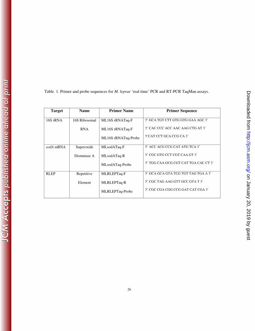
real-time PCR assay for the M. leprae-specific repetitive element (RLEP) (29). Primers and probes 202
were designed using Primer Express 2.0 software, Applied BioSystems (Table 1). Purified M. 203
leprae DNA or cDNA (5 µl) were added to a total PCR reaction volume of 25 µl containing 204
TaqMan 2X master mix, 500 nM of each primer and 100 nM of each probe for RNA-based PCR 205
assays (sodA mRNA or 16S rRNA) or 200 nM of each primer and 100 nM of the probe for the 206
DNA-based PCR assay (RLEP). Reaction mixtures were subjected to 50°C for 2 min, 95°C for 10 207
min, and 40 cycles of 95°C for 15 sec and 60°C for 1 min using a 7300 Real Time PCR System 208
(Applied BioSystems, Carlsbad, CA, USA). A standard curve for each PCR assay was generated 209
using serial 10-fold dilutions of purified M. leprae DNA ranging from 10 ng - 10 fg. Unknown 210
values were interpolated automatically for each sample using the standard curve method and 211
on January 20, 2019 by guesthttp://jcm
.asm.org/
Dow
nloaded from

10
normalized using the RLEP data for the same sample. The viability of M. leprae in human skin 212
biopsies was determined by comparing normalized 16S rRNA values for patients prior to and up to 213
2 yr after start of MDT treatment. 214
215
Specificity and sensitivity of assays. The specificity of each real-time PCR TaqMan assay was 216
determined by analyzing 10 ng of purified DNA from M. leprae, nine other mycobacterial species 217
including: M. tuberculosis H37Rv ATCC 27294, M. marinum ATCC 927, M. bovis BCG ATCC 218
35734, M. ulcerans ATCC 19423, M. simiae ATCC 25275, M. avium ATCC 25291, M. 219
intracellulare, ATCC 13950, M. kansasii ATCC 35775, M. smegmatis, ATCC 14468; and other 220
bacterial species including Staphylococcus epidermidis ATCC 12228, Streptococcus pyogenes 221
ATCC 12345, and E. coli ATCC 25992. In addition, mouse and human cDNA were also analyzed. 222
The sensitivity or lower limit of detection of each assay was determined by analyzing 10-fold 223
serial dilutions of M. leprae DNA and identifying the highest dilution, which still gave a positive 224
value (Ct <37). 225
226
Statistical Analysis. The standard curves for each RT-PCR assay using Ct values versus serial 227
DNA or cDNA concentration were calculated using a linear regression model (GraphPad InStat 228
version 3 software). The means and standard deviation of the results of sodA/RLEP and 16S 229
rRNA/RLEP RT-PCR assays for detection of M. leprae viability in axenic medium and MФ 230
cultures under various treatments were calculated were calculated using Student-t test. An alpha 231
value of 0.05 was used for all analyses. All statistical comparisons were made using linear Pearson 232
correlation coefficient (r) (GraphPad InStat version 3 software) as a measure of correlation 233
between assays at a particular time interval. 234
on January 20, 2019 by guesthttp://jcm
.asm.org/
Dow
nloaded from

11
Results 235
Identification of suitable targets for viability assays. Results of sodA/RLEP and 236
16S rRNA/RLEP RT-PCR assays demonstrated that levels of sodA gene transcripts were reduced 237
to 5% that of non-treated controls 48 hr post rifampin treatment (Fig. 1A) and that these levels 238
were significantly lower (p < 0.001) than that of 16S rRNA which were reduced to 38% of that of 239
non-treated controls (Fig. 1B). However, after a week of treatment both were essentially 240
background levels. In addition, reduction of sodA mRNA levels could be observed as early as 241
within 12 hr of rifampin treatment compared to untreated controls (data not shown). 242
243
Specificity and sensitivity of molecular M. leprae viability assays. The specificity and lower 244
limit of detection of each assay, sodA mRNA/RLEP and 16S rRNA/RLEP RT-PCR assays, were 245
analyzed using DNA from nine other mycobacterial species, Staphylococcus epidermidis, 246
Streptococcus pyogenes, E. coli and mouse and human DNA. Results showed that all three assays 247
were resulted in values Ct > 39 indicating 100% specificity of these assays for M. leprae (data not 248
shown). The sensitivity of detection of the M. leprae viability assays, defined as the lower limit of 249
detection of M. leprae for each assay, was analyzed using serial 10-fold dilutions of purified M. 250
leprae DNA and the standard curve method. The detection limit of sodA//RLEP assay was 1 pg 251
(Ct = 34.73 ± 0.3) and that of the 16S rRNA was 100 fg (Ct = 36.42 ± 0.6). Since each M. leprae 252
genome equivalent = 3 fg, these data suggest that these assays can detect 300 and 30 bacteria, 253
respectively. 254
255
M. leprae viability determination in infected MФ cultures. The ability of the RT-PCR-based M. 256
leprae viability assays to detect the effects of anti-leprosy drug treatment in infected MФ or the 257
on January 20, 2019 by guesthttp://jcm
.asm.org/
Dow
nloaded from
12
effects of immune-activated murine MФ (the host cell of M. leprae) on M. leprae viability was 258
evaluated. The sodA/RLEP RT-PCR assay results showed 16% viable M. leprae remained in MФ 259
after 48 hr of rifampin treatment and only 1% viable M. leprae remained after 48 hr infection of 260
IFN-γ-activated MФ (Table 2). Similar trends were noted for 16S rRNA/RLEP RT-PCR, 261
LIVE/DEAD® BacLight™ Bacterial Viability Assay and radiorespirometry assays. Thus, data 262
from both molecular-based assays, 16S rRNA/RLEP and sodA/RLEP, strongly correlated (p < 263
0.0001) with that from RR and LIVE/DEAD® BacLight™ Bacterial Viability Assay data (Table 264
2). However, the effects of the different treatments on M. leprae viability were lower when 265
compared to that of the sodA/RLEP RT-PCR viability assay. Ampicillin-treatment led to a 20% 266
decrease in M. leprae viability using sodA/RLEP RT-PCR assay, however, LIVE/DEAD® 267
BacLight™ and RR both showed that ampicillin did not have an affect on M. leprae viability. The 268
16S rRNA/RLEP showed that ampicillin has less of an effect on M. leprae viability than that of the 269
sodA/RLEP assay. This result was expected since M. leprae contains a β-lactamase enzyme which 270
makes it resistant to ampicillin-like drugs (18). 271
272
M. leprae viability in paired MB leprosy patient skin biopsies. A preliminary experiment was 273
performed to determine the utility of the sodA/RLEP and16S rRNA/RLEP RT-PCR M. leprae 274
viability assays to detect M. leprae viability directly from skin biopsy materials of MB leprosy 275
patients. Results indicated that the sodA/RLEP assay was only able to detect sodA cDNA from two 276
of eight pre-treatment biopsies (data not shown) and therefore the remaining biopsies taken after 277
initiation of MDT from the same patients were not evaluated. In contrast, the 16S rRNA/RLEP 278
assay showed positive results in all pretreatment specimens and declined during MDT treatment 279
(Table 3). Moreover, when the BI of patients’ skin slit smears were compared to the number of M. 280
on January 20, 2019 by guesthttp://jcm
.asm.org/
Dow
nloaded from
13
leprae as a function of M. leprae DNA using real-time RLEP PCR, a significant association was 281
observed (r = 0.6942; p = 0.001). Higher DNA concentrations correlated with higher BI values 282
and lower DNA concentrations correlated with lower BIs (Fig. 2). However, when 16S 283
rRNA/RLEP viability data were compared to patients’ BIs no correlation was observed (r = 284
0.4604; p = 0.0842). 285
286
Discussion 287
Determination of M. leprae viability is extremely difficult primarily due to the inability to cultivate 288
this bacterium on axenic media. The discovery by Shepard in 1960 (23) of the mouse foot pad 289
(MFP) technique to demonstrate replication of M. leprae was a research milestone, permitting 290
testing of new anti-leprosy drugs, determination of drug-resistant strains of M. leprae
from patient
291
specimens and initial evaluation of vaccine protection. Variations of the MFP technique involving 292
titration in large numbers of mice have been shown helpful to detect differences in the relative 293
viability of different
suspensions of M. leprae (30). However, this labor-intensive,
time-294
consuming, expensive technique is impractical for the study of interactions of M. leprae with its 295
host cell in vitro. Further complicating this is the declining availability of mouse foot pad 296
laboratories for M. leprae viability testing around the world. 297
298
When large numbers of nu/nu mouse-derived M. leprae became available to the leprosy research 299
community, radiorespirometry (RR), first described by Fransblau 1988 (5), was utilized to 300
determine the viability of a bacterial preparation based on the rate of oxidation of 14
C-palmitate by 301
M. leprae. RR was found to correlate well with growth in MFP (27) and therefore was useful to 302
evaluate the susceptibility of M. leprae to novel anti-leprosy drugs (6), a variety of environmental 303
on January 20, 2019 by guesthttp://jcm
.asm.org/
Dow
nloaded from

14
conditions (27) as well as UV (26) and gamma radiation (1). This technique was also useful for 304
characterization of the role of activated MФ in host resistance to leprosy (19) and the effects of 305
Schwann cells (M. leprae’s target in peripheral nerves) on M. leprae viability (7). However, the 306
necessity to utilize large quantities of bacteria (107) for RR-based M. leprae viability assays limits 307
the use of these assays for clinical purposes and in experiments where low numbers of bacteria are 308
available (14, 19). In addition, RR requires the use of radioactive substances which is highly 309
restricted in many areas of the world. 310
311
Recently, a fluorescence-based assay, LIVE/DEAD® BacLight™: Bacterial Viability assay for M. 312
leprae viability determination was developed (14). This assay, based on cell membrane integrity, 313
also correlated very well with MFP and RR assays for determination of M. leprae viability, 314
however it also depends on the use of relatively large numbers of bacteria (106) for analysis and 315
therefore limits it usefulness for clinical purposes and experimental conditions where only low 316
numbers of bacteria are available. 317
318
PCR assays, based on amplification of various DNA sequences within genes encoding the 18 kDa, 319
36 kDa, 65 kDa, Ag 85 and the multicopy repeat sequence RLEP of M. leprae, have been 320
successfully used to detect M. leprae in crude biological specimens even when low numbers of 321
bacteria are present (11, 12, 15 , 17, 28, 31). However, an important limitation of these DNA-based 322
PCR assays is their inability to distinguish between viable and dead organisms and thereby provide 323
information for drug efficacy in clinical settings and for short-term experimental procedures in 324
vitro. 325
326
on January 20, 2019 by guesthttp://jcm
.asm.org/
Dow
nloaded from

15
Detection of M. leprae RNA has been proposed as a promising tool for rapid detection and 327
measurement of the viability of pathogenic mycobacteria since degradation of RNA is relatively 328
rapid upon cell death (3, 13). A previous study with M. tuberculosis measured levels of M. 329
tuberculosis 85B (alpha antigen) mRNA, 16S rRNA, and IS6110 DNA using RT-PCR in patients’ 330
sputa to ascertain whether they could serve as potential markers of response to chemotherapy (4). 331
Results showed a rapid disappearance of M. tuberculosis mRNA from sputum while DNA 332
persisted in sputum from certain patients even after tuberculosis was cured. A preliminary study 333
using the M. leprae 16S rRNA as a predictor of viability showed this nucleic acid species to be a 334
suitable target for detection of M. leprae and its viability in clinical specimens using RT-PCR (8) 335
including skin slits of treated patients (9, 16). Although these assays were able to detect M. leprae 336
viability in clinical specimens, they were not tested on paired samples from the same patient 337
obtained pre- and during or post- MDT or in short-term experimental conditions where viability 338
determination can be a critical denominator. 339
340
In the present study the utility of two RNA-based RT-PCR assays, one using the 16S rRNA as the 341
RNA target and the other using a more labile sodA mRNA as the target, was evaluated for both 342
experimental and clinical usefulness. The sodA mRNA transcript, encoding the superoxide 343
dismutase A (ML0072c) of M. leprae, was selected as the mRNA target for this assay because of 344
the gene transcripts tested including: hsp18 (ML1795), gyrA (ML0006), rpoB (ML1891c), the 345
sodA transcript levels remained stable for at least 48 hr post harvesting from the mouse foot pad 346
tissues but rapidly degraded after death of M. leprae (data not shown). Also, since real-time 347
RLEP-DNA based PCR had been previously been characterized as a rapid, objective, molecular 348
on January 20, 2019 by guesthttp://jcm
.asm.org/
Dow
nloaded from

16
enumeration tool for detecting and quantifying bacterial numbers in an M. leprae preparation (29) 349
it was chosen to serve as a normalizer for these assays. 350
351
Results from in vitro experiments demonstrated that both sodA mRNA/RLEP and 16S 352
rRNA/RLEP RT-PCR assays were very good predictors of M. leprae viability in short-term 353
experiments (up to 48 hr) when bacteria were exposed to lethal concentrations of rifampin (the 354
only bacteriocidal drug in the MDT regiment for leprosy) in axenic medium or within infected 355
mouse MФ cultures. Assuming that the biological tests like RR and LIVE/DEAD® BacLight™ 356
are gold standards for M. leprae viability, the 16S/RLEP appeared to be the most sensitive 357
molecular assay for viability determination (r = 0.9817; p = 0.0005), even though sodA/RLEP 358
assay also presented a significant correlation with gold standard methods (r = 0.9463; p = 0.043). 359
360
The ability of these assays to detect the effects of rifampin treatment further demonstrates their 361
ability to differentiate between live and drug-killed M. leprae in axenic culture and within their 362
host cells (MФ). Therefore, these assays may be useful as rapid screening tools to identify 363
effective antileprosy drugs as well as for experiments to study host parasite interactions. However, 364
because of the inherent instability of the sodA mRNA species, resulting in rapid degradation 365
following death of bacilli, the sodA/RLEP RT-PCR assay may be more useful for experiments 366
which are designed to investigate early effects < 48 hr of drugs or immune factors on the viability 367
of M. leprae. Preliminary results from our laboratory suggest that the sodA/RLEP assay can detect 368
a loss in M. leprae viability as early as 12 hr post treatment (data not shown). These assays were 369
not only found to be highly specific and but more sensitive than either RR (107) or LIVE/DEAD 370
on January 20, 2019 by guesthttp://jcm
.asm.org/
Dow
nloaded from

17
BacLight® Bacterial Viability analysis (106) for determining the presence and viability or M. 371
leprae under short-term experimental conditions. 372
373
However, the sodA/RLEP assay was only able to detect the presence of M. leprae in 25% of the 374
pre-treatment biopsies tested. In contrast the 16S rRNA/RLEP assay was able to detect the 375
presence of M. leprae in all pre-treatment biopsies analyzed. Several parameters may have 376
contributed to these observed results. First, is the inherent labile nature of the mycobacterial 377
mRNA compared to that of rRNA (22) which may be further affected by the and low degree of 378
viability of M. leprae within the skin biopsies of leprosy patients in general (10). The second is 379
the relative lower sensitivity detecting sodA versus 16S rRNA due to increased copy number of 380
rRNA levels compared to that of mRNA. 381
382
In addition, results of the 16S rRNA/RLEP assay suggested a strong correlation between length of 383
therapy and decline of M. leprae viability. Even though the case numbers were low, these results 384
confirm the potential utility of this assay for monitoring anti-leprosy MDT therapy and thereby 385
potentially identifying leprosy cases which are not responding to MDT due to drug resistance, non 386
compliance or potential bacterial growth during or post treatment (relapse). In contrast, the lack of 387
detectable M. leprae sodA gene transcripts in the majority of human biopsies demonstrated that 388
this assay was not suitable for monitoring M. leprae viability in crude biological specimens. 389
390
In the present study, M. leprae viability was determined in skin biopsies based on 16S rRNA levels 391
normalized by RLEP DNA levels. When RLEP data was used as an indicator of M. leprae 392
numbers in an MB patient’s biopsy, there was a strong correlation between these results to that of 393
on January 20, 2019 by guesthttp://jcm
.asm.org/
Dow
nloaded from

18
BI results obtained by microscopic examination of the skin slit smears from the same patients. 394
Thus, the RLEP PCR assay alone may be useful for defining the clinical form of the disease and 395
potential up-or downgrading of disease status. However, no correlation was found between BI and 396
16S rRNA/RLEP levels, indicating that assessment of bacterial load per se does not reflect 397
viability in most instances. This was anticipated because DNA-based PCR positive signals and the 398
presence of acid-fast bacilli by microscopic examination persist in some cases years post-treatment 399
but do not necessarily reflect the real impact of treatment on bacterial viability (25). 400
401
In conclusion, this study has identified additional tools for leprosy diagnosis and monitoring 402
antileprosy drug efficacy for clinical purposes and for M. leprae viability in short-term 403
experimental studies which include the study of host cell/M. leprae interactions. This has been 404
made possible by the development of a procedure for the simultaneous isolation of M. leprae RNA 405
and DNA from the same sample and by incorporation of the previously described real-time RLEP 406
PCR as a good predictor of M. leprae numbers. Due to the stability and copy number of the 16S 407
rRNA as well of the degradation of this molecule over time, the 16S rRNA/RLEP assay should be 408
useful for determination of viable bacterial load in skin biopsies of MB leprosy patients and 409
therefore may be important in determining MDT efficacy and the ability of patients to still be 410
infectious post therapy. Hence, because of its high sensitivity and specificity this assay may also 411
constitute a very sensitive and specific assay for early detection of M. leprae in skin biopsies and 412
therefore potentially a predictor of the clinical form of leprosy. 413
on January 20, 2019 by guesthttp://jcm
.asm.org/
Dow
nloaded from
19
Acknowledgements 414
We wish to thank J.P. Pasqua for his excellent technical contributions to this work. This research 415
was partially funded by the HRSA,BPHC, Division of the National Hansen’s Disease Programs, 416
NIH/NIAID contract number Y1-AI-2646-01, CAPES (Coordenação de Aperfeiçoamento de 417
Pessoal de Nível Superior) and CNPq (Conselho Nacional de Desenvolvimento Científico e 418
Tecnológico). 419
on January 20, 2019 by guesthttp://jcm
.asm.org/
Dow
nloaded from

20
References 420
1. Adams L.B., Soileau N.A., Battista J.R., and J.L. Krahenbuhl. 2000. Inhibition of 421
metabolism and growth of Mycobacterium leprae by gamma irradiation. Int J Lepr Other 422
Mycobact Dis. 68: 1-10. 423
2. Britton W.J., and D.N. Lockwood. 2004. Leprosy. Lancet. 363: 1209-1219. 424
3. Chae G. T., Kim M. J., Kang T. J., Lee S. E., Shin Y., Kim J. P., Ko Y. H., Kim S. H., and 425
N. H Kim. 2002. DNA-PCR and RT-PCR for the 18-kDa gene of Mycobacterium leprae to 426
assess the efficacy of multi-drug therapy for leprosy. J Med Microbiol. 51: 417–422. 427
4. Desjardin L.E., Perkins M.D., Wolski K., Haun S., Teixeira L., Chen Y., Johnson J.L., 428
Ellner J.J., Dietze R., Bates J., Cave M.D., and K.D. Eisenach. 1999. Measurement of 429
sputum Mycobacterium tuberculosis messenger RNA as a surrogate for response to 430
chemotherapy. Am J Respir Crit Care Med. 160: 203-210. 431
5. Franzblau S. G. 1988. Oxidation of palmitic acid by Mycobacterium leprae in an axenic 432
medium. J Clin Microbiol. 26: 18-21. 433
6. Franzblau S.G., Biswas A.N., Jenner P., and M.J. Colston. 1992. Double-blind evaluation of 434
BACTEC and Buddemeyer-type radiorespirometric assays for in vitro screening of antileprosy 435
agents. Lep Rev. 63: 125-133. 436
7. Hagge D.A., Oby Robinson S., Scollard D., McCormick G., and D.L. Williams. 2002. A 437
new model for studying the effects of Mycobacterium leprae on Schwann cell and neuron 438
interactions. J Infect Dis.186: 1283-1296. 439
on January 20, 2019 by guesthttp://jcm
.asm.org/
Dow
nloaded from
21
8. Hirawati, Katoch K., Chauhan D.S., Singh H.B., Sharma V.D., Singh M., Kashyap M., and 440
V.M. Katoch. 2006. Detection of M. leprae by reverse transcription- PCR in biopsy specimens 441
from leprosy cases: a preliminary study. J Commun Dis. 38: 280-287. 442
9. Jadhav R.S., Kamble R.R, Shinde V.S., Edward S., and V.K. Edward. 2005. Use of reverse 443
transcription polymerase chain reaction for the detection of Mycobacterium leprae in the slit-444
skin smears of leprosy patients. Indian J. Lepr. 77:116-127. 445
10. Job C.K., Jayakumar J., Aschhoff M., and Mathan M.M. 1996. Viability of 446
Mycobacterium leprae in skin and peripheral nerves and persistence of nerve destruction in 447
multibacillary patients after 2 years of multidrug therapy. Int. J. Lepr. Other Mycobact. Dis. 448
64:44-50. 449
11. Kang T.J., Kim S.K., Lee S.B., Chae G.T., and J.P. Kim. 2003. Comparison of two different 450
PCR amplification products (the 18-kDa protein gene vs. RLEP repetitive sequence) in the 451
diagnosis of Mycobacterium leprae. Clin. Exp. Dermatol. 28: 420-424. 452
12. Kirschner P., Springer B., Vogel U., Meier A., Wrede A., Kiekenbeck M., Bange F.C., 453
and E.C. Böttger. 1993. Genotypic identification of mycobacteria by nucleic acid sequence 454
determination: report of a 2-year experience in a clinical laboratory. J. Clin. Microbiol. 31: 455
2882-2889. 456
13. Kurabachew M., Wondimu A., and J.J. Ryon. 1998. Reverse transcription-PCR detection of 457
Mycobacterium leprae in clinical specimens. J Clin Microbiol. 36: 1352-1356. 458
on January 20, 2019 by guesthttp://jcm
.asm.org/
Dow
nloaded from
22
14. Lahiri R., Randhawa B., and J.L. Krahenbuhl. 2005. Application of a viability-staining 459
method for Mycobacterium leprae derived from the athymic (nu/nu) mouse foot pad. J Med 460
Microbiol. 54: 235-242. 461
15. Martinez A.N., Britto C.F., Nery J.A., Sampaio E.P., Jardim M.R., Sarno E.N., and M.O. 462
Moraes. 2006. Evaluation of real-time and conventional PCR targeting complex 85 genes for 463
detection of Mycobacterium leprae DNA in skin biopsy samples from patients diagnosed with 464
leprosy. J Clin Microbiol. 44: 3154-3159. 465
16. Phetsuksiri B., Rudeeaneksin J., Supapkul P., Wachapong S., Mahotarn K., and P.J. 466
Brennan. 2006. A simplified reverse transcriptase PCR for rapid detection of Mycobacterium 467
leprae in skin specimens. FEMS Immunol Med Microbiol. 3: 319-328. 468
17. Plikaytis B.B., Gelber R.H., and T.M. Shinnick. 1990. Rapid and sensitive detection of 469
Mycobacterium leprae using a nested-primer gene amplification assay. J Clin Microbiol. 28: 470
1913-1917. 471
18. Prabhakaran K., Harris E.B., Randhawa B., Adams L.B., Williams D.L., and R.C. 472
Hastings. 1993. Use of beta-lactam/beta-lactamase-inhibitor combinations as 473
antimycobacterial agents. Microbios. 76: 251-261. 474
19. Ramasesh N., Adams L.B., Franzblau S.G. and J.L. Krahenbuhl. 1991. Effects of activated 475
macrophages on Mycobacterium leprae. Infect. Immun. 59: 2864-2869. 476
20. Rees, R.J.W. The microbiology of leprosy. In: Leprosy, 2nd Edition. RC Hastings, 477
Editor. Churchill, Livingstone, New York, 1985. P 46. 478
on January 20, 2019 by guesthttp://jcm
.asm.org/
Dow
nloaded from

23
21. Ridley D.S., and W. H. Jopling. 1966. Classification of leprosy according to immunity. A 479
five-group system. Int J Lepr Other Mycobact Dis. 34: 255-273. 480
22. Sela S., Clark-Curtis J.E., and H. Bercovier. 1989. Characterization and taxonomic 481
implication of the rRNA genes of Mycobacterium leprae. J Bacteriol. 171: 70–73. 482
23. Shepard C.C., and D.H. McRae. 1968. A method for counting acid-fast bacteria. Int J Lepr 483
Other Mycobact Dis. 36: 78-82. 484
24. Srinivas D., Rao P.N., Lakshmi T.S., and S. Suneetha . 2002. Bacterial index of granuloma 485
and its relevance compared to BI of skin smears. Lepr Rev. 73:79-80. 486
25. Torres P., Camarena J.J., Gomez J.R., Nogueira J.M., Gimeno V., Navarro J.C., and A. 487
Olmos. 2003. Comparison of PCR mediated amplification of DNA and the classical methods 488
for detection of Mycobacterium leprae in different types of clinical samples in leprosy patients 489
and contacts. Lepr Rev. 74: 18-30. 490
26. Truman R.W., and T.P. Gillis. 2000. The effect of ultraviolet light radiation on 491
Mycobacterium leprae. Int J Lepr Other Mycobact. 68: 11-17. 492
27. Truman R.W., and J.L. Krahenbuhl. 2001. Viable M. leprae as a research reagent. Int. J. 493
Lepr Other Mycobact Dis. 69:1-12. 494
28. Truman R., Fontes A.B., De Miranda A.B., Suffys P., and T.P. Gillis. 2004. Genotypic 495
variation and stability of four variable-number tandem repeats and their suitability for 496
discriminating strains of Mycobacterium leprae. J. Clin. Microbiol. 42:2558-65. 497
on January 20, 2019 by guesthttp://jcm
.asm.org/
Dow
nloaded from

24
29. Truman, R.W., Andrews, P.K., Robbins, N.Y., Adams, L.B., Krahenbuhl, J.L., and T.P. 498
Gillis. 2008. Enumeration of Mycobacterium leprae using real-time PCR. PLoS Negl. Trop. 499
Dis. 2(11):e328. Epub 2008 Nov 4. 500
30. Welch T.M., Gelber R.H., Murray L.P., H Ng H. O'Neill S.M., and L. Levy. 1980. 501
Viability of Mycobacterium leprae after multiplication in mice. Infect. Immun. 30: 325–328. 502
31. Williams D.L., Gillis T.P., Booth R.J., Looker D., and J.D. Watson. 1990. The use of a 503
specific DNA probe and polymerase chain reaction for the detection of Mycobacterium leprae. 504
J Infect Dis. 162:193-200. 505
32. Williams D.L., Oby-Robinson S., Pittman T. L., and D. M. Scollard. 2003. Purification of 506
Mycobacterium leprae RNA for gene expression analysis from leprosy biopsy specimens. 507
Biotechniques. 35: 534-536. 508
509
510
511
512
513
514
515
516
517
on January 20, 2019 by guesthttp://jcm
.asm.org/
Dow
nloaded from
26
Table 1. Primer and probe sequences for M. leprae ‘real time’ PCR and RT-PCR TaqMan assays.
Target Name Primer Name Primer Sequence
16S rRNA 16S Ribosomal
RNA
ML16S rRNATaq-F
ML16S rRNATaq-F
ML16S rRNATaq-Probe
5’ GCA TGT CTT GTG GTG GAA AGC 3’
5’ CAC CCC ACC AAC AAG CTG AT 3’
5’CAT CCT GCA CCG CA 3’
sodA mRNA Superoxide
Dismutase A
MLsodATaq-F
MLsodATaq-R
MLsodATaq-Probe
5’ ACC ACG CCG CAT ATG TCA 3’
5’ CGC GTG CCT CGT CAA GT 3’
5’ TGG CAA GCG CGT CAT TGA CAC CT 3’
RLEP Repetitive
Element
MLRLEPTaq-F
MLRLEPTaq-R
MLRLEPTaq-Probe
5’ GCA GCA GTA TCG TGT TAG TGA A 3’
5’ CGC TAG AAG GTT GCC GTA T 3’
5’ CGC CGA CGG CCG GAT CAT CGA 3’
on January 20, 2019 by guesthttp://jcm
.asm.org/
Dow
nloaded from

27
Table 2. Comparison of various methods for M. leprae viability determination in infected murine
MΦ.
M. leprae-infected MΦ
Treatment
% sodA1 % 16S rRNA
2 % BacLight
3 % Radiorespirometry
4
Rifampin (20 µug/ml) 16 ± 4.1 57 ± 14.6 60 ± 5.5 49 ± 3.2
Ampicillin (50 µg/ml) 80 ± 5.5 90 ± 5.1 100 ± 6.6 97 ± 6.8
IFN-γ (100 IU) 1 ± 0.3 13 ± 0.6 23 ± 1.4 11 ± 1.3
1. % sodA = sodA/RLEP RT-PCR values of M. leprae in treated murine MΦ divided by that in
untreated murine MΦ.
2. % 16S rRNA = 16S rRNA/RLEP RT-PCR values of M. leprae in treated murine MΦ divided by
that in untreated murine MΦ.
3. % BacLight = LIVE/DEAD® BacLight™ Bacterial Viability Assay values of M. leprae in
treated murine MΦ divided by that in untreated murine MΦ.
4. % Radiorespirometry = day 7 cumulative counts per min (cpms) using Buddemeyer
radiorespirometry of M. leprae viability in treated murine MΦ divided by that in untreated murine
MΦ.
on January 20, 2019 by guesthttp://jcm
.asm.org/
Dow
nloaded from

28
Table 3. Analysis of M. leprae viability in multibacillary leprosy patients prior to and
during leprosy multidrug therapy (MDT).
Patient #
Biospsy # Leprosy
Classificati
on1
MDT
Treatment
BI2
16S rRNA/RLEP
(% ML viability)3
Patient 1
04-01 BL Untreated 3.5 4314.3 ± 1395.7
05-01 1 yr 3.17 305.4 ± 112.5 (9)
06-01 2 yr 1.5 294 ± 76.4 (3)
Patient 2 04-02 BL Untreated 3 14.8 ± 3.1
05-02 1 yr 2 8.8 ± 3.2 (59)
06-02 2 yr 2.8 8.3 ± 3.7 (56)
Patient 3 03-03 LL Untreated 1 50.2 ± 11.5
04-03 1 yr 0.66 6.8 ± 4.9 (13)
05-03 2 ys 0.17 0.1 ± 0.05 (0.2)
Patient 4 00-04 LL Untreated 1 337.7 ± 87.8
00-04 6 months 1 11.5 ± 4.0 (4)
Patient 5 03-05 LL Untreated 1.2 4.6 ± 0.6
04-05 1 year 0.7 1.3 ± 0.2 (28)
Patient 6 02-06 LL Untreated 2.3 10.5 ± 1.2
03-06 1 year 1.7 1.0 ± 0.1 (9.4)
Patient 7 03-07 LL Untreated 2.3 39.5 ± 2.8
04-07 1 year 1.7 0.6 ± 0.3 (1.6)
Patient 8 05-08 LL Untreated 1.7 120.8 ± 23.2
06-08 1 year 1.2 0.5 ± 0.2 (0.4)
1
Leprosy patient classification according the Ripley-Jopling Scale (21).
2Bacillary index (Acid fast bacilli count) of skin slits from leprosy patient (20)
on January 20, 2019 by guesthttp://jcm
.asm.org/
Dow
nloaded from

29
3Mean and standard deviation of 16S rRNA/RLEP RT-PCR values derived from
cDNA of skin biopsies from untreated and treated leprosy patients and (% ML (M.
leprae) viability) of each treated specimen assay derived by dividing the number of
16S rRNA/RLEP RT-PCR values of leprosy patients’ biopsies after treatment by that
of untreated patient biopsy.
on January 20, 2019 by guesthttp://jcm
.asm.org/
Dow
nloaded from

30
Figure Legends
Figure 1. Molecular viability analyses of rifampin-treated M. leprae. A) M. leprae sodA/RLEP
levels; B) M. leprae 16S rRNA/RLEP levels. T-0 = untreated, T-48 hr = 48 h post rifampin
treatment, T-1 wk = 1 wk post rifampin treatment, T-2 wk = 2 wk post rifampin treatment. The
data are representative of three replicates for three independent experiments.
Figure 2. Comparison of M. leprae using real-time RLEP PCR and BI analyses in patients’
biopsies as function of pretreatment and post-treatment using Linear Pearson correlation between
BI and DNA concentration from MB (p = 0.001; r = 0.6942).
on January 20, 2019 by guesthttp://jcm
.asm.org/
Dow
nloaded from

A . B.
Figure 1 Martinez et al.
on January 20, 2019 by guesthttp://jcm
.asm.org/
Dow
nloaded from

Figure 2 Martinez et al.
on January 20, 2019 by guesthttp://jcm
.asm.org/
Dow
nloaded from

JOURNAL OF CLINICAL MICROBIOLOGY, Jan. 2010, p. 346 Vol. 48, No. 10095-1137/10/$12.00 doi:10.1128/JCM.02106-09
ERRATUM
Molecular Determination of Mycobacterium leprae Viability byUse of Real-Time PCR
Alejandra N. Martinez, Ramanuj Lahiri, Tana L. Pittman, David Scollard, Richard Truman,Milton O. Moraes, and Diana L. Williams
Laboratorio de Hansenıase, Instituto Oswaldo Cruz—Fiocruz, Rio de Janeiro, Brazil, and HRSA, BPHC, Division ofNational Hansen’s Disease Programs, Laboratory Research Branch at the School of Veterinary Medicine,
Louisiana State University, Baton Rouge, Louisiana
Volume 47, no. 7, p. 2124–2130, 2009. Page 2125, column 1, line 4 of “Rifampin treatment in axenic medium” section: “(100ng/ml)” should read “(8 mg/ml).”
Page 2125, column 1, line 6 of “Rifampin treatment in axenic medium” section: “20 �g/ml” should read “2 �g/ml.”Page 2125, column 2, line 3: “20 �g/ml” should read “2 �g/ml.”Page 2127, Table 2: The first entry in the first column should read “Rifampin (2 �g/ml).”Page 2129: In the Acknowledgments section, the following sentence was inadvertently omitted: “We acknowledge the American
Leprosy Mission for its support of a portion of this research.”
346