Embed Size (px)
Citation preview
UNIVERSIDADE FEDERAL DE SANTA MARIA
CENTRO DE CIÊNCIAS RURAIS
PROGRAMA DE PÓS-GRADUAÇÃO EM MEDICINA VETERINÁRIA
Patrícia Bräunig
CARACTERIZAÇÃO DE CÉLULAS-TRONCO MESENQUIMAIS
DERIVADAS DO TECIDO ADIPOSO E SEU POTENCIAL DE
DIFERENCIAÇÃO
Santa Maria, RS
2016
Patrícia Bräunig
CARACTERIZAÇÃO DE CÉLULAS-TRONCO MESENQUIMAIS DERIVADAS DO
TECIDO ADIPOSO E SEU POTENCIAL DE DIFERENCIAÇÃO
Tese apresentada ao Curso de Pós-Graduação
em Medicina Veterinária, da Universidade
Federal de Santa Maria (UFSM, RS), como
requisito parcial para obtenção do título de
Doutor em Medicina Veterinária.
Orientador: Prof. Paulo Bayard Dias Gonçalves
Santa Maria, RS
2016
© 2016
Todos os direitos autorais reservados a Patrícia Bräunig. A reprodução de partes ou do todo
deste trabalho só poderá ser feita mediante a citação da fonte.
E-mail: pbrä[email protected]
AGRADECIMENTOS
Agradeço à minha família em especial aos meus pais, meu irmão, minha vó e minha
cachorrinha Margô pelo amor incondicional, apoio e incentivo, todos os dias durante a
jornada do doutorado.
Às minhas amigas, Gisa, Tanusa, Bruna, Deise, Carine, Carla, Mirélle, Raquel e Bruna
pela amizade, carinho, estímulo e incentivo nos momentos difíceis assim como pelas alegrias
compartilhadas.
Ao Professor Paulo Bayard Dias Gonçalves pela orientação e pela confiança em mim
depositada.
À minha “chefa” Professora Fernanda Vogel pela amizade, incentivo, apoio,
compreensão, orientação e bons conselhos.
Aos Professores Fábio Comim e Alfredo Antoniazzi pela boa convivência, pelo
aprendizado que obtive com vocês e por estarem sempre dispostos a ajudar em todos os
momentos da realização deste trabalho.
Aos colegas de laboratório do BioRep e Ladopar que tornaram-se amigos, espero levar
essa amizade por toda vida independente de onde ela nos leve. Obrigada por toda ajuda bem
como pelas as alegrias e decepções compartilhadas.
À Universidade Federal de Santa Maria, ao Departamento de Medicina Veterinária
Preventiva e ao Programa de Pós-graduação em Medicina Veterinária (PPGMV) pela
oportunidade de realizar mais uma etapa na minha formação acadêmica e profissional. Em
especial agradeço a secretária do PPGMV, Maria, pelo trabalho competente que desempenha.
Meus sinceros agradecimentos a todos que contribuíram de forma direta ou indireta
para a elaboração e conclusão desse trabalho.
Enfim e mais importante, minha imensa gratidão a Deus por todas as bênçãos.
RESUMO
DESENVOLVIMENTO E CARACTERIZAÇÃO DE CÉLULAS-TRONCO
MESENQUIMAIS DERIVADAS DO TECIDO ADIPOSO E SEU POTENCIAL DE
DIFERENCIAÇÃO
AUTORA: Patrícia Bräunig
ORIENTADOR: Prof. Paulo Bayard Dias Gonçalves
Células-tronco mesenquimais têm demonstrado significativo potencial para aplicação
terapêutica devido ao seu fácil isolamento, baixa imunogenicidade, ausência das implicações
éticas e sua ampla plasticidade. Essas células estão nos mais diversos tecidos, destacando-se o
tecido adiposo devido á sua ampla distribuição no organismo, conveniente obtenção e o
considerável número de células-tronco mesenquimais multipotentes que podem ser isoladas
desse tecido. Assim sendo, no presente estudo, células-tronco mesenquimais derivadas do
tecido adiposo (AT-MSCs) foram isoladas do tecido adiposo localizado nas regiões próximas
ao omento e testículos de camundongos BALB/c. Durante a manutenção e expansão das AT-
MSCs in vitro, elas foram caracterizadas quanto à presença de marcadores antigênicos de
superfície e potencial de diferenciação nas linhagens osteogênica, condrogênica e
adipogênica. AT-MSCs de ambas as fontes expressaram os marcadores mesenquimais de
superfície, CD73 e CD105, assim como foram negativas para o marcador de linhagens
hematopoiéticas, CD45. Quanto ao potencial de diferenciação, os cultivos provenientes das
duas origens de tecido adiposo apresentaram capacidade de diferenciar nas três linhagens
acima citadas. Porém, foram observadas discretas diferenças tanto nos padrões de expressão
dos marcadores mesenquimais de superfície quanto nos potenciais de diferenciação entre as
AT-MSCs provenientes dos diferentes locais de deposição de gordura. Além disso, as AT-
MSCs isoladas do tecido adiposo depositado em contato com o omento quando cultivadas
com meios de diferenciação, contendo ácido retinóico e meio condicionado testicular
demonstraram expressão do gene Gdnf o qual é reconhecidamente expresso pelas células de
Sertoli. Portanto, os resultados obtidos demonstram que conforme a origem do tecido adiposo
as AT-MSCs possuem diferentes características relacionadas aos marcadores de superfície
assim como aos potenciais de diferenciação.
Palavras-chave: Ácido retinóico. Células multipotentes da fração estromal vascular.
Diferenciação in vitro. Gene marcador. Marcadores antigênicos de superfície. Meio
condicionado.
ABSTRACT
DEVELOPMENT, CHARACTERIZATION AND DIFFERENTIATION POTENTIAL
OF ADIPOSE TISSUE-DERIVED MESENCHYMAL STEM CELLS
AUTHOR: Patrícia Bräunig
ADVISER: Prof. Paulo Bayard Dias Gonçalves
Mesenchymal stem cells (MSCs) have demonstrated significant potential for clinical use due
to their convenient isolation, lack of significant immunogenicity, lack of ethical controversy
and their potential to differentiate into tissue-specific cell types. MSCs reside in almost all
tissues including the adipose tissue. Adipose tissue has main advantages as wide distribution
in the organism, suitable isolation and considerable amount of resident multipotent stem cells.
Therefore, in this study, adipose tissue-derived mesenchymal stem cells (AT-MSCs) were
isolated from BALB/c mice omentum and epididymis fat pats. During AT-MSCs maintenance
and expansion in vitro, they were characterized for the expression of antigenic surface
markers and for osteogenic, chondrogenic, and adipogenic differentiation potential. AT-
MSCs form both sources expressed mesenchymal surface markers, CD73, and CD105 and
were negative for a hematopoietic marker, CD45. The cultures derived from both adipose
tissues differentiated into all three lineages. However, differences were observed in
mesenchymal surface marker expression profiles as well as in the differentiation potential of
AT-MSCs from different fat sources. Furthermore, AT-MSCs isolated from omentum fat
depot were cultured with differentiation medium containing retinoic acid and testicular cell
conditioned medium. After treatment periods, AT-MSCs showed Gdnf gene expression, this
gene is a marker for Sertoli cells. The results showed that AT-MSCs from distinct fat depots
have different characteristics related to stem cell surface marker expression profiles and
differentiation potential.
Keywords: Antigenic surface markers. Conditioned medium. Gene marker. In vitro
differentiation. Multipotent stromal cells. Retinoic acid.
LISTA DE FIGURAS
CAPÍTULO I
Figure 1 – Differentiation of mouse adipose tissue-derived stromal vascular fraction cells
from omentum (OADSVF) and epididymis (EADSVF) in osteogenic,
chondrogenic and adipogenic lineages. (Magnification 100 X) ............................ 27
Figure 2 – Flow cytometric analysis of the MSC markers CD73 and CD105 in mouse
adipose tissue-derived stromal vascular fraction cells from omentum
(OADSVF) (A) and epididymis (EADSVF) (B), at three different passages. P2
(cell passage number 2), P4 (cell passage number 4) and P7 (cell passage
number 7) ............................................................................................................... 28
CAPÍTULO II
Figure 1 – Flow cytometric analysis of cell surface markers CD73, CD105 and CD45 in
adipose tissue-derived mesenchymal stem cells from mouse omentum (A) and
epididymis (B) ....................................................................................................... 41
Figure 2 – Mesodermal differentiation of adipose tissue-derived mesenchymal stem cells
from mouse omentum (A) and epididymis (B). (100X magnification) ................. 42
Figure 3 – Relative expression of the murine Gdnf gene in omentum AT-MSCs treated
with retinoic acid (RA), testicular cell-conditioned medium (TCC), or
RA+TCC for 7, 14, and 21 days, or with growth medium only for an untreated
control group. Values with different superscripts (a, b) are significantly
different (p < 0.05) ................................................................................................. 43
APÊNDICE
Fig. 1 – Fragments of DNA identified by electrophoretic run after PCR performed for
amplification of a fragment of the 18S rDNA gene (700 bp) from Sarcocystis
spp. Line 1, negative control (MilliQ water); Line 2, positive control
(Sarcocystis spp. DNA); Line 3, molecular marker (100-bp DNA ladder); Line
4, PCR amplification product obtained after DNA extraction from ten cysts in
PBS using Protocol 2 (kit with modification); Line 5, PCR amplification
product obtained after DNA extraction from bovine myocardium sample using
Protocol 3 (DNAzol).............................................................................................. 67
Fig. 2 – Sarcocystis spp. DNA detection frequencies from polls of ten cysts in PBS
using kit, kit with modification, DNAzol, or CTAB extraction protocols.
Different lower-case letters above the columns indicate significant difference
in the frequency of positive samples detected among protocols (Chi-square test
at a 99% confidence level) ..................................................................................... 68
Fig. 3 – Sarcocystis spp. DNA detection frequencies from bovine myocardium samples
using kit, kit with modification, DNAzol or CTAB extraction protocols.
Different lower-case letters above the columns indicate significant difference
in the frequency of positive samples detected among protocols (Chi-square test
at a 99% confidence level) ..................................................................................... 69
LISTA DE TABELAS
CAPÍTULO I
Table 1 – Flow cytometric analysis of the expression of MSC markers CD73 and
CD105, and hematopoietic marker CD45 in mouse OADSVF at various
passages ................................................................................................................. 29
Table 2 – Flow cytometric analysis of the expression of MSC markers CD73 and CD105
and hematopoietic marker, CD45 in mouse EADSVFs at various passages ......... 29
CAPÍTULO II
Table 1 – List of primer sequences and accession numbers for gene sequences used for
the mRNA gene expression analysis ..................................................................... 40
APÊNDICE
Table 1 – Frequencies of Sarcocystis spp. DNA detection by PCR using four different
DNA extraction protocols on pools of ten cysts in PBS ........................................ 64
Table 2 – Frequencies of Sarcocystis spp. DNA detection by PCR using four different
DNA extraction protocols on bovine myocardium samples .................................. 65
Table 3 – Probability of true positive PCR diagnostic of Sarcocystis spp. infection in
cattle hearts considering samples extracted with DNAzol, and comparing the
use of one, two, or three myocardium samples from each heart ........................... 66
SUMÁRIO
1 INTRODUÇÃO ............................................................................................................. 10
2 REVISÃO BIBLIOGRÁFICA ..................................................................................... 12
2.1 CÉLULAS-TRONCO ..................................................................................................... 12
2.1.1 Células-tronco mesenquimais ...................................................................................... 13
2.2 TECIDO ADIPOSO ........................................................................................................ 14
2.3 DIFERENCIAÇÃO IN VITRO ....................................................................................... 15
2.3.1 Meio Condicionado ....................................................................................................... 16
2.3.2 Potencial de diferenciação em linhagens germinativas masculinas .......................... 16
2.4 ESPERMATOGÊNESE .................................................................................................. 17
3 CAPÍTULO I – EVALUATION AND COMPARISON OF ADIPOSE TISSUE-
DERIVED STROMAL STEM CELLS FROM TWO DIFFERENT FAT
DEPOTS ......................................................................................................................... 19
Abstract .......................................................................................................................... 19
Introduction ................................................................................................................... 19
Material and Methods .................................................................................................. 21
Results ............................................................................................................................ 22
Discussion ....................................................................................................................... 23
Conclusion ...................................................................................................................... 24
References ...................................................................................................................... 25
4 CAPÍTULO II – CHARACTERIZATION OF ADIPOSE TISSUE-DERIVED
MESENCHYMAL STEM CELLS AND THEIR DIFFERENTIATION
POTENTIAL INTO MALE GERM CELLS AFTER TREATMENTS WITH
RETINOIC ACID AND CONDITIONED MEDIUM ............................................... 30
Abstract .......................................................................................................................... 30
Introduction ................................................................................................................... 30
Material and Methods .................................................................................................. 32
Results ............................................................................................................................ 34
Discussion ....................................................................................................................... 35
Conclusion ...................................................................................................................... 37
References ...................................................................................................................... 37
5 DISCUSSÃO .................................................................................................................. 44
6 CONCLUSÃO ............................................................................................................... 46
REFERÊNCIAS ............................................................................................................ 47
APÊNDICES .................................................................................................................. 54
APÊNDICE A – ARTIGO CIENTÍFICO PRODUZIDO DURANTE O
PERÍODO DO DOUTORADO EM COLABORAÇÃO COM A PROFA
FERNANDA SILVEIRA FLORES VOGEL NO LABORATÓRIO DE
DOENÇAS PARASITÁRIAS DA UFSM ................................................................... 55
10
1 INTRODUÇÃO
As células-tronco adultas possuem plasticidade mais ampla do que previamente
suposto, ultrapassando as barreiras restritas às linhagens do tecido de origem (WAGERS;
WEISSMAN, 2004; ZUK et al., 2002). Os tecidos dos mamíferos pós-nascimento possuem
pequenas populações dessas células com capacidade de auto-renovar, proliferar e diferenciar
em células especializadas, promovendo renovação e reparo do tecido (PISCAGLIA, 2008). O
tecido adiposo que está amplamente distribuído no organismo é uma fonte de células-tronco
adultas multipotentes em significativa quantidade e de fácil obtenção (FRASER et al., 2007;
MITCHELL et al., 2006). Esse tecido é dividido em duas diferentes frações, os adipócitos
maduros e a fração estromal vascular onde estão as células-tronco mesenquimais (MSCs). As
células-tronco mesenquimais derivadas do tecido adiposo (AT-MSCs) possuem ampla
plasticidade, padrão complexo de expressão de moléculas marcadoras de superfície (CDs),
além de secretar fatores relacionados ás respostas imune e inflamatória (BUNNELL et al.,
2008; MOSNA et al., 2010; STREM et al., 2005; ZUK et al., 2002).
Quando cultivadas em meios contendo fatores de indução de diferenciação as MSCs
podem transdiferenciar in vitro em diversas linhagens celulares (DA SILVA et al., 2013;
LACHAM-KAPLAN et al., 2006; MORTAZAVI; MOHAMMADI, 2013; SCHÄFLER;
BÜCHLEER, 2007; ZUK et al., 2002). Além disso, o meio condicionado proveniente do
sobrenadante de um cultivo celular contém diversos fatores solúveis que poderão auxiliar na
diferenciação das MSCs em linhagens celulares relacionadas com o cultivo (DA SILVA et al.,
2013; LACHAM-KAPLAN et al., 2006; MORTAZAVI; MOHAMMADI, 2013). Portanto, os
meios de diferenciação contendo os fatores de indução assim como o meio condicionado são
abordagens empregadas para promover a diferenciação das AT-MSCs em diversas linhagens
celulares (DRUSENHEIMER et al., 2006; HUANG et al., 2010; NAYERNIA et al., 2006;
ZHANG et al., 2014). Sendo que o potencial de diferenciação de MSCs em células
germinativas masculinas já foi abordado em alguns trabalhos (DRUSENHEIMER et al.,
2006; HUANG et al., 2010; ZHANG et al., 2014). Entretanto, o potencial de diferenciação de
AT-MSCs em células germinativas masculinas necessita ser elucidado. Assim como,
necessita-se esclarecer o potencial de ação dos meios suplementado com ácido retinóico e
condicionado testicular no processo de diferenciação das AT-MSCs em células germinativas
masculinas.
Portanto, os objetivos deste estudo foram desenvolver e caracterizar as AT-MSCs
isoladas de diferentes origens de tecido adiposo, além de verificar seu potencial de diferenciar
11
em linhagens celulares relacionadas às células germinativas masculinas. Para isso,
primeiramente, foram estabelecidos cultivos das frações estromal vascular obtidas de dois
diferentes depósitos de gordura em camundongos BALB/c. A caracterização das AT-MSCs
foi realizada pela presença de marcadores de superfície por citometria de fluxo e pela
capacidade de diferenciação adipogênica, osteogênica e condrogênica. Ademais, após os
tratamentos com ácido retinóico e/ou meio condicionado proveniente do cultivo de células do
testículo de camundongos da mesma espécie, o possível potencial de diferenciação das AT-
MSCs em linhagens celulares relacionadas às células germinativas masculinas foi verificado
por meio da expressão de genes marcadores específicoa (CHEN & LIU, 2015; HOU et al.,
2014; IKAMI et al., 2015; MENG et al., 2000).
12
2 REVISÃO BIBLIOGRÁFICA
2.1 CÉLULAS-TRONCO
As células-tronco são um tipo especial de célula não especializada com capacidade de
auto-renovar, proliferar e diferenciar em diversas linhagens celulares (BLAU et al., 2001;
VERFAILLIE, 2002). A função fundamental dessas células in vivo é participar nos processos
de renovação, reparo e regeneração dos tecidos (BLAU et al., 2001). Células-tronco são
classificadas quanto a sua origem e plasticidade sendo que a classificação mais empregada é
em embrionárias ou adultas. As células-tronco embrionárias são pluripotentes, ou seja,
possuem ampla capacidade de diferenciação, podendo diferenciar-se em todos os tipos
celulares, e encontram-se na massa celular interna do blastocisto e na prega genital do feto de
5-10 semanas. Já as células-tronco adultas (somáticas; multi ou unipotentes) possuem
capacidade de diferenciação limitada e residem nos tecidos dos mamíferos como medula
óssea, cordão umbilical, polpa dentária dentre outros (BLUM et al., 2009; PISCAGLIA,
2008; SU et al., 2011).
Os primeiros estudos envolvendo células-tronco foram realizados utilizando as células
embrionárias, objetivando empregá-las na terapia de diferentes patologias (KAJI;e LEIDEN,
2001) como facilitadoras do reparo de tecidos degenerados ou lesionados, porém a aplicação
terapêutica dessas células passou a apresentar diversos problemas, como rejeição devido à
incompatibilidade imunológica, perda da função das células infundidas, amplo potencial de
formação de teratomas, além das questões éticas e legais envolvendo células-tronco
embrionárias (BLAU et al., 2001; BLUM et al., 2009; PISCAGLIA, 2008; SU et al., 2011).
Sabe-se, desde os anos 60, que alguns tecidos de um organismo adulto se regeneram
constantemente, isso acontece com a pele, com as paredes intestinais e principalmente com o
sangue, que têm suas células destruídas e renovadas, em um complexo processo de
proliferação e diferenciação celular finamente regulado. Consequentemente, pesquisadores
isolaram células-tronco adultas de diversos tecidos, como medula óssea, tecido adiposo,
cordão umbilical, pele, polpa dentária, entre outros, tornando-as uma alternativa ao uso das
células embrionárias, já que minimizam a possibilidade de formação de tumores e não
envolvem questões éticas e legais (BLAU et al., 2001; PITTENGER et al., 1999).
13
2.1.1 Células-tronco mesenquimais
Células-tronco mesenquimais (MSCs) são células multipotentes que podem originar,
in vitro, linhagens mesenquimias e não mesenquimais e são as céluas-tronco adultas mais
plásticas conhecidas até o momento (DA SILVA et al., 2006; ZUK et al., 2002). MSCs
possuem ampla distribuição natural no organismo dos mamíferos pós-nascimento, sendo
preferencialmente encontradas nos tecidos conjuntivos e foram isoladas com sucesso de
vários tecidos como medula óssea (PITTENGER et al., 1999), músculo esquelético
(JANKOWSKI et al., 2002), dentes decíduos (MIURA et al., 2003), tecido adiposo (ZUK
et al., 2002) e paredes dos vasos sanguíneos (DA SILVA et al., 2006).
MSCs apresentam uma variedade de receptores e marcadores antigênicos de
membrana, esses facilitam sua identificação, caracterização e isolamento. Uma variedade de
marcadores de superfície (CDs) estão presentes nas MSCs: CD29, CD34, CD44, CD105,
CD73, CD90, sendo que a checagem da expressão desses marcadores é amplamente utilizada
para identificação das MSCs (MARCUS et al., 2008; DUBEY et al., 2014; JIANG et al.,
2002; MOSNA et al., 2010; PITTENGER et al., 1999; ZUK et al., 2002). Essas células
também secretam uma ampla variedade de fatores de crescimento e citocinas que possuem
função mediadora nas respostas inflamatória, imune e na migração celular (CHAMBERLAIN
et al., 2007; MOSNA et al., 2010). Além disso, outra característica importante das MSCs, no
contexto de terapia celular, está no status imunológico privilegiado apresentado por essas
células, o qual possibilita aplicação de células alogênicas com pouca implicação de resposta
imune indesejada. Acredita-se que essa característica está associada com a baixa expressão
dos antígenos do complexo de histocompatibilidade (MHC) (CHAMBERLAIN et al., 2007;
MOSNA et al., 2010).
A população de células-tronco mesenquimais derivadas da medula óssea (BM-MSCs)
é a mais pesquisada, principalmente para aplicação na terapia celular regenerativa, desde sua
descoberta nos anos 70 por Friedenstein (CHAMBERLAIN et al., 2007; PHINNEY &
PROCKOP, 2007). O desenvolvimento de protocolos para isolar, purificar e cultivar BM-
MSC já foram descritos detalhadamente em inúmeras publicações (MOSNA et al., 2010;
PITTENGER et al., 1999; REYES et al., 2001). Células-tronco mesenquimais derivadas do
tecido adiposo (AT-MSCs) representam uma alternativa atrativa às derivadas da medula óssea
devido sua abundância, acessibilidade, ampla plasticidade e ausência de imunogenicidade
(GIMBLE et al., 2007). Em adultos, estima-se que a fração de MSCs proveniente da medula
óssea é de 1:50.000 - 1:1 milhão do total de células nucleadas. Essas são obtidas a partir de
14
um volume limitado que pode ser puncionado de forma segura por doador, fazendo com que
seja necessária significativa expansão in vitro (MUSCHLER et al., 2001). Em contraste, a
frequência de MSCs no tecido adiposo é estimada numa faixa entre 1:30-1:100 pelo total de
células nucleadas (FRASER et al., 2007; MITCHELL et al., 2006). Além disso, quantidade
significativa de tecido adiposo pode ser retirado do paciente sem prejuízo para saúde desse,
como por exemplo, os procedimentos de lipoaspiração. As BM-MSCs e as AT-MSCs
possuem várias características em comum, além da multipotência, como a habilidade de
proliferar indefinidamente em cultivo e a presença de marcadores característicos na superfície
(PITTENGER et al., 1999). Porém, as AT-MSCs apresentam vantagens como relativa
abundância e facilidade de obtenção quando comparadas as BM-MSCs. Isso torna as AT-
MSCs uma excelente fonte alternativa de célula-tronco (FRASER et al., 2007; GIMBLE
et al., 2007; MITCHELL et al., 2006).
2.2 TECIDO ADIPOSO
O tecido adiposo é originado durante o desenvolvimento embrionário a partir da
mesoderme, contém um estroma de suporte o qual é facilmente isolado e representa uma fonte
de células-tronco de fácil obtenção. Em um organismo adulto, as fontes de tecido adiposo são
diversas, podendo ser coletado da região subcutânea, da região visceral, das regiões próximas
e em contato com os órgãos e gônadas (BUNNELL et al., 2008; STREM et al., 2005).
Acreditava-se que o tecido adiposo estava simplesmente envolvido no armazenamento de
lipídios, porém atualmente, já é reconhecido por possuir alta atividade endócrina metabólica,
pois secreta citocinas, (adipocinas) como leptina, adiponectina e interleucina 6 (IL-6) (KIM
et al., 2000). A localização no organismo do depósito de gordura pode influenciar nas
características das AT-MSCs, alguns estudos demonstraram que células-tronco adiposas
provenientes do subcutâneo são mais adipogênicas que as extraídas do omento (PARK et al.,
2012; TCHKONIA et al., 2002). Além disso, podem variar os padrões dos fatores secretados
e dos genes expressos entre as fontes de gordura (ATZMON et al., 2002; MAJKA et al.,
2011).
Constantemente, durante toda a vida dos mamíferos o tecido adiposo se expande e
reduz demonstrando capacidade de auto-renovação, e de diferenciação, além de metabólica,
das células presentes nesse tecido (STREM et al., 2005). Além disso, o tecido adiposo é um
tecido complexo composto por: adipócitos maduros, pré-adipócitos, fibroblastos, células da
musculatura lisa vascular, células endoteliais, macrófagos/monócitos residentes
15
(SCHÄFFLER & BÜCHLER, 2007). Esse tecido pode ser basicamente dividido em duas
diferentes frações, adipócitos maduros e a fração estromal vascular que possui composição
celular heterogênea e onde se encontram as células-tronco multipotentes derivadas do tecido
adiposo (PRUNET-MARCASSUS et al., 2006). As células que compõem a fração estromal
vascular podem ser enzimaticamente isoladas, separadas dos adipócitos por centrifugação e
mantidas em cultivo (BUNNELL et al., 2008).
Estudos indicando que as células-tronco adultas podem diferenciar para além dos
limites restritos às linhagens do tecido, sugerindo ampla plasticidade (WAGERS;
WEISSMAN, 2004), fez com que surgissem diversos trabalhos pesquisando o potencial de
transdiferenciação das MSCs (JIANG et al., 2002; PITTENGER et al., 1999; ZUK et al.,
2002). A habilidade de diferenciação das AT-MSCs em linhagens celulares mesenquimais
clássicas como: adipócitos, condrócitos, osteócitos e miócitos (BUNNELL et al., 2008;
STREM et al., 2005; ZUK et al., 2002), além da capacidade de diferenciação em linhagens
não mesenquimais como tipos celulares esquelético e neuronal demonstra o potencial plástico
dessas células, bem como sua potencial aplicação futura nas terapias celulares (ZUK et al.,
2002).
2.3 DIFERENCIAÇÃO IN VITRO
In vivo, o microambiente ou nicho em que a célula-tronco adulta se encontra contém
diversos e complexos componentes que sinalizam o destino dessa célula (WATT; HOGAN,
2000). O nicho é composto por parte das células do tecido e de substâncias extracelulares que
abrigam as células-tronco e controlam sua auto-renovação ou diferenciação. O destino é
regulado pela interação entre sinais endógenos e estímulos do microambiente, esse processo
fino e complexamente regulado controla a manutenção e a diferenciação das células-tronco
nos tecidos adultos (STEWART; STEWART, 2011; WATT; HOGAN, 2000). As condições
de cultivo tentam reproduzir, in vitro, o papel do microambiente e assim direcionar as células-
tronco a auto renovar, proliferar ou diferenciar (GEENS et al., 2011; LACHAM-KAPLAN
et al., 2006; MANNELO; TONTI, 2007). Portanto, inúmeros estudos estão sendo realizados
para o desenvolvimento de meios de cultivo suplementados com diversas substâncias solúveis
como fatores de crescimento, hormônios, proteínas, vitaminas, aminoácidos, dentre outros
visando estimular in vitro a diferenciação para um determinado tipo celular (SCHÄFLER;
BÜCHLEER, 2007; ZUK et al., 2002).
16
2.3.1 Meio Condicionado
O meio condicionado é obtido a partir do sobrenadante do cultivo de células e vem
sendo empregado como um importante aliado para diferenciação in vitro (DA SILVA et al.,
2013; LACHAM-KAPLAN et al., 2006; MORTAZAVI; MOHAMMADI, 2013). Sendo que
o meio condicionado já foi utilizado para a diferenciação condrogênica, miogênica e neural de
BM-SCs e AT-MSCs (DA SILVA et al., 2013; HAN et al., 2014; STERN-STRAETER et al.,
2013). O meio condicionado contém diversos fatores solúveis que podem auxiliar na
diferenciação em linhagens celulares relacionadas com o cultivo celular do qual foi obtido. O
meio condicionado proveniente do cultivo de células do testículo é uma fonte de diversos
fatores solúveis desconhecidos e alguns já revelados como a proteína morfogenética óssea 4
(BMP4), fator inibidor da leucemia (LIF), fator de crescimento fibroblástico básico (bFGF) e
o fator 9 de crescimento e diferenciação (GDF-9), além de hormônios como a testosterona, os
quais são necessários para o desenvolvimento e proliferação das células germinativas
masculinas (CREEMERS et al., 2002; HULEIHEL; LUNENFELD, 2004; PELLEGRINI
et al., 2003; TAKABAYASHI et al., 2001). Além dos meios de cultivo suplementado e
condicionado outros métodos como feedlayers, cocultivo, scaffolds, dentre outros, vêm sendo
empregados visando tornar as condições in vitro mais próximas da in vivo. Portanto, todas
essas abordagens utilizam análogos dos componentes celulares e da matriz extracelular do
nicho para potencializar os processos de proliferação, maturação e diferenciação das células-
tronco (CHENG et al., 2009; FLYNN et al., 2006; MANNELLO; TONTI, 2007; SOUSA
et al., 2002).
2.3.2 Potencial de diferenciação em linhagens germinativas masculinas
A obtenção de linhagens celulares germinativas a partir de células-tronco vem sendo
alvo de pesquisas que visam alcançar uma alternativa para os tratamentos de infertilidade
(MEHRABANL et al., 2015; ZHANG et al., 2014). Para tanto, necessita-se além de uma
fonte adequada de células-tronco, as condições apropriadas de diferenciação. Algumas
pesquisas se valeram da pluripotência das células-tronco embrionárias e obtiveram a
derivação dessas em células germinativas primordiais, epermatozóides e oócitos (CHEN
et al., 2007; CLARK et al., 2004; GEIJSEN et al., 2004; HUBNER et al., 2003; NAYERNIA
et al., 2006; TOYOOKA et al., 2003). Assim como, MSCs derivadas da medula óssea e do
cordão umbilical, após serem submetidas a protocolos de diferenciação contendo ácido
17
retinóico e esse podendo estar combinado com meio condicionado proveniente do cultivo de
células do testículo, promoveram a diferenciação das MSCs em possíveis células germinativas
primordiais masculinas (DRUSENHEIMER et al., 2006; HUANG et al., 2010; ZHANG et al.,
2014).
2.4 ESPERMATOGÊNESE
As células germinativas masculinas passam por um processo complexo, denominado
espermatogênese, o qual ocorre constantemente em machos e envolve múltiplas vias de
proliferação e alguns passos de diferenciação celular. Em 1956, Oakerg descreveu a cinética
da produção de espermatozoides em roedores, acreditando que a diferenciação de células
germinativas requer uma população de células-tronco. Nos testículos de camundongos adultos
existe uma população de células-tronco espermatogoniais adultas (SSCs), essas possuem a
função de auto-renovação e, consequentemente manutenção e contínua produção de
espermatozoides ao longo da vida do macho (BRINSTER; ZIMMERMANN, 1994). Sendo
que nos diferentes estádios das células germinativas masculinas, diversos marcadores
moleculares foram identificados. Destacando-se na célula germinativa primordial (CGP) os
marcadores de célula germinativa precoce como os genes Fragillis, Stella e Vasa e os genes
marcadores das SSCs, Dazl e Stra8 (HOU et al., 2014). Além desses, um fator extremamente
importante para o desenvolvimento e manutenção das SSCs é o fator neurotrófico derivado
das células gliais (GDNF) o qual é liberado pelas células de Sertoli no nicho testicular. GDNF
é um fator parácrino que promove a manutenção e auto-renovação das SSCs, sendo que o
gene Gdnf é amplamente expresso nas células de Sertoli (CHEN & LIU, 2015; IKAMI et al.,
2015; MENG et al., 2000).
Na espermatogênese também estão envolvidas diversas substâncias como hormônios,
vitaminas, fatores de crescimento, citocinas e aminoácidos (TESARIK et al., 1998). O ácido
retinóico (AR), um derivado ativo da vitamina A ou retinol, influencia na diferenciação
germinativa sendo essencial na transição para a etapa de meiose, tanto nas células germinais
masculinas como nas femininas. Receptores para ácido retinóico são expressos nas células
germinativas e nas de Sertoli as quais podem ser estimuladas por esse ácido. O AR age dentro
do núcleo sendo reconhecido por duas classes diferentes de receptores. Ambas as classes
(RARs e AXRs) consistem em três tipos de receptores, α, β, e γ codificados por diferentes
genes e que transmitem os sinais mediados pela ligação direta ao AR (ROSSI & DOLCI,
2013). Em roedores, retinóides estão envolvidos na regulação das funções testiculares os
18
quais parecem ser necessários para a espermatogênese, no desenvolvimento dos
espermatócitos nos estágios precoces da meiose (HUANG et al., 2010; NAYERNIA et al.,
2006; ROOIJ, 2001; SUGIMOTO, NABESHIMA; YOSHIDA, 2012). Pesquisas
demonstraram que o AR estimula a expressão do gene Stra8, fundamental no processo de
espermatogênese, induzindo a transição da espermatogônia indiferenciada pra espermatogônia
diferenciada (ROSSI & DOLCI, 2013; ZHOU et al., 2008). Portanto, de maneira geral, o AR
atua durante o processo de espermatogênese através da ação direta nas espermatogônias e
indiretamente por meio de mudanças no padrão de expressão dos fatores parácrinos como o
GDNF secretado pelas células de Sertoli (ROSSI & DOLCI, 2013).
Há um crescente interesse em aplicar as MSCs na medicina regenerativa, inclusive na
terapia dos casos de infertilidade, portanto, devido ás suas diversas fontes, expansão
prolongada in vitro e multipotência faz com que muitos estudos e esforços sejam feitos para
isolar, caracterizar, expandir e melhor compreender as AT-MSCs, além de estimular sua
plasticidade. Consolidando a aplicação futura dessas células na terapia celular e na engenharia
de tecidos (GAUSTAD et al., 2004; PHINNEY; PROCKOP, 2007; MEHRABANI et al.,
2015; MOSNA et al., 2010; ZHANG et al., 2014; ZUK et al., 2001).
19
3 CAPÍTULO I – EVALUATION AND COMPARISON OF ADIPOSE TISSUE-
DERIVED STROMAL STEM CELLS FROM TWO DIFFERENT FAT DEPOTS
Braunig, P.; Glanzner, W. G.; Rissi, V. B.; Gonçalves, P. B. D.
Abstract
Adipose tissue is widely distributed within a mammals’ organism, and previous
studies have demonstrated that according to the fat pad location, the adipose tissue is
composed of different cell subsets with unique characteristics. This study aimed to elucidate
the basic stem cell surface markers and differentiation potential of adipose tissue-derived cells
from the stromal vascular fraction isolated from the omentum and epididymis fat depots.
These distinct adipose sources provided stromal cells which showed differences in the
expression profiles of stem cell surface markers, CD105, CD73, and CD45, and differences in
the potential to differentiate into adipogenic, oesteogenic, and chondrogenic mesodermal
lineages. Although, omentum and epididymis fat pads provided stromal-stem cells exhibiting
distinct characteristics, both fat depots represent possible sources of multipotent stem cell for
further studies.
Keywords: Adipose-derived stromal cell. Antigenic surface markers. Differentiation
potential. Immunophenotype. Site-specificity. Stromal vascular fraction.
Introduction
Adipose tissue is an organ of great heterogeneity and plasticity. Historically, it was
believed to be involved only in lipid storage; however, adipose tissue has high metabolic
endocrine activity and, secretes adipokines such as leptin, adiponectin, and interleukin 6 (IL-
6) (KIM et al., 2000). Animals have diverse fat sources, which are divided into subcutaneous
and internal fat depots (BUNNELL et al., 2008; STREM et al., 2005). According to the fat
pad location, adipose tissue shows different metabolic properties, functions, genes expression
profiles, antigenic features, and differentiation potential (ATZMON et al., 2002; MAJKA
et al., 2011; TCHKONIA et al., 2002).
The cellular complexity of adipose tissue is divided in two different cell fractions:
mature adipocytes and the stromal-vascular fraction (SVF). The SVF is highly heterogeneous,
20
containing populations of fibroblasts, endothelial cells, vascular smooth muscle cells,
monocytes, hematopoietic cells, and somatic stem cells (PRUNET-MARCASSUS et al.,
2006; SCHÄFFLER & BÜCHLER, 2007). This fraction can be enzymatically isolated, and
separated from adipocytes by centrifugation, and maintained in culture (BUNNELL et al.,
2008). Although SVF culture has classically been used to investigate preadipocyte
differentiation into mature adipocytes, recent studies have shown that adipose tissue-derived
cells from the SVF (ASVF) display broad differentiation potential, with the ability to develop
into osteogenic, chondrogenic, myogenic, neurogenic, and cardiogenic lineages
(PRUNET-MARCASSUS et al., 2006).
ASFVs expand manyfold reaching high passage numbers and, retaining growth
capacity and differentiation potential (ZHU et al., 2008). However, the cellular composition
and expression levels of surface markers vary among passages, particularly between freshly
isolated cells and those at later passages. Previous studies have demonstrated that during
culture, serial passages of adipose tissue-derived mesenchymal stem cells (AT-MSCs) show a
progressive increase in mesenchymal markers (CD13, CD29, CD44, CD73, CD90, and
CD105) (KARP & TEO, 2009; MOSNA et al., 2010).
The fat depot location influences AT-MSCs characteristics, and studies have
demonstrated that subcutaneous adipose tissue is more adipogenic than that from the
omentum (PARK et al., 2012; TCHKONIA et al., 2002), and inguinal adipose tissue appears
to be the most plastic adipose tissue (PRUNET-MARCASSUS et al., 2006). Furthermore,
gene expression profiles and secreted substances can vary with fat sources (ATZMON et al.,
2002; MAJKA et al., 2011). Previous studies have demonstrated that AT-MSCs have great
potential for self-renewal and plasticity that is similar to bone marrow-derived mesenchymal
stem cells (BM-MSCS) (BUNNELL et al., 2008; STREM et al., 2005; ZUK et al., 2002).
However, AT-MSCs are more abundant and less difficult to obtain than BM-MSCs (FRASER
et al., 2007; GIMBLE et al., 2007; MITCHELL et al., 2006). Therefore, adipose tissue
represents a possible alternative source of multipotent stromal-stem cells
(PRUNET-MARCASSUS et al., 2006).
The aims of this study were to analyze and compare the stem cell surface markers
expression profiles and the differentiation potential into the three mesodermal lineages of
ASVF isolated from two different adipose depots in mice.
21
Material and Methods
Isolation and culture of ASVFs
Adipose tissue from 8 to 12 week old BALB/c mice were collected from the visceral
omentum and gonodal epididymis regions. Tissues were separately minced and digested in
collagenase solution (1mg/mL) for 30 min at 37 ° C. After a period of digestion, followed by
collagenase inactivation with growth medium (Dulbecco's Modified Eagle Medium-F12 +
10% fetal bovine serum + 100μg (100 IU) of penicillin and, streptomycin + 0,25μg of
amphotericin B), cells were centrifuged (200 xg, 10 min) to obtain a pellet. Cell pellets were
resuspended in growth medium, and the cell suspensions were cultured on cell culture plates
in a 5% CO2 incubator at 37 ° C. The cells were maintained in growth medium and passaged
using trypsin solution (0.25%) once they achieved 70- 80% confluency.
Adipogenic, osteogenic, and chondrogenic differentiation
At passage 6 (P6), cultures of ASVFs from the omentum (OASVF) and epididymis
(EASVF) were submitted to differentiation protocols using osteogenic medium (50 µM L-
ascorbic acid 2-phosphate, 0.1 µM dexametasone, and 15 mM β-glycerolphosphate),
chondrogenic medium (50 µM dexametasone, 50 µM L-ascorbic acid 2-phosphate, 10 ng/mL
TGF-β, and 1x insulin-transferrin-sodium selenite), and adipogenic medium (50 µM
indomethacin, 1 µM rosiglitazone, 1µM dexamethasone, and 1µg/mL insulin) at 17, 21, and
26 days. Each medium was changed every 3 days and histological staining was performed
after the differentiation period. Cells induced with osteogenic, adipogenic, and chondrogenic
media were first fixed with 4% paraformaldehyde and then stained with alizarin Red (pH 4.1),
oil red, and alcian blue (pH 2.5), respectively. After staining, cells were observed using an
inverted microscope (Leica DMI600B).
Flow cytometric analysis
OASVF and EASVF cultures at the passages 2 (P2), 4 (P4), and 7 (P7) were analyzed
by flow cytometry (FACS) for stem cells surface markers CD45, CD73, and CD105. Briefly,
cells were trypsinized and, centrifuged (200 xg, 10min), and 4 x 10⁴
cells were suspended in
stain buffer (BD Biosciences) in microtubes. Samples were then incubated with the antibodies
CD45-FITC (1.5 µg/µL), CD 73-FITC (1 µg/µL), and CD105-PE (1 µg/µL) (BD
22
Biosciences) in the dark for 20 min at 37° C. After incubation, cells were analyzed using a
flow cytometer (BD FACSuite).
Animals
All procedures using BALB/c mice in the present study were approved by the
Institutional Committee for Ethics in Animal Experiments at the Federal University of Santa
Maria, RS, Brazil, approval number 087/2014.
Results
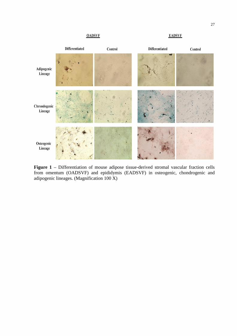
Differentiation potential of ASVFs
After the differentiation period, both adipose tissues were able to differentiate into all
three linages. However, histological staining showed that osteogenic and chondrogenic
differentiation occurred earlier in OASVF; after 21 days of differentiation the cultures stained
positive (Figure 1). For EASVF, 26 days of differentiation were needed to obtain consistent
positive staining (Figure 1). Cells derived from stromal fractions and maintained in
osteogenic medium formed aggregates or nodules, and these bone nodules stained positive
with alizarin red (Figure 1). With chondrogenic differentiation, cells developed a multilayered
matrix that was strongly stained with alcian blue, indicating an abundance of
glycosaminoglycans in the extracellular matrix (Figure 1). EASFV cultures showed several
areas that were more strongly stained with alizarin red, and alcian blue than OASVF,
indicating that epididymis adipose-derived cells more effectively differentiated into
osteogenic and chondrogenic lineages (Figure1). In adipogenic differentiation, both adipose
tissue cultures needed 17 days to differentiate and showed an accumulation of lipid-rich
vacuoles within cells. These vacuoles were positively stained by oil red staining (Figure 1).
Expression profiles of stem cell surface markers in ASVFs
This study analyzed two sources of ASVF cells, based on the expression of surface
antigens: CD45, a hematopoietic linage marker, CD105 and CD73, surface proteins expressed
in mesenchymal stem cell (MSC). Flow cytometric analysis (FACS) showed an increase in
CD105+ and CD73
+ single-positive cells and, a decrease in double-positive CD73
+/CD105
+
cells in P2, P4, and P7 OASVF (Figure 2 and Table 1). Conversely, EASVF showed a
23
tendency toward an increase in the double-positive CD73+/CD105
+ cells, as well as an
increase in the MSC CD105+
cells in the same passages mentioned above (Figure 2 and
Table 2). In both ASVFs, cells expressed insignificant levels of CD45 (Table 1 and 2), and, in
EASVF, the levels of CD105+ significantly increased with higher passages (Table 2).
Discussion
Depending on the fat location, adipose tissue-derived cells have unique characteristics
related to metabolic properties, functions, gene expression profiles, and differentiation
potential (SCHÄFFLER & BÜCHLER, 2007; TCHKONIA et al., 2002). We analyzed
ASFVs from two different fat pads in mice, the omentum and epididymis, and although the
mesenchymal surface markers were expressed in cells from both sources, their levels differed
between cells from the two adipose sources and among passages. In OASFV, there was a
decrease in double-positive CD105+/CD73
+ cells, while in EASVF there was an increase in
cells expressing the double-positive mesenchymal surface markers in P2, P4, and P7. Flow
cytometry showed that a higher number of passages was needed to achieve significative
CD105+ MSC populations in EASVF cultures, probably because selection and expansion of
AT-MSCs occurred with each passage. Previous immunophenotyping study of MSCs
revealed heterogeneity at early stages within cell populations before an eventual
subpopulation was selected through extensive cultivation (DA SILVA et al., 2006). Taken
together, the results indicated that adipose tissues isolated from mouse omentum and
epididymis have different subsets of MSC, suggesting that they are sources of different
somatic stem cells.
ASFV immunophenotype results presented in this study are in accordance with results
of other studies that relate the variable expression of surface markers to differences in tissue
sources, the method of isolation and culture, and species differences (CHAMBERLAIN et al.,
2007, DA SILVA et al., 2006; PRUNET-MARCUSSUS et al., 2006; SCHÄFFLER &
BÜCHLER, 2007). Interestingly, previous studies have demonstrated that the expression of
surface antigens, CDs, also show significant variation during subculture. For example, studies
comparing freshly isolated human adipose-derived cells and serially passaged AT-MSCs
showed a progressive increase in mesenchymal markers like CD90, CD73, and CD29
(MOSNA et al., 2010). Similar increases occurred in the present study in the expression of
MSC surface markers CD105 and CD73 in EASFV at P2, P4, and P7. However, decreases in
surface markers have also been observed. da Silva et al. (2006) reported that there was a
24
tendency toward a decrease in CD117, a stem cell factor receptor, during serial passage of
MSCs. Taken together these results illustrate that stem cell surface antigen expression profiles
vary with different cell sources and passages.
Although ASVF from both the omentum and epididymis showed the potential to
differentiate into adipogenic, osteogenic, and chondrogenic lineages, OASVF differentiated
earlier into chondrogenic and osteogenic linages (at 21 days of culture) than the epididymis
which required 26 days. The differentiation shown in the present study suggests that mouse
epididymis adipose-derived cells have greater potential to differentiate into osteogenic and
chondrogenic lineages than omentum adipose-derived cells. These results are in agreement
with a previous study that showed, in a murine system, evidence that the differentiation
capacity of the SVF is heterogeneous, and varies according to the localization of the adipose
tissue (SCHÄFFLER & BÜCHLER, 2007). In rabbits, the osteogenic potential of adipose-
derived stem cells (ADSC) isolated from visceral adipose tissue was reported to show greater
potential for differentiation compared with ADSCs isolated from subcutaneous adipose tissue
(SCHÄFFLER & BÜCHLER, 2007). Prunet-Marcussus et al. (2006) showed that mouse
inguinal adipose-derived cells showed a greater capacity to differentiate into osteogenic
lineage than did cells from an epididymis adipose fat source.
Since different fat pads have their own metabolic characteristics, fatty acid
compositions, and gene expressions (SCHÄFFLER & BÜCHLER, 2007; TCHKONIA et al.,
2002), the source of adipose tissue might be expected to influence characteristics of AT-
MSCs, such as surface markers and differentiation potential, as shown here. Therefore,
additional studies are necessary to properly understand the cellular composition and molecular
characteristics, as well as the plasticity, of SVF cells isolated from different fat depots.
Conclusion
This study demonstrated that omentum and epididymis fat pads represent different
sources of adipose-derived mesenchymal stem cells, which exhibit diverse surface marker
expression profiles when maintained in culture, as well as, distinct differentiation potentials
into osteogenic, chondrogenic lineages. Therefore, different adipose tissue sources provide
different stromal multipotent stem cells.
25
References
ATZMON, G. et al. Differential gene expression between visceral and subcutaneous fat
depots. Horm Metab Res, v. 34, p. 622-628, 2002.
BUNNELL, B. A. et al. Adipose-derived stem cells: Isolation, expansion and differentiation.
Methods, v. 45, p. 115-120, 2008.
CHAMBERLAIN, G. et al. Concise review: Mesenchymal stem cells: their phenotype,
differentiation capacity, immunological features, and potential for homing. Stem Cells, v. 25,
p. 2739-2749, 2007.
DA SILVA, L. M. et al. Mesenchymal stem cells reside in virtually all post-natal organs
tissues. J Cell Science, v. 119, n. 11, p. 2204-2213. 2006.
FRASER, J. K. et al. Differences in stem and progenitor cell yield in different subcutaneous
adipose tissue depots. Cytotherapy, n. 9, p. 459-467, 2007.
GIMBLE, J.M. et al. Adipose-derived stem cells for regenerative medicine. Circ Res, n. 100,
p. 1249-1260, 2007.
KARP, J. M. & TEO, G. S. L. Mesenchymal stem cell homing: The devil is in the details.
Cell Stem Cell, v. 4, p. 206-216. 2009.
KIM, S.; MOUSTAID-MOUSSA, N. Secretory, endocrine and autocrine/paracrine function
of the adipocyte. J Nutr, n. 130, p. 3110-3115, 2000.
MAJKA, S. et al. Concise review: Adipocyte origins: weighing the possibilities. Stem Cells,
n. 29, p. 1034-1040, 2011.
MARCUS, A. J. et al. Isolation characterization, and differentiation of stem cells derived
from rat amniotic membrane. Differentiation, v. 76, p. 130-144, 2008.
MITCHELL, J. B. et al. Immunophenotype of human adipose-derived cells: temporal changes
in stromal-associated and stem cell associated markers. Stem Cells, v. 24, p. 376-385, 2006.
MOSNA, F. et al. Human bone marrow and adipose tissue mesenchymal stem cells: a user's
guide. Stem Cells Dev, v. 19, p. 1449-1470, 2010.
PARK, H. T. et al. The relationship between fat depot-specific preadipocyte differentiation
and metabolic syndrome in obese women. Clin Endocrinol (Oxf), v. 76, p. 59-66, 2012.
PRUNET-MARCASSUS, B. et al. From heterogeneity to plasticity in adipose tissues: site-
specific differences. Exp Cell Res, v. 312, p. 727-736, 2006.
SCHÄFFLER A.; BÜCHLER, C. Concise review: Adipose tissue-derived stromal cells-basic
and clinical implications for novel cell-based therapies. Stem Cells, n. 25, p. 818-827, 2007.
STREM, B. M. et al. Multipotential differentiation of adipose tissue derived stem cells. Keio
J Med, v. 54, n. 3, p. 132-141, 2005.

26
TCHKONIA, T. et al. Fat depot origin affects adipogenesis in primary cultured and cloned
human preadipocytes. Am J Physiol Regul Integr Comp Physiol, v. 282, p. 1286-1296,
2002.
ZHU, Y. et al. Adipose-derived stem cell: a better stem cell than BMSC. Cell Bioch and
Funct, v. 26, p. 664-675, 2008.
ZUK, P. A. et al. Human adipose tissue is a source of multipotent stem cells. Mol Biol Cell,
v. 13, p. 4279-4295, 2002.
27
Figure 1 – Differentiation of mouse adipose tissue-derived stromal vascular fraction cells
from omentum (OADSVF) and epididymis (EADSVF) in osteogenic, chondrogenic and
adipogenic lineages. (Magnification 100 X)
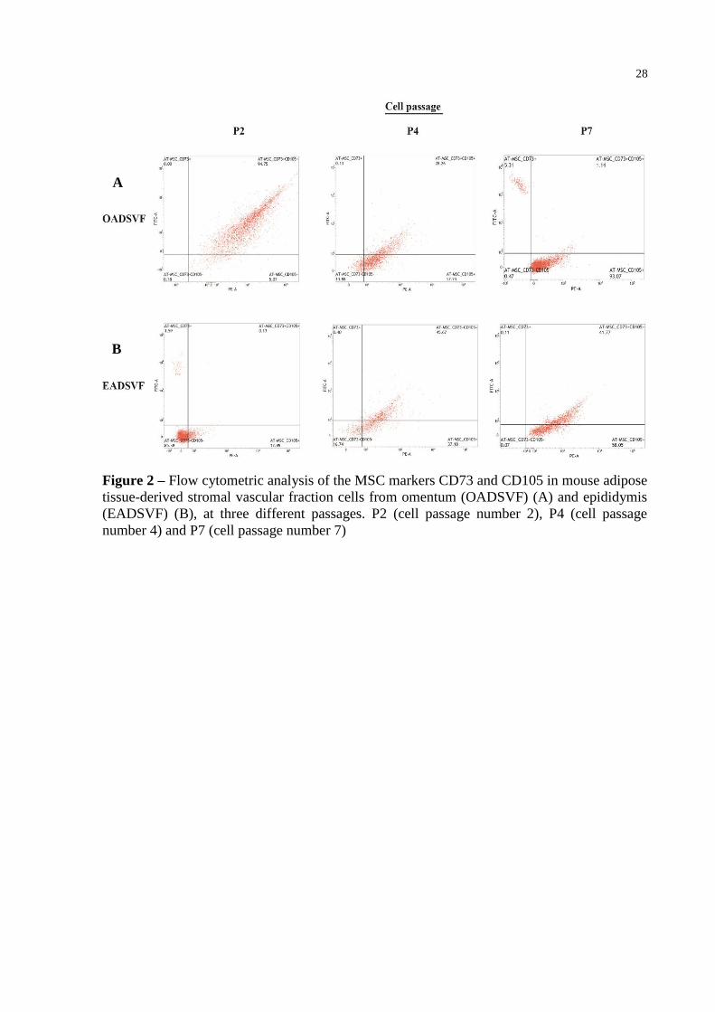
28
Figure 2 – Flow cytometric analysis of the MSC markers CD73 and CD105 in mouse adipose
tissue-derived stromal vascular fraction cells from omentum (OADSVF) (A) and epididymis
(EADSVF) (B), at three different passages. P2 (cell passage number 2), P4 (cell passage
number 4) and P7 (cell passage number 7)
A
B
29
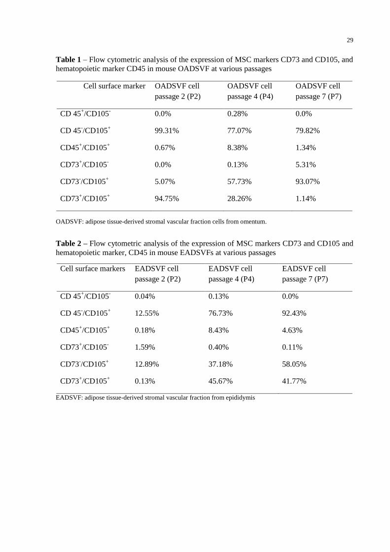
Table 1 – Flow cytometric analysis of the expression of MSC markers CD73 and CD105, and
hematopoietic marker CD45 in mouse OADSVF at various passages
Cell surface marker OADSVF cell
passage 2 (P2)
OADSVF cell
passage 4 (P4)
OADSVF cell
passage 7 (P7)
CD 45+/CD105
- 0.0% 0.28% 0.0%
CD 45-/CD105
+ 99.31% 77.07% 79.82%
CD45+/CD105
+ 0.67% 8.38% 1.34%
CD73+/CD105
- 0.0% 0.13% 5.31%
CD73-/CD105
+ 5.07% 57.73% 93.07%
CD73+/CD105
+ 94.75% 28.26% 1.14%
OADSVF: adipose tissue-derived stromal vascular fraction cells from omentum.
Table 2 – Flow cytometric analysis of the expression of MSC markers CD73 and CD105 and
hematopoietic marker, CD45 in mouse EADSVFs at various passages
Cell surface markers EADSVF cell
passage 2 (P2)
EADSVF cell
passage 4 (P4)
EADSVF cell
passage 7 (P7)
CD 45+/CD105
- 0.04% 0.13% 0.0%
CD 45-/CD105
+ 12.55% 76.73% 92.43%
CD45+/CD105
+ 0.18% 8.43% 4.63%
CD73+/CD105
- 1.59% 0.40% 0.11%
CD73-/CD105
+ 12.89% 37.18% 58.05%
CD73+/CD105
+ 0.13% 45.67% 41.77%
EADSVF: adipose tissue-derived stromal vascular fraction from epididymis
30
4 CAPÍTULO II – CHARACTERIZATION OF ADIPOSE TISSUE-DERIVED
MESENCHYMAL STEM CELLS AND THEIR DIFFERENTIATION POTENTIAL
INTO MALE GERM CELLS AFTER TREATMENTS WITH RETINOIC ACID AND
CONDITIONED MEDIUM
Braunig, P; Glanzner, W. G.; Rissi, V. B.; Gonçalves, P. B. D.
Abstract
Mesenchymal stem cells are more plastic than expected when cultured under certain
conditions and the adipose tissue is a reliable source of mesenchymal stem cells that can
transdifferentiate into multilineage cells. In the present study, adipose tissue-derived
mesenchymal stem cells (AT-MSC) were isolated from omentum and epididymis fat depots,
and, previously characterized based on stem cell surface markers and on the mesodermal
trilineage differentiation potential. Additionally, both AT-MSCs were cultured with
differentiation media containing retinoic acid (RA) and/or testicular cell-conditioned medium
(TCC). The AT-MSCs expressed mesenchymal surface markers CD73 and CD105 and
differentiated into adipogenic, chondrogenic and osteogenic lineages. After the differentiation
treatments, omentum-derived AT-MSCs expressed a gene marker related to male germ cell
lineages. These findings reaffirm the importance of adipose tissue as a source of multipotent
stromal-stem cells for differentiation researches.
Keywords: Gdnf expression. In vitro differentiation. Mesenchymal stem cells. Retinol-
derivative. Testicular cell-conditioned medium.
Introduction
Adipose tissue-derived mesenchymal stem cells (AT-MSCs) are multipotent cells
which proliferate, in vitro, for indefinite period and can be induced to differentiate into all
three germ layers, which develop into bone, cartilage, fat, muscle, heart, neural, and other
cells (ZUK et al., 2002). In addition, adipose tissue is abundant and accessible, representing a
reliable source of stem cells which exist in significant numbers in this tissue (FRASER et al.,
2007; GIMBLE et al., 2007; MITCHELL et al., 2006).
31
An area of biological research that generates great optimism is the use of stem cells for
the treatment of diseases. Much of the excitement centers on embryonic stem cells (ES),
however, this approach remains controversial for ethical reasons and also due the risk of
teratomas. Moreover, realization of this strategy as routine clinical application requires
extensive research (NAYERNIA et al., 2006; YOUNG et al., 2004). In contrast, AT-MSCs
from adult adipose tissues are well characterized, easy to isolate, and have recently been used
for therapeutic applications (FRASER et al., 2007; GIMBLE et al., 2007; MITCHELL et al.,
2006).
In vitro differentiation of AT-MSCs has demonstrated promising plasticity (ZUK
et al., 2002). Recent studies have revealed several substances that can act as differentiation
inducers, and generally, they are used in association with one another (MOSNA et al., 2010;
SCHÄFFLER & BÜCHLER, 2007). Retinoic acid (RA) is a vitamin A (retinol)–derivative
that is widely used as a differentiation inducer. In some studies, RA has been added to culture
medium for differentiation of mesenchymal stem cells (MSCs) (HUANG et al., 2010;
NAYERNIA et al., 2006) and skin-derived stem cells (TAN et al., 2016) into germ cells.
Furthermore, the conditioned medium obtained from the cell culture supernatants is applied in
association with chemical inducers to improve differentiation. Conditioned medium has been
employed for chondrogenic, myogenic, neural, and germ cell differentiation of bone marrow-
and adipose tissue-derived MSCs (DA SILVA et al., 2013; HAN et al., 2014;
STERN-STRAETER et al., 2013).
Several methods have been used to investigate in vitro and in vivo MSCs
differentiation. Analyses of marker gene transcripts allow confirmation of AT-MSC
differentiation and facilitate the selection of efficient inducers (HOU et al., 2014; PHINNEY
& PROCKOP, 2007; SCHÄFFLER & BÜCHLER, 2007). The genes commonly investigated
to confirm MSCs differentiation into male germ cell are Vasa, Stella, Dazl, Stra8, Nanos2,
Plzf, and Gdnf which is used as a marker for Sertoli cells (CHEN & LIU, 2015; HOU et al.,
2014 IKAMI et al., 2015).
Taking these data into account, the main aim of the present study was to evaluate the
expression of relevant gene markers of germinative cells in omentum and epididymis -derived
AT-MSCs, after treatments with RA and testicular cell-conditioned medium.
32
Material and Methods
Isolation and culture of AT-MSCs
Adipose tissue from 8 to 12 week old BALB/c mice, was collected from the omentum
and epididymis regions. The tissue was, separately, minced and digested in collagenase
solution (1 mg/mL) for 30 min at 37 ° C. After digestion, the collagenase solution was
inactivated with growth medium (Dulbecco's Modified Eagle Medium-F12 + 10% fetal
bovine serum + 100μg (100 IU) of penicillin and, streptomycin + 0,25μg of amphotericin B)
and the cells were centrifuged (200 xg, 10min) to obtain a pellet. The resulting pellet was
resuspended in growth medium, and the cell suspension was cultured in cell culture plates in a
5% CO2 incubator at 37 ° C. The cells were maintained in growth medium and passaged using
trypsin solution (0.25%) once they achieved 70-80% confluency.
Testicular cell-conditioned medium preparation
Testicles from 8 to 12 week old BALB/c mice were removed, minced, and
enzymatically digested in collagenase solution (1 mg/mL) for 30 minutes at 37° C. Tissue
digestion was terminated with addition of growth medium for collagenase inactivation, and
then the tissue homogenate was centrifuged for 10 min at 200 xg and the resulting pellet was
resuspended in growth medium. The testicular cells were cultured in 5% CO2 at 37 ° C and
passaged using trypsin solution (0.25%) once they reached 80-90% confluency. Seven days
after cultures were started, and every 3 days afterward for 30 days, testicular cell-conditioned
medium (TCC) was collected. After collection, the TCC was centrifuged (200 xg, 10 min),
and the supernatant was filtered (0.22 µm) and stored at - 20°C until use.
Differentiation treatments of AT-MSCs
Cultures of AT-MSCs derived from the omentum and epididymis fat pads were
induced by differentiation medium containing growth medium supplemented with 10-6
M RA
(Sigma), 50% TCC + 50% growth medium, or RA + TCC at 7, 14, and 21 days. Adipose
tissue-derived cells maintained with only growth medium were used as a control group. The
media were changed every 3 days.
33
Adipogenic, osteogenic and chondrogenic induction of AT-MSCs
Omentum and epididymis-derived AT-MSCs were cultured in osteogenic medium (50
µM L-ascorbic acid 2-phosphate, 0.1 µM dexametasone, and 15 mM β-glycerolphosphate)
and chondrogenic medium (50 µM dexametasone, 50 µM L-ascorbic acid 2-phosphate, 10
ng/mL TGF-β, and 1x insulin-transferrin-sodium selenite) for 21 days, and with adipogenic
medium (50 µM indomethacin, 1 µM rosiglitazone, 1µM dexamethasone, and 1µg/mL
insulin) for 17 days. The media were changed every 3 days and, histological staining was
performed after the differentiation period. Cells induced with osteogenic, adipogenic, and
chondrogenic media were first fixed with paraformaldehyde 4% and then stained with alizarin
red (pH 4.1), oil red, and alcian blue (pH 2.5), respectively, and observed using an inverted
microscope (Leica DMI600B).
Flow cytometry analysis
AT-MSCs from omentum and epididymis fat depots were trypsinized, centrifuged, and
then 4 x 10⁴cells were suspended in Stain Buffer (BD Biosciences) in separate microtubes.
Samples were then incubated with the antibodies CD45-FITC (1.5µg/µL), CD73-FITC
(1µg/µL), and CD105-PE (1µg/µL) (BD Biosciences) in the dark for 20 min at 37° C. After
incubation, cells were analyzed by flow cytometry using the BD FACSuite (BD Biosciences).
RNA isolation, reverse transcription, and real-time PCR
After each differentiation induction period (7, 14, and 21 days), total RNA was
isolated from the cell cultures. RNA extraction was performed using Trizol reagent
(Invitrogen) according to the manufacturer,
s instructions. After extraction, RNA
concentration and quality were checked by a NanoDrop1000 spectrophotometer (Absorbance
of 260/280 nm) (Thermo Scientific). Complementary DNA (cDNA) was synthesized from
1000 ng of RNA, which was first treated with 0.1 U of DNase Amplification Grade (Life
Techonologies) for 5 min at 37° C. After DNase inactivation at 65 °C for 10 min, samples
were incubated in 20µLwith reagents from an iScript cDNA synthesis Kit (BioRad). cDNA
synthesis was performed in three steps: 25 °C for 5 min, 42 °C for 30 min, and 85 °C for 5
min.
The relative expression levels of specific genes were determined by quantitative PCR
(qPCR) conducted in a CFX384 thermocycler (BioRad) using GoTaq qPCR Master Mix
34
(Promega). Expression of the murine Vasa, Stella, Dazl, Stra8, Nanos2, Plzf and Gdnf genes
were analyzed, in addition the murine housekeeping genes Gapdh and β-Actin. All primers
were designed based on gene sequences deposited in the GenBank database using Primer
Express Software (Applied Biosystems). Table 1 shows the genes and primer sequences used
for the qPCR analysis.
Standard two-step qPCR was performed in a 10 µL final volume containing 2 µL of
cDNA, 2x Master Mix (Promega) and 5 µM each primer. Amplification conditions were 95
°C for 3 min, followed by 39 cycles of 95 °C for 10 s and 60 °C for 1 min. Melting-curve
analyses were performed to verify product identity. To optimize the qPCR assay, serial
dilutions of the cDNA templates were used to generate a standard curve. The standard curve
was constructed by plotting the log of the dilution factor against the Ct value obtained during
amplification of each dilution. Reactions with a coefficient of determination (R2) higher than
0.98 and efficiency between 95% and 105% were considered optimized. The relative standard
curve method was used to quantify transcripts in each sample (CIKOS et al., 2007). Samples
were run in duplicate, and results were expressed relative to the average Ct values for the
Gapdh, β-Actin genes as internal controls. Samples of mRNA extracted from mouse testicles
were used as a positive control for validating primers and amplicons.
Animals
All procedures using BALB/c mice in the present study were approved by the
Institutional Committee for Ethics in Animal Experiments at the Federal University of Santa
Maria, RS, Brazil, approval number 087/2014.
Results
AT-MSCs characterization
Flow cytometric analysis demonstrated that omentum-derived AT-MSCs
predominantly expressed the surface marker CD105 and showed low levels for CD73 and
double-positive CD105/CD73 cells (Figure 1); CD105 and CD103 are typically found on
MSCs surface. Epididymis-derived AT-MSCs showed prevalent population of double-
positive CD73/CD105 cells, as well as CD105 cells
.Omentum and epididimis-derived AT-
MSCs lacked CD45 surface expression, which is seen in the hematopoietic lineage (Figure 1).
35
Differentiation protocols were used to induce AT-MSCs from both mouse fat depots in
bone, cartilage, and fat to further confirm their trilineage differentiation capacity. After
culture of AT-MSCs in osteogenic medium, the cells differentiated into osteoblasts, with
calcium accumulation indicated by positive staining with alzarin red (Figure 2). AT-MSCs
maintained in adipogenic medium showed the presence of intracellular lipid droplets,
confirmed by oil red staining (Figure 2). Chondrogenic differentiation was indicated by alcian
blue staining of abundant glycosaminoglycans within the extracellular matrix (Figure 2). The
AT-MSCs grown in culture medium (undifferentiated) did not show any lipid droplets and
maintained their typical fibroblast-like shape (Figure 2).
Evaluation of gene expression in treated AT-MSCs
A qPCR assay was performed to determine the gene expression levels. Among the
genes and sources of AT-MSCs tested, only Gdnf was expressed just in omentum-derived
AT-MSCs treated with RA differentiation media after incubation periods of, 7, 14, and 21
days (Figure 3). Conversely, no significant expression was detected in AT-MSCs maintained
in TCC medium for any period of treatment (Figure 3). Gdnf gene expression was not
detected in untreated AT-MSCs (Figure 3).
Discussion
AT-MSCs are stromal stem cells that can differentiate into all three germ layers under
suitable conditions and recent studies have shown that somatic adult stem cells are more
plastic than previous expected (PHINNEY & PROCKOP, 2007; ZUK et al., 2002). Therefore,
in the present study, the surface antigen expression of adipose tissue-derived cells indicated
that MSCs represent a significant population among the cells isolated from both, omentum
and epididymis, fat sources. Additionally, adipose tissue-derived cells from both the omentum
and epididymis showed the potential to differentiate into the three mesodermal, adipogenic,
osteogenic, and, chondrogenic, lineages.
The omentum-derived AT-MSCs expressed high levels of CD73 and CD105, which
are MSCs markers, and were negative for CD45 surface antigen, a hematopoietic lineage
marker. In addition, the effective differentiation of omentum-derived AT-MSCs into all three
lineages (adipogenic, osteogenic, and chondrogenic) confirmed the presence of AT-MSCs and
their differentiation potential.
36
The qPCR results suggest that RA and RA+TCC stimulated Gdnf expression in
omentum-derived AT-MSCs. Conversely, AT-MSCs maintained in TCC did not express the
target gene, similar to untreated cells. Taken together, these results indicate that RA was the
main factor involved in Gdnf expression.
RA has been used in association with other substances to induce neuronal
differentiation from cultured mouse AT-MSCs (BI et al., 2010; PAVLOVA et al., 2012).
However, RA alone has usually been used to promote the differentiation of embryonic stem
cells (ESCs) and MSCs into germ cells (DRUSENHEIMER et al., 2007; GEIJSEN et al.,
2004; KERKIS et al., 2007; NAYERNIA et al., 2006). An active derivative of vitamin A, RA
influences germ cell differentiation and is required for the transition to meiosis in both female
and male germ cells (KOUBOVA et al., 2006). Retinoids are involved in the regulation of
testicular functions, which appear to be necessary for spermatogenesis (LIVERA et al., 2002).
RA receptors (RARs) are expressed in both Sertoli and germ cells (ESKLID et al., 1991), and
RA functions inside the nucleus, recognizing two different classes of RARs. Both classes
(RARs and RXRs) consist of three types of receptors, α, β, and γ, encoded by distinct genes,
and they transduce RA signals by binding directly to RA-responsive elements (ROSSI &
DOLCI, 2013). Previous studies have indicated that RA favors spermatogonial differentiation
through direct action on spermatogonia and indirect action mediated by changes in the
expression of GDNF secreted by Sertoli cells (ROSSI & DOLCI, 2013).
The Gdnf gene is mainly expressed in Sertoli and neuronal cells and is considered a
glial marker that characterizes neuronal differentiation of stem cells (BI et al., 2010;
SHAKHBAZOV et al., 2009). Additionally, GDNF is a paracrine soluble factor secreted by
Sertoli cells in the testicular niche that influences the self-renewal of spermatogonial stem
cells (SSCs) and inhibit their differentiation (CHEN & LIU, 2015; IKAMI et al., 2015;
ROSSI & DOLCI, 2013). Therefore, based on the effect of RA on Gdnf gene expression
showed in this study, it seems that omentum AT-MSCs contain RA receptors, and that they
respond to RA stimulation, but further studies are necessary to properly characterize the
RARs in AT-MSCs.
The testes are abundant sources of numerous hormones and growth factors, such as
bone morphogenic protein 4 (BMP4), leukemia inhibitory factor (LIF), basic fibroblast
growth factor (bFGF), stem cell factor (SCF), growth differentiation factor-9 (GDF9), and
testosterone, all of which are needed for the development of male germ cells (CREEMERS
et al., 2002; PELLEGRINI et al., 2003; TAKABAYASHI et al., 2001). Recent studies have
shown that TCC supports the differentiation of ESCs into germ cells (LACHAM-KAPLAN
37
et al., 2006). However, in the present study, TCC was not effective promoting gene
expression maybe, due the inappropriate concentration of the soluble factors. Therefore, it
seems that TCC might be used in combination with other induction factors and might be
concentrated to improve the efficiency of differentiation.
MSCs are considered stromal adult stem cells and their potential to differentiate into
stromal lineages has been demonstrated. However, there are not studies considering the AT-
MSCs differentiation into Sertoli cells which are stromal cells that support spermatogenesis in
adult males (SCHÄFFLER & BÜCHLER, 2007; ZUK et al., 2002). Although previous
studies have shown that MSCs maintained in media containing RA differentiate into male
germ cells (DRUSENHEIMER et al., 2007; NAYERNIA et al., 2006; ZHANG et al., 2014)
and that TCC enhances this differentiation (HUANG et al., 2010; KAVIANI et al., 2014)
additional gene expression and other analyses should be performed to confirm whether the
AT-MSCs treated with RA, in the present study, can differentiate into cell lineages related to
male germ cells.
Conclusion
This study demonstrated that mouse adipose tissue-derived cells contain a significant
AT-MSC population. In addition, omental AT-MSCs express Gdnf, an important marker of
male germ cells, after treatment with RA. This observation indicates that omental-derived
AT-MSCs respond to differentiation treatments therefore, they serve as a suitable source of
multipotent stem cells for further differentiation studies.
References
BI, Y. et al. Pre-activation of retinoid signaling facilitates neuronal differentiation of
mesenchymal stem cells. Develop Growth & Differ, v. 52, p. 419-431, 2010.
CHEN, S. R. & LIU, Y. X. Regulation of spermatogonial stem cell self-renewal and
spermatocyte meiosis by Sertoli cell signaling. Reproduction, v. 149, p. 159-167, 2015.
CIKOS, S. et al. Relative quantification of mRNA: comparison of methods currently used for
real-time PCR data analysis. BMC Mol Biol, v. 8, n. 113, p. 1-14, 2007
CREEMERS, L. B. et al. Maintenance of adult mouse type A spermatogonia in vitro:
influence of serum and growth factors and comparison with prepubertal spermatogonial cell
culture. Reproduction, v. 124, p. 791-799, 2002.
38
DA SILVA, M. L. A. et al. Conditioned medium as a strategy for human stem cells
chondrogenic differentiation. J Tissue Eng Regen Med, n. 2, v. 8, p. 714-723, 2013.
DRUSENHEIMER, N. et al. Putative human male germ cells from bone marrow stem cells.
Soc Reprod Fertil Suppl., v. 63, p. 69-76, 2007.
ESKILD, W. et al. Cellular localization of mRNAs for retinoic acid receptor-alpha, cellular
retinol-binding protein, and cellular retinoic acid-binding protein in rat testis: evidence for
germ cell-specific mRNAs. Biol Reprod, v. 44, p. 53-61, 1991.
FRASER, J. K. et al. Differences in stem and progenitor cell yield in different subcutaneous
adipose tissue depots. Cytotherapy, n. 9, p. 459-467, 2007.
GEIJSEN, N. et al. Derivation of embryonic germ cells and male gametes from embryonic
stem cells. Nature, v. 427, n. 6970, p. 148-154, 2004.
GIMBLE, J. M. et al. Adipose-derived stem cells for regenerative medicine. Circ Res, n. 100,
p. 1249-60, 2007.
HAN, C. et al. Rat cortex and hippocampus-derived soluble factors for the induction of
adipose-derived mesenchymal stem cells into neuron-like cells. Cell Bio Int, v. 38, p. 768-
776, 2014.
HOU, J. et al. Generation of male differentiated germ cells from various types of stem cells.
Reproduction, n. 147, p. 179-188, 2014.
HUANG, P. et al. Differentiation of human umbilical cord Wharton´s jelly-derived
mesenchymal stem cell into germ-like cells in vitro. J Cell Biochem, v. 109, p. 747-754,
2010.
IKAMI, K. et al. Hierarchical differentiation competence in response to retinoic acid ensures
stem cell maintenance during mouse spermatogenesis. Develop Stem cells and Regen,
v. 142, p. 1582-1592, 2015.
KAVIANI, M. et al. Evalution of gametogenic potential of vitrified human umbilical cord
Wharton`s jelly-derived mesenchymal cells. Cytotherapy, v. 16, p. 203-212, 2014.
KERKIS, A. et al. In vitro differentiation of male mouse embryonic stem cells into both
presumptive sperm cells and oocytes. Cloning stem cells, v. 9, p. 535-548, 2007.
KOUBOVA, J. et al. Retinoic acid regulates sex-specific timing of meiotic initiation in mice.
Proc Natl Acad Sci USA, v. 103, p. 2474-2479, 2006.
LACHAM-KAPLAN, O. et al. Testicular cell conditioned medium supports differentiation of
embryonic stem cells into ovarian structures containing oocytes. Stem cells, v. 24, p. 266-
273, 2006.
LIVERA, G. et al. Regulation and perturbation of testicular functions by vitamin A.
Reproduction, v. 124, p. 173-180, 2002.
MITCHELL, J. B. et al. Immunophenotype of human adipose-derived cells: temporal changes
in stromal-associated and stem cell associated markers. Stem Cells, v. 24, p. 376-385, 2006.
39
MOSNA, F. et al. Human bone marrow and adipose tissue mesenchymal stem cells: a user's
guide. Stem Cells Dev, v. 19, p. 1449-1470, 2010.
NAYERNIA, K. et al. In vitro differentiated embryonic stem cells give rise to male gametes
that can generate offspring in mice. Dev Cell, v. 11, n. 2, p. 125-132, 2006.
PAVLOVA, G. et al. In vitro neuronal induction of adipose-derived stem cells and their fate
after transplantation into injured mouse brain. Curr Medic Chem v. 19, p. 5170-5177, 2012.
PELLEGRINI, M. et al. Developmental expression of BMP4/ALK3/SMAD5 signaling
pathway in the mouse testis: A potential role of BMP4 in spermatogonia differentiation.
J Cell Sci, n. 1, v. 16, p. 3363-3372, 2003.
PFAFFL, M. W. A new mathematical model for relative quantification in real time RT-PCR.
Nucleic Acids Res, v. 29, n. 9, p. 45, 2001.
PHINNEY, D. & PROCKOP, D. J. Concise review: Mesenchymal stem/multipotent stromal
cells: The state of transdifferentiation and modes of tissue repair-current views. Stem Cells,
v. 25, p. 2896-2902, 2007.
ROSSI, P. & DOLCI, S. Paracrine mechanisms involved in the control of early stages of
mammalian spermatogenesis. Front Endocrinol, v. 4, n. 181, p. 1-8, 2013.
SCHÄFFLER, A & BÜCHLER, C. Concise review: Adipose tissue-derived stromal cells -
basic and clinical implications for novel cell-based therapies. Stem Cells, n. 25, p. 818-827,
2007.
SHAKHBAZOV, A. V. et al. Plasticity of human mesenchymal stem cell phenotype and
expression profile under neurogenic conditions. Bull. Exp Biol Med, v. 147, p. 513-516,
2009.
STERN-STRAETER, J. et al. Evaluation of the effects of different culture media on the
myogenic differentiation potential of adipose tissue or bone marrow-derived human
mesenchymal stem cells. Int J Mol Med, n. 1, v. 33, p. 160-170, 2013.
TAKABAYASHI, S. et al. Novel growth factor supporting survival of murine primordial
germ cells: evidence from conditioned medium of ter fetal gonodal somatic cells. Mol
Reprod Dev, v. 60, p. 384-396, 2001.
TAN, H. et al. Retinoic acid promotes the proliferation of primordial germ cell-like cells
differentiation from mouse skin-derived stem cells in vitro. Theriogenology, v. 85, p. 408-
418, 2016.
YOUNG, H. E. et al. Adult stem cells. Anat Rec, v. 276A, p. 75-102, 2004.
ZHANG, D. et al. Potential sprematogenesis recovery with bone marrow mesenchymal stem
cells in an azoospermic rat model. Int J Sci, v. 15, p. 13151-13165, 2014.
ZUK, P. A. et al. Human adipose tissue is a source of multipotent stem cells. Mol Biol Cell,
v. 13, p. 4279-4295, 2002.
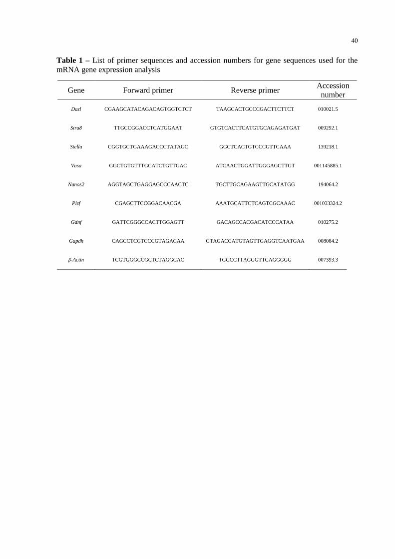
40
Table 1 – List of primer sequences and accession numbers for gene sequences used for the
mRNA gene expression analysis
Gene Forward primer Reverse primer Accession
number
Dazl CGAAGCATACAGACAGTGGTCTCT TAAGCACTGCCCGACTTCTTCT 010021.5
Stra8 TTGCCGGACCTCATGGAAT GTGTCACTTCATGTGCAGAGATGAT 009292.1
Stella CGGTGCTGAAAGACCCTATAGC GGCTCACTGTCCCGTTCAAA 139218.1
Vasa GGCTGTGTTTGCATCTGTTGAC ATCAACTGGATTGGGAGCTTGT 001145885.1
Nanos2 AGGTAGCTGAGGAGCCCAACTC TGCTTGCAGAAGTTGCATATGG 194064.2
Plzf CGAGCTTCCGGACAACGA AAATGCATTCTCAGTCGCAAAC 001033324.2
Gdnf GATTCGGGCCACTTGGAGTT GACAGCCACGACATCCCATAA 010275.2
Gapdh CAGCCTCGTCCCGTAGACAA GTAGACCATGTAGTTGAGGTCAATGAA 008084.2
β-Actin TCGTGGGCCGCTCTAGGCAC TGGCCTTAGGGTTCAGGGGG 007393.3
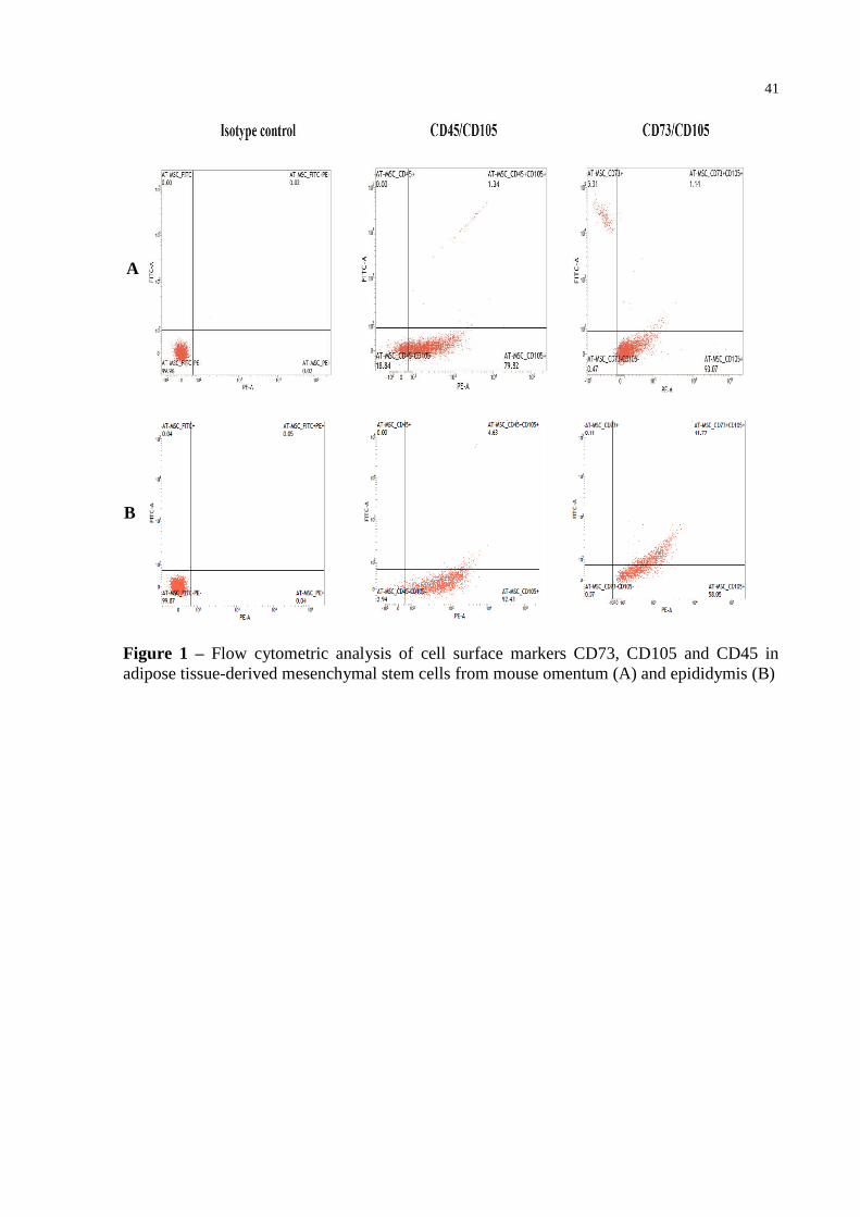
41
Figure 1 – Flow cytometric analysis of cell surface markers CD73, CD105 and CD45 in
adipose tissue-derived mesenchymal stem cells from mouse omentum (A) and epididymis (B)
A
B
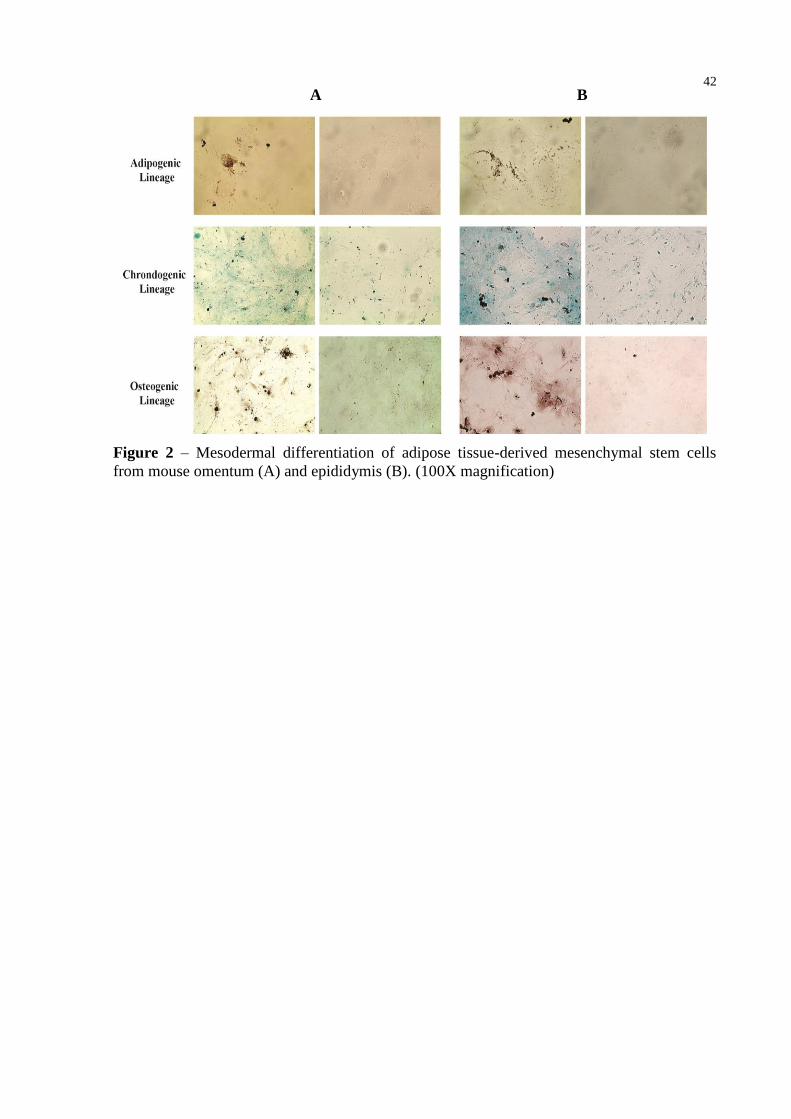
42
Figure 2 – Mesodermal differentiation of adipose tissue-derived mesenchymal stem cells
from mouse omentum (A) and epididymis (B). (100X magnification)
A B
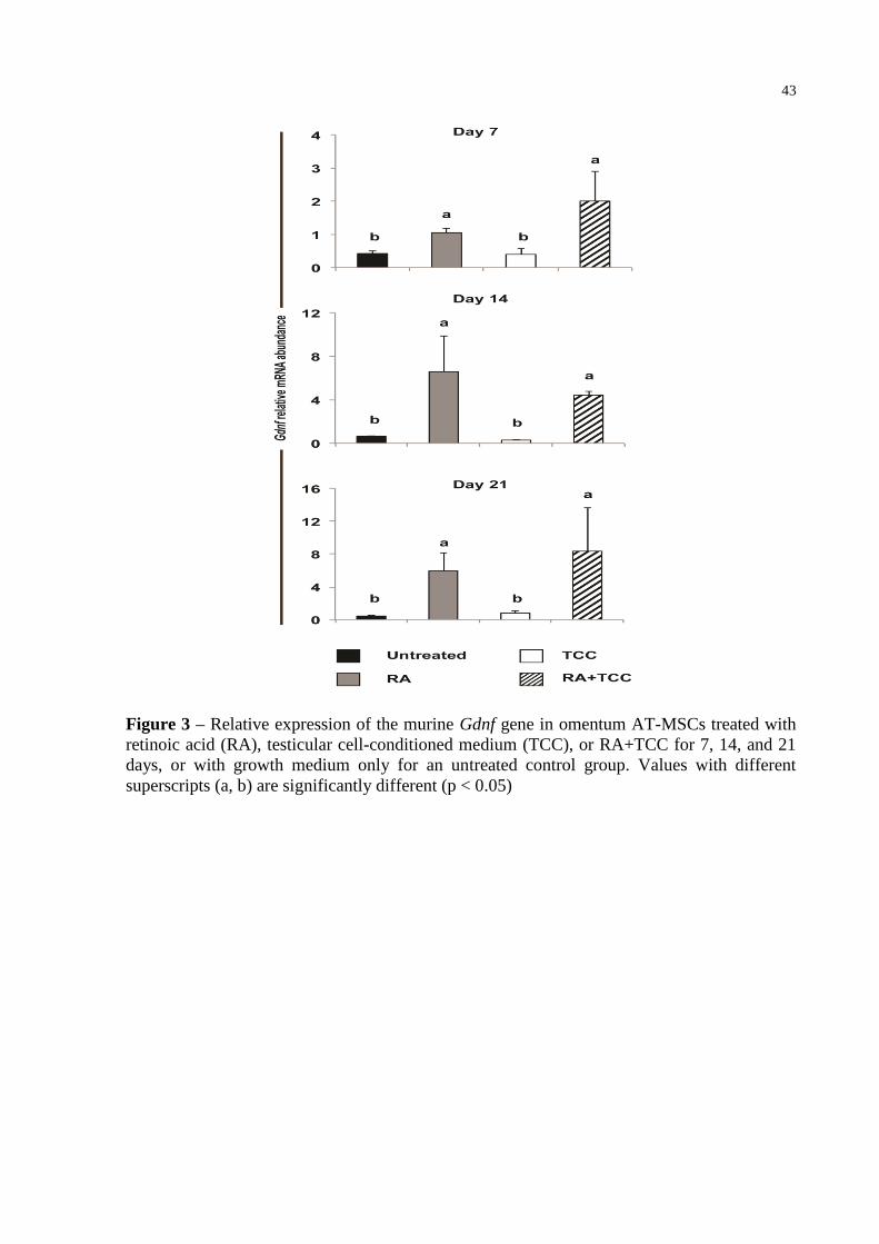
43
Figure 3 – Relative expression of the murine Gdnf gene in omentum AT-MSCs treated with
retinoic acid (RA), testicular cell-conditioned medium (TCC), or RA+TCC for 7, 14, and 21
days, or with growth medium only for an untreated control group. Values with different
superscripts (a, b) are significantly different (p < 0.05)
44
5 DISCUSSÃO
Com base nas promissoras características das células-tronco mesenquimais derivadas
do tecido adiposo (AT-MSCs) (SCHÄFFLER & BÜCHLER, 2007; ZUK et al., 2002) o
presente estudo pesquisou a presença, as características e o potencial de diferenciação das
AT-MSCs isoladas do tecido adiposo depositado em diferentes regiões do organismo de
camundongos BALB/c. Uma vez que o tecido adiposo encontra-se amplamente distribuído no
organismo dos mamíferos, estando localizado preferencialmente na região subcutânea e em
contato com os órgãos e gônadas (BUNNELL et al., 2008; STREM et al., 2005), nesse estudo
elegeu-se coletar o tecido adiposo da região do omento e testículos devido à facilidade de
obtenção, assim como devido à quantidade significativa de gordura depositada nessas regiões.
No primeiro estudo, foram isoladas e caracterizadas as AT-MSCs provenientes dos
dois diferentes depósitos de gordura acima citados e ambas as fontes demonstraram possuir
populações celulares que expressaram os marcadores de superfície mesenquimais CD73 e
CD105 e foram negativas para o marcador de linhagens hematopoiéticas CD45. Da mesma
forma, as células derivadas da fração estromal vascular de ambas as fontes, quando mantidas
em cultivo, demonstraram potencial de diferenciação condrogênica, osteogênica e
adipogênica. Esses resultados demonstraram que o tecido adiposo próximo às regiões do
omento e dos testículos possui células-tronco mesenquimais na sua composição. Porém, as
AT-MSCs isoladas das distintas fontes apresentaram diferentes padrões e níveis de expressão
dos marcadores de superfície mesenquimais em distintas passagens in vitro. Bem como, o
potencial de diferenciação condrogêncio e osteogêncio divergiu levemente entre as fontes,
demonstrando que apesar de ambos os tecidos adiposos possuírem AT-MSCs essas revelaram
diferentes características conforme sua origem. Os resultados obtidos neste estudo estão de
acordo com estudos prévios que também demonstraram variabilidade nos perfis de expressão
dos marcadores de superfície nas AT-MSCs conforme o número da passagem celular e a fonte
de tecido adiposo, assim como relataram diferença no potencial de diferenciação osteogênica
das AT-MSCs dependendo da origem do tecido adiposo (CHAMBERLAIN et al., 2007;
MOSNA et al., 2010; PRUNET-MARCUSSUS et al., 2006; SCHÄFFLER & BÜCHLER,
2007).
Uma vez cientes da presença e de algumas características das AT-MSCs reveladas no
primeiro estudo, verificou-se o possível potencial de diferenciação das AT-MSCs isoladas do
tecido adiposo da região do omento e testículos em linhagens celulares relacionadas às células
germinativas masculinas. Para isso, após os períodos de cultivo (7, 14 e 21 dias) com ácido
45
retinóico e/ou meio condicionado proveniente do sobrenadante do cultivo das células de
testículo, pesquisou-se a expressão de genes marcadores específicos relacionados às células
germinativas precoces, Vasa, Stella, Dazl, Stra8, Nanos2, Plzf, e à célula de Sertoli, Gdnf
(CHEN & LIU, 2015; HOU et al., 2014; IKAMI et al., 2015). As análises por meio da qPCR
revelaram que somente as AT-MSCs isoladas do tecido adiposo em contato com o omento
expressaram gene marcador específico, sendo detectado unicamente a expressão do gene
Gdnf. Contudo, mais análises necessitam ser realizadas para confirmar uma possível
diferenciação das AT-MSCs em linhagens celulares relacionas às células reprodutivas
masculinas.
Além disso, os resultados obtidos demonstraram que o ácido retinóico é o fator de
indução da expressão do gene Gdnf e o meio condicionado testicular, somente quando
combinado ao ácido retinóico, e em determinados períodos do tratamento (7 e 21 dias),
promoveu a expressão desse gene, porém o meio condicionado sozinho mostrou-se ineficiente
para induzir a expressão do Gdnf. Portanto, a composição ou a concentração dos fatores
solúveis que constituíram o meio condicionado testicular revelou-se ineficaz na indução da
expressão dos genes marcadores específicos relacionados às células germinativas. Resultados
semelhantes estão descritos em estudos prévios demonstrando que células-tronco
mesenquimais, após serem cultivadas com meios de diferenciação contendo ácido retinóico
ou ácido retinóico associado ao meio condicionado testicular, passaram a expressar os genes
Vasa, Dazl, Stella e Stra8, mas não relataram expressão do Gdnf (DRUSENHEIMER et al.,
2006; HUANG et al., 2010; ZHANG et al., 2014).
Considerando os resultados obtidos nos estudos que compõem a presente tese, o tecido
adiposo possui na sua composição células-tronco mesenquimais com características únicas
dependendo da origem desse tecido e que essas AT-MSCs apresentam potencial de
diferenciar quando mantidas em meios apropriados contendo fatores de indução de
diferenciação. Estes resultados reafirmam o tecido adiposo como fonte de células-tronco
multipotentes e estão de acordo com estudos prévios que demonstraram características
moleculares, biológicas e a plasticidade das AT-MSC (MOSNA et al., 2010; NAYERNIA
et al., 2006; SCHÄFFLER & BÜCHLER, 2007; ZUK et al., 2002).
Portanto, devido ao seu amplo potencial plástico acredita-se que as AT-MSCs
possuam inclusive a capacidade de diferenciar em células germinativas masculinas,
consequentemente validando sua aplicação em estudos futuros de diferenciação celular para
que possam ser utilizadas na terapia celular da infertilidade e outras desordens reprodutivas
masculinas, assim como em diversos campos da terapia celular.
46
6 CONCLUSÃO
A partir dos resultados obtidos nos estudos que compõem a presente tese, pode-se
concluir que o tecido adiposo localizado em contato com o omento e o tecido adiposo
depositado próximo aos testículos são fontes de distintas células-tronco mesenquimais, uma
vez que, as AT-MSCs isoladas dessas regiões demonstraram-se diferentes quanto ao padrão
de expressão de marcadores mesenquimais de superfície e quanto ao potencial de
diferenciação osteogênico e condrogênico.
Além disto, os resultados demonstram que somente as AT-MSCs isoladas do tecido
adiposo da região do omento responderam ao tratamento de diferenciação com ácido retinóico
expressando o gene Gdnf. Portanto, com base nesse resultado de diferenciação, sugere-se o
tecido adiposo da região do omento como o mais indicado a ser empregado em futuras
pesquisas para confirmar a diferenciação das AT-MSCs e para identificar a linhagem celular
na qual as AT-MSCs possivelmente diferenciaram.
47
REFERÊNCIAS
ATZMON, G. et al. Differential gene expression between visceral and subcutaneous fat
depots. Horm Metab Res, v. 34, p. 622-628, 2002. Disponível em: <https://www.thieme-
connect.com/DOI/DOI?10.1055/s-2002-38250>. Acesso em: jul. 2013.
BLAU, H. M. et al. The evolving concept of a stem cell: entity or function. Cell, v. 105,
p. 829-841, 2001. Disponível em: <http://www.sciencedirect.com/science/article/pii/ S00
92867401004093>. Acesso em: ago. 2013.
BLUM, B. et al. The anti-apoptotic gene surviving contributes to teratoma formation by
human embryonic stem cells. Nat Biotechnol, v. 27, n. 3, p. 281-287, 2009. Disponível em:
<http://www.nature.com/nbt/journal/v27/n3/full/nbt.1527.html>. Acesso em: jul. 2013.
BRINSTER, R. L.; ZIMMERNANN, J. W. Spermatogenesis following male germ-cell
transplantation. Proc Natl Acad Sci USA, v. 91, n. 24, p. 11298-11302, 1994. Disponível em:
<http://www.pnas.org/content/91/24/11298.short>. Acesso em: jun. 2013.
BUNNELL, B. A. et al. Adipose derived stem cells: Isolation, expansion and differentiation.
Methods, v. 45, p. 115-120, 2008. Disponível em: <http://www.sciencedirect.com/science/
article/pii/S1046202308000601>. Acesso em: jun. 2013.
CHAMBERLAIN, G. et al. Concise Review: Mesenchymal Stem Cells: Their Phenotype,
Differentiation Capacity, Immunological Features, and Potential for Homing. Stem Cells,
v. 25, p. 2739-2749, 2007. Disponível em: <http://onlinelibrary.wiley.com/doi/10.
1634/stemcells.2007-0197/pdf>. Acesso em: mar. 2015.
CHEN, H. F. et al. Derivation, characterization and differentiation of human embryonic stem
cells: comparing serum containing versus serum free media and evidence of germ cell
differentation. Human Reprod, v. 22, n. 2, p. 567-577, 2007. Disponível em:
<http://humrep.oxfordjournals.org/content/22/2/567.full>. Acesso em: abr. 2014.
CHEN, S. R. & LIU, Y. X. Regulation of spermatogonial stem cell self-renewal and
spermatocyte meiosis by Sertoli cell signaling. Reproduction, v. 149, p. 159-167, 2015.
Disponível em: < http://www.reproduction-online.org/content/149/4/R159.full.pdf+html>.
Acesso em: set. 2015.
CHENG, N. C. et al. Chondrogenic differentiation of adipose-derived adult stem cells by a
porous scaffold derived from native articular cartilage extracellular matrix. Tissue Eng Part
A, n. 15, p. 231-241, 2009. Disponível em: <http://online. liebertpub.com/doi/full/10.10
89/ten.tea.2008.0253>. Acesso em: out. 2015.
CLARK, A. T. et al. Spontaneous differentiation of germ cells from human embryonic stem
cells in vitro. Hum Mol Genet, v. 13, n. 7, p. 727-739, 2004. Disponível em: <http://hmg.
oxfordjournals.org/content/13/7/727.full>. Acesso em: nov. 2013.
COOKE, H. J. et al. A murine homologue of the human DAZ gene is autosomal and
expressed only in male and female gonads. Hum Mol Genet, n. 5, p. 513-516, 1996.
Disponível em: <http://hmg.oxfordjournals.org/content/5/4/513.full>. Acesso em: out. 2013.
48
CREEMERS, L. B. et al. Maintenance of adult mouse type A spermatogonia in vitro:
influence of serumand growth factors and comparision with prepubertal spermatogonial cell
culture. Reproduction, v. 124, p. 791-799, 2002. Disponível em: <http://www.reproduction-
online.org/content/124/6/791.full>. Acesso em: out. 2013.
DA SILVA, M. L. A. et al. Conditioned medium as a strategy for human stem cells
chondrogenic differentiation. J Tissue Eng Regen Med, n. 2, v. 8, p. 714-723, 2013.
Disponível em: <http://onlinelibrary.wiley.com/doi/10.1002/term.1812/epdf>. Acesso em:
dez. 2014.
DE ROOIJ, D. G. Proliferation and differentiation of spermatogonial stem cells.
Reproduction, n. 121, p. 347-354, 2001. Disponível em: <http://www.reproduction- online.
org/content/121/3/347.full.pdf+html>. Acesso em: jul. 2013.
DE ROOIJ, D. G.; RUSSEL, L. D. All you wanted to know about spermatogonia but were
afraid to ask. J Androl, v. 21, n. 6, p. 776-798, 2000. Disponível em: <http://onlinelibrary.
wiley.com/doi/10.1002/j.1939-4640.2000.tb03408.x/epdf>. Acesso em: abr. 2013.
DRUSENHEIMER, N. et al. Putative human male germ cells from bone marrow stem cells.
Soc Reprod Fertil Suppl, v. 63, p. 69-76, 2006. Disponível em: <https://www.
researchgate.net/profile/Gerald_Wulf/publication/6271333_Putative_human_m ale_germ_
cells_from_bone_marrow_stem_cells/links/0deec5340f6c8589dc000000.pdf>. Acesso em:
mar. 2014.
DUBEY, A. et al. Isolation, characterization, and in vitro differentiation of goat adipose-
tissue-derived-mesenchymal stem cells into pancreatic islets-like cells. Reprod Fert Dev,
v. 26, n. 1, p. 213-213, 2014. Disponível em: <http://www.publish.csiro.au/?paper=RD
v26n1Ab198>. Acesso em: abr. 2015.
FLYNN, L. et al. Decellularized placental matrices for adipose tissue engineering. J Biomed
Mater Res A, n. 79, p. 359-369, 2006. Disponível em: <http://onlinelibrary.wiley.
com/doi/10.1002/jbm.a.30762/full>. Acesso em: nov. 2015.
FRASER, J. K. et al. Differences in stem and progenitor cell yield in different subcutaneous
adipose tissue depots. Cytotherapy, n. 9, p. 459-467, 2007. Disponível em: <http://www.
tandfonline.com/doi/abs/10.1080/14653240701358460#.VnRUdrYrJdg>. Acesso em: jul.
2015.
FRIEDENSTEIN, A. J.; GORSKAJA, J. F.; KULAGINA, N. N. Fibroblast precursors in
normal and irradiated mouse hematopoietic organs. Exp Hematol, n. 4, p. 267-274, 1976.
Disponível em: <http://www.ncbi.nlm.nih.gov/pubmed/976387>. Acesso em: maio 2015.
GAUSTAD, K. G. et al. Differentiation of human adipose tissue stem cells using extracts of
rat cardiomyocytes. Bioch Bioph Res Comm, v. 314, p. 420-427, 2004. Disponível em: <
http://www.sciencedirect.com/science/article/pii/S0006291X03026913>. Acesso em: nov.
2014.
GEENS, M. et al. Sertoli cell conditioned medium induces germ cell differentiation in human
embryonic stem cells. J Assist Genet, v. 28, p. 471-480, 2011. Disponível em: <http://link.
springer.com/article/10.1007/s10815-011-9541-9>. Acesso em: jul. 2014.
49
GEIJSEN, N. et al. Derivation of embryonic germ cells and male gametes from embryonic
stem cells. Nature, v. 427, n. 6970, p. 148-154, 2004. Disponível em: <http://www.nature.
com/nature/journal/v427/n6970/abs/nature02247.html>. Acesso em: jan. 2014.
GIMBLE, J. M. et al. Adipose-derived stem cells for regenerative medicine. Circ Res, n. 100,
p. 1249-1260, 2007. Disponível em: <http://circres.ahajournals.org/content/100/9/1249.full>.
Acesso em: out. 2013.
HAN, C. et al. Rat cortex and hippocampus-derived soluble factors for the induction of
adipose-derived mesenchymal stem cells into neuron-like cells. Cell Bio Int., v. 38, p. 768-
776, 2014. Disponível em: <http://onlinelibrary.wiley.com/doi/10.1002/cbin.10256/epdf>.
Acesso em: jul. 2015.
HOU, J. et al. Generation of male differentiated germ cells from various types of stem cells.
Reproduction, n. 147, p. 179-188, 2014. Disponível em: < http://www.reproduction- online.
org/content/147/6/R179.full.pdf+html>. Acesso em: jun. 2014.
HUANG, P. et al. Differentiation of human umbilical cord wharton´s jelly derived
mesenchymal stem cell into germ like cells in vitro. J Cell Biochem, v. 109, p. 747-754,
2010. Disponível em: < http://onlinelibrary.wiley.com/doi/10.1002/jcb.22453/full>. Acesso
em: mar. 2014.
HUBNER, K. et al. Derivation of oocytes from mouse embryonic stem cells. Science, v. 300,
n. 5623, p. 1251-1256, 2003. Disponível em: <http://www.sciencemag.org/content/
300/5623/1251.full >. Acesso em: set. 2014.
HULEIHEL M.; LUNENFELD, E. Regulation of spermatogenesis by paracrine/autocrine
testicular factors. Asian J Androl, n. 6, p. 259-268, 2004. Disponível em: <http://www.
asiaandro.com/archive/1008-682x/6/259.htm>. Acesso em: ago. 2014.
IKAMI, K. et al. Hierarchical differentiation competence in response to retinoic acid ensures
stem cell maintenance during mouse spermatogenesis. Development-Stem cells and
Regeneration, v. 142, p. 1582-1592, 2015. Disponível em: <http://dev.biologists.org/content/
develop/142/9/1582.full.pdf>. Acesso em: jan. 2016.
JANKOWSKI, R. J. et al. Muscle-derived stem cells. Gene Ther, n. 9, p. 642-647, 2002.
Disponível em: <http://www.kumc.edu/Documents/msctc/Jankowski%20RJ,%20Gene%20
Ther.%202002%2 0.pdf >. Acesso em: set. 2015.
JIANG, Y. et al. Pluripotency of mesenchymal stem cells derived from adult marrow. Nature,
v. 418, p. 41-49, 2002. Disponível em: <http://www.nature.com/nature/journal/v418/n6893/
abs/nature00870.html>. Acesso em: jul. 2013.
KAJI, E. H.; LEIDEN, J. M. Gene and stem cell therapies. JAMA, v. 285, n. 5, p. 545-550,
2001. Disponível em: <http://www.sbcs.qmul.ac.uk/Documents/15776.pdf>. Acesso em: dez.
2013.
KIM, S.; MOUSTAID-MOUSSA, N. Secretory, endocrine and autocrine/paracrine function
of the adipocyte. J Nutr, n. 130, p. 3110-3115, 2000. Disponível em: < http://jn.nutrition.org/
content/130/12/3110S.full.pdf+html>. Acesso em: out. 2014.
50
LACHAM-KAPLAN, O. et al. Testicular cell conditioned medium supports differentiation of
embryonic stem cells into ovarian structures containing oocytes. Stem cells, v. 24, p. 266-
273, 2006. Disponível em: <http://onlinelibrary.wiley.com/doi/10.1634/stemcells.2005-0204/
pdf>. Acesso em: ago. 2013.
MAJKA, S. et al. Concise review: adipocyte origins: weighing the possibilities. Stem Cells,
n. 29, p. 1034-1040, 2011. Disponível em: <http://onlinelibrary.wiley.com/doi/10.1002/stem.
653/full>. Acesso em: out. 2014.
MANNELLO, F.; TONTI, A. G. Concise review: No Breakthroughs for human mesenchymal
and embryonic stem cell culture: conditioned medium, feeder layer or feeder-free; medium
with fetal calf serum, huma serum, or enriched plasma; serum-free, serum replacement
nonconditioned medium, or ad hoc formula? All that glitters is not gold! Stem cells, v. 25,
p. 1603-1609, 2007. Disponível em: <http://onlinelibrary.wiley.com/doi/10.1634/stemcells.
2007-0127/full>. Acesso: out. 2013.
MARCUS, A. J. et al. Isolation characterization, and differentiation of stem cells derived
from rat amniotic membrane. Differentiation, v. 76, p. 130-144, 2008. Disponível em:
<http://onlinelibrary.wiley.com/doi/10.1111/j.1432-0436.2007.00194.x/pdf>. Acesso em:
mar. 2015.
MEHRABANL, D. et al. Adipose tissue-derived mesenchymal stem cells repair germinal
cells of seminiferous tubules of busulfan-induced azoospermic rats. J Hum Reprod Scien,
v. 8, n. 2, p. 103-110, 2015. Disponível em: <http://www.ncbi.nlm.nih.gov/pmc/articles/
PMC4477447/>. Acesso em: jun. 2015.
MITCHELL, J. B. et al. Immunophenotype of human adipose-derived cells: temporal changes
in stromal-associated and stem cell associated markers. Stem Cells, v. 24, p. 376-385, 2006.
Disponível em: <http://onlinelibrary.wiley.com/doi/10.1634/stemcells.2005-0234/full>.
Acesso em: mar. 2015.
MIURA, M. et al. SHED: stem cells from human exfoliated deciduous teeth. Proc Natl Acad
Sci USA, v. 3, n. 100, p. 5807-5812, 2003. Disponível em: <http://www.pnas.org/content/
100/10/5807.full.pdf>. Acesso em: nov. 2014.
MOORE, K. A.; LEMISCHKA, I. R. Stem cells and their niches. Science, v. 311, n. 5769,
p. 1880-1885, 2006. Disponível em: <http://www.sciencemag.org/content/ 311/5769/1880.
full.pdf>. Acesso em: out. 2013.
MORTAZAVI, M.; MOHAMMADI, R. A. Preparation of testicular cell conditioned medium
from rat testis. Cell Journal (Yakhteh), v. 15, sup. 1, p. 62-63, 2013. Disponível em:
<http://en.journals.sid.ir/ViewPaper.aspx?ID=433603>. Acesso em: jan. 2014.
MOSNA, F. et al. Human bone marrow and adipose tissue mesenchymal stem cells: a user's
guide. Stem Cells Dev, v. 19, p. 1449-1470, 2010. Disponível em: <http://online.liebertpub.
com/doi/pdf/10.1089/scd.2010.0140>. Acesso em: dez. 2014.
MUSCHLER, G. F. et al. Age-and gender-related changes in the cellularity of human bone
marrow and the prevalence of osteoblastic progenitors. J Orthop Res, n. 19, p. 117-125,
2001. Disponível em: <http://www.sciencedirect.com/science/article/pii/S07360266000001
03>. Acesso em: jan. 2014.
51
NAYERNIA, K. et al. In vitro differentiated embryonic stem cells give rise to male gametes
that can generate offspring in mice. Dev Cell, v. 11, n. 2, p. 125-132, 2006. Disponível em:
<http://www.sciencedirect.com/science/article/pii/S1534580706002486>. Acesso em: nov.
2013.
OAKBERG, E. F. Duration of spermatogenesis in the mouse and timing of stages of the cycle
of the seminiferous epithelium. Am J Anat, v. 99, n. 3, p. 507-516, 1956. Disponível em:
<http://onlinelibrary.wiley.com/doi/10.1002/aja.1000990307/pdf>. Acesso em: jul. 2013.
OULAD, A. M. et al. Characterization of a premeiotic germ cell specific cytoplasmic protein
encoded by Stra8, a novel retinoic acid responsive gene. J Cell Biol, n. 135, p. 469-477, 1996.
Disponível em: < http://jcb.rupress.org/content/135/2/469.full.pdf+html>. Acesso em: dez.
2013.
PARK, H. T. et al. The relationship between fat depot-specific preadipocyte differentiation
and metabolic syndrome in obese women. Clin Endocrinol (Oxf), v. 76, p. 59-66, 2012.
Disponível em: < http://onlinelibrary.wiley.com/doi/10.1111/j.1365-2265.2011.04141.x/full>.
Acesso em: out. 2014.
PELLEGRINI, M. et al. Developmental expression of BMP4/ALK3/SMAD5 signaling
pathway in the mouse testis: A potential role of BMP4 in spermatogonia differentiation.
J Cell Sci, n. 1, v. 16, p. 3363-3372, 2003. Disponível em: < http://jcs.biologists.org/content/
116/16/3363.full-text.pdf>. Acesso em: nov. 2013.
PHINNEY, D.; PROCKOP, D. J. Concise Review: Mesenchymal Stem/Multipotent Stromal
Cells: The State of Transdifferentiation and Modes of Tissue Repair-Current Views. Stem
Cells, v. 25, p. 2896-2902, 2007. Disponível em: <http://onlinelibrary.wiley.com/doi/10.
1634/stemcells.2007-0637/pdf>. Acesso em: mar. 2015.
PISCAGLIA, A. C. Stem cells, a two-edged sword: risks and potentials of regenerative
medicine. World J Gastroenterol, v. 14, n. 27. p. 4273-4279, 2008. Disponível em:
<http://www.ncbi.nlm.nih.gov/pmc/articles/PMC2731176/pdf/WJG-14-4273.pdf>. Acesso
em: out. 2013.
PITTENGER, M. F. et al. Multilinage potential of adult human mesenchymal stem cells.
Science, v. 284, p. 143-146, 1999. Disponível em: <http://www.sciencemag.org/content/28
4/5411/143.full.pdf>. Acesso em: jul. 2013.
PRUNET-MARCASSUS, B. et al. From heterogeneity to plasticity in adipose tissues: site-
specific differences. Exp Cell Res, v. 312, p. 727-736, 2006. Disponível em: <http://
www.sciencedirect.com/science/article/pii/S0014482705005537>. Acesso em: set. 2014.
RAFF, M. Adult stem cell plasticity: fact or artifact? Annu Rev Dev Cell Biol, v. 19, p. 1-22,
2003. Disponível em: <http://www.annualreviews.org/doi/pdf/10.1146/annurev.cellbio.19.11
1301.143037>. Acesso em: jun. 2013.
REYES, M. et al. Purification and ex vivo expansion of postnatal human marrow mesodermal
progenitor cells. Blood, v. 98, p. 2615-2625, 2001. Disponível em: <http://www.
bloodjournal.org/content/98/9/2615.full.pdf >. Acesso em: jan. 2014.
52
ROSSI, P. & DOLCI, S. Paracrine mechanisms involved in the control of early stages of
mammalian spermatogenesis. Front Endocrinol, v. 4, n. 181, p. 1-8, 2013. Disponível em:
<http://www.ncbi.nlm.nih.gov/pmc/articles/PMC3840353/pdf/fendo-04-00181.pdf>. Acesso
em: jan. 2016.
SAITOU, M. et al. A molecular programme for the specification of germ cell fate in mice.
Nature, v. 418, p. 293-300, 2002. Disponível em: <http://www.nature.com/nature/
journal/v418/n6895/pdf/nature00927.pdf>. Acesso em: out. 2013.
SCHÄFFLER A.; BÜCHLER, C. Concise Review: Adipose Tissue-Derived Stromal Cells-
Basic and Clinical Implications for Novel Cell-Based Therapies. Stem Cells, n. 25, p. 818-
827, 2007. Disponível em: <http://onlinelibrary.wiley.com/doi/10.1634/stemcells.2006-
0589/pdf>. Acesso em: out. 2014.
SCHOLER, H. R. et al. New type of POU domain in germ line specific protein Oct4. Nature,
v. 344, p. 435-439, 1990. Disponível em: <http://www.nature.com/nature/journal/v344/
n6265/pdf/344435a0.pdf>. Acesso em: nov. 2013.
SOUSA, M. et al. Development potential of human spermatogenic cells co-cultured with
Sertoli cells. Human Reprod, v. 17, n. 1, p. 161-172, 2002. Disponível em: <http://humrep.
oxfordjournals.org/content/17/1/161.full.pdf+html>. Acesso em: jul. 2014.
STERN-STRAETER, J. et al. Evaluation of the effects of different culture media on the
myogenic differentiation potential of adipose tissue or bone marrow-derived human
mesenchymal stem cells. Int J Mol Med, n. 1, v. 33, p. 160-170, 2013. Disponível em:
<http://www.spandidos-publications.com/ijmm/33/1/160?text=fulltext>. Acesso em: set.
2014.
STEWART, M. C.; STEWART, A. A. Mesenchymal Stem Cells: Characteristics, Sources,
and Mechanisms of Action. Vet Clin Equine, n. 27, p. 243-261, 2011. Disponível em:
<http://www.sciencedirect.com/science/article/pii/S0749073911000307>. Acesso em: nov.
2014.
STREM, B. M. et al. Multipotential differentiation of adipose tissue derived stem cells. Keio
J Med, v. 54, n. 3, p. 132-141, 2005. Disponível em: <https://www.jstage.jst.go.jp/
article/kjm/54/3/54_3_132/_article>. Acesso em: abr. 2013.
SU, W. et al. Bioluminescence reporter gene imaging characterize human embryonic stem
cells derived teratoma formation. J Cell Biochem, v. 112, n. 3, p. 840-848, 2011. Disponível
em: < http://onlinelibrary.wiley.com/doi/10.1002/jcb.22982/full>. Acesso em: nov. 2013.
SUGIMOTO, R.; NABESHIMA, Y-I.; YOSHIDA, S. Retinoic acid metabolism links the
periodical differentiation of germ cells with thw cycle os Sertoli cells in mouse seminiferous
epithelium. Mech of Dev, v. 1, n. 18, p. 610-624, 2012. Disponível em: <http://www.
sciencedirect.com/science/article/pii/S092547731100116X>. Acesso em: abr. 2014.
TAKABAYASHI, S. et al. Novel growth factor supporting survival of murine primordial
germ cells: evidence from conditioned medium of ter fetal gonodal somatic cells. Mol
Reprod Dev, n. 60, p. 384-396, 2001. Disponível em: <http://onlinelibrary.wiley.
com/doi/10.1002/mrd.1101/epdf>. Acesso em: dez. 2013.
53
TCHKONIA, T. et al. Fat depot origin affects adipogenesis in primary cultured and cloned
human preadipocytes. Am J Physiol Regul Integr Comp Physiol, v. 282, p. 1286-1296,
2002. Disponível em: <http://ajpregu.physiology.org/content/282/5/R1286.full-text.pdf+
html>. Acesso em: nov. 2015.
TESARIK, J. et al. Human Spermatogenesis in Vitro: Respective Effects of Follicle-
Stimulating Hormone and Testosterone on Meiosis, Spermiogenesis, and Sertoli Cell
Apoptosis. J Clin Endoc Metab, v. 83, n. 12, p. 4467-4473, 1998. Disponível em:
<http://press.endocrine.org/doi/pdf/10.1210/jcem.83.12.5304>. Acesso em: ago. 2014.
TOYOOKA, Y. et al. Embryonic stem cells can form germ cells in vitro. Proc Natl Acad Sci
USA, v. 100, n. 20, p. 11457-11462, 2003. Disponível em: <http://www.pnas.org/content/
100/20/11457.full.pdf>. Acesso em: set. 2013.
VERFAILLIE, C. M. Adult stem cells: assessing the case for pluripotency. Trends in Cell
Biology, v. 12, n. 11, p. 502-508, 2002. Disponível em: <http://www.sciencedirect.com/
science/article/pii/S0962892402023863>. Acesso em: jul. 2013.
WAGERS, A. J.; WEISSMAN, I. L. Plasticity of adult stem cells. Cell, v. 116, p. 639-648,
2004. Disponível em: <http://ac.els-cdn.com/S0092867404002089/1-s2.0- S00928674040
02089-main.pdf?_tid=89425068-a5b2-11e5-9a93- 00000aacb35e&acdnat=1450462373_7610
0e551b92bf5cfd3176b5ee4394f4>. Acesso em: jul. 2013.
WATT, F. M.; HOGAN, B. L. M. Out of eden: Stem cells and their niches. Science, v. 287,
p. 1427-1430, 2000. Disponível em: <http://www.stem- art.com/library/Basic/Stem%20
Cells%20and%20Their%20Niches.pdf>. Acesso em: jul. 2013.
ZHANG, D. et al. Potential Sprematogenesis Recovery with Bone Marrow Mesenchymal
Stem Cells in an Azoospermic Rat Model. Int J Sci, v. 15, p. 13151-13165, 2014. Disponível
em: <https://www.researchgate.net/profile/Deying_Zhang/publication/264246913_Potential_
Spermatogenesis_Recovery_with_Bone_Marrow_Mesenchymal_Stem_Cells_in_an_Azoospe
r mic_Rat_Model/links/54421a8a0cf2e6f0c0f68062.pdf >. Acesso em: ago. 2014.
ZHOU, Q. et al. Expression of Stimulated by Retinoic Acid Gene (Stra8) and Maturation of
Murine Gonocytes and Spermatogonia Induced by Retinoic Acid in Vitro. Biol Reprod,
n. 78, p. 537-545, 2008. Disponível em: <http://www.biolreprod.org/content/78/3/537.full.
pdf+ html>. Acesso em: set. 2013.
ZUK, P. A. et al. Human adipose tissue is a source of multipotent stem cells. Mol Biol Cell,
v. 13, p. 4279-4295, 2002. Disponível em: <http://www.molbiolcell.org/content/13/12/427
9.full.pdf+html>. Acesso em: jul. 2013.

54
APÊNDICES
55
APÊNDICE A – ARTIGO CIENTÍFICO PRODUZIDO DURANTE O PERÍODO DO
DOUTORADO EM COLABORAÇÃO COM A PROFA FERNANDA SILVEIRA FLORES
VOGEL NO LABORATÓRIO DE DOENÇAS PARASITÁRIAS DA UFSM
DNA extraction methods and multiple sampling to improve molecular diagnosis of
Sarcocystis spp. in cattle hearts
Braunig, P, Portella, L. P, Cezar, A. S, Libardoni, F, Sangioni, L. A, Vogel, F.S.F,
Gonçalves, P. B. D.
ABSTRACT
Molecular detection of Sarcocystis spp. in tissue samples can be useful for experimental and
diagnostic purposes. However, the parasite spreads unevenly through tissues, forming tissue
cysts, and the cystic wall is an obstacle in DNA extraction protocols. Therefore, adequate
sampling and effective disruption of the cysts are essential to improve the accuracy of DNA
detection by PCR. The aims of this study were to evaluate the efficacy of four protocols for
DNA extraction from cysts of Sarcocystis spp. present in bovine myocardium samples or after
their harvest in phosphate saline buffer solution (PBS), as well as determine the effects of
single or multiple sampling on the accuracy of molecular diagnosis of sarcocystosis in cattle
hearts. Cysts and myocardium samples from nine bovine hearts were randomly distributed to
four DNA extraction protocols: kit, kit with modification, DNAzol, and cetyl-trimethyl
ammonium bromide (CTAB). Samples were submitted to DNA extraction and PCR as
replicates of each heart (simplicate, duplicate and triplicate), and the probability of a true
positive diagnostic was calculated. Among the protocols tested, kit with modification was
determined to be most suitable for DNA extraction from cysts in PBS (92.6% of DNA
detection by PCR); DNAzol resulted in higher DNA detection frequency from bovine
myocardium samples (48.1%). Multiple sampling improved molecular diagnosis of
Sarcocystis spp. infection in cattle hearts, increasing at 22.2% the rate of true positive
diagnostic.
Keywords: genomic DNA, isolation protocols, PCR, 18S rDNA, Sarcocystis cysts.
INTRODUCTION
Sarcocystis spp. protozoa are among the most prevalent genera of parasites of livestock
(Dubey et al. 1989; Dubey and Lindsay 2006). Cattle are currently known to harbor five of
these species (Sarcocystis cruzi, Sarcocystis hirsuta, Sarcocystis hominis, Sarcocystis
rommeli and Sarcocystis heydorni) (Dubey et al. 2015, 2016; Gjerde 2016). The prevalence of
56
bovine sarcocystosis is close to 100% in almost all the regions studied worldwide (Vangeel
et al. 2013). Cattle usually act as intermediate hosts (Dubey et al. 2015, 2016; Moré et al.
2013), presenting a chronic infection course and harboring the parasite cystic form in target
tissues such as heart, tongue, esophagus, and diaphragm muscle (Dubey et al. 1989, 2015).
Humans can be infected by the ingestion of undercooked beef containing S. hominis, acting as
definitive hosts with intestinal sarcocystosis (Fayer et al. 2015). Recently, Dubey et al. (2015)
described the S. heydorni life-cycle, including human as a definitive host and experimentally
infected calves as intermediate hosts. Thus, diagnosis of Sarcocystis species present in cattle
meat is a major issue on human health security.
Conventional methods for Sarcocystis identification are based on the cyst wall
structure observed under light and electron microscopy (Dubey et al. 1989; Gjerde 2016).
However, these are time-consuming methods with limited applications for large quantities of
samples (Moré et al. 2011). Besides, microscopy may not be reliable for differentiation
among Sarcocystis species with little morphological variation (Yang et al. 2002). Moreover,
nucleic acid based approaches have been recently developed for detection and genotyping of
Sarcocystis species. Therefore, molecular techniques such as PCR, RAPD-PCR, and PCR-
RFLP can be used to identify and differentiate between cysts present in the host tissues
(Gjerde 2016; Güçlü et al. 2004; Xiang et al. 2009; Yang et al. 2002). The small subunit
ribosomal DNA gene (18S rDNA) demonstrates considerable variability among Sarcocystis
species. Thus, 18S rDNA has been shown to be an important molecular target and a
significant marker for Sarcocystis spp. diagnosis and species differentiation (Fischer and
Odening 1996; Moré et al. 2011, 2014). Additionally, partial mitochondrial cytochrome c
oxidase subunit I gene (CoxI) sequences have been used to distinguish Sarcocystis species
that infect cattle (Gjerde 2016).
Molecular assays for protozoa infection diagnosis can be greatly improved through
more effective techniques of DNA extraction and PCR techniques with higher sensitivity for
gene identification (Babaei et al. 2011; Moré et al. 2013; Nantavisai et al. 2007). Previous
studies have revealed that both DNA extraction and amplification methods can influence the
PCR results (Güçlü et al. 2004; Yang et al. 2002). Despite the high infection rates by
Sarcocystis spp. found in cattle (Vangeel et al. 2013), parasite DNA cannot be detected by
PCR in many samples collected from infected animals, even when tissues considered as
privileged sites for the protozoan are used for molecular research (Fukuyo et al. 2002; Moré et
al. 2011, 2013), Firstly, parasite cysts may not be present in most tissue sections sampled.
Additionally, the protozoa cyst wall structure, whose thickness varies from µm to mm scales
among Sarcocystis species, comprises a barrier that hinder disruption of the cysts, thereby
greatly reducing the yield of DNA extracted from them (Babaei et al. 2011; Dubey and
Lindsay 2006; Guy et al. 2003; Nantavisai et al. 2007; Tenter 1995).
The choice of DNA extraction technique is thus a crucial step for molecular assays,
and several extraction approaches may be used including traditional laboratorial protocols and
commercially available kits. However, each method has its own set of recommendations,
limitations, and disadvantages. Therefore, the objectives of this study were to evaluate (a) the
efficacy of four DNA extraction protocols for cysts of Sarcocystis spp. present in bovine
57
myocardium samples or after their harvest in phosphate saline buffer solution (PBS), and (b)
the effects of single or multiple sampling on the accuracy of molecular diagnosis of
sarcocystosis in cattle hearts.
MATERIAL AND METHODS
Tissue and cyst samples
Nine bovine hearts obtained during slaughter at an abattoir from Santa Maria County, Rio
Grande do Sul state, Brazil, were used for sampling. Whole hearts were collected and three
pieces of 200 mg were taken from each of the nine hearts. Each of these samples was divided
in four pieces of 50 mg, which were individually stored at -20 °C until DNA extraction.
Additionally, three pieces of 50 g were taken from each heart for fresh examination and cysts
collection. The sarcocysts found were harvested in PBS (pH 7.3) for further use.
Fresh examination
Three pieces of 50 g collected from each heart were separately minced in a meat grinder with
20 ml of PBS (pH 7.3). Each sample was filtered using a strainer with gauze into a Petri dish
and examined via a stereomicroscope (NIB-100, China) at 100X magnification. Cysts of
Sarcocystis spp. were identified and isolated. After identification, 40 cysts from each piece of
myocardium were harvested with a micropipette and distributed in aliquots of ten cysts per
microtube in solution with 200 µL of PBS. These aliquots were stored at -20 °C until DNA
extraction.
DNA extraction protocols
Four aliquots of 10 cysts suspended in PBS, obtained from each myocardium sample, were
randomly destined to DNA extraction by the four different protocols tested (Table 1).
Additionally, four pieces of 50 mg, cut from each myocardium sample, were randomly
destined to each DNA extraction protocol (Table 2).
Protocol 1 (kit)
Genomic DNA was extracted using a commercial kit (Wizard Genomic DNA Purification Kit,
Promega, USA) according to manufacturer´s instructions. Briefly, 600 µL of chilled Nuclei
Lysis Solution was added to the pools of 10 cysts or 50 mg of myocardium samples,
homogenized, and incubated at 65 °C for 30 min. After incubation, 3 µL of RNase Solution
was added, incubated for 30 min at 37 °C, followed by addition of 200 µL of Protein
Precipitation Solution, vortexed, chilled on ice for 5 min, and centrifuged at 13000 × g for 4
min. The supernatant was transferred to a fresh tube containing 600 µL of isopropanol, mixed
and centrifuged at 13000 × g for 1 min. After centrifugation, the supernatant was removed,
58
and 600 µL of 70% ethanol was added to the pellet and centrifuged at the conditions
described above. The ethanol was aspirated, the pellet was air-dried, and the DNA rehydrated
in 100 µL of DNA Rehydration Solution for 1 h at 65 °C.
Protocol 2 (kit with modification)
Genomic DNA was extracted using the commercial kit (Wizard Genomic DNA Purification
Kit, Promega, USA) with a modification: the lysis step was carried out at a higher temperature
(55 °C) and kept overnight, covering a 16 hours interval (adapted from Moré et al. 2011).
Protocol 3 (DNAzol)
Genomic DNA was extracted using a specific reagent (DNAzol Reagent, Invitrogen, USA)
according to manufacturer´s instructions. Briefly, 1 ml DNAzol was added to 10 cysts
solution or 50 mg of myocardium sample previously minced using a scalpel, followed by
homogenization and centrifugation (5 min at 10,000 × g at 4 °C). Each resulting viscous
supernatant was transferred to a new tube. Genomic DNA precipitation was carried out by
adding 0.5 ml of cold 100% ethanol, homogenized, incubated for 3 min at room temperature
and centrifuged at the conditions described above. Finally, genomic DNA was washed with 1
ml of cold 75% ethanol and centrifuged (same conditions described above), and the
supernatant was discarded and the resulting pellet suspended in 100 µL of MilliQ water.
Protocol 4 (CTAB)
The pools of ten cysts and myocardium samples were incubated for 10 min with 100 µL of
lysis buffer (lysozyme 10 mg/ml, SDS 10% and proteinase K 10 mg/ml) at 37 °C. This was
followed by addition of 100 µL cetyl-trimethyl ammonium bromide (CTAB) and 100 µL of 5
M NaCl into the solution and incubation at 65 °C for 10 min. Finally, genomic DNA was
isolated from the lysate by the phenol-chloroform method (Sambrook and Russel 2001),
precipitated by cold ethanol, and solubilized in MilliQ water.
The concentration and quality of DNA extracted from each sample was analyzed using
spectrophotometer NanoDrop 1000 (absorbance of 260/280 nm for purity evaluation)
(Thermo Scientific, USA). After that, the DNA samples were stored at -20°C until use.
PCR and electrophoretic analysis
DNA extracted by the different protocols described above was submitted to the polymerase
chain reaction (PCR) under the same conditions, using a set of primers (Sarco forward 5′-
CGCAAATTACCCAATCCTGA-3′ and Sarco reverse 5′-
ATTTCTCATAAGGTGCAGGAG-3′; Moré et al. 2011) for amplification of a 700 bp
fragment (Figure 1) from the 18S rDNA gene. Each PCR was performed in a total volume of
59
25 µL, containing 10X buffer (Promega, USA); 10 mM dNTPs (Ludwig Biotec, Brazil); 50
µm of each primer (Sigma-Aldrich, Brazil); 1U Taq DNA polymerase (Promega, USA); and
50 ng of DNA as template. Genomic DNA extracted from a pool of 50 cysts of Sarcocystis
spp. was used as positive control and MilliQ water was used as negative control. The PCR
was carried out using a T100 thermal cycler (Bio-Rad, Singapore) under the following
conditions: 5 min at 94 °C for the initial hot denaturation step, followed by 35 cycles of 45 s
at 94 °C, 45 s at 55 °C, 45 s at 72 °C, and a final extension step of 5 min at 72 °C. The PCR
products were visualized by UV illumination after electrophoresis at 1% agarose gel stained
with Gel Red Nucleic Acid Stain (Biotium, USA).
Statistical Analysis
The frequency of positive samples detected at each protocol was compared by the Chi-square
test at a confidence level of 99%, using the SAS software version 9.4. This approach was used
to evaluate the efficacy of the DNA extraction protocols on both the harvested cyst pools in
PBS (Figure 2) or myocardium samples (Figure 3).
The probability of Sarcocystis spp. DNA detection was calculated with respect to the
collection of one, two, or three tissue samples (simplicate, duplicate, and triplicate) per heart.
Although there was uncertainty on the presence of cysts in all tissue samples because of their
uneven spreading throughout the tissues, the molecular detection of the parasite was
considered as “true positive” for the purpose of diagnosis. This was because all the hearts
used had the Sarcocystis spp. infection confirmed by microscopic fresh examination.
RESULTS AND DISCUSSION
The kit with modification (protocol 2) showed better results with regard to DNA extraction
from pools of ten cysts in PBS (Figure 2), with the Sarcocystis spp. DNA detected from
92.6% of the samples. This frequency was higher (p=0.0003) than the 48.1% (13/27)
detection obtained using both the kit (protocol 1) and CTAB (protocol 4). DNAzol (protocol
3) showed a lower detection frequency (7.4%; 2/27) in comparison with kit with modification
(p<0.0001), kit (p=0.0008), or CTAB (p=0.0008).
DNAzol was not appropriate for DNA extraction from pools of ten cysts in PBS due
to the low levels of Sarcocystis spp. DNA detection. Conversely, DNAzol protocol showed
better results for myocardium samples among the tested protocols (Figure 3). While the
parasite DNA was detected in 48.1% (13/27) of the myocardium samples submitted to
DNAzol extraction, lower frequencies of positive diagnosis were obtained using kit with
modification (7.4%; 2/27; p=0.0008), kit (3.7%; 1/27; p=0.0002), and CTAB (0/27;
p<0.0001).
No significant difference was found among CTAB, kit, and kit with modification.
Furthermore, none of these methods could be recommended for bovine myocardium samples,
60
because the PCR was not effective for Sarcocystis spp. DNA detection after these three
different extraction protocols.
Spectrophotometer analysis of the pools of 10 cysts revealed DNA concentration of
50 ng/µL and purity around 2.0 for both kit and kit with modification; and 200 ng/µL from
DNAzol (purity of 2.0) and CTAB (purity of 1.7). Although kit with modification showed
better PCR results, DNAzol (protocol 3) showed better purity and concentration evaluations
of DNA isolated from the pools of 10 cysts. However, this protocol probably could not
disrupt the cysts' walls satisfactorily. In turn, from the myocardium samples, the DNA
concentration and quality obtained were of approximately 250 ng/µL and 1.8, respectively,
for both kit and kit with modification; 200 ng/µL and 2.0 from DNAzol; and 200-1000 ng/µL
and 1.8 from CTAB. Although all of the four DNA extraction protocols showed satisfactory
values for DNA concentration and purity, it is important to highlight that the relative quantity
of genomic DNA from the host tissues greatly overcomes the quantity of Sarcocystis spp.
DNA isolated in myocardium samples.
PCR results of samples extracted with DNAzol were used to calculate the probability
of a true positive molecular diagnostic of Sarcocystis spp. infection in cattle hearts, comparing
the use of simplicate, duplicate or triplicate myocardium samples (Table 3). Samples
extracted with DNAzol were chosen because this protocol resulted in higher frequency of
detection of Sarcocystis DNA by PCR from myocardium samples. Frequency of Sarcocystis
spp. DNA detection increased from 55.6% (5/9), when only the first myocardium sample
collected from each heart was evaluated, to 77.8% (7/9), when both first and second samples
of each heart were considered. Thus, re-sampling led to an increase of 22.2% (2/9) in
molecular detection of Sarcocystis spp. in infected cattle hearts. However, no positive sample
was found in two of the nine hearts evaluated (22.2%), even when the third myocardium
sample was tested. Therefore, the evaluation of the third sample from each heart did not result
in any increase in frequency of Sarcocystis spp. detection. Maximum frequency of true
positive diagnosis of Sarcocystis spp. by DNA detection with multiple sampling (77.8%) was
not satisfactory, considering the identification of 100% of infection reached by fresh
examination of the hearts. Additionally to the uneven distribution of Sarcocystis cysts in the
myocardium, their walls could be not completely lysed, making it difficult to extract DNA
before amplification by PCR. Furthermore, myocardium samples contain large amounts of
host proteins such as hemoglobin, as well as lipids which complicate DNA isolation and can
also inhibit PCR (Babaei et al. 2011; Dubey and Lindsay 2006; Nantavisai et al. 2007).
There are several options for DNA extraction methods currently available. These
methods include traditional protocols, such as those based on phenol-chloroform or CTAB, as
well as a variety of safer and user-friendly commercial kits for DNA isolation. However, the
choice of the best protocol depends mainly on the nature of the sample (Wu et al. 1995; Zhao
et al. 2011). Furthermore, in some cases it is necessary to add modifications to the
conventional protocols and/or pre-treatment steps on the samples, in order to improve the
final DNA quantity and quality. These processes can include modifications of the incubation
time and/or temperature, freeze-thaw cycles, glass beads, or lysis enzymes (Babaei et al.
2011; Moré et al. 2011; Rotureau et al. 2005; Zhao et al. 2011).
61
Comparing protocols 1 and 2 for the pools of ten cysts showed that simple
modifications to the lysis step of the DNA extraction kit such as increasing incubation time
and temperature, can improve the final PCR results. This improvement could be attributed to
the effect of modification on the rupture of cysts, allowing the release of more genomic DNA
(Babaei et al. 2011). On the other hand, although Moré et al. (2011) recommended the same
commercial kit with modification for isolation of Sarcocystis DNA from tissue samples of
bovine loin, this method was not effective in case of the bovine myocardium samples tested in
the present study (Figure 3).
DNAzol is a commercial reagent largely used for total genomic DNA isolation from
animal tissues (Chomczynski et al. 1997). The lower frequency of PCR positive samples
obtained after DNAzol extraction on the pools of ten cysts (Table 1 and Figure 2) could be
indicative of a low performance of this method in disrupting the cysts. However, DNAzol
protocol resulted in higher frequency of Sarcocystis spp. DNA detection (48.1%) from
myocardium samples (Table 2 and Figure 3), showing that this protocol provided efficient
extraction of DNA from cysts in these samples.
Prevalence of Sarcocystis spp. in cattle is close to 100% in most regions around the
world (Vangeel, et al. 2013), and the cardiac muscle is one of the privileged sites for chronic
infection by this parasite (Dubey et al. 1989). However, it is possible that a smaller number or
even no cyst is present specifically at the tissue site sampled. Thus, this can result in a false
negative diagnosis for sarcocystosis because of the lack of Sarcocystis spp. DNA in the tissue
sample, as well as due to the inefficacy of the DNA extraction protocol. Furthermore, as
shown in this study, evaluation of multiple samples is strongly recommended to diminish the
frequency of false negative diagnosis of Sarcocystis spp. in infected hearts. In addition to the
use of multiple sampling and an adequate DNA extraction protocol, a fresh examination is
needed to guarantee an accurate diagnosis of the presence or absence of tissue cysts of
Sarcocystis spp. in cattle hearts. The choice of appropriate DNA extraction protocol depends
on the sample characteristics, and this is essential to improve DNA isolation for PCR or other
molecular analysis.
CONCLUSION
Microscopic examination allowed the identification of all bovine hearts infected by tissue
cysts. DNA extraction methods had a significant influence on molecular diagnosis of
Sarcocystis spp. in bovine myocardium, as well as on parasite DNA detection from pools of
cysts. Among the tested protocols, kit with modification was most suitable for DNA
extraction from cysts in PBS, while DNAzol reagent resulted in higher detection frequencies
of Sarcocystis spp. DNA from bovine myocardium. Multiple sampling improved molecular
diagnosis of Sarcocystis spp. in cattle heart.
Conflict of interest: The authors declare that they have no conflict of interest.
62
REFERENCES
Babaei Z, Oormazdi H, Rezaie S, Rezaeian M, Razmjou E (2011) Giardia
intestinalis: DNA extraction approaches to improve PCR results. Exp Parasitol 128:159-162.
Chomczynski P, Mackey K, Drews R, Wilfinger W (1997) Dnazol: a reagent for the
rapid isolation of genomic DNA. BioTechniques 22:550-553.
Dubey JP, Speer CA, Fayer R (1989) Sarcocystosis of animals and man. CRC Press,
Inc., Boca Raton, Florida.
Dubey JP, Lindsay DS (2006) Neosporosis, toxoplasmosis, and sarcocystosis in
ruminants. Vet Clin North Am Food Anim Pract 22:645-671.
Dubey JP, Van Wilpe E, Calero-Bernal R, Verma SK, Fayer R (2015) Sarcocystis
heydorni, n. Sp. (Apicomplexa: Sarcocystidae) with cattle (Bos taurus) and human (Homo
sapiens) cycle. Parasitol Res 114:4143-4147.
Dubey JP, Moré G, Van Wilpe E, Calero-Bernal R, Verma SK, Schares G (2016)
Sarcocystis rommeli, n. Sp. (Apicomplexa: Sarcocystidae) from cattle (Bos taurus) and its
differentiation from Sarcocystis hominis. J Eukaryot Microbiol 63:62-68.
Fayer R, Esposito DH, Dubey JP (2015) Human infections with Sarcocystis species.
Clin Microbiol Rev 28:295-311.
Fischer S, Odening K (1998) Characterization of bovine Sarcocystis species by
analysis of their 18S ribosomal DNA sequences. J Parasitol 84:50-54.
Fukuyo M, Battsetseg G, Byambaa B (2002) Prevalence of Sarcocystis infection in
meat-producing animals in Mongolia. Southeast Asian J Trop Med Public Health 33:490-495.
Gjerde B (2016) Molecular characterisation of Sarcocystis bovifelis, Sarcocystis
bovini n. sp., Sarcocystis hirsuta and Sarcocystis cruzi from cattle (Bos taurus) and
Sarcocystis sinensis from water buffaloes (Bubalus bubalis). Parasitol Res 115:1473-1492.
Güçlü F, Aldem OS, Güler L (2004) Differential identification of cattle Sarcocystis
spp. by random amplified polymorphic DNA-polymerase chain reaction (RAPD-PCR). Rev
Med Vet 155:440-444.
Guy RA, Payment P, Krull UJ, Horgen PA (2003) Real-time PCR quantification of
Giardia and Cryptosporidium in environmental water samples and sewage. J Appl Environ
Microbiol 69:5178-5185.
Moré G, Abrahamovich P, Jurado S, Bacigalupe D, Marin JC, Rambeaud M,
Venturini L, Venturini MC (2011) Prevalence of Sarcocystis spp. in Argentinean cattle. Vet
Parasitol 177:162-165.
Moré G, Scharesa S, Maksimova FJ, Conrathsa M, Venturini C, Schares G (2013)
Development of a multiplex real time PCR to differentiate Sarcocystis spp. affecting cattle.
Vet Parasitol 197:85-94.
Moré G, Pantchev A, Skuballa J, Langenmayer MC, Maksimov P, Conrath FJ,
Venturini MC, Schares G (2014) Sarcocystis sinensis is the most prevalent thick-walled
Sarcocystis species in beef on sale for consumers in Germany. Parasitol Res 113:2223-2230.

63
Nantavisai K, Mungthin M, Tan-Ariya P, Rangsin R, Naaglor T, Leelayoova S
(2007) Evaluation of the sensitivities of DNA extraction and PCR methods for detection of
Giardia duodenalis in stool specimens. J Clin Microbiol 45:581-583.
Rotureau B, Gego A, Carme B (2005) Trypanosomatid protozoa: a simplified DNA
isolation procedure. Exp Parasitol 111:207-209.
Sambrook J, Russell DW (2001) Molecular cloning: a laboratory manual, 3rd ed., p.
A1.26. Cold Spring Harbor Laboratory Press, Cold Spring Harbor, New York.
Tenter AM (1995) Current research on Sarcocystis species of domestic animals. Int J
Parasitol 25:1311-1330 .
Vangeel L, Houf K, Geldhof P, De Preter K, Vercruysse J, Ducatelle R, Chiers K
(2013) Different Sarcocystis spp. are present in bovine eosinophilic myositis. Vet Parasitol
197:543-548.
Wu Q, Chen M, Buchwald M, Phillips RA (1995) A simple, rapid method for
isolation of high quality genomic DNA from animal tissues. Nucleic Acids Res 23:5087-
5088.
Xiang Z, Chen X, Yang L, He Y, Jiang R, Rosenthal BM, Luan P, Atwood SW, Zuo
Y, Zhang Y, Yang Z (2009) Non-invasive methods for identifying oocysts of Sarcocystis spp.
from definitive hosts. Parasitol Int 58:293-296.
Yang ZQ, Li QQ, Zuo, YX, Chen, XW, Chen, YJ, Nie L, Wei CG, Zen JS, Attwood,
SW, Whang XZ, Zhang, YP (2002) Characterization of Sarcocystis species in domestic
animals using a PCR-RFLP analysis of variation in the 18S rrna gene: a cost-effective and
simple technique for routine species identification. Exp Parasitol 102:212-217.
Zhao X, Duszynski DW, Loker ES (2001) A simple method of DNA extraction for
Eimeria species. J Microbiol Methods 44:131-137.
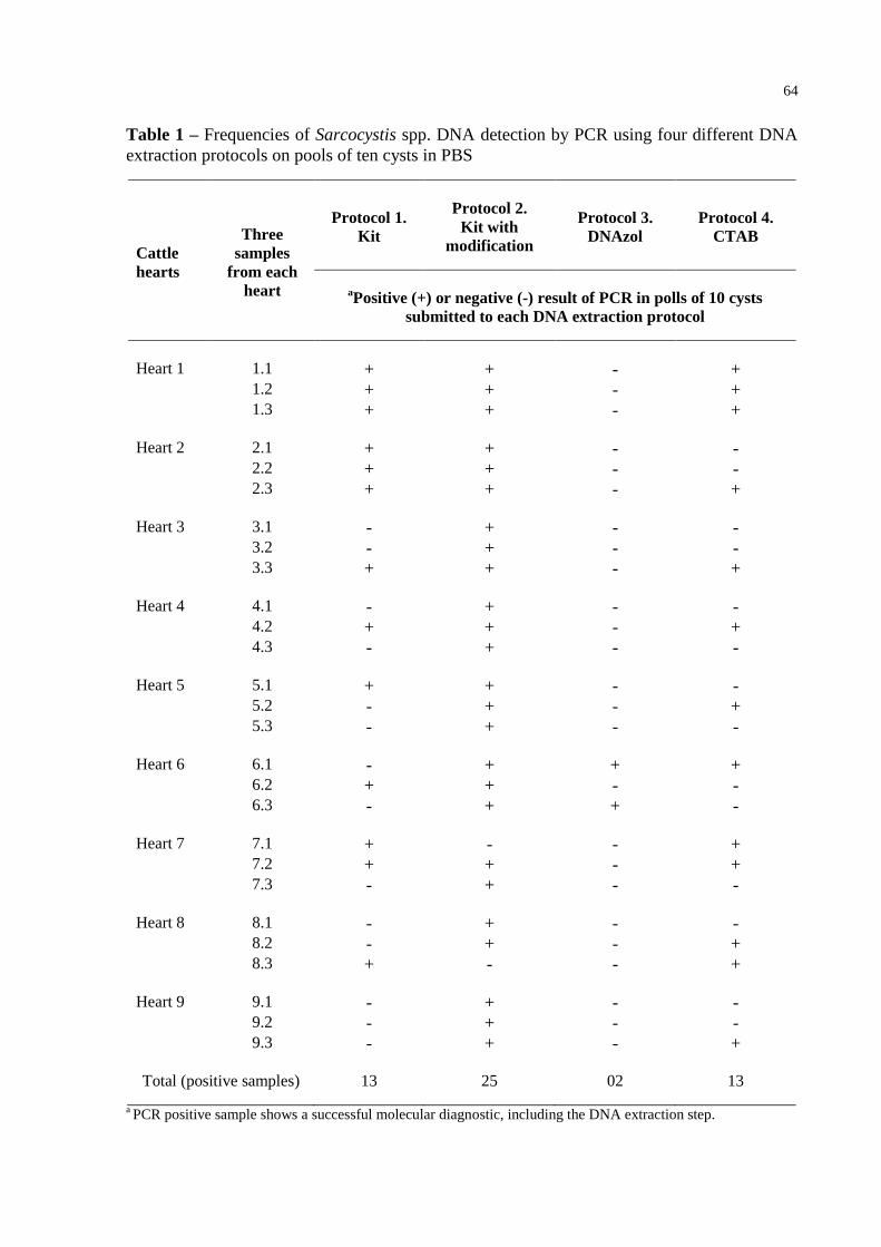
64
Table 1 – Frequencies of Sarcocystis spp. DNA detection by PCR using four different DNA
extraction protocols on pools of ten cysts in PBS
Cattle
hearts
Three
samples
from each
heart
Protocol 1.
Kit
Protocol 2.
Kit with
modification
Protocol 3.
DNAzol
Protocol 4.
CTAB
aPositive (+) or negative (-) result of PCR in polls of 10 cysts
submitted to each DNA extraction protocol
Heart 1 1.1 + + - + 1.2 + + - + 1.3 + + - +
Heart 2 2.1 + + - - 2.2 + + - - 2.3 + + - +
Heart 3 3.1 - + - - 3.2 - + - - 3.3 + + - +
Heart 4 4.1 - + - - 4.2 + + - + 4.3 - + - -
Heart 5 5.1 + + - - 5.2 - + - + 5.3 - + - -
Heart 6 6.1 - + + + 6.2 + + - - 6.3 - + + -
Heart 7 7.1 + - - + 7.2 + + - + 7.3 - + - -
Heart 8 8.1 - + - - 8.2 - + - + 8.3 + - - +
Heart 9 9.1 - + - - 9.2 - + - - 9.3 - + - +
Total (positive samples) 13 25 02 13
a PCR positive sample shows a successful molecular diagnostic, including the DNA extraction step.
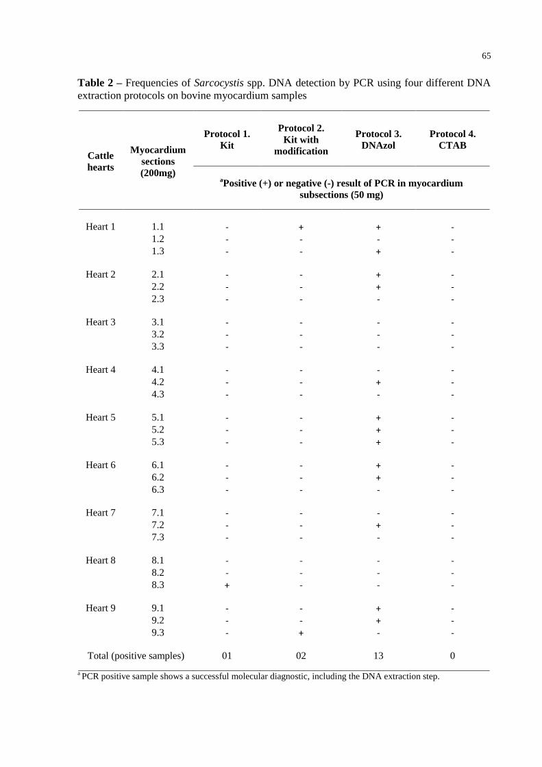
65
Table 2 – Frequencies of Sarcocystis spp. DNA detection by PCR using four different DNA
extraction protocols on bovine myocardium samples
Cattle
hearts
Myocardium
sections
(200mg)
Protocol 1.
Kit
Protocol 2.
Kit with
modification
Protocol 3.
DNAzol
Protocol 4.
CTAB
aPositive (+) or negative (-) result of PCR in myocardium
subsections (50 mg)
Heart 1 1.1 - + + - 1.2 - - - - 1.3 - - + -
Heart 2 2.1 - - + - 2.2 - - + - 2.3 - - - -
Heart 3 3.1 - - - - 3.2 - - - - 3.3 - - - -
Heart 4 4.1 - - - - 4.2 - - + - 4.3 - - - -
Heart 5 5.1 - - + - 5.2 - - + - 5.3 - - + -
Heart 6 6.1 - - + - 6.2 - - + - 6.3 - - - -
Heart 7 7.1 - - - - 7.2 - - + - 7.3 - - - -
Heart 8 8.1 - - - - 8.2 - - - - 8.3 + - - -
Heart 9 9.1 - - + - 9.2 - - + - 9.3 - + - -
Total (positive samples) 01 02 13 0
a PCR positive sample shows a successful molecular diagnostic, including the DNA extraction step.
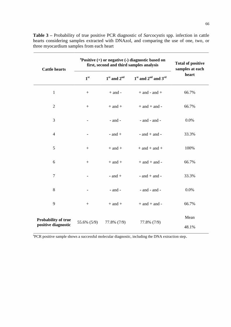
66
Table 3 – Probability of true positive PCR diagnostic of Sarcocystis spp. infection in cattle
hearts considering samples extracted with DNAzol, and comparing the use of one, two, or
three myocardium samples from each heart
Cattle hearts
aPositive (+) or negative (-) diagnostic based on
first, second and third samples analysis Total of positive
samples at each
heart 1
st 1
st and 2
nd 1
st and 2
nd and 3
rd
1 + + and - + and - and + 66.7%
2 + + and + + and + and - 66.7%
3 - - and - - and - and - 0.0%
4 - - and + - and + and - 33.3%
5 + + and + + and + and + 100%
6 + + and + + and + and - 66.7%
7 - - and + - and + and - 33.3%
8 - - and - - and - and - 0.0%
9 + + and + + and + and - 66.7%
Probability of true
positive diagnostic 55.6% (5/9) 77.8% (7/9) 77.8% (7/9)
Mean
48.1%
aPCR positive sample shows a successful molecular diagnostic, including the DNA extraction step.
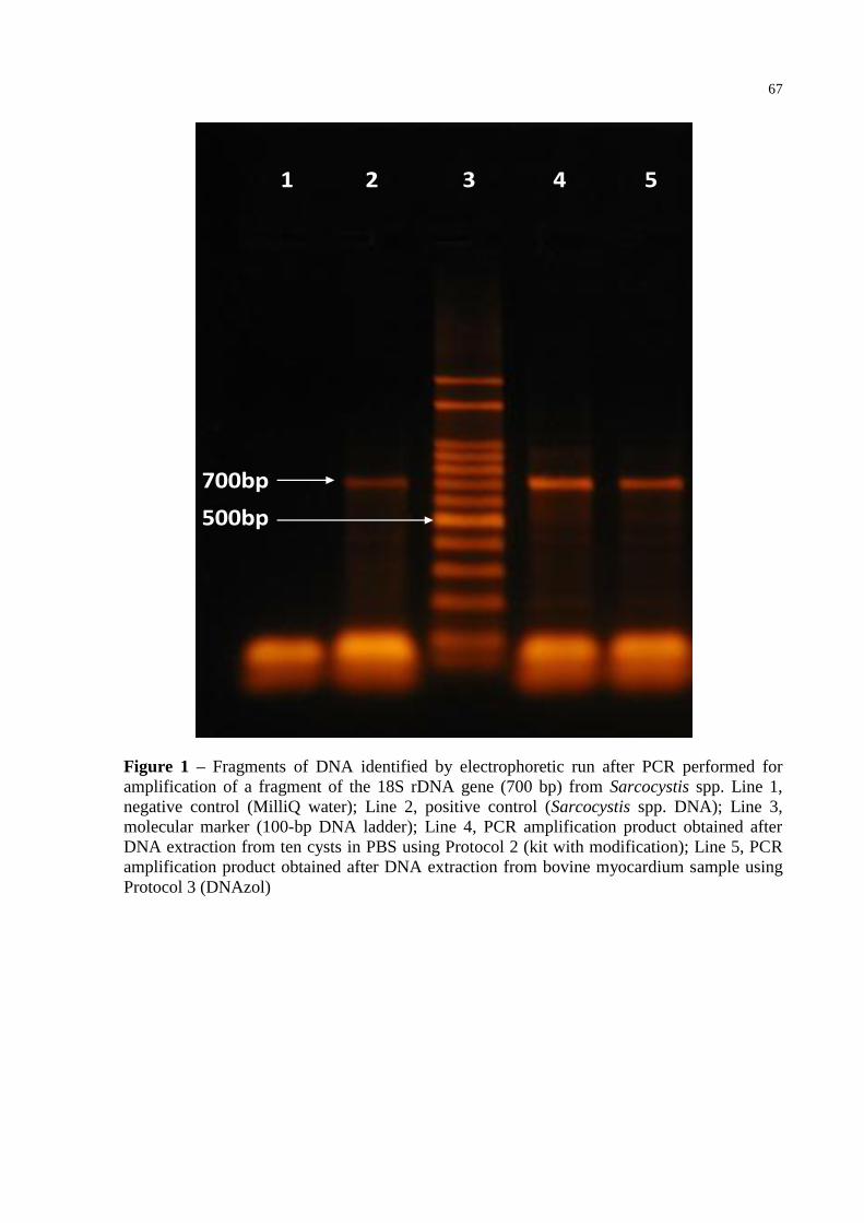
67
1 2 3 4 5
500bp
700bp
Figure 1 – Fragments of DNA identified by electrophoretic run after PCR performed for
amplification of a fragment of the 18S rDNA gene (700 bp) from Sarcocystis spp. Line 1,
negative control (MilliQ water); Line 2, positive control (Sarcocystis spp. DNA); Line 3,
molecular marker (100-bp DNA ladder); Line 4, PCR amplification product obtained after
DNA extraction from ten cysts in PBS using Protocol 2 (kit with modification); Line 5, PCR
amplification product obtained after DNA extraction from bovine myocardium sample using
Protocol 3 (DNAzol)
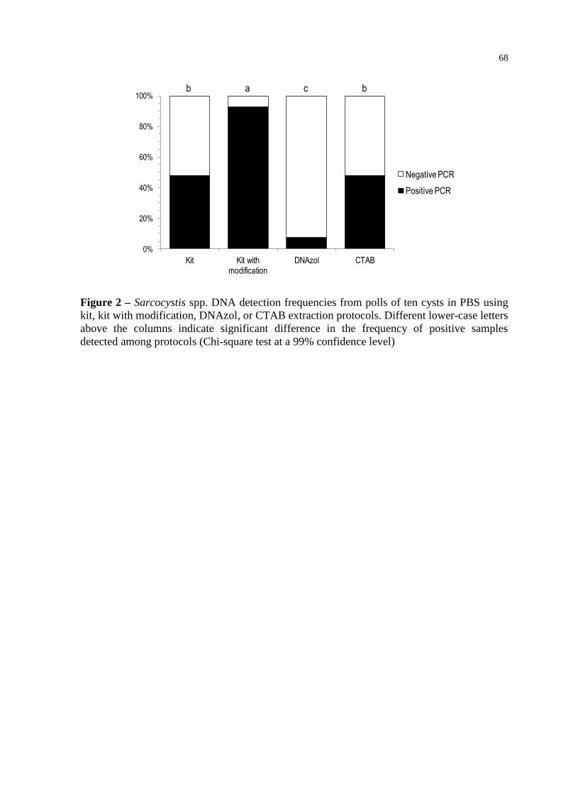
68
0%
20%
40%
60%
80%
100%
Kit Kit with modification
DNAzol CTAB
Negative PCR
Positive PCR
ab c b
Figure 2 – Sarcocystis spp. DNA detection frequencies from polls of ten cysts in PBS using
kit, kit with modification, DNAzol, or CTAB extraction protocols. Different lower-case letters
above the columns indicate significant difference in the frequency of positive samples
detected among protocols (Chi-square test at a 99% confidence level)
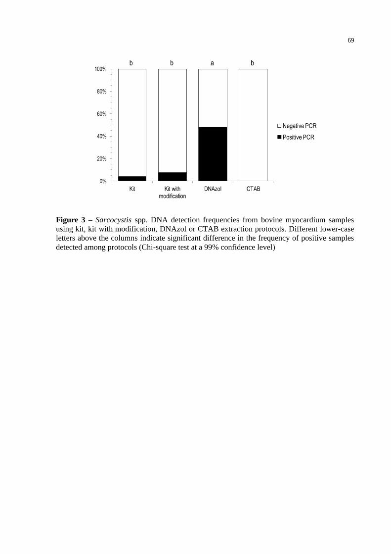
69
0%
20%
40%
60%
80%
100%
Kit Kit with modification
DNAzol CTAB
Negative PCR
Positive PCR
abb b
Figure 3 – Sarcocystis spp. DNA detection frequencies from bovine myocardium samples
using kit, kit with modification, DNAzol or CTAB extraction protocols. Different lower-case
letters above the columns indicate significant difference in the frequency of positive samples
detected among protocols (Chi-square test at a 99% confidence level)