Embed Size (px)
Citation preview
PONTIFÍCIA UNIVERSIDADE CATÓLICA DO RIO GRANDE DO SUL FACULDADE DE BIOCIÊNCIAS
PROGRAMA DE PÓS-GRADUAÇÃO BIOLOGIA CELULAR E MOLECULAR
LEONARDO ASTOLFI ROSADO
A ENZIMA CHIQUIMATO QUINASE
(EC 2.7.1.71) DE MYCOBACTERIUM TUBERCULOSIS COMO ALVO PARA O DESENVOLVIMENTO DE
DROGAS
Porto Alegre Abril, 2013

i
PONTIFÍCIA UNIVERSIDADE CATÓLICA DO RIO GRANDE DO SUL FACULDADE DE BIOCIÊNCIAS
PROGRAMA DE PÓS-GRADUAÇÃO BIOLOGIA CELULAR E MOLECULAR
A ENZIMA CHIQUIMATO QUINASE (EC 2.7.1.71) DE MYCOBACTERIUM TUBERCULOSIS COMO ALVO PARA
O DESENVOLVIMENTO DE DROGAS
Tese apresentada ao Programa de Pós-graduação em Biologia Celular e Molecular da Pontifícia Universidade Católica do Rio Grande do Sul como requisito para obtenção do grau de Doutor.
Leonardo Astolfi Rosado
Orientador:
Prof. Dr. Luiz Augusto Basso
Porto Alegre Abril, 2013
ii
PONTIFÍCIA UNIVERSIDADE CATÓLICA DO RIO GRANDE DO SUL FACULDADE DE BIOCIÊNCIAS
PROGRAMA DE PÓS-GRADUAÇÃO BIOLOGIA CELULAR E MOLECULAR
A ENZIMA CHIQUIMATO QUINASE (EC 2.7.1.71) DE MYCOBACTERIUM TUBERCULOSIS COMO ALVO PARA
O DESENVOLVIMENTO DE DROGAS
Tese apresentada ao Programa de Pós-graduação em Biologia Celular e Molecular da Pontifícia Universidade Católica do Rio Grande do Sul como requisito para obtenção do grau de Doutor.
Aprovada em ______ de __________de________.
BANCA EXAMINADORA:
Dr. Cristiano V. Bizarro (PUCRS-PPGBCM)
Dr. Rafael A. Caceres (PUCRS-PPGCC)
Dr. Jeverson Frazzon (UFRGS-ICTA)

iii
"It's a Long Way to the Top If You Wanna Rock 'n' Roll”
(AC/DC)

iv
Agradecimentos
Primeiramente eu gostaria de agradecer ao meu orientador, o professor Luiz Augusto
Basso pelo apoio durante os anos em que trabalhamos juntos, sempre incentivando e
orientando em busca do conhecimento cientifico e na minha formação como pesquisador
e pessoa.
Eu gostaria de agradecer a todos os meus colegas de laboratório pela ajuda, incentivo,
amizade e companheirismo, principalmente aos colegas Anderson Jader, Ardala Breda,
Bruno Abaddi, Christiano Ev Neves, Claudia Paiva Nunes, Daniel Lorenzini, Diana
Rostirola, Guilherme Petersen, José Eduardo Nunes, Leonardo Martinelli, Luís Timmers,
Paulo Patta, Priscila Wink, Rafael Guimarães, Rodrigo Ducati, Thiago Milech, Valnês Jr.
e Zilpa Adriana. Eu quero agradecer especialmente a Natasha, Manuel e Rafael
Munareto pelo companheirismo e amizade durante estes anos e por sempre estarem
dispostos para qualquer tipo de indiada.
Eu gostaria de fazer um agradecimento especial ao Dr. Cristiano Bizarro, por tudo que
ele me ensinou e ajudou desde o início da nossa amizade, com muita paciência,
sabedoria, conhecimento, ética e paixão pela ciência.
Eu quero agradecer aos meus amigos de fora do meio acadêmico, que sempre estiveram
do meu lado mesmo antes deste projeto e sempre me incentivaram e estiveram
presentes durante os momentos bons e ruins.
Não esquecendo da pessoa que sempre esteve ao meu lado, ajudando, incentivando,
com carinho e dedicação, a minha namorada Daiana Renck, que mesmo nos momentos
mais complicados me ajudou a achar uma resposta para as minhas dúvidas, tanto no
meio pessoal como profissional, sempre amável e compreensiva. Não tenho palavras pra
dizer tudo que gostaria, então, resumidamente, eu te amo.

v
E por fim, mas não menos importante, eu gostaria de agradecer a minha família, que
sempre me apoiou incondicionalmente e sempre esteve ao meu lado. Eu gostaria de
agradecer especialmente ao meu sobrinho Benjamin, que nasceu alguns dias antes da
prova do doutorado, e desde que chegou só trouxe alegrias.
Resumidamente, obrigado a todos!

vi
Resumo
A tuberculose se mantém como uma das principais causas de mortalidade ao redor do
mundo devido a um único agente infeccioso, o Mycobacterium tuberculosis. O gene aroK
que codifica a enzima Chiquimato quinase de Mycobacterium tuberculosis (MtSK), que
se mostrou essencial para a sobrevivência do bacilo, catalisa a transferência de um
grupo fosforil do ATP para o chiquimato (SKH), resultando em chiquimato-3-fosfato e
ADP. O presente trabalho apresenta a purificação da proteína MtSK até a
homogeneidade e a determinação de seu estado oligomérico em solução. Estudos
bioquímicos e biofísicos apresentados nesta tese sugerem que a reação catalisada pela
MtSK monomérica segue um mecanismo de equilíbrio rápido ordenado aleatório na
adição dos substratos e uma liberação ordenada sequencial dos produtos. A análise por
calorimetria de titulação isotérmica (ITC) das interações não covalentes da enzima MtSK
com diferentes ligantes resultou na determinação de assinaturas termodinâmicas para
cada um desses eventos de ligação. A comparação entre as constantes de afinidade
obtidas por espectrofotometria e ITC demostraram que o ATP não aumenta a afinidade
da proteína MtSK pelo SKH. Além disso, foi determinada em + 3,2 kJ mol-1 a energia livre
de acoplamento do SKH ao complexo binário MTSK:Mg2+ATP, sugerindo que o ATP
possui uma cooperatividade negativa em relação ao SKH. Esse trabalho sugere que a
MtSK deveria ser mais apropriadamente descrita como uma chiquimato quinase do tipo II
codificada pelo gene aroL. Adicionalmente, estre trabalho demonstra a descrição
termodinâmica do SKH se ligando a MtSK aonde os resultados sugerem o número de
prótons que estão sendo trocados durante a interação bimolecular. O valor negativo
encontrado para a capacidade calorífica em pressão constante (ΔCp) e o modelo
molecular por homologia criado sugerem uma pronunciada contribuição na

vii
dessolvatação de grupos apolares relacionados à formação do complexo. As constantes
termodinâmicas foram separadas na contribuição hidrofóbica e vibracional relacionadas à
formação do complexo binário MtSK:SKH. Os resultados obtidos relacionados à
transferência de prótons durante a formação do complexo binário foram analisados tendo
como referência o modelo estrutural da enzima MtSK construído neste trabalho. De
acordo com as cadeias laterais que fazem contato com o SKH no modelo proposto, se
pode indicar que os aminoácidos Arg58 e Arg136 atuam como fonte de prótons durante a
formação do complexo binário.

viii
Abstract
Tuberculosis remains as one of the main cause of mortality worldwide due to a single
infectious agent, Mycobacterium tuberculosis. The aroK-encoded Mycobacterium
tuberculosis Shikimate Kinase (MtSK), shown to be essential for survival of bacilli,
catalyzes the phosphoryl transfer from ATP to the carbon-3 hydroxyl group of shikimate
(SKH), yielding shikimate-3-phosphate and ADP. Here we present purification to
homogeneity, and oligomeric state determination of recombinant MtSK. Biochemical and
biophysical data suggest that the chemical reaction catalyzed by monomeric MtSK follows
a rapid-equilibrium random order of substrate binding, and ordered product release.
Isothermal titration calorimetry (ITC) for binding of ligands to MtSK provided
thermodynamic signatures of non-covalent interactions to each process. A comparison of
steady-state kinetics parameters and equilibrium dissociation constant value determined
by ITC showed that ATP binding does not increase the affinity of MtSK for SKH. In
addition, there appears to be a positive free energy coupling of 3.2 kJ mol-1 for SKH
binding to MtSK:Mg2+ATP binary complex, which suggest that ATP displays negative
cooperativity for SKH binding. We suggest that MtSK would more appropriately be
described as an aroL-encoded type II shikimate kinase. Our manuscript also gives
thermodynamic description of SKH binding to MtSK and data for the number of protons
exchanged during this bimolecular interaction. The negative value for the change in
constant pressure heat capacity (ΔCp) and molecular homology model building suggest a
pronounced contribution of desolvation of non-polar groups upon binary complex
formation. Thermodynamic parameters were deconvoluted into hydrophobic and
vibrational contributions upon MtSK:SKH binary complex formation. Data for the number
of protons exchanged during this bimolecular interaction are interpreted in light of a

ix
structural model to try to propose the likely amino acid side chains that are the proton
donors to bulk solvent following MtSK:SKH complex formation.
x
Lista de Abreviaturas e Siglas
ADP, adenosina 5’-difosfato;
ATP, adenosina 5'-trifosfato;
EcSK, Chiquimato quinase de Erwinia chrysanthemi codificada pelo gene aroL;
E4P, D-eritrose 4-fosfato;
ESI-MS, espectrometria de massa com ionização por eletrospray;
HEPES, do inglês N-2-hydroxyethylpiperazyne-N’-2-ethanesulfonic acid;
ITC, calorimetria de titulação isotérmica;
LDH, L-lactato desidrogenase;
MDR-TB, infecção causada por cepas de Mycobacterium tuberculosis resistentes a
múltiplas drogas;
MtSK, Chiquimato Quinase de Mycobacterium tuberculosis;
NADH, nicotinamida adenina dinucleotídeo reduzido;
NMP, nucleotídeo monofosfato;
PEP, fosfoenolpiruvato;
PK, piruvato quinase;
SDS-PAGE, do inglês sodium dodecyl sulfate-polyacrylamide gel eletrophoresis;
SKH, chiquimato, [3R-(3,4,5)]3,4,5-tri-hidroxi-1-ciclohexano-1-ácido carboxílico;
S3P, chiquimato 3-fosfato;
TB, Tuberculose;
TDR-TB, infecção humana causada por cepas de Mycobacterium tuberculosis totalmente
resistentes a múltiplas drogas;
Tris, tris(hidroximetil) aminometano;

xi
XDR-TB, infecção humana causada por cepas de Mycobacterium tuberculosis
extremamente resistentes a múltiplas drogas.
xii
Lista de Figuras
Figura 1. Estádios de infecção causados pelo Mycobacterium tuberculosis. 02
Figura 2. A via do ácido chiquímico. 05
Figura 3. Reação enzimática catalisada pela enzima chiquimato quinase. 06
xiii
SUMÁRIO
Capítulo 1
1
1. INTRODUÇÃO 2
1.1 A Tuberculose 2
1.2 Mycobacterium tuberculosis 4
1.3 A via do ácido chiquímico 4
1.4 Chiquimato quinase 6
2. JUSTIFICATIVA 8
3. OBJETIVOS 9
3.1 Objetivo Geral 9
3.2 Objetivos Específicos 9
Capítulo 2
10
Carta de Aceite do Manuscrito 11
Artigo Aceito PLoSOne - The mode of action of recombinant Mycobacterium tuberculosis Shikimate Kinase: kinetics and thermodynamic analyses
12
Capítulo 3 53
CONSIDERAÇÕES FINAIS 54
Referências 57
Anexos 61
Anexo A Artigo Submetido Kinetic mechanism and energetics of nucleotide binding to Mycobacterium tuberculosis Cytidine Monophosphate Kinase. Biophysical Chemistry, 2013.
62
Anexo B Artigo Publicado Biochemical characterization of uracil phosphoribosyltransferase from Mycobacterium tuberculosis. PLoSONE, 2013.
64
Anexo C Artigo Publicado Role of Serine140 in the mode of action of Mycobacterium tuberculosis β- 2 ketoacyl-ACP reductase (MabA). BMC Research Notes, 2012.
66

xiv
Anexo D Artigo Aceito Recombinant Erwinia carotovora L-asparaginase II production in Escherichia coli fed-batch cultures. Brazilian Journal of Chemical Engineering (Impresso), 2012.
68
Anexo E Artigo Publicado Molecular, kinetic and thermodynamic characterization of Mycobacterium tuberculosis orotate phosphoribosyltransferase. Molecular Biosystems (Print), v. 8, p. 572, 2012.
70
Anexo F Artigo Publicado Pyrimidin-2(1H)-ones based inhibitors of Mycobacterium tuberculosis orotate phosphoribosyltransferase. European Journal of Medicinal Chemistry, v. Epub, p. ahead-of print, 2012.
72
Anexo G Artigo Publicado Wild-Type Phosphoribosylpyrophosphate Synthase (PRS) from Mycobacterium tuberculosis: A Bacterial Class II PRS?. Plos One, v. epub, p. ahead-of print, 2012.
74
Anexo H Artigo Publicado Molecular, kinetic, thermodynamic, and structural analyses of Mycobacterium tuberculosis hisD-encoded metal-dependent dimeric histidinol dehydrogenase (EC 1.1.1.23). Archives of Biochemistry and Biophysics (Print), v. 512, p. 143-153, 2011.
76

1
Capítulo 1
Introdução
2
1. INTRODUÇÃO
1.1 A Tuberculose
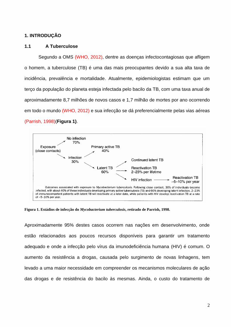
Segundo a OMS (WHO, 2012), dentre as doenças infectocontagiosas que afligem
o homem, a tuberculose (TB) é uma das mais preocupantes devido a sua alta taxa de
incidência, prevalência e mortalidade. Atualmente, epidemiologistas estimam que um
terço da população do planeta esteja infectada pelo bacilo da TB, com uma taxa anual de
aproximadamente 8,7 milhões de novos casos e 1,7 milhão de mortes por ano ocorrendo
em todo o mundo (WHO, 2012) e sua infecção se dá preferencialmente pelas vias aéreas
(Parrish, 1998)(Figura 1).
Figura 1. Estádios de infecção do Mycobacterium tuberculosis, retirado de Parrish, 1998.
Aproximadamente 95% destes casos ocorrem nas nações em desenvolvimento, onde
estão relacionados aos poucos recursos disponíveis para garantir um tratamento
adequado e onde a infecção pelo vírus da imunodeficiência humana (HIV) é comum. O
aumento da resistência a drogas, causada pelo surgimento de novas linhagens, tem
levado a uma maior necessidade em compreender os mecanismos moleculares de ação
das drogas e de resistência do bacilo às mesmas. Ainda, o custo do tratamento de
3
pacientes com TB resistente a múltiplas drogas pode assumir valores muito mais altos
que o de pacientes com TB susceptível às drogas de uso comum.
Robert Koch, em 1882, identificou uma bactéria álcool-ácido resistente, Mycobacterium
tuberculosis (MTB), como o agente causador da TB. Em 1921, a vacina BCG foi utilizada
pela primeira vez como um agente profilático contra a infecção causada pelo MTB, e é a
vacina utilizada no mundo atualmente.
O descobrimento das propriedades antibacterianas de algumas drogas de primeira linha
levaram a quimioterapias efetivas que diminuíram a taxa de mortalidade no mundo, e a
posterior introdução de algumas outras ao arsenal utilizado para combater a TB parecem
ter provido um número adequado de efetivos agentes antimicrobianos (Goodman &
Gilman et al, 2006). A quimioterapia efetiva contra TB deve incluir uma ação primária
bactericida contra organismos em rápido crescimento e uma subsequente esterilização
da população de bacilos dormentes.
A moderna terapia padrão de curta duração para TB está baseada em um regime de
quatro drogas que deve ser estritamente seguido para prevenir resistência a drogas e
reincidência. Dessa forma, a prevenção da má administração das drogas pelo paciente é
o melhor meio de garantir um tratamento efetivo e prevenir a aquisição de resistência.
Apesar da baixa permeabilidade da parede celular, que dificulta a absorção de
moléculas, as micobactérias necessitam de mecanismos moleculares para obter uma
resistência significativa (Cole et al, 1998). Mutantes de MTB resistentes a uma droga
qualquer estão naturalmente presentes em qualquer grande população bacteriana,
independentemente da exposição a drogas. A frequência de mutantes resistentes a
rifampicina e isoniazida é relativamente alta e, portanto, a grande população extracelular
de bacilos metabolicamente ativos e com rápido crescimento em lesões cavitárias
conterá organismos resistentes a pelo menos uma das drogas. Consequentemente, a

4
monoterapia ou a terapia de duas drogas impropriamente administrada selecionará
mutantes resistentes a drogas que podem conferir resistência a toda população de
bacilos. Em 2006, 500 000 casos de cepas resistentes foram relatados, demonstrando
assim a atual situação e a gravidade do problema.
Existem duas formas de TB altamente perigosas para a saúde humana; as cepas
multirresistentes (MDR) são resistentes a pelo menos isoniazida e rifampicina e
requerem um tratamento quimioterápico mais extensivo com quimioterápicos de segunda
linha; e as cepas de Mycobacterium tuberculosis extremamente resistentes a múltiplas
drogas (XDR) se mostram resistentes aos quimioterápicos tanto de primeira quanto de
segunda linha, indicando assim uma necessidade de desenvolver novos quimioterápicos
contra a TB (Hargreaves, 2008).
1.2 Mycobacterium tuberculosis
O Mycobacterium tuberculosis, principal agente causador da TB, é uma bactéria gram-
positiva com um alto conteúdo genômico de G+C. Esta bactéria se apresenta em forma
de bacilo mostrando dimensões que variam de 0,3 a 0,6 µm de largura e de 1 a 4 µm de
altura, possuindo uma alta complexidade em seu envelope celular e crescimento lento .
1.3 A Via do ácido chiquímico
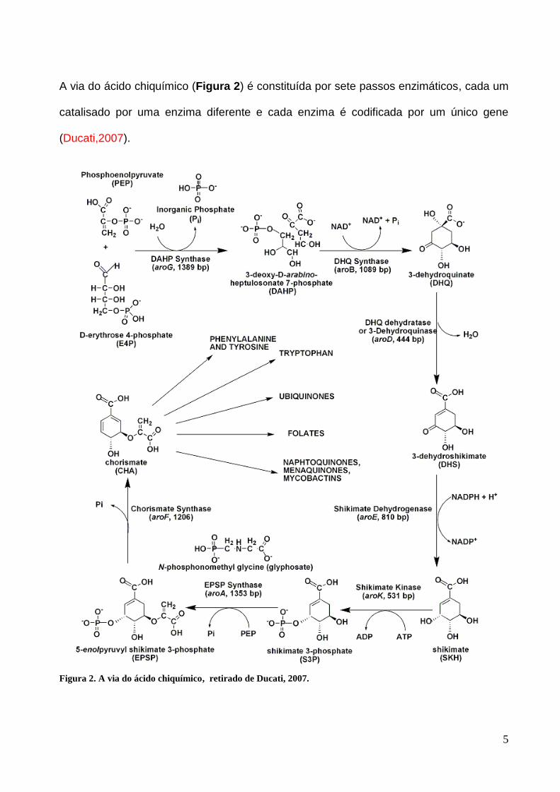
5
A via do ácido chiquímico (Figura 2) é constituída por sete passos enzimáticos, cada um
catalisado por uma enzima diferente e cada enzima é codificada por um único gene
(Ducati,2007).
Figura 2. A via do ácido chiquímico, retirado de Ducati, 2007.

6
Essa via é essencial para a vida de plantas, organismos do filo apicomplexa,
parasitos e bactérias (Bentley, 1990; Campbell, 2004; Roberts, 2002; Roberts, 1998;
Keeling, 1999; McConkey, 1999; McConkey, 2004;), incluindo o MTB (Parrish, 2002),
porém é ausente no homem, o que a torna um alvo atrativo para o descobrimento de
novos fármacos contra a TB. No bacilo causador da TB, a via do ácido chiquímico tem
como substratos iniciais fosfoenolpiruvato e D-eritrose 4-fosfato (E4P), tendo como
produto final o corismato, que é um importante componente na síntese das vitaminas E e
K, folato, ubiquinona, menaquinonas, micobactinas, naftoquinonas e dos aminoácidos
aromáticos (Hermann, 1999).
1.4 Chiquimato quinase
A enzima chiquimato quinase (MtSK) é codificada pelo gene aroK, composto por 531 pb
que resultam em uma proteína de 176 aa com peso molecular teórico de 18 451Da. Esta
enzima catalisa a quinta reação da via do ácido chiquímico, convertendo adenosina
5’trifosfato (ATP) e chiquimato (SKH) em adenosina 5’difosfato (ADP) e chiquimato-3-
fosfato (S3P), utilizando magnésio como cofator, realizando uma transferência do fosfato
γ do doador (ATP) para o carbono três do anel do ácido chiquímico (Figura 3).
Figura 3. Reação enzimática catalisada pela enzima chiquimato quinase.
Esta enzima pertence à família de enzimas Nucleosídeo Monofosfato Quinase, sendo um
importante grupo de enzimas que catalisa a transferência reversível de um fosfato,

7
pertencente a um nucleotídeo trifosfato, para um nucleotídeo monofosfato específico
(Yan, 1999). O produto desta reação é subsequentemente fosforilado, resultando em
precursores dos ácidos nucleicos. Cada membro dessa família possui três domínios
conservados: o domínio Core contendo cinco folhas-β paralelas e o P-loop que formam o
sítio de ligação dos nucleotídeos; o domínio LID, que se move sobre o sítio ativo e possui
resíduos essenciais para a ligação do ATP; e o sítio de ligação dos nucleotídeos que
possuem um fosfato (NMP), que reconhece e se liga ao nucleotídeo monofosfato. No
caso da chiquimato quinase, esse sítio reconhece e se liga ao chiquimato, que é o
substrato da reação (Pereira, 2004; Hartmann,2006; Dias, 2007; Dhaliwal; 2004 ).

8
2. JUSTIFICATIVA
A TB é uma doença que demonstra índices alarmantes em relação à incidência,
prevalência e mortalidade em todos os continentes do mundo, o que a torna uma
preocupação mundial. A rifampicina e a isoniazida, descobertas em 1966 e 1952
respectivamente, são os antibióticos mais poderosos utilizados atualmente contra a TB,
entretanto, o surgimento de novas cepas resistentes ao tratamento atual e as altas taxas
de incidência indicam a necessidade do desenvolvimento de novos agentes contra a TB,
a fim de impedir a proliferação destas cepas resistentes, o que tornaria ainda mais difícil
a luta contra esta doença. A enzima alvo deste trabalho pertence à via do ácido
chiquímico, que é uma via promissora para o estudo de novos inibidores, já que esta é
inexistente em seu hospedeiro (Homo sapiens), o que nos remete ao princípio da
toxicidade seletiva. Se o produto final desta via, o corismato, for impedido de ser
produzido, o bacilo da TB perderá sua viabilidade, devido à extrema importância do
corismato já que ele é um dos precursores na síntese do folato, ubiquinonas, vitamina E,
vitamina K e dos aminoácidos aromáticos, componentes essenciais para a sobrevivência
do bacilo.

9
3. OBJETIVOS
3.1 Objetivo Geral
Caracterização bioquímica e biofísica da enzima MtSK (E.C. 2.7.1.71 ) codificada pelo
gene aroK de Mycobacterium tuberculosis H37Rv como alvo para o desenvolvimento de
novas drogas de ação seletiva.
3.2 Objetivos Específicos
Expressar e purificar a enzima MtSK.
Determinar a massa molecular e sequência N-terminal da proteína.
Determinar as constantes cinéticas verdadeiras.
Determinar o mecanismo enzimático por calorimetria de titulação isotérmica e fluorimetria.
Determinar a variação da capacidade calorífica a pressão constante na formação do complexo MtSK:SKH.
Determinar os prótons envolvidos na formação do complexo MtSK:SKH.
10
Capítulo 2
The mode of action of recombinant Mycobacterium tuberculosis Shikimate Kinase: kinetics and thermodynamics analyses
Leonardo Astolfi Rosado, Igor Bordin Vasconcelos, Mário Sérgio Palma, Vincent Frappier, Rafael Josef Najmanovich, Diógenes Santiago Santos and Luiz Augusto Basso.

11
PONE-D-12-33107R3
The mode of action of recombinant Mycobacterium tuberculosis Shikimate Kinase: kinetics and
thermodynamics analyses
PLOS ONE
Dear Dr. Basso,
I am pleased to inform you that your manuscript has been deemed suitable for publication in PLOS
ONE.
Your manuscript will now be passed on to our Production staff, who will check your files for
correct formatting and completeness. After this review, they may return your manuscript to you so
that you can make necessary alterations and upload a final version.
Before uploading, you should check the PDF of your manuscript very closely. THERE IS NO
AUTHOR PROOFING. You should therefore consider the corrected files you upload now as
equivalent to a production proof. The text you supply at this point will be faithfully represented in
your published manuscript exactly as you supply it. This is your last opportunity to correct any
errors that are present in your manuscript files.
In addition, now that your manuscript has been accepted, please log into EM at
http://www.editorialmanager.com/pone and update your profile. Click on the "Update My
Information" link at the top of the page. Please update your user information to ensure an efficient
production and billing process.
If you or your institution will be preparing press materials for this manuscript, you must inform our
press team in advance. Please contact them at [email protected].
Please contact us at [email protected] if you have any questions, concerns, or problems, and thank
you for submitting your work to our journal.
With kind regards,
Pratul K. Agarwal
Academic Editor
PLOS ONE

12
The mode of action of recombinant Mycobacterium tuberculosis Shikimate Kinase:
kinetics and thermodynamics analyses
Leonardo Astolfi Rosado1,3, Igor Bordin Vasconcelos1,3, Mário Sérgio Palma4, Vincent
Frappier5, Rafael Josef Najmanovich 5, Diógenes Santiago Santos1,2,3.* and Luiz Augusto
Basso1,2,3,*
1Centro de Pesquisas em Biologia Molecular e Funcional (CPBMF), Instituto Nacional de
Ciência e Tecnologia em Tuberculose (INCT-TB), Pontifícia Universidade Católica do Rio
Grande do Sul (PUCRS), Av. Ipiranga 6681, Porto Alegre, RS 90619-900, Brazil.
2Programa de Pós-Graduação em Medicina e Ciências da Saúde, PUCRS, Av. Ipiranga
6681, Porto Alegre, RS 90619-900, Brazil.
3Programa de Pós-Graduação em Biologia Celular e Molecular, PUCRS, Av. Ipiranga
6681, Porto Alegre, RS 90619-900, Brazil.
4Laboratório de Biologia Estrutural e Zooquímica, Centro de Estudos de Insetos Sociais,
Departamento de Biologia, Instituto de Biociências de Rio Claro, Universidade Estadual
Paulista (UNESP), Rio Claro, SP, Brazil.
5Department of Biochemistry, Faculty of Medicine, Université de Sherbrooke, 3001, 12e
Avenue Nord. Sherbrooke, Québec, Canada J1H 5N4.
*Corresponding authors. Telephone/Fax: +55-51-33203629.
E-mail addresses: [email protected] (Luiz A. Basso) or [email protected] (Diógenes
S. Santos).

13
Abstract
Tuberculosis remains as one of the main cause of mortality worldwide due to a
single infectious agent, Mycobacterium tuberculosis. The aroK-encoded M. tuberculosis
Shikimate Kinase (MtSK), shown to be essential for survival of bacilli, catalyzes the
phosphoryl transfer from ATP to the carbon-3 hydroxyl group of shikimate (SKH), yielding
shikimate-3-phosphate and ADP. Here we present purification to homogeneity, and
oligomeric state determination of recombinant MtSK. Biochemical and biophysical data
suggest that the chemical reaction catalyzed by monomeric MtSK follows a rapid-
equilibrium random order of substrate binding, and ordered product release. Isothermal
titration calorimetry (ITC) for binding of ligands to MtSK provided thermodynamic
signatures of non-covalent interactions to each process. A comparison of steady-state
kinetics parameters and equilibrium dissociation constant value determined by ITC
showed that ATP binding does not increase the affinity of MtSK for SKH. We suggest that
MtSK would more appropriately be described as an aroL-encoded type II shikimate
kinase. Our manuscript also gives thermodynamic description of SKH binding to MtSK
and data for the number of protons exchanged during this bimolecular interaction. The
negative value for the change in constant pressure heat capacity (ΔCp) and molecular
homology model building suggest a pronounced contribution of desolvation of non-polar
groups upon binary complex formation. Thermodynamic parameters were deconvoluted
into hydrophobic and vibrational contributions upon MtSK:SKH binary complex formation.
Data for the number of protons exchanged during this bimolecular interaction are
interpreted in light of a structural model to try to propose the likely amino acid side chains
that are the proton donors to bulk solvent following MtSK:SKH complex formation.

14
Keywords: steady-state kinetics; isothermal titration calorimetry; fluorescence
spectroscopy; enzyme mechanism; shikimate kinase; tuberculosis

15
Introduction
Tuberculosis (TB), owing to Mycobacterium tuberculosis infection, still remains as
a major global health problem. Approximately 9 million new cases of TB are detected
each year, and close to 2 million people die from the disease [1]. In 2008, it has been
estimated that 390 000–510 000 cases of multidrug-resistant tuberculosis (MDR-TB,
which is defined as TB caused by strains of M. tuberculosis that are resistant to at least
isoniazid and rifampicin) emerged globally [2]. In 2008, MDR-TB caused an estimated
150 000 deaths. It has also been reported that 5.4% of MDR-TB cases were found to
have extensively drug-resistant tuberculosis (XDR-TB, which is defined as MDR-TB plus
resistance to a fluoroquinolone and at least one second-line injectable agents: amikacin,
kanamycin and/or capreomycin) [2]. In addition, TB cases due to infection with totally
drug-resistant strains (TDR-TB) have been reported [3]. The emergence of drug-resistant
strains of M. tuberculosis has thus highlighted the need for the development of new
therapeutic strategies to combat TB.
Rational inhibitor design relies on mechanistic and structural information on the
target enzyme. Enzymes offer unique opportunities for drug design that are not available
to cell surface receptors, nuclear hormone receptors, ion channel, transporters, and DNA
[4]. It has been pointed out that one of the lessons to be learned from marketed enzyme
inhibitors is that the most potent and effective inhibitors take advantage of enzyme
chemistry to achieve inhibition [5]. Moreover, the recognition of the limitations of high-
throughput screening approaches in the discovery of candidate drugs has rekindled
interest in rational design methods [6]. Accordingly, mechanistic analysis should always
be a top priority for enzyme-targeted drug programs aiming at the rational design of
potent enzyme inhibitors. Moreover, targets that are both essential to survival of, and

16
exclusive to, M. tuberculosis are particularly promising as their inhibition could lead to the
development of non-toxic drugs to the human host and having effective killing effect on
the pathogen [7].
The biosynthesis of aromatic rings from carbohydrate precursors involves a range
of chemical transformations that together constitute the shikimate pathway; through seven
enzymatic steps, phosphoenolpyruvate (PEP) and D-erythrose 4-phosphate (E4P) are
condensed to the branch point compound chorismate (endproduct), which leads to
several additional terminal pathways [7,8]. The shikimate pathway is essential in algae,
higher plants, fungi, bacteria, apicomplexan parasites and sea anemone, but absent from
humans [7-16]. The mycobacterial shikimate pathway (the main trunk) leads to the
biosynthesis of chorismic acid, which is converted by five distinct enzymes to prephenate
(precursor of phenylalanine and tyrosine), anthranilate (precursor of tryptophan),
aminodeoxychorismate (precursor of para-aminobenzoic acid -PABA – which, in turn,
leads to tetrahydrofolate synthesis), para-hydroxybenzoic acid (precursor of ubiquinone or
Coenzyme Q), and isochorismate (common precursor of naphtoquinones, menaquinones
and mycobactins) [7].
The aroK-encoded M. tuberculosis Shikimate Kinase (MtSK; EC 2.7.1.71), the fifth
enzyme of the pathway, catalyzes a phosphoryl transfer from ATP to the carbon-3
hydroxyl group of shikimate (SKH; [3R-(3,4,5)]3,4,5-trihydroxy-1-cyclohexene-1-
carboxylic acid) forming shikimate 3-phosphate (S3P) (Fig.1). Disruption of aroK gene
has demonstrated that MtSK, and thus the common aromatic biosynthesis pathway, is
essential for the viability of M. tuberculosis [17]. We have previously reported cloning and
expression in Escherichia coli of recombinant MtSK in functional form [18], thereby
confirming the correct in silico assignment to the structural gene encoding this protein.
Our research group [19,20] and others [21-23] have reported crystal structure
17
determinations of MtSK. Three functional motifs of nucleotide-binding enzymes were
recognizable in MtSK, including a Walker A-motif (that forms the phosphate-binding loop;
P-loop), a Walker B-motif, and an adenine-binding loop. MtSK belongs to the family of
nucleoside monophosphate (NMP) kinases, which are composed of three domains: (1)
the CORE domain containing the five stranded parallel β-sheet and the P-loop (residues
9-17), which forms the binding site for nucleotides; (2) the LID domain (residues glycine-
112 to aspartate-124), which closes over the active site and has residues that are
essential for the binding of ATP; and (3) the NMP-binding domain (residues threonine-33
to glutamate-61; also known as SB domain in MtSK), which functions to recognize and
bind shikimate [19-22]. More recently, based on an analysis of global movements upon
ligand binding, it has been proposed that MtSK is comprised of four domains [23]: (1) the
ESB domain (extended SB; residues 32-93); (2) the nucleotide-binding (NB) site that
includes the P-loop (Walker A motif, residues 9-17), the AB-loop (residues 148-155), and
the segment of 101-110; (3) the LID domain (residues 112-124); and (4) the Reduced
Core (RC) domain. A characteristic feature of NMP kinases is that they undergo large
conformational changes during catalysis [24].
Based on a series of high-resolution crystal structures of MtSK in apo form and as
binary and ternary complexes, it has been proposed that the enzyme mechanism is
random sequential binding of SKH and ATP (adenosine 5'-triphosphate), and release of
ADP (ADP, adenosine 5’-diphosphate) product is followed by S3P to generate free
enzyme [23]. However, no description of MtSK enzyme mechanism in solution has, to the
best of our knowledge, ever been described. Here we present purification of recombinant
MtSK to homogeneity, mass spectrometry analysis, N-terminal amino acid sequencing,
and oligomeric state determination of the recombinant protein. We also present true
steady-state kinetic parameters determination, and ligand binding by fluorescence

18
spectroscopy and isothermal titration calorimetry (ITC) data. These data demonstrate that
the chemical reaction catalyzed by monomeric MtSK follows a random order mechanism
of substrate binding, and that S3P product is released first followed by ADP dissociation
to yield free enzyme. The ITC results provided the thermodynamic signatures of non-
covalent interactions to the binding processes. In addition, we showed that there is a
positive free energy coupling of 3.2 kJ mol-1 for SKH binding to MtSK:Mg2+ATP binary
complex. Accordingly, ATP appears to display negative cooperativity for SKH binding.
Based on experimental evidence, we propose that MtSK would more appropriately be
described as an aroL-encoded type II shikimate kinase. We also present studies of the
temperature dependence of thermodynamic parameters for SKH interaction with MtSK.
The change in constant pressure heat capacity (ΔCp) on going from free to bound states
was evaluated and molecular homology model building was carried out to try to correlate
complex formation with burial of surface area. Attempts were also made to deconvolute
the thermodynamic parameters into hydrophobic and vibrational components.
Determination of changes in binding enthalpy (ΔH) was carried out in the presence of
buffers with different enthalpies of ionization. These ITC results showed that MtSK:SKH
binary complex formation is accompanied by release of protons to the bulk solvent. Based
on structural information, we suggest that the -guanidinium groups of Arg58 and/or
Arg136 are the likely sources of proton released into solution upon binary complex
formation. Understanding the mode of action of MtSK will inform us on how to better
design inhibitors targeting this enzyme with potential therapeutic application in TB
chemotherapy. The results here presented may also help chemical biologists to design
function-based chemical compounds to carry out either loss-of-function (inhibitors) or
gain-of-function (activators) experiments to reveal the biological role of MtSK in the
context of whole M. tuberculosis cells. Accordingly, it is hoped that the results here

19
described may be useful to the rational design of anti-TB agents and that they may
contribute to our understanding of the biology of M. tuberculosis.
Material and methods
Purification of M. tuberculosis Shikimate Kinase (MtSK)
The recombinant enzyme was expressed in Escherichia coli BL21 (DE3) host cells
as previously described [18]. Approximately 6 g of cells were suspended in 24 mL of Tris-
HCl (tris(hydroxymethyl)aminomethane) 50 mM pH 7.6, disrupted by sonication, and the
cell debris removed by centrifugation (48,000g for 60 min). MgCl2 was added to the
supernatant to a final concentration of 10 mM followed by addition of 1 mg of DNAse,
stirred for 30 min at 4 °C, and centrifuged (10,000g for 30 min). Interestingly, addition of
MgCl2 resulted in precipitation of MtSK whereas a number of proteins remained in the
supernatant. Accordingly, this step served two purposes in the purification protocol: lysis
of DNA by DNAse, and MtSK precipitation. The pellet was suspended in Tris-HCl 50 mM
pH 7.6 containing KCl 500 mM, and centrifuged (10,000g for 15 min). This solution was
concentrated down to approximately 20 mL, and 20 mL of Tris-HCl 50mM pH 7.6
containing KCl 500 mM and (NH4)2SO4 2M was added, resulting in a solution containing
1M (NH4)2SO4. This solution was clarified by centrifugation. The supernatant was loaded
on a Phenyl Sepharose 16/10 column (GE healthcare) previously equilibrated with buffer
A (Tris-HCl 50mM, KCl 500mM, (NH4)2SO4 1M, pH 7.6) and the adsorbed material eluted
with a linear gradient of buffer B (Tris-HCl 50mM, KCl 500mM, pH 7.6) at 1 mL min-1. The
protein fractions containing the MtSK were pooled and loaded on a Sephacryl S-100 HR
column (GE Healthcare) and isocratically eluted with buffer B at 0.25 mL min-1. The
20
fractions containing homogeneous recombinant MtSK were pooled, and stored in 85 %
(NH4)2SO4. Protein expression, purification and apparent homogeneity of recombinant
MtSK was confirmed by 12 % sodium dodecyl sulfate-polyacrylamide gel eletrophoresis
(SDS-PAGE) stained with Coomassie Brilliant Blue [25]. Protein concentration was
determined by the method of Bradford et al. [26] using the Bio-Rad protein assay kit and
bovine serum albumin as standard (Bio-Rad Laboratories).
Electrospray ionization mass spectrometry (ESI-MS) analysis
The subunit molecular mass of recombinant protein preparation was assessed by
ESI-MS, employing some adaptations made to the system described by Chassaigne and
Lobinski [27]. Samples were analyzed on a triple quadrupole mass spectrometer (model
QUATTRO II) equipped with a standard electrospray (ESI) probe (Micromass, Altrincham,
United Kingdom), and adjusted to a flow rate of ca. 250 L min-1. The source temperature
(80 °C) and needle voltage (3.6 kV) were maintained constant throughout the
experimental data collection, applying a drying gas (nitrogen) flow of 200 L h-1 and a
nebulizer gas flow of 20 L h-1. The mass spectrometer was calibrated with intact horse
heart myoglobin and its typical cone-voltage induced fragments. The subunit molecular
mass of recombinant MtSK was determined by adjusting the mass spectrometer to give a
peak with at half-height of 1 mass unit, and the sampling cone-to-skimmer lens voltage
controlling the transfer of ions to the mass analyzer was set to 38 V. About 50 pmol (10
µL) of each sample was injected into the electrospray transport solvent. The ESI
spectrum was obtained in the multichannel acquisition mode, scanning from 500 to 1,800
m/z at a scan time of 7 s. The mass spectrometer is equipped with MassLynx and
Transform software for data acquisition and spectrum handling.

21
N-terminal amino acid sequencing
The N-terminal amino acid residues of homogeneous recombinant MtSK were
identified by automated Edman degradation sequencing method [28] using PPSQ 21A
gas-phase sequencer (Shimadzu).
Oligomeric state determination
The oligomeric state of homogeneous MtSK was determined by size exclusion
liquid chromatography on Superdex 200 (HR 10/30) column (GE Healthcare). The column
was pre-equilibrated with 50 mM Tris-HCl pH 7.6 containing 200 mM NaCl at a flow rate
of 0.4 mL min-1 (4 °C), with UV detection at 280 nm. The calibration curve was
constructed employing the following protein standards: ribonuclease A (13.7 kDa),
quimotripsinogen (25 kDa), ovalbumine (43 kDa), albumine (67 kDa), aldolase (158 kDa),
catalase (232 kDa), and ferritine (440 kDa). The elution volumes (Ve) of calibration
proteins were used to calculate their corresponding partition coefficient (Kav, Eq. 1). Blue
dextran was utilized for determination of the void volume (Vo). Vt is the total bead volume
of the column. The Kav values for the standards were plotted versus the logarithm of their
corresponding molecular masses. A volume of 100 µL of recombinant protein was loaded
on the gel filtration column to obtain Ve for MtSK.
0
0
VV
VVK
t
eav
Eq. 1
22
Steady-state kinetics
Recombinant MtSK enzyme activity was assayed in the the forward direction by
coupling the ADP product to the pyruvate kinase (PK; EC 2.7.1.40) and lactate
dehydrogenase (LDH; EC 1.1.1.27) reactions following the protocol described by Millar et
al. [29]. Shikimate-dependent oxidation of NADH (nicotinamide adenine dinucleotide) was
continuously monitored at 340 nm ( = 6.22 103 M-1 cm-1). All reactions were carried out
at 25 °C and initiated with addition of recombinant MtSK (1 µg mL-1). The assay mixture
contained 100 mM Tris–HCl buffer, pH 7.6, 100 mM KCl, 5 mM MgCl2, 1.5 mM PEP, 0.2
mM NADH, 6 U mL-1 PK, and 5 U mL-1 LDH. Initial steady-state rates were calculated
from the linear portion of the reaction curve under experimental conditions in which less
than 5 % of substrate was consumed. True steady-state kinetics parameters were
determined from initial velocity measurements at varying concentrations of SKH (37 -
4800 µM) at varied-fixed ATP concentrations (9 - 1200 µM).
Values of the steady-state kinetics parameters and their respective errors were
obtained by fitting the data to the appropriate equations using the non-linear regression
function of SigmaPlot 9.0 (SPSS, Inc). Hyperbolic saturation curves of initial rate data at
single concentration of the fixed substrate and varying concentrations of the other were
fitted to the Michaelis-Menten equation (Eq. 2) [30,31], in which v is the initial velocity, V
is the apparent maximum initial velocity, A is the varying substrate concentration and K
represents the apparent Michaelis-Menten constant.
AK
VAv
Eq. 2
23
The family of lines intersecting to the left of the y-axis in double-reciprocal plots was fitted
to Eq. 3, which describes a mechanism involving ternary complex formation and a
sequential substrate binding [30,31].
ABAKBKKK
ABVv
babia max Eq. 3
For Eq. 3, v is the initial velocity (as for Eq. 2), Vmax is the true maximum initial velocity, A
and B are the concentrations of the substrates, Ka and Kb are their respective Michaelis
constants, and Kia is the dissociation constant for enzyme-substrate A binary complex
formation.
Equilibrium fluorescence spectroscopy
MtSK intrinsic protein fluorescence measurements were carried out to both
determine the order of substrate/product addition/dissociation on/from the catalytic site
and distinguish the enzyme kinetic mechanism. As MtSK has no tryptophan residues,
changes in protein tyrosine fluorescence (the polypeptide chain of MtSK has 3 Tyr
residues) upon ligand binding were monitored. Fluorescence measurements were carried
out in a RF-5301 PC Spectrofluorophotometer (Shimadzu) at 25 °C. Excitation
wavelength was 280 nm and emission spectra were collected from 300 to 500 nm. The
maximum values of fluorescence intensity at 315 nm values were plotted as a function of
increasing ligand concentration. Excitation and emission slits were 5 nm. As protein
tyrosine typically has a low sensitivity (low molar absorption coefficient and low
fluorescence quantum yield), MtSK concentration for binding experiments was 10 M.

24
Fluorescence titrations of binary complex formation were carried out by making microliter
additions of the following compounds to 2 mL solution containing 10 µM MtSK in 100 mM
Tris-HCl, 100 mM KCl, 5 mM MgCl2, pH 7.6: 120 mM SKH stock solution (10 – 1100 µM
final concentration); 120 mM S3P stock solution (59.97-952.3 µM final concentration).
Control experiments were performed in the same experimental conditions except that no
substrate was added, and these values were subtracted from those obtained in the
presence of substrate. However, owing to a large inner filter effect, ATP and ADP binding
to free MtSK could not be determined by fluorescence spectroscopy.
Equilibrium fluorescence spectroscopy data were fitted to Eq. 4, in which F is the
observed fluorescence, F0 is the initial fluorescence, F is the maximum change in
fluorescence at saturating ligand (L) concentration, and KD represents the equilibrium
dissociation constant for protein:ligand binary complex formation.
][
][
0
0
LK
L
FF
FF
D
Eq. 4
Isothermal titration calorimetry (ITC)
ITC experiments were carried out using an iTC200 Microcalorimeter (Microcal, Inc.,
Northampton, MA). The equipment’s sample cell volume is 200 μL and syringe final
volume is 39 μL. Calorimetric experiments were performed with either substrates (SKH
and ATP) or products (S3P and ADP) at 298.15 K. The reference cell (200 μL) was
loaded with water during all experiments and the sample cell (200 μL) was filled with
MtSK at 100 μM concentration for ATP and ADP binding experiments, and at 130 μM for
SKH and S3P binding experiments. The injection syringe (39 μL) was filled with
25
substrates or products at different concentrations: ATP and ADP at 6 mM, and SKH and
S3P at 4.2 mM. Owing to the large enthalpy of ionization of Tris buffer [32] that we
employed in steady-state kinetics and fluorescence spectroscopy, all ITC measurements
were carried out in HEPES (N-2-hydroxyethylpiperazyne-N’-2-ethanesulfonic acid) 50
mM, KCl 50 mM and MgCl2 5mM, pH 7.6. The binding reaction started with one injection
of 0.5 μL of ligand to prevent artifacts, followed by 17 injections of 2.26 μL at intervals of
180 s, reaching a final volume 39 μL with a stirring speed of 500 RPM. To evaluate the
temperature dependence of the binding enthalpy of MtSK:SKH binary complex formation,
the complex formation was investigated at several temperatures (273.15-313.15 K), in the
same conditions as at 298.15 K (Eq. 5). Furthermore, binding experiments were carried
out at pH 7.6 (298.15 K) in buffers with different enthalpies of ionization (ΔHion) (Pipes
2.68 kcal mol-1; Hepes 4.88 kcal mol-1; Imidazole 8.76 kcal mol-1) [33] as a method to
determine any proton exchange between the binary complex and buffer (NH+), the intrinsic
enthalpy (ΔHint) and a possible pKa shift of an ionizable group in the binding pocket (Eq. 6
and Eq. 7). NH+ represents the number of protons exchanged in the process of complex
formation, and a negative value for NH+ represents either the number of protons taken up
by the buffer or released by the protein-ligand complex. For the Henderson-Hasselbalch
equation (Eq.7), [HA] represents the concentration of acid and [A-] the concentration of its
conjugate base. The heat variation was monitored inside the cell allowing determination
of binding enthalpy of the process (ΔH) and the equilibrium association constant (Ka). All
enthalpy values for binding reactions were exothermic. Control titrations were performed
to subtract the heats of dilution and mixing for each experiment.
The Gibbs free energy (ΔG) of binding was calculated using the relationship
described in Eq. 8, in which R is the gas constant (8.314 J K-1 mol-1; 1.987 cal K-1 mol-1),
T is the temperature in Kelvin (T = °C + 273.15), and Ka is the association constant at

26
equilibrium. The entropy of binding (ΔS) can also be determined by this mathematical
formula. Single set of sites model was utilized to determine the binding and
thermodynamics constants. The initial value for n was fixed as 1 since MtSK is
monomeric in solution, and estimates for Ka, and ΔH parameters were refined by
standard Marquardt nonlinear regression method provided in the Origin 7 SR4 software.
pCT
H
Eq. 5
ionHHNHH int
Eq. 6
][
][log
HA
ApKpH a
Eq. 7
000 ln STHKRTG a Eq. 8
Molecular homology model building
To try to understand the protein conformational variation and non-covalent
interactions upon SKH binding to MtSK, bioinformatics tools were used to reconstruct and
analyze the structures. The missing residues in the structures of apo MtSK (PDB code
2IYT) and MtSK in complex with S3P and ADP (PDB code 2IYZ) were reconstructed
using Modeller [34] and a total of 100 models were generated. The pose of SKH was
based on that of S3P present in the crystal. Each model of the 100 complex models was
minimized using the conjugate gradient algorithm (300 iterations) in Gromacs [35]. The
polar (ΔASApol) and non-polar (ΔASAnon-pol) solvent accessible surface areas (ASA) were
calculated using an analytical method based on Voronoi surfaces [36]. To account for the

27
dynamic nature of protein structures, ASA values were calculated for each individual
complex and their average values used in the analysis.
Results and Discussion
Recombinant MtSK protein purification
Recombinant MtSK was purified to homogeneity using a three-step protein
purification protocol comprising a crude extract precipitation, a hydrophobic
chromatographic step (Phenyl Sepharose) followed by a gel filtration column (Sephacryl
S-100) (Fig. 2). The protein precipitation of the crude extract with MgCl2 at a final
concentration of 10 mM was efficient at precipitating MtSK whereas a number of
contaminants remained in the supernatant. It has been pointed out that direct ion-
macromolecule interactions as well as interactions with water molecules in the first
hydration shell of macromolecules appear to play a central role to Hofmeister effects [37].
The Hofmeister series ranks the relative influence of ions on the physical behavior of a
wide variety of aqueous processes ranging from colloidal assembly to protein folding.
Usually, the specific ion effects of the Hofmeister series are more pronounced for anions
than for cations [37]. The Cl- anion is situated in the borderline between kosmotropes
(“water structure makers”, strongly hydrated, stabilizing and salting-out effects on
proteins) and chaotropes (“water structure breakers”, destabilize folded proteins and have
salting-in effects on proteins) species. The Mg2+ ion has chaotropic (salting-in) effect. It is
thus somewhat puzzling the salting-out effect of 10 mM MgCl2 on MtSK. However, it has
recently been pointed out that the transport number of Mg2+ cation is higher than that for
the other common biological cations, and the solvent exchange rate is over 3 orders of

28
magnitude less than that for other common cations [38]. It would thus imply that Mg2+
cation would have a significant but largely unstudied effect on ordering of solvent and
molecules in solution [38]. Notwithstanding, it is not warranted to advance any definite
explanation as regards the MgCl2 salting-out effect on MtSK. This protein precipitation
step was followed by two chromatographic steps, namely, a hydrophobic followed by a
size-exclusion column, yielding approximately 20 mg of functional homogenous MtSK per
1.5 L of cell culture. The homogeneous recombinant MtSK was stored in 85 % (NH4)2SO4
at 4°C with no loss of activity.
Electrospray ionization mass spectrometry (ESI-MS) analysis
A value of 18,451 Da for the subunit molecular mass of recombinant MtSK protein
was determined by ESI-MS. This result is consistent with post-translational removal of N-
terminal methionine residue (131.2 Da) from the full-length gene product (predicted mass,
18,583 Da). The ESI-MS result also revealed no peak at the expected mass of both aroK-
encoded Shikimate Kinase I (19,526 Da) and aroL-encoded Shikimate Kinase II (18,998
Da) from E. coli SK [39], thus providing support for the identity of purified recombinant
protein.
N-terminal amino acid sequencing
The first 8 N-terminal amino acid residues of recombinant MtSK were identified as
APKAVLGL by the Edman degradation sequencing method. This result unambiguously
29
identifies the purified protein as MtSK, since the N-terminal amino acid sequence of aroK-
encoded Shikimate Kinase I and aroL-encoded Shikimate Kinase II from E. coli are,
respectively, MAEKRNIFLV and MTQPLFLIGP. The Edman degradation result also
confirms removal of the N-terminal methionine residue, and is in agreement with no
observation of N-terminal methionine in the crystal structure of MtSK:MgADP:SKH ternary
complex [19]. The excision of N-terminal methionine is a common type of post-
translational modification process that occurs in protein synthesized in the cytoplasm of
prokaryotic cells. The cleavage of the initiator methionine is usually directed by the
penultimate amino acid residues with the smallest side chain radii of gyration (Gly, Ala,
Ser, Thr, Pro, Val, and Cys) [40]. Removal of N-terminal methionine from recombinant
MtSK polypeptide chain conforms to this rule since alanine is the penultimate amino acid
residue.
Oligomeric state determination
A value of 20.7 ± 0.5 kDa for the molecular mass of homogeneous recombinant
MtSK was estimated by gel filtration chromatography (data not shown). This result
demonstrates that MtSK is a monomer in solution, since ESI-MS analysis suggested a
value of 18,451 Da for the subunit molecular mass of the recombinant protein.
Steady-state kinetics
Hyperbolic initial velocity as a function of substrate concentration (either ATP or
SKH) was plotted as a linear function of reciprocal of initial velocity against the reciprocal
of substrate concentration (double-reciprocal or Lineweaver-Burk plot). These data allow
both determination of true steady-state kinetic parameters and a proposal for MtSK

30
enzyme mechanism. The double-reciprocal plots showed a family of lines intersecting to
the left of the y-axis (Fig.3), which is consistent with ternary complex formation and a
sequential mechanism. This pattern of lines rules out ping-pong (parallel lines), steady-
state random (that gives non-linear reciprocal plots), and rapid-equilibrium ordered (one
of the family of lines should cross at a single value on the y-axis) mechanisms. However,
the double-reciprocal plots alone cannot distinguish between rapid-equilibrium random
and steady-state compulsory ordered bi bi mechanisms. The double-reciprocal data were
fitted to the equation for a sequential initial velocity pattern (Eq. 3), yielding the following
values for the true steady-state kinetic parameters (Table 1): kcat = 60 (± 8) s-1, KMgATP =
112 (± 4) µM, KSKH = 650 (± 28) µM, kcat/KMgATP = 5.4 (± 0.7) x 105 M-1s-1, and kcat/KSKH =
0.9 (± 0.1) x 105 M-1s-1. E. coli has two Shikimate Kinase enzymes: aroK-encoded SK I
and aroL-encoded SK II [41]. The KSKH value for E. coli SK I (20 mM) is larger than the
value for E. coli SK II (200 M) (Table 1). Although the complete sequencing of M.
tuberculosis H37Rv genome has identified by sequence homology the presence of aroK-
encoded SK I [42], the kinetic data presented here and elsewhere [43] show that MtSK
would more appropriately be described as an aroL-encoded type II enzyme. In addition,
although the steady-state kinetic parameters are in good agreement with ones previously
reported [43], no description of MtSK enzyme mechanism was presented, indicating a
need of a more complete enzymatic study, as described here. To distinguish between
random and compulsory ordered bi bi mechanisms, substrate(s) and product(s) binding
experiments were carried out as described below.

31
Equilibrium fluorescence spectroscopy
In equilibrium binary complex formation experiments, there was a quench in the
intrinsic MtSK protein fluorescence upon binding of SKH, however no fluorescence
variation was observed due to S3P presence. Titration of MtSK with SKH was hyperbolic
(Fig.4), and fitting the data to Eq. 4 yielded a value of 113 (± 4) µM for the equilibrium
dissociation constant of SKH (KD). No change in protein fluorescence could be observed
for S3P binding to free MtSK (data not shown). However, it cannot be assumed that no
MtSK:S3P binary complex was formed because S3P binding to free enzyme may result in
no change in protein fluorescence. In addition, as previously mentioned, ATP and ADP
binding to MtSK could not be assessed by fluorescence spectroscopy due to large inner
filter effects. Accordingly, ITC experiments were carried out.
Isothermal titration calorimetry (ITC)
ITC measurements were carried out to both determine the order, if any, of addition
of substrate and the order of product release to yield free enzyme. In agreement with
fluorescence spectroscopy results, no binding of S3P to free enzyme could be detected
by ITC measurements (data not shown). On the other hand, ITC measurements showed
binding of SKH, Mg2+ATP and Mg2+ADP to free MtSK enzyme (Fig.5, Table 2). These
results support a mechanism in which binding of substrates (SKH and ATP) to MtSK is
random, and S3P product release is followed by ADP dissociation to yield free enzyme.
The value for the SKH equilibrium dissociation constant determined at 298.15 K by
fluorescence spectroscopy (113 µM) is in good agreement with the value determined from
ITC data (181 M) at the same temperature. As the double-reciprocal plots of initial
32
velocity measurements could not distinguish between rapid-equilibrium random and
steady-state compulsory ordered bi bi mechanisms, fluorescence spectroscopy and ITC
results indicate that the bi bi mechanism of MtSK is rapid-equilibrium random order of
substrate addition. Based on a series of crystal structures, it has been proposed that
substrate binding to MtSK is random [23], in agreement with the results presented here.
However, whether an enzyme mechanism is rapid equilibrium or steady state cannot be
identified by crystal structure determinations. In addition, crystal structure analysis
suggested that ADP product release is followed by S3P to generate free enzyme [23].
Here we demonstrate that S3P product is released first, followed by dissociation of ADP
from MtSK:ADP binary complex to generate free enzyme for the next round of catalysis.
The ITC results showed significant heat changes upon ligand (SKH, ATP, or ADP)
binding to free MtSK enzyme, thereby providing thermodynamic signatures of non-
covalent interactions to each binding process. Observed enthalpies arise largely as a
result of changes in interatomic interactions (e.g., hydrogen bonds and/or van der Waals
interactions), in which the sign indicates whether there is a net favourable (negative ΔH)
or unfavourable (positive ΔH) redistribution of the network of interactions between the
reacting species (including solvent) [44]. Hydrophobic interactions are related to the
relative degrees of disorder in the free and bound systems and thus these interactions are
reflected in the entropy change. The release of “bound” water molecules from a surface to
the bulk solvent is usually a source of favourable entropy (positive ΔS). A reduction in
conformational states in either ligand or protein upon binary complex formation is
entropically unfavourable (negative ΔS) [44].
The ITC data for Mg2+ADP binding to MtSK suggest that it is accompanied by a
favourable redistribution of H-bonds and/or van der Waals interactions, and a large
entropically unfavorable contribution, resulting in a large value for the equilibrium

33
dissociation constant (Table 2). It is thus tempting to suggest that large MtSK protein
conformational changes occurring upon dissociation of Mg2+ADP from MtSK:Mg2+ADP
binary complex would regenerate free enzyme in a conformation that allows binding of
substrate(s) to start a new cycle of catalysis. In addition, the large KD value for ADP may
avoid MtSK:Mg2+ADP:SKH dead-end ternary complex formation, which would lock the
enzyme active site in an inactive form. The ITC data for Mg2+ATP binding to MtSK
indicate that the molecular recognition process is accompanied by favourable
redistribution of H-bonds and/or van der Waals interactions, and unfavourable entropic
contribution (Table 2). The latter may reflect protein conformational changes upon
Mg2+ATP binding. As pointed out above, MtSK belongs to the family of NMP kinases,
which are composed of CORE, LID and NMP-binding (SKH-binding or SB) domains. The
LID domain closes the active site and has residues that are essential for ATP binding [19-
23]. Moreover, NMP kinases undergo large conformational changes during catalysis [24].
It should be pointed out that a fourth domain has been proposed to be present in MtSK
structure, namely, the RC domain [23]. Interestingly, the dissociation constant value for
binding of ATP (196 M) is smaller than for ADP (562 M). It may be accounted for by the
direct interaction between the -phosphate of ATP and either Arg117 residue observed in
the crystal structure of MtSK:ATP binary complex or Lys15 that is part of the P-loop [23].
The unfavourable entropic contributions upon ATP and ADP binding to free MtSK enzyme
can thus be accounted for by protein conformational changes. The NMP-binding (or SB)
domain of MtSK functions to recognize and bind SKH [19-23]. The SKH substrate binding
to free MtSK is associated with favourable H-bonds and/or van der Waals interactions
and a positive entropic contribution (Table 2). The latter may reflect release of “bound”
water molecules either from substrate or from MtSK active site to solvent. Since SKH is a
hydrophilic molecule, with a logP of -2.22 and a logD at pH 7.4 of -5.1 the release of
34
water molecules from the complex to the bulk is among a likely contribution to the
favorable entropy of binding [45]. This favorable entropic contribution should however
compensate for the unfavorable entropic contribution due to conformational changes
known to occur upon SKH binding to MtSK [19, 21-23]. The equilibrium constant for the
intramolecular hydrolysis of bound ATP to bound ADP and phosphate at enzyme active
sites is considerably larger than the equilibrium constant for ATP hydrolysis in solution
[46]. The ITC data on SKH binding may indicate exclusion of water molecules from MtSK
active site to minimize ATP hydrolysis, in agreement with previous proposals [19]. Taken
together, the ITC data may be reporting on protein conformational changes upon ATP or
ADP binding, and on exclusion of water molecules from MtSK active site upon SKH
binding. These conclusions are in agreement with structural data on MtSK that showed
large rotational movement of the nucleotide binding domain upon ADP and ATP binding,
bringing it closer to the SKH binding domain [23]. However, the ITC data on SKH at
298.15 K suggest that the unfavorable entropy due to rotational movements of the ESB
domain and LID closure upon substrate binding to MtSK [23] is likely to be cancelled out
by the release of water molecules to bulk solvent.
The temperature dependence of thermodynamics parameters can point to the
hydrophobic properties of the SKH interaction with MtSK [45]. Changes in constant
pressure heat capacity (ΔCp) on going from free to bound states have been used to
correlate complex formation with burial of surface area [44, 45]. The data presented in
Fig.6A shows a graphical representation of observed ΔG, ΔH and -TΔS versus
temperature for experiments performed at pH 7.6 in standard buffer (Hepes 50mM, KCl
50mM and MgCl2 5mM). A value of -320 (±16) cal mol-1 K-1 for ΔCp (Eq. 5) of substrate
binding was derived from the slope of the linear curve describing the changes in ΔH
values as a function of increasing temperature (Fig.6A). Previous works showed that the
35
nature of a negative ΔCp can be derived from difference in polar and non-polar surface
areas exposed to the solvent [47, 48]. Since ΔCp is strongly correlated with polar and non-
polar surface exposed to the solvent [49], the variation of the surfaces were analyzed
subtracting the solvent accessible surface areas (ASA) of free forms of SKH and MtSK
from MtSK:SKH binary complex. Values of 440 Å2 and 1262 Å2 were estimated for,
respectively, ΔASApol and ΔASAnon-pol. These estimates suggest that desolvation of non-
polar groups upon binary complex formation makes a more pronounced contribution to
ΔCp (negative) as compared to desolvation of polar groups (positive). This phenomenon
can be addressed by the classical explanation that hydrophobic effect is due to the
properties of solvent water in repelling non-polar molecules and groups [50]. Upon binary
complex formation, the ΔASAnon-pol value indicates that there is a decrease in the number
of non-polar groups exposed to the solvent. The negative value for ΔCp could indicate a
reduction in the number of hydration shells of non-polar groups, and ensuing decrease in
entropy as temperature increases (positive value for -TS) and constant G/T (Fig.6A).
The nature of this interaction is temperature dependent [50], which is endothermic at low
temperatures and exothermic at higher temperatures, as appears to be borne out by the
results presented in Fig.6A. A decrease in vibrational freedom upon binary complex
formation and ensuing decrease in the energy of fluctuation between two states may also
be invoked to explain the negative value for ΔCp. Notwithstanding, there are a number of
contributing factors to ΔCp values, including hydrogen bonding, electrostatics, protein
conformational entropy and changes in equilibrium [49] that were not taken into account
in our data analysis. The thermodynamic data in the temperature range studied show
enthalpy-entropy compensation, resulting in temperature-independent ΔG values.
Gurney defined the ΔS as the sum of two components, the unitary entropy (ΔSU)
and the cratic entropy (ΔSC ) [51]. As pointed out by Irudayam and Henchman [52], the
36
ΔSC is a “controversial term whose interpretation and inclusion has been the subject of
much debate”, since, after 59 years later Gurney paid attention to this term, a consensus
about this theory has not been found. Sturtevant in 1977 [49] considered the ΔSC in the
formulation of his empirical method. A value of +7.9826 cal K-1 mol-1 [53] for the cratic
contribution was used to correct the ΔS values found in this work to give ΔSU.
Hydrophobic and vibrational components of thermodynamic parameters were achieved
utilizing the empirical method developed by Sturtevant [49], in which the ΔS, ΔH and ΔCp
are deconvoluted in vibrational and hydrophobic contributions (Table 3). These results
show that there is a strong hydrophobic contribution upon MtSK:SKH binary complex
formation, suggesting a decrease in the contact of the solvent with the apolar groups. In
addition, there is an increase in magnitude of the vibrational contribution, which, as
suggested by Sturtevant [49], may be ascribed to conversion of soft internal vibrational
modes to stiffer modes upon ligand (SKH) binding. The crystal structure of MtSK:SKH
binary complex indicated that there appears to be a continuum of conformations, and that
one of them showed no closure of the LID domain upon SKH binding [23], which would be
consistent with no conformational changes upon SKH binding that would be entropically
unfavorable and water release as the main contribution to the favorable entropy (positive
ΔS). In addition, the results presented here (Tables 2 and 3) indicate that the unfavorable
entropy due to rotational movements of the ESB domain and LID closure following SKH
binding to MtSK [23] is likely to be cancelled out by hydrophobic and vibrational
contributions to binary complex formation.
Ligand binding processes can be accompanied by protein and/or substrate
protonation/deprotonation. Consequently, the ΔH observed depends not only on the
intrinsic enthalpy but also the enthalpy related to an ionization event occurring on
complex formation, as described in Eq. 6. Shikimate binding processes were thus

37
evaluated in buffers with different enthalpies of ionization and the results are presented in
Fig. 6B and Table 2. The results show a linear decrease to values of NH+ = -0.47 ± 0.01
and a ΔHint of -3.2 kcal mol-1 (Y-axis intercept corresponding to ΔHion = 0), indicating
release of protons from the MtSK:SKH binary complex to the bulk solvent. In other words,
there are less protons associated with the binary complex than in the free interacting
molecules, i.e. there is no sequestering of protons from bulk solution in the protein
structure upon SKH binding to MtSK. To try to address the possible source of proton
released into solution, the pKa values of amino acid side chains in the catalytic site were
analyzed based on structural information [23]. Employing the pKa values of 3.9, 4.47 and
12.5 for, respectively, Asp (-COOH), SKH (1-COOH) and Arg (-guanidino) amino acid
residues in solution, the change in the protonated fraction of a reactant species at a fixed
pH value can be inferred by the Henderson-Hasselbalch equation (Eq. 7). This analysis
permits to predict that there is a proportion of 10-4.9 of protonated Arg side chain (either
Arg58 or Arg136) at pH 7.6. SKH and Asp34 at pH 7.6 show a proportion of, respectively,
10+3.13 and 10+3.7 of deprotonated (conjugate bases) in relation to protonated forms.
Hence, the -guanidinium groups of Arg side chains located in the catalytic site (Arg58
and Arg136) are likely the proton donors to bulk solvent, which could account for the
release of protons from MtSK:SKH binary complex into solution. The latter will convert the
salt bridge between the Arg side chain and the carboxyl group of SKH into hydrogen
bonds between the reacting species. In agreement with this proposal, these interactions
have been shown for SKH in complex with MtSK [19,21,22,23,43,]. Interestingly, the KD
value for SKH in imidazole buffer (Table 2) appears to be larger as compared to KD
values in Hepes and Pipes. To assess whether or not imidazole has any inhibitory effect
on MtSK activity, measurements of enzyme velocity in the presence of this chemical
compound were carried out. No enzyme inhibition could be observed in assay mixture

38
containing 50 mM of imidazole (9.5 U mL-1) as compared to assay mixture in the absence
of imidazole (9.2 U mL-1). Interestingly, SKH has been shown to bind to MtSK with half
occupancy [23]. It is thus tempting to suggest that the value of -0.47 for NH+ may reflect
this structural feature of MtSK:SKH binary complex formation.
It has been shown for aroL-encoded SK from Erwinia chrysanthemi (EcSK) that the
KM for ATP (620 M) is approximately four times lower than its KD value (2.6 mM) [54].
These results prompted the proposal that the conformational changes in EcSK associated
with binding of the first substrate leads to an increase in the affinity for the second
substrate. However, based on the results for Mg2+ATP, it does not appear to hold for
MtSK since the KM value (112 M) is in the same concentration range of KD determined
from ITC measurements (196 M). It has been put forward that the KM value for a
substrate in rapid-equilibrium random-order mechanisms is equal to the equilibrium
dissociation constant for dissociation of the substrate from the ternary complex [55]. The
KM value for SKH (Table 1, 650 M) is approximately 3.6-fold larger than its KD value
(Table 2, 181 M). These results suggest a positive free energy coupling (Gcoop = 3.2 kJ
mol-1) for SKH binding to MtSK:Mg2+ATP. There thus appears to be a negative
cooperativity (Gcoop 0) [56] in energy coupling of Mg2+ATP binding to MtSK on SKH
binding to the binary complex and ensuing ternary complex formation. This finding is
somewhat puzzling because one would expect that Mg2+ATP binding to MtSK should
result in increased affinity for SKH. However, this proposal should be taken with caution
as there may be additional step(s) that should be added to the simple mechanism
depicted in Fig. 7. The energy conservation law dictates that the product of KD and KM
with SKH binding first should give a value equal to (or approximately the same) the
product of KD and KM with Mg2+ATP binding first. There is, however, a 6-fold difference for
the product of dissociation constants between the routes from the apo enzyme to ternary

39
complex formation. This discrepancy could tentatively be ascribed to either an additional
step that could not be detected by the experimental approaches employed or to result
from having treated the KM values as “true” (microscopic) dissociation constants.
Interestingly, no direct interaction between SKH and the LID domain of MtSK was seen
when SKH binds as the second ligand to the nucleotide-bound enzyme [23]. It is tempting
to suggest that these reduced interactions may account for the larger KM value as
compared to KD for SKH.
Summary
Steady-state kinetics, fluorescence spectroscopy and isothermal titration
calorimetry data showed that the enzyme mechanism of monomeric MtSK is rapid-
equilibrium random order of substrate addition, and ordered product release with S3P
being released first followed by ADP dissociation from the binary complex to regenerate
free enzyme (Fig.7). The thermodynamic signatures of non-covalent interactions obtained
from ITC data upon substrate(s)/product(s) binding to MtSK demonstrated conformational
changes following nucleotide binding and release of “bound” water molecules from SKH
and/or MtSK active site to bulk solvent. Results of the dependence of enthalpy on both
temperature and buffer ionization upon MtSK:SKH binary complex formation suggested a
large hydrophobic contribution to substrate binding and indicated an important role of
Arg58 and Arg136 side chains in SKH binding. Interestingly, although the genome
sequencing of M. tuberculosis H37Rv strain has identified MtSK as an aroK-encoded
enzyme (SK I) [42], which has a large value for the Michaelis-Menten constant of SKH,
the measurements of true steady-state kinetic parameters presented here and elsewhere
[43] show that MtSK would more appropriately be described as an aroL-encoded type II

40
enzyme (SK II). Incidentally, it has recently been pointed out that understanding the mode
of action of an enzyme can be used to inform functional annotation of newly determined
sequences and structures, to select appropriate enzyme scaffolds for engineering new
functions, and to refine definitions in the current EC classifications [57].
The currently available repertoire of antimycobacterial agents reveals only a
handful of comprehensively validated targets, namely RNA polymerase, DNA gyrase,
NADH-dependent enoyl-ACP reductase and ATP synthase [58]. The complete genome
sequencing of M. tuberculosis H37Rv strain has accelerated the study and validation of
molecular targets aiming at the rational design of anti-TB drugs [42]. The target-based
rational design of new agents with anti-TB activity includes functional and structural
efforts. Accordingly, mechanistic analysis should be included in enzyme-targeted drug
programs aiming at the rational design of potent enzyme inhibitors. Moreover, ITC has
been used as an important technique for the direct determination of thermodynamic and
kinetic parameters of enzymatic reactions [59]. As mentioned above, the recognition of
the limitations of high-throughput screening approaches in the discovery of candidate
drugs has renewed interest in ITC data in the rational design of chemotherapeutic agents
[6]. Moreover, understanding the mode of action of MtSK will inform us on how to better
design inhibitors targeting this enzyme with potential therapeutic application in TB
chemotherapy. It is thus hoped that the results here described may be useful to the
rational design of anti-TB agents and that they may contribute to our understanding of the
biology of M. tuberculosis.

41
References
[1]. World Health Organization (2010) Global Tuberculosis Control 2010. Geneva:
WHO Press.
[2]. World Health Organization (2010) Multidrug and extensively drug-resistant TB
(M/XDR-TB): 2010 global report on surveillance and response. WHO Press
[3]. Velayati AA, Farnia P, Masjedi MR, Ibrahim TA, Tabarsi P, et al. (2009) Totally
drug-resistant tuberculosis strains: evidence of adaptation at the cellular level.
Eur Respir J 34: 1202-1203.
[4]. Robertson JG (2005) Mechanistic basis of enzyme-targeted drugs.
Biochemistry 44: 5561-5571.
[5]. Robertson JG (2007) Enzymes as a special class of therapeutic target: clinical
drugs and modes of action. Curr Opin Struct Biol 17: 674-679.
[6]. Ladbury JE, Klebe GK and Freire E (2010) Adding calorimetric data to decision
making in lead discovery: a hot tip. Nat Rev Drug Discov 9: 23-27.
[7]. Ducati RG, Basso LA, Santos DS (2007) Mycobacterial shikimate pathway
enzymes as targets for drug design. Curr Drug Targets 8: 423-435.
[8]. Bentley R (1990) The shikimate pathway: a metabolic tree with many branches.
Crit Rev Biochem Mol Biol 25: 307–384.
[9]. Campbell SA, Richards TA, Mui EJ, Samuel BU, Coggins JR, et al. (2004) A
complete shikimate pathway in Toxoplasma gondii: an ancient eukaryotic
innovation. Int J Parasitol 34: 5–13.
[10]. Roberts CW, Roberts F, Lyons RE, Kirisits MJ, Mui EJ, et al. (2002) The
shikimate pathway and its branches in apicomplexan parasites. J Infect Dis
185: S25–S36.

42
[11]. Roberts F, Roberts CW, Johnson JJ, Kyle DE, Krell T, et al. (1998)
Evidence for the shikimate pathway in apicomplexan parasites. Nature 393:
801–805.
[12]. Keeling PJ, Palmer JD, Donald RG, Roos DS, Waller RF, et al. (1999)
Shikimate pathway in apicomplexan parasites. Nature 397: 219–220.
[13]. McConkey GA (1999) Targeting the shikimate pathway in the malaria
parasite Plasmodium falciparum. Antimicrob Agents Chemother 43: 175–177.
[14]. McConkey GA, Pinney JW, Westhead DR, Plueckhahn K, Fitzpatrick TB, et
al. (2004) Annotating the Plasmodium genome and the enigma of the shikimate
pathway, Trends Parasitol 20: 60–65.
[15]. McFadden GI, Reith ME, Munholland J., Lang-Unnasch N (1996) Plastid in
human parasites. Nature 381: 381-482.
[16]. Starcevic A, Akthar S, Dunlap WC, Shick JM, Hranueli D, et al. (2008)
Enzymes of the shikimic acid pathway encoded in the genome of a basal
metazoan, Nematostella vectensis, have microbial origins, Proc Natl Acad Sci
USA 105: 2533-2537.
[17]. Parish T, Stoker NG (2002) The common aromatic amino acid biosynthesis
pathway is essential in Mycobacterium tuberculosis, Microbiology 148: 3069–
3077.
[18]. Oliveira JS, Pinto CA, Basso LA, Santos DS (2001) Cloning and
overexpression in soluble form of functional shikimate kinase and 5-
enolpyruvylshikimate 3-phosphate synthase enzymes from Mycobacterium
tuberculosis. Protein Expr Purif 22: 430–435.

43
[19]. Pereira JH, Oliveira JS, Canduri F, Dias MV, Palma MS, et al. (2004)
Structure of shikimate kinase from Mycobacterium tuberculosis reveals the
binding of shikimic acid. Acta Crystallogr D Biol Crystallogr 60: 2310-2319.
[20]. Dias M.V, Faím LM, Vasconcelos IB, Oliveira JS, Basso LA, et al. (2007)
Effects of magnesium and chloride ions and shikimate on the structure of
shikimate kinase from Mycobacterium tuberculosis. Acta Crystallogr Sect F
Struct Biol Cryst Commun 63: 63: 1-6.
[21]. Dhaliwal B, Nichols CE, Ren J, Lockyer M, Charles I, et al. (2004)
Crystallographic studies of shikimate binding and induced conformational
changes in Mycobacterium tuberculosis shikimate kinase. FEBS Lett 574: 49-
54.
[22]. Gan J, Gu Y, Li Y, Yan H, Ji X (2006) Crystal structure of Mycobacterium
tuberculosis shikimate kinase in complex with shikimic acid and an ATP
analogue. Biochemistry 45: 8539-8545.
[23]. Hartmann MD, Bourenkov GP, Oberschall A, Strizhov N, Bartunik HD
(2006) Mechanism of phosphate transfer catalyzed by shikimate kinase from
Mycobacterium tuberculosis. J Mol Biol 364: 411-423.
[24]. Vonrhein C, Schlauderer GJ, Schulz GE (1995) Movie of the structural
changes during a catalytic cycle of nucleoside monophosphate kinases.
Structure 3: 483-490.
[25]. Laemmli UK (1970) Cleavage of structural proteins during the assembly of
the head of bacteriophage T4. Nature 227: 680-685.
[26]. Bradford MM, McRorie RA, Williams WL (1976) A rapid and sensitive
method for the quantitation of microgram quantities of protein utilizing the
principle of protein-dye binding. Anal Biochem 72: 248–254.

44
[27]. Chassaigne H (1998) Characterization of horse kidney metallothionein
isoforms by electrospray MS and reversed-phase HPLC-electrospray MS.
Analyst 123 : 2125–2130.
[28]. Edman P, Begg G (1967) A protein sequenator. Eur J Biochem 1: 80-91.
[29]. Millar G, Lewendon A, Hunter MG, Coggins JR (1986) The cloning and
expression of the aroL gene from Escherichia coli K12. Purification and
complete amino acid sequence of shikimate kinase II. the aroL-gene product,
Biochem J 237: 427–437.
[30]. Segel IH (1993) Enzyme kinetics - Behavior analysis of rapid equilibrium
and steady-state enzyme systems. Wyley-Interscience Publication, John Wiley
& Sons, Inc, New York, USA.
[31]. Cook PF, Cleland WW (2007) Enzyme assays. In: Enzyme kinetics and
mechanism. Garland Science. pp. 19-34.
[32]. Fukada H, Takahashi K (1998) Enthalpy and heat capacity changes for the
proton dissociation of various buffer components in 0.1 M potassium chloride.
Proteins 33: 159-166.
[33]. Goldberg RN, Kishore N, Lennen RM (2002) Thermodynamic quantities for
the ionization reactions of buffers. J Phys Chem Ref Data 31: 231-370.
[34]. Eswar N, Marti-Renom MA, Webb B, Madhusudhan MS, Eramian D (2006)
Comparative protein structure modeling with MODELLER. Curr Protoc
Bioinformatics Supplement 15: 5.6.1-5.6.30.
[35]. Hess B, Kutzner C, van der Spoel D, Lindahl E (2008) GROMACS 4:
Algorithms for highly efficient, load-balanced, and scalable molecular
simulation. J Chem Theory Comput 4: 435-447.

45
[36]. McConkey BJ, Sobolev V, Edelman M (2002) Quantification of protein
surfaces, volumes and atom-atom contacts using a constrained Voronoi
procedure. Bioinformatics 18: 1365-1373.
[37]. Zhang Y, Cremer PS (2006) Interactions between macromolecules and
ions: the Hofmeister series. Curr Opin Chem Biol 10: 658-663.
[38]. Maguire ME, Cowan JA (2002) Magnesium chemistry and biochemistry.
BioMetals 15: 203-210.
[39]. Whipp MJ, Pittard AJ (1995) A reassessment of the relationship between
aroK- and aroL-encoded shikimate kinase enzymes of Escherichia coli. J
Bacteriol 177: 1627-1629.
[40]. Hirel PH, Schmitter MJ, Dessen P, Fayat G, Blanquet S (1989) Extent of N-
terminal methionine excision from Escherichia coli proteins is governed by the
side-chain length of the penultimate amino acid. Proc Natl Acad Sci USA 86:
8247-8251.
[41]. Pittard AJ (1996). In: Neidhardt FC, editors. Escherichia coli and
Salmonella: Cellular and Molecular Biology. ASM Press.
[42]. Cole ST, Brosch R, Parkhill J, Garnier T, Churcher C, et al. (1998)
Deciphering the biology of Mycobacterium tuberculosis from the complete
genome sequence. Nature 393: 537-544.
[43]. Gu Y, Reshetnikova L, Li Y, Wu Y, Yan H, et al. (2002) Crystal structure of
shikimate kinase from Mycobacterium tuberculosis reveals the dynamic role of
the LID domain in catalysis. J Mol Biol 319: 779-789.
[44]. O’Brien R, Haq I (2004). In Ladbury JE, Doyle ML, editors. Biocalorimetry 2:
Applications of calorimetry in the biological sciences. John Wiley and Sons.

46
[45]. Cooper A (2005) Heat capacity effects in protein folding and ligand binding:
a re-evaluation of the role of water in biomolecular thermodynamics. Biophys
Chem 115: 89-97.
[46]. Jencks WP (1975) Catalysis in chemistry and enzymology. Dover
Publications, Inc., New York.
[47]. Edsall JT (1935) Apparent molal heat capacities of amino acids and other
organic compounds. J Am Chem Soc 57: 1506-1507.
[48]. Gill SJ, Wadso I (1976) An equation of state describing hydrophobic
interactions. Proc Natl Acad Sci USA 73: 2955-2958.
[49]. Sturtevant JM (1977) Heat capacity and entropy changes in processes
involving proteins. Proc Natl Acad Sci USA 74: 2236-2240.
[50]. Kauzmann W (1959) Some factors in the interpretation of protein
denaturation. Adv Prot Chem 14: 1-63.
[51]. Gurney RW (1953) Ionic processes in solution. McGraw-Hill Book
Company, Inc, New York, Toronto, London.
[52]. Irudayam SJ, Henchman RH (2009) Entropic cost of protein-ligand binding
and its dependence on the entropy in solution. J Phys Chem B 113: 5871-5884.
[53]. Takahashi K, Fukada H (1985) Calorimetric studies of the binding of
Streptomyces subtilisin inhititor to subtilisin of Bacillus subtilis strain N.
Biochemistry 24: 297-300.
[54]. Krell T, Maclean J, Boam DJ, Cooper A, Resmini M, et al. (2001)
Biochemical and X-ray crystallographic studies on shikimate kinase: The
important structural role of the P-loop lysine. Protein Sci 10: 1137–1149.
[55]. Engel PC (1977) Enzyme kinetics. John Wiley & Sons, Inc, New York.

47
[56]. Weber G (1975) Energetics of ligand binding to proteins. Adv Protein Chem
29: 1-83.
[57]. Almonacid DE, Yera ER, Mitchell JBO, Babbitt PC (2010) Quantitative
comparison of catalytic mechanisms and overall reactions in convergently
evolved enzymes: implications for classification of enzyme function. PLoS
Comput Biol 6(3) e10000700.
[58]. Balganesh TS, Alzari PM, Cole ST (2008) Rising standards for tuberculosis
drug development. Trends Pharmacol Sci 29: 576-581.
[59]. Bianconi ML (2007) Calorimetry of enzyme-catalyzed reactions. Biophys
Chem 126: 59-64.

48
Figure legends
Fig.1. Shikimate Kinase catalyzed phosphoryl transfer from ATP to C3 hydroxyl group of
shikimate (SKH), yielding shikimate 3-phosphate (S3P) and ADP.
Fig.2. 12% SDS-PAGE analysis of pooled fractions of MtSK for each purification step.
Lane 1, Protein Molecular Weight Marker (Fermentas); lane 2, soluble E. coli BL21 (DE3)
[pET-23a(+)::aroK] extract; lane 3, Soluble proteins after 10 mM MgCl2 precipitation step;
lane 4, Phenyl Sepharose 16/10; and lane 5, Sephacryl S-100 HR.
Fig.3. Double-reciprocal plots for steady-state kinetics of MtSK using either ATP (A) or
SKH (B) as the variable substrate. Each curve represents varied-fixed levels of the co-
substrate, ranging from 37 to 4800 µM for SKH and from 9 to 1200 µM to ATP.
Fig.4. Fluorescence spectroscopy of the equilibrium binding of SKH to MtSK, measuring
the quench in intrinsic protein fluorescence upon ligand binding andp plotting the relative
fluorescence change as a function of SKH concentration.
Fig.5. Isothermal titration calorimetry (ITC) analysis of either SKH (A), Mg2+ATP (B), or
Mg2+ADP (C) binding to MtSK. The top graphs show raw data of the heat pulses resulting
from titration of MtSK (100 M for ATP and ADP, and 130 M for SKH and S3P) in the
calorimetric cell with one injection of 0.5 μL of ligand followed by 17 injections of 2.26 μL
injection. The injection syringe (39 μL) contained either ATP or ADP at 6mM, or SKH and
S3P at 4.2 mM. The bottom graphs show the integrated heat pulses, normalized per mol

49
of injectant as a function of the molar ratio (ligand/MtSK concentration). These binding
curves were best fitted to a single set of sites model equation.
Fig.6. MtSK:SKH thermodynamic binding parameters as a function of temperature (A)
and binding enthalpy as a function of buffer ionization enthalpy
at pH 7.6 (B). Figure 7A shows the ΔH (filled circles), -TΔS (open circles) and ΔG
(inverted filled triangles) dependence on a temperature ranging from 10 to 40ºC, which
permits determination of a ΔCp value of -320 (±16) cal mol-1 K-1. Figure 7B shows the
dependence of observed enthalpy on buffer ionization enthalpy (ΔHion) at 25 ºC. Data
fitting to Eq. 6 yielded a value of -0.47 for NH+ (number of protons exchanged during the
binding process) and -2.2 kcal mol-1 for ΔHint (intrinsic enthalpy).
Fig.7. Enzyme mechanism of MtSK.

50
Table 1. Steady-state kinetics constants of Shikimate Kinases (SKs) from different
organisms.
1 Results described here.
2 adapted from reference [43].
3 SK from Erwinia chrysanthemi. (values taken from reference [54]).
MtSK1 MtSK
2 E. coli SKI
2 E. coli
SKII2
EcSK3
kcat(s-1
) 60 (± 8) 44 (± 2) 32 35
KMgATP (µM) 112 (± 4) 83 (± 4) 160 620
KSKH (µM) 650 (± 28) 410 (± 20) 20 x 103 200 310
kcat/KMgATP (M-1
s-1
) 5.4 (± 0.7) x 105 5.3 x 10
5 2.0 x 10
5 5.6 x 10
4
kcat/KSKH (M-1
s-1
) 0.9 (± 0.1) x 105 1.1 x 10
5 1.6 x10
5 1.1 x10
5

51
Table 2. Thermodynamics parameters of formation of binary complexes
between MtSK and substrate(s) or product(s).
KD represents the equilibrium dissociation constant, ΔH is the binding enthalpy, ΔS is the binding entropy,
ΔG is the Free Gibbs energy, and –TΔS is the negative term for temperature (in Kelvin) times binding
entropy.
KD (µM) ΔH (kcal mol-1
) ΔS (cal mol-1
K-1
) ΔG (kcal mol-1
) -TΔS (kcal mol-1
)
SKH 283.15 K, Hepes 45 (± 13) 0.66 (± 0.04) 22.2 (± 6.5) -5.6 (± 1.6) -6.2 (± 1.8)
SKH 298.15 K, Hepes 181 (± 15) -4.6 (± 0.2) 1.8 (± 0.2) -5.1 (± 0.4) -0.54 (± 0.05)
SKH 313.15 K, Hepes 235 (± 9) -8.9 (± 0.1) -11.9 (± 0.4) -5.2 (± 0.2) 3.7 (± 0.1)
SKH 298.15 K, Pipes 135 (± 8) -3.5 (± 0.1) 5.7 (± 0.3) -5.2 (± 0.3) -1.7 (± 0.2)
SKH 298.15 K, Imidazole 396 (± 47) -6.4 (± 0.4) -6.0 (± 0.7) -4.6 (± 0.2) 1.8 (± 0.1)
ATP 196 (± 29) -5.1 (± 0.3) -0.15 (± 0.02) -5.1 (± 0.8) 0.044 (±0.007)
ADP 562 (± 20) -12.4 (± 0.2) -27 (± 1) -4.4 (± 0.2) 7.9 (± 0.2)
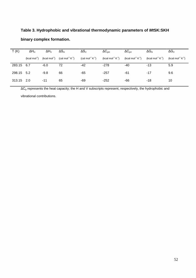
52
Table 3. Hydrophobic and vibrational thermodynamic parameters of MtSK:SKH
binary complex formation.
ΔCp represents the heat capacity; the H and V subscripts represent, respectively, the hydrophobic and
vibrational contributions.
T (K) ΔHH
(kcal mol-1)
ΔHV
(kcal mol-1)
ΔSH
(cal mol-1 K
-1)
ΔSV
(cal mol-1 K
-1)
ΔCpH
(kcal mol-1 K
-1)
ΔCpV
(kcal mol-1 K
-1)
ΔGH
(kcal mol-1 K
-1)
ΔGV
(kcal mol-1 K
-1)
283.15 6.7 -6.0 72 -42 -278 -40 -13 5.9
298.15 5.2 -9.8 66 -65 -257 -61 -17 9.6
313.15 2.0 -11 65 -69 -252 -66 -18 10

53
Capítulo 3
Considerações Finais
54
A tuberculose, ocasionada pelo bacilo Mycobacterium tuberculosis, representa um
grande problema de saúde global devido a sua alta taxa de incidência e prevalência,
além de casos de coinfecção reportados pela Organização Mundial da Saúde. Estima-se
que em 2009 ocorreram 9,2 milhões de novos casos e 1,7 milhão de mortes (WHO,
2010) em contraste aos 8,7 milhões de novos casos e 1,4 milhão de mortes encontrados
em 2011 (WHO, 2012). Apesar da diminuição de novos casos nos últimos anos, o
surgimento de novas cepas resistentes tem tornado o quadro preocupante, aonde 3,7%
(320 000) dos novos casos e 20% de casos tratados em 2011 foram diagnosticados
como MDR (WHO, 2012) em contraste aos 30 000 casos de MDR diagnosticados em
2008 (WHO, 2010). Devido ao aumento na incidência de casos resistentes, o
desenvolvimento de novas drogas contra o bacilo da tuberculose tem se mostrado um
campo de pesquisa extremamente importante para a saúde global a curto e longo prazo.
O desenvolvimento racional de novas drogas tem como base o estudo dos mecanismos
relacionados às enzimas candidatas a alvo molecular. Adicionalmente, os inibidores
enzimáticos possuem uma parcela de 25% do mercado de medicamentos nos Estados
Unidos (Robertson 2005; Robertson 2007). De acordo com a BBC, em 2010 o marcado
mundial de inibidores de quinases alcançou a marca de 28,1 bilhões de dólares, e
aproximadamente 29,1 bilhões de dólares em 2011. Acredita-se que em 2016 esse valor
suba para 40,2 bilhões de dólares (BBC, 2012). O mercado americano alcançou a marca
de 10,8 e 10,4 bilhões de dólares em 2010 e 2011 respectivamente, e se tem uma
estimativa que o valor subirá para 11,6 bilhões de dólares em 2016 (BBC, 2012). Na
busca por uma enzima alvo, a via metabólica do chiquimato foi escolhida devido a sua
essencialidade para a viabilidade do bacilo e a sua ausência no hospedeiro humano,
sendo assim um promissor alvo seletivo (Parrish, 2002). Embasado nos dados
preocupantes relacionados à infecção e desenvolvimento da tuberculose, na grande
55
importância das enzimas como alvo molecular para o desenvolvimento de drogas e tendo
em vista a relevância das enzimas classificadas como quinases nos mercados de
medicamentos mundial e americano, este trabalho versou sobre os mecanismos cinético
e termodinâmico além de características estruturais relacionados à enzima MtSK. O
desenvolvimento de um protocolo de purificação, identificação e a integridade da enzima
MtSK, forneceram embasamento para realização de ensaios de cinética em estado-
estacionário, que permitiram a identificação do mecanismo empregado pela enzima
durante a catálise da reação utilizando os substratos naturais e as suas constantes. A
determinação das constantes cinéticas indicaram que a enzima MtSK deve ser mais
propriamente classificada como uma Chiquimato quinase do tipo II codificada pelo gene
aroL, devido a maior similaridade nos valores cinéticos encontrados em comparação às
enzimas ortólogas codificadas pelos genes aroK e aroL em Escherichia coli. Além disso,
foram realizados estudos termodinâmicos através de calorimetria de titulação isotérmica
a fim de observar a formação dos complexos MtSK com os substratos/produtos naturais,
permitindo assim a definição mais adequada do mecanismo enzimático e as assinaturas
termodinâmicas na interação proteína ligante. As assinaturas termodinâmicas
identificaram que a enzima realiza uma adição ordenada aleatória na adição dos
substratos e uma liberação ordenada sequencial na liberação dos produtos, tendo como
primeiro produto a ser liberado o S3P, devido a sua incapacidade de se ligar na enzima
livre. Adicionalmente, foi possível observar que a ligação do SKH no complexo
Mg2+ATP:MtSK produziu um valor de energia livre final menos favorável do que a sua
ligação na enzima livre, indicando assim uma cooperatividade negativa, ou seja, o ATP
dificulta a ligação do SKH no sítio ativo. Devido à capacidade de ligação do SKH na
enzima livre, ensaios de formação do complexo binário MtSK:SKH foram realizados em
condições experimentais variadas. Primeiramente, ensaios termodinâmicos foram
56
realizados em condições experimentais idênticas às utilizadas na identificação do
mecanismo enzimático, tendo como única variável a temperatura. Esses ensaios
permitiram a identificação da capacidade calorífica em pressão constante, que indicou a
dessolvatação de grupos apolares e um efeito hidrofóbico importante na formação do
complexo. Esses dados foram confirmados através do modelo estrutural desenvolvido
neste trabalho. Por final, a formação do complexo foi observada tendo como variável o
tampão de reação. Este ensaio nos permitiu a identificação da transferência de 1 próton
do complexo MtSK:SKH para o solvente, indicando assim os aminoácidos arg 38 e 156
encontrados no modelo estrutural construído neste trabalho, como fonte de prótons,
tendo assim um papel importante na formação do complexo. Estes aminoácidos foram
indicados como fonte de prótons devido ao seu grau de ionização, calculado pelo valor
do pKa teórico no pH utilizado nas reações e pelas ligações de hidrogênio observadas no
modelo estrutural. Estes dados foram agrupados e submetidos à revista PLoSONE que
após revisão aceitou o trabalho para publicação. Os resultados produzidos nesta tese
permitiram a identificação de características cruciais para o desenvolvimento de
inibidores com ação seletiva tendo como alvo a enzima MtSK, acrescentando dados
importantes sobre a reação catalisada e aminoácidos importantes na formação do
complexo MtSK:SKH. O SKH pode por fim, ser utilizado como estrutura inicial para o
desenvolvimento de moléculas de ação inibitória competitiva.

57
Referências
58
Bentley, R., Crit. Rev. Biochem. Mol. Biol; 1990; 25, 307-384.
Business Communications Company (BCC), Kinase inhibitors: Global Markets;
2012.
Campbell, S.A., Richards, T. A., Mui, E.J., Samuel, B.U., Coggins, J.R., McLeod,
R., Roberts, C. W.; Int. J. Parasitol; 2004; 34,5-13.
Cole, S.T., Brosch, R., Parkhill, J., Garnier, T., Churcher C. et al; Nature; 1998;
393, 537-544.
Dhaliwal, B., C.E. Nichols, J. Ren, M. Lockyer, I. Charles, A.R. Hawkins, D.K.
Stammers, FEBS Lett.; 2004; 574, 49-54.
Dias, M.V., Faím, L.M., Vasconcelos I.B, Oliveira J.S., Basso, L.A.,Santos D.S.,
Azevedo, W.F.Jr, Acta Crystallogr. Sect. F Struct. Biol. Cryst. Commun.; 2007; 63,
1-6.
Ducati, R.G., Basso, L.A., Santos, D.S.; Current drug targets; 2007; 3, 423-435.
Goodman & Gilman A, et al. As bases farmacológicas da terapêutica. Rio de
Janeiro. Ed. McGraw-Hill, 2006: 1821p;
Haq, I.; Calorimetry in the fast lane: The use of ITC for obtaining enzyme kinetics
constants. Microcal, LLC application note. 2002.
Hargreaves S., WHO report highlights alarming rise of resistant tuberculosis. 2008;
8: 220;
Hartmann, M.D., G.P. Bourenkov, A. Oberschall, N. Strizhov, H.D. Bartunik, J. Mol.
Biol.; 2006; 364, 411-423.
Herman, K.M and Weaver, L.M; Annu. Rev. Plant Physiol. Plant Mol. Biol.;1999;
50;473–503.
Keeling, P.J., Palmer J.D.,. Donald, R.G., Roos, D.S., Waller, R.F., McFadden. G.
I. Nature; 1999, 397:219–220.
Laemmli, U.K., Cleavage of structural proteins during the assembly of the head of
bacteriophage T4, Nature.;1970; 227, 680-685.
McConkey, G. A., Antimicrob. Agents Chemother. 1999; 43; 175–177.
McConkey, G. A., J. W. Pinney, D. R. Westhead, K. Plueckhahn, T. B. Fitzpatrick,
P. Macheroux, B. Kappes. Trends Parasitol; 2004; 20:60–65.

59
Parrish, T., Stoker, N.G., Microbiology; 2002; 148, 3069–3077
Parrish, N.M., Dick, J.D, Bishai, W. R., Mechanisms of latency in Mycobacterium tuberculosis, 1998, 6 , 107-112;
Pereira, J.H., Oliveira J.S., F. Canduri, M.V. Dias, M.S. Palma, L.A. Basso, D.S.
Santos, W.F.Jr. Azevedo, Acta Crystallogr. D Biol. Crystallogr.; 2004; 60, 2310-
2319.
Roberts, F., C. W. Roberts, J. J. Johnson, D. E. Kyle, T. Krell, J. R. Coggins,G. H.
Coombs, W. K. Milhous, S. Tzipori, D. J. Ferguson, D. Chakrabarti, R. McLeod.
Nature; 1998; 393,801–805.
Roberts. C. W., F. Roberts, R. E. Lyons, M. J. Kirisits, E. J. Mui, J. et al.; J. Infect.
Dis. 2002; 185(Suppl. 1), S25–S36.
Robertson J G, Mechanistic basis of enzyme-targeted drugs, Biochem., 2005; 44,
5561-5571.
Robertson J G, Enzymes as a special class of therapeutic target: clinical drugs and
modes of action, Curr. Opin. Struct. Biol., 2007; 17, 674-679.
World Health Organization (WHO), Global Tuberculosis Control: a short update to
the 2009 report; 2010.
World Health Organization (WHO), Global Tuberculosis Report, 2012.
Yan, H., Tsai, M.D.; Adv. Enzymol. Relat. Areas Mol. Biol.; 1999, 73,:103-134

60
Anexos

61
Anexo A
Kinetic mechanism and energetics of nucleotide binding to Mycobacterium tuberculosis Cytidine Monophosphate Kinase. Biophysical chemistry, 2013. (Artigo Submetido)
Léia Jaskulski, Leonardo A. Rosado, Diana C. Rostirolla, Diógenes S. Santos, Luiz A. Basso.
62

63
Anexo B
Biochemical characterization of uracil phosphoribosyltransferase from Mycobacterium tuberculosis. PLoSONE, 2013. (Artigo Aceito)
Anne Drumond Villela, Rodrigo Gay Ducati, Leonardo Astolfi Rosado, Carlos Bloch Junior, Maura Vianna Prates, Carlos Henrique Inacio Ramos, Luiz Augusto Basso, Diógenes Santiago Santos.

64

65
Anexo C
Role of Serine140 in the mode of action of Mycobacterium tuberculosis β-Ketoacyl-ACP reductase (MabA). BMC Research Notes, 2012. (Artigo Publicado)
Leonardo A. Rosado, Rafael Andrade Caceres, Walter Filgueira de Azevedo, Luiz A Basso and Diógenes S. Santos.

66
67
Anexo D
Recombinant Erwinia carotovora L-asparaginase II production in Escherichia coli fed-batch cultures. Brazilian Journal of Chemical Engineering, 2012. (Artigo Aceito)
Gustavo Roth, José E.S. Nunes, Leonardo A. Rosado, Cristiano V. Bizarro, Giandra Volpato, Cláudia P. Nunes, Gaby Renard, Luiz A. Basso, Diógenes S. Santos, Jocelei M. Chies.

68
69
Anexo E
Molecular, kinetic and thermodynamic characterization of Mycobacterium tuberculosis orotate phosphoribosyltransferase. Molecular Biosystems, 2012. (Artigo Publicado)
Ardala Breda, Leonardo Astolfi Rosado, Daniel Macedo Lorenzini, Luiz Augusto Basso and Diógenes Santiago Santos.
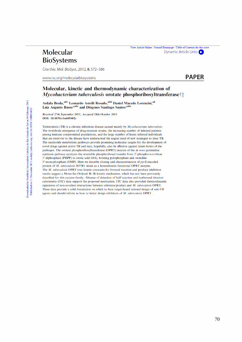
70
71
Anexo F
Pyrimidin-2(1H)-ones based inhibitors of Mycobacterium tuberculosis orotate phosphoribosyltransferase. European Journal of Medicinal Chemsitry, 2012. (Artigo Publicado)
Ardala Breda, Pablo Machado, Leonardo Astolfi Rosado, André Arigony Souto, Diógenes Santiago Santos and Luiz Augusto Basso.

72

73
Anexo G
Wild-Type Phosphoribosylpyrophosphate Synthase (PRS) from Mycobacterium tuberculosis: A Bacterial Class II PRS? PlosOne, 2012. (Artigo Publicado)
Ardala Breda, Leonardo K. B. Martinelli, Cristiano V. Bizarro, Leonardo A. Rosado, Caroline B. Borges, Diógenes S. Santos, Luiz A. Basso.
74

75
Anexo H
Molecular, kinetic, thermodynamic, and structural analyses of Mycobacterium tuberculosis hisD-encoded metal-dependent dimeric histidinol dehydrogenase (EC 1.1.1.23). Archives of Biochemistry and Biophysics, 2011. (Artigo Publicado)
José E.S. Nunes, Rodrigo G. Ducati Ardala Breda, Leonardo A. Rosado, Bibiana M. de Souza, Mario S. Palma, Diógenes S. Santos Luiz A. Basso.
76