Embed Size (px)
Citation preview
i
ALEXANDRE DE SOUZA VIOLA
PREVALÊNCIA DE HIPERPLASIA E CÂNCER DE ENDOMÉTRIO EM MULHERES ASSINTOMÁTICAS
COM SOBREPESO OU OBESIDADE
Dissertação de Mestrado
ORIENTADOR: Prof. Dr. LUIS GUILLERMO BAHAMONDES
Unicamp 2007
ii
ALEXANDRE DE SOUZA VIOLA
PREVALÊNCIA DE HIPERPLASIA E CÂNCER DE ENDOMÉTRIO EM MULHERES ASSINTOMÁTICAS
COM SOBREPESO OU OBESIDADE
Dissertação de Mestrado apresentada à Pós-Graduação da Faculdade de Ciências Médicas da Universidade Estadual de Campinas para obtenção do Título de Mestre em Tocoginecologia, área de Tocoginecologia
ORIENTADOR: Prof. Dr. LUIS GUILLERMO BAHAMONDES
Unicamp 2007
FICHA CATALOGRÁFICA ELABORADA PELA
BIBLIOTECA DA FACULDADE DE CIÊNCIAS MÉDICAS UNICAMP
Bibliotecário: Sandra Lúcia Pereira – CRB-8ª / 6044 Título em inglês: Prevalence of endometrial cancer and hyperplasia in non symptomatic women with overweight or obesity
Keywords: • Overweight • Obesity • Endometrium • Hyperplasia • Neoplasm • Post-menopause
Titulação: Mestre em Tocoginecologia Área de concentração: Tocoginecologia Banca examinadora: Prof Dr Luis Guillermo Bahamondes
Profa. Dra. Isabel Cristina Guazzelle Profa. Dra. Ilza Maria Urbano Monteiro
Data da defesa: 28 - 08 - 2007
Viola, Alexandre de Souza V811p Prevalência de hiperplasia e câncer de endométrio em
mulheres assintomáticas com sobrepeso ou obesidade / Alexandre de Souza Viola. Campinas, SP: [s.n.], 2007.
Orientador : Luis Guillermo Bahamondes Dissertação(Mestrado) Universidade Estadual de Campinas.
Faculdade de Ciências Médicas. 1. Sobrepeso. 2. Obesidade. 3. Endométrio. 4. Hiperplasia.
5. Câncer. 6. Pós-menopausa. I. Bahamondes, Luis Guillermo. II. Universidade Estadual de Campinas. Faculdade de Ciências Médicas. III. Título.

iv
Dedico este trabalho...
A Deus Pai, Filho e Espírito Santo
À minha Esposa Carolina e meu filho Lucas
Aos familires e amigos
Às Mulheres voluntárias

v
Agradecimentos
À Instituição Unicamp pelas condições oferecidas para a realização desta pesquisa e ao
incentivo da mesma.
À Prefeitura Municipal de Vinhedo pelas condições oferecidas para a realização desta
pesquisa e ao incentivo da mesma.
Aos Professores do curso de pós-graduação do Departamento de Tocoginecologia da
Unicamp.
Aos colegas Dra. Daniela Gouveia, Dra. Liliana Andrade, Dr. José M. Aldrighi pelo
incentivo, apoio técnico e científico e dedicação na realização deste estudo.
Ao Cemicamp e à enfermeiras, Creuza, Maria Cecília, Maria Margarete, Marina, Nádia,
Sara e Ximena, pela colaboração na realização deste estudo.
Às Profas. Dras. Ilza Maria Urbano Monteiro e Cássia Raquel Teatin Juliato pelas
valiosas contribuições durante a qualificação.
À Verônica pela ajuda em tantas coisas, como na formatação da tese, elaboração da aula.
À Adriana e Lusia Helena, pela amizade e ajuda durante a realização deste estudo.
À Adriele, Juliana e Clarice, minhas secretárias e cooperadoras, pela ajuda durante a
realização deste estudo.
vi
Às auxiliares de enfermagem Laís, Edite, Eliane, Isabel e Lourdes pela ajuda no atendimento
às pacientes.
Aos estatísticos Sirlei e José Vilton, por serem sempre tão atenciosos, simpáticos e
eficientes na análise dos dados.
A toda equipe da ASTEC, pela competência e disponibilidade durante a realização deste
estudo, principalmente pela paciência da Rosário e da Cylene.
À Margarete e à Sueli Regina, pela colaboração durante a realização deste estudo.
Em Especial:
Ao Prof. Dr. Luis Guillermo Bahamondes, meu orientador, por todo estímulo e ajuda na
realização desta pesquisa, e principalmente, por ser co-responsável por promover
um dos momentos mais felizes da minha vida.
vii
Sumário
Símbolos, Siglas e Abreviaturas .................................................................................................. viii
Resumo ..........................................................................................................................................ix
Summary ........................................................................................................................................xi
1. Introdução ............................................................................................................................... 13
2. Objetivos ................................................................................................................................. 20
2.1. Objetivo geral .................................................................................................................. 20
2.2. Objetivos específicos ...................................................................................................... 20
3. Publicação............................................................................................................................... 21
4. Conclusões ............................................................................................................................. 42
5. Referências Bibliográficas....................................................................................................... 43
6. Anexos .................................................................................................................................... 50
6.1. Anexo 1 – Lista de Verificação........................................................................................ 50
6.2. Anexo 2 – Formulário ...................................................................................................... 51
6.3. Anexo 3 – Termo de Consentimento Livre e Esclarecido ............................................... 53
Símbolos, Siglas e Abreviaturas viii
Símbolos, Siglas e Abreviaturas
ACOs – Anticoncepcionais orais combinados
CAISM – Centro de Atenção Integral à Saúde da Mulher
DP – Desvio padrão
IC – Intervalo de confiança
IGF – Insulin-like growth factor
IMC – Índice de massa corporal
OMS (WHO) – Organização Mundial da Saúde
OR – Odss Ratio
RR – Risco Relativo
Unicamp – Universidade Estadual de Campinas
Resumo ix
Resumo
Objetivos: Determinar a prevalência de hiperplasia e câncer de endométrio em
mulheres assintomáticas com sobrepeso ou obesidade. Sujeitos e métodos:
Realizou-se um estudo de corte transversal no qual 193 mulheres voluntárias do
Ambulatório de Reprodução Humana, Departamento de Tocoginecologia, Faculdade
de Ciências Médicas, CAISM - Unicamp foram avaliadas com biópsia de endométrio
com Pipelle de Cornier. Os achados anatomopatológicos foram catalogados como
endométrio normal, hiperplasia ou câncer de endométrio e estes resultados foram
comparados com o ìndice de massa corporal (IMC; kg/m2). Para a análise dos
dados, as mulheres foram divididas em dois grupos: em idade reprodutiva e na
pós-menopausa e pelo IMC (kg/m2) em sobrepeso ou obesidade. Resultados: A
prevalência de hiperplasia na amostra total foi de 7,3%, na idade reprodutiva de
5,8% e na pós-menopausa de 12,1%. A prevalência de adenocarcinoma de
endométrio na amostra total foi de 1,5%, na idade reprodutiva de 1% e na pós-
menopausa de 3%. A regressão logística multivariada mostrou que o risco de
apresentar hiperplasia e câncer de endométrio foi de 1,19 (IC 95%; 0,36–3,90)
para as mulheres na pós-menopausa, de 1,58 (IC 95%; 0,30-8,23) para as que
tinham obesidade severa e de 2,72 (IC 95%; 0,65–11,5) para as que tinham

Resumo x
obesidade morbida. Estar na idade reprodutiva, com sobrepeso ou obesidade
moderada não alterou o risco. Conclusões: No grupo avaliado, mulheres
assintomaticas na pós-menopausa com obesidade severa ou mórbida apresentaram
risco aumentado de apresentar hiperplasia ou câncer de endométrio. O mesmo
não ocorreu com mulheres em idade reprodutiva.
Summary xi
Summary
Objetives: To determine the prevalence of endometrial cancer and hyperplasia in
non-symptomatic women with overweight or obesity. Subjects and methods: A
cross-sectional study was carried-out at the Obstetrics and Gynecology
Departament, School of Medicine, Unicamp, with 193 women. The women were
evaluated through endometrial biopsy with a Pipelle de Cornier. The hystophatologic
findings were classified as normal endometrium, hyperplasia and cancer and those
results were compared to the body mass index (BMI; kg/m2). For the analysis
pourpose the women were divided in two groups: at reproductive age and at the
post-menopause and by the BMI (kg/m2) in overweigth and obesity. Results: The
prevalence of hyperplasia in the total sample was 7.3%, among women at
reproductive age of 5.8% and at the post-menopause it was of 12.1%. The
prevalence of endometrial cancer at the total sample was 1.5%, among women
at reproductive age it was 1% and at the post-menopause of 3%. The logistic
regression showed that the risk of endometrial hyperplasia or cancer was of
1.19 (95% CI: 0.36–3.90) for women at the post-menopause, 1.58 (95% CI: 0.30–
8.23) for those who presented severe obesity, and of 2.72 (95% CI: 0.65–11.5)
for those women with morbid obesity. Be at the reproductive age, with

Summary xii
overweight or moderate obesity did not modify the risk. Conclusions: At the
evaluated group of women, non-symptomatic women at the post-menopause with
severe or morbid obesity presented a high risk to have endometrial hyperplasia or
cancer. However, the same was not observed among women at reproductive age.
Introdução 13
1. Introdução
As mulheres com sobrepeso ou obesidade apresentam, com maior
freqüência, alguns tipos de cânceres como o de mama e de cólon e em 30% das
mulheres com câncer de endométrio o sobrepeso ou obesidade está presente
(Disaia, 2002). Entretanto, a maioria destes dados foi obtida em estudos
retrospectivos tipo caso-controle com mulheres já com o diagnóstico de câncer de
endométrio ou com sintomas de suspeita do mesmo, como sangramento vaginal
na pós-menopausa ou sangramento irregular durante a idade reprodutiva.
No entanto, estes dados apenas expressam que mulheres com câncer de
endométrio apresentam como um dos fatores de risco a obesidade. Entretanto,
podem não expressar a realidade das mulheres com sobrepeso ou obesidade, as
quais, na sua maioria, são assintomáticas quanto ao câncer de endométrio e suas
lesões precursoras, da mesma forma como ocorre em mulheres com peso normal.
Por não apresentarem sintomas, habitualmente, estas mulheres não são
submetidas rotineiramente a procedimentos que permitam um diagnóstico
precoce para este tipo de câncer como a biópsia de endométrio, curetagem
Introdução 14
uterina e ultra-sonografia pélvica (Febrasgo, 2002). Rotineiramente, isto ocorre
em mulheres sintomáticas, principalmente se estão na pós-menopausa e
apresentam sangramento uterino ou se estão ainda na idade reprodutiva e
apresentam metrorragia.
A obesidade é considerada uma epidemia global e um problema de
saúde pública (WHO, 2003). Está entre as cinco primeiras doenças nos países
desenvolvidos e entre as 10 nos países em desenvolvimento (WHO, 2004). Nos
Estados Unidos, 31% da população são obesos e outros 35% têm sobrepeso.
Destes obesos, 65% são mulheres e 15% crianças ou adolescentes (Hill et al., 2003).
Situação semelhante tem sido observada nos países da América Latina.
Segundo o Conselho Latino-Americano de Obesidade [CLAO] (1998), no México,
39% das mulheres urbanas sofrem de obesidade; na Argentina, 27% apresentam
obesidade e 32,5% sobrepeso. No Paraguai, a obesidade chega a 35,7%
(Filozof et al., 2001) e no Brasil, em 1997, a prevalência da obesidade era de
21% (Monteiro et al., 2000).
Comparando-se os dados de 1974/75 com os de 1989, do Instituto
Brasileiro de Geografia e Estatistica [IBGE, 2004], observou-se um incremento
de 53% da população obesa brasileira. Segundo alguns pesquisadores, neste
ritmo, todos os brasileiros serão obesos até a primeira metade do terceiro
milênio (Halpern et al., 1990). Hoje, segundo o CLAO, 40% dos obesos no
Brasil são mulheres e 27% homens.
Introdução 15
Apesar de os distúrbios metabólicos e alterações genéticas poderem
explicar o sobrepeso e obesidade, a tendência em ganhar peso tem sido mais
atribuída a causas sociais ou comportamentais, como o alto consumo de
energia através de dietas com excesso de calorias, e a diminuição do gasto
energético devido ao estilo de vida sedentário (CLAO, 1998; Monteiro et al.,
2000; Hill et al., 2003).
No primeiro caso, o alto consumo de energia através da ingestão de
carboidratos simples (monossacarídeos) e lipídios monoinsaturados levam a um
desequilíbrio nutricional com a alta ingestão de calorias e baixa quantidade de
outros nutrientes, principalmente proteínas e micronutrientes (Waitezberg, 2004).
Quanto à diminuição do gasto energético, o sedentarismo conseqüente
ao desenvolvimento tecnológico tem levado ao acúmulo de gordura central e
periférica, ocasionado pela interconversão dos macronutrientes (carboidratos,
lipídios e proteínas) dentro do processo metabólico, levando ao sobrepeso e
obesidade (Waitezberg, 2004).
A obesidade aumenta o risco para diabetes melittus tipo 2, doenças
cardiovasculares, como a hipertensão arterial, e alguns tipos de câncer, segundo
The National Heart Lung and Blood Institute (NHLBI, 1998). Existe uma associação
entre obesidade e cânceres de próstata, no homem, e de mama e endométrio
na mulher (CLAO, 1998; Febrasgo, 2002).
Estudos epidemiológicos realizados em diferentes países concluíram que
mulheres com câncer de endométrio apresentam, dentre os fatores de risco
Introdução 16
identificados, uma tríade caracterizada por obesidade, diabetes mellitus tipo 2 e
hipertensão arterial (David e Adéle, 2001) e que o aumento do Índice de Massa
Corporal (IMC) está associado ao maior risco relativo (RR) de óbito (Anderson
et al, 2001). A obesidade isolada eleva o RR de desenvolvimento de câncer de
endométrio entre 3 e 10 vezes (Soliman et al., 2005).
Com exceção do câncer de cérvix, o de endométrio é o câncer ginecológico
mais comum no mundo em desenvolvimento. É o quarto em incidência nas
mulheres e o oitavo em mortalidade, quando ajustado por idade (Landis et al.,
1999). Por exemplo, a estimativa de casos novos de câncer de endométrio nos
Estados Unidos em 1999 foi de 37.400, com 6.400 mortes, diretamente relacionadas
a esta doença (David e Adéle, 2001).
Como já foi apontado nos estudos caso-controle, o câncer de endométrio
apresenta como fatores de risco a obesidade, diabetes e hipertensão arterial. Por
outro lado, o tabagismo, a dieta baixa em lipídios e a atividade física diminuem o
risco. A maior incidência ocorre em mulheres brancas, obesas, na pós-menopausa
e que vivem na zona urbana. Geralmente têm um bom prognóstico, pois, na
maioria das vezes, está confinado ao útero (Swanson et al., 1993; David e
Adéle, 2001; Disaia, 2002).
A associação entre a obesidade e o câncer de endométrio em mulheres na
pós-menopausa é forte e consistente para alguns autores, porém, para as mulheres
em idade reprodutiva, isto não se aplicaria (Torneberg e Carstensen, 1994). No
entanto, outros autores encontraram uma forte associação nestas mulheres,
Introdução 17
principalmente as que apresentaram um significativo ganho de peso na idade
reprodutiva, o qual as leva ao sobrepeso ou obesidade (Swanson et al., 1993).
Por ser a hiperplasia de endométrio, em geral, um estágio prévio ao câncer,
esta patologia também estaria vinculada aos mesmos fatores de risco como
obesidade, hipertensão arterial e diabetes mellitus tipo 2. Entretanto, apresentar
hiperplasia de endométrio não significa necessariamente que a mulher virá a ter um
câncer de endométrio. Aproximadamente 10% das mulheres com hiperplasia
complexa evoluíram para câncer em até cinco anos de seguimento (Kurman et
al., 1985; WHO, 1994).
Os mecanismos que determinam as lesões proliferativas e até mesmo o
câncer de endométrio tipo 1 não são totalmente conhecidos. O sobrepeso ou a
obesidade estaria associado ao câncer de endométrio por promover um aumento
da insulin-like growth factor (IGF) I devido à hiperinsulinemia, agindo como fator
de proliferação local, acarretando hiperplasia ou câncer de endométrio (Potischman
et al, 1999; Kaaks et al, 2001; Kaaks et al., 2002) e por aumentar a exposição
das células endometriais a altas taxas de estrogênio sérico (Austin, 1991).
A elevação do estrogênio endógeno sérico ocorreria de duas formas. A
primeira, pelo processo de aromatização da androstenediona no tecido adiposo,
devido ao estado hiperinsulinêmico (Enrior et al., 1984; Garcia et al., 1998; Deruelle
et al., 2001; Bianchini et al., 2002; Webb, 2006) e a segunda, pela redução do
hormônio carregador de globulina sexual [SHBG] (Davidson et al., 1981; Troisi
et al., 1997; Nahás et al., 1998; Chubak et al., 2004; Carvalheira et al., 2006).
Introdução 18
Além da obesidade, outras situações que poderiam elevar o estrogênio
sérico já foram associadas ao câncer de endométrio, como a síndrome dos ovários
policísticos, tumor ovariano secretor de estrogênio, terapia de reposição estrogênica
sem oposição de progestogênio, uso de contraceptivos orais combinados com
altas doses de estrogênio e baixas doses de progestogênio e uso do tamoxifeno
(David e Adele, 2001; Disaia, 2002; Hammond e Johnson, 2004). Assim, por
exemplo, um ano de tratamento com estrogênio eqüino conjugado a doses de
0,625mg ou equivalente levaria a um aumento de 20% na taxa de hiperplasia
de endométrio (Woodruff e Pickared, 1994).
A obesidade também poderia estar associada com a progressão da
carcinogênese endometrial. Quanto mais jovem a mulher apresentar sobrepeso ou
obesidade, maior é o tempo de exposição endometrial ao estrogênio e, portanto,
mais avançado o estágio da doença no momento do diagnóstico, diminuindo o
sucesso do tratamento cirúrgico (David e Adele, 2001; Disaia, 2002; Hammond
e Johnson, 2004).
Os estudos sobre a carcinogênese endometrial têm sido feitos, em geral,
com mulheres na pós-menopausa, onde a possibilidade de exposição a níveis
estrogênicos altos e por períodos longos é mais comum de ocorrer do que em
mulheres em idade reprodutiva (Disaia, 2002). Porém, nos últimos 15 anos tem-se
observado um aumento da obesidade e, conseqüentemente, do hiperestrogenismo
em mulheres durante a idade reprodutiva, segundo o Centers for Disease Control
and Prevention’s Behavioral Risk Factor Surveillance System (CDC’s BRFSS,
2002) dos Estados Unidos.
Introdução 19
Hoje, mulheres obesas em idade reprodutiva, com história familiar de câncer
de endométrio e que apresentem sangramento vaginal irregular ou espessamento
do endométrio pela ultra-sonografia, têm procurado ou são encaminhadas ao
ginecologista para investigação do câncer de endométrio e de suas lesões
precursoras (Disaia, 2002).
Entretanto, isso não é o mais comum. O que habitualmente se vê é que
mulheres com obesidade e em idade reprodutiva têm procurado endocrinologistas,
nutricionistas e cirurgiões em busca da redução de peso, sem, no entanto,
preocuparem-se em procurar um ginecologista para a prevenção do câncer de
endométrio, pois na maioria das vezes são assintomáticas.
Embora não existam dúvidas de que um grande número de mulheres
com câncer e hiperplasia de endométrio são obesas ou têm sobrepeso, o inverso
não está totalmente elucidado. Saber o efeito do sobrepeso ou obesidade e
suas consequências sobre o endométrio, nas mulheres assintomáticas, poderá
modificar ou não a forma de como se tem cuidado e tratado desta população,
em particular. Conhecer a sua real prevalência pode provocar a mudança no
protocolo de atendimento e beneficiar estas mulheres com um diagnóstico mais
precoce e tratamento mais conservador.
Objetivos 20
2. Objetivos
2.1. Objetivo geral
Determinar as prevalências de hiperplasia e câncer de endométrio em
mulheres assintomáticas com sobrepeso ou obesidade.
2.2. Objetivos específicos
Determinar a prevalência de hiperplasia de endométrio.
Determinar a prevalência de câncer de endométrio.
Determinar as prevalências de hiperplasia de endométrio e de câncer de
endométrio em mulheres em idade reprodutiva e na pós-menopausa.
Correlacionar a hiperplasia endometrial e o câncer de endométrio com o
índice de massa corporal.
Publicação 21
3. Publicação
ASSOCIAÇÃO MÉDICA BRASILEIRA
São Paulo, 30 de julho de 2007.
Ilmo. Sr.
Dr. Alexandre de Souza Viola Comunicamos que em 30 de julho de 2007 recebemos o artigo “Prevalência de hiperplasia e câncer de endométrio em mulheres assintomáticas com sobrepeso ou obesidade”, protocolado sob o n.° 6804, de sua autoria, e que o mesmo será analisado pelo Conselho Editorial da Revista da Associação Médica Brasileira para possível publicação. Atenciosamente, Prof. Dr. Bruno Caramelli Editor da Revista da Associação Médica Brasileira
Publicação 22
Prevalence of endometrial cancer and hyperplasia in non-symptomatic overweight
and obese women
1Alexandre S Viola, 2Daniela Gouveia, 3Liliana Andrade, 2Jose M. Aldrighi, 1Carolina F M Viola, 1Luis Bahamondes.
1Department of Obstetrics and Gynecology and 3Department of Pathology, School of
Medicine, Universidade Estadual de Campinas (UNICAMP), Caixa Postal 6181, 13084-
971, Campinas, SP, Brazil and 2Department of Obstetrics and Gynecology, School of
Medicine, Santa Casa de Misericordia, São Paulo, Brazil
Address for correspondence
Dr. Luis Bahamondes
Caixa Postal 6181
13084-971 Campinas, SP
Brazil
Telephone: +55-19-3289-2856
Fax: +55-19-3289-2440
E-mail: [email protected]
Publicação 23
Abstract
Objectives: To determine the prevalence of endometrial hyperplasia and cancer among
non-symptomatic women with overweight and obesity.
Subjects and Methods: A cross-sectional study was carried out with 193 women who
were submitted to an endometrial biopsy with a Pipelle de Cornier. The findings were
categorized as normal, hyperplasia or cancer and these results were compared to the Body
Mass Index (BMI; kg/m2). Women were divided in two groups for analysis: at reproductive
age and at the post-menopause and by the BMI (kg/m2) as overweight and obesity.
Results: The prevalence of endometrial cancer was 1.0% in women at reproductive age
and 3.0% among women at the post-menopause. The prevalence of endometrial hyperplasia
was 5.8% in women at reproductive age and 12.1% among women at the post-menopause.
The logistic regression showed that be in the post-menopause increase the risk of endometrial
hyperplasia and cancer to 1.19 (95%CI: 0.36–3.90); presented severe obesity the OR
was 1.58 (95%CI: 0.30–8.23) and morbid obesity the OR was 2.72 (95%CI: 0.65–11.5).
Be at reproductive age with overweight or obesity does not increase the risk.
Conclusions: Our results allow that non-symptomatic women at the post-menopause
with severe or morbid obesity have a high risk to present endometrial hyperplasia or cancer;
however, the same findings were not observed among women at reproductive age.
Key words: overweight, obesity, endometrium, cancer, hyperplasia
Publicação 24
Introduction
Obesity is defined as excess body fat; however, due to the fact that it is difficult
to measure body fat directly, body mass index (BMI; kg/m2) is used habitually to
measure obesity (1). Obesity is a global epidemic and a public health problem and it is
among the first five diseases in the developed countries and among the first 10 in
developing countries (2, 3). In the US, 31% of the population are obese and 35% are
overweight (4) and in a short time by 2003–2004 the prevalence had increased to almost
34% in women (5). The prevalence of obesity and overweight increase with age.
Among adults 20–39 years of age, 26.8% were obese, at 40- to 59-year-old adults 34.8%
were obese, and among 60- to 79-year-old adults 35.2% were obese (6). In Brazil,
comparing data of 1974/75 with of 1989 (7) it was an increment of 53% of the obese
population and 40% of the obese are women.
The tendency to weight increase has been attributed to social or behavioral causes, like
high energy intake with high calories diet and low energy expenditure mainly due to
sedentary life (4, 8). The obesity increase the risk for type 2 diabetes, blood hypertension
and cardiovascular disease (9) and women with endometrial cancer habitually presents
obesity, hypertension and type 2 diabetes (10). The obesity, as isolated factor, increases
the relative risk (RR) of have endometrial cancer between 2 to 3 folds (11).
Biologically, the obesity could be linked to endometrial cancer through the
increment of the insulin-like growth factor I (IGF-I) due to hyperinsulinemia as a local
proliferation factor provoking hyperplasia or cancer (12- 14) and due to the high
exposition of the endometrial cells to high serum estrogen environment (14, 15).
However, the studies with endometrial carcinogenesis were conducted, mainly, with
women at the post-menopause when the possibility to exposure to a high estrogens
Publicação 25
environment and for long-time is greater than with women at reproductive age (16).
However, with the increase of the prevalence of obesity a hyperestrogenism has been
also observed in young women (17).
The common rules in many services is that premenopausal obese women, with
familiar history of endometrial cancer, irregular uterine bleeding or with high endometrial
thickness at the ultrasound examination were referred to investigate endometrial lesions.
However, it is common only for symptomatic women. However, it is not common for
non-symptomatic women. Habitually, premenopausal obese women have been consulting
endocrinologists, nutritionists and bariatric surgeons in order to weight loss; however, if
they were have no complaints it is uncommon to look for a gynecologist to evaluate the
endometrium. For this reason, the aim of this study was to determine the prevalence of
endometrial hyperplasia and cancer in non-symptomatic overweight or obese women.
Subjects and Methods
A cross-sectional study was conducted at the Department of Obstetrics and
Gynecology, School of Medicine, Universidade Estadual de Campinas (UNICAMP),
Campinas and at the Department of Obstetrics and Gynecology, School of Medicine,
Santa Casa de São Paulo, Brazil between March 2005 and 2007. The protocol was
approved by both IRB and all women signed an informed consent form.
It was invited to participated 264 women and 193 were selected because they
fulfill the inclusion criteria: aged between 18 to 70 years and BMI (kg/m2) > 25. Were
excluded 71 women because they presented any exclusion criteria: use of hormonal
steroids during the 12 months prior to the study, tamoxifen use, ovarian or endometrial
tumor, endometriosis, or history of hysterectomy or endometrial ablation.
Publicação 26
All women were measured by height (in cm) and weight (in kg) with lighter
cloths by the researchers to avoid self-reported errors (18). The BMI (kg/m2) was
categorized as overweight > 25.0 to < 29.5; moderate obesity > 30.0 to < 34.9; severe
obesity > 35.0 to < 39.9, and morbid obesity > 40.0 (19, 20). After that an endometrial
biopsy was taken with a suction curette Pipelle de Cornier (Prodimed, Neuilly-en-Thelle,
France). In the group of women at reproductive age the biopsy was taken on the luteal
phase of the menstrual cycle and in women at the post-menopause or with irregular
menstrual cycles, at any day. The cases with insufficient material were not considered
for analysis.
Statistical analysis
For quantitative variables we used the Mann-Whitney, ANOVA, Tukey, and
Kruskal-Wallis tests. The significance was established at p < 0.05. The relative risk to
present endometrial lesions (hyperplasia or cancer) was estimated by the odds ratio (OR)
with 95% confidence interval (CI). All the data was presented as mean ± standard
desviation (SD).
Results
Among the 193 women who were recruited, the biopsy was not possible to take
in 16 (8.2%) due to cervical stenosis and 177 women performed an endometrial biopsy.
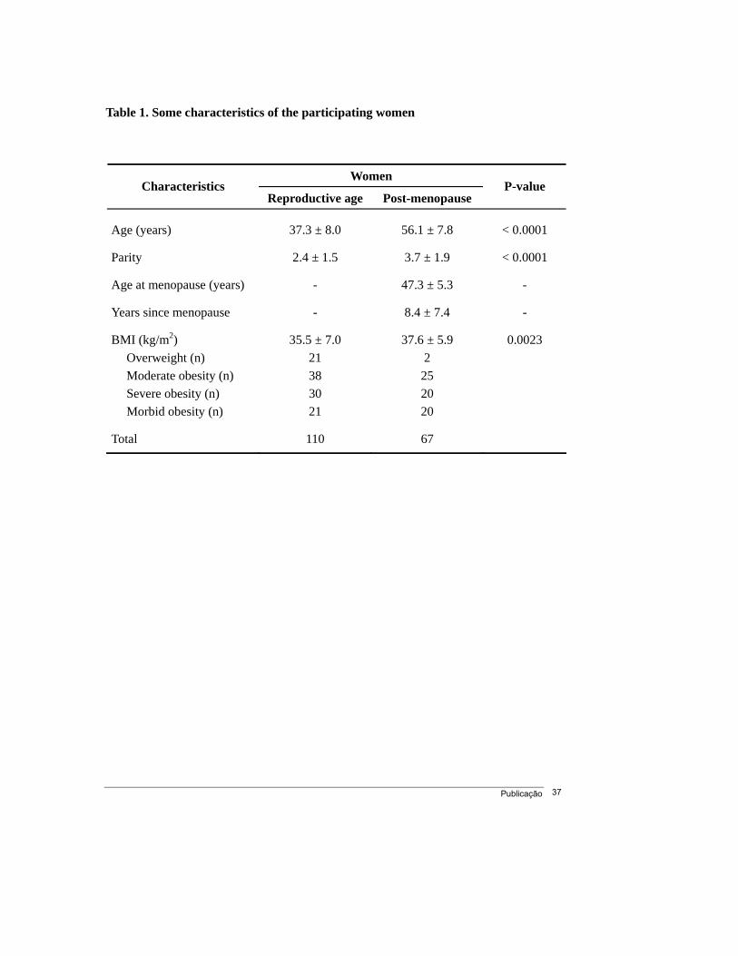
One hundred and ten women (62%) were at reproductive age and 67 (38%) at the post-
menopause. The age of women at the post-menopause was 56.1 ± 7.8 years old (range
42-70), the parity was 3.7 ± 1.9 (range 0-10), the age at the menopause was at 47.6 ± 5.3
years (range 35-58), and the time elapsed from the menopause to the study was 8.4 ± 7.4
Publicação 27
years (range 1-28). For women at reproductive age, the age was 37.3 ± 8.0 years (range
20-55) and the parity was 2.4 ± 1.5 (range 0-8) (Table 1).
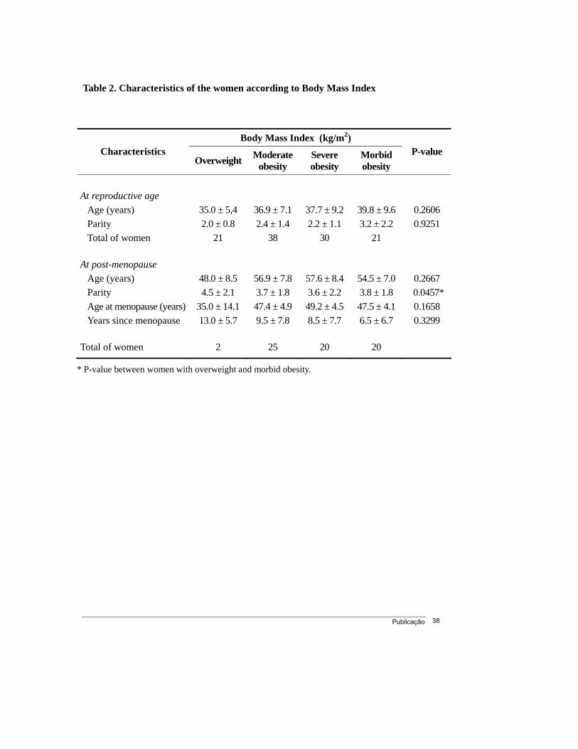
Regarding the BMI (kg/m2) in the group of women at reproductive age it was
35.5 ± 7.0 (range 26.7-66.3) and was distributed in 21 women with overweight, 38
moderate obesity, 30 with severe obesity, and 21 with morbid obesity. The group of
women at the post-menopause showed a BMI (kg/m2) of 37.6 ± 5.9 (range 27.1-64.1)
and the distribution was 2, 25, 20, and 20 women with overweight and moderate, severe,
and morbid obesity, respectively (Table 1). The characteristics of both groups of women
according to the BMI (kg/m2) were presented at Table 2. The women at reproductive age
have less number of children than the post-menopausal women.
Among the 177 endometrial biopsies 41 (23.1%) showed insufficient material for
diagnostic. From the 136 biopsies analyzed 124 (91.1%) showed normal endometrium
and 12 (8.9%) endometrial hyperplasia or cancer. The hystophatologic findings in
women with normal endometrium was 70 (51.1%) proliferative, 32 (23.5%) secretor,
and 22 (16.7%) atrophic. Among women with endometrial lesions we showed two cases
(1.5%) with cancer, nine cases with simple or complex hyperplasia without atypia
(6.6%), and one case with simple hyperplasia with atypia (Table 3). Endometrial lesions
were observed in 6.8% of the women at reproductive age and in 15.2% among the post-
menopausal women. The prevalence of endometrial hyperplasia and cancer was 5.8%
and 1.0%, respectively among women at reproductive age. In the post-menopausal
women the prevalence was 12.1% and 3.0% for endometrial hyperplasia and cancer,
respectively.
The prevalence of endometrial lesions was zero, 6.4%, 17.6%, and 9.4% for
women with overweight and moderate, severe or morbid obesity, respectively (Table 4).
Publicação 28
The logistic regression showed that be in the post-menopause and obese increased the
risk to endometrial lesions to 1.19 (95%CI: 0.36 – 3.90), have severe obesity the OR was
1.58 (95%CI: 0.30 – 8.23) and morbid obesity the OR was 2.72 (95%CI: 0.65–11.5).
However, for women at reproductive age overweight or obesity does not increase the
risk of endometrial lesions (Table 5).
Discussion
Our results showed that non-symptomatic overweighed or obese women, either at
reproductive age or at the post-menopause presented endometrial hyperplasia or cancer.
We observed 12 women with some alterations, two (1.1%) with endometrial
adenocarcinoma, one among women at reproductive age and other at the post-
menopause and 10 women (6.7%) with endometrial hyperplasia, six at the reproductive
age and 4 at the post-menopause.
Obesity, abdominal fat deposition, weight increase at adult age, blood
hypertension, and type 2 diabetes are very well defined factors linked to endometrial
cancer (6, 21). However, most of the studies were designed to evaluate women with the
disease (endometrial cancer or hyperplasia) and retrospectively it was evaluated the risk
factors. Our study was different to others (22, 23), because we studied women with one
of the knowledge risk factors (obesity) and evaluated the prevalence of the disease in
non-symptomatic women.
The International Agency for Research in Cancer (24) classified the evidence
about the association between obesity and endometrial cancer as “strong or convince”
and it was established that the relative risk (RR) of obese women (BMI; kg/m2 > 30)
when compared to women with a normal weight was in the order of two to three fold.
Publicação 29
However, although the association between endometrial cancer and obesity is strong, it
was also stated that the inconsistencies between the relationships observed in some
studies could be accounted by the use of the BMI (kg/m2) because it was unable to
evaluate the body fat disposition (24). Nevertheless, as stated above, due to the
difficulties to measure body fat directly, BMI (kg/m2) is used habitually to measure
obesity (1).
Additionally, the linkage between endometrial cancer and obesity could be
inconsistent because mainly it was evaluated women at reproductive age and at the post-
menopause (14), or other evaluated women with uterine abnormal bleeding at the
reproductive age or women with uterine bleeding at the post-menopause, and other
evaluated only non-symptomatic women (21, 25 – 32). Nevertheless, few studies
evaluated the association between BMI (kg/m2) and endometrial risk among women at
reproductive age and the number of women at that age is low (24).
One of the strengths of our study was that we evaluated 110 women at reproductive
age and 67 at the post-menopause and we did not found significant association between
obesity, even morbid, in women at reproductive age and endometrial hyperplasia and
cancer. We expected to have more cases because we excluded users and former users of
combined oral contraceptives (COC) because it was described that users of COC
presented a low risk of endometrial cancer and may benefit from the protective effect of
the progestin compound of the COC (33). Our findings were similar from Friedenreich
et al. (16) who showed that the association between obesity and endometrial cancer was
greater among post-menopausal women than women at reproductive age, and also, never
users of COC presented higher risk.
Publicação 30
One of the explanations to the fact that we do not found association between
endometrial cancer and obesity among women at reproductive years could be because
50% has BMI (kg/m2) < 35 and it was stated that the influence of obesity in young
women only have effect when it is severe or morbid (14).
One European study with more than 223,000 women (16) showed a strong
association between the risk of endometrial cancer, obesity and body fat distribution.
The RR for obese women (BMI, kg/m2 30 - < 40) when compared to normal weighted
women were 1.78, (95%CI: 1.41-2.26) and for women with morbid obesity (BMI, kg/m2
> 40) it was of 3.02 (95%CI: 1.66-5.52). These data was similar to our findings in
which post-menopausal obese women have an OR of 1.58 (95%CI: 0.30-8.23) and on
those with morbid obesity the OR was 2.72 (95%CI: 0.65-11.5).
The two to three fold risk increase in the post-menopausal women was similar to
previous findings (16, 23, 34 - 36). However, the no relationship observed among
women at reproductive age was contradictory to the study from Furberg and Thune (23)
in which 24,460 women (aged 20 - 49 years) were followed by 15.7 years. They
observed 130 cases of endometrial cancer with a RR of 2.57, (95%CI: 1.61-4.10) for
obese women (BMI; kg/m2 > 30) when compared to women with normal weight (BMI;
Kg/m2 < 25). They concluded that in women under 50 years old the high consumption
of energy (5,044-6,401 kJ/day) is of high risk when compared to low energy
consumption (< 4,266 kJ/day) (RR: 3.40, 95%CI: 1.52-7.60) for cancer and the
recreational activities could be protective.
It was described that for non-sedentary obese women the RR fall to 0.18 (95%CI:
0.05-0.62) suggesting that the inactivity and the high consume of energy were the main
risk factors for endometrial cancer independently of the BMI (kg/m2). Our sample was
Publicação 31
constituted by low income women, and although habitually they did not practice
exercise, the day-by-day activities demand a considerable amount of exercise like wash
the cloths by hand, carry the children walking to the school, among others.
The increment in the prevalence of obesity observed in the last 15 years among
women at reproductive age could pose the possibility of hyperestrogenism (17).
However, it was not observed a direct relationship between obesity and endometrial
cancer in young women. The high level of estrogens in young women could be the
result of anovulation. However, it was also postulated that among young and post-
menopausal women two ways were concurred to the risk. The first was due to the
androstenedion aromatization at the fat tissue as a consequence of hiperinsulinemia (37)
and the second through the reduction of the SHBG (38).
The prevalence of endometrial hyperplasia was 7.3 % and 1.5% for cancer. The
incidence of endometrial cancer was between 5.8 and 12.1/100,000 women around the
world, independently of the women weight and among women with endometrial cancer,
only 2.9% were at reproductive age (22). We observe only one case of endometrial
cancer and one case of complex hyperplasia in the group of women at reproductive age
and they were at 49 and 42 years old, respectively.
Based on our findings we can conclude that non-symptomatic overweight or
obese women could present endometrial hyperplasia or cancer. However, our study only
allows recommending the systematic screening of endometrial lesions on obese women
at the post-menopause even those non-symptomatic, and not for women at reproductive
age. Obesity have a high social costs and a high cost for the health system and the fact
that the prevalence of obesity is increasing in most parts of the world with the health
Publicação 32
risks associated must be take into account and all the interventions to reduce weight in
obese women through the exercise and change of diet habit must be encouraged.
Acknowledgements
This study received partial financial support from the Fundação de Amparo à
Pesquisa do Estado de São Paulo (FAPESP) Grant No. 03/08391-7.
References
1. Keys A, Fidanza F, Karvonen MJ, Kimura N, Taylor HL. Indices of relative weight
and obesity. J Chronic Dis 1972; 25:329–43.
2. World Health Organization, The global epidemic of obesity, WHO, Geneva (1997).
3. World Health Organization. Nutrition. Controlling the global obesity epidemic.
Geneva: WHO, (Report of a WHO consultation on nutrition) 2003.
4. Hill JO, Wyatt HR, Reed GW, Peters JC. Obesity and the environment: where do we
go from here? Science, 2003; 299:853-5.
5. Ogden CL, Carroll MD, Curtin LR, McDowell MA, Tabak CJ, Flegal KM.
Prevalence of overweight and obesity in the United States, 1999-2004. JAMA 2006;
295:1549–55.
6. Ogden CL, Yanovski SZ, Carroll MD, Flegal KM. The epidemiology of obesity.
Gastroenterology 2007; 132:2087-112.
7. IBGE – Instituto Brasileiro de Geografia e Estatística – Estado nutricional, precisão das
estimativas e totais da população 2002-2003. Disponível em: http//www.ibge.gov.br.
Publicação 33
8. Monteiro CA, Benicio DA, Conde WL, Popkin BM. Shifting obesity trends in
Brazil. Eur J Clin Nutr 2000;54:342-6.
9. Oguma HD, Sesso RS, Paffenbarger Jr, Lee IM. Weight change and risk of
developing type 2 diabetes. Obes Res 2005;13:945–51.
10. Chia VM, Newcomb PA, Trentham-Dietz A, Hampton JM. Obesity, diabetes, and
other factors in relation to survival after endometrial cancer diagnosis. Int J Gynecol
Cancer 2007;17:441-6.
11. Soliman PT, Oh JC, Schmeler KM, Sun CC, Slomovitz BM, Gershenson DM, et al.
Risk factors for young premenopausal women with endometrial cancer. Obstet
Gynecol 2005; 105:575-80.
12. Potischman N, Gail MH, Troisi R, Wacholder S, Hoover RN. Measurement error
does not explain the persistence of a body mass index association with endometrial
cancer after adjustment for endogenous hormones. Epidemiology 1999;10:76-9.
13. Kaaks R, Lukanova A. Energy balance and cancer: the role of insulin and insulin-
like growth factors-I. Proc Nutr Soc 2001;60:91-106.
14. Kaaks R, Lukanova A, Kurzer MS. Obesity, endogenous hormones, and endometrial
cancer risk: a synthetic review. Cancer Epidemiol Biomarkers Prev 2002;11:1531-43.
15. Austin H, Austin Jr JM, Partridge EE. Endometrial cancer, obesity and body fat
distribution. Cancer Res 1991;51:568-72.
16. Friedenreich C, Cust A, Lahmann PH, et al. Anthropometric factors and risk of
endometrial cancer: the European prospective investigation into cancer and nutrition.
Cancer Causes Control 2007;18:399-413.
Publicação 34
17. CDC’s Behavioral Risk Factor Surveillance System (BRFSS) 2002; Search to:
http://www.cdc.gov/brfss. Acess in 01mai 2004.
18. Kuczmarski MF, Kuczmarski RJ, Najjar M. Effects of age on validity of self-
reported height, weight, and body mass index: findings from the Third National Health
and Nutrition Examination Survey, 1988-1994. J Am Diet Assoc 2001;101:28–34.
19. World Health Organization WHO Technical Report Series 854. Physical status: the
use and interpretation of anthropometry Report of the WHO expert committee,
WHO, Geneva, Switzerland (1995).
20. U.S. Department of Health and Human Services and U.S. Department of Agriculture,
Dietary guidelines for Americans (6th ed.), U.S. Government Printing Office,
Washington, DC (2005).
21. Weiderpass E, Persson I, Adami HO, Magnusson C, Lindgren A, Baron JA. Body size in
different periods of life, diabetes mellitus, hypertension, and risk of postmenopausal
endometrial cancer (Sweden). Cancer Causes Control 2000;11:185–92.
22. Crissman JD, Azoury RS, Barnes AE, Schellhas HF. Endometrial carcinoma in
women 40 years of age or younger. Obstet Gynecol 1981; 57:699-704.
23. Furberg AS, Thune I. Metabolic abnormalities hypertension, hyperglycemia and
overweight), lifestyle (high energy intake and physical inactivity) and endometrial
cancer risk in a Norwegian cohort. Int J Cancer 2003;104:669-76.
24. IARC – The International Agency for Research on Cancer.Working Group: weight
control and physical activity. Handbook of cancer prevention 2002; 6:1.
Publicação 35
25. Le Marchand L, Wilkens LR, Mi MP. Early-age body size, adult weight gain and
endometrial cancer risk. Int J Cancer 1991;48:807-11.
26. Levi F, La Vecchia C, Negri E, Parazzini F, Franceschi S. Body mass at different
ages and subsequent endometrial cancer risk. Int J Cancer 1992;50:567-71.
27. Swanson CA, Potschman N, Wilblanks GD. Relation of endometrial cancer risk to
past and contemporany body fat distribution. Cancer Epidemiol Biomarkers Prev
1993; 2:321-7.
28. Olson SH, Trevisan M, Marshall JR et al. Body mass index, weight gain, and risk of
endometrial cancer. Nutr Cancer 1995;23:141-9.
29. French SA, Folsom AR, Jeffery RW, Zheng W, Mink PJ, Baxter JE. Weight
variability and incident disease in older women: the Iowa Women’s Health Study.
Int J Obes Relat Metab Disord 1997; 21:217-23.
30. Terry P, Baron JA, Weiderpass E, Yuen J, Lichtenstein P, Nyren O. Lifestyle and
endometrial cancer risk: a cohort study from the Swedish Twin Registry. Int J
Cancer 1999;82:38–42.
31. Schouten LJ, Goldbohm RA, van den Brandt PA. Anthropometry, physical activity,
and endometrial cancer risk: results from the Netherlands Cohort Study. J Natl
Cancer Inst 2004; 96:1635-8.
32. Trentham-Dietz A, Nichols HB, Hampton JM, Newcomb PA. Weight change and
risk of endometrial cancer. Int J Epidemiol 2006; 35:151-8.
33. La Vecchia C, Franceschi S, Gallus G et al. Oestrogens and obesity as risk factors
for endometrial cancer in Italy. Int J Epidemiol 1982;11:120-6.
Publicação 36
34. Jonsson F, Wolk A, Pedersen NL et al. Obesity and hormone-dependent tumors:
cohort and co-twin control studies based on the Swedish Twin Registry. Int J
Cancer 2003;106:594-9.
35. Kuriyama S, Tsubono Y, Hozawa A et al. Obesity and risk of cancer in Japan. Int J
Cancer 2005;113:148-57.
36. Lukanova A, Bjor O, Kaaks R et al. Body mass index and cancer: results from the
Northern Sweden Health and Disease Cohort. Int J Cancer 2006;118:458-66.
37. Bianchini F, Kaaks R, Vainio H. Weight control and physical activity in cancer
prevention. Obes Rev 2002;1:166-8.
38. Chubak J, Tworeger SS, Yasui Y, Ulrico CM, Stanczyk FZ, Mctiernan A.
Associations between reprodutive and menstrual factors and posmenopausal sex
hormone concentrations. Cancer Epidemiol Biomarkers Prev 2004;13:1296-301.
Publicação 37
Table 1. Some characteristics of the participating women
Women Characteristics
Reproductive age Post-menopause P-value
Age (years) 37.3 ± 8.0 56.1 ± 7.8 < 0.0001
Parity 2.4 ± 1.5 3.7 ± 1.9 < 0.0001
Age at menopause (years) - 47.3 ± 5.3 -
Years since menopause - 8.4 ± 7.4 -
BMI (kg/m2) 35.5 ± 7.0 37.6 ± 5.9 0.0023 Overweight (n) 21 2 Moderate obesity (n) 38 25 Severe obesity (n) 30 20 Morbid obesity (n) 21 20
Total 110 67
Publicação 38
Table 2. Characteristics of the women according to Body Mass Index
Body Mass Index (kg/m2) Characteristics
Overweight Moderate obesity
Severe obesity
Morbid obesity
P-value
At reproductive age Age (years) 35.0 ± 5,4 36.9 ± 7.1 37.7 ± 9.2 39.8 ± 9.6 0.2606 Parity 2.0 ± 0.8 2.4 ± 1.4 2.2 ± 1.1 3.2 ± 2.2 0.9251 Total of women 21 38 30 21
At post-menopause Age (years) 48.0 ± 8.5 56.9 ± 7.8 57.6 ± 8.4 54.5 ± 7.0 0.2667 Parity 4.5 ± 2.1 3.7 ± 1.8 3.6 ± 2.2 3.8 ± 1.8 0.0457* Age at menopause (years) 35.0 ± 14.1 47.4 ± 4.9 49.2 ± 4.5 47.5 ± 4.1 0.1658 Years since menopause 13.0 ± 5.7 9.5 ± 7.8 8.5 ± 7.7 6.5 ± 6.7 0.3299
Total of women 2 25 20 20
* P-value between women with overweight and morbid obesity.
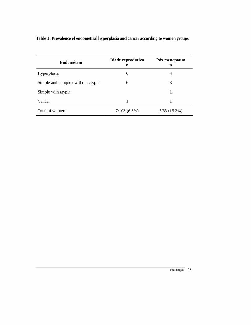
Publicação 39
Table 3. Prevalence of endometrial hyperplasia and cancer according to women groups
Endométrio Idade reprodutiva n
Pós-menopausa n
Hyperplasia 6 4
Simple and complex without atypia 6 3
Simple with atypia 1
Cancer 1 1
Total of women 7/103 (6.8%) 5/33 (15.2%)
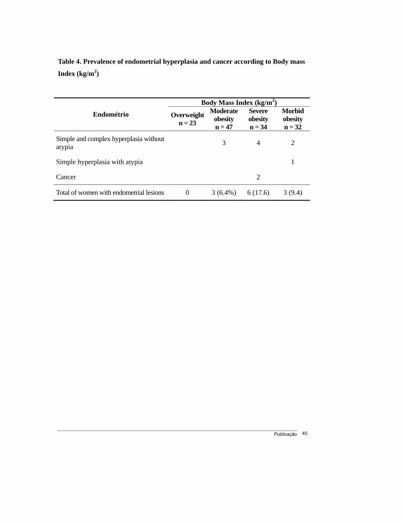
Publicação 40
Table 4. Prevalence of endometrial hyperplasia and cancer according to Body mass
Index (kg/m2)
Body Mass Index (kg/m2)
Endométrio Overweightn = 23
Moderate obesity n = 47
Severe obesity n = 34
Morbid obesity n = 32
Simple and complex hyperplasia without atypia 3 4 2
Simple hyperplasia with atypia 1
Cancer 2
Total of women with endometrial lesions 0 3 (6.4%) 6 (17.6) 3 (9.4)
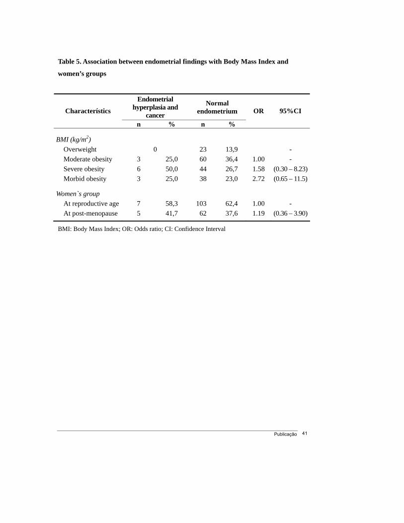
Publicação 41
Table 5. Association between endometrial findings with Body Mass Index and
women’s groups
BMI: Body Mass Index; OR: Odds ratio; CI: Confidence Interval
Endometrial hyperplasia and
cancer
Normal endometrium Characterístics
n % n %
OR 95%CI
BMI (kg/m2) Overweight 0 23 13,9 - Moderate obesity 3 25,0 60 36,4 1.00 - Severe obesity 6 50,0 44 26,7 1.58 (0.30 – 8.23) Morbid obesity 3 25,0 38 23,0 2.72 (0.65 – 11.5)
Women`s group At reproductive age 7 58,3 103 62,4 1.00 - At post-menopause 5 41,7 62 37,6 1.19 (0.36 – 3.90)
Conclusões 42
4. Conclusões
A prevalência de hiperplasia de endométrio foi de 7,3%.
A prevalência de câncer de endométrio foi de 1,5%.
As prevalências de hiperplasia de endométrio e de câncer de endométrio em
mulheres em idade reprodutiva foram de 5,8 % e de 1,0%, respectivamente,
e na pós-menopausa foram respectivamente de 12,1% e 3,0%.
A correlação da hiperplasia endometrial e do câncer de endométrio com o
índice de massa corporal mostrou que mulheres na pós-menopausa e com
obesidade severa ou mórbida têm 1,58 a 2,72 vezes mais chance de ter
hiperplasia ou câncer endometrial.
Referências Bibliográficas 43
5. Referências Bibliográficas
Anderson JW, Konz EC. Obesity and disease management: effects of weight loss
on comorbid conditions. The North American Association for the Study of Obesity.
Obesity Research, 2001; 9:S326-334.
Austin H, Austin JM Jr, Partridge EE. Endometrial cancer, obesity and body fat
distribution. Cancer Research, 1991; 51:568-72.
Bianchini F, Kaaks R, Vainio H. Weight control and physical activity in cancer
prevention. Obes Rev, 2002; 1:166-68.
Carvalheira JBC, Saad MJA. Doenças Associadas à Resistência à Insulina/
Hiperinsulinemia, não incluídas na Síndrome Metabólica. Arq Bras Endocrinol
Metab, 2006; 50:360-67.
CDC’s Behavioral Risk Factor Surveillance System (BRFSS) 2002; Disponível
em: http://www.cdc.gov/brfss. Acesso em 01 de maio de 2004.
Consenso Latino-Americano de Obesidade (CLAO) 1998; Disponível em:
http://www.abeso.org.br/doc/consenso.doc. Acesso em 24 março 2004.
Chia VM, Newcomb PA, Trentham-Dietz A, Hampton JM. Obesity, diabetes,
and other factors in relation to survival after endometrial cancer diagnosis. Int J
Gynecol Cancer, 2007; 17:441-6.
Referências Bibliográficas 44
Chubak J, Tworeger SS, Yasui Y, Ulrico CM, Stanczyk FZ, Mctiernan A. Associations
between reprodutive and menstrual factors and posmenopausal sex hormone
concentrations. Cancer Epidemiology Biomarkers Prev, 2004; 13: 1296-301.
Crissman JD, Azoury RS, Barnes A E, Schellhas HF. Endometrial carcinoma in
Women 40 years of age or younger. Obstet Gynecol, 1981; 57:699-704.
David MP, Adéle CG. Epidemiology of endometrial cancer. Best Practice & Research
Clinical Obstetrics and Gynecology, 2001; 15:341-54.
Davidson BJ, Gambone JC, Lagasse LD. Free estradiol in postmenopausal women
with and without endometrial cancer. Journal of Clinical Endocrinology and
Metabolism, 1981; 52:404-8.
Deruelle P, Leroy JL. Diagnosis of endometrial cancer. Rev Prat, 2001; 51:1439-43.
Disaia PJ. Endometrial hyperplasia/estrogen therapy in: Clinical gynecology
oncology 6th ed, 2002.
Enriori CL, Reforzo Membrives J. Peripheral aromatization as a risk factor for
breast and endometrial cancer in postmenopausal women: a review. Gynecologic
Oncology, 1984; 17:1-21.
Federação Brasileira de Ginecologia e Obstetrícia (Febrasgo), 2002; Disponível
em: http://www.febrasgo.org.br. Acesso em 01 maio 2004.
Filozof C, Gonzalez C, Sereday M, Mazza C, Braguinsk J. Obesity prevalence
and trends in Latin-American countries. Obes Rev, 2001; 2:99-106.
French SA, Folsom AR, Jeffery RW, Zheng W, Mink PJ, Baxter JE. Weight variability
and incident disease in older women: the Iowa Women’s Health Study. Int J Obes
Relat Metab Disord, 1997; 21:217-23.
Referências Bibliográficas 45
Friedenreich C, Cust A, Lahmann PH, et al. Anthropometric factors and risk of
endometrial cancer: the European prospective investigation into cancer and
nutrition. Cancer Causes Control, 2007; 18:399-413
Furberg AS, Thune I. Metabolic abnormalities hypertension, hyperglycemia and
overweight), lifestyle (high energy intake and physical inactivity) and endometrial
cancer risk in a Norwegian cohort. Int J Cancer, 2003; 104:669-76.
Garcia MGM, Carvalho MGM, Garcia MM. Análise dos fatores de risco em pacientes
com adenocarcinoma endometrial. Reprod Clim, 1998; 13:232-236.
Halpern A, Neves M Q, Marsiaj H I, Bernik M N. Survey of obesity in patient applying
for a weight reduction program. Diabetes Res Clin Pract, 1990; 1:S77-9.
Hammond R, Johnson J. Endometrial hyperplasia. Obstetrics and Gynecology,
2004; 14:99-103.
Hill JO, Wyatt HR, Reed GW, Peters JC. Obesity and the environment: where do
we go from here? Science, 2003; 299:853-5.
IARC - The International Agency for Research on Cancer. Working Group: weight
control and physical activity. Handbook of cancer prevention, 2002; 6:1.
IBGE – Instituto Brasileiro de Geografia e Estatística – Estado nutricional,
precisão das estimativas e totais da população 2002-2003. Disponível em:
http//www.ibge.gov.br. Acessado em 01 maio de 2004
Jonsson F, Wolk A, Pedersen NL, et al. Obesity and hormone-dependent tumors:
cohort and co-twin control studies based on the Swedish Twin Registry. Int J
Cancer, 2003; 106:594-9.
Referências Bibliográficas 46
Kaaks R, Lukanova A. Energy balance and cancer: the role of insulin and insulin-
like growth factors-I. Proceedings of the Nutrition Society, 2001; 60:91-106.
Kaaks R, Lukanova A, Kurzer MS. Obesity, endogenous hormones, and endometrial
cancer risk: a synthetic review. Cancer Epidemiology Biomarkers and Prevention,
2002; 11:1531-43.
Keys A, Fidanza F, Karvonen MJ, Kimura N, Taylor HL. Indices of relative weight
and obesity. J Chronic Dis, 1972; 25:329–43.
Kuczmarski MF, Kuczmarski RJ, Najjar M. Effects of age on validity of self-reported
height, weight, and body mass index: findings from the Third National Health and
Nutrition Examination Survey, 1988-1994. J Am Diet Assoc, 2001; 101:28–34.
Kuriyama S, Tsubono Y, Hozawa A, et al. Obesity and risk of cancer in Japan.
Int J Cancer, 2005; 113:148-57.
Kurman RJ, Kaminiski PF, Norris HJ. The behavior of endometrial hyperplasia. A
long-term study of “untreated” hyperplasia in 170 patients. Cancer, 1985 56:403-12.
La Vecchia C., Franceschi S, Gallus G et al. Oestrogens and obesity as risk factors
for endometrial cancer in Italy. Int J Epidemiol, 1982; 11:120-26.
Landis SH, Murray T, Bolden S, Wingo PA. Cancer statistics. Cancer Statistics,
1998; 49:8-31.
Le Marchand L, Wilkens LR, Mi MP. Early-age body size, adult weight gain and
endometrial cancer risk. Int J Cancer, 1991; 48:807-11.
Levi F, La Vecchia C, Negri E, Parazzini F, Franceschi S. Body mass at different
ages and subsequent endometrial cancer risk. Int J Cancer, 1992; 50:567-71.
Referências Bibliográficas 47
Lukanova A, Bjor O, Kaaks R et al. Body mass index and cancer: results from the
Northern Sweden Health and Disease Cohort. Int J Cancer, 2006; 118:458-66.
Monteiro CA, Benicio DA, Conde WL, Popkin BM. Shifting obesity trends in
Brazil. Eur J Clin Nutr, 2000; 54:342-6.
Nahás EAP, Pontes A, Nahás Neto J, De Lucca LA. A relação entre obesidade,
menopausa e terapia de reposição hormonal. Reprod Clim, 1998; 13: 28-31.
Ogden CL, Carroll MD, Curtin LR, McDowell MA, Tabak CJ, Flegal KM. Prevalence
of overweight and obesity in the United States, 1999-2004. JAMA 2006; 295:1549–55
Ogden CL, Yanovski SZ, Carroll MD, Flegal KM. The epidemiology of obesity.
Gastroenterology, 2007; 132:2087-112.
Oguma HD, Sesso RS, Paffenbarger Jr, Lee IM. Weight change and risk of
developing type 2 diabetes. Obes Res, 2005; 13:945–51.
Olson SH, Trevisan M, Marshall JR et al. Body mass index, weight gain, and
risk of endometrial cancer. Nutr Cancer, 1995; 23:141-49.
Potishman N, Gail MH, Troisi R, Waccholder S, Hoover RN. Mensurement error does
not explain the persistence of a body mass index association with endometrial cancer
after adjustment for endogenus hormones. Epidemiology Resources, 1999; 10:76-79.
Schouten LJ, Goldbohm RA, van den Brandt PA. Anthropometry, physical
activity, and endometrial cancer risk: results from the Netherlands Cohort Study.
J Natl Cancer Inst, 2004; 96:1635-8.
Soliman PT, Oh JC, Schmeler KM, Sun CC, Slomovitz BM, Gershenson DM, et
al. Risk factors for yong premenopausal women with endometrial cancer. Obstet
Ginecology, 2005; 105:575-80.
Referências Bibliográficas 48
Swanson CA, Potschman N, Wilblanks GD. Relation of endometrial cancer risk
to past and contemporany body fat distribution. Cancer Epidemiology Biomarks
Prev, 1993; 2:321-7.
Terry P, Baron JA, Weiderpass E, Yuen J, Lichtenstein P, Nyren O. Lifestyle
and endometrial cancer risk: a cohort study from the Swedish Twin Registry. Int
J Cancer, 1999; 82:38–42.
The National Heart, Lung and Blood Institute (NHBLI) Obesity Education Initiative
Export Panel. Obes Res, 1998; 6:51S.
Tornenberg SA, Carstensen JM. Relationship between Quetelet’s index and
cancer of breast and female genital tract in 47.000 women follwed for 25 years.
British Journal of Cancer, 1994; 69:358-61.
Trentham-Dietz A, Nichols HB, Hampton JM, Newcomb PA. Weight change and
risk of endometrial cancer. Int J Epidemiol, 2006; 35:151-58.
Troisi R, Potishman N, Hoover RN, Siiteri P, Brinton LA. Insulin and endometrial
cancer. Am J Epidemiol, 2001; 146: 476-82.
US Department of Health and Human Services and U.S. Department of Agriculture,
Dietary guidelines for Americans (6th ed.), U.S. Government Printing Office,
Washington, DC (2005).
Weiderpass E, Persson I, Adami HO, Magnusson C, Lindgren A, Baron JA.
Body size in different periods of life, diabetes mellitus, hypertension, and risk of
postmenopausal endometrial cancer (Sweden). Cancer Causes Control, 2000;
11:185–192.
Waitezberg, DL. Nutrição oral, enteral e prenteral na pratica clinica. 3 ed.São
Paulo:Editora Ateneu, 2004.
Referências Bibliográficas 49
Weeb PM Commentary: Weight gain, weght loss, and endometrial cancer.
International Journal of Epidemiology, 2006; 35:166-68.
Woodruff JD, Pikared JH. Incidence of endometrial hyperplasia in
postmenopausal women taking conjugated estrogen alone. The Menopause
Study Group. Am J Obstet Gynecol, 1994; 170:1213-23.
World Health Organization (WHO) Technical Report Series 854. Physical status:
the use and interpretation of anthropometry Report of the WHO expert
committee, WHO, Geneva, Switzerland (1995).
World Health Organization, The global epidemic of obesity, WHO, Geneva (1997).
World Health Organization. Nutrition. Controlling the global obesity epidemic.
Geneva: WHO, (Report of a WHO consultation on nutrition) 2003.
World Health Organization (WHO). Disponível em
http//www.who.int/nut/obs.htm. Acessado em: 05 de março de 2004.
Anexos 50
6. Anexos
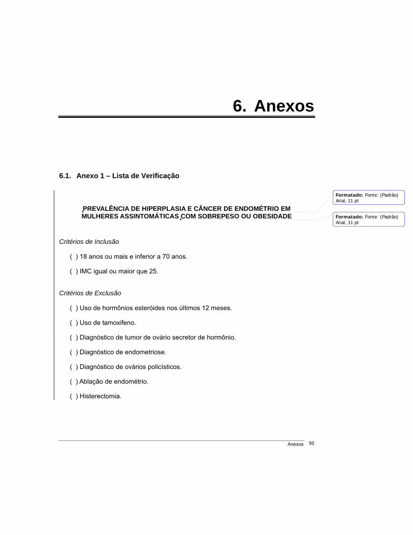
6.1. Anexo 1 – Lista de Verificação
PREVALÊNCIA DE HIPERPLASIA E CÂNCER DE ENDOMÉTRIO EM MULHERES ASSINTOMÁTICAS COM SOBREPESO OU OBESIDADE
Critérios de Inclusão
� ( ) 18 anos ou mais e inferior a 70 anos.
� ( ) IMC igual ou maior que 25.
Critérios de Exclusão
� ( ) Uso de hormônios esteróides nos últimos 12 meses.
� ( ) Uso de tamoxifeno.
� ( ) Diagnóstico de tumor de ovário secretor de hormônio.
� ( ) Diagnóstico de endometriose.
� ( ) Diagnóstico de ovários policísticos.
� ( ) Ablação de endométrio.
� ( ) Histerectomia.
Formatado: Fonte: (Padrão)Arial, 11 pt
Formatado: Fonte: (Padrão)Arial, 11 pt
Anexos 51
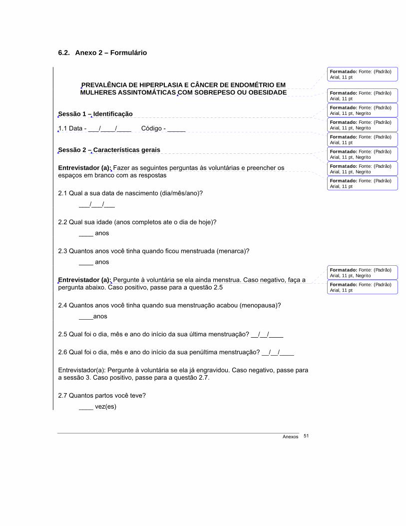
6.2. Anexo 2 – Formulário
PREVALÊNCIA DE HIPERPLASIA E CÂNCER DE ENDOMÉTRIO EM MULHERES ASSINTOMÁTICAS COM SOBREPESO OU OBESIDADE
Sessão 1 – Identificação
1.1 Data - ___/____/____ Código - _____
Sessão 2 – Características gerais
Entrevistador (a): Fazer as seguintes perguntas às voluntárias e preencher os espaços em branco com as respostas
2.1 Qual a sua data de nascimento (dia/mês/ano)?
___/___/___
2.2 Qual sua idade (anos completos ate o dia de hoje)?
____ anos
2.3 Quantos anos você tinha quando ficou menstruada (menarca)?
____ anos
Entrevistador (a): Pergunte à voluntária se ela ainda menstrua. Caso negativo, faça a pergunta abaixo. Caso positivo, passe para a questão 2.5
2.4 Quantos anos você tinha quando sua menstruação acabou (menopausa)?
____anos
2.5 Qual foi o dia, mês e ano do início da sua última menstruação? __/__/____
2.6 Qual foi o dia, mês e ano do início da sua penúltima menstruação? __/__/____
Entrevistador(a): Pergunte à voluntária se ela já engravidou. Caso negativo, passe para a sessão 3. Caso positivo, passe para a questão 2.7.
2.7 Quantos partos você teve?
____ vez(es)
Formatado: Fonte: (Padrão)Arial, 11 pt
Formatado: Fonte: (Padrão)Arial, 11 pt
Formatado: Fonte: (Padrão)Arial, 11 pt, Negrito
Formatado: Fonte: (Padrão)Arial, 11 pt, Negrito
Formatado: Fonte: (Padrão)Arial, 11 pt
Formatado: Fonte: (Padrão)Arial, 11 pt, Negrito
Formatado: Fonte: (Padrão)Arial, 11 pt, Negrito
Formatado: Fonte: (Padrão)Arial, 11 pt
Formatado: Fonte: (Padrão)Arial, 11 pt, Negrito
Formatado: Fonte: (Padrão)Arial, 11 pt
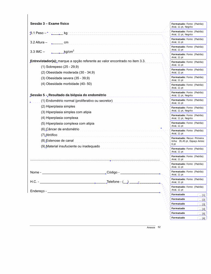
Anexos 52
Sessão 3 – Exame físico
3.1 Peso – ______ kg
3.2 Altura – ______ cm
3.3 IMC – ______ kg/cm2
Entrevistador(a): marque a opção referente ao valor encontrado no item 3.3.
(1) Sobrepeso (25 - 29,9)
(2) Obesidade moderada (30 - 34,9)
(3) Obesidade severa (35 - 39,9)
(4) Obesidade morbidade (40- 50)
Sessão 5 – Resultado da biópsia do endométrio (1) Endométrio normal (proliferativo ou secretor)
(2) Hiperplasia simples
(3) Hiperplasia simples com atipia
(4) Hiperplasia complexa
(5) Hiperplasia complexa com atipia
(6) Câncer de endométrio
(7) Atrófico
(8) Estenose de canal
(9) Material insufuciente ou inadequado
...........................................................................................................................................
Nome - Código -
H.C. - Telefone - ( ) -
Endereço -
Formatado: Fonte: (Padrão)Arial, 11 pt, Negrito
Formatado: Fonte: (Padrão)Arial, 11 pt, Negrito
Formatado: Fonte: (Padrão)Arial, 11 pt
Formatado: Fonte: (Padrão)Arial, 11 pt
Formatado: Fonte: (Padrão)Arial, 11 pt
Formatado: Fonte: (Padrão)Arial, 11 pt
Formatado: Fonte: (Padrão)Arial, 11 pt
Formatado: Fonte: (Padrão)Arial, 11 pt
Formatado: Fonte: (Padrão)Arial, 11 pt
Formatado: Fonte: (Padrão)Arial, 11 pt, Negrito
Formatado: Fonte: (Padrão)Arial, 11 pt
Formatado: Fonte: (Padrão)Arial, 11 pt, Negrito
Formatado: Fonte: (Padrão)Arial, 11 pt, Negrito
Formatado: Fonte: (Padrão)Arial, 11 pt
Formatado: Fonte: (Padrão)Arial, 11 pt
Formatado: Recuo: Primeiralinha: 35,45 pt, Espaço Antes: 6 pt
Formatado: Fonte: (Padrão)Arial, 11 pt
Formatado: Fonte: (Padrão)Arial, 11 pt
Formatado: Fonte: (Padrão)Arial, 11 pt
Formatado: Fonte: (Padrão)Arial, 11 pt
Formatado: Fonte: (Padrão)Arial, 11 pt
Formatado: Fonte: (Padrão)Arial, 11 pt
Formatado
Formatado
Formatado
Formatado
Formatado
Formatado
... [1]
... [2]
... [3]
... [4]
... [5]
... [6]
Anexos 53
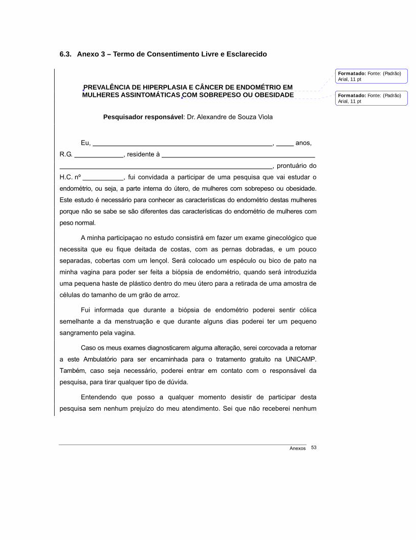
6.3. Anexo 3 – Termo de Consentimento Livre e Esclarecido
PREVALÊNCIA DE HIPERPLASIA E CÂNCER DE ENDOMÉTRIO EM MULHERES ASSINTOMÁTICAS COM SOBREPESO OU OBESIDADE
Pesquisador responsável: Dr. Alexandre de Souza Viola
Eu, , anos,
R.G. , residente à
, prontuário do
H.C. nº , fui convidada a participar de uma pesquisa que vai estudar o
endométrio, ou seja, a parte interna do útero, de mulheres com sobrepeso ou obesidade.
Este estudo é necessário para conhecer as características do endométrio destas mulheres
porque não se sabe se são diferentes das características do endométrio de mulheres com
peso normal.
A minha participaçao no estudo consistirá em fazer um exame ginecológico que
necessita que eu fique deitada de costas, com as pernas dobradas, e um pouco
separadas, cobertas com um lençol. Será colocado um espéculo ou bico de pato na
minha vagina para poder ser feita a biópsia de endométrio, quando será introduzida
uma pequena haste de plástico dentro do meu útero para a retirada de uma amostra de
células do tamanho de um grão de arroz.
Fui informada que durante a biópsia de endométrio poderei sentir cólica
semelhante a da menstruação e que durante alguns dias poderei ter um pequeno
sangramento pela vagina.
Caso os meus exames diagnosticarem alguma alteração, serei corcovada a retornar
a este Ambulatório para ser encaminhada para o tratamento gratuito na UNICAMP.
Também, caso seja necessário, poderei entrar em contato com o responsável da
pesquisa, para tirar qualquer tipo de dúvida.
Entendendo que posso a qualquer momento desistir de participar desta
pesquisa sem nenhum prejuízo do meu atendimento. Sei que não receberei nenhum
Formatado: Fonte: (Padrão)Arial, 11 pt
Formatado: Fonte: (Padrão)Arial, 11 pt
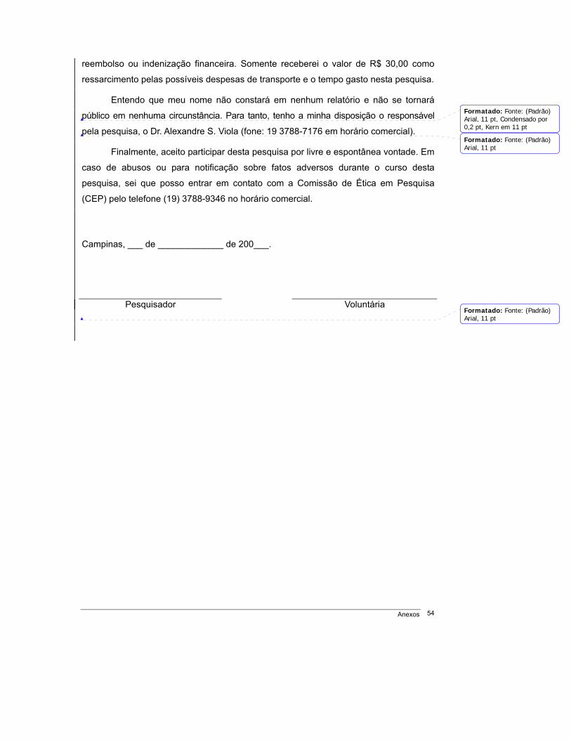
Anexos 54
reembolso ou indenização financeira. Somente receberei o valor de R$ 30,00 como
ressarcimento pelas possíveis despesas de transporte e o tempo gasto nesta pesquisa.
Entendo que meu nome não constará em nenhum relatório e não se tornará
público em nenhuma circunstância. Para tanto, tenho a minha disposição o responsável
pela pesquisa, o Dr. Alexandre S. Viola (fone: 19 3788-7176 em horário comercial).
Finalmente, aceito participar desta pesquisa por livre e espontânea vontade. Em
caso de abusos ou para notificação sobre fatos adversos durante o curso desta
pesquisa, sei que posso entrar em contato com a Comissão de Ética em Pesquisa
(CEP) pelo telefone (19) 3788-9346 no horário comercial.
Campinas, ___ de _____________ de 200___.
Pesquisador Voluntária
Formatado: Fonte: (Padrão)Arial, 11 pt, Condensado por 0,2 pt, Kern em 11 pt
Formatado: Fonte: (Padrão)Arial, 11 pt
Formatado: Fonte: (Padrão)Arial, 11 pt

Página 52: [1] Formatado ROSARIO 8/8/2007 10:26:00
Fonte: (Padrão) Arial, 11 pt, Sublinhado
Página 52: [2] Formatado ROSARIO 8/8/2007 10:26:00
Fonte: (Padrão) Arial, 11 pt
Página 52: [3] Formatado ROSARIO 8/8/2007 10:26:00
Fonte: (Padrão) Arial, 11 pt
Página 52: [4] Formatado ROSARIO 8/8/2007 10:26:00
Fonte: (Padrão) Arial, 11 pt
Página 52: [5] Formatado ROSARIO 8/8/2007 10:26:00
Fonte: (Padrão) Arial, 11 pt
Página 52: [6] Formatado ROSARIO 8/8/2007 10:26:00
Fonte: (Padrão) Arial, 11 pt