Embed Size (px)
Citation preview
PUCRS – Pontifícia Universidade Católica do Rio Grande do Sul FABIO – Faculdade de Biociências
PPGBCM – Programa de Pós-Graduação em Biologia Celular e Molecular
ANÁLISE DAS VARIANTES POLIMÓRFICAS DO ÉXON 1 DO GENE QUE CODIFICA LECTINA DE LIGAÇÃO A MANOSE (MBL2) EM PACIENTES COM
ARTRITE REUMATÓIDE
Autor
Fernanda Letícia Martiny
Orientador Maurício Reis Bogo
Porto Alegre 2011

2
PUCRS – Pontifícia Universidade Católica do Rio Grande do Sul FABIO – Faculdade de Biociências
PPGBCM – Programa de Pós-Graduação em Biologia Celular e Molecular
ANÁLISE DAS VARIANTES POLIMÓRFICAS DO ÉXON 1 DO GENE QUE CODIFICA LECTINA DE LIGAÇÃO A MANOSE (MBL2) EM PACIENTES COM
ARTRITE REUMATÓIDE
Dissertação apresentada ao Programa de Pós-Graduação em Biologia Celular e Molecular da Pontifícia Universidade Católica do Rio Grande do Sul para obtenção do grau de mestre.
Autor Fernanda Letícia Martiny
Orientador Maurício Reis Bogo
Porto Alegre 2011

3
Dedico este trabalho a minha família
(pai, mãe, noivo e irmãos) que sempre
estiveram ao meu lado, me apoiando.

4
AGRADECIMENTOS
A Deus pelo milagre da vida.
Ao meu orientador Maurício Reis Bogo pela confiança em mim depositada, pela
oportunidade e pelos ensinamentos trocados.
Ao meu, pode-se dizer orientador Zeca (José Arthur Bogo Chies) pela paciência, pelo grande
auxílio, pelo incentivo e principalmente pelo estímulo.
Aos Professores do Programa de Pós-Graduação em Biologia Molecular e Celular pela
contribuição a minha formação.
Aos colegas do Laboratório de Biologia Genômica e Molecular da PUCRS pelos momentos
de aprendizagem e de fortes discussões científicas, principalmente a Cladinara Sarturi.
Aos colegas do Laboratório de Imunogenética da UFRGS pela paciência e disponibilização
de seu tempo para me auxiliaram na bancada, em especial ao colega Tiago Degani Veit pelo
auxílio na análise dos resultados.
Aos colegas do mestrado pelas experiências e ensinamentos trocados, em especial Stefânia
Richetti e Kelen Rocha pelas horas de discussões, pela amizade, apoio e companheirismo.
Aos meus irmãos Luis Eugênio, Pedro Ivo e João Vicente pela força e ajuda.
Ao meu noivo Everson Ferreira Menezes pelo apoio incondicional, carinho, paciência, amor
e ensinamentos, por me dar força e nunca me deixar desanimar. Obrigada Bem, por estar
sempre do meu lado.
Aos meus Pais Ailton Décio Martiny e Rosaní Martiny que sempre acreditam na minha
capacidade e por me dar a oportunidade de realizar mais este sonho.
Aos indivíduos que participaram deste estudo fornecendo material biológico.
A CAPES pela bolsa de estudos que possibilitou a realização deste mestrado.

5
RESUMO
A artrite reumatóide é uma doença do sistema auto-imune que atua através de uma inflamação nas articulações tornando-se crônica com o passar dos anos, podendo desenvolver uma deformidade e destruição destas articulações devido à erosão da cartilagem e do osso. Esta doença atinge pessoas de ambos os sexos com idades variáveis. O diagnóstico da doença depende de uma série de combinações de sintomas clínicos, diagnósticos laboratoriais e radiográficos. A etiologia da AR é multifatorial, resultando da interação de fatores ambientais, hormonais e genéticos, que contribuem para sua ocorrência e expressão. Evidências epidemiológicas mostram que fatores genéticos são relatados como risco para o aumento da AR. Acredita-se que vários genes possam estar envolvidos com o aparecimento da AR, e um deles é o gene MBL2, responsável pela codificação da proteína Lectina de Ligação à Manose. A MBL é uma proteína da família das colectinas importante para o sistema imunológico, relacionada com a promoção de fagocitose de microorganismos, modulação da resposta inflamatória e apoptose. Os baixos níveis séricos de MBL estão associados com mutações genéticas através de polimorfismos do gene MBL2. Três polimorfismos de base única (SNPs) foram localizados no éxon 1 nos códons 52 (alelo variante D), 54 (alelo variante B) e 57 (alelo variante C). A presença de qualquer uma dessas variantes é chamada de alelo O, enquanto que a ausência das variantes em qualquer uma das três posições é chamada alelo A. Neste estudo, analisamos o polimorfismo do gene MBL2 em 322 pacientes brasileiros com AR e 343 indivíduos saudáveis através da técnica de sequenciamento. Quando comparamos indivíduos saudáveis euro-descendentes e afro-descendentes observamos uma diferença significativa nas frequências genotípicas e alélicas, isto devido a frequência maior do alelo C (pacientes euro-descendentes 0.0220 vs. Pacientes Afro-descendentes 0.205, controles euro-descendentes 0.029 vs. Controles afro-descendentes 0.144, valores P<0.001). Também analisamos o genótipo MBL em relação a manifestações extra-articular. Quando consideramos as variantes MBL juntas, nós encontramos um aumento da frequência do genótipo OO em pacientes com nódulos reumatóides (P=0.031). Estes achados sugerem uma associação do genótipo OO e a severidade da doença, entretanto mais estudos são necessários pra esclarecer a verdadeira função da MBL na AR. Palavras chave: Mannose Ligada a Lectina, Artrite Rreumatóide, Variantes Polimórficas

6
ABSTRACT
Rheumatoid arthritis is an autoimmune disease that operates through joint inflammation becomes chronic over the years and may develop a deformity and destruction of these joints due to erosion of cartilage and bone. This disease affects people of both sexes and varying ages. The diagnosis depends on a number of combinations of clinical symptoms, laboratory and radiographic diagnostics. The etiology of RA is multifactorial resulting from the interaction of environmental factors, hormonal and genetic factors that contribute to its occurrence and expression. Epidemiologic studies show that genetic factors are reported as increasing the risk for RA. It is believed that several genes may be involved with the onset of RA, and one of them is the MBL2 gene, responsible for encoding the protein Mannose binding lectin. MBL is a protein family of collectins important for the immune system, related to the promotion of phagocytosis of microorganisms, modulation of inflammatory response and apoptosis. The low serum levels of MBL are associated with genetic mutations by MBL2 gene polymorphisms. Three single nucleotide polymorphisms (SNPs) were located in exon 1 at codons 52 (variant allele D), 54 (variant allele B) and 57 (variant allele C) the presence of any of the variant alleles (B, C or D) has been collectively labeled O, whereas the simultaneous absence of variants at the three positions has been called allele A. We analyzed MBL2 gene polymorphisms in 322 Brazilian patients with RA and 343 healthy controls ethnically matched through sequencing. When we compared European-derived and African-derived controls we observed a significantly difference in both, genotypic and allelic frequencies, particularly concerning the frequency of the C allele (European-derived patients 0.0220 vs. African-derived patients 0.205, European-derived controls 0.029 vs. African-derived controls 0.144, both P-values <0.001). We also analyzed MBL genotype in relation to extraarticular manifestations. When considering MBL variants together we found an increased frequency of the OO genotype among patients with rheumatoid nodules (P=0.031). This finding suggests an association of the OO genotype and
disease severity, but more studies are needed to clarify the true role of MBL in RA. Key-words: Mannose-Binding Lectin, Rheumatoid Arthritis, Polymorphic variants
7
ÍNDICE
1 CAPÍTULO 1 .......................................................................................................................... 8
1.1 Introdução ......................................................................................................................... 8
1.1.1 Artrite Reumatóide .................................................................................................... 8
1.1.2 MBL ........................................................................................................................ 11
1.2 Objetivos ......................................................................................................................... 17
2 CAPÍTULO 2 – Artigo Científico ......................................................................................... 18
3 CONSIDERAÇÕES FINAIS ................................................................................................ 31
REFERÊNCIAS ....................................................................................................................... 32
ANEXO .................................................................................................................................... 38

8
1 CAPÍTULO 1
1.1 Introdução
1.1.1 Artrite Reumatóide
A artrite reumatóide (AR) é uma doença do sistema imune de caráter
inflamatório, que atua primeiramente através de uma inflamação nas articulações
tornando-se crônica com o passar dos anos. O indivíduo com AR pode desenvolver
uma deformidade e destruição das articulações devido à erosão da cartilagem e do
osso (KORB; PAVENSTADT; PAP, 2009). A AR é caracterizada por uma inflamação
na membrana sinovial, esta estrutura é responsável por envolver a articulação. A
membrana sinovial tem por função produzir o líquido sinovial que nutre e lubrifica a
superfície da articulação permitindo o seu movimento. Quando um indivíduo
predisposto é exposto a um agente desencadeante (antígeno) forma-se um
complexo processo inflamatório na membrana sinovial. Esta membrana torna-se
inflamada, mais espessa e aumenta de volume devido a produção de um líquido
inflamatório que invade o osso, destrói as cartilagens e os ligamentos levando ao
dano, deformidade articular e consequentemente dificultando o movimento e
causando dor (TAVARES et al, 2000).
Atualmente, esta doença atinge pessoas de ambos os sexos com idades
variáveis, porém há uma prevalência maior em mulheres com idade entre 40 e 60
anos (PAN; GABAY; FINCKH, 2007). Na população mundial há uma prevalência de
aproximadamente 0,5% a 1% de pessoas com AR sendo que em mulheres é de
1,37% e entre homens é de 0,74% (ANDERSON et al, 1985; GABRIEL, 2001). De
acordo com Lee; Weinblatt (2001) e Silman; Pearson (2002) não foram encontrados
casos de AR na parte rural da África do Sul dentre 500 indivíduos analisados. Na

9
Ásia existe uma prevalência de 0,3% principalmente em países como China e Japão.
Entretanto, em tribos indígenas da América do Norte há uma prevalência de 5,3 a
6,0% (alguns dados podem ser vistos na tabela 1), porém não foi possível identificar
se existe alguma relação entre a AR e a situação sócio-ecomônica (ALAMANOS;
DROSOS; 2005). No Brasil, foi realizado um estudo multicêntrico em 1999 que
constatou uma prevalência de AR em adultos variando de 0,2% a 1% (LOUZADA et
al, 2007).
Atento a esses dados e com a preocupação de uma abordagem de
tratamento diferenciada, o Ministério da Saúde publicou o “Protocolo Clínico e
Diretrizes Terapêuticas para o Tratamento da Artrite Reumatóide” (Portaria MS
n°855 de 22 de novembro de 2002), o qual é de grande valia para a sociedade
brasileira. Além disto, a Sociedade Brasileira de Reumatologia também demonstrou
o seu interesse e desenvolveu o Consenso Brasileiro para o Diagnóstico e
tratamento da Artrite Reumatóide, o qual foi atualizado em 2007 (BÉRTOLO et al.,
2007; LAURINDO; PINHEIRO; XIMENES et al, 2002;).
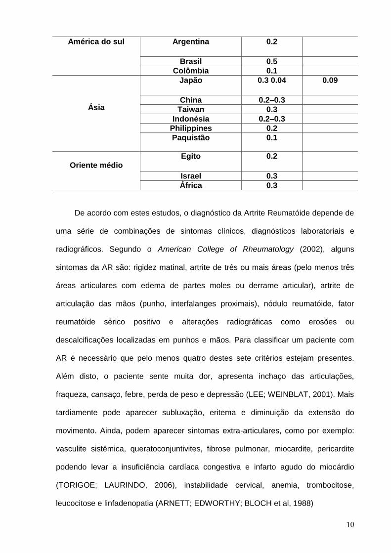
Tabela 1: Prevalência e incidência de AR no mundo (ALAMANOS; DROSOS; 2005).
População Prevalência Incidência
América do Norte Estados Unidos (população em geral)
0.9- 1.1 0.02-0.07
Estados Unidos (população nativa)
5.3–6.0 0.09–0.89
Norte da Europa
Inglaterra 0.8–1.10 0.02–0.04
Finlândia 0.8 0.03–0.04
Suécia 0.5–0.9
Noruega 0.4–0.5 0.02–0.03
Dinamarca 0.9
Irlanda 0.5
Sul da Europa
Espanha 0.5
França 0.6 0.01
Itália 0.3
Grécia 0.3–0.7 0.02
Iugoslávia 0.2
10
América do sul Argentina 0.2
Brasil 0.5
Colômbia 0.1
Ásia
Japão 0.3 0.04 0.09
China 0.2–0.3
Taiwan 0.3
Indonésia 0.2–0.3
Philippines 0.2
Paquistão 0.1
Oriente médio
Egito 0.2
Israel 0.3
África 0.3
De acordo com estes estudos, o diagnóstico da Artrite Reumatóide depende de
uma série de combinações de sintomas clínicos, diagnósticos laboratoriais e
radiográficos. Segundo o American College of Rheumatology (2002), alguns
sintomas da AR são: rigidez matinal, artrite de três ou mais áreas (pelo menos três
áreas articulares com edema de partes moles ou derrame articular), artrite de
articulação das mãos (punho, interfalanges proximais), nódulo reumatóide, fator
reumatóide sérico positivo e alterações radiográficas como erosões ou
descalcificações localizadas em punhos e mãos. Para classificar um paciente com
AR é necessário que pelo menos quatro destes sete critérios estejam presentes.
Além disto, o paciente sente muita dor, apresenta inchaço das articulações,
fraqueza, cansaço, febre, perda de peso e depressão (LEE; WEINBLAT, 2001). Mais
tardiamente pode aparecer subluxação, eritema e diminuição da extensão do
movimento. Ainda, podem aparecer sintomas extra-articulares, como por exemplo:
vasculite sistêmica, queratoconjuntivites, fibrose pulmonar, miocardite, pericardite
podendo levar a insuficiência cardíaca congestiva e infarto agudo do miocárdio
(TORIGOE; LAURINDO, 2006), instabilidade cervical, anemia, trombocitose,
leucocitose e linfadenopatia (ARNETT; EDWORTHY; BLOCH et al, 1988)

11
Estudos indicam que 20% das pessoas param suas atividades profissionais
dois anos após o diagnóstico de AR e aproximadamente 50% tornam-se inaptas
para trabalhar após dez anos de doença (YOUNG et al, 2002).
A etiologia da AR é multifatorial, resultando da interação de fatores
ambientais, hormonais e genéticos, que contribuem para sua ocorrência e
expressão. Estudos têm comprovado que fatores ambientais aumentam o risco de
desenvolver AR e também de piorar o prognóstico dos pacientes. Entretanto, o
mecanismo pelo o qual estes fatores aumentam o risco de desenvolver e expressar
a doença, ainda está incerto. Por outro lado, evidências epidemiológicas mostram
que fatores genéticos são relatados com o risco de desenvolvimento de AR
(ALAMANOS; DROSOS; 2005).
Acredita-se que vários genes possam estar envolvidos com o aparecimento
da AR, e um deles é o gene MBL2, responsável pela codificação da proteína Lectina
de Ligação a Manose (MBL). Estudos realizados sugerem a relação entre
polimorfismos do gene MBL2 e a suscetibilidade em desenvolver AR em pacientes
indianos (GUPTA et al, 2005; GUPTA et al, 2006).
1.1.2 MBL
A MBL é uma proteína sérica da família das colectinas, caracterizada pela
presença de três cadeias idênticas de polipeptídios com peso molecular de 32KDa.
Cada cadeia tem uma região C-terminal, um domínio de reconhecimento de
carboidrato dependente de cálcio (CRD), uma -hélice curta, região pescoço
hidrofóbica, uma região colágeno, e uma região N-terminal rica em cisteína (Figura
1A). As três cadeias de polipeptídios formam uma tripla hélice com as regiões
colágenos estabilizadas por interações hidrofóbicas e interações dissulfeto com a

12
região N-terminal rica em cisteína (Figura1B) (ARNOLD, 2006; DOMMETT; KLEIN;
TURNER, 2006).
Figura 1: Estrutura da proteína MBL (adaptado de DOMMETT; KLEIN; TURNNER, 2006).
A MBL é produzida pelo fígado, sendo esta, uma molécula extremamente
importante para o sistema imunológico, pois é estruturalmente semelhante a C1q
(componente intermediário da ativação da via clássica do sistema complemento) e
tem um papel homólogo a ela. Desta forma, esta proteína apresenta importantes
funções como, por exemplo: ativação do sistema complemento através da via das
lectinas, opsonização de micro-organismos, ativação de macrófagos, modulação da
resposta inflamatória e apoptose (TURNER et al., 1996; KILPATRICK, 2003;
TURNER, 2003).
O sistema complemento consiste em uma cascata bioquímica que faz parte
do sistema imune. Sua principal função é na resposta inflamatória (TURNER, 2003;
DOMMETT; KLEIN; TURNER, 2006). Trata-se de um sistema composto por cerca
de 30 proteínas que circulam no plasma ou se encontram em superfícies. O sistema

13
complemento atua de várias maneiras, como por exemplo, na defesa contra micro-
organismos, na ativação de leucócitos e na morte celular (WALPORT, 2001).
O sistema complemento pode ser ativado através de três vias principais: via
clássica, via alternativa e a via das lectinas. Todas as vias convergem para a
ativação do componente central ou C3. A via clássica tem relação com a resposta
imune específica do hospedeiro e sua ativação depende da produção de anticorpos
e da formação de complexos antígeno-anticorpo. A via alternativa é ativada e
aumenta na presença de superfícies ativadoras adequadas como as paredes
celulares das bactérias e fungos. A via das lectinas participa da resposta inata, e é
ativada pela ligação da MBL ao açúcar (manose) presente na superfície de micro-
organismos. A MBL também pode ativar a via clássica do sistema complemento
através de proteínas denominadas ficolinas, ou também através da associação da
MBL com as MASPs (MBL-associated serine proteases) (Figura 1C), pois este
complexo é semelhante a C1r e C1s, componentes necessários para que ocorra a
cascata de ativação. Posteriormente, foi descoberto que a MASP-2 era o
componente que se ligava a MBL para clivar C4 e C2 gerando C3 e também que
MASP-1 ligado a MBL gerava C3 diretamente (Figura 2) (DOMMETT; TURNER et
al., 1996; KLEIN; TURNER, 2006).

14
Figura 2: Via de ativação do complemento (adaptado de DOMMETT; KLEIN; TURNNER, 2006).
Ainda, é possível induzir a ativação da via complemento através da interação
da MBL com a IgG. Este complexo acumula-se nas articulações das pessoas
acometidas pela AR (GUPTA et al, 2005).
Altos níveis séricos de MBL estão associados com a deficiência de algumas
moléculas do sistema complemento alterando a atividade deste sistema (GUPTA et
al, 2005). Na deficiência de níveis séricos de MBL as condições de inflamação
crônica podem ser mais graves. Baixos níveis de MBL estão associados com
mutações genéticas, através de polimorfismos do gene MBL2 e a possível produção
de anticorpos anti-MBL (MADSEN et al, 1994).
A MBL é codificada pelo gene MBL2 localizado no braço longo do
cromossomo 10 q11.2-21 e contém 4 éxons. Três polimorfismos de base única
(SPNs) foram localizados no éxon 1 nos códons 52 (alelo D), 54 (alelo B) e 57 (alelo

15
C) (MADSEN et al, 1994; MADSEN et al, 1995). A ausência de qualquer destas
variantes, e consequente estado selvagem nestes três sítios, caracteriza o alelo A.
As variantes são: no códon 52 a substituição de um C para T que leva à substituição
de uma arginina por cisteína, no códon 54 um G para A (glicina para ácido aspártico)
e no códon 57 substituição de G para A (glicina para ácido glutâmico). Estas
substituições causam mudanças estruturais que determinam baixos níveis de MBL
no plasma (GUPTA et al, 2005; GARRED et al., 2003). O éxon 2 codifica uma região
rica em cisteína e uma região de colágeno, o éxon 3 codifica a região pescoço e o
éxon 4 codifica um domínio de ligação de carboidrato (Figura 3) (DOMMETT; KLEIN;
TURNNER, 2006).
Figura 3: Características do gene MBL2 (adaptado de DOMMETT; KLEIN; TURNNER, 2006).

16
Estudos comprovam que as variantes dos alelos B e C são independentes em
distintas populações (LIPSCOMBE, et al, 1996). O alelo B foi encontrado em altas
frequências na população européia e norte-americana nativa, enquanto que o alelo
C é mais encontrado na população do Saara (MADSEN et al, 1998). Altas
frequências das variantes polimórficas são observadas em populações humanas de
regiões tropicais (GARRED et al 1992).
Ip et al. (2000) realizaram estudos em pacientes chineses com AR e
constataram que 67,8% apresentaram homozigose para o alelo A, 30,3% eram
heterozigotos e 1,9% eram homozigotos para o polimorfismo no códon 54 (alelo B),
indicando que existe uma relação entre o polimorfismo e a AR. Madsen et al (1998),
encontraram frequências do alelo variante B superior a 42% em duas populações
humanas sul-americanas nativas, 11% na população caucasiana e ausência em
pacientes moçambicanos. Já para o alelo C, a frequência foi maior em
moçambicanos (24%) e baixa em sul-americanos nativos e caucasianos (1% e 3%
respectivamente). O alelo D foi observado somente na população caucasiana,
sugerindo-se assim que diferentes populações são dominadas por diferentes
haplótipos funcionais.
Geijn et al (2008) que realizaram estudo em mulheres caucasianas holandesas,
assim como Grupta et al (2005) que realizaram estudos em indianos e Stanworth et
al (1998) não encontraram relação entre a suscetibilidade em desenvolver AR e os
polimorfismos do gene MBL2.
Outras doenças também podem estar envolvidas com o polimorfismo do gene
MBL2, como por exemplo: Lupus Eritematoso Sistêmico (GARRED et al., 2001;
HUANG et al., 2003), Síndrome de Sjögrens (WANG et al, 2001; CASALS et al
2009), Doença Celíaca (BONIOTTO et al., 2002; ILTANEN et al., 2003), Fibrose
Cística (KILPATRICK, 2002), ou também pela diminuição dos níveis de MBL

17
plasmática como doenças infecciosas provocadas por vírus como a Síndrome da
Imunodeficiência Humana Adquirida (AIDS), Hepatite B e C, influenza, Herpes
simples; por bactérias (Staphylococcus aureus, Streptococcus pneumoniae,
Neisseria meningitidis, Escherichia coli e outras); por fungos (Cândida albicans,
Aspergillus fumigatus) e também por protozoários (Plasmodium falciparum,
Trypanozoma cruzi) (DOMMETT; KLEIN; TURNNER, 2006; KILPATRICK, 2002).
Maury et al (2007) constataram que o genótipo variante do gene MBL2 constitui
um significante risco genético para amiloidose em pacientes finlandeses com AR,
demonstrando que o polimorfismo do gene MBL2 está associado com a rápida
progressão da erosão em AR.
Desta forma, julgou-se necessário a realização deste estudo para determinar
se a relação entre o polimorfismo do gene MBL2 e a AR existe na população
brasileira e com que frequência ela ocorre, principalmente devido à grande variação
étnica aqui existente.
1.2 Objetivos
Analisar o polimorfismo gênico da região do éxon 1 do gene MBL2 em
pacientes com Artrite Reumatóide e comparar com um grupo controle
constituído de indivíduos que se declararam saudáveis.
Comparar a frequência alélica dos polimorfismos do gene MBL2 entre as
diferentes etnias dos pacientes com AR.

18
2 CAPÍTULO 2
Mannose-Binding Lectin gene polymorphisms in Brazilian patients with Rheumatoid
Arthritis
1Fernanda Leticia Martiny, 2Tiago Degani Veit, 3Claiton Viegas Brenol, 3João
Carlos Tavares Brenol, 3Ricardo Machado Xavier, 1,4*Maurício Reis Bogo and
2*José Artur Bogo Chies.
1Genomics and Molecular Biology Laboratory, Pontifical Catholic University of Rio
Grande do Sul, Porto Alegre Brazil.
2Department of Genetics, Federal University of Rio Grande do Sul, Brazil.
3Rheumatology Division, (University Hospital Research Center (CPE-HCPA), Federal
University of Rio Grande do Sul, Porto Alegre, RS, Brazil) Hospital de Clínicas de
Porto Alegre (HCPA), Porto Alegre, Brazil
4National Institute for Translational Medicine (INCT-TM), Porto Alegre, RS, Brazil
*Corresponding authors: Tel.: +55 51 3308-6740
E-mail addresses: [email protected] (M.R. Bogo) and [email protected]
(J.A.B.Chies)

19
Abstract
Objetive Rheumatoid Arthritis (RA) is a disease with unknown etiology but it is
probably multifactorial. RA susceptibility is related to genetic, hormonal,
immunological, and environmental factors. Mannose-Binding Lectin (MBL) is an
important protein of the human innate immune system, and is encoded by the MBL2
gene. Polymorphisms in MBL2 were associated to several diseases, and may be an
important factor in RA susceptibility. We analyzed three MBL2 gene polymorphisms
in 322 Brazilian patients with RA and 345 healthy controls ethnically matched.
Methods MBL2 gene variants were analyzed through sequencing.
Results The frequency of the AA genotpe was significantly lower among patients as
compared to controls (P = 0.044 after Bonferroni correction). When considering
MBL2 B, C and D alleles isolated, a significantly difference in both genotypic and
allelic frequencies, particularly concerning the frequency of the C allele, was
observed comparing European-derived and African-derived individuals (European-
derived patients: 0.022 vs. African-derived patients: 0.205; European-derived
controls: 0.029 vs. African-derived controls 0.144, both P < 0.001). We also analyzed
MBL2 genotype in relation to extra-articular manifestations. When considering MBL2
variants together we found an increased frequency of the OO genotype among
patients with rheumatoid nodules (P = 0.031), although this association lost
significance after Bonferroni correction.
Conclusion This finding suggests an association of MBL2 genotypes and disease susceptibility and
severity, but more studies are needed to clarify the actual role of MBL in RA.
Key Indexing Terms: Mannose-Binding Lectin, Rheumatoid Arthritis, Genetics and
Immunology.
20
Indroduction
The etiology of Rheumatoid Arthritis (RA) is unknown, but it is probably related
to genetic, immunological, and environmental factors. Considering its genetic
component, a combination of genes, instead of a single gene, predisposes to an
immunological disorder that leads to defective mechanisms of immunological
tolerance, causing autoantibody production and leading to immune complex
formation and deposition.
Since Mannose-binding lectin (MBL) is an important protein of the human
innate immune system, it is possible to hypothesize that this molecule is linked to RA
susceptibility. MBL acts as an activator of the complement system through the lectin
pathway, inducing opsonization of microorganisms and phagocytosis of apoptotic
cells by macrophages. Three functional single-nucleotide polymorphisms (SNPs)
have been described in codons 54 (allele B), 57 (allele C) and 52 (allele D) and were
associated to changes in the structure and functional deficiency of this protein. In
codon 54, an A to G substitution alters an aspartic acid to a glycine at protein level. In
codon 57 there is a G to A substitution (glycine to glutamic acid), and in codon 52 a C
to T substitution leads to a change from arginine to cystein. Altogether, the presence
of any of the variant alleles has been collectively labeled O, whereas the
simultaneous absence of variants at the three positions has been called allele A, the
wild-type allele.
In the present study, we analyzed MBL2 polymorphisms in RA patients and
healthy individuals of different ethnic origins. We also evaluated different clinical
symptoms in order to identify possible associations between the MBL2 polymorphic
variants and RA.

21
Materials and methods
The RA patient group was composed by 322 patients. Of these, 300 patients
were identified as European-derived and 22 patients as African-derived individuals.
This classification was based on physical appearance and on data about the ethnicity
of parents/grandparents reported by the participants, a classification criteria
supported by recent studies using a 48-insertion-deletion Ancestry-Informative
Marker panel (1). The patients received follow-up care at the Division of
Rheumatology of the Hospital de Clínicas de Porto Alegre (HCPA), meeting the
American College of Rheumatology criteria. The disease activity score (DAS28) and
the health assessment questionnaire (HAQ) were applied to each patient as a
measure of disease activity and physical ability and clinical manifestations were
evaluated. The control group was formed by 345 individuals from the urban
population of Porto Alegre, the same geographic area of the patients. Genotyping
was performed using PCR amplification as previously described (2) and the amplified
fragments were sequenced using MEGABACE 1000 (Amersham Biosciences). The
chromatograms were observed using the FinchTV Version 1.4.0 software. This study
protocol was approved by the Ethics Committee of the HCPA and informed consent
was obtained from all patients.
Statistical analysis
The genotypic frequencies were compared using Hardy-Weinberg (HW).
Independency tests between patients and controls group was performed using chi-
squared test or Fisher’s exact test. For the comparison of clinical and laboratory
variables with the frequencies of polymorphic variants, we used the chi-square test to
compare qualitative variables and the Student’s t-test (or Mann-Whitney) for
quantitative variables. Bonferroni correction for multiple comparisons was applied in
order to confirm whether the P values were significant. Mean DAS28 and HAQ

22
values were analyzed using one-way ANOVA and Kruskal–Wallis test, respectively.
The significance level was set at
were performed with SPSS 13.0 (SPSS Inc., Chicago, IL) and WINPEPI.
Results and Discussion
This study analyzed a possible genetic association between MBL2
polymorphisms and susceptibility to RA as well as clinical features of this disease in
Brazilian patients. Table 1 summarizes the frequencies of clinical and laboratory
features on patients.
The frequencies of MBL2 gene polymorphisms were studied in patients with
RA and healthy controls. All groups were on HW equilibrium. When alleles B, C and
D were considered together as allele O (since all diferent alleles result in the same
phenotype, i.e. reduced MBL function), no differences were observed between the
groups classified by ethnic origin and therefore we grouped all patients. In this
analysis, the frequency of the AA genotpe was significantly lower among patients as
compared to controls (P = 0.044 after Bonferroni correction). (Table 2). Nevertheless,
when we compared the allelic and genotypic frequencies between European-derived
and African-derived patients, considering the MBL2 A, B and C variants isolated,
significant differences were observed due to the high frequency of allele C among the
African-derived patients. Madsen et al (3) and Garred et al (4) showed the same high
frequency of allele C in African-derived patients with RA. Therefore, this last result in
fact reflects the differences among ethnic origin rather than individual differences due
to RA per se.
We analyzed the MBL2 variants in relation to clinical and laboratory features
as well as considering disease severity measured by DAS28 and HAQ. No significant
differences were observed when alleles were analyzed individually, suggesting that
these polymorphisms are not related to the disease physiopathology. When

23
considering homozygous OO individuals, female patients presented lower, although
not statistically significant, DAS28 means (genotype AA or AO= 4.11±1.28 (n=203)
and genotype OO= 3.49±1.16 (n=11), P=0.10) and HAQ means (AA or AO genotype
= 1.31±0.76 and OO genotype = 0.86±0.62, P=0.06) as compared to controls.
Several studies showed that MBL2 gene polymorphisms can be associated
with clinical and laboratory features in different situations such as cardiovascular
diseases, proinflammatory conditions and autoimmune diseases such as SLE (2,5,6).
Specifically concerning RA, different clinical features were associated with
polymorphic variants of the MBL2 gene in different populations (Table 3). We
analyzed the MBL2 genotype in relation to extra-articular manifestations. Considering
the alleles individually, no significant differences were observed. When considering
MBL2 variants together, however, an increased frequency of the OO genotype was
observed in patients with rheumatoid nodules [AA or AO genotype = 96.0 (n=240)
and OO genotype = 4.0 (n=10) P=0.031], although this association lost significance
after Bonferroni correction.
In the present study, the frequency of the AA genotpe was significantly lower
among patients as compared to controls. A significant difference between European-
derived and African-derived patients was observed, considering the C allele,
reflecting a higher prevalence of this allele among African-derived subjects.
Moreover, a tendency was observed suggesting a possible association of homozygous OO
individuals with rheumatoid nodules. These findings suggest an association of the MBL2 gene and
disease susceptibility and severity. However, more studies are needed to clarify the true role
of MBL2 in RA.
Acknowledgements
This work was supported by grants from CAPES (Coordenação de
Aperfeiçoamento de Pessoal de Nível Superior).

24
References
1. Santos NP, Ribeiro-Rodrigues EM, Ribeiro-Dos-Santos AK, et al. Assessing
individual interethnic admixture and population substructure using a 48-insertion-
deletion (INSEL) ancestry-informative marker (AIM) panel. Hum Mutat 2010;31:184-
190.
2. Monticielo OA, Chies JA, Mucenic T, et al. Mannose-binding lectin gene
polymorphisms in Brazilian patients with systemic lupus erythematosus. Lupus 2010
3:280-287.
3. Garred P, Larsen F, Madsen HO, et al. Mannose-binding lectin deficiency-
revisited. Mol Immun 2003;40:73-84.
4. Garred P, Larsen F, Seyfarth J, et al. Mannose-Binding lectin and its genetic
variants. Genes Immun 2006;7:85-94.
5. Schafranski MD, Ferrari LP, Scherner D, et al. High-producing MBL2 genotypes
increase the risk of acute and chronic carditis in patients with history of rheumatic
fever. Mol Immun 2008;45:3827-3831.
6. Alves Pedroso ML, Boldt AB, Pereira-Ferrari L, et al. Mannan-binding lectin MBL2
gene polymorphism in chronic hepatitis C: association with the severity of liver
fibrosis and response to interferon therapy. Clin Exp Immun 2008;152:258-264.

25
7. Graudal NA, Madsen HO, Tarp U, et al. The association of variant mannose-
binding lectin genotypes with radiographic outcome in rheumatoid arthritis. Arthritis
Rheum 2000;43:515-521.
8. Jacobsen S, Madsen HO, Klarlund M, et al. The influence of mannose binding
lectin polymorphisms on disease outcome in early polyarthritis.TIRA Group. J
Rheumatol 2001;28:935-942.
9. Maury CPJ, Aittoniemi J, Tiitinen S, et al. Variant mannose-binding lectin 2
genotype is a risk factor for reactive systemic amyloidosis in rheumatoid arthritis. J.
Internal Medicin 2007;262:466-469.
10. Troelsen LN, Garred P, Christiansen B, et al. Double role of mannose-binding
lectin in relation to carotid intima–media thickness in patients with rheumatoid
arthritis. Mol Immun 2010;47:713-718.
11. Troelsen LN, Garred O, Madsen HO, et al. Genetically Determined High Serum
Levels of Mannose-Binding Lectin and Agalactosyl IgG Are Associated With Ischemic
Heart Disease in Rheumatoid Arthritis. Arthritis & Rheumatism 2007;56:21-29.
12. Ip WK, Lau YL, Chan SY, et al. Mannose-binding lectin and rheumatoid arthritis in
southern chinese. Arthritis & Rheumatism 2000;43:1679-1687.
13. Tsutsumi A, Sasaki K, Wakamiya N, et al. Mannose-binding lectin gene:
polymorphisms in Japanese patients with systemic lupus erythematosus, rheumatoid
arthritis and Sjögren’s syndrome. Genes and Immunity 2001;2:99-104.

26
14. Geijn FE van, Hazes JMW, Geleijns K, et al. Mannose-binding lectin
polymorphisms are not associated with rheumatoid arthritis-confirmation in two large
cohorts. Rheumat 2008;47:1168-1171.
15. Gupta B, Agrawal C, Raghav SK, et al. Association of mannose-binding lectin
gene (MBL2) polymorphisms with rheumatoid arthritis in an Indian cohort of case-
control samples. J Hum Genet 2005;50:583-591.

27
Table 1 Demographic, clinical, and laboratorial features of RA patients
Patients’ features n (322) % Means DP
Females 259 80.4
European-derived 300 93.2
African-derived 22 6.8
Age (years) 60.5 12.3
Age at diagnosis
(years) 45.6 13.6
DAS means 3.9 1.3
HAQ means 1.25 0.89
RF positivity 270 83.9
Erosions 281 87.3
EA manifestations 84 26.1
Rheumatoid nodules 65 20.7
Vasculitic 8 2.5
Felty 1 0.3
Amyloidosis 3 1
Episcleritis 11 3.5
Pneumonitis 3 1
Pericarditis 0 0
Subluxations 32 10.2

28
Table 2 MBL genotypic frequency in RA patients and healthy controls
Genotype
(considering only
A and O alleles)
European-derived African-derived Patients Controls
Patients Controls Patients Controls
n=300 n=244 n=22 n=101 n=322 n=123
AA 160 (0.53) 148 (0.61) 11 (0.50) 59 (0.58)
AO 123 (0.41) 83 (0.34) 8 (0.36) 37(0.37)
OO 17 (0.06) 13 (0.05) 3 (0.14) 5 (0.05)
P-valuea 0.22 0.30 0.044
Allele
n= 600 n= 488 n= 44 n= 202
A 443 (0.74) 379 (0.78) 30 (0.68) 155 (0.77)
B 112 (0.19) 79 (0.16) 5 (0.11) 13 (0.06)
C 13 (0.02) 14 (0.03) 9 (0.20) 29 (0.14)
D 32 (0.05) 16 (0.03) 0 (0.00) 5 (0.02)
P-valuea 0.20 0.34
Genotype (considering A, B, C and D alleles)
n=300 n=244 n=22 n=101
AA 160 (0.53) 148 (0.61) 11 (0.50) 59 (0.58)
AB 91 (0.30) 60 (0.25) 4 (0.18) 10 (0.10)
AC 8 (0.03) 9 (0.04) 4 (0.18) 22 (0.22)
AD 24 (0.08) 14 (0.06) 0 (0.00) 5 (0.05)
BB 7 (0.02) 6 (0.02) 0 (0.00) 1 (0.01)

29
BC 3 (0.01) 5 (0.02) 1 (0.05) 1 (0.01)
BD 4 (0.01) 2 (0.01) 0 (0.00) 0 (0.00)
CC 1 (0.00) 0 (0.00) 2 (0.09) 3 (0.03)
DD 2 (0.01) 0 (0.00) 0 (0.00) 0 (0.00)
P-valuea 0.41 0.37
Abbreviations: A: wild type allele; O: allele B or C or D.
a Chi-square test.

30
Table 3: Characteristics of the studies on the MBL2 gene and RA.
Population Alleles/Genotypes
studied
Clinical Features Reference
Danish
Caucasian
AA, AO and OO Joint destruction 7
Danish AA, AO and OO Early erosive 8
Finnish AA, AO and OO Systemic amyloidosis 9
Danish AA, AO and OO Atherosclerosis and
cardiovascular disease
10, 11
Southern
Chinese
Allele B Associated to RA 12
Japanese Allele B Associated to
autoimmune disorders
13
Dutch
Caucasians
AA, AO and OO Not associated to RA 14
Indian Allele B Protection against RA 15

31
3 CONSIDERAÇÕES FINAIS
O presente estudo avaliou o polimorfismo no gene MBL2 em pacientes com
artrite reumatóide comparando-os com indivíduos saudáveis. Este polimorfismo
causa mudanças estruturais na molécula MBL, promovendo a diminuição de seus
níveis séricos e consequente aumento da suscetibilidade a doenças auto-imunes,
como a AR. A variação da concentração da molécula de MBL pode trazer tanto
benefícios como malefícios ao individuo. Por exemplo, altos níveis séricos de MBL
podem proteger o indivíduo contra infecções, mas ao mesmo tempo podem facilitar a
entrada de micro-organismos como a Leishmania e outros parasitas para dentro das
células. Entretanto, baixos níveis podem estar envolvidos na proteção contra estas
infecções (DOMMETT et al, 2006).
Neste trabalho encontramos uma frequência genotípica e alélica
significativamente diferente quando comparamos pacientes euro-descendetes e
africanos-descentes e indivíduos controles euro-descendentes e africanos-
descentes, principalmente no que se refere a presença do alelo C em africanos-
descententes. Esta diferença reflete as heterogeneidades étnicas existentes e não
uma relação com a AR. Outro ponto relevante foi a relação da frequência genotípica
(OO) e a presença de nódulos reumatoides. Estes resultados sugerem uma
associação do gene MBL2 e susceptibilidade à doença e sua gravidade. Entretanto,
mais estudos são necessários para esclarecer o verdadeiro papel da MBL2 na AR.

32
REFERÊNCIAS
ALAMANOS, Y.; DROSOS, A. A.. Epidemiology of adult rheumatoid arthritis.
Autoimmunity Reviews 4, (2005), p.130–136.
American College of Rheumatology Subcommittee on Rheumatoid Arthritis
Guidelines: Guidelines for the management of rheumatoid arthritis. Arthitis Rheum
46, (2002), p.328-46.
ANDERSON, K. O.; et al. Rheumatoid arthritis: review of psychological factors
related to etiology, effects, and treatment. Psychol Bulletin 98, (1985), n° 2, p.358-
357.
ARNETT, F. C.; EDWORTHY, S. M.; BLOCH, D. A. et al.: The American
Rheumatism Association 1987 revised criteria for classification of rheumatoid
arthritis. Arthritis Rheum 31, (1988), p.315-324.
ARNOLD, J. N. Mannan binding lectin and its interaction with immunoglobulins in
health and in disease. Immun Letters 106, (2006), p.103–110.
BÉRTOLO, M. B. Atualização do Consenso Brasileiro no Diagnóstico e Tratamento
da Artrite Reumatóide. Rev Bras Reumatol 47, (2007) n°3, p.151-159.
BONIOTTO, M. et al. Variant mannose-binding lectin alleles are associates with
celiac disease. Immunogenetics 54 (2002) n°8, p.596-598.

33
CASALS, M. R. et al. Mannose-binding lectin-low genotypes are associated with
milder systemic and immunological disease expression in primary Sjögren’s
syndrome. Rheumat 48 (2009), p.65–69.
DOMMETT, R. M.; KLEIN, N.; TURNER, M. W.. Mannose-binding lectin in innate
immunity: past, present and future. J Compilation 68, (2006), p.193–209.
GABRIEL, S. E.. The epidemiology of rheumatoid arthritis. Rheumatic disease
clinics of North America 27, (2001), n°2, p.269-291.
GARRED, P. et al. Mannose-binding lectin deficiency-revisited. Mol Immun 40,
(2003), p.73–84.
GARRED, P. et al. Association of mannose-binding lectin gene variation with disease
severity and infections in a populationbased cohort of systemic lupus erythematosus
patients. Genes and Immunity 2 (2001), p.442–450.
GARRED, P. et al. Diallelic polymorphism may explain variations of the blood
concentration of mannan-binding protein im Eskimos, but not in black Africans. Eur J
Immunogen 19, (1992), p.403-412.
GEIJN, F. E. van de et al. Mannose-binding lectin polymorphisms are not associated
with rheumatoid arthritis-confirmation in two large cohorts. Rheumat 47 (2008),
p.1168–1171.
GUPTA, B. et al Anti-MBL autoantibodies in patients with rheumatoid arthritis:
prevalence and clinical significance. J of Autoimmunity 27, (2006), p.125-133.

34
GUPTA, B. et al. Association of mannose-binding lectin gene (MBL2) polymorphisms
with rheumatoid arthritis in an Indian cohort of case-control samples. J Hum Genet
50, (2005), p.583–591.
HUANG, Y. F. et al. Increased frequency os the mannose-binding lectin LX haplotype
in Chinese systemic lupus erythematosus patiens. Eur J of Immunogenetics 30
(2003) p.121-124.
ILTANEN, S. et al. The association between mannan-binding lectin gene alleles and
celiac disease. The American J of Gastroent 98 (2003), p.2808-2809.
IP, W. K. Mannose-binding lectin and rheumatoid arthritis in southern chinese.
Arthritis & Rheumatism 43 (2000), n°8, p.1679–1687.
KILPATRICK, D.C.. Therapeutic Applications of Mannan-Binding Lectin.
Biochemical Society Transactions 31, (2003), p.745-747.
KILPATRICK, D.C.. Mannan-binding lectin: clinical significance and applications.
Biochimica et Biophysica Acta 1572 (2002), p.401– 413.
KORB, A.; PAVENSTADT, H.; PAP, T.. Cell death in rheumatoid arthritis. Apoptosis
(2009).
LAURINDO I. E. M., PINHEIRO G. R. C., XIMENES A. C., et al: Consenso brasileiro
para o diagnóstico e tratamento da artrite reumatóide. Rev Bras Reumatol 42,
(2002), p.355-361.

35
LEE, D. M.; WEINBLATT, M. E.. Rheumatoid arthritis: review. The Lancet 358,
(2001), p.903-911.
LIPSCOMBE, R. J. et al. Mutations in the human mannan-binding protein gene:
frequencies in several population groups. Eur J Hum Genet 4, (1996), p.13-19.
LOUZADA, P. J.. Análise Descritiva das Características Demográficas e Clínicas de
Pacientes com Artrite Reumatóide no Estado de São Paulo, Brasil. Rev Bras
Reumatol 47, (2007) n° 2, p.84-90.
MADSEN, H.O. et al. Different molecular events result in low protein levels of
mannan-binding lectin in populations from Southeast Africa and South America. J
Immunol 161, (1998), p.3169-3175.
MADSEN, H.O. et al. Interplay between promoter-and structural gene variants control
basal serum level of mannan-binding protein. J Immunol 155, (1995), p.3013–3020.
MADSEN, H.O. et al. A new frequent allele is the missing link in the structural
polymorphism of the human mannan-binding protein. Immunogenetics 40, (1994),
p.37-44.
MAURY, C. P. J. et al. Variant mannose-binding lectin 2 genotype is a risk factor for
reactive systemic amyloidosis in rheumatoid arthritis. J Internal Med 262 (2007),
p.466-469.

36
PAN, S. M.; GABAY, C.; FINCKH, A.. A systematic review of infl iximab in the
treatment of early rheumatoid arthritis. Therapeutics and Clinical Risk
Management 3(2007) n°5, p.905–911.
SILMAN, A. J.; PERSON, J. E.. Epidemiology and genetics of rheumatoid arthritis:
supplement review. Arthritis Res 4, (2002), p.S265-S272.
STANWORTH, S. J. et al. Absence of an association between mannose-binding
lectin polymorphism and rheumatoid arthritis. British J of Rheum 37 (1998), p.186–
188.
TAVARES, L. N.; GIORGI, R. D. N.; CHAHADE, W. H.; Elementos básicos de
diagnóstico da doença (artrite) reumatóide. Rev Temas de Reumatologia Clínica
(2000).
TORIGOE, D. Y.; LAURINDO, I. M. M.. Artrite Reumatóide e Doenças
Cardiovasculares. Rev Bras Reumatol 46, (2006), p.60-66.
TURNER, M. W.. The role of mannose-binding lectin in health and disease. Mol
Immun 40, (2003), p.423–429.
TURNER, M. W.. Mannose-binding lectin: the pluripotent molecule of the innate
immune system. Immun Today 17, (1996), n°11, p.532-540.

37
YOUNG, A. et al. Which patients stop working because of rheumatoid arthritis?
Results of fi ve years’ follow up in 732 patients from the Early RA Study (ERAS). Ann
Rheum Dis, 61, (2002), p.335–340.
WALPORT, M. J. Complement. The New England J of Med 344, (2001) n°14,
p.1058-1066.
WANG, Z. Y. et al. Polymorphisms of the mannose binding lectin gene in patients
with Sjögren’s syndrome. Ann Rheum 60 (2001), p.483–486.

38
ANEXO
Submission Confirmation – The Journal of Rheumatology