Embed Size (px)
Citation preview
RELATÓRIO FINAL DE PÓS-DOUTORADO
RESSONÂNCIA NUCLEAR MAGNÉTICA VERSUS
ULTRASSONOGRAFIA PARA O DIAGNÓSTICO DAS LESÕES
DO MANGUITO ROTADOR: Revisão sistemática
Mário Lenza
São Paulo – SP
2014
Mário Lenza
RESSONÂNCIA NUCLEAR MAGNÉTICA VERSUS
ULTRASSONOGRAFIA PARA O DIAGNÓSTICO DAS LESÕES
DO MANGUITO ROTADOR: Revisão sistemática
Relatório final de pós-doutorado apresentado à
Universidade Federal de São Paulo.
Supervisor: Prof. Dr. Flávio Faloppa
São Paulo – SP
2014
Lenza, Mário
Ressonância nuclear magnética versus ultrassonografia para o diagnóstico das lesões do manguito rotador: revisão sistemática. / Mário Lenza. -- São Paulo, 2014.
xiii, 77 f. Relatório final de pós-doutorado – Universidade Federal de São Paulo.
Escola Paulista de Medicina. Programa de Pós-Graduação em Cirurgia Translacional.
Título em inglês: Magnetic resonance imaging, magnetic resonance
arthrography and ultrasonography for assessing rotator cuff tears in people with shoulder pain for whom surgery is being considered (Review)
1. Imagem por Ressonância Magnética, 2. Ultrassonografia, 3. Bainha
Rotadora, 4. Diagnóstico por Imagem, 5. Precisão da Medição Dimensional.
iii
UNIVERSIDADE FEDERAL DE SÃO PAULO - UNIFESP
ESCOLA PAULISTA DE MEDICINA – EPM
Programa de Pós-Graduação em Cirurgia Translacional
Coordenador: Prof. Dr. Miguel Sabino Neto
Supervisor: Prof. Dr. Flávio Faloppa
Professor Titular do Departamento de Ortopedia e Traumatologia da EPM - UNIFESP

iv
“Há pessoas que choram por saber que as rosas
têm espinho, há outras que sorriem por saber
que os espinhos têm rosas!”
Machado de Assis
v
Dedicatórias
À minha querida esposa, Marina,
Por toda a dedicação em todos os momentos e por trazer mais vida aos dias da minha vida.
Ao meu filho, Rafinha,
Que tirou minhas noites de sono para descansar e as transformou em lúdicas para sonhar...

vi
Agradecimentos
Agradeço a todos, que de maneira direta ou indiretamente, colaboraram para que este projeto.
De maneira particular, sou muito grato:
Ao Professor Doutor Flávio Faloppa, que sempre me apoiou na busca de demonstrar que a
medicina é uma ciência de verdades transitórias.
vii
SUMÁRIO
Dedicatórias ................................................................................................................................. v
Agradecimentos .......................................................................................................................... vi
Lista de Figuras ........................................................................................................................... x
Lista de Tabelas .......................................................................................................................... xi
Lista de Abreviaturas ................................................................................................................. xii
Resumo …………………………………………………………………………………..................... xiii
1. DADOS DO PROJETO ........................................................................................................... 1
1.1 Projeto ................................................................................................................................... 2
1.2 Dados do pós-doutorando .................................................................................................... 3
1.3 Dados do supervisor ............................................................................................................. 3
1.4 Atividades no exterior ............................................................................................................ 3
2 LITERATURA ........................................................................................................................... 7
2.1 Revisão sistemática da acurácia de testes diagnósticos ...................................................... 8
2.2 Condição clínica a ser diagnosticada .................................................................................. 10
2.3 Descrição dos testes de diagnóstico por imagem ............................................................... 12
2.4 Descrição dos testes de referência ..................................................................................... 12
2.5 Importância de realizar este estudo .................................................................................... 13
2.6 Objetivos .............................................................................................................................. 13
2.7 Investigação das fontes de heterogeneidade ...................................................................... 14
2.8. Hipóteses ............................................................................................................................ 14
3 MÉTODOS ............................................................................................................................. 15
3.1 Critérios a serem considerados nos estudos desta revisão ................................................ 16
3.1.1 Tipos de estudos incluídos ............................................................................................... 16
3.1.2 Tipos de participantes ...................................................................................................... 17
viii
3.1.3 Testes de diagnósticos avaliados ..................................................................................... 17
3.1.4 Doença avaliada ............................................................................................................... 17
3.1.5 Teste de referência ........................................................................................................... 17
3.2 Estratégia de busca para a identificação dos estudos ........................................................ 17
3.3 Coleta e análise dos dados ................................................................................................. 18
3.3.1 Cálculo do tamanho da amostra ....................................................................................... 18
3.3.2 Seleção dos estudos ........................................................................................................ 18
3.3.3 Extração e manejo dos dados .......................................................................................... 19
3.3.4 Avaliação da qualidade metodológica dos estudos incluídos .......................................... 19
3.3.5 Síntese dos dados e análise estatística ........................................................................... 19
3.4 Atualização e aprimoamento da revisão ............................................................................. 20
4 RESULTADOS ....................................................................................................................... 21
4.1 Descrição dos estudos – análise qualitativa ........................................................................ 22
4.1.1 Resultado da estratégia de busca .................................................................................... 22
4.1.2 Qualidade metodológica dos estudos incluídos ............................................................... 23
4.2 Análise quantitativa ............................................................................................................. 24
4.2.1 Diagnóstico de quaisquer lesões do manguito rotador .................................................... 24
4.2.2 Diagnóstico de lesões completas do manguito rotador .................................................... 27
4.2.3 Diagnóstico de lesões parciais do manguito rotador ........................................................ 30
4.2.4 Diagnóstcio de quaisquer lesões do subescapular .......................................................... 32
4.3 Análises de sensibilidade .................................................................................................... 32
5 DISCUSSÃO .......................................................................................................................... 34
5.1 Resumo dos principais resultados ....................................................................................... 35
5.2 Qualidade da evidência ....................................................................................................... 36
5.3 Comparações com outras revisões existentes .................................................................... 36
5.4 Aplicabilidade dos resultados .............................................................................................. 37
ix
6 CONCLUSÂO ......................................................................................................................... 39
6.1 Implicações para a prática ................................................................................................... 40
6.2 Implicações para a pesquisa ............................................................................................... 40
7 ANEXOS ................................................................................................................................. 41
7.1 Anexo1 Estratégia de busca ................................................................................................ 42
8. REFERÊNCIAS ..................................................................................................................... 44
8.1 Referências dos estudos incluídos nesta revisão ............................................................... 45
8.2 Referências bibliográficas adicionais .................................................................................. 48
Abstract ..................................................................................................................................... 55
Apêndice .................................................................................................................................... 58
Apêndice 1 Parecer do comitê de ética institucional ................................................................. 59
Apêndice 2 Protocolo publicado ................................................................................................ 61
Apêndice 3 Revisão publicada ................................................................................................ 63
Bibliografias consultadas ......................................................................................................... 280
x
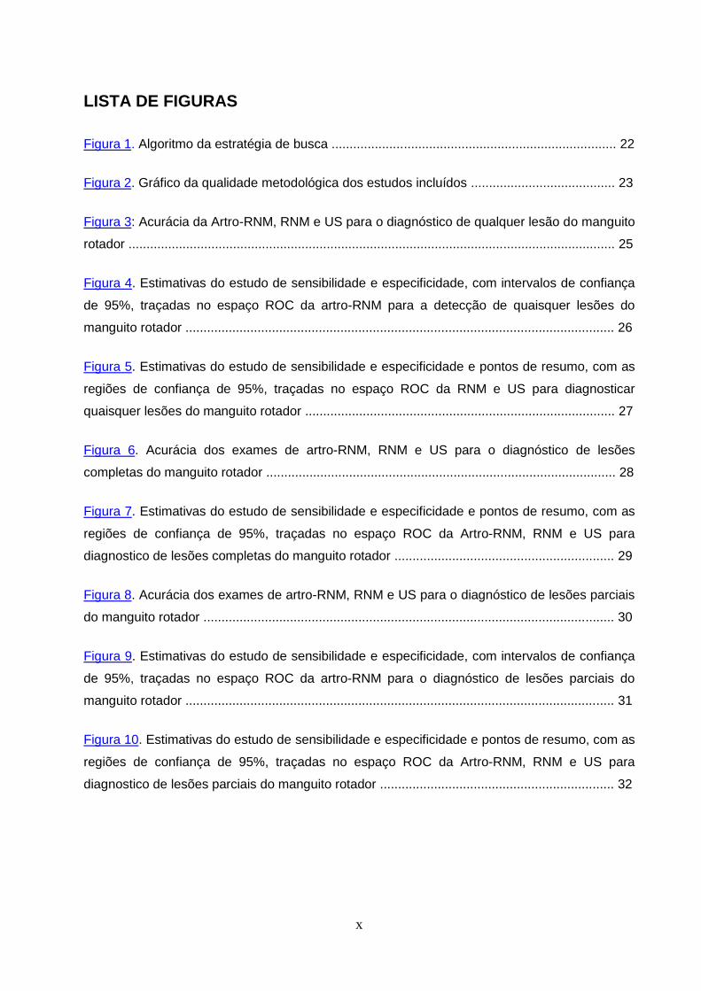
LISTA DE FIGURAS
Figura 1. Algoritmo da estratégia de busca ............................................................................... 22
Figura 2. Gráfico da qualidade metodológica dos estudos incluídos ........................................ 23
Figura 3: Acurácia da Artro-RNM, RNM e US para o diagnóstico de qualquer lesão do manguito
rotador ....................................................................................................................................... 25
Figura 4. Estimativas do estudo de sensibilidade e especificidade, com intervalos de confiança
de 95%, traçadas no espaço ROC da artro-RNM para a detecção de quaisquer lesões do
manguito rotador ....................................................................................................................... 26
Figura 5. Estimativas do estudo de sensibilidade e especificidade e pontos de resumo, com as
regiões de confiança de 95%, traçadas no espaço ROC da RNM e US para diagnosticar
quaisquer lesões do manguito rotador ...................................................................................... 27
Figura 6. Acurácia dos exames de artro-RNM, RNM e US para o diagnóstico de lesões
completas do manguito rotador ................................................................................................. 28
Figura 7. Estimativas do estudo de sensibilidade e especificidade e pontos de resumo, com as
regiões de confiança de 95%, traçadas no espaço ROC da Artro-RNM, RNM e US para
diagnostico de lesões completas do manguito rotador ............................................................. 29
Figura 8. Acurácia dos exames de artro-RNM, RNM e US para o diagnóstico de lesões parciais
do manguito rotador .................................................................................................................. 30
Figura 9. Estimativas do estudo de sensibilidade e especificidade, com intervalos de confiança
de 95%, traçadas no espaço ROC da artro-RNM para o diagnóstico de lesões parciais do
manguito rotador ....................................................................................................................... 31
Figura 10. Estimativas do estudo de sensibilidade e especificidade e pontos de resumo, com as
regiões de confiança de 95%, traçadas no espaço ROC da Artro-RNM, RNM e US para
diagnostico de lesões parciais do manguito rotador ................................................................. 32

xi
LISTA DE TABELAS
Tabela 1 Comparação de RNM, US e artro-RNM para diagnóstico de quaisquer lesões do
manguito rotador ....................................................................................................................... 24
xii
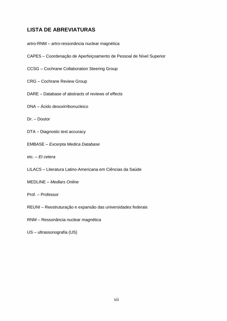
LISTA DE ABREVIATURAS
artro-RNM – artro-ressonância nuclear magnética
CAPES – Coordenação de Aperfeiçoamento de Pessoal de Nível Superior
CCSG – Cochrane Collaboration Steering Group
CRG – Cochrane Review Group
DARE – Database of abstracts of reviews of effects
DNA – Ácido desoxirribonucleico
Dr. – Doutor
DTA – Diagnostic test accuracy
EMBASE – Excerpta Medica Database
etc. – Et cetera
LILACS – Literatura Latino-Americana em Ciências da Saúde
MEDLINE – Medlars Online
Prof. – Professor
REUNI – Reestruturação e expansão das universidades federais
RNM – Ressonância nuclear magnética
US – ultrassonografia (US)
xiii
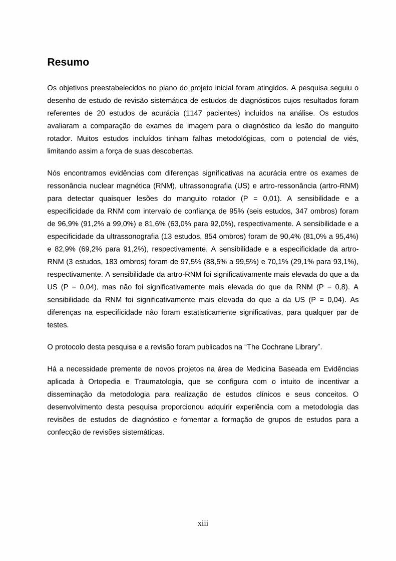
Resumo
Os objetivos preestabelecidos no plano do projeto inicial foram atingidos. A pesquisa seguiu o
desenho de estudo de revisão sistemática de estudos de diagnósticos cujos resultados foram
referentes de 20 estudos de acurácia (1147 pacientes) incluídos na análise. Os estudos
avaliaram a comparação de exames de imagem para o diagnóstico da lesão do manguito
rotador. Muitos estudos incluídos tinham falhas metodológicas, com o potencial de viés,
limitando assim a força de suas descobertas.
Nós encontramos evidências com diferenças significativas na acurácia entre os exames de
ressonância nuclear magnética (RNM), ultrassonografia (US) e artro-ressonância (artro-RNM)
para detectar quaisquer lesões do manguito rotador (P = 0,01). A sensibilidade e a
especificidade da RNM com intervalo de confiança de 95% (seis estudos, 347 ombros) foram
de 96,9% (91,2% a 99,0%) e 81,6% (63,0% para 92,0%), respectivamente. A sensibilidade e a
especificidade da ultrassonografia (13 estudos, 854 ombros) foram de 90,4% (81,0% a 95,4%)
e 82,9% (69,2% para 91,2%), respectivamente. A sensibilidade e a especificidade da artro-
RNM (3 estudos, 183 ombros) foram de 97,5% (88,5% a 99,5%) e 70,1% (29,1% para 93,1%),
respectivamente. A sensibilidade da artro-RNM foi significativamente mais elevada do que a da
US (P = 0,04), mas não foi significativamente mais elevada do que da RNM (P = 0,8). A
sensibilidade da RNM foi significativamente mais elevada do que a da US (P = 0,04). As
diferenças na especificidade não foram estatisticamente significativas, para qualquer par de
testes.
O protocolo desta pesquisa e a revisão foram publicados na “The Cochrane Library”.
Há a necessidade premente de novos projetos na área de Medicina Baseada em Evidências
aplicada à Ortopedia e Traumatologia, que se configura com o intuito de incentivar a
disseminação da metodologia para realização de estudos clínicos e seus conceitos. O
desenvolvimento desta pesquisa proporcionou adquirir experiência com a metodologia das
revisões de estudos de diagnóstico e fomentar a formação de grupos de estudos para a
confecção de revisões sistemáticas.

1
Dados do projeto
DADOS DO PROJETO
2
Dados do projeto
1. DADOS DO PROJETO
1.1. Projeto
Ressonância nuclear magnética versus ultrassonografia para o diagnóstico das lesões do
manguito rotador: revisão sistemática
Supervisor: Prof. Dr. Flávio Faloppa
Instituições de Execução do Projeto:
- Universidade Federal de São Paulo
Programa de Pós-Graduação em Cirurgia Translacional – (EPM/UNIFESP) / Rua Napoleão de
Barros, 715 – 4º andar – Vila Clementino, Cep: 04024-002 / São Paulo-SP, Brasil. Telefones:
(11) 5576-4118 / 5571-6579.
- Cabrini Hospital
Monash Department of Clinical Epidemiology, Cabrini Institute; and Department of
Epidemiology and Preventive Medicine, School of Public Health and Preventive Medicine,
Monash University – 181-183 Wattletree Rd, Malvern VIC 3144, Australia. Phone: (03) 9508
1222
- Australasian Cochrane Centre
School of Public Health and Preventive Medicine - The Alfred Centre / 99 Commercial Road
Melbourne VIC 3004, Australia. Phone: +61 3 9903 0366.
- Monash University
900 Dandenong Road Caulfield East Victoria 3145 Melbourne, Australia. Phone: + 61 3 990
32000
Vigência: setembro de 2010 a julho de 2014.
Período no exterior: Australia: março de 2011 a setembro de 2011
Bolsa REUNI - CAPES: 01/09/2010 a 28/02/2011 e 01/09/2011 a 31/05/2012
3
Dados do projeto
1.2. Dados do Pós-Doutorando
Nome: Mário Lenza
Formação profissional: Graduação em medicina (Faculdade de Ciências Médicas de Santos,
FCMS – Lusiadas / 1997-2003), Residência médica em Ortopedia e Traumatologia
(Universidade Federal de São Paulo / 2004-2007), Especialização em cirurgia do ombro e
cotovelo (Universidade Federal de São Paulo / 2007-2008), Doutor em Ciências pelo Programa
de Ortopedia e Traumatologia (Universidade Federal de São Paulo / 2008-2010). Pós-
doutorando pelo Programa de Cirurgia Translacional da Universidade Federal de São Paulo,
2010-Atual.
1.3. Dados do Supervisor
Nome: Flávio Faloppa
Formação profissional: Graduação em medicina (Universidade Federal de São Paulo / 1979),
Residência/Especialização em Ortopedia e Traumatologia (Universidade Federal de São Paulo
/ 1980-1981), Mestre em Ciências Programa de Pós-graduação em Ortopedia e Cirurgia
Plástica Reparadora (Universidade Federal de São Paulo / 1985), Doutor em Ciências
Programa de Pós-graduação em Ortopedia e Cirurgia Plástica Reparadora (Universidade
Federal de São Paulo / 1988), Livre-Docência no Departamento de Ortopedia da
EPM/UNIFESP em 1994.
1.4. Atividades no exterior
Por estar em processo de desenvolvimento na Colaboração Cochrane, a metodologia dos
estudos de revisão sistemática de diagnóstico permanece restrita aos centros internacionais e
está em fase inicial de estruturação no centro Cochrane da Austrália. Seu aprendizado,
portanto, só se configurou na medida em que fui agente participativo da construção de sua
metodologia. Ao estar vinculado a estes centros, foi possível não apenas ter me capacitado na
nova metodologia, mas também participar de seu aprimoramento, criando uma base de
conhecimento alinhada a todos os valores e princípios da Colaboração Cochrane.

7
Literatura
LITERATURA
8
Literatura
2. LITERATURA
A medicina baseada em evidências foi recentemente noticiada como um dos maiores marcos
da medicina dos últimos 160 anos, juntamente com a descoberta do DNA, o desenvolvimento
de vacinas e antibióticos e o uso de anestésicos em cirurgias (Watts, 2007). Trata-se de uma
modalidade de pesquisa que busca relacionar a melhor evidência disponível no campo da
literatura científica com a experiência clínica individual e os valores do paciente. Em sua
prática, recomenda-se o uso consciencioso da melhor evidência disponível para embasar o
processo de tomada de decisão quanto à saúde dos pacientes. Na busca destas evidências
mais relevantes, as revisões sistemáticas apresentam um papel significativo (Akobeng, 2005;
Atallah et al, 2003).
O ensino da medicina baseada em evidências vem sendo gradativamente incorporado às
instituições de pesquisa na área de Ortopedia e Traumatologia, e crescente atenção tem sido
despendida por parte destas instituições no sentido de promover o desenvolvimento de
habilidades específicas nos profissionais interessados em seu aprendizado, capacitando-os a
navegar pelo afluxo constante da literatura médica e a extrair as informações relevantes
(Degen et al, 2008).
2.1. Revisão sistemática da acurácia de testes diagnósticos
Revisão sistemática é uma metodologia de pesquisa que visa reunir toda a evidência empírica
compatível com os critérios de elegibilidade pré-definidos, a fim de responder a uma questão
específica. Esta modalidade de pesquisa permite identificar os estudos com alto risco de viés,
que podem ser tendenciosos e que muitas vezes superestimam a importância de seus achados
(Manchikanti et al, 2009; Moreno, Pantoja, 2009; Virgili et al, 2009). Por meio de uma síntese
objetiva da qualidade metodológica e dos desfechos de todos os estudos pesquisados em
certo tópico, as revisões sistemáticas ajudam a separar estudos irrelevantes ou redundantes
dos estudos mais importantes e críticos, que são dignos de reflexão. Esta característica é
especialmente interessante na atualidade, por conta do excesso de informação científica com o
qual se deparam diariamente, profissionais e gestores da área da saúde, pacientes e
pesquisadores. É improvável que todos tenham tempo, habilidades e recursos para encontrar,
avaliar e interpretar os dados obtidos por meio destas informações, incorporando-os em suas
tomadas de decisão no cuidado à saúde.
Outra propriedade das revisões sistemáticas é identificar não só o que é sabido, mas o que é
desconhecido em uma área específica, indicando necessidades de novas pesquisas em
9
Literatura
campos ainda inexplorados (Alexander, Stafford, 2009; Manchikanti et al, 2009). A informação
que se obtém por meio de revisões sistemáticas de estudos de diagnóstico possibilita
determinar o uso apropriado e a eficácia de testes diagnósticos na prática clínica, fornecendo
uma base sólida para fundamentar a criação de normas e diretrizes nos serviços primários da
saúde. Também permite analisar o desempenho e o status de certa técnica diagnóstica, bem
como avaliar a qualidade dos estudos primários de diagnóstico (Gatsonis, 2003; Gatsonis,
Paliwal, 2006).
Revisões sistemáticas e metanálises de estudos de diagnóstico podem ser usadas para a
obtenção de estimativas mais relevantes em estudos que descreverem o mesmo teste e nos
quais pacientes em um mesmo contexto estiverem disponíveis. Também são úteis para
estabelecer se e como variam os achados científicos em subgrupos particulares, além de
proverem o efeito estimado com uma generalização superior à que os estudos individuais
fornecem (Irwig et al, 1994; Leeflang et al, 2009).
No campo das revisões sistemáticas de estudos de diagnósticos, há dificuldades
metodológicas específicas. Dentre elas, a dificuldade em analisar os resultados obtidos.
Diferentemente das revisões sistemáticas de estudos de intervenção, que avaliam apenas um
desenho de estudo específico (como os ensaios clínicos randomizados), as revisões
sistemáticas de diagnóstico acessam e comparam vários modelos de desenhos de estudo
simultaneamente, o que dificulta a análise de seus resultados.
Outro desafio inerente às revisões diagnósticas é a identificação de estudos de diagnóstico.
Nas principais bases de dados da literatura atual, não há uma palavra-chave ou termo
indexado específico para a busca destes estudos; ao contrário do que ocorre com os estudos
de intervenção terapêutica, para os quais o termo “randomized controlled trial” foi definido. A
utilização dos termos Mesh (Medical Subject Heading), “sensitivity / specificity”, é aceitável; no
entanto, aplicá-los nas principais bases de dados da literatura produz achados inconsistentes.
Além disso, os dados dos estudos de diagnóstico podem ser ocultados em estudos que não
relatam as estimativas da acurácia dos testes nos seus objetivos principais, o que dificulta a
identificação de estudos de diagnóstico em bases de dados como a MEDLINE. Até que os
sistemas de indexação codifiquem propriamente os termos de estudos de diagnóstico, a busca
por eles permanecerá desafiadora, e haverá necessidade de realizar buscas manuais
adicionais, por exemplo, em listas de referência (Haynes et al, 1994; Leeflang et al, 2008).
Outra dificuldade no preparo de uma revisão sistemática de estudos de diagnóstico em relação
às revisões de estudos de intervenção consiste na forma de relatar os resultados. Enquanto
10
Literatura
nas revisões de estudos de intervenção os resultados são relatados utilizando uma única
medida de efeito - tal como diferença entre médias, diferença entre riscos, ou risco relativo - os
estudos de diagnóstico relatam pelo menos dois tipos de análises, como sensibilidade e
especificidade, valor preditivo positivo e negativo, entre outros (Gatsonis, Paliwal, 2006;
Leeflang et al, 2008).
Com a intenção de minimizar estas dificuldades e definir diretrizes metodológicas, a
Colaboração Cochrane decidiu, em 2003, preparar-se para a inclusão de revisões sistemáticas
de estudos de diagnóstico em sua base de dados de revisões sistemáticas (Manchikanti et al,
2009).
A Colaboração Cochrane, fundada em 1993, é a maior organização internacional cuja meta
principal é assistir pesquisadores na preparação, manutenção e promoção de revisões
sistemáticas (por meio da realização de colóquios, workshops, treinamentos e constantes
encontros presenciais entre os colaboradores). A organização também objetiva promover o
acesso à informação de alta qualidade, e assim, auxiliar pessoas na tomada de decisões no
cuidado à saúde (Allen et al, 2007; Green et al, 2008).
Em outubro de 2006, no encontro do Cochrane Collaboration Steering Group (CCSG), definiu-
se que as publicações de revisões (e protocolos) de diagnóstico na Cochrane Library devem
requerer aprovação prévia do grupo de revisão da Cochrane (Cochrane Review Group - CRG)
e do grupo editorial de acurácia de testes diagnósticos (Diagnostic test accuracy - DTA)
(Leeflang et al, 2008). Recentemente, para dar suporte aos novos autores, a Colaboração
Cochrane instituiu a formação do grupo DTA, dando-lhe como incumbências: aprimorar a
metodologia de revisões sistemáticas de estudos diagnósticos; desenvolver um programa de
computação para análise dos dados; e a elaboração de um guia (ainda em construção), com
orientações específicas para a elaboração de revisões na nova metodologia (Handbook). A
primeira revisão de estudos de diagnóstico foi publicada na base de dados da Cochrane em
outubro de 2008 (Leeflang et al, 2008). Até o momento, existem apenas duas revisões
publicadas com esta metodologia pela Colaboração.
Como ferramenta de treinamento para o aprendizado desta nova metodologia de estudo, os
autores deste projeto propuseram um protocolo, cujo título foi registrado na Cochrane Library
em setembro de 2009, para o desenvolvimento de uma revisão sistemática de estudos que
comparam a acurácia dos testes diagnósticos ressonância nuclear magnética versus
ultrassonografia, para o diagnóstico das lesões do manguito rotador.
2.2. Condição clínica a ser diagnosticada
11
Literatura
O manguito rotador é composto pelos tendões dos músculos subescapular, supraespinhal,
infraespinhal e redondo menor. A porção longa do tendão do bíceps também contribui com a
função do manguito, que é a de estabilizar a cabeça umeral na cavidade da glenóide,
prevenindo a migração superior da cabeça umeral (Favard et al, 2007; Matsen, 2008).
Alterações musculoesqueléticas sintomáticas do ombro são muito comuns. Uma avaliação do
sistema primário de saúde em Cambridge, Reino Unido, demonstrou uma incidência de 9,5 por
1.000 pacientes com dor no ombro. Destes, 86% apresentavam tendinopatia do manguito
rotador (Ostör et al, 2005).
As causas mais comuns de incapacidade e dor no ombro são as doenças relacionadas ao
manguito rotador em decorrência de lesão ou sobrecarga dos tendões. No ano de 2002, mais
de 4,5 milhões de consultas médicas e aproximadamente 40.000 internações para tratamento
cirúrgico foram realizadas nos Estados Unidos devido a doenças relacionadas ao manguito
rotador (Oh et al, 2007).
Comumente, a tendinopatia do manguito rotador pode resultar em lesão do tendão
supraespinhal e progredir para os demais tendões. Lewis (2008), em uma revisão narrativa
sobre fisiopatologia das lesões do manguito rotador, propôs uma teoria multifatorial com a
combinação de fatores extrínsecos e intrínsecos. Os fatores extrínsecos podem ser divididos
em causas anatômicas, tais como: forma do acrômio (curvo ou ganchoso), espessamento do
ligamento acromiclavicular, os acromiale, esporão acromial (Bigliani et al, 1991; Lewis, 2008;
Nho et al, 2008); e causas ambientais, tais como: envelhecimento, uso excessivo do ombro,
tabagismo, obesidade e distúrbios metabólicos, como a diabetes (Chen et al, 2003; Harryman
et al, 2003). Os fatores intrínsecos incluem: áreas de hipoperfusão dos tendões, processos
inflamatórios e alterações celulares dos tendões, como a desorganização da arquitetura do
colágeno, entre outros (Lewis, 2008; Rees, 2008).
As lesões do manguito rotador podem ser descritas de várias maneiras, de acordo com sua
duração (aguda ou crônica), tamanho (parciais, totais ou extensas) e etiologia (traumática ou
degenerativa); diversos sistemas de classificação foram propostos para caracterizar estas
lesões (Kuhn et al, 2007).
Atualmente, a indicação para o tratamento cirúrgico baseia-se na persistência dos sintomas
e/ou fraqueza muscular e/ou tamanho da lesão. Em geral, quando se opta pela cirurgia, o
diagnóstico por imagem pode auxiliar no planejamento do tratamento cirúrgico, uma vez que
possibilita mensurar a extensão da lesão (parcial ou total) e discriminar quais tendões estão
envolvidos (supraespinhal, infraespinhal, etc.).
12
Literatura
2.3. Descrição dos testes de diagnóstico por imagem (teste índice)
A RNM e/ou a US são os exames de escolha para caracterizar as lesões do manguito rotador
em pacientes que possuem indicação de procedimento cirúrgico. A qualidade destes exames
de imagem tem progredido substancialmente ao longo do tempo, propiciando uma avaliação
mais precisa do tamanho e extensão da lesão do manguito rotador e um planejamento mais
acurado para realização da intervenção cirúrgica (Rees, 2008).
O exame de US do ombro é um exame não invasivo, praticamente sem efeitos colaterais
associados e que permite a avaliação dinâmica das estruturas durante o movimento (Al-Shawi
et al, 2008). Pode ser utilizada no setor primário de avaliação da saúde para a investigação da
integridade dos tendões do manguito. No entanto, possui algumas limitações, como a de ser
um exame operador-dependente e possuir uma longa curva de aprendizado (O'Connor et al,
2005; Rutten et al, 2006), principalmente em vista das roturas parciais do tendão, cuja
classificação possui uma alta variabilidade interobservador, conforme relataram Le Corroller et
al (2008).
A RNM é um método não invasivo, que gera imagens de alta resolução em múltiplos planos,
utilizada com frequência nos setores secundários e terciários de saúde. Consiste em uma
avaliação estática dos tecidos, podendo exigir ou não uma injeção intra-articular de contraste
radiopaco nos tecidos moles das articulações. A RNM possui algumas contraindicações
absolutas: clipes de aneurisma intracerebral, marcapassos cardíacos, desfibriladores
automáticos, bioestimuladores, dispositivos implantados de infusão, aparelhos auditivos
internos e corpo estranho metálico orbital (Witte, 2003). E é um exame de alto custo.
2.4. Descrição dos testes de referência
Os testes de referência para a avaliação das lesões do manguito rotador são exames invasivos
de diagnóstico. O teste de referência mais usado comumente é a artroscopia diagnóstica. A
artroscopia é um procedimento cirúrgico minimamente invasivo, que consiste na introdução de
um artroscópio (tipo de endoscópio de fibra óptica) na articulação, através de uma pequena
incisão. A técnica permite ao cirurgião avaliar diretamente os tendões do manguito em suas
faces articular e bursal e, ainda, realizar um exame geral das estruturas que compõem a
articulação do ombro para detectar e tratar outras potenciais lesões (Dinnes et al, 2003; Matava
et al, 2005). Limitações associadas ao exame diagnóstico da artroscopia do ombro incluem a
curva de aprendizado e algumas variações interobservadores durante a classificação das
principais lesões (Kuhn et al, 2007).
13
Literatura
A cirurgia aberta do ombro (incluindo a mini-open) também pode ser utilizada como teste de
referência para as lesões do manguito rotador. Entretanto, é menos precisa do que a
artroscopia, porque o acesso às lesões intra-articulares e à região inferior do manguito está
prejudicado nesta abordagem cirúrgica.
2.5. Importância de realizar este estudo
Os exames de US e/ou RNM estão sendo cada vez mais usados para avaliar a presença e
extensão da lesão do manguito rotador com o objetivo de realizar o planejamento da
intervenção cirúrgica. O aprimoramento das técnicas para realizar estes testes de imagem não
invasivos resultou no aumento da confiabilidade destes exames, fazendo com que venham
substituindo parcialmente o uso da artroscopia diagnóstica, embora esta ainda seja comumente
realizada como parte do tratamento cirúrgico das doenças do ombro. Ambos, US e RNM são
operador e/ou avaliador dependentes e a RNM é um exame de alto custo. Permanece incerto
se um método é superior a outro e se o uso combinado de ambos melhora a acurácia
diagnóstica (Swen et al, 1999), bem como se seus custos justificam sua utilização. Também
permanece indefinido se estes testes não invasivos fornecem informações adicionais
relevantes em relação à artroscopia diagnóstica, a qual compõe parte do tratamento cirúrgico.
Apenas duas revisões sistemáticas com metanálise estudaram testes de diagnóstico por
imagem para as doenças do manguito rotador (de Jesus et al, 2009; Dinnes et al, 2003). A
estratégia de busca em ambos os estudos restringiu-se à literatura em língua inglesa. Uma
revisão (Dinnes et al, 2003) avaliou a acurácia diagnóstica dos testes clínicos, US e RNM (data
da estratégia: outubro, 2001), para detectar lesão do manguito rotador, tomando como teste de
referência os exames diagnósticos cirúrgicos e os não cirúrgicos. Concluiu-se que a US e a
RNM são equivalentes para diagnosticar lesão total do manguito rotador, embora a RNM tenha
maior custo e a US seja superior na detecção de lesões parciais do manguito. De Jesus et al
(2009) relataram uma metanálise comparando o diagnóstico da US versus RNM para as lesões
do manguito rotador, usando a cirurgia como teste de referência. Foram incluídos nesta revisão
65 estudos (data da estratégia: setembro, 2007), entretanto, a avaliação da qualidade
metodológica dos estudos incluídos não foi adequada. Concluiu-se que a US é tão precisa
quanto a RNM para diagnosticar tanto lesão parcial como total do manguito rotador; também foi
constatado que a US é o teste de imagem de menor custo para detectar as lesões do
manguito.
2.6. Objetivos
14
Literatura
O objetivo primário desta revisão foi comparar a acurácia dos testes diagnósticos de imagem:
ressonância nuclear magnética versus ultrassonografia, para detectar lesões do manguito
rotador em pacientes com dor no ombro e que possuem indicação de tratamento cirúrgico.
Como objetivo secundário, identificamos qual é o melhor teste de imagem para diagnosticar
lesões totais do manguito rotador; e qual é o melhor teste de imagem para diagnosticar lesões
parciais do manguito rotador.
2.7. Investigação das fontes de heterogeneidade
As possíveis fontes de heterogeneidade foram investigadas por meio da realização de análises
de subgrupo de acordo com as características da população, lesão do manguito, teste de
referência, desenho do estudo e qualidade metodológica do estudo.
• População do estudo: idosos ou jovens.
• Tipo de lesão do manguito: aguda ou crônica; parcial, total ou extensa; traumática
ou degenerativa.
• Tipo de teste de referência: cirurgia aberta (incluindo mini-open) ou artroscopia.
• Desenho do estudo: estudo transversal ou coorte ou caso-controle; e retrospectivo
ou prospectivo.
• Qualidade metodológica do estudo: baixo risco de viés ou de alto risco de viés.
2.8. Hipóteses
Esta revisão pretende testar a seguinte hipótese nula:
Não existe diferença entre a acurácia diagnóstica da ressonância nuclear magnética e a
ultrassonografia para detectar lesões (total ou parcial) do manguito rotador em pacientes com
dor no ombro e que possuem indicação de tratamento cirúrgico.

15
Método
MÉTODO
16
Método
3. MÉTODO
O método de realização deste projeto baseou-se nas recomendações propostas pela Cochrane
Collaboration Screening and Diagnostic Tests Methods Group para desenvolver revisões
sistemáticas de estudos de acurácia (http://srdta.cochrane.org/en/index.html).
Este protocolo de pesquisa foi aprovado pelo Comitê de Ética em Pesquisa (CEP) da
Universidade Federal de São Paulo / Escola Paulista de Medicina / número 0154/10 em 26 de
fevereiro de 2010 (Apêndice 1). O título foi previamente registrado na Cochrane Library em
setembro de 2009.
Cabe salientar que esta revisão sistemática foi publicada na Cochrane Library, sendo um
protocolo publicado em 2011 (Apêndice 2) e uma revisão sistemática em 2013 (Apêndice 3).
3.1. Critérios a serem considerados nos estudos desta revisão
3.1.1. Tipos de estudos incluídos
Foram incluídos todos os estudos de acurácia que compararam ressonância nuclear magnética
(RNM) e/ou ultrassonografia (US) com os testes de referência em pacientes com suspeita de
lesão parcial ou total do manguito rotador. Apenas foram incluídos os resultados de estudos
completos; quando os estudos foram expostos em resumos ou anais de conferência, somente
os dados da publicação completa foram relatados.
Foram excluídos estudos com período excessivamente longo (maior que seis meses) entre o
teste diagnóstico e o teste de referência, em vista da doença do manguito rotador ser
progressiva.
Estudos em todos os idiomas foram incluídos, quando uma tradução completa para o inglês ou
português foi obtida. Artigos que não foram traduzidos de maneira integral foram citados em
um apêndice, mas não incluídos na revisão.
Nos casos em que o mesmo estudo for publicado em mais de um artigo, foram incluídos
apenas os estudos mais atualizados ou completos. No entanto, as referências das outras
publicações foram citadas no âmbito do estudo.
17
Método
3.1.2. Tipos de participantes
Foram incluídos estudos que avaliaram pacientes com dor no ombro e suspeita de lesão
parcial ou total do manguito rotador, com indicação de cirurgia. Estudos que abordaram apenas
testes de diagnóstico clínico e/ou pacientes com diagnóstico de dor no ombro por outras
causas (instabilidade, artrose, artrite reumatóide, capsulite adesiva, tendinite calcária,
neoplasias benignas ou malignas, etc) foram excluídos.
3.1.3. Testes de diagnósticos avaliados (teste índice)
Testes de diagnóstico por imagem que comparam US e/ou RNM (incluindo artro-ressonância
nuclear magnética – artro-RNM) com o definido teste de referência foram incluídos. Estudos
que compararem exclusivamente exames clínicos, radiografias, artrografias, tomografias
computadorizadas não foram incluídos.
3.1.4. Doença avaliada
A doença avaliada nesta revisão foi a lesão do manguito rotador (total ou parcial) em pacientes
com dor no ombro e que possuem indicação de tratamento cirúrgico.
3.1.5. Teste de referência
Os testes de referência para definir a doença em questão foram a artroscopia ou a cirurgia
aberta (incluindo mini-open). Quando um estudo abordou ambos os testes (artroscopia e
cirurgia aberta), apenas a artroscopia foi tid como o teste de referência.
3.2. Estratégia de busca para a identificação dos estudos
Buscas eletrônicas
As pesquisas eletrônicas das bases de dados utilizadas foram: MEDLINE (1966 até março de
2011), EMBASE (1988 até fevereiro de 2011), LILACS (1982 até fevereiro de 2011). Não houve
restrições com base no idioma ou status da publicação.
Uma estratégia de busca foi desenvolvida conforme as orientações do capítulo sete do
Handbook (de Vet et al, 2008), a qual utilizou termos descritores e seus sinônimos das bases
de dados MEDLINE (PubMed), EMBASE (OVID WEB), e LILACS (Bireme), como descrito
abaixo e no Anexo 1:
18
Método
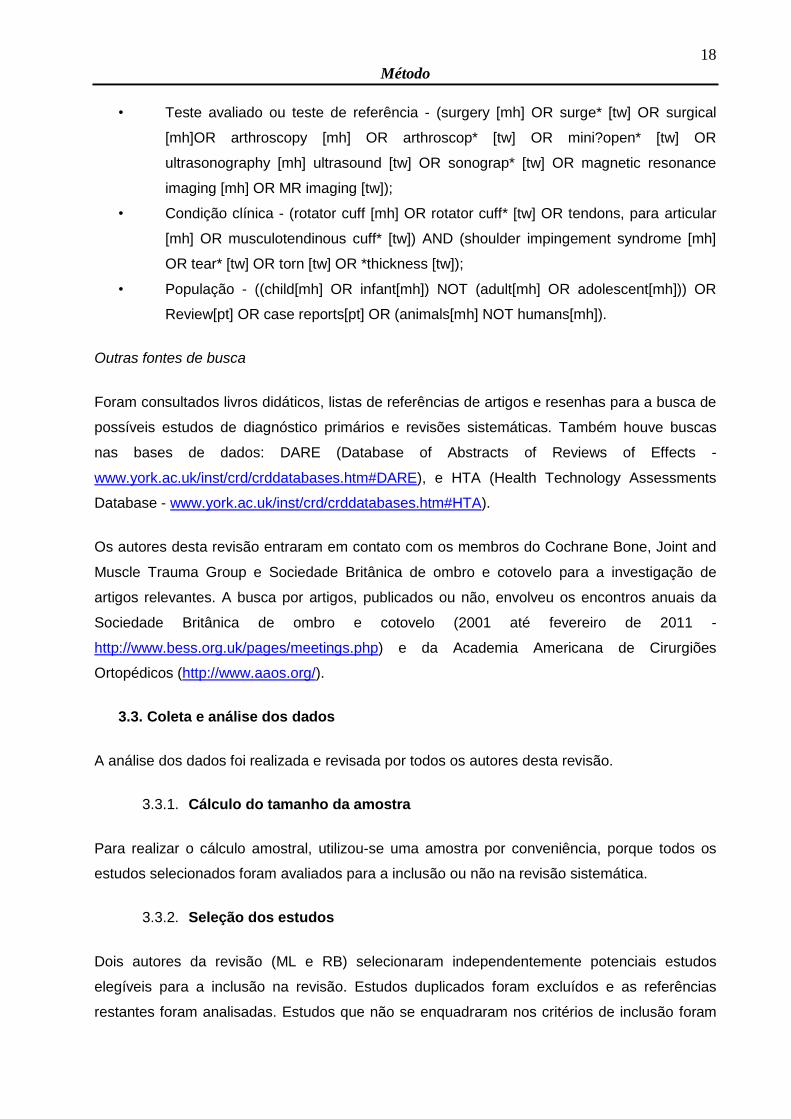
• Teste avaliado ou teste de referência - (surgery [mh] OR surge* [tw] OR surgical
[mh]OR arthroscopy [mh] OR arthroscop* [tw] OR mini?open* [tw] OR
ultrasonography [mh] ultrasound [tw] OR sonograp* [tw] OR magnetic resonance
imaging [mh] OR MR imaging [tw]);
• Condição clínica - (rotator cuff [mh] OR rotator cuff* [tw] OR tendons, para articular
[mh] OR musculotendinous cuff* [tw]) AND (shoulder impingement syndrome [mh]
OR tear* [tw] OR torn [tw] OR *thickness [tw]);
• População - ((child[mh] OR infant[mh]) NOT (adult[mh] OR adolescent[mh])) OR
Review[pt] OR case reports[pt] OR (animals[mh] NOT humans[mh]).
Outras fontes de busca
Foram consultados livros didáticos, listas de referências de artigos e resenhas para a busca de
possíveis estudos de diagnóstico primários e revisões sistemáticas. Também houve buscas
nas bases de dados: DARE (Database of Abstracts of Reviews of Effects -
www.york.ac.uk/inst/crd/crddatabases.htm#DARE), e HTA (Health Technology Assessments
Database - www.york.ac.uk/inst/crd/crddatabases.htm#HTA).
Os autores desta revisão entraram em contato com os membros do Cochrane Bone, Joint and
Muscle Trauma Group e Sociedade Britânica de ombro e cotovelo para a investigação de
artigos relevantes. A busca por artigos, publicados ou não, envolveu os encontros anuais da
Sociedade Britânica de ombro e cotovelo (2001 até fevereiro de 2011 -
http://www.bess.org.uk/pages/meetings.php) e da Academia Americana de Cirurgiões
Ortopédicos (http://www.aaos.org/).
3.3. Coleta e análise dos dados
A análise dos dados foi realizada e revisada por todos os autores desta revisão.
3.3.1. Cálculo do tamanho da amostra
Para realizar o cálculo amostral, utilizou-se uma amostra por conveniência, porque todos os
estudos selecionados foram avaliados para a inclusão ou não na revisão sistemática.
3.3.2. Seleção dos estudos
Dois autores da revisão (ML e RB) selecionaram independentemente potenciais estudos
elegíveis para a inclusão na revisão. Estudos duplicados foram excluídos e as referências
restantes foram analisadas. Estudos que não se enquadraram nos critérios de inclusão foram
19
Método
descartados. Para os estudos relevantes, foram obtidas cópias integrais. ML e RB avaliaram
independentemente os estudos relevantes e determinar a inclusão ou exclusão dos mesmos.
Todas as dúvidas ou divergências foram resolvidas por discussão e, quando necessário um
terceiro autor (FF) intercedeu para sua resolução.
3.3.3. Extração e manejo dos dados
ML e RB independentemente coletaram os dados disponíveis dos estudos incluídos utilizando
um formulário piloto de extração, sem mascaramento dos autores dos estudos ou outras
informações de identificação. Um terceiro autor da revisão (FF) foi consultado para a resolução
de eventuais divergências. Quando necessário, informações adicionais ou dados relevantes
foram requisitados aos autores dos estudos incluídos.
Estudos de diagnóstico com dados insuficientes para a confecção de tabelas 2x2 foram
excluídos das análises estatísticas, mas estes resultados foram incluídos na parte narrativa da
revisão.
3.3.4. Avaliação da qualidade metodológica dos estudos incluídos
A qualidade metodológica dos estudos incluídos foi avaliada independentemente por ML e RB
e discordâncias foram resolvidas por um terceiro revisor (FF). A qualidade metodológica foi
avaliada ao mesmo tempo em que a extração dos dados, utilizando uma lista de dados
denominada QUADAS (Whiting et al, 2003), adaptada para a revisão.
3.3.5. Síntese dos dados e análise estatística
Os índices de desempenho do diagnóstico foram extraídos ou derivados dos dados presentes
em cada estudo primário de cada exame de imagem. Foram confeccionadas tabelas 2x2 de
contingência dos casos verdadeiros positivos, falsos positivos, falsos negativos e verdadeiros
negativos. Os autores desta revisão calcularam a sensibilidade e especificidade com 95% de
intervalo de confiança para cada teste de imagem, em cada estudo. Os autores também
investigaram graficamente, por meio do Gráfico Floresta (Forest Plot), a heterogeneidade
estimada entre a sensibilidade e especificidade. Para uma análise descritiva, os resultados das
taxas dos verdadeiros positivos (sensibilidade) contra as taxas dos falsos positivos (1 –
especificidade) foram analisados graficamente utilizando o sumário da curva ROC (Receiver
Operating Characteristic).
20
Método
Para as metanálises de pares de sensibilidade e especificidade e para formação dos sumários
da curva ROC (SROC) utilizou-se o método de hierarquização dos SROC (HSROC) (Rutter et
al, 2001). O método HSROC foi fundamentado em uma abordagem de efeitos aleatórios e
levará em consideração o grau de heterogeneidade entre os estudos. Foram formalmente
avaliadas como potenciais fontes de heterogeneidade, a significância das diferenças entre os
testes e a significância das diferenças entre os subgrupos pré-definidos.
3.4. Atualização e aprimoamento da revisão
As atualizações desta revisão sistemática serão realizadas anualmente. A versão atualizada
poderá ser encontrada na Cochrane Library. Mesmo se não houver nenhum estudo clínico que
se enquadre nos critérios de inclusão nessa atualização anual ou nenhuma correção maior for
indicada, a data da última busca dos estudos de diagnóstico será colocada na seção de
estratégia de busca desta revisão.

21
Resultados
RESULTADOS
22
Resultados
4. RESULTADOS
4.1. Descrição dos estudos – análise qualitativa
4.1.1. Resultado da estratégia de busca
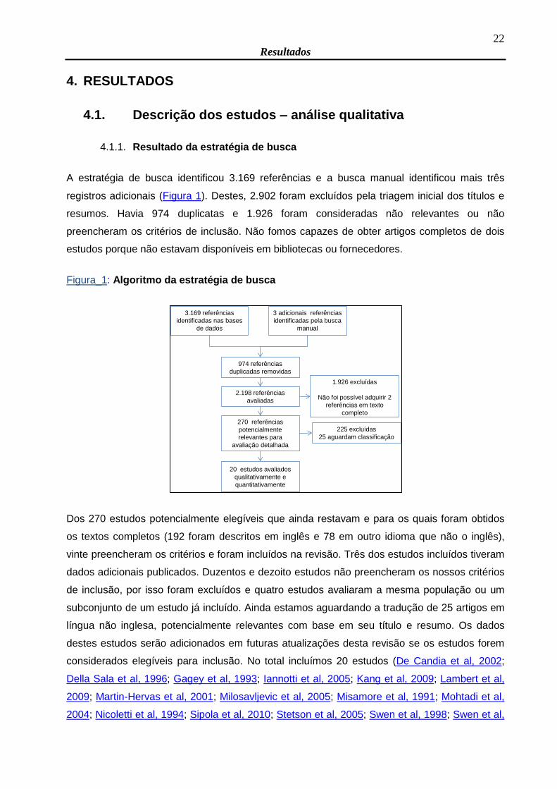
A estratégia de busca identificou 3.169 referências e a busca manual identificou mais três
registros adicionais (Figura 1). Destes, 2.902 foram excluídos pela triagem inicial dos títulos e
resumos. Havia 974 duplicatas e 1.926 foram consideradas não relevantes ou não
preencheram os critérios de inclusão. Não fomos capazes de obter artigos completos de dois
estudos porque não estavam disponíveis em bibliotecas ou fornecedores.
Figura_1: Algoritmo da estratégia de busca
Dos 270 estudos potencialmente elegíveis que ainda restavam e para os quais foram obtidos
os textos completos (192 foram descritos em inglês e 78 em outro idioma que não o inglês),
vinte preencheram os critérios e foram incluídos na revisão. Três dos estudos incluídos tiveram
dados adicionais publicados. Duzentos e dezoito estudos não preencheram os nossos critérios
de inclusão, por isso foram excluídos e quatro estudos avaliaram a mesma população ou um
subconjunto de um estudo já incluído. Ainda estamos aguardando a tradução de 25 artigos em
língua não inglesa, potencialmente relevantes com base em seu título e resumo. Os dados
destes estudos serão adicionados em futuras atualizações desta revisão se os estudos forem
considerados elegíveis para inclusão. No total incluímos 20 estudos (De Candia et al, 2002;
Della Sala et al, 1996; Gagey et al, 1993; Iannotti et al, 2005; Kang et al, 2009; Lambert et al,
2009; Martin-Hervas et al, 2001; Milosavljevic et al, 2005; Misamore et al, 1991; Mohtadi et al,
2004; Nicoletti et al, 1994; Sipola et al, 2010; Stetson et al, 2005; Swen et al, 1998; Swen et al,
3.169 referências
identificadas nas bases
de dados
3 adicionais referências
identificadas pela busca
manual
974 referências
duplicadas removidas
2.198 referências
avaliadas
1.926 excluídas
Não foi possível adquirir 2
referências em texto
completo
270 referências
potencialmente
relevantes para
avaliação detalhada
225 excluídas
25 aguardam classificação
20 estudos avaliados
qualitativamente e
quantitativamente
23
Resultados
1999; Taboury et al, 1992; Teefey et al, 2004; Venu et al, 2002; Wallny et al, 2001; Yen et al,
2004).
4.1.2. Qualidade metodológica dos estudos incluídos
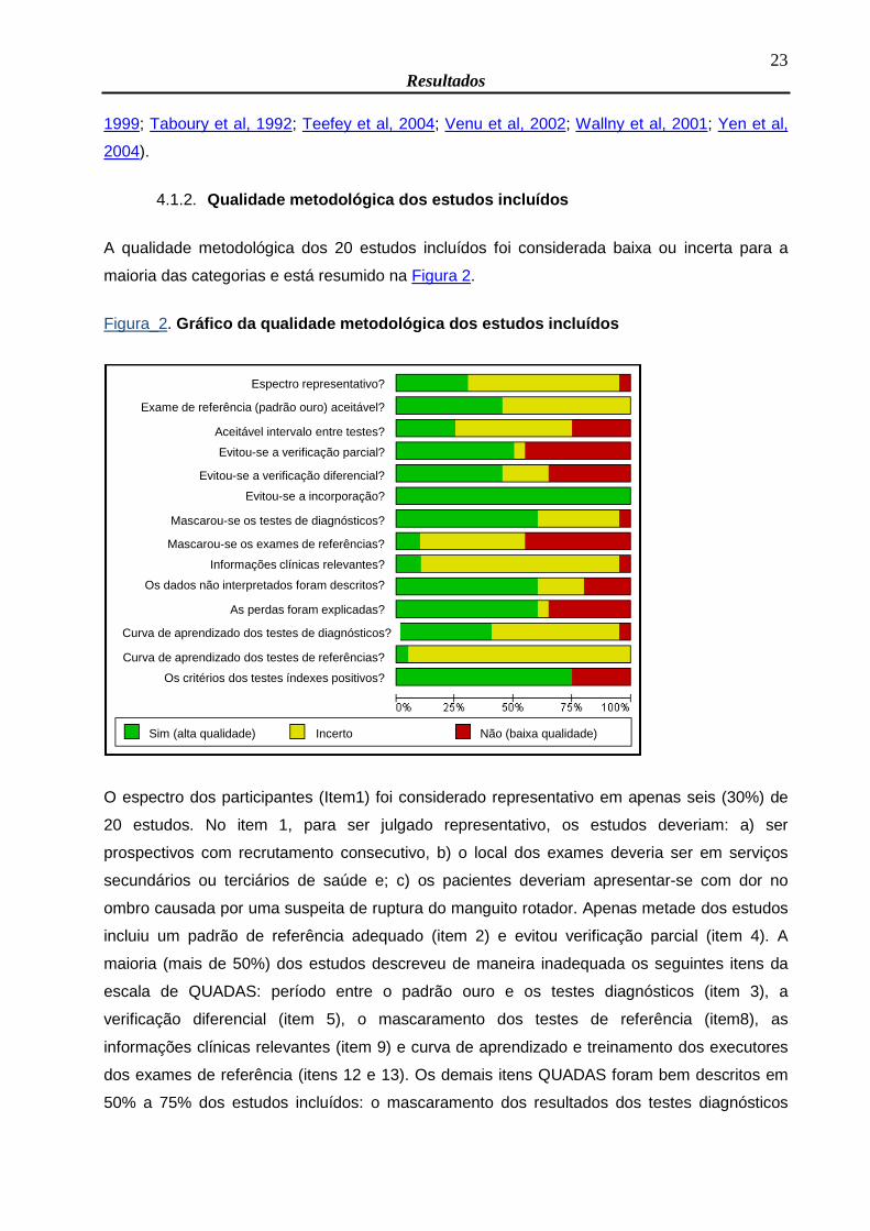
A qualidade metodológica dos 20 estudos incluídos foi considerada baixa ou incerta para a
maioria das categorias e está resumido na Figura 2.
Figura_2. Gráfico da qualidade metodológica dos estudos incluídos
O espectro dos participantes (Item1) foi considerado representativo em apenas seis (30%) de
20 estudos. No item 1, para ser julgado representativo, os estudos deveriam: a) ser
prospectivos com recrutamento consecutivo, b) o local dos exames deveria ser em serviços
secundários ou terciários de saúde e; c) os pacientes deveriam apresentar-se com dor no
ombro causada por uma suspeita de ruptura do manguito rotador. Apenas metade dos estudos
incluiu um padrão de referência adequado (item 2) e evitou verificação parcial (item 4). A
maioria (mais de 50%) dos estudos descreveu de maneira inadequada os seguintes itens da
escala de QUADAS: período entre o padrão ouro e os testes diagnósticos (item 3), a
verificação diferencial (item 5), o mascaramento dos testes de referência (item8), as
informações clínicas relevantes (item 9) e curva de aprendizado e treinamento dos executores
dos exames de referência (itens 12 e 13). Os demais itens QUADAS foram bem descritos em
50% a 75% dos estudos incluídos: o mascaramento dos resultados dos testes diagnósticos
Espectro representativo?
Exame de referência (padrão ouro) aceitável?
Aceitável intervalo entre testes?
Evitou-se a verificação parcial?
Evitou-se a verificação diferencial?
Evitou-se a incorporação?
Mascarou-se os testes de diagnósticos?
Mascarou-se os exames de referências?
Informações clínicas relevantes?
Os dados não interpretados foram descritos?
As perdas foram explicadas?
Curva de aprendizado dos testes de diagnósticos?
Curva de aprendizado dos testes de referências?
Os critérios dos testes índexes positivos?
Sim (alta qualidade) Incerto Não (baixa qualidade)
24
Resultados
(item 7), os resultados dos dados não-interpretáveis (item 10), a descrição adequada das
perdas (artigo 11) e critérios para a positividade dos testes diagnóstcios (item14). Como
antecipamos em nosso protocolo, a resposta para a "incorporação" (ponto 6) foi 'Sim' (sem
viés) para todos os estudos incluídos.
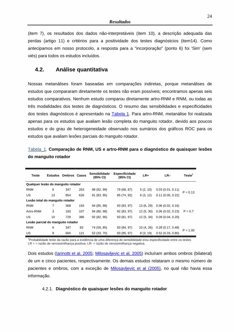
4.2. Análise quantitativa
Nossas metanálises foram baseadas em comparações indiretas, porque metanálises de
estudos que compararam diretamente os testes não eram possíveis; encontramos apenas seis
estudos comparativos. Nenhum estudo comparou diretamente artro-RNM e RNM, ou todas as
três modalidades dos testes de diagnósticos. O resumo das sensibilidades e especificidades
dos testes diagnósticos é apresentado na Tabela 1. Para artro-RNM, metanálise foi realizada
apenas para os estudos que avaliam lesão completa do manguito rotador, devido aos poucos
estudos e do grau de heterogeneidade observado nos sumários dos gráficos ROC para os
estudos que avaliam lesões parciais do manguito rotador.
Tabela_1. Comparação de RNM, US e artro-RNM para o diagnóstico de quaisquer lesões
do manguito rotador
Teste Estudos Ombros Casos Sensibilidade
(95% CI) Especificidade
(95% CI) LR+ LR– Teste
1
Qualquer lesão do manguito rotador
RNM 6 347 263 98 (92, 99) 79 (68, 87) 5 (2, 10) 0.03 (0.01, 0.11) P = 0.13
US 13 854 626 91 (83, 95) 85 (74, 92) 6 (3, 12) 0.11 (0.05, 0.22)
Lesão total do manguito rotador
RNM 7 368 193 94 (85, 98) 93 (83, 97) 13 (6, 29) 0.06 (0.02, 0.16)
P = 0.7 Artro-RNM 3 183 107 94 (80, 98) 92 (83, 97) 12 (5, 30) 0.06 (0.02, 0.23)
US 10 729 386 92 (82, 96) 93 (81, 97) 12 (5, 34) 0.09 (0.04, 0.20)
Lesão parcial do manguito rotador
RNM 6 347 83 74 (59, 85) 93 (84, 97) 10 (4, 26) 0.28 (0.17, 0.48) P = 1.00
US 8 660 121 52 (33, 70) 93 (85, 97) 8 (3, 19) 0.52 (0.33, 0.80)
1Probabilidade teste da razão para a evidência de uma diferença de sensibilidade e/ou especificidade entre os testes
LR + = razão de verossimilhança positiva; LR- = razão de verossimilhança negativa.
Dois estudos (Iannotti et al, 2005; Milosavljevic et al, 2005) incluíram ambos ombros (bilateral)
de um e cinco pacientes, respectivamente. Os demais estudos relataram o mesmo número de
pacientes e ombros, com a exceção de Milosavljevic et al (2005), no qual não havia essa
informação.
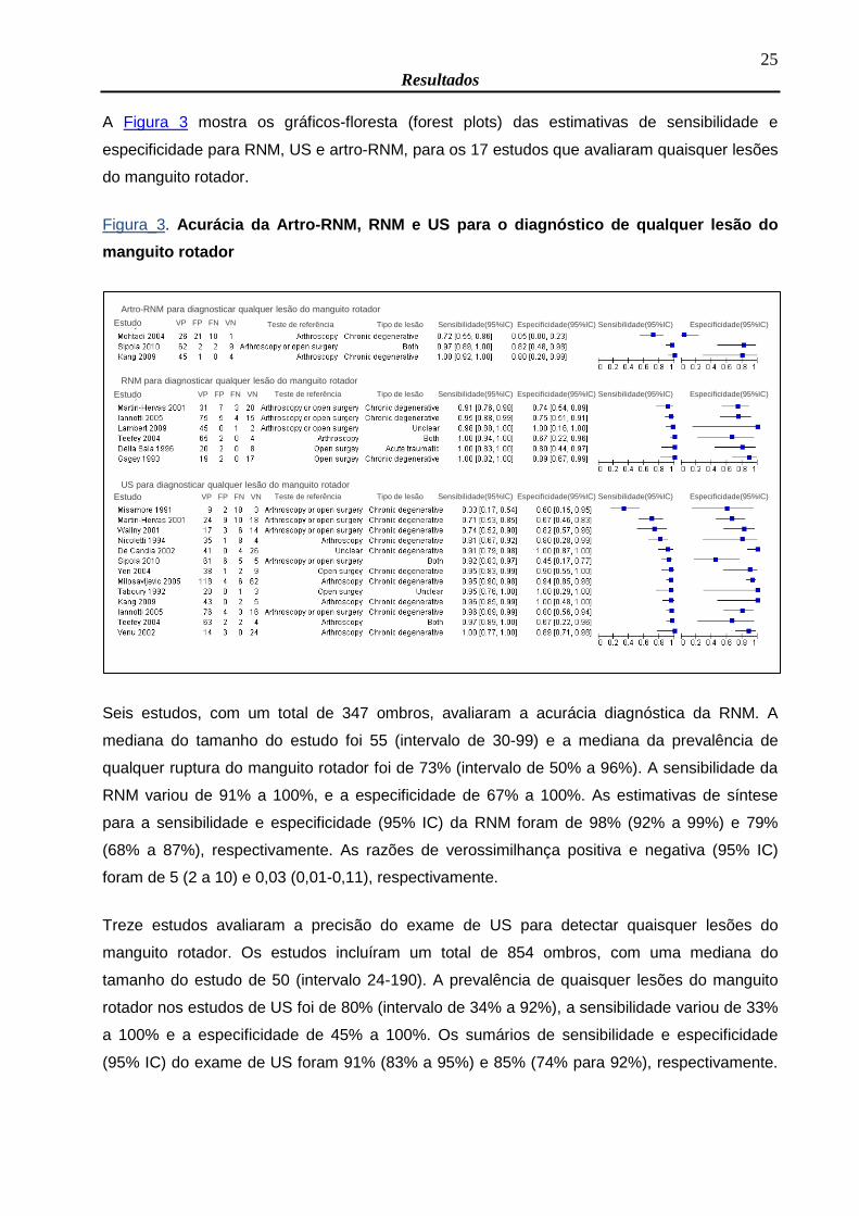
4.2.1. Diagnóstico de quaisquer lesões do manguito rotador
25
Resultados
A Figura 3 mostra os gráficos-floresta (forest plots) das estimativas de sensibilidade e
especificidade para RNM, US e artro-RNM, para os 17 estudos que avaliaram quaisquer lesões
do manguito rotador.
Figura_3. Acurácia da Artro-RNM, RNM e US para o diagnóstico de qualquer lesão do
manguito rotador
Seis estudos, com um total de 347 ombros, avaliaram a acurácia diagnóstica da RNM. A
mediana do tamanho do estudo foi 55 (intervalo de 30-99) e a mediana da prevalência de
qualquer ruptura do manguito rotador foi de 73% (intervalo de 50% a 96%). A sensibilidade da
RNM variou de 91% a 100%, e a especificidade de 67% a 100%. As estimativas de síntese
para a sensibilidade e especificidade (95% IC) da RNM foram de 98% (92% a 99%) e 79%
(68% a 87%), respectivamente. As razões de verossimilhança positiva e negativa (95% IC)
foram de 5 (2 a 10) e 0,03 (0,01-0,11), respectivamente.
Treze estudos avaliaram a precisão do exame de US para detectar quaisquer lesões do
manguito rotador. Os estudos incluíram um total de 854 ombros, com uma mediana do
tamanho do estudo de 50 (intervalo 24-190). A prevalência de quaisquer lesões do manguito
rotador nos estudos de US foi de 80% (intervalo de 34% a 92%), a sensibilidade variou de 33%
a 100% e a especificidade de 45% a 100%. Os sumários de sensibilidade e especificidade
(95% IC) do exame de US foram 91% (83% a 95%) e 85% (74% para 92%), respectivamente.
Artro-RNM para diagnosticar qualquer lesão do manguito rotador
RNM para diagnosticar qualquer lesão do manguito rotador
US para diagnosticar qualquer lesão do manguito rotador
Estudo
Estudo
VP FP FN VN
VP FP FN VN
Teste de referência
Teste de referência
Estudo VP FP FN VN Teste de referência Tipo de lesão Sensibilidade(95%IC) Especificidade(95%IC) Sensibilidade(95%IC) Especificidade(95%IC)
Tipo de lesão Sensibilidade(95%IC) Especificidade(95%IC) Sensibilidade(95%IC) Especificidade(95%IC)
Tipo de lesão Sensibilidade(95%IC) Especificidade(95%IC) Sensibilidade(95%IC) Especificidade(95%IC)
26
Resultados
As razões de verossimilhança positiva e negativa (95% IC) foram de 6 (3 a 12) e 0,11 (0,05 a
0,22), respectivamente.
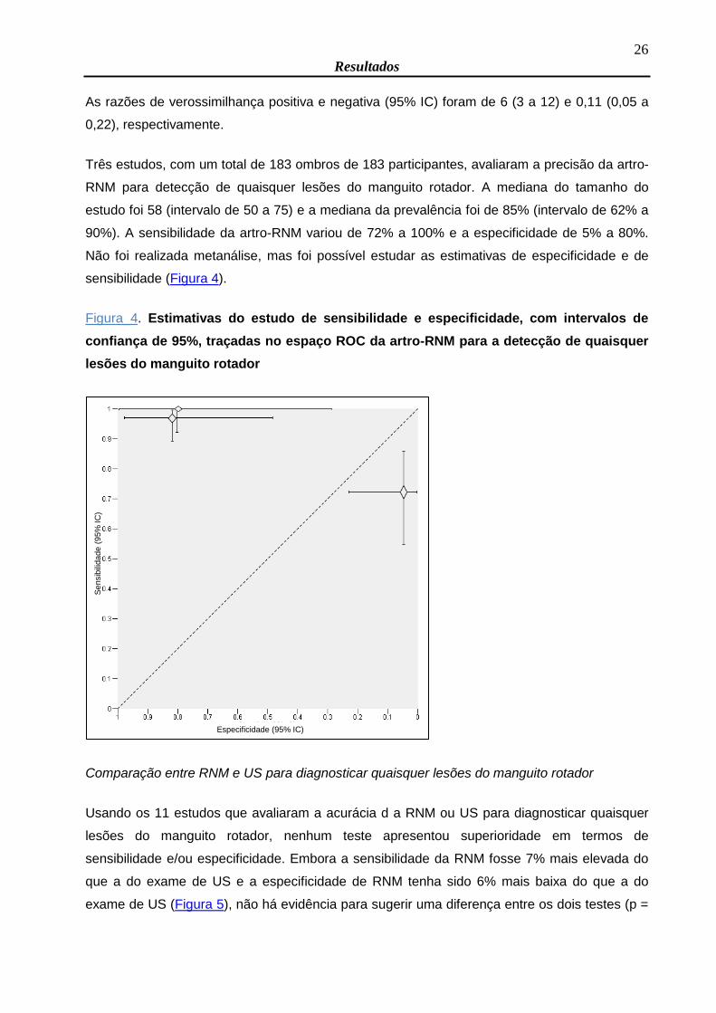
Três estudos, com um total de 183 ombros de 183 participantes, avaliaram a precisão da artro-
RNM para detecção de quaisquer lesões do manguito rotador. A mediana do tamanho do
estudo foi 58 (intervalo de 50 a 75) e a mediana da prevalência foi de 85% (intervalo de 62% a
90%). A sensibilidade da artro-RNM variou de 72% a 100% e a especificidade de 5% a 80%.
Não foi realizada metanálise, mas foi possível estudar as estimativas de especificidade e de
sensibilidade (Figura 4).
Figura_4. Estimativas do estudo de sensibilidade e especificidade, com intervalos de
confiança de 95%, traçadas no espaço ROC da artro-RNM para a detecção de quaisquer
lesões do manguito rotador
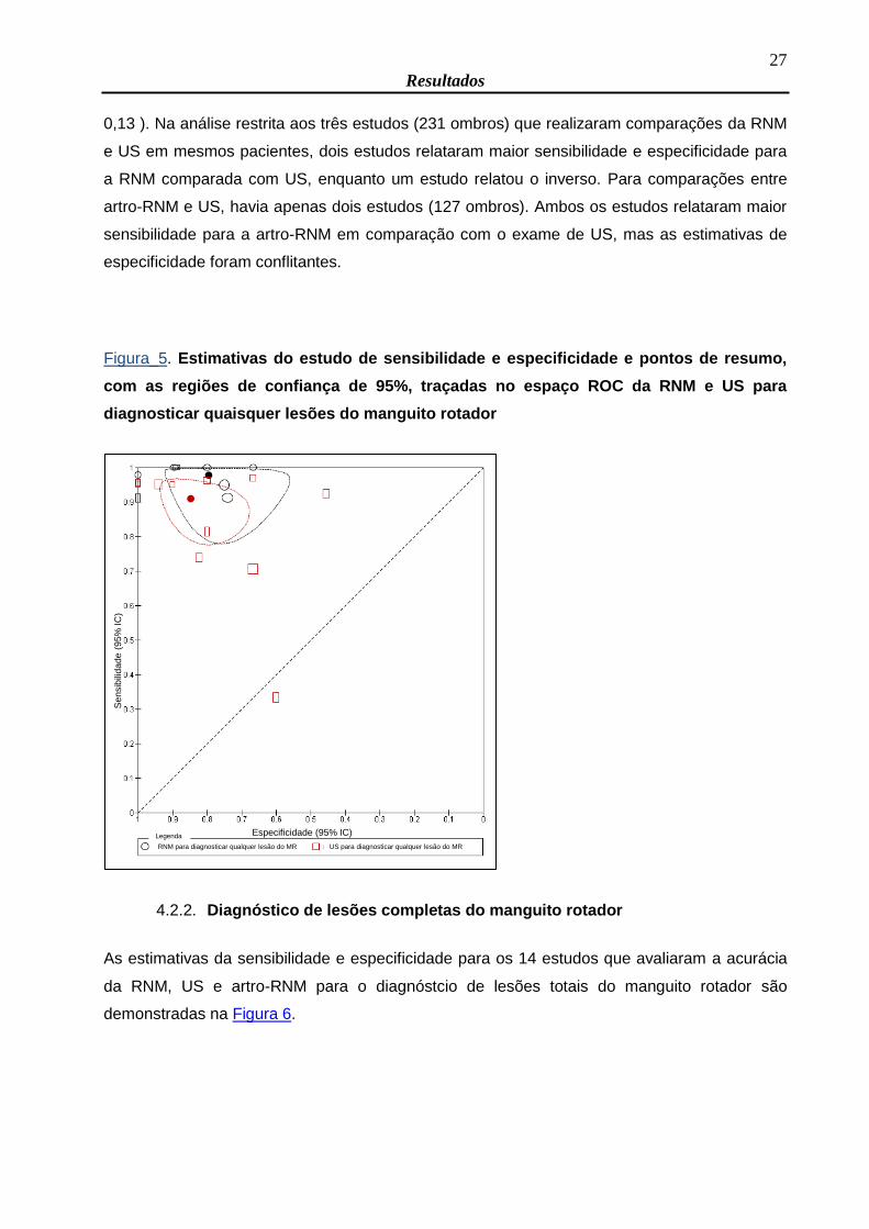
Comparação entre RNM e US para diagnosticar quaisquer lesões do manguito rotador
Usando os 11 estudos que avaliaram a acurácia d a RNM ou US para diagnosticar quaisquer
lesões do manguito rotador, nenhum teste apresentou superioridade em termos de
sensibilidade e/ou especificidade. Embora a sensibilidade da RNM fosse 7% mais elevada do
que a do exame de US e a especificidade de RNM tenha sido 6% mais baixa do que a do
exame de US (Figura 5), não há evidência para sugerir uma diferença entre os dois testes (p =
Sensib
ilidade (
95%
IC
)
Especificidade (95% IC)
27
Resultados
0,13 ). Na análise restrita aos três estudos (231 ombros) que realizaram comparações da RNM
e US em mesmos pacientes, dois estudos relataram maior sensibilidade e especificidade para
a RNM comparada com US, enquanto um estudo relatou o inverso. Para comparações entre
artro-RNM e US, havia apenas dois estudos (127 ombros). Ambos os estudos relataram maior
sensibilidade para a artro-RNM em comparação com o exame de US, mas as estimativas de
especificidade foram conflitantes.
Figura_5. Estimativas do estudo de sensibilidade e especificidade e pontos de resumo,
com as regiões de confiança de 95%, traçadas no espaço ROC da RNM e US para
diagnosticar quaisquer lesões do manguito rotador
4.2.2. Diagnóstico de lesões completas do manguito rotador
As estimativas da sensibilidade e especificidade para os 14 estudos que avaliaram a acurácia
da RNM, US e artro-RNM para o diagnóstcio de lesões totais do manguito rotador são
demonstradas na Figura 6.
Se
nsib
ilid
ad
e (
95
% IC
)
Especificidade (95% IC)Legenda
RNM para diagnosticar qualquer lesão do MR US para diagnosticar qualquer lesão do MR
28
Resultados
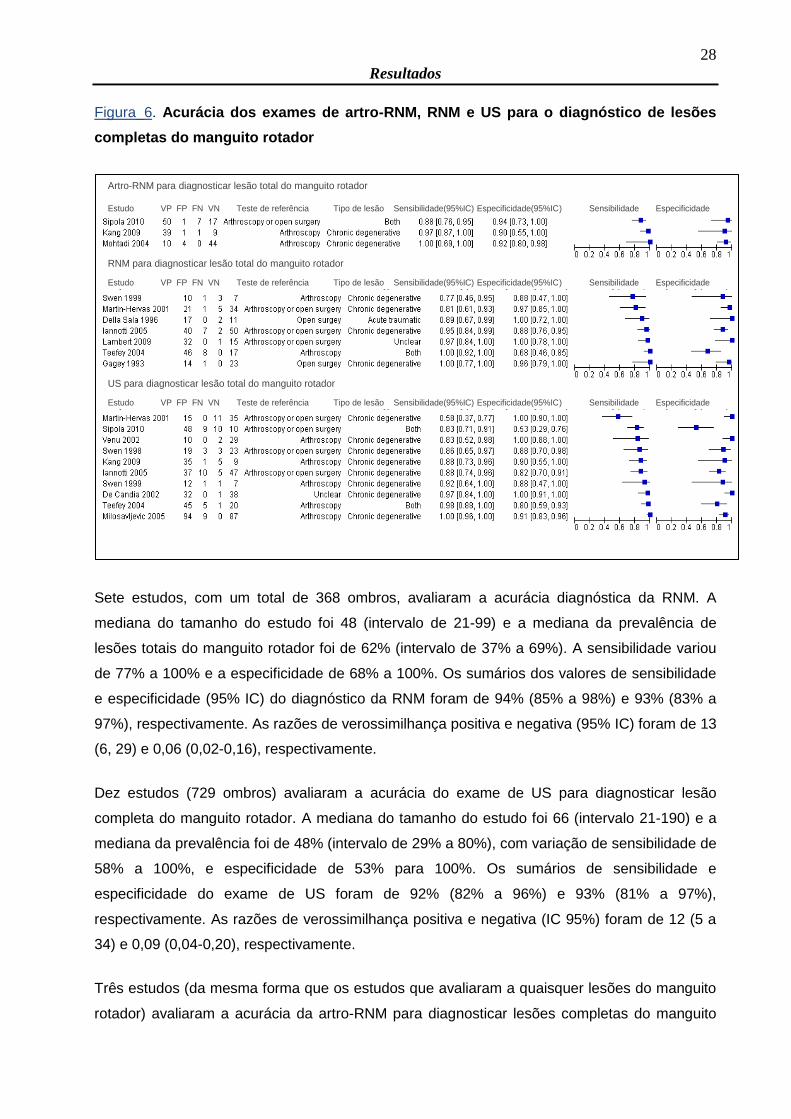
Figura_6. Acurácia dos exames de artro-RNM, RNM e US para o diagnóstico de lesões
completas do manguito rotador
Sete estudos, com um total de 368 ombros, avaliaram a acurácia diagnóstica da RNM. A
mediana do tamanho do estudo foi 48 (intervalo de 21-99) e a mediana da prevalência de
lesões totais do manguito rotador foi de 62% (intervalo de 37% a 69%). A sensibilidade variou
de 77% a 100% e a especificidade de 68% a 100%. Os sumários dos valores de sensibilidade
e especificidade (95% IC) do diagnóstico da RNM foram de 94% (85% a 98%) e 93% (83% a
97%), respectivamente. As razões de verossimilhança positiva e negativa (95% IC) foram de 13
(6, 29) e 0,06 (0,02-0,16), respectivamente.
Dez estudos (729 ombros) avaliaram a acurácia do exame de US para diagnosticar lesão
completa do manguito rotador. A mediana do tamanho do estudo foi 66 (intervalo 21-190) e a
mediana da prevalência foi de 48% (intervalo de 29% a 80%), com variação de sensibilidade de
58% a 100%, e especificidade de 53% para 100%. Os sumários de sensibilidade e
especificidade do exame de US foram de 92% (82% a 96%) e 93% (81% a 97%),
respectivamente. As razões de verossimilhança positiva e negativa (IC 95%) foram de 12 (5 a
34) e 0,09 (0,04-0,20), respectivamente.
Três estudos (da mesma forma que os estudos que avaliaram a quaisquer lesões do manguito
rotador) avaliaram a acurácia da artro-RNM para diagnosticar lesões completas do manguito
Artro-RNM para diagnosticar lesão total do manguito rotador
RNM para diagnosticar lesão total do manguito rotador
US para diagnosticar lesão total do manguito rotador
Estudo VP FP FN VN Teste de referência Tipo de lesão Sensibilidade(95%IC) Especificidade(95%IC) Sensibilidade Especificidade
Estudo VP FP FN VN Teste de referência Tipo de lesão Sensibilidade(95%IC) Especificidade(95%IC) Sensibilidade Especificidade
Estudo VP FP FN VN Teste de referência Tipo de lesão Sensibilidade(95%IC) Especificidade(95%IC) Sensibilidade Especificidade
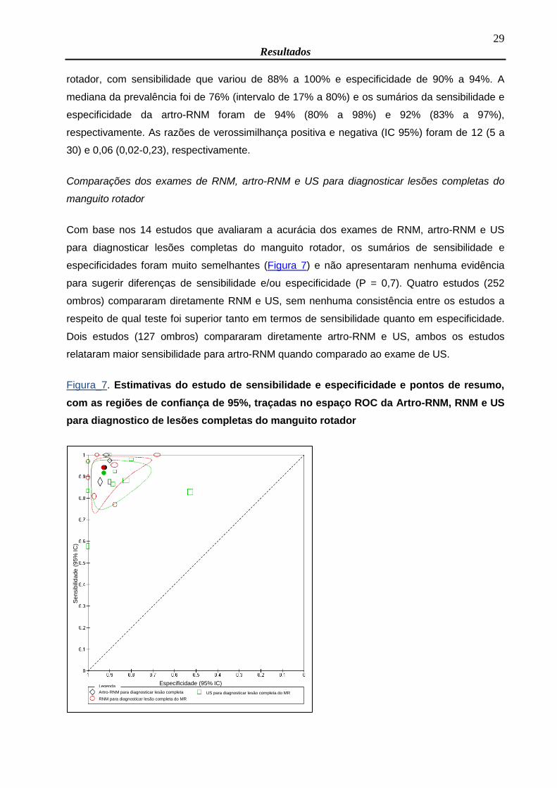
29
Resultados
rotador, com sensibilidade que variou de 88% a 100% e especificidade de 90% a 94%. A
mediana da prevalência foi de 76% (intervalo de 17% a 80%) e os sumários da sensibilidade e
especificidade da artro-RNM foram de 94% (80% a 98%) e 92% (83% a 97%),
respectivamente. As razões de verossimilhança positiva e negativa (IC 95%) foram de 12 (5 a
30) e 0,06 (0,02-0,23), respectivamente.
Comparações dos exames de RNM, artro-RNM e US para diagnosticar lesões completas do
manguito rotador
Com base nos 14 estudos que avaliaram a acurácia dos exames de RNM, artro-RNM e US
para diagnosticar lesões completas do manguito rotador, os sumários de sensibilidade e
especificidades foram muito semelhantes (Figura 7) e não apresentaram nenhuma evidência
para sugerir diferenças de sensibilidade e/ou especificidade (P = 0,7). Quatro estudos (252
ombros) compararam diretamente RNM e US, sem nenhuma consistência entre os estudos a
respeito de qual teste foi superior tanto em termos de sensibilidade quanto em especificidade.
Dois estudos (127 ombros) compararam diretamente artro-RNM e US, ambos os estudos
relataram maior sensibilidade para artro-RNM quando comparado ao exame de US.
Figura_7. Estimativas do estudo de sensibilidade e especificidade e pontos de resumo,
com as regiões de confiança de 95%, traçadas no espaço ROC da Artro-RNM, RNM e US
para diagnostico de lesões completas do manguito rotador
Sensib
ilidade (
95%
IC
)
Especificidade (95% IC)Legenda
RNM para diagnosticar lesão completa do MR
Artro-RNM para diagnosticar lesão completa US para diagnosticar lesão completa do MR
30
Resultados
4.2.3. Diagnóstico de lesões parciais do manguito rotador
A Figura 8 mostra as estimativas de sensibilidade e especificidade para os 13 estudos que
avaliaram os exames de artro-RNM, RNM e US para o diagnóstico de lesões parciais do
manguito rotador.
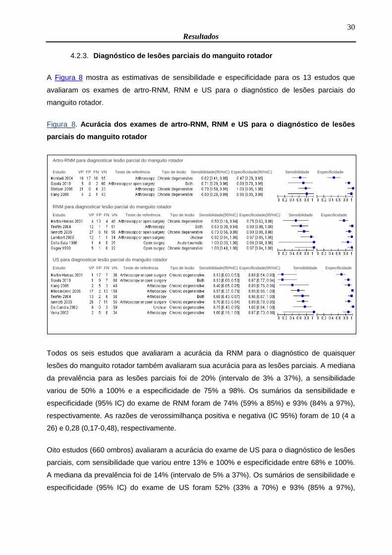
Figura_8. Acurácia dos exames de artro-RNM, RNM e US para o diagnóstico de lesões
parciais do manguito rotador
Todos os seis estudos que avaliaram a acurácia da RNM para o diagnóstico de quaisquer
lesões do manguito rotador também avaliaram sua acurácia para as lesões parciais. A mediana
da prevalência para as lesões parciais foi de 20% (intervalo de 3% a 37%), a sensibilidade
variou de 50% a 100% e a especificidade de 75% a 98%. Os sumários da sensibilidade e
especificidade (95% IC) do exame de RNM foram de 74% (59% a 85%) e 93% (84% a 97%),
respectivamente. As razões de verossimilhança positiva e negativa (IC 95%) foram de 10 (4 a
26) e 0,28 (0,17-0,48), respectivamente.
Oito estudos (660 ombros) avaliaram a acurácia do exame de US para o diagnóstico de lesões
parciais, com sensibilidade que variou entre 13% e 100% e especificidade entre 68% e 100%.
A mediana da prevalência foi de 14% (intervalo de 5% a 37%). Os sumários de sensibilidade e
especificidade (95% IC) do exame de US foram 52% (33% a 70%) e 93% (85% a 97%),
Artro-RNM para diagnosticar lesão parcial do manguito rotador
Estudo VP FP FN VN Teste de referência Tipo de lesão Sensibilidade(95%IC) Especificidade(95%IC) Sensibilidade Especificidade
RNM para diagnosticar lesão parcial do manguito rotador
Estudo VP FP FN VN Teste de referência Tipo de lesão Sensibilidade(95%IC) Especificidade(95%IC) Sensibilidade Especificidade
US para diagnosticar lesão parcial do manguito rotador
Estudo VP FP FN VN Teste de referência Tipo de lesão Sensibilidade(95%IC) Especificidade(95%IC) Sensibilidade Especificidade
31
Resultados
respectivamente. As razões de verossimilhança positiva e negativa (IC 95%) foram 8 (3 a 19) e
0,52 (0,33 e 0,80), respectivamente.
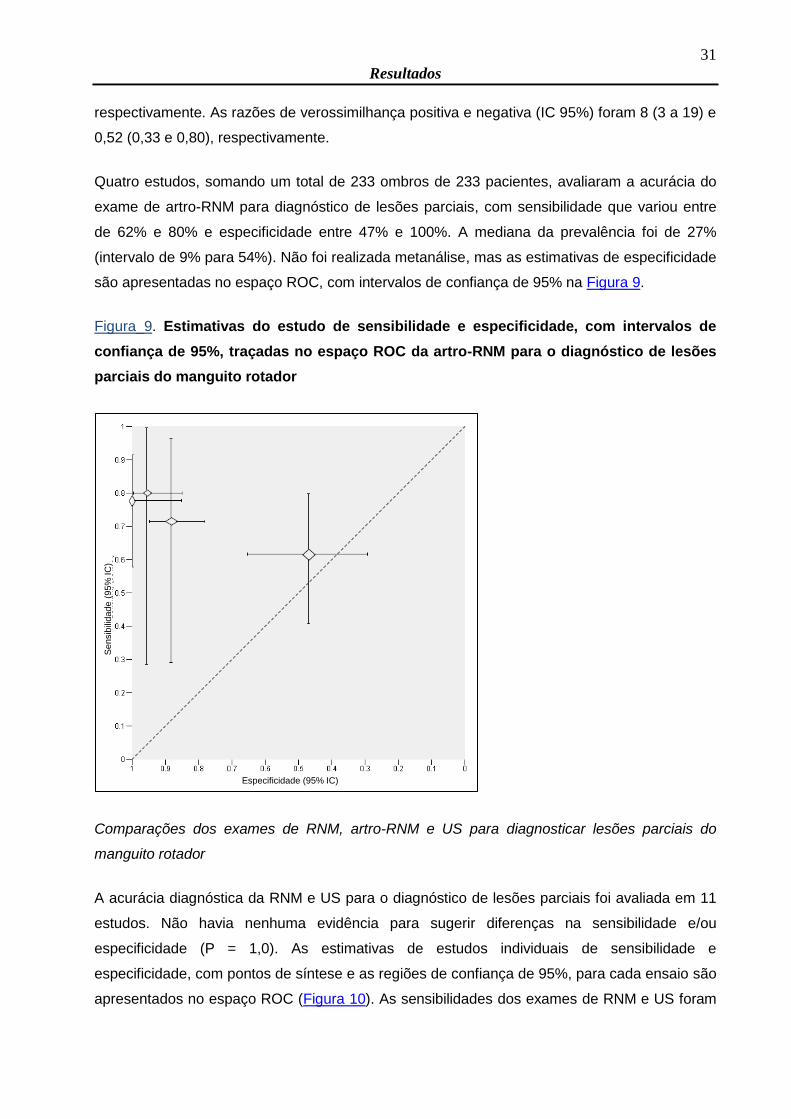
Quatro estudos, somando um total de 233 ombros de 233 pacientes, avaliaram a acurácia do
exame de artro-RNM para diagnóstico de lesões parciais, com sensibilidade que variou entre
de 62% e 80% e especificidade entre 47% e 100%. A mediana da prevalência foi de 27%
(intervalo de 9% para 54%). Não foi realizada metanálise, mas as estimativas de especificidade
são apresentadas no espaço ROC, com intervalos de confiança de 95% na Figura 9.
Figura_9. Estimativas do estudo de sensibilidade e especificidade, com intervalos de
confiança de 95%, traçadas no espaço ROC da artro-RNM para o diagnóstico de lesões
parciais do manguito rotador
Comparações dos exames de RNM, artro-RNM e US para diagnosticar lesões parciais do
manguito rotador
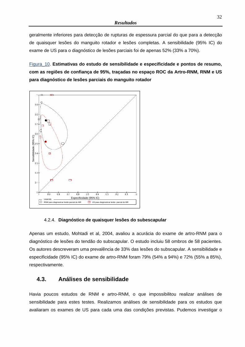
A acurácia diagnóstica da RNM e US para o diagnóstico de lesões parciais foi avaliada em 11
estudos. Não havia nenhuma evidência para sugerir diferenças na sensibilidade e/ou
especificidade (P = 1,0). As estimativas de estudos individuais de sensibilidade e
especificidade, com pontos de síntese e as regiões de confiança de 95%, para cada ensaio são
apresentados no espaço ROC (Figura 10). As sensibilidades dos exames de RNM e US foram
Se
nsib
ilid
ad
e (
95%
IC
)
Especificidade (95% IC)
32
Resultados
geralmente inferiores para detecção de rupturas de espessura parcial do que para a detecção
de quaisquer lesões do manguito rotador e lesões completas. A sensibilidade (95% IC) do
exame de US para o diagnóstico de lesões parciais foi de apenas 52% (33% a 70%).
Figura_10. Estimativas do estudo de sensibilidade e especificidade e pontos de resumo,
com as regiões de confiança de 95%, traçadas no espaço ROC da Artro-RNM, RNM e US
para diagnóstico de lesões parciais do manguito rotador
4.2.4. Diagnóstico de quaisquer lesões do subescapular
Apenas um estudo, Mohtadi et al, 2004, avaliou a acurácia do exame de artro-RNM para o
diagnóstico de lesões do tendão do subscapular. O estudo incluiu 58 ombros de 58 pacientes.
Os autores descreveram uma prevalência de 33% das lesões do subscapular. A sensibilidade e
especificidade (95% IC) do exame de artro-RNM foram 79% (54% a 94%) e 72% (55% a 85%),
respectivamente.
4.3. Análises de sensibilidade
Havia poucos estudos de RNM e artro-RNM, o que impossibilitou realizar análises de
sensibilidade para estes testes. Realizamos análises de sensibilidade para os estudos que
avaliaram os exames de US para cada uma das condições previstas. Pudemos investigar o
Se
nsib
ilid
ad
e (
95%
IC
)
Especificidade (95% IC)Legenda
RNM para diagnosticar lesão parcial do MR US para diagnosticar lesão parcial do MR
33
Resultados
impacto de dois (resultados de teste padrão de referência aceitável e teste índice mascarado)
dos cinco itens de qualidade que tínhamos especificado, porque poucos estudos marcaram
"sim" nos outros três itens (espectro representante, atraso aceitável entre os testes, e os
resultados dos testes de referência mascarados). Havia pequenas diferenças em termos de
sensibilidade e/ou especificidade. Observou-se a maior diferença entre a sensibilidade de
resumo dos exames de US para o diagnóstico de lesões completas (52% (95% IC: 33% a
70%)) e o sumário de sensibilidade (62% (45% a 77%)) com base em apenas estudos em que
o nível de referência foi aceitável. No entanto, os intervalos de confiança foram comparáveis e
as especificidades foram semelhantes. A exclusão de estudos que não satisfizeram os critérios
não afetou nossos achados.

34
Discussão
DISCUSSÃO
35
Discussão
5. DISCUSSÃO
5.1. Resumo dos principais resultados
Esta revisão teve como objetivo avaliar as evidências para a acurácia dos exames de RNM,
artro-RNM e US para diagnosticar lesões do manguito rotador em pacientes com dor no ombro.
Estes exames de imagem geralmente são realizados para caracterizar as lesões do manguito
rotador, a fim de planejar uma cirurgia. Foram incluídos apenas estudos prospectivos que
avaliaram a acurácia de pelo menos um dos testes. Foram identificados 20 estudos (1147
ombros), dos quais seis avaliaram a acurácia de dois dos testes, com comparações pareadas.
Não encontramos evidências que sugerem diferenças nas sensibilidades e especificidades dos
exames de diagnóstico de RNM, artro-RNM e US para o diagnóstcio de lesões completas ou
parciais do manguito rotador em pacientes com dor no ombro. As estimativas foram muito
semelhantes e os testes demonstraram boa capacidade discriminatória para diagnosticar as
lesões do manguito rotador. RNM e US apresentaram menor sensibilidade para diagnosticar as
lesões parciais quando comparadas com as lesões completas ou quaisquer lesões, o
diagnóstcio com US apresenta sensibilidade de apenas 52% (95% IC: 33% a 70%); isso indica
que o exame com US podem ser apenas marginalmente melhor do que a possibilidade de
excluir uma lesão parcial. As especificidades dos três testes foram, em geral, adequadas;
exceto para detecção de quaisquer lesões do manguito rotador. As estimativas de sensibilidade
e especificidade para quaisquer lesões do manguito rotador sugerem que, em uma população
de 100 pacientes com dor no ombro, se a prevalência foi de 80%, a investigação com RNM
pode não diagnosticar dois casos (2/80, 3%), enquanto que a investigação com US pode não
diagnosticar sete casos (7/80, 9%).
Por outro lado, entre os pacientes sem uma ruptura do manguito rotador (20 em 100), quatro
pacientes avaliados usando a RNM podem ter uma ruptura do manguito rotador erroneamente
diagnosticada (4/20, 20%) e podem passar por uma cirurgia desnecessária. Um número
semelhante de indivíduos (3/20, 15%) também pode ser tratado desnecessariamente, se foi
utilizado o exame de US. Nossos resultados foram baseados em uma alta prevalência de
pacientes com lesões do manguito rotador e estes resultados de uma população altamente
selecionada não podem ser generalizados para outros contextos, tais como cuidados de saúde
primária, onde a prevalência de lesões do manguito rotador é menor.
36
Discussão
5.2. Qualidade da evidência
Esta revisão foi planejada e conduzida seguindo critérios e métodos definidos em um protocolo
previmente publicado (Apêndice 2). Os resultados foram baseados em uma estratégia de
busca abrangente e sensível da literatura e teve por objetivo identificar todos os estudos
publicados. Usamos termos de pesquisa abrangentes, vários bancos de dados eletrônicos, e
não utilizamos filtros de pesquisa para termos de diagnóstico, uma vez que eles têm utilidade
limitada (de Vet 2008). Outros pontos fortes desta revisão foram a avaliação qualitativa dos
estudos e a síntese dos dados metodológicos; em ambos foram utilizados resumos meta-
analítico baseados em métodos recomendados. Para aumentar a aplicabilidade e a
confiabilidade das conclusões, foram incluídos apenas estudos prospectivos que investigaram
pacientes com dor no ombro devido a suspeita de lesão do manguito rotador. Foram excluídos
estudos retrospectivos por causa do seu potencial para o alto risco de espectro e verificação
(Bossuyt 2003; Van der Schouw 1995).
Entretanto, a revisão tem algumas limitações. Nossos resultados foram baseados em estudos
com baixo poder analítico e estudos com descrição inadequada da estrutura do desenho. A
maioria dos itens de qualidade QUADAS foi considerada incerta. Por exemplo, apenas 25%
dos estudos incluídos relataram o intervalo de tempo entre os exames de teste e o padrão de
referência. Para algumas análises, observou-se uma considerável heterogeneidade na
sensibilidade e/ou especificidade que pode ser devida a vários fatores, incluindo a variação nos
critérios de positividade do teste (testes de índice e padrão de referência), detalhes técnicos
dos testes, a variação da população e a experiência dos operadores dos testes.
Outra limitação importante desta revisão se dá por conta da restrição de recursos. Vinte e cinco
estudos potencialmente elegíveis, publicados em idiomas que não o inglês, permanecem à
espera de tradução. Estes estudos abordaram mais de 2900 participantes, um número
expressivo e que poderia fornecer dados fundamentais para análises. Estes estudos serão
considerados para inclusão em uma futura atualização da revisão.
5.3. Comparações com outras revisões existentes
Foram identificadas seis revisões sistemáticas de estudos de diagnóstico que avaliaram os
exames de imagem para as lesões do manguito rotador (de Jesus et al, 2009; Dinnes et al,
2003; Kelly et al, 2009; Ottenheijm et al, 2010; Shahabpour et al, 2008; Smith et al, 2012).
Apenas nossa revisão limitou os critérios de inclusão para estudos prospectivos.
37
Discussão
As revisões anteriores relataram resultados semelhantes. De Jesus et al (2009) compararam o
exame de US com a RNM para o diagnóstico de lesões do manguito rotador, usando cirurgia
como padrão de referência. Os autores incluíram 65 estudos (data da estratégia de busca foi
setembro de 2007) e concluíram que o exame de US é tão preciso quanto o exame de RNM
para diagnosticar ambas as lesões do manguito rotador (completa e parcial). Dinnes et al
(2003) avaliaram a acurácia de testes clínicos, US e RNM para diagnóstico de lesões do
manguito rotador (data da estratégia de busca foi outubro de 2001), como padrão de referência
eles usaram testes cirúrgicos e não-cirúrgicos (resultados também relatado em Kelly et al,
2009). Ambos Dinnes et al (2003) e Shahabpour et al (2008) também concluíram que o exame
de US e a RNM foram equivalentes para o diagnóstcio de lesões completas do manguito
rotador, mas Dinnes et al (2003), concluíram que a RM é melhor no diagnóstico de lesões
parciais do manguito rotador. Shahabpour et al (2008) concluíram que os exames de artro-
RNM e US são mais precisos para o diagnóstcio de lesões parciais do manguito rotador
quando comparados com a RNM. Enquanto os nossos resultados sugerem que a RNM pode
ser mais sensível do que o exame de US, a diferença não foi estatisticamente significativa.
Ottenheijm et al (2010) avaliaram a acurácia do exame de US para diagnosticar doenças do
espaço subacromial em pacientes de setores primários e secundários de saúde (data da
estratégia de busca foi entre 2001 e junho de 2010). Eles incluíram 23 estudos e as
metanálises de sensibilidade e especificidade foram semelhantes aos nossos resultados para o
diagnóstico de lesões completas do manguito rotador (95% versus 92% e 96% versus 93%,
respectivamente: 95% IC). No entanto, para as lesões parciais do manguito rotador, os autores
relataram uma sensibilidade combinada muito mais elevada, de 72% em comparação com a
nossa, de 52% (95% IC: 33% a 70%). Smith et al (2012) avaliaram a acurácia diagnóstica da
RNM e identificaram 44 estudos (retrospectivos e prospectivos) publicados até maio de 2011. A
sensibilidade combinada foi de 91% (95% IC: 86% a 94%) e a especificidade agrupada foi de
97% (95% IC: 96% a 98%).
Os resultados são em geral consistentes entre as diferentes revisões sistemáticas, embora
houvesse diferenças de critérios de inclusão e métodos de análise. Apesar de a nossa revisão
ter a estratégia de busca mais atualizada, incluímos um número muito menor de estudos (20
estudos) do que as outras revisões, por conta de termos restringido nossas análises apenas
para estudos prospectivos, como forma de reduzir o risco de viéses de espectro e de
verificação.
5.4. Aplicabilidade dos resultados
38
Discussão
A aplicabilidade dos nossos resultados é limitada, porque apenas 25% dos estudos incluídos
relatou um espectro suficientemente representativo de pacientes. Além disso, a verificação
parcial foi evitada em 50% dos estudos.
Os exames de RNM, artro-RNM e US apresentam acurácia semelhante para o diagnóstico de
lesões completas do manguito rotador. A sensibilidade de ambos os exames RNM e US para o
diagnóstico de lesões parciais do manguito rotador é muito inferior que a sensibilidade para o
diagnóstico de quaisquer lesões ou lesões copmpletas. Embora a diferença de sensibilidade
entre RNM e US para o diagnóstico de lesões parciais não tenha sido estatisticamente
significativa. As especificidades dos três testes foram, em geral, altas.
Em muitos países, o exame de US é mais rápido para ser executado, barato e mais facilmente
disponível nos serviços secundários e terciários de saúde. Apesar dos exames de RNM e artro-
RNM serem semelhantes para o diagnóstico de lesões completas do manguito rotador, a
escolha do teste pode depender do custo e disponibilidade.

39
Conclusão
CONCLUSÃO
40
Conclusão
6. CONCLUSÃO
6.1. Implicações para a prática
O desempenho diagnóstico dos exames de RNM e US dependem da extensão da lesão do
manguito rotador (ou seja parcial ou total/completa). Nossos resultados sugerem que a RNM,
US e artro-RNM possuem boa acurácia diagnóstica e quaisquer destes testes podem
igualmente serem usados para o diagnóstico de lesões completas do manguito rotador. Os
exames de RNM e US também possuem boa sensibilidade para o diagnóstico de quaisquer
lesões do manguito rotador, mas pouca sensibilidade para o diagnóstico de lesões parciais. A
validade e generalização de nossos resultados são limitadas porque foram baseados em
estudos pequenos e heterogêneos, não comparativos e com falhas metodológicas.
6.2. Implicações para a pesquisa
Há uma falta de estudos de coorte prospectivos de boa qualidade que comparam diretamente a
acurácia dos exames de RNM, artro-RNM e US para pacientes com suspeita de lesões do
manguito rotador. Consequentemente, são necessários mais estudos para avaliar a acurácia
comparativa destes exames de imagem em tais circunstâncias.
Estudos futuros devem utilizar um desenho tipo coorte prospectivo e consecutivo, com
mascaramento dos avaliadores dos exames e limitar a quantidade de tempo entre o teste
índice e os testes de referência, porque há evidências que lesões do manguito rotador podem
progredir ao longo tempo. Nós sugerimos a artroscopia para ser usada como o teste padrão de
referência, pois ela é mais precisa para avaliar o lado de articular e bursal do manguito rotador.
Os resultados do teste de índice e do padrão de referência devem ser interpretados por
operadores experientes.

41
Anexos
ANEXOS
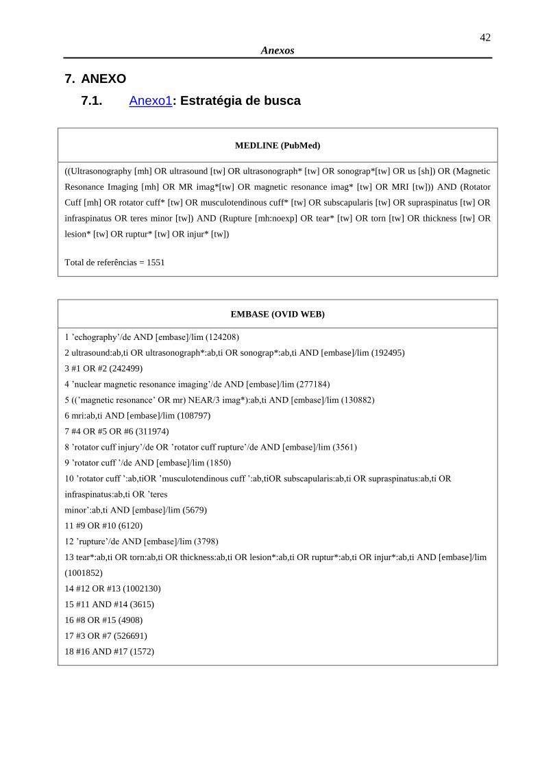
42
Anexos
7. ANEXO
7.1. Anexo1: Estratégia de busca
MEDLINE (PubMed)
((Ultrasonography [mh] OR ultrasound [tw] OR ultrasonograph* [tw] OR sonograp*[tw] OR us [sh]) OR (Magnetic
Resonance Imaging [mh] OR MR imag*[tw] OR magnetic resonance imag* [tw] OR MRI [tw])) AND (Rotator
Cuff [mh] OR rotator cuff* [tw] OR musculotendinous cuff* [tw] OR subscapularis [tw] OR supraspinatus [tw] OR
infraspinatus OR teres minor [tw]) AND (Rupture [mh:noexp] OR tear* [tw] OR torn [tw] OR thickness [tw] OR
lesion* [tw] OR ruptur* [tw] OR injur* [tw])
Total de referências = 1551
EMBASE (OVID WEB)
1 ’echography’/de AND [embase]/lim (124208)
2 ultrasound:ab,ti OR ultrasonograph*:ab,ti OR sonograp*:ab,ti AND [embase]/lim (192495)
3 #1 OR #2 (242499)
4 ’nuclear magnetic resonance imaging’/de AND [embase]/lim (277184)
5 ((’magnetic resonance’ OR mr) NEAR/3 imag*):ab,ti AND [embase]/lim (130882)
6 mri:ab,ti AND [embase]/lim (108797)
7 #4 OR #5 OR #6 (311974)
8 ’rotator cuff injury’/de OR ’rotator cuff rupture’/de AND [embase]/lim (3561)
9 ’rotator cuff ’/de AND [embase]/lim (1850)
10 ’rotator cuff ’:ab,tiOR ’musculotendinous cuff ’:ab,tiOR subscapularis:ab,ti OR supraspinatus:ab,ti OR
infraspinatus:ab,ti OR ’teres
minor’:ab,ti AND [embase]/lim (5679)
11 #9 OR #10 (6120)
12 ’rupture’/de AND [embase]/lim (3798)
13 tear*:ab,ti OR torn:ab,ti OR thickness:ab,ti OR lesion*:ab,ti OR ruptur*:ab,ti OR injur*:ab,ti AND [embase]/lim
(1001852)
14 #12 OR #13 (1002130)
15 #11 AND #14 (3615)
16 #8 OR #15 (4908)
17 #3 OR #7 (526691)
18 #16 AND #17 (1572)
43
Anexos
LILACS
(Mh Ultrasonography OR Tw ultrasound OR Tw ultrasonograph$ OR Tw Sonograp$) OR (Mh Magnetic Resonance
Imaging OR (Tw magnetic AND Tw resonance AND Tw imag$) OR Tw MRI) [Words] and Mh Rotator Cuff OR
(Tw rotator AND Tw cuff ) OR (Tw musculotendinous AND Tw cuff ) OR Tw subscapularis OR Tw supraspinatus
OR Tw infraspinatus OR (Tw teres AND Tw minor) [Words] andMh Rupture OR Tw tear$ OR Tw torn OR Tw
thickness OR Tw lesion$ OR Tw rupture$ OR Tw injur$ [Words]
Total de referências = 30

44
Referências
REFERÊNCIAS
45
Referências
8. REFERÊNCIAS
8.1. Referências dos estudos incluídos nesta revisão
De_Candia_2002
De Candia A, Doratiotto S, Paschina E, Segatto E, Pelizzo F, Bazzocchi M. Real-time
compound sonography of the rotator-cuff: evaluation of artefact reduction and image definition.
La Radiologia Medica 2003;105(4):308-14.
* De Candia A, Doratiotto S, Pelizzo F, Paschina E, Bazzocchi M. Real time compound
ultrasound of the shoulder. Radiology and Oncology 2002;36(4):319-25,+336.
Della_Sala_1996
Della Sala SW, Bianchini G. Magnetic resonance in the study of the painful shoulder. The
surgical comparison in 30 consecutive cases [La Risonanza Magnetica nello studio della spalla
dolorosa. Raffronto chirurgico in 30 casi consecutivi]. La Radiologia Medica 1996;91(4):348-55.
Gagey_1993
Gagey N, Desmoineaux P, Gagey O, Idy-Peretti I, Mazas F. Contribution of MRI to the
preoperative evaluation of rotator cuff tears [Apport de l'IRM dans le bilan pre-chirurgical des
lesions de la coiffe des rotateurs]. Revue de Chirurgie Orthopedique et Reparatrice de l'appareil
moteur 1991;77(8):521-9.
* Gagey N, Desmoineaux P, Gagey O, Idy-Peretti I, Mazas F. MRI in the pre-operative
evaluation of lesions of the rotator cuff [Apport de l'IRM dans le bilan pre-chirurgical des lesions
de la coiffe des rotateurs]. Journal de Radiologie 1993;74(1):39-46.
Iannotti_2005
Iannotti JP, Ciccone J, Buss DD, Visotsky JL, Mascha E, Cotman K, et al. Accuracy of office-
based ultrasonography of the shoulder for the diagnosis of rotator cuff tears. Journal of Bone
and Joint Surgery - American volume 2005;87(6):1305-11.
Kang_2009
46
Referências
Kang CH, Kim SS, Kim JH, Chung KB, Kim YH, Oh YW, et al. Supraspinatus tendon tears:
Comparison of 3D US and MR arthrography with surgical correlation. Skeletal Radiology
2009;38(11):1063-9.
Lambert_2009
Lambert A, Loffroy R, Guiu B, Mejean N, Lerais JM, Cercueil JP, et al. Rotator cuff tears: value
of 3.0T MRI [Perforations de la coiffe des rotateurs: intérêt de l’IRM 3,0T]. Journal de Radiologie
2009;90(5 Pt 1):583-8.
Martin_Hervas_2001
Martin-Hervas C, Romero J, Navas-Acien A, Reboiras JJ, Munuera L. Ultrasonographic and
magnetic resonance images of rotator cuff lesions compared with arthroscopy or open surgery
findings. Journal of Shoulder and Elbow Surgery 2001;10(5):410-5.
Milosavljevic_2005
Milosavljevic J, Elvin A, Rahme H. Ultrasonography of the rotator cuff: a comparison with
arthroscopy in one-hundred-and-ninety consecutive cases. Acta Radiologica 2005;46(8):858-
65.
Misamore_1991
Misamore GW, Woodward C. Evaluation of degenerative lesions of the rotator cuff. A
comparison of arthrography and ultrasonography. Journal of Bone and Joint Surgery - American
volume 1991;73(5):704-6.
Mohtadi_2004
Mohtadi NG, Vellet AD, Clark ML, Hollinshead RM, Sasyniuk TM, Fick GH, et al. A prospective,
double-blind comparison of magnetic resonance imaging and arthroscopy in the evaluation of
patients presenting with shoulder pain. Journal of Shoulder and Elbow Surgery 2004;13(3):258-
65.
Nicoletti_1994
Nicoletti SJ, de Moura L. Rotator cuff disease: do ultrasonography and pneumoarthrography
increase diagnostic reliability of the physical examination for supraspinatus lesion detection?
47
Referências
[Lesöes do manguito rotator: a ultra-sonografia e a pneumoartrografia aumentam a capacidade
diagnóstica do exame físico, para a detecçäo das roturas do supra-espinhal?]. Revista
Brasileira de Ortopedia 1994;29(9):656-60.
Sipola_2010
Sipola P, Niemitukia L, Kroger H, Hofling I, Vaatainen U. Detection and quantification of rotator
cuff tears with ultrasonography and magnetic resonance imaging - A prospective study in 77
consecutive patients with a surgical reference. Ultrasound in Medicine and Biology
2010;36(12):1981-9.
Stetson_2005
Stetson WB, Phillips T, Deutsch A. The use of magnetic resonance arthrography to detect
partial-thickness rotator cuff tears. Journal of Bone and Joint Surgery - American volume
2005;87(Suppl 2):81-8.
Swen_1998
Swen WA, Jacobs JW, Neve WC, Bal D, Bijlsma JW. Is sonography performed by the
rheumatologist as useful as arthrography executed by the radiologist for the assessment of full
thickness rotator cuff tears? Journal of Rheumatology 1998;25(9):1800-6.
Swen_1999
Swen WA, Jacobs JW, Algra PR, Manoliu RA, Rijkmans J, Willems WJ, et al. Sonography and
magnetic resonance imaging equivalent for the assessment of full-thickness rotator cuff tears.
Arthritis and Rheumatism 1999;42(10):2231-8.
Taboury_1992
Taboury J. Ultrasonography of the shoulder: diagnosis of rupture of tendons of the rotator
muscles [Echographie de l'epaule. Diagnostic de rupture des tendons des muscles rotateurs].
Annales de Radiologie 1992;35(3):133-40.
Teefey_2004
48
Referências
Teefey SA, Middleton WD, Payne WT, Yamaguchi K. Detection and measurement of rotator
cuff tears with sonography: Analysis of diagnostic errors. AJM. American Journal of
Roentgenology 2005;184(6):1768-73.
* Teefey SA, Rubin DA, Middleton WD, Hildebolt CF, Leibold RA, Yamaguchi K. Detection and
quantification of rotator cuff tears: comparison of ultrasonographic, magnetic resonance
imaging, and arthroscopic findings in seventy-one consecutive cases. Journal of Bone and Joint
Surgery - American volume 2004;86(4):708-16.
Venu_2002
Venu KM, Howlett DC, Garikipati R, Anderson HJ, Bonnici AV. Evaluation of the symptomatic
supraspinatus tendon - A comparison of ultrasound and arthroscopy. Radiography
2002;8(4):235-40.
Wallny_2001
Wallny TA, Schild RL, Schulze Bertelsbeck D, Hansmann ME, Kraft CN. Three-dimensional
ultrasonography in the diagnosis of rotator cuff lesions. Ultrasound in Medicine & Biology
2001;27(6):745-9.
Yen_2004
Yen CH, Chiou HJ, Chou YH, Hsu CC, Wu JJ, Ma HL, et al. Six surgery-correlated sonographic
signs for rotator cuff tears: Emphasis on partial-thickness tear. Clinical Imaging 2004;28(1):69-
76.
8.2. Referências bibliográficas adicionais
Akobeng_2005
Akobeng AK. Principles of evidence based medicine. Arch Dis Child. 2005;90(8):837-40.
Al_Shawi_2008
Al-Shawi A, Badge R, Bunker T. The detection of full thickness rotator cuff tears using
ultrasound. J Bone Joint Surg Br. 2008;90(7):889-92.
Alexander_2009
49
Referências
Alexander GC, Stafford RS. Does comparative effectiveness have a comparative edge? JAMA.
2009;301(23):2488-90.
Allen_2007
Allen C, Clarke M, Tharyan P. International activity in Cochrane Review Groups with particular
reference to India. National Medical Journal of India. 2007;20(5):250-5.
Atallah_2003
Atallah AN, Trevisani VFM, Valente O. Atualização Terapêutica. In: Princípios para tomada de
decisões terapêuticas com base em evidências científicas: 21 ed. São Paulo: Artes Médicas;
2003. p.1704-6.
Bigliani_1991
Bigliani LU, Ticker JB, Flatow EL, Soslowsky LJ, Mow VC. The relationship of acromial
architecture to rotator cuff disease. Clinics in sports medicine. 1991;10(4):823-38.
Bossuyt_2003
Bossuyt PM, Reitsma JB, Bruns DE, Gatsonis CA, Glasziou Paul P, Irwig LM, et al. Towards
complete and accurate reporting of studies of diagnostic accuracy: the STARD initiative. BMJ
(Clinical research ed.) 2003;326(7379):41-4.
Chen_2003
Chen AL, Shapiro JA, Ahn AK, Zuckerman JD, Cuomo F. Rotator cuff repair in patients with
type I diabetes mellitus. Journal of Shoulder and Elbow Surgery. 2003;12(5):416-21.
de_Jesus_2009
de Jesus JO, Parker L, Frangos AJ, Nazarian LN. Accuracy of MRI, MR arthrography, and
ultrasound in the diagnosis of rotator cuff tears: a meta-analysis. AJR Am J Roentgenol.
2009;192(6):1701-7.
de_Vet_2008
50
Referências
de Vet HCW, Eisinga A, Riphagen II, Aertgeerts B, Pewsner D. Chapter 7: Searching for
Studies. In: Cochrane Handbook for Systematic Reviews of Diagnostic Test Accuracy Version
0.4 [updated September 2008]. The Cochrane Collaboration, 2008.
Degen_2008
Degen RM, Hodgins JL, Bhandari M. The language of evidence based medicine: Answers to
common questions? Indian J Orthop. 2008;42(2):111-7.
Dinnes_2003
Dinnes J, Loveman E, McIntyre L, Waugh N. The effectiveness of diagnostic tests for the
assessment of shoulder pain due to soft tissue disorders: a systematic review. Health
technology assessment (Winchester, England). 2003;7(29):1-166.
Favard_2007
Favard L, Bacle G, Berhouet J. Rotator cuff repair. Joint, bone, spine: revue du rhumatisme.
2007;74(6):551-7.
Gatsonis_2003
Gatsonis C. Do we need a checklist for reporting the results of diagnostic test evaluations? The
STARD proposal. Acad Radiol. 2003;10(6):599-600.
Gatsonis_2006
Gatsonis C, Paliwal P. Meta-analysis of diagnostic and screening test accuracy evaluations:
methodologic primer. AJR Am J Roentgenol. 2006;187(2):271-81
Green_2008
Green S, Higgins JPT, Alderson P, Clarke M, Mulrow CD, Oxman AD. Introduction. In: Higgins
JPT, Green S (editors). Cochrane Handbook for Systematic Reviews of Interventions Version
5.0.1 [updated September 2008]. The Cochrane Collaboration, 2008. Available from
www.cochrane-handbook.org.
Harryman_2003
51
Referências
Harryman DT 2nd, Hettrich CM, Smith KL, Campbell B, Sidles JA, Matsen FA 3rd. A
prospective multipractice investigation of patients with full-thickness rotator cuff tears: the
importance of comorbidities, practice, and other covariables on self-assessed shoulder function
and health status. The Journal of Bone and Joint Surgery. American Volume. 2003;85-A(4):690-
6.
Haynes_1994
Haynes RB, Walker CJ, McKibbon KA, Johnston ME, Willan AR. Performances of 27 MEDLINE
systems tested by searches with clinical questions. J Am Med Inform Assoc. 1994;1(3):285-95.
Irwig_1994
Irwig L, Tosteson AN, Gatsonis C, Lau J, Colditz G, Chalmers TC, Mosteller F. Guidelines for
meta-analyses evaluating diagnostic tests. Ann Intern Med. 1994;120(8):667-76
Kelly_2009
Kelly AM, Fessell D. Ultrasound compared with magnetic resonance imaging for the diagnosis
of rotator cuff tears: a critically appraised topic. Seminars in Roentgenology 2009;44(3):196-
200.
Kuhn_2007
Kuhn JE, Dunn WR, Ma B, Wright RW, Jones G, Spencer EE, et al. Interobserver agreement in
the classification of rotator cuff tears. The American journal of sports medicine. 2007;35(3):437-
41.
Le_Corroller_2008
Le Corroller T, Cohen M, Aswad R, Pauly V, Champsaur P. Sonography of the painful shoulder:
role of the operator's experience. Skeletal radiology. 2008;37(11):979-86.
Leeflang_2008
Leeflang MM, Deeks JJ, Gatsonis C, Bossuyt PM; Cochrane Diagnostic Test Accuracy Working
Group. Systematic reviews of diagnostic test accuracy. Ann Intern Med. 2008;149(12):889-97.
Leeflang_2009
52
Referências
Leeflang MM, Bossuyt PM, Irwig L. Diagnostic test accuracy may vary with prevalence:
implications for evidence-based diagnosis. J Clin Epidemiol. 2009;62(1):5-12.
Lewis_2008
Lewis JS. Rotator cuff tendinopathy: A review. British journal of sports medicine. 2008 Sep 26.
Manchikanti_2009
Manchikanti L, Derby R, Wolfer L, Singh V, Datta S, Hirsch JA. Evidence-based medicine,
systematic reviews, and guidelines in interventional pain management: Part 7: systematic
reviews and meta-analyses of diagnostic accuracy studies. Pain Physician. 2009;12(6):929-63.
Matava_2005
Matava MJ, Purcell DB, Rudzki JR. Partial-thickness rotator cuff tears. Am J Sports Med.
2005;33(9):1405-17.
Matsen_2008
Matsen FA 3rd. Clinical practice. Rotator-cuff failure. The New England journal of medicine.
2008;358(20):2138-47.
Moreno_2009
Moreno G G, Pantoja C T. [Systematic reviews of studies of diagnostic test accuracy]. Rev Med
Chil. 2009;137(2):303-7.
Nho_2008
Nho SJ, Yadav H, Shindle MK, Macgillivray JD. Rotator cuff degeneration: etiology and
pathogenesis. The American journal of sports medicine. 2008;36(5):987-93.
O_Connor_2005
O'Connor PJ, Rankine J, Gibbon WW, Richardson A, Winter F, Miller JH. Interobserver
variation in sonography of the painful shoulder. J Clin Ultrasound. 2005;33(2):53-6.
Oh_2007
53
Referências
Oh LS, Wolf BR, Hall MP, Levy BA, Marx RG. Indications for rotator cuff repair: a systematic
review. Clinical orthopaedics and related research. 2007;455:52-63.
Ostör_2005
Ostör AJ, Richards CA, Prevost AT, Speed CA, Hazleman BL. Diagnosis and relation to general
health of shoulder disorders presenting to primary care. Rheumatology (Oxford, England).
2005;44(6):800-5.
Ottenheijm_2010
Ottenheijm RP, Jansen MJ, Staal JB, van den Bruel A, Weijers RE, de Bie RA, et al. Accuracy
of diagnostic ultrasound in patients with suspected subacromial disorders: a systematic review
and meta-analysis. Archives of Physical Medicine and Rehabilitation 2010;91(10):1616-25.
Rees_2008
Rees JL. The pathogenesis and surgical treatment of tears of the rotator cuff. The Journal of
bone and joint surgery. British volume. 2008;90(7):827-32.
Rutten_2006
Rutten MJ, Jager GJ, Blickman JG. From the RSNA refresher courses: US of the rotator cuff:
pitfalls, limitations, and artifacts. Radiographics. 2006;26(2):589-604.
Rutter_2001
Rutter CM, Gatsonis CA. A hierarchical regression approach to metaanalysis of diagnostic test
accuracy evaluations. Statistics in Medicine 2001;20(19):2865–84.
Shahabpour_2008
Shahabpour M, Kichouh M, Laridon E, Gielen JL, De Mey J. The effectiveness of diagnostic
imaging methods for the assessment of soft tissue and articular disorders of the shoulder and
elbow. European Journal of Radiology 2008;65(2):194-200.
Smith_2012

54
Referências
Smith TO, Daniell H, Geere JA, Toms AP, Hing CB. The diagnostic accuracy of MRI for the
detection of partial- and full-thickness rotator cuff tears in adults. Magnetic Resonance Imaging
2012;30(3):336-46.
Swen_1999
Swen WA, Jacobs JW, Algra PR, Manoliu RA, Rijkmans J, Willems WJ, et al. Sonography and
magnetic resonance imaging equivalent for the assessment of full-thickness rotator cuff tears.
Arthritis Rheum. 1999;42(10):2231-8.
Van_der_Schouw_1995
van der Schouw YT, Van Dijk R, Verbeek AL. Problems in selecting the adequate patient
population from existing data files for assessment studies of new diagnostic tests. Journal of
Clinical Epidemiology 1995;48(3):417-22.
Virgili_2009
Virgili G, Conti AA, Murro V, Gensini GF, Gusinu R. Systematic reviews of diagnostic test
accuracy and the Cochrane collaboration. Intern Emerg Med. 2009;4(3):255-8.
Watts_2007
Watts G. Let's pension off the "major breakthrough". BMJ. 2007 Jan 6;334 Suppl 1:s4.
Whiting_2003
Whiting P, Rutjes AW, Reitsma JB, Bossuyt PM, Kleijnen J. The development of QUADAS: a
tool for the quality assessment of studies of diagnostic accuracy included in systematic reviews.
BMC Medical Research Methodology 2003;3:25.
Witte_2003
Witte DH. Magnetic Resonance Imaging in Orthopaedics. In: Canale ST, editor(s). Campbell's
Operative Orthopaedics. 10 edition. Philadelphia: Mosby, 2003:123-64.

55
Abstract
ABSTRACT
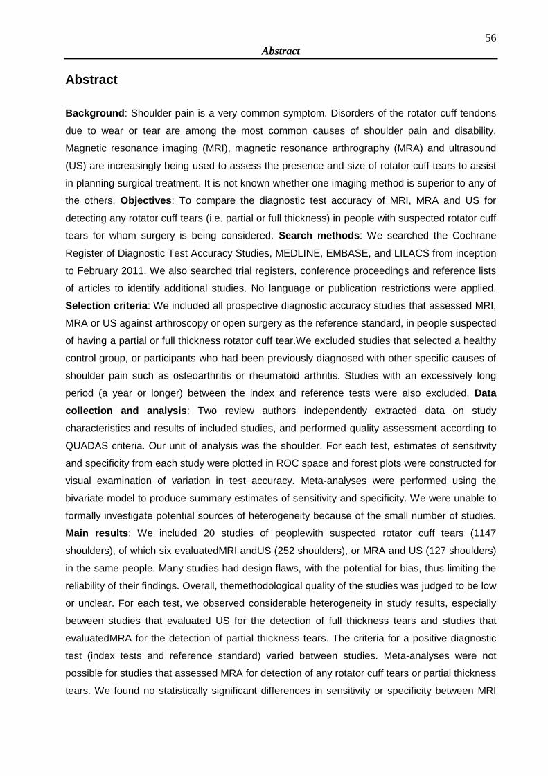
56
Abstract
Abstract
Background: Shoulder pain is a very common symptom. Disorders of the rotator cuff tendons
due to wear or tear are among the most common causes of shoulder pain and disability.
Magnetic resonance imaging (MRI), magnetic resonance arthrography (MRA) and ultrasound
(US) are increasingly being used to assess the presence and size of rotator cuff tears to assist
in planning surgical treatment. It is not known whether one imaging method is superior to any of
the others. Objectives: To compare the diagnostic test accuracy of MRI, MRA and US for
detecting any rotator cuff tears (i.e. partial or full thickness) in people with suspected rotator cuff
tears for whom surgery is being considered. Search methods: We searched the Cochrane
Register of Diagnostic Test Accuracy Studies, MEDLINE, EMBASE, and LILACS from inception
to February 2011. We also searched trial registers, conference proceedings and reference lists
of articles to identify additional studies. No language or publication restrictions were applied.
Selection criteria: We included all prospective diagnostic accuracy studies that assessed MRI,
MRA or US against arthroscopy or open surgery as the reference standard, in people suspected
of having a partial or full thickness rotator cuff tear.We excluded studies that selected a healthy
control group, or participants who had been previously diagnosed with other specific causes of
shoulder pain such as osteoarthritis or rheumatoid arthritis. Studies with an excessively long
period (a year or longer) between the index and reference tests were also excluded. Data
collection and analysis: Two review authors independently extracted data on study
characteristics and results of included studies, and performed quality assessment according to
QUADAS criteria. Our unit of analysis was the shoulder. For each test, estimates of sensitivity
and specificity from each study were plotted in ROC space and forest plots were constructed for
visual examination of variation in test accuracy. Meta-analyses were performed using the
bivariate model to produce summary estimates of sensitivity and specificity. We were unable to
formally investigate potential sources of heterogeneity because of the small number of studies.
Main results: We included 20 studies of peoplewith suspected rotator cuff tears (1147
shoulders), of which six evaluatedMRI andUS (252 shoulders), or MRA and US (127 shoulders)
in the same people. Many studies had design flaws, with the potential for bias, thus limiting the
reliability of their findings. Overall, themethodological quality of the studies was judged to be low
or unclear. For each test, we observed considerable heterogeneity in study results, especially
between studies that evaluated US for the detection of full thickness tears and studies that
evaluatedMRA for the detection of partial thickness tears. The criteria for a positive diagnostic
test (index tests and reference standard) varied between studies. Meta-analyses were not
possible for studies that assessed MRA for detection of any rotator cuff tears or partial thickness
tears. We found no statistically significant differences in sensitivity or specificity between MRI
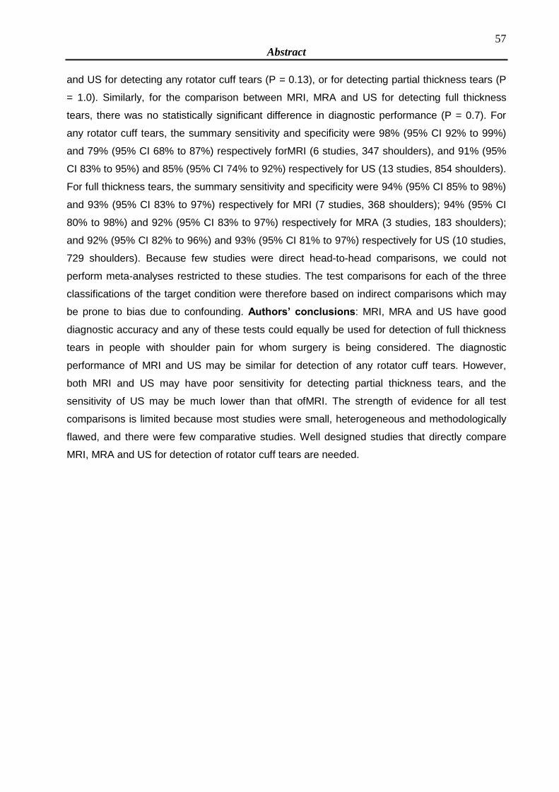
57
Abstract
and US for detecting any rotator cuff tears (P = 0.13), or for detecting partial thickness tears (P
= 1.0). Similarly, for the comparison between MRI, MRA and US for detecting full thickness
tears, there was no statistically significant difference in diagnostic performance (P = 0.7). For
any rotator cuff tears, the summary sensitivity and specificity were 98% (95% CI 92% to 99%)
and 79% (95% CI 68% to 87%) respectively forMRI (6 studies, 347 shoulders), and 91% (95%
CI 83% to 95%) and 85% (95% CI 74% to 92%) respectively for US (13 studies, 854 shoulders).
For full thickness tears, the summary sensitivity and specificity were 94% (95% CI 85% to 98%)
and 93% (95% CI 83% to 97%) respectively for MRI (7 studies, 368 shoulders); 94% (95% CI
80% to 98%) and 92% (95% CI 83% to 97%) respectively for MRA (3 studies, 183 shoulders);
and 92% (95% CI 82% to 96%) and 93% (95% CI 81% to 97%) respectively for US (10 studies,
729 shoulders). Because few studies were direct head-to-head comparisons, we could not
perform meta-analyses restricted to these studies. The test comparisons for each of the three
classifications of the target condition were therefore based on indirect comparisons which may
be prone to bias due to confounding. Authors’ conclusions: MRI, MRA and US have good
diagnostic accuracy and any of these tests could equally be used for detection of full thickness
tears in people with shoulder pain for whom surgery is being considered. The diagnostic
performance of MRI and US may be similar for detection of any rotator cuff tears. However,
both MRI and US may have poor sensitivity for detecting partial thickness tears, and the
sensitivity of US may be much lower than that ofMRI. The strength of evidence for all test
comparisons is limited because most studies were small, heterogeneous and methodologically
flawed, and there were few comparative studies. Well designed studies that directly compare
MRI, MRA and US for detection of rotator cuff tears are needed.

58
Apêndice
APÊNDICE
59
Apêndice
Apêndices
Apêndice_1. Parecer do comitê de ética institucional
60
Apêndice
61
Apêndice
Apêndice_2. Protocolo publicado
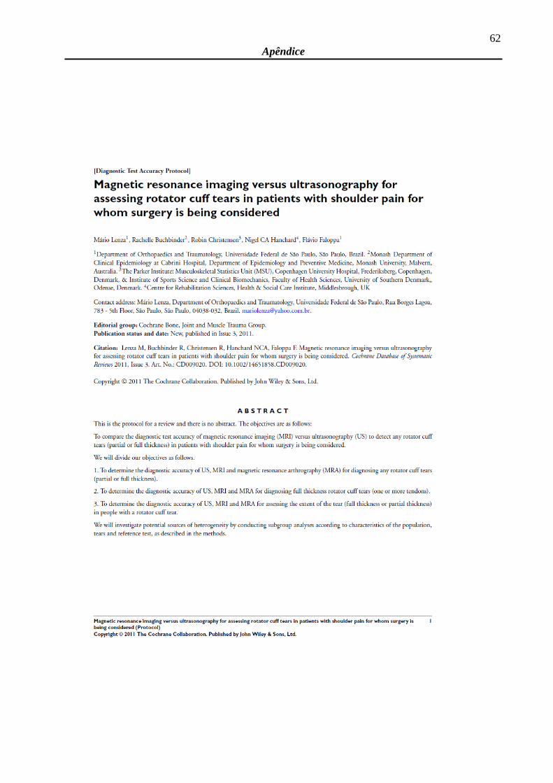
62
Apêndice
63
Apêndice
Apêndice_3. Revisão publicada
64
Apêndice
279
Bibliografias consultadas
BIBLIOGRAFIAS CONSULTADAS
280
Bibliografias consultadas
Bibliografias consultadas
ASSOCIAÇÃO BRASILEIRA DE NORMAS TÉCNICAS. Referências bibliográficas: NBR-6023.
Rio de Janeiro: ABNT; 1989.
ASSOCIAÇÃO BRASILEIRA DE NORMAS TÉCNICAS. Resumos: NBR-6028. Rio de Janeiro:
ABNT; 1980.
ASSOCIAÇÃO BRASILEIRA DE NORMAS TÉCNICAS. Sumário: NBR-6027. Rio de Janeiro:
ABNT; 1980.
BRASIL. Decreto n. 6.583, de 29 de setembro de 2008. Promulga o Acordo Ortográfico da
Língua Portuguesa, assinado em Lisboa, em 16 de dezembro de 1990. Disponível em
http://www.planalto.gov.br/ccivil_03/_Ato2007-2010/2008/Decreto/D6583.htm. Acesso em
novembro 2009.
Ferreira ABH. Novo dicionário da língua portuguesa. 2a edição. Rio de Janeiro: Nova Fronteira;
1986.
Goldenberg S. Orientação normativa para elaboração de tese. Acta Cir Bras 1993;8(1):1-24.
Rother, Edna Terezinha. Como elaborar sua tese? Estrutura e referências/ Edna Terezinha
Rother e Maria Elisa Rangel Braga. São Paulo; 2001.
Terminologia Anatômica. Terminologia Anatômica Internacional. Comissão Federativa da
Terminologia Anatômica. 1ª Ed, São Paulo, Editora Manole Ltda, 2001;169.
Magnetic resonance imaging versus ultrasonography for
assessing rotator cuff tears in patients with shoulder pain for
whom surgery is being considered (Protocol)
Lenza M, Buchbinder R, Christensen R, Hanchard NCA, Faloppa F
This is a reprint of a Cochrane protocol, prepared and maintained by The Cochrane Collaboration and published in The CochraneLibrary 2011, Issue 3
http://www.thecochranelibrary.com
Magnetic resonance imaging versus ultrasonography for assessing rotator cuff tears in patients with shoulder pain for whom surgery is
being considered (Protocol)
Copyright © 2011 The Cochrane Collaboration. Published by John Wiley & Sons, Ltd.
T A B L E O F C O N T E N T S
1HEADER . . . . . . . . . . . . . . . . . . . . . . . . . . . . . . . . . . . . . . .
1ABSTRACT . . . . . . . . . . . . . . . . . . . . . . . . . . . . . . . . . . . . . .
2BACKGROUND . . . . . . . . . . . . . . . . . . . . . . . . . . . . . . . . . . . .
4OBJECTIVES . . . . . . . . . . . . . . . . . . . . . . . . . . . . . . . . . . . . .
4METHODS . . . . . . . . . . . . . . . . . . . . . . . . . . . . . . . . . . . . . .
9ACKNOWLEDGEMENTS . . . . . . . . . . . . . . . . . . . . . . . . . . . . . . . .
9REFERENCES . . . . . . . . . . . . . . . . . . . . . . . . . . . . . . . . . . . . .
11APPENDICES . . . . . . . . . . . . . . . . . . . . . . . . . . . . . . . . . . . . .
12HISTORY . . . . . . . . . . . . . . . . . . . . . . . . . . . . . . . . . . . . . . .
12CONTRIBUTIONS OF AUTHORS . . . . . . . . . . . . . . . . . . . . . . . . . . . . .
12DECLARATIONS OF INTEREST . . . . . . . . . . . . . . . . . . . . . . . . . . . . . .
13SOURCES OF SUPPORT . . . . . . . . . . . . . . . . . . . . . . . . . . . . . . . . .
iMagnetic resonance imaging versus ultrasonography for assessing rotator cuff tears in patients with shoulder pain for whom surgery is
being considered (Protocol)
Copyright © 2011 The Cochrane Collaboration. Published by John Wiley & Sons, Ltd.
[Diagnostic Test Accuracy Protocol]
Magnetic resonance imaging versus ultrasonography forassessing rotator cuff tears in patients with shoulder pain forwhom surgery is being considered
Mário Lenza1, Rachelle Buchbinder2 , Robin Christensen3, Nigel CA Hanchard4, Flávio Faloppa1
1Department of Orthopaedics and Traumatology, Universidade Federal de São Paulo, São Paulo, Brazil. 2Monash Department of
Clinical Epidemiology at Cabrini Hospital, Department of Epidemiology and Preventive Medicine, Monash University, Malvern,
Australia. 3The Parker Institute: Musculoskeletal Statistics Unit (MSU), Copenhagen University Hospital, Frederiksberg, Copenhagen,
Denmark, & Institute of Sports Science and Clinical Biomechanics, Faculty of Health Sciences, University of Southern Denmark„
Odense, Denmark. 4Centre for Rehabilitation Sciences, Health & Social Care Institute, Middlesbrough, UK
Contact address: Mário Lenza, Department of Orthopaedics and Traumatology, Universidade Federal de São Paulo, Rua Borges Lagoa,
783 - 5th Floor, São Paulo, São Paulo, 04038-032, Brazil. [email protected].
Editorial group: Cochrane Bone, Joint and Muscle Trauma Group.
Publication status and date: New, published in Issue 3, 2011.
Citation: Lenza M, Buchbinder R, Christensen R, Hanchard NCA, Faloppa F. Magnetic resonance imaging versus ultrasonography
for assessing rotator cuff tears in patients with shoulder pain for whom surgery is being considered. Cochrane Database of SystematicReviews 2011, Issue 3. Art. No.: CD009020. DOI: 10.1002/14651858.CD009020.
Copyright © 2011 The Cochrane Collaboration. Published by John Wiley & Sons, Ltd.
A B S T R A C T
This is the protocol for a review and there is no abstract. The objectives are as follows:
To compare the diagnostic test accuracy of magnetic resonance imaging (MRI) versus ultrasonography (US) to detect any rotator cuff
tears (partial or full thickness) in patients with shoulder pain for whom surgery is being considered.
We will divide our objectives as follows.
1. To determine the diagnostic accuracy of US, MRI and magnetic resonance arthrography (MRA) for diagnosing any rotator cuff tears
(partial or full thickness).
2. To determine the diagnostic accuracy of US, MRI and MRA for diagnosing full thickness rotator cuff tears (one or more tendons).
3. To determine the diagnostic accuracy of US, MRI and MRA for assessing the extent of the tear (full thickness or partial thickness)
in people with a rotator cuff tear.
We will investigate potential sources of heterogeneity by conducting subgroup analyses according to characteristics of the population,
tears and reference test, as described in the methods.
1Magnetic resonance imaging versus ultrasonography for assessing rotator cuff tears in patients with shoulder pain for whom surgery is
being considered (Protocol)
Copyright © 2011 The Cochrane Collaboration. Published by John Wiley & Sons, Ltd.
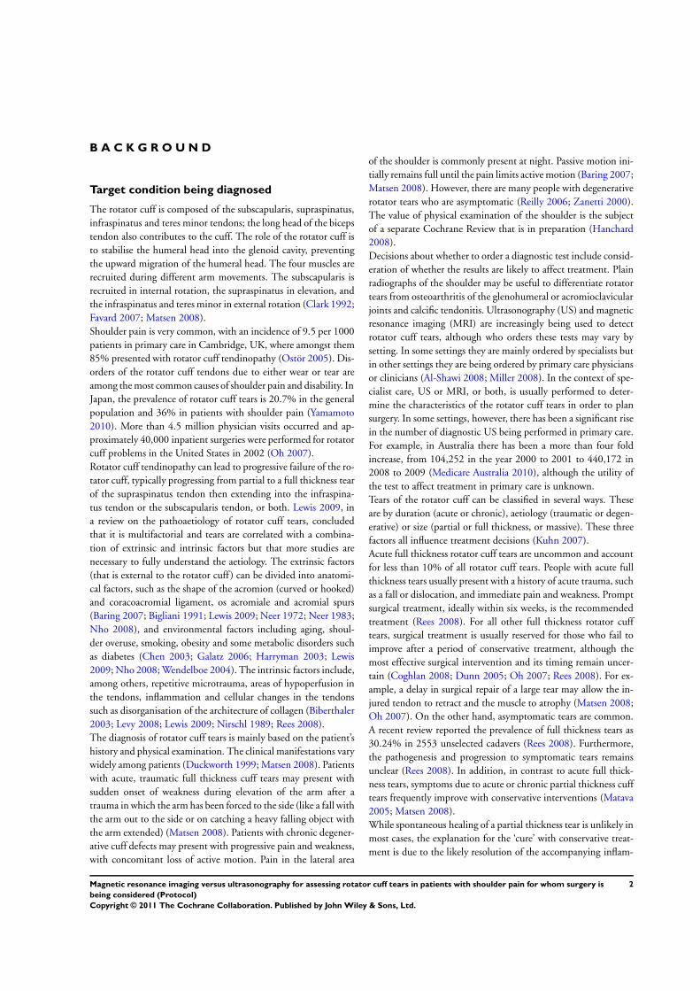
B A C K G R O U N D
Target condition being diagnosed
The rotator cuff is composed of the subscapularis, supraspinatus,
infraspinatus and teres minor tendons; the long head of the biceps
tendon also contributes to the cuff. The role of the rotator cuff is
to stabilise the humeral head into the glenoid cavity, preventing
the upward migration of the humeral head. The four muscles are
recruited during different arm movements. The subscapularis is
recruited in internal rotation, the supraspinatus in elevation, and
the infraspinatus and teres minor in external rotation (Clark 1992;
Favard 2007; Matsen 2008).
Shoulder pain is very common, with an incidence of 9.5 per 1000
patients in primary care in Cambridge, UK, where amongst them
85% presented with rotator cuff tendinopathy (Ostör 2005). Dis-
orders of the rotator cuff tendons due to either wear or tear are
among the most common causes of shoulder pain and disability. In
Japan, the prevalence of rotator cuff tears is 20.7% in the general
population and 36% in patients with shoulder pain (Yamamoto
2010). More than 4.5 million physician visits occurred and ap-
proximately 40,000 inpatient surgeries were performed for rotator
cuff problems in the United States in 2002 (Oh 2007).
Rotator cuff tendinopathy can lead to progressive failure of the ro-
tator cuff, typically progressing from partial to a full thickness tear
of the supraspinatus tendon then extending into the infraspina-
tus tendon or the subscapularis tendon, or both. Lewis 2009, in
a review on the pathoaetiology of rotator cuff tears, concluded
that it is multifactorial and tears are correlated with a combina-
tion of extrinsic and intrinsic factors but that more studies are
necessary to fully understand the aetiology. The extrinsic factors
(that is external to the rotator cuff ) can be divided into anatomi-
cal factors, such as the shape of the acromion (curved or hooked)
and coracoacromial ligament, os acromiale and acromial spurs
(Baring 2007; Bigliani 1991; Lewis 2009; Neer 1972; Neer 1983;
Nho 2008), and environmental factors including aging, shoul-
der overuse, smoking, obesity and some metabolic disorders such
as diabetes (Chen 2003; Galatz 2006; Harryman 2003; Lewis
2009; Nho 2008; Wendelboe 2004). The intrinsic factors include,
among others, repetitive microtrauma, areas of hypoperfusion in
the tendons, inflammation and cellular changes in the tendons
such as disorganisation of the architecture of collagen (Biberthaler
2003; Levy 2008; Lewis 2009; Nirschl 1989; Rees 2008).
The diagnosis of rotator cuff tears is mainly based on the patient’s
history and physical examination. The clinical manifestations vary
widely among patients (Duckworth 1999; Matsen 2008). Patients
with acute, traumatic full thickness cuff tears may present with
sudden onset of weakness during elevation of the arm after a
trauma in which the arm has been forced to the side (like a fall with
the arm out to the side or on catching a heavy falling object with
the arm extended) (Matsen 2008). Patients with chronic degener-
ative cuff defects may present with progressive pain and weakness,
with concomitant loss of active motion. Pain in the lateral area
of the shoulder is commonly present at night. Passive motion ini-
tially remains full until the pain limits active motion (Baring 2007;
Matsen 2008). However, there are many people with degenerative
rotator tears who are asymptomatic (Reilly 2006; Zanetti 2000).
The value of physical examination of the shoulder is the subject
of a separate Cochrane Review that is in preparation (Hanchard
2008).
Decisions about whether to order a diagnostic test include consid-
eration of whether the results are likely to affect treatment. Plain
radiographs of the shoulder may be useful to differentiate rotator
tears from osteoarthritis of the glenohumeral or acromioclavicular
joints and calcific tendonitis. Ultrasonography (US) and magnetic
resonance imaging (MRI) are increasingly being used to detect
rotator cuff tears, although who orders these tests may vary by
setting. In some settings they are mainly ordered by specialists but
in other settings they are being ordered by primary care physicians
or clinicians (Al-Shawi 2008; Miller 2008). In the context of spe-
cialist care, US or MRI, or both, is usually performed to deter-
mine the characteristics of the rotator cuff tears in order to plan
surgery. In some settings, however, there has been a significant rise
in the number of diagnostic US being performed in primary care.
For example, in Australia there has been a more than four fold
increase, from 104,252 in the year 2000 to 2001 to 440,172 in
2008 to 2009 (Medicare Australia 2010), although the utility of
the test to affect treatment in primary care is unknown.
Tears of the rotator cuff can be classified in several ways. These
are by duration (acute or chronic), aetiology (traumatic or degen-
erative) or size (partial or full thickness, or massive). These three
factors all influence treatment decisions (Kuhn 2007).
Acute full thickness rotator cuff tears are uncommon and account
for less than 10% of all rotator cuff tears. People with acute full
thickness tears usually present with a history of acute trauma, such
as a fall or dislocation, and immediate pain and weakness. Prompt
surgical treatment, ideally within six weeks, is the recommended
treatment (Rees 2008). For all other full thickness rotator cuff
tears, surgical treatment is usually reserved for those who fail to
improve after a period of conservative treatment, although the
most effective surgical intervention and its timing remain uncer-
tain (Coghlan 2008; Dunn 2005; Oh 2007; Rees 2008). For ex-
ample, a delay in surgical repair of a large tear may allow the in-
jured tendon to retract and the muscle to atrophy (Matsen 2008;
Oh 2007). On the other hand, asymptomatic tears are common.
A recent review reported the prevalence of full thickness tears as
30.24% in 2553 unselected cadavers (Rees 2008). Furthermore,
the pathogenesis and progression to symptomatic tears remains
unclear (Rees 2008). In addition, in contrast to acute full thick-
ness tears, symptoms due to acute or chronic partial thickness cuff
tears frequently improve with conservative interventions (Matava
2005; Matsen 2008).
While spontaneous healing of a partial thickness tear is unlikely in
most cases, the explanation for the ‘cure’ with conservative treat-
ment is due to the likely resolution of the accompanying inflam-
2Magnetic resonance imaging versus ultrasonography for assessing rotator cuff tears in patients with shoulder pain for whom surgery is
being considered (Protocol)
Copyright © 2011 The Cochrane Collaboration. Published by John Wiley & Sons, Ltd.
mation over time and may also be related to the residual cuff mus-
cles compensating for the mechanical deficiency of the torn cuff
(Fukuda 1996; Fukuda 2003; Matava 2005; Matsen 2008). As
with full thickness tears, no simple treatment algorithm for partial
thickness rotator cuff tears exists. Surgical treatment, however, is
normally indicated for patients with persisting symptoms despite
conservative treatment and in whom imaging suggests the pres-
ence of a partial thickness tear or tears. The ideal timing of surgical
intervention also remains unclear (Fukuda 2003; Matava 2005).
However, case series and anecdotal evidence suggest that satisfac-
tory results are usually achieved with surgery provided there is a
good blood supply to the tendon, contact between the torn ends,
absence of retraction and adequate trophic quality of the muscle
(Fukuda 2003).
Another recognised category of tears is massive complete tears, in
which a large area of the humeral head is uncovered (Wolfgang
1974). Post 1983 defined a massive tear as greater than 5 cm. These
tears, which are difficult to repair, are more commonly found in
women over 65 years of age and are associated with advanced atro-
phy, degeneration and progressive fatty infiltration of the rotator
cuff muscles (Dines 2007; Gerber 2000). Treatment options for
these massive, retracted tears are limited as they are often deemed
irreparable. In younger patients, consideration can be given to ten-
don transfers to improve pain and function (Neri 2009).
The indications for surgical treatment of rotator cuff tears have not
been fully defined. A recent systematic review of 14 trials for rotator
cuff disease, including tears, was unable to draw firm conclusions
about the effectiveness of surgery; although it did suggest that
there were no significant differences in outcomes between open
or arthroscopic surgery and non-operative treatment (Coghlan
2008). However, many studies have demonstrated that the size of
the tear is correlated to the final outcome and the likelihood of a
satisfactory surgical result (Bianchi 2005; Bryant 2002; Fotiadou
2008).
Index test(s)
Currently, US or MRI, or both, is usually performed in patients
contemplating surgery for rotator cuff tears to determine the char-
acteristics of the tears. The accuracy of these imaging tests is con-
sidered to have improved significantly over time, enabling useful
assessment of the size and extent of the rotator cuff tear when
planning surgery (Rees 2008).
US is a diagnostic imaging technique used to visualize deep struc-
tures of the body by recording the echoes of pulsed ultrasonic
waves directed into the tissues and reflected by tissue planes to the
transducer. These echoes are converted into ’pictures’ of the tissues
under examination. Seltzer 1979 was the first to describe ultra-
sonographic evaluation of rotator cuff diseases. US of the shoulder
is utilised in secondary, tertiary and, increasingly, primary health-
care settings to evaluate the integrity of the rotator cuff. It consists
of a non-invasive examination that has practically no side effects
and allows the dynamic visualisation of the tendons during move-
ment of the shoulder (Al-Shawi 2008). However, operator depen-
dence and a long learning curve are frequently considered to be its
limitation (O’Connor 2005; Rutten 2006), principally in views
of partial thickness tears for which Le Corroller 2008 described a
high interobserver variability.
MRI uses a powerful magnetic field to align the hydrogen atoms
of water and other molecules in the body. Pulses of radiofrequency
are applied which excite the magnetised atoms. These movements
of hydrogen atoms, which vary in different tissues, are captured
and the signal can be manipulated to build up an image of the
body (Witte 2003). The first article about the use of MRI in the
shoulder was published in 1986 (Kneeland 1986). Since then,
this technique has been widely used in secondary and tertiary
healthcare practice. MRI is a non-invasive method of imaging
which is unique in allowing high resolution images in multiple
planes. It is a static examination that may be enhanced by an
intra-articular injection of radiopaque dye (this is named magnetic
resonance arthrography), which acts as contrast material that helps
to delineate intra-articular structures and outline abnormalities.
Magnetic resonance arthrography (MRA) of the shoulder is also
useful for assessing the rotator cuff integrity. In comparison with
conventional MRI, MRA may improve diagnostic performance in
detecting shoulder diseases; however, any potential benefit from
this additional procedure must be set against the invasiveness and
additional discomfort to patients.
MRI has some absolute contraindications, namely the presence
of intracerebral aneurysm clips, cardiac pacemakers, automatic
defibrillators, biostimulators, implanted infusion devices, cochlear
implants and metallic orbital foreign bodies (Witte 2003). It is
also expensive and time consuming.
Alternative test(s)
The alternative reference tests for diagnosis of rotator cuff tears
are invasive. The most common reference test is diagnostic
arthroscopy. Arthroscopy is a minimally invasive surgical proce-
dure that involves insertion of an arthroscope, a type of fibre-optic
endoscope, into the joint through a small incision. This allows
the surgeon to inspect and probe the articular (joint) and bursal
side of the rotator cuff tendons, to assess accurately the rotator
cuff insertion (footprint) and to perform a general examination
of the shoulder joint in order to identify and treat other potential
lesions (Dinnes 2003; Matava 2005). However, limitations asso-
ciated with diagnostic shoulder arthroscopy include the need for
anaesthesia, hospital admission and some interobserver variation
in the classification of tears (Kuhn 2007).
Open surgery (including mini-open) has also been used as a ref-
erence test although it is more limited than arthroscopy because
joint surface or inferior surface tears are difficult to access and
identify using an open approach. Other available diagnostic imag-
ing tests include plain radiographs, arthrography and computed
3Magnetic resonance imaging versus ultrasonography for assessing rotator cuff tears in patients with shoulder pain for whom surgery is
being considered (Protocol)
Copyright © 2011 The Cochrane Collaboration. Published by John Wiley & Sons, Ltd.
tomography, although these are generally recognised as not being
useful for detecting failure of the rotator cuff.
Rationale
Ultrasound (US) and MRI are being increasingly used to assess
the presence and size of rotator cuff tears to assist in planning
the surgical approach. Improved techniques have resulted in in-
creased reliance on these tests, in place of a separate diagnostic
arthroscopy, although arthroscopic examination of the shoulder
joint is still commonly performed as part of the surgical treatment.
Both US and MRI are operator and reader dependent and MRI
is expensive. It is not known whether one method is superior to
the other or whether performing both enhances their value (Swen
1999). It is also not known whether they provide useful additional
information compared with diagnostic arthroscopy, which is an
accepted part of the surgical treatment; nor whether they are cost-
effective.
We have located two relevant systematic reviews with meta-analy-
ses which studied diagnostic imaging tests for rotator cuff disease
(de Jesus 2009; Dinnes 2003). The literature search in both re-
views was restricted to English language only. The earlier review
(Dinnes 2003) evaluated the diagnostic accuracy of clinical test-
ing, US and MRI (search date October 2001) for detecting rotator
cuff tears using both surgical and non-surgical tests as the refer-
ence standard. They concluded that US or MRI were equivalent
for detecting full thickness rotator cuff tears, although MRI was
more expensive and US may be better at detecting partial tears. de
Jesus 2009 reported a meta-analysis comparing the diagnostic of
US and MRI for rotator cuff tears using surgery as the reference
standard. The authors included 65 studies (search date Septem-
ber 2007) but the appraisal of the methodological quality of the
included studies was unclear or insufficient. They concluded that
US is as accurate as MRI for both full and partial thickness rotator
cuff tears and also suggested that US may be the most cost-effec-
tive imaging test for screening rotator cuff tears (de Jesus 2009).
In as much as important improvements in both US and MRI have
been made after the search strategies of both studies, and new
accuracy studies have been developed to evaluate these advances
in both US and MRI, our review will perform an updated search
for diagnostic accuracy studies for rotator cuff tears and will also
include non-English references.
O B J E C T I V E S
To compare the diagnostic test accuracy of magnetic resonance
imaging (MRI) versus ultrasonography (US) to detect any rotator
cuff tears (partial or full thickness) in patients with shoulder pain
for whom surgery is being considered.
We will divide our objectives as follows.
1. To determine the diagnostic accuracy of US, MRI and magnetic
resonance arthrography (MRA) for diagnosing any rotator cuff
tears (partial or full thickness).
2. To determine the diagnostic accuracy of US, MRI and MRA for
diagnosing full thickness rotator cuff tears (one or more tendons).
3. To determine the diagnostic accuracy of US, MRI and MRA for
assessing the extent of the tear (full thickness or partial thickness)
in people with a rotator cuff tear.
Investigation of sources of heterogeneity
We will investigate potential sources of heterogeneity by conduct-
ing subgroup analyses according to characteristics of the popula-
tion, tears and reference test, as described in the methods.
M E T H O D S
Criteria for considering studies for this review
Types of studies
All diagnostic accuracy studies that compare one or both index
tests with one or both of the reference tests in patients suspected of
having a partial or full thickness rotator cuff tear will be included.
Studies which selected a healthy control group will be excluded.
Studies with an excessively long period (more than six months)
between the index and reference tests will also be excluded because
rotator cuff tears can progress over time. We will only include
results from full reports. If studies have been reported in abstracts
or conference proceedings we will search for the full publications.
We will include English and commonly used European-origin lan-
guages articles in which a full translation can be obtained. Non-
English articles where a full translation cannot be obtained will be
cited in an Appendix but not included in the review.
In cases in which the same study has been published in more than
one report, we will include only the most updated or complete
report. Nevertheless, the references to other publications will be
cited under the study identification (ID).
Participants
Patients with shoulder pain and suspected of having a rotator cuff
tear for whom surgery is being considered. Studies that included
patients who have been previously diagnosed with other spe-
cific shoulder pain (for example shoulder instability, osteoarthritis,
rheumatoid arthritis, frozen shoulder (adhesive capsulitis), benign
or malignant tumours or referred pain) will be excluded.
4Magnetic resonance imaging versus ultrasonography for assessing rotator cuff tears in patients with shoulder pain for whom surgery is
being considered (Protocol)
Copyright © 2011 The Cochrane Collaboration. Published by John Wiley & Sons, Ltd.
Index tests
US, MRI or MRA. We anticipate that criteria for positivity will
vary, and we will report them in detail on a study by study basis
in the ’Characteristics of included studies’ table. To standardise
classification for this review, rotator cuff tears will be dichotomised
to absence and presence of any, full and partial thickness tears.
Target conditions
Studies will require at least one of three target conditions to be
included:
• presence of any rotator cuff tears (partial or full thickness);
• presence of full thickness tears (one or more tendons);
• presence of partial thickness tears (any tendon).
Reference standards
Arthroscopy or open (including mini-open) surgery findings. If
any study addresses both (arthroscopy and open surgery), we will
select arthroscopy as the reference standard.
Search methods for identification of studies
Electronic searches
We will search relevant computerised databases for eligible di-
agnostic studies: MEDLINE (PubMed) (1966 to present), EM-
BASE (Elsevier) (1980 to present), and LILACS (Bireme) (1982
to present). We will also search the Cochrane Register of Diagnos-
tic Test Accuracy Studies. There will be no restrictions based on
language, date or publication status. When possible, non-English
articles will be assessed through selective translation by a native
speaker.
We have developed a sensitive search strategy (Appendix 1), as
recommended in Chapter 7 of the Cochrane Handbook (de
Vet 2008), for MEDLINE (PubMed), EMBASE (Elsevier) and
LILACS (Bireme) (see Appendix 1 for the MEDLINE and EM-
BASE search strategies).
Searching other resources
We will check the reference lists of articles, reviews and text-
books for possibly relevant primary diagnostic studies and sys-
tematic reviews. We will also search DARE (Database of Ab-
stracts of Reviews of Effects) and the HTA Database (Health
Technology Assessments Database) for other related diagnostic
test accuracy reviews, and we will check the reference lists of
those reviews that are relevant for additional studies. We will also
search the US Health Services Research Projects in Progress and
the UK Clinical Research Network Portfolio Database for ongo-
ing and recently completed studies.
We will handsearch abstracts of the British Elbow and Shoulder
Society annual meetings (2005 to present) and American
Academy of Orthopaedic Surgeons annual meetings (2005 to
present).
We will also contact experts in the field. When necessary, we will
contact main investigators of relevant ongoing studies for further
information.
Data collection and analysis
We will use the methods suggested by the Cochrane Handbook
for Systematic Reviews of Diagnostic Test Accuracy (Deeks 2009).
Selection of studies
Two review authors (ML and RB) will independently screen the
titles and abstracts of retrieved records to identify potentially rele-
vant studies for inclusion. Duplicates will be removed and the re-
maining references will be examined. Studies which clearly do not
meet the inclusion criteria will be excluded, and copies of the full
text of potentially relevant references will be obtained. ML and RB
will independently assess full text reports and determine inclusion
or exclusion of the studies. Any uncertainties or disagreements will
be resolved by discussion and, if necessary, with adjudication by a
third author (FF). We will document reasons for exclusion.
Data extraction and management
Two review authors (ML and RB) will independently collect the
available data using a piloted data extraction form without mask-
ing of study authors and other identifying information. A third
review author (FF) will be consulted for resolution of any disagree-
ments. When necessary, we will send requests to study authors for
additional information or data.
Diagnostic studies presenting insufficient data for construction of
a two-by-two table will be excluded from statistical analysis but
will be included in the narrative part of this review.
The following data will be retrieved.
1. General information: title, journal, year, publication status,
country of study, period of study, primary objective and study de-
sign (prospective versus retrospective and consecutive versus non-
consecutive).
2. Sample size: number of participants meeting the criteria and
total number screened.
3. Baseline characteristics: baseline diagnosis, age, sex, side, nature
of onset (e.g. traumatic or non-traumatic), duration of symptoms,
prior treatment, inclusion and exclusion criteria.
4. Target condition, as reported.
5. Index test: description of technique, criteria for positive result,
timing of test and expertise of the tester. All cut-offs will be tested.
6. Reference standard test: description of technique, criteria for
positive result, time from index to reference test and expertise of
the tester. All cut-offs will be tested.
5Magnetic resonance imaging versus ultrasonography for assessing rotator cuff tears in patients with shoulder pain for whom surgery is
being considered (Protocol)
Copyright © 2011 The Cochrane Collaboration. Published by John Wiley & Sons, Ltd.
7. Side effects or complications due to index test(s) and reference
standard test(s).
8. Number of true positive (TP), true negative (TN), false positive
(FP) and false negative (FN). These data will be extracted for each
cut-off presented (e.g. any, full or partial thickness tears).
Assessment of methodological quality
The methodological quality of the included studies will be as-
sessed independently by two review authors (ML and RB) and
disagreement on study quality will be resolved by a third review
author (FF). At the same time as data extraction, the methodolog-
ical quality of selected studies will be assessed using a modified
version of the QUADAS checklist (Whiting 2003), following the
guidelines provided in Chapter 9 of the Cochrane Handbook for
Systematic Reviews of Diagnostic Test Accuracy (Reitsma 2009).
For each study, a methodological quality table will be filled. Table
1 explains how the authors will interpret the QUADAS items in
terms of the diagnosis of rotator cuff tears.
Table 1. Assessment of methodological quality: QUADAS and additional items
Item definition Item question Assessment
Representative spectrum 1. Was the spectrum of patients represen-
tative of the patients who will receive the
test in practice?
Yes: (a) the setting was secondary or tertiary
care AND (b) the population was patients
with shoulder pain suspected of a rotator
cuff tear for whom surgery is being con-
sidered AND (c) the study was prospective
AND (d) recruitment was consecutive.
Unclear: if insufficient information was
given on the setting, selection criteria, or
selection procedure to make a judgment.
No: (a) the setting was primary care OR (b)
the population was unselected but defined
by shoulder pain OR (c) the study was not
prospective OR (d) recruitment was not
consecutive.
Acceptable reference standard 2. Is the reference standard likely to classify
the target condition correctly?
Yes:
if the reference standard was arthroscopy
or a combination of arthroscopy and open
surgery (including mini-open).
Unclear: if the target condition was partial
thickness rotator cuff tears and the refer-
ence standard was open surgery (including
mini-open).
No: not applicable.
Acceptable delay between tests 3. Is the time period between reference
standard and index test short enough to be
reasonably sure that the target condition
did not change between the two tests?
Yes: if the average interval between ref-
erence standard and index test was one
month or less.
Unclear: if the interval between tests was
not clearly reported.
No: if the average interval between refer-
ence standard and index test was longer
than one month.
Partial verification avoided 4. Did the whole sample or a random se-
lection of the sample, receive verification
using the intended reference standard?
Yes: If all patients who received the in-
dex test went on to receive verification of
their disease status using a reference stan-
6Magnetic resonance imaging versus ultrasonography for assessing rotator cuff tears in patients with shoulder pain for whom surgery is
being considered (Protocol)
Copyright © 2011 The Cochrane Collaboration. Published by John Wiley & Sons, Ltd.
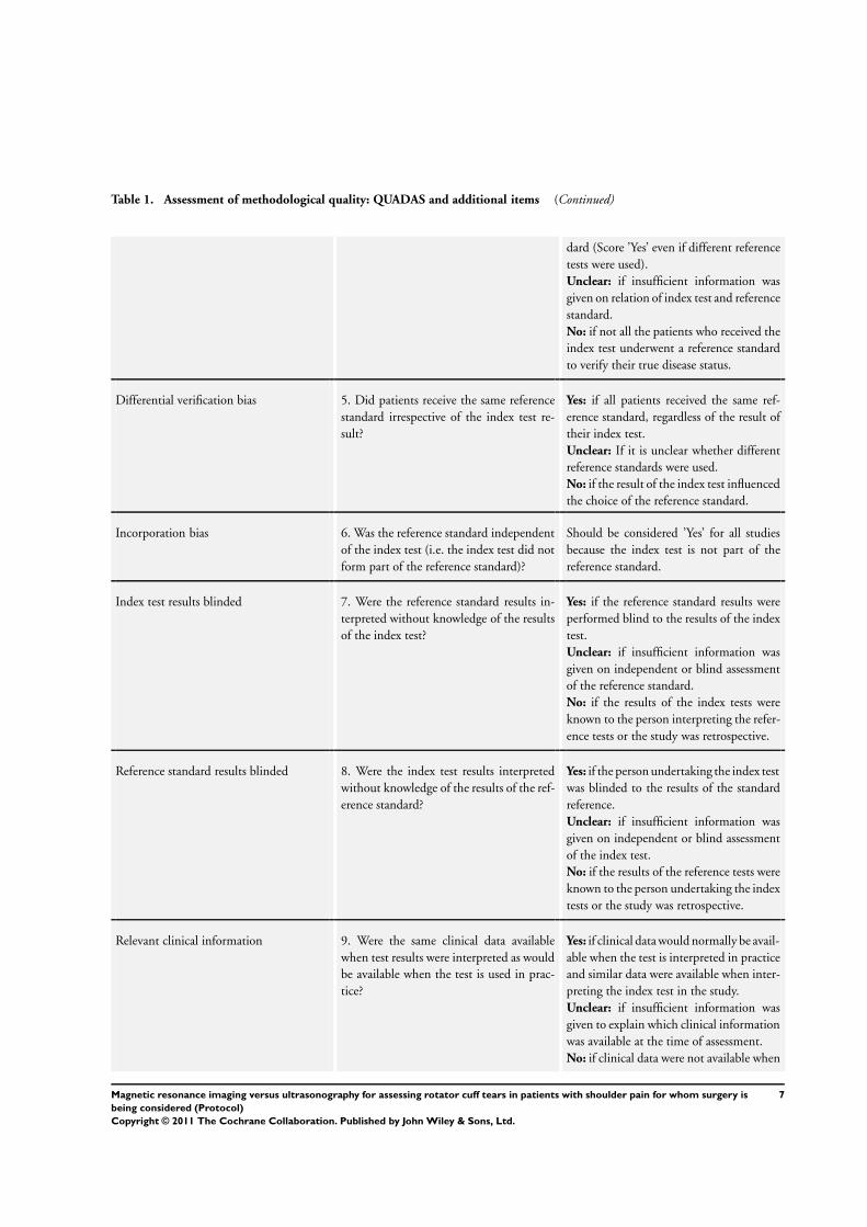
Table 1. Assessment of methodological quality: QUADAS and additional items (Continued)
dard (Score ’Yes’ even if different reference
tests were used).
Unclear: if insufficient information was
given on relation of index test and reference
standard.
No: if not all the patients who received the
index test underwent a reference standard
to verify their true disease status.
Differential verification bias 5. Did patients receive the same reference
standard irrespective of the index test re-
sult?
Yes: if all patients received the same ref-
erence standard, regardless of the result of
their index test.
Unclear: If it is unclear whether different
reference standards were used.
No: if the result of the index test influenced
the choice of the reference standard.
Incorporation bias 6. Was the reference standard independent
of the index test (i.e. the index test did not
form part of the reference standard)?
Should be considered ’Yes’ for all studies
because the index test is not part of the
reference standard.
Index test results blinded 7. Were the reference standard results in-
terpreted without knowledge of the results
of the index test?
Yes: if the reference standard results were
performed blind to the results of the index
test.
Unclear: if insufficient information was
given on independent or blind assessment
of the reference standard.
No: if the results of the index tests were
known to the person interpreting the refer-
ence tests or the study was retrospective.
Reference standard results blinded 8. Were the index test results interpreted
without knowledge of the results of the ref-
erence standard?
Yes: if the person undertaking the index test
was blinded to the results of the standard
reference.
Unclear: if insufficient information was
given on independent or blind assessment
of the index test.
No: if the results of the reference tests were
known to the person undertaking the index
tests or the study was retrospective.
Relevant clinical information 9. Were the same clinical data available
when test results were interpreted as would
be available when the test is used in prac-
tice?
Yes: if clinical data would normally be avail-
able when the test is interpreted in practice
and similar data were available when inter-
preting the index test in the study.
Unclear: if insufficient information was
given to explain which clinical information
was available at the time of assessment.
No: if clinical data were not available when
7Magnetic resonance imaging versus ultrasonography for assessing rotator cuff tears in patients with shoulder pain for whom surgery is
being considered (Protocol)
Copyright © 2011 The Cochrane Collaboration. Published by John Wiley & Sons, Ltd.
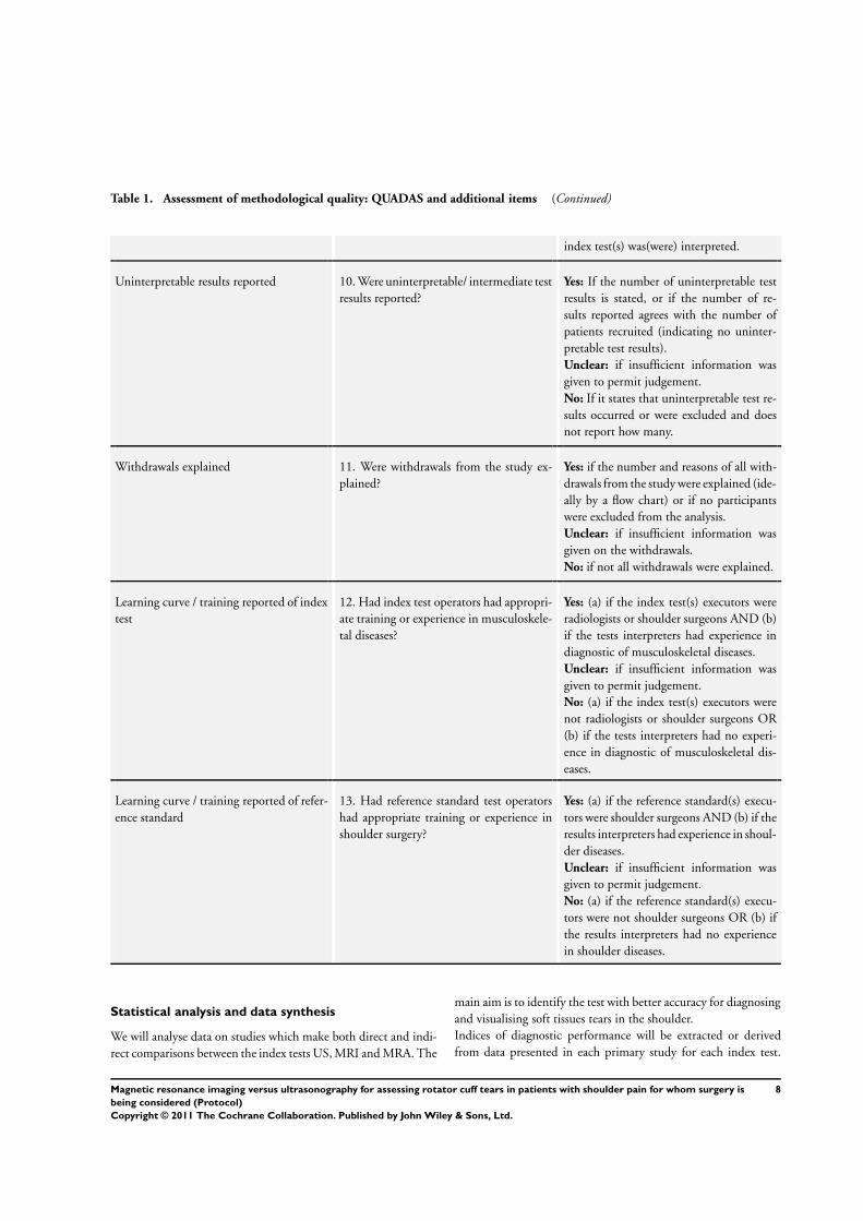
Table 1. Assessment of methodological quality: QUADAS and additional items (Continued)
index test(s) was(were) interpreted.
Uninterpretable results reported 10. Were uninterpretable/ intermediate test
results reported?
Yes: If the number of uninterpretable test
results is stated, or if the number of re-
sults reported agrees with the number of
patients recruited (indicating no uninter-
pretable test results).
Unclear: if insufficient information was
given to permit judgement.
No: If it states that uninterpretable test re-
sults occurred or were excluded and does
not report how many.
Withdrawals explained 11. Were withdrawals from the study ex-
plained?
Yes: if the number and reasons of all with-
drawals from the study were explained (ide-
ally by a flow chart) or if no participants
were excluded from the analysis.
Unclear: if insufficient information was
given on the withdrawals.
No: if not all withdrawals were explained.
Learning curve / training reported of index
test
12. Had index test operators had appropri-
ate training or experience in musculoskele-
tal diseases?
Yes: (a) if the index test(s) executors were
radiologists or shoulder surgeons AND (b)
if the tests interpreters had experience in
diagnostic of musculoskeletal diseases.
Unclear: if insufficient information was
given to permit judgement.
No: (a) if the index test(s) executors were
not radiologists or shoulder surgeons OR
(b) if the tests interpreters had no experi-
ence in diagnostic of musculoskeletal dis-
eases.
Learning curve / training reported of refer-
ence standard
13. Had reference standard test operators
had appropriate training or experience in
shoulder surgery?
Yes: (a) if the reference standard(s) execu-
tors were shoulder surgeons AND (b) if the
results interpreters had experience in shoul-
der diseases.
Unclear: if insufficient information was
given to permit judgement.
No: (a) if the reference standard(s) execu-
tors were not shoulder surgeons OR (b) if
the results interpreters had no experience
in shoulder diseases.
Statistical analysis and data synthesis
We will analyse data on studies which make both direct and indi-
rect comparisons between the index tests US, MRI and MRA. The
main aim is to identify the test with better accuracy for diagnosing
and visualising soft tissues tears in the shoulder.
Indices of diagnostic performance will be extracted or derived
from data presented in each primary study for each index test.
8Magnetic resonance imaging versus ultrasonography for assessing rotator cuff tears in patients with shoulder pain for whom surgery is
being considered (Protocol)
Copyright © 2011 The Cochrane Collaboration. Published by John Wiley & Sons, Ltd.
The review authors will construct two-by-two contingency tables
of true positive cases, false positive cases, false negative cases and
true negative cases according to the three target conditions (any,
full and partial thickness tears). The first step in a given meta-
analysis will be to graphically present the results of the individual
studies. In addition, a scatterplot of study-specific estimates of true
positive (sensitivity) and false positive rates (1-specificity) will be
used to display the data in the receiver operating characteristic
(ROC) space.
Where adequate data are available, we will conduct meta-analyses
using the hierarchical summary ROC (HSROC) model, which
takes into account both within and between study variability (
Macaskill 2010). We will apply SAS (version 9.2) software for the
hierarchical model.
The differences between the tests will be formally assessed in a
HSROC model, as well as the differences between subgroups pre-
defined as potential sources of heterogeneity.
Investigations of heterogeneity
Heterogeneity will be investigated in the first instance through
visual examination of forest plots of sensitivities and specificities
and through visual examination of the ROC plot of the raw data.
We will also explore heterogeneity by examining differences in
diagnostic accuracy according to pre-defined subgroup analyses:
by age of the population, tears and reference test, as specified below.
• The study population: old (65 years and older) or young
population.
• Type of tears: acute or chronic, and traumatic or
degenerative.
• Type of reference test: open (including mini-open) surgery
or arthroscopy.
Sensitivity analyses
If possible on the basis of the amount of data (that is the number
of eligible studies) we will compare the results of all studies with
the results of subsets of studies that comply with the following
methodological quality items of the QUADAS checklist (Whiting
2003).
• Representative spectrum.
• Acceptable reference standard.
• Acceptable delay between tests.
• Index test results blinded.
• Reference standard results blinded.
A C K N O W L E D G E M E N T S
We thank Dr Helen Handoll, Mr Jonathan Rees and the peer-
reviewers and contact editor (Dr Rob Scholten) for helpful feed-
back at editorial review. We would also like to thank Mrs Lindsey
Elstub and Dr Joanne Elliott for their assistance in preparing the
protocol.
R E F E R E N C E S
Additional references
Al-Shawi 2008
Al-Shawi A, Badge R, Bunker T. The detection of full thickness
rotator cuff tears using ultrasound. The Journal of Bone and Joint
Surgery. British Volume 2008;90(7):889–92.
Baring 2007
Baring T, Emery R, Reilly P. Management of rotator cuff disease:
specific treatment for specific disorders. Best Practice & Research.
Clinical Rheumatology 2007;21(2):279–94.
Bianchi 2005
Bianchi S, Martinoli C, Abdelwahab IF. Ultrasound of tendon
tears. Part 1: general considerations and upper extremity. Skeletal
Radiology 2005;34(9):500–12.
Biberthaler 2003
Biberthaler P, Wiedemann E, Nerlich A, Kettler M, Mussack T,
Deckelmann S, et al.Microcirculation associated with degenerative
rotator cuff lesions. In vivo assessment with orthogonal polarization
spectral imaging during arthroscopy of the shoulder. The Journal of
Bone and Joint Surgery. American Volume 2003;85-A(3):475–80.
Bigliani 1991
Bigliani LU, Ticker JB, Flatow EL, Soslowsky LJ, Mow VC. The
relationship of acromial architecture to rotator cuff disease. Clinics
in Sports Medicine 1991;10(4):823–38.
Bryant 2002
Bryant L, Shnier R, Bryant C, Murrell GA. A comparison of
clinical estimation, ultrasonography, magnetic resonance imaging,
and arthroscopy in determining the size of rotator cuff tears.
Journal of Shoulder and Elbow Surgery 2002;11(3):219–24.
Chen 2003
Chen AL, Shapiro JA, Ahn AK, Zuckerman JD, Cuomo F. Rotator
cuff repair in patients with type I diabetes mellitus. Journal of
Shoulder and Elbow Surgery 2003;12(5):416–21.
Clark 1992
Clark JM, Harryman DT II. Tendons, ligaments, and capsule of
the rotator cuff: gross and microscopic anatomy. The Journal of
Bone and Joint Surgery. American Volume 1992;74(5):713–25.
Coghlan 2008
Coghlan JA, Buchbinder R, Green S, Johnston RV, Bell SN.
Surgery for rotator cuff disease. Cochrane Database of Systematic
Reviews 2008, Issue 1. Art. No.: CD005619.
9Magnetic resonance imaging versus ultrasonography for assessing rotator cuff tears in patients with shoulder pain for whom surgery is
being considered (Protocol)
Copyright © 2011 The Cochrane Collaboration. Published by John Wiley & Sons, Ltd.
de Jesus 2009
de Jesus JO, Parker L, Frangos AJ, Nazarian LN. Accuracy of MRI,
MR arthrography, and ultrasound in the diagnosis of rotator cuff
tears: a meta-analysis. AJR. American Journal of Roentgenology 2009;
192(6):1701–7.
de Vet 2008
de Vet HCW, Eisinga A, Riphagen II, Aertgeerts B, Pewsner D.
Chapter 7: Searching for Studies. In: Cochrane Handbook for
Systematic Reviews of Diagnostic Test Accuracy Version 0.4
[updated September 2008]. The Cochrane Collaboration 2008.
Deeks 2009
Deeks JJ, Bossuyt PM, Gatsonis C (editors). Cochrane Handbook
for Systematic Reviews of Diagnostic Test Accuracy Version 1.0.0.
The Cochrane Collaboration, 2009. Available from: http://
srdta.cochrane.org/ (accessed November 2010).
Dines 2007
Dines DM, Moynihan DP, Dines J, McCann P. Irreparable rotator
cuff tears: what to do and when to do it; the surgeon’s dilemma.
The Journal of Bone and Joint Surgery. American Volume 2006;88
(10):2294–302.
Dinnes 2003
Dinnes J, Loveman E, McIntyre L, Waugh N. The effectiveness of
diagnostic tests for the assessment of shoulder pain due to soft
tissue disorders: a systematic review. Health Technology Assessment
(Winchester, England) 2003;7(29):1–166.
Duckworth 1999
Duckworth DG, Smith KL, Campbell B, Matsen FA 3rd. Self-
assessment questionnaires document substantial variability in the
clinical expression of rotator cuff tears. Journal of Shoulder and
Elbow Surgery 1999;8(4):330–3.
Dunn 2005
Dunn WR, Schackman BR, Walsh C, Lyman S, Jones EC, Warren
RF, et al.Variation in orthopaedic surgeons’ perceptions about the
indications for rotator cuff surgery. The Journal of Bone and Joint
Surgery. American Volume 2005;87(9):1978–84.
Favard 2007
Favard L, Bacle G, Berhouet J. Rotator cuff repair. Joint, Bone,
Spine: Revue du Rhumatisme 2007;74(6):551–7.
Fotiadou 2008
Fotiadou AN, Vlychou M, Papadopoulos P, Karataglis DS, Palladas
P, Fezoulidis IV. Ultrasonography of symptomatic rotator cuff tears
compared with MR imaging and surgery. European Journal of
Radiology 2008;68(1):174–9.
Fukuda 1996
Fukuda H, Hamada K, Nakajima T, Yamada N, Tomonaga A,
Goto M. Partial-thickness tears of the rotator cuff. A
clinicopathological review based on 66 surgically verified cases.
International Orthopaedics 1996;20(4):257–65.
Fukuda 2003
Fukuda H. The management of partial-thickness tears of the
rotator cuff. The Journal of Bone and Joint Surgery. British Volume
2003;85(1):3–11.
Galatz 2006
Galatz LM, Silva MJ, Rothermich SY, Zaegel MA, Havlioglu N,
Thomopoulos S. Nicotine delays tendon-to-bone healing in a rat
shoulder model. The Journal of Bone and Joint Surgery. American
Volume 2006;88(9):2027–34.
Gerber 2000
Gerber C, Fuchs B, Hodler J. The results of repair of massive tears
of the rotator cuff. The Journal of Bone and Joint Surgery. American
Volume 2000;82(4):505–15.
Hanchard 2008
Hanchard NCA, Handoll HHG. Physical tests for shoulder
impingements and local lesions of bursa, tendon or labrum that
may accompany impingement. Cochrane Database of Systematic
Reviews 2008, Issue 4. Art. No.: CD007427.
Harryman 2003
Harryman DT 2nd, Hettrich CM, Smith KL, Campbell B, Sidles
JA, Matsen FA 3rd. A prospective multipractice investigation of
patients with full-thickness rotator cuff tears: the importance of
comorbidities, practice, and other covariables on self-assessed
shoulder function and health status. The Journal of Bone and Joint
Surgery. American Volume 2003;85-A(4):690–6.
Kneeland 1986
Kneeland JB, Carrera GF, Middleton WD, Campagna NF, Ryan
LM, Jesmanowicz A, et al.Rotator cuff tears: preliminary
application of high-resolution MR imaging with counter rotating
current loop-gap resonators. Radiology 1986;160(3):695–9.
Kuhn 2007
Kuhn JE, Dunn WR, Ma B, Wright RW, Jones G, Spencer EE, et
al.Interobserver agreement in the classification of rotator cuff tears.
The American Journal of Sports Medicine 2007;35(3):437–41.
Le Corroller 2008
Le Corroller T, Cohen M, Aswad R, Pauly V, Champsaur P.
Sonography of the painful shoulder: role of the operator’s
experience. Skeletal Radiology 2008;37(11):979–86.
Levy 2008
Levy O, Relwani J, Zaman T, Even T, Venkateswaran B, Copeland
S. Measurement of blood flow in the rotator cuff using laser
Doppler flowmetry. The Journal of Bone and Joint Surgery. British
Volume 2008;90(7):893–8.
Lewis 2009
Lewis JS. Rotator cuff tendinopathy. British Journal of Sports
Medicine 2009;43(4):236–41.
Macaskill 2010
Macaskill P, Gatsonis C, Deeks JJ, Harbord RM, Takwoingi Y.
Chapter 10: Analysying and Presenting Results. Deeks JJ, Bossuyt
PM, Gatsonis C (editors), Cochrane Handbook for Systematic
Reviews of Diagnostic Test Accuracy Version 0.9.0. The Cochrane
Collaboration 2010. Available from: http://srdta.cochrane.org/.
Matava 2005
Matava MJ, Purcell DB, Rudzki JR. Partial-thickness rotator cuff
tears. The American Journal of Sports Medicine 2005;33(9):
1405–17.
Matsen 2008
Matsen FA 3rd. Clinical practice. Rotator-cuff failure. The New
England Journal of Medicine 2008;358(20):2138–47.
10Magnetic resonance imaging versus ultrasonography for assessing rotator cuff tears in patients with shoulder pain for whom surgery is
being considered (Protocol)
Copyright © 2011 The Cochrane Collaboration. Published by John Wiley & Sons, Ltd.
Medicare Australia 2010
Medicare Australia. Medicare Item Reports: Medicare Australia.
https://www.medicareaustralia.gov.au/statistics/mbs˙item.shtml
(accessed 10 May 2010).
Miller 2008
Miller D, Frost A, Hall A, Barton C, Bhoora I, Kathuria V. A ’one-
stop clinic’ for the diagnosis and management of rotator cuff
pathology: Getting the right diagnosis first time. International
Journal of Clinical Practice 2008;62(5):750–3.
Neer 1972
Neer CS 2nd. Anterior acromioplasty for the chronic impingement
syndrome in the shoulder: a preliminary report. The Journal of
Bone and Joint Surgery. American Volume 1972;54(1):41–50.
Neer 1983
Neer CS 2nd. Impingement lesions. Clinical Orthopaedics and
Related Research 1983;(173):70–7.
Neri 2009
Neri BR, Chan KW, Kwon YW. Management of massive and
irreparable rotator cuff tears. Journal of Shoulder and Elbow Surgery
2009;18(5):808–18.
Nho 2008
Nho SJ, Yadav H, Shindle MK, Macgillivray JD. Rotator cuff
degeneration: etiology and pathogenesi. The American Journal of
Sports Medicine 2008;36(5):987–93.
Nirschl 1989
Nirschl RP. Rotator cuff tendinitis: basic concepts of pathoetiology.
Instructional Course Lectures 1989;38:439–45.
O’Connor 2005
O’Connor PJ, Rankine J, Gibbon WW, Richardson A, Winter F,
Miller JH. Interobserver variation in sonography of the painful
shoulder. Journal of Clinical Ultrasound 2005;33(2):53–6.
Oh 2007
Oh LS, Wolf BR, Hall MP, Levy BA, Marx RG. Indications for
rotator cuff repair: a systematic review. Clinical Orthopaedics and
Related Research 2007;(455):52–63.
Ostör 2005
Ostör AJ, Richards CA, Prevost AT, Speed CA, Hazleman BL.
Diagnosis and relation to general health of shoulder disorders
presenting to primary care. Rheumatology (Oxford, England) 2005;
44(6):800–5.
Post 1983
Post M, Silver R, Singh M. Rotator cuff tear. Diagnosis and
treatment. Clinical Orthopaedics and Related Research 1983;(173):
78–91.
Rees 2008
Rees JL. The pathogenesis and surgical treatment of tears of the
rotator cuff. The Journal of Bone and Joint Surgery. British Volume
2008;90(7):827–32.
Reilly 2006
Reilly P, Macleod I, Macfarlane R, Windley J, Emery RJ. Dead men
and radiologists don’t lie: a review of cadaveric and radiological
studies of rotator cuff tear prevalence. Annals of the Royal College of
Surgeons of England 2006;88(2):116–21.
Reitsma 2009
Reitsma JB, Rutjes AWS, Whiting P, Vlassov VV, Leeflang MMG,
Deeks JJ. Chapter 9: Assessing methodological quality. Deeks JJ,
Bossuyt PM, Gatsonis C (editors), Cochrane Handbook for
Systematic Reviews of Diagnostic Test Accuracy Version 1.0.0. The
Cochrane Collaboration 2009. Available from: http://
srdta.cochrane.org/.
Rutten 2006
Rutten MJ, Jager GJ, Blickman JG. From the RSNA refresher
courses: US of the rotator cuff: pitfalls, limitations, and artifacts.
Radiographics 2006;26(2):589–604.
Seltzer 1979
Seltzer SE, Finberg HJ, Weissman BN, Kido DK, Collier BD.
Arthrosonography: gray-scale ultrasound evaluation of the
shoulder. Radiology 1979;132(2):467–8.
Swen 1999
Swen WA, Jacobs JW, Algra PR, Manoliu RA, Rijkmans J, Willems
WJ, et al.Sonography and magnetic resonance imaging equivalent
for the assessment of full-thickness rotator cuff tears. Arthritis and
Rheumatism 1999;42(10):2231–8.
Wendelboe 2004
Wendelboe AM, Hegmann KT, Gren LH, Alder SC, White GL Jr,
Lyon JL. Associations between body-mass index and surgery for
rotator cuff tendinitis. The Journal of Bone and Joint Surgery.
American Volume 2004;86-A(4):743–7.
Whiting 2003
Whiting P, Rutjes AW, Reitsma JB, Bossuyt PM, Kleijnen J. The
development of QUADAS: a tool for the quality assessment of
studies of diagnostic accuracy included in systematic reviews. BMC
Medical Research Methodology 2003;3:25.
Witte 2003
Witte DH. Magnetic Resonance Imaging in Orthopaedics. In:
Canale ST editor(s). Campbell’s Operative Orthopaedics. 10.
Philadelphia: Mosby, 2003:123–64.
Wolfgang 1974
Wolfgang GL. Surgical repair of tears of the rotator cuff of the
shoulder. Factors influencing the result. The Journal of Bone and
Joint Surgery. American Volume 1974;56(1):14–26.
Yamamoto 2010
Yamamoto A, Takagishi K, Osawa T, Yanagawa T, Nakajima D,
Shitara H, et al.Prevalence and risk factors of a rotator cuff tear in
the general population. Journal of Shoulder and Elbow Surgery 2010;
19(1):116–20.
Zanetti 2000
Zanetti M, Jost B, Hodler J, Gerber C. MR imaging after rotator
cuff repair: full-thickness defects and bursitis-like subacromial
abnormalities in asymptomatic subjects. Skeletal Radiology 2000;29
(6):314–9.∗ Indicates the major publication for the study
11Magnetic resonance imaging versus ultrasonography for assessing rotator cuff tears in patients with shoulder pain for whom surgery is
being considered (Protocol)
Copyright © 2011 The Cochrane Collaboration. Published by John Wiley & Sons, Ltd.
A P P E N D I C E S
Appendix 1. Search strategies
MEDLINE (PubMed)
((Ultrasonography [mh] OR ultrasound [tw] OR ultrasonograph* [tw] OR sonograp*[tw] OR us [sh]) OR (Magnetic Resonance
Imaging [mh] OR MR imag*[tw] OR magnetic resonance imag* [tw] OR MRI [tw])) AND (Rotator Cuff [mh] OR rotator cuff*
[tw] OR musculotendinous cuff* [tw] OR subscapularis [tw] OR supraspinatus [tw] OR infraspinatus OR teres minor [tw]) AND
(Rupture [mh:noexp] OR tear* [tw] OR torn [tw] OR thickness [tw] OR lesion* [tw] OR ruptur* [tw] OR injur* [tw])
EMBASE (Elsevier)
1. ’echography’/de AND [embase]/lim
2. ultrasound:ab,ti OR ultrasonograph*:ab,ti OR sonograp*:ab,ti AND [embase]/lim
3. #1 OR #2
4. ’nuclear magnetic resonance imaging’/de AND [embase]/lim
5. ((’magnetic resonance’ OR mr) NEAR/3 imag*):ab,ti AND [embase]/lim
6. mri:ab,ti AND [embase]/lim
7. #4 OR #5 OR #6
8. ’rotator cuff injury’/de OR ’rotator cuff rupture’/de AND [embase]/lim
9. ’rotator cuff ’/de AND [embase]/lim
10. ’rotator cuff ’:ab,ti OR ’musculotendinous cuff ’:ab,ti OR subscapularis:ab,ti OR supraspinatus:ab,ti OR infraspinatus:ab,ti OR
’teres minor’:ab,ti AND [embase]/lim
11. #9 OR #10
12. ’rupture’/de AND [embase]/lim
13. tear*:ab,ti OR torn:ab,ti OR thickness:ab,ti OR lesion*:ab,ti OR ruptur*:ab,ti OR injur*:ab,ti AND [embase]/lim
14. #12 OR #13
15. #11 AND #14
16. #8 OR #15
17. #3 OR #7
18. #16 AND #17
H I S T O R Y
Protocol first published: Issue 3, 2011
C O N T R I B U T I O N S O F A U T H O R S
All authors contributed to the development of the protocol and commented on and approved the final version. The guarantor of this
protocol is Mario Lenza.
12Magnetic resonance imaging versus ultrasonography for assessing rotator cuff tears in patients with shoulder pain for whom surgery is
being considered (Protocol)
Copyright © 2011 The Cochrane Collaboration. Published by John Wiley & Sons, Ltd.
D E C L A R A T I O N S O F I N T E R E S T
None known.
S O U R C E S O F S U P P O R T
Internal sources
• Universidade Federal de São Paulo, Brazil.
• The Parker Institute, Denmark.
The Parker Institute: Musculoskeletal Statistics Unit is supported by grants from The Oak Foundation.
• Teesside University, UK.
External sources
• No sources of support supplied
13Magnetic resonance imaging versus ultrasonography for assessing rotator cuff tears in patients with shoulder pain for whom surgery is
being considered (Protocol)
Copyright © 2011 The Cochrane Collaboration. Published by John Wiley & Sons, Ltd.
Magnetic resonance imaging, magnetic resonance
arthrography and ultrasonography for assessing rotator cuff
tears in people with shoulder pain for whom surgery is being
considered (Review)
Lenza M, Buchbinder R, Takwoingi Y, Johnston RV, Hanchard NCA, Faloppa F
This is a reprint of a Cochrane review, prepared and maintained by The Cochrane Collaboration and published in The Cochrane Library2013, Issue 9
http://www.thecochranelibrary.com
Magnetic resonance imaging, magnetic resonance arthrography and ultrasonography for assessing rotator cuff tears in people with
shoulder pain for whom surgery is being considered (Review)
Copyright © 2013 The Cochrane Collaboration. Published by John Wiley & Sons, Ltd.
T A B L E O F C O N T E N T S
1HEADER . . . . . . . . . . . . . . . . . . . . . . . . . . . . . . . . . . . . . . .1ABSTRACT . . . . . . . . . . . . . . . . . . . . . . . . . . . . . . . . . . . . . .2BACKGROUND . . . . . . . . . . . . . . . . . . . . . . . . . . . . . . . . . . . .5OBJECTIVES . . . . . . . . . . . . . . . . . . . . . . . . . . . . . . . . . . . . .5METHODS . . . . . . . . . . . . . . . . . . . . . . . . . . . . . . . . . . . . . .8RESULTS . . . . . . . . . . . . . . . . . . . . . . . . . . . . . . . . . . . . . . .
Figure 1. . . . . . . . . . . . . . . . . . . . . . . . . . . . . . . . . . . . . . 9Figure 2. . . . . . . . . . . . . . . . . . . . . . . . . . . . . . . . . . . . . . 10Figure 3. . . . . . . . . . . . . . . . . . . . . . . . . . . . . . . . . . . . . . 11Figure 4. . . . . . . . . . . . . . . . . . . . . . . . . . . . . . . . . . . . . . 12Figure 5. . . . . . . . . . . . . . . . . . . . . . . . . . . . . . . . . . . . . . 14Figure 6. . . . . . . . . . . . . . . . . . . . . . . . . . . . . . . . . . . . . . 15Figure 7. . . . . . . . . . . . . . . . . . . . . . . . . . . . . . . . . . . . . . 16Figure 8. . . . . . . . . . . . . . . . . . . . . . . . . . . . . . . . . . . . . . 18Figure 9. . . . . . . . . . . . . . . . . . . . . . . . . . . . . . . . . . . . . . 19Figure 10. . . . . . . . . . . . . . . . . . . . . . . . . . . . . . . . . . . . . . 20Figure 11. . . . . . . . . . . . . . . . . . . . . . . . . . . . . . . . . . . . . . 21
26DISCUSSION . . . . . . . . . . . . . . . . . . . . . . . . . . . . . . . . . . . . .28AUTHORS’ CONCLUSIONS . . . . . . . . . . . . . . . . . . . . . . . . . . . . . . .28ACKNOWLEDGEMENTS . . . . . . . . . . . . . . . . . . . . . . . . . . . . . . . .28REFERENCES . . . . . . . . . . . . . . . . . . . . . . . . . . . . . . . . . . . . .45CHARACTERISTICS OF STUDIES . . . . . . . . . . . . . . . . . . . . . . . . . . . . .
120DATA . . . . . . . . . . . . . . . . . . . . . . . . . . . . . . . . . . . . . . . .Test 1. MRA for detection of any rotator cuff tears. . . . . . . . . . . . . . . . . . . . . . . . 120Test 2. MRA for detection of full thickness tears. . . . . . . . . . . . . . . . . . . . . . . . . 121Test 3. MRA for detection of partial thickness tears. . . . . . . . . . . . . . . . . . . . . . . . 121Test 4. MRI for detection of any rotator cuff tears. . . . . . . . . . . . . . . . . . . . . . . . 122Test 5. MRI for detection of full thickness tears. . . . . . . . . . . . . . . . . . . . . . . . . 122Test 6. MRI for detection of partial thickness tears. . . . . . . . . . . . . . . . . . . . . . . . 123Test 7. US for detection of partial thickness tears. . . . . . . . . . . . . . . . . . . . . . . . . 123Test 8. US for detection of full thickness tears. . . . . . . . . . . . . . . . . . . . . . . . . . 124Test 9. US for detection of any rotator cuff tears. . . . . . . . . . . . . . . . . . . . . . . . . 125Test 11. MRA for detection of any subscapularis tendon tears. . . . . . . . . . . . . . . . . . . . 125
126ADDITIONAL TABLES . . . . . . . . . . . . . . . . . . . . . . . . . . . . . . . . . .128APPENDICES . . . . . . . . . . . . . . . . . . . . . . . . . . . . . . . . . . . . .
Figure 12. . . . . . . . . . . . . . . . . . . . . . . . . . . . . . . . . . . . . . 132Figure 13. . . . . . . . . . . . . . . . . . . . . . . . . . . . . . . . . . . . . . 133Figure 14. . . . . . . . . . . . . . . . . . . . . . . . . . . . . . . . . . . . . . 134
135CONTRIBUTIONS OF AUTHORS . . . . . . . . . . . . . . . . . . . . . . . . . . . . .136DECLARATIONS OF INTEREST . . . . . . . . . . . . . . . . . . . . . . . . . . . . . .136SOURCES OF SUPPORT . . . . . . . . . . . . . . . . . . . . . . . . . . . . . . . . .136DIFFERENCES BETWEEN PROTOCOL AND REVIEW . . . . . . . . . . . . . . . . . . . . .
iMagnetic resonance imaging, magnetic resonance arthrography and ultrasonography for assessing rotator cuff tears in people with
shoulder pain for whom surgery is being considered (Review)
Copyright © 2013 The Cochrane Collaboration. Published by John Wiley & Sons, Ltd.
[Diagnostic Test Accuracy Review]
Magnetic resonance imaging, magnetic resonancearthrography and ultrasonography for assessing rotator cufftears in people with shoulder pain for whom surgery is beingconsidered
Mário Lenza1, Rachelle Buchbinder2 , Yemisi Takwoingi3 , Renea V Johnston2, Nigel CA Hanchard4, Flávio Faloppa1
1Department of Orthopaedics and Traumatology, Universidade Federal de São Paulo, São Paulo, Brazil. 2Monash Department ofClinical Epidemiology at Cabrini Hospital, Department of Epidemiology and Preventive Medicine, School of Public Health and Pre-ventive Medicine, Monash University, Malvern, Australia. 3Public Health, Epidemiology and Biostatistics, University of Birmingham,Birmingham, UK. 4Health and Social Care Institute, Teesside University, Middlesbrough, UK
Contact address: Mário Lenza, Department of Orthopaedics and Traumatology, Universidade Federal de São Paulo, Rua Borges Lagoa,783 - 5th Floor, São Paulo, São Paulo, 04038-032, Brazil. [email protected].
Editorial group: Cochrane Bone, Joint and Muscle Trauma Group.Publication status and date: New, published in Issue 9, 2013.Review content assessed as up-to-date: 22 August 2011.
Citation: Lenza M, Buchbinder R, Takwoingi Y, Johnston RV, Hanchard NCA, Faloppa F. Magnetic resonance imaging, magneticresonance arthrography and ultrasonography for assessing rotator cuff tears in people with shoulder pain for whom surgery is beingconsidered. Cochrane Database of Systematic Reviews 2013, Issue 9. Art. No.: CD009020. DOI: 10.1002/14651858.CD009020.pub2.
Copyright © 2013 The Cochrane Collaboration. Published by John Wiley & Sons, Ltd.
A B S T R A C T
Background
Shoulder pain is a very common symptom. Disorders of the rotator cuff tendons due to wear or tear are among the most commoncauses of shoulder pain and disability. Magnetic resonance imaging (MRI), magnetic resonance arthrography (MRA) and ultrasound(US) are increasingly being used to assess the presence and size of rotator cuff tears to assist in planning surgical treatment. It is notknown whether one imaging method is superior to any of the others.
Objectives
To compare the diagnostic test accuracy of MRI, MRA and US for detecting any rotator cuff tears (i.e. partial or full thickness) inpeople with suspected rotator cuff tears for whom surgery is being considered.
Search methods
We searched the Cochrane Register of Diagnostic Test Accuracy Studies, MEDLINE, EMBASE, and LILACS from inception toFebruary 2011. We also searched trial registers, conference proceedings and reference lists of articles to identify additional studies. Nolanguage or publication restrictions were applied.
Selection criteria
We included all prospective diagnostic accuracy studies that assessed MRI, MRA or US against arthroscopy or open surgery as thereference standard, in people suspected of having a partial or full thickness rotator cuff tear. We excluded studies that selected a healthycontrol group, or participants who had been previously diagnosed with other specific causes of shoulder pain such as osteoarthritis orrheumatoid arthritis. Studies with an excessively long period (a year or longer) between the index and reference tests were also excluded.
1Magnetic resonance imaging, magnetic resonance arthrography and ultrasonography for assessing rotator cuff tears in people with
shoulder pain for whom surgery is being considered (Review)
Copyright © 2013 The Cochrane Collaboration. Published by John Wiley & Sons, Ltd.

Data collection and analysis
Two review authors independently extracted data on study characteristics and results of included studies, and performed qualityassessment according to QUADAS criteria. Our unit of analysis was the shoulder. For each test, estimates of sensitivity and specificityfrom each study were plotted in ROC space and forest plots were constructed for visual examination of variation in test accuracy.Meta-analyses were performed using the bivariate model to produce summary estimates of sensitivity and specificity. We were unableto formally investigate potential sources of heterogeneity because of the small number of studies.
Main results
We included 20 studies of people with suspected rotator cuff tears (1147 shoulders), of which six evaluated MRI and US (252 shoulders),or MRA and US (127 shoulders) in the same people. Many studies had design flaws, with the potential for bias, thus limiting thereliability of their findings. Overall, the methodological quality of the studies was judged to be low or unclear. For each test, we observedconsiderable heterogeneity in study results, especially between studies that evaluated US for the detection of full thickness tears andstudies that evaluated MRA for the detection of partial thickness tears. The criteria for a positive diagnostic test (index tests and referencestandard) varied between studies.
Meta-analyses were not possible for studies that assessed MRA for detection of any rotator cuff tears or partial thickness tears. Wefound no statistically significant differences in sensitivity or specificity between MRI and US for detecting any rotator cuff tears (P= 0.13), or for detecting partial thickness tears (P = 1.0). Similarly, for the comparison between MRI, MRA and US for detectingfull thickness tears, there was no statistically significant difference in diagnostic performance (P = 0.7). For any rotator cuff tears, thesummary sensitivity and specificity were 98% (95% CI 92% to 99%) and 79% (95% CI 68% to 87%) respectively for MRI (6 studies,347 shoulders), and 91% (95% CI 83% to 95%) and 85% (95% CI 74% to 92%) respectively for US (13 studies, 854 shoulders).For full thickness tears, the summary sensitivity and specificity were 94% (95% CI 85% to 98%) and 93% (95% CI 83% to 97%)respectively for MRI (7 studies, 368 shoulders); 94% (95% CI 80% to 98%) and 92% (95% CI 83% to 97%) respectively for MRA(3 studies, 183 shoulders); and 92% (95% CI 82% to 96%) and 93% (95% CI 81% to 97%) respectively for US (10 studies, 729shoulders).
Because few studies were direct head-to-head comparisons, we could not perform meta-analyses restricted to these studies. The testcomparisons for each of the three classifications of the target condition were therefore based on indirect comparisons which may beprone to bias due to confounding.
Authors’ conclusions
MRI, MRA and US have good diagnostic accuracy and any of these tests could equally be used for detection of full thickness tearsin people with shoulder pain for whom surgery is being considered. The diagnostic performance of MRI and US may be similar fordetection of any rotator cuff tears. However, both MRI and US may have poor sensitivity for detecting partial thickness tears, and thesensitivity of US may be much lower than that of MRI. The strength of evidence for all test comparisons is limited because most studieswere small, heterogeneous and methodologically flawed, and there were few comparative studies. Well designed studies that directlycompare MRI, MRA and US for detection of rotator cuff tears are needed.
B A C K G R O U N D
Target condition being diagnosed
The rotator cuff is composed of the subscapularis, supraspinatus,infraspinatus and teres minor tendons; the long head of the bicepstendon also contributes to the cuff. The role of the rotator cuff isto stabilise the humeral head into the glenoid cavity, preventingthe upward migration of the humeral head. The four muscles arerecruited during different arm movements. The subscapularis is
recruited in internal rotation, the supraspinatus in elevation, andthe infraspinatus and teres minor in external rotation (Clark 1992;Favard 2007; Matsen 2008).Rotator cuff tendinopathy can lead to progressive failure of the ro-tator cuff, typically progressing from partial to a full thickness tearof the supraspinatus tendon then extending into the infraspinatustendon or the subscapularis tendon, or both. A review by Lewis2009 concluded that the pathoaetiology of rotator cuff tears ismultifactorial and that tears are correlated with a combination ofextrinsic and intrinsic factors, but that more research is necessaryto fully understand the aetiology of rotator cuff tears. The extrin-
2Magnetic resonance imaging, magnetic resonance arthrography and ultrasonography for assessing rotator cuff tears in people with
shoulder pain for whom surgery is being considered (Review)
Copyright © 2013 The Cochrane Collaboration. Published by John Wiley & Sons, Ltd.
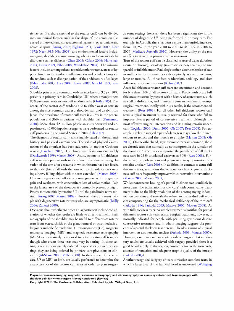
sic factors (i.e. those external to the rotator cuff ) can be dividedinto anatomical factors, such as the shape of the acromion (i.e.curved or hooked) and coracoacromial ligament, os acromiale andacromial spurs (Baring 2007; Bigliani 1991; Lewis 2009; Neer1972; Neer 1983; Nho 2008), and environmental factors includ-ing aging, shoulder overuse, smoking, obesity and some metabolicdisorders such as diabetes (Chen 2003; Galatz 2006; Harryman2003; Lewis 2009; Nho 2008; Wendelboe 2004). The intrinsicfactors include, among others, repetitive microtrauma, areas of hy-poperfusion in the tendons, inflammation and cellular changes inthe tendons such as disorganisation of the architecture of collagen(Biberthaler 2003; Levy 2008; Lewis 2009; Nirschl 1989; Rees2008).Shoulder pain is very common, with an incidence of 9.5 per 1000patients in primary care in Cambridge, UK, where amongst them85% presented with rotator cuff tendinopathy (Ostör 2005). Dis-orders of the rotator cuff tendons due to either wear or tear areamong the most common causes of shoulder pain and disability. InJapan, the prevalence of rotator cuff tears is 20.7% in the generalpopulation and 36% in patients with shoulder pain (Yamamoto2010). More than 4.5 million physician visits occurred and ap-proximately 40,000 inpatient surgeries were performed for rotatorcuff problems in the United States in 2002 (Oh 2007).The diagnosis of rotator cuff tears is mainly based on the patient’shistory and physical examination. The value of physical exami-nation of the shoulder has been addressed in another Cochranereview (Hanchard 2013). The clinical manifestations vary widely(Duckworth 1999; Matsen 2008). Acute, traumatic full thicknesscuff tears may present with sudden onset of weakness during ele-vation of the arm after a trauma in which the arm has been forcedto the side (like a fall with the arm out to the side or on catch-ing a heavy falling object with the arm extended) (Matsen 2008).Chronic degenerative cuff defects may present with progressivepain and weakness, with concomitant loss of active motion. Painin the lateral area of the shoulder is commonly present at night.Passive motion initially remains full until the pain limits active mo-tion (Baring 2007; Matsen 2008). However, there are many peo-ple with degenerative rotator tears who are asymptomatic (Reilly2006; Zanetti 2000).Decisions about whether to order a diagnostic test include consid-eration of whether the results are likely to affect treatment. Plainradiographs of the shoulder may be useful to differentiate rotatortears from osteoarthritis of the glenohumeral or acromioclavicu-lar joints and calcific tendonitis. Ultrasonography (US), magneticresonance imaging (MRI) and magnetic resonance arthrography(MRA) are increasingly being used to detect rotator cuff tears, al-though who orders these tests may vary by setting. In some set-tings, these tests are mainly ordered by specialists but in other set-tings they are being ordered by primary care physicians or clin-icians (Al-Shawi 2008; Miller 2008). In the context of specialistcare, US or MRI, or both, are usually performed to determine thecharacteristics of the rotator cuff tears in order to plan surgery.
In some settings, however, there has been a significant rise in thenumber of diagnostic US being performed in primary care. Forexample, in Australia there has been a more than fourfold increase,from 104,252 in the year 2000 to 2001 to 440,172 in 2008 to2009 (Medicare Australia 2010). However, the utility of the testto affect treatment in primary care is unknown.Tears of the rotator cuff can be classified in several ways: duration(acute or chronic), aetiology (traumatic or degenerative) or size(partial or full thickness). Radiologists often describe the size of tearin millimetres or centimetres or descriptively as small, medium,large or massive. All three factors (duration, aetiology and size)influence treatment decisions (Kuhn 2007).Acute full thickness rotator cuff tears are uncommon and accountfor less than 10% of all rotator cuff tears. People with acute fullthickness tears usually present with a history of acute trauma, suchas a fall or dislocation, and immediate pain and weakness. Promptsurgical treatment, ideally within six weeks, is the recommendedtreatment (Rees 2008). For all other full thickness rotator cufftears, surgical treatment is usually reserved for those who fail toimprove after a period of conservative treatment, although themost effective surgical intervention and its timing remain uncer-tain (Coghlan 2009; Dunn 2005; Oh 2007; Rees 2008). For ex-ample, a delay in surgical repair of a large tear may allow the injuredtendon to retract and the muscle to atrophy (Matsen 2008; Oh2007). On the other hand, asymptomatic tears are common; theseare chronic tears that normally do not compromise the function ofthe shoulder. A recent review reported the prevalence of full thick-ness tears in 2553 unselected cadavers as 30% (Rees 2008). Fur-thermore, the pathogenesis and progression to symptomatic tearsremains unclear (Rees 2008). In addition, in contrast to acute fullthickness tears, symptoms due to acute or chronic partial thick-ness cuff tears frequently improve with conservative interventions(Matava 2005; Matsen 2008).While spontaneous healing of a partial thickness tear is unlikely inmost cases, the explanation for the ‘cure’ with conservative treat-ment is due to the likely resolution of the accompanying inflam-mation over time and may also be related to the residual cuff mus-cles compensating for the mechanical deficiency of the torn cuff(Fukuda 1996; Fukuda 2003; Matava 2005; Matsen 2008). Aswith full thickness tears, no simple treatment algorithm for partialthickness rotator cuff tears exists. Surgical treatment, however, isnormally indicated for people with persisting symptoms despiteconservative treatment and in whom imaging suggests the pres-ence of a partial thickness tear or tears. The ideal timing of surgicalintervention also remains unclear (Fukuda 2003; Matava 2005).However, case series and anecdotal evidence suggest that satisfac-tory results are usually achieved with surgery provided there is agood blood supply to the tendon, contact between the torn ends,absence of retraction and adequate trophic quality of the muscle(Fukuda 2003).Another recognised category of tears is massive complete tears, inwhich a large area of the humeral head is uncovered (Wolfgang
3Magnetic resonance imaging, magnetic resonance arthrography and ultrasonography for assessing rotator cuff tears in people with
shoulder pain for whom surgery is being considered (Review)
Copyright © 2013 The Cochrane Collaboration. Published by John Wiley & Sons, Ltd.
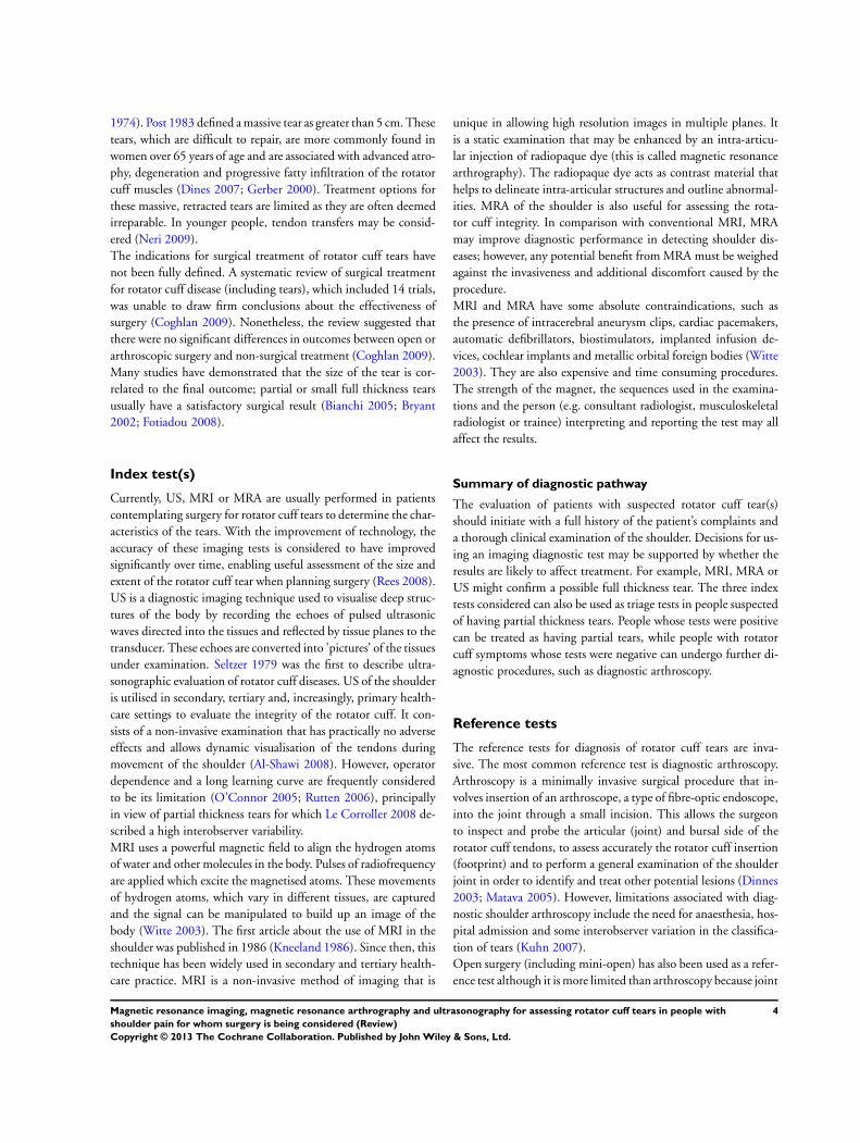
1974). Post 1983 defined a massive tear as greater than 5 cm. Thesetears, which are difficult to repair, are more commonly found inwomen over 65 years of age and are associated with advanced atro-phy, degeneration and progressive fatty infiltration of the rotatorcuff muscles (Dines 2007; Gerber 2000). Treatment options forthese massive, retracted tears are limited as they are often deemedirreparable. In younger people, tendon transfers may be consid-ered (Neri 2009).The indications for surgical treatment of rotator cuff tears havenot been fully defined. A systematic review of surgical treatmentfor rotator cuff disease (including tears), which included 14 trials,was unable to draw firm conclusions about the effectiveness ofsurgery (Coghlan 2009). Nonetheless, the review suggested thatthere were no significant differences in outcomes between open orarthroscopic surgery and non-surgical treatment (Coghlan 2009).Many studies have demonstrated that the size of the tear is cor-related to the final outcome; partial or small full thickness tearsusually have a satisfactory surgical result (Bianchi 2005; Bryant2002; Fotiadou 2008).
Index test(s)
Currently, US, MRI or MRA are usually performed in patientscontemplating surgery for rotator cuff tears to determine the char-acteristics of the tears. With the improvement of technology, theaccuracy of these imaging tests is considered to have improvedsignificantly over time, enabling useful assessment of the size andextent of the rotator cuff tear when planning surgery (Rees 2008).US is a diagnostic imaging technique used to visualise deep struc-tures of the body by recording the echoes of pulsed ultrasonicwaves directed into the tissues and reflected by tissue planes to thetransducer. These echoes are converted into ’pictures’ of the tissuesunder examination. Seltzer 1979 was the first to describe ultra-sonographic evaluation of rotator cuff diseases. US of the shoulderis utilised in secondary, tertiary and, increasingly, primary health-care settings to evaluate the integrity of the rotator cuff. It con-sists of a non-invasive examination that has practically no adverseeffects and allows dynamic visualisation of the tendons duringmovement of the shoulder (Al-Shawi 2008). However, operatordependence and a long learning curve are frequently consideredto be its limitation (O’Connor 2005; Rutten 2006), principallyin view of partial thickness tears for which Le Corroller 2008 de-scribed a high interobserver variability.MRI uses a powerful magnetic field to align the hydrogen atomsof water and other molecules in the body. Pulses of radiofrequencyare applied which excite the magnetised atoms. These movementsof hydrogen atoms, which vary in different tissues, are capturedand the signal can be manipulated to build up an image of thebody (Witte 2003). The first article about the use of MRI in theshoulder was published in 1986 (Kneeland 1986). Since then, thistechnique has been widely used in secondary and tertiary health-care practice. MRI is a non-invasive method of imaging that is
unique in allowing high resolution images in multiple planes. Itis a static examination that may be enhanced by an intra-articu-lar injection of radiopaque dye (this is called magnetic resonancearthrography). The radiopaque dye acts as contrast material thathelps to delineate intra-articular structures and outline abnormal-ities. MRA of the shoulder is also useful for assessing the rota-tor cuff integrity. In comparison with conventional MRI, MRAmay improve diagnostic performance in detecting shoulder dis-eases; however, any potential benefit from MRA must be weighedagainst the invasiveness and additional discomfort caused by theprocedure.MRI and MRA have some absolute contraindications, such asthe presence of intracerebral aneurysm clips, cardiac pacemakers,automatic defibrillators, biostimulators, implanted infusion de-vices, cochlear implants and metallic orbital foreign bodies (Witte2003). They are also expensive and time consuming procedures.The strength of the magnet, the sequences used in the examina-tions and the person (e.g. consultant radiologist, musculoskeletalradiologist or trainee) interpreting and reporting the test may allaffect the results.
Summary of diagnostic pathway
The evaluation of patients with suspected rotator cuff tear(s)should initiate with a full history of the patient’s complaints anda thorough clinical examination of the shoulder. Decisions for us-ing an imaging diagnostic test may be supported by whether theresults are likely to affect treatment. For example, MRI, MRA orUS might confirm a possible full thickness tear. The three indextests considered can also be used as triage tests in people suspectedof having partial thickness tears. People whose tests were positivecan be treated as having partial tears, while people with rotatorcuff symptoms whose tests were negative can undergo further di-agnostic procedures, such as diagnostic arthroscopy.
Reference tests
The reference tests for diagnosis of rotator cuff tears are inva-sive. The most common reference test is diagnostic arthroscopy.Arthroscopy is a minimally invasive surgical procedure that in-volves insertion of an arthroscope, a type of fibre-optic endoscope,into the joint through a small incision. This allows the surgeonto inspect and probe the articular (joint) and bursal side of therotator cuff tendons, to assess accurately the rotator cuff insertion(footprint) and to perform a general examination of the shoulderjoint in order to identify and treat other potential lesions (Dinnes2003; Matava 2005). However, limitations associated with diag-nostic shoulder arthroscopy include the need for anaesthesia, hos-pital admission and some interobserver variation in the classifica-tion of tears (Kuhn 2007).Open surgery (including mini-open) has also been used as a refer-ence test although it is more limited than arthroscopy because joint
4Magnetic resonance imaging, magnetic resonance arthrography and ultrasonography for assessing rotator cuff tears in people with
shoulder pain for whom surgery is being considered (Review)
Copyright © 2013 The Cochrane Collaboration. Published by John Wiley & Sons, Ltd.
surface or inferior surface tears are difficult to access and identifyusing an open approach. Thus open surgery is less accurate thanarthroscopy for detecting partial rotator cuff tears.
Rationale
US, MRI and MRA are increasingly being used to assess the pres-ence and size of rotator cuff tears to assist in planning surgical treat-ment. Improved techniques have resulted in increased reliance onthese tests, in place of a separate diagnostic arthroscopy, althougharthroscopic examination of the shoulder joint is still commonlyperformed as part of surgical treatment. US, MRI and MRA areoperator and reader dependent. It is not known whether any onetest is superior to either of the two others or whether performingUS and MRI or US and MRA enhances their value (Swen 1999).It is also not known whether these diagnostic tests provide use-ful additional information compared with diagnostic arthroscopy,which is an accepted part of the surgical treatment. While, theunits costs of MRI and MRA are greater than US, the cost-effec-tiveness of the three tests has not been determined.We identified two relevant systematic reviews with meta-analysesthat assessed diagnostic imaging tests for rotator cuff disease (DeJesus 2009; Dinnes 2003). The literature search in both reviewswas restricted to English language only. Dinnes 2003 evaluated thediagnostic accuracy of clinical testing of US, MRI and MRA fordetecting rotator cuff tears using both surgical and non-surgicaltests as the reference standard. The authors included 38 studiesthat assessed the accuracy of US, 29 studies that assessed the ac-curacy of MRI and 6 studies that assessed the accuracy of MRAand concluded that US or MRI were equivalent for detecting fullthickness rotator cuff tears, although MRI was more expensiveand US may be better at detecting partial tears. The search datefor the review was October 2001. A later review with a searchdate in September 2007, De Jesus 2009, conducted a meta-analy-sis comparing the diagnostic accuracy of US and MRI for rotatorcuff tears using surgery as the reference standard. This systematicreview included 65 studies but the appraisal of the methodologicalquality of the included studies was unclear or inadequate. De Jesus2009 concluded that US is as accurate as MRI for both full andpartial thickness rotator cuff tears and also suggested that US maybe the most cost-effective imaging test for detecting rotator cufftears.Important technological improvements in US, MRI and MRAhave been made since the search dates of both systematic reviews,and new studies evaluating US, MRI and MRA have been pub-lished. Our review involves an updated search for diagnostic ac-curacy studies for rotator cuff tears and will not be restricted toEnglish language publications.
O B J E C T I V E S
To compare the diagnostic test accuracy of magnetic resonanceimaging (MRI), magnetic resonance arthrography (MRA) and ul-trasonography (US) for detecting any rotator cuff tears (i.e. partialor full thickness) in people with shoulder pain for whom surgeryis being considered.
We divided our objectives as follows.
• To compare the diagnostic accuracy of US, MRI and MRAfor diagnosing any rotator cuff tears (partial or full thickness)
• To compare the diagnostic accuracy of US, MRI and MRAfor diagnosing full thickness rotator cuff tears (one or moretendons)
• To compare the diagnostic accuracy of US, MRI and MRAfor diagnosing partial thickness rotator cuff tears
Investigation of sources of heterogeneity
We planned to investigate the following potential sources of het-erogeneity:
• Type of tear: acute traumatic and chronic degenerative• Type of reference standard: open (including mini-open)
surgery or arthroscopy
M E T H O D S
Criteria for considering studies for this review
Types of studies
All diagnostic accuracy studies that compared one or more of theindex tests with one or both of the reference tests in patients sus-pected of having a partial or full thickness rotator cuff tear wereincluded. We only included results from full reports of prospectivestudies. Studies with an excessively long period of time (i.e. a yearor longer) between the index and reference tests were excludedbecause there is evidence that rotator cuff tears can progress overtime (Mall 2010; Melis 2010); however, the rate of progression isnot clearly defined.We included articles in English and languages for which a fulltranslation could be obtained. Non-English articles where a fulltranslation could not be obtained are cited in the Characteristicsof studies awaiting classification but not included in the review.For studies reported in multiple publications, we included onlythe most recent or complete report. References to the other pub-lications were cited under the same study identifier.
5Magnetic resonance imaging, magnetic resonance arthrography and ultrasonography for assessing rotator cuff tears in people with
shoulder pain for whom surgery is being considered (Review)
Copyright © 2013 The Cochrane Collaboration. Published by John Wiley & Sons, Ltd.
Participants
We included people with shoulder pain suspected of having a rota-tor cuff tear for whom surgery was being considered. Studies thatincluded healthy controls or participants who had been previouslydiagnosed with other specific shoulder pain (e.g. shoulder instabil-ity, osteoarthritis, rheumatoid arthritis, frozen shoulder, benign ormalignant tumours or referred pain) were excluded. Studies thatincluded participants with shoulder pain, but in which it was un-clear if all the participants were suspected of having rotator cufftears, were also excluded.
Index tests
Studies that assessed the accuracy of US, MRI or MRA were in-cluded.
Target conditions
We included studies that evaluated the index tests for detection ofat least one of three target conditions:
• presence of any rotator cuff tears (partial or full thickness);• presence of full thickness tears;• presence of partial thickness tears.
To standardise classification for this review, rotator cuff tears weredichotomised as absence or presence of any, full and partial thick-ness tears.
Reference standards
We required arthroscopy or open (including mini-open) surgeryfindings to be the reference standards.
Search methods for identification of studies
Electronic searches
We searched relevant computerised databases for eligible diag-nostic studies: MEDLINE (PubMed) (1966 to March 2011),EMBASE (Elsevier) (1980 to February 2011), LILACS (Bireme)(1982 to February 2011) and the Cochrane Register of DiagnosticTest Accuracy Studies (February 2011). We also searched DARE(Database of Abstracts of Reviews of Effects), the HTA Database(Health Technology Assessments Database) and the MEDIONdatabase (February 2011) for other related diagnostic test accu-racy reviews, and we checked the reference lists of those reviewsthat were relevant for additional studies. We also searched theUS Health Services Research Projects in Progress and the UKClinical Research Network Portfolio Database for ongoing andrecently completed studies. When possible, non-English articleswere assessed through translation by a native speaker.
We used a sensitive search strategy as recommended by theCochrane Collaboration for MEDLINE (PubMed), EMBASE(Elsevier) and LILACS (Bireme) (De Vet 2008). See Appendix 1for the MEDLINE and EMBASE search strategies.
Searching other resources
We checked the reference lists of articles, reviews and textbooksfor relevant primary diagnostic studies and systematic reviews. Wehandsearched abstracts of the British Elbow and Shoulder Societyannual meetings (2005 to July 2011) and American Academy ofOrthopaedic Surgeons annual meetings (2005 to July 2011). Wealso contacted experts in the field.
Data collection and analysis
We used the methods suggested in the Cochrane Handbook forSystematic Reviews of Diagnostic Test Accuracy (Deeks 2009).
Selection of studies
Two review authors (ML and RJ) independently screened the titlesand abstracts of retrieved records to identify potentially relevantstudies for inclusion. Duplicates were removed and the remain-ing references were examined. Studies which clearly did not meetthe inclusion criteria were excluded, and copies of the full text ofpotentially relevant references were obtained. ML and RJ inde-pendently assessed full text reports and determined inclusion orexclusion of the studies. Any uncertainties or disagreements wereresolved by discussion and, when necessary, by adjudication froma third author (RB).
Data extraction and management
Two review authors (ML and RJ) independently collected theavailable data using a piloted data extraction form without mask-ing of study authors or other identifying information. A third re-view author (RB) was consulted for resolution of any disagree-ments. When necessary, we sent requests to study authors for ad-ditional information or data. Diagnostic accuracy studies that re-ported insufficient data for construction of two-by-two tables wereexcluded from the review.We retrieved the following data.
1. General information: title, journal, year, publication status,country of study, period of study, primary objective and studydesign (i.e. prospective versus retrospective and consecutiveversus non-consecutive).
2. Sample size: number of participants meeting the criteriaand total number screened.
3. Baseline characteristics: baseline diagnosis, age, sex,dominant arm, nature of onset (e.g. traumatic or non-
6Magnetic resonance imaging, magnetic resonance arthrography and ultrasonography for assessing rotator cuff tears in people with
shoulder pain for whom surgery is being considered (Review)
Copyright © 2013 The Cochrane Collaboration. Published by John Wiley & Sons, Ltd.
traumatic), duration of symptoms, prior treatment, inclusionand exclusion criteria.
4. Target condition as reported.5. Index test: description of technique, criteria for positive
result, timing of test and expertise of the clinician or technicianperforming the test.
6. Reference standard test: description of technique, criteriafor positive result, time from index to reference test and expertiseof the clinician or technician performing the test.
7. Adverse effects or complications due to index test(s) andreference standard test(s).
8. Number of true positives (TP), true negatives (TN), falsepositives (FP) and false negatives (FN). We extracted data foroperational definitions for category of tear (e.g. partial, full orany thickness tears). Multiple outcome categories are oftenreported for rotator cuff tears: partial thickness tear, full thicknesstear and no tears (i.e. three-by-three tables). Currently availablemethods for evaluating diagnostic tests rely on dichotomiseddisease status. Therefore, for the assessment of each targetcondition, we dichotomised rotator cuff tears using a strategybased on the options for treatment. To create two-by-two tablesfor partial thickness tears, data for full thickness tears wereincluded with those for no tears. We did not exclude data for anycategory. We included data for partial thickness tears with thosefor full thickness tears to create two-by-two tables for any tears.
Assessment of methodological quality
The methodological quality of the included studies was assessedindependently by two review authors (ML and RJ) and disagree-ment on study quality was resolved by a third review author (RB).At the same time as data extraction, the methodological qualityof selected studies was assessed using a modified version of theQUADAS checklist (Whiting 2003), following the guidelines pro-vided in Chapter 9 of the Cochrane Handbook for SystematicReviews of Diagnostic Test Accuracy (Reitsma 2009). Appendix2 explains how we applied the QUADAS items for assessing theincluded studies.
Statistical analysis and data synthesis
Our unit of analysis was the shoulder. For each test and targetcondition, estimates of sensitivity and specificity from each studywere plotted in receiver operating characteristic (ROC) space andforest plots for visual examination of variation in test accuracy.Where adequate data were available, we conducted meta-analysesusing the bivariate model (Chu 2006; Reitsma 2005). In the bi-variate model, the logit-transformed sensitivities and specificities,and the correlation between them across studies are modelled di-rectly. The model accounts for sampling variability within studiesand also accounts for between study variability through the in-clusion of random-effects. In preliminary meta-analyses for each
target condition, we fitted the bivariate model separately for eachtest. We examined the variance of the random-effects parametersto consider the magnitude of heterogeneity and to judge whetherthere were differences in heterogeneity in sensitivities and speci-ficities between tests, before comparing the tests in a single modelfor formal assessment of comparative accuracy.Comparative accuracy studies are scarce (Takwoingi 2013). There-fore, whenever possible, we included all studies of US, MRI andMRA (i.e. an indirect comparison) in the main comparative meta-analysis for each target condition. Due to few studies of MRA andconsiderable heterogeneity in study results, we only performedpairwise comparisons of MRI and US for detection of partial thick-ness tears and any tears but compared the three tests for detectionof full thickness tears. We compared test accuracy by adding co-variate terms for test type to the parameters of the bivariate modelto determine which test was superior in terms of sensitivity orspecificity or both. The variance coefficients from the preliminarymeta-analysis and summary ROC plot for each test indicated dif-ferences in heterogeneity between tests and so we extended the bi-variate model to allow the variances of the random-effects to varywith test type. We assessed the statistical significance of the differ-ence in sensitivity or specificity between tests by using likelihoodratio tests comparing models with and without the covariate termsin the bivariate model. The summary sensitivities and specifici-ties (i.e. average operating points) were plotted on summary ROCplots with corresponding 95% confidence regions. Summary pos-itive and negative likelihood ratios were derived from functionsof the bivariate model parameters, with 95% confidence intervalscomputed using the delta method.Indirect comparisons of tests are not ideal and are susceptible tobias because other factors, such as participant and study designcharacteristics, may confound differences between tests. Thus insecondary analyses, we restricted the test comparisons to only stud-ies that evaluated the tests in the same population. Because thestudies were few, we were unable to perform meta-analyses butused linked summary ROC plots where estimates for each of thetwo tests from each study are joined by a line to illustrate the re-sults. Furthermore, for each target condition, we quantified thedifference in sensitivities and specificities between pairs of testsby computing differences in these proportions together with thecorresponding 95% CI. Thus we visually and numerically demon-strated the change and consistency of the direction of the changein test performance between the tests. We used the xtmelogit com-mand in Stata version 11.2 (StataCorp, College Station, Texas) tofit the bivariate models.
Investigations of heterogeneity
Heterogeneity was investigated in the first instance through visualexamination of forest plots and summary ROC plots. The typeof tear and type of reference standard reported in each study werepresented on forest plots along with the estimates of sensitivity and
7Magnetic resonance imaging, magnetic resonance arthrography and ultrasonography for assessing rotator cuff tears in people with
shoulder pain for whom surgery is being considered (Review)
Copyright © 2013 The Cochrane Collaboration. Published by John Wiley & Sons, Ltd.
specificity. In exploratory analyses, we ordered studies on the forestplots by each of the two covariates in turn and also by sensitivityor specificity to examine the pattern of variation between studies.If there were sufficient data we planned to formally investigateheterogeneity by adding covariates to the bivariate model for eachpotential source of heterogeneity.
Sensitivity analyses
If there were sufficient studies, we performed sensitivity analysesby comparing results based on all studies with results of subsetsof studies that complied (scored ’Yes’) with the following method-ological quality items of the QUADAS checklist (Whiting 2003).
• Representative spectrum• Acceptable reference standard• Acceptable delay between tests• Index test results blinded• Reference standard results blinded
We also investigated the effect of unit of analysis by excludingstudies that included both shoulders for any individual.
R E S U L T S
Results of the search
The search strategy identified 3169 references and the handsearchidentified an additional three records (Figure 1). Of these, 2902were excluded by initial screening of reference titles and abstracts.There were 974 duplicates and 1926 were either not relevant ordid not meet the inclusion criteria. We were unable to obtain fulltext articles for two studies because they were not available fromlibraries or vendors.
8Magnetic resonance imaging, magnetic resonance arthrography and ultrasonography for assessing rotator cuff tears in people with
shoulder pain for whom surgery is being considered (Review)
Copyright © 2013 The Cochrane Collaboration. Published by John Wiley & Sons, Ltd.
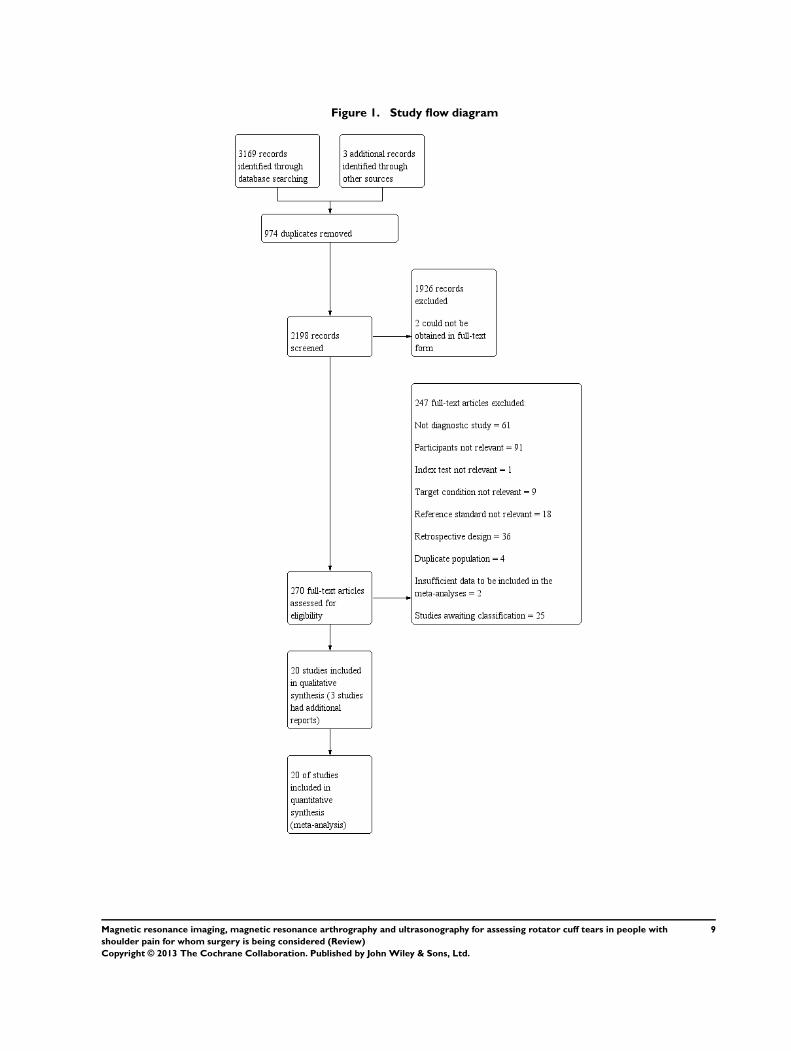
Figure 1. Study flow diagram
9Magnetic resonance imaging, magnetic resonance arthrography and ultrasonography for assessing rotator cuff tears in people with
shoulder pain for whom surgery is being considered (Review)
Copyright © 2013 The Cochrane Collaboration. Published by John Wiley & Sons, Ltd.
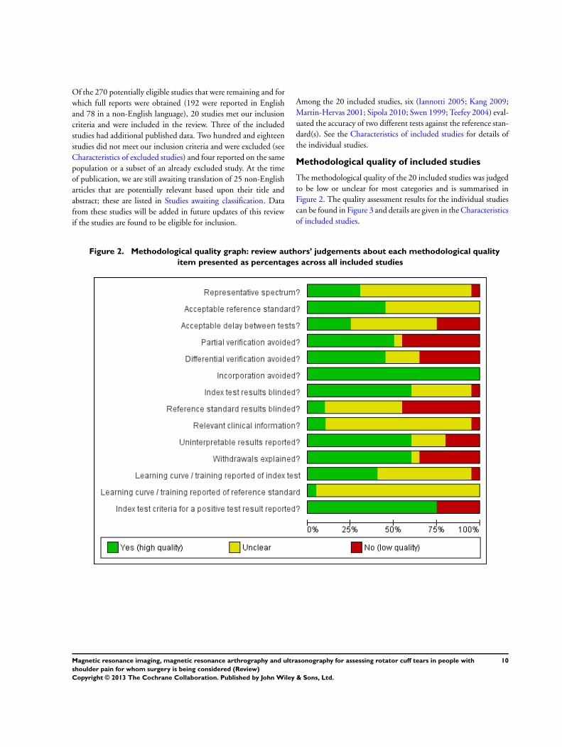
Of the 270 potentially eligible studies that were remaining and forwhich full reports were obtained (192 were reported in Englishand 78 in a non-English language), 20 studies met our inclusioncriteria and were included in the review. Three of the includedstudies had additional published data. Two hundred and eighteenstudies did not meet our inclusion criteria and were excluded (seeCharacteristics of excluded studies) and four reported on the samepopulation or a subset of an already excluded study. At the timeof publication, we are still awaiting translation of 25 non-Englisharticles that are potentially relevant based upon their title andabstract; these are listed in Studies awaiting classification. Datafrom these studies will be added in future updates of this reviewif the studies are found to be eligible for inclusion.
Among the 20 included studies, six (Iannotti 2005; Kang 2009;Martin-Hervas 2001; Sipola 2010; Swen 1999; Teefey 2004) eval-uated the accuracy of two different tests against the reference stan-dard(s). See the Characteristics of included studies for details ofthe individual studies.
Methodological quality of included studies
The methodological quality of the 20 included studies was judgedto be low or unclear for most categories and is summarised inFigure 2. The quality assessment results for the individual studiescan be found in Figure 3 and details are given in the Characteristicsof included studies.
Figure 2. Methodological quality graph: review authors’ judgements about each methodological quality
item presented as percentages across all included studies
10Magnetic resonance imaging, magnetic resonance arthrography and ultrasonography for assessing rotator cuff tears in people with
shoulder pain for whom surgery is being considered (Review)
Copyright © 2013 The Cochrane Collaboration. Published by John Wiley & Sons, Ltd.
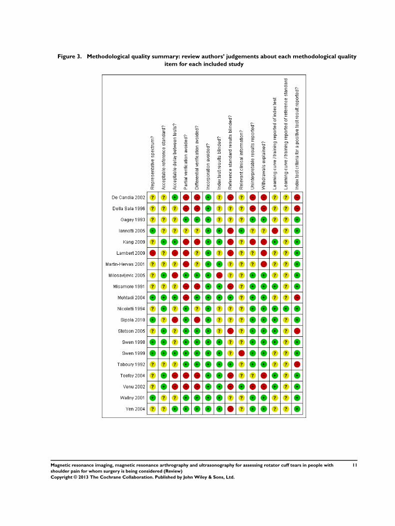
Figure 3. Methodological quality summary: review authors’ judgements about each methodological quality
item for each included study
11Magnetic resonance imaging, magnetic resonance arthrography and ultrasonography for assessing rotator cuff tears in people with
shoulder pain for whom surgery is being considered (Review)
Copyright © 2013 The Cochrane Collaboration. Published by John Wiley & Sons, Ltd.
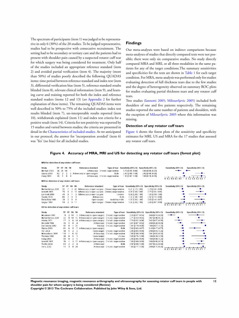
The spectrum of participants (item 1) was judged to be representa-tive in only 6 (30%) of the 20 studies. To be judged representative,studies had to be prospective with consecutive recruitment. Thesetting had to be secondary or tertiary care and the patients had topresent with shoulder pain caused by a suspected rotator cuff tearfor which surgery was being considered for treatment. Only halfof the studies included an appropriate reference standard (item2) and avoided partial verification (item 4). The majority (morethan 50%) of studies poorly described the following QUADASitems: time period between reference standard and index test (item3), differential verification bias (item 5), reference standard resultsblinded (item 8), relevant clinical information (item 9), and learn-ing curve and training reported for both the index and referencestandard readers (items 12 and 13) (see Appendix 2 for furtherexplanation of these items). The remaining QUADAS items werewell described in 50% to 75% of the included studies: index testresults blinded (item 7), un-interpretable results reported (item10), withdrawals explained (item 11) and index test criteria for apositive result (item 14). Criteria for test positivity was reported by15 studies and varied between studies; the criteria are presented indetail in the Characteristics of included studies. As we anticipatedin our protocol, the answer for ’incorporation avoided’ (item 6)was ’Yes’ (no bias) for all included studies.
Findings
Our meta-analyses were based on indirect comparisons becausemeta-analyses of studies that directly compared tests were not pos-sible; there were only six comparative studies. No study directlycompared MRA and MRI, or all three modalities in the same pa-tients for any of the target conditions.The summary sensitivitiesand specificities for the tests are shown in Table 1 for each targetcondition. For MRA, meta-analysis was performed only for studiesevaluating detection of full thickness tears due to the few studiesand the degree of heterogeneity observed on summary ROC plotsfor studies evaluating partial thickness tears and any rotator cufftears.Two studies (Iannotti 2005; Milosavljevic 2005) included bothshoulders of one and five patients respectively. The remainingstudies reported the same number of patients and shoulders, withthe exception of Milosavljevic 2005 where this information wasmissing.
Detection of any rotator cuff tears
Figure 4 shows the forest plots of the sensitivity and specificityestimates for MRI, US and MRA for the 17 studies that assessedany rotator cuff tears.
Figure 4. Accuracy of MRA, MRI and US for detecting any rotator cuff tears (forest plot)
12Magnetic resonance imaging, magnetic resonance arthrography and ultrasonography for assessing rotator cuff tears in people with
shoulder pain for whom surgery is being considered (Review)
Copyright © 2013 The Cochrane Collaboration. Published by John Wiley & Sons, Ltd.
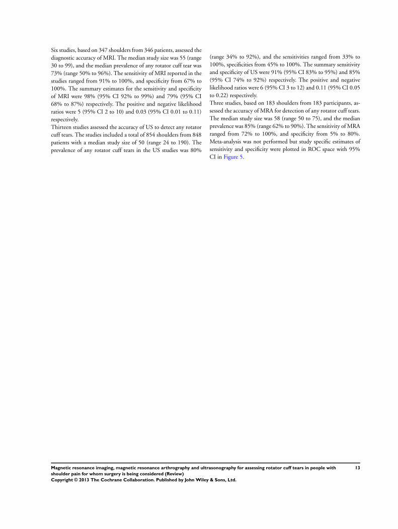
Six studies, based on 347 shoulders from 346 patients, assessed thediagnostic accuracy of MRI. The median study size was 55 (range30 to 99), and the median prevalence of any rotator cuff tear was73% (range 50% to 96%). The sensitivity of MRI reported in thestudies ranged from 91% to 100%, and specificity from 67% to100%. The summary estimates for the sensitivity and specificityof MRI were 98% (95% CI 92% to 99%) and 79% (95% CI68% to 87%) respectively. The positive and negative likelihoodratios were 5 (95% CI 2 to 10) and 0.03 (95% CI 0.01 to 0.11)respectively.Thirteen studies assessed the accuracy of US to detect any rotatorcuff tears. The studies included a total of 854 shoulders from 848patients with a median study size of 50 (range 24 to 190). Theprevalence of any rotator cuff tears in the US studies was 80%
(range 34% to 92%), and the sensitivities ranged from 33% to100%, specificities from 45% to 100%. The summary sensitivityand specificity of US were 91% (95% CI 83% to 95%) and 85%(95% CI 74% to 92%) respectively. The positive and negativelikelihood ratios were 6 (95% CI 3 to 12) and 0.11 (95% CI 0.05to 0.22) respectively.Three studies, based on 183 shoulders from 183 participants, as-sessed the accuracy of MRA for detection of any rotator cuff tears.The median study size was 58 (range 50 to 75), and the medianprevalence was 85% (range 62% to 90%). The sensitivity of MRAranged from 72% to 100%, and specificity from 5% to 80%.Meta-analysis was not performed but study specific estimates ofsensitivity and specificity were plotted in ROC space with 95%CI in Figure 5.
13Magnetic resonance imaging, magnetic resonance arthrography and ultrasonography for assessing rotator cuff tears in people with
shoulder pain for whom surgery is being considered (Review)
Copyright © 2013 The Cochrane Collaboration. Published by John Wiley & Sons, Ltd.
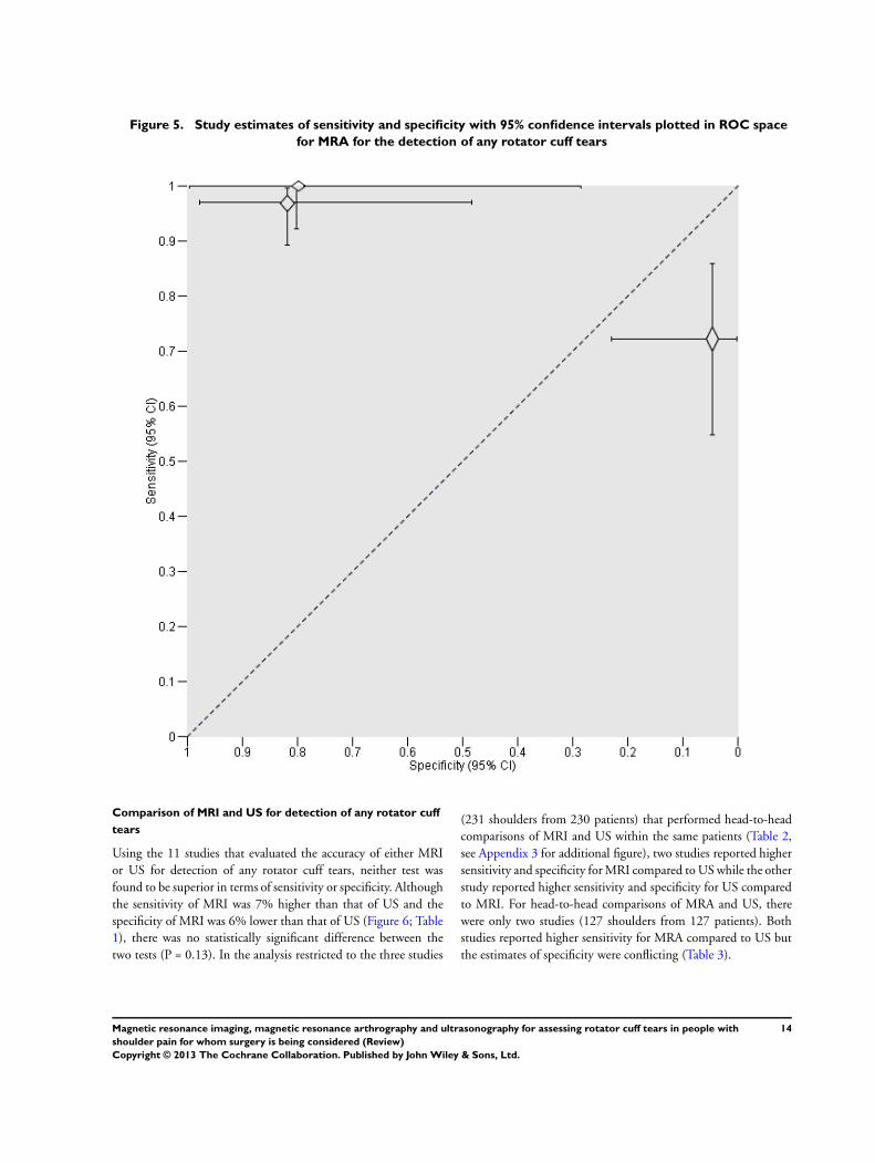
Figure 5. Study estimates of sensitivity and specificity with 95% confidence intervals plotted in ROC space
for MRA for the detection of any rotator cuff tears
Comparison of MRI and US for detection of any rotator cuff
tears
Using the 11 studies that evaluated the accuracy of either MRIor US for detection of any rotator cuff tears, neither test wasfound to be superior in terms of sensitivity or specificity. Althoughthe sensitivity of MRI was 7% higher than that of US and thespecificity of MRI was 6% lower than that of US (Figure 6; Table1), there was no statistically significant difference between thetwo tests (P = 0.13). In the analysis restricted to the three studies
(231 shoulders from 230 patients) that performed head-to-headcomparisons of MRI and US within the same patients (Table 2,see Appendix 3 for additional figure), two studies reported highersensitivity and specificity for MRI compared to US while the otherstudy reported higher sensitivity and specificity for US comparedto MRI. For head-to-head comparisons of MRA and US, therewere only two studies (127 shoulders from 127 patients). Bothstudies reported higher sensitivity for MRA compared to US butthe estimates of specificity were conflicting (Table 3).
14Magnetic resonance imaging, magnetic resonance arthrography and ultrasonography for assessing rotator cuff tears in people with
shoulder pain for whom surgery is being considered (Review)
Copyright © 2013 The Cochrane Collaboration. Published by John Wiley & Sons, Ltd.
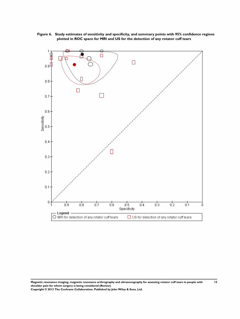
Figure 6. Study estimates of sensitivity and specificity, and summary points with 95% confidence regions
plotted in ROC space for MRI and US for the detection of any rotator cuff tears
15Magnetic resonance imaging, magnetic resonance arthrography and ultrasonography for assessing rotator cuff tears in people with
shoulder pain for whom surgery is being considered (Review)
Copyright © 2013 The Cochrane Collaboration. Published by John Wiley & Sons, Ltd.
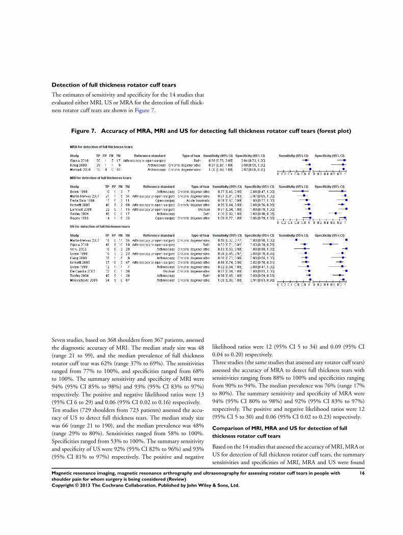
Detection of full thickness rotator cuff tears
The estimates of sensitivity and specificity for the 14 studies thatevaluated either MRI, US or MRA for the detection of full thick-ness rotator cuff tears are shown in Figure 7.
Figure 7. Accuracy of MRA, MRI and US for detecting full thickness rotator cuff tears (forest plot)
Seven studies, based on 368 shoulders from 367 patients, assessedthe diagnostic accuracy of MRI. The median study size was 48(range 21 to 99), and the median prevalence of full thicknessrotator cuff tear was 62% (range 37% to 69%). The sensitivitiesranged from 77% to 100%, and specificities ranged from 68%to 100%. The summary sensitivity and specificity of MRI were94% (95% CI 85% to 98%) and 93% (95% CI 83% to 97%)respectively. The positive and negative likelihood ratios were 13(95% CI 6 to 29) and 0.06 (95% CI 0.02 to 0.16) respectively.Ten studies (729 shoulders from 723 patients) assessed the accu-racy of US to detect full thickness tears. The median study sizewas 66 (range 21 to 190), and the median prevalence was 48%(range 29% to 80%). Sensitivities ranged from 58% to 100%.Specificities ranged from 53% to 100%. The summary sensitivityand specificity of US were 92% (95% CI 82% to 96%) and 93%(95% CI 81% to 97%) respectively. The positive and negative
likelihood ratios were 12 (95% CI 5 to 34) and 0.09 (95% CI0.04 to 0.20) respectively.Three studies (the same studies that assessed any rotator cuff tears)assessed the accuracy of MRA to detect full thickness tears withsensitivities ranging from 88% to 100% and specificities rangingfrom 90% to 94%. The median prevalence was 76% (range 17%to 80%). The summary sensitivity and specificity of MRA were94% (95% CI 80% to 98%) and 92% (95% CI 83% to 97%)respectively. The positive and negative likelihood ratios were 12(95% CI 5 to 30) and 0.06 (95% CI 0.02 to 0.23) respectively.
Comparison of MRI, MRA and US for detection of full
thickness rotator cuff tears
Based on the 14 studies that assessed the accuracy of MRI, MRA orUS for detection of full thickness rotator cuff tears, the summarysensitivities and specificities of MRI, MRA and US were found
16Magnetic resonance imaging, magnetic resonance arthrography and ultrasonography for assessing rotator cuff tears in people with
shoulder pain for whom surgery is being considered (Review)
Copyright © 2013 The Cochrane Collaboration. Published by John Wiley & Sons, Ltd.
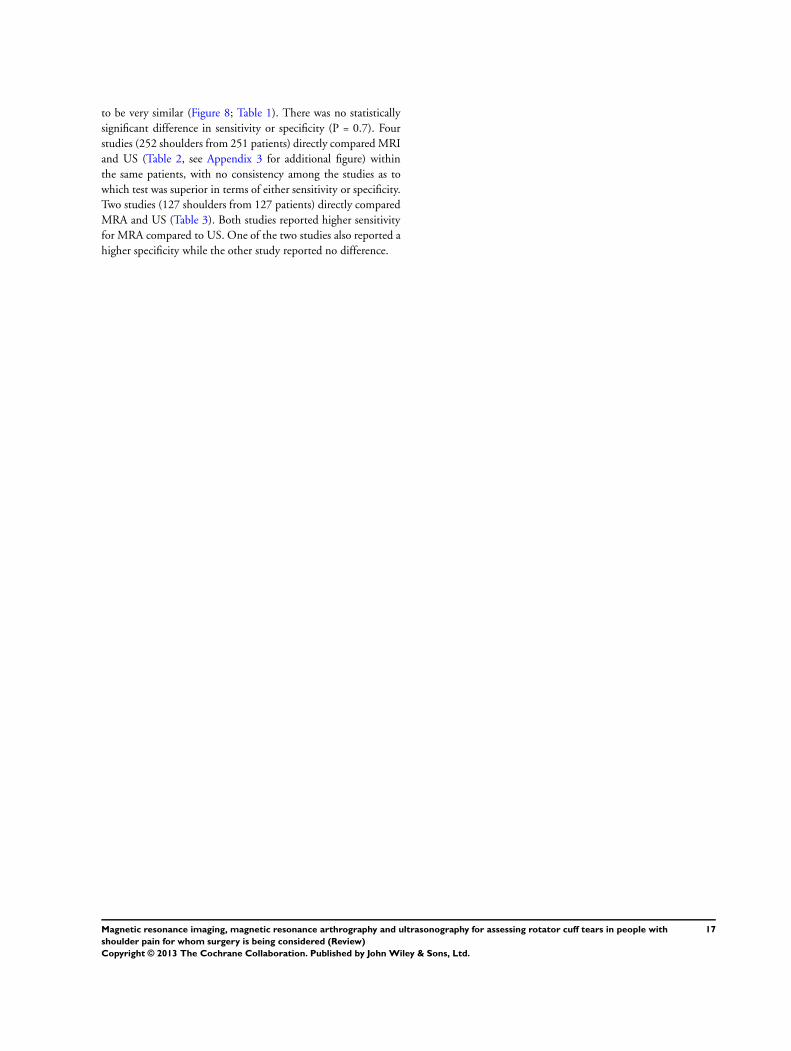
to be very similar (Figure 8; Table 1). There was no statisticallysignificant difference in sensitivity or specificity (P = 0.7). Fourstudies (252 shoulders from 251 patients) directly compared MRIand US (Table 2, see Appendix 3 for additional figure) withinthe same patients, with no consistency among the studies as towhich test was superior in terms of either sensitivity or specificity.Two studies (127 shoulders from 127 patients) directly comparedMRA and US (Table 3). Both studies reported higher sensitivityfor MRA compared to US. One of the two studies also reported ahigher specificity while the other study reported no difference.
17Magnetic resonance imaging, magnetic resonance arthrography and ultrasonography for assessing rotator cuff tears in people with
shoulder pain for whom surgery is being considered (Review)
Copyright © 2013 The Cochrane Collaboration. Published by John Wiley & Sons, Ltd.
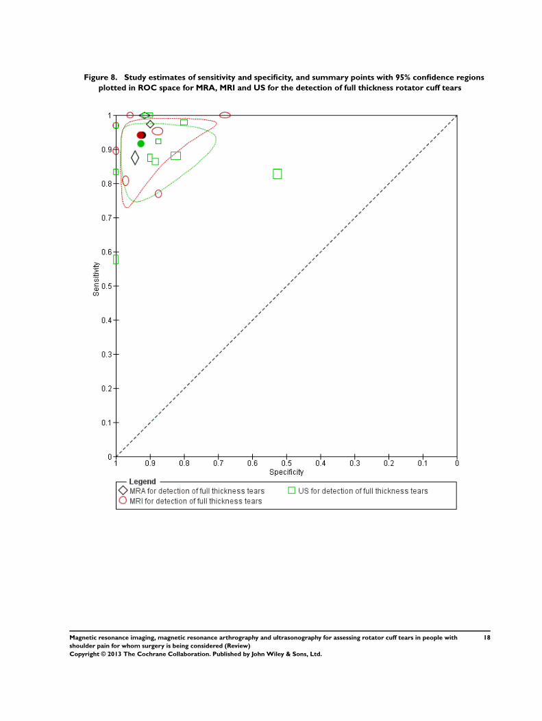
Figure 8. Study estimates of sensitivity and specificity, and summary points with 95% confidence regions
plotted in ROC space for MRA, MRI and US for the detection of full thickness rotator cuff tears
18Magnetic resonance imaging, magnetic resonance arthrography and ultrasonography for assessing rotator cuff tears in people with
shoulder pain for whom surgery is being considered (Review)
Copyright © 2013 The Cochrane Collaboration. Published by John Wiley & Sons, Ltd.
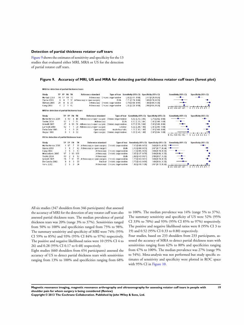
Detection of partial thickness rotator cuff tears
Figure 9 shows the estimates of sensitivity and specificity for the 13studies that evaluated either MRI, MRA or US for the detectionof partial rotator cuff tears.
Figure 9. Accuracy of MRI, US and MRA for detecting partial thickness rotator cuff tears (forest plot)
All six studies (347 shoulders from 346 participants) that assessedthe accuracy of MRI for the detection of any rotator cuff tears alsoassessed partial thickness tears. The median prevalence of partialthickness tears was 20% (range 3% to 37%). Sensitivities rangedfrom 50% to 100% and specificities ranged from 75% to 98%.The summary sensitivity and specificity of MRI were 74% (95%CI 59% to 85%) and 93% (95% CI 84% to 97%) respectively.The positive and negative likelihood ratios were 10 (95% CI 4 to26) and 0.28 (95% CI 0.17 to 0.48) respectively.Eight studies (660 shoulders from 654 participants) assessed theaccuracy of US to detect partial thickness tears with sensitivitiesranging from 13% to 100% and specificities ranging from 68%
to 100%. The median prevalence was 14% (range 5% to 37%).The summary sensitivity and specificity of US were 52% (95%CI 33% to 70%) and 93% (95% CI 85% to 97%) respectively.The positive and negative likelihood ratios were 8 (95% CI 3 to19) and 0.52 (95% CI 0.33 to 0.80) respectively.Four studies, based on 233 shoulders from 233 participants, as-sessed the accuracy of MRA to detect partial thickness tears withsensitivities ranging from 62% to 80% and specificities rangingfrom 47% to 100%. The median prevalence was 27% (range 9%to 54%). Meta-analysis was not performed but study specific es-timates of sensitivity and specificity were plotted in ROC spacewith 95% CI in Figure 10.
19Magnetic resonance imaging, magnetic resonance arthrography and ultrasonography for assessing rotator cuff tears in people with
shoulder pain for whom surgery is being considered (Review)
Copyright © 2013 The Cochrane Collaboration. Published by John Wiley & Sons, Ltd.
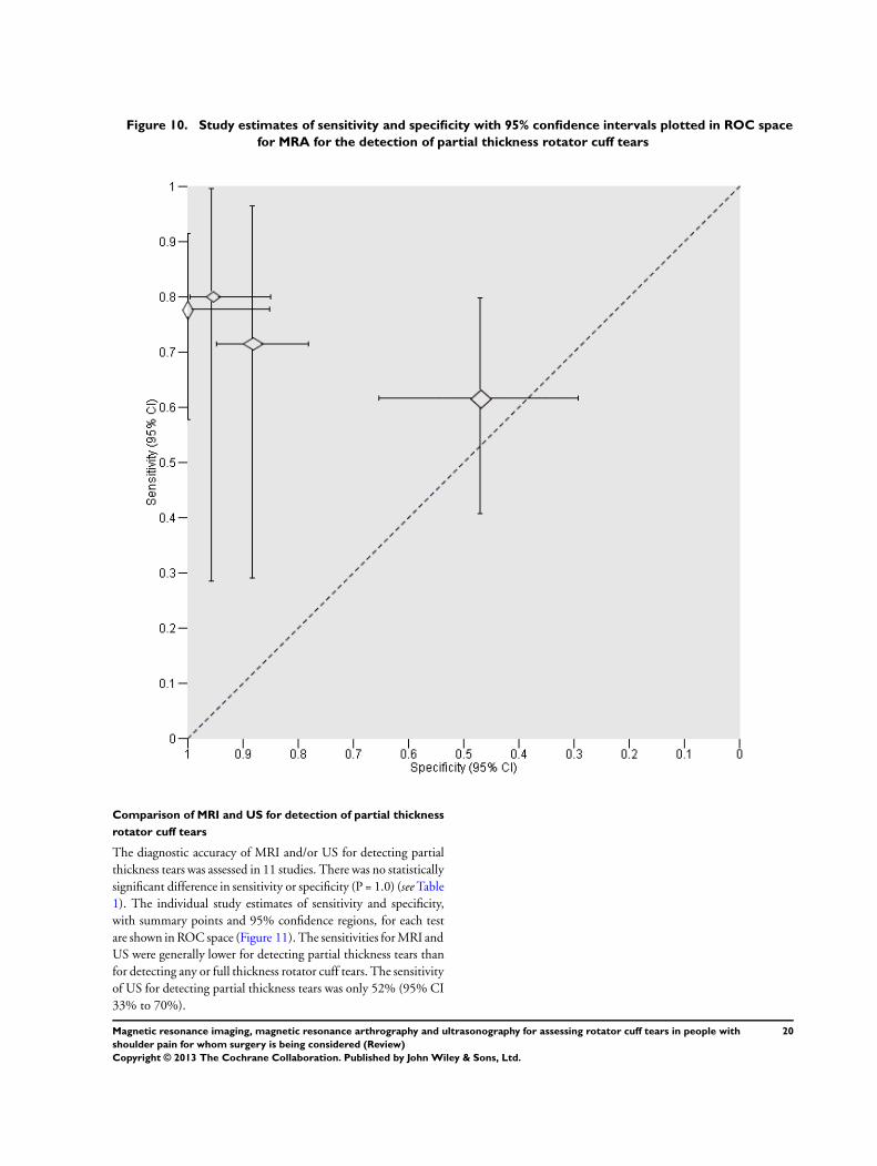
Figure 10. Study estimates of sensitivity and specificity with 95% confidence intervals plotted in ROC space
for MRA for the detection of partial thickness rotator cuff tears
Comparison of MRI and US for detection of partial thickness
rotator cuff tears
The diagnostic accuracy of MRI and/or US for detecting partialthickness tears was assessed in 11 studies. There was no statisticallysignificant difference in sensitivity or specificity (P = 1.0) (see Table1). The individual study estimates of sensitivity and specificity,with summary points and 95% confidence regions, for each testare shown in ROC space (Figure 11). The sensitivities for MRI andUS were generally lower for detecting partial thickness tears thanfor detecting any or full thickness rotator cuff tears. The sensitivityof US for detecting partial thickness tears was only 52% (95% CI33% to 70%).
20Magnetic resonance imaging, magnetic resonance arthrography and ultrasonography for assessing rotator cuff tears in people with
shoulder pain for whom surgery is being considered (Review)
Copyright © 2013 The Cochrane Collaboration. Published by John Wiley & Sons, Ltd.
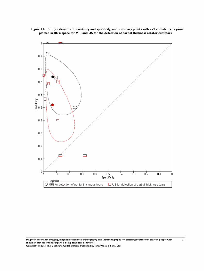
Figure 11. Study estimates of sensitivity and specificity, and summary points with 95% confidence regions
plotted in ROC space for MRI and US for the detection of partial thickness rotator cuff tears
21Magnetic resonance imaging, magnetic resonance arthrography and ultrasonography for assessing rotator cuff tears in people with
shoulder pain for whom surgery is being considered (Review)
Copyright © 2013 The Cochrane Collaboration. Published by John Wiley & Sons, Ltd.
The sensitivities and specificities of the three studies that directlycompared MRI and US are shown in a ROC space (see Appendix3 for figure) and differences between the sensitivities and speci-ficities of the tests are presented for each study in Table 2. Twoof the studies reported better sensitivity for MRI than US whileall three studies reported better specificity for MRI compared toUS. Two studies directly compared MRA and US for detectionof any rotator cuff tears. Both studies reported better sensitivityand specificity for MRI compared with those of US (Table 3). Thesame studies also assessed partial thickness tears.
Detection of any subscapularis tendon tears
One study, Mohtadi 2004, assessed the accuracy of MRA for de-tection of any subscapularis tendon tears, and included 58 shoul-ders from 58 participants. The study had a prevalence of 33% forsubscapularis tendon tears. The sensitivity and specificity of MRAwere 79% (95% CI 54% to 94%) and 72% (95% CI 55% to85%) respectively.
Investigation of heterogeneity
The type of tear and the reference standard used in each studyare shown by forest plots for each target condition in Figure 4,Figure 7 and Figure 9. The studies on each plot were ordered ac-cording to sensitivity and specificity to demonstrate any patternin the observed estimates of test accuracy. Based on these descrip-tive analyses and the magnitude of the variances of the random-effects parameters, we observed greater variability in sensitivityand specificity across studies of US than across studies of MRI or
MRA. We were unable to formally investigate potential sources ofheterogeneity because the number of studies available for each testwas either inadequate or the same value of a covariate was reportedby most studies.
Sensitivity analyses
There were few studies of MRI and MRA, and so we could not per-form sensitivity analyses for these tests. We performed sensitivityanalyses for US for each of the target conditions. We were only ableto investigate the impact of two (acceptable reference standard andindex test results blinded) of the five quality items we had specifiedbecause few studies scored ’Yes’ on the other three items (represen-tative spectrum, acceptable delay between tests, and reference testresults blinded). There were small differences in sensitivity and/or specificity (Appendix 4). The largest difference was observedbetween the summary sensitivity of US for detecting partial thick-ness tears based of all studies (52%, 95% CI 33% to 70%) and thesummary sensitivity (62%, 45% to 77%) based on only studieswhere the reference standard was acceptable. However, the confi-dence intervals were comparable and the specificities were similar.The exclusion of studies that did not meet either criteria made nodifference to our findings. Two studies included both shouldersfor six participants and one study did not report the number ofparticipants so it is unclear whether more than one shoulder wasincluded per participant. We investigated the impact of the unitof analysis on the findings for MRI and US by excluding the threestudies, thus assuming the individual as the unit of analysis; theresults were found to be consistent with the main analyses basedon shoulders.
22Magnetic resonance imaging, magnetic resonance arthrography and ultrasonography for assessing rotator cuff tears in people with
shoulder pain for whom surgery is being considered (Review)
Copyright © 2013 The Cochrane Collaboration. Published by John Wiley & Sons, Ltd.
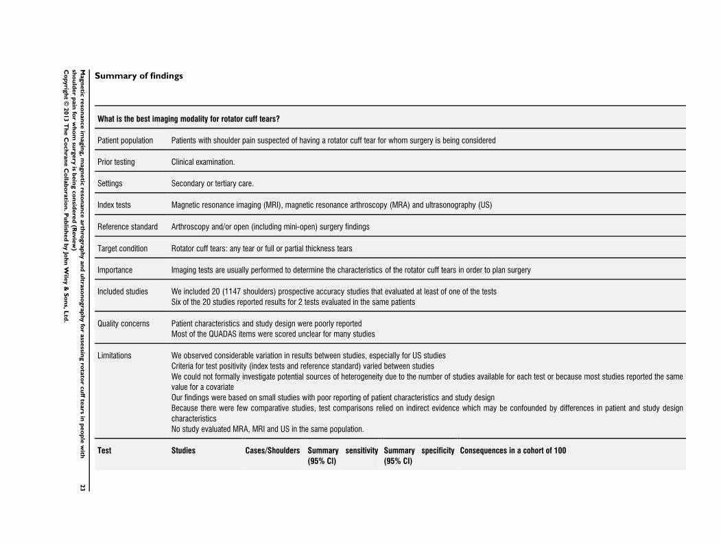
Summary of findings
What is the best imaging modality for rotator cuff tears?
Patient population Patients with shoulder pain suspected of having a rotator cuff tear for whom surgery is being considered
Prior testing Clinical examination.
Settings Secondary or tertiary care.
Index tests Magnetic resonance imaging (MRI), magnetic resonance arthroscopy (MRA) and ultrasonography (US)
Reference standard Arthroscopy and/or open (including mini-open) surgery findings
Target condition Rotator cuff tears: any tear or full or partial thickness tears
Importance Imaging tests are usually performed to determine the characteristics of the rotator cuff tears in order to plan surgery
Included studies We included 20 (1147 shoulders) prospective accuracy studies that evaluated at least of one of the tests
Six of the 20 studies reported results for 2 tests evaluated in the same patients
Quality concerns Patient characteristics and study design were poorly reported
Most of the QUADAS items were scored unclear for many studies
Limitations We observed considerable variation in results between studies, especially for US studies
Criteria for test positivity (index tests and reference standard) varied between studies
We could not formally investigate potential sources of heterogeneity due to the number of studies available for each test or because most studies reported the same
value for a covariate
Our findings were based on small studies with poor reporting of patient characteristics and study design
Because there were few comparative studies, test comparisons relied on indirect evidence which may be confounded by differences in patient and study design
characteristics
No study evaluated MRA, MRI and US in the same population.
Test Studies Cases/Shoulders Summary sensitivity
(95% CI)
Summary specificity
(95% CI)
Consequences in a cohort of 100
23
Magn
etic
reso
nan
ce
imagin
g,m
agn
etic
reso
nan
ce
arth
rogra
phy
an
du
ltraso
no
gra
phy
for
asse
ssing
rota
tor
cu
ffte
ars
inp
eo
ple
with
sho
uld
er
pain
for
wh
om
surg
ery
isb
ein
gco
nsid
ere
d(R
evie
w)
Co
pyrig
ht
©2013
Th
eC
och
ran
eC
olla
bo
ratio
n.P
ub
lished
by
Joh
nW
iley
&S
on
s,L
td.
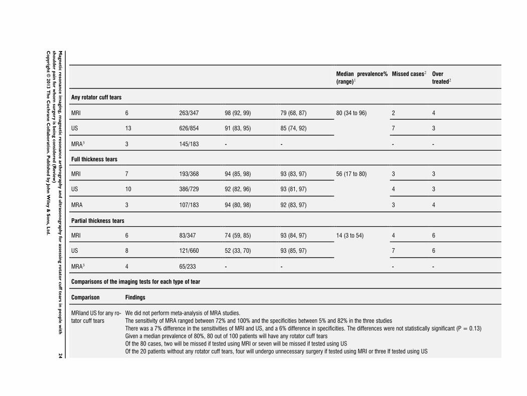
Median prevalence%
(range)1Missed cases2 Over
treated2
Any rotator cuff tears
MRI 6 263/347 98 (92, 99) 79 (68, 87) 80 (34 to 96) 2 4
US 13 626/854 91 (83, 95) 85 (74, 92) 7 3
MRA3 3 145/183 - - - -
Full thickness tears
MRI 7 193/368 94 (85, 98) 93 (83, 97) 56 (17 to 80) 3 3
US 10 386/729 92 (82, 96) 93 (81, 97) 4 3
MRA 3 107/183 94 (80, 98) 92 (83, 97) 3 4
Partial thickness tears
MRI 6 83/347 74 (59, 85) 93 (84, 97) 14 (3 to 54) 4 6
US 8 121/660 52 (33, 70) 93 (85, 97) 7 6
MRA3 4 65/233 - - - -
Comparisons of the imaging tests for each type of tear
Comparison Findings
MRIand US for any ro-
tator cuff tears
We did not perform meta-analysis of MRA studies.
The sensitivity of MRA ranged between 72% and 100% and the specificities between 5% and 82% in the three studies
There was a 7% difference in the sensitivities of MRI and US, and a 6% difference in specificities. The differences were not statistically significant (P = 0.13)
Given a median prevalence of 80%, 80 out of 100 patients will have any rotator cuff tears
Of the 80 cases, two will be missed if tested using MRI or seven will be missed if tested using US
Of the 20 patients without any rotator cuff tears, four will undergo unnecessary surgery if tested using MRI or three If tested using US24
Magn
etic
reso
nan
ce
imagin
g,m
agn
etic
reso
nan
ce
arth
rogra
phy
an
du
ltraso
no
gra
phy
for
asse
ssing
rota
tor
cu
ffte
ars
inp
eo
ple
with
sho
uld
er
pain
for
wh
om
surg
ery
isb
ein
gco
nsid
ere
d(R
evie
w)
Co
pyrig
ht
©2013
Th
eC
och
ran
eC
olla
bo
ratio
n.P
ub
lished
by
Joh
nW
iley
&S
on
s,L
td.
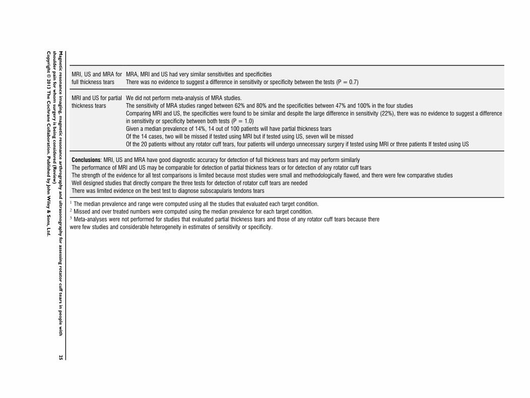
MRI, US and MRA for
full thickness tears
MRA, MRI and US had very similar sensitivities and specificities
There was no evidence to suggest a difference in sensitivity or specificity between the tests (P = 0.7)
MRI and US for partial
thickness tears
We did not perform meta-analysis of MRA studies.
The sensitivity of MRA studies ranged between 62% and 80% and the specificities between 47% and 100% in the four studies
Comparing MRI and US, the specificities were found to be similar and despite the large difference in sensitivity (22%), there was no evidence to suggest a difference
in sensitivity or specificity between both tests (P = 1.0)
Given a median prevalence of 14%, 14 out of 100 patients will have partial thickness tears
Of the 14 cases, two will be missed if tested using MRI but if tested using US, seven will be missed
Of the 20 patients without any rotator cuff tears, four patients will undergo unnecessary surgery if tested using MRI or three patients If tested using US
Conclusions: MRI, US and MRA have good diagnostic accuracy for detection of full thickness tears and may perform similarly
The performance of MRI and US may be comparable for detection of partial thickness tears or for detection of any rotator cuff tears
The strength of the evidence for all test comparisons is limited because most studies were small and methodologically flawed, and there were few comparative studies
Well designed studies that directly compare the three tests for detection of rotator cuff tears are needed
There was limited evidence on the best test to diagnose subscapularis tendons tears
1 The median prevalence and range were computed using all the studies that evaluated each target condition.2 Missed and over treated numbers were computed using the median prevalence for each target condition.3 Meta-analyses were not performed for studies that evaluated partial thickness tears and those of any rotator cuff tears because there
were few studies and considerable heterogeneity in estimates of sensitivity or specificity.
25
Magn
etic
reso
nan
ce
imagin
g,m
agn
etic
reso
nan
ce
arth
rogra
phy
an
du
ltraso
no
gra
phy
for
asse
ssing
rota
tor
cu
ffte
ars
inp
eo
ple
with
sho
uld
er
pain
for
wh
om
surg
ery
isb
ein
gco
nsid
ere
d(R
evie
w)
Co
pyrig
ht
©2013
Th
eC
och
ran
eC
olla
bo
ratio
n.P
ub
lished
by
Joh
nW
iley
&S
on
s,L
td.
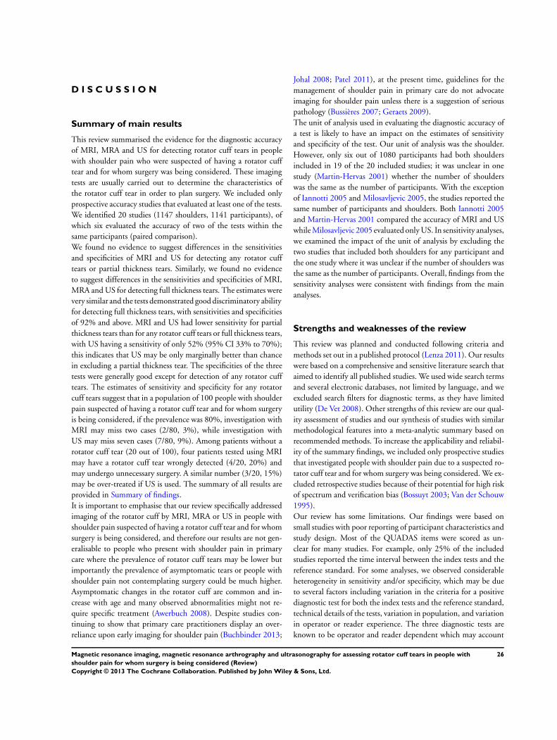
D I S C U S S I O N
Summary of main results
This review summarised the evidence for the diagnostic accuracyof MRI, MRA and US for detecting rotator cuff tears in peoplewith shoulder pain who were suspected of having a rotator cufftear and for whom surgery was being considered. These imagingtests are usually carried out to determine the characteristics ofthe rotator cuff tear in order to plan surgery. We included onlyprospective accuracy studies that evaluated at least one of the tests.We identified 20 studies (1147 shoulders, 1141 participants), ofwhich six evaluated the accuracy of two of the tests within thesame participants (paired comparison).We found no evidence to suggest differences in the sensitivitiesand specificities of MRI and US for detecting any rotator cufftears or partial thickness tears. Similarly, we found no evidenceto suggest differences in the sensitivities and specificities of MRI,MRA and US for detecting full thickness tears. The estimates werevery similar and the tests demonstrated good discriminatory abilityfor detecting full thickness tears, with sensitivities and specificitiesof 92% and above. MRI and US had lower sensitivity for partialthickness tears than for any rotator cuff tears or full thickness tears,with US having a sensitivity of only 52% (95% CI 33% to 70%);this indicates that US may be only marginally better than chancein excluding a partial thickness tear. The specificities of the threetests were generally good except for detection of any rotator cufftears. The estimates of sensitivity and specificity for any rotatorcuff tears suggest that in a population of 100 people with shoulderpain suspected of having a rotator cuff tear and for whom surgeryis being considered, if the prevalence was 80%, investigation withMRI may miss two cases (2/80, 3%), while investigation withUS may miss seven cases (7/80, 9%). Among patients without arotator cuff tear (20 out of 100), four patients tested using MRImay have a rotator cuff tear wrongly detected (4/20, 20%) andmay undergo unnecessary surgery. A similar number (3/20, 15%)may be over-treated if US is used. The summary of all results areprovided in Summary of findings.It is important to emphasise that our review specifically addressedimaging of the rotator cuff by MRI, MRA or US in people withshoulder pain suspected of having a rotator cuff tear and for whomsurgery is being considered, and therefore our results are not gen-eralisable to people who present with shoulder pain in primarycare where the prevalence of rotator cuff tears may be lower butimportantly the prevalence of asymptomatic tears or people withshoulder pain not contemplating surgery could be much higher.Asymptomatic changes in the rotator cuff are common and in-crease with age and many observed abnormalities might not re-quire specific treatment (Awerbuch 2008). Despite studies con-tinuing to show that primary care practitioners display an over-reliance upon early imaging for shoulder pain (Buchbinder 2013;
Johal 2008; Patel 2011), at the present time, guidelines for themanagement of shoulder pain in primary care do not advocateimaging for shoulder pain unless there is a suggestion of seriouspathology (Bussières 2007; Geraets 2009).The unit of analysis used in evaluating the diagnostic accuracy ofa test is likely to have an impact on the estimates of sensitivityand specificity of the test. Our unit of analysis was the shoulder.However, only six out of 1080 participants had both shouldersincluded in 19 of the 20 included studies; it was unclear in onestudy (Martin-Hervas 2001) whether the number of shoulderswas the same as the number of participants. With the exceptionof Iannotti 2005 and Milosavljevic 2005, the studies reported thesame number of participants and shoulders. Both Iannotti 2005and Martin-Hervas 2001 compared the accuracy of MRI and USwhile Milosavljevic 2005 evaluated only US. In sensitivity analyses,we examined the impact of the unit of analysis by excluding thetwo studies that included both shoulders for any participant andthe one study where it was unclear if the number of shoulders wasthe same as the number of participants. Overall, findings from thesensitivity analyses were consistent with findings from the mainanalyses.
Strengths and weaknesses of the review
This review was planned and conducted following criteria andmethods set out in a published protocol (Lenza 2011). Our resultswere based on a comprehensive and sensitive literature search thataimed to identify all published studies. We used wide search termsand several electronic databases, not limited by language, and weexcluded search filters for diagnostic terms, as they have limitedutility (De Vet 2008). Other strengths of this review are our qual-ity assessment of studies and our synthesis of studies with similarmethodological features into a meta-analytic summary based onrecommended methods. To increase the applicability and reliabil-ity of the summary findings, we included only prospective studiesthat investigated people with shoulder pain due to a suspected ro-tator cuff tear and for whom surgery was being considered. We ex-cluded retrospective studies because of their potential for high riskof spectrum and verification bias (Bossuyt 2003; Van der Schouw1995).Our review has some limitations. Our findings were based onsmall studies with poor reporting of participant characteristics andstudy design. Most of the QUADAS items were scored as un-clear for many studies. For example, only 25% of the includedstudies reported the time interval between the index tests and thereference standard. For some analyses, we observed considerableheterogeneity in sensitivity and/or specificity, which may be dueto several factors including variation in the criteria for a positivediagnostic test for both the index tests and the reference standard,technical details of the tests, variation in population, and variationin operator or reader experience. The three diagnostic tests areknown to be operator and reader dependent which may account
26Magnetic resonance imaging, magnetic resonance arthrography and ultrasonography for assessing rotator cuff tears in people with
shoulder pain for whom surgery is being considered (Review)
Copyright © 2013 The Cochrane Collaboration. Published by John Wiley & Sons, Ltd.
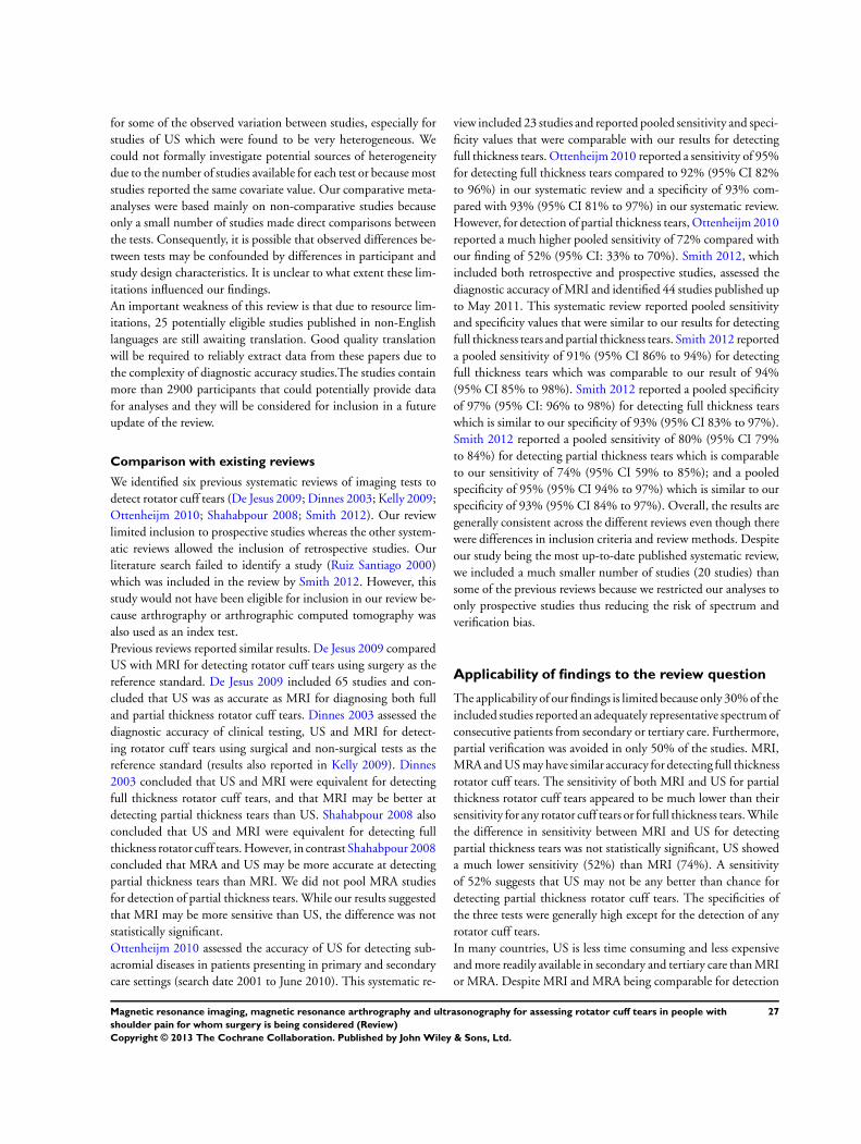
for some of the observed variation between studies, especially forstudies of US which were found to be very heterogeneous. Wecould not formally investigate potential sources of heterogeneitydue to the number of studies available for each test or because moststudies reported the same covariate value. Our comparative meta-analyses were based mainly on non-comparative studies becauseonly a small number of studies made direct comparisons betweenthe tests. Consequently, it is possible that observed differences be-tween tests may be confounded by differences in participant andstudy design characteristics. It is unclear to what extent these lim-itations influenced our findings.An important weakness of this review is that due to resource lim-itations, 25 potentially eligible studies published in non-Englishlanguages are still awaiting translation. Good quality translationwill be required to reliably extract data from these papers due tothe complexity of diagnostic accuracy studies.The studies containmore than 2900 participants that could potentially provide datafor analyses and they will be considered for inclusion in a futureupdate of the review.
Comparison with existing reviews
We identified six previous systematic reviews of imaging tests todetect rotator cuff tears (De Jesus 2009; Dinnes 2003; Kelly 2009;Ottenheijm 2010; Shahabpour 2008; Smith 2012). Our reviewlimited inclusion to prospective studies whereas the other system-atic reviews allowed the inclusion of retrospective studies. Ourliterature search failed to identify a study (Ruiz Santiago 2000)which was included in the review by Smith 2012. However, thisstudy would not have been eligible for inclusion in our review be-cause arthrography or arthrographic computed tomography wasalso used as an index test.Previous reviews reported similar results. De Jesus 2009 comparedUS with MRI for detecting rotator cuff tears using surgery as thereference standard. De Jesus 2009 included 65 studies and con-cluded that US was as accurate as MRI for diagnosing both fulland partial thickness rotator cuff tears. Dinnes 2003 assessed thediagnostic accuracy of clinical testing, US and MRI for detect-ing rotator cuff tears using surgical and non-surgical tests as thereference standard (results also reported in Kelly 2009). Dinnes2003 concluded that US and MRI were equivalent for detectingfull thickness rotator cuff tears, and that MRI may be better atdetecting partial thickness tears than US. Shahabpour 2008 alsoconcluded that US and MRI were equivalent for detecting fullthickness rotator cuff tears. However, in contrast Shahabpour 2008concluded that MRA and US may be more accurate at detectingpartial thickness tears than MRI. We did not pool MRA studiesfor detection of partial thickness tears. While our results suggestedthat MRI may be more sensitive than US, the difference was notstatistically significant.Ottenheijm 2010 assessed the accuracy of US for detecting sub-acromial diseases in patients presenting in primary and secondarycare settings (search date 2001 to June 2010). This systematic re-
view included 23 studies and reported pooled sensitivity and speci-ficity values that were comparable with our results for detectingfull thickness tears. Ottenheijm 2010 reported a sensitivity of 95%for detecting full thickness tears compared to 92% (95% CI 82%to 96%) in our systematic review and a specificity of 93% com-pared with 93% (95% CI 81% to 97%) in our systematic review.However, for detection of partial thickness tears, Ottenheijm 2010reported a much higher pooled sensitivity of 72% compared withour finding of 52% (95% CI: 33% to 70%). Smith 2012, whichincluded both retrospective and prospective studies, assessed thediagnostic accuracy of MRI and identified 44 studies published upto May 2011. This systematic review reported pooled sensitivityand specificity values that were similar to our results for detectingfull thickness tears and partial thickness tears. Smith 2012 reporteda pooled sensitivity of 91% (95% CI 86% to 94%) for detectingfull thickness tears which was comparable to our result of 94%(95% CI 85% to 98%). Smith 2012 reported a pooled specificityof 97% (95% CI: 96% to 98%) for detecting full thickness tearswhich is similar to our specificity of 93% (95% CI 83% to 97%).Smith 2012 reported a pooled sensitivity of 80% (95% CI 79%to 84%) for detecting partial thickness tears which is comparableto our sensitivity of 74% (95% CI 59% to 85%); and a pooledspecificity of 95% (95% CI 94% to 97%) which is similar to ourspecificity of 93% (95% CI 84% to 97%). Overall, the results aregenerally consistent across the different reviews even though therewere differences in inclusion criteria and review methods. Despiteour study being the most up-to-date published systematic review,we included a much smaller number of studies (20 studies) thansome of the previous reviews because we restricted our analyses toonly prospective studies thus reducing the risk of spectrum andverification bias.
Applicability of findings to the review question
The applicability of our findings is limited because only 30% of theincluded studies reported an adequately representative spectrum ofconsecutive patients from secondary or tertiary care. Furthermore,partial verification was avoided in only 50% of the studies. MRI,MRA and US may have similar accuracy for detecting full thicknessrotator cuff tears. The sensitivity of both MRI and US for partialthickness rotator cuff tears appeared to be much lower than theirsensitivity for any rotator cuff tears or for full thickness tears. Whilethe difference in sensitivity between MRI and US for detectingpartial thickness tears was not statistically significant, US showeda much lower sensitivity (52%) than MRI (74%). A sensitivityof 52% suggests that US may not be any better than chance fordetecting partial thickness rotator cuff tears. The specificities ofthe three tests were generally high except for the detection of anyrotator cuff tears.In many countries, US is less time consuming and less expensiveand more readily available in secondary and tertiary care than MRIor MRA. Despite MRI and MRA being comparable for detection
27Magnetic resonance imaging, magnetic resonance arthrography and ultrasonography for assessing rotator cuff tears in people with
shoulder pain for whom surgery is being considered (Review)
Copyright © 2013 The Cochrane Collaboration. Published by John Wiley & Sons, Ltd.
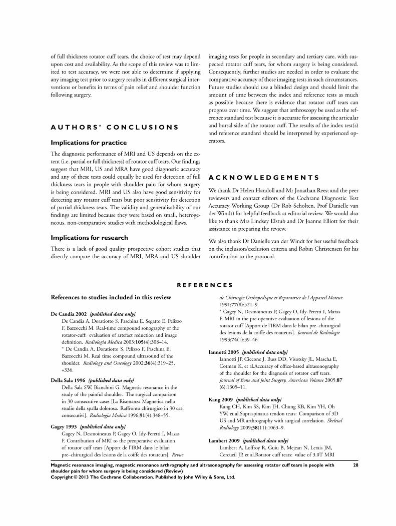
of full thickness rotator cuff tears, the choice of test may dependupon cost and availability. As the scope of this review was to lim-ited to test accuracy, we were not able to determine if applyingany imaging test prior to surgery results in different surgical inter-ventions or benefits in terms of pain relief and shoulder functionfollowing surgery.
A U T H O R S ’ C O N C L U S I O N S
Implications for practice
The diagnostic performance of MRI and US depends on the ex-tent (i.e. partial or full thickness) of rotator cuff tears. Our findingssuggest that MRI, US and MRA have good diagnostic accuracyand any of these tests could equally be used for detection of fullthickness tears in people with shoulder pain for whom surgeryis being considered. MRI and US also have good sensitivity fordetecting any rotator cuff tears but poor sensitivity for detectionof partial thickness tears. The validity and generalisability of ourfindings are limited because they were based on small, heteroge-neous, non-comparative studies with methodological flaws.
Implications for research
There is a lack of good quality prospective cohort studies thatdirectly compare the accuracy of MRI, MRA and US shoulder
imaging tests for people in secondary and tertiary care, with sus-pected rotator cuff tears, for whom surgery is being considered.Consequently, further studies are needed in order to evaluate thecomparative accuracy of these imaging tests in such circumstances.Future studies should use a blinded design and should limit theamount of time between the index and reference tests as muchas possible because there is evidence that rotator cuff tears canprogress over time. We suggest that arthroscopy be used as the ref-erence standard test because it is accurate for assessing the articularand bursal side of the rotator cuff. The results of the index test(s)and reference standard should be interpreted by experienced op-erators.
A C K N O W L E D G E M E N T S
We thank Dr Helen Handoll and Mr Jonathan Rees; and the peerreviewers and contact editors of the Cochrane Diagnostic TestAccuracy Working Group (Dr Rob Scholten, Prof Danielle vander Windt) for helpful feedback at editorial review. We would alsolike to thank Mrs Lindsey Elstub and Dr Joanne Elliott for theirassistance in preparing the review.
We also thank Dr Danielle van der Windt for her useful feedbackon the inclusion/exclusion criteria and Robin Christensen for hiscontribution to the protocol.
R E F E R E N C E S
References to studies included in this review
De Candia 2002 {published data only}
De Candia A, Doratiotto S, Paschina E, Segatto E, PelizzoF, Bazzocchi M. Real-time compound sonography of therotator-cuff: evaluation of artefact reduction and imagedefinition. Radiologia Medica 2003;105(4):308–14.∗ De Candia A, Doratiotto S, Pelizzo F, Paschina E,Bazzocchi M. Real time compound ultrasound of theshoulder. Radiology and Oncology 2002;36(4):319–25,+336.
Della Sala 1996 {published data only}
Della Sala SW, Bianchini G. Magnetic resonance in thestudy of the painful shoulder. The surgical comparisonin 30 consecutive cases [La Risonanza Magnetica nellostudio della spalla dolorosa. Raffronto chirurgico in 30 casiconsecutivi]. Radiologia Medica 1996;91(4):348–55.
Gagey 1993 {published data only}
Gagey N, Desmoineaux P, Gagey O, Idy-Peretti I, MazasF. Contribution of MRI to the preoperative evaluationof rotator cuff tears [Apport de l’IRM dans le bilanpre–chirurgical des lesions de la coiffe des rotateurs]. Revue
de Chirurgie Orthopedique et Reparatrice de l Appareil Moteur1991;77(8):521–9.∗ Gagey N, Desmoineaux P, Gagey O, Idy-Peretti I, MazasF. MRI in the pre-operative evaluation of lesions of therotator cuff [Apport de l’IRM dans le bilan pre–chirurgicaldes lesions de la coiffe des rotateurs]. Journal de Radiologie
1993;74(1):39–46.
Iannotti 2005 {published data only}
Iannotti JP, Ciccone J, Buss DD, Visotsky JL, Mascha E,Cotman K, et al.Accuracy of office-based ultrasonographyof the shoulder for the diagnosis of rotator cuff tears.Journal of Bone and Joint Surgery. American Volume 2005;87
(6):1305–11.
Kang 2009 {published data only}
Kang CH, Kim SS, Kim JH, Chung KB, Kim YH, OhYW, et al.Supraspinatus tendon tears: Comparison of 3DUS and MR arthrography with surgical correlation. Skeletal
Radiology 2009;38(11):1063–9.
Lambert 2009 {published data only}
Lambert A, Loffroy R, Guiu B, Mejean N, Lerais JM,Cercueil JP, et al.Rotator cuff tears: value of 3.0T MRI
28Magnetic resonance imaging, magnetic resonance arthrography and ultrasonography for assessing rotator cuff tears in people with
shoulder pain for whom surgery is being considered (Review)
Copyright © 2013 The Cochrane Collaboration. Published by John Wiley & Sons, Ltd.
[Perforations de la coiffe des rotateurs: intérêt de l’IRM 3,0T]. Journal de Radiologie 2009;90(5 Pt 1):583–8.
Martin-Hervas 2001 {published data only}
Martin-Hervas C, Romero J, Navas-Acien A, Reboiras JJ,Munuera L. Ultrasonographic and magnetic resonanceimages of rotator cuff lesions compared with arthroscopyor open surgery findings. Journal of Shoulder and ElbowSurgery 2001;10(5):410–5.
Milosavljevic 2005 {published data only}
Milosavljevic J, Elvin A, Rahme H. Ultrasonography ofthe rotator cuff: a comparison with arthroscopy in one-hundred-and-ninety consecutive cases. Acta Radiologica2005;46(8):858–65.
Misamore 1991 {published data only}
Misamore GW, Woodward C. Evaluation of degenerativelesions of the rotator cuff. A comparison of arthrographyand ultrasonography. Journal of Bone and Joint Surgery.American Volume 1991;73(5):704–6.
Mohtadi 2004 {published data only}
Mohtadi NG, Vellet AD, Clark ML, Hollinshead RM,Sasyniuk TM, Fick GH, et al.A prospective, double-blindcomparison of magnetic resonance imaging and arthroscopyin the evaluation of patients presenting with shoulder pain.Journal of Shoulder and Elbow Surgery 2004;13(3):258–65.
Nicoletti 1994 {published data only}
Nicoletti SJ, de Moura L. Rotator cuff disease: doultrasonography and pneumoarthrography increasediagnostic reliability of the physical examination forsupraspinatus lesion detection? [Lesöes do manguitorotator: a ultra–sonografia e a pneumoartrografia aumentama capacidade diagnóstica do exame físico, para a detecçäo dasroturas do supra–espinhal?]. Revista Brasileira de Ortopedia1994;29(9):656–60.
Sipola 2010 {published data only}
Sipola P, Niemitukia L, Kroger H, Hofling I, VaatainenU. Detection and quantification of rotator cuff tears withultrasonography and magnetic resonance imaging - Aprospective study in 77 consecutive patients with a surgicalreference. Ultrasound in Medicine and Biology 2010;36(12):1981–9.
Stetson 2005 {published data only}
Stetson WB, Phillips T, Deutsch A. The use of magneticresonance arthrography to detect partial-thickness rotatorcuff tears. Journal of Bone and Joint Surgery. American
Volume 2005;87(Suppl 2):81–8.
Swen 1998 {published data only}
Swen WA, Jacobs JW, Neve WC, Bal D, Bijlsma JW. Issonography performed by the rheumatologist as useful asarthrography executed by the radiologist for the assessmentof full thickness rotator cuff tears?. Journal of Rheumatology
1998;25(9):1800–6.
Swen 1999 {published data only}
Swen WA, Jacobs JW, Algra PR, Manoliu RA, RijkmansJ, Willems WJ, et al.Sonography and magnetic resonanceimaging equivalent for the assessment of full-thickness
rotator cuff tears. Arthritis and Rheumatism 1999;42(10):2231–8.
Taboury 1992 {published data only}
Taboury J. Ultrasonography of the shoulder: diagnosis ofrupture of tendons of the rotator muscles [Echographie del’epaule. Diagnostic de rupture des tendons des musclesrotateurs]. Annales de Radiologie 1992;35(3):133–40.
Teefey 2004 {published data only}
Teefey SA, Middleton WD, Payne WT, Yamaguchi K.Detection and measurement of rotator cuff tears withsonography: Analysis of diagnostic errors. American Journalof Roentgenology 2005;184(6):1768–73.∗ Teefey SA, Rubin DA, Middleton WD, Hildebolt CF,Leibold RA, Yamaguchi K. Detection and quantificationof rotator cuff tears: comparison of ultrasonographic,magnetic resonance imaging, and arthroscopic findings inseventy-one consecutive cases. Journal of Bone and JointSurgery. American Volume 2004;86(4):708–16.
Venu 2002 {published data only}
Venu KM, Howlett DC, Garikipati R, Anderson HJ,Bonnici AV. Evaluation of the symptomatic supraspinatustendon - A comparison of ultrasound and arthroscopy.Radiography 2002;8(4):235–40.
Wallny 2001 {published data only}
Wallny TA, Schild RL, Schulze Bertelsbeck D, HansmannME, Kraft CN. Three-dimensional ultrasonography in thediagnosis of rotator cuff lesions. Ultrasound in Medicine andBiology 2001;27(6):745–9.
Yen 2004 {published data only}
Yen CH, Chiou HJ, Chou YH, Hsu CC, Wu JJ, Ma HL, etal.Six surgery-correlated sonographic signs for rotator cufftears: Emphasis on partial-thickness tear. Clinical Imaging2004;28(1):69–76.
References to studies excluded from this review
Adams 2010 {published data only}
Adams CR, Schoolfield JD, Burkhart SS. Accuracy ofpreoperative magnetic resonance imaging in predicting asubscapularis tendon tear based on arthroscopy. Arthroscopy2010;26(11):1427–33.
Aliabadi 1991 {published data only}
Aliabadi P. Imaging evaluation of rotator cuff tears.Rheumatic Disease Clinics of North America 1991;17(3):795–7.
Aliprandi 2006 {published data only}
Aliprandi A, Fausto A, Quarenghi M, Modestino S, RandelliP, Sardanelli F. One-shot CT and MR arthrography of theshoulder with a mixture of iodinated and paramagneticcontrast agents using arthroscopy as a gold standard[Artro–TC e artro–RM di spalla con unica iniezione diuna mistura di mezzi di contrasto iodato e paramagnetico.Valutazione dei risultati con gold standard artroscopico].Radiologia Medica 2006;111(1):53–60.
Allmann 1999 {published data only}
Allmann KH, Schafer O, Hauer M, Winterer J,Laubenberger J, Reichelt A, et al.Indirect MR arthrography
29Magnetic resonance imaging, magnetic resonance arthrography and ultrasonography for assessing rotator cuff tears in people with
shoulder pain for whom surgery is being considered (Review)
Copyright © 2013 The Cochrane Collaboration. Published by John Wiley & Sons, Ltd.
of the unexercised glenohumeral joint in patients withrotator cuff tears. Investigative Radiology 1999;34(6):435–40.
Al-Shawi 2008 {published data only}
Al-Shawi A, Badge R, Bunker T. The detection of fullthickness rotator cuff tears using ultrasound. Journal of Boneand Joint Surgery. British Volume 2008;90(7):889–92.
Ardic 2006 {published data only}
Ardic F, Kahraman Y, Kacar M, Kahraman MC, FindikogluG, Yorgancioglu ZR. Shoulder impingement syndrome:Relationships between clinical, functional, and radiologicfindings. American Journal of Physical Medicine and
Rehabilitation 2006;85(1):53–60.
Auethavekiat 2006 {published data only}
Auethavekiat P, Michet CJ Jr. Images in clinical medicine.Rotator-cuff tear. New England Journal of Medicine 2006;354(19):e20.
Awerbuch 2008 {published data only}
Awerbuch MS. The clinical utility of ultrasonography forrotator cuff disease, shoulder impingement syndrome andsubacromial bursitis. Medical Journal of Australia 2008;188
(1):50–3.
Balich 1997 {published data only}
Balich SM, Sheley RC, Brown TR, Sauser DD, QuinnSF. MR imaging of the rotator cuff tendon: Interobserveragreement and analysis of interpretive errors. Radiology1997;204(1):191–4.
Bencardino 2010 {published data only}
Bencardino JT, Beltran LS. Pain related to rotator cuffabnormalities: MRI findings without clinical significance.Journal of Magnetic Resonance Imaging 2010;31(6):1286–99.
Blanchard 1999a {published data only}
Blanchard TK, Bearcroft PW, Constant CR, Griffin DR,Dixon AK. Diagnostic and therapeutic impact of MRI andarthrography in the investigation of full-thickness rotatorcuff tears. European Radiology 1999;9(4):638–42.
Blanchard 1999b {published data only}
Blanchard TK, Bearcroft PW, Maibaum A, Hazelman BL,Sharma S, Dixon AK. Magnetic resonance imaging orarthrography for shoulder problems: A randomised study.European Journal of Radiology 1999;30(1):5–10.
Blum 1993 {published data only}
Blum A, Boyer B, Regent D, Simon JM, Claudon M, MoleD. Direct coronal view of the shoulder with arthrographicCT. Radiology 1993;188(3):677–81.
Boisrenoult 1999 {published data only}
Boisrenoult P, Gagey O. Second consultation and MRimaging diagnosis of full-thickness tears of the rotator cuff,including subscapularis [Les ruptures transfixiantes de lacoiffe des rotateurs. Deuxième consultation: Diagnosticlésionnel par l’imagerie]. Revue de Chirurgie Orthopedique et
Reparatrice de l’Appareil Moteur 1999;85(SUPPL. II):97–9.
Boorstein 1992 {published data only}
Boorstein JM, Kneeland JB, Dalinka MK, Iannotti JP, SuhJS. Magnetic resonance imaging of the shoulder. Current
Problems in Diagnostic Radiology 1992;21(1):5–27.
Brandt 1989 {published data only}
Brandt TD, Cardone BW, Grant TH, Post M, Weiss CA.Rotator cuff sonography: a reassessment. Radiology 1989;173(2):323–7.
Brasseur 1994 {published data only}
Brasseur JL, Lazennec JY, Tardieu M, Richard O, Roger B,Grenier P. Dynamic ultrasonography of the shoulder fortears of the rotator cuff [Echographie dynamique de l’épauledans le conflit antéro–supérieur]. Revue d’Imagerie Medicale1994;6(11):629–31.
Brenneke 1992 {published data only}
Brenneke SL, Morgan CJ. Evaluation of ultrasonographyas a diagnostic technique in the assessment of rotator cufftendon tears. American Journal of Sports Medicine 1992;20
(3):287–9.
Bryant 2002 {published data only}
Bryant L, Shnier R, Bryant C, Murrell GA. A comparisonof clinical estimation, ultrasonography, magnetic resonanceimaging, and arthroscopy in determining the size of rotatorcuff tears. Journal of Shoulder and Elbow Surgery 2002;11
(3):219–24.
Burk 1989 {published data only}
Burk DL Jr, Karasick D, Kurtz AB, Mitchell DG, RifkinMD, Miller CL, et al.Rotator cuff tears: prospectivecomparison of MR imaging with arthrography, sonography,and surgery. American Journal of Roentgenology 1989;153
(1):87–92.
Chang 2002 {published data only}
Chang CY, Wang SF, Chiou HJ, Ma HL, Sun YC, Wu HD.Comparison of shoulder ultrasound and MR imaging indiagnosing full-thickness rotator cuff tears. Clinical Imaging
2002;26(1):50–4.
Chaubal 2007 {published data only}
Chaubal NG. Ultrasonography of shoulder [rotator cuff ].Indian Journal of Radiology and Imaging 2007;17(3):209–14.
Chen 1996 {published data only}
Chen JD, Jim YF, Chang CY. MR imaging of rotator cuffimpingement: correlation with full-thickness rotator cufftear. Zhonghua Yi Xue Za Zhi (Taipei) 1996;58(3):198–204.
Chiodi 1994 {published data only}
Chiodi E, Morini G. Lesions of the rotatory cuff: diagnosticvalidity of echography. Surgical findings [Le lesioni dellacuffia dei rotatori: validita diagnostica dell’ecografia alriscontro chirurgico]. Radiologia Medica 1994;88(6):733–5.
Chiodi 1995 {published data only}
Chiodi E, Morini G. Chronic shoulder pain.Ultrasonography versus surgery [Spalla dolorosa cronica.Ecografia e chirurgia a confronto]. Radiologia Medica 1995;89(5):600–3.
30Magnetic resonance imaging, magnetic resonance arthrography and ultrasonography for assessing rotator cuff tears in people with
shoulder pain for whom surgery is being considered (Review)
Copyright © 2013 The Cochrane Collaboration. Published by John Wiley & Sons, Ltd.
Chiou 1999 {published data only}
Chiou HJ, Chou YH, Wu JJ, Hsu CC, Tiu CM, Chang CY.High-resolution ultrasonography of the musculoskeletalsystem: Analysis of 369 cases. Journal of Medical Ultrasound1999;7(4):212–8.
Chucair 2008 {published data only}
Chucair AE. Utility of ultrasound in the study of themusculotendinous pathology of the upper limb: comparisonwith other methods: part 2 [Ultrasonografía en el estudiode la patología musculotendinosa del miembro superior:comparación con otros métodos: parte 2]. Revista Argentinade Ultrasonido 2008;7(2):111–20.
Chun 2010 {published data only}
Chun KA, Kim MS, Kim YJ. Comparisons of the variouspartial-thickness rotator cuff tears on MR arthrography andarthroscopic correlation. Korean Journal of Radiology 2010;11(5):528–35.
Crass 1987 {published data only}
Crass JR, Craig EV, Feinberg SB. The hyperextendedinternal rotation view in rotator cuff ultrasonography.Journal of Clinical Ultrasound 1987;15(6):416–20.
Crass 1988 {published data only}
Crass JR, Craig EV, Bretzke C, Feinberg SB.Ultrasonography of the rotator cuff. Radiographics 1985;5(6):941–53.∗ Crass JR, Craig EV, Feinberg SB. Ultrasonography ofrotator cuff tears: A review of 500 diagnostic studies.Journal of Clinical Ultrasound 1988;16(5):313–27.Crass JR, Craig EV, Thompson RC, Feinberg SB.Ultrasonography of the rotator cuff: Surgical correlation.Journal of Clinical Ultrasound 1984;12(8):487–91.
Cullen 2007 {published data only}
Cullen DM, Breidahl WH, Janes GC. Diagnostic accuracyof shoulder ultrasound performed by a single operator.Australasian Radiology 2007;51(3):226–9.
Cusmano 2000 {published data only}
Cusmano F, Pedrazzini M, Uccelli M, Ferrozzi F, BassiS, Corradi M, et al.Rotator cuff rupture. Diagnosticimaging [Rottura della cuffia dei rotatori. Diagnostica perimmagini]. Acta Biomed Ateneo Parmense 2000;71(6):217–26.
D’Erme 1993 {published data only}
D’Erme M, De Cupis V, De Maria M, Barbiera F, MaceroniP, Lasagni MP. Echography, magnetic resonance and double-contrast arthrography of the rotator cuff. A prospectivestudy in 30 patients [L’ecografia, la risonanza magneticae l’artrografia con doppio mezzo di contrasto della cuffiadei rotatori. Studio prospettico in 30 pazienti]. RadiologiaMedica 1993;86(1-2):72–80.
Davidson 2005 {published data only}
Davidson JF, Burkhart SS, Richards DP, Campbell SE.Use of preoperative magnetic resonance imaging to predictrotator cuff tear pattern and method of repair. Arthroscopy
2005;21(12):1428.
Davis 1991 {published data only}
Davis SJ, Teresi LM, Bradley WG, Ressler JA, Eto RT.Effect of arm rotation on MR imaging of the rotator cuff.Radiology 1991;181(1):265–8.
Demouy 1993 {published data only}
Demouy EH, Kaneko K. Rotator cuff disease: The role ofplain film and MRI. Postgraduate Radiology 1993;13(4):223–31.
De Muynck 1994 {published data only}
De Muynck M. Sonography in the diagnosis of rotatorcuff ruptures. European Journal of Physical Medicine and
Rehabilitation 1994;4(4):130–3.
Deutsch 1997 {published data only}
Deutsch A, Altchek DW, Veltri DM, Potter HG, WarrenRF. Traumatic tears of the subscapularis tendon: Clinicaldiagnosis, magnetic resonance imaging findings, andoperative treatment. American Journal of Sports Medicine1997;25(1):13–22.
Dhagat 2002 {published data only}
Dhagat P, Singh H, Sharma V. Spectrum of rotator cuffinjuries on ultrasound. Asian Oceanian Journal of Radiology
2002;7(2):99–103.
Dinter 2008 {published data only}
Dinter DJ, Lehmann LJ. Questions related with ourinclusion criteria [personal communication]. Email to: DDinter and L Lehmann 10 July 2011.∗ Dinter DJ, Martetschlager F, Busing KA, Schonberg SO,Scharf HP, Lehmann LJ. Shoulder injuries in overheadathletes: utility of MR arthrography [Schulterverletzungendes Uberkopfsportlers: Wertigkeit der MR–Arthrografie].Sportverletz Sportschaden 2008;22(3):146–52.
Drakeford 1990 {published data only}
Drakeford MK, Quinn MJ, Simpson SL, Pettine KA. Acomparative study of ultrasonography and arthrography inevaluation of the rotator cuff. Clinical Orthopaedics andRelated Research 1990;(253):118–22.
El-Dalati 2005 {published data only}
El-Dalati G, Castellarin G, Martone E, Ricci M, Vecchini E,Caffarri S, et al.Standard sonography and arthrosonographyin the study of rotator cuff tears. Radiologia Medica 2005;110(5-6):616–22.
El-Kouba 2010 {published data only}
El-Kouba G, Huber TA, Freitas JRW, Steglich V, AyzembergH, AMS. Comparison of complementary exams in thediagnosis of rotator cuff injuries [Comparação dos examescomplementares no diagnóstico das lesões do manguitorotador]. Revista Brasileira de Ortopedia 2010;45(5):418–25.
Evancho 1988 {published data only}
Evancho AM, Stiles RG, Fajman WA, Flower SP, MachaT, Brunner MC, et al.MR imaging diagnosis of rotatorcuff tears. American Journal of Roentgenology 1988;151(4):751–4.
Fabis 1999a {published data only}
Fabis J, Synder M. The sensitivity and specificity ofsonographic examination in detection of rotator cuff
31Magnetic resonance imaging, magnetic resonance arthrography and ultrasonography for assessing rotator cuff tears in people with
shoulder pain for whom surgery is being considered (Review)
Copyright © 2013 The Cochrane Collaboration. Published by John Wiley & Sons, Ltd.
tear [Czulosc i swoistosc badania ultrasonograficznego wdiagnostyce uszkodzenia pierscienia rotatorow]. ChirurgiaNarzadów Ruchu I Ortopedia Polska 1999;64(1):19–23.
Fabis 1999b {published data only}
Fabis J. Ultrasonographic evaluation of the rotator cuffafter its surgical reconstruction [Ultrasonograficzna ocenapierscienia rotatorow po jego chirurgicznej rekonstrukcji].Chirurgia Narzadów Ruchu I Ortopedia Polska 1999b;64(5):527–31.
Farin 1995 {published data only}
Farin PU, Jaroma H. Acute traumatic tears of the rotatorcuff: value of sonography. Radiology 1995;197(1):269–73.
Farin 1996a {published data only}
Farin PU, Kaukanen E, Jaroma H, Vaatainen U, MiettinenH, Soimakallio S. Site and size of rotator-cuff tear. Findingsat ultrasound, double-contrast arthrography, and computedtomography arthrography with surgical correlation.Investigative Radiology 1996;31(7):387–94.
Farin 1996b {published data only}
Farin P, Jaroma H. Sonographic detection of tears of theanterior portion of the rotator cuff (subscapularis tendontears). Journal of Ultrasound in Medicine 1996;15(3):221–5.
Farley 1992 {published data only}
Farley TE, Neumann CH, Steinbach LS, Jahnke AJ,Petersen SS. Full-thickness tears of the rotator cuff of theshoulder: diagnosis with MR imaging. American Journal ofRoentgenology 1992;158(2):347–51.
Ferrari 2002 {published data only}
Ferrari FS, Governi S, Burresi F, Vigni F, Stefani P.Supraspinatus tendon tears: Comparison of US and MRarthrography with surgical correlation. European Radiology
2002;12(5):1211–7.
Ferri 2005 {published data only}
Ferri M, Finlay K, Popowich T, Stamp G, Schuringa P,Friedman L. Sonography of full-thickness supraspinatustears: Comparison of patient positioning technique withsurgical correlation. American Journal of Roentgenology
2005;184(1):180–4.
Flannigan 1990 {published data only}
Flannigan B, Kursunoglu-Brahme S, Snyder S, Karzel R,Del Pizzo W, Resnick D. MR arthrography of the shoulder:comparison with conventional MR imaging. AmericanJournal of Roentgenology 1990;155(4):829–32.
Fotiadou 2008 {published data only}∗ Fotiadou AN, Vlychou M, Papadopoulos P, Karataglis DS,Palladas P, Fezoulidis IV. Ultrasonography of symptomaticrotator cuff tears compared with MR imaging and surgery.European Journal of Radiology 2008;68(1):174–9.Vlychou M. Questions related with our inclusion criteria[personal communication]. Email to: M Vlychou 19 July2011.Vlychou M, Dailiana Z, Fotiadou A, Papanagiotou M,Fezoulidis IV, Malizos K. Symptomatic partial rotatorcuff tears: diagnostic performance of ultrasound andmagnetic resonance imaging with surgical correlation. Acta
Radiologica 2009;50(1):101–5.
Frei 2008 {published data only}
Frei R, Chladek P, Trc T, Kopecny Z, Kautzner J.Arthroscopic evaluation of ultrasonography and magneticresonance imaging for diagnosis of rotator cuff tear.Ortopedia Traumatologia Rehabilitacja 2008;10(2):111–4.
Fritz 1992 {published data only}
Fritz RC, Stoller DW. Fat-suppression MR arthrography ofthe shoulder. Radiology 1992;185(2):614–5.
Furtschegger 1988 {published data only}
Furtschegger A, Resch H. Value of ultrasonography inpreoperative diagnosis of rotator cuff tears and postoperativefollow-up. European Journal of Radiology 1988;8(2):69–75.
Girard 1995 {published data only}
Girard JM, Garcia J. MRI of the shoulder [IRM de l’épaule].Radiologie - Journal du CEPUR 1995;15(3):32–7.
Goergen 1996 {published data only}
Goergen SK, Bradley WG Jr, Liu J, Tam JK, Dubin MD,Pema PJ, et al.Improving the diagnostic accuracy of MR inthe detection of infraspinatus tendon injuries. Journal of
Computer Assisted Tomography 1996;20(5):829–33.
Goldberg 2003 {published data only}
Goldberg JA, Bruce WJ, Walsh W, Sonnabend DH. Roleof community diagnostic ultrasound examination in thediagnosis of full-thickness rotator cuff tears. ANZ Journal ofSurgery 2003;73(10):797–9.
Hedtmann 2002 {published data only}
Hedtmann A, Fett H. Ultrasound diagnosis of the rotatorcuff [Schultersonographie bei Subakromialsyndromen mitErkrankungen und Verletzungen der Rotatorenmanschette].Der Orthopäde 2002;31(3):236–46.
Heijne 2004 {published data only}
Heijne A. Magnetic resonance imaging of the shoulder. Acta
Radiologica 2004;45(5):489–90.
Herold 2006 {published data only}
Herold T, Bachthaler M, Hamer OW, Hente R, FeuerbachS, Fellner C, et al.Indirect MR arthrography of the shoulder:use of abduction and external rotation to detect full-and partial-thickness tears of the supraspinatus tendon.Radiology 2006;240(1):152–60.
Herzog 1997 {published data only}
Herzog RJ. Magnetic resonance imaging of the shoulder.Journal of Bone and Joint Surgery. American Volume 1997;79
(6):934–53.
Herzog 1998 {published data only}
Herzog RJ. Magnetic resonance imaging of the shoulder.Instructional Course Lectures 1998;47:3–20.
Hodler 1987 {published data only}
Hodler J, Gerber C, Terrier F. Sonography of the rotator cuff[Die Sonographie der Rotatorenmanschette]. Schweizerische
Zeitschrift fur Sportmedizin 1987;35(1):5–11.
Hodler 1988 {published data only}
Hodler J, Fretz CJ, Terrier F, Gerber C. Rotator cuff tears:Correlation of sonographic and surgical findings. Radiology
1988;169(3):791–4.
32Magnetic resonance imaging, magnetic resonance arthrography and ultrasonography for assessing rotator cuff tears in people with
shoulder pain for whom surgery is being considered (Review)
Copyright © 2013 The Cochrane Collaboration. Published by John Wiley & Sons, Ltd.
Hodler 1992 {published data only}
Hodler J, Kursunoglu-Brahme S, Snyder SJ, Cervilla V,Karzel RP, Schweitzer ME, et al.Rotator cuff disease:Assessment with MR arthrography versus standard MRimaging in 36 patients with arthroscopic confirmation.Radiology 1992;182(2):431–6.
Hollister 1995 {published data only}
Hollister MS, Mack LA, Patten RM, Winter TC 3rd,Matsen FA 3rd, Veith RR. Association of sonographicallydetected subacromial/subdeltoid bursal effusion andintraarticular fluid with rotator cuff tear. American Journal
of Roentgenology 1995;165(3):605–8.
Homsi 1989 {published data only}
Homsi C, Ferreira Filho AA, Padula FM, Stump X, ZoppiFilho A, Bolliger Neto R. Comparative study betweenultrasonography and arthrography of the shoulder in rotatorcuff tears [Estudo comparativo entre a ultra–sonografia ea artrografia do ombro nas lesöes do manguito rotador].Revista Brasileira de Ortopedia 1989;24(11/12):379–82.
Horii 1998 {published data only}
Horii M, Takubo Y, Yamaguchi J, Kurokawa M, KuboT, Hirasawa Y. The diagnostic usefulness of magneticresonance imaging for a partial- thickness rotator cuff tear.Journal of Orthopaedic Surgery (Hong Kong) 1998;6(1):53–8.
Iannotti 1991 {published data only}
Iannotti JP, Zlatkin MB, Esterhai JL, Kressel HY, DalinkaMK, Spindler KP. Magnetic resonance imaging of theshoulder. Sensitivity, specificity, and predictive value.Journal of Bone and Joint Surgery. American Volume 1991;73
(1):17–29.
Imhoff 1992 {published data only}
Imhoff A, Hodler J. Arthroscopy and MRT of the shoulder- a comparative retrospective analysis [Arthroskopie undMRT an der Schulter – eine vergleichende retrospektiveAnalyse]. Zeitschrift für Orthopädie und ihre Grenzgebiete1992;130(3):188–96.
Imhoff 1993 {published data only}
Imhoff AB, Hodler J, Perrenoud A. Possibilities ofshoulder arthroscopy in comparison with magneticresonance tomography and arthro-computerizedtomography [Moglichkeiten der Schulterarthroskopieim Vergleich zur Magnetresonanz–Tomographieund Arthro–Computertomographie]. Zeitschrift fürUnfallchirurgie und Versicherungsmedizin 1993;86(1):4–17.
Imhoff 1996 {published data only}
Imhoff AB, Hodler J. Correlation of MR imaging, CTarthrography, and arthroscopy of the shoulder. Bulletin ofthe Hospital for Joint Diseases 1996;54(3):146–52.
Iovane 2001 {published data only}
Iovane A, Midiri M, Bartolotta TV, Carcione A, Lagalla R.Normal anatomy and pathological conditions of subscapularmuscle: US findings compared with surgery [Anatomianormale e quadri patologici del muscolo sotoscapolare.Confronto tra ecografia e chirurgia]. Radiologia Medica
2001;101(4):260–4.
Iyengar 2010 {published data only}
Iyengar JJ, Burnett KR, Nottage WM, Harwin SF. Theabduction external rotation (ABER) view for MRI of theshoulder. Orthopedics 2010;33(8):562–5.
Jacobson 2003 {published data only}
Jacobson JA. Sonography of the shoulder. Applied Radiology
2003;32(9):19–25.
Jacobson 2004 {published data only}
Jacobson JA, Lancaster S, Prasad A, Van Holsbeeck MT,Craig JG, Kolowich P. Full-thickness and partial-thicknesssupraspinatus tendon tears: value of US signs in diagnosis.Radiology 2004;230(1):234–42.
Jaovisidha 1999 {published data only}
Jaovisidha S, Jacobson JA, Lenchik L, Resnick D. MRimaging of rotator cuff tears: Is there a diagnostic benefit ofshoulder exercise prior to imaging. Clinical Imaging 1999;23(4):249–53.
Jeyam 2008 {published data only}
Jeyam M, Funk L, Harris J. Are shoulder surgeons anygood at diagnosing rotator cuff tears using ultrasound?: Acomparative analysis of surgeon vs radiologist. InternationalJournal of Shoulder Surgery 2008;2(1):4–6.
Jung 2009 {published data only}
Jung JY, Yoon YC, Yi SK, Yoo J, Choe BK. Comparisonstudy of indirect MR arthrography and direct MRarthrography of the shoulder. Skeletal Radiology 2009;38
(7):659–67.
Jung 2010 {published data only}
Jung JY, Jee WH, Chun HJ, Ahn MI, Kim YS. Magneticresonance arthrography including ABER view in diagnosingpartial-thickness tears of the rotator cuff: accuracy, andinter- and intra-observer agreements. Acta Radiologica
2010;51(2):194–201.
Kaneko 1994 {published data only}
Kaneko K, DeMouy EH, Brunet ME. MR evaluation ofrotator cuff impingement: Correlation with confirmed full-thickness rotator cuff tears. Journal of Computer AssistedTomography 1994;18(2):225–8.
Kautzner 2008 {published data only}
Kautzner J, Smetana P, Krotka I, Kos P, Frei R, TrcT. Shoulder joint disorder: Correlation of findings byarthroscopy and magnetic resonance imaging [Korelacepatologických nalezu ramenniho kloubu pomocí artroskopieversus nukleární magnetickou rezonancí]. Acta ChirurgiaeOrthopaedicae et Traumatologiae Cechoslovaca 2008;75(3):190–5.
Kelly 2009 {published data only}
Kelly AM, Fessell D. Ultrasound compared with magneticresonance imaging for the diagnosis of rotator cuff tears: acritically appraised topic. Seminars in Roentgenology 2009;44(3):196–200.
Kerkovsky 2008 {published data only}
Kerkovsky M, Sprlakova-Pukova A, Uher T, Vojtanik P,Rouchal M. Role of ultrasonography in the diagnosis
of shoulder joint injury [Význam UZ vyšet ení v
33Magnetic resonance imaging, magnetic resonance arthrography and ultrasonography for assessing rotator cuff tears in people with
shoulder pain for whom surgery is being considered (Review)
Copyright © 2013 The Cochrane Collaboration. Published by John Wiley & Sons, Ltd.
diagnostice poran ní ramenního kloubu]. Acta ChirurgiaeOrthopaedicae et Traumatologiae Cechoslovaca 2008;75(3):167–72.
Kluger 2003 {published data only}
Kluger R, Mayrhofer R, Kroner A, Pabinger C, Partan G,Hruby W, et al.Sonographic versus magnetic resonancearthrographic evaluation of full-thickness rotator cuff tearsin millimeters. Journal of Shoulder and Elbow Surgery 2003;12(2):110–6.
Kneeland 1987 {published data only}
Kneeland JB, Middleton WD, Carrera GF, Zeuge RC,Jesmanowicz A, Froncisz W, et al.MR imaging of theshoulder: diagnosis of rotator cuff tears. American Journal
of Roentgenology 1987;149(2):333–7.
Kujat 1986 {published data only}
Kujat R, Wippermann BW, Gebel M. Sonography of theshoulder in rotator cuff defects. Technic and clinical value[Schultersonographie bei Rotatorendefekten. Technik undAussagen]. Der Unfallchirurg 1986;89(9):398–401.
Kurol 1991 {published data only}
Kurol M, Rahme H, Hilding S. Sonography for diagnosis ofrotator cuff tear. Comparison with observations at surgeryin 58 shoulders. Acta Orthopaedica Scandinavica 1991;62
(5):465–7.
Lawson 1991 {published data only}
Lawson TL, Middleton WD. MRI and ultrasoundevaluation of the shoulder. Acta Orthopaedica Belgica 1991;57 Suppl 1:62–9.
Lee 2002 {published data only}
Lee SY, Lee JK. Horizontal component of partial-thickness tears of rotator cuff: imaging characteristics andcomparison of ABER view with oblique coronal view at MRarthrography initial results. Radiology 2002;224(2):470–6.
Lipman 1992 {published data only}
Lipman JK. Shoulder magnetic resonance imaging. WesternJournal of Medicine 1992;156(3):299.
Loew 2000 {published data only}
Loew R, Kreitner KF, Runkel M, Zoellner J, Thelen M. MRarthrography of the shoulder: comparison of low-field (0.2T) vs high-field (1.5 T) imaging. European Radiology 2000;10(6):989–96.
Lopez 2007 {published data only}
Lopez M. Rotator cuff. Correlation between magneticresonance imaging and arthroscopy [Manguito rotador.Correlacion entre resonancia magnetica nuclear yartroscopia]. Artroscopía (Buenos Aires) 2007;14(2):91–5.
Low 1998 {published data only}
Low R, Kreitner KF, Runkel M, Zollner J, Thelen M. Low-field MR arthrography of the shoulder: Early results usingan open 0.2 T MR system [Niederfeld MR–Arthrographieder Schulter: Erste Ergebnisse mit einem offenen 0,2T MR–System]. RoFo Fortschritte auf dem Gebiete derRontgenstrahlen und der Neuen Bildgebenden Verfahren 1998;168(4):316–22.
Lucas 1991 {published data only}
Lucas C, Duvauferrier R, De Korvin B, Thomazeau H,Lelievre N, Ramee A. MRI of the shoulder [IRM del’épaule]. Feuillets de Radiologie 1991;31(1):68–76.
Mack 1988 {published data only}∗ Mack LA, Gannon MK, Kilcoyne RF, Matsen RA 3rd.Sonographic evaluation of the rotator cuff. Accuracy inpatients without prior surgery. Clinical Orthopaedics and
Related Research 1988;(234):21–7.Mack LA, Matsen FA 3rd, Kilcoyne RF, Davies PK, SicklerME. US evaluation of the rotator cuff. Radiology 1985;157
(1):205–9.
Magee 2003a {published data only}
Magee T, Shapiro M, Williams D, Ramnath RR, SimonJ. Usefulness of the simultaneous acquisition of spatialharmonics technique during MRI of the shoulder. American
Journal of Roentgenology 2003;181(4):961–4.
Magee 2003b {published data only}
Magee T, Shapiro M, Williams D. Comparison of high-field-strength versus low-field-strength MRI of the shoulder.American Journal of Roentgenology 2003;181(5):1211–5.
Magee 2006 {published data only}
Magee T, Williams D. 3.0-T MRI of the supraspinatustendon. American Journal of Roentgenology 2006;187(4):881–6.
Magee 2007 {published data only}
Magee T. Can isotropic fast gradient echo imaging besubstituted for conventional T1 weighted sequences inshoulder MR arthrography at 3 tesla?. Journal of MagneticResonance Imaging 2007;26(1):118–22.
Magee 2009 {published data only}
Magee T. 3-T MRI of the shoulder: Is MR arthrographynecessary?. American Journal of Roentgenology 2009;192(1):86–92.
Malvestiti 1997 {published data only}
Malvestiti O, Mariani C, Scorsolini A, Ratti F, Ferraris G,Columbaro G. Subacromial impingement syndrome androtator cuff tear. Ultrasonography of 140 cases [Sindromeda conflitto sotto–acromiale e lesioni della cuffia deirotatori. Esperienza ecografica in 140 casi]. RadiologiaMedica 1997;94(1-2):37–42.
Martin 2008 {published data only}
Martin D, Jeer PJ, Kalairajah Y, Falworth M, Zadow S,Simmons N. Air bubble saline arthrosonography in imagingrotator cuff tears. Orthopedics 2008;31(2):140–2.
Masaoka 1999 {published data only}
Masaoka S, Hashizume H, Senda M, Nishida K, NagoshiM, Inoue H. Ultrasonographic analysis of shoulder rotatorcuff tears. Acta Medica Okayama 1999;53(2):81–9.
Masciocchi 1989 {published data only}
Masciocchi C, Barile A, Fascetti E, Gallucci M, BeomonteZobel B, Laconi MM, et al.Magnetic resonance of theshoulder: technique, anatomy and clinical results [Risonazamagnetica della spalla: tecnica, anatomia e risultati clinici].Radiologia Medica 1989;78(5):485–91.
34Magnetic resonance imaging, magnetic resonance arthrography and ultrasonography for assessing rotator cuff tears in people with
shoulder pain for whom surgery is being considered (Review)
Copyright © 2013 The Cochrane Collaboration. Published by John Wiley & Sons, Ltd.
Meister 2004 {published data only}
Meister K, Thesing J, Montgomery WJ, Indelicato PA,Walczak S, Fontenot W. MR arthrography of partialthickness tears of the undersurface of the rotator cuff: Anarthroscopic correlation. Skeletal Radiology 2004;33(3):136–41.
Mendieta-Sevilla 2009 {published data only}
Mendieta-Sevilla SR, de Lourdes Munoz-Carlin M, Canto-Vera R, Gonzalez-Perez CE, Trejo-Trejo JS, Torres-Zavala A.Evaluation with ultrasound and arthro-ultrasonography ofcomplete rotator cuff rupture [Evaluacion por ultrasonido yartroultrasonografia de la ruptura completa del manguito delos rotadores]. Revista de Investigacion Clinica; Organo del
Hospital de Enfermedades de la Nutricion 2009;61(1):33–40.
Merl 1996 {published data only}
Merl T, Weinhardt H, Ottl G, Lenz M, Riel KA. Diagnosticimaging of the shoulder joint in impingement [BildgebendeDiagnostik des Schultergelenkes bei Impingement].Rontgenpraxis; Zeitschrift fur radiologische Technik 1996;49
(1):8–11.
Middleton 1993 {published data only}
Middleton WD. Sonographic detection and quantificationof rotator cuff tears. American Journal of Roentgenology1993;160(1):109–10.
Miller 2008 {published data only}
Miller D, Frost A, Hall A, Barton C, Bhoora I, KathuriaV. A ’one-stop clinic’ for the diagnosis and managementof rotator cuff pathology: Getting the right diagnosis firsttime. International Journal of Clinical Practice 2008;62(5):750–3.
Montrucchio 1997 {published data only}
Montrucchio E, Iovane A, Midiri M, Finazzo M, La Tona G,Lagalla R. Normal anatomy and pathologic features of thesupraspinatus muscle: comparison between ultrasonographyand surgery. Analysis of the potential sources of diagnosticerrors [Anatomia normale e quadri patologici del muscolosovraspinato: confronto tra ecografia e chirurgia. Analisidelle possibili fonti di errore diagnostico]. RadiologiaMedica 1997;93(4):342–7.
Monu 1994 {published data only}
Monu JU, Pruett S, Vanarthos WJ, Pope TL Jr. Isolatedsubacromial bursal fluid on MRI of the shoulder insymptomatic patients: correlation with arthroscopicfindings. Skeletal Radiology 1994;23(7):529–33.
Moosmayer 2005 {published data only}
Moosmayer S, Smith HJ. Diagnostic ultrasound of theshoulder - a method for experts only? Results from anorthopedic surgeon with relative inexpensive compared tooperative findings. Acta Orthopaedica 2005;76(4):503–8.
Moosmayer 2007 {published data only}
Moosmayer S, Heir S, Smith HJ. Sonography of the rotatorcuff in painful shoulders performed without knowledgeof clinical information: results from 58 sonographicexaminations with surgical correlation. Journal of Clinical
Ultrasound 2007;35(1):20–6.
Morrison 1990 {published data only}
Morrison DS, Ofstein R. The use of magnetic resonanceimaging in the diagnosis of rotator cuff tears. Orthopedics1990; Vol. 13, issue 6:633–7.
Naqvi 2009 {published data only}
Naqvi GA, Jadaan M, Harrington P. Accuracy ofultrasonography and magnetic resonance imaging fordetection of full thickness rotator cuff tears. International
Journal of Shoulder Surgery 2009;3(4):94–7.
Narbona 2007 {published data only}
Narbona P. Superior labrum anterior-posterior lesionsin rotator cuff rupture. Efficacy of clinical and imagingfindings [Diagnostico de la lesion SLAP en las rupturasdel manguito rotador. Eficacia del examen fisico y de laresonancia magnetica nuclear sin contraste]. Artroscopía
(Buenos Aires) 2007;14(2):96–101.
Needell 1997 {published data only}
Needell SD, Zlatkin MB. Comparison of fat-saturationfast spin echo versus conventional spin- echo MRI in thedetection of rotator cuff pathology. Journal of Magnetic
Resonance Imaging 1997;7(4):674–7.
Nelson 1991 {published data only}
Nelson MC, Leather GP, Nirschl RP, Pettrone FA, FreedmanMT. Evaluation of the painful shoulder. A prospectivecomparison of magnetic resonance imaging, computerizedtomographic arthrography, ultrasonography, and operativefindings. Journal of Bone and Joint Surgery. American Volume
1991;73(5):707–16.
Nogueira-Barbosa 2002 {published data only}
Nogueira-Barbosa MH, Volpon JB, Elias Júnior J, MuccilloG. Diagnostic imaging of shoulder rotator cuff lesions[Diagnóstico por imagem nas rupturas do manguitorotador]. Acta Ortopedica Brasileira 2002;10(4):31–9.
Norregaard 2002 {published data only}
Norregaard J, Krogsgaard MR, Lorenzen T, Jensen EM.Diagnosing patients with longstanding shoulder joint pain.Annals of the Rheumatic Diseases 2002;61(7):646–9.
Oh 2009 {published data only}
Oh DK, Yoon YC, Kwon JW, Choi SH, Jung JY, Bae S,et al.Comparison of indirect isotropic MR arthrographyand conventional MR arthrography of labral lesions androtator cuff tears: a prospective study. American Journal ofRoentgenology 2009;192(2):473–9.
Oh 2010 {published data only}
Oh JH, Kim JY, Choi JA, Kim WS. Effectiveness ofmultidetector computed tomography arthrography for thediagnosis of shoulder pathology: comparison with magneticresonance imaging with arthroscopic correlation. Journal of
Shoulder and Elbow Surgery 2010;19(1):14–20.
Ostlere 1997 {published data only}
Ostlere SJ. Imaging the rotator cuff - A practical approach.Imaging 1997;9(2):89–93.
35Magnetic resonance imaging, magnetic resonance arthrography and ultrasonography for assessing rotator cuff tears in people with
shoulder pain for whom surgery is being considered (Review)
Copyright © 2013 The Cochrane Collaboration. Published by John Wiley & Sons, Ltd.
Ozcakar 2005 {published data only}
Ozcakar L, Vanderstraeten G, Parlevliet T. Sonography andvisualizing rotator cuff injuries in older people. Journal of
the American Geriatrics Society 2005;53(10):1840–1.
Paavolainen 1994 {published data only}
Paavolainen P, Ahovuo J. Ultrasonography and arthrographyin the diagnosis of tears of the rotator cuff. Journal of Boneand Joint Surgery. American Volume 1994;76(3):335–40.
Palmer 1993 {published data only}
Palmer WE, Brown JH, Rosenthal DI. Rotator cuff:Evaluation with fat-suppressed MR arthrography. Radiology
1993;188(3):683–7.
Palmer 1994 {published data only}
Palmer WE. Imaging options for suspected rotator cuffdisorder. American Journal of Roentgenology 1994;163(4):990–1.
Parsa 1997 {published data only}
Parsa M, Tuite M, Norris M, Orwin J. MR imaging ofrotator cuff tendon tears: comparison of T2*-weightedgradient-echo and conventional dual-echo sequences.American Journal of Roentgenology 1997;168(6):1519–24.
Pattee 1988 {published data only}
Pattee GA, Snyder SJ. Sonographic evaluation of the rotatorcuff: correlation with arthroscopy. Arthroscopy 1988;4(1):15–20.
Patten 1994 {published data only}
Patten RM, Spear RP, Richardson ML. Diagnosticperformance of magnetic resonance imaging for thediagnosis of rotator cuff tears using supplemental images inthe oblique sagittal plane. Investigative Radiology 1994;29
(1):87–93.
Peetrons 1986 {published data only}
Peetrons P, Delmotte S, Stehman M, Peetrons A. Lesions ofthe rotator cuff: the specific value of echography [Lesions dela coiffe des rotateurs: apport specifique de l’echographie].Acta Orthopaedica Belgica 1986;52(6):703–16.
Pfirrmann 1999 {published data only}
Pfirrmann CW, Zanetti M, Weishaupt D, Gerber C, HodlerJ. Subscapularis tendon tears: detection and grading at MRarthrography. Radiology 1999;213(3):709–14.
Pfirrmann 2004 {published data only}
Pfirrmann CW, Schmid MR, Zanetti M, Jost B, Gerber C,Hodler J. Assessment of fat content in supraspinatus musclewith proton MR spectroscopy in asymptomatic volunteersand patients with supraspinatus tendon lesions. Radiology
2004;232(3):709–15.
Pigeau 1992 {published data only}
Pigeau I, Doursounian L, Sokolow C, Valenti P, DjermagY, Maigne JY, et al.Contribution of gradient-echo MRI inthe study of subacromial pathology: correlation betweensurgery and arthrography [Apport de l’IRM par echo degradient dans l’etude de la pathologie sous–acromiale:correlation chirurgicale et arthrographique]. Annales de
Radiologie 1992;35(3):143–9.
Poey 1990 {published data only}
Poey C, Fajadet P, Pages M, Chaffai MA, Lassoued S,Maquin P, et al.MRI in subacromial pathology. Report of20 cases [IRM dans la pathologie sous–acromiale. A proposde 20 observations]. Journal de Radiologie 1990;71(8-9):457–66.
Porcellini 1994 {published data only}
Porcellini G, Campi F, de Nicolo F, Vottari S, ValbonesiC, Arcangeli E. Rotator cuff rupture in the shoulderimpingement syndrome. Echography and arthrography:2 diagnostic methods compared [Rottura della cuffia deirotatori nella sindrome da attrito della spalla. Ecografiae artrografia: due metodiche diagnostiche a confronto].Radiologia Medica 1994;88(5):564–8.
Prendergast 1992 {published data only}
Prendergast N, Rafii M. Magnetic resonance imaging of theshoulder joint. Current Opinion in Radiology 1992;4(6):70–6.
Quinn 1995 {published data only}
Quinn SF, Sheley RC, Demlow TA, Szumowski J. Rotatorcuff tendon tears: Evaluation with fat-suppressed MRimaging with arthroscopic correlation in 100 patients.Radiology 1995;195(2):497–500.
Rafii 1990 {published data only}
Rafii M, Firooznia H, Sherman O, Minkoff J, Weinreb J,Golimbu C, et al.Rotator cuff lesions: signal patterns at MRimaging. Radiology 1990;177(3):817–23.
Read 1998 {published data only}
Read JW, Perko M. Shoulder ultrasound: diagnosticaccuracy for impingement syndrome, rotator cuff tear, andbiceps tendon pathology. Journal of Shoulder and Elbow
Surgery 1998;7(3):264–71.
Recht 1993 {published data only}
Recht MP, Resnick D. Magnetic resonance-imaging studiesof the shoulder. Diagnosis of lesions of the rotator cuff.Journal of Bone and Joint Surgery. American Volume 1993;75
(8):1244–53.
Recht 1994 {published data only}
Recht MP, Resnick D. Magnetic resonance imaging studiesof the shoulder: diagnosis of lesions of the rotator cuff.Instructional Course Lectures 1994;43:483–92.
Reinus 1995 {published data only}
Reinus WR, Shady KL, Mirowitz SA, Totty WG. MRdiagnosis of rotator cuff tears of the shoulder: value ofusing T2-weighted fat-saturated images. American Journalof Roentgenology 1995;164(6):1451–5.
Roberts 1998 {published data only}
Roberts CS, Galloway KP, Honaker JT, Hulse G, SeligsonD. Sonography for the office screening of suspected rotatorcuff tears: early experience of the orthopedic surgeon.American Journal of Orthopedics 1998;27(7):503–6.
Roberts 2001 {published data only}
Roberts CS, Walker JA 2nd, Seligson D. Diagnosticcapabilities of shoulder ultrasonography in the detection of
36Magnetic resonance imaging, magnetic resonance arthrography and ultrasonography for assessing rotator cuff tears in people with
shoulder pain for whom surgery is being considered (Review)
Copyright © 2013 The Cochrane Collaboration. Published by John Wiley & Sons, Ltd.
complete and partial rotator cuff tears. American Journal of
Orthopedics 2001;30(2):159–62.
Robertson 1995 {published data only}
Robertson PL, Schweitzer ME, Mitchell DG, SchlesingerF, Epstein RE, Frieman BG, et al.Rotator cuff disorders:interobserver and intraobserver variation in diagnosis withMR imaging. Radiology 1995;194(3):831–5.
Rouaud 1989 {published data only}
Rouaud JP, Dufour D, Caroit M. Echography in simpleshoulder pain [L’echographie dans les epaules douloureusessimples]. Revue du Rhumatisme et des Maladies Osteo-Articulaires 1989;56(12):805–7.
Rubin 1997 {published data only}
Rubin DA. Not-so-fast MR imaging of the rotator cuff.American Journal of Roentgenology 1997;168(4):1112–3.
Rutten 2010a {published data only}
Rutten MJ, Jager GJ, Kiemeney LA. Ultrasound detection ofrotator cuff tears: observer agreement related to increasingexperience. American Journal of Roentgenology 2010;195(6):W440–6.
Rutten 2010b {published data only}
Rutten MJ, Spaargaren GJ, van Loon T, de Waal MalefijtMC, Kiemeney LA, Jager GJ. Detection of rotator cufftears: the value of MRI following ultrasound. EuropeanRadiology 2010;20(2):450–7.
Sahin-Akyar 1998 {published data only}
Sahin-Akyar G, Miller TT, Staron RB, McCarthy DM,Feldman F. Gradient-echo versus fat-suppressed fast spin-echo MR imaging of rotator cuff tears. AJM. American
Journal of Roentgenology 1998;171(1):223–7.
Sartoris 1992 {published data only}
Sartoris DJ. Diagnosing shoulder pain: What’s the bestimaging approach?. Physician and Sportsmedicine 1992;20
(9):151–6,159-62,164.
Sasaki 1990 {published data only}
Sasaki M, Ehara S, Nakasato T, Tamakawa Y, KuboyaY, Sugisawa M, et al.MR of the shoulder with a 0.2-T permanent-magnetic unit. American Journal ofRoentgenology 1990;154(4):777–8.
Schneider 2003 {published data only}
Schneider TL, Schmidt-Wiethoff R, Drescher W, FinkB, Schmidt J, Appell HJ. The significance of subacromialarthrography to verify partial bursal-side rotator cuffruptures. Archives of Orthopaedic and Trauma Surgery 2003;123(9):481–4.
Schreinemachers 2009 {published data only}
Schreinemachers SA, van der Hulst VP, Willems WJ,Bipat S, van der Woude HJ. Detection of partial-thickness supraspinatus tendon tears: is a single directMR arthrography series in ABER position as accurate asconventional MR arthrography?. Skeletal Radiology 2009;38(10):967–75.
Schultz 1994 {published data only}
Schultz E. What is the significance on increased signalwithin the rotator cuff on T2-weighted images in the
coronal oblique plane?. American Journal of Roentgenology
1994;163(2):472.
Seeger 1988 {published data only}
Seeger LL, Gold RH, Bassett LW, Ellman H. Shoulderimpingement syndrome: MR findings in 53 shoulders.American Journal of Roentgenology 1988;150(2):343–7.
Sheah 2009 {published data only}
Sheah K, Bredella MA, Warner JJ, Halpern EF, Palmer WE.Transverse thickening along the articular surface of therotator cuff consistent with the rotator cable: identificationwith MR arthrography and relevance in rotator cuffevaluation. American Journal of Roentgenology 2009;193(3):679–86.
Shellock 1996 {published data only}
Shellock FG, Stoller D, Crues JV. MRI of the shoulder: arational approach to the reporting of findings. Journal ofMagnetic Resonance Imaging 1996;6(1):268–70.
Shellock 2001 {published data only}
Shellock FG, Bert JM, Fritts HM, Gundry CR, Easton R,Crues JV, et al.Evaluation of the rotator cuff and glenoidlabrum using a 0.2-Tesla extremity magnetic resonance(MR) system: MR results compared to surgical findings.Journal of Magnetic Resonance Imaging 2001;14(6):763–70.
Shiv 1990 {published data only}
Shiv VK, Marya SK, Taneja K, Mehrotra G, Mittal SK,Bhargava SK. Sonographic evaluation of rotator cuffinjuries. Indian Journal of Radiology and Imaging 1990;44
(3):197–200.
Singer 1995 {published data only}
Singer B, Cotten A, Chastanet P. Ultrasonography ofthe rotator cuff [Échographie de la coiffe des rotateurs].Radiologie - Journal du CEPUR 1995;15(5):11–9.
Singson 1996 {published data only}
Singson RD, Hoang T, Dan S, Friedman M. MR evaluationof rotator cuff pathology using T2-weighted fast spin-echotechnique with and without fat suppression. American
Journal of Roentgenology 1996;166(5):1061–5.
Skib 1998 {published data only}
Skib RA. Magnetic resonance imaging assessment of therotator cuff: is it really accurate?. Arthroscopy 1998;14(6):656–8.
Soble 1989 {published data only}
Soble MG, Kaye AD, Guay RC. Rotator cuff tear: clinicalexperience with sonographic detection. Radiology 1989;173
(2):319–21.
Sonin 1996 {published data only}
Sonin AH, Peduto AJ, Fitzgerald SW, Callahan CM,Bresler ME. MR imaging of the rotator cuff mechanism:comparison of spin-echo and turbo spin-echo sequences.American Journal of Roentgenology 1996;167(2):333–8.
37Magnetic resonance imaging, magnetic resonance arthrography and ultrasonography for assessing rotator cuff tears in people with
shoulder pain for whom surgery is being considered (Review)
Copyright © 2013 The Cochrane Collaboration. Published by John Wiley & Sons, Ltd.
Sonnabend 1997 {published data only}
Sonnabend D, Hughes HJ, Giuffre B, Farrell R. Ultrasoundassessment of shoulder pathology. Surgery of the shoulder.6th International Congress, Helsinki, Finland. 1995:13-7.∗ Sonnabend DH, Hughes JS, Giuffre BM, Farrell R. Theclinical role of shoulder ultrasound. Australian and NewZealand Journal of Surgery 1997;67(9):630–3.
Soto Araiza 1998 {published data only}
Soto Araiza G, Arredondo Estrada JH, Onofre Castillo J,Arredondo Galán JH, Padilla Dieste A, Garza MontemayorM, et al.Ultrasound of the painful shoulder: study of 42patients [El ultrasonido en el hombro doloroso: estudio de42 pacientes]. Revista Mexicana de Radiología 1998;52(4):163–7.
Steinbach 2000 {published data only}
Steinbach LS, Gunther SB. Magnetic resonance imagingof the rotator cuff. Seminars in Roentgenology 2000;35(3):200–16.
Strauss 1998 {published data only}
Strauss J, Krause D, Holtzmann P, Gonzalves M, MaillefertJF, Tavernier C. Correlations and discordante observationsMRI versus arthroscopy in shoulder cuff tears [Correlationset analyse de discordances - IRM - Arthroscopie dans lesruptures de coiffe de l’epaule]. Rhumatologie 1998;50(8):266–73.
Suder 1994 {published data only}
Suder PA, Hougaard K, Frich LH, Rasmussen OS, LundorfE. Intraarticular findings in the chronically painful shoulder.A study of 32 posttraumatic cases. Acta Orthopaedica
Scandinavica 1994;65(3):339–43.
Sunde 2001 {published data only}
Sunde P, Edenberg J. Diagnosis of rotator cuff injury[Diagnostikk av rotatorcuffskade]. Tidsskrift for Den NorskeLaegeforening 2001;121(18):2203.
Sunde 2008 {published data only}
Sunde P, Tariq R. Ultrasound - a good alternative to MRin shoulder problems [Ultralyd – et godt alternativ til MRved skulderlidelser]. Tidsskrift for Den Norske Laegeforening2008;128(7):842.
Taboury 1995 {published data only}
Taboury J. Ultrasonography of the tendons of the rotatorcuffs of the shoulder [Etude echographique des tendons desmuscles rotateurs de l’epaule]. Annales de Radiologie 1995;38(5):275–9.
Takagishi 1993 {published data only}
Takagishi K, Itoman M, Araki T, Nishimura A, YamamotoM, Ikeda T. MR imaging of recurrent dislocation andimpingement syndrome of the shoulder. Journal ofOrthopaedic Surgery 1993;1(1):99–101.
Takagishi 1996 {published data only}
Takagishi K, Makino K, Takahira N, Ikeda T, Tsuruno K,Itoman M. Ultrasonography for diagnosis of rotator cufftear. Skeletal Radiology 1996;25(3):221–4.
Teefey 2000 {published data only}
Teefey SA, Hasan SA, Middleton WD, Patel M, WrightRW, Yamaguchi K. Ultrasonography of the rotator cuff: A
comparison of ultrasonographic and arthroscopic findingsin one hundred consecutive cases. Journal of Bone and JointSurgery. American Volume 2000;82(4):498–504.
Teefey 2009 {published data only}
Teefey SA, Petersen B, Prather H. Shoulder ultrasound vsMRI for rotator cuff pathology. PM & R : The Journal of
Injury, Function, and Rehabilitation 2009;1(5):490–5.
Theodoropoulos 2010 {published data only}
Theodoropoulos JS, Andreisek G, Harvey EJ, Wolin P.Magnetic resonance imaging and magnetic resonancearthrography of the shoulder: Dependence on the levelof training of the performing radiologist for diagnosticaccuracy. Skeletal Radiology 2010;39(7):661–7.
Tirman 1994 {published data only}
Tirman PF, Bost FW, Steinbach LS, Mall JC, Peterfy CG,Sampson TG, et al.MR arthrographic depiction of tears ofthe rotator cuff: benefit of abduction and external rotationof the arm. Radiology 1994;192(3):851–6.
Torstensen 1999 {published data only}
Torstensen ET, Hollinshead RM. Comparison of magneticresonance imaging and arthroscopy in the evaluation ofshoulder pathology. Journal of Shoulder and Elbow Surgery1999;8(1):42–5.
Touzard 1991 {published data only}
Touzard RC, Pigeau I, Doursounian L, Maigne JY, VadrotD. Contribution of echogradient magnetic resonanceimaging in the study of subacromial diseases. Surgicaland arthrographic correlations [Apport de l’IRM par echode gradient dans l’etude de la pathologie sous–acromiale.Correlation chirurgicale et arthrographique]. Chirurgie
1991;117(7):569–76.
Toyoda 2005 {published data only}
Toyoda H, Ito Y, Tomo H, Nakao Y, Koike T, Takaoka K.Evaluation of rotator cuff tears with magnetic resonancearthrography. Clinical Orthopaedics and Related Research
2005;(439):109–15.
Traughber 1992 {published data only}
Traughber PD, Goodwin TE. Shoulder MRI: arthroscopiccorrelation with emphasis on partial tears. Journal ofComputer Assisted Tomography 1992;16(1):129–33.
Traughber 1996 {published data only}
Traughber P, Czech M. Accuracy of fat-suppressed MRimaging of the shoulder for detection of partial-thicknessrotator cuff tears. Radiology 1996;198(1):293.
Traughber 2006 {published data only}
Traughber PD, Merandi S, Traughber KA. MR arthrographyis not proven to be preferred baseline MRI examination.American Journal of Roentgenology 2006;186(1):265–6.
Tuite 1994 {published data only}
Tuite MJ, Yandow DR, DeSmet AA, Orwin JF, QuintanaFA. Diagnosis of partial and complete rotator cuff tearsusing combined gradient echo and spin echo imaging.Skeletal Radiology 1994;23(7):541–5.
38Magnetic resonance imaging, magnetic resonance arthrography and ultrasonography for assessing rotator cuff tears in people with
shoulder pain for whom surgery is being considered (Review)
Copyright © 2013 The Cochrane Collaboration. Published by John Wiley & Sons, Ltd.
Tuite 1995 {published data only}
Tuite MJ, Yandow DR, De Smet AA, Orwin JF, QuintanaFA. Effect of field of view on MR diagnosis of rotator cufftears. Skeletal Radiology 1995;24(7):495–8.
Tuite 1998 {published data only}
Tuite MJ, Turnbull JR, Orwin JF. Anterior versus posterior,and rim-rent rotator cuff tears: prevalence and MRsensitivity. Skeletal Radiology 1998;27(5):237–43.
Tuite 2001 {published data only}
Tuite MJ, Asinger D, Orwin JF. Angled oblique sagittal MRimaging of rotator cuff tears: comparison with standardoblique sagittal images. Skeletal Radiology 2001;30(5):262–9.
Turrin 1997 {published data only}
Turrin A, Cappello A, Mauri M, Zambelloni M. Echographyof the shoulder with the patient supine in the diagnosis ofrotator cuff rupture [L’ecografia della spalla con pazientesupino nella diagnosi della rottura della cuffia dei rotatori].Radiologia Medica 1997;94(3):170–5.
Vahlensieck 2001 {published data only}
Vahlensieck M. On: Indirect MR arthrography inthe diagnosis rotator cuff injuries [Zu: IndirekteMR–Arthrographie in der Diagnostik vonRotatorenmanschettenlasionen]. ROFO. Fortschritte auf
dem Gebiete der Rontgenstrahlen und der Nuklearmedizin2001;173(2):161.
Vander Maren 1995 {published data only}
Vander Maren C, Shahabpour M, Willems S, VandeBerg B, Handelberg F, Malghem J. The value of MRIin the evaluation of lesions of the supraspinous muscle.Multicentric retrospective study of 66 records [Interetde l’I.R.M. dans le bilan lesionnel du supra–epineux.Etude multicentrique retrospective de 66 dossiers]. ActaOrthopaedica Belgica 1995;61 Suppl 1:8–13.
Van Dyck 2009 {published data only}
Van Dyck P, Gielen JL, Veryser J, Weyler J, VanhoenackerFM, Van Glabbeek F, et al.Tears of the supraspinatustendon: assessment with indirect magnetic resonancearthrography in 67 patients with arthroscopic correlation.Acta Radiologica 2009;50(9):1057–63.
Vanecek 2000 {published data only}
Vanecek I, Kasparek R. Ultrasonographic examination ofthe rotator cuff of the shoulder: our eight-year experience[Ultrasonograficke vysetreni rotatorove manzety ramennihokloubu – nase osmilete zkusenosti]. Acta ChirurgiaeOrthopaedicae et Traumatologiae Cechoslovaca 2000;67(5):316–23.
Van Holsbeeck 1995 {published data only}
Van Holsbeeck MT, Kolowich PA, Eyler WR, Craig JG,Shirazi KK, Habra GK, et al.US depiction of partial-thickness tear of the rotator cuff. Radiology 1995;197(2):443–6.
Van Moppes 1995 {published data only}
Van Moppes FI, Veldkamp O, Roorda J. Role of shoulderultrasonography in the evaluation of the painful shoulder.
European Journal of Radiology 1995; Vol. 19, issue 2:142–6.
Waldt 2007 {published data only}
Waldt S, Bruegel M, Mueller D, Holzapfel K, Imhoff AB,Rummeny EJ, et al.Rotator cuff tears: assessment with MRarthrography in 275 patients with arthroscopic correlation.European Radiology 2007;17(2):491–8.
Wallny 1999 {published data only}
Wallny T, Wagner UA, Prange S, Schmitt O, Reich H.Evaluation of chronic tears of the rotator cuff by ultrasound.A new index. Journal of Bone and Joint Surgery. British
Volume 1999;81(4):675–8.
Walz 2007 {published data only}
Walz DM, Miller TT, Chen S, Hofman J. MR imagingof delamination tears of the rotator cuff tendons. Skeletal
Radiology 2007;36(5):411–6.
Wang 1994 {published data only}
Wang YM, Shih TT, Jiang CC, Su CT, Huang KM, HangYS, et al.Magnetic resonance imaging of rotator cuff lesions.Journal of the Formosan Medical Association 1994;93(3):234–9.
Weinstabl 1988 {published data only}
Weinstabl R, Gritzmann N, Hertz H. Significance ofultrasonography in the diagnosis of shoulder pathology[Stellenwert der Sonographie in der pathologischenSchulterdiagnostik]. Zeitschrift fur Unfallchirurgie,Versicherungsmedizin und Berufskrankheiten 1988;81(3):157–64.
Wiener 1993 {published data only}
Wiener SN, Seitz WH Jr. Sonography of the shoulder inpatients with tears of the rotator cuff: accuracy and value forselecting surgical options. American Journal of Roentgenology1993;160(1):103–7.
Wilson 1994 {published data only}
Wilson AJ. Is shoulder arthrography considered anacceptable alternative to MR imaging in the evaluation ofrotator cuff tears?. American Journal of Roentgenology 1994;162(6):1495.
Wnorowski 1997 {published data only}
Wnorowski DC, Levinsohn EM, Chamberlain BC,McAndrew DL. Magnetic resonance imaging assessment ofthe rotator cuff: Is it really accurate?. Arthroscopy 1997;13
(6):710–9.
Wu 2003 {published data only}
Wu HP, Dubinsky TJ, Richardson ML. Association ofshoulder sonographic findings with subsequent surgicaltreatment for rotator cuff injury. Journal of Ultrasound inMedicine 2003;22(2):155–61.
Yagci 2001 {published data only}
Yagci B, Manisali M, Yilmaz E, Ozkan M, Ekin A, OzaksoyD, et al.Indirect MR arthrography of the shoulder indetection of rotator cuff ruptures. European Radiology 2001;11(2):258–62.
39Magnetic resonance imaging, magnetic resonance arthrography and ultrasonography for assessing rotator cuff tears in people with
shoulder pain for whom surgery is being considered (Review)
Copyright © 2013 The Cochrane Collaboration. Published by John Wiley & Sons, Ltd.
Yamakawa 2001 {published data only}
Yamakawa S, Hashizume H, Ichikawa N, Itadera E, InoueH. Comparative studies of MRI and operative findings inrotator cuff tear. Acta Medica Okayama 2001;55(5):261–8.
Yeh 2003 {published data only}
Yeh LR, Kuen-Huang Chen C, Lai PH. MR arthrographicevaluation of the shoulder: Comparison of neutral positionand ABER position. Chinese Journal of Radiology 2003;28
(4):209–16.
Yeu 1994 {published data only}
Yeu K, Jiang CC, Shih TT. Correlation between MRI andoperative findings of the rotator cuff tear. Journal of the
Formosan Medical Association 1994;93(2):134–9.
Zehetgruber 2002 {published data only}
Zehetgruber H, Lang T, Wurnig C. Distinction betweensupraspinatus, infraspinatus and subscapularis tendontears with ultrasound in 332 surgically confirmed cases.Ultrasound in Medicine and Biology 2002;28(6):711–7.
Ziegler 2004 {published data only}
Ziegler DW. The use of in-office, orthopaedist-performedultrasound of the shoulder to evaluate and manage rotatorcuff disorders. Journal of Shoulder and Elbow Surgery 2004;13(3):291–7.
Zlatkin 1989 {published data only}
Zlatkin MB, Iannotti JP, Roberts MC, Esterhai JL, DalinkaMK, Kressel HY, et al.Rotator cuff tears: diagnosticperformance of MR imaging. Radiology 1989;172(1):223–9.
Zlatkin 2004 {published data only}
Zlatkin MB, Hoffman C, Shellock FG. Assessment of therotator cuff and glenoid labrum using an extremity MRsystem: MR results compared to surgical findings from amulti-center study. Journal of Magnetic Resonance Imaging
2004;19(5):623–31.
References to studies awaiting assessment
Engebretsen 1994 {published data only}
Engebretsen L, Craig EV, Fritts HM. Evaluation of therotator cuff with magnetic tomography [Evaluering avskulderens rotatormuskulatur med magnettomografi].Tidsskrift for Den Norske Laegeforening 1994;114(6):682–3.
Farin 1990 {published data only}
Farin P, Danner R, Jaroma H. The sonography of rotator cuffruptures. The correlation of the sonographic, arthrographicand intraoperative findings in rotator cuff ruptures[Sonographie der Rotatorenmanschettenruptur. Korrelationsonographischer, arthrographischer und intraoperativerBefunde bei Rotatorenmanschettenrupturen]. ROFO.Fortschritte auf dem Gebiete der Rontgenstrahlen und der
Nuklearmedizin 1990;153(6):711–5.
Guo 2000 {published data only}
Guo A, Fujita K, Mizuno K. Diagnostic value ofarthrography and MRI in rotator cuff tears. Zhonghua Wai
Ke Za Zhi [Chinese Journal of Surgery] 2000;38(4):263–5.
Habermeyer 1984 {published data only}
Habermeyer P, Mayer R, Mayr B. Comparison ofarthrography, computer tomography and ultrasonographyin diagnosis of rotator cuff lesions [VergleichendeDiagnostik der Rotatoren–verletzung durch Arthrographie,Computertomographie und Sonographie]. Zeitschrift furUnfallchirurgie, Versicherungsmedizin und Berufskrankheiten
1984;77(3):121–9.
Hedtmann 1995 {published data only}
Hedtmann A, Fett H. Ultrasonography of theshoulder in subacromial syndromes with disordersand injuries of the rotator cuff [Schultersonographiebei Subakromialsyndromen mit Erkrankungen undVerletzungen der Rotatorenmanschette]. Der Orthopäde
1995;24(6):498–508.
Heininger-Biner 2000 {published data only}
Heininger-Biner K, Muller M, Hertel R. Diagnosis ofrotator cuff rupture: correlation of clinical findings andmagnetic resonance tomography with intraoperativefindings [Diagnostik der Rotatorenmanschettenruptur:Korrelation des klinischen Befundes und derMagnetresonanztomographie mit dem intraoperativenBefund]. Zeitschrift fur Orthopadie und Ihre Grenzgebiete
2000;138(6):478–80.
Kayser 2005 {published data only}
Kayser R, Hampf S, Pankow M, Seeber E, Heyde CE.Validity of ultrasound examinations of disorders of theshoulder joint [Validitat der sonographischen Untersuchungdes Schultergelenkes – Prospektive, randomisierte unddoppelt blinde, arthroskopisch kontrollierte Studie an 239Fallen]. Ultraschall in der Medizin 2005;26(4):291–8.
Kenn 2000 {published data only}
Kenn W, Hufnagel P, Muller T, Gohlke F, Bohm D, KellnerM, et al.Arthrography, ultrasound and MRI in rotator cufflesions: a comparison of methods in partial lesions and smallcomplete ruptures [Arthrographie, Ultraschall und MRTbei Rotatorenmanschettenlasionen – ein Methodenvergleichbei Partiallasionen und kleinen kompletten Rupturen].RoFo. Fortschritte auf dem Gebiete der Rontgenstrahlen undder Nuklearmedizin 2000;172(3):260–6.
Kumagai 1991 {published data only}
Kumagai H, Mikasa M, Tanaka Y, Hashimoto S. MRarthrography with intraarticular Gd-DTPA in the diagnosisof rotator cuff tears. Japanese Journal of Clinical Radiology1991;36(4):455–60.
Kumagai 1992 {published data only}
Kumagai H. MR imaging of rotator cuff tears. NipponIgaku Hoshasen Gakkai Zasshi 1992;52(2):172–81.
Kumagai 1995 {published data only}
Kumagai H, Ito H, Kubo A. Comparison between T2*-and T2-weighted images in diagnosing rotator cuff tears.Nippon Igaku Hoshasen Gakkai Zasshi 1995;55(3):133–7.
Labanauskaite 2002 {published data only}
Labanauskaite G. Usefulness of ultrasonography in thediagnosis of rotator cuff tears [Ultragarsinio tyrimo reiksme
40Magnetic resonance imaging, magnetic resonance arthrography and ultrasonography for assessing rotator cuff tears in people with
shoulder pain for whom surgery is being considered (Review)
Copyright © 2013 The Cochrane Collaboration. Published by John Wiley & Sons, Ltd.
diagnozuojant peties sanario rotatoriu plysimus]. Medicina
2002;38(2):131–4.
Manych 2007 {published data only}
Manych M. MR arthrography enables accurate diagnosisof partial-thickness rotator cuff tears [MR–Arthrographieermöglicht zuverlässige Diagnose]. RoFo. Fortschritte
auf dem Gebiet der Rontgenstrahlen und der BildgebendenVerfahren 2007;179(9):886.
Nagamori 1995 {published data only}
Nagamori M, Okamura K, Usui M, Ishii S. Usefulnessof MR imaging for rotator cuff tear. Hokkaido Journal of
Orthopedic and Traumatic Surgery 1995;38(2):6–9.
Qu 2008 {published data only}
Qu N, Yao WW, Yang SX, Lu ZH. MR imaging diagnosisof shoulder joint injury. Chinese Journal of Radiology 2008;42(3):236–41.
Rudolph 2000 {published data only}
Rudolph J, Lorenz M, Schroder R, Sudkamp NP, Felix R,Maurer J. Indirect MR arthrography in the diagnosis ofrotator cuff lesions [Indirekte MR–Arthrographie in derDiagnostik von Rotatorenmanschettenlasionen]. RoFo:Fortschritte auf dem Gebiete der Rontgenstrahlen und der
Nuklearmedizin 2000;172(8):686–91.
Sakuragi 1989 {published data only}
Sakuragi K. Morphological and clinical study of shoulderjoint diseases by ultrasonography. Journal of the JapaneseOrthopaedic Association 1989;63(11):1330–42.
Sasaki 1991 {published data only}
Sasaki T, Saito Y, Tarusawa K, Saikawa Y, Yodono H,Takekawa S, et al.MRI of the rotator cuff injury. Japanese
Journal of Clinical Radiology 1991;36(4):461–7.
Schedel 1990 {published data only}
Schedel H, Hilbertz T, Fink U, Mayr B, Lehrberger K,Theermann R. Use of digital subtraction arthrography inlesions of the rotator cuff -- comparison with ultrasound[Einstz der Digitalen–Subtraktions–Arthrographie beiLasionen der Rotatorenmanschette – Vergleich mitUltraschall]. Digitale Bilddiagnostik 1990;10(1):30–5.
Schroder 2003 {published data only}
Schroder RJ, Bostanjoglo M, Kaab M, Herzog H, HidajatN, Rottgen R, et al.Accuracy of routine MRI in lesionsof the supraspinatus tendon - comparison with surgicalfindings [Treffsicherheit der nativen und kontrastverstarktenMRT im Routineeinsatz bei Supraspinatussehnenrupturen– Vergleich mit operativen Ergebnissen]. RoFo. Fortschritteauf dem Gebiete der Rontgenstrahlen und der Nuklearmedizin
2003;175(7):920–8.
Sell 1992 {published data only}
Sell S, Konig H, Gutsche I, Kusswetter W. Nuclear magneticresonance tomography and sonography in diagnosis oflesions of the rotator cuff [Die Kernspintomographieund Sonographie in der Diagnostik von Lasionen derRotatorenmanschette]. Sportverletz Sportschaden 1992;6(1):20–3.
Sperner 1993 {published data only}
Sperner G, Resch H, Golser K, Lener M, Seykora P.The value of sonography in ruptures of the rotatorcuff [Die Wertigkeit der Sonographie bei Rupturen derRotatorenmanschette]. Der Unfallchirurg 1993;96(3):119–23.
Vahlensieck 1996 {published data only}
Vahlensieck M, Sommer T. Indirect MR arthrography ofthe shoulder. An alternative to direct MR arthrography?[Indirekte MR–Arthrographie der Schulter. Alternative zurdirekten MR–Arthrographie?]. Der Radiologe 1996;36(12):960–5.
Wallny 2000 {published data only}
Wallny T, Schild RL, Perlick L, Schultz Bertelsbeck D,Schmitt O. Three-dimensional ultrasound evaluation ofthe rotator cuff. Preliminary results of clinical application[Die dreidimensionale Ultraschalluntersuchung derRotatorenmanschette. Erste Ergebnisse in der klinischenAnwendung]. Ultraschall in der Medizin 2000;21(4):180–5.
Wang 2009 {published data only}
Wang YL, Guo XG, Cheng LQ, Wei M, Xu X, Ma L. MRimage-guided anterior direct arthrography of the shoulder.Zhongguo Gu Shang 2009;22(9):665–7.
Additional references
Baring 2007
Baring T, Emery R, Reilly P. Management of rotator cuffdisease: specific treatment for specific disorders. Best Practice
and Research. Clinical Rheumatology 2007;21(2):279–94.
Bianchi 2005
Bianchi S, Martinoli C, Abdelwahab IF. Ultrasound oftendon tears. Part 1: general considerations and upperextremity. Skeletal Radiology 2005;34(9):500–12.
Biberthaler 2003
Biberthaler P, Wiedemann E, Nerlich A, Kettler M, MussackT, Deckelmann S, et al.Microcirculation associated withdegenerative rotator cuff lesions. In vivo assessment withorthogonal polarization spectral imaging during arthroscopyof the shoulder. Journal of Bone and Joint Surgery. AmericanVolume 2003;85-A(3):475–80.
Bigliani 1991
Bigliani LU, Ticker JB, Flatow EL, Soslowsky LJ, Mow VC.The relationship of acromial architecture to rotator cuffdisease. Clinics in Sports Medicine 1991;10(4):823–38.
Bossuyt 2003
Bossuyt PM, Reitsma JB, Bruns DE, Gatsonis CA, GlasziouPaul P, Irwig LM, et al.Towards complete and accuratereporting of studies of diagnostic accuracy: the STARDinitiative. BMJ 2003;326(7379):41–4.
Buchbinder 2013
Buchbinder R, Staples MP, Shanahan EM, Roos JF. Generalpractitioner management of shoulder pain in comparisonwith rheumatologist expectation of care and best evidence:an Australian national survey. Public Library of Science one
2013;8(4):e61243. [DOI: 10.1371/journal.pone.0061243]
41Magnetic resonance imaging, magnetic resonance arthrography and ultrasonography for assessing rotator cuff tears in people with
shoulder pain for whom surgery is being considered (Review)
Copyright © 2013 The Cochrane Collaboration. Published by John Wiley & Sons, Ltd.
Bussières 2007
Bussières AE, Peterson C, Taylor JA. Diagnostic imagingpractice guidelines for musculoskeletal complaints in adults--an evidence-based approach: introduction. Journal ofManipulative and Physiological Therapeutics 2007;30(9):617–83.
Chen 2003
Chen AL, Shapiro JA, Ahn AK, Zuckerman JD, Cuomo F.Rotator cuff repair in patients with type I diabetes mellitus.Journal of Shoulder and Elbow Surgery 2003;12(5):416–21.
Chu 2006
Chu H, Cole SR. Bivariate meta-analysis of sensitivity andspecificity with sparse data: a generalized linear mixedmodel approach. Journal of Clinical Epidemiology 2006;59
(12):1331-2; author reply 1332-3.
Clark 1992
Clark JM, Harryman DT 2nd. Tendons, ligaments, andcapsule of the rotator cuff: gross and microscopic anatomy.Journal of Bone and Joint Surgery. American Volume 1992;74
(5):713–25.
Coghlan 2009
Coghlan JA, Buchbinder R, Green S, Johnston RV, BellSN. Surgery for rotator cuff disease. Cochrane Database
of Systematic Reviews 2009, Issue 1. [DOI: 10.1002/14651858.CD005619.pub2]
Crass 1985
Crass JR, Craig EV, Bretzke C, Feinberg SB.Ultrasonography of the rotator cuff. Radiographics 1985;5(6):941–53.
De Candia 2003
De Candia A, Doratiotto S, Paschina E, Segatto E, PelizzoF, Bazzocchi M. Real-time compound sonography of therotator-cuff: evaluation of artefact reduction and imagedefinition. Radiologia Medica 2003;105(4):308–14.
De Jesus 2009
De Jesus JO, Parker L, Frangos AJ, Nazarian LN. Accuracyof MRI, MR arthrography, and ultrasound in the diagnosisof rotator cuff tears: a meta-analysis. American Journal of
Roentgenology 2009;192(6):1701–7.
De Vet 2008
De Vet HCW, Eisinga A, Riphagen II, Aertgeerts B,Pewsner D. Chapter 7: Searching for Studies. In: CochraneHandbook for Systematic Reviews of Diagnostic TestAccuracy Version 0.4 [updated September 2008]. TheCochrane Collaboration, 2008. Available from: http://srdta.cochrane.org/handbook-dta-reviews.
Deeks 2009
Deeks JJ, Bossuyt PM, Gatsonis C (editors). CochraneHandbook for Systematic Reviews of Diagnostic TestAccuracy Version 1.0.0. The Cochrane Collaboration,2009. Available from: http://srdta.cochrane.org/ (accessedNovember 2010).
Dines 2007
Dines DM, Moynihan DP, Dines J, McCann P. Irreparablerotator cuff tears: what to do and when to do it; the
surgeon’s dilemma. Journal of Bone and Joint Surgery.
American Volume 2006;88(10):2294–302.
Dinnes 2003
Dinnes J, Loveman E, McIntyre L, Waugh N. Theeffectiveness of diagnostic tests for the assessment ofshoulder pain due to soft tissue disorders: a systematicreview. Health Technology Assessment (Winchester, England)
2003;7(29):1–166.
Duckworth 1999
Duckworth DG, Smith KL, Campbell B, Matsen FA3rd. Self-assessment questionnaires document substantialvariability in the clinical expression of rotator cuff tears.Journal of Shoulder and Elbow Surgery 1999;8(4):330–3.
Dunn 2005
Dunn WR, Schackman BR, Walsh C, Lyman S, JonesEC, Warren RF, et al.Variation in orthopaedic surgeons’perceptions about the indications for rotator cuff surgery.Journal of Bone and Joint Surgery. American Volume 2005;87
(9):1978–84.
Favard 2007
Favard L, Bacle G, Berhouet J. Rotator cuff repair. Joint,
Bone, Spine: Revue du Rhumatisme 2007;74(6):551–7.
Fukuda 1996
Fukuda H, Hamada K, Nakajima T, Yamada N, TomonagaA, Goto M. Partial-thickness tears of the rotator cuff. Aclinicopathological review based on 66 surgically verifiedcases. International Orthopaedics 1996;20(4):257–65.
Fukuda 2003
Fukuda H. The management of partial-thickness tears ofthe rotator cuff. Journal of Bone and Joint Surgery. British
Volume 2003;85(1):3–11.
Gagey 1991
Gagey N, Desmoineaux P, Gagey O, Idy-Peretti I, MazasF. Contribution of MRI to the preoperative evaluationof rotator cuff tears [Apport de l’IRM dans le bilanpre–chirurgical des lesions de la coiffe des rotateurs]. Revue
de Chirurgie Orthopedique et Reparatrice de l’Appareil Moteur1991;77(8):521–9.
Galatz 2006
Galatz LM, Silva MJ, Rothermich SY, Zaegel MA,Havlioglu N, Thomopoulos S. Nicotine delays tendon-to-bone healing in a rat shoulder model. Journal of Bone andJoint Surgery. American Volume 2006;88(9):2027–34.
Geraets 2009
Geraets JJ, de Jongh AC, Boeke AJ, Buis PA, SpinnewijnWE, Geijer RM, et al.Summary of the practice guidelinefor shoulder complaints from the Dutch College of GeneralPractitioners. Nederlands Tijdschrift voor Geneeskunde 2009;153:A164.
Gerber 2000
Gerber C, Fuchs B, Hodler J. The results of repair ofmassive tears of the rotator cuff. Journal of Bone and Joint
Surgery. American Volume 2000;82(4):505–15.
Hanchard 2013
Hanchard NCA, Lenza M, Handoll HHG, Takwoingi Y.Physical tests for shoulder impingements and local lesions of
42Magnetic resonance imaging, magnetic resonance arthrography and ultrasonography for assessing rotator cuff tears in people with
shoulder pain for whom surgery is being considered (Review)
Copyright © 2013 The Cochrane Collaboration. Published by John Wiley & Sons, Ltd.
bursa, tendon or labrum that may accompany impingement.Cochrane Database of Systematic Reviews 2013, Issue 4.[DOI: 10.1002/14651858.CD007427.pub2]
Harryman 2003
Harryman DT 2nd, Hettrich CM, Smith KL, CampbellB, Sidles JA, Matsen FA 3rd. A prospective multipracticeinvestigation of patients with full-thickness rotator cufftears: the importance of comorbidities, practice, and othercovariables on self-assessed shoulder function and healthstatus. Journal of Bone and Joint Surgery. American Volume2003;85-A(4):690–6.
Johal 2008
Johal P, Martin D, Broadhurst N. Managing shoulderpain in general practice - assessment, imaging and referral.Australian Family Physician 2008;37(4):263–5.
Kneeland 1986
Kneeland JB, Carrera GF, Middleton WD, CampagnaNF, Ryan LM, Jesmanowicz A, et al.Rotator cuff tears:preliminary application of high-resolution MR imagingwith counter rotating current loop-gap resonators. Radiology
1986;160(3):695–9.
Kuhn 2007
Kuhn JE, Dunn WR, Ma B, Wright RW, Jones G, SpencerEE, et al.Interobserver agreement in the classification ofrotator cuff tears. American Journal of Sports Medicine 2007;35(3):437–41.
Le Corroller 2008
Le Corroller T, Cohen M, Aswad R, Pauly V, Champsaur P.Sonography of the painful shoulder: role of the operator’sexperience. Skeletal Radiology 2008;37(11):979–86.
Lenza 2011
Lenza M, Buchbinder R, Christensen R, HanchardNCA, Faloppa F. Magnetic resonance imaging versusultrasonography for assessing rotator cuff tears in patientswith shoulder pain for whom surgery is being considered.Cochrane Database of Systematic Reviews 2011, Issue 3.[DOI: 10.1002/14651858.CD009020]
Levy 2008
Levy O, Relwani J, Zaman T, Even T, Venkateswaran B,Copeland S. Measurement of blood flow in the rotator cuffusing laser Doppler flowmetry. Journal of Bone and JointSurgery. British Volume 2008;90(7):893–8.
Lewis 2009
Lewis JS. Rotator cuff tendinopathy. British Journal of Sports
Medicine 2009;43(4):236–41.
Mall 2010
Mall NA, Kim HM, Keener JD, Steger-May K, TeefeySA, Middleton WD, et al.Symptomatic progression ofasymptomatic rotator cuff tears: a prospective study ofclinical and sonographic variables. Journal of Bone and Joint
Surgery. American Volume 2010;92(16):2623–33.
Martino 1998
Martino F, Mocci A, Rizzo A, Dicandia V, Strada A,Macarini L, et al.Echography of the supraspinatus tendon:forced passive adduction maneuver [Ecografia del tendine
sopraspinoso: manovra di adduzione passiva forzata].Radiologia Medica 1998;95(4):298–3.
Matava 2005
Matava MJ, Purcell DB, Rudzki JR. Partial-thicknessrotator cuff tears. American Journal of Sports Medicine 2005;33(9):1405–17.
Matsen 2008
Matsen FA 3rd. Clinical practice. Rotator-cuff failure. New
England Journal of Medicine 2008;358(20):2138–47.
Medicare Australia 2010
Medicare Australia. Medicare Item Reports: MedicareAustralia. https://www.medicareaustralia.gov.au/statistics/mbs˙item.shtml (accessed 10 May 2010).
Melis 2010
Melis B, DeFranco MJ, Chuinard C, Walch G. Naturalhistory of fatty infiltration and atrophy of the supraspinatusmuscle in rotator cuff tears. Clinical Orthopaedics and
Related Research 2010;468(6):1498–505.
Neer 1972
Neer CS 2nd. Anterior acromioplasty for the chronicimpingement syndrome in the shoulder: a preliminaryreport. Journal of Bone and Joint Surgery. American Volume
1972;54(1):41–50.
Neer 1983
Neer CS 2nd. Impingement lesions. Clinical Orthopaedicsand Related Research 1983;(173):70–7.
Neri 2009
Neri BR, Chan KW, Kwon YW. Management of massiveand irreparable rotator cuff tears. Journal of Shoulder and
Elbow Surgery 2009;18(5):808–18.
Nho 2008
Nho SJ, Yadav H, Shindle MK, Macgillivray JD. Rotatorcuff degeneration: etiology and pathogenesis. American
Journal of Sports Medicine 2008;36(5):987–93.
Nirschl 1989
Nirschl RP. Rotator cuff tendinitis: basic concepts ofpathoetiology. Instructional Course Lectures 1989;38:439–45.
O’Connor 2005
O’Connor PJ, Rankine J, Gibbon WW, Richardson A,Winter F, Miller JH. Interobserver variation in sonographyof the painful shoulder. Journal of Clinical Ultrasound 2005;33(2):53–6.
Oh 2007
Oh LS, Wolf BR, Hall MP, Levy BA, Marx RG. Indicationsfor rotator cuff repair: a systematic review. ClinicalOrthopaedics and Related Research 2007;(455):52–63.
Ostör 2005
Ostör AJ, Richards CA, Prevost AT, Speed CA, HazlemanBL. Diagnosis and relation to general health of shoulderdisorders presenting to primary care. Rheumatology 2005;44(6):800–5.
Ottenheijm 2010
Ottenheijm RP, Jansen MJ, Staal JB, van den Bruel A,Weijers RE, de Bie RA, et al.Accuracy of diagnostic
43Magnetic resonance imaging, magnetic resonance arthrography and ultrasonography for assessing rotator cuff tears in people with
shoulder pain for whom surgery is being considered (Review)
Copyright © 2013 The Cochrane Collaboration. Published by John Wiley & Sons, Ltd.
ultrasound in patients with suspected subacromial disorders:a systematic review and meta-analysis. Archives of PhysicalMedicine and Rehabilitation 2010;91(10):1616–25.
Patel 2011
Patel S, Hossain FS, Colaco HB, El-Husseiny M, Lee MH.The accuracy of primary care teams in diagnosing disordersof the shoulder. Journal of Evaluation in Clinical Practice
2011;17(1):118–22.
Post 1983
Post M, Silver R, Singh M. Rotator cuff tear. Diagnosis andtreatment. Clinical Orthopaedics and Related Research 1983;(173):78–91.
Rees 2008
Rees JL. The pathogenesis and surgical treatment of tears ofthe rotator cuff. Journal of Bone and Joint Surgery. BritishVolume 2008;90(7):827–32.
Reilly 2006
Reilly P, Macleod I, Macfarlane R, Windley J, Emery RJ.Dead men and radiologists don’t lie: a review of cadavericand radiological studies of rotator cuff tear prevalence.Annals of the Royal College of Surgeons of England 2006;88
(2):116–21.
Reitsma 2005
Reitsma JB, Glas AS, Rutjes AW, Scholten RJ, BossuytPM, Zwinderman AH. Bivariate analysis of sensitivity andspecificity produces informative summary measures indiagnostic reviews. Journal of Clinical Epidemiology 2005;58(10):982–90.
Reitsma 2009
Reitsma JB, Rutjes AWS, Whiting P, Vlassov VV, LeeflangMMG, Deeks JJ. Chapter 9: Assessing methodologicalquality. Deeks JJ, Bossuyt PM, Gatsonis C (editors),Cochrane Handbook for Systematic Reviews of DiagnosticTest Accuracy Version 1.0.0. The Cochrane Collaboration2009. Available from: http://srdta.cochrane.org/.
Ruiz Santiago 2000
Ruiz Santiago F, Ortega Herrera R, López Milena G,Moreno Gayá M, Cañadillas Barea L. Correlation ofdifferent imaging diagnostic methods in rotator cuffrupture. Rehabilitacion 2000;34:139-46.
Rutten 2006
Rutten MJ, Jager GJ, Blickman JG. From the RSNArefresher courses: US of the rotator cuff: pitfalls, limitations,and artifacts. Radiographics 2006;26(2):589–604.
Seltzer 1979
Seltzer SE, Finberg HJ, Weissman BN, Kido DK, CollierBD. Arthrosonography: gray-scale ultrasound evaluation ofthe shoulder. Radiology 1979;132(2):467–8.
Shahabpour 2008
Shahabpour M, Kichouh M, Laridon E, Gielen JL, DeMey J. The effectiveness of diagnostic imaging methods forthe assessment of soft tissue and articular disorders of theshoulder and elbow. European Journal of Radiology 2008;65
(2):194–200.
Smith 2012
Smith TO, Daniell H, Geere JA, Toms AP, Hing CB. Thediagnostic accuracy of MRI for the detection of partial-and full-thickness rotator cuff tears in adults. MagneticResonance Imaging 2012;30(3):336–46.
Takwoingi 2013
Takwoingi Y, Leeflang MM, Deeks JJ. Empirical evidenceof the importance of comparative studies of diagnostic testaccuracy. Annals of Internal Medicine 2013;158(7):544–54.
Teefey 2005
Teefey SA, Middleton WD, Payne WT, Yamaguchi K.Detection and measurement of rotator cuff tears withsonography: Analysis of diagnostic errors. American Journalof Roentgenology 2005;184(6):1768–73.
Van der Schouw 1995
van der Schouw YT, Van Dijk R, Verbeek AL. Problems inselecting the adequate patient population from existing datafiles for assessment studies of new diagnostic tests. Journal
of Clinical Epidemiology 1995;48(3):417–22.
Van Holsbeeck 1991
Van Holsbeeck M, Introcaso JH. Sonography of theshoulder. In: van Holsbeeck M, Introcaso JH editor(s).Musculoskeletal ultrasound. St. Louis: Mosby, 1991:265–84.
Wendelboe 2004
Wendelboe AM, Hegmann KT, Gren LH, Alder SC, WhiteGL Jr, Lyon JL. Associations between body-mass index andsurgery for rotator cuff tendinitis. Journal of Bone and JointSurgery. American Volume 2004;86-A(4):743–7.
Whiting 2003
Whiting P, Rutjes AW, Reitsma JB, Bossuyt PM, KleijnenJ. The development of QUADAS: a tool for the qualityassessment of studies of diagnostic accuracy included insystematic reviews. BMC Medical Research Methodology2003;3:25.
Witte 2003
Witte DH. Magnetic Resonance Imaging in Orthopaedics.In: Canale ST editor(s). Campbell’s Operative Orthopaedics.10th Edition. Philadelphia: Mosby, 2003:123–64.
Wolfgang 1974
Wolfgang GL. Surgical repair of tears of the rotator cuff ofthe shoulder. Factors influencing the result. Journal of Bone
and Joint Surgery. American Volume 1974;56(1):14–26.
Yamamoto 2010
Yamamoto A, Takagishi K, Osawa T, Yanagawa T, NakajimaD, Shitara H, et al.Prevalence and risk factors of a rotatorcuff tear in the general population. Journal of Shoulder andElbow Surgery 2010;19(1):116–20.
Zanetti 2000
Zanetti M, Jost B, Hodler J, Gerber C. MR imaging afterrotator cuff repair: full-thickness defects and bursitis-like subacromial abnormalities in asymptomatic subjects.Skeletal Radiology 2000;29(6):314–9.
∗ Indicates the major publication for the study
44Magnetic resonance imaging, magnetic resonance arthrography and ultrasonography for assessing rotator cuff tears in people with
shoulder pain for whom surgery is being considered (Review)
Copyright © 2013 The Cochrane Collaboration. Published by John Wiley & Sons, Ltd.
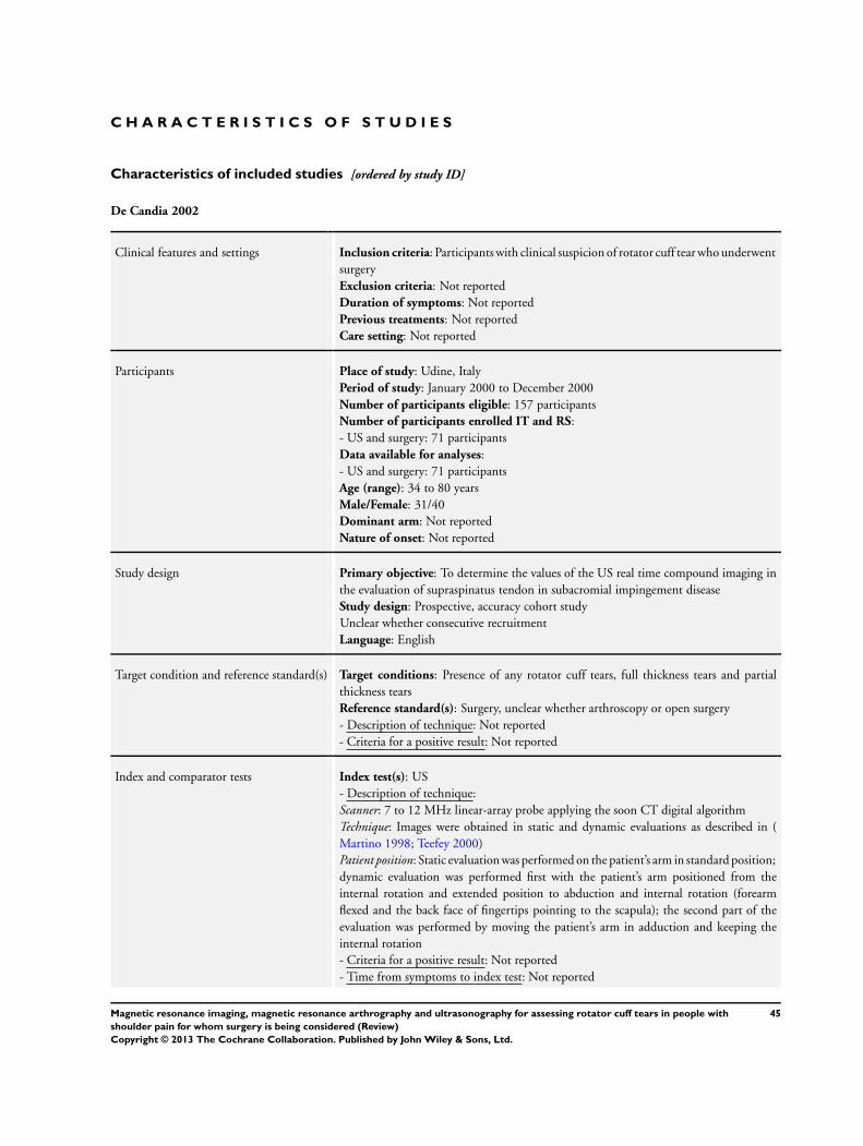
C H A R A C T E R I S T I C S O F S T U D I E S
Characteristics of included studies [ordered by study ID]
De Candia 2002
Clinical features and settings Inclusion criteria: Participants with clinical suspicion of rotator cuff tear who underwentsurgeryExclusion criteria: Not reportedDuration of symptoms: Not reportedPrevious treatments: Not reportedCare setting: Not reported
Participants Place of study: Udine, ItalyPeriod of study: January 2000 to December 2000Number of participants eligible: 157 participantsNumber of participants enrolled IT and RS:- US and surgery: 71 participantsData available for analyses:- US and surgery: 71 participantsAge (range): 34 to 80 yearsMale/Female: 31/40Dominant arm: Not reportedNature of onset: Not reported
Study design Primary objective: To determine the values of the US real time compound imaging inthe evaluation of supraspinatus tendon in subacromial impingement diseaseStudy design: Prospective, accuracy cohort studyUnclear whether consecutive recruitmentLanguage: English
Target condition and reference standard(s) Target conditions: Presence of any rotator cuff tears, full thickness tears and partialthickness tearsReference standard(s): Surgery, unclear whether arthroscopy or open surgery- Description of technique: Not reported- Criteria for a positive result: Not reported
Index and comparator tests Index test(s): US- Description of technique:Scanner: 7 to 12 MHz linear-array probe applying the soon CT digital algorithmTechnique: Images were obtained in static and dynamic evaluations as described in (Martino 1998; Teefey 2000)Patient position: Static evaluation was performed on the patient’s arm in standard position;dynamic evaluation was performed first with the patient’s arm positioned from theinternal rotation and extended position to abduction and internal rotation (forearmflexed and the back face of fingertips pointing to the scapula); the second part of theevaluation was performed by moving the patient’s arm in adduction and keeping theinternal rotation- Criteria for a positive result: Not reported- Time from symptoms to index test: Not reported
45Magnetic resonance imaging, magnetic resonance arthrography and ultrasonography for assessing rotator cuff tears in people with
shoulder pain for whom surgery is being considered (Review)
Copyright © 2013 The Cochrane Collaboration. Published by John Wiley & Sons, Ltd.

De Candia 2002 (Continued)
- Time from index test to reference standard: Index test was performed on the day beforereference standard
Follow-up Adverse events due to index test(s): Not reportedAdverse events due to reference standard test(s): Not reported
Notes Part of population of this study was also reported in De Candia 2003 Although DeCandia 2003 is more updated than this study, there were no extra data available to beincluded in the analysesThe rotator cuff tears were focused on only supraspinatus tendon tearsA two-by-two table of the ITs and RS was given, which tallied with the reported summarydata
Table of Methodological Quality
Item Authors’ judgement Description
Representative spectrum?All tests
Unclear Unclear whether consecutive recruitment
Acceptable reference standard?All tests
Unclear The reference standard was surgery (un-clear whether arthroscopy or open surgery)and the target conditions were presence ofany rotator cuff tears, full thickness tearsand partial thickness tears
Acceptable delay between tests?All tests
Yes Index test was performed on the day beforereference standard
Partial verification avoided?All tests
No Not all the patients who received the indextest underwent a reference standard to ver-ify their true disease statusOf the 157 eligible participants, only 71(45.2%) underwent to reference standard
Differential verification avoided?All tests
No The result of the index test probably influ-enced the choice of the reference standard
Incorporation avoided?All tests
Yes The index test did not form part of thereference standard
Index test results blinded?All tests
Unclear Insufficient information was given to per-mit judgement
Reference standard results blinded?All tests
No The results of the index tests were probablyknown to the person interpreting the refer-ence tests
Relevant clinical information?All tests
Unclear Not reported
46Magnetic resonance imaging, magnetic resonance arthrography and ultrasonography for assessing rotator cuff tears in people with
shoulder pain for whom surgery is being considered (Review)
Copyright © 2013 The Cochrane Collaboration. Published by John Wiley & Sons, Ltd.
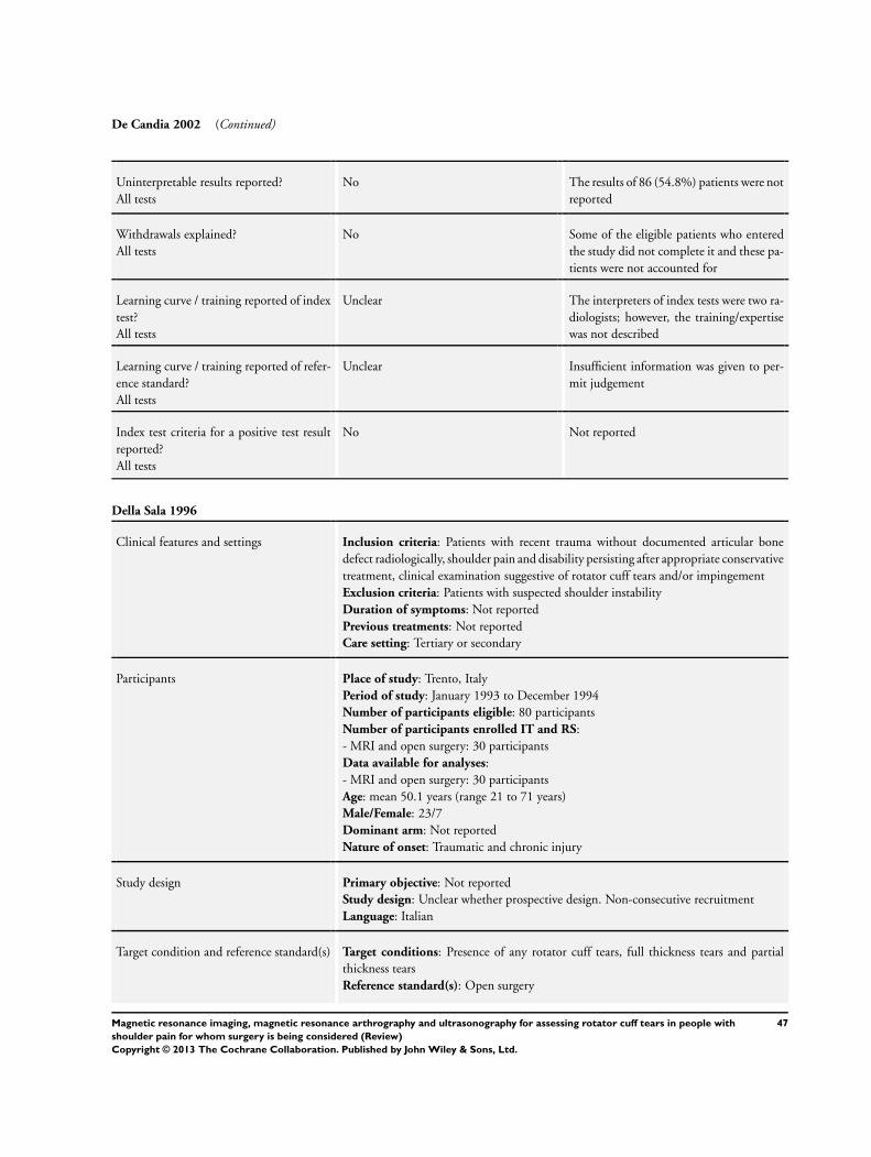
De Candia 2002 (Continued)
Uninterpretable results reported?All tests
No The results of 86 (54.8%) patients were notreported
Withdrawals explained?All tests
No Some of the eligible patients who enteredthe study did not complete it and these pa-tients were not accounted for
Learning curve / training reported of indextest?All tests
Unclear The interpreters of index tests were two ra-diologists; however, the training/expertisewas not described
Learning curve / training reported of refer-ence standard?All tests
Unclear Insufficient information was given to per-mit judgement
Index test criteria for a positive test resultreported?All tests
No Not reported
Della Sala 1996
Clinical features and settings Inclusion criteria: Patients with recent trauma without documented articular bonedefect radiologically, shoulder pain and disability persisting after appropriate conservativetreatment, clinical examination suggestive of rotator cuff tears and/or impingementExclusion criteria: Patients with suspected shoulder instabilityDuration of symptoms: Not reportedPrevious treatments: Not reportedCare setting: Tertiary or secondary
Participants Place of study: Trento, ItalyPeriod of study: January 1993 to December 1994Number of participants eligible: 80 participantsNumber of participants enrolled IT and RS:- MRI and open surgery: 30 participantsData available for analyses:- MRI and open surgery: 30 participantsAge: mean 50.1 years (range 21 to 71 years)Male/Female: 23/7Dominant arm: Not reportedNature of onset: Traumatic and chronic injury
Study design Primary objective: Not reportedStudy design: Unclear whether prospective design. Non-consecutive recruitmentLanguage: Italian
Target condition and reference standard(s) Target conditions: Presence of any rotator cuff tears, full thickness tears and partialthickness tearsReference standard(s): Open surgery
47Magnetic resonance imaging, magnetic resonance arthrography and ultrasonography for assessing rotator cuff tears in people with
shoulder pain for whom surgery is being considered (Review)
Copyright © 2013 The Cochrane Collaboration. Published by John Wiley & Sons, Ltd.
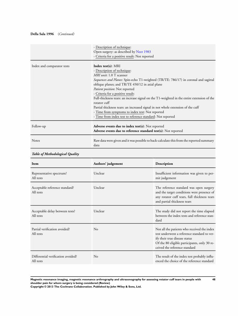
Della Sala 1996 (Continued)
- Description of technique:Open surgery: as described by Neer 1983- Criteria for a positive result: Not reported
Index and comparator tests Index test(s): MRI- Description of technique:MRI unit: 1.0 T scannerSequences and Planes: Spin-echo T1-weighted (TR/TE: 786/17) in coronal and sagittaloblique planes; and TR/TE 450/12 in axial planePatient position: Not reported- Criteria for a positive result:Full-thickness tears: an increase signal on the T1-weighted in the entire extension of therotator cuffPartial thickness tears: an increased signal in not whole extension of the cuff- Time from symptoms to index test: Not reported- Time from index test to reference standard: Not reported
Follow-up Adverse events due to index test(s): Not reportedAdverse events due to reference standard test(s): Not reported
Notes Raw data were given and it was possible to back-calculate this from the reported summarydata
Table of Methodological Quality
Item Authors’ judgement Description
Representative spectrum?All tests
Unclear Insufficient information was given to per-mit judgement
Acceptable reference standard?All tests
Unclear The reference standard was open surgeryand the target conditions were presence ofany rotator cuff tears, full thickness tearsand partial thickness tears
Acceptable delay between tests?All tests
Unclear The study did not report the time elapsedbetween the index tests and reference stan-dard
Partial verification avoided?All tests
No Not all the patients who received the indextest underwent a reference standard to ver-ify their true disease statusOf the 80 eligible participants, only 30 re-ceived the reference standard
Differential verification avoided?All tests
No The result of the index test probably influ-enced the choice of the reference standard
48Magnetic resonance imaging, magnetic resonance arthrography and ultrasonography for assessing rotator cuff tears in people with
shoulder pain for whom surgery is being considered (Review)
Copyright © 2013 The Cochrane Collaboration. Published by John Wiley & Sons, Ltd.
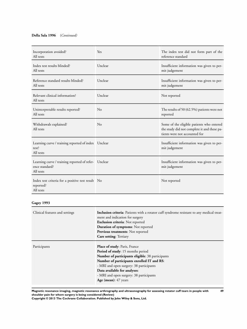
Della Sala 1996 (Continued)
Incorporation avoided?All tests
Yes The index test did not form part of thereference standard
Index test results blinded?All tests
Unclear Insufficient information was given to per-mit judgement
Reference standard results blinded?All tests
Unclear Insufficient information was given to per-mit judgement
Relevant clinical information?All tests
Unclear Not reported
Uninterpretable results reported?All tests
No The results of 50 (62.5%) patients were notreported
Withdrawals explained?All tests
No Some of the eligible patients who enteredthe study did not complete it and these pa-tients were not accounted for
Learning curve / training reported of indextest?All tests
Unclear Insufficient information was given to per-mit judgement
Learning curve / training reported of refer-ence standard?All tests
Unclear Insufficient information was given to per-mit judgement
Index test criteria for a positive test resultreported?All tests
No Not reported
Gagey 1993
Clinical features and settings Inclusion criteria: Patients with a rotator cuff syndrome resistant to any medical treat-ment and indication for surgeryExclusion criteria: Not reportedDuration of symptoms: Not reportedPrevious treatments: Not reportedCare setting: Tertiary
Participants Place of study: Paris, FrancePeriod of study: 15 months periodNumber of participants eligible: 38 participantsNumber of participants enrolled IT and RS:- MRI and open surgery: 38 participantsData available for analyses:- MRI and open surgery: 38 participantsAge (mean): 47 years
49Magnetic resonance imaging, magnetic resonance arthrography and ultrasonography for assessing rotator cuff tears in people with
shoulder pain for whom surgery is being considered (Review)
Copyright © 2013 The Cochrane Collaboration. Published by John Wiley & Sons, Ltd.
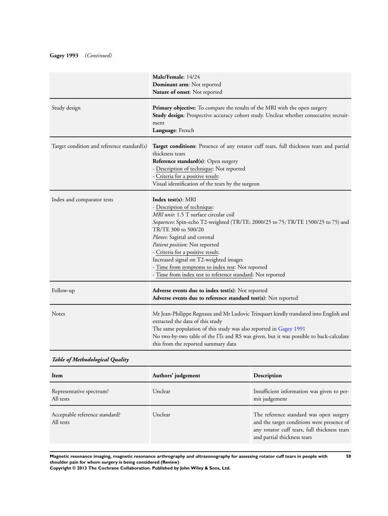
Gagey 1993 (Continued)
Male/Female: 14/24Dominant arm: Not reportedNature of onset: Not reported
Study design Primary objective: To compare the results of the MRI with the open surgeryStudy design: Prospective accuracy cohort study. Unclear whether consecutive recruit-mentLanguage: French
Target condition and reference standard(s) Target conditions: Presence of any rotator cuff tears, full thickness tears and partialthickness tearsReference standard(s): Open surgery- Description of technique: Not reported- Criteria for a positive result:Visual identification of the tears by the surgeon
Index and comparator tests Index test(s): MRI- Description of technique:MRI unit: 1.5 T surface circular coilSequences: Spin-echo T2-weighted (TR/TE: 2000/25 to 75; TR/TE 1500/25 to 75) andTR/TE 300 to 500/20Planes: Sagittal and coronalPatient position: Not reported- Criteria for a positive result:Increased signal on T2-weighted images- Time from symptoms to index test: Not reported- Time from index test to reference standard: Not reported
Follow-up Adverse events due to index test(s): Not reportedAdverse events due to reference standard test(s): Not reported
Notes Mr Jean-Philippe Regnaux and Mr Ludovic Trinquart kindly translated into English andextracted the data of this studyThe same population of this study was also reported in Gagey 1991No two-by-two table of the ITs and RS was given, but it was possible to back-calculatethis from the reported summary data
Table of Methodological Quality
Item Authors’ judgement Description
Representative spectrum?All tests
Unclear Insufficient information was given to per-mit judgement
Acceptable reference standard?All tests
Unclear The reference standard was open surgeryand the target conditions were presence ofany rotator cuff tears, full thickness tearsand partial thickness tears
50Magnetic resonance imaging, magnetic resonance arthrography and ultrasonography for assessing rotator cuff tears in people with
shoulder pain for whom surgery is being considered (Review)
Copyright © 2013 The Cochrane Collaboration. Published by John Wiley & Sons, Ltd.
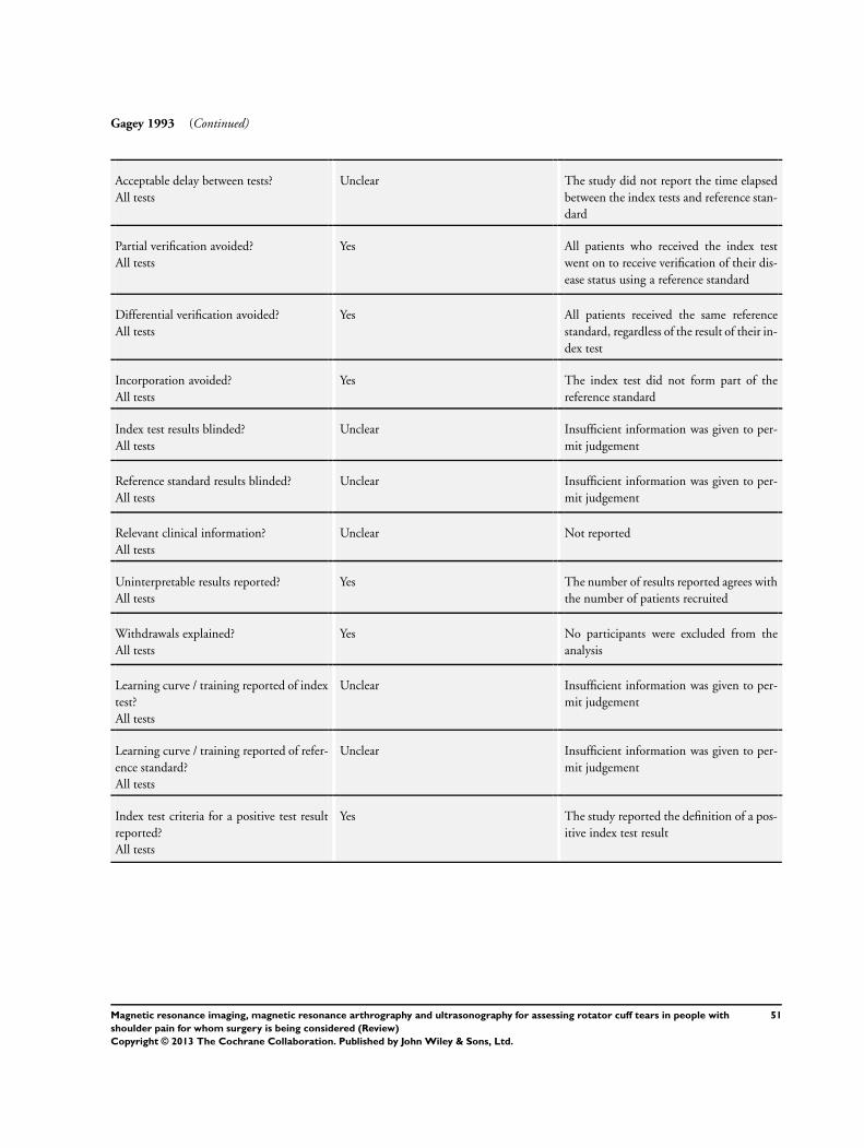
Gagey 1993 (Continued)
Acceptable delay between tests?All tests
Unclear The study did not report the time elapsedbetween the index tests and reference stan-dard
Partial verification avoided?All tests
Yes All patients who received the index testwent on to receive verification of their dis-ease status using a reference standard
Differential verification avoided?All tests
Yes All patients received the same referencestandard, regardless of the result of their in-dex test
Incorporation avoided?All tests
Yes The index test did not form part of thereference standard
Index test results blinded?All tests
Unclear Insufficient information was given to per-mit judgement
Reference standard results blinded?All tests
Unclear Insufficient information was given to per-mit judgement
Relevant clinical information?All tests
Unclear Not reported
Uninterpretable results reported?All tests
Yes The number of results reported agrees withthe number of patients recruited
Withdrawals explained?All tests
Yes No participants were excluded from theanalysis
Learning curve / training reported of indextest?All tests
Unclear Insufficient information was given to per-mit judgement
Learning curve / training reported of refer-ence standard?All tests
Unclear Insufficient information was given to per-mit judgement
Index test criteria for a positive test resultreported?All tests
Yes The study reported the definition of a pos-itive index test result
51Magnetic resonance imaging, magnetic resonance arthrography and ultrasonography for assessing rotator cuff tears in people with
shoulder pain for whom surgery is being considered (Review)
Copyright © 2013 The Cochrane Collaboration. Published by John Wiley & Sons, Ltd.
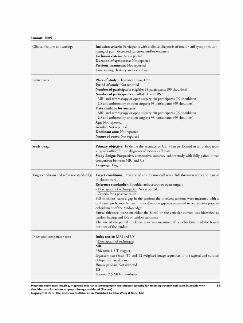
Iannotti 2005
Clinical features and settings Inclusion criteria: Participants with a clinical diagnosis of rotator cuff symptoms, con-sisting of pain, decreased function, and/or weaknessExclusion criteria: Not reportedDuration of symptoms: Not reportedPrevious treatments: Not reportedCare setting: Tertiary and secondary
Participants Place of study: Cleveland, Ohio, USAPeriod of study: Not reportedNumber of participants eligible: 98 participants (99 shoulders)Number of participants enrolled IT and RS:- MRI and arthroscopy or open surgery: 98 participants (99 shoulders)- US and arthroscopy or open surgery: 98 participants (99 shoulders)Data available for analyses:- MRI and arthroscopy or open surgery: 98 participants (99 shoulders)- US and arthroscopy or open surgery: 98 participants (99 shoulders)Age: Not reportedGender: Not reportedDominant arm: Not reportedNature of onset: Not reported
Study design Primary objective: To define the accuracy of US, when performed in an orthopaedicsurgeon’s office, for the diagnosis of rotator cuff tearsStudy design: Prospective, consecutive, accuracy cohort study with fully paired directcomparison between MRI and USLanguage: English
Target condition and reference standard(s) Target conditions: Presence of any rotator cuff tears, full thickness tears and partialthickness tearsReference standard(s): Shoulder arthroscopy or open surgery- Description of technique(s): Not reported- Criteria for a positive result:Full thickness tears: a gap in the tendon; the involved tendons were measured with acalibrated probe or ruler, and the total tendon gap was measured in centimetres prior todébridement of the tendon edgesPartial thickness tears: on either the bursal or the articular surface was identified astendon-fraying and loss of tendon substanceThe size of the partial thickness tears was measured after débridement of the frayedportions of the tendon
Index and comparator tests Index test(s): MRI and US- Description of technique:MRI
MRI unit: 1.5-T magnetSequences and Planes: T1 and T2-weighted image sequences in the sagittal and coronaloblique and axial planesPatient position: Not reportedUS
Scanner: 7.5 MHz transducer
52Magnetic resonance imaging, magnetic resonance arthrography and ultrasonography for assessing rotator cuff tears in people with
shoulder pain for whom surgery is being considered (Review)
Copyright © 2013 The Cochrane Collaboration. Published by John Wiley & Sons, Ltd.
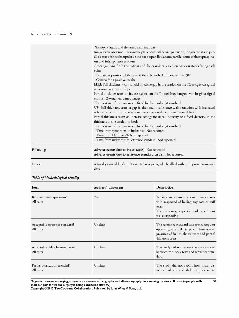
Iannotti 2005 (Continued)
Technique: Static and dynamic examinationsImages were obtained in transverse plane scans of the biceps tendon; longitudinal and par-allel scans of the subscapularis tendon; perpendicular and parallel scans of the supraspina-tus and infraspinatus tendonsPatient position: Both the patient and the examiner seated on backless stools facing eachotherThe patient positioned the arm at the side with the elbow bent to 90o
- Criteria for a positive result:MRI: Full thickness tears: a fluid filled the gap in the tendon on the T2-weighted sagittalor coronal oblique imagesPartial thickness tears: an increase signal on the T1-weighted images, with brighter signalon the T2-weighted paired imageThe location of the tear was defined by the tendon(s) involvedUS: Full thickness tears: a gap in the tendon substance with retraction with increasedechogenic signal from the exposed articular cartilage of the humeral headPartial thickness tears: an increase echogenic signal intensity or a focal decrease in thethickness of the tendon or bothThe location of the tear was defined by the tendon(s) involved- Time from symptoms to index test: Not reported- Time from US to MRI: Not reported- Time from index test to reference standard: Not reported
Follow-up Adverse events due to index test(s): Not reportedAdverse events due to reference standard test(s): Not reported
Notes A two-by-two table of the ITs and RS was given, which tallied with the reported summarydata
Table of Methodological Quality
Item Authors’ judgement Description
Representative spectrum?All tests
Yes Tertiary or secondary care, participantswith suspected of having any rotator cufftearsThe study was prospective and recruitmentwas consecutive
Acceptable reference standard?All tests
Unclear The reference standard was arthroscopy oropen surgery and the target conditions werepresence of full thickness tears and partialthickness tears
Acceptable delay between tests?All tests
Unclear The study did not report the time elapsedbetween the index tests and reference stan-dard
Partial verification avoided?All tests
Unclear The study did not report how many pa-tients had US and did not proceed to
53Magnetic resonance imaging, magnetic resonance arthrography and ultrasonography for assessing rotator cuff tears in people with
shoulder pain for whom surgery is being considered (Review)
Copyright © 2013 The Cochrane Collaboration. Published by John Wiley & Sons, Ltd.

Iannotti 2005 (Continued)
surgery
Differential verification avoided?All tests
Unclear Insufficient information was given to per-mit judgement
Incorporation avoided?All tests
Yes The index test did not form part of thereference standard
Index test results blinded?All tests
Yes The study was prospective and the resultsof the index tests were interpreted beforethe reference standard
Reference standard results blinded?All tests
No The results of the index tests were known tothe person interpreting the reference tests
Relevant clinical information?All tests
Yes The authors had knowledge of history,physical findings and radiographs
Uninterpretable results reported?All tests
Unclear Insufficient information was given to per-mit judgement
Withdrawals explained?All tests
Unclear Insufficient information was given to per-mit judgement
Learning curve / training reported of indextest?All tests
No Performace of index tests was not by radi-ologist or surgeon and they had only twotraining sections
Learning curve / training reported of refer-ence standard?All tests
Unclear Insufficient information was given to per-mit judgement
Index test criteria for a positive test resultreported?All tests
Yes The study reported the definition of a pos-itive index test result
54Magnetic resonance imaging, magnetic resonance arthrography and ultrasonography for assessing rotator cuff tears in people with
shoulder pain for whom surgery is being considered (Review)
Copyright © 2013 The Cochrane Collaboration. Published by John Wiley & Sons, Ltd.
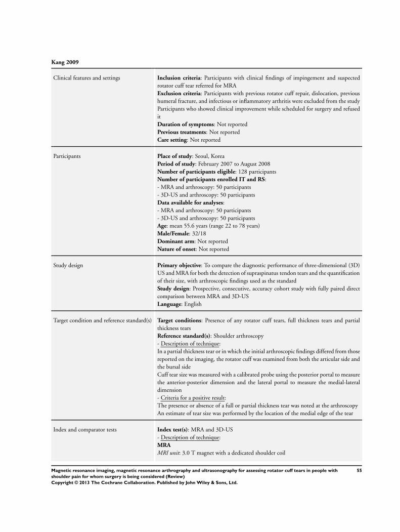
Kang 2009
Clinical features and settings Inclusion criteria: Participants with clinical findings of impingement and suspectedrotator cuff tear referred for MRAExclusion criteria: Participants with previous rotator cuff repair, dislocation, previoushumeral fracture, and infectious or inflammatory arthritis were excluded from the studyParticipants who showed clinical improvement while scheduled for surgery and refuseditDuration of symptoms: Not reportedPrevious treatments: Not reportedCare setting: Not reported
Participants Place of study: Seoul, KoreaPeriod of study: February 2007 to August 2008Number of participants eligible: 128 participantsNumber of participants enrolled IT and RS:- MRA and arthroscopy: 50 participants- 3D-US and arthroscopy: 50 participantsData available for analyses:- MRA and arthroscopy: 50 participants- 3D-US and arthroscopy: 50 participantsAge: mean 55.6 years (range 22 to 78 years)Male/Female: 32/18Dominant arm: Not reportedNature of onset: Not reported
Study design Primary objective: To compare the diagnostic performance of three-dimensional (3D)US and MRA for both the detection of supraspinatus tendon tears and the quantificationof their size, with arthroscopic findings used as the standardStudy design: Prospective, consecutive, accuracy cohort study with fully paired directcomparison between MRA and 3D-USLanguage: English
Target condition and reference standard(s) Target conditions: Presence of any rotator cuff tears, full thickness tears and partialthickness tearsReference standard(s): Shoulder arthroscopy- Description of technique:In a partial thickness tear or in which the initial arthroscopic findings differed from thosereported on the imaging, the rotator cuff was examined from both the articular side andthe bursal sideCuff tear size was measured with a calibrated probe using the posterior portal to measurethe anterior-posterior dimension and the lateral portal to measure the medial-lateraldimension- Criteria for a positive result:The presence or absence of a full or partial thickness tear was noted at the arthroscopyAn estimate of tear size was performed by the location of the medial edge of the tear
Index and comparator tests Index test(s): MRA and 3D-US- Description of technique:MRA
MRI unit: 3.0 T magnet with a dedicated shoulder coil
55Magnetic resonance imaging, magnetic resonance arthrography and ultrasonography for assessing rotator cuff tears in people with
shoulder pain for whom surgery is being considered (Review)
Copyright © 2013 The Cochrane Collaboration. Published by John Wiley & Sons, Ltd.
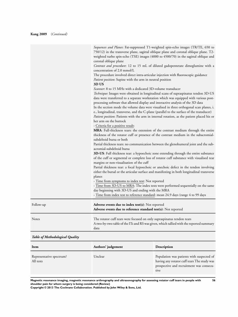
Kang 2009 (Continued)
Sequences and Planes: Fat-suppressed T1-weighted spin-echo images (TR/TE, 650 to750/12) in the transverse plane, sagittal oblique plane and coronal oblique plane. T2-weighted turbo spin-echo (TSE) images (4000 to 4500/70) in the sagittal oblique andcoronal oblique planeContrast and procedure: 12 to 15 mL of diluted gadopentetate dimeglumine with aconcentration of 2.0 mmol/LThe procedure involved direct intra-articular injection with fluoroscopic guidancePatient position: Supine with the arm in neutral position3D US
Scanner: 8 to 15 MHz with a dedicated 3D-volume transducerTechnique: Images were obtained in longitudinal scans of supraspinatus tendon 3D-USdata were transferred to a separate workstation which was equipped with various post-processing software that allowed display and interactive analysis of the 3D dataIn the section mode the volume data were visualised in three orthogonal scan planes, i.e., longitudinal, transverse, and the C-plane (parallel to the surface of the transducer)Patient position: Patients with the arm in internal rotation, as the patient placed his orher arm on the buttock- Criteria for a positive result:MRA: Full-thickness tears: the extension of the contrast medium through the entirethickness of the rotator cuff or presence of the contrast medium in the subacromial-subdeltoid bursa or bothPartial thickness tears: no communication between the glenohumeral joint and the sub-acromial-subdeltoid bursa3D-US: Full thickness tear: a hypoechoic zone extending through the entire substanceof the cuff or segmental or complete loss of rotator cuff substance with visualised tearmargins or non-visualisation of the cuffPartial thickness tear: a focal hypoechoic or anechoic defect in the tendon involvingeither the bursal or the articular surface and manifesting in both longitudinal transverseplanes- Time from symptoms to index test: Not reported- Time from 3D-US to MRA: The index tests were performed sequentially on the sameday beginning with 3D-US and ending with the MRA- Time from index test to reference standard: mean 24.9 days (range 4 to 99 days
Follow-up Adverse events due to index test(s): Not reportedAdverse events due to reference standard test(s): Not reported
Notes The rotator cuff tears were focused on only supraspinatus tendon tearsA two-by-two table of the ITs and RS was given, which tallied with the reported summarydata
Table of Methodological Quality
Item Authors’ judgement Description
Representative spectrum?All tests
Unclear Population was patients with suspected ofhaving any rotator cuff tears The study wasprospective and recruitment was consecu-tive
56Magnetic resonance imaging, magnetic resonance arthrography and ultrasonography for assessing rotator cuff tears in people with
shoulder pain for whom surgery is being considered (Review)
Copyright © 2013 The Cochrane Collaboration. Published by John Wiley & Sons, Ltd.
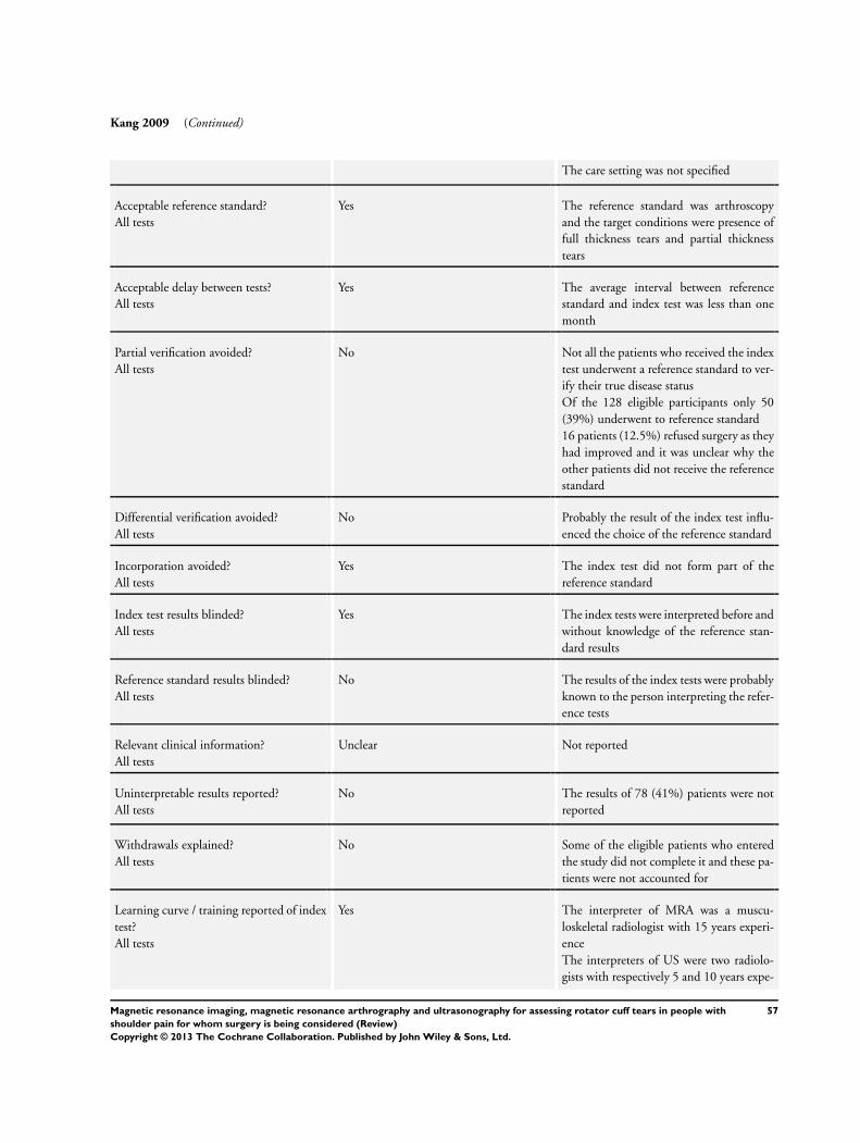
Kang 2009 (Continued)
The care setting was not specified
Acceptable reference standard?All tests
Yes The reference standard was arthroscopyand the target conditions were presence offull thickness tears and partial thicknesstears
Acceptable delay between tests?All tests
Yes The average interval between referencestandard and index test was less than onemonth
Partial verification avoided?All tests
No Not all the patients who received the indextest underwent a reference standard to ver-ify their true disease statusOf the 128 eligible participants only 50(39%) underwent to reference standard16 patients (12.5%) refused surgery as theyhad improved and it was unclear why theother patients did not receive the referencestandard
Differential verification avoided?All tests
No Probably the result of the index test influ-enced the choice of the reference standard
Incorporation avoided?All tests
Yes The index test did not form part of thereference standard
Index test results blinded?All tests
Yes The index tests were interpreted before andwithout knowledge of the reference stan-dard results
Reference standard results blinded?All tests
No The results of the index tests were probablyknown to the person interpreting the refer-ence tests
Relevant clinical information?All tests
Unclear Not reported
Uninterpretable results reported?All tests
No The results of 78 (41%) patients were notreported
Withdrawals explained?All tests
No Some of the eligible patients who enteredthe study did not complete it and these pa-tients were not accounted for
Learning curve / training reported of indextest?All tests
Yes The interpreter of MRA was a muscu-loskeletal radiologist with 15 years experi-enceThe interpreters of US were two radiolo-gists with respectively 5 and 10 years expe-
57Magnetic resonance imaging, magnetic resonance arthrography and ultrasonography for assessing rotator cuff tears in people with
shoulder pain for whom surgery is being considered (Review)
Copyright © 2013 The Cochrane Collaboration. Published by John Wiley & Sons, Ltd.
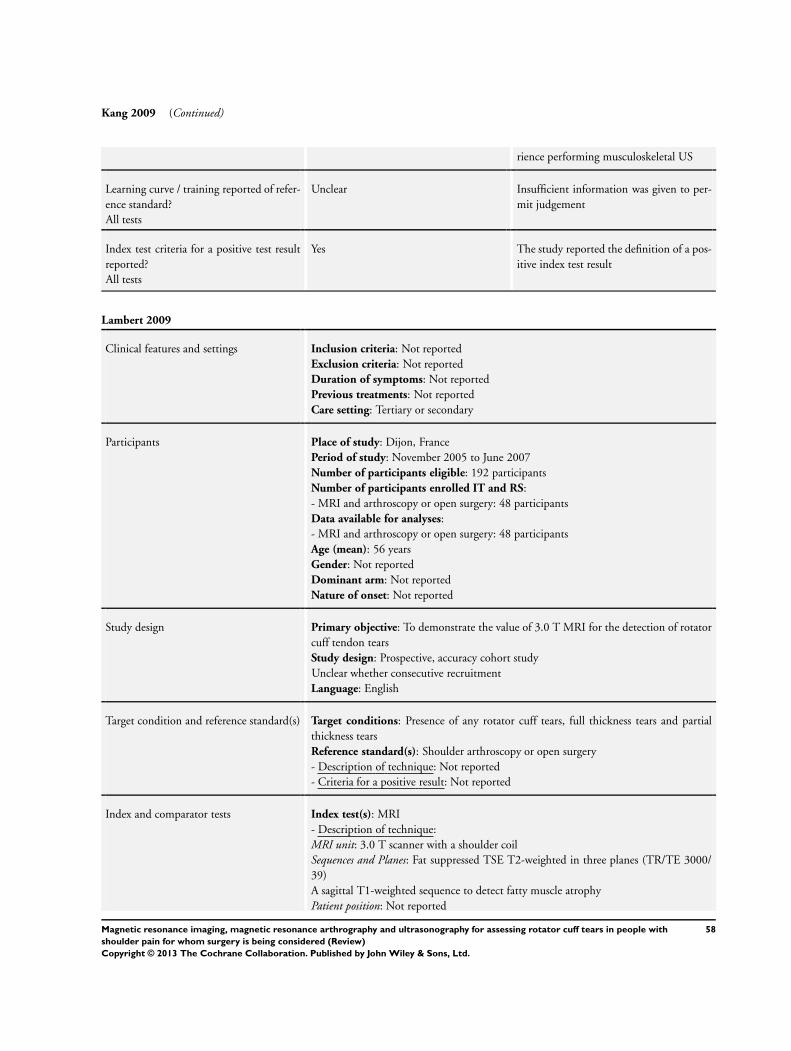
Kang 2009 (Continued)
rience performing musculoskeletal US
Learning curve / training reported of refer-ence standard?All tests
Unclear Insufficient information was given to per-mit judgement
Index test criteria for a positive test resultreported?All tests
Yes The study reported the definition of a pos-itive index test result
Lambert 2009
Clinical features and settings Inclusion criteria: Not reportedExclusion criteria: Not reportedDuration of symptoms: Not reportedPrevious treatments: Not reportedCare setting: Tertiary or secondary
Participants Place of study: Dijon, FrancePeriod of study: November 2005 to June 2007Number of participants eligible: 192 participantsNumber of participants enrolled IT and RS:- MRI and arthroscopy or open surgery: 48 participantsData available for analyses:- MRI and arthroscopy or open surgery: 48 participantsAge (mean): 56 yearsGender: Not reportedDominant arm: Not reportedNature of onset: Not reported
Study design Primary objective: To demonstrate the value of 3.0 T MRI for the detection of rotatorcuff tendon tearsStudy design: Prospective, accuracy cohort studyUnclear whether consecutive recruitmentLanguage: English
Target condition and reference standard(s) Target conditions: Presence of any rotator cuff tears, full thickness tears and partialthickness tearsReference standard(s): Shoulder arthroscopy or open surgery- Description of technique: Not reported- Criteria for a positive result: Not reported
Index and comparator tests Index test(s): MRI- Description of technique:MRI unit: 3.0 T scanner with a shoulder coilSequences and Planes: Fat suppressed TSE T2-weighted in three planes (TR/TE 3000/39)A sagittal T1-weighted sequence to detect fatty muscle atrophyPatient position: Not reported
58Magnetic resonance imaging, magnetic resonance arthrography and ultrasonography for assessing rotator cuff tears in people with
shoulder pain for whom surgery is being considered (Review)
Copyright © 2013 The Cochrane Collaboration. Published by John Wiley & Sons, Ltd.
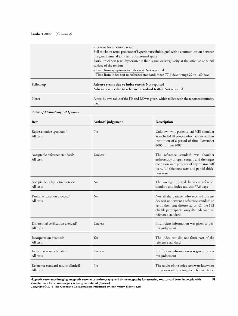
Lambert 2009 (Continued)
- Criteria for a positive result:Full thickness tears: presence of hyperintense fluid signal with a communication betweenthe glenohumeral joint and subacromial spacePartial thickness tears: hyperintense fluid signal or irregularity at the articular or bursalsurface of the tendon- Time from symptoms to index test: Not reported- Time from index test to reference standard: mean 77.6 days (range 22 to 169 days)
Follow-up Adverse events due to index test(s): Not reportedAdverse events due to reference standard test(s): Not reported
Notes A two-by-two table of the ITs and RS was given, which tallied with the reported summarydata
Table of Methodological Quality
Item Authors’ judgement Description
Representative spectrum?All tests
No Unknown why patients had MRI shoulderas included all people who had one at theirinstitution of a period of time November2005 to June 2007
Acceptable reference standard?All tests
Unclear The reference standard was shoulderarthroscopy or open surgery and the targetcondition were presence of any rotator cufftears, full thickness tears and partial thick-ness tears
Acceptable delay between tests?All tests
No The average interval between referencestandard and index test was 77.6 days
Partial verification avoided?All tests
No Not all the patients who received the in-dex test underwent a reference standard toverify their true disease status. Of the 192eligible participants, only 48 underwent toreference standard
Differential verification avoided?All tests
Unclear Insufficient information was given to per-mit judgement
Incorporation avoided?All tests
Yes The index test did not form part of thereference standard
Index test results blinded?All tests
Unclear Insufficient information was given to per-mit judgement
Reference standard results blinded?All tests
No The results of the index tests were known tothe person interpreting the reference tests
59Magnetic resonance imaging, magnetic resonance arthrography and ultrasonography for assessing rotator cuff tears in people with
shoulder pain for whom surgery is being considered (Review)
Copyright © 2013 The Cochrane Collaboration. Published by John Wiley & Sons, Ltd.
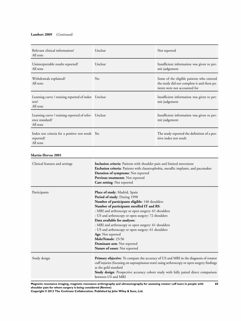
Lambert 2009 (Continued)
Relevant clinical information?All tests
Unclear Not reported
Uninterpretable results reported?All tests
Unclear Insufficient information was given to per-mit judgement
Withdrawals explained?All tests
No Some of the eligible patients who enteredthe study did not complete it and these pa-tients were not accounted for
Learning curve / training reported of indextest?All tests
Unclear Insufficient information was given to per-mit judgement
Learning curve / training reported of refer-ence standard?All tests
Unclear Insufficient information was given to per-mit judgement
Index test criteria for a positive test resultreported?All tests
Yes The study reported the definition of a pos-itive index test result
Martin-Hervas 2001
Clinical features and settings Inclusion criteria: Patients with shoulder pain and limited movementExclusion criteria: Patients with claustrophobia, metallic implants, and pacemakerDuration of symptoms: Not reportedPrevious treatments: Not reportedCare setting: Not reported
Participants Place of study: Madrid, SpainPeriod of study: During 1998Number of participants eligible: 140 shouldersNumber of participants enrolled IT and RS:- MRI and arthroscopy or open surgery: 61 shoulders- US and arthroscopy or open surgery: 72 shouldersData available for analyses:- MRI and arthroscopy or open surgery: 61 shoulders- US and arthroscopy or open surgery: 61 shouldersAge: Not reportedMale/Female: 25/36Dominant arm: Not reportedNature of onset: Not reported
Study design Primary objective: To compare the accuracy of US and MRI in the diagnosis of rotatorcuff injuries (focusing on supraspinatus tears) using arthroscopy or open surgery findingsas the gold standardStudy design: Prospective accuracy cohort study with fully paired direct comparisonbetween US and MRI
60Magnetic resonance imaging, magnetic resonance arthrography and ultrasonography for assessing rotator cuff tears in people with
shoulder pain for whom surgery is being considered (Review)
Copyright © 2013 The Cochrane Collaboration. Published by John Wiley & Sons, Ltd.
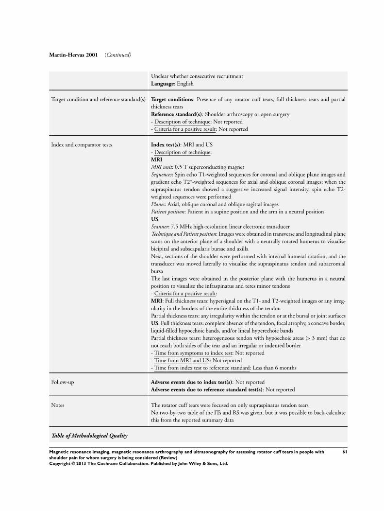
Martin-Hervas 2001 (Continued)
Unclear whether consecutive recruitmentLanguage: English
Target condition and reference standard(s) Target conditions: Presence of any rotator cuff tears, full thickness tears and partialthickness tearsReference standard(s): Shoulder arthroscopy or open surgery- Description of technique: Not reported- Criteria for a positive result: Not reported
Index and comparator tests Index test(s): MRI and US- Description of technique:MRI
MRI unit: 0.5 T superconducting magnetSequences: Spin echo T1-weighted sequences for coronal and oblique plane images andgradient echo T2*-weighted sequences for axial and oblique coronal images; when thesupraspinatus tendon showed a suggestive increased signal intensity, spin echo T2-weighted sequences were performedPlanes: Axial, oblique coronal and oblique sagittal imagesPatient position: Patient in a supine position and the arm in a neutral positionUS
Scanner: 7.5 MHz high-resolution linear electronic transducerTechnique and Patient position: Images were obtained in transverse and longitudinal planescans on the anterior plane of a shoulder with a neutrally rotated humerus to visualisebicipital and subscapularis bursae and axillaNext, sections of the shoulder were performed with internal humeral rotation, and thetransducer was moved laterally to visualise the supraspinatus tendon and subacromialbursaThe last images were obtained in the posterior plane with the humerus in a neutralposition to visualise the infraspinatus and teres minor tendons- Criteria for a positive result:MRI: Full thickness tears: hypersignal on the T1- and T2-weighted images or any irreg-ularity in the borders of the entire thickness of the tendonPartial thickness tears: any irregularity within the tendon or at the bursal or joint surfacesUS: Full thickness tears: complete absence of the tendon, focal atrophy, a concave border,liquid-filled hypoechoic bands, and/or lineal hyperechoic bandsPartial thickness tears: heterogeneous tendon with hypoechoic areas (> 3 mm) that donot reach both sides of the tear and an irregular or indented border- Time from symptoms to index test: Not reported- Time from MRI and US: Not reported- Time from index test to reference standard: Less than 6 months
Follow-up Adverse events due to index test(s): Not reportedAdverse events due to reference standard test(s): Not reported
Notes The rotator cuff tears were focused on only supraspinatus tendon tearsNo two-by-two table of the ITs and RS was given, but it was possible to back-calculatethis from the reported summary data
Table of Methodological Quality
61Magnetic resonance imaging, magnetic resonance arthrography and ultrasonography for assessing rotator cuff tears in people with
shoulder pain for whom surgery is being considered (Review)
Copyright © 2013 The Cochrane Collaboration. Published by John Wiley & Sons, Ltd.
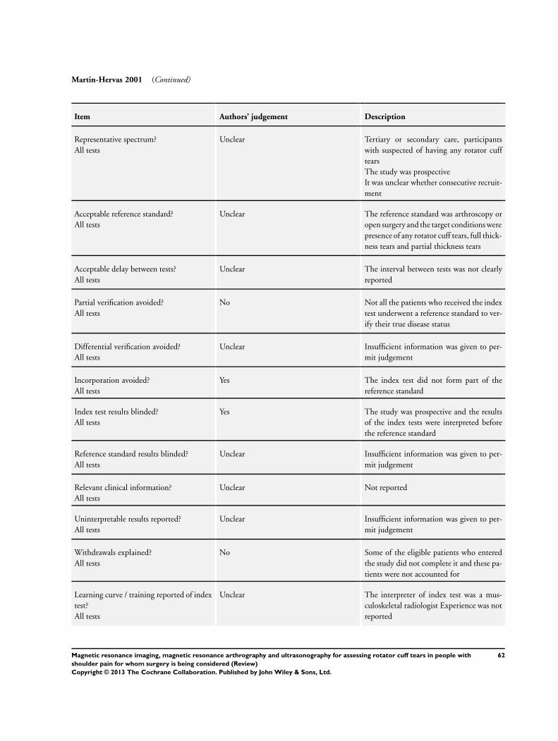
Martin-Hervas 2001 (Continued)
Item Authors’ judgement Description
Representative spectrum?All tests
Unclear Tertiary or secondary care, participantswith suspected of having any rotator cufftearsThe study was prospectiveIt was unclear whether consecutive recruit-ment
Acceptable reference standard?All tests
Unclear The reference standard was arthroscopy oropen surgery and the target conditions werepresence of any rotator cuff tears, full thick-ness tears and partial thickness tears
Acceptable delay between tests?All tests
Unclear The interval between tests was not clearlyreported
Partial verification avoided?All tests
No Not all the patients who received the indextest underwent a reference standard to ver-ify their true disease status
Differential verification avoided?All tests
Unclear Insufficient information was given to per-mit judgement
Incorporation avoided?All tests
Yes The index test did not form part of thereference standard
Index test results blinded?All tests
Yes The study was prospective and the resultsof the index tests were interpreted beforethe reference standard
Reference standard results blinded?All tests
Unclear Insufficient information was given to per-mit judgement
Relevant clinical information?All tests
Unclear Not reported
Uninterpretable results reported?All tests
Unclear Insufficient information was given to per-mit judgement
Withdrawals explained?All tests
No Some of the eligible patients who enteredthe study did not complete it and these pa-tients were not accounted for
Learning curve / training reported of indextest?All tests
Unclear The interpreter of index test was a mus-culoskeletal radiologist Experience was notreported
62Magnetic resonance imaging, magnetic resonance arthrography and ultrasonography for assessing rotator cuff tears in people with
shoulder pain for whom surgery is being considered (Review)
Copyright © 2013 The Cochrane Collaboration. Published by John Wiley & Sons, Ltd.
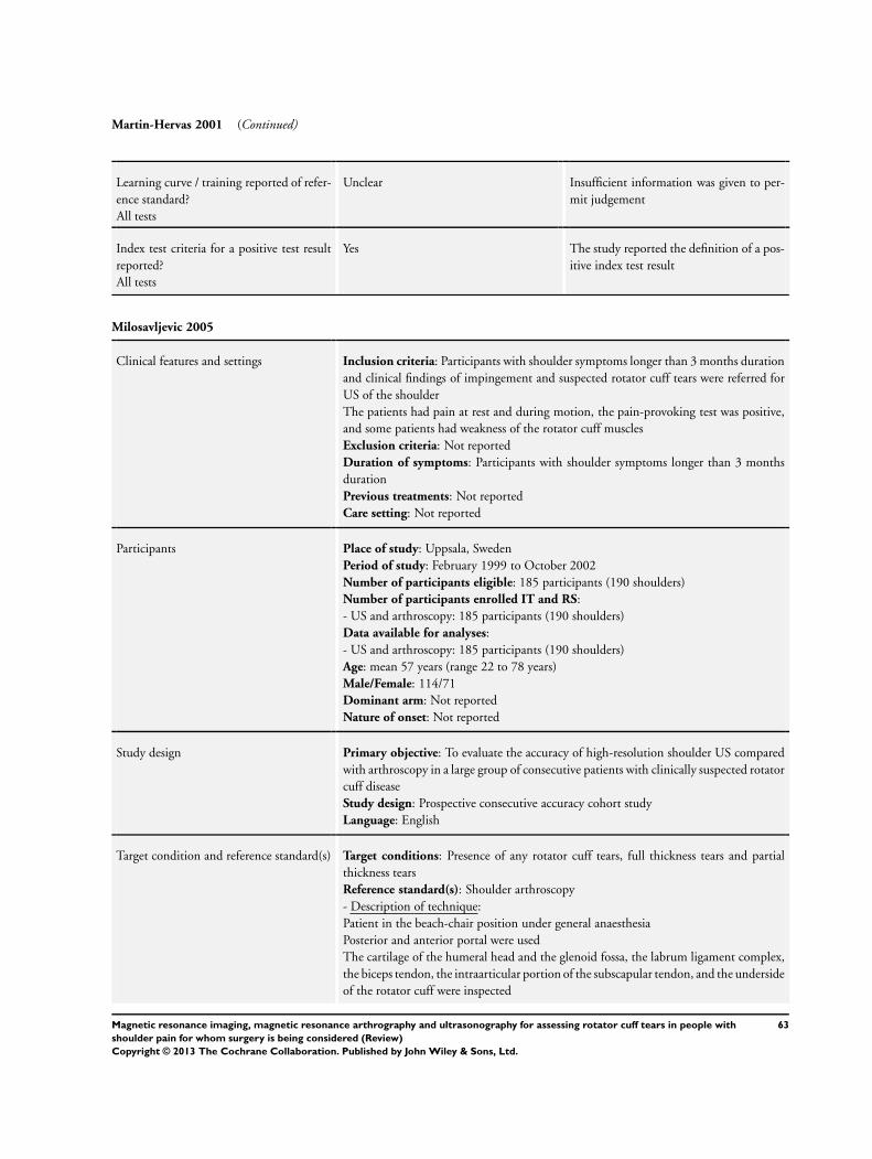
Martin-Hervas 2001 (Continued)
Learning curve / training reported of refer-ence standard?All tests
Unclear Insufficient information was given to per-mit judgement
Index test criteria for a positive test resultreported?All tests
Yes The study reported the definition of a pos-itive index test result
Milosavljevic 2005
Clinical features and settings Inclusion criteria: Participants with shoulder symptoms longer than 3 months durationand clinical findings of impingement and suspected rotator cuff tears were referred forUS of the shoulderThe patients had pain at rest and during motion, the pain-provoking test was positive,and some patients had weakness of the rotator cuff musclesExclusion criteria: Not reportedDuration of symptoms: Participants with shoulder symptoms longer than 3 monthsdurationPrevious treatments: Not reportedCare setting: Not reported
Participants Place of study: Uppsala, SwedenPeriod of study: February 1999 to October 2002Number of participants eligible: 185 participants (190 shoulders)Number of participants enrolled IT and RS:- US and arthroscopy: 185 participants (190 shoulders)Data available for analyses:- US and arthroscopy: 185 participants (190 shoulders)Age: mean 57 years (range 22 to 78 years)Male/Female: 114/71Dominant arm: Not reportedNature of onset: Not reported
Study design Primary objective: To evaluate the accuracy of high-resolution shoulder US comparedwith arthroscopy in a large group of consecutive patients with clinically suspected rotatorcuff diseaseStudy design: Prospective consecutive accuracy cohort studyLanguage: English
Target condition and reference standard(s) Target conditions: Presence of any rotator cuff tears, full thickness tears and partialthickness tearsReference standard(s): Shoulder arthroscopy- Description of technique:Patient in the beach-chair position under general anaesthesiaPosterior and anterior portal were usedThe cartilage of the humeral head and the glenoid fossa, the labrum ligament complex,the biceps tendon, the intraarticular portion of the subscapular tendon, and the undersideof the rotator cuff were inspected
63Magnetic resonance imaging, magnetic resonance arthrography and ultrasonography for assessing rotator cuff tears in people with
shoulder pain for whom surgery is being considered (Review)
Copyright © 2013 The Cochrane Collaboration. Published by John Wiley & Sons, Ltd.
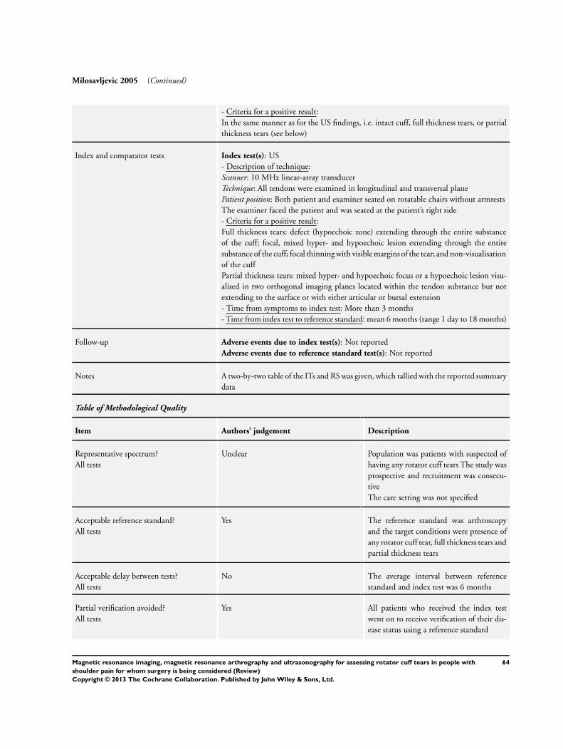
Milosavljevic 2005 (Continued)
- Criteria for a positive result:In the same manner as for the US findings, i.e. intact cuff, full thickness tears, or partialthickness tears (see below)
Index and comparator tests Index test(s): US- Description of technique:Scanner: 10 MHz linear-array transducerTechnique: All tendons were examined in longitudinal and transversal planePatient position: Both patient and examiner seated on rotatable chairs without armrestsThe examiner faced the patient and was seated at the patient’s right side- Criteria for a positive result:Full thickness tears: defect (hypoechoic zone) extending through the entire substanceof the cuff; focal, mixed hyper- and hypoechoic lesion extending through the entiresubstance of the cuff; focal thinning with visible margins of the tear; and non-visualisationof the cuffPartial thickness tears: mixed hyper- and hypoechoic focus or a hypoechoic lesion visu-alised in two orthogonal imaging planes located within the tendon substance but notextending to the surface or with either articular or bursal extension- Time from symptoms to index test: More than 3 months- Time from index test to reference standard: mean 6 months (range 1 day to 18 months)
Follow-up Adverse events due to index test(s): Not reportedAdverse events due to reference standard test(s): Not reported
Notes A two-by-two table of the ITs and RS was given, which tallied with the reported summarydata
Table of Methodological Quality
Item Authors’ judgement Description
Representative spectrum?All tests
Unclear Population was patients with suspected ofhaving any rotator cuff tears The study wasprospective and recruitment was consecu-tiveThe care setting was not specified
Acceptable reference standard?All tests
Yes The reference standard was arthroscopyand the target conditions were presence ofany rotator cuff tear, full thickness tears andpartial thickness tears
Acceptable delay between tests?All tests
No The average interval between referencestandard and index test was 6 months
Partial verification avoided?All tests
Yes All patients who received the index testwent on to receive verification of their dis-ease status using a reference standard
64Magnetic resonance imaging, magnetic resonance arthrography and ultrasonography for assessing rotator cuff tears in people with
shoulder pain for whom surgery is being considered (Review)
Copyright © 2013 The Cochrane Collaboration. Published by John Wiley & Sons, Ltd.
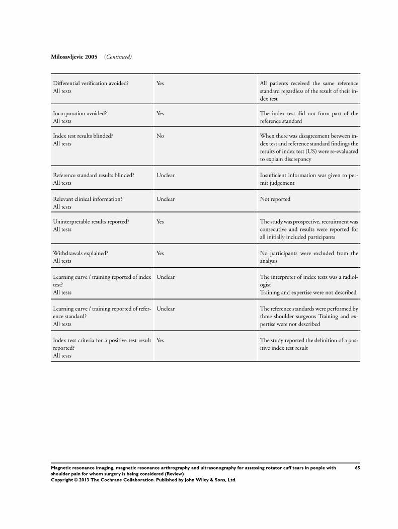
Milosavljevic 2005 (Continued)
Differential verification avoided?All tests
Yes All patients received the same referencestandard regardless of the result of their in-dex test
Incorporation avoided?All tests
Yes The index test did not form part of thereference standard
Index test results blinded?All tests
No When there was disagreement between in-dex test and reference standard findings theresults of index test (US) were re-evaluatedto explain discrepancy
Reference standard results blinded?All tests
Unclear Insufficient information was given to per-mit judgement
Relevant clinical information?All tests
Unclear Not reported
Uninterpretable results reported?All tests
Yes The study was prospective, recruitment wasconsecutive and results were reported forall initially included participants
Withdrawals explained?All tests
Yes No participants were excluded from theanalysis
Learning curve / training reported of indextest?All tests
Unclear The interpreter of index tests was a radiol-ogistTraining and expertise were not described
Learning curve / training reported of refer-ence standard?All tests
Unclear The reference standards were performed bythree shoulder surgeons Training and ex-pertise were not described
Index test criteria for a positive test resultreported?All tests
Yes The study reported the definition of a pos-itive index test result
65Magnetic resonance imaging, magnetic resonance arthrography and ultrasonography for assessing rotator cuff tears in people with
shoulder pain for whom surgery is being considered (Review)
Copyright © 2013 The Cochrane Collaboration. Published by John Wiley & Sons, Ltd.
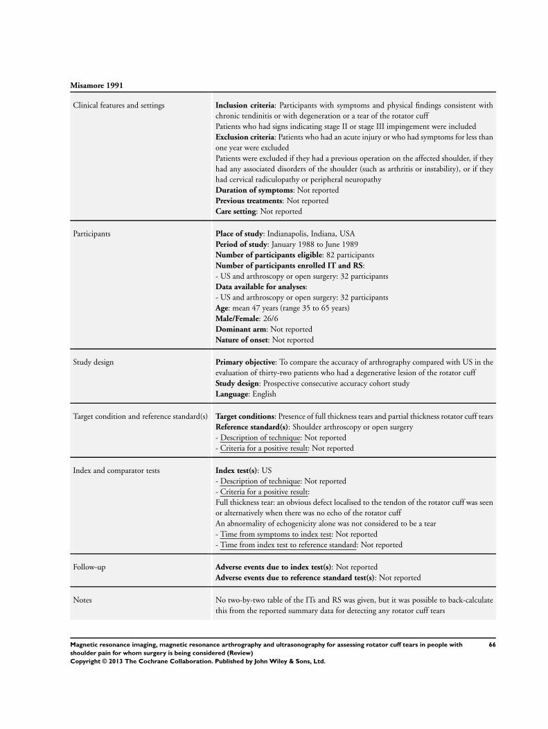
Misamore 1991
Clinical features and settings Inclusion criteria: Participants with symptoms and physical findings consistent withchronic tendinitis or with degeneration or a tear of the rotator cuffPatients who had signs indicating stage II or stage III impingement were includedExclusion criteria: Patients who had an acute injury or who had symptoms for less thanone year were excludedPatients were excluded if they had a previous operation on the affected shoulder, if theyhad any associated disorders of the shoulder (such as arthritis or instability), or if theyhad cervical radiculopathy or peripheral neuropathyDuration of symptoms: Not reportedPrevious treatments: Not reportedCare setting: Not reported
Participants Place of study: Indianapolis, Indiana, USAPeriod of study: January 1988 to June 1989Number of participants eligible: 82 participantsNumber of participants enrolled IT and RS:- US and arthroscopy or open surgery: 32 participantsData available for analyses:- US and arthroscopy or open surgery: 32 participantsAge: mean 47 years (range 35 to 65 years)Male/Female: 26/6Dominant arm: Not reportedNature of onset: Not reported
Study design Primary objective: To compare the accuracy of arthrography compared with US in theevaluation of thirty-two patients who had a degenerative lesion of the rotator cuffStudy design: Prospective consecutive accuracy cohort studyLanguage: English
Target condition and reference standard(s) Target conditions: Presence of full thickness tears and partial thickness rotator cuff tearsReference standard(s): Shoulder arthroscopy or open surgery- Description of technique: Not reported- Criteria for a positive result: Not reported
Index and comparator tests Index test(s): US- Description of technique: Not reported- Criteria for a positive result:Full thickness tear: an obvious defect localised to the tendon of the rotator cuff was seenor alternatively when there was no echo of the rotator cuffAn abnormality of echogenicity alone was not considered to be a tear- Time from symptoms to index test: Not reported- Time from index test to reference standard: Not reported
Follow-up Adverse events due to index test(s): Not reportedAdverse events due to reference standard test(s): Not reported
Notes No two-by-two table of the ITs and RS was given, but it was possible to back-calculatethis from the reported summary data for detecting any rotator cuff tears
66Magnetic resonance imaging, magnetic resonance arthrography and ultrasonography for assessing rotator cuff tears in people with
shoulder pain for whom surgery is being considered (Review)
Copyright © 2013 The Cochrane Collaboration. Published by John Wiley & Sons, Ltd.
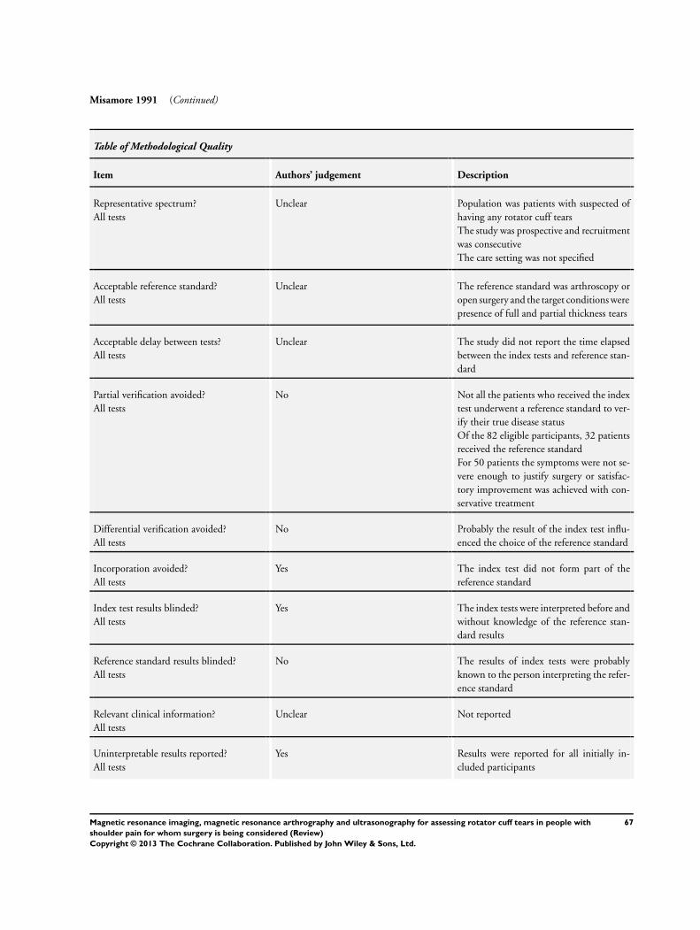
Misamore 1991 (Continued)
Table of Methodological Quality
Item Authors’ judgement Description
Representative spectrum?All tests
Unclear Population was patients with suspected ofhaving any rotator cuff tearsThe study was prospective and recruitmentwas consecutiveThe care setting was not specified
Acceptable reference standard?All tests
Unclear The reference standard was arthroscopy oropen surgery and the target conditions werepresence of full and partial thickness tears
Acceptable delay between tests?All tests
Unclear The study did not report the time elapsedbetween the index tests and reference stan-dard
Partial verification avoided?All tests
No Not all the patients who received the indextest underwent a reference standard to ver-ify their true disease statusOf the 82 eligible participants, 32 patientsreceived the reference standardFor 50 patients the symptoms were not se-vere enough to justify surgery or satisfac-tory improvement was achieved with con-servative treatment
Differential verification avoided?All tests
No Probably the result of the index test influ-enced the choice of the reference standard
Incorporation avoided?All tests
Yes The index test did not form part of thereference standard
Index test results blinded?All tests
Yes The index tests were interpreted before andwithout knowledge of the reference stan-dard results
Reference standard results blinded?All tests
No The results of index tests were probablyknown to the person interpreting the refer-ence standard
Relevant clinical information?All tests
Unclear Not reported
Uninterpretable results reported?All tests
Yes Results were reported for all initially in-cluded participants
67Magnetic resonance imaging, magnetic resonance arthrography and ultrasonography for assessing rotator cuff tears in people with
shoulder pain for whom surgery is being considered (Review)
Copyright © 2013 The Cochrane Collaboration. Published by John Wiley & Sons, Ltd.
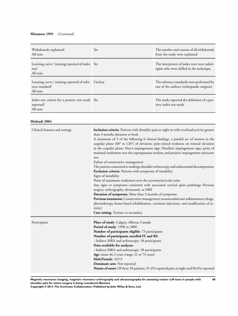
Misamore 1991 (Continued)
Withdrawals explained?All tests
Yes The number and reasons of all withdrawalsfrom the study were explained
Learning curve / training reported of indextest?All tests
Yes The interpreters of index tests were radiol-ogists who were skilled in the technique
Learning curve / training reported of refer-ence standard?All tests
Unclear The reference standards were performed byone of the authors (orthopaedic surgeon)
Index test criteria for a positive test resultreported?All tests
Yes The study reported the definition of a pos-itive index test result
Mohtadi 2004
Clinical features and settings Inclusion criteria: Patients with shoulder pain at night or with overhead activity greaterthan 3 months duration or bothA minimum of 3 of the following 6 clinical findings: a painful arc of motion in thescapular plane (60° to 120°) of elevation; pain-related weakness on resisted elevationin the scapular plane; Neer’s impingement sign; Hawkin’s impingement sign; point ofmaximal tenderness over the supraspinatus tendon; and positive impingement xylocainetestFailure of conservative managementThe patients consented to undergo shoulder arthroscopy and subacromial decompressionExclusion criteria: Patients with symptoms of instabilitySigns of instabilityPoint of maximum tenderness over the acromioclavicular jointAny signs or symptoms consistent with associated cervical spine pathology Previoussurgery, arthrography, ultrasound, or MRIDuration of symptoms: More than 3 months of symptomsPrevious treatments: Conservative management (nonsteroidal anti-inflammatory drugs,physiotherapy, home-based rehabilitation, cortisone injections, and modification of ac-tivity)Care setting: Tertiary or secondary
Participants Place of study: Calgary, Alberta, CanadaPeriod of study: 1998 to 2000Number of participants eligible: 73 participantsNumber of participants enrolled IT and RS:- Indirect MRA and arthroscopy: 58 participantsData available for analyses:- Indirect MRA and arthroscopy: 58 participantsAge: mean 46.2 years (range 21 to 73 years)Male/Female: 43/15Dominant arm: Not reportedNature of onset: Of these 58 patients, 91.4% reported pain at night and 96.6% reported
68Magnetic resonance imaging, magnetic resonance arthrography and ultrasonography for assessing rotator cuff tears in people with
shoulder pain for whom surgery is being considered (Review)
Copyright © 2013 The Cochrane Collaboration. Published by John Wiley & Sons, Ltd.
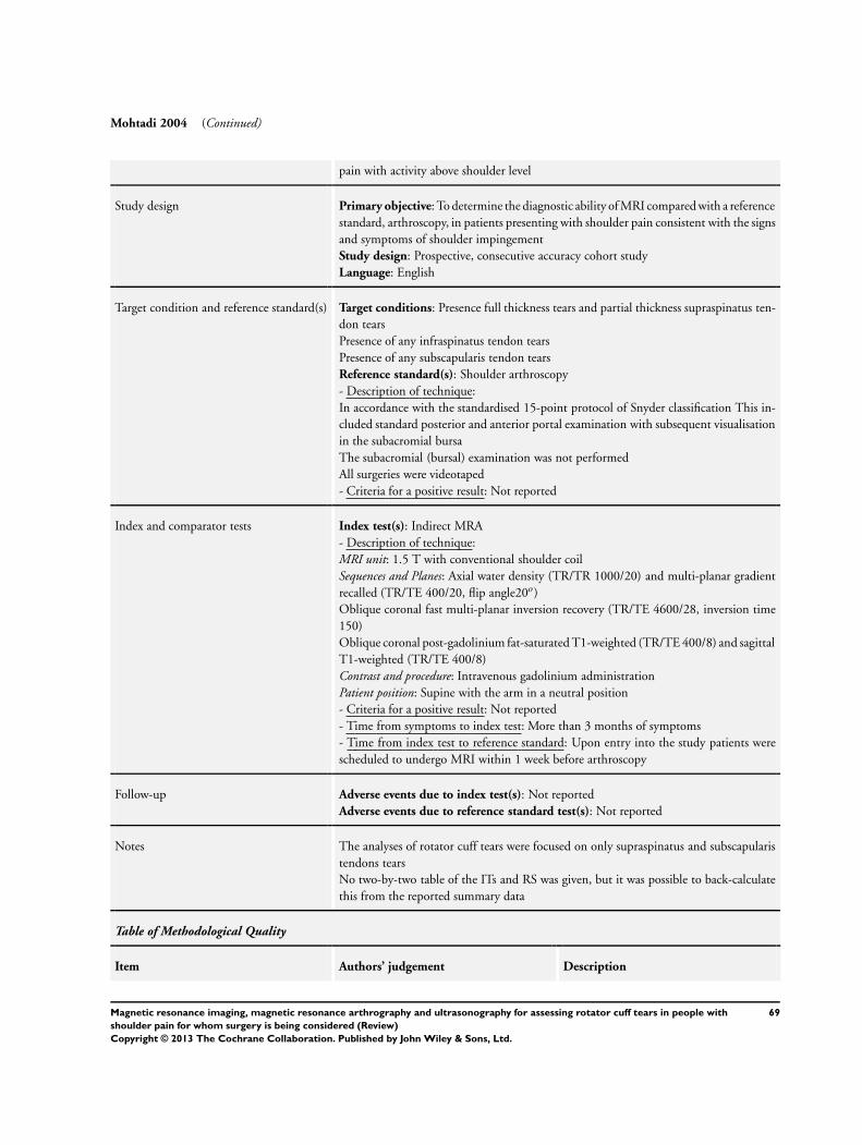
Mohtadi 2004 (Continued)
pain with activity above shoulder level
Study design Primary objective: To determine the diagnostic ability of MRI compared with a referencestandard, arthroscopy, in patients presenting with shoulder pain consistent with the signsand symptoms of shoulder impingementStudy design: Prospective, consecutive accuracy cohort studyLanguage: English
Target condition and reference standard(s) Target conditions: Presence full thickness tears and partial thickness supraspinatus ten-don tearsPresence of any infraspinatus tendon tearsPresence of any subscapularis tendon tearsReference standard(s): Shoulder arthroscopy- Description of technique:In accordance with the standardised 15-point protocol of Snyder classification This in-cluded standard posterior and anterior portal examination with subsequent visualisationin the subacromial bursaThe subacromial (bursal) examination was not performedAll surgeries were videotaped- Criteria for a positive result: Not reported
Index and comparator tests Index test(s): Indirect MRA- Description of technique:MRI unit: 1.5 T with conventional shoulder coilSequences and Planes: Axial water density (TR/TR 1000/20) and multi-planar gradientrecalled (TR/TE 400/20, flip angle20o)Oblique coronal fast multi-planar inversion recovery (TR/TE 4600/28, inversion time150)Oblique coronal post-gadolinium fat-saturated T1-weighted (TR/TE 400/8) and sagittalT1-weighted (TR/TE 400/8)Contrast and procedure: Intravenous gadolinium administrationPatient position: Supine with the arm in a neutral position- Criteria for a positive result: Not reported- Time from symptoms to index test: More than 3 months of symptoms- Time from index test to reference standard: Upon entry into the study patients werescheduled to undergo MRI within 1 week before arthroscopy
Follow-up Adverse events due to index test(s): Not reportedAdverse events due to reference standard test(s): Not reported
Notes The analyses of rotator cuff tears were focused on only supraspinatus and subscapularistendons tearsNo two-by-two table of the ITs and RS was given, but it was possible to back-calculatethis from the reported summary data
Table of Methodological Quality
Item Authors’ judgement Description
69Magnetic resonance imaging, magnetic resonance arthrography and ultrasonography for assessing rotator cuff tears in people with
shoulder pain for whom surgery is being considered (Review)
Copyright © 2013 The Cochrane Collaboration. Published by John Wiley & Sons, Ltd.
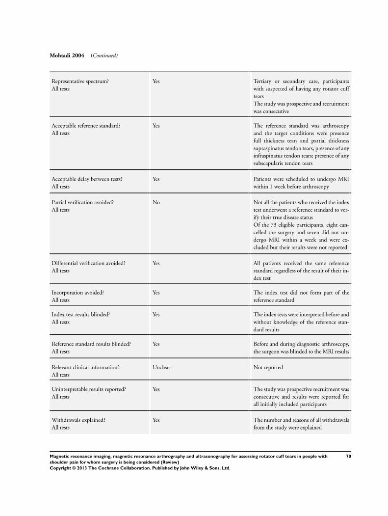
Mohtadi 2004 (Continued)
Representative spectrum?All tests
Yes Tertiary or secondary care, participantswith suspected of having any rotator cufftearsThe study was prospective and recruitmentwas consecutive
Acceptable reference standard?All tests
Yes The reference standard was arthroscopyand the target conditions were presencefull thickness tears and partial thicknesssupraspinatus tendon tears; presence of anyinfraspinatus tendon tears; presence of anysubscapularis tendon tears
Acceptable delay between tests?All tests
Yes Patients were scheduled to undergo MRIwithin 1 week before arthroscopy
Partial verification avoided?All tests
No Not all the patients who received the indextest underwent a reference standard to ver-ify their true disease statusOf the 73 eligible participants, eight can-celled the surgery and seven did not un-dergo MRI within a week and were ex-cluded but their results were not reported
Differential verification avoided?All tests
Yes All patients received the same referencestandard regardless of the result of their in-dex test
Incorporation avoided?All tests
Yes The index test did not form part of thereference standard
Index test results blinded?All tests
Yes The index tests were interpreted before andwithout knowledge of the reference stan-dard results
Reference standard results blinded?All tests
Yes Before and during diagnostic arthroscopy,the surgeon was blinded to the MRI results
Relevant clinical information?All tests
Unclear Not reported
Uninterpretable results reported?All tests
Yes The study was prospective recruitment wasconsecutive and results were reported forall initially included participants
Withdrawals explained?All tests
Yes The number and reasons of all withdrawalsfrom the study were explained
70Magnetic resonance imaging, magnetic resonance arthrography and ultrasonography for assessing rotator cuff tears in people with
shoulder pain for whom surgery is being considered (Review)
Copyright © 2013 The Cochrane Collaboration. Published by John Wiley & Sons, Ltd.
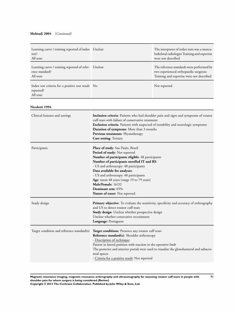
Mohtadi 2004 (Continued)
Learning curve / training reported of indextest?All tests
Unclear The interpreter of index tests was a muscu-loskeletal radiologist Training and expertisewere not described
Learning curve / training reported of refer-ence standard?All tests
Unclear The reference standards were performed bytwo experienced orthopaedic surgeonsTraining and expertise were not described
Index test criteria for a positive test resultreported?All tests
No Not reported
Nicoletti 1994
Clinical features and settings Inclusion criteria: Patients who had shoulder pain and signs and symptoms of rotatorcuff tears with failure of conservative treatmentExclusion criteria: Patients with suspected of instability and neurologic symptomsDuration of symptoms: More than 3 monthsPrevious treatments: PhysiotherapyCare setting: Tertiary
Participants Place of study: Sao Paulo, BrazilPeriod of study: Not reportedNumber of participants eligible: 48 participantsNumber of participants enrolled IT and RS:- US and arthroscopy: 48 participantsData available for analyses:- US and arthroscopy: 48 participantsAge: mean 48 years (range 19 to 79 years)Male/Female: 16/32Dominant arm: 65%Nature of onset: Not reported
Study design Primary objective: To evaluate the sensitivity, specificity and accuracy of arthrographyand US to detect rotator cuff tearsStudy design: Unclear whether prospective designUnclear whether consecutive recruitmentLanguage: Portuguese
Target condition and reference standard(s) Target conditions: Presence any rotator cuff tearsReference standard(s): Shoulder arthroscopy- Description of technique:Patient in lateral position with traction in the operative limbThe posterior and anterior portals were used to visualise the glenohumeral and subacro-mial spaces- Criteria for a positive result: Not reported
71Magnetic resonance imaging, magnetic resonance arthrography and ultrasonography for assessing rotator cuff tears in people with
shoulder pain for whom surgery is being considered (Review)
Copyright © 2013 The Cochrane Collaboration. Published by John Wiley & Sons, Ltd.
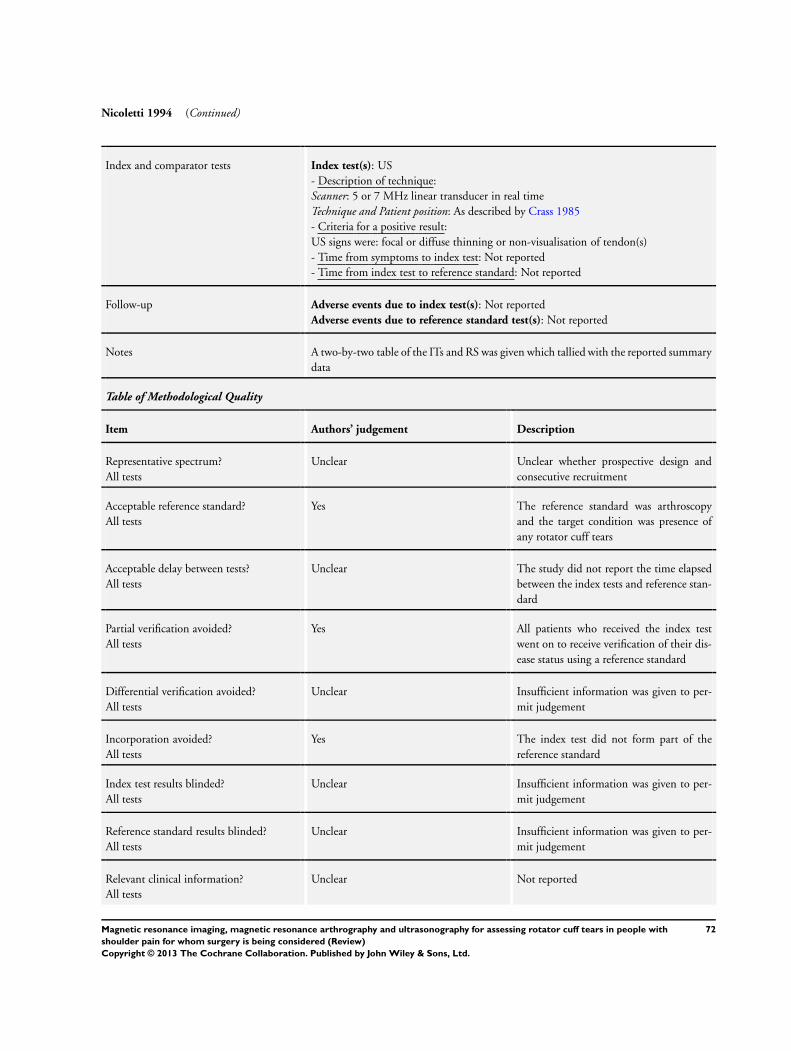
Nicoletti 1994 (Continued)
Index and comparator tests Index test(s): US- Description of technique:Scanner: 5 or 7 MHz linear transducer in real timeTechnique and Patient position: As described by Crass 1985- Criteria for a positive result:US signs were: focal or diffuse thinning or non-visualisation of tendon(s)- Time from symptoms to index test: Not reported- Time from index test to reference standard: Not reported
Follow-up Adverse events due to index test(s): Not reportedAdverse events due to reference standard test(s): Not reported
Notes A two-by-two table of the ITs and RS was given which tallied with the reported summarydata
Table of Methodological Quality
Item Authors’ judgement Description
Representative spectrum?All tests
Unclear Unclear whether prospective design andconsecutive recruitment
Acceptable reference standard?All tests
Yes The reference standard was arthroscopyand the target condition was presence ofany rotator cuff tears
Acceptable delay between tests?All tests
Unclear The study did not report the time elapsedbetween the index tests and reference stan-dard
Partial verification avoided?All tests
Yes All patients who received the index testwent on to receive verification of their dis-ease status using a reference standard
Differential verification avoided?All tests
Unclear Insufficient information was given to per-mit judgement
Incorporation avoided?All tests
Yes The index test did not form part of thereference standard
Index test results blinded?All tests
Unclear Insufficient information was given to per-mit judgement
Reference standard results blinded?All tests
Unclear Insufficient information was given to per-mit judgement
Relevant clinical information?All tests
Unclear Not reported
72Magnetic resonance imaging, magnetic resonance arthrography and ultrasonography for assessing rotator cuff tears in people with
shoulder pain for whom surgery is being considered (Review)
Copyright © 2013 The Cochrane Collaboration. Published by John Wiley & Sons, Ltd.
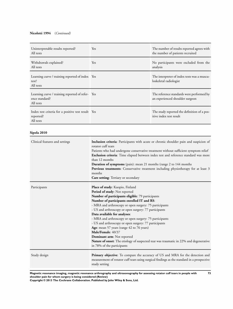
Nicoletti 1994 (Continued)
Uninterpretable results reported?All tests
Yes The number of results reported agrees withthe number of patients recruited
Withdrawals explained?All tests
Yes No participants were excluded from theanalysis
Learning curve / training reported of indextest?All tests
Yes The interpreter of index tests was a muscu-loskeletal radiologist
Learning curve / training reported of refer-ence standard?All tests
Yes The reference standards were performed byan experienced shoulder surgeon
Index test criteria for a positive test resultreported?All tests
Yes The study reported the definition of a pos-itive index test result
Sipola 2010
Clinical features and settings Inclusion criteria: Participants with acute or chronic shoulder pain and suspicion ofrotator cuff tearsPatients who had undergone conservative treatment without sufficient symptom reliefExclusion criteria: Time elapsed between index test and reference standard was morethan 12 monthsDuration of symptoms (pain): mean 21 months (range 2 to 144 monthsPrevious treatments: Conservative treatment including physiotherapy for at least 3monthsCare setting: Tertiary or secondary
Participants Place of study: Kuopio, FinlandPeriod of study: Not reportedNumber of participants eligible: 79 participantsNumber of participants enrolled IT and RS:- MRA and arthroscopy or open surgery: 75 participants- US and arthroscopy or open surgery: 77 participantsData available for analyses:- MRA and arthroscopy or open surgery: 75 participants- US and arthroscopy or open surgery: 77 participantsAge: mean 57 years (range 42 to 76 years)Male/Female: 40/37Dominant arm: Not reportedNature of onset: The etiology of suspected tear was traumatic in 22% and degenerativein 78% of the participants
Study design Primary objective: To compare the accuracy of US and MRA for the detection andmeasurement of rotator cuff tears using surgical findings as the standard in a prospectivestudy setting
73Magnetic resonance imaging, magnetic resonance arthrography and ultrasonography for assessing rotator cuff tears in people with
shoulder pain for whom surgery is being considered (Review)
Copyright © 2013 The Cochrane Collaboration. Published by John Wiley & Sons, Ltd.
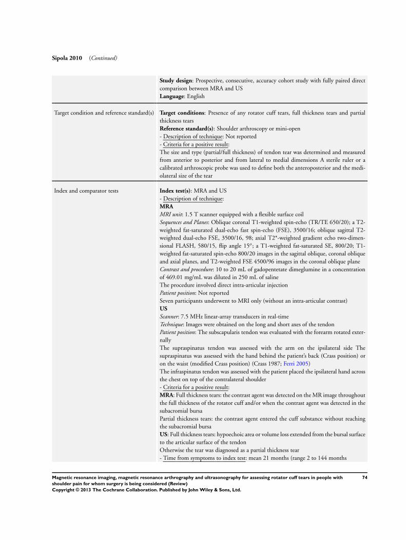
Sipola 2010 (Continued)
Study design: Prospective, consecutive, accuracy cohort study with fully paired directcomparison between MRA and USLanguage: English
Target condition and reference standard(s) Target conditions: Presence of any rotator cuff tears, full thickness tears and partialthickness tearsReference standard(s): Shoulder arthroscopy or mini-open- Description of technique: Not reported- Criteria for a positive result:The size and type (partial/full thickness) of tendon tear was determined and measuredfrom anterior to posterior and from lateral to medial dimensions A sterile ruler or acalibrated arthroscopic probe was used to define both the anteroposterior and the medi-olateral size of the tear
Index and comparator tests Index test(s): MRA and US- Description of technique:MRA
MRI unit: 1.5 T scanner equipped with a flexible surface coilSequences and Planes: Oblique coronal T1-weighted spin-echo (TR/TE 650/20); a T2-weighted fat-saturated dual-echo fast spin-echo (FSE), 3500/16; oblique sagittal T2-weighted dual-echo FSE, 3500/16, 98; axial T2*-weighted gradient echo two-dimen-sional FLASH, 580/15, flip angle 15o ; a T1-weighted fat-saturated SE, 800/20; T1-weighted fat-saturated spin-echo 800/20 images in the sagittal oblique, coronal obliqueand axial planes, and T2-weighted FSE 4500/96 images in the coronal oblique planeContrast and procedure: 10 to 20 mL of gadopentetate dimeglumine in a concentrationof 469.01 mg/mL was diluted in 250 mL of salineThe procedure involved direct intra-articular injectionPatient position: Not reportedSeven participants underwent to MRI only (without an intra-articular contrast)US
Scanner: 7.5 MHz linear-array transducers in real-timeTechnique: Images were obtained on the long and short axes of the tendonPatient position: The subscapularis tendon was evaluated with the forearm rotated exter-nallyThe supraspinatus tendon was assessed with the arm on the ipsilateral side Thesupraspinatus was assessed with the hand behind the patient’s back (Crass position) oron the waist (modified Crass position) (Crass 1987; Ferri 2005)The infraspinatus tendon was assessed with the patient placed the ipsilateral hand acrossthe chest on top of the contralateral shoulder- Criteria for a positive result:MRA: Full thickness tears: the contrast agent was detected on the MR image throughoutthe full thickness of the rotator cuff and/or when the contrast agent was detected in thesubacromial bursaPartial thickness tears: the contrast agent entered the cuff substance without reachingthe subacromial bursaUS: Full thickness tears: hypoechoic area or volume loss extended from the bursal surfaceto the articular surface of the tendonOtherwise the tear was diagnosed as a partial thickness tear- Time from symptoms to index test: mean 21 months (range 2 to 144 months
74Magnetic resonance imaging, magnetic resonance arthrography and ultrasonography for assessing rotator cuff tears in people with
shoulder pain for whom surgery is being considered (Review)
Copyright © 2013 The Cochrane Collaboration. Published by John Wiley & Sons, Ltd.
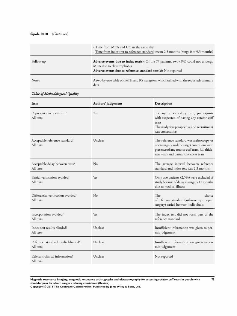
Sipola 2010 (Continued)
- Time from MRA and US: in the same day- Time from index test to reference standard: mean 2.3 months (range 0 to 9.5 months)
Follow-up Adverse events due to index test(s): Of the 77 patients, two (3%) could not undergoMRA due to claustrophobiaAdverse events due to reference standard test(s): Not reported
Notes A two-by-two table of the ITs and RS was given, which tallied with the reported summarydata
Table of Methodological Quality
Item Authors’ judgement Description
Representative spectrum?All tests
Yes Tertiary or secondary care, participantswith suspected of having any rotator cufftearsThe study was prospective and recruitmentwas consecutive
Acceptable reference standard?All tests
Unclear The reference standard was arthroscopy oropen surgery and the target conditions werepresence of any rotator cuff tears, full thick-ness tears and partial thickness tears
Acceptable delay between tests?All tests
No The average interval between referencestandard and index test was 2.3 months
Partial verification avoided?All tests
Yes Only two patients (2.5%) were excluded ofstudy because of delay in surgery 12 monthsdue to medical illness
Differential verification avoided?All tests
No The choiceof reference standard (arthroscopy or opensurgery) varied between individuals
Incorporation avoided?All tests
Yes The index test did not form part of thereference standard
Index test results blinded?All tests
Unclear Insufficient information was given to per-mit judgement
Reference standard results blinded?All tests
Unclear Insufficient information was given to per-mit judgement
Relevant clinical information?All tests
Unclear Not reported
75Magnetic resonance imaging, magnetic resonance arthrography and ultrasonography for assessing rotator cuff tears in people with
shoulder pain for whom surgery is being considered (Review)
Copyright © 2013 The Cochrane Collaboration. Published by John Wiley & Sons, Ltd.
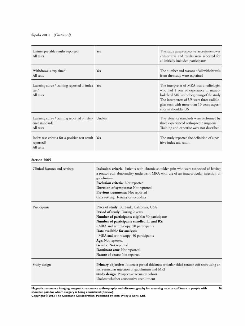
Sipola 2010 (Continued)
Uninterpretable results reported?All tests
Yes The study was prospective, recruitment wasconsecutive and results were reported forall initially included participants
Withdrawals explained?All tests
Yes The number and reasons of all withdrawalsfrom the study were explained
Learning curve / training reported of indextest?All tests
Yes The interpreter of MRA was a radiologistwho had 1 year of experience in muscu-loskeletal MRI at the beginning of the studyThe interpreters of US were three radiolo-gists each with more than 10 years experi-ence in shoulder US
Learning curve / training reported of refer-ence standard?All tests
Unclear The reference standards were performed bythree experienced orthopaedic surgeonsTraining and expertise were not described
Index test criteria for a positive test resultreported?All tests
Yes The study reported the definition of a pos-itive index test result
Stetson 2005
Clinical features and settings Inclusion criteria: Patients with chronic shoulder pain who were suspected of havinga rotator cuff abnormality underwent MRA with use of an intra-articular injection ofgadoliniumExclusion criteria: Not reportedDuration of symptoms: Not reportedPrevious treatments: Not reportedCare setting: Tertiary or secondary
Participants Place of study: Burbank, California, USAPeriod of study: During 2 yearsNumber of participants eligible: 50 participantsNumber of participants enrolled IT and RS:- MRA and arthroscopy: 50 participantsData available for analyses:- MRA and arthroscopy: 50 participantsAge: Not reportedGender: Not reportedDominant arm: Not reportedNature of onset: Not reported
Study design Primary objective: To detect partial thickness articular-sided rotator cuff tears using anintra-articular injection of gadolinium and MRIStudy design: Prospective accuracy cohortUnclear whether consecutive recruitment
76Magnetic resonance imaging, magnetic resonance arthrography and ultrasonography for assessing rotator cuff tears in people with
shoulder pain for whom surgery is being considered (Review)
Copyright © 2013 The Cochrane Collaboration. Published by John Wiley & Sons, Ltd.

Stetson 2005 (Continued)
Language: English
Target condition and reference standard(s) Target conditions: Presence partial articular-side thickness tearsReference standard(s): Shoulder arthroscopy- Description of technique:All participants were taken to surgery and underwent a complete 15-point glenohumeralarthroscopic examinationThe presence or absence of articular-sided rotator cuff tears was recorded- Criteria for a positive result: Not reported
Index and comparator tests Index test(s): MRA- Description of technique:MRI unit: 1.5 T scanner.Sequences and Planes: Axial proton-density-weighted image with fat suppression, obliquecoronal proton-density-weighted image, oblique coronal T2-weighted with fat suppres-sion, oblique sagittal T1-weighted, and oblique sagittal proton-density-weighted imagewith fat suppression. In addition, axial T1-weighted with fat suppression, oblique coro-nal T1-weighted with fat suppressionContrast and procedure: 1.5 mL of gadolinium with normal saline solution intra-articu-larly into the glenohumeral joint under fluoroscopic controlPatient position: Supine in neutral position and abduction and external rotation imageswere also acquired- Criteria for a positive result: Not reported- Time from symptoms to index test: Not reported- Time from index test to reference standard: Not reported
Follow-up Adverse events due to index test(s): Not reportedAdverse events due to reference standard test(s): Not reported
Notes The authors described as false positive the four patients who were incorrectly diagnosedwith MRA, as having a full thickness tear, but, at the time of shoulder arthroscopy, theyhad partial thickness articular-sided tearsTo make concordance with our analyses we described these participants as false negativeNo two-by-two table of the ITs and RS was given, but it was possible to back-calculatethis from the reported summary data
Table of Methodological Quality
Item Authors’ judgement Description
Representative spectrum?All tests
Unclear The study was prospective and the popu-lation was participants with shoulder painand suspected of having any rotator cufftears. However, it was unclear whetherthere was consecutive recruitment
Acceptable reference standard?All tests
Yes The reference standard was arthroscopyand the target condition was presence ofpartial articular-side rotator cuff tears
77Magnetic resonance imaging, magnetic resonance arthrography and ultrasonography for assessing rotator cuff tears in people with
shoulder pain for whom surgery is being considered (Review)
Copyright © 2013 The Cochrane Collaboration. Published by John Wiley & Sons, Ltd.
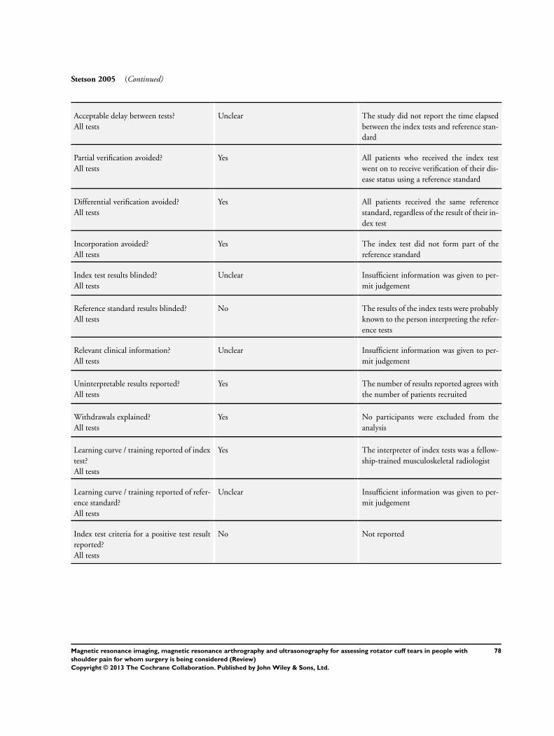
Stetson 2005 (Continued)
Acceptable delay between tests?All tests
Unclear The study did not report the time elapsedbetween the index tests and reference stan-dard
Partial verification avoided?All tests
Yes All patients who received the index testwent on to receive verification of their dis-ease status using a reference standard
Differential verification avoided?All tests
Yes All patients received the same referencestandard, regardless of the result of their in-dex test
Incorporation avoided?All tests
Yes The index test did not form part of thereference standard
Index test results blinded?All tests
Unclear Insufficient information was given to per-mit judgement
Reference standard results blinded?All tests
No The results of the index tests were probablyknown to the person interpreting the refer-ence tests
Relevant clinical information?All tests
Unclear Insufficient information was given to per-mit judgement
Uninterpretable results reported?All tests
Yes The number of results reported agrees withthe number of patients recruited
Withdrawals explained?All tests
Yes No participants were excluded from theanalysis
Learning curve / training reported of indextest?All tests
Yes The interpreter of index tests was a fellow-ship-trained musculoskeletal radiologist
Learning curve / training reported of refer-ence standard?All tests
Unclear Insufficient information was given to per-mit judgement
Index test criteria for a positive test resultreported?All tests
No Not reported
78Magnetic resonance imaging, magnetic resonance arthrography and ultrasonography for assessing rotator cuff tears in people with
shoulder pain for whom surgery is being considered (Review)
Copyright © 2013 The Cochrane Collaboration. Published by John Wiley & Sons, Ltd.
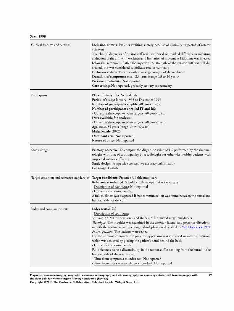
Swen 1998
Clinical features and settings Inclusion criteria: Patients awaiting surgery because of clinically suspected of rotatorcuff tearsThe clinical diagnosis of rotator cuff tears was based on marked difficulty in initiatingabduction of the arm with weakness and limitation of movement Lidocaine was injectedbelow the acromion, if after the injection the strength of the rotator cuff was still de-creased; this was considered to indicate rotator cuff tearsExclusion criteria: Patients with neurologic origins of the weaknessDuration of symptoms: mean 2.3 years (range 0.3 to 10 years)Previous treatments: Not reportedCare setting: Not reported, probably tertiary or secondary
Participants Place of study: The NetherlandsPeriod of study: January 1993 to December 1995Number of participants eligible: 48 participantsNumber of participants enrolled IT and RS:- US and arthroscopy or open surgery: 48 participantsData available for analyses:- US and arthroscopy or open surgery: 48 participantsAge: mean 55 years (range 30 to 76 years)Male/Female: 28/20Dominant arm: Not reportedNature of onset: Not reported
Study design Primary objective: To compare the diagnostic value of US performed by the rheuma-tologist with that of arthrography by a radiologist for otherwise healthy patients withsuspected rotator cuff tearsStudy design: Prospective consecutive accuracy cohort studyLanguage: English
Target condition and reference standard(s) Target conditions: Presence full thickness tearsReference standard(s): Shoulder arthroscopy and open surgery- Description of technique: Not reported- Criteria for a positive result:A full-thickness was diagnosed if free communication was found between the bursal andhumeral sides of the cuff
Index and comparator tests Index test(s): US- Description of technique:Scanner: 7.5 MHz linear array and the 5.0 MHz curved array transducersTechnique: The shoulder was examined in the anterior, lateral, and posterior directions,in both the transverse and the longitudinal planes as described by Van Holsbeeck 1991Patient position: The patients were seatedFor the anterior approach, the patient’s upper arm was visualised in internal rotation,which was achieved by placing the patient’s hand behind the back- Criteria for a positive result:Full thickness tears: a discontinuity in the rotator cuff extending from the bursal to thehumeral side of the rotator cuff- Time from symptoms to index test: Not reported- Time from index test to reference standard: Not reported
79Magnetic resonance imaging, magnetic resonance arthrography and ultrasonography for assessing rotator cuff tears in people with
shoulder pain for whom surgery is being considered (Review)
Copyright © 2013 The Cochrane Collaboration. Published by John Wiley & Sons, Ltd.
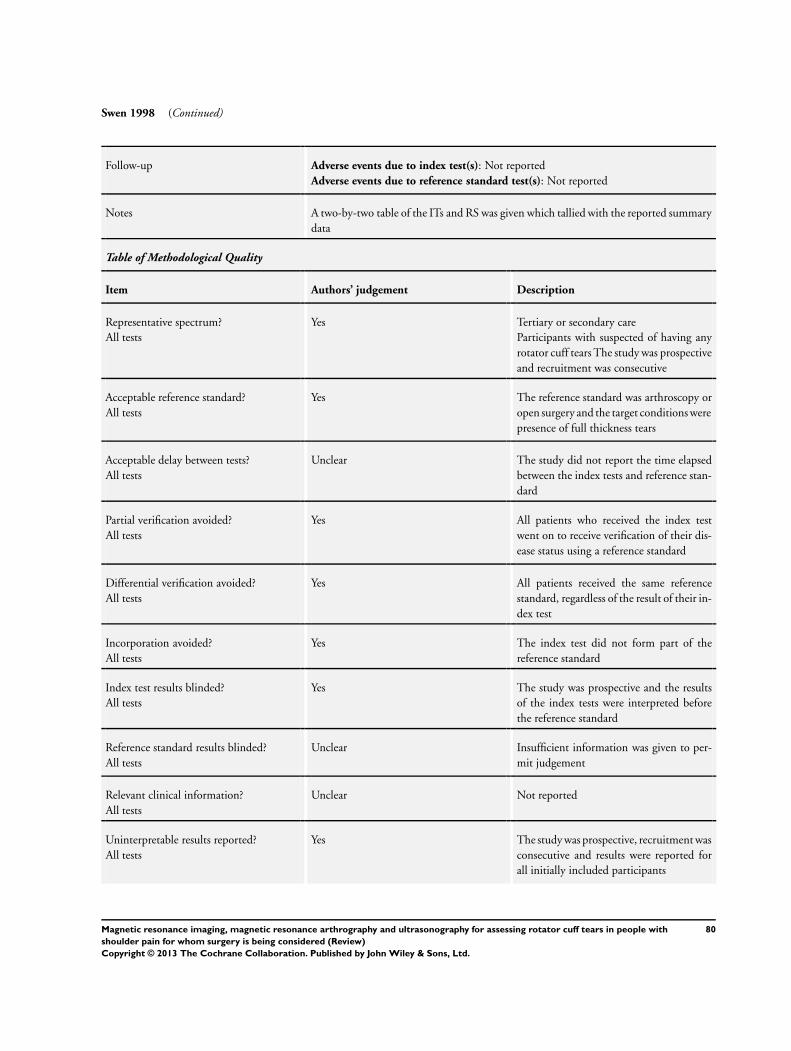
Swen 1998 (Continued)
Follow-up Adverse events due to index test(s): Not reportedAdverse events due to reference standard test(s): Not reported
Notes A two-by-two table of the ITs and RS was given which tallied with the reported summarydata
Table of Methodological Quality
Item Authors’ judgement Description
Representative spectrum?All tests
Yes Tertiary or secondary careParticipants with suspected of having anyrotator cuff tears The study was prospectiveand recruitment was consecutive
Acceptable reference standard?All tests
Yes The reference standard was arthroscopy oropen surgery and the target conditions werepresence of full thickness tears
Acceptable delay between tests?All tests
Unclear The study did not report the time elapsedbetween the index tests and reference stan-dard
Partial verification avoided?All tests
Yes All patients who received the index testwent on to receive verification of their dis-ease status using a reference standard
Differential verification avoided?All tests
Yes All patients received the same referencestandard, regardless of the result of their in-dex test
Incorporation avoided?All tests
Yes The index test did not form part of thereference standard
Index test results blinded?All tests
Yes The study was prospective and the resultsof the index tests were interpreted beforethe reference standard
Reference standard results blinded?All tests
Unclear Insufficient information was given to per-mit judgement
Relevant clinical information?All tests
Unclear Not reported
Uninterpretable results reported?All tests
Yes The study was prospective, recruitment wasconsecutive and results were reported forall initially included participants
80Magnetic resonance imaging, magnetic resonance arthrography and ultrasonography for assessing rotator cuff tears in people with
shoulder pain for whom surgery is being considered (Review)
Copyright © 2013 The Cochrane Collaboration. Published by John Wiley & Sons, Ltd.
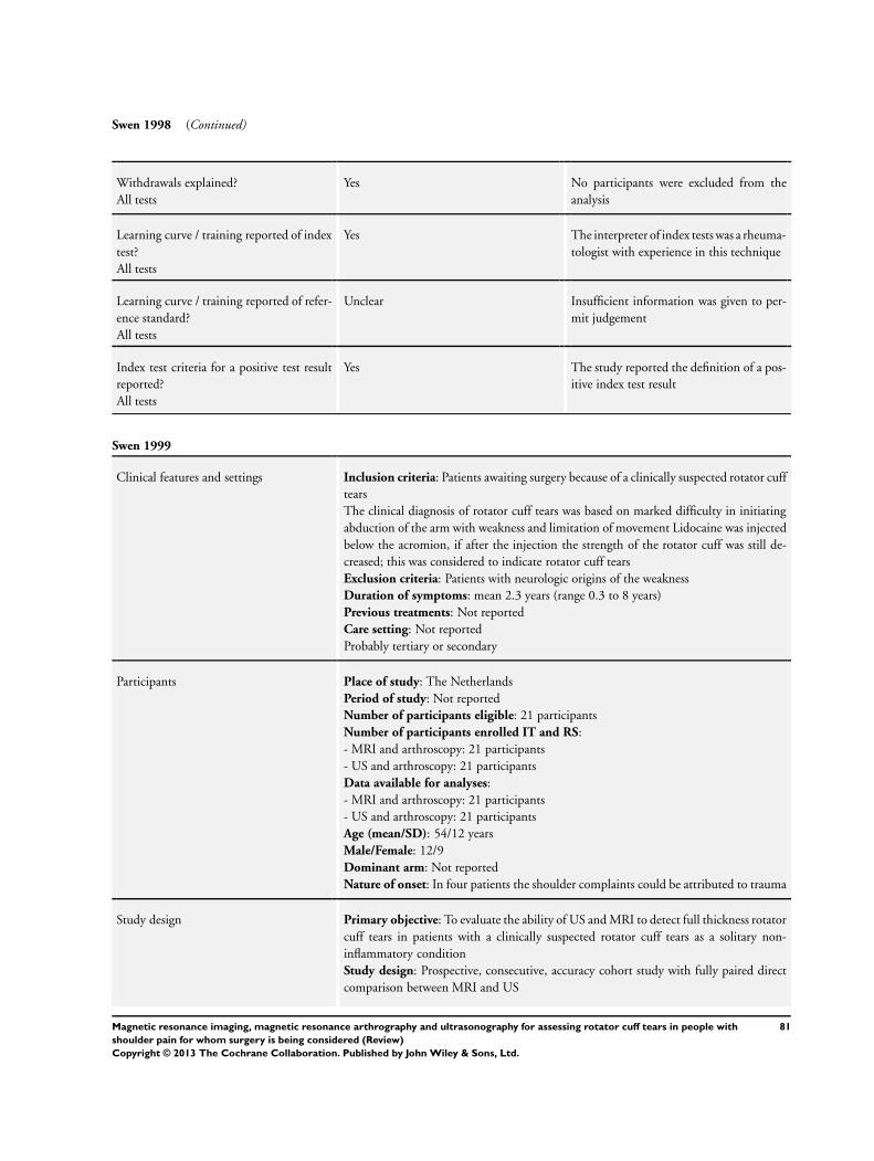
Swen 1998 (Continued)
Withdrawals explained?All tests
Yes No participants were excluded from theanalysis
Learning curve / training reported of indextest?All tests
Yes The interpreter of index tests was a rheuma-tologist with experience in this technique
Learning curve / training reported of refer-ence standard?All tests
Unclear Insufficient information was given to per-mit judgement
Index test criteria for a positive test resultreported?All tests
Yes The study reported the definition of a pos-itive index test result
Swen 1999
Clinical features and settings Inclusion criteria: Patients awaiting surgery because of a clinically suspected rotator cufftearsThe clinical diagnosis of rotator cuff tears was based on marked difficulty in initiatingabduction of the arm with weakness and limitation of movement Lidocaine was injectedbelow the acromion, if after the injection the strength of the rotator cuff was still de-creased; this was considered to indicate rotator cuff tearsExclusion criteria: Patients with neurologic origins of the weaknessDuration of symptoms: mean 2.3 years (range 0.3 to 8 years)Previous treatments: Not reportedCare setting: Not reportedProbably tertiary or secondary
Participants Place of study: The NetherlandsPeriod of study: Not reportedNumber of participants eligible: 21 participantsNumber of participants enrolled IT and RS:- MRI and arthroscopy: 21 participants- US and arthroscopy: 21 participantsData available for analyses:- MRI and arthroscopy: 21 participants- US and arthroscopy: 21 participantsAge (mean/SD): 54/12 yearsMale/Female: 12/9Dominant arm: Not reportedNature of onset: In four patients the shoulder complaints could be attributed to trauma
Study design Primary objective: To evaluate the ability of US and MRI to detect full thickness rotatorcuff tears in patients with a clinically suspected rotator cuff tears as a solitary non-inflammatory conditionStudy design: Prospective, consecutive, accuracy cohort study with fully paired directcomparison between MRI and US
81Magnetic resonance imaging, magnetic resonance arthrography and ultrasonography for assessing rotator cuff tears in people with
shoulder pain for whom surgery is being considered (Review)
Copyright © 2013 The Cochrane Collaboration. Published by John Wiley & Sons, Ltd.

Swen 1999 (Continued)
Language: English
Target condition and reference standard(s) Target conditions: Presence full thickness tearsReference standard(s): Shoulder arthroscopy- Description of technique:First the arthroscope was introduced in the glenohumeral joint and then into the sub-acromial spaceAfter introducing the scope into the subacromial space, the bursa was removed to enableexamination of the bursal side of the cuff- Criteria for a positive result:A full-thickness was diagnosed if free communication was found between the bursal andhumeral sides of the cuff
Index and comparator tests Index test(s): MRI and US- Description of technique:MRI
MRI unit: 1.0T system with a dedicated shoulder coil as receiverSequences: T1-weighted (TR/TE 680/15) and a standard T2 coronal spin-echo sequence(TR/TE 3000/15,105 ms)Planes: Oblique coronalPatient position: Supine positionUS
Scanner: 7.5 MHz linear array and the 5.0 MHz curved array transducersTechnique: The shoulder was examined in the anterior, lateral, and posterior directions,in both the transverse and the longitudinal planes as described by Van Holsbeeck 1991Patient position: The patients were seatedFor the anterior approach, the patient’s upper arm was visualised in internal rotation,which was achieved by placing the patient’s hand behind the back- Criteria for a positive result:MRI: Full-thickness tears: a focal, well-defined area of increased signal intensity on T1-weighted and T2-weighted images that extended through the entire thickness of thetendonUS: Full-thickness tears: a discontinuity in the rotator cuff, extending from the bursalto the humeral side of the rotator cuff- Time from symptoms to index test: Not reported- Time from Conventional MRA and 3D isotropic MRA: Not reported- Time from index test to reference standard: MRI and US were performed within 3weeks before surgery
Follow-up Adverse events due to index test(s): Not reportedAdverse events due to reference standard test(s): Not reported
Notes The results of index test were interpreted by two experienced musculoskeletal radiologistsThe data of only one reader (reader 1) were arbitrarily chosen to be included in ouranalysesRaw data were given and it was possible to back-calculate this from the reported summarydata
Table of Methodological Quality
82Magnetic resonance imaging, magnetic resonance arthrography and ultrasonography for assessing rotator cuff tears in people with
shoulder pain for whom surgery is being considered (Review)
Copyright © 2013 The Cochrane Collaboration. Published by John Wiley & Sons, Ltd.
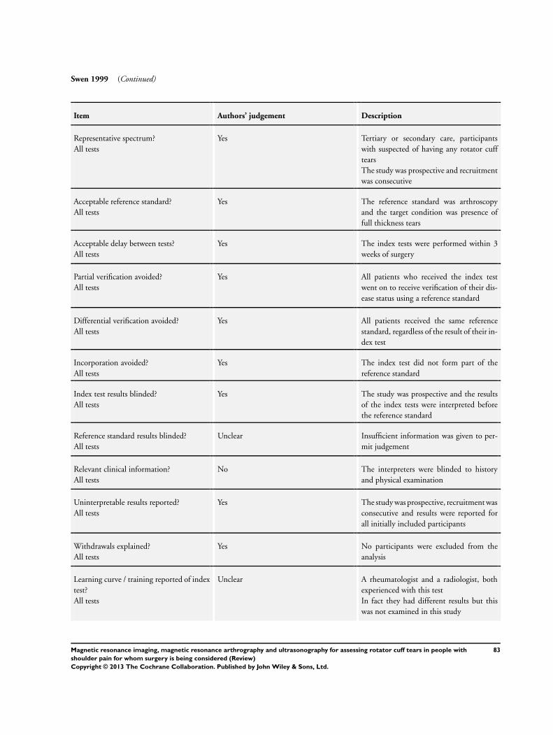
Swen 1999 (Continued)
Item Authors’ judgement Description
Representative spectrum?All tests
Yes Tertiary or secondary care, participantswith suspected of having any rotator cufftearsThe study was prospective and recruitmentwas consecutive
Acceptable reference standard?All tests
Yes The reference standard was arthroscopyand the target condition was presence offull thickness tears
Acceptable delay between tests?All tests
Yes The index tests were performed within 3weeks of surgery
Partial verification avoided?All tests
Yes All patients who received the index testwent on to receive verification of their dis-ease status using a reference standard
Differential verification avoided?All tests
Yes All patients received the same referencestandard, regardless of the result of their in-dex test
Incorporation avoided?All tests
Yes The index test did not form part of thereference standard
Index test results blinded?All tests
Yes The study was prospective and the resultsof the index tests were interpreted beforethe reference standard
Reference standard results blinded?All tests
Unclear Insufficient information was given to per-mit judgement
Relevant clinical information?All tests
No The interpreters were blinded to historyand physical examination
Uninterpretable results reported?All tests
Yes The study was prospective, recruitment wasconsecutive and results were reported forall initially included participants
Withdrawals explained?All tests
Yes No participants were excluded from theanalysis
Learning curve / training reported of indextest?All tests
Unclear A rheumatologist and a radiologist, bothexperienced with this testIn fact they had different results but thiswas not examined in this study
83Magnetic resonance imaging, magnetic resonance arthrography and ultrasonography for assessing rotator cuff tears in people with
shoulder pain for whom surgery is being considered (Review)
Copyright © 2013 The Cochrane Collaboration. Published by John Wiley & Sons, Ltd.
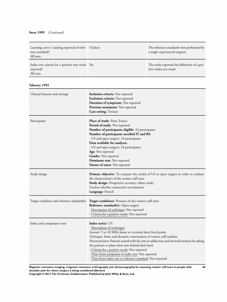
Swen 1999 (Continued)
Learning curve / training reported of refer-ence standard?All tests
Unclear The reference standards were performed bya single experienced surgeon
Index test criteria for a positive test resultreported?All tests
Yes The study reported the definition of a pos-itive index test result
Taboury 1992
Clinical features and settings Inclusion criteria: Not reportedExclusion criteria: Not reportedDuration of symptoms: Not reportedPrevious treatments: Not reportedCare setting: Tertiary
Participants Place of study: Paris, FrancePeriod of study: Not reportedNumber of participants eligible: 24 participantsNumber of participants enrolled IT and RS:- US and open surgery: 24 participantsData available for analyses:- US and open surgery: 24 participantsAge: Not reportedGender: Not reportedDominant arm: Not reportedNature of onset: Not reported
Study design Primary objective: To compare the results of US to open surgery in order to evaluatethe characteristics of the rotator cuff tearsStudy design: Prospective accuracy cohort studyUnclear whether consecutive recruitmentLanguage: French
Target condition and reference standard(s) Target conditions: Presence of any rotator cuff tearsReference standard(s): Open surgery- Description of technique: Not reported- Criteria for a positive result: Not reported
Index and comparator tests Index test(s): US- Description of technique:Scanner: 5 to 10 MHz linear or vectorial short focal probeTechnique: Static and dynamic examination of rotator cuff tendonsPatient position: Patients seated with the arm in adduction and internal rotation by askingthe patients to place their arm behind their back- Criteria for a positive result: Not reported- Time from symptoms to index test: Not reported- Time from index test to reference standard: Not reported
84Magnetic resonance imaging, magnetic resonance arthrography and ultrasonography for assessing rotator cuff tears in people with
shoulder pain for whom surgery is being considered (Review)
Copyright © 2013 The Cochrane Collaboration. Published by John Wiley & Sons, Ltd.
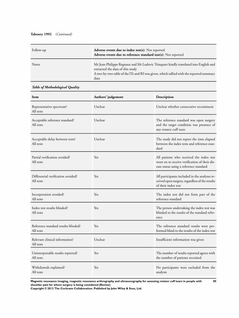
Taboury 1992 (Continued)
Follow-up Adverse events due to index test(s): Not reportedAdverse events due to reference standard test(s): Not reported
Notes Mr Jean-Philippe Regnaux and Mr Ludovic Trinquart kindly translated into English andextracted the data of this studyA two-by-two table of the ITs and RS was given, which tallied with the reported summarydata
Table of Methodological Quality
Item Authors’ judgement Description
Representative spectrum?All tests
Unclear Unclear whether consecutive recruitment
Acceptable reference standard?All tests
Unclear The reference standard was open surgeryand the target condition was presence ofany rotator cuff tears
Acceptable delay between tests?All tests
Unclear The study did not report the time elapsedbetween the index tests and reference stan-dard
Partial verification avoided?All tests
Yes All patients who received the index testwent on to receive verification of their dis-ease status using a reference standard
Differential verification avoided?All tests
Yes All participants included in the analyses re-ceived open surgery, regardless of the resultsof their index test
Incorporation avoided?All tests
Yes The index test did not form part of thereference standard
Index test results blinded?All tests
Yes The person undertaking the index test wasblinded to the results of the standard refer-ence
Reference standard results blinded?All tests
Yes The reference standard results were per-formed blind to the results of the index test
Relevant clinical information?All tests
Unclear Insufficient information was given
Uninterpretable results reported?All tests
Yes The number of results reported agrees withthe number of patients recruited
Withdrawals explained?All tests
Yes No participants were excluded from theanalysis
85Magnetic resonance imaging, magnetic resonance arthrography and ultrasonography for assessing rotator cuff tears in people with
shoulder pain for whom surgery is being considered (Review)
Copyright © 2013 The Cochrane Collaboration. Published by John Wiley & Sons, Ltd.
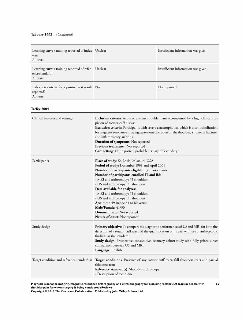
Taboury 1992 (Continued)
Learning curve / training reported of indextest?All tests
Unclear Insufficient information was given
Learning curve / training reported of refer-ence standard?All tests
Unclear Insufficient information was given
Index test criteria for a positive test resultreported?All tests
No Not reported
Teefey 2004
Clinical features and settings Inclusion criteria: Acute or chronic shoulder pain accompanied by a high clinical sus-picion of rotator cuff diseaseExclusion criteria: Participants with severe claustrophobia, which is a contraindicationfor magnetic resonance imaging; a previous operation on the shoulder; a humeral fracture;and inflammatory arthritisDuration of symptoms: Not reportedPrevious treatments: Not reportedCare setting: Not reported, probable tertiary or secondary
Participants Place of study: St. Louis, Missouri, USAPeriod of study: December 1998 and April 2001Number of participants eligible: 130 participantsNumber of participants enrolled IT and RS:- MRI and arthroscopy: 71 shoulders- US and arthroscopy: 71 shouldersData available for analyses:- MRI and arthroscopy: 71 shoulders- US and arthroscopy: 71 shouldersAge: mean 59 (range 31 to 80 years)Male/Female: 41/30Dominant arm: Not reportedNature of onset: Not reported
Study design Primary objective: To compare the diagnostic performances of US and MRI for both thedetection of a rotator cuff tear and the quantification of its size, with use of arthroscopicfindings as the standardStudy design: Prospective, consecutive, accuracy cohort study with fully paired directcomparison between US and MRILanguage: English
Target condition and reference standard(s) Target conditions: Presence of any rotator cuff tears, full thickness tears and partialthickness tearsReference standard(s): Shoulder arthroscopy- Description of technique:
86Magnetic resonance imaging, magnetic resonance arthrography and ultrasonography for assessing rotator cuff tears in people with
shoulder pain for whom surgery is being considered (Review)
Copyright © 2013 The Cochrane Collaboration. Published by John Wiley & Sons, Ltd.
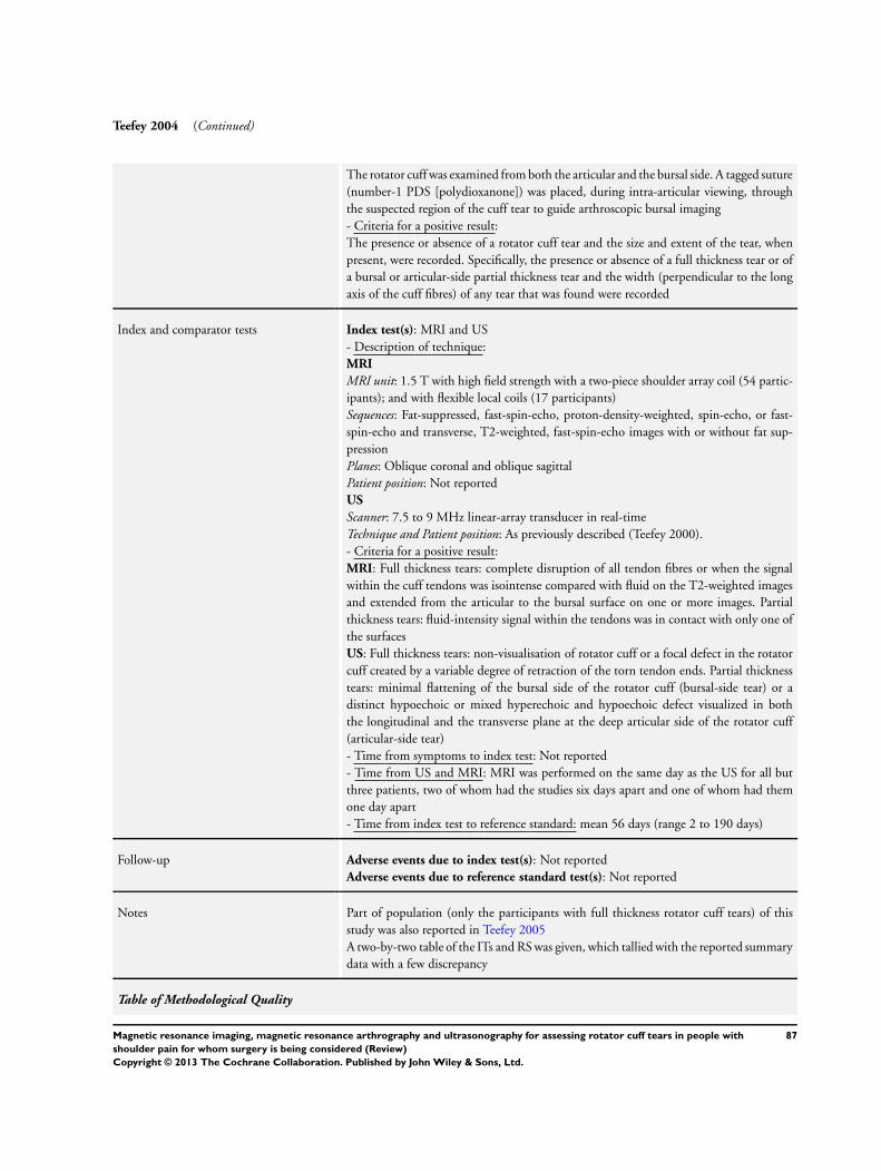
Teefey 2004 (Continued)
The rotator cuff was examined from both the articular and the bursal side. A tagged suture(number-1 PDS [polydioxanone]) was placed, during intra-articular viewing, throughthe suspected region of the cuff tear to guide arthroscopic bursal imaging- Criteria for a positive result:The presence or absence of a rotator cuff tear and the size and extent of the tear, whenpresent, were recorded. Specifically, the presence or absence of a full thickness tear or ofa bursal or articular-side partial thickness tear and the width (perpendicular to the longaxis of the cuff fibres) of any tear that was found were recorded
Index and comparator tests Index test(s): MRI and US- Description of technique:MRI
MRI unit: 1.5 T with high field strength with a two-piece shoulder array coil (54 partic-ipants); and with flexible local coils (17 participants)Sequences: Fat-suppressed, fast-spin-echo, proton-density-weighted, spin-echo, or fast-spin-echo and transverse, T2-weighted, fast-spin-echo images with or without fat sup-pressionPlanes: Oblique coronal and oblique sagittalPatient position: Not reportedUS
Scanner: 7.5 to 9 MHz linear-array transducer in real-timeTechnique and Patient position: As previously described (Teefey 2000).- Criteria for a positive result:MRI: Full thickness tears: complete disruption of all tendon fibres or when the signalwithin the cuff tendons was isointense compared with fluid on the T2-weighted imagesand extended from the articular to the bursal surface on one or more images. Partialthickness tears: fluid-intensity signal within the tendons was in contact with only one ofthe surfacesUS: Full thickness tears: non-visualisation of rotator cuff or a focal defect in the rotatorcuff created by a variable degree of retraction of the torn tendon ends. Partial thicknesstears: minimal flattening of the bursal side of the rotator cuff (bursal-side tear) or adistinct hypoechoic or mixed hyperechoic and hypoechoic defect visualized in boththe longitudinal and the transverse plane at the deep articular side of the rotator cuff(articular-side tear)- Time from symptoms to index test: Not reported- Time from US and MRI: MRI was performed on the same day as the US for all butthree patients, two of whom had the studies six days apart and one of whom had themone day apart- Time from index test to reference standard: mean 56 days (range 2 to 190 days)
Follow-up Adverse events due to index test(s): Not reportedAdverse events due to reference standard test(s): Not reported
Notes Part of population (only the participants with full thickness rotator cuff tears) of thisstudy was also reported in Teefey 2005A two-by-two table of the ITs and RS was given, which tallied with the reported summarydata with a few discrepancy
Table of Methodological Quality
87Magnetic resonance imaging, magnetic resonance arthrography and ultrasonography for assessing rotator cuff tears in people with
shoulder pain for whom surgery is being considered (Review)
Copyright © 2013 The Cochrane Collaboration. Published by John Wiley & Sons, Ltd.
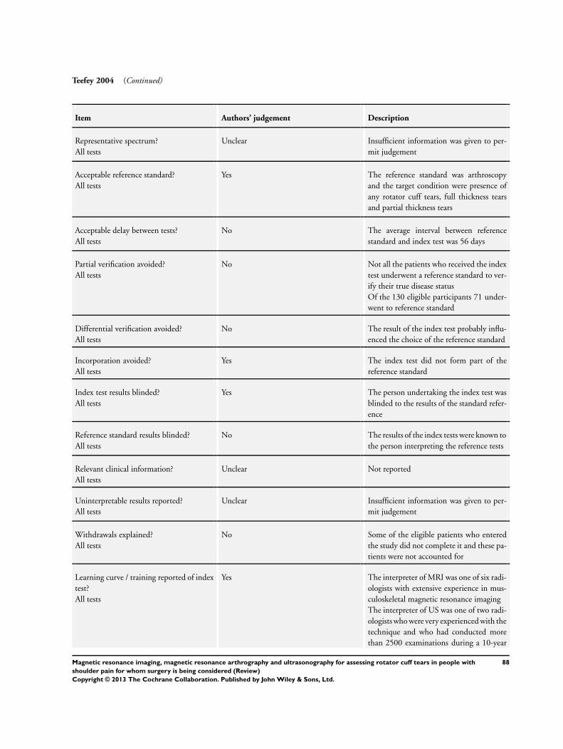
Teefey 2004 (Continued)
Item Authors’ judgement Description
Representative spectrum?All tests
Unclear Insufficient information was given to per-mit judgement
Acceptable reference standard?All tests
Yes The reference standard was arthroscopyand the target condition were presence ofany rotator cuff tears, full thickness tearsand partial thickness tears
Acceptable delay between tests?All tests
No The average interval between referencestandard and index test was 56 days
Partial verification avoided?All tests
No Not all the patients who received the indextest underwent a reference standard to ver-ify their true disease statusOf the 130 eligible participants 71 under-went to reference standard
Differential verification avoided?All tests
No The result of the index test probably influ-enced the choice of the reference standard
Incorporation avoided?All tests
Yes The index test did not form part of thereference standard
Index test results blinded?All tests
Yes The person undertaking the index test wasblinded to the results of the standard refer-ence
Reference standard results blinded?All tests
No The results of the index tests were known tothe person interpreting the reference tests
Relevant clinical information?All tests
Unclear Not reported
Uninterpretable results reported?All tests
Unclear Insufficient information was given to per-mit judgement
Withdrawals explained?All tests
No Some of the eligible patients who enteredthe study did not complete it and these pa-tients were not accounted for
Learning curve / training reported of indextest?All tests
Yes The interpreter of MRI was one of six radi-ologists with extensive experience in mus-culoskeletal magnetic resonance imagingThe interpreter of US was one of two radi-ologists who were very experienced with thetechnique and who had conducted morethan 2500 examinations during a 10-year
88Magnetic resonance imaging, magnetic resonance arthrography and ultrasonography for assessing rotator cuff tears in people with
shoulder pain for whom surgery is being considered (Review)
Copyright © 2013 The Cochrane Collaboration. Published by John Wiley & Sons, Ltd.
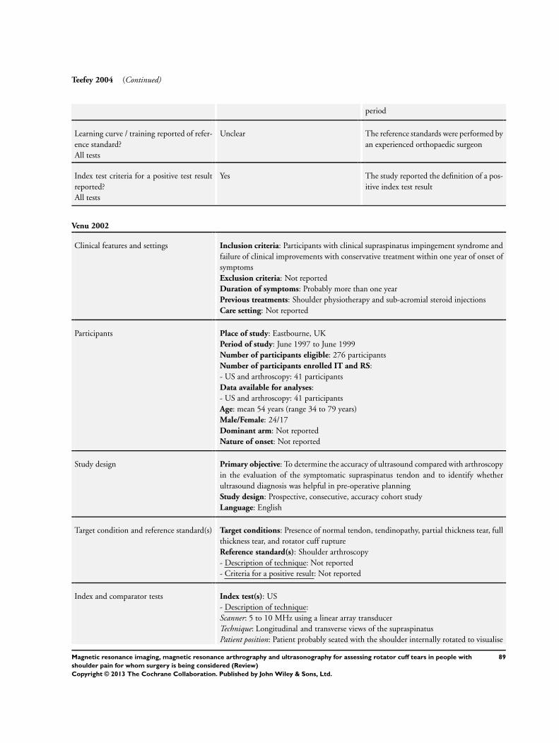
Teefey 2004 (Continued)
period
Learning curve / training reported of refer-ence standard?All tests
Unclear The reference standards were performed byan experienced orthopaedic surgeon
Index test criteria for a positive test resultreported?All tests
Yes The study reported the definition of a pos-itive index test result
Venu 2002
Clinical features and settings Inclusion criteria: Participants with clinical supraspinatus impingement syndrome andfailure of clinical improvements with conservative treatment within one year of onset ofsymptomsExclusion criteria: Not reportedDuration of symptoms: Probably more than one yearPrevious treatments: Shoulder physiotherapy and sub-acromial steroid injectionsCare setting: Not reported
Participants Place of study: Eastbourne, UKPeriod of study: June 1997 to June 1999Number of participants eligible: 276 participantsNumber of participants enrolled IT and RS:- US and arthroscopy: 41 participantsData available for analyses:- US and arthroscopy: 41 participantsAge: mean 54 years (range 34 to 79 years)Male/Female: 24/17Dominant arm: Not reportedNature of onset: Not reported
Study design Primary objective: To determine the accuracy of ultrasound compared with arthroscopyin the evaluation of the symptomatic supraspinatus tendon and to identify whetherultrasound diagnosis was helpful in pre-operative planningStudy design: Prospective, consecutive, accuracy cohort studyLanguage: English
Target condition and reference standard(s) Target conditions: Presence of normal tendon, tendinopathy, partial thickness tear, fullthickness tear, and rotator cuff ruptureReference standard(s): Shoulder arthroscopy- Description of technique: Not reported- Criteria for a positive result: Not reported
Index and comparator tests Index test(s): US- Description of technique:Scanner: 5 to 10 MHz using a linear array transducerTechnique: Longitudinal and transverse views of the supraspinatusPatient position: Patient probably seated with the shoulder internally rotated to visualise
89Magnetic resonance imaging, magnetic resonance arthrography and ultrasonography for assessing rotator cuff tears in people with
shoulder pain for whom surgery is being considered (Review)
Copyright © 2013 The Cochrane Collaboration. Published by John Wiley & Sons, Ltd.
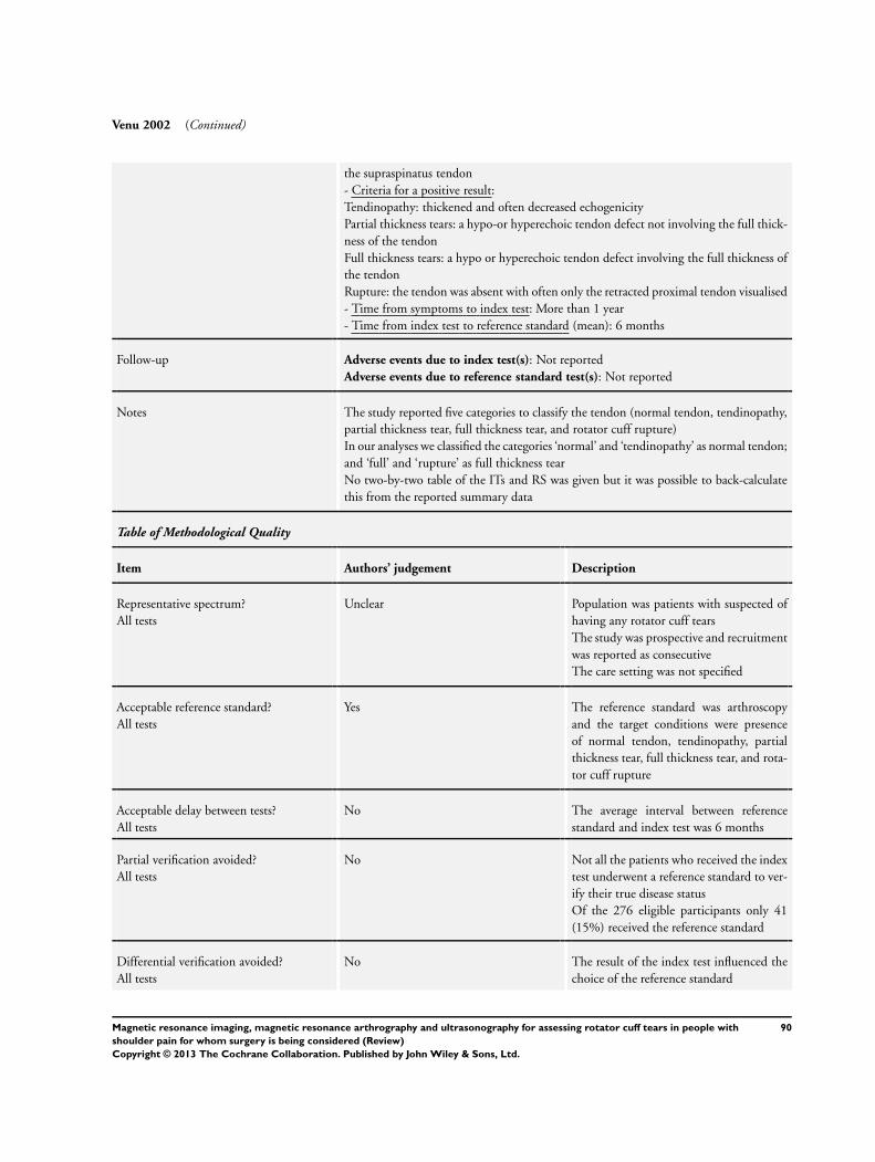
Venu 2002 (Continued)
the supraspinatus tendon- Criteria for a positive result:Tendinopathy: thickened and often decreased echogenicityPartial thickness tears: a hypo-or hyperechoic tendon defect not involving the full thick-ness of the tendonFull thickness tears: a hypo or hyperechoic tendon defect involving the full thickness ofthe tendonRupture: the tendon was absent with often only the retracted proximal tendon visualised- Time from symptoms to index test: More than 1 year- Time from index test to reference standard (mean): 6 months
Follow-up Adverse events due to index test(s): Not reportedAdverse events due to reference standard test(s): Not reported
Notes The study reported five categories to classify the tendon (normal tendon, tendinopathy,partial thickness tear, full thickness tear, and rotator cuff rupture)In our analyses we classified the categories ‘normal’ and ‘tendinopathy’ as normal tendon;and ‘full’ and ‘rupture’ as full thickness tearNo two-by-two table of the ITs and RS was given but it was possible to back-calculatethis from the reported summary data
Table of Methodological Quality
Item Authors’ judgement Description
Representative spectrum?All tests
Unclear Population was patients with suspected ofhaving any rotator cuff tearsThe study was prospective and recruitmentwas reported as consecutiveThe care setting was not specified
Acceptable reference standard?All tests
Yes The reference standard was arthroscopyand the target conditions were presenceof normal tendon, tendinopathy, partialthickness tear, full thickness tear, and rota-tor cuff rupture
Acceptable delay between tests?All tests
No The average interval between referencestandard and index test was 6 months
Partial verification avoided?All tests
No Not all the patients who received the indextest underwent a reference standard to ver-ify their true disease statusOf the 276 eligible participants only 41(15%) received the reference standard
Differential verification avoided?All tests
No The result of the index test influenced thechoice of the reference standard
90Magnetic resonance imaging, magnetic resonance arthrography and ultrasonography for assessing rotator cuff tears in people with
shoulder pain for whom surgery is being considered (Review)
Copyright © 2013 The Cochrane Collaboration. Published by John Wiley & Sons, Ltd.
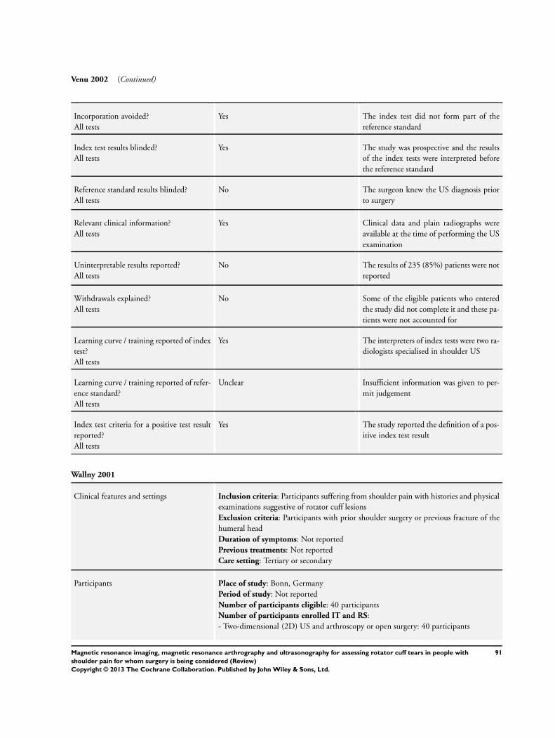
Venu 2002 (Continued)
Incorporation avoided?All tests
Yes The index test did not form part of thereference standard
Index test results blinded?All tests
Yes The study was prospective and the resultsof the index tests were interpreted beforethe reference standard
Reference standard results blinded?All tests
No The surgeon knew the US diagnosis priorto surgery
Relevant clinical information?All tests
Yes Clinical data and plain radiographs wereavailable at the time of performing the USexamination
Uninterpretable results reported?All tests
No The results of 235 (85%) patients were notreported
Withdrawals explained?All tests
No Some of the eligible patients who enteredthe study did not complete it and these pa-tients were not accounted for
Learning curve / training reported of indextest?All tests
Yes The interpreters of index tests were two ra-diologists specialised in shoulder US
Learning curve / training reported of refer-ence standard?All tests
Unclear Insufficient information was given to per-mit judgement
Index test criteria for a positive test resultreported?All tests
Yes The study reported the definition of a pos-itive index test result
Wallny 2001
Clinical features and settings Inclusion criteria: Participants suffering from shoulder pain with histories and physicalexaminations suggestive of rotator cuff lesionsExclusion criteria: Participants with prior shoulder surgery or previous fracture of thehumeral headDuration of symptoms: Not reportedPrevious treatments: Not reportedCare setting: Tertiary or secondary
Participants Place of study: Bonn, GermanyPeriod of study: Not reportedNumber of participants eligible: 40 participantsNumber of participants enrolled IT and RS:- Two-dimensional (2D) US and arthroscopy or open surgery: 40 participants
91Magnetic resonance imaging, magnetic resonance arthrography and ultrasonography for assessing rotator cuff tears in people with
shoulder pain for whom surgery is being considered (Review)
Copyright © 2013 The Cochrane Collaboration. Published by John Wiley & Sons, Ltd.
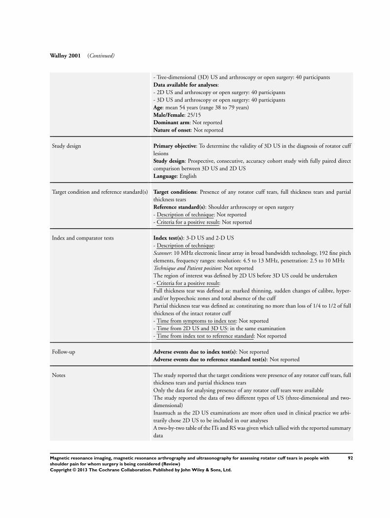
Wallny 2001 (Continued)
- Tree-dimensional (3D) US and arthroscopy or open surgery: 40 participantsData available for analyses:- 2D US and arthroscopy or open surgery: 40 participants- 3D US and arthroscopy or open surgery: 40 participantsAge: mean 54 years (range 38 to 79 years)Male/Female: 25/15Dominant arm: Not reportedNature of onset: Not reported
Study design Primary objective: To determine the validity of 3D US in the diagnosis of rotator cufflesionsStudy design: Prospective, consecutive, accuracy cohort study with fully paired directcomparison between 3D US and 2D USLanguage: English
Target condition and reference standard(s) Target conditions: Presence of any rotator cuff tears, full thickness tears and partialthickness tearsReference standard(s): Shoulder arthroscopy or open surgery- Description of technique: Not reported- Criteria for a positive result: Not reported
Index and comparator tests Index test(s): 3-D US and 2-D US- Description of technique:Scanner: 10 MHz electronic linear array in broad bandwidth technology, 192 fine pitchelements, frequency ranges: resolution: 4.5 to 13 MHz, penetration: 2.5 to 10 MHzTechnique and Patient position: Not reportedThe region of interest was defined by 2D US before 3D US could be undertaken- Criteria for a positive result:Full thickness tear was defined as: marked thinning, sudden changes of calibre, hyper-and/or hypoechoic zones and total absence of the cuffPartial thickness tear was defined as: constituting no more than loss of 1/4 to 1/2 of fullthickness of the intact rotator cuff- Time from symptoms to index test: Not reported- Time from 2D US and 3D US: in the same examination- Time from index test to reference standard: Not reported
Follow-up Adverse events due to index test(s): Not reportedAdverse events due to reference standard test(s): Not reported
Notes The study reported that the target conditions were presence of any rotator cuff tears, fullthickness tears and partial thickness tearsOnly the data for analysing presence of any rotator cuff tears were availableThe study reported the data of two different types of US (three-dimensional and two-dimensional)Inasmuch as the 2D US examinations are more often used in clinical practice we arbi-trarily chose 2D US to be included in our analysesA two-by-two table of the ITs and RS was given which tallied with the reported summarydata
92Magnetic resonance imaging, magnetic resonance arthrography and ultrasonography for assessing rotator cuff tears in people with
shoulder pain for whom surgery is being considered (Review)
Copyright © 2013 The Cochrane Collaboration. Published by John Wiley & Sons, Ltd.
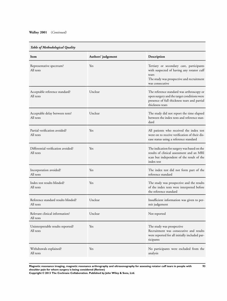
Wallny 2001 (Continued)
Table of Methodological Quality
Item Authors’ judgement Description
Representative spectrum?All tests
Yes Tertiary or secondary care, participantswith suspected of having any rotator cufftearsThe study was prospective and recruitmentwas consecutive
Acceptable reference standard?All tests
Unclear The reference standard was arthroscopy oropen surgery and the target conditions werepresence of full thickness tears and partialthickness tears
Acceptable delay between tests?All tests
Unclear The study did not report the time elapsedbetween the index tests and reference stan-dard
Partial verification avoided?All tests
Yes All patients who received the index testwent on to receive verification of their dis-ease status using a reference standard
Differential verification avoided?All tests
Yes The indication for surgery was based on theresults of clinical assessment and an MRIscan but independent of the result of theindex test
Incorporation avoided?All tests
Yes The index test did not form part of thereference standard
Index test results blinded?All tests
Yes The study was prospective and the resultsof the index tests were interpreted beforethe reference standard
Reference standard results blinded?All tests
Unclear Insufficient information was given to per-mit judgement
Relevant clinical information?All tests
Unclear Not reported
Uninterpretable results reported?All tests
Yes The study was prospectiveRecruitment was consecutive and resultswere reported for all initially included par-ticipants
Withdrawals explained?All tests
Yes No participants were excluded from theanalysis
93Magnetic resonance imaging, magnetic resonance arthrography and ultrasonography for assessing rotator cuff tears in people with
shoulder pain for whom surgery is being considered (Review)
Copyright © 2013 The Cochrane Collaboration. Published by John Wiley & Sons, Ltd.
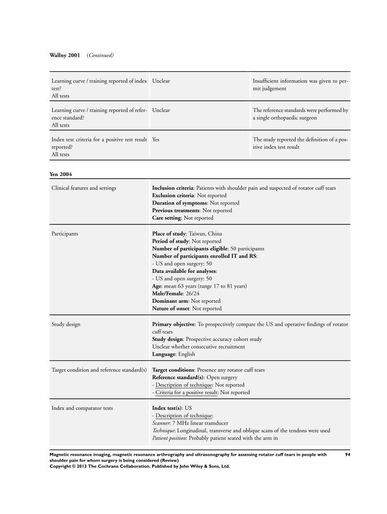
Wallny 2001 (Continued)
Learning curve / training reported of indextest?All tests
Unclear Insufficient information was given to per-mit judgement
Learning curve / training reported of refer-ence standard?All tests
Unclear The reference standards were performed bya single orthopaedic surgeon
Index test criteria for a positive test resultreported?All tests
Yes The study reported the definition of a pos-itive index test result
Yen 2004
Clinical features and settings Inclusion criteria: Patients with shoulder pain and suspected of rotator cuff tearsExclusion criteria: Not reportedDuration of symptoms: Not reportedPrevious treatments: Not reportedCare setting: Not reported
Participants Place of study: Taiwan, ChinaPeriod of study: Not reportedNumber of participants eligible: 50 participantsNumber of participants enrolled IT and RS:- US and open surgery: 50Data available for analyses:- US and open surgery: 50Age: mean 63 years (range 17 to 81 years)Male/Female: 26/24Dominant arm: Not reportedNature of onset: Not reported
Study design Primary objective: To prospectively compare the US and operative findings of rotatorcuff tearsStudy design: Prospective accuracy cohort studyUnclear whether consecutive recruitmentLanguage: English
Target condition and reference standard(s) Target conditions: Presence any rotator cuff tearsReference standard(s): Open surgery- Description of technique: Not reported- Criteria for a positive result: Not reported
Index and comparator tests Index test(s): US- Description of technique:Scanner: 7 MHz linear transducerTechnique: Longitudinal, transverse and oblique scans of the tendons were usedPatient position: Probably patient seated with the arm in
94Magnetic resonance imaging, magnetic resonance arthrography and ultrasonography for assessing rotator cuff tears in people with
shoulder pain for whom surgery is being considered (Review)
Copyright © 2013 The Cochrane Collaboration. Published by John Wiley & Sons, Ltd.
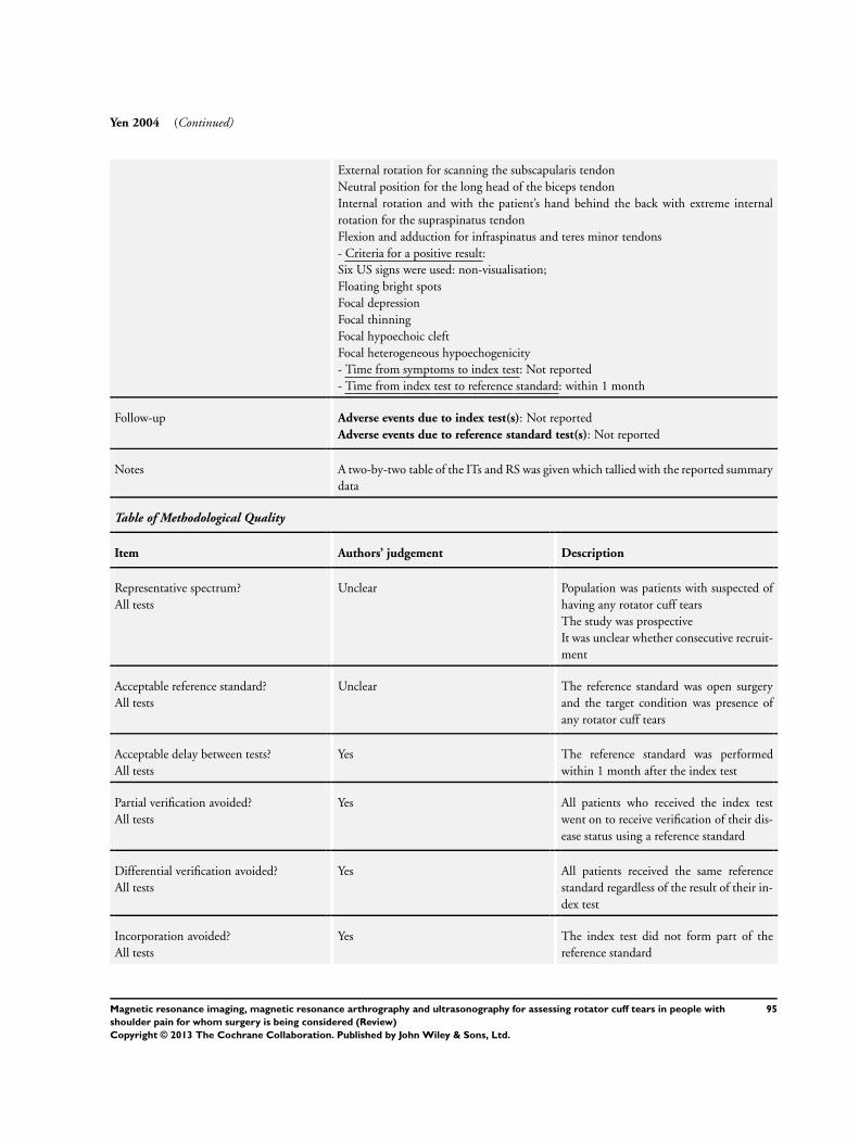
Yen 2004 (Continued)
External rotation for scanning the subscapularis tendonNeutral position for the long head of the biceps tendonInternal rotation and with the patient’s hand behind the back with extreme internalrotation for the supraspinatus tendonFlexion and adduction for infraspinatus and teres minor tendons- Criteria for a positive result:Six US signs were used: non-visualisation;Floating bright spotsFocal depressionFocal thinningFocal hypoechoic cleftFocal heterogeneous hypoechogenicity- Time from symptoms to index test: Not reported- Time from index test to reference standard: within 1 month
Follow-up Adverse events due to index test(s): Not reportedAdverse events due to reference standard test(s): Not reported
Notes A two-by-two table of the ITs and RS was given which tallied with the reported summarydata
Table of Methodological Quality
Item Authors’ judgement Description
Representative spectrum?All tests
Unclear Population was patients with suspected ofhaving any rotator cuff tearsThe study was prospectiveIt was unclear whether consecutive recruit-ment
Acceptable reference standard?All tests
Unclear The reference standard was open surgeryand the target condition was presence ofany rotator cuff tears
Acceptable delay between tests?All tests
Yes The reference standard was performedwithin 1 month after the index test
Partial verification avoided?All tests
Yes All patients who received the index testwent on to receive verification of their dis-ease status using a reference standard
Differential verification avoided?All tests
Yes All patients received the same referencestandard regardless of the result of their in-dex test
Incorporation avoided?All tests
Yes The index test did not form part of thereference standard
95Magnetic resonance imaging, magnetic resonance arthrography and ultrasonography for assessing rotator cuff tears in people with
shoulder pain for whom surgery is being considered (Review)
Copyright © 2013 The Cochrane Collaboration. Published by John Wiley & Sons, Ltd.
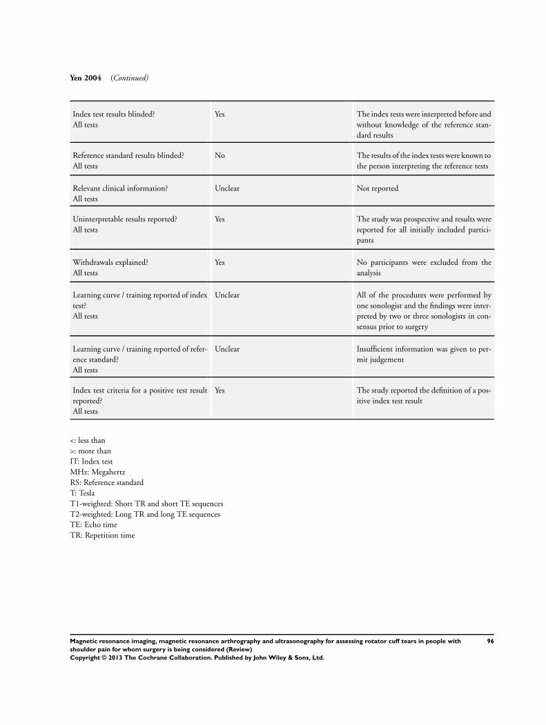
Yen 2004 (Continued)
Index test results blinded?All tests
Yes The index tests were interpreted before andwithout knowledge of the reference stan-dard results
Reference standard results blinded?All tests
No The results of the index tests were known tothe person interpreting the reference tests
Relevant clinical information?All tests
Unclear Not reported
Uninterpretable results reported?All tests
Yes The study was prospective and results werereported for all initially included partici-pants
Withdrawals explained?All tests
Yes No participants were excluded from theanalysis
Learning curve / training reported of indextest?All tests
Unclear All of the procedures were performed byone sonologist and the findings were inter-preted by two or three sonologists in con-sensus prior to surgery
Learning curve / training reported of refer-ence standard?All tests
Unclear Insufficient information was given to per-mit judgement
Index test criteria for a positive test resultreported?All tests
Yes The study reported the definition of a pos-itive index test result
<: less than>: more thanIT: Index testMHz: MegahertzRS: Reference standardT: TeslaT1-weighted: Short TR and short TE sequencesT2-weighted: Long TR and long TE sequencesTE: Echo timeTR: Repetition time
96Magnetic resonance imaging, magnetic resonance arthrography and ultrasonography for assessing rotator cuff tears in people with
shoulder pain for whom surgery is being considered (Review)
Copyright © 2013 The Cochrane Collaboration. Published by John Wiley & Sons, Ltd.
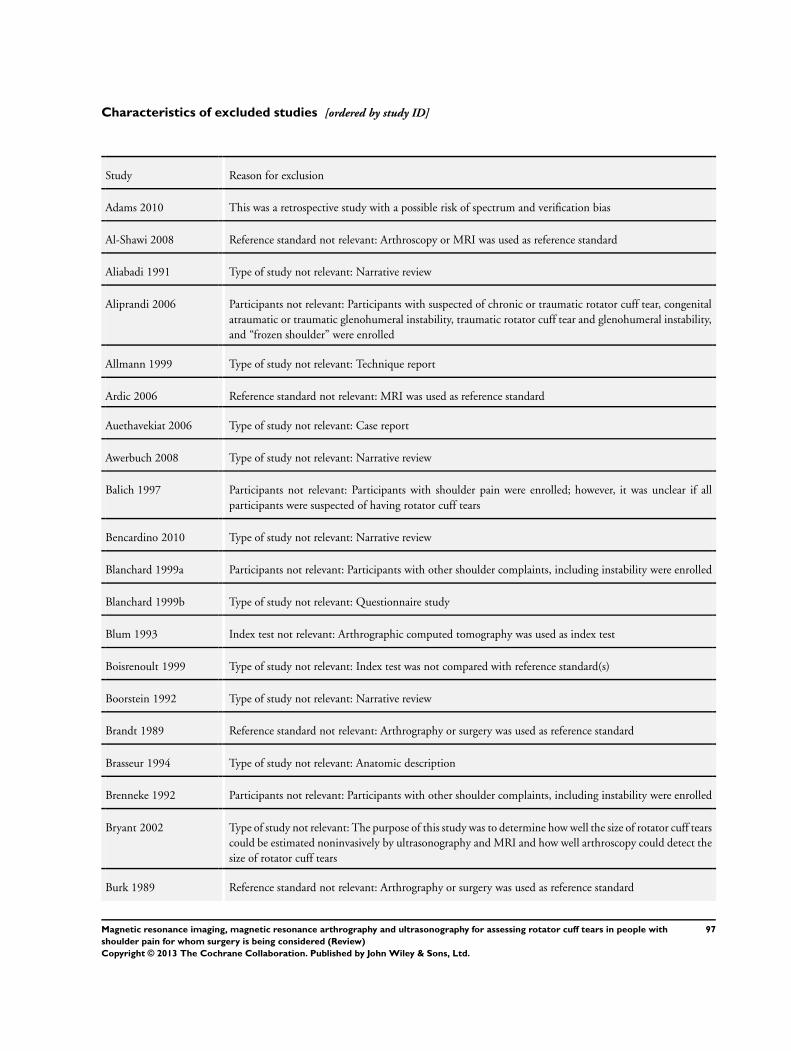
Characteristics of excluded studies [ordered by study ID]
Study Reason for exclusion
Adams 2010 This was a retrospective study with a possible risk of spectrum and verification bias
Al-Shawi 2008 Reference standard not relevant: Arthroscopy or MRI was used as reference standard
Aliabadi 1991 Type of study not relevant: Narrative review
Aliprandi 2006 Participants not relevant: Participants with suspected of chronic or traumatic rotator cuff tear, congenitalatraumatic or traumatic glenohumeral instability, traumatic rotator cuff tear and glenohumeral instability,and “frozen shoulder” were enrolled
Allmann 1999 Type of study not relevant: Technique report
Ardic 2006 Reference standard not relevant: MRI was used as reference standard
Auethavekiat 2006 Type of study not relevant: Case report
Awerbuch 2008 Type of study not relevant: Narrative review
Balich 1997 Participants not relevant: Participants with shoulder pain were enrolled; however, it was unclear if allparticipants were suspected of having rotator cuff tears
Bencardino 2010 Type of study not relevant: Narrative review
Blanchard 1999a Participants not relevant: Participants with other shoulder complaints, including instability were enrolled
Blanchard 1999b Type of study not relevant: Questionnaire study
Blum 1993 Index test not relevant: Arthrographic computed tomography was used as index test
Boisrenoult 1999 Type of study not relevant: Index test was not compared with reference standard(s)
Boorstein 1992 Type of study not relevant: Narrative review
Brandt 1989 Reference standard not relevant: Arthrography or surgery was used as reference standard
Brasseur 1994 Type of study not relevant: Anatomic description
Brenneke 1992 Participants not relevant: Participants with other shoulder complaints, including instability were enrolled
Bryant 2002 Type of study not relevant: The purpose of this study was to determine how well the size of rotator cuff tearscould be estimated noninvasively by ultrasonography and MRI and how well arthroscopy could detect thesize of rotator cuff tears
Burk 1989 Reference standard not relevant: Arthrography or surgery was used as reference standard
97Magnetic resonance imaging, magnetic resonance arthrography and ultrasonography for assessing rotator cuff tears in people with
shoulder pain for whom surgery is being considered (Review)
Copyright © 2013 The Cochrane Collaboration. Published by John Wiley & Sons, Ltd.
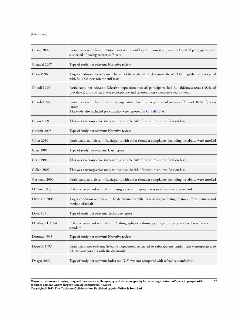
(Continued)
Chang 2002 Participants not relevant: Participants with shoulder pain; however, it was unclear if all participants weresuspected of having rotator cuff tears
Chaubal 2007 Type of study not relevant: Narrative review
Chen 1996 Target condition not relevant: The aim of the study was to determine the MRI findings that are associatedwith full thickness rotator cuff tears
Chiodi 1994 Participants not relevant: Selective population that all participants had full thickness tears (100% ofprevalence) and the study was retrospective and reported non-consecutive recruitment
Chiodi 1995 Participants not relevant: Selective population that all participants had rotator cuff tears (100% of preva-lence)The study also included patients that were reported in Chiodi 1994
Chiou 1999 This was a retrospective study with a possible risk of spectrum and verification bias
Chucair 2008 Type of study not relevant: Narrative review
Chun 2010 Participants not relevant: Participants with other shoulder complaints, including instability were enrolled
Crass 1987 Type of study not relevant: Case report
Crass 1988 This was a retrospective study with a possible risk of spectrum and verification bias
Cullen 2007 This was a retrospective study with a possible risk of spectrum and verification bias
Cusmano 2000 Participants not relevant: Participants with other shoulder complaints, including instability were enrolled
D’Erme 1993 Reference standard not relevant: Surgery or arthrography was used as reference standard
Davidson 2005 Target condition not relevant: To determine the MRI criteria for predicting rotator cuff tear pattern andmethod of repair
Davis 1991 Type of study not relevant: Technique report
De Muynck 1994 Reference standard not relevant: Arthrography or arthroscopy or open surgery was used as referencestandard
Demouy 1993 Type of study not relevant: Narrative review
Deutsch 1997 Participants not relevant: Selective population, restricted to subscapularis tendon tear (retrospective, soselected out patients with the diagnosis)
Dhagat 2002 Type of study not relevant: Index test (US) was not compared with reference standard(s)
98Magnetic resonance imaging, magnetic resonance arthrography and ultrasonography for assessing rotator cuff tears in people with
shoulder pain for whom surgery is being considered (Review)
Copyright © 2013 The Cochrane Collaboration. Published by John Wiley & Sons, Ltd.
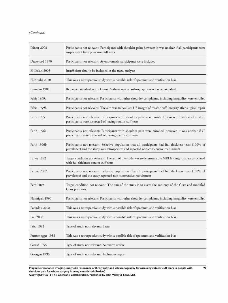
(Continued)
Dinter 2008 Participants not relevant: Participants with shoulder pain; however, it was unclear if all participants weresuspected of having rotator cuff tears
Drakeford 1990 Participants not relevant: Asymptomatic participants were included
El-Dalati 2005 Insufficient data to be included in the meta-analyses
El-Kouba 2010 This was a retrospective study with a possible risk of spectrum and verification bias
Evancho 1988 Reference standard not relevant: Arthroscopy or arthrography as reference standard
Fabis 1999a Participants not relevant: Participants with other shoulder complaints, including instability were enrolled
Fabis 1999b Participants not relevant: The aim was to evaluate US images of rotator cuff integrity after surgical repair
Farin 1995 Participants not relevant: Participants with shoulder pain were enrolled; however, it was unclear if allparticipants were suspected of having rotator cuff tears
Farin 1996a Participants not relevant: Participants with shoulder pain were enrolled; however, it was unclear if allparticipants were suspected of having rotator cuff tears
Farin 1996b Participants not relevant: Selective population that all participants had full thickness tears (100% ofprevalence) and the study was retrospective and reported non-consecutive recruitment
Farley 1992 Target condition not relevant: The aim of the study was to determine the MRI findings that are associatedwith full thickness rotator cuff tears
Ferrari 2002 Participants not relevant: Selective population that all participants had full thickness tears (100% ofprevalence) and the study reported non-consecutive recruitment
Ferri 2005 Target condition not relevant: The aim of the study is to assess the accuracy of the Crass and modifiedCrass positions
Flannigan 1990 Participants not relevant: Participants with other shoulder complaints, including instability were enrolled
Fotiadou 2008 This was a retrospective study with a possible risk of spectrum and verification bias
Frei 2008 This was a retrospective study with a possible risk of spectrum and verification bias
Fritz 1992 Type of study not relevant: Letter
Furtschegger 1988 This was a retrospective study with a possible risk of spectrum and verification bias
Girard 1995 Type of study not relevant: Narrative review
Goergen 1996 Type of study not relevant: Technique report
99Magnetic resonance imaging, magnetic resonance arthrography and ultrasonography for assessing rotator cuff tears in people with
shoulder pain for whom surgery is being considered (Review)
Copyright © 2013 The Cochrane Collaboration. Published by John Wiley & Sons, Ltd.
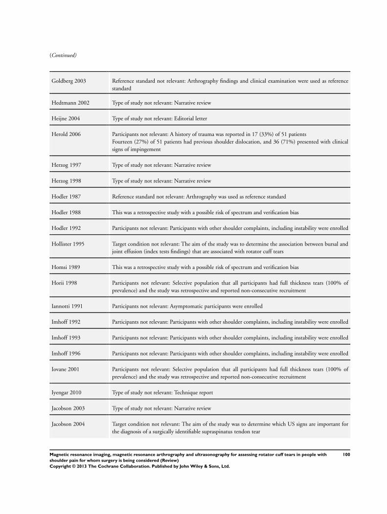
(Continued)
Goldberg 2003 Reference standard not relevant: Arthrography findings and clinical examination were used as referencestandard
Hedtmann 2002 Type of study not relevant: Narrative review
Heijne 2004 Type of study not relevant: Editorial letter
Herold 2006 Participants not relevant: A history of trauma was reported in 17 (33%) of 51 patientsFourteen (27%) of 51 patients had previous shoulder dislocation, and 36 (71%) presented with clinicalsigns of impingement
Herzog 1997 Type of study not relevant: Narrative review
Herzog 1998 Type of study not relevant: Narrative review
Hodler 1987 Reference standard not relevant: Arthrography was used as reference standard
Hodler 1988 This was a retrospective study with a possible risk of spectrum and verification bias
Hodler 1992 Participants not relevant: Participants with other shoulder complaints, including instability were enrolled
Hollister 1995 Target condition not relevant: The aim of the study was to determine the association between bursal andjoint effusion (index tests findings) that are associated with rotator cuff tears
Homsi 1989 This was a retrospective study with a possible risk of spectrum and verification bias
Horii 1998 Participants not relevant: Selective population that all participants had full thickness tears (100% ofprevalence) and the study was retrospective and reported non-consecutive recruitment
Iannotti 1991 Participants not relevant: Asymptomatic participants were enrolled
Imhoff 1992 Participants not relevant: Participants with other shoulder complaints, including instability were enrolled
Imhoff 1993 Participants not relevant: Participants with other shoulder complaints, including instability were enrolled
Imhoff 1996 Participants not relevant: Participants with other shoulder complaints, including instability were enrolled
Iovane 2001 Participants not relevant: Selective population that all participants had full thickness tears (100% ofprevalence) and the study was retrospective and reported non-consecutive recruitment
Iyengar 2010 Type of study not relevant: Technique report
Jacobson 2003 Type of study not relevant: Narrative review
Jacobson 2004 Target condition not relevant: The aim of the study was to determine which US signs are important forthe diagnosis of a surgically identifiable supraspinatus tendon tear
100Magnetic resonance imaging, magnetic resonance arthrography and ultrasonography for assessing rotator cuff tears in people with
shoulder pain for whom surgery is being considered (Review)
Copyright © 2013 The Cochrane Collaboration. Published by John Wiley & Sons, Ltd.
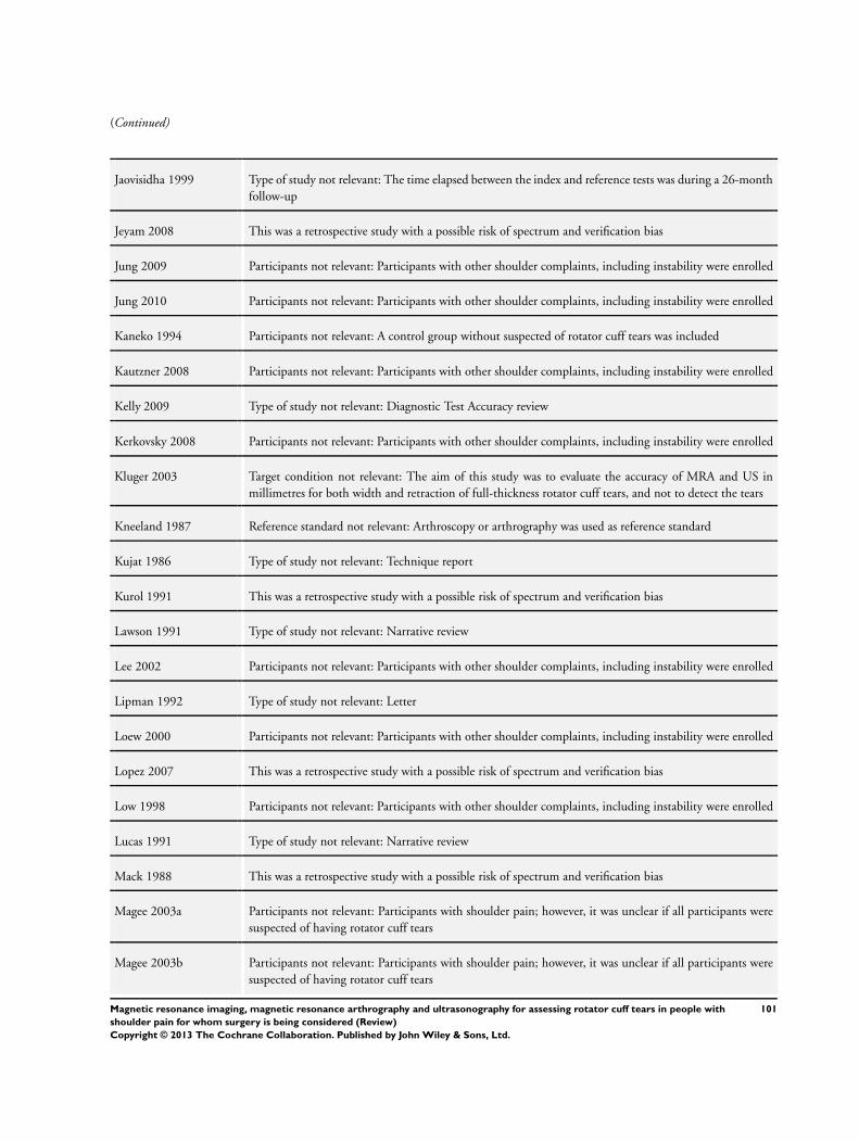
(Continued)
Jaovisidha 1999 Type of study not relevant: The time elapsed between the index and reference tests was during a 26-monthfollow-up
Jeyam 2008 This was a retrospective study with a possible risk of spectrum and verification bias
Jung 2009 Participants not relevant: Participants with other shoulder complaints, including instability were enrolled
Jung 2010 Participants not relevant: Participants with other shoulder complaints, including instability were enrolled
Kaneko 1994 Participants not relevant: A control group without suspected of rotator cuff tears was included
Kautzner 2008 Participants not relevant: Participants with other shoulder complaints, including instability were enrolled
Kelly 2009 Type of study not relevant: Diagnostic Test Accuracy review
Kerkovsky 2008 Participants not relevant: Participants with other shoulder complaints, including instability were enrolled
Kluger 2003 Target condition not relevant: The aim of this study was to evaluate the accuracy of MRA and US inmillimetres for both width and retraction of full-thickness rotator cuff tears, and not to detect the tears
Kneeland 1987 Reference standard not relevant: Arthroscopy or arthrography was used as reference standard
Kujat 1986 Type of study not relevant: Technique report
Kurol 1991 This was a retrospective study with a possible risk of spectrum and verification bias
Lawson 1991 Type of study not relevant: Narrative review
Lee 2002 Participants not relevant: Participants with other shoulder complaints, including instability were enrolled
Lipman 1992 Type of study not relevant: Letter
Loew 2000 Participants not relevant: Participants with other shoulder complaints, including instability were enrolled
Lopez 2007 This was a retrospective study with a possible risk of spectrum and verification bias
Low 1998 Participants not relevant: Participants with other shoulder complaints, including instability were enrolled
Lucas 1991 Type of study not relevant: Narrative review
Mack 1988 This was a retrospective study with a possible risk of spectrum and verification bias
Magee 2003a Participants not relevant: Participants with shoulder pain; however, it was unclear if all participants weresuspected of having rotator cuff tears
Magee 2003b Participants not relevant: Participants with shoulder pain; however, it was unclear if all participants weresuspected of having rotator cuff tears
101Magnetic resonance imaging, magnetic resonance arthrography and ultrasonography for assessing rotator cuff tears in people with
shoulder pain for whom surgery is being considered (Review)
Copyright © 2013 The Cochrane Collaboration. Published by John Wiley & Sons, Ltd.
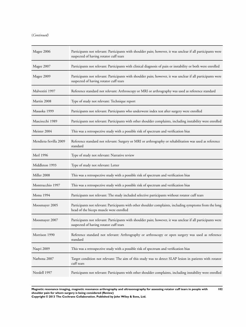
(Continued)
Magee 2006 Participants not relevant: Participants with shoulder pain; however, it was unclear if all participants weresuspected of having rotator cuff tears
Magee 2007 Participants not relevant: Participants with clinical diagnosis of pain or instability or both were enrolled
Magee 2009 Participants not relevant: Participants with shoulder pain; however, it was unclear if all participants weresuspected of having rotator cuff tears
Malvestiti 1997 Reference standard not relevant: Arthroscopy or MRI or arthrography was used as reference standard
Martin 2008 Type of study not relevant: Technique report
Masaoka 1999 Participants not relevant: Participants who underwent index test after surgery were enrolled
Masciocchi 1989 Participants not relevant: Participants with other shoulder complaints, including instability were enrolled
Meister 2004 This was a retrospective study with a possible risk of spectrum and verification bias
Mendieta-Sevilla 2009 Reference standard not relevant: Surgery or MRI or arthrography or rehabilitation was used as referencestandard
Merl 1996 Type of study not relevant: Narrative review
Middleton 1993 Type of study not relevant: Letter
Miller 2008 This was a retrospective study with a possible risk of spectrum and verification bias
Montrucchio 1997 This was a retrospective study with a possible risk of spectrum and verification bias
Monu 1994 Participants not relevant: The study included selective participants without rotator cuff tears
Moosmayer 2005 Participants not relevant: Participants with other shoulder complaints, including symptoms from the longhead of the biceps muscle were enrolled
Moosmayer 2007 Participants not relevant: Participants with shoulder pain; however, it was unclear if all participants weresuspected of having rotator cuff tears
Morrison 1990 Reference standard not relevant: Arthrography or arthroscopy or open surgery was used as referencestandard
Naqvi 2009 This was a retrospective study with a possible risk of spectrum and verification bias
Narbona 2007 Target condition not relevant: The aim of this study was to detect SLAP lesion in patients with rotatorcuff tears
Needell 1997 Participants not relevant: Participants with other shoulder complaints, including instability were enrolled
102Magnetic resonance imaging, magnetic resonance arthrography and ultrasonography for assessing rotator cuff tears in people with
shoulder pain for whom surgery is being considered (Review)
Copyright © 2013 The Cochrane Collaboration. Published by John Wiley & Sons, Ltd.
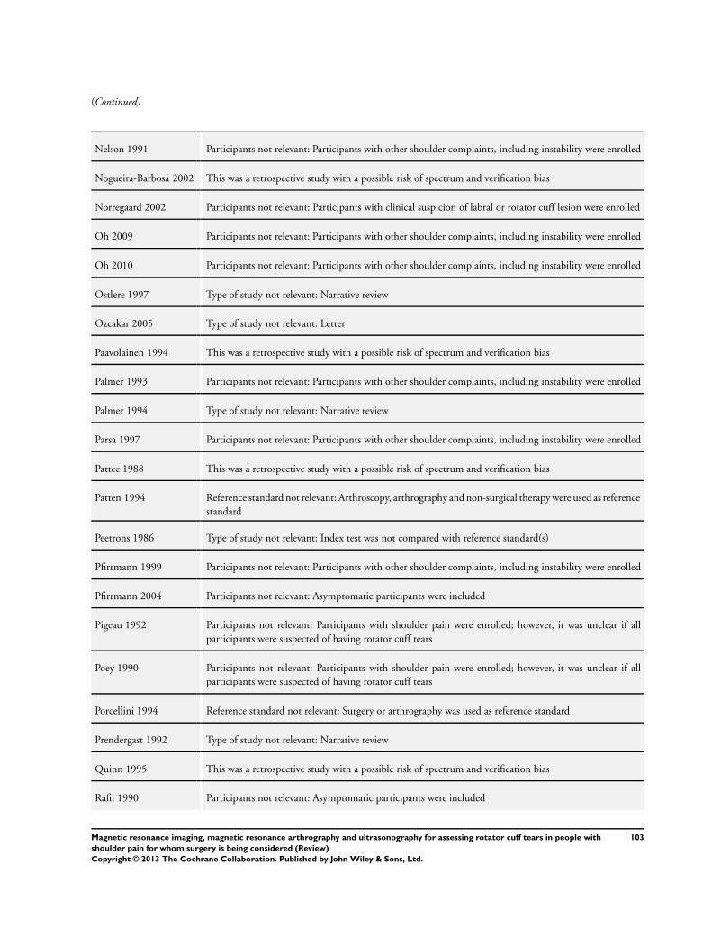
(Continued)
Nelson 1991 Participants not relevant: Participants with other shoulder complaints, including instability were enrolled
Nogueira-Barbosa 2002 This was a retrospective study with a possible risk of spectrum and verification bias
Norregaard 2002 Participants not relevant: Participants with clinical suspicion of labral or rotator cuff lesion were enrolled
Oh 2009 Participants not relevant: Participants with other shoulder complaints, including instability were enrolled
Oh 2010 Participants not relevant: Participants with other shoulder complaints, including instability were enrolled
Ostlere 1997 Type of study not relevant: Narrative review
Ozcakar 2005 Type of study not relevant: Letter
Paavolainen 1994 This was a retrospective study with a possible risk of spectrum and verification bias
Palmer 1993 Participants not relevant: Participants with other shoulder complaints, including instability were enrolled
Palmer 1994 Type of study not relevant: Narrative review
Parsa 1997 Participants not relevant: Participants with other shoulder complaints, including instability were enrolled
Pattee 1988 This was a retrospective study with a possible risk of spectrum and verification bias
Patten 1994 Reference standard not relevant: Arthroscopy, arthrography and non-surgical therapy were used as referencestandard
Peetrons 1986 Type of study not relevant: Index test was not compared with reference standard(s)
Pfirrmann 1999 Participants not relevant: Participants with other shoulder complaints, including instability were enrolled
Pfirrmann 2004 Participants not relevant: Asymptomatic participants were included
Pigeau 1992 Participants not relevant: Participants with shoulder pain were enrolled; however, it was unclear if allparticipants were suspected of having rotator cuff tears
Poey 1990 Participants not relevant: Participants with shoulder pain were enrolled; however, it was unclear if allparticipants were suspected of having rotator cuff tears
Porcellini 1994 Reference standard not relevant: Surgery or arthrography was used as reference standard
Prendergast 1992 Type of study not relevant: Narrative review
Quinn 1995 This was a retrospective study with a possible risk of spectrum and verification bias
Rafii 1990 Participants not relevant: Asymptomatic participants were included
103Magnetic resonance imaging, magnetic resonance arthrography and ultrasonography for assessing rotator cuff tears in people with
shoulder pain for whom surgery is being considered (Review)
Copyright © 2013 The Cochrane Collaboration. Published by John Wiley & Sons, Ltd.
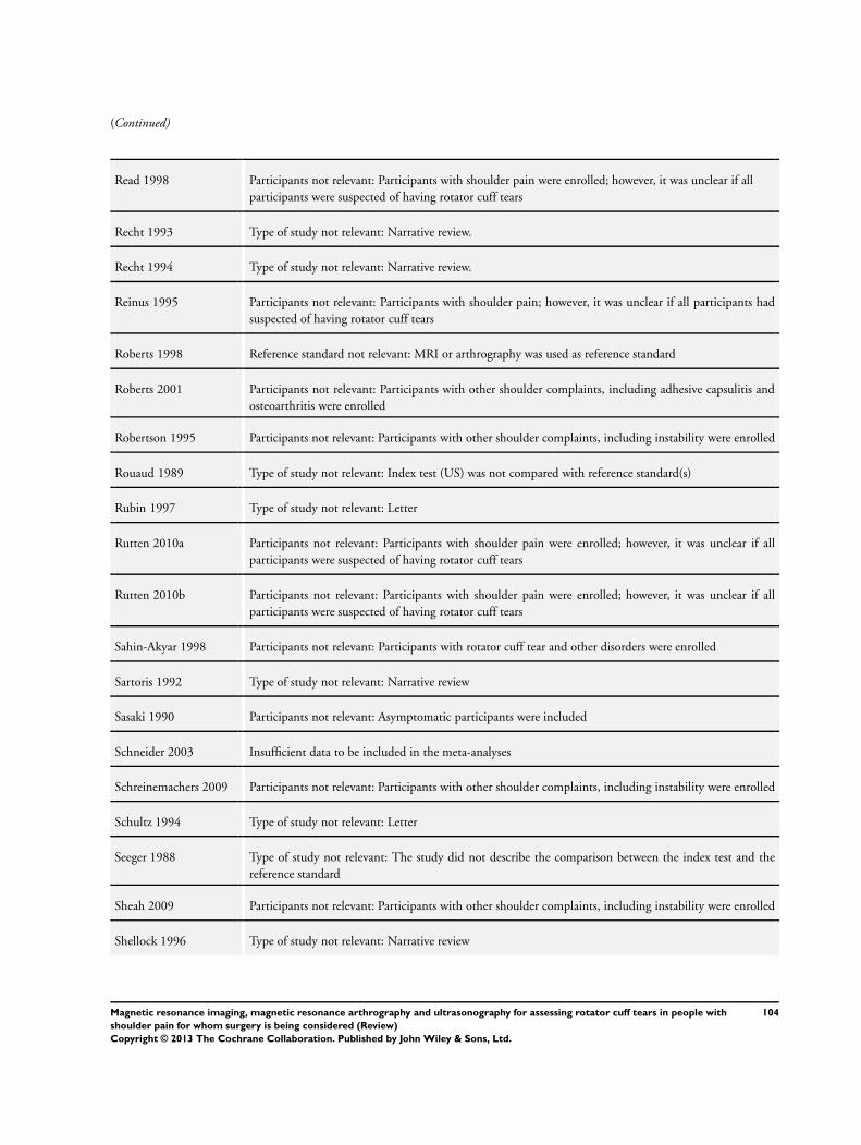
(Continued)
Read 1998 Participants not relevant: Participants with shoulder pain were enrolled; however, it was unclear if allparticipants were suspected of having rotator cuff tears
Recht 1993 Type of study not relevant: Narrative review.
Recht 1994 Type of study not relevant: Narrative review.
Reinus 1995 Participants not relevant: Participants with shoulder pain; however, it was unclear if all participants hadsuspected of having rotator cuff tears
Roberts 1998 Reference standard not relevant: MRI or arthrography was used as reference standard
Roberts 2001 Participants not relevant: Participants with other shoulder complaints, including adhesive capsulitis andosteoarthritis were enrolled
Robertson 1995 Participants not relevant: Participants with other shoulder complaints, including instability were enrolled
Rouaud 1989 Type of study not relevant: Index test (US) was not compared with reference standard(s)
Rubin 1997 Type of study not relevant: Letter
Rutten 2010a Participants not relevant: Participants with shoulder pain were enrolled; however, it was unclear if allparticipants were suspected of having rotator cuff tears
Rutten 2010b Participants not relevant: Participants with shoulder pain were enrolled; however, it was unclear if allparticipants were suspected of having rotator cuff tears
Sahin-Akyar 1998 Participants not relevant: Participants with rotator cuff tear and other disorders were enrolled
Sartoris 1992 Type of study not relevant: Narrative review
Sasaki 1990 Participants not relevant: Asymptomatic participants were included
Schneider 2003 Insufficient data to be included in the meta-analyses
Schreinemachers 2009 Participants not relevant: Participants with other shoulder complaints, including instability were enrolled
Schultz 1994 Type of study not relevant: Letter
Seeger 1988 Type of study not relevant: The study did not describe the comparison between the index test and thereference standard
Sheah 2009 Participants not relevant: Participants with other shoulder complaints, including instability were enrolled
Shellock 1996 Type of study not relevant: Narrative review
104Magnetic resonance imaging, magnetic resonance arthrography and ultrasonography for assessing rotator cuff tears in people with
shoulder pain for whom surgery is being considered (Review)
Copyright © 2013 The Cochrane Collaboration. Published by John Wiley & Sons, Ltd.
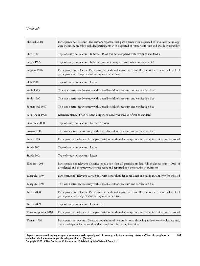
(Continued)
Shellock 2001 Participants not relevant: The authors reported that participants with suspected of ‘shoulder pathology’were included, probable included participants with suspected of rotator cuff tears and shoulder instability
Shiv 1990 Type of study not relevant: Index test (US) was not compared with reference standard(s)
Singer 1995 Type of study not relevant: Index test was not compared with reference standard(s)
Singson 1996 Participants not relevant: Participants with shoulder pain were enrolled; however, it was unclear if allparticipants were suspected of having rotator cuff tears
Skib 1998 Type of study not relevant: Letter
Soble 1989 This was a retrospective study with a possible risk of spectrum and verification bias
Sonin 1996 This was a retrospective study with a possible risk of spectrum and verification bias
Sonnabend 1997 This was a retrospective study with a possible risk of spectrum and verification bias
Soto Araiza 1998 Reference standard not relevant: Surgery or MRI was used as reference standard
Steinbach 2000 Type of study not relevant: Narrative review
Strauss 1998 This was a retrospective study with a possible risk of spectrum and verification bias
Suder 1994 Participants not relevant: Participants with other shoulder complaints, including instability were enrolled
Sunde 2001 Type of study not relevant: Letter
Sunde 2008 Type of study not relevant: Letter
Taboury 1995 Participants not relevant: Selective population that all participants had full thickness tears (100% ofprevalence) and the study was retrospective and reported non-consecutive recruitment
Takagishi 1993 Participants not relevant: Participants with other shoulder complaints, including instability were enrolled
Takagishi 1996 This was a retrospective study with a possible risk of spectrum and verification bias
Teefey 2000 Participants not relevant: Participants with shoulder pain were enrolled; however, it was unclear if allparticipants were suspected of having rotator cuff tears
Teefey 2009 Type of study not relevant: Case report
Theodoropoulos 2010 Participants not relevant: Participants with other shoulder complaints, including instability were enrolled
Tirman 1994 Participants not relevant: Selective population of five professional throwing athletes were evaluated; and,these participants had other shoulder complaints, including instability
105Magnetic resonance imaging, magnetic resonance arthrography and ultrasonography for assessing rotator cuff tears in people with
shoulder pain for whom surgery is being considered (Review)
Copyright © 2013 The Cochrane Collaboration. Published by John Wiley & Sons, Ltd.
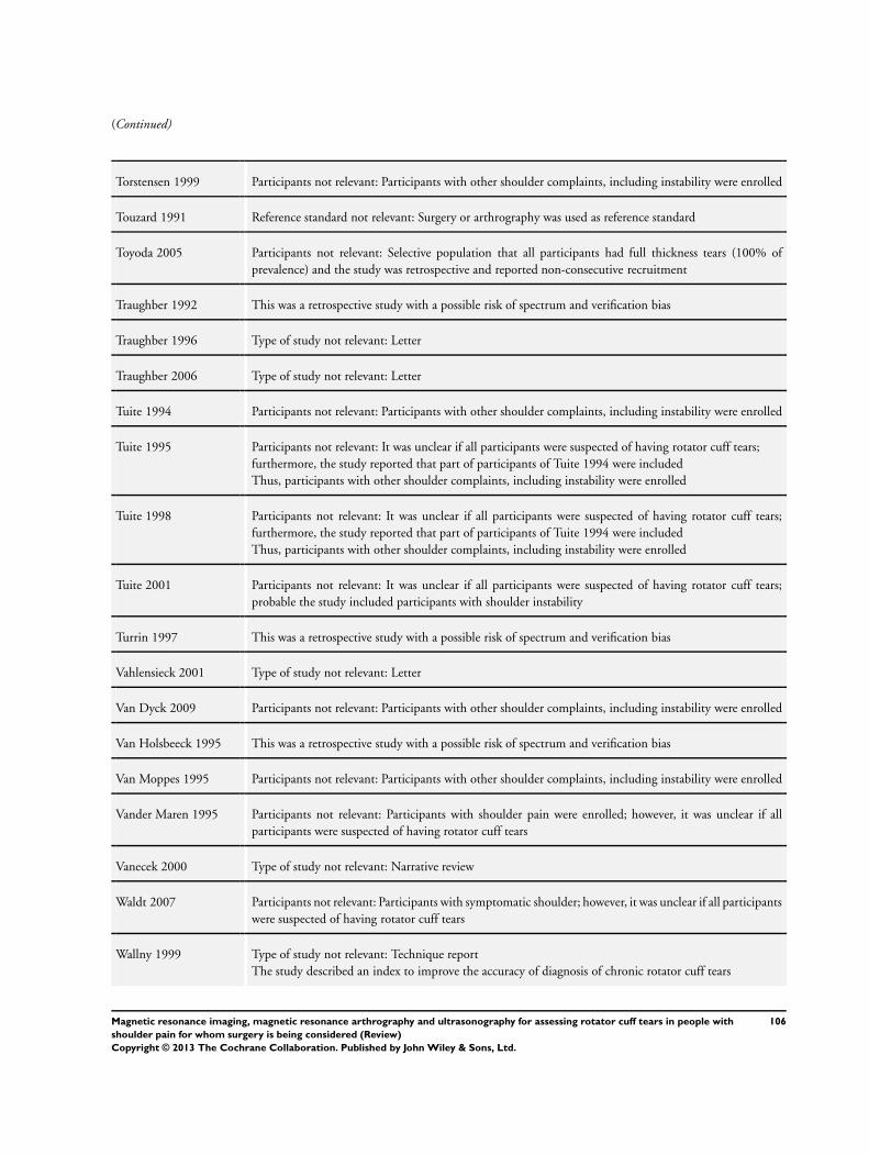
(Continued)
Torstensen 1999 Participants not relevant: Participants with other shoulder complaints, including instability were enrolled
Touzard 1991 Reference standard not relevant: Surgery or arthrography was used as reference standard
Toyoda 2005 Participants not relevant: Selective population that all participants had full thickness tears (100% ofprevalence) and the study was retrospective and reported non-consecutive recruitment
Traughber 1992 This was a retrospective study with a possible risk of spectrum and verification bias
Traughber 1996 Type of study not relevant: Letter
Traughber 2006 Type of study not relevant: Letter
Tuite 1994 Participants not relevant: Participants with other shoulder complaints, including instability were enrolled
Tuite 1995 Participants not relevant: It was unclear if all participants were suspected of having rotator cuff tears;furthermore, the study reported that part of participants of Tuite 1994 were includedThus, participants with other shoulder complaints, including instability were enrolled
Tuite 1998 Participants not relevant: It was unclear if all participants were suspected of having rotator cuff tears;furthermore, the study reported that part of participants of Tuite 1994 were includedThus, participants with other shoulder complaints, including instability were enrolled
Tuite 2001 Participants not relevant: It was unclear if all participants were suspected of having rotator cuff tears;probable the study included participants with shoulder instability
Turrin 1997 This was a retrospective study with a possible risk of spectrum and verification bias
Vahlensieck 2001 Type of study not relevant: Letter
Van Dyck 2009 Participants not relevant: Participants with other shoulder complaints, including instability were enrolled
Van Holsbeeck 1995 This was a retrospective study with a possible risk of spectrum and verification bias
Van Moppes 1995 Participants not relevant: Participants with other shoulder complaints, including instability were enrolled
Vander Maren 1995 Participants not relevant: Participants with shoulder pain were enrolled; however, it was unclear if allparticipants were suspected of having rotator cuff tears
Vanecek 2000 Type of study not relevant: Narrative review
Waldt 2007 Participants not relevant: Participants with symptomatic shoulder; however, it was unclear if all participantswere suspected of having rotator cuff tears
Wallny 1999 Type of study not relevant: Technique reportThe study described an index to improve the accuracy of diagnosis of chronic rotator cuff tears
106Magnetic resonance imaging, magnetic resonance arthrography and ultrasonography for assessing rotator cuff tears in people with
shoulder pain for whom surgery is being considered (Review)
Copyright © 2013 The Cochrane Collaboration. Published by John Wiley & Sons, Ltd.
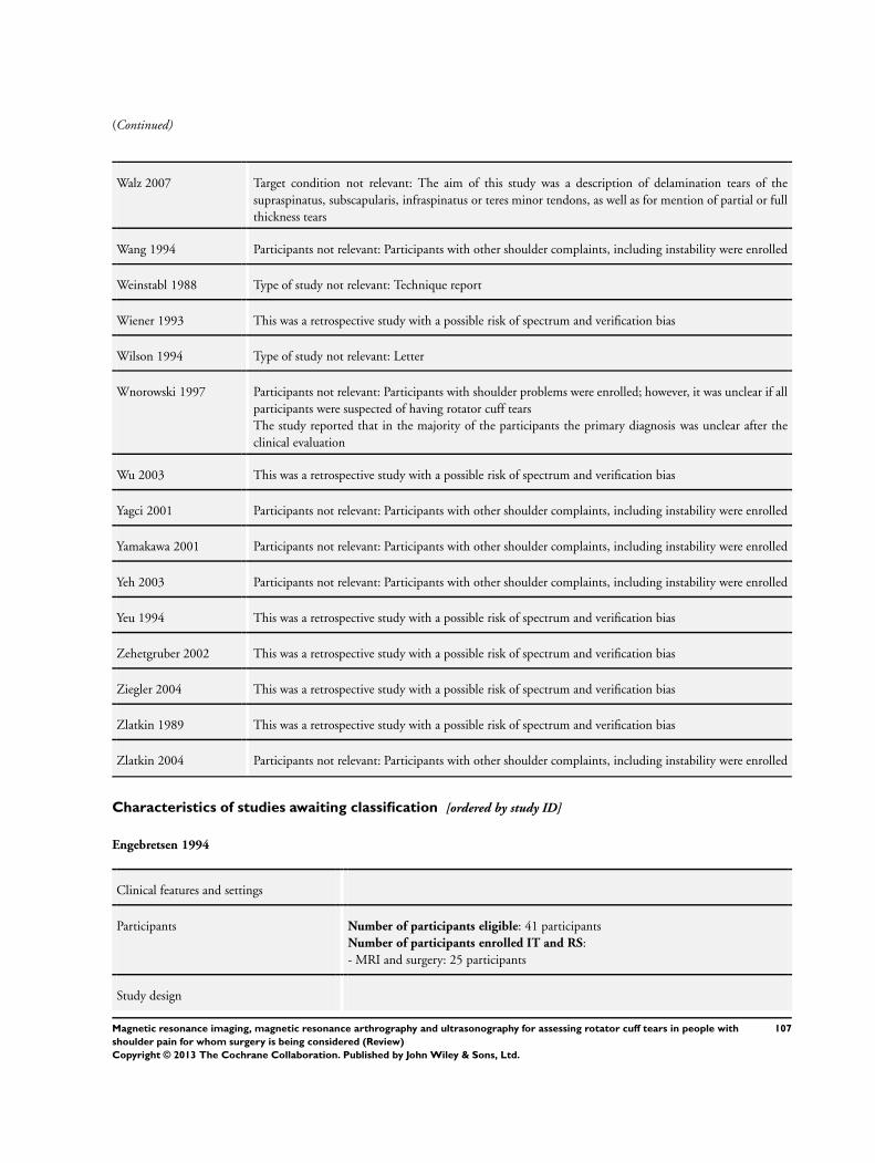
(Continued)
Walz 2007 Target condition not relevant: The aim of this study was a description of delamination tears of thesupraspinatus, subscapularis, infraspinatus or teres minor tendons, as well as for mention of partial or fullthickness tears
Wang 1994 Participants not relevant: Participants with other shoulder complaints, including instability were enrolled
Weinstabl 1988 Type of study not relevant: Technique report
Wiener 1993 This was a retrospective study with a possible risk of spectrum and verification bias
Wilson 1994 Type of study not relevant: Letter
Wnorowski 1997 Participants not relevant: Participants with shoulder problems were enrolled; however, it was unclear if allparticipants were suspected of having rotator cuff tearsThe study reported that in the majority of the participants the primary diagnosis was unclear after theclinical evaluation
Wu 2003 This was a retrospective study with a possible risk of spectrum and verification bias
Yagci 2001 Participants not relevant: Participants with other shoulder complaints, including instability were enrolled
Yamakawa 2001 Participants not relevant: Participants with other shoulder complaints, including instability were enrolled
Yeh 2003 Participants not relevant: Participants with other shoulder complaints, including instability were enrolled
Yeu 1994 This was a retrospective study with a possible risk of spectrum and verification bias
Zehetgruber 2002 This was a retrospective study with a possible risk of spectrum and verification bias
Ziegler 2004 This was a retrospective study with a possible risk of spectrum and verification bias
Zlatkin 1989 This was a retrospective study with a possible risk of spectrum and verification bias
Zlatkin 2004 Participants not relevant: Participants with other shoulder complaints, including instability were enrolled
Characteristics of studies awaiting classification [ordered by study ID]
Engebretsen 1994
Clinical features and settings
Participants Number of participants eligible: 41 participantsNumber of participants enrolled IT and RS:- MRI and surgery: 25 participants
Study design
107Magnetic resonance imaging, magnetic resonance arthrography and ultrasonography for assessing rotator cuff tears in people with
shoulder pain for whom surgery is being considered (Review)
Copyright © 2013 The Cochrane Collaboration. Published by John Wiley & Sons, Ltd.
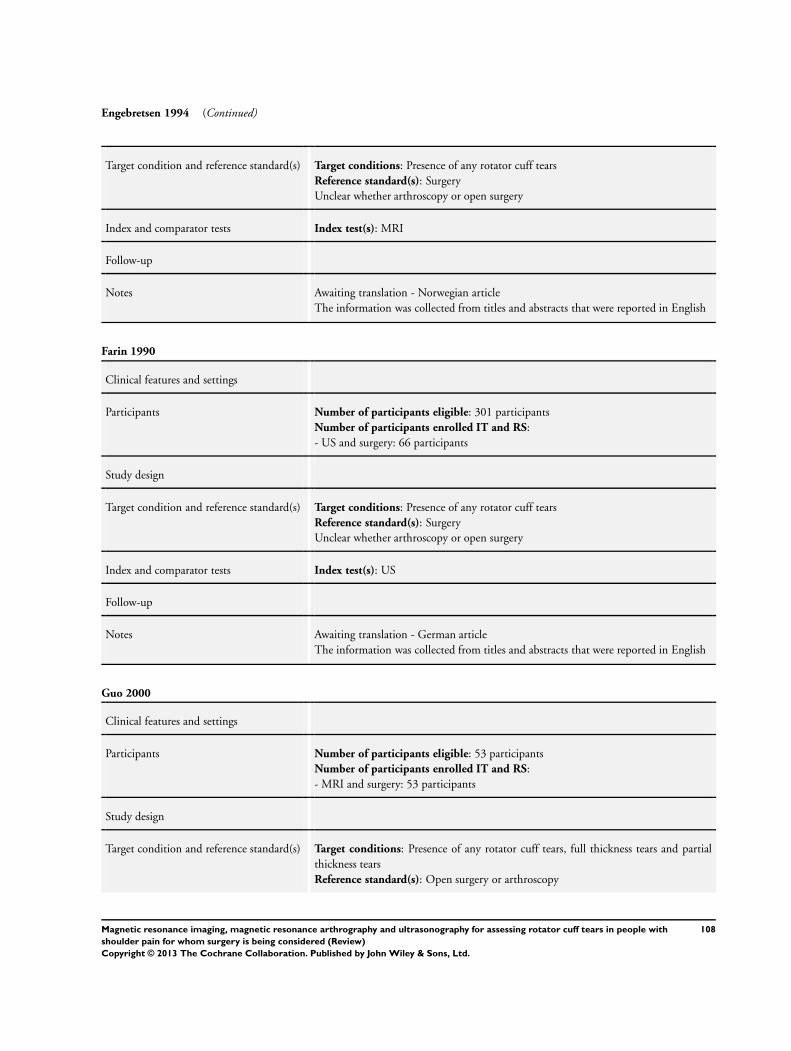
Engebretsen 1994 (Continued)
Target condition and reference standard(s) Target conditions: Presence of any rotator cuff tearsReference standard(s): SurgeryUnclear whether arthroscopy or open surgery
Index and comparator tests Index test(s): MRI
Follow-up
Notes Awaiting translation - Norwegian articleThe information was collected from titles and abstracts that were reported in English
Farin 1990
Clinical features and settings
Participants Number of participants eligible: 301 participantsNumber of participants enrolled IT and RS:- US and surgery: 66 participants
Study design
Target condition and reference standard(s) Target conditions: Presence of any rotator cuff tearsReference standard(s): SurgeryUnclear whether arthroscopy or open surgery
Index and comparator tests Index test(s): US
Follow-up
Notes Awaiting translation - German articleThe information was collected from titles and abstracts that were reported in English
Guo 2000
Clinical features and settings
Participants Number of participants eligible: 53 participantsNumber of participants enrolled IT and RS:- MRI and surgery: 53 participants
Study design
Target condition and reference standard(s) Target conditions: Presence of any rotator cuff tears, full thickness tears and partialthickness tearsReference standard(s): Open surgery or arthroscopy
108Magnetic resonance imaging, magnetic resonance arthrography and ultrasonography for assessing rotator cuff tears in people with
shoulder pain for whom surgery is being considered (Review)
Copyright © 2013 The Cochrane Collaboration. Published by John Wiley & Sons, Ltd.
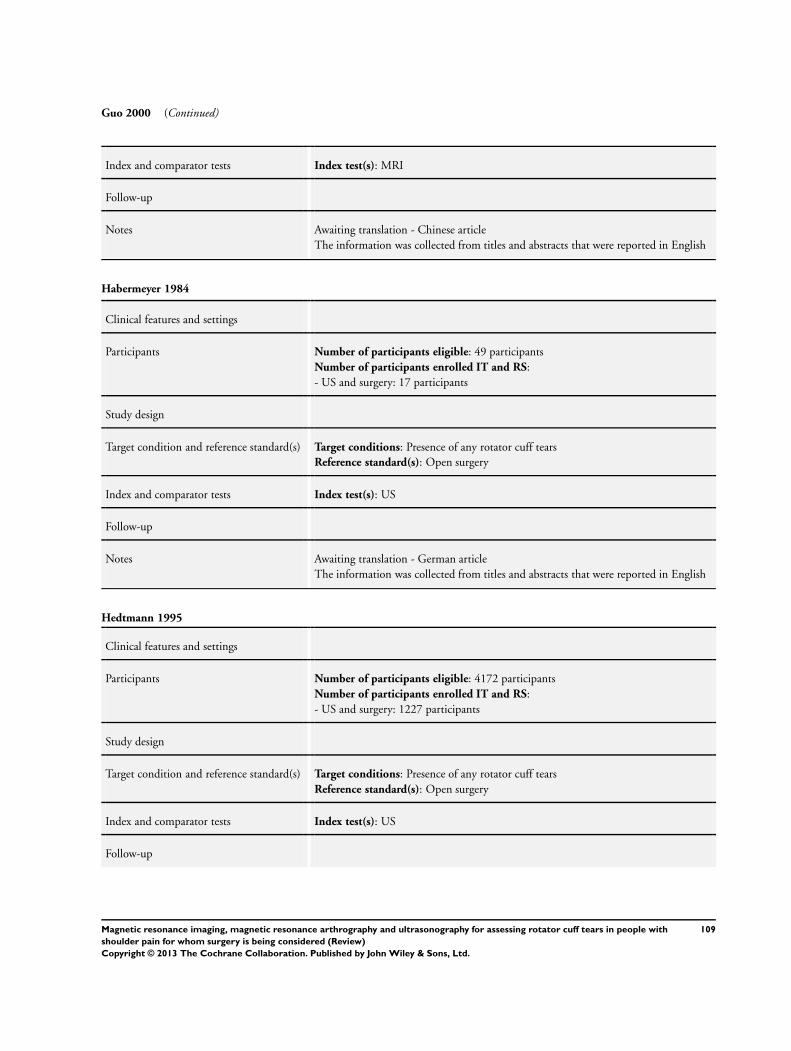
Guo 2000 (Continued)
Index and comparator tests Index test(s): MRI
Follow-up
Notes Awaiting translation - Chinese articleThe information was collected from titles and abstracts that were reported in English
Habermeyer 1984
Clinical features and settings
Participants Number of participants eligible: 49 participantsNumber of participants enrolled IT and RS:- US and surgery: 17 participants
Study design
Target condition and reference standard(s) Target conditions: Presence of any rotator cuff tearsReference standard(s): Open surgery
Index and comparator tests Index test(s): US
Follow-up
Notes Awaiting translation - German articleThe information was collected from titles and abstracts that were reported in English
Hedtmann 1995
Clinical features and settings
Participants Number of participants eligible: 4172 participantsNumber of participants enrolled IT and RS:- US and surgery: 1227 participants
Study design
Target condition and reference standard(s) Target conditions: Presence of any rotator cuff tearsReference standard(s): Open surgery
Index and comparator tests Index test(s): US
Follow-up
109Magnetic resonance imaging, magnetic resonance arthrography and ultrasonography for assessing rotator cuff tears in people with
shoulder pain for whom surgery is being considered (Review)
Copyright © 2013 The Cochrane Collaboration. Published by John Wiley & Sons, Ltd.
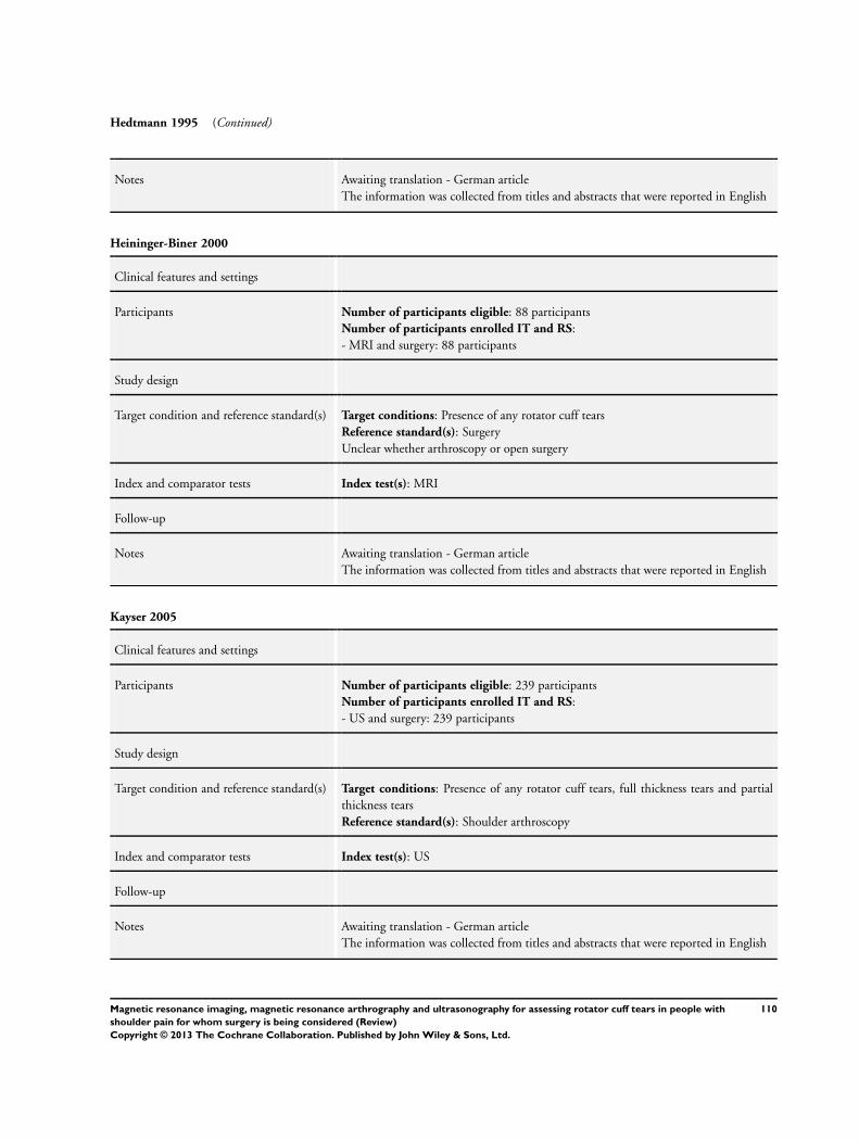
Hedtmann 1995 (Continued)
Notes Awaiting translation - German articleThe information was collected from titles and abstracts that were reported in English
Heininger-Biner 2000
Clinical features and settings
Participants Number of participants eligible: 88 participantsNumber of participants enrolled IT and RS:- MRI and surgery: 88 participants
Study design
Target condition and reference standard(s) Target conditions: Presence of any rotator cuff tearsReference standard(s): SurgeryUnclear whether arthroscopy or open surgery
Index and comparator tests Index test(s): MRI
Follow-up
Notes Awaiting translation - German articleThe information was collected from titles and abstracts that were reported in English
Kayser 2005
Clinical features and settings
Participants Number of participants eligible: 239 participantsNumber of participants enrolled IT and RS:- US and surgery: 239 participants
Study design
Target condition and reference standard(s) Target conditions: Presence of any rotator cuff tears, full thickness tears and partialthickness tearsReference standard(s): Shoulder arthroscopy
Index and comparator tests Index test(s): US
Follow-up
Notes Awaiting translation - German articleThe information was collected from titles and abstracts that were reported in English
110Magnetic resonance imaging, magnetic resonance arthrography and ultrasonography for assessing rotator cuff tears in people with
shoulder pain for whom surgery is being considered (Review)
Copyright © 2013 The Cochrane Collaboration. Published by John Wiley & Sons, Ltd.
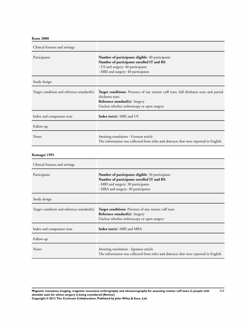
Kenn 2000
Clinical features and settings
Participants Number of participants eligible: 40 participantsNumber of participants enrolled IT and RS:- US and surgery: 40 participants- MRI and surgery: 40 participants
Study design
Target condition and reference standard(s) Target conditions: Presence of any rotator cuff tears, full thickness tears and partialthickness tearsReference standard(s): SurgeryUnclear whether arthroscopy or open surgery
Index and comparator tests Index test(s): MRI and US
Follow-up
Notes Awaiting translation - German articleThe information was collected from titles and abstracts that were reported in English
Kumagai 1991
Clinical features and settings
Participants Number of participants eligible: 30 participantsNumber of participants enrolled IT and RS:- MRI and surgery: 30 participants- MRA and surgery: 30 participants
Study design
Target condition and reference standard(s) Target conditions: Presence of any rotator cuff tearsReference standard(s): SurgeryUnclear whether arthroscopy or open surgery
Index and comparator tests Index test(s): MRI and MRA
Follow-up
Notes Awaiting translation - Japanese articleThe information was collected from titles and abstracts that were reported in English
111Magnetic resonance imaging, magnetic resonance arthrography and ultrasonography for assessing rotator cuff tears in people with
shoulder pain for whom surgery is being considered (Review)
Copyright © 2013 The Cochrane Collaboration. Published by John Wiley & Sons, Ltd.
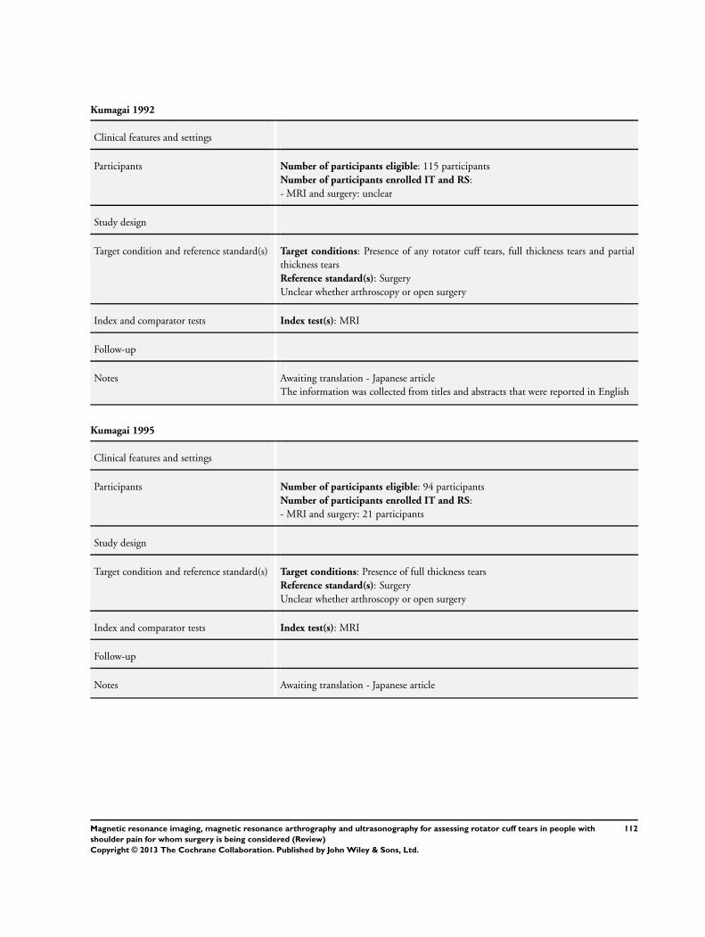
Kumagai 1992
Clinical features and settings
Participants Number of participants eligible: 115 participantsNumber of participants enrolled IT and RS:- MRI and surgery: unclear
Study design
Target condition and reference standard(s) Target conditions: Presence of any rotator cuff tears, full thickness tears and partialthickness tearsReference standard(s): SurgeryUnclear whether arthroscopy or open surgery
Index and comparator tests Index test(s): MRI
Follow-up
Notes Awaiting translation - Japanese articleThe information was collected from titles and abstracts that were reported in English
Kumagai 1995
Clinical features and settings
Participants Number of participants eligible: 94 participantsNumber of participants enrolled IT and RS:- MRI and surgery: 21 participants
Study design
Target condition and reference standard(s) Target conditions: Presence of full thickness tearsReference standard(s): SurgeryUnclear whether arthroscopy or open surgery
Index and comparator tests Index test(s): MRI
Follow-up
Notes Awaiting translation - Japanese article
112Magnetic resonance imaging, magnetic resonance arthrography and ultrasonography for assessing rotator cuff tears in people with
shoulder pain for whom surgery is being considered (Review)
Copyright © 2013 The Cochrane Collaboration. Published by John Wiley & Sons, Ltd.
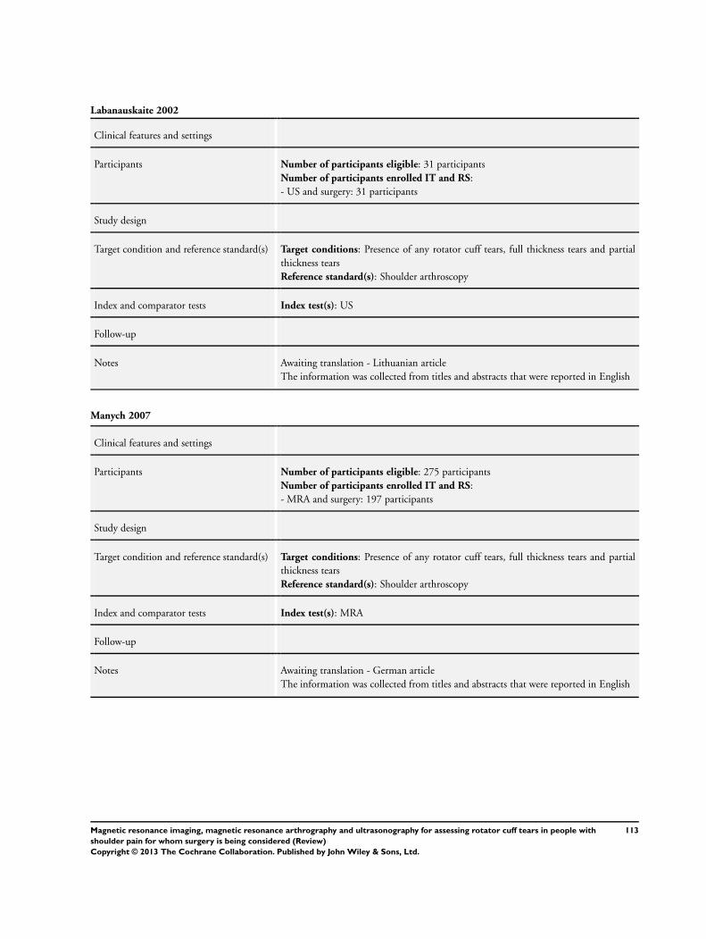
Labanauskaite 2002
Clinical features and settings
Participants Number of participants eligible: 31 participantsNumber of participants enrolled IT and RS:- US and surgery: 31 participants
Study design
Target condition and reference standard(s) Target conditions: Presence of any rotator cuff tears, full thickness tears and partialthickness tearsReference standard(s): Shoulder arthroscopy
Index and comparator tests Index test(s): US
Follow-up
Notes Awaiting translation - Lithuanian articleThe information was collected from titles and abstracts that were reported in English
Manych 2007
Clinical features and settings
Participants Number of participants eligible: 275 participantsNumber of participants enrolled IT and RS:- MRA and surgery: 197 participants
Study design
Target condition and reference standard(s) Target conditions: Presence of any rotator cuff tears, full thickness tears and partialthickness tearsReference standard(s): Shoulder arthroscopy
Index and comparator tests Index test(s): MRA
Follow-up
Notes Awaiting translation - German articleThe information was collected from titles and abstracts that were reported in English
113Magnetic resonance imaging, magnetic resonance arthrography and ultrasonography for assessing rotator cuff tears in people with
shoulder pain for whom surgery is being considered (Review)
Copyright © 2013 The Cochrane Collaboration. Published by John Wiley & Sons, Ltd.
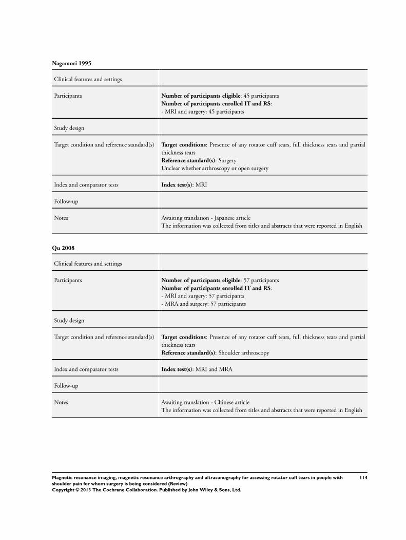
Nagamori 1995
Clinical features and settings
Participants Number of participants eligible: 45 participantsNumber of participants enrolled IT and RS:- MRI and surgery: 45 participants
Study design
Target condition and reference standard(s) Target conditions: Presence of any rotator cuff tears, full thickness tears and partialthickness tearsReference standard(s): SurgeryUnclear whether arthroscopy or open surgery
Index and comparator tests Index test(s): MRI
Follow-up
Notes Awaiting translation - Japanese articleThe information was collected from titles and abstracts that were reported in English
Qu 2008
Clinical features and settings
Participants Number of participants eligible: 57 participantsNumber of participants enrolled IT and RS:- MRI and surgery: 57 participants- MRA and surgery: 57 participants
Study design
Target condition and reference standard(s) Target conditions: Presence of any rotator cuff tears, full thickness tears and partialthickness tearsReference standard(s): Shoulder arthroscopy
Index and comparator tests Index test(s): MRI and MRA
Follow-up
Notes Awaiting translation - Chinese articleThe information was collected from titles and abstracts that were reported in English
114Magnetic resonance imaging, magnetic resonance arthrography and ultrasonography for assessing rotator cuff tears in people with
shoulder pain for whom surgery is being considered (Review)
Copyright © 2013 The Cochrane Collaboration. Published by John Wiley & Sons, Ltd.
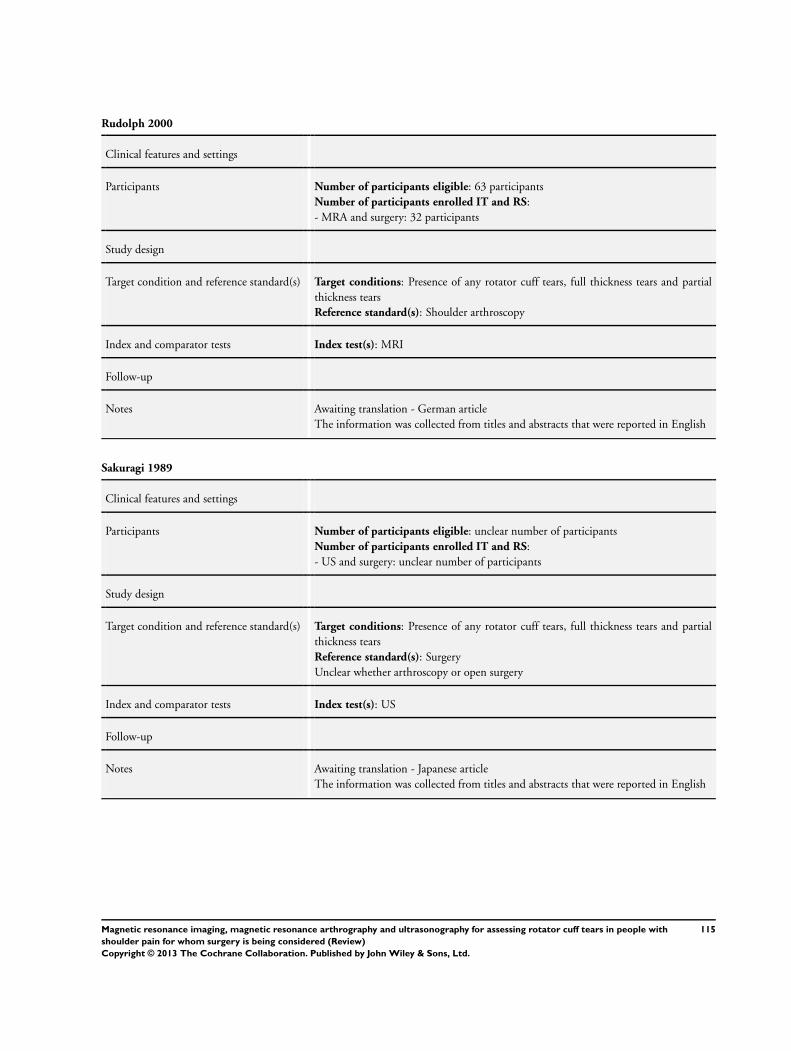
Rudolph 2000
Clinical features and settings
Participants Number of participants eligible: 63 participantsNumber of participants enrolled IT and RS:- MRA and surgery: 32 participants
Study design
Target condition and reference standard(s) Target conditions: Presence of any rotator cuff tears, full thickness tears and partialthickness tearsReference standard(s): Shoulder arthroscopy
Index and comparator tests Index test(s): MRI
Follow-up
Notes Awaiting translation - German articleThe information was collected from titles and abstracts that were reported in English
Sakuragi 1989
Clinical features and settings
Participants Number of participants eligible: unclear number of participantsNumber of participants enrolled IT and RS:- US and surgery: unclear number of participants
Study design
Target condition and reference standard(s) Target conditions: Presence of any rotator cuff tears, full thickness tears and partialthickness tearsReference standard(s): SurgeryUnclear whether arthroscopy or open surgery
Index and comparator tests Index test(s): US
Follow-up
Notes Awaiting translation - Japanese articleThe information was collected from titles and abstracts that were reported in English
115Magnetic resonance imaging, magnetic resonance arthrography and ultrasonography for assessing rotator cuff tears in people with
shoulder pain for whom surgery is being considered (Review)
Copyright © 2013 The Cochrane Collaboration. Published by John Wiley & Sons, Ltd.
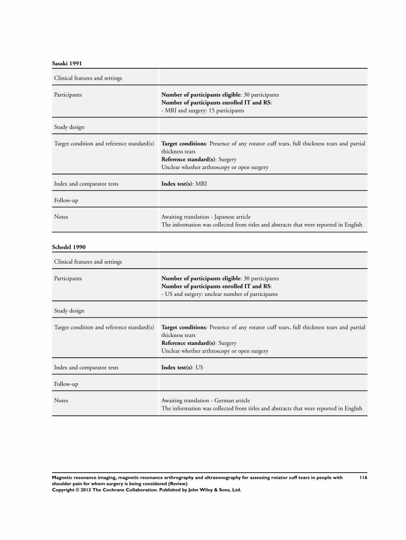
Sasaki 1991
Clinical features and settings
Participants Number of participants eligible: 30 participantsNumber of participants enrolled IT and RS:- MRI and surgery: 15 participants
Study design
Target condition and reference standard(s) Target conditions: Presence of any rotator cuff tears, full thickness tears and partialthickness tearsReference standard(s): SurgeryUnclear whether arthroscopy or open surgery
Index and comparator tests Index test(s): MRI
Follow-up
Notes Awaiting translation - Japanese articleThe information was collected from titles and abstracts that were reported in English
Schedel 1990
Clinical features and settings
Participants Number of participants eligible: 30 participantsNumber of participants enrolled IT and RS:- US and surgery: unclear number of participants
Study design
Target condition and reference standard(s) Target conditions: Presence of any rotator cuff tears, full thickness tears and partialthickness tearsReference standard(s): SurgeryUnclear whether arthroscopy or open surgery
Index and comparator tests Index test(s): US
Follow-up
Notes Awaiting translation - German articleThe information was collected from titles and abstracts that were reported in English
116Magnetic resonance imaging, magnetic resonance arthrography and ultrasonography for assessing rotator cuff tears in people with
shoulder pain for whom surgery is being considered (Review)
Copyright © 2013 The Cochrane Collaboration. Published by John Wiley & Sons, Ltd.

Schroder 2003
Clinical features and settings
Participants Number of participants eligible: 80 participantsNumber of participants enrolled IT and RS:- MRI and surgery: 80 participants
Study design
Target condition and reference standard(s) Target conditions: Presence of any rotator cuff tears, full thickness tears and partialthickness tearsReference standard(s): Open surgery or shoulder arthroscopy
Index and comparator tests Index test(s): MRI
Follow-up
Notes Awaiting translation - German articleThe information was collected from titles and abstracts that were reported in English
Sell 1992
Clinical features and settings
Participants Number of participants eligible: 37 participantsNumber of participants enrolled IT and RS:- MRI and surgery: unclear number of participants- US and surgery: unclear number of participants
Study design
Target condition and reference standard(s) Target conditions: Presence of any rotator cuff tears, full thickness tears and partialthickness tearsReference standard(s): Open surgery or shoulder arthroscopy
Index and comparator tests Index test(s): MRI and US
Follow-up
Notes Awaiting translation - German articleThe information was collected from titles and abstracts that were reported in English
117Magnetic resonance imaging, magnetic resonance arthrography and ultrasonography for assessing rotator cuff tears in people with
shoulder pain for whom surgery is being considered (Review)
Copyright © 2013 The Cochrane Collaboration. Published by John Wiley & Sons, Ltd.
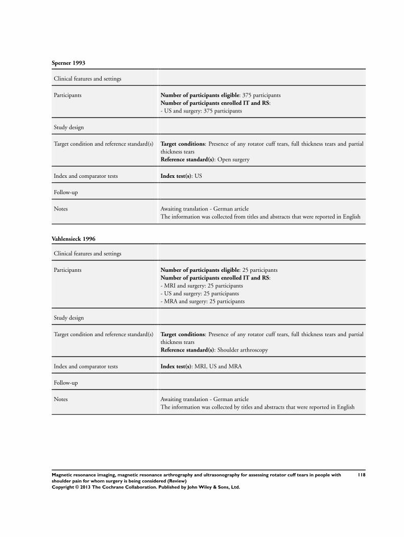
Sperner 1993
Clinical features and settings
Participants Number of participants eligible: 375 participantsNumber of participants enrolled IT and RS:- US and surgery: 375 participants
Study design
Target condition and reference standard(s) Target conditions: Presence of any rotator cuff tears, full thickness tears and partialthickness tearsReference standard(s): Open surgery
Index and comparator tests Index test(s): US
Follow-up
Notes Awaiting translation - German articleThe information was collected from titles and abstracts that were reported in English
Vahlensieck 1996
Clinical features and settings
Participants Number of participants eligible: 25 participantsNumber of participants enrolled IT and RS:- MRI and surgery: 25 participants- US and surgery: 25 participants- MRA and surgery: 25 participants
Study design
Target condition and reference standard(s) Target conditions: Presence of any rotator cuff tears, full thickness tears and partialthickness tearsReference standard(s): Shoulder arthroscopy
Index and comparator tests Index test(s): MRI, US and MRA
Follow-up
Notes Awaiting translation - German articleThe information was collected by titles and abstracts that were reported in English
118Magnetic resonance imaging, magnetic resonance arthrography and ultrasonography for assessing rotator cuff tears in people with
shoulder pain for whom surgery is being considered (Review)
Copyright © 2013 The Cochrane Collaboration. Published by John Wiley & Sons, Ltd.
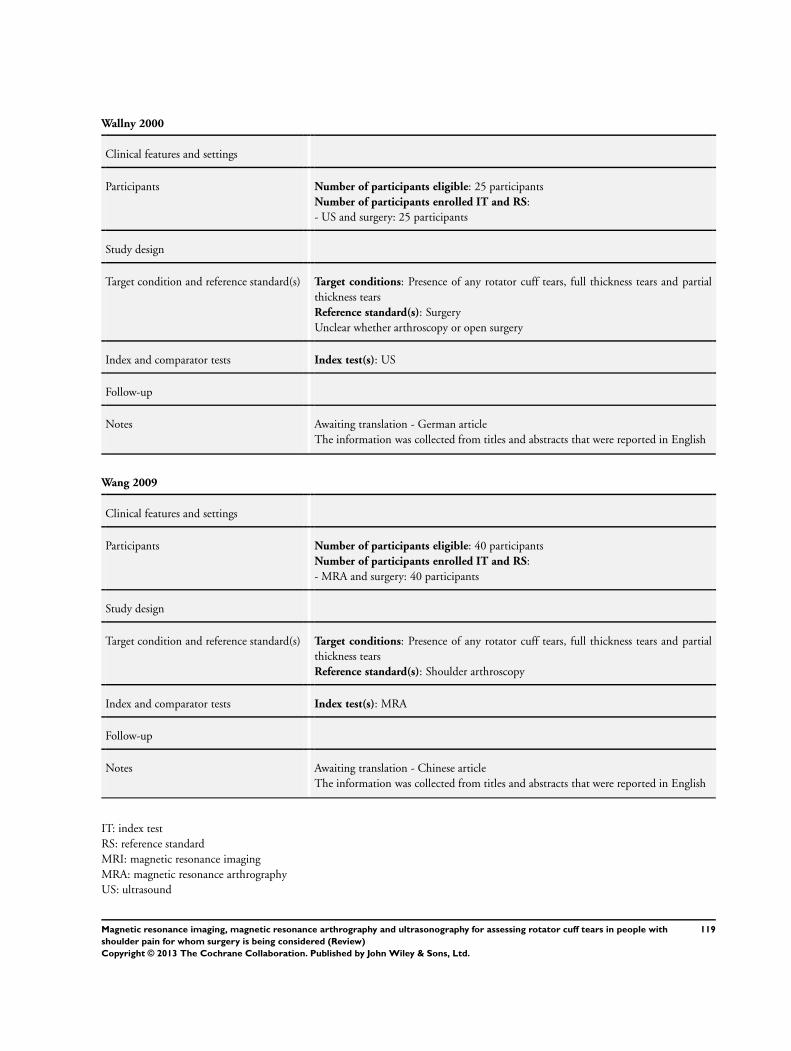
Wallny 2000
Clinical features and settings
Participants Number of participants eligible: 25 participantsNumber of participants enrolled IT and RS:- US and surgery: 25 participants
Study design
Target condition and reference standard(s) Target conditions: Presence of any rotator cuff tears, full thickness tears and partialthickness tearsReference standard(s): SurgeryUnclear whether arthroscopy or open surgery
Index and comparator tests Index test(s): US
Follow-up
Notes Awaiting translation - German articleThe information was collected from titles and abstracts that were reported in English
Wang 2009
Clinical features and settings
Participants Number of participants eligible: 40 participantsNumber of participants enrolled IT and RS:- MRA and surgery: 40 participants
Study design
Target condition and reference standard(s) Target conditions: Presence of any rotator cuff tears, full thickness tears and partialthickness tearsReference standard(s): Shoulder arthroscopy
Index and comparator tests Index test(s): MRA
Follow-up
Notes Awaiting translation - Chinese articleThe information was collected from titles and abstracts that were reported in English
IT: index testRS: reference standardMRI: magnetic resonance imagingMRA: magnetic resonance arthrographyUS: ultrasound
119Magnetic resonance imaging, magnetic resonance arthrography and ultrasonography for assessing rotator cuff tears in people with
shoulder pain for whom surgery is being considered (Review)
Copyright © 2013 The Cochrane Collaboration. Published by John Wiley & Sons, Ltd.
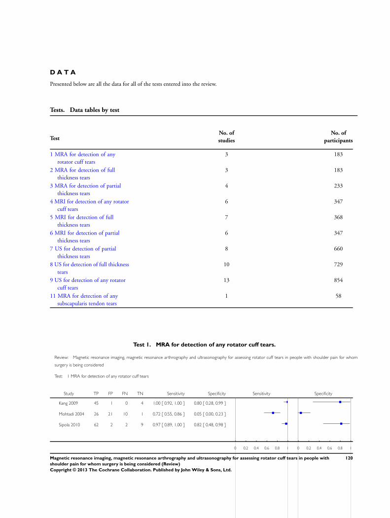
D A T A
Presented below are all the data for all of the tests entered into the review.
Tests. Data tables by test
TestNo. of
studies
No. of
participants
1 MRA for detection of anyrotator cuff tears
3 183
2 MRA for detection of fullthickness tears
3 183
3 MRA for detection of partialthickness tears
4 233
4 MRI for detection of any rotatorcuff tears
6 347
5 MRI for detection of fullthickness tears
7 368
6 MRI for detection of partialthickness tears
6 347
7 US for detection of partialthickness tears
8 660
8 US for detection of full thicknesstears
10 729
9 US for detection of any rotatorcuff tears
13 854
11 MRA for detection of anysubscapularis tendon tears
1 58
Test 1. MRA for detection of any rotator cuff tears.
Review: Magnetic resonance imaging, magnetic resonance arthrography and ultrasonography for assessing rotator cuff tears in people with shoulder pain for whom
surgery is being considered
Test: 1 MRA for detection of any rotator cuff tears
Study TP FP FN TN Sensitivity Specificity Sensitivity Specificity
Kang 2009 45 1 0 4 1.00 [ 0.92, 1.00 ] 0.80 [ 0.28, 0.99 ]
Mohtadi 2004 26 21 10 1 0.72 [ 0.55, 0.86 ] 0.05 [ 0.00, 0.23 ]
Sipola 2010 62 2 2 9 0.97 [ 0.89, 1.00 ] 0.82 [ 0.48, 0.98 ]
0 0.2 0.4 0.6 0.8 1 0 0.2 0.4 0.6 0.8 1
120Magnetic resonance imaging, magnetic resonance arthrography and ultrasonography for assessing rotator cuff tears in people with
shoulder pain for whom surgery is being considered (Review)
Copyright © 2013 The Cochrane Collaboration. Published by John Wiley & Sons, Ltd.
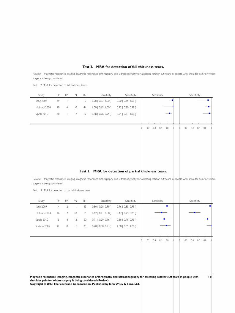
Test 2. MRA for detection of full thickness tears.
Review: Magnetic resonance imaging, magnetic resonance arthrography and ultrasonography for assessing rotator cuff tears in people with shoulder pain for whom
surgery is being considered
Test: 2 MRA for detection of full thickness tears
Study TP FP FN TN Sensitivity Specificity Sensitivity Specificity
Kang 2009 39 1 1 9 0.98 [ 0.87, 1.00 ] 0.90 [ 0.55, 1.00 ]
Mohtadi 2004 10 4 0 44 1.00 [ 0.69, 1.00 ] 0.92 [ 0.80, 0.98 ]
Sipola 2010 50 1 7 17 0.88 [ 0.76, 0.95 ] 0.94 [ 0.73, 1.00 ]
0 0.2 0.4 0.6 0.8 1 0 0.2 0.4 0.6 0.8 1
Test 3. MRA for detection of partial thickness tears.
Review: Magnetic resonance imaging, magnetic resonance arthrography and ultrasonography for assessing rotator cuff tears in people with shoulder pain for whom
surgery is being considered
Test: 3 MRA for detection of partial thickness tears
Study TP FP FN TN Sensitivity Specificity Sensitivity Specificity
Kang 2009 4 2 1 43 0.80 [ 0.28, 0.99 ] 0.96 [ 0.85, 0.99 ]
Mohtadi 2004 16 17 10 15 0.62 [ 0.41, 0.80 ] 0.47 [ 0.29, 0.65 ]
Sipola 2010 5 8 2 60 0.71 [ 0.29, 0.96 ] 0.88 [ 0.78, 0.95 ]
Stetson 2005 21 0 6 23 0.78 [ 0.58, 0.91 ] 1.00 [ 0.85, 1.00 ]
0 0.2 0.4 0.6 0.8 1 0 0.2 0.4 0.6 0.8 1
121Magnetic resonance imaging, magnetic resonance arthrography and ultrasonography for assessing rotator cuff tears in people with
shoulder pain for whom surgery is being considered (Review)
Copyright © 2013 The Cochrane Collaboration. Published by John Wiley & Sons, Ltd.

Test 4. MRI for detection of any rotator cuff tears.
Review: Magnetic resonance imaging, magnetic resonance arthrography and ultrasonography for assessing rotator cuff tears in people with shoulder pain for whom
surgery is being considered
Test: 4 MRI for detection of any rotator cuff tears
Study TP FP FN TN Sensitivity Specificity Sensitivity Specificity
Della Sala 1996 20 2 0 8 1.00 [ 0.83, 1.00 ] 0.80 [ 0.44, 0.97 ]
Gagey 1993 19 2 0 17 1.00 [ 0.82, 1.00 ] 0.89 [ 0.67, 0.99 ]
Iannotti 2005 75 5 4 15 0.95 [ 0.88, 0.99 ] 0.75 [ 0.51, 0.91 ]
Lambert 2009 45 0 1 2 0.98 [ 0.88, 1.00 ] 1.00 [ 0.16, 1.00 ]
Martin-Hervas 2001 31 7 3 20 0.91 [ 0.76, 0.98 ] 0.74 [ 0.54, 0.89 ]
Teefey 2004 65 2 0 4 1.00 [ 0.94, 1.00 ] 0.67 [ 0.22, 0.96 ]
0 0.2 0.4 0.6 0.8 1 0 0.2 0.4 0.6 0.8 1
Test 5. MRI for detection of full thickness tears.
Review: Magnetic resonance imaging, magnetic resonance arthrography and ultrasonography for assessing rotator cuff tears in people with shoulder pain for whom
surgery is being considered
Test: 5 MRI for detection of full thickness tears
Study TP FP FN TN Sensitivity Specificity Sensitivity Specificity
Della Sala 1996 17 0 2 11 0.89 [ 0.67, 0.99 ] 1.00 [ 0.72, 1.00 ]
Gagey 1993 14 1 0 23 1.00 [ 0.77, 1.00 ] 0.96 [ 0.79, 1.00 ]
Iannotti 2005 40 7 2 50 0.95 [ 0.84, 0.99 ] 0.88 [ 0.76, 0.95 ]
Lambert 2009 32 0 1 15 0.97 [ 0.84, 1.00 ] 1.00 [ 0.78, 1.00 ]
Martin-Hervas 2001 21 1 5 34 0.81 [ 0.61, 0.93 ] 0.97 [ 0.85, 1.00 ]
Swen 1999 10 1 3 7 0.77 [ 0.46, 0.95 ] 0.88 [ 0.47, 1.00 ]
Teefey 2004 46 8 0 17 1.00 [ 0.92, 1.00 ] 0.68 [ 0.46, 0.85 ]
0 0.2 0.4 0.6 0.8 1 0 0.2 0.4 0.6 0.8 1
122Magnetic resonance imaging, magnetic resonance arthrography and ultrasonography for assessing rotator cuff tears in people with
shoulder pain for whom surgery is being considered (Review)
Copyright © 2013 The Cochrane Collaboration. Published by John Wiley & Sons, Ltd.
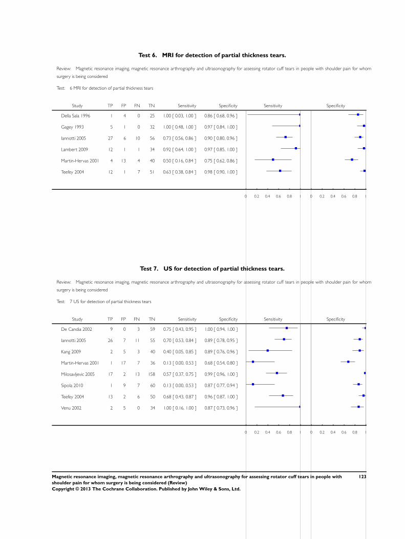
Test 6. MRI for detection of partial thickness tears.
Review: Magnetic resonance imaging, magnetic resonance arthrography and ultrasonography for assessing rotator cuff tears in people with shoulder pain for whom
surgery is being considered
Test: 6 MRI for detection of partial thickness tears
Study TP FP FN TN Sensitivity Specificity Sensitivity Specificity
Della Sala 1996 1 4 0 25 1.00 [ 0.03, 1.00 ] 0.86 [ 0.68, 0.96 ]
Gagey 1993 5 1 0 32 1.00 [ 0.48, 1.00 ] 0.97 [ 0.84, 1.00 ]
Iannotti 2005 27 6 10 56 0.73 [ 0.56, 0.86 ] 0.90 [ 0.80, 0.96 ]
Lambert 2009 12 1 1 34 0.92 [ 0.64, 1.00 ] 0.97 [ 0.85, 1.00 ]
Martin-Hervas 2001 4 13 4 40 0.50 [ 0.16, 0.84 ] 0.75 [ 0.62, 0.86 ]
Teefey 2004 12 1 7 51 0.63 [ 0.38, 0.84 ] 0.98 [ 0.90, 1.00 ]
0 0.2 0.4 0.6 0.8 1 0 0.2 0.4 0.6 0.8 1
Test 7. US for detection of partial thickness tears.
Review: Magnetic resonance imaging, magnetic resonance arthrography and ultrasonography for assessing rotator cuff tears in people with shoulder pain for whom
surgery is being considered
Test: 7 US for detection of partial thickness tears
Study TP FP FN TN Sensitivity Specificity Sensitivity Specificity
De Candia 2002 9 0 3 59 0.75 [ 0.43, 0.95 ] 1.00 [ 0.94, 1.00 ]
Iannotti 2005 26 7 11 55 0.70 [ 0.53, 0.84 ] 0.89 [ 0.78, 0.95 ]
Kang 2009 2 5 3 40 0.40 [ 0.05, 0.85 ] 0.89 [ 0.76, 0.96 ]
Martin-Hervas 2001 1 17 7 36 0.13 [ 0.00, 0.53 ] 0.68 [ 0.54, 0.80 ]
Milosavljevic 2005 17 2 13 158 0.57 [ 0.37, 0.75 ] 0.99 [ 0.96, 1.00 ]
Sipola 2010 1 9 7 60 0.13 [ 0.00, 0.53 ] 0.87 [ 0.77, 0.94 ]
Teefey 2004 13 2 6 50 0.68 [ 0.43, 0.87 ] 0.96 [ 0.87, 1.00 ]
Venu 2002 2 5 0 34 1.00 [ 0.16, 1.00 ] 0.87 [ 0.73, 0.96 ]
0 0.2 0.4 0.6 0.8 1 0 0.2 0.4 0.6 0.8 1
123Magnetic resonance imaging, magnetic resonance arthrography and ultrasonography for assessing rotator cuff tears in people with
shoulder pain for whom surgery is being considered (Review)
Copyright © 2013 The Cochrane Collaboration. Published by John Wiley & Sons, Ltd.
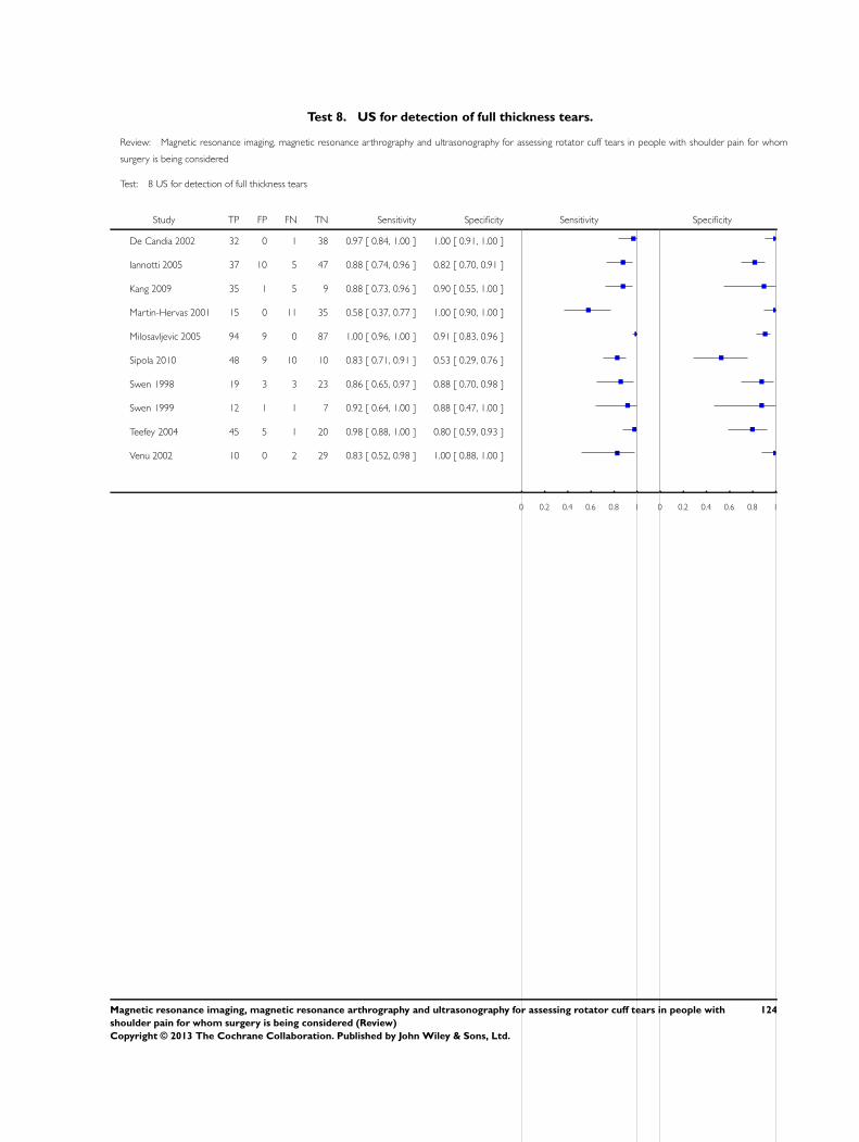
Test 8. US for detection of full thickness tears.
Review: Magnetic resonance imaging, magnetic resonance arthrography and ultrasonography for assessing rotator cuff tears in people with shoulder pain for whom
surgery is being considered
Test: 8 US for detection of full thickness tears
Study TP FP FN TN Sensitivity Specificity Sensitivity Specificity
De Candia 2002 32 0 1 38 0.97 [ 0.84, 1.00 ] 1.00 [ 0.91, 1.00 ]
Iannotti 2005 37 10 5 47 0.88 [ 0.74, 0.96 ] 0.82 [ 0.70, 0.91 ]
Kang 2009 35 1 5 9 0.88 [ 0.73, 0.96 ] 0.90 [ 0.55, 1.00 ]
Martin-Hervas 2001 15 0 11 35 0.58 [ 0.37, 0.77 ] 1.00 [ 0.90, 1.00 ]
Milosavljevic 2005 94 9 0 87 1.00 [ 0.96, 1.00 ] 0.91 [ 0.83, 0.96 ]
Sipola 2010 48 9 10 10 0.83 [ 0.71, 0.91 ] 0.53 [ 0.29, 0.76 ]
Swen 1998 19 3 3 23 0.86 [ 0.65, 0.97 ] 0.88 [ 0.70, 0.98 ]
Swen 1999 12 1 1 7 0.92 [ 0.64, 1.00 ] 0.88 [ 0.47, 1.00 ]
Teefey 2004 45 5 1 20 0.98 [ 0.88, 1.00 ] 0.80 [ 0.59, 0.93 ]
Venu 2002 10 0 2 29 0.83 [ 0.52, 0.98 ] 1.00 [ 0.88, 1.00 ]
0 0.2 0.4 0.6 0.8 1 0 0.2 0.4 0.6 0.8 1
124Magnetic resonance imaging, magnetic resonance arthrography and ultrasonography for assessing rotator cuff tears in people with
shoulder pain for whom surgery is being considered (Review)
Copyright © 2013 The Cochrane Collaboration. Published by John Wiley & Sons, Ltd.
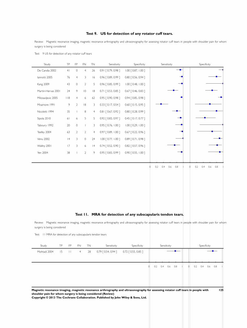
Test 9. US for detection of any rotator cuff tears.
Review: Magnetic resonance imaging, magnetic resonance arthrography and ultrasonography for assessing rotator cuff tears in people with shoulder pain for whom
surgery is being considered
Test: 9 US for detection of any rotator cuff tears
Study TP FP FN TN Sensitivity Specificity Sensitivity Specificity
De Candia 2002 41 0 4 26 0.91 [ 0.79, 0.98 ] 1.00 [ 0.87, 1.00 ]
Iannotti 2005 76 4 3 16 0.96 [ 0.89, 0.99 ] 0.80 [ 0.56, 0.94 ]
Kang 2009 43 0 2 5 0.96 [ 0.85, 0.99 ] 1.00 [ 0.48, 1.00 ]
Martin-Hervas 2001 24 9 10 18 0.71 [ 0.53, 0.85 ] 0.67 [ 0.46, 0.83 ]
Milosavljevic 2005 118 4 6 62 0.95 [ 0.90, 0.98 ] 0.94 [ 0.85, 0.98 ]
Misamore 1991 9 2 18 3 0.33 [ 0.17, 0.54 ] 0.60 [ 0.15, 0.95 ]
Nicoletti 1994 35 1 8 4 0.81 [ 0.67, 0.92 ] 0.80 [ 0.28, 0.99 ]
Sipola 2010 61 6 5 5 0.92 [ 0.83, 0.97 ] 0.45 [ 0.17, 0.77 ]
Taboury 1992 20 0 1 3 0.95 [ 0.76, 1.00 ] 1.00 [ 0.29, 1.00 ]
Teefey 2004 63 2 2 4 0.97 [ 0.89, 1.00 ] 0.67 [ 0.22, 0.96 ]
Venu 2002 14 3 0 24 1.00 [ 0.77, 1.00 ] 0.89 [ 0.71, 0.98 ]
Wallny 2001 17 3 6 14 0.74 [ 0.52, 0.90 ] 0.82 [ 0.57, 0.96 ]
Yen 2004 38 1 2 9 0.95 [ 0.83, 0.99 ] 0.90 [ 0.55, 1.00 ]
0 0.2 0.4 0.6 0.8 1 0 0.2 0.4 0.6 0.8 1
Test 11. MRA for detection of any subscapularis tendon tears.
Review: Magnetic resonance imaging, magnetic resonance arthrography and ultrasonography for assessing rotator cuff tears in people with shoulder pain for whom
surgery is being considered
Test: 11 MRA for detection of any subscapularis tendon tears
Study TP FP FN TN Sensitivity Specificity Sensitivity Specificity
Mohtadi 2004 15 11 4 28 0.79 [ 0.54, 0.94 ] 0.72 [ 0.55, 0.85 ]
0 0.2 0.4 0.6 0.8 1 0 0.2 0.4 0.6 0.8 1
125Magnetic resonance imaging, magnetic resonance arthrography and ultrasonography for assessing rotator cuff tears in people with
shoulder pain for whom surgery is being considered (Review)
Copyright © 2013 The Cochrane Collaboration. Published by John Wiley & Sons, Ltd.
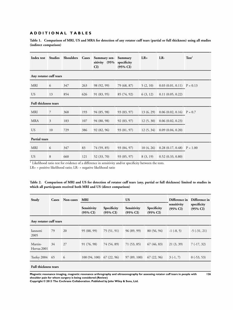
A D D I T I O N A L T A B L E S
Table 1. Comparison of MRI, US and MRA for detection of any rotator cuff tears (partial or full thickness) using all studies
(indirect comparison)
Index test Studies Shoulders Cases Summary sen-
sitivity (95%
CI)
Summary
specificity
(95% CI)
LR+ LR- Test1
Any rotator cuff tears
MRI 6 347 263 98 (92, 99) 79 (68, 87) 5 (2, 10) 0.03 (0.01, 0.11) P = 0.13
US 13 854 626 91 (83, 95) 85 (74, 92) 6 (3, 12) 0.11 (0.05, 0.22)
Full thickness tears
MRI 7 368 193 94 (85, 98) 93 (83, 97) 13 (6, 29) 0.06 (0.02, 0.16) P = 0.7
MRA 3 183 107 94 (80, 98) 92 (83, 97) 12 (5, 30) 0.06 (0.02, 0.23)
US 10 729 386 92 (82, 96) 93 (81, 97) 12 (5, 34) 0.09 (0.04, 0.20)
Partial tears
MRI 6 347 83 74 (59, 85) 93 (84, 97) 10 (4, 26) 0.28 (0.17, 0.48) P = 1.00
US 8 660 121 52 (33, 70) 93 (85, 97) 8 (3, 19) 0.52 (0.33, 0.80)
1 Likelihood ratio test for evidence of a difference in sensitivity and/or specificity between the tests.LR+ = positive likelihood ratio; LR- = negative likelihood ratio
Table 2. Comparison of MRI and US for detection of rotator cuff tears (any, partial or full thickness) limited to studies in
which all participants received both MRI and US (direct comparison)
Study Cases Non-cases MRI US Difference in
sensitivity
(95% CI)
Difference in
specificity
(95% CI)Sensitivity
(95% CI)
Specificity
(95% CI)
Sensitivity
(95% CI)
Specificity
(95% CI)
Any rotator cuff tears
Iannotti2005
79 20 95 (88, 99) 75 (51, 91) 96 (89, 99) 80 (56, 94) -1 (-8, 5) -5 (-31, 21)
Martin-Hervas 2001
34 27 91 (76, 98) 74 (54, 89) 71 (53, 85) 67 (46, 83) 21 (3, 39) 7 (-17, 32)
Teefey 2004 65 6 100 (94, 100) 67 (22, 96) 97 (89, 100) 67 (22, 96) 3 (-1, 7) 0 (-53, 53)
Full thickness tears
126Magnetic resonance imaging, magnetic resonance arthrography and ultrasonography for assessing rotator cuff tears in people with
shoulder pain for whom surgery is being considered (Review)
Copyright © 2013 The Cochrane Collaboration. Published by John Wiley & Sons, Ltd.
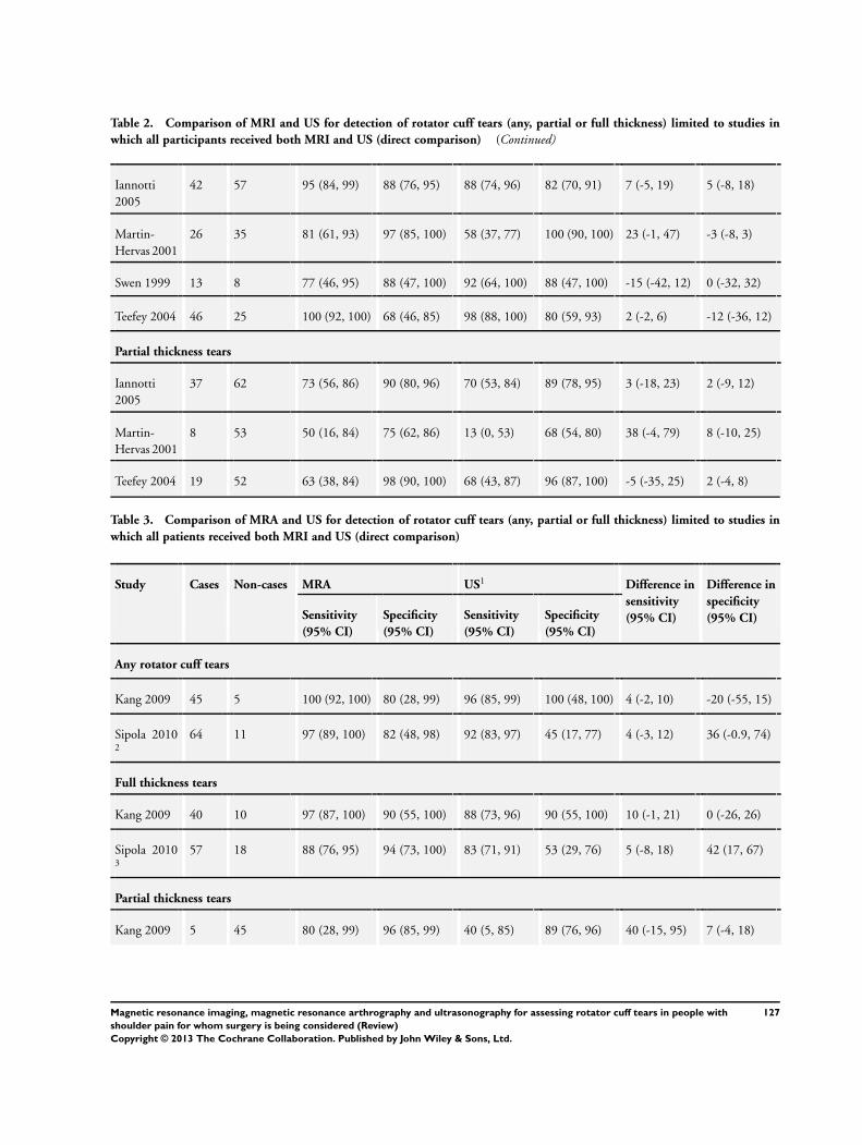
Table 2. Comparison of MRI and US for detection of rotator cuff tears (any, partial or full thickness) limited to studies in
which all participants received both MRI and US (direct comparison) (Continued)
Iannotti2005
42 57 95 (84, 99) 88 (76, 95) 88 (74, 96) 82 (70, 91) 7 (-5, 19) 5 (-8, 18)
Martin-Hervas 2001
26 35 81 (61, 93) 97 (85, 100) 58 (37, 77) 100 (90, 100) 23 (-1, 47) -3 (-8, 3)
Swen 1999 13 8 77 (46, 95) 88 (47, 100) 92 (64, 100) 88 (47, 100) -15 (-42, 12) 0 (-32, 32)
Teefey 2004 46 25 100 (92, 100) 68 (46, 85) 98 (88, 100) 80 (59, 93) 2 (-2, 6) -12 (-36, 12)
Partial thickness tears
Iannotti2005
37 62 73 (56, 86) 90 (80, 96) 70 (53, 84) 89 (78, 95) 3 (-18, 23) 2 (-9, 12)
Martin-Hervas 2001
8 53 50 (16, 84) 75 (62, 86) 13 (0, 53) 68 (54, 80) 38 (-4, 79) 8 (-10, 25)
Teefey 2004 19 52 63 (38, 84) 98 (90, 100) 68 (43, 87) 96 (87, 100) -5 (-35, 25) 2 (-4, 8)
Table 3. Comparison of MRA and US for detection of rotator cuff tears (any, partial or full thickness) limited to studies in
which all patients received both MRI and US (direct comparison)
Study Cases Non-cases MRA US1 Difference in
sensitivity
(95% CI)
Difference in
specificity
(95% CI)Sensitivity
(95% CI)
Specificity
(95% CI)
Sensitivity
(95% CI)
Specificity
(95% CI)
Any rotator cuff tears
Kang 2009 45 5 100 (92, 100) 80 (28, 99) 96 (85, 99) 100 (48, 100) 4 (-2, 10) -20 (-55, 15)
Sipola 20102
64 11 97 (89, 100) 82 (48, 98) 92 (83, 97) 45 (17, 77) 4 (-3, 12) 36 (-0.9, 74)
Full thickness tears
Kang 2009 40 10 97 (87, 100) 90 (55, 100) 88 (73, 96) 90 (55, 100) 10 (-1, 21) 0 (-26, 26)
Sipola 20103
57 18 88 (76, 95) 94 (73, 100) 83 (71, 91) 53 (29, 76) 5 (-8, 18) 42 (17, 67)
Partial thickness tears
Kang 2009 5 45 80 (28, 99) 96 (85, 99) 40 (5, 85) 89 (76, 96) 40 (-15, 95) 7 (-4, 18)
127Magnetic resonance imaging, magnetic resonance arthrography and ultrasonography for assessing rotator cuff tears in people with
shoulder pain for whom surgery is being considered (Review)
Copyright © 2013 The Cochrane Collaboration. Published by John Wiley & Sons, Ltd.
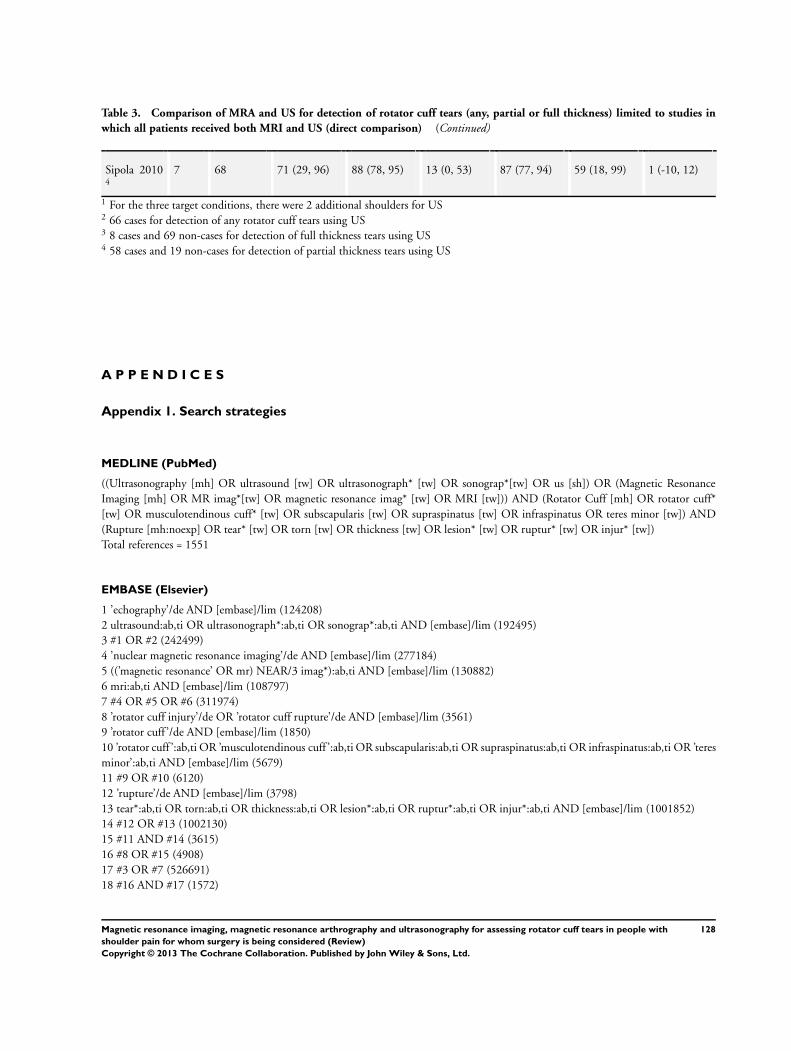
Table 3. Comparison of MRA and US for detection of rotator cuff tears (any, partial or full thickness) limited to studies in
which all patients received both MRI and US (direct comparison) (Continued)
Sipola 20104
7 68 71 (29, 96) 88 (78, 95) 13 (0, 53) 87 (77, 94) 59 (18, 99) 1 (-10, 12)
1 For the three target conditions, there were 2 additional shoulders for US2 66 cases for detection of any rotator cuff tears using US3 8 cases and 69 non-cases for detection of full thickness tears using US4 58 cases and 19 non-cases for detection of partial thickness tears using US
A P P E N D I C E S
Appendix 1. Search strategies
MEDLINE (PubMed)
((Ultrasonography [mh] OR ultrasound [tw] OR ultrasonograph* [tw] OR sonograp*[tw] OR us [sh]) OR (Magnetic ResonanceImaging [mh] OR MR imag*[tw] OR magnetic resonance imag* [tw] OR MRI [tw])) AND (Rotator Cuff [mh] OR rotator cuff*[tw] OR musculotendinous cuff* [tw] OR subscapularis [tw] OR supraspinatus [tw] OR infraspinatus OR teres minor [tw]) AND(Rupture [mh:noexp] OR tear* [tw] OR torn [tw] OR thickness [tw] OR lesion* [tw] OR ruptur* [tw] OR injur* [tw])Total references = 1551
EMBASE (Elsevier)
1 ’echography’/de AND [embase]/lim (124208)2 ultrasound:ab,ti OR ultrasonograph*:ab,ti OR sonograp*:ab,ti AND [embase]/lim (192495)3 #1 OR #2 (242499)4 ’nuclear magnetic resonance imaging’/de AND [embase]/lim (277184)5 ((’magnetic resonance’ OR mr) NEAR/3 imag*):ab,ti AND [embase]/lim (130882)6 mri:ab,ti AND [embase]/lim (108797)7 #4 OR #5 OR #6 (311974)8 ’rotator cuff injury’/de OR ’rotator cuff rupture’/de AND [embase]/lim (3561)9 ’rotator cuff ’/de AND [embase]/lim (1850)10 ’rotator cuff ’:ab,ti OR ’musculotendinous cuff ’:ab,ti OR subscapularis:ab,ti OR supraspinatus:ab,ti OR infraspinatus:ab,ti OR ’teresminor’:ab,ti AND [embase]/lim (5679)11 #9 OR #10 (6120)12 ’rupture’/de AND [embase]/lim (3798)13 tear*:ab,ti OR torn:ab,ti OR thickness:ab,ti OR lesion*:ab,ti OR ruptur*:ab,ti OR injur*:ab,ti AND [embase]/lim (1001852)14 #12 OR #13 (1002130)15 #11 AND #14 (3615)16 #8 OR #15 (4908)17 #3 OR #7 (526691)18 #16 AND #17 (1572)
128Magnetic resonance imaging, magnetic resonance arthrography and ultrasonography for assessing rotator cuff tears in people with
shoulder pain for whom surgery is being considered (Review)
Copyright © 2013 The Cochrane Collaboration. Published by John Wiley & Sons, Ltd.
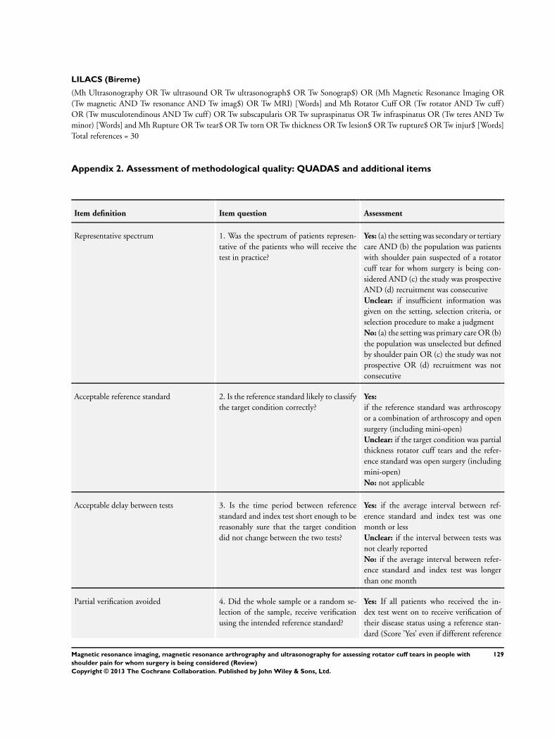
LILACS (Bireme)
(Mh Ultrasonography OR Tw ultrasound OR Tw ultrasonograph$ OR Tw Sonograp$) OR (Mh Magnetic Resonance Imaging OR(Tw magnetic AND Tw resonance AND Tw imag$) OR Tw MRI) [Words] and Mh Rotator Cuff OR (Tw rotator AND Tw cuff )OR (Tw musculotendinous AND Tw cuff ) OR Tw subscapularis OR Tw supraspinatus OR Tw infraspinatus OR (Tw teres AND Twminor) [Words] and Mh Rupture OR Tw tear$ OR Tw torn OR Tw thickness OR Tw lesion$ OR Tw rupture$ OR Tw injur$ [Words]Total references = 30
Appendix 2. Assessment of methodological quality: QUADAS and additional items
Item definition Item question Assessment
Representative spectrum 1. Was the spectrum of patients represen-tative of the patients who will receive thetest in practice?
Yes: (a) the setting was secondary or tertiarycare AND (b) the population was patientswith shoulder pain suspected of a rotatorcuff tear for whom surgery is being con-sidered AND (c) the study was prospectiveAND (d) recruitment was consecutiveUnclear: if insufficient information wasgiven on the setting, selection criteria, orselection procedure to make a judgmentNo: (a) the setting was primary care OR (b)the population was unselected but definedby shoulder pain OR (c) the study was notprospective OR (d) recruitment was notconsecutive
Acceptable reference standard 2. Is the reference standard likely to classifythe target condition correctly?
Yes:
if the reference standard was arthroscopyor a combination of arthroscopy and opensurgery (including mini-open)Unclear: if the target condition was partialthickness rotator cuff tears and the refer-ence standard was open surgery (includingmini-open)No: not applicable
Acceptable delay between tests 3. Is the time period between referencestandard and index test short enough to bereasonably sure that the target conditiondid not change between the two tests?
Yes: if the average interval between ref-erence standard and index test was onemonth or lessUnclear: if the interval between tests wasnot clearly reportedNo: if the average interval between refer-ence standard and index test was longerthan one month
Partial verification avoided 4. Did the whole sample or a random se-lection of the sample, receive verificationusing the intended reference standard?
Yes: If all patients who received the in-dex test went on to receive verification oftheir disease status using a reference stan-dard (Score ’Yes’ even if different reference
129Magnetic resonance imaging, magnetic resonance arthrography and ultrasonography for assessing rotator cuff tears in people with
shoulder pain for whom surgery is being considered (Review)
Copyright © 2013 The Cochrane Collaboration. Published by John Wiley & Sons, Ltd.
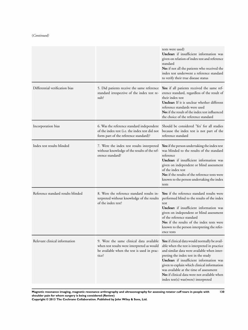
(Continued)
tests were used)Unclear: if insufficient information wasgiven on relation of index test and referencestandardNo: if not all the patients who received theindex test underwent a reference standardto verify their true disease status
Differential verification bias 5. Did patients receive the same referencestandard irrespective of the index test re-sult?
Yes: if all patients received the same ref-erence standard, regardless of the result oftheir index testUnclear: If it is unclear whether differentreference standards were usedNo: if the result of the index test influencedthe choice of the reference standard
Incorporation bias 6. Was the reference standard independentof the index test (i.e. the index test did notform part of the reference standard)?
Should be considered ’Yes’ for all studiesbecause the index test is not part of thereference standard
Index test results blinded 7. Were the index test results interpretedwithout knowledge of the results of the ref-erence standard?
Yes: if the person undertaking the index testwas blinded to the results of the standardreferenceUnclear: if insufficient information wasgiven on independent or blind assessmentof the index testNo: if the results of the reference tests wereknown to the person undertaking the indextests
Reference standard results blinded 8. Were the reference standard results in-terpreted without knowledge of the resultsof the index test?
Yes: if the reference standard results wereperformed blind to the results of the indextestUnclear: if insufficient information wasgiven on independent or blind assessmentof the reference standardNo: if the results of the index tests wereknown to the person interpreting the refer-ence tests
Relevant clinical information 9. Were the same clinical data availablewhen test results were interpreted as wouldbe available when the test is used in prac-tice?
Yes: if clinical data would normally be avail-able when the test is interpreted in practiceand similar data were available when inter-preting the index test in the studyUnclear: if insufficient information wasgiven to explain which clinical informationwas available at the time of assessmentNo: if clinical data were not available whenindex test(s) was(were) interpreted
130Magnetic resonance imaging, magnetic resonance arthrography and ultrasonography for assessing rotator cuff tears in people with
shoulder pain for whom surgery is being considered (Review)
Copyright © 2013 The Cochrane Collaboration. Published by John Wiley & Sons, Ltd.
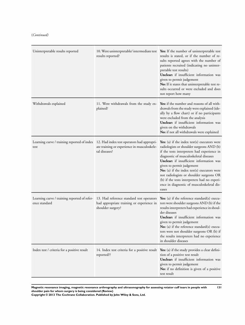
(Continued)
Uninterpretable results reported 10. Were uninterpretable/ intermediate testresults reported?
Yes: If the number of uninterpretable testresults is stated, or if the number of re-sults reported agrees with the number ofpatients recruited (indicating no uninter-pretable test results)Unclear: if insufficient information wasgiven to permit judgementNo: If it states that uninterpretable test re-sults occurred or were excluded and doesnot report how many
Withdrawals explained 11. Were withdrawals from the study ex-plained?
Yes: if the number and reasons of all with-drawals from the study were explained (ide-ally by a flow chart) or if no participantswere excluded from the analysisUnclear: if insufficient information wasgiven on the withdrawalsNo: if not all withdrawals were explained
Learning curve / training reported of indextest
12. Had index test operators had appropri-ate training or experience in musculoskele-tal diseases?
Yes: (a) if the index test(s) executors wereradiologists or shoulder surgeons AND (b)if the tests interpreters had experience indiagnostic of musculoskeletal diseasesUnclear: if insufficient information wasgiven to permit judgementNo: (a) if the index test(s) executors werenot radiologists or shoulder surgeons OR(b) if the tests interpreters had no experi-ence in diagnostic of musculoskeletal dis-eases
Learning curve / training reported of refer-ence standard
13. Had reference standard test operatorshad appropriate training or experience inshoulder surgery?
Yes: (a) if the reference standard(s) execu-tors were shoulder surgeons AND (b) if theresults interpreters had experience in shoul-der diseasesUnclear: if insufficient information wasgiven to permit judgementNo: (a) if the reference standard(s) execu-tors were not shoulder surgeons OR (b) ifthe results interpreters had no experiencein shoulder diseases
Index test / criteria for a positive result 14. Index test criteria for a positive resultreported??
Yes: (a) if the study provides a clear defini-tion of a positive test resultUnclear: if insufficient information wasgiven to permit judgementNo: if no definition is given of a positivetest result
131Magnetic resonance imaging, magnetic resonance arthrography and ultrasonography for assessing rotator cuff tears in people with
shoulder pain for whom surgery is being considered (Review)
Copyright © 2013 The Cochrane Collaboration. Published by John Wiley & Sons, Ltd.
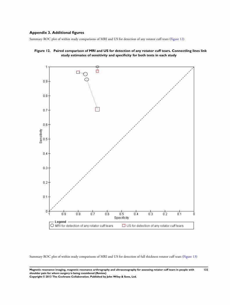
Appendix 3. Additional figures
Summary ROC plot of within study comparisons of MRI and US for detection of any rotator cuff tears (Figure 12)
Figure 12. Paired comparison of MRI and US for detection of any rotator cuff tears. Connectling lines link
study estimates of sensitivity and specificity for both tests in each study
Summary ROC plot of within study comparisons of MRI and US for detection of full thickness rotator cuff tears (Figure 13)
132Magnetic resonance imaging, magnetic resonance arthrography and ultrasonography for assessing rotator cuff tears in people with
shoulder pain for whom surgery is being considered (Review)
Copyright © 2013 The Cochrane Collaboration. Published by John Wiley & Sons, Ltd.
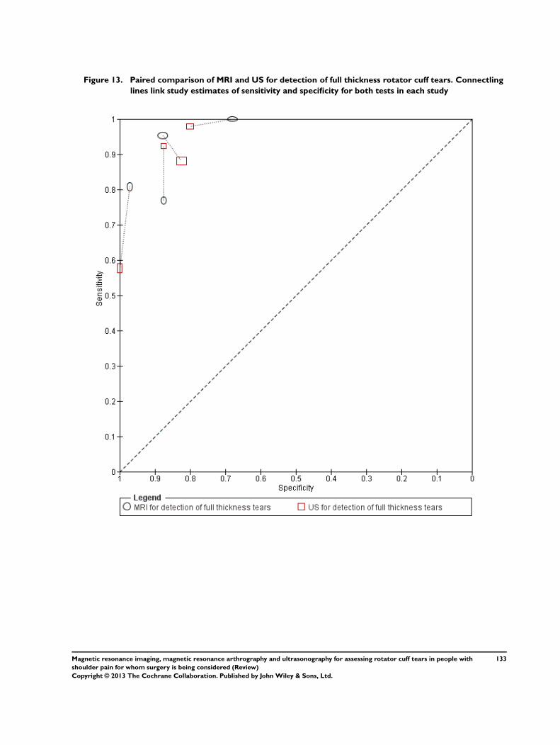
Figure 13. Paired comparison of MRI and US for detection of full thickness rotator cuff tears. Connectling
lines link study estimates of sensitivity and specificity for both tests in each study
133Magnetic resonance imaging, magnetic resonance arthrography and ultrasonography for assessing rotator cuff tears in people with
shoulder pain for whom surgery is being considered (Review)
Copyright © 2013 The Cochrane Collaboration. Published by John Wiley & Sons, Ltd.
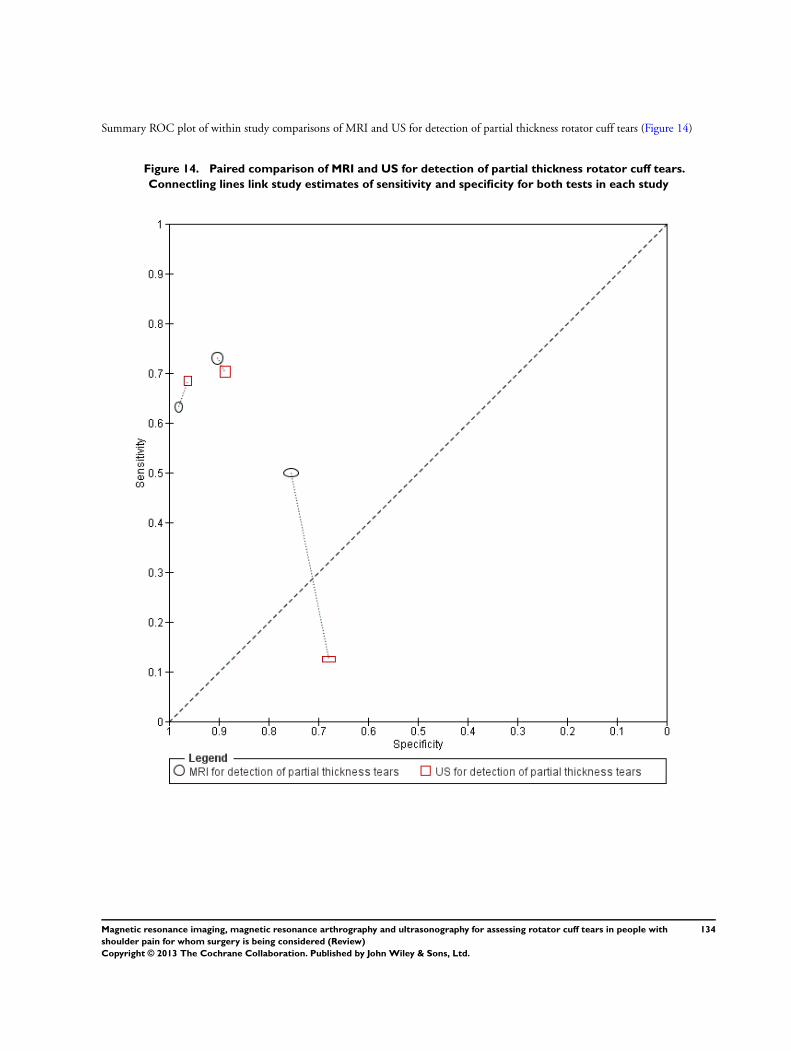
Summary ROC plot of within study comparisons of MRI and US for detection of partial thickness rotator cuff tears (Figure 14)
Figure 14. Paired comparison of MRI and US for detection of partial thickness rotator cuff tears.
Connectling lines link study estimates of sensitivity and specificity for both tests in each study
134Magnetic resonance imaging, magnetic resonance arthrography and ultrasonography for assessing rotator cuff tears in people with
shoulder pain for whom surgery is being considered (Review)
Copyright © 2013 The Cochrane Collaboration. Published by John Wiley & Sons, Ltd.
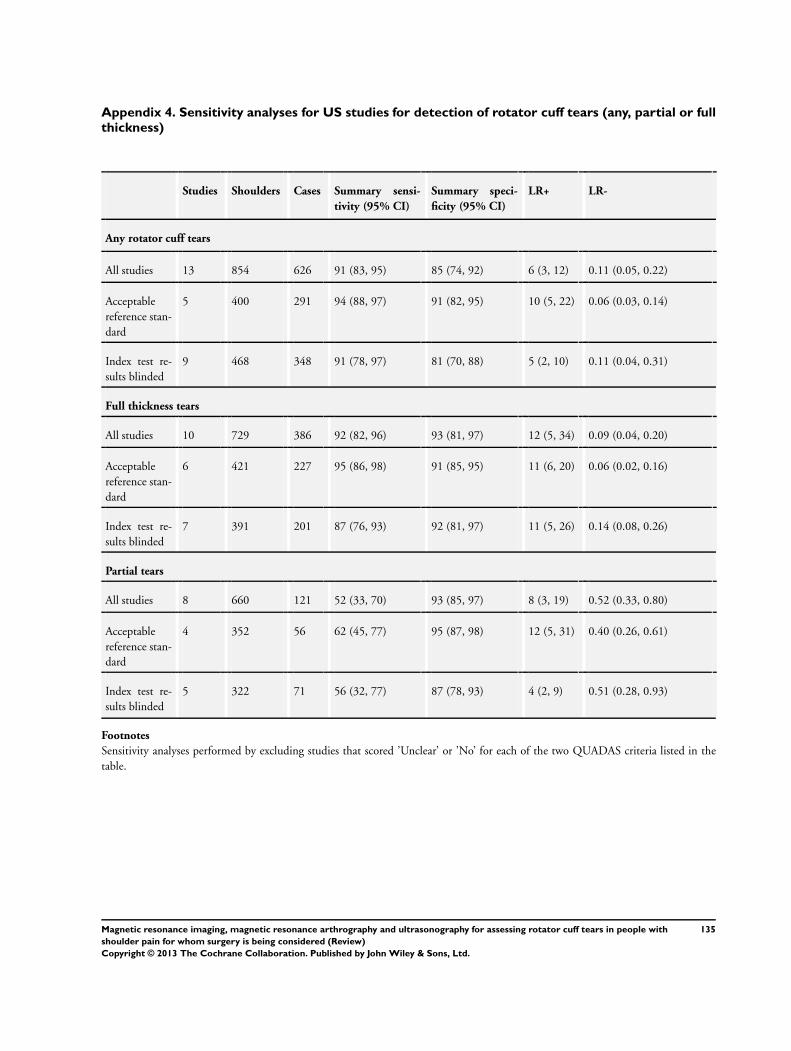
Appendix 4. Sensitivity analyses for US studies for detection of rotator cuff tears (any, partial or fullthickness)
Studies Shoulders Cases Summary sensi-
tivity (95% CI)
Summary speci-
ficity (95% CI)
LR+ LR-
Any rotator cuff tears
All studies 13 854 626 91 (83, 95) 85 (74, 92) 6 (3, 12) 0.11 (0.05, 0.22)
Acceptablereference stan-dard
5 400 291 94 (88, 97) 91 (82, 95) 10 (5, 22) 0.06 (0.03, 0.14)
Index test re-sults blinded
9 468 348 91 (78, 97) 81 (70, 88) 5 (2, 10) 0.11 (0.04, 0.31)
Full thickness tears
All studies 10 729 386 92 (82, 96) 93 (81, 97) 12 (5, 34) 0.09 (0.04, 0.20)
Acceptablereference stan-dard
6 421 227 95 (86, 98) 91 (85, 95) 11 (6, 20) 0.06 (0.02, 0.16)
Index test re-sults blinded
7 391 201 87 (76, 93) 92 (81, 97) 11 (5, 26) 0.14 (0.08, 0.26)
Partial tears
All studies 8 660 121 52 (33, 70) 93 (85, 97) 8 (3, 19) 0.52 (0.33, 0.80)
Acceptablereference stan-dard
4 352 56 62 (45, 77) 95 (87, 98) 12 (5, 31) 0.40 (0.26, 0.61)
Index test re-sults blinded
5 322 71 56 (32, 77) 87 (78, 93) 4 (2, 9) 0.51 (0.28, 0.93)
Footnotes
Sensitivity analyses performed by excluding studies that scored ’Unclear’ or ’No’ for each of the two QUADAS criteria listed in thetable.
135Magnetic resonance imaging, magnetic resonance arthrography and ultrasonography for assessing rotator cuff tears in people with
shoulder pain for whom surgery is being considered (Review)
Copyright © 2013 The Cochrane Collaboration. Published by John Wiley & Sons, Ltd.
C O N T R I B U T I O N S O F A U T H O R S
All authors contributed to the development of the review and commented on and approved the final version. The guarantor of thisreview is Mario Lenza.
D E C L A R A T I O N S O F I N T E R E S T
None known.
S O U R C E S O F S U P P O R T
Internal sources
• Universidade Federal de São Paulo, Brazil.• The Parker Institute, Denmark.
The Parker Institute: Musculoskeletal Statistics Unit is supported by grants from The Oak Foundation• Teesside University, UK.• Department of Epidemiology and Preventive Medicine, School of Public Health and Preventive Medicine, Monash University,
Australia.In kind support
• Cabrini Institute, Cabrini Hospital, Malvern, Victoria, Australia.In kind support
External sources
• No sources of support supplied
D I F F E R E N C E S B E T W E E N P R O T O C O L A N D R E V I E W
1. We only included studies of participants suspected of having rotator cuff tears. Studies that reported results of people who hadbeen previously diagnosed with, or suspected of having, other specific shoulder diagnoses were excluded. If it was unclear whether ornot all participants were suspected of having rotator cuff tears, we also excluded these studies
2. Inasmuch as there is no set time point beyond which it is known that rotator cuff tears progress, we accepted studies in whichthe time between the index test and the reference standard test was up to a year (rather than six months as specified in the protocol).
3. We included the MEDION database in our search strategy.
4. We restricted our analyses to prospective studies and excluded retrospective studies because of the high risk of spectrum andverification biases in these studies.
5. We made an amendment in the assessment of methodological quality - item seven (index test results blinded). We removed “ifthe study was retrospective” as a reason to say No because we included only prospective studies.
6. We made an amendment in the assessment of methodological quality - item eight (reference standard results blinded). Weexcluded “if the study was retrospective” as a reason to say No because it was covered by the first part of the sentence.
7. We included in the assessment of methodological quality table an additional generic quality item assessing whether or not thecriteria for a positive index test result was reported.
8. We used the bivariate model for meta-analysis instead of the hierarchical summary ROC (HSROC) model. Given the availableinformation, we assumed a common threshold was applicable but with heterogeneity around this common threshold due to variationin interpretation in practice. Therefore we consider the bivariate model and the estimation of summary points (with 95% confidenceregions) appropriate for summarising the results of the review.
136Magnetic resonance imaging, magnetic resonance arthrography and ultrasonography for assessing rotator cuff tears in people with
shoulder pain for whom surgery is being considered (Review)
Copyright © 2013 The Cochrane Collaboration. Published by John Wiley & Sons, Ltd.
9. We conducted sensitivity analyses to examine the effect of unit of analysis.
137Magnetic resonance imaging, magnetic resonance arthrography and ultrasonography for assessing rotator cuff tears in people with
shoulder pain for whom surgery is being considered (Review)
Copyright © 2013 The Cochrane Collaboration. Published by John Wiley & Sons, Ltd.
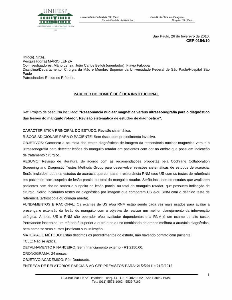
Rua Botucatu, 572 - 1º andar – conj. 14 - CEP 04023-062 - São Paulo / Brasil Tel.: (011) 5571-1062 - 5539.7162
1
São Paulo, 26 de fevereiro de 2010.
CEP 0154/10
IImo(a). Sr(a). Pesquisador(a) MÁRIO LENZA Co-Investigadores: Mário Lenza, João Carlos Belloti (orientador), Flávio Faloppa Disciplina/Departamento: Cirurgia da Mão e Membro Superior da Universidade Federal de São Paulo/Hospital São Paulo Patrocinador: Recursos Próprios.
PARECER DO COMITÊ DE ÉTICA INSTITUCIONAL
Ref: Projeto de pesquisa intitulado: “Ressonância nuclear magnética versus ultrassonografia para o diagnóstico
das lesões do manguito rotador: Revisão sistemática de estudos de diagnóstico”.
CARACTERÍSTICA PRINCIPAL DO ESTUDO: Revisão sistemática.
RISCOS ADICIONAIS PARA O PACIENTE: Sem risco, sem procedimento invasivo.
OBJETIVOS: Comparar a acurácia dos testes diagnósticos de imagem da ressonância nuclear magnética versus a
ultrassonografia para detectar lesões do manguito rotador em pacientes com dor no ombro que possuem indicação
de tratamento cirúrgico..
RESUMO: Revisão de literatura, de acordo com as recomendações propostas pela Cochrane Collaboration
Screening and Diagnostic Testes Methods Group para desenvolver revisões sistemáticas de estudos de acurácia.
Serão incluídos todos os estudos de acurácia que comparam ressonância RNM e/ou US com os testes de referência
em pacientes com suspeita de lesão parcial ou total do manguito rotador. Serão incluídos os estudos que avaliarem
pacientes com dor no ombro e suspeita de lesão parcial ou total do manguito rotador, que possuem indicação de
cirurgia. Serão inclduídos testes de diagnóstico por imagem que comparem US e/ou RNM com o definido teste de
referência (artroscopia ou cirurgia aberta).
FUNDAMENTOS E RACIONAL: Os exames de US e/ou RNM estão sendo cada vez mais usados para avaliar a
presença e extensão da lesão do manguito com o objetivo de realizar um melhor planejamento da intervenção
cirúrgica. Ambos, US e RNM são operador e/ou avaliador dependentes e a RNM é um exame de alto custo.
Permanece incerto se um método é superior a outro e se o uso combinado de ambos melhora a acurácia diagnóstica,
bem como se seus custos justificam sua utilização..
MATERIAL E MÉTODO: Estão descritos os procedimentos do estudo, não havendo contato com paciente.
TCLE: Não se aplica.
DETALHAMENTO FINANCEIRO: Sem financiamento externo - R$ 2150,00.
CRONOGRAMA: 24 meses.
OBJETIVO ACADÊMICO: Pós-Doutorado.
ENTREGA DE RELATÓRIOS PARCIAIS AO CEP PREVISTOS PARA: 21/2/2011 e 21/2/2012.
Rua Botucatu, 572 - 1º andar – conj. 14 - CEP 04023-062 - São Paulo / Brasil Tel.: (011) 5571-1062 - 5539.7162
2
O Comitê de Ética em Pesquisa da Universidade Federal de São Paulo/Hospital São Paulo ANALISOU e APROVOU
o projeto de pesquisa referenciado.
1. Comunicar toda e qualquer alteração do projeto e termo de consentimento livre e esclarecido. Nestas
circunstâncias a inclusão de pacientes deve ser temporariamente interrompida até a resposta do Comitê, após
análise das mudanças propostas.
2. Comunicar imediatamente ao Comitê qualquer evento adverso ocorrido durante o desenvolvimento do estudo.
3. Os dados individuais de todas as etapas da pesquisa devem ser mantidos em local seguro por 5 anos para
possível auditoria dos órgãos competentes.
Atenciosamente,
Prof. Dr. José Osmar Medina Pestana Coordenador do Comitê de Ética em Pesquisa da Universidade Federal de São Paulo/ Hospital São Paulo 0154/10

![Síndrome do Manguito Rotador: Reabilitação...Hewlett-Packard Company [Data] Síndrome do Manguito Rotador: Reabilitação. 1 1 Diretrizes AMB ... Caso não haja melhora com o uso](https://img.document.onl/doc/110x75/5e684224854f4007036b862c/sndrome-do-manguito-rotador-reabilitao-hewlett-packard-company-data.jpg)