Embed Size (px)
Citation preview

Research ArticleCardiovascular Biomarkers in Chronic Kidney Disease:State of Current Research and Clinical Applicability
Luis D’Marco,1 Antonio Bellasi,2,3 and Paolo Raggi4,5
1Unidad Avanzada de Investigacion y Diagnostico Ecografico y Renal, Clınica Puerto Ordaz, Puerto Ordaz, Venezuela2U.O.C. di Nefrologia e Dialisi, Ospedale Sant’Anna, Azienda Ospedaliera Sant’Anna, Como, Italy3Department of Health Sciences, University of Milan, Milan, Italy4Mazankowski Alberta Heart Institute, University of Alberta, Edmonton, AB, Canada5Department of Radiology, Emory University, Atlanta, GA, USA
Correspondence should be addressed to Paolo Raggi; [email protected]
Received 15 December 2014; Revised 15 March 2015; Accepted 18 March 2015
Academic Editor: Shih-Ping Hsu
Copyright © 2015 Luis D’Marco et al. This is an open access article distributed under the Creative Commons Attribution License,which permits unrestricted use, distribution, and reproduction in any medium, provided the original work is properly cited.
The high incidence of cardiovascular events in chronic kidney disease (CKD) warrants an accurate evaluation of risk aimed atreducing the burden of disease and its consequences. The use of biomarkers to identify patients at high risk has been in use in thegeneral population for several decades andhas receivedmixed reactions in themedical community. Somepractitioners have becomestaunch supporters and users while others doubt the utility of biomarkers and rarely measure them. In CKD patients numerousmarkers similar to those used in the general population and others more specific to the uremic population have emerged; howevertheir utility for routine clinical application remains to be fully elucidated. The reproducibility and standardization of the serumassays are serious limitations to the broad implementation of these tests.The lack of focused research and validation in randomizedtrials rather than ad hoc measurement of multiple serum markers in observational studies is also cause for concern related to theclinical applicability of these markers. We review the current literature on biomarkers that may have a relevant role in field ofnephrology.
1. Introduction
The very high incidence of cardiovascular disease (CVD)events and premature mortality in patients with chronickidney disease (CKD) [1], with a sharp increase in risk asglomerular filtration rate (GFR) declines below 60mL/min/1.72m2 [2], offers a rationale for better risk stratification inthis population. Several traditional risk factors and factorsmore closely related to loss of renal function (anemia,oxidative stress, inflammation, and bone mineral disorders)contribute to the high incidence of cardiovascular complica-tions seen in patients with CKD. Whether biomarkers helpimprove the identification of patients at risk of cardiovascularevents has been at the core of extensive research in the generalpopulation and in patients with CKD [3]. This approachpredicates that an accurate assessment of cardiovascular riskat an early stage would facilitate more aggressive and focusedtreatment of those in greater need of preventive measures
with the goal to reduce event rates. In this review, we focuson established and emerging laboratory biomarkers for theassessment of risk in CKD and compare them to their use inthe general population.
2. Natriuretic Peptides
The natriuretic peptides are a family of hormones that play amajor role in sodium and body volume homeostasis; specif-ically they control natriuresis, vasodilatation, and diuresis[4]. Three major natriuretic peptides have been identified:atrial natriuretic peptide (ANP), B-type natriuretic peptide(BNP), and C-type natriuretic peptide (CNP). They all sharea common 17-amino-acid ring structure and have actionsthat are targeted at protecting the cardiovascular systemfrom the effects of volume overload [5]. While ANP ispreferentially produced in and secreted by the atria, BNP
Hindawi Publishing CorporationDisease MarkersVolume 2015, Article ID 586569, 16 pageshttp://dx.doi.org/10.1155/2015/586569

2 Disease Markers
is produced by the ventricular myocardium in responseto ventricular stretching and wall stress [6]. CNP, derivedprimarily from endothelial cells, is also synthesized by themyocardium.Upon ventricularmyocyte stretch, pre-proBNPis enzymatically cleaved to proBNP and released as activeBNP hormone (amino acids 79–108) or an inactive fragment,NT-proBNP (amino acids 1–76, released in a 1 : 1 ratio).
Natriuretic peptides, in particular BNP and NT-ProBNP,have been investigated as biomarkers in several conditionsand an increase in their serum levels has been associated withdegree of left ventricular dysfunction, severity of congestiveheart failure symptoms, and ultimately a poor prognosisin community-based and general population studies [7–11].Furthermore, NT-proBNP is a good marker for predictionof first cardiovascular events in the population, as well asthe risk of stroke in patients with atrial fibrillation [12, 13].NT-proBNP has a longer half-life and thus its levels may bemore stable (less affected by acute stress) than BNP. Both BNPand NT-proBNP are eliminated only to a small degree duringa hemodialysis session [14], and NT-proBNP appears toaccumulate to a larger degree during dialysis [15, 16]. Severalstudies have confirmed that both BNP and NT-proBNP areuseful markers of cardiovascular risk in CKD patients. Ingeneral, they have been shown to correlate with the severity ofheart failure and left ventricular dysfunction and to be usefulin guiding the management of heart failure in CKD. PlasmaBNP concentrations increase progressively with decreasingrenal function, and this relationship remains present whenpatients are subdivided into systolic and diastolic heart failure(𝑃 < 0.01) [17]. In a prospective evaluation of 213 CKDnondialysis patients, Vickery and coworkers demonstratedthat NT-proBNP (≥89 pmol/L, HR 2.5, 𝑃 < 0.05) and high-sensitivity C-reactive protein (hsCRP ≥ 4.7mg/L, HR 1.9,𝑃 < 0.05) were independently associated with increased all-cause mortality [3]. Similar findings were reported in dialysispatients [18]. Additionally, NT-proBNP, but not BNP, was anindependent predictor of death in a heart failure populationwith eGFRs < 60mL/min/1.73m2 [19].
BNP was prospectively studied as a biomarker of cardio-vascular events in CKD patients in a community-based studyin Japan [20]. The investigators collected baseline plasmaBNP, serum creatinine, and urinary protein levels from 9625patients. The risk of cardiovascular events was significantlyhigher in participants with the highest BNP serum levels. Ina study of 134 hemodialysis patients Sommerer et al. foundthat plasma levels of NT-proBNP were elevated in 100% andcardiac troponin T (cTNT) levels in approximately 40% ofasymptomatic patients [21]. Both increased NT-proBNP andcTNT were strongly associated with an adverse cardiovascu-lar outcome (overall there were 23 deaths due to myocardialinfarction and sudden cardiac death). In a prospective cohortstudy of 230 patients undergoing chronic peritoneal dialysis,Wang et al. reported that NT-pro-BNP was an independentrisk marker of congestive heart failure, death, or a combinedend-point including death and other adverse cardiovascularoutcomes [22] (odds ratios varied between 4.25 and 9.1 forthe fourth compared to the 1st quartile of NT-pro-BNP).The authors concluded that in chronic peritoneal dialysispatients NT-pro-BNP adds prognostic information beyond
that contributed by left ventricular hypertrophy, systolicdysfunction, and other conventional risk factors. Finally,NT-pro-BNP is a predictor of mortality in CKD patientsindependent of volume overload and dialysis modality [23,24].
3. Cardiac Troponin T
Troponin (TN) T, I, and C are components of the contractileapparatus of striated muscle. Specific forms of troponinsT and I are present in the heart muscle, namely, cTNTand cTNI. cTNI is exclusively expressed in cardiomyocytesand is released into the circulation after myocardial celldamage. Increased levels of troponins are detectable 3–12 haftermyocardial injury, with the concentration being in directproportion to the extent of injury [5].The serum levels returnto the normal range after 5–14 days from injury, which is 4times longer than for the creatine kinase myocardial bandisoenzyme (CK-MB) fraction, probably because of sustainedrelease of structurally-bound protein from disintegratingmyofibrils [25].
cTNT is cardiac specific and is not present in theserum following nonmyocardial muscle damage. The levelsof cTNT are commonly increased among patients with renaldysfunction in the absence of acute myocardial infarction[26]. Although the exact mechanism is unknown, severalstudies suggested that increased levels of troponins areassociated with increased risk of coronary artery disease(CAD) and death in predialysis populations [27]. Severalexplanations, although controversial, have been proposed tounderstand cTNT elevation in uremic patients. Among themare increased left ventricular wall stress, acute or chronic vol-ume overload, silent plaque rupture in the presence of diffusecoronary atherosclerosis, and apoptosis of cardiomyocytes[28, 29].The prevalence of elevated cTNT plasma levels priorto hemodialysis sessions varies between 20% and 53%. Aserum level of cTNT > 0.01 microg/L is relatively common(28,2%) in patients with CKD stage 3-4 [26]. A cohort of 847dialysis patients was followed in a multicenter study in theNetherlands between 1997 and 2001. After an average follow-up of two years patients with baseline cTNT levels between0.05 and 0.10 microg/L showed a hazard ratio for all-causedeath of 2.2 (95% CI, 1.7 to 2.8) compared with patients withlevels below 0.05 microg/L [30]. For those with levels greaterthan 0.10microg/L (11%), the hazard ratio rose to 3.3 (95%CI,2.5 to 4.5). In this investigation there was no significant differ-ence in risk of events between patients receiving hemodialysisor peritoneal dialysis and between patients with high and lowresidual renal function. Cardiac TNT was used as a markerto identify occult obstructive coronary artery disease (CAD)in 142 patients at the start of their renal replacement therapy[31]. Of 60 asymptomatic patients at the time of evaluation,35 (43.8%) had obstructive CAD and 27 had multivesselCAD as assessed by invasive coronary angiography. On step-wise logistic regression analyses cTNT was an independentpredictor of asymptomatic multivessel CAD (sensitivity andspecificity of cTNT to predict multivessel CAD: 92.6% and63.6%, resp.). Several publications have further supported thevalue of cTNT as a predictor of unfavorable CAD events in

Disease Markers 3
CKD populations [32–34]. cTNT and cTNI were found to beequivalent in differentiating short-term prognosis in patientswith CKD suffering a non-ST-segment elevation myocardialinfarction [35]. In summary, cTNT may be helpful to detectasymptomatic CAD, especially multivessel disease, and asa predictor of mortality in CKD patients. However, as forseveral other markers, the variability of the test both day today and from one laboratory to the other may limit the abilityto issuewidely applicable guidelines on the use this biomarkerin CKD.
4. C-Reactive Protein
Systemic inflammation plays a major role in the develop-ment of atherosclerosis leading to coronary heart disease.C-reactive protein (CRP), an acute phase reactant, is anestablished marker of systemic inflammation in the generalpopulation and patients with CKD [36–38]. Elevated CRPlevels, when measured using highly sensitive assays (hsCRP),are predictive of future coronary events in the generalpopulation [39]; currently a serum level of hsCRP >3mg/Lis considered a marker of high risk for events [40]. Althoughthe clinical utility of hsCRP in CKD is still under debate,a few studies reported a significant association betweenhsCRP and all-cause mortality in end-stage renal failure atconcentrations varying between 1 and 3mg/L [18, 41, 42].Apple et al. followed prospectively 399 CKD stage 5 patientsin whom they measured several biomarkers. They reportedthat hsCRP, cTNT, and cTNI were independent predictorsof death after a 2-year follow-up. NT-proBNP was onlypredictive when analysed as a categorical variable but not asa continuous variable [18]. Snaedal et al. measured hsCRPserially in a three-month observational study of 228 prevalenthemodialysis patients [43]. They reported that median andmean serial CRP levels were associated with an increasedhazard ratio for all-cause death (1.013; 95% confidence inter-val, 1.004 to 1.022 and 1.012; 95% confidence interval, 1.004to 1.020, resp.). Of interest, 13% of the patients enrolled hadpersistently low hsCRP levels (<5mg/L), 19% had CRP levels> 10mg/L, and 68% demonstrated fluctuating values. Hence,in the majority of hemodialysis patients, hsCRP is a “movingtarget” depending on a variety of inflammatory stimuli in theindividual patient. This observation may be prognosticallyimportant. In fact, Nascimento et al. reported that the mor-tality rate in 180 hemodialysis patients followed for 21monthsafter 4 sequential measurements of CRP was significantlyhigher in patients with persistently elevated levels (34%death rate) than in those with low (8%) or intermittentlyelevated (14%) CRP levels [44]. Of note, the mortality ratewas not statistically different between patients with low andintermittently elevated CRP levels. In another prospectiveobservational study of 402 patients receiving peritonealdialysis the investigators reported a 1.4% increase inmortalityfor each 1mg/L increase in hsCRP level after 2 years of follow-up [45].There are also conflicting reports on the value of CRPin CKD. Kanwar et al. measured troponin T and I and hsCRPin a cohort of 173 prevalent hemodialysis patients dividedinto 2 groups according to clinical characteristics: patientwith CAD or CAD equivalent status (peripheral arterial
disease and diabetes) and patients without any relevant priorcardiovascular history [46]. Prior history of CAD or CADrisk-equivalence was strongly predictive of mortality andhsCRP did not add to the risk stratification of these patients.On the contrary, both troponin T and I added incrementalprognostic information. In the lower-risk patients hsCRPwaspredictive ofmortality while troponinswere not. In a study byHonda et al. where hsCRP, IL-6 serum albumin, and fetuinA were measured in 176 prevalent hemodialysis patients,hsCRP was found to be a good predictor of malnutritionbut a weaker predictor of CVD and mortality than IL-6[47]. Overall, hsCRP may be useful as an additional piece ofinformation to assess risk in CKDpatients, but its value needsto be carefully considered in the context of several limitations.There is likely a substantial difference in the significance ofhsCRP elevation in predialysis and dialysis patients. In fact,exposure to dialysismembranes, evenmodern biocompatiblemembranes, and vascular access infections are associatedwith marked cytokine and hsCRP release; these events donot obviously take place in predialysis patients. hsCRP levelsmay be modulated by the concurrent use of statins. Theactual risk may differ in patients with persistently elevatedas opposed to intermittently elevated CRP levels. Finally thepredictive value of hsCRPmay be enhanced by combining itsmeasurement with that of several other markers of risk [48].
5. Adiponectin
Recent research has focused on visceral adipose tissue asa source of inflammation and adipocytokines secretion.Adiponectin (APN) is a protein secreted by adipocytes andhas anti-inflammatory and antiatherogenic activities andenhances insulin-sensitivity [49, 50]. APN likely exerts itsantiatherosclerotic activities by suppressing the release ofproinflammatory cytokines such as TNF-𝛼 and IL-6 andstimulating the release of anti-inflammatory cytokines suchas IL-10 [51, 52] and via enhancement of insulin sensitivity.Low APN levels have BEEN observed in patients withobesity, the metabolic syndrome, diabetes mellitus, CAD,and essential hypertension. In contrast, plasma levels ofAPN in patients with CKD are increased up to threefoldthe physiological levels [53–55], most likely due to reducedclearance or catabolism [56]. In the general population lowAPN levels have been associated with the development ofcardiovascular disease [57, 58]. A few observational studieslinked APN to adverse cardiovascular outcomes in patientswith CKD [59, 60]. Becker and coworkers evaluated 227nondiabetic patients with CKD and 76 healthy subjects ofsimilar age, sex, and body mass index [61]. After a meanfollow-up of 54 months, they concluded that, despite higherserum levels than in control subjects, relatively low plasmaAPN levels were predictive of cardiovascular events amongpatients with mild to moderate CKD. Similarly, Zoccali etal. reported that plasma APN levels were 2.5 times higher(𝑃 < 0.0001) among dialysis patients (15.0 ± 7.7 𝜇g/mL)than among healthy subjects (6.3 ± 2.0 𝜇g/mL), but the APNlevels were lower among the dialysis patients who developedcardiovascular complications than those who remained eventfree [62]. The increased risk of cardiovascular events in
4 Disease Markers
CKD patients with lower APN concentrations (𝑃 < 0.05)relative to other CKD patients was unchanged after adjustingfor multiple traditional and CKD-specific risk factors. Each1 𝜇/mL increase in APN concentration was associated with a3% reduction in risk of cardiovascular events.
In contrast, a subanalysis of the modification of diet inrenal disease (MDRD) database performed in 820 patientswith CKD showed a direct correlation between increasedAPN plasma concentration and the relative risk of cardiovas-cular mortality [63]. In multivariable adjusted Cox models,1 𝜇/mL increase in APN was associated with a 3% (hazardratio 1.03; 95% CI 1.01 to 1.05; 𝑃 = 0.02) increased riskof all-cause and 6% (hazard ratio 1.06; 95% CI 1.03 to 1.09;𝑃 < 0.001) increased risk of cardiovascular mortality. Apotential explanation for these apparently conflicting data isthe reported association between increased APN concentra-tions and poor nutritional status in CKD [64]. However, theexistence of a link between higher APN levels and increasedcardiovascular risk in CKD remains to be clarified. In viewof the bidirectional association of APN with events, its roleas a useful marker of cardiovascular risk in CKD remainsuncertain pending accumulation of further evidence.
6. Leptin
Leptin is a single-chain 16-kDa protein whose production isunder the control of the obese (ob) gene. It is a neurotrans-mitter entirely produced in adipocytes, with the fundamentalfunction of controlling appetite, regulating food intake, andenergy expenditure [65]. Leptin is also a proinflammatorycytokine and is predominantly cleared from the circulationby the kidneys via a combination of glomerular filtration andtubular degradation [66]. Leptin levels increase in proportionto insulin levels, glucocorticoids, cytokines, and obesity[67]. In humans leptin is an anorectic agent and decreasesappetite by decreasing the level of neuropeptide Y, a potentstimulator of food intake, and increasing alpha-melanocyte-stimulating hormone (alpha-MSH), an inhibitor of foodintake, respectively [68, 69]. In healthy subjects and patientswith type 2 diabetes mellitus, weight loss has been shown tobe accompanied by a reduction in leptin serum concentration[70, 71]. These data suggest that high leptin concentrationsare a component of the metabolic syndrome and may havea role in increasing the cardiovascular risk of these patients.Leptin levels are significantly elevated in patients with CKD,particularly in those undergoing dialysis comparedwith non-dialysis patients [72, 73]. Besides decreased renal clearanceother factors affect leptin levels in CKD. Metabolic acidosisreduces the release of leptin from adipocytes and uremicfactors of unclear origin reduce leptin gene expression inadipocytes probably as a negative feedback due to decreasedelimination [74, 75].
Leptin is believed to have proatherogenic effects thatinclude the development of hypertension, oxidative stress,endothelial dysfunction, inflammation, and proliferation ofvascular smooth muscle cells [64, 76]. High leptin levelsinduce activation of the sympathetic nervous system both viacentral and peripheral mechanisms. Several studies linkedincreased leptin and early markers of arterial disease such
as increased carotid intima media thickness and decreasedarterial distensibility [77–79]. Aguilera et al. described anassociation of serum levels of leptin with elevated lipidsconcentration and LVH in a small peritoneal dialysis cohort[80]. In a small size cohort of kidney transplant recipients(𝑛 = 74) there was a positive association between elevatedserum leptin levels and increased peripheral arterial stiffness[79].
At this time there are sparse and conflicting data onthe role of leptin as a marker of risk in CKD. Scholzeet al. reported an association of leptin serum levels withcardiovascular events in 71 prevalent hemodialysis patientsfollowed for 83 months [81]. The serum level of leptin wassignificantly lower in patients who died from cardiovascularcomplications (4.7 ± 9.4 microg/L, 𝑛 = 32) than in survivors(7.7 ± 7.8microg/L; 𝑛 = 23; 𝑃 = 0.003). Additionally, survivalwas shorter in patients with leptin concentrations below themedian (<2.6microg/L). Two other studies reported thatleptin did not add incremental prognostic value for all-causemortality and cardiovascular morbidity in 101 hemodialysispatients followed for a period of 4 years [82] and 181 patientsfollowed for 3 years [83]. Hence, the value of leptin as amarker of risk remains unclear in CKD.
7. Fibroblast Growth Factor 23 and Klotho
It has long been recognized that secondary hyperparathy-roidism (SHPT) is a consequence and a complication ofCKD. The global syndrome that involves bone and vasculardisease has been aptly termed CKD-mineral bone disorder(CKD-MBD) [84]. SHPT is a complex process that involvesnumerous steps such as vitamin D deficiency, hypo- and/orhypercalcemia, and hyperphosphatemia. Phosphorus levelswere believed to be under the direct control of the parathyroidglands until the fairly recent discovery of a new hormonecalled fibroblast growth factor 23 (FGF-23). FGF-23 is a 32-kD protein secreted by bone osteocytes; its primary functionis to promote phosphaturia by suppressing the expressionof sodium-phosphate cotransporters NaPi-2a and NaPi-2c inthe proximal tubule [85, 86]. In addition, FGF-23 acts as acounter-regulatory hormone for vitamin D by blocking thegeneration of 1,25(OH)2D both through inhibition of therenal 1𝛼-hydroxylase enzyme and through the stimulationof the 24-hydroxylase enzyme that is responsible for thedegradation of both the 25(OH)D and 1,25(OH)2D metabo-lites [87]. Klotho acts as a coreceptor for FGF-23 and itspresence appears to be mandatory to induce FGF-23-specificsignaling pathways in the kidney, parathyroid glands, andother tissues [88, 89]. Once Klotho is shed from the cell byproteolytic cleavage, it is released in the circulation and servesas a phosphaturic and hypocalciuric hormone independent ofFGF-23 [90].
However, it has been postulated that FGF-23 may alsooperate via Klotho independent mechanisms. In the acutekidney injury (AKI) setting, FGF-23 elevation may precedecreatinine elevation and phosphate metabolism impairment[91]. Whether this is due to inflammation or a decrease inKlotho in response to acute uremia needs further elucidation.

Disease Markers 5
Like phosphorus, PTH, and vitamin D, FGF-23 hasbeen independently associated with risk of all-cause deathin dialysis and CKD patients, heart failure, cardiovascularevents, and death in the general population [92, 93]. Across-sectional study in 177 patients with mild to moderateCKD submitted to coronary angiography found that FGF-23 increases early in the course of CKD and, as shown forother bone metabolism hormones (PTH, Fetuin A), it is anindependent predictor of CAD severity after adjusting fortraditional risk factor [94]. Seiler et al. measured plasmaFGF-23 levels in 149 CKD patients not receiving dialysis[95]. Patients were stratified according to themedian baselineFGF-23 levels (>104 versus ≤104 rU/mL) and were followedfor an average of 4.8 ± 0.9 years till the first occurrenceof a cardiovascular event. At baseline, patients with moreadvanced CKD demonstrated higher FGF-23 serum levels.Traditional cardiovascular risk factors and prevalent car-diovascular disease did not differ between groups. Fiftypatients experienced a cardiovascular event during follow-up. Compared to CKD patients with an FGF-23 level belowthe median, those with levels above the median experienceda higher rate of events [HR 2.49 (95% CI 1.40–4.39); 𝑃 =0.002]. In the Chronic Renal Insufficiency Cohort (CRIC),a graded and independent association between high levelsof FGF-23 and CKD progression, congestive heart failure,and atherosclerotic events (myocardial infarction, stroke, andperipheral vascular disease) was documented among 3860CKD 2–4 patients [96, 97]. Serum levels of FGF-23 weremeasured in 13,448 subjects with preserved renal function(mean eGFR was 97mL/min per 1.73m2) enrolled in theatherosclerosis risk in communities study (ARIC) [98]. Theinvestigators documented that among patients with an FGF-23 serum level in the top quintile (>54.6 pg/mL) comparedto those in the versus first quintile (<32.0 pg/mL) the risk ofESRD increased 2-fold (HR 2.1, 95% CI: 1.31–3.36, 𝑃 < 0.001)independent of numerous confounders. In theHeart and Soulstudy the investigators measured FGF-23 in 833 outpatientswith stable CAD and no CKD [99]. During a follow-up of6 years, 220 patients died and 182 suffered a CVD event.After adjusting for traditional risk factors, patients in thehighest tertile of FGF-23 concentrations demonstrated a 2-fold greater risk of death (HR, 2.15 [95% CI, 1.43 to 3.24]) andCVD events (HR, 1.83 [CI, 1.15 to 2.91]).
The coreceptor Klotho has been shown to have directvascular effects by increasing both production of reactiveoxygen species in human vascular smooth muscle cells andNO production in endothelial cells. Klotho deficiency hasbeen associated with oxidative stress and inflammation inESRD patients [100].
In a large observational study of 804 community-dwellingelderly (age greater than 65 years), plasma Klotho wasinversely and independently associated with the risk of all-cause mortality (data adjusted for age, sex, education, bodymass index, physical activity, total cholesterol, high-densitylipoprotein cholesterol, cognition, 25-hydroxyvitamin D,parathyroid hormone, serumcalcium,mean arterial pressure,and chronic diseases). Participants within the lowest tertile ofserum Klotho (<575 pg/mL) had a 78% greater risk of deaththan patients within the highest tertile (>763 pg/mL; hazard
ratio: 1.78; 95%; CI 1.20–2.63) [101]. In a cross-sectional studyof 956 individuals, Klotho gene polymorphisms were asso-ciated with occult coronary artery disease (CAD) (definedas the occurrence of a reversible perfusion defect on nuclearmyocardial stress testing) independent of known risk factorsfor CAD [102]. Recently a significant correlationwas reportedbetween severity of CAD and reduced levels of Klotho geneexpression in the aorta as well as lower serum levels of solubleKlotho [103]. In a subanalysis of the Heart and Soul study theserum levels of FGF-23 and its coreceptor Klotho lacked anassociation with left ventricular mass and ejection fraction inthe absence of kidney disease [104].
There is no reliable assay to measure Klotho to thisdate and despite the above findings the evidence to supportthe utility of FGF-23 and Klotho as markers of risk is stillinconclusive; therefore they cannot be recommended forroutine clinical use.
8. Fetuin-A and Calciprotein Particles
The alpha 2-Heremans Schmid glycoprotein, also known asFetuin A, is secreted by hepatocytes in greater concentrationduring fetal than adult life. It is believed to modulate boneformation and brain development although its full functionis still unknown. Fetuin A forms soluble complexes in thecirculation with calcium-phosphate crystals. The calcium-phosphate and Fetuin-A complexes form stable particles (lessthan 100–200 nm in diameter) called calciprotein particles(CPPs) that exist as colloids and do not precipitate [105].A growing body of evidence suggests that CPPs may beisolated from the serum of patients with CKD but not inhealthy individuals; they are further believed to mediate inpart the harmful effects associated with chronically elevatedphosphate in CKD [106–108]. Unlike inorganic phosphatethat stimulates the expression of sodium-phosphate cotrans-porters across the cellular membrane, CPPs seem to inducecellular responses through an interaction with the plasmamembrane and the induction of intracellular messengers butthe mechanisms remain to be elucidated. In a cohort of 200patients suffering from CKD stages 3 and 4, higher levelsof CPPs were associated with hyperphosphatemia, increasedlevels of C-reactive protein, oxidized low density protein,bone morphogenic proteins 2 and 7 as well as a rapid declinein renal function, and a greater aortic pulse wave velocity((PWV) beta coefficient 0.059, 𝑃 = 0.016, 𝑅2 = 0.362) whichis known to be a strong predictor of major CV events in CKDpatients [107].
The use of CPPs has also been proposed as a simple pre-dictor of the overall uremicmilieu propensity for calcification[106]. The clinical validity of such test has been assessed in arecent series of 184 CKD stages 3 and 4 patients. Descendingtertiles of T
50(a measure of the calcifying propensity of a
patient’s serum) were associated with higher aortic PWV aswell as all-cause mortality, independent of several traditionaland CKD specific risk factors [109].
Although intriguing, more studies are necessary to fur-ther elucidate the role of CPPs as a key mediator of CVdamage and as a potential therapeutic target in CKDpatients.

6 Disease Markers
9. Wingless (Wnt) Antagonists Inhibitors
Wingless (Wnt) antagonist inhibitors such as sclerostin andDickkopf-1 (DKK-1) are newly described factors involvedin the bone-vascular axis [110]. Wnt activation promotesosteoblast and suppresses osteoclast activity by increasingthe ratio of osteoprotegerin (OPG) to receptor activatorof nuclear factor-kappaB ligand (RANKL). This leads tobone mineralization and bone turnover downregulation.The Wnt pathway is also involved in vascular and cardiacvalve calcification and preliminary data suggest that itsoverexpression may be identified within ectopic calcificationor during calciphylaxis [111, 112]. Reduced Wnt signalingseems to occur in the earliest phases of CKD likely dueto the increased expression of Sclerostin and DKK-1 and itmay be responsible for accelerated loss of bone mass andstrength, uremic resistance to PTH, and increased vascularcalcification [110, 113, 114].
Nonetheless, the clinical significance of sclerostin as amarker of risk remains unclear. In a series of 140 patientswith CKD stages 2–5D [115], advancing stages of CKDwere accompanied by a graded increase in serum levels ofsclerostin. At this stage it is unclear whether this is due toreduced renal clearance or increased production in CKD.In the study by Desjardins et al. [115], sclerostin levels wereassociated with serum levels of phosphate, FGF-23, and IL-6as well as protein bound uremic solutes such as indoxylsul-phate and p-cresyl sulphate and were independent predictorsof arterial stiffness and mortality, although they were notassociated with vascular calcification. Further adjustmentsfor confounders significantly attenuated the strength of theassociation of sclerostin with mortality [115].
In a post hoc analysis of 100 prevalent hemodialysispatients, higher levels of serum sclerostin were associatedwith decreasedmortality (age and sex adjusted HR: 0.33, 95%CI 0.15–0.73; 𝑃 = 0.006) [116]. After further adjustment forbone specific alkaline phosphatase, serum sclerostin levelsdropped out of the multivariable model as a significantvariable.
Drechsler and coworkers documented an independentinverse association of sclerostin with cardiovascular (HR0.29; 95% CI 0.13–0.62) and all-cause mortality (HR 0.39;95% CI: 0.22–0.68) in 673 hemodialysis patients enrolled inthe NECOSAD study [117]. The association tended to losestatistical significance when the observation was prolongedpast the first 18 months of dialysis. At the current stage ofknowledge, therefore, the role of Wnt signaling inhibitors asmarkers of risk needs further elucidation and confirmation.
10. Neutrophil Gelatinase-Associated Lipocalin
Neutrophil gelatinase-associated lipocalin (NGAL) is a 25–135 kDa (depending on the tertiary structure of the peptide)member of the lipocalin iron-carrying proteins family [118]and is highly expressed in kidney following ischemic ornephrotoxic injury [119–121]. Although NGAL was origi-nally isolated in neutrophils [122], numerous other tissuesexpress this protein, namely, kidney, liver, and vascular
cells (endothelial, smooth muscle cells, and macrophages inatherosclerotic plaques), as well as cardiomyocytes [123–125].
NGAL inhibits the degradation of gelatinase B leadingto enhanced proteolytic activity with collagen degradationeffects [123]. At steady state, NGAL concentration is negli-gible (about 20 ng/mL) in both blood and urine [126]. Theseconcentrations likely reflect neutrophils production and renalclearance, the major regulators of NGAL concentration atsteady state. As renal function declines, NGAL levels increaseand they become markedly increased in chronic dialysis.However, high-flux hemodialysis may significantly removeNGAL from plasma [127].
Identification of increased levels of NGAL in bloodand urine in several renal disease states has generated aninterest in NGAL as an early marker of acute kidney injury[128]. A recent meta-analysis of 19 studies including 2538patients from 8 countries summarized the available evidenceregarding NGAL as a biomarker [129]. Although data weremainly derived from patients undergoing cardiac surgery,plasma NGAL was a strong predictor of acute kidney injury,initiation of renal replacement therapy, and in-hospital mor-tality with odds ratios ranging from 8.8 to 18.6 for high versuslow NGAL [129].
AlthoughNGALwas associated with progression of CKDafter kidney injury in a few reports [130–133], an analysis fromthe Chronic Renal Insufficiency Cohort (CRIC) questionedits role as a useful biomarker to predict progression [134].In the CRIC cohort of 3386 patients suffering from CKD2–4, NGAL was an independent predictor of worsening ofCKD (defined as a 50% eGFR decrease or dialysis initiation)but it did not add significantly to the discrimination modelsuggesting a marginal value of urinary NGAL as a predictorof CKD progression [134].
Investigators from Japan recently reported on the use ofNGLA as a marker of CV risk in 88 hemodialysis patients[135]. At the end of 1-year follow-up they recorded 20 events;patients with event had significantly higher levels of NGALand pro-BNP at study inception than event-free survivors(357 ± 21 versus 290 ± 11 ng/mL and 553 ± 119 versus172 ± 73 pg/mL, 𝑃 < 0.01 for NGAL and proBNP, resp.).After adjustment for confounders each 1 ng/mL increase inNGAL was associated with a 3% (OR: 1.03; 95% CI: 1.01–1.06)increase in the risk of CV events. Patients with simultaneousincrease in NGAL and pro-BNP exhibited a 25-fold higherrisk of CVD than patients with low NGAL and pro-BNP.
NGAL has been investigated as a prognostic marker inpatients suffering from acute heart failure (AHF). Maisel andcoworkers evaluated 186 patients admitted via the emergencydepartment with AHF and measured NGAL and BNP [136].At 30 days, heart failure readmission and death occurred in29 patients; theNGAL serum level was higher in patients withevents (134 versus 84 ng/mL, 𝑃 < 0.001) and NGAL was anindependent predictor of events inmultivariablemodels (𝑃 =0.001), while BNP was only of borderline significance (𝑃 =0.052). The addition of NGAL to a multivariable-adjustedmodel lead to a 29.8% net reclassification improvement forprediction of events.
In the Rancho Bernardo Study, NGAL was associatedwith inflammation, lower HDL-cholesterol levels, and lower

Disease Markers 7
creatinine clearance [137]. In this study each standard devia-tion increase in log-transformed NGAL level was associatedwith a significant increase in all-causemortality (HR 1.19; 95%CI: 1.07–1.32), CV death (HR 1.33; 95% CI: 1.12–1.57), and thecombined endpoint (HR 1.26; 95% CI: 1.10–1.45).
In summary, the limited available evidence suggests thatNGAL is a weak marker of risk for renal function declinebut might be a predictor of cardiac event in the setting ofHF in patients with and without renal disease. As for otherbiomarkers, future work will need to define reliable cutoffvalues in different populations and standardize results arisingfrom different available assays.
11. Plasma Growth Differentiation Factor-15
Plasma growth differentiation factor-15 (GDF-15) is gener-ated as a 40-KDa propeptide from which the NH2-terminusis cleaved and the resulting 30-KDa protein is secreted asactive form [138]. GDF-15 has been described as a potentialinhibitor of left ventricular hypertrophy [139]. An increasein pre- or afterload in experimental settings induces anincrease in plasma GDF-15 possibly through proinflam-matory cytokines and oxidative stress-dependent signalingpathways [140, 141]. Several studies have shown that GDF-15 is over expressed in patients suffering from atheroscleroticdiseases such as myocardial infarction, stroke, and otherstypes of atherosclerotic disease [142–144]. The literatureregarding GDF-15 in patients affected by CKD is limited.Interesting data were derived from the 2,614 participantsin the Framingham Offspring study followed for a meanof 9.5 years [145]. The investigators reported that higherplasma levels of GDF-15 were associated with incident CKD(multivariable-adjusted OR 1.9 per 1-unit increase in log-GDF-15, 95% CI 1.6–2.3, 𝑃 < 0.0001) and a rapid declinein kidney function (OR 1.6, 95% CI 1.3–1.8, 𝑃 < 0.0001).In a prospective observational study Lajer et al. showed thatGDF-15 was an independent predictor of all-cause and car-diovascular mortality (HR 3.6 [95% CI 1.3–10.3]; 𝑃 = 0.014)and worsening renal function in 451 patients with diabeticnephropathy [146]. Additionally, a study originally designedto evaluate GDF-15 for prognostication of cardiovascular andcancer morbidity and mortality in 940 older men reporteda close association between the highest GDF-15 tertile anddecline in renal function [147]. Although this marker is ofsome interest there is a need for substantially more researchin patients with renal failure.
12. Asymmetric Dimethylarginine
As an endogenous inhibitor of nitric oxide (NO) synthases,asymmetric dimethylarginine (ADMA) causes endothelialdysfunction, vasoconstriction, elevation of blood pressure,and aggravation of experimental atherosclerosis [148, 149].After its release frommethylated proteins, circulatingADMAlevels are regulated via glomerular filtration and enzy-matic degradation by dimethylarginine dimethylaminohy-drolase. Originally described by Vallance and colleagues[150], ADMA has been investigated fairly extensively inthe CKD population, especially in dialysis. Although the
exact mechanism linking ADMA with CVD risk in dial-ysis patients remains to be clarified, one of the potentialmechanisms involves an interaction with the sympatheticnervous system [151, 152]. Inhibition of NO synthesis byADMA leads to enhanced norepinephrine release fromsympathetic nerve endings, whereas sympathetic activationimpairs endothelium-dependent NO dilation [153, 154]. Zoc-cali et al. reported that ADMA is an independent predictorof cardiovascular events (HR: 1.17, 1.04–1.33, 𝑃 = 0.008)and mortality (HR 1.26, 95% Cl 1.11–1.41, 𝑃 = 0.0001)in patients receiving chronic hemodialysis [155]. The sameresearches showed that ADMA is associated with progressionof carotid intima media thickness, cardiac remodeling, andLVH in hemodialysis [156, 157]. In patients with mild tomoderate CKD, patients with levels of ADMA above themedian (0.46±0.12) showed a faster decline in renal function(𝑃 < 0.0001), and their mean time to end-stage renal failurewas significantly shorter than in patients with ADMA levelsbelow the median (52.8 months (95% CI 46.9 to 58.8) versus71.6 months (95%CI 66.2 to 76.9)) [158].The risk of doublingthe serum creatinine level or the need for renal replacementtherapy increased by 47% for every 0.1 𝜇mol/L increase inADMA concentration. In contrast, Busch et al. did not findany predictive role for ADMA in a heterogeneous group ofpatients suffering from CKD [159].
13. Paraoxonase 1
Paraoxonase 1 (PON1) is a 354-amino acid HDL-associatedenzyme and has been shown to reduce the susceptibilityof LDL to peroxidation [160]. This activity may provideantiatherogenic protection.
Juretic et al. reported a significantly lower concentrationof PON1 in 69 patients receiving chronic hemodialysis com-pared to 145 controls [161]. Similarly, Dirican and coworkersfound PON1 levels to be significantly lower in predialysis(𝑛 = 28) and hemodialysis (𝑛 = 44) patients comparedto controls (𝑛 = 26) [162]. Serum PON1 activity correlatedinversely with serum urea and Cr levels. Subsequently, Saeedet al. showed that PON1 serum level was the best predictor ofcarotid IMT in a small cohort of predialysis and hemodialysispatients [163]. Sztanek et al. showed that the serum PON1concentration was lower in patients who had undergonerenal transplant (𝑛 = 78) and those receiving chronichemodialysis (𝑛 = 108) than in controls (𝑛 = 63, 𝑃 = 0.05)[164]. They further described a significant correlation withother biomarkers associated with cardiovascular risk such ashomocysteine (𝑃 = 0.003) and ADMA (𝑃 ≤ 0.05). Recentevidence suggests that optimization of renal replacementtherapy may produce an increase in PON1 activity, due inpart to removal of PON1 inhibitors by high efficiency dialysis[165].
14. Conclusions
It is very unlikely that any marker will have sufficientreproducibility, predictive power, and ease of accessibilityto be embraced as the single best marker for predictionof untoward events in CKD. More likely and logically,

8 Disease Markers
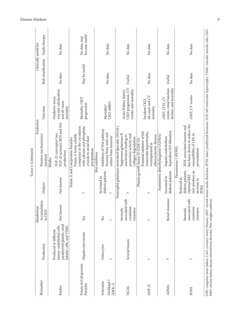
Table1:Summaryof
evidence
ontheu
seof
serum
biom
arkersforC
Vris
kpredictio
nin
CKD.
Biom
arker
Metabolism
Predictio
nClinicallyuseful
for
Prod
uctio
nAc
cumulation
inCK
DDialysis
Putativ
emechanism
(s)
involved
Outcome
Risk
stratificatio
nGuide
therapy
Natriu
retic
peptides
Atria
lnatriu
retic
peptide(ANP)
Atria
lmyocardium
Yes
Dialyzableo
nly
toas
malldegree
Markero
fleft
ventric
ular
dysfu
nctio
n,severityof
CHF
inbo
thgeneraland
CKD
popu
latio
ns
All-causem
ortalityin
CKDanddialysis
patie
nts.Sudd
encardiacd
eath
andMI
indialysispatie
nts
Useful
Useful(forC
HF)
B-type
natriuretic
peptide(BN
P)Ve
ntric
ular
myocardium
C-type
natriuretic
peptide(CN
P)En
dothelial
cells/m
yocardium
Cardiactropo
nins
Trop
oninsT
,I,and
CStria
tedmuscle
Yes
Increasedin
dialysispatie
nts
Released
after
cardiacinjury
(cTN
T)
PredictsCA
Dand
deathin
CKDand
dialysispatie
nts
Useful
Nodata
Cardiactropo
ninI
(cTN
I)Ca
rdiomyocytes
Cardiactropo
ninT
(cTN
T)Ca
rdiomyocytes
C-reactiv
eprotein
C-reactiv
eprotein
(CRP
)Liver-acutep
hase
reactant
?Increasesd
uring
dialysis
Markero
fsystemic
inflammation
PredictsCA
D,
all-c
ause
mortality
Not
useful
ifused
alon
eNot
useful
Adipon
ectin
Adipon
ectin
(APN
)Ad
ipocytes
Yes
?Infl
uenced
bynu
trition
alsta
tus
Anti-infl
ammatoryand
antia
therogenicactiv
ities,
enhances
insulin
-sensitivity.
Associatedwith
malnu
trition
Cardiovascular
events,
all-c
ause
mortality
Not
useful,
influ
encedby
nutrition
alsta
tus
Not
useful,
influ
encedby
nutrition
alsta
tus
Leptin
Leptin
Adipocytes
Yes,glom
erular
filtrationand
tubu
lar
degradation
Increasedlevels
influ
encedby
insulin
resistance,
metabolic
acidosis,
and
urem
ictoxins
Ano
recticagentassociated
with
cIMTandarteria
lstiffn
ess
Inversea
ssociatio
nwith
cardiovascular
events?
Not
useful
Not
useful
Fibrob
lastgrow
thfactor
23(FGF-23)
Fibrob
lastgrow
thfactor
23Oste
ocytes
Yes
Increasedlevels,
thou
ghlow
molecular
weightand
dialysable
Supp
resses
renaltub
ular
phosph
ater
eabsorption,
1,25O
Hvitamin
Dactiv
ation
andincreases1,25H
Ovitamin
Ddegradationandpo
ssibly
iPTH
secretion
Associated
with
ESRD
occurrence,
CKDprogression,
LVH,C
HF,and
atherosclerotic
events,
all-c
ause
mortality
Useful
Nodata
Disease Markers 9
Table1:Con
tinued.
Biom
arker
Metabolism
Predictio
nClinicallyuseful
for
Prod
uctio
nAc
cumulation
inCK
DDialysis
Putativ
emechanism
(s)
involved
Outcome
Risk
stratificatio
nGuide
therapy
Klotho
Klotho
Prod
uced
indifferent
tissues:end
othelialcells,
parathyroidglands,renal
tubu
larc
ells,
andVS
MC
Not
know
nNot
know
nFG
F-23
coreceptor
(see
FGF-23
actio
ns);RO
SandNO
prod
uctio
n
Oxidativ
estre
ss,
vascular
calcificatio
n,andall-c
ause
mortality
Nodata
Nodata
Fetuin
AandCa
lciproteinParticles
Fetuin-A/C
alciprotein
Particles
Hepatocytes/serum
Yes
?
Fetuin
Aform
ssolub
lecomplexes
inthec
irculation
with
calcium
andph
osph
ate
crystalsto
avoidtheir
precipitatio
n.
Mortality.CK
Dprogression
May
beuseful
Nodata;m
aybecomeu
seful
Wnt
inhibitors
Sclerostin
Oste
ocytes
Yes
Increasedin
dialysispatie
nts
Inhibitorsof
Wnt
pathway.
Increase
bone
massa
ndredu
cebo
neturnover
Mortality?
CKD-M
BD?
Nodata
Nodata
Dickkop
f-1(D
KK-1)
??
?
Neutro
philgelatinase-associated
lipocalin
(NGAL)
NGAL
Severaltissues
Inversely
associated
with
creatin
ine
clearance
?
Supp
resses
gelatin
aseB
inactiv
ationleadingto
proteolytic
activ
ityand
collagendegradation
AcuteK
idneyInjury,
CKDprogression,
CVevents,
andmortality
Useful
Nodata
Plasmag
rowth
factor-15(G
DF-15)
GDF-15
??
?
Potentialinh
ibito
rofleft
ventric
ular
hypertroph
y,overexpressedin
atherosclerotic
disease
Incident
CKD,
all-c
ause
andCV
mortality
Nodata
Nodata
Asym
metric
dimethylarginine(ADMA)
ADMA
?Re
nalclearance
Increasedin
dialysispatie
nts
Impairs
endo
thelium
depend
entN
Odilatation
cIMT,LV
H,C
Vevents,
renalfun
ction
decline,and
mortality
Useful
Nodata
Paraoxon
ase1
(PON1)
PON1
?
Inversely
associated
with
creatin
ine
clearance
Redu
cedin
dialysispatie
nts.
Optim
alRR
Tmay
prod
ucea
nincrease
inPO
N1
HDL-associated
enzymea
ndhasb
eenshow
nto
redu
cethe
susceptib
ilityof
LDLto
peroxidatio
n
cIMT,CV
events
Nodata
Nodata
CHF:
congestiv
eheartfailure;C
AD:coron
aryartery
Dise
ase;cIMT:
carotid
intim
amediathickn
ess;iPTH
:intactp
arathyroid
horm
one;LV
H:left
ventric
ular
hypertroph
y;VS
MC:
vascular
smoo
thcells;C
KD-
MBD
:chron
ickidn
eydiseasem
ineralbo
nedisorder;W
nt:w
inglessp
athw
ay.

10 Disease Markers
a combination of biomarkers may provide useful prognosticinformation. Markers may also become useful as a guide fortherapy to select the patient most likely to respond to anintervention with a more individualized approach. However,several methodological issues must be addressed before theroutine use of serum biomarkers can be implemented. Theclose correlation between different biomarkers likely reflectsthe complexity of the pathophysiological processes leadingto CVD in CKD. Although an integrated approach seemsreasonable, large ad hoc observational studies should definethe most parsimonious approach to risk prediction to avoidunnecessary and expensive testing in different clinical set-tings. In this regard, the lack of standardization for differentassays and the use of cohort specific cutoff values to definehigh versus low risk subjects do not allow for a reliablecomparison of different studies. Validation of cutoff values(external validation) as well as definition of the inter- andintraassay variability for each commercially available assayis a priority to understand how to use the informationderived from different biomarkers. Most of all, however,biomarkers must be easy to use and understand to becometools the practicing physician can implement daily. Althoughpromising, available data (Table 1) on serum biomarkers forrisk prediction are currently insufficient to recommend theirroutine use for prognostication and as a guide to therapy inCKD patients.
Abbreviations
95% CI: 95% confidence intervalADMA: Asymmetric dimethylarginineANP: Atrial Natriuretic PeptideBNP: B-type natriuretic peptideCAD: Coronary artery diseaseCHF: Congestive heart failurecIMT: Carotid intima media thicknessCKD: Chronic kidney diseaseCKD-MBD: Chronic kidney disease mineral bone
disorderCPP: Calciprotein particlesCRP: C-reactive proteincTNI: Cardiac troponinCV: CardiovascularCVD: Cardiovascular diseaseDKK-1: Dickkopf-1FGF-23: Fibroblast growth factor 23GDF-15: Plasma growth factor-15HR: Hazard ratioiPTH: Intact parathyroid hormoneLVH: Left ventricular hypertrophyNGAL: Neutrophil gelatinase-associated
lipocalinOR: Odds ratioPON1: Paraoxonase 1ROC: Receiver operating characteristicVSMC: Vascular smooth cellsWnt: Wingless pathway.
Conflict of Interests
There is no conflict of interests to report for any of the authorsrelated to this paper.
References
[1] E. L. Schiffrin,M. L. Lipman, and J. F. E.Mann, “Chronic kidneydisease: effects on the cardiovascular system,” Circulation, vol.116, no. 1, pp. 85–97, 2007.
[2] A. S. Go, G. M. Chertow, D. Fan, C. E. McCulloch, andC.-Y. Hsu, “Chronic kidney disease and the risks of death,cardiovascular events, and hospitalization,” The New EnglandJournal of Medicine, vol. 351, no. 13, pp. 1296–1305, 2004.
[3] S. Vickery, M. C. Webb, C. P. Price, R. I. John, N. A. Abbas, andE. J. Lamb, “Prognostic value of cardiac biomarkers for death ina non-dialysis chronic kidney disease population,” NephrologyDialysis Transplantation, vol. 23, no. 11, pp. 3546–3553, 2008.
[4] R. G. Fassett, S. K. Venuthurupalli, G. C. Gobe, J. S. Coombes,M. A. Cooper, and W. E. Hoy, “Biomarkers in chronic kidneydisease: a review,” Kidney International, vol. 80, no. 8, pp. 806–821, 2011.
[5] Y. Iwanaga and S. Miyazaki, “Heart failure, chronic kidneydisease, and biomarkers—an integrated viewpoint,” CirculationJournal, vol. 74, no. 7, pp. 1274–1282, 2010.
[6] Y. Iwanaga, I. Nishi, S. Furuichi et al., “B-type natriureticpeptide strongly reflects diastolic wall stress in patients withchronic heart failure: comparison between systolic and diastolicheart failure,” Journal of the American College of Cardiology, vol.47, no. 4, pp. 742–748, 2006.
[7] P. A.McCullough, R.M. Nowak, J. McCord et al., “B-type natri-uretic peptide and clinical judgment in emergency diagnosisof heart failure: analysis from Breathing Not Properly (BNP)Multinational Study,” Circulation, vol. 106, no. 4, pp. 416–422,2002.
[8] V. Epshteyn, K. Morrison, P. Krishnaswamy et al., “Utility of B-type natriuretic peptide (BNP) as a screen for left ventriculardysfunction in patients with diabetes,” Diabetes Care, vol. 26,no. 7, pp. 2081–2087, 2003.
[9] A. S. Maisel, P. Krishnaswamy, R. M. Nowak et al., “Rapidmeasurement of B-type natriuretic peptide in the emergencydiagnosis of heart failure,”TheNewEngland Journal ofMedicine,vol. 347, no. 3, pp. 161–167, 2002.
[10] R. Xu, P. Ye, L. Luo et al., “Association between high-sensitivitycardiac troponin T and N-terminal pro-brain natriuretic pep-tide in a community based population,” Chinese Medical Jour-nal, vol. 127, no. 4, pp. 638–644, 2014.
[11] K. Kara, A. A. Mahabadi, M. H. Geisel et al., “B-type natriureticpeptide: distribution in the general population and the associa-tionwithmajor cardiovascular and coronary events—theHeinzNixdorf Recall Study,” Clinical Research in Cardiology, vol. 103,no. 2, pp. 125–132, 2014.
[12] K. Kara, N. Lehmann, T. Neumann et al., “NT-proBNP issuperior to BNP for predicting first cardiovascular eventsin the general population: the Heinz Nixdorf Recall Study,”International Journal of Cardiology, vol. 183, pp. 155–161, 2015.
[13] Z. Hijazi, J. Oldgren, U. Andersson et al., “Cardiac biomarkersare associated with an increased risk of stroke and death inpatients with atrial fibrillation: a randomized evaluation oflong-term anticoagulation therapy (RE-LY) substudy,” Circula-tion, vol. 125, no. 13, pp. 1605–1616, 2012.

Disease Markers 11
[14] H. G. Wahl, S. Graf, H. Renz, and W. Fassbinder, “Eliminationof the cardiac natriuretic peptides B-type natriuretic peptide(BNP) and N-terminal proBNP by hemodialysis,” ClinicalChemistry, vol. 50, no. 6, pp. 1071–1074, 2004.
[15] Y. Ishizaka, Y. Yamamoto, M. Tanaka et al., “Molecular formsof human brain natriuretic peptide (BNP) in plasma of patientson hemodialysis (HD),” Clinical Nephrology, vol. 43, no. 4, pp.237–242, 1995.
[16] P. A. McCullough, T. Omland, and A. S. Maisel, “B-typenatriuretic peptides: a diagnostic breakthrough for clinicians,”Reviews in Cardiovascular Medicine, vol. 4, no. 2, pp. 72–80,2003.
[17] S. Niizuma, Y. Iwanaga, T. Yahata et al., “Impact of left ven-tricular end-diastolic wall stress on plasma B-type natriureticpeptide in heart failure with chronic kidney disease and end-stage renal disease,” Clinical Chemistry, vol. 55, no. 7, pp. 1347–1353, 2009.
[18] F. S. Apple, M. A. M. Murakami, L. A. Pearce, and C. A.Herzog, “Multi-biomarker risk stratification of N-terminal pro-B-type natriuretic peptide, high-sensitivity C-reactive protein,and cardiac troponin T and I in end-stage renal disease for all-cause death,” Clinical Chemistry, vol. 50, no. 12, pp. 2279–2285,2004.
[19] C. R. DeFilippi, S. L. Seliger, S.Maynard, and R.H. Christenson,“Impact of renal disease on natriuretic peptide testing for diag-nosing decompensated heart failure and predicting mortality,”Clinical Chemistry, vol. 53, no. 8, pp. 1511–1519, 2007.
[20] M. Sakuma, M. Nakamura, F. Tanaka et al., “Plasma B-typenatriuretic peptide level and cardiovascular events in chronickidney disease in a community-based population,” CirculationJournal, vol. 74, no. 4, pp. 792–797, 2010.
[21] C. Sommerer, J. Beimler, V. Schwenger et al., “Cardiac biomark-ers and survival in haemodialysis patients,” European Journal ofClinical Investigation, vol. 37, no. 5, pp. 350–356, 2007.
[22] A. Y.-M.Wang, C.W.-K. Lam, C.-M. Yu et al., “N-terminal pro-brain natriuretic peptide: an independent risk predictor of car-diovascular congestion, mortality, and adverse cardiovascularoutcomes in chronic peritoneal dialysis patients,” Journal of theAmerican Society of Nephrology, vol. 18, no. 1, pp. 321–330, 2007.
[23] R. Paniagua, M.-D. Ventura, M. Avila-Dıaz et al., “NT-proBNP,fluid volume overload and dialysis modality are independentpredictors of mortality in ESRD patients,” Nephrology DialysisTransplantation, vol. 25, no. 2, pp. 551–557, 2010.
[24] M. Svensson, A. Gorst-Rasmussen, E. B. Schmidt, K. A. Jor-gensen, and J. H. Christensen, “NT-pro-BNP is an independentpredictor of mortality in patients with end-stage renal disease,”Clinical Nephrology, vol. 71, no. 4, pp. 380–386, 2009.
[25] A. S. Jaffe, L. Babuin, and F. S. Apple, “Biomarkers in acutecardiac disease: the present and the future,” Journal of theAmerican College of Cardiology, vol. 48, no. 1, pp. 1–11, 2006.
[26] S. Kiatchoosakun, D. Sahasthas, C. Wongvipaporn, and C.Pongskul, “Cardiac troponin-T in pre-end stage kidney disease,”Journal of the Medical Association ofThailand, vol. 91, no. 12, pp.1806–1811, 2008.
[27] C. Sommerer, E. Giannitsis, V. Schwenger, and M. Zeier,“Cardiac biomarkers in haemodialysis patients: the prognosticvalue of amino-terminal pro-B-type natriuretic peptide andcardiac troponin T,” Nephron—Clinical Practice, vol. 107, no. 3,pp. c77–c81, 2007.
[28] M. C. Iliou, C. Fumeron, M. O. Benoit et al., “Factors associatedwith increased serum levels of cardiac troponins T and I
in chronic haemodialysis patients: chronic haemodialysis andnew cardiac markers evaluation (CHANCE) study,” NephrologyDialysis Transplantation, vol. 16, no. 7, pp. 1452–1458, 2001.
[29] F. Mallamaci, C. Zoccali, S. Parlongo et al., “Troponin is relatedto left ventricular mass and predicts all-cause and cardiovascu-lar mortality in hemodialysis patients,”TheAmerican Journal ofKidney Diseases, vol. 40, no. 1, pp. 68–75, 2002.
[30] B. Havekes, J. G. van Manen, R. T. Krediet, E. W. Boeschoten,J. P. Vandenbroucke, and F. W. Dekker, “Serum troponinT concentration as a predictor of mortality in hemodialysisand peritoneal dialysis patients,” American Journal of KidneyDiseases, vol. 47, no. 5, pp. 823–829, 2006.
[31] T.Hayashi, Y.Obi, T. Kimura et al., “Cardiac troponinT predictsoccult coronary artery stenosis in patients with chronic kidneydisease at the start of renal replacement therapy,” NephrologyDialysis Transplantation, vol. 23, no. 9, pp. 2936–2942, 2008.
[32] J. Dierkes, U. Domrose, S. Westphal et al., “Cardiac troponinT predicts mortality in patients with end-stage renal disease,”Circulation, vol. 102, no. 16, pp. 1964–1969, 2000.
[33] M. Goicoechea, S. Garcıa De Vinuesa, F. Gomez-Campdera etal., “Clinical significance of cardiac troponin T levels in chronickidney disease patients: predictive value for cardiovascularrisk,” American Journal of Kidney Diseases, vol. 43, no. 5, pp.846–853, 2004.
[34] C. DeFilippi, S.Wasserman, S. Rosanio et al., “Cardiac troponinT and C-reactive protein for predicting prognosis, coronaryatherosclerosis, and cardiomyopathy in patients undergoinglong-term hemodialysis,” The Journal of the American MedicalAssociation, vol. 290, no. 3, pp. 353–359, 2003.
[35] C.Melloni, K. P. Alexander, S.Milford-Beland et al., “Prognosticvalue of troponins in patients with non-ST-segment elevationacute coronary syndromes and chronic kidney disease,”ClinicalCardiology, vol. 31, no. 3, pp. 125–129, 2008.
[36] A. X. Garg, P. G. Blake, W. F. Clark, C. M. Clase, R. B. Haynes,and L. M. Moist, “Association between renal insufficiency andmalnutrition in older adults: results from the NHANES III,”Kidney International, vol. 60, no. 5, pp. 1867–1874, 2001.
[37] M. G. Shlipak, L. F. Fried, C. Crump et al., “Elevations ofinflammatory and procoagulant biomarkers in elderly personswith renal insufficiency,” Circulation, vol. 107, no. 1, pp. 87–92,2003.
[38] H.-J. Hsu, C.-H. Yen, K.-H. Hsu et al., “Chronic kidneydisease stage is a modulator on the association between high-sensitivity C-reactive protein and coronary vasospastic angina,”The Scientific World Journal, vol. 2014, Article ID 852507, 9pages, 2014.
[39] P. M. Ridker, “Clinical application of C-reactive protein forcardiovascular disease detection and prevention,” Circulation,vol. 107, no. 3, pp. 363–369, 2003.
[40] T. A. Pearson, G. A.Mensah, R.W. Alexander et al., “Markers ofinflammation and cardiovascular disease: application to clinicaland public health practice: a statement for healthcare profes-sionals from the centers for disease control and prevention andthe AmericanHeart Association,”Circulation, vol. 107, no. 3, pp.499–511, 2003.
[41] F. M. van der Sande, J. P. Kooman, and K. M. Leunissen, “Thepredictive value of C-reactive protein in end-stage renal disease:is it clinically significant?” Blood Purification, vol. 24, no. 4, pp.335–341, 2006.
[42] F. Mallamaci, G. Tripepi, S. Cutrupi, L. S. Malatino, and C.Zoccali, “Prognostic value of combined use of biomarkers of

12 Disease Markers
inflammation, endothelial dysfunction, and myocardiopathy inpatients with ESRD,” Kidney International, vol. 67, no. 6, pp.2330–2337, 2005.
[43] S. Snaedal, O. Heimburger, A. R. Qureshi et al., “Comorbidityand acute clinical events as determinants of C-reactive proteinvariation in hemodialysis patients: implications for patientsurvival,”American Journal of Kidney Diseases, vol. 53, no. 6, pp.1024–1033, 2009.
[44] M. M. Nascimento, R. Pecoits-Filho, A. R. Qureshi et al., “Theprognostic impact of fluctuating levels of C-reactive protein inBrazilian haemodialysis patients: a prospective study,” Nephrol-ogy Dialysis Transplantation, vol. 19, no. 11, pp. 2803–2809, 2004.
[45] S.-H. Liu, Y.-J. Li, H.-H. Wu et al., “High-sensitivity C-reactiveprotein predicts mortality and technique failure in peritonealdialysis patients,” PLoS ONE, vol. 9, no. 3, Article ID e93063,2014.
[46] M. Kanwar, M. Hashem, H. Rosman et al., “Usefulness of clin-ical evaluation, troponins, and C-reactive protein in predictingmortality among stable hemodialysis patients,” The AmericanJournal of Cardiology, vol. 98, no. 9, pp. 1283–1287, 2006.
[47] H. Honda, A. R. Qureshi, O. Heimburger et al., “Serumalbumin, C-reactive protein, interleukin 6, and fetuin a as pre-dictors ofmalnutrition, cardiovascular disease, andmortality inpatients with ESRD,” The American Journal of Kidney Diseases,vol. 47, no. 1, pp. 139–148, 2006.
[48] C. Zoccali, G. Tripepi, and F. Mallamaci, “Predictors of cardio-vascular death in ESRD,” Seminars in Nephrology, vol. 25, no. 6,pp. 358–362, 2005.
[49] F. M. Fisher, M. E. Trujillo, W. Hanif et al., “Serum highmolecular weight complex of adiponectin correlates better withglucose tolerance than total serum adiponectin in Indo-Asianmales,” Diabetologia, vol. 48, no. 6, pp. 1084–1087, 2005.
[50] M. Bouskila, U. B. Pajvani, and P. E. Scherer, “Adiponectin: arelevant player in PPAR𝛾-agonist-mediated improvements inhepatic insulin sensitivity?” International Journal of Obesity, vol.29, supplement 1, pp. S17–S23, 2005.
[51] T. Yokota, C. S. R. Meka, T. Kouro et al., “Adiponectin, a fatcell product, influences the earliest lymphocyte precursors inbone marrow cultures by activation of the cyclooxygenase-prostaglandin pathway in stromal cells,” The Journal ofImmunology, vol. 171, no. 10, pp. 5091–5099, 2003.
[52] M. C. Wulster-Radcliffe, K. M. Ajuwon, J. Wang, J. A. Chris-tian, and M. E. Spurlock, “Adiponectin differentially regulatescytokines in porcinemacrophages,”Biochemical andBiophysicalResearch Communications, vol. 316, no. 3, pp. 924–929, 2004.
[53] K. Hotta, T. Funahashi, Y. Arita et al., “Plasma concentrations ofa novel, adipose-specific protein, adiponectin, in type 2 diabeticpatients,”Arteriosclerosis,Thrombosis, andVascular Biology, vol.20, no. 6, pp. 1595–1599, 2000.
[54] J. P.Whitehead, A. A. Richards, I. J. Hickman, G. A.Macdonald,and J. B. Prins, “Adiponectin—a key adipokine in the metabolicsyndrome,” Diabetes, Obesity and Metabolism, vol. 8, no. 3, pp.264–280, 2006.
[55] Z. Dzielinska, A. Januszewicz, A. Więcek et al., “Decreasedplasma concentration of a novel anti-inflammatory protein—adiponectin—in hypertensive men with coronary artery dis-ease,”Thrombosis Research, vol. 110, no. 5-6, pp. 365–369, 2003.
[56] N. Komura, S. Kihara, M. Sonoda et al., “Increment andimpairment of adiponectin in renal failure,” CardiovascularResearch, vol. 86, no. 3, pp. 471–477, 2010.
[57] M. Kumada, S. Kihara, S. Sumitsuji et al., “Association ofhypoadiponectinemia with coronary artery disease in men,”
Arteriosclerosis, Thrombosis, and Vascular Biology, vol. 23, no.1, pp. 85–89, 2003.
[58] N. Ouchi, M. Ohishi, S. Kihara et al., “Association of hypoad-iponectinemia with impaired vasoreactivity,”Hypertension, vol.42, no. 3, pp. 231–234, 2003.
[59] B. Kollerits, D. Fliser, I. M. Heid, E. Ritz, and F. Kronenberg,“Gender-specific association of adiponectin as a predictor ofprogression of chronic kidney disease: the Mild to ModerateKidney Disease study,” Kidney International, vol. 71, no. 12, pp.1279–1286, 2007.
[60] C. Zoccali and F.Mallamaci, “Adiponectin and leptin in chronickidney disease: causal factors or mere risk markers?” Journal ofRenal Nutrition, vol. 21, no. 1, pp. 87–91, 2011.
[61] B. Becker, F. Kronenberg, J. T. Kielstein et al., “Renal insulinresistance syndrome, adiponectin and cardiovascular events inpatients with kidney disease: the mild and moderate kidneydisease study,” Journal of the American Society of Nephrology,vol. 16, no. 4, pp. 1091–1098, 2005.
[62] C. Zoccali, F. Mallamaci, G. Tripepi et al., “Adiponectin,metabolic risk factors, and cardiovascular events amongpatients with end-stage renal disease,” Journal of the AmericanSociety of Nephrology, vol. 13, no. 1, pp. 134–141, 2002.
[63] V. Menon, L. Li, X. Wang et al., “Adiponectin and mortality inpatients with chronic kidney disease,” Journal of the AmericanSociety of Nephrology, vol. 17, no. 9, pp. 2599–2606, 2006.
[64] O. M. Kaisar, D. W. Johnson, J. B. Prins, and N. Isbel, “The roleof novel biomarkers of cardiovascular disease in chronic kidneydisease: focus on adiponectin and leptin,” Current CardiologyReviews, vol. 4, no. 4, pp. 287–292, 2008.
[65] P. Stenvinkel, “Leptin—a new hormone of definite interest forthe nephrologist,” Nephrology Dialysis Transplantation, vol. 13,no. 5, pp. 1099–1101, 1998.
[66] S. K. Fried, M. R. Ricci, C. D. Russell, and B. Laferrere, “Regula-tion of leptin production in humans,” Journal of Nutrition, vol.130, no. 12, pp. 3127–3131S, 2000.
[67] R. V. Considine, M. K. Sinha, M. L. Heiman et al., “Serumimmunoreactive-leptin concentrations in normal-weight andobese humans,”The New England Journal of Medicine, vol. 334,no. 5, pp. 292–295, 1996.
[68] D. G. Baskin, J. E. Blevins, and M. W. Schwartz, “How thebrain regulates food intake and body weight: the role of leptin,”Journal of Pediatric Endocrinology and Metabolism, vol. 14, no.6, pp. 1417–1429, 2001.
[69] T.M.Mizuno, S. P. Kleopoulos, H. T. Bergen, J. L. Roberts, C. A.Priest, and C. V. Mobbs, “Hypothalamic pro-opiomelanocortinmRNA is reduced by fasting and [corrected] in ob/ob and db/dbmice, but is stimulated by leptin,” Diabetes, vol. 47, no. 2, pp.294–297, 1998.
[70] M. Mapfei, J. Halaas, E. Ravussin et al., “Leptin levels in humanand rodent:measurement of plasma leptin and obRNA in obeseand weight-reduced subjects,”NatureMedicine, vol. 1, no. 11, pp.1155–1161, 1995.
[71] G. Boden, K. Sargrad, C. Homko, M. Mozzoli, and T. P. Stein,“Effect of a low-carbohydrate diet on appetite, blood glucoselevels, and insulin resistance in obese patients with type 2diabetes,” Annals of Internal Medicine, vol. 142, no. 6, pp. 403–411, 2005.
[72] E. Merabet, S. Dagogo-Jack, D. W. Coyne et al., “Increasedplasma leptin concentration in end-stage renal disease,” Journalof Clinical Endocrinology andMetabolism, vol. 82, no. 3, pp. 847–850, 1997.

Disease Markers 13
[73] J. J. Dıez, P. Iglesias, M. J. Fernandez-Reyes et al., “Serumconcentrations of leptin, adiponectin and resistin, and theirrelationship with cardiovascular disease in patients with end-stage renal disease,” Clinical Endocrinology, vol. 62, no. 2, pp.242–249, 2005.
[74] D. Teta, A. Bevington, J. Brown, I. Pawluczyk, K. Harris, andJ. Walls, “Acidosis downregulates leptin production from cul-tured adipocytes through a glucose transport-dependent post-transcriptional mechanism,” Journal of the American Society ofNephrology, vol. 14, no. 9, pp. 2248–2254, 2003.
[75] L. Nordfors, F. Lonnqvist, O. Heimburger, A. Danielsson, M.Schalling, and P. Stenvinkel, “Low leptin gene expression andhyperleptinemia in chronic renal failure,” Kidney International,vol. 54, no. 4, pp. 1267–1275, 1998.
[76] A. Scholze andM. Tepel, “Role of leptin in reverse epidemiologyin chronic kidney disease,” Seminars in Dialysis, vol. 20, no. 6,pp. 534–538, 2007.
[77] M. Ciccone, R. Vettor, N. Pannacciulli et al., “Plasma leptin isindependently associated with the intima-media thickness ofthe common carotid artery,” International Journal of Obesity,vol. 25, no. 6, pp. 805–810, 2001.
[78] A. Singhal, I. S. Farooqi, T. J. Cole et al., “Influence of leptinon arterial distensibility: a novel link between obesity andcardiovascular disease?” Circulation, vol. 106, no. 15, pp. 1919–1924, 2002.
[79] M. C. Lee, Y. C. Chen, G. J. Ho, M. H. Shih, K. C. Chou,and B. G. Hsu, “Serum leptin levels positively correlate withperipheral arterial stiffness in kidney transplantation patients,”Transplantation Proceedings, vol. 46, no. 2, pp. 353–358, 2014.
[80] A. Aguilera, M. A. Bajo, F. Rebollo et al., “Leptin as a marker ofnutrition and cardiovascular risk in peritoneal dialysis patients,”Advances in Peritoneal Dialysis, vol. 18, pp. 212–217, 2002.
[81] A. Scholze, D. Rattensperger, W. Zidek, and M. Tepel, “Lowserum leptin predicts mortality in patients with chronic kidneydisease stage 5,” Obesity, vol. 15, no. 6, pp. 1617–1622, 2007.
[82] I. Beberashvili, I. Sinuani, A. Azar et al., “Longitudinal studyof leptin levels in chronic hemodialysis patients,” NutritionJournal, vol. 10, no. 1, article 68, 2011.
[83] Y.-C. Tsai, C.-T. Lee, T.-L. Huang et al., “Inflammatorymarker but not adipokine predicts mortality among long-termhemodialysis patients,” Mediators of Inflammation, vol. 2007,Article ID 19891, 5 pages, 2007.
[84] S. Moe, T. Drueke, G. Block, and J. Cannata-andia, “KDIGOclinical practice guideline for the diagnosis, evaluation, preven-tion, and treatment of chronic kidney disease-mineral and bonedisorder (CKD-MBD),” Kidney International, vol. 113, pp. S1–S130, 2009.
[85] S. Liu, J. Zhou,W. Tang, X. Jiang, D.W. Rowe, and L. D. Quarles,“Pathogenic role of Fgf23 in Hyp mice,” The American Journalof Physiology—Endocrinology andMetabolism, vol. 291, no. 1, pp.E38–E49, 2006.
[86] H. Segawa, E. Kawakami, I. Kaneko et al., “Effect of hydrolysis-resistant FGF23-R179Q on dietary phosphate regulation of therenal type-II Na/Pi transporter,” Pflugers Archiv, vol. 446, no. 5,pp. 585–592, 2003.
[87] T. Shimada, H. Hasegawa, Y. Yamazaki et al., “FGF-23 isa potent regulator of vitamin D metabolism and phosphatehomeostasis,” Journal of Bone and Mineral Research, vol. 19, no.3, pp. 429–435, 2004.
[88] M. Yamazaki, K. Ozono, T. Okada et al., “Both FGF23 and extra-cellular phosphate activate Raf/MEK/ERK pathway via FGF
receptors inHEK293 cells,” Journal of Cellular Biochemistry, vol.111, no. 5, pp. 1210–1221, 2010.
[89] V. P. Eswarakumar, I. Lax, and J. Schlessinger, “Cellular signal-ing by fibroblast growth factor receptors,” Cytokine and GrowthFactor Reviews, vol. 16, no. 2, pp. 139–149, 2005.
[90] M. Kuro-O, “Phosphate and klotho,” Kidney International, vol.79, supplement 121, pp. S20–S23, 2011.
[91] M. Christov, S. S. Waikar, R. C. Pereira et al., “Plasma FGF23levels increase rapidly after acute kidney injury,” Kidney Inter-national, vol. 84, no. 4, pp. 776–785, 2013.
[92] O. M. Gutierrez, M. Mannstadt, T. Isakova et al., “Fibroblastgrowth factor 23 and mortality among patients undergoinghemodialysis,” The New England Journal of Medicine, vol. 359,no. 6, pp. 584–592, 2008.
[93] J. H. Ix, M. G. Shlipak, C. L. Wassel, and M. A. Whooley,“Fibroblast growth factor-23 and early decrements in kidneyfunction: the heart and soul study,” Nephrology Dialysis Trans-plantation, vol. 25, no. 3, pp. 993–997, 2010.
[94] M. Kanbay, M. Nicoleta, Y. Selcoki et al., “Fibroblast growthfactor 23 and fetuin A are independent predictors for thecoronary artery disease extent in mild chronic kidney disease,”Clinical Journal of the American Society of Nephrology, vol. 5, no.10, pp. 1780–1786, 2010.
[95] S. Seiler, B. Reichart, D. Roth, E. Seibert, D. Fliser, and G. H.Heine, “FGF-23 and future cardiovascular events in patientswith chronic kidney disease before initiation of dialysis treat-ment,” Nephrology Dialysis Transplantation, vol. 25, no. 12, pp.3983–3989, 2010.
[96] T. Isakova, P. Wahl, G. S. Vargas et al., “Fibroblast growth factor23 is elevated before parathyroid hormone and phosphate inchronic kidney disease,”Kidney International, vol. 79, no. 12, pp.1370–1378, 2011.
[97] J. J. Scialla, H. Xie, M. Rahman et al., “Fibroblast growth factor-23 and cardiovascular events in CKD,” Journal of the AmericanSociety of Nephrology, vol. 25, no. 2, pp. 349–360, 2014.
[98] C. M. Rebholz, M. E. Grams, J. Coresh et al., “Serum fibroblastgrowth factor-23 is associated with incident kidney disease,”Journal of the American Society of Nephrology, vol. 26, no. 1, pp.192–200, 2015.
[99] B. D. Parker, L. J. Schurgers, V. M. Brandenburg et al., “Theassociations of fibroblast growth factor 23 and uncarboxylatedmatrix Gla protein with mortality in coronary artery disease:the heart and soul study,” Annals of Internal Medicine, vol. 152,no. 10, pp. 640–648, 2010.
[100] H. J. Oh, B. Y. Nam, M. J. Lee et al., “Decreased circulatingKlotho levels in patients undergoing dialysis and relationshipto oxidative stress and inflammation,” Peritoneal Dialysis Inter-national, vol. 35, no. 1, pp. 43–51, 2015.
[101] R. D. Semba, A. R. Cappola, K. Sun et al., “Plasma klotho andmortality risk in older community-dwelling adults,” Journals ofGerontology—Series A Biological Sciences and Medical Sciences,vol. 66, no. 7, pp. 794–800, 2011.
[102] D. E. Arking, D. M. Becker, L. R. Yanek et al., “KLOTHO allelestatus and the risk of early-onset occult coronary artery disease,”TheAmerican Journal ofHumanGenetics, vol. 72, no. 5, pp. 1154–1161, 2003.
[103] J. F. Navarro-Gonzalez, J. Donate-Correa, M. M. de Fuentes, H.Perez-Hernandez, R. Martınez-Sanz, and C. Mora-Fernandez,“Reduced Klotho is associated with the presence and severity ofcoronary artery disease,” Heart, vol. 100, no. 1, pp. 34–40, 2014.

14 Disease Markers
[104] I. Agarwal,N. Ide, J.H. Ix et al., “Fibroblast growth factor-23 andcardiac structure and function,” Journal of the American HeartAssociation, vol. 3, no. 1, Article ID e000584, 2014.
[105] M. Kuro-o, “Klotho, phosphate and FGF-23 in ageing anddisturbed mineral metabolism,” Nature Reviews Nephrology,vol. 9, no. 11, pp. 650–660, 2013.
[106] A. Pasch, S. Farese, S. Graber et al., “Nanoparticle-based testmeasures overall propensity for calcification in serum,” Journalof the American Society of Nephrology, vol. 23, no. 10, pp. 1744–1752, 2012.
[107] E. R. Smith, M. L. Ford, L. A. Tomlinson, C. Rajkumar,L. P. McMahon, and S. G. Holt, “Phosphorylated fetuin-A-containing calciprotein particles are associated with aorticstiffness and a procalcific milieu in patients with pre-dialysisCKD,” Nephrology, Dialysis, Transplantation, vol. 27, no. 5, pp.1957–1966, 2012.
[108] E. R. Smith, E.Hanssen, L. P.McMahon, and S. G.Holt, “Fetuin-A-containing calciprotein particles reduce mineral stress in themacrophage,” PLoS ONE, vol. 8, no. 4, Article ID e60904, 2013.
[109] E. R. Smith, M. L. Ford, L. A. Tomlinson et al., “Serum calcifica-tion propensity predicts all-causemortality in predialysis CKD,”Journal of the American Society of Nephrology, vol. 25, no. 2, pp.339–348, 2014.
[110] M. G. Vervloet, Z. A. Massy, V. M. Brandenburg et al., “Bone:a new endocrine organ at the heart of chronic kidney diseaseand mineral and bone disorders,” The Lancet Diabetes andEndocrinology, vol. 2, no. 5, pp. 427–436, 2014.
[111] V. M. Brandenburg, R. Kramann, R. Koos et al., “Relation-ship between sclerostin and cardiovascular calcification inhemodialysis patients: a cross-sectional study,” BMC Nephrol-ogy, vol. 14, no. 1, article 219, 2013.
[112] R. Kramann, V. M. Brandenburg, L. J. Schurgers et al., “Novelinsights into osteogenesis and matrix remodelling associatedwith calcific uraemic arteriolopathy,”NephrologyDialysis Trans-plantation, vol. 28, no. 4, pp. 856–868, 2013.
[113] D. Cejka, J. Herberth, A. J. Branscum et al., “Sclerostin anddickkopf-1 in renal osteodystrophy,” Clinical Journal of theAmerican Society of Nephrology, vol. 6, no. 4, pp. 877–882, 2011.
[114] S. Thambiah, R. Roplekar, P. Manghat et al., “Circulatingsclerostin and dickkopf-1 (DKK1) in predialysis chronic kidneydisease (CKD): relationship with bone density and arterialstiffness,” Calcified Tissue International, vol. 90, no. 6, pp. 473–480, 2012.
[115] L. Desjardins, S. Liabeuf, R. B. Oliveira et al., “Uremic toxicityand sclerostin in chronic kidney disease patients,” Nephrologie&Therapeutique, vol. 10, no. 6, pp. 463–470, 2014.
[116] L. Viaene, G. J. Behets, K. Claes et al., “Sclerostin: another bone-related protein related to all-cause mortality in haemodialysis?”Nephrology Dialysis Transplantation, vol. 28, no. 12, pp. 3024–3030, 2013.
[117] C. Drechsler, P. Evenepoel, M. G. Vervloet et al., “High levelsof circulating sclerostin are associated with better cardiovas-cular survival in incident dialysis patients: results from theNECOSAD study,”Nephrology Dialysis Transplantation, vol. 30,no. 2, pp. 288–293, 2015.
[118] K. Mori, H. T. Lee, D. Rapoport et al., “Endocytic delivery oflipocalin-siderophore-iron complex rescues the kidney fromischemia-reperfusion injury,” The Journal of Clinical Investiga-tion, vol. 115, no. 3, pp. 610–621, 2005.
[119] K. Mori and K. Nakao, “Neutrophil gelatinase-associatedlipocalin as the real-time indicator of active kidney damage,”Kidney International, vol. 71, no. 10, pp. 967–970, 2007.
[120] K. M. Schmidt-Ott, K. Mori, Y. L. Jau et al., “Dual actionof neutrophil gelatinase-associated lipocalin,” Journal of theAmerican Society of Nephrology, vol. 18, no. 2, pp. 407–413, 2007.
[121] R. Hirsch, C. Dent, H. Pfriem et al., “NGAL is an early predic-tive biomarker of contrast-induced nephropathy in children,”Pediatric Nephrology, vol. 22, no. 12, pp. 2089–2095, 2007.
[122] L. Kjeldsen, A. H. Johnsen, H. Sengelov, and N. Borregaard,“Isolation and primary structure of NGAL, a novel proteinassociated with human neutrophil gelatinase,” The Journal ofBiological Chemistry, vol. 268, no. 14, pp. 10425–10432, 1993.
[123] J. B. Cowland and N. Borregaard, “Molecular characterizationand pattern of tissue expression of the gene for neutrophilgelatinase-associated lipocalin from humans,” Genomics, vol.45, no. 1, pp. 17–23, 1997.
[124] A. Friedl, S. P. Stoesz, P. Buckley, and M. N. Gould, “Neutrophilgelatinase-associated lipocalin in normal and neoplastic humantissues. Cell type-specific pattern of expression,” HistochemicalJournal, vol. 31, no. 7, pp. 433–441, 1999.
[125] A.-L. Hemdahl, A. Gabrielsen, C. Zhu et al., “Expressionof neutrophil gelatinase-associated lipocalin in atherosclerosisand myocardial infarction,” Arteriosclerosis, Thrombosis, andVascular Biology, vol. 26, no. 1, pp. 136–142, 2006.
[126] J. R. Charlton, D. Portilla, and M. D. Okusa, “A basic scienceview of acute kidney injury biomarkers,” Nephrology DialysisTransplantation, vol. 29, no. 7, pp. 1301–1311, 2014.
[127] C. Donadio, “Effect of glomerular filtration rate impairmenton diagnostic performance of neutrophil gelatinase-associatedlipocalin and B-type natriuretic peptide as markers of acutecardiac and renal failure in chronic kidney disease patients,”Critical Care, vol. 18, no. 1, article R39, 2014.
[128] J. Mishra, M. A. Qing, A. Prada et al., “Identification of neu-trophil gelatinase-associated lipocalin as a novel early urinarybiomarker for ischemic renal injury,” Journal of the AmericanSociety of Nephrology, vol. 14, no. 10, pp. 2534–2543, 2003.
[129] M. Haase, R. Bellomo, P. Devarajan, P. Schlattmann, and A.Haase-Fielitz, “Accuracy of neutrophil gelatinase-associatedlipocalin (NGAL) in diagnosis and prognosis in acute kidneyinjury: a systematic review and meta-analysis,” The AmericanJournal of Kidney Diseases, vol. 54, no. 6, pp. 1012–1024, 2009.
[130] J. Mishra, C. Dent, R. Tarabishi et al., “Neutrophil gelatinase-associated lipocalin (NGAL) as a biomarker for acute renalinjury after cardiac surgery,”The Lancet, vol. 365, no. 9466, pp.1231–1238, 2005.
[131] T. L. Nickolas, M. J. O’Rourke, J. Yang et al., “Sensitivity andspecificity of a single emergency department measurement ofurinary neutrophil gelatinase-associated lipocalin for diagnos-ing acute kidney injury,” Annals of Internal Medicine, vol. 148,no. 11, pp. 810–819, 2008.
[132] A. Viau, K. El Karoui, D. Laouari et al., “Lipocalin 2 is essentialfor chronic kidney disease progression in mice and humans,”Journal of Clinical Investigation, vol. 120, no. 11, pp. 4065–4076,2010.
[133] D. Bolignano, A. Lacquaniti, G. Coppolino et al., “Neu-trophil gelatinase-associated lipocalin as an early biomarker ofnephropathy in diabetic patients,” Kidney and Blood PressureResearch, vol. 32, no. 2, pp. 91–98, 2009.
[134] K. D. Liu, W. Yang, A. H. Anderson et al., “Urine neu-trophil gelatinase-associated lipocalin levels do not improverisk prediction of progressive chronic kidney disease,” KidneyInternational, vol. 83, no. 5, pp. 909–914, 2013.

Disease Markers 15
[135] F. Furuya, H. Shimura, H. Yokomichi et al., “Neutrophilgelatinase-associated lipocalin levels associated with cardiovas-cular disease in chronic kidney disease patients,” Clinical andExperimental Nephrology, vol. 18, no. 5, pp. 778–783, 2014.
[136] A. S. Maisel, C. Mueller, R. Fitzgerald et al., “Prognostic utilityof plasma neutrophil gelatinase-associated lipocalin in patientswith acute heart failure: the NGAL EvaLuation Along with B-typeNaTriuretic Peptide in acutely decompensated heart failure(GALLANT) trial,” European Journal of Heart Failure, vol. 13,no. 8, pp. 846–851, 2011.
[137] A. Thompson, P. Gao, L. Orfei et al., “Lipoprotein-associatedphospholipase A
2and risk of coronary disease, stroke, and
mortality: collaborative analysis of 32 prospective studies,” TheLancet, vol. 375, no. 9725, pp. 1536–1544, 2010.
[138] M. R. Bootcov, A. R. Bauskin, S. M. Valenzuela et al., “MIC-1,a novel macrophage inhibitory cytokine, is a divergent memberof the TGF-𝛽 superfamily,” Proceedings of the National Academyof Sciences of the United States of America, vol. 94, no. 21, pp.11514–11519, 1997.
[139] T. Kempf, M. Eden, J. Strelau et al., “The transforming growthfactor-𝛽 superfamily member growth-differentiation factor-15protects the heart from ischemia/reperfusion injury,” Circula-tion Research, vol. 98, no. 3, pp. 351–360, 2006.
[140] J. Strelau, M. Bottner, P. Lingor et al., “GDF-15/MIC-1 anovel member of the TGF-beta superfamily,” Journal of NeuralTransmission, Supplementum, no. 60, pp. 273–276, 2000.
[141] J. Xu, T. R. Kimball, J. N. Lorenz et al., “GDF15/MIC-1 functionsas a protective and antihypertrophic factor released from themyocardium in association with SMAD protein activation,”Circulation Research, vol. 98, no. 3, pp. 342–350, 2006.
[142] D. Schlittenhardt, A. Schober, J. Strelau et al., “Involve-ment of growth differentiation factor-15/macrophage inhibitorycytokine-1 (GDF-15/MIC-1) in oxLDL-induced apoptosis ofhuman macrophages in vitro and in arteriosclerotic lesions,”Cell and Tissue Research, vol. 318, no. 2, pp. 325–333, 2004.
[143] D. A. Brown, S. N. Breit, J. Buring et al., “Concentrationin plasma of macrophage inhibitory cytokine-1 and risk ofcardiovascular events in women: a nested case-control study,”The Lancet, vol. 359, no. 9324, pp. 2159–2163, 2002.
[144] T. Kempf, E. Bjorklund, S. Olofsson et al., “Growth-differentiation factor-15 improves risk stratification in ST-segment elevation myocardial infarction,” European HeartJournal, vol. 28, no. 23, pp. 2858–2865, 2007.
[145] J. E. Ho, S.-J. Hwang, K. C. Wollert et al., “Biomarkers ofcardiovascular stress and incident chronic kidney disease,”Clinical Chemistry, vol. 59, no. 11, pp. 1613–1620, 2013.
[146] M. Lajer, A. Jorsal, L. Tarnow, H.-H. Parving, and P. Rossing,“Plasma growth differentiation factor-15 independently predictsall-cause and cardiovascularmortality aswell as deterioration ofkidney function in type 1 diabetic patients with nephropathy,”Diabetes Care, vol. 33, no. 7, pp. 1567–1572, 2010.
[147] L. Wallentin, B. Zethelius, L. Berglund et al., “GDF-15 forprognostication of cardiovascular and cancer morbidity andmortality in men,” PLoS ONE, vol. 8, no. 12, Article ID e78797,2013.
[148] M. T. Bedford and S. G. Clarke, “Protein arginine methylationin mammals: who, what, and why,”Molecular Cell, vol. 33, no. 1,pp. 1–13, 2009.
[149] R. H. Boger, R. Maas, F. Schulze, and E. Schwedhelm, “Asym-metric dimethylarginine (ADMA) as a prospective marker ofcardiovascular disease and mortality—an update on patient
populations with a wide range of cardiovascular risk,” Pharma-cological Research, vol. 60, no. 6, pp. 481–487, 2009.
[150] P. Vallance, A. Leone, A. Calver, J. Collier, and S. Moncada,“Accumulation of an endogenous inhibitor of nitric oxidesynthesis in chronic renal failure,”The Lancet, vol. 339, no. 8793,pp. 572–575, 1992.
[151] P. Schwarz, R. Diem, N. J. Dun, and U. Forstermann, “Endoge-nous and exogenous nitric oxide inhibits norepinephrinerelease from rat heart sympathetic nerves,”Circulation Research,vol. 77, no. 4, pp. 841–848, 1995.
[152] F. Costa, N. J. Christensen, G. Farley, and I. Biaggioni, “NOmodulates norepinephrine release in human skeletal muscle:implications for neural preconditioning,” American Journal ofPhysiology—Regulatory Integrative and Comparative Physiology,vol. 280, no. 5, pp. R1494–R1498, 2001.
[153] M. L. Hijmering, E. S. G. Stroes, J. Olijhoek, B. A. Hutten,P. J. Blankestijn, and T. J. Rabelink, “Sympathetic activa-tion markedly reduces endothelium-dependent, flow-mediatedvasodilation,” Journal of the American College of Cardiology, vol.39, no. 4, pp. 683–688, 2002.
[154] F. Mallamaci, G. Tripepi, R. Maas, L. Malatino, R. Boger, and C.Zoccali, “Analysis of the relationship between norepinephrineand asymmetric dimethyl arginine levels among patients withend-stage renal disease,” Journal of the American Society ofNephrology, vol. 15, no. 2, pp. 435–441, 2004.
[155] C. Zoccali, S. M. Bode-Boger, F. Mallamaci et al., “Plasmaconcentration of asymmetrical dimethylarginine and mortalityin patients with end-stage renal disease: a prospective study,”The Lancet, vol. 358, no. 9299, pp. 2113–2117, 2001.
[156] F. Mallamaci, F. Benedetto, G. Tripepi, L. Malatino, R. Boeger,and C. Zoccali, “Asymmetric dimethylarginine (ADMA) andcarotid atherosclerosis in end-stage renal disease,” Journal ofHypertension, vol. 20, p. S140, 2002.
[157] F. Mallamaci, G. Tripepi, G. Bonanno, G. Seminara, P. Fatuzzo,and F. Rapisarda, “Asymmetric dimethylarginine (ADMA), leftventricular hypertrophy and systolic dysfunction in hemodial-ysis patients,” Journal of the American Society of Nephrology, vol.13, 2002.
[158] D. Fliser, F. Kronenberg, J. T. Kielstein et al., “Asymmetricdimethylarginine and progression of chronic kidney disease:the mild to moderate kidney disease study,” Journal of theAmerican Society of Nephrology, vol. 16, no. 8, pp. 2456–2461,2005.
[159] M. Busch, C. Fleck, G. Wolf, and G. Stein, “Asymmetri-cal (ADMA) and symmetrical dimethylarginine (SDMA) aspotential risk factors for cardiovascular and renal outcome inchronic kidney disease—possible candidates for paradoxicalepidemiology?” Amino Acids, vol. 30, no. 3, pp. 225–232, 2006.
[160] P. N. Durrington, B. Mackness, and M. I. Mackness, “Paraox-onase and atherosclerosis,” Arteriosclerosis, Thrombosis, andVascular Biology, vol. 21, no. 4, pp. 473–480, 2001.
[161] D. Juretic, M. Tadijanovic, B. Rekic, V. Simeon-Rudolf, E.Reiner, and M. Baricic, “Serum paraoxonase activities inhemodialyzed uremic patients: cohort study,” Croatian MedicalJournal, vol. 42, no. 2, pp. 146–150, 2001.
[162] M. Dirican, R. Akca, E. Sarandol, and K. Dilek, “Serumparaoxonase activity in uremic predialysis and hemodialysispatients,” Journal of Nephrology, vol. 17, no. 6, pp. 813–818, 2004.
[163] S. A. Saeed, M. Elsharkawy, K. Elsaeed, and O. Fooda,“Paraoxonase-1 (PON1) activity as a risk factor for atherosclero-sis in chronic renal failure patients,”Hemodialysis International,vol. 12, no. 4, pp. 471–479, 2008.

16 Disease Markers
[164] F. Sztanek, I. Seres, M. Harangi et al., “Decreased paraoxonase1 (PON1) lactonase activity in hemodialyzed and renal trans-planted patients. A novel cardiovascular biomarker in end-stagerenal disease,” Nephrology Dialysis Transplantation, vol. 27, no.7, pp. 2866–2872, 2012.
[165] A. Gugliucci, E. Kinugasa, H. Ogata, R. Caccavello, and S.Kimura, “Activation of paraoxonase 1 after hemodialysis isassociated with HDL remodeling and its increase in the HDL2fraction and VLDL,” Clinica Chimica Acta, vol. 430, pp. 9–14,2014.

Submit your manuscripts athttp://www.hindawi.com
Stem CellsInternational
Hindawi Publishing Corporationhttp://www.hindawi.com Volume 2014
Hindawi Publishing Corporationhttp://www.hindawi.com Volume 2014
MEDIATORSINFLAMMATION
of
Hindawi Publishing Corporationhttp://www.hindawi.com Volume 2014
Behavioural Neurology
EndocrinologyInternational Journal of
Hindawi Publishing Corporationhttp://www.hindawi.com Volume 2014
Hindawi Publishing Corporationhttp://www.hindawi.com Volume 2014
Disease Markers
Hindawi Publishing Corporationhttp://www.hindawi.com Volume 2014
BioMed Research International
OncologyJournal of
Hindawi Publishing Corporationhttp://www.hindawi.com Volume 2014
Hindawi Publishing Corporationhttp://www.hindawi.com Volume 2014
Oxidative Medicine and Cellular Longevity
Hindawi Publishing Corporationhttp://www.hindawi.com Volume 2014
PPAR Research
The Scientific World JournalHindawi Publishing Corporation http://www.hindawi.com Volume 2014
Immunology ResearchHindawi Publishing Corporationhttp://www.hindawi.com Volume 2014
Journal of
ObesityJournal of
Hindawi Publishing Corporationhttp://www.hindawi.com Volume 2014
Hindawi Publishing Corporationhttp://www.hindawi.com Volume 2014
Computational and Mathematical Methods in Medicine
OphthalmologyJournal of
Hindawi Publishing Corporationhttp://www.hindawi.com Volume 2014
Diabetes ResearchJournal of
Hindawi Publishing Corporationhttp://www.hindawi.com Volume 2014
Hindawi Publishing Corporationhttp://www.hindawi.com Volume 2014
Research and TreatmentAIDS
Hindawi Publishing Corporationhttp://www.hindawi.com Volume 2014
Gastroenterology Research and Practice
Hindawi Publishing Corporationhttp://www.hindawi.com Volume 2014
Parkinson’s Disease
Evidence-Based Complementary and Alternative Medicine
Volume 2014Hindawi Publishing Corporationhttp://www.hindawi.com