Embed Size (px)
Citation preview

Revisão taxonômica do complexo Gonatodes concinnatus (Reptilia: Sphaerodactylidae)
Marcelo José Sturaro
MUSEU PARAENSE EMÍLIO GOELDI UNIVERSIDADE FEDERAL DO PARÁ
PROGRAMA DE PÓS-GRADUAÇÃO EM ZOOLOGIA CURSO DE MESTRADO EM ZOOLOGIA
Revisão taxonômica do complexo Gonatodes concinnatus
(Reptilia: Sphaerodactylidae)
MARCELO JOSÉ STURARO
Dissertação apresentada ao Programa de Pós-
graduação em Zoologia, Curso de Mestrado, do
Museu Paraense Emílio Goeldi e Universidade
Federal do Pará como requisito parcial para
obtenção do grau de mestre em Zoologia.
Orientadora: Profa. Dra. Teresa Cristina Sauer de Ávila Pires
BELÉM-PA
2009
MARCELO JOSÉ STURARO
Revisão taxonômica do complexo Gonatodes concinnatus
(Reptilia: Sphaerodactylidae)
Dissertação apresentada ao Programa de Pós-
graduação em Zoologia, Curso de Mestrado, do
Museu Paraense Emílio Goeldi e Universidade
Federal do Pará como requisito parcial para
obtenção do grau de mestre em Zoologia.
Orientadora: Profa. Dra. Teresa Cristina Sauer de Ávila Pires
BELÉM-PA
2009

MARCELO JOSÉ STURARO
Revisão taxonômica do complexo Gonatodes concinnatus (Reptilia: Sphaerodactylidae)
Dissertação aprovada como requisito para obtenção do grau de Mestre no curso de Pós-graduação
em Zoologia, Curso de Mestrado, do Museu Paraense Emílio Goeldi e Universidade Federal do
Pará, pela Comissão formada pelos professores:
Orientadora:
_________________________________________________ Prof. Dra. Teresa Cristina Sauer de Ávila Pires Departamento de Zoologia, Museu Paraense Emílio Goeldi
_________________________________________________ Prof. Dr. Miguel Trefaut Urbano Rodrigues Instituto de Biociência, Universidade do Estado de São Paulo
_________________________________________________ Dr. Charles C. Cole Departament of Herpetology, American Museum of Natural History
_________________________________________________ Dr. Paulo Gustavo Homem Passos Departamente de Vertebrados, Museu Nacional do Rio de Janeiro
_________________________________________________ Dr. Marinus Steven Hoogmoed Departamento de Zologia, Museu Paraense Emílio Goeldi
Belém, 06 de abril de 2009

v
“As criaturas que habitam esta terra em que
vivemos, sejam elas seres humanos ou animais,
estão aqui para contribuir, cada uma com sua
maneira peculiar, para a beleza e prosperidade
do mundo.”
Dalai Lama

vi
Aos meus pais (Vladir e Mary Neusa) e
irmãos (Júnior e Márcio), com
todo amor.

vii
Agradecimentos
Considero que uma dissertação é fruto de um trabalho coletivo, no qual diversas
instituições, profissionais, amigos e familiares, são responsáveis pela sua execução. Várias
pessoas ajudaram de alguma forma na elaboração deste trabalho, as quais agradeço agora.
À amiga e orientadora Teresa Cristina Sauer de Ávila Pires pela dedicação, incentivo,
paciência, escolha do grupo e oportunidade dada para a realização deste trabalho.
Ao amigo e professor Marinus Steven Hoogmoed pelo apoio, paciência, atenção prestada e
pela ajuda na bibliografia.
A todos os meus amigos de sala Adriano Maciel, Alexandro Bastos, Amanda Lima,
Fernanda Santos, Fernando Carvalho Filho, Iori Van Velthem, José Guimarães, Laura Miglio,
Lincoln Carneiro, Marcela Lima, Marina Ramos, Naiara Santos, Pedro Peloso e Silvia Pavan que
contribuíram de alguma forma neste trabalho.
Às pessoas que me acompanharam nas coletas Amanda Lima, Francílio Rodrigues, Pedro
Peloso, Reginaldo Rocha e Ulisses Galatti.
Ao Marco A. Ribeiro-Júnior, Toby A. Gardner, Jerriane O. Gomes, Miguel T. Rodrigues,
Pedro L. Peloso, pelo envio das fotos dos lagartos.
Aos curadores Ana Lúcia Prudente (Museu Paraense Emílio Goeldi), Hussan Zaher (Museu
de Zoologia da Univerdidade de São Paulo), José P. Pombal-Jr e Ulisses Caramaschi (Museu
Nacional do Rio de Janeiro), Jucivaldo Lima (Instituto de Pesquisas Científicas e Tecnológica do
Estado do Amapá), pela colaboração, por permitirem o acesso à coleção e pelo empréstimo de
material. Colin McCarthy (British Museum of Natural History) pelas fotos dos sintipos de
Goniodactylus concinnatus e Goniodactylus buckleyi.
viii
Aos colegas de laboratório Alessandra Travasso, Alessandro Menks, Angelo Dourado,
Annelise D’ Angionella, Darlan Feitosa, Fabrício Sarmento, Francílio Rodrigues, Heriberto Silva,
Jerriane Gomes, João Costa, Marco A. Ribeiro-Júnior, Reginaldo Rocha (Rochinha), entre outros,
pela ajuda dada em algum momento desse trabalho.
Aos funcionários do Museu Paraense Emílio Goeldi. Em especial a Dorotéa de
Albuquerque e Anete Marques, que sempre me ajudaram nas questões relacionas à Pós-
Graduação. Reginaldo Rocha pela sua dedicação à coleção de Herpetologia e ajuda no
laboratório.
Aos meus Pais e Irmãos pela formação sólida, apoio e incentivo, que me proporcionaram a
continuidade dos meus estudos.
À Amanda Barros pela paciência, carinho e companhia.
Ao Conselho Nacional de Desenvolvimento Científico e Tecnológico (CNPq) e à Pós-
Graduação em Zoologia, pelo apoio financeiro.

ix
Sumário
Sumário .............................................................................................................................. ix
Lista de Figuras ................................................................................................................... x
Resumo ............................................................................................................................... xi
Abstract .............................................................................................................................. xii
Introdução ............................................................................................................................ 1
Histórico Taxonômico do complexo Gonatodes concinnatus ............................................. 6
Referências Bibliográficas ................................................................................................. 10
Tabela I. Resumo dos nomes e combinações que foram utilizados em espécies do gênero
Gonatodes .......................................................................................................................... 19
Artigo a ser submetido ao Periódico Zootaxa ................................................................... 23
Abstract.................................................................................................................. 23
Resumo.................................................................................................................. 24
Introduction............................................................................................................ 25
Material and Methods............................................................................................ 27
Results.................................................................................................................... 31
Discussion.............................................................................................................. 58
Acknowledgments................................................................................................. 59
Literature Cited...................................................................................................... 60
Appendix I – Additional material examined.......................................................... 64
Figure Legends.......................................................................................................65
Tables..................................................................................................................... 67
Figures................................................................................................................... 75

x
Lista de Figuras
Figura 1. Resumo das relações filogenéticas e do nível mais elevado da taxonomia de
Sphaerodactylidae e gêneros relacionados (retirado de Gamble et al. 2008a, figura 5 ).
Figura 2. Exemplares de Gonatodes grupo concinnatus. (A) G. concinnatus, Ecuador (Foto:
http://forums.phelsumaweb.com/viewtopic.php); (B) Holótipo de G. tapajonicus, Cachoeira do
Limão, margem direita do rio Tapajós, estado do Pará, Brasil (Foto: M.T. Rodrigues); (C)
Gonatodes sp., município de Almeirim, distrito de Monte Dourado, norte do Pará, Brasil (Foto:
M.A. Ribeiro-Júnior); (D) Gonatodes sp., município de Portel, entre os rios Aruanã e Pacajá,
Pará, Brasil (Foto: J.O. Gomes). (E) Gonatodes sp., margem direita do rio Xingu, município de
Anapu, Pará, Brasil (Foto: P.L. Peloso). (F) Gonatodes sp., margem direita do rio Xingu,
município de Anapu, Pará, Brasil.
Figura 3. Filogenia Bayesiana Particionada de Gonatodes e grupos externos, segundo Gamble et
al. (2008b, figura 1)

xi
Resumo
O complexo Gonatodes concinnatus, conforme estabelecido aqui, consiste nas espécies
caracterizadas por uma mancha suprahumeral branca com margens pretas; vermiculações no
dorso; e escamas alargadas sob a cauda, apresentando a seqüência 1’1’1” e, em alguns casos,
1’1’2” (na porção anterior). Duas espécies são atualmente reconhecidas neste grupo amazônico,
G. concinnatus e G. tapajonicus. Novos materiais encontrados no leste da Amazônia (nos estados
do Pará e Amapá, Brasil) fizeram necessária a revisão dessas espécies. Analisamos diversas
populações dentro deste complexo, provenientes do Peru, Equador, Colômbia e Brasil (mas não
da Venezuela), incluindo os novos registros. Os espécimes foram separados em grupos definidos
com base no padrão de coloração. Análises discriminantes, utilizando o método por passos
(stepwise), foram realizadas para comparar a morfologia externa (representada por medições e
contagens de escama, separadamente) nestes grupos. Os resultados apóiam o reconhecimento de
quatro táxons, correspondendo a G. concinnatus, da Amazônia Ocidental, no nordeste do
Equador e do Peru; G. tapajonicus, da bacia do Rio Tapajós, no Pará, Brasil; e duas novas
espécies, uma do leste da Amazônia, nos estados do Pará e Amapá, Brasil, e outra da região cis-
andina central da Colômbia. As diagnoses e descrições de todas as espécies são apresentadas.

xii
Abstract
The Gonatodes concinnatus complex, as established here, consists of Gonatodes species
characterized by a white suprahumeral spot with black margins; vermiculations on back; and
transversely enlarged scales under the tail, showing the sequence 1’1’1”, and in some cases
1’1’2” (on the anterior portion). Two species are presently recognized in this Amazonian group,
G. concinnatus and G. tapajonicus. New material found in eastern Amazonia (states of Pará and
Amapá, Brazil) made it necessary to review species of this complex. We analyzed several
populations within this complex, from Peru, Ecuador, Colômbia, and Brazil (but not from
Venezuela), including those new records. Specimens were separated in groups defined on basis of
color pattern. Stepwise discriminant function analyses were then performed to compare the
external morphology (represented by measurements and scale counts, separately) in these groups.
Results support recognition of four taxa, corresponding to G. concinnatus, from western
Amazonia, in Ecuador and northeastern Peru; G. tapajonicus, from the Tapajós river basin, in
Pará, Brazil; and two new species, one from eastern Amazonia, in the states of Pará and Amapá,
Brazil, and another from cis-andean central Colombia. Diagnoses and descriptions of all species
are presented.

1
Introdução
Estudos envolvendo sistemática e taxonomia representam a base do conhecimento sobre
biodiversidade, auxiliando na compreensão das espécies existentes, dos padrões de distribuição
geográfica e da evolução dos distintos grupos de organismos conhecidos. Deste modo, tais
estudos permitem direcionar políticas apropriadas para lidar com a conservação da
biodiversidade, tarefa cada vez mais importante, principalmente em países megadiversos como o
Brasil, em virtude dos ecossistemas naturais estarem sob forte e crescente impacto de ações
antrópicas (Margules & Pressey 2000).
Os lagartos apresentam cerca de 4750 espécies conhecidas, distribuídas em 19 famílias
(Zug et al. 2001; Pianka & Vitt 2003; Uetz 2009). A família Sphaerodactylidae foi proposta por
Underwood (1954), com base na estrutura das pálpebras e da retina, incluindo cinco gêneros
neotropicais neste grupo (Coleodactylus, Gonatodes, Lepidoblepharis, Pseudogonatodes e
Sphaerodactylus). Kluge (1967, 1976) reclassificou o táxon como uma subfamília de Gekkonidae
e grupo-irmão de Gekkoninae. Kluge (1987) considerou-os como uma tribo, Sphaerodactylini,
incluindo nela um gênero com distribuição africana e asiática (Pristurus). Han et al. (2004), com
base em dados moleculares, novamente elevaram o taxon à categoria de subfamília e
consideraram o gênero Pristurus membro de Gekkoninae. Gamble et al. (2008a) (Figura 1),
igualmente através de dados moleculares, corroboraram a monofilia dos cinco gêneros
neotropicais, porém considerando-os grupo-irmão de Saurodactylus, do norte da África,
propuseram o reconhecimento da família Sphaerodactylidae contendo, além desses gêneros,
Euleptes, Aristelliger, Pristurus, Quedenfeldtia e Teratoscincus.

2
Figura 1. Resumo das relações filogenéticas e do nível mais elevado da taxonomia de
Sphaerodactylidae e gêneros relacionados (Gamble et al. 2008a, figura 5 ).
O clado formado pelos cinco gêneros neotropicais (Coleodactylus, Pseudogonatodes,
Sphaerodactylus, Gonatodes e Lepidoblepharis) caracteriza-se pela presença de pupila
usualmente redonda (oval em Gonatodes antillensis); dígitos ligeiramente dilatados na base, com
apenas uma linha de lamelas subdigitais alargadas; ausência de poros pré-cloacais e femorais;
escutcheon presente ou ausente; fenda e ossos pós-cloacais ausentes e ausência de voz (Kluge
1967; Vanzolini 1968; Hoogmoed 1973, Avila-Pires 1995). As relações dentro desse clado foram
estudadas por Kluge (1995), onde os Gonatodes ficaram na base deste grupo, formando a
seguinte filogenia: (Gonatodes (Lepidoblepharis (Sphaerodactylus (Coleodactylus,

3
Pseudogonatodes)))). As garras expostas, não protegidas por um estojo ungueal, como ocorre em
Gonatodes, foram consideradas a condição plesiomórfica nesse clado. Um recente estudo com
dados moleculares chegou a uma topologia diferente, com uma dicotomia basal tendo de um lado
Sphaerodactylus (Coleodactylus, Pseudogonatodes) e do outro Gonatodes, Lepidoblepharis
(Gamble et al. 2008a) (Figura 1). As diferentes topologias entre esses estudos mostram a
necessidade de novos trabalhos filogenéticos envolvendo tanto dados moleculares como
morfológicos, para se obter filogenias cada vez mais robustas, tentando elucidar as relações
dentro do grupo e os possíveis processos evolutivos que atuaram na sua divergência.
Os Gonatodes são caracterizados por apresentarem escamas dorsais da cabeça reduzidas;
pálpebras inferiores ausentes ou rudimentares; dorsais granulares e justapostas; ventrais maiores
que as dorsais, lisas, imbricadas e arredondadas; poros femorais ausentes; garras não retráteis;
escutcheon presente na parte posterior do ventre e nas coxas; dentes cônicos e subiguais; língua
carnosa, ligeiramente fendida na extremidade anterior. A maioria das espécies é diurna,
apresentando pupilas redondas, exceto G. antillensis, que apresenta pupila oval e hábito noturno
(Peters & Donoso-Barros 1970; Hoogmoed 1973; Rivero-Blanco 1979; Avila-Pires 1995; Kluge
1995).
O gênero Gonatodes contém 22 espécies distribuídas pela porção central e norte da
América do Sul, América Central (até o norte do estado de Chiapas, México, no litoral do
Pacífico, e até o norte da Nicarágua, ao longo da costa do Caribe) e Antilhas (Donoso-Barros
1968; Peter & Donoso-Barros 1970; Duellman 1978; Rivero-Blanco 1979; Rodrigues 1980;
Nascimento et al. 1987; Avila-Pires 1995; Espinoza & Icochea 1995; Esqueda 2004; Powell &
Henderson 2005; Cole & Kok 2006; Barrio-Amoros & Brewer-Carías 2008; Rivas & Schargel
2008). G. albogularis foi introduzida em tempos recentes na Flórida, EUA (Schwartz &
Henderson 1991). A taxonomia do gênero Gonatodes sempre foi problemática, apresentando

4
espécies descritas originalmente em outros gêneros, além de diversos nomes para a mesma
espécie (Tabela I).
A espécie G. humeralis (Guichenot, 1855) apresenta a distribuição mais ampla do gênero,
sendo comum em toda a Amazônia – no Brasil, Guiana Francesa, Suriname, Guyana, Venezuela,
Colômbia, Equador e Peru. No Brasil ocorre em todos os estados amazônicos – Amapá, Roraima,
Pará, Amazonas, Rondônia, Acre, oeste do Maranhão –, e estende-se ainda ao norte do Mato
Grosso. Na Venezuela ocorre na região Guianense (inclusive na costa nordeste, até Sucre) e nas
ilhas Trinidad e Tobago (Donoso-Barros 1968; Vanzolini 1968; Hoogmoed 1973; Rivero-Blanco
1979; Avila-Pires 1995; Murphy 1997; Gorzula & Señaris 1998).
Um complexo de Gonatodes (Figura 2), neste trabalho apontado como complexo G.
concinnatus, apresenta uma mancha suprahumeral branca margeada de negro, vermiculações no
dorso, e escamas alargadas sob a cauda, apresentando a seqüência 1’1’1” (em alguns casos,
1’1’2” na porção anterior), sendo composto por duas espécies, G. concinnatus e G. tapajonicus.
Gonatodes concinnatus (O’Shaughnessy, 1881) ocorre no nordeste do Peru, Equador, Colombia,
e noroeste da Venezuela (Rivero-Blanco 1979). G. tapajonicus Rodrigues, 1980, é conhecido
somente da localidade-tipo, Cachoeira do Limão, Rio Tapajós, Pará (Ávila-Pires, 1995).

5
Figura 2. Exemplares de complexo Gonatodes concinnatus. (A) G. concinnatus, Ecuador (Foto:
http://forums.phelsumaweb.com/viewtopic.php); (B) Holótipo de G. tapajonicus, Cachoeira do
Limão, margem direita do rio Tapajós, estado do Pará, Brasil (Foto: M.T. Rodrigues; MZUSP
53676); (C) Gonatodes sp., município de Almeirim, distrito de Monte Dourado, norte do Pará,
Brasil (Foto: M.A. Ribeiro-Júnior; MPEG 23822); (D) Gonatodes sp., município de Portel, entre
os rios Aruanã e Pacajá, Pará, Brasil (Foto: J.O. Gomes; MPEG 24649). (E) Gonatodes sp.,
margem direita do rio Xingu, município de Anapu, Pará, Brasil (Foto: P.L. Peloso; MPEG
25598). (F) Gonatodes sp., margem direita do rio Xingu, município de Anapu, Pará, Brasil
(MPEG-25597).

6
Gamble et al. (2008b), estudando a filogenia molecular de Gonatodes, com dados de 11
espécies dentre as 20 conhecidas, aponta Gonatodes humeralis como grupo-irmão de G.
concinnatus, e estes como grupo-irmão do clado formado por G. ocellatus e G. ceciliae (Figura
3). Para as análises de Gamble e colaboradores foram utilizados dados de dois exemplares de G.
concinnatus, ambos provenientes do Equador. G. tapajonicus não foi incluído no estudo.
Segundo os autores, a separação entre G. humeralis e G. concinnatus teria ocorrido no Mioceno
inferior, há 21 ± 4,3 milhões de anos. Dentro de G. humeralis foram encontrados dois grupos, um
a leste e outro a oeste da Amazônia, que teriam se separado no Plioceno superior, por volta de 1,9
± 0,8 milhões de anos. Se as espécies aqui incluídas em G. grupo concinnatus forem realmente
mais próximas filogeneticamente entre si, sua origem e evolução teriam ocorrido em algum
momento a partir do Mioceno, ou seja, após a separação de G. humeralis (seu grupo-irmão). É
também interessante observar que a distribuição total de G. grupo concinnatus, apesar de existir
uma lacuna no estado do Amazonas (Brasil), se aproxima daquela de G. humeralis, podendo
tanto representar uma evolução temporalmente paralela entre esses dois grupos (o que implicaria
em uma origem plio-pleistocênica dessas espécies), como apenas uma coincidência geográfica
resultante de eventos cladísticos independentes (onde as espécies do grupo concinnatus teriam se
originado durante o Mioceno-Plioceno).

7
Figura 3. Filogenia Bayesiana Partiocionada dos geconideos Gonatodes e grupos externos. Os
números acima dos nós indicam a probabilidade posterior Bayesiana e os quadrados pretos nos
indicam o suporte parcimony bootstrap maior que 70 (Gamble et al. 2008b, figura 1)
Histórico taxonômico do complexo Gonatodes concinnatus
O’Shaughnessy (1881) descreveu Goniodactylus concinnatus com base em três espécimes
procedentes de Canelos, Ecuador (BMNH 80.12.8.29-31). No mesmo trabalho O’Shaughnessy
descreveu Goniodactylus buckleyi (BMNH 80.12.8.32-34), com base em um exemplar
procedente da Pallatanga e dois de Canelos, Ecuador.
Boulenger (1885), no seu catálogo de lagartos, analisando os tipos de Goniodactylus
concinnatus e G. buckleyi, observou que se tratavam da mesma espécie, sendo as diferenças
apresentadas por O’Shaughnessy (1881) apenas dimorfismo sexual, onde os três exemplares de

8
G. concinnatus eram machos e os três de G. buckleyi eram fêmeas. Boulenger (1885), além disso,
transferiu a espécie para o gênero Gonatodes, sob o nome G. concinnatus.
Peters & Donoso-Barros (1970), no catálogo de lagartos e anfisbenas da região Neotropical,
citam como distribuição de G. concinnatus as terras baixas da Amazônia da Colômbia e Equador;
o norte da Venezuela e o estado do Amazonas, Brasil.
Duellman (1978), em seu estudo sobre a herpetofauna de Santa Cecília, Equador, registra a
espécie em Lago Agrio e Santa Cecilia, e apresenta dados sobre sua ecologia.
Rivero-Blanco (1979) reconhece duas subespécies, G. concinnatus concinnatus
(O’Shaughnessy, 1881), do Ecuador, Peru e Colombia, e G. c. ligiae Donoso-Barros, 1967, do
Bosque de La Carabela, próximo a Barinitas, Barinas, Venezuela, apontando a forma das
manchas suprahumerais (“a scapular white spot or ocellus, bordered by black”) como a principal
diferença entre elas (em forma de barra vertical, quase se tocando, em G. c. concinnatus, e em
forma de ocelo em G. c. ligiae). Nesse mesmo trabalho, Rivero-Blanco (1979) menciona que os
espécimes de G. concinnatus concinnatus de Villavincencio, Meta, Colombia, apresentam um
padrão de coloração do dorso intermediário entre o Ecuador e Peru (dorso mais manchado) e
Venezuela (sem manchas).
Gonatodes tapajonicus foi descrita por Rodrigues (1980) com base em nove exemplares,
coletados pelo próprio autor na Cachoeira do Limão (04º41’S, 56º21’W), Rio Tapajós, Pará,
estando o holótipo (MZUSP 53676) e parátipos depositados no Museu de Zoologia da
Universidade de São Paulo. Rodrigues (1980) aponta que tanto G. tapajonicus como G.
concinnatus, apresentam grânulos na região gular achatados e grandes, quando comparados com
G. hasemani e G. annularis. Em relação a G. concinnatus, o autor cita que a espécie difere no
padrão de coloração (não apontando as diferenças) e na forma da sinfisal, embora saliente a
necessidade de séries maiores para uma comparação mais detalhada da folidose dessas duas

9
espécies. Avila-Pires (1995), em seu estudo sobre os lagartos da Amazônia Brasileira, não
registra qualquer material novo da espécie, que parece continuar sendo apenas conhecida da
série-tipo.
Recentemente foram coletados no leste da Amazônia (nos estados do Pará e Amapá),
espécimes de Gonatodes que compartilham com G. concinnatus e G. tapajonicus diversas
características, entre as quais a presença, nos machos, de uma mancha branca suprahumeral em
forma de ocelo ou barra, marginada de negro (Figura 2). Para que essas novas ocorrências do
grupo pudessem ser identificadas, fez-se necessária uma revisão conjunta desses taxons, aqui
tratados como complexo Gonatodes concinnatus.
O presente estudo teve como objetivo analisar a variação da morfologia externa, utilizando
contagens de escamas e morfometria, separadamente, através de análises discriminantes. Os
grupos comparados através da análise discriminante foram estabelecidos através do padrão de
coloração.
Foram analisadas populações provenientes do Peru, Equador, Colômbia e Brasil,
totalizando 70 espécies, incluindo os novos registros. Tendo em vista a dificuldade em se obter
material, exemplares da Venezuela, e consequentemente o taxon G. c. ligiae, não foram incluídos
na análise. Foram encontrados quatro padrões de coloração dentre os espécimes analisados, os
quais foram apoiados pelas análises discriminantes, indicando a existência de quatro táxons
distintos: Gonatodes concinnatus, do oeste da Amazônia (Equador e nordeste do Peru);
Gonatodes tapajonicus, da Amazônia Oriental, na porção central do estado do Pará; e duas
espécies novas, sendo uma do leste da Amazônia, nos estados do Amapá e Pará, Brasil, e outra da
porção cis-andina central da Colômbia. Cada uma dessas espécies é diagnosticada e descrita.
Os resultados dessa dissertação de mestrado, que podem ser considerados como uma
contribuição à sistemática do gênero Gonatodes, em especial do complexo G. concinnatus, são

10
apresentados na forma de artigo a ser submetido ao periódico Zootaxa. Contudo, buscando evitar
qualquer dubiedade quanto à validade do trabalho aqui apresentado para fins de nomenclatura
zoológica, não apresento nomes para os novos taxons propostos.
11
Referências Bibliográficas
Amaral, A. (1932) Estudos sobre lacertilios neotropicos I. Novos gêneros e espécies de lagartos
do Brasil. Memórias do Instituto Butantan, 7, 3–24.
Avila-Pires, T.C.S. (1995) Lizards of Brazilian Amazonia (Reptilia: Squamata). Zoologische
Verhandelingen, 299, 1–706.
Barbour, T. (1937) Third list of Antillean reptiles and amphibians. Bulletin of the Museum of
Comparative Zoology, 82, 77–166.
Barbour, T. & Ramsdem, C. T. (1919) The herpetology of Cuba. Memoirs of the Museum of
Comparative Zoology, 1919, 71–213.
Barrio-Amoros, C.L. & Brewer-Carías, C. (2008) Herpetological results of the 2002 expedition to
Sarisariñama, a tepui inVenezuelan Guayana, with the description of five new species. Zootaxa,
1942, 1–68.
Boulenger, G.A. (1885) Catalogue of the Lizards in the British Museum (Natural History.) Vol. I.
Geckonidae, Eublepharidae, Uroplatidae, Pygopodidae, Agamidae. Trustees of the British
Museum, London, 436 pp.
Boulenger, G.A. (1887) On a new geckoid lizard from British Guiana. Proceeding of the Zoology
Society of London, 1887, 153–154.

12
Cole, C.J. & Kok, P.J.R. (2006) A new species of gekkonid lizard (Sphaerodactylinae:
Gonatodes) from Guyana, South American. American Museum Novitates, 3524, 1–13.
Cope, E.D. (1863) Descriptions of new American Squamata, in the Museum of the Smithsonian
Institution. Proceedings of the Academy of Natural Sciences of Philadelphia, 1863, 100–106.
Cope, E.D. (1868) An examination of the Reptilia and Batrachia obtained by the Orton
Expedidion to Ecuador and the Upper Amazon, with notes on other species. Proceedings of the
Academy of Natural Sciences of Philadelphia, 20, 96–140.
Donoso-Barros, R. (1966) Dos nuevos gonatodes de Venezuela. Publicación Ocasional del
Museo Nacional de Historia Natural, Santiago de Chile, 11, 1–32.
Donoso-Barros, R. (1967) Diagnosis de dos nuevas especies del género Gonatodes de Venezuela.
Noticiario Mensual del Museo Nacional de Historia Natural de Santiago, 129, página não
numerada.
Donoso-Barros, R. (1968) The lizards of Venezuela (Check list and key). Caribbean Journal of
Science, 8, 105–122.
Duellman, W.E. (1978) The biology of an Equatorial Herpetofauna in Amazonian Ecuador.
Miscellaneous Publication Museum of Natural History of University Kansas, 65, 1–352.

13
Duméril, A.M.C. & Bibron, G. (1836) Erpetologie Générale ou Histoire Naturelle Complete des
Reptiles. Vol.3. Librairie Encyclopédique Roret, Paris, 528 pp.
Duméril, A. (1856) Description des reptiles nouveaux ou imparfaictement connus de la colletion
du Muséum d’Histoire Naturelle et remarques sur la classification et les caractères des Reptiles.
Deuxième Mémoire, Troisième, quatrième et cinquième familles de l’Ordre des Sauriens
(Geckotiens, Varaniens et Iguaniens). Archives du Muséum d’Histoire Naturelle, 8, 437–588.
Espinoza, N.D. & Icochea, J. (1995) Lista taxonómica preliminar de los reptiles viventes del
Peru. Publicaciones del Museo de Historia Natural Universidad Nacional Mayor de San Marcos,
49,1–27.
Esqueda, L.F. (2004) Una nueva especie de Gonatodes (Squamata: Gekkonidae) proveniente del
piedemonte cisandino de Venezuela. Herpetotropicos, 1, 32–39.
Fitzinger, L. (1843) Systema Reptilium, Fasciculus primus: Amblygossae. Braumüller et Seidel,
Vienna. 106 pp.
Gamble, T.; Bauer, A.M.; Greenbaum, E.; Jackman, T.R. (2008a). Evidence for Gondwanan
vicariance in an ancient clade of gecko lizards. Journal of Biogeography, 35, 88–104.
Gamble, T.; Simons, A.M.; Colli, G.R. & Vitt, L.J. (2008b) Tertiary climate change and the
diversification of the amazonian gecko genus Gonatodes (Sphaerodactylidae, Squamata).
Molecular Phylogenetics and Evolution, 46, 269–277.

14
Garman, S. (1892) The reptiles of the Galapagos Islands. Bulletin if the Essex Institute, 24, 73–
87.
Gorzula, S. & Señaris, J.C. (1998) Contribution to the herpetofauna of the Venezuelan Guayana.
I. A data base. Scientia Guaianae, 8, 1–267.
Gray, J.E. (1831) A synopsis of the species of the class Reptilia. In: Griffith, E. & Pidgeon, E.
(Eds.) The class Reptilia arranged by the Baron Cuvier, with specific descriptions. London, pp.
1–110.
Gray, J.E. (1845) Catalogue of the specimens of lizards in the collection of the British Museum.
Trustees of the British Museum, London. 289 pp.
Griffin, L.E. (1917) A list of the South American lizards of the Carnegie Musuem, with
descriptions of four new species. Annals of the Carnegie Museum, 11, 304–320.
Guichenot, A. (1855) Animaux nouveaux ou rares recueillis pendant l'expédition dans les parties
centrales de l'Amérique du Sud, de Rio de Janeiro a Lima et de Lima au Para; executée par ordre
du gouvernement français pendant les années 1843 a 1847, sous la direction du comte Francis de
Castelnau. Vol. 2 , Reptiles. P. Bertrand, Paris. 96 pp.
Günther, A. (1859) Second list of cold-blooded Vertebrata collected by Mr. Fraser in the Andes
of western Ecuador. Proceedings of the Zoological Society of London, 1859, 402–420.

15
Hallowell, E. 1855. Contributions to South American herpetology. Journal of the Academy of
Natural Sciences of Philadelphia, Second Series, 3, 33–36.
Han. D.; Zkou, K. & Bauer, A.M. (2004) Phylogenetic relationships among gekkotan lizards
inferred from c-mos nuclear DNA sequences and a new classification of the Gekkota. Biological
Journal of the Linnean Society, 83, 353–368.
Hoogmoed, M.S. (1973) Notes on the herpetofauna of Surinam IV: The lizards and
amphisbaenians of Surinam. Biogeographica, 4, 1–419.
Kluge, A.G. (1967) Higher taxonomic categories of gekkonid lizards and their evolution. Bulletin
of the American Museum of Natural History, 135, 1–60.
Kluge, A.G. (1976) Phylogenetic relationships in the lizards family Pygopodidae: an evaluation
of theory, methods and data. Miscellaneous Publications of the Museum of Zoology, University of
Michigan, 152, 1–72.
Kluge, A.G. (1987) Cladistic relationships in the Gekkonidae (Squamata, Sauria). Miscellaneous
Publications of the Museum of Zoology, University of Michigan, 173, 1–54.
Kluge, A.G. (1995) Cladistic relationships of Sphaerodactyl lizards. Americam Museum
Novitates, 3199, 1–23.

16
Lichtenstein, H. (1856) Nomenclator reptilium et amphibiorum Musei Zoologici Berolinensis.
Berlim, 45 pp.
Lidth de Jeude, T.W. van (1887) On a collection of reptiles and fishes from the West Indies.
Notes from the Leyden Museum, 9, 129–139.
Lidth de Jeude, T.W. van (1904). Reptiles and Batrachians from Surinam. Notes from he Leyden
Museum, 25, 83–94.
Margules, C.R. & Pressey, R.L. (2000) Systematic conservation planning. Nature, 405, 243–253.
Murphy, J. C. 1997. Amphibians and Reptiles of Trinidad and Tobago. Krieger Publishing
Company, Malabar, Florida, 245 pp.
Nascimento, F.P., Avila-Pires, T.C.S. & Cunha, O.R. (1987) Os répteis da área de Carajás, Pará,
Brasil (Squamata). II. Boletim do Museu Paraense Emílio Goeldi, Série Zoologia, 3, 33–65.
Noble, G.K. (1921) Some new lizards from northwestern Peru. Annals of the New York Academy
of Sciences, 29, 133–139.
Noble, G.K. (1923) New lizards from the tropical research station British Guiana. Zoologica, 3,
301–305

17
O'Shaughnessy, A.W.E. (1875) Note on G. fuscus, Hallowell, and caudiscutatus, Gther. and
descriptions of G. braconnierei, from New Grenada, and G. sulcatus from Cuba, spp. nn. Annals
and Magazine of Natural History, 16, 265–266.
O'Shaughnessy, A.W.E. (1881) An account of the Collection of Lizards made by Mr. Buckley in
Ecuador, and now in the British Museum, with descriptions of the new species. Proceedings of
the Zoological Society of London, 1881, 227–245.
Peters, J.A. & Donoso-Barros, R. (1970) Catalogue of the Neotropical Squamata: part II. Lizards
and amphisbaenians. United States National Museum Bulletin, 297, 1–293.
Peters, W.C.H. (1871) Über eine von Hrn. Dr. Robert Abendroth in dem Hochlande von Peru
gemachte Sammlung von Amphibien, welche derselbe dem Königl. zoologischen Museum
geschenkt hat. Monatsberichte der königlich Akademie der Wissenschaften zu Berlin, 1871, 397–
404.
Pianka, E.R. & Vitt, L.J. (2003) Lizards: windows to the evolution of diversity. University of
California Press, California, 333 pp.
Powell, R. & Henderson, R.W. (2005) A new species of Gonatodes (Squamata: Gekkonidae)
from the West Indies. Caribbean Journal of Science, 41, 709–715.
18
Reinhard, J. & Lütken, C.F. 1863. Bidrag til det vestindiske Öriges of navnligen til de dansk-
vestindiske Öers Herpetologie. Videnskabelige Meddelelser fra den Naturhistoriske Forening i
Kjöbennhava, 1863, 150–291
Rivas, G.A. & Schargel, W.E. (2008) Gecko on the rocks: an enigmatic new species of
Gonatodes (Sphaerodactylidae) from Inselbergs of the Venezuela Guayana, Zootaxa, 1925, 39–
50.
Rivero-Blanco, C. (1964) Una nueva especie del genero Gonatodes Fitzinger (Sauria:
Sphaerodactylidae) de Venezuela, con una clave para las especies del País. Acta Biologica
Venezuelana 4, 169–184.
Rivero-Blanco, C. (1979) The Neotropical lizard genus Gonatodes Fitzinger (Sauria:
Sphaerodactylinae). Tese de Doutorado, Texas A&M University. 233 pp.
Rodrigues, M.T. (1980) Descrição de uma nova espécie de Gonatodes da Amazonia (Sauria,
Gekkonidae). Papéis Avulsos de Zoologia, 33, 309–314.
Roze, J.A. (1956) La herpetofauna de las Islas Los Roques y la Orchila. In: Sociedad de Ciencias
Naturales La Salle (Ed.) El Archipiélago de Los Roques y la Orchila. Editorial Sucre, Caracas. pp
79–86.
Roze, J.A. (1963) Una nueva especie del genero Gonatodes (Sauria: Gekkonidae) de Venezuela.
Publicaciones Ocasionales del Museo Ciencia Natural de Caracas, Zoology, 1963, 1–4.

19
Schwartz, A. & Henderson, R.W. (1991) Amphibians and reptiles of the West Indies:
descriptions, distributions, and natural history. University of Florida Press, Florida, Gainesville.
720 pp.
Shreve, B. (1947) On Venezuelan reptiles and amphibians collected by Dr. H. G. Kugler. Bulletin
of the Museum of Comparative Zoology, 99, 519–537.
Steindachner, F. (1867) Reptilia. In: Steindachner, F. (Ed.) Reise der Österreichischen Fregatte
Novara um die Erde in den Jahren 1857, 1858, 1859 unter den Befehlen des Commodore B. von
Wüllerstorf-Urbair, Zoology, vol. 1, part 3. Kaiserlich-Königlichen Hof-Staatsdruckerei, Vienna,
pp. 1–198.
Uetz, P. (2009) The Reptilia Database. Disponível em: http://www.tigr.org/reptiles/search.php
(Acessado 10 março 2009).
Underwood, G. (1954) On the classification and evolution of geckos. Proceedings of the
Zoological Society of London, 124, 469–492.
Vanzonili, P.E. (1955) Sôbre Gonatodes varius (Auguste Duméril) com notas sôbre outras
espécies do gênero (Sauria, Gekkonidae). Papéis Avulsos de Zoologia, 12, 119–132.
Vanzolini, P.E. (1968) Lagartos brasileiros da família Gekkonidae (Sauria). Arquivos de
Zoologia, 17, 1–84.

20
Vanzolini, P.E. & Williams, E.E. (1962) Jamaican and Hispaniolan Gonatodes and allied forms
(Sauria, Gekkonidae). Bulletin of the Museum of Comparative Zoology, 127, 479–498.
Zug, G.R., Vitt, L.J. & Caldwell, J.P. (2001) Herpetology. Academic Press San Diego, London,
630 pp.
21
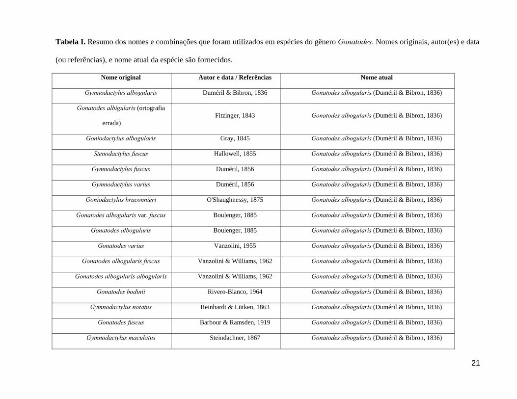
Tabela I. Resumo dos nomes e combinações que foram utilizados em espécies do gênero Gonatodes. Nomes originais, autor(es) e data
(ou referências), e nome atual da espécie são fornecidos.
Nome original Autor e data / Referências Nome atual
Gymnodactylus albogularis Duméril & Bibron, 1836 Gonatodes albogularis (Duméril & Bibron, 1836)
Gonatodes albigularis (ortografia
errada) Fitzinger, 1843 Gonatodes albogularis (Duméril & Bibron, 1836)
Goniodactylus albogularis Gray, 1845 Gonatodes albogularis (Duméril & Bibron, 1836)
Stenodactylus fuscus Hallowell, 1855 Gonatodes albogularis (Duméril & Bibron, 1836)
Gymnodactylus fuscus Duméril, 1856 Gonatodes albogularis (Duméril & Bibron, 1836)
Gymnodactylus varius Duméril, 1856 Gonatodes albogularis (Duméril & Bibron, 1836)
Goniodactylus braconnieri O'Shaughnessy, 1875 Gonatodes albogularis (Duméril & Bibron, 1836)
Gonatodes albogularis var. fuscus Boulenger, 1885 Gonatodes albogularis (Duméril & Bibron, 1836)
Gonatodes albogularis Boulenger, 1885 Gonatodes albogularis (Duméril & Bibron, 1836)
Gonatodes varius Vanzolini, 1955 Gonatodes albogularis (Duméril & Bibron, 1836)
Gonatodes albogularis fuscus Vanzolini & Williams, 1962 Gonatodes albogularis (Duméril & Bibron, 1836)
Gonatodes albogularis albogularis Vanzolini & Williams, 1962 Gonatodes albogularis (Duméril & Bibron, 1836)
Gonatodes bodinii Rivero-Blanco, 1964 Gonatodes albogularis (Duméril & Bibron, 1836)
Gymnodactylus notatus Reinhardt & Lütken, 1863 Gonatodes albogularis (Duméril & Bibron, 1836)
Gonatodes fuscus Barbour & Ramsden, 1919 Gonatodes albogularis (Duméril & Bibron, 1836)
Gymnodactylus maculatus Steindachner, 1867 Gonatodes albogularis (Duméril & Bibron, 1836)

22
Goniodactylus braconnieri O’Shaughnessy 1875 Gonatodes albogularis (Duméril & Bibron, 1836)
Gonatodes notatus Barbour, 1937 Gonatodes albogularis (Duméril & Bibron, 1836)
Gonatodes albogularis notatus Vanzolini & Williams, 1962 Gonatodes albogularis (Duméril & Bibron, 1836)
Gonatodes alexandermendesi Cole & Kok, 2006 Gonatodes alexandermendesi Cole & Kok, 2006
Gonatodes annularis Boulenger, 1887 Gonatodes annularis Boulenger, 1887
Gonatodes Boonii (posteriormente
corrigido para booni) Lidth de Jeude, 1904 Gonatodes annularis Boulenger, 1888
Gonatodes beebei Noble, 1923 Gonatodes annularis Boulenger, 1889
Gymnodactylus antillensis Lidth de Jeude, 1887 Gonatodes antillensis (Lidth de Jeude, 1887)
Gonatodes vittatus roquensis Roze, 1956 Gonatodes antillensis (Lidth de Jeude, 1887)
Gonatodes antillensis Rivero-Blanco, 1964 Gonatodes antillensis (Lidth de Jeude, 1887)
Gonatodes atricucullaris Noble, 1921 Gonatodes atricucullaris Noble, 1921
Gymnodactylus caudiscutatus Günther, 1859 Gonatodes caudiscutatus (Günther, 1859)
Gonatodes caudiscutatus Boulenger, 1885 Gonatodes caudiscutatus (Günther, 1859)
Gonatodes collaris Garman, 1892 Gonatodes caudiscutatus (Günther, 1859)
Gonatodes ceciliae Donoso-Barros, 1966 Gonatodes ceciliae Donoso-Barros, 1966
Goniodactylus concinnatus O'Shaughnessy, 1881 Gonatodes concinnatus (O'Shaughnessy, 1881)
Goniodactylus buckleyi O'Shaughnessy, 1881 Gonatodes concinnatus (O'Shaughnessy, 1881)
Gonatodes concinnatus Boulenger, 1885 Gonatodes concinnatus (O'Shaughnessy, 1881)
Gonatodes ligiae Donoso-Barros, 1967 Gonatodes concinnatus (O'Shaughnessy, 1881)

23
Gonatodes daudini Powell & Henderson, 2005 Gonatodes daudini Powell & Henderson, 2005
Gonatodes eladioi Nascimento, Avila-Pires &
Cunha, 1987
Gonatodes eladioi Nascimento, Avila-Pires & Cunha,
1987
Gonatodes caudiscutatus
falconensis Shreve, 1947 Gonatodes falconensis Shreve, 1947
Gonatodes falconensis Vanzolini, 1968 Gonatodes falconensis Shreve, 1947
Gonatodes hasemani Griffin, 1917 Gonatodes hasemani Griffin, 1917
Gonatodes spinulosus Amaral, 1932 Gonatodes hasemani Griffin, 1917
Gymnodactylus humeralis Guinhenot, 1855 Gonatodes humeralis (Guinhenot, 1855)
Gonatodes ferrugineus Cope, 1863 Gonatodes humeralis (Guinhenot, 1855)
Goniodactylus ferrugineus Cope, 1868 Gonatodes humeralis (Guinhenot, 1855)
Gymnodactylus incertus Peters, 1871 Gonatodes humeralis (Guinhenot, 1855)
Goniodactylus sulcatus O'Shaughnessy, 1875 Gonatodes humeralis (Guinhenot, 1855)
Gonatodes humeralis Boulenger, 1885 Gonatodes humeralis (Guinhenot, 1855)
Gonatodes infernalis Rivas & Schargel, 2008 Gonatodes infernalis Rivas & Schargel, 2008
Cyrtodactylus ocellatus Gray, 1831 Gonatodes ocellatus (Gray, 1831)
Goniodactylus ocellatus Gray, 1845 Gonatodes ocellatus (Gray, 1831)
Gonatodes ocellatus Boulenger, 1885 Gonatodes ocellatus (Gray, 1831)
Gonatodes petersi Donoso-Barros, 1967 Gonatodes petersi Donoso-Barros, 1967
Gonatodes purpurogularis Esqueda, 2004 Gonatodes purpurogularis Esqueda, 2004

24
Gonatodes seigliei Donoso-Barros, 1966 Gonatodes seigliei Donoso-Barros, 1966
Gonatodes superciliaris Barrio-Amorós & Brewer-
Carias, 2008
Gonatodes superciliaris Barrio-Amorós & Brewer-
Carias, 2008
Gonatodes taniae Roze, 1963 Gonatodes taniae Roze, 1963
Gonatodes tapajonicus Rodrigues, 1980 Gonatodes tapajonicus Rodrigues, 1980
Gymnodactylus vittatus Lichtenstein, 1856 Gonatodes vittatus (Lichtenstein, 1856)
Gonatodes gilli Cope, 1863 Gonatodes vittatus (Lichtenstein, 1856)
Gonatodes vittatus vittatus Roze, 1956 Gonatodes vittatus (Lichtenstein, 1856)
25
[Artigo a ser submetido para a revista Zootaxa]
Taxonomic revision of the geckos of the Gonatodes concinnatus complex (Squamata:
Sphaerodactylidae), with description of two new species
MARCELO JOSÉ STURARO1,2 & TERESA C.S. AVILA-PIRES1,2
1 - Museu Paraense Emílio Goeldi / CZO, CP 399, 66017-970 Belém, Pará, Brasil. E-mail:
(MJS) [email protected] and (TCSAP) [email protected]
2 - Programa de Pós-Graduação em Zoologia, Universidade Federal do Pará - Museu Paraense
Emílio Goeldi.
Abstract
The Gonatodes concinnatus complex, as established here, consists of Gonatodes species
characterized by a white suprahumeral spot with black margins; vermiculations on back; and
transversely enlarged scales under the tail, showing the sequence 1’1’1”, and in some cases
1’1’2” (on the anterior portion). Two species are presently recognized in this Amazonian
complex: G. concinnatus and G. tapajonicus. New material collected in eastern Amazonia (states
of Pará and Amapá, Brazil) made it necessary to review these species. We analyzed several
populations within this complex, from Peru, Ecuador, Colômbia, and Brazil (but not Venezuela),
including those new records. Specimens were separated in groups defined on basis of color
pattern. Stepwise discriminant function analyses were then performed to compare the external

26
morphology (represented by measurements and scale counts, separately) in these groups. Results
support recognition of four taxa, corresponding to G. concinnatus, from western Amazonia, in
Ecuador and northeastern Peru; G. tapajonicus, from the Tapajós river basin, in Pará, Brazil; and
two new species, one from eastern Amazonia, in the states of Pará (north and south of the
Amazon river) and Amapá, Brazil, and another from central Colombia, east of the Andes.
Diagnoses and descriptions of all species are presented.
Key words: lizard, South America, Amazonian rainforest, taxonomy
Resumo
O complexo Gonatodes concinnatus, conforme estabelecido aqui, consiste nas espécies
caracterizadas por uma mancha suprahumeral branca com margens pretas; vermiculações no
dorso; e escamas alargadas sob a cauda, apresentando a seqüência 1’1’1”, e em alguns casos
1’1’2” (na porção anterior). Duas espécies são atualmente reconhecidas neste grupo basicamente
amazônico, G. concinnatus e G. tapajonicus. Novos espécimes coletados no leste da Amazônia
(nos estados do Pará e Amapá, Brasil) fizeram necessária a revisão dessas espécies. Analisamos
diversas populações dentro deste complexo, provenientes do Peru, Equador, Colômbia, e Brasil
(mas não da Venezuela), incluindo os novos registros. Os espécimes foram separados em grupos
definidos com base no padrão de coloração. Análises discriminantes, utilizando o método por
passos (stepwise), foram realizadas para comparar a morfologia externa (representada por
medições e contagens de escama, separadamente) nestes grupos. Os resultados apóiam o
reconhecimento de quatro táxons, correspondendo a G. concinnatus, da Amazônia Ocidental, no
nordeste do Equador e do Peru; G. tapajonicus, da bacia do Rio Tapajós, no Pará, Brasil; e duas
novas espécies, uma do leste da Amazônia, nos estados do Pará (ao norte e ao sul do Rio

27
Amazonas) e Amapá, Brasil, e outra da Colômbia Central, a leste dos Andes. As diagnoses e
descrições de todas as espécies são apresentadas.
Palavras-chaves: lagarto, América do Sul, Floresta Amazônica, taxonomia
Introduction
The genus Gonatodes consists currently of 22 species distributed in Central and South America,
in the Antilles and, as a recent introduction, in Florida, United States (Peters & Donoso-Barros
1970; Rivero-Blanco 1979; Avila-Pires 1995; Esqueda 2004; Krysko & Daniels 2005; Powel and
Henderson 2005; Cole and Kok 2006; Barrio-Amoros & Brewer-Carias 2008, Rivas & Schargel,
2008). It is characterized by having dorsal scales granular and juxtaposed; ventral scales larger
than dorsals, flat, smooth and imbricate; femoral and precloacal pores absent; escutcheon present
in males, on posterior surface of belly and ventral aspect of thigh; and free claws (Peters &
Donoso-Barros 1970; Hoogmoed 1973; Avila-Pires 1995; Kluge 1995). Sexual dimorphism is
evident in color pattern, usually with colourful males and cryptic females. Male color pattern is
an important character for recognizing species, since differences in scale counts between species
are frequently small (Vanzolini, 1968; Rivero-Blanco, 1979).
Gonatodes concinnatus (O’Shaughnessy, 1881) and Gonatodes tapajonicus Rodrigues,
1980 have in common the presence of a white suprahumeral spot with black margins;
vermiculations on back (except in G. c. ligiae); and transversely enlarged scales under the tail,
showing the sequence 1’1’1”, and sometimes 1’1’2” (under anterior portion of the tail), as
defined by Avila-Pires (1995). According to Rodrigues (1980), Gonatodes tapajonicus differs
from G. concinnatus by its color pattern and the form of the mental. Recent material from eastern
Amazonia presented the same characteristics common to both species mentioned above, but with
28
differences in color pattern. In order to identify this material and considering the variation
reported in G. concinnatus, it was necessary to undertake a revision of this group, that we are
calling “G. concinnatus complex”.
O’Shaughnessy (1881) described Goniodactylus concinnatus based on three specimens
from Canelos, Ecuador (BMNH 1946.9.7.10-12), and Goniodactylus buckleyi based on one
specimen from Pallatanga (probably in error, see Rivero-Blanco, 1968) and two from Canelos,
Ecuador. Boulenger (1885) examined the types of Goniodactylus concinnatus and G. buckleyi
and observed that they represented the same species, respectively male and female; the species
was considered under the combination Gonatodes concinnatus. Subsequent authors followed
Boulenger (1885).
Gonatodes ligiae was described by Donoso-Barros (1967), who provided only an
imprecise description, based on two specimens from Bosque de la Carabela (holotype) and
Parque de Moromoy (paratype), Barinitas, Vezenuela. Rivero-Blanco (1968), based on
specimens from the type-locality, but without examining the type material of G. ligiae,
considered it a synonym of G. concinnatus and Rivero-Blanco (1979) recognized it as a
subspecies of G. concinnatus. The shape of the suprahumeral spots (a vertical bar in G.
concinnatus concinnatus and an ocellus in G. concinnatus ligiae) and the presence (G. c.
concinnatus) or absence (G. c. ligiae) of white spots or vermiculations on the back and flanks
were pointed out as diagnostic characters.
Gonatodes tapajonicus was described by Rodrigues (1980) based on nine specimens
from Cachoeira do Limão (04º41’S, 56º21’W), Tapajós River, Pará, Brazil.
In this paper we evaluate the validity of the currently recognized species of the
Gonatodes concinnatus complex, and verify the status of additional populations from eastern
29
Amazonia (states of Pará and Amapá, Brazil), by analyzing variation of external morphology.
Gonatodes c. ligiae will not be considered here, since we did not examine material from
Venezuela, but the status of this taxon should be verified in future studies.
Material and Methods
Specimens examined are listed in Appendix I. They are deposited in the following institutions:
Museu Nacional/UFRJ, Rio de Janeiro, Brazil (MNRJ); Museu Paraense Emilio Goeldi, Pará,
Brazil (MPEG); Museu de Zoologia, Universidade de São Paulo, São Paulo, Brazil (MZUSP);
Nationaal Natuurhistorisch Museum, Leiden, The Netherlands (RMNH). We examined photos of
all syntypes of Goniodactylus buckleyi and Goniodactylus concinnatus, which are deposited in
The Natural History Museum (former British Museum of Natural History), London, United
Kingdom (BMNH).
Measurements and scale counts
Measurements were taken with an electronic caliper (to the nearest 0.1 mm), when necessary
under a stereomicroscope, as follows: SVL (snout-vent length, from tip of snout to cloacal
opening); TL (tail length, from cloacal opening to tip of tail); HL (head length, from
anteriormost point of rostral to anterior margin of ear-opening); HW (head width, on the widest
part of head); HD (head depth, on the highest part of head); ED (eye diameter; between anterior
and posterior corner of the eye); IOD (interorbital distance; between anterosuperior margins of
eyes); IND (internostril distance, between medial margins of nasal scales); SSL (supranasal scale
length, between anterior and posterior corners of scale); SSW (supranasal scale width, between
lateral corners of scale); RSL (rostral scale length, between anterior and posterior corners of

30
scale); RSW (rostral scale width, between lateral corners of scale); MSL (mental scale length,
between anterior and posterior corners of scale); MSW (mental scale width, between lateral
corners of scale); UAL (upper arm length, from axil to tip of elbow); LAL (lower arm length,
from tip of elbow to wrist); HAL (hand length, from wrist to tip of claw of longest finger); FL
(forelimb length, from axil to tip of claw of longest finger); THL (thigh length, from groin to
knee); LLL (lower leg length, from knee to ankle); FTL (foot length, from ankle to tip of claw
of longest toe); HLL (hind limb length, from groin to tip of longest toe); DBL (distance between
fore- and hind limb, from axil to groin), HSL (suprahumeral spot length, measured between
anterior and posterior margins); HSW (suprahumeral spot width, measured between lateral and
medial ends).
Scale counts were taken under a stereomicroscope, as follows: SL (supralabials:
distinctly enlarged scales along the upper jaw); IL (infralabials: distinctly enlarged scales along
the lower jaw); PR (postrostral scales: scales in contact to rostral); SP (small postrostral scales:
small scales in contact to rostral medially); PN (postnasal scales: scales in contact with posterior
portion of nasal); LS (loreal scales: in a line between postnasals and anterior corner of orbit);
PM (postmental scales: in contact to posterior portion of mental); SAM (scales around midbody,
counted midway between fore- and hind limbs); VLR (ventral scales in a longitudinal row,
counted along a midventral line, from anterior margin of forelimbs to anterior margin of hind
limbs); VTR (ventral scales in a transversal row, counted midway between fore- and hind limb);
SCS (supraciliary scales: enlarged and flattend scales on anterior portion of supraciliary flap);
SSC (supraciliary spines: scales with coninal shape on the anterior portion of supraciliary flap);
PL2F, PL3F, PL4F (proximal lamellae under respectively second, third and fourth fingers,
counted from base of finger to the sharp angle between first and second phalanges); PL2T,
31
PL3T, PL4T (proximal lamellae under respectively second, third and fourth toes, counted from
base of toe to the sharp angle between first and second phalanges); DL2F, DL3F, DL4F (distal
lamellae under respectively second, third and fourth fingers, counted between the sharp angle
between first and second phalanges and ungual scale); DL2T, DL3T, DL4T (distal lamellae
under respectively second, third and fourth toes, counted between the sharp angle between first
and second phalanges and ungual scale); subdigital lamellae under the second (SL2F), third
(SL3F) and fourth (SL4F) finger, and second (SL2T), third (SL3T) and fourth (SL4T) toe,
counted between base of digit articulation and ungual scale; LS2F, LS3F, LS4F (lateral rows of
scales on distal part of respectively second, third and fourth fingers, counted between fourth
distal subdigital lamella and dorsal scale); LS2T, LS3T, LS4T (lateral rows of scales on distal
part of respectively second, third and fourth toes, counted between the fourth subdigital lamella
when counted from the claw towards the hand, and dorsal scale); RSE (rows of escutcheon
scales under thigh, counted along a line between the anterior and the posterior aspects of the
thigh).
Measurements and scale counts were taken on the right side of the body, except when this
side was damaged. In this case data from the left side of the body were used.
Sex was determined by the presence of an escutcheon on posterior portion of belly and
ventral surface of thighs in males, which is absent in females. Specimens with SVL under 35 mm
were defined as juveniles.
Statistical Analysis
We used discriminant function analysis for scales counts (Tabachnick and Fidell 2001)
and size-free discriminant function analysis for measurements (Strauss 1985, Reis et al. 1990), to

32
test if groups defined a priori on basis of color pattern could be differentiated by a combination
of other morphological characters. Measurements may be influenced by factors such as sexual
dimorphism, developmental stage and indeterminate growth, which makes it important to use
size-free analysis for such data. As proposed by Strauss (1985), size-free discriminant analysis
consists of regressing each variable separately on the first principal component of a principal
component analysis and then applying the discriminant function analysis to the residuals
obtained from these regressions. The nonparametric resampling method of jackknifing was used
to test the statistical significance of the canonical functions based on the correct classification
rate (McGarigal et al. 2000). In the discriminant function analyses we applied the stepwise
method, due to the high number of variables and low number of individuals in most samples. We
adjusted the F-enter and F-remove to select only the first six variables in each case (scales counts
and measurements), increasing the robustness of the analyses. The forward direction option was
used, so that at each step all variables were reviewed and evaluated to determine which one
would contribute most to the discrimination between groups. The variables in each analysis that
showed best discrimination power between groups were subsequently used in a new discriminant
function analysis.
Missing scale counts and morphometric values were estimated using a Missing Value
Analysis, based on the linear regression of the observed variables. Missing values represented
never more then 4.3% of the total number of cases. For all analyses we used the statistic software
SYSTAT for Windows, version 12 (SYSTAT Software 2007).
Groupings for the discriminant analysis were defined by color pattern of preserved male
specimens, but some observations were based on color in life. Females and juveniles were

33
assigned to groups, based on their locality and its the nearness to certain male patterns, since no
geographic differences in female pattern were found.
Species descriptions
Species descriptions follow Avila-Pires (1995) with the addition of some morphometric
and scale count characters. Statistics are presented as “minimum”–“maximum” (“mean” ±
“standard deviation”, N=“sample size”). Tail length was measured only in specimens with intact,
non-regenerated tail.
Results
We found four different color patterns (see Table 1; Fig. 1, 2) which were used to define
the groups for the discriminant analysis (herein named A, B, C and D, as for the color patterns).
Stepwise discriminant function analysis using scale counts revealed highly significant
differences among groups (Wilks's Lambda=0.036, df=18; approx. F=21.6, df=173, p=0.000).
The six variables selected as the most powerful discriminators among groups are listed in Table
2. Distal lamellae under second toe was selected first, classifying 67.1% of the specimens,
followed by proximal lamellae under fourth toe, infralabials, proximal lamellae under second
finger, ventral scales in a longitudinal row, and supralabials, whose additions improved the
classification criterion to respectively 82.9%, 87.9%, 90%, 90% and 97.1%. The jackknifed
classification matrix correctly classified unknown specimens 92.9% of the times (Table 3). The
first and second component function explained, respectively, 74.7% and 22.4% of the total
variation in the six meristic variables. The first component function provided separation between
A+B (which overlapped), C and D groups (Fig. 3A). Characters with the largest loadings on the

34
first principal component were proximal lamellae under second finger and distal lamellae under
second toe (Table 2). The second component function provided separation between A and D, and
B and D, groups (Fig. 3A). Characters with the largest loadings on the second principal
component were proximal lamellae under fourth toe and infralabials (Table 2).
Stepwise size-free discriminant function analysis, using measurements, also revealed
highly significant differences among the four groups (Wilks's Lambda=0.0.51, df=18; approx.
F=17.964, df =173, p=0.000). The six variables selected as the most powerful discriminators
among groups are listed in Table 4. Mental scale length was selected first, classifying 65.7% of
the specimens, followed by head width, lower arm length, supranasal scale length, rostral scale
length and interorbital distance, whose additions improved the classification criterion to
respectively 78.6%, 81.4%, 87.1%, 91.4% and 90%.Although classification with six variables
was lower than with five ones, graphic separation of groups in the discriminant function was
clearer. The jackknifed classification matrix correctly classified unknown specimens 88.6%% of
the time (Table 5).The first and second component functions explained, respectively, 68.9% and
27.1% of the total variation in the six meristic variables. The first component function provided
separation between groups A and C, A and B, and showed partial overlapp between C and D.
(Fig. 3B). Characters with the largest loadings on the first principal component were supranasal
scale length and mental scale length (Table 4).The second component function provided
separation, with small overlapping, between A and B+D groups, C and B+D (Fig. 3B).
Characters with large loadings on the second principal component were head width and lower
arm length (Table 4).
35
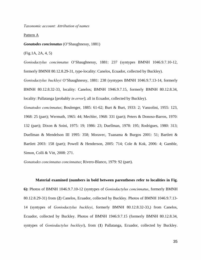
Taxonomic account: Attribution of names
Pattern A
Gonatodes concinnatus (O’Shaughnessy, 1881)
(Fig.1A, 2A, 4, 5)
Goniodactylus concinnatus O’Shaughnessy, 1881: 237 (syntypes BMNH 1046.9.7.10-12,
formerly BMNH 80.12.8.29-31, type-locality: Canelos, Ecuador, collected by Buckley).
Goniodactylus buckleyi O’Shaughnessy, 1881: 238 (syntypes BMNH 1046.9.7.13-14, formerly
BMNH 80.12.8.32-33, locality: Canelos; BMNH 1946.9.7.15, formerly BMNH 80.12.8.34,
locality: Pallatanga [probably in error]; all in Ecuador, collected by Buckley).
Gonatodes concinnatus; Boulenger, 1885: 61-62; Burt & Burt, 1933: 2; Vanzolini, 1955: 123,
1968: 25 (part); Wermuth, 1965: 44; Mechler, 1968: 331 (part); Peters & Donoso-Barros, 1970:
132 (part); Dixon & Soini, 1975: 19; 1986: 23; Duellman, 1978: 195; Rodrigues, 1980: 313;
Duellman & Mendelson III 1995: 358; Moravec, Tuanama & Burgos 2001: 51; Bartlett &
Bartlett 2003: 158 (part); Powell & Henderson, 2005: 714; Cole & Kok, 2006: 4; Gamble,
Simon, Colli & Vitt, 2008: 271.
Gonatodes concinnatus concinnatus; Rivero-Blanco, 1979: 92 (part).
Material examined (numbers in bold between parentheses refer to localities in Fig.
6): Photos of BMNH 1046.9.7.10-12 (syntypes of Goniodactylus concinnatus, formerly BMNH
80.12.8.29-31) from (2) Canelos, Ecuador, collected by Buckley. Photos of BMNH 1046.9.7.13-
14 (syntypes of Goniodactylus buckleyi, formerly BMNH 80.12.8.32-33,) from Canelos,
Ecuador, collected by Buckley. Photos of BMNH 1946.9.7.15 (formerly BMNH 80.12.8.34,
syntypes of Goniodactylus buckleyi), from (1) Pallatanga, Ecuador, collected by Buckley.

36
MZUSP 3382-83 (fields number ORCES 656, 656A), two adult females, from (3) Loreto, Napo
Province, Ecuador (~0o38’S and 77o19’W), collected by J. Olalla, April 1952. MZUSP 54655
(formerly MCZ 156856), an adult male, from (4) Limoncocha, Napo Province, Ecuador (~0o24’S
and 76o37’W), collected by K. Miyata, 9-11 February 1979. MZUSP 28248-49 (field numbers
P.SOINI 329-30), two adult males, from (8) Moropón, Departamento Loreto, Peru (~5o44’52”S
and 78o32’08”W), collected by P. Soini, August 1971. MZUSP 28260-63 (field numbers
P.SOINI 589, 636, 707-8), two juvenile females and two adult males, from (8) Moropón,
Departamento Loreto, Peru (~5o44’52”S and 78o32’08”W), collected by P. Soini, November-
December 1971. MZUSP 28273-79 (field numbers P.SOINI 821-22, 824, 826, 830, 928-29), a
juvenile females, two adult females and four adult males, from (8) Moropón, Departamento
Loreto, Peru (~5o44’52”S and 78o32’08”W), collected by P. Soini, February 1972. MZUSP
28311-13 (field numbers P.SOINI 1212-13, 1262), an adult female and two adult males, from (8)
Moropón, Departamento Loreto, Peru (~5o44’52”S and 78o32’08”W), collected by P. Soini,
October 1972. MZUSP 28319 (field numbers P.SOINI 1385), an adult male, from (8) Moropón,
Departamento Loreto, Peru (~5o44’52”S and 78o32’08”W), collected by P. Soini, June 1972.
MZUSP 28375 (field numbers P.SOINI 1489), an adult female, from (8) Moropón,
Departamento Loreto, Peru (~5o44’52”S and 78o32’08”W), collected by P. Soini, 1972. MZUSP
28354-55 (field numbers P.SOINI 1309-10), a juveline and an adult females, from (9)
Yanamono, Departamento Loreto, Peru (~3o23’01”S and 72o45’01”W), collected by P. Soini,
July 1972. MZUSP 56657, an adult female, from (10) Rio Orosa, Departamento Loreto, Peru
(~3o30’60”S and 72o06’03”W), collected by P. Soini, August 1976. MZUSP 13458, an adult
male, from (11) Estirón, Rio Ampiyacu, Departamento Loreto, Peru (~4o10’30”S and
70o48’04”W), collected by B. Malkin, 15-22 May 1966.
37
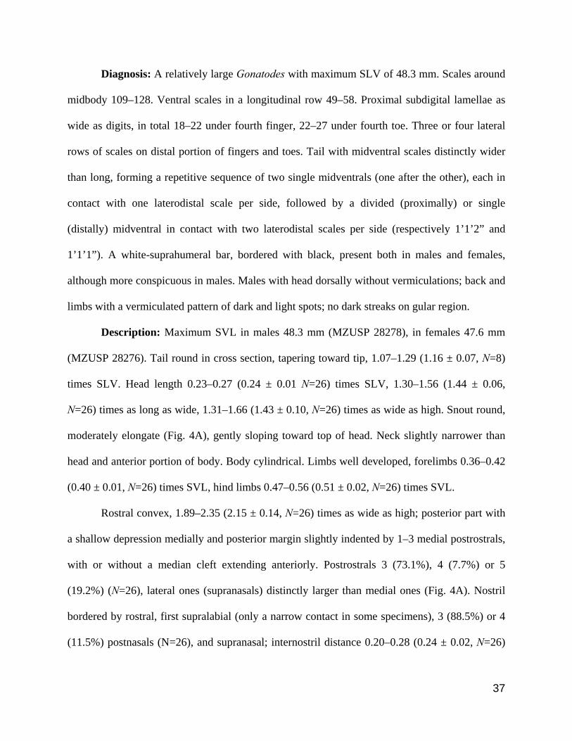
Diagnosis: A relatively large Gonatodes with maximum SLV of 48.3 mm. Scales around
midbody 109–128. Ventral scales in a longitudinal row 49–58. Proximal subdigital lamellae as
wide as digits, in total 18–22 under fourth finger, 22–27 under fourth toe. Three or four lateral
rows of scales on distal portion of fingers and toes. Tail with midventral scales distinctly wider
than long, forming a repetitive sequence of two single midventrals (one after the other), each in
contact with one laterodistal scale per side, followed by a divided (proximally) or single
(distally) midventral in contact with two laterodistal scales per side (respectively 1’1’2” and
1’1’1”). A white-suprahumeral bar, bordered with black, present both in males and females,
although more conspicuous in males. Males with head dorsally without vermiculations; back and
limbs with a vermiculated pattern of dark and light spots; no dark streaks on gular region.
Description: Maximum SVL in males 48.3 mm (MZUSP 28278), in females 47.6 mm
(MZUSP 28276). Tail round in cross section, tapering toward tip, 1.07–1.29 (1.16 ± 0.07, N=8)
times SLV. Head length 0.23–0.27 (0.24 ± 0.01 N=26) times SLV, 1.30–1.56 (1.44 ± 0.06,
N=26) times as long as wide, 1.31–1.66 (1.43 ± 0.10, N=26) times as wide as high. Snout round,
moderately elongate (Fig. 4A), gently sloping toward top of head. Neck slightly narrower than
head and anterior portion of body. Body cylindrical. Limbs well developed, forelimbs 0.36–0.42
(0.40 ± 0.01, N=26) times SVL, hind limbs 0.47–0.56 (0.51 ± 0.02, N=26) times SVL.
Rostral convex, 1.89–2.35 (2.15 ± 0.14, N=26) times as wide as high; posterior part with
a shallow depression medially and posterior margin slightly indented by 1–3 medial postrostrals,
with or without a median cleft extending anteriorly. Postrostrals 3 (73.1%), 4 (7.7%) or 5
(19.2%) (N=26), lateral ones (supranasals) distinctly larger than medial ones (Fig. 4A). Nostril
bordered by rostral, first supralabial (only a narrow contact in some specimens), 3 (88.5%) or 4
(11.5%) postnasals (N=26), and supranasal; internostril distance 0.20–0.28 (0.24 ± 0.02, N=26)

38
times head width. Supranasal scale roughly oval, circular or semicircular, 0.8–2.3 (1.21 ± 0.28;
N=26) times as wide as long. Postnasals slightly larger than, or similar in size to, adjacent
loreals. Scales on snout convex, hexagonal to round, juxtaposed, relatively uniform in size.
Canthus rostralis rounded. Loreal region with scales slightly more elongate than those on snout,
largest on row adjacent to supralabials; 9–13 (10.7 ± 1.0*, N=26) loreal scales in a line between
postnasals and anterior corner of orbit. Top and posterior portion of head, as well as supraorbital
region, with granular scales. A short supraciliary flap present, anteriorly with 6–10 (8.3 ± 1.1,
N=26) enlarged, flattened scales, among which 0–4 (1.7 ± 1.5; N=26) small, conical spines. Pupil
round, eye diameter 0.19–0.24 (0.21 ± 0.01, N=25) times head length; interorbital distance 0.25–
0.35 (0.31 ± 0.02, N=26) times head width. Scales on temporal region similar to those on top of
head. Ear-opening much smaller than eye, oval, posterior to, and at same level of, commissure of
mouth. Supralabials 6–7 (6.3 ± 0.5), distinctly enlarged and decreasing in size posteriorly, one or
two of them posterior to centre of eye, followed to corner of mouth by small scales.
Mental large, distinctly wider anteriorly than posteriorly, with posterior margin forming a
wide angle, 1.02–1.25 (1.13 ± 0.07, N=26) times as wide as long; 2–4 (mostly two) postmentals
(Fig. 4B). Scales on chin flat, smooth, polygonal, juxtaposed, larger anteriorly, decreasing in size
posteriorly. Infralabials 5–7 (6.0 ± 0.8; N=26), distinctly enlarged and decreasing in size
posteriorly, one–two, occasionally three, of them posterior to centre of eye, followed to corner of
mouth by small scales.
Scales on nape small and granular, becoming slightly larger on sides of neck. Scales on
throat anteriorly like those on posterior part of chin; posteriorly flat, smooth, hexagonal or round,
imbricate, with a short transitional zone between the anterior and posterior parts.

39
Dorsals granular, increasing in size toward flanks. Ventrals larger than dorsals, roughly
hexagonal, flat, smooth, imbricate, in oblique rows; 49–58 (55.0 ± 2.3, N=25) scales along a
midventral line between anterior margin of forelimbs and vent; 17–20 (18.5 ± 1, N=25) scales in
a transverse row at midbody. Scales around midbody 109–128 (120.6 ± 5.3, N=25), with a short
transitional zone between ventrals and scales on flanks. Scales on preanal plate similar to
ventrals, except for those bordering vent, which are very small. Escutcheon present in males on
posterior portion of belly and on 4–5 (mostly four) rows (body-knee direction) of ventral surface
of thighs.
Scales on anterodorsal surface of forelimbs flat, smooth, roundish, imbricate, largest
close to the wrist; on posterodorsal and ventral surfaces convex, smooth, rhomboid, juxtaposed,
relatively small; on ventral surface flat, smooth, rhomboid, subimbricate and smaller than
anterodorsally. Scales on anterodorsal and ventral surface of hind limbs flat, smooth, rhomboid,
imbricate; on posterodorsal surface convex, smooth, round, subimbricate, relatively small.
Numbers of lamellae under fingers and toes are presented in Table 7 (Fig. 4C and D). Claws
exposed, non-retractile, between two basal scales.
Scales on tail dorsally and laterally relatively small, rhomboid, flat, smooth, imbricate.
On ventral surface of tail scales smooth, flat, imbricate, increasing in size toward midventral
line; midventral scales, except close to the base of the tail, distinctly wider than long, forming a
repetitive sequence of two single midventrals (one after the other), each in contact with one
laterodistal scale per side, followed by a divided (in the anterior portion of tail) or single (in
posterior portion of tail) midventral scale in contact with two laterodistal scales per side –
respectively 1’1’2” and 1’1’1” in the codification by Avila-Pires (1995: Figure 2) (Fig. 4E).

40
Color in preservative: In males, dorsal surface of head beige color, spotless. Back and
flanks, base of tail and hind limbs with relatively large, beige and brown vermiculations. A large,
conspicuous, white suprahumeral bar, bordered with black, extending dorsally at least to the
dorsolateral region, in some cases almost reaching the middorsal line (Fig. 5A); never in the
form of an ocellus. Ventral surface of head, gular region and chest beige or reddish-brown,
without oblique streaks; belly gray; underside of limbs beige. Tail brown and/or black dorsally,
white and/or brown ventrally. Escutcheon area (belly and thighs) light gray (Fig. 5).
In females, dorsal surface of head and limbs with brown and black irregular spots; back
gray with dorsolateral pairs of black spots and, in some specimens, pairs of beige spots; flanks
gray with black and brown spots. Suprahumeral bar conspicuous, white with black margins,
thinner than that of males; in some cases they almost reach the middorsal line. Ventral surface of
head and gular region white with dark oblique streaks, in contact or not at midventral line; belly
and underside of limbs light gray. Tail brown and/or black dorsally, white and/or brown
ventrally; original tail distally with white bands that form complete rings around the tail.
Color in life: In males, head dorsally and laterally orange or reddish brown with cream
color spots. Back olive green with reddish brown vermiculations or brown and black with white
vermiculations. Suprahumeral bar white with black margins. Head ventrally and gular region
orange, in some specimens with cream streaks; remaining ventral region yellowish gray or black,
lighter on escutcheon areas (belly and thighs); tail black. (Duellman 1978; Vitt and Torre 1996).
In females, head dorsally and laterally, and back grayish tan with irregular brown spots or
drab gray with irregular crossbans, black at the anterior edge and white at the posterior edge.
Suprahumeral bar white with black margins, thinner than that of males. Head ventrally and gular

41
region cream color with dark streaks; belly creamy tan or yellow; tail grayish tan with irregular
brown spots or black and white banded (Duellman 1978; Vitt and Torre 1996).
Variation: Body proportions and scale counts are given in Tables 6 and 7. Suprahumeral
bar varies (within the same population) in extension, with upper limit between dorsolateral and
middorsal lines.
Distribution: Western Amazonia, in Ecuador and northeastern Peru (Fig. 6).
Remarks: Gonatodes ligiae Donoso-Barros, 1967, from Venezuela, has not been
considered in this paper and its inclusion in G. concinnatus deserves further studies. Material
from Colombia previously identified as G. concinnatus is here considered as a distinct species
(see ‘Pattern D’).
Pattern B
Gonatodes tapajonicus Rodrigues, 1980
(Figs. 1B, 2B, 7, 8)
Gonatodes tapajonicus Rodrigues, 1980: 309 (holotype MZUSP 53676, type-locality: Cachoeira
do Limão, Rio Tapajós, Pará, Brazil); Vanzolini, 1986: 10; Avila-Pires, 1995: 283.
Material examined (numbers in bold between parentheses refer to localities in Fig.
6): MZUSP 53676 (holotype, field number MTR 79.1213), an adult male, from (11) Cachoeira
do Limão, right margin of Rio Tapajos, Pará, Brazil (~4o41’S and 56o21’W), collected on tree
trunk at 60 cm above ground (Rodrigues 1980) by M.T. Rodrigues, 30 January to 5 February
1979. MZUSP 53669, 53671-74, 53677 (paratypes, field numbers MTR 79.1142-43, 79.1150,

42
79.1152-53, 79.1214, respectively), a male and five females, all from type-locality, collected by
M.T. Rodrigues, 30 January to 5 February 1979.
Diagnosis: A relatively large Gonatodes with maximum SVL in males 53 mm (MZUSP
53676), in females 55 mm (MZUSP 53671) (Rodrigues 1980). Scales around midbody 120–126.
Ventral scales in a longitudinal row 53–59. Proximal subdigital lamellae as wide as digits, in
total 18–22 under fourth finger, 23–27 under fourth toe. Three to four lateral rows of scales on
distal portion of fingers and toes. Tail with midventral scales distinctly wider than long, forming
a repetitive sequence of two single midventrals (one after the other), each in contact with one
laterodistal scale per side, followed by a divided (proximally) or single (distally) midventral in
contact with two laterodistal scales per side (respectively 1’1’2” and 1’1’1”). A white
suprahumeral ocellus, bordered with black, present both in males and females, although more
conspicuous in males. Males with a vermiculated pattern of light and dark (vivid yellow and
reddish brown in life) spots both on head and body dorsally; gular region with dark oblique
streaks (yellow with reddish brown streaks in life).
Description: Maximum SVL in males 53 mm (MZUSP 53676), in females 55 mm
(MZUSP 53671) (Rodrigues 1980). Tail round in cross section, tapering toward tip, 1.21–1.23
(1.22 ± 0.02, N=2) times the SLV. Head length 0.22–0.25 (0.23 ± 0.01 N=7) times SLV, 1.45–
1.58 (1.5 ± 0.06, N=7) times as long as wide, 1.28–1.44 (1.36 ± 0.05, N=7) times as wide as high.
Snout round, moderately elongate (Fig 7A), gently sloping toward top of head. Neck slightly
narrower than head and anterior portion of body. Body cylindrical. Limbs well developed,
forelimbs 0.37–0.44 (0.40 ± 0.03, N=7) times SVL, hind limbs 0.45–0.54 (0.51 ± 0.04, N=7)
times SVL.

43
Rostral convex, 1.86–2.21 (2.02 ± 0.15, N=7) times as wide as high; posterior part with a
shallow depression medially and posterior margin slightly indented by 1–3 median postrostrals,
with or without a median cleft extending anteriorly. Postrostral 3 (85.7%) or 5 (14.3%), lateral
ones (supranasals) distinctly larger than medial ones (N=7) (Fig 7A). Nostril bordered by rostral,
first supralabial (only a narrow contact in some specimens), 3 (85.7%) or 4 (14.3%) postnasals
(N=7), and supranasal; internostril distance 0.21–0.27 (0.25 ± 0.03; N=7) times head width.
Supranasal scale roughly oval or circular, 0.99–1.40 (1.19 ± 0.16; N=7) times as wide as long.
Postnasals slightly larger than, or similar in size to, adjacent loreals. Scales on snout convex,
hexagonal to round, juxtaposed, relatively uniform in size. Canthus rostralis round. Loreal region
with scales slightly more elongate than those on snout, largest on row adjacent to supralabials,
10–13 (11.1 ± 0.9, N=7) loreal scales in a line between postnasals and anterior corner of orbit.
Top and posterior portion of head, as well as supraorbital region, with granular scales. A short
supraciliary flap present, anteriorly with 7–10 (8.1 ± 1.1, N=7) enlarged and flattened scales,
among which 2–4 (2.9 ± 0.7; N=7) small, conical spines. Pupil round, eye diameter 0.21–0.25
(0.23 ± 0.01, N=7) times head length; interorbital distance 0.34–0.42 (0.38 ± 0.03, N=7) times
head width. Scales on temporal region similar to those on top of head. Ear-opening much smaller
than eye, oval, posterior to, and at same level of, commissure of mouth. Supralabials 6–8 (7 ±
0.8; N=7), distinctly enlarged and decreasing in size posteriorly, 1-3 of them posterior to centre
of eye, followed to corner of mouth by small scales (Fig. 7A).
Mental large, distinctly wider anteriorly than posteriorly, with posterior margin forming a
wide angle, 1.07–1.39 (1.21 ± 0.11; N=7) times as wide as long; 2–3 (mostly two) postmentals
(Fig. 7B). Scales on chin flat, smooth, polygonal, juxtaposted, decreasing in size posteriorly.
Infralabials 5–7 (6.1 ± 0.7; N=7) distinctly enlarged and decreasing in size posteriorly; 1–2,

44
occasionally three, of them posterior to centre of eye, followed to corner of mouth by small
scales (Fig. 7B).
Scales on nape small and granular, becoming slightly larger on sides of neck. Scales on
throat anteriorly like those on posterior part of chin; posteriorly flat, smooth, hexagonal or round,
imbricate, with a short transitional zone between the anterior and posterior parts.
Dorsals granular, increasing in size toward the flanks. Ventrals larger than dorsals,
roughly hexagonal, flat, smooth, imbricate, in oblique rows; 53–59 (55.8 ± 2.2; N=6) scales
along the midventral line between anterior margin of forelimbs and vent; 19–20 (19.5 ± 0.5;
N=6) scales in a transverse line at midbody. Scales around midbody 120–126 (122.7 ± 2.4; N=6),
with a short transitional zone between ventrals and scales on flanks. Scales on preanal plate
similar to ventrals, except for those bordering vent, which are very small. Escutcheon present in
males on posterior portion of belly and on four (N=2) rows (body-knee direction) of ventral
surface of thighs.
Scales on anterodorsal surface of forelimbs flat, smooth, roundish, imbricate, largest
close to the wrist; on posterodorsal and ventral surfaces convex, smooth, rhomboid, juxtaposed,
relatively small; on ventral surface flat, smooth, rhomboid, subimbricate and smaller than
anterodorsally. Scales on anterodorsal and ventral surface of hind limbs flat, smooth, rhomboid,
imbricate; on posterodorsal surface convex, smooth, round, subimbricate, relatively small.
Numbers of lamellae under fingers and toes are presented in Table 7 (Fig. 7D).
Scales on tail dorsally and laterally relatively small, rhomboid, flat, smooth, imbricate.
On ventral surface of tail scales smooth, flat, imbricate, increasing in size toward midventral
line; midventral scales, except close to the base of the tail, distinctly wider than long, forming a
repetitive sequence of two single midventrals (one after the other), each in contact laterodistally
45
with one scale per side, followed by a divided (in the anterior portion of tail) or single (in the
posterior portion of tail) midventral scale in contact laterodistally with two scales per side –
respectively 1’1’2” and 1’1’1” in the codification by Avila-Pires (1995: Figure 2) (Fig. 7E).
Color in preservative: In males, dorsal surface of head with relatively large reddish-
brown and beige vermiculations. Sides of head with oblique and/or longitudinal, beige and
brown bands. Back and flanks, base of tail and limbs with beige and brown vermiculations. A
large, conspicuous, white suprahumeral ocellus with black margin. Ventral surface of head and
gular region beige with oblique black streaks, in contact or not at midventral line; chest beige;
belly dark-brown; undersides of limbs and base of tail brown. Dorsal surface of tail brown,
ventral surface reddish-brown; distal part in one specimen with a beige ring around the tail.
Escutcheon area (belly and thighs) light gray.
In females, dorsal surface of head, and limbs with brown and beige irregular spots. Sides
of head with beige and brown oblique and/or longitudinal bands. Back gray with dorsolateral
pairs of black and white spots; flanks gray with black and brown irregular spots. A moderately
large, conspicuous, white suprahumeral ocellus, with black margin, smaller than that of males.
Ventral surface of head and gular region white with dark oblique streaks, in contact or not at
midventral line; belly and underside of limbs light gray. Tail brown and/or black dorsally, white
and/or brown ventrally; original tail distally with white bands that form complete rings around
the tail.
Color in life (from photos of holotype, Fig. 8): Head dorsally and laterally reddish-
brown and vivid yellow. Suprahumeral ocellus white with black margin. Vermiculations on back
brown and yellow, smaller vermiculations on flanks bluish-white and black. Head ventrally and
gular region vivid yellow with reddish-brown streaks, chest vivid yellow. Remaining ventral

46
region dark gray, lighter on escutcheon areas (belly and thighs). Tail dark gray to black all
around, except near the base, where it is similar to the back.
Variation: Body proportions and scale counts are given in Tables 6 and 7. The
suprahumeral ocellus does not vary in extension between the specimens.
Distribution: Known only from the type-locality, on the right margin of the Tapajós
river, state of Pará, Brazil (Fig. 6).
Pattern C
Gonatodes sp. nov. 1
(Fig. 1C, 2C, 9, 10)
Holotype (number in bold between parentheses refer to localities in Fig. 6): MPEG
25596 (field number BML 446), an adult male, from (14) Fazenda Caracol, right margin of Rio
Xingu, Anapu, Pará, Brazil (3o27’30”S and 51o40’33”W), collected in pitfall trap inside primary
forest, by A.A. Lima, M.J. Sturaro and R.A.T. Rocha, 12 January 2008.
Paratypes (numbers in bold between parentheses refer to localities in Fig. 6): MPEG
25164 (field number BML 213), an adult male, collected in a pitfall trap by A.A. Lima and
R.A.T. Rocha, 10 November 2007. MPEG 25595, 25597 (field number BML 363, 925), a
juvenile female and an adult male, collected by A.A. Lima, M.J. Sturaro and R.A.T. Rocha, 9 to
25 January 2008. MPEG 25598-25601 (field numbers BML 1292-93, 1470, 1554), an adult
male, an adult females and two juvenile females, respectively, collected by A.A. Lima, M.J.
Sturaro, R.A.T. Rocha, P.L.V. Peloso and F.S. Rodrigues, 5 to 16 March 2008, all from type-
locality (14). MPEG 23822-27 (field numbers J-2266, 3271, 3328, 3348, 3379, 3497,
respectively), five males and a female, from (13) Forest near the Estação Ecológica do Jari,

47
Monte Dourado, Almeirim, Pará, Brazil (0o35’27’’S and 52º44’09’’W), collected by T.A.
Gardner and M.A. Ribeiro-Jr, 11 May to 22 June 2005. FL 361, from (15) Floresta Nacional do
Amapá, município Ferreira Gomes, state of Amapá, Brazil (01º06’37’’N and 51º53’37’’W)
collected by J. Lima, 07 June 2005. MPEG 24643 (field number MV 35), male, collected by
T.C.S. Avila Pires and J.O. Gomes, 9 February 2007, and MPEG 24644-50 (field numbers MV
332, 343, 347, 355, 385, 413), five males and a female, collected by J.O. Gomes, 20-25 March
2007, all from (16) Fazenda Riacho Monte Verde, Portel, Pará, Brazil (~ 3o15’S and 50o19’W).
Diagnosis: A relatively large Gonatodes, with maximum SLV of 55.7 mm. Scales around
midbody 121-143. Ventral scales in a longitudinal row 53-60. Proximal subdigital lamellae as
wide as digits, in total 18-23 under fourth finger, 23-28 under fourth toe. Three or four lateral
rows of scales on distal portions of fingers and toes. Tail with midventral scales distinctly wider
than long, forming a repetitive sequence of two single midventrals (one after the other), each in
contact laterodistally with one scale per side, followed by a divided (proximally) or single
(distally) midventral in contact laterodistally with two scales per side (respectively 1’1’2” and
1’1’1”). A white suprahumeral spot, with black margin, either in the form of an ocellus (FL 361)
or of a vertical bar, both in males and females (but thinner in the latter). Males with a
vermiculated pattern of light and dark (reddish-brown and yellow in life) spots on head and body
dorsally (white in preservative); gular region light with dark oblique streaks or redddish with
white oblique streaks (orange with vivid yellow in life).
Description: Maximum SVL in males of 55.7 mm (MPEG 23822), in females of 46.6
mm (MPEG 24647). Tail round in cross section, tapering toward tip, 1.11–1.26 (1.2 ± 0.05, N=8)
times SLV. Head length 0.21–28 (0.24 ± 0.02 N=23) times SLV, 1.21–1.56 (1.41 ± 0.08, N=23)
times as long as wide, 1.17–1.54 (1.40, ± 0.10, N=23) times as wide as high. Snout round,
48
moderately elongate (Fig 9A), gently sloping toward top of head. Neck slightly narrower than
head and anterior portion of body. Body cylindrical. Limbs well developed, forelimbs 0.38–0.44
(x=0.41, sd=0.02, N=23) times SVL, hind limbs 0.47–0.55 (x=0.51, sd=0.02, N=23) times SVL.
Rostral convex, 1.78–3.01 (2.02 ± 0.28, N=23) times as wide as high; posterior part
slightly indented by 0–2 median postrostrals, with a median cleft extending anteriorly.
Postrostrals 2 (8.7%), 3 (87%) or 4 (4.3%), laterals ones (supranasals) distinctly larger than
medial ones (N=23) (Fig 9A). Nostril bordered by rostral, first supralabial (only a narrow contact
in some speciemens), three postnasals, and supranasal; internostril distance 0.22–0.31 (0.25 ±
0.02; N=23) times head width. Supranasal roughly oval, circular or semicircular, 0.88–1.44 (1.13
± 0.13; N=23) times as wide as long. Postnasals vary from slightly smaller to slightly larger than
adjacent loreals. Scales on snout convex, hexagonal to round, juxtaposed, relatively uniform in
size. Canthus rostralis rounded. Loreal region with scales slightly more elongate than those on
snout, largest on row adjacent to supralabials, 10–13 (11.2 ± 0.9, N=23) loreals on a line between
postnasals and anterior corner of eye. Top and posterior portion of head, as well as supraorbital
region, with granular scales. A short supraciliary flap present, anteriorly with 7–12 (9.4 ± 1.4,
N=23) enlarged and flatterned scales, among which zero, one or three (1.1 ± 1.2; N=23) small,
conical spines. Pupil round, eye diameter 0.20–0.27 (0.22 ± 0.02, N=23) times head length;
interorbital distance 0.22–0.31 (0.25 ± 0.02, N=23) times head width. Scales on temporal region
similar to those on top of head. Ear-opening much smaller than eye, oval, posterior to, and at
same level of, commissure of mouth. Supralabials 5–7 (6.2 ± 0.5; N=23), distinctly enlarged
anteriorly and decreasing in size posteriorly, one or two of them posterior to centre of eye,
followed to corner of mouth by small scales (Fig. 9A).

49
Mental larger, distinctly wider anteriorly than posteriorly, with posterior margin forming
a wide angle a right angle, 1.17–1.41 (1.28 ± 0.07; N=23) times as wide as long; 2–3 (mostly
two) postmentals (Fig. 2B). Scales on chin flat, smooth, polygonal, juxtaposted, larger anteriorly,
decreasing in size posteriorly. Infralabials 5–8 (6.3 ± 0.9; N=23), distinctly enlarged anteriorly
and decreasing in size posteriorly; one–two, occasionally three, of them posterior to centre of
eye, followed to corner of mouth by small scales.
Scales on nape small and granular, becoming slightly larger on sides of neck. Scales on
throat anteriorly like those on posterior part of chin; posteriorly flat, smooth, hexagonal or round,
imbricate, with a short transitional zone between the anterior and posterior parts.
Dorsals granular, increasing in size toward the flanks. Ventrals larger than dorsals,
roughly hexagonal, flat, smooth, imbricate, in oblique rows; 52–60 (56.2 ± 2.5; N=21) scales
along the midventral line between anterior margin of forelimbs and vent; 18–24 (21.2 ± 1.3;
N=21) scales in a transverse line at midbody, with a short transitional zone between ventrals and
scales on flanks. Scales around midbody 118–143 (130 ± 6.5; N=21). Scales on preanal plate
similar to ventrals, except for those bordering vent, which are very small. Escutcheon present in
males on posterior portion of belly and on 4 –5 (4.4 ± 0.5; N=16) rows (body-knee direction) of
ventral surface of thighs.
Scales on anterodorsal surface of forelimbs flat, smooth, roundish, imbricate, largest
close to the wrist; on posterodorsal and ventral surfaces convex, smooth, rhomboid, juxtaposed,
relatively small; on ventral surface flat, smooth, rhomboid, subimbricate and smaller than
anterodorsally. Scales on anterodorsal and ventral surface of hind limbs flat, smooth, rhomboid,
imbricate; on posterodorsal surface convex, smooth, round, subimbricate, relatively small.

50
Numbers of lamellae under fingers and toes are presented in Table 7 (Fig. 9D). Claws exposed,
non-retractile, between two basal scales.
Scales on tail dorsally and laterally relatively small, rhomboid, flat, smooth, imbricate.
On ventral surface of tail scales smooth, flat, imbricate, increasing in size toward midventral
line; midventral scales, except close to the base, distinctly wider than long, forming a repetitive
sequence of two single midventrals (one after the other), each in contact laterodistally with one
scale per side, followed by single (in posterior portion of tail) midventral scale in contact
laterodistally with two scales per side – 1’1’1” in the codification by Avila-Pires (1995: Figure
2) (Fig. 9E).
Color in preservative: In males, dorsal surface of head with reddish-brown and beige
vermiculations. Sides of head, in some specimens, with oblique and/or longitudinal, beige and
brown bands. Back and flanks, base of tail and hind limbs with relatively large, beige color and
dark-brown (or ligh-brown) vermiculations. A large, conspicuous, white suprahumeral spot with
black margin, which varies from an ocellus to a vertical bar, in some specimens almost reaching
the middorsal line. Ventral surface of head and gular region beige with black streaks, in some
specimens dark-brown with beige streaks, in contact or not at midvental line; chest beige; belly
dark-brown; undersides of limbs and base of tail brown. Dorsal surface of tail brown; a white
band may be present distally, forming a complete ring around the tail. Under the tail anteriorly
and posteriorly dark-brown or black, in some specimens, posteriorly brown with white band.
Escutcheon area (belly and thighs) light gray.
In females, dorsal surface of head and limbs with brown and beige irregular spots. Back
gray with dorsolateral pairs of black and white spots; flanks gray with black and brown irregular
spots. A moderately large, conspicuous, white suprahumeral spot with black margin (varying
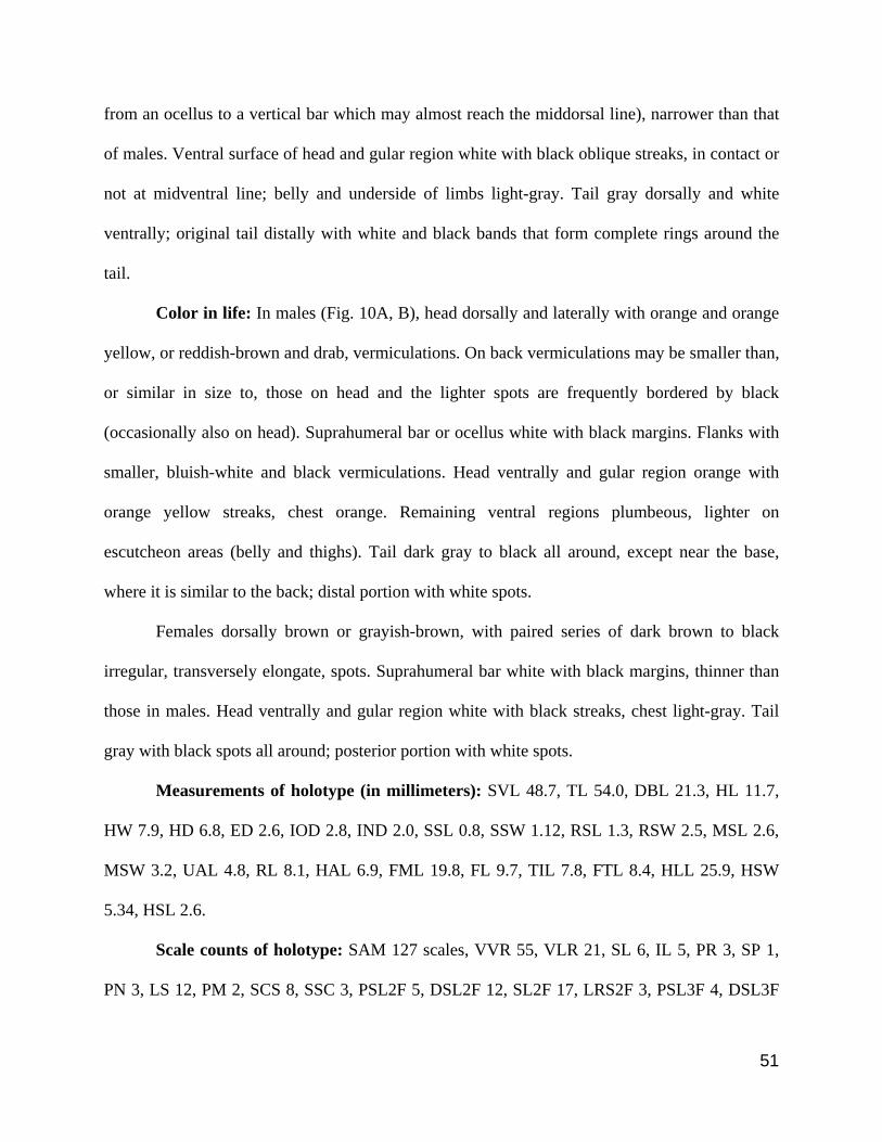
51
from an ocellus to a vertical bar which may almost reach the middorsal line), narrower than that
of males. Ventral surface of head and gular region white with black oblique streaks, in contact or
not at midventral line; belly and underside of limbs light-gray. Tail gray dorsally and white
ventrally; original tail distally with white and black bands that form complete rings around the
tail.
Color in life: In males (Fig. 10A, B), head dorsally and laterally with orange and orange
yellow, or reddish-brown and drab, vermiculations. On back vermiculations may be smaller than,
or similar in size to, those on head and the lighter spots are frequently bordered by black
(occasionally also on head). Suprahumeral bar or ocellus white with black margins. Flanks with
smaller, bluish-white and black vermiculations. Head ventrally and gular region orange with
orange yellow streaks, chest orange. Remaining ventral regions plumbeous, lighter on
escutcheon areas (belly and thighs). Tail dark gray to black all around, except near the base,
where it is similar to the back; distal portion with white spots.
Females dorsally brown or grayish-brown, with paired series of dark brown to black
irregular, transversely elongate, spots. Suprahumeral bar white with black margins, thinner than
those in males. Head ventrally and gular region white with black streaks, chest light-gray. Tail
gray with black spots all around; posterior portion with white spots.
Measurements of holotype (in millimeters): SVL 48.7, TL 54.0, DBL 21.3, HL 11.7,
HW 7.9, HD 6.8, ED 2.6, IOD 2.8, IND 2.0, SSL 0.8, SSW 1.12, RSL 1.3, RSW 2.5, MSL 2.6,
MSW 3.2, UAL 4.8, RL 8.1, HAL 6.9, FML 19.8, FL 9.7, TIL 7.8, FTL 8.4, HLL 25.9, HSW
5.34, HSL 2.6.
Scale counts of holotype: SAM 127 scales, VVR 55, VLR 21, SL 6, IL 5, PR 3, SP 1,
PN 3, LS 12, PM 2, SCS 8, SSC 3, PSL2F 5, DSL2F 12, SL2F 17, LRS2F 3, PSL3F 4, DSL3F

52
14, SL3F 19, LRS3F 4, PSL4F 6, DSL4F 13, SL4F 19, LRS4F 4, PSL2T 5, DSL2T 12, SL2T
17, LRS2T 4, PSL3T 5, DSL3T 15 , SL3T 20, LRS3T 3, PSL4T 10, DSL4T 13, SL4T 23,
LRS4T 3, RSE 4.
Variation: Body proportions and scale counts are given in Tables 6 and 7. The
suprahumeral spot varies among populations and, to a smaller degree, within populations. In the
only specimen from Amapá examined, the spot has the shape of an ocellus. Specimens from
Pará, north of the Amazon river, show a vertical bar reaching dorsally at least the dorsolateral
line, in some cases almost the middorsal line. South of the Amazon, all specimens from Monte
Verde present a short vertical bar (not reaching the dorsolateral line, but always with dorso-
ventral axis longer than anterior-posterior axis), while in the specimens from Anapu the vertical
bar is similar to those from northern Pará (although one specimen presents a vertical bar and a
small ocellus above the bar, on both sides).
Distribution: Eastern Brazilian Amazonia, in the states of Pará and Amapá. Up till now
it is only known south of the Amazon river in the interfluvium Xingu-Tocantins, Pará state, and
north of the Amazon river east of Paru river, in the states of Pará and Amapá (Fig. 6).
Habitat: All specimens were in terra firme, undisturbed or little-disturbed forest,
collected in pitfall traps, on the base of Sapopema trees and fissures in rock inside primary forest.
In the type locality a pair was collected in the fissure of a rock. At the type-locality specimens
from hid inside rock crevices or tree trunk holes when disturbed.

53
Pattern D
Gonatodes sp. nov. 2
Gonatodes concinnatus: Vanzolini, 1955: 126 (part); 1968: 26 (part); Mechler, 1968: 331 (part);
Peters & Donoso-Barros, 1970 (part); Rivero-Blanco, 1979: 94 (part); Sanchez, Castaño &
Cardenas 1995: 317; Bartlett & Bartlett 2003: 158 (part); Avila-Pires, 2005: 31.
(Fig. 1D, 2D, 11)
Holotype (number in bold between parentheses refer to localities in Fig. 6): MZUSP
49153 (field number FMEDEM 1465), adult male, from (5) Villavicencio, State of Meta,
Colombia (~4o09’S and 73o37’W), collected by W. W. Lamar and F. Medem, 22 October 1977.
Paratypes (numbers in bold between parentheses refer to localities in Fig. 6):
MZUSP 2145-46 (former AMNH 35292-93, respectively), two females, collected by N. Maria;
MZUSP 49152, an adult female, collected by M. Lugor and F. Medem, 10 October 1977;
MZUSP 49154 (field number FMEDEM 1471), an adult female, collected by W. W. Lamar and
F. Medem, October 1977, from (5) the type-locality. MZUSP 49155-62 (field number FMEDEM
1467), five adult females and three adult males, from (6) Finca “El Buque”, from Villavicencio,
Meta, Colombia (~4o08’19”N and 73o38’36”W); MZUSP 44777 (field number F 2544), an adult
female, from (7) Finca Guadualito, Güejar River, Meta, Colombia (~2055’N and 73o14’W),
collected by L. Klein and F. Medem, 7 September 1967.
Diagnosis: A relatively large Gonatodes, with maximum SLV of 45.8 mm. Scales around
midbody 105–123. Ventral scales in a longitudinal row 44–54. Proximal subdigital lamellae as
wide as digits, in total 16–21 under fourth finger, 18–24 under fourth toe. Two or three lateral
rows of scales on distal portion of fingers and toes. Tail with midventral scales distinctly wider
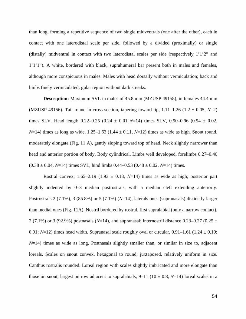
54
than long, forming a repetitive sequence of two single midventrals (one after the other), each in
contact with one laterodistal scale per side, followed by a divided (proximally) or single
(distally) midventral in contact with two laterodistal scales per side (respectively 1’1’2” and
1’1’1”). A white, bordered with black, suprahumeral bar present both in males and females,
although more conspicuous in males. Males with head dorsally without vermiculation; back and
limbs finely vermiculated; gular region without dark streaks.
Description: Maximum SVL in males of 45.8 mm (MZUSP 49158), in females 44.4 mm
(MZUSP 49156). Tail round in cross section, tapering toward tip, 1.11–1.26 (1.2 ± 0.05, N=2)
times SLV. Head length 0.22–0.25 (0.24 ± 0.01 N=14) times SLV, 0.90–0.96 (0.94 ± 0.02,
N=14) times as long as wide, 1.25–1.63 (1.44 ± 0.11, N=12) times as wide as high. Snout round,
moderately elongate (Fig. 11 A), gently sloping toward top of head. Neck slightly narrower than
head and anterior portion of body. Body cylindrical. Limbs well developed, forelimbs 0.27–0.40
(0.38 ± 0.04, N=14) times SVL, hind limbs 0.44–0.53 (0.48 ± 0.02, N=14) times.
Rostral convex, 1.65–2.19 (1.93 ± 0.13, N=14) times as wide as high; posterior part
slightly indented by 0–3 median postrostrals, with a median cleft extending anteriorly.
Postrostrals 2 (7.1%), 3 (85.8%) or 5 (7.1%) (N=14), laterals ones (supranasals) distinctly larger
than medial ones (Fig. 11A). Nostril bordered by rostral, first supralabial (only a narrow contact),
2 (7.1%) or 3 (92.9%) postnasals (N=14), and supranasal; internostril distance 0.23–0.27 (0.25 ±
0.01; N=12) times head width. Supranasal scale roughly oval or circular, 0.91–1.61 (1.24 ± 0.19;
N=14) times as wide as long. Postnasals slightly smaller than, or similar in size to, adjacent
loreals. Scales on snout convex, hexagonal to round, juxtaposed, relatively uniform in size.
Canthus rostralis rounded. Loreal region with scales slightly imbricated and more elongate than
those on snout, largest on row adjacent to supralabials; 9–11 (10 ± 0.8, N=14) loreal scales in a

55
line between postnasals and anterior corner of orbit. Top and posterior portion of head, as well as
supraorbital region, with granular scales. A short supraciliary flap present, anteriorly with 7–10
(8.4 ± 1.0, N=14) enlarged and flattened scales, among which 0–3 (2.1 ± 1.1; N=14) small,
conical spines. Pupil round, eye diameter 0.20–0.23 (0.22 ± 0.01, N=14) times head length;
interorbital distance 0.30–0.38 (0.35 ± 0.02, N=14) times head width. Scales on temporal region
similar to those on top of head. Ear-opening much smaller than eye, oval, posterior to, and at
same level of commissure of mouth. Supralabials 6–7 (6.2 ± 0.4; N=14) distinctly enlarged and
decreasing in size posteriorly, one or two posterior to centre of eye, followed to corner of mouth
by small scales (Fig. 11A).
Mental larger, distinctly wider anteriorly than posteriorly, with posterior margin forming
a moderate or wide angle, 1.20–1.51 (1.32 ± 0.09; N=14) times as wide as long; 2–3 (mostly
two) postmentals (Fig. 11B). Scales on chin juxtaposted, smooth, flat, polygonal, larger
anteriorly, decreasing in size posteriorly. Infralabials 4–6 (5.6 ± 0.6; N=14) distinctly enlarged
and decreasing in size posteriorly; one–two, occasionally three, of them posterior to centre of
eye, followed to corner of mouth by small scales.
Scales on nape small and granular, becoming slightly larger on sides of neck. Scales on
throat anteriorly like those on posterior part of chin; posteriorly flat, smooth, hexagonal or round,
imbricate, with a short transitional zone between the anterior and posterior parts.
Dorsals granular, increasing in size toward the flanks. Ventrals larger than dorsals,
roughly hexagonal, flat, smooth, imbricate, in oblique rows; 44–56 (50.4 ± 3.6; N=14) scales
along a midventral line between anterior margin of forelimbs and vent; 17–19 (17.7 ± 0.7; N=14)
scales in a transverse row at midbody. Scales around midbody 100–124 (113.5 ± 6.1; N=14),
with a short transitional zone between ventrals and scales on flanks. Scales on preanal plate

56
similar to ventrals, except for those bordering vent, which are very small. Escutcheon present in
males on posterior portion of belly and on 4–5 (mostly four) rows (body-knee direction) of
ventral surface the thighs.
Scales on anterodorsal surface of forelimbs flat, smooth, roundish, imbricate, largest
close to the wrist; on posterodorsal and ventral surfaces convex, smooth, rhomboid, juxtaposed,
relatively small; on ventral surface flat, smooth, rhomboid, subimbricate and smaller than
anterodorsally. Scales on anterodorsal and ventral surface of hind limbs flat, smooth, rhomboid,
imbricate; on posterodorsal surface convex, smooth, round, subimbricate, relatively small.
Numbers of lamellae under fingers and toes are presented in Table 7 (Fig. 11D). Claws exposed,
non-retractile, between two basal scales.
Scales on tail dorsally and laterally relatively small, rhomboid, flat, smooth, imbricate.
On ventral surface of tail scales smooth, flat, imbricate, increasing in size toward midventral
line; posteriorly midventral scales, except close to the base, distinctly wider than long, forming a
repetitive sequence of two single midventrals (one after the other), each in contact with one
laterodistal scale per side, followed by a divided (in the anterior portion of tail) or single (in
posterior portion of tail) midventral scale in contact with two laterodistal scales per side –
respectively 1’1’2” and 1’1’1” in the codification by Avila-Pires (1995: Figure 2) (Fig. 11E).
Color in preservative: In males, dorsal surface of head reddish-brown, spotless. Back
and flanks, base of tail and hind limbs with relatively small, beige and brown vermiculations. A
large, conspicuous, white suprahumeral bar with black margins. Ventral surface of head, gular
region and chest brown, without oblique streaks; belly dark-brown or dark-gray; underside of
limbs beige color. Tail dark brown dorsally and brown ventrally. Dorsal surface of tail dark
brown; ventral surface of tail brown. Escutcheon area (belly and thighs) light gray.

57
In females, dorsal surface of head and limbs light-brown or gray, with black irregular
spots; brown and black oblique and/or longitudinal bands may be present on sides of head. Back
gray with dorsolateral pairs of black spots; flanks gray with black and brown irregular spots. A
moderately large, conspicuous, white suprahumeral bar, with black margins, but less defined
than that on males; in some specimens it almost reaches the middorsal line. Ventral surface of
head and gular region white with dark oblique streaks, in contact or not at midventral line; belly
and underside of limbs white or beige. Dorsal surface of tail light-brown or brown with black
spots, ventral surface of original tail white, in some specimens with black small spots.
Measurements of holotype (in millimeters): SVL 42.5, TL 46.0 (distally regenerate),
DBL 17.8, HL 9.8, HW 6.7, HD 4.7, ED 2.0, IOD 2.1, IND 1.7, SSL 0.7, SSW 0.8, RSL 1.1,
RSW 2.1, MSL 2.2, MSW 2.6, UAL 5.0, RL 5.3, HAL 6.3, FML 11.5, FL 7.3, TIL 6.2, FTL 7.3,
HLL 20.8, HSW 2.8, HSL 1.57.
Scale counts of holotype: SAM 116 scales, VVR 51, VLR 17, SL 6, IL 6, PR 3, SP 1,
PN 3, LS 9, PM 2, SCS 8, SSC 2, PSL2F 6, DSL2F 11, SL2F 17, LRS2F 3, PSL3F 6, DSL3F
13, SL3F 19, LRS3F 3, PSL4F 7, DSL4F 11, SL4F 18, LRS4F 3, PSL2T 6, DSL2T 11, SL2T
17, LRS2T 3, PSL3T 6, DSL3T 13 , SL3T 19, LRS3T 3, PSL4T 11, DSL4T 12, SL4T 23,
LRS4T 3, RSE 4.
Variation: Measurements and scale counts are given in Tables 6 and 7. The
suprahumeral bar does not vary in extension between the specimens.
Distribution: All specimens studied are from Departamento Meta, Colombia. Sanchez et
al. (1995) reported G. concinnatus in Colombia from “Amazonas, Casanare, Boyacá,
Cundinamarca, Meta, Pasto, Orinoco, Guaviare”. It is necessary, however, to verify whether all
these records do refer to the species here described.

58
Remarks: Rivero-Blanco (1979) considered the specimens from Villavincencio and
vicinity, Meta, Colombia, as intergrades between Gonatodes concinnatus concinnatus, from Peru
and Ecuador, and G. c. ligiae, from Venezuela.
Species comparisons
The four species considered here under the term Gonatodes concinnatus complex are
easily distinguishable from the remaining 20 species of Gonatodes by the presence of a
moderately large, white suprahumeral spot with black margin, and dark and white vermiculations
on back, in males (vermiculations absent, however, in the nominal species Gonatodes ligiae
Donoso-Barros, from Venezuela, considered by Rivero-Blanco (1979) a subspecies of G.
concinnatus, not examined in this paper). A white antehumeral bar is present in G. ceciliae, G.
humeralis, and G. ocellatus. All remaining species present no similar bar or spot in the humeral
region and do not present vermiculations on back.
The species of the G. concinnatus complex differ moreover from G. alexandermendesi,
G. hasemani and G. infernalis by the absence of a very elongate spine on the supraciliary flap,
and from Gonatodes annularis, G. caudiscutatus, G. eladioi, G. infernalis and G. hasemani by
the subcaudal sequence (1’1’2” in the first species, midventrals not enlarged in G. hasemani,
1’1” in the remaining three species). From G. albogularis, G.antillensis, G. atricucullaris, G.
daudini, G. humeralis, G. petersi and G. vittatus this group differs moreover by having a higher
number (3–4) of lateral rows of scales on toes and fingers, except Gonatodes sp. nov. 2, which
has 2–3 lateral rows of scales like the seven above-mentioned species.

59
The species in the G. concinnatus complex reach a larger SVL than Gonatodes
albogularis, G. antillenisi, G. atricucullaris, G. caudiscutatus, G. daudini, G. eladioi, G.
falconensis, G. humeralis, G. petersi and G. vittatus, none of which exceed 42 mm in SVL.
Rivero-Blanco (1979) reported that the specimens of Gonatodes concinnatus from
Venezuela, which he considered as Gonatodes c. ligiae, have a white suprahumeral ocellus, and
the other populations (Peru and Ecuador) a vertical bar. The degree of elongation of this spot
(from a round ocellus to a bar almost reaching the middorsal line) seems to be to a certain extant
fixed within populations (either an ocellus, a short bar, or a median to long bar), but variable
among populations. Thus, all specimens from G. tapajonicus, known from only one locality,
have ocelli, while in Gonatodes sp. n. 1, an ocellus is present in the only specimen known from
Amapá, a short vertical bar occurs in all specimens from another locality, and specimens from
two other localities have all an elongate vertical bar. Specimens from Venezuela considered to be
G. concinnatus lack moreover vermiculations on the sides or dorsal surfaces of the body.
Considering that specimens from Colombia are here demonstrated to belong to a different
species, a large gap exists between G. c. concinnatus and G. c. ligiae. Taxonomic studies
including enough samples from Venezuela are therefore necessary to establish the status of these
populations.
Within the Gonatodes concinnatus complex, color pattern separates two groups – in G.
concinnatus and Gonatodes sp. nov. 2 the head has no vermiculation dorsally and no dark streaks
ventrally, while those are present in G. tapajonicus and Gonatodes sp. nov. 1. Gonatodes sp.
nov. 2 differs from G. tapajonicus and Gonatodes sp. nov. 1 in size (Table 6) and from all three
in number of lateral scales on distal part of digits (2–3 in Gonatodes sp. nov. 1 3–4 in the other
species). G. tapajonicus differs from Gonatodes sp.n. 1 by its yellow and brown color, while the

60
latter is orange and orange-yellow. Otherwise, body proportions (Table 6) and scales counts
(Table 7) are very similar between the species in the complex, even though the species were
clearly separated on basis of discriminant function analyses. Main characters pointed by the
discriminant analyses were proximal and distal lamellae under second toe, which separate
Gonatodes sp. nov. 1 from Gonatodes sp. nov. 2; proximal lamellae under fourth toe and
infralabials, which separate Gonatodes sp. nov. 2 from G. concinnatus and G. tapajonicus;
supranasal scale length and mental scale length, which separate G. concinnatus from Gonatodes
sp. nov. 2; and head width and lower arm length separate G. concinnatus and Gonatodes sp. nov.
2 from G. tapajonicus and Gonatodes sp. nov. 1.
Discussion
Male color pattern is an important character to distinguish species in Gonatodes, some of
which are very similar in external morphology and scale counts (Vanzolini 1968; Rivero-Blanco
1979, Rodrigues 1990). It is important however to take into consideration that polymorphism in
colour pattern is known in some species of Gonatodes – e.g. in Gonatodes annularis (Rivero-
Blanco 1968; 1979; Hoogmoed 1973) and G. hasemani (Avila-Pires 1995).
Considering the similarities, we assume that the species here considered as the Gonatodes
concinnatus complex form a monophyletic group, but this needs to be tested. Gamble et al.
(2008) pointed G. concinnatus (the only species of the complex they analysed) as the sister
species of G. humeralis. If our assumption of monophyly of the species here studied is correct,
G. concinnatus complex would have the same age – early Miocene – as G. humeralis. It is
interesting to observe that it and G. humeralis are also the most widespread Gonatodes species in
Amazonia. It is possible therefore that they have a similar, parallel history, even though, at least

61
concerning external morphology, divergence within G. concinnatus complex has been more
accentuated than in G. humeralis.
Gonatodes sp. nov. 1 seems to be restricted to eastern Amazonia, on both sides of the
Amazon River. Even though no other lizard is known with exactly the same distribution, there
are some examples of lizards that are present on both sides of the lower Amazon, e.g.
Arthrosaura kockii and Tretioscincus agilis (Avila-Pires 1995). Ayres & Clutton-Brock (1992)
have shown that for primates the lower Amazon acted less as a barrier than the middle course of
the river. The dynamics of island formation and river channel changes near the Amazon mouth
may have allowed such movements across the lower Amazon.
Gonatodes humeralis occurs in sympatry with many larger species of the genus, namely
G. annularis, G. concinnatus, G. hasemani, G. tapajonicus (Avila-Pires 1995; Dixon & Soini
1975; Rivero-Blanco 1979; Moravec et al. 2001) but there are no records of sympatry between
two larger species or any three species of Gonatodes. In our fieldwork in the Xingu River,
Gonatodes sp. nov. 1 and G. humeralis were found on the right margin of the river, while G.
hasemani and G. humeralis were found on the left margin The Xingu river seems therefore to
act as a barrier between G. hasemani and Gonatodes sp. nov. 1. On the other hand, north of the
Amazon the distribution of Gonatodes sp. nov.1 allows us to suppose a possible sympatry with
G. annularis, apart from that with G. humeralis, but this has yet to be verified. The exact
distribution of G. annularis and Gonatodes sp. nov. 1, whether they occur together or what
separates them, and in case they occur in syntopy how they interact, are at the moment open
questions.

62
Acknowledgments
Hussam Zaher (MZUSP), José P. Pombal-Jr and Ulisses Caramaschi (MNRJ), and Jucivaldo D.
Lima kindly loaned or allowed access to specimens under their care. Amanda Lima, Jerriane
Gomes, Jucivaldo Lima, Marco Antônio Ribeiro Jr. and Ulisses Galatti permitted examination of
specimens under study by them. Jerriane Gomes, Marco Antônio Ribeiro Jr., Pedro L. Peloso
and Toby A. Gardner provided photos and/or data from Gonatodes sp. nov. 1, Miguel T.
Rodrigues (USP) photos of Gonatodes tapajonicus, and Colin McCarthy (BMNH) photos and
important information on the syntypes of Goniodactylus concinnatus and Goniodactylus
buckleyi. Part of the material here studied was obtained during expeditions supported by the
Brazilian Council for the Development of Science/CNPq (process 473287/04-8), United
Kingdom’s Darwin Initiative, Grupo Orsa, Precious Woods Pará, and FIDESA/Belo Monte
project. MJS thanks Amanda Lima, Francílio Rodrigues, Pedro L. Peloso, Mariana Araguaia and
Reginaldo Rocha for their help and companionship during fieldwork. MJS was supported by a
scholarship from CNPq. Marinus Hoogmoed helped with literature access and patience.
Literature Cited
Avila-Pires, T.C.S. (1995) Lizards of Brazilian Amazonia (Reptilia: Squamata). Zoologische
Verhandelingen, 299, 1–706.
Avila Pires, T.C.S. (2005) Reptiles. In: Hollowell, T & Reynolds, R.P. (Eds.) Checklist of the
Terrestrial Vertebrates of the Guiana Shield. Bulletin of the Biological Society of Washington 13,
25–40.
Ayres, J.M. & Clutton-Brock, T.H. (1992) River boundaries and species range size in
Amazonian primates. The American Naturalist, 140, 531–567.

63
Bartlett, R.D. & Bartlett (2003) Reptiles and amphibians of the Amazon: an ecotourist’s guide.
University Press of Florida, Florida, 291 pp.
Barrio-Amoros, C.L. & Brewer-Carías, C. (2008) Herpetological results of the 2002 expedition
to Sarisariñama, a tepui inVenezuelan Guayana, with the description of five new species.
Zootaxa, 1942, 1–68.
Boulenger, G.A. (1885) Catalogue of the Lizards in the British Museum (Natural History.) Vol.
I. Geckonidae, Eublepharidae, Uroplatidae, Pygopodidae, Agamidae. Trustees of the British
Museum, London, 436 pp.
Burt, C.E. & Burt, M.D. (1933) A preliminary check list of the lizards of South America.
Transactions of the Academy of Science of St. Louis, 28, 1–104.
Cole, C.J. & Kok, P.J.R. (2006) A new species of gekkonid lizard (Sphaerodactylinae:
Gonatodes) from Guyana, South American. American Museum Novitates, 3524, 1–13.
Dixon, J.R. & Soini, P. (1975) The reptiles of the upper Amazon Basin, Iquitos Region, Peru: I.
Lizards and amphisbaenians. Contributions in Biology and Geology. 4, 1–58.
Donoso-Barros, R. (1967) Diagnosis de dos nuevas especies del género Gonatodes de
Venezuela. Noticiario Mensual Del Museo Nacional de Historia Natural de Santiago, 129, not
pagination.
Donoso-Barros, R. (1968) The Lizards of Venezuela (Check List and Key). Caribbean Journal
of Science, 8, 105–122.
Duellman, W.E. (1978) The biology of an Equatorial Herpetofauna in Amazonian Ecuador.
Miscellaneous Publication Museum of Natural History of University Kansas, 65, 1–352.

64
Duellman, W.E. & Mendelson III, J.R. (1995) Amphibians and reptiles from northern
Departamento Loreto, Peru: taxonomy and biogeography. The University of Kansas Science
Bulletin, 55, 329–376.
Esqueda, L.F. (2004) Una nueva especie de Gonatodes (Squamata: Gekkonidae) proveniente del
piedemonte cisandino de Venezuela. Herpetotropicos, 1, 32–39.
Gamble, T.; Simons, A. M.; Colli, G.R. & Vitt, L.J. (2008) Tertiary climate change and the
diversification of the amazonian gecko genus Gonatodes (Sphaerodactylidae, Squamata).
Molecular Phylogenetics and Evolution, 46, 269–277.
Hoogmoed, M.S. (1973) Notes on the herpetofauna of Surinam IV: The lizards and
amphisbaenians of Surinam. Biogeographica, 4, 1–419.
Krysko, K.L. & Daniels, K.J. (2005) A key to the geckos (Sauria:Gekkonidae) of Florida.
Caribbean Journal of Science, 41, 28–36.
Kluge, A.G. (1995) Cladistic relationships of Sphaerodactyl lizards. Americam Museum
Novitates, 3199, 1–23.
McGarigal, K., Cushman, S. & Stafford, S. G. (2000) Multivariate Statistics for Wildlife and
Ecology Research. Springer-Verlag, New York, 283 pp.
Mechler, B. (1968) Les geckonidés de la Colombie. Revue Suisse de Zoologie 75, 305–371.
Moravec, J., Tuanama, I.A. & Burgos, A.M. (2001) Reptiles recently recorded from the
surroundings of Iquitos (Departamento Loreto, Peru). Časopis Národního muzea, Řada
přírodovědná, 170, 47–68.
O'Shaughnessy, A.W.E. (1881) An account of the Collection of Lizards made by Mr. Buckley in
Ecuador, and now in the British Museum, with descriptions of the new species. Proceedings of
the Zoological Society of London, 1881, 227–245.

65
Peters, J.A. & Donoso-Barros, R. (1970) Catalogue of the Neotropical Squamata: part II. Lizards
and amphisbaenians. United States National Museum Bulletin, 297, 1–293.
Powell, R. & Henderson, R.W. (2005) A new species of Gonatodes (Squamata: Gekkonidae)
from the West Indies. Caribbean Journal of Science, 41, 709–715.
Reis, S.F.; Pessôa, L.M. & Strauss, R. (1990) Applicaion of size-free canonical discriminant
analysis to studies of geographic differentiation. Revista Brasileira de Genética, 13, 509–520.
Rivas, G.A. & Schargel, W.E. (2008) Gecko on the rocks: an enigmatic new species of
Gonatodes (Sphaerodactylidae) from Inselbergs of the Venezuelan Guayana. Zootaxa, 1925:39–
50.
Rivero-Blanco, C. (1968) Un genero y dos especies de tuqueques (Sauria: Sphaerodactylinae)
citados por primera vez para Venezuela, con notas sobre la distribucion de otras especies poco
conocidas. Memoria de La Sociedad de Ciencias Naturales la Salle, 27, 104–119.
Rivero-Blanco, C. (1979) The Neotropical lizard genus Gonatodes Fitzinger (Sauria:
Sphaerodactylinae). PhD Tese, Texas A&M University. 233 pp.
Rodrigues, M.T. (1980) Descrição de uma nova espécie de Gonatodes da Amazonia (Sauria,
Gekkonidae). Papéis Avulsos de Zoologia, 33, 309–314.
Sanchez, H.; Castaño, O & Cardenas, G. (1995) Diversidad de los reptiles en Colombia. In:
Rangel-Ch, J.O. (Ed.), Colombia Diversidad biotica I. Guadalupe, Santafé de Bogota, Colombia,
pp. 277–325.
Strauss, R.E. (1985) Evolutionary allometry and variation in body form in the South American
catfish genus Corydoras (Callichthyidae). Systematic Zoology, 34, 381–396.
Tabachnick, B.G. & Fidell, L.S. (2001) Using multivariate statistics. Allyn & Bacon, Boston,
Massachusetts, 966 pp.

66
Vanzonili, P.E. (1955) Sôbre Gonatodes varius (Auguste Duméril) com notas sôbre outras
espécies do gênero (Sauria, Gekkonidae). Papéis Avulsos de Zoologia, 12, 119–132.
Vanzolini, P.E. (1968) Lagartos brasileiros da família Gekkonidae (Sauria). Arquivos de
Zoologia, 17, 1–84.
Vanzolini, P.E. (1986) Addenda and corrigenda to the Catalogue of Neotropical Squamata.
Smithsonian Herpetological Information Service, 70: 1–25.
Wermuth, H. (1965) Liste der rezenten amphibian und reptilian: Gekkonidae, Pygopodidae,
Xantusiidae. Das Tierreich Lief, 89:1–246.

67
Appendix I – Additional material examined. Total number of specimens per species is in
parentheses.
Gonatodes albogularis (2): NICARAGUA (MNRJ 3164, 10670).
Gonatodes annularis (22): BRAZIL: Amapá: Mazagão, Cachoeira Inajá, Rio Camaipi (MPEG
2667), Oiapoque, km 90 of BR-156, Aldeia Tukai (MPEG 24439), Serra do Navio (MPEG
15080, 15087, 15100, 15148, 19592, 19627-31), Serra do Navio, Área Urucum (MPEG 19213);
Pará: Cachoeira Porteira, Trombetas River (MPEG 16264), Oriximiná, Cruz Alta, 6 km from
Trombetas River (MPEG 15396), Oriximiná, Porto Trombetas (MPEG 24198); FRENCH
GUIANA: Petit Saut, River Sinnamary (MPEG 15826, 15830, 15838, 15841-42); SURINAME:
(MPEG 21823).
Gonatodes caudiscutatus (19): ECUADOR: Guayas: Guayaquil (MZUSP 9181-90, 54370-72),
Balzar (MZUSP 9191); Los Rios: Palenque, Rio Palenque Science Center (RMNH 40149-153).
Gonatodes ceciliae (5): TRININDAD: Arima, Spring Hill Estate Arima Vallei (RMNH 10160),
Maracas, River ly Maracas (RMNH 14913); VENEZUELA: Sucre: San Juan de lãs Galdonas
(RMNH 40134-35; MZUSP 53553).
Gonatodes eladioi (1): BRAZIL: Pará: Marabá, Salobo, Serra dos Carajás (MPEG 18020).
Gonatodes hasemani (22): BRAZIL: Pará: Juruá, Rio Xingu (MZUSP 67240-57); Rondônia:
Porto Velho, Área de inundação da UHE de Samuel (MPEG 15569-71), Porto Velho, Rio
Yamary, Cachoeira do Samuel, Território do Guaporé (MNRJ 10678).
Gonatodes humeralis (16): BRAZIL: Pará: Piçarra (MNRJ 16107), Tucuruí, Lago da UHE
Tucuruí (MPEG 21881-87). PERU: Loreto: Moropón (MZUSP 28280-87).
Gonatodes taniae (1): VENEZUELA: Aragua: Rancho Grande (MZUSP 57575).

68
69
Figure Legends
Figure 1. Dorsal color patterns in preservative in Gonatodes concinnatus complex. (A) pattern A
(MZUSP 54655); (B) pattern B (MZUSP 53674, holotype); (C) pattern C (MPEG 25596) and
(D) pattern D (MZUSP 49153). Scale bar = 5 mm.
Figure 2. Ventral color pattern in preservative in Gonatodes concinnatus complex. (A) pattern A
(MZUSP 54655); (B) pattern B (MZUSP 53674, holotype); (C) pattern C (MPEG 25596) and
(D) pattern D (MZUSP 49153). Scale bar = 5 mm.
Figure 3. Bivariate scattergrams from stepwise discriminant function analysis (A) using scale
counts and (B) measurements. Gray triangle = Pattern A. Black circles = Pattern B. Black
squares = Pattern C. Gray circles = Pattern D.
Figure 4. Gonatodes concinnatus (MZUSP 54655). (A) Dorsal and (B) ventral views of head;
ventral views of (C) right hand and (D) right foot; (E) ventral view of tail. Scale bar = 5 mm.
Figure 5. Gonatodes concinnatus (syntypes, BMNH 1946.9.7.10-12). (A) Dorsal, (B) ventral
views. Scale bar = 5 mm.
Figure 6. Geographic distribution of Gonatodes concinnatus complex. Circles = Gonatodes
concinnatus. Star = Gonatodes tapajonicus. Squares = Gonatodes sp. nov. 1. Triangles =
Gonatodes sp. nov. 2. Numbers refer to the localities cited in the text.
Figure 7. Gonatodes tapajonicus (paratype, MZUSP 53674). (A) Dorsal and (B) ventral views
of head; ventral views of (C) right hand and (D) right foot; (E) ventral view of tail. Scale bar = 5
mm.

70
Figure 8. Gonatodes tapajonicus from type-locality, Cachoeira do Limão, Pará, Brazil. (A)
Adult male (holotype, MZUSP 53676). SVL = 53 mm and (B) Throat color pattern of male
(holotype). (Photos by Miguel Rodrigues)
Figure 9. Gonatodes sp. nov. 1 (holotype, MPEG 25596). (A) Dorsal and (B) ventral views of
head; ventral views of (C) right hand and (D) right foot; (E) ventral view of tail; detail of (F)
dorsal scales of trunk and (G) tail. Scale bar = 5 mm.
Figure 10. Gonatodes sp. nov. 1 from Anapu, Rio Xingu, Pará, Brazil. (A) Adult male (paratype,
MPEG 25598, SVL = 46.7 mm), (B) female in life (paratype, MPEG 25599, SVL = 39.7 mm)
and (C) Throat color pattern of male (paratype, MPEG 25598). (Photos by Pedro Peloso)
Figure 11. Gonatodes sp. nov. 2 (holotype, MZUSP 49153). (A) Dorsal and (B) ventral views of
head; ventral views of (C) right hand and (D) right foot; (E) ventral view of tail; detail of (F)
dorsal scales of trunk and (G) tail. Scale bar = 5 mm.

71
Tables
Table 1. Comparative data among color patterns. Patterns Throat Head dorsally Back and limbs
A Beige, immaculate Beige without vermiculations
Brown and beige vermiculations
B White with black oblique stripes
(in life, vivid yellow with reddish-brown oblique stripes)
Brown and beige vermiculations (in life,
vivid yellow and reddish-brown vermiculations)
Brown and beige vermiculations (in life,
vivid yellow and reddish-brown vermiculations
C
White or dark-brown with black and white oblique stripes (in life,
orange with orange yellow oblique stripes)
Dark-brown and beige vermiculations (in life,
orange with orange yellow vermiculations)
Dark-brown and beige vermiculations (in life,
orange with orange yellow vermiculations)
D Brown, immaculate Beige without vermiculations
Brown and beige small vermiculations

72
Table 2. Results of stepwise discriminant function analysis of scale counts comparing the four
groups identified on basis of color pattern. 1DF: loadings on the first principal component; 2DF
loadings on the second principal component.
F(+ent,-rem) Wilks's Lambda Approx. F-ratio p-value 1DF 2DF
DSL2T 44.679 0.33 44.679 0 0.65 -0.008
PSL4T 36.711 0.122 40.249 0 -0.057 -0.696
IL 13.583 0.075 34.527 0 0.42 -0.441
PSL2F 9.406 0.052 28.557 0 -0.669 -0.2
VLR 4.36 0.043 25.084 0 0.473 0.056
SL 3.939 0.036 21.598 0 0.072 -0.001
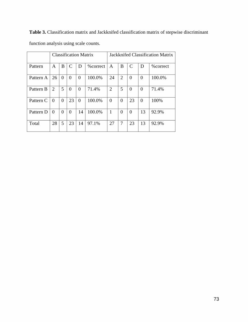
73
Table 3. Classification matrix and Jackknifed classification matrix of stepwise discriminant
function analysis using scale counts.
Classification Matrix Jackknifed Classification Matrix
Pattern A B C D %correct A B C D %correct
Pattern A 26 0 0 0 100.0% 24 2 0 0 100.0%
Pattern B 2 5 0 0 71.4% 2 5 0 0 71.4%
Pattern C 0 0 23 0 100.0% 0 0 23 0 100%
Pattern D 0 0 0 14 100.0% 1 0 0 13 92.9%
Total 28 5 23 14 97.1% 27 7 23 13 92.9%

74
Table 4. Results of stepwise discriminant function analysis of measurements comparing the four
groups identified on basis of color pattern. 1DF: loadings on the first principal component; 2DF
loadings on the second principal component.
F(+ent,-rem) Wilks's Lambda Approx. F-ratio p-value 1DF 2DF
MSL 33.213 0.398 33.213 0 -0.561 0.198
HW 18.501 0.215 25.068 0 0.214 0.869
RL 14.107 0.129 23.599 0 0.455 0.579
SSL 8.252 0.093 20.13 0 0.63 0.162
RSW 7.684 0.068 19.317 0 -0.525 0.34
IOD 6.796 0.051 17.962 0 0.528 -0.201
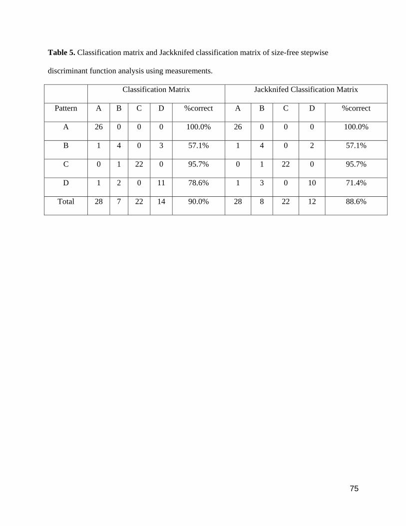
75
Table 5. Classification matrix and Jackknifed classification matrix of size-free stepwise
discriminant function analysis using measurements.
Classification Matrix Jackknifed Classification Matrix
Pattern A B C D %correct A B C D %correct
A 26 0 0 0 100.0% 26 0 0 0 100.0%
B 1 4 0 3 57.1% 1 4 0 2 57.1%
C 0 1 22 0 95.7% 0 1 22 0 95.7%
D 1 2 0 11 78.6% 1 3 0 10 71.4%
Total 28 7 22 14 90.0% 28 8 22 12 88.6%

76
Table 6. Snout-vent length and body proportions of Gonatodes concinnatus, Gonatodes tapajonicus, Gonatodes sp. nov. 1 and
Gonatodes sp. nov. 2. Abbreviations are listed in Material and Methods. sd=standard deviation; N=number of specimens. Tail length
of specimens with broken or regenerated tail was excluded.
Measurement or body proportion
Gonatodes concinnatus Gonatodes tapajonicus Gonatodessp. nov. 1 Gonatodes sp. nov. 2 Range Mean sd N Range Mean sd N Range Mean sd N Range Mean sd N
SVL 25.9–48.3 41.60 6.70 26 37.8–51.4 46.10 4.80 7 22.3–55.7 45.00 9.50 23 34.1–45.8 40.90 3.10 14 TL/SVL 1.07–1.16 1.16 0.07 8 1.21–1.23 1.22 0.02 2 1.11–1.26 1.20 0.05 8 1.06–1.32 1.19 0.18 2 HL/SVL 0.23–0.27 0.24 0.01 26 0.22–0.25 0.23 0.10 7 0.21–0.28 0.24 0.02 23 0.22–0.25 0.24 0.01 14 HL/HW 1.30–1.56 1.44 0.06 26 1.45–1.58 1.50 0.06 7 1.21–1.56 1.41 0.08 23 1.44–1.58 1.44 0.05 12 HW/HD 1.31–1.66 1.43 0.10 26 1.28–1.44 1.36 0.05 7 1.17–1.54 1.40 0.10 23 1.25–1.63 1.44 0.11 14 ED/HL 0.19–0.24 0.21 0.10 26 0.21–0.25 0.23 0.01 7 0.20–0.27 0.22 0.02 23 0.20–0.23 0.22 0.01 14
IOD/HW 0.25–0.35 0.31 0.02 26 0.34–0.42 0.38 0.03 7 0.22–0.31 0.25 0.02 23 0.30–0.38 0.35 0.02 14 IND/HW 0.20–0.28 2.10 0.30 25 0.21–0.27 0.25 0.02 7 0.14–0.21 0.17 0.01 23 0.23–0.27 0.25 0.01 14 SSW/SSL 0.82–2.34 1.21 0.28 26 0.99–1.40 1.19 0.16 7 0.88–1.44 1.13 0.13 23 0.91–1.61 1.24 0.19 14
RSW/RSW 1.89–2.35 2.15 0.14 26 1.86–2.21 2.02 0.15 7 1.78-3.01 2.02 0.28 23 1.65–2.19 1.93 0.13 14 MSW/MSL 1.02–1.25 1.13 0.07 26 1.07–1.39 1.21 0.11 7 1.17–1.41 1.28 0.07 23 1.20–1.51 1.32 0.09 14
FL/SLV 0.36–0.42 0.40 0.01 26 0.37–0.44 0.40 0.03 7 0.38–0.44 0.41 0.02 23 0.27–0.40 0.38 0.04 14 HLL/SLV 0.47–0.56 0.51 0.01 26 0.45–0.54 0.51 0.04 7 11.7–28.6 22.70 4.50 23 16.7–21.8 19.70 1.40 14

77
Table 7. Scale counts of specimens of Gonatodes concinnatus, Gonatodes tapajonicus, Gonatodes sp. nov. 1 and Gonatodes sp. nov.
2. Abbreviations are listed in Material and Methods. sd=standard deviation; N=number of specimens.
Scales counts Gonatodes concinnatus Gonatodes tapajonicus Gonatodes sp. nov. 1 Gonatodes sp. nov. 2 Range Mean sd N Range Mean sd N Range Mean sd N Range Mean sd N
SAM 109–128 120.6 5.3 25 120–126 122.8 2.3 6 118–143 130.0 6.5 21 100–124 113.5 6.1 14 VVR 49–58 55.0 2.3 25 53–59 55.8 2.2 6 52–60 56.2 2.5 21 44–56 50.4 3.6 14 VLR 17–20 18.5 1.0 25 19–20 19.5 0.5 6 18–24 21.2 1.3 21 17–19 17.7 0.7 14 SL 6–7 6.3 0.5 26 6–8 7.0 0.8 7 5–7 6.2 0.5 23 6–7 6.2 0.4 14 IL 5–7 6.0 0.8 26 5–7 6.1 0.7 7 5–8 6.3 0.9 23 4–6 5.6 0.6 14 PR 3–5 3.5 0.8 26 3–5 3.3 0.8 7 2–4 3.0 0.4 23 2–5 3.1 0.6 14 SP 1–3 1.5 0.8 26 1–3 1.3 0.8 7 0–2 1.0 0.4 23 0–3 1.1 0.6 14 SIS 4–6 5.0 0.4 26 5–7 6.1 0.7 7 4–8 6.0 0.8 23 4–7 5.1 0.8 14 PN 3–4 3.1 0.3 26 3–4 3.3 0.5 7 3 3.0 0.0 23 2–3 2.9 0.3 14 LS 9–13 10.7 0.9 26 10–13 11.1 0.9 7 10–13 11.2 0.9 23 9–11 10.0 0.8 14 PM 2–4 2.3 0.6 26 2–3 2.3 0.5 7 2–3 2.0 0.2 23 2–3 2.3 0.5 14
CM1IL 5–8 6.8 0.9 26 6–8 6.7 0.8 7 6–8 7.0 0.8 23 6–10 7.7 1.3 14 SCS 6–10 8.3 1.1 26 7–10 8.1 1.1 7 7–12 9.4 1.4 23 7–10 8.4 1.0 14 SSC 0–4 1.7 1.5 26 2–4 2.9 0.7 7 0–3 1.1 1.2 23 0–3 2.1 1.1 14
PSL2F 5–7 6.0 0.3 26 6 6.0 0.0 7 4–6 5.1 0.4 23 5–6 5.7 0.5 14 DSL2F 8–12 1.2 1.1 26 10–12 10.3 0.8 7 10–13 11.8 0.9 23 7–11 8.9 1.0 14 SL2F 14–18 16.2 1.2 26 16–18 16.3 0.8 7 15–19 16.9 1.1 23 13–17 14.6 1.1 14
LRS2F 3–4 3.1 0.3 26 3–4 3.1 0.4 7 3–4 3.1 0.3 23 2–3 2.9 0.3 14 PSL3F 6–7 6.3 0.5 26 6–7 6.1 0.4 7 5–6 5.4 0.5 23 5–6 5.9 0.3 14 DSL3F 11–14 12.1 0.9 26 12–14 13.1 0.7 7 13–16 14.3 1.0 23 9–13 11.1 1.2 14 SL3F 17–20 18.6 1.1 26 18–20 19.3 0.8 7 18–22 19.7 1.3 23 15–19 17.1 1.2 14
LRS3F 3–4 3.0 0.2 26 3–4 3.1 0.4 7 3–4 3.2 0.4 23 2–3 2.8 0.4 14 PSL4F 7–9 7.7 0.6 26 7–8 7.4 0.5 7 6–8 6.6 0.7 23 6–8 7.0 0.7 14 DSL4F 10–14 12.1 0.9 26 11–14 12.9 1.1 7 12–16 13.9 1.0 23 9–13 10.7 1.1 14 SL4F 17–22 19.7 1.1 26 18–22 20.3 1.3 7 18–23 20.5 1.2 23 16–21 17.7 1.5 14
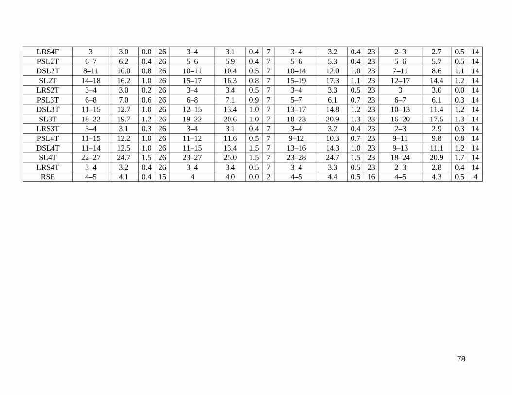
78
LRS4F 3 3.0 0.0 26 3–4 3.1 0.4 7 3–4 3.2 0.4 23 2–3 2.7 0.5 14 PSL2T 6–7 6.2 0.4 26 5–6 5.9 0.4 7 5–6 5.3 0.4 23 5–6 5.7 0.5 14 DSL2T 8–11 10.0 0.8 26 10–11 10.4 0.5 7 10–14 12.0 1.0 23 7–11 8.6 1.1 14 SL2T 14–18 16.2 1.0 26 15–17 16.3 0.8 7 15–19 17.3 1.1 23 12–17 14.4 1.2 14
LRS2T 3–4 3.0 0.2 26 3–4 3.4 0.5 7 3–4 3.3 0.5 23 3 3.0 0.0 14 PSL3T 6–8 7.0 0.6 26 6–8 7.1 0.9 7 5–7 6.1 0.7 23 6–7 6.1 0.3 14 DSL3T 11–15 12.7 1.0 26 12–15 13.4 1.0 7 13–17 14.8 1.2 23 10–13 11.4 1.2 14 SL3T 18–22 19.7 1.2 26 19–22 20.6 1.0 7 18–23 20.9 1.3 23 16–20 17.5 1.3 14
LRS3T 3–4 3.1 0.3 26 3–4 3.1 0.4 7 3–4 3.2 0.4 23 2–3 2.9 0.3 14 PSL4T 11–15 12.2 1.0 26 11–12 11.6 0.5 7 9–12 10.3 0.7 23 9–11 9.8 0.8 14 DSL4T 11–14 12.5 1.0 26 11–15 13.4 1.5 7 13–16 14.3 1.0 23 9–13 11.1 1.2 14 SL4T 22–27 24.7 1.5 26 23–27 25.0 1.5 7 23–28 24.7 1.5 23 18–24 20.9 1.7 14
LRS4T 3–4 3.2 0.4 26 3–4 3.4 0.5 7 3–4 3.3 0.5 23 2–3 2.8 0.4 14 RSE 4–5 4.1 0.4 15 4 4.0 0.0 2 4–5 4.4 0.5 16 4–5 4.3 0.5 4

79
Figure 1. Dorsal color patterns in preservative in Gonatodes concinnatus complex. (A) pattern A
(MZUSP 54655); (B) pattern B (MZUSP 53674, holotype); (C) pattern C (MPEG 25596) and
(D) pattern D (MZUSP 49153). Scale bar = 5 mm.

80
Figure 2. Ventral color pattern in preservative in Gonatodes concinnatus complex. (A) pattern A
(MZUSP 54655); (B) pattern B (MZUSP 53674, holotype); (C) pattern C (MPEG 25596) and
(D) pattern D (MZUSP 49153). Scale bar = 5 mm.
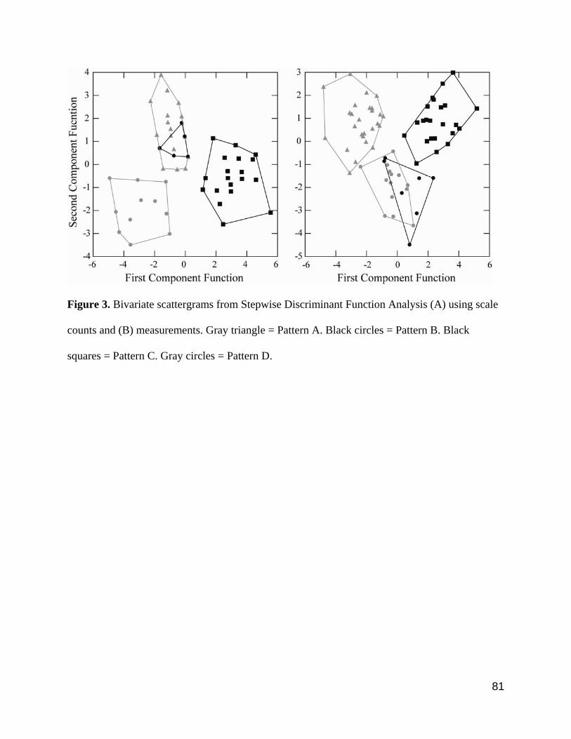
81
Figure 3. Bivariate scattergrams from Stepwise Discriminant Function Analysis (A) using scale
counts and (B) measurements. Gray triangle = Pattern A. Black circles = Pattern B. Black
squares = Pattern C. Gray circles = Pattern D.

82
Figure 4. Gonatodes concinnatus (MZUSP 54655). (A) Dorsal and (B) ventral views of head;
ventral views of (C) right hand and (D) right foot; (E) ventral view of tail. Scale bar = 5 mm.

83
Figure 5. Gonatodes concinnatus (syntypes, BMNH 1946.9.7.10-12). (A) Dorsal, (B) ventral
views. Scale bar = 5 mm.
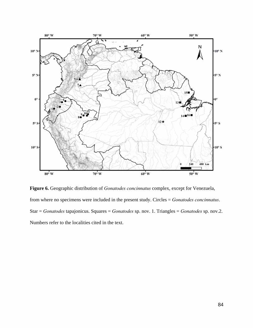
84
Figure 6. Geographic distribution of Gonatodes concinnatus complex, except for Venezuela,
from where no specimens were included in the present study. Circles = Gonatodes concinnatus.
Star = Gonatodes tapajonicus. Squares = Gonatodes sp. nov. 1. Triangles = Gonatodes sp. nov.2.
Numbers refer to the localities cited in the text.

85
Figure 7. Gonatodes tapajonicus (paratype, MZUSP 53674). (A) Dorsal and (B) ventral views
of head; ventral views of (C) right hand and (D) right foot; (E) ventral view of tail. Scale bar = 5
mm.
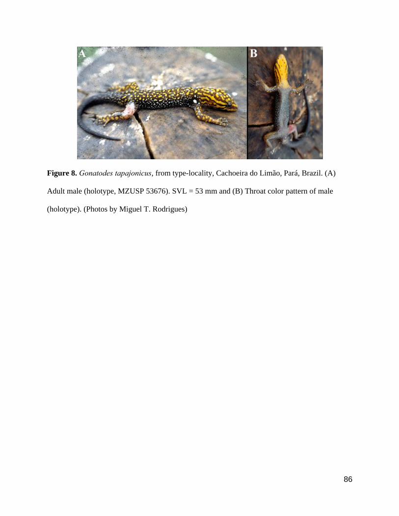
86
Figure 8. Gonatodes tapajonicus, from type-locality, Cachoeira do Limão, Pará, Brazil. (A)
Adult male (holotype, MZUSP 53676). SVL = 53 mm and (B) Throat color pattern of male
(holotype). (Photos by Miguel T. Rodrigues)
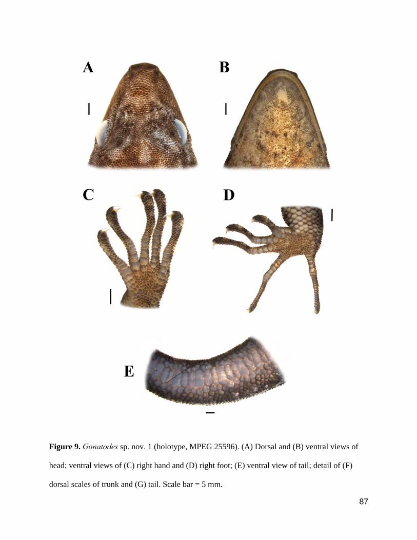
87
Figure 9. Gonatodes sp. nov. 1 (holotype, MPEG 25596). (A) Dorsal and (B) ventral views of
head; ventral views of (C) right hand and (D) right foot; (E) ventral view of tail; detail of (F)
dorsal scales of trunk and (G) tail. Scale bar = 5 mm.

88
FIGURE 10. Gonatodes sp. nov. 1 from Anapu, Rio Xingu, Pará, Brazil. (A) Adult male
(paratype, MPEG 25598, SVL = 46.7 mm), (B) Throat color pattern of male (paratype, MPEG
25598), (C) female in life (paratype, MPEG 25599, SVL = 39.7 mm) and (D) throat color pattern
of female (paratype, MPEG 25599) (Photos by Pedro L. V. Peloso).

89
Figure 11. Gonatodes sp. nov. 2 (holotype, MZUSP 49153). (A) Dorsal and (B) ventral views of
head; ventral views of (C) right hand and (D) right foot; (E) ventral view of tail; detail of (F)
dorsal scales of trunk and (G) tail. Scale bar = 5 mm.