Embed Size (px)
Citation preview
1
JOANA RAMOS-JORGE
SINAIS E SINTOMAS ASSOCIADOS COM A ERUPÇÃO
DE DENTES DECÍDUOS: ESTUDO LONGITUDINAL
BELO HORIZONTE
2010
2
JOANA RAMOS-JORGE
SINAIS E SINTOMAS ASSOCIADOS COM A ERUPÇÃO
DE DENTES DECÍDUOS: ESTUDO LONGITUDINAL
Dissertação apresentada ao Programa de Pós-
Graduação da Faculdade de Odontologia da
Universidade Federal de Minas Gerais como
requisito parcial para obtenção do grau de Mestre
em Odontologia - área de concentração em
Odontopediatria
Orientador: Prof. Saul Martins de Paiva
Co-orientadora: Prof.ª Isabela Almeida Pordeus
Faculdade de Odontologia
Universidade Federal de Minas Gerais
Belo Horizonte
2010
3
4
5
AGRADECIMENTOS
Ao Professor Dr. Saul Martins de Paiva, meu exemplo de pesquisador, pela
maturidade, equilíbrio e olhar crítico durante minha orientação. Por acreditar em mim,
ter-me ensinado tanto, por ter-me dado lições de disciplina, responsabilidade e
competência.
À Professora Dra. Isabela Almeida Pordeus, pela enorme dedicação ao Programa de
Pós-Graduação e pelos valiosos ensinamentos, fundamentais para minha formação.
À Professora Dra. Maria Letícia Ramos Jorge, minha grande incentivadora, pela
colaboração na coleta de dados, pela análise estatística do estudo e por ter sido capaz
de despertar em mim um amor imenso pela Odontologia e pela pesquisa.
À Professora Dra. Miriam Pimenta Parreira do Vale, pelos momentos de aprendizado,
pelas lições de vida e pela dedicação ao Mestrado em Odontopediatria.
Às Professoras Dra. Ana Cristina Borges de Oliveira e Dra. Patrícia Zarzar, pelo
carinho com que me receberam.
Aos Professores do Departamento de Ortodontia e Odontopediatria, em especial Dra.
Júnia Maria Cheib Serra-Negra, Dra. Sheyla Márcia Auad e Dra. Laura Helena
Pereira Machado Martins, pelos momentos de aprendizagem.
À Professora Dra. Efigênia Ferreira e Ferreira, com quem tive um grande ganho de
conhecimento.
Ao Professor Dr. Mauro Henrique Nogueira Guimarães de Abreu, pelos ensinamentos
e pela disponibilidade em participar de minha banca examinadora.
6
Ao Professor Dr. Luciano José Pereira, por participar da minha banca examinadora.
Às minhas queridas colegas de Mestrado, Anita, Andréa e Kelly. Foi perfeito ter vocês
por perto durante esse tempo!
Aos colegas de Mestrado, Maurício, Patrícia, Thiago, e, de Doutorado, Cíntia, Milene,
Cristiane, Fernanda, Claudinha e Camila.
À Laís, Beth e Zuleica, secretárias da Pós-Graduação, pela disponibilidade para
ajudar sempre.
À Dra. Carolina Tângari, coordenadora de Saúde Bucal do município de Diamantina,
pela atenção e ajuda na busca dos bebês.
Aos bebês e seus pais/responsáveis que foram tão disponíveis e fundamentais para a
realização deste estudo.
Às alunas de iniciação científica da Universidade Federal dos Vales do Jequitinhonha e
Mucuri, Izabella Fernandes, Laís Almeida, Isabela Veloso, Nayara Gabriela Vilarino
Silva, Lílian Capanema, Nathália Resende e Valdirene Souza e Silva, e aos mestrandos
Patrícia Faria e Paulo Antônio Martins Júnior, por me ajudarem na coleta com tanto
empenho. Muito obrigada!
7
AGRADECIMENTOS AFETIVOS
Ao meu pai, José Arnaldo, meu exemplo de integridade, por estar sempre presente,
apoiando minhas escolhas, me acompanhando sempre.
À minha mãe, Celídia, que, mesmo ausente fisicamente, está presente em cada
momento, como meu modelo humano para a vida.
Ao meu namorado, Thiago Motta, por estar sempre do meu lado, me incentivando,
contribuindo para o meu crescimento profissional, para a realização de meus objetivos
e meus ideais.
Às minhas irmãs, Ana Carolina, Letícia e Florinda, por me apoiarem sempre e
preencherem minha vida com tanto afeto.
Aos meus cunhados Fernando, Leandro e Rubens, pelo carinho, pela amizade e por me
estimularem em busca de meus objetivos.
Aos meus sobrinhos, Clara, Artur, Sofia, Pedro, Tomás e Lídia, maiores razões de
minha alegria!
A Lecy, por me apoiar com tanta afeição.
Às minhas amigas, Aninha, Tati, Sú e Manu, por torcerem sempre por mim com muito
carinho.
Aos meus queridos professores de graduação Dr. Gustavo Eustáquio Brito Alvim de
Melo e Dra. Tania Regina Riul, obrigada pelos ensinamentos e pelo incentivo durante o
meu curso. Vocês foram fundamentais para minha realização profissional!

8
RESUMO
9
Sinais e sintomas associados com a erupção de dentes decíduos: estudo longitudinal
RESUMO
A associação entre erupção de dentes decíduos e a manifestação de sinais e sintomas em
bebês é tema controverso na literatura. Ainda não há evidência científica que suporte
essa associação, entretanto pais e profissionais de saúde continuam acreditando que a
erupção de dentes decíduos é causa da manifestação de sinais e sintomas em bebês.
Assim, este estudo teve como objetivo avaliar a associação entre erupção de dentes
decíduos e manifestação de sinais e sintomas em bebês. Foi realizado um estudo
longitudinal com 47 bebês, que ainda não apresentavam todos os incisivos erupcionados
e sem história de doenças crônicas ou distúrbios capazes de provocar aumento dos
sinais e sintomas avaliados. Aferições das temperaturas timpânica e axilar e exame
clínico bucal foram realizados diariamente, ao longo de um período de oito meses. Por
meio de entrevista às mães, foi investigada a presença de 14 sinais e sintomas
associados com a erupção de dentes decíduos e apresentados pelos bebês nas últimas 24
horas. A erupção de dentes decíduos esteve associada à elevação da temperatura
timpânica no dia da erupção (P=0.004) bem como à ocorrência de outros sinais e
sintomas. Os sinais e sintomas mais frequentemente associados à erupção dos dentes
decíduos foram irritabilidade (P<0.001), aumento da salivação (P<0.001), coriza
(P<0.001) e perda de apetite (P<0.001). Além desses, diarréia, brotoeja e distúrbios de
sono também estiveram associados à erupção dos dentes decíduos. Portanto, este estudo
contribui para fortalecer o conceito de que não se pode atribuir à erupção de dentes
decíduos a ocorrência de sinais e sintomas graves, como febre.

10
ABSTRACT
11
Signs and symptoms associated with the primary teeth eruption: longitudinal
study
ABSTRACT
The association between eruption of primary teeth and manifestation of signs and
symptoms in infants is a controversial issue in literature. There is still no scientific
evidence that supports this association. However, parents and health professionals
continue to believe that eruption of primary teeth is because of the manifestion of signs
and symptoms in infants. Thus, the aim of this study was Assess the association
between primary tooth eruption and the manifestation of signs and symptoms in infants.
A longitudinal study was carried out with 47 non-institutionalized infants between five
and 15 months of age in the city of Diamantina, Brazil. The non-randomized
convenience sample was based on a registry of infants provided by the Municipal
Secretary of Health. Eligible participants were infants with between zero and seven
erupted teeth and no history of chronic disease or disorders that could cause an increase
in the symptoms assessed in the study. Tympanic and axillary temperature readings and
clinical oral exams were performed daily. A daily interview with the mothers was
carried to investigate the occurrence of 14 signs and symptoms associated to teething
presented by the infants in the previous 24 hours. Teething was associated to a rise in
tympanic temperature on the day of the eruption (P=0.004) as well as the occurrence of
other signs and symptoms. Maximal tympanic and axillary temperature was 36.8 ºC and
36.6 ºC, respectively. The most frequent sign and symptoms associated to teething were
irritability (P<0.001), increased salivation (P<0.001), runny nose (P<0.001) and loss of
appetite (P<0.001). Signs and symptoms, such as irritability, increased salivation, runny
nose, loss of appetite, diarrhea, rash and sleep disturbance, were associated to primary
12
tooth eruption. The present study supports the concept that the occurrence of severe
signs and symptoms, such as fever, is not attributed to teething.
13
LISTA DE ABREVIATURAS
BD Becton Dickinson
SPSS Statistical Package for Social Sciences
UFMG Universidade Federal de Minas Gerais
USA United States of America
Incoterm Indústria de Termômetros
CNPq Conselho Nacional de Desenvolvimento Científico e Tecnológico
FAPEMIG Fundação de Amparo à Pesquisa do Estado de Minas Gerais
COEP Comitê de Ética em Pesquisa
14
LISTA DE TABELAS
Tabela1: Análise descritiva das características dos bebês, de suas mães e do
número de dias de coleta dos dados.........................................................................
31
Tabela 2: Análise descritiva e comparação das temperaturas timpânica e axilar em
momentos de não erupção, um dia antes, no dia e um dia após a erupção de dentes..........
31
Tabela 3: Análise descritiva e comparação dos sinais e sintomas em momentos de não
erupção, um dia antes, no dia e um dia após a erupção de dentes........................................
32
15
SUMÁRIO
1 CONSIDERAÇÕES INICIAIS.............................................................................. 15
2 ARTIGO: Signs and symptoms associated with the primary teeth eruption:
longitudinal study…………………………………………………………………….
19
2.1 Abstract………………………………………………………………………...
2.2 Introduction……………………………………………………………………
2.3 Methods……………………………………………………………………….
2.4 Results…………………………………………………………………………
2.5 Discussion………………………………………………………………............
2.6 Conclusion…………………………………………………………………….
2.7 Acknowledgements…………………………………………………………….
2.8 References………………………………………………………………...........
21
22
23
25
26
29
29
29
3 CONSIDERAÇÕES FINAIS…………………………………………………… 33
4 REFERÊNCIAS GERAIS………………………………………………………. 35
5 APÊNDICES……………………………………………………………………… 38
5.1 Apêndice A – Carta de Apresentação..............................................................
5.2 Apêndice B – Termo de Consentimento Livre e Esclarecido........................
5.3 Apêndice C – Formulário 1 – Identificação.....................................................
39
41
42
16
5.4 Apêndice D – Formulário 2 – Exame de temperatura auditiva.....................
5.5 Apêndice E – Formulário 3 – Exame de temperatura axilar.........................
5.6 Apêndice F – Formulário 4 – Avaliação dos sinais e sintomas......................
5.7 Apêndice G – Formulário 5 - Exame clínico bucal.........................................
43
44
45
47
6 ANEXOS.................................................................................................................. 48
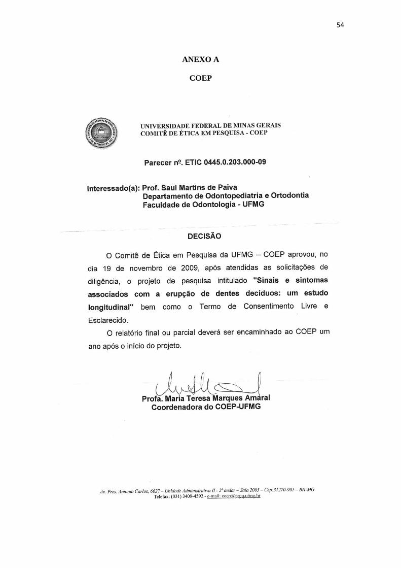
6.1 Anexo A – Autorização COEP...........................................................................
6.2 Anexo B – Normas para publicação na Pediatrics..........................................
49
50

17
CONSIDERAÇÕES INICIAIS
18
Considerações iniciais
CONSIDERAÇÕES INICIAIS
A associação entre erupção de dentes decíduos e o aparecimento de
manifestações orgânicas locais e gerais em bebês tem sido debatida há mais de 5000
anos. O relato dessas manifestações na literatura varia desde sintomatologia simples,
como a salivação excessiva, até uma sintomatologia mais grave, como a convulsão. Em
1839, mais de cinco mil mortes de bebês na Inglaterra e País de Gales foram atribuídas
à erupção de dentes decíduos (Dally, 1996).
A erupção é um processo fisiológico normal, definido como um processo em que
o dente se move de sua posição de desenvolvimento dentro do osso até a sua
emergência na cavidade bucal (Kardos, 1996; Craddock., 2004).
Por ser definida como processo fisiológico, a erupção de dentes decíduos
associada à manifestação de sinais e sintomas é assunto controverso no meio médico e
odontológico.
Estudos realizados em diferentes países revelaram que a maioria dos pais e
profissionais de saúde associou a erupção de dentes decíduos ao aparecimento de sinais
e sintomas. Wake et al. (1999) verificaram por meio da aplicação de questionário que,
dentre 92 pais de bebês, apenas um acreditava que a erupção de dentes decíduos não
causava sintomatologia. A maioria (70-85%) associou a erupção de dentes decíduos
com febre, dor, irritabilidade, distúrbios de sono, aumento da salivação e vermelhidão
da face.
Owais et al. (2010), em recente estudo, verificaram que aproximadamente 72%
da amostra composta por pais de bebês relacionavam a erupção de dentes decíduos a
19
episódios de diarréia e quase 85% acreditavam que a erupção podia causar febre. Esses
resultados estão de acordo com os achados de Cunha et al. (2004), que verificaram que
95% dos pais relataram a ocorrência de manifestações locais e sistêmicas em seus filhos
durante a erupção dos dentes decíduos.
Alguns profissionais de saúde têm destacado que os sinais e sintomas, quando
presentes devido à erupção dentária, são de baixa gravidade e mais relacionados a um
desconforto do que à ocorrência de doença (Sarrell et al., 2005). Em estudo realizado
por Jaber et al. (1992), foi constatado um aumento da temperatura durante a erupção do
primeiro dente decíduo. No entanto, o intervalo de confiança mostrou que, em 95% dos
casos, a temperatura variou de 37,33°C a 37,86°C. Esses autores concluíram que a febre
acima de 38,5°C não deve ser atribuída apenas à erupção dentária.
Em uma avaliação de 50 bebês que foram levados ao hospital pelos pais com
sinais e sintomas relacionados à erupção dentária, verificou-se que 48 apresentavam
outras causas que poderiam levar ao quadro clínico apresentado (Lloyd, 1996).
Entretanto, a maior parte dos estudos realizados é retrospectiva e demonstra a
visão dos pais e profissionais da saúde frente ao processo de erupção de dentes
decíduos. Portanto, não avalia a possível associação da erupção dentária com a
manifestação de sinais e sintomas.
Essa associação foi também testada em estudos prospectivos. Um deles foi
realizado com 111 bebês com média de idade de quatro meses ao início do estudo. Os
pais mediram a temperatura timpânica e observaram a presença ou ausência de 18
sintomas diariamente, até o momento em que os bebês completassem 12 meses. Nesse
período, foi observada a erupção de 475 dentes. Os sintomas mais freqüentes nos
períodos de erupção foram: aumento da salivação, irritabilidade, diminuição do apetite
20
para alimentos sólidos e elevação da temperatura média (Macknin et al., 2000). Em
outro estudo, os examinadores realizaram avaliações de temperatura timpânica e exame
clínico dos bebês. A associação entre sintomas e erupção dentária não foi confirmada
(Wake et al., 2000).
Em revisão sistemática da literatura, Tighe e Roe (2007) concluíram que não
existem evidências científicas suficientes para indicar que determinado sinal ou sintoma
ocorra devido exclusivamente à erupção de dentes decíduos. Sugeriram que o
profissional deve considerar outras patologias orgânicas ao atender uma criança doente.
Isso é relevante uma vez que a erupção dos dentes decíduos ocorre geralmente em um
período em que os bebês apresentam freqüentes episódios de doenças de baixa
gravidade (Jarman e Kohlenberg, 1991).
Dessa forma, o presente estudo tem como objetivo verificar a associação entre
erupção de dentes decíduos e manifestação de sintomatologia.
Diante da importância da publicação de pesquisas para o desenvolvimento
científico, esta dissertação foi estruturada na forma de artigo.

21
ARTIGO
22
Paper
Signs and symptoms associated to primary tooth eruption: A longitudinal study
Joana Ramos-Jorge1, Isabela A. Pordeus
1, Maria L. Ramos-Jorge
2, Saul M. Paiva
1
1 Department of Pediatric Dentistry and Orthodontics, Faculty of Dentistry,
Universidade Federal de Minas Gerais, Belo Horizonte, Brazil.
2 Department of Pediatric Dentistry and Orthodontics, Faculty of Dentistry,
Universidade Federal dos Vales do Jequitinhonha e Mucuri, Diamantina, Brazil.
Author for correspondence:
Joana Ramos Jorge
Rua Nunes Vieira, 255/502
30310-300, Belo Horizonte, MG, Brazil
Phone: +55 31 2515 4887
e-mail: [email protected]
Key words: teething, tooth eruption, symptoms
Paper formatted in compliance with norms of the periodical Pediatrics
23
Abstract
Objective: Assess the association between primary tooth eruption and the manifestation
of signs and symptoms in infants.
Patients and Methods: A longitudinal study was carried out with 47 non-
institutionalized infants between five and 15 months of age in the city of Diamantina,
Brazil. The non-randomized convenience sample was based on a registry of infants
provided by the Municipal Secretary of Health. Eligible participants were infants with
between zero and seven erupted teeth and no history of chronic disease or disorders that
could cause an increase in the symptoms assessed in the study. Tympanic and axillary
temperature readings and clinical oral exams were performed daily. A daily interview
with the mothers was carried to investigate the occurrence of 14 signs and symptoms
associated to teething presented by the infants in the previous 24 hours.
Results: Teething was associated to a rise in tympanic temperature on the day of the
eruption (P=0.004) as well as the occurrence of other signs and symptoms. Maximal
tympanic and axillary temperature was 36.8 ºC and 36.6 ºC, respectively. The most
frequent sign and symptoms associated to teething were irritability (P<0.001), increased
salivation (P<0.001), runny nose (P<0.001) and loss of appetite (P<0.001).
Conclusions: Signs and symptoms, such as irritability, increased salivation, runny nose,
loss of appetite, diarrhea, rash and sleep disturbance, were associated to primary tooth
eruption. The present study supports the concept that the occurrence of severe signs and
symptoms, such as fever, is not attributed to teething.
24
INTRODUCTION
Tooth eruption has been held responsible for a variety of systemic
manifestations in infants. The association between teething and irritability, increased
salivation, sleep disturbance, fever, diarrhea and loss of appetite remains unclear, since
the onset of these disorders may simply coincide with the teething. Moreover, some of
these signs and symptoms may imply more serious conditions.1 Studies involving
parents, pediatricians and other healthcare professionals have associated teething with
signs and symptoms.2-9
However, prospective studies have offered contradictory
findings.10,11
In a study involving 21 children between six months and two years of age
institutionalized at day care centers in Melbourne, Australia, tympanic temperature
readings and clinical oral exams were carried out and the results did not confirm any
association between tooth eruption and disturbances.11
However, such an association
was found in a study carried out in the city of Cleveland (USA) involving 111 infants
between three and 5.6 months of age at the beginning of the data collection period. The
parents read the tympanic temperature and observed the presence or absence of 18
symptoms on a daily basis until the infants reached 12 months of age. The eruption of
475 teeth was observed in this period and the following were the most frequent
symptoms: increased salivation, irritability, loss of appetite for solid foods and rise in
mean temperature.10
However, these studies had limitations, such as which parents and
caregivers read the temperature and performed the exam of the infant's oral cavity.
Currently, there is not enough scientific evidence to indicate that certain signs or
symptoms occur only because the eruption of primary teeth.12
Thus, the aim of the
present prospective longitudinal study was to investigate the association between tooth
25
eruption and a range of signs and symptoms of teething while minimizing the
limitations found in previous studies.
METHODS
Subjects
The study was carried out over an eight-month period and involved 47 non-
institutionalized infants (i.e., received care at home) between five and 15 months of age
in the city of Diamantina, Brazil. The non-randomized convenience sample was based
on the registry of infants in this age range provided by the Diamantina Secretary of
Health. The study sample size was based on data on the mean and SD scores of previous
study.13
Estimating that clinically significant difference between two groups would be
1SD and adopting a effect size of 0.5 (µ1- µ2/SD, i.e. mean of temperature in non
eruption day =36.9ºC – mean of temperature in eruption day= 37.4ºC/1), a sample size
of 44 would give 90% power to detect this a difference at a significance level of 0.05.
Due to the possibility of losses, fifty-three babies were actually recruited.
Eligible participants were infants with up to seven erupted incisors and no
history of chronic disease or disorders that could cause an increase in the signs and
symptoms assessed in the study.
Data Collection
The pilot study was carried out with seven infants between six and 15 months of
age selected by convenience in the city of Diamantina; these infants did not make up
part of the main study. The pilot study was performed was to test the data collection
process and ascertain the applicability of the instruments. The data from this pilot study
demonstrated that there was no need to modify the methods proposed for the study.
26
Data collection was performed daily at the residences of the infants over an
eight-month period. The visits were scheduled beginning at 4 pm in order to minimize
the variation in the child's temperature throughout the day. The visits time was
previously arranged with the mother in order to avoid temperature readings during baths
or sleep. The possible occurrence of signs and symptoms during the eruption of primary
incisors was assessed. Data collection began prior to the eruption of at least one of the
incisors and ended one week following the eruption of the last incisor.
Eleven validated dentists trained in handling the thermometers and performing
the examination of the oral cavity carried out clinical exams on the infants to determine
tooth eruption. The calibration exercise consisted of two steps: the theoretical step
involved discussion on the criteria for the diagnosis of tooth eruption and an analysis of
photographs. A specialist in pediatric dentistry (gold standard in this theoretical
framework) coordinated this step, instructing general dentists on how to perform the
examination and determine temperature. In the clinical step, the dentists examined
seven previously selected infants between six to 15 months of age. The dentist with the
best level of intra-examiner and inter-examiner agreement in the theoretical step was
considered the gold standard in the clinical step. Inter-examiner agreement was tested
comparing each examiner with the gold standard. A one-day interval between
evaluations was used to test the intra-examiner agreement so that the diagnosis of tooth
eruption was performed under similar conditions, as a greater interval between
evaluations could compromise the calibration and, consequently, the reliability of the
study. Both inter-examiner and intra-examiner kappa values were 1.0. The dentists were
also calibrated for the use of axillary and tympanic thermometers, achieving kappa
values greater than 0.8.
27
The clinical exam was performed with the aid of a head lamp (PETZL®
, Tikka
XP, Crolles, FR) to provide a standardized light source for the visual exam and with
palpation using the index finger on the alveolar ridge. Temperature was read using an
infrared auricular thermometer (Incoterm®, Porto Alegre, RS, Brazil) and a digital
axillary thermometer (BD®, São Paulo, SP, Brazil); tympanic and axillary temperatures
were assessed as continuous variables. If an infant's temperature exceeded 37.5 ºC, the
child would be referred to the nearest children's medical care service. Mothers were
interviewed to investigate the occurrence of signs and symptoms in the previous 24
hours, such as increased salivation, rash, runny nose, diarrhea, loss of appetite, cold,
irritability, fever, smelly urine, constipation, vomiting, colic and seizure. Signs and
symptoms were recorded daily on a standardized chart. The mean frequency of signs
and symptoms was calculated on days of non-eruption, on the day of eruption and on
the days prior to and following the eruption of primary incisors. The data collection
sequence was as follows: 1) reading of tympanic and axillary temperature; 2) interview;
and 3) clinical exam.
Erupted teeth not assessed on the day of eruption or on the days prior to and
following eruption were excluded from the analysis. The day of eruption was defined as
the first day on which the incisor edge emerged in the oral cavity without being
completely covered by gingival tissue.
Statistical analysis
Statistical analysis was performed using the Statistical Package for the Social
Sciences (SPSS for Windows, version 15.0, SPSS Inc, Chicago, IL, USA). Mean,
standard deviation, median, minimum and maximum values were calculated for each
variable quantitative and frequency analysis was calculated for the variable qualitative.
28
Since tympanic and axillary temperature and the frequency of signs and symptoms
scores were not normally distributed (Shapiro-Wilk test), a non-parametric test for
repeated measures was used (Wilcoxon test). For each continuous variable (tympanic
temperature, axillary temperature, mean frequency of signs and symptoms),
comparisons were made between days of non-eruption, day of eruption and days prior
to and following eruption of the primary incisors (Wilcoxon test). The mean frequency
of signs and symptoms was calculated based on the following formula: number of days
on which the infant exhibited a sign or symptom divided by the total number of days
evaluated. This formula was applied separately for the non-eruption day, eruption day,
previous day and following day.
Based on the Bonferroni correction, P-values equal to or less than 0.016 were
considered significant. Bonferroni correction is a method used to address the problem of
multiple comparisons. The correction is based on the idea that if an experimenter is
testing n dependent or independent hypotheses on a set of data, then one way of
maintaining the error rate is to test each individual hypothesis at a statistical
significance level of 1/n times what it would be if only one hypothesis were tested. So if
one wants the significance level for the whole family of tests to be at most α then the
Bonferroni correction would be to test each of the individual tests at a significance level
of α/n. Statistically significant simply means that a given result is unlikely to have
occurred by chance assuming the null hypothesis is actually correct (i.e., no difference
among groups, no effect of treatment, no relation among variables). Thus, the
significance value adopted (p = 0.016) is the result of 0.05/3 [α=0.05; 3 multiple
comparisons (1: non-eruption vs previous day; 2: non-eruption vs eruption; 3: non-
eruption vs following day)].14
Ethical considerations
29
The present study received approval from the Human Research Ethics
Committee of the Universidade Federal de Minas Gerais (Brazil). All parents received
information regarding the objectives of the study and signed informed consent forms.
RESULTS
A total of 53 infants were initially enrolled in the study, 47 (88.7%) of whom
participated through to the end of the study. The following were the main reasons for
dropouts: moving away from the city; no tooth erupted; impossibility of assessment on
the day of eruption or previous/following day. A total of 231 teeth erupted throughout
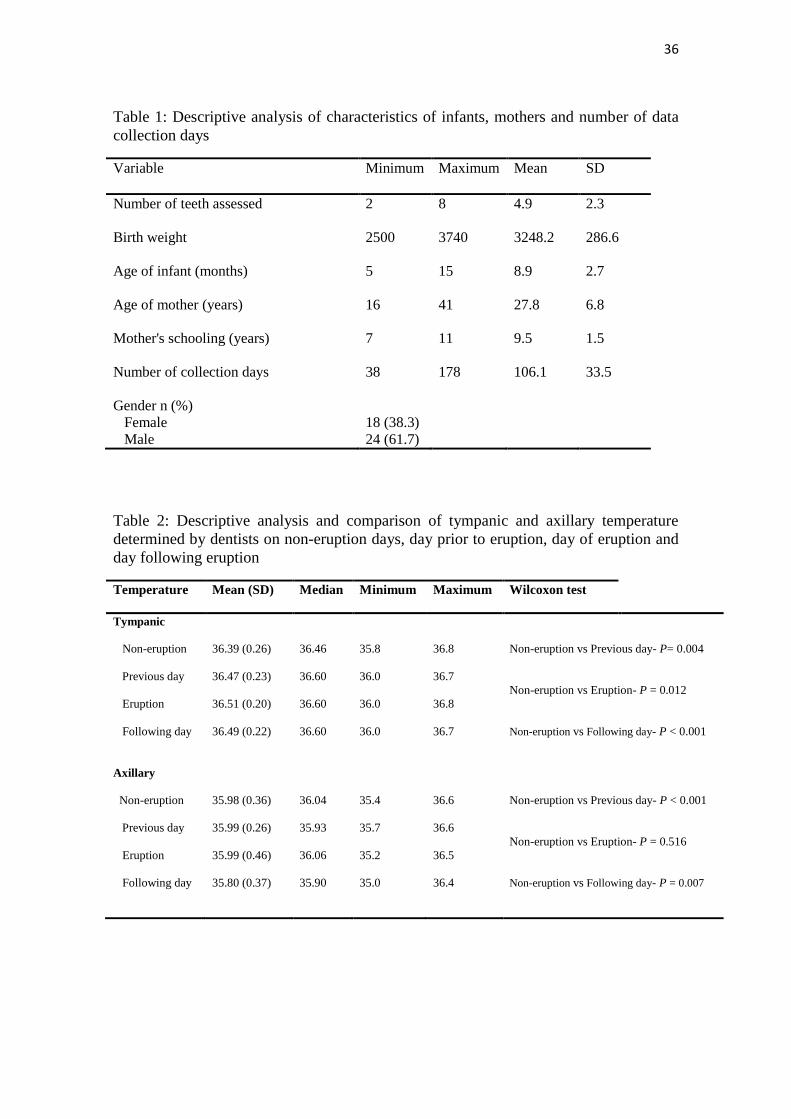
the study. The mean number of teeth per infant was nearly five (range: 2 to 8). Table 1
displays the descriptive information on the infants and their mothers.
Mean tympanic and axillary temperature determined by dentists on non-eruption
days, day on which eruption occurred and the days prior to and following incisor
eruption are displayed in Table 2. There were statistically significant differences in
tympanic temperature between non-eruption days and the day of eruption (P=0.004),
previous day (P=0.012) and following day (P<0.001). Regarding axillary temperature,
there was a statistically significant difference only between non-eruption days and the
day following eruption (P=0.007). Mean tympanic and axillary temperature rose 0.12
°C and 0.01 °C on days of eruption in relation to non-eruption days.
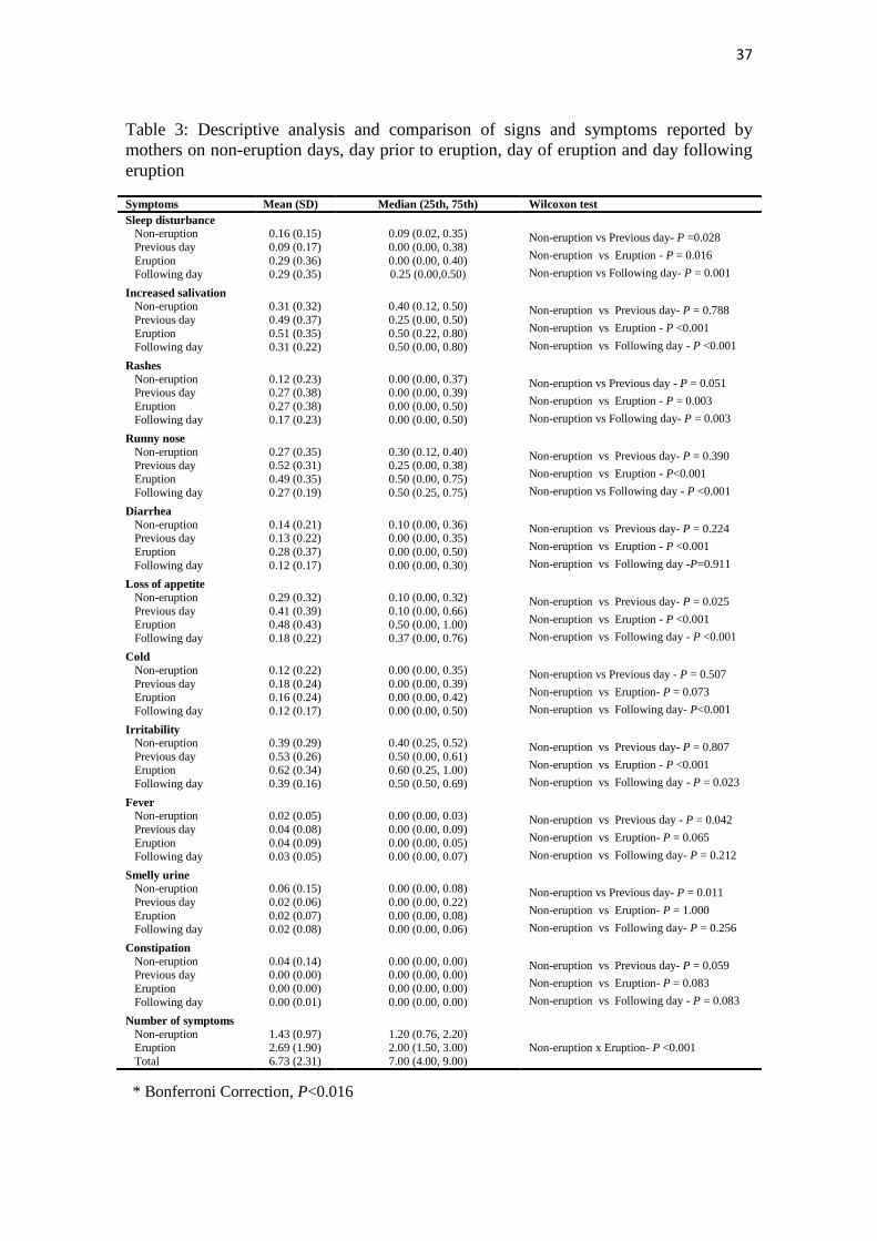
The associations between signs and symptoms reported by mothers and tooth
eruption were statistically significant. Sleep disturbance (P=0.016), increased salivation
(P<0.001), rash (P=0.003), runny nose (P<0.001), diarrhea (P<0.001), loss of appetite
(P<0.001) and irritability (P<0.001) were associated with tooth eruption. The analysis
of mean frequencies revealed that the most common symptoms on days of eruption
were irritability, increased salivation, runny nose and loss of appetite (Table 3). As no
30
infant experienced seizure or colic and reports of vomiting were rare throughout the
study, these signs and symptoms were not included in Table 3. The mean number of
symptoms occurring on days of eruption (2.69) was nearly twofold that of non-eruption
days (1.43); this difference was statistically significant (P<0.001) (Table 3).
DISCUSSION
The design adopted makes the present study unique and original. This is the first
prospective study in which temperature readings and clinical oral exams were
performed on a daily basis by trained examiners. The decision was made to investigate
non-institutionalized infants, as viral and bacterial infections are rapidly disseminated in
day care centers and could affect the frequency of signs and symptoms.15,16
Moreover, a
previous study carried out in Brazil found that, at public and private daycare centers, the
proportion of caregivers to children aged zero to two years is 1:6 and 1:9, respectively,17
which could have a negative effect on the validity and reliability of the data. The aim of
assessing the day prior to and following eruption was based on previous studies
reporting that infants exhibit signs and symptoms on days surrounding the day of
eruption that can may be associated with teething.10,18
Methods were employed in order to minimize observer bias. The data collection
sequence (temperature reading, followed by interview with mother and, lastly, the
clinical exam) was designed so that mothers would not be biased with regard to
communicating more signs and symptoms when it was determined that a tooth was
erupting. However, it is possible that such bias occurred on the day following tooth
eruption. Another limitation of the present study is the non-use of objective measures of
signs and symptoms such as irritability, loss of appetite and increased salivation.
31
The present study confirms the findings of previous studies that tooth eruption is
associated to a slight rise in body temperature. 10,18
Significant differences were found in
mean tympanic temperature between non-eruption days and day of eruption, one day
prior to eruption and one day following eruption. However, there was a significant
difference in axillary temperature only between non-eruption days and one day
following eruption. Despite these statistically significant associations, maximal
tympanic (36.8 ºC) and axillary (36.6 ºC) temperature did not characterize fever, as the
variation in temperature remained within the range of normality.19
There was a mean
temperature increase of 0.12 ºC between non-eruption days and the day of eruption. A
previous prospective study found a greater temperature increase between these
evaluation times (0.5 ºC). However, the authors assessed rectal temperature and the
readings were performed by caregivers.
Tympanic temperature was higher than axillary temperature in the present study.
Tympanic thermometers are more accurate than axillary thermometers in young
children when compared with reference standards of pulmonary artery temperature
under controlled conditions.19
Moreover, reading tympanic temperature is a fast, easily
executed technique.20
The importance of assessing axillary temperature resides in the
fact that this type of reading is widely used by parents and healthcare professionals for
the diagnosis of fever.
The results of the present study reveal a greater frequency of systemic
manifestations (sleep disturbance, increased salivation, rash, runny nose, diarrhea, loss
of appetite and irritability) on the day of eruption and one day following eruption in
comparison to non-eruption days. The aforementioned study carried out in Cleveland
also reports an association between teething and increased salivation, irritability, sleep
32
disturbance and loss of appetite on the day of eruption.10
Some of these signs and
symptoms may be explained by the increase in inflammatory cytokine levels in the
gingival crevicular fluid surrounding the teeth. High levels of IL-1ß and TNFα have
been correlated with fever, gastrointestinal disturbance, sleep disturbance and appetite
disturbance.18
Unlike the Cleveland study,10
the present study found a statistically
significant association between teething and diarrhea. However, the study carried out in
Australia found no associations between teething in institutionalized infants and signs
and symptoms.11
The conclusion of all prospective studies is that no specific symptoms
can reliably predict the emergence of a tooth. Furthermore, signs and symptoms that can
be attributed to teething are not serious; thus, the presence of fever (> 38.5 ºC) or other
clinically important symptoms is very unlikely to be caused by tooth eruption.
CONCLUSION
The results demonstrate associations between teething and sleep disturbance,
increased salivation, rash, runny nose, diarrhea, loss of appetite, irritability and a slight
rise in temperature. These associations were significant on the day of eruption and one
day following eruption. Therefore, it is not possible to predict eruption through the
observation of signs and symptoms, as there were no associations with the day prior to
eruption. The findings of this study contribute toward supporting the concept that
teething is not associated to severe signs and symptoms. Thus, health professionals
involved in the care of infants should seek other causes before attributing severe signs
and symptoms to teething.
ACKNOWLEDGEMENTS
33
This study was supported by the Brazilian fostering agencies National Council
for Scientific and Technological Development (CNPq) and State of Minas Gerais
Research Foundation (FAPEMIG).
REFERENCES
1. Swann IL. Teething complications, a persisting misconception. Postgraduate Medical
Journal. 1979; 55: 24-25.
2. Wake M, Hesketh K, Allen MA. Parent beliefs about infant teething: A survey of
Australian parents. Journal of Paediatrics and Child Health. 1999; 35: 446-449.
3. Barlow BS, Kanellis MJ, Slayton RL. Tooth eruption symptoms: a survey of parents
and health professionals. ASDC Journal of Dentistry for Children. 2002; 69: 148-150.
4. Wake M, Hesketh K. Teething symptoms: cross sectional survey of five groups of
child health professionals. British Medical Journal. 2002; 325: 814.
5. Cunha RF, Pugliesi DMC, Garcia LD, Murata SS. Systemic and local teething
disturbances: prevalence in a clinic for infants. Journal of Dentistry for Children. 2004;
71: 24-26.
6. Sarrell EM, Horev Z, Cohen Z, Cohen HA. Parents' and medical personnel's beliefs
about infant teething. Patient Education and Counseling. 2005; 57:122-5.
7. Oziegbe EO, Folayan MO, Adekoya-Sofowora CA, Esan TA, Owotade FJ. Teething
problems and parental beliefs in Nigeria. The Journal of Contemporary Dental Practice.
2009; 10: 75-82.
8. Owais AI, Zawaideh F, Bataineh O. Challenging parents' myths regarding their
children's teething. International Journal of Dental Hygiene. 2010; 8: 28-34.
34
9. Feldens CA, Junior IMF, Ottoni AB, Feldens EG, Vítolo MR. Teething symptoms in
the first year of life and associated factors: a cohort study. The Journal of Clinical
Pediatric Dentistry. 2010; 34: 201-206.
10. Macknin ML, Piedmonte M, Jacobs J, Skibinski C. Symptoms associated with
infant teething: a prospective study. Pediatrics. 2000; 105: 747-752.
11. Wake M, Hesketh K, Lucas J. Teething and tooth eruption in infants: A Cohort
Study. Pediatrics. 2000; 106: 1374-1379.
12. Tighe M, Roe MF. Does a teething child need serious illness excluding? Archives of
Disease in Childhood. 2007; 92: 266-268.
13. Jaber L, Cohen IJ, Mor A. Fever associated with teething. Archives of Disease in
Childhood. 1992; 67: 233-234.
14. Riffenburgh RH. Statistics in medicine. 2th ed. San Diego, California: Elsevier;
2006.
15. Gensheimer KF. A public health perspective on child care. Pediatrics. 1994; 94:
1116-1118.
16. Louhiala PJ, Jaakkola N, Ruotsalainen R, Jaakkola JJ. Form of day care and
respiratory infections among finnish children. American Journal of Public Health.
1995; 85: 1109-1112.
17. Barros AJ, Halpern R, Menegon OE. Public and private day-care centers in Pelotas,
RS: compliance with the regulations. Jornal de Pediatria. 1998; 74: 397-403.
35
18. Shapira J, Berenstein-Ajzman G, Engelhard D, Cahan S, Kalickman I, Barak V.
Cytokine levels in gingival crevicular fluid of erupting primary teeth correlated with
systemic disturbances accompanying teething. Pediatric Dentistry. 2003; 25: 441-448.
19. Robinson JL, Jou H, Spady DW. Accuracy of parents in measuring body
temperature with a tympanic thermometer. BMC Family Practice. 2005; 6: 3.
20. El-Radhi AS, Barry W. Thermometry in paediatric practice. Archives of Disease in
Childhood. 2006; 91: 351-356.
36
Table 1: Descriptive analysis of characteristics of infants, mothers and number of data
collection days
Variable Minimum Maximum Mean SD
Number of teeth assessed 2 8 4.9 2.3
Birth weight 2500 3740 3248.2 286.6
Age of infant (months) 5 15 8.9 2.7
Age of mother (years) 16 41 27.8 6.8
Mother's schooling (years) 7 11 9.5 1.5
Number of collection days 38 178 106.1 33.5
Gender n (%)
Female
Male
18 (38.3)
24 (61.7)
Table 2: Descriptive analysis and comparison of tympanic and axillary temperature
determined by dentists on non-eruption days, day prior to eruption, day of eruption and
day following eruption
Temperature Mean (SD) Median Minimum Maximum Wilcoxon test
Tympanic
Non-eruption
Previous day
Eruption
Following day
36.39 (0.26)
36.47 (0.23)
36.51 (0.20)
36.49 (0.22)
36.46
36.60
36.60
36.60
35.8
36.0
36.0
36.0
36.8
36.7
36.8
36.7
Non-eruption vs Previous day- P= 0.004
Non-eruption vs Eruption- P = 0.012
Non-eruption vs Following day- P < 0.001
Axillary
Non-eruption
Previous day
Eruption
Following day
35.98 (0.36)
35.99 (0.26)
35.99 (0.46)
35.80 (0.37)
36.04
35.93
36.06
35.90
35.4
35.7
35.2
35.0
36.6
36.6
36.5
36.4
Non-eruption vs Previous day- P < 0.001
Non-eruption vs Eruption- P = 0.516
Non-eruption vs Following day- P = 0.007
37
Table 3: Descriptive analysis and comparison of signs and symptoms reported by
mothers on non-eruption days, day prior to eruption, day of eruption and day following
eruption
Symptoms Mean (SD) Median (25th, 75th) Wilcoxon test
Sleep disturbance
Non-eruption
Previous day
Eruption Following day
0.16 (0.15)
0.09 (0.17)
0.29 (0.36) 0.29 (0.35)
0.09 (0.02, 0.35)
0.00 (0.00, 0.38)
0.00 (0.00, 0.40) 0.25 (0.00,0.50)
Non-eruption vs Previous day- P =0.028
Non-eruption vs Eruption - P = 0.016
Non-eruption vs Following day- P = 0.001
Increased salivation
Non-eruption
Previous day
Eruption
Following day
0.31 (0.32)
0.49 (0.37)
0.51 (0.35)
0.31 (0.22)
0.40 (0.12, 0.50)
0.25 (0.00, 0.50)
0.50 (0.22, 0.80)
0.50 (0.00, 0.80)
Non-eruption vs Previous day- P = 0.788
Non-eruption vs Eruption - P <0.001
Non-eruption vs Following day - P <0.001
Rashes
Non-eruption Previous day
Eruption Following day
0.12 (0.23) 0.27 (0.38)
0.27 (0.38) 0.17 (0.23)
0.00 (0.00, 0.37) 0.00 (0.00, 0.39)
0.00 (0.00, 0.50) 0.00 (0.00, 0.50)
Non-eruption vs Previous day - P = 0.051
Non-eruption vs Eruption - P = 0.003
Non-eruption vs Following day- P = 0.003
Runny nose
Non-eruption Previous day
Eruption
Following day
0.27 (0.35) 0.52 (0.31)
0.49 (0.35)
0.27 (0.19)
0.30 (0.12, 0.40) 0.25 (0.00, 0.38)
0.50 (0.00, 0.75)
0.50 (0.25, 0.75)
Non-eruption vs Previous day- P = 0.390
Non-eruption vs Eruption - P<0.001
Non-eruption vs Following day - P <0.001
Diarrhea
Non-eruption Previous day
Eruption
Following day
0.14 (0.21) 0.13 (0.22)
0.28 (0.37)
0.12 (0.17)
0.10 (0.00, 0.36) 0.00 (0.00, 0.35)
0.00 (0.00, 0.50)
0.00 (0.00, 0.30)
Non-eruption vs Previous day- P = 0.224
Non-eruption vs Eruption - P <0.001
Non-eruption vs Following day -P=0.911
Loss of appetite
Non-eruption
Previous day Eruption
Following day
0.29 (0.32)
0.41 (0.39) 0.48 (0.43)
0.18 (0.22)
0.10 (0.00, 0.32)
0.10 (0.00, 0.66) 0.50 (0.00, 1.00)
0.37 (0.00, 0.76)
Non-eruption vs Previous day- P = 0.025
Non-eruption vs Eruption - P <0.001
Non-eruption vs Following day - P <0.001
Cold
Non-eruption
Previous day Eruption
Following day
0.12 (0.22)
0.18 (0.24) 0.16 (0.24)
0.12 (0.17)
0.00 (0.00, 0.35)
0.00 (0.00, 0.39) 0.00 (0.00, 0.42)
0.00 (0.00, 0.50)
Non-eruption vs Previous day - P = 0.507
Non-eruption vs Eruption- P = 0.073
Non-eruption vs Following day- P<0.001
Irritability
Non-eruption
Previous day Eruption
Following day
0.39 (0.29)
0.53 (0.26) 0.62 (0.34)
0.39 (0.16)
0.40 (0.25, 0.52)
0.50 (0.00, 0.61) 0.60 (0.25, 1.00)
0.50 (0.50, 0.69)
Non-eruption vs Previous day- P = 0.807
Non-eruption vs Eruption - P <0.001
Non-eruption vs Following day - P = 0.023
Fever
Non-eruption
Previous day
Eruption Following day
0.02 (0.05)
0.04 (0.08)
0.04 (0.09) 0.03 (0.05)
0.00 (0.00, 0.03)
0.00 (0.00, 0.09)
0.00 (0.00, 0.05) 0.00 (0.00, 0.07)
Non-eruption vs Previous day - P = 0.042
Non-eruption vs Eruption- P = 0.065
Non-eruption vs Following day- P = 0.212
Smelly urine
Non-eruption
Previous day
Eruption Following day
0.06 (0.15)
0.02 (0.06)
0.02 (0.07) 0.02 (0.08)
0.00 (0.00, 0.08)
0.00 (0.00, 0.22)
0.00 (0.00, 0.08) 0.00 (0.00, 0.06)
Non-eruption vs Previous day- P = 0.011
Non-eruption vs Eruption- P = 1.000
Non-eruption vs Following day- P = 0.256
Constipation
Non-eruption Previous day
Eruption
Following day
0.04 (0.14) 0.00 (0.00)
0.00 (0.00)
0.00 (0.01)
0.00 (0.00, 0.00) 0.00 (0.00, 0.00)
0.00 (0.00, 0.00)
0.00 (0.00, 0.00)
Non-eruption vs Previous day- P = 0.059
Non-eruption vs Eruption- P = 0.083
Non-eruption vs Following day - P = 0.083
Number of symptoms
Non-eruption Eruption
Total
1.43 (0.97) 2.69 (1.90)
6.73 (2.31)
1.20 (0.76, 2.20) 2.00 (1.50, 3.00)
7.00 (4.00, 9.00)
Non-eruption x Eruption- P <0.001
* Bonferroni Correction, P<0.016

38
CONSIDERAÇÕES FINAIS
39
Considerações finais
CONSIDERAÇÕES FINAIS
Tem sido muito discutida na literatura a preocupação com a integridade física da
criança, uma vez que doenças graves podem ser subestimadas devido à ocorrência da
erupção dentária (Tighe e Roe, 2007).
Diante da revisão realizada neste estudo, fica evidente que os bebês
desenvolvem sinais e sintomas que seus pais e/ou responsáveis atribuem à erupção de
dentes decíduos. Uma variedade de sinais e sintomas pode simplesmente coincidir com
o período de erupção de dentes.
A dúvida de que a erupção de dentes decíduos possa causar sintomatologia está
presente até mesmo em profissionais da área de saúde. Não é raro encontrar a descrição
de sintomatologia associada à erupção de dentes em livros texto de Pediatria e
Odontopediatria.
No entanto, a crença de que a erupção de dentes decíduos está associada à
manifestação de sinais e sintomas pode contribuir para o adiamento da tomada de
decisão clínica em casos de doenças de maior gravidade, como gastroenterites,
infecções urinárias, meningites entre outras. Além disso, essa crença também pode
estimular o uso excessivo de medicamentos em bebês mascarando assim uma
sintomatologia que pode ser importante para um diagnóstico preciso.
Este estudo contribui para esclarecer que os sinais e sintomas associados à
erupção de dentes decíduos não são graves e também demonstra que a presença de febre
não pode estar associada à erupção de dentes. Esse é resultado que deve ser divulgado
tanto para a comunidade acadêmica, como também para toda a população.

40
REFERÊNCIAS GERAIS
41
REFERÊNCIAS
Dally A. The lancet and the gum-lancet: 400 years of teething babies. Lancet. 1996;
348: 1710-1711.
Kardos TB. The mechanism of tooth eruption. British Dental Journal. 1996; 181: 91-
95.
Craddock HL, Youngson CC. Eruptive tooth movement – the current state of
knowledge. British Dental Journal. 2004; 197: 385-391.
Wake M, Hesketh K, Allen MA. Parent beliefs about infant teething: A survey of
Australian parents. Journal of Paediatrics and Child Health. 1999; 35: 446-449.
Owais AI, Zawaideh F, Bataineh O. Challenging parents’ myths regarding their
children’s teething. International Journal of Dental Hygiene. 2010; 8: 28-34.
Cunha RF, Pugliesi DMC, Garcia LD, Murata SS. Systemic and local teething
disturbances: prevalence in a clinic for infants. Journal of Dentistry for Children. 2004;
71: 24-26.
Sarrell EM, Horev Z, Cohen Z, Cohen HA. Parents’ and medical personnel’s beliefs
about infant teething. Patient Education and Counseling. 2005; 57: 122-125.
Jaber L, Cohen IJ, Mor A. Fever associated with teething. Archives of Disease in
Childhood. 1992; 67: 233-234.
Lloyd S. Teething in babies: separating fact from fiction. Professional Care of Mother
and Child. 1996; 6: 155-156.
42
Macknin ML, Piedmonte M, Jacobs J, Skibinski C. Symptoms associated with infant
teething: a prospective study. Pediatrics. 2000; 105: 747-752.
Wake M, Hesketh K, Lucas J. Teething and tooth eruption in infants: A Cohort Study.
Pediatrics. 2000; 106: 1374-1379.
Tighe M, Roe MF. Does a teething child need serious illness excluding? Archives of
Disease in Childhood. 2007; 92: 266-268.
Jarman FC, Kohlenberg TM. The health effects of day care. Journal of Paediatrics and
child Health. 1991; 27: 272-281.

43
APÊNDICES
44
APÊNDICE A
CARTA DE APRESENTAÇÃO
Diamantina, ... de 2009.
Prezado Pai/Mãe/Responsável Legal sou Joana Ramos Jorge, aluna do Programa de
Pós-Graduação em Odontologia, área de Odontopediatria, da Universidade Federal de
Minas Gerais (UFMG). Gostaria que você lesse esse documento com atenção, pois, o
objetivo dele é firmar acordo por escrito, mediante a sua autorização para a sua
participação e a de seu filho nesta pesquisa.
- Qual é o objetivo da pesquisa?
Estamos querendo avaliar se problemas de saúde geral podem estar associados
com a cavidade bucal do bebê.
- Por que estamos realizando esta pesquisa?
Até o momento ainda não podemos afirmar se existe associação entre febre,
gripe, diarréia e outros sintomas com a erupção de dentes de leite no bebê. Esta pesquisa
é importante para a orientar os médicos e cirurgiões-dentistas durante o atendimento da
criança com esses sintomas.
- Como vai ser a pesquisa?
Todos os dias um examinador irá à sua casa para examinar o bebê e você vai
responder a umas perguntas sobre o estado de saúde da criança. São respostas que
levarão pouco tempo para responder. É importante destacar que não haverá desconforto
ou qualquer risco para o seu filho.
45
- Você é obrigado a participar?
Não. É um direito seu não participar da pesquisa. Sendo assim, esclareço que
você é COMPLETAMENTE LIVRE para não participar e não permitir a participação
do seu filho. Você pode ainda desistir de fazer parte da pesquisa em qualquer momento
da realização desta.
- Quais são os benefícios?
Como benefício pela participação na pesquisa, seu filho receberá atendimento
odontológico na clínica de Odontopediatria da Faculdade de Odontologia de
Diamantina.
- Aspectos éticos
1- O projeto dessa pesquisa, o modo como ela será realizada, foi submetido à avaliação
pelo Comitê de Ética em Pesquisa com Seres Humanos da UFMG e aprovado.
2- As informações relacionadas a você e seu filho ficarão sob minha responsabilidade.
Somente a equipe envolvida terá acesso a essas informações.
- Você tem alguma dúvida?
Ligue para os seguintes telefones ou envie e-mail para:
(38) 3531-1415/(31) 2515-4887
[email protected] (Joana Ramos Jorge)
46
APÊNDICE B
TERMO DE CONSENTIMENTO LIVRE E ESCLARECIDO
Eu, __________________________________________________________________,
por __________________________________________________________________,
concordo e autorizo a participação de meu filho (a) no estudo “SINAIS E SINTOMAS
ASSOCIADOS COM A ERUPÇÃO DE DENTES DECÍDUOS: estudo longitudinal”
que será executado pela Mestranda Joana Ramos Jorge, sob orientação do(a) Prof(a).
Dr. Saul Martins de Paiva, do Programa de Pós-Graduação em Odontologia, UFMG.
Concordo e autorizo com a utilização dos dados coletados desde que seja mantido o
sigilo de sua identificação conforme normas do Comitê de Ética em Pesquisa desta
Universidade. Autorizo ainda a realização de fotografias dos dentes e da cavidade bucal
do bebê, para utilização como material didático para aulas expositivas, apresentação em
eventos científicos ou para publicação de artigo em revista científica da área da saúde,
nacional e/ou internacional.
Diamantina, ____ de ______ de 2009
_________________________________________
Assinatura do pai/mãe/responsável
Pai/mãe/responsável
47
APÊNDICE C
Formulário 1 – Identificação
Nome do bebê__________________________________________________________
Nome do responsável____________________________________________________
Peso ao nascimento (g)__________________ Idade (meses)_____________________
Data de nascimento____/____/____
Escolaridade da mãe (em anos de estudo)____________ Idade da mãe___________
Endereço______________________________________________________________
Telefone______________________
História Médica
______________________________________________________________________
______________________________________________________________________
Observações durante a coleta de dados
______________________________________________________________________
______________________________________________________________________
______________________________________________________________________
______________________________________________________________________
______________________________________________________________________
Responsável pela coleta__________________________________________________
48
APÊNDICE D
Formulário 2 – Exame de temperatura auditiva
Dia 1 2 3 4 5 6 7 8 9 10 11 12 13 14 15
Temperatura
(°C)
Dia 16 17 18 19 20 21 22 23 24 25 26 27 28 29 30
Temperatura
(°C)
Dia 31 32 33 34 35 36 37 38 39 40 41 42 43 44 45
Temperatura
(°C)
Dia 46 47 48 49 50 51 52 53 54 55 56 57 58 59 60
Temperatura (°C)
Dia 61 62 63 64 65 66 67 68 69 70 71 72 73 74 75
Temperatura
(°C)
Dia 76 77 78 79 80 81 82 83 84 85 86 87 88 89 90
Temperatura (°C)
Dia 91 92 93 94 95 96 97 98 99 100 101 102 103 104 105
Temperatura (°C)
Dia 106 107 108 109 110 111 112 113 114 115 116 117 118 119 120
Temperatura
(°C)
49
APÊNDICE E
Formulário 3 – Exame de temperatura axilar
Dia 1 2 3 4 5 6 7 8 9 10 11 12 13 14 15
Temperatura
(°C)
Dia 16 17 18 19 20 21 22 23 24 25 26 27 28 29 30
Temperatura
(°C)
Dia 31 32 33 34 35 36 37 38 39 40 41 42 43 44 45
Temperatura
(°C)
Dia 46 47 48 49 50 51 52 53 54 55 56 57 58 59 60
Temperatura (°C)
Dia 61 62 63 64 65 66 67 68 69 70 71 72 73 74 75
Temperatura
(°C)
Dia 76 77 78 79 80 81 82 83 84 85 86 87 88 89 90
Temperatura (°C)
Dia 91 92 93 94 95 96 97 98 99 100 101 102 103 104 105
Temperatura (°C)
Dia 106 107 108 109 110 111 112 113 114 115 116 117 118 119 120
Temperatura
(°C)
50
APÊNDICE F
Formulário 5 – Avaliação dos Sinais e Sintomas (prospectiva)
Assinalar o número correspondente ao sinal/sintoma apresentado pela criança: 0-
nenhum, 1- distúrbios do sono, 2- salivação aumentada, 3- brotoeja, 4- coriza, 5-
diarréia, 6- falta de apetite, 7- resfriado, 8- vômito, 9- irritabilidade, 10- febre, 11-
odor forte da urina, 12- cólica, 13- convulsão, 14- constipação
Dia 1 2 3 4 5 6 7 8 9 10
Sinal/ Sintoma
Dia 11 12 13 14 15 16 17 18 19 20
Sinal/
Sintoma
Dia 21 22 23 24 25 26 27 28 29 30
Sinal/
Sintoma
Dia 31 32 33 34 35 36 37 38 39 40
Sinal/
Sintoma
Dia 41 42 43 44 45 46 47 48 49 50
Sinal/
Sintoma
Dia 51 52 53 54 55 56 57 58 59 60
Sinal/
Sintoma
51
APÊNDICE F
Formulário 5 – Avaliação dos Sinais e Sintomas (prospectiva)
Assinalar o número correspondente ao sinal/sintoma apresentado pela criança: 0-
nenhum, 1- distúrbios do sono, 2- salivação aumentada, 3- brotoeja, 4- coriza, 5-
diarréia, 6- falta de apetite, 7- resfriado, 8- vômito, 9- irritabilidade, 10- febre, 11-
odor forte da urina, 12- cólica, 13- convulsão, 14- constipação
Dia 61 62 63 64 65 66 67 68 69 70
Sinal/ Sintoma
Dia 71 72 73 74 75 76 77 78 79 80
Sinal/
Sintoma
Dia 81 82 83 84 85 86 87 88 89 90
Sinal/
Sintoma
Dia 91 92 93 94 95 96 97 98 99 100
Sinal/
Sintoma
Dia 101 102 103 104 105 106 107 108 109 110
Sinal/
Sintoma
Dia 111 112 113 114 115 116 117 118 119 120
Sinal/
Sintoma
52
APÊNDICE G
Formulário 5 - Exame clínico bucal
Dia 1 2 3 4 5 6 7 8 9 10 11 12 13 14 15
Erupção
dentária
(dente)
Dia 16 17 18 19 20 21 22 23 24 25 26 27 28 29 30
Erupção
dentária (dente)
Dia 31 32 33 34 35 36 37 38 39 40 41 42 43 44 45
Erupção
dentária (dente)
Dia 46 47 48 49 50 51 52 53 54 55 56 57 58 59 60
Erupção dentária
(dente)
Dia 61 62 63 64 65 66 67 68 69 70 71 72 73 74 75
Erupção
dentária (dente)
Dia 76 77 78 79 80 81 82 83 84 85 86 87 88 89 90
Erupção dentária
(dente)
Dia 91 92 93 94 95 96 97 98 99 100 101 102 103 104 105
Erupção dentária
(dente)
Dia 106 107 108 109 110 111 112 113 114 115 116 117 118 119 120
Erupção
dentária
(dente)

53
ANEXOS
54
ANEXO A
COEP
55
ANEXO B
NORMAS DE PUBLICAÇÃO DO PERIÓDICO PEDIATRICS
Author Guidelines
Pediatrics is the official peer-reviewed journal of the American Academy of Pediatrics.
Pediatrics publishes original research, clinical observations, and special feature articles
in the field of pediatrics, as broadly defined. Contributions pertinent to pediatrics are
also included from related fields such as nutrition, surgery, dentistry, public health,
child health services, human genetics, basic sciences, psychology, psychiatry,
education, sociology, and nursing.
The journal, published monthly, has a circulation of 66,000 and is translated into six
different languages. Its 2008 impact factor was 4.789.
Pediatrics has been continuously published by the American Academy of Pediatrics
since January 1948.
When submitting to Pediatrics, authors must attest that the manuscript is being
submitted only to Pediatrics, that it will not be submitted elsewhere while under
consideration, and that it has not been published elsewhere.
When preparing the manuscript for Pediatrics, authors must first determine the
manuscript type, and then select the appropriate manuscript preparation instructions
from the types listed below. Authors must also become familiar with journal style and
correct preparation of figures, tables, and multimedia before submitting a manuscript.
Acceptance Criteria
Relevance to readers is of major importance in manuscript selection. Pediatrics will
consider manuscripts in the following categories: reports of original research,
particularly clinical research; review articles; special articles; and case reports.
Generally, all papers will be reviewed by at least two outside consultants who are
selected by the editors based on their expertise in the topic of the manuscript.
A report of original research will be judged on the importance and originality of the
research, its scientific strength, its clinical relevance, the clarity with which it is
presented, and the number of submissions on the same topic. The decision to publish is
not based on the direction of results.
Unsolicited commentaries will be considered; however, most commentaries are
solicited by the editors. Case reports are of interest only when they present a new entity
or illustrate a major new aspect of a previously reported entity.
56
If your manuscript is accepted, the editors reserve the right to determine whether it will
be published in the print edition (which includes electronic publication) or only in the
electronic edition of Pediatrics.
Journal Style
All aspects of the manuscript (tables, illustrations, and references) should be prepared
according to the International Committee of Medical Journal Editors (ICMJE)
requirements.
Grammar, Punctuation, and Usage. Grammar, punctuation, and scientific writing
style should follow the most current edition of the AMA Manual of Style.1
Author Listing. All authors' names should be listed in their entirety. All authors must
clearly present institutional/professional affiliations and degrees held.
Abbreviations. On the title page, authors should provide a list of abbreviations used in
the paper and what they stand for. All acronyms in the text should be expanded at first
mention, followed by the abbreviation in parentheses. The acronym may appear in the
text thereafter. Acronyms may be used in the abstract if they occur 3 or more times
therein. Generally, abbreviations should be limited to those defined in the AMA Manual
of Style, current edition. Uncommon abbreviations should be listed at the beginning of
the article.
Keywords. Authors should provide keywords on the title page and use Medical Subject
Headings (MeSH) terms as a guide. Visit: http://www.nlm.nih.gov/mesh/meshhome.html
Units of Measure. Authors should use Système International (SI)2,3 values.
Proprietary Products. Authors should use nonproprietary names of drugs or devices
unless mention of a manufacturer is pertinent to the discussion. If a proprietary product
is cited, the name and location of the manufacturer must also be included.
References. Authors are responsible for the accuracy of references. Citations should be
numbered in the order in which they appear in the text. Review articles should be
appropriately cited. Reference style should follow that of the AMA Manual of Style,
current edition. Abbreviated journal names should reflect the style of Index Medicus.
Visit: http://www.nlm.nih.gov/tsd/serials/lji.html
Manuscript Preparation
Manuscripts should be prepared according to ICMJE guidelines.4 Refer to the following
“article types” for specific guidelines on preparing a manuscript.
Regular articles require a structured abstract. Label each section of the structured
abstract with the appropriate subheading. Case Reports, Reviews, and Special Articles
require short, unstructured abstracts. Commentaries do not require abstracts.
57
Research or project support should be acknowledged as a footnote on the title page.
Technical and other assistance should be identified on the title page.
Authors submitting manuscripts or letters to the editor involving adverse drug or
medical device events or product problems should also report these to the appropriate
governmental agency.
Title Page
The title page must include author names, degrees, and institutional/professional
affiliations, a short title, abbreviations, keywords, financial disclosure, and conflict of
interest. Please include the contact information for the corresponding author (eg,
address, telephone, fax, and e-mail address).
Title lengths should be kept to 15 words or 97 characters (including spaces) for all
submissions, regardless of article type.
Contributor’s Statement Page
All submissions must contain a contributor’s statement page, directly following the title
page.
An “author” is generally considered to be someone who has made substantive
intellectual contributions to a published study and is required to meet the following
criteria:
1) Substantial contributions to conception and design, acquisition of data, or analysis
and interpretation of data.
2) Drafting the article or revising it critically for important intellectual content; and
3) Final approval of the version to be published.
Acquisition of funding, collection of data, or general supervision of the research group
alone does not constitute authorship. All persons designated as authors should qualify
for authorship, and all those who qualify should be listed. Each author should have
participated sufficiently in the work to take public responsibility for appropriate
portions of the content.
All contributors who do not meet the criteria for authorship should be listed in an
acknowledgments section. Because readers may infer their endorsement of the data and
conclusions, these persons must give written permission to be acknowledged.
Article Types
Regular Articles
Abstract length: 250 words or fewer
58
Article length: 3,000 words or fewer
NOTE: Abstracts and References are not included in the 3,000 word count. Regular
articles are original research contributions that aim to change clinical practice or the
understanding of a disease process. Regular articles include but are not limited to
clinical trials, interventional studies, cohort studies, case-control studies, epidemiologic
assessments, and surveys. Components of a Regular Article include:
What’s Known, What’s New Brief summaries on the topic of “What's Known on this
Subject” and “What This Study Adds”, each limited to 40 words. These summaries
appear on the published articles as well as the separate Pediatrics Digest, a weekly
product provided as free access.
Structured Abstract. A structured abstract must include headings such as Objective,
Patients and Methods, Results, and Conclusions. The objective should clearly state the
hypothesis; patients and methods, inclusion criteria and study design; results, the
outcome of the study; and conclusions, the outcome in relation to the hypothesis and
possible directions of future study.
Introduction. A 1- to 2-paragraph introduction outlining the wider context that
generated the study and the hypothesis.
Patients and Methods. A "Patients and Methods" section detailing inclusion criteria
and study design to ensure reproducibility of the research.
Discussion. An expanded discussion highlighting antecedent literature on the topic and
how the current study changes the perception of a disease process.
Conclusion. A concluding paragraph presenting the impact of the study and possible
new research directions on the subject.
Figures, Tables, and Multimedia
Figures
Authors should number figures in the order in which they appear in the text. Figures
include graphs, charts, photographs, and illustrations. Each figure should be
accompanied by a legend that does not exceed 50 words. Use abbreviations unless these
have not been expanded in the text. If a figure is reproduced from another source,
authors are required to obtain permission from the copyright holder, and proof of
permission must be sent to the editorial office in Burlington, VT, at initial submission.
Authors are also required to provide level of magnification for histology slides. Figure
arrays should be clearly labeled, preassembled, and submitted to scale according to the
width and depth of a journal page (40 picas wide by 56 picas deep). Figure parts of an
array should be clearly marked in capital letters in 10-point Helvetica font in the upper
left-hand corner of each figure part. Figures should be submitted separately from the
text file.
59
Technical Requirements for figures. For an original submission, authors may submit
JPEG or PDF files. However, at revision, authors will need to submit higher resolution
files (150-300 dpi). The following file types are acceptable: TIFF, EPS, and PDF.
Pediatrics cannot accept Excel or Powerpoint files. Color files must be in CMYK (cyan,
magenta, yellow, black) mode.
For more information regarding digital art submission, visit Cadmus Communications
http://cjs.cadmus.com/da/index.jsp
Tables
Tables should be numbered in the order in which they are cited in the text and include
appropriate headers. Tables should not reiterate information presented in the Results
section, but rather should provide clear and concise data that further illustrate the main
point. Tabular data should directly relate to the hypothesis. Table formatting should
follow the most current edition of the AMA Manual of Style.
Multimedia
Pediatrics publishes supplemental content in the online article. References to online
supplemental content appear in the print journal. Such data include but are not limited to
tables, videos, audio files, slide shows, data sets, and Web sites. Authors are responsible
for clearly labeling such supporting information and are accountable for its accuracy.
Supplemental data will not be professionally copyedited.
Videos
Pediatrics encourages the submission of videos to accompany the electronic editions of
articles. Videos should be submitted in QuickTime 4.0 or higher format, and may be
prepared on either a personal computer or Macintosh computer.
All videos should be submitted at the desired reproduction size and length. To avoid
excessive delays in downloading the files, videos should be no more than 6MB in size,
and run between 30 and 60 seconds in length. Authors are encouraged to use
QuickTime’s “compress” option when preparing files to help control file size. In
addition, cropping frames and image sizes can significantly reduce file sizes. Files
submitted can be looped to play more than once, provided file size does not become
excessive.
Authors will be notified if problems exist with videos as submitted, and will be asked to
modify them. No editing will be done to the videos at the editorial office—all changes
are the responsibility of the author.
Video files should be named clearly to correspond with the figure they represent (ie,
figure1.mov, etc). Be sure all video files have filenames that are no more than 8
characters long and include the suffix “.mov.” A caption for each video should be
60
provided (preferably in a similarly named Word file submitted with the videos), stating
clearly the content of the video presentation and its relevance to the materials submitted.
IMPORTANT: One to four traditional still images from the video must be provided,
along with mm:ss time indexes for each. These still images will be published in the
print edition of the article and will act as thumbnail images in the electronic edition that
will link to the full video file. Please indicate clearly in your text whether a figure has a
video associated with it, and be sure to indicate the name of the corresponding video
file. A brief figure legend should also be provided.
Technical Requirements
For text, use PDF, RTF, or Word files; for figures, JPEG or PDF files; for figure
legends, Word or RTF files; for tables, Word, Excel, HTML, or PDF files (one table per
file); for videos, QuickTime (version 4.0 or higher) or MPEG files; for video legends,
Word or RTF files; for audio files, MP3 or WAV files; for slide shows, Powerpoint; and
for Web sites, provide a complete list of files and the name of the main page in HTML,
PDF, JPEG, BMP, plain text, or Excel.
Supplements to Pediatrics
The proceedings of sponsored meetings can be accepted as supplements to Pediatrics.
Supplements to Pediatrics must contain material pertinent to a pediatric audience.
Supplement Costs
The cost to sponsor a printed supplement to Pediatrics is $975 per page. This estimate
includes all costs for production, copyediting, press, distribution and postage, and online
production and hosting of the supplement. A budget contract estimate will be issued for
your approval prior to scheduling. Also included are 500 complimentary copies of the
supplement. Additional printed copies can be purchased by contacting Alain Park,
Managing Editor, at [email protected].
We offer the option of publishing online-only supplements to Pediatrics. The
submission and production processes are exactly the same as those supplements that are
published both in print and online. The difference is that no copies of the supplement
are printed—thereby eliminating costs associated with printing and postage. The cost to
sponsor an online-only supplement is $485 per page.
A 50% deposit is required at budget contract and scheduling.
Conceptual Approval
Approval of the topic of a supplement must be obtained from Virginia A. Moyer, MD,
MPH, Deputy Editor, prior to submission. To facilitate this process, we ask for a brief
61
letter outlining the supplement, a proposed table of contents listing titles and authors of
prospective papers, and a statement describing who will underwrite the cost of the
supplement. This material should be sent to the deputy editor [pediatrics-
[email protected]] during the planning stages of the supplement, ideally several
months prior to submission (Please note: Pediatrics does not accept supplements
financed by for-profit corporations if the topics in the supplement bear close relation to
the products sold by the corporation).
Submission Requirements
To submit the supplement after conceptual approval, please send 4 hard copies, plus a
CD-ROM, of the entire supplement to the deputy editor at our Houston editorial office
(see page 15). Our production team can accept material prepared using Microsoft Word
or any of the commonly used word processing programs. Material appearing in
Pediatrics is subject to editorial standards specified by the most current edition of the
AMA Manual of Style.
Once the supplement is received by the deputy editor, it is sent out in its entirety to
reviewers. If the supplement is provisionally accepted, revisions may be required.
We estimate 120 days from final acceptance to publication. This time can vary
depending on the number of other supplements in production and the length of your
supplement.
Manuscript Submission
Pediatrics requires that all manuscripts be submitted electronically.5 To submit a
manuscript, please follow the instructions below:
Getting Started
1. Launch your Web browser (Internet Explorer 5 or higher or Netscape 6 or higher) and
go to the Pediatrics homepage (http://www.pediatrics.org).
2. Click on “Submit/Track My Manuscript.”
3. Log-in or click the “Create Account” option if you are a first-time user of Manuscript
Central.
4. If you are creating a new account:
After clicking on “Create Account” enter your name and e-mail information and click
“Next.” Your e-mail information is very important.
Enter your institution and address information as prompted and then click “Next.”
Enter a user ID and password of your choice (we recommend using your e-mail address
as your user ID) and then select your area of expertise. Click “Finish” when done.
62
5. Log-in and select “Author Center.”
Submitting Your Manuscript
6. After you have logged in, click the blue star reading “Click here to submit a new
manuscript.”
7. Enter data and answer questions as prompted.
8. Click on the “Next” button on each screen to save your work and advance to the
screen.
9. Corresponding authors will need to enter all co-author functioning emails. It is
important that these emails be up-to-date working emails since copyright forms and
other important correspondence will sent to them.
10. You will be prompted to upload your files:
Click on the “Browse” button and locate the file on your computer.
Select the description of the file in the drop-down menu next to the Browse button.
When you have selected all files you wish to upload, click the “Upload” button.
11. Review your submission (in both PDF and HTML formats) before sending it to the
editors. Click the “Submit” button when you are done reviewing.
You may stop a submission at any phase and save it to submit later. After submission,
you will receive a confirmation via e-mail. You can also log-on to Manuscript Central
any time to check the status of your manuscript. The editors will inform you via e-mail
once a decision has been made.
Conditions of Publication
All authors are required to affirm the following statements before their manuscript is
considered:
That the manuscript is being submitted only to Pediatrics, that it will not be submitted
elsewhere while under consideration, that it has not been published elsewhere, and,
should it be published in Pediatrics, that it will not be published elsewhere—either in
similar form or verbatim—without permission of the editors. These restrictions do not
apply to abstracts or to press reports of presentations at scientific meetings.
That all authors are responsible for reported research.
That all authors have participated in the concept and design; analysis and interpretation
of data; drafting or revising of the manuscript, and that they have approved the
manuscript as submitted.
63
All authors are also required to disclose any professional affiliation, financial
agreement, or other involvement with any company whose product figures prominently
in the submitted manuscript.
Artwork
Black-and-white illustrations will be printed without charge. Authors will be charged
for all color illustrations and other special processing. It is the responsibility of the
authors to make arrangements before manuscripts are processed.
Low-resolution files may be adequate for review; however, in all cases we will require
high-resolution files before publication. Please see the instructions for preparing
electronic art at: http://cjs.cadmus.com/da
Copyright
At the time of submission, all authors will receive instructions for submitting an online
copyright form. No paper will be considered for review until all authors have completed
their copyright form.
Please note, it is our practice not to accept copyright forms via fax, email, or regular
mail unless there is a problem with the online author account that cannot be resolved.
Every effort should be made for authors to use the online copyright system.
Corresponding authors can log in to the submission system at any time to check on the
status of any co-author’s copyright form.
All accepted manuscripts become the permanent property of the American Academy of
Pediatrics and may not be published elsewhere, in whole or in part, without written
permission from the Academy (with certain exceptions: authors retain certain rights
including the right to republish their work in books and other scholarly collections).
Authors who were employees of the United States Government at the time the work was
done should so state on the copyright agreement. Articles authored by federal
employees will remain in the public domain.
Note: We cannot accept any copyright which has been altered, revised, amended, or
otherwise changed. Our original copyright form must be used as is.
Ordering Reprints
Reprint order forms will be sent to the corresponding author. If you are not the
corresponding author and wish to order reprints, you may either contact the
corresponding author or download an order form from www.pediatrics.org (click on
“Reprints” and then “Author Reprints”). Reprints are available at any time after
publication. However, reprints ordered after publication may cost more. Delivery of
reprints is usually 4 to 6 weeks after publication.
To order author reprints, please contact:
64
Teri Curtin
Cadmus Journal Services
500 Cadmus Lane
Easton, MD 21601-0969
Phone: 800/257-7792
E-mail: [email protected]
Pediatrics’ Editorial Offices
Vermont
University of Vermont College of Medicine
89 Beaumont Ave
Given Courtyard, S261
Burlington, VT 05405-0068
Ph: 802-656-2505
Houston
Texas Children’s Hospital
6621 Fannin St, MC 1-3420, Suite A190
Houston, TX 77030-2399
Ph: 832-824-1166
Fax: 832-825-1167
Publisher’s Office
American Academy of Pediatrics
141 Northwest Point Blvd
Elk Grove Village, IL 60007
Ph: 847-434-4000
Fax: 847-434-8000
Article Submission Problems?
Contact ScholarOne Customer Support
65
434/964-4100 (or) http://mchelp.manuscriptcentral.com/gethelpnow/index.html
References
1. Iverson C, Christiansen S, Flanagin A, et al. AMA Manual of Style. 10th ed. New
York, NY: Oxford University Press; 2007.
2. Lundberg GD. SI unit implementation: the next step. JAMA. 1988;260:73-76.
3. Système International conversion factors for frequently used laboratory components.
JAMA. 1991;266:45-47.
4. International Committee of Medical Journal Editors. Uniform requirements for
manuscripts submitted to biomedical journals. http://www.icmje.org.
5. Clarke M, Lucey J. From paper to web-based submission: the evolution of
Pediatrics’ manuscript submission and review. Pediatrics. 2003:112:1413-1414.