Embed Size (px)
Citation preview

UNIVERSIDADE DO ALGARVE
Study of the role of cCcbe1, a novel gene coding
for an EGF-like domain protein in the process of
induction and organogenesis of the heart
João Francisco Venturinha Furtado
Tese para obtenção do grau de Doutor em Ciências Biomédicas
Trabalho efetuado sob a orientação de:
Professor Doutor José António Belo
2014
Study of the role of cCcbe1, a novel gene coding for an
EGF-like domain protein in the process of induction and
organogenesis of the heart
Declaração de autoria de trabalho
Declaro ser o autor deste trabalho, que é original e inédito. Autores e trabalhos
consultados estão devidamente citados no texto e constam da listagem de
referências incluída.
Copyright – João Francisco Venturinha Furtado. Universidade do Algarve.
Departamento de Ciências Biomédicas e Medicina.
A Universidade do Algarve tem o direito, perpétuo e sem limites geográficos, de
arquivar e publicitar este trabalho através de exemplares impressos reproduzidos
em papel ou de forma digital, ou por qualquer outro meio conhecido ou que venha
a ser inventado, de o divulgar através de repositórios científicos e de admitir a sua
cópia e distribuição com objetivos educacionais ou de investigação, não
comerciais, desde que seja dado crédito ao autor e editor.

iii
ACKNOWLEDGEMENTS
I would like to express my gratitude and respect to my supervisor, Professor José A. Belo, who has given me the privilege to work in his lab for all these years. Thank you for believing I could carry out this project, for all the continuous guidance, support and enthusiasm throughout my PhD project and during the writing of the thesis.
I would like to thank my colleagues Ana Carolina, Ana Perestrelo, Elizabeth Correia, Fernando Cristo, Margaret Soares, Marta Burlacu, Sara Marques and Tiago Justo for their help, inspiration and friendship. Particularly, Elizabeth Correia and Margaret Soares for helping me in some experiments performed in this thesis.
I would like to express my gratitude to my great friend and colleague José Inácio, I can never thank you enough. Your generosity and enthusiasm is incredible. You are a true professional in every sense of the word.
To Paulo Pereira, thank you for your endless support and ability to always put things in perspective.
I would like to thank, Ricardo Cabrita, André Mozes, Filipe Figueiredo, Marco Inácio and Hugo Galvão for being there when I needed, for the lunches and coffee breaks, which help me to relax and remember that life, with friends like these beside me, it’s easy.
I would also like to thank the “Fundação para a Ciência e Tecnologia” for financial support, the University of Algarve and the Biomedical Science department for enabling me to carry out my PhD studies and the “Centro de Biologia Molecular e Estrutural” for all the support and great conditions.
I would like to thanks all my friends and family for their support. Especially to my parents and brothers, thank you for everything you have done to get me here. I couldn’t have done it without you.
Finally, I must acknowledge my girlfriend and best friend, Anastasia Matei, without her love, encouragement, comprehension and help, I would not have finished this thesis. Thanks for always being there.

iv
RESUMO
A biologia do desenvolvimento é uma área que aborda os mecanismos envolvidos
na formação progressiva de um animal a partir do ovo fertilizado, no qual abrange
o crescimento, a diferenciação celular e a morfogénese que marcam as diversas
fases do desenvolvimento dos seres vivos. Durante a embriogénese em
vertebrados, o coração é o primeiro órgão a ser formado, tendo um papel vital na
distribuição de nutrientes e oxigénio no embrião. Além disso, a cardiogénese é
um processo muito sensível e consequentemente qualquer perturbação no
desenvolvimento do coração leva a malformações cardíacas e, frequentemente,
à morte embrionária. Realmente, a incidência de cardiopatias congénitas na
população mundial é de 8 a 9 por 1000 nados vivos, sendo a segunda causa de
morte no primeiro ano de vida, logo a seguir à prematuridade, segundo a
Organização Mundial de Saúde.
Devido à sua conservação evolucionária, o nosso conhecimento sobre a formação
do coração foi praticamente adquirido através de estudos em modelos de
organismos vertebrados, nomeadamente, anfíbios, ratinho e galinha. No presente
estudo, o modelo escolhido foi o da galinha (Gallus gallus), isto porque trata-se
de um modelo animal utilizado já há bastante tempo em estudos sobre a biologia
do desenvolvimento. O embrião de galinha é de fácil acesso, grande e translúcido,
o que o torna ideal para manipulações cirúrgicas. O seu estádio de
desenvolvimento é facilmente previsível, podem ser cultivados “in vitro” durante 3
a 4 dias e são perfeitos para análise de linhagem e destino celular assim como
para as técnicas de microinjeção. Além disso, o desenvolvimento embrionário do
humano e da galinha partilham mecanismos morfológicos idênticos, e defeitos
cardíacos encontrados no embrião de galinha são similares aos que se encontram
nos humanos. Posto isto, o embrião de galinha como modelo embrionário oferece
múltiplas vantagens em relação ao embrião de mamífero, uma vez que permite
cultivar o embrião “ex ovo”, permitindo assim observar os movimentos celulares
inerentes a formação do coração, algo que não e possível no embrião de
mamífero.
Grande parte do conhecimento acerca da região formadora do coração provém
de estudos que utilizam o embrião de galinha como modelo. As células

v
precursoras do coração são originárias do epiblasto, estas ingressam na linha
primitiva e localizam-se na parte posterior da linha primitiva, (à exceção do nó de
Hensen), migrando no sentido anterior-lateral e formando dois campos pré-
cardíacos na placa mesodérmica anterior de cada lado da linha primitiva a estádio
HH4-5 do desenvolvimento embrionário da galinha. Estas duas zonas cardíacas
bilaterais, conhecidas como campo primário cardíaco, fundem-se a estádio HH9,
aquando da diferenciação em cardiomiócitos, formando um único tubo cardíaco
linear. Mais tarde no desenvolvimento cardíaco o campo secundário cardíaco é o
responsável pela extensão do coração e consequente “looping” (HH11). Mesmo
enquanto se forma, as regiões básicas do coração tornam-se aparentes, primeiro
o “truncus” e ventrículos, depois o átrio e no final o “sinus-venosus”. A circulação
fica estabelecida por volta do estádio HH16 e a divisão do coração em lado
esquerdo e direito ocorre durante os dias 3-5 do desenvolvimento embrionário da
galinha.
No desenvolvimento cardíaco, a natureza do estabelecimento bioquímico e
molecular é essencial para compreender a relação entre os aspetos genéticos e
morfológicos da formação do coração. A necessidade de identificar genes que
estão diferencialmente expressos entre populações de células dentro do embrião
e do coração embrionário é um ponto crítico para a elucidação da complexidade
que é a formação e o desenvolvimento do coração. Embora diversas linhas de
evidência tenham determinado um certo número de genes como sendo cruciais
para o desenvolvimento do coração, os indutores desses genes e os seus outros
alvos permanecem desconhecidos. É por este motivo, que não é nada
surpreendente descobrir que muito ainda permanece desconhecido em relação
aos mecanismos que controlam a formação do coração. Para um melhor
entendimento das moléculas e mecanismos envolvidos no desenvolvimento do
coração foi efetuado no nosso laboratório um rastreio diferencial (Affymetrix
GeneChip Chick system) para genes expressos nas células precursoras do
coração/hemangioblasto. Este rastreio permitiu a identificação de novos genes
com um aumento de expressão na região dos precursores cardiogénicos e entre
eles estava o “Collagen and calcium-binding EGF-like domain 1” (cCcbe1). Esta
proteína é altamente conservada entre espécies durante os processos de
desenvolvimento, possuindo uma elevada homologia 79%, 70% e 80% com os
seus homólogos em ratinho, peixe zebra e humano, respetivamente.
vi
Adicionalmente, cCcbe1 codifica para uma proteína de 396 aa e que contém um
domínio, do inglês, Collagen and calcium binding EGF-like, normalmente
presentes em grande número nas proteínas celulares e associadas às
membranas. As moléculas pertencentes à família do EGF (do inglês Epidermal
Growth Factor) possuem um papel importante no desenvolvimento e função do
coração, e os seus domínios cálcio-EGF conservados, como aqueles detetados
no Ccbe1, foram já associados à formação embrionária do coração.
Neste contexto, procedeu-se ao estudo e caracterização do padrão de expressão
do cCcbe1 utilizando-se técnicas de hibridação in situ e histologia. De modo a
obter um maior detalhe na exatidão da caracterização do padrão de expressão do
cCcbe1 realizaram-se hibridações in situ em conjunto com outros genes,
nomeadamente, o Nkx2.5, um dos marcadores iniciais das células percursoras
cardíacas e expresso no coração ao longo do desenvolvimento, nomeadamente
em células na região cardíaca primária e secundária, e Islet-1, que expressa em
células altamente proliferativas e indiferenciadas, nomeadamente em células na
região cardíaca secundária. Os dados apresentados neste estudo revelam que
cCcbe1 é expresso inicialmente (estádios HH4 a HH8) nos dois campos
cardiogénicos na placa mesodérmica anterior de cada lado da linha primitiva.
Durante estádios HH9 a HH13, o transcrito de cCcbe1 é detetado na região sino-
atrial. Nestas regiões, os níveis do transcrito não são apenas encontrados na
mesoderme paraxial, mas também na placa mesodérmica lateral esplâncnica e
somática. Estes níveis elevados de expressão nestes domínios refletem um papel
potencial de cCcbe1 no desenvolvimento dos sómitos e na formação do coração.
Ao longo do desenvolvimento embrionário, desde o estádio HH14 a HH18, o
cCcbe1 é expresso na região dorsal do coração (dorsal e lateralmente ao
coração), nomeadamente, na zona onde a formação do conus arteriosus ocorre
e perto dos arcos faríngeos, mais especificamente na região do campo cardíaco
secundário. cCcbe1 também é detetado nos sómitos e na região da cabeça,
especificamente, na área acima do olho conhecida como vena capitis (veia da
cabeça). Ao realizar as duplas hibridações in situ verificou-se que cCcbe1 é co-
expresso com os dois genes (Nkx2.5 e Islet1) na região do campo cardíaco
primário e secundário a estadios inicias e posteriormente somente no campo
cardíaco secundário durante o desenvolvimento do coração em embrião de
galinha. Adicionalmente, em experiências de perda e ganho de função do cCcbe1

vii
foi demonstrado que é necessário para a correta formação do coração. A injeção
de oligonucleotido morpholino complementar ao gene cCcbe1, provocou
malformações cardíacas nos embriões de galinha, no qual a fusão das duas
regiões bilaterais formadoras do coração estava incompleta ou deficiente. O
mesmo aconteceu quando se efetuou o ganho de função do cCcbe1, levando ao
fenótipo de cardia bífida (as regiões formadoras do coração permaneceram na
placa anterior da mesoderme sem que migrassem para a linha média do embrião
de galinha e assim formassem um tubo cardíaco). Adicionalmente, verificou-se
que o ganho e perda de função do cCcbe1 em embriões de galinha altera os níveis
de proliferação cardíaca, e a alteração dos níveis de Hnk1 sugerem que a
migração das células da crista neural cardíaca está afetada, levando a um
desenvolvimento incorreto dos cardiomiócitos.
De um modo geral, os resultados apresentados nesta tese sugerem que o cCcbe1
em galinha é expresso nas zonas formadoras do coração e é necessário durante
o desenvolvimento inicial do coração.
Palavras-Chave: Embrião de galinha, Ccbe1, região formadora do coração,
mesoderme cardíaca, campo secundário cardíaco, proliferação, células da crista
neural cardíaca.
viii
ABSTRACT
The vertebrate heart is a complex organ composed of several cell types, being
developed through cardiogenic regions that have different expressing specific
genes involved in heart specification. Understanding heart development on a
molecular level is a requirement for unravel the causes of congenital heart
diseases since specific cardiac lineages have been associated with cardiovascular
malformations. During the course of a differential screen to identify transcripts
specific for chick heart/hemangioblast precursor cells, we have identified Ccbe1
(Collagen and calcium-binding EGF-like domain 1). The current study intends to
accomplish a detailed characterization of the expression pattern and functional
analyses, by overexpression and knockdown approaches, of chick (c)Ccbe1.
Whole-mount in situ hybridization analysis demonstrate that cCcbe1 is expressed
in the early cardiac precursors of the heart forming regions at stage HH4 and at
later stages is highly specific for the second heart field. Furthermore, functional
analyses of cCcbe1 revealed an important role of cCcbe1 in early heart tube
formation. In addition, the results presented in this thesis suggested that cCcbe1
is an important gene during heart development, is required for proper proliferation
and migration of the heart precursors, and might be limited to multipotent and
highly proliferative progenitors and downregulated upon cellular commitment into
more specific cardiac phenotypes.
Keywords: Chick, cCcbe1, heart forming regions, second heart field, heart
development, proliferation
ix
LIST OF CONTENTS
ACKNOWLEDGEMENTS .................................................................................... iii
RESUMO ............................................................................................................. iv
ABSTRACT ........................................................................................................ viii
LIST OF FIGURES .............................................................................................. xii
LIST OF TABLES ............................................................................................... xiv
LIST OF ABBREVIATIONS, ACRONYMS AND SYMBOLS ............................... xv
CHAPTER 1 – GENERAL INTRODUCTION ....................................................... 1
GENERAL INTRODUCTION ............................................................................ 2
1.1 Chick (gallus gallus) as a model.............................................................. 2
1.2 Early steps in chick development ............................................................ 3
1.2.1 Cleavage .............................................................................................. 3
1.2.2 Gastrulation .......................................................................................... 4
1.2.2.1 Formation of epiblast and hypoblast .............................................. 4
1.2.2.2 The primitive streak ....................................................................... 6
1.3 Heart development in vertebrates ........................................................... 9
1.3.1 Origin, location and migration of cardiac precursors .......................... 10
1.3.2 Formation of the early heart tube ....................................................... 12
1.3.3 Second heart field elongates the heart tube ....................................... 14
1.4 Heart forming regions ............................................................................ 17
1.4.1 Signaling pathways regulating the HFR ............................................. 20
1.4.1.1 Bone Morphogenetic Protein (Bmp) ............................................ 22
1.4.1.2 Activin and Nodal ......................................................................... 22
1.4.1.3 Wingless-type MMTV integration site (Wnt) ................................. 23
1.4.1.4 Fibroblast Growth Factors (Fgf) ................................................... 24
x
1.4.1.5 Notch ........................................................................................... 25
1.4.2 Regulation of Second Heart Field ...................................................... 26
1.4.2.1 Regulation of proliferation in Second Heart Field ........................ 27
1.4.2.2 Control of gradual differentiation during heart tube elongation .... 30
1.4.2.3 Patterning of cardiac precursor cells in the dorsal pericardial wall
................................................................................................................ 32
1.5 Aim of this thesis....................................................................................... 33
CHAPTER 2 – MATERIALS AND METHODS .................................................. 34
2.1 Chick embryo collection and culture ......................................................... 35
2.2 Embryo dissection and fixation ................................................................. 35
2.3 Synthesis of antisense mRNA probe ........................................................ 36
2.3.1 Transforming of competent E. coli cells ............................................. 36
2.3.2 Plasmid amplification ......................................................................... 36
2.3.3 Plasmid DNA Isolation ....................................................................... 37
2.3.4 Plasmid Linearization and Purification ............................................... 37
2.3.5 Antisense RNA probe synthesis by in vitro transcription .................... 39
2.3.6 Antisense RNA probe purification ...................................................... 39
2.3.7 Agarose gel electrophoresis of DNA .................................................. 39
2.4 Morpholinos and DNA constructs ............................................................. 40
2.5 Early chick embryo electroporation ........................................................... 41
2.6 Whole-mount in situ hybridization ............................................................. 41
2.6.1 Embryo pre-treatments ...................................................................... 41
2.6.2 Hybridization ...................................................................................... 42
2.6.3 Antibody incubation ............................................................................ 42
2.6.4 Immunological detection .................................................................... 43
2.6.5 Double whole-mount in situ hybridization ........................................... 44
2.7 Histological sections ................................................................................. 44

xi
2.8 Immunohistochemistry analyses ............................................................... 45
2.9 Western blotting ........................................................................................ 46
2.10 Statistical Analysis .................................................................................. 46
CHAPTER 3 – Expression and function of Ccbe1 in the chick early
cardiogenic regions are required for correct heart development ................ 48
3.1 Abstract .................................................................................................... 49
3.2 Introduction ............................................................................................... 50
3.3 Results ...................................................................................................... 52
3.3.1 cCcbe1 expression during early heart development .......................... 52
3.3.2 cCcbe1 is expressed in the second heart field ................................... 55
3.3.3 cCcbe1 knockdown leads to aberrant heart formation ....................... 58
3.3.4 cCcbe1 knockdown affects the proliferation of the cardiac cells ........ 62
3.3.5 cCcbe1 overexpression leads to cardia bifida .................................... 64
3.3.6 cCcbe1 overexpression also affects cell proliferation ......................... 67
3.3.7 cCcbe1 loss and gain-of-function affects Hnk1 expression ................ 69
3.4 Discussion ................................................................................................ 73
CHAPTER 4 – GENERAL DISCUSSION .......................................................... 78
CHAPTER 5 – FUTURE PERSPECTIVES ........................................................ 84
REFERENCES ................................................................................................... 87
APPENDIX ....................................................................................................... 102

xii
LIST OF FIGURES
Figure 1.1 – The process of the discoidal meroblastic cleavage in a chick egg. . 4
Figure 1.2 – Formation of the chick embryo two layered blastoderm .................. 5
Figure 1.3 – Schematic diagram of a cross-section of a chick embryo undergoing
gastrulation. .......................................................................................................... 6
Figure 1.4 – Early steps in chick development. ................................................... 7
Figure 1.5 – Mesoderm organization during embryogenesis. .............................. 8
Figure 1.6 – Localization of the two sources of cardiac precursors cells, from stage
HH3 to stage HH10, during chick heart development. ....................................... 11
Figure 1.7 – Schematic representation of early heart tube development in chick.
........................................................................................................................... 13
Figure 1.8 – Schematic diagram of vertebrate cardiogenesis ............................ 14
Figure 1.9 – Schematic representation of the location and contribution of the
anterior and secondary heart field into the primitive heart tube. ......................... 16
Figure 1.10 – Development of the vertebrate early heart tube. ......................... 18
Figure 1.11 – Contribution of the second heart field to the developing heart. .... 20
Figure 1.12 – Summary of major signaling pathways controlling heart
development. ...................................................................................................... 21
Figure 1.13 – Signaling pathways regulating the second heart field. ................. 27
Figure 1.14 – Second heart field major signaling pathways. ............................. 29
Figure 3.1 – cCcbe1 expression in developing chick embryos. ......................... 54
Figure 3.2 – Double WISH analysis of cCcbe1 and Nkx2.5 expression. ........... 56
Figure 3.3 – Double WISH analysis of cCcbe1 and Islet-1 expression. ............. 57

xiii
Figure 3.4 – cCcbe1 loss-of-function leads to heart malformations. .................. 59
Figure 3.5 – Immunofluorescence analysis of cCcbe1 and Control MO in chick
embryos. ............................................................................................................ 61
Figure 3.6 – cCcbe1 knockdown reduce cell proliferation in chick embryos. ..... 63
Figure 3.7 – cCcbe1 gain-of-function in chick embryos. .................................... 66
Figure 3.8 – cCcbe1 knockdown disturbs cell proliferation in chick embryos. ... 68
Figure 3.9 – Hnk1 immunofluorescence analysis of cCcbe1 loss-of-function in
chick embryos. ................................................................................................... 70
Figure 3.10 – Hnk1 immunofluorescence analysis of cCcbe1 gain-of-function in
chick embryos. ................................................................................................... 72

xiv
LIST OF TABLES
Table 2.1 – List of restriction enzymes and RNA polymerases used for antisense
RNA probe preparation ...................................................................................... 38
Table 2.2 – Embryo’s Proteinase K time. ........................................................... 42

xv
LIST OF ABBREVIATIONS, ACRONYMS AND SYMBOLS
3’ 3 prime
5’ 5 prime
g Microgram
l Microliter
m Micra
°C Degrees Celsius
aa amino acid
AP Alkaline phosphatase
AV Atrioventricular canal
Bmp Bone morphogenetic proteins
Ccbe1 Collagen and calcium-binding EGF-like domain 1
cCcbe1 Chick Collagen and calcium-binding EGF-like domain 1
cDNA Copy deoxyribonucleic acid
CaMKs Calmodulin-dependent protein kinases
CK1 Casein kinase 1
DEPC Diethyl pyrocarbonate
DIG Digoxigenin
DNA Deoxyribonucleic acid
Dvl Disheveled
EB Elution buffer
ES Embryonic stem
EGF Epidermal growth factor
EST Expressed sequence tag
Fgf Fibroblast growth factor
Fgfr Fibroblast growth factor receptor

xvi
FHF First Heart Field
Fz Frizzle
g Gram
GFP Green fluorescent protein
Gsk3β Glycogen synthase kinase 3β
h Hour
H/HPC Heart/hemangioblast precursor cells
HFR Heart forming region
HH Hamburger and Hamilton
Hnk1 Human natural killer 1
JNK Jun N-Terminal kinase
kb Kilobase
LB Luria broth
LRP Low receptor protein
min Minute
MF20 Sarcomeric myosin heavy chain
Mhc Myosin heavy chain
ml Milliliter
MO Morpholino oligonucleotides
mRNA Messenger ribonucleic acid
NICD Notch intracellular domain
OFT Outflow tract
PCP Planar cell polarity
PBS Phosphate Buffered Saline
PBT Phosphate Buffered Saline Tween
PCR Polymerase chain reaction
PK Proteinase K
xvii
PKC Protein kinase C
RNA Ribonucleic acid
ROCK Rho-associated protein kinase
rpm Revolutions per minute
RT Room temperature
RT-PCR Reverse transcriptase-PCR
s Second
S.E.M. Standard error of the mean
SHF Second Heart Field
Shh Sonic hedgehog
ON Overnight
TAE Tris-Acetate-EDTA
Tgf Transforming growth factor
Tween-20 Polyoxyethylenesorbitan monolaurate
WISH Whole-mount in situ hybridization
Wnt Wingless/Integrated family members

CHAPTER 1 – GENERAL INTRODUCTION
CHAPTER 1 – GENERAL INTRODUCTION
2
GENERAL INTRODUCTION
Scientists have been fascinated with the developmental aspects of the heart for
many centuries. While several model systems including zebrafish, mouse or
Xenopus embryos are frequently studied to gain understanding into the
convoluted processes of the vertebrate heart development, the avian embryo has
positioned itself as the oldest model system used by scientists to elucidate the
principles of basic vertebrate embryology and cardiovascular development,
mainly because it is easy to acquire and possible to visualize the living embryo
at early stages of development. Furthermore, development of the chick heart is
quite similar to that of the human heart (Ruijtenbeek et al., 2002; Tutarel et al.,
2005).
1.1 Chick (gallus gallus) as a model
The embryo of the domestic chicken (Gallus gallus) is the animal model with the
longstanding history in developmental biology studies, covering more than 2
Millennia. Therefore, the chick, Gallus gallus, is one of the organisms of choice
for developmental biologists. Detailed descriptions of avian development were
published as early as the 16th century. This long history of descriptive analysis
combined with the experimental embryology of recent times contributes to an
enormous bibliography of avian system and organ development. Avian embryos
offer a number of distinct advantages over other developmental systems: the
embryos are large, translucent and easily accessible, which makes them ideal for
performing delicate microsurgical manipulations. They can be cultured in vitro for
3-4 days and are perfect for microinjecting, cell fate and lineage analysis
procedures. Most importantly, it is available all year-round and grows quickly
(hatching in 21 days). Since the developmental stages of the chick have been
well characterized, both prior to gastrulation (Eyal-Giladi et al., 1976) and
following gastrulation (Hamburger and Hamilton, 1951) its developmental stage
can be accurately predicted.
CHAPTER 1 – GENERAL INTRODUCTION
3
The chick embryo has become one of the most commonly used animal models
for the study of heart formation and patterning, due to several techniques, such
as, in vitro or in ovo electroporation (allowing gain-of-function and loss-of-function
experiments in a time and space controlled way); culture of chick embryonic stem
(ES) cells; novel methods for transgenesis; the completion of the chicken genome
project and with the publication of a large-scale expressed sequence tag (EST)
intended at avian gene discovery is a useful tool for chick developmental research
(www.chick.umist.ac.uk). Also, using classical techniques such as grafting and
lineage tracing, the chicken is one of the most resourceful experimental systems
available (Stern, 2004). Thus, in the context of this thesis, the chick embryo offers
a powerful system to reveal the mechanisms of cardiogenesis.
1.2 Early steps in chick development
1.2.1 Cleavage
All vertebrates go through similar steps in early developmental. Just immediately
following fertilization, the ovum undergoes a series of mitotic divisions, giving rise
to several smaller cells designates blastomeres. Further cleavages create a
single-layered blastoderm (Figure 1.1) (Eyal-Giladi, 1997). This process is
referred to as cleavage and takes place while the egg is still in the oviduct, before
the albumen and the shell are secreted upon it. Between the blastoderm and the
yolk there is an area termed the subgerminal cavity (Figure 1.2A). At this phase,
several cells localized in the middle of the blastoderm dies, leaving behind a one
cell thick layer, the area pellucida (gives rise mainly to embryonic tissues) and a
surrounding area opaca (forms the extra-embryonic tissues). Between the area
pellucida and the area opaca there is a thin layer of cells termed the marginal
zone. Some of the marginal zone cells are important in the determination of cell
fate during early chick development (Eyal-Giladi, 1997).

CHAPTER 1 – GENERAL INTRODUCTION
4
Figure 1.1 – The process of the discoidal meroblastic cleavage in a chick egg. (A-D) Progressive stages viewed from the future dorsal side of the embryo (animal pole). (E) Schematic transversal section in an early-cleavage embryo (Adapted from(Gilbert, 2003).
1.2.2 Gastrulation
The term gastrulation is derived from the Greek word “gaster”, meaning stomach
or gut. It is an early phase in the development of animal embryos, in which the
morphology of the embryo is dramatically restructured (shapes the internal and
external features of developing animals). This is a key morphogenetic process
that involves complex and highly coordinate cell movements and transforms the
relatively unstructured early embryo into a gastrula with three germ layers:
ectoderm, mesoderm and endoderm (Sanders et al., 1993).
1.2.2.1 Formation of epiblast and hypoblast
By the time a chicken has laid an egg, the blastoderm contains around 20000
cells. At this point, the majority of the cells of the area pellucida remain at the
surface, forming the epiblast (Figure 1.2A). Then a small number of cells from the
epiblast delaminate into the subgerminal cavity to form disconnected cell clusters
known as the primary hypoblast (Figure 1.2B). Subsequently, a sheet of cells
from the posterior margin of the blastoderm (Koller's sickle) migrates anteriorly to

CHAPTER 1 – GENERAL INTRODUCTION
5
join the primary hypoblast, thus forming the secondary hypoblast (Figure 1.2C)
(Eyal-Giladi et al., 1992). At this moment the blastoderm is composed by the
epiblast, the hypoblast and the space that separates them, the blastocoel.
Figure 1.2 – Formation of the chick embryo two layered blastoderm (A) The blastoderm is composed of a single layer of cells, the epiblast; (B) Cells delaminate from epiblast into the subgerminal cavity to form the primary hypoblast; (C) Cells from the posterior margin of the blastoderm (Koller’s sickle and the posterior marginal cells behind, showed in green in the image) migrate and incorporate the primary hypoblast, thus forming the secondary hypoblast (Adapted from Eyal-Giladi et al. 1992).
The hypoblast cells does not contribute for any cells in the future embryo, instead
it forms portions of the external membranes, especially the yolk sac and the stalk
linking the yolk mass to the endodermal digestive tube, and also provide chemical
signals that specify the migration of epiblast cell (Rosenquist, 1966). On the other
hand, the epiblast gives rise to the embryo itself and to some of the
extraembryonic structures (Rosenquist, 1966).

CHAPTER 1 – GENERAL INTRODUCTION
6
1.2.2.2 The primitive streak
The primitive streak development is the major structural characteristic of
gastrulation in amniote (mammalian, avian and reptile) embryos. It is visible at
about 6-7 h of incubation, stage HH2, (Hamburger and Hamilton, 1951) as cells
accumulate in the middle layer, followed by a thickening of the epiblast at the
posterior marginal zone, just anterior to Koller’s sickle. The epiblast cells move in
the direction of the midline of the extending primitive streak and ingress ventrally
undergoing an epithelial to mesenchymal transition (Figure 1.3) (Eyal-Giladi et
al., 1992).
Figure 1.3 – Schematic diagram of a cross-section of a chick embryo undergoing gastrulation. The migration of endodermal and mesodermal cells trough the primitive streak (Adapted from Gilbert, 2003)
The primitive streak elongates anteriorly as the cells ingress and reaches its
maximum length at HH4 (Figure 1.4C). A depression called the primitive groove
is formed as the cells converge and it is from this opening that the migrating cells
pass into the blastocoel. Located at the anterior end of the primitive streak is a
regional thickening of cells termed Hensen’s node (Figure 1.4C), which
constitutes the primary embryonic organizer of the chick embryo, and will guide
the subsequent development of the embryo by ensuring the correct set up and

CHAPTER 1 – GENERAL INTRODUCTION
7
patterning of the main axes of the body plan (Boettger et al., 2001). The embryo
axes are defined by the primitive streak that separates the left and right side of
the embryo and extends from the posterior to the anterior side. The migrating
cells enter through the dorsal side of the embryo and move to the ventral side.
Figure 1.4 – Early steps in chick development. (A-E) Images are representations of the dorsal surface of chick embryo. Gastrulation commences with the emergence of the primitive streak, in the posterior part of the embryo from Koller’s sickle at stage HH3. As gastrulation proceeds the primitive streak gets to its full extension at stage HH4 and starts is regression along the midline, giving rise to different axial structures in the embryo (Adapted from Gilbert, 2003).
Following formation of the definitive primitive streak, epiblast cells migrate
through the lateral portions of the primitive groove, leave the epithelium, and form
the definitive mesoderm and definitive endoderm. Other cells instead, migrate
through Hensen’s node and pass down into the blastocoel and migrate anteriorly,
forming foregut, head mesoderm, and notochord (Figure 1.4E) (Schoenwolf et
al., 1992). This dynamic cellular movement implies that the cell populations in
the primitive streak are in constant flux during gastrulation.

CHAPTER 1 – GENERAL INTRODUCTION
8
After the primitive streak formation, the embryo is composed of three germ layers:
the ectoderm (surrounds the embryo and will give rise to skin and neural tissues),
mesoderm (will give rise to several tissues including the heart, muscle, kidney,
and blood) and endoderm (is the inner layer that forms the gut tube, liver,
pancreas, gallbladder and lungs). The mesoderm, located between the
endoderm and ectoderm, can be divided into four regions: 1) the
chordamesoderm, will form the notochord; 2) the paraxial mesoderm forms the
somites, which can become bone, cartilage and muscle; 3) the intermediate
mesoderm that will form the kidneys and gonads; 4) the lateral plate mesoderm
gives rise to the heart, blood vessel and blood cells of the circulatory system
(Figure 1.5) (Gilbert, 2003).
Figure 1.5 – Mesoderm organization during embryogenesis. The mesoderm is divided in four regions: the lateral plate mesoderm, the chordamesoderm, the paraxial mesoderm and the intermediate mesoderm (Adapted from Gilbert, 2003).
During the later stages of gastrulation, the primitive streak gradually regresses
along the midline and a series of daughter cells from the Hensen’s node are
arranged giving rise to the different axial structures in the embryo. The first cells
that ingress through the node at stage HH4 give rise to prospective notochord,

CHAPTER 1 – GENERAL INTRODUCTION
9
prechordal mesendoderm, floor plate cells, prospective medial somites and
endodermal precursors. Then, the mesodermal precursors migrate through the
primitive streak giving rise to specific types of mesoderm according to their
relative position in the axial midline (Solnica-Krezel, 2005). As the chick embryo
develops and take form the anterior-to-posterior gradient becomes evident, that
is, in the anterior part of the embryo organogenesis has started, while in its
posterior end gastrulation is still taking place (Darnell et al., 1999).
1.3 Heart development in vertebrates
The vertebrate heart is an extraordinary and complex muscular vessel that works
like a pump, providing a continuous circulation through the body. Its architecture
and function depend on the correct development of numerous components,
including chambers, conduction system, coronary circulation, valves and main
vessels. The molecular and morphological events of the developing heart are
sensitive to genetic perturbation, and congenital heart defects have been
detected in 1% of live births (Harvey, 2002).
The heart is the first organ to form during embryogenesis and its circulatory
function is critical for the viability of the developing embryo. Due to the
evolutionary conservation, our understanding of vertebrate heart formation has
been acquired from studies using vertebrate animal models, such as chick,
mouse and amphibians (Srivastava, 2006). In fact, in all of these animal models,
the heart arises from cardiac precursor cells in the anterior lateral plate
mesoderm of the early embryo, where a single bilaterally heart field takes the
shape of a crescent in mammals, while in the chick embryo they are arranged as
bilateral fields on either side of the primitive streak (Brand, 2003). Signals from
the surrounding tissues such as members of the transforming growth factor (Tgf)
β, bone morphogenetic protein (Bmp) and fibroblast growth factor (Fgf) families
promote the specification of myocardial fate (Sugi and Lough, 1995). The bilateral
heart fields are formed in the splanchnic mesoderm as a result of the splitting of
the lateral plate mesoderm into two layers.

CHAPTER 1 – GENERAL INTRODUCTION
10
The morphological changes are a result of a complex interaction of multiple
transcription factors, such as Nkx2.5, Gata4/6 and Mef2c, which confers
commitment and determination of the heart field precursor cells to a myocardial
fate (Abu-Issa and Kirby, 2007). Moreover, the heart fields reverse their position
by a 125º rotation and then fuse medially as a continuous stream into the forming
heart tube and simultaneously differentiate into a beating heart (Stalsberg and
DeHaan, 1969). These morphogenetic movements require several cytoskeletal,
adhesive and extracellular structural proteins and their regulators, such as RhoA,
whereas differentiation requires the expression of another set of skeletal proteins,
such as cardiac troponin-I and sarcomeric myosin (Abu-Issa and Kirby, 2007). In
the chick embryo, the developing heart is initially assembled as a linear structure
at stage HH10 and then gradually begins to loop, starting at stage HH11. During
this phase, the basic regions of the heart become apparent, the truncus, ventricle,
atrium and the sinus venosus (Stalsberg and DeHaan, 1969). The circulation is
well established by about stage HH16 and the division into left and right sides
takes place during days three to five of embryo development (Harvey and
Rosenthal, 1999).
1.3.1 Origin, location and migration of cardiac precursors
Fate map and explant studies in the chick embryo showed that at stage HH3 the
cardiac precursors are located in the epiblast and primitive streak (Figure 1.6 A).
During early primitive streak stage, heart precursors cells within the epiblast, are
bilaterally distributed on both sides of the primitive streak, caudal to the node
(Lopez-Sanchez et al., 2001). Around stage HH3+, the anterior half of the
primitive streak, with the exception of the Hensen’s node, contains the cardiac
precursors (Figure 1.6 B), and these cells will contribute to all layers of the heart
tube, including endocardium, myocardium and epicardium (Garcia-Martinez and
Schoenwolf, 1993).

CHAPTER 1 – GENERAL INTRODUCTION
11
Figure 1.6 – Localization of the two sources of cardiac precursors cells, from stage HH3 to stage HH10, during chick heart development. Cardiac precursors are located: (A) in the epiblast and primitive streak (green and red area), (B) In the anterior primitive streak in an anterior–posterior sequence (green and red area); (C-D) cardiac precursors migrate to the anterior lateral plate mesoderm, the heart forming region: the cells occupying a more lateral position will give rise to the left ventricle (red area), whereas the medial region forms the outflow tract (OFT) and most of the right ventricle (green area); (E) The heart forming regions migrate to the midline to fuse; (F) The primitive heart tube is formed from the lateral-most part of the heart forming mesoderm (red area). The visceral mesoderm behind the primitive heart tube (green area) contains the cells that will populate the heart to form the OFT, the right ventricle, the atrioventricular canal and the atria. A-D, dorsal view; E and F, ventral view. (Adapted from(López-Sánchez and García-Martínez, 2011).
The cardiac precursor cells in the primitive streak presents an anterior-posterior
sequence, meaning that the cells that will give rise to the outflow tract (OFT) and
right ventricle are located in the anterior region of the anterior half of the primitive
streak (Figure 1.6B, green area), while the left ventricle, the atria, and the sinus
venosus forming cells are in the posterior region of the anterior half of the primitive
streak (Figure 1.6B, red dots) (Garcia-Martinez and Schoenwolf, 1993).
At stage HH4, cardiac precursor cells from the epiblast migrate through the
primitive streak (anterior-laterally) to form the bilateral cardiogenic mesoderm
(Figure 1.6C-D) known as the heart forming region (HFR) (Münsterberg and Yue,
2008; Yang et al., 2002). At this moment, the epiblast progenitor cells of the
pharyngeal and foregut endoderm migrate through the primitive streak to
incorporate the endoderm. In addition, the cardiogenic mesoderm is adjacent to
the endoderm that plays a crucial role during cardiac specification (Lopez-
Sanchez et al., 2009). After the cardiac precursor cells take up residence in the
lateral plate mesoderm as the HFR, it remains as progenitors for an extended

CHAPTER 1 – GENERAL INTRODUCTION
12
period of time without showing any signs of differentiation until their migration
towards the midline (Colas et al., 2000). In the chick embryo, there was a
misconception, about the fusion of the heart fields occurring at stage HH6, in
which supposedly a cardiac crescent was to be formed (as in happens in humans
and mouse embryos) (DeHaan, 1963). That was strengthened when the
expression of Nkx2.5 showed up as a crescent in stages HH5 to HH8
(Schultheiss et al., 1995). The expression of Nkx2.5 in the midline was exclusively
present in the endoderm and ectoderm, meaning, there was no splanchnic
mesoderm established at the midline (Colas et al., 2000). Therefore, a single
bilaterally symmetrical heart field takes the shape of a crescent in mammals but
remains as separate bilateral heart fields in the chick embryo until stage HH9
(Figure 1.6D-E).
1.3.2 Formation of the early heart tube
From stage HH7 begins the organization of the HFR in the anterior lateral plate
mesoderm, which splits into somatic and splanchnic mesoderm (originates the
pericardial cavity), being the cardiac precursor cells restricted to the splanchnic
mesoderm, with two distinct cell populations: one localized in the lateral-most side
of the splanchnic mesoderm and other in the mediocaudal region of the
splanchnic mesoderm (Linask, 1992). With the formation of the foregut pocket
the bilateral heart fields invert along the anterior-posterior axis of the coelom, twist
ventral and fuse at the midline (stage HH9) to form a linear heart tube (Figure
1.7), composed by an outer myocardial layer and an inner endocardial layer (Abu-
Issa and Kirby, 2007; Hurle et al., 1980; Kirby, 2002). Later, the margins of the
tube fuse and closes dorsally attached to the ventral pharynx to form the roof of
the linear heart tube, which is suspended between the dorsal and ventral
mesocardium. The rupture of both mesocardium generate a heart tube that is
attached only at its anterior (arterial pole) and posterior (venous pole) limits
(Noden, 1991).

CHAPTER 1 – GENERAL INTRODUCTION
13
Figure 1.7 – Schematic representation of early heart tube development in chick. Formation of the ventral midline heart tube from the cardiogenic mesenchyme in chick embryo from stage HH8 to HH12. The stage HH8 shows separate bilateral heart fields until stage HH9. At stage HH10 the linear heart tube is formed and starts the looping process. The stage HH11 shows an elongated heart tube that is slightly looped, being more prominent at stage HH12 (adapted from(Abu-Issa and Kirby, 2007).
The newly formed heart tube comprises cells from the lateral-most region of the
HFR contributing to the entire left ventricle and most of the atria, whereas the
medial region forms the outflow tract (OFT) and the majority of the right ventricle
(Figure 1.7, blue area and red area, respectively) (Abu-Issa and Kirby, 2007). The
looping process of the linear heart tube begins around stage HH11, whereby the
ventricular region of the heart adopts a pronounced rightward curvature (Figure
1.8C) thus establishing the fundamental pattern of the four-chambered heart. It
is also during the early stages of looping that primitive segments become evident
in the heart tube, namely the OFT, future right and left ventricle, atrioventricular
canal, and future atria (Figure 1.8) (Harvey and Rosenthal, 1999). With the
increasing thickness of the ventricular walls, the cardiac jelly is rapidly replaced
by a network of myocardial trabeculae allowing blood to flow to the myocardium.
Septa then forms between the atria and ventricles, endocardial cushions and

CHAPTER 1 – GENERAL INTRODUCTION
14
valves develop, conduction tissue becomes specialized, and the epicardial layer
is formed. It is at this point that the heart resembles its mature form (Figure 1.8D)
(Nakajima, 2010; Taber, 1998).
Figure 1.8 – Schematic diagram of vertebrate cardiogenesis Bilaterally symmetrical cardiac progenitor cells (A) are specified to form distinct regions of the linear heart tube (B). The heart undergoes rightward looping (C) and begins to establish the orientation of the four-chambered mature heart (D). A-D, ventral view. AHF, anterior heart field; FP, foregut pocket (anterior intestinal portal); LA, left atrium; LV, left ventricle; OFT, outflow tract; PS, primitive streak; RA, right atrium; RV, right ventricle; SHF, posterior second heart field (Adapted from(Nakajima, 2010).
1.3.3 Second heart field elongates the heart tube
Studies carried out more than 30 years ago suggested that elongation of the heart
tube results from the addition of an extra cardiac cell population to the outflow
and inflow tract (Virágh and Challice, 1973). Marking experiments in ovo of chick
cardiogenesis showed that the outflow tract is added during the looping process
of the heart tube (between stages HH11-22), in which its derived mainly from
cells that reside in the pharyngeal and splanchnic mesoderm rather than cell
expansion of the primitive heart tube (de la Cruz et al., 1977).
In 2001, several studies about the exact spatial fate map of the heart field were
conducted by three different groups, using experimental manipulation of chick
embryos and transgenic approaches in mice, in which they rediscovered that a
population of cells in pharyngeal and splanchnic mesoderm give rise to the
myocardium of the OFT and right ventricle (Kelly et al., 2001; Mjaatvedt et al.,

CHAPTER 1 – GENERAL INTRODUCTION
15
2001; Waldo et al., 2001). In the study performed by Waldo et al. (2001) it was
showed, using marking experiments and quail-chick chimeras, that during chick
cardiogenesis, the OFT is secondarily added to the linear heart tube from a
secondary heart field situated at the splanchnic mesoderm beneath the floor of
the foregut just caudal to the OFT. These myocardial precursor cells express the
heart-specific transcription factor Nkx2.5 and Gata4, but not the Myosin heavy
chain (Mhc). When precursor cells move to the OFT, they begin to express
human natural killer (Hnk) 1 and then Mhc. This cell population was described as
the second source of the heart, therefore named the “secondary heart field” which
is located in the splanchnic mesoderm behind the heart (Figure 1.9E). Mjaatvedt
et al. (2001) showed, using fate-mapping, ablation and explantation experiments
in chick embryos, that the OFT is not derived from expansion from the primitive
heart tube, rather originates from the mesoderm surrounding the aortic sac. This
population was defined as the anterior heart field (Figure 1.9D). So, Waldo et al.
(2001) and Mjaatvedt et al. (2001) showed that some of the myocardium
originates from a distinct heart field and called it the secondary heart field and
anterior heart field, respectively. Moreover, a field of the same name (anterior
heart field) was identified in mice by Kelly et al. (2001), who reported a LacZ
transgenic insertion into the mouse Fgf10 locus, in which LacZ activity reporting
Fgf10 expression was found in the myocardium of the OFT and right ventricle of
the looped heart, and in the mesodermal core of the pharyngeal arches
(pharyngeal and splanchnic mesoderm), a similar location to that propose by
Mjaatvedt et al, 2001 (Figure 1.9C). Altogether, these studies demonstrated that
cells comprising the earliest fusing heart tube, originated from the first heart field
(FHF) progenitors, are not responsible for the totality of the OFT and right
ventricle progenitors, and that these structures derive completely or in part, from
the precursors of the SHF, which are later added to the linear heart tube.

CHAPTER 1 – GENERAL INTRODUCTION
16
Figure 1.9 – Schematic representation of the location and contribution of the anterior and secondary heart field into the primitive heart tube. This figure shows the location and contribution of cells that are added into the heart after the formation of the heart tube. (A) Ventral view of the looped heart of an E9.5 mouse embryo (stage HH12 in chick embryo). (B) Ventral view of the anterior and secondary heart fields overlapping on each other. (C) The red area as proposed by Kelly et al. (2001): mouse embryo at E9.5 and developed until E11.5. (D) The blue area as proposed by Mjaatvedt et al. (2001): chick embryo at stage HH16 and developed until stage HH22. (E) The yellow area as proposed by Waldo et al. (2001): chick embryo at stage HH14 and developed until stage HH22. Note that the outflow is divided from anterior to posterior into aortic sac (AoS), truncus (T), conus (C), and right ventricle (RV) (adapted from(Abu-Issa et al., 2004).
If all the cells in the heart fields described originate in the bilateral heart forming
regions, these differences reveals a complex patterning of the cardiogenic
mesoderm, indicating that the cardiac progenitors go through all of the early steps
in commitment and then are inhibited from differentiating when the heart tube is
assembled. Only later, these inhibited cardiogenic cells are added to the poles of
the heart tube development. However, the data from the Islet-1 null mouse
indicated a more complex cardiogenic model than a simple model of the
cardiogenic field being divided into prospective regions; instead the heart is
constructed from several populations in the bilateral cardiogenic fields that
assemble in a coordinated fashion from different locations and at different times

CHAPTER 1 – GENERAL INTRODUCTION
17
(Cai et al., 2003). At the gastrula stage Islet-1 is expressed in the mediocaudal
region of the heart forming mesoderm (Figure 1.8A, blue dots), but not in the
lateral region of the HFR. Later, Islet-1 is expressed in the pharyngeal and
splanchnic mesoderm, that are connected to the OFT, and in the caudal
splanchnic mesoderm, which is connected with the inflow tract (extracardiac blue
dots in figure 1.8B). Indeed, Cai et al. (2003) report that an LIM homeodomain
transcription factor Islet-1 null mutant showed severe heart defects involving the
OFT, the right ventricle and the atria, which are added to the heart tube during
the looping process. Islet-1 seems to be required for the proliferation, survival
and migration of these cells into the heart tube, and is downregulated when the
cardiac precursor cells differentiate (Cai et al., 2003). This idea is supported by a
lineage tracing study based on the use of a lacZ reporter gene targeted to the α-
cardiac actin locus, demonstrating that the left ventricle and the OFT are derived
exclusively from a single lineage (the FHF and the SHF, respectively), and all
other regions of the heart tube possess both lineages (Meilhac et al., 2004).
Taken together, all of this studies revealed a population of cardiac progenitor cells
in the pharyngeal mesoderm that gives rise to a major part of the amniote heart.
These undifferentiated and highly proliferative cells, known as SHF, contribute
progressively to the poles of the elongating heart tube during looping
morphogenesis being essential for correct alignment of the aorta with the left
ventricle. The SHF deployment when perturbed it causes anomalies in OFT
formation that underlie 30% of human congenital heart defects (like tetralogy of
Fallot, double outlet right ventricle) (Srivastava and Olson, 2000).
1.4 Heart forming regions
The transformation of an epithelial sheet of cells into a functional heart is one of
the most fascinating morphogenetic processes of embryonic development. The
heart forming regions, FHF and SHF, with a common origin appear to contribute
with different cells to the developing heart in a temporally and spatially specific
fashion (Figure 1.10). While, some cardiac precursors differentiate earlier (FHF),

CHAPTER 1 – GENERAL INTRODUCTION
18
others (SHF) remain undifferentiated, indicating a control in the timing of
differentiation. Moreover, the cardiac cells that differentiate later are more
sensitive to genetic perturbation of several genes than the cardiac precursors that
differentiate earlier (Buckingham et al., 2005).
Figure 1.10 – Development of the vertebrate early heart tube. (A) The cardiac progenitors are located in the anterior lateral mesoderm. The cardiac crescent (CC) in mouse and cardiogenic fields in the chick embryo are located in the anterior splanchnic mesoderm underlying the head fold (HF). Late differentiating SHF cells are localized medially. (A’) Transverse section in A demonstrating positive signals from underlying endoderm FGF and BMP, and negative signals from the midline β-catenin/WNT. (B) The linear heart tube (HT) is attached at the arterial pole (AP) and venous pole (VP). (B’) Transverse section in B showing the ventral HT attached to the dorsal mesocardium (DM) and comprised of an outer myocardial layer (MC) and inner endocardial tube (EC) separated by cardiac jelly (CJ). SHF cells are situated in medial splanchnic mesoderm in the dorsal pericardial wall (DPC wall) (red box) underlying the pharynx (Ph). (C) During looping the AP of the HT is attached to the first (PAA1) and second (PAA2) pharyngeal arch arteries. (C’) Sagittal section showing the transverse pericardial sinus (TPS) after breakdown of the DM, and location of SHF cells in the DPC wall. A, anterior; C, coelom; D, dorsal; End, endoderm; L, lateral; M, medial; N, node; NT, neural tube; P, posterior; PM, paraxial mesoderm; SoM, somatic mesoderm; SpM, splanchnic mesoderm; V, ventral. Color code: pink, FHF and derivatives; green, SHF and derivatives; blue, posterior SHF; yellow, endoderm. (Adapted from(Kelly, 2012).
The FHF contributes to the myocardial cells of the primitive heart tube, which
contributes exclusively to the left ventricle. Regarding to the SHF, it lies anterior
and posterior, and dorsal to the linear heart tube and is derived from the
pharyngeal mesoderm medial to the heart forming regions (Figure 1.10A-C),
CHAPTER 1 – GENERAL INTRODUCTION
19
contributing exclusively to the OFT region (Buckingham et al., 2005). The cells of
the SHF localized in the dorsal pericardial wall become separated from the heart
tube when the dorsal mesocardium breaks down, being attached only at the
arterial and venous poles of the heart tube (Figure 1.10C-C’). The other
structures, such as, the right ventricle, the atrioventricular canal, and the atria
have contribution from both lineages.
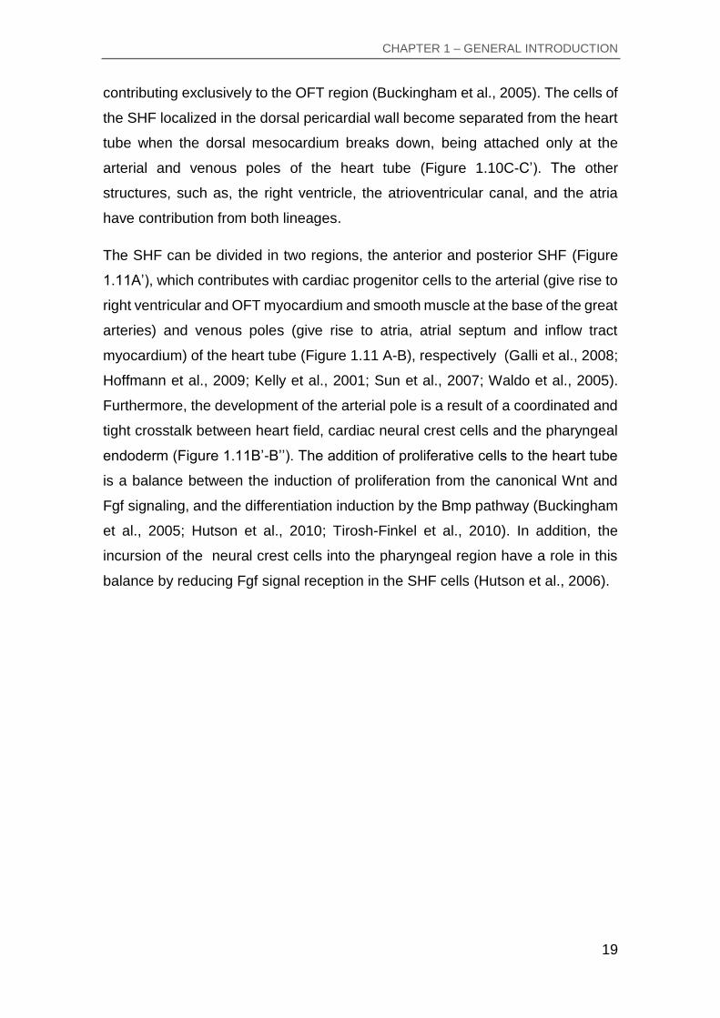
The SHF can be divided in two regions, the anterior and posterior SHF (Figure
1.11A’), which contributes with cardiac progenitor cells to the arterial (give rise to
right ventricular and OFT myocardium and smooth muscle at the base of the great
arteries) and venous poles (give rise to atria, atrial septum and inflow tract
myocardium) of the heart tube (Figure 1.11 A-B), respectively (Galli et al., 2008;
Hoffmann et al., 2009; Kelly et al., 2001; Sun et al., 2007; Waldo et al., 2005).
Furthermore, the development of the arterial pole is a result of a coordinated and
tight crosstalk between heart field, cardiac neural crest cells and the pharyngeal
endoderm (Figure 1.11B’-B’’). The addition of proliferative cells to the heart tube
is a balance between the induction of proliferation from the canonical Wnt and
Fgf signaling, and the differentiation induction by the Bmp pathway (Buckingham
et al., 2005; Hutson et al., 2010; Tirosh-Finkel et al., 2010). In addition, the
incursion of the neural crest cells into the pharyngeal region have a role in this
balance by reducing Fgf signal reception in the SHF cells (Hutson et al., 2006).
CHAPTER 1 – GENERAL INTRODUCTION
20
Figure 1.11 – Contribution of the second heart field to the developing heart. (A) Lateral view of the SHF contribution to the heart at E9.5, showing anterior (green) and posterior regions (blue). Core pharyngeal arch mesoderm of arches 1 (PA1CM) and 2 (PA2CM). (A’) Sagittal section of A, demonstrating the different contributions of the FHF and SHF. (B) Embryonic heart at E10.5 showing parts of the heart derived from the anterior SHF (green), linear heart tube (pink), and posterior SHF (blue). The anterior SHF also contributes with smooth muscle (SmM) at the arterial pole of the heart tube, which is attached to pharyngeal arch arteries (PAA) 3, 4, and 6. (B’) Transverse section in B showing juxtaposition between anterior SHF cells (green) and neural crest-derived mesenchyme (CNC, orange) lateral to and underlying the pharynx (Ph). (B’’) Zoom of the red boxed area in B’ showing mixed CNC and mesodermal (Mes) mesenchymal cells and the balance between FGF and BMP signals regulating proliferation and differentiation during the elongation of the OFT. A-SHF, anterior second heart field; AVC, atrioventricular canal; D, dorsal; d-OFT, distal outflow tract; DPC wall, dorsal pericardial wall; End, endoderm; LA, left atrium; LV, left ventricle; OFT, outflow tract; p-OFT, proximal outflow tract; P-SHF, posterior second heart field; RA, right atrium; SoM, somatic mesoderm; V, ventral. Color code: pink, FHF and derivatives; green, SHF and derivatives; blue, posterior SHF; yellow, endoderm (Adapted from(Kelly, 2012).
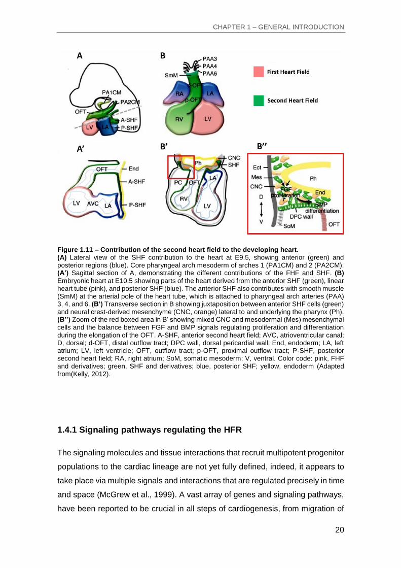
1.4.1 Signaling pathways regulating the HFR
The signaling molecules and tissue interactions that recruit multipotent progenitor
populations to the cardiac lineage are not yet fully defined, indeed, it appears to
take place via multiple signals and interactions that are regulated precisely in time
and space (McGrew et al., 1999). A vast array of genes and signaling pathways,
have been reported to be crucial in all steps of cardiogenesis, from migration of
CHAPTER 1 – GENERAL INTRODUCTION
21
primitive-streak progenitor cells, formation of the HFR, cardiac looping, to later
morphogenetic mechanisms. The major signaling pathways controlling heart
development are the Wnt proteins, Fgf, Notch, Bmp, Nodal and Activin (Figure
1.12) (Noseda et al., 2011). While the architecture of the heart is different
between species, the inductive paths are ancestral and conserved, as is the
transcription machinery that controls the cardiomyogenesis fate. Such
information is crucial to comprehend the formation of the heart and to transform
the knowledge into quantitative network models that provides for uncountable
translational applications.
Figure 1.12 – Summary of major signaling pathways controlling heart development. Schematic representation of the major signaling pathways from left to right: A) BMP, B) Activin/Nodal, C) canonical Wnt D) non-canonical Wnt, E) FGF and F) notch pathways. For details of each, see the text. Circle indicates ligand; inverted triangle, ligand antagonist; rectangle, receptor ligand-binding and signaling domains (light and dark, respectively); square, protein kinase; triangle, G-protein; hexagon, scaffold protein; pentagon, protease; oval, transcription factor (Adapted from (Noseda et al., 2011).
CHAPTER 1 – GENERAL INTRODUCTION
22
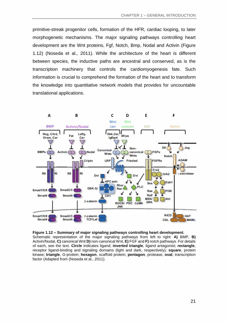
1.4.1.1 Bone Morphogenetic Protein (Bmp)
The Bmp are multifunctional regulators that play a panoply of roles in
development by regulating cell proliferation, differentiation and apoptosis in
different tissues (Massagué and Chen, 2000). In the chick embryo, Bmp are
necessary for the cardiogenic activity of anterior endoderm, inducing the
specification of the cardiogenic mesodermal cells, shown by the ability of
inhibitors like Chordin and Noggin to suppress cardiac differentiation (Andree et
al., 1998; Schultheiss et al., 1997). Activation of BMP signaling pathway starts
when Bmp ligand binds to Bmp receptor I and II (Kishigami and Mishina, 2005).
The receptor II activates the receptor I by phosphorylation of the serine/theronine
residue, thus, the activated receptor I can now phosphorylate the Smad
transcription factors, triggering the intracellular signal cascade. Moreover, Bmp
activity is regulated by a large number of extracellular antagonists, such as,
Cerberus, Chordin and Noggin. These molecules bind directly to Bmp ligands
and block their interaction with signaling receptors, thus inhibiting the Bmp
signaling (Figure 1.12A) (Rodríguez Esteban et al., 1999).
1.4.1.2 Activin and Nodal
Activin and Nodal share receptors I and II and have the same Smad signaling
pathway (Figure 1.12B) (Kitisin et al., 2007). In addition, they signal via Smad2/3,
and the R-Smad/Smad4 complexes promote cooperative DNA binding by the
forkhead winged-helix protein FoxH1, which mediates many effects of the
pathway. The majority of Nodal effects requires a co-receptor of the EGF-CFC
family, whose members are defined by an epidermal growth factor-like domain
and include mammalian Cripto and Cryptic, Xenopus FRL-1, and zebrafish one-
eyed pinhead (Chu and Shen, 2010). The Nodal and Activin activity are regulated
by the extracellular antagonists, Lefty, and Cerberus, and follistatin (Fst),
respectively. Moreover, Nodal is vital for establishing the anterior-posterior and
left-right axes, gastrulation, primitive streak development, and thus the formation
of mesoderm and endoderm, germ layer specification being a requirement for
later cardiac tissue formation (Schier, 2003).

CHAPTER 1 – GENERAL INTRODUCTION
23
1.4.1.3 Wingless-type MMTV integration site (Wnt)
Signaling by the Wnt family of secreted glycoproteins is one of the fundamental
mechanisms that direct/regulate cell differentiation, proliferation, survival, polarity
and migration during embryogenesis (Logan and Nusse, 2004). The signaling
pathways involve 19 Wnt secreted proteins, 10 Frizzle (Fz) receptors and the co-
receptors LRP5 and LRP6 (lipoprotein low-density-lipoprotein receptor-related
protein) in mammals, suggesting a vast complexity of this signaling pathway
(Figure 1.12C-D) (Bejsovec, 2005). Wnt signaling pathways are divided in
canonical (β-catenin) and noncanonical (β-catenin-independent), which have two
branches, the planar cell polarity pathway and the calcium pathway (Nusse, 2005;
Nusse and Varmus, 2012; Nusse and Varmus, 1992).
The canonical Wnt pathway (Figure 1.12C) plays a role in the regulation of the
amount of transcriptional co-activator β-catenin, which controls crucial
developmental gene expression programs. It starts when Wnt proteins (Wnt1, 2a,
3a and 8) bind to Fz and LRP family members, either on the producing or adjacent
cells (MacDonald et al., 2009). Upon receptor binding disheveled protein (Dvl) is
activated, which in turn inhibits a protein complex that includes the constitutively
glycogen synthase kinase 3β (Gsk3β) and CK1 (casein kinase 1), as well as the
scaffolding proteins axin and adenomatous polyposis coli (APC). This complex
normally phosphorylates β-catenin and targets it for degradation. These events
lead to decreased phosphorylation of β-catenin, allowing stabilization,
cytoplasmic accumulation and consequent nuclear translocation of the protein
(MacDonald et al., 2009). The inhibition of the degradation complex allows high
levels of β-catenin to accumulate in the nucleus, where it interacts with TCF/LEF
family DNA binding proteins to activate the transcription of Wnt target genes
(Mosimann et al., 2009). In the chick embryo, experiments showed that the levels
of Wnt canonical (Wnt3a and 8c) signaling are lower in the anterior mesoderm
(cardiogenic) and higher in the posterior mesoderm (blood-forming), thus Wnt β-
catenin signals inhibits cardiac tissue formation in chick embryo and therefore
needs to be inhibited itself for proper heart development (Marvin et al., 2001;
Tzahor and Lassar, 2001).
CHAPTER 1 – GENERAL INTRODUCTION
24
The noncanonical Wnts (Figure 1.12D) such as Wnt4, 5a and 11 activate two
main signaling pathways. In the planar cell polarity (PCP) pathway, Wnt proteins
bind to Fz receptors, that recruit Dvl and activate Rho-family small GTPases (Rho
and Rac) and their downstream effectors such as Rho-associated protein kinase
(ROCK) and Jun N-Terminal kinase (JNK) (Tada et al., 2002). On the other side,
the Wnt/Calcium signaling pathway, through the G-protein-dependent activity of
Frizzled receptors, induce the release of Ca2+ intracellular by phospholipase C,
which activate the Ca2+ - dependent protein kinases Protein Kinase C (PKC) and
CaMKs (calmodulin-dependent protein kinases) (Kohn and Moon, 2005; Sheldahl
et al., 2003). In the chick embryo, experiments demonstrated that Wnt
noncanonical, like Wnt11, is present in the precardiac mesoderm suggesting a
role in myogenesis (Eisenberg and Eisenberg, 1999; Eisenberg et al., 1997). In
addition, noncanonical Wnt signaling inhibits the canonical Wnt signaling
promoting myocardial differentiation and have a role in cardiac morphogenesis
by regulating cadherin-mediated cell adhesion and cell polarity (Brade et al.,
2006).
1.4.1.4 Fibroblast Growth Factors (Fgf)
Fibroblast growth factors (Fgf) (Figure 1.12E) and their receptors control a
panoply of cellular processes, regulating cellular proliferation, survival, apoptosis,
migration and differentiation (Böttcher and Niehrs, 2005). Fgf contain a large
family of growth factors, with twenty two ligands and four transmembrane
receptor tyrosine kinases (Fgfr) (Itoh and Ornitz, 2004; Turner and Grose, 2010).
Fgf proteins are characterized by their high affinity with heparin, a molecule that
facilitates their binding to cell surface Fgfr. Binding of the Fgf ligands to the
extracellular domain of the Fgfr in combination with heparan sulfate leads to the
dimerization of the receptor resulting in the transphosphorylation of specific
intracellular tyrosine residues in the receptor (Turner and Grose, 2010).
Consequently, activate cytoplasmic signal transduction pathways, such as the
Ras/ERK pathway that is associated with differentiation and proliferation, the Akt

CHAPTER 1 – GENERAL INTRODUCTION
25
pathway, which is associated with cell survival or the PKC pathways that have a
role in cell morphology and migration) (Dailey et al., 2005; Schlessinger, 2000).
In the chick embryo, experiments demonstrated that Fgf have several functions
in early cardiogenesis (Parlow et al., 1991; Sugi et al., 1993), such as, the loss of
function of Fgf2 causes loss of cardiac precursor proliferation (Sugi et al., 1995;
Sugi et al., 1993). On the other hand, Fgf2 gain of function induce cardiac actin
(Sugi and Lough, 1995). Ectopic delivery of Fgf8, that is expressed in the
endoderm adjacent to precardiac mesoderm, promotes lateral expansion of the
heart field and ectopic expression of Nkx2.5 and Mef2c, but not Gata4 (Alsan and
Schultheiss, 2002). Fgf and Bmp signaling have an interesting relationship in the
development of the SHF, such as, the role of Bmp4 in the differentiation of cardiac
precursors and at the same time inhibits Fgf (like Fgf8), keeping the cardiac
precursor cells in a proliferative undifferentiated state (Tirosh-Finkel et al., 2010).
1.4.1.5 Notch
Notch signaling (Figure 1.12F) has been shown to be involved in a wide range of
developmental processes, such as, cell fate decisions, cellular development,
differentiation, proliferation, apoptosis, adhesion and epithelial-to-mesenchymal
transition (Miazga and McLaughlin, 2009; Watanabe et al., 2006). The Notch
encodes for a transmembrane protein receptor (Notch 1 to 4), that functions at
the cell surface to bind transmembrane ligands (Delta 1 to 4 or Jagged 1 and 2)
on adjacent cells (Bray, 2006). Upon ligand binding, a series of proteolytic
cleavages (y-secretase complex and a disintegrin and metalloproteinase)
releases the intracellular domain of Notch (NICD) allowing for translocation of the
NICD into the nucleus. This cleavage product mediates the function of Notch in
the nucleus by interacting with CSL proteins (CBF1/recombination signal binding
protein for immunoglobulin kappa J region [Rbpj], suppressor of hairless, Lag1)
to activate downstream target genes (Bray, 2006). The primary downstream
effectors of Notch signaling have been identified as members of the
Hairy/Enhancer of split and Hey families of repressive basic helix–loop–helix
transcription factors that are important to chamber specification and demarcating

CHAPTER 1 – GENERAL INTRODUCTION
26
the AV canal from surrounding myocardium (Davis and Turner, 2001; Iso et al.,
2003; Niessen and Karsan, 2008). Since the NICD interacts with Smad and Dvl
proteins, there is also a crosstalk with the TGF and Wnt cascades (Blokzijl et al.,
2003). In the chick embryo, experiments demonstrated that Notch while blocks
cardiac muscle differentiation, it enhances the expression of conduction system
markers (Chau et al., 2006). Notch signal pathway plays a key role in the
processes of AV canal, myocardial and OFT development and regulation of
endothelial-mesenchymal transition during heart valves formation (Niessen and
Karsan, 2008; Rutenberg et al., 2006).
1.4.2 Regulation of Second Heart Field
The SHF, that is located at caudal pharyngeal region, contains undifferentiated
and highly proliferative cell population that can give rise to myocardial,
endocardial and smooth muscle cells (Laugwitz et al., 2005; Moretti et al., 2006),
which is regulated by a complex network of intercellular signals. These signals
control and are controlled by transcription factors in the pharyngeal mesoderm
and adjacent cells types, like neural crest-derived cells (Figure 1.13) (Dyer and
Kirby, 2009; Vincent and Buckingham, 2010). The SHF is characterized by the
expression of transcription factors Islet-1 and Tbx1 and the growth factors Fgf8
and Fgf10 (Cai et al., 2003; Ilagan et al., 2006; Kelly et al., 2001; Xu et al., 2004).

CHAPTER 1 – GENERAL INTRODUCTION
27
Figure 1.13 – Signaling pathways regulating the second heart field. (A) Signaling at the arterial pole of the heart tube displaying zones of Wnt, FGF, Hedgehog (Hh), and BMP. (B) Schematic network representation of the major signaling pathways and regulatory genes involved in SHF development during the transition from proliferating progenitor cell (top) to differentiated cardiomyocyte (bottom). Isl1 and Tbx1 have a central position in controlling the proliferative progenitor cell state (top), FGF/BMP antagonism with a pivotal position in regulating the balance between proliferation and differentiation (middle), and the activation of the cardiomyogenic program by a network of interacting transcription factors (bottom). Gray lines, direct protein interactions; dotted lines, microRNA silencing. LV, left ventricle; RV, right ventricle;
OFT, outflow tract; SHF, second heart field; NC, neural crest‐derived cells (Adapted from(Kelly, 2012).
1.4.2.1 Regulation of proliferation in Second Heart Field
The activation of the cardiac transcriptional program in the cardiogenic mesoderm
(anterior lateral splanchnic mesoderm) is regulated by signals from adjacent
tissues, including Wnt, Fgf and Bmp signals (Figure 1.14) (Evans et al., 2010).
Progressive control of mesodermal precursor cells to a myocardial fate begins
with the induction of Mesp1 in the anterior mesoderm precursor cells, followed by
activation of cardiac transcription factors and epigenetic regulators, including Isl1,
Tbx5, Nkx2-5, Mef2c, Gata4 and Baf60c that together drive cardiomyogenesis
(Miquerol and Kelly, 2013). As the linear heart tube form, SHF cells (expressing
Isl1) in medial splanchnic mesoderm remain in contact with the pharyngeal
endoderm and their continued proliferation, delayed differentiation and
contribution to growth of the myocardium is regulated by canonical Wnt, Fgf and

CHAPTER 1 – GENERAL INTRODUCTION
28
Hedgehog signalling pathways (Figure 1.13) (Cai et al., 2003). During heart
development the SHF cells contributes to the elongation of the heart tube through
the dorsal mesocardium. After its rupture and consequent dorsal closure of the
heart tube, cardiac precursors cells in pharyngeal mesoderm are isolated in a
highly proliferation region (dorsal pericardial wall) contributing to the heart tube
extension by addition at both poles (van den Berg et al., 2009). In the chick
embryo, the proliferative center is localized caudally in the dorsal pericardial wall,
moving from this region towards the poles of the heart tube at a high rate (van
den Berg et al., 2009). As cells differentiate at the poles of the heart tube, cell
proliferation dramatically drop, to be restarted during the development of the
cardiac chamber (de Boer et al., 2012).
In the dorsal pericardial wall, the proliferation of the cardiac precursors appears
to be controlled by Fgf signaling (Figure 1.14), namely Fgf3, 8 and 10, which are
expressed in the pharyngeal mesoderm and adjacent pharyngeal epithelia. Fgf8,
within pharyngeal mesoderm, appears to be the main controller of heart tube
extension with important contributions from Fgf10 and Fgf3 revealed by analysis
of mutant embryos (Park et al., 2008; Urness et al., 2011; Watanabe et al., 2010).
These Fgfs, expressed in the pharyngeal region, are regulated by Wnt/β-catenin
and Notch signaling (Cohen et al., 2007; Klaus et al., 2012). The conditional loss-
of-function analysis of these signaling pathways in the SHF results in OFT defects
leading to subsequent arterial pole septation defects (Klaus et al., 2012; Park et
al., 2008). Additionally, the migration of neural crest cells into the caudal
pharyngeal regions regulates local Fgf signaling and leads to a reduction in cell
proliferation in the SHF, acting as a proliferative brake during the terminal stages
of heart tube extension (OFT septation) (Hutson et al., 2006). Recently it was
shown, that canonical Wnt signaling (β-catenin) operate downstream of Notch in
the regulation of cardiac precursors cell differentiation and OFT morphogenesis
(Klaus et al., 2012). Hedgehog (Hh), expressed in the pharyngeal endoderm, is
required for maximal proliferation in the SHF and to maintain progenitor cell
properties of SHF cells in the dorsal pericardial wall (Dyer and Kirby, 2009). In
addition, Hh signaling has been shown to control progenitor cell proliferation in
the posterior SHF via the transcription factor Tbx5, in a pathway vital for atrial
septation (Xie et al., 2012).

CHAPTER 1 – GENERAL INTRODUCTION
29
Figure 1.14 – Second heart field major signaling pathways. Image shows the major signaling pathways known to control proliferation and progressive differentiation of second heart field cardiac progenitor cells in the early embryo (Adapted from(Rochais et al., 2009b).
In all animal models, LIM homeodomain transcription factor Islet-1 (Figure 1.13)
marks proliferating undifferentiated cardiac precursors. Islet-1 is essential for
proliferation and survival of the SHF progenitors, and might be required for their
migration into the heart (Cai et al., 2003), indeed, analysis of Islet-1 mutants
demonstrated defects in the arterial and venous poles of the heart tube (Park et
al., 2006). Initial expression of Islet-1 is dependent of Wnt/-catenin and Fgf
signaling, both of which are required for proliferation of early cardiac precursors
(Cohen et al., 2007). β-catenin can directly activate transcription from Islet-1 and
Fgf10 promoters and when canonical Wnt signaling is increased Islet-1 positive
cells shows up-regulation of Fgf, promoting proliferation (Kwon et al., 2009).
Moreover, Islet-1 controls the T-box transcription factor 1 (Tbx1) (Figure 1.13),
that regulates Fgf signaling (Cai et al., 2003; Vitelli et al., 2002). Loss of Tbx1 in
mouse embryos demonstrated a reduction of cardiac progenitor cells proliferation
resulting in a hypoplastic dorsal pericardial wall and in a short narrow OFT (Zhang
et al., 2006). This transcription factor is the major candidate gene for DiGeorge
syndrome in man, associated with craniofacial and cardiovascular anomalies
including OFT congenital heart defects (Baldini, 2005).
CHAPTER 1 – GENERAL INTRODUCTION
30
Hes1, a target gene of the Notch signalling pathway, is present in the dorsal
pericardial wall and is required for proper cardiac precursors cells proliferation,
preventing precocious differentiation (Rochais et al., 2009a). In SHF
development proliferation is a crucial regulatory step, however cell survival also
as an important role in the SHF that was demonstrated with the transcription
factor Hand2 (Tsuchihashi et al., 2011).
1.4.2.2 Control of gradual differentiation during heart tube elongation
The transition from proliferative cardiac precursors to differentiated
cardiomyocytes requires both downregulation of expansion pathways and
upregulation of differentiation pathways (Figure 1.14). The SHF cells during heart
tube elongation migrates to the ventral region of the embryo and as these cells
approach both poles of the heart tube, they are exposed to Bmp and
noncanonical Wnt signals that drive differentiation and at the same time cell
proliferation decreases (Figure 1.13 and Figure 1.14). Thus, the balance and
transition between undifferentiated cardiac precursors to differentiated
cardiomyocytes is important to regulate the gradual addition of SHF cells to the
heart tube. At the distal OFT (arterial pole) the differentiation is regulated by Bmp
signaling, antagonizing Fgf signals through neural crest-derived cells (Msx1 or
Msx2) promoting cardiac differentiation (Figure 1.14) (Hutson et al., 2010; Tirosh-
Finkel et al., 2010). Indeed, the balance between these pathways is influenced
by neural crest cell influx into the pharyngeal region, playing a crucial role in
slowing the proliferative effect of Fgf in the SHF region (Hutson et al., 2006).
Furthermore, Bmp signaling is capable of the activation of transcription
microRNAs that targets SHF regulatory genes, like Islet-1 and Tbx1 (Wang et al.,
2010). When cardiac progenitor cells are near to the heart tube, they initiate
expression of genes encoding crucial cardiac transcription factors, such as
Nkx2.5, Tbx20, Mef2c, Gata4 and Hand2, that are required to activate the cardiac
tissue formation (Waldo et al., 2001). Specification of cardiac precursor cells in
pharyngeal mesoderm is increased after Nkx2.5 ablation through upregulation of
Jarid2 and Bmp ligand gene expression (Barth et al., 2010). On the other hand,

CHAPTER 1 – GENERAL INTRODUCTION
31
Nkx2.5 is capable of repressing Bmp2 activated Smad1 signaling to inhibit
differentiation (Prall et al., 2007).
In embryos lacking Tbx2 and Tbx3, the Bmp signal is downregulated resulting in
increased Fgf signaling proximal to the OFT (Mesbah et al., 2012). In addition,
Tbx1, 2 and 3 regulates positively the proliferation and negatively the
differentiation in the caudal pharyngeal region maintaining the cardiac progenitor
cells homeostasis in the SHF. It can inhibit BMP signaling by direct binding to the
mediator Smad1 and suppressing BMP4/Smad1 pathway. While, in Tbx1
knockout mice occurs premature differentiation of the SHF progenitors due to
abnormal induction of differentiation, in gain of function approaches it leads to
reduction of differentiation in the OFT (Chen et al., 2009; Liao et al., 2008).
Moreover, Tbx1 has been shown to interact with Mef2c and serum response
factor, thus preventing cardiomyocyte differentiation (Chen et al., 2009; Pane et
al., 2012). Therefore, loss of function of any two of these three genes results in
severely altered Bmp and Fgf signaling, pharyngeal hypoplasia and heart tube
expansion defects (Mesbah et al., 2012). Different from Tbx1, Islet-1 seems to be
maintained in the distal part of the OFT and through Mef2c it contributes to
cardiac progenitor differentiation (Dodou et al., 2004).
Studies in which -catenin was constitutively activated in cardiac progenitors,
caused lack of differentiation to myocardial cells. Loss of Wnt signaling reduces
the number of Islet-1 expressing cells, causing OFT and right ventricle defects,
while overexpression of Wnt signaling expands Islet-1 positive population (Cohen
et al., 2007). Therefore, the canonical Wnt/-catenin signaling inhibits cardiac
induction, promoting proliferation and sustaining the SHF in an undifferentiated
state. Its repression is essential for myocardial differentiation, which induces both
Bmp and noncanonical Wnt (Wnt11 and Wnt5a) expression in the SHF region
(Cohen et al., 2012; Klaus et al., 2007).

CHAPTER 1 – GENERAL INTRODUCTION
32
1.4.2.3 Patterning of cardiac precursor cells in the dorsal pericardial wall
In the dorsal pericardial wall the cardiac precursors are regulated by complex
interactions of signals, which promotes proliferation and gradually regulates
differentiation (Figure 1.14). During heart tube elongation the SHF cells become
patterned, being distinguished in two populations of cells, the anterior SHF
adjacent to the arterial pole and the posterior SHF adjacent to the venous pole,
in which they are responsible for the formation of the right ventricular, OFT and
atrial myocytes. Among the genetic markers that distinguish these populations of
precursor cells and provide the information of anterior-posterior patterning in the
pharyngeal mesoderm, are the Fgf10 and a Mef2c expressed in the anterior SHF,
and Tbx5 and Osr1 present in the posterior SHF (Kelly, 2012). Hox genes are
key controllers of anterior–posterior information in the developing embryo,
contributing to the atrial myocardium, inferior wall of the OFT and to the
myocardium at the base of the outlet of the right ventricle (sub-pulmonary
myocardium) (Bertrand et al., 2011). Moreover, the future subpulmonary
myocardium appears to be mainly dependent on Tbx1 function, which is known
to have contributions to the inferior wall of the developing OFT (Théveniau-Ruissy
et al., 2008). Interestingly, in the Tbx1 null embryos, the failure of subpulmonary
myocardial development is associated with abnormal expression of Fgf10
transgene in myocardium at the venous pole of the heart (Kelly and Papaioannou,
2007). This suggests that Tbx1 plays a role not only in regulating proliferation and
differentiation delay, but also in patterning of cardiac precursors cells in the dorsal
pericardial wall.
CHAPTER 1 – GENERAL INTRODUCTION
33
1.5 Aim of this thesis
Previously in our lab, a differential screening to search for novel genes
differentially expressed in the heart/hemangioblast precursor cells of chick
embryos was performed. From this screening, several genes were identified.
Among others, Collagen and calcium-binding EGF-like domain 1 (cCcbe1), a
novel protein containing signal peptide for secretion, collagen domains and a
calcium binding EGF-like domain, was found to be upregulated in the cardiac
progenitors in comparison to the embryonic control cells. The overall aim of this
thesis is to study and characterize the role of cCcbe1 during early heart
development in the chick.
In particular, the specific aims of this thesis are:
First, to generate a fine and detailed characterization of the expression pattern of
cCcbe1 during early heart development, employing double whole-mount in situ
hybridization with well-known cardiac markers and histology techniques to
determine in which structures this gene might be playing a role.
Second, by employing overexpression and knockdown approaches, to address
the functional role of cCcbe1 in chick early heart development.

CHAPTER 2 – MATERIALS AND METHODS

CHAPTER 2 – MATERIALS AND METHODS
35
2.1 Chick embryo collection and culture
Fertilized chicken eggs (Sociedade Agrícola Quinta da Freiria, SA, Torres
Vedras, Portugal) were incubated for 1-3 days maintained at 38 °C in a humidified
incubator. Embryos were staged according to Hamburger and Hamilton
(Hamburger and Hamilton, 1951) (Appendix 1). For the culture, embryos were
explanted at HH3+/HH4 together with the vitelline membrane and anchored to a
metacrilate ring following the protocol of New (New, 1955).
2.2 Embryo dissection and fixation
Fertilized chick eggs were removed from the incubator after the suitable time of
incubation, in order to obtain the appropriated developmental stages, and broken
into a sterile petri dish. Embryos were dissected (suitable and sterilized surgery
material) from the egg and placed in ice-cold RNase-free phosphate buffer saline
(PBS 1X plus diethyl pyrocarbonate (DEPC) treated water) for further
manipulations. Extraembryonic membranes were removed and for embryos older
than stage 12, a small hole was placed in the head and heart with a glass-pulled
pipette. Embryos were fixed overnight at 4ºC in 4% paraformaldehyde (4%PFA
plus PBS1X DEPC), in order to preserve all the embryo structures and to reduce
RNA degradation, and dehydrated the following day. Following removal of the
fixative from the embryos, two 5 min washes were carried out with ice-cold PBT
(PBS 1X + 0.1% Tween-20). The embryos were then dehydrated for 10 min in
each of 25% methanol (in PBT), 50% methanol (in PBT), 75% methanol (in PBT)
and twice in 100% methanol and subsequently stored at –20°C until required.
The embryos must be stored in 100% methanol to prevent the formation of water
crystals, in order to protect the integrity of the cells (List of solutions and their
composition in appendix 2).

CHAPTER 2 – MATERIALS AND METHODS
36
2.3 Synthesis of antisense mRNA probe
The chick Ccbe1 (1191-bp) was generated by RT-PCR cloning (clone was
obtained from the BBSRC chick EST database: ChEST963b3 for cCcbe1; seq.
identifier 603865952F1).
In order to study a particular gene it is necessary to generate a probe that will
mark its domains of expression, meaning, an antisense RNA strand for the mRNA
of interest must be synthesized and the following steps must be taken.
2.3.1 Transforming of competent E. coli cells
To generate the probes used in whole-mount in situ hybridization (WISH),
chemically competent Escherichia coli cells were transformed with a plasmid that
contains the coding sequence of the gene of interest. The transforming DNA was
added (no more than 50ng in a volume of 10µl or less) to 100µl of competent cells
(DH5α Max efficiency) in a polypropylene tube, under a flame of a Bunsen burner.
The tube was swirl gently in order to mix their content, and stored on ice for 20
min. The tube was placed in a preheated 37ºC water bath for 60 seconds, without
shaking, and rapidly transferred to an ice bath, allowing the cells to cool for 2 min.
After this period, 400µl of Luria Broth (LB) medium was added and the tube was
transferred to a shaking incubator at 37ºC for 45 min to allow the bacteria to
recover and to express the antibiotic resistance marker encoded by the plasmid.
Appropriated volume (up to 200µl per 90-nm plate) of the transformed competent
cells was transferred onto an agar plate with ampicillin. The plate was stored at
room temperature until the liquid has been absorbed, and then inverted and
incubated at 37ºC for 12-16 hours.
2.3.2 Plasmid amplification
Individual bacterial colonies were inoculated into 3ml of LB media containing the
appropriate selective antibiotic (100µg/ml of ampicillin). The tube containing the

CHAPTER 2 – MATERIALS AND METHODS
37
suspension of cells with the plasmid of interest was incubated at 37ºC in a
shaking incubator overnight (225rpm).
2.3.3 Plasmid DNA Isolation
Plasmid DNA extraction was carried out using a miniprep kit (QIAprep Spin
miniprep kit, Qiagen), according to the protocol provided by the manufacturer:
1.5ml of cell suspension was pelleted by centrifuging 1 min at 9000 rpm in a
microcentrifuge and the supernatant discarded; then 250µl of a resuspension
buffer (Solution I)) was added and the tube vortexed until the cells were in
suspension, then another 250µl of lyses buffer (Solution II) was added and the
tube mixed thoroughly by inverting several times, to obtain a clear lysate. The
cells were left incubating up to 5 min in this buffer. In order to stop the lyses
reaction, 350µl of neutralization buffer (Solution III) was added and the tube was
gently mixed by inverting several times until a flocculent white precipitate forms.
The tube was centrifuged for 10 min at maximum speed (13000rpm) and a
compact white pellet was observed. Then the supernatant, by pipetting, were
transferred to the QIAprep spin column, following centrifugation for 60s and the
discard of the flow-through. The QIAprep spin column was washed by adding
500µl of washing buffer I (HB) and centrifuging for 60s. The flow-through was
discarded and 700µl of washing buffer II was added to the column, and was
centrifuged for an additional 1 min to remove the residual wash buffer. To elute
the DNA, the column was placed in a clean 1.5ml microcentrifuge tube and 50 µl
to 100µl of elution buffer (EB) (10mM Tris-HCl, pH 8.5) was added to each
QIAprep spin column, let stand for 1 min, centrifuged for another 1 min and after
that the DNA was collected.
2.3.4 Plasmid Linearization and Purification
In order to make antisense probes it‘s necessary to use restriction enzymes and
RNA polymerases to synthesized antisense RNA probes for the genes of interest,

CHAPTER 2 – MATERIALS AND METHODS
38
namely, cCcbe1, Nkx2.5, Tbx-5, Mhc and Islet-1 (Table 1). The reaction mixture
was prepared in a 1.5ml microtube: 10µl (2µg) of plasmid DNA was digested
using the appropriated restriction enzyme, the adequate volume of the supplied
reaction buffer (Roche) and sterile deionized water to a total volume of 30µl; and
then was incubated at 37ºC for 3h or overnight. The digestion product was
checked on a 1% agarose gel (SeaKem LE Agarose), using 1/30 of the digestion
reaction volume, to confirm the completion of the digestion.
Table 2.1 – List of restriction enzymes and RNA polymerases used for antisense RNA probe preparation
Clone Restriction enzyme RNA polymerase
cCcbe1 (in pGem-TEasy)
NcoI SP6
Tbx5 (in pBlueScriptIIKS(+/-))
XbaI T7
Nkx2.5 (in pGem-TEasy)
NcoI SP6
Islet-1 (in pBlueScriptIIKS(+/-))
NotI T7
Mhc (in pBlueScriptIIKS(+/-))
NdeI T7
The linearized DNA was purified using spin columns from the QIAprep Spin
Miniprep Kit. To do this, 5x the volume of the digestion reaction mixture volume
(30µl) was added with washing buffer I (150µl), thoroughly mixed, transferred to
the column and centrifuged for 1min at maximum speed (13000rpm). The flow-
through was discarded; the spin column was washed with 700µl of ETOH buffer
(buffer II) and centrifuged at maximum speed for 1min. Again, the flow-through
was discarded and the spin column centrifuged at maximum speed (13000rpm)
for 1min, to get rid of residual ethanol from buffer II. The column was placed into
a clean 1.5ml microcentrifuge tube; the nuclease-free water (Promega) was
added to the center of the QIAquick membrane for 5 min, to elute the DNA, and
then centrifuged for 1 min at maximum speed. The spin column was discarded
and the linearized DNA in the microtube was now purified.

CHAPTER 2 – MATERIALS AND METHODS
39
2.3.5 Antisense RNA probe synthesis by in vitro transcription
The RNA probes were generate by in vitro transcription from a linearized DNA
template using the appropriated SP6 or T7 (Roche) RNA polymerase, which
synthesizes RNA complementary to the DNA template. Transcription reaction
components were added in the following order to a 1.5ml RNase-free tube at
room temperature: nuclease-free water up to a final of 20μl, 10μl linearized and
purified DNA template, 2μl 10X transcription buffer (Roche), 2μl 10X DIG (Roche)
or 2µl Fluo (Roche) RNA labelling mix, 1µl of RNase inhibitor (RNAsin; Promega)
and 1μl of T7 or SP6 RNA polymerase. The reaction mix was then incubated at
37°C for 2 h.
2.3.6 Antisense RNA probe purification
Mini Quick Spin RNA columns were used to remove unincorporated nucleotides
from the prepared labelled RNA. During centrifugation mini Quick spin columns
allow larger molecules (DNA, RNA or oligonucleotides) to pass through quickly,
while retaining smaller molecules (such as unincorporated oligonucleotides). The
matrix of the mini Quick Spin RNA columns (Roche) was initially resuspended in
the buffer by gentle inverting. The top cap of the column was removed, the bottom
tip snapped off and it was placed in a sterile microcentrifuge tube. To pack the
column matrix and remove residual buffer the column was centrifuged (at 4ºC)
for 1min at 1000g. The antisense RNA probe was then applied to the center of
the column bed, followed by the centrifugation (at 4ºC) of the column for 4 min at
1000g, the sample was collected in a microcentrifuge tube and stored at -20ºC.
2.3.7 Agarose gel electrophoresis of DNA
Electrophoreses through agarose gels is commonly used for separating and
analyzing DNA, since the DNA fragments are separated according to the
molecular weight. This technique was used to check the completion of a

CHAPTER 2 – MATERIALS AND METHODS
40
restriction enzyme digestion or to determine the yield of the plasmid DNA
purification.
DNA size determination and semi-quantification were achieved by agarose gel
electrophoresis. The size of the DNA fragment to be resolved determined the
percentage of agarose in the gel. Gels ranging from 1% to 2% (w/v) agarose were
routinely used. Agarose gels were prepared by adding an appropriate amount of
DNA grade agarose to 1x TAE (40mM Tris.acetate, 2mM EDTA pH8) buffer and
boiling until dissolved. Upon cooling to approximately 50°C, Ethidium bromide
(EtBr) was added to a final concentration of 0.05%. The agarose solution was
then poured into a suitable mold to set and following this, placed in an
electrophoresis apparatus with 1x TAE running buffer. DNA samples were mixed
with Orange G loading buffer (15% Ficoll, 0.2% Orange G) and then loaded
alongside a 1 kb Plus DNA ladder (Invitrogen). The DNA was electrophoresed at
90-120V. The EtBr is a fluorescent dye that intercalates between the bases of the
DNA, allowing the gel to be visualized by ultraviolet (UV) illumination. The gel
was then observed with a UVP® white/UV transilluminator (AGP Technologies),
Gene Flash BioImaging System
2.4 Morpholinos and DNA constructs
The pCAGGS-GFP vector (Momose et al., 1999), carrying the cDNA of the green
fluorescence protein under the control of the CAGGS promoter, was used to
control the extent and efficiency of electroporation. Chick Ccbe1 overexpression
plasmids were based on a modified pCAGGS-MCS-IRES-GFP vector (gift from
I. Palmeirin, CBME, UALG). The coding sequence of cCcbe1 was amplified by
PCR. The cCcbe1 coding sequence was isolated by reverse transcriptase (RT)-
PCR according to the published sequence (GeneBank accession no.
XM_001233357) and subcloned into the EcoRI site of pCAGGS-MCS-IRES-
GFP. Fluorescein-tagged antisense morpholinos oligonucleotides cCcbe1MO: 5’-
CGCGGCTCTGCGCTCACCTGAAGCA-3’ and CoMO: 5’-
CCTCTTACCTCAGTTACAATTTATA-3’ were designed and produced by Gene
Tools.

CHAPTER 2 – MATERIALS AND METHODS
41
2.5 Early chick embryo electroporation
Embryos were microinjected and electroporated as described previously
(Tavares et al., 2007) at HH3+/HH4 stage with DNA solution (0.5–3 mg/ml; 0.1%
Fast Green; Sigma) in the region fated to form the heart. For the knockdown
experiments, control morpholino (CoMO) or cCcbe1 morpholino (cCcbe1 MO)
(Gene Tools LLC) were electroporated. For the gain-of-function experiments the
control (pCAGGS-IRES-GFP) or cCcbe1 overexpression (pCAGGS-cCcbe1-
IRES-GFP) vectors were electroporated. The embryos were then incubated at 38
°C for the appropriate period of time (10–30 h), at end of the cultured fixed with
4% paraformaldehyde (PFA) and processed for WISH and immunofluorescence.
The embryos were observed under a fluorescence stereomicroscope (Leica
MZ16FA).
2.6 Whole-mount in situ hybridization
It’s a technique that allows specific nucleic acid sequences (mRNAs) to be
detected and studied their expression patterns in embryonic tissues or sections.
The constant improvements in the protocol allow us to detect and visualize two,
or even three, mRNAs in the same embryo. This allows a finer characterization
of the spatial and temporal relationships between the expressions of genes, even
to the level of being able to show simultaneous expression of two genes within
one cell. In this study, WISH was carried out in order to detect mRNA transcripts
within wild-type chick embryos.
2.6.1 Embryo pre-treatments
Embryos were rehydrated by washing through the methanol series in reverse,
100% - 75% - 50% - 25% (for periods of 5 to 10 min). Embryos were washed two
times for 5 min in PBT and then incubated (in the dark) in a solution of 6%
hydrogen peroxide in PBT during 1 h at room temperature. After this, embryos

CHAPTER 2 – MATERIALS AND METHODS
42
were washed three times for 5 min in PBT and then treated with 10μg/ml
proteinase K (Roche) in PBT at room temperature and the period of treatment
depended on the HH developmental stage of the chick embryo (Table 2). After
that, the embryos were washed for 5min in a freshly prepared glycine solution
(2mg/ml in PBT), subsequently two washes were performed with PBT and refixed
with 0.2% glutaraldehyde/4% paraformaldehyde in PBT for 20 min. Embryos
were then washed twice in PBT for 5 min.
Table 2.2 – Embryo’s Proteinase K time.
Embryonic stage Proteinase K time (minutes)
HH3 – 5 1’ HH6 - 8 6’
HH9 - 12 10’ HH13 - 16 12’ HH17 - 20 15’ HH21 - 27 20’
2.6.2 Hybridization
Hybridization comprises two steps. First the embryos (on vials) must be incubated
in 1ml prehybridization solution (Appendix 2) for 3 hours at 70ºC, period in which
the solution can penetrate into the cells and the optimal temperature to the
hybridization is achieved. Then the hybridization buffer was removed and new
hybridization buffer with the previously prepared antisense RNA probe was
added. Then it was performed overnight at 70ºC.
2.6.3 Antibody incubation
After the hybridization steps, the solution was removed and replaced with solution
I (pre-heated to 70ºC) (Appendix 2) and incubated at 70ºC for 1 h (this step was
performed twice). Subsequently the solution I, was removed and replaced with
solution III (pre-heated to 70ºC) (Appendix 2) and incubated at 65ºC for 30 min
CHAPTER 2 – MATERIALS AND METHODS
43
(this step was performed twice). Afterwards, the embryos were rinsed 2 times
with MABT solution (Appendix 2) for 5 minutes each, at room temperature with
rocking. In order to block unspecific antibody binding sites, the embryos were
incubated with blocking solution (MABT/10% Sheep Serum/2% Blocking
Reagent) for 2 to 3 h at room temperature in a rocker. The previously solution
was removed and replaced by the same solution but containing a dilution 1/2000
of anti-DIG-AP antibody (anti-Digoxigenin Fab fragments antibody, AP
conjugated, Roche) or 1/4000 anti-Fluo-AP antibody (anti-Fluorescein Fab
fragments antibody, AP conjugated, Roche) being incubated overnight at 4ºC
with smooth agitation.
2.6.4 Immunological detection
The antibody solution was removed and the embryos were washed in MABT
three times for 5 min and three times for 1 h at room temperature. The embryos
were then washed with NTMT solution (Appendix 2) three times for 10 min. The
detection buffer contains a precipitating substrate of alkaline phosphatase (AP),
is added to the embryos which by the previous treatments, have antibodies
attached with this enzyme in the place where the antisense RNA probe is
connected to the mRNA of the gene, which provides where the gene of interest
is being expressed. So, the embryos were incubated in the detection buffer (BM
Purple or BCIP, Roche) in the dark, at room temperature, for the appropriated
time. Once achieved the desired staining, the embryos were washed with NTMT
solution (it’s used when it’s needed to take some photographs and continue to a
Double WISH) or several times in PBT in order to stop completely the reaction.
Embryos were photographed, post-fixed, dehydrated and subsequently stored at
-20°C.

CHAPTER 2 – MATERIALS AND METHODS
44
2.6.5 Double whole-mount in situ hybridization
The double (two-color) WISH method is used to determine both spatial and
temporal patterns of gene expression, using two genes of interest. The procedure
was the same as describe above except that the embryos were hybridized with
two different probes, one attached to DIG and the other attached to Fluo. Color
reaction for anti-Fluo/AP substrate (BCIP) was performed and a light blue color
appeared. Then the embryos were washed once with PBT for 5 min, twice with
MABT for 5 min, twice with MABT for 10 min and then incubated in MABT at 70ºC
for 1 h. After inactivating the AP attached to the anti-Fluo antibody in this last
incubation step, the embryos were washed twice with MABT for 10 min, and
incubated with blocking solution for 2 to 3 h; afterwards the blocking solution was
removed and replaced by the same solution but containing anti-DIG/AP antibody
being incubated overnight at 4ºC with smooth agitation. Subsequently, the
immunological detection step (2.6.4) was repeated, but the detection buffer used
was the BM Purple, that yields a purple staining.
All the embryos used for the whole mount in situ hybridization and the double
WISH were photographed in PBT or NTMT using a Leica DC 200 camera coupled
to a Leica MZ16FA stereomicroscope. Images were processed using Adobe
Photoshop software.
2.7 Histological sections
The embryos were processed for histological analysis by sectioning after re-
fixation and dehydration by methanol series. Subsequently, they were washed in
isopropanol for 15 min at 65ºC, after that, the embryos were transferred to the
molds and incubated with a 1:1 of isopropanol and paraffin solution during 30 min
at 65ºC. The next step is embedding the embryos in paraffin by washing for 1 h
at 65ºC and a second time overnight at 65ºC. The embryos were oriented, the
paraffin solidified and the blocks were stored at 4ºC. The sectioning was
performed using microtome Leica RM2125RT, first it’s was performed a trim at
40µm to remove the excess of paraffin, and then sections at 8µm, both sagittal

CHAPTER 2 – MATERIALS AND METHODS
45
and transversal sections of the embryo were collected to microscope slides
(Menzel-Glaser, Superfrost), subsequently the slides with the sections of the
embryos were placed in a hot chamber at 37ºC overnight, in order to the sections
adhere to the slides.
After the sections, the next step is to remove the paraffin from the tissue’s
sections by washing twice with xylene during 15 min to remove the paraffin. The
final step is to mount with DPX (mountant for histology, Fluka) and microscope
cover glasses of 24x60mm and let stand them overnight.
Histological sections of the embryos were photographed using an Olympus DP11
camera attached to an optic microscope (Olympus). Images were processed
using Adobe Photoshop software.
2.8 Immunohistochemistry analyses
Fixed untreated, CoMO, cCcbe1MO, pCAGGS-IRES-GFP (control) and
pCAGGS-Ccbe1-IRES-GFP injected embryos were washed with PBS,
dehydrated in methanol series and paraffin embedded. Serial 8 m sections were
taken (microtome Leica-RM 2135), dewaxed and rehydrated. After tissue
rehydration, antigen retrieval was performed in 10 mM TrisBase/1mM EDTA
solution/0.05% Tween20 pH 9.0. Immunostaining was performed using primary
antibodies against avian MF20 (1:200; MF 20-c; DSHB), Phospho-Histone H3
(Ser10) (1:400; Cell Signaling), Hnk1 (1:200; 1C10; DSHB) and fluorescently
coupled secondary antibodies Alexa Fluor 594 goat anti-mouse (1:800; #A11005;
Molecular Probes) or Alexa Fluor 488 goat anti-rabbit (1:800; #A11008; Molecular
Probes). Cell nuclei were labeled with 4', 6-diamidino-2-phenylindole (1ug/ml;
DAPI; Sigma). Sections were mounted with Mowiol and analyzed with Zeiss
Axioimager Z2 microscope (Carl Zeiss Group). For quantitation of the Hnk1
signal, fluorescence images of the heart region were processed using ImageJ
software. The level of background fluorescence was estimated by averaging
background values at four points of each image and was subtracted from the
fluorescence. Then areas of fluorescence were marked manually, and

CHAPTER 2 – MATERIALS AND METHODS
46
fluorescence values were calculated automatically as described elsewhere
(Nakamura et al., 2012).
2.9 Western blotting
The area pellucida of four cCcbe1 MO injected embryos and respective control
MO at stage HH11 embryos was microdissected and suspended in ice-cold lysis
buffer consisting of 20 mM HEPES, pH 7.5, 50 mM β-glycerophosphate, 10%
glycerol, 2 mM EGTA, 1% Triton X-100, 1 mM sodium vanadate, and the
Complete protease inhibitor cocktail (Roche, Indianapolis, IN). The explants were
homogenized on ice, centrifuged and transferred to a fresh tube. Upon
quantification of the total protein concentrations by the method of Bradford, 10 µg
of protein extracts from control and cCcbe1 knockdown embryos were loaded on
a 12% SDS-PAGE polyacrilamide gel, subjected to electrophoresis and
transferred to Hybond-C extra membrane (Amersham Pharmacia Biotech). Blots
were probed with the antibody against Ccbe1 (Ccbe1 ab 101967; 1:500; Abcam,
UK) followed by 1 hour at RT with a rabbit polyclonal secondary antibody (Dako,
Denmark), developed using a chemiluminescent substrate (Pierce) and analysed
using Chemidoc (Bio-Rad).
2.10 Statistical Analysis
Results were expressed as mean ± standard error of the mean (S.E.M.), and
experiments were performed at least in triplicate. Significant differences were
assessed by t-test using SigmaStat vs 3.5 software. Differences at p-value <0.05
were considered to be significant.

CHAPTER 3 – RESULTS

Expression and function of Ccbe1 in the chick early cardiogenic regions are required for correct heart development
48
Expression and function of Ccbe1 in the chick early cardiogenic
regions are required for correct heart development
João Furtado a,b, Margaret Bento a,b, Elizabeth Correia a,b, José Inácio a,b and José
A. Belo a,b,c
a Regenerative Medicine Program, Departamento de Ciências Biomédicas e Medicina,
Universidade do Algarve, Portugal.
b IBB–Institute for Biotechnology and Bioengineering, Centro de Biomedicina Molecular e
Estrutural, Universidade do Algarve, Campus de Gambelas, Faro, Portugal.
c Faculdade de Ciências Médicas, Universidade Nova de Lisboa, Campo Mártires da Pátria 130,
Lisboa, Portugal
Manuscript submitted to PLOS ONE
Author’s contribution:
The majority of the experimental work was performed by J. Furtado with the
exception of the generation of pCAGGS-cCcbe1-IRES-GFP vector which was
generated by M. Bento; and some of the gain-of-function experiments were
performed by both.
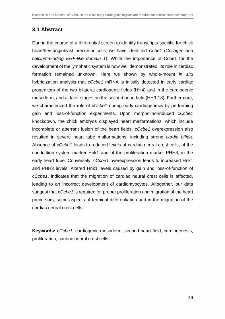
Expression and function of Ccbe1 in the chick early cardiogenic regions are required for correct heart development
49
3.1 Abstract
During the course of a differential screen to identify transcripts specific for chick
heart/hemangioblast precursor cells, we have identified Ccbe1 (Collagen and
calcium-binding EGF-like domain 1). While the importance of Ccbe1 for the
development of the lymphatic system is now well demonstrated, its role in cardiac
formation remained unknown. Here we shown by whole-mount in situ
hybridization analysis that cCcbe1 mRNA is initially detected in early cardiac
progenitors of the two bilateral cardiogenic fields (HH4) and in the cardiogenic
mesoderm, and at later stages on the second heart field (HH9-18). Furthermore,
we characterized the role of cCcbe1 during early cardiogenesis by performing
gain and loss-of-function experiments. Upon morpholino-induced cCcbe1
knockdown, the chick embryos displayed heart malformations, which include
incomplete or aberrant fusion of the heart fields. cCcbe1 overexpression also
resulted in severe heart tube malformations, including strong cardia bifida.
Absence of cCcbe1 leads to reduced levels of cardiac neural crest cells, of the
conduction system marker Hnk1 and of the proliferation marker PHH3, in the
early heart tube. Conversely, cCcbe1 overexpression leads to increased Hnk1
and PHH3 levels. Altered Hnk1 levels caused by gain and loss-of-function of
cCcbe1, indicates that the migration of cardiac neural crest cells is affected,
leading to an incorrect development of cardiomyocytes. Altogether, our data
suggest that cCcbe1 is required for proper proliferation and migration of the heart
precursors, some aspects of terminal differentiation and in the migration of the
cardiac neural crest cells.
Keywords: cCcbe1, cardiogenic mesoderm, second heart field, cardiogenesis,
proliferation, cardiac neural crest cells.

Expression and function of Ccbe1 in the chick early cardiogenic regions are required for correct heart development
50
3.2 Introduction
The vertebrate heart develops from distinct cardiogenic pools located in separate
regions and exposed to specific signals during development. These pools of
cardiac progenitors are temporally segregated in the developing embryo and give
rise to distinct cardiac structures (Harvey, 2002; Laugwitz et al., 2008). In the
chick embryo, the cardiac precursors are located in the cardiogenic mesoderm
(heart forming region) at stage HH4-5, and consist of two different populations of
heart progenitors, namely the first heart field (FHF) and the second heart field
(SHF). The FHF precursors clearly differentiate earlier than the precursors of the
SHF, indicating differential control on loss of precursor state and timing of
differentiation. Furthermore, the SHF precursors are much more sensitive to
genetic perturbation than the FHF precursors (Buckingham et al., 2005). The FHF
is derived from the anterior splanchnic mesoderm and contributes to the
myocardial cells of the primitive heart tube, which ultimately contributes to the left
ventricular region. On the other hand, the SHF lies anterior and dorsal to the
linear heart tube and is derived from the pharyngeal mesoderm medial to the
heart fields, which will contribute to the outflow tract region (Laugwitz et al., 2008;
Vincent and Buckingham, 2010). The cells of the SHF localized in the dorsal
pericardial wall become separated from the heart tube when the dorsal
mesocardium breaks down, maintaining continuity with the heart only at the
arterial and venous poles. The remaining structures, namely, the right ventricle,
the atrioventricular canal and the atria, have contribution from both heart forming
regions (HFR). Understanding the molecular control of heart organogenesis,
including the characterization and functional analysis of novel genes involved in
cardiogenesis, has major implications for treating congenital and adult heart
diseases since specific heart lineages have been associated with particular
human cardiovascular malformations.
We have recently reported a differential screening using Affimetrix GeneChip®
Chicken Genome arrays aiming to identify novel genes required for the
development and differentiation of the vertebrate heart and hemangioblast
precursor cell lineages (Bento et al., 2011). A construct containing EGFP
expression under the control of a 2.5 kb promoter fragment upstream the ATG of

Expression and function of Ccbe1 in the chick early cardiogenic regions are required for correct heart development
51
chick Cerberus (cCer; Tavares et al., 2007) was used to electroporate chick
embryos and isolate early cardiac progenitors. The genetic profiling provided
relevant data about the identity of the chick heart/hemangioblast precursors and
led to the detection of more than 700 transcripts expressed in the heart forming
regions (HFR), which included hundreds of uncharacterized genes enriched in
the heart/hemangioblast precursors in comparison to embryonic control cells
(Bento et al., 2011). Among the uncharacterized genes was the chick collagen
and calcium-binding EGF-like domain 1 (cCcbe1; gene ID: 770043), a protein
containing signal peptide for secretion, collagen domains and a calcium binding
EGF-like domain. The gene has 11 exons, and is localized in the Gallus gallus
chromosome Z, oriented in the reverse strand at the genomic region between
9,158,827K - 9,247,702K. Furthermore, cCcbe1 encodes a predicted protein with
396 amino acids with a molecular weight of 42.9kDA, highly conserved across
vertebrates. The cCcbe1 protein sequence is 79%, 70% and 80% identical to the
mouse, zebrafish and human Ccbe1 protein, respectively.
While the importance of mouse (m)Ccbe1 for the development of the lymphatic
system is indisputable (Bos et al., 2011; Hogan et al., 2009) , its role in cardiac
development remains unknown, despite the increasing evidence of a potential
function in cardiogenesis. Indeed, expression analysis has shown that mCcbe1
is expressed in heart precursors of the FHF, SHF and proepicardium in mouse
embryos from embryonic day (E)7.0 to E9.5 (Facucho-Oliveira et al., 2011).
Furthermore, analyses of mCcbe1 heterozygous knockout embryos have shown
X-Gal staining at the mesothelium of the heart at E12.5 (Bos et al., 2011). In
humans, mutations in CCBE1 are associated with Hennekam syndrome, a
disorder characterized by abnormal lymphatic system development where some
patients present as well congenital heart defects, including hypertrophic
cardiomyopathy (Alders et al., 2009; Connell et al., 2010).
Here, we show that during early chick development cCcbe1 is expressed in the
early cardiac progenitors that emerge from the primitive streak to form the two
bilateral cardiogenic fields at HH4 and in the cardiogenic mesoderm of the FHF
and SHF between HH5 to HH8. As development proceeds cCcbe1 localizes
predominantly in the region of the SHF (HH9 to HH18). In addition, we address

Expression and function of Ccbe1 in the chick early cardiogenic regions are required for correct heart development
52
the functional role of cCcbe1 in chick early heart development by employing
overexpression and knockdown approaches. Through cCcbe1 knockdown, the
embryos displayed heart abnormalities, which the phenotype included aberrant
or incomplete fusion of the heart forming regions. Furthermore, cCcbe1
morphants embryos demonstrated reduced levels of the proliferation of cells in
cardiac regions and in the Hnk1 signal in the region of the heart tube. On the
other hand, cCcbe1 overexpression resulted in severe heart tube abnormalities,
being cardia bifida the most common phenotype, seen in half of the treated
embryos. Moreover, overexpressed-cCcbe1 embryos demonstrated increased
proliferation of cells in cardiac regions and Hnk1 levels in the cardiac neural crest
cells and heart tube region. Taken together, the gain and loss-of-function of
cCcbe1 affects the proliferation of the cardiac cells, and the alteration in the Hnk1
levels, suggests that the migration of cardiac neural crest cells is affected, leading
to an incorrect development of cardiomyocytes. These data support that cCcbe1
plays a crucial role during early heart development.
3.3 Results
3.3.1 cCcbe1 expression during early heart development
Whole-mount in situ hybridization demonstrated that cCcbe1 mRNA is first
detected at stage HH4-4+ as two patches on each side of the primitive streak
corresponding to the bilateral cardiogenic mesoderm (Figure 3.1A, black arrow).
From stage HH5 to stage HH8, cCcbe1 expression expands along the lateral
plate mesoderm (Figure 3.1B-E, black arrow). cCcbe1 is also expressed in the
first somite and it can be observed since the formation of the somite at stage HH7
until HH11+ (Figure 3.1D-H). Transverse sections of whole mount stained
embryos at stage HH6 (Figure 3.1C’) revealed that cCcbe1 is expressed in the
paraxial mesoderm (orange arrows), splanchnic lateral plate mesoderm (black
arrows) and in the somatic lateral plate mesoderm (yellow arrow). At stage HH9
to HH11, cCcbe1 transcripts were observed near the posterior part of the heart,
namely, in the sino-atrial region, splanchnic mesoderm, somatic mesoderm and
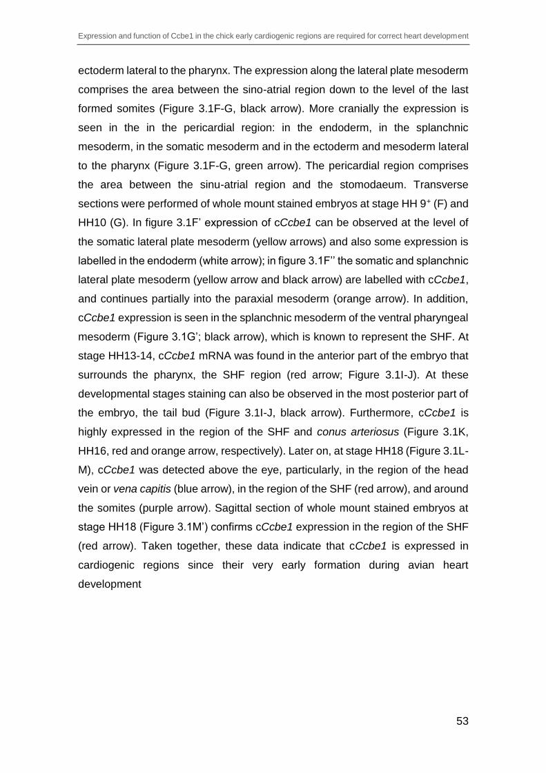
Expression and function of Ccbe1 in the chick early cardiogenic regions are required for correct heart development
53
ectoderm lateral to the pharynx. The expression along the lateral plate mesoderm
comprises the area between the sino-atrial region down to the level of the last
formed somites (Figure 3.1F-G, black arrow). More cranially the expression is
seen in the in the pericardial region: in the endoderm, in the splanchnic
mesoderm, in the somatic mesoderm and in the ectoderm and mesoderm lateral
to the pharynx (Figure 3.1F-G, green arrow). The pericardial region comprises
the area between the sinu-atrial region and the stomodaeum. Transverse
sections were performed of whole mount stained embryos at stage HH 9+ (F) and
HH10 (G). In figure 3.1F’ expression of cCcbe1 can be observed at the level of
the somatic lateral plate mesoderm (yellow arrows) and also some expression is
labelled in the endoderm (white arrow); in figure 3.1F’’ the somatic and splanchnic
lateral plate mesoderm (yellow arrow and black arrow) are labelled with cCcbe1,
and continues partially into the paraxial mesoderm (orange arrow). In addition,
cCcbe1 expression is seen in the splanchnic mesoderm of the ventral pharyngeal
mesoderm (Figure 3.1G’; black arrow), which is known to represent the SHF. At
stage HH13-14, cCcbe1 mRNA was found in the anterior part of the embryo that
surrounds the pharynx, the SHF region (red arrow; Figure 3.1I-J). At these
developmental stages staining can also be observed in the most posterior part of
the embryo, the tail bud (Figure 3.1I-J, black arrow). Furthermore, cCcbe1 is
highly expressed in the region of the SHF and conus arteriosus (Figure 3.1K,
HH16, red and orange arrow, respectively). Later on, at stage HH18 (Figure 3.1L-
M), cCcbe1 was detected above the eye, particularly, in the region of the head
vein or vena capitis (blue arrow), in the region of the SHF (red arrow), and around
the somites (purple arrow). Sagittal section of whole mount stained embryos at
stage HH18 (Figure 3.1M’) confirms cCcbe1 expression in the region of the SHF
(red arrow). Taken together, these data indicate that cCcbe1 is expressed in
cardiogenic regions since their very early formation during avian heart
development

Expression and function of Ccbe1 in the chick early cardiogenic regions are required for correct heart development
54
Figure 3.1 – cCcbe1 expression in developing chick embryos. (A) Expression of cCcbe1 is present at HH4+ in the cardiogenic mesoderm (black arrow); (B–E) the expression is present in the anterior lateral plate mesoderm as two patches on either side of the head process (black arrows point to the heart-forming fields); C´: Transverse paraffin sections (8µm) of whole mount stained embryos at stage HH6, cCcbe1 is expressed in the paraxial mesoderm (orange arrow), splanchnic lateral plate mesoderm (black arrow) and in the somatic

Expression and function of Ccbe1 in the chick early cardiogenic regions are required for correct heart development
55
lateral plate mesoderm (yellow arrow). (F–H) Green arrow point to the lines of expression in the lateral pharynx region; bilateral expression can be observed in the sino-atrial region (yellow arrow), and in the lateral plate mesoderm (black arrow); (I–K) Expression can be seen in the tail bud (black arrow); in a specific region behind the heart, known as the SHF (red arrow) and near the anterior part of the heart (orange arrow, conus arteriosus region); F’-F’’ and G’: Transverse paraffin sections (8µm) of whole mount stained embryos at stage HH9 (F) and HH10 (G) respectively (from anterior to posterior). The sections are oriented with dorsal up and ventral down. (F’) Expression of cCcbe1 can be observed at the level of the somatic lateral plate mesoderm (yellow arrow) and also some expression is labelled in the endoderm (white arrow); (F’’) Somatic and splanchnic lateral plate mesoderm (yellow arrow and black arrow) are labelled with cCcbe1, and continues partially into the paraxial mesoderm (orange arrow); (G’) cCcbe1 expression is seen in the somatic lateral plate mesoderm (yellow arrow) and in the splanchnic mesoderm of the ventral pharyngeal mesoderm (black arrow); (L–M) Lateral view of the embryo with expression above de eye (blue arrow), in the SHF region (red arrow) and also some expression is seen around the somites (purple arrow); M’: Sagittal paraffin section (8µm) of whole mount stained embryos at stage HH18, cCcbe1 is expressed in the region of the SHF (red arrow).
3.3.2 cCcbe1 is expressed in the second heart field
To evaluate in greater detail whether cCcbe1 expression is correlated with the
first, second or both heart field populations, we performed double WISH for
cCcbe1 and the cardiac markers Nkx2.5 or Islet-1. The homeobox transcription
factor Nkx2.5 is one of the earliest markers of the cardiac precursor cells and in
heart muscle through life (Schultheiss et al., 1995), and it is found in both FHF
and SHF (Gessert and Kühl, 2010; Lints et al., 1993; Waldo et al., 2001). The
LIM homeodomain transcription factor Islet-1 has become a popular molecular
marker demarcating the SHF (Cai et al., 2003; Laugwitz et al., 2008).
Double WISH with cCcbe1 and Nkx2.5 showed the co-localization in the
cardiogenic mesoderm at stage HH7 (Figure 3.2F, black arrow), and also in the
region of sino-venosus by stage HH10 (Figure 3.2H, yellow arrow). In transverse
sections it is possible to observe cCcbe1 and Nkx2.5 co-expressed in the
cardiogenic mesoderm of the heart forming fields (F’ and F’’; black arrow), some
co-labelling in the ventrolateral aspect of the splanchnic mesoderm (G’ and G’’;
black arrow) and in the dorsomedially region of the splanchnic mesoderm
extending towards the cranial paraxial mesoderm (G’ and G’’; red arrow), namely
the SHF. At stage HH12 we found co-labelling in the most anterior part of the
heart, the region of the conus arteriosus (blue arrow) and in the ventral
pharyngeal mesoderm between the attachments of the cardiac outflow and inflow
tracts (red arrow). However, unlike Nkx2.5, cCcbe1 expression is absent from the

Expression and function of Ccbe1 in the chick early cardiogenic regions are required for correct heart development
56
heart tube (Figure 3.2I). Transverse sections showed overlapping expression of
cCcbe1 and Nkx2.5 in endoderm (Figure 3.2I’; black arrow), in the conus
arteriosus (Figure 3.2I’; blue arrow) and a pale co-expression in splanchnic
mesoderm (SHF region; red arrow; Figure 3.2I’’). At stage HH18, the expression
of both genes coincides in the SHF and in the conus arteriosus (Figure 3.2J; red
and blue arrow). Sagittal (J’) and transverse (J’’ and J’’’) sections of double
stained embryos showed that cCcbe1 and Nkx2.5 are co-expressed in the SHF
(red arrow).
Figure 3.2 – Double WISH analysis of cCcbe1 and Nkx2.5 expression. (A-E) Nkx2.5 expression in developing chick embryos. (F-J) Comparative expression of cCcbe1 and Nkx2.5 during early heart development. All are ventral views (anterior to top) except for E that is lateral view. (F-H) cCcbe1 and Nkx2.5 have overlapping patterns of expression in the heart fields (F and G; black arrow) and in the sino-venosus (H; yellow arrow); F’ and F’’: Transverse paraffin sections (8µm) of double stained embryos at stage HH7, cCcbe1 and Nkx2.5 are co-expressed in the cardiogenic mesoderm of the heart forming fields (black arrow); G’ and G’’: Transverse paraffin sections (8µm) of double stained embryos at stage HH8+, cCcbe1 and Nkx2.5 are co-labeled in the ventrolateral aspect of the splanchnic mesoderm (black arrow) and in the dorsomedially region of the splanchnic mesoderm (red arrow). (I) Co-expression in the region of the conus arteriosus (blue arrow) and in the ventral pharyngeal mesoderm (red arrow, SHF); I’
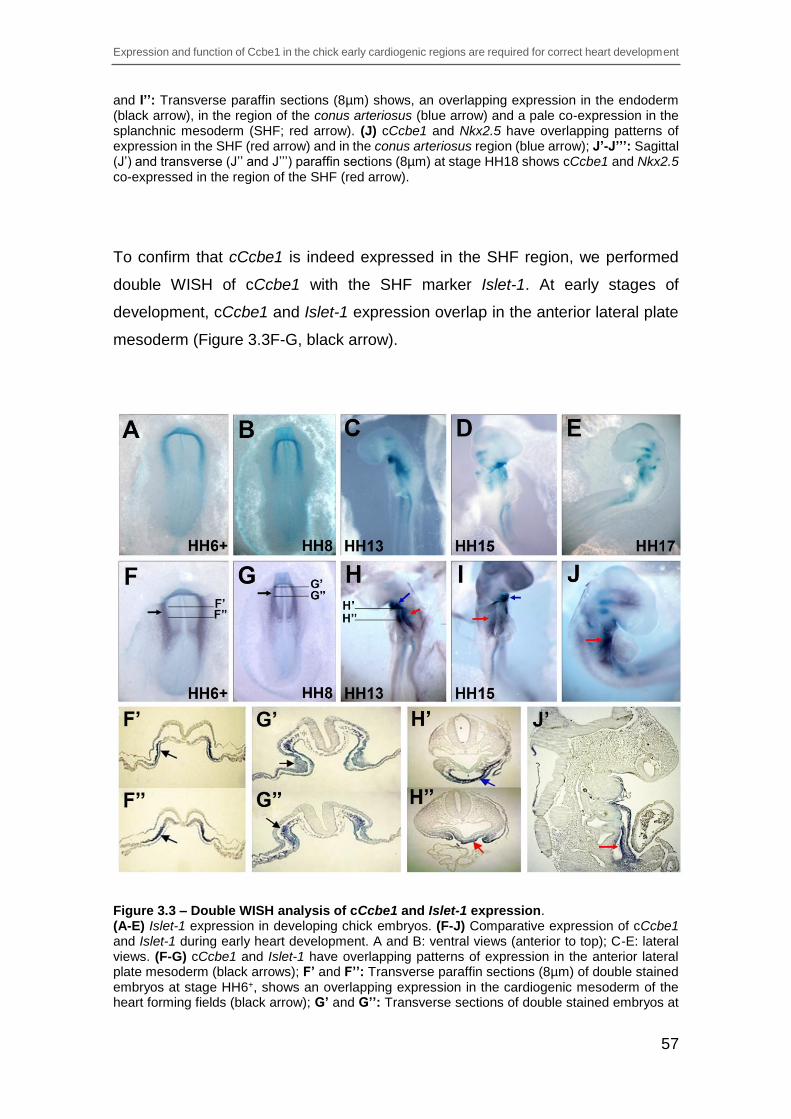
Expression and function of Ccbe1 in the chick early cardiogenic regions are required for correct heart development
57
and I’’: Transverse paraffin sections (8µm) shows, an overlapping expression in the endoderm (black arrow), in the region of the conus arteriosus (blue arrow) and a pale co-expression in the splanchnic mesoderm (SHF; red arrow). (J) cCcbe1 and Nkx2.5 have overlapping patterns of expression in the SHF (red arrow) and in the conus arteriosus region (blue arrow); J’-J’’’: Sagittal (J’) and transverse (J’’ and J’’’) paraffin sections (8µm) at stage HH18 shows cCcbe1 and Nkx2.5 co-expressed in the region of the SHF (red arrow).
To confirm that cCcbe1 is indeed expressed in the SHF region, we performed
double WISH of cCcbe1 with the SHF marker Islet-1. At early stages of
development, cCcbe1 and Islet-1 expression overlap in the anterior lateral plate
mesoderm (Figure 3.3F-G, black arrow).
Figure 3.3 – Double WISH analysis of cCcbe1 and Islet-1 expression. (A-E) Islet-1 expression in developing chick embryos. (F-J) Comparative expression of cCcbe1 and Islet-1 during early heart development. A and B: ventral views (anterior to top); C-E: lateral views. (F-G) cCcbe1 and Islet-1 have overlapping patterns of expression in the anterior lateral plate mesoderm (black arrows); F’ and F’’: Transverse paraffin sections (8µm) of double stained embryos at stage HH6+, shows an overlapping expression in the cardiogenic mesoderm of the heart forming fields (black arrow); G’ and G’’: Transverse sections of double stained embryos at
Expression and function of Ccbe1 in the chick early cardiogenic regions are required for correct heart development
58
stage HH8, both are co-labeled in the dorsomedially region of the splanchnic mesoderm (black arrow). (H-J) co-expression of cCcbe1 and Islet-1 is observed in the caudal part of the distal outflow tract of the heart (conus arteriosus, blue arrow) and in the ventral pharyngeal mesoderm (SHF; red arrow); H’-J’: Transverse sections shows, a co-expression in the region of the conus arteriosus (H’; blue arrow) and in the splanchnic mesoderm of the SHF (H’’; red arrow); J’: Sagittal paraffin section (8µm) of double stained embryos at stage HH18, cCcbe1 and Islet-1 are co-expressed in the region of the SHF (red arrow).
Transverse sections at stage HH6+ (F) and HH8 (G) showed that cCcbe1 and
Islet-1 are co-expressed in the cardiogenic mesoderm of the heart forming fields
(F’-F’’, black arrow) and in the dorsomedially region of the splanchnic mesoderm
(G’-G’’). Later during development they are co-expressed in the anterior part of
the embryo that surrounds the pharynx, the SHF (Figure 3.3H-J; red arrow;
pharyngeal mesoderm) and in the region of the conus arteriosus (Figure 3.3H
and I, blue arrow). Transverse and sagittal sections showed that cCcbe1 and
Islet-1 are co-labelled in the caudal part of the distal outflow tract of the heart (H’;
blue arrow; conus arteriosus) and in the splanchnic mesoderm of the pharyngeal
floor, which corresponds to the SHF (H’’-J’; red arrow). Our data, demonstrates
that cCcbe1 expression is present early in the FHF and SHF and later is highly
specific of the SHF region.
3.3.3 cCcbe1 knockdown leads to aberrant heart formation
To dissect the role of cCcbe1 during early chick development, we used
morpholino technology to knockdown cCcbe1. A splicing inhibitory morpholino
oligonucleotide was designed (Gene Tolls, LLC) by targeting the splice donor site
of E2 (cCcbe1MO). A standard morpholino was used as control (CoMO).
Embryos at HH3+ were injected with each MOs (1mM) into the right and left sides
of the primitive streak, followed by in vivo electroporation. Both morpholinos were
fluorescein tagged at the 3' end to assess injection efficiency and stability of the
morpholino over time. Furthermore, western blot analysis of injected embryos
was performed to confirm that the cCcbe1 MO was causing the knockdown of
cCcbe1 and leading to decreased amount of protein. Indeed, injection with the

Expression and function of Ccbe1 in the chick early cardiogenic regions are required for correct heart development
59
morpholino cCcbe1 resulted in a reduction of cCcbe1 protein when compared
with CoMO injected embryos (Figure 3.4G).
Figure 3.4 – cCcbe1 loss-of-function leads to heart malformations. The embryos were first targeted at stage HH3+/HH4 with the designed morpholino and were collected later in development between stage HH9+ and HH10. (A-C’) Embryos injected with the control morpholino. (D-F’) Embryos injected with the cCcbe1 MO. (A-F) Localization and efficiency of the MO injection by detection of fluorescein expression. (A’-F’) Detection of Tbx5, Mhc and Islet-1 expression by WISH of the injected embryos with CoMO and cCcbe1 MO, respectively. All embryos are ventral. (G) Western blot analysis of the cCcbe1 MO and Control MO embryos. It was possible to see a loss in cCcbe1 protein levels observed in the cCcbe1 knockdown in comparison to morpholino control embryos. (H) Analysis of the phenotypes caused by electroporated embryos with cCcbe1 MO and Control MO. Bar charts showing the percentage of chick embryos presenting cardiac alterations after injection with Control or cCcbe1 MO. Only embryos at stage HH9 and later were considered to this analysis. The total of samples analyzed (n): 100 Control MO and 110 cCcbe1 MO embryos. The y-axis represents the percentage of embryos. The x-axis represents the defects: normal development (ND), severe cardiac alterations (SCA), moderate cardiac alterations (MCA) and cardia bifida (CB).
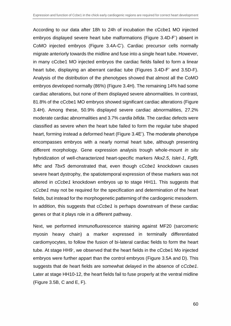
Expression and function of Ccbe1 in the chick early cardiogenic regions are required for correct heart development
60
According to our data after 18h to 24h of incubation the cCcbe1 MO injected
embryos displayed severe heart tube malformations (Figure 3.4D-F’) absent in
CoMO injected embryos (Figure 3.4A-C’). Cardiac precursor cells normally
migrate anteriorly towards the midline and fuse into a single heart tube. However,
in many cCcbe1 MO injected embryos the cardiac fields failed to form a linear
heart tube, displaying an aberrant cardiac tube (Figures 3.4D-F’ and 3.5D-F).
Analysis of the distribution of the phenotypes showed that almost all the CoMO
embryos developed normally (86%) (Figure 3.4H). The remaining 14% had some
cardiac alterations, but none of them displayed severe abnormalities. In contrast,
81.8% of the cCcbe1 MO embryos showed significant cardiac alterations (Figure
3.4H). Among these, 50.9% displayed severe cardiac abnormalities, 27.2%
moderate cardiac abnormalities and 3.7% cardia bifida. The cardiac defects were
classified as severe when the heart tube failed to form the regular tube shaped
heart, forming instead a deformed heart (Figure 3.4E’). The moderate phenotype
encompasses embryos with a nearly normal heart tube, although presenting
different morphology. Gene expression analysis trough whole-mount in situ
hybridization of well-characterized heart-specific markers Nkx2.5, Islet-1, Fgf8,
Mhc and Tbx5 demonstrated that, even though cCcbe1 knockdown causes
severe heart dystrophy, the spatiotemporal expression of these markers was not
altered in cCcbe1 knockdown embryos up to stage HH11. This suggests that
cCcbe1 may not be required for the specification and determination of the heart
fields, but instead for the morphogenetic patterning of the cardiogenic mesoderm.
In addition, this suggests that cCcbe1 is perhaps downstream of these cardiac
genes or that it plays role in a different pathway.
Next, we performed immunofluorescence staining against MF20 (sarcomeric
myosin heavy chain) a marker expressed in terminally differentiated
cardiomyocytes, to follow the fusion of bi-lateral cardiac fields to form the heart
tube. At stage HH9-, we observed that the heart fields in the cCcbe1 Mo injected
embryos were further appart than the control embryos (Figure 3.5A and D). This
suggests that de heart fields are somewhat delayed in the absence of cCcbe1.
Later at stage HH10-12, the heart fields fail to fuse properly at the ventral midline
(Figure 3.5B, C and E, F).
Expression and function of Ccbe1 in the chick early cardiogenic regions are required for correct heart development
61
Figure 3.5 – Immunofluorescence analysis of cCcbe1 and Control MO in chick embryos. Embryos were target at HH3+/HH4 with cCcbe1 (cCcbe1 MO) and Control (CoMO) morpholino and allowed to develop until stage HH12. Some embryos were subsequently analyzed by whole mount (A-F) or in sections (Ca, Cb, Fa and Fb) immunofluorescence staining for MF20 (myocardium: red; Dapi: blue). (A-C) Embryos injected with CoMO showed no cardiac malformations. (D-F) embryos injected with cCcbe1 MO showed alterations in cardiac tube fusion: a delay in the fusion (D), fusion failure (E) and incomplete fusion (F). (Ca-Cb) Transverse sections (8μm) of embryos electroporated with CoMO at the level of the heart. (Fa-Fb) Transverse sections (8μm) of embryos electroporated with cCcbe1 MO at the level of the heart: these images highlight the malformations at the ventral midline (yellow arrowhead) and the lack of cell expressing myosin heavy chain (green arrowhead) caused by a failure on initiation of cardiac differentiation at the ventral midline.
Indeed, in transverse sections we confirmed that the cardiac fields do not properly
fuse at the ventral midline, and that the spece between the cardiac fields is
replaced by that do not express the cardiomyocyte marker MF20 (Figure 3.5Fa
and Fb, greem arrow). In addition, the closure of the dorsal mesocardium was
also affected in cCcbe1 MO injected embryos (Figure 3.5Cb and Fb; yellow
arrow). The dorsal mesocardium is a transient structure formed when the
splanchnic mesoderm (SHF) of opposite sides of the embryo come together from
dorsal and ventral to the heart, forming double layered supporting membranes.
Expression and function of Ccbe1 in the chick early cardiogenic regions are required for correct heart development
62
After the rupture of the dorsal mesocardium the heart tube closes dorsally and
the dorsal pericardial walls fuse, something that in the cCcbe1 morphant embryos
seems also to fail to happen. Taken together, these data indicates that the bi-
lateral fields fail to properly fuse at the midline in the absence of cCcbe1, leading
to the development of an aberrant heart tube.
3.3.4 cCcbe1 knockdown affects the proliferation of the cardiac cells
Cell proliferation, while not the only mechanism, is an important process that
contributes for the formation of the heart, which relies greatly on the rapid
proliferation of cardiomyocytes at specific stages during early development
(Sissman, 1966; Stalsberg and DeHaan, 1969). It is known that the initially
formed myocardial tube continues to grow by recruitment of cells that originate
from flanking mesoderm, dubbed the SHF (Cai et al., 2003; Mjaatvedt et al., 2001;
Waldo et al., 2001). Additionally, the newly formed heart tube is non-proliferating,
implying that growth of the primary heart tube can only occur by differentiation of
precursor cells. During the looping of the heart cells are added from the
splanchnic and pharyngeal mesoderm. Usually, if something interfers with the
ability of these cells to replicate at this stage heart defects will became apparent.
To determine if cCcbe1 is involved in the proliferation of heart precursor cells, we
analysed cell proliferation through immunostaing for phospho-Histone 3 (PHH3),
which marks only cells in mitosis, on transverse sections of cCcbe1 knokdown
and control embryos.
Comparative analysis of proliferation in cCcbe1 knockdown embryos and control
embryos revealed that the proliferation decreases in both pharyngeal and
splanchnic mesoderm of the cCcbe1 morphant embryos (Figure 3.6A-B).
Quantification of the cell proliferation in the cardiac region (splanchnic mesoderm
and pharyngeal endoderm regions) and overall (cardiac plus non-cardiac
regions) was consistent with decreased proliferation on those regions.

Expression and function of Ccbe1 in the chick early cardiogenic regions are required for correct heart development
63
Figure 3.6 – cCcbe1 knockdown reduce cell proliferation in chick embryos. Embryos at stage HH3+/HH4 were target with the cCcbe1 MO (B) and CoMO (A), and developed until HH12. Embryos were transverse sectioned and then immunohistochemistry staining was

Expression and function of Ccbe1 in the chick early cardiogenic regions are required for correct heart development
64
performed for PHH3 (green; Aa-Bb’). (B-Bb’) cCcbe1 treated embryos showing heart alterations and a decrease in proliferating cells; (A-Ab’) Control morpholino treated embryos showing normal heart development and proliferation. Note that at this stage the heart is not proliferative, therefore the region of the pharyngeal and splanchnic mesoderm was taken in consideration (SHF contribution). Dapi: blue; PHH3: green. (C) Analysis of the cCcbe1 knockdown in cardiac cells proliferation. Embryos were subsequently analyzed in transverse sections by immunohistochemistry staining for PHH3. Proliferating cells were counted in 2 distinct regions: cardiac region (pharyngeal and splanchnic mesoderm) and overall (all the regions in the embryo: cardiac and non-cardiac). The total of embryos analyzed (n): 4 control MO and 4 cCcbe1 MO. The y-axis represents the PHH3 positive cells. The x-axis represents the regions of the counted PHH3 positive cells: anterior overall (AO), anterior cardiac region (ACR), medial overall (MO), medial cardiac region (MCR), posterior overall (PO) and posterior cardiac region (PCR). Error bars represent the S.E.M. from four replicates. *p<0.05; **p<0.01; ***p<0.001.
We selected sections at the anterior, medial and posterior levels of the heart tube
region to count the number of proliferating cells. cCcbe1 MO embryos showed an
overall decrease (ratio 2:1) in proliferating cells, but in the cardiac cells this
difference in proliferation was even higher (ratio 3:1) when compared with the
control MO embryos (Figure 3.6C). These data are similar to those found in Islet-
1 null mutants in which, absence of Islet-1 leads to a decrease in cell proliferation
and cell survival (Cai et al., 2003). Since like Islet-1, cCcbe1 is expressed in those
regions, our data suggests that cCcbe1 is required for proper proliferation of SHF
progenitors
3.3.5 cCcbe1 overexpression leads to cardia bifida
In the chick embryo, cardiac precursor cells migrate anteriorly towards the midline
and fuse into a single heart tube. If this migration event is blocked, the two heart
primordia remain separated and cardia bifida occurs. Interestingly, it has been
reported that overexpression of CCBE1 inhibits cell migration in breast cancer
cells (Barton et al., 2010). To dissect the role of cCcbe1 during early chick
development, we used gain-of-function approaches to overexpress cCcbe1. A
vector with constitutive promoter (CAGGS) was used to clone the cCcbe1
transcript (pCAGGS-cCcbe1-IRES-GFP). An empty pCAGGS-IRES-GFP vector
was used as control. Embryos at HH3+/HH4 were injected with each vector into
the right and left sides of the primitive streak, followed by in vivo electroporation.
Furthermore, WISH of injected embryos was performed to confirm that pCAGGS-

Expression and function of Ccbe1 in the chick early cardiogenic regions are required for correct heart development
65
cCcbe1-IRES-GFP was causing overexpression of cCcbe1. Indeed, injection with
the pCAGGS-cCcbe1 resulted in a co-localized cCcbe1 expression with GFP,
including in places where cCcbe1 is normally absent (Figure 3.7C-C’).
According to our data, upon 18h to 24h of incubation the injected embryos with
cCcbe1 overexpression displayed severe heart tube malformations (Figure 3.7B-
B’) that were absent in control vector injected embryos (Figure 3.7A-A’). Normally,
cardiac precursor cells migrate anteriorly towards the midline and fuse into a
single heart tube. In many cCcbe1 overexpression vector injected embryos both
heart fields failed to migrate properly towards the midline remaining in the lateral
plate mesoderm, i.e. cardia bifida (Figure 3.7B and F).
Analysis of the distribution of the phenotypes showed that almost all the control
vector injected embryos developed normally (86.8%) (Figure 3.7D). The
remaining 13.2% had some cardiac alterations, but none of them displayed cardia
bifida. In contrast, 89.5% of the cCcbe1 overexpression embryos showed
significant cardiac alterations (Figure 3.7D). Among these, 52.6% displayed
cardia bifida and 36.9% milder cardiac alterations, i.e. the heart fields were able
to migrate to the midline but they failed to fuse properly and the hearts
consistently failed to undergo looping of the heart tube (Figure 3.7D).
The cardia bifida phenotype that was observed in the cCcbe1 overexpression
embryos were strikingly similar to those seen in embryos lacking the cardiac
transcription factor Gata4. In these embryos, two separate heart tubes develop
in the majority of mutants (Kuo et al., 1997; Molkentin et al., 1997).
Immunofluorescence analysis of MF20 showed that the cardiac fields of cCcbe1
overexpression embryos remain close to the lateral plate mesoderm and
consequently the embryos showed a bifid heart phenotype (Figure 3.7E-Fc’). This
observation is consistent with failure of the migration of the cardiac fields towards
the midline. In light with the similarities with the Gata4 mutants, we examined as
well the expression pattern of Gata4 in the cCcbe1 overexpression vector injected
embryos.

Expression and function of Ccbe1 in the chick early cardiogenic regions are required for correct heart development
66
Figure 3.7 – cCcbe1 gain-of-function in chick embryos. Embryos were targeted at stage HH3+/HH4 with the control vector pCAGGS-IRES-GFP (A) or with the overexpression vector pCAGGS-cCcbe1-IRES-GFP (B-C) and collected at stage HH11. (A) Localization and efficiency of the control vector pCAGGS-GFP injection by detection of fluorescein expression. (A’) Embryos injected with control vector showed no cardiac malformations detected with Gata4 by WISH. (B-C) Localization and efficiency of the overexpression vector pCAGGS-cCcbe1-IRES-GFP injection by detection of fluorescein expression. (B’) Embryos injected with pCAGGS-cCcbe1-IRES-GFP showed alterations in cardiac tubes fusion detected with Gata4 by WISH, namely, bifid heart was observed (formation of two separate heart tubes). (C’) Detection of cCcbe1 expression by WISH, demonstrating that pCAGGS-cCcbe1-IRES-GFP is overexpressing cCcbe1. (D) Analysis of the defects caused by electroporated embryos with control vector pCAGGS-IRES-GFP or overexpression vector pCAGGS-cCcbe1-IRES-GFP. Bar charts showing the percentage of chick embryos presenting cardiac alterations after injection with control vector or overexpression vector. Only embryos at stage HH9 and later were considered to this analysis. The total of samples analyzed (n): 38 control vector and 38 overexpression vector embryos. The y-axis represents the percentage of embryos. The x-axis represents the defects: normal development, cardia bifida and other cardiac alterations. (E-F) Some embryos were subsequently analyzed immunohistochemistry staining for

Expression and function of Ccbe1 in the chick early cardiogenic regions are required for correct heart development
67
MF20 (myocardium: red; Dapi: blue) in transverse sections (8μm). (E-Ec’) Embryos injected with control vector showed no cardiac malformations. (F-Fc’) embryos injected with overexpression vector showed cardia bifida defects. These images showed none alteration of cell expressing MF20.
We observed that, despite the development of cardia bifida in the cCcbe1
overexpression embryos, the expression of Gata4 was also present in cCcbe1
overexpression embryos up to stage HH11 (Figure 3.7A-B’). This suggests that
cCcbe1 may not be required for the specification and determination of the heart
fields, but instead for proper morphogenetic patterning of the cardiogenic
mesoderm.
3.3.6 cCcbe1 overexpression also affects cell proliferation
To determine if cCcbe1 overexpression also affects the proliferation of heart
precursor cells, we analyzed cell proliferation through immunostaining for PHH3
on transverse section of cCcbe1 overexpression and control embryos.
Comparative analysis of proliferation in cCcbe1 overexpression embryos and
control embryos revealed that the proliferation increases in both pharyngeal and
splanchnic mesoderm of the cCcbe1 gain-of-function embryos (Figure 3.8A-B).
cCcbe1 overexpression embryos showed an overall increase in proliferating
cells. Quantification of proliferating cells in the cardiac region (splanchnic
mesoderm and pharyngeal endoderm regions) and overall (cardiac plus non-
cardiac regions) was consistent with increased proliferation in those regions. As
for cardiac cells proliferation, we also observed an increase in cCcbe1
overexpression embryos when compared with the control embryos (Figure 3.8C).
This data is consistent with a role of cCcbe1 in the control of SHF progenitors
proliferation.
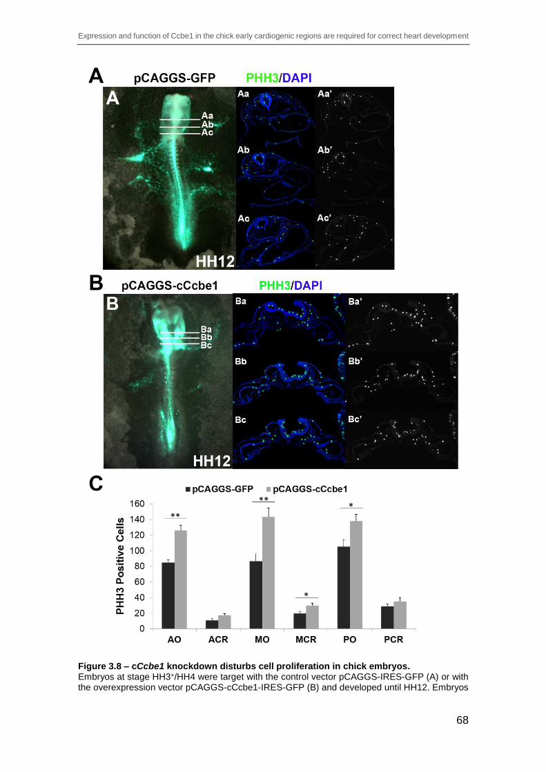
Expression and function of Ccbe1 in the chick early cardiogenic regions are required for correct heart development
68
Figure 3.8 – cCcbe1 knockdown disturbs cell proliferation in chick embryos. Embryos at stage HH3+/HH4 were target with the control vector pCAGGS-IRES-GFP (A) or with the overexpression vector pCAGGS-cCcbe1-IRES-GFP (B) and developed until HH12. Embryos

Expression and function of Ccbe1 in the chick early cardiogenic regions are required for correct heart development
69
were transverse sectioned and then immunohistochemistry staining was performed for PHH3 (green; Aa-Bc’). (A-Ac’) Control treated embryos shows normal heart development and proliferation. (B-Bb’) Overexpression-cCcbe1 treated embryos shows heart alterations and an increase in proliferating cells. Note that at this stage the heart is not proliferative, therefore the region of the pharyngeal and splanchnic mesoderm was taken in consideration (SHF contribution). Dapi: blue; PHH3: green.; (C) Analysis of the cCcbe1 overexpression in cardiac cells proliferation. Embryos were subsequently analyzed in transverse sections by immunohistochemistry staining for PHH3. Proliferating cells were counted in 2 distinct regions: cardiac region (pharyngeal and splanchnic mesoderm) and overall (all the regions in the embryo: cardiac and non-cardiac). The total of embryos analyzed (n): 4 pCAGGS-GFP and 4 pCAGGS-cCcbe1. The y-axis represents the PHH3 positive cells. The x-axis represents the regions of the counted PHH3 positive cells: anterior overall (AO), anterior cardiac region (ACR), medial overall (MO), medial cardiac region (MCR), posterior overall (PO) and posterior cardiac region (PCR). Error bars represent the S.E.M. from four replicates. *p<0.05; **p<0.01.
3.3.7 cCcbe1 loss and gain-of-function affects Hnk1 expression
Monoclonal antibody Hnk1 carbohydrate recognizes a subset of cell surface
glycoproteins that mediate cell-cell or cell-substrate interactions (Kruse et al.,
1984). During cardiogenesis, in various species (including the chick) Hnk1
epitope is present in migrating neural crest cells (Bronner-Fraser, 1987; Vincent
and Thiery, 1984), in developing conduction cardiomyocytes, in cardiomyocytes
expressing both MF20 and Hnk1 (not working cardiomyocytes), and in
endocardial cushion mesenchymal cells (Nakajima et al., 2001). In addition,
during chick early cardiogenesis, Hnk1 is scattered along the primitive heart tube,
only later becoming restricted to the myocardium of the sinus venosus and atrium
in which the central conduction system will be developed at a later stage
(Nakajima et al., 2001). To determine if cCcbe1 loss- and gain-of-function
influences Hnk1 expression in the chick embryo, we performed immunostaining
with the Hnk1 antibody on transverse sections at the heart region of cCcbe1
knockdown, overexpression and respective control embryos (stage HH12).

Expression and function of Ccbe1 in the chick early cardiogenic regions are required for correct heart development
70
Figure 3.9 – Hnk1 immunofluorescence analysis of cCcbe1 loss-of-function in chick embryos. Embryos were target at HH3+/HH4 with cCcbe1 and Control morpholino and allowed to develop until stage HH12 (A and B). Embryos were subsequently analyzed transversal sections (Aa-Bc)

Expression and function of Ccbe1 in the chick early cardiogenic regions are required for correct heart development
71
by immunostaining for Hnk1 (migrating CNC and conducting system marker: red; Dapi: blue). (Aa-Ac’) Transverse sections (8μm) of embryos electroporated with CoMO at the level of the heart; Hnk1 expression is detected through the heart tube and CNC. (Ba-Bc’) Transverse sections (8μm) of embryos electroporated with cCcbe1 MO at the level of the heart: these images highlight the lack of Hnk1 expression in the heart tube. (C) Quantitative analysis of Hnk1 immunostaining in two distinct regions: cardiac neural crest (CNC) cells and heart tube (HT). The total of embryos analyzed (n): 3 control MO and 3 cCcbe1 MO. The y-axis represents the Hnk1 fluorescence signal. The x-axis represents the regions of the Hnk1 measured signal: anterior CNC (ACNC), medial CNC (MCNC), posterior CNC (PCNC), anterior heart tube (AHT), medial heart tube (MHT), posterior heart tube (PHT). Error bars represent the S.E.M. from three replicates. *p<0.05; **p<0.01.
On the one hand, when comparing control and cCcbe1 knockdown embryos,
while there were no significant differences in the cardiac neural crest (CNC) cells,
the distribution of Hnk1 in the heart tube was substantially decreased (Figure
3.9A-B). Quantification of the level of Hnk1 signal in cCcbe1 morphants and
control embryos in sections at the anterior, medial and posterior levels of the
heart tube region showed that the level of Hnk1 was decreased in the cCcbe1
morphant embryos (Figure 3.9C). While the difference in the heart tube was
consistent in all the analysed embryos, there were no obvious differences in most
of the embryos at the level of the CNC cells. On the other hand, when comparing
control and cCcbe1 overexpression embryos, the Hnk1 was increased in both
CNC cells and heart tube regions in the cCcbe1 overexpression embryos (Figure
3.10A-B). Indeed, when we measured the Hnk1 signal the difference between the
control and the cCcbe1 gain-of-function embryos was evident (Figure 3.10C),
especially in the CNC cells where the intensity of Hnk1 staining was wider and
stronger than in the control embryos. Taken together, altered Hnk1 levels caused
by gain and loss-of-function of cCcbe1 suggest that the migration of CNC cells is
affected, leading to an incorrect development of cardiomyocytes.

Expression and function of Ccbe1 in the chick early cardiogenic regions are required for correct heart development
72
Figure 3.10 – Hnk1 immunofluorescence analysis of cCcbe1 gain-of-function in chick embryos. Embryos were target at HH3+/HH4 with the overexpression vector pCAGGS-cCcbe1 and the control vector pCAGGS-GFP and allowed to develop until stage HH12 (A and B). Embryos were

Expression and function of Ccbe1 in the chick early cardiogenic regions are required for correct heart development
73
subsequently analyzed transversal sections (Aa-Bc) by immunostaining for Hnk1 (migrating CNC and conducting system marker: red; Dapi: blue). (Aa-Ac’) Transverse sections (8μm) of embryos electroporated with control vector at the level of the heart; Hnk1 expression is detected through the heart tube and CNC. (Ba-Bc’) Transverse sections (8μm) of embryos electroporated with overexpression vector at the level of the heart: these images highlight increase of Hnk1 expression in the heart tube and CNC. (C) Quantitative analysis of Hnk1 immunostaining in two distinct regions: cardiac neural crest (CNC) cells and heart tube (HT). The total of embryos analyzed (n): 3 control vector and 3 overexpression vector. The y-axis represents the Hnk1 fluorescence signal. The x-axis represents the regions of the Hnk1 measured signal: anterior CNC (ACNC), medial CNC (MCNC), posterior CNC (PCNC), anterior heart tube (AHT), medial heart tube (MHT), posterior heart tube (PHT). Error bars represent the S.E.M. from three replicates. *p<0.05, **p<0.01.
3.4 Discussion
Expression analysis showed that cCcbe1 mRNA was initially detected very early
in development in the cardiogenic region on either side of the primitive streak at
stage HH4. cCcbe1 mRNA staining at this stage of development coincides with
the mesodermal cells that emerge from the anterior two-thirds of the primitive
streak to form two bilateral cardiogenic fields in the anterior lateral plate
mesoderm. It is known that the precursor heart cells are restricted to the
splanchnic mesoderm (Linask, 1992) and this region was where cCcbe1
expression was also visible. From stage HH5 to HH8, expression of cCcbe1
mRNA expands along the lateral plate mesoderm of the embryo resembling that
observed in staining performed using markers of the FHF and SHF such as
Nkx2.5 and Islet-1, respectively. Indeed, histological sections showed that
cCcbe1 is co-expressed with Nkx2.5 and Islet-1 in the splanchnic mesoderm,
specifically in the ventrolateral aspect of the splanchnic mesoderm, where the
differentiating myocardial cells (FHF) is known to be present, and also at the
dorsomedially region, where is known to be a region of undifferentiated cells
(SHF). Although, cCcbe1-expressing cells could be detected in the myocardial
tissue forming the primordial of the primitive heart tube at HH8, expression in the
cardiac tissue in more advanced stages of heart morphogenesis (HH9+-18) was
undetectable. cCcbe1 expression is therefore continuous in the cardiogenic
mesoderm and residual in the primordial myocardial tissues followed by absence
of expression at later stages of heart development. This suggests that high levels
of cCcbe1 expression are limited to multipotent and highly proliferative

Expression and function of Ccbe1 in the chick early cardiogenic regions are required for correct heart development
74
progenitors at the inflow region of the heart tube and in the SHF, and
downregulated upon cellular commitment towards cardiac lineages. Later in
development (HH13-18) the cCcbe1 mRNA was found predominantly in the
region of the conus arteriosus and in the SHF region. The latter coincided with
the expression of Islet-1, which marks undifferentiated and highly proliferative
cardiac progenitors (Cai et al., 2003). In fact, cCcbe1 once expressed in both
heart fields population at early stages, progressively became restricted to the
SHF population, as observed in Islet-1 expression. Interestingly, in mice Ccbe1
expression was shown in the three major populations of cardiac progenitors,
namely the FHF, SHF and proepicardium (Facucho-Oliveira et al., 2011). This
raises the possibility of a role of cCcbe1 in heart progenitors during
cardiogenesis.
The Ccbe1 protein contains a collagen and calcium binding EGF-like domains,
and may function in extracellular matrix remodeling and migration (Barton et al.,
2010). The knockdown of cCcbe1 leads to incorrect formation of the heart tube.
The bilateral cardiac progenitor populations seem to fail to migrate properly
towards the midline in the cCcbe1 knockdown embryos. It has been shown that
in ovarian cancer cell lines silencing of CCBE1 is associated with increased cell
migration and that, in contrast, CCBE1 overexpression inhibits it (Barton et al.,
2010). Therefore, it is possible that avian cCcbe1 is required for the proper
migration of the bilateral cardiac progenitor cells. Alternatively, and knowing that
the heart precursor cells are highly proliferative, it is possible that this phenotype
is also related to altered cell proliferation. Indeed, it has been shown that
alterations in the proliferation of cardiac precursor cells affect the migration of the
precursors towards the midline (Linask et al., 2005). Immunohistochemistry
analysis of the proliferation marker PHH3 revealed that knockdown of cCcbe1
leads to decreased proliferation of the cardiac progenitor cells. These data
indicate that in the absence of cCcbe1 the bilateral cardiac progenitor cells fail to
fuse at the midline to form the heart tube due to, at least in part, defective cell
proliferation.
On the other hand, the most severe phenotype of cCcbe1 overexpression is
cardia bifida. A defect that develops in several loss-of-function models, including

Expression and function of Ccbe1 in the chick early cardiogenic regions are required for correct heart development
75
Gata4 (Zhang et al., 2003); N-cadherin (Nakagawa and Takeichi, 1997), Rho
kinases (Wei et al., 2001), Furin (Roebroek et al., 1998). Indeed, the defects
observed in cCcbe1 overexpression embryos are strikingly similar to those seen
in embryos lacking the transcription factor Gata4, in which the precardiac
mesoderm fails to migrate properly hampering its fusing at the ventral midline
(Zhang et al., 2003). Since it has been shown that overexpression of CCBE1 in
cell culture leads to blockade of cell migration (Barton et al., 2010), it is possible
that excess cCcbe1 in chick heart precursor cells may hamper their migration
towards the midline to form the heart tube. Interestingly, cCcbe1 overexpression
did not affect the expression of the cardiac transcription factor Gata4, despite the
obvious morphological alterations. Similarly knockdown of cCcbe1 did not alter
the expression of cardiac specification markers Nkx2.5 (data not shown), Tbx5,
Mhc, Fgf8 (data not shown) and Isl1. Taken together, these data suggest that
cCcbe1 is not important for cardiomyocyte commitment, but instead it likely has
a role in the proper proliferation and migration of the cardiac precursor cells to
form the heart tube.
As mentioned above, the heart precursor cells proliferate and migrate as they
travel towards their final destination at the midline to form the heart primordia and
where they differentiate into cardiomyocytes. In the chick this happens around
stage HH9 of development at the beginning of the fusion of the heart tube. Taking
this into consideration, we analyzed the terminal cardiomyocyte differentiation in
the cCcbe1 knockdown embryos. According to our data, the cells at the ventral
midline in cCcbe1 knockdown embryos do not express the cardiomyocytes
marker MF20. This suggests that the cells at the midline fail to initiate cardiac
differentiation at the ventral midline. Alternatively and since the analyzed cardiac
markers were unaffected in the gain- and loss-of-function experiments, it is also
possible that defective proliferation and migration of the cardiac precursors cells
hampers proper fusion at the midline and consequently non-cardiac fated cells
occupy their place. In any of the cases, this affects the formation of a normal
beating heart tube, indicating that cCcbe1 is involved in the coordination of early
heart organogenesis in the avian embryo possibly through the regulation of
proliferation and migration of cardiac precursors towards the ventral midline.

Expression and function of Ccbe1 in the chick early cardiogenic regions are required for correct heart development
76
Furthermore, this is consistent with the cCcbe1 expression at the bilateral heart
fields as they migrate to the midline.
It is known that Hnk1 is normally present in the cardiomyocyte precursor of the
conduction system tissue and is involved in the development of mature His-
Purkinje System architecture and consequently its function (Aoyama et al., 1995;
Blom et al., 1999; Chuck and Watanabe, 1997; Nakajima et al., 2001). In addition,
it is also known that Hnk1 is present in migrating CNC cells, and that the CNC
cells are required for outflow septation (at later stages of development) and for
the correct normal heart looping by addition of myocardium to the heart tube from
the SHF (Yelbuz et al., 2002). Furthermore, CNC may not contribute directly to
the His-Purkinje conduction system, but it may be required for its normal
development (Bronner-Fraser, 1987). Following ablation of CNC cells in chick
embryos, the conduction system bundles fail to compact leading to inhibition or
delay in the maturation of the conduction system function (Gurjarpadhye et al.,
2007). Furthermore, fate mapping studies CNC cells are found in close proximity
with the developing conduction system (Nakamura et al., 2006; Poelmann et al.,
2004; Poelmann and Gittenberger-de Groot, 1999), and deletion of Hf1b from
CNC resulted in atrial and atrioventricular conduction dysfunction (St. Amand et
al., 2006). According to our results, while increased cCcbe1 levels resulted in
expansion of Hnk1-expressing domain in both CNC cells and heart tube region,
loss of cCcbe1 decreased Hnk1 signal mostly in the heart tube region. This may
affect the migration of CNC cells leading to an incorrect development of
cardiomyocytes, and hence suggests that cCcbe1 may be also required during
early heart development for the establishment of the heart conduction system.
In conclusion, here we presented that cCcbe1 is expressed in both FHF and SHF
progenitor populations, and that later becomes restricted to the SHF. This
suggested that cCcbe1 is downregulated as the progenitor cells differentiate
towards more definitive cardiac phenotypes. Upon cCcbe1-loss-of-function
during early cardiogenesis the fusion of the heart fields was incomplete or failed
to fuse correctly leading to the formation of an aberrant heart tube. On the other
hand, cCcbe1-gain-of-function led to severe heart tube defects, including marked
cardia bifida. Furthermore, playing with the levels of cCcbe1 influences Hnk1

Expression and function of Ccbe1 in the chick early cardiogenic regions are required for correct heart development
77
expression suggesting a possible role in the migration of the CNC cells leading
to an incorrect development of cardiomyocytes. Taken together, these data
support the view that cCcbe1 plays an important role during early heart
development and, therefore, is a candidate gene for cardiomyopathy. The
relevance of Ccbe1 in mammalian cardiogenesis and the possible significance of
Ccbe1 alterations in cardiac syndromes should deserve further attention.

CHAPTER 4 – GENERAL DISCUSSION

CHAPTER 4 – GENERAL DISCUSSION
79
Congenital heart defects represent almost 1% of newborn children and in the
human population 30% of cardiac malformations leads to the loss of the embryo
before birth (Bruneau, 2008). This suggests that many of the congenital heart
malformations arise from defects at early stages of heart development. Driven by
curiosity and in the way to discover new treatments for heart diseases, scientists
have been trying to reveal unknown processes required for cardiovascular
development. While various model organisms such as mouse, Xenopus or
zebrafish embryos are often studied to gain insights about the complex processes
driving normal as well as abnormal development of the vertebrate heart, the
chicken embryo has centered itself as the longstanding classic system used by
embryologists and cardiovascular scientists to illustrate the principles of basic
vertebrate embryology and cardiovascular development. The avian embryo offers
distinct advantages as an embryonic model system to study cardiovascular
disease. Unlike mammals, it is easy to obtain and direct observation of a living
embryo is possible at early stages of embryogenesis, allowing ex ovo culture and
the direct observation of the cellular movements comprising heart formation.
Furthermore, chick and human heart development share similar morphological
mechanisms and many cardiovascular defects found in the avian embryo are
similar to those found in humans (Abu-Issa and Kirby, 2008; Tutarel et al., 2005).
In this thesis, the main focus was to study and characterize the role of cCcbe1
during early heart development in the chick.
This novel gene, cCcbe1, is first detected at stage HH4-4+ in the cardiogenic
mesoderm of the HFR on either side of the primitive. cCcbe1 expression in the
bilateral HFR could be observed until the beginning of their fusion to form the
linear heart tube at stage HH9. Double WISH analysis revealed that cCcbe1 is
co-expressed with Nkx2.5 and Islet-1 in these regions, which is consistent with
cCcbe1 expression in the HFR since very early during cardiac development.
Furthermore, transverse sections of double WISH analysis demonstrated that
cCcbe1 co-labels with Nkx2.5 in the ventrolateral aspect of the splanchnic
mesoderm (FHF) and with Nkx2.5 and Islet-1 a stronger co-expression in the
dorsomedially region of the splanchnic mesoderm, which corresponds to the
SHF, extending towards the cranial paraxial mesoderm. At stage HH9 to HH12,

CHAPTER 4 – GENERAL DISCUSSION
80
cCcbe1 transcripts were observed near the posterior part of the heart (inflow),
which could mean the beginning of the restriction of cCcbe1 to the SHF
population in the caudal dorsal pericardial wall, known as the center of
proliferation, where proliferate rapidly contributing with progenitors to the correct
cardiac morphogenesis in both poles of the heart (van den Berg et al., 2009).
More cranially cCcbe1 expression was detected lateral to the pharynx
progressing to the anterior part of the heart (outflow) near the region of the conus
arteriosus, which is consistent with the contribution of proliferating progenitors to
the poles of the heart tube. At stage HH13 to HH18, cCcbe1 expression was
found in the anterior part of the embryo that surrounds the pharynx, known to
represent the SHF (pharyngeal mesoderm). Double WISH analysis revealed that
cCcbe1 is co-expressed with Nkx2.5 and Islet-1 in this region, indicating that
cCcbe1 is indeed expressed in the SHF. Furthermore, cCcbe1 was also found in
the caudal part of the distal outflow tract of the heart tube, coinciding with the
expression of Islet-1, which is known to be a region where SHF contribute with
cells to the elongation of the heart tube. Taken together, cCcbe1 exhibits an
interesting expression pattern during early heart development, being initially
expressed since very early in the FHF and SHF of the heart forming regions and
later is highly specific of the SHF region during early heart development in the
chick. Furthermore, due to the overlapping expression of cCcbe1 and Islet-1 in
the SHF, specifically in the center of rapid proliferation, cCcbe1 might play a
general role in the proliferation rather than having a specific function in delineating
fields of heart precursors.
The cCcbe1 functional studies presented in this thesis demonstrate that cCcbe1
is required during early heart development. The knockdown of cCcbe1 leads to
incorrect formation of the heart tube, in which the bilateral fields seem to fail to
migrate properly towards the midline, fuse and close to develop a proper heart
tube. Knowing that the heart precursor cells are highly proliferative, it is likely that
this phenotype is related to the observed altered cell proliferation. In addition,
studies performed in our lab support these findings. Accordingly, mouse
embryonic fibroblast isolated from mCcbe1 KO proliferate less when compared
with the WT cells (Perestrelo et al, unpublished). Furthermore, in Islet-1 knockout
mice have also been shown to have reduced cell proliferation, which resulted in

CHAPTER 4 – GENERAL DISCUSSION
81
reduce proliferative cells in the region of the pharyngeal endoderm and much
lower in the splanchnic mesoderm leading to a deficient migration of the SHF
progenitors into the heart tube, and consequently leads to absence of the OFT,
right ventricle and must of the atria (Cai et al., 2003). Therefore, reduced cell
proliferation in the absence of cCcbe1 in the avian embryo may be the underlying
mechanism causing improper movement of the bilateral cardiac fields towards
the midline. Furthermore, it is known that the cardiac tube formation is closely
associated with the morphogenesis of the foregut. The medial region of the
foregut walls approach each other at the midline commencing the ventral closure
where the two separated myocardial sheets facing the midline continue to
proliferate and approach each other to fuse shortly into a linear heart tube,
beneath the ventral foregut (Linask et al., 2005). We found that the fusion of the
opposing foregut endodermal tissue might be blocked or partially blocked by
knockdown of cCcbe1, first, due to the expression that this gene presents in the
SHF region, then for the phenotype that the morphants embryos showed, and
last for the decreased in cardiac proliferation. In addition, knockdown of cCcbe1
activity could arrest the fusion of the two cardiac sheets at the midline remaining
as two separate or partially separated epithelial compartments. Therefore, the
remodeling events leading to closure of the foregut and fusion of the cardiac
primordia into a single heart tube seem to require cCcbe1 activity.
On the other hand, the most severe phenotype of cCcbe1 overexpression is
cardia bifida. Indeed, the defects observed in cCcbe1 overexpression embryos
are strikingly similar to those seen in embryos lacking the transcription factor
Gata4, in which the precardiac mesoderm fails to migrate properly hampering its
fusing at the ventral midline (Zhang et al., 2003). Since it has been shown that
overexpression of CCBE1 in cell culture leads to blockade of cell migration
(Barton et al., 2010), it is possible that excess cCcbe1 in chick heart precursor
cells may hamper their migration towards the midline to form the heart tube.
cCcbe1 overexpression did however not affect the expression of the cardiac
transcription factor Gata4, despite the obvious morphological alterations. In
addition, it has been shown that MMP2, which like cCcbe1 is detected in regions
similar to Islet-1, mediates cell migration and proliferation of cardiac progenitors,
and is implicated as well in (cardiac) neural crest. Indeed, MMP2 is known to be

CHAPTER 4 – GENERAL DISCUSSION
82
required for the degradation of extracellular matrix allowing cellular
movement/migration. Loss of MMP2 in avian embryos leads to severe heart tube
defects including cardia bifida, which is consistent with blockade of cellular
migration. According to this report and like with cCcbe1 overexpression, MMP2
loss of function leads to defects associated with ventral closure of the heart tube
(Linask et al., 2005). This suggests that altering the levels cCcbe1 may interfere
with the migration of cardiac progenitor cells. Taken together, these data indicate
that cCcbe1 may not be required for cardiomyocyte commitment, but instead it
likely has a role in proper proliferation and migration of the cardiac precursor cells
to form the heart tube.
The presence of cCcbe1 in the SHF, overlapping with the expression of Islet-1
and the role in the cardiac proliferation, led us to consider if cCcbe1 would also
interact with the CNC, since it is known that CNC are involved in cardiovascular
development being a major component in the tissue interactions in the caudal
pharynx and OFT (SHF regions) (Keyte and Hutson, 2012). To achieve this, we
performed immunostaining for Hnk1 on transverse sections at the heart region of
cCcbe1 overexpressed, knockdown and respective control embryos. According
to our results, while increased cCcbe1 levels resulted in expansion of Hnk1-
expressing domain in both CNC cells and heart tube region, loss of cCcbe1
decreased Hnk1 signal mostly in the heart tube region. This may affect the
migration of CNC cells leading to myocardial dysfunction, and thus suggests that
cCcbe1 may be also required during early heart development for the
establishment of the heart conduction system.
In conclusion, the data presented here demonstrates that cCcbe1 is present in
both FHF and SHF progenitor populations, and that later becomes restricted to
the SHF being located at the rapid proliferation center, which is known to be a
driving force of cardiac morphogenesis. The functional studies of cCcbe1
revealed that, altering cCcbe1 expression causes cardiac abnormalities, being
most probably triggered by the arrest of the fusion of the precardiac cells at the
midline. cCcbe1 activity seem to be involved in the remodeling events that lead
to the closure of the foregut and fusion of the cardiac primordia into a linear heart
tube. Furthermore, disturbing the levels of cCcbe1 causes alterations in the Hnk1
CHAPTER 4 – GENERAL DISCUSSION
83
expression suggesting a possible role in the migration of the CNC cells leading
to an incorrect development of cardiomyocytes. Together, our results reveal a
crucial role for cCcbe1 during early heart development, which may be a crucial
piece of evidence towards the understanding of the molecular genetics of
cardiomyopathy disorders.

CHAPTER 5 – FUTURE PERSPECTIVES

CHAPTER 5 – FUTURE PERSPECTIVES
85
This PhD project focused on the study and characterization of the potential role
of cCcbe1 in early heart development, where some questions have been
answered while others remained unanswered. To address some of these issues
several experiments may be undertaken.
First it should be further analyze the functional role of cCcbe1 by overexpression
and knockdown experiments in chick embryos, specifically:
- WISH with other markers to better characterize the phenotypes observed
(i.e. aMhc, vMhc, Shh, Fgf10).
- WISH and immunostaining for MMP2, at key stages of early heart
development, to access any interactions between this gene and cCcbe1.
MMP2 is involved in the extracellular matrix (ECM), mediates cell
migration, and is implicated in neural crest and cardiac development.
Studies carried out by Linask, K. et al 2005, showed that neutralizing
MMP2 produced severe heart tube defects (ex: cardia bifida). This occurs
due to the arrest of heart tube bending by inhibiting the breakdown of the
dorsal mesocardial ECM. Furthermore, alterations in cell proliferation
within the dorsal mesocardium and mesoderm of the anterior heart field
occurred.
- Observe the effect of the functional experiments of cCcbe1 in the high
proliferative SHF cells from stage HH13 to HH22, through electroporation
in ovo experiments. At these stages of development the SHF cells have
already start the contribution to the formation of the outflow tract and right
ventricle. The phenotype will be evaluated by the presence or absence of
structures corresponding to the SHF progenitor population contribution.
Furthermore, WISH will be performed using different cardiac markers for
the posterior regions (i.e. Tbx5, aMhc) and anterior regions (i.e. vMhc,
Fgf10, Wnt11) of the heart.
- Isolate second heart fields explants of knockdown and overexpression
injected embryos in order to perform migration, proliferation (BrdU) and
differentiation (MF20) assays as described by Dyer and Kirby, 2009.

CHAPTER 5 – FUTURE PERSPECTIVES
86
- Ascertain the role of cCcbe1 in cardiac neural crest cells using specific
markers, like Hnk1, Wnt1, Pitx2, Pax3, in embryos from stage HH14 to
HH24, through in ovo electroporation.
- Determination of apoptosis will be analyzed in knockdown embryos by
TUNEL assays.
- Determine if cCcbe1 functions in a tissue-autonomous fashion, will be
assessed through components of the extracellular matrix such as
Fibronectin and Fibrillin, in which are involved the HFR fusion.
Determine the cCcbe1 signaling pathway, by accessing the type of proteins that
might be transcriptionally activating or downregulating cCcbe1 and access the
requirement of the gene to promote cardiac differentiation by beads implantation
experiments. Beads are coated with proteins (i.e. Fgf8, Fgf10, Bmp4, Gata4, Shh,
Nkx2.5, MMP2, Pax3, Islet-1) and placed in the developing embryo to provide
sustained exposure. Moreover, the method allows for continued development so
that embryos can be analyzed at a more mature stage to detect changes in
anatomy and in the expression pattern of the gene of interest.

REFERENCES

REFERENCES
88
Abu-Issa, R., Kirby, M.L., 2007. Heart field: From Mesoderm to Heart Tube. Cell Dev.
Biol., 23:45-68.
Abu-Issa, R., Kirby, M.L., 2008. Patterning of the heart field in the chick. .
Developmental biology 319, 223-333.
Abu-Issa, R., Waldo, K., Kirby, M.L., 2004. Heart fields: one, two or more?
Developmental Biology 272, 281-285.
Alders, M., Hogan, B.M., Gjini, E., Salehi, F., Al-Gazali, L., Hennekam, E.A., Holmberg,
E.E., Mannens, M.M.A.M., Mulder, M.F., Offerhaus, G.J.A., Prescott, T.E., Schroor,
E.J., Verheij, J.B.G.M., Witte, M., Zwijnenburg, P.J., Vikkula, M., Schulte-Merker, S.,
Hennekam, R.C., 2009. Mutations in CCBE1 cause generalized lymph vessel dysplasia
in humans. Nat Genet 41, 1272-1274.
Alsan, B.H., Schultheiss, T.M., 2002. Regulation of avian cardiogenesis by Fgf8
signaling. Development 129, 1935-1943.
Andree, B., Duprez, D., Vorbusch, B., Arnold, H.H., Brand, T., 1998. BMP-2 induces
ectopic expression of cardiac lineage markers and interferes with somite formation in
chicken embryos. Mech Dev., 70:119 –131.
Aoyama, N., Tamaki, H., Kikawada, R., Yamashina, S., 1995. Development of the
conduction system in the rat heart as determined by Leu-7 (HNK-1)
immunohistochemistry and computer graphics reconstruction. Lab. Invest., 72:355–366.
Baldini, A., 2005. Dissecting contiguous gene defects: TBX1. Current Opinion in
Genetics & Development 15, 279-284.
Barth, J.L., Clark, C.D., Fresco, V.M., Knoll, E.P., Lee, B., Argraves, W.S., Lee, K.-H.,
2010. Jarid2 is among a set of genes differentially regulated by Nkx2.5 during outflow
tract morphogenesis. Developmental Dynamics 239, 2024-2033.
Barton, C.a., Gloss, B.S., Qu, W., Statham, a.L., Hacker, N.F., Sutherland, R.L., Clark,
S.J., O’Brien, P.M., 2010. Collagen and calcium-binding EGF domains 1 is frequently
inactivated in ovarian cancer by aberrant promoter hypermethylation and modulates cell
migration and survival. British journal of cancer 102, 87–96.
Bejsovec, A., 2005. Wnt Pathway Activation: New Relations and Locations. Cell 120,
11-14.
Bento, M., Correia, E., Tavares, A.T., Becker, J.D., Belo, J.A., 2011. Identification of
differentially expressed genes in the heart precursor cells of the chick embryo. Gene
Expression Patterns 11, 437-447.

REFERENCES
89
Bertrand, N., Roux, M., Ryckebüsch, L., Niederreither, K., Dollé, P., Moon, A., Capecchi,
M., Zaffran, S., 2011. Hox genes define distinct progenitor sub-domains within the
second heart field. Developmental Biology 353, 266-274.
Blokzijl, A., Dahlqvist, C., Reissmann, E., Falk, A., Moliner, A., Lendahl, U., Ibáñez,
C.F., 2003. Cross-talk between the Notch and TGF-β signaling pathways mediated by
interaction of the Notch intracellular domain with Smad3. The Journal of Cell Biology
163, 723-728.
Blom, N.A., Gittenberger-de Groot, A.C., DeRuiter, M.C., Poelmann, R.E., Mentink,
M.M.T., Ottenkamp, J., 1999. Development of the Cardiac Conduction Tissue in Human
Embryos Using HNK-1 Antigen Expression: Possible Relevance for Understanding of
Abnormal Atrial Automaticity. Circulation 99, 800-806.
Boettger, T., Knoetgen, H., Wittler, L., Kessel, M., 2001. The avian organizer. Int. J. Dev.
Biol., 45, 281-287.
Bos, F.L., Caunt, M., Peterson-Maduro, J., Planas-Paz, L., Kowalski, J., Karpanen, T.,
van Impel, A., Tong, R., Ernst, J.A., Korving, J., van Es, J.H., Lammert, E., Duckers,
H.J., Schulte-Merker, S., 2011. CCBE1 Is Essential for Mammalian Lymphatic Vascular
Development and Enhances the Lymphangiogenic Effect of Vascular Endothelial Growth
Factor-C In Vivo. Circulation Research 109, 486-491.
Böttcher, R.T., Niehrs, C., 2005. Fibroblast Growth Factor Signaling during Early
Vertebrate Development. Endocrine Reviews 26, 63-77.
Brade, T., Männer, J., Kühl, M., 2006. The role of Wnt signalling in cardiac development
and tissue remodelling in the mature heart. Cardiovascular Research 72, 198-209.
Brand, T., 2003. Heart development: molecular insights into cardiac specification and
early morphogenesis. Dev. Biol. , 258, 251-219.
Bray, S.J., 2006. Notch signalling: a simple pathway becomes complex. Nat Rev Mol
Cell Biol., 7:678–689.
Bronner-Fraser, M., 1987. Perturbation of cranial neural crest migration by the HNK-1
antibody. Developmental Biology 123, 321-331.
Bruneau, B.G., 2008. The developmental genetics of congenital heart disease. . Nature,
451, 943-458.
Buckingham, M., Meilhac, S., Zaffran, S., 2005. Building the mammalian heart from two
sources of myocardial cells. Nature reviews. Genetics 6, 826–835.
Cai, C.L., Liang, X., Shi, Y., Chu, P.H., Pfaff, S.L., Chen, J., Evans, S., 2003. Isl1
identifies a cardiac progenitor population that proliferates prior to differentiation and
contributes a majority of cells to the heart. Dev. Cell 5 (6), 877–889.

REFERENCES
90
Chau, M.D.L., Tuft, R., Fogarty, K., Bao, Z.-Z., 2006. Notch signaling plays a key role
in cardiac cell differentiation. Mechanisms of Development 123, 626-640.
Chen, L., Fulcoli, F.G., Tang, S., Baldini, A., 2009. Tbx1 Regulates Proliferation and
Differentiation of Multipotent Heart Progenitors. Circulation Research 105, 842-851.
Chu, J., Shen, M.M., 2010. Functional redundancy of EGF-CFC genes in epiblast and
extraembryonic patterning during early mouse embryogenesis. Developmental Biology
342, 63-73.
Chuck, E.T., Watanabe, M., 1997. Differential expression of PSA-NCAM and HNK-1
epitopes in the developing cardiac conduction system of the chick. Developmental
Dynamics 209, 182-195.
Cohen, E.D., Miller, M.F., Wang, Z., Moon, R.T., Morrisey, E.E., 2012. Wnt5a and
Wnt11 are essential for second heart field progenitor development. Development 139,
1931-1940.
Cohen, E.D., Wang, Z., Lepore, J.J., Lu, M.M., Taketo, M.M., Epstein, D.J., Morrisey,
E.E., 2007. Wnt/beta-catenin signaling promotes expansion of Isl-1-positive cardiac
progenitor cells through regulation of FGF signaling. J. Clin. Invest. , 117:1794–1804.
Colas, J.F., Lawson, A., Schoenwolf, G.C., 2000. Evidence that translation of smooth
muscle alpha-actin mRNA is delayed in the chick promyocardium until fusion of the
bilateral heart-forming regions. . Dev. Dyn., 218, 316–330.
Connell, F., Kalidas, K., Ostergaard, P., Brice, G., Homfray, T., Roberts, L., Bunyan, D.,
Mitton, S., Mansour, S., Mortimer, P., Jeffery, S., 2010. Linkage and sequence analysis
indicate that CCBE1 is mutated in recessively inherited generalised lymphatic dysplasia.
Hum Genet 127, 231-241.
Dailey, L., Ambrosetti, D., Mansukhani, A., Basilico, C., 2005. Mechanisms underlying
differential responses to FGF signaling. Cytokine & growth factor reviews 16, 233-247.
Darnell, D.K., Stark, M.R., Schoenwolf, G.C., 1999. Timing and cell interactions
underlying neural induction in the chick embryo. Development, 126(111):2505-2514.
Davis, R.L., Turner, D.L., 2001. Vertebrate hairy and Enhancer of split related proteins:
transcriptional repressors regulating cellular differentiation and embryonic patterning.
Oncogene 20, 8342-8357.
de Boer, B.A., van den Berg, G., de Boer, P.A.J., Moorman, A.F.M., Ruijter, J.M., 2012.
Growth of the developing mouse heart: An interactive qualitative and quantitative 3D
atlas. Developmental Biology 368, 203-213.
de la Cruz, M.V., Sánchez Gómez, C., Arteaga, M.M., 1977. Experimental study of the
development of the truncus and the conus in the chick embryo. J Anat, 123: 661–686.

REFERENCES
91
DeHaan, R.L., 1963. Organization of the cardiogenic plate in the early chick embryo.
Acta Embryol. Morphol. Exp., 6, 26–38.
Dodou, E., Verzi, M.P., Anderson, J.P., Xu, S.M., Black, B.L., 2004. Mef2c is a direct
transcriptional target of ISL1 and GATA factors in the anterior heart field during mouse
embryonic development. Development 131, 3931-3942.
Dyer, L.A., Kirby, M.L., 2009. The role of secondary heart field in cardiac development.
Developmental Biology 336, 137-144.
Eisenberg, C.A., Eisenberg, L.M., 1999. WNT11 promotes cardiac tissue formation of
early mesoderm. Developmental Dynamics 216, 45-58.
Eisenberg, C.A., Gourdie, R.G., Eisenberg, L.M., 1997. Wnt-11 is expressed in early
avian mesoderm and required for the differentiation of the quail mesoderm cell line QCE-
6. Development 124, 525-536.
Evans, S.M., Yelon, D., Conlon, F.L., Kirby, M.L., 2010. Myocardial Lineage
Development. Circulation Research 107, 1428-1444.
Eyal-Giladi, 1997. Establishment of the axis in chordates: facts and speculations.
Development, 124, 2285-2296.
Eyal-Giladi, H., Debby, H., Harel, N., 1992. The posterior section of the chick area
pellucida and its involvement in hypoblast and primitive streak formation. Development,
116: 819-830.
Eyal-Giladi, H., Kochav, S., Menashi, M.K., 1976. On the origin of primordial germ cells
in the chick embryo. Differentiation research in biological diversity 6, 13–16.
Facucho-Oliveira, J., Bento, M., Belo, J.A., 2011. Ccbe1 expression marks the cardiac
and lymphatic progenitor lineages during early stages of mouse development. The
International journal of developmental biology 55, 1007-1014.
Galli, D., Domínguez, J.N., Zaffran, S., Munk, A., Brown, N.A., Buckingham, M.E.,
2008. Atrial myocardium derives from the posterior region of the second heart field,
which acquires left-right identity as Pitx2c is expressed. Development 135, 1157-1167.
Garcia-Martinez, V., Schoenwolf, G.C., 1993. Primitive-streak origin of the
cardiovascular system in avian embryos. Dev. Biol., 159, 706– 719.
Gessert, S., Kühl, M., 2010. The Multiple Phases and Faces of Wnt Signaling During
Cardiac Differentiation and Development. Circulation Research 107, 186-199.
Gilbert, S.F., 2003. Developmental Biology. Sinauer Associates.

REFERENCES
92
Gurjarpadhye, A., Hewett, K.W., Justus, C., Wen, X., Stadt, H., Kirby, M.L., Sedmera,
D., Gourdie, R.G., 2007. Cardiac neural crest ablation inhibits compaction and electrical
function of conduction system bundles. American Journal of Physiology - Heart and
Circulatory Physiology 292, H1291-H1300.
Hamburger, V., Hamilton, H.L., 1951. A series of normal stages in the development of
the chick embryo. . Journal of Morphology 88, 49–92.
Harvey, R.P., 2002. Patterning the vertebrate heart. Nat Rev Genet, 544–556.
Harvey, R.P., Rosenthal, N., 1999. Heart Development. Academic Press, San Diego.
Hoffmann, A.D., Peterson, M.A., Friedland-Little, J.M., Anderson, S.A., Moskowitz,
I.P., 2009. sonic hedgehog is required in pulmonary endoderm for atrial septation.
Development 136, 1761-1770.
Hogan, B.M., Bos, F.L., Bussmann, J., Witte, M., Chi, N.C., Duckers, H.J., Schulte-
Merker, S., 2009. ccbe1 is required for embryonic lymphangiogenesis and venous
sprouting. Nat Genet 41, 396-398.
Hurle, J.M., Icardo, J.M., Ojeda, J.L., 1980. Compositional and structural heterogenicity
of the cardiac jelly of the chick embryo tubular heart: a TEM, SEM and histochemical
study. Journal of Embryology and Experimental Morphology 56, 211-223.
Hutson, M.R., Zeng, X.L., Kim, A.J., Antoon, E., Harward, S., Kirby, M.L., 2010.
Arterial pole progenitors interpret opposing FGF/BMP signals to proliferate or
differentiate. Development 137, 3001-3011.
Hutson, M.R., Zhang, P., Stadt, H.A., Sato, A.K., Li, Y.-X., Burch, J., Creazzo, T.L.,
Kirby, M.L., 2006. Cardiac arterial pole alignment is sensitive to FGF8 signaling in the
pharynx. Developmental Biology 295, 486-497.
Ilagan, R., Abu-Issa, R., Brown, D., Yang, Y.-P., Jiao, K., Schwartz, R.J., Klingensmith,
J., Meyers, E.N., 2006. Fgf8 is required for anterior heart field development.
Development 133, 2435-2445.
Iso, T., Kedes, L., Hamamori, Y., 2003. HES and HERP families: Multiple effectors of
the notch signaling pathway. Journal of Cellular Physiology 194, 237-255.
Itoh, N., Ornitz, D.M., 2004. Evolution of the Fgf and Fgfr gene families. Trends in
genetics : TIG 20, 563-569.
Kelly, R.G., 2012. The Second Heart Field. Developmental Biology 100, 33-65.

REFERENCES
93
Kelly, R.G., Brown, N.A., Buckingham, M.E., 2001. The Arterial Pole of the Mouse
Heart Forms from Fgf10-Expressing Cells in Pharyngeal Mesoderm. Developmental cell
1, 435-440.
Kelly, R.G., Papaioannou, V.E., 2007. Visualization of outflow tract development in the
absence of Tbx1 using an FgF10 enhancer trap transgene. Developmental Dynamics 236,
821-828.
Keyte, A., Hutson, M.R., 2012. The neural crest in cardiac congenital anomalies.
Differentiation 84, 25-40.
Kirby, M.L., 2002. Molecular embryogenesis of the heart. Pediatr Dev Path 5, 516-543.
Kishigami, S., Mishina, Y., 2005. BMP signaling and early embryonic patterning.
Cytokine & growth factor reviews 16, 265-278.
Kitisin, K., Saha, T., Blake, T., Golestaneh, N., Deng, M., Kim, C., Tang, Y., Shetty, K.,
Mishra, B., Mishra, L., 2007. TGF-beta Signaling in Development. Sci. STKE 2007, cm1.
Klaus, A., Müller, M., Schulz, H., Saga, Y., Martin, J.F., Birchmeier, W., 2012. Wnt/β-
catenin and Bmp signals control distinct sets of transcription factors in cardiac progenitor
cells. Proceedings of the National Academy of Sciences 109, 10921-10926.
Klaus, A., Saga, Y., Taketo, M.M., Tzahor, E., Birchmeier, W., 2007. Distinct roles of
Wnt/β-catenin and Bmp signaling during early cardiogenesis. Proceedings of the National
Academy of Sciences 104, 18531-18536.
Kohn, A.D., Moon, R.T., 2005. Wnt and calcium signaling: beta-catenin-independent
pathways. Cell calcium, 38, 439–446.
Kruse, J., Mailhammer, R., Wernecke, H., Faissner, A., Sommer, I., Goridis, C.,
Schachner, M., 1984. Neural cell adhesion molecules and myelin-associated glycoprotein
share a common carbohydrate moiety recognized by monoclonal antibodies L2 and HNK-
1. Nature 311, 153-155.
Kuo, C.T., Morrisey, E.E., Anandappa, R., Sigrist, K., Lu, M.M., Parmacek, M.S.,
Soudais, C., Leiden, J.M., 1997. GATA4 transcription factor is required for ventral
morphogenesis and heart tube formation. Genes & Development 11, 1048-1060.
Kwon, C., Qian, L., Cheng, P., Nigam, V., Arnold, J., Srivastava, D., 2009. A regulatory
pathway involving Notch1/[beta]-catenin/Isl1 determines cardiac progenitor cell fate. Nat
Cell Biol 11, 951-957.
Laugwitz, K.-L., Moretti, A., Caron, L., Nakano, A., Chien, K.R., 2008. Islet1
cardiovascular progenitors: a single source for heart lineages? Development 135, 193-
205.
REFERENCES
94
Laugwitz, K.L., Moretti, A., Lam, J., Gruber, P., Chen, Y., Woodard, S., Lin, L.Z., Cai,
C.L., Lu, M.M., Reth, M., Platoshyn, O., Yuan, J.X., Evans, S., Chien, K.R., 2005.
Postnatal isl1+ cardioblasts enter fully differentiated cardiomyocyte lineages. Nature,
433:647– 653.
Liao, J., Aggarwal, V.S., Nowotschin, S., Bondarev, A., Lipner, S., Morrow, B.E., 2008.
Identification of downstream genetic pathways of Tbx1 in the second heart field.
Developmental Biology 316, 524-537.
Linask, K.K., 1992. N-Cadherin localization in early heart development and polar
expression of Na+, K+-ATPase, and integrin during pericardial coelom formation and
epithelialization of the differentiating myocardium. Dev. Biol., 151, 213– 224.
Linask, K.K., Han, M., Cai, D.H., Brauer, P.R., Maisastry, S.M., 2005. Cardiac
morphogenesis: Matrix metalloproteinase coordination of cellular mechanisms
underlying heart tube formation and directionality of looping. Developmental Dynamics
233, 739-753.
Lints, T.J., Parsons, L.M., Hartley, L., Lyons, I., Harvey, R.P., 1993. Nkx2.5: a novel
murine homeobox gene expressed in early heart progenitor cells and their myogenic
descendants. Development, 119: 419-431.
Logan, C.Y., Nusse, R., 2004. THE WNT SIGNALING PATHWAY IN
DEVELOPMENT AND DISEASE. Annual Review of Cell and Developmental Biology
20, 781-810.
López-Sánchez, C., García-Martínez, V., 2011. Molecular determinants of cardiac
specification. Cardiovascular Research 91, 185-195.
Lopez-Sanchez, C., Garcia-Martinez, V., Schoenwolf, G.C., 2001. Localization of cells
of the prospective neural plate, heart and somites within the primitive streak and epiblast
of avian embryos at intermediate primitive-streak stages. Cells Tissues Organs, 169:334-
346.
Lopez-Sanchez, C., Garcia-Masa, N., Gañan, C.M., Garcia-Martinez, V., 2009.
Movement and commitment of primitive streak precardiac cells during cardiogenesis. .
The International journal of developmental biology, 53, 1445–1455.
MacDonald, B.T., Tamai, K., He, X., 2009. Wnt/²-Catenin Signaling: Components,
Mechanisms, and Diseases. Developmental cell 17, 9-26.
Marvin, M.J., Di Rocco, G., Gardiner, A., Bush, S.M., Lassar, A.B., 2001. Inhibition of
Wnt activity induces heart formation from posterior mesoderm. Genes & Development
15, 316-327.
Massagué, J., Chen, Y.-G., 2000. Controlling TGF-β signaling. Genes & Development
14, 627-644.

REFERENCES
95
Meilhac, S.M., Esner, M., Kelly, R.G., Nicolas, J.F., Buckingham, M.E., 2004. The clonal
origin of myocardial cells in different regions of the embryonic mouse heart. . Dev. Cell
6(5):685–698.
Mesbah, K., Rana, M.S., Francou, A., van Duijvenboden, K., Papaioannou, V.E.,
Moorman, A.F., Kelly, R.G., Christoffels, V.M., 2012. Identification of a
Tbx1/Tbx2/Tbx3 genetic pathway governing pharyngeal and arterial pole
morphogenesis. Human Molecular Genetics 21, 1217-1229.
Miazga, C.M., McLaughlin, K.A., 2009. Coordinating the timing of cardiac precursor
development during gastrulation: A new role for Notch signaling. Developmental
Biology 333, 285-296.
Miquerol, L., Kelly, R.G., 2013. Organogenesis of the vertebrate heart. Wiley
Interdisciplinary Reviews: Developmental Biology 2, 17-29.
Mjaatvedt, C.H., Nakaoka, T.M.-R., R., Norris, R.A., Kern, M.J., Eisenberg, C.A.,
Turner, D., Markwald, R.R., 2001. The outflow tract of the heart is recruited from a novel
heart-forming field. Dev Biol 238, 297-109.
Molkentin, J.D., Lin, Q., Duncan, S.A., Olson, E.N., 1997. Requirement of the
transcription factor GATA4 for heart tube formation and ventral morphogenesis. Genes
& Development 11, 1061-1072.
Momose, T., Tonegawa, A., Takeuchi, J., Ogawa, H., Umesono, K., Yasuda, K., 1999.
Efficient targeting of gene expression in chick embryos by microelectroporation.
Development, Growth & Differentiation 41, 335-344.
Moretti, A., Caron, L., Nakano, A., Lam, J.T., Bernshausen, A., Chen, Y., Qyang, Y., Bu,
L., Sasaki, M., Martin-Puig, S., Sun, Y., Evans, S.M., Laugwitz, K.-L., Chien, K.R., 2006.
Multipotent Embryonic Isl1+ Progenitor Cells Lead to Cardiac, Smooth Muscle, and
Endothelial Cell Diversification. Cell 127, 1151-1165.
Mosimann, C., Hausmann, G., Basler, K., 2009. Beta-Catenin hits chromatin: regulation
of Wnt target gene activation. Nat Rev Mol Cell Biol 10, 276-286.
Münsterberg, A., Yue, Q., 2008. Cardiac progenitor migration and specification: The dual
function of Wnts. . Cell adhesion migration 2, 74–76.
Nakagawa, S., Takeichi, M., 1997. N-cadherin is crucial for heart formation in the chick
embryo. Development, growth & differentiation 39, 451-455.
Nakajima, Y., 2010. Second lineage of heart forming region provides new understanding
of conotruncal heart defects. Congenital Anomalies 50, 8-14.

REFERENCES
96
Nakajima, Y., Yoshimura, K., Nomura, M., Nakamura, H., 2001. Expression of HNK1
epitope by the cardiomyocytes of the early embryonic chick: In situ and in vitro studies.
The Anatomical Record 263, 326-333.
Nakamura, T., Colbert, M.C., Robbins, J., 2006. Neural Crest Cells Retain Multipotential
Characteristics in the Developing Valves and Label the Cardiac Conduction System.
Circulation Research 98, 1547-1554.
Nakamura, T., Saito, D., Kawasumi, A., Shinohara, K., Asai, Y., Takaoka, K., Dong, F.,
Takamatsu, A., Belo, J.A., Mochizuki, A., Hamada, H., 2012. Fluid flow and interlinked
feedback loops establish left/right asymmetric decay of Cerl2 mRNA. Nat Commun 3.
New, D.A.T., 1955. A new technique for the cultivation of the chick embryo in vitro.
Embryol. Exp. Morphol., 3, 326-331.
Niessen, K., Karsan, A., 2008. Notch Signaling in Cardiac Development. Circulation
Research 102, 1169-1181.
Noden, D.M., 1991. Origins and patterning of avian outflow tract endocardium.
Development 111, 867-876.
Noseda, M., Peterkin, T., Simões, F.C., Patient, R., Schneider, M.D., 2011. Cardiopoietic
Factors: Extracellular Signals for Cardiac Lineage Commitment. Circulation Research
108, 129-152.
Nusse, R., 2005. Wnt signaling in disease and in development. Cell Research, 15, 28–32.
Nusse, R., Varmus, H., 2012. Three decades of Wnts: a personal perspective on how a
scientific field developed. The EMBO Journal 31, 2670-2684.
Nusse, R., Varmus, H.E., 1992. Wnt genes. Cell 69, 1073-1087.
Pane, L.S., Zhang, Z., Ferrentino, R., Huynh, T., Cutillo, L., Baldini, A., 2012. Tbx1 is a
negative modulator of Mef2c. Hum Mol Geneti 21, 2485-2496.
Park, E.J., Ogden, L.A., Talbot, A., Evans, S., Cai, C.-L., Black, B.L., Frank, D.U., Moon,
A.M., 2006. Required, tissue-specific roles for Fgf8 in outflow tract formation and
remodeling. Development 133, 2419-2433.
Park, E.J., Watanabe, Y., Smyth, G., Miyagawa-Tomita, S., Meyers, E., Klingensmith, J.,
Camenisch, T., Buckingham, M., Moon, A.M., 2008. An FGF autocrine loop initiated in
second heart field mesoderm regulates morphogenesis at the arterial pole of the heart.
Development 135, 3599-3610.

REFERENCES
97
Parlow, M.H., Bolender, D.L., Kokan-Moore, N.P., Lough, J., 1991. Localization of
bFGF-like proteins as punctate inclusions in the preseptation myocardium of the chicken
embryo. Developmental Biology 146, 139-147.
Poelmann, R., Jongbloed, M.M., Molin, D.M., Fekkes, M., Wang, Z., Fishman, G.,
Doetschman, T., Azhar, M., Gittenberger-de Groot, A., 2004. The neural crest is
contiguous with the cardiac conduction system in the mouse embryo: a role in induction?
Anat Embryol 208, 389-393.
Poelmann, R.E., Gittenberger-de Groot, A.C., 1999. A Subpopulation of Apoptosis-Prone
Cardiac Neural Crest Cells Targets to the Venous Pole: Multiple Functions in Heart
Development? Developmental Biology 207, 271-286.
Prall, O.W., Menon, M.K., Solloway, M.J., Watanabe, Y., Zaffran, S., Bajolle, F., Biben,
C., McBride, J.J., Robertson, B.R., Chaulet, H., 2007. An Nkx2-5/Bmp2/Smad1 negative
feedback loop controls heart progenitor specification and proliferation. Cell, 128: 947–
959.
Rochais, F., Dandonneau, M., Mesbah, K., Jarry, T., Mattei, M.-G., Kelly, R.G., 2009a.
<italic>Hes1</italic> Is Expressed in the Second Heart Field and Is Required for Outflow
Tract Development. PLoS ONE 4, e6267.
Rochais, F., Mesbah, K., Kelly, R.G., 2009b. Signaling Pathways Controlling Second
Heart Field Development. Circulation Research 104, 933-942.
Rodríguez Esteban, C., Capdevila, J., Economides, A.N., Pascual, J., Ortiz, A., Izpisúa
Belmonte, J.C., 1999. The novel Cer-like protein Caronte mediates the establishment of
embryonic left-right asymmetry. . Nature 401, 243–451.
Roebroek, A.J., Umans, L., Pauli, I.G., Robertson, E.J., van Leuven, F., Van de Ven,
W.J., Constam, D.B., 1998. Failure of ventral closure and axial rotation in embryos
lacking the proprotein convertase Furin. Development 125, 4863-4876.
Rosenquist, C.G., 1966. A radioautographic study of labelled grafts in the chick
blastoderm: development from primitive streak to stage 12. Contr. Embryol. Carneg.
Inst., 38, 71-110.
Ruijtenbeek, K., De Mey, J.G.R., Blanco, C.E., 2002. The Chicken Embryo in
Developmental Physiology of the Cardiovascular System: A Traditional Model with New
Possibilities. American Journal of Physiology - Regulatory, Integrative and Comparative
Physiology 283, R549-R551.
Rutenberg, J.B., Fischer, A., Jia, H., Gessler, M., Zhong, T.P., Mercola, M., 2006.
Developmental patterning of the cardiac atrioventricular canal by Notch and Hairy-
related transcription factors. Development 133, 4381-4390.
REFERENCES
98
Sanders, E.J., Varedi, M., French, A.S., 1993. Cell proliferation in the gastrulating chick
embryo: a study using BrdU incorporation and PCNA localization. . Development, 118,
389-199.
Schier, A.F., 2003. NODAL SIGNALING IN VERTEBRATE DEVELOPMENT.
Annual Review of Cell and Developmental Biology 19, 589-621.
Schlessinger, J., 2000. Cell Signaling by Receptor Tyrosine Kinases. Cell 103, 211-225.
Schoenwolf, G.C., Garcia-Martinez, V., Diaz, M.S., 1992. Mesoderm movement and fate
during amphibian gastrulation and neurulation. Development Dynamics, 193: 235-248.
Schultheiss, T.M., J.B., B., Lassar, A.B., 1997. A role for bone morphogenetic proteins
in the induction of early cardiac myogenesis. Genes Dev., 11, 451-462.
Schultheiss, T.M., Xydas, S., Lassar, A.B., 1995. Induction of avian cardiac myogenesis
by anterior endoderm. . Development 121, 4203–4214.
Sheldahl, L.C., Slusarski, D.C., Pandur, P., Miller, J.R., Kühl, M., Moon, R.T., 2003.
Dishevelled activates Ca2+ flux, PKC, and CamKII in vertebrate embryos. The Journal
of Cell Biology 161, 769-777.
Sissman, J., 1966. Cell multiplication rates during development of the primitive cardiac
tube in the chick embryo. Nature, 504-507.
Solnica-Krezel, L., 2005. Conserved Patterns of Cell Movements during Vertebrate
Gastrulation. Current Biology 15, R213-R228.
Srivastava, D., 2006. Making or breaking the heart: From lineage determination to
morphogenesis. Cell, 126: 1037-1048.
Srivastava, D., Olson, E.N., 2000. A genetic blueprint for cardiac development. Nature,
407:221–226.
St. Amand, T.R., Lu, J.T., Zamora, M., Gu, Y., Stricker, J., Hoshijima, M., Epstein, J.A.,
Ross Jr, J.J., Ruiz-Lozano, P., Chien, K.R., 2006. Distinct roles of HF-1b/Sp4 in
ventricular and neural crest cells lineages affect cardiac conduction system development.
Developmental Biology 291, 208-217.
Stalsberg, H., DeHaan, R.L., 1969. The precardiac areas and formation of the tubular
heart in the chick embryo. Dev. Biol. , 19, 128.
Stern, C.D., 2004. The chick embryo – past, present and future as a model system in
developmental biology. Mechanisms of Development 121, 1011-1013.

REFERENCES
99
Sugi, Y., Lough, J., 1995. Activin-A and FGF-2 Mimic the Inductive Effects of Anterior
Endoderm on Terminal Cardiac Myogenesis in Vitro. Developmental Biology 168, 567-
574.
Sugi, Y., Sasse, J., Barron, M., Lough, J., 1995. Developmental expression of fibroblast
growth factor receptor-1 (cek-1; flg) during heart development. Developmental Dynamics
202, 115-125.
Sugi, Y., Sasse, J., Lough, J., 1993. Inhibition of Precardiac Mesoderm Cell Proliferation
by Antisense Oligodeoxynucleotide Complementary to Fibroblast Growth Factor-2
(FGF-2). Developmental Biology 157, 28-37.
Sun, Y., Liang, X., Najafi, N., Cass, M., Lin, L., Cai, C.-L., Chen, J., Evans, S.M., 2007.
Islet 1 is expressed in distinct cardiovascular lineages, including pacemaker and coronary
vascular cells. Developmental Biology 304, 286-296.
Taber, L.A., 1998. Mechanical aspects of cardiac development. Prog. Biophys. Mol.
Biol., 69, 237-255.
Tada, M., Concha, M.L., Heisenberg, C.-P., 2002. Non-canonical Wnt signalling and
regulation of gastrulation movements. Seminars in Cell & Developmental Biology 13,
251-260.
Tavares, A.T., Andrade, S., Silva, A.C., Belo, J.A., 2007. Cerberus is a feedback inhibitor
of Nodal asymmetric signaling in the chick embryo. Development 134, 2051-2060.
Théveniau-Ruissy, M., Dandonneau, M., Mesbah, K., Ghez, O., Mattei, M.-G., Miquerol,
L., Kelly, R.G., 2008. The del22q11.2 Candidate Gene Tbx1 Controls Regional Outflow
Tract Identity and Coronary Artery Patterning. Circulation Research 103, 142-148.
Tirosh-Finkel, L., Zeisel, A., Brodt-Ivenshitz, M., Shamai, A., Yao, Z., Seger, R.,
Domany, E., Tzahor, E., 2010. BMP-mediated inhibition of FGF signaling promotes
cardiomyocyte differentiation of anterior heart field progenitors. Development 137, 2989-
3000.
Tsuchihashi, T., Maeda, J., Shin, C.H., Ivey, K.N., Black, L., Olson, E.N., Yamagishi,
H., Srivastava, D., 2011. Hand2 function in second heart field progenitors is essential for
cardiogenesis. Developmental Biology 351, 62-69.
Turner, N., Grose, R., 2010. Fibroblast growth factor signalling: from development to
cancer. Nat Rev Cancer 10, 116-129.
Tutarel, O., Norozi, K., Hornung, O., Orhan, G., Wübbolt-Lehmann, P., Wessel, A.,
Yelbuz, T.M., 2005. Cardiac Failure in the Chick Embryo Resembles Heart Failure in
Humans. Circulation 112, e352-e353.

REFERENCES
100
Tzahor, E., Lassar, A.B., 2001. Wnt signals from the neural tube block ectopic
cardiogenesis. Genes & Development 15, 255-260.
Urness, L.D., Bleyl, S.B., Wright, T.J., Moon, A.M., Mansour, S.L., 2011. Redundant
and dosage sensitive requirements for Fgf3 and Fgf10 in cardiovascular development.
Developmental Biology 356, 383-397.
van den Berg, G., Abu-Issa, R., de Boer, B.A., Hutson, M.R., de Boer, P.A.J., Soufan,
A.T., Ruijter, J.M., Kirby, M.L., van den Hoff, M.J.B., Moorman, A.F.M., 2009. A
Caudal Proliferating Growth Center Contributes to Both Poles of the Forming Heart
Tube. Circulation Research 104, 179-188.
Vincent, M., Thiery, J.P., 1984. A cell surface marker for neural crest and placodal cells:
further evolution of the peripheral and central nervous system. Dev Biol 103, 468-481.
Vincent, S.D., Buckingham, M.E., 2010. Chapter One - How to Make a Heart: The Origin
and Regulation of Cardiac Progenitor Cells, in: Peter, K. (Ed.), Current Topics in
Developmental Biology. Academic Press, pp. 1-41.
Virágh, S., Challice, C.E., 1973. Origin and differentiation of cardiac muscle cells in the
mouse. . Journal of Ultrastructure Research 42, 41–24.
Vitelli, F., Taddei, I., Morishima, M., Meyers, E.N., Lindsay, E.A., Baldini, A., 2002. A
genetic link between Tbx1 and fibroblast growth factor signaling. Development 129,
4605-4611.
Waldo, K.L., Hutson, M.R., Ward, C.C., Zdanowicz, M., Stadt, H.A., Kumiski, D., Abu-
Issa, R., Kirby, M.L., 2005. Secondary heart field contributes myocardium and smooth
muscle to the arterial pole of the developing heart. Developmental Biology 281, 78-90.
Waldo, K.L., Kumiski, D.H., Wallis, K.T., Stadt, H.A.H., M.R., Platt, D.H., Kirby, M.L.,
2001. Conotruncal myocardium arises from a secondary heart field. Development 128,
3179-3188.
Wang, J., Greene, S.B., Bonilla-Claudio, M., Tao, Y., Zhang, J., Bai, Y., Huang, Z.,
Black, B.L., Wang, F., Martin, J.F., 2010. Bmp Signaling Regulates Myocardial
Differentiation from Cardiac Progenitors Through a MicroRNA-Mediated Mechanism.
Developmental cell 19, 903-912.
Watanabe, Y., Kokubo, H., Miyagawa-Tomita, S., Endo, M., Igarashi, K., Aisaki, K.i.,
Kanno, J., Saga, Y., 2006. Activation of Notch1 signaling in cardiogenic mesoderm
induces abnormal heart morphogenesis in mouse. Development 133, 1625-1634.
Watanabe, Y., Miyagawa-Tomita, S., Vincent, S.D., Kelly, R.G., Moon, A.M.,
Buckingham, M.E., 2010. Role of Mesodermal FGF8 and FGF10 Overlaps in the
Development of the Arterial Pole of the Heart and Pharyngeal Arch Arteries. Circulation
Research 106, 495-503.
REFERENCES
101
Wei, L., Roberts, W., Wang, L., Yamada, M., Zhang, S., Zhao, Z., Rivkees, S.A.,
Schwartz, R.J., Imanaka-Yoshida, K., 2001. Rho kinases play an obligatory role in
vertebrate embryonic organogenesis. Development 128, 2953-2962.
Xie, L., Hoffmann, Andrew D., Burnicka-Turek, O., Friedland-Little, Joshua M., Zhang,
K., Moskowitz, Ivan P., 2012. Tbx5-Hedgehog Molecular Networks Are Essential in the
Second Heart Field for Atrial Septation. Developmental cell 23, 280-291.
Xu, H., Morishima, M., Wylie, J.N., Schwartz, R.J., Bruneau, B.G., Lindsay, E.A.,
Baldini, A., 2004. Tbx1 has a dual role in the morphogenesis of the cardiac outflow tract.
Development 131, 3217-3227.
Yang, X., Dormann, D., Munsterberg, A., Weijer, C.J., 2002. Cell movement patterns
during gastrulation in the chick are controlled by positive and negative chemotaxis
mediated by FGF4 and FGF8. Dev. Cell 3:425–437.
Yelbuz, T.M., Waldo, K.L., Kumiski, D.H., Stadt, H.A., Wolfe, R.R., Leatherbury, L.,
Kirby, M.L., 2002. Shortened Outflow Tract Leads to Altered Cardiac Looping After
Neural Crest Ablation. Circulation 106, 504-510.
Zhang, H., Toyofuku, T., Kamei, J., Hori, M., 2003. GATA-4 regulates cardiac
morphogenesis through transactivation of the N-cadherin gene. Biochemical and
Biophysical Research Communications 312, 1033-1038.
Zhang, Z., Huynh, T., Baldini, A., 2006. Mesodermal expression of Tbx1 is necessary
and sufficient for pharyngeal arch and cardiac outflow tract development. Development
133, 3587-3595.

APPENDIX
Appendix 1
Hamburger and Hamilton stage table

Appendix 2
List of Solutions
Solution Composition
Hybridization buffer
50% formamide; 5x SSC (pH7.5);
0.1% Tween20; 50µg/ml tRNA;
100µg/ml Heparin; 0.01% DEPC
MABT 0.1M maleic acid buffer; 0.15M NaCl;
0.1% Tween-20 (pH7.5)
NTMT 0.1M NaCl; 0.1M Tris-HCl (pH9.5);
0.5M MgCl2; 0.1% tween-20
PBS
13,6mM NaCl; 0.27mM KCl; 0.8mM
Na2HPO42H2O; 0.01 mM
CaCl2.2H2O; 0.05mM MgCl2.6H2O
(pH7.5)
Solution I 50% Formamide; 4xSSC (pH4.5);
H2O MilQ; 0.1% Tween-20
Solution III 50% Formamide; 2xSSC (pH4.5);
H2O MilQ
SSC 20X 3M NaCl; 0.3M Sodium Citrate
TAE Buffer 40mM Tris-acetate; 2mM EDTA
(pH8)