Embed Size (px)
Citation preview
UNIVERSIDADE FEDERAL DO PARANÁ
PRÓ REITORIA DE PESQUISA E PÓS-GRADUAÇÃO
PROGRAMA DE PÓS-GRADUAÇÃO EM CIÊNCIA ANIMAL
ALVARO MARIO BURIN JUNIOR
SUPLEMENTAÇÃO DE ARGININA E MANGANÊS SOBRE O
DESEMPENHO PRODUTIVO, DESENVOLVIMENTO ÓSSEO E
COMPETÊNCIA IMUNOLÓGICA DE FRANGOS DE CORTE
PALOTINA-PR
2016
ALVARO MARIO BURIN JUNIOR
SUPLEMENTAÇÃO DE ARGININA E MANGANÊS SOBRE O
DESEMPENHO PRODUTIVO, DESENVOLVIMENTO ÓSSEO E
COMPETÊNCIA IMUNOLÓGICA DE FRANGOS DE CORTE
Dissertação apresentada ao Programa de Pós-graduação em Ciência Animal, área de concentração Produção Animal, linha de pesquisa em Nutrição e Produção Avícola, Setor Palotina, Universidade Federal do Paraná como parte das exigências para obtenção do título de Mestre em Ciência Animal.
Orientador: Profa. Dra. Jovanir Inês Müller Fernandes
PALOTINA
2016
ii
DADOS CURRICULARES DO AUTOR
Alvaro Mario Burin Junior, filho de Vera Lucia Gehlen Burin e Alvaro Mario
Burin, nasceu na cidade de Palotina no Paraná, dia 01 de Novembro de 1991.
Iniciou o curso de Medicina Veterinária pela Universidade Federal do Paraná
– Setor Palotina, em Março de 2009, concluído em Dezembro de 2013.
Em Março de 2014 iniciou o curso de Mestrado no Programa de Pós
Graduação em Ciência Animal na Universidade Federal do Paraná – Setor Palotina.

iii
“O valor das coisas não está no tempo que elas duram, mas na intensidade que acontecem. Por isso, existem momentos inesquecíveis, coisas inexplicáveis e pessoas incomparáveis.”
Fernando Pessoa

iv
Aos meus pais, Alvaro e Vera, e meu irmão Gui!
Por serem meus maiores exemplos,
Como pessoas, profissionais e família, acima de tudo!
DEDICO
v
AGRADECIMENTOS
Primeiramente a Deus, por permitir que tudo isso fosse possível e por guiar e
acompanhar meus passos!
À minha família, que sempre prestou incondicional apoio. Especialmente à
meus pais, por não medirem esforços pra que eu pudesse alcançar meus objetivos.
Os meus mais sinceros agradecimentos, por isso e por tudo!
À Universidade Federal do Paraná – Setor Palotina pelo acolhimento desde a
graduação!
Pelo exercício pleno da nobre função de orientar. Por levar tão a sério a fiel
definição desta palavra, buscando extrair de seus orientados, mais do que eles
acreditam que podem dar. Por ter criado uma família além das nossas famílias, que
proporcionam momentos únicos de aprendizado, convivência e também, tolerância.
Por ensinar que o bom sempre pode melhorar, e que o ruim… bem, o ruim você
apaga tudo e começa novamente! Por todo o suporte, apoio e principalmente
confiança antes e durante o meu estágio fora do país. Pelo apoio contínuo que pude
contar desde a graduação e especialmente durante o mestrado, meus sinceros e
profundos agradecimentos, professora Jovanir !
Ao professor Nelson Fernandes, pela impressionante dedicação, disposição e
todo o suporte prestado nas análises do sistema imunológico. Entendo este
agradecimento, é claro, à Alessandra Snak e Arielle Lara! Muito obrigado por
incorporarem e vivenciarem este projeto junto comigo, se frustrando, buscando
alternativas e comemorando a cada piloto realizado! Vocês foram incríveis!
À todos os docentes do PPGCA – UFPR Palotina pela multiplicação do
conhecimento, minha gratidão. Em especial, aos professores Américo Garcez e
Alexandre Leseur pela solicitude que sempre me atenderam e pelos inúmeros
conhecimentos transmitidos, muito obrigado!
À Zinpro, empresa que possibilitou a realização deste projeto de pesquisa
especialmente, Alba Fireman e Ton Kramer, muito obrigado!
vi
À todos os integrantes do Laboratório de Experimentação Avícola (LEA). Esse
projeto simplesmente não teria acontecido sem a valiosa ajuda de cada um de
vocês. Um agradecimento muito especial aos colegas de mestrado: Mayra, Lidiane,
Adrieli, Anete, Jean e Joice por todos os momentos compartilhados e suporte
prestado antes, durante e depois da realização dos experimentos; e aos alunos de
Iniciação Científica que abraçaram a causa e fizeram deste projeto deles também:
Daiane, Luis Miguel, Mauricio, Jonas, Heloísa, Eliana, Elisangela, Thais, Alexandra e
Krishna.
À Tatiana Carlesso dos Santos e Isabele Kaneko por todo o suporte prestado
e know-how transmitido durante a realização das análises na Universidade Estadual
de Maringá.
Aos nutricionistas, Leonel Molin (C.Vale) e Alisson Rotter (TECTRON) pelo
apoio e atenção prestados na formulação das dietas.
Ao Dr. David Ledoux, por viabilizar meu estágio na University of Missouri,
suporte fornecido durante todo o período do estágio e pelos valiosos conhecimentos
transmitidos. Agradeço profundamente à Francine Russo por todo o carinho e
atenção que sempre me tratou desde o início do período do estágio e a Ricardo
Rodrigues “Jamanta“ pelas longas horas de aprendizado em estatística.
Aos amigos Ton, Ricardo, Mallu, João, Pedro, Jéssica e Dayanna, pelas
conversas incentivadores, descontraídas e auxílio sempre que precisei, muito
obrigado!
À CAPES pela bolsa fornecida, que possibilitou minha dedicação exclusiva
durante o período do meu mestrado.

vii
RESUMO
Para investigar a participação de manganês orgânico e suplementação de
arginina sobre o desempenho produtivo, a qualidade óssea e competência
imunológica de frangos de corte, foram realizados dois experimentos. No primeiro
experimento foram utilizados 1.800 pintos de um dia de idade, Cobb 500, machos,
distribuídos em um delineamento inteiramente casualizado, com 4 tratamentos e 9
repetições cada tratamento. Consistia em um fatorial 2x2 (2 fontes de manganês x 2
relações Arg:Lis), com os seguintes tratamentos: T1: Controle Inorgânico (80 ppm
MnSO4); T2: 40 ppm MnSO4 + 40 ppm Mn orgânico; T3: Controle inorgânico + L-Arg
(DigArg: DigLys 120); T4: 40 ppm MnSO4 + 40 ppm Mn orgânico + L-Arg (DigArg:
DigLys 120). Durante todo o período experimental (1 a 45 dias), houve diferença
significativa para conversão alimentar, que foi melhor (P < 0,05) para aves
alimentadas com Mn inorgânico e suplementação de Arg em relação as aves não
suplementadas. Aos 7 dias de idade, diâmetro e resistência a quebra da tíbia de
frangos de corte alimentados com dietas suplementadas com Arg foram
estatisticamente superior em comparação às dietas não suplementadas. Aos 45 dias
de idade, houve uma interação entre a suplementação de Mn e Arg para diâmetro.
As outras medições de osso avaliadas não foram afetadas por qualquer efeito. Não
houve diferença estatística para análises de densitometria, tipo de colágeno e
histologia. A concentração de Mn no fêmur de frangos de corte com 21 e 45 dias não
foi diferente para os tratamentos. A substituição parcial de uma fonte de Mn orgânico
não causou prejuízos para o desempenho ou a qualidade do osso em comparação
com dietas exclusivamente com Mn inorgânico. Para o segundo experimento, no dia
da eclosão, 640 pintos macho, Cobb 500 foram pesados e aleatoriamente atribuídos
em um fatorial 2 x 2 (duas fontes de manganês x duas relações Arg:Lis) compondo 4
tratamentos e 8 repetições cada tratamento, com 10 aves cada repetição. Os
tratamentos consistiram de T1: controle inorgânico (80 ppm MnSO4); T2: 40 ppm
MnSO4 + 40 ppm Mn orgânico; T3: controle inorgânico + L-Arg (ArgDig:LisDig 120);
T4: 40 ppm MnSO4 + 40 ppm orgânico Mn + L-Arg (ArgDig:LisDig 120). Para os
tratamentos 1 e 2 a relação ArgDig:LisDig utilizada foi de 112, considerada normal
em dietas a base de milho e farelo de soja. Dois grupos independentes, cada um
composto por estes 4 tratamentos e 8 repetições de cada tratamento, foram
viii
desafiados ou não com uma vacina intramuscular de Salmonella enterididis. Não
foram observadas diferenças para análises atividade fagocitária de macrófagos. Não
houve interação entre os fatores (fonte de manganês e relação Arg:Lis) para aves
desafiadas ou não desafiadas. Aves desafiadas alimentadas com fontes de
manganês associadas, mostraram maior (P < 0,05) percentagem de linfócitos CD8
de mucosa, em comparação com fonte inorgânica. Para linfócitos CD4 de mucosa,
CD4 gerais e CD8 não ativados, a suplementação de dietas com Arg (Arg:Lis 120),
resultou em uma percentagem mais elevada (P < 0,05) dessas células, em
comparação com a relação comercial de Arg:Lis (112 ). Para aves desafiadas, fontes
Mn associadas resultaram em um percentual maior (P < 0,05) dos linfócitos CD8 não
ativados, mas o oposto aconteceu com monócitos supressores. Suplementação de
Arg não alterou qualquer população de linfócitos de aves desafiadas. As dietas com
Mn inorgânico, resultaram em uma maior proteção humoral (aumento dos níveis de
IgM) apenas quando associado a suplementação de Arg (P < 0,05). O uso de fontes
associadas de Mn aumentou os níveis de IgM em dietas com níveis de Arg de uma
dieta comercial.
Palavras-chave: Arginase, relação Arg:Lys; mineral orgânico, colágeno, desafio.
ix
ABSTRACT
To investigate the participation of organic manganese and arginine
supplementation on the productive performance, bone quality and immunological
response, these experiments were conducted. For the first trial were used 1,800 one-
day-old Cobb 500 male broiler chickens, assigned in a completely random design,
with 4 treatments and 9 replicates each treatment. It consisted in a factorial 2x2 (2
manganese sources x 2 Arg:Lys ratio), with the treatments as it follows: T1: Inorganic
Control (80 ppm MnSO4); T2: 40 ppm MnSO4 + 40 ppm organic Mn; T3: Inorganic
Control + L-Arg (DigArg:DigLys 1.20); T4: 40 ppm MnSO4 + 40 ppm organic Mn + L-
Arg (DigArg:DigLys 1.20). For the entire experimental period (1 to 45 days), there
was significant difference for the FCR that was better (P < 0.05) for birds fed with
inorganic Mn and Arg supplementation. At 7 days old, tibiotarsus diameter and
strength of broilers fed supplemented Arg were statiscally higher, compared to non-
supplemented diets. At 45 days old, there was an interaction between Mn and Arg for
diameter. The other bone measurements assessed were not affect by any effect. No
statistical difference may be observed either in treatments for densitometry, histology
analyses and type of collagen. The concentration of Mn in the femur of broilers with
21 and 45 days was not different for treatments. The partial substitution of an organic
Mn source did not cause any losses to performance or bone quality compared to
exclusive inorganic Mn diets. For the second trial, on the day of hatch, 640 male,
Cobb 500 broiler chicks were weighted, and randomly assigned to a factorial 2 x 2
design (2 manganese sources x 2 Arg:Lys ratio) composing 4 treatments and 8
replicates each treatment, with 10 birds each replicate. The treatments consisted of
T1: Inorganic Control (80 ppm MnSO4); T2: 40 ppm MnSO4 + 40 ppm organic Mn;
T3: Inorganic Control + L-Arg (DigArg:DigLys 1.20); T4: 40 ppm MnSO4 + 40 ppm
organic Mn + L-Arg (DigArg:DigLys 1.20. For treatments 1 and 2, the digestible
Arg:Lys ratio was 1.12, considered normal using corn-soybean meal based diets.
Two independent groups, each composed by these 4 treatments and 8 replicates
each treatment, were challenged or not with an intramuscular Salmonella enterididis
vaccine. No differences were observed to macrophage phagocytic activity analyses.
There was no interaction between the main effects (manganese source and
arginine:lysine ratio) for challenged or unchallenged birds. Unchallenged birds fed
x
associated manganese sources showed higher (P < 0.05) mucosa CD8 lymphocytes
counting, compared to inorganic source. For mucosa CD4, general CD4 and non-
activated CD8 lymphocytes, birds which were fed arginine supplemented diet
(Arg:Lys 1.20), had a higher percentage (P < 0.05) of this cells, compared to the
commercial Arg:Lys level (1.12). For challenged birds, associated Mn sources had a
higher (P < 0.05) percentage of non-activated CD8 lymphocytes, but the opposite
happened to suppressor monocytes. Arg supplementation did not alter any
lymphocyte population for challenged birds. The inorganic Mn diets, resulted in
higher humoral protection (increased IgM levels) only when associated with
supplementation of L-Arg (P < 0.05). However, the use of an associated Mn source,
was able to sustain high levels of IgM in commercial levels of Arg.
Keywords: Arginase, Arg:Lys ratio, organic mineral, collagen, challenge.
xi
LISTA DE TABELAS
Chapter 1
Table 1. Composition and calculated nutritional levels of the experimental diets ...... 51
Table 2. Analyzed levels of Manganese (ppm) by diet period and treatments .......... 55
Table 3. Performance of broilers from 1 to 7 days old supplemented with arginine and
manganese sources .................................................................................................. 56
Table 4. Performance of broilers from 1 to 21 days old supplemented with arginine
and manganese sources ........................................................................................... 56
Table 5. Performance of broilers from 1 to 45 days old supplemented with arginine
and manganese sources ........................................................................................... 56
Table 6. Unfolded interaction of FCR for broilers from 1 to 45 days .......................... 57
Table 7. Carcass and cuts yield for 45 days old broilers supplemented with arginine
and manganese sources ........................................................................................... 57
Table 8. Level of GGT (gama-glutamyl transferase) by treatment for each age
assessed ................................................................................................................... 58
Table 9. Serum concentration of alkaline phosphatase in the three ages assessed . 58
Table 10. Serum concentration of urea in the three ages assessed .......................... 58
Table 11. Bone measurements in tibia for 7 days old broilers supplemented with
arginine and manganese sources ............................................................................. 59
Table 12. Bone measurements in tibia for 21 days old broilers supplemented with
arginine and manganese sources ............................................................................. 59
Table 13. Bone measurements in tibia for 45 days old broilers supplemented with
arginine and manganese sources ............................................................................. 60
Table 14. Unfolded interaction of diameter for 45 days old broilers in tibia ............... 60
xii
Table 15. Bone densitometry in broilers tibiotarsus ................................................... 60
Table 16. Histological analyses of epiphyseal tibiotarsus of chicks at 7 days old
supplemented with arginine and manganese sources ............................................... 61
Table 17. Unfolded interaction of growth plate thickness for 7 days old broilers in
tibiotarsus .................................................................................................................. 61
Table 18. Histological analyses of epiphyseal tibiotarsus of broilers at 21 days old
supplemented with arginine and manganese sources ............................................... 61
Table 19. Histological analyses of epiphyseal tibiotarsus of broilers at 45 days old
supplemented with arginine and manganese sources ............................................... 62
Table 20. Collagen types in periosteal and transition areas (%) at 7 days old
supplemented with arginine and manganese sources. .............................................. 62
Table 21. Collagen types in periosteal and transition areas (%) at 21 days old
supplemented with arginine and manganese sources ............................................... 62
Table 22. Bone ash and concentration of Mn in the femur of broilers with 21 and 45
days supplemented with arginine and manganese sources ...................................... 63
Chapter 2
Table 1. Composition and calculated nutritional levels of the experimental diets ...... 78
Table 2. Flow cytometer results of unchallenged broilers on 9th day…………………82
Table 3: Flow cytometer results of broilers challenged with Salmonella enteritidis
vaccine on 9th day ..................................................................................................... 82
Table 4. IgM and IgG concentration in serum of unchallenged and challenged birds
.................................................................................................................................. 83
Table 5. Unfolded interaction between the mineral source x arginine supplementation
for IgG in unchallenged birds ..................................................................................... 83
Table 6. Unfolded interaction between the mineral source x arginine supplementation
for IgM in challenged birds ........................................................................................ 83
Table 7. Weight of organs of 28 days old unchallenged broilers supplemented with
arginine and manganese sources……………………………………...…………………84
xiii
Table 8. Weight of organs of 28 days old challenged broilers supplemented with
arginine and manganese sources ............................................................................. 84
Table 9. Macrophage phagocytic activity for unchallenged broilers supplemented with
arginine and manganese sources ............................................................................. 85
xiv
LISTA DE FIGURAS
Figura 1. Organização especial dos condrócitos na placa de crescimento...............21
Figura 2. Metabolismo da Arg em mamífero, com duas diferentes vias para produzir
NO, Pro e poliaminas …………………………………..................................................31
Figura 3. Ilustração da relação competitiva entre a atividade da arginase e iNOS pelo
substrato comum, Arg.………………………………….................................................32
Figura 4. Síntese de citrulina e ornitina em mamíferos..............................................36
CHAPTER 1
Figure 1. Collagen assessment in broiler tibiotarsus…..............................................54
CHAPTER 2
Figure 1. Salmonella vaccine used as challenge…..…..............................................77
Figure 2. Procedures for macrophage collection.…..…..............................................80
xv
SUMÁRIO
1 - INTRODUÇÃO ..................................................................................................... 17
2 - REVISÃO BIBLIOGRÁFICA ................................................................................ 19
2.1 - TECIDO ÓSSEO – ESTRUTURA, COMPONENTES E CRESCIMENTO ........ 19
2.2 - MINERAIS – FUNÇÕES, DISPONIBILIDADE E INTERAÇÕES A NÍVEL
INTESTINAL .............................................................................................................. 22
2.2.1 - Manganês – importância e relação com o metabolismo e desenvolvimento
ósseo ......................................................................................................................... 26
2.2.2 - Manganês – importância e relação com a competência imunológica ............ 27
2.3 - METABOLISMO E IMPORTÂNCIA DA ARGININA NO CRESCIMENTO E
COMPETÊNCIA IMUNOLÓGICA DE AVES ............................................................. 29
2.3.1 - Manganês como co-fator da arginase ............................................................ 31
2.3.2 - Arginina, óxido nítrico sintetase e arginase - relação com a competência
imunológica ............................................................................................................... 33
2.3.3 - Arginina e prolina – relação com a formação de colágeno e metabolismo
ósseo ......................................................................................................................... 35
3. REFERÊNCIAS ..................................................................................................... 38
4. OBJETIVOS .......................................................................................................... 47
4.1 OBJETIVOS ESPECÍFICOS ............................................................................... 47
CHAPTER 1 – ARGININE AND MANGANESE SUPPLEMENTATION ON THE
PRODUCTIVE PERFORMANCE AND BONE DEVELOPMENT .............................. 48
INTRODUCTION ....................................................................................................... 49
MATERIAL AND METHODS ..................................................................................... 50
RESULTS .................................................................................................................. 55
DISCUSSION ............................................................................................................ 63
CONCLUSION .......................................................................................................... 68
REFERENCES .......................................................................................................... 69
xvi
CHAPTER 2 - ARGININE AND MANGANESE SUPPLEMENTATION ON THE
IMMUNE CAPACITY OF BIRDSCHALLENGED WITH Salmonella enterididis. ....... 74
INTRODUCTION ....................................................................................................... 75
MATERIAL AND METHODS ..................................................................................... 76
RESULTS .................................................................................................................. 81
DISCUSSION ............................................................................................................ 85
CONCLUSION .......................................................................................................... 88
REFERENCES .......................................................................................................... 88
5 CONSIDERAÇÕES FINAIS ................................................................................... 91
6 REFERÊNCIAS ...................................................................................................... 93
17
1. INTRODUÇÃO
O objetivo de indústrias de um modo geral é produzir o melhor produto com
menos custo. Com o setor avícola não é diferente, o qual gerencia vários fatores
com foco no aumento da eficiência produtiva, aliado a redução constante de custos
com o objetivo de aumentar sua rentabilidade. Isso faz da cadeia avícola, um setor
de destaque na economia brasileira. Segundo relatório da ABPA (Associação
Brasileira de Proteína Animal) no último ano o país saltou da terceira para a segunda
posição, com uma produção de 13,146 milhões de toneladas, ultrapassando a China
(um aumento de 3,51% em relação ao ano de 2014) (ABPA, 2016). Além disso, o
Brasil é o maior exportador mundial com 4,1 milhões de toneladas seguido por
Estados Unidos e União Europeia.
A nutrição desempenha um papel fundamental para que o frango de corte
consiga expressar o potencial para qual foi selecionado, influenciando na velocidade
de crescimento da ave, formação óssea e capacidade de resposta imunológica.
Applegate e Angel (2014) relatam que essa grande capacidade de crescimento e
mudanças na composição corporal das aves fizeram com que a exigência nutricional
tenha sido alterada além da capacidade que o frango tem de compensar através do
aumento do consumo de ração. Esses autores complementam que a preocupação
com exigência nutricional deixou de ser apenas a prevenção de alguma deficiência
nutricional e passou a ser uma forma de otimizar o crescimento e saúde das aves.
Todo esse potencial produtivo, aliado ao atendimento das exigências
nutricionais de forma precisa proporciona à ave de linhagens modernas alcançar o
peso corporal desejado em um tempo reduzido e com máxima eficiência alimentar
(Shim et al., 2012). Entretanto, existem várias consequências que acompanham
esse grande potencial produtivo.
Vários pesquisadores apontam evidências de efeitos adversos do rápido
crescimento corporal do frango de corte sobre o sistema locomotor (Wise, 1975;
Sorensen, 1992; Lilburn, 1994; Bessei, 2006). Esses problemas preocupam desde o
ponto de vista do bem-estar animal (Danbury et al., 2000), até perdas econômicas
(Cook, 2000) pelo aumento da mortalidade (Thorp, 1994). Ainda, segundo Bessei
(2006), é evidente a alta incidência de problemas locomotores observados em
linhagens modernas de alto desempenho. Em uma pesquisa desenvolvida em 1993
18
nos EUA, estimou-se que perdas causadas por problemas locomotores em frangos
de corte devido a mortalidade, refugos e condenações no abate representam 3,2%
(Sullivan, 1994). Complementarmente, Knowles et al. (2008), em estudos realizados
no Reino Unido revelaram descarte de 3,3% de aves por problemas locomotores
evidentes e 27,6% com dificuldade de locomoção, relacionados diretamente com a
taxa de crescimento. Em pesquisa realizada por Pfeifer e Dall´Aqua (2002) também
no Reino Unido, relataram que para cada 1% de aves com problemas locomotores
evidentes, existe cerca de mais 2 a 3% de aves com problemas locomotores
subclínicos.
Manganês (Mn) é um mineral traço essencial para crescimento dos animais.
Além disso desempenha um papel fundamental no desenvolvimento ósseo
embrionário e durante a vida da ave (Richards et al., 2010). Este mineral é um
ativador de glicotransferases que são essenciais para a síntese de polissacarídeos e
glicoproteína, os precursores para o desenvolvimento da matriz orgânica do osso
(Underwood, 1981).
Mn é também um cofator para arginase, uma enzima transaminase que
converte arginina (Arg) em ornitina e ureia (Wu e Morris, 1998). Arginase requer Mn
para sua atividade catalítica e estabilidade e desempenha um papel importante na
formação do colágeno, especialmente em aves. Arg, considerada um aminoácido
essencial para as aves, participa dos processos de mineralização e no metabolismo
ósseo de frangos pela síntese de substratos (poliaminas e prolina) que atuam na
síntese de colágeno. Para Arg ser usada na síntese de poliaminas (putrescina,
espermina e espermidina) ou prolina (Pro), ela precisa ser hidrolisada em ornitina
pela arginase. Como as aves não podem sintetizar ornitina, praticamente toda a
ornitina no plasma deriva do metabolismo da Arg pela ação da arginase. Arg é
também precursora do óxido nítrico (NO), principal mediador citotóxico de células
imunes (Griffith e Stuehr, 1995), pela ação catalítica da enzima óxido nítrico
sintetase (NOS), e portanto compete com a arginase oelo mesmo substrato (Wu et
al., 2010). Assim, há uma complexa compartimentalização de degradação Arg para
diferentes vias metabólicas, e portanto sua suplementação dietética precisa garantir
tanto a saúde do animal, quanto o restante das funções corporais em condições
fisiológicas ou patológicas.
19
A suplementação de Mn numa forma mais biodisponível poderia influenciar a
degradação de Arg via arginase e melhorar a formação óssea. Por outro lado, é
necessário estudar o efeito sobre a imunidade, uma vez que a arginase e a NOS
concorrem pela Arg.
2. REVISÃO BIBLIOGRÁFICA
2.1 TECIDO ÓSSEO – ESTRUTURA, COMPONENTES E CRESCIMENTO
Há tempos o tecido ósseo deixou de ser reconhecido como um órgão estático
com função mecânica de sustentação do corpo do animal e proteção dos órgãos
vitais, para assumir o status de um tecido caracterizado como um tecido dinâmico,
complexo, influenciado por fatores fisiológicos, nutricionais e físicos, e que está
intimamente relacionado com o crescimento do animal (Biewener e Bertram 1994).
Serve ainda de reserva metabólica de cálcio e fósforo ao organismo, os quais
podem ser mobilizados durante alterações da homeostase (Macari et al., 2002).
O osso é composto por uma matriz mineral (em sua maioria cristais de
hidroxiapatita) e uma matriz orgânica (Johnsson et al., 2015). A matriz mineral é
composta predominantemente por Ca e P, constituindo aproximadamente 60 a 70%
do peso do osso e proporciona dureza e força compressiva ao osso (Rath et al.,
2000). Colágeno é o principal constituinte da matriz orgânica, contribuindo para a
força tênsil do osso e fornecendo suporte para a matriz mineral (Riggs et al., 1993;
Oviedo-Rondón et al., 2006). Além de colágeno, lipídios, proteoglicanos e proteínas
não colagenosas, como osteocalcina, osteonectina e osteopontinas compõe o
restante da matriz orgânica do osso (Kierszenbaum, 2008; Johnsson et al., 2015).
Essas proteínas contribuem para uma variedade de funções no osso, como
estabilização da matriz óssea, calcificação e atividades metabólicas regulatórias
(Termine e Robey, 1996). Já proteoglicanos são responsáveis pela resiliência e
integridade estrutural do tecido cartilaginoso, além de serem hidrofílicos e portanto,
responsáveis pelo alto conteúdo de água na cartilagem (Luo et al., 2002). Segundo
(Taylor e Gallo, 2006), o sulfato de condroitina (SC) é o principal componente da
substância fundamental presente na matriz extracelular de diversos tecidos
conectivos, pertencendo a família dos glicosaminoglicanos (GAG) que por sua vez é
20
um tipo de proteoglicano. Esclarecem ainda que o SC trata-se de uma longa cadeia
de polissacarídeos não ramificados compostos por repetidas unidades de
dissacarídeos. O SC é amplamente distribuído na matriz extracelular, onde
estabelece-se como um componente essencial de proteoglicanos através de
ligações covalentes com proteínas (Dudhia, 2005).
Aparte da matriz extracelular, as células que fazem parte da remodelação
óssea são os osteoblastos, produzindo componentes ósseos e osteoclastos,
responsáveis pelo remodelamento ósseo (Johnsson et al., 2015).
Devido à grande importância que representa, o processo de mineralização
óssea tem sido estudado tanto em relação a estrutura química, quanto física. A
mineralização afeta a resistência dos ossos, que permite que o esqueleto suporte a
gravidade e a carga adicional representada pelo peso corporal (Shim et al., 2012).
Durante o desenvolvimento embrionário o modelo da cartilagem hialina do esqueleto
apendicular é formado e serve como molde para a formação do tecido ósseo
(Gilbert, 1997). De acordo com Bain e Watkins, (1993), o processo de conversão
deste modelo em osso, chamado de ossificação endocondral, que inicia na vida
embrionária, ocorre essencialmente após a eclosão. O crescimento dos ossos
longos continua através do processo de ossificação endocondral pela substituição
de nova cartilagem hialina, formada na placa de crescimento, por tecido
mineralizado (Marks e Popoff, 1988; Junqueira e Carneiro, 2004).
A cartilagem da placa de crescimento é fundamental para o processo de
alongamento de osso. Condrócitos originários da zona de descanso da placa de
crescimento prosseguem através de uma série de fenótipos intermediários:
proliferação, pré-hipertrófica e hipertrófica, antes de chegar a um estado
terminalmente diferenciado. Interrupção da sequência de maturação do condrócito
provoca muitas anormalidades esqueléticas em frangos de corte, como
discondroplasia tibial (DT), que é uma causa comum de deformidade e claudicação
no frango de corte (Farquharson e Jefferies, 2000). Esta sequência de maturação
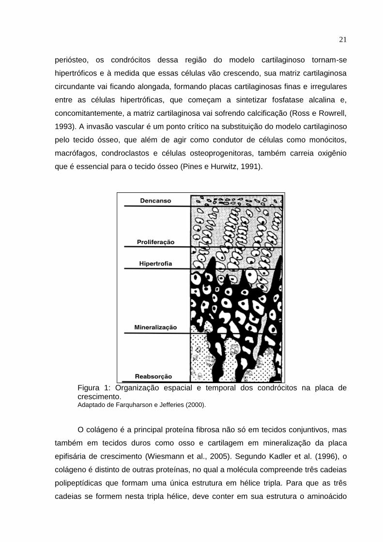
celular é exemplificada na Figura 1.
Na sequência de eventos ocorrem proliferação e agregação de células
mesenquimais no local do futuro osso, para que ocorra crescimento longitudinal na
placa epifisária que liga as regiões da epífise e diáfise dos ossos. Tendo formado o
21
periósteo, os condrócitos dessa região do modelo cartilaginoso tornam-se
hipertróficos e à medida que essas células vão crescendo, sua matriz cartilaginosa
circundante vai ficando alongada, formando placas cartilaginosas finas e irregulares
entre as células hipertróficas, que começam a sintetizar fosfatase alcalina e,
concomitantemente, a matriz cartilaginosa vai sofrendo calcificação (Ross e Rowrell,
1993). A invasão vascular é um ponto crítico na substituição do modelo cartilaginoso
pelo tecido ósseo, que além de agir como condutor de células como monócitos,
macrófagos, condroclastos e células osteoprogenitoras, também carreia oxigênio
que é essencial para o tecido ósseo (Pines e Hurwitz, 1991).
Figura 1: Organização espacial e temporal dos condrócitos na placa de crescimento. Adaptado de Farquharson e Jefferies (2000).
O colágeno é a principal proteína fibrosa não só em tecidos conjuntivos, mas
também em tecidos duros como osso e cartilagem em mineralização da placa
epifisária de crescimento (Wiesmann et al., 2005). Segundo Kadler et al. (1996), o
colágeno é distinto de outras proteínas, no qual a molécula compreende três cadeias
polipeptídicas que formam uma única estrutura em hélice tripla. Para que as três
cadeias se formem nesta tripla hélice, deve conter em sua estrutura o aminoácido
22
glicina a cada sequência de três aminoácidos, na qual frequentemente os outros
dois são os aminoácidos prolina e hidroxiprolina.
Swenson (1988), explica que a formação de fibrilas de colágeno envolve
reações no meio intracelular e extracelular. No interior da célula ocorre síntese de
moléculas de protocolágenos, hidroxilação de resíduos de prolina e lisina e
glicosilação dos resíduos de hidroxilisina, para formar monômeros de pró-colágeno
sendo secretado para o exterior da célula na forma de tripla hélice de configuração
helicoidal. No exterior da célula, há uma hidrólise proteolítica limitada do pró-
colágeno, para formar o tropocolágeno, sendo necessária para que ocorra a
hidroxilação, a presença de vitamina C. Esta hidroxilação faz parte das mudanças
póstraducionais que o colágeno é submetido, sendo exposto a ação da lisil-oxidase
para iniciar a formação das ligações cruzadas intermoleculares maduras e
irredutíveis (hidroxilisil piridolina e lisil piridolina). Estas cross-linking da molécula
madura de colágeno conferem sua força tênsil e ajudam a suportar o estresse físico
(Knott e Bailey, 1998; Farquharson e Jefferies, 2000).
Atualmente, 19 tipos de colágeno já foram identificados, os quais possuem
particularidades e estão distribuídos entre vários tipos de tecidos no organismo
animal. Em diferentes tecidos mineralizados, como osso e cartilagem, existem
diferentes tipos estruturais de colágenos, sendo o colágeno fibrilar tipo I o principal
(Wiesmann et al., 2005). Já em áreas em mineralização, como na cartilagem durante
a ossificação endocondral, o colágeno fibrilar tipo II é formado (Hiltunen et al., 1993).
Os osteoblastos secretam colágeno fibrilar tipo III ao longo da superfície do
periósteo, o qual serve como um substrato para migração celular, principalmente em
casos de remodelação óssea (Wiesmann et al., 2005).
2.2 MINERAIS – FUNÇÕES, DISPONIBILIDADE E INTERAÇÕES A NÍVEL INTESTINAL
Estudos acerca da importância dos minerais para a saúde dos animais
iniciaram-se entre 1928 e 1931 através do fornecimento de dietas purificadas à
ratos. Esses estudos, mostraram que Cobre (Cu), Manganês (Mn) e Zinco (Zn) são
elementos essenciais a saúde (Underwood, 1977). Na década de 90 com o advento
de técnicas moleculares, o metabolismo, função mineral e os diversos mecanismos

23
complexos as quais eles estão envolvidos começaram a ser desvendados (O’Dell e
Sunde, 1997). Minerais traço como Zn, Cu e Mn são cruciais para uma grande
variedade de processos fisiológicos em todos os animais (Richards et al., 2010).
De acordo com Suttle (2010), os minerais atuam sob 4 principais formas no
organismo animal, sendo elas:
1. Estrutural: formam componentes estruturais em órgãos e tecidos,
exemplificados por minerais como Cálcio (Ca) e Fósforo (P) nos ossos, e Zn e
P na estabilidade de moléculas de membrana as quais fazem parte.
2. Fisiológico: ocorrem em fluidos e tecidos como eletrólitos responsáveis por
manter a pressão osmótica, equilíbrio ácido-base, permeabilidade e
transmissão de impulsos nervosos. Como exemplos de minerais temos, Sódio
(Na), Potássio (K), Cloro (Cl), Ca e Magnésio (Mg).
3. Catalítica: atuam como catalisadores em enzimas e sistemas endócrinos
como componentes da estrutura de metaloenzimas, hormônios ou como
coenzimas. Atividades podem ser anabólicas ou catabólicas. Manganês é um
exemplo.
4. Regulatório: minerais que regulam replicação ou diferenciação celular, como
por exemplo Ca influenciando na transdução de sinais.
Historicamente, a suplementação de minerais traço em dietas para aves é
feita utilizando sais inorgânicos, principalmente na forma de óxidos e sulfatos.
Inclusive para pesquisas de exigências nutricionais as fontes inorgânicas foram
utilizadas, incluindo o NRC de 1994 e as Tabelas Brasileiras Para Aves e Suínos em
suas três edições, sendo a última delas publicada em 2011.
A importância e o papel de minerais como Ca e P e da vitamina D é muito
bem documentada, sendo raro caracterizar-se problemas locomotores pela falta do
atendimento da exigência de algum desses nutrientes. Contudo, o que ainda não
está igualmente claro, é o efeito antagônico que Ca e P podem exercer sobre
minerais traço. Underwood e Suttle (2001) afirmam que isto é devido à tendência de
minerais catiônicos (como Mn, Zn e Cu) formarem complexos insolúveis com P livre,
ácido fítico e outros componentes da digesta. Isto acontece devido a capacidade do
fitato de formar quelatos com esses minerais através de ligações covalentes,
resultando em complexos altamente estáveis e insolúveis (Leesson, 2005).
24
Dibner et al. (2007) apontam que o denominador comum das interações
antagonistas entre minerais, é a dissociação do sal inorgânico em pH relativamente
baixo do início do sistema gastrointestinal. Quando o mineral atinge o pH mais
elevado dos segmentos mais distais do intestino, pode ligar-se com outros minerais,
nutrientes e componentes não nutritivos da digesta, como o fitato e fibra, que o
tornam insolúveis, e formas insolúveis são excretadas. Devido a esses
antagonismos, o uso de sais inorgânicos podem resultar em uma grande
variabilidade ou até em baixa biodisponibilidade do mineral.
As exigências de minerais traços para frangos de corte se baseiam em níveis
preconizados pelo NRC (1994), algumas delas se referindo a dados de 1950, e
portanto, nutricionistas frequentemente utilizam níveis mais elevados de minerais,
muitas vezes baseados em seu próprio conhecimento prático (Leeson, 2005).
Mondal et al. (2010), complementam afirmando que os níveis preconizados pelo
NRC podem não oferecer o aporte ótimo de minerais para atender o máximo
potencial produtivo das linhagens modernas, e que essa maior quantidade de
minerais utilizada pela indústria pode muitas vezes exceder a quantidade mineral
necessária para o atendimento da exigência do frango. Somado a isto, considerando
a grande variabilidade ou até baixa biodisponibilidade de diferentes fontes minerais
(Dibner et al., 2007), o aumento da margem de segurança na suplementação de
microminerais resulta em alto nível de excreção mineral (Leeson, 2005), gerando
problemas de contaminação ambiental (Aksu et al., 2011).
Baseado nesse pressuposto, especialmente na última década, cresceu o
interesse por fontes de microminerais orgânicos na avicultura, devido ao seu
conceito de estabilidade, biodisponibilidade e apelo ambiental. A AAFCO
(Association of American Feed Control Officials) em 2005, definiu os microminerais
orgânicos como compostos formados através de quelação, processo de ligação
química entre um mineral traço com um carreador orgânico. Foram caracterizados
seis diferentes tipos e a agência salienta que existem importantes diferenças entre
eles que podem interferir em sua estabilidade e biodisponibilidade:
1. Complexo Metal Aminoácido específico: Produto resultado da formação de
um complexo entre um metal sal solúvel com um aminoácido pré
determinado.
25
2. Complexo Metal Aminoácido: Produto resultado da formação de um complexo
entre um metal sal solúvel com um aminoácido.
3. Quelato Metal Aminoácido: Produto resultante da reação de um íon metálico
de um sal metálico solúvel com aminoácidos com uma relação molar de um
mol de metal para um a três (de preferência dois) moles de aminoácidos,
formando ligações covalentes coordenadas. O peso médio dos aminoácidos
hidrolisados deve ser aproximadamente 150 e o peso molecular resultante do
quelato não deve exceder 800.
4. Metal Proteinato: Produto resultante da quelação de um sal solúvel com
aminoácidos e/ou proteínas parcialmente hidrolisadas.
5. Complexo Metal Polissacarídeo: Produto resultante da formação de um
complexo entre um sal solúvel com uma solução de polissacarídeos
declarados como ingredientes.
6. Metal Propionato: Produto resultante da reação de um sal metálico solúvel
com ácido propionico.
Uma das hipóteses para maior biodisponibilidade dos minerais orgânicos é
que desta forma eles ficam protegidos de interações indesejáveis no trato
gastrointestinal. Suporte para esta hipótese foi fornecido por Wedekind et al. (1992)
em um estudo clássico, foram comparados a biodisponibilidade de Zn-metionina em
relação a de sulfato de Zn, usando três diferentes dietas: purificada; semi-purificada;
e uma dieta prática de milho e farelo de soja. Estimativas de biodisponibilidade para
Zn-metionina relativa para sulfato de Zn, foram 117, 177 e 206% para o purificada,
semi-purificada, e dieta a base de milho-farelo de soja, respectivamente.
Em uma revisão publicada por Swiatkiewicz et al. (2014) em que compilaram
recentes estudos com aves comerciais, esses autores concluiram que a
biodisponibilidade e eficácia das fontes orgânicas de Zn, Mn e Cu são superiores em
relação as fontes inorgânicas tradicionalmente usadas. Acrescentam ainda, que
fontes orgânicas podem reduzir a excreção de minerais nas excretas e, desta forma,
reduzir potenciais efeitos nocivos da avicultura intensiva sobre o meio ambiente.
Resultado semelhante foi obtido por Nollet et al. (2007) que suplementaram os
minerais Mn, Zn, Fe e Cu na forma orgânica para frangos de corte em menores
níveis do que a dieta controle, composta por minerais inorgânicos, e obtiveram
26
excreções de 46, 63, 73 e 55% menores, respectivamente, quando comparados as
dietas suplementadas com minerais inorgânicos.
2.2.1 Manganês – importância e relação com o metabolismo e desenvolvimento ósseo
O Mn é um elemento traço essencial para os animais, com particular
importância para o rápido crescimento das aves. Isto acontece devido ao fato deste
mineral ser um importante cofator de metaloenzimas que atuam em diversos tecidos
e sistemas do organismo das aves, dentre eles no tecido ósseo. Já no ano de 1937,
Wilgus et al., demonstraram que o Mn é essencial para prevenção da perose e
ratificou a sua importância para o desenvolvimento apropriado dos ossos em
frangos. Mais tarde em 1962, Leach e Muenster estudaram a composição química
da cartilagem epifisária de frangos acometidos por perose e sugeriram que um
deficiência em Mn teria alterado o conteúdo de mucopolissacarídeos deste tecido.
Então em 1989, Hurley e Keen reafirmaram esta conclusão e atribuíram ao Mn uma
importante função, cofator para a ativação de um grupo de enzimas, as
glicosiltransferases. Dentre elas, a galactosiltransferase, que é necessária para a
formação de mucopolissacarídeos que desempenha um importante papel na síntese
da matriz cartilaginosa (Leach et al., 1969). Essas afirmações foram então
extensivamente confirmadas (Saltman e Strause, 1993; Cashman e Flynn, 1998), e
hoje, sabe-se que o Mn é um componente crucial de enzimas envolvidas no sistema
antioxidante, metabolismo proteico e formação óssea, como superóxido dismutase,
transferases, hidrolases e ligases (Keen et al., 1999; 2000).
Wang et al. (2014) estudaram o efeito de três níveis de Mn (controle com 60
ppm e dois níveis deficientes, 40 e 8,7 ppm) sobre a largura da zona de proliferação
da placa de crescimento e taxa de condrócitos que sofreram apoptose. A medida
que o nível de Mn foi reduzido na dieta, a largura da zona de proliferação diminuiu e
houve um aumento do número de condrócitos apoptóticos. Esses pesquisadores
discutem que, conforme esperado, a deficiência de Mn causou uma supressão na
placa de crescimento proximal da tíbia. Especulam ainda que dietas deficientes em
Mn podem inibir a proliferação de condrócitos e promover a apoptose. O
27
crescimento da tíbia depende da proliferação, diferenciação e apoptose dos
condrócitos.
Liu et al. (2015) mediram a área e largura do osso trabecular na metáfise tibial
e mostraram que dietas deficientes em Mn apresentaram tanto uma menor área,
quanto menor largura de osso trabecular quando comparadas ao controle que
atendia a exigência de Mn, segundo o NRC (1994).
2.2.2 Manganês – importância e relação com a competência imunológica
Vários minerais tem sido estudados como potentes imunomoduladores
(Lawrence, 1981; Beach et al., 1982; Neilan et al., 1983; Smialowicz et al., 1984).
Embora o papel do Zn esteja muito bem documentado em relação a importância
para o bom funcionamento do sistema imune (Shankar, 1998; Fraker et al., 2000; Ibs
et al., 2003), existe pouca informação sobre os efeitos de deficiência de Mn e os
mecanismos pelos quais atua na resposta imunológica (Son et al., 2007; Kehl-Fie e
Skaar, 2010). Smialowicz et al. (1984) trataram ratos com cloreto de manganês
(MnCl2) e reportaram uma melhora na atividade de células natural killer (NK) e um
aumento nos níveis circulantes de interferon (IFN), que conhecidamente afetam a
capacidade primária de resposta à antígenos. Esses autores relatam que nos níveis
utilizados, o Mn não afetou a resposta proliferativa de células do baço para células T
ou B. Esses mesmo autores concluíram ainda que a aplicação de MnCl2 melhorou a
função dos macrófagos através da indução de interferon, mesmo mecanismo pelo
qual Mn aumentou a atividade das células NK.
Estes resultados foram confirmados por Son et al. (2007), que trabalharam
com suplementação de Mn e magnésio (Mg). Estes autores encontraram que a
capacidade citolítica das células NK e a citotoxicidade de macrófagos contra células
tumorais foram aumentadas em ratos suplementados em relação ao controle. A
capacidade de resposta imunológica após suplementação com Mg e Mn apresentam
efeitos positivos, no entanto, a explicação exata de como os minerais modulam a
resposta imune ainda não foi completamente elucidada.
Outra importante atuação do Mn no organismo é como cofator da
metaloenzima manganês superóxido dismutase (MnSOD) (Li et al., 2010), uma
enzima com função antioxidante que possui um papel fundamental na detoxificação
28
de radicais livres superóxidos (Zhu et al., 2015). Sies (1985) definiu o estresse
oxidativo como “uma perturbação no equilíbrio pro-oxidante-antioxidante em favor do
primeiro.” Assim, o estresse oxidativo é essencialmente um desequilíbrio entre a
produção de várias espécies reativas e a capacidade dos mecanismos de defesa
natural do organismo para lidar com estes compostos reativos e prevenir os efeitos
adversos. Espécies reativas de oxigênio (EROs) e o nitrogênio podem atacar vários
substratos no organismo, incluindo lipídeos, proteínas, DNA, levando a altas taxas
de morte e turnover celular (Mayne, 2003). De acordo com Underwood e Suttle
(1999), um dos dois tipos existentes de MnSOD atua essencialmente na
mitocôndria, evitando que a ave sofra os efeitos deletérios do estresse oxidativo.
Em um interessante estudo publicado por Corbin et al. (2008), comprovou-se
que abcessos causados por Staphilococcus aureus eram desprovidos de Mn,
enquanto tecidos saudáveis que circundavam os abcessos estavam repletos do
metal. Estudos subsequentes revelaram que a proteína calprotectina do hospedeiro
é necessária para sequestrar o Mn nestes abcessos. Esta proteína representa cerca
de 40-50% do total de proteínas que compõe o citoplasma de neutrófilos em
mamíferos (Gebhardt et al., 2006). Este aumento na quantidade de Mn disponível
em abcessos de animais deficientes em calprotectinas, coincide com o aumento da
carga bacteriana nestes órgãos, sugerindo que a quelação de manganês mediada
por calprotectina é necessária para proteger contra infecções microbianas.
Complementarmente a estes resultados in vivo, Corbin et al. (2008)
demonstraram que a calprotectina é capaz de se ligar ao Mn in vitro e inibir o
crescimento de bactérias de uma maneira independente de contato físico, que é
reversível após a adição de qualquer excesso de Mn e Zn.
Como medida de defesa, de acordo com trabalho publicado por Kehl-Fie e
Skaar (2010) para contornar as defesas do hospedeiro, as bactérias possuem
transportadores de alta afinidade para sequestrar estes metais específicos, limitando
a disponibilidade de Zn e Mn como um mecanismo de defesa contra a infecção.
Concluem ainda, que está se tornando clara a utilização desta estratégia de
sequestro de Zn e Mn por parte do hospedeiro, com o objetivo de limitar a ação de
patógenos.
29
2.3 METABOLISMO E IMPORTÂNCIA DA ARGININA NO CRESCIMENTO E COMPETÊNCIA IMUNOLÓGICA DE AVES
Aves são conhecidamente animais uricotélicos, ou seja, o seu metabolismo
excretório de nitrogênio acontece na forma de ácido úrico (Leningher, 2002). Os
enterócitos das aves não possuem as enzimas necessárias para a produção de
citrulina que, em mamíferos é a precursora da síntese de Arg (Tamir e Ratner,
1963). A síntese da Arg ocorre principalmente no eixo intestino-renal. Células do
epitélio do intestino delgado produzem citrulina e células dos túbulos proximais nos
rins extraem a citrulina da circulação sanguínea, que é convertida à Arg, que retorna
à circulação. Assim, em mamíferos, quase a totalidade da Arg sintetizada pelo
organismo é degradada a ornitina e ureia, no fígado, devido à alta atividade da
arginase hepática. Entretanto, nos rins, devido à baixa atividade da arginase renal,
existe síntese líquida de Arg para o organismo (Wu e Morris, 1998).
A inabilidade das aves para sintetizar ornitina, citrulina e arginina é atribuida a
ausência de quatro enzimas do ciclo da ureia, carbamil fosfato sintetase, ornitina
carbamil transferase, arginino succinato sintetase e arginino succinato liase (Tamir e
Ratner, 1963; Wu et al., 1995).
Devido a essas diferenças metabólicas, a Arg é considerada um aminoácido
essencial para as aves. Arg é um dos mais versáteis aminoácidos nas células
animais, constitui proteínas, e é precursor de importantes moléculas como NO, ureia,
poliaminas, Pro, glutamato e creatina (Wu e Morris, 1998). Arginase converte Arg em
ornitina e ureia. Entre 40 a 60% de ureia excretada pelas aves provem do
metabolismo da Arg, e, como as aves não podem sintetizar ornitina, quase toda a
ornitina plasmática em aves também é derivada do metabolismo da Arg (Nesheim,
1968; Austic e Nesheim, 1970; Stutz et al., 1972; Chu e Nesheim, 1979).
Dois importantes destinos metabólicos da Arg são controlados por duas
enzimas distintas (Grazi et al., 1975). A primeira degradação de Arg é mediada pela
arginase, liberando ornitina e ureia, enquanto a outra é catalisada pela enzima óxido
nítrico sintetase e tem como produto o NO.
Ao contrário dos mamíferos que possuem uma clara caracterização das
enzimas arginase I (enzima citosólica encontrada no fígado) e arginase II (enzima
mitocondrial encontrada em vários tecidos) no que se refere a propriedades
30
moleculares e imunológicas, distribuição tecidual, localização intracelular e
regulação da expressão (Ash, 2004), nas aves apenas a arginase renal é tida como
de importância na degradação de Arg (Ruiz-Feria et al., 2001).
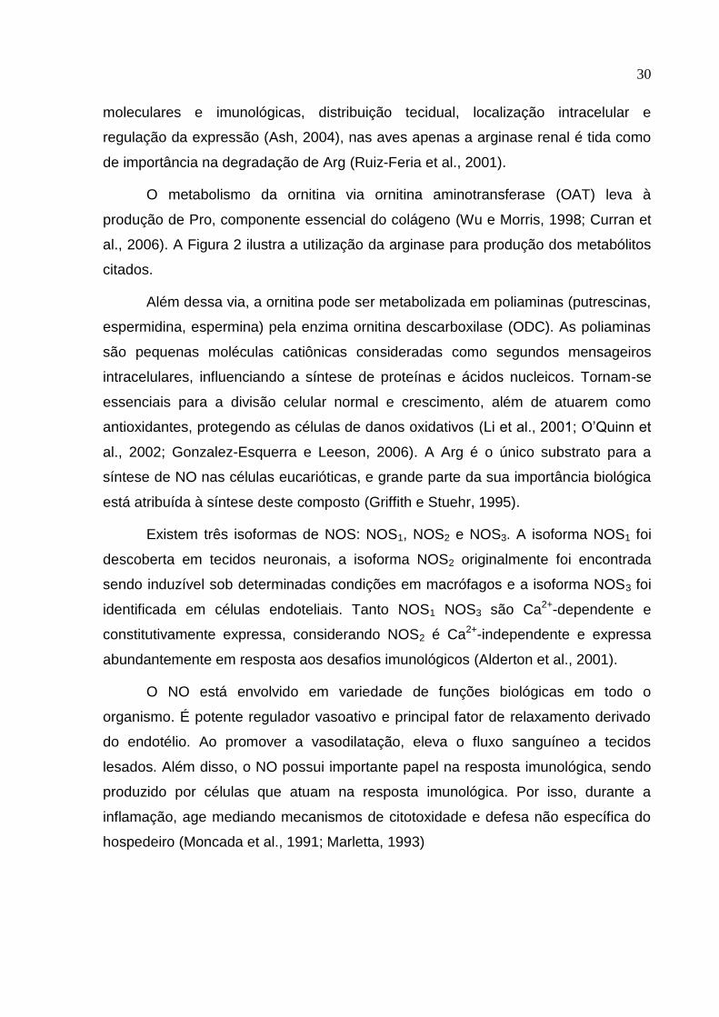
O metabolismo da ornitina via ornitina aminotransferase (OAT) leva à
produção de Pro, componente essencial do colágeno (Wu e Morris, 1998; Curran et
al., 2006). A Figura 2 ilustra a utilização da arginase para produção dos metabólitos
citados.
Além dessa via, a ornitina pode ser metabolizada em poliaminas (putrescinas,
espermidina, espermina) pela enzima ornitina descarboxilase (ODC). As poliaminas
são pequenas moléculas catiônicas consideradas como segundos mensageiros
intracelulares, influenciando a síntese de proteínas e ácidos nucleicos. Tornam-se
essenciais para a divisão celular normal e crescimento, além de atuarem como
antioxidantes, protegendo as células de danos oxidativos (Li et al., 2001; O’Quinn et
al., 2002; Gonzalez-Esquerra e Leeson, 2006). A Arg é o único substrato para a
síntese de NO nas células eucarióticas, e grande parte da sua importância biológica
está atribuída à síntese deste composto (Griffith e Stuehr, 1995).
Existem três isoformas de NOS: NOS1, NOS2 e NOS3. A isoforma NOS1 foi
descoberta em tecidos neuronais, a isoforma NOS2 originalmente foi encontrada
sendo induzível sob determinadas condições em macrófagos e a isoforma NOS3 foi
identificada em células endoteliais. Tanto NOS1 NOS3 são Ca2+-dependente e
constitutivamente expressa, considerando NOS2 é Ca2+-independente e expressa
abundantemente em resposta aos desafios imunológicos (Alderton et al., 2001).
O NO está envolvido em variedade de funções biológicas em todo o
organismo. É potente regulador vasoativo e principal fator de relaxamento derivado
do endotélio. Ao promover a vasodilatação, eleva o fluxo sanguíneo a tecidos
lesados. Além disso, o NO possui importante papel na resposta imunológica, sendo
produzido por células que atuam na resposta imunológica. Por isso, durante a
inflamação, age mediando mecanismos de citotoxidade e defesa não específica do
hospedeiro (Moncada et al., 1991; Marletta, 1993)
31
Figura 2: Metabolismo da Arg em mamífero, com duas diferentes vias para produzir NO, Pro e poliaminas. Adaptado de Witte e Barbul (2003). Legenda: OAT: ornitina aminotransferase; ODC: ornitina descarboxilase.
Todos os processos de degradação da Arg geram compostos úteis ao
organismo, seja para deposição proteica, ação imunológica, ou excreção de
nitrogênio, que pode ser tóxico se estiver em excesso. Portanto, os produtos do
catabolismo da Arg são tão necessários ao bom funcionamento do organismo
quanto a própria Arg (Dhanakoti et al., 1990).
2.3.1 Manganês como co-fator da arginase
A maior parte do Mn no organismo é encontrado nas mitocôndrias. Ele ativa
uma série de enzimas, tais como hidrolases, transferases, quinases e
decarboxylases, e é um constituinte de algumas outras. Uma das mais conhecidas
metaloenzimas ativadas pelo Mn é a piruvato carboxilase, que catalisa a conversão
de piruvato a oxaloacetato (Scrutton et al., 1966). Outras enzimas incluem a
arginase, que está envolvida na conversão do aminoácido Arg para ureia e ornitina,
e superóxido dismutase mitocondrial (SOD). A estrutura e função das mitocôndrias,
portanto, são particularmente afetadas pela concentração de Mn. Mn também ativa
enzimas associadas com o metabolismo de ácidos graxos e síntese de proteínas
(Wilson et al., 1979).
Uma característica comum de todas as arginases até agora estudadas, sejam
elas de origem eucariótica ou procariótica, é a exigência de cátions divalentes para a
sua atividade, sendo o Mn seu ativador fisiológico (Ash, 2004). A base molecular
32
para e seleção deste mineral específico como cofator na reação ainda permanece
desconhecida.
A descoberta da óxido nítrico sintetase, que catalisa a oxidação de Arg para
formar NO e citrulina, tem gerado grande interesse na interação entre as enzimas
NOS e arginase. Nos mamíferos, vários tecidos expressam ambos tipo I e tipo II
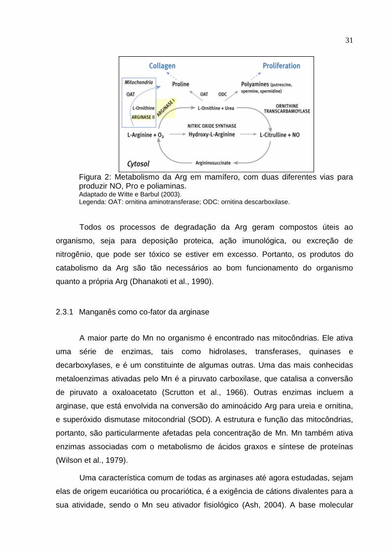
arginases, bem como óxido nítrico sintase, e por estas enzimas competirem por um
substrato comum, Arg, a co-expressão destas enzimas levanta questões
interessantes sobre a regulação de fluxo de Arg através dos caminhos concorrentes
(Ash, 2004). A Figura 3 ilustra essa competição pela Arg.
Ruiz-Feria et al. (2001) explicam que a concentração de Arg plasmática é
diretamente proporcional as mensurações de ureia e ornitina plasmática. Entende-se
portanto que quanto maior o nível do aminoácido Arg presente nas dietas, mais
substrato para as referidas enzimas estará disponível para produzir os metabolitos
ornitina e NO.
Para mamíferos há um consenso de que arginase modula a atividade da
iNOS, provavelmente regulando a disponibilidade intracelular de Arg (Berkowitz et
al., 2003). Entretanto, para aves, devido ao metabolismo particular da Arg, ainda não
há um consenso desta regulação.
Figura 3: Ilustração da relação competitiva entre a atividade da arginase e iNOS pelo substrato comum, Arg. Adaptado de Ash (2004).
33
Nas aves, pouco tem sido elucidado a respeito desse mecanismo competitivo
entre as enzimas. Por outro lado, Ash (2004) discute no seu trabalho de revisão um
mecanismo inibitório recíproco entre a arginase I e a iNOS em mamíferos,
prevenindo a produção de níveis demasiadamente elevados de NO e por outro lado,
a inibição da atividade da arginase I resultaria na elevação da produção de NO.
Desta forma, a arginase II atuaria produzindo ornitina para a síntese de poliaminas e
Pro, necessárias para o reparo tecidual.
Essa elevação da atividade da arginase renal também é observada quando
são fornecidos níveis muito altos de Arg (Stutz et al., 1972). A degradação,
excessivamente rápida de Arg, em ornitina e ureia, pode comprometer a síntese de
NO, sendo esta a única via fisiológica de produção deste composto. Adicionalmente,
a elevação da ureia plasmática, de acordo com (Prabhakar et al., 1997), pode inibir a
ação da NOS.
2.3.2. Arginina, óxido nítrico sintetase e arginase - relação com a competência imunológica
Ao contrário da iNOS (óxido nítrico sintetase induzível), pouco é conhecido
sobre a regulação e a função das arginases atuando no sistema imunológico. Já foi
especulado que arginase participa na regulação da síntese de NO competindo pelo
substrato comum Arg. Outras funções sugeridas incluem um envolvimento em
processos fibrogênicos ou reparativos via síntese de colágeno ou ações
antiinflamatórias via produção de poliaminas (Jenkinson et al., 1996). Munder et al.
(1998) demonstrou que as citocinas Th1 e Th2, bem como as correspondentes
células T, competitivamente regulam o equilíbrio do metabolismo de Arg em
macrófagos de ratos. Enquanto células Th1 e citocinas induzem iNOS e suprimem
arginase, células Th2 e citocinas induzem arginase e suprimem iNOS.
Arginase também pode desempenhar um papel semelhante durante a
cicatrização de feridas. Uma regulação recíproca da iNOS e atividade da arginase já
foi demonstrada em ratos com um aumento na expressão de iNOS na fase inicial de
cicatrização de feridas, provavelmente, criando um ambiente citotóxico e um
aumento na expressão de arginase na fase posterior, de reparo (Albina et al., 1990).
34
O ambiente hipóxico na cicatrização de feridas pode ser um indutor adicional de
arginase (Louis et al., 1998).
Além disso, Boucher et al. (1999) mostrou que a IL-4, um mediador gerado
durante a inflamação, pode desempenhar um papel importante na alternação entre
alta formação de NO para baixa formação de NO, e aumentar a síntese de ornitina e
poliaminas durante o reparo do tecido.
Existem vários estudos relatando os efeitos da Arg sobre diversos aspectos
das funções imunes em animais e humanos (Park et al., 1991; Wrightham et al.,
1992; Ochoa et al., 2001). O principal efeito imunomodulador de Arg em animais
está relacionado ao aumento da produção de NO pelos macrófagos através de iNOS
(Tsai et al., 2002). Os macrófagos são considerados células centrais na imunidade
específica e inespecífica e também são capazes de segregar potentes citocinas,
incluindo IL-1 e TNF-α (Amber et al., 1991; Tsai et al., 2002; Tayade et al., 2006).
Citocinas são uma classe de proteínas ou pequenos peptídeos, a qual transmitem
informação entre células e possuem funções imunomodulatórias. Citocinas incluem
interleucinas, interferon, fator de necrose tumoral, entre outros. IL-1 pode promover
a proliferação de linfócitos B e secretam anticorpos. IL-2 é um fator melhorador de
amplo espectro in vivo. TNF-α pode matar ou inibir certas células tumorais in vivo e
in vitro.
Guo et al. (2015) mostraram que a suplementação de Arg resultou em
aumento nos níveis de citocinas e Reynolds et al. (1990) e Yeh et al. (2002), na
produção de IL-2 de linfócitos.
O NO é um composto que participa de vários fenômenos dentre eles
citotoxicidade mediada por macrófagos, vasodilatação, inibição da ativação, adesão
e agregação plaquetária. O NO é o principal mediador citotóxico de células imunes
efetoras ativadas e constitui a mais importante molécula reguladora do sistema
imune (Hibbs et al., 1988). A expressão da iNOS é o resultado de uma resposta
inflamatória localizada ou difusa, resultante de uma infecção ou dano tecidual.
Segundo Salvemini et al. (1996), o NO é um potente vasodilatador e seu
envolvimento na resposta inflamatória pode ter relação com sua habilidade em
aumentar a permeabilidade vascular e o edema por meio de mudanças no fluxo
sanguíneo local e do aumento na produção de prostaglandinas pró-inflamatórias.
35
Além da produção de NO, a suplementação de Arg ainda melhora o peso e
função do timo (Barbul et al., 1980), a resposta linfocitária a mitógenos como
Concanavalina A e Fitohemaglutinina (Ochoa et al., 2001) e induz efeitos
estimulatórios na produção ou função de citocinas e outras células do sistema
imunológico (Ochoa et al., 2001; Wu et al., 2008). Arg é também um dos fatores
necessários para a diferenciação e liberação de linfócitos B de medula óssea (Park
et al., 1991; De Jonge et al., 2002).
2.3.3. Arginina e prolina – relação com a formação de colágeno e metabolismo ósseo
Mamíferos podem sintetizar prolina através da arginina (via arginase tipo I e
II), ornitina amino transferase e P-5-C redutase (Wu et al., 2008).
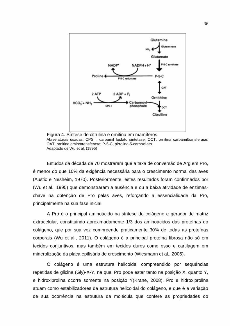
Ornitina é sintetizada a partir de glutamato, através da formação de pirrolina-
5-carboxilato (P-5-C) na mucosa intestinal de mamíferos. P-5-C é uma enzima
reguladora importante na síntese de ornitina e citrulina a partir de glutamina nos
enterócitos. Devido a ausência de P-5-C e ornitina carbamil transferase nos
enterócitos das aves, estas são incapazes de sintetizar ornitina e citrulina a partir
glutamina (Wu et al., 1995) (Figura 4). Portanto, praticamente toda a ornitina
plasmática é derivada do metabolismo da Arg (Stutz et al., 1972; Chu e Nesheim,
1979). Para que a Arg possa ser utilizada para a síntese das poliaminas (putrescina,
espermina e espermidina) ou Pro, precisa ser hidrolisada para ureia e ornitina pela
arginase (Wu e Morris, 1998).
36
Figura 4. Síntese de citrulina e ornitina em mamíferos. Abreviaturas usadas: CPS I, carbamil fosfato sintetase; OCT, ornitina carbamiltransferase; OAT, ornitina aminotransferase; P-5-C, pirrolina-5-carboxilato. Adaptado de Wu et al. (1995)
Estudos da década de 70 mostraram que a taxa de conversão de Arg em Pro,
é menor do que 10% da exigência necessária para o crescimento normal das aves
(Austic e Nesheim, 1970). Posteriormente, estes resultados foram confirmados por
(Wu et al., 1995) que demonstraram a ausência e ou a baixa atividade de enzimas-
chave na obtenção de Pro pelas aves, reforçando a essencialidade da Pro,
principalmente na sua fase inicial.
A Pro é o principal aminoácido na síntese do colágeno e gerador de matriz
extracelular, constituindo aproximadamente 1/3 dos aminoácidos das proteínas do
colágeno, que por sua vez compreende praticamente 30% de todas as proteínas
corporais (Wu et al., 2011). O colágeno é a principal proteína fibrosa não só em
tecidos conjuntivos, mas também em tecidos duros como osso e cartilagem em
mineralização da placa epifisária de crescimento (Wiesmann et al., 2005).
O colágeno é uma estrutura helicoidal compreendido por sequências
repetidas de glicina (Gly)-X-Y, na qual Pro pode estar tanto na posição X, quanto Y,
e hidroxiprolina ocorre somente na posição Y(Krane, 2008). Pro e hidroxiprolina
atuam como estabilizadores da estrutura helicoidal do colágeno, e que é a variação
de sua ocorrência na estrutura da molécula que confere as propriedades do

37
colágeno em termos de rigidez, estabilidade química e respostas biomecânicas (Wu
et al., 2011).
38
3. REFERÊNCIAS
ABPA Associação Brasileira de Proteína Animal (http://abpa-br.com.br/noticia/producao-de-carne-de-frango-totaliza-13146-milhoes-de-toneladas-em-2015-1545), janeiro 2016.
Aksu, T., M. Aksu, M. a. Yoruk, and M. Karaoglu. 2011. Effects of organically-complexed minerals on meat quality in chickens. Br. Poult. Sci. 52:558–563.
Albina, J. E., C. D. Mills, W. L. Henry, and M. D. Caldwell. 1990. Temporal expression of different pathways of L-arginine metabolism in healing wounds. J. Immunol. 144:3877.
Alderton, W. K., C. E. Cooper, and R. G. Knowles. 2001. Nitric oxide synthases : structure , function and inhibition. 615:593–615.
Amber, I. J., J. B. Hibbs, C. J. Parker, B. B. Johnson, R. R. Taintor, and Z. Vavrin. 1991. Activated macrophage conditioned medium: identification of the soluble factors inducing cytotoxicity and the L-arginine dependent effector mechanism. J. Leukoc. Biol. 49:610–620.
Applegate, T. J., and R. Angel. 2014. Nutrient requirements of poultry publication: History and need for an update. J. Appl. Poult. Res. 23:567–575
Ash, D. 2004. Arginine metabolism: enzymology, nutrition, and clinical significance. J. Nutr. 134:2741S–2897S.
Austic, R. E., and M. C. Nesheim. 1970. Role of kidney arginase in variations of the arginine requirement of chicks. J. Nutr. 100:855–868.
Bain, S.D and Watkins, B. . 1993. Local modulation of skeletal growth and bone modeling in poultry. J. Nutr.
Barbul, A., D. A. Sisto, H. L. Wasserkrug, and G. Efron. 1981. Arginine stimulates lymphocyte immune response in healthy human beings. Surgery 90:244–251.
Beach, R. S., E. M. Gershwin, and L. S. Hurley. 1982. Gestational zinc deprivation in mice: persistence of immunodeficiency for three generations. Science (80). 218:469–471.
Berkowitz, D. E., R. White, D. Li, K. M. Minhas, A. Cernetich, S. Kim, S. Burke, A. a. Shoukas, D. Nyhan, H. C. Champion, and J. M. Hare. 2003. Arginase Reciprocally Regulates Nitric Oxide Synthase Activity and Contributes to Endothelial Dysfunction in Aging Blood Vessels. Circulation 108:2000–2006.
Bessei, W. 2006. Welfare of broilers: A review. Worlds. Poult. Sci. J. 62:455–466.
39
Biewener, A. a, and J. E. Bertram. 1994. Structural response of growing bone to exercise and disuse. J. Appl. Physiol. 76:946–55.
Boucher, J. L., C. Moali, and J. P. Tenu. 1999. Nitric oxide biosynthesis, nitric oxide synthase inhibitors and arginase competition for L-arginine utilization. Cell. Mol. Life Sci. 55:1015–1028.
Cashman, K., and A. Flynn. 1998. Trace elements and bone metabolism. Bibl Nutr Dieta 54:150–64.
Chu, S. W., and M. C. Nesheim. 1979. The relationship of plasma arginine and kidney arginase activity to arginine degradation in chickens. J. Nutr. 109:1752–1758.
Cook, M. E. 2000. Skeletal deformities and their causes: introduction. Poult. Sci. 79:982–4.
Corbin, B. D., E. H. Seeley, A. Raab, J. Feldmann, M. R. Miller, V. J. Torres, K. L. Anderson, B. M. Dattilo, K. L. Anderson, P. M. Dunman, R. Gerads, R. Caprioli, W. Nacken, W. J. Chazin, and E. P. Skaar. 2008. Bacterial Growth in Tissue Abscesses. Science (80-. ). 319:962–966.
Curran, J. N., D. C. Winter, and D. Bouchier-Hayes. 2006. Biological fate and clinical implications of arginine metabolism in tissue healing. Wound Repair Regen. 14:376–386.
Danbury, T. C., C. A. Weeks, J. P. Chambers, A. E. Waterman Pearson, and S. C. Kestin. 2000. Self selection of the analgesic drug carprofen by lame broiler chickens. Vet. Rec. 146:307–311.
Dhanakoti, S. N., J. T. Brosnan, G. R. Herzberg, and M. E. Brosnan. 1990. Renal arginine synthesis: studies in vitro and in vivo. Am. J. Physiol. 259:E437–42.
Dibner, J. J., J. D. Richards, M. L. Kitchell, and M. a. Quiroz. 2007. Metabolic challenges and early bone development. J. Appl. Poult. Res. 16:126–137.
Dudhia, J. 2005. Aggrecan Aging and Assembly in Articular Cartilage. Cell Mol Life Sci. 62:2241–2256.
Farquharson, C., and D. Jefferies. 2000. Chondrocytes and longitudinal bone growth: the development of tibial dyschondroplasia. Poult. Sci. 79:994–1004.
Fraker, P. J., L. E. King, T. Laakko, and T. L. Vollmer. 2000. The dynamic link between the integrity of the immune system and zinc status. J. Nutr. 130:1399S–406S.
Gebhardt, C., J. Németh, P. Angel, and J. Hess. 2006. S100A8 and S100A9 in inflammation and cancer. Biochem. Pharmacol. 72:1622–1631.
40
Gilbert, S. F. 1997. Early vertebrate development: Mesoderm and endoderm. Pages 341–357 in Developmental Biology. 5th ed. Sinauer Assoc. Inc., Sunderland, MA.
Gonzalez-Esquerra, R., and S. Leeson. 2006. Concentrations of putrescine, spermidine, and spermine in duodenum and pancreas as affected by the ratio of arginine to lysine and source of methionine in broilers under heat stress. Poult. Sci. 85:1398–1408.
Grazi, E., E. Magri, and G. Balboni. 1975. On the control of arginine metabolism in chicken kidney and liver. Eur. J. Biochem. 60:431–436.
Griffith, O. W., and D. J. Stuehr. 1995. Nitric Oxide Synthases: Properties and Catalytic Mechanism. Annu. Revis. Physiol. 57:707–736.
Guo, Y. W., B. L. Shi, S. M. Yan, Y. Q. Xu, J. L. Li, and T. Y. Li. 2015. Effects Of Arginine On Cytokines And Nitric Oxide Synthesis In Broilers. 25:366–371.
Hibbs, J. B., R. R. Taintor, V. Zdenek, and E. Rachlin. 1988. Nitric oxide: a cytotoxic activated macrophage effector molecule. Biochem. Biophys. Res. Commun. 153:87–94.
Hiltunen, A., Aro, H. T., and Vuorio, E. (1993). Regulation of extracellular matrix genes during fracture healing in mice. Clin. Orthop. 297, 23–27.
Hurley LS,Keen CL (1989) Manganese. In:MertzW(ed) Trace element in human and animal nutrition, 5th edn. Academic, San Diego, pp 185–221.
Ibs, K.-H., Z. I. Function, and L. Rink. 2003. Immunity Enhanced by Trace Elements. Sci. York 133:1452S–6S.
Jenkinson, C. P., W. W. Grody, and S. D. Cederbaum. 1996. Comparative properties of arginases. Comp. Biochem. Physiol. - B Biochem. Mol. Biol. 114:107–132.
Johnsson, M., K. B. Jonsson, L. Andersson, P. Jensen, and D. Wright. 2015. Genetic regulation of bone metabolism in the chicken: similarities and differences to Mammalian systems. PLoS Genet. 11:e1005250.
Junqueira, L. C. & Carneiro, J. Histologia Básica. 10ed. Rio de Janeiro: Guanabara Koogan, 2004.
De Jonge, W. J., K. L. Kwikkers, A. a. Te Velde, S. J. H. Van Deventer, M. a. Nolte, R. E. Mebius, J. M. Ruijter, M. C. Lamers, and W. H. Lamers. 2002. Arginine deficiency affects early B cell maturation and lymphoid organ development in transgenic mice. J. Clin. Invest. 110:1539–1548.
Kadler, K. E., D. F. Holmes, J. a Trotter, and J. a Chapman. 1996. Collagen fibril formation. Biochemistry 316:1–11.
41
Keen, C.L., Enunsa, J.L. And Clegg, M.S. (2000) Manganese metabolism in animals and humans including the toxicity of manganese. Metal Ions in Biological Systems 37: 89-121.
Keen, C.L., Ensunsa, J.L., Watson, M.H., Baly, D.L., Donovan, S.M., Monaco, M.H. And Clegg, M.S. (1999) Nutritional aspectsof manganese from experimental studies. Neurotoxicology 20: 213- 223.
Kehl-Fie, T. E., and E. P. Skaar. 2010. Nutritional immunity beyond iron: a role for manganese and zinc. Curr. Opin. Chem. Biol. 14:218–224.
Kierszenbaum, A. L. Tecidos Conjuntivos. In: Kierszenbaum A L, Histologia e Biologia Celular. 2° ed. Rio de Janeiro: Elsevier, 2008, p.133-144.
Knott, L., and A. J. Bailey. 1998. Collagen Cross-Links in Mineralizing Tissues : A Review of Their Chemistry , Function , and Clinical Relevance. Bone 22:181–187.
Knowles, T. G., S. C. Kestin, S. M. Haslam, S. N. Brown, L. E. Green, A. Butterworth, S. J. Pope, D. Pfeiffer, and C. J. Nicol. 2008. Leg disorders in broiler chickens: prevalence, risk factors and prevention. PLoS One 3:e1545.
Krane, S. M. 2008. The importance of proline residues in the structure, stability and susceptibility to proteolytic degradation of collagens. Amino Acids 35:703–710.
Lawrence, D. a. 1981. Heavy metal modulation of lymphocyte activities. Toxicol. Appl. Pharmacol. 57:439–451.
Leach, R. M., and A. M. Muenster. 1962. Studies on the role of manganese in bone formation I. Effect upon the mucopolysaccharide content of chick bone. J. Nutr. 78:51–56.
Leach, R. M., A. M. Muenster, and E. M. Wien. 1969. Studies on the role of manganese in bone formation II. Effect upon chondroitin sulfate synthesis in chick epiphyseal cartilage. Arch. Biochem. Biophys. 133:22–28.
Leeson, S. 2005. Trace mineral requirements of poultry— validity of the NRC recommendations. Pages 107–118 in Redefining Mineral Nutrition. J. Taylor-Pickard and L. Tucker, ed. Nottingham Univ. Press, UK. 68.
Leningher AL. 2002.Princípios de Bioquímica. 3 ed. São Paulo(SP): Sarvier.
Li, S., L. Lu, S. Hao, Y. Wang, L. Zhang, S. Liu, B. Liu, K. Li, and X. Luo. 2010. Dietary Manganese Modulates Expression of the Manganese-Containing Superoxide Dismutase. J. Nutr. 141:189–194.
Li, H., C. J. Meininger, J. R. Hawker, T. E. Haynes, D. Kepka-Lenhart, S. K. Mistry, S. M. Morris, and G. Wu. 2001. Regulatory role of arginase I and II in nitric oxide, polyamine, and proline syntheses in endothelial cells. Am. J. Physiol. Endocrinol. Metab. 280:E75–82.
42
Lilburn, M. S. 1994. Skeletal growth of commercial poultry species. Poult. Sci. 73:897–903.
Liu, R., C. Jin, Z. Wang, Z. Wang, J. Wang, and L. Wang. 2015. Effects of manganese deficiency on the microstructure of proximal tibia and OPG/RANKL gene expression in chicks. Vet. Res. Commun. 39:31–37.
Louis, C. A., J. S. Reichner, W. L. Henry, B. Mastrofrancesco, T. Gotoh, M. Mori, and J. E. Albina. 1998. Distinct arginase isoforms expressed in primary and transformed macrophages: regulation by oxygen tension. Am. J. Physiol.:775–782.
Luo, X. M., G. J. Fosmire, and R. M. Leach. 2002. Chicken keel cartilage as a source of chondroitin sulfate. Poult. Sci. 81:1086–1089.
Macari, M.; Furlan, R.L.; Gonzales, E. Fisiologia aviária aplicada à frangos de corte.ed.Jaboticabal:FUNEP/UNESP, 375p, 2002.
Marletta, M. A. 1993. Nitric oxide synthase structure and mechanism. J Biol Chem 268:12231–12234.
Marks, S. C., Jr., and S. N. Popoff. 1988. Bone cell biology: The regulation of development, structure, and function in the skeleton. Am. J. Anat. 183:1–44.
Mayne, S. T. 2003. Antioxidant Nutrients and Chronic Disease: Use of Biomarkers of Exposure and Oxidative Stress Status in Epidemiologic Research. J. Nutr. 133:933S–940.
Moncada, S., R. M. J. Palmer, and E. A. Higgs. 1991. Nitric Oxide: Physiology, Pathophysiology and Pharmacology. Pharmacol. Rev. 43:109–142.
Mondal, S., S. Haldar, P. Saha, and T. K. Ghosh. 2010. Metabolism and tissue distribution of trace elements in broiler chickens’ fed diets containing deficient and plethoric levels of copper, manganese, and zinc. Biol. Trace Elem. Res. 137:190–205.
Munder, M., K. Eichmann, and M. Modolell. 1998. Alternative Metabolic States in Murine Macrophages Reflected by the Nitric Oxide Synthase/Arginase Balance: Competitive Regulation by CD4 + T Cells Correlates with Th1/Th2 Phenotype. J. Immunol. 160:5347–5354.
Neilan, B. a., K. O’Neill, and B. S. Handwerger. 1983. Effect of low-level lead exposure on antibody-dependent and natural killer cell-mediated cytotoxicity. Toxicol. Appl. Pharmacol. 69:272–275.
Nesheim, M. C. 1968. Kidney Arginase Activity and Lysine Tolerance in Strains of Chickens Selected for a High or Low Requirement of Arginine ’. J. Nutr.
Nollet, L., J. D. Van Der Klis, M. Lensing, and P. Spring. 2007. The effect of replacing inorganic with organic trace minerals in broiler diets on productive performance and mineral excretion. J. Appl. Poult. Res. 16:592–597.
43
NRC. 1994. National Research Council. Nutrient Requirements of Poultry. 8 rev. ed. Washington: National Academy Press.
O’Dell, B.L. and Sunde, R.A. (1997) Introduction to Handbook of Nutritionally Essential Minerals. Marcel Dekker Inc., New York, pp. 8–11.
O’Quinn, P. R., D. a. Knabe, and G. Wu. 2002. Arginine catabolism in lactating porcine mammary tissue. J. Anim. Sci. 80:467–474.
Ochoa, J. B., J. Stange, P. Kearny, G. Gellin, E. Endean, and E. Fitzpatrick. 2001. Effects of L-Arginine on the Proliferation of T Lymphocyte Subpopulations. J. Parenter. Enter. Nutr. 25:23–29.
Oviedo EO, Ferket PR. Nutritional factors that affect leg problems in meat poultry: A Review. In: Ferket PR, editor. Proceedings of the 32nd Annual Carolina Poultry Nutrition Conference; 2005; North Carolina: Carolina Feed Industry Association in Cooperation with NC State University; 2005. p.58.
Oviedo-Rondón, E. O., P. R. Ferket, and G. B. Havenstein. 2006. Nutritional factors that affect leg problems in broilers and turkeys. 17:89–103.
Park, K. G., P. D. Hayes, P. J. Garlick, H. Sewell, and O. Eremin. 1991. Stimulation of lymphocyte natural cytotoxicity by l-argin- ine. Lancet 337:645–646.
Pfeifer, D.J.; Dall´Aqua, F. An Analysis of an industry national broiler chicken leg weakness study in united Kingdom. Epidemiology Division of the Royal Veterinary College University of London, 2002.
Pines, M., and S. Hurwitz. 1991. The role of the growth plate in longitudinal bone growth. Poult Sci 70:1806–1814.
Prabhakar, S. S., G. A. Zeballos, M. Montoya-Zavala, and C. Leonard. 1997. Urea inhibits inducible nitric oxide synthase in macrophage cell line. Am. J. Physiol.:1882–1888.
Rath, N. C., G. R. Huff, W. E. Huff, and J. M. Balog. 2000. Factors regulating bone maturity and strength in poultry. Poult. Sci. 79:1024–1032.
Reynolds, J. V, J. M. Daly, J. Shou, R. Sigal, M. M. Ziegler, and a Naji. 1990. Immunologic effects of arginine supplementation in tumor-bearing and non-tumor-bearing hosts. Ann Surg 211:202–210.
Richards, J. D., J. Zhao, R. J. Harreil, C. a. Atwell, and J. J. Dibner. 2010. Trace mineral nutrition in poultry and swine. Asian-Australasian J. Anim. Sci. 23:1527–1534.
Riggs, C. M., L. C. Vaughan, G. P. Evans, L. E. Lanyon, and a. Boyde. 1993. Mechanical implications of collagen fibre orientation in cortical bone of the equine radius. Anat. Embryol. (Berl). 187:239–248.
44
Ross, M. H., Rowrell, L. J. Histologia- Texto e Atlas 2a ed. São Paulo: Média Panamericana, 779p, 1993.
Ruiz-Feria, C. a, M. T. Kidd, and R. F. Wideman. 2001. Plasma levels of arginine, ornithine, and urea and growth performance of broilers fed supplemental L-arginine during cool temperature exposure. Poult. Sci. 80:358–369.
Saltman, P. D., and L. G. Strause. 1993. The role of trace minerals in osteoporosis. J. Am. Coll. Nutr. 12:384–389.
Salvemini, D., Z. Q. Wang, P. S. Wyatt, D. M. Bourdon, M. H. Marino, P. T. Manning, and M. G. Currie. 1996. Nitric oxide: a key mediator in the early and late phase of carrageenan-induced rat paw inflammation. Br. J. Pharmacol. 118:829–838.
Seedor, J.G., H.A. Quarruccio and D.D. Thompson, 1991. The bisphosphonate alendronate (MK-217) inhibits bone loss due to ovariectomy in rats. J. Bone Miner. Res., 6: 339-346.
Scrutton, M. C., M. F. Utter, and A. S. Mildvan. 1966. Pyruvate Carboxylase: The presence of tightly bound manganese. J. Biol. Chem. 241:3480–3487.
Shankar, a H. 1998. Zinc and immune function:The biological basis of altered resistance to infection. Am J Clin Nutr 68:447S–463S.
Shim, M. Y., a B. Karnuah, a D. Mitchell, N. B. Anthony, G. M. Pesti, and S. E. Aggrey. 2012. The effects of growth rate on leg morphology and tibia breaking strength, mineral density, mineral content, and bone ash in broilers. Poult. Sci. 91:1790–5.
Sies, H. (1985) Oxidative stress. Introductory remarks. In: Oxidative Stress (Sies, H., ed.), pp. 1–8. Academic Press, London, U.K.
Smialowicz, R. J., R. W. Luebke, R. R. Rogers, M. M. Riddle, and D. G. Rowe. 1985. Manganese chloride enhances natural cell-mediated immune effector cell function: Effects on macrophages. Immunopharmacology 9:1–11.
Smialowicz, R. J., R. Roger, M. M. Riddle, and S. G. A. 1984. Immunologic Effects of Nickel : I . Suppression Humoral Immunity ’. Environ. Res. 3:413–427.
Sørensen, P. 1992. The genetics of leg disorders. Pages 213–229 in Bone Biology and Skeletal Disorders in Poultry. C. C. Whitehead, ed. Abingdon, Oxfordshire, UK.
Son, E.-W., S.-R. Lee, H.-S. Choi, H.-J. Koo, J.-E. Huh, M.-H. Kim, and S. Pyo. 2007. Effects of supplementation with higher levels of manganese and magnesium on immune function. Arch. Pharm. Res. 30:743–9.
Stutz, M. W., J. E. Savage, and B. L. O’Dell. 1972. Cation-anion balance in relation to arginine metabolism in the chick. J. Nutr. 102:449–458.
45
Sullivan, T. W. 1994. Symposium: Skeletal Problems in Poultry Skeletal Problems in Poultry: Estimated Annual Cost and Descriptions TOM W. SULLIVAN 1. Poult. Sci. 73:879–882.
Suttle, N. 2010. Mineral nutrition of livestock.
Swenson, M. J. (Ed) Dukes. Fisiologia dos animais domésticos.10a ed., Rio de Janeiro: Guanabara Koogan, 799p, 1988.
Świątkiewicz, S. Arczewska-Włosek, A. Jozefiak, D. 2014. The efficacy of organic minerals in poultry nutrition: review and implications of recent studies. Worlds. Poult. Sci. J. 70:475–486.
Tamir, H., and S. Ratner. 1963. Enzymes of arginine metabolism in chicks. Arch. Biochem. Biophys. 102:249–258.
Tayade, C., T. N. Jaiswal, S. C. Mishra, and M. Koti. 2006. L-Arginine stimulates immune response in chickens immunized with intermediate plus strain of infectious bursal disease vaccine. Vaccine 24:552–560.
Taylor, K. R., and R. L. Gallo. 2006. Glycosaminoglycans and their proteoglycans: host-associated molecular patterns for initiation and modulation of inflammation. FASEB J. 20:9–22.
Termine, J. D., and P. Gehron Robey, 1996. Bone matrix proteins and the mineralization process. Pages 24–28 in: Primer on the Metabolic Bone DiseasesandDisorders of Mineral Metab- olism.M. J. Favus, ed. Lippincott-Raven Publishers, Philadelphia, PA.
Thorp, B. H. 1994. Skeletal disorders in the fowl: a review. Avian Pathol. 23:203–236.
Tsai, H.-J., H. F. Shang, C. L. Yeh, and S. ling Yeh. 2002. Effects of dietary glutamine on antioxidant enzyme activity and immune response in burned mice. Burns 28:258–263.
Underwood, E. J. The mineral nutrition of livestock.2nd. ed. London: Cammon Wealth Agricultural Bureaux, 1981. 180 p.
Underwood, E.J. (1977) Trace Elements in Human and Animal Nutrition, 4th edn. Academic Press, London.
Underwood, E.J. and Suttle, N.F. (1999) The Mineral Nutrition of Livestock, 3rd edn. CAB International, Wallingford, UK.
Underwood, E., and Suttle, N.F.. 2001. The Mineral Nutrition of Livestock. CABI Publishing, London, UK.
46
Wang, J., Z. Y. Wang, Z. J. Wang, R. Liu, S. Q. Liu, and L. Wang. 2014. Effects of manganese deficiency on chondrocyte development in tibia growth plate of Arbor Acres chicks. J. Bone Miner. Metab. 33:23–29.
Wedekind, K. J., a. E. Hortin, and D. H. Baker. 1992. Methodology for assessing zinc bioavailability: efficacy estimates for zinc methionine, zinc sulphate and zinc oxide. J. Anim. Sci. 70:178–187.
Wiesmann, H. P., U. Meyer, U. Plate, and H. J. Höhling. 2005. Aspects of collagen mineralization in hard tissue formation. Int. Rev. Cytol. 242:121–156.
Wilgus, H. S., L. C. Norris, and G. F. Heusee. 1937. The role of Manganese and certain other trace elements in the prevention of perosis. J. Nutr. 14:155–167.
Wise, D. R. 1975. Skeletal abnormalities in table poultry--a review. Avian Pathol. 4:1–10.
Wilson ED, Fisher KH, Garcia PA: 1979. Principles of Nutrition. John Wiley, N.Y.
Wrightham, M. N., a. J. Cann, and H. F. Sewell. 1992. L-arginine: A therapeutic option for AIDS/HIV infection? Med. Hypotheses 38:236–239.
Wu, G., F. W. Bazer, R. C. Burghardt, G. a. Johnson, S. W. Kim, D. a. Knabe, P. Li, X. Li, J. R. McKnight, M. C. Satterfield, and T. E. Spencer. 2011. Proline and hydroxyproline metabolism: Implications for animal and human nutrition. Amino Acids 40:1053–1063.
Wu, G., F. W. Bazer, R. C. Burghardt, G. a. Johnson, S. W. Kim, X. L. Li, M. C. Satterfield, and T. E. Spencer. 2010. Impacts of amino acid nutrition on pregnancy outcome in pigs: mechanisms and implications for swine production. J. Anim. Sci. 88.
Wu, G., F. W. Bazer, S. Datta, G. a. Johnson, P. Li, M. C. Satterfield, and T. E. Spencer. 2008. Proline metabolism in the conceptus: Implications for fetal growth and development. Amino Acids 35:691–702.
Wu, G., N. E. Flynn, W. Yan, and D. G. Barstow. 1995. Glutamine metabolism in chick. 721:717–721.
Wu, G., and S. M. Morris. 1998. Arginine metabolism : nitric oxide and beyond. Biochem. J. 336:1–17.
Yeh, S. L., C. L. Yeh, M. T. Lin, P. N. Lo, and W. J. Chen. 2002. Effects of Glutamine-Supplemented Total Parenteral Nutrition on Cytokine Production and T Cell Population in Septic Rats. Nutrition 18:631–635.
Zhu, Y.-W., L. Lu, W.-X. Li, L.-Y. Zhang, C. Ji, X. Lin, H.-C. Liu, J. Odle, and X.-G. Luo. 2015. Effect of dietary manganese on antioxidant status and expression levels of heat-shock proteins and factors in tissues of laying broiler breeders under normal and high environmental temperatures. Br. J. Nutr. 114:1965–1974.
47
4. OBJETIVOS
Investigar a participação do manganês como ativador da enzima
galactosiltransferase, importante na formação dos mucopolissacarídeos, e como
cofator da arginase, enzima que converte arginina em ornitina sobre o processo de
formação óssea e síntese da matriz cartilaginosa e a competência imunológica de
frangos de corte.
4.1 OBJETIVOS ESPECÍFICOS
Avaliar o efeito da suplementação de manganês na forma orgânica e de
arginina sobre o desempenho produtivo, formação e desenvolvimento ósseo e
rendimento de carcaça de frangos de corte.
Avaliar o efeito da suplementação de manganês na forma orgânica e de
arginina sobre a competência imunológica de frangos de corte submetidos a um
desafio vacinal por Salmonella enterididis.
48
CHAPTER 1 - ARGININE AND MANGANESE SUPPLEMENTATION ON THE
PRODUCTIVE PERFORMANCE AND BONE DEVELOPMENT
Abstract – Arginine (Arg) metabolism is responsible to provide ornithine in birds, through the enzyme arginase, in order to be converted in polyamines and proline, an important amino acid in collagen synthesis. Manganese (Mn), in turn, activates a number of metalloenzymes (galactosiltransferases) that participates in the bone formation, including arginase. To investigate the participation of Arg and Mn as a cofactor of metalloenzymes on the productive performance and bone quality this experiment was conducted. Were used 1,800 one-day-old Cobb 500 male broiler chickens, assigned in a completely random design, with 4 treatments and 9 replicates each treatment. It consisted in a factorial 2x2 (2 manganese sources x 2 Arg:Lys ratio), with the treatments as it follows: T1: Inorganic Control (80 ppm MnSO4); T2: 40 ppm MnSO4 + 40 ppm organic Mn; T3: Inorganic Control + L-Arg (DigArg:DigLys 1.20); T4: 40 ppm MnSO4 + 40 ppm organic Mn + L-Arg (DigArg:DigLys 1.20). For treatments 1 and 2, the digestible Arg:Lys ratio was 1.12, considered normal using corn-soybean meal based diets. For the entire experimental period (1 to 45 days), there was significant difference for the FCR, which was better (P < 0,05) for birds fed with inorganic Mn and Arg supplementation. At 7 days old, tibiotarsus diameter and strength of broilers fed supplemented Arg were statiscally higher, compared to non-supplemented diets. At 45 days old, there was an interaction between Mn and Arg for diameter. The other bone measurements assessed were not affect by any effect. No statistical difference may be observed either in treatments for densitometry, histology analyses and type of collagen. The concentration of Mn in the femur of broilers with 21 and 45 days was not different for treatments. The partial inclusion of an organic Mn source did not cause any losses to performance or bone quality compared to exclusive inorganic Mn diets.
Key-words: bone metabolism; metalloenzymes; arginase
49
INTRODUCTION
In the last decades broiler strains have been intensively selected for improved
growth, feed efficiency and meat production. However, the skeleton did not follow the
same improvement, remaining a potential weak link in order to physically support
heavier carcasses at ever-younger slaughter ages.
A number of trace elements play important roles in bone metabolism, among
which copper (Cu), zinc (Zn) and manganese (Mn) are the most important. The need
for Mn in livestock nutrition became evident when two diseases of poultry, perosis
and nutritional chondrodystrophy, were prevented by feeding Mn supplements
(Schaible et al., 1938), and breeder hens deprived of Mn produced chick embryos
with malformed skeletons (Lyons and Insko, 1937). Since then, Mn is considered an
essential nutrient to bone metabolism because of its known function as an enzyme
activator and as a constituent of several metalloenzymes (Keen et al., 2000). Mn is
important in the synthesis of mucopolysaccharides (Saltman and Strause, 1993), and
Mn deficiency impairs the synthesis of cartilaginous organic matrix, which in turn,
retards endochondral osteogenesis and causes skeletal abnormalities (Cashman and
Flynn, 1998). Mn is a cofactor for polymerase and galactosyltransferase, which are
involved in the biosynthesis of chondroitin sulfate, a major component of the bone
hyaline cartilage structure (Hurley and Keen, 1989). It is also a cofactor for arginase,
a transaminase enzyme that converts Arg into ornithine and urea (Ash, 2004).
Arginase requires Mn for its catalytic activity and stability and plays an
important role in the formation of collagen, especially in birds. Arg, considered an
essential amino acid for birds (Wu et al., 2009), influences mineral metabolism and
bone mineralization of broiler chicks by synthesis of substrates (polyamine and
proline) implicated in collagen synthesis (Curran et al., 2006). For Arg to be used in
the synthesis of polyamines (putrescine, spermine, and spermidine) or proline (Pro),
it needs to be hydrolyzed into ornithine by arginase. As birds cannot synthesize
ornithine, practically all ornithine in the plasma derives from the metabolism of Arg by
arginase action. Pro is a nutritionally essential amino acid for poultry (Baker, 2009),
because of inadequate endogenous synthesis via the arginase and P5C synthase
pathways relative to needs (Wu et al., 2011). Pro and hydroxyproline are major
amino acid in the collagen proteins, which contain three chains of polypeptides and
50
are major extracellular components in connective tissues (e.g., skin, tendon,
cartilage, vessels of the vascular system, and bone). This organic matrix, confers
flexibility and strength to bones (Dermience et al., 2015).
This study was conducted with the aim of assess the interaction between Mn
and Arg on performance, carcass yield and bone development and its quality.
MATERIAL AND METHODS
All procedures described bellow were previously approved by CEUA (Comitê
de Ética no Uso de Animais) – UFPR – Campus Palotina, under protocol number
51/2014.
Chicken assay procedure
The experiment was conducted in the Experimental Poultry House at Federal
University of Paraná – Campus Palotina. For this trial, were used 1,800 one-day-old
Cobb 500 male broiler chickens obtained from a local company. The fertile eggs went
trough all commercial production procedures, including vaccination. The
experimental design consisted in a completely random design, in a factorial 2 x 2 (2
Mn sources x 2 Arg:Lys ratio) with 9 replicates each treatment, filling 36 pens. Fifty
chickens were randomly weighted and assigned for each replicate (15 birds/m2). The
poultry house was equipped with automatic control of exhaust fans, cooling plates
and heating hoods, and it was programmed to provide thermal comfort to the birds
following the specifications on the lineage manual throughout 45 days. The birds
were housed on wood shavings, and mash feed and water were available ad libitum
following the treatment, using a 24-h lighting schedule, throughout the trial.
Experimental diets
The treatments consisted of: T1: Inorganic Control (80 ppm MnSO4); T2: 40
ppm MnSO4 + 40 ppm organic Mn; T3: Inorganic Control + L-Arg (Arg:Lys 1.20); T4:
40 ppm MnSO4 + 40 ppm organic Mn + L-Arg (Arg:Lys 1.20). For treatments 1 and 2,
the digestible Arg:Lys ratio was 1.12, considered normal using corn-soybean meal
based diets. This corresponds to 1.48% digestible Arg for the starter diet; 1.38% for
the grower diet; and 1.27% for the finisher diet, in a commercial corn-soybean meal
diet. For the treatments T3 and T4 the 1.20 Arg:Lys ratio was obtained by the
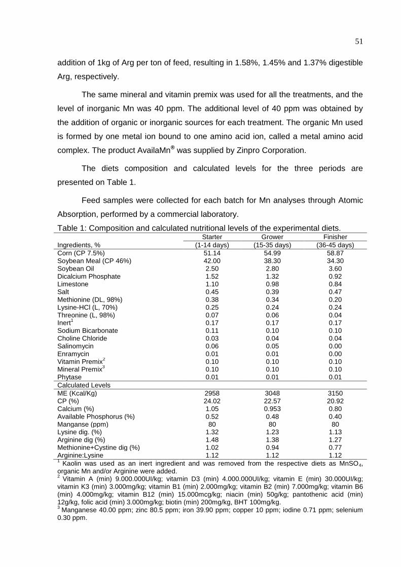
51
addition of 1kg of Arg per ton of feed, resulting in 1.58%, 1.45% and 1.37% digestible
Arg, respectively.
The same mineral and vitamin premix was used for all the treatments, and the
level of inorganic Mn was 40 ppm. The additional level of 40 ppm was obtained by
the addition of organic or inorganic sources for each treatment. The organic Mn used
is formed by one metal ion bound to one amino acid ion, called a metal amino acid
complex. The product AvailaMn® was supplied by Zinpro Corporation.
The diets composition and calculated levels for the three periods are
presented on Table 1.
Feed samples were collected for each batch for Mn analyses through Atomic
Absorption, performed by a commercial laboratory.
Table 1: Composition and calculated nutritional levels of the experimental diets. Starter Grower Finisher
Ingredients, % (1-14 days) (15-35 days) (36-45 days)
Corn (CP 7.5%) 51.14 54.99 58.87 Soybean Meal (CP 46%) 42.00 38.30 34.30 Soybean Oil 2.50 2.80 3.60 Dicalcium Phosphate 1.52 1.32 0.92 Limestone 1.10 0.98 0.84 Salt 0.45 0.39 0.47 Methionine (DL, 98%) 0.38 0.34 0.20 Lysine-HCl (L, 70%) 0.25 0.24 0.24 Threonine (L, 98%) 0.07 0.06 0.04 Inert
1 0.17 0.17 0.17
Sodium Bicarbonate 0.11 0.10 0.10 Choline Chloride 0.03 0.04 0.04 Salinomycin 0.06 0.05 0.00 Enramycin 0.01 0.01 0.00 Vitamin Premix
2 0.10 0.10 0.10
Mineral Premix3
0.10 0.10 0.10 Phytase 0.01 0.01 0.01
Calculated Levels
ME (Kcal/Kg) 2958 3048 3150 CP (%) 24.02 22.57 20.92 Calcium (%) 1.05 0.953 0.80 Available Phosphorus (%) 0.52 0.48 0.40 Manganse (ppm) 80 80 80 Lysine dig. (%) 1.32 1.23 1.13 Arginine dig (%) 1.48 1.38 1.27 Methionine+Cystine dig (%) 1.02 0.94 0.77 Arginine:Lysine 1.12 1.12 1.12 1
Kaolin was used as an inert ingredient and was removed from the respective diets as MnSO4, organic Mn and/or Arginine were added. 2
Vitamin A (min) 9.000.000UI/kg; vitamin D3 (min) 4.000.000UI/kg; vitamin E (min) 30.000UI/kg; vitamin K3 (min) 3.000mg/kg; vitamin B1 (min) 2.000mg/kg; vitamin B2 (min) 7.000mg/kg; vitamin B6 (min) 4.000mg/kg; vitamin B12 (min) 15.000mcg/kg; niacin (min) 50g/kg; pantothenic acid (min) 12g/kg, folic acid (min) 3.000mg/kg; biotin (min) 200mg/kg, BHT 100mg/kg. 3
Manganese 40.00 ppm; zinc 80.5 ppm; iron 39.90 ppm; copper 10 ppm; iodine 0.71 ppm; selenium 0.30 ppm.
52
Productive performance
The growth performance (feed intake and weight gain) of the birds was
evaluated weekly by weighing birds and leftover feed by replicate. Mortality was
recorded daily, and feed conversion rate was calculated considering the weight of
dead birds.
Carcass yield
Carcass yield was determined from two birds per replicate pen (18
birds/treatment). The birds were selected as representative based on the average
weight (95%) of replicate pen, and they were identified and euthanized by electric
stunning and bleeding. Carcass yield was calculated as the ratio of hot eviscerated
carcass weight and the body weight before euthanasia. Prime cut yield (whole
breast, thigh and legs, all including skin and bones) was calculated in relation to the
weight of the eviscerated carcass. Abdominal fat around the cloaca, in the cloacal
bursa, gizzards, proventriculus, and adjacent abdominal muscles was removed as
described by Smith (1993). Then, abdominal fat was weighed and calculated in
relation to the eviscerated carcass.
Sample collection
For all sample collection on the 7th and 21th days were sacrificed 12 birds per
treatment and on 45th day, 18 birds per treatment. In the collections, chicks were
randomly selected, weighted, submitted to electrical stunning and then slaughtered
by severing the jugular vein.
Blood
To analyze the serum concentrations of Alkaline Phosphatase, Urea and
Gama-glutamil transferase (GGT), blood was collected from live broilers at 7, 21 and
45 days old birds by auxiliary vein puncture and submitted to centrifugation at 1500
rpm for 10 minutes to obtain serum. After that, all samples were frozen at -18 ºC
freezer, until analyses. Kits were acquired from Labtest Laboratory, kept during all
time at 15 ºC and were applied to an automatic biochemical analyzer model BS-120,
Mindray.
53
Bone Measurements
Legs were removed for weighting. After that, left legs were banded and stored
in -18oC freezer for future removal of all adherent tissue. Femur and tibia of the left
leg were weighted and the length and diameter were measured with the aid of a
digital paquimeter (mm) for assessment of the Seedor index (Seedor et al., 1991)
obtained by dividing the weight of the bone (mg) by its length (mm).
Left tibiotarsus was placed above a radiographic film in parallel to an
aluminum penetrometer with 10 steps of 1 mm each. They were radiographed,
revealed and scanned. Aluminum (penetrometer) was used as radiographic
reference, 10 steps with 1 mm thickness among one step and another. Data obtained
from the bone in gray values were converted to values relative to the thickness of the
aluminum ladder. For each bone were determined the value in millimeters of
aluminum (mmAl), for bone mineral density.
The tibiotarsus was then subjected to bending test (breaking bone resistance)
at constant strain rate for visco-elastic material with the aim of a CT3 Texture
Analyzer (Brookfield AMETEK Inc., Massachusetts, USA).
Left femurs were subjected to fat extraction by successive treatment in
petroleum ether for 8 hours, then dried for 12 hours in an oven, weighted and
calcined in a muffle furnace at 540o C for 6 h for determination of bone ash. Bone ash
(1 gram per bone) was dissolved in 50% HNO3 and submitted to digestion.
Afterwards, 2% HNO3 was added to the solution that was weighted to obtain the
dilution factor. The mineral extract was carefully filtered through Whatman® Grade
42 filter paper (Whatman International Ltd., Maidstone, England) and the filtrates
were used to analyze Mn concentrations through flame atomic absorption
spectrophotometry on a Solaar Spectrometer, Thermo Fisher Scientific.
Adherent tissue of the right tibiotarsus were removed just after the collection
has finished tibiotarsus was fixed in 10% formaline solution for future decalcification
using Haug Solution (Nitric acid and phloroglucin). Tibiotarsus proximal extremity
were included in paraffin and taken to the microtome to cuts with 5μm thick. Sections
were then deparaffinized in xylene and hydrated through decreasing grades of
alcohol. The slides were stained with hematoxylin-eosin, were captured by a system
of magnifying glass and the images were measured with the aid of a computerized
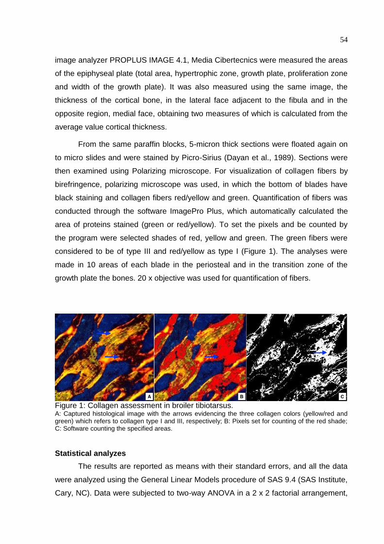
54
image analyzer PROPLUS IMAGE 4.1, Media Cibertecnics were measured the areas
of the epiphyseal plate (total area, hypertrophic zone, growth plate, proliferation zone
and width of the growth plate). It was also measured using the same image, the
thickness of the cortical bone, in the lateral face adjacent to the fibula and in the
opposite region, medial face, obtaining two measures of which is calculated from the
average value cortical thickness.
From the same paraffin blocks, 5-micron thick sections were floated again on
to micro slides and were stained by Picro-Sirius (Dayan et al., 1989). Sections were
then examined using Polarizing microscope. For visualization of collagen fibers by
birefringence, polarizing microscope was used, in which the bottom of blades have
black staining and collagen fibers red/yellow and green. Quantification of fibers was
conducted through the software ImagePro Plus, which automatically calculated the
area of proteins stained (green or red/yellow). To set the pixels and be counted by
the program were selected shades of red, yellow and green. The green fibers were
considered to be of type III and red/yellow as type I (Figure 1). The analyses were
made in 10 areas of each blade in the periosteal and in the transition zone of the
growth plate the bones. 20 x objective was used for quantification of fibers.
Figure 1: Collagen assessment in broiler tibiotarsus. A: Captured histological image with the arrows evidencing the three collagen colors (yellow/red and green) which refers to collagen type I and III, respectively; B: Pixels set for counting of the red shade; C: Software counting the specified areas.
Statistical analyzes
The results are reported as means with their standard errors, and all the data
were analyzed using the General Linear Models procedure of SAS 9.4 (SAS Institute,
Cary, NC). Data were subjected to two-way ANOVA in a 2 x 2 factorial arrangement,
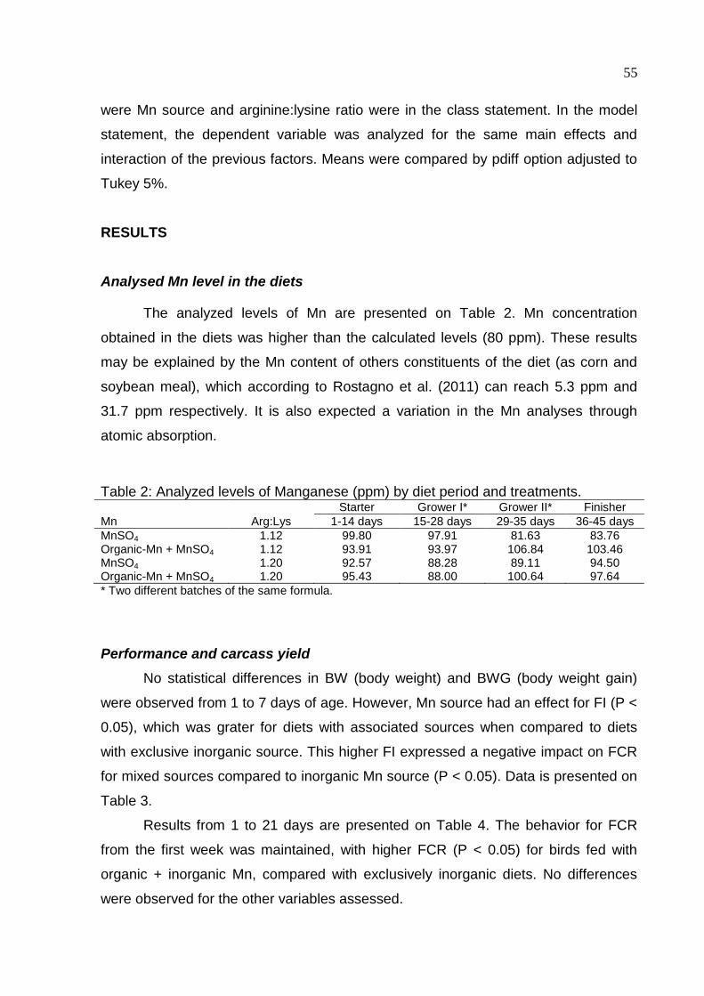
55
were Mn source and arginine:lysine ratio were in the class statement. In the model
statement, the dependent variable was analyzed for the same main effects and
interaction of the previous factors. Means were compared by pdiff option adjusted to
Tukey 5%.
RESULTS
Analysed Mn level in the diets
The analyzed levels of Mn are presented on Table 2. Mn concentration
obtained in the diets was higher than the calculated levels (80 ppm). These results
may be explained by the Mn content of others constituents of the diet (as corn and
soybean meal), which according to Rostagno et al. (2011) can reach 5.3 ppm and
31.7 ppm respectively. It is also expected a variation in the Mn analyses through
atomic absorption.
Table 2: Analyzed levels of Manganese (ppm) by diet period and treatments. Starter Grower I* Grower II* Finisher
Mn Arg:Lys 1-14 days 15-28 days 29-35 days 36-45 days
MnSO4 1.12 99.80 97.91 81.63 83.76
Organic-Mn + MnSO4 1.12 93.91 93.97 106.84 103.46 MnSO4 1.20 92.57 88.28 89.11 94.50 Organic-Mn + MnSO4 1.20 95.43 88.00 100.64 97.64
* Two different batches of the same formula.
Performance and carcass yield
No statistical differences in BW (body weight) and BWG (body weight gain)
were observed from 1 to 7 days of age. However, Mn source had an effect for FI (P <
0.05), which was grater for diets with associated sources when compared to diets
with exclusive inorganic source. This higher FI expressed a negative impact on FCR
for mixed sources compared to inorganic Mn source (P < 0.05). Data is presented on
Table 3.
Results from 1 to 21 days are presented on Table 4. The behavior for FCR
from the first week was maintained, with higher FCR (P < 0.05) for birds fed with
organic + inorganic Mn, compared with exclusively inorganic diets. No differences
were observed for the other variables assessed.
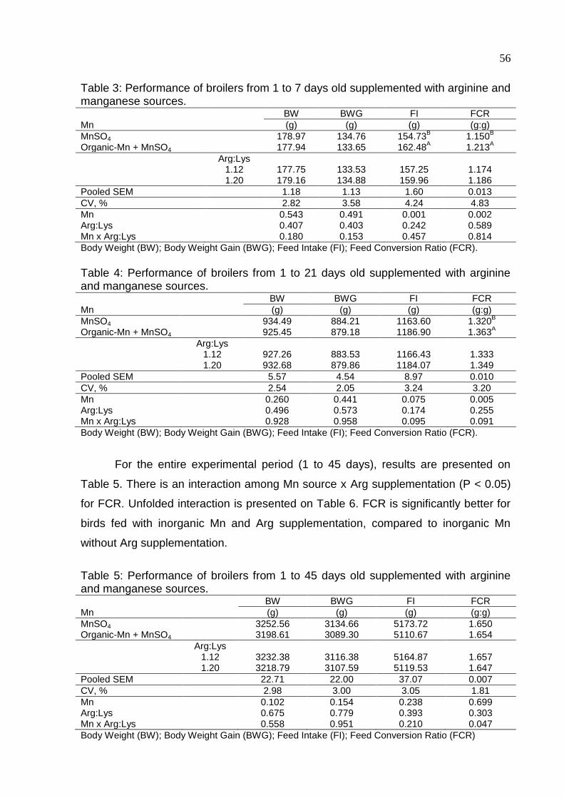
56
Table 3: Performance of broilers from 1 to 7 days old supplemented with arginine and manganese sources. BW BWG FI FCR
Mn (g) (g) (g) (g:g)
MnSO4 178.97 134.76 154.73B 1.150
B
Organic-Mn + MnSO4 177.94 133.65 162.48A 1.213
A
Arg:Lys 1.12 177.75 133.53 157.25 1.174 1.20 179.16 134.88 159.96 1.186
Pooled SEM 1.18 1.13 1.60 0.013
CV, % 2.82 3.58 4.24 4.83
Mn 0.543 0.491 0.001 0.002 Arg:Lys 0.407 0.403 0.242 0.589 Mn x Arg:Lys 0.180 0.153 0.457 0.814
Body Weight (BW); Body Weight Gain (BWG); Feed Intake (FI); Feed Conversion Ratio (FCR).
Table 4: Performance of broilers from 1 to 21 days old supplemented with arginine and manganese sources. BW BWG FI FCR
Mn (g) (g) (g) (g:g)
MnSO4 934.49 884.21 1163.60 1.320B
Organic-Mn + MnSO4 925.45 879.18 1186.90 1.363A
Arg:Lys 1.12 927.26 883.53 1166.43 1.333 1.20 932.68 879.86 1184.07 1.349
Pooled SEM 5.57 4.54 8.97 0.010
CV, % 2.54 2.05 3.24 3.20
Mn 0.260 0.441 0.075 0.005 Arg:Lys 0.496 0.573 0.174 0.255 Mn x Arg:Lys 0.928 0.958 0.095 0.091
Body Weight (BW); Body Weight Gain (BWG); Feed Intake (FI); Feed Conversion Ratio (FCR).
For the entire experimental period (1 to 45 days), results are presented on
Table 5. There is an interaction among Mn source x Arg supplementation (P < 0.05)
for FCR. Unfolded interaction is presented on Table 6. FCR is significantly better for
birds fed with inorganic Mn and Arg supplementation, compared to inorganic Mn
without Arg supplementation.
Table 5: Performance of broilers from 1 to 45 days old supplemented with arginine and manganese sources. BW BWG FI FCR
Mn (g) (g) (g) (g:g)
MnSO4 3252.56 3134.66 5173.72 1.650
Organic-Mn + MnSO4 3198.61 3089.30 5110.67 1.654
Arg:Lys 1.12 3232.38 3116.38 5164.87 1.657 1.20 3218.79 3107.59 5119.53 1.647
Pooled SEM 22.71 22.00 37.07 0.007
CV, % 2.98 3.00 3.05 1.81
Mn 0.102 0.154 0.238 0.699 Arg:Lys 0.675 0.779 0.393 0.303 Mn x Arg:Lys 0.558 0.951 0.210 0.047
Body Weight (BW); Body Weight Gain (BWG); Feed Intake (FI); Feed Conversion Ratio (FCR)
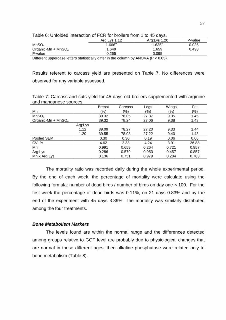
57
Table 6: Unfolded interaction of FCR for broilers from 1 to 45 days. Arg:Lys 1.12 Arg:Lys 1.20 P-value
MnSO4 1.666A
1.635B
0.036 Organic-Mn + MnSO4 1.649 1.659 0.498 P-value 0.265 0.095
Different uppercase letters statistically differ in the column by ANOVA (P < 0.05).
Results referent to carcass yield are presented on Table 7. No differences were
observed for any variable assessed.
Table 7: Carcass and cuts yield for 45 days old broilers supplemented with arginine and manganese sources. Breast Carcass Legs Wings Fat
Mn (%) (%) (%) (%) (%)
MnSO4 39.32 78.05 27.37 9.35 1.45
Organic-Mn + MnSO4 39.32 78.24 27.06 9.38 1.43
Arg:Lys 1.12 39.09 78.27 27.20 9.33 1.44 1.20 39.55 78.03 27.22 9.40 1.43
Pooled SEM 0.30 0.30 0.19 0.06 0.06
CV, % 4.62 2.33 4.24 3.91 26.88
Mn 0.991 0.659 0.264 0.721 0.857 Arg:Lys 0.286 0.579 0.953 0.457 0.857 Mn x Arg:Lys 0.136 0.751 0.979 0.284 0.783
The mortality ratio was recorded daily during the whole experimental period.
By the end of each week, the percentage of mortality were calculate using the
following formula: number of dead birds / number of birds on day one × 100. For the
first week the percentage of dead birds was 0.11%, on 21 days 0.83% and by the
end of the experiment with 45 days 3.89%. The mortality was similarly distributed
among the four treatments.
Bone Metabolism Markers
The levels found are within the normal range and the differences detected
among groups relative to GGT level are probably due to physiological changes that
are normal in these different ages, then alkaline phosphatase were related only to
bone metabolism (Table 8).
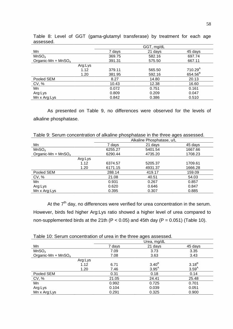
58
Table 8: Level of GGT (gama-glutamyl transferase) by treatment for each age assessed. GGT, mg/dL
Mn 7 days 21 days 45 days
MnSO4 369.75 582.16 697.74 Organic-Mn + MnSO4 391.31 575.50 667.11
Arg:Lys 1.12 379.11 565.50 710.29
A
1.20 381.95 592.16 654.56B
Pooled SEM 8.27 14.80 20.13
CV, % 10.43 12.38 16.60
Mn 0.072 0.751 0.161 Arg:Lys 0.809 0.209 0.047 Mn x Arg:Lys 0.842 0.386 0.510
As presented on Table 9, no differences were observed for the levels of
alkaline phosphatase.
Table 9: Serum concentration of alkaline phosphatase in the three ages assessed. Alkaline Phosphatase, u/L
Mn 7 days 21 days 45 days
MnSO4 6255.27 5401.54 1667.66 Organic-Mn + MnSO4 6290.44 4735.20 1708.23
Arg:Lys 1.12 6374.57 5205.37 1709.61 1.20 6171.15 4931.37 1666.28
Pooled SEM 288.14 419.17 159.09
CV, % 21.08 40.51 54.03
Mn 0.931 0.267 0.857 Arg:Lys 0.620 0.646 0.847 Mn x Arg:Lys 0.395 0.307 0.885
At the 7th day, no differences were verified for urea concentration in the serum.
However, birds fed higher Arg:Lys ratio showed a higher level of urea compared to
non-supplemented birds at the 21th (P < 0.05) and 45th day (P = 0.051) (Table 10).
Table 10: Serum concentration of urea in the three ages assessed. Urea, mg/dL
Mn 7 days 21 days 45 days
MnSO4 7.09 3.73 3.35 Organic-Mn + MnSO4 7.08 3.63 3.43
Arg:Lys 1.12 6.71 3.40
B 3.18
B
1.20 7.46 3.95A
3.59A
Pooled SEM 0.31 0.18 0.14
CV, % 21.05 24.41 25.48
Mn 0.992 0.725 0.701 Arg:Lys 0.104 0.039 0.051 Mn x Arg:Lys 0.291 0.325 0.900
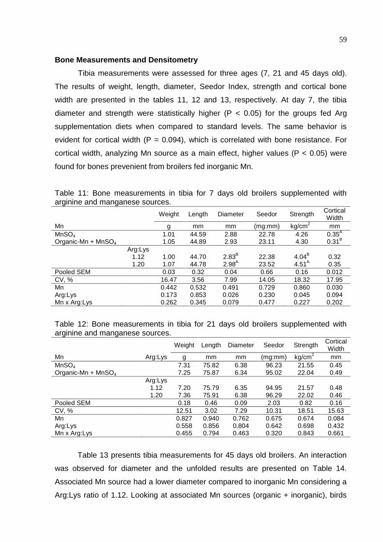
59
Bone Measurements and Densitometry
Tibia measurements were assessed for three ages (7, 21 and 45 days old).
The results of weight, length, diameter, Seedor Index, strength and cortical bone
width are presented in the tables 11, 12 and 13, respectively. At day 7, the tibia
diameter and strength were statistically higher (P < 0.05) for the groups fed Arg
supplementation diets when compared to standard levels. The same behavior is
evident for cortical width (P = 0.094), which is correlated with bone resistance. For
cortical width, analyzing Mn source as a main effect, higher values (P < 0.05) were
found for bones prevenient from broilers fed inorganic Mn.
Table 11: Bone measurements in tibia for 7 days old broilers supplemented with arginine and manganese sources.
Weight Length Diameter Seedor Strength Cortical Width
Mn g mm mm (mg:mm) kg/cm2 mm
MnSO4 1.01 44.59 2.88 22.78 4.26 0.35A
Organic-Mn + MnSO4 1.05 44.89 2.93 23.11 4.30 0.31B
Arg:Lys 1.12 1.00 44.70 2.83
B 22.38
4.04
B 0.32
1.20 1.07 44.78 2.98A 23.52 4.51
A 0.35
Pooled SEM 0.03 0.32 0.04 0.66 0.16 0.012
CV, % 16.47 3.56 7.99 14.05 18.32 17.95
Mn 0.442 0.532 0.491 0.729 0.860 0.030 Arg:Lys 0.173 0.853 0.026 0.230 0.045 0.094 Mn x Arg:Lys 0.262 0.345 0.079 0.477 0.227 0.202
Table 12: Bone measurements in tibia for 21 days old broilers supplemented with arginine and manganese sources.
Weight Length Diameter Seedor Strength Cortical Width
Mn Arg:Lys g mm mm (mg:mm) kg/cm2 mm
MnSO4 7.31 75.82 6.38 96.23 21.55 0.45 Organic-Mn + MnSO4 7.25 75.87 6.34 95.02 22.04 0.49
Arg:Lys 1.12 7.20 75.79 6.35 94.95
21.57 0.48
1.20 7.36 75.91 6.38 96.29 22.02 0.46
Pooled SEM 0.18 0.46 0.09 2.03 0.82 0.16
CV, % 12.51 3.02 7.29 10.31 18.51 15.63
Mn 0.827 0.940 0.762 0.675 0.674 0.084 Arg:Lys 0.558 0.856 0.804 0.642 0.698 0.432 Mn x Arg:Lys 0.455 0.794 0.463 0.320 0.843 0.661
Table 13 presents tibia measurements for 45 days old broilers. An interaction
was observed for diameter and the unfolded results are presented on Table 14.
Associated Mn source had a lower diameter compared to inorganic Mn considering a
Arg:Lys ratio of 1.12. Looking at associated Mn sources (organic + inorganic), birds
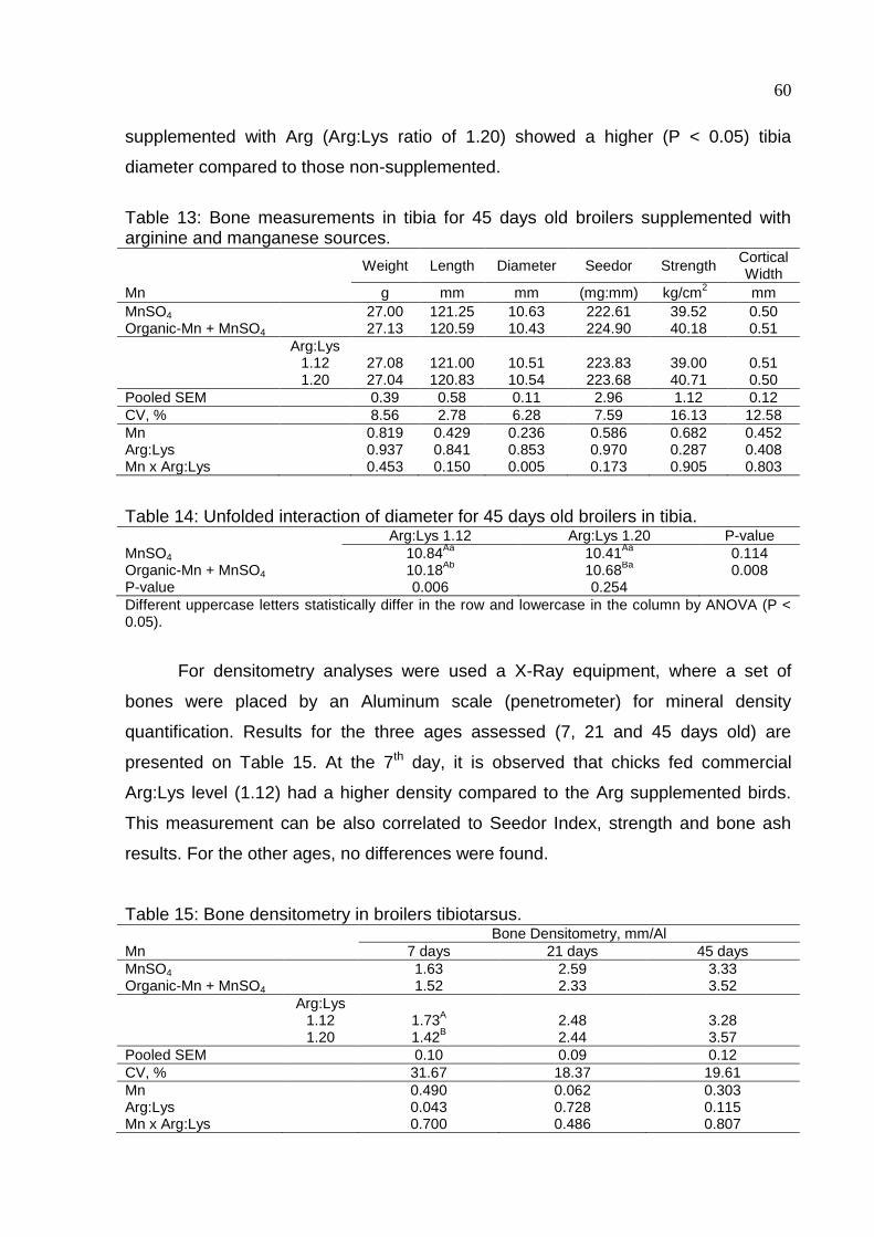
60
supplemented with Arg (Arg:Lys ratio of 1.20) showed a higher (P < 0.05) tibia
diameter compared to those non-supplemented.
Table 13: Bone measurements in tibia for 45 days old broilers supplemented with arginine and manganese sources.
Weight Length Diameter Seedor Strength Cortical Width
Mn g mm mm (mg:mm) kg/cm2 mm
MnSO4 27.00 121.25 10.63 222.61 39.52 0.50 Organic-Mn + MnSO4 27.13 120.59 10.43 224.90 40.18 0.51
Arg:Lys 1.12 27.08 121.00 10.51 223.83
39.00 0.51
1.20 27.04 120.83 10.54 223.68 40.71 0.50
Pooled SEM 0.39 0.58 0.11 2.96 1.12 0.12
CV, % 8.56 2.78 6.28 7.59 16.13 12.58
Mn 0.819 0.429 0.236 0.586 0.682 0.452 Arg:Lys 0.937 0.841 0.853 0.970 0.287 0.408 Mn x Arg:Lys 0.453 0.150 0.005 0.173 0.905 0.803
Table 14: Unfolded interaction of diameter for 45 days old broilers in tibia. Arg:Lys 1.12 Arg:Lys 1.20 P-value
MnSO4 10.84Aa
10.41Aa
0.114 Organic-Mn + MnSO4 10.18
Ab 10.68
Ba 0.008
P-value 0.006 0.254
Different uppercase letters statistically differ in the row and lowercase in the column by ANOVA (P < 0.05).
For densitometry analyses were used a X-Ray equipment, where a set of
bones were placed by an Aluminum scale (penetrometer) for mineral density
quantification. Results for the three ages assessed (7, 21 and 45 days old) are
presented on Table 15. At the 7th day, it is observed that chicks fed commercial
Arg:Lys level (1.12) had a higher density compared to the Arg supplemented birds.
This measurement can be also correlated to Seedor Index, strength and bone ash
results. For the other ages, no differences were found.
Table 15: Bone densitometry in broilers tibiotarsus. Bone Densitometry, mm/Al
Mn 7 days 21 days 45 days
MnSO4 1.63 2.59 3.33 Organic-Mn + MnSO4 1.52 2.33 3.52
Arg:Lys 1.12 1.73
A 2.48 3.28
1.20 1.42B 2.44 3.57
Pooled SEM 0.10 0.09 0.12
CV, % 31.67 18.37 19.61
Mn 0.490 0.062 0.303 Arg:Lys 0.043 0.728 0.115 Mn x Arg:Lys 0.700 0.486 0.807
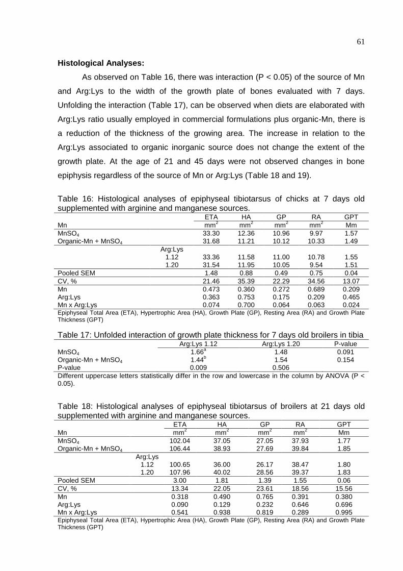
61
Histological Analyses:
As observed on Table 16, there was interaction (P < 0.05) of the source of Mn
and Arg:Lys to the width of the growth plate of bones evaluated with 7 days.
Unfolding the interaction (Table 17), can be observed when diets are elaborated with
Arg:Lys ratio usually employed in commercial formulations plus organic-Mn, there is
a reduction of the thickness of the growing area. The increase in relation to the
Arg:Lys associated to organic inorganic source does not change the extent of the
growth plate. At the age of 21 and 45 days were not observed changes in bone
epiphysis regardless of the source of Mn or Arg:Lys (Table 18 and 19).
Table 16: Histological analyses of epiphyseal tibiotarsus of chicks at 7 days old supplemented with arginine and manganese sources. A Mn
a
ETA HA GP RA GPT
mm2 mm
2 mm
2 mm
2 Mm
MnSO4 33.30 12.36 10.96 9.97 1.57 Organic-Mn + MnSO4 31.68 11.21 10.12 10.33 1.49
Arg:Lys 1.12 33.36 11.58 11.00 10.78 1.55 1.20 31.54 11.95 10.05 9.54 1.51
Pooled SEM 1.48 0.88 0.49 0.75 0.04
CV, % 21.46 35.39 22.29 34.56 13.07
Mn 0.473 0.360 0.272 0.689 0.209 Arg:Lys 0.363 0.753 0.175 0.209 0.465 Mn x Arg:Lys 0.074 0.700 0.064 0.063 0.024 Epiphyseal Total Area (ETA), Hypertrophic Area (HA), Growth Plate (GP), Resting Area (RA) and Growth Plate Thickness (GPT)
Table 17: Unfolded interaction of growth plate thickness for 7 days old broilers in tibia Arg:Lys 1.12 Arg:Lys 1.20 P-value
MnSO4 1.66a
1.48
0.091 Organic-Mn + MnSO4 1.44
b 1.54 0.154
P-value 0.009 0.506
Different uppercase letters statistically differ in the row and lowercase in the column by ANOVA (P < 0.05).
Table 18: Histological analyses of epiphyseal tibiotarsus of broilers at 21 days old supplemented with arginine and manganese sources. a Mn
a
ETA HA GP RA GPT
mm2 mm
2 mm
2 mm
2 Mm
MnSO4 102.04 37.05 27.05 37.93 1.77 Organic-Mn + MnSO4 106.44 38.93 27.69 39.84 1.85
Arg:Lys 1.12 100.65 36.00 26.17 38.47 1.80 1.20 107.96 40.02 28.56 39.37 1.83
Pooled SEM 3.00 1.81 1.39 1.55 0.06
CV, % 13.34 22.05 23.61 18.56 15.56
Mn 0.318 0.490 0.765 0.391 0.380 Arg:Lys 0.090 0.129 0.232 0.646 0.696 Mn x Arg:Lys 0.541 0.938 0.819 0.289 0.995 Epiphyseal Total Area (ETA), Hypertrophic Area (HA), Growth Plate (GP), Resting Area (RA) and Growth Plate Thickness (GPT)
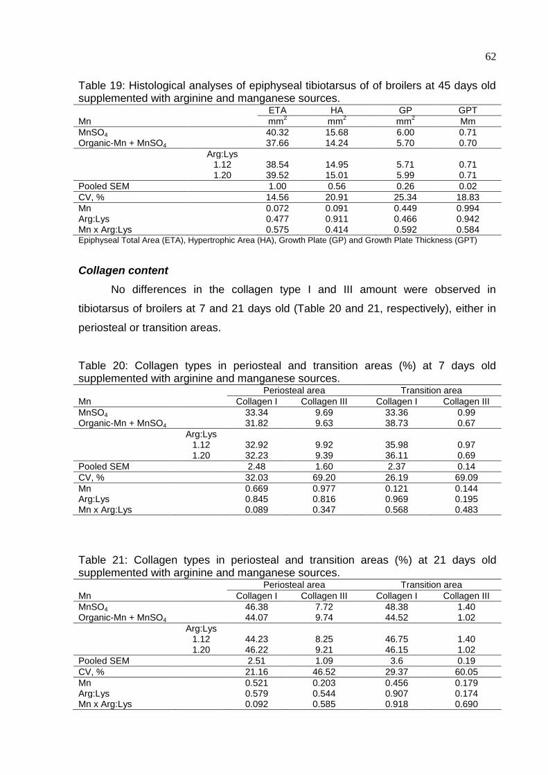
62
Table 19: Histological analyses of epiphyseal tibiotarsus of of broilers at 45 days old supplemented with arginine and manganese sources. a Mn
a
ETA HA GP GPT
mm2 mm
2 mm
2 Mm
MnSO4 40.32 15.68 6.00 0.71 Organic-Mn + MnSO4 37.66 14.24 5.70 0.70
Arg:Lys 1.12 38.54 14.95 5.71 0.71 1.20 39.52 15.01 5.99 0.71
Pooled SEM 1.00 0.56 0.26 0.02
CV, % 14.56 20.91 25.34 18.83
Mn 0.072 0.091 0.449 0.994 Arg:Lys 0.477 0.911 0.466 0.942 Mn x Arg:Lys 0.575 0.414 0.592 0.584 Epiphyseal Total Area (ETA), Hypertrophic Area (HA), Growth Plate (GP) and Growth Plate Thickness (GPT)
Collagen content
No differences in the collagen type I and III amount were observed in
tibiotarsus of broilers at 7 and 21 days old (Table 20 and 21, respectively), either in
periosteal or transition areas.
Table 20: Collagen types in periosteal and transition areas (%) at 7 days old supplemented with arginine and manganese sources. Periosteal area Transition area
Mn Collagen I Collagen III Collagen I Collagen III
MnSO4 33.34 9.69 33.36 0.99 Organic-Mn + MnSO4 31.82 9.63 38.73 0.67
Arg:Lys 1.12 32.92 9.92 35.98 0.97 1.20 32.23 9.39 36.11 0.69
Pooled SEM 2.48 1.60 2.37 0.14
CV, % 32.03 69.20 26.19 69.09
Mn 0.669 0.977 0.121 0.144 Arg:Lys 0.845 0.816 0.969 0.195 Mn x Arg:Lys 0.089 0.347 0.568 0.483
Table 21: Collagen types in periosteal and transition areas (%) at 21 days old supplemented with arginine and manganese sources. Periosteal area Transition area
Mn Collagen I Collagen III Collagen I Collagen III
MnSO4 46.38 7.72 48.38 1.40 Organic-Mn + MnSO4 44.07 9.74 44.52 1.02
Arg:Lys 1.12 44.23 8.25 46.75 1.40 1.20 46.22 9.21 46.15 1.02
Pooled SEM 2.51 1.09 3.6 0.19
CV, % 21.16 46.52 29.37 60.05
Mn 0.521 0.203 0.456 0.179 Arg:Lys 0.579 0.544 0.907 0.174 Mn x Arg:Lys 0.092 0.585 0.918 0.690
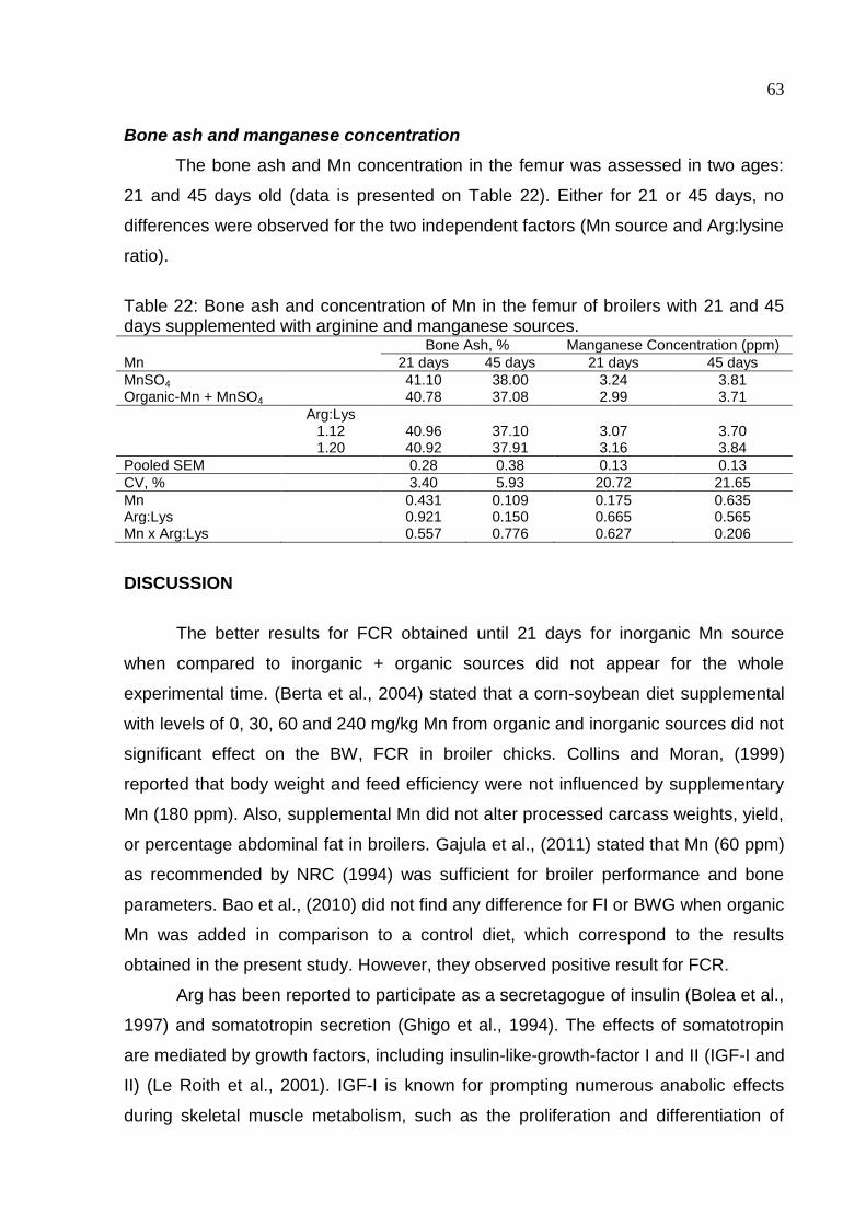
63
Bone ash and manganese concentration
The bone ash and Mn concentration in the femur was assessed in two ages:
21 and 45 days old (data is presented on Table 22). Either for 21 or 45 days, no
differences were observed for the two independent factors (Mn source and Arg:lysine
ratio).
Table 22: Bone ash and concentration of Mn in the femur of broilers with 21 and 45 days supplemented with arginine and manganese sources. Bone Ash, % Manganese Concentration (ppm)
Mn 21 days 45 days 21 days 45 days
MnSO4 41.10 38.00 3.24 3.81 Organic-Mn + MnSO4 40.78 37.08 2.99 3.71
Arg:Lys 1.12 40.96 37.10 3.07 3.70 1.20 40.92 37.91 3.16 3.84
Pooled SEM 0.28 0.38 0.13 0.13
CV, % 3.40 5.93 20.72 21.65
Mn 0.431 0.109 0.175 0.635 Arg:Lys 0.921 0.150 0.665 0.565 Mn x Arg:Lys 0.557 0.776 0.627 0.206
DISCUSSION
The better results for FCR obtained until 21 days for inorganic Mn source
when compared to inorganic + organic sources did not appear for the whole
experimental time. (Berta et al., 2004) stated that a corn-soybean diet supplemental
with levels of 0, 30, 60 and 240 mg/kg Mn from organic and inorganic sources did not
significant effect on the BW, FCR in broiler chicks. Collins and Moran, (1999)
reported that body weight and feed efficiency were not influenced by supplementary
Mn (180 ppm). Also, supplemental Mn did not alter processed carcass weights, yield,
or percentage abdominal fat in broilers. Gajula et al., (2011) stated that Mn (60 ppm)
as recommended by NRC (1994) was sufficient for broiler performance and bone
parameters. Bao et al., (2010) did not find any difference for FI or BWG when organic
Mn was added in comparison to a control diet, which correspond to the results
obtained in the present study. However, they observed positive result for FCR.
Arg has been reported to participate as a secretagogue of insulin (Bolea et al.,
1997) and somatotropin secretion (Ghigo et al., 1994). The effects of somatotropin
are mediated by growth factors, including insulin-like-growth-factor I and II (IGF-I and
II) (Le Roith et al., 2001). IGF-I is known for prompting numerous anabolic effects
during skeletal muscle metabolism, such as the proliferation and differentiation of
64
satellite cells (Florini et al. 1996), and the aggregation of myofibrillar protein by a
combination of the effects on protein synthesis and degradation (Duclos 2005).
Despite these facts, no differences were observed for any of the variables
assessed. On the other hand, Mendes et al. (1997), reported a higher carcass yield
for broilers fed higher Arg:Lys ratio, and a lower carcass fat deposition, which was
also found by Fernandes et al., (2014). Tan et al., (2011) established that Arg
differentially regulates the expression of fat-metabolizing genes in skeletal muscle
and white adipose tissue, favoring lipogenesis in muscle but lipolysis in adipose
tissue. Although Arg did not have any effects on carcass, our results for FCR are in
accordance to those found by Fernandes et al. (2013), which birds fed with
supplemented diets showed better FCR compared to non-supplemented. Similarly,
(Tan et al., 2014a) found that Arg supplementation increased BWG and reduced
linear reduced FCR (Tan et al., 2014b). There was no difference regarding Mn
source.
The value of serum alkaline phosphatase activity is the most commonly used
marker of bone formation (Eriksen et al., 1995; Plebani et al., 1996). Its activity is
associated with the increased osteoblatic activity in the bone and decreases as bone
maturation occurs. This enzyme takes place in the bone and hepatic tissue, therefore
hepatic disturbances may also increases alkaline phosphatase levels which could
lead to a misinterpretation. Then, it was also assessed the levels of gamma glutamyl
transpeptidase (GGT), which is the most sensible indicator of liver disturbances to
determine whether the levels were physiological or pathological. The activity of GGT
serum activity progressively increased with age when there is rapid body
development due to higher feed intake and feed gain (Almeida and Bruno, 2006),
with a concurrent significant increase of liver metabolism. As no differences were
observed for GGT levels, we considered that alkaline phosphatase was related only
to bone metabolism. It may be concluded that independently of the Mn source fed
and/or level of Arg, the animals did not express any difference in the bone
mineralization.
Arg is mainly metabolized by enzymes, arginase and NOS to produce urea
and L-ornithine, or NO and citrulline, respectively (Tan et al., 2014b). It has been
reported that between 40 to 60% of urea excreted by birds is from Arg metabolism.
According to (Wu and Morris, 1998), the supplementation of Arg in elevated levels
promotes an increase in kidney arginase activity and increases ornithine levels. The
65
higher urea value for birds fed supplemented Arg diets, confirm that Arg
supplementation influenced its breakdown into ornithine and urea by arginase, which
Mn is the cofactor. As no differences where find for Mn source, the conclusion is
either sources were fed in a concentration that allowed an equal action of the Mn as
an enzyme activator.
Ornithine is the precursor of Pro and polymines, which are involved in the cell
multiplication process, also improving bone support, implicating in the complex
process of bone differentiation. Rath and Reddi (1981) found that polyamine
synthesis was enhanced during osteogenesis.
At the first week, it was observed a higher diameter and strength (P < 0.05) in
the bones of chicks fed 1.20 Arg:Lys ratio, in contrast to 1.12 Arg:Lys ratio (Table
12). What remains unclear is the opposite result for bone densitometry (Table 16),
which does not make sense if bone density has a high correlation with its strength.
The higher tibiotarsus diameter at 45 days (P < 0.05), also was not enough to
improve bone resistance, as Seedor index, break strength and bone densitometry
were not affected by any of the main effects (Table 14). Measurements as Seedor
index (Seedor, 1991), break strength, (Merkley, 1981; Ruff and Hughes, 1985; Park
et al., 2003; Kim et al., 2006) cortical width and bone densitometry (Onyango et al.,
2003; Kim et al., 2006) has been referred as capable to indicate bone mineralization,
pointing out any deficiency may have been occurred.
Despite the fact that Liu et al. (2015) reported a lower trabecular area for tibias
from birds fed deficient Mn diets compared to the control diet. Bao et al. (2010), did
not find any difference in tibia break strength, length or width when organic Mn were
added, in comparison to a control diet.
That leads us to believe, that as it happens in performance, the isolated
administration of Mn influences just when the birds are subjected to severe
deficiency (Kealy and Sullivan, 1966) or high levels promoting toxicity (Southern and
Baker, 1983).
Based on a diet containing 10mg Mn/kg diet, it was claimed that
supplementation of 50mg Mn/kg diet in the form of Mn carbonate could significantly
reduce the incidence of perosis (Gallup and Norris, 1939) but there was no further
growth response to supplemental Mn content when a diet containing 37.5mg Mn/kg
was fed to broiler chickens (Gardiner, 1972).In the study conducted by Bao et al.,
(2010) when Zn was sufficient in the diet, the control diet containing 17.8mg Mn/kg
66
diet did not produce Mn deficiency symptoms. According to the author, the priority for
bird growth is Zn requirement. Provided all the macro nutrient requirements are
satisfied, the growth rate of broilers appears to be determined primarily by dietary Zn
content. Therefore, under a Zn sufficient condition, it is hard to produce Cu, Fe and
Mn deficiency symptoms.
Beattie and Avenell, (1992) postulated that Cu, Fe, Mn and Zn are required for
the growth, development and maintenance of healthy bones and Cu and Fe
deficiency could decrease bone strength, while Mn deficiency results in perosis
(Scott et al., 1982; Medeiros et al., 1997; Roughead and Lukaski, 2003).
Perosis occurrence was not a problem in the present experiment, with few
cases spread out among all the treatments. It may be conclude that the Mn level
used, regardless the source was enough to provide satisfactory growth (as shows
performance results) and bone quality.
For analysis of tibia epiphyseal cartilage blades, were considered three
distinct regions characterized by morphological appearance: rest area, area of
cartilage in proliferation (growth plate) and hypertrophic cartilage area. The zone of
calcified cartilage was considered as the lower limit for determining the thickening of
the hypertrophic zone. The total area of the epiphysis was also measured.
As observed on Table 27, there was interaction (P < 0.05) of the source of Mn
and Arg:Lys to the width of the growth plate of bones evaluated with 7 days.
Unfolding the interaction, can be observed when diets are elaborated with Arg:Lys
ratio usually employed in commercial formulations and plus Mn organic, there was a
reduction of the thickness of the growing area. The increase in the Arg:Lys ratio with
associated organic inorganic source does not change the extent of the growth plate.
Considering a P-value of 0.10 due to the high coefficient of variation for this
type of evaluation, interaction among the source of Mn and Arg:Lys to the other
measures may be observed, showing the same behavior reported to the thickness of
the growth plate, which there is a smaller thickness when the Mn organic source is
used whereas the relationship Arg:Lys practiced in commercial feed formulation.
The tibial discondroplasia (TD) is a common injury in the bone growth area in
chickens and is characterized by the persistence of a abnormal cartilage mass in the
metaphysis, immediately below the zone or growth plate, especially in tibia, resulting
in plugs of cartilage in place of true bone. Bones are therefore weak, and can result
in bone breaks when the bird grows heavy.
67
Rath et al., (1997) have demonstrated that tibias exhibiting TD have normal
collagen content, but reduced amounts of sulfated glycosaminoglycans and
metalloenzymes activity when compared to normal tibias. These results suggest
supplementation with Mn and Zn may alleviate TD. Few time later, this group also
showed that the extent of collagen crosslinking in tibia correlated with bone breaking
strength (Rath et al., 1999), which implies the importance of cooper.
Bone tissue consists of a cartilage matrix containing about 95% collagen,
which gives the bone tensile strength. Boskey et al. (1999), affirm that exists a
mechanistic link between type I collagen and bone strength and nutritional factors
may alter collagen synthesis which may influence bone density and bone mechanical
properties. Collagen amounts might be influenced by Mn and/or Arg content in the
diet. Arg is involved both in the synthesis of substrates (polyamine and Pro)
implicated in collagen synthesis, and Mn works as the cofactor which activates the
enzyme to promote Arg breakdown. It is also important to consider that Zn and Cu
play important roles in bone development via their actions on collagen, while Mn-
dependent enzymes promote formation of the proteoglycan matrix in the cartilage
model for developing bone. Mn is an activator of glycotransferases that are essential
for syntheses of polysaccharides and glycoprotein, the precursors for development of
organic matrix of bone (Underwood, 1981). Deficiency of Mn might affect the
structural components of cartilage resulting in higher leg scores.
Zn plays an integral part in the synthesis of 2 important functional proteins,
collagen and keratin (Underwood and Suttle, 2001). Collagen is the major structural
protein of internal tissues, including cartilage and bone, whereas keratin is the
structural protein of the feathers, skin, beak, and claws. Cu plays a role in the proper
cross-linking of collagen and elastin (Underwood and Suttle, 2001; Rath et al., 1999)
and Mn is essential for the maintenance of bone mineralization (Strause et al., 1986).
Based upon all the exposed, it seems very clear the impossibility to attribute
the bone mineralization process, especially the endochondral ossification and
collagen synthesis to just Mn as Cu and Zn acts mutually.
On the other hand, bone ash is a critical measure of how mineralized the bone
is. Chicken with bone problems, generally have a lower percentage of ashes when
compared to healthy birds. The amount of ash (inorganic material) present in bone is
proportional to its degree of hardness or strength of compression (Bonser and
Casinos, 2003); the organic component of bone is important in providing tensile
68
strength and flexibility (Velleman, 2000). Is the balance between these two bone
components that contribute to its resistance to breakage.
Mineral homeostasis is precisely maintained in the body and is predominantly
achieved by balancing tissue storage and excretion (Suttle, 2010). Tissue mineral
concentrations are indicators of body storage and mineral status and have been used
as biomarkers in bioavailability studies (Wang et al., 2007). Several researchers
reported a linear relationship between the concentration of Mn in bone and its level in
the diet (Watson et al., 1970; Southern and Baker, 1983; Black et al., 1984). The Mn
retention level found in the current study was the consistent supplied in the diet.
Bao et al., (2010) did not find any difference in the tibia mineral content when
organic Mn was added to the control diet. However, Mn concentration was affect, in
contrary to what was observed in the present study, where no difference was seen
(Table 22). In agreement with the present study, Berta et al. (2004) reported that the
same level of supplementation of the two Mn sources did not express any differences
in Mn concentrations in organs and tissue in broiler chicks.
CONCLUSION
The partial substitution of 50% of the inorganic Mn source (MnSO4) per
organic Mn met the requirement of broilers, resulting in no addicional gains for both
productive performance or the bone measurements. Arginine supplementation
resulted in better FCR when fed with inorganic Mn source.
Diets elaborated with Arg:Lys ratio usually employed in commercial
formulations and plus Mn organic, resulted in reduction of the thickness of the
growing area of tibiotarsus, arguably preventing tibial discondroplasia.
Higher levels of Mn of organic source may be evaluated for better
understanding of the contribution of the Mn in the activity of arginase on Arg in
modern strains broiler chickens.
69
REFERENCES
Almeida, I. C. L., and L. D. G. Bruno. 2006. Bone mineral density : review. Brazilian J. Poult. Sci. 8:69–73.
Baker, D. H. 2009. Advances in protein-amino acid nutrition of poultry. Amino Acids 37:29–41.
Bao, Y. M., M. Choct, P. a Iji, and K. Bruerton. 2010. Trace mineral interactions in broiler chicken diets. Br. Poult. Sci. 51:109–17.
Beattie, J. H., and a Avenell. 1992. Trace element nutrition and bone metabolism. Nutr. Res. Rev. 5:167–188.
Berta, E., E. Andrasofzky, A. Bersenyi, R. Glavits, A. Gaspardy, and S. G. Fekete. 2004. Effect of Inorganic and Organic Manganese Supplementation on the Performance and. Acta Vet. Hung. 52:199–209.
Black, J. R., C. B. Ammerman, P. R. Henry, and R. D. Miles. 1984. Biological availability of manganese sources and effects of high dietary manganese on tissue mineral composition of broiler-type chicks. Poult. Sci. 63:1999–2006.
Bolea, S., Pertusa, J. A. G., Martı´ n, F., Sanchez-Andres, J. V. and Soria, B. 1997. Regulation of pancreatic B-cell eletrical activity and insulin release by physiological amino acid con- centrations. Eur. J. Physiol. 433: 699-704.
Bonser, R. H. C., and a Casinos. 2003. Regional variation in cortical bone properties from broiler fowl--a first look. Br. Poult. Sci. 44:350–4.
Boskey, a L., T. M. Wright, and R. D. Blank. 1999. Collagen and bone strength. J. Bone Miner. Res. 14:330–335.
Cashman, K., and A. Flynn. 1998. Trace elements and bone metabolism. Bibl Nutr Dieta 54:150–64.
Collins, N. E., and E. T. Moran. 1999. Influence of supplemental manganese and zinc on live performance and carcass quality of broilers. Appl. Poult. Sci. 8:222.227.
Curran, J. N., D. C. Winter, and D. Bouchier-Hayes. 2006. Biological fate and clinical implications of arginine metabolism in tissue healing. Wound Repair Regen. 14:376–386.
Dayan, D., Hiss, Y., Hirshberg, A., Bubis, J.J. And Wolman, M. 1989. Are the polarization colors of Picrosirius red-stained collagen determined only by the diameter of the fibers. Histochemistry 93,27-29.
Dermience, M., G. Lognay, F. Mathieu, and P. Goyens. 2015. Effects of thirty elements on bone metabolism. J. Trace Elem. Med. Biol. 32:86–106.
70
Duclos, M. J. 2005. Insulin-like growth factor-I (IGF-I) mRNA levels and chicken muscle growth. J. Physiol. Pharma- col. 56: 25-35.
Eriksen, E. F., K. Brixen, and P. Charles. 1995. New markers of bone metabolism: clinical use in metabolic bone disease. EurJEndocr 132:251–263.
Fernandes, J. I. M., A. E. Murakami, L. M. G. de Souza, I. C. Ospina-Rojas, and R. M. Rossi. 2014. Effect of arginine supplementation of broiler breeder hens on progeny performance. Can. J. Anim. Sci. 94:313–321.
Florini, J. R., Ewton, D. Z. and Coolican, S. A. 1996. Growth hormone and the insulin-like growth factor system in myogen- esis. Endocrinol. Rev. 17: 481?517.
Gajula, S. S., V. K. Chelasani, A. K. Panda, V. L. N. R. Mantena, and R. R. Savaram. 2011. Effect of supplemental inorganic Zn and Mn and their interactions on the performance of broiler chicken, mineral bioavailability, and immune response. Biol. Trace Elem. Res. 139:177–187.
Gallup, W. D., and L. C. Norris. 1939. The amount of manganese required to prevent perosis in the chick. Poult. Sci. 18:76–82.
Gardiner, E. E. 1972. Lack of response to added dietary manganese of chicks fed wheat-soybean meal or corn-soybean meal based diets. Can. J. Anim. Sci. 52:737–740.
Ghigo, E., Ceda, G. P., Valcavi, R., Goffi, S., Mucci, M., Valenti, G., Cocchi, D., Muller, E. E. and Camanni, F. 1994. Low doses of either intravenously or orally administered ar- ginine areable to enhance growth hormone response to growth hormone releasing hormone in elderly subjects. J. Endocrinol. Invest. 17: 113-122.
Kealy, R. D., and T. W. Sullivan. 1966. Studies on Manganese Requirement and Interactions in the Diet of Young Turkeys. Poult. Sci. 45:1352–1358.
Keen, C.L., Enunsa, J.L. and Clegg, M.S. 2000. Manganese metabolism in animals and humans including the toxicity of manganese. Metal Ions in Biological Systems 37: 89-121.
Kim, W. K., L. M. Donalson, a D. Mitchell, L. F. Kubena, D. J. Nisbet, and S. C. Ricke. 2006. Effects of alfalfa and fructooligosaccharide on molting parameters and bone qualities using dual energy X-ray absorptiometry and conventional bone assays. Poult. Sci. 85:15–20.
Liu, R., C. Jin, Z. Wang, Z. Wang, J. Wang, and L. Wang. 2015. Effects of manganese deficiency on the microstructure of proximal tibia and OPG/RANKL gene expression in chicks. Vet. Res. Commun. 39:31–37.
Medeiros, D. M., J. Ilich, J. Ireton, V. Matkovic, L. Shiry, and R. Wildman. 1997. Femurs from rats fed diets deficient in copper or iron have decreased mechanical strength and altered mineral composition. J. Trace Elem. Exp. Med. 10:197–203.

71
Mendes, a a, S. E. Watkins, J. a England, and E. a Saleh. 1997. Influence of Dietary Lysine Levels and Arginine : Lysine Ratios on Performance of Broilers Exposed to Heat or Cold Stress During the Period of Three to Six Weeks of Age 1 Birds and Housing. :472–481.
Merkley, J. W. 1981. A Comparison of Bone Strengths from Broilers Reared Under Various Conditions in Coops and Floor Pens 1. Poult. Sci. 60:98=106.
Onyango, E. M., P. Y. Hester, R. Stroshine, and O. Adeola. 2003. Bone densitometry as an indicator of percentage tibia ash in broiler chicks fed varying dietary calcium and phosphorus levels. Poult. Sci. 82:1787–1791.
Park, S. Y., S. G. Birkhold, L. F. Kubena, D. J. Nisbet, and S. C. Ricke. 2003. Effect of storage condition on bone breaking strength and bone ash in laying hens at different stages in production cycles. Poult. Sci. 82:1688–91.
Plebani, M., D. Bernardi, M. Zaninotto, M. De Paoli, S. Secchiero, and L. Sciacovelli. 1996. New and traditional serum markers of bone metabolism in the detection of skeletal metastases. Clin. Biochem. 29:67–72.
Rath, N. C., J. M. Balog, W. E. Huff, G. R. Huff, G. B. Kulkarni, and J. F. Tierce. 1999. Comparative differences in the composition and biomechanical properties of tibiae of seven- and seventy-two-week-old male and female broiler breeder chickens. Poult. Sci. 78:1232–1239.
Rath, N. C., W. E. Huff, J. M. Balog, G. R. Bayyari, and R. P. Reddy. 1997. Matrix metalloproteinase activities in avian tibial dyschondroplasia. Poult. Sci. 76:501–505.
Le Roith, D., C. Bondy, S. Yakar, J. L. Liu, and a. Butler. 2001. The somatomedin hypothesis: 2001. Endocr. Rev. 22:53–74.
Lyons, M. and Insko, W.M. Jr. 1937. Chondrodystrophy in the Chick Embryo Produced by Manganese Deficiency in the Diet of the Hen. Bulletin No. 371, Kentucky Agricultural Experiment Station.
Rath, N. C. and Reddi, A. H. 1981. Changes in polyamines, RNA synthesis, and cell proliferation during matrix-induced cartilage, bone, and bone marrow development. Dev. Biol. 82: 211-216.
Roughead, Z. K., and H. C. Lukaski. 2003. Inadequate copper intake reduces serum insulin-like growth factor-I and bone strength in growing rats fed graded amounts of copper and zinc. J. Nutr. 133:442–448.
Rostagno, H. S. 2011.Tabelas brasileiras para aves e suínos: composição de alimentos e exigências nutricionais. Viçosa, Brasil: Universidade Federal de Viçosa - Departamento de Zootecnia.
Ruff, C. R., and B. L. Hughes. 1985. Bone Strength of Height-Restricted Broilers as Affected by Levels of Calcium, Phosphorus, and Manganese. Poult. Sci. 64:1628–1636.
72
Saltman, P. D., and L. G. Strause. 1993. The role of trace minerals in osteoporosis. J. Am. Coll. Nutr. 12:384–389.
SAS Institute. 2006. SAS User’s Guide: Statistics. Version 9.1.3 ed. SAS Inst. Inc., Cary, NC.
Schaible, P.J., Bandemer, S.L. and Davidson, J.A. 1938. The Manganese Content of Feedstuffs and its Relation to Poultry Nutrition. Technical Bulletin No. 159, Michigan Agricultural Experiment Station.
Scott, M.L., Nesheim, M.C. & Young, R.J. 1982. Essential inorganic elements, in: SCOTT, M.L., NESHEIM, M.C. & YOUNG, R.J. (Eds) Nutrition of the Chicken, pp. 277—382 (New York, M.L. Scott & Associates).
Seedor, J.G., H.A. Quarruccio and D.D. Thompson, 1991. The bisphosphonate alendronate (MK-217) inhibits bone loss due to ovariectomy in rats. J. Bone Miner. Res., 6: 339-346.
Smith, M. O. 1993. Parts Yield of Broilers Reared Under Cycling High Temperatures. Poult. Sci. 72:1146–1150.
Southern-, L. L. E. E., and H. Baker. 1983. Eimeria acervulina Infection in Chicks Fed Deficient or Excess Levels of Manganese. :172–177.
Strause, L. G., J. Hegenauer, P. Saltman, R. Cone, and D. Resnick. 1986. Effects of long-term dietary manganese and copper deficiency on rat skeleton. J. Nutr. 116:135–141.
Tan, J., T. J. Applegate, S. Liu, Y. Guo, and S. D. Eicher. 2014a. Supplemental dietary l-arginine attenuates intestinal mucosal disruption during a coccidial vaccine challenge in broiler chickens. Br. J. Nutr. 112:1098–1109.
Tan, J.-Z., Y.-M. Guo, T. J. Applegate, E.-C. Du, and X. Zhao. 2014b. Dietary L-arginine modulates immunosuppression in broilers inoculated with an intermediate strain of infectious bursa disease virus. J. Sci. Food Agric. 95:126–135.
Tan, B., Y. Yin, Z. Liu, W. Tang, H. Xu, X. Kong, X. Li, K. Yao, W. Gu, S. B. Smith, and G. Wu. 2011. Dietary l-arginine supplementation differentially regulates expression of lipid-metabolic genes in porcine adipose tissue and skeletal muscle. J. Nutr. Biochem. 22:441–445.
Underwood, E. The mineral nutrition of livestock.1981. London: Academic Press, p. 111.
Underwood, E., and N. Suttle. 2001. The Mineral Nutrition of Livestock. CABI Publishing, London, UK.
Velleman, S. G. 2000. The role of the extracellular matrix in skeletal muscle development. Poult. Sci. 78:778–784.
73
Wang, Z., S. Cerrate, C. Coto, F. Yan, and P. W. Waldroup. 2007. Evaluation of Mintrex® Copper as a Source of Copper in Broiler Diets. Int. J. Poult. Sci. 6:308–313.
Watson, L. T., C. B. Ammerman, S. M. Miller, and R. H. Harms. 1970. Biological Assay of Inorganic Manganese for Chicks 1. Poult. Sci. 49:1548–1554.
Wu, G., F. W. Bazer, R. C. Burghardt, G. a. Johnson, S. W. Kim, D. a. Knabe, P. Li, X. Li, J. R. McKnight, M. C. Satterfield, and T. E. Spencer. 2011. Proline and hydroxyproline metabolism: Implications for animal and human nutrition. Amino Acids 40:1053–1063.
Wu, G., F. W. Bazer, T. a. Davis, S. W. Kim, P. Li, J. Marc Rhoads, M. Carey Satterfield, S. B. Smith, T. E. Spencer, and Y. Yin. 2009. Arginine metabolism and nutrition in growth, health and disease. Amino Acids 37:153–168.
Wu, G., and S. M. Morris. 1998. Arginine metabolism : nitric oxide and beyond. Biochem. J. 336:1–17.
74
CHAPTER 2 - ARGININE AND MANGANESE SUPPLEMENTATION ON THE IMMUNE CAPACITY OF BIRDS CHALLENGED WITH Salmonella enteritidis
Abstract - To assess the immune modulation of arginine (Arg) and organic
manganese in broilers, the experiment was conducted. On the day of hatch, 640
male, Cobb 500 broiler chicks were weighted, and randomly assigned to a factorial 2
x 2 design (2 manganese sources x 2 Arg:Lys ratio) composing 4 treatments and 8
replicates each treatment, with 10 birds each replicate. The treatments consisted of
T1: Inorganic Control (80 ppm MnSO4); T2: 40 ppm MnSO4 + 40 ppm organic Mn;
T3: Inorganic Control + L-Arg (DigArg:DigLys 1.20); T4: 40 ppm MnSO4 + 40 ppm
organic Mn + L-Arg (DigArg:DigLys 1.20. For treatments 1 and 2, the digestible
Arg:Lys ratio was 1.12, considered normal using corn-soybean meal based diets.
Two independent groups, each composed by these 4 treatments and 8 replicates
each treatment, were challenged or not with an intramuscular Salmonella enterididis
vaccine. No differences were observed to macrophage phagocytic activity analyses.
There was no interaction between the main effects (manganese source and
arginine:lysine ratio) for challenged or unchallenged birds. Unchallenged birds fed
associated manganese sources showed higher (P < 0.05) mucosa CD8 lymphocytes
counting, compared to inorganic source. For mucosa CD4, general CD4 and non-
activated CD8 lymphocytes, birds which were fed arginine supplemented diet
(Arg:Lys 1.20), had a higher percentage (P < 0.05) of this cells, compared to the
commercial Arg:Lys level (1.12). For challenged birds, associated Mn sources had a
higher (P < 0.05) percentage of non-activated CD8 lymphocytes, but the opposite
happened to suppressor monocytes. Arg supplementation did not alter any
lymphocyte population for challenged birds. The inorganic Mn diets, resulted in
higher humoral protection (increased IgM levels) only when associated with
supplementation of L-Arg (P < 0.05). However, the use of an associated Mn source,
was able to sustain high levels of IgM in commercial levels of Arg.
Key-words: Arginase, Arg:Lys ratio, organic mineral, challenge.
75
INTRODUCTION
To modern broiler strains express their impressive performance, is
indispensable that they are provided with the right nutrients and proper sanitary
conditions. One way to improve immune response and avian health is through the
use of nutritional supplements (Kidd, 2004).
Chickens do not produce citrullin in the intestine, which is a precursor of
arginine (Arg) (Kidd et al., 2001), which makes it essential for broilers (Wu et al.,
2010). NO can enhance the body's non-specific immunity by non-specifically killing
pathogens such as bacteria, fungi, parasites and tumor cells (Li et al., 2009). Studies
show that immunity is supported by the addition of Arg to the diet due to the
enhanced release of nitric oxide (NO) from macrophages (Webel et al., 1998), which
is an important component of the macrophage defense against Salmonella (Xie et al.,
1994). Another important function is that NO regulates the synthesis and secretion of
some immune cytokines, such as tumor necrosis factor (TNF), prostaglandin E2
(PGE2), interleukin (IL) and interferon (IFN), which leads to a more wide influence on
the immune function (Guo et al., 2015).
Furthermore, authors have reported effects on the weight of lymphoid organs
(Kwak et al., 1999), and the ratio of heterophils/lymphocytes (Su and Austic, 1998;
Lee et al., 2002). De Jonge et al., (2002), showed that an arginase-mediated
selective arginine deficiency results in an impairment of pre–B cell maturation in the
bone marrow. The authors explained that a reduced number of B cells leaves the
bone marrow and enters the periphery, resulting in a pronounced reduction in the
number of these cells in the spleen and lymph nodes, the virtual absence of visually
identifiable Payers patch, and significantly reduced serum IgM levels in mice.
Besides of role of manganese (Mn) in maintaining normal structure of
tibiometatarsal joint and in the functioning of metalloenzymes as a catalyst
(McDowell, 1992), Mn has been identified as an important element in supporting
normal immune functions in broiler chickens (Kidd, 2004), as it interacts with
neutrophils and macrophages through plasma membrane cells that are involved in
immune response (Hurley and Keen, 1987).
Additionally, Mn is a cofactor of arginine, a metallo-enzyme that converts
arginine into ornithine and urea (Wu and Morris, 1998). As Arg is a precursor of NO,
through the oxide nitric synthase (NOS) enzyme, these enzymes compete by the
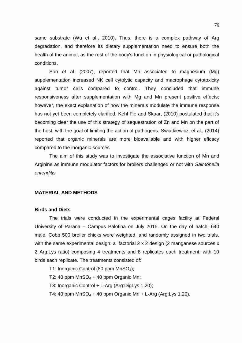
76
same substrate (Wu et al., 2010). Thus, there is a complex pathway of Arg
degradation, and therefore its dietary supplementation need to ensure both the
health of the animal, as the rest of the body's function in physiological or pathological
conditions.
Son et al. (2007), reported that Mn associated to magnesium (Mg)
supplementation increased NK cell cytolytic capacity and macrophage cytotoxicity
against tumor cells compared to control. They concluded that immune
responsiveness after supplementation with Mg and Mn present positive effects;
however, the exact explanation of how the minerals modulate the immune response
has not yet been completely clarified. Kehl-Fie and Skaar, (2010) postulated that it's
becoming clear the use of this strategy of sequestration of Zn and Mn on the part of
the host, with the goal of limiting the action of pathogens. Swiatkiewicz, et al., (2014)
reported that organic minerals are more bioavailable and with higher eficacy
compared to the inorganic sources
The aim of this study was to investigate the associative function of Mn and
Arginine as immune modulator factors for broilers challenged or not with Salmonella
enteriditis.
MATERIAL AND METHODS
Birds and Diets
The trials were conducted in the experimental cages facility at Federal
University of Parana – Campus Palotina on July 2015. On the day of hatch, 640
male, Cobb 500 broiler chicks were weighted, and randomly assigned in two trials,
with the same experimental design: a factorial 2 x 2 design (2 manganese sources x
2 Arg:Lys ratio) composing 4 treatments and 8 replicates each treatment, with 10
birds each replicate. The treatments consisted of:
T1: Inorganic Control (80 ppm MnSO4);
T2: 40 ppm MnSO4 + 40 ppm Organic Mn;
T3: Inorganic Control + L-Arg (Arg:DigLys 1.20);
T4: 40 ppm MnSO4 + 40 ppm Organic Mn + L-Arg (Arg:Lys 1.20).
77
For treatments T1 and T2 the digestible arginine:lysine ratio was 1.12,
considered normal using corn-soybean meal based diets, which corresponds to
1.48% digestible Arg in a commercial corn-soybean meal diet. For the treatments T3
and T4 the 1.20 Arg:Lys ratio was obtained by the addition of 1kg of Arg per ton of
feed, resulting in 1.58% digestible Arg.
The organic Mn used is formed by one metal ion bound to one amino acid ion,
called a metal amino acid complex. The product AvailaMn® was supplied by Zinpro
Corporation.
The birds were kept in wired 4-level battery cages (80 cm x 55 cm) from 1 to
21 days of age, in an environmentally controlled room (exhaust fans, heaters and air
conditioning). From the first until the seventh day, cages were lined with wood
shavings to help maintain thermal comfort to the chicks. Diets were fed in mash form
and birds were allowed ad libitum access to feed and water, with 24-h lightening
schedule throughout the trial. The basal diets were formulated to meet the nutrient
requirements of chickens from 1 to 21 days and the formula and calculated levels are
presented on Table 1.
Vaccine against Salmonella enteretidis challenge in chicks
In the unchallenged group the birds received just the experimental diets. In the
challenged group, birds were fed the same experimental diets and at the 9th day were
challenged with an intramuscular Salmonella enterididis vaccine (Figure 1).
Figure 1: Salmonella vaccine used as challenge
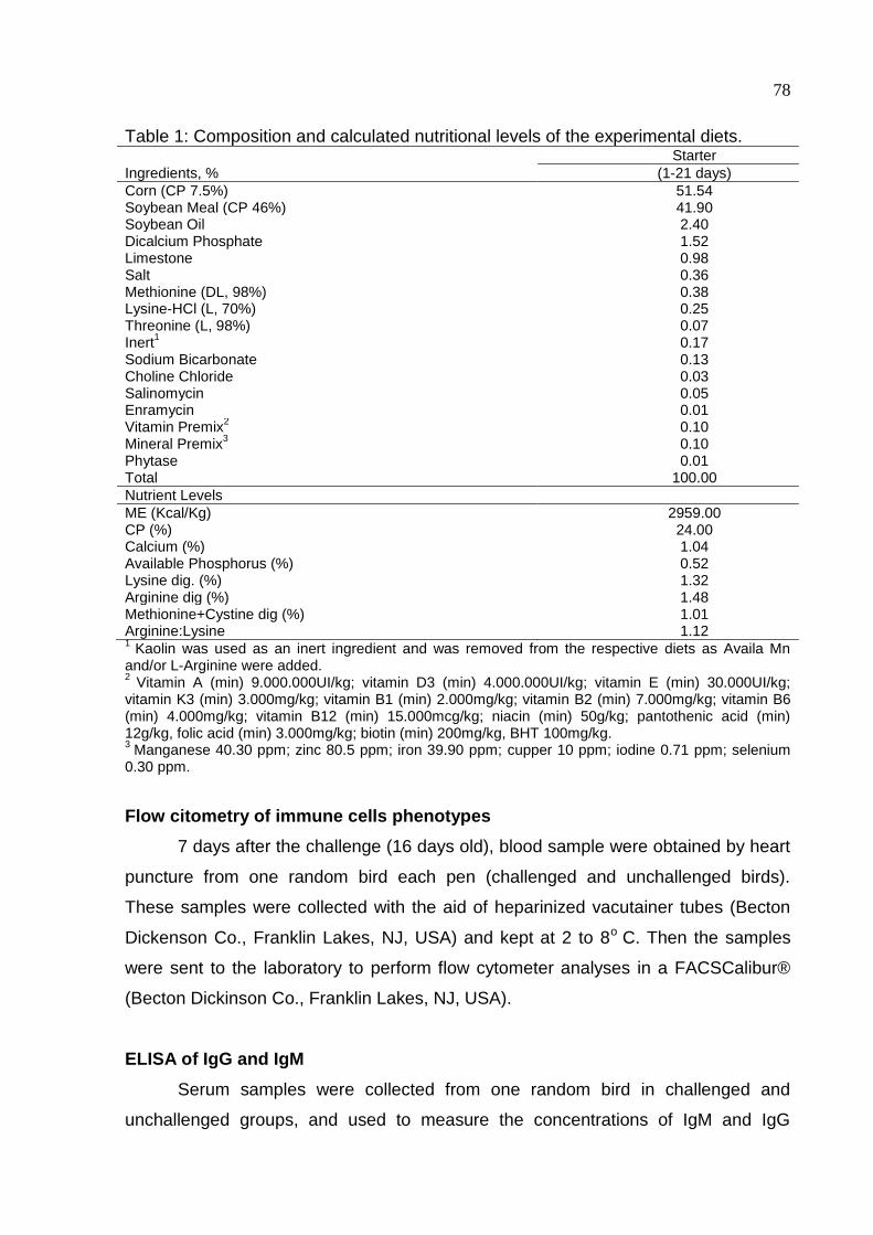
78
Table 1: Composition and calculated nutritional levels of the experimental diets. Starter
Ingredients, % (1-21 days)
Corn (CP 7.5%) 51.54 Soybean Meal (CP 46%) 41.90 Soybean Oil 2.40 Dicalcium Phosphate 1.52 Limestone 0.98 Salt 0.36 Methionine (DL, 98%) 0.38 Lysine-HCl (L, 70%) 0.25 Threonine (L, 98%) 0.07 Inert
1 0.17
Sodium Bicarbonate 0.13 Choline Chloride 0.03 Salinomycin 0.05 Enramycin 0.01 Vitamin Premix
2 0.10
Mineral Premix3
0.10 Phytase 0.01 Total 100.00
Nutrient Levels
ME (Kcal/Kg) 2959.00 CP (%) 24.00 Calcium (%) 1.04 Available Phosphorus (%) 0.52 Lysine dig. (%) 1.32 Arginine dig (%) 1.48 Methionine+Cystine dig (%) 1.01 Arginine:Lysine 1.12 1
Kaolin was used as an inert ingredient and was removed from the respective diets as Availa Mn and/or L-Arginine were added. 2
Vitamin A (min) 9.000.000UI/kg; vitamin D3 (min) 4.000.000UI/kg; vitamin E (min) 30.000UI/kg; vitamin K3 (min) 3.000mg/kg; vitamin B1 (min) 2.000mg/kg; vitamin B2 (min) 7.000mg/kg; vitamin B6 (min) 4.000mg/kg; vitamin B12 (min) 15.000mcg/kg; niacin (min) 50g/kg; pantothenic acid (min) 12g/kg, folic acid (min) 3.000mg/kg; biotin (min) 200mg/kg, BHT 100mg/kg. 3
Manganese 40.30 ppm; zinc 80.5 ppm; iron 39.90 ppm; cupper 10 ppm; iodine 0.71 ppm; selenium 0.30 ppm.
Flow citometry of immune cells phenotypes
7 days after the challenge (16 days old), blood sample were obtained by heart
puncture from one random bird each pen (challenged and unchallenged birds).
These samples were collected with the aid of heparinized vacutainer tubes (Becton
Dickenson Co., Franklin Lakes, NJ, USA) and kept at 2 to 8o C. Then the samples
were sent to the laboratory to perform flow cytometer analyses in a FACSCalibur®
(Becton Dickinson Co., Franklin Lakes, NJ, USA).
ELISA of IgG and IgM
Serum samples were collected from one random bird in challenged and
unchallenged groups, and used to measure the concentrations of IgM and IgG
79
isotypes, 21 days after the day of challenge. It was used an Indirect ELISA (Enzyme
Linked Immunosorbent Assay), which the antigens used were extracted and
quantified from the exactly same salmonella vaccine used to challenge the birds.
After quantification, it was diluted in carbonate buffer with final concentration of 1
microgram of protein in 100 microliters. Then, 100 microliters were added to each
assay plate and incubated at 4-8 ºC for 16 hours with the aim to promote protein
adsorption in the assay plate. The plate was then washed 3 times with a wash
solution, when the 1:400 serum:incubation buffer were added and the plate submitted
to incubation at 37 ºC for 1 hour and then washed 3 times with wash solution. It was
used IgM and IgG conjugates in 1:15,000 and 1:30,000 conjugate:incubation buffer
ratio, respectively and incubated for one hour at 37ºC. After that it was added OPD
substrate and then the plate was incubated in a dark room for 40 minutes at room
temperature. Stop solution (Sulfuric Acid 5%) were added before reading the plate.
Absorbance was assessed using a 495 nm wavelength.
Organ Collection
At 28 days old, 8 birds from each treatment for both groups (unchallenged and
challenged) were randomly selected. The broilers were submitted to electrical
stunning and then slaughtered by severing the jugular vein. The lymphatic organs
(spleen, thymus and bursa) were immediately removed, carefully stripped of adhering
connective tissue and individually weighed. Relative organ weights were calculated
as percentage of body weight.
Macrophage Phagocytic Activity
At 19 days old, 8 birds per treatment (only for the unchallenged group) were
inoculated by intra-abdominal injection of Sephadex G-50® (Sigma) at 3%, (1.0
mL/100g of live weight) Qureshi et al. 1986, Konjufca, 2004) to migrate macrophages
to abdominal cavity. For this procedure were used G-14 intravascular catheters. After
48 hours the birds were washed with neutral detergent and sanitized with alcohol
70%, being then sacrificed by cervical dislocation. The abdominal cavity was opened
and 20 mL of PBS (phosphate buffer solution) was injected to help in the abdominal
liquid collection. This step was conducted in an extremely clean environment, with
the aid of sterile equipment. Previously the abdominal liquid collection, was prepared
the saline solution containing 107 UFC/mL de Escherichia coli (ATCC 8739). The
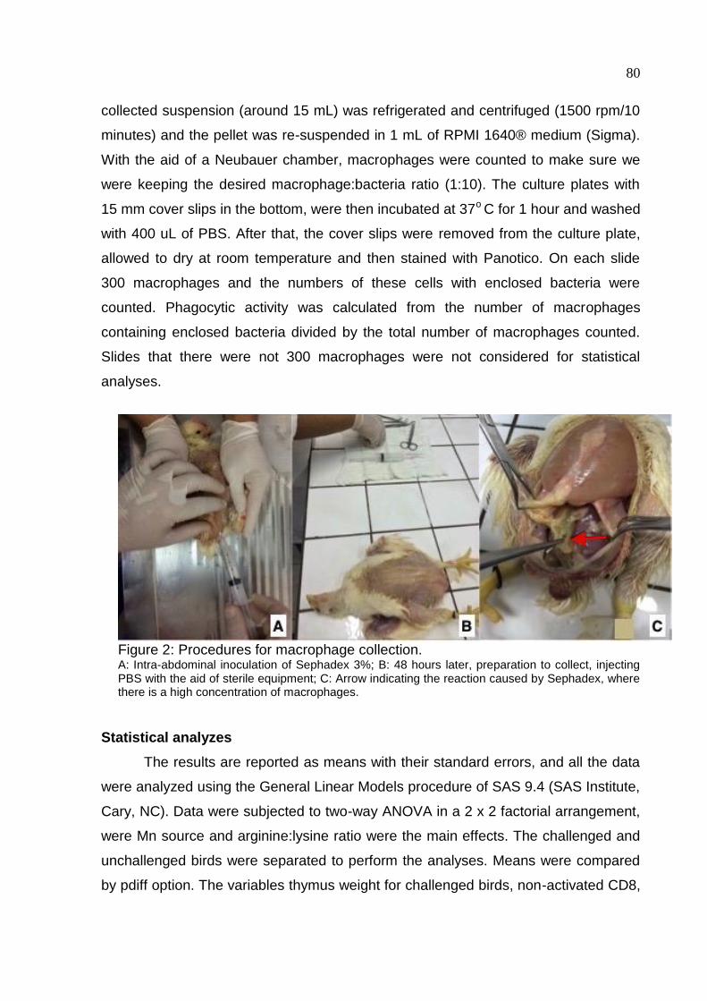
80
collected suspension (around 15 mL) was refrigerated and centrifuged (1500 rpm/10
minutes) and the pellet was re-suspended in 1 mL of RPMI 1640® medium (Sigma).
With the aid of a Neubauer chamber, macrophages were counted to make sure we
were keeping the desired macrophage:bacteria ratio (1:10). The culture plates with
15 mm cover slips in the bottom, were then incubated at 37o C for 1 hour and washed
with 400 uL of PBS. After that, the cover slips were removed from the culture plate,
allowed to dry at room temperature and then stained with Panotico. On each slide
300 macrophages and the numbers of these cells with enclosed bacteria were
counted. Phagocytic activity was calculated from the number of macrophages
containing enclosed bacteria divided by the total number of macrophages counted.
Slides that there were not 300 macrophages were not considered for statistical
analyses.
Figure 2: Procedures for macrophage collection. A: Intra-abdominal inoculation of Sephadex 3%; B: 48 hours later, preparation to collect, injecting PBS with the aid of sterile equipment; C: Arrow indicating the reaction caused by Sephadex, where there is a high concentration of macrophages.
Statistical analyzes
The results are reported as means with their standard errors, and all the data
were analyzed using the General Linear Models procedure of SAS 9.4 (SAS Institute,
Cary, NC). Data were subjected to two-way ANOVA in a 2 x 2 factorial arrangement,
were Mn source and arginine:lysine ratio were the main effects. The challenged and
unchallenged birds were separated to perform the analyses. Means were compared
by pdiff option. The variables thymus weight for challenged birds, non-activated CD8,
81
suppressor monocytes, monocytes for unchallenged birds did not present normal
distribution, therefore were transformed using Proc Rank.
RESULTS
Flow citometry of immune cells phenotypes
As presented on Table 2 and 3, there was no interaction between the main
effects (Mn source and Arg:Lys ratio) for unchallenged or challenged birds,
repectively. However, unchallenged birds fed associated Mn sources showed higher
(P > 0.05) mucosa T cytotoxic lymphocytes counting (mucose CD8), compared to
inorganic source. For mucosa T helper (CD4), T helper (general CD4) and non-
activated CD8, birds which were fed Arg supplemented diet (Arg:Lys 1.20), had a
higher amount of this cells, compared to the commercial Arg:Lys level (1.12).
For challenged birds, analyzing Mn source as a main effect, exclusive
inorganic Mn diet had a higher amount of cells (P > 0.05) compared to associated
sources for mucosa general CD4, non-activated and activated CD8 and APC
(Antigen Presenting Cell). Just for suppressor monocytes this patter has changed,
which birds fed associated Mn source had a higher amount (P > 0.05) compared to
inorganic Mn source. Also, it can be observed an increased of suppressors
monocytes when Arg was supplemented, in comparison to non-supplemented diets.
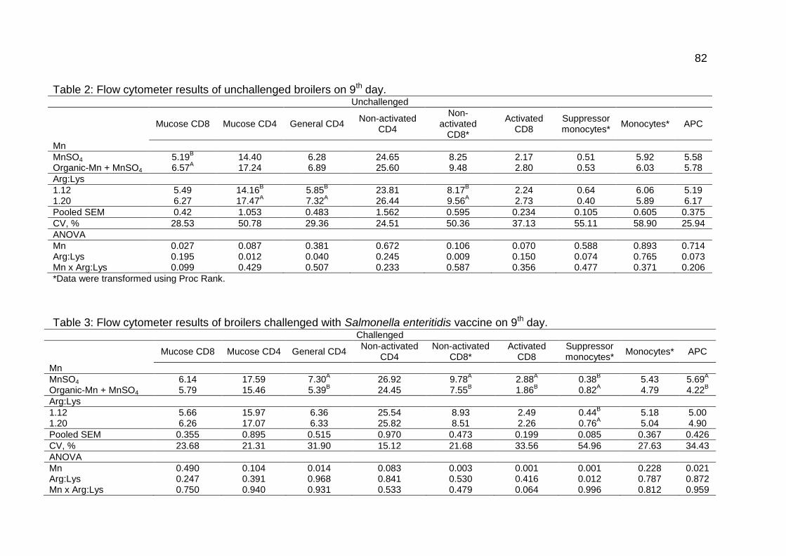
82
Table 2: Flow cytometer results of unchallenged broilers on 9th day.
Unchallenged
Mucose CD8 Mucose CD4 General CD4
Non-activated CD4
Non-activated
CD8*
Activated CD8
Suppressor monocytes*
Monocytes* APC
Mn
MnSO4 5.19B 14.40 6.28 24.65 8.25 2.17 0.51 5.92 5.58
Organic-Mn + MnSO4 6.57A 17.24 6.89 25.60 9.48 2.80 0.53 6.03 5.78
Arg:Lys
1.12 5.49 14.16B 5.85
B 23.81 8.17
B 2.24 0.64 6.06 5.19
1.20 6.27 17.47A 7.32
A 26.44 9.56
A 2.73 0.40 5.89 6.17
Pooled SEM 0.42 1.053 0.483 1.562 0.595 0.234 0.105 0.605 0.375
CV, % 28.53 50.78 29.36 24.51 50.36 37.13 55.11 58.90 25.94
ANOVA
Mn 0.027 0.087 0.381 0.672 0.106 0.070 0.588 0.893 0.714 Arg:Lys 0.195 0.012 0.040 0.245 0.009 0.150 0.074 0.765 0.073 Mn x Arg:Lys 0.099 0.429 0.507 0.233 0.587 0.356 0.477 0.371 0.206
*Data were transformed using Proc Rank.
Table 3: Flow cytometer results of broilers challenged with Salmonella enteritidis vaccine on 9th day. Challenged
Mucose CD8 Mucose CD4 General CD4
Non-activated CD4
Non-activated CD8*
Activated CD8
Suppressor monocytes*
Monocytes* APC
Mn
MnSO4 6.14 17.59 7.30A 26.92 9.78
A 2.88
A 0.38
B 5.43 5.69
A
Organic-Mn + MnSO4 5.79 15.46 5.39B 24.45 7.55
B 1.86
B 0.82
A 4.79 4.22
B
Arg:Lys
1.12 5.66 15.97 6.36 25.54 8.93 2.49 0.44B 5.18 5.00
1.20 6.26 17.07 6.33 25.82 8.51 2.26 0.76A 5.04 4.90
Pooled SEM 0.355 0.895 0.515 0.970 0.473 0.199 0.085 0.367 0.426
CV, % 23.68 21.31 31.90 15.12 21.68 33.56 54.96 27.63 34.43
ANOVA
Mn 0.490 0.104 0.014 0.083 0.003 0.001 0.001 0.228 0.021 Arg:Lys 0.247 0.391 0.968 0.841 0.530 0.416 0.012 0.787 0.872 Mn x Arg:Lys 0.750 0.940 0.931 0.533 0.479 0.064 0.996 0.812 0.959
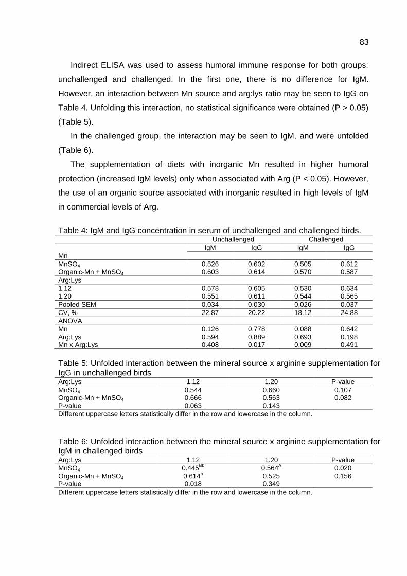
83
Indirect ELISA was used to assess humoral immune response for both groups:
unchallenged and challenged. In the first one, there is no difference for IgM.
However, an interaction between Mn source and arg:lys ratio may be seen to IgG on
Table 4. Unfolding this interaction, no statistical significance were obtained (P > 0.05)
(Table 5).
In the challenged group, the interaction may be seen to IgM, and were unfolded
(Table 6).
The supplementation of diets with inorganic Mn resulted in higher humoral
protection (increased IgM levels) only when associated with Arg (P < 0.05). However,
the use of an organic source associated with inorganic resulted in high levels of IgM
in commercial levels of Arg.
Table 4: IgM and IgG concentration in serum of unchallenged and challenged birds. Unchallenged Challenged
IgM IgG IgM IgG
Mn
MnSO4 0.526 0.602 0.505 0.612 Organic-Mn + MnSO4 0.603 0.614 0.570 0.587
Arg:Lys
1.12 0.578 0.605 0.530 0.634 1.20 0.551 0.611 0.544 0.565
Pooled SEM 0.034 0.030 0.026 0.037
CV, % 22.87 20.22 18.12 24.88
ANOVA
Mn 0.126 0.778 0.088 0.642 Arg:Lys 0.594 0.889 0.693 0.198 Mn x Arg:Lys 0.408 0.017 0.009 0.491
Table 5: Unfolded interaction between the mineral source x arginine supplementation for IgG in unchallenged birds Arg:Lys 1.12 1.20 P-value
MnSO4 0.544 0.660 0.107 Organic-Mn + MnSO4 0.666 0.563 0.082 P-value 0.063 0.143
Different uppercase letters statistically differ in the row and lowercase in the column.
Table 6: Unfolded interaction between the mineral source x arginine supplementation for IgM in challenged birds Arg:Lys 1.12 1.20 P-value
MnSO4 0.445Bb
0.564A 0.020
Organic-Mn + MnSO4 0.614a 0.525 0.156
P-value 0.018 0.349
Different uppercase letters statistically differ in the row and lowercase in the column.
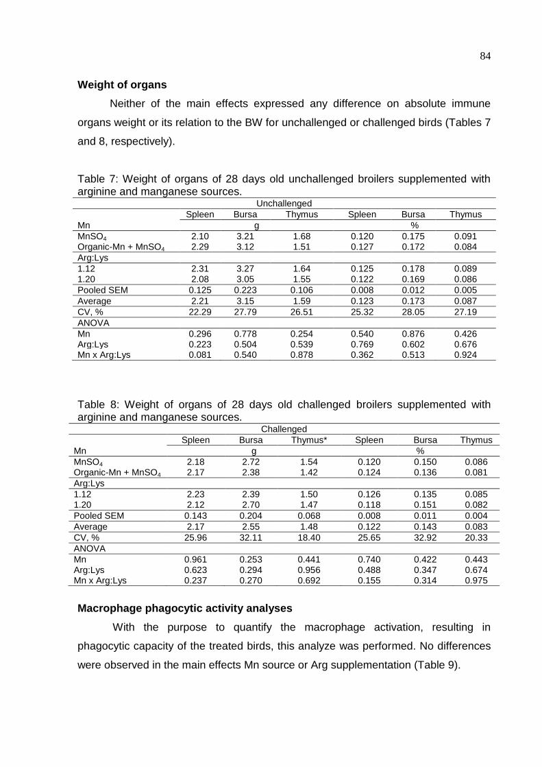
84
Weight of organs
Neither of the main effects expressed any difference on absolute immune
organs weight or its relation to the BW for unchallenged or challenged birds (Tables 7
and 8, respectively).
Table 7: Weight of organs of 28 days old unchallenged broilers supplemented with arginine and manganese sources.
Unchallenged
Spleen Bursa Thymus Spleen Bursa Thymus
Mn g %
MnSO4 2.10 3.21 1.68 0.120 0.175 0.091 Organic-Mn + MnSO4 2.29 3.12 1.51 0.127 0.172 0.084
Arg:Lys
1.12 2.31 3.27 1.64 0.125 0.178 0.089 1.20 2.08 3.05 1.55 0.122 0.169 0.086
Pooled SEM 0.125 0.223 0.106 0.008 0.012 0.005
Average 2.21 3.15 1.59 0.123 0.173 0.087
CV, % 22.29 27.79 26.51 25.32 28.05 27.19
ANOVA
Mn 0.296 0.778 0.254 0.540 0.876 0.426 Arg:Lys 0.223 0.504 0.539 0.769 0.602 0.676 Mn x Arg:Lys 0.081 0.540 0.878 0.362 0.513 0.924
Table 8: Weight of organs of 28 days old challenged broilers supplemented with arginine and manganese sources.
Challenged
Spleen Bursa Thymus* Spleen Bursa Thymus
Mn g %
MnSO4 2.18 2.72 1.54 0.120 0.150 0.086 Organic-Mn + MnSO4 2.17 2.38 1.42 0.124 0.136 0.081
Arg:Lys
1.12 2.23 2.39 1.50 0.126 0.135 0.085 1.20 2.12 2.70 1.47 0.118 0.151 0.082
Pooled SEM 0.143 0.204 0.068 0.008 0.011 0.004
Average 2.17 2.55 1.48 0.122 0.143 0.083
CV, % 25.96 32.11 18.40 25.65 32.92 20.33
ANOVA
Mn 0.961 0.253 0.441 0.740 0.422 0.443 Arg:Lys 0.623 0.294 0.956 0.488 0.347 0.674 Mn x Arg:Lys 0.237 0.270 0.692 0.155 0.314 0.975
Macrophage phagocytic activity analyses
With the purpose to quantify the macrophage activation, resulting in
phagocytic capacity of the treated birds, this analyze was performed. No differences
were observed in the main effects Mn source or Arg supplementation (Table 9).
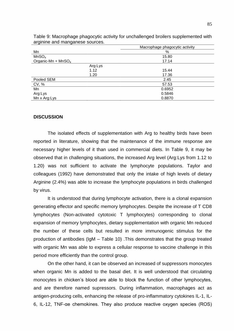
85
Table 9: Macrophage phagocytic activity for unchallenged broilers supplemented with arginine and manganese sources. Macrophage phagocytic activity
Mn %
MnSO4 15.80 Organic-Mn + MnSO4 17.14
Arg:Lys 1.12 15.44 1.20 17.36
Pooled SEM 2.45
CV, % 57.53
Mn 0.6952 Arg:Lys 0.5846 Mn x Arg:Lys 0.8870
DISCUSSION
The isolated effects of supplementation with Arg to healthy birds have been
reported in literature, showing that the maintenance of the immune response are
necessary higher levels of it than used in commercial diets. In Table 9, it may be
observed that in challenging situations, the increased Arg level (Arg:Lys from 1.12 to
1.20) was not sufficient to activate the lymphocyte populations. Taylor and
colleagues (1992) have demonstrated that only the intake of high levels of dietary
Arginine (2.4%) was able to increase the lymphocyte populations in birds challenged
by virus.
It is understood that during lymphocyte activation, there is a clonal expansion
generating effector and specific memory lymphocytes. Despite the increase of T CD8
lymphocytes (Non-activated cytotoxic T lymphocytes) corresponding to clonal
expansion of memory lymphocytes, dietary supplementation with organic Mn reduced
the number of these cells but resulted in more immunogenic stimulus for the
production of antibodies (IgM – Table 10) .This demonstrates that the group treated
with organic Mn was able to express a cellular response to vaccine challenge in this
period more efficiently than the control group.
On the other hand, it can be observed an increased of suppressors monocytes
when organic Mn is added to the basal diet. It is well understood that circulating
monocytes in chicken’s blood are able to block the function of other lymphocytes,
and are therefore named supressors. During inflammation, macrophages act as
antigen-producing cells, enhancing the release of pro-inflammatory cytokines IL-1, IL-
6, IL-12, TNF-αe chemokines. They also produce reactive oxygen species (ROS)
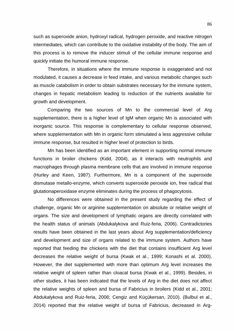
86
such as superoxide anion, hydroxyl radical, hydrogen peroxide, and reactive nitrogen
intermediates, which can contribute to the oxidative instability of the body. The aim of
this process is to remove the inducer stimuli of the cellular immune response and
quickly initiate the humoral immune response.
Therefore, in situations where the immune response is exaggerated and not
modulated, it causes a decrease in feed intake, and various metabolic changes such
as muscle catabolism in order to obtain substrates necessary for the immune system,
changes in hepatic metabolism leading to reduction of the nutrients available for
growth and development.
Comparing the two sources of Mn to the commercial level of Arg
supplementation, there is a higher level of IgM when organic Mn is associated with
inorganic source. This response is complementary to cellular response observed,
where supplementation with Mn in organic form stimulated a less aggressive cellular
immune response, but resulted in higher level of protection to birds.
Mn has been identified as an important element in supporting normal immune
functions in broiler chickens (Kidd, 2004), as it interacts with neutrophils and
macrophages through plasma membrane cells that are involved in immune response
(Hurley and Keen, 1987). Furthermore, Mn is a component of the superoxide
dismutase metallo-enzyme, which converts superoxide peroxide ion, free radical that
glutationaperoxidase enzyme eliminates during the process of phagocytosis.
No differences were obtained in the present study regarding the effect of
challenge, organic Mn or arginine supplementation on absolute or relative weight of
organs. The size and development of lymphatic organs are directly correlated with
the health status of animals (Abdukalykova and Ruiz-feria, 2006). Contradictories
results have been obtained in the last years about Arg supplementation/deficiency
and development and size of organs related to the immune system. Authors have
reported that feeding the chickens with the diet that contains insufficient Arg level
decreases the relative weight of bursa (Kwak et al., 1999; Konashi et al. 2000).
However, the diet supplemented with more than optimum Arg level increases the
relative weight of spleen rather than cloacal bursa (Kwak et al., 1999). Besides, in
other studies, it has been indicated that the levels of Arg in the diet does not affect
the relative weights of spleen and bursa of Fabricius in broilers (Kidd et al., 2001;
Abdukalykova and Ruiz-feria, 2006; Cengiz and Küçükersan, 2010). (Bulbul et al.,
2014) reported that the relative weight of bursa of Fabricius, decreased in Arg-
87
deficient group at grower and finisher phases in accordance with Kwak et al. (1999)
and (Konashi et al., 2000). Furthermore, the relative weight of spleen increased when
Arg was supplemented to the diet at starter phase in accordance with (Kwak et al.,
1999).
Phagocytosis is an innate immune reaction for the clearance of bacterial
infections of animals, which plays an indispensable role in the defense against
various infections (Seifert et al., 2011). Macrophages are a key arm of the innate
immune defense system in intracellular bacterial killing (Ibuki et al., 2011). The
antimicrobial activities of macrophages are due to the generation of reactive oxygen
species and reactive N species, such as H2O2 and NO, which are important
metabolites of Arg (Bronte and Zanovello, 2005). An in vitro study in channel catfish
has suggested that supplementation of Arg to cell-culture media enhances the
phagocytic activity of head kidney macrophages (Pohlenz et al., 2012). As in the
present study, no significant influence was observed in the study published by Tan et
al. (2014). Contradictory, Sung et al. (1991), found that nitric oxide production of
macrophages is increased by a local concentration of Arg.
It is necessary to consider that a competition among arginase and nitric oxide
synthetases (iNOS) for Arginine might happen. As a common substrate, it could lead
to a reduction of the synthesis of NO, the main cytotoxic mediator of immune-
activated effector cells and one of the most important regulating molecules of the
immune system. This could lead to a major impairment to the bird immune capacity,
leaving it more exposed to common challenges present in modern poultry production,
as Salmonella or Clostridium.
Broilers from unchallenged group were subjected to analyses of macrophage
phagocytic activity with 19 days old. The percentage is provided by a ratio among the
number of active macrophage divided by total macrophages counted × 100. No
differences were observed. Arginine supplementation was capable of keeping the
macrophage activation capacity, even though arginine was being converted to
ornithine and urea to be used as precursors for bone mineralization. There was no
prejudice on macrophage phagocytic activity for the treatments assessed (Table 7).
In recent times, Mn has become an element of increasing concern because of
extremely rapid growth of broiler chickens, which exerts additional stress on the
skeletal structure. So, there is a need to reassess the responses of broiler chicken to
88
higher levels of Mn supplementation with particular emphasis on performance,
mineral uptake by the tissues and immune competence.
CONCLUSION
Dietary supplementation with organic Mn resulted in a short and effective
cellular respons, reducing the number of lymphocytes of challenged birds but
resulting in more immunogenic stimulus for the production of antibodies (IgM)
humoral. The group treated with OrganicMn was able to express a cellular response
to vaccine challenge in this period more efficiently than the control group.
Higher levels of Mn or more bioavailable sources can be needed for optimized
the immune competence and improved immune response in broiler chicken.
Increasing levels of organic Mn associated with arginine supplementation
should be evaluated for better understanding of the contribution of the Mn in the
activity of arginase.
REFERENCES
Abdukalykova, S., and C. a Ruiz-feria. 2006. Arginine and Vitamin E Improve the Cellular and Humoral Immune Response of Broiler Chickens. 5:121–127.
Bronte, V., and P. Zanovello. 2005. Regulation of immune responses by L-arginine metabolism. Nat. Rev. Immunol. 5:641–654.
Bulbul, T., V. Ozdemir, a Bulbul, and E. Ulutas. 2014. The effect of dietary L-arginine intake on the level of antibody titer, the relative organ weight and colon motility in broilers. Pol. J. Vet. Sci. 17:113–21.
Cengiz, Ö., and S. Küçükersan. 2010. Effects of graded contents of arginin sup- plementation on growth performance , hae- matological parameters and immune system in broilers. :409–417.
Fernandes, J. I. M., a E. Murakami, E. N. Martins, M. I. Sakamoto, and E. R. M. Garcia. 2009. Effect of arginine on the development of the pectoralis muscle and the diameter and the protein:deoxyribonucleic acid rate of its skeletal myofibers in broilers. Poult. Sci. 88:1399–1406.
Florini, J. R., D. Z. Ewton, and S. a Coolican. 1996. Growth hormone and the insulin-like growth factor system in myogenesis. Endocr. Rev. 17:481–517.
89
Guo, Y. W., B. L. Shi, S. M. Yan, Y. Q. Xu, J. L. Li, and T. Y. Li. 2015. Effects of arginine on cytokines and nitric oxide synthesis in broilers. 25:366–371.
Halevy, O., Y. Nadel, M. Barak, I. Rozenboim, and D. Sklan. 2003. Early posthatch feeding stimulates satellite cell proliferation and skeletal muscle growth in turkey poults. J. Nutr. 133:1376–1382.
Hurley LS, Keen CL. 1987. Manganese. In: Mertz W, editor. , ed. Trace Elements in Human and Animal Nutrition, 5th ed. San Diego: Academic Press. Pp.185–223.
Ibuki, M., J. Kovacs-Nolan, K. Fukui, H. Kanatani, and Y. Mine. 2011. ?? 1-4 mannobiose enhances Salmonella-killing activity and activates innate immune responses in chicken macrophages. Vet. Immunol. Immunopathol. 139:289–295.
De Jonge, W. J., K. L. Kwikkers, A. a. Te Velde, S. J. H. Van Deventer, M. a. Nolte, R. E. Mebius, J. M. Ruijter, M. C. Lamers, and W. H. Lamers. 2002. Arginine deficiency affects early B cell maturation and lymphoid organ development in transgenic mice. J. Clin. Invest. 110:1539–1548.
Kehl-Fie, T. E., and E. P. Skaar. 2010. Nutritional immunity beyond iron: a role for manganese and zinc. Curr. Opin. Chem. Biol. 14:218–224.
Kidd, M. T. 2004. Nutritional modulation of immune function. Poult. Sci. 83:650–657.
Kidd, M. T., E. D. Peebles, S. K. Whitmarsh, J. B. Yeatman, and R. F. Wideman. 2001. Growth and immunity of broiler chicks as affected by dietary arginine. Poult. Sci. 80:1535–1542.
Konashi, S., K. Takahashi, and Y. Akiba. 2000. Effects of dietary essential amino acid deficiencies on immunological Effects of dietary essential amino acid deficiencies on immunological variables in broiler chickens. :449–456.
Kwak, H., R. E. Austic, and R. R. Dietert. 1999. Influence of dietary arginine concentration on lymphoid organ growth in chickens. Poult. Sci. 78:1536–1541.
Lee, J. E., R. E. Austic, S. a Naqi, K. a Golemboski, and R. R. Dietert. 2002. Dietary arginine intake alters avian leukocyte population distribution during infectious bronchitis challenge. Poult. Sci. 81:793–798.
Li, H. Y., S. M. Yan, B. L. Shi, and X. Y. Guo. 2009. Effect of Chitosan on Nitric Oxide Content and Inducible Nitric Oxide Synthase Activity in Serum and Expression of Inducible Nitric Oxide Synthase mRNA in Small Intestine of Broiler Chickens *. Asian-Australasian J. Anim. Sci. 22:1048–1053.
McDowell, L. R. 1989. Vitamins in Animal Nutrition. Academic Press Inc. Harcourt Brace Jovanovich Publishers, San Diego, CA.
Pohlenz, C., A. Buentello, W. Mwangi, and D. M. Gatlin. 2012. Arginine and glutamine supplementation to culture media improves the performance of various channel catfish immune cells. Fish Shellfish Immunol. 32:762–768.
90
Ruiz-Feria, C. a, M. T. Kidd, and R. F. Wideman. 2001. Plasma levels of arginine, ornithine, and urea and growth performance of broilers fed supplemental L-arginine during cool temperature exposure. Poult. Sci. 80:358–369.
Seifert, S., C. Fritz, N. Carlini, S. W. Barth, C. M. a. P. Franz, and B. Watzl. 2011. Modulation of innate and adaptive immunity by the probiotic Bifidobacterium longum PCB133 in turkeys. Poult. Sci. 90:2275 –2280.
Son, E.-W., S.-R. Lee, H.-S. Choi, H.-J. Koo, J.-E. Huh, M.-H. Kim, and S. Pyo. 2007. Effects of supplementation with higher levels of manganese and magnesium on immune function. Arch. Pharm. Res. 30:743–9.
Su, C. L., and R. E. Austic. 1998. The utilization of dipeptides containing L-arginine by chicken macrophages. Poult. Sci. 77:1852–1857.
Sung, Y. J., J. H. Hotchkiss, R. E. Austic, and R. R. Dietert. 1991. L-arginine-dependent production of a reactive nitrogen intermediate by macrophages of a uricotelic species. J. Leukoc. Biol. 50:49–56.
Tan, J., S. Liu, Y. Guo, T. J. Applegate, and S. D. Eicher. 2014. Dietary l-arginine supplementation attenuates lipopolysaccharide-induced inflammatory response in broiler chickens. Br. J. Nutr. 111:1394–404.
Webel, D. M., R. W. Johnson, and D. H. Baker. 1998. Lipopolysaccharide-Induced Reductions in Food Intake Do Not Decrease the Efficiency of Lysine and Threonine Utilization for Protein Accretion in Chickens. J. Nutr. 128:1760–1766.
Wu, G., F. W. Bazer, R. C. Burghardt, G. a. Johnson, S. W. Kim, X. L. Li, M. C. Satterfield, and T. E. Spencer. 2010. Impacts of amino acid nutrition on pregnancy outcome in pigs: mechanisms and implications for swine production. J. Anim. Sci. 88.
Wu, G., and S. M. Morris. 1998. Arginine metabolism : nitric oxide and beyond. Biochem. J. 336:1–17.
Xie, Q., Y. Kashiwabara, and C. Nathan. 1994. Role of transcription factor NF-kappaB/Rel in induction of nitric oxide synthase. J.Biol.Chem. 269:4705–4708.
91
5. CONSIDERAÇÕES FINAIS
A substituição parcial de 50% da fonte de Mn inorgânica utilizada (MnSO4)
pela fonte orgânica não comprometeu o desempenho produtivo, rendimento de
carcaça e mineralização óssea de frangos de corte. Estes resultados indicam que é
possível a substituição parcial e provavelmente total dos minerais inorgânicos por
fontes orgânicas na produção animal, que busca melhores resultados zootécnicos
de forma sustentável.
As fontes orgânicas de minerais podem ser melhor aproveitadas pelo animal,
devido as menores perdas decorrentes de interações e antagonismo entre minerais,
formação de complexos indisponíveis e consequente redução da absorção, além da
menor excreção no meio ambiente e menores riscos de contaminação por metais
pesados. Contudo, o uso de fontes orgânicas ainda é limitada devido ao seu custo
elevado, o que onera o custo da fração mineral das dietas. Entretanto, são produtos
de alto valor agregado e inseridos em um contexto de sustentabilidade.
A nutrição pode ser usada como uma ferramenta para modular o sistema
imunológico das aves. Nutrientes também participam do metabolismo como
imunomoduladores, a fim de melhorar as funções imunológicas e a resistência a
infecções. O efeito imunomodulador da arginina está na síntese de proteínas,
deposição de colágeno e na ação secretagoga de hormônios. Além disso, atua
sobre a proliferação celular e maturação dos linfócitos e serve de substrato para a
para biossíntese do óxido nítrico, que por sua vez, tem função fundamental em
processos inflamatórios favorecendo o reparo tecidual e contribuindo para manter o
fluxo sanguíneo nos tecidos.
Entretanto, para algumas dessas funções imunomoduladoras são necessários
co-fatores enzimáticos. O manganês é co-fator da argininase e portanto atua de
forma direta e indireta na resposta imune. A incorporação de Mn orgânico a uma
dieta elaborada com minerais inorgânicos em níveis parciais em relação à exigência
das aves desafiadas imunologicamente, reduziu o número de linfócitos nas aves
desafiadas, porém possibilitou um maior estímulo imunogênico para produção de
anticorpos (IgM). Aves suplementadas com Mn orgânico foram capazes de
expressar uma resposta mais eficiente ao desafio, quando comparadas ao grupo
com Mn exclusivamente irnorgânico, independente da suplementação de Arg.
92
Os nutrientes utilizados em rações de frangos estão fundamentado em pesquisas
que avaliaram as funções produtivas economicamente importantes. Entretanto,
estes resultados demonstram que é preciso levar em consideração que, com a
contínua evolução genética das aves, é imprescindível conhecer não somente as
exigências nutricionais de microminerais, mas também assegurar que estes estejam
disponíveis para o animal imunologicamente comprometido.
A maior biodisponibilidade das fontes orgânicas de minerais permite que elas
sejam incluídas na dieta em concentrações mais baixas, sem efeitos negativos sobre
o desempenho das aves e o meio ambiente. Neste sentido, é necessário avaliar a
substituição de minerais inorgânicos por orgânicos em níveis maiores ou totais para
o melhor entendimento da relação dos minerais com imunomoduladores dietéticos.
Estes conceitos são muito importantes considerando que a indústria avícola
moderna está cada vez mais comprometida com as questões ambientais, de
segurança alimentar e sustentabilidade.
93
6. REFERÊNCIAS
Abdukalykova, S., and C. a Ruiz-feria. 2006. Arginine and Vitamin E Improve the Cellular and Humoral Immune Response of Broiler Chickens. 5:121–127.
ABPA Associação Brasileira de Proteína Animal (http://abpa-br.com.br/noticia/producao-de-carne-de-frango-totaliza-13146-milhoes-de-toneladas-em-2015-1545), janeiro 2016.
Aksu, T., M. Aksu, M. a. Yoruk, and M. Karaoglu. 2011. Effects of organically-complexed minerals on meat quality in chickens. Br. Poult. Sci. 52:558–563.
Albina, J. E., C. D. Mills, W. L. Henry, and M. D. Caldwell. 1990. Temporal expression of different pathways of L-arginine metabolism in healing wounds. J. Immunol. 144:3877.
Alderton, W. K., C. E. Cooper, and R. G. Knowles. 2001. Nitric oxide synthases : structure , function and inhibition. 615:593–615.
Almeida, I. C. L., and L. D. G. Bruno. 2006. Bone mineral density : review. Brazilian J. Poult. Sci. 8:69–73.
Amber, I. J., J. B. Hibbs, C. J. Parker, B. B. Johnson, R. R. Taintor, and Z. Vavrin. 1991. Activated macrophage conditioned medium: identification of the soluble factors inducing cytotoxicity and the L-arginine dependent effector mechanism. J. Leukoc. Biol. 49:610–620.
Applegate, T. J., and R. Angel. 2014. Nutrient requirements of poultry publication: History and need for an update. J. Appl. Poult. Res. 23:567–575
Ash, D. 2004. Arginine metabolism: enzymology, nutrition, and clinical significance. J. Nutr. 134:2741S–2897S.
Austic, R. E., and M. C. Nesheim. 1970. Role of kidney arginase in variations of the arginine requirement of chicks. J. Nutr. 100:855–868.
Bain, S.D and Watkins, B. . 1993. Local modulation of skeletal growth and bone modeling in poultry. J. Nutr.
Baker, D. H. 2009. Advances in protein-amino acid nutrition of poultry. Amino Acids 37:29–41.
Bao, Y. M., M. Choct, P. a Iji, and K. Bruerton. 2010. Trace mineral interactions in broiler chicken diets. Br. Poult. Sci. 51:109–17.
Barbul, A., D. A. Sisto, H. L. Wasserkrug, and G. Efron. 1981. Arginine stimulates lymphocyte immune response in healthy human beings. Surgery 90:244–251.
Beach, R. S., E. M. Gershwin, and L. S. Hurley. 1982. Gestational zinc deprivation in mice: persistence of immunodeficiency for three generations. Science (80). 218:469–471.
94
Beattie, J. H., and a Avenell. 1992. Trace element nutrition and bone metabolism. Nutr. Res. Rev. 5:167–188.
Berkowitz, D. E., R. White, D. Li, K. M. Minhas, A. Cernetich, S. Kim, S. Burke, A. a. Shoukas, D. Nyhan, H. C. Champion, and J. M. Hare. 2003. Arginase Reciprocally Regulates Nitric Oxide Synthase Activity and Contributes to Endothelial Dysfunction in Aging Blood Vessels. Circulation 108:2000–2006.
Berta, E., E. Andrasofzky, A. Bersenyi, R. Glavits, A. Gaspardy, and S. G. Fekete. 2004. Effect of Inorganic and Organic Manganese Supplementation on the Performance and. Acta Vet. Hung. 52:199–209.
Bessei, W. 2006. Welfare of broilers: A review. Worlds. Poult. Sci. J. 62:455–466.
Biewener, A. a, and J. E. Bertram. 1994. Structural response of growing bone to exercise and disuse. J. Appl. Physiol. 76:946–55.
Black, J. R., C. B. Ammerman, P. R. Henry, and R. D. Miles. 1984. Biological availability of manganese sources and effects of high dietary manganese on tissue mineral composition of broiler-type chicks. Poult. Sci. 63:1999–2006.
Bolea, S., Pertusa, J. A. G., Martı´ n, F., Sanchez-Andres, J. V. and Soria, B. 1997. Regulation of pancreatic B-cell eletrical activity and insulin release by physiological amino acid con- centrations. Eur. J. Physiol. 433: 699-704.
Bonser, R. H. C., and a Casinos. 2003. Regional variation in cortical bone properties from broiler fowl--a first look. Br. Poult. Sci. 44:350–4.
Boskey, a L., T. M. Wright, and R. D. Blank. 1999. Collagen and bone strength. J. Bone Miner. Res. 14:330–335.
Boucher, J. L., C. Moali, and J. P. Tenu. 1999. Nitric oxide biosynthesis, nitric oxide synthase inhibitors and arginase competition for L-arginine utilization. Cell. Mol. Life Sci. 55:1015–1028.
Bronte, V., and P. Zanovello. 2005. Regulation of immune responses by L-arginine metabolism. Nat. Rev. Immunol. 5:641–654.
Bulbul, T., V. Ozdemir, a Bulbul, and E. Ulutas. 2014. The effect of dietary L-arginine intake on the level of antibody titer, the relative organ weight and colon motility in broilers. Pol. J. Vet. Sci. 17:113–21.
Cashman, K., and A. Flynn. 1998. Trace elements and bone metabolism. Bibl Nutr Dieta 54:150–64.
Cashman, K., and A. Flynn. 1998. Trace elements and bone metabolism. Bibl Nutr Dieta 54:150–64.
Cengiz, Ö., and S. Küçükersan. 2010. Effects of graded contents of arginin sup- plementation on growth performance , hae- matological parameters and immune system in broilers. :409–417.
95
Chu, S. W., and M. C. Nesheim. 1979. The relationship of plasma arginine and kidney arginase activity to arginine degradation in chickens. J. Nutr. 109:1752–1758.
Collins, N. E., and E. T. Moran. 1999. Influence of supplemental manganese and zinc on live performance and carcass quality of broilers. Appl. Poult. Sci. 8:222.227.
Cook, M. E. 2000. Skeletal deformities and their causes: introduction. Poult. Sci. 79:982–4.
Corbin, B. D., E. H. Seeley, A. Raab, J. Feldmann, M. R. Miller, V. J. Torres, K. L. Anderson, B. M. Dattilo, K. L. Anderson, P. M. Dunman, R. Gerads, R. Caprioli, W. Nacken, W. J. Chazin, and E. P. Skaar. 2008. Bacterial Growth in Tissue Abscesses. Science (80-. ). 319:962–966.
Curran, J. N., D. C. Winter, and D. Bouchier-Hayes. 2006. Biological fate and clinical implications of arginine metabolism in tissue healing. Wound Repair Regen. 14:376–386.
Curran, J. N., D. C. Winter, and D. Bouchier-Hayes. 2006. Biological fate and clinical implications of arginine metabolism in tissue healing. Wound Repair Regen. 14:376–386.
Danbury, T. C., C. A. Weeks, J. P. Chambers, A. E. Waterman Pearson, and S. C. Kestin. 2000. Self selection of the analgesic drug carprofen by lame broiler chickens. Vet. Rec. 146:307–311.
Dayan, D., Hiss, Y., Hirshberg, A., Bubis, J.J. And Wolman, M. 1989. Are the polarization colors of Picrosirius red-stained collagen determined only by the diameter of the fibers. Histochemistry 93,27-29.
De Jonge, W. J., K. L. Kwikkers, A. a. Te Velde, S. J. H. Van Deventer, M. a. Nolte, R. E. Mebius, J. M. Ruijter, M. C. Lamers, and W. H. Lamers. 2002. Arginine deficiency affects early B cell maturation and lymphoid organ development in transgenic mice. J. Clin. Invest. 110:1539–1548.
De Jonge, W. J., K. L. Kwikkers, A. a. Te Velde, S. J. H. Van Deventer, M. a. Nolte, R. E. Mebius, J. M. Ruijter, M. C. Lamers, and W. H. Lamers. 2002. Arginine deficiency affects early B cell maturation and lymphoid organ development in transgenic mice. J. Clin. Invest. 110:1539–1548.
Dermience, M., G. Lognay, F. Mathieu, and P. Goyens. 2015. Effects of thirty elements on bone metabolism. J. Trace Elem. Med. Biol. 32:86–106.
Dhanakoti, S. N., J. T. Brosnan, G. R. Herzberg, and M. E. Brosnan. 1990. Renal arginine synthesis: studies in vitro and in vivo. Am. J. Physiol. 259:E437–42.
Dibner, J. J., J. D. Richards, M. L. Kitchell, and M. a. Quiroz. 2007. Metabolic challenges and early bone development. J. Appl. Poult. Res. 16:126–137.
Duclos, M. J. 2005. Insulin-like growth factor-I (IGF-I) mRNA levels and chicken muscle growth. J. Physiol. Pharma- col. 56: 25-35.
96
Dudhia, J. 2005. Aggrecan Aging and Assembly in Articular Cartilage. Cell Mol Life Sci. 62:2241–2256.
Eriksen, E. F., K. Brixen, and P. Charles. 1995. New markers of bone metabolism: clinical use in metabolic bone disease. EurJEndocr 132:251–263.
Farquharson, C., and D. Jefferies. 2000. Chondrocytes and longitudinal bone growth: the development of tibial dyschondroplasia. Poult. Sci. 79:994–1004.
Fernandes, J. I. M., a E. Murakami, E. N. Martins, M. I. Sakamoto, and E. R. M. Garcia. 2009. Effect of arginine on the development of the pectoralis muscle and the diameter and the protein:deoxyribonucleic acid rate of its skeletal myofibers in broilers. Poult. Sci. 88:1399–1406.
Fernandes, J. I. M., A. E. Murakami, L. M. G. de Souza, I. C. Ospina-Rojas, and R. M. Rossi. 2014. Effect of arginine supplementation of broiler breeder hens on progeny performance. Can. J. Anim. Sci. 94:313–321.
Florini, J. R., D. Z. Ewton, and S. a Coolican. 1996. Growth hormone and the insulin-like growth factor system in myogenesis. Endocr. Rev. 17:481–517.
Florini, J. R., Ewton, D. Z. and Coolican, S. A. 1996. Growth hormone and the insulin-like growth factor system in myogen- esis. Endocrinol. Rev. 17: 481?517.
Fraker, P. J., L. E. King, T. Laakko, and T. L. Vollmer. 2000. The dynamic link between the integrity of the immune system and zinc status. J. Nutr. 130:1399S–406S.
Gajula, S. S., V. K. Chelasani, A. K. Panda, V. L. N. R. Mantena, and R. R. Savaram. 2011. Effect of supplemental inorganic Zn and Mn and their interactions on the performance of broiler chicken, mineral bioavailability, and immune response. Biol. Trace Elem. Res. 139:177–187.
Gallup, W. D., and L. C. Norris. 1939. The amount of manganese required to prevent perosis in the chick. Poult. Sci. 18:76–82.
Gardiner, E. E. 1972. Lack of response to added dietary manganese of chicks fed wheat-soybean meal or corn-soybean meal based diets. Can. J. Anim. Sci. 52:737–740.
Gebhardt, C., J. Németh, P. Angel, and J. Hess. 2006. S100A8 and S100A9 in inflammation and cancer. Biochem. Pharmacol. 72:1622–1631.
Ghigo, E., Ceda, G. P., Valcavi, R., Goffi, S., Mucci, M., Valenti, G., Cocchi, D., Muller, E. E. and Camanni, F. 1994. Low doses of either intravenously or orally administered ar- ginine areable to enhance growth hormone response to growth hormone releasing hormone in elderly subjects. J. Endocrinol. Invest. 17: 113-122.
Gilbert, S. F. 1997. Early vertebrate development: Mesoderm and endoderm. Pages 341–357 in Developmental Biology. 5th ed. Sinauer Assoc. Inc., Sunderland, MA.
97
Gonzalez-Esquerra, R., and S. Leeson. 2006. Concentrations of putrescine, spermidine, and spermine in duodenum and pancreas as affected by the ratio of arginine to lysine and source of methionine in broilers under heat stress. Poult. Sci. 85:1398–1408.
Grazi, E., E. Magri, and G. Balboni. 1975. On the control of arginine metabolism in chicken kidney and liver. Eur. J. Biochem. 60:431–436.
Griffith, O. W., and D. J. Stuehr. 1995. Nitric Oxide Synthases: Properties and Catalytic Mechanism. Annu. Revis. Physiol. 57:707–736.
Guo, Y. W., B. L. Shi, S. M. Yan, Y. Q. Xu, J. L. Li, and T. Y. Li. 2015. Effects Of Arginine On Cytokines And Nitric Oxide Synthesis In Broilers. 25:366–371.
Guo, Y. W., B. L. Shi, S. M. Yan, Y. Q. Xu, J. L. Li, and T. Y. Li. 2015. Effects of arginine on cytokines and nitric oxide synthesis in broilers. 25:366–371.
Halevy, O., Y. Nadel, M. Barak, I. Rozenboim, and D. Sklan. 2003. Early posthatch feeding stimulates satellite cell proliferation and skeletal muscle growth in turkey poults. J. Nutr. 133:1376–1382.
Hibbs, J. B., R. R. Taintor, V. Zdenek, and E. Rachlin. 1988. Nitric oxide: a cytotoxic activated macrophage effector molecule. Biochem. Biophys. Res. Commun. 153:87–94.
Hiltunen, A., Aro, H. T., and Vuorio, E. (1993). Regulation of extracellular matrix genes during fracture healing in mice. Clin. Orthop. 297, 23–27.
Hurley LS, Keen CL. 1987. Manganese. In: Mertz W, editor. , ed. Trace Elements in Human and Animal Nutrition, 5th ed. San Diego: Academic Press. Pp.185–223.
Hurley LS,Keen CL (1989) Manganese. In:MertzW(ed) Trace element in human and animal nutrition, 5th edn. Academic, San Diego, pp 185–221.
Ibs, K.-H., Z. I. Function, and L. Rink. 2003. Immunity Enhanced by Trace Elements. Sci. York 133:1452S–6S.
Ibuki, M., J. Kovacs-Nolan, K. Fukui, H. Kanatani, and Y. Mine. 2011. ?? 1-4 mannobiose enhances Salmonella-killing activity and activates innate immune responses in chicken macrophages. Vet. Immunol. Immunopathol. 139:289–295.
Jenkinson, C. P., W. W. Grody, and S. D. Cederbaum. 1996. Comparative properties of arginases. Comp. Biochem. Physiol. - B Biochem. Mol. Biol. 114:107–132.
Johnsson, M., K. B. Jonsson, L. Andersson, P. Jensen, and D. Wright. 2015. Genetic regulation of bone metabolism in the chicken: similarities and differences to Mammalian systems. PLoS Genet. 11:e1005250.
Junqueira, L. C. & Carneiro, J. Histologia Básica. 10ed. Rio de Janeiro: Guanabara Koogan, 2004.
98
Kadler, K. E., D. F. Holmes, J. a Trotter, and J. a Chapman. 1996. Collagen fibril formation. Biochemistry 316:1–11.
Kealy, R. D., and T. W. Sullivan. 1966. Studies on Manganese Requirement and Interactions in the Diet of Young Turkeys. Poult. Sci. 45:1352–1358.
Keen, C.L., Ensunsa, J.L., Watson, M.H., Baly, D.L., Donovan, S.M., Monaco, M.H. And Clegg, M.S. (1999) Nutritional aspectsof manganese from experimental studies. Neurotoxicology 20: 213- 223.
Keen, C.L., Enunsa, J.L. And Clegg, M.S. (2000) Manganese metabolism in animals and humans including the toxicity of manganese. Metal Ions in Biological Systems 37: 89-121.
Keen, C.L., Enunsa, J.L. and Clegg, M.S. 2000. Manganese metabolism in animals and humans including the toxicity of manganese. Metal Ions in Biological Systems 37: 89-121.
Kehl-Fie, T. E., and E. P. Skaar. 2010. Nutritional immunity beyond iron: a role for manganese and zinc. Curr. Opin. Chem. Biol. 14:218–224.
Kehl-Fie, T. E., and E. P. Skaar. 2010. Nutritional immunity beyond iron: a role for manganese and zinc. Curr. Opin. Chem. Biol. 14:218–224.
Kidd, M. T. 2004. Nutritional modulation of immune function. Poult. Sci. 83:650–657.
Kidd, M. T., E. D. Peebles, S. K. Whitmarsh, J. B. Yeatman, and R. F. Wideman. 2001. Growth and immunity of broiler chicks as affected by dietary arginine. Poult. Sci. 80:1535–1542.
Kierszenbaum, A. L. Tecidos Conjuntivos. In: Kierszenbaum A L, Histologia e Biologia Celular. 2° ed. Rio de Janeiro: Elsevier, 2008, p.133-144.
Kim, W. K., L. M. Donalson, a D. Mitchell, L. F. Kubena, D. J. Nisbet, and S. C. Ricke. 2006. Effects of alfalfa and fructooligosaccharide on molting parameters and bone qualities using dual energy X-ray absorptiometry and conventional bone assays. Poult. Sci. 85:15–20.
Knott, L., and A. J. Bailey. 1998. Collagen Cross-Links in Mineralizing Tissues : A Review of Their Chemistry , Function , and Clinical Relevance. Bone 22:181–187.
Knowles, T. G., S. C. Kestin, S. M. Haslam, S. N. Brown, L. E. Green, A. Butterworth, S. J. Pope, D. Pfeiffer, and C. J. Nicol. 2008. Leg disorders in broiler chickens: prevalence, risk factors and prevention. PLoS One 3:e1545.
Konashi, S., K. Takahashi, and Y. Akiba. 2000. Effects of dietary essential amino acid deficiencies on immunological Effects of dietary essential amino acid deficiencies on immunological variables in broiler chickens. :449–456.
Krane, S. M. 2008. The importance of proline residues in the structure, stability and susceptibility to proteolytic degradation of collagens. Amino Acids 35:703–710.
99
Kwak, H., R. E. Austic, and R. R. Dietert. 1999. Influence of dietary arginine concentration on lymphoid organ growth in chickens. Poult. Sci. 78:1536–1541.
Lawrence, D. a. 1981. Heavy metal modulation of lymphocyte activities. Toxicol. Appl. Pharmacol. 57:439–451.
Le Roith, D., C. Bondy, S. Yakar, J. L. Liu, and a. Butler. 2001. The somatomedin hypothesis: 2001. Endocr. Rev. 22:53–74.
Leach, R. M., A. M. Muenster, and E. M. Wien. 1969. Studies on the role of manganese in bone formation II. Effect upon chondroitin sulfate synthesis in chick epiphyseal cartilage. Arch. Biochem. Biophys. 133:22–28.
Leach, R. M., and A. M. Muenster. 1962. Studies on the role of manganese in bone formation I. Effect upon the mucopolysaccharide content of chick bone. J. Nutr. 78:51–56.
Lee, J. E., R. E. Austic, S. a Naqi, K. a Golemboski, and R. R. Dietert. 2002. Dietary arginine intake alters avian leukocyte population distribution during infectious bronchitis challenge. Poult. Sci. 81:793–798.
Leeson, S. 2005. Trace mineral requirements of poultry— validity of the NRC recommendations. Pages 107–118 in Redefining Mineral Nutrition. J. Taylor-Pickard and L. Tucker, ed. Nottingham Univ. Press, UK. 68.
Leningher AL. 2002.Princípios de Bioquímica. 3 ed. São Paulo(SP): Sarvier.
Li, H. Y., S. M. Yan, B. L. Shi, and X. Y. Guo. 2009. Effect of Chitosan on Nitric Oxide Content and Inducible Nitric Oxide Synthase Activity in Serum and Expression of Inducible Nitric Oxide Synthase mRNA in Small Intestine of Broiler Chickens *. Asian-Australasian J. Anim. Sci. 22:1048–1053.
Li, H., C. J. Meininger, J. R. Hawker, T. E. Haynes, D. Kepka-Lenhart, S. K. Mistry, S. M. Morris, and G. Wu. 2001. Regulatory role of arginase I and II in nitric oxide, polyamine, and proline syntheses in endothelial cells. Am. J. Physiol. Endocrinol. Metab. 280:E75–82.
Li, S., L. Lu, S. Hao, Y. Wang, L. Zhang, S. Liu, B. Liu, K. Li, and X. Luo. 2010. Dietary Manganese Modulates Expression of the Manganese-Containing Superoxide Dismutase. J. Nutr. 141:189–194.
Lilburn, M. S. 1994. Skeletal growth of commercial poultry species. Poult. Sci. 73:897–903.
Liu, R., C. Jin, Z. Wang, Z. Wang, J. Wang, and L. Wang. 2015. Effects of manganese deficiency on the microstructure of proximal tibia and OPG/RANKL gene expression in chicks. Vet. Res. Commun. 39:31–37.
Liu, R., C. Jin, Z. Wang, Z. Wang, J. Wang, and L. Wang. 2015. Effects of manganese deficiency on the microstructure of proximal tibia and OPG/RANKL gene expression in chicks. Vet. Res. Commun. 39:31–37.
100
Louis, C. A., J. S. Reichner, W. L. Henry, B. Mastrofrancesco, T. Gotoh, M. Mori, and J. E. Albina. 1998. Distinct arginase isoforms expressed in primary and transformed macrophages: regulation by oxygen tension. Am. J. Physiol.:775–782.
Luo, X. M., G. J. Fosmire, and R. M. Leach. 2002. Chicken keel cartilage as a source of chondroitin sulfate. Poult. Sci. 81:1086–1089.
Lyons, M. and Insko, W.M. Jr. 1937. Chondrodystrophy in the Chick Embryo Produced by Manganese Deficiency in the Diet of the Hen. Bulletin No. 371, Kentucky Agricultural Experiment Station.
Macari, M.; Furlan, R.L.; Gonzales, E. Fisiologia aviária aplicada à frangos de corte.ed.Jaboticabal:FUNEP/UNESP, 375p, 2002.
Marks, S. C., Jr., and S. N. Popoff. 1988. Bone cell biology: The regulation of development, structure, and function in the skeleton. Am. J. Anat. 183:1–44.
Marletta, M. A. 1993. Nitric oxide synthase structure and mechanism. J Biol Chem 268:12231–12234.
Mayne, S. T. 2003. Antioxidant Nutrients and Chronic Disease: Use of Biomarkers of Exposure and Oxidative Stress Status in Epidemiologic Research. J. Nutr. 133:933S–940.
McDowell, L. R. 1989. Vitamins in Animal Nutrition. Academic Press Inc. Harcourt Brace Jovanovich Publishers, San Diego, CA.
Medeiros, D. M., J. Ilich, J. Ireton, V. Matkovic, L. Shiry, and R. Wildman. 1997. Femurs from rats fed diets deficient in copper or iron have decreased mechanical strength and altered mineral composition. J. Trace Elem. Exp. Med. 10:197–203.
Mendes, a a, S. E. Watkins, J. a England, and E. a Saleh. 1997. Influence of Dietary Lysine Levels and Arginine : Lysine Ratios on Performance of Broilers Exposed to Heat or Cold Stress During the Period of Three to Six Weeks of Age 1 Birds and Housing. :472–481.
Merkley, J. W. 1981. A Comparison of Bone Strengths from Broilers Reared Under Various Conditions in Coops and Floor Pens 1. Poult. Sci. 60:98=106.
Moncada, S., R. M. J. Palmer, and E. A. Higgs. 1991. Nitric Oxide: Physiology, Pathophysiology and Pharmacology. Pharmacol. Rev. 43:109–142.
Mondal, S., S. Haldar, P. Saha, and T. K. Ghosh. 2010. Metabolism and tissue distribution of trace elements in broiler chickens’ fed diets containing deficient and plethoric levels of copper, manganese, and zinc. Biol. Trace Elem. Res. 137:190–205.
Munder, M., K. Eichmann, and M. Modolell. 1998. Alternative Metabolic States in Murine Macrophages Reflected by the Nitric Oxide Synthase/Arginase Balance: Competitive Regulation by CD4 + T Cells Correlates with Th1/Th2 Phenotype. J. Immunol. 160:5347–5354.
101
Neilan, B. a., K. O’Neill, and B. S. Handwerger. 1983. Effect of low-level lead exposure on antibody-dependent and natural killer cell-mediated cytotoxicity. Toxicol. Appl. Pharmacol. 69:272–275.
Nesheim, M. C. 1968. Kidney Arginase Activity and Lysine Tolerance in Strains of Chickens Selected for a High or Low Requirement of Arginine ’. J. Nutr.
Nollet, L., J. D. Van Der Klis, M. Lensing, and P. Spring. 2007. The effect of replacing inorganic with organic trace minerals in broiler diets on productive performance and mineral excretion. J. Appl. Poult. Res. 16:592–597.
NRC. 1994. National Research Council. Nutrient Requirements of Poultry. 8 rev. ed. Washington: National Academy Press.
O’Dell, B.L. and Sunde, R.A. (1997) Introduction to Handbook of Nutritionally Essential Minerals. Marcel Dekker Inc., New York, pp. 8–11.
O’Quinn, P. R., D. a. Knabe, and G. Wu. 2002. Arginine catabolism in lactating porcine mammary tissue. J. Anim. Sci. 80:467–474.
Ochoa, J. B., J. Stange, P. Kearny, G. Gellin, E. Endean, and E. Fitzpatrick. 2001. Effects of L-Arginine on the Proliferation of T Lymphocyte Subpopulations. J. Parenter. Enter. Nutr. 25:23–29.
Onyango, E. M., P. Y. Hester, R. Stroshine, and O. Adeola. 2003. Bone densitometry as an indicator of percentage tibia ash in broiler chicks fed varying dietary calcium and phosphorus levels. Poult. Sci. 82:1787–1791.
Oviedo EO, Ferket PR. Nutritional factors that affect leg problems in meat poultry: A Review. In: Ferket PR, editor. Proceedings of the 32nd Annual Carolina Poultry Nutrition Conference; 2005; North Carolina: Carolina Feed Industry Association in Cooperation with NC State University; 2005. p.58.
Oviedo-Rondón, E. O., P. R. Ferket, and G. B. Havenstein. 2006. Nutritional factors that affect leg problems in broilers and turkeys. 17:89–103.
Park, K. G., P. D. Hayes, P. J. Garlick, H. Sewell, and O. Eremin. 1991. Stimulation of lymphocyte natural cytotoxicity by l-argin- ine. Lancet 337:645–646.
Park, S. Y., S. G. Birkhold, L. F. Kubena, D. J. Nisbet, and S. C. Ricke. 2003. Effect of storage condition on bone breaking strength and bone ash in laying hens at different stages in production cycles. Poult. Sci. 82:1688–91.
Pfeifer, D.J.; Dall´Aqua, F. An Analysis of an industry national broiler chicken leg weakness study in united Kingdom. Epidemiology Division of the Royal Veterinary College University of London, 2002.
Pines, M., and S. Hurwitz. 1991. The role of the growth plate in longitudinal bone growth. Poult Sci 70:1806–1814.
102
Plebani, M., D. Bernardi, M. Zaninotto, M. De Paoli, S. Secchiero, and L. Sciacovelli. 1996. New and traditional serum markers of bone metabolism in the detection of skeletal metastases. Clin. Biochem. 29:67–72.
Pohlenz, C., A. Buentello, W. Mwangi, and D. M. Gatlin. 2012. Arginine and glutamine supplementation to culture media improves the performance of various channel catfish immune cells. Fish Shellfish Immunol. 32:762–768.
Prabhakar, S. S., G. A. Zeballos, M. Montoya-Zavala, and C. Leonard. 1997. Urea inhibits inducible nitric oxide synthase in macrophage cell line. Am. J. Physiol.:1882–1888.
Rath, N. C. and Reddi, A. H. 1981. Changes in polyamines, RNA synthesis, and cell proliferation during matrix-induced cartilage, bone, and bone marrow development. Dev. Biol. 82: 211-216.
Rath, N. C., G. R. Huff, W. E. Huff, and J. M. Balog. 2000. Factors regulating bone maturity and strength in poultry. Poult. Sci. 79:1024–1032.
Rath, N. C., J. M. Balog, W. E. Huff, G. R. Huff, G. B. Kulkarni, and J. F. Tierce. 1999. Comparative differences in the composition and biomechanical properties of tibiae of seven- and seventy-two-week-old male and female broiler breeder chickens. Poult. Sci. 78:1232–1239.
Rath, N. C., W. E. Huff, J. M. Balog, G. R. Bayyari, and R. P. Reddy. 1997. Matrix metalloproteinase activities in avian tibial dyschondroplasia. Poult. Sci. 76:501–505.
Reynolds, J. V, J. M. Daly, J. Shou, R. Sigal, M. M. Ziegler, and a Naji. 1990. Immunologic effects of arginine supplementation in tumor-bearing and non-tumor-bearing hosts. Ann Surg 211:202–210.
Richards, J. D., J. Zhao, R. J. Harreil, C. a. Atwell, and J. J. Dibner. 2010. Trace mineral nutrition in poultry and swine. Asian-Australasian J. Anim. Sci. 23:1527–1534.
Riggs, C. M., L. C. Vaughan, G. P. Evans, L. E. Lanyon, and a. Boyde. 1993. Mechanical implications of collagen fibre orientation in cortical bone of the equine radius. Anat. Embryol. (Berl). 187:239–248.
Ross, M. H., Rowrell, L. J. Histologia- Texto e Atlas 2a ed. São Paulo: Média Panamericana, 779p, 1993.
Rostagno, H. S. 2011.Tabelas brasileiras para aves e suínos: composição de alimentos e exigências nutricionais. Viçosa, Brasil: Universidade Federal de Viçosa - Departamento de Zootecnia.
Roughead, Z. K., and H. C. Lukaski. 2003. Inadequate copper intake reduces serum insulin-like growth factor-I and bone strength in growing rats fed graded amounts of copper and zinc. J. Nutr. 133:442–448.
103
Ruff, C. R., and B. L. Hughes. 1985. Bone Strength of Height-Restricted Broilers as Affected by Levels of Calcium, Phosphorus, and Manganese. Poult. Sci. 64:1628–1636.
Ruiz-Feria, C. a, M. T. Kidd, and R. F. Wideman. 2001. Plasma levels of arginine, ornithine, and urea and growth performance of broilers fed supplemental L-arginine during cool temperature exposure. Poult. Sci. 80:358–369.
Ruiz-Feria, C. a, M. T. Kidd, and R. F. Wideman. 2001. Plasma levels of arginine, ornithine, and urea and growth performance of broilers fed supplemental L-arginine during cool temperature exposure. Poult. Sci. 80:358–369.
Saltman, P. D., and L. G. Strause. 1993. The role of trace minerals in osteoporosis. J. Am. Coll. Nutr. 12:384–389.
Saltman, P. D., and L. G. Strause. 1993. The role of trace minerals in osteoporosis. J. Am. Coll. Nutr. 12:384–389.
Salvemini, D., Z. Q. Wang, P. S. Wyatt, D. M. Bourdon, M. H. Marino, P. T. Manning, and M. G. Currie. 1996. Nitric oxide: a key mediator in the early and late phase of carrageenan-induced rat paw inflammation. Br. J. Pharmacol. 118:829–838.
SAS Institute. 2006. SAS User’s Guide: Statistics. Version 9.1.3 ed. SAS Inst. Inc., Cary, NC.
Schaible, P.J., Bandemer, S.L. and Davidson, J.A. 1938. The Manganese Content of Feedstuffs and its Relation to Poultry Nutrition. Technical Bulletin No. 159, Michigan Agricultural Experiment Station.
Scott, M.L., Nesheim, M.C. & Young, R.J. 1982. Essential inorganic elements, in: SCOTT, M.L., NESHEIM, M.C. & YOUNG, R.J. (Eds) Nutrition of the Chicken, pp. 277—382 (New York, M.L. Scott & Associates).
Scrutton, M. C., M. F. Utter, and A. S. Mildvan. 1966. Pyruvate Carboxylase: The presence of tightly bound manganese. J. Biol. Chem. 241:3480–3487.
Seedor, J.G., H.A. Quarruccio and D.D. Thompson, 1991. The bisphosphonate alendronate (MK-217) inhibits bone loss due to ovariectomy in rats. J. Bone Miner. Res., 6: 339-346.
Seedor, J.G., H.A. Quarruccio and D.D. Thompson, 1991. The bisphosphonate alendronate (MK-217) inhibits bone loss due to ovariectomy in rats. J. Bone Miner. Res., 6: 339-346.
Seifert, S., C. Fritz, N. Carlini, S. W. Barth, C. M. a. P. Franz, and B. Watzl. 2011. Modulation of innate and adaptive immunity by the probiotic Bifidobacterium longum PCB133 in turkeys. Poult. Sci. 90:2275 –2280.
Shankar, a H. 1998. Zinc and immune function:The biological basis of altered resistance to infection. Am J Clin Nutr 68:447S–463S.
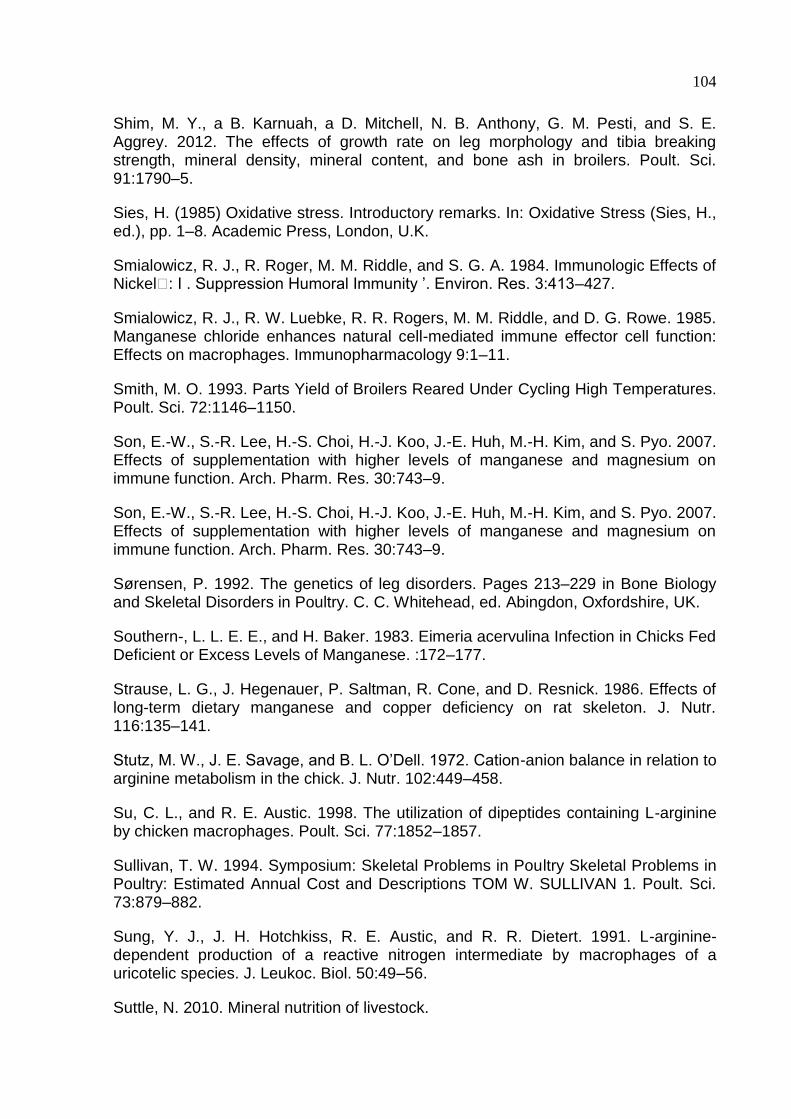
104
Shim, M. Y., a B. Karnuah, a D. Mitchell, N. B. Anthony, G. M. Pesti, and S. E. Aggrey. 2012. The effects of growth rate on leg morphology and tibia breaking strength, mineral density, mineral content, and bone ash in broilers. Poult. Sci. 91:1790–5.
Sies, H. (1985) Oxidative stress. Introductory remarks. In: Oxidative Stress (Sies, H., ed.), pp. 1–8. Academic Press, London, U.K.
Smialowicz, R. J., R. Roger, M. M. Riddle, and S. G. A. 1984. Immunologic Effects of Nickel : I . Suppression Humoral Immunity ’. Environ. Res. 3:413–427.
Smialowicz, R. J., R. W. Luebke, R. R. Rogers, M. M. Riddle, and D. G. Rowe. 1985. Manganese chloride enhances natural cell-mediated immune effector cell function: Effects on macrophages. Immunopharmacology 9:1–11.
Smith, M. O. 1993. Parts Yield of Broilers Reared Under Cycling High Temperatures. Poult. Sci. 72:1146–1150.
Son, E.-W., S.-R. Lee, H.-S. Choi, H.-J. Koo, J.-E. Huh, M.-H. Kim, and S. Pyo. 2007. Effects of supplementation with higher levels of manganese and magnesium on immune function. Arch. Pharm. Res. 30:743–9.
Son, E.-W., S.-R. Lee, H.-S. Choi, H.-J. Koo, J.-E. Huh, M.-H. Kim, and S. Pyo. 2007. Effects of supplementation with higher levels of manganese and magnesium on immune function. Arch. Pharm. Res. 30:743–9.
Sørensen, P. 1992. The genetics of leg disorders. Pages 213–229 in Bone Biology and Skeletal Disorders in Poultry. C. C. Whitehead, ed. Abingdon, Oxfordshire, UK.
Southern-, L. L. E. E., and H. Baker. 1983. Eimeria acervulina Infection in Chicks Fed Deficient or Excess Levels of Manganese. :172–177.
Strause, L. G., J. Hegenauer, P. Saltman, R. Cone, and D. Resnick. 1986. Effects of long-term dietary manganese and copper deficiency on rat skeleton. J. Nutr. 116:135–141.
Stutz, M. W., J. E. Savage, and B. L. O’Dell. 1972. Cation-anion balance in relation to arginine metabolism in the chick. J. Nutr. 102:449–458.
Su, C. L., and R. E. Austic. 1998. The utilization of dipeptides containing L-arginine by chicken macrophages. Poult. Sci. 77:1852–1857.
Sullivan, T. W. 1994. Symposium: Skeletal Problems in Poultry Skeletal Problems in Poultry: Estimated Annual Cost and Descriptions TOM W. SULLIVAN 1. Poult. Sci. 73:879–882.
Sung, Y. J., J. H. Hotchkiss, R. E. Austic, and R. R. Dietert. 1991. L-arginine-dependent production of a reactive nitrogen intermediate by macrophages of a uricotelic species. J. Leukoc. Biol. 50:49–56.
Suttle, N. 2010. Mineral nutrition of livestock.
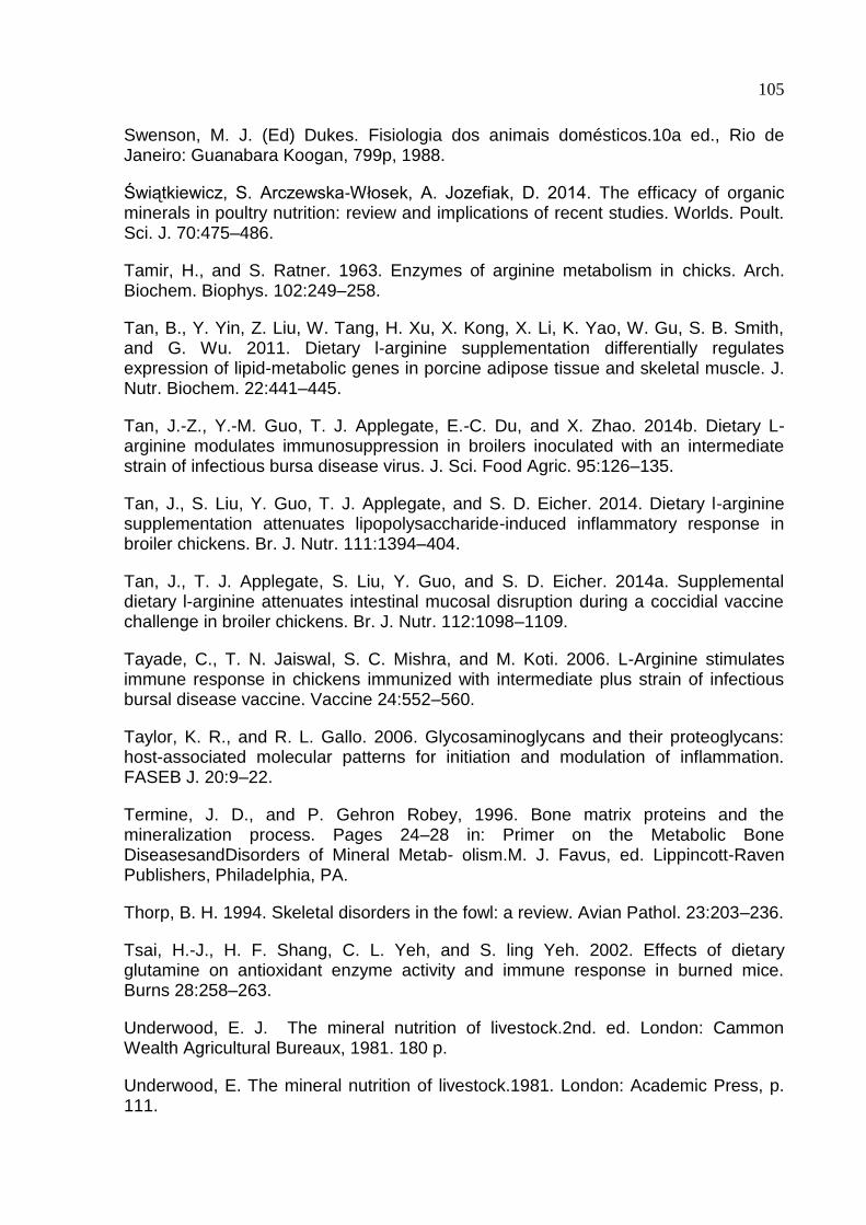
105
Swenson, M. J. (Ed) Dukes. Fisiologia dos animais domésticos.10a ed., Rio de Janeiro: Guanabara Koogan, 799p, 1988.
Świątkiewicz, S. Arczewska-Włosek, A. Jozefiak, D. 2014. The efficacy of organic minerals in poultry nutrition: review and implications of recent studies. Worlds. Poult. Sci. J. 70:475–486.
Tamir, H., and S. Ratner. 1963. Enzymes of arginine metabolism in chicks. Arch. Biochem. Biophys. 102:249–258.
Tan, B., Y. Yin, Z. Liu, W. Tang, H. Xu, X. Kong, X. Li, K. Yao, W. Gu, S. B. Smith, and G. Wu. 2011. Dietary l-arginine supplementation differentially regulates expression of lipid-metabolic genes in porcine adipose tissue and skeletal muscle. J. Nutr. Biochem. 22:441–445.
Tan, J.-Z., Y.-M. Guo, T. J. Applegate, E.-C. Du, and X. Zhao. 2014b. Dietary L-arginine modulates immunosuppression in broilers inoculated with an intermediate strain of infectious bursa disease virus. J. Sci. Food Agric. 95:126–135.
Tan, J., S. Liu, Y. Guo, T. J. Applegate, and S. D. Eicher. 2014. Dietary l-arginine supplementation attenuates lipopolysaccharide-induced inflammatory response in broiler chickens. Br. J. Nutr. 111:1394–404.
Tan, J., T. J. Applegate, S. Liu, Y. Guo, and S. D. Eicher. 2014a. Supplemental dietary l-arginine attenuates intestinal mucosal disruption during a coccidial vaccine challenge in broiler chickens. Br. J. Nutr. 112:1098–1109.
Tayade, C., T. N. Jaiswal, S. C. Mishra, and M. Koti. 2006. L-Arginine stimulates immune response in chickens immunized with intermediate plus strain of infectious bursal disease vaccine. Vaccine 24:552–560.
Taylor, K. R., and R. L. Gallo. 2006. Glycosaminoglycans and their proteoglycans: host-associated molecular patterns for initiation and modulation of inflammation. FASEB J. 20:9–22.
Termine, J. D., and P. Gehron Robey, 1996. Bone matrix proteins and the mineralization process. Pages 24–28 in: Primer on the Metabolic Bone DiseasesandDisorders of Mineral Metab- olism.M. J. Favus, ed. Lippincott-Raven Publishers, Philadelphia, PA.
Thorp, B. H. 1994. Skeletal disorders in the fowl: a review. Avian Pathol. 23:203–236.
Tsai, H.-J., H. F. Shang, C. L. Yeh, and S. ling Yeh. 2002. Effects of dietary glutamine on antioxidant enzyme activity and immune response in burned mice. Burns 28:258–263.
Underwood, E. J. The mineral nutrition of livestock.2nd. ed. London: Cammon Wealth Agricultural Bureaux, 1981. 180 p.
Underwood, E. The mineral nutrition of livestock.1981. London: Academic Press, p. 111.
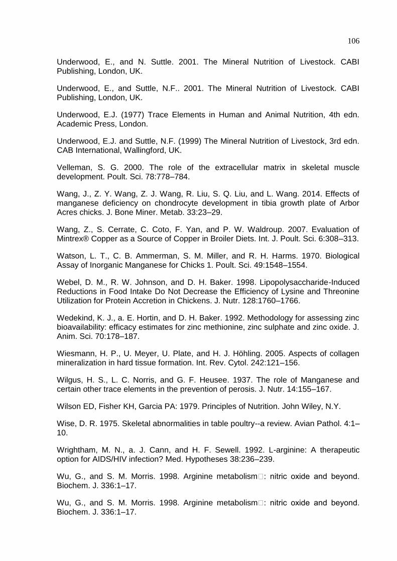
106
Underwood, E., and N. Suttle. 2001. The Mineral Nutrition of Livestock. CABI Publishing, London, UK.
Underwood, E., and Suttle, N.F.. 2001. The Mineral Nutrition of Livestock. CABI Publishing, London, UK.
Underwood, E.J. (1977) Trace Elements in Human and Animal Nutrition, 4th edn. Academic Press, London.
Underwood, E.J. and Suttle, N.F. (1999) The Mineral Nutrition of Livestock, 3rd edn. CAB International, Wallingford, UK.
Velleman, S. G. 2000. The role of the extracellular matrix in skeletal muscle development. Poult. Sci. 78:778–784.
Wang, J., Z. Y. Wang, Z. J. Wang, R. Liu, S. Q. Liu, and L. Wang. 2014. Effects of manganese deficiency on chondrocyte development in tibia growth plate of Arbor Acres chicks. J. Bone Miner. Metab. 33:23–29.
Wang, Z., S. Cerrate, C. Coto, F. Yan, and P. W. Waldroup. 2007. Evaluation of Mintrex® Copper as a Source of Copper in Broiler Diets. Int. J. Poult. Sci. 6:308–313.
Watson, L. T., C. B. Ammerman, S. M. Miller, and R. H. Harms. 1970. Biological Assay of Inorganic Manganese for Chicks 1. Poult. Sci. 49:1548–1554.
Webel, D. M., R. W. Johnson, and D. H. Baker. 1998. Lipopolysaccharide-Induced Reductions in Food Intake Do Not Decrease the Efficiency of Lysine and Threonine Utilization for Protein Accretion in Chickens. J. Nutr. 128:1760–1766.
Wedekind, K. J., a. E. Hortin, and D. H. Baker. 1992. Methodology for assessing zinc bioavailability: efficacy estimates for zinc methionine, zinc sulphate and zinc oxide. J. Anim. Sci. 70:178–187.
Wiesmann, H. P., U. Meyer, U. Plate, and H. J. Höhling. 2005. Aspects of collagen mineralization in hard tissue formation. Int. Rev. Cytol. 242:121–156.
Wilgus, H. S., L. C. Norris, and G. F. Heusee. 1937. The role of Manganese and certain other trace elements in the prevention of perosis. J. Nutr. 14:155–167.
Wilson ED, Fisher KH, Garcia PA: 1979. Principles of Nutrition. John Wiley, N.Y.
Wise, D. R. 1975. Skeletal abnormalities in table poultry--a review. Avian Pathol. 4:1–10.
Wrightham, M. N., a. J. Cann, and H. F. Sewell. 1992. L-arginine: A therapeutic option for AIDS/HIV infection? Med. Hypotheses 38:236–239.
Wu, G., and S. M. Morris. 1998. Arginine metabolism : nitric oxide and beyond. Biochem. J. 336:1–17.
Wu, G., and S. M. Morris. 1998. Arginine metabolism : nitric oxide and beyond. Biochem. J. 336:1–17.
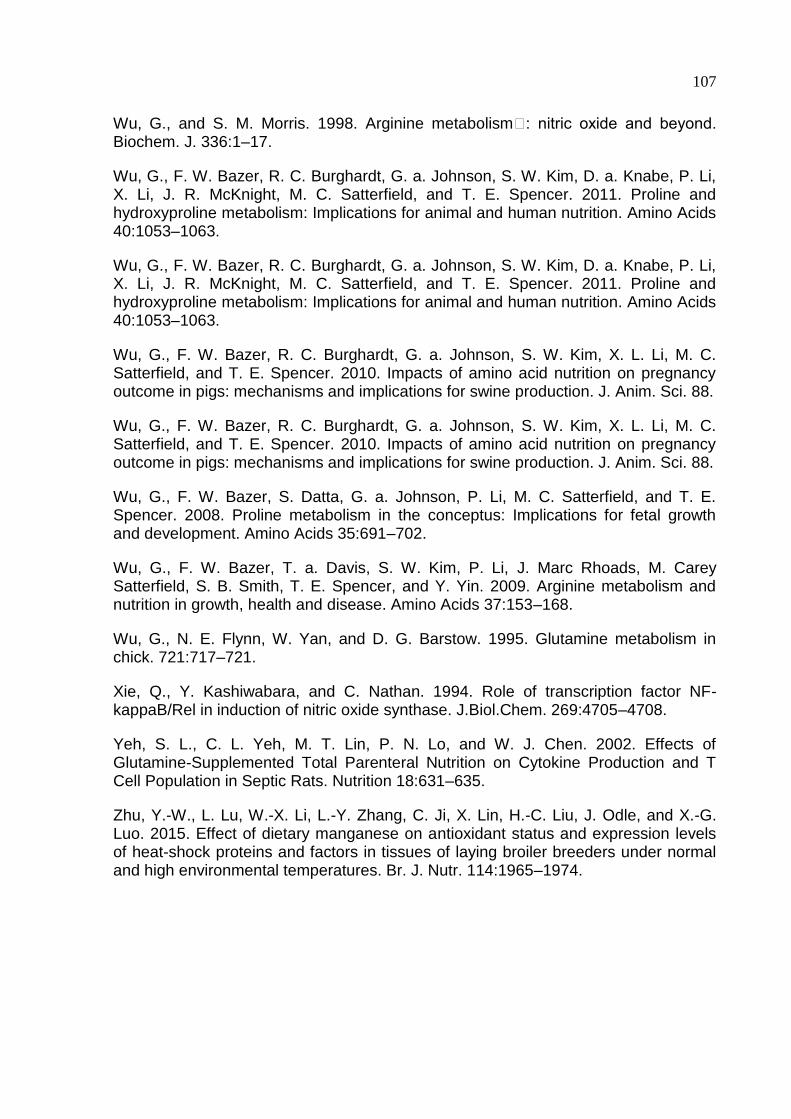
107
Wu, G., and S. M. Morris. 1998. Arginine metabolism : nitric oxide and beyond. Biochem. J. 336:1–17.
Wu, G., F. W. Bazer, R. C. Burghardt, G. a. Johnson, S. W. Kim, D. a. Knabe, P. Li, X. Li, J. R. McKnight, M. C. Satterfield, and T. E. Spencer. 2011. Proline and hydroxyproline metabolism: Implications for animal and human nutrition. Amino Acids 40:1053–1063.
Wu, G., F. W. Bazer, R. C. Burghardt, G. a. Johnson, S. W. Kim, D. a. Knabe, P. Li, X. Li, J. R. McKnight, M. C. Satterfield, and T. E. Spencer. 2011. Proline and hydroxyproline metabolism: Implications for animal and human nutrition. Amino Acids 40:1053–1063.
Wu, G., F. W. Bazer, R. C. Burghardt, G. a. Johnson, S. W. Kim, X. L. Li, M. C. Satterfield, and T. E. Spencer. 2010. Impacts of amino acid nutrition on pregnancy outcome in pigs: mechanisms and implications for swine production. J. Anim. Sci. 88.
Wu, G., F. W. Bazer, R. C. Burghardt, G. a. Johnson, S. W. Kim, X. L. Li, M. C. Satterfield, and T. E. Spencer. 2010. Impacts of amino acid nutrition on pregnancy outcome in pigs: mechanisms and implications for swine production. J. Anim. Sci. 88.
Wu, G., F. W. Bazer, S. Datta, G. a. Johnson, P. Li, M. C. Satterfield, and T. E. Spencer. 2008. Proline metabolism in the conceptus: Implications for fetal growth and development. Amino Acids 35:691–702.
Wu, G., F. W. Bazer, T. a. Davis, S. W. Kim, P. Li, J. Marc Rhoads, M. Carey Satterfield, S. B. Smith, T. E. Spencer, and Y. Yin. 2009. Arginine metabolism and nutrition in growth, health and disease. Amino Acids 37:153–168.
Wu, G., N. E. Flynn, W. Yan, and D. G. Barstow. 1995. Glutamine metabolism in chick. 721:717–721.
Xie, Q., Y. Kashiwabara, and C. Nathan. 1994. Role of transcription factor NF-kappaB/Rel in induction of nitric oxide synthase. J.Biol.Chem. 269:4705–4708.
Yeh, S. L., C. L. Yeh, M. T. Lin, P. N. Lo, and W. J. Chen. 2002. Effects of Glutamine-Supplemented Total Parenteral Nutrition on Cytokine Production and T Cell Population in Septic Rats. Nutrition 18:631–635.
Zhu, Y.-W., L. Lu, W.-X. Li, L.-Y. Zhang, C. Ji, X. Lin, H.-C. Liu, J. Odle, and X.-G. Luo. 2015. Effect of dietary manganese on antioxidant status and expression levels of heat-shock proteins and factors in tissues of laying broiler breeders under normal and high environmental temperatures. Br. J. Nutr. 114:1965–1974.


![0000091335 - Repositório Aberto da Universidade do Porto ... · possuem as enzimas necessárias para degradar completamente a arginina via arginina deiminase [22], outras apenas](https://img.document.onl/doc/110x75/5c1104b109d3f2d36d8b9c8b/0000091335-repositorio-aberto-da-universidade-do-porto-possuem-as-enzimas.jpg)