Embed Size (px)
Citation preview

Susana Filipa Garcia Ramos
Glycosylphosphatidylinositol from
Apicomplexan protozooa
Universidade Nova de Lisboa
Instituto de Higiene e Medicina Tropical
Centro de Malária e outras Doenças Tropicais, LA
2009
2
Universidade nova de Lisboa
Instituto de Higiene e Medicina Tropical
Glycosylphosphatidylinositol from
Apicomplexan protozooa
Susana Filipa Garcia Ramos
A supplementary dissertation submmited to obtain a Doctor of Philosophy degreee in Biomedical Sciences, Parasitology speciality
Supervisor: Doctor Henrique Silveira
Instituto de Higiene e Medicina Tropical, Centro de Malária e outras Doenças Tropicais
Co-Supervisor: Doctor Jean-Marc Reichhart
Institut de Biologie Moléculaire et Cellulaire, Réponse immunitaire et développment chez les insects
2009

3
Susana Filipa Garcia Ramos was awarded with a PhD
grant (SFRH/BD/12210/2003) by the Fundação para a
Ciência e Tecnologia, Ministério da Ciência Tecnologia e
Ensino Superior

4
INDEX
5
INDEX 4
ACKNOWLEDGMENTS 6
I-GLYCOSYLPHOSPHATIDYLINOSITOL ANCHORS 8
I-1 – Structure of the GPI anchor 12
I.2 – Biosynthesis of GPI-anchored proteins 14
I.3 – Functions of GPI-anchored proteins 18
I.4 – Properties of GPI-anchored proteins related to the anchor 18
I.4.1- Lipid rafts 19
I.5 – GPI anchor functions 20
I.5.1 – Signal transduction 21
I.5.2 – Cell-cell communication 21
I.5.3 – Apical targeting 22
I.5.4 – Binding to bacterial toxins 22
I.5.5 – Prion disease pathogenesis 23
I.5.6 – Immune stimulation 24
I.6 – Structural significance of the GPI-anchored 25
II – APICOMPLEXAN PARASITES 26
II.1 – Biological significance of Apicomplexan parasites 27
II.2 – Apicomplexan parasites biology 28
II.2.1 – Structure of the apical complex 29
II.3- Apicomplexan parasites GPI’s 32
II.3.1 – Plasmodium 33
III.3.1.1 – Induction of cytokine production 35
III.3.1.2 – Apoptosis promotion 36
III.3.1.3 – Induction of adhesion molecules 37
III.3.1.4 – The mosquito stage 37
III.3.1.5 – Naturally acquired immunity to PfGPI and malaria tolerance 38
II.3.2 – Toxoplasma 40
III.3.2.1 – GPI-anchored T. gondii surface proteins 41
III.3.2.2 – TgGPI induction of cytokine production 41
III.3.2.3 – Apoptosis promotion 42
II.3.3 – Other Apicomplexan parasites 43
II.3.3.1 – Eimeria 43
II.3.3.2 – Babesia 44
III – GPIS AS THERAPEUTIC AGENTS 45
IV - REFERENCES 49

6
Acknowledgments

7
I would like to sincerely thank to Prof. Dr. Henrique Silveira, for all the support and understanding,
for supervising and revising this dissertation.
I would also like to thank to Dr. Fátima Nogueira, for correcting this dissertation and for all the
suggestions, friendship and availability.

8
I
Glycosylphosphatidylinositol anchors
9
Many eukaryotic proteins are present at the cell surface embedded in the plasma membrane. For
most of them, insertion in the membrane is accomplished by an anchor composed of carbohydrates
and lipids, known as Glycosylphosphatidylinositol (GPI) anchors.
These molecules anchor several proteins to the membrane of eukaryotic cells. The anchor consists
of a glycolipid structure that is added as a post-translational modification to the C terminus of many
proteins. The lipid content of the molecule facilitates the anchoring at the phospholipid bilayer.
The GPI anchor is broadly distributed among eukaryotic organisms, including protozoa, fungi,
plants, insects and mammals (Nosjean et al., 1997). GPIs are not present in Eubacteria, but
interestingly, GPI-anchored proteins have been detected in some Archaeobacteria, such as a strain
from the genus Sulfolobus, which has been considered to be closely related to eukaryotes
(Kobayashi et al., 1997). GPI-anchored proteins are involved in several processes, and have
important roles in signal transduction, lipid raft partitioning, prion disease pathogenesis, immune
responses, cellular communication, apical membrane targeting and the pathobiology of
trypanosomal parasites (Paulick & Bertozzi, 2008).
Glycosylphosphatidylinositol anchors were first characterized approximately 20 years ago. In 1976,
Ikezawa et al. purified a phospholipase (type C – PI-PLC) enzyme from Bacillus cereus that was
found to release alkaline phosphatase from rat kidney slices. In the following years several PLCs
with similar function were purified from other bacteria, and, accordingly, other proteins were found to
be released upon treatment with these enzymes. As phospholipase enzymes were known to act
upon phosphatidylinositol molecules, the released proteins were suggested to be covalently
attached to the cell membrane via a site on the protein and a phosphatidylinositol molecule
embedded in the lipid bilayer (reviewed by Low, 1989; Figure I.1).
In 1985, Ferguson et al. found that the C terminus of the variant surface glycoprotein (VSG) of the
parasite Trypanosoma brucei contained: i) an ethanolamine amide-linked to the C-terminal
aminoacid, ii) a polysaccharide consisting of mannose, glucosamine and variable amounts of
Galactose, and iii) a phospholipid that could be degraded by bacterial PLC generating 1,2-dimyristyl
10
glycerol. At the same time, Tse et al. (1985) found that the C-terminal cysteine of Thy-1, a
glycoprotein expressed on mammalian thymocytes and the brain, was attached to a structure that
contained ethanolamine, glucosamine, galactosamine, mannose, myo-inositol, phosphate, glycerol
and stearic acid. Together with PLC studies, data obtained from these authors allowed establishing
the general structure of the GPI anchor (Figure I.1).
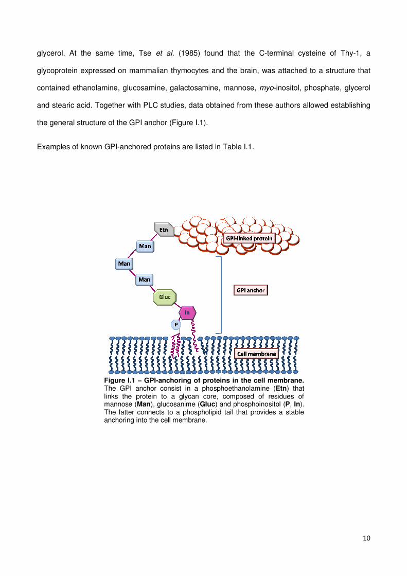
Examples of known GPI-anchored proteins are listed in Table I.1.
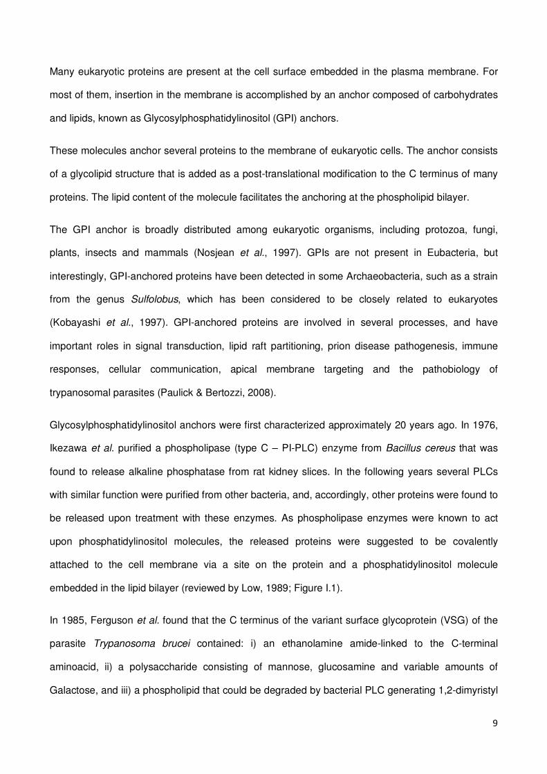
Figure I.1 – GPI-anchoring of proteins in the cell membrane. The GPI anchor consist in a phosphoethanolamine (Etn) that links the protein to a glycan core, composed of residues of mannose (Man), glucosanime (Gluc) and phosphoinositol (P, In). The latter connects to a phospholipid tail that provides a stable anchoring into the cell membrane.
11
Table I.1 Examples of GPI-anchored proteins in eukaryotes
Enzymes Alkaline phosphatase,
Acetylcholinesterase,
5’-Nucleotidase,
Alkaline phosphodiesterase I,
Trehalase,
Leishmania surface protease PSP (gp63),
Renal dipeptidase (MDP),
Aminopeptidase P,
NAD+ glycohydrolase,
Carboxipeptidase M,
Carbone anhydrase IV,
Silkworm aminopeptidase N,
ADP-ribosyltransferase,
Yeast aspartyl protease,
Chlorella nitrate reductase
Receptors Plasmodium transferrin receptor,
CD14,
CD16,
CD48,
Folate-binding protein,
Urokinase receptor,
CNTF receptor
Protozoal antigens Trypanosoma VSG and PARP (procyclin),
Toxoplasma surface antigens (P22, P30 and P43),
Giardia GP49,
Paramecium surface antigens
Mammalian antigens Thy-1,
CD55 (DAF),
Ly6 family (CD59, Ly6A/E),
Carcinoembrionic antigen (CEA),
Qa-2,
CD24
Miscellaneous Prions (PrPC, PrP
Sc),
Squid Sgp-1 and Sgp-2,
NCAM-120 (the shortest CD56), CD58 (LFA-3),
Dictyostelium Contact site A,
Mouse F3,
Chick F11,
Chicken axonin-1,
Polysphondylium GP64,
Grasshopper REGA-1
12
I.1 – Structure of the GPI anchor
The GPI anchor has a complex structure that comprises a phosphoethanolamine linker, a glycan
core, and a phospholipid tail (Figure I.1).
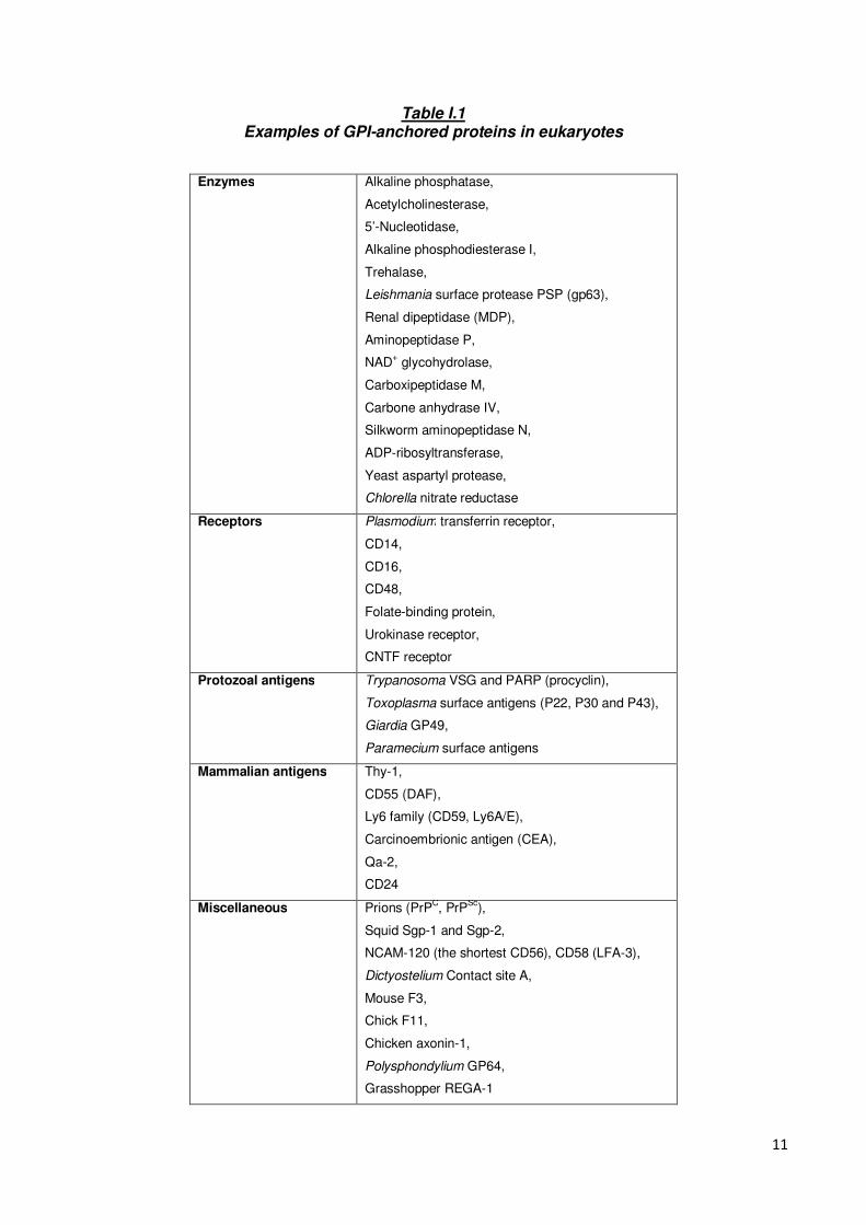
The C-terminus of a GPI-anchored protein is linked through a phosphoethanolamine bridge to the
highly conserved glycan core, comprising a mannose (α1-2), a mannose (α1-6), a mannose (α1-4),
a glucosamine (α1-6), and a myo-inositol. A phospholipid tail attaches the GPI-anchor to the cell
membrane (Figure I.2, Paulick & Bertozzi, 2008).
Figure I.2 – Structure of the GPI-anchor from the human erythrocyte acetylcholinesterase. Adapted from Paulick & Bertozzi, 2008. The GPI anchor is composed of three domains: the phosphoethanolamine linker (red), the glycan core (black) and the phospholipid tail (blue). Groups represented in blue are variable. Targets for phospholipase enzymes (PI-PLD and PI-PLC) cleavage are represented in green.
13
The phosphoinositol, glucosamine and mannose residues within the glycan core may be variously
modified with phosphoethanolamine groups and other sugars (Figure I.2), rendering high diversity to
these molecules.
The phosphoethanolamine side chain, attached to either the second or third mannose residue of the
glycan core, is only found in higher eukaryotes, not in protozoa. GPIs from mammals, in contrast to
protozoan ones, have one or two phosphoethanolamine residues, in addition to that involved in the
core structure, and one phosphoethanolamine residue at the mannose site (Brewis et al., 1995).
The most common side chains attached to the first mannose residue is another mannose. In both
mammalian and protozoa complex side chains (like N-acetylgalactosamine-containing
polysaccharides) are found, attached to the third mannose residue. The galactosamine residue in
the core is rarely modified, except for a glycoprotein of unknown function in Trypanosoma cruzi
(Macrae et al., 2005).
The lipid anchor of the phosphoinositol ring may be a diacylglycerol, an alkylacylglycerol or a
ceramide (McConville & Ferguson, 1993).
Also, many GPIs have an extra fatty acid attached to the myo-inositol ring that must contribute to
the functions of the anchor and that renders the GPI anchor resistance to bacterial PI-PLC cleavage
(Ikezawa, 2002; McConville & Ferguson, 1993; Roberts et al., 1988).
The anchor of each protein is a mixture of several GPI homologues, and as such the same protein
may be anchored by different GPI structures. For example, the VSG protein of T. brucei exhibits
microheterogeneity in the number of galactose residues in the side chain (Ferguson et al., 1988),
and the 5’-Nucleotidase of bovine liver is anchored by a mixture of 5 homologues that differ in the
substituents of the mannoside moiety (Taguchi et al., 1994).
14
I.2 – Biosynthesis of GPI-anchored proteins
The biosynthesis of GPI precursors and the post-translational protein modifications with GPI anchor
occur in the endoplasmatic reticulum (ER). During this process, the COOH terminal - (C’) of the
protein is split off and a new C’ is combined with the ethanolamine of the GPI precursor (Ikezawa,
2002). The GPI precursor linked to the protein is transported to the Golgi complex, where further
modifications in the GPI occur. After this process, GPI-anchored proteins are assumed to be
transported from Golgi to the plasma membrane in the form of lipid rafts and expressed as clusters
in the cell surface (Ikezawa, 2002; Kinoshita & Inoue, 2000).
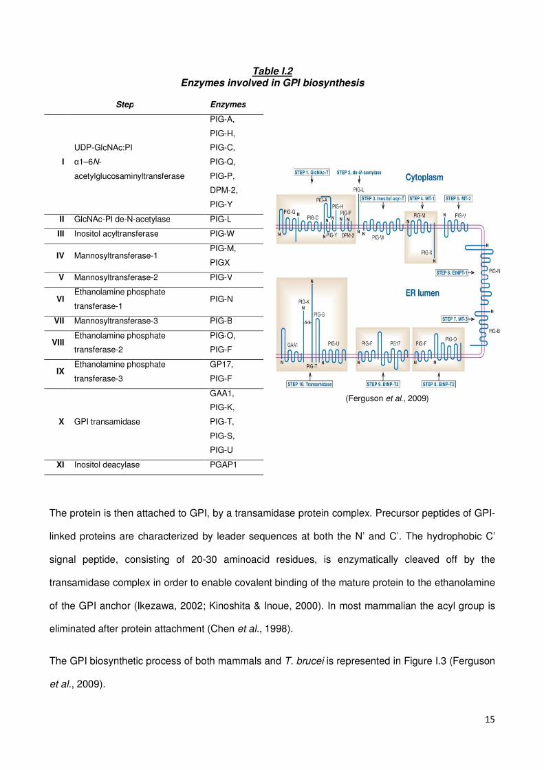
GPI biosynthesis occurs in a step-wise fashion, involving several biosynthetic steps and numerous
enzymes. Around 20 genes are involved in this process, and some steps are catalyzed by protein
complexes consisting of 4 or more enzymes, which are listed in Table I.2 (Ferguson et al., 2009).
The GPI biosynthetic process starts with enzymatic attachment of an acetylated glucosamine to a
phosphoinositol ring (generation of GlcNAc-PI from UDP-GlcNAc and phosphatidylinositol). Several
enzymes are involved in this step, acting as a complex of at least 6 proteins (Ikezawa, 2002;
Kinoshita & Inoue, 2000).
The molecule is then deacylated (generation of GlcN-PI from GlcNAc-PI), and further acylated
(acylation of the inositol ring of GlcN-PI to generate GlcN-acyl-PI). Three mannose residues are
then sequentially added to the GPI precursor. After the addition of the first mannose (generating
Man-GlcN-acyl-PI), a modification takes place, with a phosphoethanolamine side chain (generating
EtNP-Man-GlcN-acyl-PI) (Ikezawa, 2002; Kinoshita & Inoue, 2000).
Phosphoethanolamine are then added to both second and third mannose residues (generating
EtNP-Man-Man-(EtNP)Man-GlcN-acyl-PI and EtNP-Man-(EtNP)Man-(EtNP)Man-GlcN-acyl-PI).
15
Table I.2 Enzymes involved in GPI biosynthesis
Step Enzymes
(Ferguson et al., 2009)
I
UDP-GlcNAc:PI
α1–6N-
acetylglucosaminyltransferase
PIG-A,
PIG-H,
PIG-C,
PIG-Q,
PIG-P,
DPM-2,
PIG-Y
II GlcNAc-PI de-N-acetylase PIG-L
III Inositol acyltransferase PIG-W
IV Mannosyltransferase-1 PIG-M,
PIGX
V Mannosyltransferase-2 PIG-V
VI Ethanolamine phosphate
transferase-1 PIG-N
VII Mannosyltransferase-3 PIG-B
VIII Ethanolamine phosphate
transferase-2
PIG-O,
PIG-F
IX Ethanolamine phosphate
transferase-3
GP17,
PIG-F
X GPI transamidase
GAA1,
PIG-K,
PIG-T,
PIG-S,
PIG-U
XI Inositol deacylase PGAP1
The protein is then attached to GPI, by a transamidase protein complex. Precursor peptides of GPI-
linked proteins are characterized by leader sequences at both the N’ and C’. The hydrophobic C’
signal peptide, consisting of 20-30 aminoacid residues, is enzymatically cleaved off by the
transamidase complex in order to enable covalent binding of the mature protein to the ethanolamine
of the GPI anchor (Ikezawa, 2002; Kinoshita & Inoue, 2000). In most mammalian the acyl group is
eliminated after protein attachment (Chen et al., 1998).
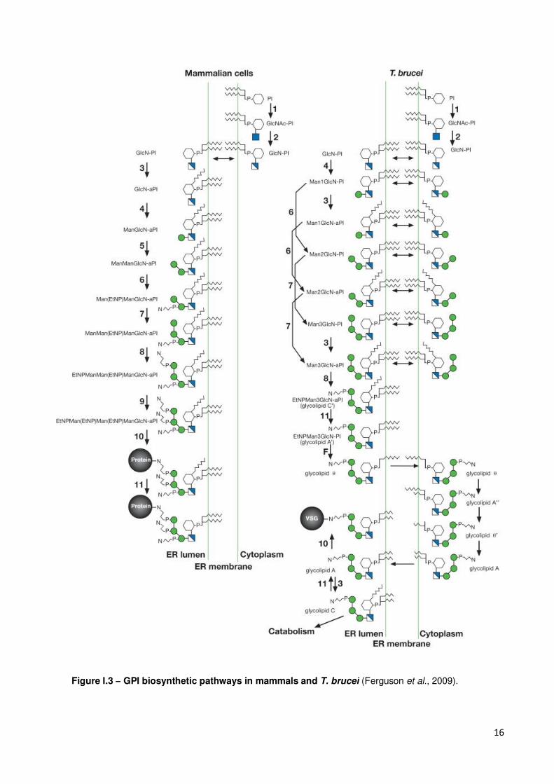
The GPI biosynthetic process of both mammals and T. brucei is represented in Figure I.3 (Ferguson
et al., 2009).
16
Figure I.3 – GPI biosynthetic pathways in mammals and T. brucei (Ferguson et al., 2009).
17
The GPI biosynthesis process differs between mammals and T. brucei in STEP 3. In mammals, the
inositol transferase acts directly on the inositol moiety of glucosamine phosphatidylinositol, then
mannosylation of the glucosamine takes place. In T. brucei mannosylation of glucosamine
phosphatidylinositol precedes to acylation of the inositol moiety. There is a unique remodeling
process in which two acyl groups in the diacylglycerol moiety of phosphatidylinositol are replaced
with myristol groups by sequential deacylation-reacylation (Ferguson et al., 2009).
Requirements for GPI attachment are not completely identical in mammalian cells and protozoan
parasites (Moran & Caras, 1994). The difference is dependent on the aminoacid triplet found at the
cleavage/attachment sites. In T. brucei, relatively larger triplets are permitted.
Proteins to be anchored are also synthesized in the ER, and have a specific C’ signal peptide for
attachment to the ER membrane. Upon synthesis, the N’ leader peptide is cleaved and the protein
remains bound to the luminal side of the ER membrane by the C’ hydrophobic peptide. The peptide
is then translocated to the site of the transamidase complex. This complex cleaves the C’
hydrophobic peptide, and binds the new C’ to the ethanolamine head of the GPI precursor.
The C’ sequence of the protein to be GPI modified comprises 4 different regions: i) an unstructured
linker region of 11 residues, flexible and polar; ii) a region of small residues that includes the
cleavage/attachment site; iii) a spacer sequence of moderately polar residues with intervening
hydrophobic residues; iv) a hydrophobic tail (Eisenhaber et al., 1998).
18
I.3 – Functions of GPI-anchored proteins
GPI-anchored proteins exist in every major cell type and tissue, and range in size from 12
aminoacid (as the glycopeptide Cd52) to 175kDa (protein CDw109).
GPI-anchored proteins display diverse biological functions. Many have enzymatic activities, some
are involved in cell-cell contact and adhesion, others are important in the regulation of the
complement cascade, and some have antigenic properties, in mammals and protozoan parasites
(Paulick & Bertozzi, 2008). In Trypanosoma brucei, the VSG protein forms a protective coat around
the parasite and acts as its major antigen (Ferguson, 1999).
Many of the GPI-anchored proteins have now been functionally characterized, but others, like the
prion protein haven’t been assigned a function yet.
Some GPI-anchored proteins are also believed to be involved in the embryonic development, as
deficiency in the PIG-A gene, involved in the first step of GPI biosynthesis is a lethal mutation.
Defects in GPI anchor biosynthesis are also lethal in yeasts (Kawagoe et al., 1996; Leidich et al.,
1994).
I.4 – Properties of GPI-anchored proteins related to the GPI-anchor
GPIs are believed to confer the attached protein some unique properties.
The anchor may affect the overall conformation of the GPI-anchored protein, either directly, or by
causing the protein to interact with the membrane. In fact, GPIs are known to alter the structure of
the attached protein. The human OX7 antibody recognizes the Thy-1 glycoprotein but binding does
19
not occur after treatment with PI-PLC (Barboni et al., 1995). Also, in T. brucei, an antibody that
binds to the GPI-anchored protein procyclin shows remarkable reduction in affinity for the same
protein lacking the lipid tail (Butikofer et al., 2001).
Another interesting property of GPI-anchored proteins is the ability to transfer spontaneously to cell
membranes both in vivo and in vitro – cell surface painting (Kooyman et al., 1995; Medof et al.,
1996). The purified human decay-accelerating factor (DAF) was shown to insert onto sheep
erythrocytes, and when exogenously added, it moves freely on the sheep cell surface and functions
normally. This has been shown to occur with many GPI-anchored proteins, and the lipid portion of
the GPI was found to be essential for cell membrane insertion (Medof et al., 1996). Transgenic mice
engineered to express specific GPI-anchored proteins in red blood cell alone were found to display
the same proteins at the surface of vascular endothelial cells of several organs in addition to blood
cells (Kooyman et al., 1995). Also, human patients infected with Trypanosoma were found to have
VSG protein form the parasite surface coat on erythrocyte membranes (Medof et al., 1996). Thus,
evidence indicates that GPI-anchored proteins have the ability to spontaneously transfer
themselves from one cell to another.
I.4.1 – Lipid rafts – a membrane anchoring spot?
GPI-anchored proteins are believed to associate with lipid rafts, membrane microdomains enriched
in glycosphingolipids, cholesterol, and certain types of lapidated proteins (Munro, 2003; Rajendran
& Simons, 2005). Lipid rafts organize the plasma membrane into a series of discrete smaller
domains that can serve as platforms for a variety of cellular functions, such as vesicular trafficking
and signal transduction.
20
Lipid rafts are hypothesized to form by self-association of sphingolipids, favored by their long and
mostly saturated hydrocarbons that allow them to pack tightly in a bilayer. Cholesterol molecules are
believed to fill the voids between the associating sphingolipids. The presence of cholesterol may be
necessary for the function and the formation of lipid rafts, as depletion of cellular cholesterol has
been shown to disrupt these rafts (Ranjendran & Simons, 2005). The tight packing of sphingolipids
suggests that lipid rafts are less fluid than the phospholipid bilayer. At the same time, it may allow
for close packing of GPI-anchored proteins, as they show the same insolubility pattern under
specific conditions as lipid rafts (Ranjendran & Simons, 2005).
The fact that signaling proteins have been associated with these lipid raft domains indicated that
GPIs may be important for signal transduction (Simons & Toomre, 2000).
I.5 – GPI-anchor functions
So far, the only confirmed role of the GPI is to provide the attached protein with a stable membrane
anchoring device that is resistant to most extracellular proteases and lipases (Low, 1989; Low &
Saltiel, 1988). As the GPI represents an extremely complex way of protein attachment to the
membrane it may serve other biological functions other than the anchoring, such as signal
transduction, cell-cell communication, apical targeting, binding to bacterial toxins, prion disease
pathogenesis and immune stimulation.
21
I.5.1 – Signal transduction
GPIs may serve as an intermediary between the exterior of a cell and internal signaling molecules
and it may also allow for signal transduction by a GPI-anchored protein. The GPI does not cross the
membrane but may physically interact with transmembrane proteins involved in intracellular
signaling (Paulick & Bertozzi, 2008).
Antibody crosslinking between GPI-anchored proteins may affect the transduction of cellular
activation or inhibition signals, resulting in Ca2+ fluxes, protein tyrosine phosphorilation or cytokine
production. These phenomena have been associated with the anchor itself, as they are not
observed when the GPI anchor is replaced by a transmembrane embedded domain (Jones &
Varela-Nieto, 1998; Robinson, 1997).
I.5.2 – Cell-cell communication
Many GPI-anchored proteins are involved in signaling and cell-cell communication, such as Decay
Accelerating Factor (DAF) and Thy-1. As the anchor confers them the ability to diffuse freely on the
cell surface, these proteins move rapidly upon external stimuli (Low & Saltiel, 1988), and this
mobility is thought to facilitate cell-cell interactions and communication.
22
I.5.3 – Apical targeting
Polarized cells have different membrane domains displaying different protein and lipid contents
allowing for specialized functions in each domain (Schuck & Simons, 2006). In these cells, the
establishment of apical and basolateral domains, separated by tight junctions, is important for
asymmetric growth, directional migration and for the transport and delivery of signals and nutrients.
Many of the GPI-anchored proteins are delivered to the apical domain, and the anchor has been
proposed to act as an apical targeting signal. Additionally, as lipid rafts may function as platforms for
the formation of apical targeting vesicles, GPIs may mediate protein-lipid raft association (Brown &
Rose, 1992; Schuck & Simons, 2006).
I.5.4 – Binding to bacterial toxins
Aerolysin is a bacterial toxin secreted by Aeromonas hydrophilia. It is a hydrophobic protein that
binds to certain sensitive cells and forms oligomers that insert into the cell membrane, forming
channels and killing the cell (Diep et al., 1998).
Thy-1 and contactin are receptors for aerolysin. Even though they are unrelated in function, they are
both GPI-anchored, which is an important determinant or aerolysin binding (Diep et al., 1998).
Some GPI-anchored proteins binds strongly to aerolysin while others do not, suggesting that the
variable regions of the anchor (as sugar and phosphoethanolamine side chains) are responsible for
this specificity.
23
The α-toxin from Clostridium septicum is also a pore-forming toxin that was shown to bind to GPI-
anchors (Gordon et al., 1999).
The fact that GPI-anchored proteins appear to concentrate in membrane microdomains such as
lipid rafts may facilitate toxin concentration and oligomerization, for membrane insertion.
I.5.5 – Prion disease pathogenesis
Prion disease is characterized by the formation of insoluble protein plaques within neurons and
related cells in the brain, which are associated with neurodegeneration (Taylor & Hooper, 2006).
Plaque formation depends on a conformational alteration of the normal cell prion protein PrPC to the
pathogenic scrapie form, PrPSC.
PrPC is a cell-surface, GPI-linked protein expressed by a variety of cell types, but being particularly
abundant in neurons (Taylor & Hooper, 2006), and thought to be involved in processes such as
oxidative stress, cell signaling, copper and zinc metabolism, and in synaptic transmission.
For conversion and disease progression, the incoming PrPSC has to be inserted into a contiguous
membrane with PrPC, in order to induce the change of PrPC into PrPSC, thus increasing PrPSC
amounts that cluster to form the plaques. Lipid rafts provide a favorable environment for
conformational conversion of PrPC to PrPSC, by concentrating the proteins within confined regions of
the membrane (Campana et al., 2005; Sarnataro et al., 2004). The conversion may affect signaling
events involving PrPC, leading to the removal of neuroprotective signals and/or the initiation of
neurotoxic signals (Aguzzi, 2005).
Mice expressing an anchorless, secreted version of PrPC never develop clinical prion disease, when
infected with PrPSC. Even though the disease was prevented, it didn’t avoid PrPSC plaque formation,
24
indicating that lack of the GPI may prevent the delivery of neurotoxic signals after PrPSC plaque
formation (Chesebreo et al., 2005). Thus, GPI anchoring of PrPC is essential for both the PrPC
functions and the disease propagation, as it ensures the signal transduction process.
I.5.6 – Immune stimulation
Cross-linking of GPI-anchored proteins on T lymphocytes (such as Thy-1, Ly-6 A/E, CD48 and
CD59), induces T-cell mitogenesis. Ligation of GPI-anchored proteins induces an intracellular flux of
Ca2+, an up-regulation of activation-associated cell surface proteins and the elaboration of growth
promoting lymphokines (Loertscher & Lavery, 2002).
Numerous T lymphocytes cell surface membrane proteins, as Thy-1, are anchored in the membrane
through a GPI (Horejsi et al., 1999). Binding of the rat mAb G7 or cross linking of other Thy-1-
specific mAbs induces an acute rise in Ca2+ concentration, IL-2 synthesis, IL-2 receptor expression
and T cell proliferation (Gunter et al., 1984; Kroczek et al., 1986).
PrPC also participates in an early event of T cell activation (Cashman et al, 1990). Thy-1 and PrPC
are both covalently bound to GPIs and both proteins concentrate in lipid rafts, but presenting a
different distribution: Thy-1 strictly co-localizes at the center of the raft, while PrPC is found in the
semi-ordered lipid domain that separated the raft from the fluid glycerolipid domain (Madore et al.,
1999).
The replacement of the GPI by a transmembrane-anchoring peptide was found to abolish the
activating or inhibitory potential of cell surface proteins, thus, the generation of the putative signal is
related to the anchor itself (Robinson et al, 1989).
25
I.6 – Structural significance of the GPI anchor
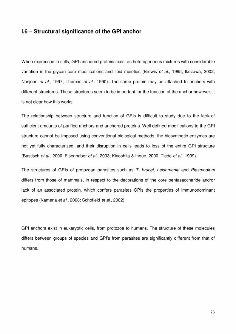
When expressed in cells, GPI-anchored proteins exist as heterogeneous mixtures with considerable
variation in the glycan core modifications and lipid moieties (Brewis et al., 1995; Ikezawa, 2002;
Nosjean et al., 1997; Thomas et al., 1990). The same protein may be attached to anchors with
different structures. These structures seem to be important for the function of the anchor however, it
is not clear how this works.
The relationship between structure and function of GPIs is difficult to study due to the lack of
sufficient amounts of purified anchors and anchored proteins. Well defined modifications to the GPI
structure cannot be imposed using conventional biological methods, the biosynthetic enzymes are
not yet fully characterized, and their disruption in cells leads to loss of the entire GPI structure
(Bastisch et al., 2000; Eisenhaber et al., 2003; Kinoshita & Inoue, 2000; Tiede et al., 1999).
The structures of GPIs of protozoan parasites such as T. brucei, Leishmania and Plasmodium
differs from those of mammals, in respect to the decorations of the core pentasaccharide and/or
lack of an associated protein, which confers parasites GPIs the properties of immunodominant
epitopes (Kamena et al., 2008; Schofield et al., 2002).
GPI anchors exist in eukaryotic cells, from protozoa to humans. The structure of these molecules
differs between groups of species and GPI’s from parasites are significantly different from that of
humans.

26
II
Apicomplexan parasites
27
The Apicomplexa comprise a large group of protozoans, characterized by the presence of an apical
complex in at least some stages of their life cycle. These organisms are unicellular, spore-forming,
unflagellated with the exception of some gametes and exclusively parasitic.
Apicomplexan parasites were first observed in 1674 by van Leeuwenhoek, who saw Eimeria stiedai
oocysts in the gall bladder of a rabbit. By 1987, more than 4000 species, belonging to over 300
genera were included in the Apicomplexa group.
The classification of parasites into the Apicomplexa phylum has been controversy, especially
because the first identification studies were based on optical microscopy.
II.1 – Biological significance of Apicomplexan parasites
Apicomplexa parasites are of high biological importance, as they are the agents of severe diseases
affecting humans and animals.
Diseases include Malaria (Plasmodium), Toxoplasmosis (Toxoplasma gondii), Cryptisporidiosis
(Cryptosporidium parvum), Cyclosporiasis (Cyclospora cayetanesis), Isosporiasis (Isospora bella)
and Babesiosis (Babesia).
Parasites from the genus Babesia and Theileria are also responsible for cattle diseases, while
Eimeria parasites affect mostly poultry.
28
II.2 – Apicomplexan parasites biology
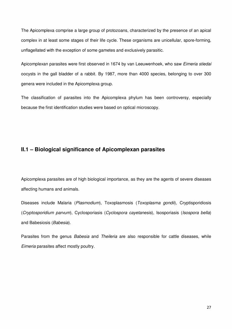
Most members of this group have complex life-cycles that involve both asexual and sexual
reproduction. In general, the invasive forms (sporozoites – Figure II.1-4) invade the host cells,
where they reproduce and multiply forming merozoites. These either engage in repeated cycles of
invasion and multiplication (Figure II.1-1) or differentiate into gametocytes (Figure II.1-2). These
initiate the sexual reproduction, forming a zygote (cyst – Figure II.1-3). The cyst then matures and
divides, forming new haploid sporozoites (Figure II.1-4).
Some variations exist in this basic life cycle according to the parasite species, including the fact that
many Apicomplexa have more than one host.
Figure II.1 – General life cycle of an Apicomplexa. From http://commons.wikimedia.org. 1 – merozoites; 2 – gametocytes; 3 – zygote (cyst); 4 – sporozoites.
29
II.2.1 – Structure of the apical complex
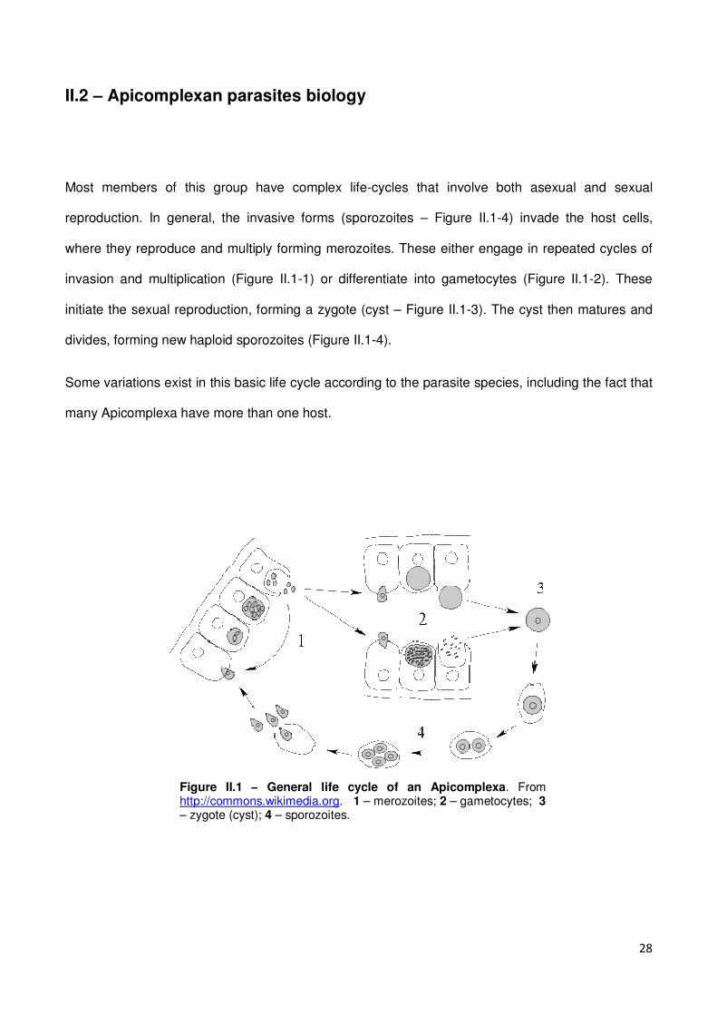
Apicomplexa are unicellular parasites whose cells are polarized. At the apical part of the cell there is
an apical complex (Figure II.2), which is a unique structure representative of these organisms. It
includes secretory organelles, such as rhoptries, micronemes and dense granules. These structures
are involved in host cell invasion, as they secrete enzymes that allow the parasite to invade new
cells. The tip is surrounded by a band of microtubules, called the polar ring. In some Apicomplexa
there is also a funnel of rods called the conoid. This is a motile structure that contains spiral fibers
that protrude during invasion. The Apicomplexa cell is surrounded by a trimembranar pellicle,
underneath which lays a microtubule network, essential for parasite motility (Soldati et al., 2004).
Figure II.2 – Apicomplexan apical complex. From Cowman & Crabb, 2006.The apical complex comprises the micronemes, rhoptries, dense granules and the polar ring.
30
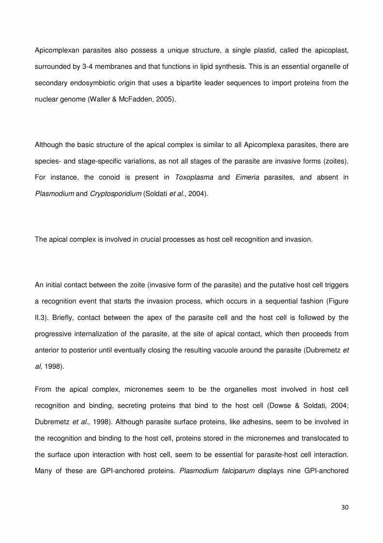
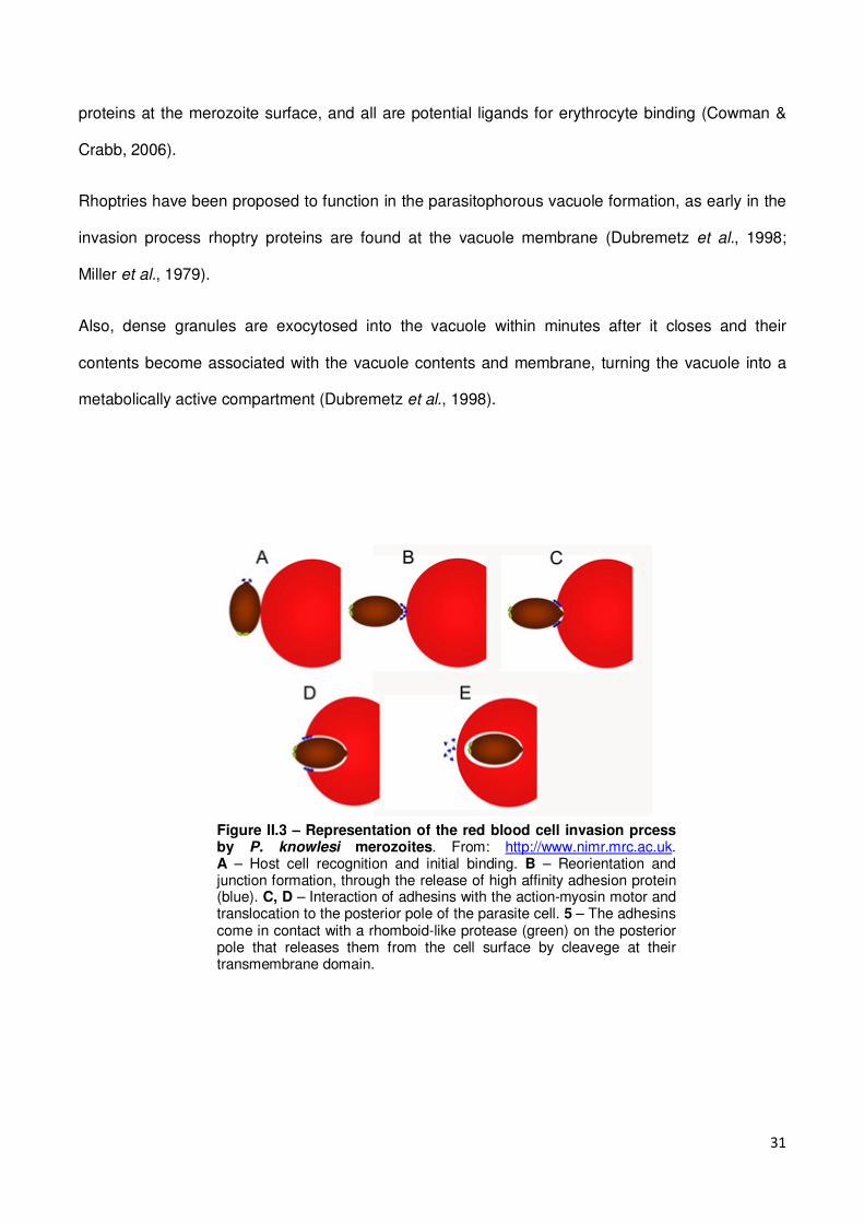
Apicomplexan parasites also possess a unique structure, a single plastid, called the apicoplast,
surrounded by 3-4 membranes and that functions in lipid synthesis. This is an essential organelle of
secondary endosymbiotic origin that uses a bipartite leader sequences to import proteins from the
nuclear genome (Waller & McFadden, 2005).
Although the basic structure of the apical complex is similar to all Apicomplexa parasites, there are
species- and stage-specific variations, as not all stages of the parasite are invasive forms (zoites).
For instance, the conoid is present in Toxoplasma and Eimeria parasites, and absent in
Plasmodium and Cryptosporidium (Soldati et al., 2004).
The apical complex is involved in crucial processes as host cell recognition and invasion.
An initial contact between the zoite (invasive form of the parasite) and the putative host cell triggers
a recognition event that starts the invasion process, which occurs in a sequential fashion (Figure
II.3). Briefly, contact between the apex of the parasite cell and the host cell is followed by the
progressive internalization of the parasite, at the site of apical contact, which then proceeds from
anterior to posterior until eventually closing the resulting vacuole around the parasite (Dubremetz et
al, 1998).
From the apical complex, micronemes seem to be the organelles most involved in host cell
recognition and binding, secreting proteins that bind to the host cell (Dowse & Soldati, 2004;
Dubremetz et al., 1998). Although parasite surface proteins, like adhesins, seem to be involved in
the recognition and binding to the host cell, proteins stored in the micronemes and translocated to
the surface upon interaction with host cell, seem to be essential for parasite-host cell interaction.
Many of these are GPI-anchored proteins. Plasmodium falciparum displays nine GPI-anchored
31
proteins at the merozoite surface, and all are potential ligands for erythrocyte binding (Cowman &
Crabb, 2006).
Rhoptries have been proposed to function in the parasitophorous vacuole formation, as early in the
invasion process rhoptry proteins are found at the vacuole membrane (Dubremetz et al., 1998;
Miller et al., 1979).
Also, dense granules are exocytosed into the vacuole within minutes after it closes and their
contents become associated with the vacuole contents and membrane, turning the vacuole into a
metabolically active compartment (Dubremetz et al., 1998).
Figure II.3 – Representation of the red blood cell invasion prcess by P. knowlesi merozoites. From: http://www.nimr.mrc.ac.uk. A – Host cell recognition and initial binding. B – Reorientation and junction formation, through the release of high affinity adhesion protein (blue). C, D – Interaction of adhesins with the action-myosin motor and translocation to the posterior pole of the parasite cell. 5 – The adhesins come in contact with a rhomboid-like protease (green) on the posterior pole that releases them from the cell surface by cleavege at their transmembrane domain.
32
Another intriguing property of the Apicomplexa is a substrate-based gliding motility that has been
found to be essential for migration through biological barriers and for host-cell invasion and egress.
Some GPI-anchored adhesive proteins released, as TRAP in Plasmodium sporozoites and its
homologue TgMIC2, in Txoxoplasma gondii, were found to be essential for the invasion processes
(Keeley & Soldati, 2004). These proteins are thought to bind to target cells, as the actin-myosin
motor functions, driving the parasite forward, or into the target cell (Baum et al.¸2008).
III.3 – Apicomplexan parasites GPI’s
Apicomplexa parasites have surface proteins anchored by GPI’s, many of which are essential for
parasite survival and infectivity.
GPI’s from protozoans differ from those found on higher eukaryotes, in both structure and number.
Protozoans seem to use these anchorage molecules much more often than higher eukaryotes. In
fact most of the proteins existing at the surface of protozoans are GPI-anchored, rather than
transmembrane proteins. These anchored proteins have diverse functions, like surface coating
proteins, hydrolases and receptors. Additionally some are known to act in parasite protection or
specific host-parasite interactions (McConville & Ferguson, 1993).
Structurally, all GPI anchors characterized contain an identical ethanolamine-phosphate-Manα1-
2Manα1-6Manα1-4GlcNα1-6myo-inositol backbone. GPIs of higher eukaryotes are substituted with
one or two additional ethanolamine phosphate residues, which are not observed in protozoans.
Another difference between protozoan and higher eukaryotes GPIs is that in the latter it was shown
that GPI-anchored proteins accumulate in membrane microdomains as lipid rafts, while in lower
33
eukaryotes, there is no direct evidence that GPI-anchored proteins associate with membrane
microdomains (McConville & Ferguson, 1993).
An interesting feature of some protozoal GPI anchors is that they are able to exist in a protein free
state, while mammalian ones do not.
Apart from its role in protein anchoring, parasite GPIs have been found to have properties that
contribute to determine the outcome of infection, in particular due to their immunogenic
characteristics.
III.3.1 – Plasmodium
Plasmodium parasites cause the disease broadly known as malaria. There are around 200 species
of Plasmodium parasites that infect animals such as birds, reptiles and mammals, most specially
rodents and humans.
The parasite has a life cycle that alternates between a mosquito vector and a vertebrate host. The
disease is caused by the repeated cycles of asexual reproduction in red blood cells, causing
symptoms such as anemia, renal failure, fever, and so on. The deadliest form of malaria is caused
by the human-malaria parasite P. falciparum, which induces in the parasitized red blood cells the
ability to adhere to endothelial tissue, as the walls of blood capillaries. If this should happen in the
brain, oxygen delivery to brain cells is hindered, and the person develops a coma that leads to
death.
34
GPI anchors represent the major carbohydrate modification in intraerythrocytic P. falciparum
parasites (Gowda et al., 1997), and are produced in excess of what is necessary for protein
attachment to the cell membrane (Gerold et al., 1994).
P. falciparum synthesizes 2 forms of mature GPI anchors: a Man4-GlcN-Pi and a Man3-GlcN-Pi, in
which the Pi is a 1,2-diacylglycerol and acylated inositol residues (Gerold et al., 1994). Even though
the intraerythrocytic stages of the parasite use both forms to anchor proteins (Schmidt et al., 1998),
the GPI-anchored surface proteins of P. falciparum MSP-1 and MSP-2 are anchored exclusively by
Man4-GlcN-Pi anchors (Gerold et al., 1996). Nevertheless, the parasite uses indiscriminately 5
existing different structures of the Man4-GlcN-Pi PfGPI from for protein attachment (Naik et al.,
2000a).
P. falciparum GPI (PfGPI) biosynthesis was shown to be essential for parasite survival, and to occur
in a stage-specific manner (Naik et al., 2000a). Data available on PfGPI biosynthesis revealed that
in intraerythrocytic stages it occurs exclusively during maturation of early trohpozoites to late
trohpozoites. Interference of PfGPI biosynthesis by glucosamine results in parasite growth inhibition
in a dose-dependent manner, arresting trophozoite maturation (Naik et al, 2003).
Besides the vital role of PfGPI as protein anchor, these molecules show an interesting feature, as
they are able to immune-stimulate the vertebrate host thus contributing to the disease.

35
III.3.1.1 – Induction of cytokine production
PfGPIs have the ability to induce proinflammatory cytokine production by macrophages, causing
symptoms that resemble malaria illness (Nebl et al., 2005), and were suggested to be the dominant
parasite components responsible for malaria pathogenesis. Schizont rupture leads to the release of
parasite products that trigger the inflammatory cascade, resulting in acute febrile episodes (Clark et
al., 1981), suggesting that clinical manifestations of malaria may be mediated in part by the
overproduction of proinflammatory cytokines (Miller et al., 1994). Soluble parasite products were
shown to activate macrophages to release TNF-α and IL-1, and PfGPI was found to be the principle
mediator of this response (Schofield & Hackett, 1993).
The anchor itself stimulates the macrophages to produce TNF-α, IL-1 and IFN-γ and causes
symptoms like pyrexia and hypoglycemia. The latter is related to the ability of PfGPIs to mimick the
action of insulin (Elased et al., 2001).
Additionally, the endotoxin activity of major P. falciparum surface antigens (MSP-1 and MSP-2),
released upon schizont rupture, is restricted to the GPI moiety. While protein denaturation or
digestion does not affect cytokine production, treatment with the GPI cleaving Phospholipase A2
(PLA2) and others leads to a decrease of most of the proinflammatory activity (Schofield & Hackett,
1993). The bioactivity of purified GPI’s is comparable to that obtained in vivo with live parasites
(Schofield & Hackett, 1993; Schofield et al., 1996).
GPIs from P. falciparum were found to act both against and as a substrate for cell signaling: while
the glycan core activates the Protein Tyrosine Kinase (PTK) signaling, the lipid moiety acts as a
second messenger for Protein Kinase C (PKC) activation (Tachado et al., 1997). Although the
glycan moiety can by itself activate PTK signaling, it is insufficient to induce cytokine production.
The lipid moiety is only able to activate when linked to the glycan core. Thus, the simultaneous
36
activation of PTK and PKC signaling leads to the activation of transcription factors of the NF-kB
family, and production of proinflammatory cytokines and nitric oxide (NO).
In fact, proinflammatory cytokine induction by PfGPI was found to require the simultaneous
recognition of the glycan core and the lipid moiety (Vijaykumar et al., 2001).
GPIs from P. falciparum are recognized by Toll-Like Receptors (TLRs), namely, TLR2, and activate
cytokine production via the intracellular adaptor MyD88 (Krishnegowda et al., 2005), or by lectin-like
receptors (Klabunde et al., 2002). Recognition of PfGPI by the receptors triggers signaling pathways
that lead to the induction of TNF-α, IL-6, IL-12 and NO (Zhu et al., 2005). Additionally, Lu et al
(2006) reported phosphorilation of ERK1/2, JNK, p38, c-Jun and of the activating transcription
factor-1, involved in immune signaling transduction, in PfGPI-stimulated macrophages, indicating
that other pathways, such as JNK pathway, are activated in response to PfGPIs may contribute to
TNF-α production in macrophages.
III.3.1.2 – Apoptosis promotion
Apoptosis also seems to play a role in malaria pathogenesis, as infection increases the number of
apoptotic cells in the spleen, liver and astrocytes (Guha et al., 2006; Helmby et al., 2000; Potter et
al., 2006). Even though the numbers of apoptotic lymphocytes is similar in healthy and malaria
infected patients, the number of apoptotic cells is higher in short-term cultures of lymphocytes from
P. falciparum-infected patients (Balde et al., 1995; Riccio et al., 2003). Additionally, cytoadherence
of infected red blood cells induces the apoptosis of endothelial cells, which may also contribute to
cerebral malaria.
PfGPIs induce cardiomyocyte apoptosis in vitro and limited apoptosis in mouse liver and spleen
tissue, when injected in vivo. However, it is not known if this is a direct effect of these molecules, or
a result of TNF-α and NO production (Wennicke et al., 2008; Wichmann et al., 2007). Nonetheless,
37
the fact that PfGPIs up-regulate apoptotic genes and a myocardium damage marker suggests that it
may have a direct effect on cardiomyocyte apoptosis, which in turn, may contribute to the lethality of
malaria, as some malaria-infected patients, presenting fulminant heart failure show typical signs of
cardiac myocyte apoptosis (Wennicke et al., 2008).
III.3.1.3 – Induction of adhesion molecules
PfGPIs induce the expression of host adhesion molecules (ICAM-1, VCAM-1 and E-selectin) and
NO production in vascular endothelial cells, in a cytokine-independent manner, not requiring TNF-α
or IL-1 (Schofield et al., 1996; Tachado et al., 1996). NO production induced by PfGPIs involves
PTK, PKC and NF-kB/c-Rel, and is blocked by anti-PfGPI antibodies (Tachado et al., 1996).
Adhesion molecules are of critical importance in malaria pathogenesis, as they allow for infected red
blood cell sequestration in vascular endothelium, as consequently, to organ failure and cerebral
malaria.
III.3.1.4 – The mosquito stage
Also in the mosquito stage, PfGPIs were showed to induce strong expression of several
antimicrobial peptides and the production of NO (important for the midgut epithelium immune
response to the parasite) and to dramatically reduce the number of mosquito eggs (Arrighi et al.,
2009; Akman-Anderson et al., 2007; Lim et al., 2005).
38
III.3.1.5 – Naturally acquired immunity to PfGPI and malaria tolerance
PfGPIs seem to act as major determinants in immune response to malaria, leading to several of the
known malaria-associated clinical manifestations. In a mouse model PfGPIs were shown to have
several key properties of a toxin, as they are able to induce TNF-α, IL-1 and NO production by
macrophages (Schofield & Hackett, 1993; Tachado et al., 1996), to up-regulate the expression of
adhesion molecules in endothelial cells (Schofield et al., 1996), and to induce aberrant regulation of
temperature and hypoglycemia, and cause severe anemia and cachexia (Elased et al., 2001;
Schofield & Hackett, 1993). All these activities were blocked with anti-PfGPI antibodies that were
associated with protection from malarial acidosis, pulmonary edema, cerebral syndrome and fatality
caused by infection with P. berghei (Schofield et al., 2002).
These and other studies indicate that pro-inflammatory cytokine responses may be partially
responsible for many of the clinical manifestations of severe malaria (Kwiatkowski et al., 1990,
1993; Lyke et al, 2004) and that immune responses against PfGPI may offer protection against
symptomatic malaria in humans, known as malaria tolerance (Keenihan et al., 2003).
Several field studies show that malaria infected individuals have anti-PfGPI antibodies, whose
prevalence and concentration increase with age, and that this response may attenuate malaria
symptoms. Highly purified PfGPIs elicited a specific IgM and IgG response and these antibodies
were found in the plasma of adults and children exposed to endemic or seasonal P. falciparum
malaria (Boutlis et al., 2002; Naik et al., 2000b; de Souza et al., 2002).
A study that aimed at determining the anti-PfGPI antibodies load on Javanese children and adults
that migrated to Papua, a malaria endemic region in Indonesia, revealed that anti-PfGPI IgGs
peaked in children only after 3-4 malaria infections, while in adults peaked after the first infection.
Thus, adults seem more likely to show a persistent high response. The few children that showed a
strong response were less likely to experience symptoms with consequent parasitemia (Keenihan et
al., 2003).
39
Neutralization of PfGPIs by antibodies was also shown to reduce the severity of clinical attacks of
malaria in the Gambia. Malaria exposed individuals had higher levels of anti-PfGPI antibodies at the
end of the malaria transmission season, and were higher in children with asymptomatic infections.
Although antibodies seemed to be rapidly boosted by clinical malaria infection, children under 2
years old were seronegative for anti-PfGPI antibodies, even during an acute infection. Nonetheless,
this population showed relatively low levels of anti-PfGPI antibodies, that are rather short-lived,
indicating that the provided immunity to clinical symptoms may be only transient (de Souza et al.,
2002).
In a Kenyan population, malaria-immune adults maintained high levels of anti-PfGPI antibodies,
while children at risk of developing clinical malaria and anemia had lower antibody levels (Naik et
al., 2000b). This study established an association between anti-PfGPI levels and protection from
severe malaria anemia and fever in children.
A study in Senegal associated the anti-PfGPI response to protection against cerebral malaria, as
patients with confirmed cerebral malaria had significantly lower levels of anti-PfGPI IgGs than those
with mild malaria (Perraut et al., 2005).
In Mandang, Papua New Guinea, a region where malaria transmission is intense, the prevalence
and concentration of anti-PfGPI antibodies also increased with age in asymptomatic children and
adults (Boutlis et al, 2002). The antimalarial treatment reduced the anti-PfGPI antibody response in
children and adolescents within 6 weeks. PfGPI elicited both IgG and IgM responses. While the IgG
response is more persistent in adults and of the IgG3 subclass (Boutlis et al., 2003), IgM response
is generally of shorter duration and coincides with infection.
All these studies indicate that natural immunity and tolerance to malaria in endemic or seasonal
malaria regions includes the development of anti-PfGPI antibodies, which levels increase with age
of the host, and that are in fact associated with the attenuation of the disease.
40
III.3.2 – Toxoplasma
The medical significant species of this genus is Toxoplasma gondii that causes a disease called
toxoplasmosis. This parasite has the cat as its definite host, but it can be carried by nearly all warm-
blooded animals (Montoya & Liesenfeld, 2004).
Acute infections can be asymptomatic, but often give flu-like symptoms in the early-acute stages,
that fade in a few days/months, when the parasite enters in a latent stage. Although this stage is
mainly asymptomatic, it can develop in immunocompromised patients, as HIV infected people,
causing severe encephalitis that leads to death (Luft & Remington, 1992). In pregnant women T.
gondii infection can cause congenital infectious diseases (Desmonts et al., 1981).
In the developmental stage in which virulent tachyzoites differentiate into bradyzoites, where
disease propagation is observed, there is an overexpression of free GPIs from T. gondii (TgGPIs)
and tachyzoites-specific GPI-anchored proteins (Azzouz et al., 2006).
TgGPIs are essential for parasite viability (Wichroski & Ward, 2003), and exist in two precursor
forms, both of which are able to bound to proteins in animal cells (Zinecker et al., 2001), while in
human cells, one of them remains in the free, unbound form. This precursor has an additional
glucose residue linked to the N-acetylglucosamine side branch and has immunogenic properties, as
the ability to induce high levels of TNF-α production (Azzouz et al., 2006; Striepen et al., 1997).
41
III.3.2.1 – GPI-anchored T. gondii surface antigens
In the host, T. gondii parasites interact with the immune system, and the initial interaction is
achieved by the parasite’s surface antigens. T. gondii has a superfamily of GPI-anchored antigens
that includes the surface antigen (SAG) family and the SAG-unrelated antigen (SUSA) family
(Boothroyd et al., 1997; Jung et al. 2004; Lekutis et al., 2001). It has been proposed that SAG
proteins could regulate virulence by eliciting the host immune response, be involved in immune
evasion, and participate in invasion of a variety of host cells. SUSA proteins (SUSA1 and SUSA2)
were found to be highly expressed in the chronic form of the parasite, as well as to interact with the
host immune system (Pollard et al., 2008).
III.3.2.2 – TgGPI induction of cytokine production
Apart from surface antigens, the GPI anchor itself seems to elicit immune responses from the host.
TgGPIs dominate the surface of T. gondii tachyzoites and were shown to be involved in the
pathogenicity of the parasite infection. TgGPIs are able to activate tyrosine kinase in macrophages
(Debierre-Grockiego et al., 2007a), and to induce TNF-α production from macrophages, through the
activation of the transcription factor NF-kB (Debierre-Grockiego et al., 2003). The glycan cores and
lipid moieties from the anchor were sufficient to stimulate macrophages.
MyD88, an adaptor molecule in the TLR signaling for NF-kB activation was proved to be essential
for the response to T. gondii (Scanga et al., 2002). Additionally, in hamsters, T. gondii isolated GPI
anchors were found activate the immune receptor TLR4, while the glycan core and the lipid moieties
activate both TLR2 and TLR4 in vivo (Debierre-Grockiego etal., 2007a). Deficiency on MyD88
impairs TNF-α production by macrophages upon stimulation with T. gondii GPI anchors, confirming
that this immunogenicity depends on TLR recognition of the TgGPI, MyD88 signaling and NF-kB-
dependent transcription of TNF-α.
42
Debierre-Grockiego and co-workers (2009) also found that the GPI anchor from T. gondii up-
regulates MHC class I and class II molecules on the surface of both unstimulated and IFN-gamma-
stimulated primary murine macrophages, thus increasing antigen presentation to CD8(+)
lymphocytes.
III.3.2.3 – Apoptosis promotion
T. gondii is able to promote and inhibit apoptosis in the infected cells. It can render the infected cell
resistant to programmed cell death induced by several apoptotic signals (Carmen et al., 2006;
Goebel et al., 1999, 2001; Luder & Gross, 2005; Nash et al., 1998; Orlofsky et al., 1999, 2002;
Payne et al., 2003). The increased apoptosis of immune cells after T. gondii infection may suppress
the response to the parasite (Khan et al., 1996; Mordue et al., 2001; Wei et al., 2002).
As high levels of apoptosis are accompanied by hyperinduction of proinflammatory cytokines,
TgGPIs were proposed to contribute to the development of apoptosis. TgGPI failed to block
apoptosis induction in human-derived cells. Interestingly it also did not promote apoptosis, since
markers of apoptosis, as caspase 3/7 activation, phosphatidylserine expression at the cell surface
or DNA strand breaks were not observed in the presence of TgGPIs, suggesting that these are not
involved in survival or apoptosis of host cells (Debierre-Grockiego et al., 2007b).
43
III.3.3 – Other Apicomplexan parasites
Not much data is yet available regarding GPIs of other Apicomplexan parasites. Some studies
performed so far focus on GPI-anchored proteins identified, and only a few on the properties of the
anchor itself.
III.3.3.1 – Eimeria
This is the genera of the Apicomplexa that comprises more species. These parasites infect the
gastrointestinal tract of a broad range of vertebrate animals, including birds such as chickens and
mammalians like goats and rodents.
As in T. gondii, the surface of Eimeria parasites is coated with a family of developmentally
expressed GPI-anchored surface antigen (SAG) proteins that are involved in processes such as
invasion and are most likely recognized by the host immune system. Surface GPI-anchored proteins
are differentially expressed in the different invasive stages (oocyst, sporozoite, merozoite) in a way
that is not related to antigen-switching but rather to a co-expression of the TgGPI-anchored proteins
in each parasite (Tabarés et al., 2004). Apart from this, not much is known about the GPI-anchors
and GPI-anchored proteins of Eimeria parasites, and the role they play in infection.
44
III.3.3.2 – Babesia
Babesia parasites infect blood cells and are characterized by their round, rod or abstract shape and
lack of any mobility structures, as cilia or flagella. This group includes several species, many of
which can infect humans, causing Babesiosis, although this disease occurs mainly in cattle. These
parasites are transmitted by ticks.
In cattle, Babesia bovis parasites stimulate the inducible nitric oxide synthase (iNOS) transcription
and NO production by peripheral blood macrophages (Stich et al., 1998), as well as the production
of inflammatory and regulatory cytokines, such as IL-1β, IL-12 and TNF-α (Shoda et al., 2000).
A lipid fraction of B. bovis infected erythrocytes also induces NO production by bovine
macrophages, indicating that GPI molecules from this parasite may signal for immune responses in
the host (Shoda et al., 2000).
Data obtained so far in Apicomplexa parasites GPIs have focused mainly in Plasmodium and
Toxoplasma, due to their medical significance. Nonetheless, GPIs seem to be important in general
for Apicomplexa parasite survival, and GPI-anchored proteins have been identified in a few more
parasites. Further studies should provide more information about the role of these molecules in the
infection by these parasites.

45
IV
GPI anchors as therapeutic targets
46
The Apicomplexa parasite to which most attention has been paid in terms of therapeutics and
vaccine development has been the malaria causing Plasmodium. However, antigenic-based
vaccination for parasite clearance has proved to be difficult and in most cases, ineffective, which
may increase the selective pressure upon the parasite that is able to mount escape mechanisms,
such as antigenic variation, immune evasion and others. Thus, inducing sterilizing immunity does
not seem to be a feasible objective for anti-malaria vaccination that should rather aim to prevent or
reduce morbidity and mortality (Schofield, 2007).
An alternative strategy is based on anti-toxic vaccination, as the development of anti-toxic vaccines
should confer immunization against parasite products that cause host pathology, and avoiding
parasite killing, which might not be a necessary objective. Also, malaria morbidity and fatalities were
shown to have a toxic basis, which argues in favor of the development of anti-toxic vaccines
(Schofield, 2007).
Malaria toxin-induced cytokines (TNF-α, IL-1 and IL-6) may contribute to a systemic anti-
inflammatory cascade which results in malaria-associated symptoms, as renal failure and
pulmonary edema, shock, etc. Additionally, organ-specific disease may result from local release of
toxin by sequestered parasites (Schofield, 2007). Thus, preventing toxin-induced symptoms should
help alleviate the burden of the disease.
PfGPIs are parasite toxins that were shown to be the major TNF-α and IL-1 inducer, sufficient to kill
mice when inoculated (Schofield & Hackett, 1993). Also, mice immunized against Man4-PfGPIs
were protected from severe malarial pathology, including cerebral syndrome, acidosis, pulmonary
edema and fatality (Schofield et al., 2002), thus revealing in an animal model the efficacy of a
prototype anti-toxic vaccine.
In human populations from malaria endemic and seasonal areas, malaria exposed individuals were
shown to carry anti-PfGPI antibodies in the serum, indicating that naturally acquired immunity to
malaria involves the recognition of parasite GPIs and the subsequent establishment of anti-PfGPI
response. Importantly, these antibodies were frequently associated with disease protection, and, up
47
to date, most data associate anti-PfGPI antibodies to clinical protection against malarial anemia
(Keenihan et al., 2003; Naik et al., 2000b; Perraut et al., 2005; de Souza et al., 2002).
It is important that vaccination strategies do not impair the development of naturally acquired
immunity.
Naturally acquired immunity to malaria ameliorates the clinical impact of infection and maintains the
host/pathogen balance, although it does not confer resistance to infection (Schofield, 2007).
Accordingly, mechanisms of acquired immunity to the disease are set both earlier and more easily
than anti-parasite immunity that is difficult to achieve and partially effective, even though ongoing
exposure to the parasite is necessary to maintain immunity (Schofield & Mueller, 2006).
Blocking PfGPI toxicity was shown to protect the host from the disease without killing the parasite,
which also contributes to reducing the selective pressure upon the parasite (Schofield, 2007).
These parasite toxins are responsible for proinflammatory cytokine induction and cytoadherence
promotion, thus contributing to several malaria-associated symptoms. The fact that individuals
tolerant to malaria disease were shown to have an anti-GPI based naturally acquired immune
response to malaria and that blocking of GPI toxicity reduces the severity of malaria-associated
symptoms suggest that parasite GPIs are good candidates for therapeutics and vaccine
development. Also as anti-GPI responses were shown not to interfere with parasitemia indicates
that an anti-GPI-based vaccination strategy would not impose an extended selective pressure upon
the parasite, which is important to avoid vaccination failure.
Based on these data, Schofield (2007) proposed a strategy based on conjugating chemically
synthetic PfGPI glycan to an immunogenic carrier protein of parasite or bacterial origin.
48
GPIs exist in the surface of eukaryotic cells, being widely distributed among the Eukarya Kingdom.
These molecules anchor proteins with diverse functions, including enzymes, receptors, surface
antigens and others, thus being essential for numerous processes vital for survival. Even though its
basic structure is similar in all molecules characterized so far, several differences, especially
regarding the modifications in the side chains of the glycan core and the lipid moiety were found in
GPI molecules. Different structures are present in the same organism, but critical differences are
found between organisms, especially between higher eukaryotes and protozoans. These
differences extend also to the biosynthetic pathway, indicating that these molecules and their
biosynthesis may provide suitable therapeutic targets for controlling protozoan parasites infections.
Particularly in malaria disease, naturally acquired immunity and tolerance to the disease is, at least
in part, associated with anti-PfGPIs antibodies. These attenuate the clinical manifestations of the
disease without affecting the parasite development, thus reducing the selective pressure upon the
parasite and consequently decreasing selection of mutant parasites. Anti-GPI vaccination strategies
thus promise a more reliable and effective protection than antigen-based vaccines.

49
V
References
50
Aguzzi,A. (2005) Cell biology. Prion toxicity: all sail and no anchor. Science 308 (5727), 1420-1421.
Akman-Anderson,L., Olivier,M., and Luckhart,S. (2007) Induction of nitric oxide synthase and
activation of signaling proteins in Anopheles mosquitoes by the malaria pigment, hemozoin.
Infect.Immun. 75 (8), 4012-4019.
Arrighi,R.B., Debierre-Grockiego,F., Schwarz,R.T., and Faye,I. (2009) The immunogenic properties
of protozoan glycosylphosphatidylinositols in the mosquito Anopheles gambiae. Dev.Comp
Immunol. 33 (2), 216-223.
Azzouz,N., Shams-Eldin,H., Niehus,S., Debierre-Grockiego,F., Bieker,U., Schmidt,J., Mercier,C.,
Delauw,M.F., Dubremetz,J.F., Smith,T.K., and Schwarz,R.T. (2006) Toxoplasma gondii grown in
human cells uses GalNAc-containing glycosylphosphatidylinositol precursors to anchor surface
antigens while the immunogenic Glc-GalNAc-containing precursors remain free at the parasite cell
surface. Int.J Biochem.Cell Biol 38 (11), 1914-1925.
Balde,A.T., Sarthou,J.L., and Roussilhon,C. (1995) Acute Plasmodium falciparum infection is
associated with increased percentages of apoptotic cells. Immunol.Lett. 46 (1-2), 59-62.
Barboni,E., Rivero,B.P., George,A.J., Martin,S.R., Renoup,D.V., Hounsell,E.F., Barber,P.C., and
Morris,R.J. (1995) The glycophosphatidylinositol anchor affects the conformation of Thy-1 protein. J
Cell Sci 108 ( Pt 2) 487-497.
Bastisch,I., Tiede,A., Deckert,M., Ziolek,A., Schmidt,R.E., and Schubert,J. (2000)
Glycosylphosphatidylinositol (GPI)-deficient Jurkat T cells as a model to study functions of GPI-
anchored proteins. Clin.Exp Immunol. 122 (1), 49-54.
Baum,J., Gilberger,T.W., Frischknecht,F., and Meissner,M. (2008) Host-cell invasion by malaria
parasites: insights from Plasmodium and Toxoplasma. Trends Parasitol. 24 (12), 557-563.
51
Boothroyd,J.C., Black,M., Bonnefoy,S., Hehl,A., Knoll,L.J., Manger,I.D., Ortega-Barria,E., and
Tomavo,S. (1997) Genetic and biochemical analysis of development in Toxoplasma gondii.
Philos.Trans.R.Soc.Lond B Biol Sci 352 (1359), 1347-1354.
Boutlis,C.S., Gowda,D.C., Naik,R.S., Maguire,G.P., Mgone,C.S., Bockarie,M.J., Lagog,M., Ibam,E.,
Lorry,K., and Anstey,N.M. (2002) Antibodies to Plasmodium falciparum
glycosylphosphatidylinositols: inverse association with tolerance of parasitemia in Papua New
Guinean children and adults. Infect.Immun. 70 (9), 5052-5057.
Boutlis,C.S., Fagan,P.K., Gowda,D.C., Lagog,M., Mgone,C.S., Bockarie,M.J., and Anstey,N.M.
(2003) Immunoglobulin G (IgG) responses to Plasmodium falciparum glycosylphosphatidylinositols
are short-lived and predominantly of the IgG3 subclass. J Infect.Dis. 187 (5), 862-865.
Brewis,I.A., Ferguson,M.A., Mehlert,A., Turner,A.J., and Hooper,N.M. (1995) Structures of the
glycosyl-phosphatidylinositol anchors of porcine and human renal membrane dipeptidase.
Comprehensive structural studies on the porcine anchor and interspecies comparison of the glycan
core structures. J Biol Chem. 270 (39), 22946-22956.
Brown,D.A. and Rose,J.K. (1992) Sorting of GPI-anchored proteins to glycolipid-enriched
membrane subdomains during transport to the apical cell surface. Cell 68 (3), 533-544.
Butikofer,P., Malherbe,T., Boschung,M., and Roditi,I. (2001) GPI-anchored proteins: now you see
'em, now you don't. FASEB J 15 (2), 545-548.
Campana,V., Sarnataro,D., and Zurzolo,C. (2005) The highways and byways of prion protein
trafficking. Trends Cell Biol 15 (2), 102-111.
Carmen,J.C., Hardi,L., and Sinai,A.P. (2006) Toxoplasma gondii inhibits ultraviolet light-induced
apoptosis through multiple interactions with the mitochondrion-dependent programmed cell death
pathway. Cell Microbiol. 8 (2), 301-315.

52
Cashman,N.R., Loertscher,R., Nalbantoglu,J., Shaw,I., Kascsak,R.J., Bolton,D.C., and
Bendheim,P.E. (1990) Cellular isoform of the scrapie agent protein participates in lymphocyte
activation. Cell 61 (1), 185-192.
Chen,R., Walter,E.I., Parker,G., Lapurga,J.P., Millan,J.L., Ikehara,Y., Udenfriend,S., and
Medof,M.E. (1998) Mammalian glycophosphatidylinositol anchor transfer to proteins and
posttransfer deacylation. Proc.Natl.Acad.Sci U.S.A 95 (16), 9512-9517.
Chesebro,B., Trifilo,M., Race,R., Meade-White,K., Teng,C., LaCasse,R., Raymond,L., Favara,C.,
Baron,G., Priola,S., Caughey,B., Masliah,E., and Oldstone,M. (2005) Anchorless prion protein
results in infectious amyloid disease without clinical scrapie. Science 308 (5727), 1435-1439.
Clark,I.A., Virelizier,J.L., Carswell,E.A., and Wood,P.R. (1981) Possible importance of macrophage-
derived mediators in acute malaria. Infect.Immun. 32 (3), 1058-1066.
Cowman,A.F. and Crabb,B.S. (2006) Invasion of red blood cells by malaria parasites. Cell 124 (4),
755-766.
de Souza,J.B., Todd,J., Krishegowda,G., Gowda,D.C., Kwiatkowski,D., and Riley,E.M. (2002)
Prevalence and boosting of antibodies to Plasmodium falciparum glycosylphosphatidylinositols and
evaluation of their association with protection from mild and severe clinical malaria. Infect.Immun.
70 (9), 5045-5051.
Debierre-Grockiego,F., Azzouz,N., Schmidt,J., Dubremetz,J.F., Geyer,H., Geyer,R., Weingart,R.,
Schmidt,R.R., and Schwarz,R.T. (2003) Roles of glycosylphosphatidylinositols of Toxoplasma
gondii. Induction of tumor necrosis factor-alpha production in macrophages. J Biol Chem. 278 (35),
32987-32993.
53
Debierre-Grockiego,F., Campos,M.A., Azzouz,N., Schmidt,J., Bieker,U., Resende,M.G.,
Mansur,D.S., Weingart,R., Schmidt,R.R., Golenbock,D.T., Gazzinelli,R.T., and Schwarz,R.T.
(2007a) Activation of TLR2 and TLR4 by glycosylphosphatidylinositols derived from Toxoplasma
gondii. J Immunol. 179 (2), 1129-1137.
Debierre-Grockiego,F., Hippe,D., Schwarz,R.T., and Luder,C.G. (2007b) Toxoplasma gondii
glycosylphosphatidylinositols are not involved in T. gondii-induced host cell survival. Apoptosis. 12
(4), 781-790.
Debierre-Grockiego,F., Molitor,N., Schwarz,R.T., and Luder,C.G. (2009) Toxoplasma gondii
glycosylphosphatidylinositols up-regulate major histocompatibility complex (MHC) molecule
expression on primary murine macrophages. Innate.Immun. 15 (1), 25-32.
Desmonts,G., Naot,Y., and Remington,J.S. (1981) Immunoglobulin M-immunosorbent agglutination
assay for diagnosis of infectious diseases: diagnosis of acute congenital and acquired Toxoplasma
infections. J Clin.Microbiol. 14 (5), 486-491.
Diep,D.B., Nelson,K.L., Raja,S.M., Pleshak,E.N., and Buckley,J.T. (1998)
Glycosylphosphatidylinositol anchors of membrane glycoproteins are binding determinants for the
channel-forming toxin aerolysin. J Biol Chem. 273 (4), 2355-2360.
Dowse,T. and Soldati,D. (2004) Host cell invasion by the apicomplexans: the significance of
microneme protein proteolysis. Curr.Opin.Microbiol. 7 (4), 388-396.
Dubremetz,J.F., Garcia-Reguet,N., Conseil,V., and Fourmaux,M.N. (1998) Apical organelles and
host-cell invasion by Apicomplexa. Int.J Parasitol. 28 (7), 1007-1013.
Eisenhaber,B., Bork,P., and Eisenhaber,F. (1998) Sequence properties of GPI-anchored proteins
near the omega-site: constraints for the polypeptide binding site of the putative transamidase.
Protein Eng 11 (12), 1155-1161.
54
Eisenhaber,B., Maurer-Stroh,S., Novatchkova,M., Schneider,G., and Eisenhaber,F. (2003)
Enzymes and auxiliary factors for GPI lipid anchor biosynthesis and post-translational transfer to
proteins. Bioessays 25 (4), 367-385.
Elased,K.M., Gumaa,K.A., de Souza,J.B., Rahmoune,H., Playfair,J.H., and Rademacher,T.W.
(2001) Reversal of type 2 diabetes in mice by products of malaria parasites. II. Role of inositol
phosphoglycans (IPGs). Mol.Genet.Metab 73 (3), 248-258.
Ferguson,M.A., Low,M.G., and Cross,G.A. (1985) Glycosyl-sn-1,2-dimyristylphosphatidylinositol is
covalently linked to Trypanosoma brucei variant surface glycoprotein. J Biol Chem. 260 (27), 14547-
14555.
Ferguson,M.A., Homans,S.W., Dwek,R.A., and Rademacher,T.W. (1988) Glycosyl-
phosphatidylinositol moiety that anchors Trypanosoma brucei variant surface glycoprotein to the
membrane. Science 239 (4841 Pt 1), 753-759.
Ferguson, M.A., Kinoshita,T., and Hart,G.W. (2009) Essentials of Glycobiology – Part II: Structure
and Biosynthesis – Glycosylphosphatodylinositol Anchors. The Consortium of Glycobiology Editors.
La Jolla, California.
Gerold,P., Dieckmann-Schuppert,A., and Schwarz,R.T. (1994) Glycosylphosphatidylinositols
synthesized by asexual erythrocytic stages of the malarial parasite, Plasmodium falciparum.
Candidates for plasmodial glycosylphosphatidylinositol membrane anchor precursors and
pathogenicity factors. J Biol Chem. 269 (4), 2597-2606.
Gerold,P., Schofield,L., Blackman,M.J., Holder,A.A., and Schwarz,R.T. (1996) Structural analysis of
the glycosyl-phosphatidylinositol membrane anchor of the merozoite surface proteins-1 and -2 of
Plasmodium falciparum. Mol.Biochem.Parasitol. 75 (2), 131-143.
55
Goebel,S., Luder,C.G., and Gross,U. (1999) Invasion by Toxoplasma gondii protects human-
derived HL-60 cells from actinomycin D-induced apoptosis. Med.Microbiol.Immunol. 187 (4), 221-
226.
Goebel,S., Gross,U., and Luder,C.G. (2001) Inhibition of host cell apoptosis by Toxoplasma gondii
is accompanied by reduced activation of the caspase cascade and alterations of poly(ADP-ribose)
polymerase expression. J Cell Sci 114 (Pt 19), 3495-3505.
Gordon,V.M., Nelson,K.L., Buckley,J.T., Stevens,V.L., Tweten,R.K., Elwood,P.C., and Leppla,S.H.
(1999) Clostridium septicum alpha toxin uses glycosylphosphatidylinositol-anchored protein
receptors. J Biol Chem. 274 (38), 27274-27280.
Gowda,D.C., Gupta,P., and Davidson,E.A. (1997) Glycosylphosphatidylinositol anchors represent
the major carbohydrate modification in proteins of intraerythrocytic stage Plasmodium falciparum. J
Biol Chem. 272 (10), 6428-6439.
Guha,M., Kumar,S., Choubey,V., Maity,P., and Bandyopadhyay,U. (2006) Apoptosis in liver during
malaria: role of oxidative stress and implication of mitochondrial pathway. FASEB J 20 (8), 1224-
1226.
Gunter,K.C., Malek,T.R., and Shevach,E.M. (1984) T cell-activating properties of an anti-Thy-1
monoclonal antibody. Possible analogy to OKT3/Leu-4. J Exp Med. 159 (3), 716-730.
Helmby,H., Jonsson,G., and Troye-Blomberg,M. (2000) Cellular changes and apoptosis in the
spleens and peripheral blood of mice infected with blood-stage Plasmodium chabaudi chabaudi AS.
Infect.Immun. 68 (3), 1485-1490.
Horejsi,V., Drbal,K., Cebecauer,M., Cerny,J., Brdicka,T., Angelisova,P., and Stockinger,H. (1999)
GPI-microdomains: a role in signalling via immunoreceptors. Immunol.Today 20 (8), 356-361.
56
Ikezawa,H., Yamanegi,M., Taguchi,R., Miyashita,T., and Ohyabu,T. (1976) Studies on
phosphatidylinositol phosphodiesterase (phospholipase C type) of Bacillus cereus. I. purification,
properties and phosphatase-releasing activity. Biochim.Biophys.Acta 450 (2), 154-164.
Ikezawa,H. (2002) Glycosylphosphatidylinositol (GPI)-anchored proteins. Biol Pharm.Bull. 25 (4),
409-417.
Jones,D.R. and Varela-Nieto,I. (1998) The role of glycosyl-phosphatidylinositol in signal
transduction. Int.J Biochem.Cell Biol 30 (3), 313-326.
Jung,C., Lee,C.Y., and Grigg,M.E. (2004) The SRS superfamily of Toxoplasma surface proteins.
Int.J Parasitol. 34 (3), 285-296.
Kamena,F., Tamborrini,M., Liu,X., Kwon,Y.U., Thompson,F., Pluschke,G., and Seeberger,P.H.
(2008) Synthetic GPI array to study antitoxic malaria response. Nat.Chem.Biol 4 (4), 238-240.
Kawagoe,K., Kitamura,D., Okabe,M., Taniuchi,I., Ikawa,M., Watanabe,T., Kinoshita,T., and
Takeda,J. (1996) Glycosylphosphatidylinositol-anchor-deficient mice: implications for clonal
dominance of mutant cells in paroxysmal nocturnal hemoglobinuria. Blood 87 (9), 3600-3606.
Keeley,A. and Soldati,D. (2004) The glideosome: a molecular machine powering motility and host-
cell invasion by Apicomplexa. Trends Cell Biol 14 (10), 528-532.
Keenihan,S.H., Gramzinksi,R., Ratiwayanto,S., Hadiputranto,H., Riberu,W., Soebianto,S.,
Rusjdy,F., Syafruddin,D., Kartikasari,A., Djojosubroto,M., Setianingsih,I., Harahap,A., Krisin,
Fryauff,D., Richie,T., Charoenvit,Y., Marwoto,H.A., Kumar,S., Hoffman,S., Marzuki,S., and Baird,K.
(2003) Plasmodium falciparum. Mechanisms of innate and acquired protection against Plasmodium
falciparum in Javanese transmigrant adults and children newly resident in malaria-endemic
Northwest Papua. Adv.Exp Med.Biol 531 83-102.
Khan,I.A., Matsuura,T., and Kasper,L.H. (1996) Activation-mediated CD4+ T cell unresponsiveness
during acute Toxoplasma gondii infection in mice. Int.Immunol. 8 (6), 887-896.
57
Kinoshita,T. and Inoue,N. (2000) Dissecting and manipulating the pathway for glycosylphos-
phatidylinositol-anchor biosynthesis. Curr.Opin.Chem.Biol 4 (6), 632-638.
Klabunde,J., Uhlemann,A.C., Tebo,A.E., Kimmel,J., Schwarz,R.T., Kremsner,P.G., and Kun,J.F.
(2002) Recognition of plasmodium falciparum proteins by mannan-binding lectin, a component of
the human innate immune system. Parasitol.Res. 88 (2), 113-117.
Kobayashi,T., Nishizaki,R., and Ikezawa,H. (1997) The presence of GPI-linked protein(s) in an
archaeobacterium, Sulfolobus acidocaldarius, closely related to eukaryotes. Biochim.Biophys.Acta
1334 (1), 1-4.
Kooyman,D.L., Byrne,G.W., McClellan,S., Nielsen,D., Tone,M., Waldmann,H., Coffman,T.M.,
McCurry,K.R., Platt,J.L., and Logan,J.S. (1995) In vivo transfer of GPI-linked complement restriction
factors from erythrocytes to the endothelium. Science 269 (5220), 89-92.
Krishnegowda,G., Hajjar,A.M., Zhu,J., Douglass,E.J., Uematsu,S., Akira,S., Woods,A.S., and
Gowda,D.C. (2005) Induction of proinflammatory responses in macrophages by the
glycosylphosphatidylinositols of Plasmodium falciparum: cell signaling receptors,
glycosylphosphatidylinositol (GPI) structural requirement, and regulation of GPI activity. J Biol
Chem. 280 (9), 8606-8616.
Kroczek,R.A., Gunter,K.C., Seligmann,B., and Shevach,E.M. (1986) Induction of T cell activation by
monoclonal anti-Thy-1 antibodies. J Immunol. 136 (12), 4379-4384.
Kwiatkowski,D. (1990) Tumour necrosis factor, fever and fatality in falciparum malaria.
Immunol.Lett. 25 (1-3), 213-216.
Kwiatkowski,D., Molyneux,M.E., Stephens,S., Curtis,N., Klein,N., Pointaire,P., Smit,M., Allan,R.,
Brewster,D.R., Grau,G.E., and . (1993) Anti-TNF therapy inhibits fever in cerebral malaria. Q.J Med.
86 (2), 91-98.
58
Leidich,S.D., Drapp,D.A., and Orlean,P. (1994) A conditionally lethal yeast mutant blocked at the
first step in glycosyl phosphatidylinositol anchor synthesis. J Biol Chem. 269 (14), 10193-10196.
Lekutis,C., Ferguson,D.J., Grigg,M.E., Camps,M., and Boothroyd,J.C. (2001) Surface antigens of
Toxoplasma gondii: variations on a theme. Int.J Parasitol. 31 (12), 1285-1292.
Lim,J., Gowda,D.C., Krishnegowda,G., and Luckhart,S. (2005) Induction of nitric oxide synthase in
Anopheles stephensi by Plasmodium falciparum: mechanism of signaling and the role of parasite
glycosylphosphatidylinositols. Infect.Immun. 73 (5), 2778-2789.
Loertscher,R. and Lavery,P. (2002) The role of glycosyl phosphatidyl inositol (GPI)-anchored cell
surface proteins in T-cell activation. Transpl.Immunol. 9 (2-4), 93-96.
Low,M.G. and Saltiel,A.R. (1988) Structural and functional roles of glycosyl-phosphatidylinositol in
membranes. Science 239 (4837), 268-275.
Low,M.G. (1989) Glycosyl-phosphatidylinositol: a versatile anchor for cell surface proteins. FASEB J
3 (5), 1600-1608.
Luder,C.G. and Gross,U. (2005) Apoptosis and its modulation during infection with Toxoplasma
gondii: molecular mechanisms and role in pathogenesis. Curr.Top.Microbiol.Immunol. 289 219-237.
Luft,B.J. and Remington,J.S. (1992) Toxoplasmic encephalitis in AIDS. Clin.Infect.Dis. 15 (2), 211-
222.
Lyke,K.E., Burges,R., Cissoko,Y., Sangare,L., Dao,M., Diarra,I., Kone,A., Harley,R., Plowe,C.V.,
Doumbo,O.K., and Sztein,M.B. (2004) Serum levels of the proinflammatory cytokines interleukin-1
beta (IL-1beta), IL-6, IL-8, IL-10, tumor necrosis factor alpha, and IL-12(p70) in Malian children with
severe Plasmodium falciparum malaria and matched uncomplicated malaria or healthy controls.
Infect.Immun. 72 (10), 5630-5637.
59
Macrae,J.I., Acosta-Serrano,A., Morrice,N.A., Mehlert,A., and Ferguson,M.A. (2005) Structural
characterization of NETNES, a novel glycoconjugate in Trypanosoma cruzi epimastigotes. J Biol
Chem. 280 (13), 12201-12211.
Madore,N., Smith,K.L., Graham,C.H., Jen,A., Brady,K., Hall,S., and Morris,R. (1999) Functionally
different GPI proteins are organized in different domains on the neuronal surface. EMBO J 18 (24),
6917-6926.
McConville,M.J. and Ferguson,M.A. (1993) The structure, biosynthesis and function of glycosylated
phosphatidylinositols in the parasitic protozoa and higher eukaryotes. Biochem.J 294 ( Pt 2) 305-
324.
Medof,M.E., Nagarajan,S., and Tykocinski,M.L. (1996) Cell-surface engineering with GPI-anchored
proteins. FASEB J 10 (5), 574-586.
Miller,L.H., Aikawa,M., Johnson,J.G., and Shiroishi,T. (1979) Interaction between cytochalasin B-
treated malarial parasites and erythrocytes. Attachment and junction formation. J Exp Med. 149 (1),
172-184.
Miller,L.H., Good,M.F., and Milon,G. (1994) Malaria pathogenesis. Science 264 (5167), 1878-1883.
Montoya,J.G. and Liesenfeld,O. (2004) Toxoplasmosis. Lancet 363 (9425), 1965-1976.
Moran,P. and Caras,I.W. (1994) Requirements for glycosylphosphatidylinositol attachment are
similar but not identical in mammalian cells and parasitic protozoa. J Cell Biol 125 (2), 333-343.
Mordue,D.G., Monroy,F., La Regina,M., Dinarello,C.A., and Sibley,L.D. (2001) Acute toxoplasmosis
leads to lethal overproduction of Th1 cytokines. J Immunol. 167 (8), 4574-4584.
Munro,S. (2003) Lipid rafts: elusive or illusive? Cell 115 (4), 377-388.
60
Naik,R.S., Davidson,E.A., and Gowda,D.C. (2000a) Developmental stage-specific biosynthesis of
glycosylphosphatidylinositol anchors in intraerythrocytic Plasmodium falciparum and its inhibition in
a novel manner by mannosamine. J Biol Chem. 275 (32), 24506-24511.
Naik,R.S., Branch,O.H., Woods,A.S., Vijaykumar,M., Perkins,D.J., Nahlen,B.L., Lal,A.A.,
Cotter,R.J., Costello,C.E., Ockenhouse,C.F., Davidson,E.A., and Gowda,D.C. (2000b)
Glycosylphosphatidylinositol anchors of Plasmodium falciparum: molecular characterization and
naturally elicited antibody response that may provide immunity to malaria pathogenesis. J Exp Med.
192 (11), 1563-1576.
Naik,R.S., Krishnegowda,G., and Gowda,D.C. (2003) Glucosamine inhibits inositol acylation of the
glycosylphosphatidylinositol anchors in intraerythrocytic Plasmodium falciparum. J Biol Chem. 278
(3), 2036-2042.
Nash,P.B., Purner,M.B., Leon,R.P., Clarke,P., Duke,R.C., and Curiel,T.J. (1998) Toxoplasma
gondii-infected cells are resistant to multiple inducers of apoptosis. J Immunol. 160 (4), 1824-1830.
Nebl,T., De Veer,M.J., and Schofield,L. (2005) Stimulation of innate immune responses by malarial
glycosylphosphatidylinositol via pattern recognition receptors. Parasitology 130 Suppl S45-S62.
Nosjean,O., Briolay,A., and Roux,B. (1997) Mammalian GPI proteins: sorting, membrane residence
and functions. Biochim.Biophys.Acta 1331 (2), 153-186.
Orlofsky,A., Somogyi,R.D., Weiss,L.M., and Prystowsky,M.B. (1999) The murine antiapoptotic
protein A1 is induced in inflammatory macrophages and constitutively expressed in neutrophils. J
Immunol. 163 (1), 412-419.
Orlofsky,A., Weiss,L.M., Kawachi,N., and Prystowsky,M.B. (2002) Deficiency in the anti-apoptotic
protein A1-a results in a diminished acute inflammatory response. J Immunol. 168 (4), 1840-1846.
Paulick,M.G. and Bertozzi,C.R. (2008) The glycosylphosphatidylinositol anchor: a complex
membrane-anchoring structure for proteins. Biochemistry 47 (27), 6991-7000.
61
Payne,T.M., Molestina,R.E., and Sinai,A.P. (2003) Inhibition of caspase activation and a
requirement for NF-kappaB function in the Toxoplasma gondii-mediated blockade of host apoptosis.
J Cell Sci 116 (Pt 21), 4345-4358.
Perraut,R., Marrama,L., Diouf,B., Sokhna,C., Tall,A., Nabeth,P., Trape,J.F., Longacre,S., and
Mercereau-Puijalon,O. (2005) Antibodies to the conserved C-terminal domain of the Plasmodium
falciparum merozoite surface protein 1 and to the merozoite extract and their relationship with in
vitro inhibitory antibodies and protection against clinical malaria in a Senegalese village. J
Infect.Dis. 191 (2), 264-271.
Pollard,A.M., Onatolu,K.N., Hiller,L., Haldar,K., and Knoll,L.J. (2008) Highly polymorphic family of
glycosylphosphatidylinositol-anchored surface antigens with evidence of developmental regulation
in Toxoplasma gondii. Infect.Immun. 76 (1), 103-110.
Potter,S., Chan-Ling,T., Ball,H.J., Mansour,H., Mitchell,A., Maluish,L., and Hunt,N.H. (2006)
Perforin mediated apoptosis of cerebral microvascular endothelial cells during experimental cerebral
malaria. Int.J Parasitol. 36 (4), 485-496.
Rajendran,L. and Simons,K. (2005) Lipid rafts and membrane dynamics. J Cell Sci 118 (Pt 6), 1099-
1102.
Riccio,E.K., Junior,I.N., Riccio,L.R., das Gracas,A.M., Corte-Real,S., Morgado,M., Daniel-
Ribeiro,C.T., and de Fatima Ferreira-da-Cruz (2003) Malaria associated apoptosis is not
significantly correlated with either parasitemia or the number of previous malaria attacks.
Parasitol.Res. 90 (1), 9-18.
Roberts,W.L., Myher,J.J., Kuksis,A., Low,M.G., and Rosenberry,T.L. (1988) Lipid analysis of the
glycoinositol phospholipid membrane anchor of human erythrocyte acetylcholinesterase.
Palmitoylation of inositol results in resistance to phosphatidylinositol-specific phospholipase C. J
Biol Chem. 263 (35), 18766-18775.
62
Robinson,P.J., Millrain,M., Antoniou,J., Simpson,E., and Mellor,A.L. (1989) A glycophospholipid
anchor is required for Qa-2-mediated T cell activation. Nature 342 (6245), 85-87.
Robinson,P.J. (1997) Signal transduction via GPI-anchored membrane proteins. Adv.Exp Med.Biol
419 365-370.
Sarnataro,D., Campana,V., Paladino,S., Stornaiuolo,M., Nitsch,L., and Zurzolo,C. (2004) PrP(C)
association with lipid rafts in the early secretory pathway stabilizes its cellular conformation. Mol.Biol
Cell 15 (9), 4031-4042.
Scanga,C.A., Aliberti,J., Jankovic,D., Tilloy,F., Bennouna,S., Denkers,E.Y., Medzhitov,R., and
Sher,A. (2002) Cutting edge: MyD88 is required for resistance to Toxoplasma gondii infection and
regulates parasite-induced IL-12 production by dendritic cells. J Immunol. 168 (12), 5997-6001.
Schmidt,A., Schwarz,R.T., and Gerold,P. (1998) Plasmodium falciparum: asexual erythrocytic
stages synthesize two structurally distinct free and protein-bound glycosylphosphatidylinositols in a
maturation-dependent manner. Exp Parasitol. 88 (2), 95-102.
Schofield,L. and Hackett,F. (1993) Signal transduction in host cells by a glycosylphosphatidylinositol
toxin of malaria parasites. J Exp Med. 177 (1), 145-153.
Schofield,L., Novakovic,S., Gerold,P., Schwarz,R.T., McConville,M.J., and Tachado,S.D. (1996)
Glycosylphosphatidylinositol toxin of Plasmodium up-regulates intercellular adhesion molecule-1,
vascular cell adhesion molecule-1, and E-selectin expression in vascular endothelial cells and
increases leukocyte and parasite cytoadherence via tyrosine kinase-dependent signal transduction.
J Immunol. 156 (5), 1886-1896.
Schofield,L., Hewitt,M.C., Evans,K., Siomos,M.A., and Seeberger,P.H. (2002) Synthetic GPI as a
candidate anti-toxic vaccine in a model of malaria. Nature 418 (6899), 785-789.
Schofield,L. and Mueller,I. (2006) Clinical immunity to malaria. Curr.Mol.Med. 6 (2), 205-221.
63
Schofield,L. (2007) Rational approaches to developing an anti-disease vaccine against malaria.
Microbes.Infect. 9 (6), 784-791.
Schuck,S. and Simons,K. (2006) Controversy fuels trafficking of GPI-anchored proteins. J Cell Biol
172 (7), 963-965.
Shoda,L.K., Palmer,G.H., Florin-Christensen,J., Florin-Christensen,M., Godson,D.L., and
Brown,W.C. (2000) Babesia bovis-stimulated macrophages express interleukin-1beta, interleukin-
12, tumor necrosis factor alpha, and nitric oxide and inhibit parasite replication in vitro.
Infect.Immun. 68 (9), 5139-5145.
Simons,K. and Toomre,D. (2000) Lipid rafts and signal transduction. Nat.Rev.Mol.Cell Biol 1 (1), 31-
39.
Soldati,D., Foth,B.J., and Cowman,A.F. (2004) Molecular and functional aspects of parasite
invasion. Trends Parasitol. 20 (12), 567-574.
Stich,R.W., Shoda,L.K., Dreewes,M., Adler,B., Jungi,T.W., and Brown,W.C. (1998) Stimulation of
nitric oxide production in macrophages by Babesia bovis. Infect.Immun. 66 (9), 4130-4136.
Striepen,B., Zinecker,C.F., Damm,J.B., Melgers,P.A., Gerwig,G.J., Koolen,M., Vliegenthart,J.F.,
Dubremetz,J.F., and Schwarz,R.T. (1997) Molecular structure of the "low molecular weight antigen"
of Toxoplasma gondii: a glucose alpha 1-4 N-acetylgalactosamine makes free glycosyl-
phosphatidylinositols highly immunogenic. J Mol.Biol 266 (4), 797-813.
Tabares,E., Ferguson,D., Clark,J., Soon,P.E., Wan,K.L., and Tomley,F. (2004) Eimeria tenella
sporozoites and merozoites differentially express glycosylphosphatidylinositol-anchored variant
surface proteins. Mol.Biochem.Parasitol. 135 (1), 123-132.
64
Tachado,S.D., Gerold,P., McConville,M.J., Baldwin,T., Quilici,D., Schwarz,R.T., and Schofield,L.
(1996) Glycosylphosphatidylinositol toxin of Plasmodium induces nitric oxide synthase expression in
macrophages and vascular endothelial cells by a protein tyrosine kinase-dependent and protein
kinase C-dependent signaling pathway. J Immunol. 156 (5), 1897-1907.
Tachado,S.D., Gerold,P., Schwarz,R., Novakovic,S., McConville,M., and Schofield,L. (1997) Signal
transduction in macrophages by glycosylphosphatidylinositols of Plasmodium, Trypanosoma, and
Leishmania: activation of protein tyrosine kinases and protein kinase C by inositolglycan and
diacylglycerol moieties. Proc.Natl.Acad.Sci U.S.A 94 (8), 4022-4027.
Taguchi,R., Hamakawa,N., Harada-Nishida,M., Fukui,T., Nojima,K., and Ikezawa,H. (1994)
Microheterogeneity in glycosylphosphatidylinositol anchor structures of bovine liver 5'-nucleotidase.
Biochemistry 33 (4), 1017-1022.
Taylor,D.R. and Hooper,N.M. (2006) The prion protein and lipid rafts. Mol.Membr.Biol 23 (1), 89-99.
Thomas,J.R., Dwek,R.A., and Rademacher,T.W. (1990) Structure, biosynthesis, and function of
glycosylphosphatidylinositols. Biochemistry 29 (23), 5413-5422.
Tiede,A., Bastisch,I., Schubert,J., Orlean,P., and Schmidt,R.E. (1999) Biosynthesis of
glycosylphosphatidylinositols in mammals and unicellular microbes. Biol Chem. 380 (5), 503-523.
Tse,A.G., Barclay,A.N., Watts,A., and Williams,A.F. (1985) A glycophospholipid tail at the carboxyl
terminus of the Thy-1 glycoprotein of neurons and thymocytes. Science 230 (4729), 1003-1008.
Vijaykumar,M., Naik,R.S., and Gowda,D.C. (2001) Plasmodium falciparum
glycosylphosphatidylinositol-induced TNF-alpha secretion by macrophages is mediated without
membrane insertion or endocytosis. J Biol Chem. 276 (10), 6909-6912.
Waller,R.F. and McFadden,G.I. (2005) The apicoplast: a review of the derived plastid of
apicomplexan parasites. Curr.Issues Mol.Biol 7 (1), 57-79.
65
Wei,S., Marches,F., Borvak,J., Zou,W., Channon,J., White,M., Radke,J., Cesbron-Delauw,M.F., and
Curiel,T.J. (2002) Toxoplasma gondii-infected human myeloid dendritic cells induce T-lymphocyte
dysfunction and contact-dependent apoptosis. Infect.Immun. 70 (4), 1750-1760.
Wennicke,K., Debierre-Grockiego,F., Wichmann,D., Brattig,N.W., Pankuweit,S., Maisch,B.,
Schwarz,R.T., and Ruppert,V. (2008) Glycosylphosphatidylinositol-induced cardiac myocyte death
might contribute to the fatal outcome of Plasmodium falciparum malaria. Apoptosis. 13 (7), 857-866.
Wichmann,D., Schwarz,R.T., Ruppert,V., Ehrhardt,S., Cramer,J.P., Burchard,G.D., Maisch,B., and
Debierre-Grockiego,F. (2007) Plasmodium falciparum glycosylphosphatidylinositol induces limited
apoptosis in liver and spleen mouse tissue. Apoptosis. 12 (6), 1037-1041.
Wichroski,M.J. and Ward,G.E. (2003) Biosynthesis of glycosylphosphatidylinositol is essential to the
survival of the protozoan parasite Toxoplasma gondii. Eukaryot.Cell 2 (5), 1132-1136.
Zhu,J., Krishnegowda,G., and Gowda,D.C. (2005) Induction of proinflammatory responses in
macrophages by the glycosylphosphatidylinositols of Plasmodium falciparum: the requirement of
extracellular signal-regulated kinase, p38, c-Jun N-terminal kinase and NF-kappaB pathways for the
expression of proinflammatory cytokines and nitric oxide. J Biol Chem. 280 (9), 8617-8627.
Zinecker,C.F., Striepen,B., Geyer,H., Geyer,R., Dubremetz,J.F., and Schwarz,R.T. (2001) Two
glycoforms are present in the GPI-membrane anchor of the surface antigen 1 (P30) of Toxoplasma
gondii. Mol.Biochem.Parasitol. 116 (2), 127-135.