Embed Size (px)
Citation preview
Update on Graves’
Ophthalmopathy
Faculdade de Medicina da Universidade de Lisboa
Mestrado Integrado em Medicina - Ano letivo 2015/2016
Clínica Universitária de Endocrinologia
Trabalho Final de Mestrado
Prof. Responsável da Unidade: Professor Doutor Mário Rui Mascarenhas
Orientador: Doutora Ana Paula Barbosa
Discente: Ana Sofia Cruz Fernandes, nº 12777
Update on Graves’ Ophthalmopathy
Abstract
Oftalmopatia de Graves, doença ocular tiroideia ou orbitopatia tiroideia é uma patologia
inflamatória autoimune que ocorre em aproximadamente 25% dos casos de Doença de
Graves. Apesar da oftalmopatia de Graves ser autolimitada na maioria dos doentes,
pode ser extremamente incapacitante devido aos seus efeitos na visão e estéticos,
levando a uma diminuição significativa da qualidade de vida. O diagnóstico é
tipicamente clínico, baseando-se nos sinais e sintomas oculares. O endocrinologista,
médico de medicina geral e familiar e internista podem ter um papel importante no
diagnóstico precoce da oftalmopatia, na avaliação da gravidade e atividade da mesma, e
na referenciação a centros especializados. O sistema de classificação do Grupo Europeu
da Orbitopatia de Graves (EUGOGO) é o mais usado na Europa e foi concebido para
avaliar a atividade e gravidade da oftalmopatia de Graves e para guiar a abordagem
terapêutica. O plano de abordagem baseado na Declaração de Consenso da EUGOGO
sobre o tratamento da oftalmopatia de Graves (2008) é descrito neste trabalho. Os mais
recentes avanços no tratamento da oftalmopatia de Graves estão também incluídos.
Abstract
Graves’ ophthalmopathy (GO), thyroid eye disease (TED) or thyroid associated
orbitopathy (TAO) is an inflammatory autoimmune disorder that occurs in
approximately 25% of all Graves’ disease (GD) patients. Although GO is self-limited in
the majority of patients, it may be severely disabling because of its effect on vision and
appearance, leading to a significant decrease in quality of life. Diagnosis is typically
made clinically based on presenting ocular signs and symptoms. The endocrinologist
and both the primary care and the internal medicine doctors can play an important role
in early diagnosis of GO, in grading severity and activity, and in arranging appropriate
referral to specialized centers. The European Group of Graves’ Orbitopathy (EUGOGO)
classification is the most used grading system in Europe and was conceived to assess
the activity and severity of GO and guide the therapeutic decision making. The
management plan based on the Consensus Statement of the EUGOGO on management
of GO (2008) is described. Recent advances in the treatment of GO are also included.
Abbreviations
GO Graves’ ophthalmopathy
GD Graves’ disease
RAI radioactive iodine
HLA-DR3 human leukocyte antigen–DR3
TSH-R thyrotropin receptor
T3 triiodothyronine
T4 thyroxine
TRAb thyrotropin receptor antibodies
IGF-1R insulin-like growth factor-1 receptor
DON dysthyroid optic neuropathy
CAS clinical activity score
EUGOGO European Group on Graves’ Orbitopathy
ANCA anti-neutrophil cytoplasmic antibody
TT total thyroidectomy
ST subtotal thyroidectomy
ORT orbital radiotherapy
IV intravenous
PO per os (oral)
FDA Food and Drug Administration
1. Introduction
GO is an autoimmune condition usually associated with GD that produces expansion of
the extraocular muscles and fat in the orbit [1, 2].
GO is the most prevalent non-thyroid symptom in GD [3], being the most common
cause of morbidity and discomfort in these patients [4]. From 80% to 90% of patients
have hyperthyroidism during GO onset, although this condition may also occur in
patients with euthyroid or hypothyroid autoimmune thyroiditis [1, 3]. GO is also one of
the most common causes of exophthalmos [4].
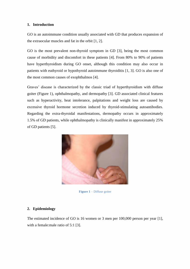
Graves’ disease is characterized by the classic triad of hyperthyroidism with diffuse
goiter (Figure 1), ophthalmopathy, and dermopathy [3]. GD associated clinical features
such as hyperactivity, heat intolerance, palpitations and weight loss are caused by
excessive thyroid hormone secretion induced by thyroid-stimulating autoantibodies.
Regarding the extra-thyroidal manifestations, dermopathy occurs in approximately
1.5% of GD patients, while ophthalmopathy is clinically manifest in approximately 25%
of GD patients [5].
Figure 1 – Diffuse goiter
2. Epidemiology
The estimated incidence of GO is 16 women or 3 men per 100,000 person per year [1],
with a female:male ratio of 5:1 [3].

GO particularly affects young women. It usually starts between the third and fourth
decades of life, and its clinical presentation may be more severe in elderly subjects and
men, and milder in asian people [3].
There are many risk factors that can affect the development or progression of GO, and
the majority of these are not modifiable, such as age, gender, ethnicity, genetics, and
thyrotropin receptor antibody status [6]. Others, such as smoking, RAI exposure, and
abnormal thyroid function, can be modified with a potential favorable impact on the
course of the disease [6, 7]. Smoking is the most important risk factor amenable to
modification in these patients and the risk is proportional to daily cigarette intake [1].
Smoking has been associated with more severe and progressive GO, worsening of GO
after RAI treatment, and worse response to the immunosuppressant therapies [6].
Smoking, even past smoking, is an independent risk factor associated with impaired
response to intravenous corticosteroids in patients with GO [1].
No specific gene or polymorphism has been identified in GO, however, HLA-DR3 has
been studied extensively [6].
3. Pathophysiology
The pathogenesis of this illness is summarized in three main phenomena: (1)
inflammation of the periorbital soft tissues; (2) overproduction of glycosaminoglycans
(especially hyaluronic acid) by orbital fibroblasts; and (3) hyperplasia of adipose tissue
[1, 8]. As hyaluronic acid is a profoundly hydrophilic glycosaminoglycan, the orbital
tissues retain water and become very edematous [9].
A close temporal relationship exists between the onset of Graves’ hyperthyroidism and
the onset of clinically apparent GO, suggesting that these two autoimmune conditions
may share etiologic factors [9].
Graves’ hyperthyroidism is caused by autoantibodies that bind to TSH-R on thyroid
follicular epithelial cells, causing hyperplasia and the production of thyroid hormones,
T3 and T4 [10].
Several findings in patients with GO, including elevated TSH-R expression in orbital
tissues and elevated levels of TRAb, support the concept that immunoreactivity against
TSH-R in the orbit plays a pivotal role in GO pathogenesis [1, 9].
The serum levels of TRAb correlate with the severity and clinical activity of the disease
and high levels in early GO predict a poor prognosis [9].
In addition to TSH-R, orbital fibroblasts from patients with GO express high levels of
IGF-1R. Recent studies suggest that these receptors engage in cross-talk induced by
TSH-R ligation to synergistically enhance TSH-R signaling, hyaluronic acid production,
and the secretion of inflammatory mediators. However, antibodies that specifically
target IGF-1R have not been found in normal sera, making it unlikely that such
antibodies play a role in GO pathogenesis [9]. Therefore, more studies are required to
fully understand the role of IGF-1R and IGF-1R autoantibodies in the pathophysiology
of GO [8].
Both cellular and humoral immunity directed against TSH-R expressed on orbital
fibroblasts likely initiate the disease process. Activation of helper T cells recognizing
TSH-R peptides and ligation of TRAb leads to the secretion of inflammatory cytokines
and chemokines and enhanced hyaluronic acid production and adipogenesis [9].
The orbit contains different subpopulations of fibroblasts exhibiting phenotypic
heterogeneity. Fibroblasts within the extraocular muscles, when exposed to cytokines,
can differentiate into myofibroblasts and then participate in inflammation, repair, and
fibrosis (especially in advanced stages of the disease). On the other hand, half of the
fibroblasts within the adipose tissue are preadipocytes that, under appropriate
conditions, can be induced to differentiate into adipocytes and thus contribute to the
increased tissue volume associated with the disease [1, 3].
4. Clinical features
Although GO may develop before or concomitantly with Graves’ hyperthyroidism, in
60% of cases it follows (ranging from months to years) the diagnosis of systemic GD
[6].
GO is typically a clinical diagnosis, based on presenting ocular symptoms and signs [1].
The presence of TRAb and imaging characteristics may be of great value in the
diagnostic workup [2]. However, when clinical features are sufficient, orbital imaging
may not be necessary for diagnosis [1].

It may affect one or both eyes. Bilateral GO is most common, although the severity of
the condition in each orbit may be different [3].
Symptoms of GO are primarily of mechanical origin, due to an increase in orbital
connective and/or muscle tissue within the space-limited bony orbit, leading to
protrusion of the eye and subsequent ocular symptoms [8].
Symptoms are related to (1) ocular exposure, (2) periorbital soft tissue inflammation
and congestion, or (3) extraocular muscle involvement (See Table 1). Upper eyelid
retraction (90% of patients) and proptosis are the two most common signs of GO
(Figure 2). Proptosis is easily assessed with the Hertel exophthalmometer. The lacrimal
gland is frequently involved and enlarged [1]. Other signs and symptoms include: ocular
or retro-ocular pain at rest, ptosis, corneal opacity, chemosis, lagophthalmos and corneal
ulcers. Optic disc edema and afferent pupil defect are specific signs but are not always
present [1, 3].
Table 1 – Symptoms of GO
Figure 2 – Patient with upper eyelid retraction and proptosis, more evident on the left side
Ocular exposure
•Ocular dryness and grittiness
•Photophobia
•Excessive tearing
•Blurred vision
Periorbital soft tissue inflammation and
congestion
•Sensation of retroocular pressure
•Conjunctival redness
•Eyelid swelling (Figure 3)
Extraocular muscle involvement
•Aching with eye movement
•Restricted ocular motility
•Double vision
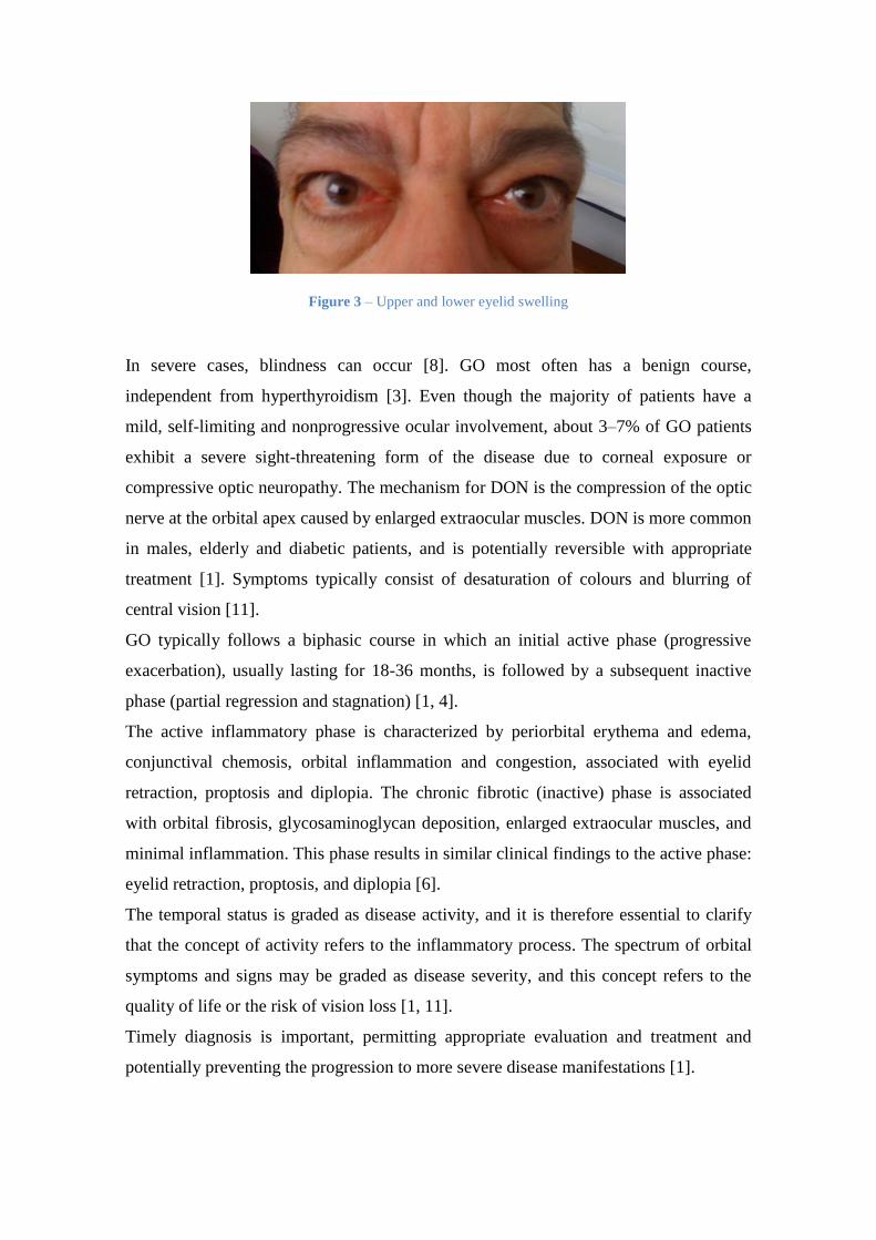
Figure 3 – Upper and lower eyelid swelling
In severe cases, blindness can occur [8]. GO most often has a benign course,
independent from hyperthyroidism [3]. Even though the majority of patients have a
mild, self-limiting and nonprogressive ocular involvement, about 3–7% of GO patients
exhibit a severe sight-threatening form of the disease due to corneal exposure or
compressive optic neuropathy. The mechanism for DON is the compression of the optic
nerve at the orbital apex caused by enlarged extraocular muscles. DON is more common
in males, elderly and diabetic patients, and is potentially reversible with appropriate
treatment [1]. Symptoms typically consist of desaturation of colours and blurring of
central vision [11].
GO typically follows a biphasic course in which an initial active phase (progressive
exacerbation), usually lasting for 18-36 months, is followed by a subsequent inactive
phase (partial regression and stagnation) [1, 4].
The active inflammatory phase is characterized by periorbital erythema and edema,
conjunctival chemosis, orbital inflammation and congestion, associated with eyelid
retraction, proptosis and diplopia. The chronic fibrotic (inactive) phase is associated
with orbital fibrosis, glycosaminoglycan deposition, enlarged extraocular muscles, and
minimal inflammation. This phase results in similar clinical findings to the active phase:
eyelid retraction, proptosis, and diplopia [6].
The temporal status is graded as disease activity, and it is therefore essential to clarify
that the concept of activity refers to the inflammatory process. The spectrum of orbital
symptoms and signs may be graded as disease severity, and this concept refers to the
quality of life or the risk of vision loss [1, 11].
Timely diagnosis is important, permitting appropriate evaluation and treatment and
potentially preventing the progression to more severe disease manifestations [1].
5. Assessment Protocols of GO
Because of the broad clinical overlap of the active and inactive phases, it is important to
identify where a patient is situated in the evolution of his disease [6]. Several
classification schemes have been conceived to assess the clinical manifestations of GO
[1]. Any such classification or assessment scheme must be reproducible and able to
accurately score the severity and activity of the inflammatory phase of the disease [6].
In 1969, Werner devised the NO SPECS Classification (No physical signs or symptoms,
Only signs, Soft tissue involvement, Proptosis, Extraocular muscle signs, Corneal
involvement, and Sight loss), a clever mnemonic which was the basis of the
ophthalmology index for many decades [6]. This classification grades exclusively for
clinical severity and does not adequately identify patients in the active phase of the
disease [1, 6]. Since then, a number of other classification and assessment schemes have
been developed including the CAS, based on 4 of the 5 classical signs of inflammation
(pain, redness, swelling, and impaired function), the VISA (vision, inflammation,
strabismus, and appearance), and the EUGOGO Classification [4, 6]. These scoring
systems assess inflammatory signs in an attempt to identify patients with active disease
who are most likely to respond to treatment [6].
The VISA and EUGOGO classifications are the two most widely used grading systems
conceived to assess the activity and severity of GO. The first is more frequently used in
North America and Canada while the latter is in Europe. Both are grounded in the NO
SPECS and CAS classifications and are used to guide the therapeutic decision making,
warranting an adequate treatment. As both classifications have been devised for both
clinical and research purposes, they have some complexity in the extensive data to be
collected [1].
The EUGOGO Classification is an assessment protocol for the evaluation of patients
with GO based upon activity and severity parameters. The disease activity is evaluated
based on the modified CAS (See Table 2) [1].
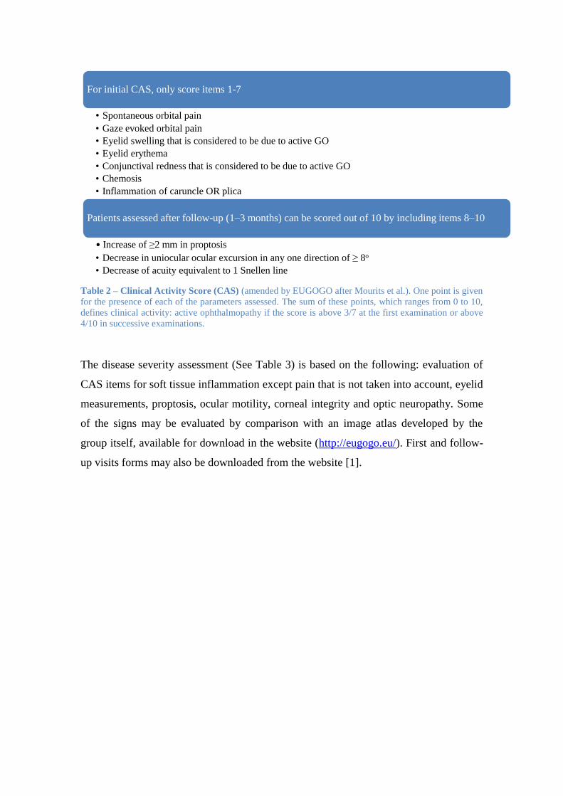
Table 2 – Clinical Activity Score (CAS) (amended by EUGOGO after Mourits et al.). One point is given
for the presence of each of the parameters assessed. The sum of these points, which ranges from 0 to 10,
defines clinical activity: active ophthalmopathy if the score is above 3/7 at the first examination or above
4/10 in successive examinations.
The disease severity assessment (See Table 3) is based on the following: evaluation of
CAS items for soft tissue inflammation except pain that is not taken into account, eyelid
measurements, proptosis, ocular motility, corneal integrity and optic neuropathy. Some
of the signs may be evaluated by comparison with an image atlas developed by the
group itself, available for download in the website (http://eugogo.eu/). First and follow-
up visits forms may also be downloaded from the website [1].
For initial CAS, only score items 1-7
• Spontaneous orbital pain
• Gaze evoked orbital pain
• Eyelid swelling that is considered to be due to active GO
• Eyelid erythema
• Conjunctival redness that is considered to be due to active GO
• Chemosis
• Inflammation of caruncle OR plica
Patients assessed after follow-up (1–3 months) can be scored out of 10 by including items 8–10
• Increase of ≥2 mm in proptosis
• Decrease in uniocular ocular excursion in any one direction of ≥ 8o
• Decrease of acuity equivalent to 1 Snellen line
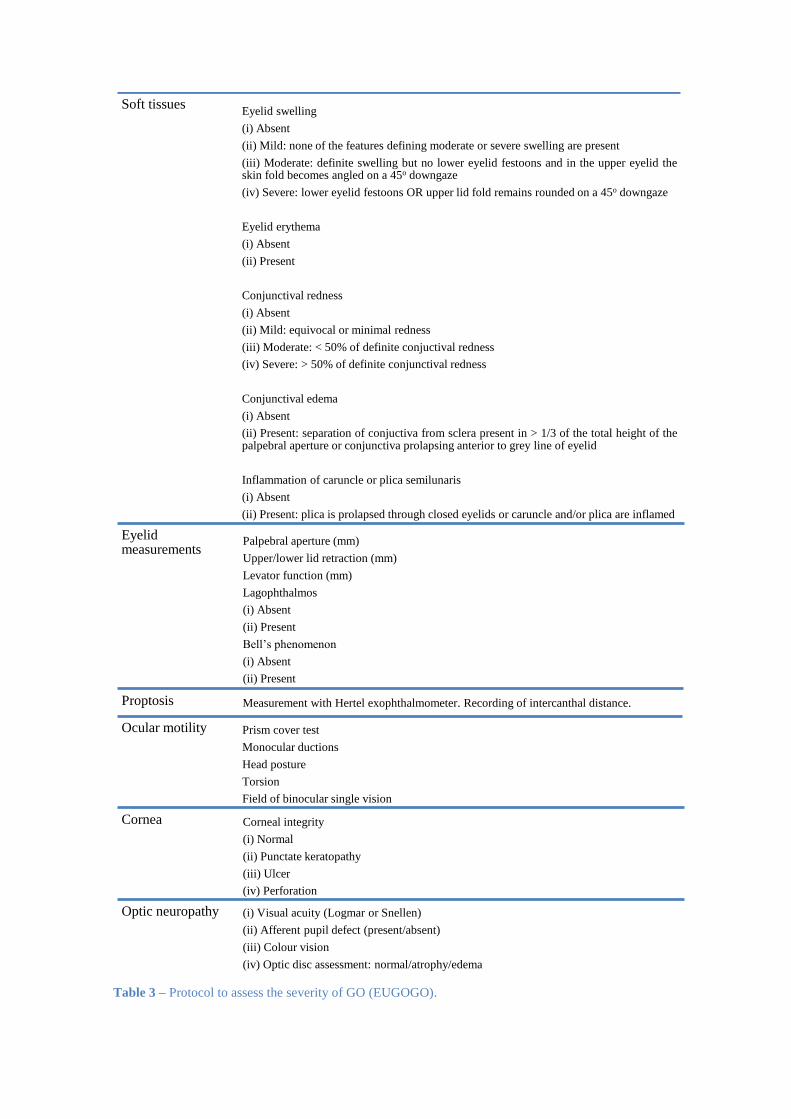
Table 3 – Protocol to assess the severity of GO (EUGOGO).
Soft tissues Eyelid swelling
(i) Absent
(ii) Mild: none of the features defining moderate or severe swelling are present
(iii) Moderate: definite swelling but no lower eyelid festoons and in the upper eyelid the skin fold becomes angled on a 45o downgaze
(iv) Severe: lower eyelid festoons OR upper lid fold remains rounded on a 45o downgaze
Eyelid erythema
(i) Absent
(ii) Present
Conjunctival redness
(i) Absent
(ii) Mild: equivocal or minimal redness
(iii) Moderate: < 50% of definite conjuctival redness
(iv) Severe: > 50% of definite conjunctival redness
Conjunctival edema
(i) Absent
(ii) Present: separation of conjuctiva from sclera present in > 1/3 of the total height of the palpebral aperture or conjunctiva prolapsing anterior to grey line of eyelid
Inflammation of caruncle or plica semilunaris
(i) Absent
(ii) Present: plica is prolapsed through closed eyelids or caruncle and/or plica are inflamed
Eyelid measurements
Palpebral aperture (mm)
Upper/lower lid retraction (mm)
Levator function (mm)
Lagophthalmos
(i) Absent
(ii) Present
Bell’s phenomenon
(i) Absent
(ii) Present
Proptosis Measurement with Hertel exophthalmometer. Recording of intercanthal distance.
Ocular motility Prism cover test
Monocular ductions
Head posture
Torsion
Field of binocular single vision
Cornea Corneal integrity
(i) Normal
(ii) Punctate keratopathy
(iii) Ulcer
(iv) Perforation
Optic neuropathy (i) Visual acuity (Logmar or Snellen)
(ii) Afferent pupil defect (present/absent)
(iii) Colour vision
(iv) Optic disc assessment: normal/atrophy/edema

The management of patients with GO depends on the degree of severity of the
ophthalmopathy, a concept clarified earlier. The disease is classified as mild, moderate,
severe, or sight-threatening as follows [1, 12].
1. Mild: characteristics of GO have a minimum impact on the patient’s life. They
usually present one or more of the following signs: minor lid retraction (< 2
mm), mild soft tissue involvement, exophthalmos < 3 mm (above the normal
range for the race and gender), transient or no diplopia, and corneal exposure
responsive to lubricants.
2. Moderate to severe: patients without sight-threatening GO whose eye disease
has sufficient impact on daily life to justify the risks of immunosuppression (if
active) or surgical intervention (if inactive). Patients usually present one or more
of the following signs: lid retraction (> 2 mm), moderate or severe soft tissue
involvement, exophthalmos ≥ 3 mm (above the normal range for the race and
gender), and inconstant (or constant) diplopia.
3. Sight-threatening GO: patients with DON, corneal breakdown or, infrequently,
globe subluxation. These cases warrant immediate intervention.
6. Treatment
A coordinated approach should be performed by a multidisciplinary team of
ophthalmologists, endocrinologists, radiologists, and orbital surgeons, in order to design
an individualized treatment plan [1, 12]. All patients with GO, except for mildest cases,
should be referred to multidisciplinary clinics for further assessment and management
[13].
There is no consensus as to the best treatment strategy for GO, but in this paper I will
focus on the management plan based on the Consensus Statement of the EUGOGO on
management of GO (2008) and in the scientific evidence published since then [1, 6].
6.1. Measures for all patients with GO
6.1.1. Restore Euthyroidism
Patients with uncontrolled thyroid dysfunctions are more likely to experience severe
GO, and it has also been shown that restoring euthyroidism can improve GO [1, 6].
Frequent monitoring (every 4-6 weeks) of thyroid function is imperative during the
early stages of treatment [1]. There are 3 main treatment modalities for patients with
Graves’ hyperthyroidism: antithyroid drugs, thyroidectomy, and RAI treatment [6].
6.1.1.1. Antithyroid Drugs
The thionamides (propylthiouracil, methimazole, and carbimazole) lower thyroid
hormone production by inhibiting iodination of thyroglobulin and ultimately the
production of T4 and T3. Several studies have shown that antithyroid medications do not
adversely affect GO [6]. According to Burch [14], the principal disadvantage of
thionamide therapy is adverse effects. Minor adverse effects such as pruritic rash and
arthralgias occur in approximately 5% of patients receiving methimazole, and typically
begin within the first few weeks of starting therapy. A mild rash may be manageable
with antihistamine therapy. These patients may be switched to propylthiouracil but 30 to
50% have a similar reaction. The more severe adverse effects of antithyroid medications
include agranulocytosis, hepatotoxicity, and ANCA-positive vasculitis. Agranulocytosis
occurs in approximately 1 in 500 patients, is dose-related with methimazole, and almost
always develops within the first 90 days of therapy. The typical presentation is high
fever and severe pharyngitis. In a patient developing agranulocytosis while taking either
methimazole or propylthiouracil, use of the other drug is not recommended owing to
cross-reactivity between the two drugs. Methimazole hepatotoxicity is frequently
cholestatic, whereas propylthiouracil use has been associated with hepatocellular injury,
including fulminant hepatic failure. Hepatic dysfunction generally occurs within the
first few days to 3 months after drug initiation. ANCA-positive vasculitis is much more
frequent with propylthiouracil, being rare with methimazole or carbimazole, and can
occur months or years after initiation of drug therapy. Patients typically present with
polyarthritis, fever, and purpura, and more severely affected individuals develop
glomerulonephritis and pneumonitis. Before starting antithyroid drugs, the patient
should be alerted to stop the medication immediately and call their physician when there
are symptoms suggestive of agranulocytosis or hepatic injury [14].
6.1.1.2. Thyroidectomy
Thyroidectomy is now recommended by fewer than 1% of experts for the initial
management of GD. However, is the favored method to attain euthyroidism in patients
with moderate to severe active GO who cannot take antithyroid drugs. If surgery is
chosen as the primary therapy for GD, near-total or total thyroidectomy is the procedure
of choice [15].
In a retrospective analysis involving TT vs ST, no difference was found in postoperative
progression of dysthyroid ophthalmopathy after the shift from ST to TT. However, this
change dramatically reduced the risk for recurrence of GD without increasing the rate of
complications [16].
TT has a nearly 0% risk of recurrence, whereas ST may have an 8% chance of
persistence or recurrence of hyperthyroidism at 5 years. Potential complications of
surgery include transient and permanent hypoparathyroidism and recurrent laryngeal
nerve injury [15].
The findings of a recent study indicate that thyroidectomy as the treatment of
hyperthyroidism may lower the risk of developing GO. Further studies will be required
to substantiate these results [7].
6.1.1.3. RAI Treatment
RAI is an effective therapy for Grave’s hyperthyroidism, but it may cause progression
of preexisting GO or its de novo occurrence in 15% to 20% of patients [6, 14, 17]. The
development or worsening of GO following RAI is thought to be the consequence of
radiation damage to the thyrocytes, resulting in release of thyroid antigens and
activation of autoimmune reactions directed to the orbit [18].
Several factors have been found to be associated with the development or exacerbation
of GO after RAI treatment, such as tobacco smoking, high pretreatment T3 values (≥
325 ng/dL or > 5 nmol/L), active and progressive GO over the preceding 3 months, high
serum TRAb levels (> 8.8 IU/L), and post-RAI hypothyroidism not promptly corrected
by levothyroxine replacement [15, 18].
This risk may be reduced in patients with active GO by a short cycle (3 months) of
orally delivered corticoids after the treatment (beginning with 0.3–0.5 mg of prednisone
per kilogram daily 1-3 days after radioiodine and tapering the dose until withdrawal)
and by avoiding post-RAI hypothyroidism [1]. Lai et al. found that a lower dose of oral
prednisone (approximately 0.2 mg/kg body weight) for a shorter duration of six weeks
is also effective [17].
Patients without preexisting GO and without risk factors should not be treated with
corticosteroid prophylaxis [19].
6.1.2. Supportive Treatment
Smoking cessation advice and referral to smoking cessation services should be offered
to all patients known or suspected of having GD or GO who are current smokers [12].
Artificial tears can alleviate symptoms of corneal exposure and prevent corneal
ulceration [12]. Sunglasses can improve photosensitivity [6]. Patients should be advised
to sleep with the head of the bed slightly elevated. Nocturnal ointment is of great benefit
for incomplete eyelid closure [1]. Prisms (or monocular occlusion) can resolve diplopia,
one of the most distressing symptoms [6, 12].
6.2. Measures for Patients with Mild Ophthalmopathy
Although oral corticosteroids and ORT reduce the active inflammatory symptoms, there
is no proven role for these modalities in decreasing the risk of disease progression in
patients with mild ophthalmopathy [6].
Local measures are the mainstay therapy for these patients that generally have a self-
limiting process. In the majority of studies which have investigated the natural history
of GO in untreated patients, the orbitopathy improved in about half, remained stable in
about 35% and worsened in approximately 15% of patients [1]. Watchful waiting is
appropriate for the majority of patients with mild GO [20].
Selenium is a trace element and, through its effects from selenoproteins, plays an
important role in cell development and proliferation, oxidative stress protection, and
production of T3. Most of the European countries are selenium deficient. Since selenium
acts as a potent antioxidant, and oxygen free radicals contribute to the orbital
inflammatory process, theoretically it could be of some therapeutic benefit in GO [6,
21]. A recent study showed that a 6-month course with oral selenium supplements in the
form of sodium selenite at a dose of 100 μg twice daily (equivalent to 91.3 μg of
elemental selenium twice daily) significantly improved quality of life, reduced ocular
involvement, and slowed progression of disease in patients with active mild GO [1, 12].
Patients taking selenium had no adverse drug reactions, but concerns about selenium
toxicity include the increased risk of diabetes mellitus, glaucoma, and neurotoxicity [6].
Botulinum toxin injection into the levator superioris and Müller muscle complex may
be considered to temporarily treat upper eyelid retraction and is a valuable therapeutic
option in active disease where definitive surgery remains contraindicated [1, 6].
Rehabilitative surgery (Müllerectomy or blepharoplasty), if needed, should be
considered providing that the GO remains stable and inactive [1].
6.3. Measures for Patients with Moderate to Severe Ophthalmopathy
In this subtype of patients, eye disease has sufficient impact on daily life to justify the
risks of immunosuppressant treatment (if active) or surgical intervention (if inactive).
Only patients in the active phase of the disease will respond to immunosuppressive
treatments [1].
6.3.1. Immunosuppressive Medical Treatment and ORT
Although corticosteroids are the most often used therapy for GO, the precise dosage,
duration, preparation, and route (IV, PO, or periocular) of administration remain a
matter of opinion and debate [6].
A recent meta-analysis compared the efficacy and tolerability of the most common
immunosuppressive monotherapies for patients with active and moderate to severe
ophthalmopathy. Current evidence gave priority to intravenous glucocorticoids, which
had a statistically significant advantage over oral glucocorticoids and caused fewer
adverse events, such as hypertension and weight gain. Retrobulbar injections of
glucocorticoids and orbital radiotherapy did not provide greater efficacy than oral
glucocorticoids, although better tolerability and fewer adverse events were shown [13].
Pulses of IV steroids were associated with fewer side effects, shorter treatment course,
and lower relapse risk compared with PO steroids. Oral glucocorticoids might be
considered when IV infusions are not logistically possible or if the patient prefers the
oral route. They can also be used in some moderate to severe patients when the
determination of activity is uncertain. A therapeutic trial using a three-day course of oral
prednisolone (50 mg) can determine whether clinical features show improvement, and,
therefore, IV corticosteroids or radiotherapy may be indicated [1].
The dosage scheme most widely used in the United States and Europe consists of
methylprednisolone 500 mg IV weekly for 6 weeks, followed by 250 mg IV weekly for
another 6 weeks, for a cumulative dose of 4.5 g [1, 3]. If there is no clinical response,
treatment with corticosteroids may be discontinued after the first 6 weeks and other
treatments may be attempted [1].
Prolongation of treatment after 12 weeks in patients who respond to corticosteroids
should be related to disease severity and its impact on the quality of life [1]. If no

compression neuropathy exists, it is advisable not to exceed a cumulative dose of 8 g
[3]. Consecutive day-dosing should also be avoided [1].
Prior to the administration of IV treatment, appropriate liver assessment is
recommended, including abdominal ultrasound for liver morphology, liver function
tests, detection of hepatitis viral markers, and autoantibodies. Patients with recent
hepatitis, liver dysfunction, severe cardiovascular morbidity, or severe hypertension
must be excluded. During treatment, liver enzymes, glucose levels, and blood pressure
should be monitored on a monthly basis. Assessment of adrenal function may be
advisable at the end of treatment [1].
Side effects of long-term corticosteroid therapy should be taken into account. These
include hyperglycemia, hypertension, weight gain, fat distribution remodeling,
psychiatric disorders, osteoporosis, cataracts, glaucoma and hepatotoxicity – fatal
autoimmune hepatitis can occur. Patients must be monitored carefully, weighing the
benefits and risks [3, 4, 6, 22].
ORT has been routinely used in GO since the 1940s [3]. Though the efficacy of ORT
alone for treatment of GO remains controversial, its combination with glucocorticoids is
effective in early and active disease and has an acceptable safety profile [1]. Satisfactory
results will not be achieved in patients with long-standing disease, no inflammatory
activity, and established fibrotic events [3].
A recent large retrospective study compared the risk of developing compressive optic
neuropathy in 351 patients with active thyroid eye disease treated only with
corticosteroids (144 patients) or with corticosteroids plus ORT (105 patients). The main
indication for offering ORT to patients already on corticosteroids were: development of
significant restriction in ocular motility (88%); total cumulative dose of corticosteroids
over 8 g (17%); intolerance to corticosteroids (8.6%); and inadequate control of disease
activity with corticosteroids (6.5%). At an average of 3.2 years follow-up, the
concomitant use of corticosteroids therapy and ORT achieved a significant decrease in
the risk of development of compressive optic neuropathy as compared to the use of
corticosteroids therapy alone (0% vs 17%) [1].
The typical ORT protocol is a total of 20 Gy per orbit fractionated in 10 days (2 Gy/d)
over a 2-week period. However, the optimum fractionation, duration, and dosing of
ORT remains unsettled, and lower doses seem to perform just as well as higher doses
[6].
ORT has been shown to improve ocular motility and soft tissue inflammatory signs. It is
also effective for the recovery of vision lost due to compression in the active stages of
the disease. However, remission of proptosis is uncommon, with responses of only a
few millimeters and minimal or doubtful cosmetic impact [3].
ORT is a relatively safe procedure but should be avoided in patients younger than 35
years of age or in patients with severe uncontrolled hypertension or diabetes (especially
if there is preexisting diabetic retinopathy) [1, 6].
No severe complications have been reported with ORT, except for visual changes or
blindness in the setting of prior diabetic retinopathy. Cataract is the most common
major side effect, but it may also be due to previous treatment with high corticosteroid
doses. Alopecia of the tail of the eyebrows can occur in some patients due to inclusion
in the radiation fields, with subsequent full recovery. Some eye dryness may occur in a
very small proportion of patients. Radiation-induced second tumors are exceptional.
Some cases of pigmented basal cell carcinoma have been reported, and that’s the reason
why ORT should not be used in young patients [3].
Thus, ORT should be considered a second-line treatment, when the first course of
steroids has produced only a partial response and the disease is still active [1]. However,
ORT may be used as first line treatment with or without concomitant corticosteroids
when required [3].
6.3.2. Other immunosuppressors
One of the limitations of corticosteroid therapy is that a significant proportion of
patients with active GO (20-30%) do not respond to IV methylprednisolone and
approximately another 10-20% of patients present with disease relapse after therapy
withdrawal [22]. These patients can be candidates for a trial of treatment with
methotrexate (7.5–10 mg/week). In alternative, patients who are nonresponsive to
corticosteroids may be treated with combination therapy of cyclosporine A (5
mg/kg/day in 2 doses plus oral glucocorticoids), azathioprine, or specific monoclonal
antibody agents (see below) [1].
6.3.3. Novel Therapeutic Agents
In recent years, immunosuppressors that act as disease-modifying drugs, as an
alternative to corticosteroids, have been proposed as novel agents which target the
various antigens involved in GO pathophysiology [22].
An anti-IGF-1R antibody, teprotumumab, previously developed as a therapeutic
strategy for several types of solid tumors and lymphomas, is currently being tested in a
phase II randomized placebo-controlled clinical trial after showing to be effective in
attenuating orbital fibroblasts stimulation in vitro [22].
There are some evidences in short series reports of the efficacy of other molecules such
as tocilizumab, adalimumab, or etanercept [1]. Tocilizumab is a FDA-approved
humanized monoclonal antibody against the interleukin-6 receptor for use in
rheumatoid arthritis and has shown promise in a recent study in GO patients refractory
to IV methylprednisolone treatment. After therapy, CAS improved in 18 out of 18
patients, proptosis reduced in 13 out of 18, and ocular motility improved in 15 out of 18.
One patient with compressive optic neuropathy improved without the need for orbital
decompression surgery. These positive preliminary results warrant further randomized
and controlled studies [22].
Rituximab is a monoclonal antibody directed against B cell surface CD20 [9]. The
rationale for its use in GO is the potential blockade of pathogenic autoantibody
generation, the depletion of B cells involved in antigen presentation and the production
of inflammatory cytokines [22]. In preliminary clinical trials, rituximab significantly
reduced the inflammatory and severity of the ophthalmopathy in patients with active eye
disease. However, two recently completed randomized controlled trials showed
conflicting results, indicating that the agent is not uniformly effective. The reasons for
this are unclear but may be related to the differences in the study designs. Because GO
duration was shorter in the study showing benefit (mean duration 4 months vs. 12
months), it is possible that a subgroup of patients with short disease duration might
benefit from the therapy [1, 9].
Other promising treatments such as the production of TSH-R antagonists, either as
monoclonal TSH-R-blocking antibodies or as small-molecule-ligand antagonists of
TSH-R, are being developed [1].
6.3.4. Surgical Treatment
There are four components of ophthalmopathy that require attention from the surgical
point of view: proptosis, restrictive strabismus, eyelid abnormality (retraction), and
cosmetic concerns (eyelid fat bags, periorbital hollows, and rhytids) [23].
In most cases, surgery is indicated for the correction of residual structural changes in
patients with stable, inactive (fibrotic) moderate to severe ophthalmopathy [6].
Rehabilitative surgery should only be performed in patients who show evidence of
disease quiescence over a period of at least 6 months [1].
The surgical management of GO involves four major stages of rehabilitation: orbital
decompression, extraocular muscle surgery, eyelid repositioning, and cosmetic soft
tissue redraping. Not all patients require all four stages, but one may require more than
one stage of surgery [23].
Orbital decompression is indicated not only for extreme vision-threatening proptosis,
but also cosmetically disfiguring proptosis. In theory, there are four bony orbital walls
available for decompression: medial, inferior, lateral, and superior. However, orbital
roof approach is associated with serious complications, and hence is reserved for
extreme cases [23]. Typically, decompression begins with orbital fat removal. For many
surgeons, intraconal fat removal is the “first wall” of orbital decompression for mild (2-
3 mm) proptosis [23].
Many different techniques and approaches have been described for orbital
decompression surgery [1]. An essential step in the surgical planning of a
decompression surgery is a thorough imaging study of the orbit with either magnetic
resonance imaging or computed tomography [23].
Strabismus surgery is usually carried out in the inactive phase of the disease, following
orbital decompression surgery. This stage constitutes the correction of diplopia, the
most disabling side effect of GO, and the most challenging to treat. Patients should,
therefore, be encouraged to have realistic expectations [23].
Correction of eyelid abnormality is the third stage, and most commonly includes
correction of upper and lower eyelid retraction. There are several surgical options
described for the correction of eyelid retraction. Overall, the surgical correction gives
good results, although there is still some unpredictability and sometimes more than one
stage of surgery is required [23].
The aesthetic concerns of periocular sequelae of GO are best treated toward the end of
the surgical rehabilitation. Upper and/or lower blepharoplasty is frequently needed as
the last step in the functional and cosmetic rehabilitation of patients. Botulinum toxin
and hyaluronic acid filler injections can be of great help [1, 23].
6.4. Measures for Patients with Sight-Threatening Ophthalmopathy
Patients with sight-threatening ophthalmopathy due to DON must be treated urgently
with high-dose IV glucocorticoids (3 x 500 mg–1 g on consecutive days within one
week; if necessary, repeated after a week). If the response is insufficient after 1-2
weeks, or the dose/duration of steroid treatment required induces significant side
effects, orbital decompression should be offered promptly [1]. Though optic neuropathy
responds well to surgery, the nerve is resilient, and minor degrees of compressive optic
neuropathy are probably not as emergent as once thought [23]. ORT is not
recommended unless as an adjunct to proved therapies [20].
The management of sight-threatening corneal breakdown includes: frequent topical
lubricants, moisture chambers, botulinum toxin injections, levator recession surgery,
tarsorrhaphy, or even orbital decompression in very severe cases of exophthalmos
which impede lid closing. IV methylprednisolone should be administered before surgery
if the disease is active [1]. In the case of corneal perforation/severe ulceration,
appropriate antibiotics, amniotic membrane transplant, and corneal transplant need to be
considered [20].
7. Conclusion
The management of active GO can be a challenging therapeutic dilemma. Therefore, a
coordinated approach between healthcare professionals is required in order to meet the
needs of patients. Timely diagnosis of sight-threatening GO and identification of risk
factors for severe outcomes are critical. All patients must be assessed for disease
activity and severity to determine the best course of action. Optimal treatment by a
multidisciplinary team improves quality of life. Risk factor modification begins with
smoking cessation and normalization of thyroid function. The first-line treatment for
moderate to severe GO or DON is IV glucocorticoids, but often a multimodality
approach with the addition of ORT or orbital decompression may be required.
In the last few years, as the result of greater understanding of the pathophysiology of
GO, novel therapeutic agents against specific immunological targets were developed.
Although the lack of randomized controlled trials suggests caution in generalizing
results, some data show promising results.

References
1. Barrio-Barrio, J., et al., Graves' Ophthalmopathy: VISA versus EUGOGO
Classification, Assessment, and Management. J Ophthalmol, 2015: p. 249125.
2. Verma, R., M. Gupta, and V.K. Mehta, Thyroid associated orbitopathy. BMJ
Case Rep, 2013.
3. Vilar-Gonzalez, S., et al., [Thyroid orbitopathy, an overview with special
attention to the role of radiotherapy]. Endocrinol Nutr, 2015. 62(4): p. 188-99.
4. Petrovic, M.J., et al., Evaluation of the patients with Grave's ophthalmopathy
after the corticosteroids treatment. Vojnosanit Pregl, 2012. 69(3): p. 249-52.
5. Bartalena, L. and V. Fatourechi, Extrathyroidal manifestations of Graves'
disease: a 2014 update. J Endocrinol Invest, 2014. 37(8): p. 691-700.
6. Bhatti, M.T. and J.J. Dutton, Thyroid eye disease: therapy in the active phase. J
Neuroophthalmol, 2014. 34(2): p. 186-97.
7. Stein, J.D., et al., Risk factors for developing thyroid-associated ophthalmopathy
among individuals with Graves disease. JAMA Ophthalmol, 2015. 133(3): p.
290-6.
8. Virakul, S., et al., Platelet-derived growth factor: a key factor in the
pathogenesis of graves' ophthalmopathy and potential target for treatment. Eur
Thyroid J, 2014. 3(4): p. 217-26.
9. Bahn, R.S., Current Insights into the Pathogenesis of Graves' Ophthalmopathy.
Horm Metab Res, 2015. 47(10): p. 773-8.
10. Bahn, R.S., Graves' ophthalmopathy. N Engl J Med, 2010. 362(8): p. 726-38.
11. Dolman, P.J., Evaluating Graves' orbitopathy. Best Pract Res Clin Endocrinol
Metab, 2012. 26(3): p. 229-48.
12. Perros, P., et al., Management of patients with Graves' orbitopathy: initial
assessment, management outside specialised centres and referral pathways. Clin
Med (Lond), 2015. 15(2): p. 173-8.
13. Mou, P., et al., Common Immunosuppressive Monotherapy for Graves'
Ophthalmopathy: A Meta-Analysis. PLoS One, 2015. 10(10): p. e0139544.
14. Burch, H.B. and D.S. Cooper, Management of Graves Disease: A Review.
JAMA, 2015. 314(23): p. 2544-54.
15. Bahn Chair, R.S., et al., Hyperthyroidism and other causes of thyrotoxicosis:
management guidelines of the American Thyroid Association and American
Association of Clinical Endocrinologists. Thyroid, 2011. 21(6): p. 593-646.
16. Annerbo, M., P. Stalberg, and P. Hellman, Management of Grave's disease is
improved by total thyroidectomy. World J Surg, 2012. 36(8): p. 1943-6.
17. Lai, A., et al., Lower dose prednisone prevents radioiodine-associated
exacerbation of initially mild or absent graves' orbitopathy: a retrospective
cohort study. J Clin Endocrinol Metab, 2010. 95(3): p. 1333-7.
18. Vannucchi, G., et al., Graves' orbitopathy activation after radioactive iodine
therapy with and without steroid prophylaxis. J Clin Endocrinol Metab, 2009.
94(9): p. 3381-6.
19. Shiber, S., et al., Glucocorticoid regimens for prevention of Graves'
ophthalmopathy progression following radioiodine treatment: systematic review
and meta-analysis. Thyroid, 2014. 24(10): p. 1515-23.
20. Bartalena, L., et al., Consensus statement of the European Group on Graves'
orbitopathy (EUGOGO) on management of GO. Eur J Endocrinol, 2008. 158(3):
p. 273-85.
21. Zarkovic, M., The role of oxidative stress on the pathogenesis of graves' disease.
J Thyroid Res, 2012: p. 302537.
22. Salvi, M., Immunotherapy for Graves' ophthalmopathy. Curr Opin Endocrinol
Diabetes Obes, 2014. 21(5): p. 409-14.
23. Naik, M.N., et al., Minimally invasive surgery for thyroid eye disease. Indian J
Ophthalmol, 2015. 63(11): p. 847-53.