Embed Size (px)
Citation preview
Identification and comparison of laboratory and wild caught Drosophila gut microbiota
Maria Inês da Silva Pais
Mestrado em Biologia Evolutiva e do Desenvolvimento
2011
Universidade de Lisboa !"#$%&"&'(&'()*+,#*"-(
.'/"01"2',13(&'(4*3%35*"(6,*2"%((
(
(
Co-option of Hedgehog and Wingless pathways in the formation of evolutionary novelties
(
7"01"(839*"(:*0'-(7"0*"%;"((
Dissertação de mestrado orientada por:
.3$130(<%*3(8$#',"(=(!"#$%&"&'(&'()*+,#*"-(&"(>,*;'0-*&"&'(&'(?*-@3"(
.3$130"(:"10A#*"(4'%&"&'(=(B,-1*1$1'(39(4*3%35CD(?'*&',(>,*;'0-*1C(
(
(
7'-10"&3('2(4*3%35*"(E;3%$1*;"('(&3(.'-',;3%;*2',13(
(
(
FGGH(!

Identification and comparison of laboratory and wild caught Drosophila gut microbiota
Maria Inês da Silva Pais Dissertação de mestrado orientada por: Doutor Élio Sucena – Faculdade de Ciências Universidade de Lisboa Doutor Luis Teixeira – Instituto Gulbenkian de Ciência
Mestrado em Biologia Evolutiva e do Desenvolvimento
2011
Universidade de Lisboa !"#$%&"&'(&'()*+,#*"-(
.'/"01"2',13(&'(4*3%35*"(6,*2"%((
(
(
Co-option of Hedgehog and Wingless pathways in the formation of evolutionary novelties
(
7"01"(839*"(:*0'-(7"0*"%;"((
Dissertação de mestrado orientada por:
.3$130(<%*3(8$#',"(=(!"#$%&"&'(&'()*+,#*"-(&"(>,*;'0-*&"&'(&'(?*-@3"(
.3$130"(:"10A#*"(4'%&"&'(=(B,-1*1$1'(39(4*3%35CD(?'*&',(>,*;'0-*1C(
(
(
7'-10"&3('2(4*3%35*"(E;3%$1*;"('(&3(.'-',;3%;*2',13(
(
(
FGGH(!
i
Abstract
The digestive tract is constantly in contact with several microorganisms that constitute the
gut microbiota. The crucial role this microbial community, both in mammals and in insects, plays
in the host physiology is starting to be revealed. The microbiota affects the host nutrition,
developmental process and immune system. We can discriminate between two types of
microorganisms inside the gut: indigenous microorganisms that colonize the host and transient
microorganisms that are acquired from the environment and food. Insects that are found in the
nature and reared under laboratory conditions may present differences in the gut microbiota
composition. This has also been reported in Drosophila melanogaster, where it is still not clear
whether the flora found in the digestive tract establishes a symbiotic relationship with the host or
if it is continuously loaded with the ingested food.
Here, we attempt to characterize the microbial community and its stability inside the gut of
Drosophila melanogaster, both reared under laboratory conditions and collected from nature.
We observe more diversity in wild caught flies, compared with laboratory flies. We show that the
microbiota of laboratory-reared flies’ guts is not stable and almost disappears when flies are fed
with sterile food. Moreover, we show that these bacteria grow on the flies’ food. In contrast, wild
caught flies microbiota is able to persist inside the gut, even when flies were fed with sterile
food. Conditions were established to rear axenic flies in order to perform future infections with
wild caught flies gut microbiota, to understand if the ability to persist inside the host is due to the
microbiota composition that is found in the wild.
We show the importance of identifying the indigenous microbial flora present inside the gut,
before start studying the microbiota role in the host. This study will pave the way to future
research on gut symbionts of insects.
Keywords: microbiota; gut; stability; Drosophila melanogaster.
ii
Resumo
Todos os Eucariotas vivem num íntima associação com variados microrganismos.
Tradicionalmente estes microrganismos eram denominados por comensais, pois pensava-se
que apenas estes beneficiavam desta associação, desconhecendo-se possíveis efeitos no
hospedeiro. Contudo, muitos estudos têm vindo a mostrar que a comunidade microbiana é
vantajosa em diversos aspectos da fisiologia do hospedeiro, tal como na nutrição, no processo
do desenvolvimento e no sistema imunitário. Desta forma, a palavra comensalismo têm vindo a
ser substituída por mutualismo, pois hoje sabe-se que ambos beneficiam desta associação.
Um dos papéis mais importantes que a comunidade microbiana desempenha no hospedeiro
é no seu sistema imunitário. A flora protege da colonização de patogéneos externos, limitando
a sua proliferação no intestino. Também está envolvida em activar o sistema imune a níveis
basais, tornando o hospedeiro apto para lidar contra infecções. Vários estudos também têm
mostrado a importância da flora a nível do desenvolvimento das próprias células do sistema
imune.
São escassos os estudos em Drosophila melanogaster que tentam compreender o papel
dos microbiota no sistema imunitário. Em 2008, Ryu e colaboradores mostraram a importância
da flora na activação de níveis basais deste sistema. Este estudo também mostra a importância
da regulação do sistema imunitário e o seu efeito na composição da flora intestinal. Para que
se possa estabelecer uma relação com os microrganismos é necessário que, os níveis do
sistema imunitário do hospedeiro se mantenham baixos, a fim de não os expulsar. Este estudo
mostrou que quando o sistema imune era desregulado, havia uma espécie de bactéria
presente na flora do hospedeiro que desaparecia, que era importante na inibição da
proliferação de uma espécie patogénica. Como consequência, a espécie patogénica que
também se encontrava presente na flora, proliferava e comprometia a sobrevivência do
hospedeiro.
A flora intestinal também desempenha um papel muito importante na nutrição do
hospedeiro, ajudando a degradar certos compostos e a produzir algumas moléculas e enzimas
que o hospedeiro, por si só, não consegue produzir. Um estudo recente em Drosophila
melanogaster mostrou a importância da flora intestinal na nutrição. Quando moscas axénicas
(livres de quaisquer microrganimos) se encontravam num meio de comida que não era muito
rico em nutrientes, o seu desenvolvimento era afectado, tendo as larvas um tamanho abaixo do
normal. Pelo contrario, quando as moscas tinham a flora normal, mesmo num meio de comida
iii
pobre, o seu desenvolvimento era normal. Isto demonstrou que mesmo num meio escasso em
nutrientes, as bactérias presentes na flora ajudam a um correcto desenvolvimento do
hospedeiro.
A composição da flora presente no intestino também é afectada pela dieta do hospedeiro.
Um estudo em humanos que sofriam de obesidade, demonstrou que quando estes eram
sujeitos a uma dieta saudável, havia uma mudança nas bactérias que eram dominantes,
havendo um aumento das bactérias do género Bacteroidetes e uma diminuição do género
Firmicutes. Em Drosophila melanogaster também foi mostrado em laboratório, que quando
estas eram sujeitas a uma dieta diferente, havia uma mudança na relativa abundância de
bactérias presentes na flora.
Vários estudos têm demonstrado que insectos encontrados na natureza e mantidos em
laboratório apresentam floras diferentes. Em Drosophila melanogaster estudos recentes
também já demonstraram o mesmo, nomeadamente que as moscas que são mantidas em
laboratório, apresentam uma grande falta de diversidade quando comparadas com moscas
selvagens.
Quando se estudam os microrganismos presentes no intestino, podemos encontrar também
microrganismos que não estão a colonizar o hospedeiro, mas que se encontram presentes pois
foram adquiridos do ambiente ou da comida ingerida. São denominados por microrganismos
transientes, pois estão só de passagem no tracto intestinal, juntamente com a comida. Por
outro lado temos os microrganismos indígenas, que colonizam e proliferam no hospedeiro. A
maior parte dos estudos não tem a preocupação de distinguir entre estes dois tipos de
microrganismos, o que pode levar a variabilidade entre resultados ou mesmo a mal-
interpretações.
Este estudo tenta fazer uma melhor caracterização da flora em Drosophila melanogaster,
em moscas selvagens que foram apanhadas na natureza e em moscas que são mantidas em
laboratório. Tentou-se também compreender a estabilidade da flora no sistema digestivo, quer
nas moscas selvagens quer nas de laboratório, tentando fazer-se uma distinção entre bactérias
transientes e bactérias indígenas.
Verificou-se que as moscas selvagens apresentam mais diversidade e maior número de
bactérias, quando comparadas com as moscas de laboratório. Isto deve-se provavelmente ao
tipo de dieta e ao ambiente que as rodeia. Na natureza, há uma grande quantidade de
microrganismos presentes e a dieta apresenta-se muito mais rica e diversa. Pelo contrário, em
laboratório, as condições em que as moscas são mantidas são mais assépticas do que na
natureza, e apenas um tipo de comida é administrado.
Para se compreender a estabilidade da flora, desenvolveu-se um protocolo em que se
administrava às moscas comida estéril, evitando a reinfecção de possíveis bactérias
transientes. Verificou-se que as moscas do laboratório perderam quase por completo toda a

iv
flora com este protocolo, ao contrário das moscas selvagens, em que grande parte da flora
conseguiu persistir no sistema digestivo. Também se verificou que as bactérias encontradas no
interior do sistema digestivo das moscas de laboratório conseguem crescer na comida que é
administrada.
Estes resultados demonstram que as moscas de laboratório apresentam uma flora instável,
a qual é constituída por muitas bactérias transientes que crescem na comida e desaparecem
do sistema digestivo quando é evitada a reinfecção das moscas com a sua própria flora. O
mesmo não acontece nas moscas selvagens, que demonstram manter uma população mais
estável no sistema digestivo.
Moscas de laboratório foram desenvolvidas axénicas, com o objectivo futuro de transferir a
flora presente nas moscas selvagens que se manteve estável até ao fim para estas moscas.
Deste modo, se a flora das moscas selvagens depois de transferida para moscas de
laboratório for capaz de persistir, esta colonização pode ser devido às bactérias que são
diferentes ou mesmo ao seu comportamento que é diferente, como por exemplo a capacidade
de formar biofilmes para poderem colonizar no hospedeiro.
Para finalizar, este estudo demonstra a importância de se estudar a estabilidade da flora e
de ter em conta se existem microrganismos transientes, antes de começar qualquer estudo
sobre a influência da flora no hospedeiro. Caso contrário, podem originar-se conclusões
erradas e variabilidade nos resultados, dependendo de como se mantêm as moscas em
laboratório. É importante proceder primeiro a uma caracterização da flora, verificar se é
representativa da flora em insectos presentes na natureza, e só depois estudar o seu papel.
Palavras-chave: flora; tracto intestinal; estabilidade; Drosophila melanogaster.

v
Contents
Abstract ....................................................................................................................................... i Resumo ...................................................................................................................................... ii Contents ..................................................................................................................................... v 1. Introduction ............................................................................................................................. 1
1.1 Importance of host microbiota ....................................................................................... 1 1.1.1 Microbiota role in host nutrition ............................................................................... 1 1.1.2 Microbiota role in immune system .......................................................................... 2
1.2 Factors that influence microbiota composition and consequent outcomes on the host . 3 1.2.1 Host diet .................................................................................................................. 3 1.2.2 Host genotype and environment ............................................................................. 3 1.2.3 Host immune response ........................................................................................... 4
1.3 Drosophila melanogaster microbial flora ....................................................................... 5 1.4 Microbiota community and symbiosis ............................................................................ 6 1.5 Aims of the project ......................................................................................................... 7
2. Material and methods ............................................................................................................. 8 2.1. Accessing the microbiota stability .................................................................................... 8
2.1.1. Fly collections and maintenance ............................................................................. 8 2.1.2. Aging flies for microbiota analyses ......................................................................... 8 2.1.3. Bacterial culture ...................................................................................................... 8 2.1.4. PCR amplification and sequencing ......................................................................... 9 2.1.5. Statistical analyses ................................................................................................. 9
2.2. Axenic flies .................................................................................................................. 10 2.2.1. Axenic flies maintenance ...................................................................................... 10 2.2.2. Confirmation of axenic flies axenic state .............................................................. 11
3. Results and discussion ......................................................................................................... 12 3.1. Microbiota comparison between wild and laboratory flies .......................................... 12
3.1.1. Setting-up the conditions to grow Drosophila microbiota ...................................... 12 3.1.2. Microbiota diversity in wild and laboratory flies ..................................................... 14 3.1.3. Microbiota load present in wild and laboratory flies .............................................. 20
3.2. Drosophila microbiota stability and colonization inside the host ................................. 22 3.2.1. There is a correlation between number of bacteria found on the host and on the
food 25 3.2.2. Microbiota from laboratory-reared flies grows on the food ................................... 27

vi
3.3. Wild caught flies maintain their microbiota community in contrast to laboratory-
reared flies ................................................................................................................................. 28 3.4. Establishing conditions to rear axenic flies ................................................................. 30
4. Concluding remarks .............................................................................................................. 34 5. Acknowledgments ................................................................................................................ 36 6. Bibliography .......................................................................................................................... 37 Annexs ...................................................................................................................................... 41
1
1. Introduction
1.1 Importance of host microbiota
All Eukaryotes establish intimate relationships with diverse microorganisms, which are
normally called by microbial flora or microbiota community. Even thought the microbiota
communities are variable among the organisms, displaying more complexity in higher
vertebrates than in invertebrates, their influence on the host is highly similar.
Studies with the microbiota that live inside the digestive tract are revealing their importance
in several fields. These bacteria have been traditionally referred as commensals, because it
was though that they would be the only ones benefiting from this relationship, being the effects
on the host largely ignored. Recently, the crucial role the microbiota community plays on the
host physiology has started to be revealed. The term commensalism is being increasingly
replaced by the term mutualism, because now it is known that both the host and the microbiota
benefit from this relationship. Microbiota community is known to influence two main aspects of
the host physiology: nutrition and immunity.
1.1.1 Microbiota role in host nutrition
One of the crucial roles that microbiota play on the host is on their nutrition. They improve the
host ability to live on suboptimal diets, improving digestion efficiency, helping in compounds
degradation and in the production of metabolites that the host by itself is not capable to produce
[1], [2].
Bacteria present in the gut from mice were shown to be important in fat storage regulation.
They promote the absorption of monosaccharides from the gut lumen and keep triglycerides in
adipocytes [3]. Harboring bacteria that promote a more efficient extraction and storage of fat
can result in diseases like obesity. Changes in two bacteria divisions in humans, an increase in
Firmicutes, mainly Lactobacillus sp., and a decrease of Bacteroidetes in the distal gut
microbiota can lead to obesity [4], [5]. However, it was shown in mice that the absence of
microbiota is not enough to protect the host from obesity, when feeding with a high-fat diet [6].
A few studies in insects also try to understand the contribution of their microbiota in nutrition.
Microbial community present in termite guts contributes to plant aromatic compounds
degradation, providing carbon and energy that are required to the host [7]. Recently, it was
published the first study in Drosophila melanogaster that highlights the importance of microbial
2
flora to host nutrition. Leulier et al. show that the presence of microbiota is crucial to the normal
host development when a poor-diet is administrated [2]. Flies lacking microbiota community
need to be in a rich medium for a normal development. This study also shows that the presence
of a single species of the genus Lactobacillus is sufficient to recapitulate the growth promoted
by the normal gut microbiota, which is in agreement to studies done in humans demonstrating
the importance of Lactobacillus sp. in nutrient uptake [5]. As Leulier et al. have shown,
associated with this nutrition role is a consequent role in development. Flies that are in a poor-
diet medium and lack gut microbiota, have problems in development and present lower larval
sizes, in contrast to flies that have normal gut microbiota [2]. Also in mice it was shown that the
absence of microbiota could compromise the normal brain development, leading to altered
expression profiles of signaling pathways and contributing for a different behavior. These
signaling pathways that are affected were also implicated in food-intake regulation [8].
1.1.2 Microbiota role in immune system
Several studies have been showing the importance of microbiota in protecting the host from
external pathogens. Microbial community is implicated on a process termed colonization
resistance, in which its presence inhibits the colonization by external pathogens. This was
demonstrated in mice, where in the absence of microbiota it was possible the colonization by
the pathogen Salmonella enterica, resulting in intestinal inflammation. The colonization was not
possible in mice that were harboring the normal gut microbiota [9]. A recent study in humans
also reported nasal colonization resistance by the bacterium Staphylococcus epidermidis, which
is able to inhibit the biofilm formation of Staphylococcus aureus, a strong human pathogen
responsible for causing severe infections such as pneumonia and endocarditis [10]. Microbiota
is also involved in immune priming, triggering immune basal levels making the host able to fight
against pathogens. In mice it has been shown the importance of microbiota to help in the
production of several Interleukines, which play a major role in mammals immune system [11],
[12], [13], [14]. In insects there are also some examples. Few studies in Anopheles gambie
show that microbial flora is involved in the activation of immune genes against Plasmodium. In
addition, presence of microbiota compromises the ability of Plasmodium to establish infection by
triggering reactive oxygen species (ROS) production, which inhibits the parasite development
[15]. In the Aedes aegypti, the dengue mosquito vector, it was shown that microbiota was
involved in triggering basal levels of antimicrobial peptides, which were needed to limit the virus
load in mosquito [16]. In contrast, two recent studies reported that the presence of gut
microbiota becomes advantageous to viruses proliferation in mammals [17], [18]. In Drosophila
melanogaster there is a lack of studies that try to understand the role of microbiota in the
3
immune system. Ryu and collaborators have shown that the presence of normal gut flora
induces basal levels of expression in the immune system. They also show that the presence of
one bacteria species, normally present in Drosophila gut is important to inhibit the proliferation
of another species that is pathogenic to the host. [19].
1.2 Factors that influence microbiota composition and consequent outcomes on the host
1.2.1 Host diet
The gut microbiota inhabit a microenvironment that is under a constant traffic of food and
metabolites. The composition of this microbial flora has been shown to be largely influenced by
the host diet. In obese humans, where there is dominance in bacteria belonging to Firmicutes
division, it was shown that after a diet therapy there was a decrease in Firmicutes and a
consequent increase in Bacteroidetes [4]. A different study has recreated the human microbial
flora into germ-free mice, and by altering ingredients in the food saw consequent changes in the
microbiota dominant species. In insects, the influence of host diet on the microbial flora is also
verified [20]. In cockroaches, for instances, altered diets lead to changes in microbiome, with
consequent changes on lactate and acetate production [21], [22]. A recent paper in Drosophila
melanogaster shows that wild flies collected from different natural foods presented different
microbiota composition. They also provided different diets to flies in the lab and they saw that
there was a shift in the dominants species according to the type of diet [23].
1.2.2 Host genotype and environment
Several studies have been reporting differences on the gut microbiota between laboratory-
raised animals and the ones collected in the natural environment. Chandler et al. reported that
laboratory-reared flies microbiota do not resemble the natural microbiota that is found in the wild
[23]. Similar, a study in the European corn borer reports differences between laboratory-reared
insects and the ones that are found in nature [24]. Also, another study with mosquitos detected
differences on the gut microbiota between the ones collected in the field and raised in the
laboratory [25]. One of the probable causes of microbiota differences between animals that are
found in the natural environment and the ones that are raised under laboratory conditions is
coupled with the diet. Whereas in nature there is a big diversity in the type of aliments where
4
animals can feed, in the laboratory they are feed with a standard diet. Variability in the
laboratory environment can also influence, as it was shown by Friswell et al., where moving
young mice to a different room, within the same facility, was sufficient to alter their gut
microbiota [26]. Also another study found that the same strain of mice from different suppliers
presented different gut microbiota composition. This could be due to the different foods
administrated in each facility, to the bacteria present in the environment or even to the
conditions in which they were maintained. These differences in microbial flora had
consequences at the immune level of the hosts, presenting different numbers of Th17 immune
cells in the lamina propria of the small intestine [27].
Rawls et al. reciprocally transplanted zebrafish and mouse gut microbiota to understand what
would be the behavior of flora microorganisms. After the transplant of gut microbiota from one
animal to the other, the species of bacteria that were present in the donor animal were the same
present in the animal that received the transplant. However, the relative abundances of the
bacterial divisions changed in recipient animal, resembling their normal gut microbiota. This
shows that the environment and the different selective pressures that both hosts experience
largely influence the gut microbiota composition [28]. In humans there are also evidences that
microbiota depend on the host genotype, by showing a higher similarity gut microbiota
composition between monozygotic twins [29].
Microbial flora present in the intestinal tracts can also be dependent on the complexity of the
intestinal tract. While humans harbor trillions of microbes in their intestinal tracts, including
approximately 1100 prevalent species, in insects this diversity is much lower. While in the
mammalian tract there are several niches to colonize, in insects like Drosophila melanogaster
the intestinal tract is much more simpler, lacking the complex niches that are present in humans
[30], [31], [32].
1.2.3 Host immune response
Another important factor that influences the gut microbiota is the host immune system. In
normal conditions, microbiota are non pathogenic to the host and they can inhabit inside the gut
without being eliminated by the immune system. It is important to establish a constant
homeostasis, by suppressing immune responses against commensal bacteria, because
spontaneous activation or prolonged immune response can be detrimental to the host [33], [34],
[19]. In mice there are examples of immune system negative regulation to maintain the gut
homeostasis. Microbial flora is involved in the induction of Treg cells that maintain the intestinal
homeostasis by preventing innate and adaptive immune responses [35], [36]. It was also shown
that a negative regulation by microbiota in the Toll-like receptor signaling is crucial for intestinal

5
homeostasis and when this negative regulation is absence the mice exhibited susceptibility to
colitis [37].
In Drosophila melanogaster a few studies have characterized some immune modulators
responsible for contributing to gut homeostasis. Activation of the Immune Deficiency (IMD)
pathway, involved in fighting against gram-negative bacteria, increases expression of PIMS,
PGRP-LB and PGRP-SC1 that through a negative feedback loop lead to IMD downregulation
[38]. Antimicrobial peptides (AMPs) are also downregulated by Caudal, an homeobox
transcription factor with a primary function in development. Ryu and collaborators have shown
that Caudal is indispensable to maintain low levels of AMPs in the gut and when Caudal is
disrupted there is an imbalance in the gut microbiota, increasing certain bacteria levels that
became pathogenic to the host [19]. There is also regulation of reactive oxygen species (ROS)
that are involved in combating pathogens, by the immune-regulated catalase (IRC), which
buffers the redox status of the gut [39].
1.3 Drosophila melanogaster microbial flora
Some studies have been emerging aiming to characterize Drosophila gut microbiota.
Drosophila melanogaster that are laboratory reared present low diversity contrasting with flies
that are caught in the wild. Laboratory flies comprise species from Firmicutes and
Proteobacteria phyla, from Bacilli and Alphaproteobacteria classes respectively. The main
genuses found are Lactobacillus, Staphylococcus, Acetobacter, Glucanobacter and
Wolbachia [19], [40], [41], [42], [32]. Wild flies present greater diversity, comprising also bacteria
from Actinobacteria and Bacteridetes phyla, adding to the ones mentioned before. Moreover,
the variety of species that they comprise is much higher, including bacteria belonging to
genuses Micrococcus, Providencia, Bradyrhizobium, Chitinophaga, Erwinia, Brucella,
Pseudomonas and Serratia [43], [44], [45], [23].
Some studies have tried to understand the role of microbiota in the physiology of Drosophila
melanogaster. To approach these interactions between the host and microbial community,
axenic culture techniques have been developed, to generate germ-free or axenic flies, which
are free of any microorganisms. This can be done by either administrating antibiotics to the flies,
by sterilizing the eggs, or both, and maintaining these flies in sterile conditions. It is possible to
develop germ-free flies by sterilizing the eggs, because the gut microbiota is horizontally
transmitted to next generation through feces or contamination of the egg corion [46]. Two
different studies reported an increase in gut bacteria during the larvae and adult stages [46],
[42]. Although Bakula et al. mentions that this increase is due to the increase of larvae’s gut

6
length and continuous ingestion of bacteria from the food surface [46], Ren et al. claims that
these bacteria are actually proliferating on the fly [42].
Two different studies tried to access the role of microbiota in Drosophila life span, but they
arrived to different conclusions. While Brummel et al. shows that the presence of bacteria is
able to increase the life-span [40], Ren et al. shows that the presence of microbiota has no
effect in Drosophila life-span [42]. These contradictory results can be due to the different food
that was administrated in both labs, together with a difference in the microbiota composition. It
is known that microbiota are beneficial in the developmental process and that its absence
causes a slower development and a decreasing in the number of eggs that are released [46],
[42]. This is probably coupled with the important role that they have in the host nutrition, as it
was previous mentioned, that flies in a poor-diet medium need to be associated with microbiota
for a correct developmental process [2].
1.4 Microbiota community and symbiosis
Microbiota normally found to be associated with the host are frequently called by host
symbionts. A symbiont can be defined as “Any microorganism that spends a portion or all of its
life associated with another organism of a different species” (adapted from [47]) and a microbial
symbiont relationship with the host can range from mutualistic to pathogenic. It is noteworthy
that the same microorganism can have different effects on the host, depending on the micro
environmental conditions that they experience and on the local where they are colonizing. As
well as commensal bacteria can became mutualistic when the host suffers a colonization by
pathogenic bacteria, there are also commensal bacteria that can became pathogenic to the
host, when there is a change on the microenvironment [48], [49], [19]. When we study the
microbial community that is present in the gut, we can discriminate between two types of
microbial species: indigenous or autochthonous and transient or allochthonous. The former are
able to colonize the host and are always found in particular areas of the intestinal tract,
maintaining stable populations there. The latter cannot colonize the host except in anomalous
conditions, but they can be found on the host because they are acquired from the surrounding
environment and from the ingested food [50]. A microorganism to be indigenous to the gut must
have a doubling time that can compensate the food transit time or must be in a local without
transit, to be able to maintain a stable population inside the host. Indigenous species are
sometimes also dependent on the neighboring microbial community, to be able to persist inside
the host [48].
This distinction between the two types of microorganisms is becoming important mainly for
7
people that work with insect’s microbiota, due to the simplicity of their intestinal tracts. There are
several studies that were previous mentioned showing several differences in the gut microbial
community between laboratory-reared insects and the ones collected from the wild [23], [24],
[25]. The problem in studies that involve wild collected insects is that they do not discriminate
between indigenous and transient species that can come from the environment. Ignoring these
differences and variability in the gut microbiota can lead to results misinterpretation [27].
1.5 Aims of the project
The aims of this project were: I) Compare the microbiota between flies-reared under
laboratory conditions and flies caught in the natural environment; II) Understand the microbiota
stability and behavior in these two Drosophila melanogaster populations III) Develop axenic flies
to further transfections with microbiota from both laboratory and wild caught flies.
8
2. Material and methods
2.1. Accessing the microbiota stability
2.1.1. Fly collections and maintenance
Wild caught flies were collected from a fig tree, with the local figs inside of traps, sterilized by
autoclaving. Drosophila melanogaster males were identified according to Markow and O’Grady,
2005 [56] and selected for further experiments.
All the flies were maintained at 25ºC on standard cornmeal agar medium containing an
antifungal agent (0.2g of Carbendazim, 100g of Nipagin and 1L of absolute ethanol). The food
recipe (Vienna recipe) was as follows: 80g of molasses, 22g of beet syrup, 8g of agar, 10g of
soy flour, 80g of cornmeal, 18g of yeast and 1L of purified boiled water. The food was then
autoclaved and 25ml of antifungical were added.
2.1.2. Aging flies for microbiota analyses
Analyses of single whole flies and single guts were performed, from both wild caught flies
and laboratory-reared flies. Ten initial flies were collected at day 0 as a control and single
guts/whole flies were plated. Ten flies for each condition were aged for 10 days, in vials with a
food area of 3.8cm2 that were changed twice a day. For laboratory-reared gut analyses, five flies
were used for both initial control and aging. In addition, for these flies a different aging setup
was also performed in cages (n=3), one fly per cage, with a food area of 486.3cm2 that was
changed every day. 5-days old laboratory-reared flies were used to do these experiments,
amplified from vials where six females and four males were crossed for 2 days. Wild flies were
plated at day 0 right after they were collected, so they were not controlled for age.
2.1.3. Bacterial culture
To isolate bacteria from single guts, flies were dissected after a brief wash in 70% ethanol.
9
The gut was isolated (except foregut), including the crop and malpighian tubules, and
homogenized in 250µl of Luria Broth medium (LB). To isolate bacteria from whole flies, single
flies were briefly washed in 70% ethanol and homogenized with pestles in 250µl of LB. To
isolate bacteria from food, 290g of food surface were taken with a sterile spatula and diluted in
10ml LB. The food was completely dissolved in LB with the help of a microloop and by
vortexing. Three serial dilutions were made from all initial samples (food, guts and whole flies),
with a dilution factor of 10. From each dilution and initial samples 30 µl were plated in 5 different
media: Mannitol [19], Brain heart infusion (BHI) [51], Lactobbacilli broth (MRS) [19], Liver Broth
Infusion (Liver) [44] and Luria Broth (LB) [51]. All the plates were incubated at 25ºC for 7 days.
After the incubation period, 3 colonies representing each morphological type were selected and
restreaked on to a new culture-plate from the same medium where they were obtained.
2.1.4. PCR amplification and sequencing
Bacterial 16S rRNA gene was amplified from single colonies that were restreaked. Primers
used were 27f (5’-GAGAGTTTGATCCTGGCTCAG-3’) and 1495r (5’-
CTACGGCTACCTTGTTACGA – 3’). The PCR program was: 94ºC for 4 min; 25 cycles of 4ºC
for 30s, 58ºC for 1min, and 72ºC for 2 min; 72ºC for 10 min. Only colonies of guts from
laboratory-reared flies were sequenced. The amplified region was sequenced and the first 300
nucleotides after forward primer were cut to align, in order to obtain V2 hypervariable region. All
the sequences were aligned in ClustalW2. Blast was done in NCBI using the 16S microbial
database, for sequences from each clustered group, and the closest hit was considered (more
than 95% level of sequence identity).
2.1.5. Statistical analyses
The graphics were done in Excel® version 14.0.1 (Figures 1 to 6), in Prism® version 5.0d
(Figures 8 to 10 and Figure 1S in annexs) and in R® version 2.13.2 (Figure 7). In Figure 6,
the species were identified from all the media where they have grown. The number of cfu
(colony forming units) presented in the graphic was calculated from the medium where they
were present in higher numbers. In Figures 2 to 5 all the bacteria that have grown in different
media were considered to each individual. In the further analyses (Figures 7 to 10), the medium
with the highest total number of cfu per individual was chosen to plot, representing with a bar
the median between the biological replicates. All the numbers were plotted in a log scale.
All statistical analyses were done using R® v2.13.2. When the differences of the bacterial
10
load between wild caught flies and laboratory-reared flies were analyzed (Figure 7), two
parameters were taken into account: the environment (wild versus laboratory) and the body part
that was plated (gut versus whole flies). To observe if there were significant effects in the
bacterial load by these two parameters, two-way analysis of variance (ANOVA) test was used.
To use this parametric test, a logarithmic transformation was done and the normal distribution of
the values was tested with Shapiro-Wilk normality test (P = 0.3766). To compare bacterial load
between day 0 and day 10 when flies were subjected to the aging protocol and in the food
protocol (Figures 8 to 10), a Wilcox test was used. To test the overall significance in the
bacterial load from laboratory flies at day 0, at day 10 in cages and in the control vials (Figure
8a) a Kruskal-Wallis test was done. Then, to compare between them a Kruskal-Wallis multiple
comparison test was done.
Coefficient of variation (Cv) was used to access the individual variation in the different
conditions, by doing the ratio between the standard deviation and the mean.
In the text, all the cfu values per fly or per gut mentioned, correspond to the median number
between the individuals.
2.2. Axenic flies
2.2.1. Axenic flies maintenance
0-6 h embryos were collected, washed in water and dechorionated for 10min in 2.7% sodium
hypochlorite. Embryos were washed in a falcon tube containing 50ml 70% ethanol for 5min and
collected in a sterile hood with a cell strainer. After washed in sterile, distilled water, embryos
were transferred into axenic food vials. Flies were maintained in sterile beacons covered with
foil, which were just opened under sterile conditions. All fly manipulations were done inside a
sterile hood.
Three different food recipes were used to grow germ-free flies. The first was the standard
cornmeal-agar, Vienna recipe, already described above (Flies collection and maintenance –
section 2.1). The other two recipes used were the Special recipe (20g of yeast extract, 20g of
peptone, 90g of sugar, 10g of agar and 80g of yeast) and the Normal recipe (45g of molasses,
20g of yeast extract, 70g of cornmeal, 10g of agar and 75g of sugar). 1L purified boiled water
was added to both recipes. After the food was autoclaved, 25ml of antifungal agent were added,
as previously described in the “Vienna” food recipe.

11
2.2.2. Confirmation of axenic flies axenic state
In the first generation, flies’ axenic state was confirmed by culture-dependent method. 3 flies
from each vial were collected inside the sterile hood and homogenized in 250µl of LB. 30µl were
further plated in 4 different culture media: Lactobbacilli broth and Liver Broth Infusion to grow
bacteria, Cooke Rose Bengal Agar to grow fungi and Yeast Extract-Peptone-Dextrose Agar to
grow yeast.
In generation 2, flies were tested with a culture-independent method, by PCR to 16S rRNA
gene. Phenol-Cloroform DNA extraction was used to extract both bacterial and host DNA. DNA
extraction was done to 5 flies from each vial. Flies were smashed in 250µl of lysis buffer (Tris
HCl 0.1M (pH 9.0), EDTA 0.1M and SDS 1%) and incubated 30min at 70ºC. To precipitate DNA
35µl of KAc 8M were added and incubated on ice for 30min. After centrifugation at 13,000 rpm
for 15 min, the aqueous phase was extracted twice with 250µl phenol-chloroform (1:1). DNA
was allowed to precipitate with the addition of 150µl of Isopropanol and collected by
centrifugation at 10,000 rpm for 20 min. The DNA pellet was washed by adding 1ml 70%
Ethanol and centrifugated 5min at 13,000 rpm. Finally the pellet was ressuspended in 30µl of
Tris-EDTA buffer (1 mM EDTA, 10 mM Tris-HCl; pH 8.0). When glycogen was added, it was
added 1 µl before the addition of Isopropanol. Bacterial gene 16S r RNA was amplified using
primers 27f (5’-GAGAGTTTGATCCTGGCTCAG-3’) and 1495r (5’-
CTACGGCTACCTTGTTACGA – 3’). RpL32 primers, used as a positive control for DNA
extraction were: RpL32-F (5’-TCCTACCAGCTTCAAGATGAC-39) and RpL32-R (5’-
CACGTTGTGCACCAGGAACT-3’).
Other DNA extraction methods were tested: the “Proteinase K” protocol was done according
Zaneveld et al. 2011 [52]; the UltraCleanTM Microbial DNA Isolation Kit extraction was done
according Ryu et al. 2008 [19]: the “DrosDel” protocol was done according the protocol that
described in DrosDel website [53].
12
3. Results and discussion
3.1. Microbiota comparison between wild and laboratory flies
3.1.1. Setting-up the conditions to grow Drosophila microbiota
In this study, we aimed to characterize Drosophila melanogaster gut microbiota and their
stability inside the gut, from both wild-caught and laboratory-reared populations. We also
wanted to isolate the bacteria from both wild and laboratory flies, to future controlled infections
in axenic flies.
Studies that aim to characterize the microbiota community can use culture-independent or
culture-dependent methods. Culture-independent methods can potentially give us a more
complete description of the microbiota composition, because certain species cannot be
cultivable and can just be accessed by PCR. However, in Drosophila there seems not to be a
big difference in results obtained from these two different methods [42].
In this study, as we wanted to characterize and also isolate the bacteria, we had to use
culture-dependent techniques. For this, we used 5 different media that are normally used to
cultivate Drosophila gut microbiota [44], [19], [51]. We tested these media with 2 stocks from the
laboratory, the normal stock that we use VF-0058-3 [54] and a stock collected from the wild, M-
01, in October 2010, 8 months before this experiment. We made 4 dilutions from the same
sample of flies’ guts, from both stocks, and then we plated each dilution in all the media, in
either aerobic and anaerobic conditions. We chose the dilution that had between 30 to 300 cfu
(colony forming units) per plate, to count the colonies and convert the number to cfu per fly’s
gut. In some media, more than one bacterial colony morphological type was identified. In all
media we chose 3 colonies representative from each morphological type and we did colony
PCR with 16S rRNA gene universal primers. Genes encoded to rRNA are extensively used do
determine taxonomy and phylogeny, because they are well conserved. The 16S rDNA
sequence is composed by 9 hypervariable regions, where the sequences have diverged over
the evolutionary time. The conserved regions in between, allow to design primers to amplify the
variable regions and identify the bacteria. A BLAST was done to all the sequences in NCBI
13
against 16S Microbial database and the closest hit was considered (more than 95% level of
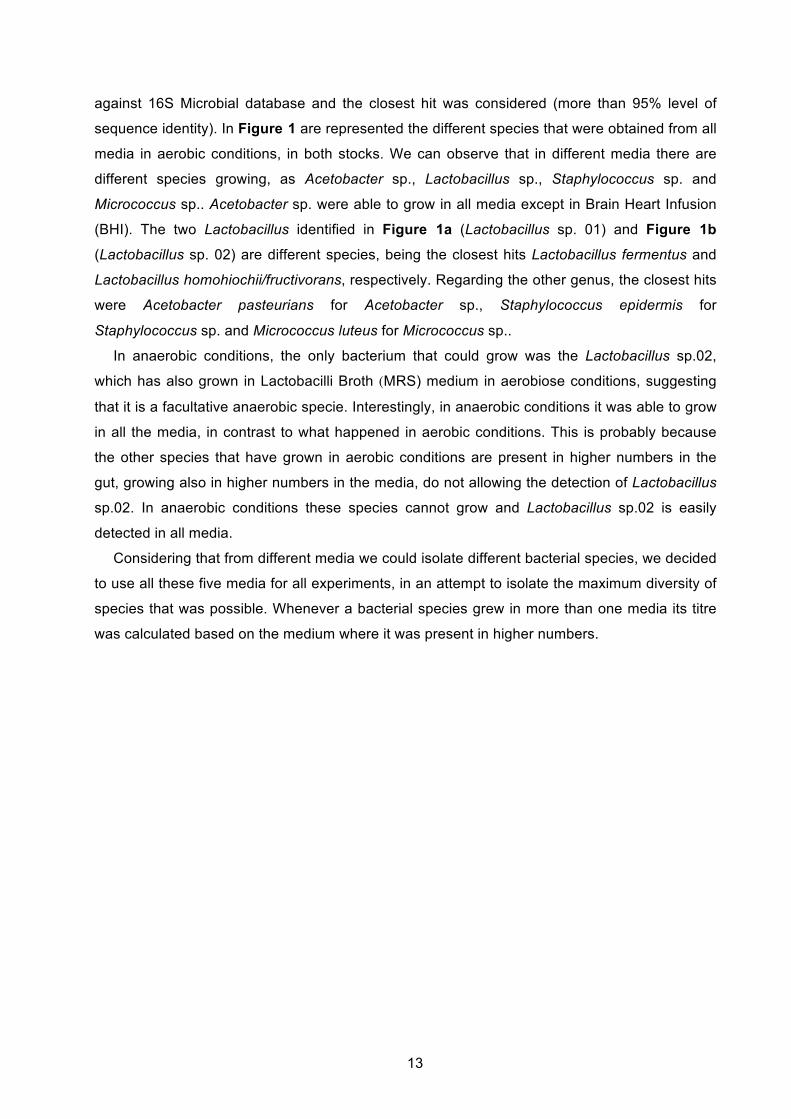
sequence identity). In Figure 1 are represented the different species that were obtained from all
media in aerobic conditions, in both stocks. We can observe that in different media there are
different species growing, as Acetobacter sp., Lactobacillus sp., Staphylococcus sp. and
Micrococcus sp.. Acetobacter sp. were able to grow in all media except in Brain Heart Infusion
(BHI). The two Lactobacillus identified in Figure 1a (Lactobacillus sp. 01) and Figure 1b
(Lactobacillus sp. 02) are different species, being the closest hits Lactobacillus fermentus and
Lactobacillus homohiochii/fructivorans, respectively. Regarding the other genus, the closest hits
were Acetobacter pasteurians for Acetobacter sp., Staphylococcus epidermis for
Staphylococcus sp. and Micrococcus luteus for Micrococcus sp..
In anaerobic conditions, the only bacterium that could grow was the Lactobacillus sp.02,
which has also grown in Lactobacilli Broth (MRS) medium in aerobiose conditions, suggesting
that it is a facultative anaerobic specie. Interestingly, in anaerobic conditions it was able to grow
in all the media, in contrast to what happened in aerobic conditions. This is probably because
the other species that have grown in aerobic conditions are present in higher numbers in the
gut, growing also in higher numbers in the media, do not allowing the detection of Lactobacillus
sp.02. In anaerobic conditions these species cannot grow and Lactobacillus sp.02 is easily
detected in all media.
Considering that from different media we could isolate different bacterial species, we decided
to use all these five media for all experiments, in an attempt to isolate the maximum diversity of
species that was possible. Whenever a bacterial species grew in more than one media its titre
was calculated based on the medium where it was present in higher numbers.
14
3.1.2. Microbiota diversity in wild and laboratory flies
To characterize the microbiota from Drosophila melanogaster we used flies from an isogenic
laboratory stock w1118 iso [54] and flies that were collected from nature. Wild flies were collected
from a rotting figs under figs trees and after their identification as Drosophila melanogaster,
microbiota analyses were immediately performed. All the analysis was done by plating single
guts or single whole flies, for both laboratory and wild caught flies.
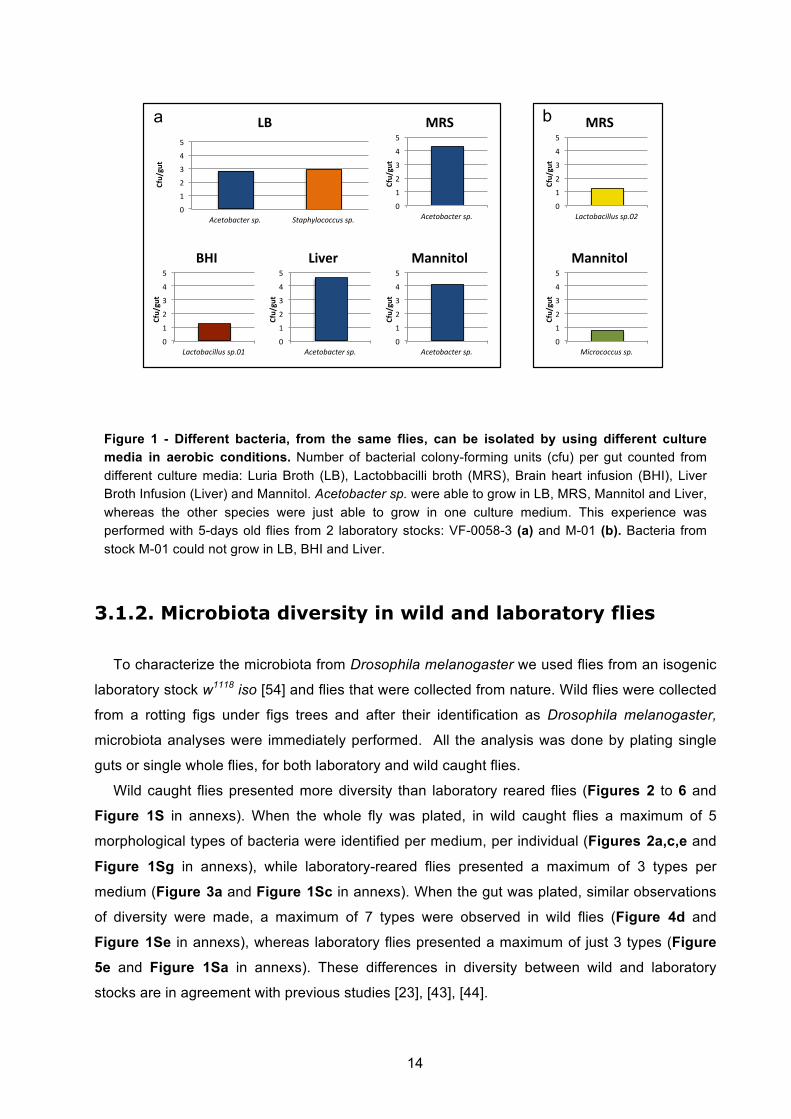
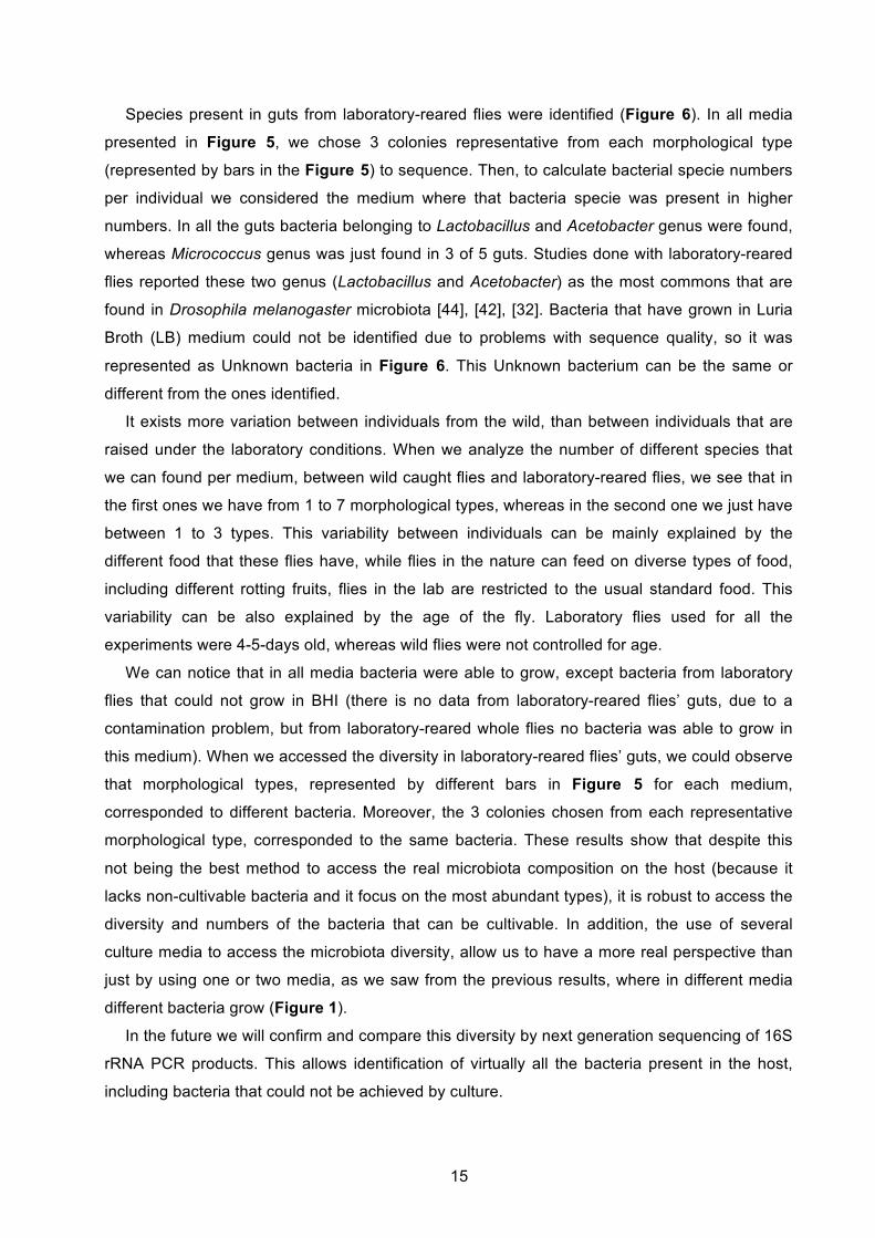
Wild caught flies presented more diversity than laboratory reared flies (Figures 2 to 6 and
Figure 1S in annexs). When the whole fly was plated, in wild caught flies a maximum of 5
morphological types of bacteria were identified per medium, per individual (Figures 2a,c,e and
Figure 1Sg in annexs), while laboratory-reared flies presented a maximum of 3 types per
medium (Figure 3a and Figure 1Sc in annexs). When the gut was plated, similar observations
of diversity were made, a maximum of 7 types were observed in wild flies (Figure 4d and
Figure 1Se in annexs), whereas laboratory flies presented a maximum of just 3 types (Figure
5e and Figure 1Sa in annexs). These differences in diversity between wild and laboratory
stocks are in agreement with previous studies [23], [43], [44].
!"
#"
$"
%"
&"
'"
!"#$%&'"$#()*+,) -$'+./0%"%""1*)*+,)
!"#$%#&'
()'
!"
#"
$"
%"
&"
'"
!"#$%&'"$#()*+,)
!"#$%#&'
*+,'
!"
#"
$"
%"
&"
'"
2'"$%&'"3001*)*+,45)
!"#$%#&'
*+,'
!"
#"
$"
%"
&"
'"
63"(%"%""1*)*+,)
!"#$%#&'
*-../&01'
!"
#"
$"
%"
&"
'"
2'"$%&'"3001*)*+,47)
!"#$%#&'
)23'
!"
#"
$"
%"
&"
'"
!"#$%&'"$#()*+,)
!"#$%#&'
(/456'
!"
#"
$"
%"
&"
'"
!"#$%&'"$#()*+,)
!"#$%#&'
*-../&01'
a b
Figure 1 - Different bacteria, from the same flies, can be isolated by using different culture media in aerobic conditions. Number of bacterial colony-forming units (cfu) per gut counted from different culture media: Luria Broth (LB), Lactobbacilli broth (MRS), Brain heart infusion (BHI), Liver Broth Infusion (Liver) and Mannitol. Acetobacter sp. were able to grow in LB, MRS, Mannitol and Liver, whereas the other species were just able to grow in one culture medium. This experience was performed with 5-days old flies from 2 laboratory stocks: VF-0058-3 (a) and M-01 (b). Bacteria from stock M-01 could not grow in LB, BHI and Liver.
15
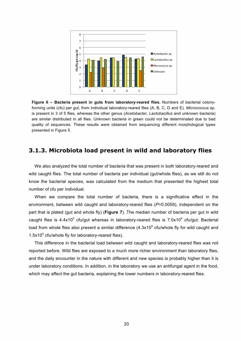
Species present in guts from laboratory-reared flies were identified (Figure 6). In all media
presented in Figure 5, we chose 3 colonies representative from each morphological type
(represented by bars in the Figure 5) to sequence. Then, to calculate bacterial specie numbers
per individual we considered the medium where that bacteria specie was present in higher
numbers. In all the guts bacteria belonging to Lactobacillus and Acetobacter genus were found,
whereas Micrococcus genus was just found in 3 of 5 guts. Studies done with laboratory-reared
flies reported these two genus (Lactobacillus and Acetobacter) as the most commons that are
found in Drosophila melanogaster microbiota [44], [42], [32]. Bacteria that have grown in Luria
Broth (LB) medium could not be identified due to problems with sequence quality, so it was
represented as Unknown bacteria in Figure 6. This Unknown bacterium can be the same or
different from the ones identified.
It exists more variation between individuals from the wild, than between individuals that are
raised under the laboratory conditions. When we analyze the number of different species that
we can found per medium, between wild caught flies and laboratory-reared flies, we see that in
the first ones we have from 1 to 7 morphological types, whereas in the second one we just have
between 1 to 3 types. This variability between individuals can be mainly explained by the
different food that these flies have, while flies in the nature can feed on diverse types of food,
including different rotting fruits, flies in the lab are restricted to the usual standard food. This
variability can be also explained by the age of the fly. Laboratory flies used for all the
experiments were 4-5-days old, whereas wild flies were not controlled for age.
We can notice that in all media bacteria were able to grow, except bacteria from laboratory
flies that could not grow in BHI (there is no data from laboratory-reared flies’ guts, due to a
contamination problem, but from laboratory-reared whole flies no bacteria was able to grow in
this medium). When we accessed the diversity in laboratory-reared flies’ guts, we could observe
that morphological types, represented by different bars in Figure 5 for each medium,
corresponded to different bacteria. Moreover, the 3 colonies chosen from each representative
morphological type, corresponded to the same bacteria. These results show that despite this
not being the best method to access the real microbiota composition on the host (because it
lacks non-cultivable bacteria and it focus on the most abundant types), it is robust to access the
diversity and numbers of the bacteria that can be cultivable. In addition, the use of several
culture media to access the microbiota diversity, allow us to have a more real perspective than
just by using one or two media, as we saw from the previous results, where in different media
different bacteria grow (Figure 1).
In the future we will confirm and compare this diversity by next generation sequencing of 16S
rRNA PCR products. This allows identification of virtually all the bacteria present in the host,
including bacteria that could not be achieved by culture.
16
!"
#"
$"
%"
&"
'"
("
)"
*"
+,-" .+" /01" .2345" /677289:"
!"#$%&'
()*+,*-'./0*
!"
#"
$"
%"
&"
'"
("
)"
*"
+,-" .+" /01" .2345" /677289:"
!"#$%&'
()*+,*-'./0*
!"
#"
$"
%"
&"
'"
("
)"
*"
+,-" .+" /01" .2345" /677289:"
!"#$%&'
()*+,*-'./0*
!"
#"
$"
%"
&"
'"
("
)"
*"
+,-" .+" /01" .2345" /677289:"
!"#$%&'
()*+,*-'./0*
!"
#"
$"
%"
&"
'"
("
)"
*"
+,-" .+" /01" .2345" /677289:"
!"#$%&'
()*+,*-'./0*
a b
c d
e
Figure 2 - Microbiota diversity in individual wild caught whole flies. Number of bacterial colony-forming units (cfu) per single whole fly (a-e) counted from different culture media (BHI, LB, MRS, Liver and Mannitol). Each bar represents a different morphological type identified in each medium. Between 1-5 morphological types can be identified in different culture media. Flies used were not age controlled (n=5).
17
!"
#"
$"
%"
&"
'"
("
)"
*"
+,-" .+" /01" .2345" /677289:"
!"#$%&'
()*+,*-'./0*
!"
#"
$"
%"
&"
'"
("
)"
*"
+,-" .+" /01" .2345" /677289:"
!"#$%&'
()*+,*-'./0*
!"
#"
$"
%"
&"
'"
("
)"
*"
+,-" .+" /01" .2345" /677289:"
!"#$%&'
()*+,*-'./0*
!"
#"
$"
%"
&"
'"
("
)"
*"
+,-" .+" /01" .2345" /677289:"
!"#$%&'
()*+,*-'./0*
!"
#"
$"
%"
&"
'"
("
)"
*"
+,-" .+" /01" .2345" /677289:"
!"#$%&'
()*+,*-'./0*
!"
#"
$"
%"
&"
'"
("
)"
*"
+,-" .+" /01" .2345" /677289:"
!"#$%&'
()*+,*-'./0*
!"
#"
$"
%"
&"
'"
("
)"
*"
+,-" .+" /01" .2345" /677289:"
!"#$%&'
()*+,*-'./0*
!"
#"
$"
%"
&"
'"
("
)"
*"
+,-" .+" /01" .2345" /677289:"
!"#$%&'
()*+,*-'./0*
!"
#"
$"
%"
&"
'"
("
)"
*"
+,-" .+" /01" .2345" /677289:"
!"#$%&'
()*+,*-'./0*
!"
#"
$"
%"
&"
'"
("
)"
*"
+,-" .+" /01" .2345" /677289:"
!"#$%&'
()*+,*-'./0*
a b
c d
e f
g h
i j
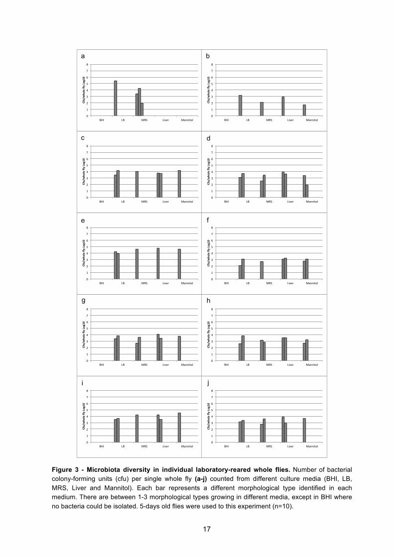
Figure 3 - Microbiota diversity in individual laboratory-reared whole flies. Number of bacterial colony-forming units (cfu) per single whole fly (a-j) counted from different culture media (BHI, LB, MRS, Liver and Mannitol). Each bar represents a different morphological type identified in each medium. There are between 1-3 morphological types growing in different media, except in BHI where no bacteria could be isolated. 5-days old flies were used to this experiment (n=10).
18
!"
#"
$"
%"
&"
'"
("
)"
*"
+," -./" +0123" -4550678"
!"#$%&
'(#)'*+(,-'
!"
#"
$"
%"
&"
'"
("
)"
*"
+," -./" +0123" -4550678"
!"#$%&
'(#)'*+(,-'
!"
#"
$"
%"
&"
'"
("
)"
*"
+," -./" +0123" -4550678"
!"#$%&
'(#)'*+(,-'
!"
#"
$"
%"
&"
'"
("
)"
*"
+," -./" +0123" -4550678"
!"#$%&
'(#)'*+(,-'
!"
#"
$"
%"
&"
'"
("
)"
*"
+," -./" +0123" -4550678"
!"#$%&
'(#)'*+(,-'
!"
#"
$"
%"
&"
'"
("
)"
*"
+," -./" +0123" -4550678"
!"#$%&
'(#)'*+(,-'
!"
#"
$"
%"
&"
'"
("
)"
*"
+," -./" +0123" -4550678"
!"#$%&
'(#)'*+(,-'
!"
#"
$"
%"
&"
'"
("
)"
*"
+," -./" +0123" -4550678"
!"#$%&
'(#)'*+(,-'
!"
#"
$"
%"
&"
'"
("
)"
*"
+," -./" +0123" -4550678"
!"#$%&
'(#)'*+(,-'
!"
#"
$"
%"
&"
'"
("
)"
*"
+," -./" +0123" -4550678"
!"#$%&
'(#)'*+(,-'
a b
c d
e f
g h
i j
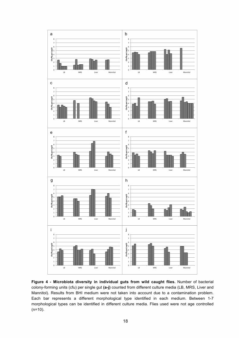
Figure 4 - Microbiota diversity in individual guts from wild caught flies. Number of bacterial colony-forming units (cfu) per single gut (a-j) counted from different culture media (LB, MRS, Liver and Mannitol). Results from BHI medium were not taken into account due to a contamination problem. Each bar represents a different morphological type identified in each medium. Between 1-7 morphological types can be identified in different culture media. Flies used were not age controlled (n=10).
19
!"
#"
$"
%"
&"
'"
("
)"
*"
+," -./" +0123" -4550678"
!"#$%&
'(#)'*+(,-'
!"
#"
$"
%"
&"
'"
("
)"
*"
+," -./" +0123" -4550678"
!"#$%&
'(#)'*+(,-'
!"
#"
$"
%"
&"
'"
("
)"
*"
+," -./" +0123" -4550678"
!"#$%&
'(#)'*+(,-'
!"
#"
$"
%"
&"
'"
("
)"
*"
+," -./" +0123" -4550678"
!"#$%&
'(#)'*+(,-'
!"
#"
$"
%"
&"
'"
("
)"
*"
+," -./" +0123" -4550678"
!"#$%&
'(#)'*+(,-'
a b
c d
e
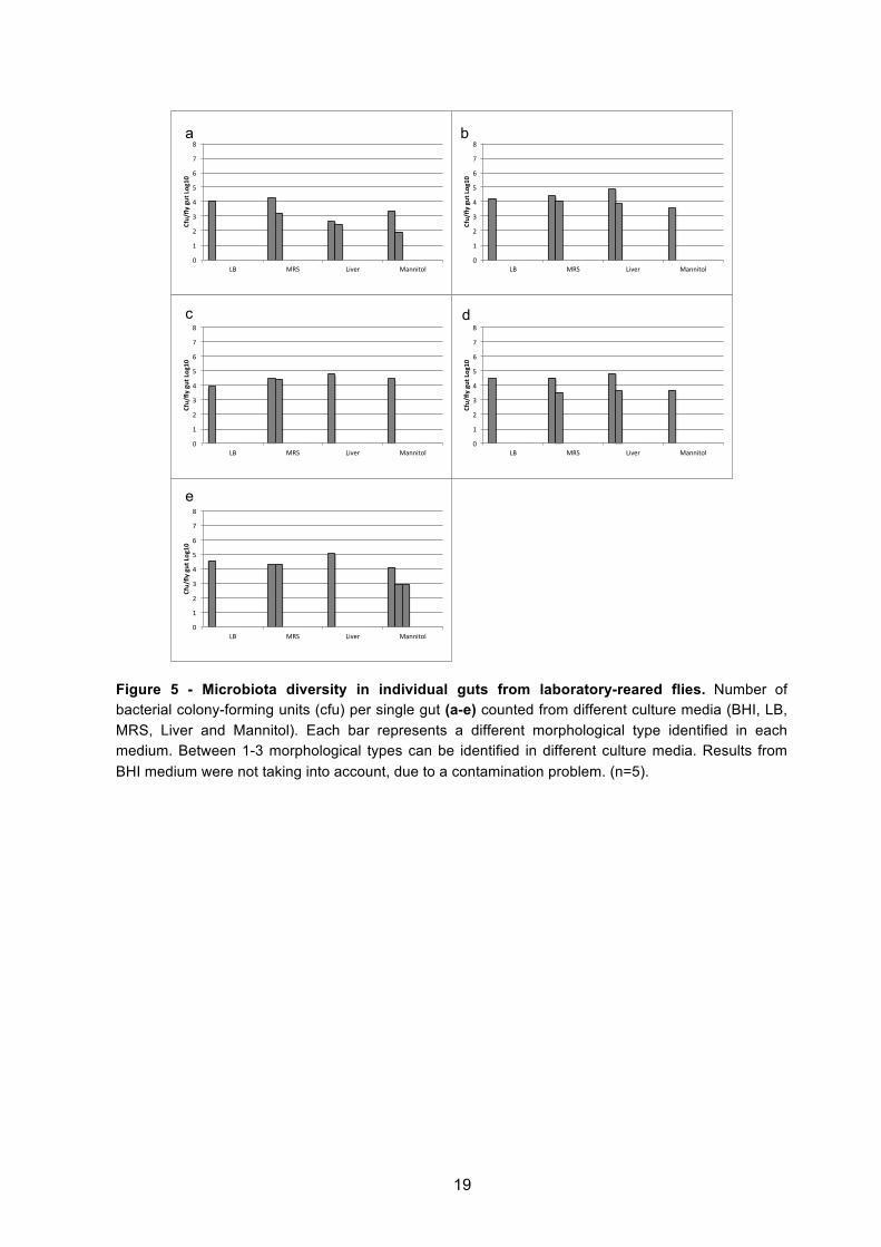
Figure 5 - Microbiota diversity in individual guts from laboratory-reared flies. Number of bacterial colony-forming units (cfu) per single gut (a-e) counted from different culture media (BHI, LB, MRS, Liver and Mannitol). Each bar represents a different morphological type identified in each medium. Between 1-3 morphological types can be identified in different culture media. Results from BHI medium were not taking into account, due to a contamination problem. (n=5).
20
3.1.3. Microbiota load present in wild and laboratory flies
We also analyzed the total number of bacteria that was present in both laboratory-reared and
wild caught flies. The total number of bacteria per individual (gut/whole flies), as we still do not
know the bacterial species, was calculated from the medium that presented the highest total
number of cfu per individual.
When we compare the total number of bacteria, there is a significative effect in the
environment, between wild caught and laboratory-reared flies (P=0.0059), independent on the
part that is plated (gut and whole fly) (Figure 7). The median number of bacteria per gut in wild
caught flies is 4.4x10⁵ cfu/gut whereas in laboratory-reared flies is 7.0x10⁴ cfu/gut. Bacterial
load from whole flies also present a similar difference (4.3x10⁵ cfu/whole fly for wild caught and
1.5x10⁴ cfu/whole fly for laboratory-reared flies).
This difference in the bacterial load between wild caught and laboratory-reared flies was not
reported before. Wild flies are exposed to a much more richer environment than laboratory flies,
and the daily encounter in the nature with different and new species is probably higher than it is
under laboratory conditions. In addition, in the laboratory we use an antifungal agent in the food,
which may affect the gut bacteria, explaining the lower numbers in laboratory-reared flies.
!"
#"
$"
%"
&"
'"
("
)"
*"
+" ," -" ." /"
!"#$%&
'(#)'*+(',-'
+0123450216"789"
:5023450;<<=7"789"
>;0630300=7"789"
?@A@3B@"
Figure 6 – Bacteria present in guts from laboratory-reared flies. Numbers of bacterial colony-forming units (cfu) per gut, from individual laboratory-reared flies (A, B, C, D and E). Micrococcus sp. is present in 3 of 5 flies, whereas the other genus (Acetobacter, Lactobacillus and unknown bacteria) are similar distributed in all flies. Unknown bacteria in green could not be determinated due to bad quality of sequences. These results were obtained from sequencing different morphological types presented in Figure 5.
21
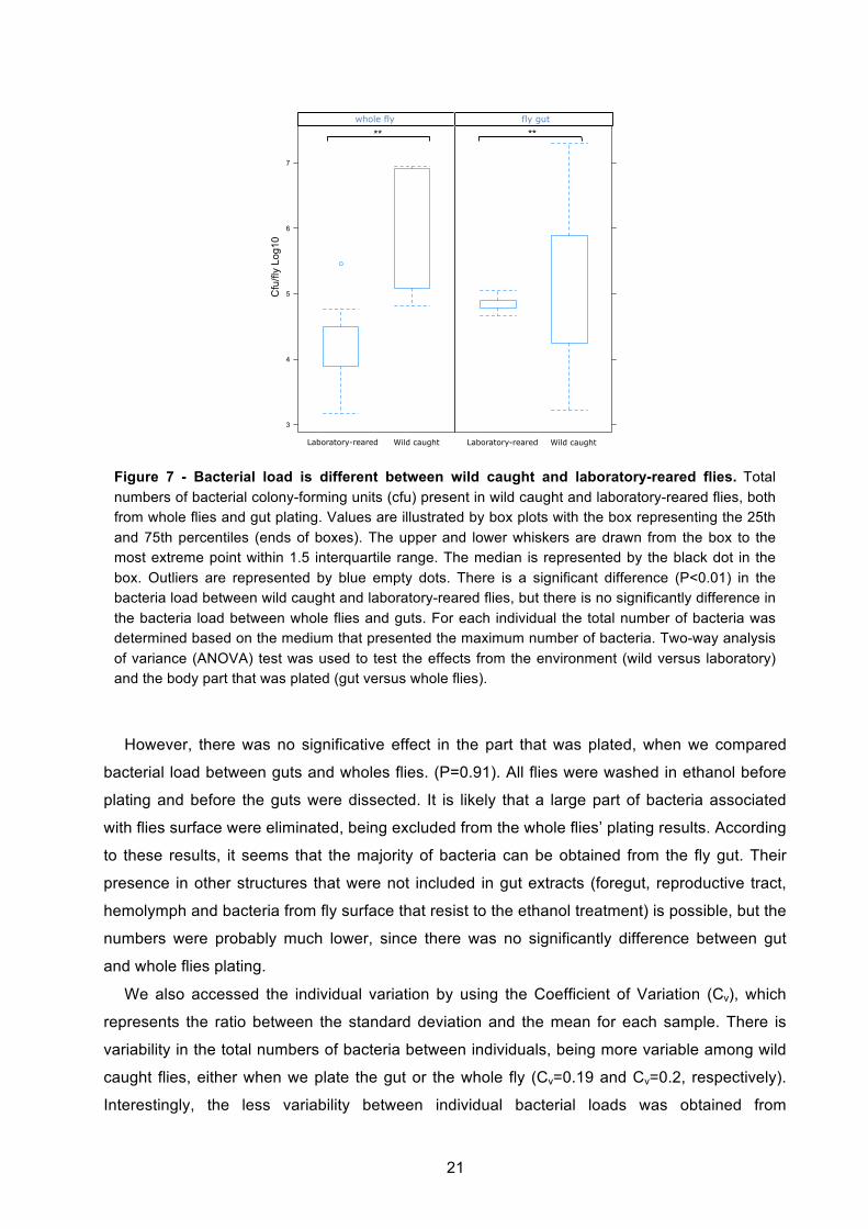
However, there was no significative effect in the part that was plated, when we compared
bacterial load between guts and wholes flies. (P=0.91). All flies were washed in ethanol before
plating and before the guts were dissected. It is likely that a large part of bacteria associated
with flies surface were eliminated, being excluded from the whole flies’ plating results. According
to these results, it seems that the majority of bacteria can be obtained from the fly gut. Their
presence in other structures that were not included in gut extracts (foregut, reproductive tract,
hemolymph and bacteria from fly surface that resist to the ethanol treatment) is possible, but the
numbers were probably much lower, since there was no significantly difference between gut
and whole flies plating.
We also accessed the individual variation by using the Coefficient of Variation (Cv), which
represents the ratio between the standard deviation and the mean for each sample. There is
variability in the total numbers of bacteria between individuals, being more variable among wild
caught flies, either when we plate the gut or the whole fly (Cv=0.19 and Cv=0.2, respectively).
Interestingly, the less variability between individual bacterial loads was obtained from
cfu
3
4
5
6
7
lab wild
fly
lab wild
gutwhole fly fly gut
Laboratory-reared Wild caught Laboratory-reared Wild caught
** **
Cfu
/fly
Log1
0
Figure 7 - Bacterial load is different between wild caught and laboratory-reared flies. Total numbers of bacterial colony-forming units (cfu) present in wild caught and laboratory-reared flies, both from whole flies and gut plating. Values are illustrated by box plots with the box representing the 25th and 75th percentiles (ends of boxes). The upper and lower whiskers are drawn from the box to the most extreme point within 1.5 interquartile range. The median is represented by the black dot in the box. Outliers are represented by blue empty dots. There is a significant difference (P<0.01) in the bacteria load between wild caught and laboratory-reared flies, but there is no significantly difference in the bacteria load between whole flies and guts. For each individual the total number of bacteria was determined based on the medium that presented the maximum number of bacteria. Two-way analysis of variance (ANOVA) test was used to test the effects from the environment (wild versus laboratory) and the body part that was plated (gut versus whole flies).
22
laboratory-reared flies, when the gut was plated (Cv=0.03) (Figure 7). This would be
expectable, taking into account the fact that they were exposed always to the same conditions
when they were raised, and they are all the same age, opposite to wild flies. Higher variability
was obtained in laboratory flies when the whole fly was plated (Cv=0.05). This higher variability
can be explained by different things: i) It is possible that when flies were washed in ethanol
before the plating, bacteria elimination from fly’s surface was different among the flies, resulting
in bacterial load differences; ii) Bacteria is probably best extracted when we homogenize one
gut than one fly. In whole flies there are more tissues to homogenize, which can results in
differences between flies; iii) We have also to consider that flies can have more variability in the
bacterial load present in the parts that were not considered in gut dissections.
In this two last sections we could observe that wild caught flies present more diversity and
higher bacterial load in the microbiota community, when compared with laboratory-reared flies.
This higher diversity in wild flies was already reported by previous studies. However, this
variability at the individual level, in wild caught or laboratory-reared Drosophila melanogaster
flies was not determined before. All previous studies in microbiota diversity were done with
pools of flies, and not with single flies. There are also a few studies that quantify the bacteria
that is present inside the gut and in whole flies, because they use techniques that show ratio
between species but not real quantities present in the host. [23], [43], [44], [32], [42].
These results will be more interesting when we characterize all the species from all
individuals, both guts and whole flies. We already did for laboratory-reared flies’ guts, as we
show in Figure 6. All individuals showed a similar composition of microbiota inside the gut. Wild
caught flies already showed by identification of colonies morphological types to present a higher
diversity. It will be interesting to see this diversity from individual to individual, since they were
not maintained in the same conditions, as laboratory flies were.
3.2. Drosophila microbiota stability and colonization inside the host
Considering the previous results, we could observe that there are differences in the
microbiota community between flies that are reared under laboratory conditions and the wild
flies, which harbor a more diversity community. This shows that laboratory flies lack diversity in
the microbiota and maybe are not representative from the natural microbiota found in wild flies.
Two recent studies also showed this concern. Wong et al. reported low-diversity in the gut
microbiota of laboratory-reared Drosophila melanogaster [32]. In addition, Chandler et al. show
23
that gut microbiota from flies reared under laboratory conditions are not representative from the
wild caught flies gut microbiota [23].
All studies that are done in Drosophila melanogaster gut microbiota do not take into account
the possible presence of transient species inside the gut. The difference that we also obtained
between flies caught in the natural environment and flies that are in the laboratory for several
years reinforce the importance of addressing to this question. Laboratory flies can be acquiring
bacteria from laboratory environment, including food, and losing microbiota that was normally
present when they were caught.
Here, we developed a new protocol to understand the stability of gut microbiota in
laboratory-reared and wild caught flies.
To discriminate between transient bacteria that came from the environment we had to raise
flies in conditions that would minimize reinfection of the flies by their own feces and microbiota.
We kept single laboratory-reared adult flies in large cages (Figure 2S in annex). In each cage
food was provided in petri dishes with a total area of 486.3cm2. Every day the food was
changed and cage sterilized. The purpose was to minimize the probability of the fly to get
reinfected by contacting the places where the fly was before and by the bacteria that would
come out with the feces. We also maintained single flies in vials without changing the food,
allowing the reinfection of the flies, as a control. We maintained these flies during 10 days, and
guts extracts were plated in the beginning and in the end of this protocol.
Surprisingly, flies that were kept in cages suffered a dramatic decrease in their gut microbiota
after 10 days, changing from 7.0x10⁴ cfu/gut that were present in the beginning of the protocol
to 8 cfu/gut. In contrast, flies that were kept the 10 days in the same vials had an increase in
their gut bacteria to 2.9x10⁶ cfu/gut (Figure 8a).

24
The elimination of almost all the microbiota from the gut, in the flies that were kept in cages
with new food, shows that the gut microbiota of laboratory-reared flies is unstable. This
decrease was because we avoided the reinfection with the transient bacteria that was being
loaded by the food. When they keep feeding on the same food, they are being reinfected by
their transient bacteria, causing an increase in bacterial load over age.
This result has never been reported before and may have a big impact in all the studies that
are being done in Drosophila gut microbiota. Many papers characterized the microbiota
community and studied its influence in the fly, without knowing if they were studying the
microbiota community that normally colonizes the fly [42], [19], [55], [40]. This can lead to wrong
conclusions, depending on the type of question that they were trying to answer. For example,
Ren et al. reports an increasing in microbiota load over age. This increase is probably because
flies are being reinfected with the bacteria from the feces that are in the food, and they do not
know if the increase is inside the fly or if the bacteria are growing on the food [42]. This study in
the light of these new results has no biologic meaning, they reported an increase in Drosophila
microbiota load as they could have reported a decrease if they would have kept flies with a
different protocol.
This protocol of maintaining single flies per cage is not feasible when we want to do several
samples at the same time, due to constrains in cages number and space. We decided to try the
same principle but by keeping flies in vials. Single flies were kept per vial and the vials were
Laboratory-reared flies gut
day 0
day 1
0 CNT
day 1
0 CG
0
2
4
6
8
10
Cfu
/gut
Log
10
Laboratory-reared flies gut
Day 0
Day 10
0
2
4
6
8
10
Cfu
/gut
Log
10
Wild caught flies gut
Day 0
Day 10
0
2
4
6
8
10
Cfu
/gut
Log
10
Microbiota from wild and laboratory flies
LG LF WG W
F0
2
4
6
8
10
Laboratory-reared whole flies
Day 0
Day 10
0
2
4
6
8
10
Cfu
/who
le fl
y Lo
g10
Wild caught whole flies
Day 0
Day 10
0
2
4
6
8
10
Cfu
/who
le fl
y Lo
g10
Microbiota after aging comparing wild and laboratory flies
LG LF WG W
F0
2
4
6
8
Laboratory-reared flies gut
day 0
day 1
0 CNT
day 1
0 CG
0
2
4
6
8
10
Cfu
/gut
Log
10
Laboratory-reared flies gut
Day 0
Day 10
0
2
4
6
8
10
Cfu
/gut
Log
10
Wild caught flies gut
Day 0
Day 10
0
2
4
6
8
10
Cfu
/gut
Log
10
Microbiota from wild and laboratory flies
LG LF WG W
F0
2
4
6
8
10
Laboratory-reared whole flies
Day 0
Day 10
0
2
4
6
8
10
Cfu
/gut
Log
10
Wild caught whole flies
Day 0
Day 10
0
2
4
6
8
10
Cfu
/gut
Log
10
Microbiota after aging comparing wild and laboratory flies
LG LF WG W
F0
2
4
6
8
a **
Laboratory-reared flies gut
day 0
day 1
0 CNT
day 1
0 CG
0
2
4
6
8
10
Cfu
/gut
Log
10
Laboratory-reared flies gut
Day 0
Day 10
0
2
4
6
8
10
Cfu
/gut
Log
10Wild caught flies gut
Day 0
Day 10
0
2
4
6
8
10C
fu/g
ut L
og10
Microbiota from wild and laboratory flies
LG LF WG W
F0
2
4
6
8
10
Laboratory-reared whole flies
Day 0
Day 10
0
2
4
6
8
10
Cfu
/gut
Log
10
Wild caught whole flies
Day 0
Day 10
0
2
4
6
8
10
Cfu
/gut
Log
10
Microbiota after aging comparing wild and laboratory flies
LG LF WG W
F0
2
4
6
8
b c ** **
Figure 8 - Drosophila microbiota from laboratory-reared flies is not stable. Total numbers of bacterial colony-forming units (cfu), from flies that were subjected to different aging protocols. (a) Gut microbiota from laboratory-reared flies significantly decreases when flies are aged in cages (day 10 CG) comparing with flies aged in vials without changing (day 10 CNT). Total number of cfu from laboratory-reared guts (b) and whole flies (c), before (day 0) and after (day 10) aging in vials. Both decreases in microbiota from guts (a,b) and whole flies (c) are significantly different with p<0.01 (**). Kruskal-Wallis proceeded by a Kruskal-Wallis multiple comparison test was done (a). Wilcox test was used to compare between day 0 and day 10 (b and c). Each dot represents one gut (a,b) or one fly (c). n=5 for (a,b) and n=10 for (c).
25
changed twice a day because the food area was much smaller than the food from cages (3.8
cm2). The same huge decrease in gut microbiota load was verified when flies were subjected to
the aging protocol in vials. Gut microbiota significantly decreased from 7.0x10⁴ cfu/gut to
2.5x101 cfu/gut (P=0.001) (Figure 8b). Although by doing this protocol in cages we could
achieve a lower microbiota number at day 10, this number was not significantly different from
the numbers obtained in the vials at the same day (P=0.2). We decided to keep the vials
protocol for follow up experiments because we can test more flies at the same time.
We repeated the same experiment to measure the bacteria in the whole fly to observe if, as
in the gut, it was also decreasing. The decrease was also significantly different, changing from
1.5x10⁴ cfu/gut to 1.2x10³ cfu/gut (P=0.006) (Figure 8c). Considering that almost all the
bacteria disappear from the gut after the protocol, ending just with 2.5x101 cfu/gut, there must
be bacteria persisting outside the gut, as in the reproductive tract and flies’ surface, that can
explain the difference between bacterial load obtained from guts and from whole flies at day 10.
We can explain the higher number of bacteria persisting in the whole fly after the 10 days,
because whereas in the gut there is constant food traffic, in the other parts that were also
included in the whole fly, the microenvironment is more stable.
When we consider this decrease after the protocol in whole flies, we cannot say if this
difference is just due to the bacteria that is disappearing inside the gut, or if can also be
explained by some bacteria that is disappearing in fly’s surface. To approach this we could have
plated instead the whole fly, just the carcass without the gut, to be sure that the difference was
not only in the gut.
In contrast to results obtained when we compare whole flies plating versus gut plating
(Figure 7), where the difference is not significant, here we can see that there are more bacteria
outside the gut, because they persist after the aging protocol.
3.2.1. There is a correlation between number of bacteria found on the host and on the food
In laboratory-reared flies, we could observe that there was a huge decrease in the bacterial
load when there was an attempt to avoid the reinfection with their own bacteria. In contrast,
there was a huge increase when they were kept in the same vials and reinfected with their own
bacteria. We measured the bacteria that were present in the last food where flies have been
before they were collected at day 10. We analyzed food from cages, from the vials that were
changed twice a day and from the control vials that were not changed.
Interestingly, we observed a correlation between bacterial load found in the food and found
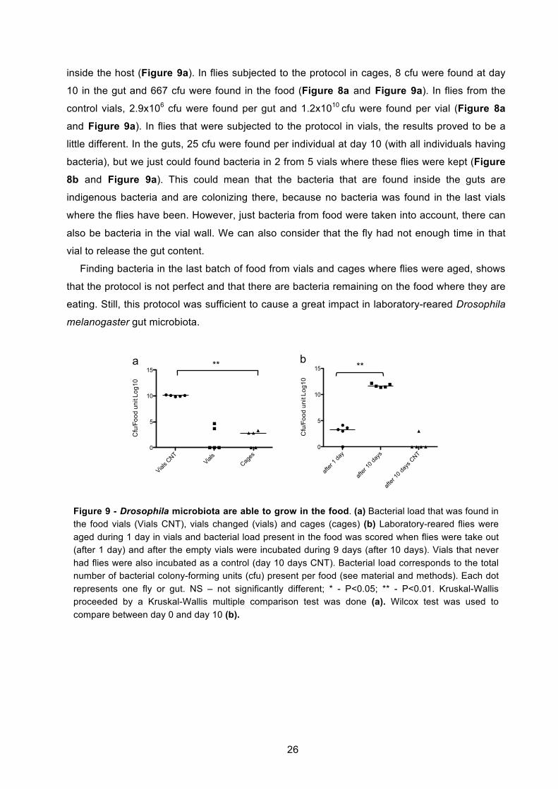
26
inside the host (Figure 9a). In flies subjected to the protocol in cages, 8 cfu were found at day
10 in the gut and 667 cfu were found in the food (Figure 8a and Figure 9a). In flies from the
control vials, 2.9x10⁶ cfu were found per gut and 1.2x10¹⁰ cfu were found per vial (Figure 8a
and Figure 9a). In flies that were subjected to the protocol in vials, the results proved to be a
little different. In the guts, 25 cfu were found per individual at day 10 (with all individuals having
bacteria), but we just could found bacteria in 2 from 5 vials where these flies were kept (Figure
8b and Figure 9a). This could mean that the bacteria that are found inside the guts are
indigenous bacteria and are colonizing there, because no bacteria was found in the last vials
where the flies have been. However, just bacteria from food were taken into account, there can
also be bacteria in the vial wall. We can also consider that the fly had not enough time in that
vial to release the gut content.
Finding bacteria in the last batch of food from vials and cages where flies were aged, shows
that the protocol is not perfect and that there are bacteria remaining on the food where they are
eating. Still, this protocol was sufficient to cause a great impact in laboratory-reared Drosophila
melanogaster gut microbiota.
Cfu
/Foo
d un
it Lo
g10
Vials C
NTVial
s
Cages
0
5
10
15
Cfu
/Foo
d un
it Lo
g10
after
1 da
y
after
10 da
ys
after
10 da
ys C
NT0
5
10
15
Cfu
/Foo
d un
it Lo
g10
Vials C
NTVial
s
Cages
0
5
10
15
Cfu
/Foo
d un
it Lo
g10
after
1 da
y
after
10 da
ys0
5
10
15
b a ** **
Figure 9 - Drosophila microbiota are able to grow in the food. (a) Bacterial load that was found in the food vials (Vials CNT), vials changed (vials) and cages (cages) (b) Laboratory-reared flies were aged during 1 day in vials and bacterial load present in the food was scored when flies were take out (after 1 day) and after the empty vials were incubated during 9 days (after 10 days). Vials that never had flies were also incubated as a control (day 10 days CNT). Bacterial load corresponds to the total number of bacterial colony-forming units (cfu) present per food (see material and methods). Each dot represents one fly or gut. NS – not significantly different; * - P<0.05; ** - P<0.01. Kruskal-Wallis proceeded by a Kruskal-Wallis multiple comparison test was done (a). Wilcox test was used to compare between day 0 and day 10 (b).

27
3.2.2. Microbiota from laboratory-reared flies grows on the food
From the previous section, we could observe a correlation between the bacterial load found
inside the gut and on the respective food. Moreover, we observed an increase on the microbiota
from flies that were kept in the same vials, in contrast to what happened with flies where the
food was changed. This increase in the bacterial load means that bacteria must be growing
somewhere. As they almost disappear from the gut when the fly is not reinfected by its own
microbiota, we asked if the bacteria could be growing on the food. To approach this, we put
single flies per vial during 1 day and then we took the flies out. Then we incubated the empty
vials during 9 days. We measured the bacteria present in the food at these two time-points,
after 1 day and after 10 days from the day 0. After 1 day we could find 2x10³ cfu/food unit and
after 10 days 4x10¹² cfu/food unit were found (Figure 9b). These results mean that during the 9
days that the vials were incubated without flies, 1012 cfu have grown on the food, meaning that
bacteria that belong to Drosophila microbiota can actually grow on the food. Vials that never
had flies inside were incubated as a control, to be sure that there was no contamination on the
food. After 10 days 3x10³ cfu/food unit were found in 1 from 5 vials. Food is not made under
absolute sterile conditions, being possible contaminations with bacteria that are present in the
laboratory environment.
We still do not know which are the bacteria that are growing on the food, but from preliminary
data we observed that there was Acetobacter sp. growing on the food, the same that is found in
laboratory-reared flies’ guts (Figure 6). It will be interesting to see if the bacteria contamination
that was found in one from the five control vials (Figure 9b) can be also found inside the guts,
particularly after the protocol. If the same bacteria would be found inside the guts from flies that
aged in cages or in vials (where the food was changed) at day 10, it would probably mean that
they are being contaminated by the food.
A previous study, reported this correlation between the bacterial load that is found inside the
gut and on the food [42]. However, they concluded that the microbiota was not growing on the
food but inside the fly, because they saw an increase in total number of bacteria in the gut over
age. Here, we show that this increase is dependent on how the flies are maintained. Moreover,
we show that microbiota from laboratory-reared flies can actually grow on the food, in a flies
presence-independent manner.
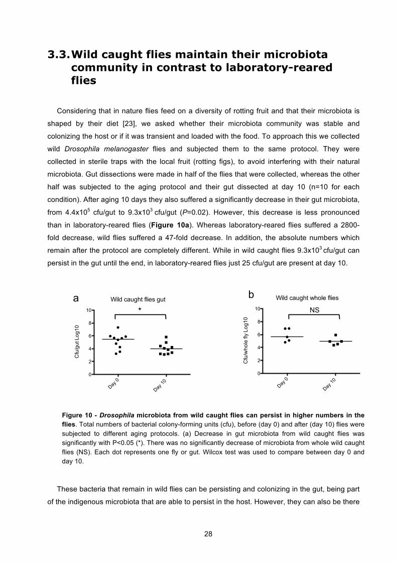
28
3.3. Wild caught flies maintain their microbiota community in contrast to laboratory-reared flies
Considering that in nature flies feed on a diversity of rotting fruit and that their microbiota is
shaped by their diet [23], we asked whether their microbiota community was stable and
colonizing the host or if it was transient and loaded with the food. To approach this we collected
wild Drosophila melanogaster flies and subjected them to the same protocol. They were
collected in sterile traps with the local fruit (rotting figs), to avoid interfering with their natural
microbiota. Gut dissections were made in half of the flies that were collected, whereas the other
half was subjected to the aging protocol and their gut dissected at day 10 (n=10 for each
condition). After aging 10 days they also suffered a significantly decrease in their gut microbiota,
from 4.4x10⁵ cfu/gut to 9.3x10³ cfu/gut (P=0.02). However, this decrease is less pronounced
than in laboratory-reared flies (Figure 10a). Whereas laboratory-reared flies suffered a 2800-
fold decrease, wild flies suffered a 47-fold decrease. In addition, the absolute numbers which
remain after the protocol are completely different. While in wild caught flies 9.3x10³ cfu/gut can
persist in the gut until the end, in laboratory-reared flies just 25 cfu/gut are present at day 10.
These bacteria that remain in wild flies can be persisting and colonizing in the gut, being part
of the indigenous microbiota that are able to persist in the host. However, they can also be there
Laboratory-reared flies gut
day 0
day 1
0 CNT
day 1
0 CG
0
2
4
6
8
10
Cfu
/gut
Log
10
Laboratory-reared flies gut
Day 0
Day 10
0
2
4
6
8
10
Cfu
/gut
Log
10
Wild caught flies gut
Day 0
Day 10
0
2
4
6
8
10
Cfu
/gut
Log
10
Microbiota from wild and laboratory flies
LG LF WG W
F0
2
4
6
8
10
Laboratory-reared whole flies
Day 0
Day 10
0
2
4
6
8
10
Cfu
/who
le fl
y Lo
g10
Wild caught whole flies
Day 0
Day 10
0
2
4
6
8
10
Cfu
/who
le fl
y Lo
g10
Microbiota after aging comparing wild and laboratory flies
LG LF WG W
F0
2
4
6
8
Laboratory-reared flies gut
day 0
day 1
0 CNT
day 1
0 CG
0
2
4
6
8
10
Cfu
/gut
Log
10
Laboratory-reared flies gut
Day 0
Day 10
0
2
4
6
8
10
Cfu
/gut
Log
10
Wild caught flies gut
Day 0
Day 10
0
2
4
6
8
10
Cfu
/gut
Log
10
Microbiota from wild and laboratory flies
LG LF WG W
F0
2
4
6
8
10
Laboratory-reared whole flies
Day 0
Day 10
0
2
4
6
8
10
Cfu
/gut
Log
10
Wild caught whole flies
Day 0
Day 10
0
2
4
6
8
10
Cfu
/gut
Log
10
Microbiota after aging comparing wild and laboratory flies
LG LF WG W
F0
2
4
6
8
a b * NS
Figure 10 - Drosophila microbiota from wild caught flies can persist in higher numbers in the flies. Total numbers of bacterial colony-forming units (cfu), before (day 0) and after (day 10) flies were subjected to different aging protocols. (a) Decrease in gut microbiota from wild caught flies was significantly with P<0.05 (*). There was no significantly decrease of microbiota from whole wild caught flies (NS). Each dot represents one fly or gut. Wilcox test was used to compare between day 0 and day 10.
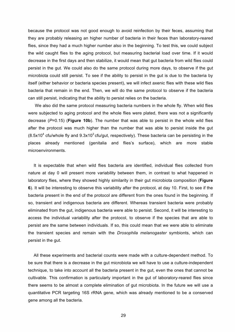
29
because the protocol was not good enough to avoid reinfection by their feces, assuming that
they are probably releasing an higher number of bacteria in their feces than laboratory-reared
flies, since they had a much higher number also in the beginning. To test this, we could subject
the wild caught flies to the aging protocol, but measuring bacterial load over time. If it would
decrease in the first days and then stabilize, it would mean that gut bacteria from wild flies could
persist in the gut. We could also do the same protocol during more days, to observe if the gut
microbiota could still persist. To see if the ability to persist in the gut is due to the bacteria by
itself (either behavior or bacteria species present), we will infect axenic flies with these wild flies
bacteria that remain in the end. Then, we will do the same protocol to observe if the bacteria
can still persist, indicating that the ability to persist relies on the bacteria.
We also did the same protocol measuring bacteria numbers in the whole fly. When wild flies
were subjected to aging protocol and the whole flies were plated, there was not a significantly
decrease (P=0.15) (Figure 10b). The number that was able to persist in the whole wild flies
after the protocol was much higher than the number that was able to persist inside the gut
(8.5x10⁴ cfu/whole fly and 9.3x10³ cfu/gut, respectively). These bacteria can be persisting in the
places already mentioned (genitalia and flies’s surface), which are more stable
microenvironments.
It is expectable that when wild flies bacteria are identified, individual flies collected from
nature at day 0 will present more variability between them, in contrast to what happened in
laboratory flies, where they showed highly similarity in their gut microbiota composition (Figure
6). It will be interesting to observe this variability after the protocol, at day 10. First, to see if the
bacteria present in the end of the protocol are different from the ones found in the beginning. If
so, transient and indigenous bacteria are different. Whereas transient bacteria were probably
eliminated from the gut, indigenous bacteria were able to persist. Second, it will be interesting to
access the individual variability after the protocol, to observe if the species that are able to
persist are the same between individuals. If so, this could mean that we were able to eliminate
the transient species and remain with the Drosophila melanogaster symbionts, which can
persist in the gut.
All these experiments and bacterial counts were made with a culture-dependent method. To
be sure that there is a decrease in the gut microbiota we will have to use a culture-independent
technique, to take into account all the bacteria present in the gut, even the ones that cannot be
cultivable. This confirmation is particularly important in the gut of laboratory-reared flies since
there seems to be almost a complete elimination of gut microbiota. In the future we will use a
quantitative PCR targeting 16S rRNA gene, which was already mentioned to be a conserved
gene among all the bacteria.
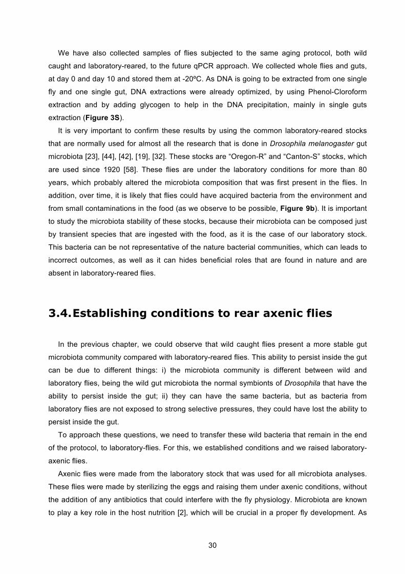
30
We have also collected samples of flies subjected to the same aging protocol, both wild
caught and laboratory-reared, to the future qPCR approach. We collected whole flies and guts,
at day 0 and day 10 and stored them at -20ºC. As DNA is going to be extracted from one single
fly and one single gut, DNA extractions were already optimized, by using Phenol-Cloroform
extraction and by adding glycogen to help in the DNA precipitation, mainly in single guts
extraction (Figure 3S).
It is very important to confirm these results by using the common laboratory-reared stocks
that are normally used for almost all the research that is done in Drosophila melanogaster gut
microbiota [23], [44], [42], [19], [32]. These stocks are “Oregon-R” and “Canton-S” stocks, which
are used since 1920 [58]. These flies are under the laboratory conditions for more than 80
years, which probably altered the microbiota composition that was first present in the flies. In
addition, over time, it is likely that flies could have acquired bacteria from the environment and
from small contaminations in the food (as we observe to be possible, Figure 9b). It is important
to study the microbiota stability of these stocks, because their microbiota can be composed just
by transient species that are ingested with the food, as it is the case of our laboratory stock.
This bacteria can be not representative of the nature bacterial communities, which can leads to
incorrect outcomes, as well as it can hides beneficial roles that are found in nature and are
absent in laboratory-reared flies.
3.4. Establishing conditions to rear axenic flies
In the previous chapter, we could observe that wild caught flies present a more stable gut
microbiota community compared with laboratory-reared flies. This ability to persist inside the gut
can be due to different things: i) the microbiota community is different between wild and
laboratory flies, being the wild gut microbiota the normal symbionts of Drosophila that have the
ability to persist inside the gut; ii) they can have the same bacteria, but as bacteria from
laboratory flies are not exposed to strong selective pressures, they could have lost the ability to
persist inside the gut.
To approach these questions, we need to transfer these wild bacteria that remain in the end
of the protocol, to laboratory-flies. For this, we established conditions and we raised laboratory-
axenic flies.
Axenic flies were made from the laboratory stock that was used for all microbiota analyses.
These flies were made by sterilizing the eggs and raising them under axenic conditions, without
the addition of any antibiotics that could interfere with the fly physiology. Microbiota are known
to play a key role in the host nutrition [2], which will be crucial in a proper fly development. As
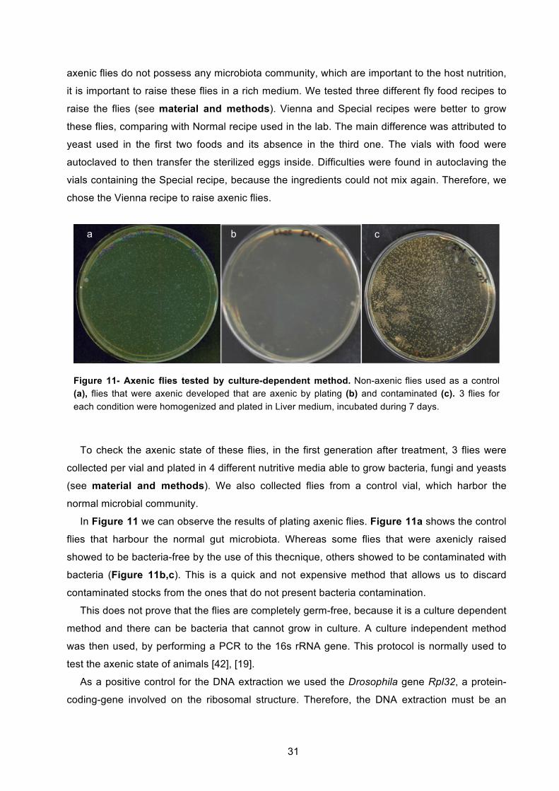
31
axenic flies do not possess any microbiota community, which are important to the host nutrition,
it is important to raise these flies in a rich medium. We tested three different fly food recipes to
raise the flies (see material and methods). Vienna and Special recipes were better to grow
these flies, comparing with Normal recipe used in the lab. The main difference was attributed to
yeast used in the first two foods and its absence in the third one. The vials with food were
autoclaved to then transfer the sterilized eggs inside. Difficulties were found in autoclaving the
vials containing the Special recipe, because the ingredients could not mix again. Therefore, we
chose the Vienna recipe to raise axenic flies.
To check the axenic state of these flies, in the first generation after treatment, 3 flies were
collected per vial and plated in 4 different nutritive media able to grow bacteria, fungi and yeasts
(see material and methods). We also collected flies from a control vial, which harbor the
normal microbial community.
In Figure 11 we can observe the results of plating axenic flies. Figure 11a shows the control
flies that harbour the normal gut microbiota. Whereas some flies that were axenicly raised
showed to be bacteria-free by the use of this thecnique, others showed to be contaminated with
bacteria (Figure 11b,c). This is a quick and not expensive method that allows us to discard
contaminated stocks from the ones that do not present bacteria contamination.
This does not prove that the flies are completely germ-free, because it is a culture dependent
method and there can be bacteria that cannot grow in culture. A culture independent method
was then used, by performing a PCR to the 16s rRNA gene. This protocol is normally used to
test the axenic state of animals [42], [19].
As a positive control for the DNA extraction we used the Drosophila gene Rpl32, a protein-
coding-gene involved on the ribosomal structure. Therefore, the DNA extraction must be an
Figure 11- Axenic flies tested by culture-dependent method. Non-axenic flies used as a control (a), flies that were axenic developed that are axenic by plating (b) and contaminated (c). 3 flies for each condition were homogenized and plated in Liver medium, incubated during 7 days.
a b c
b
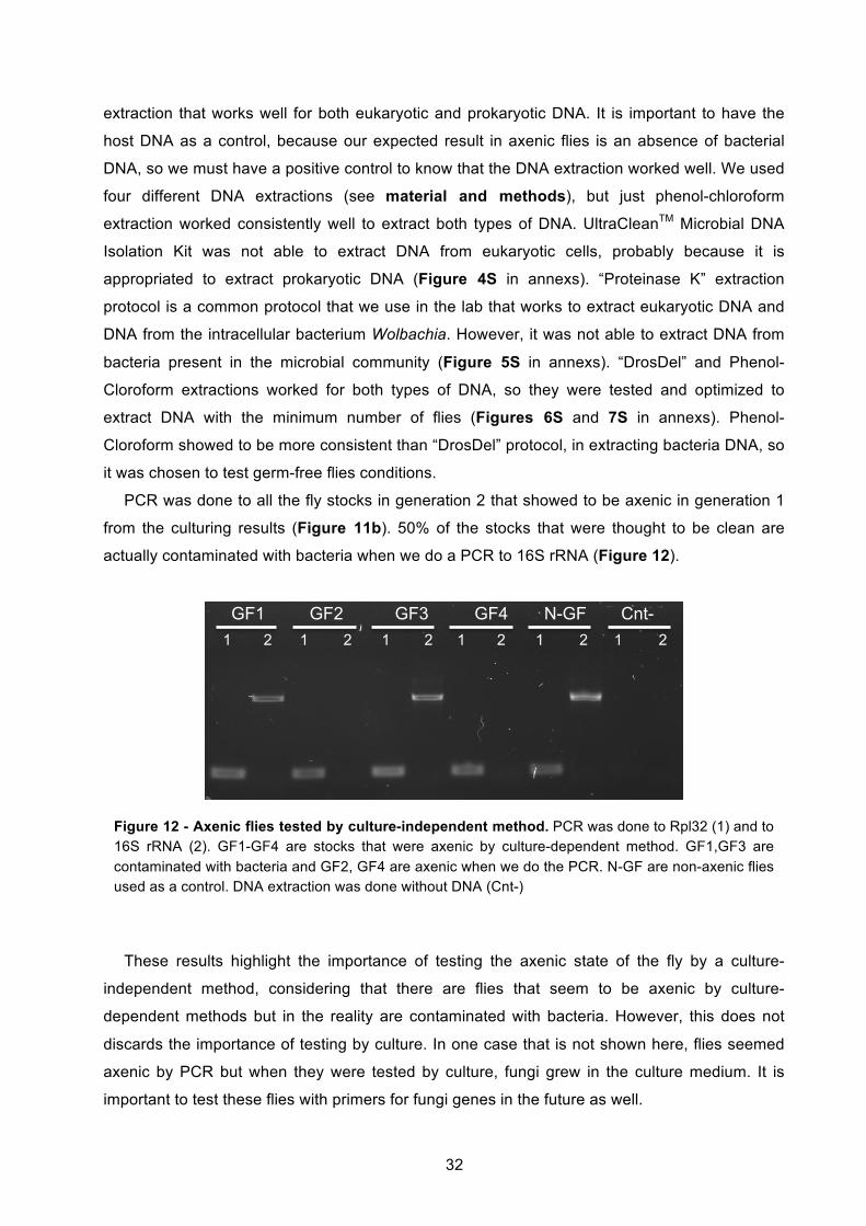
32
extraction that works well for both eukaryotic and prokaryotic DNA. It is important to have the
host DNA as a control, because our expected result in axenic flies is an absence of bacterial
DNA, so we must have a positive control to know that the DNA extraction worked well. We used
four different DNA extractions (see material and methods), but just phenol-chloroform
extraction worked consistently well to extract both types of DNA. UltraCleanTM Microbial DNA
Isolation Kit was not able to extract DNA from eukaryotic cells, probably because it is
appropriated to extract prokaryotic DNA (Figure 4S in annexs). “Proteinase K” extraction
protocol is a common protocol that we use in the lab that works to extract eukaryotic DNA and
DNA from the intracellular bacterium Wolbachia. However, it was not able to extract DNA from
bacteria present in the microbial community (Figure 5S in annexs). “DrosDel” and Phenol-
Cloroform extractions worked for both types of DNA, so they were tested and optimized to
extract DNA with the minimum number of flies (Figures 6S and 7S in annexs). Phenol-
Cloroform showed to be more consistent than “DrosDel” protocol, in extracting bacteria DNA, so
it was chosen to test germ-free flies conditions.
PCR was done to all the fly stocks in generation 2 that showed to be axenic in generation 1
from the culturing results (Figure 11b). 50% of the stocks that were thought to be clean are
actually contaminated with bacteria when we do a PCR to 16S rRNA (Figure 12).
These results highlight the importance of testing the axenic state of the fly by a culture-
independent method, considering that there are flies that seem to be axenic by culture-
dependent methods but in the reality are contaminated with bacteria. However, this does not
discards the importance of testing by culture. In one case that is not shown here, flies seemed
axenic by PCR but when they were tested by culture, fungi grew in the culture medium. It is
important to test these flies with primers for fungi genes in the future as well.
GF1 GF2 GF3 GF4 N-GF Cnt- 1 1 1 1 1 2 2 2 2 2 1 2
Figure 12 - Axenic flies tested by culture-independent method. PCR was done to Rpl32 (1) and to 16S rRNA (2). GF1-GF4 are stocks that were axenic by culture-dependent method. GF1,GF3 are contaminated with bacteria and GF2, GF4 are axenic when we do the PCR. N-GF are non-axenic flies used as a control. DNA extraction was done without DNA (Cnt-)

33
To raise axenic flies, it is important to choose a very rich and nutritive food, because
microbiota are not present to help on the host nutrition. A recent study shows this importance,
by reporting that axenic flies have an abnormal development and lower body size when they are
raised in a poor-diet medium. When they are raised in a rich medium this problem is less
pronounced.
34
4. Concluding remarks
Several studies aim to characterize the microbial community and understand how they
influence its host. Some studies have been showing that there are differences in the microbiota
between insects from the wild and from the laboratory. If the laboratory-reared insects do not
have a representative microbiota of the wild insects, this can became a problem for all the
studies that try to characterize the role of the microbiota on the host.
Recently, two studies have shown this concern [23], [32]. They reported that there is low-
diversity associated with the gut microbiota of Drosophila melanogaster and that the microbial
community found in laboratory-reared flies is different from wild caught flies.
Here, through a protocol that we developed that prevents the reinfection of the flies with their
transient bacteria, we show that laboratory-reared flies gut microbial community is not stable.
Surprisingly, laboratory-reared flies almost lost all their microbial community that was present in
the gut. In contrast, wild caught flies were able to maintain a more stable population inside the
gut.
Laboratory flies are under laboratory conditions for many years. The standard food that is
administrated to these flies has probably modified the gut microbiota composition that was first
present when flies were caught. Also, in the laboratory there are not very strong selective
pressures, in contrast to what happens in nature, where flies are constantly exposed to several
microorganisms, including opportunistic pathogens. In fact, bacteria present in the gut are
involved in the colonization resistance mechanism, in which they limit the ability of these
pathogens to colonize the gut [9]. We can consider the possibility that the microbiota community
of laboratory-reared flies could have lost the ability to colonize the host, because it is not
advantageous under laboratory conditions.
The protocol here developed can also be used to isolate possible natural pathogens of
Drosophila melanogaster. As we mentioned above, bacteria present in the gut are involved in
limiting the colonization from external pathogens. We observed that by preventing the
reinfection from the food in wild caught flies, the number of gut bacteria also suffered a
decrease. If the flies that are collected from the wild are already infected with a pathogen, this
pathogen can be able to proliferate and cross the hemolymph barrier, when gut microbiota start
to decrease. We can isolate bacteria that remains in whole flies after the protocol and test them
as potential pathogens of Drosophila melanogaster.
35
We show that microbiota from laboratory flies depend on the conditions in how flies are
maintained. In addition, it was also previously mentioned that microbiota community influences
several aspects on the host, particularly the host nutrition and immunity. If we disrupt the normal
microbiota present in the gut, this can leads to severe outcomes on the host physiology.
Therefore, it is important to normalize in all the research studies that are done in Drosophila
melanogaster, the conditions where flies are maintained, even if the study is not directly with the
microbial community. This way, we are minimizing the variability that we could obtain in the
results, or even avoiding some results that would be inexplicable.
This study highlights the importance of identifying the indigenous microbial flora.
Understanding the influence of microbiota on its host without knowing if they are actually
colonizing the host can lead to incorrect conclusions, as well as it can hide the roles that they
have in nature and can be absent in laboratory-reared flies.
However, it is possible that even transient microbiota have an influence in the host. Still, we
cannot do extrapolations of the true biological role that they may have in nature, because the
microbiota community and their behavior can be completely different.

36
5. Acknowledgments
First, I want to express my gratitude to my supervisor, Luis Teixeira, for giving me the great
opportunity to join to his lab and for allowing me to start working in the first day when I went
there to the interview. I am really grateful for all his help, friendship and patience. Thank you for
teaching me how to make good science.
I want to thank all the members of my lab, Sabine, Sara, Nelson, Gil, Ewa, Catarina and
Marta. Thank you for all the help in the laboratory, but mostly for their friendship and great
environment in the lab. A special thank to Nelson for all the helpful advises with the statistical
analysis. I am also thankful to Carolina, without her the last experiments could not have been
done.
I would also like to thank my family and friends, for all the happiness and joy that they bring
to my life. Specially, I would like to thank Tiago, for always supporting me until the last minute.
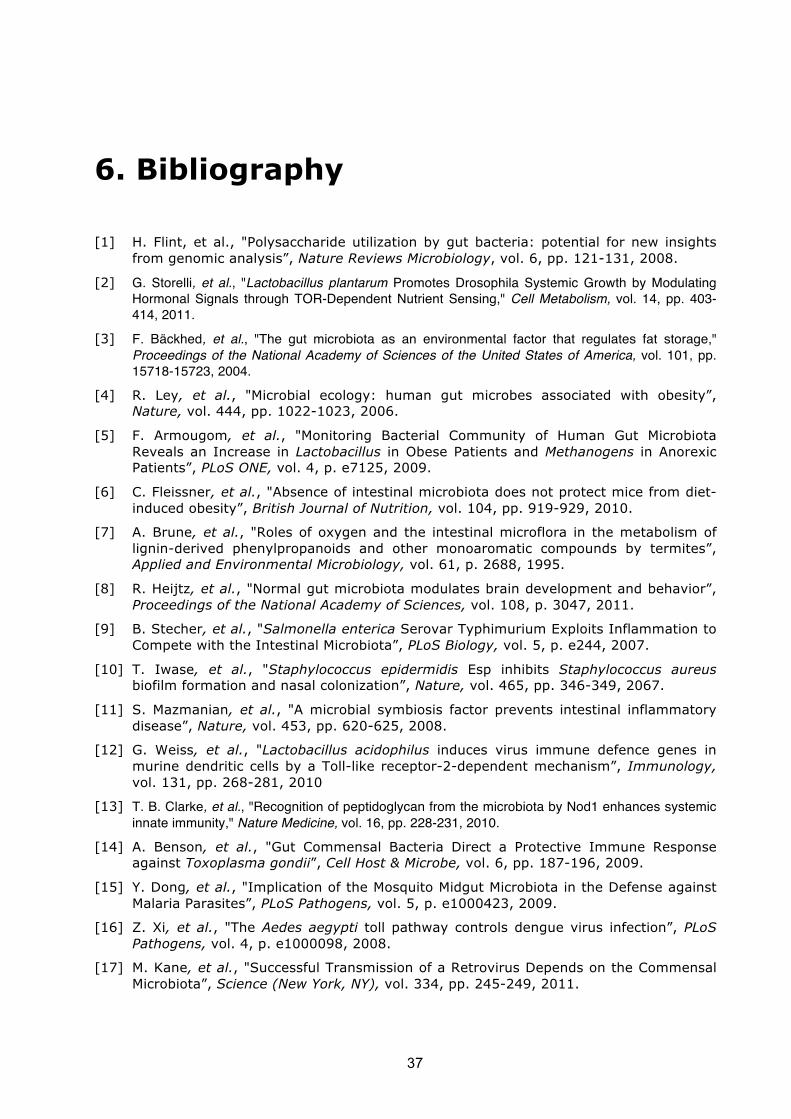
37
6. Bibliography
[1] H. Flint, et al., "Polysaccharide utilization by gut bacteria: potential for new insights from genomic analysis”, Nature Reviews Microbiology, vol. 6, pp. 121-131, 2008.
[2] G. Storelli, et al., "Lactobacillus plantarum Promotes Drosophila Systemic Growth by Modulating Hormonal Signals through TOR-Dependent Nutrient Sensing," Cell Metabolism, vol. 14, pp. 403-414, 2011.
[3] F. Bäckhed, et al., "The gut microbiota as an environmental factor that regulates fat storage," Proceedings of the National Academy of Sciences of the United States of America, vol. 101, pp. 15718-15723, 2004.
[4] R. Ley, et al., "Microbial ecology: human gut microbes associated with obesity”, Nature, vol. 444, pp. 1022-1023, 2006.
[5] F. Armougom, et al., "Monitoring Bacterial Community of Human Gut Microbiota Reveals an Increase in Lactobacillus in Obese Patients and Methanogens in Anorexic Patients”, PLoS ONE, vol. 4, p. e7125, 2009.
[6] C. Fleissner, et al., "Absence of intestinal microbiota does not protect mice from diet-induced obesity”, British Journal of Nutrition, vol. 104, pp. 919-929, 2010.
[7] A. Brune, et al., "Roles of oxygen and the intestinal microflora in the metabolism of lignin-derived phenylpropanoids and other monoaromatic compounds by termites”, Applied and Environmental Microbiology, vol. 61, p. 2688, 1995.
[8] R. Heijtz, et al., "Normal gut microbiota modulates brain development and behavior”, Proceedings of the National Academy of Sciences, vol. 108, p. 3047, 2011.
[9] B. Stecher, et al., "Salmonella enterica Serovar Typhimurium Exploits Inflammation to Compete with the Intestinal Microbiota”, PLoS Biology, vol. 5, p. e244, 2007.
[10] T. Iwase, et al., "Staphylococcus epidermidis Esp inhibits Staphylococcus aureus biofilm formation and nasal colonization”, Nature, vol. 465, pp. 346-349, 2067.
[11] S. Mazmanian, et al., "A microbial symbiosis factor prevents intestinal inflammatory disease”, Nature, vol. 453, pp. 620-625, 2008.
[12] G. Weiss, et al., "Lactobacillus acidophilus induces virus immune defence genes in murine dendritic cells by a Toll-like receptor-2-dependent mechanism”, Immunology, vol. 131, pp. 268-281, 2010
[13] T. B. Clarke, et al., "Recognition of peptidoglycan from the microbiota by Nod1 enhances systemic innate immunity," Nature Medicine, vol. 16, pp. 228-231, 2010.
[14] A. Benson, et al., "Gut Commensal Bacteria Direct a Protective Immune Response against Toxoplasma gondii”, Cell Host & Microbe, vol. 6, pp. 187-196, 2009.
[15] Y. Dong, et al., "Implication of the Mosquito Midgut Microbiota in the Defense against Malaria Parasites”, PLoS Pathogens, vol. 5, p. e1000423, 2009.
[16] Z. Xi, et al., "The Aedes aegypti toll pathway controls dengue virus infection”, PLoS Pathogens, vol. 4, p. e1000098, 2008.
[17] M. Kane, et al., "Successful Transmission of a Retrovirus Depends on the Commensal Microbiota”, Science (New York, NY), vol. 334, pp. 245-249, 2011.
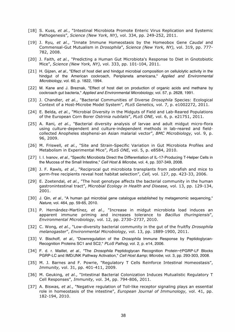
38
[18] S. Kuss, et al., "Intestinal Microbiota Promote Enteric Virus Replication and Systemic Pathogenesis”, Science (New York, NY), vol. 334, pp. 249-252, 2011.
[19] J. Ryu, et al., "Innate Immune Homeostasis by the Homeobox Gene Caudal and Commensal-Gut Mutualism in Drosophila”, Science (New York, NY), vol. 319, pp. 777-782, 2008.
[20] J. Faith, et al., "Predicting a Human Gut Microbiota's Response to Diet in Gnotobiotic Mice”, Science (New York, NY), vol. 333, pp. 101-104, 2011.
[21] H. Gijzen, et al., "Effect of host diet and hindgut microbial composition on cellulolytic activity in the hindgut of the American cockroach, Periplaneta americana," Applied and Environmental Microbiology, vol. 60, p. 1822, 1994.
[22] M. Kane and J. Breznak, "Effect of host diet on production of organic acids and methane by cockroach gut bacteria," Applied and Environmental Microbiology, vol. 57, p. 2628, 1991.
[23] J. Chandler, et al., "Bacterial Communities of Diverse Drosophila Species: Ecological Context of a Host-Microbe Model System”, PLoS Genetics, vol. 7, p. e1002272, 2011.
[24] E. Belda, et al., "Microbial Diversity in the Midguts of Field and Lab-Reared Populations of the European Corn Borer Ostrinia nubilalis”, PLoS ONE, vol. 6, p. e21751, 2011.
[25] A. Rani, et al., "Bacterial diversity analysis of larvae and adult midgut micro-flora using culture-dependent and culture-independent methods in lab-reared and field-collected Anopheles stephensi-an Asian malarial vector”, BMC Microbiology, vol. 9, p. 96, 2009.
[26] M. Friswell, et al., "Site and Strain-Specific Variation in Gut Microbiota Profiles and Metabolism in Experimental Mice”, PLoS ONE, vol. 5, p. e8584, 2010.
[27] I. I. Ivanov, et al., "Specific Microbiota Direct the Differentiation of IL-17-Producing T-Helper Cells in the Mucosa of the Small Intestine," Cell Host & Microbe, vol. 4, pp. 337-349, 2008.
[28] J. F. Rawls, et al., "Reciprocal gut microbiota transplants from zebrafish and mice to germ-free recipients reveal host habitat selection”, Cell, vol. 127, pp. 423-33, 2006.
[29] E. Zoetendal, et al., "The host genotype affects the bacterial community in the human gastronintestinal tract”, Microbial Ecology in Health and Disease, vol. 13, pp. 129-134, 2001.
[30] J. Qin, et al., "A human gut microbial gene catalogue established by metagenomic sequencing," Nature, vol. 464, pp. 59-65, 2010.
[31] P. Hernández-Martínez, et al., "Increase in midgut microbiota load induces an apparent immune priming and increases tolerance to Bacillus thuringiensis”, Environmental Microbiology, vol. 12, pp. 2730–2737, 2010.
[32] C. Wong, et al., "Low-diversity bacterial community in the gut of the fruitfly Drosophila melanogaster”, Environmental Microbiology, vol. 13, pp. 1889-1900, 2011.
[33] V. Bischoff, et al., "Downregulation of the Drosophila Immune Response by Peptidoglycan-Recognition Proteins SC1 and SC2," PLoS Pathog, vol. 2, p. e14, 2006.
[34] F. d. r. Maillet, et al., "The Drosophila Peptidoglycan Recognition Protein PGRP-LF Blocks PGRP-LC and IMD/JNK Pathway Activation," Cell Host & Microbe, vol. 3, pp. 293-303, 2008.
[35] M. J. Barnes and F. Powrie, "Regulatory T Cells Reinforce Intestinal Homeostasis”, Immunity, vol. 31, pp. 401-411, 2009.
[36] M. Geuking, et al., "Intestinal Bacterial Colonization Induces Mutualistic Regulatory T Cell Responses”, Immunity, vol. 34, pp. 794-806, 2011.
[37] A. Biswas, et al., "Negative regulation of Toll-like receptor signaling plays an essential role in homeostasis of the intestine”, European Journal of Immunology, vol. 41, pp. 182-194, 2010.
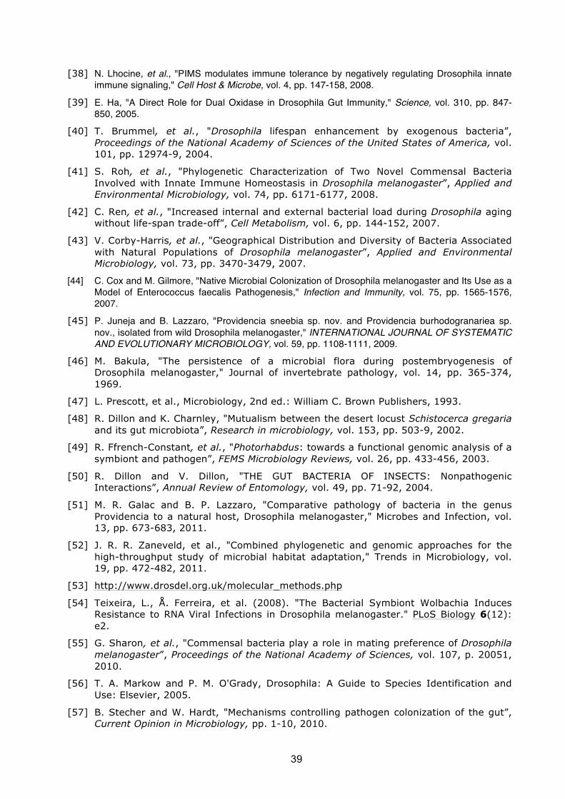
39
[38] N. Lhocine, et al., "PIMS modulates immune tolerance by negatively regulating Drosophila innate immune signaling," Cell Host & Microbe, vol. 4, pp. 147-158, 2008.
[39] E. Ha, "A Direct Role for Dual Oxidase in Drosophila Gut Immunity," Science, vol. 310, pp. 847-850, 2005.
[40] T. Brummel, et al., "Drosophila lifespan enhancement by exogenous bacteria”, Proceedings of the National Academy of Sciences of the United States of America, vol. 101, pp. 12974-9, 2004.
[41] S. Roh, et al., "Phylogenetic Characterization of Two Novel Commensal Bacteria Involved with Innate Immune Homeostasis in Drosophila melanogaster”, Applied and Environmental Microbiology, vol. 74, pp. 6171-6177, 2008.
[42] C. Ren, et al., "Increased internal and external bacterial load during Drosophila aging without life-span trade-off”, Cell Metabolism, vol. 6, pp. 144-152, 2007.
[43] V. Corby-Harris, et al., "Geographical Distribution and Diversity of Bacteria Associated with Natural Populations of Drosophila melanogaster”, Applied and Environmental Microbiology, vol. 73, pp. 3470-3479, 2007.
[44] C. Cox and M. Gilmore, "Native Microbial Colonization of Drosophila melanogaster and Its Use as a Model of Enterococcus faecalis Pathogenesis," Infection and Immunity, vol. 75, pp. 1565-1576, 2007.
[45] P. Juneja and B. Lazzaro, "Providencia sneebia sp. nov. and Providencia burhodogranariea sp. nov., isolated from wild Drosophila melanogaster," INTERNATIONAL JOURNAL OF SYSTEMATIC AND EVOLUTIONARY MICROBIOLOGY, vol. 59, pp. 1108-1111, 2009.
[46] M. Bakula, "The persistence of a microbial flora during postembryogenesis of Drosophila melanogaster," Journal of invertebrate pathology, vol. 14, pp. 365-374, 1969.
[47] L. Prescott, et al., Microbiology, 2nd ed.: William C. Brown Publishers, 1993.
[48] R. Dillon and K. Charnley, "Mutualism between the desert locust Schistocerca gregaria and its gut microbiota”, Research in microbiology, vol. 153, pp. 503-9, 2002.
[49] R. Ffrench-Constant, et al., "Photorhabdus: towards a functional genomic analysis of a symbiont and pathogen”, FEMS Microbiology Reviews, vol. 26, pp. 433-456, 2003.
[50] R. Dillon and V. Dillon, "THE GUT BACTERIA OF INSECTS: Nonpathogenic Interactions”, Annual Review of Entomology, vol. 49, pp. 71-92, 2004.
[51] M. R. Galac and B. P. Lazzaro, "Comparative pathology of bacteria in the genus Providencia to a natural host, Drosophila melanogaster," Microbes and Infection, vol. 13, pp. 673-683, 2011.
[52] J. R. R. Zaneveld, et al., "Combined phylogenetic and genomic approaches for the high-throughput study of microbial habitat adaptation," Trends in Microbiology, vol. 19, pp. 472-482, 2011.
[53] http://www.drosdel.org.uk/molecular_methods.php
[54] Teixeira, L., Å. Ferreira, et al. (2008). "The Bacterial Symbiont Wolbachia Induces Resistance to RNA Viral Infections in Drosophila melanogaster." PLoS Biology 6(12): e2.
[55] G. Sharon, et al., "Commensal bacteria play a role in mating preference of Drosophila melanogaster”, Proceedings of the National Academy of Sciences, vol. 107, p. 20051, 2010.
[56] T. A. Markow and P. M. O'Grady, Drosophila: A Guide to Species Identification and Use: Elsevier, 2005.
[57] B. Stecher and W. Hardt, "Mechanisms controlling pathogen colonization of the gut”, Current Opinion in Microbiology, pp. 1-10, 2010.

40
[58] M. Ashburner, et al., Drosophila: A Laboratory Handbook, 1st ed. Plain View, N.Y.: Cold Spring Harbor Laboratory Press, 2005.
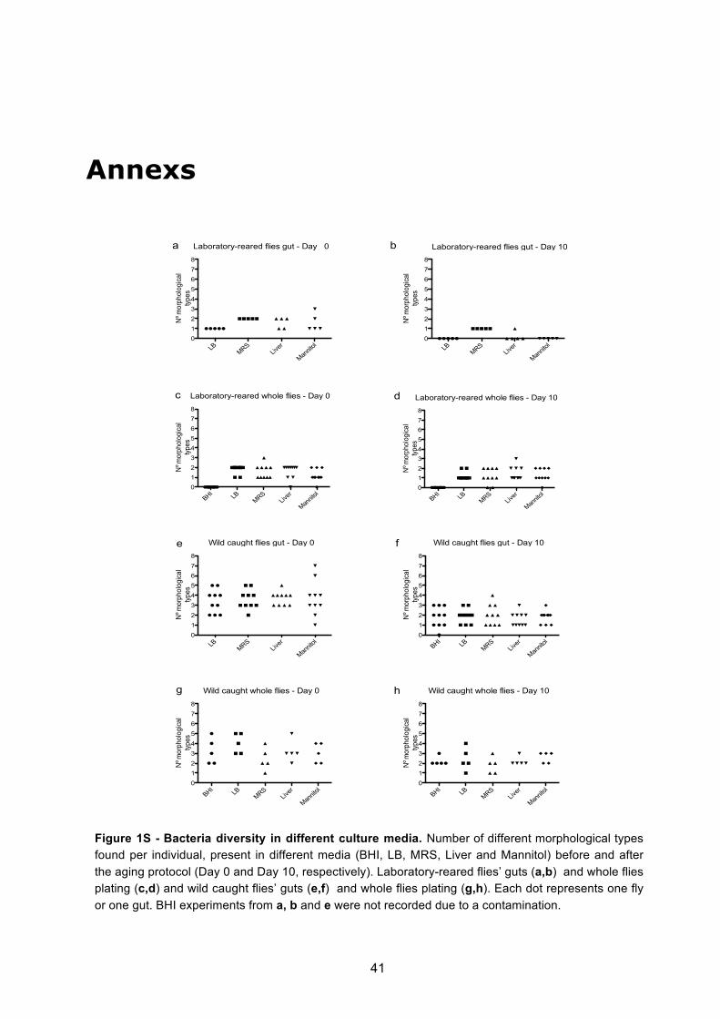
41
Annexs
Laboratory-reared flies gut - Day 10
LB MRSLive
r
Mannito
l012345678
Nº m
orph
olog
ical
types
Laboratory-reared whole flies - Day 0
BHI LB MRSLive
r
Mannito
l012345678
Nº m
orph
olog
ical
types
Wild caught flies gut - Day 0
LB MRSLive
r
Mannito
l012345678
Nº m
orph
olog
ical
types
Wild caught whole flies - Day 0
BHI LB MRSLive
r
Mannito
l012345678
Nº m
orph
olog
ical
types
Lab_gut_nr_species_day10
LB MRSLive
r
Mannito
l012345678
Nº m
orph
olog
ical
types
Laboratory-reared whole flies - Day 10
BHI LB MRSLive
r
Mannito
l012345678
Nº m
orph
olog
ical
types
Wild caught flies gut - Day 10
BHI LB MRSLive
r
Mannito
l012345678
Nº m
orph
olog
ical
types
Wild caught whole flies - Day 10
BHI LB MRSLive
r
Mannito
l012345678
Nº m
orph
olog
ical
types
a b
c d
e f
g h
Laboratory-reared flies gut - Day 10
LB MRSLive
r
Mannito
l012345678
Nº m
orph
olog
ical
types
Laboratory-reared whole flies - Day 0
BHI LB MRSLive
r
Mannito
l012345678
Nº m
orph
olog
ical
types
Wild caught flies gut - Day 0
LB MRSLive
r
Mannito
l012345678
Nº m
orph
olog
ical
types
Wild caught whole flies - Day 0
BHI LB MRSLive
r
Mannito
l012345678
Nº m
orph
olog
ical
types
Lab_gut_nr_species_day10
LB MRSLive
r
Mannito
l012345678
Nº m
orph
olog
ical
types
Laboratory-reared whole flies - Day 10
BHI LB MRSLive
r
Mannito
l012345678
Nº m
orph
olog
ical
types
Wild caught flies gut - Day 10
BHI LB MRSLive
r
Mannito
l012345678
Nº m
orph
olog
ical
types
Wild caught whole flies - Day 10
BHI LB MRSLive
r
Mannito
l012345678
Nº m
orph
olog
ical
types
Figure 1S - Bacteria diversity in different culture media. Number of different morphological types found per individual, present in different media (BHI, LB, MRS, Liver and Mannitol) before and after the aging protocol (Day 0 and Day 10, respectively). Laboratory-reared flies’ guts (a,b) and whole flies plating (c,d) and wild caught flies’ guts (e,f) and whole flies plating (g,h). Each dot represents one fly or one gut. BHI experiments from a, b and e were not recorded due to a contamination.
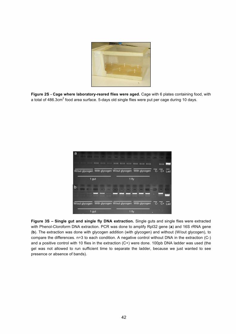
42
Figure 2S - Cage where laboratory-reared flies were aged. Cage with 6 plates containing food, with a total of 486.3cm2 food area surface. 5-days old single flies were put per cage during 10 days.
W/out glycogen C- Lad With glycogen W/out glycogen With glycogen C+
1 gut 1 fly
W/out glycogen C- Lad With glycogen W/out glycogen With glycogen C+
1 gut 1 fly
a
b
Figure 3S – Single gut and single fly DNA extraction. Single guts and single flies were extracted with Phenol-Cloroform DNA extraction. PCR was done to amplify Rpl32 gene (a) and 16S rRNA gene (b). The extraction was done with glycogen addition (with glycogen) and without (W/out glycogen), to compare the differences. n=3 to each condition. A negative control without DNA in the extraction (C-) and a positive control with 10 flies in the extraction (C+) were done. 100pb DNA ladder was used (the gel was not allowed to run sufficient time to separate the ladder, because we just wanted to see presence or absence of bands).
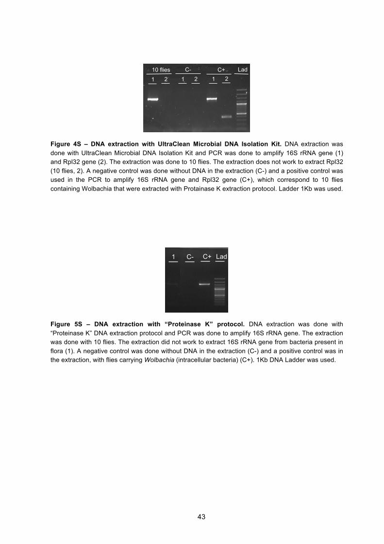
43
10 flies C- C+ Lad
1 2 1 2 1 2
Figure 4S – DNA extraction with UltraClean Microbial DNA Isolation Kit. DNA extraction was done with UltraClean Microbial DNA Isolation Kit and PCR was done to amplify 16S rRNA gene (1) and Rpl32 gene (2). The extraction was done to 10 flies. The extraction does not work to extract Rpl32 (10 flies, 2). A negative control was done without DNA in the extraction (C-) and a positive control was used in the PCR to amplify 16S rRNA gene and Rpl32 gene (C+), which correspond to 10 flies containing Wolbachia that were extracted with Protainase K extraction protocol. Ladder 1Kb was used.
C- C+ Lad 1
Figure 5S – DNA extraction with “Proteinase K” protocol. DNA extraction was done with “Proteinase K” DNA extraction protocol and PCR was done to amplify 16S rRNA gene. The extraction was done with 10 flies. The extraction did not work to extract 16S rRNA gene from bacteria present in flora (1). A negative control was done without DNA in the extraction (C-) and a positive control was in the extraction, with flies carrying Wolbachia (intracellular bacteria) (C+). 1Kb DNA Ladder was used.

44
1 fly 2 flies 5 flies 25 flies C+ Lad C-
1 fly 2 flies 5 flies 25 flies C+ Lad C-
a
b
Figure 6S – DNA extraction “DrosDel” protocol. DNA extraction was done with “DrosDel” DNA extraction protocol and PCR was done to amplify Rpl32 gene (a) and 16S rRNA gene (b). The extraction was done with 1, 2, 5 and 25 flies (n=3 for each). A negative control was done without DNA in the extraction (C-) and a positive control was done to amplify both genes in PCR, with flies containing Wolbachia extracted with Proteinase K protocol (C+). 100pb DNA Ladder was used. 1KB DNA Ladder should have been used in b.
25 flies 10 flies 5 flies 2 flies C- Lad
25 flies 10 flies 5 flies 2 flies C- Lad
a
b
Figure 7S – DNA extraction with Phenol-Cloroform protocol. DNA extraction was done with Phenol-Cloroform DNA extraction protocol and PCR was done to amplify Rpl32 gene (a) and 16S rRNA gene (b). The extraction was done with 25, 10, 5 and 2 flies (n=3 for each). A negative control was done without DNA in the extraction (C-). 100pb DNA Ladder was used.

45