Embed Size (px)
Citation preview
UNIVERSIDADE DE SÃO PAULO FACULDADE DE ODONTOLOGIA DE BAURU
CAMILA MOREIRA MACHADO
Avaliação de próteses metalocerâmicas unitárias implantossuportadas carregadas imediatamente na região
posterior: estudo prospectivo de 6 meses
BAURU 2018

CAMILA MOREIRA MACHADO
Avaliação de próteses metalocerâmicas unitárias implantossuportadas carregadas imediatamente na região
posterior: estudo prospectivo de 6 meses
Tese apresentada a Faculdade de Odontologia de Bauru da Universidade de São Paulo para obtenção do título de Mestre/Doutor em Ciências no Programa de Ciências Odontológicas Aplicadas, na área de concentração Reabilitação Oral Orientador: Prof. Dr. Estevam Augusto Bonfante
BAURU 2018

Autorizo, exclusivamente para fins acadêmicos e científicos, a reprodução total ou parcial desta dissertação/tese, por processos fotocopiadores e outros meios eletrônicos. Assinatura: Data:
Comitê de Ética da FOB-USP Protocolo nº: 50811015.0.0000.5417 Data: 24/11/2015
Machado, Camila Moreira Avaliação de próteses metalocerâmicas unitárias implantossuportadas carregadas imediatamente na região posterior: estudo prospectivo de 6 meses / Camila Moreira. – Bauru, 2018. 98p. : il. ; 31cm. Tese (Doutorado)– Faculdade de Odontologia de Bauru. Universidade de São Paulo Orientador: Prof. Dr. Estevam Augusto Bonfante

(Cole a cópia de sua folha de aprovação aqui)


DEDICATÓRIA
Dedico este trabalho, especialmente, às pessoas que sonharam junto
comigo e não mediram esforços para me ajudar a realizar este sonho...
Aos meus amados pais Urivando e Everli,
Vocês são a minha grande inspiração. Obrigada por deixarem eu seguir o meu
caminho em busca da conquista dos meus sonhos, mesmo sabendo que o maior
sacrifício seria a distância entre nós. Vocês são os meus maiores incentivadores e
responsáveis por eu ter cumprido mais esta etapa. Serei eternamente grata a vocês
por tudo que fizeram e fazem por mim. É tudo por vocês e para vocês!
À minha irmã Renata,
Minha metade, minha eterna companheira. Você é o meu maior referencial de
determinação, força, coragem, humildade, paciência e compaixão. Obrigada por
estar presente em todos os momentos da minha vida, apesar da distância física.
Somos tão diferentes e ao mesmo tempo tão iguais. Eu não seria completa sem
você. Eternamente será a minha “Tata”. Amo você incondicionalmente!


AGRADECIMENTO ESPECIAL
Ao meu querido orientador Prof. Dr. Estevam Augusto Bonfante,
Ter a oportunidade de trabalhar com você é, sem dúvida, um aprendizado constante.
Nunca me esquecerei da forma tão acolhedora com que me recebeu na Prótese,
mesmo sem nos conhecermos anteriormente. Obrigada por ter confiado a mim a
realização deste estudo clínico. A forma como você sempre me ensinou e ajudou foi
essencial para o desenvolvimento deste trabalho. Foram muitos desafios
enfrentados, mas também muitos resultados positivos alcançados. A sua
simplicidade, humildade, dignidade, positividade e sabedoria são admiráveis e
inspiram todos os que convivem com você. Muito obrigada por todas as
oportunidades que me concedeu e por todas as lições, muito além da Odontologia!


AGRADECIMENTOS
A Deus, por todas as bênçãos na minha vida e, especialmente, por todas as
pessoas tão especiais que Ele coloca à minha volta.
Aos meus familiares, em especial à minha avó. Obrigada pela torcida e
preocupação. Mesmo longe sentia seus cuidados.
Um agradecimento muito especial ao Prof. Dr. Luiz Fernando Pegoraro que é, sem
dúvida, um dos grandes exemplos de professor, e principalmente, de ser humano. O
senhor teve um papel fundamental na minha formação, durante a graduação e pós-
graduação. Muito obrigada pela confiança e por ter acreditado no nosso trabalho.
Por ter me recebido tão bem na Prótese e ter oferecido todo o apoio que estava ao
seu alcance. Tenho muita gratidão, admiração e carinho pelo senhor.
Ao Prof. Dr. Leonardo Rigoldi Bonjardim. Obrigada por ser tão solícito,
atendendo-nos sempre tão bem e apresentando importantes ideias e sugestões.
Ao Prof. Dr. Carlos Ferreira dos Santos, por toda ajuda e orientação acerca do
envio do projeto à Fundação de Amparo à Pesquisa do Estado de São Paulo. Você
é uma das pessoas que me inspiram!
Aos meus queridos pacientes e participantes deste estudo: Ricardo, Alessandra,
Rita de Cássia, Adriana, Adassa, Karina, Ana Paula, Keticheila, Helenice, Sandra,
Ana Lúcia, Antônio, Arilson, Danielle, Paulo Cesário, Érica, Gentília, Sibeli e Kelly.
Muito obrigada pela confiança depositada em nossa equipe e pelo compromisso
para retornar a todas as avaliações.
Ao meu grande amigo Ernesto, pela linda amizade e apoio incondicional. Obrigada
pelo conhecimento e experiências compartilhadas. A convivência com você nos
últimos três anos fez tudo ser mais leve e tranquilo. Você também faz parte desta
conquista. Meu irmão científico! Meu irmão de vida!


Ao Everardo, querido amigo obrigada pela ajuda no desenvolvimento de todas as
etapas do estudo.
Ao Patrick, que gentilmente aceitou realizar todas as cirurgias de instalação dos
implantes nos pacientes do estudo. Muito obrigada pela convivência tão agradável e
pela disposição em todos os momentos.
Aos meus amigos goianos, Mayara, Priscilla e Paulo Vitor. É tão maravilhoso saber
que a nossa amizade continua apesar da distância física. Obrigada pelo carinho e
torcida. Amo vocês!
Aos meus amigos bauruenses, Vanessa, Marilisa, Adriana, Amanda, Daniel,
Camila e Cristiane, por me fazerem gostar dessa cidade que já se tornou a minha
segunda casa. Obrigada pelos conselhos, pelas conversas e por sempre me ajudar.
Amo vocês!
Aos professores do departamento de Prótese e Periodontia por todos os
ensinamentos transmitidos e convivência agradável.
Ao Prof. Dr. José Roberto Pereira Lauris pela disponibilidade e contribuição com a
estatística deste trabalho, sempre nos atendendo de forma muito agradável.
Aos professores e funcionários da Disciplina de Radiologia e Estomatologia,
em especial aos funcionários Fernanda e Roberto, por toda a ajuda e disponibilidade
no agendamento e processamento das radiografias digitais e tomografias. Muito
obrigada!
À funcionária Maristela Ferrari, que nos ajudou imensamente com a documentação
enviada para o Comitê de Ética em Pesquisas em Seres Humanos. Muito obrigada
pela disponibilidade e atenção!
Às funcionárias do setor de triagem, Sandra, Cristina e Leucy, que gentilmente
agendaram mais de 200 pacientes para fazer a triagem do estudo. Muito obrigada
pela ajuda!


Aos contemporâneos de Pós-graduação em Reabilitação Oral. Obrigada pelo
conhecimento compartilhado e parcerias em outros trabalhos científicos. Obrigada a
todos!
Aos funcionários do Departamento de Prótese e Periodontia, especialmente,
Deborah, Ziley, Walquíria, Cleide, Marcelo e Reivanildo. Obrigada pelo carinho e por
toda a ajuda.
À Fundação de Amparo à Pesquisa do Estado de São Paulo (FAPESP), pelo
fundamental suporte financeiro concedido para a realização desta pesquisa
(Processo: 2015/26920-4).
À SIN sistema de implantes, pela doação de todos os implantes e componentes
protéticos utilizados no estudo. Muito obrigada!
À Curaprox – Curadens, pela doação das escovas dentais que contribuíram para a
higienização oral de todos os pacientes do estudo.
À Sulzer, pela doação das pontas misturadoras T- mixer, usadas para dispensar o
silicone fluido nas moldagens dos pacientes e nas confecções de réplicas.
Ao laboratório de prótese dentária Digidental Solutions, pela parceria na
confecção de todas as coroas do estudo. Muito obrigada pelo compromisso,
precisão e eficiência! Vocês foram fundamentais para o andamento do estudo e
instalação das coroas em tempo hábil.


“A alegria de ver e compreender é a mais bela dádiva da natureza.”
Albert Einstein


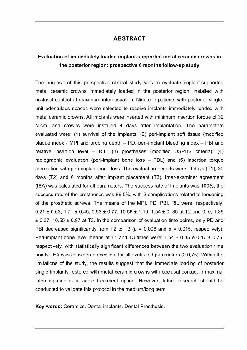
RESUMO
O objetivo deste estudo clínico prospectivo foi avaliar próteses metalocerâmicas
unitárias implantossuportadas imediatamente carregadas na região posterior,
instaladas com contato oclusal em máxima intercuspidação. Dezenove pacientes
com perdas unitárias posteriores foram selecionados para receber implantes
unitários imediatamente carregados com coroas metalocerâmicas. Todos os
implantes foram inseridos com torque de inserção mínimo de 32 N.cm. e as coroas
foram instaladas 4 dias após a instalação dos implantes. Os parâmetros avaliados
foram: (1) sobrevida dos implantes, (2) tecido mole peri-implantar (índice de placa
modificado - IPM, profundidade de sondagem - PS, índice de sangramento peri-
implantar - ISP e nível de inserção relativo - NIR) (3) prótese (Critério USPHS
modificado), (4) avaliação radiográfica (perda óssea peri-implantar - POP) e (5)
correlação do torque de inserção com perda óssea peri-implantar. Os períodos de
avaliação foram: 9 dias (T1), 30 dias (T2) e 6 meses após a instalação do implante
(T3). Concordância inter-examinadores (CIC) foi calculada para todos os parâmetros
avaliados. A taxa de sucesso dos implantes foi de 100%; a taxa de sucesso da
prótese foi de 89,5%, sendo observadas 2 complicações relacionadas com
afrouxamento dos parafusos protéticos. As médias do IPM, PS, ISP NIR foram
respectivamente: 0,21 ± 0,63, 1,71 ± 0,45, 0,53 ± 0,77, 10,56 ± 1,19, 1,54 ± 0,35 no
tempo T2 e 0, 0, 1,36 ± 0,37, 10,55 ± 0,97 no tempo T3. Na comparação dos
tempos, somente PS e ISP diminuíram significantemente de T2 para T3 (p = 0,006 e
p = 0,015, respectivamente). As médias do nível ósseo peri-implantar nos tempos T1
e T3 foram de 1,54 ± 0,35 e 0,47 ± 0,76, respectivamente, com diferenças
estatisticamente significantes encontradas entre os dois períodos de avaliação. CIC
foi considerado excelente para todos os parâmetros avaliados (≥ 0,75). Dentro das
limitações do estudo, os resultados sugerem que a carga imediata de implantes
unitários posteriores restaurados com coroas metalocerâmicas com contato oclusal
em máxima intercuspidação é uma opção viável de tratamento. Entretanto,
pesquisas futuras devem ser conduzidas para a validação de tal protocolo a
médio/longo prazos.
Palavras-chave: Cerâmica. Implantes dentários. Prótese dentária.

ABSTRACT
Evaluation of immediately loaded implant-supported metal ceramic crowns in the posterior region: prospective 6 months follow-up study
The purpose of this prospective clinical study was to evaluate implant-supported
metal ceramic crowns immediately loaded in the posterior region, installed with
occlusal contact at maximum intercuspation. Nineteen patients with posterior single-
unit edentulous spaces were selected to receive implants immediately loaded with
metal ceramic crowns. All implants were inserted with minimum insertion torque of 32
N.cm. and crowns were installed 4 days after implantation. The parameters
evaluated were: (1) survival of the implants; (2) peri-implant soft tissue (modified
plaque index - MPI and probing depth – PD, peri-implant bleeding index – PBI and
relative insertion level – RIL; (3) prostheses (modified USPHS criteria); (4)
radiographic evaluation (peri-implant bone loss – PBL) and (5) insertion torque
correlation with peri-implant bone loss. The evaluation periods were: 9 days (T1), 30
days (T2) and 6 months after implant placement (T3). Inter-examiner agreement
(IEA) was calculated for all parameters. The success rate of implants was 100%; the
success rate of the prostheses was 89.5%, with 2 complications related to loosening
of the prosthetic screws. The means of the MPI, PD, PBI, RIL were, respectively:
0.21 ± 0.63, 1.71 ± 0.45, 0.53 ± 0.77, 10.56 ± 1.19, 1.54 ± 0, 35 at T2 and 0, 0, 1.36
± 0.37, 10.55 ± 0.97 at T3. In the comparison of evaluation time points, only PD and
PBI decreased significantly from T2 to T3 (p = 0.006 and p = 0.015, respectively).
Peri-implant bone level means at T1 and T3 times were: 1.54 ± 0.35 e 0.47 ± 0.76,
respectively, with statistically significant differences between the two evaluation time
points. IEA was considered excellent for all evaluated parameters (≥ 0,75). Within the
limitations of the study, the results suggest that the immediate loading of posterior
single implants restored with metal ceramic crowns with occlusal contact in maximal
intercuspation is a viable treatment option. However, future research should be
conducted to validate this protocol in the medium/long term.
Key words: Ceramics. Dental implants. Dental Prosthesis.


LISTA DE TABELAS
Tabela 1: Escores do coeficiente interclasse ..................................................... 43
Tabela 2: Escores do Índice de placa modificado .............................................. 44
Tabela 3: Escores do Índice de sangramento peri-implantar ............................. 44
Tabela 4: Critérios USPHS modificado (Serviço de saúde pública norte
americano modificado) ....................................................................... 46
Tabela 5: Pacientes excluídos do estudo e motivos da exclusão ...................... 51
Tabela 6: Características das variáveis do estudo ............................................ 52
Tabela 7: Índice de placa modificado (IPM) e mudanças do IPM entre os
tempos de avaliação .......................................................................... 53
Tabela 8: Profundidades de sondagem (PS) peri-implantar e mudanças da
PS entre os tempos de avaliação ...................................................... 54
Tabela 9: Índice de sangramento peri-implantar (ISP) e mudanças do ISP
entre os tempos de avaliação ............................................................ 54
Tabela 10: Nível de inserção relativo peri-implantar (NIR) e mudanças do NIR
entre os tempos de avaliação ............................................................ 55
Tabela 11: Avaliação da prótese usando Critério USPHS modificado em 6
meses ................................................................................................. 56
Tabela 12: Média do nível ósseo marginal peri-implantar e mudanças do nível
ósseo entre os tempos de avaliação .................................................. 57
Tabela 13: Interação qualitativa do torque de inserção na perda óssea peri-
implantar ............................................................................................ 57
Tabela 14: Coeficiente de correlação interclasse ................................................ 58

LISTA DE ABREVIATURA, SIGLAS E SÍMBOLOS
% Por cento
± Mais ou menos
< menor
≤ Menor ou igual
≥ Maior ou igual
> Maior
µm Micrometro
DP Desvio padrão
IPM Índice de placa modificado
ISP Índice de sangramento peri-implantar
mg Miligrama
mm Milímetro
n Número de pacientes
N.cm. Newtom centímetro
NIR Nível de inserção relativo
p Nível de significância
POP Perda óssea peri-implantar
PS Profundidade de sondagem
TiAlV Titânio, alumínio e vanádio
USPHS Critério do serviço de saúde pública norte americano

SUMÁRIO
1 INTRODUÇÃO ........................................................................................... 17
2 REVISÃO DE LITERATURA ..................................................................... 23
3 PROPOSIÇÃO ........................................................................................... 35 4 MATERIAL E MÉTODOS .......................................................................... 39
4.1 SELEÇÃO DOS PACIENTES .................................................................... 39
4.2 PROCEDIMENTO CIRÚRGICO ................................................................. 40
4.3 PROCEDIMENTO PROTÉTICO ................................................................ 41
4.4 AVALIAÇÕES IMPLANTES / PRÓTESES ................................................. 42
4.4.1 Sobrevida dos implantes ............................................................................ 43
4.4.2 Avaliação do tecido mole peri-implantar ..................................................... 43
4.4.2.1 Índice de placa modificado (IPM) ............................................................... 43
4.4.2.2 Profundidade de sondagem (PS) ............................................................... 44
4.4.2.3 Índice de sangramento peri-implantar (ISP) ............................................... 44
4.4.2.4 Nível de inserção relativo (NIR) .................................................................. 45
4.4.3 Avaliação da prótese .................................................................................. 45
4.4.4 Perda óssea peri-implantar ........................................................................ 46
4.4.5 Correlação do torque de inserção com perda óssea peri-implantar ........... 47
4.5 ANÁLISE ESTATÍSTICA ............................................................................ 47
5 RESULTADOS ........................................................................................... 51 5.1 PARTICIPANTES ....................................................................................... 51
5.2 SOBREVIDA DOS IMPLANTES ................................................................ 53
5.3 AVALIAÇÃO DO TECIDO MOLE PERI-IMPLANTAR ................................ 53
5.3.1 Índice de placa modificado (IPM) ............................................................... 53
5.3.2 Profundidade de sondagem (PS) ............................................................... 54
5.3.3 Índice de sangramento peri-implantar (ISP) ............................................... 54
5.3.4 Nível de inserção relativo (NIR) .................................................................. 55


5.4 AVALIAÇÃO DA PRÓTESE ....................................................................... 55
5.5 PERDA ÓSSEA PERI-IMPLANTAR ........................................................... 56
5.6 CORRELAÇÃO DO TORQUE DE INSERÇÃO COM PERDA ÓSSEA
PERI-IMPLANTAR ..................................................................................... 57
5.7 COEFICIENTE DE CORRELAÇÃO INTERCLASSE ................................. 58
6 DISCUSSÃO .............................................................................................. 61 7 CONCLUSÕES .......................................................................................... 69 REFERÊNCIAS .......................................................................................... 73 ANEXO ....................................................................................................... 95


1 INTRODUÇÃO


Introdução 17
1 INTRODUÇÃO
Os implantes dentários têm sido uma importante opção de tratamento para o
suporte de diferentes restaurações protéticas. Originalmente, os implantes foram
utilizados para reabilitar pacientes totalmente edêntulos. Com a evolução dos
implantes e das técnicas cirúrgicas começaram a ser também usados para o
tratamento de pacientes parcialmente edêntulos e, subsequentemente, para
reabilitar casos unitários (1). Os critérios comumente aceitos para a avaliação do sucesso do tratamento
com implantes foram propostos por Albrektsson e colaboradores em 1986, (2) com o
objetivo de avaliar o sucesso da osseointegração e a sobrevivência dos implantes.
Entre eles, aceitava-se que a perda óssea marginal ficasse em torno de 1 mm no
primeiro ano e de até 0.2 mm nos anos subsequentes. Entretanto, após décadas,
novos parâmetros foram introduzidos na odontologia para avaliar o sucesso do
implante, uma vez que a indústria e a comunidade odontológica apresentaram um
grande avanço de vários sistemas de implantes, das técnicas cirúrgicas e das
próteses. Assim, houve a necessidade de revisão dos critérios de sucesso já
existentes para aperfeiçoar a avaliação clínica da qualidade do tratamento com
implante, como por exemplo a perda óssea marginal entre 0.24 mm a 0.75 mm em 5
anos, parâmetros protéticos e satisfação do paciente. (3) Entretanto, ressalta-se
que tais parâmetros estão claramente descritos quando da aplicação clássica do
protocolo de 2 estágios cirúrgicos, onde o implante é apenas carregado tardiamente. A chave do sucesso a longo prazo das próteses implantossuportadas é a
distribuição de tensões e a carga transferida para o osso circundante ao implante.
(4-6) Embora uma quantidade mínima de estresse é considerada necessária para a
remodelação óssea, (7) quantidades muito elevadas de estresse podem levar a
microlesões e induzir a reabsorção óssea e o fracasso mecânico do implante, por
exceder os limites de tensão que o osso pode tolerar. (5, 8) Apesar de alguns
estudos relatarem a baixa incidência de fracasso dos implantes, (4, 9, 10) a taxa de
complicações mecânicas durante a sua manutenção é consideravelmente alta. (11,
12) Entretanto, em virtude das formas de avaliação, critérios utilizados e
18 Introdução
discrepâncias entre taxas de sucesso, de sucesso acumulada e de sobrevivência,
(13) os relatos de sucesso na implantodontia podem estar superestimados. (14)
O fracasso do tratamento com implantes depende dos fatores biológicos e
mecânicos. A causa biológica é principalmente a peri-implantite, um processo
patológico que resulta em destruição óssea e perda de inserção óssea. (15) Estima-
se que até 50% da população apresente peri-implantite, sendo também as
estimativas bastante variáveis entre os estudos, uma vez que os critérios de
definição da doença não são padronizados. (16, 17) As causas mecânicas envolvem
os componentes do sistema implante-prótese, como: fratura do intermediário,
afrouxamento do parafuso, perda do parafuso, fratura da infraestrutura, cerâmica ou
do metal e fratura do implante. (1, 18, 19)
Por muitos anos, o uso de implantes dentários na reabilitação oral seguiu o
protocolo de submersão do implante de 3 a 6 meses durante a osseointegração,
para reduzir os riscos de falha causado por micromovimentos na interface osso-
implante, como mencionado por Adell et al. (2011). (9) Posteriormente, o avanço da
técnica cirúrgica e do design do implante levaram à evidências de que o
carregamento imediato poderia ser realizado com sucesso. (20-26) Estudos têm
reportado uma taxa de sucesso de 95-100% para implantes carregados
imediatamente após a sua instalação com coroas provisórias. (27, 28)
A carga imediata dos implantes dentários oferece diversas vantagens para o
paciente e para o cirurgião-dentista, como a manutenção da altura dos tecidos moles
peri-implantares e o aumento da densidade óssea peri-implantar. (29) Além disso,
há uma redução significativa no tempo de tratamento e na morbidade dos pacientes,
evitando uma segunda cirurgia para a exposição dos implantes e instalação da
prótese. Estudos mostram que este protocolo proporciona uma satisfação
significantemente maior dos pacientes. (25, 26, 30, 31)
A estabilidade primária ou inicial do implante é um pré-requisito para a
osseointegração. É definida como sendo a fixação que o implante adquire na sua
inserção no osso durante a cirurgia, sendo um parâmetro primariamente mecânico.
A falta desta estabilidade pode resultar em falha do implante e, é particularmente
importante no protocolo de carga imediata. (32, 33) A medição do momento de força
(torque) necessário para inserir o implante no osso é um parâmetro quantitativo e
clinicamente utilizado para avaliar a estabilidade primária do implante durante a
cirurgia. (32, 34-36) Acredita-se que quanto maior o torque de inserção, maior é a

Introdução 19
estabilidade inicial obtida. (37) Entretanto, esta afirmação é controversa e não deve
ser generalizada para todas as macrogeometrias de implantes. (38)
Uma revisão sistemática da Cochrane sobre a eficácia dos implantes
imediatos de dentes unitários afirmou que altos valores de torque de inserção são
um pré-requisito para o procedimento de carga imediata bem-sucedido, visto que
reduz a micromovimentação do implante. (39) Tem sido proposto que o torque
mínimo do implante deve ser de 32 N.cm para o protocolo de carga imediata. (40-43)
Por outro lado, estudos têm sugerido que o alto torque de inserção pode resultar em
reabsorção óssea peri-implantar por causa da excessiva compressão óssea quando
o implante é submetido à carga oclusal. (44, 45)
Estudos clínicos retrospectivos ou prospectivos têm focado principalmente na
sobrevivência do implante, em especial nos parâmetros relacionados com a
estabilidade óssea e a saúde do tecido mole peri-implantar. (19, 23, 24, 46, 47)
Entretanto, poucos estudos clínicos controlados avaliaram o sucesso do protocolo
de carga imediata com coroa definitiva instalada com contato oclusal. (48, 49) Dessa
forma, é necessário a análise do comportamento do conjunto implante/prótese por
meio de parâmetros clínicos e radiográficos, visto que este tipo de protocolo de
reabilitação apresenta um menor custo e menor tempo de tratamento para o
paciente e o profissional, por causa da ausência da etapa de provisionalização e
instalação imediata da coroa definitiva.

20

7 CONCLUSÕES


Conclusões 23
7 CONCLUSÕES
O acompanhamento de 6 meses de implantes posteriores unitários
carregados imediatamente com coroas metalocerâmica instaladas com contato
oclusal em máxima intercuspidação apresentou resultados satisfatórios das taxas de
sucesso dos implantes e das próteses, dos parâmetros peri-periimplantares e da
perda óssea peri-implantar. Entretanto, acompanhamentos de médio e longo prazos
são necessários para a avaliação do prognóstico deste protocolo de tratamento.


REFERÊNCIAS


Referências 27
REFERÊNCIAS
1. Albrektsson T, Dahl E, Enbom L, Engevall S, Engquist B, Eriksson AR, et al. Osseointegrated Oral Implants. A Swedish Multicenter Study of 8139 Consecutively Inserted Nobelpharma Implants. Journal of periodontology. 1988;59(5):287-96.
2. Albrektsson T, Zarb G, Worthington P, Eriksson AR. The Long-Term Efficacy of Currently Used Dental Implants: A Review and Proposed Criteria of Success. The International journal of oral & maxillofacial implants. 1986;1(1):11-25.
3. Laurell L, Lundgren D. Marginal Bone Level Changes at Dental Implants after 5 Years in Function: A Meta-Analysis. Clinical implant dentistry and related research. 2011;13(1):19-28.
4. Branemark PI. Osseointegration and Its Experimental Background. The Journal of prosthetic dentistry. 1983;50(3):399-410.
5. Brunski JB. Biomechanical Factors Affecting the Bone-Dental Implant Interface. Clinical materials. 1992;10(3):153-201.
6. Van Staden RC, Guan H, Loo YC. Application of the Finite Element Method in Dental Implant Research. Computer methods in biomechanics and biomedical engineering. 2006;9(4):257-70.
7. Vaillancourt H, Pilliar RM, McCammond D. Finite Element Analysis of Crestal Bone Loss around Porous-Coated Dental Implants. Journal of applied biomaterials : an official journal of the Society for Biomaterials. 1995;6(4):267-82.
8. Kim Y, Oh TJ, Misch CE, Wang HL. Occlusal Considerations in Implant Therapy: Clinical Guidelines with Biomechanical Rationale. Clinical oral implants research. 2005;16(1):26-35.
9. Adell R, Lekholm U, Rockler B, Branemark PI. A 15-Year Study of Osseointegrated Implants in the Treatment of the Edentulous Jaw. International journal of oral surgery. 1981;10(6):387-416.
10. Albrektsson T. A Multicenter Report on Osseointegrated Oral Implants. The Journal of prosthetic dentistry. 1988;60(1):75-84.

28 Referências
11. Esposito M, Hirsch JM, Lekholm U, Thomsen P. Biological Factors Contributing to Failures of Osseointegrated Oral Implants. (I). Success Criteria and Epidemiology. European journal of oral sciences. 1998;106(1):527-51.
12. Esposito M, Grusovin MG, Felice P, Karatzopoulos G, Worthington HV, Coulthard P. The Efficacy of Horizontal and Vertical Bone Augmentation Procedures for Dental Implants - a Cochrane Systematic Review. European journal of oral implantology. 2009;2(3):167-84.
13. Papaspyridakos P, Chen CJ, Singh M, Weber HP, Gallucci GO. Success Criteria in Implant Dentistry: A Systematic Review. Journal of dental research. 2012;91(3):242-8.
14. Griggs JA. Dental Implants. Dental clinics of North America. 2017;61(4):857-71.
15. Petkovic AB, Matic SM, Stamatovic NV, Vojvodic DV, Todorovic TM, Lazic ZR, et al. Proinflammatory Cytokines (Il-1beta and Tnf-Alpha) and Chemokines (Il-8 and Mip-1alpha) as Markers of Peri-Implant Tissue Condition. International journal of oral and maxillofacial surgery. 2010;39(5):478-85.
16. Buser D, Sennerby L, De Bruyn H. Modern Implant Dentistry Based on Osseointegration: 50 Years of Progress, Current Trends and Open Questions. Periodontology 2000. 2017;73(1):7-21.
17. Coli P, Christiaens V, Sennerby L, Bruyn H. Reliability of Periodontal Diagnostic Tools for Monitoring Peri-Implant Health and Disease. Periodontology 2000. 2017;73(1):203-17.
18. Oh TJ, Yoon J, Misch CE, Wang HL. The Causes of Early Implant Bone Loss: Myth or Science? Journal of periodontology. 2002;73(3):322-33.
19. Meloni SM, De Riu G, Pisano M, De Riu N, Tullio A. Immediate Versus Delayed Loading of Single Mandibular Molars. One-Year Results from a Randomised Controlled Trial. European journal of oral implantology. 2012;5(4):345-53.
20. Gapski R, Wang HL, Mascarenhas P, Lang NP. Critical Review of Immediate Implant Loading. Clinical oral implants research. 2003;14(5):515-27.
21. Lorenzoni M, Pertl C, Zhang K, Wimmer G, Wegscheider WA. Immediate Loading of Single-Tooth Implants in the Anterior Maxilla. Preliminary Results after One Year. Clinical oral implants research. 2003;14(2):180-7.

Referências 29
22. Ribeiro FS, Pontes AE, Marcantonio E, Piattelli A, Neto RJ, Marcantonio E, Jr. Success Rate of Immediate Nonfunctional Loaded Single-Tooth Implants: Immediate Versus Delayed Implantation. Implant dentistry. 2008;17(1):109-17.
23. Raes F, Eccellente T, Lenzi C, Ortolani M, Luongo G, Mangano C, et al. Immediate Functional Loading of Single Implants: A Multicenter Study with 4 Years of Follow-Up. Journal of dental research, dental clinics, dental prospects. 2018;12(1):26-37.
24. Mitsias M, Siormpas K, Pistilli V, Trullenque-Eriksson A, Esposito M. Immediate, Early (6 Weeks) and Delayed Loading (3 Months) of Single, Partial and Full Fixed Implant Supported Prostheses: 1-Year Post-Loading Data from a Multicentre Randomised Controlled Trial. European journal of oral implantology. 2018;11(1):63-75.
25. Muelas-Jimenez MI, Olmedo-Gaya MV, Manzano-Moreno FJ, Reyes-Botella C, Vallecillo-Capilla M. Long-Term Survival of Dental Implants with Different Prosthetic Loading Times in Healthy Patients: A 5-Year Retrospective Clinical Study. Journal of prosthodontics : official journal of the American College of Prosthodontists. 2017;26(2):99-106.
26. Benic GI, Mir-Mari J, Hammerle CH. Loading Protocols for Single-Implant Crowns: A Systematic Review and Meta-Analysis. The International journal of oral & maxillofacial implants. 2014;29 Suppl:222-38.
27. Nissan J, Narobai D, Gross O, Ghelfan O, Chaushu G. Long-Term Outcome of Cemented Versus Screw-Retained Implant-Supported Partial Restorations. The International journal of oral & maxillofacial implants. 2011;26(5):1102-7.
28. Schincaglia GP, Marzola R, Giovanni GF, Chiara CS, Scotti R. Replacement of Mandibular Molars with Single-Unit Restorations Supported by Wide-Body Implants: Immediate Versus Delayed Loading. A Randomized Controlled Study. The International journal of oral & maxillofacial implants. 2008;23(3):474-80.
29. Chaushu G, Chaushu S, Tzohar A, Dayan D. Immediate Loading of Single-Tooth Implants: Immediate Versus Non-Immediate Implantation. A Clinical Report. The International journal of oral & maxillofacial implants. 2001;16(2):267-72.
30. Schropp L, Isidor F, Kostopoulos L, Wenzel A. Patient Experience of, and Satisfaction with, Delayed-Immediate Vs. Delayed Single-Tooth Implant Placement. Clinical oral implants research. 2004;15(4):498-503.

30 Referências
31. Dierens M, Collaert B, Deschepper E, Browaeys H, Klinge B, De Bruyn H. Patient-Centered Outcome of Immediately Loaded Implants in the Rehabilitation of Fully Edentulous Jaws. Clinical oral implants research. 2009;20(10):1070-7.
32. Wang TM, Lee MS, Wang JS, Lin LD. The Effect of Implant Design and Bone Quality on Insertion Torque, Resonance Frequency Analysis, and Insertion Energy During Implant Placement in Low or Low- to Medium-Density Bone. The International journal of prosthodontics. 2015;28(1):40-7.
33. Trisi P, De Benedittis S, Perfetti G, Berardi D. Primary Stability, Insertion Torque and Bone Density of Cylindric Implant Ad Modum Branemark: Is There a Relationship? An in Vitro Study. Clinical oral implants research. 2011;22(5):567-70.
34. O'Sullivan D, Sennerby L, Meredith N. Measurements Comparing the Initial Stability of Five Designs of Dental Implants: A Human Cadaver Study. Clinical implant dentistry and related research. 2000;2(2):85-92.
35. Degidi M, Daprile G, Piattelli A, Iezzi G. Development of a New Implant Primary Stability Parameter: Insertion Torque Revisited. Clinical implant dentistry and related research. 2013;15(5):637-44.
36. Verrastro Neto A, Andrade R, Correa MG, Casarin RCV, Casati MZ, Pimentel SP, et al. The Impact of Different Torques for the Insertion of Immediately Loaded Implants on the Peri-Implant Levels of Angiogenesis- and Bone-Related Markers. International journal of oral and maxillofacial surgery. 2018;47(5):651-7.
37. Trisi P, Todisco M, Consolo U, Travaglini D. High Versus Low Implant Insertion Torque: A Histologic, Histomorphometric, and Biomechanical Study in the Sheep Mandible. The International journal of oral & maxillofacial implants. 2011;26(4):837-49.
38. Freitas AC, Jr., Bonfante EA, Giro G, Janal MN, Coelho PG. The Effect of Implant Design on Insertion Torque and Immediate Micromotion. Clinical oral implants research. 2012;23(1):113-8.
39. Esposito M, Grusovin MG, Willings M, Coulthard P, Worthington HV. The Effectiveness of Immediate, Early, and Conventional Loading of Dental Implants: A Cochrane Systematic Review of Randomized Controlled Clinical Trials. The International journal of oral & maxillofacial implants. 2007;22(6):893-904.

Referências 31
40. Ottoni JM, Oliveira ZF, Mansini R, Cabral AM. Correlation between Placement Torque and Survival of Single-Tooth Implants. The International journal of oral & maxillofacial implants. 2005;20(5):769-76.
41. Del Fabbro M, Testori T, Francetti L, Taschieri S, Weinstein R. Systematic Review of Survival Rates for Immediately Loaded Dental Implants. The International journal of periodontics & restorative dentistry. 2006;26(3):249-63.
42. Sennerby L, Gottlow J. Clinical Outcomes of Immediate/Early Loading of Dental Implants. A Literature Review of Recent Controlled Prospective Clinical Studies. Australian dental journal. 2008;53 Suppl 1:S82-8.
43. Esposito M, Grusovin MG, Maghaireh H, Worthington HV. Interventions for Replacing Missing Teeth: Different Times for Loading Dental Implants. The Cochrane database of systematic reviews. 2013;3:CD003878.
44. Duyck J, Corpas L, Vermeiren S, Ogawa T, Quirynen M, Vandamme K, et al. Histological, Histomorphometrical, and Radiological Evaluation of an Experimental Implant Design with a High Insertion Torque. Clinical oral implants research. 2010;21(8):877-84.
45. Campos FE, Gomes JB, Marin C, Teixeira HS, Suzuki M, Witek L, et al. Effect of Drilling Dimension on Implant Placement Torque and Early Osseointegration Stages: An Experimental Study in Dogs. Journal of oral and maxillofacial surgery : official journal of the American Association of Oral and Maxillofacial Surgeons. 2012;70(1):e43-50.
46. Romeo E, Lops D, Margutti E, Ghisolfi M, Chiapasco M, Vogel G. Long-Term Survival and Success of Oral Implants in the Treatment of Full and Partial Arches: A 7-Year Prospective Study with the Iti Dental Implant System. The International journal of oral & maxillofacial implants. 2004;19(2):247-59.
47. Blanes RJ, Bernard JP, Blanes ZM, Belser UC. A 10-Year Prospective Study of Iti Dental Implants Placed in the Posterior Region. I: Clinical and Radiographic Results. Clinical oral implants research. 2007;18(6):699-706.
48. Cannizzaro G, Felice P, Loi I, Viola P, Ferri V, Leone M, et al. Machined Versus Roughened Immediately Loaded and Finally Restored Single Implants Inserted Flapless: Preliminary 6-Month Data from a Split- Mouth Randomised Controlled Trial. European journal of oral implantology. 2016;9 Suppl 1(2):155-63.
49. Heinemann F, Grufferty B, Papavasiliou G, Dominiak M, Garcia JJ, Trullenque-Eriksson A, et al. Immediate Occluding Definitive Partial Fixed

32 Referências
Prosthesis Versus Non-Occluding Provisional Restorations - 4-Month Post-Loading Results from a Pragmatic Multicenter Randomised Controlled Trial. European journal of oral implantology. 2016;9(1):47-56.
50. Bothe RT, Beaton, L.E. and Davenport, H.A. Reaction of Bone to Multiple Metallic Implants. Surgery, Gynecology and Obstetrics. 1940;71:598-602.
51. Branemark PI, Hansson BO, Adell R, Breine U, Lindstrom J, Hallen O, et al. Osseointegrated Implants in the Treatment of the Edentulous Jaw. Experience from a 10-Year Period. Scandinavian journal of plastic and reconstructive surgery Supplementum. 1977;16:1-132.
52. Branemark PI, Adell R, Breine U, Hansson BO, Lindstrom J, Ohlsson A. Intra-Osseous Anchorage of Dental Prostheses. I. Experimental Studies. Scandinavian journal of plastic and reconstructive surgery. 1969;3(2):81-100.
53. Winter W, Klein D, Karl M. Effect of Model Parameters on Finite Element Analysis of Micromotions in Implant Dentistry. The Journal of oral implantology. 2013;39(1):23-9.
54. Albrektsson T, Branemark PI, Hansson HA, Lindstrom J. Osseointegrated Titanium Implants. Requirements for Ensuring a Long-Lasting, Direct Bone-to-Implant Anchorage in Man. Acta orthopaedica Scandinavica. 1981;52(2):155-70.
55. Albrektsson T, Jansson T, Lekholm U. Osseointegrated Dental Implants. Dental clinics of North America. 1986;30(1):151-74.
56. Weber HP, Morton D, Gallucci GO, Roccuzzo M, Cordaro L, Grutter L. Consensus Statements and Recommended Clinical Procedures Regarding Loading Protocols. The International journal of oral & maxillofacial implants. 2009;24 Suppl:180-3.
57. Maniatopoulos C, Pilliar RM, Smith DC. Threaded Versus Porous-Surfaced Designs for Implant Stabilization in Bone-Endodontic Implant Model. Journal of biomedical materials research. 1986;20(9):1309-33.
58. Soballe K, Hansen ES, H BR, Jorgensen PH, Bunger C. Tissue Ingrowth into Titanium and Hydroxyapatite-Coated Implants During Stable and Unstable Mechanical Conditions. Journal of orthopaedic research : official publication of the Orthopaedic Research Society. 1992;10(2):285-99.

Referências 33
59. Cannizzaro G, Felice P, Leone M, Ferri V, Viola P, Esposito M. Immediate Versus Early Loading of 6.5 Mm-Long Flapless-Placed Single Implants: A 4-Year after Loading Report of a Split-Mouth Randomised Controlled Trial. European journal of oral implantology. 2012;5(2):111-21.
60. Ericsson I, Nilson H, Lindh T, Nilner K, Randow K. Immediate Functional Loading of Branemark Single Tooth Implants. An 18 Months' Clinical Pilot Follow-up Study. Clinical oral implants research. 2000;11(1):26-33.
61. Szmukler-Moncler S, Piattelli A, Favero GA, Dubruille JH. Considerations Preliminary to the Application of Early and Immediate Loading Protocols in Dental Implantology. Clinical oral implants research. 2000;11(1):12-25.
62. Henry PJ, Laney WR, Jemt T, Harris D, Krogh PH, Polizzi G, et al. Osseointegrated Implants for Single-Tooth Replacement: A Prospective 5-Year Multicenter Study. The International journal of oral & maxillofacial implants. 1996;11(4):450-5.
63. Buser D, Mericske-Stern R, Bernard JP, Behneke A, Behneke N, Hirt HP, et al. Long-Term Evaluation of Non-Submerged Iti Implants. Part 1: 8-Year Life Table Analysis of a Prospective Multi-Center Study with 2359 Implants. Clinical oral implants research. 1997;8(3):161-72.
64. Scheller H, Urgell JP, Kultje C, Klineberg I, Goldberg PV, Stevenson-Moore P, et al. A 5-Year Multicenter Study on Implant-Supported Single Crown Restorations. The International journal of oral & maxillofacial implants. 1998;13(2):212-8.
65. Palmer RM, Palmer PJ, Smith BJ. A 5-Year Prospective Study of Astra Single Tooth Implants. Clinical oral implants research. 2000;11(2):179-82.
66. Wennstrom JL, Ekestubbe A, Grondahl K, Karlsson S, Lindhe J. Implant-Supported Single-Tooth Restorations: A 5-Year Prospective Study. Journal of clinical periodontology. 2005;32(6):567-74.
67. Newman MG. The Single-Tooth Implant as a Standard of Care. The International journal of oral & maxillofacial implants. 1999;14(5):621-2.
68. Schnitman PA, Wohrle PS, Rubenstein JE, DaSilva JD, Wang NH. Ten-Year Results for Branemark Implants Immediately Loaded with Fixed Prostheses at Implant Placement. The International journal of oral & maxillofacial implants. 1997;12(4):495-503.

34 Referências
69. Chiapasco M, Gatti C, Rossi E, Haefliger W, Markwalder TH. Implant-Retained Mandibular Overdentures with Immediate Loading. A Retrospective Multicenter Study on 226 Consecutive Cases. Clinical oral implants research. 1997;8(1):48-57.
70. Cornelini R, Cangini F, Covani U, Barone A, Buser D. Immediate Loading of Implants with 3-Unit Fixed Partial Dentures: A 12-Month Clinical Study. The International journal of oral & maxillofacial implants. 2006;21(6):914-8.
71. Atieh MA, Atieh AH, Payne AG, Duncan WJ. Immediate Loading with Single Implant Crowns: A Systematic Review and Meta-Analysis. The International journal of prosthodontics. 2009;22(4):378-87.
72. Laney WR. Glossary of Oral and Maxillofacial Implants. The International journal of oral & maxillofacial implants. 2017;32(4):Gi-G200.
73. Papaspyridakos P, Chen CJ, Chuang SK, Weber HP. Implant Loading Protocols for Edentulous Patients with Fixed Prostheses: A Systematic Review and Meta-Analysis. The International journal of oral & maxillofacial implants. 2014;29 Suppl:256-70.
74. Donati M, La Scala V, Billi M, Di Dino B, Torrisi P, Berglundh T. Immediate Functional Loading of Implants in Single Tooth Replacement: A Prospective Clinical Multicenter Study. Clinical oral implants research. 2008;19(8):740-8.
75. Atieh MA, Payne AG, Duncan WJ, Cullinan MP. Immediate Restoration/Loading of Immediately Placed Single Implants: Is It an Effective Bimodal Approach? Clinical oral implants research. 2009;20(7):645-59.
76. Merli M, Moscatelli M, Mariotti G, Piemontese M, Nieri M. Immediate Versus Early Non-Occlusal Loading of Dental Implants Placed Flapless in Partially Edentulous Patients: A 3-Year Randomized Clinical Trial. Journal of clinical periodontology. 2012;39(2):196-202.
77. Cannizzaro G, Leone M, Consolo U, Ferri V, Esposito M. Immediate Functional Loading of Implants Placed with Flapless Surgery Versus Conventional Implants in Partially Edentulous Patients: A 3-Year Randomized Controlled Clinical Trial. The International journal of oral & maxillofacial implants. 2008;23(5):867-75.
78. Romanos GE, Nentwig GH. Immediate Versus Delayed Functional Loading of Implants in the Posterior Mandible: A 2-Year Prospective Clinical Study of 12 Consecutive Cases. The International journal of periodontics & restorative dentistry. 2006;26(5):459-69.

Referências 35
79. Crespi R, Cappare P, Gherlone E, Romanos GE. Immediate Versus Delayed Loading of Dental Implants Placed in Fresh Extraction Sockets in the Maxillary Esthetic Zone: A Clinical Comparative Study. The International journal of oral & maxillofacial implants. 2008;23(4):753-8.
80. Turkyilmaz I, Tumer C. Early Versus Late Loading of Unsplinted Tiunite Surface Implants Supporting Mandibular Overdentures: A 2-Year Report from a Prospective Study. Journal of oral rehabilitation. 2007;34(10):773-80.
81. Cannizzaro G, Leone M. Restoration of Partially Edentulous Patients Using Dental Implants with a Microtextured Surface: A Prospective Comparison of Delayed and Immediate Full Occlusal Loading. The International journal of oral & maxillofacial implants. 2003;18(4):512-22.
82. Vohra F, Alkhudhairy F, Al-Kheraif AA, Akram Z, Javed F. Peri-Implant Parameters and C-Reactive Protein Levels among Patients with Different Obesity Levels. Clinical implant dentistry and related research. 2018;20(2):130-6.
83. Papi P, Letizia C, Pilloni A, Petramala L, Saracino V, Rosella D, et al. Peri-Implant Diseases and Metabolic Syndrome Components: A Systematic Review. European review for medical and pharmacological sciences. 2018;22(4):866-75.
84. Coelho PG, Pippenger B, Tovar N, Koopmans SJ, Plana NM, Graves DT, et al. Effect of Obesity or Metabolic Syndrome and Diabetes on Osseointegration of Dental Implants in a Miniature Swine Model: A Pilot Study. Journal of oral and maxillofacial surgery : official journal of the American Association of Oral and Maxillofacial Surgeons. 2018.
85. Balshi TJ, Wolfinger GJ. Immediate Loading of Branemark Implants in Edentulous Mandibles: A Preliminary Report. Implant dentistry. 1997;6(2):83-8.
86. den Hartog L, Raghoebar GM, Stellingsma K, Vissink A, Meijer HJ. Immediate Non-Occlusal Loading of Single Implants in the Aesthetic Zone: A Randomized Clinical Trial. Journal of clinical periodontology. 2011;38(2):186-94.
87. Degidi M, Nardi D, Piattelli A. Immediate Versus One-Stage Restoration of Small-Diameter Implants for a Single Missing Maxillary Lateral Incisor: A 3-Year Randomized Clinical Trial. Journal of periodontology. 2009;80(9):1393-8.

36 Referências
88. Shibly O, Patel N, Albandar JM, Kutkut A. Bone Regeneration around Implants in Periodontally Compromised Patients: A Randomized Clinical Trial of the Effect of Immediate Implant with Immediate Loading. Journal of periodontology. 2010;81(12):1743-51.
89. Buser D, Bornstein MM, Weber HP, Grutter L, Schmid B, Belser UC. Early Implant Placement with Simultaneous Guided Bone Regeneration Following Single-Tooth Extraction in the Esthetic Zone: A Cross-Sectional, Retrospective Study in 45 Subjects with a 2- to 4-Year Follow-Up. Journal of periodontology. 2008;79(9):1773-81.
90. Gjelvold B, Kisch J, Chrcanovic BR, Albrektsson T, Wennerberg A. Clinical and Radiographic Outcome Following Immediate Loading and Delayed Loading of Single-Tooth Implants: Randomized Clinical Trial. Clinical implant dentistry and related research. 2017;19(3):549-58.
91. Oh TJ, Shotwell JL, Billy EJ, Wang HL. Effect of Flapless Implant Surgery on Soft Tissue Profile: A Randomized Controlled Clinical Trial. Journal of periodontology. 2006;77(5):874-82.
92. Hall JA, Payne AG, Purton DG, Torr B, Duncan WJ, De Silva RK. Immediately Restored, Single-Tapered Implants in the Anterior Maxilla: Prosthodontic and Aesthetic Outcomes after 1 Year. Clinical implant dentistry and related research. 2007;9(1):34-45.
93. De Rouck T, Collys K, Wyn I, Cosyn J. Instant Provisionalization of Immediate Single-Tooth Implants Is Essential to Optimize Esthetic Treatment Outcome. Clinical oral implants research. 2009;20(6):566-70.
94. Prosper L, Crespi R, Valenti E, Cappare P, Gherlone E. Five-Year Follow-up of Wide-Diameter Implants Placed in Fresh Molar Extraction Sockets in the Mandible: Immediate Versus Delayed Loading. The International journal of oral & maxillofacial implants. 2010;25(3):607-12.
95. Guncu MB, Aslan Y, Tumer C, Guncu GN, Uysal S. In-Patient Comparison of Immediate and Conventional Loaded Implants in Mandibular Molar Sites within 12 Months. Clinical oral implants research. 2008;19(4):335-41.
96. Testori T, Galli F, Capelli M, Zuffetti F, Esposito M. Immediate Nonocclusal Versus Early Loading of Dental Implants in Partially Edentulous Patients: 1-Year Results from a Multicenter, Randomized Controlled Clinical Trial. The International journal of oral & maxillofacial implants. 2007;22(5):815-22.

Referências 37
97. Misch CE, Perel ML, Wang HL, Sammartino G, Galindo-Moreno P, Trisi P, et al. Implant Success, Survival, and Failure: The International Congress of Oral Implantologists (Icoi) Pisa Consensus Conference. Implant dentistry. 2008;17(1):5-15.
98. Roos-Jansaker AM, Lindahl C, Renvert H, Renvert S. Nine- to Fourteen-Year Follow-up of Implant Treatment. Part Ii: Presence of Peri-Implant Lesions. Journal of clinical periodontology. 2006;33(4):290-5.
99. Tarnow DP, Cho SC, Wallace SS. The Effect of Inter-Implant Distance on the Height of Inter-Implant Bone Crest. Journal of periodontology. 2000;71(4):546-9.
100. Caneva M, Salata LA, de Souza SS, Baffone G, Lang NP, Botticelli D. Influence of Implant Positioning in Extraction Sockets on Osseointegration: Histomorphometric Analyses in Dogs. Clinical oral implants research. 2010;21(1):43-9.
101. Dias EC, Bisognin ED, Harari ND, Machado SJ, da Silva CP, Soares GD, et al. Evaluation of Implant-Abutment Microgap and Bacterial Leakage in Five External-Hex Implant Systems: An in Vitro Study. The International journal of oral & maxillofacial implants. 2012;27(2):346-51.
102. Broggini N, McManus LM, Hermann JS, Medina RU, Oates TW, Schenk RK, et al. Persistent Acute Inflammation at the Implant-Abutment Interface. Journal of dental research. 2003;82(3):232-7.
103. Shin YK, Han CH, Heo SJ, Kim S, Chun HJ. Radiographic Evaluation of Marginal Bone Level around Implants with Different Neck Designs after 1 Year. The International journal of oral & maxillofacial implants. 2006;21(5):789-94.
104. Gomez-Polo M, Bartens F, Sala L, Tamini F, Celemin A, Del Rio J. The Correlation between Crown-Implant Ratios and Marginal Bone Resorption: A Preliminary Clinical Study. The International journal of prosthodontics. 2010;23(1):33-7.
105. Ueda T, Kremer U, Katsoulis J, Mericske-Stern R. Long-Term Results of Mandibular Implants Supporting an Overdenture: Implant Survival, Failures, and Crestal Bone Level Changes. The International journal of oral & maxillofacial implants. 2011;26(2):365-72.
38 Referências
106. Albrektsson T, Chrcanovic B, Ostman PO, Sennerby L. Initial and Long-Term Crestal Bone Responses to Modern Dental Implants. Periodontology 2000. 2017;73(1):41-50.
107. Monje A, Caballe-Serrano J, Nart J, Penarrocha D, Wang HL, Rakic M. Diagnostic Accuracy of Clinical Parameters to Monitor Peri-Implant Conditions: A Matched Case-Control Study. Journal of periodontology. 2018;89(4):407-17.
108. Koka S, Zarb G. On Osseointegration: The Healing Adaptation Principle in the Context of Osseosufficiency, Osseoseparation, and Dental Implant Failure. The International journal of prosthodontics. 2012;25(1):48-52.
109. Albrektsson T. Is Surgical Skill More Important for Clinical Success Than Changes in Implant Hardware? Clinical implant dentistry and related research. 2001;3(4):174-5.
110. Jemt T, Olsson M, Renouard F, Stenport V, Friberg B. Early Implant Failures Related to Individual Surgeons: An Analysis Covering 11,074 Operations Performed During 28 Years. Clinical implant dentistry and related research. 2016;18(5):861-72.
111. Wilson TG, Jr. The Positive Relationship between Excess Cement and Peri-Implant Disease: A Prospective Clinical Endoscopic Study. Journal of periodontology. 2009;80(9):1388-92.
112. Gapski R, Neugeboren N, Pomeranz AZ, Reissner MW. Endosseous Implant Failure Influenced by Crown Cementation: A Clinical Case Report. The International journal of oral & maxillofacial implants. 2008;23(5):943-6.
113. Chrcanovic BR, Albrektsson T, Wennerberg A. Periodontally Compromised Vs. Periodontally Healthy Patients and Dental Implants: A Systematic Review and Meta-Analysis. Journal of dentistry. 2014;42(12):1509-27.
114. Lindhe J, Meyle J, Group DoEWoP. Peri-Implant Diseases: Consensus Report of the Sixth European Workshop on Periodontology. Journal of clinical periodontology. 2008;35(8 Suppl):282-5.
115. Lang NP, Berglundh T, Working Group 4 of Seventh European Workshop on P. Periimplant Diseases: Where Are We Now?--Consensus of the Seventh European Workshop on Periodontology. Journal of clinical periodontology. 2011;38 Suppl 11:178-81.

Referências 39
116. De Bruyn H, Vandeweghe S, Ruyffelaert C, Cosyn J, Sennerby L. Radiographic Evaluation of Modern Oral Implants with Emphasis on Crestal Bone Level and Relevance to Peri-Implant Health. Periodontology 2000. 2013;62(1):256-70.
117. Cecchinato D, Parpaiola A, Lindhe J. Mucosal Inflammation and Incidence of Crestal Bone Loss among Implant Patients: A 10-Year Study. Clinical oral implants research. 2014;25(7):791-6.
118. Wennerberg A, Albrektsson T. Effects of Titanium Surface Topography on Bone Integration: A Systematic Review. Clinical oral implants research. 2009;20 Suppl 4:172-84.
119. Jimbo R, Coelho PG, Bryington M, Baldassarri M, Tovar N, Currie F, et al. Nano Hydroxyapatite-Coated Implants Improve Bone Nanomechanical Properties. Journal of dental research. 2012;91(12):1172-7.
120. Esposito M, Ardebili Y, Worthington HV. Interventions for Replacing Missing Teeth: Different Types of Dental Implants. The Cochrane database of systematic reviews. 2014(7):CD003815.
121. Szmukler-Moncler S, Salama H, Reingewirtz Y, Dubruille JH. Timing of Loading and Effect of Micromotion on Bone-Dental Implant Interface: Review of Experimental Literature. Journal of biomedical materials research. 1998;43(2):192-203.
122. Tabassum A, Walboomers XF, Wolke JG, Meijer GJ, Jansen JA. Bone Particles and the Undersized Surgical Technique. Journal of dental research. 2010;89(6):581-6.
123. Trisi P, Perfetti G, Baldoni E, Berardi D, Colagiovanni M, Scogna G. Implant Micromotion Is Related to Peak Insertion Torque and Bone Density. Clinical oral implants research. 2009;20(5):467-71.
124. Shalabi MM, Wolke JG, de Ruijter AJ, Jansen JA. A Mechanical Evaluation of Implants Placed with Different Surgical Techniques into the Trabecular Bone of Goats. The Journal of oral implantology. 2007;33(2):51-8.
125. Dos Santos MV, Elias CN, Cavalcanti Lima JH. The Effects of Superficial Roughness and Design on the Primary Stability of Dental Implants. Clinical implant dentistry and related research. 2011;13(3):215-23.
40 Referências
126. Skalak R, Zhao Y. Interaction of Force-Fitting and Surface Roughness of Implants. Clinical implant dentistry and related research. 2000;2(4):219-24.
127. Coelho PG, Jimbo R, Tovar N, Bonfante EA. Osseointegration: Hierarchical Designing Encompassing the Macrometer, Micrometer, and Nanometer Length Scales. Dental materials : official publication of the Academy of Dental Materials. 2015;31(1):37-52.
128. Coelho PG, Jimbo R. Osseointegration of Metallic Devices: Current Trends Based on Implant Hardware Design. Archives of biochemistry and biophysics. 2014;561:99-108.
129. Coelho PG, Granato R, Marin C, Bonfante EA, Janal MN, Suzuki M. Biomechanical and Bone Histomorphologic Evaluation of Four Surfaces on Plateau Root Form Implants: An Experimental Study in Dogs. Oral surgery, oral medicine, oral pathology, oral radiology, and endodontics. 2010;109(5):e39-45.
130. Bashutski JD, D'Silva NJ, Wang HL. Implant Compression Necrosis: Current Understanding and Case Report. Journal of periodontology. 2009;80(4):700-4.
131. Winwood K, Zioupos P, Currey JD, Cotton JR, Taylor M. The Importance of the Elastic and Plastic Components of Strain in Tensile and Compressive Fatigue of Human Cortical Bone in Relation to Orthopaedic Biomechanics. Journal of musculoskeletal & neuronal interactions. 2006;6(2):134-41.
132. Alghamdi H, Anand PS, Anil S. Undersized Implant Site Preparation to Enhance Primary Implant Stability in Poor Bone Density: A Prospective Clinical Study. Journal of oral and maxillofacial surgery : official journal of the American Association of Oral and Maxillofacial Surgeons. 2011;69(12):e506-12.
133. Herekar M, Sethi M, Ahmad T, Fernandes AS, Patil V, Kulkarni H. A Correlation between Bone (B), Insertion Torque (It), and Implant Stability (S): Bits Score. The Journal of prosthetic dentistry. 2014;112(4):805-10.
134. Coelho PG, Marin C, Teixeira HS, Campos FE, Gomes JB, Guastaldi F, et al. Biomechanical Evaluation of Undersized Drilling on Implant Biomechanical Stability at Early Implantation Times. Journal of oral and maxillofacial surgery : official journal of the American Association of Oral and Maxillofacial Surgeons. 2013;71(2):e69-75.
135. Hof M, Pommer B, Strbac GD, Vasak C, Agis H, Zechner W. Impact of Insertion Torque and Implant Neck Design on Peri-Implant Bone Level: A

Referências 41
Randomized Split-Mouth Trial. Clinical implant dentistry and related research. 2014;16(5):668-74.
136. Degidi M, Daprile G, Piattelli A. Implants Inserted with Low Insertion Torque Values for Intraoral Welded Full-Arch Prosthesis: 1-Year Follow-Up. Clinical implant dentistry and related research. 2012;14 Suppl 1:e39-45.
137. Norton MR. The Influence of Insertion Torque on the Survival of Immediately Placed and Restored Single-Tooth Implants. The International journal of oral & maxillofacial implants. 2011;26(6):1333-43.
138. Rea M, Botticelli D, Ricci S, Soldini C, Gonzalez GG, Lang NP. Influence of Immediate Loading on Healing of Implants Installed with Different Insertion Torques--an Experimental Study in Dogs. Clinical oral implants research. 2015;26(1):90-5.
139. Stocchero M, Toia M, Cecchinato D, Becktor JP, Coelho PG, Jimbo R. Biomechanical, Biologic, and Clinical Outcomes of Undersized Implant Surgical Preparation: A Systematic Review. The International journal of oral & maxillofacial implants. 2016;31(6):1247-63.
140. Maloney WJ, Weinberg MA. Implementation of the American Society of Anesthesiologists Physical Status Classification System in Periodontal Practice. Journal of periodontology. 2008;79(7):1124-6.
141. Sennerby L, Rocci A, Becker W, Jonsson L, Johansson LA, Albrektsson T. Short-Term Clinical Results of Nobel Direct Implants: A Retrospective Multicentre Analysis. Clinical oral implants research. 2008;19(3):219-26.
142. Fleiss JL. Design and Analysis of Clinical Experiments: John Wiley & Sons; 2011.
143. Mombelli A, van Oosten MA, Schurch E, Jr., Land NP. The Microbiota Associated with Successful or Failing Osseointegrated Titanium Implants. Oral microbiology and immunology. 1987;2(4):145-51.
144. Cvar JF, Ryge G. Reprint of Criteria for the Clinical Evaluation of Dental Restorative Materials. 1971. Clinical oral investigations. 2005;9(4):215-32.
145. Nemli SK, Gungor MB, Aydin C, Yilmaz H, Turkcan I, Demirkoprulu H. Clinical Evaluation of Submerged and Non-Submerged Implants for Posterior Single-Tooth Replacements: A Randomized Split-Mouth Clinical Trial. International journal of oral and maxillofacial surgery. 2014;43(12):1484-92.

42 Referências
146. Novaes AB, Jr., de Souza SL, de Barros RR, Pereira KK, Iezzi G, Piattelli A. Influence of Implant Surfaces on Osseointegration. Brazilian dental journal. 2010;21(6):471-81.
147. De Riu G, Meloni SM, Pisano M, Massarelli O, Tullio A. Computed Tomography-Guided Implant Surgery for Dental Rehabilitation in Mandible Reconstructed with a Fibular Free Flap: Description of the Technique. The British journal of oral & maxillofacial surgery. 2012;50(1):30-5.
148. Merli M, Bernardelli F, Esposito M. Computer-Guided Flapless Placement of Immediately Loaded Dental Implants in the Edentulous Maxilla: A Pilot Prospective Case Series. European journal of oral implantology. 2008;1(1):61-9.
149. Degidi M, Piattelli A. Immediate Functional and Non-Functional Loading of Dental Implants: A 2- to 60-Month Follow-up Study of 646 Titanium Implants. J Periodontol. 2003;74(2):225-41.
150. Guarnieri R, Grande M, Ippoliti S, Iorio-Siciliano V, Riccitiello F, Farronato D. Influence of a Laser-Lok Surface on Immediate Functional Loading of Implants in Single-Tooth Replacement: Three-Year Results of a Prospective Randomized Clinical Study on Soft Tissue Response and Esthetics. The International journal of periodontics & restorative dentistry. 2015;35(6):865-75.
151. Coelho PG, Bonfante EA, Marin C, Granato R, Giro G, Suzuki M. A Human Retrieval Study of Plasma-Sprayed Hydroxyapatite-Coated Plateau Root Form Implants after 2 Months to 13 Years in Function. Journal of long-term effects of medical implants. 2010;20(4):335-42.
152. Coelho PG, Marin C, Granato R, Suzuki M. Histomorphologic Analysis of 30 Plateau Root Form Implants Retrieved after 8 to 13 Years in Function. A Human Retrieval Study. Journal of biomedical materials research Part B, Applied biomaterials. 2009;91(2):975-9.
153. Berglundh T, Abrahamsson I, Lang NP, Lindhe J. De Novo Alveolar Bone Formation Adjacent to Endosseous Implants. Clinical oral implants research. 2003;14(3):251-62.
154. Coelho PG, Suzuki M, Guimaraes MV, Marin C, Granato R, Gil JN, et al. Early Bone Healing around Different Implant Bulk Designs and Surgical Techniques: A Study in Dogs. Clinical implant dentistry and related research. 2010;12(3):202-8.

Referências 43
155. Leonard G, Coelho P, Polyzois I, Stassen L, Claffey N. A Study of the Bone Healing Kinetics of Plateau Versus Screw Root Design Titanium Dental Implants. Clinical oral implants research. 2009;20(3):232-9.
156. Marin C, Granato R, Suzuki M, Gil JN, Janal MN, Coelho PG. Histomorphologic and Histomorphometric Evaluation of Various Endosseous Implant Healing Chamber Configurations at Early Implantation Times: A Study in Dogs. Clinical oral implants research. 2010;21(6):577-83.
157. Jepsen S, Ruhling A, Jepsen K, Ohlenbusch B, Albers HK. Progressive Peri-Implantitis. Incidence and Prediction of Peri-Implant Attachment Loss. Clinical oral implants research. 1996;7(2):133-42.
158. Luterbacher S, Mayfield L, Bragger U, Lang NP. Diagnostic Characteristics of Clinical and Microbiological Tests for Monitoring Periodontal and Peri-Implant Mucosal Tissue Conditions During Supportive Periodontal Therapy (Spt). Clinical oral implants research. 2000;11(6):521-9.
159. Qian J, Wennerberg A, Albrektsson T. Reasons for Marginal Bone Loss around Oral Implants. Clinical implant dentistry and related research. 2012;14(6):792-807.
160. Cooper LF, Tarnow D, Froum S, Moriarty J, De Kok IJ. Comparison of Marginal Bone Changes with Internal Conus and External Hexagon Design Implant Systems: A Prospective, Randomized Study. The International journal of periodontics & restorative dentistry. 2016;36(5):631-42.
161. Linkevicius T, Apse P. Influence of Abutment Material on Stability of Peri-Implant Tissues: A Systematic Review. The International journal of oral & maxillofacial implants. 2008;23(3):449-56.
162. Gu Q, Shi Q, Yang H. The Role of Tlr and Chemokine in Wear Particle-Induced Aseptic Loosening. Journal of biomedicine & biotechnology. 2012;2012:596870.
163. Burton L, Paget D, Binder NB, Bohnert K, Nestor BJ, Sculco TP, et al. Orthopedic Wear Debris Mediated Inflammatory Osteolysis Is Mediated in Part by Nalp3 Inflammasome Activation. Journal of orthopaedic research : official publication of the Orthopaedic Research Society. 2013;31(1):73-80.
164. Abrahamsson I, Berglundh T, Lindhe J. Soft Tissue Response to Plaque Formation at Different Implant Systems. A Comparative Study in the Dog. Clinical oral implants research. 1998;9(2):73-9.

44 Referências
165. Rasperini G, Maglione M, Cocconcelli P, Simion M. In Vivo Early Plaque Formation on Pure Titanium and Ceramic Abutments: A Comparative Microbiological and Sem Analysis. Clinical oral implants research. 1998;9(6):357-64.
166. Torgersen S, Moe G, Jonsson R. Immunocompetent Cells Adjacent to Stainless Steel and Titanium Miniplates and Screws. European journal of oral sciences. 1995;103(1):46-54.
167. Staubli N, Walter C, Schmidt JC, Weiger R, Zitzmann NU. Excess Cement and the Risk of Peri-Implant Disease - a Systematic Review. Clinical oral implants research. 2017;28(10):1278-90.
168. Linkevicius T, Puisys A, Vindasiute E, Linkeviciene L, Apse P. Does Residual Cement around Implant-Supported Restorations Cause Peri-Implant Disease? A Retrospective Case Analysis. Clinical oral implants research. 2013;24(11):1179-84.
169. Pauletto N, Lahiffe BJ, Walton JN. Complications Associated with Excess Cement around Crowns on Osseointegrated Implants: A Clinical Report. The International journal of oral & maxillofacial implants. 1999;14(6):865-8.
170. Ramer N, Wadhwani C, Kim A, Hershman D. Histologic Findings within Peri-Implant Soft Tissue in Failed Implants Secondary to Excess Cement: Report of Two Cases and Review of Literature. The New York state dental journal. 2014;80(2):43-6.
171. Wittneben JG, Millen C, Bragger U. Clinical Performance of Screw- Versus Cement-Retained Fixed Implant-Supported Reconstructions--a Systematic Review. The International journal of oral & maxillofacial implants. 2014;29 Suppl:84-98.
172. Mehl C, Gassling V, Schultz-Langerhans S, Acil Y, Bahr T, Wiltfang J, et al. Influence of Four Different Abutment Materials and the Adhesive Joint of Two-Piece Abutments on Cervical Implant Bone and Soft Tissue. The International journal of oral & maxillofacial implants. 2016;31(6):1264-72.
173. Berardini M, Trisi P, Sinjari B, Rutjes AW, Caputi S. The Effects of High Insertion Torque Versus Low Insertion Torque on Marginal Bone Resorption and Implant Failure Rates: A Systematic Review with Meta-Analyses. Implant dentistry. 2016;25(4):532-40.
174. Isidor F. Influence of Forces on Peri-Implant Bone. Clinical oral implants research. 2006;17 Suppl 2:8-18.
Referências 45
175. Sugiura T, Horiuchi K, Sugimura M, Tsutsumi S. Evaluation of Threshold Stress for Bone Resorption around Screws Based on in Vivo Strain Measurement of Miniplate. Journal of musculoskeletal & neuronal interactions. 2000;1(2):165-70.
176. Gil LF, Suzuki M, Janal MN, Tovar N, Marin C, Granato R, et al. Progressive Plateau Root Form Dental Implant Osseointegration: A Human Retrieval Study. Journal of biomedical materials research Part B, Applied biomaterials. 2015;103(6):1328-32.
177. Baldassarri M, Bonfante E, Suzuki M, Marin C, Granato R, Tovar N, et al. Mechanical Properties of Human Bone Surrounding Plateau Root Form Implants Retrieved after 0.3-24 Years of Function. Journal of biomedical materials research Part B, Applied biomaterials. 2012;100(7):2015-21.
178. Pessoa RS, Sousa RM, Pereira LM, Neves FD, Bezerra FJ, Jaecques SV, et al. Bone Remodeling around Implants with External Hexagon and Morse-Taper Connections: A Randomized, Controlled, Split-Mouth, Clinical Trial. Clinical implant dentistry and related research. 2017;19(1):97-110.
179. Prati AJ, Casati MZ, Ribeiro FV, Cirano FR, Pastore GP, Pimentel SP, et al. Release of Bone Markers in Immediately Loaded and Nonloaded Dental Implants: A Randomized Clinical Trial. Journal of dental research. 2013;92(12 Suppl):161S-7S.


ANEXO


Anexo 49
Anexo 1: Aprovação do Comitê de Ética em Pesquisa em Seres Humanos

50 Anexo
Anexo 51
52 Anexo