Embed Size (px)
Citation preview

UNIVERSIDADE DE SÃO PAULO FACULDADE DE ODONTOLOGIA DE BAURU
LUCIANA LOURENÇO RIBEIRO VITOR
Fatores de crescimento e síntese de proteínas na resposta celular após aplicação do Laser de Baixa Intensidade
BAURU 2018


LUCIANA LOURENÇO RIBEIRO VITOR
Fatores de crescimento e síntese de proteínas na resposta celular após aplicação do Laser de Baixa Intensidade
Tese apresentada a Faculdade de Odontologia de Bauru da Universidade de São Paulo para obtenção do título de Doutor em Ciências no Programa de Ciências Odontológicas Aplicadas, na área de concentração Odontopediatria. Orientadora: Profa. Dra. Thais Marchini de Oliveira
BAURU 2018

Vitor, Luciana Lourenço Ribeiro Fatores de crescimento e síntese de proteínas na resposta celular após aplicação do Laser de Baixa Intensidade/ Luciana Lourenço Ribeiro Vitor. – Bauru, 2018. 70p. : il. ; 31cm. Dissertação (Doutorado) – Faculdade de Odontologia de Bauru. Universidade de São Paulo Orientador: Prof. Dr. Thais Marchini de Oliveira
Autorizo, exclusivamente para fins acadêmicos e científicos, a reprodução total ou parcial desta dissertação/tese, por processos fotocopiadores e outros meios eletrônicos. Assinatura: Data:
Comitê de Ética da FOB-USP Protocolo nº: CAAE 54859816.1.0000.5417 Data: 25/04/2016

(Cole a cópia de sua folha de aprovação aqui)


Dados Curriculares
Luciana Lourenço Ribeiro Vitor
1994-1997 Graduação em Odontologia – Faculdade de Odontologia de
Bauru/USP
1998-2000 Curso de Pós-graduação em Odontopediatria nível Especialização –
Hospital de Reabilitação de Anomalias Craniofaciais/USP
2012-2014 Curso de Pós-graduação em Odontopediatria nível Mestrado –
Hospital de Reabilitação de Anomalias Craniofaciais/USP
2014-2018 Curso de Pós-graduação em Odontopediatria nível Doutorado -
Faculdade de Odontologia de Bauru/USP


DEDICATÓRIA
À Deus,
Muitos caminhos tortuosos me trouxeram até aqui conduzida pelas mãos de Deus!
Gratidão por tudo!
Ao meu esposo Guilherme,
Seu apoio incondicional é o que me faz ir longe! Conciliar a mãe e a profissional só é
possível porque você está ao meu lado! Te amo! Muito obrigada!
Ao meu filho Alberto,
Você chegou junto com esse doutorado! Em muitos momentos achei que não seria
possível fazer tudo! Descobri através de você uma força e motivação para continuar!
Te amo!
Aos meus pais Alda (in memoriam) e Raimundo (in memoriam),
Vocês me mostraram o caminho do estudo! Sigo nele!


AGRADECIMENTOS ESPECIAIS
Todo este trabalho é fruto da colaboração de muitas pessoas. Meu sincero
agradecimento pela dedicação em muitos momentos ao longo desses quatro anos!
À minha orientadora Profa. Dra. Thais Marchini de Oliveira,
O privilégio de tê-la como orientadora vai além dos seus conhecimentos técnicos e
científicos. Você me ensinou a PERSISTIR! Sua dedicação é inspiradora! Uma vez
você me disse que eu me “auto-orientava”, mas gostaria que você soubesse que eu
observava e aprendia com cada gesto seu na orientação dos alunos, na condução
das inúmeras atividades, no delineamento da pesquisa, no trato com os funcionários
e com os pacientes. Você me ensinou a ensinar! Eu tenho muito orgulho do nosso
relacionamento profissional nestes seis anos, desde o meu mestrado, porque nós
soubemos não deteriorar nosso relacionamento pessoal! Você é a pessoa mais forte
que eu conheço! Obrigada por confiar em mim!
Aos amigos Ms Mariel Tavares de Oliveira Prado, Prof. Dr. Natalino Lourenço
Neto e Profa. Dra. Nádia Carolina Teixeira Marques
Vocês fazem parte ativa dessa conquista! Nádia e Natal, muito obrigada por me
ensinarem tudo sobre a parte laboratorial. Vocês começaram essa jornada difícil e a
capacidade de descobrirem e pesquisarem tudo sozinhos foi muito inspiradora.
Muito obrigada!
Mariel, quantas noites e madrugadas no laboratório, compartilhando angústias e
dúvidas... você foi aquela que viu minha melhor e pior parte, aquilo que só o dia a
dia revela! Muito obrigada por toda paciência e cumplicidade! Esse trabalho é parte
da sua história também!
Ao Prof. Dr. Rodrigo Cardoso de Oliveira
Muito obrigada por toda a sua colaboração no dia a dia desta pesquisa! Você
sempre se mostrou disponível em nos ajudar e orientar sempre com muita calma e
paciência!


À técnica do Centro Integrado de Pesquisa Rafaela,
Rafa, muito obrigada por toda ajuda! Das broncas aos ensinamentos, é difícil
mensurar o quanto você me ajudou! Sou muito grata por ter conhecido você! E tenho
certeza que levo uma amizade para a vida!
À Ms. Cintia Kazuko Tokuhara,
Muito obrigada por toda a sua ajuda e paciência! Não me lembro de ter ouvido um
não de você.... até bem pouco tempo acreditava que você era funcionária da
faculdade. Você me ensinou muito!
Ao técnico do laboratório do Departamento de Farmacologia Thiago Dionísio,
Thiago, muito obrigada por tudo! Por toda a paciência, auxilio e orientação! As
instituições são feitas por pessoas e a FOB tem muita sorte de tê-lo como
funcionário.
Aos técnicos do Centro Integrado de Pesquisa Márcia e Marcelo,
Agradeço por toda disponibilidade, ajuda e dedicação! Ir ao CIP é sempre melhor
pelas pessoas que lá dedicam seu trabalho! Muito obrigada!
À amiga Ms. Paula Karine Jorge,
Ao longo desses seis anos de convivência, sei que em muitos momentos você
confiou em mim para te orientar e ensinar. Espero ter conseguido atender a suas
expectativas! Eu aprendi com certeza! Muito obrigada por me ouvir e por me
aconselhar! Você é mais corajosa e forte do que pensa! Tenho certeza que te verei
brilhar! Te desejo todo sucesso do mundo!


AGRADECIMENTOS
Minha sincera gratidão a todos que direta ou indiretamente contribuíram para a
elaboração deste trabalho.
À minha irmã Cristiane, meu cunhados André, Cristiana e Cássio, meus sobrinhos
Bernardo, Bruna e Yuri e minha sogra D. Mila (in memoriam)
Obrigada pelo apoio e torcida! Obrigada pelos momentos compartilhados! Amo
vocês!
Às amigas Ana Lucia, Adriana, Claudia e Danielle
Vocês são especiais, não importa a distância ou o momento. Sei que posso contar
com vocês! Muito obrigada por tudo!
Aos amigos de Pós-graduação Eloá, Tássia, Daniela, Stefania, Franciny, Nathália,
Bianca Zeponi, Christiane, Gabriela, Priscila, Maysa, Kim, Maiara Falzoni,
Maiara, Bianca Zalaf, Bianca Bergantin, Anna Paola, Giuliana e Camilla
Muito obrigada por toda convivência e momentos compartilhados! Esses anos foram
mais fáceis com vocês! Obrigada por todo apoio e carinho com meu filho! Cada
demonstração de afeto está marcada no meu coração! Tenho certeza do sucesso de
vocês no que escolherem realizar!
À Faculdade de Odontologia de Bauru, na pessoa de seu Diretor Prof. Dr. Carlos
Ferreira dos Santos.
A oportunidade de concluir esse doutorado nesta instituição é de muita gratidão.
Espero honrar o nome da instituição sempre!
Ao Centro Integrado de Pesquisa, da FOB-USP, na pessoa de seu Coordenador
Prof. Dr. Rodrigo Cardoso de Oliveira.
Agradeço imensamente a oportunidade de aprendizado neste espaço da instituição.
Seu pleno funcionamento é reflexo direto dos seus coordenadores e funcionários.
Ao Departamento de Ciências Biológicas na Disciplina de Bioquímica na pessoa
da Profa. Dra. Marília Afonso Rabelo Buzalaf e na Disciplina de Farmacologia na


pessoa do Prof. Dr. Carlos Ferreira do Santos por permitirem a utilização dos
equipamentos necessários para a realização desse estudo.
Aos Professores do Departamento de Odontopediatria da FOB-USP:
Profa. Dra. Maria Aparecida de Andrade Moreira Machado
Muito obrigada por ser esse exemplo de dedicação à nossa instituição! Profa
Cidinha você é inspiradora pela sua determinação, conhecimento e garra! Nesse
tempo de Pós-Graduação não pude conviver mais de perto com você, mas gostaria
que soubesse que em todos os momentos presenciais tentei absorver seus
ensinamentos. Muito obrigada pelo carinho demonstrado pelo meu filho sempre que
me encontrava!
Profa. Dra. Daniela Rios
A convivência mais de perto nesses últimos anos só confirmou o que eu já sabia!
Dani, sua paixão pelo ensino e pela clínica são admiráveis e inspiradoras! Sou muito
grata pela oportunidade que tive de ser orientada por você na clínica e por ter visto
sua atuação com os alunos de graduação! Tenha certeza que hoje você é uma
referência de professora para mim!
Profa. Dra. Cleide Felício de Carvalho Carrara
Que sorte eu tive de ter você nos momentos importantes da minha vida de pós-
graduação! Cleide, sua simplicidade cativa e não me surpreendeu o seu sucesso
com os alunos de graduação!
Prof. Dr. Thiago Cruvinel da Silva
Muito obrigada por toda ajuda neste período de pós-graduação! Nossas conversas,
mesmo que por curtos períodos, ali durante a clínica, me ajudaram muito!
Aos Professores do Departamento de Saúde Coletiva da FOB-USP, Profs. Drs.
Heitor Marques Honório e José Roberto Pereira Lauris
Muito obrigada pela disponibilidade e apoio na análise estatística desse trabalho!
Vocês sempre tornaram esse momento muitas vezes assustador, mais fácil, sempre
dispostos a explicar e ajudar.


Aos funcionários do Departamento de Odontopediatria e Ortodontia da FOB-USP
Estela Ferrari, Lilian Rosana Candida, Cleonice Selmo, Alexandre Montilha,
Lourisvalda Celestino, Evandro José Dionísio e a ex-funcionária D. Lia
Agradeço pela disponibilidade, atenção e carinho sempre! Vocês são especiais!
Tenho um carinho muito grande por vocês!
Aos funcionários da Pós-Graduação da FOB-USP Fatima Cassador Carvalho,
Leila Regina da Silva Yerga Sanchez e Hebe de Freitas Pereira por toda ajuda e
disponibilidade sempre!
Às crianças e seus pais ou responsáveis,
Pela confiança depositada no nosso atendimento!
À agência de fomento CAPES e CNPQ agradeço pela concessão da bolsa de
estudo deste doutorado.


“O mundo não está interessado nas
tempestades que você encontrou. Querem saber se trouxe o navio."
William McFee


RESUMO
Esta tese teve como objetivo verificar a resposta celular de fibroblastos pulpares após a variação nos parâmetros de fotobiomodulação. Fibroblastos pulpares de dentes decíduos humanos em 4a passagem foram plaqueados, deixados a aderir, submetidos a privação nutricional, e em seguida irradiados com laser de baixa intensidade a 660 nm. Os grupos em estudo foram baseados na variação da potência e tempo, resultando em dosimetrias diferentes, variando entre 2,5 a 6,2 J/cm2. No artigo 1, o efeito dos diferentes parâmetros da fotobiomodulação foram verificados por meio da expressão gênica do COL1 por RT-PCR, nos períodos de 6, 12 e 24 horas. No artigo 2, avaliou-se a síntese proteica de fatores angiogênicos (VEGF-A, VEGF-C, VEGF-D, VEGFR1, VEGFR2, FGF-2, PDGF, PLGF, PECAM-1, e BMP-9) por ELISA Multiplex, nos períodos de 6, 12 e 24 horas após fotobiomodulação, no sobrenadante e lisado celular. Os dados foram analisados por ANOVA a dois critérios seguido pelo teste de Tukey (p<0.05) nos dois artigos. No artigo 1, a comparação intragrupo da expressão gênica do COL1 mostrou que a variação da fotobiomodulação com mais tempo (50 s) e menor potência (5 mW) em uma densidade de energia de 6,2 J/cm2 mudou o padrão de expressão gênica do COL1 pelos fibroblastos pulpares de 12 h para 6 h. A comparação intergrupo da expressão genica COL1 não mostrou diferenças estatisticamente significativas. No artigo 2, fibroblastos pulpares secretaram e produziram todas as proteínas testadas antes e após a fotobiomodulação, exceto PDGF no lisado. A comparação intragrupo, no artigo 2, mostrou padrões similares da síntese proteica dos fatores angiogênicos para todos os grupos: a secreção de VEGF-A, VEGF-C, VEGFR1 e BMP-9 aumentou significativamente no sobrenadante enquanto que FGF-2 e VEGF-A aumentou significativamente no lisado. A comparação intergrupo da sintese de proteínas revelou que a dosimetria 2,5 J/cm2 aumentou a secreção de VEGF-D (p=0.4779), VEGFR1 (p=0.8570) e BMP-9 (p=0.0042) no sobrenadante e VEGFR1 (p=0.0128) e VEGF-D (p=0.0036) no lisado; e diminuiu VEGF-A (p=0.0176), VEGF-C (p=0.0328), PDGF (p=0.0077), PLGF (p=0.0004) no sobrenadante e PLGF (p=0.0094) e BMP-9 (p= 0.5645) no lisado. A dosimetria de 3.7 J/cm2 aumentou VEGF-A p=0.8410), FGF-2 (p=0.4778), BMP-9 (p=0.0042) e VEGFR1 (p=0.8570) no sobrenadante e VEGF-A (p=0.8412), VEGF-C (p=0.5908) e VEGFR1 (p=0.0128) no lisado; e diminuiu VEGF-A (p=0.0176), PDGF (p=0.0077) e PLGF (p=0.0004) no sobrenadante e PLGF (p=0.0094) e BMP-9 (p=0.0276) no lisado. Concluiu-se que as dosimetrias de 2,5 a 6,2 J/cm2 biomodularam a expressão gênica de COL1, e as dosimetrias de 2,5 J/cm2 e 3.7 J/cm2 biomodularam a síntese proteica de vários fatores angiogênicos. Na dosimetria de 6,2 J/cm2, o maior tempo e a menor potência mudaram o padrão da expressão gênica de COL1 pelos fibroblastos pulpares. A dosimetria de 3.7 J/cm2 foi a mais efetiva na produção e secreção de fatores angiogênicos. Palavras-chave: Polpa Dentária. Dente Decíduo. Terapia a Laser de Baixa Intensidade

ABSTRACT
Growth factors and protein synthesis in cellular response after photobiomodulation therapy
This thesis aimed to verify the cellular response of pulp fibroblasts after the variation in photobiomodulation parameters. Pulp fibroblasts at 4th passage were plated, led to adhere, subjected to serum starvation, and subsequently irradiated with 660 nm low-level laser. The study groups were based on the variation of the power and time, resulting in different dosimetries ranging from 2.5 to 6.2 J/cm2. In article #1, the effect of different photobiomodulation parameters were verified through the COL1 gene expression by RT-PCR, at 6, 12, and 24 hours. In article #2, the protein synthesis of angiogenic factors (VEGF-A, VEGF-C, VEGF-D, VEGFR1, VEGFR2, FGF-2, PDGF, PLGF, PECAM-1, and BMP-9) was measured by ELISA Multiplex assay, at 6, 12, and 24 hours, in the supernatant and lysate. Data were analyzed by two-way ANOVA followed by Tukey test (p<0.05) in both articles #1 and #2. In article #1, the intragroup comparison of the COL1 gene expression showed that the variation of PBM application with longer time (50 s) and smaller power (5 mW) at the energy density of 6.2 J/cm2 changed the COL1 mRNA expression pattern by pulp fibroblasts from 12 h to 6 h. The intergroup comparison of COL1 gene expression revealed no statistically significant differences. In article #2, pulp fibroblasts secreted and produced all the tested proteins before and after PBM, except for PDGF in the lysate. The intragroup comparison, in article #2, showed similar patterns of angiogenic factors synthesis for all groups: the secretion of VEGF-A, VEGF-C, VEGFR1, and BMP-9 increased significantly in the supernatant, while FGF-2 and VEGF-A increased significantly in the lysate. The intergroup comparison of the protein synthesis revealed that the dosimetry of 2.5 J/cm2 exhibited upregulation of VEGF-D (p=0.4779), VEGFR1 (p=0.8570), and BMP-9 (p=0.0042) in supernatant and VEGFR1 (p=0.0128) and VEGF-D (p=0.0036) in lysate; and downregulation of VEGF-A (p=0.0176), VEGF-C (p=0.0328), PDGF (p=0.0077), and PLGF (p=0.0004) in supernatant and PLGF (p=0.0094) and BMP-9 (p= 0.5645) in lysate. The dosimetry of 3.7 J/cm2 upregulated VEGF-A (p=0.8410), FGF-2 (p=0.4778), BMP-9 (p=0.0042), and VEGFR1 (p=0.8570) in supernatant and VEGF-A (p=0.8412), VEGF-C (p=0.5908), and VEGFR1 (p=0.0128) in lysate; and downregulated VEGF-A (p=0.0176), PDGF (p=0.0077), PLGF (p=0.0004) in supernatant and PLGF (p=0.0094) and BMP-9 (p=0.0276) in lysate. We concluded that the energy densities from 2.5 to 6.2 J/cm2 biomodulated the COL1 gene expression, but red laser at 2.5 J/cm2 and 3.7 J/cm2 biomodulated the synthesis of several angiogenic proteins. At the energy density of 6.2 J/cm2, longer application time and smaller power changed the pattern of COL1 gene expression by pulp fibroblasts. However, the dosimetry of 3.7 J/cm2 was the most effective for the production and secretion of angiogenic factors.
Keywords: Dental pulp. Tooth, deciduous. Low-level Light Therapy.


TABLE OF CONTENTS
1 INTRODUCTION 15
2 ARTICLES 21
2.1 Article #1 - Photobiomodulation changes type 1 collagen gene
expression by pulp fibroblasts
23
2.2 Article #2 – Photobiomodulation upregulates angiogenic protein
synthesis by pulp fibroblasts
25
3 DISCUSSION 47
3.1 Methodology 47
3.2 Results 47
3.3 Clinical relevance 49
4 CONCLUSIONS 53
REFERENCES 57
ANNEXES 67


1 Introduction

1 Introduction 15
1 INTRODUCTION
Vital pulp therapy (VPT) is the treatment of choice for deep caries lesion
with pulp vitality, comprising: selective caries removal followed by indirect pulp
capping, direct pulp capping, or pulpotomy (Aapd, 2017b;a; Dhar et al., 2017). VPT
primarily aims to preserve the pulp capable of regenerating (Smail-Faugeron et al.,
2016; Aapd, 2017a;b; Coll et al., 2017). Although selective caries removal avoids
pulpal exposure in teeth with deep caries lesion (Aapd, 2017b; Schwendicke, 2017),
sometimes pulpal exposure due to caries occurs. According to the American
Association of Pediatric Dentistry’s guideline, pulpotomy is the vital pulp therapy of
choice for teeth with pulp exposure due to caries (Aapd, 2017b;a).
The most recent systematic review on pulp treatment for extensive decay
states that Mineral Trioxide Aggregate (MTA) is the most effective medicament to
heal the root pulp after pulpotomy, but MTA is expensive and further studies are
necessary to find second choices (Smail-Faugeron et al., 2018). In the searching for
alternatives, photobiomodulation therapy (PBM) emerges for clinical use on root pulp
after pulpotomy of primary teeth with successful outcomes (Fernandes et al., 2015;
Marques et al., 2015; Uloopi et al., 2016; Kuo et al., 2017; Sivadas et al., 2017).
PBM is the application of 600-1000 nm wavelength non-ionizing visible
infrared light with output power of 1-500 mW, continuous or pulsatile radiation, in a
constant beam of relatively low energy density (0.04-50 J/cm2) to inhibit or stimulate
directly biological and cellular processes (Alghamdi et al., 2012; Hadis et al., 2016).
PBM is a nonthermal process, involving endogenous chromophores, which leads to
photophysical and photochemical events, such as wound healing and tissue
regeneration (Anders et al., 2015). These events do not follow the Law of
Reciprocity, that is, the effects are not directly proportional to the total energy amount
provided (Hadis et al., 2016).
Accordingly, PBM depends on wavelength, dose, and application time
(Karu, 1989), so that the variation in power and time results in different dosimetries
and may lead to different biological responses. Dosimetry (dose, or radiant
exposure, or energy density) is the amount of light leaving the laser device tip that
reaches a surface (Hadis et al., 2016).
To calculate the energy density directed at a spot during the therapy, first
the energy (J) has to be known: E (J) = P (W) x t (s), where P is the power and t the

1 Introduction 16
time during the laser is used. For example, 5 mW (0.005 W) of power for 10 seconds
delivers 50mW (=0.05J) of energy. The energy density is calculated as (Laakso et al.,
1993): D (J/cm2) = E (J)/output area (cm2). If the output area is 0.04 cm2, and the
applied energy (E) is 0.05J, then the dose (D) is 1.2 J/cm2.
PBM is dose dependent because a therapeutic window occurs between
energy densities from 0.5 to 4 J/cm2 (Laakso et al., 1993). Moreover, the variation of
power and time results in similar energy densities but the inhibitory and stimulatory
effects are not fully understood on different cellular types (Hadis et al., 2016).
Two mechanisms, namely primary and secondary, explain the PBM effect
on the cell (Karu, 1989;1999; Karu et al., 2004; Alghamdi et al., 2012). The primary
mechanism comprises the photochemical and photophysical events in the
mitochondria (Karu, 1989; Alghamdi et al., 2012) through absorption of the light by
the respiratory chain components (cytochrome c oxidase) leading to the change in
the mitochondrial redox state (Karu, 1989). The secondary mechanisms may occur
hours or even days after the irradiation (Karu, 1999). One of this effects in on DNA
because the changes in cytochrome c oxidase and redox state cause a change in
biochemical reactions through photo-signal transduction and amplification chain
resulting in increased proliferation, marked by DNA synthesis (Karu, 1999).
To date, the literature lacks consensus on the best combination of PBM
power and time to achieve the best dose for pulp repair. Pulp repair relies on the
extracellular matrix (ECM) and angiogenesis processes (Tziafas, 1995; Tziafas et al.,
2000; Tziafas et al., 2001; Tziafas, 2004). Pulp fibroblasts play a key role in
regulating pulp repair because they secrete COL1 to produce loose and dense ECM
(Leblond, 1989; Kuo et al., 1992) and angiogenic growth factors (Jeanneau et al.,
2017).
Collagens are ECM molecules used in structural integrity, tissue
scaffolding, and tissue repair (Lamande and Bateman, 1999; Kadler et al., 2007;
Gordon and Hahn, 2010). The primary ECM component of many tissues is Collagen
type 1 (COL1), including the pulp (Frantz et al., 2010). On one hand, light emitting
diodes (LED) and PBM modulates the collagen production in human osteoblast
(Oliveira et al., 2016). On the other hand, the literature lacks information on the
effects of PBM on COL1 production by pulp fibroblasts.
Angiogenesis is the main process to form blood vessels, mediated by
angiogenic molecules (Folkman and Shing, 1992; Yamazaki and Morita, 2006).

1 Introduction 17
VEGF family proteins (VEGF-A, VEGF-C, VEGF-D, and PLGF) and receptors
(VEGFR1 and VEGFR2) regulates the biological activity response of the endothelial
cells through proliferation, migration, vascular permeability, and angiogenesis
(Holmes et al., 2007; Stuttfeld and Ballmer‐Hofer, 2009; Smith et al., 2015a). Other
angiogenic growth factors are FGF-2, BMP-9, PDGF, and PECAM-1. FGF-2 and
BMP-9 regulates angiogenesis (David et al., 2008; Simons et al., 2016). PDGF has
an indirect angiogenic effect by maintaining and stabilizing newly-formed blood
vessels (Zhang et al., 2017). The literature reports the PECAM-1 regulation of
angiogenesis (Lourenco-Neto et al., 2018; Marques et al., 2018).
Pulp fibroblasts produces angiogenic molecules and receptors for
physiological angiogenesis in a paracrine manner, promoting endothelial cell
chemotaxis, proliferation, and differentiation (Matsushita et al., 2000; Tran-Hung et
al., 2006; Anca et al., 2013; Chmilewsky et al., 2014; Smith et al., 2015b). Works
from other fields show that PBM modulated the synthesis of angiogenic growth
factors by mesenchymal stem cells from adipose tissue (Oliveira et al., 2015) and
gingival fibroblasts (Saygun et al., 2008; Damante et al., 2009). Recently, the
literature reports that PBM at 660 nm increase VEGF gene expression in fibroblasts
(Szezerbaty et al., 2018), but little is known whether higher gene expression results
in higher protein production and secretion.
From a more biological and less invasive point of view, the use of PBM
would help in pulp repair. Considering the importance of the pulp fibroblasts in
dentin-pulp regeneration after VPT and the lack of understanding about the PBM
effects of different irradiances on these cells, this study aimed to verify the cellular
response of pulp fibroblasts after photobiomodulation through the COL1 gene
expression and protein synthesis of angiogenic factors.

1 Introduction 18
2 Articles


2 Articles 21
2 ARTICLES
The articles of this thesis were written according to the submission guidelines of the
corresponding journals.
• ARTICLE 1 - Photobiomodulation changes type 1 collagen gene expression
by pulp fibroblasts. Laser Physics
• ARTICLE 2 - Photobiomodulation upregulates angiogenic protein synthesis
by pulp fibroblasts. Plos One

2 Articles 22
2 Articles 23
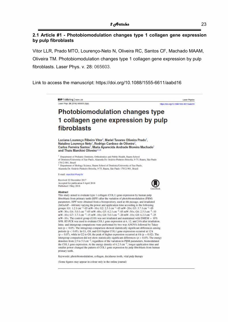
2.1 Article #1 - Photobiomodulation changes type 1 collagen gene expression by pulp fibroblasts Vitor LLR, Prado MTO, Lourenço-Neto N, Oliveira RC, Santos CF, Machado MAAM,
Oliveira TM. Photobiomodulation changes type 1 collagen gene expression by pulp
fibroblasts. Laser Phys. v. 28: 065603.
Link to access the manuscript: https://doi.org/10.1088/1555-6611/aabd16

2 Articles 24

2 Articles 25
2.2 Article #2 - Photobiomodulation upregulates angiogenic protein synthesis by pulp fibroblasts
ABSTRACT
This study aimed to assess the protein synthesis of angiogenic factors by pulp
fibroblasts from human teeth (HPF) after photobiomodulation (PBM). HPF at 4th
passage was plated, led to adhere, subjected to serum starvation, and subsequently
irradiated with 660 nm low-level laser according to the following groups: 2.5 J/cm2 -
10 mW/10 sec ; 3.7 J/cm2 - 15 mW/10 sec. The control group was not irradiated. The
synthesis of the following angiogenic factors was obtained by ELISA Multiplex Assay:
VEGF-A, VEGF-C, VEGF-D, VEGFR1, VEGFR2, FGF-2, PDGF, PLGF, PECAM-1,
and BMP-9 at 6, 12, and 24 hours after PBM, in supernatant and lysate. Two-way
ANOVA followed by Tukey test were applied to test intra- and intergroup
comparisons (P<0.05). HPF secreted and produced all the tested proteins before and
after PBM, except for PDGF in the lysate. The intragroup comparisons showed
similar patterns of angiogenic factors synthesis for all groups: the secretion of VEGF-
A, VEGF-C, VEGFR1, and BMP-9 increased significantly in the supernatant, while
FGF-2, VEGF-A increased significantly in the lysate (p<0.05). The dosimetry of 2.5
J/cm2 exhibited upregulation of VEGF-D (p=0.4779), VEGFR1 (p=0.8570), and BMP-
9 (p=0.0042) in supernatant and VEGFR1 (p=0.0128) and VEGF-D (p=0.0036) in
lysate; and downregulation of VEGF-A (p=0.0176), VEGF-C (p=0.0328), PDGF
(p=0.0077), PLGF (p=0.0004) in supernatant and PLGF (p=0.0094), BMP-9 (p=
0.5645) in lysate. The dosimetry of 3.7 J/cm2 had upregulation of VEGF-A p=0.8410),
FGF-2 (p=0.4778), BMP-9 (p=0.0042), and VEGFR1 (p=0.8570) in supernatant and
VEGF-A (p=0.8412), VEGF-C (p=0.5908), and VEGFR1 (p=0.0128) in lysate; and
downregulation of VEGF-A (p=0.0176), PDGF (p=0.0077), PLGF (p=0.0004) in
supernatant and PLGF (p=0.0094), and BMP-9 (p=0.0276) in lysate. The dosimetries
of 2.5 J/cm2 and 3.7 J/cm2 biomodulates the synthesis of several angiogenic
proteins. However, the dosimetry of 3.7 J/cm2 was the most effective for the
production and secretion of angiogenic factors
Keywords: Photobiomodulation. Angiogenesis Factor. Deciduous tooth. Vital pulp
therapy.

2 Articles 26
1 - Introduction
Angiogenesis is the main process to form blood vessels in wound repair
mediated by angiogenic molecules [1, 2]. VEGF family proteins (VEGF-A, VEGF-C,
VEGF-D, and PLGF) and receptors (VEGFR1 and VEGFR2) regulate the biological
activity response of the endothelial cells through proliferation, migration, vascular
permeability, and angiogenesis [3-5]. Other angiogenic growth factors are FGF-2,
BMP-9, PDGF, and PECAM-1. FGF-2 and BMP-9 regulates angiogenesis [6, 7].
PDGF has an indirect angiogenic effect by maintaining and stabilizing newly-formed
blood vessels [8]. The literature reports the PECAM-1 regulation of angiogenesis [9,
10].
One of the pulp regeneration keystones is angiogenesis [11, 12]. Pulp
fibroblasts produces angiogenic molecules and receptors for physiological
angiogenesis in a paracrine manner, promoting endothelial cell chemotaxis,
proliferation, and differentiation [12-16]. Different stress conditions, such as caries or
trauma, result in different composition and concentration of the secretome from pulp
cells [17].
Works from other fields reveal that photobiomodulation (PBM) modulated the
synthesis of angiogenic growth factors by mesenchymal stem cells from adipose
tissue [18] and gingival fibroblasts [19, 20]. Recently, the literature reports that PBM
at 660 nm increase VEGF gene expression in fibroblasts [21], but little is known
whether higher gene expression results in higher protein production and secretion.
Clinically, PBM has been successfully used in vital pulp therapy [22, 23], but the
literature lacks a protocol regarding the most effective PBM parameters. PBM
biological effects depend on wavelength, dose, and application time [24], so that
different dosimetries may result in different biological responses [25].
Therapies that keeps pulp vitality after caries or trauma relies on the pulp
tissue capable of regenerating [26, 27]. From a more biological and less invasive
point of view, the use of PBM would help in pulp repair. Thus, this study aimed to
assess the protein synthesis of angiogenic factors by pulp fibroblasts after
photobiomodulation.

2 Articles 27
2 - Material and methods
2.1 - Cell culture
Pulp fibroblasts from human primary teeth (HPF) were obtained through a
Biorepository [28, 29]. 2 x 10⁴ cells/well were seeded in 24-well plate (code 3524,
polystyrene, Corning, NY, USA) with Dulbecco’s modified Eagle’s medium (DMEM -
Sigma Chemical Co, St. Louis, MO) supplemented with 10% Fetal Bovine Serum
(FBS), incubated at 37ºC and 5% CO2, for 24 hours to adhere. The culture medium
was replaced by DMEM supplemented with 1% FBS to synchronize the cell cycle
prior to irradiation and achieve proliferative deficit [28, 30]. After 24 h, prior to
irradiation, the culture medium was replaced by DMEM without phenol red (Sigma
Chemical Co, St. Louis, MO) supplemented with 10% FBS for all groups [28]. Groups
were plated in biological triplicate (3 wells per group).
2.2 PBM irradiation
The study groups were divided according to different dosimetries: 2.5 J/cm2 -
10 mW/10 sec; 3.7 J/cm2 - 15 mW/10 sec. The control group was not irradiated
(control). Aluminum-gallium-indium-phosphide (InGaAlP) diode laser (Twin Flex
Evolution MMOptics® – São Carlos, Brazil), at 660nm wavelength, 0.04 cm2 laser tip
area, 0.225 cm tip diameter, continuous mode was applied according to the study
groups. Based on previous studies [28, 30] a black mask covers the culture plate, the
distance between the laser beam and the cell culture was standardized at 1 mm, and
the output power was checked by a radiometer (Laser Check MMOptics® – São
Carlos/SP, BR), prior to laser application. The control group (not irradiated) was
maintained in DMEM+10%FBS [28, 30].
2.3 Collection of supernatant and lysate
After 6, 12 and 24 hours, the supernatant of all groups was collected and
stored for the analysis. After the collection of the supernatant, 300 ul/well of lysis
solution (CellyticnM, Sigma Aldrich, C2978, 50Ml) was dispensed to obtain the
chemical lysis. Also, the mechanical lysis was performed by scratching the pipette tip.
The lysate of all groups was collected and stored for analysis.
2.4 Immunoassay procedure
The following panels were used to verify the protein synthesis of VEGF-A,
VEGF-C, VEGF-D, VEGFR1, VEGFR2, FGF2, PLGF, PDGF, PECAM-1, and BMP-9
at 6, 12 and 24 hours after irradiation, in supernatant and lysate: HAGP1MAG-12K

2 Articles 28
(MILLIPLEX® MAP Human Angiogenesis Panel 1) and HANG2MAG-12K
(MILLIPLEX® MAP Human Angiogenesis Panel 2). All samples, quality control
samples and standards were prepared as recommended in the MILLIPLEX® map
assay kit protocols with supplied diluents, following the manufacturer’s instructions.
The assay plate was then analyzed with the Luminex® instrument equipped with
xPONENT® and Multiplex Analyst 5.1 software. The results were obtained in pg/ml.
2.5 - Statistical analysis
All statistical analyses were obtained with level of significance of 5% (Prism
Graph Pad 7 for Windows). Intra- and intergroup comparisons were analyzed by two-
way ANOVA followed by Tukey test.
3 – Results
3.1 - Intragroup comparison
In control group, HPF secreted all the tested proteins before PBM, except for
PDGF in the lysate. Non-irradiated HPF secreted higher levels of VEGF-A, VEGF-C,
and VEGFR2, but low levels of FGF-2, PDGF, VEGFR1, PECAM-1, VEGF-D, PLGF,
and BMP-9 in supernatant (Table 1). In lysate, non-irradiated HPF produced higher
levels of FGF-2, VEGF-C, and VEGFR2, but lower levels of VEGFR1, VEGF-A,
PECAM-1, VEGF-D, PLGF, BMP-9 (Table 1). Over time, the secretion of VEGF-A,
VEGF-C, VEGFR1, and BMP-9 by non-irradiated HPF increased significantly in the
supernatant, while FGF-2, VEGF-A increased significantly in the lysate (p<0.05). In
the supernatant of non-irradiated HPF, VEGFR2 and PLGF decreased the synthesis
overtime, while FGF-2 had a peak of maximum secretion at 12 h, and PDGF,
PECAM-1, VEGF-D, and BMP-9 had different secretion patterns, but without
statistically significant differences (p>0.05). In the lysate of non-irradiated HPF,
VEGFR2, VEGF-C, VEGFR1 decreased the synthesis over time, while PECAM1,
VEGF-D, PLGF, BMP-9 was constant, but without statistically significant differences
(p>0.05).

2 Articles 29
Table 1 – Angiogenic protein synthesis by non-irradiated HPF (control group)
SUPERNATANT
PROTEINS 6h 12h 24h P value VEGF-C 66.48±1.75a 115.22±5.00b
201.46±34.23c <0.0001*
VEGF-A 31.03±4.00a 69.00±9.96b 214.70±23.50c <0.0001* VEGFR2 31.33±2.22a 30.05±0.00a 30.05±0.00a 0.7230 FGF-2 17.66±5.01a 36.63±42.89a 22.36±3.14a 0.6113 PDGF 7.65±2.84a 8.33±1.24a 10.88±4.06a 0.7879
VEGFR1 6.00±0.48a 7.35±0.80a 17.13±2.42b <0.0001* PECAM-1 3.67±0.25a 3.38±0.00a 3.52±0.25a 0.3153 VEGF-D 2.24±0.25a 2.39±0.00a 2.47±0.14a 0.5768 PLGF 1.04±0.13a 1.01±0.09a 0.98±0.13a 0.2807 BMP-9 0.61±0.10a 0.72±0.04ab 0.76±0.02b 0.0140*
LYSATE PROTEINS 6h 12h 24h P value FGF-2 1296.67±233.14a 1420.33±179.51ab 1754.33±311.59b 0.0005* VEGFR2 30.58±2.16a 29.33±0.00a 28.12±2.10a 0.6734 VEGF-C 26.06±1.56a 22.06±3.07a 23.99±2.71a 0.0515 VEGFR1 10.74±0.00a 7.12±3.24a 8.39±6.01a 0.1741 VEGF-A 8.78±0.36a 10.07±0.39a 14.46±0.39b <0.0001* PECAM-1 3.24±0.24a 3.52±0.43a 3.24±0.24a 0.7874 VEGF-D 2.38±0.24a 2.52±0.00a 2.52±0.00a 0.4728 PLGF 0.60±0.05a 0.55±0.00a 0.55±0.00a 0.6410 BMP-9 0.52±0.00a 0.52±0.00a 0.49±0.05a 0.5645 PDGF 0.00 0.00 0.00 ------ * Different superscript lowercase letters in row mean statistically significant differences among periods (Two-way ANOVA followed by Tukey test; P<0.05). Values in pg/ml.
HPF irradiated with 2.5 J/cm2 secreted all tested proteins, except for PDGF in
the lysate. The quantification of the proteins in both supernatant and lysate after
irradiation with 2.5 J/cm2 showed a pattern similar to that of control group. Overtime,
VEGF-A, VEGF-C, and VEGFR1 increased significantly in supernatant (p<0.05),
while FGF-2, VEGF-A, and VEGF-D increased significantly in the lysate (p<0.05)
(Table 2). The other angiogenic proteins had different patterns of synthesis overtime,
but without statistically significant differences (p>0.05) (Table 2).
2 Articles 30
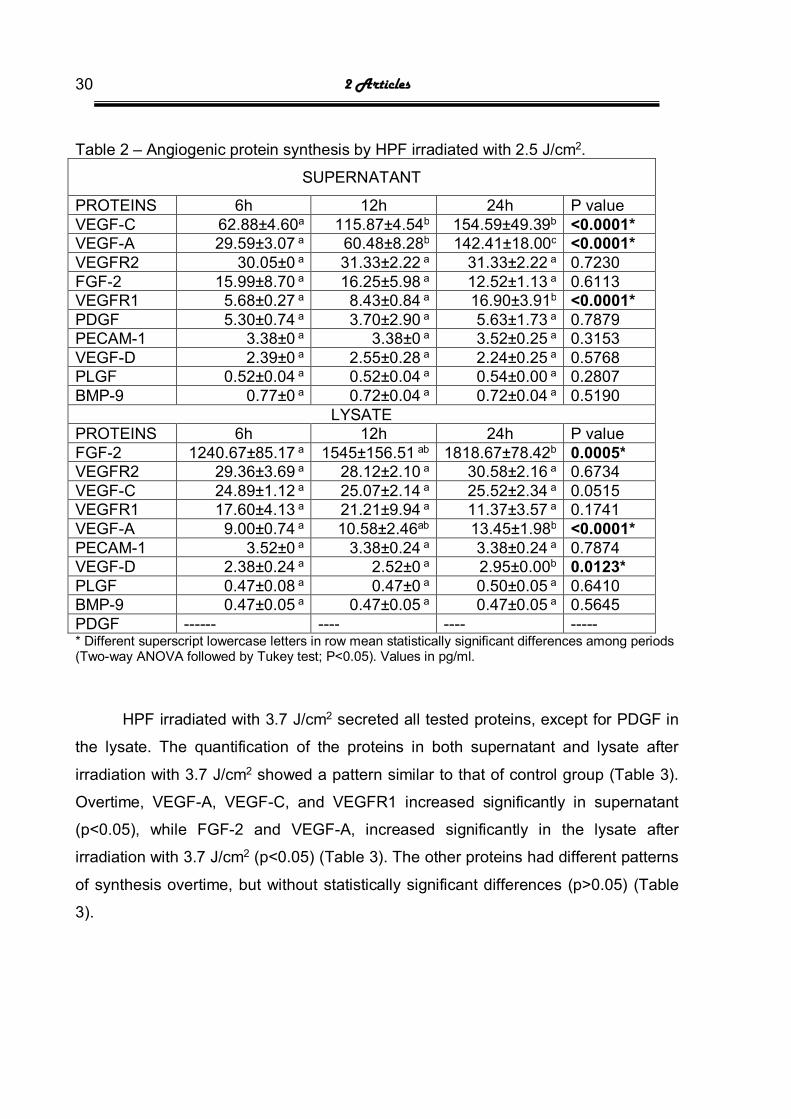
Table 2 – Angiogenic protein synthesis by HPF irradiated with 2.5 J/cm2.
SUPERNATANT
PROTEINS 6h 12h 24h P value VEGF-C 62.88±4.60a 115.87±4.54b 154.59±49.39b <0.0001* VEGF-A 29.59±3.07 a 60.48±8.28b 142.41±18.00c <0.0001* VEGFR2 30.05±0 a 31.33±2.22 a 31.33±2.22 a 0.7230 FGF-2 15.99±8.70 a 16.25±5.98 a 12.52±1.13 a 0.6113 VEGFR1 5.68±0.27 a 8.43±0.84 a 16.90±3.91b <0.0001* PDGF 5.30±0.74 a 3.70±2.90 a 5.63±1.73 a 0.7879 PECAM-1 3.38±0 a 3.38±0 a 3.52±0.25 a 0.3153 VEGF-D 2.39±0 a 2.55±0.28 a 2.24±0.25 a 0.5768 PLGF 0.52±0.04 a 0.52±0.04 a 0.54±0.00 a 0.2807 BMP-9 0.77±0 a 0.72±0.04 a 0.72±0.04 a 0.5190
LYSATE PROTEINS 6h 12h 24h P value FGF-2 1240.67±85.17 a 1545±156.51 ab 1818.67±78.42b 0.0005* VEGFR2 29.36±3.69 a 28.12±2.10 a 30.58±2.16 a 0.6734 VEGF-C 24.89±1.12 a 25.07±2.14 a 25.52±2.34 a 0.0515 VEGFR1 17.60±4.13 a 21.21±9.94 a 11.37±3.57 a 0.1741 VEGF-A 9.00±0.74 a 10.58±2.46ab 13.45±1.98b <0.0001* PECAM-1 3.52±0 a 3.38±0.24 a 3.38±0.24 a 0.7874 VEGF-D 2.38±0.24 a 2.52±0 a 2.95±0.00b 0.0123* PLGF 0.47±0.08 a 0.47±0 a 0.50±0.05 a 0.6410 BMP-9 0.47±0.05 a 0.47±0.05 a 0.47±0.05 a 0.5645 PDGF ------ ---- ---- ----- * Different superscript lowercase letters in row mean statistically significant differences among periods (Two-way ANOVA followed by Tukey test; P<0.05). Values in pg/ml.
HPF irradiated with 3.7 J/cm2 secreted all tested proteins, except for PDGF in
the lysate. The quantification of the proteins in both supernatant and lysate after
irradiation with 3.7 J/cm2 showed a pattern similar to that of control group (Table 3).
Overtime, VEGF-A, VEGF-C, and VEGFR1 increased significantly in supernatant
(p<0.05), while FGF-2 and VEGF-A, increased significantly in the lysate after
irradiation with 3.7 J/cm2 (p<0.05) (Table 3). The other proteins had different patterns
of synthesis overtime, but without statistically significant differences (p>0.05) (Table
3).

2 Articles 31
Table 3 – Angiogenic protein synthesis by HPF irradiated wit 3.7 J/cm2.
SUPERNATANT
PROTEINS 6h 12h 24h P value VEGF-C 62.81±8.54a 115,86±4.17b 207.85±10.98c <0.0001* VEGF-A 37.03±2.75a 60,60±9.73a 178.75±19.02b <0.0001* VEGFR2 31.33±2.22 a 30,05±0 a 30.05±0 a 0.7230 FGF-2 21.56±7.86 a 16,89±5.68 a 16.06±3.89 a 0.6113 PDGF 6.41±1.80 a 7,00±1.45 a 4.78±1.77 a 0.7879 VEGFR1 4.94±0.66a 8,53±0.98b 18.50±0.73c <0.0001* PECAM-1 3.67±0.25 a 3,52±0.25 a ///3.38±0a 0.3153 VEGF-D 2.24±0.25 a 2,24±0.25 a 2.39±0a 0.5768 BMP-9 0.76±0.06a 0,75±0.04a 0.75±0.04a 0.5190 PLGF 0.66±0.05 a 0,61±0 a 0.69±0.08 a 0.2807
LYSATE PROTEINS 6h 12h 24h P value FGF-2 1253.67±234a 1435,00±244,01 AB 1732.67±178.35 b 0.0005* VEGFR2 29.33±0 a 29.33±0 a 29.33±0a 0.6734 VEGF-C 28.05±0.96 a 23,27±3.52 a 24.39±2.53 a 0.0515 VEGF-A 10.30±0.78a 10.31±1.40a 13.91±2.91b <0.0001* VEGFR1 6.11±0 a 13.75±7.57 a 9.14±5.25 a 0.1741 PECAM-1 3.24±0.24 a 3.38±0.24 a 3.52±0a 0.7874 VEGF-D 2.52±0 a 2.66±0.25 a 2.52±0a 0.4409 PLGF 0.47±0.08 a 0.50±0.05 a 0.52±0.05a 0.6410 BMP-9 0.47±0.05a 0.44±0a 0.44±0.a 0.5645 PDGF ----- ------ ---- ---- * Different superscript lowercase letters in row mean statistically significant differences among periods (Two-way ANOVA followed by Tukey test; P<0.05). Values in pg/ml.

2 Articles 32
3.1 - Intergroup comparison
The results were expressed as percentage of controls, using the values from
Table 1.
In supernatant, at 6 hours, the dosimetry of 3.7 J/cm2 non-significantly
increased the secretion of VEGF-A (p=0.8410) and FGF-2 (p=0.4778) (Figure 1A and
1E). Both dosimetries significantly biomodulated the BMP-9 secretion (p=0.0042),
after 6 hours in supernatant (Figure 1F). At 12 hours, both dosimetries increased
VEGFR1 secretion in supernatant without statistically significant differences
(p=0.8570) (Figure 1D). The dosimetry of 2.5 J/cm2 increased the VEGF-D secretion
in supernatant without statistically significant differences (p=0.4779), at 12 hours
(Figure 1C). At 24 hours, VEGF-A secretion in the supernatant significantly
decreased to lower values after PBM with both dosimetries (p=0.0176) (Figure 1A).
At this period, VEGF-C secretion significantly decrease after PBM application with
2.5 J/cm2 (p=0.0328) (Figure 1B). The dosimetry of 3.7 J/cm2 had significant higher
synthesis of VEGF-A (p=0.0083) and VEGF-C (p=0.0150) than the dosimetry of 2.5
Jcm2 in supernatant at 24h (Figure 1A and 1B). Both dosimetries downregulated the
PDGF secretion in supernatant, at all periods, with statistically significant differences
(p=0.0077) at 24 hours (Figure 1I). Statistically significant lower values of PLGF
secretion in the supernatant occurred at all periods (p=0.0004) for both dosimetries
(Figure 1J). At all periods, VEGFR2 (Figure 1E) and PECAM-1 (Figure 1H) had
values statistically similar to that of control group for both dosimetries.

2 Articles 33
Figure 1A-J – Intergroup comparison of the quantification of angiogenic factors after PBM in supernatant * indicates a significant difference with the control considering each angiogenic factor and each period separately
(Two-way ANOVA; P<0.05). # indicates a significant difference between the dosimetries of 2.5 and 3.7 J/cm2
considering each angiogenic factor and each period separately (Two-way ANOVA; P<0.05)
2 Articles 34
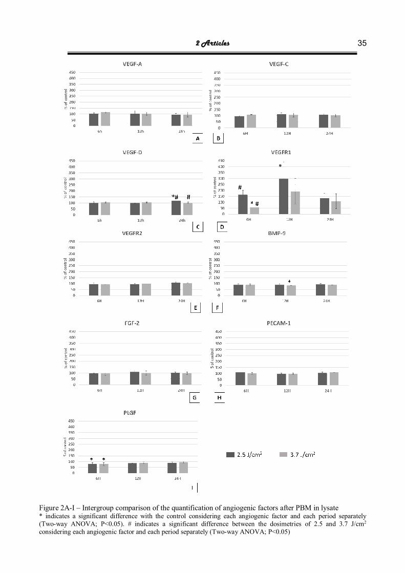
In lysate, at 6 hours, the dosimetry of 3.7 J/cm2 non-significantly increased the
production of VEGF-A (p=0.8412) and VEGF-C (p=0.5908) (Figure 2A and 2B). The
dosimetry of 2.5 J/cm2 biomodulated the VEGFR1 production in the lysate at 6 hours
without statistically differences with control group (p=0.2871), but with significant
differences with the dosimetry of 3.7 J/cm2 (p=0.0440) (Figure 2D) Statistically
significant lower values of PLGF production (p=0.0094) in lysate occurred at 6 hours
for both dosimetries (Figure 2I). After 12 hours, both dosimetries increased the
VEGFR1 production in lysate, with statistically significant differences with control
group for 2.5 J/cm2 (p=0.0128) (Figure 2D). Both dosimetries decreased the BMP-9
production in the lysate, with statistically significant difference (p=0.0276) for 3.7
J/cm2, at 12 hours (Figure 2F). At 24 hours, VEGF-D production in the lysate
significantly increased after PBM with 2.5 J/cm2 compared with control group and the
dosimetry of 3.7 J/cm2 (p=0.0036) (Figure 2C). At all periods, VEGFR2 (Figure 2E)
and PECAM-1 (Figure 2H) had values statistically similar to that of control group for
both dosimetries. At all periods, PDGF was not produced in the lysate for both
dosimetries and control group.
2 Articles 35
Figure 2A-I – Intergroup comparison of the quantification of angiogenic factors after PBM in lysate * indicates a significant difference with the control considering each angiogenic factor and each period separately
(Two-way ANOVA; P<0.05). # indicates a significant difference between the dosimetries of 2.5 and 3.7 J/cm2
considering each angiogenic factor and each period separately (Two-way ANOVA; P<0.05)

2 Articles 36
4 - Discussion
This study showed the photobiomodulation of the main angiogenic factors in
supernatant and lysate. As far as we are concerned, this is the first study addressing
the synthesis of angiogenic proteins by pulp fibroblasts after PBM, which makes
comparisons difficult.
In terms of biomodulation, the higher power and dose upregulated the
angiogenic factors (VEGF-A, VEGF-C, FGF-2, BMP-9, and VEGFR1 in supernatant
and VEGF-A and VEGF-C in lysate). In general, photobiomodulation of
angiogenesis increases VEGF production due to nitric oxide modulation [21, 31]. This
occurs through nitric oxide production synthase via activation of PLCϒ-Ca dependent
influx [32-34].
FGF-2 plays a key role in pulp repair by regulating angiogenesis [35].
Although through different methodologies and with different cell lines, in general,
PBM upregulates FGF-2 synthesis [19, 20, 36, 37], similarly to the result of this study.
Moreover, FGF-2 synergistically potentializes the action of VEGF on endothelial cells,
which in turn increases VEGF receptor expression [6, 13].
The higher dose (3.7 J/cm2) also upregulated the VEGFR1 synthesis.
VEGFR1 controls vascular biology acting as “VEGF trap” by sequestering VEGF-A
away from VEGFR2 and forming non-signaling VEGFR1-VEGFR2 heterodimer
through dimerization, transphosphorylation, and regulation of expression levels [6,
15, 34, 38, 39].
In supernatant, 24 hours after PBM, the secretion of VEGF-A, VEGF-C, and
FGF2 was statistically lower than control group for both doses, but with markedly
decreasing for the lower dose (2.5 J/cm2). The regulation of VEGF family proteins
and receptors is a complex interplay [40]. Many factors regulated VEGF synthesis:
VEGFR1 (as mentioned above) [6, 15, 34, 38, 39], VEGF bioavailability through
either plasmin cleavage or extracellular proteolysis [3, 6], splicing of VEGF proteins
affecting temporal and spatial distribution [34], FGF controlling of VEGF upstream [6],
and VEGFR2 binding to integrin [6, 33, 34, 38]. Integrin is a transmembrane
heterodimer that mediates cell-matrix adhesion through binding to extracellular
proteins, such as collagen, fibronectin, and laminin [38]. Integrin recruitment
qualitatively modulates the signaling of VEGF receptors [34, 38] and the VEGFR2
binding to integrin prevents aberrant angiogenesis stimulation [6, 33]. PBM increases

2 Articles 37
COL1 gene expression by HPF [30]. Collagen 1 downregulated the VEGF-A
mediated activation of VEGFR2 through phosphatase SHP2 recruitment [38]. Taken
together, these information suggest the crosstalk between proteins of the signaling
cascade of VEGF protein and receptors.
After PBM, BMP-9 was upregulated in supernatant and downregulated in
lysate with both dosimetries. In dentistry, the literature reported an increase in BMP-9
levels after PBM with different methodologies (immunohistochemistry) and cells
(osteoblasts) [41]. Upon BMP-9 stimulation, stem cells reveal odontoblastic
differentiation [42]. Further studies are necessary to understand the role of secreted
BMP-9, after PBM, in pulp angiogenesis and repair.
Both dosimetries downregulated the synthesis of PDGF (supernatant) and
PLGF (supernatant and lysate). To the best or our knowledge, the literature lacks
studies on the protein synthesis and production of PLGF by pulp fibroblasts. Studies
on angiogenesis show that PLGF plays a crucial role in inflammation-associated
angiogenesis by recruiting inflammatory cells, mediated by binding VEGFR1 [6, 34,
39]. The literature reported an increase in PDGF levels after PBM, but with different
wavelength (1064 nm) and dosimetry (1.6 J/cm2) on different cells (palatal tissue)
[41, 43]. Works from other fields highlighted the inhibitory effect of PBM in PDGF-
stimulated cells [44]. Although Rombouts, Giraud (17) reports the increase of PDGF
transcription by pulp cells, we did not find the same protein transduction in this study.
The methodology of this study was based on previous research [28-30]. We
used 660 nm wavelength based on the maintenance of HPF viability and proliferation
[28] and effect in gene expression [18, 21, 30]. Before PBM, HPF secreted and
produced all the tested angiogenic factors, except for PDGF in lysate, which agrees
with the literature [13, 45-49]. Other studies show that to simulate pulp stress
conditions either hypoxia or serum deprivation lead to different angiogenic secretion
by pulp cells [17, 50, 51]. This would explain the synthesis and production of
angiogenic proteins by non-irradiated HPF in this present study. Further studies are
necessary to verify whether hypoxia would change the angiogenic secretome of pulp
fibroblasts [52]
2 Articles 38
5 - Conclusion
The dosimetries of 2.5 J/cm2 and 3.7 J/cm2 biomodulates the synthesis of
several angiogenic protein. However, the dosimetry of 3.7 J/cm2 was the most
effective for the production and secretion of angiogenic factors.
REFERENCES
1. Folkman J, Shing Y. Angiogenesis. The Journal of biological chemistry. 1992;267(16):10931-4. Epub 1992/06/05. PubMed PMID: 1375931.
2. Yamazaki Y, Morita T. Molecular and functional diversity of vascular endothelial growth factors. Molecular Diversity. 2006;10(4):515. doi: 10.1007/s11030-006-9027-3.
3. Holmes K, Roberts OL, Thomas AM, Cross MJ. Vascular endothelial growth factor receptor-2: Structure, function, intracellular signalling and therapeutic inhibition. Cellular Signalling. 2007;19(10):2003-12. doi: https://doi.org/10.1016/j.cellsig.2007.05.013.
4. Stuttfeld E, Ballmer‐Hofer K. Structure and function of VEGF receptors. IUBMB life. 2009;61(9):915-22.
5. Smith GA, Fearnley GW, Harrison MA, Tomlinson DC, Wheatcroft SB, Ponnambalam S. Vascular endothelial growth factors: multitasking functionality in metabolism, health and disease. Journal of Inherited Metabolic Disease. 2015;38(4):753-63. doi: 10.1007/s10545-015-9838-4.
6. Simons M, Gordon E, Claesson-Welsh L. Mechanisms and regulation of endothelial VEGF receptor signalling. Nature reviews Molecular cell biology. 2016;17(10):611-25. Epub 2016/07/28. doi: 10.1038/nrm.2016.87. PubMed PMID: 27461391.
7. David L, Mallet C, Keramidas M, Lamandé N, Gasc J-M, Dupuis-Girod S, et al. Bone morphogenetic protein-9 is a circulating vascular quiescence factor. Circulation Research. 2008;102(8):914-22. doi: 10.1161/CIRCRESAHA.107.165530. PubMed PMID: PMC2561062.
8. Zhang M, Jiang F, Zhang X, Wang S, Jin Y, Zhang W, et al. The Effects of Platelet-Derived Growth Factor-BB on Human Dental Pulp Stem Cells Mediated Dentin-Pulp Complex Regeneration. Stem cells translational medicine. 2017;6(12):2126-34. Epub 2017/10/25. doi: 10.1002/sctm.17-0033. PubMed PMID: 29064632.
9. Marques NCT, Neto NL, Prado MTO, Vitor LLR, Rodini CO, Sakai VT, et al. CD31 expression in human primary teeth treated with photobiomodulation therapy. Lasers in Dental Science. 2018;2(2):103-8.

2 Articles 39
10. Lourenco-Neto N, Marques N, Prado M, Vitor L, Rodini C, Oliveira T. Histological and CD31 Immunolocalization after Pulp Therapy using Mta or Portland Cement. Int J Dentistry Oral Sci. 2018;5(3):622-5.
11. Cooper PR, Holder MJ, Smith AJ. Inflammation and regeneration in the dentin-pulp complex: a double-edged sword. J Endod. 2014;40(4 Suppl):S46-51. Epub 2014/04/05. doi: 10.1016/j.joen.2014.01.021. PubMed PMID: 24698693.
12. Matsushita K, Motani R, Sakuta T, Yamaguchi N, Koga T, Matsuo K, et al. The role of vascular endothelial growth factor in human dental pulp cells: induction of chemotaxis, proliferation, and differentiation and activation of the AP-1-dependent signaling pathway. Journal of dental research. 2000;79(8):1596-603. Epub 2000/10/07. doi: 10.1177/00220345000790081201. PubMed PMID: 11023281.
13. Tran-Hung L, Mathieu S, About I. Role of human pulp fibroblasts in angiogenesis. Journal of dental research. 2006;85(9):819-23. Epub 2006/08/26. doi: 10.1177/154405910608500908. PubMed PMID: 16931864.
14. Chmilewsky F, Jeanneau C, Laurent P, About I. Pulp fibroblasts synthesize functional complement proteins involved in initiating dentin-pulp regeneration. The American journal of pathology. 2014;184(7):1991-2000. Epub 2014/05/13. doi: 10.1016/j.ajpath.2014.04.003. PubMed PMID: 24814102.
15. Smith Gina A, Fearnley Gareth W, Tomlinson Darren C, Harrison Michael A, Ponnambalam S. The cellular response to vascular endothelial growth factors requires co-ordinated signal transduction, trafficking and proteolysis. Bioscience Reports. 2015;35(5):e00253. doi: 10.1042/BSR20150171. PubMed PMID: PMC4613718.
16. Anca V, Sigbjørn L, Ole I, Athanasia B, Ellen B. Vascular endothelial growth factors signalling in normal human dental pulp: a study of gene and protein expression. European Journal of Oral Sciences. 2013;121(2):92-100. doi: doi:10.1111/eos.12019.
17. Rombouts C, Giraud T, Jeanneau C, About I. Pulp Vascularization during Tooth Development, Regeneration, and Therapy. Journal of dental research. 2017;96(2):137-44. Epub 2017/01/21. doi: 10.1177/0022034516671688. PubMed PMID: 28106505.
18. Oliveira TS, Serra AJ, Manchini MT, Bassaneze V, Krieger JE, de Tarso Camillo de Carvalho P, et al. Effects of low level laser therapy on attachment, proliferation, and gene expression of VEGF and VEGF receptor 2 of adipocyte-derived mesenchymal stem cells cultivated under nutritional deficiency. Lasers in medical science. 2015;30(1):217-23. Epub 2014/09/07. doi: 10.1007/s10103-014-1646-9. PubMed PMID: 25192841.
19. Damante CA, De Micheli G, Miyagi SP, Feist IS, Marques MM. Effect of laser phototherapy on the release of fibroblast growth factors by human gingival fibroblasts. Lasers in medical science. 2009;24(6):885-91. Epub 2008/07/05. doi: 10.1007/s10103-008-0582-y. PubMed PMID: 18600291.

2 Articles 40
20. Saygun I, Karacay S, Serdar M, Ural AU, Sencimen M, Kurtis B. Effects of laser irradiation on the release of basic fibroblast growth factor (bFGF), insulin like growth factor-1 (IGF-1), and receptor of IGF-1 (IGFBP3) from gingival fibroblasts. Lasers in medical science. 2008;23(2):211-5. doi: 10.1007/s10103-007-0477-3.
21. Szezerbaty SKF, de Oliveira RF, Pires-Oliveira DAA, Soares CP, Sartori D, Poli-Frederico RC. The effect of low-level laser therapy (660 nm) on the gene expression involved in tissue repair. Lasers in medical science. 2018;33(2):315-21. doi: 10.1007/s10103-017-2375-7.
22. Fernandes AP, Lourenco Neto N, Teixeira Marques NC, Silveira Moretti AB, Sakai VT, Cruvinel Silva T, et al. Clinical and radiographic outcomes of the use of Low-Level Laser Therapy in vital pulp of primary teeth. International journal of paediatric dentistry. 2015;25(2):144-50. Epub 2014/07/01. doi: 10.1111/ipd.12115. PubMed PMID: 24974864.
23. Marques NC, Neto NL, Rodini Cde O, Fernandes AP, Sakai VT, Machado MA, et al. Low-level laser therapy as an alternative for pulpotomy in human primary teeth. Lasers in medical science. 2015;30(7):1815-22. Epub 2014/09/23. doi: 10.1007/s10103-014-1656-7. PubMed PMID: 25240388.
24. Karu T. Photobiology of low-power laser effects. Health physics. 1989;56(5):691-704. Epub 1989/05/01. PubMed PMID: 2651364.
25. Huang YY, Chen AC, Carroll JD, Hamblin MR. Biphasic dose response in low level light therapy. Dose-response : a publication of International Hormesis Society. 2009;7(4):358-83. Epub 2009/12/17. doi: 10.2203/dose-response.09-027.Hamblin. PubMed PMID: 20011653; PubMed Central PMCID: PMCPMC2790317.
26. AAPD. Use of Vital Pulp Therapies in Primary Teeth with Deep Caries Lesions. Pediatric dentistry. 2017;39(6):173-86. Epub 2017/11/29. PubMed PMID: 29179355.
27. Coll JA, Seale NS, Vargas K, Marghalani AA, Al Shamali S, Graham L. Primary Tooth Vital Pulp Therapy: A Systematic Review and Meta-analysis. Pediatric dentistry. 2017;39(1):16-123. Epub 2017/03/16. PubMed PMID: 28292337.
28. Marques NCT, Lourenço-Neto N, Prado MTO, Vitor LLR, Oliveira RC, Sakai VT, et al. Effects of PBM in different energy densities and irradiance on maintaining cell viability and proliferation of pulp fibroblasts from human primary teeth. Lasers in medical science. 2017;32(7):1621-8. Epub 2017/08/13. doi: 10.1007/s10103-017-2301-z. PubMed PMID: 28801810.
29. Lourenço-Neto N, Pereira LF, Marques NCT, Prado M, Vitor LLR, Tokuhara CK, et al. Storage protocol of dental pulp cells from human exfoliated deciduous teeth. Brazilian Dental Science. 2017;20(3):126-31.
30. Vitor LLR, Prado MTO, Lourenço-Neto N, Oliveira RC, Santos CF, Machado MAAM, et al. Photobiomodulation changes type 1 collagen gene expression by pulp fibroblasts. Laser Physics. 2018;28(6):065603.
2 Articles 41
31. Prindeze NJ, Moffatt LT, Shupp JW. Mechanisms of action for light therapy: A review of molecular interactions. Experimental Biology and Medicine. 2012;237(11):1241-8. doi: 10.1258/ebm.2012.012180.
32. Fraisl P. Crosstalk between oxygen- and nitric oxide-dependent signaling pathways in angiogenesis. Experimental Cell Research. 2013;319(9):1331-9. doi: https://doi.org/10.1016/j.yexcr.2013.02.010.
33. Cébe-Suarez S, Zehnder-Fjällman A, Ballmer-Hofer K. The role of VEGF receptors in angiogenesis; complex partnerships. Cellular and Molecular Life Sciences. 2006;63(5):601-15. doi: 10.1007/s00018-005-5426-3. PubMed PMID: PMC2773843.
34. Koch S, Tugues S, Li X, Gualandi L, Claesson-Welsh L. Signal transduction by vascular endothelial growth factor receptors. The Biochemical journal. 2011;437(2):169-83. Epub 2011/06/30. doi: 10.1042/bj20110301. PubMed PMID: 21711246.
35. Shimabukuro Y, Ueda M, Ozasa M, Anzai J, Takedachi M, Yanagita M, et al. Fibroblast growth factor-2 regulates the cell function of human dental pulp cells. J Endod. 2009;35(11):1529-35. Epub 2009/10/21. doi: 10.1016/j.joen.2009.08.010. PubMed PMID: 19840642.
36. Aykol G, Baser U, Maden I, Kazak Z, Onan U, Tanrikulu-Kucuk S, et al. The effect of low-level laser therapy as an adjunct to non-surgical periodontal treatment. Journal of periodontology. 2011;82(3):481-8. Epub 2010/10/12. doi: 10.1902/jop.2010.100195. PubMed PMID: 20932157.
37. Oliveira LSS, Araujo AA, Araujo Junior RF, Barboza CAG, Borges BCD, Silva JSP. Low-level laser therapy (780 nm) combined with collagen sponge scaffold promotes repair of rat cranial critical-size defects and increases TGF-beta, FGF-2, OPG/RANK and osteocalcin expression. International journal of experimental pathology. 2017;98(2):75-85. Epub 2017/05/31. doi: 10.1111/iep.12226. PubMed PMID: 28556971; PubMed Central PMCID: PMCPMC5485360.
38. Koch S, Claesson-Welsh L. Signal Transduction by Vascular Endothelial Growth Factor Receptors. Cold Spring Harbor Perspectives in Medicine. 2012;2(7):a006502. doi: 10.1101/cshperspect.a006502. PubMed PMID: PMC3385940.
39. Claesson-Welsh L. VEGF receptor signal transduction – A brief update. Vascular Pharmacology. 2016;86:14-7. doi: https://doi.org/10.1016/j.vph.2016.05.011.
40. Grünewald FS, Prota AE, Giese A, Ballmer-Hofer K. Structure–function analysis of VEGF receptor activation and the role of coreceptors in angiogenic signaling. Biochimica et Biophysica Acta (BBA) - Proteins and Proteomics. 2010;1804(3):567-80. doi: https://doi.org/10.1016/j.bbapap.2009.09.002.
41. Tim CR, Pinto KN, Rossi BR, Fernandes K, Matsumoto MA, Parizotto NA, et al. Low-level laser therapy enhances the expression of osteogenic factors during

2 Articles 42
bone repair in rats. Lasers in medical science. 2014;29(1):147-56. Epub 2013/03/22. doi: 10.1007/s10103-013-1302-9. PubMed PMID: 23515631.
42. Wang J, Zhang H, Zhang W, Huang E, Wang N, Wu N, et al. Bone Morphogenetic Protein-9 Effectively Induces Osteo/Odontoblastic Differentiation of the Reversibly Immortalized Stem Cells of Dental Apical Papilla. Stem cells and development. 2014;23(12):1405-16. doi: 10.1089/scd.2013.0580. PubMed PMID: PMC4046201.
43. Keskiner I, Lutfioglu M, Aydogdu A, Saygun NI, Serdar MA. Effect of Photobiomodulation on Transforming Growth Factor-beta1, Platelet-Derived Growth Factor-BB, and Interleukin-8 Release in Palatal Wounds After Free Gingival Graft Harvesting: A Randomized Clinical Study. Photomedicine and laser surgery. 2016;34(6):263-71. Epub 2016/04/19. doi: 10.1089/pho.2016.4094. PubMed PMID: 27088277; PubMed Central PMCID: PMCPMC4892223.
44. Baek S, Lee KP, Cui L, Ryu Y, Hong JM, Kim J, et al. Low-power laser irradiation inhibits PDGF-BB-induced migration and proliferation via apoptotic cell death in vascular smooth muscle cells. Lasers in medical science. 2017;32(9):2121-7. Epub 2017/10/07. doi: 10.1007/s10103-017-2338-z. PubMed PMID: 28983687.
45. Tran-Hung L, Laurent P, Camps J, About I. Quantification of angiogenic growth factors released by human dental cells after injury. Archives of oral biology. 2008;53(1):9-13. Epub 2007/09/04. doi: 10.1016/j.archoralbio.2007.07.001. PubMed PMID: 17764655.
46. Chang Y-C, Chang M-C, Chen Y-J, Liou J-U, Chang H-H, Huang W-L, et al. Basic Fibroblast Growth Factor Regulates Gene and Protein Expression Related to Proliferation, Differentiation, and Matrix Production of Human Dental Pulp Cells. Journal of Endodontics. 2017;43(6):936-42. doi: https://doi.org/10.1016/j.joen.2017.01.024.
47. Caviedes-Bucheli J, Gomez-Sosa JF, Azuero-Holguin MM, Ormeno-Gomez M, Pinto-Pascual V, Munoz HR. Angiogenic mechanisms of human dental pulp and their relationship with substance P expression in response to occlusal trauma. International endodontic journal. 2017;50(4):339-51. Epub 2016/03/10. doi: 10.1111/iej.12627. PubMed PMID: 26953220.
48. Grando Mattuella L, Poli de Figueiredo JA, Nor JE, de Araujo FB, Medeiros Fossati AC. Vascular endothelial growth factor receptor-2 expression in the pulp of human primary and young permanent teeth. J Endod. 2007;33(12):1408-12. Epub 2007/11/27. doi: 10.1016/j.joen.2007.08.019. PubMed PMID: 18037047.
49. Matsushita K, Motani R, Sakuta T, Nagaoka S, Matsuyama T, Abeyama K, et al. Lipopolysaccharide enhances the production of vascular endothelial growth factor by human pulp cells in culture. Infection and immunity. 1999;67(4):1633-9. Epub 1999/03/20. PubMed PMID: 10084996; PubMed Central PMCID: PMCPMC96506.
50. Bakopoulou A, Kritis A, Andreadis D, Papachristou E, Leyhausen G, Koidis P, et al. Angiogenic Potential and Secretome of Human Apical Papilla Mesenchymal Stem Cells in Various Stress Microenvironments. Stem cells and development.

2 Articles 43
2015;24(21):2496-512. Epub 2015/07/24. doi: 10.1089/scd.2015.0197. PubMed PMID: 26203919; PubMed Central PMCID: PMCPMC4620528.
51. Aranha AM, Zhang Z, Neiva KG, Costa CA, Hebling J, Nor JE. Hypoxia enhances the angiogenic potential of human dental pulp cells. J Endod. 2010;36(10):1633-7. Epub 2010/09/21. doi: 10.1016/j.joen.2010.05.013. PubMed PMID: 20850667.
52. Mignon C, Uzunbajakava NE, Raafs B, Botchkareva NV, Tobin DJ. Photobiomodulation of human dermal fibroblasts in vitro: decisive role of cell culture conditions and treatment protocols on experimental outcome. Scientific reports. 2017;7(1):2797. Epub 2017/06/07. doi: 10.1038/s41598-017-02802-0. PubMed PMID: 28584230; PubMed Central PMCID: PMCPMC5459822.

2 Articles 44

3 Discussion


3 Discussion 47
3 DISCUSSION
This section provides the discussion of important ideas of methodology,
results, and clinical relevance of the study.
3.1 Methodology
The methodology of the two studies were based on previous research
(Lourenço-Neto et al., 2017; Marques et al., 2017; Fernandes et al., 2018). We used
660-nm wavelength based on the maintenance of HPF viability and proliferation
(Marques et al., 2017; Fernandes et al., 2018), effect in gene expression (Oliveira et
al., 2015; Szezerbaty et al., 2018) and on the assumption that the visible spectrum
would have effects on protein synthesis (Alghamdi et al., 2012). The used irradiances
ranged from 1.2 to 6.2 J/cm2, that is, irradiances shorter than those reported to
decrease immortalized oral fibroblast viability (Engel et al., 2016).
Recently, a study found that the optical parameters, biological factors,
protocols, and oxygen level in cell culture medium impacts the results of the
experiment in dermal fibroblasts (Mignon et al., 2017). Other studies show that to
simulate pulp stress conditions either hypoxia or serum deprivation lead to different
angiogenic secretion by pulp cells (Aranha et al., 2010; Bakopoulou et al., 2015;
Rombouts et al., 2017). This would explain the synthesis and production of
angiogenic proteins by non-irradiated HPF in this present study. Further studies are
necessary to verify whether hypoxia would change the angiogenic secretome of pulp
fibroblasts (Mignon et al., 2017)
3.2 Results
Photobiomodulation upregulates the COL1 gene expression and the synthesis
of angiogenic factors by pulp fibroblasts. Considering the two articles together, the
energy density of 3.7 J/cm2 (15 mW/10 sec) was the most effective considering
COL1 gene expression and protein synthesis of angiogenic factors.
While some studies found similar results of increased COL1 gene expression
by irradiated fibroblasts (Martignago et al., 2015; Pansani et al., 2017), others
concluded that the amount of COL1 expression was not different between irradiated
and non-irradiated fibroblasts (Pereira et al., 2002; Marques et al., 2004). The

3 Discussion 48
rationale behind this distinct outcome is the different cell type (gingival fibroblasts
rather than pulp fibroblasts), wavelength (904 nm), irradiance (3 J/cm2), and
application time. Different cell types have dissimilar morphological patterns leading to
distinct secretion of ECM components (Martinez and Araujo, 2004). Thus, different
PBM parameters would result in different biological responses (Huang et al., 2009),
and would affect collagen gene expression (Zhang et al., 2003).
Considering the synthesis of angiogenic factors, the literature reports that
photobiomodulation increases VEGF production due to nitric oxide modulation
(Prindeze et al., 2012; Szezerbaty et al., 2018), through nitric oxide production
synthase via activation of PLCϒ-Ca dependent influx (Cébe-Suarez et al., 2006; Koch
et al., 2011; Fraisl, 2013). PBM also upregulates FGF-2 synthesis (Saygun et al.,
2008; Damante et al., 2009; Aykol et al., 2011; Oliveira et al., 2017). Moreover, FGF-
2 synergistically potentializes the action of VEGF on endothelial cells, which in turn
increases VEGF receptor expression (Tran-Hung et al., 2006; Simons et al., 2016).
In dentistry, the literature reported an increase in BMP-9 levels after PBM, similar to
the results of the Article #2, but with different methodologies (immunohistochemistry)
and cells (osteoblasts) (Tim et al., 2014). The literature reported an increase in PDGF
levels after PBM, but with different wavelength (1064 nm) and dosimetry (1.6 J/cm2)
on different cells (palatal tissue) (Tim et al., 2014; Keskiner et al., 2016), which was
different from the result of Article #2.
We suggest a cross-talking in the signaling cascade of VEGF proteins and
receptors because the secretion of VEGF-A, VEGF-C, and FGF-2 were lower than
the control group, 24 hours after the irradiation with 2.5 J/cm2. One of the
mechanisms of VEGF control is the VEGFR2 binding to integrin, a transmembrane
heterodimer that mediates cell-matrix adhesion through binding to extracellular
proteins, such as collagen (Cébe-Suarez et al., 2006; Koch et al., 2011; Koch and
Claesson-Welsh, 2012; Simons et al., 2016). Other mechanism of VEGF control is
the collagen 1 downregulation mediated by the activation of VEGFR2 through
phosphatase SHP2 recruitment (Koch and Claesson-Welsh, 2012). As seen in Article
#1, PBM increases COL1 gene expression by pulp fibroblast, which could have
influenced the signaling cascade of angiogenic factors.

3 Discussion 49
3.3 Clinical relevance
Despite the successful clinical outcomes of PBM use in pulp vital therapy of
primary teeth (Fernandes et al., 2015; Marques et al., 2015; Aapd, 2017b;a; Dhar et
al., 2017), the literature lacks a PBM protocol with the most effective wavelength,
power, and application time tailored to achieve dentin-pulp regeneration in vital pulp
therapy of primary teeth. Both Articles #1 and #2 provide a guideline for the clinical
use of PBM, based on in vitro gene expression and synthesis of endogenous growth
factors aiming at pulp regeneration

3 Discussion 50

4 Conclusions


4 Conclusions 53
4 CONCLUSIONS
Conclusion Article #1 – The energy densities from 2.5 to 6.2 J/cm2
biomodulated the COL1 gene expression. At the energy density of 6.2 J/cm2, longer
application time and smaller power changed the pattern of COL1 gene expression by
pulp fibroblasts from HPF.
Conclusion Article #2 - The dosimetries of 2.5 J/cm2 and 3.7 J/cm2
biomodulates the synthesis of several angiogenic protein. However, the dosimetry of
3.7 J/cm2 was the most effective for the production and secretion of angiogenic
factors.

4 Conclusions 54

References


References
57
REFERENCES
Aapd. Pulp Therapy for Primary and Immature Permanent Teeth. Pediatr Dent. 2017a;39(6):325-333.
Aapd. Use of Vital Pulp Therapies in Primary Teeth with Deep Caries Lesions. Pediatr Dent. 2017b;39(6):173-186.
Alghamdi, KM; Kumar, A; Moussa, NA. Low-level laser therapy: a useful technique for enhancing the proliferation of various cultured cells. Lasers Med Sci. 2012;27(1):237-249.
Anca, V; Sigbjørn, L; Ole, I; Athanasia, B; Ellen, B. Vascular endothelial growth factors signalling in normal human dental pulp: a study of gene and protein expression. European Journal of Oral Sciences. 2013;121(2):92-100.
Anders, JJ; Lanzafame, RJ; Arany, PR. Low-level light/laser therapy versus photobiomodulation therapy. Photomed Laser Surg. 2015;33(4):183-184.
Aranha, AM; Zhang, Z; Neiva, KG; Costa, CA; Hebling, J; Nor, JE. Hypoxia enhances the angiogenic potential of human dental pulp cells. J Endod. 2010;36(10):1633-1637.
Aykol, G; Baser, U; Maden, I; Kazak, Z; Onan, U; Tanrikulu-Kucuk, S; Ademoglu, E; Issever, H; Yalcin, F. The effect of low-level laser therapy as an adjunct to non-surgical periodontal treatment. J Periodontol. 2011;82(3):481-488.
Bakopoulou, A; Kritis, A; Andreadis, D; Papachristou, E; Leyhausen, G; Koidis, P; Geurtsen, W; Tsiftsoglou, A. Angiogenic Potential and Secretome of Human Apical Papilla Mesenchymal Stem Cells in Various Stress Microenvironments. Stem Cells Dev. 2015;24(21):2496-2512.
Cébe-Suarez, S; Zehnder-Fjällman, A; Ballmer-Hofer, K. The role of VEGF receptors in angiogenesis; complex partnerships. Cellular and Molecular Life Sciences. 2006;63(5):601-615.
Chmilewsky, F; Jeanneau, C; Laurent, P; About, I. Pulp fibroblasts synthesize functional complement proteins involved in initiating dentin-pulp regeneration. Am J Pathol. 2014;184(7):1991-2000.
References
58
Coll, JA; Seale, NS; Vargas, K; Marghalani, AA; Al Shamali, S; Graham, L. Primary Tooth Vital Pulp Therapy: A Systematic Review and Meta-analysis. Pediatr Dent. 2017;39(1):16-123.
Damante, CA; De Micheli, G; Miyagi, SP; Feist, IS; Marques, MM. Effect of laser phototherapy on the release of fibroblast growth factors by human gingival fibroblasts. Lasers Med Sci. 2009;24(6):885-891.
David, L; Mallet, C; Keramidas, M; Lamandé, N; Gasc, J-M; Dupuis-Girod, S; Plauchu, H; Feige, J-J; Bailly, S. Bone morphogenetic protein-9 is a circulating vascular quiescence factor. Circulation Research. 2008;102(8):914-922.
Dhar, V; Marghalani, AA; Crystal, YO; Kumar, A; Ritwik, P; Tulunoglu, O; Graham, L. Use of Vital Pulp Therapies in Primary Teeth with Deep Caries Lesions. Pediatr Dent. 2017;39(5):146-159.
Engel, KW; Khan, I; Arany, PR. Cell lineage responses to photobiomodulation therapy. J Biophotonics. 2016;9(11-12):1148-1156.
Fernandes, A; Lourenço-Neto , N; Marques, NCT; Vitor, LLR; Prado, MTO; Oliveira, RC; Machado, MaaM; Oliveira, TM. Cellular response of pulp fibroblast to single or multiple photobiomodulation applications. Laser Physics. 2018;28(6):065604.
Fernandes, AP; Lourenco Neto, N; Teixeira Marques, NC; Silveira Moretti, AB; Sakai, VT; Cruvinel Silva, T; Andrade Moreira Machado, MA; Marchini Oliveira, T. Clinical and radiographic outcomes of the use of Low-Level Laser Therapy in vital pulp of primary teeth. Int J Paediatr Dent. 2015;25(2):144-150.
Folkman, J; Shing, Y. Angiogenesis. J Biol Chem. 1992;267(16):10931-10934.
Fraisl, P. Crosstalk between oxygen- and nitric oxide-dependent signaling pathways in angiogenesis. Experimental Cell Research. 2013;319(9):1331-1339.
Frantz, C; Stewart, KM; Weaver, VM. The extracellular matrix at a glance. Journal of Cell Science. 2010;123(24):4195-4200.
Gordon, MK; Hahn, RA. Collagens. Cell Tissue Res. 2010;339(1):247-257.
Hadis, MA; Zainal, SA; Holder, MJ; Carroll, JD; Cooper, PR; Milward, MR; Palin, WM. The dark art of light measurement: accurate radiometry for low-level light therapy. Lasers Med Sci. 2016;31(4):789-809.

References
59
Holmes, K; Roberts, OL; Thomas, AM; Cross, MJ. Vascular endothelial growth factor receptor-2: Structure, function, intracellular signalling and therapeutic inhibition. Cellular Signalling. 2007;19(10):2003-2012.
Huang, YY; Chen, AC; Carroll, JD; Hamblin, MR. Biphasic dose response in low level light therapy. Dose Response. 2009;7(4):358-383.
Jeanneau, C; Lundy, FT; El Karim, IA; About, I. Potential Therapeutic Strategy of Targeting Pulp Fibroblasts in Dentin-Pulp Regeneration. J Endod. 2017;43(9s):S17-s24.
Kadler, KE; Baldock, C; Bella, J; Boot-Handford, RP. Collagens at a glance. J Cell Sci. 2007;120(Pt 12):1955-1958.
Karu, T. Photobiology of low-power laser effects. Health Phys. 1989;56(5):691-704.
Karu, T. Primary and secondary mechanisms of action of visible to near-IR radiation on cells. J Photochem Photobiol B. 1999;49(1):1-17.
Karu, TI; Pyatibrat, LV; Afanasyeva, NI. A novel mitochondrial signaling pathway activated by visible-to-near infrared radiation. Photochem Photobiol. 2004;80(2):366-372.
Keskiner, I; Lutfioglu, M; Aydogdu, A; Saygun, NI; Serdar, MA. Effect of Photobiomodulation on Transforming Growth Factor-beta1, Platelet-Derived Growth Factor-BB, and Interleukin-8 Release in Palatal Wounds After Free Gingival Graft Harvesting: A Randomized Clinical Study. Photomed Laser Surg. 2016;34(6):263-271.
Koch, S; Claesson-Welsh, L. Signal Transduction by Vascular Endothelial Growth Factor Receptors. Cold Spring Harbor Perspectives in Medicine. 2012;2(7):a006502.
Koch, S; Tugues, S; Li, X; Gualandi, L; Claesson-Welsh, L. Signal transduction by vascular endothelial growth factor receptors. Biochem J. 2011;437(2):169-183.
Kuo, HY; Lin, JR; Huang, WH; Chiang, ML. Clinical outcomes for primary molars treated by different types of pulpotomy: A retrospective cohort study. J Formos Med Assoc. 2017;18(Mar):[Epud ahead of print].
Kuo, MYP; Lan, WH; Lin, SK; Tsai, KS; Hahn, LJ. Collagen gene expression in human dental pulp cell cultures. Archives of Oral Biology. 1992;37(11):945-952.

References
60
Laakso, L; Richardson, C; Cramond, T. Factors affecting Low Level Laser Therapy. Aust J Physiother. 1993;39(2):95-99.
Lamande, SR; Bateman, JF. Procollagen folding and assembly: The role of endoplasmic reticulum enzymes and molecular chaperones. Seminars in Cell & Developmental Biology. 1999;10(5):455-464.
Leblond, CP. Synthesis and secretion of collagen by cells of connective tissue, bone, and dentin. Anat Rec. 1989;224(2):123-138.
Lourenco-Neto, N; Marques, N; Prado, M; Vitor, L; Rodini, C; Oliveira, T. Histological and CD31 Immunolocalization after Pulp Therapy using Mta or Portland Cement. Int J Dentistry Oral Sci. 2018;5(3):622-625.
Lourenço-Neto, N; Pereira, LF; Marques, NCT; Prado, M; Vitor, LLR; Tokuhara, CK; Oliveira, RC; Machado, MaaM; Oliveira, TM. Storage protocol of dental pulp cells from human exfoliated deciduous teeth. Brazilian Dental Science. 2017;20(3):126-131.
Marques, MM; Pereira, AN; Fujihara, NA; Nogueira, FN; Eduardo, CP. Effect of low-power laser irradiation on protein synthesis and ultrastructure of human gingival fibroblasts. Lasers in Surgery and Medicine. 2004;34(3):260-265.
Marques, NC; Neto, NL; Rodini Cde, O; Fernandes, AP; Sakai, VT; Machado, MA; Oliveira, TM. Low-level laser therapy as an alternative for pulpotomy in human primary teeth. Lasers Med Sci. 2015;30(7):1815-1822.
Marques, NCT; Lourenço-Neto, N; Prado, MTO; Vitor, LLR; Oliveira, RC; Sakai, VT; Santos, CF; Machado, M; Oliveira, TM. Effects of PBM in different energy densities and irradiance on maintaining cell viability and proliferation of pulp fibroblasts from human primary teeth. Lasers Med Sci. 2017;32(7):1621-1628.
Marques, NCT; Neto, NL; Prado, MTO; Vitor, LLR; Rodini, CO; Sakai, VT; Machado, MaaM; Santos, CF; Oliveira, TM. CD31 expression in human primary teeth treated with photobiomodulation therapy. Lasers in Dental Science. 2018;2(2):103-108.
Martignago, CC; Oliveira, RF; Pires-Oliveira, DA; Oliveira, PD; Pacheco Soares, C; Monzani, PS; Poli-Frederico, RC. Effect of low-level laser therapy on the gene expression of collagen and vascular endothelial growth factor in a culture of fibroblast cells in mice. Lasers Med Sci. 2015;30(1):203-208.
Martinez, EF; Araujo, VC. In vitro immunoexpression of extracellular matrix proteins in dental pulpal and gingival human fibroblasts. Int Endod J. 2004;37(11):749-755.

References
61
Matsushita, K; Motani, R; Sakuta, T; Yamaguchi, N; Koga, T; Matsuo, K; Nagaoka, S; Abeyama, K; Maruyama, I; Torii, M. The role of vascular endothelial growth factor in human dental pulp cells: induction of chemotaxis, proliferation, and differentiation and activation of the AP-1-dependent signaling pathway. J Dent Res. 2000;79(8):1596-1603.
Mignon, C; Uzunbajakava, NE; Raafs, B; Botchkareva, NV; Tobin, DJ. Photobiomodulation of human dermal fibroblasts in vitro: decisive role of cell culture conditions and treatment protocols on experimental outcome. Sci Rep. 2017;7(1):2797.
Oliveira, FA; Matos, AA; Santesso, MR; Tokuhara, CK; Leite, AL; Bagnato, VS; Machado, MA; Peres-Buzalaf, C; Oliveira, RC. Low intensity lasers differently induce primary human osteoblast proliferation and differentiation. J Photochem Photobiol B. 2016;163:14-21.
Oliveira, LSS; Araujo, AA; Araujo Junior, RF; Barboza, CaG; Borges, BCD; Silva, JSP. Low-level laser therapy (780 nm) combined with collagen sponge scaffold promotes repair of rat cranial critical-size defects and increases TGF-beta, FGF-2, OPG/RANK and osteocalcin expression. Int J Exp Pathol. 2017;98(2):75-85.
Oliveira, TS; Serra, AJ; Manchini, MT; Bassaneze, V; Krieger, JE; De Tarso Camillo De Carvalho, P; Antunes, DE; Bocalini, DS; Ferreira Tucci, PJ; Silva, JA, Jr. Effects of low level laser therapy on attachment, proliferation, and gene expression of VEGF and VEGF receptor 2 of adipocyte-derived mesenchymal stem cells cultivated under nutritional deficiency. Lasers Med Sci. 2015;30(1):217-223.
Pansani, TN; Basso, FG; Turrioni, APS; Soares, DG; Hebling, J; Souza Costa, CA. Effects of low-level laser therapy and epidermal growth factor on the activities of gingival fibroblasts obtained from young or elderly individuals. Lasers in Medical Science. 2017;32(1):45-52.
Pereira, AN; Eduardo, CDP; Matson, E; Marques, MM. Effect of low-power laser irradiation on cell growth and procollagen synthesis of cultured fibroblasts. Lasers in Surgery and Medicine. 2002;31(4):263-267.
Prindeze, NJ; Moffatt, LT; Shupp, JW. Mechanisms of action for light therapy: A review of molecular interactions. Experimental Biology and Medicine. 2012;237(11):1241-1248.
Rombouts, C; Giraud, T; Jeanneau, C; About, I. Pulp Vascularization during Tooth Development, Regeneration, and Therapy. J Dent Res. 2017;96(2):137-144.

References
62
Saygun, I; Karacay, S; Serdar, M; Ural, AU; Sencimen, M; Kurtis, B. Effects of laser irradiation on the release of basic fibroblast growth factor (bFGF), insulin like growth factor-1 (IGF-1), and receptor of IGF-1 (IGFBP3) from gingival fibroblasts. Lasers in Medical Science. 2008;23(2):211-215.
Schwendicke, F. Contemporary concepts in carious tissue removal: A review. Journal of Esthetic and Restorative Dentistry. 2017.
Simons, M; Gordon, E; Claesson-Welsh, L. Mechanisms and regulation of endothelial VEGF receptor signalling. Nat Rev Mol Cell Biol. 2016;17(10):611-625.
Sivadas, S; Rao, A; Natarajan, S; Shenoy, R; Srikrishna, SB. Pulpal Response to Ferric Sulfate and Diode Laser When Used as Pulpotomy Agent: An In vivo Study. J Clin Diagn Res. 2017;11(6):Zc87-zc91.
Smail-Faugeron, V; Glenny, AM; Courson, F; Durieux, P; Muller-Bolla, M; Fron Chabouis, H. Pulp treatment for extensive decay in primary teeth. Cochrane Database Syst Rev. 2018;5:Cd003220.
Smail-Faugeron, V; Porot, A; Muller-Bolla, M; Courson, F. Indirect pulp capping versus pulpotomy for treating deep carious lesions approaching the pulp in primary teeth: a systematic review. Eur J Paediatr Dent. 2016;17(2):107-112.
Smith, GA; Fearnley, GW; Harrison, MA; Tomlinson, DC; Wheatcroft, SB; Ponnambalam, S. Vascular endothelial growth factors: multitasking functionality in metabolism, health and disease. Journal of Inherited Metabolic Disease. 2015a;38(4):753-763.
Smith, Gina a; Fearnley, Gareth w; Tomlinson, Darren c; Harrison, Michael a; Ponnambalam, S. The cellular response to vascular endothelial growth factors requires co-ordinated signal transduction, trafficking and proteolysis. Bioscience Reports. 2015b;35(5):e00253.
Stuttfeld, E; Ballmer‐Hofer, K. Structure and function of VEGF receptors. IUBMB life. 2009;61(9):915-922.
Szezerbaty, SKF; De Oliveira, RF; Pires-Oliveira, DaA; Soares, CP; Sartori, D; Poli-Frederico, RC. The effect of low-level laser therapy (660 nm) on the gene expression involved in tissue repair. Lasers in Medical Science. 2018;33(2):315-321.
Tim, CR; Pinto, KN; Rossi, BR; Fernandes, K; Matsumoto, MA; Parizotto, NA; Renno, AC. Low-level laser therapy enhances the expression of osteogenic factors during bone repair in rats. Lasers Med Sci. 2014;29(1):147-156.

References
63
Tran-Hung, L; Mathieu, S; About, I. Role of human pulp fibroblasts in angiogenesis. J Dent Res. 2006;85(9):819-823.
Tziafas, D. Basic mechanisms of cytodifferentiation and dentinogenesis during dental pulp repair. Int J Dev Biol. 1995;39(1):281-290.
Tziafas, D. The future role of a molecular approach to pulp-dentinal regeneration. Caries Res. 2004;38(3):314-320.
Tziafas, D; Belibasakis, G; Veis, A; Papadimitriou, S. Dentin regeneration in vital pulp therapy: design principles. Adv Dent Res. 2001;15:96-100.
Tziafas, D; Smith, AJ; Lesot, H. Designing new treatment strategies in vital pulp therapy. J Dent. 2000;28(2):77-92.
Uloopi, KS; Vinay, C; Ratnaditya, A; Gopal, AS; Mrudula, KJ; Rao, RC. Clinical Evaluation of Low Level Diode Laser Application For Primary Teeth Pulpotomy. J Clin Diagn Res. 2016;10(1):Zc67-70.
Yamazaki, Y; Morita, T. Molecular and functional diversity of vascular endothelial growth factors. Molecular Diversity. 2006;10(4):515.
Zhang, M; Jiang, F; Zhang, X; Wang, S; Jin, Y; Zhang, W; Jiang, X. The Effects of Platelet-Derived Growth Factor-BB on Human Dental Pulp Stem Cells Mediated Dentin-Pulp Complex Regeneration. Stem Cells Transl Med. 2017;6(12):2126-2134.
Zhang, Y; Song, S; Fong, CC; Tsang, CH; Yang, Z; Yang, M. cDNA microarray analysis of gene expression profiles in human fibroblast cells irradiated with red light. J Invest Dermatol. 2003;120(5):849-857.

References
64

Annexes

Annexes
67
ANNEX A – Ethical approval
Annexes
68

Annexes
69
ANNEX B – Ethical approval of the Biorrepository
Annexes
70