Embed Size (px)
Citation preview

UNIVERSIDADE ESTADUAL PAULISTA
INSTITUTO DE BIOCIÊNCIAS
CAMPUS DE BOTUCATU
AVALIAÇÃO DOS EFEITOS IN VITRO DE ESPÉCIES VEGETAIS COMO
POTENCIAIS ATIVOS DESPIGMENTANTES
JÉSSICA ELEONORA PEDROSO SANCHES SILVEIRA
Dissertação apresentada ao Instituto
de Biociências, Campus de Botucatu,
UNESP, para obtenção do título de
Mestre no Programa de PG em
Biologia Geral e Aplicada
BOTUCATU – SP
2007
UNIVERSIDADE ESTADUAL PAULISTA
INSTITUTO DE BIOCIÊNCIAS
CAMPUS DE BOTUCATU
AVALIAÇÃO DOS EFEITOS IN VITRO DE ESPÉCIES VEGETAIS COMO
POTENCIAIS ATIVOS DESPIGMENTANTES
JÉSSICA ELEONORA PEDROSO SANCHES SILVEIRA
PROF. DR. LUIZ CLÁUDIO DI STASI
Dissertação apresentada ao Instituto
de Biociências, Campus de Botucatu,
UNESP, para obtenção do título de
Mestre no Programa de PG em
Biologia Geral e Aplicada
BOTUCATU – SP
2007
FICHA CATALOGRÁFICA ELABORADA PELA SEÇÃO TÉC. AQUIS. E TRAT. DA INFORMAÇÃO DIVISÃO TÉCNICA DE BIBLIOTECA E DOCUMENTAÇÃO - CAMPUS DE BOTUCATU - UNESP
BIBLIOTECÁRIA RESPONSÁVEL: ROSEMEIRE APARECIDA VICENTE
Silveira, Jéssica Eleonora Pedroso Sanches. Avaliação dos defeitos in vitro de espécies vegetais como potenciais ativos despigmentantes / Jéssica Eleonora Pedroso Sanches Silveira. – Botucatu : [s.n.], 2007. Dissertação (mestrado) – Instituto de Biociências de Botucatu, Universidade Estadual Paulista, 2007. Orientador: Prof. Dr. Luiz Cláudio Di Stasi Assunto CAPES: 20100000
1. Biologia. 2. Antioxidantes. 3. Pele. CDD 574 Palavras chave: Antioxidante; Citocinas; Despigmentantes; Extratos vegetais; Melanócitos; Melanogênese; Pele; Tirosinase; α-MSH.

Banca examinadora
Prof. Dr. Luiz Cláudio Di Stasi
Prof. Dra. Clélia Akiko Hiruma Lima
Prof. Dra. Vanessa Alves Arruda

“Hay hombres que luchan un dia y son buenos.
Hay otros que luchan un año y son mejores.
Hay quienes luchan muchos años y son muy buenos.
Pero hay los que luchan toda la vida:
esos son los imprescindibles.”
Bertolt Brecht

Dedico este trabalho
À minha família:
Meu pai Hélio, minha mãe Nelma,
Minha irmã Pâmela e meu irmão Hélio César
"Paz e harmonia - esta é a verdadeira riqueza de uma família."
(Benjamin Franklin)
Agradecimentos
A Deus e à querida Mãe do Céu, fontes de graças e bênçãos também nesta caminhada
À Maria del Carmen Velazquez por acreditar, confiar e tornar o sonho possível
À Chemyunion e à equipe de P&D pelo apoio, sugestões e ensinamentos
À EVIC Brasil, em especial à Idalina Santos e Dra. Carla Peron
Aos colegas do Laboratório de Fitomedicamentos
Aos verdadeiros amigos
“Se você deseja um ano de prosperidade, cultive grãos
Se você deseja 10 anos de prosperidade, cultive árvores
Mas se você quer 100 anos de prosperidade, cultive gente”
Ditado Chinês

Agradeço ao Professor Dr. Luiz Cláudio, pela paciência,
pelos ensinamentos, pela confiança e
pela dedicação.
"Aprender é a única coisa de que a mente nunca se cansa,
nunca tem medo e nunca se arrepende."
Leonardo da Vinci

Resumo
O acúmulo anormal da melanina em partes específicas do corpo como manchas (melasma,
sardas, efélides, manchas senis etc) tem se tornado um problema estético. Em adição, o
crescente interesse por terapias complementares gera demanda para tratamentos
despigmentantes, utilizando-se de fontes naturais, especialmente de origem vegetal,
considerados seguros e eficazes. Na melanogênese estão envolvidos uma série de
mediadores, dentre os quais se destacam as citocinas; como a interleucina 1 alfa (IL-1 α) e
o fator de necrose tumoral alfa (TNF-α); o hormônio melanócito estimulante alfa (α-MSH)
e a tirosinase. Neste estudo, o efeito de 12 extratos vegetais foi investigado a fim de se
encontrar agentes com potencial ação e uso como despigmentante. Como triagem, os
métodos de lipoperoxidação e DPPH foram adotados para avaliar o potencial antioxidante e
o ensaio anti-tirosinase in vitro foi importante na seleção das plantas para a avaliação em
cultura de células, na qual os extratos de Rheum rhaponticum L. e Coccoloba uvifera L.,
ambas da família Polygonaceae, foram escolhidos. Os melanócitos em cultura celular foram
submetidos ou não à radiação ultravioleta e estudou-se a atividade da tirosinase e a
produção de IL-1 α, TNF-α e α-MSH nestas condições. Ambos os extratos escolhidos
apresentaram efeitos inibidores da atividade da tirosinase, tanto nos grupos expostos ou não
à radiação solar, diminuição da produção do hormônio α-MSH, nas maiores concentrações
estudadas nos grupos também expostos ou não à radiação ultravioleta, diminuição da
produção de IL-1α em células expostas ou não à luz solar e também diminuição da
produção de TNF-α pelos melanócitos expostos à radiação ultravioleta. Os extratos
estudados mostraram-se eficazes como fortes candidatos para aplicação cosmética como
agentes clareadores da pele.

Abstract
The accumulation of an abnormal melanin amount in different specific parts of the skin as
more pigmented patches (melasma, freckles, ephelide, senile lentigines etc.) might become
an esthetic problem yet. Moreover, a move towards complementary therapies created a
demand for a natural, safe, and efficacious depigmenting treatment, particularly from plant
source. Melanogenesis is under control of several endogenous mediators, among them are
cytokines; such as interleukin 1 alpha (IL-1α) and Tumor Necrosis Factor alpha (TNF-α);
the melanocyte stimulating hormone alpha (α-MSH) and tyrosinase. In this study, the effect
of 12 plant extracts was investigated in order to find potential depigmentating agents. As a
screening, lipoperoxidation and DPPH method were adopted to evaluate the antioxidant
potential and the tyrosinase in vitro assay was important in plant selection for cell culture
assessment, in which Rheum rhaponticum L. and Coccoloba uvifera L., plants from
Polygonaceae family, were chosen. Melanocytes in cell culture were submmited or not
under solar radiation and the tyrosinase activity and the production of IL-1 α, TNF-α and α-
MSH under these conditions were studied. Both of the chosen extracts showed inhibitory
effects of tyrosinase activity, in the groups that were or not exposed to UV radiation,
decrease of α-MSH production, regarding the highest concentration of the exposed and
non-exposed to UV radiation groups, decrease of IL-1α production in the cells which were
or not submitted to solar light and also decrease of TNF-α production by the melanocytes
which were exposed to UV. The extracts showed their effectiveness as strong candidates
for cosmetic application as whitening agents.

Sumário
Lista de figuras......................................................................................................................12 Lista de tabelas......................................................................................................................13 1. Introdução.........................................................................................................................14 2. Objetivos...........................................................................................................................20 3. Triagem.............................................................................................................................21 4. Resultados Preliminares da triagem e Seleção das Espécies para Estudos.......................26 5. Artigo científico sobre Rheum rhaponticum L..................................................................29
6. Artigo científico sobre Coccoloba uvifera L....................................................................50 7. Discussão e Conclusão......................................................................................................71 8. Referências Bibliográficas................................................................................................74 9. Anexos...............................................................................................................................79
Lista de Figuras Figura 1: processos reguladores da melanogênese (Tsatmali e cols, 2002)..........................16
Figura 1 do Artigo científico sobre Rheum rhaponticum L.: Produção de IL-1α.................46
Figura 2 do Artigo científico sobre Rheum rhaponticum L.: Produção de TNF-α...............47
Figura 3 do Artigo científico sobre Rheum rhaponticum L.: Produção de Tirosinase.........48
Figura 4 do Artigo científico sobre Rheum rhaponticum L.: Produção de α-MSH..............49
Figura 1 do Artigo científico sobre Coccoloba uvifera L: Produção de IL-1α.....................70
Figura 2 do Artigo científico sobre Coccoloba uvifera L.: Produção de TNF-α..................70
Figura 3 do Artigo científico sobre Coccoloba uvifera L.: Produção de Tirosinase............71
Figura 4 do Artigo científico sobre Coccoloba uvifera L.: Produção de α-MSH.................71

Lista de Tabelas
Tabela 1 – Plantas escolhidas para a avaliação do efeito despigmentante............................22
Tabela 2 - Concentração inibitória 50% da atividade antioxidante......................................27
Tabela 3 – Concentração inibitória 50% da atividade antitirosinase....................................28

Introdução
14
1. Introdução
O sistema de pigmentação da pele humana está baseado em dois tipos celulares,
melanócitos e queratinócitos, os quais interagem como uma unidade funcional denominada
unidade melanina-epidermal (Fitzpatrick e cols., 1979), cuja atividade funcional é o fator
determinante da coloração da pele (Romero-Graillet e cols., 1997).
Nos mamíferos, a melanina é responsável pela coloração da pele, cabelos e olhos,
sendo que na espécie humana, possui papel fundamental na pigmentação da pele e dos
cabelos. A melanina é produzida por um processo fisiológico denominado melanogênese,
tendo a função de proteção da pele dos prejuízos induzidos pela radiação solar via absorção
da luz ultravioleta e remoção das espécies reativas de oxigênio (Nerya e cols., 2003).
A melanogênese é um processo fisiológico resultante da síntese dos pigmentos de
melanina, os quais têm importante função protetora contra a fotocarcinogênese cutânea
(Baurin e cols, 2002).
Nos humanos, o estímulo da pigmentação cutânea sobre o nível constitutivo basal,
comumente chamado de bronzeado, é fisiologicamente estimulado pela radiação
ultravioleta (UV) da luz solar. O escurecimento da pele induzido pela luz solar envolve um
aumento do número de melanócitos, assim como um estímulo na síntese da melanina e da
transferência de melanina dos melanócitos aos queratinócitos (Buscá e cols., 2000).
Distúrbios na quantidade e distribuição da melanina podem causar uma série de
doenças relacionadas à hipopigmentação ou à hiperpigmentação. O acúmulo anormal de
melanina é responsável por diversos processos de hiperpigmentação como melasma, sardas
e melanomas malignos, além de representar, para a espécie humana, um sério problema
estético, o qual gera uma enorme demanda de produtos cosméticos.
O principal estímulo para a pigmentação da pele in vivo é a radiação ultravioleta, a
qual aumenta a melanização dos melanócitos (Rosen e cols., 1987). Para estimular a
melanogênese, a radiação ultravioleta pode agir diretamente sobre os melanócitos ou
indiretamente por meio da liberação de fatores derivados dos queratinócitos ou de outras
células que circundam os melanócitos (Friedmann & Gilchrest, 1987; Bos & Kapsenberg,
1993; Gilchrest e cols., 1996), tais como fator de crescimento dos fibroblastos, endotelina 1

Introdução
15
(ET-1), hormônio adrenocorticotrópico (ACTH), hormônio melanócito estimulante alfa (α-
MSH), prostaglandinas, histamina e óxido nítrico (NO).
Os melanócitos são células da pele especializadas cuja origem embrionária se dá na
crista neural, os quais são responsáveis pela produção de melanina. A síntese da melanina
ocorre em organelas intracelulares especializadas denominadas melanossomos (Buscá e
cols., 2000), os quais contêm enzimas específicas necessárias à produção deste pigmento.
Entre elas, as mais bem caracterizadas são tirosinase, proteína relacionada a tirosinase 1 e
dopacromo tautomerase (DCT). A tirosinase catalisa as duas reações da formação de
melanina, a hidroxilação da tirosina, resultando em 3,4-dihidroxifenilalanina (DOPA) e a
oxidação da DOPA em Dopaquinona. Nos mamíferos, dois tipos principais de melaninas
são produzidos, as eumelaninas e as feomelaninas.
As eumelaninas são polímeros de alto peso molecular com complexa estrutura
química e possuem coloração marrom à preta. Estão presentes nos seguintes grupos étnicos:
caucasianos brancos, mongolóides e negros.
As feomelaninas possuem estrutura química semelhante às eumelaninas e sua
coloração vai do amarelo ao vermelho, estando presente nos caucasianos brancos (Barel,
2003).
Dentre os fatores secretores dos queratinócitos, os quais induzem a formação dos
melanócitos, estão as prostaglandinas E2, α-MSH (Hormônio Melanócito Estimulante alfa),
ACTH (hormônio adrenocorticotrófico), endotelina-1 e NO (óxido nítrico). O α-MSH, o
ACTH e a PGE2 ativam a via dos melanócitos, enquanto que o NO ativa os eventos de
sinalização dependentes do GMP (Guanosina monofosfato) cíclico (Buscá e cols, 2000).
Introdução
16
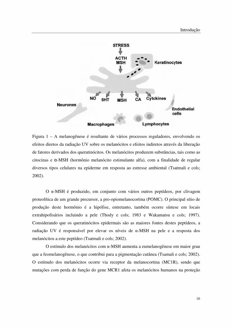
Figura 1 – A melanogênese é resultante de vários processos reguladores, envolvendo os
efeitos diretos da radiação UV sobre os melanócitos e efeitos indiretos através da liberação
de fatores derivados dos queratinócitos. Os melanócitos produzem substâncias, tais como as
citocinas e α-MSH (hormônio melanócito estimulante alfa), com a finalidade de regular
diversos tipos celulares na epiderme em resposta ao estresse ambiental (Tsatmali e cols;
2002).
O α-MSH é produzido, em conjunto com vários outros peptídeos, por clivagem
proteolítica de um grande precursor, a pro-opiomelanocortina (POMC). O principal sítio de
produção deste hormônio é a hipófise, entretanto, também ocorre síntese em locais
extrahipofisários incluindo a pele (Thody e cols; 1983 e Wakamatsu e cols; 1997).
Considerando que os queratinócitos epidermais são as maiores fontes destes peptídeos, a
radiação UV é responsável por elevar os níveis de α-MSH na pele e a resposta dos
melanócitos a este peptídeo (Tsatmali e cols; 2002).
O estímulo dos melanócitos com α-MSH aumenta a eumelanogênese em maior grau
que a feomelanogênese, o que contribui para a pigmentação cutânea (Tsamali e cols; 2002).
O estímulo dos melanócitos ocorre via receptor da melanocortina (MC1R), sendo que
mutações com perda de função do gene MCR1 afeta os melanócitos humanos na proteção

Introdução
17
contra os danos do DNA causado pela radiação UV, o que pode ocasionar câncer de pele
(Lassalle; 2003).
A POMC é uma proteína precursora que além de ser produzida em alguns tecidos
não-hipofisários, é também originada nos queratinócitos. Quando produzida em um nível
cutâneo, os peptídeos derivados da POMC (exemplo ACTH, α-MSH e endotelina-1) têm
função importante na fisiologia da pele, incluindo imunomodulação e respostas locais ao
estresse, sendo que as diferentes células da pele podem expressar de diferentes maneiras os
genes da POMC (Mazurkiewicz e cols; 2000).
Nos melanócitos há produção de uma série de citocinas e fatores de crescimento,
aos quais eles também reagem e, portanto podem ser considerados tipos celulares
imunocompetentes com o potencial de modular sua resposta frente a diferentes condições.
Estudos demonstram que os melanócitos tanto podem agir no estímulo da produção como
responder ao estímulo de IL-1α (interleucina 1 alfa) e IL-6 (interleucina 6), visto que a
exposição à radiação ultravioleta estimula a síntese de IL-1α (Tsamali e cols; 2002).
A exposição do tecido cutâneo ao estresse (radiação ultravioleta) aumenta a
produção do hormônio α-MSH, juntamente com a liberação de citocinas proinflamatórias
(TNF-α e IL-1α), as quais normalmente regulam a expressão de moléculas de adesão de
células às células adjacentes. O TNF-α (fator de necrose tumoral alfa) está envolvido em
várias reações imunes celulares e inflamatórias através da ativação do receptor
correspondente, tendo sua atividade modulada por vários estímulos cutâneos, no qual o
mais importante é a radiação ultravioleta (Slominski e cols.; 2004).
Enquanto o comportamento dos melanócitos é influenciado por mediadores
inflamatórios, as células adjacentes contribuem para o processo através da ativação de
citocinas próinflamatórias como a IL-1α e o TNF-α. A molécula de adesão intercelular 1
(ICAM-1), um importante regulador das interações célula-alvo imunes, é normalmente
expressa em níveis muito baixos em culturas normais de melanócitos. Estes níveis podem
ter aumento significativo de maneira dose-dependente com elevação dos níveis de TNF-α e
IL-1 α (Lee e cols.; 2002).
A tirosinase catalisa duas distintas reações de conversão da tirosina em Dopa
(atividade tirosina hidroxilase) e a oxidação da Dopa resultante em dopaquinona (atividade
dopa oxidase). A enzima oxida fenóis e difenóis usando um mecanismo catalítico que

Introdução
18
depende da presença de cobre no sítio ativo. A partir da produção da dopaquinona, uma
série de reações enzimáticas e não-enzimáticas ocorrem para produzir compostos
dihidroxiindólicos. Os pigmentos de melanina, eumelanina e feomelanina, são produzidos
por oxidação e polimerização destes compostos indólicos (Mayer; 1987; Sánchez-Ferrer e
cols.; 1995; Van Gelder e cols.; 1997; Ito e cols.; 2000; Ito; 2003). No Sistema Nervoso
Central esta enzima participa da síntese de dopamina que é precursora direta de outras
catecolaminas importantes como a noradrenalina e adrenalina, sendo recentemente
implicada em inúmeras doenças neurodegenerativas, especialmente a Doença de Parkinson
(Asanuma e cols.; 2003).
A tirosinase (oxidase polifenólica EC 1.14.18.1) possui o papel primordial de
participar da biossíntese da melanina e tem recebido especial atenção nos últimos anos
como uma ferramenta indispensável para o desenvolvimento de uma grande variedade de
pesquisas e estudos (Seo e cols.; 2003), visto o grande espectro de uso e potencialidades
dos agentes inibidores e indutores desta enzima para as indústrias farmacêutica, cosmética e
de alimentos. Esta enzima possui uma ampla distribuição nos animais, insetos, vegetais e
fungos, onde ocorre em várias isoformas (Yokochi e cols.; 2003 e Jaenicke & Decker;
2003).
A tirosinase é a enzima que desencadeia a melanogênese, iniciando a cascata de
reações que converte tirosina ao biopolímero melanina. Duas proteínas relacionadas à
tirosinase (TRPs) são conhecidas, TRP-1, a mais abundante glicoproteína dos melanócitos,
e a TRP-2. As TRPs possuem funções catalíticas e reguladoras na melanogênese (Seiberg e
cols.; 2000). A produção de TRP-1 possui função reguladora na desestabilização da
tirosinase, levando à diminuição da produção de melanina (Seiberg e cols.; 2000).
Na epiderme humana, diversos mecanismos enzimáticos e não-enzimáticos estão
disponíveis para controlar o acúmulo de espécies reativas de oxigênio, sendo a tirosinase
um dos mais importantes sistemas para a eliminação destas espécies, já que esta enzima é
capaz de utilizar superóxido para produzir melanina (Perluigi e cols.; 2003; Friedmann &
Gilchrest; 1987), de modo que compostos antioxidantes, capazes de inibir a produção de
espécies reativas de oxigênio podem reduzir a hiperpigmentação ou prevenir a
melanogênese.
Introdução
19
Assim sendo, o desevolvimento de agentes clareadores da pele no tratamento de
hiperpigmentação induzida pela luz ultravioleta ou por condições médicas como melasma e
melanodermia pós-inflamatória é uma importante área de pesquisa (Choi e cols.; 2005). A
atividade biológica de várias plantas tem sido estudada com o propósito de uso em
cosméticos (Mizuno & Tanaka; 1986). Além disso, fontes como plantas têm sido avaliadas
para o desenvolvimento de antioxidantes naturais que podem estar envolvidos com
produtos para cuidados anti-idade e anti-rugas (Anes & Saul; 1987). Muitos componentes
endógenos de plantas foram relatados como agentes que retardam o processo de oxidação
no seu ambiente natural e em produtos nos quais estes compostos foram adicionados (Pratt;
1994).
Objetivos
20
2. Objetivos
A melanina tem importante função na proteção da pele contra diversos efeitos
prejudiciais da radiação solar ultravioleta, entretanto, o seu acúmulo anormal gera um
problema estético. Os agentes tópicos disponíveis para o tratamento da hiperpigmentação
incluem os inibidores da tirosinase, retinóides, hidroquinonas e agentes citotóxicos aos
melanócitos. Infelizmente, os resultados destes tratamentos, às vezes, não são satisfatórios,
e se faz necessário buscar terapias mais eficazes, mais seguras e menos irritantes. Em
adição, o crescente interesse por terapias complementares gera demanda para tratamentos
despigmentantes, utilizando-se de fontes naturais, especialmente de origem vegetal,
considerados seguros e eficazes.
Dentre os mediadores responsáveis pela formação da melanina estão as citocinas
como a interleucina 1 alfa (IL-1 α) e o fator de necrose tumoral alfa (TNF-α), o hormônio
melanócito estimulante alfa (α-MSH) e a tirosinase. Além das formações de espécies
reativas de oxigênio que contribuem para este processo.
Considerando as informações apresentadas, o presente estudo visou investigar
extratos vegetais como potenciais ativos despigmentantes, especialmente através dos efeitos
de inibição da enzima tirosinase e antioxidante in vitro, pelo método DPPH e de
peroxidação lipídica e também selecionar as espécies vegetais mais promissoras nestes
estudos iniciais para se avaliar seus efeitos sobre a atividade da tirosinase e a produção do
hormônio melanócito estimulante alfa, da interleucina 1 alfa e do fator de necrose tumoral
alfa em culturas celulares (melanócitos), comparando grupos celulares expostos e não
expostos à radiação ultravioleta.

Triagem
21
3. Triagem
Com o objetivo de selecionar plantas com potencialidades de atividade biológica de
interesse para o projeto, foi realizada revisão bibliográfica com base em estudos
etnofarmacológicos (priorizando-se espécies utilizadas tópica e popularmente para
problemas de pele) e quimiotaxonômicos (considerando-se espécies vegetais que possuem
em sua composição grupos químicos com atividade sobre a pigmentação como estilbenos,
hidroquinonas ou espécies de gêneros botânicos já descritos na literatura como agentes
despigmentantes, especialmente inibidores da tirosinase). A partir desta revisão foram
selecionadas 12 espécies vegetais (Tabela 1) para a realização do presente estudo. Esta
seleção obedeceu dois critérios: qualidade das informações etnofarmacológicas e
quimiotaxonômicas e disponibilidade de obtenção do material vegetal.
As plantas foram adquiridas através da empresa Chemyunion Química Ltda. dos
fornecedores Quimer (Coccoloba uvifera L., Eugenia crenata Vell., Eugenia uniflora L.,
Myrcia sphaerocarpa DC, Polygala senega L., Polygonum acre H. B. & K., Rheum
officinale Baill., Rheum rhaponticum L., Syzigyum jambolanum DC e Vitex agnus-castus
L.) e Santosflora (Jacaranda caroba Hort. e Tabebuia avellanedae Lorentz), os quais são
responsáveis pela identificação taxonômica e depósito de exsicata.
O método de extração utilizado para todas as plantas citadas acima também foi
realizado conforme metodologia utilizada pelo Departamento de Pesquisa e
Desenvolvimento da Chemyunion Química Ltda, usando-se extração metanólica a quente
por 4h sob agitação e refluxo, seguida de filtração e concentração do extrato em
rotaevaporador. Todos os extratos obtidos foram armazenados em vidro âmbar.
Na etapa de triagem das espécies vegetais foram realizados testes in vitro para
determinação da atividade antioxidante: método DPPH, 1,1-difenil-2-picrilhidrazina,
(Blois, 1958 e Brand-Williams e cols.; 1995), e método de peroxidação lipídica ( Stocks e
cols.; 1974 e Fee & Tietelbaun; 1972) e da atividade antitirosinase: método antitirosinase in
vitro conforme descrito por Kim e cols.; 2003.

Triagem
22
Tabela 1 – Plantas escolhidas para a avaliação do efeito despigmentante.
Nome científico/família Nome popular Partes utilizadas
Coccoloba uvifera L.
(Polygonaceae)
Uva de mato Raiz
Eugenia crenata Vell.
(Myrtaceae)
Cambuí Folha e talo
Eugenia uniflora L.
(Myrtaceae)
Pitanga Folha e talo
Jacaranda caroba Lem.
(Bignoniaceae)
Carobinha Folíolos
Myrcia sphaerocarpa DC
(Myrtaceae)
Pedra-ume-caá Folha
Polygala senega L.
(Polygalaceae)
Polígala Raiz
Polygonum acre H.B. & K.
(Polygonaceae)
Erva de bicho Talo e folhas
Rheum officinale Baill.
(Polygonaceae)
Ruibarbo chinês Rizoma
Rheum rhaponticum L.
(Polygonaceae)
Ruibarbo europeu Rizoma
Syzygium jambolanum DC
(Myrtaceae)
Jambolão Talo e folhas
Tabebuia avellanedae Lorentz
(Bignoniaceae)
Ipê roxo Folíolos
Vitex agnus-castus L.
(Lamiaceae)
Agno casto Fruto
2.1. Método do DPPH
O método DPPH foi realizado para confirmar o potencial das plantas escolhidas. As
ações dos extratos foram analisadas quanto ao potencial das amostras captarem o radical

Triagem
23
livre DPPH (1,1-difenil-2-picrilhidrazina). Os compostos testes foram preparados através
de uma diluição seriada nas concentrações de 1,9x10⁻⁴ a 100 mg/mL (metanol).
100µL de cada diluição foram adicionados em duplicata à placa de 96 poços. A
seguir, 50 µL de uma solução metanólica de DPPH 0,4mM foi adicionada por poço. Após 5
minutos de incubação à temperatura ambiente, ao abrigo da luz, a redução do radical livre
DPPH foi mensurada pela leitura da absorbância em 517 nm, contra um branco específico
(metanol) em cada avaliação, formado somente pelas amostras nas suas respectivas
diluições.
Como controle foi utilizado 50µL de solução metanólica de DPPH mais 100µL de
metanol. Os resultados foram expressos em porcentagem de inibição do radical DPPH,
calculado segundo a equação:
% de Inibição = [(Absorbância do controle - absorbância da amostra)/absorbância
do controle] X 100
A determinação da IC50, ou seja, concentração da amostra que causa 50% de
inibição da concentração inicial de DPPH foi obtida por regressão linear dos pontos
plotados graficamente. Para a plotagem dos pontos, foram utilizados os valores das médias
obtidas de triplicatas realizadas para cada um dos testes (Anexo 1).
Os dados obtidos com os compostos foram comparados com Trolox, um tocoferol
usado como controle positivo na avaliação da atividade antioxidante (Friaa & Brault, 2006).
2.2. Peroxidação lipídica
A avaliação da atividade antioxidante foi realizada através do método de
peroxidação lipídica a qual se baseia no princípio de que o ácido tricloroacético precipita as
proteínas das amostras de cérebro (de ratos Wistar machos, cujas cabeças foram doadas
pelos alunos do Laboratório de Fitomedicamentos do Instituto de Biociências da UNESP,
campus de Rubião Junior em Botucatu que estudam colite), enquanto que o reativo
tiobarbitúrico reage com o malonildialdeído (MDA), liberado pela lipoperoxidação causada
pelo ferro e ácido ascórbico, gerando uma cor cuja intensidade é determinada no
espectrofotômetro. Quanto maior a intensidade da cor, maior a concentração de MDA. Um

Triagem
24
composto será antioxidante se inibir a lipoperoxidação causada pelo ferro e ácido
ascórbico, reduzindo os valores de MDA. Os compostos testes foram preparados através de
uma diluição seriada nas concentrações de 6,25 a 400mg/mL (DMSO 20%).
As amostras teste foram preparadas em eppendorf de 2mL, onde se adicionou 50
µL da solução de Ferro-Ácido Ascórbico 100 µM, 1mL de membrana de cérebro diluída,
50 µL do composto teste, reativo tiobarbitúrico (Sigma T-5500), sob agitação e banho-
maria em tempos determinados. Após centrifugação, o sobrenadante foi colocado em
duplicata em microplaca de 96 poços. A leitura foi realizada a 532nm (espectrofotômetro
Bio TekTM Power Wave 340), contra brancos específicos e os respectivos controles.
A concentração inibitória 50% (IC50) foi determinada com base na fórmula 1-ABS
teste/ABS controle máximo através de regressão linear dos pontos plotados graficamente.
Os dados obtidos com os compostos foram comparados com a Quercetina, usada
como controle positivo na avaliação da atividade antioxidante, e com o Ácido Kójico,
agente com ação clareadora, usado em preparações cosméticas no tratamento tópico de
discromias.
2.3. Atividade antitirosinase
Para a avaliação da atividade antitirosinase, os compostos testes foram preparados
através de uma diluição seriada nas concentrações de 50 a 3200 mg/mL (DMSO 20%).
A tirosinase é um polifenol oxidase, sendo um heterotetrâmero constituído por 2
cadeias leves e 2 cadeias pesadas, os quais, por sua vez, formam 3 domínios. Possui
atividades importantes como de monofenalase e difenalase, conforme o substrato no qual
atue. Quando age na tirosina, apresenta atividade de monofenalase, produzindo DOPA;
quando age na DOPA, apresenta atividade de difenalase, formando Dopaquinona. A
Dopaquinona, numa seqüência de outras reações forma dopacromo, que pode ser lido a 492
nm no espectrofotômetro.
60µL de cada diluição foram adicionados em duplicata à placa de 96 poços. A
seguir, 30 µL de enzima tirosinase foi adicionada por poço. Após 5 minutos de incubação à
temperatura ambiente, adicionou-se 80µL de substrato L-tirosina, a atividade antitirosinase

Triagem
25
foi mensurada através de 10 leituras cinéticas da absorbância em 492 nm, contra um branco
específico (DMSO 20%) em cada avaliação, formado somente pelas amostras nas suas
respectivas diluições e os valores foram expressos em concentração (de dopacromo).
Como controle foi utilizado ácido kójico, preparado da mesma forma que as
amostras teste. Os resultados foram determinados com base na fórmula 1-Concentração
teste/Concentração do controle máximo através de regressão linear dos pontos plotados
graficamente.

Resultados Preliminares
26
4. Resultados preliminares da triagem e seleção de espécies para estudos
No método de DPPH, todas as plantas apresentaram algum potencial antioxidante
através da captação de radicais livres nas concentrações mais altas estudadas, sendo que os
extratos de Eugenia uniflora L., Eugenia crenata Vell., Coccoloba uvifera L., Syzigyum
jambolanum DC, Rheum rhaponticum L. e Rheum officinale Baill. mostraram-se mais
potentes que os demais (Tabela 2 e Anexos).
No método de peroxidação lipídica, os extratos que apresentaram melhor atividade
antioxidante foram os mesmos que mostraram-se potentes na avaliação realizada pelo
método de captação de radical livre: Rheum officinale Baill., Eugenia crenata Vell.,
Syzigym jambolanum DC, Rheum rhaponticum L, Eugenia uniflora L. e Coccoloba uvifera
L (Anexos).
Os resultados dos efeitos dos extratos vegetais sobre a atividade da tirosinase
(Tabela 3 e Anexos) foram essenciais para a seleção das espécies vegetais para os estudos
posteriores, visto que as espécies mais potentes na inibição da atividade da tirosinase foram
Rheum rhaponticum L. e Coccoloba uvifera L. Estes resultados em combinação com
aqueles descritos para a atividade antioxidante permitiram a escolha destas espécies para os
estudos de seus efeitos sobre as culturas de melanócitos estimuladas com irradiação
ultravioleta, cujos resultados se encontram nos manuscritos apresentados na próxima seção.

Resultados Preliminares
27
Tabela 2 - Concentração inibitória 50% da atividade antioxidante
Amostras estudadas Concentração inibitória 50% (IC50)
Quercetina
0,53
Rheum officinale Baill.
(Polygonaceae)
5,93
Eugenia crenata Vell.
(Myrtaceae)
7,17
Syzigyum jambolanum DC
(Myrtaceae)
8,70
Rheum rhaponticum L.
(Polygonaceae)
8,92
Eugenia uniflora L.
(Myrtaceae)
9,24
Coccoloba uvifera L.
(Polygonaceae)
9,84
Jacaranda caroba Hort.
(Bignoniaceae)
13,63
Myrcia sphaerocarpa DC
(Myrtaceae)
18,95
Polygonum acre H.B. & K.
(Polygonaceae)
23,50
Vitex agnus-castus L.
(Lamiaceae)
51,85
Ácido kójico
103,18
Tabebuia avellanedae Lorentz
(Bignoniaceae)
108,34
Polygala senega L.
(Polygalaceae)
270,44

Resultados Preliminares
28
Tabela 3 – Concentração inibitória 50% da atividade antitirosinase
Amostras estudadas Concentração inibitória 50% (IC50)
Ácido kójico
0,02
Rheum rhaponticum L.
(Polygonaceae)
0,06
Coccoloba uvifera L.
(Polygonaceae)
0,09
Eugenia crenata Vell.
(Myrtaceae)
0,89
Rheum officinale Baill.
(Polygonaceae)
2,18
Syzigyum jambolanum DC
(Myrtaceae)
2,42
Eugenia uniflora L.
(Myrtaceae)
3,45
Polygonum acre H.B. & K.
(Polygonaceae)
12,88
Myrcia sphaerocarpa DC
(Myrtaceae)
41,86
Tabebuia avellanedae Lorentz
(Bignoniaceae)
107,97
Vitex agnus-castus L.
(Lamiaceae)
173,12
Polygala senega L.
(Polygalaceae)
1,42 X 10⁶
Jacaranda caroba Hort.
(Bignoniaceae)
2,46 X 10173

Artigo científico sobre Rheum rhaponticum L.
29
5. Artigo científico sobre Rheum rhaponticum L.
O artigo foi escrito de acordo com as normas de publicação da Revista Científica
Experimental Dermatology e submetido em dezembro de 2007.
A methanolic Rheum rhaponticum L. root extract inhibits IL-1, TNF-α and α-MSH
production and tyrosinase activity in melanocyte submitted to
solar-simulated radiation
Jéssica Eleonora Pedroso Sanches Silveiraa,b, Maria del Carmen Velazquez Peredab,
Samara Eberlinb, Gustavo Campos Dieamantb, Luiz Claudio Di Stasia,*
aDepartament of Pharmacology, São Paulo State University, Botucatu, Brazil
bResearch and Development Department of Chemyunion Química Ltda, Sorocaba, Brazil
*Corresponding author: Dr. Luiz Claudio Di Stasi (Ph.D.). Departamento de
Farmacologia/Laboratório de Fitomedicamentos, Instituto de Biociências, Universidade
Estadual Paulista “Júlio de Mesquita Filho” (UNESP), Rubião Junior, CEP 18618-000,
Botucatu, SP, Brazil.
Tel.: +55-14-3811-6347
E-mail address: [email protected]

Artigo científico sobre Rheum rhaponticum L.
30
Abstract
Pigmentation of human skin is closely involved in protection against environmental
stresses, in particular exposure to ultraviolet (UV) radiation. However, the accumulation of
melanin in the skin can cause some problems as melasma, solar lentigines and actinic
keratoses. Ultraviolet radiation exposure can generate the production of many mediators
that stimulate the melanin synthesis, contributing for the pigmentation increasing. Among
these mediators, IL-1 (Interleukin 1), TNF-α (tumor necrosis factor alpha), α-MSH
(melanocyte stimulating hormone alpha) and the tyrosinase are important factors related to
skin pigmentation. In this research, we have examined the photoprotective effect of the
methanolic Rheum rhaponticum L. root extract (RRE) in melanocytes using a solar
simulator as the source of UV radiation (SSR). We found that RRE has a protective effect
against SSR-induced damage, resulting in the decrease of IL-1 and TNF-α levels. In
addition, the tyrosinase activity and α-MSH production were also normalized, contributing
for the maintenance of the cell homeostasis.
Key words: Rheum rhaponticum L., cytokines, α-MSH, tyrosinase activity.

Artigo científico sobre Rheum rhaponticum L.
31
Introduction
Melanin pigmentation of the skin in mammals was shown to result from the close
interaction between the epidermal melanocytes and the keratinocytes (1). Melanocytes are
key components of the skin pigmentary system through their ability to produce melanin.
These cells are found at many locations throughout the body. In the skin they are
associated with the hair follicle and in some mammals, including humans, are also found in
the basal layer of the interfollicular epidermis. Mature melanocytes form long dendritic
processes that ramify among the neighboring keratinocytes. In this way, each melanocyte
makes contact with around 30–40 keratinocytes and this constitutes the epidermal–
melanin unit. This association enables the melanocyte to transfer melanin into the
keratinocytes, where it determines skin color and helps in protecting against the damaging
effects of UVR(2).
In human epidermis, the process of UV stimulation is composed of three major
steps; the first step is the proliferation of melanocytes, followed by the synthesis and
activation of tyrosinase and finally the transfer of melanosomes to keratinocytes. During
the first two steps, a complex network exists in the epidermis for secreting and responding
to autocrine and paracrine cytokines by keratinocytes and melanocytes. Corresponding
receptors, which are also regulated in their expression by various cytokines, participate in
the complex process in which there is cross-talk in signaling between cytokines to support
the enhanced proliferation of melanocytes. These paracrine mediators include α-
melanocyte-stimulating hormone among others. The secretion of α-melanocyte-stimulating
hormone has been reported to be triggered by primary inflammatory cytokines such as
interleukin (IL)-1 and tumor necrosis factor (TNF)-α, which are released by UVB-exposed
cells (3).

Artigo científico sobre Rheum rhaponticum L.
32
Melanogenesis is target for a variety of hormones and other biological modifiers.
UV light stimulates the secretion of alpha-melanocyte stimulating hormone (α-MSH),
adrenocorticotropic hormone (ACTH), prostaglandin E2, endothelin-1 (ET-1) and nitric
oxide, which induces melanogenesis process (4; 5). On the other hand, cytokines, such
interleukin-1 (IL-1), IL-6, tumor necrosis factor-alpha (TNF-α), interferon-gamma (IFN-γ)
and transforming growth factor-beta (TGF-β) are produced by melanocytes and
keratinocytes to compensate the exacerbated production of melanin, mainly by the
decrease of expression and release of tyrosinase (6; 7; 8; 9).
Rheum rhaponticum L. (Polygonaceae family) commonly known as Sibiric Rhubarb
is a plant originates from Central Asia. This plant, in the 17th century was introduced into
Europe and has been cutivated since then in Europe, United States, East Asia and Latin
America (10). The standardized extract from roots of this plant consists mainly of rhaponticin
and desoryrhaponticin and small amounts of the aglycones trans-rhapontigenin and
desoxythapontigenin, secondary metabolites have a hydroxystilbene backbone and are
structurally related to resveratrol(11). Indeed, after the administration of Rheum rhaponticum
L. extract in rats, two resveratrol (piceatannol and rhapontigenin glucosides), as well as
resveratrol, rhapontigenin and other stilbenes derivatives were identified as mean
metabolites(12). Hydroxystilbenes are plant polyphenols exerting a number of health-
promoting effects, including antioxidant activity(13) and inhibition of the tyrosinase activity
(14, 15), while resveratrol is a potent natural metabolites useful as whitening agent (16,17).
Many plant extracts and their active principles have been described and utilized as
cosmetic agents, in particular, as skin lightening products. The growing demand for
depigmenting agents is being perceived, and effective compounds from natural sources,
acting as tyrosinase inhibitors or blocking others melanogenic pathways, have been
reported (15, 18, 19, 20).

Artigo científico sobre Rheum rhaponticum L.
33
In this way, we evaluated the effects of a dry extract of the root of Rheum
rhaponticum L. (RRE) in human melanocytes culture under solar-simulated radiation.
Considering the down-regulating activity of some cytokines on melanogenesis process, we
first evaluated the effects of RRE on the release of IL-1α and TNF-α in cell supernatants.
We found that RRE decrease IL-1α and TNF-α levels submitted to UV radiation. Our data
also demonstrate a decrease in the levels of tyrosinase. Additional studies were performed
in order to reveal the possible effects of RRE on α-MSH production after UV radiation and,
corroborating the hypopigmenting effect of RRE, the levels of these hormones were found
reduced after cell treatment.
Materials and Methods
Plant Material
Dry extract of Rheum rhaponticum L. (RRE) was manufactured and provided by
Chemyunion Química Ltda (Sorocaba, SP, Brazil). Crude plants (root), obtained for
Brazilian suitable suppliers, were sliced into small pieces and extracted with methanol. The
extract was concentrated in a rotary evaporator (Buchi RE 111 Buchi Laboratoriums-
Tecnick AG,Flawil, Switzerland) in order to remove the solvent. The dry extract obtained
was dissolved in dimethyl sulfoxide 20% (DMSO) for the assessment of antioxidant and
antityrosinase activities. For the determination of radical scavenging activity, the solvent
used was methanol. In addition, for evaluation of the melanocyte cultures, the extract was
dissolved in culture medium and diluted into appropriate concentrations immediately
before use. The final concentration of the dried extract was 31%.

Artigo científico sobre Rheum rhaponticum L.
34
Antioxidant activity (In vitro Lipid peroxidation)
Trichloroacetic acid precipitates proteins of rats brain while the tiobarbituric reactive
reacts with malonyldialdehyde (MDA), released by the lipoperoxidation caused by iron and
ascorbic acid, producing a color whose intensity is determinated by spectrophotometer.
The increase of intensity is a result of the increase of MDA concentration. An element will
be considered antioxidant if it is capable to inhibit the lipoperoxidation caused by the
reagents, reducing the MDA values. The test elements were prepared through a serial
dilution in 400 to 6,25 mg/mL doses (DMSO 20%).
For the test samples, it was added 50 µL of Iron-Ascorbic Acid 100 µM solution, 1
mL of brain homogenate, 50 µL of the plant extract and tiobarbituric. After centrifugation,
the supernatant was paced in duplicate in 96-well microplate. The measurements were
performed in spectrometer (Bio TekTM Power Wave 340). Lipid peroxide inhibitory activity
was expressed as IC50.
In vitro Tyrosinase Assay
Antityrosinase effect was assessed through enzymatic assay. Potassium
phosphate buffer (50mM) at pH 6.5, tyrosinase (333 units/mL) and test compounds
dissolved in DMSO 20% were inserted into 96-well plates. After 5 min at room
temperature, L-tyrosine was added.
The diphenolase activity of tyrosinase was measured spectrophotometrically by
following the increase in absorbance at 475nm by production of dopachrome from L-
DOPA. The reaction was done at 37o C.

Artigo científico sobre Rheum rhaponticum L.
35
Cell culture and treatment protocol
Human epidermal melanocytes (Cryopreserved HEM, Cat. 104-05n), melanocyte
growth medium (Cat. 135-500) and trypsin/EDTA solution (Subculture Reagent Kit, Cat.
090K) were purchased from Cell Applications, Inc™ San Diego, CA. Cells were
subcultured in 25 cm2 flasks (Corning Inc, New York, NY) at 37°C in 5% CO2 humidified
incubator, and expanded for at least five passages. The medium was changed twice a
week. At 80-90% confluent, cells were trypsinized and seeded into 24- well culture plates
(Nunc, Roskilde, DM). Twenty-four hours after melanocytes seeding, cells were washed
with PBS and irradiated with ultraviolet radiation in a dose of 300mJ/cm2(Seiberg et al,
2000). Immediately after irradiation, cells were incubated with doses of 4.9, 2.4, 1.2 and
0.6 mg/mL of RRE (Rheum rhaponticum L. extract) dissolved in culture medium. After 48 h
of treatment, cell-free supernatants and cell lysate were collected and assays were
performed using commercial kits. Each experiment was conducted in triplicate of three
independent experiments. Selection of these doses was based on previous results of
cytotoxicity assays (data not shown).
Ultraviolet radiation
A Multiport solar UVA and UVB simulator (Model 601; Solar Light Co., Philadelphia,
PA) was used for the UV irradiation. The simulator was equipped with a 150W Xenon
lamp, with a liquid filter and 1-mm Schott WG 320 filter, emitting a continuous spectrum of
radiation beginning at 290 nm through the infrared spectrum and maximally peaking at 400
nm. The lamp was housed in a black plastic tube with six apertures, 1-cm in diameter. The
apparatus was calibrated before each use. The UV irradiation time lasted for 1 min to
reach a dose of 300mJ/cm2 .

Artigo científico sobre Rheum rhaponticum L.
36
Quantification of IL-1α and TNF-α Levels
IL-1α and TNF-α were quantified by using a commercially available ELISA
kit (DuoSet, R&D Systems, Minneapolis, MN). Cytokine determinations were done
according to R&D sandwich ELISA protocol. Briefly, anti-human IL-1α and TNF-α capture
antibody were coated onto a 96-well microplate (Nunc) overnight at room temperature. A
blocking step was performed for 1 h at room temperature (RT). After washing, the
recombinant standards and samples were added to the coated plates and incubated for 2
h at RT. The plate was washed and then incubated with detection antibody for 1 h. The IL-
1α and TNF-α binding was colored by streptavidin-horseradish peroxidase, and the optical
density was read using a microplate reader at 450 nm, after stopping the reaction using 2
N H2SO4. Cytokine titers were expressed as pg per mL, calculated by reference to
standard curves constructed with known amounts of recombinant cytokines.
Tyrosinase Activity in Melanocyte Culture
Tyrosinase activity was evaluated through immunoprecipitation assay kit of protein
tyrosine kinase (USBiological, Swampscott, MA). Phosphopeptide standard and cell lysate
samples were added to a pre-coated streptavidin plate at 37°C for 30 minutes. After
washing, blocking buffer solution was added and incubated at 37°C for additional 30
minutes. The block solution was discarded and anti-phosphotyrosine HRP (horseradish
peroxidase) was incubated for 1 hour at room temperature. Substrate solution was added
during 15 minutes and the enzyme reaction was stopped by an acid solution. Optical

Artigo científico sobre Rheum rhaponticum L.
37
density was read using a microplate reader at 450 nm. Tyrosine kinase activity was
compared with known standards and expressed as pg per mL.
Quantification of α-Melanocyte Stimulating Hormone (α-MSH)
α-MSH was quantified by using a commercially available enzymatic immunoassay
kit (Phoenix Pharmaceuticals, Inc., Belmont, CA). α-MSH peptide standard, cell lysate
samples, primary antiserum and biotinylated peptide were added to each well of a pre-
coated immunoplate for 2 hours. After washing, streptavidin-horseradish peroxidase was
added and incubated for 1 h, followed further washes. After substrate solution was added,
incubation was continued by 1 hour, 2N HCl was added to stop the enzyme reaction, and
the optical density was read using a microplate reader at 450 nm. α-MSH titers were
expressed as pg per mL, calculated by reference to standard curves constructed with
known amounts of recombinant peptides.
Statistical Analysis
For statistical analysis o the cell culture experiments, a parametric method, the
one-way analysis of variance (ANOVA) followed by the Tukey test, was used to compare
data among all groups. Statistical significance was considered when P < 0.05, P<0.01 or
P<0.001.
Artigo científico sobre Rheum rhaponticum L.
38
Results
Antioxidant and antityrosinase effects of the methanolic Rheum rhaponticum L. root extract
The methanolic Rheum rhaponticum L. root extract presented antioxidant and
antityrosinase activities in the in vitro assays. Quercetin, as it is known as a potent
antioxidant, was used as reference product in the antioxidant effect (Quercetin IC50 -
0,53µg/mL; Rheum rhaponticum L. extract IC50 – 8,92 µg/mL). In comparison to the
reference product which was used, the Rheum rhaponticum L. extract antioxidant assay
results demonstrated that the plant extract has an antioxidant potential. Regarding the
antityrosinase assay, Kojic Acid, widely used in whitening products, was the reference
product and the results of Rheum rhaponticum L. root extract was quite similar to the
reference (Kojic Acid IC50 - 0,02µg/mL; Rheum rhaponticum L. extract IC50 - 0,06µg/mL).
Besides the antioxidant potential, Rheum rhaponticum L. demonstrates in vitro
effectiveness in antityrosinase assay.
Rheum rhaponticum L. extract inhibits IL-1 levels in melanocytes
Cytokines are also known to down-regulate transcription factors crucial to regulate
melanocyte proliferation and to decrease melanossome transfer to neighboring
keratinocytes (6, 7, 8, 9). With the purpose to investigate possible immunostimulatory effects
of RRE, we assayed the production of IL-1 in melanocyte culture supernatants (figure 1).
In basal conditions, without SSR, a decrease in the levels of cytokines was observed in the
cells which were treated with RRE. This behavior was similar to that observed in the
groups that were treated with kojic acid. In the groups only submitted to UV radiation,
increase of 1,73 fold was observed to IL-1α production in relation to controls. However, in

Artigo científico sobre Rheum rhaponticum L.
39
the groups with RRE treatment, there was a decrease in the citokyne production in relation
to the control exposed to UV radiation and the results were comparable to that observed in
the cells in basal condition.
Rheum rhaponticum L. extract decreases TNF-α levels in melanocytes induced by UV light
TNF-α, along with a myriad of other cytokines, is modulated in the skin by diverse
stimuli, most importantly UVR and it also regulates the expression of POMC (pro-
opiomelanocortin) peptides and MC1-R (melanocortin receptor 1) in melanocytes (12).This
cytokine is present in both the epidermis and dermis of normal skin (15).
With the same purpose of testing IL-1 levels, TNF-α levels were also assessed in
melanocytes. Although the fact that RRE presented no effect in the groups in basal
conditions (without SSR), a decrease of TNF-α occurred in the groups which were
exposed to UV radiation, leading to a basal condition (figure 2).
Tyrosinase Activity
Tyrosinase is the enzyme that catalyses the rate-limiting step of melanin
synthesis(8) and in the present study, the effect of a dry extract of Rheum rhaponticum L.
(RRE) was evaluated on tyrosinase activity in melanocytes cell culture submitted to UV
radiation and incubated with different doses of RRE during 48h. Our results demonstrated
a reduction in tyrosinase activity, in basal conditions. As we expected, the UV radiation
(UVR) produced a rise of 1.51 fold in enzymatic activity, which was prevented by the
treatment of cells with all tested doses of RRE (figure 3).

Artigo científico sobre Rheum rhaponticum L.
40
Quantification of α-Melanocyte Stimulating Hormone (α-MSH)
Another important observation obtained in this study was that the incubation of
human melanocytes with 4.9, 2.4 and 1.2mg/mL of RRE led to a reduction in the release of
α-MSH (figure 4), which is known to be one of the triggering factors for tyrosinase
activation. The reduced effects on α-MSH release were elicited by the two highest doses
(4.9 and 2.4mg/mL) tested leading to decreases in the levels of this parameter, in relation
to control basal. Considering the groups which were exposed to UV radiation, the
decrease in α-MSH levels was observed to all tested doses.
Discussion
Melanin plays an important role in protecting human skin from the harmful effects of
UV sun radiation, however its abnormal accumulation in different specific parts of the skin
as more pigmented patches, might become an aesthetic problem (6). UV irradiation of the
skin results in important damages, such as erythema, swelling, photoaging and skin
cancer. One of the modern aims of dermatology is to find substances that can act as
photoprotective agents and can also ameliorate the skin injuries. In this study, we have
analysed some of the mechanisms in which RRE exerts this protective effect. Tyrosinase
inhibition is the most common approach to achieve skin hypopigmentation as this enzyme
catalyses the rate-limiting step of melanin synthesis (6). Tyrosinase inhibition is the most
common approach to achieve skin hypopigmentation as this enzyme catalyses the rate-
limiting step of melanin synthesis (6). Intracellular H2O2, generated in response to these
cytokines induces a transcient reduction of tyrosinase and other melanogenic protein
activities, through the down-regulation of transcriptional controls of tyrosinase

Artigo científico sobre Rheum rhaponticum L.
41
expression(8). In this way, at first, in vitro antioxidant and antityrosinase effects were
observed, demonstrating that Rheum rhaponticum L. could be involved in the process of
helping the skin healthy condition. In order to confirm our finds, we tested the tyrosinase
activity in melanocytes culture and as we expected, the results were similar to those from
in vitro.
Moreover, a number of biological effectors can reduce normal or hyperpigmentation
in mammals by various means that cover melanogenesis biochemistry. Cytokines such IL-
1, IL-6, TNF-α, IFN-γ and TGF-β belong to these factors and could be release with th.e
purpose to the counterbalance the excessive production of positive regulators of
melanogenesis (6). Cytokines are also known to down-regulate transcription factors crucial
to regulate melanocyte proliferation and to decrease melanossome transfer to neighboring
keratinocytes (6,7,8,9).
Indeed, using human melanocytes culture, we have shown that RRE down-
regulates the production of IL-1α and TNF-α.
As alpha-melanocyte stimulating hormone (α-MSH) is produced and released by
keratinocytes and melanocytes and it is involved in regulating melanogenesis and
melanocyte formation (21), another parameter that was also observed in cell cultures was α-
MSH production. The release of this hormone was diminished regarding the highest
concentrations of RRE. Both in basal conditions and the groups submitted to UV radiation,
the highest doses of RRE were effective in reducing α-MSH. And considering the groups
that received UV radiation, even the lowest dose led to a decrease of α-MSH production.
Several lines of evidence indicate the relationship between free radicals
and cytokines either because ROS can be the mediatiors of some of the biological
effects of cytokines or because free radicals can stimulate the secretion of

Artigo científico sobre Rheum rhaponticum L.
42
different citokynes. In fact, exposure of sensitive cells to cytotoxic concentrations
of TNF-α is associated with the accumulation of malonyl dialdehyde, a marker of
lipid peroxidation, and the toxicity is prevented by the presence of oxyradical
scavengers (22). The hydroxyl radical is one of the most reactive radicals
generated from biologic molecules and can damage living cells (22).Some plant
extracts have the ability to scavenge hydroxyl radicals and may protect cellular
lipids against free radical reactions (23).
The production of IL-1, TNF-α, and α-MSH is closely related to the UV radiation
stimuli and these processes can be interlinked to contribute for the skin damage.
Moreover, the production of ROS contributes for accelerating the external consequences
of this hazard, as the skin is one of the tissues that suffer the most. In our experiments,
Rheum rhaponticum L. extract markedly inhibited tyrosinase activity in vitro and
melanocytes cultures. Indeed, Rheum rhaponticum L. extract decreased IL-1, TNF-α and
α-MSH production in melanocytes submitted to solar stimulation. Since Rheum
rhaponticum L. is a plant with several hydroxystilbenes actives as antioxidant and
antityrosinase activity, its is possible that these secondary metabolites are related to
inhibitory effects detected in this work. In this way, Rheum rhaponticum L. can be used as
an auxiliary agent in pharmaceutical and or cosmetic preparations for restablishing the
skin normal condition after stresses, such as UV radiation.
Several hypopigmenting agents from natural source have been developed and
utilized to ameliorate various cutaneous hyperpigmentary disorders and complexion

Artigo científico sobre Rheum rhaponticum L.
43
discolorations (20, 24, 25, 26). In the same line, the findings presented in this study corroborate
with this new depigmenting category, once demonstrated another compound able to
correct dysfunctions in melanogenesis metabolism.
Acknoledgements
This work was supported by grants from the Chemyunion Química Ltda. The
authors also express many thanks to Leonardo Noboru Seito for his technical assistance.
We thank the CNPq (Science and Technology Ministry, Brazil) for a fellowship to L. C. Di
Stasi.
References
1 – Westerhof, W. The discovery of the human melanocyte. Pigment Cell Res. 2006:
19(3): 183-193.
2 – Tsatmali, M, Ancans, J, Thody, AJ., 2002. Melanocyte Function and Its Control by
Melanocortin Peptides. J. Histochem. Cytochem., 2002: 50: 125.
3 - Hachiya, A, Kobayashi, A, Yoshida, Y, Kitahara, T, Takema, T, Imokawa, G.. Biphasic
Expression of Two Paracrine Melanogenic Cytokines, Stem Cell Factor and Endothelin-1,
in Ultraviolet B-Induced Human Melanogenesis. American Journal of Pathology, 2004:
106(3): 2099-2109
4 - Bolognia JL, Orlow SJ. Pigmentary disorders. In Dermatology (vol.2), Bolognia JL,
Jorizzo J, Rapini R, (eds). Mosby: Philadelphia; 2003: 43-52.

Artigo científico sobre Rheum rhaponticum L.
44
5 - Briganti S, Camera E, Picardo M. Chemical and instrumental approaches to treat
hyperpigmentation. Pigment Cell Res 2003: 16: 101-110.
6 - Solano F, Briganti S, Picardo M, Ghanem G. Hypopigmenting agents: an updated
review on biological, chemical and clinical aspects. Pigment Cell Res. 2006: 19: 550-571.
7 – Solominski A, Tobin, DJ, Shibahara, S, Wortsman, J. Melanin Pigmentation in
Mamalian Skin and its hormonal function. Physiol Rev. 2004: 84: 1155-1228.
8 - Briganti S, Camera E, Picardo M. Chemical and instrumental approaches to treat
hyperpigmentation. Pigment Cell Res 2003: 16: 101-110.
9 – Swope, VB, Abdel-Malek, Z, Kassem, LM, Nordlund, JJ. Interleukins 1α and 6 and
tumor necrosis factor-α are paracrine inhibitors of human melanocyte proliferation and
melanogenesis. J Invest Dermatol. 1991: 96: 180-185.
10 –Wober, J., Möller, F, Richter et al. Activation of estrogen receptor by a special extract
of Rheum rhaponticum (ERr 731®), its aglycones and structurally related compounds.
Journal of Steroid Biochemistry & Molecular Biology, 2007: 107: 191–201.
11 – Aggarwal, B.B., Bhardwaj, A., Aggarwal, R.S., Seeram, N.P., Shishodia, S., Takada,
Y. Role of resveratrol in prevention and therapy of cancer: preclinical and clinical studies,
Anticancer Res, 2004. 24 (5A): 2783-2840.
12 – Raal, A., Pokk, P., Arend, A., Aunapuu, M., Jogi, J., Okva, K., Püssae, T.
Pharmacokinetics of hydroxystilbenes of Rheum rhaponticum.L. European Journal of
Pharmaceutical Sciences. 2007: 32S:24.
13 – Cuendet, M., Potterat, O., Salvi, A., Testa, B., Hostettmann, K. A stilbene and
dihydrochalcones with radical scavenging activities from Loiseleuria procumbens.
Phytochemistry. 2000: 54: 871-874.
14 - Tida, K., Hase, s., Shimomura, K., Sudo, S., Kadota, S., Namba, T. Potent inhibitors
of tyrosinase activity and melanin biosynthesis fro rheum officinale. Planta Medica. 1995:
61(5):425-428.

Artigo científico sobre Rheum rhaponticum L.
45
15 - Kim, Y.M., Yun, J., Lee, C-K., Lee, H., Min, K.R., Kim, Y. Oxyresveratrol and
hydroxystilbenne compounds. Inhibitory effect on tyrosinase and mechanism of action.
Journal of Biological Chemistry. 2002: 277(18): 16340-16344.
16 - Shin, N-H., Ryu, S.H., Choi, E.J.et al. Oxyresveratrol as the potent inhibitor on Dopa
Oxidase activity of mushroom tyrosinase. Biochemical and Biophysical Research
Communication. 1998: 243:801-803.
17 - Petit, L., Piérard, G.E. Skin-lightening products revisited. International Journal of
Cosmetic Sciences , 2003: 25:169-181.
18 – Maeda K, Fukuda M. Arbutin: mechanism of its depigmenting action in human
melanocyte culture. Pharmacol Exp Ther 1996: 276: 765-769.
19 – Somonot D, McColl J, Thome D. Tyrosinase inhibitors: activity of a Rumex extract in
combination with kojic acid and arbutin. Cosmet Toil 2002: 117: 51-56.
20 – Choi H, Ahn S, Lee BG, Chang I, Hwang JS. Inhibition of skin pigmentation by an
extract of Lepidium apetalum and its possible implication in IL-6 mediated signaling.
Pigment Cell Res 2005: 18: 439-446.
21 – Costin GE, Hearing VJ. Human skin pigmentation: melanocytes modulate skin color
in response to stress. FASEB J, 2007. 21: 1-19.
22 – Bergamini, CM, Gambetti, S, Dondi, A, Cervellati, C Oxygen, reactive oxygen species
and tissue damage. Curr Pharm Des. 2004: 10(14): 1611-26.
23 - Reiter, RJ, Acuna-Castroviejo, D, Tan, DX, Burkhardt, S, Free radical-mediated
molecular damage. Mechanisms for the protective actions of melatonin in the central
nervous system, Annals of the New York Academy of Sciences 2001: 939: 200–215.
24 – Cho YH, Kim JH, Park SM, Lee BC, Pyo HB, Park HD. New cosmetics agents for skin
whitening from Angelica dahurica. J Cosmet Sci 2006: 57: 11-21.

Artigo científico sobre Rheum rhaponticum L.
46
25 – Lee SH, Choi SY, Kim H, Hwang JS, Lee BG, Gao JJ, Kim SY. Mulberroside F
isolated from the leaves of Morus alba inhibits melanin biosynthesis. Biol Pharm Bull 2002:
25: 1045-1048.
26 – No JK, Soung DY, Kim YJ, Shim KH, Jun YS, Rhee SH, Yokozawa T, Chung HY.
Inhibition of tyrosinase by green tea components. Life Sci 1999: 65: 241-246.
0
5
10
15
20
25
30
35
1
IL-1
alp
ha
(pg
/mL
)
*
Control 4,9 2,4 1,2 0,6 3,1 1,6 0,8 UVR 4,9 2,4 1,2 0,6 3,1 1,6 0,8
Non-exposed to UV Radiation Exposed to UV Radiation
+
*
*
#
Fig. 1. Interleucin-1 (IL-1) production by human melanocytes treated with Rheum rhaponticum L. extract
(RRE) and Kojic Acid (AK) with and without solar-simulated radiation (SSR). Cells were treated for 48
hours and IL-1 levels were measured in culture supernatants. The data are presented as mean ± SD of
three individual experiments, performed in triplicate. * P < 0.001 in relation to control; # P < 0.001 in
relation to the control exposed to UV radiation; + P < 0.001 in relation to SSR control and P < 0.001 in
relation to all tested concentrations of kojic acid (AK) exposed to UV radiation.

Artigo científico sobre Rheum rhaponticum L.
47
0
5
10
15
20
25
30
1
TN
F-a
lph
a (p
g/m
L)
+
Control 4,9 2,4 1 ,2 0,6 3,1 1,6 0,8 UVR 4,9 2,4 1,2 0,6 3,1 1,6 0,8
Non-exposed to UV Exposed to UV
*
+
Fig 2. Rheum rhaponticum L. extract (RRE) inhibits Tumor Necrosis Factor α (TNF-α) induced by
solar-simulated radiation (SSR). * P < 0.001 in relation to control; + P<0.01 in relation to control
treated with UV radiation.

Artigo científico sobre Rheum rhaponticum L.
48
0
5
10
15
20
25
30
1
Tyr
osi
nas
e (p
g/m
L)
* *
+
Control 4,9 2,4 1,2 0,6 3,1 1,6 0,8 RUV 4,9 2,4 1,2 0,6 3,1 1,6 0,8
-3 -4 -3 -4Non-exposed to UV Exposed to UV
*
+
#
Fig. 3. The effect of the extract of Rheum rhaponticum L.(RRE) on tyrosinase activity. Human
melanocytes were treated with RRE for 48h and tyrosinase activity was measured in culture
lysate. The data are presented as mean ± SD of three individual experiments, performed in
triplicate. * P<0.001 in relation to control; + P<0.001 in relation to control exposed to SSR; #
P<0.01 in relation to RRE 0.0006 exposed to UV radiation

Artigo científico sobre Rheum rhaponticum L.
49
0
10
20
30
40
50
60
1
Alp
ha-
MS
H (
pg
/mL
)
*
#
Control 4,9 2,4 1,2 3,1 1,6 RUV 4,9 2,4 1,2 3,1 1,6
Non-exposed to UV Exposed to UV
*
+
**
+
Fig. 4. The effect of the extract of Rheum rhaponticum L. (RRE) on alpha-Melanocyte Stimulating
Hormone (α-MSH) production. Human melanocytes were treated with RRE for 48h and α-MSH levels
were measured in culture lysate. The data are presented as mean ± SD of three individual experiments,
performed in triplicate. * P<0,001 in relation to control; + P<0.001 in relation to control exposed to UV
radiation; # P<0.001 in relation to RRE 0.0012 exposed to UV radiation and P<0.01 in relation to all
tested concentrations of Kojic Acid (AK) exposed to UV radiation. (ANOVA, Tukey).
Artigo científico sobre Coccoloba uvifera L.
50
6. Artigo científico sobre Coccoloba uvifera L.
O artigo foi escrito de acordo com as normas de publicação da Revista Científica
Pigment Cell Research e submetido em dezembro de 2007.
Inhibition of the IL-1, TNF-α and α-MSH production and tyrosinase activity by
Coccoloba uvifera L. in UV-stimulated melanocytes
Jéssica Eleonora Pedroso Sanches Silveiraa,b, Maria del Carmen Velazquez Peredac,
Samara Eberlinc, Gustavo Campos Dieamantc, Luiz Claudio Di Stasia,*
aDepartament of Pharmacology, São Paulo State University, Botucatu, Brazil
bResearch and Development Department of Chemyunion Química Ltda, Sorocaba, Brazil
*Corresponding author: Dr. Luiz Cláudio Di Stasi (Ph.D.). Departamento de
Farmacologia/Laboratório de Fitomedicamentos, Instituto de Biociências, Universidade
Estadual Paulista “Júlio de Mesquita Filho” (UNESP), Rubião Junior, CEP 18618-000,
Botucatu, SP, Brazil.
Tel.: +55-14-3811-6347
E-mail address: [email protected]

Artigo científico sobre Coccoloba uvifera L.
51
Summary
Pigmentary disorders are caused by various factors, including inflammation, imbalance of
hormones and genetic disorder. Excessive exposure to ultraviolet radiation (UVR) causes
not only post-inflammatory pigmentation, but can also lead to skin cancer. In melanocytes,
UVR stimulates many mediators, such as interleukin 1 α, tumor necrosis factor α,
melanocyte stimulating hormone α and tyrosinase. The effect of UVR in melanocytes and
the surrounding cells is the object of several studies; however, natural agents that can
protect skin must be investigated in order to replace the synthetic ones which result in lots
of side effects and to bring other options to cosmetic and pharmaceutical fields. Our
results demonstrated that Coccoloba uvifera L. (Polygonaceae family) was efficient in
protecting the increase production of pro-inflammatory cytokines and melanocyte
stimulating hormone α, as well as its showed inhibitory tyrosinase activity in melanocytes
culture submitted to ultraviolet radiation.
Key words: melanocyte, tyrosinase, α-MSH, IL-1α, TNF-α
Introduction
There are a number of environmental and intrinsic factors that regulate the
cutaneous physiological system. Among these factors, Slominski and Wortsman, 2000
refers that most prominent environmental factor affecting the skin is the solar radiation,
particularly within the UVA and UVB wavelengths. Many of the effects of UV on human
skin are indirectly mediated by up-regulation of various growth factors and cytokines, some
of which work as paracrine or autocrine regulators of melanocytes (Halaban et al, 1988;
Imokawa et al, 1992; Wakamatsu et al, 1997). Melanocyte stimulating hormone alpha (α-

Artigo científico sobre Coccoloba uvifera L.
52
MSH) and pro-inflammatory cytokines such as interleukin 1 alpha (IL-1α) and tumor
necrosis factor alpha (TNF-α) synthesized by keratinocytes and melanocytes are important
mediators of response to skin to UV-radiation.
α-MSH is a melanocortin peptide which is produced together with several other
peptides by the proteolitical cleavage of the large precursor protein pro-opiomelanocortin
(POMC) and the keratinocytes and melanocytes secrete α-MSH in response to ultraviolet
radiation (Chakraborty et al, 1996). Melanocortin peptides exert their effects through
melanocortin receptors (MCRs). On binding the MC1-R, α-MSH activates adenylate
cyclase which, in turn, causes an increase in the intracellular cAMP. This increase results,
via protein kinase A (PKA), in the activation of tyrosinase, the rate-limiting enzyme in the
melanin pathway (Tsamali et al, 2002).
Cytokines, such as IL-1α and TNF-α, are highly produced in the surrounding cells
in response to several stimuli. They should exist in supernatants of cultures or in the
epidermis at concentrations sufficient to stimulate melanocytes and have the potential to
activate melanocytes at physiological concentrations in vitro. (Imokawa, 2004). Interleukin-
1 alpha (IL-1α), IL-6, tumor necrosis factor-alpha (TNF-α), interferon-gamma (IFN-γ) and
transforming growth factor-beta (TGF-β) are produced by melanocytes and keratinocytes
to compensate the exacerbated production of melanin, mainly by the release of tyrosinase
(Solano, 2006; Slominski, 2004; Briganti, 2003; Swope, 1991).
The highest incidence of cancer is found in the skin, but endogenous pigmentation
is associated with markedly reduced risk and agents that enhance skin pigmentation have
the potential to reduce both photodamage and skin cancer incidence (Brown, 2001).
Ultraviolet (UV) radiation causes sunburn reactions and immunosuppression,
among other effects and it is considered to be an important environmental hazard for
humans (Kripke, 1990). At cellular levels, UV radiation triggers cytokine production
(Takashima and Bergstresser, 1996) such as TNF-α, IL-1α and IL-6 and these cytokines

Artigo científico sobre Coccoloba uvifera L.
53
are thought to play pathogenic roles in the development of UV induced cutaneous
inflammation (Takashima and Bergstresser, 1996). The effect of the cytokines on POMC
gene expression is mediated by the tyrosine phosphorylation cascade (Katahira et al,
1998), contributing for enhancing the skin pigmentation.
On the other hand, abnormal melanogenesis can cause many disorders and
aesthetic problems, such as postiinflammatory pigmentation and melasma (Taylor, 2002).
Postinflammatory hyperpigmentation can be considered the default pathophysiologic
response to cutaneous injury. This response is thought to be predicated to the labile
response of melanocytes to irritation or inflammation (Grimes and Stockon, 1988).
Moreover, melasma occurs due to hormonal factors, ultraviolet radiation and also the
lability of melanocytes (Taylor, 2002). For these reasons, new discovery on agents that
can affect melanogenesis are required.
Many plant extracts and their active principles have been described and utilized as
cosmetic agents, in particular, as skin lightening products. The growing demand for
depigmenting agents is being perceived and effective compounds from natural sources,
acting as tyrosinase inhibitors or blocking others melanogenic pathways, have been
reported (Maeda, 1996; Kim, 2002; Simonot, 2002; Choi, 2005).
Coccoloba uvifera L. commonly known as Jamaican Kino and sea grape is a plant
from Polygonaceae family originates from Jamaica and Tropical South America (McGookin
and Heibron, 1925). This botanical family includes several active plants with antioxidant
and inhibitory tyrosine hydroxystilbenes compounds (Cuedent et al., 2000; Kim et al.,
2002; Aggarwal et al., 2004; Raal et al., 2007).
In this way, Coccoloba uvifera L. dry extract was investigated on UV-stimulated
melanocytes in view of to evaluate its effects on the production of interleukin 1alpha (IL-
1α), tumor necrosis factor alpha (TNF-α), melanocyte stimulating hormone alpha (α-MSH)
and tyrosinase activity.

Artigo científico sobre Coccoloba uvifera L.
54
Results
Coccoloba uvifera L. presented antioxidant potential
The antioxidant potencial was assessed using the lipoperoxidation method. The
effect of the plant extract was compared to quercetin, a potent well-known antioxidant
agent. The IC50 (inhibition concentration 50) of methanolic Coccoloba uvifera L. root
extract (CUE) was 9,84µg/mL, while IC50 of Quercetin was 0,53µg/mL.
Coccoloba uvifera L. extract inhibits IL-1 levels in melanocytes
Cytokines are also known to down-regulate transcription factors crucial to regulate
melanocyte proliferation and to decrease melanossome transfer to neighboring
keratinocytes (Solano et al, 2006; Solominski et al, 2004; Briganti et al, 2003; Swope et al,
1991). With the purpose to investigate possible immunostimulatory effects of CUE, we
assayed the production of IL-1 in melanocyte culture supernatants (figure 1). In basal
conditions, without UV radiation exposure, a decrease in the levels of cytokines was
observed in the cells which were treated with the plant extract and this behavior was
similar to that observed in the groups that were treated with kojic acid. In the groups only
submitted to UV radiation, increase of 1,73 fold was observed to IL-1α production in
relation to control. However, in the groups with CUE treatment, there was a decrease in
the cytokine production in relation to the control exposed to UV radiation and the results
were comparable to that observed in the cells in basal condition.

Artigo científico sobre Coccoloba uvifera L.
55
Coccoloba uvifera extract inhibits TNF-α levels in melanocytes induced by UV light
TNF-α, along with a myriad of other cytokines, is modulated in the skin by diverse
stimuli, most importantly UVR and it also regulates the expression of POMC (pro-
opiomelanocortin) peptides and MC1-R (melanocortin receptor 1) in melanocytes
(Bergamini et al, 2004).This cytokine is present in both the epidermis and dermis of normal
skin (Solominski et al, 2004).
With the same purpose of testing IL-1α levels, TNF-α levels were also assessed in
melanocytes. Although the fact that CUE presented no effect in the groups in basal
conditions, a decrease of TNF-α occurred in the groups which were exposed to UV
radiation, leading to a basal condition (figure 2). The plant extract effect was very similar to
kojic acid behavior.
Decrease in the intracellular levels of tyrosinase
Tyrosinase is the enzyme that catalyses the melanin synthesis and it was
evaluated, in the present study, the effect of a dry extract of Coccoloba uvifera L. (CUE) on
tyrosinase activity in melanocytes cell culture submitted to UV radiation and incubated with
different doses of CUE during 48h. Our results demonstrated a reduction in tyrosinase
activity, in basal conditions. As we expected, the UV radiation (UVR) produced a sharp
rise of 1,51 fold in enzymatic activity, which was prevented by the treatment of cells with all
doses of testes of CUE (figure 3).
The results observed in cell culture were similar to those of the enzymatic assay, in which
the plant extract activity was compared to kojic acid, a potent inhibitor of tyrosinase

Artigo científico sobre Coccoloba uvifera L.
56
activity, and both effects presented almost the same results (Coccoloba uvifera L. IC50:
0,09µg/mL; Kojic Acid IC50: 0,02µg/mL).
Quantification of α-Melanocyte Stimulating Hormone (α-MSH)
α-MSH which is known to be one of the triggering factors for tyrosinase activation
and the release of this factor was assessed under the effect of Cocoloba uvifera extract.
The reduced effects on α-MSH release were elicited by the two highest doses (19.5 and
9.8mg/mL) tested leading to decreases in the levels of this parameter, in relation to control
and the same behavior was observed regarding the groups with were exposed to UV
radiation.
Discussion
Epidermal and dermal hyperpigmentation can be dependent on either increased
numbers of melanocytes or activity of melanogenic enzymes. Upon exposure of skin to UV
radiation, melanogenesis is initiated with the first step of amino acid tyrosine oxidation by
tyrosinase, producing DOPA which is then metabolized to further formation of melanin
(Ifuku, 2003).
Ultraviolet light, tanning, photoaging, drugs and chemicals, as much, hormonal
influences and chronic inflammation, could increase the production of the triggering factors
to skin melanization such as alpha-melanocyte stimulating hormone (α-MSH) release
(Briganti et al., 2003).
Alpha-melanocyte stimulating hormone (α-MSH) is produced and released by
keratinocytes and melanocytes and it is involved in regulating melanogenesis and

Artigo científico sobre Coccoloba uvifera L.
57
melanocyte formation (Costin, 2007). After the binding to a melanocyte-specific receptor,
α-MSH activates the adenylate cyclase to G-protein pathway, culminating in the up-
regulation of tyrosinase, which leads to melanin synthesis (Lin, 2007).
Several pharmacologic and cosmetic agents inhibit tyrosinase or other
melanogenic pathway targets (Briganti et al., 2003), leading to skin color reestablishment.
As unusual pigmentation caused by medical conditions such as melasma,
postinflammatory melanoderma and solar lentigo or by UV radiation is undesirable
(Solominski et al., 2000), the development of agents that act in different ways is essential.
In this way, we examined in the present study the effects of a Coccoloba uvifera L. extract
(CUE) on tyrosinase activity both in vitro and in melanocytes culture submitted or not to
UV radiation and incubated with different doses of CUE. Our results demonstrated a
reduction in tyrosinase activity in both tests and considering the cell cultures, in basal
conditions, with the doses of 19.6 x 10⁻³, 9.8 x 10⁻³, 4.9 x 10⁻³ and 2.4 x 10⁻³ mg/mL, there
is a decrease up to 0.61, 0.68, 0.70 and 0.72 fold in relation to control, (figure 3). As we
expected, the UV radiation (UVR) produced a sharp rise of 1,51 fold in enzymatic activity,
which was prevented by the treatment of cells with all tested doses of CUE.
Even in melanocytes their actions are not confined to melanogenesis and evidence
is emerging that α-MSH affects several aspects of melanocyte behavior. There is
increasing evidence that melanocytes may have a number of functions. Melanocytes are
capable of secreting a wide range of signaling molecules and it has been suggested that
they could function as regulatory cells in maintaining epidermal homeostasis. Their ability
to respond to regulatory peptides such as α-MSH might be an integral part of such a
function. (Ito et al., 2000).
Besides the pigmentation stimulation, α-MSH has been shown to have a variety of
other functions, including modulation of the inflammatory system and the immune
response (Ito et al., 2000).

Artigo científico sobre Coccoloba uvifera L.
58
Therefore, another important observation obtained with this study was that the
incubation of human melanocytes with 19.5 x 10⁻³, 9.8 x 10⁻³ and 4.9 x 10⁻³mg/mL of CUE
led to a reduction in the release of α-MSH (figure 4). The reduced effects on the α-MSH
release were elecited by the highest tested doses (19.8 and 9.8mg/mL) leading
respectively to 0.47 and 0.59mg/mL fold decreases in the level of this parameter, in
relation to basal condition. Concurrently, reduced levels were found in the cultures
submitted to UV radiation and treated also with the highest doses of CUE (19.8 x 10⁻³ and
9.8 x 10⁻³mg/mL).
In the other side, a number of biological effectors can reduce normal or
hyperpigmentation in mammals by various means that cover melanogenesis biochemistry.
Cytokines such IL-1α, IL-6, TNF-α, IFN-γ and TGF-β belong to these factors and could be
release with the purpose to the counterbalance the excessive production of positive
regulators of melanogenesis (Solano, 2006). In addition to interleukin 1 α and 6, tumor
necrosis factor alpha (TNF-α) is able to decrease pigmentation by acting also on
tyrosinase activity (Solano et al., 2006). Intracellular H2O2, generated in response to these
cytokines induces a transcient reduction of tyrosinase and other melanogenic protein
activities, through the down-regulation of transcriptional controls of tyrosinase expression
(Briganti, 2003). Cytokines are also known to down-regulate transcription factors crucial to
regulate melanocyte proliferation and to decrease melanossome transfer to neighboring
keratinocytes (Solano, 2006; Slominski, 2004; Briganti, 2003; Swope, 1991).
With the purpose to investigate possible immunostiulatory effects of CUE, the
production of IL-1 and TNF-α were also measured in the melanocytes supernadants.
Regarding basal conditions, only the groups in which IL-1 production was assessed, there
was a decrease of this cytokine release. However both IL-1 and TNF-α productions
decreased in the groups stimulated by UV radiation.

Artigo científico sobre Coccoloba uvifera L.
59
To speculate the mechanisms through which Coccoloba uvifera extract acts, we
focus in its botanical family. Plants that belong from Poligonacea family had shown its
activity as antioxidants and tyrosinase inhibitors due to the presence of hydroxyestilbenes
(Cuendet et al., 2000; Tida, Hase, Shimomura et al., 1995 and Kim et al., 2002). As C.
uvifera belongs to the same family of these plants that already been studied, it can present
the same compounds. Thus, the botanical family of C. uvifera extract could be one of the
possible explanation for the results obtained in this work.
As melanocytes are not simply melanin-producing cells and their actions are not
confined to melanogenesis, it is important to evaluate the mechanisms involved in
melanocytes response to UV radiation to discover agents that can help the treatment of
UV-related diseases.
The skin response to UV radiation is a complex process that includes many factors.
For this reason, agents that contribute for reestablishing the normal skin condition have to
comprise as many mediators activities as possible. Moreover, their efficacy is closely
related to the quantity of processes that they can affect. Therefore, the knowledge about
cytokines production, α-MSH release and tyrosinase activity is crucial in order to
investigate agents that act in melanocytes function.
In our experiments, Coccoloba uvifera L. extract inhibited tyrosinase activity in vitro
and melanocytes cultures. Indeed, Coccoloba uvifera L. extract inhibited IL-1, TNF-α and
α-MSH production in melanocytes submitted to solar stimulation. In this way, this plant can
be used as an auxiliary agent in pharmaceutical and or cosmetic preparations for
restablishing the skin normal condition after stresses, such as UV radiation.
Artigo científico sobre Coccoloba uvifera L.
60
Material and Methods
Plant Material
Dry extract of Coccoloba uvifera L. (CUE) was manufactured and provided by
Chemyunion Química Ltda (Sorocaba, SP, Brazil). Crude plants, obtained for Brazilian
suitable suppliers, were sliced into small pieces and extracted with methanol. The extract
was concentrated in a rotary evaporator (Buchi RE 111 Buchi Laboratoriums-Tecnick
AG,Flawil, Switzerland) in order to remove the solvent. Extract was dissolved in culture
medium and diluted into appropriate concentrations immediately before use.
Cell culture and treatment protocol
Human epidermal melanocytes (Cryopreserved HEM, Cat. 104-05n), melanocyte
growth medium (Cat. 135-500) and trypsin/EDTA solution (Subculture Reagent Kit, Cat.
090K) were purchased from Cell Applications, Inc™ San Diego, CA. Cells were
subcultured in 25 cm2 flasks (Corning Inc, New York, NY) at 37°C in 5% CO2 humidified
incubator, and expanded for at least five passages. The medium was changed twice a
week. At 80-90% confluent, cells were trypsinized and seeded into 24- well culture plates
(Nunc, Roskilde, DM). Twenty-four hours after melanocytes seeding, cells were washed
with PBS and irradiated with ultraviolet radiation in a dose of 300mJ/cm2. Immediately after
irradiation, cells were incubated with doses of 19.5, 9.8, 4.9 and 2.4 mg/mL of CUE
(Coccoloba uvifera L. extract) dissolved in culture medium. After 48 h of treatment, cell-
free supernatants and cell lysate were collected and assays were performed using
commercial kits. Each experiment was conducted in triplicate of three independent

Artigo científico sobre Coccoloba uvifera L.
61
experiments. Selection of these doses was based on previous results of cytotoxicity
assays (data not shown).
Antioxidant activity (In vitro Lipoperoxidation)
Trichloroacetic acid precipitates proteins of rats brain while the tiobarbituric reactive
reacts with malonyldialdehyde (MDA), released by the lipoperoxidation caused by iron and
ascorbic acid, producing a color whose intensity is determinated by spectrophotometer.
The most is the intensity; the most is the MDA concentration. An element will be
considered antioxidant if it is capable to inhibit the lipoperoxidation caused by the
reagents, reducing the MDA values. The test elements were prepared through a serial
dilution in 400 to 6,25 mg/mL doses (DMSO 20%).
For the test samples, it was added 50 µL of Iron-Ascorbic Acid 100 µM solution, 1
mL of brain homogenate, 50 µL of the plant extract and tiobarbituric. After centrifugation,
the supernatant was paced in duplicate in 96-well microplate. The measurements were
performed in spectrometer (Bio TekTM Power Wave 340).
In vitro Tyrosinase Assay
Antityrosinase effect was assessed through enzymatic assay. Potassium
phosphate buffer (50mM) at pH 6.5, tyrosinase (333units/mL) and test compounds
dissolved in DMSO 20% were inserted into 96-well plates. After 5 min at room
temperature, L-tyrosine was added.
Artigo científico sobre Coccoloba uvifera L.
62
The diphenolase activity of tyrosinase was measured spectrophotometrically by
following the increase in absorbance at 475nm by production of dopachrome from L-
DOPA. The reaction was done at 37o C.
Ultraviolet radiation
A Multiport solar UVA and UVB simulator (Model 601; Solar Light Co., Philadelphia,
PA) was used for the UV irradiation. The simulator was equipped with a 150W Xenon
lamp, with a liquid filter and 1-mm Schott WG 320 filter, emitting a continuous spectrum of
radiation beginning at 290 nm through the infrared spectrum and maximally peaking at 400
nm. The lamp was housed in a black plastic tube with six apertures, 1-cm in diameter. The
apparatus was calibrated before each use. The UV irradiation time lasted for 1 min to
reach a dose of 300mJ/cm2 (Seiberg e cols., 2000).
Quantification of IL-1α and TNF-α Levels
IL-1α and TNF-α were quantified by using a commercially available ELISA kit
(DuoSet, R&D Systems, Minneapolis, MN). Cytokine determinations were done according
to R&D sandwich ELISA protocol. Briefly, anti-human IL-1α and TNF-α capture antibody
were coated onto a 96-well microplate (Nunc) overnight at room temperature. A blocking
step was performed for 1 h at room temperature (RT). After washing, the recombinant
standards and samples were added to the coated plates and incubated for 2 h at RT. The
plate was washed and then incubated with detection antibody for 1 h. The IL-1α and TNF-
α binding was colored by streptavidin-horseradish peroxidase, and the optical density was
read using a microplate reader at 450 nm, after stopping the reaction using 2 N H2SO4.

Artigo científico sobre Coccoloba uvifera L.
63
Cytokine titers were expressed as pg per mL, calculated by reference to standard curves
constructed with known amounts of recombinant cytokines.
Tyrosinase Activity
Tyrosinase activity was evaluated through immunoprecipitation assay kit of protein
tyrosine kinase (USBiological, Swampscott, MA). Phosphopeptide standard and cell lysate
samples were added to a pre-coated streptavidin plate at 37°C for 30 minutes. After
washing, blocking buffer solution was added and incubated at 37°C for additional 30
minutes. The block solution was discarded and anti-phosphotyrosine HRP (horseradish
peroxidase) was incubated for 1 hour at room temperature. Substrate solution was added
during 15 minutes and the enzyme reaction was stopped by an acid solution. Optical
density was read using a microplate reader at 450 nm. Tyrosine kinase activity was
compared with known standards and expressed as pg per mL.
Quantification of α-Melanocyte Stimulating Hormone (α-MSH)
α-MSH was quantified by using a commercially available enzymatic immunoassay
kit (Phoenix Pharmaceuticals, Inc., Belmont, CA). α-MSH peptide standard, cell lysate
samples, primary antiserum and biotinylated peptide were added to each well of a pre-
coated immunoplate for 2 hours. After washing, streptavidin-horseradish peroxidase was
added and incubated for 1 h, followed further washes. After substrate solution was added,
incubation was continued by 1 hour, 2N HCl was added to stop the enzyme reaction, and
the optical density was read using a microplate reader at 450 nm. α-MSH titers were

Artigo científico sobre Coccoloba uvifera L.
64
expressed as pg per mL, calculated by reference to standard curves constructed with
known amounts of recombinant peptides.
Statistical Analysis
For statistical analysis, a parametric method, the one-way analysis of variance
(ANOVA) followed by the Tukey test, was used to compare data among all groups.
Statistical significance was considered when P < 0.05, P<0.01 or P<0.001.
Acknowledgements
This work was supported by grants from the Chemyunion Química Ltda. The
authors also express many thanks to Leonardo Noboru Seito for his technical assistance.
We thank the CNPq (Science and Technology Ministry, Brazil) for a fellowship to L.C. Di
Stasi.
References
1- Abdel-Malek, Z., Swope, V. B., Suzuki, I., Akcali, C., Harriger, M. D., Boyce, S. T.,
Urabe, K, Hearing, V. J. (1995). Mitogenic and melanogenic stimulation of normal human
melanocytes by melanotropic peptides. Proc Natl Acad Sci. 92, 1789–1793
2 - Agar, N., Young, A.R. Melanogenesis: a photoprotective response to DNA damage?
(2005). Mutat Res. 571(1-2), 121-32.
3 – Aggarwal, B.B., Bhardwaj, A., Aggarwal, R.S., Seeram, N.P., Shishodia, S., Takada, Y.
(2004) Role of resveratrol in prevention and therapy of cancer: preclinical and clinical
studies, Anticancer Res. 24 (5A), 2783-2840.

Artigo científico sobre Coccoloba uvifera L.
65
4 - Bergamini, C. M., Gambetti, S., Dondi, A., Cervellati, C. (2004) Oxygen, reactive
oxygen species and tissue damage. Curr Pharm Des. 10(14): 1611-26.
5 – Briganti, S., Camera, E., Picardo, M. (2003) Chemical and instrumental approaches to
treat hyperpigmentation. Pigment Cell Res. 16: 101-110.
6 – Brown, D. A. (2001) Skin pigmentation enhancers. Journal of Photochemistry and
Photobiology B: Biology. 63, 148-161
7 – Bustamante, J., Bredeston, L., Malanga, G., Mordoh, J.(1993) Role of melanin as a
scavenger of active oxygen species. Pigment Cell Res. 6, 348–353. 414
8 – Chackraborty, A.K., Funasaka, Y., Slominski, A, Ermak, G., Hwang, J., Pawelek, J.M.,
Jchihashi, M. (1996) Production and release of pro-piomelanocortin (POMC) derived
peptides by human melanocytes and keratinocytes culture: regulation by ultraviolet. B
Biochem Biophys Acta 1313, 130-138
9 – Costin GE, Hearing VJ. 2007. Human skin pigmentation: melanocytes modulate skin
color in response to stress. FASEB J 21: 1-19.
10 – Cuendet, M., Potterat, O., Salvi, A., Testa, B., Hostettmann, K. (2000) A stilbene and
dihydrochalcones with radical scavenging activities from Loiseleuria procumbens.
Phytochemistry 54, 871-874.
11 – Gordon, P.R., Mansur, C.P., Gilchrest, B.A. (1989) Regulation of human melanocyte
growth, dendricity, and melanization by keratinocyte derived factors.J Invest Dermatol.
92(4), 565-72.
12 – Grimers, P.E., Stockon, T. (1988) Pigmentary disorders in blacks. Dermatol Clin. 6:
271-281
13 - Halaban, R., Langdon, R., Birchall, N., Cuono, C., Baird, A., Scott, G., Moellmann,
G., McGuire, J. (1998) Basic fibroblast growth factor from human keratinocytes is a natural
mitogen for melanocytes. J. Cell Biol., 107, 1611

Artigo científico sobre Coccoloba uvifera L.
66
14 – Hruza, L.L., Pentland, A.P. (1993) Mechanisms of UV-induced inflammation.J Invest
Dermatol, 100(1), 35-41.
15 - Ifuku O. 2003. Whitening Cosmetics. In Functional Cosmetology, Takeda K, Harada
S, Ando M (sup). Society of Cosmetic Chemists of Japan, Yakuji Nippo: 145-162.
16 – Imokawa, G. (2004) Autocrine and Paracrine Regulation of Melanocytes in Human
Skin and in Pigmentary Disorders. Pigment Cell Res,17, 96-110
17 – Imokawa, G., Yada, Y., Miyagishi, M.. (1992) Endothelins secreted from human
keratinocytes are intrinsic mitogens for human melanocytes. J Biol Chem. 267, 24675–80.
18 – Ito, S., Wakamatsu, K., Ozeki, H. Chemical analysis of melanins and its application to
the study of the regulation of melanogenesis. Pigment Cell res. 13(8):103-109, 2000.
19 – Kaidbey, K.H., Agin, P.P., Sayre, R.M., Kligman, A.M. (1979) Photoprotection by
melanin—A comparison of black and Caucasian skin, J. Am. Acad. Dermatol. 1, 249–260.
20 – Katahira M, Iwasaki Y, Aoki Y, Oiso Y, Saito H. (1998) Cytokine regulation of the rat
pro-opiomelanocortin gene expression in AtT-20 cells. Endocrinology 139, 2414–2422
21 – Kim, Y.M., Yun, J., Lee, C-K., Lee, H., Min, K.R., Kim, Y. (2002) Oxyresveratrol and
hydroxystilbenne compounds. Inhibitory effect on tyrosinase and mechanism of action.
Journal of Biological Chemistry 277(18), 16340-16344.
22 – Kobayashi, N., Nakagawa, A., Muramatsu, T., Yamashina, Y., Shirai, T., Hashimoto,
M.W., Ishigaki, Y., Ohnishi, T., Mori, T. (1998) Supranuclear melanin caps reduce
ultraviolet induced DNA photoproducts in human epidermis, J. Invest. Dermatol. 110, 806–
810.
23 – Kripke, M.L. (1990). Photoimmunology. Photochem. Photobiol. 52,919–924.
24 – Lin JY, Fisher DE. 2007. Melanocyte biology and skin pigmentation. Nature 445: 843-
850.

Artigo científico sobre Coccoloba uvifera L.
67
25 – Ma, W., Wlaschek, M., Tantcheva-Poor, I., Schneider, L.A., Naderi, L, Razi-Wolf, Z,
Schuller, J, Scharffetter-Kochanek, K. (2001) Clin Chronological ageing and photoageing
of the fibroblasts and the dermal connective tissue. Exp Dermatol, 26(7), 592-9.
26 – McGookin , A., Heibron, I.M. (1925) Tannin occurring in the kino of Eucalyptus
calophilla. Journal Pharmacology and Experimental Therapeutics 26(6), 421-446.
27 – Scott, G., Jacobs, S., Leopardi, S., Anthony, F.A., Learn, D, Malaviya, R, Pentland, A.
(2005) Effects of PGF2alpha on human melanocytes and regulation of the FP receptor by
ultraviolet radiation. Exp Cell Res; 304(2), 407-16.
28 – Solano, F., Briganti, S., Picardo, M., Ghanem, G. (2006) Hypopigmenting agents: an
updated review on biological, chemical and clinical aspects. Pigment Cell Res. 19, 550-
571.
29 – Solominski, A., Tobin, D.J., Shibahara, S., Wortsman, J. (2004) Melanin Pigmentation
in Mamalian Skin and its hormonal function. Physiol Rev. 84, 1155-1228.
30 – Solominski, A., Wortsman, J., Luger, T., Paus, R., Solomon, S.(2000) Corticotropin
Releasing Hormone and Proopiomelanocortin. Physiol Rev 80, 979-1020, 2000.
31 - Swope V.B., Abdel-Malek Z., Kassem L.M., Nordlund J.J. (1991) Interleukins 1α and 6
and tumor necrosis factor-α are paracrine inhibitors of human melanocyte proliferation and
melanogenesis. J Invest Dermatol. 96, 180-185.
32 – Raal, A., Pokk, P., Arend, A., Aunapuu, M., Jogi, J., O˜kva, K., Püssae, T. (2007)
Pharmacokinetics of hydroxystilbenes of Rheum rhaponticum.L. European Journal of
Pharmaceutical Sciences 32S, 24.
33 – Takashima , A., Bergstresser, P.R. (1996). Impact of UVB radiation on the epidermal
cytokine network. Photochem. Photobiol. 63, 397–400.
34 – Taylor, S.C. (2002) Skin of color: biology, structure, function and implications for
dermatologic disease. J Am Acad Dermatol. 46, S41-S61

Artigo científico sobre Coccoloba uvifera L.
68
35 - Tida, K., Hase, s., Shimomura, K., Sudo, S., Kadota, S., Namba, T. Potent inhibitors
of tyrosinase activity and melanin biosynthesis fro rheum officinale. Planta Medica. 1995:
61(5):425-428.
36 - Tsatmali, M., Ancans, J., Thody, A.J., (2002) Melanocyte Function and Its Control by
Melanocortin Peptides. J. Histochem. Cytochem., 50, 125.
37 - Wakamatsu, K., Graham, A., Cook, D., Thody, A.J. (1997) Characterization of ACTH
peptides in human skin and their activation of the melanocortin-1 receptor. Pigment Cell
Res.10, 288–97.
38 - Yamakoshi, J., Otsuka, F., Sano, A., Tokutake, S., Saito, M., Kikuchi, M., Kubota, Y.
(2003) Lightening Effect on Ultraviolet-Induced Pigmentation of Guinea Pig Skin by Oral
Administration of a Proanthocyanidin-Rich Extract from Grape Seeds Pigment Cell
Research 16, 629–638.
39 - Yasui, H., Sakurai, H. (2003) Age-dependent generation of reactive oxygen species in
the skin of live hairless rats exposed to UVA light. Experimental Dermatology 12, 655–661.
Figure legends
Fig. 1. Interleucin-1 (IL-1) production by human melanocytes treated with Coccoloba
uvifera L. extract (CUE) and Kojic Acid (AK), exposed and non-exposed to ultraviolet
radiation. * P<0,001 in relation to control; + P<0,001 in relation to control exposed to
ultraviolet radiation; P<0,01 in relation to all tested concentrations of kojic acid and #
P<0,05 in relation to the other concentrations of CUE.
Fig. 2. Coccoloba uvifera L. extract (CUE) inhibits Tumor Necrosis Factor α (TNF-α)
induced by solar radiation. * P<0,001 in relation to control; + P<0,01 in relation to control
exposed to ultraviolet radiation

Artigo científico sobre Coccoloba uvifera L.
69
Fig. 3. The effect of the extract of Coccoloba uvifera L. (CUE) on tyrosinase activity. *
P<0,001 in relation to control; # P<0,001 in relation to control exposed to ultraviolet
radiation; + P<0, in relation to all tested concentrations of kojic acid which were exposed to
UV radiation and in relation to CUE in the concentration 0,0024 exposed to UV radiation;
§ P<0,05 in relation to kojic acid in the concentrations 0,1563 and 0,0781 which were
exposed to UV; ∆P<0,05 in relation to kojic acid in the concentration 0,0781 exposed to
UV radiation.
Fig. 4. The effect of the extract of Coccoloba uvifera L. (CUE) on alpha-Melanocyte
Stimulating Hormone (α-MSH) production. Human * P<0,001 in relation to control; +
P<0,001 001 in relation to control exposed to ultraviolet radiation; # P<0,001 in relation to
CUE in the concentrations of 0,0098 and 0,0049 exposed to UV radiation and to all tested
concentrations of kojic acid exposed to UV radiation; § P<0,001 in relation to CUE 0,0049
exposed to UVR and to all concentrations of kojic acid groups which were exposed with
UVR; ∆P<0,05 in relation to all concentrations of kojic acid groups which were exposed
with UVR.

Artigo científico sobre Coccoloba uvifera L.
70
Figure 1
0
5
10
15
20
25
30
35
1
IL-1
alp
ha
(pg
/mL
)
*
Control 19,5 9,8 4,9 2,4 3,1 1,6 0,8 RUV 19,5 9,8 4,9 2,4 3,1 1,6 0,8
Basal Condition UV Radiation
*#
+
*
+
Figure 2

Artigo científico sobre Coccoloba uvifera L.
71
0
5
10
15
20
25
30
1
TN
F-a
lph
a (p
g/m
L)
+
Control 19,5 9,8 4,9 2,4 3,1 1,6 0,8 RUV 19,5 9,8 4,9 2,4 3,1 1,6 0,8
-3 -4 -3 -4
Basal Condition UV Radiation
*
+
Figure 3

Artigo científico sobre Coccoloba uvifera L.
72
0
5
10
15
20
25
30
1
Tyr
osi
nas
e (p
g/m
L)
* *
Contro 19,5 9,8 4,9 2,4 3,1 1,6 0,8 RUV 19,5 9,8 4,9 2,4 3,1 1,6 0,8
x10-3 CUE (mg/mL) x10-4 AK(mg/mL) x10-3 CUE (mg/mL) x10-4
AK(mg/mL)Basal Condition UV Radiation
+
#
*
#
§ ∆
Figure 4
0
10
20
30
40
50
60
1
Alp
ha-
MS
H (
pg
/mL
)
*
Control 1 9,5 9,8 4,9 3,1 1,6 RUV 19,5 9,8 4,9 3,1 1,6
x10 -3 CUE (mg/mL) x10 -4 AK(mg/mL) x10 -3 CUE (mg/mL) x10 -4 AK(mg/mL)
B asal C ond it io n U V R ad iat io n
*
*
#
*§
∆+
+

Discussão e Conclusão
73
6. Discussão e Conclusão
O desenvolvimento de agentes despigmentantes eficazes é uma importante área em
crescimento na pesquisa do tratamento da hiperpigmentação induzida pela radiação UV ou
por condições dermatológicas como melasma, melanodermias pós-inflamatórias e lentigo
solar.
Vários produtos já existentes no mercado atuam através da inibição da enzima
tirosinase. Apesar disto, buscam-se novos ativos despigmentantes mais eficazes; e a
identificação e elucidação do mecanismo de ação destes ativos é objetivo importante para
seu desenvolvimento.
Embora a hipopigmentação possa ser conseguida através de agentes físicos,
biológicos e tratamentos físicos (Jacques & McAuliffe, 1991), neste estudo, o objetivo foi
pesquisar agentes de fonte vegetal que possam ser usados como ativos despigmentantes.
Com este propósito, extratos vegetais foram investigados a fim de verificar o potencial
destas plantas como agentes clareadores da pele.
Neste estudo, verificamos o efeito antioxidante de 12 extratos vegetais, através dos
métodos de captação de radicais livres por DPPH e de lipoperoxidação e selecionamos as 2
espécies vegetais que apresentaram os melhores resultados: Rheum rhaponticum L. e
Coccoloba uvifera L. para estudar seus efeitos na produção de citocinas IL-1α (interleucina
1 alfa), TNF-α (fator de necrose tumoral alfa), hormônio melanócito estimulante alfa (α-
MSH) e na atividade da enzima tirosinase em cultura de melanócitos submetidos a
irradiação ultravioleta.
Na epiderme humana, diversos mecanismos enzimáticos e não-enzimáticos estão
disponíveis para controlar o acúmulo de espécies reativas de oxigênio sendo que a

Discussão e Conclusão
74
tirosinase é capaz de utilizar superóxido para produzir melanina (Perluigi e cols., 2003;
Friedmann e Gilchrest, 1987), de modo que compostos antioxidantes, capazes de inibir a
produção de espécies reativas de oxigênio podem reduzir a hiperpigmentação ou prevenir a
melanogênese.
O estímulo dos melanócitos com α-MSH aumenta a eumelanogênese em maior grau
que a feomelanogênese, o que contribui para a pigmentação cutânea, sendo que aumenta
também a atividade da tirosinase (Ito e cols., 2000).
Os melanócitos podem também atuar como reguladores das respostas imunes da
pele através da produção de uma séria de citocinas, incluindo IL-1α (interleucina 1alfa), IL-
6 (interleucina 6), IL-3 (interleucina 3) e TNF-α (fator de necrose tumoral alfa) (Köck et al.
1991,1992).
Baseando-se nestes mecanismos, pode-se observar os importantes efeitos dos
extratos de Rheum rhaponticum L. e Coccoloba uvifera L. como agentes clareadores da
pele, comparando os resultados observados com aqueles obtidos pela substância referência,
o ácido kójico.
A diminuição de interleucina 1α foi observada tanto em condições basais, onde não
houve nenhum estímulo na cultura celular, quanto nas células estimuladas com radiação
ultravioleta onde esta diminuição foi mais pronunciada.
Quanto aos resultados referentes ao TNF-α, houve diminuição significativa nos
níveis desta citocina, praticamente retornando à condição basal, nas células onde houve
estimula da radiação ultravioleta. Em condições basais não houve alterações significativas.
Quanto ao método de avaliação da atividade da tirosinase, ambos extratos
apresentaram diminuição significativa; tanto nas células estimuladas quanto nas células não
estimuladas por radiação ultravioleta.

Discussão e Conclusão
75
Na avaliação da produção do hormônio melanócito estimulante α (α-MSH), apesar
de resultados semelhantes, principalmente em condições basais, onde somente nas maiores
concentrações do extrato foi observada diminuição estatisticamente significativa, o extrato
de Rheum rhaponticum apresentou melhores resultados para a cultura de células expostas à
radiação ultravioleta, visto que foi observada uma menor produção de hormônio.
Em comparação com o ácido kójico, os resultados obtidos com os extratos
estudados foram semelhantes em todas as análises realizadas, mostrando-se eficazes na
hipopigmentação em função dos efeitos apresentados.
Assim sendo, pode-se concluir que as plantas Coccoloba uvifera L. e Rheum
rhaponticum L. por interferirem na produção dos mediadores da melanogênese, seja
diminuindo a produção dos mesmos ou retornando à condição basal após o estímulo da
radiação ultravioleta, podem ser considerados potenciais agentes despigmentantes.
Em adição, outras espécies inicialmente analisadas apresentaram importantes efeitos
antioxidantes e inibidores da atividade da tirosinase, os quais também podem ser espécies
potenciais para a realização de novos estudos tendo-se em vista a obtenção de novos
produtos úteis no controle do processo de pigmentação da pele, tais como Eugenia crenata
Vell., Eugenia uniflora L., Rheum officinale Baill. e Syzigyum jambolanum DC.

Referências Bibliográficas
76
7. Referências Bibliográficas
As referências bibliográficas específicas de cada artigo não foram inseridas neste item.
Ames, B.N. and Saul, R.L. Oxidative DNA damage, cancer and aging. Ann. Intern. Med.
107, 526–45, 1987.
Asanuma, M., Miyazaki, I., Ohawa, N. Dopamina- or l-DOPA-induced neurotoxicity: the
role of dopamine quinone formation and tyrosinase in a model of Parkinson’s disease.
Neurotox. Res. 5(3):165-176, 2003.
Barel, A. Melanogenesis. Intensive Course in Dermato-Cosmetic Sciences 1: 171-174,
2003.
Baurin, N., Arnoult, E., Scior, T., Do, Q.T., Bernard, P. Preliminary screening of some
tropical plants for anti-tyrosinase activity. J. Ethnopharmacol. 82:155-158, 2002.
Blois, M.S.Antioxidant determination by the use of stable free radical. Nature, 181, 1199-
1200, 1958.
Bos, J.D., Kapsenberg, M.L. The skin immune system: progress in cutaneous biology.
Immunol. Today 14:75-80. 1993.
Brand-Williams, W.; Cuvelier, M. E.; & Berset, C. Use of free radical method to evaluate
antioxidant activity. Lebensm-Wiss.U. Technol., 28, 25-30, 1995.
Buscá, R., Ballotti, R. Cyclic AMP a Key Messenger in the regulation of skin
pigmentation. Pigment Cell Res. 13: 60-69, 2000.
Choi, H, Ahn, S., Lee, B.G., Chang, I., Hwang, J.S. Inhibition of skin pigmentation by an
extract of Lepidium apetalum and its possible implication in IL-6 mediated signaling.
Pigment Cell Res, 18(6): 439-46, 2005.
Referências Bibliográficas
77
Fee, J.A., Teitelbaum, H.D. Evidence that superoxide dismutase play a role in protecting
red blood cells against peroxidative hemolysis. Biochem. Biophys. Res. Commun.,
Orlando, 49: 150-158, 1972.
Fitzpatrick, T.D., Szabo, G., Sieji,, Quevedo, W.C. Biology of the melanin pigmentarty
system. In. Biology of the melanocyte system., Fitzpatrick, T.D. editor. McGraw-Hill
Book Co., New York, 131-163, 1979.
Friaa, O and Brault, D. Kinetics of the reaction between the antioxidant Trolox and the free
radical DPPH in semi-aqueous solution. Org Biomol Chem; 4(12): 2417-23, 2006.
Friedmann, P.S., Gilchrest, B.A. Ultraviolet radiation directly induces pigment production
by cultured human melanocytes. J. Cell Physiol. 133:88-94, 1987.
Gilchrest, B., Park, H.Y., Eller, M.S., Yaar, M. Mechanism of ultraviolet light-induced
pigmentation. Photochem. Photobiol. 63:1-10, 1996.
Ito, S. A chemist’s view of melanogenesis. Pigment Cell Res. 16:230-236, 2003.
Ito, S., Wakamatsu, K., Ozeki, H. Chemical analysis of melanins and its application to the
study of the regulation of melanogenesis. Pigment Cell Res. 13(8):103-109, 2000.
Jaenicke, E. and Decker, H. Tyrosinases from crustaceans form hexamers. Biochem J.,
371(Pt 2): 515-23, 2003.
Jacques, S.L., and McAuliffe, D.J. The melanosome: threshold temperature for explosive
vaporization and internal absorption coefficient during pulsed laser irradiation.
Photochem. Photobiol. 53, 769–775, 1991.
Jimbow K, Jimbow M. Chemical, pharmacologic and physical agents causing
hypomelanoses. In Nordlund JJ, Boissy RE, Hearing VJ, King RA, Ortonne J-P: eds.
The Pigmentary System. Oxford University Press, 621-627, 1998.
Referências Bibliográficas
78
Kim, S.J., Son, K.H., Chang, H.W., Kang, S.S., Kim, H.P. Tyrosinase inhibitory prenylated
flavonoids from Sophora flavescens. Biol. Pharm. Bull. 26 (9): 1348-1350, 2003.
Köck, A., Schauer, E., Urbanski, A., Schwarz, T., Luger, T.A. Melanotropic hormones
(MSH) regulate cytokine release in normal human melanocytes. J Invest Dermatol
98:823a, 1992
Köck, A., Schwarz, T., Luger, T.A. The human melanocyte is an immunocompetent
epidermal cell: production of immunomodulatory cytokines. Arch Dermatol Res
283:56a, 1991
Lassalle, M.W., Igarashi, S., Sasaki, M., Wakamatsu, K., Ito, S., Horikoshi, T. Effects of
Melanogenesis-Inducing Nitric Oxide and Histamine on the Production of Eumelanin
and Pheomelanin in Cultured Human Melanocytes. Pigment Cell Res. 16: 81-84, 2003.
Lee, S.H., Choi, S.Y., Kim, H., Hwang, J.S., Lee, B.G., Gao, J.J., Kim, S.Y. Mulberroside
F isolated from the leaves of Morus alba inhibits melanin biosynthesis. Biol. Pharm.
Bull. 25(8):1045-1048, 2002.
Mayer, A.M. Polyphenol oxidases in plants – recent progress. Phytochemistry 26:11-20,
1987.
Mazurkiewicz, J.E., Corliss, D., Slominski, A. Spatiotemporal Expression, Distribution,
and Processing of POMC and POMC-derived Peptides in Murine Skin. The Journal of
Histochemistry & Cytochemistry. 48: 905-914, 2000.
Mizuno, M. and Tanaka, T. Chemistry of phyto-ingredients – recent advance of crude drugs
research. In The science of plants in cosmetics. Fragrance Press, Tokyo, 1986.
Nerya, O., Vaya, J., Musa, R., Izrael, S., Ben-aire, R., Tamir, S. Glabrene and
isoquiritigenin as tyrosinase inhibitors from licorice roots. J. Agric. Food. Chem.
51:1201-1207, 2003.

Referências Bibliográficas
79
Paine, C., Sharlow, E., Liebel, F., Eisinger, M., Shapiro, S., Seiberg, M. An alternative
approach to depigmentation by soybean extracts via inhibition of the PAR-2 pathway.
The Journal of Investigative Dermatology. 116: 587-595, 2001
Perluigi, M., De marco, F., Foppoli, C., Coccia, R., Blarzino,C., Marcante, M.L., Cini, C.
Tyrosinase protects human melanocytes from ROS-generating compounds. Biochem.
Biphys. Res. Com. 305:250-256, 2003.
Pratt, D.E. Natural anti-oxidants from plant material. In Phenolic compounds and their
effects on Health II, ACS symposium series 547, pp. 54–71. American Chemical
Society, Washington DC, 1994)
Romero-Graillet, C., Aberdam, E., Clément,M., Ortonne, J-P., Ballotti, r. Nitric oxide
produced by ultraviolet-irradiated keratinocytes stimulates melanogenesis. J. clin.
Invest. 99(4):635-642, 1997.
Rosen, C.F., Seki, Y., Farinelli, R.S., Stern, R.S., Fitzpatrick, T.B., Pathak, M.A., Gange,
R.W. A comparison of the melanocyte response to narrow band UVA and UVB
exposition in vivo. J. Invest. Dermatol. 88:774-779, 1987.
Sánchez-Ferrer, A., Rodriguez-López, J.N., Garcia-Cánovas, F., Garcia-Camona, F.
tyrosinase: a comprehensive review of its mechanism. Biochim. Biophys. Acta 1247:1-
11, 1995.
Seiberg, M., Paine, C., Sharlow, E., Andrade-Gordon, P., Costanzo, M., Eisinger, M.,
Shapiro, S.S. The protease-activated receptor 2 regulates pigmentation via
keratinocyte-melanocyte interactions. Experimental Cell Research. 254: 25-32, 2000.
Seo. S-Y., Sharma, V.K.; Sharma, N. Mushroom tyrosinase: recent prospects. J. Agric.
Food Chem. 51:2837-2853, 2003.

Referências Bibliográficas
80
Slominski, A , Tobin, D.J., Shibahara, S., Wortsman, J. Melanin Pigmentation in
Mammalian Skin and Its Hormonal Regulation Physiol Rev; 84: 1155 – 1228, 2004.
Solano, F., Briganti, S., Picardo, M., Ghanem, G. Hypopigmentating agents: na updated
review on biological, chemical and clinical aspects. Pigment Cell Research. 19: 550-
571, 2006
Stocks, J; Gutterid, J.M.; Sharp, R.J.; Dormandy, T.L.. Assay using brain homogenate for
measuring the antioxidant activity of biological fluids. Clin. Sci. Mol. Med., London,
47(3):215-222, 1974.
Thody, A.J., Ridley, K, Penny, R.J., Chalmers, R, Fisher, C., Shuster, S. MSH peptides are
present in mammalian skin. Peptides; 4(6): 813-6, 1983.
Tsatmali, M, Ancans, J, Thody, AJ., 2002. Melanocyte Function and Its Control by
Melanocortin Peptides. J. Histochem. Cytochem., 50: 125, 2002
Van Gelder, C.W.. Flurkey,W.H., Wichers, H.J. Sequence and structural features of plant
and fungal tyrosinases. Phytochemistry 45:1309-1323, 1997.
Yokochi, N., Morita, T., Yagi, T. Inhibition of diphenolase activity of tyrosinase by vitamin
B6 compounds. J. Agric. Food Chem. 51:2733-2736, 2003.

Anexos
81
8. Anexos
Gráficos referentes à avaliação da atividade antioxidante através da captação de radicais-
livres pelo método DPPH
0
10
20
30
40
50
60
70
80
90
100
110
100 50 25
12,5
6,25
3,12
5
1,56
2
0,78
10,
39
0,19
5
0,09
7
0,04
8
0,02
4
0,01
2
0,00
6
0,00
3
0,00
15
7.5x
104
3.7x
10-4
1,9x
10-4
Inib
ição
de
rad
icia
is D
PP
H (
%)
Trolox® (mg/mL)
"Eugenia uniflora" L.
Concentrações do Extrato (mg/mL)
Anexos
82
0
10
20
30
40
50
60
70
80
90
100
110
100 50 25
12,5
6,25
3,12
5
1,56
2
0,78
10,
39
0,19
5
0,09
7
0,04
8
0,024
0,012
0,00
6
0,00
3
0,00
15
7.5x
104
3.7x
10-4
1,9x
10-4
Inib
ição
de
rad
icia
is D
PP
H (
%)
Trolox® (mg/mL)
"Eugenia crenata" Vell.
0
10
20
30
40
50
60
70
80
90
100
110
100 50 25
12,5
6,25
3,12
5
1,56
2
0,78
10,
39
0,19
5
0,09
7
0,04
8
0,02
4
0,01
2
0,00
6
0,00
3
0,00
15
7.5x
104
3.7x
10-4
1,9x
10-4
Inib
ição
de
rad
icia
is D
PP
H (
%)
Trolox® (mg/mL)
"Coccoloba uvifera" L.
Concentrações do Extrato (mg/mL)
Concentrações do Extrato (mg/mL)

Anexos
83
0
10
20
30
40
50
60
70
80
90
100
110
100 50 25
12,5
6,25
3,12
5
1,56
2
0,78
10,3
9
0,19
5
0,09
7
0,04
8
0,02
4
0,01
2
0,00
6
0,00
3
0,00
15
7.5x
104
3.7x
10-4
1,9x
10-4
Inib
ição
de
rad
icia
is D
PP
H (
%)
Trolox® (mg/mL)
"Syzigyum jambolanum" DC
0
10
20
30
40
50
60
70
80
90
100
110
100 50 25
12,5
6,25
3,12
5
1,56
2
0,78
10,
39
0,19
5
0,09
7
0,04
8
0,02
4
0,01
2
0,00
6
0,00
3
0,00
15
7.5x
104
3.7x
10-4
1,9x
10-4
Inib
ição
de
rad
icia
is D
PP
H (
%)
Trolox® (mg/mL)
"Rheum rhaponticum" L.
Concentrações do Extrato (mg/mL)
Concentrações do Extrato (mg/mL)

Anexos
84
0
10
20
30
40
50
60
70
80
90
100
110
100 50 25
12,5
6,25
3,12
5
1,56
2
0,78
10,
39
0,19
5
0,09
7
0,04
8
0,02
4
0,01
2
0,00
6
0,00
3
0,00
15
7.5x
104
3.7x
10-4
1,9x
10-4
Inib
ição
de
rad
icia
is D
PP
H (
%)
Trolox® (mg/mL)
"Rhem officinale" Baill.
0
10
20
30
40
50
60
70
80
90
100
110
100 50 25
12,5
6,25
3,12
5
1,56
2
0,78
10,
39
0,19
5
0,09
7
0,04
8
0,02
4
0,01
2
0,00
6
0,00
3
0,00
15
7.5x
104
3.7x
10-4
1,9x
10-4
Inib
ição
de
rad
icia
is D
PP
H (
%)
Trolox® (mg/mL)
"Jacaranda caroba" Lem.
Concentrações do Extrato (mg/mL)
Concentrações do Extrato (mg/mL)

Anexos
85
0
10
20
30
40
50
60
70
80
90
100
110
100 50 25
12,5
6,25
3,12
5
1,56
2
0,78
10,
39
0,19
5
0,09
7
0,04
8
0,02
4
0,01
2
0,00
6
0,00
3
0,00
15
7.5x
104
3.7x
10-4
1,9x
10-4
Inib
ição
de
rad
icia
is D
PP
H (
%)
Trolox® (mg/mL)
"Myrcia sphaerocarpa"DC
0
10
20
30
40
50
60
70
80
90
100
110
100 50 2512,
56,2
5
3,125
1,562
0,781
0,39
0,195
0,097
0,048
0,024
0,012
0,006
0,003
0,001
5
7.5x1
04
3.7x1
0-4
1,9x1
0-4
Inib
ição
de
rad
icia
is D
PP
H (
%)
Trolox® (mg/mL)
"Tabebuia avellanedae" Lorentz
Concentrações do Extrato (mg/mL)
Concentrações do Extrato (mg/mL)
Anexos
86
0
10
20
30
40
50
60
70
80
90
100
110
100 50 25
12,5
6,25
3,12
5
1,56
2
0,78
10,
39
0,19
5
0,09
7
0,04
8
0,02
4
0,01
2
0,00
6
0,00
3
0,00
15
7.5x
104
3.7x
10-4
1,9x
10-4
Inib
ição
de
rad
icia
is D
PP
H (
%)
Trolox® (mg/mL)
"Vitex agnus-castus" L.
0
10
20
30
40
50
60
70
80
90
100
110
100 50 25
12,5
6,25
3,12
5
1,56
2
0,78
10,
39
0,19
5
0,09
7
0,04
8
0,02
4
0,01
2
0,00
6
0,00
3
0,00
15
7.5x
104
3.7x
10-4
1,9x
10-4
Inib
ição
de
rad
icia
is D
PP
H (
%)
Trolox® (mg/mL)
"Poligonum acre" H. B.& K.
Concentrações do Extrato (mg/mL)
Concentrações do Extrato (mg/mL)
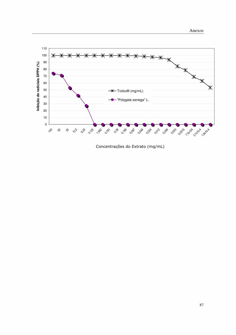
Anexos
87
0
10
20
30
40
50
60
70
80
90
100
110
100 50 25
12,5
6,25
3,12
5
1,56
2
0,78
10,
39
0,19
5
0,09
7
0,04
8
0,02
4
0,01
2
0,00
6
0,00
3
0,00
15
7.5x
104
3.7x
10-4
1,9x
10-4
Inib
ição
de
rad
icia
is D
PP
H (
%)
Trolox® (mg/mL)
"Polygala senega" L.
Concentrações do Extrato (mg/mL)

Anexos
88
Dados brutos referentes ao Ensaio de Peroxidação Lipídica
Amostra Nome científico [sol.estoque] [na reação] Abs 1 Abs 2 Média Abs
Branco 1 Branco 1 - - 0,033 0,031 0,0320 Branco 2 Branco 2 - - 0,032 0,033 0,0325
10 10 - - 0,034 0,037 0,0355 20 20 - - 0,592 0,6 0,5960 40 40 - - 0,554 0,541 0,5475 60 60 - - 0,122 0,119 0,1205 80 80 - - 0,183 0,182 0,1825 100 100 - - 0,303 0,291 0,2970 120 120 - - 0,33 0,343 0,3365 140 140 - - 0,488 0,472 0,4800 C1 C1 - - 0,52 0,547 0,5335 C2 C2 - - 0,577 0,578 0,5775
Cmax Cmax - - 0,693 0,689 0,6910 2,50 0,1136 0,489 0,501 0,4950 5,00 0,2273 0,446 0,432 0,4390
10,00 0,4545 0,338 0,336 0,3370 20,00 0,9091 0,246 0,242 0,2440
Quercetina Quercetina
40,00 1,8182 0,155 0,157 0,1560 6,25 0,2841 0,539 0,514 0,5265 12,5 0,5682 0,51 0,498 0,5040 25 1,1364 0,458 0,44 0,4490 50 2,2727 0,434 0,389 0,4115 100 4,5455 0,324 0,315 0,3195
Ruibarbo chinês
Rheum officinaleBaill.
200 9,0909 0,239 0,218 0,2285 6,25 0,2841 0,579 0,581 0,5800 12,5 0,5682 0,562 0,546 0,5540 25 1,1364 0,547 0,542 0,5445 50 2,2727 0,526 0,509 0,5175 100 4,5455 0,439 0,424 0,4315 200 9,0909 0,336 0,372 0,3540
Pitanga Eugenia uniflora L.
400 18,1818 0,031 0,03 0,0305 6,25 0,2841 0,584 0,611 0,5975
12,50 0,5682 0,579 0,54 0,5595 25,0 1,1364 0,547 0,536 0,5415 50 2,2727 0,498 0,503 0,5005 100 4,5455 0,428 0,424 0,4260 200 9,0909 0,303 0,287 0,2950
Ruibarbo europeu
Rheum rhaponticumL.
400 18,1818 0,092 0,086 0,0890

Anexos
89
Amostra Nome científico [sol.estoque] [na reação] Abs 1 Abs 2 Média Abs
6,25 0,2841 0,594 0,595 0,5945 12,50 0,5682 0,605 0,578 0,5915 25,0 1,1364 0,551 0,557 0,5540 50 2,2727 0,554 0,558 0,5560 100 4,5455 0,579 0,554 0,5665 200 9,0909 0,457 0,458 0,4575
Pedra-ume-caá Myrcia
sphaerocarpaDC
400 18,1818 0,34 0,328 0,3340 6,25 0,2841 0,599 0,585 0,5920
12,50 0,5682 0,584 0,573 0,5785 25,0 1,1364 0,542 0,547 0,5445 50 2,2727 0,468 0,471 0,4695 100 4,5455 0,34 0,337 0,3385 200 9,0909 0,158 0,16 0,1590
Cambui Eugenia crenataVell.
400 18,1818 0,046 0,047 0,0465 6,25 0,2841 0,587 0,551 0,5690
12,50 0,5682 0,591 0,634 0,6125 25,0 1,1364 0,555 0,527 0,5410 50 2,2727 0,509 0,492 0,5005 100 4,5455 0,4 0,395 0,3975 200 9,0909 0,262 0,256 0,2590
Jambolão Syzigyum
jambolanumDC
400 18,1818 0,1 0,097 0,0985 6,25 0,2841 0,536 0,528 0,5320
12,50 0,5682 0,515 0,523 0,5190 25,0 1,1364 0,521 0,531 0,5260 50 2,2727 0,496 0,52 0,5080 100 4,5455 0,489 0,48 0,4845 200 9,0909 0,451 0,453 0,4520
Erva de bicho Polygonum acreH.B.
& K.
400 18,1818 0,394 0,388 0,3910 6,25 0,2841 0,555 0,557 0,5560
12,50 0,5682 0,573 0,541 0,5570 25,0 1,1364 0,56 0,561 0,5605 50 2,2727 0,542 0,541 0,5415 100 4,5455 0,538 0,552 0,5450 200 9,0909 0,497 0,504 0,5005
Agno casto Vitex agnu- castus L.
400 18,1818 0,486 0,497 0,4915
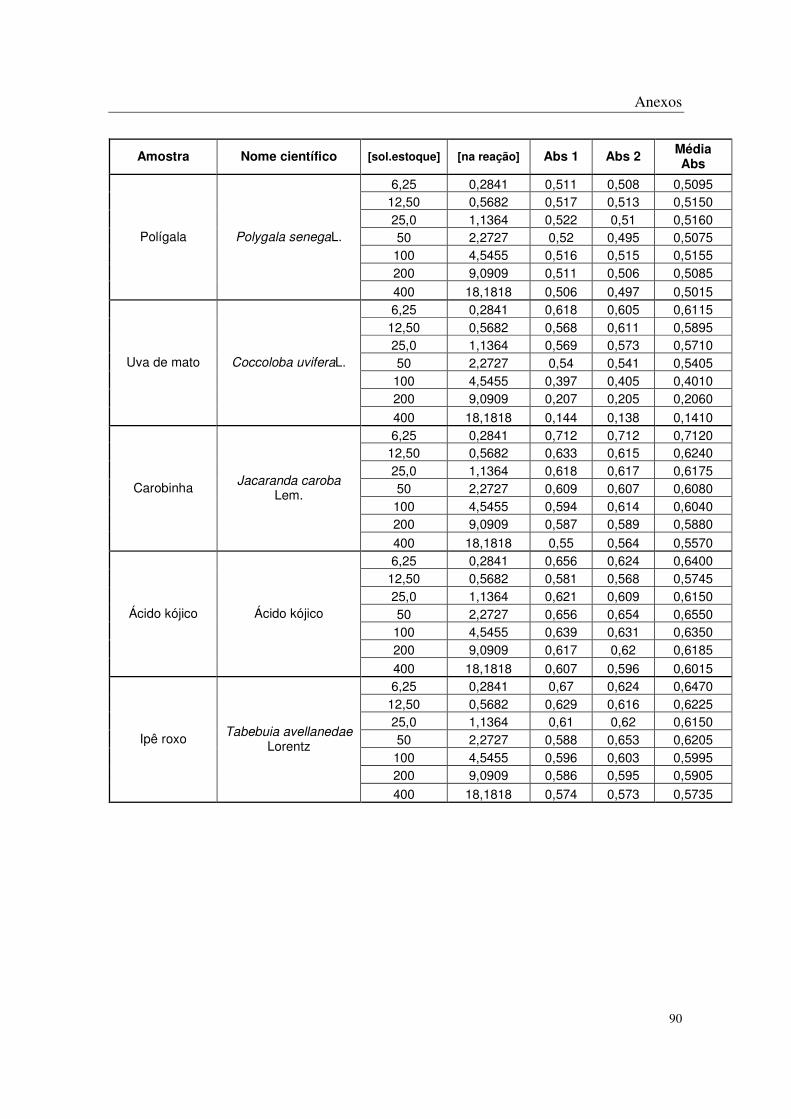
Anexos
90
Amostra Nome científico [sol.estoque] [na reação] Abs 1 Abs 2 Média Abs
6,25 0,2841 0,511 0,508 0,5095 12,50 0,5682 0,517 0,513 0,5150 25,0 1,1364 0,522 0,51 0,5160 50 2,2727 0,52 0,495 0,5075 100 4,5455 0,516 0,515 0,5155 200 9,0909 0,511 0,506 0,5085
Polígala Polygala senegaL.
400 18,1818 0,506 0,497 0,5015 6,25 0,2841 0,618 0,605 0,6115
12,50 0,5682 0,568 0,611 0,5895 25,0 1,1364 0,569 0,573 0,5710 50 2,2727 0,54 0,541 0,5405 100 4,5455 0,397 0,405 0,4010 200 9,0909 0,207 0,205 0,2060
Uva de mato Coccoloba uviferaL.
400 18,1818 0,144 0,138 0,1410 6,25 0,2841 0,712 0,712 0,7120
12,50 0,5682 0,633 0,615 0,6240 25,0 1,1364 0,618 0,617 0,6175 50 2,2727 0,609 0,607 0,6080 100 4,5455 0,594 0,614 0,6040 200 9,0909 0,587 0,589 0,5880
Carobinha Jacaranda caroba
Lem.
400 18,1818 0,55 0,564 0,5570 6,25 0,2841 0,656 0,624 0,6400
12,50 0,5682 0,581 0,568 0,5745 25,0 1,1364 0,621 0,609 0,6150 50 2,2727 0,656 0,654 0,6550 100 4,5455 0,639 0,631 0,6350 200 9,0909 0,617 0,62 0,6185
Ácido kójico Ácido kójico
400 18,1818 0,607 0,596 0,6015 6,25 0,2841 0,67 0,624 0,6470
12,50 0,5682 0,629 0,616 0,6225 25,0 1,1364 0,61 0,62 0,6150 50 2,2727 0,588 0,653 0,6205 100 4,5455 0,596 0,603 0,5995 200 9,0909 0,586 0,595 0,5905
Ipê roxo Tabebuia avellanedae Lorentz
400 18,1818 0,574 0,573 0,5735

Anexos
91
Amostra Nome científico % Inib 1
% Inib 2
% Inib t
equação da reta R2 IC50
29% 27% 28% 36% 37% 36% 51% 51% 51% 65% 65% 65%
Quercetina Quercetina
78% 77% 77%
y=0,4466x+0,2616 0,9545 0,53
17% 12% 15% 22% 15% 19% 30% 25% 28% 33% 34% 34% 50% 46% 48%
Ruibarbo chinês
Rheum officinale Baill.
63% 63% 63%
y=0,053x+0,1857 0,9421 5,93
11% 1% 6% 14% 7% 11% 16% 8% 12% 19% 13% 16% 33% 28% 30% 48% 37% 43%
Pitanga Eugenia uniflora L.
95% 95% 95%
y=0,0476x+0,06 0,9897 9,24
10% 6% 8% 11% 17% 14% 16% 18% 17% 24% 23% 23% 34% 35% 35% 53% 56% 55%
Ruibarbo europeu
Rheum rhaponticum L.
86% 87% 86%
y=0,0423x+0,1225 0,9865 8,92
9% 9% 9% 7% 11% 9%
15% 15% 15% 15% 15% 15% 11% 15% 13% 30% 30% 30%
Pedra-ume-caá Myrcia
sphaerocarpa DC
48% 50% 49%
y=0,0218x+0,0868 0,9614 18,95
8% 10% 9% 10% 12% 11% 17% 16% 16% 28% 28% 28% 48% 48% 48% 76% 75% 76%
Cambui Eugenia crenata
Vell.
93% 93% 93%
y=0,0485x+0,1524 0,9069 7,17

Anexos
92
Amostra Nome científico % Inib 1
% Inib 2
% Inib t
equação da reta R2 IC50
10% 16% 13% 9% 3% 6%
15% 19% 17% 22% 25% 23% 39% 40% 39% 60% 61% 60%
Jambolão Syzigyum
jambolanumDC
85% 85% 85%
y=0,043x+0,1261 0,9517 8,70
23% 23% 23% 26% 24% 25% 25% 23% 24% 28% 25% 26% 29% 30% 30% 35% 34% 35%
Erva de bicho Polygonum acre
H.B. & K.
43% 44% 43%
y=0,0112x+0,2368 0,9855 23,50
20% 19% 20% 17% 21% 19% 19% 19% 19% 22% 21% 22% 22% 20% 21% 28% 27% 28%
Agno casto Vitex agnus-castus
L.
30% 28% 29%
y=0,0059x+0,1941 0,8737 51,85
26% 26% 26% 25% 26% 25% 25% 26% 25% 25% 28% 27% 26% 25% 25% 26% 27% 26%
Polígala Polygala senegaL.
27% 28% 27%
y=0,0009x+0,2566 0,5702 270,44
9% 11% 10% 16% 10% 13% 16% 15% 16% 20% 20% 20% 41% 40% 41% 69% 70% 70%
Uva de mato Coccoloba uvifera L.
79% 80% 79%
y=0,0378x+0,1282 0,9584 9,84
-5% -5% -5% 7% 9% 8% 9% 9% 9%
10% 10% 10% 12% 9% 11% 13% 13% 13%
Carobinha Jacaranda caroba
Lem.
19% 17% 18%
y=0,03053x+0,0839 0,9827 13,63

Anexos
93
Amostra Nome científico % Inib 1
% Inib 2
% Inib t
equação da reta R2 IC50
3% 8% 6% 14% 16% 15% 8% 10% 9% 3% 4% 3% 6% 7% 6% 9% 9% 9%
Ácido kójico Ácido kójico
10% 12% 11%
y=0,0045x+0,0357 0,8998 103,18
1% 8% 5% 7% 9% 8%
10% 9% 9% 13% 4% 8% 12% 11% 12% 14% 12% 13%
Ipê roxo Tabebuia
avellanedae Lorentz
15% 15% 15%
y=0,0038x+0,0883
0,9041 108,34
Curva Padrão Solução de Ferro-Ácido Ascórbico
y = 0,0034x - 0,043R2 = 0,933
0,0000
0,1000
0,2000
0,3000
0,4000
0,5000
0,6000
0 50 100 150
Concentração (uM)
Ab
sorb
ânci
a (n
m)

Anexos
94
Dados brutos referentes ao Ensaio Antitirosinase in vitro
Amostras Nome científico [sol.estoque] [na reação] [DopaCr]1 [DopaCr]2 Média
BL BL ---- ---- ---- ---- ---- 65,78 65,78 ---- ---- 58,614273 57,87917 58,25 95,67 95,67 ---- ---- 99,255014 92,42905 95,84 148 148 ---- ---- 151,34237 157,1182 154,23 222 222 ---- ---- 233,46397 237,9796 235,72 333 333 ---- ---- 328,92246 317,8959 323,41
---- ---- 35,405995 35,82605 35,62 Controle Controle
---- ---- 311,38498 297,208 304,30 17,5 2,625 328,39738 337,5337 332,97 35 5,25 280,30059 262,238 271,27 70 10,5 216,13652 211,8309 213,98 140 21 149,97718 161,3188 155,65
Ácido kójico Ácido kójico
280 42 72,056172 84,13288 78,09 50 7,5 308,0245 321,6764 314,85 100 15 287,96667 301,5136 294,74 200 30 280,4056 284,1861 282,30 400 60 286,91652 253,9419 270,43 800 120 262,23804 283,451 272,84
1600 240 206,0551 217,9218 211,99
Ruibarbo chinês Rheum officinale Baill.
3200 480 157,43323 170,9801 164,21 50 7,5 327,59596 315,6045 321,60 100 15 316,57682 303,9372 310,26 200 30 334,61796 346,3933 340,51 400 60 280,38649 265,0461 272,72 800 120 213,73148 225,7229 219,73
1600 240 219,99727 223,8864 221,94
Pitanga Eugenia uniflora L.
3200 480 224,3185 236,634 230,48 50 7,5 328,89233 321,6543 325,27 100 15 306,09783 309,6628 307,88 200 30 281,89892 315,9286 298,91 400 60 257,05184 283,6274 270,34 800 120 274,01267 252,4065 263,21
1600 240 182,40254 215,46 198,93
Cambui Eugenia crenata Vell.
3200 480 184,88725 129,2514 157,07

Anexos
95
Amostras Nome científico [sol.estoque] [na reação] [DopaCr]1 [DopaCr]2 Média
50 7,5 341,09981 342,9363 342,02 100 15 331,05295 334,5099 332,78 200 30 298,53567 296,159 297,35 400 60 267,53083 296,159 281,84 800 120 284,2756 259,1044 271,69
1600 240 280,60255 279,8463 280,22
Jambolão Syzigyum jambolanum
DC
3200 480 156,90727 171,1673 164,04 50 7,5 362,70597 381,6114 372,16 100 15 349,20212 392,1984 370,70 200 30 336,45449 384,8523 360,65 400 60 303,28903 317,7652 310,53 800 120 285,89606 276,0653 280,98
1600 240 277,90178 344,4488 311,18
Pedra-ume-caá Myrcia
sphaerocarpaDC
3200 480 262,8855 277,0375 269,96 50 7,5 356,10487 353,5278 354,82 100 15 323,09405 358,5592 340,83 200 30 357,94562 359,9091 358,93 400 60 336,47018 331,9297 334,20 800 120 307,26358 318,3081 312,79
1600 240 294,01017 300,7596 297,38
Agno casto Vitex agnus-castus L.
3200 480 262,59467 266,0307 264,31 50 7,5 361,62712 362,118 361,87 100 15 350,45989 354,8777 352,67 200 30 358,68192 355,2458 356,96 400 60 359,90909 361,0135 360,46 800 120 324,44393 326,162 325,30
1600 240 276,46166 279,1614 277,81
Erva de bicho Polygonum acre H.B.
& K.
3200 480 226,14778 219,8892 223,02 50 7,5 372,30348 381,9981 377,15 100 15 391,93817 385,4342 388,69 200 30 374,26695 380,0346 377,15 400 60 361,87256 358,6819 360,28 800 120 365,92221 378,3166 372,12
1600 240 345,4285 363,2224 354,33
Polígala Polygala senega L.
3200 480 325,42567 320,0261 322,73 50 7,5 338,0655 347,5147 342,79 100 15 311,43595 312,6631 312,05 200 30 259,40403 269,4668 264,44 400 60 218,78477 214,4897 216,64 800 120 141,35047 139,5097 140,43
1600 240 84,28716 88,33681 86,31
Ruibarbo europeu Rheum rhaponticum L.
3200 480 52,135359 51,39906 51,77

Anexos
96
Amostras Nome científico [sol.estoque] [na reação] [DopaCr]1 [DopaCr]2 Média
50 7,5 297,28811 298,2034 297,75 100 15 310,91541 329,4241 320,17 200 30 325,86475 327,3902 326,63 400 60 287,72867 291,9999 289,86 800 120 256,50792 250,2028 253,36
1600 240 210,94801 189,8969 200,42
Carobinha Jacaranda caroba
Lem.
3200 480 128,06558 123,6926 125,88 50 7,5 303,18649 305,017 304,10 100 15 295,25419 281,7286 288,49 200 30 312,84764 290,7796 301,81 400 60 329,62751 327,1868 328,41 800 120 288,84733 296,2712 292,56
1600 240 244,20268 249,2875 246,75
Ipê roxo Tabebuia avellanedae
Lorentz
3200 480 230,37199 228,7449 229,56 50 7,5 289,4575 274,5082 281,98 100 15 296,06776 311,2205 303,64 200 30 304,81363 306,6442 305,73 400 60 225,89736 237,5924 231,74 800 120 126,03166 134,5741 130,30
1600 240 48,437434 55,04769 51,74
Uva de mato Coccoloba uvifera L.
3200 480 10,097955 42,74245 26,42
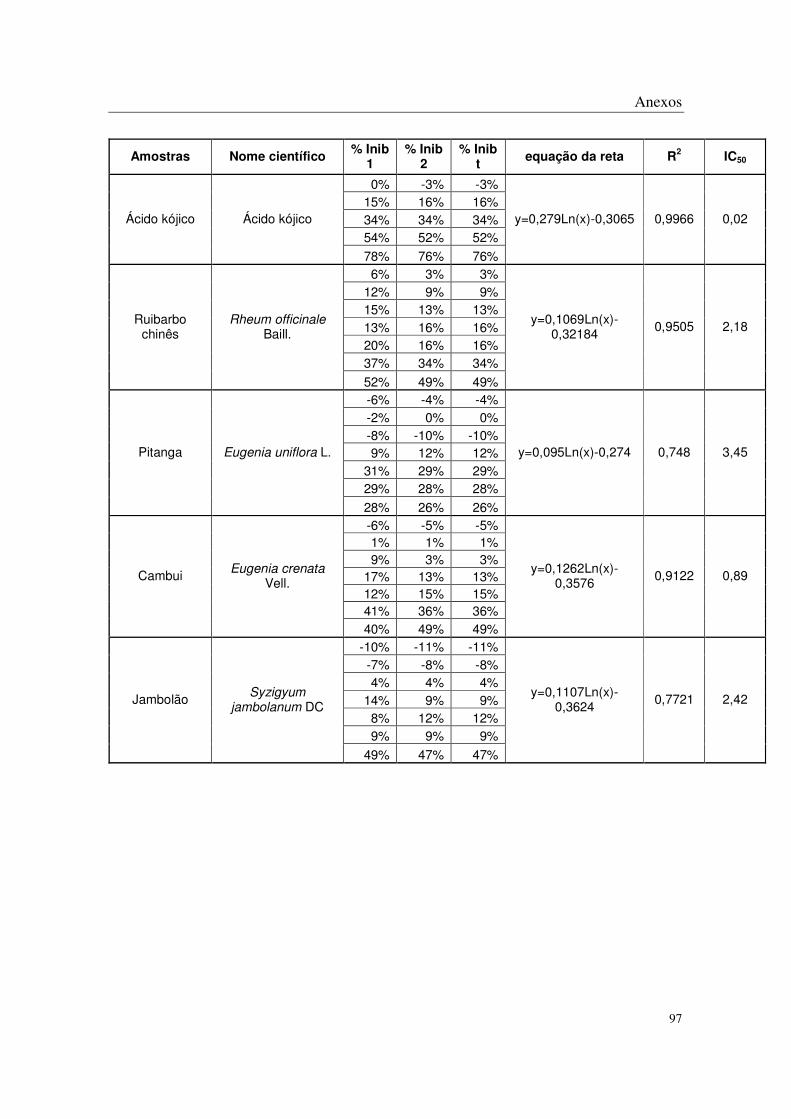
Anexos
97
Amostras Nome científico % Inib 1
% Inib 2
% Inib t
equação da reta R2 IC50
0% -3% -3% 15% 16% 16% 34% 34% 34% 54% 52% 52%
Ácido kójico Ácido kójico
78% 76% 76%
y=0,279Ln(x)-0,3065 0,9966 0,02
6% 3% 3% 12% 9% 9% 15% 13% 13% 13% 16% 16% 20% 16% 16% 37% 34% 34%
Ruibarbo chinês
Rheum officinale Baill.
52% 49% 49%
y=0,1069Ln(x)-0,32184
0,9505 2,18
-6% -4% -4% -2% 0% 0% -8% -10% -10% 9% 12% 12%
31% 29% 29% 29% 28% 28%
Pitanga Eugenia uniflora L.
28% 26% 26%
y=0,095Ln(x)-0,274 0,748 3,45
-6% -5% -5% 1% 1% 1% 9% 3% 3%
17% 13% 13% 12% 15% 15% 41% 36% 36%
Cambui Eugenia crenata Vell.
40% 49% 49%
y=0,1262Ln(x)-0,3576
0,9122 0,89
-10% -11% -11% -7% -8% -8% 4% 4% 4%
14% 9% 9% 8% 12% 12% 9% 9% 9%
Jambolão Syzigyum jambolanum DC
49% 47% 47%
y=0,1107Ln(x)-0,3624
0,7721 2,42

Anexos
98
Amostras Nome científico % Inib 1
% Inib 2
% Inib t
equação da reta R2 IC50
-17% -20% -20% -13% -20% -20%
-9% -17% -17% 2% 0% 0% 8% 9% 9%
10% -1% -1%
Pedra-ume-caá Myrcia
sphaerocarpa DC
15% 13% 13%
y=0,0841Ln(x)-0,395 0,8372 41,86
-11% -13% -13% -1% -8% -8%
-12% -14% -14% -5% -6% -6% 4% 0% 0% 8% 5% 5%
Agno casto Vitex agnus-castus
L.
18% 16% 16%
y=0,0664Ln(x)-0,3009
0,8409 173,12
-13% -15% -15% -10% -12% -12% -12% -14% -14% -13% -15% -15%
-1% -4% -4% 14% 12% 12%
Erva de bicho Polygonum acre
H.B.& K.
29% 29% 29%
y=0,0981Ln(x)-0,4284 0,7515 12,88
-16% -20% -20% -23% -24% -24% -17% -20% -20% -13% -15% -15% -14% -18% -18%
-8% -13% -13%
Polígala Polygala senegaL.
-2% -3% -3%
y=0,0389Ln(x)-0,3199 0,7092 1,42E+06
-6% -9% -9% 3% 1% 1%
19% 16% 16% 32% 31% 31% 56% 55% 55% 74% 73% 73%
Ruibarbo europeu
Rheum rhaponticum L.
84% 84% 84%
y=0,2509Ln(x)-0,5088
0,9913 0,06

Anexos
99
Amostras Nome científico % Inib 1
% Inib 2
% Inib t
equação da reta R2 IC50
-7% -4% 8% -27% -32% 1%
-194% -162% -1% 3% 3% 11%
17% 21% 22% 35% 39% 38%
Carobinha Jacaranda caroba
Lem.
55% 57% 61%
y=0,0013Ln(x)-0,028 0,957 2,46E+173
-18% -20% 6% -40% -44% 11%
-144% -140% 7% -9% -8% -1% 2% -1% 10%
22% 18% 24%
Ipê roxo Tabebuia
avellanedae Lorentz
30% 30% 29%
y=0,0502Ln(x)-0,0818
0,5009 107,97
0% 4% 13% -21% -23% 6% -32% -33% 6% 22% 18% 29% 57% 57% 60% 84% 83% 84%
Uva de mato Coccoloba uvifera L.
96% 89% 92%
y=0,2294Ln(x)-0,5253 0,8687 0,09