Embed Size (px)
Citation preview
UNIVERSIDADE FEDERAL DE PELOTAS Programa de Pós-Graduação em Biotecnologia
Tese
Leptospira interrogans: cinética da infecção e avaliação da proteção conferida por LigA
Mariana Loner Coutinho
Pelotas, 2011

MARIANA LONER COUTINHO
Leptospira interrogans: cinética da infecção e avaliação da proteção conferida por LigA
Orientador: José Antonio Guimarães Aleixo
Co-orientador: David A. Haake
Pelotas, 2011
Tese apresentada ao Programa de Pós-Graduação em Biotecnologia da Universidade Federal de Pelotas, como requisito parcial à obtenção do título de Doutor em Ciências (área de conhecimento: Biotecnologia).


Banca examinadora:
Prof. Dr. Éverton Fagonde da Silva (UFPel)
Prof. Dr. Odir Antonio Dellagostin (UFPel)
Prof. Dra. Sílvia de Oliveira Hübner (UFPel)
Prof. Dr. José Antonio Guimarães Aleixo (UFPel)

Ao Daniel por aceitar que um ano pode ter 882 dias.

AGRADECIMENTOS
Escrever uma página de agradecimentos na qual deve-se combinar as mais
diversas pessoas, com personalidades, estilos e opiniões de vida completamente
diferentes parece não fazer jus à importância que essas pessoas tiveram na minha
vida ao longo desses anos, faz parecer que elas sempre estiveram ali e sempre
estarão ao meu dispor, o que não é correto e nem passível de expectativa, elas me
estenderam a mão nas inúmeras vezes que precisei e foram aos poucos fazendo
parte da minha vida.
Ao Daniel, por sempre ter me dado um apoio incondicional, por ter sempre
entedido quando o trabalho veio primeiro. Aos meus pais, por terem ensinado
através do exemplo, por sempre, desde o meu primeiro até o último dia perguntarem
o que eu tinha aprendido na escola hoje. Por terem me mostrado que a vida de
professor universitário não é somente “sucesso e fama”. À minha irmã Lúcia pelas
valiosas recomendações de distrações e de como lidar com a saudade, e à minha
irmã Eleonora e tia Vilma, por terem sempre me apoiado nos momentos difíceis.
Aos meus queridos amigos do laboratório, Flávia, Marcelo, Karla, Carla,
Juliana e Carina, pelas viagens, chopps e cafés, mas por entenderem e dividirem
comigo a alegria de uma expressão de proteína espetacular e a tristeza de um Mac
ELISA que não dá certo. À todos os amigos que eu colecionei durante esses anos
que se mantiveram presentes no meu dia-a dia por meio de saídas, telefonemas, e-
mails, skype, googletalk, enfim qualquer modernidade que serviu para me fazer
sentir mais perto de casa.
Ao meu orientador José Antonio Guimarães Aleixo, por sempre ter acreditado
no meu potencial, por ter me apoiado, entedido e por dividir o seu entusiasmo sobre
ciência, seriados de tv e cultura norte-americana.
Ao meu Co-orientador David Haake por ter me acolhido em seu laboratório
sem ao menos me conhecer e por ter generosamente financiado o trabalho aqui
descrito. Ao que sempre me guiou, criticou e elogiou em todos os momentos. À
Henry Choy, Jane Babbitt, James Matsunaga e Marija Pinne (meu comitê ad-hoc
americano) pelos seus valiosos ensinamentos sobre pesquisa, por terem me
apoiado em todas as minhas batalhas diárias, e por terem me introduzido à tantos

aspectos diferentes da cultura americana (e letoniana), e por um apoio incondicional
sempre que eu precisei.
À Lorraine que me acolheu em sua casa e me adotou de forma a ser a minha
família americana.
Ao CNPq que me deu apoio finaceiro para a realização deste doutorado.
Aos modelos animais que colaboraram imensamente.
A todos aqueles que de alguma forma contribuíram na minha formação
academica o meu mais sincero muito obrigada!

Resumo
COUTINHO, Mariana Loner. Leptospira interrogans: cinética da infecção e avaliação da proteção conferida por LigA. 2011. <89>f. Tese (Doutorado) - Programa de Pós-Graduação em Biotecnologia. Universidade Federal de Pelotas, Pelotas.
A Leptospira interrogans é o agente causador da leptospirose mais comum, uma
zoonose que é endêmica em países tropicais como Brasil e Índia. As vacinas hoje
existentes se baseiam em bacterinas de sorovares mais comumente encontrados na
espécie a ser vacinada, mas essas vacinas não desenvolvem uma imunidade
duradoura ou de proteção cruzada. As vacinas de subunidade com proteínas
recombinantes possuem potencial para produção de uma vacina eficiente contra a
leptospirose uma vez que podem oferecer proteção cruzada. LigA é uma proteína
que possui função de adesão ao tecido do hospedeiro e já foi relatada como
protetora contra o desafio letal por Leptospira interrogans. A primeira parte deste
documento versa sobre a definição de uma região protetora de três segmentos
constituída pelos domínios 11 e 12, acrescidos do domínio 10 ou 13. O efeito da
vacina foi avaliado com base na patologia, resposta imune humoral e quantificação
da carga microbiana nos rins. A segunda parte deste trabalho descreve a cinética da
infecção por Leptospira interrogans em hamster, de forma a apontar como o organismo
reage durante a primeira fase da infecção leptospírica. Essa avaliação foi realizada
por meio de quantificação da carga microbiana em órgãos como rins, fígado,
pulmões e baço, além de acompanhamento da evolução da doença por intermédio
de hemograma e análises bioquímicas. Os dados analisados mostram que a maior
carga bacteriana é observada após 6 dias de infecção em ambas as rotas.
Palavras-chave: Leptospira. Leptospirose. Tempo real. Quantificação.
Abstract
COUTINHO, Mariana Loner. Leptospira interrogans: cinética da infecção e avaliação da proteção conferida por LigA. 2011. <89>f. Tese (Doutorado) - Programa de Pós-Graduação em Biotecnologia. Universidade Federal de Pelotas, Pelotas.
Leptospira interrogans is the usual causative agent of leptospirosis, a zoonosis that is
endemic in tropical countries like Brazil and India. The available vaccines are
bacterin-based from the more prevalent serovars to the vaccinated specie, but these
vaccines do not develop long-lasting immunity or cross-protection. Subunit vaccines
with recombinant proteins have the potential to generate an efficient leptospirosis
vaccine. LigA is a protein that has adhesin function to the host and has been
reported as protective against lethal challenge of Leptospira interrogans. The first part of
this document defines a three-segment protective region of LigA constituted as
domains 11 and 12, added of domains 10 or 13. The vaccine effect was evaluated
based on pathology, humoral immune response and kidney leptospiral burden. The
second part of this work aims to point out the importance of the correct
understanding of the Leptospira interrogans infection kinectics to elucidate how the
organism reacts in the first phase of the leptospiral infection. This evaluation is
accessed by leptospiral load quantification in key-organs such as kidneys, liver, lungs
and spleen, and by the evolution of the disease by hemmogram and biochemical
analysis.
Keywords: Leptospira. Leptospirosis. Real-time. Quantification.

Lista de Tabelas
TABELA 1: ESPÉCIES DESCRITAS ......................................................................................................... 12 TABLE 2: LEPTOSPIRAL PROTEINS THAT BIND TO ECM .................................................................. 21
Sumário
1 Introdução ........................................................................................................... 11
1.1 Epidemiologia da leptospirose ............................................................................ 13
1.1.1 Hospedeiro Reservatório................................................................................ 14
1.1.2 Hospedeiro acidental ..................................................................................... 15
1.1.3 Apresentação ................................................................................................. 17
2 Artigo 1: Animal Models of Leptospirosis ............................................................ 18
2.1 Animal Models – How well do they reproduce infection ...................................... 18
2.1.1 Immunology .................................................................................................... 18
2.2 Pathogenesis of infection .................................................................................... 21
2.2.1 Leptospiral adhesion ...................................................................................... 21
2.2.2 Complement evasion ..................................................................................... 22
2.3 Leptospiral response to the host environment .................................................... 22
2.3.1 Outer membrane proteins – reviews definition, types .................................... 22
2.3.2 Regulation of expression in response to host-like conditions ......................... 23
2.4 Progress Towards a Subunit Vaccine ................................................................. 24
2.4.1 Where are we now: whole cell bacterins ........................................................ 24
2.4.2 Research on new vaccines (including recombinant) ...................................... 24
3 Objetivos ............................................................................................................. 26
3.1 Geral ................................................................................................................... 26
3.2 Objetivos Específicos.......................................................................................... 26
4 Paper 1 - A LigA Three-Domain Region Protects Hamsters from Lethal Infection by Leptospira interrogans .......................................................................................... 27
4.1 Non-technical Author Summary .......................................................................... 30
4.2 Introduction ......................................................................................................... 31
4.3 Materials & Methods ........................................................................................... 33
4.4 Results ................................................................................................................ 37
4.5 Discussion .......................................................................................................... 40
4.6 Acknowledgements ............................................................................................. 43
4.7 References ......................................................................................................... 43

4.8 FIGURES ............................................................................................................ 50
4.9 TABLES .............................................................................................................. 56
5 Paper 2 – Kinectics of the Hamster Model of Leptospira interrogans infection ... 59
5.1 Abstract ............................................................................................................... 61
5.2 Introduction ......................................................................................................... 62
5.3 Materials & Methods ........................................................................................... 63
5.4 Results ................................................................................................................ 68
5.5 Discussion .......................................................................................................... 71
5.6 Acknowledgements ............................................................................................. 72
5.7 References ......................................................................................................... 73
5.8 Figures ................................................................................................................ 75
5.9 Conclusões ......................................................................................................... 81
5.9.1 Paper 1........................................................................................................... 81
5.9.2 Paper 2........................................................................................................... 81
6 Referências ......................................................................................................... 82

11
1 Introdução
A leptospirose é causada por bactérias do gênero Leptospira, a qual pretence à
ordem das Spirochaetales, e difere das outras espiroquetas pela presença de
“ganchos” nas extremidades (WHO, 2003). A leptospirose é considerada uma
doença zoonótica que atinge principalmente países tropicais. No Brasil o número de
casos é variável, com 4.000 casos confirmados laboratorialmente a cada ano, mas
esse número pode variar muito uma vez que a notificação não é obrigatória e que
muitos casos não são confirmados e/ou apresentam sintomatologia clínica muito
branda. As leptospiras patogênicas são mantidas na natureza nos hospedeiros
reservatórios que são espécies nas quais a infecção é endemica e é perpetuada
pelo contato direto dos animais (LEVETT, 2001).
A classificação antiga por patogenicidade agrupava as leptospiras em duas
espécies diferentes denominadas Leptospira interrogans (patogênica) e Leptospira biflexa
(saprofítica). Este sistem foi substituído por uma classificação genetic que contém 7
espécies patogênicas, 6 espécies de patogenicidade intermediária, e 6 espécies
saprófitas.
A classificação genetic dos sorovares é determinada pelo perfil de
hibridização DNA-DNA obtido e confirmado pela técnica de multiloccus enzyme
electrophoresis (LEVETT, 2001). Este método de classificação permite que um
serovar seja classificado em duas species diferentes ao mesmo tempo dependendo
da cepa utilizada (BRENNER et al., 1999).

12
Tabela 1: Espécies descritas
Patogênicas Patogenicidade Intermediária Saprofíticas
1 L. alexanderi L. alstonii L.biflexa
2 L. santarosai L. kmetyl L. meyeri
3 L. borgpetersenii L. wolffii L. yanagawae
4 L. noguchi L. licerasiae L. tersptrae
5 L. interrogans L. broomii L. vanthielii
6 L. weilii L. inadai L. wolbachii
7 L. kirschneri

13
1.1 Epidemiologia da leptospirose
Os reservatórios naturais das espécies patogênicas são roedores como Rattus
norvegicus e animais selvagens, os quais carreiam a bactéria em seus túbulos renais
e disseminam os organismos na sua urina. Humanos e animais domésticos são
infectados com leptospiras pelo contato com urina ou tecidos de animais infectados
ou pela exposição indireta à agua contaminada. Em países desenvolvidos, é
normalmente associada com atividades recreacionais, como canoagem (SEIVAR, et
al, 2003) ou atividades profissionais que envolvem exposição ao sistema de esgoto
ou trablhadores de abatedouros (CENTERS FOR DISEASE CONTROL AND
PREVENTION, 1998). Em países em desenvolvimento, a doença primariamente
ocorre em áreas com pouco saneamento, nas quais há a ocorrência de esgotos
abertos ou áreas que são propensas à inundações após chuva severa (KO et al.,
1999).
As leptospiras provavelmente não podem penetrar a pele intacta; elas
normalmente penetram no hospedeiro através de pequenas lesões na pele,
conjuntiva ou sistema reprodutivo, apesar de existirem alguns relatos de infecções
iniciadas no sistema digestório ou através de mordidas de ratos (LUZZI, G. A, et al,
1993 and GOLLOP et al, 1987), mas estas não são freqüentes e provavelmente são
acidentais. Uma vez dentro do hospedeiro, as leptospiras se espalham por órgãos
como fígado, coração, rins, pulmões, cérebro, músculo esquelético, glândulas
adrenais e sangue. A imunidade desenvolvida pela infecção é principalmente
humoral a qual opsoniza as bactérias e induz fagocitose por macrófagos. A maioria
dos anticorpos produzidos reage contra o LPS da leptospira e a imunidade conferida
é sorovar-específica (FAINE et al., 1999).

14
A leptospirose é normalmente classificada como uma doença emergente, e
isso se deve ao fato de o número total de casos de leptospirose humana não ser
conhecido. Na maior parte dos países a leptospirose não é uma doença de
notificação obrigatória às autoridades, resultando em dados epidemiologicos não
confiáveis. É estimado que entre 350.000 à 500.000 pessoas são acometidas pela
forma severa da doença, mas o número pode ser muito maior uma vez que a
maioria dos casos de leptospirose não são severos (HARTSKEERL et al. 2011). No
Brasil o número de pacientes confirmados de leptospirose é em torno de 4.000 por
ano, com picos óbvios em áreas que sofreram inundações recentes. A taxa de
mortalidade é entre 10 à 15% (DADOS EPIDEMIOLOGICOS, MINISTÉRIO DA
SAÚDE, 2010).
1.1.1 Hospedeiro Reservatório
Ratos e camundongos desempenham um importante papel na disseminação
das leptospiras no ambiente uma vez que albergam as espiroquetas nos seus rins
sem demonstrar a presença de nenhuma patologia séria além de nefrite intersticial.
A disseminação das leptospiras pode ocorrer a partir dos sexto dia após a infecção
com uma carga microbiana de 1x10⁵ à 1x10⁷ leptospiras/ mL (MONAHAN et al,
2008) e durar por vários meses, sem apresentar uma perda significativa de peso ou
sinal clínico (NALLY et al, 2005). Foi demonstrado que nefrite intersticial é o achado
patologico mais proeminente em ratos infectados experimentalmente ou capturados
no sistema de esgoto da cidade de Salvador (Brasil) e que tinham culturas renais
positivas (FARIA et al. 2007).
15
Algumas species são consideradas hospedeiros de manutenção para um
serovar específico tais como Icterohaemorrhagiae e Ballum em ratos, sorogrupo
Ballum em camundongos, Pomona, Tarassovi e Bratislava em suínos, Hardjo e
Pomona em ovinos e canicola em caninos (LEVETT, 2001). Em bovinos o sorovar
mais importante é o Hardjo, que geralmente causa uma infecção subclínica. A
adaptação da bacteria ao seu hospedeiro é exemplificada pela adaptação de L.
borgpetersenii sorovar Hardjo ao gado europeu. Neste caso, a adaptação à trasmissão
direta resultou emu ma redução genômica (BULACH et al., 2006), levando à perda
da capacidade de sobrevivência no meio-ambiente e limitando a sua transmissão ao
contato direto entre hospedeiros.
1.1.2 Hospedeiro acidental Ao contrário do hospedeiro reservatório, a leptospirose em um hospedeiro
accidental normalmente causa manifestações clínicas mais severas. A infecção
leptospírica em bovinos recebe muita atenção devido à sua importância econômica
devido à problemas associados com reprodução, lactação e riscos aos humanos.
A infecção bovina pelos sorovares Hardjo, Pomona e Grippotyphosa
geralmente resulta em infecção subclínica a qual, quando não tratada, pode levar à
infecção crônica e perdas econômicas como baixa taxa de crescimento, agalactia e
aborto. O estado crônico resulta emu ma eliminação continua de bactéria na urina e,
consequentemente, disseminação aos animais não infectados e descendentes.
Entretanto, quando o gado é infectado com outros sorovares, uma infecção mais
aguda e severa é gerada com uma pletora de sintomas, que tipicamente incluem
febre, anemia, hemoglobinúria e congestão pulmonar (FAINE, 1999).

16
A leptospirose aguda bovina geralmente ocorre em animais jovens e
apresenta sintomas severos como septicemia com febre, anorexia e anemia
hemolítica aguda, hemoglobinúria e icterícia, e em alguns casos pode progredir para
meningite e morte. Diminuição severa na produção de leite e hemolactia podem
ocorrer em vacas em lactação infectadas com os sorovares hardjo-pratijno ou
hardjo-bovis; a diminuição na produção de leite pode durar de 2 à 10 dias com o leite
apresentando uma coloração amarela, um alto númeor de células somáticas e pode
até mesmo conter coágulos sanguíneos. Apesar da recuperação dos animais ocorrer
após 10 dias, a produção de leite pode não retornar ao nível original até o final
daquele período de lactação (ELLIS, et al, 1984). A leptospirose possui uma variação grande de sintomas em humanos, que
podem variar entre sintomas parecidos com uma gripe até falência renal aguda,
hemorragia pulmonary e morte, os sinais mais comuns incluem febre, calafrios,
cefaléias, mialgias severas, náusea, emese e prostração (DOLHNIKOFF et al.
2007), com 20 à 70% dos casos apresentando envolvimento pulmonar com
hemorragia alveolar. Foi relatado que o envolvimento pulmonar é indicative de mau
prognóstico (SPICHLER, et al. 2008) e é observado 4 à 6 dias após a infecção e
pode levar rapidamente à morte. Outros indicadores de mau prognóstico em
humanos são oligúria, baixa contagem de plaquetas (<70,000), creatinine elevada
(>3mg/dL) e idade (>40 anos).

17
1.1.3 Apresentação Este documento é composto por três artigos que foram ou serão submetidos
à publicação em periódicos científicos e indexados. O primeiro artigo é uma
introdução ao tema de modelos animais atualmente usados em pesquisa de
leptospirose. Este artigo discute a epidemiologia da leptospirose nos hospedeiros
reservatório e acidental e, posteriormente, foca nos modelos animais utilizados em
pesquisa tal como hamsters, porquinho-da-índia, camundongos e ratos;
considerando a condição de hospedeiro acidental ou reservatório. Considerações
são feitas sobre o uso de ratos e camundongos, uma vez que eles não são
acometidos por leptospirose e, portanto, podem ser usados como modelos de
infecção crônica ou em testes de virulência pela utilização de camundongos knock-
out. Como modelos de infecção aguda utilizam-se mais frequentemente os hamsters
e porquinhos-da-índia, que apresentam sintomas característicos da doença. É ainda
objeto desta revisão uma abordagem sobre a patogênese da infecção e os
mecanismos de resposta imune da leptospira em diferentes ambientes. Ao final é
feita uma abordagem das vacinas existentes comercialmente formuladas de
bacterinas e as vacinas de subunidade recombinantes.
Existem várias vacinas de bacterinas comercialmente disponíveis para uso
em animais, mas infelizmente elas não induzem proteção longa ou cruzada, o que
demanda a revacinação anual mais prevalentes na região (SRIKRAM et al, 2011).
Atualmente o foco das pesquisas em vacinas se dá em entender as razões que
levam à proteção conferida pelas bacterinas (BROWN et al, 2003 e SRIKRAM et al,
2008), ou ao desenvolvimento de vacinas de subunidade recombinante composta
por proteínas de membrana como a LigA presentes em vários sorovares como a
LigA (SILVA et al, 2007), que protege embora ainda não seja conhecida quão severa
seja a infecção em animais immunizados. O primeiro artigo referente à pesquisa
desenvolvida foi submetido e aceito pelo periódico PloS Neglected Tropical Diseases
e versa sobre a produção de vacina recombinante contra LigA de Leptospira
interrogans, mais especificamente sobre a identifição da menor região protetora. É
descrito a avaliação da patologia e funções renais e hepáticas dos animais
sobreviventes, bem como determinado a carga bacteriana nos rins e a resposta
immune obtida.

18
A Leptospira penetra na pele por abrasões ou em membranas intactas (LEVETT,
2001), porém a rota mais utilizada para infecção experimental é a intraperitoneal (IP)
pois reproduz a doença mais facilmente e é de fácil execução técnica. A
desvantagem da utilização dessa rota no estudo da leptospirose é a eliminação da
proteção immune conferida pela pele, podendo levar à presunções errôneas de
como a infecção natural se desenvolve. O segundo artigo desta tese, a ser
submetido ao periódico PloS Neglected Tropical Diseases, dedica-se ao estudo de
differentes formas de inoculação de leptospira utilizando duas rotas: intradérmica e
sub-cutânea. A análise foi feita através do acompanhamento da cinética da
disseminação da infecção em órgãos e das funções renal e hepática no momento da
eutanásia. A produção de anticorpos foi avaliada de forma a identificar o momento
no qual a resposta imune humoral começa a ser eficaz.
2 Artigo 1: Animal Models of Leptospirosis
2.1 Animal Models – How well do they reproduce infection
2.1.1 Immunology
In experimental infections, rats usually require several folds more of leptospira
inoculum than hamsters or guinea pigs. One important immunological feature is that
mouse Toll-like receptor 4 (TLR4) and TLR2 recognizes LPS, while in humans the
leptospiral LPS is only recognized by the TLR2 (CHASSIN et al., 2009). TRL2 knock-
out mice are resistant to leptospiral infection, while TLR4 has an intermediate
survival curve when compared to double knock-out mice. Another finding is that the
TLR4-/- mice had higher liver bacterial load than the wild type, but both TLR2 -/- and
TLR4 -/- had comparable kidney bacterial loads to WT.

19
2.1.1.1 Histopathology
Both hamsters and guinea pigs are considered good animal models for
leptospirosis. Hamsters present renal pathology before 10 days after the inoculation,
with damage of tubular epithelia with cortical cellular necrosis (SILVA et al, 2008),
shrunken glomeruli with the presence of protenaiceous material (BARNETT et al,
1999), and petechial hemorrhages in the glomeruli and proximal tubules. At
necropsy, organs are usually discolored due to icterus and kidneys can present
areas of capsular depression. The major finding in kidneys is interstitial nephritis
(LEVETT 2001).
Liver can present dissociation of hepatic trabecula with cytoplasmic size
variation (SILVA et al, 2008); and edema of liver cells with deformity of hepatic cords
(HE et al, 2008), lungs can show widespread bleeding with pulmonary congestion
and alveolar hemorrhage (DOLHNIKOFF et al, 2007).

20
2.1.1.2 Challenge routes
Leptospira usually infects an organism by a breakage in the skin or intact
membranes, only a few minor reports have included the oral route of infection or
infection through bites (LEVETT, 2001). It is commonly used and widely accepted
that the intraperitoneal (IP) route is the route of choice, since it is easy to perform and
does reproduce infection more easily and faster than other routes.
In an early study (STAVITSKY, 1945), guinea pigs and hamsters were used to
test different routes of infection and it was shown that blood cultures were positive
with IP and intracardiac (IC) after only one hour, subcutaneous (SQ) after 24 h and
oral (O), intradermal (ID) and intraocular (IO) after 48 h with the onset of jaundice
between 5 to 10 days post infection, for SQ and O routes respectively. In this report,
with the IP route, it was possible to isolate leptospires from blood, kidneys, bone
marrow, liver and adrenal glands from guinea pigs, but hamsters only presented
positive cultures in blood at euthanasia.
A recent kinetics study that used the IP route in hamsters showed a high liver
and kidney colonization, with leptospires reaching 1 x 10⁵ and 5 x 10⁴ copies /µg of
tissue DNA, respectively.
Although the IP route has been used to study leptospirosis, it has a caveat of
eliminating the protection conferred by the immune system of skin and membranes
which can lead to erroneous assumptions on how the natural infection really works.
Also, leptospires have OMPs that can undergo transcriptional changes depending on
the environment (as discussed later), therefore eliminating the invasion step that
leptospires go through when entering a body can result in underexpression or
overexpression of different proteins that do not mimic a natural infection.

21
2.2 Pathogenesis of infection
2.2.1 Leptospiral adhesion
The first step in the leptospiral infection of the organism is the adhesion to the
tissue, either intact membranes or broken skin. Leptospira has the ability to adhere
both to host cells as well as extra-cellular matrix (EMC); it was demonstrated that the
adherent leptospires rapidly invade fibroblasts, renal cells and macrophages in vitro
(MÉRIEN et al, 1997 and 2007).
Several leptospiral outer membrane proteins have been proven to adhere to
ECM structures such as collagen type I and IV, laminin and fibronectin (BARBOSA et
al, 2006, CHOY et al 2007). LigB has been shown to bind fibronectin and fibrinogen
and inhibits fibrin formation, which might help the leptospiral adhesion at the site of
infection (CHOY et al, 2011). OmpL37 is a new protein that was recently found to
bind to human skin and aorta elastin (table 2), both of which could help in the host
adhesion process (PINNE et al, 2010).
Table 2: Leptospiral proteins that bind to ECM
Protein Ligand Reference
LigB Collagen, laminin,
tropoelastin and elastin
Fibronectin modulated
by calcium and
fibrinogen
CHOY et al, 2007 ; LIN YP, CHANG YF 2007; LIN et al, 2009; LIN YP, CHANG YF 2008; LIN et al, 2009;
OmpL37 Skin and aorta Elastin PINNE et al, 2010

22
LenA collagen IV, laminin and
fibronectin, human
plasminogen and
complement regulator
factor H
ATZINGEN M.V.,et al, 2008 and VERMA, et al 2010
2.2.2 Complement evasion
After the entry and adhesion to the host tissue, leptospires have to overcome
the host’s innate immune system (CARROLL, M. C. 2004). Non-pathogenic
leptospires are susceptible to the human complement (CINCO et al., 1983).
Pathogenic leptospires express Len proteins (STEVENSON et al, 2007, VERMA et
al, 2006 and BARBOSA et al, 2006), which are proteins that binds to factor H, a
complement component that signals self-proteins and inactivates C3b bound to the
membrane. The Len family is composed of 6 proteins named Len A, B, C, D, E and
Len F and bind to fibronectin. Pathogenic leptospires also bind C4BP, from the
classical complement pathway, which is a cofactor of factor I that inactivates C4b
(BARBOSA et al, 2009).
2.3 Leptospiral response to the host environment
2.3.1 Outer membrane proteins – reviews definition, types
As mentioned before the leptospiral LPS has unique features that are highly
variable amongst serovars due to O-side chains sugars variability (HAAKE and
MATSUNAGA, 2010), and it is the major component of the leptospiral membrane.
There are several outer membrane proteins (OMPs) and transmembrane OMPs that
have been the focus of intense research in the last decade, some of them are only
expressed in pathogenic serovars (CULLEN et al., 2004; MATSUNAGA et al., 2005;

23
CULLEN et al., 2005). A large group of OMPs are the lipoproteins such as LipL32,
LipL21, LipL36, LipL48 e LipL41. LipL32 is definitely the most abundant protein in the
leptospiral membrane although it is not entirely expressed on the surface; most of it
can be found after membrane permeabilization (PINNE et al, 2009). While LipL32,
LipL41, LipL21 are expressed both in vivo and in vitro, LipL36 is anchored to the
inner face of the outer membrane and is only expressed in vitro (HAAKE et al.,
2000).
Other proteins are the OmpL1, a transmembrane protein with porin function,
and P31LipL45, is a peripherical protein that uses the lipoprotein secretory channel
to reach the inner and outer membranes (CULLEN et al., 2003).
The Leptospiral Immunoglobulin-Like (Lig) protein family is composed by 2
proteins named LigA (PALANIAPPAN et al., 2002) and LigB (MATSUNAGA et al.,
2003) and a pseudo-gene, ligC. LigA has a secreted form and both proteins have
adhesion and invasion function. These proteins are recognized by the sera of
naturally infected patients and their expression occurs mostly in vivo with a marked
decrease in expression after a few rounds of culture (MATSUNAGA et al, 2003).
2.3.2 Regulation of expression in response to host-like conditions
Leptospires have an unusually large number of transcriptional regulators
indicating their ability to respond to environmental conditions. In terms of
environments, leptospires encounter outside a wide range, like mud, to eye
conjunctiva. Lig proteins have been shown to superexpress in vitro when osmolarity
is equal to physiological osmolarity (MATSUNAGA et al, I&I 2007).
In a recent study, iron limitation was found to be a major transcriptional
regulator for almost 100 genes (LO et al, 2010). Another relevant factor is the
temperature shift from environment to host, with hundreds of proteins being up-or
down-regulated, most of which are not characterized yet (LO et al, 2006)

24
2.4 Progress Towards a Subunit Vaccine
2.4.1 Where are we now: whole cell bacterins
There are several commercially available vaccines against leptospirosis both
for animals and humans. They are all made with bacterins from the most important
serovars that affect each species (FAINE, 1999). Although they protect well against
infection they lack several characteristics of an ideal vaccine, e.g., they do not
provide long-lasting immunity which requires annual boosters, they do not protect
against several serovars or serogroups, and they lack coverage if there is a
geographical niche for a specific serovar not predicted by the manufacturer. Human
vaccines are only available in a few countries such as China, Japan, Vietnam and
Cuba (ADLER and MONTECZUMA, 2010) and have the same shortcomings as
veterinary vaccines.
2.4.2 Research on new vaccines (including recombinant)
The use of live attenuated or bacterin vaccines in animals has focused on how
the vaccine works and what are the requirements for cross-protection (SRIKRAM et
al, 2011) and efficient immune response (BROWN et al, 2003), like the induction of
Th1 immune response is more efficient in preventing leptospiral infections. The
development of inflammatory cytokines such as TNF-α and IFN-γ during infection
seem to correlate to a poor outcome, while IL-4 and IL-10 are favorable to disease
resolution (SRIKRAM et al, 2008).
A lot of effort has been put on developing recombinant vaccines against
leptospirosis; so far the main focus of research has been the discovery of a
protective protein that is expressed in multiple pathogenic serovars such as LipL32.
Despite LipL32 being the most abundant protein on the leptospiral membrane and
generating a high antibody titer, it does not completely protect against lethal infection

25
(BRANGER et al., 2001, BRANGER et al., 2005 SEIXAS et al., 2007) which suggest
that a LipL32 vaccine could be used in combination with other protective proteins.
Since the first report of partial protection with a recombinant leptospiral protein
(HAAKE Et al, 1999), a lot of effort has been put into finding the protein that would
not only prevent from lethal infection, but other less severe forms of the disease and
shedding.
In this matter, the final portion of LigA has been the most successful, several
unrelated groups reporting protection in hamsters, LigA was tested under multiple
forms such as recombinant protein (SILVA et al, 2007), DNA vaccine (FAYSAl et al,
2007) or liposomes (FAISAL SM et al,2009). Although it has been reported as a
protective protein, there is an urgent need to clarify other features of the protection
conferred by this protein. It is known that it does not prevent leptospiral shedding in
urine, but it remains unclear how well does LigA provides cross-protection against
other serovars, how long does immunity last and what would be the best adjuvant to
be used for this protein.

26
3 Objetivos
3.1 Geral
Conhecer a região protetora de LigA de Lepstospira interrogans e avaliar a cinética
da infecção experimental.
3.2 Objetivos Específicos
- Determinar a região minima requerida de LigA recombinante para eficácia da vacina;
- Estabelecer um critério quantitativo de ponto final em experimentos de pesquisa em leptospirose que utilizam hamsters;
- Avaliar a prevenção de leptospirose em tecidos e sangue em hamsters vacinados;
- Avaliar a cinética da infecção leptospírica em rotas de infecção mais naturais que a intraperitoneal em hamsters;
- Analisar a progressão da leptospirose em hamsters;

27
4 Paper 1 - A LigA Three-Domain Region Protects Hamsters from
Lethal Infection by Leptospira interrogans
(Text formatted as used by the scientific journal PLoS Neglected Tropical
Diseases)

28
A LigA Three-Domain Region Protects Hamsters from
Lethal Infection by Leptospira interrogans†
Mariana L. Coutinho1,2, Henry A. Choy1,3, Melissa M. Kelley1, James Matsunaga1,3, Jane
T. Babbitt1,3, Michael S. Lewis1, Jose Antonio G. Aleixo2 and David A. Haake*1,3,4,5
1Veterans Affairs Greater Los Angeles Healthcare System, Los Angeles, CA 90073, U.S.A.,
2Centro de Desenvolvimento Tecnologico, Universidade Federal de Pelotas, Pelotas, Brasil,
Departments of 3Medicine and 4Urology, David Geffen School of Medicine at UCLA, and
5Microbiology, Immunology and Molecular Genetics, UCLA, Los Angeles, CA 90095
†Submitted in partial fulfillment of the requirements for the degree of Doctor of Science in the
Faculty of Graduate Studies, Universidade Federal de Pelotas, Pelotas, Brasil
*Correspondence: Email: [email protected] Tel. 310-268-3814; Fax 310-268-4928

29
Abstract
The leptospiral LigA protein consists of 13 bacterial immunoglobulin-like
(Big) domains and is the only purified recombinant subunit vaccine that has been
demonstrated to protect against lethal challenge by a clinical isolate of Leptospira
interrogans in the hamster model of leptospirosis. We determined the minimum
number and location of LigA domains required for immunoprotection. Immunization
with domains 11 and 12 was found to be required but insufficient for protection.
Inclusion of a third domain, either 10 or 13, was required for 100% survival after
intraperitoneal challenge with Leptospira interrogans serovar Copenhageni strain
Fiocruz L1-130. As in previous studies, survivors had renal colonization; here we
quantitated the leptospiral burden by qPCR to be 1.2 x 10³ to 8 x 10⁵ copies of
leptospiral DNA per microgram of kidney DNA. Although renal histopathology in
survivors revealed tubulointerstitial changes indicating an inflammatory response to
the infection, blood chemistry analysis indicated that renal function was normal.
These studies define the Big domains of LigA that account for its vaccine efficacy
and highlight the need for additional strategies to achieve sterilizing immunity to
protect the mammalian host from leptospiral infection and its consequences.

30
4.1 Non-technical Author Summary
Leptospirosis is the most widespread bacterial infection transmitted to man
from host animals that harbor the bacteria in their kidneys. Human infections caused
by the bacterium, Leptospira interrogans, frequently result in a life-threatening illness
characterized by jaundice and kidney failure. Vaccines are urgently needed to
prevent leptospirosis in populations at risk. The leptospiral protein, LigA, is a
promising vaccine candidate because it is the first purified protein to be shown to
protect animals from fatal leptospirosis. The goal of this study was to determine
which of LigA’s 13 domains are required for the protective effect. Immunization with
domains 11 and 12 was found to be required, but insufficient, for protection. A third
domain, either 10 or 13, was required for 100% survival. As in previous studies,
residual bacteria were cultured from the kidneys of survivors. However, in contrast to
previous studies, we determined the amount of bacterial DNA in the kidneys as a
measure of vaccine efficacy. We also examined the kidneys microscopically for signs
of damage and measured blood chemistries to assess kidney function. These are
important steps towards developing vaccines that provide protection from kidney
damage and infection.

31
4.2 Introduction
Pathogenic Leptospira species are globally distributed spirochetes that
cause 350,000-500,000 severe human infections annually with an incidence of >10
cases per 100,000 population in humid, subtropical regions of the world and a
mortality rate of 10% [1,2,3]. These figures are likely to be underestimates because
leptospirosis is a neglected tropical disease that occurs more commonly among
medically underserved populations [4,5]. The infection is endemic wherever there is
exposure to urine of reservoir host animals that harbour the organism in their renal
tubules [6]. At least 18 species and more than 200 leptospiral serovars have been
described, many of which were isolated by cultivation of kidneys from a wide diversity
of infested wild and domestic animals [1,7]. Environmental contamination of water
and soil results in frequent outbreaks of leptospirosis among the poor in developing
countries. Leptospirosis is also emerging among participants of aquatic sports and
adventure tourism [8,9]. In the urban setting, Rattus norvegicus is the most important
vector of human leptospirosis [5]. Serovars of Leptospira interrogans carried by rats
cause life-threatening hepatorenal failure and pulmonary hemorrhage syndromes in
tropical regions, especially where heavy rainfall occurs in urban areas with poor
sanitation and flood control infrastructure [10]. Commercially available whole-cell
bacterin vaccines for prevention of leptospirosis in animals provide relatively short-
term serovar-specific protection and require frequent boosters [11]. Although
inactivated whole-cell vaccines have been administered to humans, they are rarely
used today because of their reactogenicity. Thus, there is an urgent need for
development of novel vaccine strategies that provide safe, long-term, cross-
protective immunity.

32
Recombinant surface-exposed outer membrane proteins (OMPs) are
attractive subunit vaccine candidates because in contrast to the lipopolysacchride,
leptospiral OMPs are relatively well conserved and those that are surface-exposed
represent potential targets for immune-mediated defense mechanisms. We have
developed a suite of complementary approaches for determining which leptospiral
OMPs are surface-exposed, including surface immunofluorescence, surface
biotinylation, surface proteolysis, surface immunoprecipitation, and surface ELISA
[12,13,14,15]. Using these approaches, a number of transmembrane OMPs and
surface lipoproteins have been identified [16,17]. Despite the rapid increase in
knowledge about leptospiral OMPs, progress in understanding their vaccine potential
has been slow. Although LipL32 is the most abundant pathogenic leptospiral OMP
[18], purified, recombinant LipL32 has no detectable vaccine efficacy [19].
Nevertheless, hamsters immunized with recombinant bacillus Calmette-Guerin
expressing LipL32 were partially protected from lethal challenge [20] and there is
evidence for immunoprotection employing lipL32-containing viral or DNA-based
vectors [21,22]. Synergistic immunoprotection has been observed using a
combination of leptospiral OMPs, OmpL1 and lipidated LipL41, expressed as
membrane proteins in E. coli [23].
Leptospiral immunoglobulin-like (Lig) proteins are of great interest as
mediators of leptospiral pathogenetic mechanisms, as serodiagnostic antigens, and
as effective recombinant vaccinogens [24,25,26,27,28]. At least two of the three
members of the Lig protein family are outer membrane lipoproteins containing a
tandem series of bacterial immunoglobulin-like (Big) domains [29]. Lig protein
expression is associated with virulence and is strongly and rapidly induced by
increasing the osmolarity of the culture medium to physiologic levels found in the
mammalian host, suggesting that they may be involved in the initial stages of host
tissue colonization [30,31]. LigA consists of 13 Big domains, the first six of which are
nearly identical in sequence to those in LigB, while the last seven are unique to LigA
[32] and mediate interactions with host extracellular matrix proteins and fibrinogen
[24,33]. One study has found that the region shared by LigA and LigB was not
immunoprotective [27], while another study reported that this region conferred some
immunoprotective activity [34]. In contrast, several groups have reported that
immunization with the LigA-unique region induced protection from lethal infection
either in a mouse model [28] or in the hamster model [27,35] of leptospirosis.

33
Although hamsters surviving leptospiral challenge were found to have sublethal
kidney infection, both the extent of infection and its effects on the kidney, the key
target organ in leptospirosis, were not well understood. In this study, we determined
which LigA domains are most strongly associated with immunoprotection and the
effect of LigA immunization on the burden of infection and the histopathology in the
kidney. Our results show that protection from lethal infection required immunization
with domains 11 and 12 along with a third domain, either 10 or 13.
4.3 Materials & Methods
Leptospiral strain and cultivation. L. interrogans serovar Copenhageni
strain Fiocruz L1-130 was maintained in Ellinghausen-McCullough-Johnson-Harris
(EMJH) medium [36] supplemented with 1% rabbit serum (Rockland
Immunochemicals, Gilbertsville, PA) and 100 µg/ml 5-fluorouracil at 30°C in a shaker
incubator. Organisms were passaged no more than five times prior to hamster
challenge. Hamster tissues were cultured in semi-solid EMJH or semi-solid Probumin
Vaccine Grade Solution (Millipore, Billirica, MA) containing 0.2% Bacto agar (BD,
Franklin Lakes, NJ) and 100 µg/ml 5-fluorouracil in a stationary incubator at 30°C and
were examined for leptospiral growth for up to two months.
Preparation of recombinant proteins. PCR primers were designed to
amplify gene fragments encoding various immunoglobulin-like domains from ligA of
L. interrogans serovar Copenhageni strain Fiocruz L1-130 (Table 1). DNA amplicons,
which included Nde I and Xho I restriction endonuclease sites, were ligated into pET-
20b(+) (Novagen), providing a carboxy-terminal His6 tag, and used to transform
Escherichia coli BLR(DE3)pLysS (Novagen). Protein expression was induced with
isopropyl-β-D-thiogalactopyranoside at 30°C and soluble proteins were released with
BugBuster (Novagen) and purified with nickel-affinity chromatography as previously
described [25]. All proteins were stored at 4°C after dialysis in PBS. Hamster immunization. Groups of four female Syrian hamsters, 5 to 6 weeks
of age (Harlan Bioscience, Indianapolis, IN), were immunized subcutaneously with
100 µg of recombinant protein, PBS, or 1 x 10⁸ heat-killed (56°C for 1 h) leptospires
(HKL) in a total volume of 0.5 mL on days 0, 14 and 28 with Freünd’s adjuvant

34
(complete adjuvant for the first immunization, incomplete adjuvant for subsequent
immunizations). Blood samples were obtained two days before the first immunization
and 10 to 12 days after each immunization via the retro-orbital route. All animal
procedures were approved by the Veterans Affairs Greater Los Angeles Healthcare
System Institutional Animal Care and Use Committee and adhere to the United
States Health Research Extension Act of 1985 (Public Law 99-158, November 20,
1985, “Animals in Research”), the National Institutes of Health’s Plan for Use of
Animals in Research (Public Law 103-43, June 10, 1993), U.S. Government
Principles for the Utilization and Care of Veterbrate Animals Used in Testing,
Research, and Training, Public Health Service Policy on Humane Care and Use of
Laboratory Animals, the United States Department of Agriculture’s Animal Welfare
Act & Regulations, and Veterans Health Administration Handbook 1200.7.
ELISA. Ninety-six-well ELISA microtiter plates (Immulon 4HBX,Thermo
Fisher, Waltham, MA) were coated either with 100 µL of 10 µg/mL of recombinant
LigA protein or 1 x 10⁹ heat-inactivated leptospires/mL diluted in PBS, pH7.2
(Invitrogen, Carlsbad, CA), by overnight incubation at 4°C. The plates were allowed
to warm to room temperature (RT), washed once with 200 µL of PBS, and blocked
with Protein-Free Blocking Buffer (PFBB, Thermo Fisher, Rockford, IL) for 1 to 2 h at
RT. Wells were washed with PBS, sera diluted with PFBB were added in a volume of
100 µL, and plates were incubated for 1 h at 37°C. Non-binding antibodies were
removed with three PBS washes, and Horseradish peroxidase (HRP)-conjugated
anti-Syrian hamster immunoglobulin secondary antibody (Jackson ImmunoResearch,
West Grove, PA) 1:5000 was incubated for 30 min at RT. Following three washes
with PBS, 100 µL of 1-Step Turbo TMB HRP substrate (Thermo Fisher) was added
and incubated for 30 min at RT with shaking. The reaction was stopped by the
addition of 50 µL of 2 M H2SO4, and plates were immediately read in a Bio-Rad 550
Microplate Reader at 450 nm. End-point titers were defined as the highest titer that
yielded a reading two standard deviations above the result with sera from PBS-
immunized hamsters. Geometric mean end-point titers were calculated as previously
described [37]. Challenge and sample collection. Fourteen days after the third
immunization (day 42), hamsters were challenged intraperitoneally with 1 x 10³ L.
interrogans serovar Copenhageni strain Fiocruz L1-130 in 0.5 mL of EMJH. The

35
animals were weighed daily and observed for end-point criteria, including loss of
appetite, gait or breathing difficulty, prostration, ruffled fur, or weight loss of ≥ 10% of
the animal’s maximum weight. Animals that reached end-point criteria were
euthanized with isoflurane and tissue samples were collected in formalin for
histopathology or incubated overnight at 4°C in RNAlater (Ambion, Austin, TX) and
stored at -80°C. Processing tissues for histopathology involved formalin fixation,
paraffin embedding, sectioning, and periodic acid Schiff (PAS) stains in a Dako
automated slide processor. Blinded scoring of kidney sections used a scale of 0 to 5
for the extent of histopathology, ranging from normal to severe renal tubular damage,
based on the degree of hyaline cast deposition, interstitial inflammation, mitosis,
Bowman's space dilation, tubular atrophy and associated capsular depression. Blood
was collected for serology and chemistry analysis (Antech Diagnostics, Irvine, CA).
100 µL of blood or pulverized kidney or liver were inoculated into semi-solid medium
at dilutions of 1:100 and 1:10,000 and incubated at 30°C.
Microscopic agglutination test (MAT). Sera collected at euthanasia were
examined at a 1:50 dilution by MAT as previously described [38] with live L.
interrogans serovar Copenhageni strain Fiocruz L1-130. Briefly, heat-inactivated
serum, diluted in physiologically buffered water, pH7.6, was incubated overnight at
4°C with 2 to 4 x 10⁸ leptospires/ mL and examined under dark-field microscopy for >
50% reduction in the number of free leptospires when compared with serum from
uninfected animals.
Quantitative PCR (qPCR). Kidneys were stored in RNAlater and DNA was
extracted with DNeasy Blood and Tissue kit according to the manufacturer
instructions (Qiagen, Valencia, CA) with modifications. 15 to 25 mg of kidney were
immersed in 360 µL of ATL buffer and the tissue was homogenized in a 24-Fast Prep
tissue homogenizer (MP Biomedicals, Solon, OH) using lysing matrix A with a setting
of 6 m/s for 40s. 40 µL of proteinase K at a concentration of 15 mg/mL of protein
were added and the samples were incubated for 3 h at 37°C. Two volumes of AL
buffer-ethanol (1:1) were added and the mixture was applied to a spin column, on
which the bound DNA was washed with washing solutions 1 and 2 and eluted with
200 µL of AE buffer-water (1:4). The purified DNA was stored at -80°C until use.

36
The extracted DNA was used in a qPCR using the Bio-Rad iQ5 Real-time
System (Bio-Rad, Hercules, CA). 100 ng of total DNA was combined with 1 µM of
each primer and 12.5 µL iQ SYBR Green Supermix (Bio-Rad) and brought to a final
volume of 25 µL with nuclease-free water (Ambion). 4 samples were run per group
and each sample was run in duplicate. qPCR primer pairs were LipL32-f, 5-
CGCGTTACCAGGGCTGCCTT-3’, and LipL32-r, 5’-CGCTTGTGGTGCTTTCGGTG-
3’, and hamster GAPDH-f, 5’-CTGGTTACCAGGGCTGCCTT-3’, and GAPDH-r, 5’-
CCGTTCTCAGCCTTGACTGTGC-3’, resulting in amplicons of 152 bp and 146 bp,
respectively. The PCR protocol consisted of an initial incubation step at 95°C for
12.5 min followed by 40 cycles of amplification (95°C for 15 s, 57°C for 30 s and
72°C for 30 s). The level of the lipL32 gene of L. interrogans was normalized to that
of hamster gapdh, using Bio-Rad iQ5 software and Microsoft Excel. Standard curves
were generated for each gene ranging from 10 to 1.6 x 10⁶ copies of Leptospira (20-
fold dilutions) and 0.02 to 200 ng (10-fold dilutions) of hamster DNA.
Statistics. Survival differences between groups were analyzed by Fisher’s
Exact Test using GraphPad InStat version 3.10 (GraphPad Software Inc., La Jolla,
CA). One-way analysis of variance (ANOVA) was used to test for differences
between multiple (≥3) groups using a P value < 0.05. For ordinal data, such as the
histopathology scores, the Kruskal-Wallis one-way ANOVA with Dunn's post-test was
included. The unpaired, two-tailed Student’s t-test assuming unequal variance was
used to test for differences between two groups using a P value < 0.05.

37
4.4 Results
Recombinant LigA proteins and hamster response to immunization. Eight
clones were designed to express recombinant proteins corresponding to various LigA
domains from the second half of domain 7 to domain 13 (Table 1) of L. interrogans
serovar Copenhageni. All proteins were expressed and purified as soluble proteins
and found to be stable at 4°C after dialysis in sterile PBS. These proteins were
employed as hamster immunogens in two independent experiments (#1 and #2) and
as antigens in an indirect ELISA to measure the corresponding antibody response.
As shown in Figure 1, hamsters had higher antibody titers after the third
immunization than after one or two immunizations (one-way ANOVA with test for
linear trend, P < 0.05), except in the HKL (experiment #2) and LigA7’-11 groups.
There was no correlation between the antibody titer and the number of domains in
the LigA protein (Pearson correlation coefficient 0.29, P > 0.05).
Immunoprotective LigA domains. Hamsters were challenged with virulent L.
interrogans via the intraperitoneal route and observed daily, with a 10% decrease in
body weight included as an end-point criterion. Body weight was found to be a useful
measure of the response of animals to challenge; a decrease in body weight was the
earliest observable sign of clinical leptospirosis. In contrast to animals that were
immunized with LigA7’-13 and exhibited 100% challenge survival (Figure 2A, Table
2), non-surviving animals that were sham-immunized with PBS began to lose weight
on day 8 after the challenge and reached -10% of peak weight within 48 h (Figure
2B).
Immunization with different recombinant LigA protein constructs (Table 1)
resulted in dramatically different challenge outcomes (Table 2 and Figure 3). In both
experiments, there was 100% survival in hamsters immunized with either the LigA7’-
13 or LigA10-13 proteins. In experiment #1, immunization with either the LigA7’-11
protein or the LigA12-13 protein resulted in < 50% survival. This result indicated that
no single LigA domain was sufficient to afford 100% immunoprotection. For this
reason, a second experiment was performed to identify the LigA domain(s) and the
minimum number of domains required to protect hamsters from lethal challenge.
Interestingly, both the LigA10-12 and the LigA11-13 proteins were both effective
immunogens, while the LigA11-12 protein consisting of their shared domains

38
afforded only 25% survival. Taken as a whole, these data indicate that LigA domains
11 and 12 are required but not sufficient to induce 100% survival. A recombinant
LigA protein construct consisting of at least three specific Big domains is needed to
induce a maximally protective immune response. The protective effect was not
merely a reflection of antibody titer; as there was no correlation between survival and
geometric mean end-point titer (Figure 1, one-way ANOVA, P > 0.05).
Effect of LigA immunization on organ colonization. As previously reported
[27], immunization with LigA proteins provided non-sterilizing immunity, as organisms
were isolated from the kidneys of animals surviving challenge. Cultures of kidney
tissue from all hamsters surviving to 28 days were positive (Table 2). In contrast, only
3 and 10 of 56 animals had positive liver and blood cultures, respectively (data not
shown). One non-surviving animal immunized with LigA11-12 had a positive blood
culture but negative cultures of the kidney and liver. The residual kidney infection
was reflected in lower weight gain of hamsters after challenge (Figure 4). Among the
surviving hamsters, those immunized with LigA10-13 had a non-statistical trend of
gaining less weight after challenge than those immunized with LigA7’-13 or heat-
killed leptospires. Infection resulted in the formation of agglutinating antibodies; the
MAT was positive in nearly all LigA-immunized animals surviving for 28 days (Table
2). The only exceptions were one animal from the HKL control group and two from
the LigA12-13 group that met end-point criteria early, the latter presumably because
these animals had insufficient time to develop agglutinating antibodies.
To more accurately assess the leptospiral burden, DNA from kidneys was
analyzed by qPCR. As shown in Table 2 and Figure 5, groups immunized with LigA
fragments had a mean of 1.2 x 10³ to 8 x 10⁵ copies of leptospiral DNA per
microgram of kidney DNA. As expected, kidneys from animals immunized with heat-
killed leptospires had a lower leptospiral burden than groups immunized with LigA
proteins such as LigA10-12, LigA11-13, LigA10-13 (experiment #1) and LigA7´-13
(experiment #1) (non-parametric ANOVA, Dunn´s post-test, P < 0.05). Leptospiral
burden appeared to have a significant effect on animal health as reflected in the
weight of surviving hamsters; there was an inverse correlation (Pearson correlation
coefficient -0.51, P < 0.05) in experiment #2 between the percent weight gain during
the last week of the experiment and the copies of leptospiral DNA per µg of kidney
tissue DNA. However, there were no significant differences in the leptospiral burden

39
among groups with 100% survival immunized with different LigA proteins (Non-
parametric ANOVA, P > 0.05).
Pathology. Hemorrhagic areas were frequently noted on gross examination of
the kidney and lungs of animals that did not survive challenge. Organs of survivors
were usually normal in appearance but the kidneys occasionally appeared shrunken,
pale, or had surface depressions indicating underlying infarction. Histopathological
changes in the kidneys were largely limited to tubulointerstitial damage. Glomeruli
were uniformly unaffected, except for one case of hyaline deposition seen in an HKL-
immunized hamster. Although Bowman's space was dilated in some cases, the cells
of the glomerulus were unaffected. Tubulointerstitial changes included renal tubular
damage, encompassing changes of thinning of renal tubular epithelial cells (compare
Figures 6A and 6B), increasing hyaline cast deposition, mitosis, tubular atrophy
(Figure 6C), interstitial inflammation (Figure 6D), and associated capsular retraction
(Figure 6E). Renal tubular obstruction was the most likely cause of hyaline cast
deposition of the material staining intensely with PAS (Figure 6F). Other changes due
to tubular obstruction were dilated Bowman's space with or without hyaline casts.
Mitoses were seen in only 2 cases, which further supported tubular injury because
the rate of tubular cell turnover is normally close to zero. As shown in Table 2, scores
based on the extent of renal tubular damage were higher in groups immunized with
the LigA10-12 and LigA11-12 proteins, suggesting that immunization with these
constructs was associated with relatively more histopathology than other LigA
constructs. Groups immunized with HKL and the LigA7’-13 protein had lower renal
histopathology scores (Table 2) and there was an inverse correlation between renal
histopathology score and weight gain (Pearson correlation coefficient -0.75, P <
0.01). There was also an inverse correlation between renal histopathology score and
leptospiral burden (Pearson correlation coefficient -0.84, P < 0.01) for animals with >
1.5 x 10⁴ copies of leptospiral DNA/µg of tissue DNA, suggesting that a more intense
immune response (reflected by interstitial nephritis) may be partially effective at
clearing residual infection.
Serum chemistries were measured to evaluate liver and kidney function of the
hamsters (Table 3). Alanine aminotransferase and alkaline phosphatase levels were
moderately elevated in all groups, consistent with hepatitis and cholestasis,
respectively. However, bilirubin levels were universally normal, indicating that hepatic

40
cholestasis had not progressed to biliary obstruction. Blood urea nitrogen (BUN)
levels were increased in all groups and extremely elevated in the PBS control, while
creatinine was low in all groups and elevated in the PBS control group (one-way
ANOVA with Dunn’s post test, P < 0.05), indicating that renal dysfunction and/or
dehydration contributed to mortality in these animals. In contrast, serum creatinine
and BUN levels were universally normal in survivors, indicating that the renal tubular
damage observed by histopathology had not progressed to frank kidney failure.
4.5 Discussion
In this study, we identified the LigA domains involved in protecting hamsters
from lethal leptospiral infection. Intraperitoneal inoculation was performed with 1000
L. interrogans serovar Copenhageni strain Fiocruz L1-130, resulting in a lethal
infection in all control animals (Table 2, Figure 3). This is the same challenge dose
used in a previously successful LigA protection study and is estimated to be ~ 20-fold
over the LD50 for this strain [27]. We found that a LigA protein construct consisting of
at least three Big domains are required for immunoprotection and that the 11th and
12th specific Big domains must be included in this construct. Given that the average
pairwise sequence identity among LigA Big domains is only 37% [32], the domains
identified here are likely to be antigenically unique and contain unique
immunoprotective epitopes. Compared to maximally protective proteins, less
protective LigA proteins elicited similar antibody titers in hamsters (Figure 1),
suggesting that protection was not solely due to the antigenicity of the respective
LigA vaccine. The mechanism of LigA mediated immunoprotection has not been
elucidated, but may involve the disruption of a key function of LigA in leptospiral
pathogenesis and/or the enhancement of host defense mechanisms. One key
function of LigA is to mediate binding of Leptospira to host molecules such as
fibronectin and fibrinogen [24]. Fibronectin- and fibrinogen-binding activity is found
within domains 7 through 13 of LigA, with the carboxy-proximal domains 10 to 13
being required for fibronectin binding (unpublished study, H. A. Choy). Finer mapping
of the LigA binding activities may give clues as to the possible immunoprotective
mechanism.

41
As noted previously, LigA immunization converts an otherwise lethal infection
into a sublethal kidney infection [27]. The burden of infection and its effects on
vaccinated hamsters, qPCR and a histopathology scoring system were included as
quantitative outcome measures. To our knowledge, this is the first vaccine study to
use qPCR to quantitate leptospiral burden in animals after challenge. The application
of qPCR to leptospiral vaccine studies allows for the accurate determination of the
leptospiral burden, especially in the kidney, where colonization can lead to kidney
damage and/or urinary shedding of the pathogen. We found that the heat-killed
leptospires may not confer sterilizing immunity. Although the kidneys from the
immunized animals were culture negative, leptospiral DNA was detected by qPCR.
Reverse transcription-qPCR studies are needed to determine whether the low levels
of DNA in these kidneys represent viable spirochetes or are remnants of leptospires
killed by the host immune system. Comparison of quantitation results among
surviving hamsters shows that immunization with as few as three LigA domains did
not result in significantly higher levels of renal colonization than immunization with
longer constructs such as the seven-domain LigA7’-13 protein (Figure 5). However,
immunization with LigA10-12 did lead to greater histopathology, indicating different
protective effects of the LigA10-12 and LigA11-13 constructs (Table 2).
Histopathology analysis of kidney sections was performed using PAS staining,
which is useful for evaluating many different types of nephropathology, including the
severity of tubulointerstitial damage in our study. PAS staining facilitated
identification of proximal tubules by their carbohydrate-containing brush border,
evaluation of tubular basement membrane changes, as well as tubular atrophy
(Figures 6A, B, and C). A striking finding of our study was the identification of
intensely staining protein casts in the tubules of 32% of animals, both in solid and
“bubbly” deposition patterns (Figure 6E). These protein casts probably represent
Tamm-Horsfall glycoprotein (THP), also known as uromodulin or TAMM protein, a
glycoprotein that is produced by renal tubular epithelial cells [39]. THP is the most
abundant protein in mammalian urine and though its deposition, in and of itself, is not
pathologic, the high frequency of THP deposition in our study, including one case
with extensive tubular deposition that occurred in an animal that succumbed to acute
leptospirosis, suggests that increased THP deposition is related to the pathogenesis
of leptospiral renal pathology. These physiologic hyaline deposits are usually solid,
but in our study all cases demonstrated both a solid and "bubbly" deposition pattern.

42
This "bubbly" pattern appeared to be due to a pathological process rather than an
artifact of fixation and/or embedding, but further studies are needed to confirm this
conclusion.
Insufficient information is currently available to understand how broadly LigA
immunoprotection can be applied. Whereas ligB has been found in all pathogenic
Leptospira species, ligA has been found in only L. interrogans and L. kirschneri [32].
L. interrogans serovar Lai is the only L. interrogans isolate found not to contain ligA
[40]. If ligA deficiency is confirmed in other Lai isolates, this would be a notable
exception because the organism is both highly virulent and epidemiologically
important. Recently, it was reported that homologous immunization with LigA7-13
that was expressed and purified under denaturing conditions did not protect hamsters
against lethal infection by L. interrogans serovar Manilae strain L495, an organism
that expresses LigA [19]. This result stands in stark contrast to previously successful
immunization studies involving L. interrogans serovars Manilae (strain UP-MMC-
NIID), Copenhageni and Pomona [27,28,41]. Although there were differences in the
strains and adjuvants used, the finding that denatured LigA did not protect against
lethal challenge could indicate that the protective epitope is conformational rather
than linear. Accordingly, our finding that protective segments include domains 11 and
12 plus a third domain (10 or 13) on either end, suggests that three domains may be
required for proper conformational folding. Additional research is needed to further
define the structural requirements for LigA vaccine efficacy.
We strongly recommend daily weighing of animals in leptospiral challenge
experiments, including studies evaluating vaccine efficacy. We found that 10%
weight loss effectively identified animals with leptospiral infection that had advanced
to a premorbid condition. A similar result was observed in a recent study of
leptospirosis in guinea pigs [42]. Weight loss is an objective end-point criterion that
avoids uncertainty about whether an animal is able to eat and drink sufficient
amounts of food and water. Thus, weight should be monitored along with other
clinical parameters as different challenge doses or different strains may not present
the same pattern of disease.

43
In summary, we have mapped the immunoprotective segment of LigA and
determined the minimal number of domains necessary to protect hamsters from
lethal infection. This work also extends previous studies by quantifying the sublethal
burden of infection and by defining the renal histopathological consequences of
infection. It is worth noting that the immunoprotective domains we identified are
contained within a segment that is known to mediate interactions with host
extracellular matrix proteins [24]. This suggests that LigA-mediated
immunoprotection may involve interference with key leptospiral-host interactions
rather than a bactericidal mechanism. Further studies to define the kinetics of
leptospiral infection in immunized animals may provide insight into both the
mechanism of LigA-mediated immunoprotection and the development of vaccines for
sterilizing immunity against leptospirosis.
4.6 Acknowledgements
We are grateful to Dr. Arthur H. Cohen, Director of Renal Pathology in
the Department of Pathology and Laboratory Medicine at Cedars-Sinai (Los Angeles,
CA), for assistance in scoring hamster kidney sections. We thank Long Chieh Wang
and Alice Shapiro for technical assistance.
4.7 References
1. Levett PN (1997) Leptospirosis. In: Emmerson A, Hawkey P, Gillespie S, editors.
Principles and Practice of Clinical Bacteriology. Edinburgh. pp. 677-690.
2. Hartskeerl RA, Collares-Pereira M, Ellis WA (2011) Emergence, control and re-
emerging leptospirosis: dynamics of infection in the changing world. Clin
Microbiol Infect 17: 494-501.
44
3. WHO (2003) Human leptospirosis: Guidance for diagnosis, surveillance and
control. Geneva, Switzerland: WHO.
4. Hotez PJ, Bottazzi ME, Franco-Paredes C, Ault SK, Periago MR (2008) The
neglected tropical diseases of Latin America and the Caribbean: a review of
disease burden and distribution and a roadmap for control and elimination.
PLoS Negl Trop Dis 2: e300.
5. Reis RB, Ribeiro GS, Felzemburgh RD, Santana FS, Mohr S, et al. (2008) Impact
of environment and social gradient on Leptospira infection in urban slums.
PLoS Negl Trop Dis 2: e228.
6. Tucunduva de Faria M, Calderwood MS, Athanazio DA, McBride AJ, Hartskeerl
RA, et al. (2008) Carriage of Leptospira interrogans among domestic rats from
an urban setting highly endemic for leptospirosis in Brazil. Acta Trop 108: 1-5.
7. Haake DA (2009) Spirochetes. In: Schaechter M, editor. Encyclopedia of
Microbiology. 3rd ed. Oxford: Academic Press. pp. 278-292.
8. Haake DA, Dundoo M, Cader R, Kubak BM, Hartskeerl RA, et al. (2002)
Leptospirosis, water sports, and chemoprophylaxis. Clin Infect Dis 34: e40-43.
9. Stern EJ, Galloway R, Shadomy SV, Wannemuehler K, Atrubin D, et al. (2010)
Outbreak of leptospirosis among adventure race participants in Florida, 2005.
Clin Infect Dis 50: 843-849.
10. Ko AI, Galvao Reis M, Ribeiro Dourado CM, Johnson WD, Jr., Riley LW, et al.
(1999) Urban epidemic of severe leptospirosis in Brazil. Lancet 354: 820-825.

45
11. Bey RF, Johnson RC (1986) Current status of leptospiral vaccines. Prog Vet
Microbiol Immunol 2: 175-197.
12. Pinne M, Haake D (2009) A comprehensive approach to identification of surface-
exposed, outer membrane-spanning proteins of Leptospira interrogans. PLoS
One 4: e6071.
13. Haake DA, Walker EM, Blanco DR, Bolin CA, Miller MN, et al. (1991) Changes in
the surface of Leptospira interrogans serovar grippotyphosa during in vitro
cultivation. Infect Immun 59: 1131-1140.
14. Cullen PA, Xu X, Matsunaga J, Sanchez Y, Ko AI, et al. (2005) Surfaceome of
Leptospira spp. Infect Immun 73: 4853-4863.
15. Matsunaga J, Wernied K, Zuerner R, Frank A, Haake DA (2006) LipL46 is a
novel, surface-exposed lipoprotein expressed during leptospiral dissemination
in the mammalian host. Microbiology 152: 3777-3786.
16. Haake DA, Matsunaga J (2010) Leptospira: a spirochaete with a hybrid outer
membrane. Mol Microbiol 77: 805-814.
17. Pinne M, Choy HA, Haake D (2010) The OmpL37 surface-exposed protein is
expressed by pathogenic Leptospira during infection and binds skin and
vascular elastin. PLoS Negl Trop Dis 4: e815.
18. Haake DA, Chao G, Zuerner RL, Barnett JK, Barnett D, et al. (2000) The
leptospiral major outer membrane protein LipL32 is a lipoprotein expressed
during mammalian infection. Infect Immun 68: 2276-2285.

46
19. Lucas DS, Cullen PA, Lo M, Srikram A, Sermswan RW, et al. (2011)
Recombinant LipL32 and LigA from Leptospira are unable to stimulate
protective immunity against leptospirosis in the hamster model. Vaccine 29:
3413-3418.
20. Seixas FK, da Silva EF, Hartwig DD, Cerqueira GM, Amaral M, et al. (2007)
Recombinant Mycobacterium bovis BCG expressing the LipL32 antigen of
Leptospira interrogans protects hamsters from challenge. Vaccine 26: 88-95.
21. Branger C, Chatrenet B, Gauvrit A, Aviat F, Aubert A, et al. (2005) Protection
against Leptospira interrogans sensu lato challenge by DNA immunization
with the gene encoding hemolysin-associated protein 1. Infect Immun 73:
4062-4069.
22. Branger C, Sonrier C, Chatrenet B, Klonjkowski B, Ruvoen-Clouet N, et al. (2001)
Identification of the hemolysis-associated protein 1 as a cross-protective
immunogen of Leptospira interrogans by adenovirus-mediated vaccination.
Infect Immun 69: 6831-6838.
23. Haake DA, Mazel MK, McCoy AM, Milward F, Chao G, et al. (1999) Leptospiral
outer membrane proteins OmpL1 and LipL41 exhibit synergistic
immunoprotection. Infect Immun 67: 6572-6582.
24. Choy HA, Kelley MM, Chen TL, Moller AK, Matsunaga J, et al. (2007)
Physiological osmotic induction of Leptospira interrogans adhesion: LigA and
LigB bind extracellular matrix proteins and fibrinogen. Infect Immun 75: 2441-
2450.

47
25. Choy HA, Kelley MM, Croda J, Matsunaga J, Babbitt JT, et al. (2011) The
multifunctional LigB adhesin binds homeostatic proteins with potential roles in
cutaneous infection by pathogenic Leptospira interrogans. PLoS ONE 6:
e16879.
26. Croda J, Ramos JG, Matsunaga J, Queiroz A, Homma A, et al. (2007) Leptospira
immunoglobulin-like proteins as a serodiagnostic marker for acute
leptospirosis. J Clin Microbiol 45: 1528-1534.
27. Silva EF, Medeiros MA, McBride AJ, Matsunaga J, Esteves GS, et al. (2007) The
terminal portion of leptospiral immunoglobulin-like protein LigA confers
protective immunity against lethal infection in the hamster model of
leptospirosis. Vaccine 25: 6277-6286.
28. Koizumi N, Watanabe H (2004) Leptospiral immunoglobulin-like proteins elicit
protective immunity. Vaccine 22: 1545-1552.
29. Matsunaga J, Barocchi MA, Croda J, Young TA, Sanchez Y, et al. (2003)
Pathogenic Leptospira species express surface-exposed proteins belonging to
the bacterial immunoglobulin superfamily. Mol Microbiol 49: 929-945.
30. Matsunaga J, Medeiros MA, Sanchez Y, Werneid KF, Ko AI (2007) Osmotic
regulation of expression of two extracellular matrix-binding proteins and a
haemolysin of Leptospira interrogans: differential effects on LigA and Sph2
extracellular release. Microbiology 153: 3390-3398.
31. Matsunaga J, Sanchez Y, Xu X, Haake DA (2005) Osmolarity, a key
environmental signal controlling expression of leptospiral proteins LigA and
LigB and the extracellular release of LigA. Infect Immun 73: 70-78.

48
32. McBride AJ, Cerqueira GM, Suchard MA, Moreira AN, Zuerner RL, et al. (2009)
Genetic diversity of the leptospiral immunoglobulin-like (Lig) genes in
pathogenic Leptospira spp. Infect Genet Evol 9: 196-205.
33. Lin YP, McDonough SP, Sharma Y, Chang YF (2010) The terminal
immunoglobulin-like repeats of LigA and LigB of Leptospira enhance their
binding to gelatin binding domain of fibronectin and host cells. PLoS ONE 5:
e11301.
34. Yan W, Faisal SM, McDonough SP, Divers TJ, Barr SC, et al. (2009)
Immunogenicity and protective efficacy of recombinant Leptospira
immunoglobulin-like protein B (rLigB) in a hamster challenge model. Microbes
Infect 11: 230-237.
35. Faisal SM, Yan W, Chen CS, Palaniappan RU, McDonough SP, et al. (2008)
Evaluation of protective immunity of Leptospira immunoglobulin like protein A
(LigA) DNA vaccine against challenge in hamsters. Vaccine 26: 277-287.
36. Ellis WA, Thiermann AB (1986) Isolation of Leptospira interrogans serovar
bratislava from sows in Iowa. Am J Vet Res 47: 1458-1460.
37. Perkins FT (1958) A ready reckoner for the calculation of geometric mean
antibody titres. J Gen Microbiol 19: 540-541.
38. Faine S (1982) Guidelines for the control of leptospirosis. Geneva: World Health
Organization.
39. Jeanpierre C, Whitmore SA, Austruy E, Cohen-Salmon M, Callen DF, et al.
(1993) Chromosomal assignment of the uromodulin gene (UMOD) to
16p13.11. Cytogenet Cell Genet 62: 185-187.

49
40. Ren SX, Fu G, Jiang XG, Zeng R, Miao YG, et al. (2003) Unique physiological
and pathogenic features of Leptospira interrogans revealed by whole-genome
sequencing. Nature 422: 888-893.
41. Faisal SM, Yan W, Chen CS, Palaniappan RU, McDonough SP, et al. (2007)
Evaluation of protective immunity of Leptospira immunoglobulin like protein A
(LigA) DNA vaccine against challenge in hamsters. Vaccine 26: 277-287.
42. Goris MG, Wagenaar JF, Hartskeerl RA, van Gorp EC, Schuller S, et al. (2011)
Potent innate immune response to pathogenic leptospira in human whole
blood. PLoS ONE 6: e18279.

50
4.8 FIGURES
Figure 1: Antibody response in hamsters immunized with recombinant LigA proteins or heat-killed leptospires (HKL).
Total hamster immunoglobulin responses to immunogens were measured by ELISA.
Geometric mean end-point titers and standard deviations (n= 4) are shown after the
first (gray), second (blue), and third (black) immunizations. In all cases, pre-immune
sera were negative. Recombinant LigA proteins are represented by their domain
numbers. Asterisks indicate proteins that provided 100% protection against lethal
challenge.

51
Figura 2: . Hamster weight as an end-point for leptospiral infection. Animals were weighed at the time of challenge and daily thereafter for 28 days.
Data are shown for experiment #2. A. Animals immunized with recombinant
LigA7’-13 progressively gained weight (lines represent individual animals). B. Control
animals sham-immunized with phosphate-buffered saline (PBS) had stable or
increasing weights until day 8 or 9 after challenge, after which they lost weight and
met the end-point criterion of a 10% weight decrease (lines represent individual
animals).

52
Figura 3 Mapping of the immunoprotective segment of LigA.
Recombinant LigA proteins were tested for protective efficacy. The number of
animals surviving (survivors/total) and days to endpoint after challenge are shown.
Surviving animals were observed for up to 28 days. Ig-like domains of fully protective
proteins are represented by dark symbols with green numbers while Ig-like domains
of partially protective and non-protective proteins are represented by white symbols
with red numbers.

53
Figura 4: Percentage weight gain in hamsters immunized with protective immunogens.
Mean and standard deviation (n=4) of percent weight gain from challenge to
28 days in groups that had 100% survival, including hamsters immunized with
recombinant LigA proteins (represented by their domain numbers), heat-killed
leptospires (HKL) and a non-immunized and unchallenged control (CTRL) group.

54
Figura 5: Leptospiral burden in kidney tissue.
Kidney tissue was subjected to DNA extraction and real-time PCR to measure
the leptospiral burden per microgram of tissue DNA. Means are depicted as bold
horizontal bars along with standard deviations. Animals that survived to 28 days (blue
diamonds) had higher leptospiral burdens than those that met end-point criterion
early (red circles). Of the groups with 100% survival to 28 days, animals immunized
with heat-killed leptospires (HKL) had lower bacterial burdens than those immunized
with LigA fragments. LigA-immunized groups that survived up to 28 days were used
for statistical comparisons (one-way ANOVA, P < 0.05).
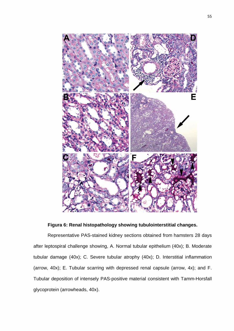
55
Figura 6: Renal histopathology showing tubulointerstitial changes.
Representative PAS-stained kidney sections obtained from hamsters 28 days
after leptospiral challenge showing, A. Normal tubular epithelium (40x); B. Moderate
tubular damage (40x); C. Severe tubular atrophy (40x); D. Interstitial inflammation
(arrow, 40x); E. Tubular scarring with depressed renal capsule (arrow, 4x); and F.
Tubular deposition of intensely PAS-positive material consistent with Tamm-Horsfall
glycoprotein (arrowheads, 40x).

56
4.9 TABLES Table 1. Recombinant LigA Proteins
Proteina Amino acid coordinatesb
MW (Da)
Primersc
LigA7’-13 L631-P1224 63,422 f-AACATATCTCATATGCTTACCGTTTCCAACACAAACGCCAA
r-TTCCTCGAGTGGCTCCGTTTTAATAGAGGCTAAT
LigA7’-11 L631-A1033 42,991 f-AACATATCTCATATGCTTACCGTTTCCAACACAAACGCCAA
r-GACGTCCTCGAGAGCAGAAGTGACATACAAGGTAGTAGA
LigA7’-9 L631-A851 24,034 f-AACATATCTCATATGCTTACCGTTTCCAACACAAACGCCAA
r-AGTCTCGAGCGCTGCGGTAACGGATAATTTGGA
LigA10-13 E852-P1224 40,602 f-ACGCTTACGCATATGGAACTTACTGAGATTGTGCTAAATCC
r-TTCCTCGAGTGGCTCCGTTTTAATAGAGGCTAAT
LigA10-12 E852-E1124 30,085 f-ACGCTTACGCATATGGAACTTACTGAGATTGTGCTAAATCC
r-GCGTAGCTCGAGCTCGTCATTGACGAATATCCA
LigA11-13 R943-P1224 31,233 f-CATCAATGACATATGAGAATAGCTTCAATCGAAGTAACACC
r-TTCCTCGAGTGGCTCCGTTTTAATAGAGGCTAAT
LigA11-12 R943-E1124 20,716 f-CATCAATGACATATGAGAATAGCTTCAATCGAAGTAACACC
r-GCGTAGCTCGAGCTCGTCATTGACGAATATCCA
LigA12-13 V1034-P1224 21,645 f-ATACAGTCTCATATGGTCCTTATTGACATAGAAGTCAAGCC
r-TTCCTCGAGTGGCTCCGTTTTAATAGAGGCTAAT
aLigA protein designations list the first and last domains included in the construct; 7’
indicates a half domain.
bCoordinates refer to the first and last amino acids in the LigA protein of L.
interrogans serovar Copenhageni strain Fiocruz L1-130.
cForward (f) and reverse (r) primer sequences, including an Nde I or Xho I site,
respectively, are listed in the 5’ to 3’ direction.

57
Table 2 – Summary of Immunoprotection Outcomesa
Immunogen
(LigA Domains)
Survivalb
(%)
MAT
(Positives/Total)
Culturec
(Positives/Total)
Histologyc
(Mean Score)
qPCRc,d
(Log10)
Experiment 1
7’-13 100 3/3 4/4 ND 5.79 ± 2.0
7’-9 0* 1/3 4/4 ND 3.92 ± 0.3
7’-11 50 2/4 2/4 ND 3.10** ± 0.8
10-13 100 4/4 4/4 ND 5.92 ± 0.8
12-13 50 0/4 4/4 ND 5.53 ± 1.3
HKL 100 3/3 0/4 ND 2.48** ± 0.6
PBS 0* 0/1 4/4 ND 4.66 ± 1.0
Experiment 2
7’-13 100 4/4 4/4 1.0 4.13 ± 0.2
10-13 100 4/4 4/4 2.5 4.90 ± 1.0
10-12 100 4/4 4/4 3.3** 4.81 ± 0.4
11-13 100 4/4 4/4 1.75 5.61 ± 1.2
11-12 25 4/4 1/4 3.3** 4.29 ± 0.8
HKL 100 3/4 0/4 1.25 2.39** ± 0.2
PBS 0* 3/3 2/4 ND 2.72** ± 0.2
aAbbreviations: HKL = heat-killed leptospires, PBS = phosphate-buffered saline, ND = not
done
bFour animals per group. *Statistically different from the LigA7’-13 group (Fisher’s exact test,
P < 0.05).
cData refer to kidney analysis. Means are shown for Histology and qPCR (n=4).
**Statistically different from the LigA7’-13 group (ANOVA, Dunn post-test, P < 0.05).
dExpressed as copies per microgram of tissue DNA.

58
Table 3 – Chemistry Resultsa
Group (LigA
Protein)
BUNb
(mg/dL) AlkPhosb
(IU/L) Calcium (mg/dL)
Creatinine (mg/dL)
Phosb
(mg/dL) SGPTb
(IU/L) Total
bilirubin (mg/dL)
Total protein (g/dL)
7’-13 21 ± 2 127 ± 8* 13 ± 0.7 0.3 ± 0.1 7.6 ± 0.5 64 ± 11 0.1 ± 0 6.6 ± 0.4
10-13 25 ±5 78 ± 14 12.8± 2.2 0.2 ± 0 6.8 ± 1.5 57 ± 26 0.1 ± 0 5.6 ± 1
10-12 26 ± 6 72 ± 4 11.6± 1.8 0.2 ± 0 6.6 ± 1 39 ± 20 0.1 ± 0 5.3 ± 0.6
11-12 67 ±77 71 ± 18 9.7 ± 1.3 0.8 ± 1.2 7.5 ± 3.3 49 ± 10 0.1 ± 0 4.6 ± 0.6
11-13 22 ± 2 98 ± 4 13± 0.9 0.2 ± 0.1 7.6 ± 0.6 51 ± 16 0.1 ± 0 6.2 ± 0.5
HKLc 23 ±2 73 ± 10 12.5 ± 0.9 0.2 ± 0 6.8 ± 1 42 ± 2 0.1 ± 0 6.2 ±0.5
PBSd 235 ± 49* 130 ± 15* 13.7± 1.4 6.8 ± 1.6* 18.1 ± 1 48 ± 5 0.3 ± 0.1 6.7 ± 0.4
Ref.
range
9-30 15-45 8-12 0.5-2.2 4.2-8.5 10-35 0-1 4.5-6.5
aMeans with standard deviations from Experiment 2 are displayed (n=4). Bold numbers: Significantly
different from group immunized with heat-killed leptospires (one-way ANOVA with Dunn’s post test, P
< 0.05). *Significantly different from group immunized with heat-killed leptospires (one-way ANOVA
with Dunn’s post test, P < 0.01)
bAbbreviations: BUN, blood urea nitrogen; AlkPhos, alkaline phosphatase; Phos, phosphorus; SGPT,
serum glutamic pyruvic transaminase
cHeat-killed leptospires
dPhosphate buffered saline

59
5 Paper 2 – Kinectics of the Hamster Model of Leptospira
interrogans infection
(Text format as used by the scientific journal PLoS Neglected Tropical
Diseases)

60
Kinetics of the Hamster Model of Leptospira interrogans
Infection†
Mariana L. Coutinho*1,2, Long Chieh Wang1, Henry A. Choy 1,3,, James Matsunaga1,3 ,
Jane T. Babbitt 1,3, Michael S. Lewis 1, Jose Antonio G. Aleixo 2 and David A. Haake 1,3,4,5.
1Veterans Affairs Greater Los Angeles Healthcare System, Los Angeles, CA 90073, U.S.A.,
2Centro de Desenvolvimento Tecnologico, Universidade Federal de Pelotas, Pelotas, Brasil,
Departments of 3Medicine and 4Urology, David Geffen School of Medicine at UCLA, and
5Microbiology, Immunology and Molecular Genetics, UCLA, Los Angeles, CA 90095
61
5.1 Abstract
Leptospirosis is a zoonosis caused by Leptospira. The organism penetrates the skin
through lesions or abrasions and rapidly disseminate to the kidney, liver and other organs. In
humans, leptospirosis ranges from asymptomatic to a life-threatening infection characterized
by hepatorenal failure and pulmonary hemorrhage. The intraperitoneal route of infection is
the most widely used to experimentally inoculate hamsters, but it does not represent the
natural route of infection. Here we report the use of more natural routes of infection such as
intradermal or subcutaneous inoculation of leptospires and how the disease progresses
during time. Animals showed hepatic and renal failure that started at day 7 post infection and
a peak of leptospiral burden in the key-organs of leptospirosis at day 6, after which
leptospires tended to remain in the kidneys.

62
5.2 Introduction
Leptospirosis is a zoonosis with worldwide distribution caused by spirochetes
belonging to the genus Leptospira (Haake, 2009). Up to 500,000 severe human infections
are documented annually in tropical areas with an incidence of >10 cases per 100,000
population and a mortality rate of 10% (Hartskeerl et al., 2011). Human infection typically
occurs in persons involved in water-related occupations such a rice–field workers, or
that deal with animals or their carcasses, or recreational activities in humid,
subtropical regions where conditions are favorable for survival of the organism. In
developing countries, often patients get infected by direct exposure to rats or open
sewer. In these areas, transmission is thought to involve cutaneous or mucous
membrane exposure to water contaminated with urine from infected animals (KO,
1999). Rodents are frequently the source of organisms causing human infections, but
many mammalian species have been found to harbor infection in their kidneys
(Levett 2001). Once the organisms reach the bloodstream they rapidly disseminate to
the kidney, liver and other organs. In humans, leptospirosis ranges from
asymptomatic to a life-threatening infection characterized by hepatorenal failure and
pulmonary hemorrhage.
Severe leptospirosis occurs as a dual-phase disease with a leptospiremic
phase in the first week after infection followed by an immune phase during which
antibody is produced and organisms appear in the urine (Levett 2001). Symptoms of
organ dysfunction, such as jaundice and azotemia, are absent during the initial
presentation of infection and typically begin as organisms are cleared from the
bloodstream and the immune phase begins (Levett, 2001).

63
The kinetics of leptospirosis has been described, examining the time to
appearance of organisms in various tissues depending on the challenge route
(Stavitsky 1945). These initial studies primarily involved dark-field observations and
leptospires isolation from experimentally infected guinea pigs and hamsters which
are highly susceptible to infection with virulent leptospires. More recently, quantitative
PCR has been used to measure the leptospiral burden in various organs after
intraperitoneal challenge (Lourdault et al, 2009).
The IP infection has been successfully used to reproduce leptospirosis in
animal models such as hamsters and guinea pigs, and although it has advantages
this is not likely a natural route of infection. Other routes of infection such as
conjuntival, subcutaneously, intradermal, oral, intracardiac and intracranial have
been reported, but somehow the quantification of the leptospiral burden through
these alternate routes of infection remains not well studied. The understanding of
how a natural infection progresses can be used to help the development of new
vaccines and the clinical treatment of leptospirosis.
In this study we report the inoculation of hamsters with intradermal and
subcutaneous routes of infection and the harvesting of the organs at different time-
points to provide a better understanding of the kinetics of leptospiral infection.
5.3 Materials & Methods
Leptospiral strain and cultivation. Leptospira interrogans serovar Copenhageni
strain Fiocruz L1-130 was cultivated in Ellinghausen-McCullough-Johnson-Harris
(EMJH) medium (Haake t al., 2002) supplemented with 1% rabbit serum (Rockland
Immunochemicals, Gilbertsville, PA) and 100 µg/ml 5-fluorouracil at 30°C in a shaker

64
incubator. Leptospiral cultures (passage 2) in log phase of growth were centrifuged
at 5,000g for 5 minutes and diluted in fresh EMJH prior to hamster inoculation.
Hamster infection and sample collection. Groups of three to four female
Syrian hamsters, 5 to 6 weeks of age (Harlan Bioscience, Indianapolis, IN), were
inoculated either subcutaneously (0.5 mL) or intradermally (0.05 mL) with 107
leptospires or EMJH alone on day 0. Animals were euthanized at different time-points
ranging from 3 hours to 12 days post-infection. Hamsters were randomly selected for
euthanasia unless they presented clinical signs of leptospirosis, such as loss of
appetite, gait or breathing difficulty, prostration, ruffled fur, or weight loss of ≥ 10% of
the animal’s maximum weight. Kidney, liver, lungs and spleen were collected in
formalin for histopathology or incubated overnight at 4°C in RNAlater (Ambion,
Austin, TX) and stored at -80°C. Paraffin embedded tissues were sectioned and
stained with periodic acid Schiff (PAS) in a Dako automated slide processor. Kidney
sections were scored on a scale of 0 (normal tissue) to 5 (severe renal tubular
damage) based the severity of histopathology. Blood was collected for serology and
chemical analysis (Antech Diagnostics, Irvine, CA). All animal procedures were
approved by the Veterans Affairs Greater Los Angeles Healthcare System
Institutional Animal Care and Use Committee and adhere to the United States Health
Research Extension Act of 1985 (Public Law 99-158, November 20, 1985, “Animals
in Research”), the National Institutes of Health’s Plan for Use of Animals in Research
(Public Law 103-43, June 10, 1993), U.S. Government Principles for the Utilization
and Care of Veterbrate Animals Used in Testing, Research, and Training, Public
Health Service Policy on Humane Care and Use of Laboratory Animals, the United
States Department of Agriculture’s Animal Welfare Act & Regulations, and Veterans
Health Administration Handbook 1200.7.
65
DNA extraction and Quantitative PCR (qPCR). Tissue DNA was extracted using
either the FastDNA SPIN Kit (MP Biomedicals, Santa Ana, CA) according to the
manufacturer instructions or with DNeasy Blood and Tissue kit (Qiagen, Valencia,
CA) with modifications as previously described (Coutinho, et al. 2011). The purified
DNA was stored at -80°C until use.
DNA was amplified in a PCR using the 16S primers 16sfor, 5‘-
GGCGGCGCGTCTTAAACATG-3’, and 16srev, 5‘-TTCCCCCCATTGAGCAAGATT-
3’, as described before (Lourdault et al., 2009). Two µL of sample DNA was added
to a mix containing forward and reverse primers (0.4 µM), dNTPs (200 µM), PCR
buffer (1x) and Hot Start Taq DNA polymerase (Qiagen, Germantown, MD). The
reaction was performed in a Vapoprotect Thermocycler (Eppendorf) under the
following conditions: initial denaturation at 95°C for 15 min followed by 30 cycles of
amplification (94°C for 30 s, 57°C for 30 s, 72°C for 30 s) and a final cycle of 72°C for
1 min. The amplicons were examined by agarose gel electrophoresis to screen for
Leptospira-positive samples in experiment #1.
PCR-positive samples from experiment #1 and all samples from experiment
#2 were tested by qPCR using the Bio-Rad iQ5 Real-time System (Bio-Rad,
Hercules, CA). One hundred nanograms of total DNA was combined with 1 µM of
each primer and 12.5 µL iQ SYBR Green Supermix (Bio-Rad) and brought to a final
volume of 25 µL with nuclease-free water (Ambion, Austin, TX). Each sample was
run in duplicate. qPCR primer pairs were LipL32-f, 5‘-
CGCGTTACCAGGGCTGCCTT-3’, and LipL32-r, 5’-CGCTTGTGGTGCTTTCGGTG-
3’, and hamster GAPDH-f, 5’-CTGGTTACCAGGGCTGCCTT-3’, and GAPDH-r, 5’-
CCGTTCTCAGCCTTGACTGTGC-3’, the latter being used as a reference gene to
which lipL32 was normalized using Bio-Rad iQ5 software and Microsoft Excel. The

66
resulting amplicons were 152 bp and 146 bp, respectively. The PCR protocol
consisted of an initial incubation step at 95°C for 12.5 min followed by 40 cycles of
amplification (95°C for 15 s, 57°C for 30 s and 72°C for 30 s). Standard curves were
generated for each gene ranging from 10 to 1.6 x 10⁶ copies of Leptospira (20-fold
dilutions) and 0.02 to 200 ng (10-fold dilutions) of hamster DNA.

67
ELISA. Ninety-six-well ELISA microtiter plates (Immulon 4HBX,Thermo Fisher,
Waltham, MA) were coated with 1 x 10⁹ heat-inactivated leptospires/mL diluted in
PBS, pH7.2 (Invitrogen, Carlsbad, CA), by overnight incubation at 4°C as described
previously (Choy et al. 2007). Briefly, the plates were blocked with Protein-Free
Blocking Buffer (PFBB, Thermo Fisher, Rockford, IL) for 1 to 2 h at room temperature
(RT). Sera were tested in triplicate after dilution with PFBB, to wells in a volume of
100 µL, and plates were incubated for 1 h at 37°C. Non-binding antibodies were
removed, and Horseradish peroxidase (HRP)-conjugated anti-Syrian hamster
secondary antibody (Jackson ImmunoResearch, West Grove, PA) 1:5000 was
incubated for 30 min at RT. One hundred µL of 1-Step Turbo Ultra TMB HRP
substrate (Thermo Fisher) was added to the wells and incubated for 30 min at RT
with shaking. The reaction was stopped by the addition of 50 µL of 2 M H2SO4, and
plates were immediately read in a Bio-Rad 550 Microplate Reader at 450 nm. End-
point titers were defined as the highest titer that yielded a reading two standard
deviations above the result with sera from mock-infected hamsters. Geometric mean
end-point titters were calculated as previously described (Perkins, 1958).
Statistics. Differences between variances were analyzed by the f-test. All other
analises were done by GraphPad InStat version 3.10 (GraphPad Software Inc., La
Jolla, CA). One-way analysis of variance (ANOVA) was used to test for differences
between multiple (≥3) groups using a P value < 0.05. The Kruskal-Wallis one-way
ANOVA with Dunn's post-test was included. The unpaired, two-tailed Student’s t-test
assuming unequal variance was used to test for differences between two groups
using a P value < 0.05.
68
5.4 Results
End-point criteria and the infection effect on body weight. All animals were
observed for the presence of clinical signs of disease and, in experiment #1, two
animals (one from each route), succumbed to leptospirosis and were found dead in
their cage on day 12. All other animals were euthanized (three animals/time-
point/route).
In experiment #2 hamster body weight was measured at time of infection and
each day thereafter. Uninfected hamsters gained an average of 3.6% of body weight
per day during the course of the study. As shown in Figure 7, weight gain in infected
hamsters was comparable with uninfected hamsters until the sixth day after
challenge. A decrease in body weight was the earliest observable sign of clinical
leptospirosis and a cumulative 10% weight loss from peak weight was used as an
endpoint criterion. As in our previous study [Coutinho 2011], spontaneous death was
not observed in any animals in which the 10% weight loss criterion was employed.
By day 7 after SQ challenge, 10/13 (77%) hamsters had lost ≥10% of peak weight.
Weight loss among ID challenged hamsters was not as rapid; by day 7 after
challenge, 6/13 (46%) animals had lost ≥10% of peak weight. The difference in
weight loss on day 7 between the hamsters challenged ID vs. SQ was significant (T-
test, P<0.02). Comparing each day with day 0, there is a significant difference
(P˂0.001, Anova with Bonferroni’s multiple comparison test) in both routes that was
noticed from day 6 until the end of the experiment.
Leptospiral burden on organs. The leptospiral burden in kidneys, lungs,
spleen and liver was accessed in both intradermal and subcutaneous route (table 1
and 2).
69
Subcutaneous route. Leptospires were detected in the spleen within 24 to 48
hours after infection and the bacterial load seemed to have peaked on days 5 and 6
and declined afterwards. In both experiments leptospiral burden was only seen from
day 5 on and declined after day 6 or 7. The leptospiral load on lungs was detected on
day 5 and lasted for 2 to 4 days presenting infection levels much lower than spleen.
Although the presence of leptospires was detected very early in liver, it only
rose after 4 days and reached their maximums load on day 6 with a fast decline
afterwards.The most affected organ was kidney, in which the bacteria seems to
colonize fast and showed a much higher load than the other organs, but it also
showed a peak around days 5 and 6 followed by a decline which could explained by
the elimination of leptospires in the urine.
Intradermal route. The spleen of animals infected through the ID route showed
that leptospires took 48 to 72 h to infect the organ with a peak on day 5, with similar
pattern of decline in bacterial load after as observed in the SQ route. It was observed
a difference when routes were compared in both experiments denoting that the SQ
route allows more leptospires to reach the spleen on days 5 (exp #2, P<0.001) and 6
(exp #1, P<0.05) than the ID infection (Parametric ANOVA with Bonferroni post-test).
In exp #1 the lung bacterial load was not possible to be quantified due to low
amounts of bacterial DNA, however exp #2 showed a peak of leptospiral DNA in
lungs on days 5 and 6 with a decline after this low peak. Both routes seemed not to
differ from one another except on day 6 from exp #1(Parametric ANOVA with
Bonferroni post-test, P<0.01).
In a screening PCR on experiment 1 the leptospiral DNA in liver was not
detected and therefore not used in qPCR, experiment #2 however showed very low

70
leptospiral burden with ID route and which was different than the SQ route on day 6
(Parametric ANOVA with Bonferroni post-test, P<0.001). The kidneys were highly
infected, although not as high as the SQ route (Parametric ANOVA with Bonferroni
post-test, P<0.001) with a peak on day 5 followed by a decline that still kept the
leptospiral load much higher than any of the other organs tested.
Blood chemistries. The serum collected at time of euthanasia was
submitted to chemical analysis to evaluate liver and kidney function. Alanine
aminotransferase was a little bit elevated in all groups, and alkaline phosphatase was
extremely elevated on day 6 in both routes (p<0.05) when compared to controls,
pointing to cholestasis without biliary obstruction since total bilirubin level was normal
for all samples except three animals on day 7 of the SQ route which were 2 to 8
times higher than other animals.
Blood urea nitrogen was very elevated on day 7 of both routes (p<0.05) and
day 8 of ID group (p<0.01). Elevated creatinine (>3mg/dL) level has been described
as a prognosis indicator in humans. In hamsters it was observed increased creatinine
in both routes on days 7 and 8 post infection (p<0.05), indicating renal malfunction at
day 7 and 8. Although calcium level was mildly elevated, phosphorous level seem to
have a tendency to increase from day 1 on in both routes, it was statistically different
from controls on days 6 and 7 (p<0.01), suggesting that rhabdomyolysis might have
started early in the disease.
Antibody Response. Serum collected from hamsters at time of euthanasia
were used in an ELISA at a dilution of 1:6400, chosen as the best signal-to-noise
71
ratio (data not shown). The SQ route didn’t show any improvement on antibody level
during the 8 days of experiment, however, in the ID route it was observed an
increase in antibody levels toward days 8 and 9 (Parametric ANOVA with Dunnett
post-test, P<0.01 and P<0.05 respectively). Antibody levels against leptospires in a
natural infection are usually only detected at the end of the first week of symptoms
when the immune phase of the disease begins.
5.5 Discussion
Hamsters is one of most well characterized animal models used in
leptospirosis, unlike rats and mice it shows signs of severe disease. It is not clear
how leptospires distributes itself in the body after infection and how much time it
takes to reach the key organs.
Our results show that the skin plays a major role in preventing the leptospiral
burden that reaches the organs, with a much lower number of copies in the
intradermal route when compared to subcutaneous inoculation. It is worth noting that
the outcome on the body weight and blood analysis was the same in both routes.
While it can be accepted that the leptospiral burden in the kidneys decreases when
the leptospires start to be shed, however it is intriguing that they also decrease in
target organs such as lung and liver at the same time.
Our approach and results are consistent with the report by Lourdault et
al.(2009), which described the kinetics of infection by L. interrogans sv Copenhageni,
strain L1-130, the same strain used here, inoculated into guinea pigs by the
intraperitoneal (i.p.), subcutaneous (s.c.), and conjunctival routes. In that study,
leptospiral burden in the liver, kidneys, and lungs peaked on day six, shortly before
72
guinea pigs succumbed to infection. In contrast, we were able to follow hamsters for
a total of 8-9 days and observed a drop in leptospiral burden during the last 2-3 days.
Lourdault et al.(2009), reported that the kinetics of infection in guinea pigs challenged
via the i.p., s.c., and c.j. routes were similar. A novel aspect of our study was the
comparison of the s.c. and i.d. routes with the finding of significant differences in the
kinetics of infection between these routes. We believe that the i.d. challenge route is
a biologically relevant route of infection and worthy of further study because it has the
potential of incorporating interactions between leptospires and defense mechanisms
of the skin, such as the dendritic cells. Antigen-presentation by dendritic cells has
the potential of “jump-starting” the host immune response at an early stage of
infection.In this study we report the kinectics of the leptospiral infection during the
first phase of the disease by two different routes of infection that are more natural
than intraperitoneal inoculation. The finding that both routes produce the same result
at practically the same time with intradermal injection resulting in a lower leptospiral
burden is very useful in the development of better vaccines and treatment evaluation
strategies.
5.6 Acknowledgements
We thank Karina Gunarto for technical assistance. This study was supported by VA
Medical Research Funds (to D.A.H. and J.M.) , Public Health Service grant AI-
034431 (to D.A.H.) from the National Institute of Allergy and Infectious Diseases and
the Brazilian Conselho Nacional de Pesquisa (process number 201549/2008-5).

73
5.7 References
CHOY, H.A.; KELLEY, M.M.; CHEN, T.L.; MØLLER, A.K.; MATSUNAGA, J.; HAAKE
D.A. (2007) Physiological osmotic induction of Leptospira interrogans adhesion: LigA
and LigB bind extracellular matrix proteins and fibrinogen. Infection and Immunity. 75:2441-2450.
COUTINHO, M.L.; CHOY, H.; KELLEY, M.; MATSUNAGA, J.; BABBITT, J.; LEWIS,
M.; ALEIXO, J.A.G.; HAAKE, D.A. (2011) A LigA Three-Domain Region Protects
Hamsters from Lethal Infection by Leptospira interrogans. PLoS Neglected tropical
Diseases. In press.
HAAKE, D.A.; DUNDOO, M.; CADER, R.; KUBAK, B.M.; HARTSKEERL, R.A.
Leptospirosis, water sports, and chemoprophylaxis. Clinical and Infectious Diseases 34: e40-43. 2002
HAAKE, D.A.; MATSUNAGA, J.; (2010) Leptospira: a spirochaete with a hybrid outer
membrane. Molecular microbiology. 77 (4). 805-814.
HARTSKEERL RA, COLLARES-PEREIRA M, ELLIS WA. Emergence, control and
re-emerging leptospirosis: dynamics of infection in the changing world. Clinical Microbioly Infection 17: 494-501. 2011.
KO, A.I.; REIS, M.G.; DOURADO, C.M.R.; JONHSON, W.D.; RILEY, L.W. (1999)
Urban epidemic of severe leptospirosis in Brazil. Salvador Leptospirosis Study Group
Lancet 354: 820-825. 1999.
LEVETT, P. (2001) Leptospirosis. Clinical Microbiology Reviews 14 (2): 296 – 326.
LOURDAULT, K.; AVIAT, F.; PICARDEAU, M. (2009) Use of quantitative real-time
PCR for studying the dissemination of Leptospira interrogans in the guinea pig
infection model of leptospirosis. Journal of Medical Microbiology, 58, 648–655.

74
MATSUNAGA, J.; MEDEIROS, M.A.; SANCHEZ, Y.; WERNEID, K.F.; KO, A.I.
(2007) Osmotic regulation of expression of two extracellular matrix-binding proteins
and a haemolysin of Leptospira interrogans: differential effects on LigA and Sph2
extracellular release. Microbiology 153: 3390-3398.
Perkins FT (1958) A ready reckoner for the calculation of geometric mean antibody
17 titres. J Gen Microbiol 19: 540-541.
STAVISTSKY, A.B.(1945) Studies on the pathogenesis of leptospirosis. The journal of Infectious Diseases. v. 76 (3), p.179- 192.

75
5.8 Figures
Figura 7: Hamster weight as a marker for disease progression.
Animals were weighed at the time of challenge (day zero) and daily thereafter.
Data are shown for experiment #2. Bars represent weight relative to day zero for
animals challenged subcutaneously (red), intradermally (blue), or the control group
(green). Infected animals gained weight until day six, after which they progressively
lost weight.
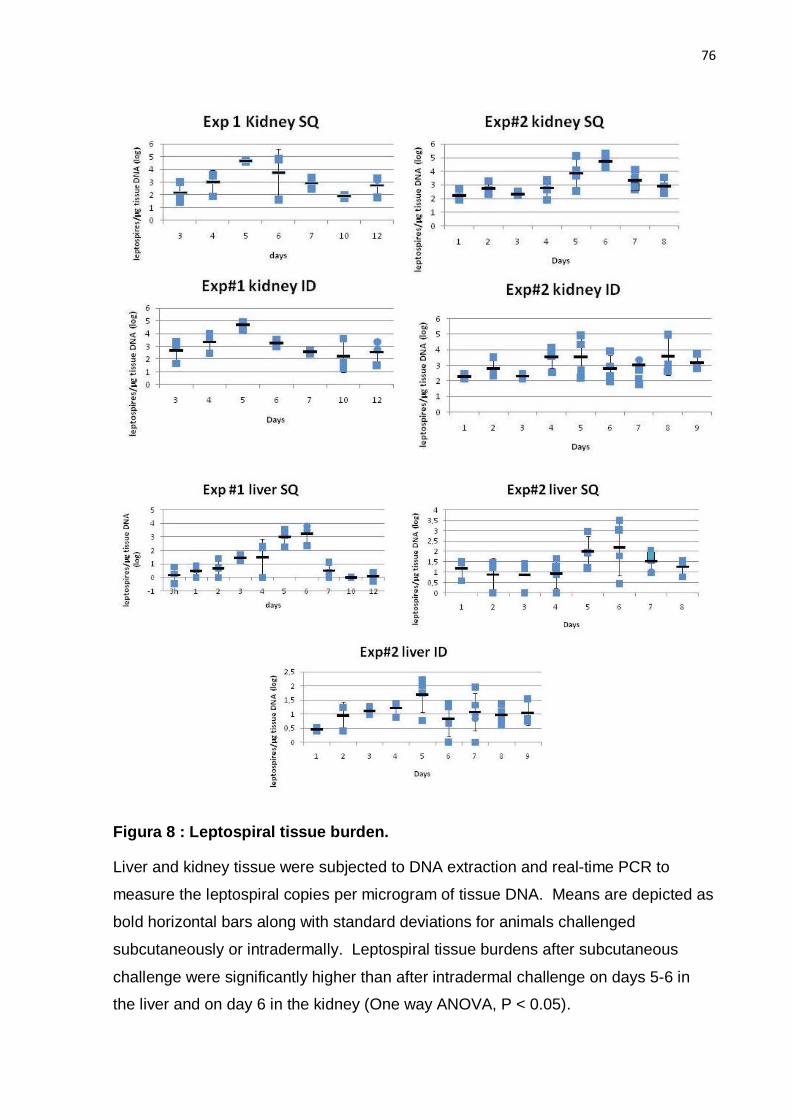
76
Figura 8 : Leptospiral tissue burden.
Liver and kidney tissue were subjected to DNA extraction and real-time PCR to
measure the leptospiral copies per microgram of tissue DNA. Means are depicted as
bold horizontal bars along with standard deviations for animals challenged
subcutaneously or intradermally. Leptospiral tissue burdens after subcutaneous
challenge were significantly higher than after intradermal challenge on days 5-6 in
the liver and on day 6 in the kidney (One way ANOVA, P < 0.05).

77
Figura 9: Kinetics of liver and kidney function
Mean and standard deviations are shown for blood samples obtained from
hamsters challenged intradermally. Markers of liver function (alkaline phosphatase
and SGPT) peaked on day 6 after challenge. Markers of kidney function (serum
creatinine and blood urea nitrogen) became abnormal on day 6 and peaked on day
8. Results for hamsters challenged subcutaneously were similar.

78
Figura 10: Antibody response in infected hamsters
Hamster immunoglobulin responses to heat-killed leptospires were measured by
ELISA. Mean and standard deviations are shown for serum samples (tested in
triplicate at a dilution of 1:6400) obtained from hamsters challenged intradermally
(red) or subcutaneously (blue). Dotted gray line represents two standard deviations
above background ELISA for sera from six uninfected control hamsters. Anti-
leptospiral antibody levels in sera from hamsters challenged intradermally were
higher than those from control hamster and hamsters challenged subcutaneously
beginning on day 8 after challenge (P < 0.05).

79
Table 1 – Leptospiral burden in various organs Exp# 1
Table 1: Kinectics of leptospiral infection in experiment #1. Numbers represent
the average of three animals at each time-point. Bold numbers depict statistical
difference between routes within the same day.
Time-points
Organ/ Experiment (mean ± SD)*
Days Spleen Lungs Liver Kidneys
SQ ID SQ ID SQ ID SQ ID
3h < 20
1 <20 < 20 < 20
22.1 (± 16.0)
233.7 (± 234.4)
2 < 20 252.9 (± 118.3)
< 20 < 20 23.4 (± 7.8)
849.4 (± 895.9)
3 < 20 242.8 (± 197,2)
< 20 < 20 30.5 (± 13.5)
21.9 (± 7,9) 383.8 (± 552.6)
231.2 (± 72.0)
4 86.2 (± 100.8)
958.7 (± 1,772.6)
< 20 < 20 126.4 (± 112.2)
23.2 (± 19.2)
2,418.1 (± 2,036.2)
1,174.3 (± 1,107.1)
5 8,28.8 (± 556.4)
1,535.9 (± 2,867.3)
123.1 (± 137.1)
110.6 (± 8.8)
1,678.3 (± 197.2)
279.2 (± 427.3)
49,538.0 (± 8,176)
36,306.0 (± 61,300.8)
6 2,939.0 (± 2,801.5)
164.2 (± 238,0)
396.4 (± 292.3)
398.5 (± 514.7)
3,399.1 (± 233.6)
1,126.3 (± 1,550.5)
43,885.4 (± 37,983)
82,082.6 (± 78,750.9)
7 90.6 (± 34.2)
109.6 (± 131.8)
< 20 236.8 (± 322.2)
< 20 47.8 (± 33.9)
1,095.1 (± 916)
3,627.9 (± 3,930.5)
8
64.5 (± 58.3)
127.5 (±
10.9)
24.6 (± 16.1)
1,523.2 (± 1,812.3)
10 187 (± 316.6)
< 20
81.6 (± 21.8)
12
< 20
1,149.4 (± 952.5)
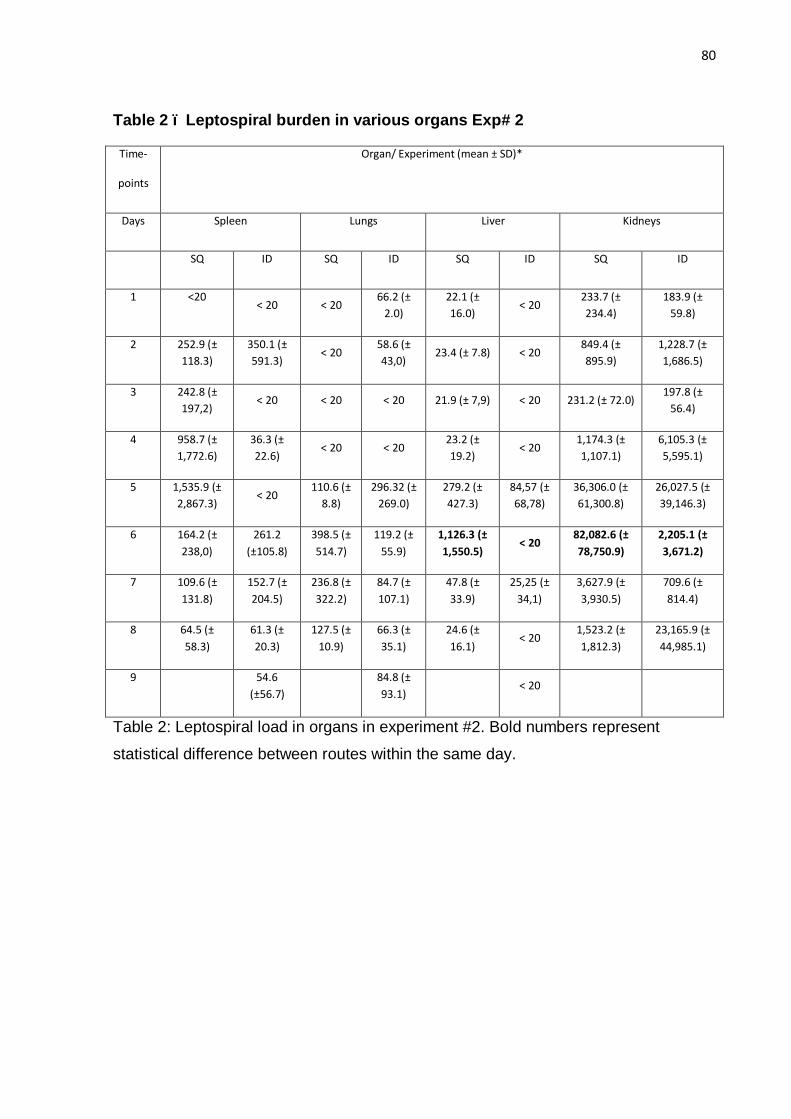
80
Table 2 – Leptospiral burden in various organs Exp# 2
Time-
points
Organ/ Experiment (mean ± SD)*
Days Spleen Lungs Liver Kidneys
SQ ID SQ ID SQ ID SQ ID
1 <20 < 20 < 20
66.2 (± 2.0)
22.1 (± 16.0)
< 20 233.7 (± 234.4)
183.9 (± 59.8)
2 252.9 (± 118.3)
350.1 (± 591.3)
< 20 58.6 (± 43,0)
23.4 (± 7.8) < 20 849.4 (± 895.9)
1,228.7 (± 1,686.5)
3 242.8 (± 197,2)
< 20 < 20 < 20 21.9 (± 7,9) < 20 231.2 (± 72.0) 197.8 (±
56.4)
4 958.7 (± 1,772.6)
36.3 (± 22.6)
< 20 < 20 23.2 (± 19.2)
< 20 1,174.3 (± 1,107.1)
6,105.3 (± 5,595.1)
5 1,535.9 (± 2,867.3)
< 20 110.6 (±
8.8) 296.32 (±
269.0) 279.2 (± 427.3)
84,57 (± 68,78)
36,306.0 (± 61,300.8)
26,027.5 (± 39,146.3)
6 164.2 (± 238,0)
261.2 (±105.8)
398.5 (± 514.7)
119.2 (± 55.9)
1,126.3 (± 1,550.5)
< 20 82,082.6 (± 78,750.9)
2,205.1 (± 3,671.2)
7 109.6 (± 131.8)
152.7 (± 204.5)
236.8 (± 322.2)
84.7 (± 107.1)
47.8 (± 33.9)
25,25 (± 34,1)
3,627.9 (± 3,930.5)
709.6 (± 814.4)
8 64.5 (± 58.3)
61.3 (± 20.3)
127.5 (± 10.9)
66.3 (± 35.1)
24.6 (± 16.1)
< 20 1,523.2 (± 1,812.3)
23,165.9 (± 44,985.1)
9
54.6 (±56.7)
84.8 (± 93.1)
< 20
Table 2: Leptospiral load in organs in experiment #2. Bold numbers represent
statistical difference between routes within the same day.
81
5.9 Conclusões
5.9.1 Paper 1
• A região minima requerida para proteção contra desafio letal de Leptospira interrogans inclui os domínios 11 e 12 sendo necessária a inclusão de um terceiro domínio que pode ser tanto a região 10 quanto a 13.
• Os hamsters sobrevivem apesar de apresentarem uma alta carga microbiana renal;
• O acompanhamento do peso e observância da sua perda pode ser utilizada como auxiliar na determinação de ponto-final em experimentos que usam hamsters como modelo experimental
5.9.2 Paper 2
• As leptospiras são rapidamente disseminadas no organismo e alcançam densidades máximas 6 dias após a infecção de hamsters SC ou ID;
• A rota intradérmica de inoculação prove o mesmo resultado que a rota subcutânea em hamsters em termos de sinais clinicos e cinética da infecção, mas com uma carga microbiana que atinge os órgãos muito menor.

82
6 Referências
ADLER, B.; MONCTEZUMA, A.P. Leptospira and leptospirosis. Veterinary Microbiology 140 287–296. 2010.
ATZINGEN, M.V.; BARBOSA, A.S.; DE BRITO, T.; VASCONCELLOS, S.A;
DEMORAIS, Z.M.; LIMA, M; ABREU, P.A.; NASCIMENTO, A.L. Lsa 21, a novel
leptospiral protein binding adhesive Matrix molecules present during human infection.
BMC Microbiol. 29, 8-70. (2008).
BARBOSA, A.S; ABREU, P.A.; NEVES, F.O.; ATZINGEN, M.V.; WATANABE, M.M.;
VIEIRA, M.L.; MORAIS, Z.M.; VASCONCELLOS, S.A.; NASCIMENTO, A.L. A newly
identified leptospira adhesin mediates attachment to laminin. Infection and Immunity. 11, 6356-6364. (2006).
BARNETT, J. K.; BARNETT, D.; BOLIN, C.A.; SUMMERS, T.A.; WAGAR; HEVILLE,
N.F.; HARTSKEERL N.A.; HAAKE, D.A. Expression and distribution of leptospiral
outer membrane components during renal infection of hamsters. Infection and Immunity. 67:853–861. 1999.
BRANGER, C., SONRIER, C., CHATRENET, B., KLONJKOWSKI, B., RUVOEN-
CLOUET, N., AUBERT, A., ANDRE-FONTAINE, G., ELOIT, M. Identification of the
hemolysis-associated protein 1 as a cross-protective immunogen of Leptospira
interrogans by adenovirus-mediated vaccination. Infection and Immunity 69, 6831–
6838. 2001.
BRANGER, C., CHATRENET, B., GAUVRIT, A., AVIAT, F., AUBERT, A., BACH,
J.M., ANDRE-FONTAINE, G., Protection against Leptospira interrogans sensu lato
challenge by DNA immunization with the gene encoding hemolysin-associated
protein 1. Infection and Immunity 73, 4062– 4069. 2005.
BRENNER, D. J.; KAUFMANN, A. F.; SULZER, K. R.; STEIGERWALT, A. G.;
ROGERS, F. C.; WEYANT, R. S.. Further determination of DNA relatedness
between serogroups and serovars in the family Leptospiraceae with a proposal for
83
Leptospira alexanderi sp. nov. and four new Leptospira genomospecies. International Journal of Systematic Bacteriology, 49: 839–858. 1999
BROWN, R.A.; BLUMERMAN, S.; GAY, C.; BOLIN, C.; DUBY, R.; BALDWIN, C.L.
Comparison of three different leptospiral vaccines for induction of a type 1 immune
response to Leptospira borgpetersenii serovar Hardjo. Vaccine 21 4448–4458. 2003.
BULACH, D.M.; ZUERNER, R.L.; WILSON, P.; SEEMANN, T.; MCGRATH, A.;
CULLEN, P.A.; DAVIS, J.; JOHNSON, M.; KUCZEK, E.; ALT, D.P.; PETERSON-
BURCH, B.; COPPEL, R.L.; ROOD, J.I.; DAVIES, J.K.; ADLER, B. Genome
reduction in Leptospira borgpetersenii reflects limited transmission potential.
Proceedings of the National Academy of Sciences 103, 14560–14565.2006.
CENTERS FOR DISEASE CONTROL AND PREVENTION . Outbreak of acute
febrile illness among athletes participating in triathlons – Wisconsin and Illinois.
Morbidity and Mortality Weekly Report 47: 585-588. 1998.
CHOY, H.A.; KELLEY, M.M.; CHEN, T.L.; MØLLER, A.K.; MATSUNAGA, J.; HAAKE
D.A. Physiological osmotic induction of Leptospira interrogans adhesion: LigA and
LigB bind extracellular matrix proteins and fibrinogen. Infection and Immunity
75:2441-2450. 2007.
CHOY, H.A.; KELLEY, M.M.; CRODA, J.; MATSUNAGA, J.; BABBITT, J.T.; The
multifunctional LigB adhesin binds homeostatic proteins with potential roles in
cutaneous infection by pathogenic Leptospira interrogans. PLoS ONE 6: e16879.
2011.
CINCO, M., AND E. BANFI. Interactions between human polymorphonuclear
leukocytes and one strain of pathogenic Leptospira (L. interrogans sp.) and one of
saprophytic Leptospira (L. biflexa sp.). FEMS Microbiology Letters 19:51–54. 1983.
CINCO, M. New insights into the pathogenicity of leptospires: evasion of host
defences. NEW MICROBIOLOGICA, 33, 283-292, 2010
CULLEN, P.A.; HAAKE, D.A.; BULACH, D.M.; ZUERNER, R.L.; ADLER, B.. LipL21

84
is a novel surface-exposed lipoprotein of pathogenic Leptospira species. Infection and Immunity 71 (5): 2414 - 2421. 2003.
CULLEN, P.; HAAKE, D.; ADLER, B.. Outer membrane proteins of pathogenic
spirochetes. FEMS Microbiology Reviews 28(3): 291 – 318. 2004.
CULLEN, P.A.; XU, X.; MATSUNAGA, J.; SANCHEZ,Y.; KO, A.I.; HAAKE, D.;
ADLER, B. . Surfaceome of Leptospira spp. Infection and Immunity 73 (8): 4853 –
4863. 2005.
DOLHNIKOFF, M.; MAUAD, T.; BETHLEM, E.P.; CARVALHO, C.R.R. Pathology and
Pathophysiology of Pulmonary Manifestations in LeptospirosiS The Brazilian Journal of Infectious Diseases;11(1):142-148. 2007
ELLIS, W.A.; Bovine leptospirosis in the tropics: Prevalence, pathogenesis and
control. 1984
FAISAL SM, YAN W, MCDONOUGH SP, CHANG YF. Leptospira immunoglobulin-
like protein A variable region (LigAvar) incorporated in liposomes and PLGA
microspheres produces a robust immune response correlating to protective immunity.
Vaccine 27: 378-387. 2009
FAINE, S.; ADLER,B.; BOLIN,C.; PEROLAT, P. Leptospira and Leptospirosis, 2a
ed.. Melbourne: MediSc, 1999.
FIGUEIRA, C.P.; CRODA,. J.; CHOY, H.; HAAKE,D.A.; REIS, M.G.; KO.A.I.;
PICARDEAU, M.; Heterologous expression of pathogen-specific genes ligA and ligB
in the saprophyte Leptospira biflexa confers enhanced adhesion to cultured cells and
fibronectin. BMC Microbiology. 11: 129. 2011
GOLLOP, J. H.; KATZ, A. R.; RUDOY, R. C.; SASAKI, D. M. Rat-bite leptospirosis.
West. J. Med. 159:76–77. 1993.
HAAKE, D.A., MAZEL, M.K., MCCOY, A.M., MILWARD, F., CHAO, G.,
MATSUNAGA, J., WAGAR, E.A. Leptospiral outer membrane proteins OmpL1 and

85
LipL41 exhibit synergistic immunoprotection. Infection and Immunity 67, 6572–
6582. ., 1999.
HAAKE, D.A.; CHAO, G.; ZUERNER, R.L.; BARNETT, J.K.; BARNETT, D.; MAZEL,
M.; MATSUNAGA, J.; LEVETT, P.N.; BOLIN, C. A.. The Leptospiral Major Outer
Membrane Protein LipL32 is a Lipoprotein Expressed During Mammalian Infection.
Infection and Immunity 68: 2276-2285. 2000.
HAAKE, D.A.; MATSUNAGA, J.; Leptospira: a spirochaete with a hybrid outer
membrane. Molecular microbiology. 77 (4). 805-814. 2010.
HARTSKEERL, R.A.; COLLARES-PEREIRA, M.; ELLIS, W.A. Emergence, control
and re-emerging leptospirosis: dynamics of infection in the changing world. Clin Microbiology Infect 17: 494-501. 2011
HE, H.J.; WANG, W.Y.; WU, Z.D.; LV, Z.Y.; LI, J. TAN, L.Z. Protection of guinea
pigs against Leptospira interrogans serovar Lai by LipL21 DNA vaccine. Cell MolImmunol. 5(5):385-91. 2008
HOKE, D.E.; EGAN, S.; CULLEN, P.A.; ADLER, B.; LipL32 Is an extracellular matrix-
interacting protein of Leptospira spp. and Pseudoalteromonas tunicata. Infection and Immunity 76, 2063–2069. 2008.
KO, A.I.; REIS, M.G.; DOURADO, C.M.R.; JONHSON, W.D.; RILEY, L.W.; Urban
epidemic of severe leptospirosis in Brazil. Salvador Leptospirosis Study Group
Lancet 354: 820-825. 1999.
LEVETT, P. Leptospirosis. Clinical Microbiology Reviews 14 (2): 296 – 326. 2001.
LIN, Y.P.; CHANG, Y.F. A domain of the Leptospira LigB contributes to high affinity
binding of fibronectin. Biochem Biophys Res Commun, 362:443-448. 2007.

86
LIN ,Y.P.; LEE, D.W.; MCDONOUGH, S.P.; NICHOLSON, L.K.; SHARMA, Y.;
CHANG, Y.F. Repeated domains of leptospira immunoglobulin-like proteins interact
with elastin and tropoelastin. J BiolChem, 284:19380-19391. 2009
LIN, Y.P.; CHANG, Y.F. The C-terminal variable domain of LigB from Leptospira
mediates binding to fibronectin. J Vet Sci, 9:133-144. 2008
LIN, Y.P.; GREENWOOD, A.; YAN, W.; NICHOLSON, L.K.; SHARMA, Y.;
MCDONOUGH, S.P.;CHANG, Y.F. A novel fibronectin type III module binding motif
identified on C-terminus of Leptospira immunoglobulin-like protein, LigB. Biochem Biophys Res Commun, 389:57-62. 2009
LUZZI, G. A.; MILNE, L. M.; S. A. WAITKINS. Rat-bite acquired leptospirosis. J. Infect. 15:57–60. 1987.
MATSUNAGA, J.; BAROCCHI, M.A.; CRODA, J.; YOUNG, T.A.; SANCHEZ, Y.;
SIQUEIRA, I.; BOLIN, C.A.; REIS, M.G.; RILEY, L.W.; HAAKE, D.A.; KO, A.I.
Pathogenic Leptospira species express surface-exposed proteins belonging to the
bacterial immunoglobulin superfamily. Molecular Microbiology 49(4): 929–945.
2003.
MATSUNAGA, J.; SANCHEZ, Y.; XU, X.; HAAKE, D.. Osmolarity, a key
environmental signal controlling expression of leptospiral proteins Lig A and Lig B
and the extracellular release of Lig A. Infection and Immunity 73 (1) 70 – 78. 2005.
MATSUNAGA, J.; MEDEIROS, M.A.; SANCHEZ, Y.; WERNEID, K.F.; KO, A.I.
Osmotic regulation of expression of two extracellular matrix-binding proteins and a
haemolysin of Leptospira interrogans: differential effects on LigA and Sph2
extracellular release. Microbiology 153: 3390-3398. 2007
MERIEN, F.; BARANTON, G.; PEROLAT, P. Invasion of Vero cells and induction of
apoptosis in macrophages by pathogenic Leptospira interrogans are correlated with
virulence. Infection and Immunity 65, 729–738. 1997.

87
MONAHAN, A.V.; CALLANAN, J.J.; NALLY, J.E. Proteomic Analysis of Leptospira
interrogans Shed in Urine of Chronically Infected Hosts. Infection and Immunity. 76
(11): 4952. 2008.
NALLY, J.E.; CHOW, E.; FISHBEIN, M.C.; BLANCO, D.R.; LOVETT, M.A. Changes
in lipopolysaccharide O antigen distinguish acute versus chronic Leptospira
interrogans infections. Infection and Immunity. 73 (6) 7014-7. 2005.
PALANIAPPAN, R.U.M.; CHANG, Y.; JUSUF, S.S.D.; ARTIUSHIN, S.; TIMONEY,
J.F.; MCDONOUGH, S.P.; BARR, S.C.; DIVERS, T. J.; SIMPSOM, K. W.;
MCDONOUGH, P.L.; MOHAMMED, H.O.. Cloning and molecular characterization of
an immunogenic Lig A protein of Leptospira interrogans. Infection and Immunity 70
(11): 5924 – 5930. 2002.
PINNE, M.; CHOY, H.A.; HAAKE, D.A. The OmpL37 Surface-Exposed Protein Is
Expressed by Pathogenic Leptospira during Infection and Binds Skin and Vascular
Elastin.PLoSNegl Trop Dis 4(9): e815. 2010
PINNE , M.; HAAKE, D.A. A Comprehensive Approach to Identification of Surface-
Exposed, Outer Membrane-Spanning Proteins of Leptospira interrogans. PLoS ONE
4(6): e6071. 2009
RISTOW, P.; BOURHY, P.; MCBRIDE, F.W.; FIGUEIRA, C.P.; HUERRE, M.; AVE,
P.;GIRONS, I.S.; KO, A.I.; PICARDEAU, M.;. The OmpA-Like protein Loa22 Is
Essential for leptospiral virulence.PLoSPathogogens 3, e97. 2007
SEIXAS, F.K., DA SILVA, E´ .F., HARTWIG, D.D., CERQUEIRA, G.M., AMARAL, M.,
FAGUNDES, M.Q., DOSSA, R.G., DELLAGOSTIN, O.A. Recombinant
Mycobacterium bovis BCG expressing the LipL32 antigen of Leptospira interrogans
protects hamsters from challenge. Vaccine 26, 88–95. ., 2007.

88
SEJVAR, J; BANCROFT, E.; WINTHROP, K.; BETTINGER, J.; BAJANI, M.; BRAGG,
S.; SHUTT, K.; KAISER, R.; MARANO, N.; POPOVIC, T.; TAPPERO, J.; ASHFORD,
D.; MASCOLA, L.; VUGIA, D.; PERKINS, B.; ROSENSTEIN, N.; ECO-CHALLENGE
INVESTIGATION TEAM. Leptospirosis in “Eco-Challenge”athletes, Malaysian
Borneo, 2000. Emerging Infectious Diseases 9 (6): 702 – 7. 2003.
SILVA, E.F.; SANTOS, C.S.; ATHANAZIO, D.A.; SEYFFERT, N.; SEIXAS F.K.,
CERQUEIRA, G.M.; FAGUNDES, M.Q.; BROD, C.S.; REIS, M.G.; DELLAGOSTIN,
O.A.; KO, A.I. Characterization of virulence of Leptospira isolates in a hamster
model. Vaccine. 26(31):3892-6. 2008
SILVA, E´ .F., MEDEIROS, M.A., MCBRIDE, A.J.A., MATSUNAGA, J., ESTEVES,
G.S., RAMOS, J.G.R., SANTOS, C.S., CRODA, J., HOMMA, A., DELLAGOSTIN,
O.A., HAAKE, D.A., REIS, M.G., KO, A.I. The terminal portion of leptospiral
immunoglobulin-like protein LigA confers protective immunity against lethal infection
in the hamster model of leptospirosis. Vaccine 25, 6277–6286. 2007.
SPICHLER, A.S.; VILAÇA, P.J.; ATHANAZIO, D.A.; ALBUQUERQUE, J.O.M.;
BUZZAR, M.; CASTRO, B.; SEGURO, A.; VINETZ, J.M. Predictors of lethality in
severe leptospirosis in urban Brazil. Am. J. Trop. Med. Hyg. 79(6): 911 – 914. 2008.
SRIKRAM, A.; WONGRATANACHEEWIN, S.; PUAPAIROJ, S.; WUTHIEKANUN,
V.; SERMSWAN, R.W. Analyses of Vaccination Protocols for Leptospira interrogans
Serovar Autumnalis in Hamsters. Am. J. Trop. Med. Hyg., 79(5), 779–786. 2008.
SRIKRAM, A.; ZHANG, K.; BARTPHO,T.; LO,M.; HOKE,D.E.; SERMSWAN,R.W.;
ADLER, B.; MURRAY, G.L. Cross-protective Immunity Against Leptospirosis Elicited
by a Live, Attenuated Lipopolysaccharide Mutant. The Journal of Infectious Diseases;1–10. 2011
STAVISTSKY, A.B.. Studies on the pathogenesis of leptospirosis. The journal of Infectious Diseases. v. 76 (3), p.179- 192, 1945.

89
STEVENSON, B., CHOY, H.A., PINNE, M., ROTONDI, M.L., MILLER, M.C.,
DEMOLL, E., KRAICZY, P., COOLEY, A.E., CREAMER, T.P., SUCHARD, M.A.,
BRISSETTE, C.A., VERMA, A., HAAKE, D.A., Leptospira interrogans endostatin-like
outer membrane proteins bind host fibronectin, laminin and regulators of
complement. PLoS ONE 2, e1188. 2007.
VERMA, A.; BRISSETTE, C.A.; BOWMAN, A.A.; SHAH, S.T.; ZIPFEL, P.F.;
STEVENSON, B. Leptospiral endostatin-like protein Len A is a bacterial cell-surface
receptor for human plasminogen. Infect. Immun. (2010).
WORLD HEALTH ORGANIZATION Human Leptospirosis: Guidance for Diagnosis, Surveillance and Control. Malta, WHO Library ; 2003 (2004).