Embed Size (px)
Citation preview
UNIVERSIDADE FEDERAL DE PERNAMBUCO
LABORATÓRIO DE IMUNOPATOLOGIA KEIZO ASAMI
PROGRAMA DE PÓS GRADUAÇÃO EM BIOLOGIA APLICADA À SAÚDE
AURENICE ARRUDA DUTRA DAS MERCÊS
FRACIONAMENTO DE PROTEÍNAS PLASMÁTICAS COM EMPREGO DE
HEPARINA IMOBILIZADA EM SUPORTES MAGNÉTICOS
Recife,
2016
AURENICE ARRUDA DUTRA DAS MERCÊS
FRACIONAMENTO DE PROTEÍNAS PLASMÁTICAS COM EMPREGO DE
HEPARINA IMOBILIZADA EM SUPORTES MAGNÉTICOS
Dissertação apresentada à Universidade Federal
de Pernambuco como parte dos requisitos para
obtenção do título de mestre em Biologia
Aplicada à Saúde.
Área de Concentração: Ciências Biológicas I
Orientador: Prof. Dr. Luiz Bezerra de Carvalho
Júnior
Co-orientadora: Prof.ª Dra. Jackeline da Costa
Maciel
Recife,
2016
UNIVERSIDADE FEDERAL DE PERNAMBUCO
PROGRAMA DE PÓS GRADUAÇÃO EM BIOLOGIA APLICADA À SAÚDE
Parecer da comissão examinadora da dissertação de mestrado de:
AURENICE ARRUDA DUTRA DAS MERCÊS
FRACIONAMENTO DE PROTEÍNAS PLASMÁTICAS COM EMPREGO DE
HEPARINA IMOBILIZADA EM SUPORTES MAGNÉTICOS
Aprovado em: 22/02/2016
Prof. Dr. Luiz Bezerra de Carvalho Júnior
Orientador – Membro interno
Prof. Dr. Ian Porto Gurgel do Amaral
Membro externo
Prof.ª Dra. Sinara Mônica Vitalino de Almeida
Membro suplente externo

Aos meus pais: João Bosco e Socorro, ao meu irmão João
Bosco Filho e ao meu namorado Taciano, pelo incentivo, apoio
e amor incondicional.
Dedico.
AGRADECIMENTOS
A Deus por financiar a minha sabedoria, me dar força interior para superar as dificuldades,
mostrar os caminhos nas horas incertas e me suprir em todas as minhas necessidades.
Aos meus amados pais João Bosco e Maria do Socorro e irmão João Bosco Filho, minha
família, pela dedicação, confiança, paciência, apoio incondicional e incentivo aos estudos
principalmente por acreditarem em meu potencial realizando esforços para que eu pudesse
chegar até aqui.
Ao meu namorado, companheiro e amigo, Taciano, pelo amor, carinho e compreensão ao me
escutar; por estar sempre presente ao meu lado, compartilhando momentos de alegria e me
incentivando a seguir em frente e não desistir dos meus objetivos.
Ao meu queridíssimo orientador e “Pai Científico”, Prof. Luiz Carvalho, pelas oportunidades
acadêmicas e momentos descontraídos desde a iniciação científica sempre com imensa
confiança, dedicação e carinho. Obrigada por acreditar em mim.
A minha co-orientadora e amiga Prof. Dra. Jackeline Maciel, por me incentivar sempre a
buscar meu crescimento pessoal e profissional, pela paciência, cumplicidade e amizade
sincera.
Aos meus companheiros dos grupos IMOBIO e BMC, em especial aos queridos: Gabriela
Ayres, Igor, Luiza, Sinara, Lúcia e Mariana pelas trocas de experiência e boa amizade.
Aos meus queridíssimos e amados amigos da turma do mestrado: Andriu Catena, Amanda
Quintino, Romério Alencar e Priscila Leão pelos momentos de aprendizado e alegria durante
o convívio dentro e fora do laboratório.
Aos meus companheiros científicos do Departamento de Física, os doutorandos Karciano e
Davian, por me proporcionar uma boa amizade, troca de experiências e aprendizado
multidisciplinar.
A todos os meus professores que durante o mestrado proporcionaram conhecimentos e
experiências acadêmicas. Meus Sinceros agradecimentos em especial aos queridos
Professores: Luiz Carvalho, Eduardo Beltrão, José Luiz e Gustavo Nascimento.
A todos os funcionários do LIKA pelo apoio e suporte, em especial aos queridos Fábio
Constantino e o Senhor Otaviano.
A minha queridíssima amiga e Professora Monica Albuquerque, que desde a graduação
durante o grupo PET-Parasitologia, me proporciona momentos de aprendizado,
descontração, alegria e amizade sincera.
Aos queridíssimos amigos que conheci durante minha vida acadêmica, desde a graduação,
que levarei sempre em minha vida: Karla Ribeiro, Ricardo Souza, Raquel Varela, Joicy
Kelly, Taciana Higino, Sílvia Guedes e Renata Vieira. Vocês estão em meu coração!

“Eu nunca vejo o que já foi feito. Eu somente
vejo o que ainda falta para ser feito.”
Marie Curie
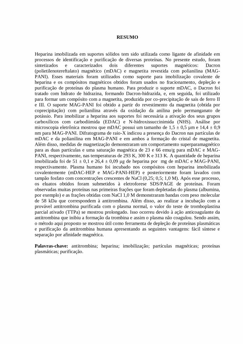
RESUMO
Heparina imobilizada em suportes sólidos tem sido utilizada como ligante de afinidade em
processos de identificação e purificação de diversas proteínas. No presente estudo, foram
sintetizados e caracterizados dois diferentes suportes magnéticos: Dacron
(polietilenotereftalato) magnético (mDAC) e magnetita revestida com polianilina (MAG-
PANI). Esses materiais foram utilizados como suporte para imobilização covalente de
heparina e os compósitos magnéticos obtidos foram usados no fracionamento, depleção e
purificação de proteínas do plasma humano. Para produzir o suporte mDAC, o Dacron foi
tratado com hidrato de hidrazina, formando Dacron-hidrazida, e, em seguida, foi utilizado
para formar um compósito com a magnetita, produzida por co-precipitação de sais de ferro II
e III. O suporte MAG-PANI foi obtido a partir do revestimento da magnetita (obtida por
coprecipitação) com polianilina através da oxidação da anilina pelo permanganato de
potássio. Para imobilizar a heparina aos suportes foi necessária a ativação dos seus grupos
carboxílicos com carbodiimida (EDAC) e N-hidroxissuccinimida (NHS). Análise por
microscopia eletrônica mostrou que mDAC possui um tamanho de 1,5 ± 0,5 µm e 14,4 ± 0,9
nm para MAG-PANI. Difratograma de raio-X indicou a presença do Dacron nas partículas de
mDAC e da polianilina em MAG-PANI e em ambos a formação do cristal de magnetita.
Além disso, medidas de magnetização demonstraram um comportamento superparamagnético
para as duas partículas e uma saturação magnética de 23 e 66 emu/g para mDAC e MAG-
PANI, respectivamente, nas temperaturas de 293 K, 300 K e 313 K. A quantidade de heparina
imobilizada foi de 51 ± 0,1 e 26,4 ± 0,09 μg de heparina por mg de mDAC e MAG-PANI,
respectivamente. Plasma humano foi incubado nos compósitos com heparina imobilizada
covalentemente (mDAC-HEP e MAG-PANI-HEP) e posteriormente foram lavados com
tampão fosfato com concentrações crescentes de NaCl (0,25; 0,5; 1,0 M). Após esse processo,
os eluatos obtidos foram submetidos à eletroforese SDS/PAGE de proteínas. Foram
observadas muitas proteínas nas primeiras frações que foram depletadas do plasma (albumina,
por exemplo) e as frações obtidas com NaCl 1,0 M demonstraram bandas com peso molecular
de 58 kDa que correspondem à antitrombina. Além disso, ao realizar a incubação com a
provável antitrombina purificada com o plasma normal, o valor do teste de tromboplastina
parcial ativado (TTPa) se mostrou prolongado. Isso ocorreu devido à ação anticoagulante da
antitrombina que inibiu a formação da trombina e assim o plasma não coagulou. Sendo assim,
o método aqui proposto se mostrou útil como ferramenta de depleção de proteínas plasmáticas
e purificação da antitrombina humana apresentando as seguintes vantagens: fácil síntese e
separação por afinidade magnética.
Palavras-chave: antitrombina; heparina; imobilização; partículas magnéticas; proteínas
plasmáticas; purificação.
ABSTRACT
Heparin immobilized on solid supports has been used as a ligand in affinity purification
processes and identification of several proteins. In the present study, two different magnetic
supports were synthesized and characterized: Dacron (polyethylene terephthalate) magnetic
(mDAC) and magnetite coated with polyaniline (MAG-PANI). These materials were used as
supports for covalent immobilization of heparin and these magnetic obtained composites were
used for the fractionation, depletion and purification of human plasma protein. To produce the
support mDAC, Dacron was treated with hydrazine hydrate forming Dacron hydrazide, and
then was used to form a composite with magnetite produced by co-precipitation of salts iron II
and III. MAG-PANI support was obtained from the coating magnetite (obtained by co-
precipitation) with polyaniline by oxidation of aniline with potassium permanganate. To
immobilize the heparin on the supports it was necessary to activate its carboxyl groups with
carbodiimide (EDC) and N-hydroxysuccinimide (NHS). Electron microscopy analysis
showed that mDAC has a size of 1.5 ± 0.5 µm and 14.4 ± 0.9 nm for MAG-PANI. X-ray
diffraction indicated the presence of Dacron in mDAC particles and polyaniline in PANI-
MAG and formation of magnetite crystal in both preparations. Furthermore, magnetization
measurements demonstrated a superparamagnetic behavior for the two particles and a
magnetic saturation of 23 and 66 emu/g to mDAC and MAG-PANI, respectively, at
temperatures of 293 K, 300 K and 313 K. The amount of heparin immobilized was 51 ± 0.1
g and 26.4 ± 0.09 g heparin per mg of mDAC and MAG-PANI, respectively. Human
plasma was incubated with the magnetic composites of heparin (mDAC-HEP and MAG-
PANI-HEP) that were subsequently washed with phosphate buffer containing increasing
concentrations of NaCl (0.25; 0.5; 1.0 M). After this process, the obtained eluates were
subjected to electrophoresis SDS/PAGE of proteins. Many proteins were observed in the early
fractions that were depleted of the plasma (albumin, for example) and the fractions obtained
from 1.0 M NaCl showed bands with a molecular weight of 58 kDa corresponding to
antithrombin. Furthermore, by performing incubation with this purified antithrombin from the
plasma the value of the activated partial thromboplastin time test (APTT) showed prolonged.
Therefore, the proposed method has proven useful as a tool depletion of plasma proteins and
purification of human antithrombin having the following advantages: facile synthesis and
magnetic affinity separation.
Keywords: antithrombin; heparin; immobilization; magnetic particles; plasma proteins;
purification.
LISTA DE ILUSTRAÇÕES
REVISÃO DA LITERATURA
Figura 1 Cascata da coagulação. Fonte: Adaptado de OVERBEY et al., 2014. 18
Figura 2 Representação esquemática da cascata da coagulação, anticoagulação e
fibrinólise. As grandes setas azuis correspondem à via principal da
coagulação, incluindo os complexos tenase e protrombinase (círculos em
vermelho). Linhas contínuas e descontínuas representam vias de ativação
e inativação, respectivamente. As setas vermelhas correspondem às
diversas funções da trombina, as verdes às do TFPI, as azuis às da
antitrombina, alaranjado às da proteína Z e roxo às da proteína C. PC:
proteína C; APC: proteína C ativada; PS: proteína S; FL: fosfolipídios;
tPA: plasminogênio tissular; Ca+2: cálcio; FT: fator tissular; i: inativo;
EPCR: receptor endotelial da proteína C; ZPI: protease dependente da
proteína Z; TAFI: inibidor da fibrinólise ativado pela trombina. Fonte:
REZENDE, 2010. 20
Figura 3 Estrutura terciária da α-antitrombina. Fonte: Adaptado de:
http://pubs.rsc.org/services/images/RSCpubs.ePlatform.Service.FreeConte
nt.ImageService.svc/ImageService/Articleimage/2014/SC/c4sc01295j/c4s
c01295j-f1.gif 21
Figura 4 Ação fisiológica de inibição exercida pela interação da antitrombina com
heparina na cascata da coagulação. Fonte: Adaptado de:
http://www.nature.com/nrcardio/journal/v11/n3/images_article/nrcardio.2
013.211-f1.jpg. 22
Figura 5 Estrutura molecular da heparina. Fonte: Adaptado de LEE et al., 2013. 25
Figura 6 Interação das proteínas com a heparina carregada negativamente. Fonte:
Adaptado de ESKO et al., 2009. 25
Figura 7 Ligação entre heparina e antitrombina. Fonte: Disponível em:
http://en.academic.ru/pictures/enwiki/65/Antithrom%2Bheparin.jpeg 26
Figura 8 Esquema de ativação dos grupos carboxílicos por EDAC/NHS. Fonte:
Adaptado de HERMANSON, 2008. 27
Figura 9 Princípios da cromatografia de afinidade à heparina. (a) representa a 1ª
etapa que corresponde à imobilização da heparina em um suporte ou
coluna. (b) ilustra a incubação do suporte com as proteínas do plasma
humano e a interação das proteínas que tem afinidade com a heparina. (c)
demonstra a formação de um complexo de ligação entre a heparina e
proteína específica com posterior eluição e purificação desta proteína.
Fonte: Elaborado pela autora (2016).
28
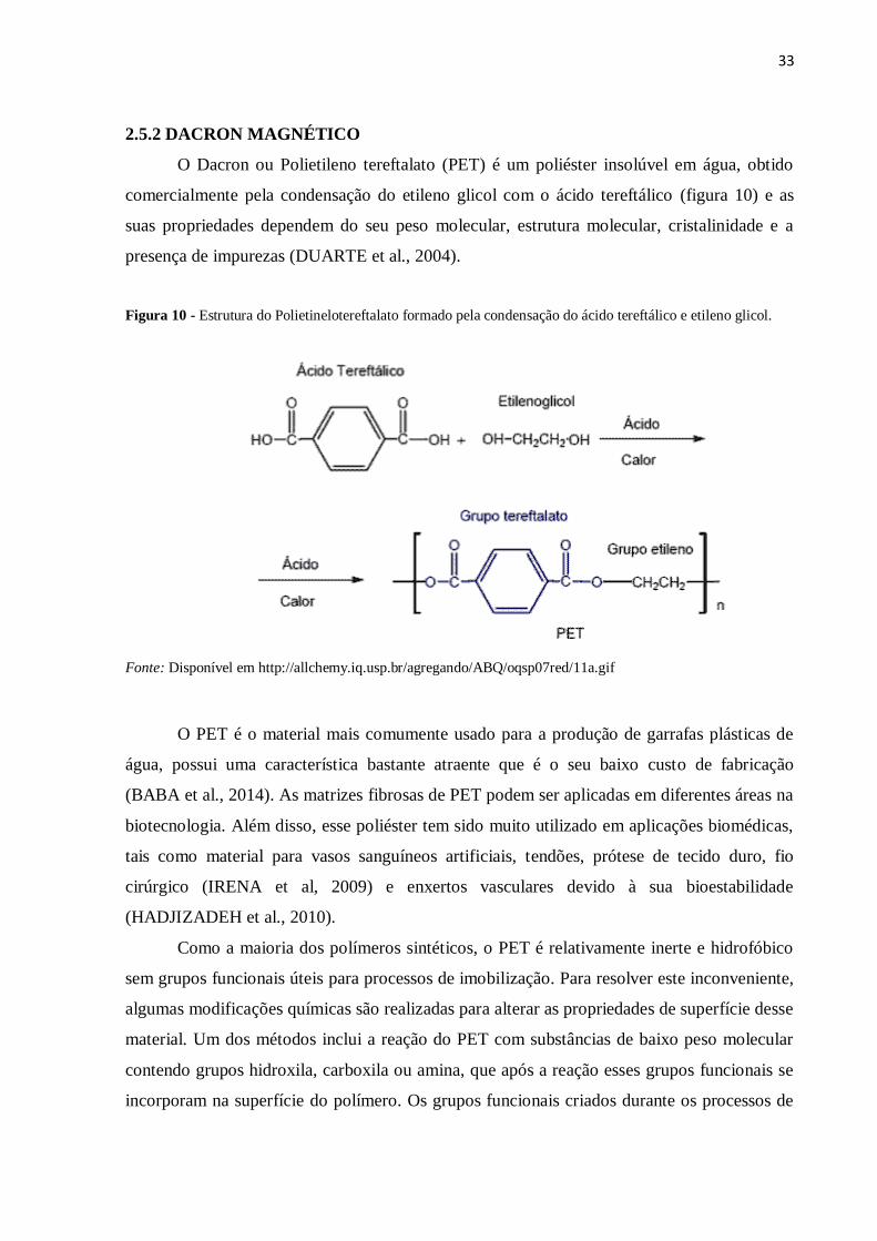
Figura 10 Estrutura do Polietinelotereftalato formado pela condesação do ácido
tereftálico e etileno glicol. Fonte: Disponível em
http://allchemy.iq.usp.br/agregando/ABQ/oqsp07red/11a.gif 33
Figura 11 Formação de Dacron-hidrazida a partir da reação de hidrazinólise do
Dacron. Em destaque (vermelho) o grupo hidrazida formado. Fonte:
Elaborado pela autora (2016). 34
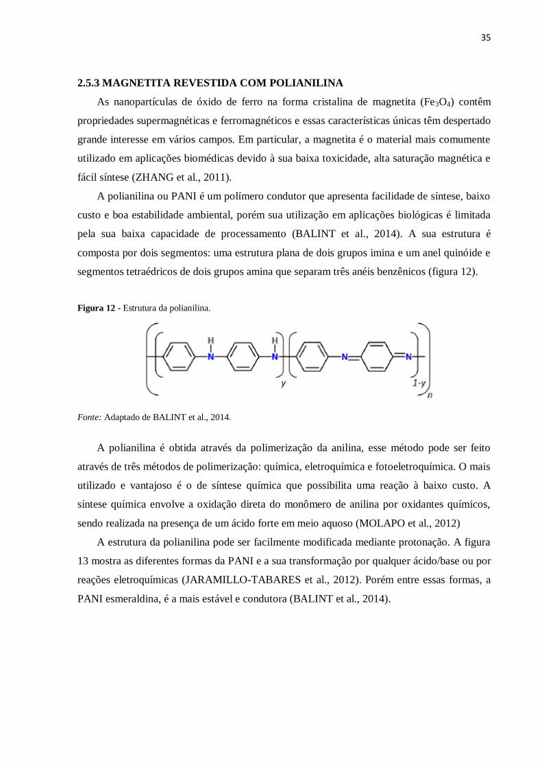
Figura 12 Estrutura da polianilina. Fonte: Adaptado de BALINT et al., 2014. 35
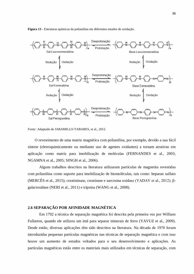
Figura 13 Estruturas químicas da polianilina em diferentes estados de oxidação.
Fonte: Adaptado de JARAMILLO-TABARES, et al., 2012. 36
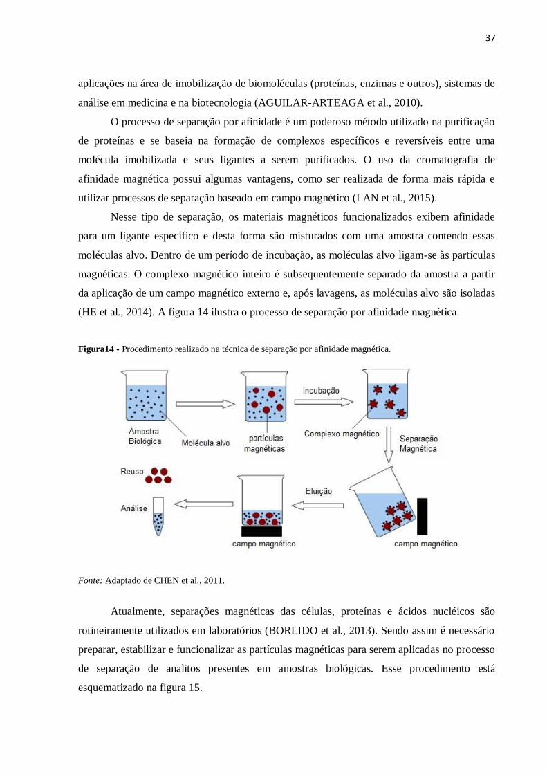
Figura 14 Procedimento realizado na técnica de separação por afinidade magnética.
Fonte: Adaptado de CHEN et al., 2011. 37
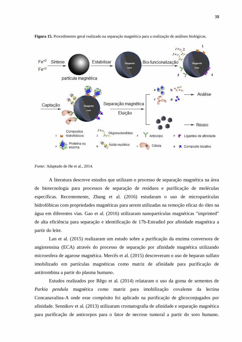
Figura 15 Procedimento geral realizado para separação magnética para a realização
de análises biológicas. Fonte: Adaptado de He et al., 2014. 38
ARTIGO 1
Figure 1 SEM images and EDX of Dacron hydrazide (a) and mDAC (b). 57
Figure 2 X-ray diffraction patterns of magnetite (M), Dacron (D) and mDAC
particles (mDAC). 58
Figure 3 Hysteresis curves at 293 K, 300 K and 313 K for MAG (a) and mDAC
(b). 59
Figure 4 Infrared spectra of MAG, Dacron-hydrazide; mDAC and mDAC-HEP (In
ascending order). 1: Band of NH2 group of the hydrazide; 2: Band of
amide I; 3: Band of amide II. 60
Figure 5 Activity removed of antithrombin present in fresh plasma and after direct
contact with the mDAC derivatives. Immediately synthesized (a) and 2
years stored (b). 61
Figure 6 Activated partial thromboplastin time (aPTT) of fresh plasma performed
by adding small aliquot of the eluate (1.0 M) of mDAC derivatives.
Immediately synthesized (a) and 2 years stored (b). 62
Figure 7 Electrophoresis of protein fractions of plasma obtained with the use of
magnetic particles. Elutions were carried out using increasing NaCl
concentrations (0.25; 0.5 and 1.0 M) in the magnetic derivatives: mDAC-
HEP (a) and mDAC (b). AT: Standard Antithrombin. (0): Elutions
performed in supports immediately synthesized. (24): Elutions performed
in supports stored for 24 months. Arrows: Antithrombin (58 kDa). 62
Figure 8 Schematic route of mDAC-HEP composite synthesis. Dacron is firstly
hydrolyzed under hydrazine hydrate that introduces amino group and
subsequently co-precipitated with Fe2+
and Fe3+
. Hep is simultaneously
functionalized with EDAC/NHS. Finally, magnetic Dacron-hydrazide and
functionalized Hep are incubated to form mDAC-HEP. 63
ARTIGO 2
Figure 1 Images obtained by transmission electron microscopy of the synthesized
magnetic particles. (A) MAG. (B) MAG-PANI. 72
Figure 2 X-ray diffraction patterns of MAG (magnetite bare) and MAG-PANI
(magnetite coated with polyaniline). (P) polyaniline phase. (M) magnetite
phase. 73
Figure 3 Hysteresis curve obtained for the synthesized magnetic particles. (A)
MAG-PANI. (B) MAG. 74
Figure 4 Elution of plasma proteins based on the affinity to the MAG-PANI-HEP
composite. 75
Figure 5 Electrophoresis performed from protein eluates obtained in plasma
fractionation. (A) Samples extracted with 0.25M NaCl (F1, F2 and F3);
0.5 M (F4, F5 and F6); (B) Samples extracted with 1.0 M (F7, F8 and F9). 76
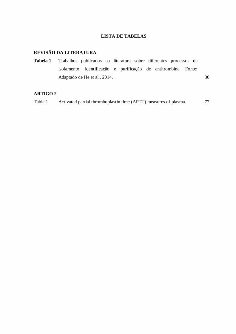
LISTA DE TABELAS
REVISÃO DA LITERATURA
Tabela 1 Trabalhos publicados na literatura sobre diferentes processos de
isolamento, identificação e purificação de antitrombina. Fonte:
Adaptado de He et al., 2014. 30
ARTIGO 2
Table 1 Activated partial thromboplastin time (APTT) measures of plasma. 77
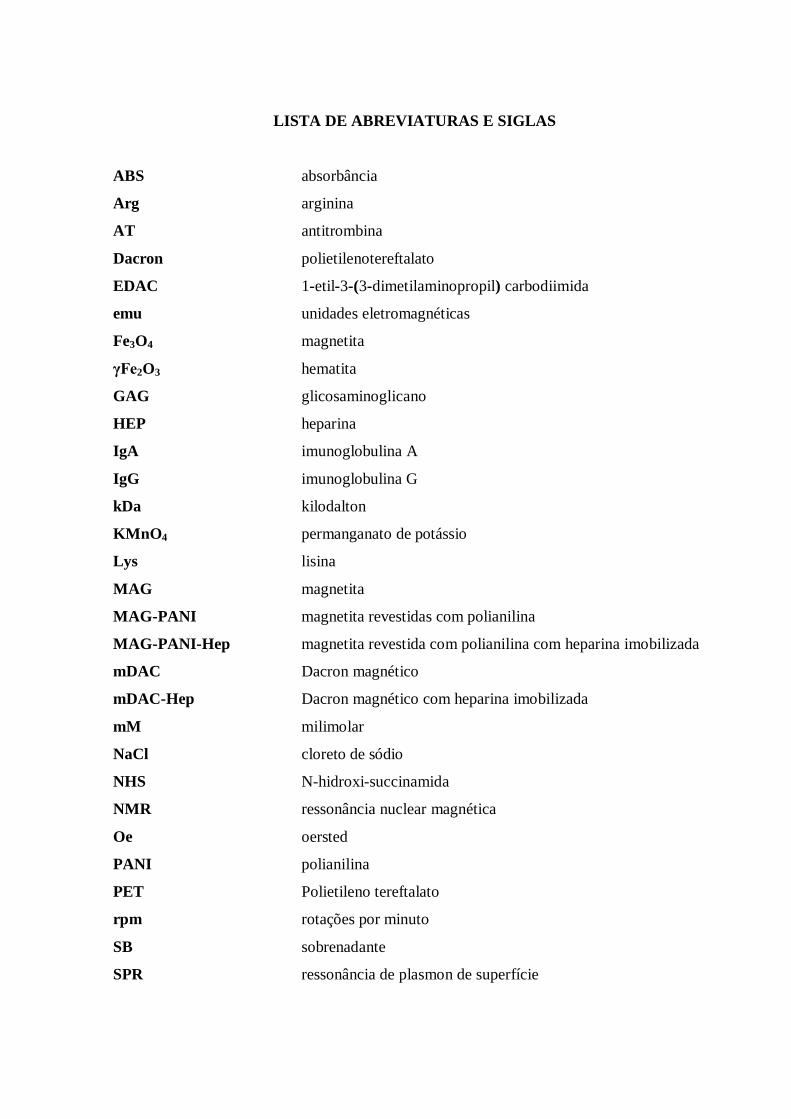
LISTA DE ABREVIATURAS E SIGLAS
ABS absorbância
Arg arginina
AT antitrombina
Dacron polietilenotereftalato
EDAC 1-etil-3-(3-dimetilaminopropil) carbodiimida
emu unidades eletromagnéticas
Fe3O4 magnetita
γFe2O3 hematita
GAG glicosaminoglicano
HEP heparina
IgA imunoglobulina A
IgG imunoglobulina G
kDa kilodalton
KMnO4 permanganato de potássio
Lys lisina
MAG magnetita
MAG-PANI magnetita revestidas com polianilina
MAG-PANI-Hep magnetita revestida com polianilina com heparina imobilizada
mDAC Dacron magnético
mDAC-Hep Dacron magnético com heparina imobilizada
mM milimolar
NaCl cloreto de sódio
NHS N-hidroxi-succinamida
NMR ressonância nuclear magnética
Oe oersted
PANI polianilina
PET Polietileno tereftalato
rpm rotações por minuto
SB sobrenadante
SPR ressonância de plasmon de superfície
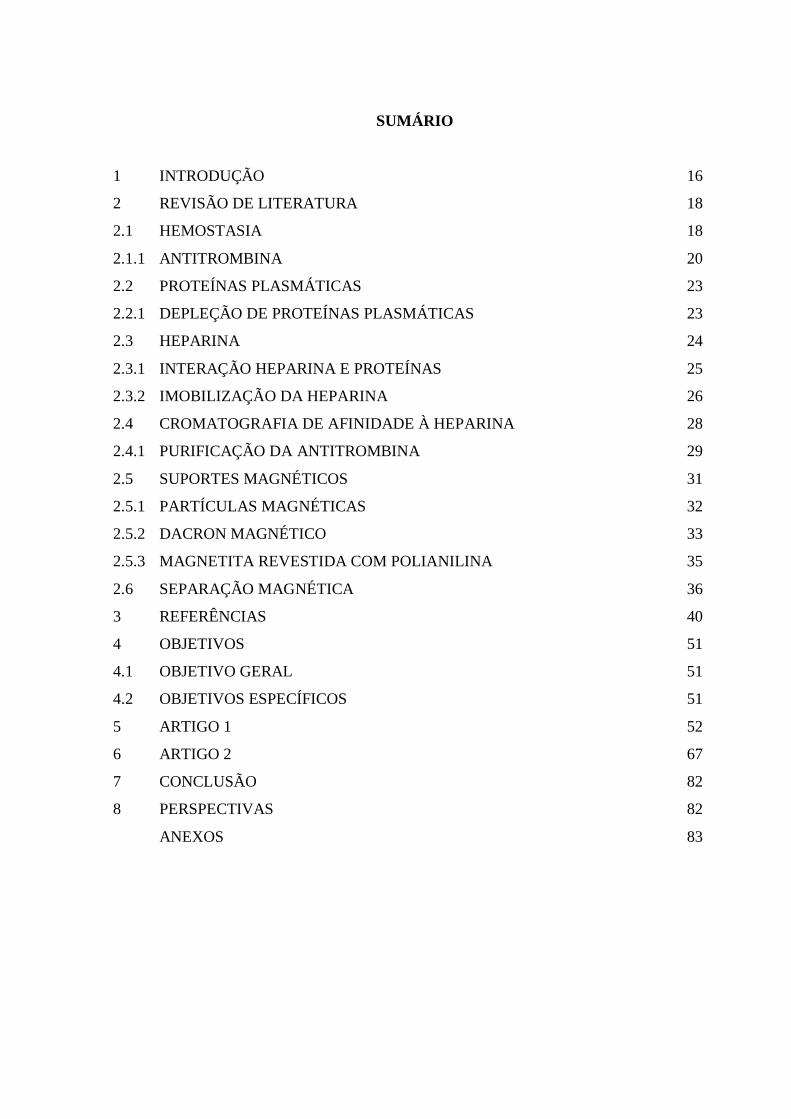
SUMÁRIO
1 INTRODUÇÃO 16
2 REVISÃO DE LITERATURA 18
2.1 HEMOSTASIA 18
2.1.1 ANTITROMBINA 20
2.2 PROTEÍNAS PLASMÁTICAS 23
2.2.1 DEPLEÇÃO DE PROTEÍNAS PLASMÁTICAS 23
2.3 HEPARINA 24
2.3.1 INTERAÇÃO HEPARINA E PROTEÍNAS 25
2.3.2 IMOBILIZAÇÃO DA HEPARINA 26
2.4 CROMATOGRAFIA DE AFINIDADE À HEPARINA 28
2.4.1 PURIFICAÇÃO DA ANTITROMBINA 29
2.5 SUPORTES MAGNÉTICOS 31
2.5.1 PARTÍCULAS MAGNÉTICAS 32
2.5.2 DACRON MAGNÉTICO 33
2.5.3 MAGNETITA REVESTIDA COM POLIANILINA 35
2.6 SEPARAÇÃO MAGNÉTICA 36
3 REFERÊNCIAS 40
4 OBJETIVOS 51
4.1 OBJETIVO GERAL 51
4.2 OBJETIVOS ESPECÍFICOS 51
5 ARTIGO 1 52
6 ARTIGO 2 67
7 CONCLUSÃO 82
8 PERSPECTIVAS 82
ANEXOS 83
16
1 INTRODUÇÃO
A hemostasia é um processo fisiológico que tem como objetivo manter o fluxo
sanguíneo no interior dos vasos e cessar a perda sanguínea. Trata-se de um processo
multifuncional que envolve a participação de vários componentes celulares e acelulares,
incluindo a resposta vascular, agregação plaquetária e a cascata de coagulação. Distúrbios da
hemostasia podem estar associados tanto com hemorragia ou com doenças tromboembólicas
(BERGER et al., 2014). Esse processo pode ser afetado por anormalidades genéticas,
condições patológicas e pela entrada de fatores exógenos, tais como toxinas de origem animal,
alérgenos ou medicamentos (HIREMATH et al., 2016).
O mecanismo bioquímico do sistema hemostático da coagulação gera enzimas tais
como a trombina, que precisam ser inibidas, após exercer sua função e se houver um distúrbio
desta inibição ocasiona coagulação sistêmica descontrolada (MOORE et al., 2015). Desta
forma, o controle fisiológico deste processo é determinado por anticoagulantes naturais, tais
como a antitrombina (AT) que inibe a trombina e outros fatores de coagulação
(FERNÁNDEZ e VILLAMEDIANA, 2012).
A heparina é um polissacarídeo linear sulfatado pertencente à família dos
glicosaminoglicanos, que tem sido utilizada na clínica devido à sua propriedade
anticoagulante (VISKOV et al., 2013). Além disso, é bastante conhecida por apresentar
afinidade com várias proteínas, as chamadas “proteínas de ligação à heparina”, tais como o
fator de crescimento de fibroblastos básico, fator de crescimento endotelial vascular,
fibronectina e AT, além de desempenhar um importante papel na regulação da atividade e
estabilidade destas proteínas (ARISAKA et al., 2013). A interação da heparina com a AT foi
o primeiro caso relatado de uma interação de significado fisiológico entre a heparina e uma
proteína específica (CAPILA e LINHARDT, 2002). Essas interações ocorrem devido a
presença dos sítios de ligação nas proteínas que contêm aminoácidos básicos (Lys e Arg)
cujas cargas positivas, provavelmente, interagem com os grupamentos sulfatos e carboxilatos
(carregados negativamente) presentes nas cadeias da heparina (ESKO et al., 2009).
Quando imobilizada, a heparina pode interagir com fatores da coagulação,
funcionando como um ligante de afinidade, capaz de interagir com proteínas, o que a
literatura descreve como “cromatografia de afinidade à heparina” (KRAPFENBAUER e
FOUNTOULAKIS, 2009). A heparina imobilizada em suportes sólidos é amplamente
utilizada na cromatografia de afinidade para a purificação e identificação dessas proteínas que
são capazes de se ligar à glicosaminoglicano (MURUGESAN et al., 2008). Desta forma a
17
heparina tem sido utilizada em processos de purificação e identificação de proteínas da
cascata da coagulação sanguínea (LI et al., 2009). Além disso, a elevada afinidade de ligação
da AT com a heparina tem sido utilizada em sistemas de cromatografia de afinidade para o
isolamento da AT a partir do plasma (HEGER et al., 2002). No processo de imobilização é
comum o uso de suportes insolúveis em água que apresentem: resistência física, estabilidade
mecânica e térmica à biomolécula imobilizada (CARAMORI e FERNANDES, 2004). E,
quando magnetizados, a recuperação do compósito suporte-biomolécula pode facilmente ser
obtida mediante a aplicação de um campo magnético externo (MACIEL, 2012).
Portanto, este trabalho propõe a síntese de compósitos magnéticos com heparina
covalentemente imobilizada e sua aplicação no fracionamento e isolamento de proteínas do
plasma humano com base na cromatografia de afinidade à heparina.
18
2 REVISÃO DA LITERATURA
2.1 HEMOSTASIA
A hemostasia é um processo fisiológico que tem como objetivo manter o fluxo
sanguíneo no interior dos vasos e cessar a perda sanguínea. Além disso, é um mecanismo
complexo de defesa, responsável pelo controle da perda de sangue resultante de uma lesão
vascular. Trata-se de um processo multifuncional e bem regulado que envolve a participação
de vários componentes fisiológicos celulares e acelulares, incluindo a resposta vascular,
agregação plaquetária e a cascata de coagulação. Distúrbios da hemostasia podem estar
associados tanto com hemorragia ou com doenças tromboembólicas (BERGER et al., 2014).
Sendo assim, a hemostasia é fortemente regulada, onde qualquer condição clínica
possibilita biomarcadores para o diagnóstico. Esse processo pode ser afetado por
anormalidades genéticas, condições patológicas e pela entrada de fatores exógenos, tais como
toxinas de origem animal, alérgenos ou medicamentos (HIREMATH et al., 2016).
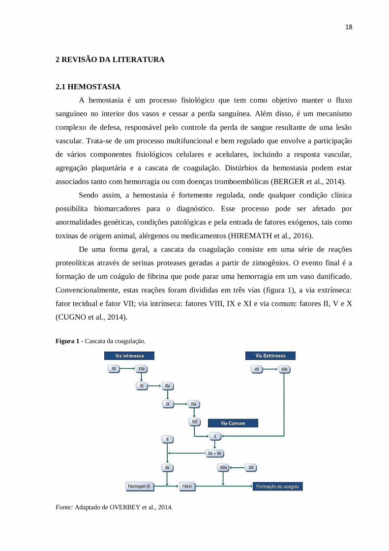
De uma forma geral, a cascata da coagulação consiste em uma série de reações
proteolíticas através de serinas proteases geradas a partir de zimogênios. O evento final é a
formação de um coágulo de fibrina que pode parar uma hemorragia em um vaso danificado.
Convencionalmente, estas reações foram divididas em três vias (figura 1), a via extrínseca:
fator tecidual e fator VII; via intrínseca: fatores VIII, IX e XI e via comum: fatores II, V e X
(CUGNO et al., 2014).
Figura 1 - Cascata da coagulação.
Fonte: Adaptado de OVERBEY et al., 2014.
19
Quando ocorre um dano ao vaso, o fator tecidual (FT) é exposto e liga-se ao fator VII-
VIIa, assim a via extrínseca é ativada iniciando o processo de coagulação (FURIE e FURIE,
1988; REZENDE, 2010).
Enquanto que na via intrínseca, a ativação do fator XII ocorre quando o sangue entra
em contato com uma superfície contendo cargas elétricas negativas. Tal processo é
denominado "ativação por contato" e requer ainda a presença de outros componentes do
plasma: pré-calicreína (uma serinoprotease) e cininogênio de alto peso molecular (um cofator
não enzimático). O fator XII, assim ativado, ativa o fator XI que, por sua vez, ativa o fator IX.
O fator IX ativado, na presença de fator VIII ativado por traços de trombina e em presença de
íons cálcio (complexo tenase), ativa o fator X da coagulação desencadeando a geração de
trombina e, subsequentemente, formação de fibrina (DAVIE et al., 1991; REZENDE, 2010).
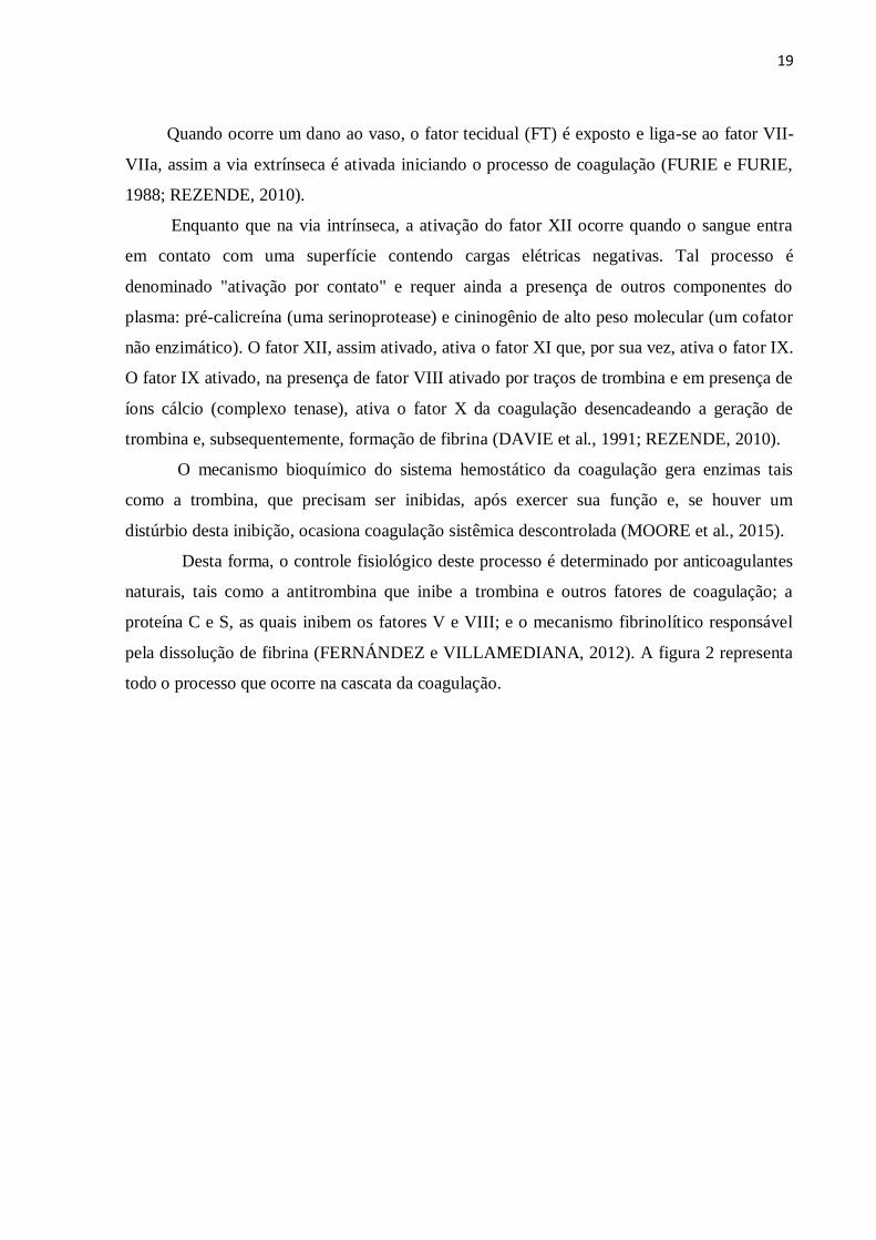
O mecanismo bioquímico do sistema hemostático da coagulação gera enzimas tais
como a trombina, que precisam ser inibidas, após exercer sua função e, se houver um
distúrbio desta inibição, ocasiona coagulação sistêmica descontrolada (MOORE et al., 2015).
Desta forma, o controle fisiológico deste processo é determinado por anticoagulantes
naturais, tais como a antitrombina que inibe a trombina e outros fatores de coagulação; a
proteína C e S, as quais inibem os fatores V e VIII; e o mecanismo fibrinolítico responsável
pela dissolução de fibrina (FERNÁNDEZ e VILLAMEDIANA, 2012). A figura 2 representa
todo o processo que ocorre na cascata da coagulação.
20
Figura 2 - Representação esquemática da cascata da coagulação, anticoagulação e fibrinólise. As grandes setas
azuis correspondem à via principal da coagulação, incluindo os complexos tenase e protrombinase (círculos em
vermelho). Linhas contínuas e descontínuas representam vias de ativação e inativação, respectivamente. As setas
vermelhas correspondem às diversas funções da trombina, as verdes às do TFPI, as azuis às da antitrombina,
alaranjado às da proteína Z e roxo às da proteína C. PC: proteína C; APC: proteína C ativada; PS: proteína S; FL:
fosfolipídios; tPA: plasminogênio tissular; Ca+2: cálcio; FT: fator tissular; i: inativo; EPCR: receptor endotelial
da proteína C; ZPI: protease dependente da proteína Z; TAFI: inibidor da fibrinólise ativado pela trombina.
Fonte: REZENDE, 2010.
2.1.1 ANTITROMBINA
Antitrombina (AT) é o principal inibidor das proteases da cascata de coagulação tais
como fator Xa, trombina e fator IXa (AZHAR et al., 2016). Trata-se de uma glicoproteína do
plasma humano que pertence à família dos inibidores de serina protease (serpina). Além do
21
seu efeito anticoagulante, a AT também possui propriedades anti-inflamatórias, que é
observado quando há uma elevada concentração deste inibidor presente no sangue (MARIE et
al., 2015). Com um peso molecular de 58 kDa, a AT é sintetizada no fígado e circula no
plasma sanguíneo em duas formas principais onde a isoforma predominante, a α-isoforma,
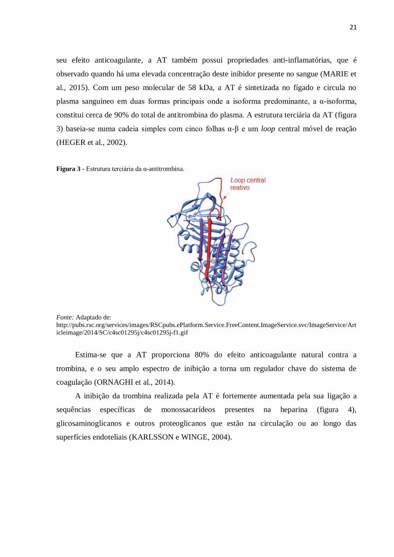
constitui cerca de 90% do total de antitrombina do plasma. A estrutura terciária da AT (figura
3) baseia-se numa cadeia simples com cinco folhas α-β e um loop central móvel de reação
(HEGER et al., 2002).
Figura 3 - Estrutura terciária da α-antitrombina.
Fonte: Adaptado de:
http://pubs.rsc.org/services/images/RSCpubs.ePlatform.Service.FreeContent.ImageService.svc/ImageService/Art
icleimage/2014/SC/c4sc01295j/c4sc01295j-f1.gif
Estima-se que a AT proporciona 80% do efeito anticoagulante natural contra a
trombina, e o seu amplo espectro de inibição a torna um regulador chave do sistema de
coagulação (ORNAGHI et al., 2014).
A inibição da trombina realizada pela AT é fortemente aumentada pela sua ligação a
sequências específicas de monossacarídeos presentes na heparina (figura 4),
glicosaminoglicanos e outros proteoglicanos que estão na circulação ou ao longo das
superfícies endoteliais (KARLSSON e WINGE, 2004).
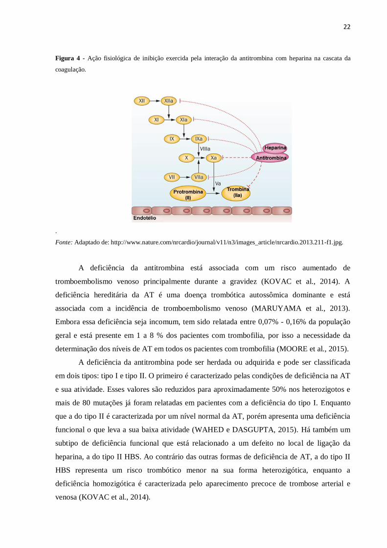
22
Figura 4 - Ação fisiológica de inibição exercida pela interação da antitrombina com heparina na cascata da
coagulação.
.
Fonte: Adaptado de: http://www.nature.com/nrcardio/journal/v11/n3/images_article/nrcardio.2013.211-f1.jpg.
A deficiência da antitrombina está associada com um risco aumentado de
tromboembolismo venoso principalmente durante a gravidez (KOVAC et al., 2014). A
deficiência hereditária da AT é uma doença trombótica autossômica dominante e está
associada com a incidência de tromboembolismo venoso (MARUYAMA et al., 2013).
Embora essa deficiência seja incomum, tem sido relatada entre 0,07% - 0,16% da população
geral e está presente em 1 a 8 % dos pacientes com trombofilia, por isso a necessidade da
determinação dos níveis de AT em todos os pacientes com trombofilia (MOORE et al., 2015).
A deficiência da antitrombina pode ser herdada ou adquirida e pode ser classificada
em dois tipos: tipo I e tipo II. O primeiro é caracterizado pelas condições de deficiência na AT
e sua atividade. Esses valores são reduzidos para aproximadamente 50% nos heterozigotos e
mais de 80 mutações já foram relatadas em pacientes com a deficiência do tipo I. Enquanto
que a do tipo II é caracterizada por um nível normal da AT, porém apresenta uma deficiência
funcional o que leva a sua baixa atividade (WAHED e DASGUPTA, 2015). Há também um
subtipo de deficiência funcional que está relacionado a um defeito no local de ligação da
heparina, a do tipo II HBS. Ao contrário das outras formas de deficiência de AT, a do tipo II
HBS representa um risco trombótico menor na sua forma heterozigótica, enquanto a
deficiência homozigótica é caracterizada pelo aparecimento precoce de trombose arterial e
venosa (KOVAC et al., 2014).
23
2.2 PROTEÍNAS PLASMÁTICAS
O plasma humano contém proteínas com uma ampla gama de funções biológicas e é
um dos fluidos biológicos mais comumente utilizados para o diagnóstico clínico (KULLOLLI
et al., 2013). Este complexo de proteínas está associado a diversos processos que ocorrem no
organismo, desta forma, no plasma é possível encontrar proteínas relacionadas com a
progressão de doenças e que podem ser uma fonte de biomarcadores (AHN e KHAN, 2014).
Albumina, Imunoglobulina G (IgG), Imunoglobulina A (IgA), transferrina,
haptoglobina e α-1-antitripsina representam 90% da massa de proteína no plasma sanguíneo
(JAVANMARD et al., 2014) e tendem a mascarar as proteínas de menor abundância e esse é
o maior problema para os estudos que envolvem, por exemplo, amostras de soro ou plasma
(KARATAS et al., 2007).
A capacidade de identificar e quantificar as proteínas de baixa abundância no plasma
facilita sua análise proteômica e a busca de biomarcadores de doenças (KULLOLLI et al.,
2013).
2.2.1 DEPLEÇÃO DE PROTEÍNAS PLASMÁTICAS
Nos últimos anos, a proteômica tem auxiliado no desenvolvimento de métodos de
diagnóstico para várias doenças. Com essas tecnologias é possível detectar e identificar
compostos específicos que podem ser usados como marcadores de diagnóstico, pois essas
substâncias encontram-se presentes apenas em células e tecidos doentes ou secretadas para o
ambiente externo ou interno do organismo nas doenças (BORMOTOVA et al., 2015).
Nas amostras biológicas, tais como plasma ou soro há milhares de proteínas diferentes
e um dos maiores desafios da análise dessas amostras é a elevada dinâmica dos peptídeos e
proteínas com concentrações elevadas (FISCHNALLER et al., 2014). Dessa forma, a
proteômica é um campo de crescimento rápido que se dedica ao estudo e descoberta de
biomarcadores, no entanto se limita devido à presença das proteínas de grande abundância no
plasma. Como consequência dificulta a detecção das proteínas de baixa abundância, que são
mais susceptíveis de serem biomarcadores (MAHN e ISMAIL, 2011).
Para analisar os componentes de baixa abundância, as proteínas de alta abundância,
em especial a albumina, devem ser removidas por adsorção a corantes imobilizados, através
de extração por imunoafinidade ou captura de afinidade via peptídeos imobilizados (ANDAC
et al., 2013). Por isso, vários métodos e adsorventes tem sido desenvolvidos e aplicados para
reduzir a complexidade do plasma, essas técnicas apresentam desvantagens consideráveis, tais
como o alto custo, instrumentação cara, etc.
24
Nenhum método analítico estabelecido até hoje pode esclarecer completamente toda a
dinâmica das proteínas presentes no plasma ou soro. Porém, existe um método disponível
para fracionar ou remover as proteínas da amostra através de colunas de depleção, construídas
de modo a remover de uma forma geral as proteínas mais abundantes dos fluidos corporais.
As colunas existentes são baseadas em anticorpos, anticorpos variantes e recombinantes ou
outro tipo de matriz de afinidade modificada (SUNDEBERG et al., 2015). O maior
inconveniente é o volume da amostra limitada, o que requer passos pré-analíticos demorados
e resultando em amostras muito pequenas para a próxima análise, limitando medições
repetitivas, intra e inter precisão das amostras e geração de dados confiáveis (UZUN et al.,
2013).
2.3 HEPARINA
A heparina foi descoberta em 1916 e sua aplicação como anticoagulante na clínica ocorre
desde 1935 (SAKIYAMA-ELBERT, 2014), além disso, vem sendo amplamente utilizada no
tratamento de tromboembolismo (MOURIER et al., 2015).
Ao longo das últimas décadas, foi mostrado que a heparina está envolvida em muitos
processos biológicos, através da sua interação com um elevado número de proteínas (LEVER
e PAGE, 2012) e que também está envolvida na regulação de eventos importantes como a
sinalização celular e o controle de uma variedade de funções biológicas (HANSEN et al.,
2013).
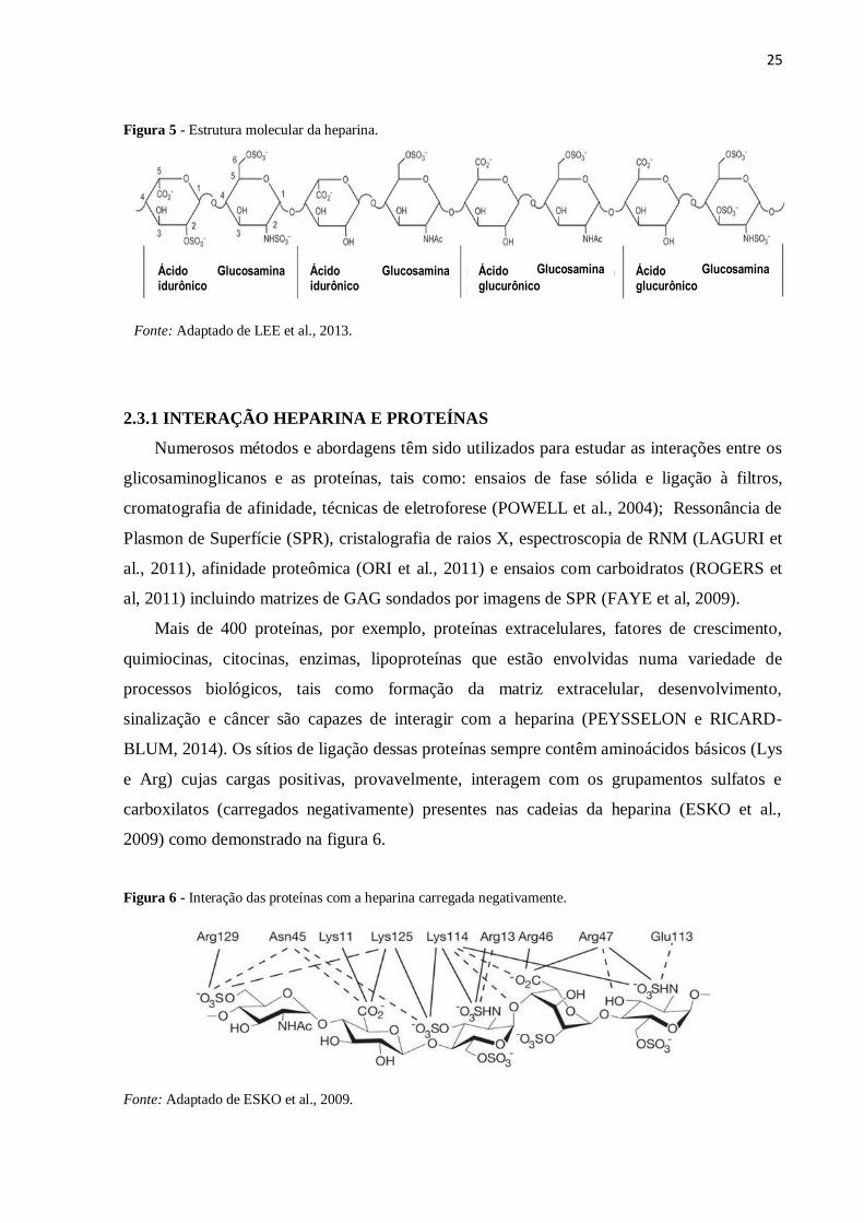
Sob o ponto de vista molecular, a heparina (figura 5) é um polissacarídeo pertencente à
família dos glicosaminoglicanos (GAGs) de cadeia linear polimérica altamente sulfatada,
formada por unidades repetidas de dissacarídeos constituídos por um ácido urônico
(glucurônico ou idurônico) e uma glucosamina (MOURIER et al., 2015) seu peso molecular
varia de 5 kDa a 50 kDa, com peso médio de 15 kDa (VIEIRA, 2012).
Devido a esta estrutura a heparina possui a mais alta densidade de carga negativa, quando
comparada a qualquer outra macromolécula biologicamente ativa, (CAPILA e LINHARDT,
2002), o que permite sua interação com um alto número de proteínas diferentes
(FLENGSRUD e ANTONSEN, 2015).
25
Figura 5 - Estrutura molecular da heparina.
Fonte: Adaptado de LEE et al., 2013.
2.3.1 INTERAÇÃO HEPARINA E PROTEÍNAS
Numerosos métodos e abordagens têm sido utilizados para estudar as interações entre os
glicosaminoglicanos e as proteínas, tais como: ensaios de fase sólida e ligação à filtros,
cromatografia de afinidade, técnicas de eletroforese (POWELL et al., 2004); Ressonância de
Plasmon de Superfície (SPR), cristalografia de raios X, espectroscopia de RNM (LAGURI et
al., 2011), afinidade proteômica (ORI et al., 2011) e ensaios com carboidratos (ROGERS et
al, 2011) incluindo matrizes de GAG sondados por imagens de SPR (FAYE et al, 2009).
Mais de 400 proteínas, por exemplo, proteínas extracelulares, fatores de crescimento,
quimiocinas, citocinas, enzimas, lipoproteínas que estão envolvidas numa variedade de
processos biológicos, tais como formação da matriz extracelular, desenvolvimento,
sinalização e câncer são capazes de interagir com a heparina (PEYSSELON e RICARD-
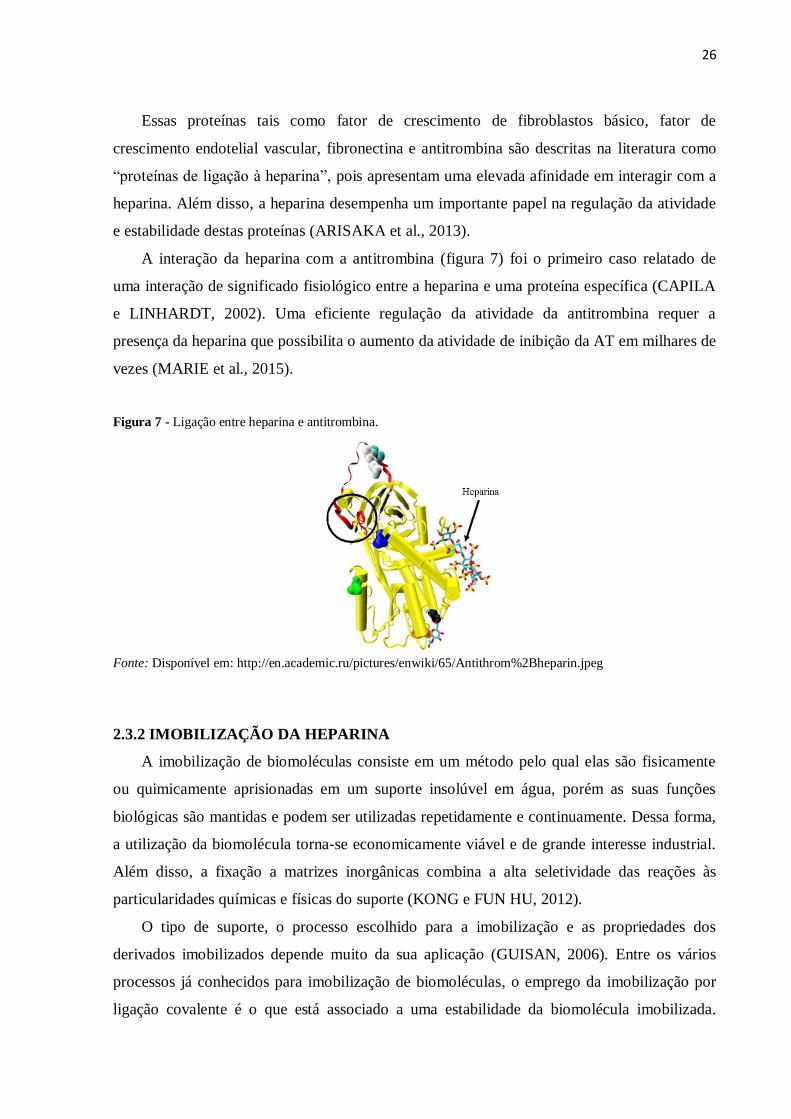
BLUM, 2014). Os sítios de ligação dessas proteínas sempre contêm aminoácidos básicos (Lys
e Arg) cujas cargas positivas, provavelmente, interagem com os grupamentos sulfatos e
carboxilatos (carregados negativamente) presentes nas cadeias da heparina (ESKO et al.,
2009) como demonstrado na figura 6.
Figura 6 - Interação das proteínas com a heparina carregada negativamente.
Fonte: Adaptado de ESKO et al., 2009.
Glucosamina Glucosamina Ácido idurônico
Ácido idurônico
Ácido glucurônico
Glucosamina Ácido glucurônico
Glucosamina
26
Essas proteínas tais como fator de crescimento de fibroblastos básico, fator de
crescimento endotelial vascular, fibronectina e antitrombina são descritas na literatura como
“proteínas de ligação à heparina”, pois apresentam uma elevada afinidade em interagir com a
heparina. Além disso, a heparina desempenha um importante papel na regulação da atividade
e estabilidade destas proteínas (ARISAKA et al., 2013).
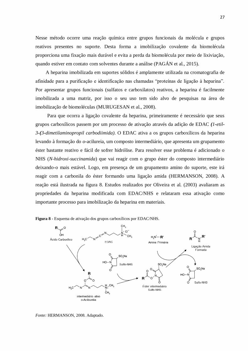
A interação da heparina com a antitrombina (figura 7) foi o primeiro caso relatado de
uma interação de significado fisiológico entre a heparina e uma proteína específica (CAPILA
e LINHARDT, 2002). Uma eficiente regulação da atividade da antitrombina requer a
presença da heparina que possibilita o aumento da atividade de inibição da AT em milhares de
vezes (MARIE et al., 2015).
Figura 7 - Ligação entre heparina e antitrombina.
Fonte: Disponível em: http://en.academic.ru/pictures/enwiki/65/Antithrom%2Bheparin.jpeg
2.3.2 IMOBILIZAÇÃO DA HEPARINA
A imobilização de biomoléculas consiste em um método pelo qual elas são fisicamente
ou quimicamente aprisionadas em um suporte insolúvel em água, porém as suas funções
biológicas são mantidas e podem ser utilizadas repetidamente e continuamente. Dessa forma,
a utilização da biomolécula torna-se economicamente viável e de grande interesse industrial.
Além disso, a fixação a matrizes inorgânicas combina a alta seletividade das reações às
particularidades químicas e físicas do suporte (KONG e FUN HU, 2012).
O tipo de suporte, o processo escolhido para a imobilização e as propriedades dos
derivados imobilizados depende muito da sua aplicação (GUISAN, 2006). Entre os vários
processos já conhecidos para imobilização de biomoléculas, o emprego da imobilização por
ligação covalente é o que está associado a uma estabilidade da biomolécula imobilizada.
27
Nesse método ocorre uma reação química entre grupos funcionais da molécula e grupos
reativos presentes no suporte. Desta forma a imobilização covalente da biomolécula
proporciona uma fixação mais durável e evita a perda da biomolécula por meio de lixiviação,
quando estiver em contato com solventes durante a análise (PAGÁN et al., 2015).
A heparina imobilizada em suportes sólidos é amplamente utilizada na cromatografia de
afinidade para a purificação e identificação nas chamadas “proteínas de ligação à heparina”.
Por apresentar grupos funcionais (sulfatos e carboxilatos) reativos, a heparina é facilmente
imobilizada a uma matriz, por isso o seu uso tem sido alvo de pesquisas na área de
imobilização de biomoléculas (MURUGESAN et al., 2008).
Para que ocorra a ligação covalente da heparina, primeiramente é necessário que seus
grupos carboxílicos passem por um processo de ativação através da adição de EDAC (1-etil-
3-(3-dimetilaminopropil carbodiimida). O EDAC ativa a os grupos carboxílicos da heparina
levando à formação do o-acilureia, um composto intermediário, que apresenta um grupamento
éster bastante reativo e fácil de sofrer hidrólise. Para resolver esse problema é adicionado o
NHS (N-hidroxi-succinamida) que vai reagir com o grupo éster do composto intermediário
deixando-o mais estável. Logo, em presença de um grupamento amino do suporte, este irá
reagir com a carbonila do éster formando uma ligação amida (HERMANSON, 2008). A
reação está ilustrada na figura 8. Estudos realizados por Oliveira et al. (2003) avaliaram as
propriedades da heparina modificada com EDAC/NHS e relataram essa ativação como
importante processo para imobilização da heparina em materiais.
Figura 8 - Esquema de ativação dos grupos carboxílicos por EDAC/NHS.
Fonte: HERMANSON, 2008. Adaptado.
28
2.4 CROMATOGRAFIA DE AFINIDADE À HEPARINA
Quando imobilizada, a heparina pode interagir com fatores da coagulação, funcionando
como um ligante de afinidade, capaz de interagir com estas proteínas (KRAPFENBAUER e
FOUNTOULAKIS, 2009), isso é o que a literatura descreve como cromatografia de afinidade
à heparina. Este método (figura 9) ocorre devido à interação (afinidade) entre as moléculas de
interesse e a heparina imobilizada ao suporte insolúvel (FAROOQUI, 1980; XIONG et al.,
2008).
Figura 9 - Princípios da cromatografia de afinidade à heparina. (a) representa a 1ª etapa que corresponde à
imobilização da heparina em um suporte ou coluna. (b) ilustra a incubação do suporte com as proteínas do
plasma humano e a interação das proteínas que tem afinidade com a heparina. (c) demonstra a formação de um
complexo de ligação entre a heparina e proteína específica com posterior eluição e purificação desta proteína.
Fonte: Elaborado pela autora (2016).
Em geral, este método de cromatografia de afinidade é bastante utilizado para fracionar
ou purificar proteínas e outras substâncias biológicas que podem interagir com a heparina. A
elevada afinidade de ligação da AT com a heparina tem sido utilizada em sistemas de
cromatografia de afinidade para o isolamento da antitrombina a partir do plasma (HEGER et
29
al., 2002). Embora a cromatografia de afinidade seja uma boa abordagem para estudar essa
interação entre a antitrombina e a heparina, até agora essa metodologia não foi explorada para
determinar constantes de ligação entre estes dois ligantes (MARIE et al., 2015).
Cromatografia de afinidade à heparina também tem sido utilizada para purificar
lactoferrina a partir de leite de cabra, bovino e humano. Além disso, também é utilizada para
purificar alguns fatores de crescimento (fator de betacelulina e fator de crescimento de
fibroblastos básico) a partir do soro de leite bovino (OUNIS et al., 2008).
Recentemente, Bjarnadóttir e Flensrud (2014) utilizaram cromatografia de afinidade à
heparina para detectar proteínas do plasma humano e foi observado que entre essas proteínas
muitas eram biomarcadores já descritos na literatura. Hu et al. (2010) utilizaram a
cromatografia de afinidade à heparina para purificação do vírus da síndrome reprodutiva e
respiratória dos suínos em cultura de célula, chegando a remover das células cerca de 96%
desse vírus. A separação de pequenos componentes proteicos presentes no soro do leite foram
isolados por Ounis et al. (2008) utilizando a cromatografia de afinidade à heparina.
A alta afinidade de ligação da heparina a várias proteínas, incluindo fatores de
crescimento, tem sido relacionada com a sua carga líquida negativa em pH neutro
(KAWAKAMI et al., 2006; FENG et al., 2004). Além disso, o uso da cromatografia de
afinidade à heparina pode ser aplicada como uma estratégia para remover seletivamente
algumas proteínas de grande abundância, facilitando a análise de proteínas de baixa
concentração no plasma. Já foi demonstrado que a albumina pode ser removida, por exemplo,
através de técnicas de colunas de imunoafinidade, aprisionamento isoelétrico e cromatografia
de afinidade (LEI et al., 2008).
2.4.1 PURIFICAÇÃO DA ANTITROMBINA
Como já mencionado anteriormente, o uso da cromatografia de afinidade à heparina é
o método mais utilizado no processo de isolamento ou purificação da antitrombina a partir do
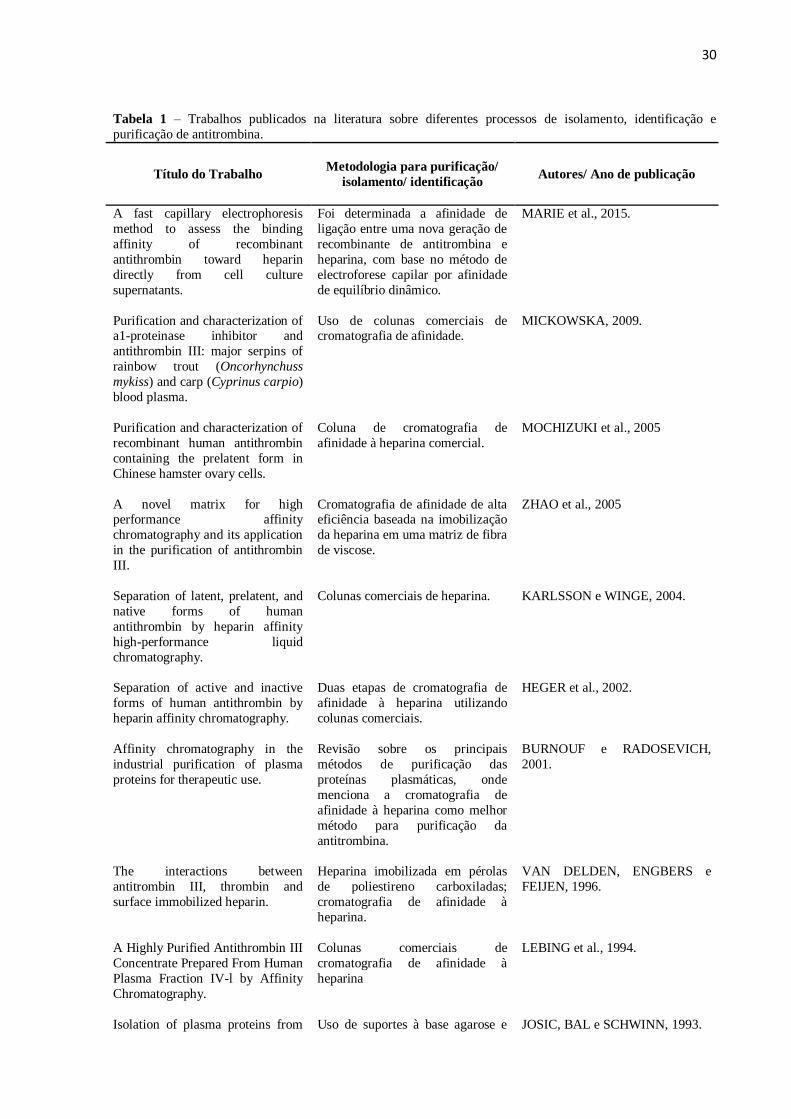
plasma humano. De acordo com a tabela 1, a purificação da antitrombina baseada na sua
ligação por afinidade à heparina foi descrita há quatro décadas e continua até os dias de hoje,
utilizando diferentes metodologias como mostrado nesta tabela.
30
Tabela 1 – Trabalhos publicados na literatura sobre diferentes processos de isolamento, identificação e
purificação de antitrombina.
Título do Trabalho
Metodologia para purificação/
isolamento/ identificação
Autores/ Ano de publicação
A fast capillary electrophoresis
method to assess the binding
affinity of recombinant
antithrombin toward heparin
directly from cell culture
supernatants.
Foi determinada a afinidade de
ligação entre uma nova geração de
recombinante de antitrombina e
heparina, com base no método de
electroforese capilar por afinidade
de equilíbrio dinâmico.
MARIE et al., 2015.
Purification and characterization of a1-proteinase inhibitor and
antithrombin III: major serpins of
rainbow trout (Oncorhynchuss
mykiss) and carp (Cyprinus carpio)
blood plasma.
Uso de colunas comerciais de cromatografia de afinidade.
MICKOWSKA, 2009.
Purification and characterization of
recombinant human antithrombin
containing the prelatent form in
Chinese hamster ovary cells.
Coluna de cromatografia de
afinidade à heparina comercial.
MOCHIZUKI et al., 2005
A novel matrix for high performance affinity
chromatography and its application
in the purification of antithrombin
III.
Cromatografia de afinidade de alta eficiência baseada na imobilização
da heparina em uma matriz de fibra
de viscose.
ZHAO et al., 2005
Separation of latent, prelatent, and
native forms of human
antithrombin by heparin affinity
high-performance liquid
chromatography.
Colunas comerciais de heparina.
KARLSSON e WINGE, 2004.
Separation of active and inactive
forms of human antithrombin by
heparin affinity chromatography.
Duas etapas de cromatografia de
afinidade à heparina utilizando
colunas comerciais.
HEGER et al., 2002.
Affinity chromatography in the
industrial purification of plasma
proteins for therapeutic use.
Revisão sobre os principais
métodos de purificação das
proteínas plasmáticas, onde
menciona a cromatografia de
afinidade à heparina como melhor
método para purificação da
antitrombina.
BURNOUF e RADOSEVICH,
2001.
The interactions between
antitrombin III, thrombin and
surface immobilized heparin.
Heparina imobilizada em pérolas
de poliestireno carboxiladas;
cromatografia de afinidade à
heparina.
VAN DELDEN, ENGBERS e
FEIJEN, 1996.
A Highly Purified Antithrombin III
Concentrate Prepared From Human
Plasma Fraction IV-l by Affinity
Chromatography.
Colunas comerciais de
cromatografia de afinidade à
heparina
LEBING et al., 1994.
Isolation of plasma proteins from Uso de suportes à base agarose e JOSIC, BAL e SCHWINN, 1993.
31
the clotting cascade by heparin
affinity chromatography.
outros polímeros, como matriz de
afinidade, para purificação da
antitrombina via cromatografia de
afinidade.
Purification and Large-Scale
Preparation of Antithrombin III
Cromatografia de afinidade à
heparina com Ultrogel de Heparina
HOFFMAN, 1989.
Isolation and Characterization of
an Antithrombin III Variant with
Reduced Carbohydrate Content and Enhanced Heparin Binding
Cromatografia com uso de plasma
normal em Heparina-Sepharose.
PETERSON e BLACKBURN,
1985.
Development of Large Scale
Fractionation Methods
VII. Preparation of Antithrombin
III Concentrate
Cromatografia de afinidade por
Heparina-Sepharose.
WICKERHAUSER, WILLIAMS e
MERCER, 1979.
Purification Of Antithrombin III
By Affinity Chromatography.
Cromatografia de afinidade à
heparina em gel de Sepharose.
MILLER-ANDERSSON, BORG e
ANDERSSON, 1974.
2.5 SUPORTES MAGNÉTICOS
Na escolha de um suporte para uma determinada aplicação, devem ser analisadas suas
propriedades físico-químicas, além da possibilidade de regeneração do material (MACIEL,
2012). Os suportes utilizados para imobilização de biomoléculas devem apresentar um bom
perfil como resistência física, insolubilidade, resistência a ataques microbianos, além de
estabilidade mecânica e térmica (CARAMORI e FERNANDES, 2004). E, quando
magnetizados, a recuperação do compósito suporte-biomolécula pode facilmente ser obtida
mediante a aplicação de um campo magnético.
A literatura descreve o uso de diferentes partículas magnéticas como suportes para
imobilização de biomoléculas, alguns exemplos são: Dacron magnético para imobilização de
heparan sulfato (MERCÊS et al., 2015) e tripsina (AMARAL et al., 2006); terra de
diatomáceas magnética e argila magnética para imobilização de invertase (CABRERA et al.,
2014); levana magnética para imobilização de tripsina (MACIEL et al. 2012); nanopartículas
de magnetita modificadas com APTES (3-aminopropiltrietoxisilano) e funcionalizadas com
glutaraldeído para imobilização de albumina (MALTAS et al. 2011); magnetita revestida com
polianilina para imobilização de β-galactosidase (NERI et al, 2011 ) e tripsina (MACIEL et
al., 2016).
32
2.5.1 PARTÍCULAS MAGNÉTICAS
As partículas magnéticas foram empregadas pela primeira vez na década de 1940
como uma nova tecnologia no tratamento de água poluída (ARIAS et al., 2001).
Nanopartículas à base de óxido de ferro podem ser sintetizadas por diferentes formas
(NICOLÁS et al., 2013), por exemplo: co-precipitação dos íons Fe+2
/Fe+3
em uma solução
aquosa utilizando uma base como agente de precipitação (WU et al., 2007; FRIED et al.,
2001), técnicas de sol-gel (XU et al., 2007), métodos coloidais (MARTÍNEZ et al., 2007),
reação de pirólise (CHIU et al., 2007) entre outros. O procedimento mais barato, simples e
ecologicamente correto é o método de coprecipitação (KANG et al., 1996; QU et al., 1999).
De acordo com Laurent et al. (2008), a técnica de co-precipitação é provavelmente a via
mais eficiente para obter partículas magnéticas. Os óxidos de ferro tais como magnetita
(Fe3O4) ou hematita (γFe2O3) são geralmente preparadas por uma mistura estequiométrica de
sais férricos e ferrosos em meio aquoso. A reação química de formação de Fe3O4 pode ser
escrita de acordo com a equação abaixo:
Fe+2
+ 2Fe+3
+ 8OH- Fe3O4 + 4H2O
Ainda de acordo com o mesmo autor, a precipitação completa de Fe3O4 deve acontecer a
um pH entre 8 e 14, com uma relação estequiométrica de 2:1 (Fe3+
/Fe2+
) em um ambiente não
oxidante sem oxigênio.
Recentemente, os nanomateriais magnéticos (magnetita, hematita e metais de ferritina),
têm sido amplamente utilizados na área da biomedicina e de catalisadores devido às suas
excelentes propriedades magnéticas (CHINNARAJ et al., 2015). Nanopartículas de óxido de
ferro têm sido desenvolvidas como novos sistemas promissores para serem aplicados em
diferentes áreas, tais como: agentes de imagem na ressonância magnética; mediadores de
calor no tratamento do câncer por hipertermia; suporte sólido para catálise heterogênea;
biossensores; adsorventes para a remoção de corantes e metais; e em sistemas de entrega de
drogas: “drug delivery” (DEBRASSI et al., 2011).
As partículas magnéticas modificadas são constituídas por um núcleo de óxido de ferro
revestido com um polímero. Esse revestimento possui grupos ativos que podem ser
conjugados a biomoléculas tais como carboidratos, proteínas e enzimas (MA et. al, 2003;
YMAURA et al., 2004). As partículas de magnetita superparamagnéticas revestidas com
polímeros são usualmente formadas por núcleos magnéticos responsáveis por uma resposta
magnética forte e uma camada polimérica para fornecer grupos funcionalizáveis e
característicos (WUNDERBALDINGER et al., 2002).
33
2.5.2 DACRON MAGNÉTICO
O Dacron ou Polietileno tereftalato (PET) é um poliéster insolúvel em água, obtido
comercialmente pela condensação do etileno glicol com o ácido tereftálico (figura 10) e as
suas propriedades dependem do seu peso molecular, estrutura molecular, cristalinidade e a
presença de impurezas (DUARTE et al., 2004).
Figura 10 - Estrutura do Polietinelotereftalato formado pela condensação do ácido tereftálico e etileno glicol.
Fonte: Disponível em http://allchemy.iq.usp.br/agregando/ABQ/oqsp07red/11a.gif
O PET é o material mais comumente usado para a produção de garrafas plásticas de
água, possui uma característica bastante atraente que é o seu baixo custo de fabricação
(BABA et al., 2014). As matrizes fibrosas de PET podem ser aplicadas em diferentes áreas na
biotecnologia. Além disso, esse poliéster tem sido muito utilizado em aplicações biomédicas,
tais como material para vasos sanguíneos artificiais, tendões, prótese de tecido duro, fio
cirúrgico (IRENA et al, 2009) e enxertos vasculares devido à sua bioestabilidade
(HADJIZADEH et al., 2010).
Como a maioria dos polímeros sintéticos, o PET é relativamente inerte e hidrofóbico
sem grupos funcionais úteis para processos de imobilização. Para resolver este inconveniente,
algumas modificações químicas são realizadas para alterar as propriedades de superfície desse
material. Um dos métodos inclui a reação do PET com substâncias de baixo peso molecular
contendo grupos hidroxila, carboxila ou amina, que após a reação esses grupos funcionais se
incorporam na superfície do polímero. Os grupos funcionais criados durante os processos de
34
modificação podem servir para imobilização covalente de várias biomoléculas como, por
exemplo, carboidratos, peptídeos e proteínas (IRENA et al., 2009).
A síntese de partículas de Dacron ferromagnéticas inicialmente foi descrita por
Carneiro Leão et al. (1991). Primeiramente, o Dacron sob a forma de folhas é submetido à
hidrazinólise, uma reação que consiste na adição de grupamentos hidrazidas ao suporte. As
hidrazidas formadas são obtidas pela reação entre o hidrato de hidrazina e grupamentos
ésteres presentes no Dacron (figura 11). O pó obtido é mecanicamente resistente e não
biodegradável. Essas propriedades o qualificam como um suporte adequado para
imobilização. Sua magnetização é realizada através da co-precipitação de sais de ferro em
meio aquoso e alcalino. Esse suporte quando magnetizado possibilita sua rápida separação de
uma mistura reacional.
Figura 11 - Formação de Dacron-hidrazida a partir da reação de hidrazinólise do Dacron. Em destaque
(vermelho) o grupo hidrazida formado.
Fonte: Elaborado pela autora (2016).
A literatura descreve alguns trabalhos que utilizaram suportes de Dacron magnéticos
para imobilização de biomoléculas, tais como: heparan sulfato (MERCÊS et al., 2015); α-L-
Rhamnosidase (SORIA et al., 2012); β-galactosidase (NERI et al., 2011); invertase
(CADENA et al., 2010); tripsina (AMARAL et al., 2006) e proteínas (OLIVEIRA et al.,
1989).
35
2.5.3 MAGNETITA REVESTIDA COM POLIANILINA
As nanopartículas de óxido de ferro na forma cristalina de magnetita (Fe3O4) contêm
propriedades supermagnéticas e ferromagnéticos e essas características únicas têm despertado
grande interesse em vários campos. Em particular, a magnetita é o material mais comumente
utilizado em aplicações biomédicas devido à sua baixa toxicidade, alta saturação magnética e
fácil síntese (ZHANG et al., 2011).
A polianilina ou PANI é um polímero condutor que apresenta facilidade de síntese, baixo
custo e boa estabilidade ambiental, porém sua utilização em aplicações biológicas é limitada
pela sua baixa capacidade de processamento (BALINT et al., 2014). A sua estrutura é
composta por dois segmentos: uma estrutura plana de dois grupos imina e um anel quinóide e
segmentos tetraédricos de dois grupos amina que separam três anéis benzênicos (figura 12).
Figura 12 - Estrutura da polianilina.
Fonte: Adaptado de BALINT et al., 2014.
A polianilina é obtida através da polimerização da anilina, esse método pode ser feito
através de três métodos de polimerização: química, eletroquímica e fotoeletroquímica. O mais
utilizado e vantajoso é o de síntese química que possibilita uma reação à baixo custo. A
síntese química envolve a oxidação direta do monômero de anilina por oxidantes químicos,
sendo realizada na presença de um ácido forte em meio aquoso (MOLAPO et al., 2012)
A estrutura da polianilina pode ser facilmente modificada mediante protonação. A figura
13 mostra as diferentes formas da PANI e a sua transformação por qualquer ácido/base ou por
reações eletroquímicas (JARAMILLO-TABARES et al., 2012). Porém entre essas formas, a
PANI esmeraldina, é a mais estável e condutora (BALINT et al., 2014).
36
Figura 13 - Estruturas químicas da polianilina em diferentes estados de oxidação.
Fonte: Adaptado de JARAMILLO-TABARES, et al., 2012.
O revestimento de uma matriz magnética com polianilina, por exemplo, devido a sua fácil
síntese (eletroquimicamente ou mediante uso de agentes oxidantes) a tornam atrativas em
aplicação como matriz para imobilização de moléculas (FERNANDES et al., 2003;
NGAMNA et al., 2005; SINGH et al., 2006).
Alguns trabalhos descritos na literatura utilizaram partículas de magnetita revestidas
com polianilina como suporte para imobilização de biomoléculas, tais como: heparan sulfato
(MERCÊS et al., 2015); creatininase, creatinase e sarcosina oxidase (YADAV et al., 2012); β-
galactosidase (NERI et al., 2011) e tripsina (WANG et al., 2008).
2.6 SEPARAÇÃO POR AFINIDADE MAGNÉTICA
Em 1792 a técnica de separação magnética foi descrita pela primeira vez por William
Fullarton, quando ele utilizou um ímã para separar minerais de ferro (YAVUZ et al., 2009).
Desde então, diversas aplicações têm sido descritos na literatura. Na década de 1970 foram
introduzidas pequenas partículas magnéticas nas técnicas de separação magnética e com isso
houve um aumento de estudos voltados para o seu desenvolvimento e aplicações. As
partículas magnéticas estão entre os materiais mais utilizados em técnicas de separação, com
37
aplicações na área de imobilização de biomoléculas (proteínas, enzimas e outros), sistemas de
análise em medicina e na biotecnologia (AGUILAR-ARTEAGA et al., 2010).
O processo de separação por afinidade é um poderoso método utilizado na purificação
de proteínas e se baseia na formação de complexos específicos e reversíveis entre uma
molécula imobilizada e seus ligantes a serem purificados. O uso da cromatografia de
afinidade magnética possui algumas vantagens, como ser realizada de forma mais rápida e
utilizar processos de separação baseado em campo magnético (LAN et al., 2015).
Nesse tipo de separação, os materiais magnéticos funcionalizados exibem afinidade
para um ligante específico e desta forma são misturados com uma amostra contendo essas
moléculas alvo. Dentro de um período de incubação, as moléculas alvo ligam-se às partículas
magnéticas. O complexo magnético inteiro é subsequentemente separado da amostra a partir
da aplicação de um campo magnético externo e, após lavagens, as moléculas alvo são isoladas
(HE et al., 2014). A figura 14 ilustra o processo de separação por afinidade magnética.
Figura14 - Procedimento realizado na técnica de separação por afinidade magnética.
Fonte: Adaptado de CHEN et al., 2011.
Atualmente, separações magnéticas das células, proteínas e ácidos nucléicos são
rotineiramente utilizados em laboratórios (BORLIDO et al., 2013). Sendo assim é necessário
preparar, estabilizar e funcionalizar as partículas magnéticas para serem aplicadas no processo
de separação de analitos presentes em amostras biológicas. Esse procedimento está
esquematizado na figura 15.
38
Figura 15. Procedimento geral realizado na separação magnética para a realização de análises biológicas.
Fonte: Adaptado de He et al., 2014.
A literatura descreve estudos que utilizam o processo de separação magnética na área
de biotecnologia para processos de separação de resíduos e purificação de moléculas
específicas. Recentemente, Zhang et al. (2016) estudaram o uso de micropartículas
hidrofóbicas com propriedades magnéticas para serem utilizadas na remoção eficaz do óleo na
água em diferentes vias. Gao et al. (2016) utilizaram nanopartículas magnéticas "imprinted"
de alta eficiência para separação e identificação de 17b-Estradiol por afinidade magnética a
partir do leite.
Lan et al. (2015) realizaram um estudo sobre a purificação da enzima conversora de
angiotensina (ECA) através do processo de separação por afinidade magnética utilizando
microesfera de agarose magnética. Mercês et al. (2015) descreveram o uso de heparan sulfato
imobilizado em partículas magnéticas como matriz de afinidade para purificação de
antitrombina a partir do plasma humano.
Estudos realizados por Rêgo et al. (2014) relataram o uso da goma de sementes de
Parkia pendula magnética como matriz para imobilização covalente da lectina
Concanavalina-A onde esse compósito foi aplicado na purificação de glicoconjugados por
afinidade. Sennikov et al. (2013) utilizaram cromatografia de afinidade e separação magnética
para purificação de anticorpos para o fator de necrose tumoral a partir do soro humano.
39
Angeli et al. (2009) demonstraram o uso de um compósito de levana ferromagnético como
matriz de afinidade para purificar lectinas através de um procedimento rápido e fácil baseado
na afinidade magnética. O uso de nanoesferas magnéticas de sílica foi descrito por Ma et al.
(2006) no processo de separação por afinidade de uma mistura de proteínas.
Em resumo, a separação por afinidade magnética permite uma remoção rápida e fácil
de compostos ligados às partículas magnéticas funcionalizadas a partir de misturas
heterogêneas. Além de outras vantagens tais como manipulação fácil, suscetível à
automatização, miniaturização e não há necessidade de diluir a amostra ou perda de material
durante a lavagem. Ainda possui uma separação rápida e eficaz das partículas magnéticas da
mistura reacional sem precisar utilizar métodos como filtração ou centrifugação. Isso faz com
que os materiais magnéticos sejam úteis não só na rotina diária de trabalho no laboratório,
mas também na produção prática (HORÁK et al., 2007).
40
3 REFERÊNCIAS
AGUILAR-ARTEAGA, K.; RODRIGUEZ J.A.; BARRADOB, E. Magnetic solids in analytical chemistry: A review. Analytica Chimica Acta, 674, 157-165, 2010.
AHN, S.B.; KHAN, A. Detection and quantitation of twenty-seven cytokines, chemokines
and growth factors pre- and post-high abundance protein depletion in human plasma. EuPA
open proteomics, 3, 78-84, 2014.
AMARAL, I.P.G.; CARNEIRO-DA-CUNHA, M.G.; CARVALHO-JÚNIOR, L.B.;
BEZERRA, R.S. Fish trypsin immobilized on ferromagnetic Dacron. Process Biochemistry, 41, 1213-1216, 2006.
ANDAC, M.; GALAEV, I.Y.; DENIZLI, A. Molecularly imprinted poly(hydroxyethyl
methacrylate) based cryogel for albumin depletion from human serum. Colloids and Surfaces
B: Biointerfaces, 109, 259-265, 2013.
ANGELI, R.; PAZ, N.V.N.; MACIEL, J.C.; ARAUJO, F.F.B.; PAIVA, P.M.G.;
CALAZANS, G.M.T.; VALENTE, A.P.; ALMEIDA, F.C.L.; COELHO, L.C.B.B.;
CARVALHO JR, L.B.; SILVA, M.P.; CORREIA, M.T.S. Ferromagnetic Levan Composite:
An Affinity Matrix to Purify Lectin. Journal of Biomedicine and Biotechnology, 2009, 1-6, 2009.
ARIAS, J.L.; GALLARDO, V.; GÓMEZ-LOPERA, S.A.; PLAZA, R.D.; DELGADO, A.V.
Synthesis and characterization of poly(ethyl-2-cyanoacrylate) nanoparticles with a magnetic core. Journal of Controlled Release, 77, 309-321, 2001.
ARISAKA, Y.; KOBAYASHI, J.; YAMATO, M.; AKIYAMA, Y.; OKANO, T. Switching of
cell growth/detachment on heparin-functionalized thermoresponsive surface for rapid cell
sheet fabrication and manipulation. Journal of Biomaterials, 34, 4214-4222, 2013.
AZHAR, A.; KHAN, M.S.; SWAMINATHAN, A.; NASEEM, A.; CHATTERJEE, S.;
JAIRAJPURI, M.A. Oxidized antithrombin is a dual inhibitor of coagulation
andangiogenesis: Importance of low heparin affinity. International Journal of Biological
Macromolecules, 82, 541-550, 2016.
BABA, S; SATO, H.; HUANG, L.; URITANI, A.; FUNAHASHI, R.; AKEDO, J. Formation
and characterization of polyethylene terephthalate-based (Bi0.15Sb0.85)2Te3 thermoelectric
modules with CoSb3 adhesion layer by aerosol deposition. Journal of Alloys and
Compounds, 589, 56-60, 2014.
BALINT, R.; CASSIDY, N.J.; CARTMELL, S.H. Conductive polymers: Towards a smart biomaterial for tissue engineering. Acta Biomaterialia, 10, 2341-2353, 2014.
BERGER, M.; DA SILVA, W.O.B.; SANTI, L.; GUIMARÃES, J. A. Hemostasia: uma breve
revisão. Caderno pedagógico - Lajeado, 11, 140-148, 2014.
BJARNADÓTTIR, S.G.; FLENGSRUD, R. Affinity chromatography, two-dimensional
electrophoresis, adapted immunodepletion and mass spectrometry used for detection of
41
porcine and piscine heparin-binding human plasma proteins. Journal of Chromatography B,
944, 107-113, 2014.
BORLIDO, L.; AZEVEDO, A.M.; ROQUE, A.C.A.; AIRES-BARROS, M.R. Magnetic separations in biotechnology. Biotechnology Advances, 31, 1374-1385, 2013.
BORMOTOVA, E.A.; MIL’MAN, B.L.; GUPALOVA, T.V. A New Approach to the
Depletion of Albumin and Immunoglobulin G from Human Serum. Applied Biochemistry
and Microbiology, 51, 367-373, 2015.
BURNOUF, T.; RADOSEVICH, M. Affinity chromatography in the industrial purification of
plasma proteins for therapeutic use. Journal of biochemical and biophysical methods, 49, 575-586, 2001.
CABRERA, M.; MACIEL, J.C.; QUISPE-MARCATOMA, J.; PANDEY, B ; NERI, D.F.M.;
SORIA, F.; BAGGIO-SAITOVITCH, E.; CARVALHO, L.B. Magnetic composites from
minerals: study of the iron phases in clay and diatomite using Mössbauer spectroscopy,
magnetic measurements and XRD. Hyperfine Interaction, 224, 197-204, 2014.
CADENA, P.G.; JERONIMO, R.A.S.; MELO, J.M.; SILVA, R.A.; LIMA FILHO, J.L.;
PIMENTEL, M.C.B. Covalent immobilization of invertase on polyurethane, plast-film and
ferromagnetic Dacron. Bioresource Technology, 101, 1595-1602, 2010.
CAPILA, I.; LINHARDT, R.J. Heparin - Protein Interactions. Angewandte Chemie
International, 41, 390-412, 2002.
CARAMORI, S.S.; FERNANDES, K.F. Covalent immobilisation of horseradish peroxidase
onto poly(ethylene terephtalate)-poly(aniline) composite. Process Biochemistry, 39, 883-888, 2004.
CARNEIRO-LEAO A.M.A.; OLIVEIRA E.A.; CARVALHO-JR L.B. Immobilization of
protein on ferromagnetic dacron. Applied Biochemistry and Biotechnology, 33, 53-58, 1991.
CHEN, L.; WANG, T.; TONG, J. Application of derivatized magnetic materials to the
separation and the preconcentration of pollutants in water samples. Trends in Analytical
Chemistry, 30, 1095-1108, 2011.
CHINNARAJ, K.; MANIKANDAN, A.; RAMU, P.; ANTONY, S.A.; NEERAJA, P.
Comparative Studies of Microwave- and Sol-Gel-Assisted Combustion Methods of Fe3O4
Nanostructures: Structural, Morphological, Optical, Magnetic, and Catalytic Properties. Journal of Superconductivity and Novel Magnetism, 28, 179-190, 2015.
CHIU, W.; RADIMAN, S.; ABDULLAH, M.; KHIEW, P.; HUANG, N.; ABD-SHUKOR, R.
One pot synthesis of monodisperse Fe3O4 nanocrystals by pyrolysis reaction of organometallic compound. Materials Chemistry and Physics, 106, 231-235, 2007.
CUGNO, M.; GUALTIEROTTI, R.; TEDESCHI, A.; MERONI, P.L. Autoantibodies to
coagulation factors: From pathophysiology to diagnosis and therapy. Autoimmunity Reviews, 13, 40-48, 2014.
42
DAVIE, E.W.; FUJIKAWA, K.; KISIEL, W. The coagulation cascade: initiation,
maintenance and regulation. Biochemistry, 30, 10363–10370, 1991.
DEBRASSI, A.; BÜRGER, C.; RODRIGUES, C.A.; NEDELKO, N.; SLAWSKA-
WANIEWSKA, A.; DŁUZEWSKI, P.; SOBCZAK, K.; GRENECHE, J.-M. Synthesis,
characterization and in vitro drug release of magnetic N-benzyl-O-carboxymethylchitosan nanoparticles loaded with indomethacin. Acta Biomaterialia, 7, 3078-3085, 2011.
DUARTE, L.T.; PAULA E SILVA, E.M.; BRANCO, J.R.T.; LINS, V.F.C. Production and
characterization of thermally sprayed polyethylene terephthalate coatings. Surface and
Coatings Technology, 182, 261-267, 2004.
ESKO, J.D.; LINHARDT, R.J. Proteins that Bind Sulfated Glycosaminoglycans. In: Varki
A, Cummings RD, Esko JD, et al., editors. Essentials of Glycobiology. 2nd edition. Cold
Spring Harbor (NY): Cold Spring Harbor Laboratory Press; 2009. Chapter 35. Available
from: http://www.ncbi.nlm.nih.gov/books/NBK1948/.
FAROOQUI, A.A. Purification of enzymes by heparin-sepharose affinity chromatography. Journal of Chromatography, 184, 335-345, 1980.
FAYE, C.; CHAUTARD, E.; OLSEN, B.R.; RICARD-BLUM, S. The first draft of the
endostatin interaction network. The Journal of Biological Chemistry, 284, 22041-22047, 2009.
FENG, W.; ZHAO, L.; WANG, K. Interaction of polysaccharides with interferon-gamma
using an improved ELISA approach. Carbohydrate Polymers, 58, 89-94, 2004.
FERNANDES, K.F.; LIMA, C.S.; PINHO, H.; COLLINS, C.H. Immobilisation of
horseradish peroxidase onto polynailine polymers. Process Biochemistry, 38, 1379-1384,
2003.
FERNÁNDEZ, J.A.P.; VILLAMEDIANA, R.L. Trombofilia y trombosis. Medicine, 11, 1345-1352, 2012.
FISCHNALLER, M.; KÖCK, R.; BAKRY, R.; BONN, G.K. Enrichment and desalting of
tryptic protein digests and the protein depletion using boron nitride. Analytica Chimica Acta, 823, 40-50, 2014.
FLENGSRUD, R.; ANTONSEN, S.G. The binding of pentapeptides to biological and
synthetic high affinity heparin. Bioorganic & Medicinal Chemistry Letters, 25. 4774-4776, 2015.
FRIED, T.; SHEMER, G.; MARKOVICH, G. Ordered two dimensional arrays of ferrite
nanoparticles. Advanced Materials, 13, 1158-1161, 2001.
FURIE, B.; FURIE, B.C. The Molecular Basis of Blood Coagulation. Cell, 53, 505-518, 1988.
43
GAO, R.; CUI, X.; HAO, Y.; ZHANG, L.; LIU, D.; TANG, Y. A highly-efficient imprinted
magnetic nanoparticle for selective separation and detection of 17b-estradiol in milk. Food
Chemistry, 194, 1040-1047, 2016.
GUISAN, J. M. Immobilization of enzymes as the 21st century begins. In: Immobilization of
enzymes and cells. (second edition), J. M. Guisan, (Ed.), Humana Press Inc., ISBN 1-59745-053-7, New Jersey, 2006.
HADJIZADEH, A.; AJJI, A.; BUREAU, M.N. Preparation and characterization of NaOH
treated micro-fibrous polyethylene terephthalate nonwovens for biomedical application. Journal of the mechanical behavior of biomedical materials, 3, 574-583, 2010.
HANSEN, S.U.; MILLER, G.J.; JAYSON, G.C.; GARDINER, J.M. First Gram-Scale
Synthesis of a Heparin-Related dodecasaccharide. Organic Letters, 15, 88-91, 2013
HE, J.; HUANG, M.; WANG, D.; ZHANG, Z.; LI, G. Magnetic separation techniques in
sample preparation for biological analysis: A review. Journal of Pharmaceutical and
Biomedical Analysis, 101, 84-101, 2014.
HEGER, A.; GRUNERT, T.; SCHULZ, P.; JOSIC, D.; BUCHACHER, A. Separation of
active and inactive forms of human antithrombin by heparin affinity chromatography.
Thrombosis Research, 106, 157-164, 2002.
HERMANSON, Greg. Bioconjugate Techniques. Rockford, IL USA. 2008. 1323 p.
HIREMATH, V.; A.N. URS, A.N.N.; JOSHI, V.; SUVILESH, K.N.; SAVITHA, M.N.;
AMOG, P.U.; RUDRESHA, G.V.; YARISWAMY, M.; VISHWANATH, B.S. Differential
action of medically important Indian BIG FOUR snake venoms on rodent blood coagulation. Toxicon, 110, 19-26, 2016.
HOFFMAN, D.L.; BERKELEY, B.A. Purification and Large-Scale Preparation of
Antithrombin III. The American Journal of Medicina, 87, 3B_23S-3B26S, 1989.
HORÁK, D.; BABIC, M.; MACKOVÁ, H.; BENES, M.J. Preparation and properties of
magnetic nano- and microsized particles for biological and environmental separations.
Journal of Separation Science, 30, 1751-1772, 2007.
HU, J.; NI, Y.; DRYMAN, B.A..; MENG, X.J.; ZHANG, C. Purification of porcine
reproductive and respiratory syndrome virus from cell culture using ultrafiltration and heparin
affinity chromatography. Journal of Chromatography A, 1217, 3489-3493, 2010.
IRENA, G.; JOLANTA, B.; KAROLINA, Z. Chemical modification of poly(ethylene
terephthalate) and immobilization of the selected enzymes on the modified film. Applied
Surface Science, 255, 8293-8298, 2009.
JARAMILLO-TABARES, B.E.; ISAZA, F.J.; TORRESI, S.I.C. Stabilization of polyaniline
by the incorporation of magnetite nanoparticles. Materials Chemistry and Physics, 132, 529-
533, 2012.
44
JAVANMARD, M.; EMAMINEJAD, S.; GUPTA, C.; PROVINE, J.; DAVIS, R.W.; HOWE,
R.T. Depletion of cells and abundant proteins from biological samples by enhanced dielectrophoresis. Sensors and Actuators B, 193, 918-924, 2014.
JOSIC, D.; BAL, F.; SCHWINN, H. Isolation of plasma proteins from the clotting cascade by
heparin affinity chromatography. Journal of Chromatography, 632, 1-10, 1993.
KANG, Y.S.; RISBUD, S.; RABOLT, J.F.; STROEVE, P. Synthesis and characterization of
nanometer-size Fe3O4 and gamma-Fe2O3 particles. Chemistry of Materials, 8, 2209-2211,
1996.
KARATAS, M.; AKGÖL, S.; YAVUZ, H.; SAY, R.; DENIZLI, A. Immunoglobulin G
depletion from human serum with metal-chelated beads under magnetic field. International
Journal of Biological Macromolecules, 40, 254-260, 2007.
KARLSSON, G.; WINGE, S. Separation of latent, prelatent, and native forms of human
antithrombin by heparin affinity high-performance liquid chromatography. Protein
Expression and Purification, 33, 339-345, 2004.
KAWAKAMI, A.; HIRAYAMA, K.; KAWAKAMI, F.; KAWAKAMI, H.; FUJIHARA, M.;
OHTSUKI, K. Purification and biochemical characterization of a fibroblast growth factor-
binding protein (FGF-BP) from the lactoferrin fraction of bovine milk. Biochimica et
Biophysica Acta, 1760, 421-431, 2006.
KONG, F.; FUN HU, Y. Biomolecule immobilization techniques for bioactive paper
fabrication. Analytical and Bioanalytical Chemistry, 403, 7-13, 2012.
KOVAC, M.; MITIC, G.; MILJIC, P.; MIKOVIC, Z.; MANDIC, V.; DJORDJEVIC, V.;
RADOJKOVIC, D.; BERECZKY, Z.; MUSZBEK, L. Poor pregnancy outcome in women
with homozygous type-II HBS antithrombin deficiency. Thrombosis Research, 133, 1158-1160, 2014.
KRAPFENBAUER, K.; FOUNTOULAKIS, M. Improved Enrichment and Proteomic
Analysis of Brain Proteins with Signaling Function by Heparin Chromatography. Methods in
Molecular Biology, 566, 165-180, 2009.
KULLOLLI, M.; WARREN, J.; ARAMPATZIDOU, M.; PITTERI, S.J. Performance
evaluation of affinity ligands for depletion of abundant plasma proteins. Journal of
Chromatography B, 939, 10-16, 2013.
LAGURI, C.; SAPAY, N.; SIMORRE, J.-P.; BRUTSCHER, B.; IMBERTY, A.; GANS, P.;
LORTAT-JACOB, H. 13C-labeled heparan sulfate analogue as a tool to study protein/heparan
sulfate interactions by NMR spectroscopy: application to the CXCL12α chemokine. Journal
of the American Chemical Society, 133, 9642-9645, 2011.
LAN, X.; LIAO, D.; WU, S.; WANG, F.; SUN, J.; TONG, Z. Rapid purification and
characterization of angiotensin converting enzyme inhibitory peptides from lizard fish protein
hydrolysates with magnetic affinity separation. Food Chemistry, 182, 136–142, 2015.
45
LAURENT, S.; FORGE, D.; PORT, M.; ROCH, A.; ROBIC, C.; ELST, L.V.; MULLER,
R.N. Magnetic Iron Oxide Nanoparticles: Synthesis, Stabilization, Vectorization,
Physicochemical Characterizations, and Biological Applications. Chemical Reviews, 108,
2064-2110, 2008.
LEBING, W.R.; HAMMOND, D.J.; WYDICK III, J.E.; BAUMBACH, G. A. A Highly
Purified Antithrombin III Concentrate Prepared From Human Plasma Fraction IV-1 by
Affinity Chromatography. Vox Sanguinis, 67, 117-124, 1994.
LEE, S.; RAW, A.; YU, L.; LIONBERGER, R.; YA, N.; VERTHELYI, D.; ROSENBERG,
A.; KOZLOWSKI, S.; WEBBER, K.; WOODCOCK, J. Scientific considerations in the
review and approval of generic enoxaparin in the United States. Nature Biotechnology, 31, 220-226, 2013.
LEI, T.; HE, Q.Y.; WANG, Y.L.; SI, L.S.; CHIU, J.F. Heparin chromatography to deplete
high-abundance proteins for serum proteomics. Clinica Chimica Acta, 388, 173-178, 2008.
LEVER, R.; PAGE, C.P. Non-anticoagulant Effects of Heparin: An Overview. Heparin - A
Century of Progress, 207, 281-305, 2012.
MA, M.; ZHANG, Y.; YU, W.; SHEN, H.; ZHANG, H.; GU, N. Preparation and
characterization of magnetite nanoparticles coated by amino silane. Colloids and Surfaces A:
Physicochemistry Engineering Aspects, 212, 219-226, 2003.
MA, Z.-Y.; LIU, X.-Q.; GUAN, Y.-P.; LIU, H.-Z. Synthesis of magnetic silica nanospheres
with metal ligands and application in affinity separation of proteins. Colloids and Surfaces A:
Physicochemical and Engineering Aspects, 275, 87-91, 2006.
MACIEL, J.C. Compósitos de partículas magnéticas e polímeros para imobilização de
tripsina. Tese Doutorado. 2012. 116p. Dissertação (Doutorado em Ciências Biológicas) – Centro de Ciências Biológicas, Universidade Federal de Pernambuco, Recife, 2012.
MACIEL, J.C..; ANDRAD, P.L.; NERI, D.F.M.; CARVALHO JR, L.B.; CARDOSO, C.A.;
CALAZANS, G.M.T.; AGUIAR, J.A..; SILVA, M.P.C. Preparation and characterization of
magnetic levan particles as matrix for trypsin immobilization. Journal of Magnetism and
Magnetic Materials, 324,1312-1316, 2012.
MACIEL, J.C.; MERCÊS, A.A.D.; CABRERA, M.; SHIGEYOSI, W.T.; DE SOUZA, S.D.;
OLZON-DIONYSIO; FABRIS, J.D.; CARDOSO, C.A.; NERI, D.F.M.; SILVA, M.P.C.;
CARVALHO JR., L.B. Magnetic nanoparticles coated with polyaniline to stabilize
immobilized trypsin. Hyperfine Interactions. (aceito com data de publicação conhecida)
DOI: 10.1007/s10751-016-1264-y, 2016: no prelo.
MAHN, A.; ISMAIL, M. Depletion of highly abundant proteins in blood plasma by
ammonium sulfate precipitation for 2D-PAGE analysis. Journal of Chromatography B, 879,
3645-3648, 2011.
MALTAS, E.; OZMEN, M.; VURAL, H.C.; YILDIZ, S.; ERSOZ, M. Immobilization of albumin on magnetite nanoparticles. Materials Letters, 65, 3499-3501, 2011.
46
MARIE, A.-L..; TRAN, N.T.; BIANCHINI, E.P.; SALLER, F.; PAUTUS, S.; ABACHE, T.;
PLANTIER, J.-L.; URBAIN, R.; BORGEL, D.; TAVERNA, M. A fast capillary
electrophoresis method to assess the binding affinityof recombinant antithrombin toward
heparin directly from cellculture supernatants. Journal of Pharmaceutical and Biomedical
Analysis, 111, 64-70, 2015.
MARTÍNEZ, M.; ESPINOSA, R.; PÉREZ, J. ARENAS, A. Synthesis of magnetite (Fe3O4)
nanoparticles without surfactants at room temperature. Materials Letters, 61, 4447-4451, 2007.
MARUYAMA, K.; MORISHITA, E.; KARATO, M.; KADONO, T.; SEKIYA, A.; GOTO,
Y.; SATO, T.; NOMOTO, H.; OMI, W.; TSUZURA, S.; IMAI, H.; ASAKURA, H.;
OHTAKE, S.; NAKAO, S. Antithrombin deficiency in three Japanese families: One novel
and two reported point mutations in the antithrombin gene. Thrombosis Research, 132, e118–e123, 2013.
MERCÊS, A.A.D.; MACIEL, J.C.; CARVALHO JÚNIOR, L.B. Magnetic particles as
affinity matrix for purification of antithrombin. IOP Conference Series: Materials Science
and Engineering, 97, 012002, 2015.
MICKOWSKA, B. Purification and characterization of a1-proteinase inhibitor and
antithrombin III: major serpins of rainbow trout (Oncorhynchuss mykiss) and carp (Cyprinus carpio) blood plasma. Fish Physiology and Biochemistry, 35, 231-240, 2009
MILLER-ANDERSSON, M.; BORG, H..; ANDERSSON, L.-O. Purification of Antithrombin
III by Affinity Chromatography. Thrombosis Research, 5, 439-452, 1974.
MOCHIZUKI, S.; MIYANO, K.; KONDO, M.; HIROSE, M.; MASAKI, A.; OHI, H.
Purification and characterization of recombinant human antithrombin containing the prelatent
form in Chinese hamster ovary cells. Protein Expression and Purification, 41, 323-331, 2005.
MOLAPO, K.M.; NDANGILI, P.M.; AJAYI, R.F.; MBAMBISA, G.; MAILU, S.M.;
NJOMO, N.; MASIKINI, M.; BAKER, P.; IWUOHA, E.I. Electronics of Conjugated
Polymers (I): Polyaniline. International Journal of Electrochemical Science, 7, 11859-
11875, 2012.
MOORE, G.W.; DE JAGER, N.; CUTLER, J. A. Development of a novel, rapid assay for
detection of heparin-binding defect antithrombin deficiencies: the heparin-antithrombin
binding (HAB) ratio. Thrombosis Research, 135, 161-166, 2015.
MOURIER, P.; ANGER, P.; MARTINEZ, C.; HERMAN, F.; VISKOV, C. Quantitative
compositional analysis of heparin using exhaustive heparinase digestion and strong anion
exchange chromatography. Analytical Chemistry Research, 3, 46-53, 2015.
MURUGESAN, S.; XIE, J.; LINHARDT, R.J. Immobilization of heparin: approaches and applications. Current Topics in Medicinal Chemistry, 8, 90-100, 2008.
47
NERI, D.F.M.; BALCÃO, V.M.; CARDOSO, S.M.; SILVA, A.M.S.; DOMINGUES,
M.R.M.; TORRES, D.P.M.; RODRIGUES, L.R.M.; CARVALHO JR., L.B.; TEIXEIRA,
J.A.C. Characterization of galactooligosaccharides produced by β-galactosidase immobilized
onto magnetized Dacron. International Dairy Journal, 21, 172-178, 2011.
NERI, D.F.M.; BALCAO, V.M.; DOURADO, F.O.Q.; OLIVEIRA, J.M.B.; CARVALHO-
JÚNIOR, L.B.; TEIXEIRA, J.A. Immobilized β-galactosidase onto magnetic particles coated
with polyaniline: Support characterization and galactooligosaccharides production. Journal
of Molecular Catalysis B: Enzymatic, 70 74-80, 2011.
NGAMNA, O.; MORRIN, A.; MOULTON, S.E.; KILLARD, A.J.; SMYTH, M.R.;
WALLACE, G.G. An HRP based biosensor using sulphonated polyaniline. Synthetic Metals, 153, 185–188, 2005.
NICOLÁS, P.; SALETA, M.; TROIANI, H.; ZYSLER, R.; LASSALLE, V.; FERREIRA,
M.L. Preparation of iron oxide nanoparticles stabilized with biomolecules: Experimental and mechanistic issues. Acta Biomaterialia, 9, 4754-4762, 2013.
OLIVEIRA, E.A.; SILVA, M.P.C.; FIGUEIREDO, Z.M.B.; CARVALHO, L.B.
Immobilization of proteins on plates of dacron. Applied Biochemistry and Biotechnology, 221, 109-114, 1989.
OLIVEIRA, G.B., CARVALHO JR., L.B., SILVA, M.P.C. Properties of carbodiimide treated
heparin. Biomaterials, 24, 4777–4783, 2003.
ORI, A.; WILKINSON, M.C.; FERNIG, D.G. A systems biology approach for the
investigation of the heparin/heparan sulfate interactome. The Journal of Biological
Chemistry, 286, 19892-19904, 2011.
ORNAGHI, S.; BARNHART, K.T.; FRIELING, J.; STREISAND, J.; PAIDAS, M.J. Clinical
syndromes associated with acquired antithrombin deficiency via microvascular leakage and
the related risk of thrombosis. Thrombosis Research, 133, 972-984, 2014.
OUNIS, W.B.; GAUTHIER, S.F.; TURGEON, S.L.; ROUFIK, S.; POULIOT, Y. Separation
of minor protein components from whey protein isolates by heparin affinity chromatography.
International Dairy Journal ,18, 1043-1050, 2008.
OVERBEY, D.M.; JONES, E.L.; ROBINSON, T.N. How Hemostatic Agents Interact With the Coagulation Cascade. AORN Journal, 100, 148-159, 2014.
PAGÁN, M.; SUAZO, D.; DEL TORO, N.; GRIEBENOWN, K. A comparative study of
different protein immobilization methods for the construction of an efficient nano-structured lactate oxidase-SWCNT-biosensor. Biosensors and Bioelectronics, 64, 138-146, 2015.
PETERSON, C.B.; BLACKBURN, M.N. Isolation and Characterization of an Antithrombin
III Variant with Reduced Carbohydrate Content and Enhanced Heparin Binding. The Journal
of Biological Chemistry, 260, 610-615, 1985.
48
PEYSSELON, F.; RICARD-BLUM, S. Heparin–protein interactions: From affinity and
kinetics to biological roles. Application to an interaction network regulating angiogenesis. Matrix Biology, 35, 73-81, 2014.
POWELL, A.K.; YATES, E.A.; FERNIG, D.G.; TURNBULL, J.E.; Interactions of
heparin/heparin sulfate with proteins: appraisal of structural factors and experimental approaches. Glycobiology, 14, 17R–30R, 2004.
QU, S.C.; YANG, H.B.; REN, D.W.; KAN, S.H.; ZOU, G.T.; LI, D.M. Magnetite
nanoparticles prepared by precipitation from partially reduced ferric chloride aqueous solutions. Journal of Colloid and Interface Science, 215, 190-192, 1999.
RÊGO, M.J.B.M.; ALMEIDA, S.M.; BEZERRA, S.A.; CARVALHO JÚNIOR, L.B.;
BELTRÃO, E.I.C. Magnetic Parkia pendula seed gum as matrix for Concanavalin A lectin
immobilization and its application in affinity purification. Anais da Academia Brasileira de
Ciências, 86, 1351-1357, 2014.
REZENDE, S.M. Distúrbios da hemostasia: doenças hemorrágicas. Revista Médica de Minas
Gerais, 20, 534-553, 2010.
ROGERS, C.J.; CLARK, P.M.; TULLY, S.E.; ABROL, R.; GARCIA, K.C.; GODDARD III,
W.A.; HSIEH-WILSON, L.C. Elucidating glycosaminoglycan–protein–protein interactions
using carbohydrate microarray and computational approaches. Proceedings of the National
Academy of Sciences of the U.S.A, 108, 9747-9752, 2011.
SAKIYAMA-ELBERT, S.E. Incorporation of heparin into biomaterials. Acta Biomaterialia, 10, 1581-1587, 2014.
SENNIKOV, S.V.; GOLIKOVA, E.A.; KIREEV, F.D.; LOPATNIKOVA, J.A. Purification
of human immunoglobulin G autoantibodies to tumor necrosis factor using affinity
chromatography and magnetic separation. Journal of Immunological Methods, 390, 92-98,
2013.
SINGH, S.; SOLANKI, P.R.; PANDEY, M.K.; MALHOTRA, B.D. Covalent immobilization
of cholesterol esterase and cholesterol oxidase on polyaniline films for application to
cholesterol biosensor. Analytica Chimica Acta, 568, 126–132, 2006.
SORIA, F.; ELLENRIEDER, G.; OLIVEIRA, G.B.; CABRERA, M.; CARVALHO, L.B. α-
L-Rhamnosidase of Aspergillus terreus immobilized on ferromagnetic supports. Applied
Microbiology and Biotechnology, 93, 1127-1134, 2012.
SUNDBERG, M.; BERGQUIST, J.; RAMSTRÖM, M. High-abundant protein depletion
strategies applied on dog cerebrospinal fluid and evaluated by high-resolution mass
spectrometry. Biochemistry and Biophysics Reports, 3, 68-75, 2015.
UZUN, L.; ARMUTCU, C.; BICEN, Ö.; ERSÖZ, A.;, SAY, R.; DENIZLI, A. Simultaneous
depletion of immunoglobulin G and albumin from human plasma using novel monolithic
cryogel columns. Colloids and Surfaces B: Biointerfaces, 112, 1-8, 2013.
49
VAN DELDEN, C.J.; ENGBERS, G.H.M.; FEIJEN, J. The interactions between antithrombin
III, thrombin and surface immobilized heparin. Journal of Biomaterials Science, Polymer Edition, 7, 989-1004, 1996.
VIEIRA, R. Imobilização de heparina em membranas de óxido de alumínio anódico
revestidas com polianilina como matriz de afinidade para purificação de antitrombina. 2012. 45p. Dissertação (Mestrado em Biologia Aplicada à Saúde) - Laboratório de
Imunopatologia Keizo Asami, Universidade Federal de Pernambuco, Recife, 2012.
WAHED, A.; DASGUPTA, A. Hematology and Coagulation: A Comprehensive Review for
Board Preparation, Certification and Clinical Practice. Elsevier, 2015. Cap 16. 324p.
WANG, S.; BAO, H.; YANG, P.; CHEN, G. Immobilization of trypsin in polyaniline-coated
nano-Fe3O4/carbon nanotube composite for protein digestion. Analytica Chimica Acta, 612, 182-189, 2008.
WICKERHAUSER, M.; WILLIAMS, C.; MERCER, J. Development of Large Scale
Fractionation Methods VII. Preparation of Antithrombin III Concentrate. Vox Sanguinis, 36, 281-293, 1979.
WU, J.H.; KO, S.P.; LIU, H.L.; KIM, S.S.; JU, J.S.; KIM, Y.K. Sub 5 nm magnetite
nanoparticles: synthesis, microstructure, and magnetic properties. Materials Letters, 61, 3124-3129, 2007.
WUNDERBALDINGER, P.; JOSEPHSON, L.; WEISSLEDER, R. Tat peptide directs
enhanced clearance and hepatic permeability of magnetic nanoparticles. Bioconjugate
Chemistry, 13, 264-268, 2002.
XIONG, S.; ZHANG, L.; HE, Q.Y. Fractionation of Proteins by Heparin Chromatography.
2D PAGE: Sample Preparation and Fractionation Methods in Molecular Biology™, 424, 213-221, 2008.
XU, H.; YANG, W.; FU, K.; DU, Y.; SUI, J.; CHEN Y.; YI, Z.; LI, M.; ZOU, G. Preparation
and magnetic properties of magnetite nanoparticles by sol–gel method. Journal of
Magnetism and Magnetic Materials, 309, 307-311, 2007.
YADAV, S.; DEVI, R.; BHAR, P.; SINGHLA, S.; PUNDIR, C.S. Immobilization of
creatininase, creatinase and sarcosine oxidase on iron oxide nanoparticles/chitosan-g-
polyaniline modified Pt electrode for detection of creatinine. Enzyme and Microbial
Technology, 50, 247-254, 2012.
YAVUZ, C.T.; PRAKASH, A.; MAYO, J.T.; COLVIN, V.L. Magnetic separations: From steel plants to biotechnology. Chemical Engineering Science, 64, 2510-2521, 2009.
YMAURA, M.; CAMILO, R.L.; SAMPAIO, L.C.; MACÊDO, M.A.; NAKAMURA, M.;
TOMA, H.E. Preparation and characterization of (3-aminopropyl) triethoxysilane-coated
magnetite nanoparticles. Journal of Magnetism and Magnetic Materials, 279, 210-217,
2004.
50
ZHANG, L.; LI, L.; DANG, Z.-M. Bio-inspired durable, superhydrophobic magnetic particles
for oil/water separation. Journal of Colloid and Interface Science, 463, 266-271, 2016.
ZHANG, Y.J.; LIN, Y-W.; CHANG, C-C.; WU, T-M. Conducting and magnetic behaviors of
polyaniline coated multi-walled carbon nanotube composites containing monodispersed
magnetite nanoparticles. Synthetic Metals, 161, 937-942, 2011.
ZHAO, R.; LUO, J.; SHANGGUAN, D.; LIU, G. A novel matrix for high performance
affinity chromatography and its application in the purification of antithrombin III. Journal of
Chromatography B, 816, 175-181, 2005.
51
4 OBJETIVOS
4.1 OBJETIVO GERAL
Produzir dois suportes (Dacron magnético e magnetita revestida com polianilina)
imobilizar heparina nesses materiais e utilizar esses derivados magnéticos para o
fracionamento/purificação de proteínas plasmáticas.
4.2 OBJETIVOS ESPECÍFICOS
Imobilizar covalentemente heparina às partículas de Dacron magnético (mDAC) e
magnetita revestida com polianilina (MAG-PANI);
Caracterização físico-química das partículas magnéticas sintetizadas;
Determinar a quantidade de heparina imobilizada;
Utilizar os derivados magnéticos para o fracionamento de proteínas plasmáticas;
Identificar as proteínas separadas ou purificadas;
Realizar testes in vitro com as proteínas fracionadas mediante suas funções biológicas
específicas.
52
5 ARTIGO 1
SYNTHESIS AND CHARACTERIZATION OF MAGNETIZED DACRON-HEPARIN
COMPOSITE EMPLOYED FOR ANTITHROMBIN AFFINITY PURIFICATION
Aurenice Arruda Dutra das Mercês1, Ricardo de Souza Silva
1, Karciano José Santos Silva
2,
Jackeline da Costa Maciel3, Givanildo Bezerra Oliveira
4, Davian Martinez Buitrago
2, José
Albino Oliveira de Aguiar2, Luiz Bezerra de Carvalho Júnior
1.
1Laboratório de Imunopatologia Keizo Asami, Universidade Federal de Pernambuco, Cidade
Universitária, 50670-901 Recife, PE, Brazil.
2Departamento de Física, Universidade Federal de Pernambuco, Cidade Universitária,
50670-901 Recife, PE, Brazil.
3Universidade Federal de Roraima, Aeroporto, 69310-000 Boa Vista, RR, Brazil.
4Centro de Ciências da Saúde, Universidade Federal do Recôncavo, Centro, 44380-000 Cruz
das Almas, BA, Brazil.
ABSTRACT
This works aimed to immobilize heparin onto magnetic Dacron-hydrazide (mDAC) and to use
for human antithrombin purification. Dacron was firstly converted to Dacron-hydrazide and
magnetized by co-precipitation method of Fe2+
/Fe3+
. Heparin (HEP) was activated by
carbodiimide and N-hydroxysuccinimide and immobilized on mDAC yielding the mDAC-
HEP composite containing 51±0.1 μg heparin/mg of support. EDX and infrared spectra
analyses confirmed each synthesis step of mDAC comparing to magnetite. mDAC exhibited
superparamagnetism and magnetic saturation equal to about 23 emu/g to 293 K, 300 K and
313 K. Human plasma was incubated with mDAC-HEP (zero time and 2 years stored
preparations) and washed with phosphate buffer (PBS) containing increasing NaCl
concentration. mDAC-HEP was capable to remove the plasma antithrombin activity (about
20%) using both zero time and 2 years stored preparations. Eluate obtained with 1.0 M NaCl
in PBS was also able to prolong coagulation time (aPTT) using both mDAC preparations.
Electrophoresis of the eluates revealed bands of antithrombin (58 kDa). These mDAC-HEP
properties were preserved even after 10 reuses. This method presents the following
advantages: easy synthesis of the composite at low cost, affinity purification steps with the
help of magnetic field and reuse.
Key words: antithrombin; heparin; immobilization; magnetic support; purification.
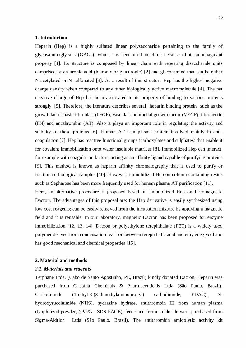
53
1. Introduction
Heparin (Hep) is a highly sulfated linear polysaccharide pertaining to the family of
glycosaminoglycans (GAGs), which has been used in clinic because of its anticoagulant
property [1]. Its structure is composed by linear chain with repeating disaccharide units
comprised of an uronic acid (iduronic or glucuronic) [2] and glucosamine that can be either
N-acetylated or N-sulfonated [3]. As a result of this structure Hep has the highest negative
charge density when compared to any other biologically active macromolecule [4]. The net
negative charge of Hep has been associated to its property of binding to various proteins
strongly [5]. Therefore, the literature describes several "heparin binding protein" such as the
growth factor basic fibroblast (bFGF), vascular endothelial growth factor (VEGF), fibronectin
(FN) and antithrombin (AT). Also it plays an important role in regulating the activity and
stability of these proteins [6]. Human AT is a plasma protein involved mainly in anti-
coagulation [7]. Hep has reactive functional groups (carboxylates and sulphates) that enable it
for covalent immobilization onto water insoluble matrices [8]. Immobilized Hep can interact,
for example with coagulation factors, acting as an affinity ligand capable of purifying proteins
[9]. This method is known as heparin affinity chromatography that is used to purify or
fractionate biological samples [10]. However, immobilized Hep on column containing resins
such as Sepharose has been more frequently used for human plasma AT purification [11].
Here, an alternative procedure is proposed based on immobilized Hep on ferromagnetic
Dacron. The advantages of this proposal are: the Hep derivative is easily synthesized using
low cost reagents; can be easily removed from the incubation mixture by applying a magnetic
field and it is reusable. In our laboratory, magnetic Dacron has been proposed for enzyme
immobilization [12, 13, 14]. Dacron or polyethylene terephthalate (PET) is a widely used
polymer derived from condensation reaction between terephthalic acid and ethyleneglycol and
has good mechanical and chemical properties [15].
2. Material and methods
2.1. Materials and reagents
Terphane Ltda. (Cabo de Santo Agostinho, PE, Brazil) kindly donated Dacron. Heparin was
purchased from Cristália Chemicals & Pharmaceuticals Ltda (São Paulo, Brazil).
Carbodiimide (1-ethyl-3-(3-dimethylaminopropyl) carbodiimide; EDAC), N-
hydroxysuccinimide (NHS), hydrazine hydrate, antithrombin III from human plasma
(lyophilized powder, ≥ 95% - SDS-PAGE), ferric and ferrous chloride were purchased from
Sigma-Aldrich Ltda (São Paulo, Brazil). The antithrombin amidolytic activity kit
54
(TriniCHROMTM
Antithrombin IIa) was acquired from the Trinity Biotech SA (New Jersey,
USA), whereas the activated partial thromboplastin time (aPTT) kit was from Labtest
Diagnóstica (Minas Gerais, Brazil). Human blood was collected from a volunteer under the
Ethical Committee of the Universidade Federal de Pernambuco approval.
2.2. Synthesis of magnetic Dacron-hydrazide (mDAC)
Dacron (polyethylene terephthalate) was converted to Dacron-hydrazide by hydrazinolysis
reaction according to Amaral et al. [12]. Briefly: Films of Dacron (4 g) were cut in strips and
incubated in methanol (100 mL) containing hydrazine hydrate (10 mL) at 40°C for 48 h with
stirring. Afterward, the Dacron-hydrazide obtained (white powder) was washed twice with
methanol and distilled water, successively. Then the Dacron-hydrazide was magnetized by co-
precipitation method of Fe2+
/Fe3+
(mDAC). Briefly: Hydrazide-Dacron (2 g) was stirred in
deionized water (100 mL), after was added an aqueous solution (10 mL) containing
FeCl3.6H2O (1.1 M) and FeCl2.4H2O (0.6 M). This mixture remained under vigorous stirring
at 90°C for 30 min with pH adjusted to 10.0 using ammonium hydroxide.
2.3. Characterization of magnetic particles
The particle size, morphology and chemical composition of Dacron-hydrazide and mDAC
were established by scanning electronic microscopy (SEM) utilizing a QUANTA 200 FEG
electron microscope equipped with Energy Dispersive X-ray (EDX). The structural properties
were characterized by X-ray diffraction (XRD) performed at room temperature in the range
10⁰ - 90⁰ , in equal 2θ steps of 0.02⁰ , using Bruker D8 Advance diffractometer with CuKα
radiation (λ = 1. 5406 Å). Magnetization measurements were made as a function of applied
magnetic field (M x H) using a vibrating sample magnetometer (VSM), Versalab,
manufactured by Quantum Design. The measurements were obtained at 293 K, 300 K and
313 K, with magnetic fields in the -30KOe to +30 KOe range.
2.4. Immobilization of heparin on the mDAC
First, a solution of heparin (3 mg/mL) was treated with EDAC/NHS to activate heparin
carboxylic groups according to Oliveira et al. [16]. An aliquot of this functionalized heparin
(1 mL) was incubated with 30 mg of mDAC for 72 h with mild agitation yielding the
covalently immobilized heparin on the magnetic support (mDAC-HEP composite). The
composite were recovered under a magnetic field (6000 Oe) and washed ten times with
deionized water to remove non-immobilized heparin. Under this magnetic field the particles
55
precipitation occurred in about 30 s but one minute was used to assure that all of them were
collected. The supernatant (SB), first washed (L1) and second washed (L2) were reserved for
heparin determination. This magnetic composite was stored in water at 4°C and used in two
moments: time zero (immediately synthesized) and 24 months stored (stored during two
years) in order to investigate the composite stability for purification of antithrombin.
2.5. Determination of heparin
Heparin was determined according to Oliveira et al. [16] based on the property of heparin to
form complexes with basic dyes such as methylene blue. The calibration curve was
constructed using heparin concentrations from 10 to 100 g/mL. The amount of immobilized
heparin was estimated through the difference between the amounts of offered heparin with
that found in the supernatants (SB, L1 and L2).
2.6. Physical-chemical analysis
Infrared spectra of the 24 months stored magnetite (MAG), mDAC and mDAC-HEP were
analyzed using, respectively, a BRUKER instrument model IFS 66 (Central Analytical of the
Departamento de Química Fundamental, Universidade Federal de Pernambuco, Brazil).
2.7. Plasma antithrombin purification
Citrated human plasma (1.0 mL) was incubated with mDAC-HEP (30 mg) and kept under
stirring for 1 h at 4°C. Both time zero and 24 months stored preparations of mDAC and
mDAC-HEP were used in the plasma AT purification. Then, the magnetic composite was
collected by a magnetic field (6000 Oe) and the supernatant stored for further analysis. The
composite was washed out with 10 mM phosphate buffer, pH 7.2, containing 150 mM NaCl
(PBS) and subsequently; the composite was eluted with the same buffer containing an
increasing gradient of NaCl (0.25 M; 0.5 M; 1.0 M). Furthermore, both mDAC-HEP
preparations (time zero and 24 months stored) were reused ten times intercalating each
successive use with PBS washing fifty times.
2.8. Measurement of antithrombin activity
The plasma antithrombin activity was evaluated by its amidolytic inhibition using
chromogenic substrate assay (Tos-Gly-Pro-Arg-ANBA-IPA) and the aPTT. These procedures
were carried out according to the manufacturers instructions. The aPTT test for the plasma
56
was performed adding 20 µL of eluate obtained with the gradient 1.0 M NaCl to the reaction
system kit.
2.9. Electrophoresis SDS/PAGE
The eluates obtained by increasing gradient of NaCl (0.25 M; 0.5 M; 1.0 M) were analyzed by
gel electrophoresis sodium dodecyl sulfate (SDS-PAGE) 12,5% according to Laemmli [17].
The two mDAC-HEP preparations were used: time zero and 24 months stored.
2.10. Statistics
Statistical analysis was performed using OriginPro 8.5 (OriginLab, Northampton, MA 01060,
USA). All data are presented as the arithmetic mean ± standard deviation of at least 3
independent replicates, except the data of reuse the material.
3. Results and Discussion
3.1. Morphology and size of particles
A white powder was obtained after Dacron hydrazinolysis reaction (Dacron-hydrazide) and a
dark one after being magnetized by the co-precipitation of Fe2+
/Fe3+
(mDAC). Images
obtained by SEM (Fig. 1) showed heterogeneous morphology and similar size for both
Dacron-hydrazide (Fig. 1a; 0.7 – 2.0 μm) and mDAC (Fig. 1b; 1.0 – 2.0 μm) microparticles,
whereas EDX analysis demonstrated the elements C, N and O in Dacron-hydrazide and the
additional presence of iron (Fe) in mDAC particles. These findings indicate the presence of
Dacron in the magnetic composite.
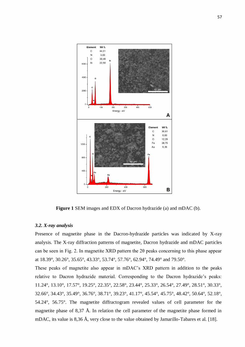
57
Figure 1 SEM images and EDX of Dacron hydrazide (a) and mDAC (b).
3.2. X-ray analysis
Presence of magnetite phase in the Dacron-hydrazide particles was indicated by X-ray
analysis. The X-ray diffraction patterns of magnetite, Dacron hydrazide and mDAC particles
can be seen in Fig. 2. In magnetite XRD pattern the 2θ peaks concerning to this phase appear
at 18.39°, 30.26°, 35.65°, 43.33°, 53.74°, 57.76°, 62.94°, 74.49° and 79.50°.
These peaks of magnetite also appear in mDAC’s XRD pattern in addition to the peaks
relative to Dacron hydrazide material. Corresponding to the Dacron hydrazide’s peaks:
11.24°, 13.10°, 17.57°, 19.25°, 22.35°, 22.58°, 23.44°, 25.33°, 26.54°, 27.49°, 28.51°, 30.33°,
32.66°, 34.43°, 35.49°, 36.76°, 38.71°, 39.23°, 41.17°, 45.54°, 45.75°, 48.42°, 50.64°, 52.18°,
54.24°, 56.75°. The magnetite diffractogram revealed values of cell parameter for the
magnetite phase of 8,37 Å. In relation the cell parameter of the magnetite phase formed in
mDAC, its value is 8,36 Å, very close to the value obtained by Jamarillo-Tabares et al. [18].
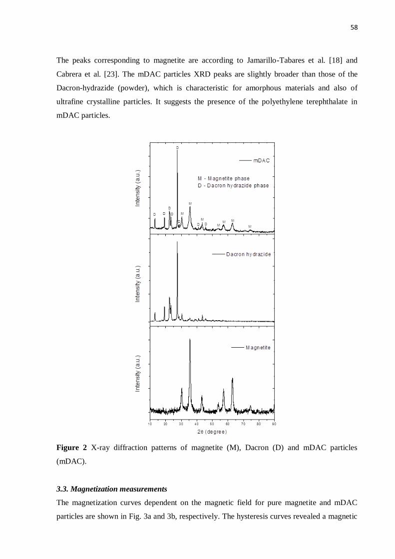
58
The peaks corresponding to magnetite are according to Jamarillo-Tabares et al. [18] and
Cabrera et al. [23]. The mDAC particles XRD peaks are slightly broader than those of the
Dacron-hydrazide (powder), which is characteristic for amorphous materials and also of
ultrafine crystalline particles. It suggests the presence of the polyethylene terephthalate in
mDAC particles.
Figure 2 X-ray diffraction patterns of magnetite (M), Dacron (D) and mDAC particles
(mDAC).
3.3. Magnetization measurements
The magnetization curves dependent on the magnetic field for pure magnetite and mDAC
particles are shown in Fig. 3a and 3b, respectively. The hysteresis curves revealed a magnetic
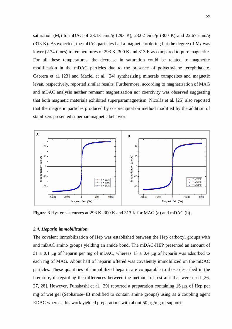
59
saturation (Ms) to mDAC of 23.13 emu/g (293 K), 23.02 emu/g (300 K) and 22.67 emu/g
(313 K). As expected, the mDAC particles had a magnetic ordering but the degree of MS was
lower (2.74 times) to temperatures of 293 K, 300 K and 313 K as compared to pure magnetite.
For all these temperatures, the decrease in saturation could be related to magnetite
modification in the mDAC particles due to the presence of polyethylene terephthalate.
Cabrera et al. [23] and Maciel et al. [24] synthesizing minerals composites and magnetic
levan, respectively, reported similar results. Furthermore, according to magnetization of MAG
and mDAC analysis neither remnant magnetization nor coercivity was observed suggesting
that both magnetic materials exhibited superparamagnetism. Nicolás et al. [25] also reported
that the magnetic particles produced by co-precipitation method modified by the addition of
stabilizers presented superparamagnetic behavior.
Figure 3 Hysteresis curves at 293 K, 300 K and 313 K for MAG (a) and mDAC (b).
3.4. Heparin immobilization
The covalent immobilization of Hep was established between the Hep carboxyl groups with
and mDAC amino groups yielding an amide bond. The mDAC-HEP presented an amount of
51 ± 0.1 μg of heparin per mg of mDAC, whereas 13 ± 0.4 μg of heparin was adsorbed to
each mg of MAG. About half of heparin offered was covalently immobilized on the mDAC
particles. These quantities of immobilized heparin are comparable to those described in the
literature, disregarding the differences between the methods of restraint that were used [26,
27, 28]. However, Funahashi et al. [29] reported a preparation containing 16 g of Hep per
mg of wet gel (Sepharose-4B modified to contain amine groups) using as a coupling agent
EDAC whereas this work yielded preparations with about 50 g/mg of support.
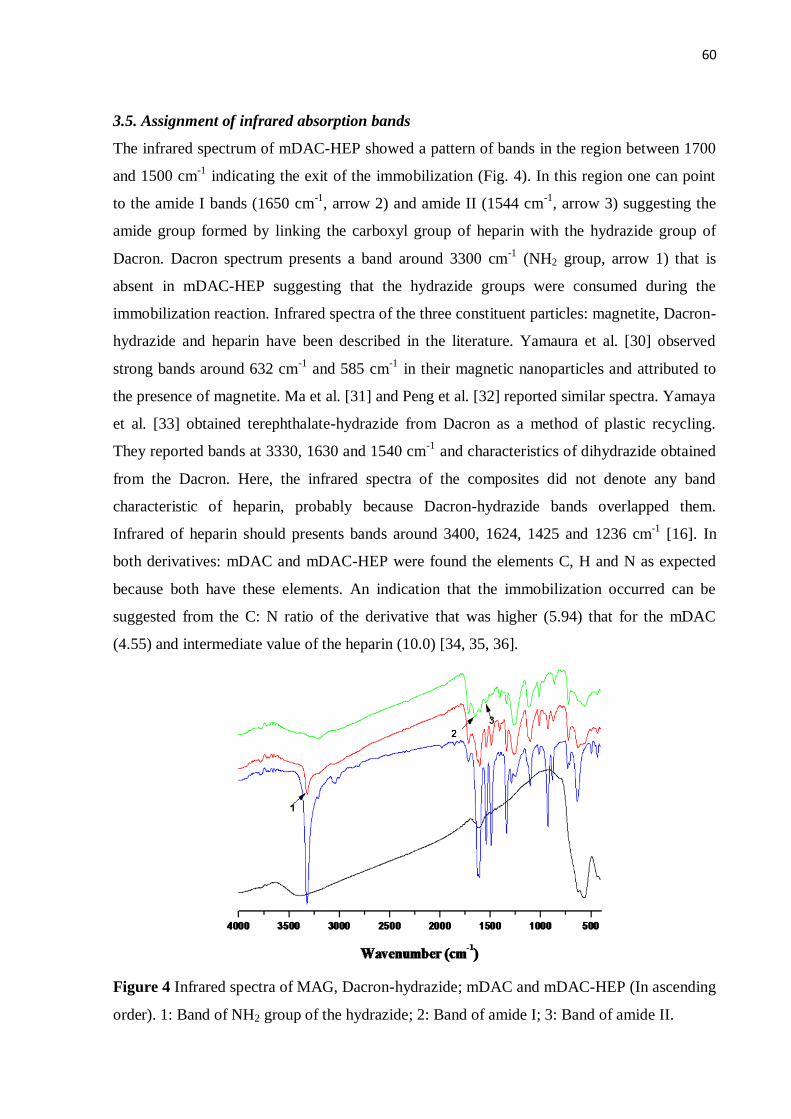
60
3.5. Assignment of infrared absorption bands
The infrared spectrum of mDAC-HEP showed a pattern of bands in the region between 1700
and 1500 cm-1
indicating the exit of the immobilization (Fig. 4). In this region one can point
to the amide I bands (1650 cm-1
, arrow 2) and amide II (1544 cm-1
, arrow 3) suggesting the
amide group formed by linking the carboxyl group of heparin with the hydrazide group of
Dacron. Dacron spectrum presents a band around 3300 cm-1
(NH2 group, arrow 1) that is
absent in mDAC-HEP suggesting that the hydrazide groups were consumed during the
immobilization reaction. Infrared spectra of the three constituent particles: magnetite, Dacron-
hydrazide and heparin have been described in the literature. Yamaura et al. [30] observed
strong bands around 632 cm-1
and 585 cm-1
in their magnetic nanoparticles and attributed to
the presence of magnetite. Ma et al. [31] and Peng et al. [32] reported similar spectra. Yamaya
et al. [33] obtained terephthalate-hydrazide from Dacron as a method of plastic recycling.
They reported bands at 3330, 1630 and 1540 cm-1
and characteristics of dihydrazide obtained
from the Dacron. Here, the infrared spectra of the composites did not denote any band
characteristic of heparin, probably because Dacron-hydrazide bands overlapped them.
Infrared of heparin should presents bands around 3400, 1624, 1425 and 1236 cm-1
[16]. In
both derivatives: mDAC and mDAC-HEP were found the elements C, H and N as expected
because both have these elements. An indication that the immobilization occurred can be
suggested from the C: N ratio of the derivative that was higher (5.94) that for the mDAC
(4.55) and intermediate value of the heparin (10.0) [34, 35, 36].
Figure 4 Infrared spectra of MAG, Dacron-hydrazide; mDAC and mDAC-HEP (In ascending
order). 1: Band of NH2 group of the hydrazide; 2: Band of amide I; 3: Band of amide II.
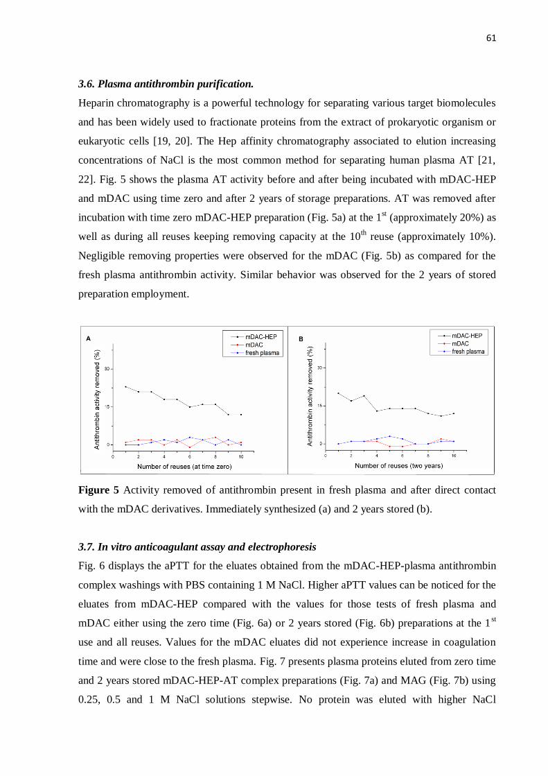
61
3.6. Plasma antithrombin purification.
Heparin chromatography is a powerful technology for separating various target biomolecules
and has been widely used to fractionate proteins from the extract of prokaryotic organism or
eukaryotic cells [19, 20]. The Hep affinity chromatography associated to elution increasing
concentrations of NaCl is the most common method for separating human plasma AT [21,
22]. Fig. 5 shows the plasma AT activity before and after being incubated with mDAC-HEP
and mDAC using time zero and after 2 years of storage preparations. AT was removed after
incubation with time zero mDAC-HEP preparation (Fig. 5a) at the 1st (approximately 20%) as
well as during all reuses keeping removing capacity at the 10th reuse (approximately 10%).
Negligible removing properties were observed for the mDAC (Fig. 5b) as compared for the
fresh plasma antithrombin activity. Similar behavior was observed for the 2 years of stored
preparation employment.
Figure 5 Activity removed of antithrombin present in fresh plasma and after direct contact
with the mDAC derivatives. Immediately synthesized (a) and 2 years stored (b).
3.7. In vitro anticoagulant assay and electrophoresis
Fig. 6 displays the aPTT for the eluates obtained from the mDAC-HEP-plasma antithrombin
complex washings with PBS containing 1 M NaCl. Higher aPTT values can be noticed for the
eluates from mDAC-HEP compared with the values for those tests of fresh plasma and
mDAC either using the zero time (Fig. 6a) or 2 years stored (Fig. 6b) preparations at the 1st
use and all reuses. Values for the mDAC eluates did not experience increase in coagulation
time and were close to the fresh plasma. Fig. 7 presents plasma proteins eluted from zero time
and 2 years stored mDAC-HEP-AT complex preparations (Fig. 7a) and MAG (Fig. 7b) using
0.25, 0.5 and 1 M NaCl solutions stepwise. No protein was eluted with higher NaCl
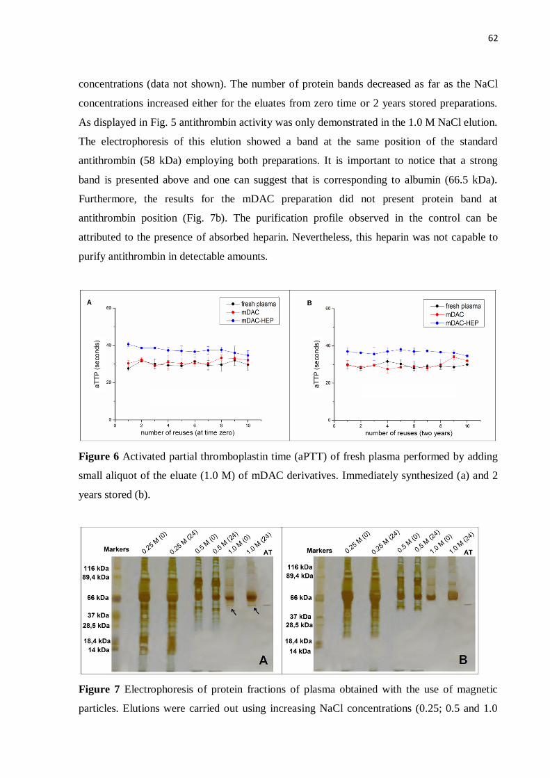
62
concentrations (data not shown). The number of protein bands decreased as far as the NaCl
concentrations increased either for the eluates from zero time or 2 years stored preparations.
As displayed in Fig. 5 antithrombin activity was only demonstrated in the 1.0 M NaCl elution.
The electrophoresis of this elution showed a band at the same position of the standard
antithrombin (58 kDa) employing both preparations. It is important to notice that a strong
band is presented above and one can suggest that is corresponding to albumin (66.5 kDa).
Furthermore, the results for the mDAC preparation did not present protein band at
antithrombin position (Fig. 7b). The purification profile observed in the control can be
attributed to the presence of absorbed heparin. Nevertheless, this heparin was not capable to
purify antithrombin in detectable amounts.
Figure 6 Activated partial thromboplastin time (aPTT) of fresh plasma performed by adding
small aliquot of the eluate (1.0 M) of mDAC derivatives. Immediately synthesized (a) and 2
years stored (b).
Figure 7 Electrophoresis of protein fractions of plasma obtained with the use of magnetic
particles. Elutions were carried out using increasing NaCl concentrations (0.25; 0.5 and 1.0
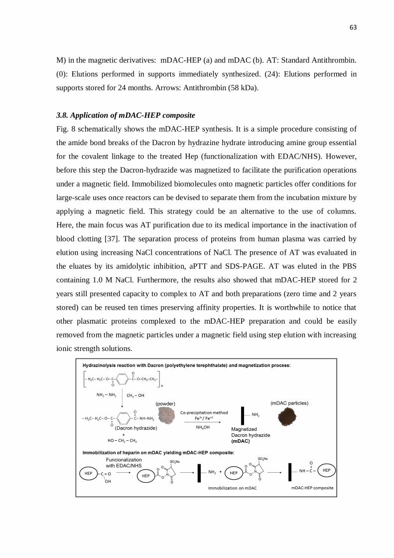
63
M) in the magnetic derivatives: mDAC-HEP (a) and mDAC (b). AT: Standard Antithrombin.
(0): Elutions performed in supports immediately synthesized. (24): Elutions performed in
supports stored for 24 months. Arrows: Antithrombin (58 kDa).
3.8. Application of mDAC-HEP composite
Fig. 8 schematically shows the mDAC-HEP synthesis. It is a simple procedure consisting of
the amide bond breaks of the Dacron by hydrazine hydrate introducing amine group essential
for the covalent linkage to the treated Hep (functionalization with EDAC/NHS). However,
before this step the Dacron-hydrazide was magnetized to facilitate the purification operations
under a magnetic field. Immobilized biomolecules onto magnetic particles offer conditions for
large-scale uses once reactors can be devised to separate them from the incubation mixture by
applying a magnetic field. This strategy could be an alternative to the use of columns.
Here, the main focus was AT purification due to its medical importance in the inactivation of
blood clotting [37]. The separation process of proteins from human plasma was carried by
elution using increasing NaCl concentrations of NaCl. The presence of AT was evaluated in
the eluates by its amidolytic inhibition, aPTT and SDS-PAGE. AT was eluted in the PBS
containing 1.0 M NaCl. Furthermore, the results also showed that mDAC-HEP stored for 2
years still presented capacity to complex to AT and both preparations (zero time and 2 years
stored) can be reused ten times preserving affinity properties. It is worthwhile to notice that
other plasmatic proteins complexed to the mDAC-HEP preparation and could be easily
removed from the magnetic particles under a magnetic field using step elution with increasing
ionic strength solutions.
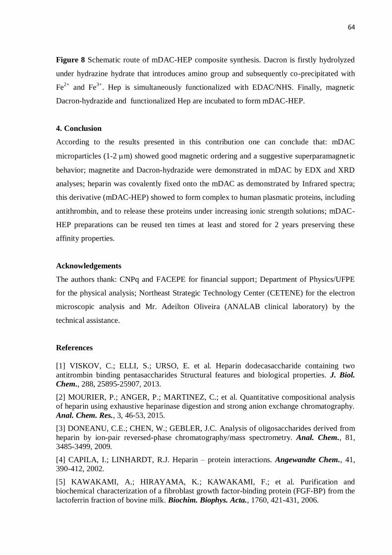
64
Figure 8 Schematic route of mDAC-HEP composite synthesis. Dacron is firstly hydrolyzed
under hydrazine hydrate that introduces amino group and subsequently co-precipitated with
Fe2+
and Fe3+
. Hep is simultaneously functionalized with EDAC/NHS. Finally, magnetic
Dacron-hydrazide and functionalized Hep are incubated to form mDAC-HEP.
4. Conclusion
According to the results presented in this contribution one can conclude that: mDAC
microparticles (1-2 m) showed good magnetic ordering and a suggestive superparamagnetic
behavior; magnetite and Dacron-hydrazide were demonstrated in mDAC by EDX and XRD
analyses; heparin was covalently fixed onto the mDAC as demonstrated by Infrared spectra;
this derivative (mDAC-HEP) showed to form complex to human plasmatic proteins, including
antithrombin, and to release these proteins under increasing ionic strength solutions; mDAC-
HEP preparations can be reused ten times at least and stored for 2 years preserving these
affinity properties.
Acknowledgements
The authors thank: CNPq and FACEPE for financial support; Department of Physics/UFPE
for the physical analysis; Northeast Strategic Technology Center (CETENE) for the electron
microscopic analysis and Mr. Adeilton Oliveira (ANALAB clinical laboratory) by the
technical assistance.
References
[1] VISKOV, C.; ELLI, S.; URSO, E. et al. Heparin dodecasaccharide containing two
antitrombin binding pentasaccharides Structural features and biological properties. J. Biol.
Chem., 288, 25895-25907, 2013.
[2] MOURIER, P.; ANGER, P.; MARTINEZ, C.; et al. Quantitative compositional analysis
of heparin using exhaustive heparinase digestion and strong anion exchange chromatography.
Anal. Chem. Res., 3, 46-53, 2015.
[3] DONEANU, C.E.; CHEN, W.; GEBLER, J.C. Analysis of oligosaccharides derived from
heparin by ion-pair reversed-phase chromatography/mass spectrometry. Anal. Chem., 81,
3485-3499, 2009.
[4] CAPILA, I.; LINHARDT, R.J. Heparin – protein interactions. Angewandte Chem., 41,
390-412, 2002.
[5] KAWAKAMI, A.; HIRAYAMA, K.; KAWAKAMI, F.; et al. Purification and
biochemical characterization of a fibroblast growth factor-binding protein (FGF-BP) from the
lactoferrin fraction of bovine milk. Biochim. Biophys. Acta., 1760, 421-431, 2006.
65
[6] ARISAKA, Y.; KOBAYASHI, J.; YAMATO, M.; et al. Switching of cell
growth/detachment on heparin-functionalized thermoresponsive surface for rapid cell sheet
fabrication and manipulation. J. Biomater., 34, 4214-4222, 2013.
[7] KARLSSON, G.; WINGE, S. Separation of latent, prelatent, and native forms of human
antithrombin by heparin affinity high-performance liquid chromatography. Protein Expr.
Purif., 33, 339-345, 2004.
[8] MURUGESAN, S.; XIE, J.; LINHARDT, R.J. Immobilization of heparin: approaches and
applications. Curr. Top. Med. Chem., 8, 80-100, 2008.
[9] Krapfenbauer K, Fountoulakis M. Improved enrichment and proteomic analysis of brain
proteins with signaling function by heparin chromatography. Methods Mol. Biol., 566, 165-
180, 2009.
[10] LEI, T.; HE, Q.Y.; WANG, Y.L.; et al. Heparin chromatography to deplete high-
abundance proteins for serum proteomics. Clinic Chimica Acta, 388, 173-178, 2008.
[11] JOSIC, D.; BAL, F.; SCHWINN, H. Isolation of plasma proteins from the clotting
cascade by heparin affinity chromatography. J. Chromatogr., 632, 1-10, 1993.
[12] AMARAL, I.P.G.; CARNEIRO-DA-CUNHA, M.G.; CARVALHO-JÚNIOR, L.B.; et al.
Fish trypsin immobilized on ferromagnetic Dacron. Process Biochem., 41, 1213-1216, 2006.
[13] NERI, D.F.M.; BALCÃO, V.M.; CARDOSO, S.M.; et al. Characterization of
galactooligosaccharides produced by β-galactosidase immobilized onto magnetized Dacron.
Int. Dairy J., 21, 172-178, 2011.
[14] SORIA, F.; ELLENRIEDER, G.; OLIVEIRA, G.B.; et al. α-L-Rhamnosidase of
Aspergillus terreus immobilized on ferromagnetic supports. Appl. Microbiol. Biotechnol., 93,
1127-1134, 2012.
[15] DUARTE, L.T.; PAULA E SILVA, E.M.; BRANCO, J.R.T.; et al. Production and
characterization of thermally sprayed polyethylene terephthalate coatings. Surf. Coat
Technol., 182, 261-267, 2004.
[16] OLIVEIRA, G.B.; CARVALHO JR, L.B.; SILVA, M.P.C. Properties of carbodiimide
treated heparin. J. Biomater., 24, 4777-4783, 2003.
[17] LAEMMLI, U.K. Cleavage of structural proteins during the assembly of head of
bacteriophage T4. Nat., 227, 680-685, 1970.
[18] JARAMILLO-TABARES, B.E.; ISAZA, F.J.; TORRESI, S.I.C. Stabilization of
polyaniline by the incorporation of magnetite nanoparticles. Mater. Chem. Phys., 132, 529-
533, 2012.
[19] MARQUES, M.A.M.; ESPINOSA, B.J.; SILVEIRA, E.K.X.; et al. Continued proteomic
analysis of Mycobacterium leprae subcellular fractions. J. Proteomics., 4, 2942-2953, 2004.
[20] SRIVASTAVA, P.N.; FAROOQUI, A.A. Heparin-sepharose affinity chromatography
for purification of bull seminal-plasma hyaluronidase. Biochem. J., 183, 531-537, 1979.
[21] KONG, Y.; ZHANG, G.; LUO, J.; et al. Facile purification and characterization of
recombinant human antithrombin III from transgenic goat milk. J. Chem. Technol.
Biotechnol., 86, 1303-1309, 2011.
[22] WICKERHAUSER, M.; WILLIAMS, C.; MERCER, J. Development of large scale
fractionation methods. VII Preparation of antithrombin III concentrate. Vox Sanguinis, 36,
281-293, 1979.
66
[23] CABRERA, M.; MACIEL, J.C.; QUISPE-MARCATOMA, J.; et al. Magnetic
composites from minerals: study of the iron phases in clay and diatomite using Mössbauer
spectroscopy, magnetic measurements and XRD. Hyperfine Interact., 224, 197-204, 2014.
[24] MACIEL, J.C.; ANDRAD, P.L.; NERI, D.F.M.; et al. Preparation and characterization
of magnetic levan particles as matrix for trypsin immobilization. J. Magn. Magn. Mater.,
324, 1312-1316, 2012.
[25] NICOLÁS, P.; SALETA, M.; TROIANI, H.; et al. Preparation of iron oxide
nanoparticles stabilized with biomolecules: Experimental and mechanistic issues. Acta
Biomater., 9, 4754-4762, 2013.
[26] CHEN, L.; GUO, C.; GUAN, Y.; et al. Isolation of lactoferrin from acid whey by
magnetic affinity separation. Sep. Purif. Technol., 56, 168-174, 2007.
[27] BJÖRKLUND, M.; HEARN, M.T.W. Synthesis of silica-based heparin-affinity
adsorbents. J. Chromatogr. A, 728, 149-169, 1996.
[28] DENIZLI, A.; PISKIN, E. Heparin-immobilized polyhydroxyethylmethacrylate
microbeads for cholesterol removal: a preliminary report. J. Chromatogr. B, 670, 157-161,
1995.
[29] FUNAHASHI, M.; MATSUMOTO, I.; SENO, N. Preparation of three types of heparin-
sepharose and their binding activities to thrombin and antithrombin III. Anal. Biochem. 126,
414-421, 1982.
[30] YAMAURA, M.; CAMILO, R.L.; SAMPAIO, L.C.; et al. Preparation and
characterization of (3-aminopropyl) triethoxysilane-coated magnetite nanoparticles. J. Magn.
Magn. Mater., 279, 210-217, 2004.
[31] MA, M.; ZHANG, Y.; YU, W.; et al. Preparation and characterization of magnetite
nanoparticles coated by amino silane. Colloids Surf A: Physicochem. Eng. Asp., 212, 219-
226, 2003.
[32] PENG, Z.G.; HIDAJAT, K.; UDDIN, M.S. Adsorption of bovine serum albumin on
nanosized magnetic particles. J. Colloid Interface Sci., 271, 277-283, 2004.
[33] YAMAYE, M.; HASHIME, T.; YAMAMOTO, K.; et al. Chemical recycling of
poly(ethylene terephthalate) 2 Preparation of terephthalohydroxamic acid and
terephthalohydrazide. Ind. Eng. Chem. Res., 41, 3993-3998, 2002.
[34] GRIFFIN, C.C.; LINHARDT, R.J.; VAN GORP, C.L.; et al. Isolation and
characterization of heparan sulfate from crude porcine intestinal mucosal peptidoglycan
heparin. Carbohydr. Res., 276, 183-197, 1995.
[35] DANISHEFSKY, I.; SISKOVIC, E. Heparin derivatives prepared by modification of the
uronic acid carboxyl groups. Thromb Res., 1, 173-182, 1972.
[36] BURSON, S.L.; FAHRENBACH, M.J.; FROMMHAGEN, L.H.; et al. Isolation and
Purification of Mactins, Heparin-like Anticoagulants from Mollusca. J. Am. Chem. Soc., 78,
5874-5878, 1956.
[37] LIU, T.; LIU, Y.; CHEN, Y.; et al. Immobilization of heparin/poly-L-lysine
nanoparticles on dopamine-coated surface to create a heparin density gradient for selective
direction of platelet and vascular cells behavior. Acta Biomater., 10, 1940-1954, 2014.
67
6 ARTIGO 2
FRACTIONATION OF HUMAN PLASMA PROTEINS BY AFFINITY TO HEPARIN
IMMOBILIZED ONTO MAGNETITE COATED WITH POLYANILINE.
Aurenice Arruda Dutra das Mercês1, Karciano José Santos Silva
2, Gabriela Ayres Fragoso
Nascimento¹, Jackeline da Costa Maciel3, Davian Martinez Buitrago
2, José Albino Oliveira de
Aguiar2, Luiz Bezerra de Carvalho Júnior
1.
1Laboratório de Imunopatologia Keizo Asami, Universidade Federal de Pernambuco, Cidade
Universitária, 50670-901 Recife, PE, Brazil.
2Departamento de Física, Universidade Federal de Pernambuco, Cidade Universitária,
50670-901 Recife, PE, Brazil.
3Universidade Federal de Roraima, Aeroporto, 69310-000 Boa Vista, RR, Brazil.
ABSTRACT
This works aimed to immobilize heparin (HEP) onto magnetite coated with polyaniline
particles (MAG-PANI) and to use for fractionation and isolation of proteins from human
plasma. Magnetite (MAG) was obtained by co-precipitation method of Fe2+/Fe3+ and then
the particles were coated with polyaniline by the oxidation reaction of aniline with potassium
permanganate. HEP was activated by carbodiimide and N-hydroxysuccinimide and
immobilized on MAG-PANI yielding the MAG-PANI-HEP composite containing 26.4 ± 0.09
μg heparin/mg of support. TEM analysis showed that MAG-PANI has a spherical
morphology and size of 14.4 ± 0.9 nm and XRD indicates the presence of polyaniline phase in
MAG. Besides that MAG-PANI exhibited superparamagnetism and magnetic saturation equal
to about 66 emu/g to 293 K, 300 K and 313 K. Human plasma was diluted (1:5) with
phosphate buffer (PBS) and incubated with MAG-PANI-HEP and washed with phosphate
buffer (PBS) containing increasing NaCl concentration and the fractionation process was
monitored at 280 nm. Electrophoresis of collected fractions 1 to 6 showed the presence of
proteins with 23.9 to 151.3 kDa. F9 collected with 1.0 M NaCl in PBS showed a single band
(58.8 kDa) corresponding to antithrombin, a protein with anticoagulant activity. APTT test
was performed and it was observed that the plasma does not clotted after being incubated with
F9. This method can be applied to the blood products industry since it proved useful as a tool
for the fractionation of human plasma being possible to make the depletion of plasma proteins
and also isolate human antithrombin.
68
Key words: fractionation; heparin; immobilization; magnetite; polyaniline; plasma proteins.
1. Introduction
Human plasma contains proteins with a wide range of biological functions and is one of the
most commonly used biofluids in clinical diagnostics. However, the complexity of plasma,
namely due to the wide dynamic range of protein abundance, has been a major obstacle to the
comprehensive analysis and understanding of the plasma proteome [1]. These high-abundance
proteins hinder the detection of low-abundance proteins which are potential biomarkers for
various diseases. To improve their detection, high-abundance proteins like IgG and albumin
must be removed [2]. Different methods can be used to deplete these proteins in plasma
samples, being liquid chromatography the most popular one. A variety of stationary phases
are available for this purpose such as affinity columns containing lectins, peptides or
inorganic ligands. These methods are easy to use and to scale-up, but are relatively expensive.
Additionally, they have some disadvantages, such as non-specific interactions that lead to the
loss of some proteins [3]. Heparin is a linear polysaccharide of the glycosaminoglycan family,
has a highly sulfated chain formed by repeating disaccharide units consisting of a uronic acid
(iduronic or glucuronic) and glucosamine [4]. Due to this structure heparin has a higher
negative charge density, when compared to any other biologically active macromolecule [5],
allowing heparin to interact with a large number of different proteins [6]. Heparin when
immobilized may interact with the coagulation factors, acting as an affinity ligand capable of
interacting with these proteins [7], this method is known as heparin-affinity chromatography
and is also widely used to fractionate or purify proteins and other biological substances that
may interact with heparin. The high binding affinity of AT to heparin has been used in affinity
chromatography systems for the isolation of antithrombin from human plasma [8].
Furthermore, the use of heparin affinity chromatography can be applied as a strategy to
selectively remove some abundance proteins, facilitating the analysis of low concentration in
plasma proteins [9]. Magnetic particles are among the most commonly used materials in
separation techniques, with applications such as immobilization of proteins, enzymes, and
other bioactive agents used in medical analytical systems and biotechnology. The
paramagnetic properties of these particles allow the easy isolation of products in solution by
attracting them with the aid of an external magnetic field. Thus, suspended magnetic particles
tagged with analytes can be removed from large volume samples using a magnet and their
isolation and purification are easier and faster than with other methods [10]. The iron oxide
nanoparticles in the crystalline form of magnetite (Fe3O4) contain supermagnetic and
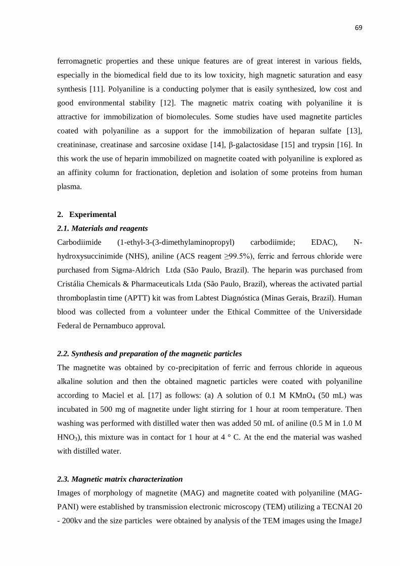
69
ferromagnetic properties and these unique features are of great interest in various fields,
especially in the biomedical field due to its low toxicity, high magnetic saturation and easy
synthesis [11]. Polyaniline is a conducting polymer that is easily synthesized, low cost and
good environmental stability [12]. The magnetic matrix coating with polyaniline it is
attractive for immobilization of biomolecules. Some studies have used magnetite particles
coated with polyaniline as a support for the immobilization of heparan sulfate [13],
creatininase, creatinase and sarcosine oxidase [14], β-galactosidase [15] and trypsin [16]. In
this work the use of heparin immobilized on magnetite coated with polyaniline is explored as
an affinity column for fractionation, depletion and isolation of some proteins from human
plasma.
2. Experimental
2.1. Materials and reagents
Carbodiimide (1-ethyl-3-(3-dimethylaminopropyl) carbodiimide; EDAC), N-
hydroxysuccinimide (NHS), aniline (ACS reagent ≥99.5%), ferric and ferrous chloride were
purchased from Sigma-Aldrich Ltda (São Paulo, Brazil). The heparin was purchased from
Cristália Chemicals & Pharmaceuticals Ltda (São Paulo, Brazil), whereas the activated partial
thromboplastin time (APTT) kit was from Labtest Diagnóstica (Minas Gerais, Brazil). Human
blood was collected from a volunteer under the Ethical Committee of the Universidade
Federal de Pernambuco approval.
2.2. Synthesis and preparation of the magnetic particles
The magnetite was obtained by co-precipitation of ferric and ferrous chloride in aqueous
alkaline solution and then the obtained magnetic particles were coated with polyaniline
according to Maciel et al. [17] as follows: (a) A solution of 0.1 M KMnO4 (50 mL) was
incubated in 500 mg of magnetite under light stirring for 1 hour at room temperature. Then
washing was performed with distilled water then was added 50 mL of aniline (0.5 M in 1.0 M
HNO3), this mixture was in contact for 1 hour at 4 ° C. At the end the material was washed
with distilled water.
2.3. Magnetic matrix characterization
Images of morphology of magnetite (MAG) and magnetite coated with polyaniline (MAG-
PANI) were established by transmission electronic microscopy (TEM) utilizing a TECNAI 20
- 200kv and the size particles were obtained by analysis of the TEM images using the ImageJ
70
1.48v (Wayne Rasband, National Institutes of Health, USA) software. The structural
properties were characterized by X-ray diffraction (XRD) performed at room temperature in
the range 10⁰ - 90⁰ , in equal 2θ steps of 0.02⁰ , using Bruker D8 Advance diffractometer
with CuKα radiation (λ = 1. 5406 Å). Magnetization measurements were made as a function
of applied magnetic field (M x H) using a vibrating sample magnetometer (VSM), Versalab,
manufactured by Quantum Design. The measurements were obtained at 293 K, 300 K and
313 K, with magnetic fields in the -30KOe to +30 KOe range.
2.4. Immobilization and determination of heparin
A solution of heparin with approximately 3.5 mg/mL was treated with EDAC/NHS to activate
heparin carboxylic groups according to Oliveira et al [18]. An aliquot (1 mL) of this
functionalized heparin solution was incubated with 30 mg of MAG-PANI for 72 h with mild
agitation yielding the covalently immobilized heparin on the magnetic support (MAG-PANI-
HEP composite). The composite were recovered under a magnetic field (6000 Oe) and
washed ten times with deionized water to remove non-immobilized heparin. Under this
magnetic field the particles precipitation occurred in about 10. The supernatant (SB), first
washed (L1) and second washed (L2) were incubated with methylene blue at room
temperature. This resulted in a complex of methylene blue with heparin. The solution was
measured at 664 nm using a spectrophotometer (Ultrospec 3000 Pro UV-VIS). The
calibration curve was obtained through measurement of the absorbance of a series of
standards at heparin concentrations of 10–100 μg/mL. The amount of immobilized heparin
was calculated from the calibration curve.
2.5. Fractioning of proteins by magnetic composite affinity
Human plasma was obtained from intravenous blood puncture using a citrate tube. The total
plasma was diluted (1:5) with 10 mM phosphate buffer, pH 7.2, containing 150 mM NaCl
(PBS). So, 1 mL of the diluted plasma was filtered through a membrane (0.22 µm) and
incubated at 30 mg MAG-PANI-HEP for 60 minutes at 4°C. Then, the magnetic composite
was collected by a magnetic field (6000 Oe) and the proteins that bind affinity to the magnetic
composite were eluted with the PBS containing an increasing gradient of NaCl (0.25 M; 0.5
M; 1.0 M; 1.5 M; 2.0 M). For each concentration gradient, 3 fractions were obtained.
2.6. Monitoring of eluted proteins
71
During the elution process, the fractions collected with NaCl (0.25, 0.5, 1.0, 1.5 and 2.0 M) in
PBS were monitored at 280 nm (Ultrospec 3000 Pro UV-VIS spectrophotometer) and and
absorbance values were gathered to mount a chromatogram graph.
2.7. SDS–PAGE electrophoresis
Electrophoresis of proteins SDS/PAGE (sodium dodecyl sulfate/polyacrylamide gel
electrophoresis) 10.0% was performed to the fractions collected between 1 and 9
corresponding the eluates obtained with NaCl (0.25, 0.5, 1.0 M) in PBS. The molecular
weight corresponding to protein bands were analyzed according to their electrophoretic
mobility and compared with a standard molecular weight (260 kDa to 26 kDa).
2.8. Antithrombin activity test
The isolated antithrombin (AT) obtained in the fractionation process was incubated at a
normal plasma to check their in vitro activity as follows: (a) 100 uL of normal plasma was
mixed with 100 µL of third eluate obtained with the gradient 1.0 M NaCl for 5 minutes at
4°C; (b) the APTT test for this plasma solution was performed using the reaction system kit.
3. Results and Discussion
3.1. Morphology and size of magnetic particles
Magnetic particles obtained by co-precipitation method showed a black powder aspect. The
treatment of the particles with KMnO4 was important to oxidize the aniline and thus obtain
the coating with the polyaniline. From the analysis of images obtained with TEM, MAG
particles (Fig. 1A), and MAG-PANI particles (Fig. 1B) have a varied morphology like a
sphere. In addition, MAG-PANI have a size of 14.4 ± 0.9 nm while MAG showed a smaller
size (10.0 ± 1.4 nm). The particles of MAG-PANI have the coating with polyaniline which
increased its size when compared to bare particle (MAG). The increased particle size of
MAG-PANI when compared to MAG PANI may be attributed to the coating on the magnetite
particles with polyaniline The coating produced changes in the size particle according to
Maciel et al. [19] Silva et al. [20]. Similar results were shown by Chelminiak et al. [21] that
synthesized magnetite nanoparticles coated with poly (acrylic acid) and this material by TEM
analysis showed a size of about 13 nm, larger than compared to pure magnetite.
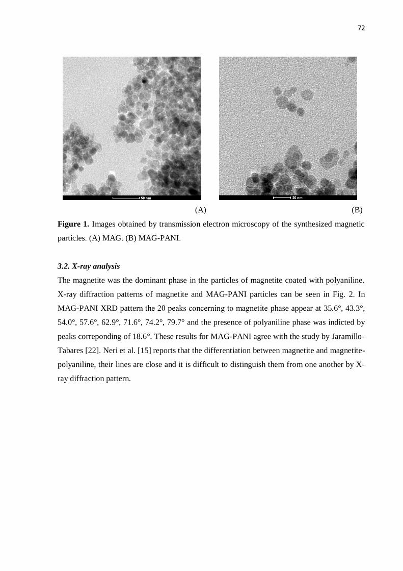
72
(A) (B)
Figure 1. Images obtained by transmission electron microscopy of the synthesized magnetic
particles. (A) MAG. (B) MAG-PANI.
3.2. X-ray analysis
The magnetite was the dominant phase in the particles of magnetite coated with polyaniline.
X-ray diffraction patterns of magnetite and MAG-PANI particles can be seen in Fig. 2. In
MAG-PANI XRD pattern the 2θ peaks concerning to magnetite phase appear at 35.6°, 43.3°,
54.0°, 57.6°, 62.9°, 71.6°, 74.2°, 79.7° and the presence of polyaniline phase was indicted by
peaks correponding of 18.6°. These results for MAG-PANI agree with the study by Jaramillo-
Tabares [22]. Neri et al. [15] reports that the differentiation between magnetite and magnetite-
polyaniline, their lines are close and it is difficult to distinguish them from one another by X-
ray diffraction pattern.
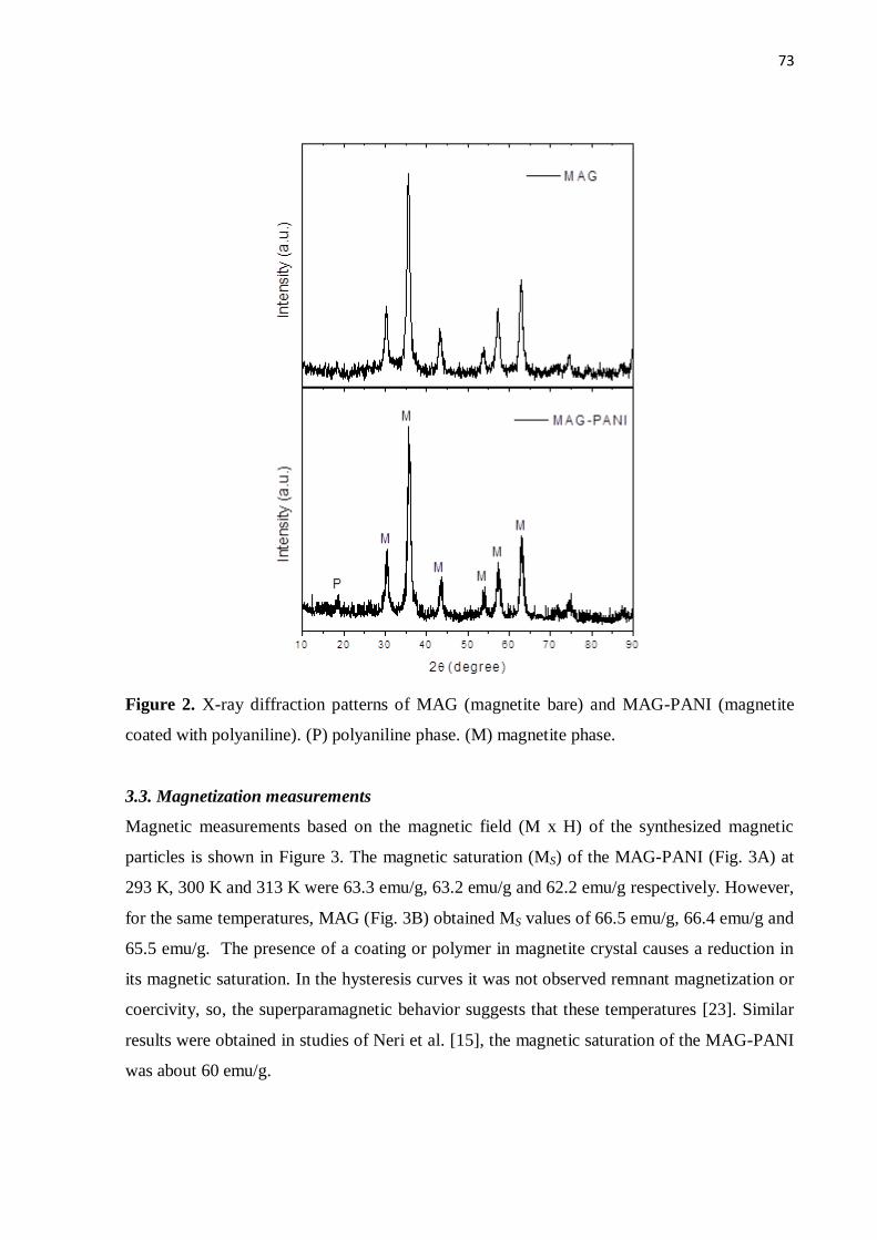
73
Figure 2. X-ray diffraction patterns of MAG (magnetite bare) and MAG-PANI (magnetite
coated with polyaniline). (P) polyaniline phase. (M) magnetite phase.
3.3. Magnetization measurements
Magnetic measurements based on the magnetic field (M x H) of the synthesized magnetic
particles is shown in Figure 3. The magnetic saturation (MS) of the MAG-PANI (Fig. 3A) at
293 K, 300 K and 313 K were 63.3 emu/g, 63.2 emu/g and 62.2 emu/g respectively. However,
for the same temperatures, MAG (Fig. 3B) obtained MS values of 66.5 emu/g, 66.4 emu/g and
65.5 emu/g. The presence of a coating or polymer in magnetite crystal causes a reduction in
its magnetic saturation. In the hysteresis curves it was not observed remnant magnetization or
coercivity, so, the superparamagnetic behavior suggests that these temperatures [23]. Similar
results were obtained in studies of Neri et al. [15], the magnetic saturation of the MAG-PANI
was about 60 emu/g.
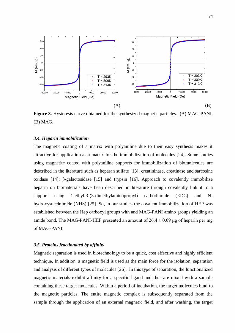
74
(A) (B)
Figure 3. Hysteresis curve obtained for the synthesized magnetic particles. (A) MAG-PANI.
(B) MAG.
3.4. Heparin immobilization
The magnetic coating of a matrix with polyaniline due to their easy synthesis makes it
attractive for application as a matrix for the immobilization of molecules [24]. Some studies
using magnetite coated with polyaniline supports for immobilization of biomolecules are
described in the literature such as heparan sulfate [13]; creatininase, creatinase and sarcosine
oxidase [14]; β-galactosidase [15] and trypsin [16]. Approach to covalently immobilize
heparin on biomaterials have been described in literature through covalently link it to a
support using 1-ethyl-3-(3-dimethylaminopropyl) carbodiimide (EDC) and N-
hydroxysuccinimide (NHS) [25]. So, in our studies the covalent immobilization of HEP was
established between the Hep carboxyl groups with and MAG-PANI amino groups yielding an
amide bond. The MAG-PANI-HEP presented an amount of 26.4 ± 0.09 μg of heparin per mg
of MAG-PANI.
3.5. Proteins fractionated by affinity
Magnetic separation is used in biotechnology to be a quick, cost effective and highly efficient
technique. In addition, a magnetic field is used as the main force for the isolation, separation
and analysis of different types of molecules [26]. In this type of separation, the functionalized
magnetic materials exhibit affinity for a specific ligand and thus are mixed with a sample
containing these target molecules. Within a period of incubation, the target molecules bind to
the magnetic particles. The entire magnetic complex is subsequently separated from the
sample through the application of an external magnetic field, and after washing, the target
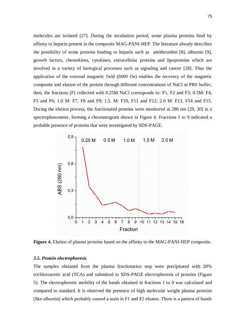
75
molecules are isolated [27]. During the incubation period, some plasma proteins bind by
affinity to heparin present in the composite MAG-PANI-HEP. The literature already describes
the possibility of some proteins binding to heparin such as antithrombin [8], albumin [9],
growth factors, chemokines, cytokines, extracellular proteins and lipoproteins which are
involved in a variety of biological processes such as signaling and cancer [28]. Thus the
application of the external magnetic field (6000 Oe) enables the recovery of the magnetic
composite and elution of the protein through different concentrations of NaCl in PBS buffer,
then, the fractions (F) collected with 0.25M NaCl corresponds to: F1, F2 and F3; 0.5M: F4,
F5 and F6; 1.0 M: F7, F8 and F9; 1.5. M: F10, F11 and F12; 2.0 M: F13, F14 and F15.
During the elution process, the fractionated proteins were monitored at 280 nm [29, 30] in a
spectrophotometer, forming a chromatogram shown in Figure 4. Fractions 1 to 9 indicated a
probable presence of proteins that were investigated by SDS-PAGE.
Figure 4. Elution of plasma proteins based on the affinity to the MAG-PANI-HEP composite.
3.5. Protein electrophoresis
The samples obtained from the plasma fractionation step were precipitated with 20%
trichloroacetic acid (TCA) and submitted to SDS-PAGE electrophoresis of proteins (Figure
5). The electrophoretic mobility of the bands obtained in fractions 1 to 9 was calculated and
compared to standard. It is observed the presence of high molecular weight plasma proteins
(like albumin) which probably caused a stain in F1 and F2 eluates. There is a pattern of bands
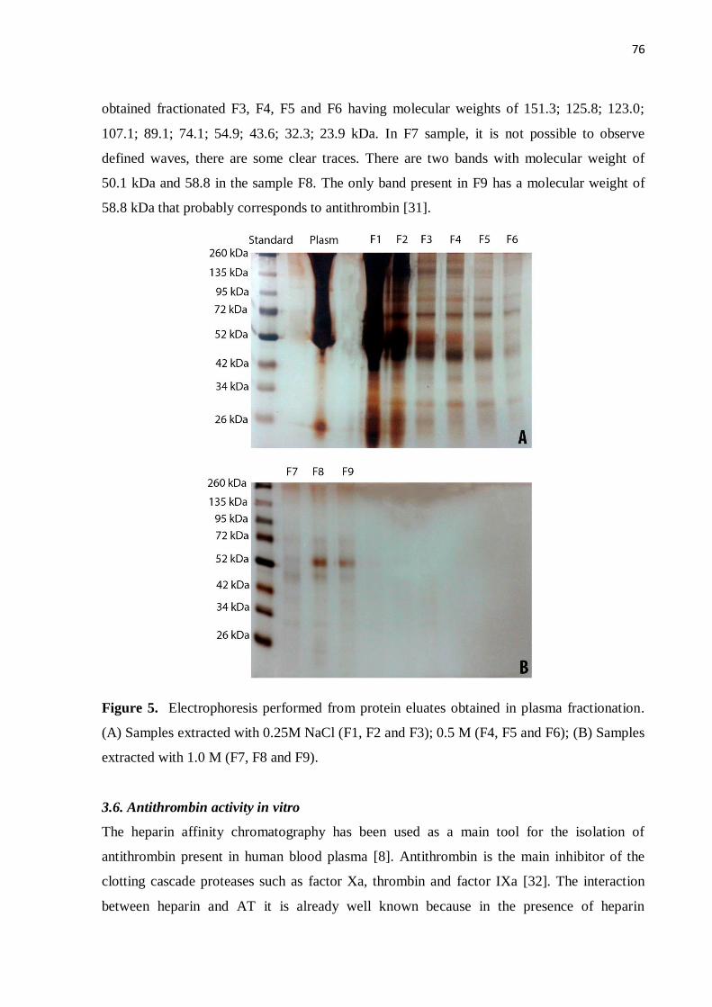
76
obtained fractionated F3, F4, F5 and F6 having molecular weights of 151.3; 125.8; 123.0;
107.1; 89.1; 74.1; 54.9; 43.6; 32.3; 23.9 kDa. In F7 sample, it is not possible to observe
defined waves, there are some clear traces. There are two bands with molecular weight of
50.1 kDa and 58.8 in the sample F8. The only band present in F9 has a molecular weight of
58.8 kDa that probably corresponds to antithrombin [31].
Figure 5. Electrophoresis performed from protein eluates obtained in plasma fractionation.
(A) Samples extracted with 0.25M NaCl (F1, F2 and F3); 0.5 M (F4, F5 and F6); (B) Samples
extracted with 1.0 M (F7, F8 and F9).
3.6. Antithrombin activity in vitro
The heparin affinity chromatography has been used as a main tool for the isolation of
antithrombin present in human blood plasma [8]. Antithrombin is the main inhibitor of the
clotting cascade proteases such as factor Xa, thrombin and factor IXa [32]. The interaction
between heparin and AT it is already well known because in the presence of heparin
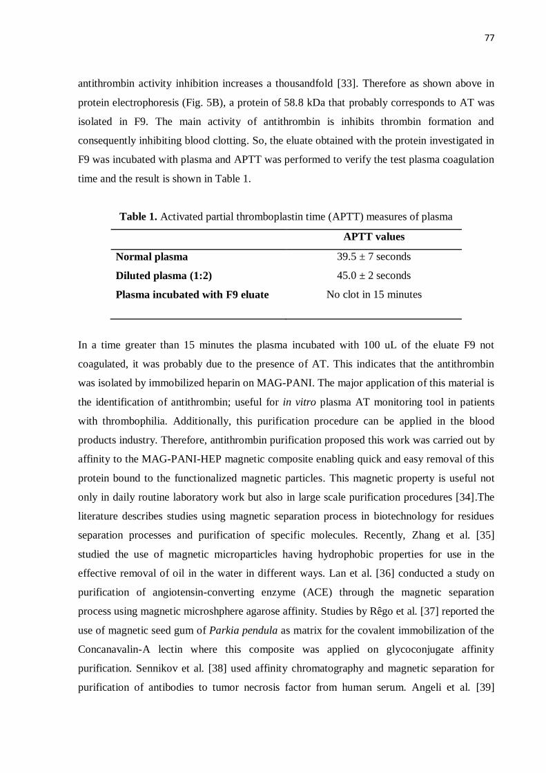
77
antithrombin activity inhibition increases a thousandfold [33]. Therefore as shown above in
protein electrophoresis (Fig. 5B), a protein of 58.8 kDa that probably corresponds to AT was
isolated in F9. The main activity of antithrombin is inhibits thrombin formation and
consequently inhibiting blood clotting. So, the eluate obtained with the protein investigated in
F9 was incubated with plasma and APTT was performed to verify the test plasma coagulation
time and the result is shown in Table 1.
Table 1. Activated partial thromboplastin time (APTT) measures of plasma
APTT values
Normal plasma 39.5 ± 7 seconds
Diluted plasma (1:2) 45.0 ± 2 seconds
Plasma incubated with F9 eluate No clot in 15 minutes
In a time greater than 15 minutes the plasma incubated with 100 uL of the eluate F9 not
coagulated, it was probably due to the presence of AT. This indicates that the antithrombin
was isolated by immobilized heparin on MAG-PANI. The major application of this material is
the identification of antithrombin; useful for in vitro plasma AT monitoring tool in patients
with thrombophilia. Additionally, this purification procedure can be applied in the blood
products industry. Therefore, antithrombin purification proposed this work was carried out by
affinity to the MAG-PANI-HEP magnetic composite enabling quick and easy removal of this
protein bound to the functionalized magnetic particles. This magnetic property is useful not
only in daily routine laboratory work but also in large scale purification procedures [34].The
literature describes studies using magnetic separation process in biotechnology for residues
separation processes and purification of specific molecules. Recently, Zhang et al. [35]
studied the use of magnetic microparticles having hydrophobic properties for use in the
effective removal of oil in the water in different ways. Lan et al. [36] conducted a study on
purification of angiotensin-converting enzyme (ACE) through the magnetic separation
process using magnetic microshphere agarose affinity. Studies by Rêgo et al. [37] reported the
use of magnetic seed gum of Parkia pendula as matrix for the covalent immobilization of the
Concanavalin-A lectin where this composite was applied on glycoconjugate affinity
purification. Sennikov et al. [38] used affinity chromatography and magnetic separation for
purification of antibodies to tumor necrosis factor from human serum. Angeli et al. [39]
78
demonstrated the use of a levan-ferromagnetic composite as affinity matrix for purifying
lectins through a quick and easy procedure based on magnetic affinity.
4. Conclusion
According to the above results, MAG-PANI nanoparticles exhibited a superparamagnetic
behavior when subjected to a magnetic field of +30 kOe to -30 kOe at temperatures of 293 K,
300 K and 313 K. In addition, the presence the phase magnetite and polyaniline have been
proven MAG-PANI through X-ray analysis. Moreover, the composite MAG-PANI-HEP can
be used as a tool for fractionation and depletion of plasma proteins and also in the purification
of antithrombin, an important protein in the blood coagulation cascade. The proposed method
based on the affinity magnetic separation is effective, easy and specific
Acknowledgements
The authors thank: CNPq and FACEPE for financial support; Department of Physics/UFPE
for the physical measures, Northeast Strategic Technology Center (CETENE) for the electron
microscopic analysis.
References
[1] KULLOLLI, M.; WARREN, J.; ARAMPATZIDOU, M.; PITTERI, S.J. Performance
evaluation of affinity ligands for depletion of abundant plasma proteins. Journal of
Chromatography B, 939, 10-16, 2013.
[2] URBAS, L.; BRNE, P.; GABOR, B.; BARUT, M.; STRLIC, M.; PETRIC, T.C.;
STRANCAR, A. Depletion of high-abundance proteins from human plasma using a
combination of an affinity and pseudo-affinity column. Journal of Chromatography A,
1216, 2689–2694, 2009.
[3] MAHN, A.; ISMAIL, M. Depletion of highly abundant proteins in blood plasma by
ammonium sulfate precipitation for 2D-PAGE analysis. Journal of Chromatography B,
879, 3645-3648, 2011.
[4] MOURIER, P.; ANGER, P.; MARTINEZ, C.; HERMAN, F.; VISKOV, C. Quantitative
compositional analysis of heparin using exhaustive heparinase digestion and strong anion
exchange chromatography. Analytical Chemistry Research, 3, 46-53, 2015.
[5] CAPILA, I.; LINHARDT, R.J. Heparin - Protein Interactions. Angewandte Chemie
International, 41, 390-412, 2002.
[6] FLENGSRUD, R.; ANTONSEN, S.G. The binding of pentapeptides to biological and
synthetic high affinity heparin. Bioorganic & Medicinal Chemistry Letters, 25. 4774-
4776, 2015.
[7] KRAPFENBAUER, K.; FOUNTOULAKIS, M. Improved Enrichment and Proteomic
Analysis of Brain Proteins with Signaling Function by Heparin Chromatography. Methods
in Molecular Biology, 566, 165-180, 2009.
79
[8] HEGER, A.; GRUNERT, T.; SCHULZ, P.; JOSIC, D.; BUCHACHER, A. Separation of
active and inactive forms of human antithrombin by heparin affinity chromatography.
Thrombosis Research, 106, 157-164, 2002.
[9] LEI, T.; HE, Q.Y.; WANG, Y.L.; SI, L.S.; CHIU, J.F. Heparin chromatography to deplete
high-abundance proteins for serum proteomics. Clinica Chimica Acta, 388, 173-178,
2008.
[10] AGUILAR-ARTEAGA, K.; RODRIGUEZ J.A.; BARRADOB, E. Magnetic solids in
analytical chemistry: A review. Analytica Chimica Acta, 674, 157-165, 2010.
[11] ZHANG, Y.J.; LIN, Y-W.; CHANG, C-C.; WU, T-M. Conducting and magnetic
behaviors of polyaniline coated multi-walled carbon nanotube composites containing
monodispersed magnetite nanoparticles. Synthetic Metals, 161, 937-942, 2011
[12] BALINT, R.; CASSIDY, N.J.; CARTMELL, S.H. Conductive polymers: Towards a
smart biomaterial for tissue engineering. Acta Biomaterialia, 10, 2341-2353, 2014.
[13] MERCÊS, A.A.D.; MACIEL, J.C.; CARVALHO JÚNIOR, L.B. Magnetic particles as
affinity matrix for purification of antithrombin. IOP Conference Series: Materials
Science and Engineering, 97, 012002, 2015.
[14] YADAV, S.; DEVI, R.; BHAR, P.; SINGHLA, S.; PUNDIR, C.S. Immobilization of
creatininase, creatinase and sarcosine oxidase on iron oxide nanoparticles/chitosan-g-
polyaniline modified Pt electrode for detection of creatinine. Enzyme and Microbial
Technology, 50, 247-254, 2012.
[15] NERI, D.F.M.; BALCAO, V.M.; DOURADO, F.O.Q.; OLIVEIRA, J.M.B.;
CARVALHO-JÚNIOR, L.B.; TEIXEIRA, J.A. Immobilized β-galactosidase onto
magnetic particles coated with polyaniline: Support characterization and
galactooligosaccharides production. Journal of Molecular Catalysis B: Enzymatic, 70
74-80, 2011.
[16] WANG, S.; BAO, H.; YANG, P.; CHEN, G. Immobilization of trypsin in polyaniline-
coated nano-Fe3O4/carbon nanotube composite for protein digestion. Analytica Chimica
Acta, 612, 182-189, 2008.
[17] MACIEL, J.C.; MERCÊS, A.A.D.; CABRERA, M.; SHIGEYOSI, W.T.; DE SOUZA,
S.D.; OLZON-DIONYSIO; FABRIS, J.D.; CARDOSO, C.A.; NERI, D.F.M.; SILVA,
M.P.C.; CARVALHO JR., L.B. Magnetic nanoparticles coated with polyaniline to
stabilize immobilized trypsin. Hyperfine Interactions. DOI: 10.1007/s10751-016-1264-y,
2016: no prelo.
[18] OLIVEIRA, G.B.; CARVALHO JR, L.B.; SILVA, M.P.C. Properties of carbodiimide
treated heparin. Biomaterial, 24, 4777-4783, 2003.
[19] MACIEL, J.C.; ANDRAD, P.L.; NERI, D.F.M. et al. Preparation and characterization
of magnetic levan particles as matrix for trypsin immobilization. Journal of Magnetism
and Magnetic Material, 324, 1312-1316, 2012.
[20] SILVA, A.K.A.; SILVA, E.L.; OLIVEIRA, E.E.; NAGASHIMA JR, T.; SOARES,
L.A.L.; MEDEIROS, A.C.; ARAÚJO, J.H.; ARAÚJO, I.B.; CARRIÇO, A.S..; EGITO,
E.S.T. Synthesis and characterization of xylan-coated magnetite microparticles,
International Journal of Pharmaceutics, 334, 42-47, 2007.
[21] CHEŁMINIAK, D.; ZIEGLER-BOROWSKA, M.; KACZMAREK, H. Synthesis of
magnetite nanoparticles coated with poly(acrylic acid) by photopolymerization. Materials
Letters, 164, 464-467, 2016.
[22] JARAMILLO-TABARES, B.E.; ISAZA, F.J.; TORRESI, S.I.C. Stabilization of
polyaniline by the incorporation of magnetite nanoparticles. Materials Chemistry and
Physics, 132, 529-533, 2012.
[23] CHINNARAJ, K,. MANIKANDAN, A., RAMU, P., ANTONY, S.A., NEERAJA, P.
Comparative Studies of Microwave- and Sol-Gel-Assisted Combustion Methods of
80
Fe3O4 Nanostructures: Structural, Morphological, Optical, Magnetic, and Catalytic
Properties. Journal of Superconductivity and Novel Magnetism, 28, 179-190, 2015.
[24] SINGH, S.; SOLANKI, P.R.; PANDEY, M.K.; MALHOTRA, B.D. Covalent
immobilization of cholesterol esterase and cholesterol oxidase on polyaniline films for
application to cholesterol biosensor. Analytica Chimica Acta, 568, 126-132, 2006.
[25] SAKIYAMA-ELBERT, S.E. Incorporation of heparin into biomaterials. Acta
Biomaterialia, 10, 1581-1587, 2014.
[26] AGUILAR-ARTEAGA, K.; RODRIGUEZ J.A.; BARRADOB, E. Magnetic solids in
analytical chemistry: A review. Analytica Chimica Acta, 674, 157-165, 2010.
[27] HE, J.; HUANG, M.; WANG, D.; ZHANG, Z.; LI, G. Magnetic separation techniques
in sample preparation for biological analysis: A review. Journal of Pharmaceutical and
Biomedical Analysis, 101, 84-101, 2014.
[28] PEYSSELON, F.; RICARD-BLUM, S. Heparin–protein interactions: From affinity
and kinetics to biological roles. Application to an interaction network regulating
angiogenesis. Matrix Biology, 35, 73-81, 2014.
[29] BEAVEN, G.H.; HOLIDAY, E.R. Ultraviolet absorption spectra of proteins and
amino acids. Advances Protein Chemistry, 7, 319-386, 1952.
[30] LAYNE, E. Spectrophotometric and turbidimetric methods for measuring proteins.
Methods in Enzymology, 3, 447-454, 1957.
[31] KARLSSON, G.; WINGE, S. Separation of latent, prelatent, and native forms of
human antithrombin by heparin affinity high-performance liquid chromatography. Protein
Expression and Purification, 33, 339-345, 2004.
[32] AZHAR, A.; KHAN, M.S.; SWAMINATHAN, A.; NASEEM, A.; CHATTERJEE,
S.; JAIRAJPURI, M.A. Oxidized antithrombin is a dual inhibitor of coagulation
andangiogenesis: Importance of low heparin affinity. International Journal of Biological
Macromolecules, 82, 541-550, 2016.
[33] MARIE, A.-L..; TRAN, N.T.; BIANCHINI, E.P.; SALLER, F.; PAUTUS, S.;
ABACHE, T.; PLANTIER, J.-L.; URBAIN, R.; BORGEL, D.; TAVERNA, M. A fast
capillary electrophoresis method to assess the binding affinityof recombinant antithrombin
toward heparin directly from cellculture supernatants. Journal of Pharmaceutical and
Biomedical Analysis, 111, 64-70, 2015.
[34] HORÁK, D.; BABIC, M.; MACKOVÁ, H.; BENES, M.J. Preparation and properties
of magnetic nano- and microsized particles for biological and environmental separations.
Journal of Separation Science, 30, 1751-1772, 2007.
[35] ZHANG, L.; LI, L.; DANG, Z.-M. Bio-inspired durable, superhydrophobic magnetic
particles for oil/water separation. Journal of Colloid and Interface Science, 463, 266-
271, 2016.
[36] LAN, X.; LIAO, D.; WU, S.; WANG, F.; SUN, J.; TONG, Z. Rapid purification and
characterization of angiotensin converting enzyme inhibitory peptides from lizard fish
protein hydrolysates with magnetic affinity separation. Food Chemistry, 182, 136–142,
2015.
[37] RÊGO, M.J.B.M.; ALMEIDA, S.M.; BEZERRA, S.A.; CARVALHO JÚNIOR, L.B.;
BELTRÃO, E.I.C. Magnetic Parkia pendula seed gum as matrix for Concanavalin A
lectin immobilization and its application in affinity purification. Anais da Academia
Brasileira de Ciências, 86, 1351-1357, 2014.
[38] SENNIKOV, S.V.; GOLIKOVA, E.A.; KIREEV, F.D.; LOPATNIKOVA, J.A.
Purification of human immunoglobulin G autoantibodies to tumor necrosis factor using
affinity chromatography and magnetic separation. Journal of Immunological Methods,
390, 92-98, 2013.
81
[39] ANGELI, R.; PAZ, N.V.N.; MACIEL, J.C.; ARAUJO, F.F.B.; PAIVA, P.M.G.;
CALAZANS, G.M.T.; VALENTE, A.P.; ALMEIDA, F.C.L.; COELHO, L.C.B.B.;
CARVALHO JR, L.B.; SILVA, M.P.; CORREIA, M.T.S. Ferromagnetic Levan
Composite: An Affinity Matrix to Purify Lectin. Journal of Biomedicine and
Biotechnology, 2009, 1-6, 2009.
82
7 CONCLUSÃO
As micropartículas de mDAC e as nanopartículas de MAG-PANI sintetizadas
exibiram um comportamento superparamagnético ao serem submetidas à um campo
magnético com intervalo entre + 30 KOe e – 30 KOe nas temperaturas de 293 K, 300 K e 313
K. Além disso, a presença das fases de Dacron e polianilina foram comprovadas em mDAC e
MAG-PANI, respectivamente, através das análises por raios-X. Esses revestimentos das
partículas magnéticas foram importantes para a efetivação do processo de imobilização
covalente da heparina que precisou ser funcionalizada com EDAC/NHS.
Os compósitos magnéticos com heparina covalentemente imobilizada (mDAC-HEP e
MAG-PANI-HEP) foram aplicados no processo de fracionamento das proteínas plasmáticas e
a partir disso foi realizada a depleção de proteínas e também a purificação da antitrombina
advinda do plasma humano.
As proteínas, em especial a antitrombina, foram capazes de interagir de forma
específica com a heparina e foram fracionadas através do processo de separação por afinidade
com aplicação de um campo magnético externo. Entre as vantagens em utilizar esse método
podemos destacar o baixo custo, facilidade no manuseio e especificidade, que é baseada na já
conhecida interação da cromatografia de afinidade à heparina com as proteínas.
8 PERSPECTIVAS
Considerando-se a grande quantidade de proteínas plasmáticas que podem se ligar a
heparina, entre elas alguns biomarcadores, esse estudo inicial permitirá uma avaliação futura
do perfil de diferentes proteínas presentes em pacientes com câncer de próstata e mama, por
exemplo. Além da possibilidade de ser utilizada como ferramenta de monitoramento da
antitrombina em casos de pacientes com trombofilias e também em estudos clínicos de
cadeias de fibrinogênio e seus fragmentos na patogênese de doenças cardiovasculares.
Portanto, os compósitos magnéticos com heparina imobilizada poderão ser utilizados
como um sistema de triagem das proteínas do plasma humano, baseado no seu padrão de
ligação à heparina.
83
ANEXO
84
85
86
87
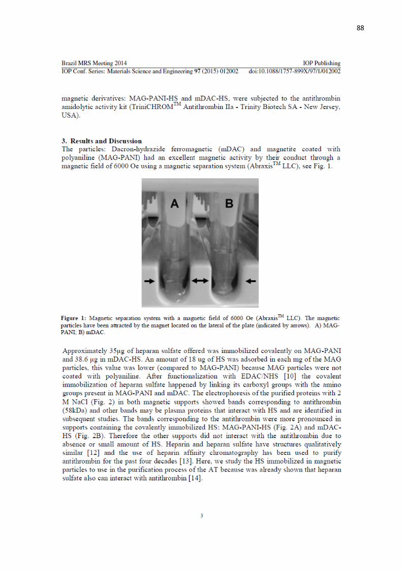
88
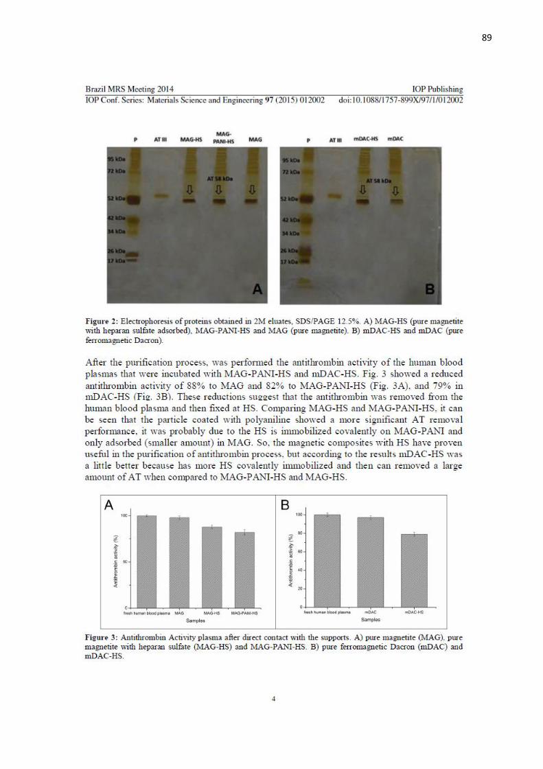
89
90
91
92