Embed Size (px)
Citation preview
1
APLICABILIDADE DE TÉCNICAS DE EXPANSÃO PULMONAR EM PACIENTES
COM COMPROMETIMENTO NO NÍVEL DE CONSCIÊNCIA.
CAIO CÉSAR ARAÚJO MORAIS
RECIFE
2015
UNIVERSIDADE FEDERAL DE PERNAMBUCO CENTRO DE CIÊNCIAS DA SAÚDE
DEPARTAMENTO DE FISIOTERAPIA PROGRAMA DE PÓS-GRADUAÇÃO EM FISIOTERAPIA
2
CAIO CÉSAR ARAÚJO MORAIS
APLICABILIDADE DE TÉCNICAS DE EXPANSÃO PULMONAR EM PACIENTES
COM COMPROMETIMENTO NO NÍVEL DE CONSCIÊNCIA.
RECIFE
2015
Dissertação apresentada ao Programa de Pós-
Graduação em Fisioterapia da Universidade
Federal de Pernambuco como requisito parcial
à obtenção do título de Mestre em Fisioterapia.
Linha de Pesquisa: instrumentação e
intervenção fisioterapêutica
Orientadora(s):
Profª Armèle de Fátima Dornelas de Andrade e
Profª Shirley de Lima Campos
3
4
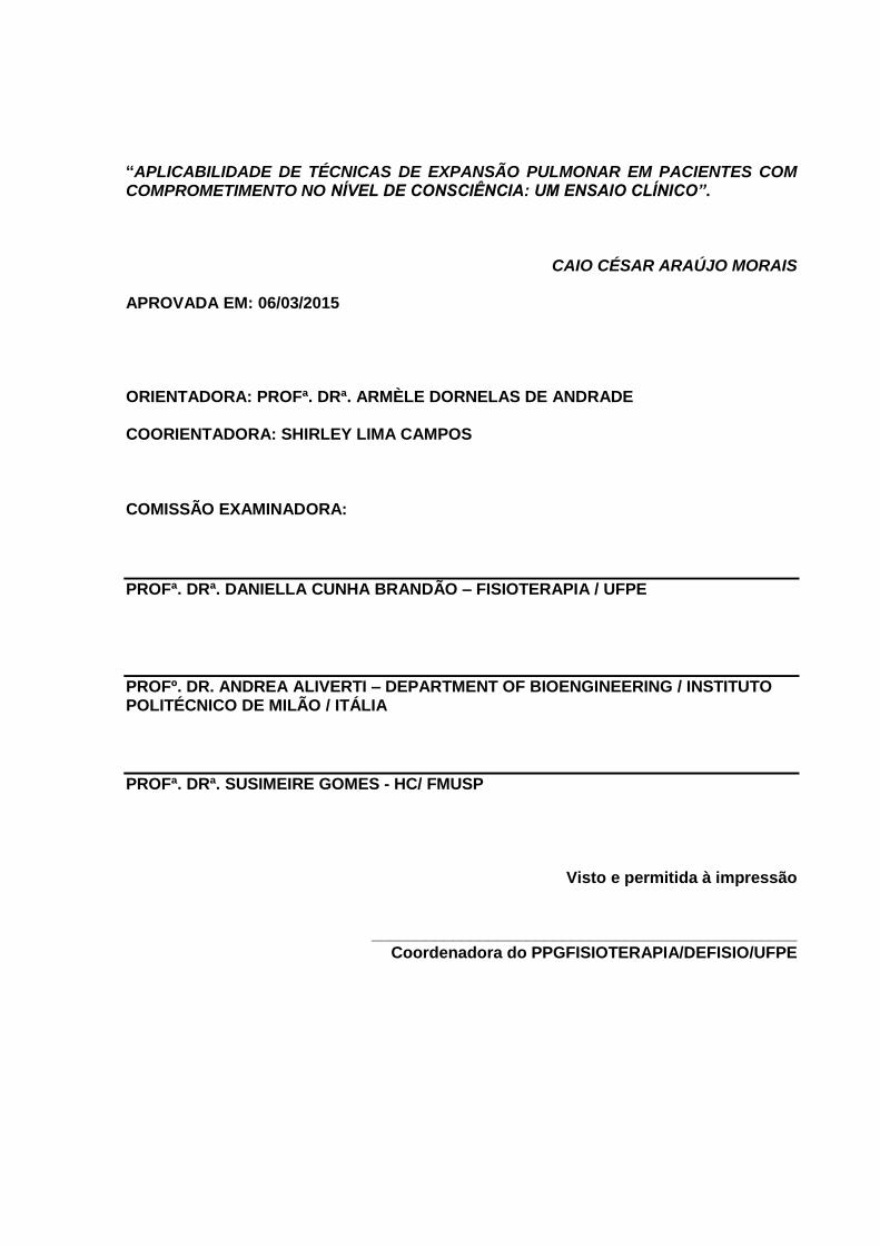
“APLICABILIDADE DE TÉCNICAS DE EXPANSÃO PULMONAR EM PACIENTES COM COMPROMETIMENTO NO NÍVEL DE CONSCIÊNCIA: UM ENSAIO CLÍNICO”.
CAIO CÉSAR ARAÚJO MORAIS
APROVADA EM: 06/03/2015 ORIENTADORA: PROFª. DRª. ARMÈLE DORNELAS DE ANDRADE COORIENTADORA: SHIRLEY LIMA CAMPOS COMISSÃO EXAMINADORA:
PROFª. DRª. DANIELLA CUNHA BRANDÃO – FISIOTERAPIA / UFPE
PROFº. DR. ANDREA ALIVERTI – DEPARTMENT OF BIOENGINEERING / INSTITUTO POLITÉCNICO DE MILÃO / ITÁLIA
PROFª. DRª. SUSIMEIRE GOMES - HC/ FMUSP
Visto e permitida à impressão
_______________________________________________ Coordenadora do PPGFISIOTERAPIA/DEFISIO/UFPE
5
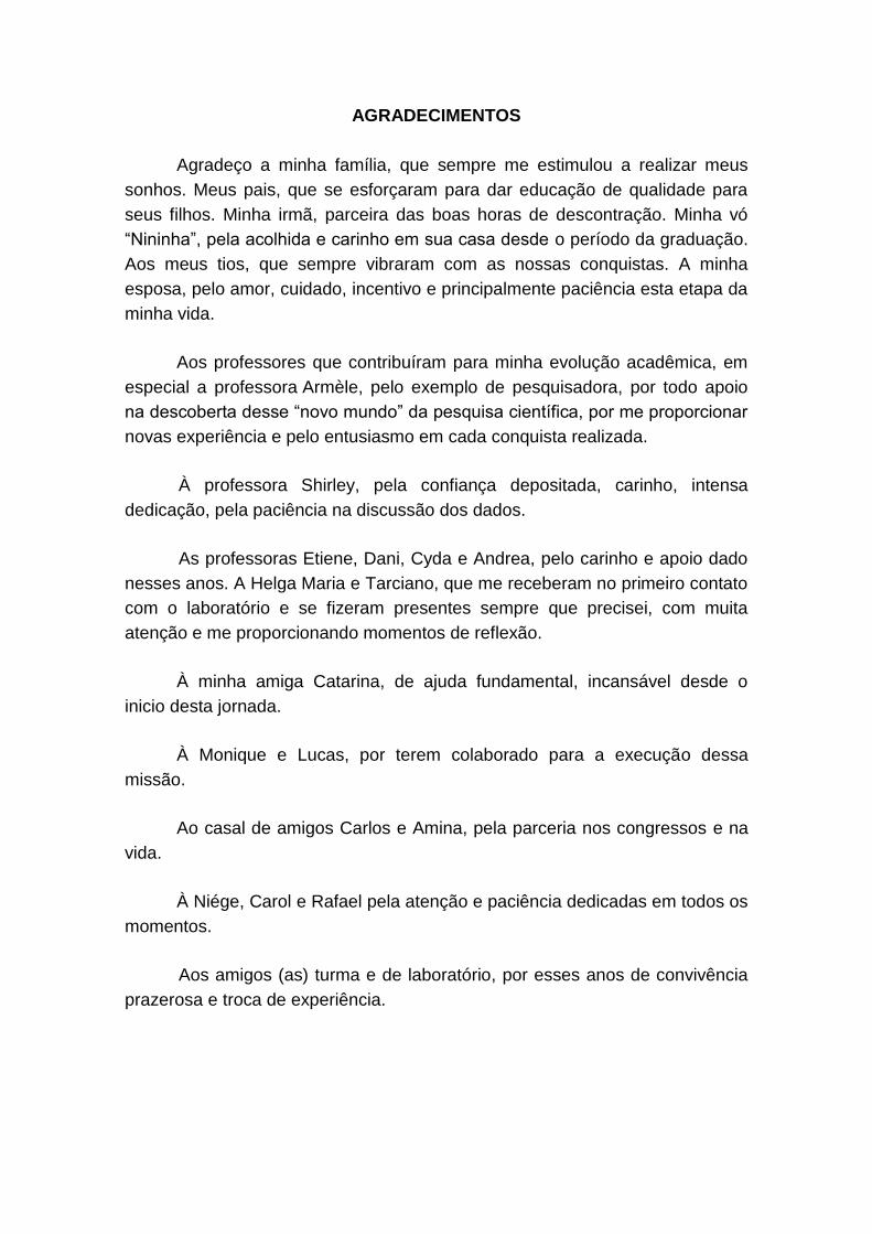
AGRADECIMENTOS
Agradeço a minha família, que sempre me estimulou a realizar meus
sonhos. Meus pais, que se esforçaram para dar educação de qualidade para
seus filhos. Minha irmã, parceira das boas horas de descontração. Minha vó
“Nininha”, pela acolhida e carinho em sua casa desde o período da graduação.
Aos meus tios, que sempre vibraram com as nossas conquistas. A minha
esposa, pelo amor, cuidado, incentivo e principalmente paciência esta etapa da
minha vida.
Aos professores que contribuíram para minha evolução acadêmica, em
especial a professora Armèle, pelo exemplo de pesquisadora, por todo apoio
na descoberta desse “novo mundo” da pesquisa científica, por me proporcionar
novas experiência e pelo entusiasmo em cada conquista realizada.
À professora Shirley, pela confiança depositada, carinho, intensa
dedicação, pela paciência na discussão dos dados.
As professoras Etiene, Dani, Cyda e Andrea, pelo carinho e apoio dado
nesses anos. A Helga Maria e Tarciano, que me receberam no primeiro contato
com o laboratório e se fizeram presentes sempre que precisei, com muita
atenção e me proporcionando momentos de reflexão.
À minha amiga Catarina, de ajuda fundamental, incansável desde o
inicio desta jornada.
À Monique e Lucas, por terem colaborado para a execução dessa
missão.
Ao casal de amigos Carlos e Amina, pela parceria nos congressos e na
vida.
À Niége, Carol e Rafael pela atenção e paciência dedicadas em todos os
momentos.
Aos amigos (as) turma e de laboratório, por esses anos de convivência
prazerosa e troca de experiência.
6
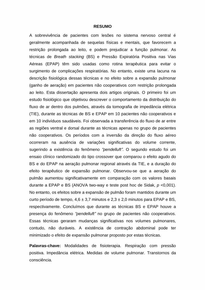
RESUMO
A sobrevivência de pacientes com lesões no sistema nervoso central é
geralmente acompanhada de sequelas físicas e mentais, que favorecem a
restrição prolongada ao leito, e podem prejudicar a função pulmonar. As
técnicas de Breath stacking (BS) e Pressão Expiratória Positiva nas Vias
Aéreas (EPAP) têm sido usadas como rotina terapêutica para evitar o
surgimento de complicações respiratórias. No entanto, existe uma lacuna na
descrição fisiológica dessas técnicas e no efeito sobre a expansão pulmonar
(ganho de aeração) em pacientes não cooperativos com restrição prolongada
ao leito. Esta dissertação apresenta dois artigos originais. O primeiro foi um
estudo fisiológico que objetivou descrever o comportamento da distribuição do
fluxo de ar dentro dos pulmões, através da tomografia de impedância elétrica
(TIE), durante as técnicas de BS e EPAP em 10 pacientes não cooperativos e
em 10 indivíduos saudáveis. Foi observada a transferência do fluxo de ar entre
as regiões ventral e dorsal durante as técnicas apenas no grupo de pacientes
não cooperativos. Os períodos com a inversão da direção do fluxo aéreo
ocorreram na ausência de variações significativas do volume corrente,
sugerindo a existência do fenômeno “pendelluft”. O segundo estudo foi um
ensaio clínico randomizado do tipo crossover que comparou o efeito agudo do
BS e do EPAP na aeração pulmonar regional através da TIE, e a duração do
efeito terapêutico de expansão pulmonar. Observou-se que a aeração do
pulmão aumentou significativamente em comparação com os valores basais
durante a EPAP e BS (ANOVA two-way e teste post hoc de Sidak, p <0,001).
No entanto, os efeitos sobre a expansão de pulmão foram mantidos durante um
curto período de tempo, 4,6 ± 3,7 minutos e 2,3 ± 2,0 minutos para EPAP e BS,
respectivamente. Concluímos que durante as técnicas BS e EPAP houve a
presença do fenômeno “pendelluft” no grupo de pacientes não cooperativos.
Essas técnicas geraram mudanças significativas nos volumes pulmonares,
contudo, não duráveis. A existência de contração abdominal pode ter
minimizado o efeito de expansão pulmonar proposto por estas técnicas.
Palavras-chave: Modalidades de fisioterapia. Respiração com pressão
positiva. Impedância elétrica. Medidas de volume pulmonar. Transtornos da
consciência.
7
ABSTRACT
The survival of patients with lesions in the central nervous system is usually
accompanied by physical and mental sequelae. These impairments favor the
prolonged restriction to the bed, which may contribute with changes in
respiratory function. Breath Stacking (BS) and Expiratory Positive Airway
Pressure (EPAP) have been used as a prophylaxis routine to prevent
respiratory complications. However, there is a gap in the physiological
description and in the effect on lung aeration in non-cooperative patients with
prolonged bed rest. This master's thesis presents two articles. The first was a
physiological study that aimed to describe the physiological behavior of airflow
displacement into the lung, using electrical impedance tomography (EIT), during
BS and EPAP techniques in 10 non-cooperative patients and in 10 health
subjects. It was observed an airflow shift between ventral and dorsal regions
during BS and EPAP techniques in the non-cooperative group. The ventilatory
tracings showed that all periods with reversing of the airflow direction occurred
in the absence of significant variations in VT and flow, suggesting the existence
of pendelluft phenomenon. The second study was a randomized crossover
study trial that compared the acute effect of BS and EPAP on the regional lung
aeration by EIT, measured the duration of the therapeutic effect of lung
expansion and evaluated the influence of these techniques on cardiorespiratory
system. It was observed that lung aeration increased significantly in comparison
with baseline during EPAP and BS (2-way ANOVA and Sidak post hoc, all P <
0.001). However, the effects on lung expansion were kept for a short time, 4.6 ±
3.7 minutes and 2.3 ± 2.0 minutes for EPAP and BS, respectively. There were
no clinically significant differences on cardiorespiratory variables. We conclude
that there was a presence of the pendelluft phenomenon during BS and EPAP
in non-cooperative patients, and these techniques generated a significant
change on lung volumes, but not durable. The existence of expiratory muscle
contraction may have minimized the effect of lung expansion proposed by these
techniques.
Keywords: Physical therapy techniques. Positive-pressure respiration.
Electrical impedance. Lung volume measurements. Consciousness disorders.
8
LISTA DE SIGLAS E ABREVIATURAS
BS Breath Stacking
EPAP Expiratory Positive Airway Pressure
ECG Escala de Coma de Glasgow
TIE Tomografia de Impedância Elétrica
EIT Electrical Impedance Tomography
IEFE Impedância Elétrica ao Final da Expiração
IEFI Impedância Elétrica ao Final da Inspiração
EELI End Expiratory Lung Impedance
EILI End Inspiratory Lung Impedance
EELV End Expiratory Lung Volume
ROI Região de Interesse / Region of interest
RV Região Ventral
RD Região Dorsal
ΔZ Variação de impedância / Change of impedance
Φ Ângulo Fase / Phase Angle
FC Frequência Cardíaca
FR Frequência Respiratória
PAS Pressão Arterial Sistólica
PAD Pressão Arterial Diastólica
PAM Pressão Arterial Média
SpO2 Saturação periférica de oxigênio / Oxygen saturation
HR Heart Rate
RR Respiratory Rate
SBP Systolic Blood Pressure
DBP Diastolic Blood Pressure
MAP Mean Arterial Pressure
VC Volume Corrente
VT Tidal Volume
Paw Airway Pressure
9
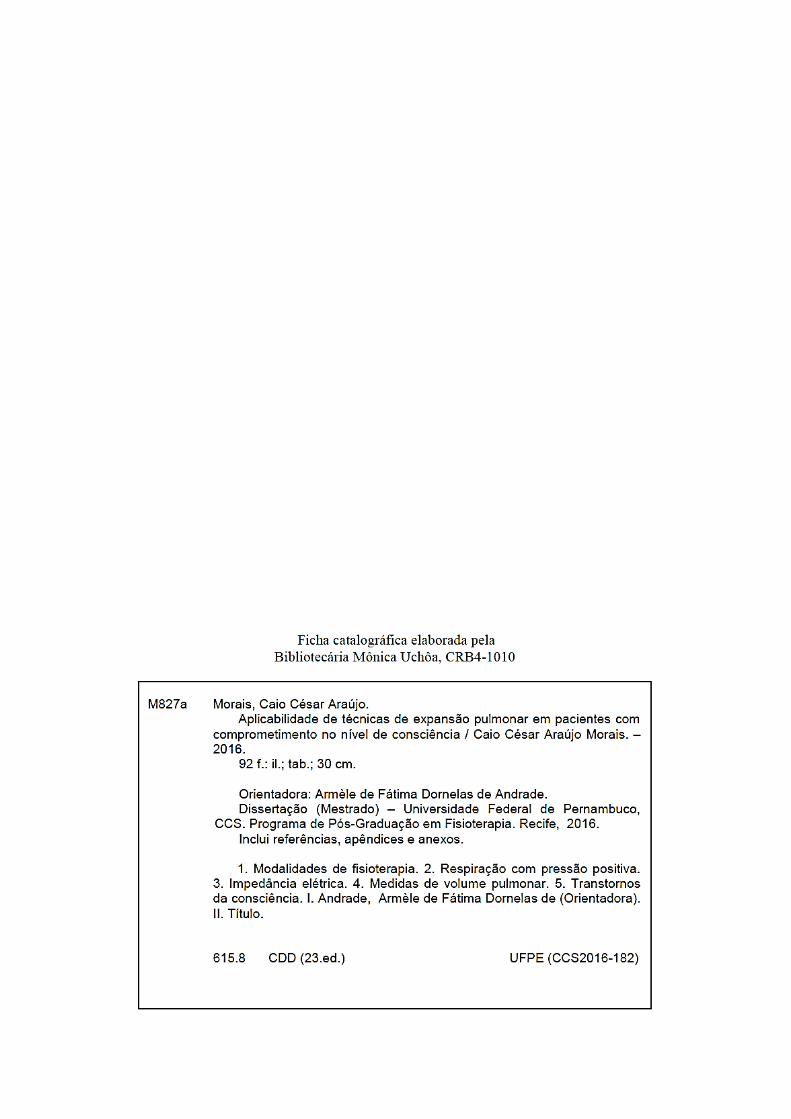
LISTA DE TABELAS
Artigo 1
Tabela 1. Characteristics of non-cooperative patients. 63
Tabela 2. Characteristics of health subjects. 63
Tabela 3. Phase angles during the quiet breath and the interventions (BS and EPAP) in non-cooperative patients and healthy subjects.
64
Artigo 2
Tabela 1. Statistical test applied according the outcomes and period of analysis. 81
Tabela 2. Sample clinical characteristics. 81
Tabela 3. Behavior of the cardiorespiratory variables before, during and after the techniques of EPAP and BS. 82
10
LISTA DE FIGURAS
Referencial Teórico
Figura 1.
Relação entre a capacidade residual funcional (CRF) e o volume de oclusão (VO) pulmonar. Em situações normais a CRF é maior que o VO. Algumas condições como a posição supina, obesidade e indução anestésica causam a redução da CRF, favorecendo o colapso das pequenas vias aéreas.
14
Figura 2.
Comportamento da pressão de vias aéreas (Paw), pressão
pleural (Ppl) e pressão transpulmonar (PL), com aplicação
do EPAP a 10 cmH2O. II = inspiração espontânea; EE =
exalação espontânea.
15
Figura 3. Ilustração do resistor a fluxo (A) e do resistor pressórico por válvula de spring-load (B). 16
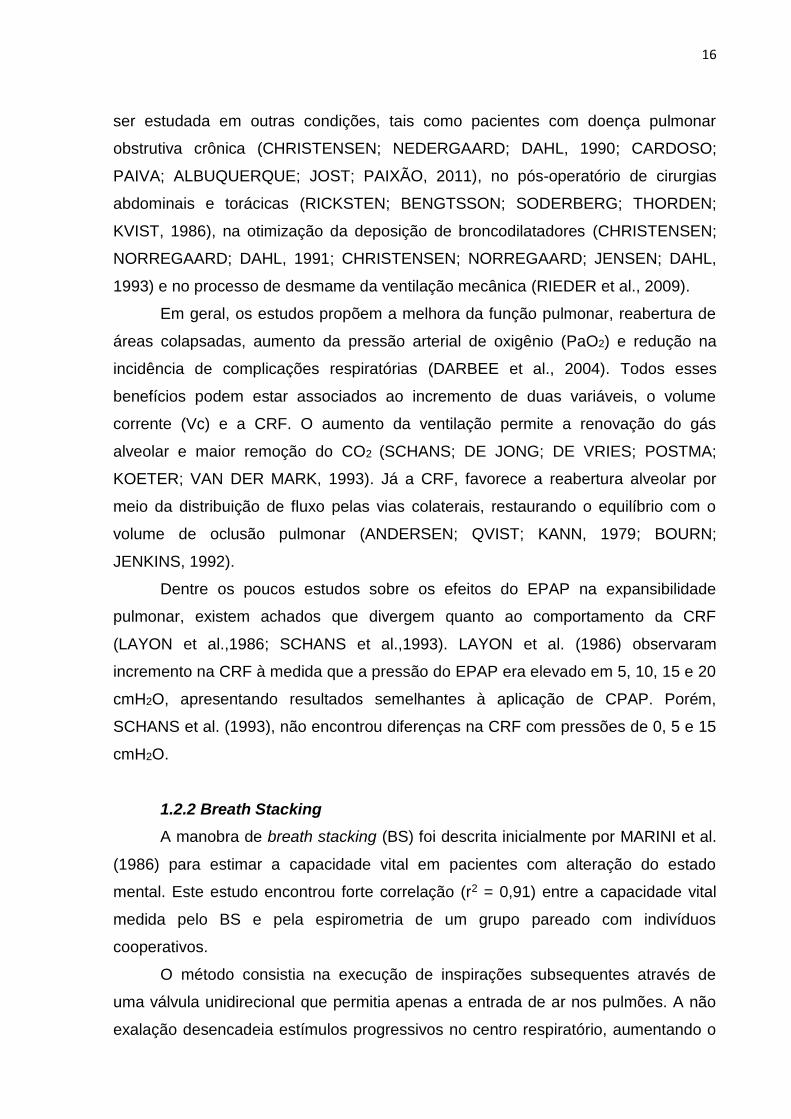
Figura 4.
Comparação esquemática entre a espirometria de incentivo (EI) e a manobra de breath stacking (BS). A conexão da válvula unidirecional para o realização do BS favoreceu o acumulo de volume ao longo dos ciclos respiratórios, em magnitudes maiores que encontradas durante a capacidade inspiratória (CI) e a EI.
18
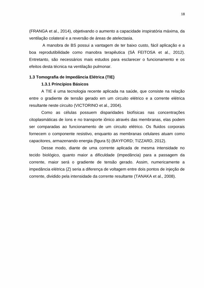
Figura 5. Representação das propriedades de impedância celular. As membranas se comportam como capacitores em série, enquanto os fluidos intra e extracelulares como resistores.
20
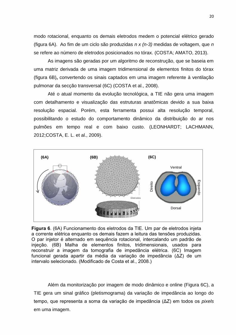
Figura 6.
(6A) Funcionamento dos eletrodos da TIE. Um par de eletrodos injeta a corrente elétrica enquanto os demais fazem a leitura das tensões produzidas. O par injetor é alternado em sequência rotacional, intercalando um padrão de injeção. (6B) Malha de elementos finitos, tridimensionais, usados para reconstruir a imagem da tomografia de impedância elétrica. (6C) Imagem funcional gerada apartir da média da variação de impedância (ΔZ) de um intervalo selecionado.
21
Figura 7.
Ilustração da capacidade da TIE identificar variações no volume corrente (VC) e na pressão positiva ao final da expiração (ΔPEEP). ΔIEFE: variação de impedância elétrica ao final da expiração; ΔZ: variação de impedância elétrica.
22
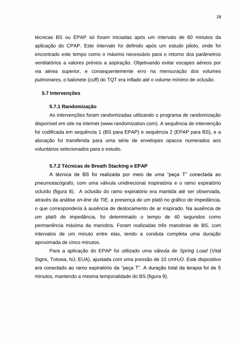
Figura 8. Aplicação da técnica de breath stacking. A imagem “A” ilustra: 1- tubo de traqueostomia; 2- pneumotacógrafo; 3-
30
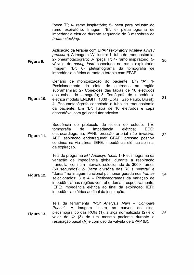
11
“peça T”; 4- ramo inspiratório; 5- peça para oclusão do ramo expiratório. Imagem “B”: 6- pletismograma de impedância elétrica durante sequência de 3 manobras de breath stacking.
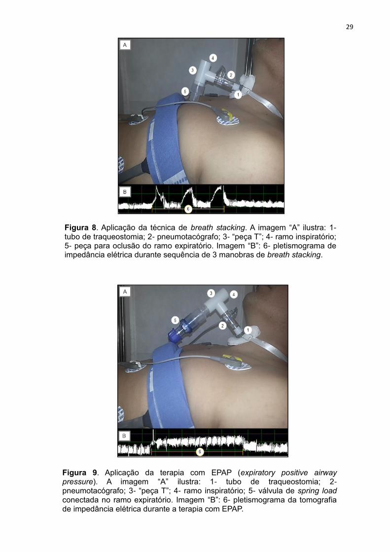
Figura 9.
Aplicação da terapia com EPAP (expiratory positive airway pressure). A imagem “A” ilustra: 1- tubo de traqueostomia; 2- pneumotacógrafo; 3- “peça T”; 4- ramo inspiratório; 5- válvula de spring load conectada no ramo expiratório. Imagem “B”: 6- pletismograma da tomografia de impedância elétrica durante a terapia com EPAP.
30
Figura 10.
Cenário de monitorização do paciente. Em “A”: 1-Posicionamento da cinta de eletrodos na região supramamilar; 2- Conexões das faixas de 16 eletrodos aos cabos do tomógrafo; 3- Tomógrafo de impedância elétrica modelo ENLIGHT 1800 (Dixtal, São Paulo, Brasil); 4- Pneumotacógrafo conectado a tubo de traqueostomia da paciente. Em “B”: Faixa de 16 eletrodos e capa descartável com gel condutor adesivo.
31
Figura 11.
Sequência do protocolo de coleta do estudo. TIE: tomografia de impedância elétrica; ECG: eletrocardiograma; PANI: pressão arterial não invasiva; AET: aspiração endotraqueal; CPAP: pressão positiva contínua na via aérea; IEFE: impedância elétrica ao final da expiração.
32
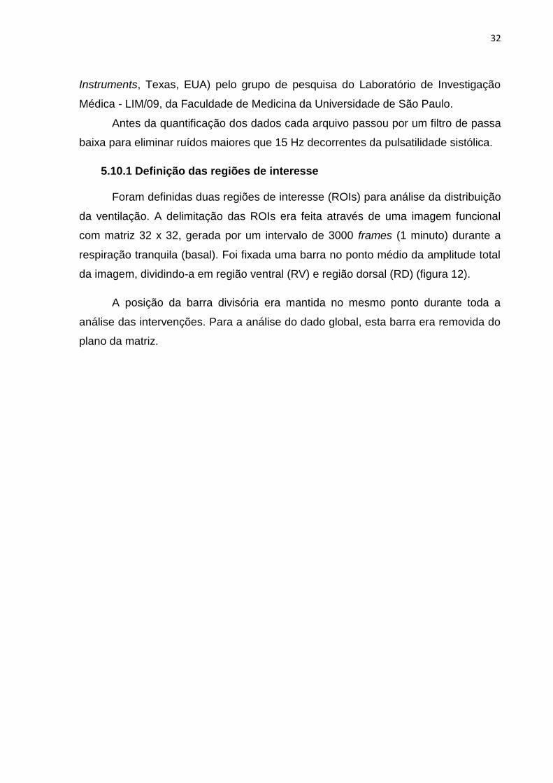
Figura 12.
Tela do programa EIT Analisys Tools. 1- Pletismograma da variação de impedância global durante a respiração tranquila, com um intervalo selecionado de 3000 frames (60 segundos); 2- Barra divisória das ROIs “ventral” e “dorsal” na imagem funcional pulmonar gerada nos frames selecionados; 3 e 4 – Pletismogramas da variação de impedância nas regiões ventral e dorsal, respectivamente; IEFE: impedância elétrica ao final da expiração; IEFI: impedância elétrica ao final da inspiração.
34
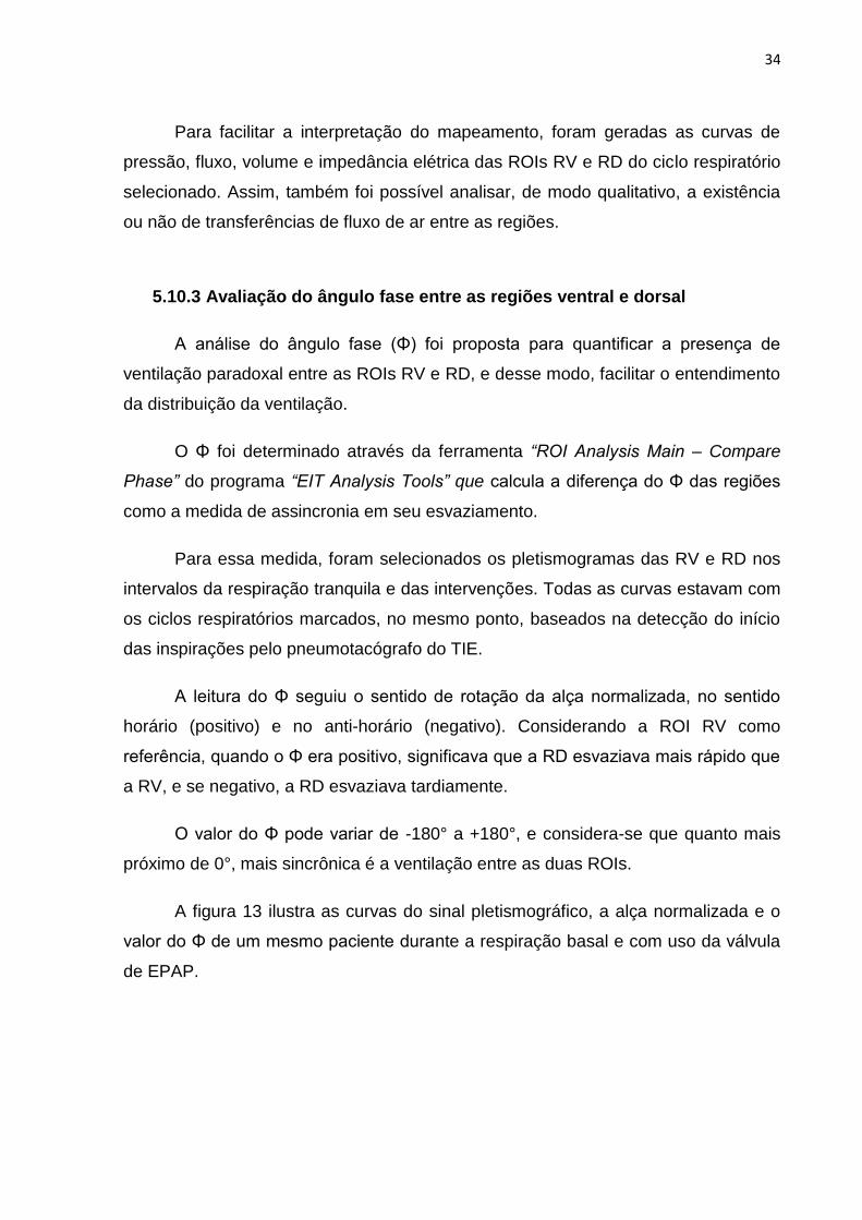
Figura 13.
Tela da ferramenta “ROI Analysis Main – Compare Phase”. A imagem ilustra as curvas do sinal pletismográfico das ROIs (1), a alça normalizada (2) e o valor do Φ (3) de um mesmo paciente durante a respiração basal (A) e com uso da válvula de EPAP (B).
36
12
Artigo 1
Figura 1.
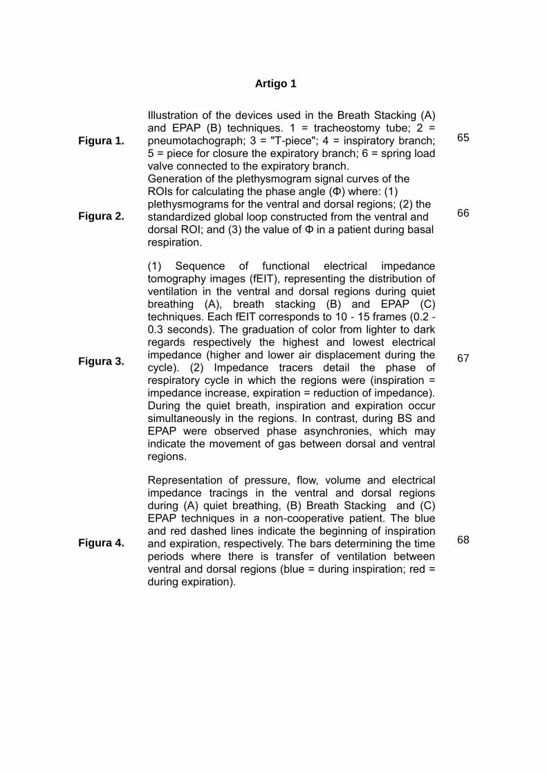
Illustration of the devices used in the Breath Stacking (A) and EPAP (B) techniques. 1 = tracheostomy tube; 2 = pneumotachograph; 3 = "T-piece"; 4 = inspiratory branch; 5 = piece for closure the expiratory branch; 6 = spring load valve connected to the expiratory branch.
65
Figura 2.
Generation of the plethysmogram signal curves of the ROIs for calculating the phase angle (Φ) where: (1) plethysmograms for the ventral and dorsal regions; (2) the standardized global loop constructed from the ventral and dorsal ROI; and (3) the value of Φ in a patient during basal respiration.
66
Figura 3.
(1) Sequence of functional electrical impedance tomography images (fEIT), representing the distribution of ventilation in the ventral and dorsal regions during quiet breathing (A), breath stacking (B) and EPAP (C) techniques. Each fEIT corresponds to 10 - 15 frames (0.2 - 0.3 seconds). The graduation of color from lighter to dark regards respectively the highest and lowest electrical impedance (higher and lower air displacement during the cycle). (2) Impedance tracers detail the phase of respiratory cycle in which the regions were (inspiration = impedance increase, expiration = reduction of impedance). During the quiet breath, inspiration and expiration occur simultaneously in the regions. In contrast, during BS and EPAP were observed phase asynchronies, which may indicate the movement of gas between dorsal and ventral regions.
67
Figura 4.
Representation of pressure, flow, volume and electrical impedance tracings in the ventral and dorsal regions during (A) quiet breathing, (B) Breath Stacking and (C) EPAP techniques in a non-cooperative patient. The blue and red dashed lines indicate the beginning of inspiration and expiration, respectively. The bars determining the time periods where there is transfer of ventilation between ventral and dorsal regions (blue = during inspiration; red = during expiration).
68
13
Artigo 2
Figura 1.
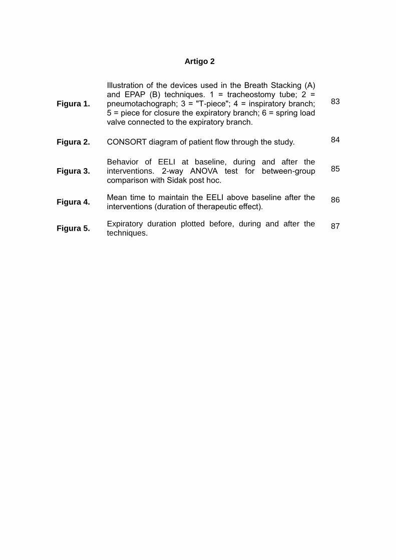
Illustration of the devices used in the Breath Stacking (A) and EPAP (B) techniques. 1 = tracheostomy tube; 2 = pneumotachograph; 3 = "T-piece"; 4 = inspiratory branch; 5 = piece for closure the expiratory branch; 6 = spring load valve connected to the expiratory branch.
83
Figura 2. CONSORT diagram of patient flow through the study. 84
Figura 3. Behavior of EELI at baseline, during and after the interventions. 2-way ANOVA test for between-group comparison with Sidak post hoc.
85
Figura 4. Mean time to maintain the EELI above baseline after the interventions (duration of therapeutic effect).
86
Figura 5. Expiratory duration plotted before, during and after the techniques.
87
14
SUMÁRIO
1. INTRODUÇÃO ........................................................................................... 12
2. JUSTIFICATIVA ......................................................................................... 23
3. HIPÓTESES DO ESTUDO ........................................................................ 24
4. OBJETIVOS ............................................................................................... 25
4.1 Objetivo Geral ......................................................................................... 25
4.2 Objetivos Específicos .............................................................................. 25
5. MATERIAL E MÉTODOS .......................................................................... 26
6. RESULTADOS .......................................................................................... 36
7. CONSIDERAÇÕES FINAIS ....................................................................... 37
REFERÊNCIAS BIBLIOGRÁFICAS .......................................................... 38
APÊNDICES .............................................................................................. 45
APÊNCIDE A - TERMO DE CONSENTIMENTO LIVRE E ESCLARECIDO
....................................................................................................................45
APÊNCIDE B - AIRFLOW DISPLACEMENT ON REGIONAL LUNG
VENTILATION DURING BREATH STACKING AND EXPIRATORY
POSITIVE AIRWAY PRESSURE IN NON-COOPERATIVE PATIENTS.... 47
APÊNCIDE C - IMMEDIATE EFFECT OF LUNG EXPANSION
TECHNIQUES IN NON-COOPERATIVE PATIENTS WITH PROLONGED
BED RESTRICTION: A RANDOMIZED CROSSOVER STUDY ................ 69
ANEXOS ................................................................................................... 88
ANEXO A - Aprovação do Comitê de Ética ................................................ 88
ANEXO B - Parecer Final do Comitê de Ética ........................................... 91
12
1. INTRODUÇÃO
As lesões do sistema nervoso central, como o acidente vascular encefálico
(AVE), tumores cerebrais e o traumatismo crânioencefálico (TCE), são algumas das
principais causas de morbimortalidade no Brasil (MANSUR et al., 2006;
RODRIGUES et al., 2014).
Estas condições normalmente necessitam de uma abordagem neurocirúrgica
para possibilitar o aumento da sobrevida (JÜTTLER et al., 2007; MOREIRA
FALEIRO et al., 2005). Entretanto, boa parte dos sobreviventes apresentam,
comumente, sequelas físicas e mentais duradouras. (QUEIROZ et al., 2012;
OLIVEIRA et al., 2012; KIM et al., 2012).
O comprometimento mental, ou cognitivo, é caracterizado pelo déficit no
funcionamento intelectual, que depende da atenção, processamento de informações,
interação social, verbalização e memória (REZAEI et al., 2012). Esse fator, quando
associado à limitação motora, favorece a restrição prolongada dos pacientes ao leito
(TAN et al., 2001), podendo contribuir com o surgimento de complicações
respiratórias.
A permanência prolongada na posição supina, que é a normalmente adotada
no leito, pode reduzir em até um litro a capacidade residual funcional (CRF) (DEAN,
1985; DUGGAN; KAVANAGH, 2005). Estudos também mostram que a variação da
posição sentada para supina diminui o volume corrente de modo significativo, 0,6 ±
0,2 L vs 0,5 ± 0,1 L, respectivamente, devido a menor mobilidade da caixa torácica
(ROMEI et al., 2010).
Assim, quando ocorre a redução dos volumes pulmonares, há uma
aproximação do nível da CRF com o do volume de oclusão pulmonar, favorecendo o
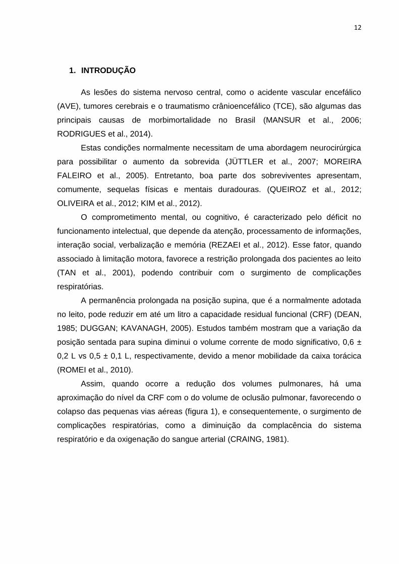
colapso das pequenas vias aéreas (figura 1), e consequentemente, o surgimento de
complicações respiratórias, como a diminuição da complacência do sistema
respiratório e da oxigenação do sangue arterial (CRAING, 1981).
13
1.1 Atuação da fisioterapia respiratória
Frente às alterações da função cerebral e ao consequente risco de
complicações pulmonares associadas à restrição ao leito, novas abordagens
terapêuticas são necessárias para melhorar a assistência ao paciente neurocirúrgico
(GENTILE et al., 2011;BHAT et al., 2014).
Nesse contexto, a fisioterapia respiratória, desde 1915, tem sido discutida
quanto aos seus benefícios na função pulmonar (SELSBY, 1989). Dentro desta
especialidade, a terapia de expansão pulmonar (TEP) é uma conduta fundamentada
em propor a profilaxia e o tratamento das afecções respiratórias que implicam em
reduções volumétricas (O’DONOHUE, 1985; WESTERDAHL et al., 2005; AGOSTINI
et al., 2013)
Este tipo de terapia tem por objetivo principal incrementar o volume de ar
através do aumento do gradiente de pressão transpulmonar (PL = Paw - Ppl), seja por
redução da pressão pleural (Ppl) ou por aumento na pressão da via aérea (Paw)
(CRAING, 1981).
Recentemente, um guideline brasileiro de fisioterapia recomendou o uso de
técnicas para reexpansão pulmonar de pacientes acamados por longos períodos,
devido aos riscos associados à perda volumétrica (FRANÇA et al., 2010). Contudo,
diante dos quadros com alteração no estado cognitivo, verifica-se que o déficit no
estado de consciência e de cooperação inviabiliza a execução de diversos recursos
terapêuticos já fundamentados pela literatura (BHAT et al., 2014).
Figura 1. Relação entre a capacidade residual funcional (CRF) e o volume de oclusão (VO) pulmonar. Em situações normais a CRF é maior que o VO. Algumas condições como a posição supina, obesidade e indução anestésica causam a redução da CRF, favorecendo o colapso das pequenas vias aéreas (Adaptado de Craing, 1981)
14
Desse modo, poucas são as alternativas terapêuticas que poderiam ser
propostas para a prevenção e tratamento nestes pacientes, pela prescindibilidade de
colaboração para serem realizadas. Dentre estas, existe a técnica de Breath
Stacking (BS), que permite o empilhamento de volumes inspirados (MARINI et al.,
1986), e a aplicação de pressão positiva na via aérea por meio de uma válvula de
EPAP (Expiratory Positive Airway Pressure)(FREITAS et al., 2009).
Na prática clínica, alguns serviços de saúde têm adotado de forma empírica o
uso destas condutas como rotina para TEP profilática de modo intermitente.
Entretanto, ainda não estão elucidados quais os efeitos destas terapias no sistema
respiratório neste tipo de pacientes.
1.2 Técnicas de Expansão Pulmonar
1.2.1 Pressão Positiva Expiratória nas Vias Aéreas (Expiratory Positive
Airway Pressure – EPAP)
O EPAP é um instrumento utilizado rotineiramente na fisioterapia, cujo
princípio consiste na aplicação de uma resistência ao fluxo de ar durante a expiração
em respiração espontânea, sustentando uma pressão positiva na via aérea (FALK et
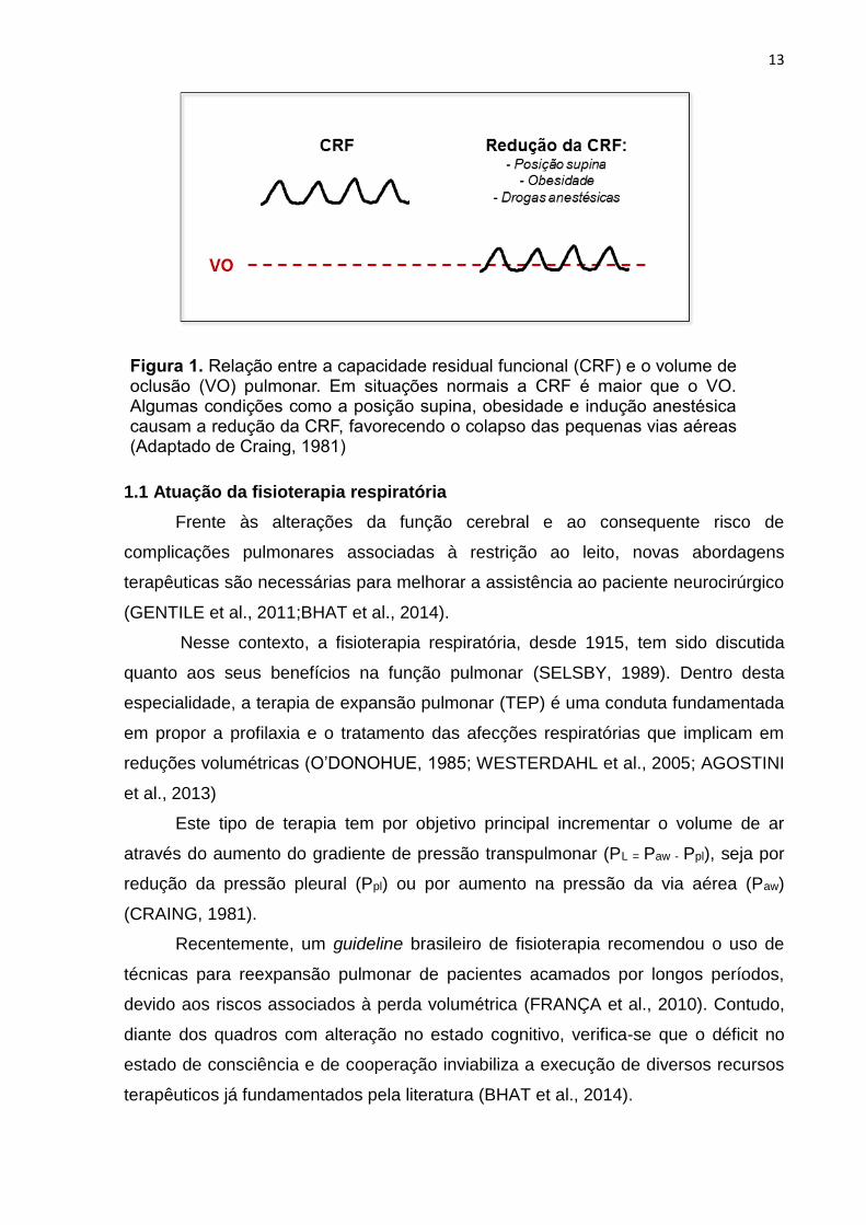
al., 1984). Fisiologicamente, o aumento da pressão transpulmonar está associado ao
comportamento da Ppl e da Paw. Durante a inspiração, existe a negativação da Ppl e a
queda da Paw, abaixo do nível da pressão atmosférica. Entretanto, na expiração, o
uso do resistor aumenta a Paw, garantindo a manutenção da PL positiva (figura 2)
(LAYON; BANNER, 1986).
Dois tipos de dispositivos são utilizados para aumentar a pressão expiratória,
Figura 2. Comportamento da pressão de vias aéreas (Paw), pressão pleural
(Ppl) e pressão transpulmonar (PL), com aplicação do EPAP a 10 cmH2O. IE =
inspiração espontânea; EE = exalação espontânea. (Modificada de Layon & Banner, 1986)
IE EE
t
Paw
t
(cm
H2O
) (cm
H2O
)
Ppl
15
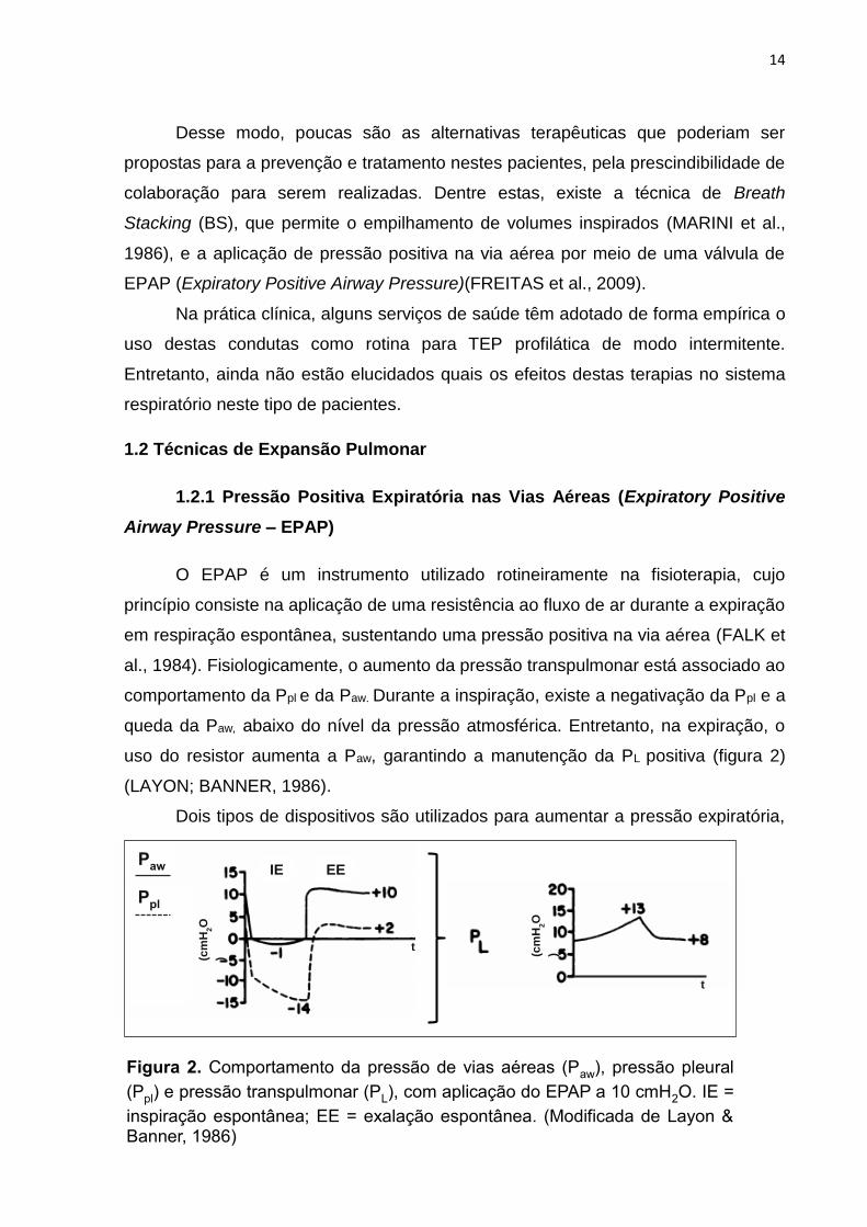
os resistores a fluxo e os lineares de pressão (SEHLIN et al., 2007). Os resistores a
fluxo são válvulas com orifícios de 1,5 a 5,0 mm de diâmetro que geram turbulência
na passagem do gás exalado. Este tipo de instrumento demanda um fluxo constante
de gás e a diminuição deste fluxo acarreta redução da pressão retrógada gerada
(CHRISTENSEN; NEDERGAARD; DAHL, 1990).
Já os resistores lineares de pressão mantêm um limiar de pressão constante,
mesmo com a cessação do fluxo. Suas espécies mais difundidas são as válvulas de
spring-load (MONTEIRO et al., 2012), que geram a resistência devido à compressão
da mola, e o resistor por selo d’água, em que o nível de pressão está relacionado
com a profundidade de inserção do tubo em um recipiente (WESTERDAHL et al.,
2005; CAVALLI; NOHAMA, 2013).
Neste panorama, o principio da manutenção da pressão positiva na via aérea
durante a expiração é mais fidedigno quando utilizado o resistor por limiar
pressórico, pois garante a pressão expiratória positiva previamente estabelecida,
bem como sua regulação precisa, sem a necessidade de um fluxo exalatório
constante, gerando pressões maiores que àquelas decorrentes da aplicação do
resistor a fluxo. (FREITAS et al., 2009; SEHLIN et al., 2007).
Os dispositivos, geralmente, são conectados ao ramo exalatório de uma “peça
T”, que possui no ramo inspiratório uma válvula unidirecional, garantindo a saída do
fluxo de gás apenas através do resistor (figura 3).
Inicialmente, esse recurso terapêutico foi sugerido para o tratamento de
pacientes com fibrose cística, com o objetivo de desobstrução brônquica (FALK et
al., 1984). Devido à simplicidade para manuseio e o baixo custo, a técnica passou a
Figura 3. Ilustração do resistor a fluxo (A) e do resistor pressórico por válvula de spring-load (B).
(A) (B)
16
ser estudada em outras condições, tais como pacientes com doença pulmonar
obstrutiva crônica (CHRISTENSEN; NEDERGAARD; DAHL, 1990; CARDOSO;
PAIVA; ALBUQUERQUE; JOST; PAIXÃO, 2011), no pós-operatório de cirurgias
abdominais e torácicas (RICKSTEN; BENGTSSON; SODERBERG; THORDEN;
KVIST, 1986), na otimização da deposição de broncodilatadores (CHRISTENSEN;
NORREGAARD; DAHL, 1991; CHRISTENSEN; NORREGAARD; JENSEN; DAHL,
1993) e no processo de desmame da ventilação mecânica (RIEDER et al., 2009).
Em geral, os estudos propõem a melhora da função pulmonar, reabertura de
áreas colapsadas, aumento da pressão arterial de oxigênio (PaO2) e redução na
incidência de complicações respiratórias (DARBEE et al., 2004). Todos esses
benefícios podem estar associados ao incremento de duas variáveis, o volume
corrente (Vc) e a CRF. O aumento da ventilação permite a renovação do gás
alveolar e maior remoção do CO2 (SCHANS; DE JONG; DE VRIES; POSTMA;
KOETER; VAN DER MARK, 1993). Já a CRF, favorece a reabertura alveolar por
meio da distribuição de fluxo pelas vias colaterais, restaurando o equilíbrio com o
volume de oclusão pulmonar (ANDERSEN; QVIST; KANN, 1979; BOURN;
JENKINS, 1992).
Dentre os poucos estudos sobre os efeitos do EPAP na expansibilidade
pulmonar, existem achados que divergem quanto ao comportamento da CRF
(LAYON et al.,1986; SCHANS et al.,1993). LAYON et al. (1986) observaram
incremento na CRF à medida que a pressão do EPAP era elevado em 5, 10, 15 e 20
cmH2O, apresentando resultados semelhantes à aplicação de CPAP. Porém,
SCHANS et al. (1993), não encontrou diferenças na CRF com pressões de 0, 5 e 15
cmH2O.
1.2.2 Breath Stacking
A manobra de breath stacking (BS) foi descrita inicialmente por MARINI et al.
(1986) para estimar a capacidade vital em pacientes com alteração do estado
mental. Este estudo encontrou forte correlação (r2 = 0,91) entre a capacidade vital
medida pelo BS e pela espirometria de um grupo pareado com indivíduos
cooperativos.
O método consistia na execução de inspirações subsequentes através de
uma válvula unidirecional que permitia apenas a entrada de ar nos pulmões. A não
exalação desencadeia estímulos progressivos no centro respiratório, aumentando o
17
esforço na inspiração e o volume de gás empilhado a cada ciclo respiratório. O
incremento de volume reduz à medida que a complacência torácica declina e os
músculos respiratórios ficam em posição de desvantagem mecânica, entretanto, o
fluxo de ar é mantido até que os esforços respiratórios sejam insuficientes para
vencer a força de recolhimento elástico torácico (MARINI et al., 1986).
Extrapolando os dados dessa experiência, (BAKER et al., 1990) propuseram
o uso do BS como uma terapia de expansão pulmonar, já que essa manobra garante
uma inspiração profunda e maiores volumes quando comparado à espirometria de
incentivo convencional (figura 4).
Alguns autores modificaram a técnica clássica de BS utilizando uma bolsa de
ressuscitação manual, conectada por uma máscara ou direto na via aérea artificial,
para gerar o empilhamento de ar nos pulmões (BACH et al., 2008) (MCKIM et al.,
2012).
Em geral, esta terapêutica tem sido utilizada em pacientes no pós-operatório
toracoabdominal (DIAS et al., 2008), nas desordens neuromusculares (KANG;
BACH, 2000; BACH et al., 2008; MCKIM et al., 2012), e em pacientes pós AVE
Figura 4. Comparação esquemática entre a espirometria de incentivo (EI) e a manobra de Breath Stacking (BS). A conexão da válvula unidirecional para o realização do BS favoreceu o acumulo de volume ao longo dos ciclos respiratórios, em magnitudes maiores que encontradas durante a capacidade inspiratória (CI) e a EI. (Modificado de Baker, Lamb, & Marini, 1990)
18
(FRANGA et al., 2014), objetivando o aumento a capacidade inspiratória máxima, da
ventilação colateral e a reversão de áreas de atelectasia.
A manobra de BS possui a vantagem de ter baixo custo, fácil aplicação e a
boa reprodutibilidade como manobra terapêutica (SÁ FEITOSA et al., 2012).
Entretanto, são necessários mais estudos para esclarecer o funcionamento e os
efeitos desta técnica na ventilação pulmonar.
1.3 Tomografia de Impedância Elétrica (TIE)
1.3.1 Princípios Básicos
A TIE é uma tecnologia recente aplicada na saúde, que consiste na relação
entre o gradiente de tensão gerado em um circuito elétrico e a corrente elétrica
resultante neste circuito (VICTORINO et al., 2004).
Como as células possuem disparidades biofísicas nas concentrações
citoplasmáticas de íons e no transporte iônico através das membranas, elas podem
ser comparadas ao funcionamento de um circuito elétrico. Os fluidos corporais
fornecem o componente resistivo, enquanto as membranas celulares atuam como
capacitores, armazenando energia (figura 5) (BAYFORD; TIZZARD, 2012).
Desse modo, diante de uma corrente aplicada de mesma intensidade no
tecido biológico, quanto maior a dificuldade (impedância) para a passagem da
corrente, maior será o gradiente de tensão gerado. Assim, numericamente a
impedância elétrica (Z) seria a diferença de voltagem entre dois pontos de injeção de
corrente, dividido pela intensidade da corrente resultante (TANAKA et al., 2008).
19
1.3.2 Avaliação da ventilação regional pulmonar
A avaliação do sistema respiratório é facilitada pela grande variação de
impedância, gerada pela entrada e saída de ar no parênquima pulmonar
(LEONHARDT; LACHMANN, 2012). Normalmente, com uma frequência de 10KHz, a
impedância elétrica da superfície torácica é de 2 a 4 Ωm, e a do pulmão é em média
de 10 Ωm. Quando o alvéolo é insuflado, existe um estiramento dos septos,
tornando-os mais delgados, aumentando a resistência à passagem da corrente
elétrica (COSTA, E. et al., 2009). Harris et al. encontraram aumento de mais de
300% na impedância do parênquima (7,2 – 23,6 Ωm) em indivíduos saudáveis,
enquanto que a do tórax permanecia constante.
Estas propriedades biológicas do tórax tornam a TIE um importante
dispositivo de monitorização por imagem do sistema respiratório por oferecer
informações online sobre a ventilação regional pulmonar de modo não invasivo e
livre de radiação ionizante (COSTA, E. L. et al., 2009).
Para aquisição de dados, são posicionados eletrodos em uma secção do
tórax, onde há emissão de corrente elétrica de baixa amplitude e alta frequência.
Em geral, pares de eletrodos alternam a função de injetar a corrente elétrica, de
Figura 5. Representação das propriedades de impedância celular. As
membranas se comportam como capacitores em série, enquanto os fluidos
intra e extracelulares como resistores.
20
modo rotacional, enquanto os demais eletrodos medem o potencial elétrico gerado
(figura 6A). Ao fim de um ciclo são produzidas n x (n-3) medidas de voltagem, que n
se refere ao número de eletrodos posicionados no tórax. (COSTA; AMATO, 2013).
As imagens são geradas por um algoritmo de reconstrução, que se baseia em
uma matriz derivada de uma imagem tridimensional de elementos finitos do tórax
(figura 6B), convertendo os sinais captados em uma imagem referente à ventilação
pulmonar da secção transversal (6C) (COSTA et al., 2008).
Até o atual momento da evolução tecnológica, a TIE não gera uma imagem
com detalhamento e visualização das estruturas anatômicas devido a sua baixa
resolução espacial. Porém, esta ferramenta possui alta resolução temporal,
possibilitando o estudo do comportamento dinâmico da distribuição do ar nos
pulmões em tempo real e com baixo custo. (LEONHARDT; LACHMANN,
2012;COSTA, E. L. et al., 2009).
Além da monitorização por imagem de modo dinâmico e online (Figura 6C), a
TIE gera um sinal gráfico (pletismograma) da variação de impedância ao longo do
tempo, que representa a soma da variação de impedância (ΔZ) em todos os pixels
em uma imagem.
Figura 6. (6A) Funcionamento dos eletrodos da TIE. Um par de eletrodos injeta a corrente elétrica enquanto os demais fazem a leitura das tensões produzidas. O par injetor é alternado em sequência rotacional, intercalando um padrão de injeção. (6B) Malha de elementos finitos, tridimensionais, usados para reconstruir a imagem da tomografia de impedância elétrica. (6C) Imagem funcional gerada apartir da média da variação de impedância (ΔZ) de um intervalo selecionado. (Modificado de Costa et al., 2008.)
Eletrodos
(6A) (6B) (6C)
Ventral
Dorsal
Dir
eito E
sq
ue
rdo
21
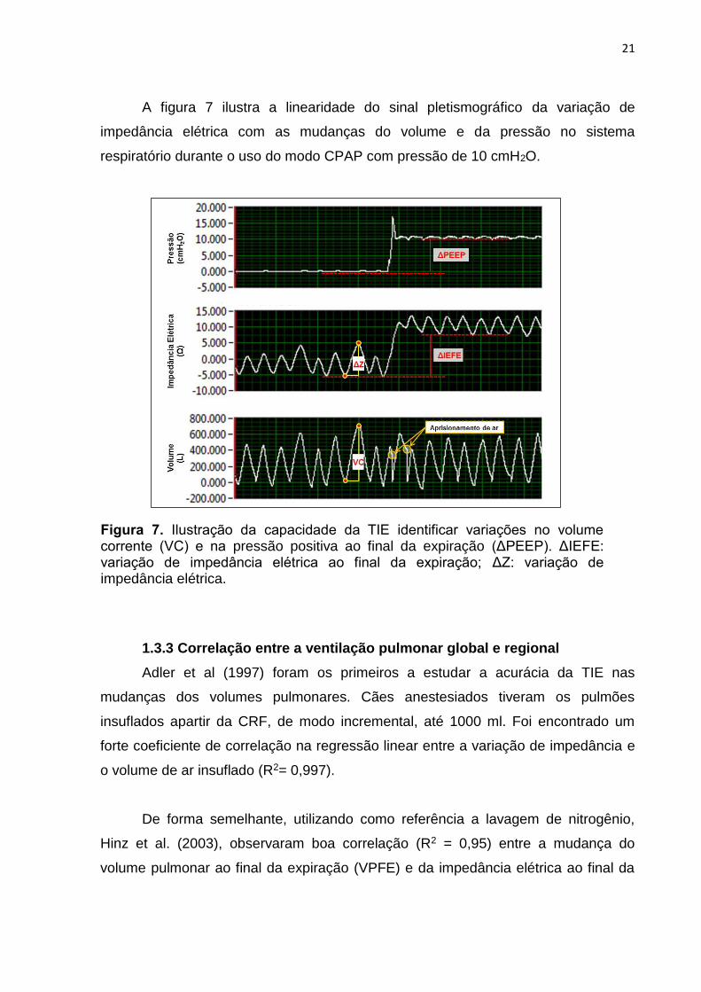
A figura 7 ilustra a linearidade do sinal pletismográfico da variação de
impedância elétrica com as mudanças do volume e da pressão no sistema
respiratório durante o uso do modo CPAP com pressão de 10 cmH2O.
1.3.3 Correlação entre a ventilação pulmonar global e regional
Adler et al (1997) foram os primeiros a estudar a acurácia da TIE nas
mudanças dos volumes pulmonares. Cães anestesiados tiveram os pulmões
insuflados apartir da CRF, de modo incremental, até 1000 ml. Foi encontrado um
forte coeficiente de correlação na regressão linear entre a variação de impedância e
o volume de ar insuflado (R2= 0,997).
De forma semelhante, utilizando como referência a lavagem de nitrogênio,
Hinz et al. (2003), observaram boa correlação (R2 = 0,95) entre a mudança do
volume pulmonar ao final da expiração (VPFE) e da impedância elétrica ao final da
Figura 7. Ilustração da capacidade da TIE identificar variações no volume corrente (VC) e na pressão positiva ao final da expiração (ΔPEEP). ΔIEFE: variação de impedância elétrica ao final da expiração; ΔZ: variação de impedância elétrica.
22
expiração (IEFE), com pressão positiva ao final da expiração (PEEP) de 0, 5, 10 e
15 cmH2O.
Estas comparações também foram realizadas utilizando a tomografia
computadorizada (TC) (FRERICHS et al., 2002). O volume pulmonar foi
gradualmente elevado de 200 a 600 ml em etapas de 100 ml em diferentes níves de
PEEP. A correlação entre as alterações na densidade pulmonar e a TIE foi de 0,75
para região ventral e 0,93 para região dorsal.
Outros estudos descrevem resultados semelhantes quanto à acurácia de a
TIE identificar alterações volumétricas (VICTORINO et al., 2004; GRIVANS et al.,
2011; MARQUIS et al., 2006), fortalecendo o uso desta ferramenta para estimar as
mudanças ventilatórias durante as terapias respiratórias à beira do leito.
23
2. JUSTIFICATIVA
Em tempos de saúde baseada em evidências científicas, é necessário
elucidar os mecanismos fisiológicos e os efeitos clínicos das terapias respiratórias,
para que não sejam empregadas na prática diária de modo indiscriminado. (NOBRE
et al., 2000).
A maioria dos estudos publicados apresenta limitação quanto à metodologia
de avaliação empregada, utilizando instrumentos inadequados para discriminar a
mudança e/ou detectar o efeito da terapia, tais como, imagens de raio-X, oximetria
de pulso, análise dos gases sanguíneos e função pulmonar (MACKENZIE et al.,
1978; MARDIROSSIAN; SCHNEIDER, 1992; SINGH et al., 2015).
Estas ferramentas de avaliação refletem uma condição global do sistema
respiratório e não são suficientes para entender o comportamento do fluxo de ar
dentro dos pulmões e detalhar os efeitos fisiológicos e funcionais da terapia.
Como a TIE é capaz de monitorizar as alterações na ventilação e na aeração
pulmonar, mapeando o movimento de entrada e saída do fluxo de gás em diferentes
regiões pulmonares de um corte transverso (COSTA, E. L. et al., 2009;
LEONHARDT; LACHMANN, 2012), seu uso na avaliação dos efeitos da TEP pode
ser determinante para a compreensão dos efeitos fisiológicos e auxiliar na seleção
apropriada de técnicas direcionadas para resolução de disfunções pulmonares,
principalmente de caráter heterogêneo.
Desse modo, este estudo, utilizando a TIE como ferramenta de avaliação,
buscou responder os seguintes questionamentos acerca das TEP:
I. Como ocorre a distribuição do fluxo ar em pacientes neurocirúrgicos durante
a aplicação das técnicas de BS e EPAP?
II. Qual são as mudanças na aeração pulmonar, desencadeadas pelo BS e
EPAP?
III. Por quanto tempo o efeito terapêutico é mantido?
IV. Existem efeitos deletérios nas variáveis cardiorrespiratórias?
24
3. HIPÓTESES DO ESTUDO
As hipóteses investigadas neste estudo foram:
(1) As técnicas de BS e EPAP, tendo diferentes mecanismos de ação
(princípios), divergem no modo de distribuição do fluxo de ar nos pulmões;
(2) A técnica do BS desencadeará maior incremento da aeração pulmonar
que a técnica EPAP.
(3) O ganho de expansibilidade pulmonar (aeração) será mantida após o
término das manobras.
(4) Ambas as técnicas BS e EPAP não produzem efeitos deletérios no
sistema cardiorrespiratório em decorrência de sua aplicação em pacientes
neurocirúrgicos.
25
4. OBJETIVOS
4.1 Objetivo Geral
- Avaliar os efeitos das técnicas de expansão pulmonar Breath Stacking (BS) e
Expiratory Positive Airway Pressure (EPAP) na ventilação pulmonar regional em
pacientes neurocirúrgicos, não cooperativos e restritos ao leito, através da
tomografia por impedância elétrica (TIE).
4.2 Objetivos Específicos
Estudo 1
- Mapear a distribuição do fluxo de ar nas regiões ventral e dorsal dos pulmões
durante as técnicas de BS e EPAP através da variação de impedância elétrica
regional.
- Verificar a existência de assincronia ventilatória entre as regiões ventral e
dorsal, antes e durante as técnicas de BS e EPAP.
Estudo 2
- Comparar os efeitos durante e imediatamente após a aplicação das técnicas BS
e EPAP em pacientes não cooperativos e restritos ao leito sobre as seguintes
variáveis:
Aeração pulmonar regional (EELI);
Mensurar o tempo de duração do efeito terapêutico (TET) na manutenção da
aeração pulmonar proporcionado pelas técnicas;
Avaliar a influência das técnicas de BS e EPAP em variáveis
cardiorrespiratórias (frequência cardíaca – FC; pressão arterial sistólica –
PAS; pressão arterial diastólica – PAD; pressão arterial média – PAM;
frequência respiratória – FR; saturação periférica de oxigênio - SpO2) antes,
durante a após a execução das técnicas BS e EPAP.
26
5. MATERIAL E MÉTODOS
5.1 Aspectos Éticos
O referido estudo foi aprovado pelo Comitê de Ética em Pesquisa do Centro
de Ciências da Saúde da Universidade Federal de Pernambuco, sob o CAAE
15037913.0.0000.5208 – Anexo I.
Todos os responsáveis foram informados sobre os objetivos, procedimentos, riscos e
benefícios do estudo e participaram voluntariamente de acordo com o Termo de
Consentimento Livre e Esclarecido – TCLE (APÊNDICE A).
5.2 Delineamento do estudo
Trata-se de um ensaio clínico controlado randomizado do tipo crossover,
desenvolvido na Unidade de Cuidados Especiais em Neurocirurgia (UCEN) do
Hospital da Restauração, entre os meses de agosto de 2014 a janeiro de 2015.
5.3 População estudada
A amostra foi composta por pacientes submetidos à neurocirurgia, com
restrição no leito por mais de 14 dias e sem suporte ventilatório mecânico por mais
de 2 dias, caracterizando o sucesso do desmame da ventilação artificial.
Foram incluídos os sujeitos incapazes de responder ao comando (Escala de
Coma de Glasgow - ECG: 3 – 10 pontos) (STERNBACH, 2000), com idade entre 18
e 65 anos e perimetria torácica na região mamilar entre 88 e 98 centímetros, em uso
de tubo de traqueostomia (TQT), e excluídos aqueles com marca passo cardíaco,
comorbidades respiratórias prévias (asma, doença pulmonar obstrutiva crônica,
patologias restritivas pulmonares e da caixa torácica), expansibilidade torácica
assimétrica, deformidade na caixa torácica, distensão abdominal, fratura de costelas,
espasticidade em algum hemicorpo com pontuação maior que 2 na Escala de
Ashworth para os membros superiores (BOHANNON; SMITH, 1987), movimentos
descoordenados nos membros, instabilidade cardiorrespiratória (FC < 60 ou > 120
bpm; FR > 35 ipm; PAM < 60 mmHg ou > 120 mmHg; SpO2 < 90%; sinais
característicos de aumento do trabalho respiratório como tiragem dos músculos
intercostais e uso de musculatura acessória) (ADLER; MALONE, 2012).
27
5.4 Cálculo amostral
O cálculo amostral foi realizado através do aplicativo “Quantitative
Measurement tools” para estudo crossover desenvolvido pela Massachusetts
General Hospital Mallinckrodt General Clinical Research Center (Massachusetts,
EUA), disponível em site na internet (http://hedwig.
mgh.harvard.edu/sample_size/size.html).
Foram utilizados para o cálculo os dados de 5 pacientes no estudo piloto para
os desfechos de ΔIEFE e TET. O maior numero amostral foi obtido para o ΔIEFE,
sendo necessários 9 pacientes, acreditando que há 81% de probabilidade que o
estudo detecte uma diferença no tratamento, unilateralmente, com nível de
significância de 5% (p<0,05), se a mínima diferença detectável entre os tratamentos
BS e EPAP fosse de 23,35 unidades. Este cálculo foi baseado na premissa de que o
desvio padrão da diferença nas variáveis é de 21,76. Considerando a probabilidade
de ocorrer ruídos nos sinais da análise, optou-se por acrescentar 40% para repor
perda amostral, portanto, sendo coletados 13 pacientes.
5.5 Protocolo de coleta dos dados
Participaram do estudo três pesquisadores, sendo o pesquisador 1
responsável pelo preparo do paciente e da avaliação antes, durante e após
intervenção; o pesquisador 2 responsável por aplicar as técnicas de BS e EPAP. O
terceiro pesquisador foi responsável pelo processo de randomização.
5.6 Preparação do paciente
Os pacientes foram posicionados em decúbito dorsal com cabeceira elevada
em 45º, sendo realizada a tricotomia dos pelos torácicos, seguida de higienização
com algodão umedecido de álcool a 70%, para minimizar a resistência à corrente
elétrica emitida pela TIE.
Objetivando garantir que o protocolo iniciasse na mesma condição para todos
os pacientes (homogeneização), antes das aplicações das técnicas foi realizada a
aspiração endotraqueal composta por 3 inserções da sonda no tubo de
traqueostomia com pressão negativa regulada entre 100 e 150 mmHg, com sonda
número 14 French, durante 15 segundos. No intuito de minimizar os efeitos pós
AET, os pacientes eram submetidos à assistência ventilatória por 2 minutos no modo
CPAP com 10 cmH2O através do ventilador Vivo40 (General Electric, Suécia). As
28
técnicas BS ou EPAP só foram iniciadas após um intervalo de 60 minutos da
aplicação do CPAP. Este intervalo foi definido após um estudo piloto, onde foi
encontrado este tempo como o máximo necessário para o retorno dos parâmetros
ventilatórios a valores prévios a aspiração. Objetivando evitar escapes aéreos por
via aérea superior, e consequentemente erro na mensuração dos volumes
pulmonares, o balonete (cuff) do TQT era inflado até o volume mínimo de oclusão.
5.7 Intervenções
5.7.1 Randomização
As intervenções foram randomizadas utilizando o programa de randomização
disponível em site na internet (www.randomization.com). A sequência de intervenção
foi codificada em sequência 1 (BS para EPAP) e sequência 2 (EPAP para BS), e a
alocação foi transferida para uma série de envelopes opacos numerados aos
voluntários selecionados para o estudo.
5.7.2 Técnicas de Breath Stacking e EPAP
A técnica de BS foi realizada por meio de uma “peça T” conectada ao
pneumotacógrafo, com uma válvula unidirecional inspiratória e o ramo expiratório
ocluído (figura 8). A oclusão do ramo expiratório era mantida até ser observada,
através da análise on-line da TIE, a presença de um platô no gráfico de impedância,
o que corresponderia à ausência de deslocamento de ar inspirado. Na ausência de
um platô de impedância, foi determinado o tempo de 40 segundos como
permanência máxima da manobra. Foram realizadas três manobras de BS, com
intervalos de um minuto entre elas, tendo a conduta completa uma duração
aproximada de cinco minutos.
Para a aplicação do EPAP foi utilizado uma válvula de Spring Load (Vital
Signs, Totowa, NJ, EUA), ajustada com uma pressão de 10 cmH2O. Este dispositivo
era conectado ao ramo expiratório da “peça T”. A duração total da terapia foi de 5
minutos, mantendo a mesma temporalidade do BS (figura 9).
29
Figura 8. Aplicação da técnica de breath stacking. A imagem “A” ilustra: 1- tubo de traqueostomia; 2- pneumotacógrafo; 3- “peça T”; 4- ramo inspiratório; 5- peça para oclusão do ramo expiratório. Imagem “B”: 6- pletismograma de impedância elétrica durante sequência de 3 manobras de breath stacking.
Figura 9. Aplicação da terapia com EPAP (expiratory positive airway pressure). A imagem “A” ilustra: 1- tubo de traqueostomia; 2- pneumotacógrafo; 3- “peça T”; 4- ramo inspiratório; 5- válvula de spring load conectada no ramo expiratório. Imagem “B”: 6- pletismograma da tomografia de impedância elétrica durante a terapia com EPAP.
30
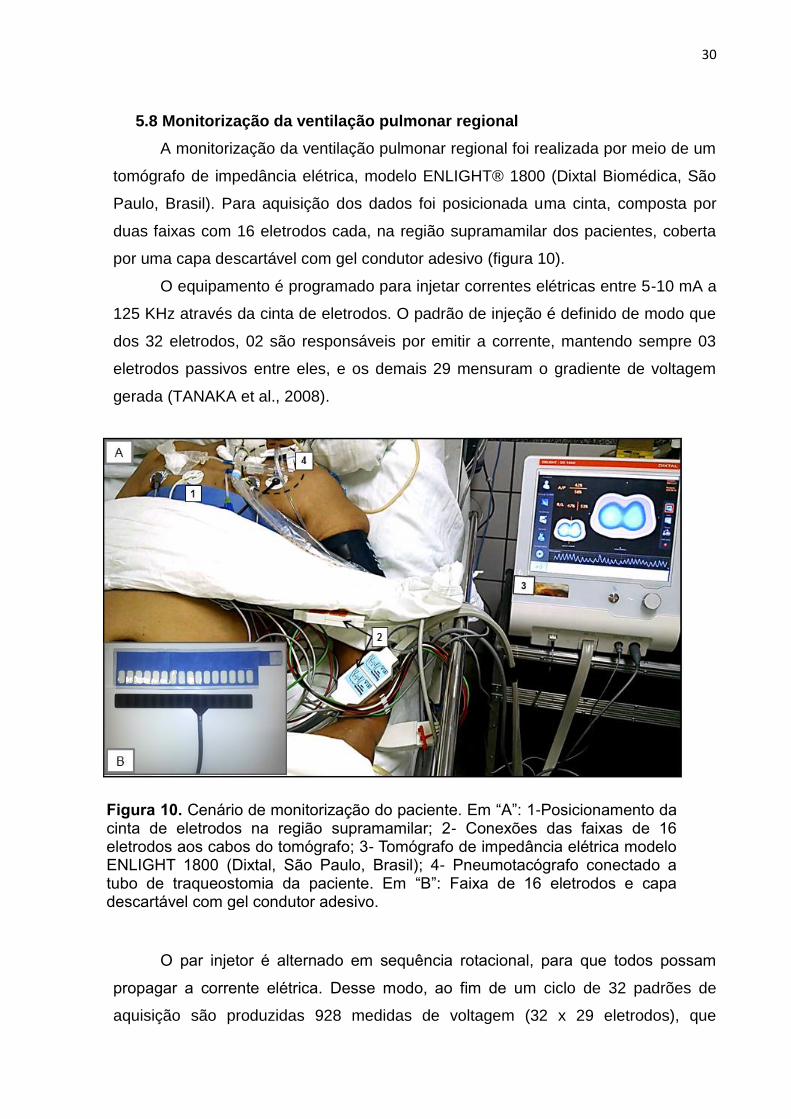
5.8 Monitorização da ventilação pulmonar regional
A monitorização da ventilação pulmonar regional foi realizada por meio de um
tomógrafo de impedância elétrica, modelo ENLIGHT® 1800 (Dixtal Biomédica, São
Paulo, Brasil). Para aquisição dos dados foi posicionada uma cinta, composta por
duas faixas com 16 eletrodos cada, na região supramamilar dos pacientes, coberta
por uma capa descartável com gel condutor adesivo (figura 10).
O equipamento é programado para injetar correntes elétricas entre 5-10 mA a
125 KHz através da cinta de eletrodos. O padrão de injeção é definido de modo que
dos 32 eletrodos, 02 são responsáveis por emitir a corrente, mantendo sempre 03
eletrodos passivos entre eles, e os demais 29 mensuram o gradiente de voltagem
gerada (TANAKA et al., 2008).
O par injetor é alternado em sequência rotacional, para que todos possam
propagar a corrente elétrica. Desse modo, ao fim de um ciclo de 32 padrões de
aquisição são produzidas 928 medidas de voltagem (32 x 29 eletrodos), que
Figura 10. Cenário de monitorização do paciente. Em “A”: 1-Posicionamento da cinta de eletrodos na região supramamilar; 2- Conexões das faixas de 16 eletrodos aos cabos do tomógrafo; 3- Tomógrafo de impedância elétrica modelo ENLIGHT 1800 (Dixtal, São Paulo, Brasil); 4- Pneumotacógrafo conectado a tubo de traqueostomia da paciente. Em “B”: Faixa de 16 eletrodos e capa descartável com gel condutor adesivo.
31
consiste em um “frame de voltagens”. Como o equipamento usado possui uma
frequência de aquisição de 50Hz, é possível obter imagens com resolução temporal
de um “frame” (quadro) a cada 1,2 segundos (60s/50Hz).
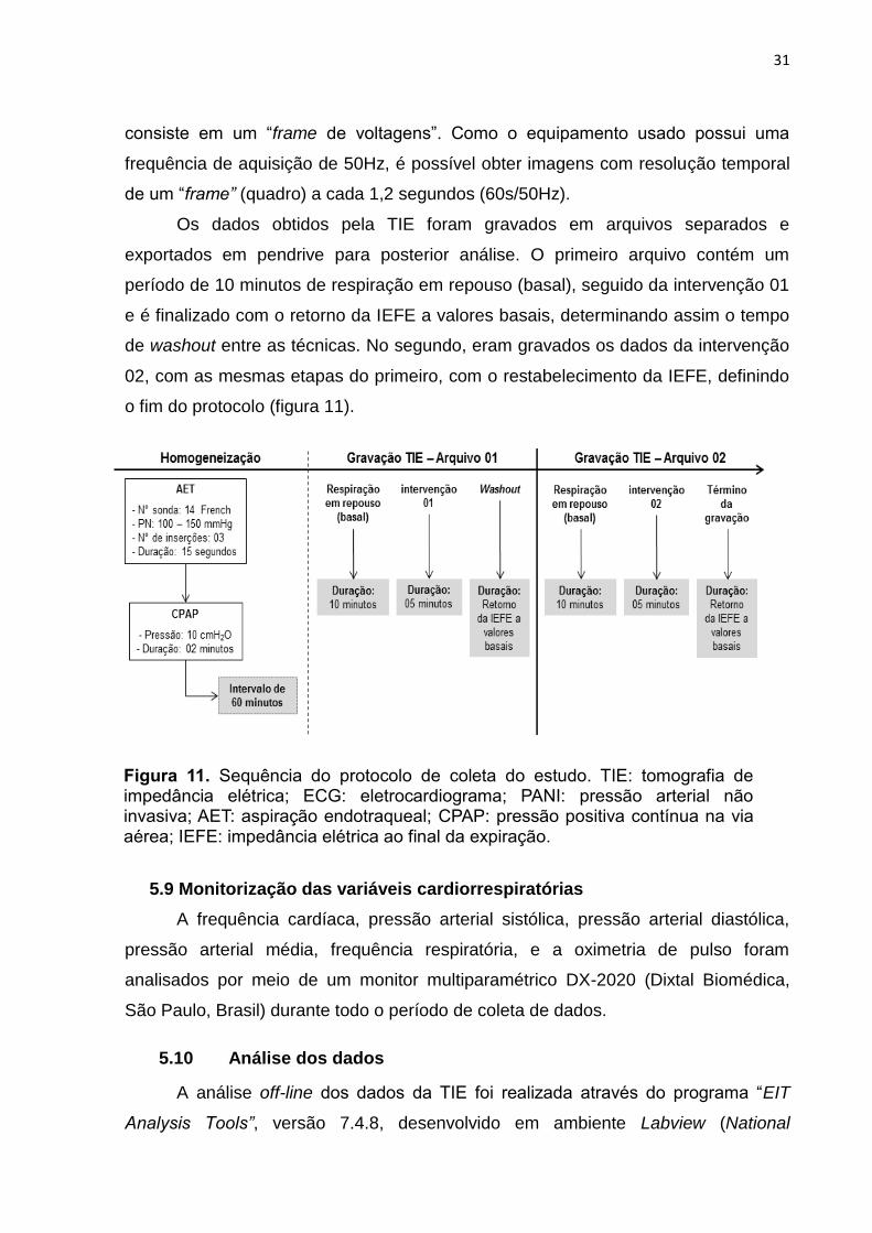
Os dados obtidos pela TIE foram gravados em arquivos separados e
exportados em pendrive para posterior análise. O primeiro arquivo contém um
período de 10 minutos de respiração em repouso (basal), seguido da intervenção 01
e é finalizado com o retorno da IEFE a valores basais, determinando assim o tempo
de washout entre as técnicas. No segundo, eram gravados os dados da intervenção
02, com as mesmas etapas do primeiro, com o restabelecimento da IEFE, definindo
o fim do protocolo (figura 11).
5.9 Monitorização das variáveis cardiorrespiratórias
A frequência cardíaca, pressão arterial sistólica, pressão arterial diastólica,
pressão arterial média, frequência respiratória, e a oximetria de pulso foram
analisados por meio de um monitor multiparamétrico DX-2020 (Dixtal Biomédica,
São Paulo, Brasil) durante todo o período de coleta de dados.
5.10 Análise dos dados
A análise off-line dos dados da TIE foi realizada através do programa “EIT
Analysis Tools”, versão 7.4.8, desenvolvido em ambiente Labview (National
Figura 11. Sequência do protocolo de coleta do estudo. TIE: tomografia de impedância elétrica; ECG: eletrocardiograma; PANI: pressão arterial não invasiva; AET: aspiração endotraqueal; CPAP: pressão positiva contínua na via aérea; IEFE: impedância elétrica ao final da expiração.
32
Instruments, Texas, EUA) pelo grupo de pesquisa do Laboratório de Investigação
Médica - LIM/09, da Faculdade de Medicina da Universidade de São Paulo.
Antes da quantificação dos dados cada arquivo passou por um filtro de passa
baixa para eliminar ruídos maiores que 15 Hz decorrentes da pulsatilidade sistólica.
5.10.1 Definição das regiões de interesse
Foram definidas duas regiões de interesse (ROIs) para análise da distribuição
da ventilação. A delimitação das ROIs era feita através de uma imagem funcional
com matriz 32 x 32, gerada por um intervalo de 3000 frames (1 minuto) durante a
respiração tranquila (basal). Foi fixada uma barra no ponto médio da amplitude total
da imagem, dividindo-a em região ventral (RV) e região dorsal (RD) (figura 12).
A posição da barra divisória era mantida no mesmo ponto durante toda a
análise das intervenções. Para a análise do dado global, esta barra era removida do
plano da matriz.
33
5.10.2 Mapeamento da distribuição da ventilação pulmonar regional
Para mapear a distribuição da ventilação regional foi selecionado um ciclo
respiratório completo, localizado no meio do intervalo de frames da respiração
tranquila e das técnicas BS e EPAP. Foram geradas 8 imagens funcionais, com
intervalos de 10 a 15 frames (0,2 a 0,3 segundos) de modo sequencial ao longo do
ciclo (4 imagens na inspiração e 4 na expiração).
Cada imagem funcional foi analisada nas ROIs RV e RD quanto à fase que se
encontravam de acordo com sentido das respectivas curvas de impedância
(inspiração = aumento da impedância; expiração = redução da impedância).
Figura 12. Tela do programa EIT Analisys Tools. 1- Pletismograma da variação de impedância global durante a respiração tranquila, com um intervalo selecionado de 3000 frames (60 segundos); 2- Barra divisória das ROIs “ventral” e “dorsal” na imagem funcional pulmonar gerada nos frames selecionados; 3 e 4 – Pletismogramas da variação de impedância nas regiões ventral e dorsal, respectivamente; IEFE: impedância elétrica ao final da expiração; IEFI: impedância elétrica ao final da inspiração.
34
Para facilitar a interpretação do mapeamento, foram geradas as curvas de
pressão, fluxo, volume e impedância elétrica das ROIs RV e RD do ciclo respiratório
selecionado. Assim, também foi possível analisar, de modo qualitativo, a existência
ou não de transferências de fluxo de ar entre as regiões.
5.10.3 Avaliação do ângulo fase entre as regiões ventral e dorsal
A análise do ângulo fase (Φ) foi proposta para quantificar a presença de
ventilação paradoxal entre as ROIs RV e RD, e desse modo, facilitar o entendimento
da distribuição da ventilação.
O Φ foi determinado através da ferramenta “ROI Analysis Main – Compare
Phase” do programa “EIT Analysis Tools” que calcula a diferença do Φ das regiões
como a medida de assincronia em seu esvaziamento.
Para essa medida, foram selecionados os pletismogramas das RV e RD nos
intervalos da respiração tranquila e das intervenções. Todas as curvas estavam com
os ciclos respiratórios marcados, no mesmo ponto, baseados na detecção do início
das inspirações pelo pneumotacógrafo do TIE.
A leitura do Φ seguiu o sentido de rotação da alça normalizada, no sentido
horário (positivo) e no anti-horário (negativo). Considerando a ROI RV como
referência, quando o Φ era positivo, significava que a RD esvaziava mais rápido que
a RV, e se negativo, a RD esvaziava tardiamente.
O valor do Φ pode variar de -180° a +180°, e considera-se que quanto mais
próximo de 0°, mais sincrônica é a ventilação entre as duas ROIs.
A figura 13 ilustra as curvas do sinal pletismográfico, a alça normalizada e o
valor do Φ de um mesmo paciente durante a respiração basal e com uso da válvula
de EPAP.
35
5.10.4 Medida da aeração pulmonar regional
As análises da aeração pulmonar regional foram realizadas através do
traçado do pletismograma de impedância elétrica global.
A aeração foi obtida pela razão da soma das medidas de IEFE durante um
intervalo de frames pelo número de medições feitas (n):
AERAÇÃO = (IEFE1+IEFE2+⋯+IEFE𝑛
𝑛) = (
1
𝑛∑ 𝐼𝐸𝐹𝐸𝑖
𝑛
𝑖=1
)
Figura 13. Tela da ferramenta “ROI Analysis Main – Compare Phase”. A imagem ilustra as curvas do sinal pletismográfico das ROIs (1), a alça normalizada (2) e o valor do Φ (3) de um mesmo paciente durante a respiração basal (A) e com uso da válvula de EPAP (B).
36
6 RESULTADOS
Atendendo às normas vigentes do Programa de Pós-graduação Strictu Sensu
em Fisioterapia da UFPE para elaboração da dissertação, este estudo resultou na
elaboração de dois artigos originais, apresentados no Apêndice B e C,
respectivamente:
ARTIGO 1: Airflow Displacement on Regional Lung Ventilation During Breath
Stacking and Expiratory Positive Airway Pressure in Non-Cooperative Patients
ARTIGO 2: Immediate Effect of Lung Expansion Techniques in Non-cooperative
Patients with Prolonged Bed Restriction: A Randomized Crossover Study
Ambos os artigos foram apresentados conforme as normas de submissão do
periódico.
37
7 CONSIDERAÇÕES FINAIS
O presente estudo mostrou que a expansão pulmonar durante as técnicas de
Breath Stacking e EPAP foi acompanhada por transferências de fluxos de ar entre
as regiões ventral e dorsal dos pulmões, sugerindo a existência do fenômeno
“Pendelluft”. A descoberta da presença do “pendelluft” durante as intervenções exige
novas investigações para entender se existem efeitos deletérios na remoção de gás
carbónico e na oxigenação desses pacientes.As duas intervenções proporcionaram
um aumento significativo da aeração pulmonar quando comparado ao período pré-
intervenção. Contudo, o tempo de manutenção deste efeito terapêutico não foi
considerado como clinicamente importante. Não foram observados efeitos adversos
no sistema cardiovascular.
38
REFERÊNCIAS BIBLIOGRÁFICAS
ADLER, A.; AMYOT, R. Monitoring changes in lung air and liquid volumes with electrical impedance tomography. Journal of Applied Physiology, p. 1762–1767, 1997.
ADLER, J.; MALONE, D. Early mobilization in the intensive care unit: A systematic Review. Cardiopulmonary Physical Therapy Journal, v. 23, n. 1, p. 5–13, 2012.
AGOSTINI, P.; NAIDU, B.; CIESLIK, H.; et al. Effectiveness of incentive spirometry in patients following thoracotomy and lung resection including those at high risk for developing pulmonary complications. Thorax, v. 68, p. 580–5, 2013.
ANDERSEN, JB; QVIST, J; KANN, T. Recruiting collapsed lung through collateral channels with positive end-expiratory pressure. Scandinavian Journal of Respiratory Diseases, v. 60, n. 5, p. 260–266, 1979.
BACH, J. R.; MAHAJAN, K.; LIPA, B.; et al. Lung insufflation capacity in neuromuscular disease. American journal of physical medicine & rehabilitation / Association of Academic Physiatrists, v. 87, p. 720–725, 2008.
BAKER, W. L.; LAMB, V. J.; MARINI, J. J. Breath-stacking increases the depth and duration of chest expansion by incentive spirometry. The American review of respiratory disease, v. 141, p. 343–346, 1990.
BAYFORD, R.; TIZZARD, A. Bioimpedance imaging: an overview of potential clinical applications. Analyst, v. 137, n. 20, p. 4635–43, 2012.
BHAT, A.; CHAKRAVARTHY, K.; RAO, B. Chest physiotherapy techniques in neurological intensive care units of India: A survey. Indian Journal of Critical Care Medicine, v. 18, p. 363, 2014.
BOHANNON, R. W.; SMITH, M. B. Interrater reliability of a modified Ashworth scale of muscle spasticity. Physical therapy, v. 67, p. 206–207, 1987.
BORGES, J. B.; SUAREZ-SIPMANN, F.; BOHM, S. H.; et al. Regional lung perfusion estimated by electrical impedance tomography in a piglet model of lung collapse. Journal of Applied Physiology, v. 112, p. 225–236, 2012.
BOURN, J.; JENKINS, S. Post-operative Respiratory Physiotherapy: Indications for treatment. Physiotherapy, v. 78, n. 2, p. 80–85, 1992.
CARDOSO, DM; PAIVA, DN; ALBUQUERQUE, IM; JOST, RT; PAIXÃO, A. Efeitos da pressão positiva expiratória nas vias aéreas sobre a atividade
39
eletromiográfica da musculatura acessória da inspiração em portadores de DPOC. J Bras Pneumol., v. 37, n. 1, p. 46–53, 2011.
CAVALLI, F.; NOHAMA, P. Novo dispositivo EPAP subaquático no pós-operatório de cirurgia de revascularização do miocárdio. Fisioterapia em Movimento, p. 37–45, 2013.
CHRISTENSEN, EF; NEDERGAARD, T; DAHL, R. Long-term treatment of chronic bronchitis with positive expiratory pressure mask and chest physiotherapy. CHEST, v. 97, p. 645–650, 1990.
CHRISTENSEN, EF; NORREGAARD OLE; DAHL, R. Treatment of bronchial asthma with terbutaline inhaled by conespacer combined with positive expiratory pressure mask. CHEST, v. 100, p. 317–321, 1991.
CHRISTENSEN, EF; NORREGAARD OLE; JENSEN LW; DAHL, R. Inhaled p2-Agonist and Positive Pressure in Bronchial Asthma. Chest, v. 104, p. 1108–1113, 1993.
COSTA, E. L.; CHAVES, C. N.; GOMES, S.; et al. Real-time detection of pneumothorax using electrical impedance tomography. Critical care medicine, v. 36, n. 4, p. 1230–8, 2008.
COSTA, E. L.; LIMA, R. G.; AMATO, M. B. Electrical impedance tomography. Current Opinion in Critical Care, v. 15, n. 1, p. 18–24, 2009.
COSTA, E. L. V.; AMATO, M. B. P. Electrical Impedance Tomography in Critically Ill Patients. Clinical Pulmonary Medicine, v. 20, n. 4, p. 178–186, 2013.
COSTA, E.; LIMA, R.; AMATO, M. Bedside Aplications of Electrical Impedance Tomography. in: Yearbook of Intensive Care and Emergency Medicine. Springer Verlag. Eds. Jean-Louis Vincent. 2009.
CRAING, D. B. Postoperative Recovery of Pulmonary Function. Anesthesia and analgesia, v. 60, n. 1, p. 46–52, 1981.
D’ANGELO, E.; AGOSTONI, E. Continuos recording of pleural surface pressure at various sites. Respiration Physiology, v. 19, p. 356–368, 1973.
D’ANGELO, M. G.; SANT’AMBROGIO;; AGOSTONI, E. Effect of diaphragm activity or paralysis on distribution of pleural pressure. J Appl Physiol, v. 37, n. 3, p. 311–315, 1974.
DARBEE, J.; OHTAKE, P.; GRANT, B.; CERNY, J. Physiologic Evidence for the Efficacy of Positive Expiratory Pressure as an Airway Clearance Technique in Patients With Cystic Fibrosis. Phys Ther, v. 84, n. 6, p. 524–537, 2004.
40
DEAN, E. Effect of body position on pulmonary function. Physical therapy, v. 65, p. 613–618, 1985.
DIAS, C.; PLÁCIDO, T.; FERREIRA, M.; GUIMARÃES, F.; MENEZES, S. Inspirometria de incentivo e breath stacking: repercussões sobre a capacidade inspiratória em indivíduos submetidos à cirurgia abdominal. Revista Brasileira de Fisioterapia, v. 12, n. 2, p. 94–99, 2008.
DUGGAN, M.; KAVANAGH, B. P. Pulmonary atelectasis: a pathogenic perioperative entity. Anesthesiology, v. 102, n. 4, p. 838–854, 2005.
FALK, M; KELSTRUP, M; ANDERSEN J.B; KINOSHITA, T; FALK, P; STOVRING, S; GOTHGEN, I. Falk_Improving the ketchup bottle method with positive expiratory pressure, PEP, in cystic fibrosis_1984.pdf. , 1984.
FRANÇA, E.; FERRARI, F. Fisioterapia em pacientes críticos adultos: recomendações do Departamento de Fisioterapia da Associação de Medicina Intensiva Brasileira. RBTI, v. 24, n. 1, p. 6–22, 2010.
FRANGA, A.; RATTES, C.; CAMPOS, S.; et al. Dinâmica ventilatória e assimetria da caixa torácica, durante breath stacking e espirometria de incentivo, em pacientes pós-acidente vascular encefálico: ensaio clínico cruzado. ASSOBRAFIR Ciência, v. 5 (supl1), p. 114–114, 2014.
FREITAS, F. S.; SILVA, L.; TAVARES, L.; et al. Aplicação da Pressão Positiva Expiratória nas Vias Aéreas (EPAP): Existe um Consenso? Fisioter Mov., v. 22, n. 2, p. 281–292, 2009.
FRERICHS, I.; HINZ, J.; HERRMANN, P.; et al. Detection of local lung air content by electrical impedance tomography compared with electron beam CT. Journal of applied physiology, v. 93, n. 2, p. 660–6, 2002.
GENTILE, J. KKLEBER D. A.; HIMURO, H. S.; ROJAS, S. S. O.; et al. Condutas no paciente com trauma crânioencefálico *. Rev Bras Clin Med, v. 9, n. 1, p. 74–82, 2011.
GRIVANS, C.; LUNDIN, S.; STENQVIST, O.; LINDGREN, S. Positive end-expiratory pressure-induced changes in end-expiratory lung volume measured by spirometry and electric impedance tomography. , v. c, p. 1068–1077, 2011.
HARRIS, N.; SUGGETT, A.; BARBER, D.; BROWN, B. Applications of applied potential tomography ( APT ) in respiratory medicine Applications of applied potential tomography medicine. Clin Phys Physiol Meas, v. 8, n. Suppl A, p. 155–165, 1987.
HINZ, J.; HAHN, G.; NEUMANN, P.; et al. End-expiratory lung impedance change enables bedside monitoring of end-expiratory lung volume change. Intensive care medicine, v. 29, n. 1, p. 37–43, 2003.
41
HRISTARA-PAPADOPOULOU, A.; TSANAKAS, J.; DIOMOU, G.; PAPADOPOULOU, O. Current devices of respiratory physiotherapy. Hippokratia, v. 12, p. 211–220, 2008.
JÜTTLER, E.; SCHWAB, S.; SCHMIEDEK, P.; et al. Decompressive surgery for the treatment of malignant infarction of the middle cerebral artery (DESTINY): A randomized, controlled trial. Stroke, v. 38, p. 2518–2525, 2007.
KANG, S. W.; BACH, J. R. Maximum insufflation capacity. Chest, v. 118, p. 61–65, 2000.
KIM, J.; KIM, C. H.; KANG, H.-S.; PARK, C.-K.; CHUNG, C. K. Cognitive Function of Korean Neurosurgical Patients: Cross-sectional Study Using the Korean Version of the Mini-mental Status Examination. Journal of Cerebrovascular and Endovascular Neurosurgery, v. 14, p. 11, 2012.
LAYON, J.; BANNER, M. Continuous positive airway pressure and expiratory positive airway pressure increase functional residual capacity equivalently. CHEST, v. 89, n. 4, p. 517–521, 1986.
LEONHARDT, S.; LACHMANN, B. Electrical impedance tomography: the holy grail of ventilation and perfusion monitoring? Intensive care medicine, v. 38, n. 12, p. 1917–29, 2012.
MACKENZIE, C.; SHIN, B.; MCASLAN, T. Chest physiotherapy: the effect on arterial oxygenation. Anesthesia & Analgesia, p. 28–30, 1978.
MANSUR, A. D. P.; SOUZA, M. D. F. M. DE; TIMERMAN, A.; et al. Trends in the risk of death from cardiovascular, cerebrovascular and ischemic diseases in thirteen States of Brazil from 1980 to 1998. Arquivos brasileiros de cardiologia, v. 87, p. 641–648, 2006.
MARDIROSSIAN, G.; SCHNEIDER, R. E. netry. Anesth Prog, v. 39, p. 194–196, 1992.
MARINI, J.; RODRIGUES, R.; LAMB, V. Involuntary breath stacking: An alternative method for vital capacity estimation in poorly cooperative subjects. The American Review of Respiratory Disease, v. 134, p. 694–698, 1986.
MARQUIS, F.; COULOMBE, N.; COSTA, R.; et al. Electrical impedance tomography’s correlation to lung volume is not influenced by anthropometric parameters. Journal of Clinical Monitoring and Computing, v. 20, p. 201–207, 2006.
MCKIM, D. A.; KATZ, S. L.; BARROWMAN, N.; NI, A.; LEBLANC, C. Lung volume recruitment slows pulmonary function decline in duchenne muscular dystrophy. Archives of Physical Medicine and Rehabilitation, v. 93, n. July, p. 1117–1122, 2012.
42
MONTEIRO, M. B.; BERTON, D. C.; MOREIRA, M. A. F.; MENNA-BARRETO, S. S.; TEIXEIRA, P. J. Z. Effects of expiratory positive airway pressure on dynamic hyperinflation during exercise in patients with COPD. Respiratory care, v. 57, n. 9, p. 1405–12, 2012.
MORAIS, C. A.; RATTES, C.; CAMPOS, S. L.; et al. Distribution Of Ventilatory By Electrical Impedance Tomography During Breath Staking And Expiratory Positive Airway Pressure. Am J Respir Crit Care Med, v. 189, p. A2401, 2014.
MOREIRA FALEIRO, R.; GODOY PIMENTA, N. J.; MENDES FALEIRO, L. C.; et al. Craniotomia descompressiva para tratamento precoce da hipertensão intracraniana traumática. Arquivos de Neuro-Psiquiatria, v. 63, p. 508–513, 2005.
NOBRE, M.; BERNARDO, W.; JATENE, F. Medicina Baseada em Evidências: a arte de aplicar o conhecimento científico na prática clínica. Revista da Associação Médica Brasileira, v. 46, p. 285–288, 2000.
O’DONOHUE, W. National survey of the usage of lung expansion modalities for the prevention and treatment of postoperative atelectasis following abdominal and thoracic surgery. Chest, v. 87, p. 76–80, 1985.
OLIVEIRA, E.; LAVRADOR, J. P.; SANTOS, M. M.; ANTUNES, J. L. Traumatismo Crâneo-Encefálico: Abordagem Integrada. Acta Médica Portuguesa, v. 25, n. 3, p. 179–192, 2012.
QUEIROZ, J. W. M.; FIGUEIREDO, E. G.; SEYFERT, C. E.; TEIXEIRA, M. J. Síndrome confusional aguda pós-neurocirurgia: etiologia, diagnóstico e tratamento. Arq Bras Neurocir, v. 31, n. 3, p. 151–155, 2012.
REBER, A.; ENGBERG, G.; WEGENIUS, G.; HEDENSTIERNA, G. Lung aeration. Anaesthesia, v. 51, n. December 1995, p. 733–737, 1996.
REZAEI, S.; ASGARI, K.; YOUSEFZADEH, S.; MOOSAVI, H.-A.; KAZEMNEJAD, E. Effects of neurosurgical treatment and severity of head injury on cognitive functioning, general health and incidence of mental disorders in patients with traumatic brain injury. Archives of trauma research, v. 1, p. 93–100, 2012.
RICKSTEN, SE; BENGTSSON, A; SODERBERG, C; THORDEN, M; KVIST, H. Effects of periodic positive airway pressure by mask on postoperative pulmonary function. CHEST, v. 89, n. 6, p. 774–781, 1986.
RIEDEL, T.; RICHARDS, T.; SCHIBLER, A. The value of electrical impedance tomography in assessing the effect of body position and positive airway pressures on regional lung ventilation in spontaneously breathing subjects. Intensive Care Medicine, v. 31, p. 1522–1528, 2005.
43
RIEDER, M. D. M.; COSTA, A. D. DA; VIEIRA, S. R. R. Short-term effects of positive expiratory airway pressure in patients being weaned from mechanical ventilation. Clinics, v. 64, n. 5, p. 403–408, 2009.
RODRIGUES, D. B.; LIMA, L. DE O.; PEREIRA, E. L. R.; et al. Epidemiologia das neoplasias intracranianas no Hospital do Servidor Público Estadual de São Paulo: 2010-2012. Arq Bras Neurocir, v. 33, n. 1, p. 6–12, 2014.
ROMEI, M.; MAURO, A. L.; D’ANGELO, M. G.; et al. Effects of gender and posture on thoraco-abdominal kinematics during quiet breathing in healthy adults. Respiratory Physiology and Neurobiology, v. 172, p. 184–191, 2010.
ROUSSOS, C. S.; FUKUCHI, Y.; MACKLEM, P. T.; ENGEL, L. A. Influence of diaphragmatic contraction on ventilation distribution in horizontal man. Journal of applied physiology, v. 40, p. 417–424, 1976.
ROUSSOS, C. S.; MARTIN, R. R.; ENGEL, L. A. Diaphragmatic contraction and the gradient of alveolar expansion in the lateral posture. Journal of applied physiology, v. 43, n. 1, p. 32–38, 1977.
SÁ FEITOSA, L. A. DE; BARBOSA, P. A.; PESSOA, M. F.; RODRIGUES-MACHADO, M. D. G.; ANDRADE, A. D. DE. Clinimetric properties of breath-stacking technique for assessment of inspiratory capacity. Physiotherapy research international, v. 17, n. 1, p. 48–54, 2012.
SCHANS, CP; DE JONG, W; DE VRIES, G; POSTMA, DS; KOETER, GH; VAN DER MARK, T. VAN DER. Effect of positive expiratory pressure on breathing pattern in healthy subjects. Eur Respir J, v. 6, p. 60–66, 1993.
SEHLIN, M.; ÖHBERG, F.; JOHANSSON, G.; WINSÖ, O. Physiological responses to positive expiratory pressure breathing: a comparison of the PEP bottle and the PEP mask. Respiratory care, p. 1000–1005, 2007.
SELSBY, D. Chest physiotherapy may be harmful in some patients. Bmj, v. 298, n. March, p. 541–542, 1989.
SINGH, V.; KHATANA, S.; GUPTA, P. Blood gas analysis for bedside diagnosis The acid-base control. , p. 1–7, 2015.
STERNBACH, G. L. The Glasgow Coma Scale. The Journal of Emergency Medicine, 2000.
TAN, T.; DING, Y.; LEE, A. Impaired Mobility in Older Persons Attending a Geriatric Assessment Clinic: Causes and Management. Singapore Med J, v. 42, n. 2, p. 68–72, 2001.
TANAKA, H.; ORTEGA, N. R. S.; GALIZIA, M. S.; BORGES, J. B.; AMATO, M. B. P. Fuzzy modeling of electrical impedance tomography images of the lungs. Clinics, v. 63, n. 3, p. 363–370, 2008.
44
TSUZAKI, K.; HALES, C. A; STRIEDER, D. J.; VENEGAS, J. G. Regional lung mechanics and gas transport in lungs with inhomogeneous compliance. Journal of applied physiology, v. 75, p. 206–216, 1993.
VICTORINO, J. A; BORGES, J. B.; OKAMOTO, V. N.; et al. Imbalances in regional lung ventilation: a validation study on electrical impedance tomography. American journal of respiratory and critical care medicine, v. 169, n. 7, p. 791–800, 2004.
WALLIS, C.; PRASAD, A. Who needs chest physiotherapy? Moving from anecdote to evidence. Archives of disease in childhood, v. 80, p. 393–397, 1999.
WESTERDAHL, E.; LINDMARK, B.; ERIKSSON, T.; et al. Deep-breathing exercises reduce atelectasis and improve pulmonary function after coronary artery bypass surgery. Chest, v. 128, n. 5, p. 3482–8, 2005.
YOSHIDA, T.; TORSANI, V.; GOMES, S.; et al. Spontaneous effort causes occult pendelluft during mechanical ventilation. American Journal of Respiratory and Critical Care Medicine, v. 188, p. 1420–1427, 2013.
ZIKRIA, B. A; SENCER, J. L.; KINNEY, J. M.; BROELL, J. R. Alterations in ventilatory function and breathing patterns following surgical trauma. Annals of surgery, v. 179, n. 1, p. 1–7, 1974.

45
APÊNDICES
APÊNDICE A - TERMO DE CONSENTIMENTO LIVRE E ESCLARECIDO
Este é um convite para participação da pesquisa “APLICABILIDADE DE
TÉCNICAS DE EXPANSÃO PULMONAR EM PACIENTES COM
COMPROMETIMENTO NO NÍVEL DE CONSCIÊNCIA” que é coordenada
pelo pesquisador Caio César Araújo Morais.
A participação é voluntária, o que significa que poderá haver desistência
a qualquer momento, retirando o consentimento, sem que isso traga algum
prejuízo ou penalidade.
Essa pesquisa procura avaliar a aplicabilidade das técnicas de breath
stacking, manobra de compressão e descompressão torácica e EPAP em
indivíduos acamados, não cooperativos, com comprometimento no nível
de consciência, em variáveis ventilatórias e cardiocirculatórias. Caso
decida aceitar o convite, será submetido (a) ao(s) seguinte(s) procedimentos:
Técnicas de expansão pulmonar, a saber, EPAP e breath stacking
(empilhamento da respiração), estas sendo avaliadas pela tomografia de
Impedância Elétrica (TIE).
Os riscos envolvidos com participação são: possibilidade da existência
de efeitos relacionados a variáveis hemodinâmicas do sistema
cardiocirculatório e respiratório, que serão minimizados através das
seguintes providências: participação de equipe qualificada, e caso for
necessário, interrupção ou suspensão do procedimento.
A pesquisa propõe os seguintes benefícios: melhora das trocas
gasosas e do padrão respiratório, podendo se tornar menos superficial
após a realização das técnicas de expansão pulmonar e como
consequente prevenção de complicações associadas a redução do
volume pulmonar, auxílio na ampliação do conhecimento sobre métodos
de avaliação das alterações respiratórias e adequação das técnicas
terapêuticas e preventivas a fim de melhorar a qualidade de sobrevida de
pessoas que possuem o mesmo diagnóstico semelhante.
Todas as informações obtidas serão sigilosas e não havendo identificado
em nenhum momento. Os dados coletados estarão sob responsabilidade do
pesquisador responsável (Caio César Araújo Morais) e serão arquivados em
papel e meio digital (computador pessoal) até 5 anos de realização da
pesquisa, e guardados no Laboratório de Fisioterapia Cardiopulmonar
(endereço abaixo). A divulgação dos resultados será feita de forma a não
identificar os voluntários.
A participação na pesquisa não trará qualquer tipo de ônus ou despesas.
O participante ficará com uma cópia deste termo e toda a dúvida a
respeito desta pesquisa poderá ser retirada diretamente com Caio César
Araújo Morais no Laboratório de Fisioterapia Cardiopulmonar do
46
Departamento de Fisioterapia da Universidade Federal do Pernambuco,
Av. Prof. Moraes Rêgo, 1235 – Cidade Universitária – Recife-PE ou pelo
telefone (81) 2126-8496 e (81) 99210792 (inclusive ligações a cobrar) ou
Monique Cleia de Pontes Bandeira, Departamento de Fisioterapia da
Universidade Federal do Pernambuco, Av. Prof. Moraes Rêgo, 1235 –
Cidade Universitária – Recife-PE, CEP: 50740-600, ou pelo telefone (81)
2126-8496 e (81) 99210792 (inclusive ligações a cobrar).
Dúvidas a respeito da ética dessa pesquisa poderão ser questionadas
ao Comitê de Ética em Pesquisa (Av. da Engenharia s/n – 1º Andar, Cidade
Universitária, Recife-PE, CEP: 50740-600 ou pelo telefone (81) 2126-8588.
Consentimento Livre e Esclarecido
Declaro que compreendi os objetivos desta pesquisa, como ela será realizada,
os riscos e benefícios envolvidos e concordo em participar voluntariamente da
pesquisa “APLICABILIDADE DE TÉCNICAS DE EXPANSÃO PULMONAR
EM PACIENTES COM COMPROMETIMENTO NO NÍVEL DE CONSCIÊNCIA”
Participante da pesquisa ou representante legal:
Nome:
Assinatura:
Pesquisador responsável
Nome: Caio César Araújo Morais
Email: [email protected]
Assinatura:
Endereço profissional: Av. Prof. Moraes Rêgo, 1235 – Cidade Universitária –
Recife-PE.
Fone: (81) 2126-8496 (81) 9921 0792 – inclusive ligações a cobrar
Testemunha 1:
Nome:__________________________________________________________
Assinatura: ______________________________________________________
Testemunha 2:
Nome:__________________________________________________________
Assinatura: ______________________________________________________
47
APÊNDICE B - ARTIGO 1
AIRFLOW DISPLACEMENT ON REGIONAL LUNG VENTILATION DURING
BREATH STACKING AND EXPIRATORY POSITIVE AIRWAY PRESSURE IN
NON-COOPERATIVE PATIENTS
A ser submetido ao periódico: Journal of Applied Physiology
49
Airflow Displacement on Regional Lung Ventilation During Breath
Stacking and Expiratory Positive Airway Pressure in Non-Cooperative
Patients
Caio C. A. Morais1, Shirley L. Campos1, Catarina Rattes1, Monique C. P. Bandeira1,
Daniela C. Brandão1, Marcelo B. P. Amato2, Armèle D. de Andrade1
1 - Physical Therapy Department, Universidade Federal de Pernambuco (UFPE) - Recife/Brazil;
2 - Cardio-Pulmonary Department, Pulmonary Division, Heart Institute (Incor), Universidade de
São Paulo (USP) - São Paulo/Brazil;
Abstract
Background: Lung re-expansion techniques generally are used to prophylaxy
the respiratory complications in patients restricted in bed. However, there is a
gap in the physiological description of how the ventilation distribution occurs.
This study aimed to describe the physiological behavior of airflow displacement
in the ventral and dorsal regions during Breath Stacking (BS) and Expiratory
Positive Airway Pressure (EPAP) techniques in non-cooperative patients.
Methods: 10 patients unable to respond to the command and restricted to bed
were evaluated in a crossover design. The BS was maintained until the
maximum period of 40 seconds and the EPAP was applied by a spring load
valve resistor with 10 cmH2O of pressure. The behavior of regional lung
ventilation was assessed by electrical impedance tomography. The results were
compared with a data of 10 control health subjects without lung disease.
Results: It was observed an electrical impedance shift between ventral and
dorsal regions during BS and EPAP techniques, indicating that while a lung was
inflating the other was deflating. The ventilatory tracings showed that all periods
with reversing of the airflow direction occurred in the absence of significant
variations in VT and flow, suggesting the existence of pendelluft phenomenon.
Phase angle analysis confirms the asynchrony between ventral and dorsal
regions during BS and EPAP in both groups (Friedman test and Dunn’s post
hoc, p < 0.05), however, the paradoxical ventilation was higher during the BS in
the non-cooperative patients group. Conclusion: This study shows airflow
displacements between ventral and dorsal regions during BS and EPAP
techniques, indicating the presence of pendelluft phenomenon.
50
Keywords: physical therapy techniques; positive-pressure respiration; electrical
impedance; lung volume measurements; consciousness disorders.
Corresponding author:
[email protected] / Telephone number: 55 81 2126-8937
Physiotherapy Department, Universidade Federal de Pernambuco; Av. Jorn. Anibal Fernandes,
s/n. Cidade Universitária, CEP: 50740 – 560; Recife, Brazil.
Introduction
Routinely, lung re-expansion techniques are used to prophylaxy the
respiratory complications in patients restricted in bed (1). These techniques
propose the increase in transpulmonary pressure by physiological mechanisms
that reduce the pleural pressure or increase the alveolar pressure (3, 4).
Techniques as Breath Stacking (BS) and Expiratory Positive Airway
Pressure (EPAP) were developed in the 80s and are still applied nowadays
because they have low cost to improve in lung function, revers atelectasis and
increased oxygenation (5, 6, 7).
The BS consists on the implementation of subsequent inspiratory efforts
through a one way valve, which allows stacked volume of gas during each
inspiration, until it reaches a maximum lung volume (7).
The principle of EPAP is to promote the increased alveolar pressure.
This effect is obtained through devices that generate a resistance to airflow
during expiration, like a spring-load valve (9, 10).
Even though several authors having studied the effects of these
techniques, there is no description of how ventilation distribution occurs. We
believe that this physiological understanding is essential to elucidate the action
inside the lungs.
In this context, the electrical impedance tomography (EIT) appears as a
recent respiratory system monitoring tool with the differential to allow the
assessment of regional ventilation in real time without radiation (11,12).
51
Several studies describe the accuracy of TIE to identify changes in lung
volumes, allowing the use of this device to estimate ventilatory changes during
conduits at the bedside (13, 14, 15).
Therefore, this study aimed to describe the airflow displacement in non-
cooperative patients through EIT during BS and EPAP techniques. We believe
that this information will clarify the physiological mechanisms involved.
Methods
Subjects
Were studied 10 patients undergoing neurosurgery, in a crossover
design, according to the inclusion criteria: unable to respond to the command
(Glasgow Coma Scale < 10 points), restricted to bed for more than 14 days,
aged 18-65 years, breathing spontaneously through the tracheostomy tube, no
respiratory infection and chest circumference between 88 and 98 centimeters
just below the axilla region. Patients were excluded if they had chronic lung
diseases, asymmetrical chest expansion, chest deformity, abdomen distension,
rib fracture, spasticity in any hemisphere with Ashworth Scale score more than
2 points for upper limbs, uncoordinated movements of the limbs and
cardiorespiratory instability (heart rate < 60 or > 120 bpm; respiratory rate > 35
ipm; mean arterial pressure < 60 mmHg or > 120 mmHg; oxygen saturation <
90%).
This stage of the study was conducted in the Neurosurgery Special Care
Unit of the Hospital da Restauração (Recife, Brazil), from August 2014 to
January 2015.
In order to understand the effect of BS and EPAP in neurological
patients, we have also evaluated the behavior of these techniques in health
subjects (health group). It were included 10 subjects aged 18-65 years, with a
body mass index (BMI) of ≤ 27.49 kg/m2, without history of smoking and no
airflow obstruction [forced expiratory volume in 1 s (FEV1)/ forced vital capacity
(FVC) of > 70%]. These assessments were performed in the Laboratory of
Cardiopulmonary Physiotherapy and Physiology at the Universidade Federal de
Pernambuco (Recife, Brazil), between January to February 2015.
52
The study was approved by Ethics Committee of Universidade Federal
de Pernambuco (number: 15037913.0.0000.5208). Patient’s relatives and
health subjects gave written informed consent.
Interventions
The BS and EPAP techniques were randomized using the online
software (http://www.randomization.com). The interventions were encoded in
sequence 1 (BS to EPAP) and sequence 2 (EPAP to BS). The second
technique was applied after the return to baseline lung volume.
Patients were positioned in Fowler’s position with the head up 45 degree
and submitted to a homogenization step, which consists of endotracheal suction
followed by Continuous Positive Airway Pressure (CPAP) at 10 cmH2O during 2
minutes.
The first technique began after an interval of 60 minutes. In order to
minimize the possibility of air leaks though the upper airway, the cuff was
inflated to ensure a minimal occluding volume.
The BS was performed by a T-tube with a one-way inspiratory valve and
the expiratory branch occluded (Figure 1A). The expiratory occlusion was
maintained Until the presence of a plateau in the online impedance
plethysmogram was observed, which correspond to the absence of inspired air
displacement. We established 40 seconds as the maximum time of the
maneuver. EPAP was applied with a spring load valve resistor (Vital Signs,
Totowa, NJ, USA), adjusted with a 10 cmH2O pressure. This device was
connected to the expiratory branch of the T-tube (Figure 1B).
For the control group, after 5 minutes positioned with the head up 45
degree, the BS and EPAP was applied with the same devices though a face
mask, according to the randomized software.
Regional Lung Ventilation Assessment
The data was acquired by EIT monitor (Enlight 1800, Timpel, São Paulo,
Brazil). This device produces 50 images/second, allowing to obtain images with
53
high temporal resolution (15). A belt with 32 electrodes was placed around the
circumference of the thorax just below the level of the axilla. Small electrical
currents (5-10 mA; 125 kHz) were injected through pairs of electrodes in a
rotating sequence. The cross section images were generated by a
reconstruction algorithm based on a sensitivity matrix, derived from a three-
dimensional finite model, with 6 cm thick of a human thorax.
Definition regions of interest (ROIs)
The lung was divided into two ROIs: the ventral and the dorsal. The
delimitation was made positioning a horizontal line at the midpoint of the total
amplitude of functional EIT image (fEIT) with 32 x 32 matrix during quiet
breathing.
Data analysis
EIT data was analyzed offline using the software “EIT analysis Tools”,
version 7.4, developed in LabView (National Instruments, Texas, USA) by
Timpel S. A. (São Paulo, Brazil) partnered with Faculdade de Medicina and
Escola Politécnica da Universidade de São Paulo. Cardiac oscillations of the
EIT signal were filtered with a low-pass filter.
Regional airflow distribution
The regional airflow distribution in the lungs has been mapped using
changes in fEIT during a complete respiratory cycle in the quiet breath and the
BS and EPAP techniques. The selected cycles were located in the center of the
frames range of these three periods. Eight functional images were acquired with
ranges from 10 to 15 frames (0.2 to 0.3 seconds), sequentially throughout the
cycle (4 images during the inspiration and 4 during the expiration).
In order to facilitate the interpretation of the airflow distribution, airway
pressure (Paw), flow and tidal volume (VT) tracings were generated at the same
time of the ROIs electrical impedance changes by a pneumotachograph
integrated with the EIT device.
Phase angle
54
The phase angle (Φ) was used to quantify the paradoxical ventilation
between the ROIs during the quiet breathing and the interventions. All
impedance curves were marked at the same point, based on the detection of
the beggining of inspiration by the pneumotachograph plugged in the EIT
monitor.
The Φ was calculated by the same method as described Riedel et al (16)
using the software “EIT analysis Tools” from the plethysmograms signal of the
lungs (Figure 2).
The interpretation of the Φ followed the direction of the standard loop
rotation, clockwise (positive) or anti-clockwise (negative). Considering the
ventral ROI as a reference, when Φ was positive, meant that the dorsal was
emptied faster than the ventral, and if negative, the dorsal was emptied late.
The Φ value ranges from -180 ° to + 180 °, and it was considered that the closer
to 0°, more synchronic was the ventilation between the regions.
Statistical analysis
Qualitative analysis was done of fEIT images, Paw, VT, flow and EIT
plethysmograms to describe the airflow displacement in the ventral and dorsal
regions. Phase angle data were showed as median and interquartile range
(IQR) and evaluated using Kruskal-Wallis Test followed by Dunn’s test of Prism
3. Differences as the level of p < 0.05 were considered significant.
Results
Sixty non-cooperative patients were eligible to the study. Forty seven had
exclusion criteria and three had excessive noise in the EIT file. Therefore, 10
neurosurgical patients were analyzed, whose, 5 were female. The
characteristics of the non-cooperative patients are summarized in Table 1.
In the healthy group were evaluated 5 males and 5 females. The
characteristics of these subjects are presented in Table 2.
Qualitative analyze of the regional airflow distribution
55
The sequences of fEIT images showed that during the basal respiration
inflation occurs simultaneously in dorsal regions in non-cooperative patients and
healthy subjects. In sequence, the lungs were expanded similarly and changed
the respiratory cycle phase (expiration) at the same time. Figure 3A represents
this behavior in a neurosurgical patient.
On the other hand, in non-cooperative patients during the BS and EPAP
techniques, we observed differences in the airflow distribution. In the initial
stage of the cycle, dorsal regions were inflated while the ventral deflate.
Inversely, when the expiration was started, we could observe an airflow shift
from ventral to dorsal region. However, in the healthy group the behavior during
BS and EPAP was very similar such as during basal respiration. Figures 3B and
3C showed the fEIT discoordination during BS and EPAP, respectively, in a
neurosurgical patient.
The Figure 4 shows the ventilation curves (Paw, flow and VT) and the
variation of electrical impedance in the ventral and the dorsal regions, at the
same period during the figures 3A, 3B and 3C.
The analyses of the tracings during quiet breathing prove that the
increase and the decrease in electrical impedance in the two regions occur at
the same time that changes from inspiration to expiration on the VT and flow
(blue and red lines).
During the interventions, the ventilation and impedance curves were
essential to clarify the behavior of airflow distribution. In the course of
respiratory cycle it can be verified the presence of impedance electrical
plethysmograms with inverse directions (blue and red bars), indicating an
increase in one region with concomitant decrease in other. All of these periods
with reversing of the cycle direction occurred in the absence of significant
variations in VT and flow, suggesting the existence of air movement from one
region to another, named pendelluft phenomenon (17).
At this representation, the Paw was kept constant throughout the
expiration in 10 cmH2O with the EPAP valve, but during the BS it is noted that
56
as the tidal volume is stacked there is an increase in the pressure,
characterizing the increase in AUTO-PEEP.
Analysis of paradoxical ventilation (phase angle)
The Table 3 presents the measured phase angles before and during the
interventions in non-cooperative patients and in healthy subjects. In the non-
cooperative patients before the two techniques (quiet breath), the value of Φ is
close to 0 °. However, when the techniques of BS and EPAP are applied the Φ
was raised significantly to 156 (144; 164.5) and 52.4 (47.9; 70.6), respectively.
In healthy subjects, we did not find a significant increase of phase angle during
BS and EPAP (p > 0.05).
Discussion
This is the first study that investigated the physiological changes in the
regional airflow displacement during lung re-expansion techniques. Our data
suggests the existence of air movement between ventral and dorsal regions
(pendelluft phenomenon) during BS and EPAP in non-cooperative patients. At
these same periods, there was an increase in the value of Φ, confirming the
presence of a paradoxical pattern. It was observed that in healthy subjects, the
phase angle has not increased significantly during these techniques.
Observations from the regional airflow distribution shows that the
pendelluft existed in non-cooperative in two moments due to interventions: at
the beginning of inspiration and during expiration. At the first moment, we
believe that the threshold for opening the on-way valve requires a larger
diaphragm effort, generating an airflow shift from ventral to dorsal.
This hypothesis is supported by the results found by D’Angelo et al. They
showed that during phrenic nerve stimulation the pleural pressure decreased
more in the diaphragmatic surface than in the other parts of the lung (18).
Thereby, when there are an increase in respiratory muscle activity the lungs
don’t exhibitted fluid-like behavior, which fundamentally similarly transmit the
pressure gradient across the lung surface (19).
57
One recent study in a model of Acute Respiratory Distress Syndrome
evaluated through the EIT and Ppl the presence of pendelluft during the
transition from spontaneous ventilation to control ventilation (muscle paralysis).
It was also identified pendelluft from nondependent to dependent regions
extended well beyond the ventilator triggering phase and lasted until
esophageal pressure reached its most negative levels, and so it was
demonstrated that the extent of the pendelluft was proportional to the intensity
of the inspiratory effort (20).
Evidence the presence of pendelluft in the expiratory phase and in the
opposite direction of the gravitational vector was other important finding of the
study, because normally the airflow shift occurs from nondependent to
dependent regions.
At this period (expiration), we think that the airflow had opposite direction
due to established decubitus (fowler’s position with the head up 45 degree) and
the combination of the resistance to exhale and the tension generated by the
abdominal muscles against the abdominal contents.
It was established by Roussos et al. that in the supine position the
abdomen pushes the diaphragm of the dependent portion of gravity in the
cephalic direction, favoring the regional lung emptying (21). However, the same
authors also found that in the forced exhalation, the abdominal content
compresses the lung regions simultaneously while maintaining the same
emptying time (22).
The interventions studied in our study require the presence of an
expiratory resistor (EPAP) or a temporary occlusion of the expiratory limb,
preventing the air outlet (BS). These conditions imposed on the exhalation
phase may have modifying the transmission of the abdominal hydrostatic
pressure gradient to the thorax.
We can note in fEIT that during the first half of EPAP the VT was exhaled
slowly, accompanied by a discrete and continuous negative flow. At this point,
while the dorsal region was deflated the ventral was inflated. But at the second
58
half of expiration period, the patient won the valve resistance and starting from
there exist synchronism between the ROIs.
Regarding to BS, we believe that the no understanding of the occlusion
of the expiratory branch in neurological group favored a constant and prolonged
contraction of the abdominal muscles until the respiratory center request a new
inspiration and this fact may explain the differences of the results against the
control group.
We proposed to quantifie the existence of these paradoxical ventilations
by the measurement of phase angle. Riedel et al. had already shown the
possibility of evaluate the presence of pendelluft by the phase angle during
different levels of positive pressure (16).
Thereby, joining the observations from ventilation and impedance
tracings with the analysis of the phase angle, we can interpret that during EPAP
the dorsal region transfers volume of gas to the ventral and this transfer period
implies a longer inspiratory time in the ventral regions, delaying exhalation. In
BS intervention, due to blockage to gas outlet, during the whole expiratory
period there was a volume transferred to the ventral region. Probably, the air
compression in the closed system has unleashed the displacement of the air
flow towards the region of higher pressure to lower pressure.
Greenblatt et al. suggest that the contribution of pendelluft to overall gas
transport depends on its magnitude (23). The large pendelluft during BS may
explain the previous results that our group found where they showed increased
ventilation in the ventral region during the technique of BS (24).
One point that needs to be studied is the composition of the volume
added by the pendelluft, because the airflow could be gas that would have
otherwise ended up in the anatomical dead space, or it could be gas coming
from neighboring alveolar units (23). Thus, the place from the airflow may
influence the gas exchange.
The differential of this study was the use of EIT as a tool for airflow
distribution assessment during lung re-expansion interventions. This proposed
59
methodology will help in the understanding of the mechanisms involved in
several re-expansion conducts. Additional studies necessary are needed to
verify the behavior of these lung re-expansion interventions in patients with lung
diseases.
Conclusions
This study shows airflow displacements between ventral and dorsal
regions during BS and EPAP techniques, indicating the presence of pendelluft
phenomenon.
References
1. Wallis C, Prasad a. Who needs chest physiotherapy? Moving from anecdote to evidence. Arch Dis Child. 1999;80:393–7.
2. Westerdahl E, Lindmark B, Eriksson T, Friberg O, Hedenstierna G, Tenling A. Deep-breathing exercises reduce atelectasis and improve pulmonary function after coronary artery bypass surgery. Chest. 2005;128(5):3482–8.
3. Craing DB. Postoperative Recovery of Pulmonary Function. Anesth Analg. 1981;60(1):46–52.
4. Marini J, Rodrigues R, Lamb V. Involuntary breath stacking: An alternative method for vital capacity estimation in poorly cooperative subjects. Am Rev Respir Dis. 1986;134:694–8.
5. Falk, M; Kelstrup, M; Andersen J.B; Kinoshita, T; Falk, P; Stovring, S; Gothgen I. Improving the ketchup bottle method with positive expiratory pressure, PEP, in cystic fibrosis. Eur J Respir Dis. 1984. 65:423–32.
6. Hristara-Papadopoulou A, Tsanakas J, Diomou G, Papadopoulou O. Current devices of respiratory physiotherapy. Hippokratia. 2008;12:211–20.
7. Baker WL, Lamb VJ, Marini JJ. Breath-stacking increases the depth and duration of chest expansion by incentive spirometry. Am Rev Respir Dis. 1990;141:343–6.
8. Layon J, Banner M. Continuous positive airway pressure and expiratory positive airway pressure increase functional residual capacity equivalently. Chest. 1986;89(4):517–21.
60
9. Van der Schans, CP; de Jong, W; de Vries, G; Postma, DS; Koeter, GH; Van der Mark T. Effect of positive expiratory pressure on breathing pattern in healthy subjects. Eur Respir J. 1993;6:60–6.
10. Costa EL, Lima RG, Amato MB. Electrical impedance tomography. Curr Opin Crit Care. 2009;15(1):18–24.
11. Leonhardt S, Lachmann B. Electrical impedance tomography: the holy grail of ventilation and perfusion monitoring? Intensive Care Med. 2012;38(12):1917–29.
12. Frerichs I, Hinz J, Herrmann P, Weisser G, Hahn G, Dudykevych T, et al. Detection of local lung air content by electrical impedance tomography compared with electron beam CT. J Appl Physiol. 2002;93(2):660–6.
13. Victorino J a, Borges JB, Okamoto VN, Matos GFJ, Tucci MR, Caramez MPR, et al. Imbalances in regional lung ventilation: a validation study on electrical impedance tomography. Am J Respir Crit Care Med [Internet]. 2004;169(7):791–800.
14. Grivans C, Lundin S, Stenqvist O, Lindgren S. Positive end-expiratory pressure-induced changes in end-expiratory lung volume measured by spirometry and electric impedance tomography. Acta Anaesthesiol Scand. 2011;55:1068–77.
15. Borges JB, Suarez-Sipmann F, Bohm SH, Tusman G, Melo a., Maripuu E, et al. Regional lung perfusion estimated by electrical impedance tomography in a piglet model of lung collapse. J Appl Physiol. 2012;112:225–36.
16. Riedel T, Richards T, Schibler A. The value of electrical impedance tomography in assessing the effect of body position and positive airway pressures on regional lung ventilation in spontaneously breathing subjects. Intensive Care Med. 2005;31:1522–8.
17. Tsuzaki K, Hales C a, Strieder DJ, Venegas JG. Regional lung mechanics and gas transport in lungs with inhomogeneous compliance. J Appl Physiol. 1993;75:206–16.
18. D’Angelo MG, Sant’Ambrogio;, Agostoni E. Effect of diaphragm activity or paralysis on distribution of pleural pressure. J Appl Physiol. 1974;37(3):311–5.
19. D’Angelo E, Agostoni E. Continuos recording of pleural surface pressure at various sites. Respir Physiol. 1973;19:356–68.
20. Yoshida T, Torsani V, Gomes S, Santis RR De, Beraldo M a., Costa EL V, et al. Spontaneous effort causes occult pendelluft during mechanical ventilation. Am J Respir Crit Care Med. 2013;188:1420–7.
61
21. Roussos CS, Fukuchi Y, Macklem PT, Engel L a. Influence of diaphragmatic contraction on ventilation distribution in horizontal man. J Appl Physiol. 1976;40:417–24.
22. Roussos CS, Martin RR, Engel L a. Diaphragmatic contraction and the gradient of alveolar expansion in the lateral posture. J Appl Physiol. 1977;43(1):32–8.
23. Greenblatt EE, Butler JP, Venegas JG, Winkler T. Pendelluft in the bronchial tree. J Appl Physiol. 2014;117(9):979–88.
24. Morais CA, Rattes C, Campos SL, Brandao DC, Bandeira MCDP, Goncalves TF, et al. Distribution Of Ventilatory By Electrical Impedance Tomography During Breath Staking And Expiratory Positive Airway Pressure. Am J Respir Crit Care Med. 2014;189:A2401.
62
LEGENDS
Figure 1. Illustration of the devices used in the Breath Stacking (A) and EPAP
(B) techniques. 1 = tracheostomy tube; 2 = pneumotachograph; 3 = "T-piece"; 4
= inspiratory branch; 5 = piece for closure the expiratory branch; 6 = spring load
valve connected to the expiratory branch.
Figure 2. Generation of the plethysmogram signal curves of the ROIs for
calculating the phase angle (Φ) where: (1) plethysmograms for the ventral and
dorsal regions; (2) the standardized global loop constructed from the ventral
and dorsal ROI; and (3) the value of Φ in a patient during basal respiration.
Figure 3. (1) Sequence of functional electrical impedance tomography images
(fEIT), representing the distribution of ventilation in the ventral and dorsal
regions during: (A) quiet breathing; (B) breath stacking and (C) EPAP
techniques. Each fEIT corresponds to 10 - 15 frames (0.2 - 0.3 seconds). The
graduation of color from lighter to dark regards respectively the highest and
lowest electrical impedance (higher and lower air displacement during the
cycle). (2) Impedance tracers detail the phase of respiratory cycle in which the
regions were (inspiration = impedance increase, expiration = reduction of
impedance). During the quiet breath, inspiration and expiration occur
simultaneously in the regions. In contrast, during BS and EPAP were observed
phase asynchronies, which may indicate the movement of gas between dorsal
and ventral regions.
Figure 4. Representation of pressure, flow, volume and electrical impedance
tracings in the ventral and dorsal regions during (A) quiet breathing, (B) Breath
Stacking and (C) EPAP techniques in a non-cooperative patient. The blue and
red dashed lines indicate the beginning of inspiration and expiration,
respectively. The bars determining the time periods where there is transfer of
ventilation between ventral and dorsal regions (blue = during inspiration; red =
during expiration).
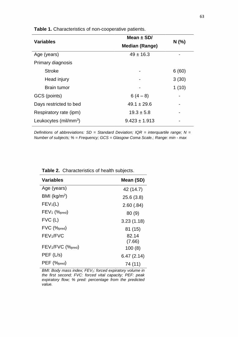
63
Table 1. Characteristics of non-cooperative patients.
Variables Mean ± SD/
Median (Range) N (%)
Age (years) 49 ± 16.3 -
Primary diagnosis
Stroke - 6 (60)
Head injury - 3 (30)
Brain tumor - 1 (10)
GCS (points) 6 (4 – 8) -
Days restricted to bed 49.1 ± 29.6 -
Respiratory rate (ipm) 19.3 ± 5.8 -
Leukocytes (mil/mm3) 9.423 ± 1.913 -
Definitions of abbreviations: SD = Standard Deviation; IQR = interquartile range; N =
Number of subjects; % = Frequency; GCS = Glasgow Coma Scale.; Range: min - max
Table 2. Characteristics of health subjects.
Variables Mean (SD)
Age (years) 42 (14.7)
BMI (kg/m2) 25.6 (3.8)
FEV1(L) 2.60 (.84)
FEV1 (%pred) 80 (9)
FVC (L) 3.23 (1.18)
FVC (%pred) 81 (15)
FEV1/FVC 82.14 (7.66)
FEV1/FVC (%pred) 100 (8)
PEF (L/s) 6.47 (2.14)
PEF (%pred) 74 (11)
BMI: Body mass index; FEV1: forced expiratory volume in the first second; FVC: forced vital capacity; PEF: peak expiratory flow; % pred: percentage from the predicted value.
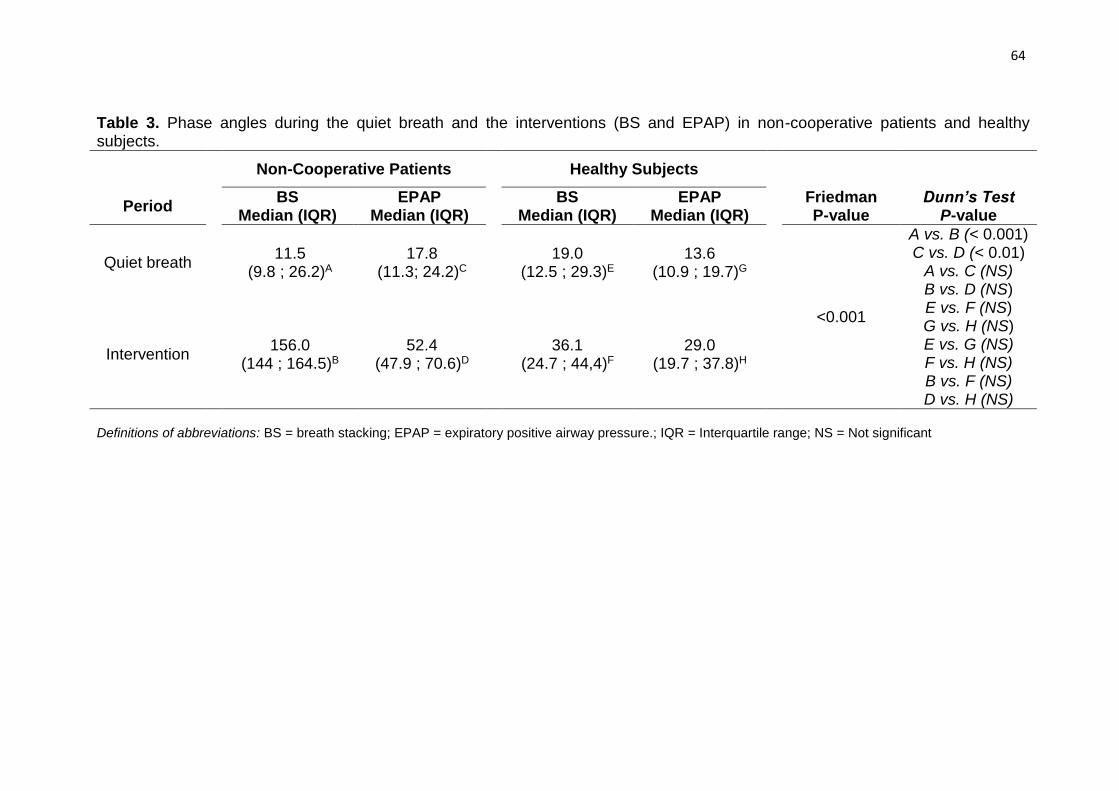
64
Table 3. Phase angles during the quiet breath and the interventions (BS and EPAP) in non-cooperative patients and healthy subjects.
Period
Non-Cooperative Patients Healthy Subjects
BS Median (IQR)
EPAP Median (IQR)
BS
Median (IQR) EPAP
Median (IQR)
Friedman P-value
Dunn’s Test P-value
Quiet breath 11.5
(9.8 ; 26.2)A
17.8 (11.3; 24.2)C
19.0 (12.5 ; 29.3)E
13.6 (10.9 ; 19.7)G
<0.001
A vs. B (< 0.001) C vs. D (< 0.01)
A vs. C (NS) B vs. D (NS) E vs. F (NS) G vs. H (NS) E vs. G (NS) F vs. H (NS) B vs. F (NS) D vs. H (NS)
Intervention 156.0
(144 ; 164.5)B
52.4 (47.9 ; 70.6)D
36.1 (24.7 ; 44,4)F
29.0 (19.7 ; 37.8)H
Definitions of abbreviations: BS = breath stacking; EPAP = expiratory positive airway pressure.; IQR = Interquartile range; NS = Not significant
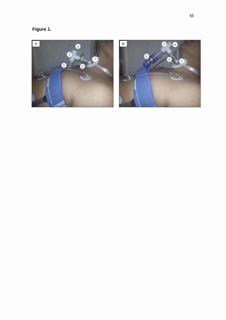
65
Figure 1.
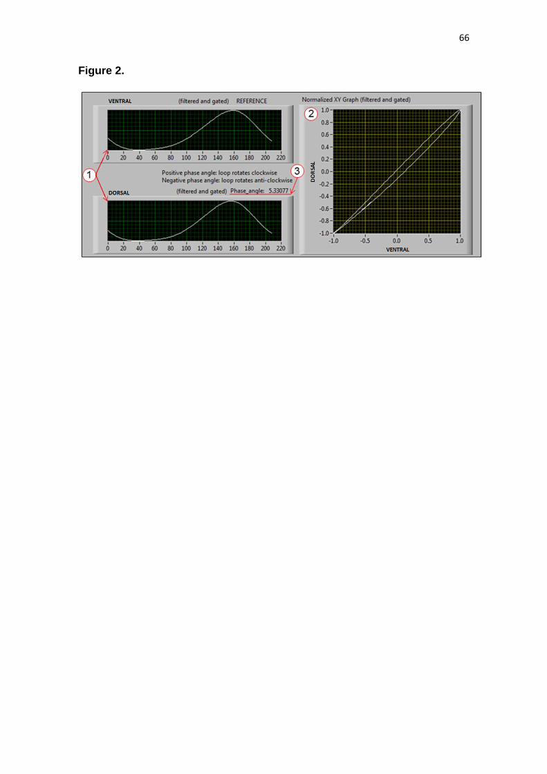
66
Figure 2.
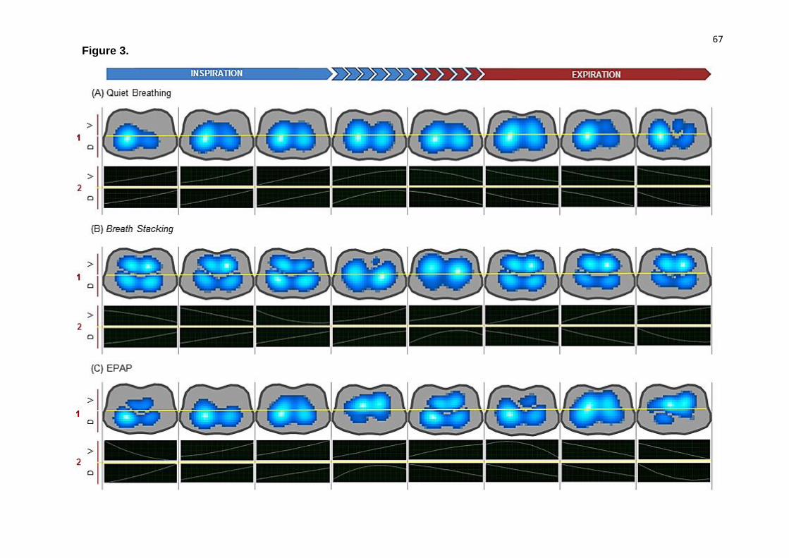
67
Figure 3.
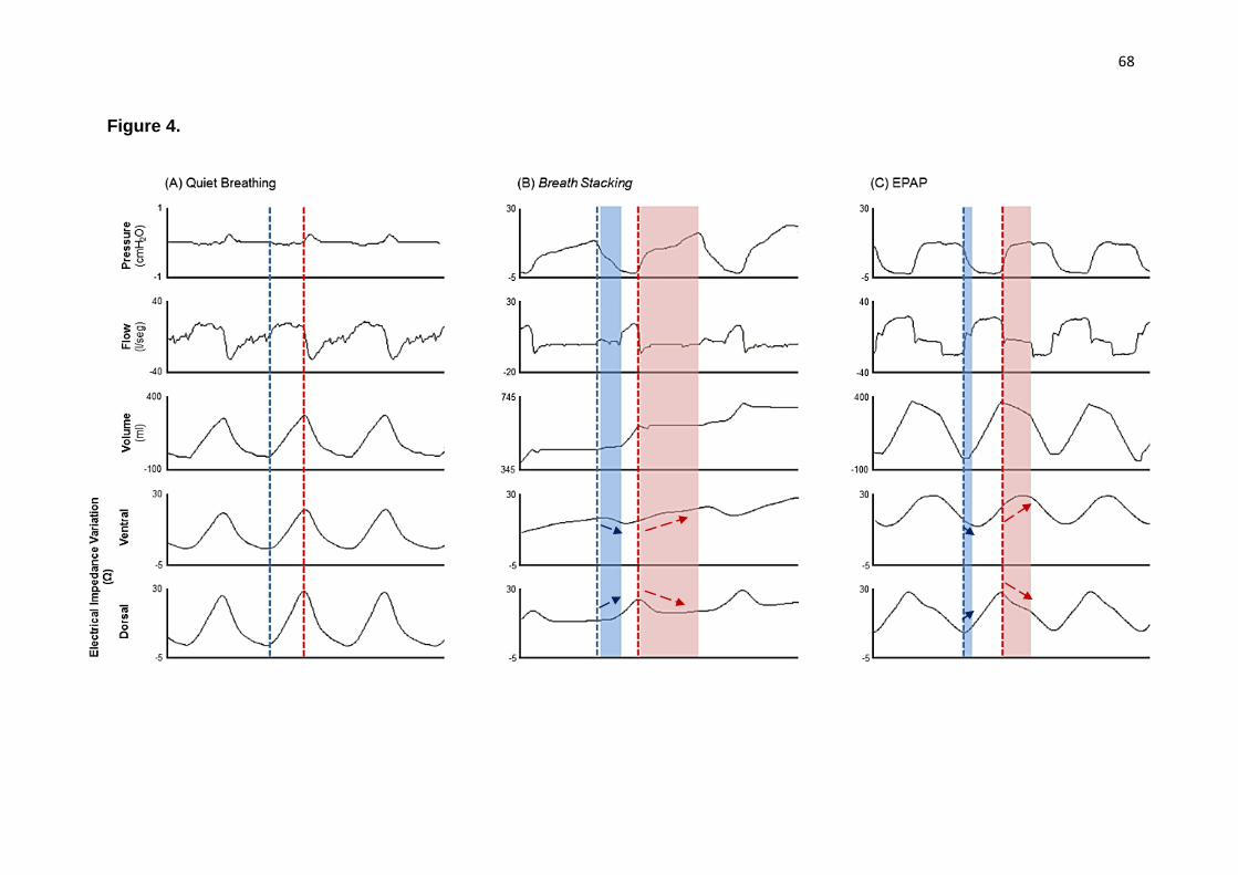
68
Figure 4.
69
APÊNDICE B - ARTIGO 2
IMMEDIATE EFFECT OF EXPIRATORY POSITIVE AIRWAY PRESSURE
AND BREATH STACKING IN NON-COOPERATIVE PATIENTS DETECTED
BY ELECTRICAL IMPEDANCE TOMOGRAPHY: A RANDOMIZED
CROSSOVER STUDY
A ser submetido ao periódico: Archives of Physical Medicine and
Rehabilitation
70
IMMEDIATE EFFECT OF LUNG EXPANSION TECHNIQUES IN NON-
COOPERATIVE PATIENTS WITH PROLONGED BED RESTRICTION: A
RANDOMIZED CROSSOVER STUDY
Caio César Araújo Morais1, Shirley Lima Campos1, Catarina Rattes1, Lucas Monte1, Daniela
Brandão1, Marcelo Britto Passos Amato2, Armèle Dornelas de Andrade1
1 - Physical Therapy Department, Universidade Federal de Pernambuco (UFPE) - Recife/Brazil;
2 - Pulmonary Division, Universidade de São Paulo (USP) - São Paulo/Brazil;
Abstract Introduction: The survival of patients with lesions in the central nervous system is usually accompanied by physical and mental sequelae. These impairments favor the prolonged restriction to the bed, which may contribute with changes in respiratory function. In this context, lung re-expansion techniques are used to prevent or treat the various respiratory complications. Objective: To compare the acute effect of Breath Stacking (BS) and Expiratory Positive Airway Pressure (EPAP) techniques on the regional lung aeration, and the duration of the therapeutic effect of lung expansion in non-cooperative patients with prolonged bed rest. Secondary aims included to evaluate the influence of these techniques on cardiorespiratory system. Methods: This was a randomized crossover study. Ten patients were included with Glasgow Coma Scale between 3-10 points, 18-65 years old, undergoing neurosurgery, unable to respond to the command and restricted to bed. The regional lung aeration changes were assessed by EIT device (Enligh, Timpel, São Paulo, Brazil). For interventions, EPAP was applied through a Spring Load valve resistor, adjusted with a pressure of 10 cmH2O. BS was performed three times with one minute intervals between them. The volume was stacked and maintained until 40 seconds. Results: Aeration increased significantly in comparison with baseline during EPAP and BS (2-way ANOVA and Sidak post hoc, all P < 0.001). However, the effects on lung expansion were kept for a short time, 4.6 ± 3.7 minutes and 2.3 ± 2.0 minutes for EPAP and BS, respectively. There were no clinically significant differences on cardiorespiratory variables. Conclusions: It was observed that during the BS and EPAP techniques existed a significant change on lung volumes, but not durable, and it didn’t generate adverse effects on the cardiovascular system in non-cooperative with prolonged bed rest. The existence of expiratory muscle contraction may have to minimize the effect of lung expansion proposed by these techniques. Keywords: physical therapy techniques; positive-pressure respiration; electrical
impedance; lung volume measurements;
71
Introduction
The survival of patients with lesions in the Central Nervous System is
usually accompanied by physical and mental permanents sequelae. This
impairment of cognitive status associated with motor injury favors prolonged
restriction of these patients in bed (1, 2). The supine position is usually adopted
in the hospitals units, and this is associated with changes in respiratory function
as reduced functional residual capacity (FRC) (3), tidal volume and mobility of
the rib cage (4).
There are few interventions that could be used in patients with deficit of
awareness and cooperation with the principle to keep the lung open (5), such as
Breath Stacking technique (BS) (6) and Expiratory Positive Airway Pressure
(EPAP) (7). The mechanisms of BS is to increase the lung expansion by
stacking the gas, cycle to cycle through a one-way inspiratory valve (8). So, the
lung aeration increases as the inspired volume is trapped, like an intrinsic PEEP
(6). For EPAP, this effect is obtained through spring-load valves that generate a
resistance to airflow during expiration, thereby retaining gas volume inside the
lungs (7).
In the clinical routine, lung expansion techniques have been used as a
prophylaxis routine of respiratory conditions that involve volumetric reductions
6, 7). However, the effects of these interventions need to be elucidated to
support the prophylactic use in cases that the only risk factor is the prolonged
bed rest.
The effects on lung volume promoted by BS and EPAP can be accurately
and safely measured using an electrical impedance tomography (EIT). This
recent imaging modality offers information about gas distribution and has a
strong linear correlation with regional ventilation within the thorax (8, 9).
Thereby, the primary aim of this study was to analyze the effect of BS
and EPAP techniques on the regional lung aeration by EIT, and also measure
the duration of therapeutic effect of lung expansion in non-cooperative patients,
post neurosurgery, in prolonged bed restriction, with no lung diseases.

72
Secondary aims include evaluate the influence of the interventions on
cardiorespiratory variables.
Methods
Subjects
The sample was composed by patients undergoing neurosurgery, unable
to respond to commands and restricted in bed for more than 14 days. It was
included subjects with age between 18 and 65 years, Glasgow Coma Scale <
10 points, breathing spontaneously through the tracheostomy tube. To ensure
that the only risk factor for lung volume reduction was the bed restriction, we
excluded patients with respiratory infection, chronic lung diseases, chest
deformity, rib fracture, asymmetrical chest expansion and abdomen distension.
Other exclusion criteria were: chest circumference less than 88 and greater
than 98 centimeters (just below the axilla region), presence of spasticity in any
hemisphere with Ashworth Scale score more than 2 points for upper limbs,
uncoordinated movements of the limbs and cardiorespiratory instability (heart
rate < 60 or > 120 bpm; respiratory rate > 35 ipm; mean arterial pressure < 60
mmHg or > 120 mmHg; oxygen saturation < 90%).
Study design
This was a randomized crossover study, conducted in the Neurosurgery
Special Care Unit of Restoration Hospital (Brazil), executed between the
months of August 2014 to January 2015. The study was approved by
institutional ethics committee (number: 15037913.0.0000.5208) and all patients’
families signed an informed consent form. (Clinical trials number:
NCT02613832)
Procedures
Patients were positioned in supine with the head up 45 degree. In order
to ensure that the protocol had initiated on the same condition
(homogenization), before the interventions, endotracheal suction was performed
with 3 inserts of the catheter in the tracheostomy tube with negative pressure
regulated between 100 and 150 mmHg. Patients were breathed during 2
minutes at 10 cmH2O with Continuous Positive Airway Pressure (CPAP) to
73
minimize the effects of post suction. The first technique begun after an interval
of 60 minutes of CPAP. This interval was defined, after a pilot study, as the
maximum time required for ventilatory parameters to return to previous
suctioning. In order to avoid the possibility of air leakage though the upper
airway, the cuff was inflated to ensure a minimal occluding volume.
Data collection
Three researchers participated in the study. One was responsible for the
patient´s preparation and evaluations. The second was responsible for the
randomization process and the third was responsible for applying the
techniques of BS and EPAP.
Randomization
Interventions were randomized using the online software, available at the
website “www.randomization.com”. The intervening sequence was encoded in
sequence 1 (BS to EPAP) and sequence 2 (EPAP to BS). Then the sequences
were transferred to a series of numbered opaque envelopes for allocation of
volunteers.
Protocol of Breath Stacking and EPAP
The BS was performed by a T-tube with a one-way inspiratory valve and
the expiratory branch occluded (Figure 1A). The expiratory occlusion was
maintained until observed the presence of a plateau in the impedance
plethysmogram, which corresponds to the absence of inspired air displacement
or when a maximum time of 40 seconds was attained. Three BS interventions
were performed at one minute intervals between them. EPAP was applied with
a Spring Load valve resistor (Vital Signs, Totowa, NJ, USA), adjusted with a
pressure of 10 cmH2O. This valve was connected to the expiratory branch of
the T-tube. The total duration of treatment was 5 minutes (Figure 1B). The
washout period was determined when the lung volume returned to baseline
values after the first technique.
Electrical impedance tomography
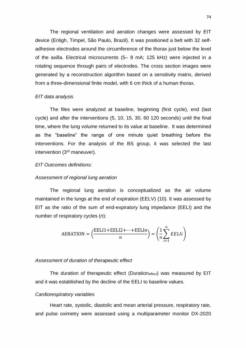
74
The regional ventilation and aeration changes were assessed by EIT
device (Enligh, Timpel, São Paulo, Brazil). It was positioned a belt with 32 self-
adhesive electrodes around the circumference of the thorax just below the level
of the axilla. Electrical microcurrents (5– 8 mA; 125 kHz) were injected in a
rotating sequence through pairs of electrodes. The cross section images were
generated by a reconstruction algorithm based on a sensitivity matrix, derived
from a three-dimensional finite model, with 6 cm thick of a human thorax.
EIT data analysis
The files were analyzed at baseline, beginning (first cycle), end (last
cycle) and after the interventions (5, 10, 15, 30, 60 120 seconds) until the final
time, where the lung volume returned to its value at baseline. It was determined
as the “baseline” the range of one minute quiet breathing before the
interventions. For the analysis of the BS group, it was selected the last
intervention (3rd maneuver).
EIT Outcomes definitions:
Assessment of regional lung aeration
The regional lung aeration is conceptualized as the air volume
maintained in the lungs at the end of expiration (EELV) (10). It was assessed by
EIT as the ratio of the sum of end-expiratory lung impedance (EELI) and the
number of respiratory cycles (n):
AERATION = (EELI1+EELI2+⋯+EELI𝑛
𝑛) = (
1
𝑛∑ 𝐸𝐸𝐿𝐼𝑖
𝑛
𝑖=1
)
Assessment of duration of therapeutic effect
The duration of therapeutic effect (Durationeffect) was measured by EIT
and it was established by the decline of the EELI to baseline values.
Cardiorespiratory variables
Heart rate, systolic, diastolic and mean arterial pressure, respiratory rate,
and pulse oximetry were assessed using a multiparameter monitor DX-2020
75
(Dixtal Biomedical, São Paulo, Brazil) before, during, and after the first, fifth and
tenth minute in each intervention.
Statistical Methods
Sample Size
The sample size was calculated using the online software “Quantitative
Measurement tools” for crossover study, developed by MGH Massachusetts
General Hospital Mallinckrodt General Clinical Research Center
(Massachusetts, EUA), available on web site
(http://hedwig.mgh.harvard.edu/sample_size/size.html).
The EELI and Durationeffect data of a pilot study with five patients were
used as outcomes in the sample calculation. The largest sample number was
obtained for the EELI with the minimal detectable difference between BS and
EPAP treatment of 14.22 units and standard deviation of the difference between
the two values for the same patient in the variable of 14.26 units. So, it was
required 10 patients, believing that the probability that the study will detect a
treatment difference at a two-side is 80%, with a significance level of 5% (p
<0.05). Considering the probability of noise in the analysis of signals, it was
decided to add 30% to reset sample loss, being collected 13 patients.
Data Analysis
The statistical analysis was performed using the statistical programs
SPSS for Windows, version 20.0 – Statistical Package for the Social Science,
and SigmaPlot for Windows, version 12.0.
The Shapiro-Wilk test was used to assess normality. Normally distributed
data was expressed as mean ± SD and non-parametric data was expressed as
medians and interquartile range (IQR). Categorical data are presented as
absolute values or number of events (%). The P-value was considered
significant when < 0.05 according to the analysis table of statistical tests applied
(table 1).
Results
Patient characteristics
76
A total of sixty patients were assessed for eligibility criteria (Figure 1).
The Table 2 shows the clinical characteristics of the ten patients that concluded
the analysis.
Effects in the regional lung aeration
The Figure 3 shows the behavior of EELI at baseline, during and after the
interventions. The EELI increased significantly in comparison with baseline
during EPAP and BS (2-way ANOVA and Sidak post hoc, all P < 0.001). There
was no significant difference between groups during the interventions. The EELI
was not higher after EPAP and BS in comparison with baseline (2-way ANOVA
and Sidak post hoc, all P > 0.05).
Duration of therapeutic effect
The figure 4 shows the time that the EELI was maintained above the
baseline values for each patient. There weren´t significant differences between
EPAP (4.6 ± 3.7min) and BS (2.3 ± 2.0min) (T-test, P = 0.103).
Effects on cardiorespiratory variables
Regarding of the cardiorespiratory variables, only diastolic blood
pressure increased significantly when compared during the EPAP (75.1 mmHg)
and the BS (89.4 mmHg) (2-way ANOVA and Sidak post hoc, P < 0.05). There
weren´t differences for the variables SBP, MAP, HR, RR and SpO2 (Table 4).
Discussion
Our data showed that regional lung aeration increased during EPAP and
BS, however the effects on lung expansion were not kept for a long time. In this
trial we had the interest to evaluate the effect of EPAP and BS in cases with
prolonged bed restriction as the only risk factor for the lug volume reduction.
Another finding of this study was the absence of adverse effects on
cardiorespiratory variables considered "clinically" important.
The first justification for the primary aim would be that we used
insufficient "dose" of techniques (duration and intensity pressure) to provide
sustainable change in lung volume. Other one could be, even at risk for
77
development volume reduction, these samples do not need specifics
prophylactic interventions only to keep the lungs expanded. Possibly, these
patients may have compensatory mechanisms, such as intermittent sighs,
periods of hyperventilation (during Cheyne Stokes).
However, as is known, both applied techniques have as principle of
action the hinder of air flow output completely (for BS) or partially (for EPAP) to
lung expansion. Due to no comprehension of the patients about what is
occurring during the techniques, in each expiration period, it was common to
observe the abdominal muscles contracted to overcome the resistance imposed
by the devices.
Leevers et al (11), observed in an animal model that the increases in
lung volume was associated with high expiratory muscles activity, measured by
EMG. It was described that the expiratory time (TE) increased similarly to the
recruitment of abdominal muscles. This effect, expiratory prolongation, has
been shown in others studies (12, 13) as a response to the increase of EELV.
The authors suggest that the shortening of the abdominal muscles is primarily
mediated by vagal reflexes for prevent an increase in FRC, thus preserving
diaphragm initial length and maintaining the tidal volume.
Our patients were not monitored EMG of abdominal muscles. However,
similarly to those previous studies, we could infer the presences of abdominal
effort if there were increasing of TE during the maneuvers of lung expansion.
Thus, we use the flow signal from the integrated pneumotachograph by the EIT
monitor to analysis the expiratory time behavior before, during and after the
techniques (Figure 5).
As we can observe, there were an increase in expiratory time during
techniques, especially during the BS. This fact may explain the failure to
maintain the lung expansion for a long time. Thus, we can deduce that for some
patients the set pressure of the spring load valve (10cmH20) or the duration of
the BS have been high, so that the larger the resistance, the greater the
expiratory activity generated, and the less the effect of pulmonary expansion.
78
Therefore, new studies are needed to check the effect of different doses
(volumes generated by devices) and evaluate to long-term clinical outcomes, as
reducing pulmonary complications and hospitalization.
Conclusion
It was observed that during the BS and EPAP techniques existed a
significant change on lung volumes, but not durable, and didn’t generated
adverse effects on the cardiovascular system in non-cooperative with prolonged
bed rest. The existence expiratory muscle contraction may have to minimize the
effect of lung expansion proposed by these techniques.
References
1. Kim J, Kim CH, Kang H-S, Park C-K, Chung CK. Cognitive Function of Korean Neurosurgical Patients: Cross-sectional Study Using the Korean Version of the Mini-mental Status Examination. J Cerebrovasc Endovasc Neurosurg. 2012;14:11.
2. Tan T, Ding Y, Lee A. Impaired Mobility in Older Persons Attending a Geriatric Assessment Clinic: Causes and Management. Singapore Med J. 2001;42(2):68–72.
3. Dean E. Effect of body position on pulmonary function. Phys Ther. 1985;65:613–8.
4. Romei M, Mauro a. L, D’Angelo MG, Turconi a. C, Bresolin N, Pedotti a., et al. Effects of gender and posture on thoraco-abdominal kinematics during quiet breathing in healthy adults. Respir Physiol Neurobiol. 2010;172:184–91.
5. Bhat A, Chakravarthy K, Rao B. Chest physiotherapy techniques in neurological intensive care units of India: A survey. Indian J Crit Care Med. 2014;18:363.
6. Baker WL, Lamb VJ, Marini JJ. Breath-stacking increases the depth and duration of chest expansion by incentive spirometry. Am Rev Respir Dis. 1990;141:343–6.
7. Layon J, Banner M. Continuous positive airway pressure and expiratory positive airway pressure increase functional residual capacity equivalently. CHEST. 1986; 89(4):517–21.
79
8. Marini J, Rodrigues R, Lamb V. Involuntary breath stacking: An alternative method for vital capacity estimation in poorly cooperative subjects. Am Rev Respir Dis. 1986;134:694–8.
9. Costa ELV, Amato MBP. Electrical Impedance Tomography in Critically Ill Patients. Clin Pulm Med. 2013;20(4):178–86.
10. Reber A, Engberg G, Wegenius G, Hedenstierna G. Lung aeration. Anaesthesia. 1996; 51:733–7.
11. Leevers AM, Road JD. Abdominal muscle activation by expiratory threshold loading in awake dogs. Respiration Physiology. 1993; 93: 289 - 303.
12. Oliven A, Kelsen SG. Effect of hypercapnia and PEEP on expiratory muscle EMG and shortening. J. Appl. Physiol. 1989; 66: 1408-1413.
13. Finkler J, Iscoe S. Control of breathing at elevated lung volumes in anesthetized cats. J. Appl. Physiol. 1984; 56: 839-844.
80
LEGEND OF FIGURES
Figure 1. Illustration of the devices used in the Breath Stacking (A) and EPAP (B) techniques. 1 = tracheostomy tube; 2 = pneumotachograph; 3 = "T-piece"; 4 = inspiratory branch; 5 = piece for closure the expiratory branch; 6 = spring load valve connected to the expiratory branch.
Figure 2. CONSORT diagram of patient flow through the study.
Figure 3. Behavior of EELI at baseline, during and after the interventions. 2-way ANOVA test for between-group comparison with Sidak post hoc. Figure 4. Mean time to maintain the EELI above baseline after the interventions (duration of therapeutic effect). Figure 5. Expiratory duration plotted before, during and after the techniques.
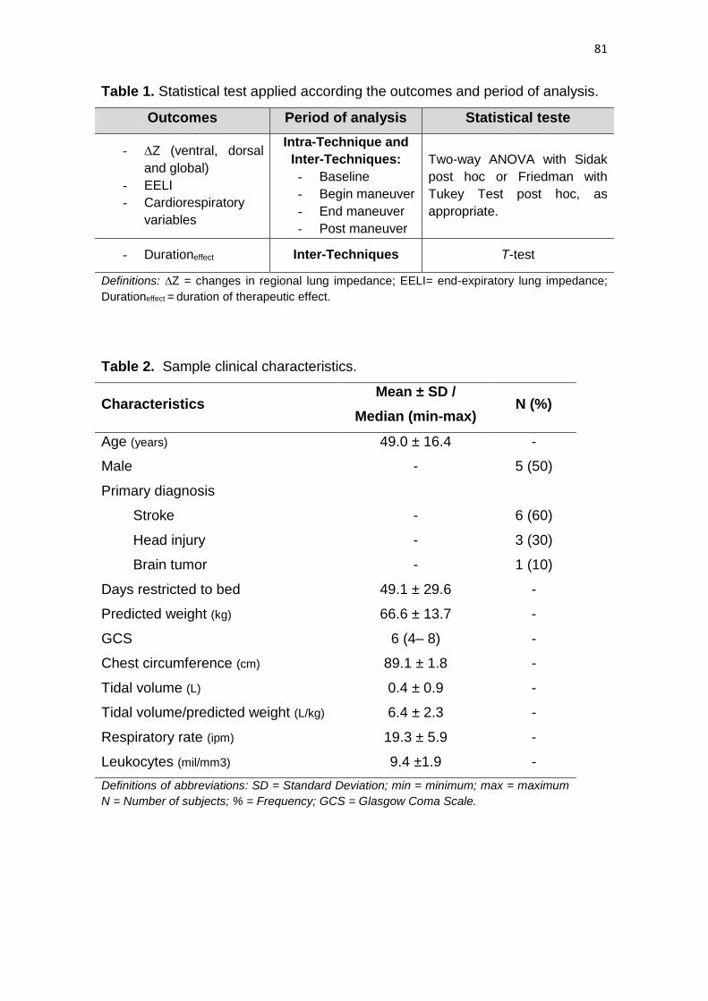
81
Table 1. Statistical test applied according the outcomes and period of analysis.
Outcomes Period of analysis Statistical teste
- ∆Z (ventral, dorsal
and global)
- EELI
- Cardiorespiratory
variables
Intra-Technique and
Inter-Techniques:
- Baseline
- Begin maneuver
- End maneuver
- Post maneuver
Two-way ANOVA with Sidak
post hoc or Friedman with
Tukey Test post hoc, as
appropriate.
- Durationeffect Inter-Techniques T-test
Definitions: ∆Z = changes in regional lung impedance; EELI= end-expiratory lung impedance;
Durationeffect = duration of therapeutic effect.
Table 2. Sample clinical characteristics.
Characteristics Mean ± SD /
Median (min-max) N (%)
Age (years) 49.0 ± 16.4 -
Male - 5 (50)
Primary diagnosis
Stroke - 6 (60)
Head injury - 3 (30)
Brain tumor - 1 (10)
Days restricted to bed 49.1 ± 29.6 -
Predicted weight (kg) 66.6 ± 13.7 -
GCS 6 (4– 8) -
Chest circumference (cm) 89.1 ± 1.8 -
Tidal volume (L) 0.4 ± 0.9 -
Tidal volume/predicted weight (L/kg) 6.4 ± 2.3 -
Respiratory rate (ipm) 19.3 ± 5.9 -
Leukocytes (mil/mm3) 9.4 ±1.9 -
Definitions of abbreviations: SD = Standard Deviation; min = minimum; max = maximum
N = Number of subjects; % = Frequency; GCS = Glasgow Coma Scale.
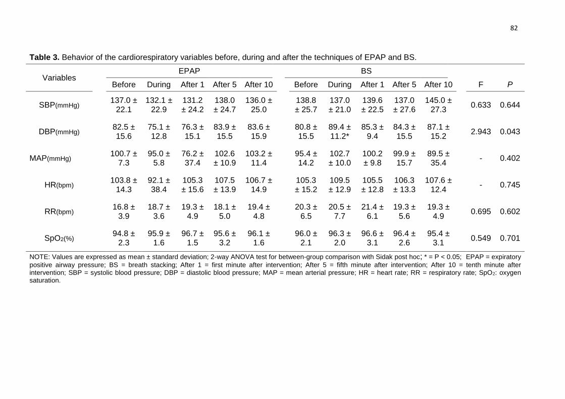
82
Table 3. Behavior of the cardiorespiratory variables before, during and after the techniques of EPAP and BS.
Variables EPAP BS
Before During After 1 After 5 After 10 Before During After 1 After 5 After 10 F P
SBP(mmHg)
137.0 ± 22.1
132.1 ± 22.9
131.2 ± 24.2
138.0 ± 24.7
136.0 ± 25.0
138.8 ± 25.7
137.0 ± 21.0
139.6 ± 22.5
137.0 ± 27.6
145.0 ± 27.3
0.633 0.644
DBP(mmHg)
82.5 ± 15.6
75.1 ± 12.8
76.3 ± 15.1
83.9 ± 15.5
83.6 ± 15.9
80.8 ± 15.5
89.4 ± 11.2*
85.3 ± 9.4
84.3 ± 15.5
87.1 ± 15.2
2.943 0.043
MAP(mmHg)
100.7 ± 7.3
95.0 ± 5.8
76.2 ± 37.4
102.6 ± 10.9
103.2 ± 11.4
95.4 ± 14.2
102.7 ± 10.0
100.2 ± 9.8
99.9 ± 15.7
89.5 ± 35.4
- 0.402
HR(bpm)
103.8 ± 14.3
92.1 ± 38.4
105.3 ± 15.6
107.5 ± 13.9
106.7 ± 14.9
105.3 ± 15.2
109.5 ± 12.9
105.5 ± 12.8
106.3 ± 13.3
107.6 ± 12.4
- 0.745
RR(bpm)
16.8 ± 3.9
18.7 ± 3.6
19.3 ± 4.9
18.1 ± 5.0
19.4 ± 4.8
20.3 ±
6.5 20.5 ±
7.7 21.4 ±
6.1 19.3 ±
5.6 19.3 ±
4.9 0.695 0.602
SpO2(%)
94.8 ± 2.3
95.9 ± 1.6
96.7 ± 1.5
95.6 ± 3.2
96.1 ± 1.6
96.0 ±
2.1 96.3 ±
2.0 96.6 ±
3.1 96.4 ±
2.6 95.4 ±
3.1 0.549 0.701
NOTE: Values are expressed as mean ± standard deviation; 2-way ANOVA test for between-group comparison with Sidak post hoc; * = P < 0.05; EPAP = expiratory
positive airway pressure; BS = breath stacking; After 1 = first minute after intervention; After 5 = fifth minute after intervention; After 10 = tenth minute after intervention; SBP = systolic blood pressure; DBP = diastolic blood pressure; MAP = mean arterial pressure; HR = heart rate; RR = respiratory rate; SpO2: oxygen saturation.
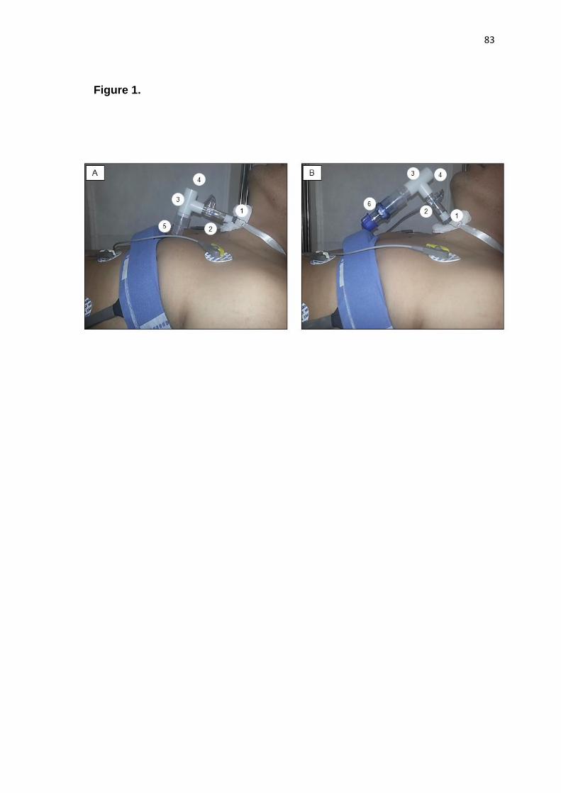
83
Figure 1.
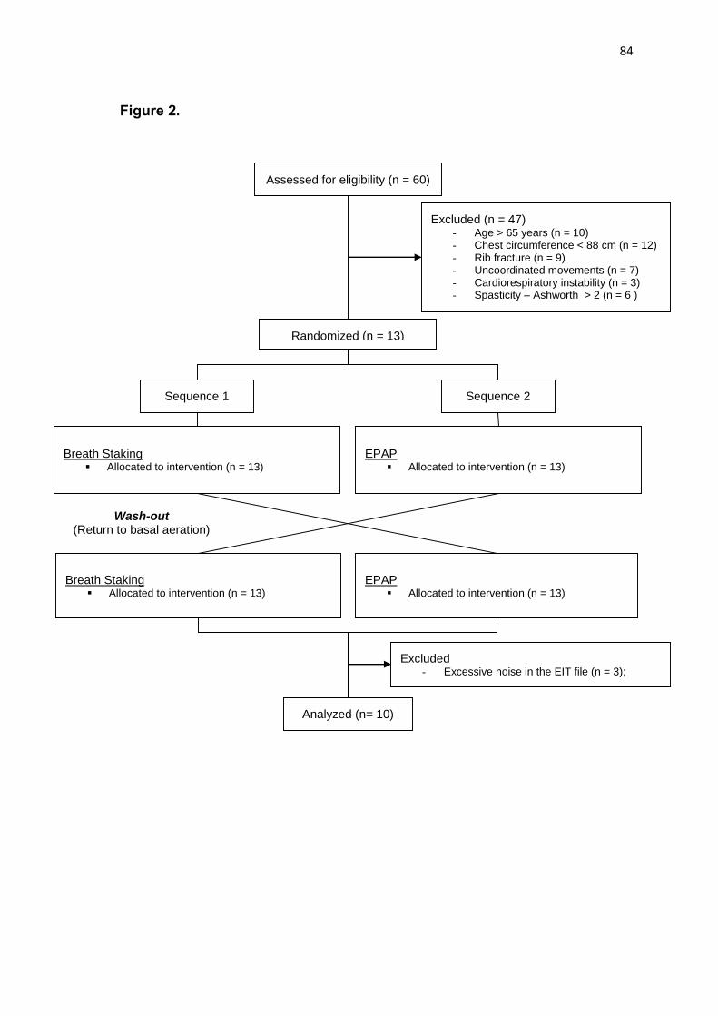
84
Figure 2.
Assessed for eligibility (n = 60)
Excluded (n = 47) - Age > 65 years (n = 10) - Chest circumference < 88 cm (n = 12) - Rib fracture (n = 9) - Uncoordinated movements (n = 7) - Cardiorespiratory instability (n = 3) - Spasticity – Ashworth > 2 (n = 6 )
Breath Staking Allocated to intervention (n = 13)
Randomized (n = 13)
Sequence 1 Sequence 2
EPAP Allocated to intervention (n = 13)
Breath Staking Allocated to intervention (n = 13)
EPAP Allocated to intervention (n = 13)
Analyzed (n= 10)
Excluded - Excessive noise in the EIT file (n = 3);
Wash-out (Return to basal aeration)
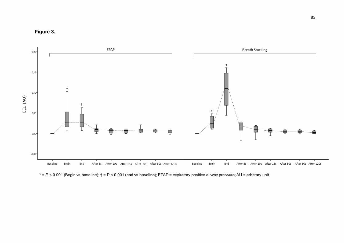
85
Figure 3.
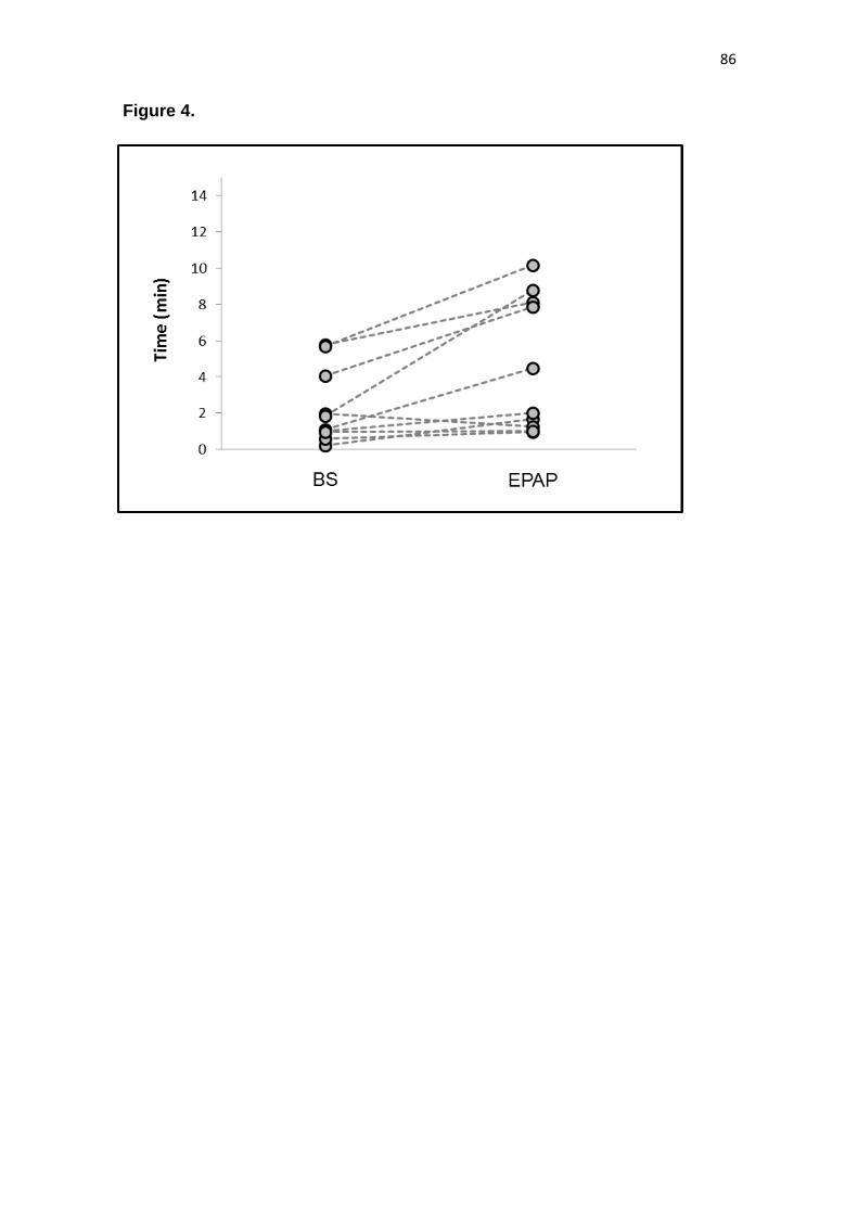
86
Figure 4.
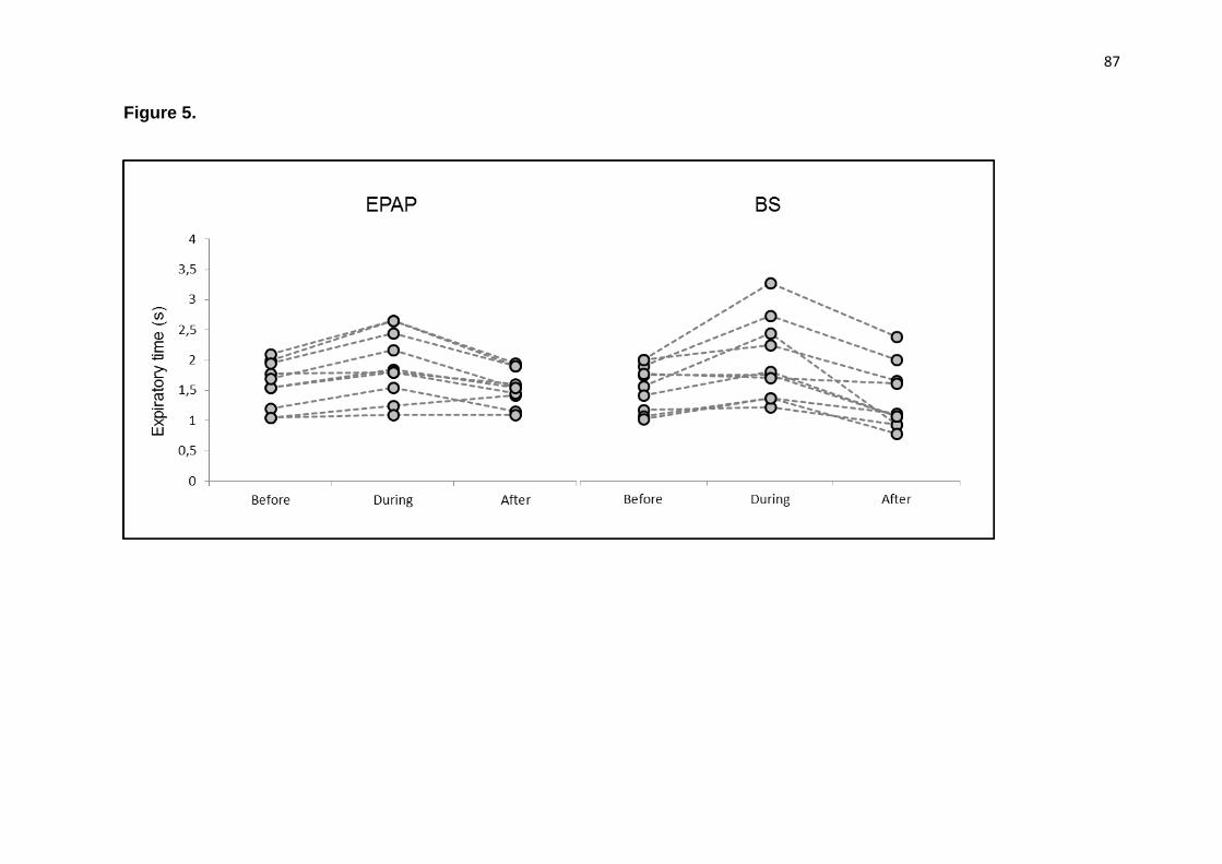
87
Figure 5.

88
ANEXOS
ANEXO A - Aprovação do Comitê de Ética
89
90
91
ANEXO B - Parecer Final do Comitê de Ética
92