Embed Size (px)
Citation preview
1
VALTER TADEU BOLDARINE
APERFEIÇOAMENTO DO ENSAIO DE QUANTIFICAÇÃO DO RNA
MENSAGEIRO DA TIROGLOBULINA POR RT-PCR EM TEMPO REAL NO
SEGUIMENTO DE PACIENTES COM CARCINOMA DIFERENCIADO DA
TIROIDE
Dissertação apresentada à Universidade
Federal de São Paulo para a obtenção do
título de Mestre em Ciências
São Paulo
2009

Livros Grátis
http://www.livrosgratis.com.br
Milhares de livros grátis para download.
2
VALTER TADEU BOLDARINE
APERFEIÇOAMENTO DO ENSAIO DE QUANTIFICAÇÃO DO RNA
MENSAGEIRO DA TIROGLOBULINA POR RT-PCR EM TEMPO REAL NO
SEGUIMENTO DE PACIENTES COM CARCINOMA DIFERENCIADO DA
TIROIDE
Dissertação apresentada à Universidade
Federal de São Paulo para a obtenção do
título de Mestre em Ciências
Orientador: Rui Monteiro de Barros Maciel
Co-orientadora: Rosa Paula Mello Biscolla
Coordenador do Programa: Sérgio Atala Dib
São Paulo
2009
3
FICHA CATALOGRÁFICA
Boldarine, Valter Tadeu
Aperfeiçoamento do ensaio de quantificação do RNA mensageiro da tiroglobulina por
RT-PCR em tempo real no seguimento de pacientes com carcinoma diferenciado da
tiroide São Paulo, 2009
p 48.
Tese (Mestrado) – Universidade Federal de São Paulo-Escola Paulista de Medicina. Programa de Pós Graduação em Endocrinologia Título em inglês: Development of a sensitive and specific quantitavive RT-PCR assay for blood thyroglobulin messenger RNA (TgmRNA) in the follow-up of patients with differentiated thyroid carcinoma 1. Tiroglobulina RNAm 2. PCR quantitativo 3. Splicing alternativo
4. Carcinoma diferenciado de tiroide
4
Banca Examinadora
1. Prof. Dr. Julio Abucham, Professor Associado, Disciplina de Endocrinologia, Departamento
de Medicina, Escola Paulista de Medicina, Universidade Federal de São Paulo
2. Pedro Wesley Rosário, Médico-Doutor, Centro de Pesquisas da Santa Casa de Belo
Horizonte
3. Ileana G. S. de Rubió, Pesquisadora-Doutora, Disciplina de Endocrinologia, Departamento
de Clínica Médica, Faculdade de Medicina, Universidade de São Paulo
Suplente: Profa. Dra. Miriam Oliveira Ribeiro, Professora Adjunta, Universidade Presbiteriana
Mackenzie

5
Dedico esta tese de mestrado aos meus pais, que sempre me apoiaram e
me deram forças para continuar, mesmo nos momentos mais difíceis.
A vocês, obrigado por tudo.
6
AGRADECIMENTOS
Sendo sucinto e sincero, espero transmitir, por meio destes agradecimentos, meus sentimentos
quanto ao processo desta dissertação de mestrado.
Agradeço ao Professor Dr. Rui Monteiro de Barros Maciel, meu orientador, que ao longo dos anos
se tornou na verdade meu amigo, e sem o qual não estaria apresentando esta tese ao mundo
científico.
À minha co-orientadora Dra. Rosa Paula Biscolla, que confiou em minha capacidade e me convidou
para realizar o presente trabalho sob sua co-orientação (sempre solícita), a todos que participaram
de alguma forma deste trabalho, seja diretamente (Gustavo, Cléber, Cláudia(s), Dani, Conceição,
Felipe e Teresa), seja indiretamente, através de conselhos (Dra. Janete, Dr. Gilberto), ou mesmo de
dicas como aprimorar minhas técnicas (Gisele, Flávia, Rosana, Ilda), e também a todos os
funcionários do Programa de Pós Graduação em Endocrinologia.
Agradeço também ao apoio e sempre boa vontade da Ângela. Faço uma menção especial a todos do
ambulatório de Tiróide, por seus ensinamentos clínicos (pós-graduandos, residentes, chefes) e aos
solícitos funcionários da Pós-Graduação. Agradeço também à Professora Dra. Miriam (minha
professora da graduação que abriu meus olhos para o mundo da Endocrinologia Molecular).
Às minhas irmãs pelo apoio a todo o momento.
À Amanda, por estar sempre ao meu lado, mostrando muita paciência e disposição.
Aos pacientes, por consentirem em participar deste estudo.
À Fundação de Amparo à Pesquisa do Estado de São Paulo (FAPESP), pela bolsa de Mestrado
(processo nº05/55842-0) e pelo financiamento da pesquisa realizada (processo n º04/09934-7).
Obrigado
7
ÍNDICE
1 APRESENTAÇÃO 8
2 INTRODUÇÃO E OBJETIVOS 10 2.1 SÍNTESE E SECREÇÃO DE TIROGLOBULINA 11 2.2 SEGUIMENTO DE PACIENTES COM CARCINOMA DIFERENCIADO 13 DE TIROIDE 2.3 PAPEL DO RNA MENSAGEIRO COMO MARCADOR TUMORAL 15 2.4 OBJETIVOS 19
3 ARTIGO SUBMETIDO À PUBLICAÇÃO 20
“DEVELOPMENT OF A SENSITIVE AND SPECIFIC
QUANTITATIVE RT-PCR ASSAY FOR BLOOD
THYROGLOBULIN MESSENGER RNA (TgmRNA) IN THE
FOLLOW-UP OF PATIENTS WITH DIFFERENTIATED
THYROID CARCINOMA”
4 CONCLUSÕES 42
5 REFERÊNCIAS 44
8
1-APRESENTAÇÃO
9
1. APRESENTAÇÃO
Nesta tese de mestrado apresentamos, de acordo com as recomendações do Programa de
Pós-Graduação em Endocrinologia da Universidade Federal de São Paulo-Escola Paulista de
Medicina, um trabalho completo em inglês intitulado “Development of a sensitive and specific
quantitative RT-PCR assay for blood thyroglobulin messenger RNA (TgmRNA) in the follow-up of
patients with differentiated thyroid carcinoma”, de autoria de Valter T. Boldarine, Gustavo S.
Guimarães, Claudia C. D. Nakabashi, Cleber P. Camacho, Danielle M. Andreoni, Maria da
Conceição O. C. Mamone, Elza S. Ikejiri, Teresa S. Kasamatsu, Felipe Crispim, Flavio C. Hojaij,
Rui M. B. Maciel e Rosa P. M. Biscolla.
A idéia deste trabalho derivou-se de interesse antigo do grupo de tiroide da Disciplina de
Endocrinologia, que é o seguimento dos pacientes portadores de câncer diferenciado da tiroide por
meio de marcadores sensíveis e específicos, especialmente a tiroglobulina (1-13).
Além do trabalho apresentado nesta tese, durante o Mestrado participei de outras
publicações que contribuíram para meu aprendizado e formação. São elas:
1. Vieira TC, Boldarine VT, Abucham J. Molecular analysis of PROP1, PIT1, HESX1, LHX3, and
LHX4 shows high frequency of PROP1 mutations in patients with familial forms of combined
pituitary hormone deficiency. Arquivos Brasileiros de Endocrinologgia e Metabologia 2007; 51:
1097-1103
2. Ramos HE, Nesi-França S, Boldarine VT, Pereira RM, Chiamolera MI, Graf H, de Lacerda L,
Carvalho GA, Maciel RMB. Clinical and Molecular Analysis of Thyroid Hypoplasia: A Population-
based Approach in Southern Brazil". Thyroid 2009; 19:61-68
3. Ramos HE, Boldarine VT, Nesi-França S, Camacho CP, Guimarães GS, Dias da Silva MR, Graf H,
de Lacerda L, Carvalho GA, Maciel RMB. NKX2.5 missense mutations are associated with thyroid
ectopy: a study of 157 patients with thyroid dysgenesis. Submetido ao Journal of Clinical
Endocrinology and Metabolism
10
2-INTRODUÇÃO E OBJETIVOS
11
INTRODUÇÃO E OBJETIVOS
2.1. SÍNTESE E SECREÇÃO DE TIROGLOBULINA
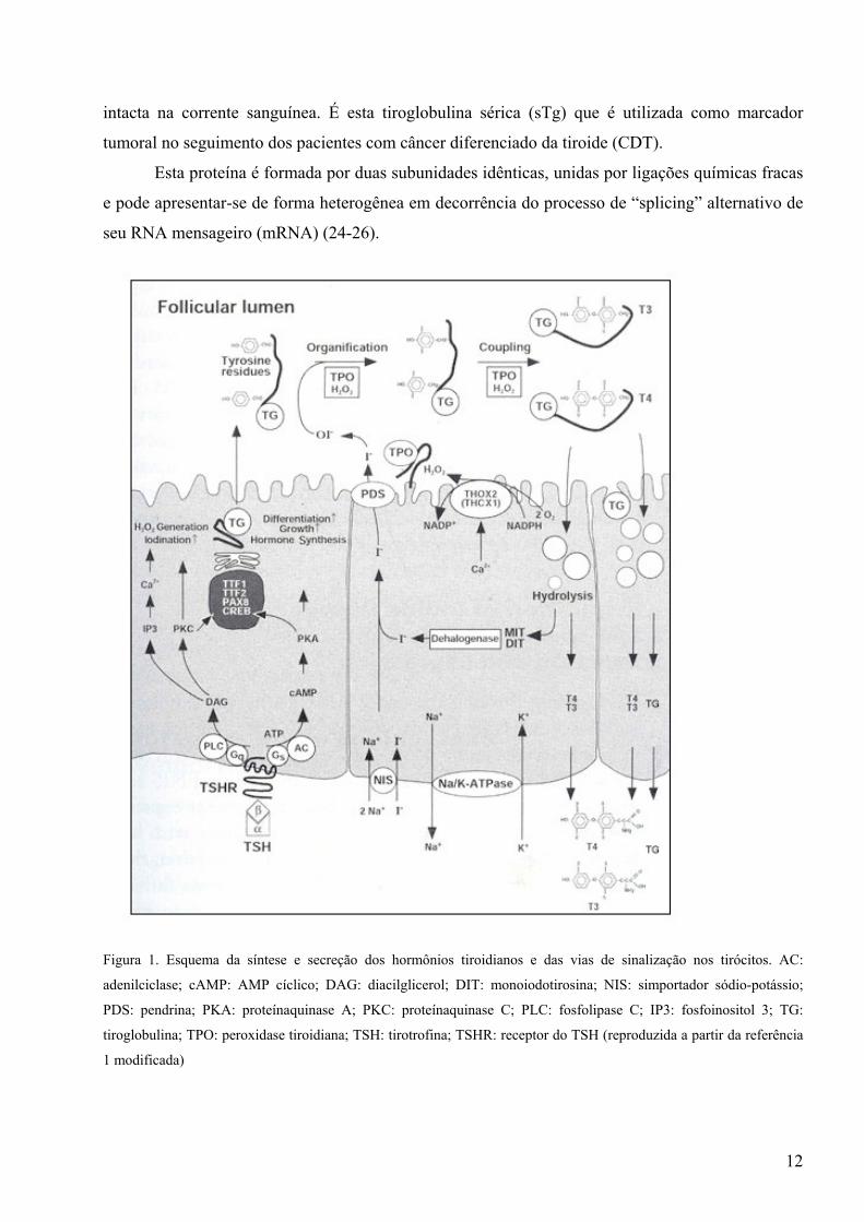
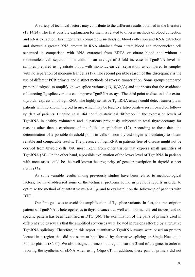
A tiroglobulina (Tg) é uma proteína de 660 kDa secretada pelo tirócito no lúmen folicular e
corresponde a 70-80% do conteúdo protéico da glândula. É codificada por um gene de cerca de 300
kb (48 exons localizados no cromossomo 8), sintetizada no retículo endoplasmático e exportada
para o lúmen folicular. O principal papel da Tg é funcionar como um suporte para a síntese dos
hormônios tiroidianos (T3 e T4) e, também, como um reservatório intra-tiroidiano de T3, T4 e iodo.
Assim, quando o organismo necessita de T4 e T3, a Tg do lúmen folicular, sob estímulo do TSH, é
captada pelos tirócitos, digerida nos lisosomos, com consequente liberação do T4 e T3 na corrente
sanguínea (Figura 1). A Tg é uma proteína de quase 3000 aminoácidos com 66 resíduos tirosina,
dimerizada e glicada no retículo endoplasmático do tirócito, fatores necessários à sua
funcionalidade; quando madura migra para a região apical do tirócito em pequenas vesículas que a
secretam no lúmen folicular. Para a síntese de T3 e T4 é necessário que o iodeto acople-se a radicais
tirosil da molécula de Tg. Para tal, o iodeto deve ser oxidado e para isso requer a presença de H2O2,
reação catalizada pela peroxidase tiroidiana (TPO), glicoproteína com 933 aminoácidos e 103 kDa
que contém um grupo prostético heme, localizada na membrana apical do tirócito com seu domínio
catalítico voltado para o coloide. Sua expressão é também controlada pelo TSH. Além de catalizar a
oxidação do iodeto, a TPO é essencial para a incorporação do iodeto nos resíduos tirosil da
molécula de Tg, reação denominada organificação, com a formação da monoiodo-tirosina (MIT). O
MIT, ainda ligado à TPO, pode sofrer nova oxidação e reagir com outro radical de iodeto,
produzindo a diiodotirosina (DIT). Além disso, a TPO atua no acoplamento das iodotirosinas (MIT
e DIT) para a formação do T4 e do T3 (Figura 1). H2O2 é um fator essencial e limitante na oxidação
do iodeto, para sua organificação e para a reação de acoplamento de MIT e DIT; para que a reação
se processe, além de H2O2, é necessária a oxidação de NADPH por NADPH-oxidases que na tiroide
receberam o nome de THOX1 e THOX2, de oxidases da tiroide (do inglês “thyroid-oxidases”).
Apresentam-se como proteínas transmembrana, com sítios para ligação NADPH, FAD e cálcio, o
que indica que a ativação do sistema H2O2 é dependente deste íon, estimulada tanto pela via do
AMP cíclico, como por fosfatidil-inositol e inibida por concentrações elevadas de iodeto (Figura 1)
(23). Uma pequena quantidade de Tg escapa dos mecanismos de digestão lisossomal e é secretada
12
intacta na corrente sanguínea. É esta tiroglobulina sérica (sTg) que é utilizada como marcador
tumoral no seguimento dos pacientes com câncer diferenciado da tiroide (CDT).
Esta proteína é formada por duas subunidades idênticas, unidas por ligações químicas fracas
e pode apresentar-se de forma heterogênea em decorrência do processo de “splicing” alternativo de
seu RNA mensageiro (mRNA) (24-26).
Figura 1. Esquema da síntese e secreção dos hormônios tiroidianos e das vias de sinalização nos tirócitos. AC:
adenilciclase; cAMP: AMP cíclico; DAG: diacilglicerol; DIT: monoiodotirosina; NIS: simportador sódio-potássio;
PDS: pendrina; PKA: proteínaquinase A; PKC: proteínaquinase C; PLC: fosfolipase C; IP3: fosfoinositol 3; TG:
tiroglobulina; TPO: peroxidase tiroidiana; TSH: tirotrofina; TSHR: receptor do TSH (reproduzida a partir da referência
1 modificada)
13
2.2. SEGUIMENTO DE PACIENTES COM CARCINOMA DIFERENCIADO DE TIROIDE
Os carcinomas diferenciados da tiroide (CDT), tanto do tipo papilífero (PTC, do inglês
“papillary thyroid carcinoma”), quanto do folicular (FTC, do inglês “follicular thyroid carcinoma”),
estão entre as neoplasias endócrinas mais comuns, porém com maior índice de cura. Os pacientes
apresentam sobrevida de aproximadamente 95% para o PTC e de 70% a 80% para o FTC (14).
Porém, pode haver recorrência tumoral, ou mesmo morte decorrente do CDT e o prognóstico
depende de fatores relacionados ao paciente, ao tratamento e à própria doença, sejam estes de
origem genética, ambiental ou mesmo dependentes da interação entre ambos (14-16).
A conduta recomendada pelas diversas diretrizes emanadas pelas sociedades nacionais e
internacionais de tiroide diante de um paciente com CDT é a tiroidectomia total complementada
pela ablação do tecido remanescente com iodo radioativo (16-19). Nesta situação, o paciente não
deve apresentar sTg circulante, pois a tiroide é sua única fonte de produção. Assim, depois do
tratamento, concentrações detectáveis de sTg podem indicar a persistência ou, até mesmo, a
recorrência da doença, o que pode ocorrer décadas após o diagnóstico inicial e justifica o
seguimento permanente dos pacientes (14-16).
Para este seguimento, as diretrizes internacionais recomendam que o método mais sensível
para avaliar a recorrência do tumor é a dosagem da sTg, em associação com a realização de
ultrassonografia da região cervical (USC); hoje em dia não se recomenda mais a realização rotineira
da pesquisa de metástases com o emprego do iodo radioativo (PCI), especialmente nos pacientes
que não apresentem anticorpos anti-tiroglobulina circulantes (15-21,27,28).
Apesar de sua grande utilidade no seguimento do câncer de tiroide, a dosagem de sTg não é,
no entanto, tecnicamente simples. Várias limitações tornaram-se evidentes, desde a descrição dos
primeiros métodos radioimunológicos, apesar de seu uso ter comprovado a utilidade prática
potencialmente esperada (21,23,27,28). Algumas das limitações técnicas foram, pelo menos
teoricamente, contornadas com a adoção, em rotina, dos métodos imunométricos (21,23). No
entanto, uma série de problemas podem interferir na determinação, a saber:
a) sensibilidade funcional inadequada numa série de métodos disponíveis comercialmente, o que
limita a detecção de pequenas massas de tecido tiroidiano, especialmente quando o TSH está
suprimido;
b) falta de padrão internacional, o que determina grande variabilidade entre os diversos métodos
disponíveis;
14
c) variação inter-ensaio acima do desejável, em especial, se levarmos em consideração o intervalo
de tempo, entre a coleta de amostras, utilizado habitualmente para o seguimento dos pacientes com
carcinoma diferenciado (6 a 12 meses);
d) possibilidade de efeito "gancho", especialmente, em ensaios imunométricos, o que causa a
obtenção de valores inapropriadamente baixos em pacientes com concentrações de Tg
especialmente elevadas;
e) presença de anticorpos endógenos anti-Tg no soro de 13 a 23% dos pacientes com CDT, o que
pode determinar resultados falsamente baixos nos ensaios imunométricos e falsamente elevados nos
radioimunoensaios (21).
Tendo em vista estas limitações, desenvolvemos em nosso laboratório uma série de
alternativas para a resolução destes problemas. Assim, nosso ensaio é um método
imunoradiométrico sensível, que permite a detecção segura de valores de 1 ng/mL. Além disso,
introduzimos um protocolo de arquivo de amostras: assim, estas são guardadas congeladas por um
prazo mínimo de 1 ano e quando da colheita de nova amostra, processamos a antiga em paralelo,
diminuindo, desta maneira, a variação inter-ensaio. Fazemos sempre a pesquisa de anticorpos anti-
Tg, o que evita a presença de falso-negativos, como já mencionado. Também executamos sempre os
ensaios em duas etapas, com o intuito de evitar o efeito "gancho" (21,23,27,28).
Além disso, é esperado que o nível da sTg varie de acordo com a concentração de TSH.
Assim, trabalhos evidenciaram sensibilidade maior para detecção de sTg em vigência de níveis de
TSH elevado (obtido por meio da suspensão do L-T4 e consequente hipotiroidismo ou após
estímulo com TSH recombinante - rhTSH)), do que com o TSH suprimido durante a terapia com L-
T4 (17-19).
Desta forma, apesar da sTg ser bastante útil no seguimento pós-operatório de pacientes com
CDT, podem ocorrer resultados falso-negativos (sTg indetectável na persistência ou recorrência
tumoral), que se explicam por diversas razões, como: a) menor sensibilidade do exame durante a
terapia supressora de TSH; b) interferência do anticorpo anti-Tg; c) desdiferenciação tumoral, que
secretaria Tg não reconhecida pelo ensaio. Estes fatores justificam a busca de alternativas à
dosagem de sTg para o acompanhamento de pacientes com CDT, como por exemplo a
quantificação do RNA mensageiro da tiroglobulina (TgmRNA).
15
2.3. PAPEL DO RNA MENSAGEIRO COMO MARCADOR TUMORAL
O conceito de detecção molecular de células tumorais na corrente sanguínea foi descrito no
início da década de 90 para tumores sólidos, como carcinoma de próstata, neuroblastoma e
carcinoma de mama (29-31).
O primeiro estudo de TgmRNA data de 1996, quando Ditkoff e cols. (32) detectaram por meio
de RT-PCR o TgmRNA em 9 pacientes com doença metastática e em menos de 10% dos pacientes
sem mestástase. Ainda nesse grupo, a detecção do TgmRNA foi mais freqüente naqueles pacientes
com história prévia de metástases. Controles normais e com doenças benignas não amplificaram o
TgmRNA. A partir destes resultados concluiu-se que a presença na circulação sanguínea do
TgmRNA poderia representar tumores disseminados pela via hematogênica.
Seguindo essa idéia, outro trabalho conseguiu detectar o TgmRNA, além do mRNA da TPO
(TPO mRNA) em 54,2% dos indivíduos em seguimento de CDT, incluindo 5 pacientes de um
grupo de 8 que não apresentavam evidência de doença no momento do exame (33). Os autores,
entretanto, não conseguiram explicar a detecção de TgmRNAs e TPO mRNA em 10% dos
pacientes com doença tiroidiana benigna, que desaparecia no pós-operatório, o que demonstrava
que a presença no sangue de mRNA específico da tiróide não está necessariamente relacionada com
mestástase.
O próximo trabalho nessa linha de pesquisa, publicado em 1998, foi um dos mais bem-
sucedidos até então. Ringel e cols. (34) detectaram TgmRNA em 26 de 33 pacientes (79%) com
remanescente em leito tiroidiano ou doença metastática confirmada por PCI, num grupo de 87
pacientes com CDT, enquanto que sTg foi encontrada em apenas 12 destes pacientes (com TSH
suprimido). Porém, apesar de terem obtido uma sensibilidade consideravelmente alta, a
especificidade foi inferior. Este mesmo grupo desenvolveu um método de RT-PCR quantitativo
(35) e amplificou o TgmRNA em todos os 32 voluntários normais. A partir daí aplicaram o método
em 107 pacientes com CDT e verificaram o achado de TgmRNA em 38% dos pacientes sem
captação anormal de iodo na PCI, 75% daqueles com captação em leito tiroidiano, 84% com doença
cervical e 94% com metástase à distância (36).
Em nosso laboratório desenvolvemos há alguns anos a amplificação do TgmRNA em
associação com o mRNA do transportador sódio-iodeto (NIS) (5). O interesse maior no estudo do
NIS como marcador tumoral era avaliar se havia correlação entre sua expressão e a captação de
iodo, na tentativa de explicar a discordância entre sTg e PCI encontrada em alguns pacientes; sua
amplificação, por "nested PCR", entretanto, mostrou baixa eficácia (sensibilidade de 16,6% e
especificidade de 54%), não demonstrando tal correlação. A baixa especificidade pode ser
16
explicada, pelo menos em parte, pela expressão do NIS em tecidos não tiroidianos. No mesmo
estudo, a sensibilidade e especificidade do TgmRNA, entretanto, foram bastante aceitáveis, de 83%
e 71%, respectivamente, em comparação com a sTg, de 50% e 89%.
Outro ponto importante é o fato de que a avaliação do TgmRNA por RT-PCR quantitativo em
crianças com doença de tiróide benigna e maligna não mostrou diferença significativa entre os dois
grupos (37), sendo que por esse motivo, apesar de alguns resultados iniciais promissores, pairaram
dúvidas sobre o verdadeiro valor e eficácia da detecção do TgmRNA no seguimento do CDT, pois
vários pacientes em aparente remissão clínica apresentavam TgmRNA circulante detectável (falso-
positivos), enquanto que outros pacientes sabidamente com metástases apresentavam TgmRNA
indetectável (falso-negativos).
Outro grupo desenvolveu um teste para determinação de TgmRNA empregando um RT-PCR
semiquantitativo com variação do número de ciclos (32 a 36) na reação, mas não houve diferença
significativa de expressão entre os indivíduos normais e os pacientes submetidos a tiroidectomia por
outra doença que não CDT; entretanto, neste estudo os pacientes não foram avaliados com PCI para
avaliar possíveis remanescentes tiroidianos pós-operatórios (38).
Em outro estudo por RT-PCR quantitativo, utilizando 57 pacientes tiroidectomizados, também
não se encontrou diferença de expressão do TgmRNA entre pacientes com e sem mestástases.
Entretanto, a metodologia foi diferente, pois não se realizou tratamento com DNAse (para prevenir
a amplificação de DNA genômico) e a padronização da curva de concentração foi feita com
GAPDH, e não mRNA de tiroide (39).
O que se pode concluir da apresentação destes trabalhos é que há realmente uma variedade
enorme de protocolos utilizados, em diversos níveis, como por exemplo, processamento da amostra
de sangue (variação quanto ao tipo de tubo em que a amostra é coletada, volume do sangue
utilizado, separação de camadas celulares por gradiente ou utilização de sangue total, tipo de kit de
extração de RNA e tratamento ou não do RNA com DNAse). Como exemplo podemos citar o
trabalho de Ezlinger e cols. (40), que compararam a eficácia de extração de vários métodos e
concluiram que a utilização de tubos com citrato com subseqüente separação da camada
mononuclear e extração imediata garantem um melhor resultado, embora essa metodologia não fora
comparada com um dos métodos mais utilizados (amostras de sangue adicionadas diretamente em
tubos estéreis contendo substâncias estabilizadoras de RNA).
Outras divergências encontradas entre os trabalhos também dizem respeito à metodologia do
RT-PCR, onde não se seguiu uma padronização quanto à quantidade de RNA total utilizada para a
síntese de cDNA, quantidade de cDNA empregada para a realização da PCR, seqüência de
oligonucleotídeos escolhida (residindo aqui a maior controvérsia entre os autores), tipo de
17
amplificação (se quantitativa, qualitativa ou semi-quantitativa) e também número de ciclos
utilizados. Com relação a este último aspecto, o estudo de Bojunga e cols. (41) observa incremento
da sensibilidade com o aumento de ciclos, porém com perda na especificidade, com a detecção de
TgmRNA em diversos tecidos, como timo, pulmão, entre outros, evidenciando o fenômeno
conhecido como transcrição ilegítima.
Outros autores buscaram correlacionar o mRNA de mais de um gene. Assim, Gupta e cols.
(42), estudando a especificidade de “primers” em amplificar, além do TgmRNA, o mRNA do
receptor de TSH (TSH-R mRNA) em sangue de indivíduos normais e de pacientes com câncer de
tiroide, concluiram que é provável que o processamento pós-trancricional (“splicings”) seja
diferente entre as células da tiroide e outras e que a seleção do “primer” possa determinar maior
especificidade ao método. Ainda com relação a esse assunto, o “splicing” alternativo pode afetar
não somente a especificidade, mas também a sensibilidade, pois se o “primer” for direcionado para
áreas sabidamente de risco de “splicing”, aumentará a probabilidade de resultados falso-negativos,
como demonstrado por Savagner e cols. (43). Neste estudo cerca de 33% dos pacientes
apresentavam “splicing” alternativo.
Apesar da possibilidade de baixa especificidade por transcrição ilegítima da Tg por células
não tiroidianas, ainda não se pode afastar a hipótese de alta sensibilidade do método, diagnosticando
precocemente possíveis recidivas. No trabalho de Grammatopoulos e cols. (44), por exemplo, a
determinação de TgmRNA correlaciou-se melhor com as metástases identificadas por PCI do que a
dosagem de sTg.
Por fim, Fugazzola e cols. estudaram 36 pacientes com CDT em aparente remissão e com
anticorpo anti-Tg negativo (45). A sTg foi dosada por dois métodos com sensibilidade de 0,9 ug/L e
0,1 ug/L, além do TgmRNA, antes e após a administração de TSH recombinante, e então avaliado o
grau de concordância entre estes exames e a avaliação clínica (USG cervical, PCI, etc). A Tg com
sensibilidade de 0,9 ug/L apresentou concordância de 53% no basal e 55% após estimulação do
TSH. Os resultados da quantificação do RNA foram concordantes com a avaliação clínica em 66%
no basal e pós-estimulação. Neste estudo, as condições de RT-PCR foram otimizadas para evitar
expressão extra-tiroidiana e nestas condições, não foi observada amplificação de Tg RNAm em
pacientes com agenesia tiroidiana, sendo que desta forma, o emprego de uma metodologia que
diminua o risco de interferência da expressão extra-tiroidiana talvez possa ser útil.
18
Tendo em vista estas discrepâncias metodológicas resolvemos estabelecer um novo método
para a determinação do TgmRNA, utilizando as recomendações sugeridas por M. D. Ringel em
editorial publicado no Journal of Clinical Endocrinology and Metabolism (46), ou seja: a) construir
“primers” em áreas livres de “splicings”; b) tratar as amostras de sangue de modo a evitar qualquer
tipo de contaminação com DNA; c) quantificar o ensaio de TgmRNA e estabelecer a curva-padrão;
d) validar o ensaio com um grande número de amostras de pacientes com história clínica muito bem
definida.
19
2.3. OBJETIVOS
1. Aperfeiçoar o método quantitativo para a medida do TgmRNA a partir do sangue periférico
2. Validar o ensaio em pacientes com Carcinoma Difenciado de Tiróide
20
3 – “DEVELOPMENT OF A SENSITIVE AND SPECIFIC
QUANTITATIVE RT-PCR ASSAY FOR BLOOD THYROGLOBULIN
MESSENGER RNA (TgmRNA) IN THE FOLLOW-UP OF PATIENTS
WITH DIFFERENTIATED THYROID CARCINOMA”
21
3 – DEVELOPMENT OF A SENSITIVE AND SPECIFIC QUANTITATIVE RT-PCR
ASSAY FOR BLOOD THYROGLOBULIN MESSENGER RNA (TgmRNA) IN THE
FOLLOW-UP OF PATIENTS WITH DIFFERENTIATED THYROID CARCINOMA
Valter T. Boldarine, Gustavo S. Guimarães, Claudia C. D. Nakabashi, Cleber P. Camacho,
Danielle M. Andreoni, Maria da Conceição O. C. Mamone, Elza S. Ikejiri, Teresa S. Kasamatsu,
Felipe Crispim, Flavio C. Hojaij, Rui M. B. Maciel, and Rosa P. M. Biscolla
Laboratory of Molecular Endocrinology, Division of Endocrinology, Department of Medicine, Escola Paulista de
Medicina, Federal University of São Paulo, São Paulo, Brazil
Key words: Thyroglobulin mRNA, Real Time quantitative RT-PCR, thyroid cancer
Correspondence to:
Rosa Paula M. Biscolla, MD, PhD
Laboratory of Molecular Endocrinology
Division of Endocrinology, Department of Medicine
Escola Paulista de Medicina, Universidade Federal de São Paulo
Rua Pedro de Toledo 781, 12nd. Floor
04039-032 Sao Paulo, SP, Brazil
Phone: +55-11-5084-5231 Fax: +55-11-5084-5231
e-mail: [email protected]
22
ABSTRACT
The measurement of serum thyroglobulin (sTg) has been considered the most sensitive
tumor marker in the follow-up of patients with differentiated thyroid carcinoma (DTC) after total
thyroidectomy and radioiodine therapy. However, sTg assays present some technical problems,
specially, the interference by endogenous anti-thyroglobulin antibodies (TgAb), present in
approximately 20% of DTC patients' sera. Therefore, in order to enhance sensitivity and to hinder
the interference by TgAb, several investigators have tried to quantify the mRNA Tg by Real-Time
RT-PCR. However, the results have been variable and some of them did not report a correlation
between mRNA Tg measurement and presence of metastases.
The aim of this study was to evaluate TgmRNA expression, performed by a new sensitive
and specific Real-Time PCR, in peripheral blood of 104 patients who previously undergone total
thyroidectomy and radioiodine treatment. DTC patients were studied during L-T4 therapy. Eighty-
two patients out of 104 (78.8%) were considered “free of disease”, and 22 (21.2%) presented
metastases. The quantification of mRNA was significantly different between patients “free of
disease” and those with metastases: TgmRNA (90.1 vs 951.3 ug/uL) (P<0,0001). In conclusion,
the differences proposed in the TgmRNA quantification made it a reliable method, and allowed us
to differentiate patients free of disease from patients with metastases and thus could be an
appropriate molecular marker in the follow-up of patients with thyroid carcinoma, especially in
patients with positive TgAb.
23
INTRODUCTION
Measurement of serum thyroglobulin (sTg) is considered the most sensitive tumor marker to
discover residual, recurring, or metastatic disease in the follow-up of patients with differentiated
thyroid carcinoma (DTC) after total thyroidectomy and ablative radioiodine therapy. sTg
measurement in hypothyroidism or after recombinant human TSH (rhTSH), in association with
neck ultrasound (US), is the current recommended guideline to follow-up these patients (1-3).
However, the low sensitivity of sTg during thyroid hormone suppression therapy, as well as an
interference on its measurement caused by endogenous anti-thyroglobulin antibodies (TgAb),
present in approximately 15-20% of DTC patients' sera, reduces its clinical utility (4).
Therefore, in order to enhance sensitivity and to hinder the interference by TgAb, several
authors have developed qualitative RT-PCR assays to amplify Tg messenger RNA (TgmRNA)
present in circulating thyroid cells from patients with DTC (5-13). The outcome of these studies has
been variable, with some groups showing correlation between the presence of metastases and the
detection of TgmRNA (5-8,11,13), whereas others have not demonstrated such correlation
(9,10,12). The reasons to explain such discrepancies amongst those studies are most likely related to
methodological aspects, as: a) different methods of standardization; b) use of primers including Tg
splice variants; c) interference of ectopic transcription, and d) number of PCR cycles used in the
reactions (14-16).
Thereafter, some investigators have tried to quantify TgmRNA employing real-time RT-
PCR (15-23). The results have also been variable and some of them did not report a correlation
between TgmRNA quantification and the presence of disease. The protocols used in these
quantitative studies also differed too much, especially regarding mRNA extraction, normalization
process and different primers.
Due to these controversies, an editorial and some reviews have appointed the possible
methodological dilemmas envolving these data, including primers design, removal of contaminating
DNA, sample handling, and improvements on assay’s normalization, sensitivity and specificity
(14,15,24). In this report we have examined these points in order to develop an enhanced method of
quantitative TgmRNA and to evaluate it on the follow-up of DTC patients.
24
SUBJECTS AND METHODS
Subjects
We studied 124 patients, 104 with DTC, followed in the Division of Endocrinology,
Department of Medicine, Federal University of Sao Paulo. A review of clinical data was carried out
to obtain each patient’s history, surgical and pathology reports, laboratory and radiological
examinations. All DTC patients (90 females and 14 males, age range 16-78 yr, median 35 yr) had
been treated with total or near-total thyroidectomy, 90 of whom also received radioiodine ablation
(30-200 mCi; mean: 116+42), according to current clinical protocols in use at our institution (3).
Fourteen of the 104 patients did not receive radioiodine ablation due to a negative whole body scan
(WBS) after surgery (less than 1% uptake in thyroid bed). Seventy-one patients had papillary
thyroid carcinoma (12 presented the follicular variant) and 33 had follicular carcinoma, classified
according to the World Health Organization (25). Thirteen out of the 104 patients (12.5%)
presented positive TgAb. All DTC patients were studied during L-T4 therapy (TSH concentrations
below 0.2 mU/L).
Eighty-two of the 104 DTC patients (78.8%) were considered “free of disease”, since they
presented sTg levels <1 ng/mL on hypothyroidism, negative WBS, and negative neck ultrasound for
metastases (follow-up: 3-12 years). In this group of patients the mean ablative radioiodine dose was
100 + 75.1 mCi of 131I; as mentioned, 14/82 patients did not receive radioiodine because WBS
showed less than 1% uptake in the thyroid bed after surgery. Six out of these 82 patients presented
positive TgAb, with negative WBS and neck US, and showed no evidence of recurrent disease
during follow up (mean: 8 +2 years).
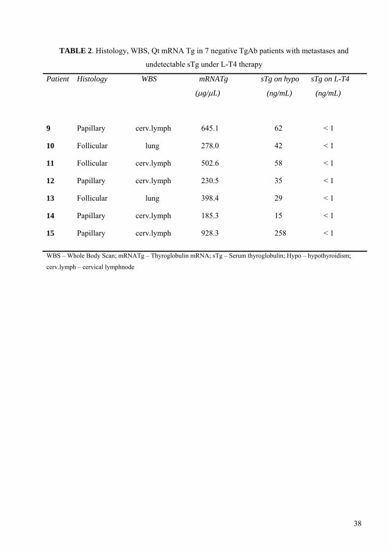
Twenty-two out of the 104 patients (21.2%) presented metastases: 8/22 patients had
detectable levels of sTg under TSH suppression, and positive WBS uptake (lung, cervical lymph
nodes and/or bone) after radioiodine treatment (Table1, patients 1-8); 7/22 patients presented sTg
levels above 5 ng/mL in hypothyroidism, in spite of undetectable sTg levels under L-T4. Of these 7
patients, 2 had lung uptake and 5 presented cervical uptake on WBS, with suspicious metastatic
cervical disease, confirmed by aspirative cytology and surgery (Table 2, patients 9-15). The
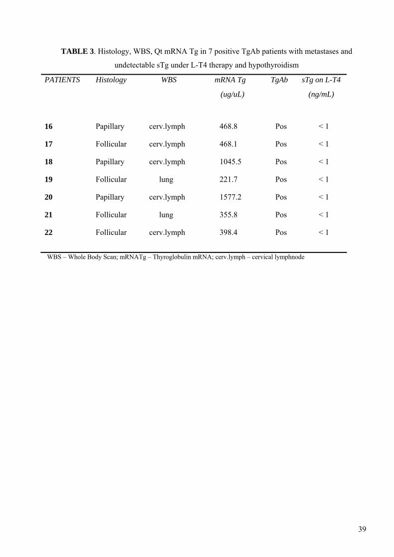
remaining 7 patients had positive TgAb with undetectable levels of sTg even in hypothyroidism;
however, WBS uptake was observed in all these 7 patients, 5/7 in the thyroid bed and 2 in the lung.
In addition, neck US showed the presence of suspicious cervical lymph nodes in 5 of them, which
were confirmed as thyroid metastases by cytology and by pathology report after surgery (Table 3,
patients 16-22).
25
Methods
Blood samples were obtained for determinations of sTg (Delfia Thyroglobulin, time-
resolved fluoroimmunoassay, PerkinElmer Life and Analytical Sciences, Wallac Oy, Turku,
Finland, with a sensitivity of 1 ng/mL), serum TSH (immunofluorimetric method, normal range 0.3
to 5.0 mU/L) (26), and TgAbs (immunofluorimetric assay, normal range below 40 U/mL) (27).
RNA isolation and cDNA synthesis
Total RNA was isolated from 1 mL of venous blood and transferred immediately into tubes
containing 3 mL of Trizol LS Reagent (Life Technologies, Gaithersburg, MD, USA) according to
manufacturer’s recommendations (28). Total RNA (1.5µg) was treated with DNAse (Ambion,
Applied Biosystems, Foster City, CA, USA), and then stopped with the addition of 5 uL of DNAse
Inactivaton Reagent (Ambion) to the samples. The complementary DNA was reverse transcribed to
a final volume of 60 µL, using 50 ng of OligoDT (Life Technologies), 10 U of RNAse inhibitor
(Life Technologies), 200 U of Superscript III (Life Technologies), 50 mM Tris-HCl (pH 8.3), 75
mM KCl, 3 mM MgCl2, 500 µM of each dNTPs and 10 mM of dithiothreitol (DTT). Finally, the
samples were incubated at 50C for 1 hour and then stored at -20C until their usage. Reverse
transcriptase negative samples were prepared for each individual reaction and served as controls for
detection of assay contamination.
Tg Primer Design
The design of intron-spanning primers was made targeting different exons in order to
discriminate possible amplification of genomic DNA contaminants and in regions free of alternative
splicings. The analysis was performed using prediction programs for alternative Splicing and
transcript Diversity Database (http://www.ebi.ac.uk/asd/altsplice) from Ensembl
(http://ensemble.org gene ID ENSG00000042832), for splice sites (NNSPLICE:
http://www.fruitfly.org/seq_tools/splice.html), and for exonic-splicing enhancer (ESE) (ESEFinder:
http:/exon.cshl.edu/ESE/) (29-31).
The final choice of Tg primer sequences was: sense, 5'-CTATCGACGGCTCCTTCTTG-3';
antisense, 5'- CCAGTTGCCACTCACCTCTC -3', generating an amplicon of 116-bp encompassing
26
exons 40 and 41 of the Tg cDNA (Genebank, accession number NM_003235.4). These primers
were near the 3' end of TgmRNA, which optimized the use of Oligo dT to perform the cDNA
synthesis.
Real-time Quantitative PCR and assay quantification
The PCR products were detected by using the ABI Prism 7700 sequence detection system
(Applied Biosystems). Real-time quantitative PCR was performed using 2 µL of first-strand cDNA
in a 20 µL reaction volume containing 10mM of each Tg primer and 17 µL of SYBR Green Mix
(Applied Biosystems); cycling conditions for PCR included denaturing for 10 min at 95C, followed
by 40 cycles of 15 sec at 95 C, 20 sec at 55C, 20 sec at 72C and 20 sec at 75C. In order to confirm
the identity of the amplicons, samples were visualized by agarose 1.5% gel electrophoresis followed
by ethidium bromide staining and sequenced (BigDye Terminator Cycle Sequencing Ready
Reaction Kit, Applied Biosystems).
An absolute standard curve was designed by using plasmids containing PCR amplified Tg
cDNA fragment as template. The standard curve included 5 different dilutions, with a range of 0.32
to 200 µg of plasmids/µL of solution (0.32, 1.6, 8, 40, and 200 µg/µL of plasmids), and was
calculated by two independent methods, spectrophotometry and gel analysis. Independent standard
curves were generated from the same plasmids preparation, for each 96-well plate as well as for
negative control template reactions. cDNAs from different patients were quantified by comparing
the crossing points of samples with those of the standards containing the plasmid concentration as
stated above. For each experimental sample, the amount of target reference was determined from
the appropriate standard curve. The quantification was performed in triplicate and the results were
expressed as µg of RNA/µL of solution.
Tissue Panels
cDNA samples from normal thyroid tissue and from 7 different non-thyroidal tissues (lung,
testis, liver, brain, pancreas β cells, an osteosarcoma cell line and a mammary cell line) were used to
validate the specificity of the selected Tg primers, comparing their expression by RT-PCR, using
the following conditions: 2 min at 95C, then 40 cycles of 20 sec at 95C, 20 sec at 55C and 20 sec at
72C. The PCR products were visualized by agarose 1.5% gel electrophoresis followed by ethidium
27
bromide staining. To evaluate possible contaminations we used a negative control containing water
instead of cDNA.
ROC Curve and Statistical analysis
In order to set a cut-off point for the quantification of TgmRNA, we performed a receiver
operating characteristics (ROC) curve and calculated its respective sensitivity and specificity. The
results were log-transformed and the statistical analysis of data was performed using paired
Student´s t test by the StatView 4.5 software (Abacus Concepts, Inc, Berkeley, CA). A p value <
0.05 was considered significant.
28
RESULTS
Validation of the TgmRNA quantitative method
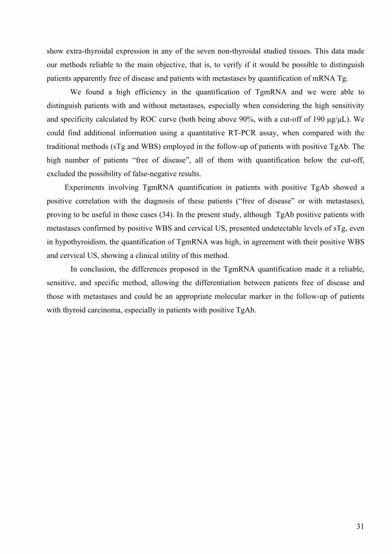
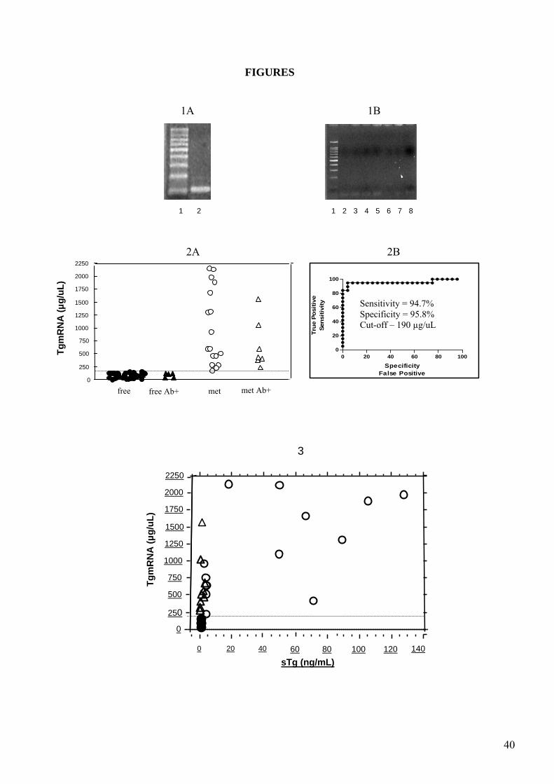
We did not find any TgmRNA expression in all 7 extra-thyroidal tissues (Figure 1A), but we
found expression in normal thyroid tissue (Figure 1B). We conclude that the pair of primers used in
our study was specific for thyroid tissue.
The identity of generated amplicons was confirmed by agarose gel electrophoresis and
nucleotide sequencing. Standard curves showed an efficacy (r2) of 0.99 and a slope of 3.45, with a
high level of confidence. The mean interassay coefficient of variation (CV) for the threshold cycle
of the absolute standard curve containing PCR amplified Tg cDNA fragments (0.32, 1.6, 8, 40, and
200 µg/µL of plasmids), assayed in triplicate, was 1.0-1.5%; the intraassay CV for the threshold
cycle of the standards was 0-2% . To rule out possible accidental laboratory contamination of RT-
PCR, negative controls without reverse transcriptase or without cDNA consistently failed to
demonstrate detectable PCR products at 40 cycles, showing that there were no external
contaminants in our reactions.
Quantification of TgmRNA and sTg in patients with DTC and with benign thyroid disease and
normal individuals
TgmRNA concentration was significantly different between patients considered "free of
disease" and patients with metastases (mean= 90.1 + 8,6 vs 951.3 + 36,8µg/µL) (p<0.0001). There
was no overlap between the two groups (Figure 2A). In a ROC curve, a level of 190 µg/µL of
TgmRNA presented a sensitivity of 94.7% and a specificity of 95.8% (Figure 2B). Employing this
cut-off value we were able to discriminate almost all patients between the two groups. We also
noticed that the test was useful for patients with positive TgAb, since patients with metastases (n=7)
presented high TgmRNA quantification (mean= 685,7 + 31,8), while those “free of disease” (n=7)
showed a low TgmRNA quantification (mean= 165,2 + 13,9).
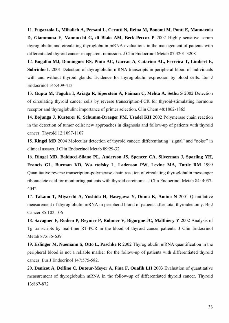
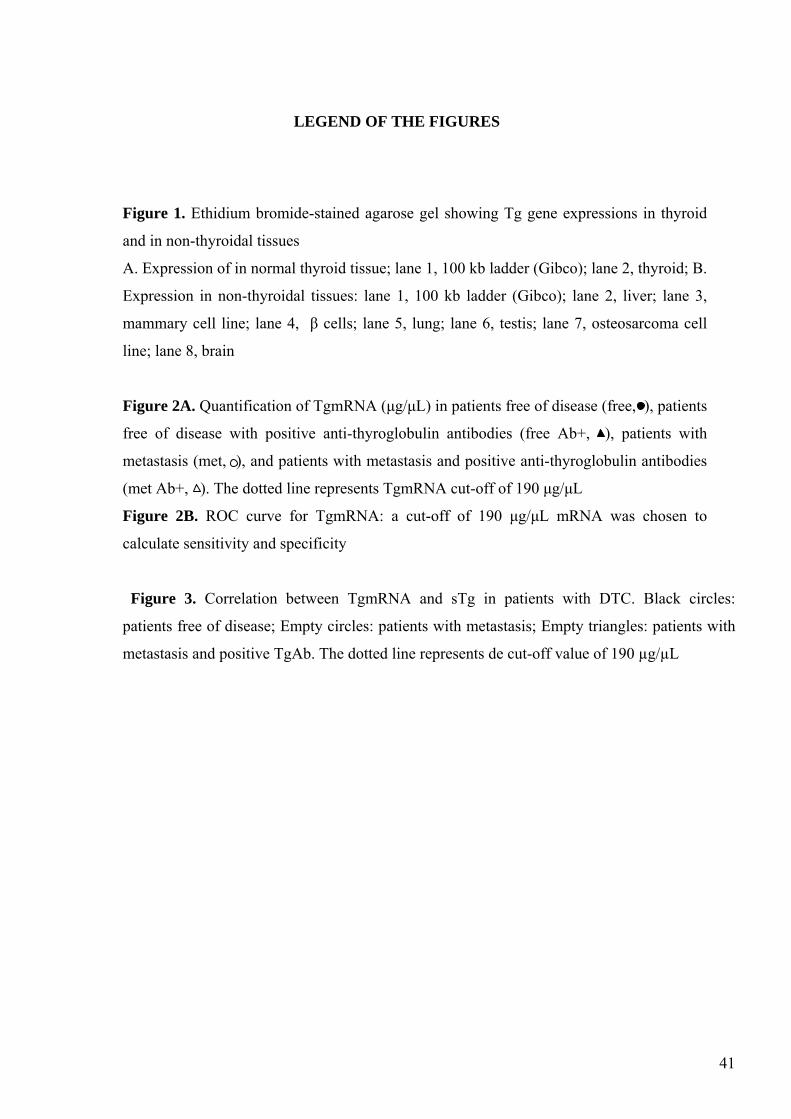
When we compared TgmRNA quantification with the measurement of sTg in patients “free of
disease”, all of them presented sTg lower than 1 ng/mL and values of TgmRNA below the cut-off
of 190 µg/µL. On the other hand, all but one patient with metastases showed TgmRNA above the
cut-off value. It´s worth noting that all 7 patients with metastases and positive TgAb had TgmRNA
above the cut-off. In these patients, sTg levels were below 1 ng/mL, probably due to interference of
anti-thyroglobulin antibodies (Figure 3).
29
DISCUSSION
Currently, the measurement of sTg, specially performed with TSH stimulation, has been
considered a specific and sensitive marker of recurrence of the disease and indicates the presence of
metastases in the follow-up of patients with DTC (1-3). However the presence of TgAb in the sera
of 15-20% of patients with DTC and the expensive cost of rhTSH to avoid the hypothyroidism
status could be a limiting factor for the use of sTg as a thyroid tumor maker. Therefore, a sensitive
screening test to diagnose recurrent thyroid cancer even in the presence of TgAb and not requiring
TSH stimulation would be really useful (15).
Based on the previous evidence of positive TgmRNA in the peripheral blood of DTC
patients with negative sTg and positive TgAb, several authors have suggested the use of TgmRNA
by RT-PCR as a suitable test (5,6-8,10). However, in all of these previous studies, the authors
identified a group of patients (about 20%) with positive TgmRNA but considered free of disease by
traditional methods (negative WBS, negative neck US and undetectable levels of sTg); therefore,
they were considered false positive results (5-8,10). Accordingly, TgmRNA showed to be more
sensitive then sTg, but with lower specificity. In a previous report of our group we have found 3
patients with negative sTg, positive TgAb and positive TgmRNA, in whom we detected metastases
of papillary thyroid carcinoma in cervical lymph node, by neck US-guided aspirative cytology (8).
To avoid the problem of specificity, the quantification of TgmRNA has been used as a tool
in the follow up of patients with DTC. Thus, Ringel et al. showed that 89% of patients with local
metastases and 94% of patients with distant metastases had TgmRNA above a cut-off level of 3 pg
of TgmRNA Eq/µg thyroid RNA; however, 38% of patients with no uptake on WBS and
undetectable levels of sTg also presented TgmRNA above the mentioned cut-off level (16).
Savagner et al. showed that the quantification of TgmRNA is a good choice in patients displaying
TgAb, but 18% of patients considered "cured" had TgmRNA values above 1 pg TgmRNA/µg RNA
(18). On the other hand, Takano et al. detected Tg transcripts in all patients submitted to total
thyroidectomy with and without metastases and even in patients with medullary thyroid carcinoma
(17). Similarly Bugalho et al. investigated the diagnostic specificity of TgmRNA detection,
analyzing blood samples from healthy volunteers and from patients previously subjected to
thyroidectomy for reasons other than a carcinoma of the follicular cells using a semi-quantitative
PCR and were also unable to find any difference between them (12). Other investigators also did
not demonstrate correlation between quantitative TgmRNA levels and the presence or absence of
thyroid tissue (20-23). Therefore, nowadays, based on the heterogeneity of the methods and
contradictory data, it was difficult to define the clinical value of TgmRNA quantification.
30
A variety of technical factors may contribute to the different results obtained in the literature
(13,14,24). The first possible explanation for them is related to diverse methods of blood collection
and RNA extraction. Eszlinger et al. compared 3 methods of blood collection and RNA extraction
and showed a greater RNA amount in RNA obtained from citrate blood and mononuclear cell
separated in comparison with RNA extracted from EDTA or citrate blood and without a
mononuclear cell separation. In addition, an average of 5-fold increase in TgmRNA levels in
samples prepared using citrate blood with mononuclear cell separation, as compared to samples
with no separation of mononuclear cells (19). The second possible reason of this discrepancy is the
use of different PCR primers and distinct methods of reverse transcription. Some groups compared
primers designed to amplify known splice variants (13,18,32,33) and it appears that the avoidance
of detecting Tg splice variants can improve TgmRNA assays. The third point to discuss is the extra-
thyroidal expression of TgmRNA. The highly sensitive TgmRNA assays could detect transcripts in
patients with no known thyroid tissue, which may be lead to a false-positive result based on follow-
up data of patients. Bugalho et al. did not find statistical difference in the expression levels of
TgmRNA in healthy volunteers and in patients previously subjected to total thyroidectomy for
reasons other than a carcinoma of the follicular epithelium (12). According to these data, the
determination of a possible threshold point in cells of non-thyroid origin is mandatory to obtain
reliable and comparable results. The presence of TgmRNA in patients free of disease might not be
derived from thyroid cells, but, most likely, from other tissues that express small quantities of
TgmRNA (34). On the other hand, a possible explanation of the lower level of TgmRNA in patients
with metastases could be the well-known heterogeneity of gene transcription in thyroid cancer
tissue (35).
As some variable results among previously studies have been related to methodological
factors, we have addressed some of the technical problems found in previous reports in order to
optimize the method of quantitative mRNA Tg, and to evaluate it on the follow-up of patients with
DTC.
Our first goal was to avoid the amplification of Tg splice variants. In fact, the transcription
pattern of TgmRNA is heterogeneous in thyroid cancer, as well as in normal thyroid tissues, and no
specific pattern has been identified in DTC (36). The examination of the pairs of primers used in
different studies reveals that the amplified sequences were located in regions affected by alternative
TgmRNA splicings. Therefore, in this report quantitative TgmRNA assays were based on primers
located in a region that did not seem to be affected by alternative splicing or Single Nucleotide
Polimorphisms (SNPs). We also designed primers in a region near the 3´end of the gene, in order to
favoring the synthesis of cDNA when using Oligo dT. In addition, these pair of primers did not
31
show extra-thyroidal expression in any of the seven non-thyroidal studied tissues. This data made
our methods reliable to the main objective, that is, to verify if it would be possible to distinguish
patients apparently free of disease and patients with metastases by quantification of mRNA Tg.
We found a high efficiency in the quantification of TgmRNA and we were able to
distinguish patients with and without metastases, especially when considering the high sensitivity
and specificity calculated by ROC curve (both being above 90%, with a cut-off of 190 µg/µL). We
could find additional information using a quantitative RT-PCR assay, when compared with the
traditional methods (sTg and WBS) employed in the follow-up of patients with positive TgAb. The
high number of patients “free of disease”, all of them with quantification below the cut-off,
excluded the possibility of false-negative results.
Experiments involving TgmRNA quantification in patients with positive TgAb showed a
positive correlation with the diagnosis of these patients (“free of disease” or with metastases),
proving to be useful in those cases (34). In the present study, although TgAb positive patients with
metastases confirmed by positive WBS and cervical US, presented undetectable levels of sTg, even
in hypothyroidism, the quantification of TgmRNA was high, in agreement with their positive WBS
and cervical US, showing a clinical utility of this method.
In conclusion, the differences proposed in the TgmRNA quantification made it a reliable,
sensitive, and specific method, allowing the differentiation between patients free of disease and
those with metastases and could be an appropriate molecular marker in the follow-up of patients
with thyroid carcinoma, especially in patients with positive TgAb.
32
REFERENCES
1. Cooper DS, Doherty GM, Haugen BR, Kloos RT, Lee SL, Mandel SJ, Mazzaferri EL,
McIver B, Sherman SI, Tuttle RM 2006 Management guidelines for patients with thyroid nodules
and differentiated thyroid cancer. Thyroid 16:109-42
2. Pacini F, Schlumberger M, Dralle H, Elisei R, Smit JWA, Wiersinga WM and the
European Thyroid Cancer Taskforce 2006 European consensus for the management of patients
with differentiated carcinoma of the follicular epithelium. Eur J Endocrinol 154:787-803
3. Maia AL, Ward LS, Carvalho GA, Graf H, Maciel LMZ, Maciel RMB, Rosario PW,
Vaisman M 2007 Thyroid nodules and differentiated thyroid cancer: Brazilian consensus. Arq Bras
Endocrinol Metab 51: 867-893
4. Demers LM, Spencer CA 2003 Laboratory Medicine Practice Guidelines. Laboratory Support
for the Diagnosis and Monitoring of Thyroid Disease. Thyroid 13:57-67
5. Ditkoff BA, Marvin MR, Yemul S, Shi YJ, Chabot J, Feind C, Lo Gerfo PL 1996 Detection
of circulating thyroid cells in peripheral blood. Surgery 120:959-964
6. Tallini G, Gossein R, Emanuel J, Gill J, Kinder B, Dimich AB, Costa J, Robbins R, Burrow
GN, Rosai J 1998 Detection of thyroglobulin thyroid peroxidase, and RET/PTC1 mRNA
transcripts in the peripheral blood of patients with thyroid disease. J Clin Oncol 16:1158-1166
7. Ringel MD, Ladenson PW, Levine MA 1998 Molecular diagnosis of residual and recurrent
thyroid cancer by amplification of thyroglobulin messenger ribonucleic acid in peripheral blood. J
Clin Endocrinol Metab 83: 4435-4442
8. Biscolla RP, Cerutti JM, Maciel RM 2000 Detection of recurrent thyroid cancer by sensitive
nested reverse transcription-polymerase chain reaction of thyroglobulin and sodium/iodide
symporter messenger ribonucleic acid transcripts in peripheral blood. J Clin Endocrinol Metab
85:3623-3627
9. Bojunga J, Roddiger S, Stanisch M, Kusterer K, Kurck R, Renneberg H, Adams S,
Lindhorst E, Csadel KII, Schumm-Draeger PM 2000 Molecular detection of thyroglobulin
mRNA transcripts in peripheral blood of patients with thyroid disesase by RT-PCR. Br J Cancer
82:1650-1655.
10. Bellantone R, Lombardi DP, Bossola M, Ferrante A, Princi P, Boscherini M, Maussier L,
Salvatori M, Rufini V, Reale F, Romano L, Tallini G, Zelano G, Pontecorvi A 2001 Validity of
thyroglobulin mRNA assay in peripheral blood of post-operative thyroid carcinoma patients in
predicting tumor recurrences varies according to the histologic type: Results of a prospective study.
Cancer 92:2273-2279
33
11. Fugazzola L, Mihalich A, Persani L, Cerutti N, Reina M, Bonomi M, Ponti E, Mannavola
D, Giammona E, Vannucchi G, di Blaio AM, Beck-Peccoz P 2002 Highly sensitive serum
thyroglobulin and circulating thyroglobulin mRNA evaluations in the management of patients with
differentiated thyroid cancer in apparent remission. J Clin Endocrinol Metab 87:3201-3208
12. Bugalho MJ, Domingues RS, Pinto AC, Garrao A, Catarino AL, Ferreira T, Limbert E,
Sobrinho L 2001 Detection of thyroglobulin mRNA transcripts in peripheral blood of individuals
with and without thyroid glands: Evidence for thyroglobulin expression by blood cells. Eur J
Endocrinol 145:409-413
13. Gupta M, Taguba I, Ariaga R, Siperstein A, Faiman C, Mehta A, Sethu S 2002 Detection
of circulating thyroid cancer cells by reverse transcription-PCR for thyroid-stimulating hormone
receptor and thyroglobulin: importance of primer selection. Clin Chem 48:1862-1865
14. Bojunga J, Kusterer K, Schumm-Draeger PM, Usadel KH 2002 Polymerase chain reaction
in the detection of tumor cells: new approaches in diagnosis and follow-up of patients with thyroid
cancer. Thyroid 12:1097-1107
15. Ringel MD 2004 Molecular detection of thyroid cancer: differentiating “signal” and “noise” in
clinical assays. J Clin Endocrinol Metab 89:29-32
16. Ringel MD, Balducci-Silano PL, Anderson JS, Spencer CA, Silverman J, Sparling YH,
Francis GL, Burman KD, Wa rtofsky L, Ladenson PW, Levine MA, Tuttle RM 1999
Quantitative reverse transcription-polymerase chain reaction of circulating thyroglobulin messenger
ribonucleic acid for monitoring patients with thyroid carcinoma. J Clin Endocrinol Metab 84: 4037-
4042
17. Takano T, Miyarchi A, Yoshida H, Hasegawa Y, Duma K, Amino N 2001 Quantitative
measurement of thyroglobulin mRNA in peripheral blood of patients after total thyroidectomy. Br J
Cancer 85:102-106
18. Savagner F, Rodien P, Reynier P, Rohmer V, Bigorgne JC, Malthiery Y 2002 Analysis of
Tg transcripts by real-time RT-PCR in the blood of thyroid cancer patients. J Clin Endocrinol
Metab 87:635-639
19. Ezlinger M, Nuemann S, Otto L, Paschke R 2002 Thyroglobulin mRNA quantification in the
peripheral blood is not a reliable marker for the follow-up of patients with differentiated thyroid
cancer. Eur J Endocrinol 147:575-582.
20. Denizot A, Delfino C, Dutour-Meyer A, Fina F, Ouafik LH 2003 Evaluation of quantitative
measurement of thyroglobulin mRNA in the follow-up of differentiated thyroid cancer. Thyroid
13:867-872
34
21. Span PN, Sleegers MJ, van den Brock WJ, Ross HA, Nieuwlaat WA, Hermus AR, Sweep
CG 2003 Quantitative detection of peripheral thyroglobulin mRNA has limited clinical value in the
follow-up of thyroid cancer patients. Ann Clin Biochem 40:94-99
22. Elisei R, Vivaldi A, Agate L, Molinaro E, Nencetti C, Grasso L, Pinchera A, Pacini F 2004
Low specificity of blood thyroglobulin messenger ribonucleic acid assay prevents its use in the
follow-up of differentiated thyroid cancer patients. J Clin Endocrinol Metab 89:33-39
23. Lombardi CP, Bossola M, Princi P, Boscherini M, La Torre G, Raffaelli M, Traini E,
Salvatori M, Pontecorvi A, Bellantone R 2008 Circulating thyroglobulin mRNA does not predict
early and midterm recurrences in patients undergoing thyroidectomy for cancer. Am J Surg 196:
326-32
24. Gupta M, Chia S-Y 2007 Circulating thyroid cancer markers. Curr Opin Endocrinol Diabetes
Obes 14: 383-388
25. DeLellis RA, Lloyd RV, Heitz PU, Eng C 2004 Pathology and Genetics of tumours of the
endocrine organs. World Health Organization Classification of Tumours
26. Vieira JGH, Kunii IS, Nishida SK, Matsumura LK, Russo EMK, Maciel RMB 1992
Development of an immunofluorimetric assay for the measurement of human thyrotropin (TSH) in
serum and in total blood collected in filter paper. Arq Brasil Endocrinol Metab 36: 7-12
27. Vieira JGH, Tachibana TT, Fonseca RMG, Nishida SK, Maciel RMB 1996 Development of
an immunofluorimetric assay for the measurement of anti-thyroglobulin antibodies. Arq Brasil
Endocrinol Metab 40: 232-7
28. Deng MY, Wang H, Ward GB, Beckham TR, McKenna TS 2005 Comparison of six RNA
extraction methods for the detection of classical swine fever virus by real-time and conventional
reverse transcription-PCR. J Vet Diag Invest 17: 574-8
29. Thanaraj TA, Stamm S, Clark F, Riethoven JJ, Le Texier V, Muilu J 2004 ASD: the
alternative splicing database. Nucleic Acids Res 32: D64-9
30. Reese MG, Eeckman FH, Kulp D, Haussler D 1997 Improved splice site detection in Genie. J
Comput Biol 4:311-23
31. Cartegni L, Wang J, Zhu Z, Zhang MQ, Krainer AR 2003 ESEfinder: A web resource to
identify exonic splicing enhancers. Nucleic Acids Res 31:3568-71
32. Chinnapa P, Taguba L, Arciaga R, Faiman C, Siperstein A, Mehta AE, Reddy K, Nasr C,
Gupta MK 2004 Detection of thyrotropin-receptor messenger ribonucleic acid (mRNA) and
thyroglobulin mRNA transcripts in peripheral blood of patients with thyroid disease: sensitive and
specific markers for thyroid cancer. J Clin Endocrinol Metab 89:3705-3709
35
33. Wagner K, Arciaga R, Siperstein A, Milas M, Warshawsky I, Reddy SSK, Gupta MK
2005 Thyrotropin receptor/thyroglobulin messenger ribonucleic acid in peripheral blood and fine-
needle aspiration cytology: diagnostic synergy for detecting thyroid cancer. J Clin Endocrinol
Metab 90: 1921-1924
34. Saha S, Bardelli A, Buckhaults P, Velculescu VE, Rago C, St Croix B, Romans KE, Choti
MA, Lengauer C, Kinzler KW, Vogelstein B 2001 A Phosphatase associated with metastases of
colorectal cancer. Science 294:1343-1346.
35. Chelly J, Concordet JP, Kaplan JC, Kahan A 1989 Illegitimate transcription: transcription of
any gene in any cell type. Proc Natl Acad Sci USA 86:2617-2621aet al 1989
36. Bertaux F, Noel M., Malthiery Y, Fragu P 1991 Determination of a heterogeneous
transcription pattern of thyroglobulin mRNA in human thyroid tissues. Biochem Biophys Res
Commun 178: 586-592
36
ACKNOWLEDGEMENTS
This work has been supported by the São Paulo State Research Foundation (FAPESP), grant
04/09934-7 to RMBM and a research-fellowship grant 05/55842-0 to VTB. We thank Drs. Janete
M. Cerutti and José Gilberto H. Vieira for helpful discussions, Mrs. Gisele Oler for technical
support with the ABI PRISM 7700 Sequence Detection System, and Angela Faria for superb
secretarial assistance. RMBM is an investigator of the Brazilian Research Council and of the Fleury
Institute; RPMB is investigator of the Fleury Institute.
37
TABLES
TABLE 1. Histology, WBS, Qt mRNA Tg in 8 negative TgAb patients with metastases and
detectable sTg under L-T4 therapy and hypothyroidism
Patient Histology WBS mRNATg
(µg/µL)
sTg on L-T4
(ng/mL)
1
2
3
4
5
6
7
8
Papillary
Follicular
Papillary
Follicular
Follicular
Papillary
Papillary
Papillary
cerv.lymph
cerv.lymph/bone
cerv.lymph
lung
lung
cerv.lymph
cerv.lymph
cerv.lymph
2096.5
2101.5
1953.5
1836.4
1420.7
430.8
1653.2
1153.9
42
75
125
101
89
28
64
50
WBS – Whole Body Scan; mRNATg – Thyroglobulin mRNA; sTg – Serum thyroglobulin; Hypo – hypothyroidism;
cerv.lymph – cervical lymphnode
38
TABLE 2. Histology, WBS, Qt mRNA Tg in 7 negative TgAb patients with metastases and
undetectable sTg under L-T4 therapy
Patient Histology WBS mRNATg
(µg/µL)
sTg on hypo sTg on L-T4
(ng/mL) (ng/mL)
9
10
11
12
13
14
15
Papillary
Follicular
Follicular
Papillary
Follicular
Papillary
Papillary
cerv.lymph
lung
cerv.lymph
cerv.lymph
lung
cerv.lymph
cerv.lymph
645.1
278.0
502.6
230.5
398.4
185.3
928.3
62 < 1
42 < 1
58 < 1
35 < 1
29 < 1
15 < 1
258 < 1
WBS – Whole Body Scan; mRNATg – Thyroglobulin mRNA; sTg – Serum thyroglobulin; Hypo – hypothyroidism;
cerv.lymph – cervical lymphnode
39
TABLE 3. Histology, WBS, Qt mRNA Tg in 7 positive TgAb patients with metastases and
undetectable sTg under L-T4 therapy and hypothyroidism
PATIENTS Histology WBS mRNA Tg
(ug/uL)
TgAb sTg on L-T4
(ng/mL)
16
17
18
19
20
21
22
Papillary
Follicular
Papillary
Follicular
Papillary
Follicular
Follicular
cerv.lymph
cerv.lymph
cerv.lymph
lung
cerv.lymph
lung
cerv.lymph
468.8
468.1
1045.5
221.7
1577.2
355.8
398.4
Pos < 1
Pos < 1
Pos < 1
Pos < 1
Pos < 1
Pos < 1
Pos < 1
WBS – Whole Body Scan; mRNATg – Thyroglobulin mRNA; cerv.lymph – cervical lymphnode
40
FIGURES
1A 1B
1 2 1 2 3 4 5 6 7 8
2A 2B
3
sTg (ng/mL)
0 250 500 750
1000 1250 1500 1750 2000 2250
0 20 40 60 80 100 120 140
Tgm
RN
A (µ
g/uL
)
0 250 500 750
1000 1250 1500 1750 2000 2250
*
* *
*
*
*
free free Ab+ met met Ab+
0 20 40 60 80 1000
20
40
60
80
100
SpecificityFalse Positive
True
Pos
itive
Sens
itivi
ty Sensitivity = 94.7% Specificity = 95.8% Cut-off – 190 µg/uL
Tgm
RN
A (µ
g/uL
)
41
LEGEND OF THE FIGURES
Figure 1. Ethidium bromide-stained agarose gel showing Tg gene expressions in thyroid
and in non-thyroidal tissues
A. Expression of in normal thyroid tissue; lane 1, 100 kb ladder (Gibco); lane 2, thyroid; B.
Expression in non-thyroidal tissues: lane 1, 100 kb ladder (Gibco); lane 2, liver; lane 3,
mammary cell line; lane 4, β cells; lane 5, lung; lane 6, testis; lane 7, osteosarcoma cell
line; lane 8, brain
Figure 2A. Quantification of TgmRNA (µg/µL) in patients free of disease (free, ), patients
free of disease with positive anti-thyroglobulin antibodies (free Ab+, ), patients with
metastasis (met, ), and patients with metastasis and positive anti-thyroglobulin antibodies
(met Ab+, ). The dotted line represents TgmRNA cut-off of 190 µg/µL
Figure 2B. ROC curve for TgmRNA: a cut-off of 190 µg/µL mRNA was chosen to
calculate sensitivity and specificity
Figure 3. Correlation between TgmRNA and sTg in patients with DTC. Black circles:
patients free of disease; Empty circles: patients with metastasis; Empty triangles: patients with
metastasis and positive TgAb. The dotted line represents de cut-off value of 190 µg/µL
42
4 - CONCLUSÕES
43
4. CONCLUSÕES
Os aperfeiçoamentos propostos para o método de quantificação do RNA mensageiro da
tiroglobulina por PCR em tempo real apresentados neste estudo tornaram-no um método confiável,
sensível, específico e tecnicamente simples. Desta forma, este método permitiu diferenciar o grupo
de pacientes livres de doença do grupo de pacientes com metástases, inclusive na presença de
anticorpos anti-tiroglobulina.
44
5- REFERÊNCIAS
45
5. REFERÊNCIAS
1. Maciel RMB 1983 Desenvolvimento de um método radioimunológico para a dosagem de
tiroglobulina sérica e sua aplicação no seguimento de pacientes portadores de câncer
diferenciado da tiróide. Tese de Doutorado apresentada ao Curso de Pós-Graduação em
Endocrinologia Clínica da Escola Paulista de Medicina, Universidade Federal de São Paulo.
2. Maciel RMB, Vieira JGH, Fonseca RMG, Russo EMK, Oliveira MAD, Rocca A 1986
Desenvolvimento de um método radioimunológico para a dosagem de tiroglobulina sérica. Arq
Brasil Endocrinol Metab 30: 31-39
3. Maciel RMB, Segreto C, Buchala J, da Rosa JC, Romão LA, Aoyama EM, Serson D, Alonso
G, Chacra AR 1986 Aplicação da dosagem da tiroglobulina sérica no seguimento de pacientes
portadores de câncer diferenciado da tiróide. Arq Brasil Endocrinol Metab 30: 60-63
4. Marone MMS, Correa PHS, Maciel RMB, Scalissi NM, Bianco AC 1988 Diagnóstico de
metástases do carcinoma diferenciado de tireóide: pesquisa de corpo inteiro vs. tireoglobulina
sérica. Arq Brasil Endocrinol Metab 32: 24-27
5. Biscolla RPM, Cerutti JM, Maciel RMB 2000 Detection of recurrent thyroid cancer by
sensitive nested RT-PCR of thyroglobulin and sodium/iodine symporter messenger RNA
transcripts in peripheral blood. J Clin Endocrinol Metab 46: 65-71
6. Maciel RMB 2002 O laboratório no diagnóstico e seguimento de doenças auto-imunes e
neoplásicas da tiróide. Arq Brasil Endocrinol Metab 46: 65-71
7. Alves MLD, Maciel RMB, Valeri FV, Contrera JD, Andrade JM, Llorach-Velludo MA, Iazigi
N 2002 Valor preditivo do exame clínico, cintilografia, ultra-sonografia, citologia aspirativa e
tiroglobulina sérica no nódulo tiroideano único e atóxico: estudo prospectivo de 110 pacientes.
Arq Brasil Endocrinol Metab 46: 648-653
8. Maciel RMB 2004 Ainda em busca do ensaio ideal para a tiroglobulina sérica no seguimento de
pacientes com câncer diferenciado da tiróide. Arq Brasil Endocrinol Metab 48: 434-436
9. Guimarães GS, Latini FRM, Camacho CP, Maciel RMB, Dias-Neto E, Cerutti JM 2006
Identification of candidates for tumor-specific alternative splicing in the thyroid. Genes,
Chromosomes and Cancer 45: 540-553
10. Maciel RMB, Biscolla RPM 2006 Diagnóstico e tratamento do câncer da tiróide, in
Endocrinologia Clínica, editado por L Vilar, 3ª edição. Medsi, Editora Médica e Científica, Rio de
Janeiro, 2006, pp 240-252
46
11. Biscolla, RPM, Ikejiri, ES, Mamone, MC, Nakabashi, CCD, Andrade, VP, Kasamatsu, TS,
Crispim, F, Chiamolera MI, Andreoni DM, Camacho CP, Hojaij FC, Vieira JGH, Furlanetto RP,
Maciel RMB 2007 Diagnóstico de metástases de carcinoma papilífero de tiróide através da
dosagem de tiroglobulina no líquido obtido da lavagem da agulha utilizada na punção aspirativa.
Arq Brasil Endocrinol Metab 51: 419-425
12. Rosário PW, Tavares Jr WC, Biscolla RPM, Purisch S, Maciel, RMB 2007 Emprego da ultra-
sonografia cervical no seguimento de pacientes com carcinoma diferenciado da tiróide. Arq Brasil
Endocrinol Metab 51: 593-600
13. Maciel, RMB 2007 O ensaio de tiroglobulina com melhor sensibilidade funcional enquanto os
pacientes tomam L-T4 substituirá a tiroglobulina estimulada pelo TSH no seguimento dos
pacientes com câncer diferenciado da tiróide? Arq Brasil Endocrinol Metab 51: 862-866
14. Maciel RMB, Biscolla RPM 2007 Nódulos e câncer de tiróide, in Endocrinologia, editado por
MJA Saad, RMB Maciel e BB Mendonça, Editora Atheneu, São Paulo, pp 423-440
15. Vini L, Harmer C. Management of thyroid cancer 2002 Lancet Oncol 3: 407-414
16. Sherman SI 2003 Thyroid carcinoma Lancet 361: 501-511
17. Cooper DS, Doherty GM, Haugen BR, Kloos RT, Lee SL, Mandel SJ, Mazzaferri EL, McIver
B, Sherman SI, Tuttle RM 2006 Management guidelines for patients with thyroid nodules and
differentiated thyroid cancer. Thyroid 16:109-42
18. Pacini F, Schlumberger M, Dralle H, Elisei R, Smit JWA, Wiersinga WM and the European
Thyroid Cancer Taskforce 2006 European consensus for the management of patients with
differentiated carcinoma of the follicular epithelium. Eur J Endocrinol 154:787-803
19. Maia AL, Ward LS, Carvalho GA, Graf H, Maciel LMZ, Maciel RMB, Rosario PW, Vaisman
M 2007 Thyroid nodules and differentiated thyroid cancer: Brazilian consensus. Arq Bras
Endocrinol Metab 51: 867-893
20. Tuttle RM, Leboeuf R, Martorella AJ 2007 Papillary thyroid cancer: monitoring and therapy.
Endocrinol Matab Clin N Am 36: 753-778
21. Demers LM, Spencer CA 2003 Laboratory Medicine Parctice Guidelines. Laboratory Support
for the Diagnosis and Monitoring of Thyroid Disease. Thyroid 13: 57-67
22. Rosário PW, Tavares Jr. WC, Biscolla RPM, Purish S, Maciel RMB 2007 Emprego da ultra-
sonografia cervical no seguimento de pacientes com carcinoma diferenciado da tiróide. Arq Bras
Endocrinol Metab 51: 593-600
23. Maciel RMB 2007 Tiróide: fisiologia e avaliação diagnóstica, in Endocrinologia, editado por MJA
Saad, RMB Maciel e BB Mendonça, Editora Atheneu, São Paulo, pp 299-330
47
24. Mercken L, Simons MJ, Brocas H, Vassart G 1989 Alternative splicing may be responsible for
heterogeneity of thyroglobulin structure. Biochimie 71: 223-226
25. Bertaux F, Noel M, Malthiery Y, Fragu P 1991 Determination of a heterogeneous transcription
pattern of thyroglobulin mRNA in human thyroid tissues. Biochem Biophys Res Commun 178:
586-592
26. Bertaux F, Noel M, Lasmoles F. Fragu P 1995 Identification of the exon structure and four
alternative transcripts of the thyroglobulin-encoding gene. Gene 156: 297-301
27. Maciel RMB 2007 O ensaio de tiroglobulina com melhor sensibilidade funcional enquanto os
pacientes tomam L-T4 substituirá a tiroglobulina estimulada pelo TSH no seguimento dos
pacientes com câncer diferenciado da tiróide? Arq Brasil Endocrinol Metab 51: 862-866
28. Maciel RMB 2004 Ainda em busca do ensaio ideal para a tiroglobulina sérica no seguimento de
pacientes com câncer diferenciado da tiróide. Arq Brasil Endocrinol Metab 48: 434-436
29. Moreno JG, Croce CM, Fischer R, Monne M, Vihko P, Mulholland SG et al. 1992 Detection of
hematogenous micrometastasis in patients with prostate cancer. Cancer Res 52: 6110-6112
30. Burchill AS, Bradbury FM, Smith B, Lewis IJ, Selby P 1994 Neuroblastoma cell detection by
reverse transcriptase-polimerase chain reaction (RT-PCR) for tyrosin hydroxylase mRNA. Int J
Cancer 54: 671-675
31. Johnson PWM, Burchill SA, Selby PJ 1995 The molecular detection of circulation tumour cells.
Br J Cancer 72: 268-276
32. Ditkoff BA, Marvin MR, Yemul S, Shi YJ, Chabot J, Feind C, Lo Gerfo PL 1996 Detection of
circulating thyroid cells in peripheral blood. Surgery 120: 959-964
33. Tallini G, Gossein R, Emanuel J, Gill J, Kinder B, Dimich AB, Costa J, Robbins R, Burrow
GN, Rosai J 1998 Detection of thyroglobulin, thyroid peroxidase, and RET/PTC1 mRNA
transcripts in the peripheral blood of patients with thyroid disease. J Clin Oncol 16:1158-1166
34. Ringel MD, Ladenson PW, Levine MA 1998 Molecular diagnosis of residual and recurrent
thyroid cancer by amplification of thyroglobulin messenger ribonucleic acid in peripheral blood.
J Clin Endocrinol Metab 83: 4435-4442
35. Wingo ST, Ringel MD, Anderson FS, Patel AD, Lukes YD, Djuh YY, Solomon B, Nicholson
D, Balducci-Silano PL, Levine MA, Francis GL, Tuttle RM 1999 Quantitative Reverse
Transcription PCR Measurement of Thyroglobulin mRNA in Peripheral Blood of Healthy
Subjects. Clin Chem 45: 785-789
36. Ringel MD, Balducci-Silano PL, Anderson JS, Spencer CA, Silverman J, Sparling YH, Francis
GL, Burman KD, Wartofsky L, Ladenson PW, Levine MA, Tuttle RM 1999 Quantitative reverse
48
transcription-polymerase chain reaction of circulating thyroglobulin messenger ribonucleic acid
for monitoring patients with thyroid carcinoma. J Clin Endocrinol Metab 84: 4037-4042
37. Fenton C, Anderson JS, Patel AD, Lukes Y, Solomon B, Tuttle M 2001 Thyroglobulin
messenger ribonucleic acid levels in the peripheral blood of children with benign and malignant
thyroid disease. Pediatr Res 49: 429-34
38. Bojunga J, Roddiger S, Stanisch M, Kusterer K, Kurck R, Renneberg H, Adams S, Lindhorst E,
Usadel KH, Schumm-Draeger PM 2000 Molecular detection of thyroglobulin mRNA transcripts
in peripheral blood of patients with thyroid disesase by RT-PCR. Br J Cancer 82:1650-1655.
39. Span PN, Sleegers MJ, van den Broek WJ, Ross HA, Nieuwlaat WA, Hermus AR, Sweep CG
2003 Quantitative detection of peripheral thyroglobulin mRNA has limited clinical value in the
follow-up of thyroid cancer patients. Ann Clin Biochem 40: 94-99
40. Ezlinger M, Nuemann S, Otto L, Paschke R 2002 Thyroglobulin mRNA quantification in the
peripheral blood is not a reliable marker for the follow-up of patients with differentiated thyroid
cancer. Eur J Endocrinol 147:575-582.
41. Bojunga J, Kusterer K, Schumm-Draeger PM, Usadel KH 2002 Polymerase chain reaction in
the detection of tumor cells: new approaches in diagnosis and follow-up of patients with
thyroid cancer. Thyroid 12:1097-1107
42. Gupta M, Taguba I, Ariaga R, Siperstein A, Faiman C, Mehta A, Sethu S 2002 Detection of
circulating thyroid cancer cells by reverse transcription-PCR for thyroid-stimulating hormone
receptor and thyroglobulin: importance of primer selection. Clin Chem 48:1862-1865
43. Savagner F, Rodien P, Reynier P, Rohmer V, Bigorgne JC, Malthiery Y 2002 Analysis of Tg
transcripts by real-time RT-PCR in the blood of thyroid cancer patients. J Clin Endocrinol
Metab 87:635-639
44. Grammatopoulos D, Elliott Y, Smith SC, Brown I, Greive RJ, Hillhouse EW, et al. 2003
Measurement of thyroglobulin mRNA in peripheral blood as an adjunctive test for monitoring
thyroid cancer. Mol Pathol 56:162-166
45. Fugazzola L, Mihalich A, Persani L, Cerutti N, Reina M, Vannucchi G, et al. 2002 Highly
sensitive serum thyroglobulin and circulating thyroglobulin mRNA evaluations in the
management of patients with differentiated thyroid cancer in apparent remission. J Clin
Endocrinol Metab 87: 3201-3208
46. Ringel MD 2004 Editorial: molecular detection of thyroid cancer: differentiating “signal” and
“noise” in clinical assays J Clin Endocrinol Metab 89: 29-32
Livros Grátis( http://www.livrosgratis.com.br )
Milhares de Livros para Download: Baixar livros de AdministraçãoBaixar livros de AgronomiaBaixar livros de ArquiteturaBaixar livros de ArtesBaixar livros de AstronomiaBaixar livros de Biologia GeralBaixar livros de Ciência da ComputaçãoBaixar livros de Ciência da InformaçãoBaixar livros de Ciência PolíticaBaixar livros de Ciências da SaúdeBaixar livros de ComunicaçãoBaixar livros do Conselho Nacional de Educação - CNEBaixar livros de Defesa civilBaixar livros de DireitoBaixar livros de Direitos humanosBaixar livros de EconomiaBaixar livros de Economia DomésticaBaixar livros de EducaçãoBaixar livros de Educação - TrânsitoBaixar livros de Educação FísicaBaixar livros de Engenharia AeroespacialBaixar livros de FarmáciaBaixar livros de FilosofiaBaixar livros de FísicaBaixar livros de GeociênciasBaixar livros de GeografiaBaixar livros de HistóriaBaixar livros de Línguas
Baixar livros de LiteraturaBaixar livros de Literatura de CordelBaixar livros de Literatura InfantilBaixar livros de MatemáticaBaixar livros de MedicinaBaixar livros de Medicina VeterináriaBaixar livros de Meio AmbienteBaixar livros de MeteorologiaBaixar Monografias e TCCBaixar livros MultidisciplinarBaixar livros de MúsicaBaixar livros de PsicologiaBaixar livros de QuímicaBaixar livros de Saúde ColetivaBaixar livros de Serviço SocialBaixar livros de SociologiaBaixar livros de TeologiaBaixar livros de TrabalhoBaixar livros de Turismo