1
Rinovírus no Distrito Federal: caracterização
epidemiológica e molecular por sequenciamento
de alta performance
Larissa da Costa Souza
Brasília
2021
Universidade de Brasília
Instituto de Ciências Biológicas
Departamento de Biologia Celular
Programa de Pós-Graduação em Biologia
Microbiana
2
Rinovírus no Distrito Federal: caracterização
epidemiológica e molecular por sequenciamento
de alta performance
Larissa da Costa Souza
Orientador: Prof. Dr. Tatsuya Nagata
Tese apresentada ao Programa de Pós-
Graduação em Biologia Microbiana da
Universidade de Brasília, como parte das
exigências à obtenção do título de Doutor em
Biologia Microbiana.
Brasília
2021
Universidade de Brasília
Instituto de Ciências Biológicas
Departamento de Biologia Celular
Programa de Pós-Graduação em Biologia
Microbiana
3
Trabalho realizado no Laboratório de Microscopia Eletrônica e Virologia,
Departamento de Biologia Celular do Instituto de Ciências Biológicas da Universidade
de Brasília e no Laboratório de Biologia Molecular da Gerência de Biologia Médica do
Laboratório Central de Saúde Pública do DF
Orientador: Prof. Dr. Tatsuya Nagata
Banca Examinadora:
Profa. Dra. Ana Cláudia de Souza (UniCEUB) - Examinador Externo
Profa. Dra. Menira Borges de Lima Dias e Souza (UFG) - Examinador Externo
Prof. Dr. Luis Isamu Barros Kanzaki (UnB) - Examinador Interno
Prof. Dr. Tatsuya Nagata (UnB) - Orientador
Membro Suplente: Profa. Dra. Larissa Fernandes Matos (UnB)

4
Ao meu marido Alexandre Cesar Palermo, pelo amor e companheirismo. Aos meus três
filhos Rodrigo Souza Palermo, Nathalia Souza Palermo e Estela Souza Palermo pelo
carinho, alegria e compreensão.
Dedico

5
Agradecimentos
Ao professor Dr. Tatsuya Nagata pela oportunidade de realização deste trabalho, desde o
início, com grande incentivo, paciência e dedicação. À Universidade de Brasília pela
oportunidade de realização desse doutorado e de toda a minha formação profissional. À
minha família, marido e filhos, por todo amor, apoio e compreensão nos momentos
ausentes. Aos meus pais Wayne e Adriano pelo amor, ensinamentos e exemplos de vida.
Aos meus irmãos Thiago e João Adriano pela amizade e parceria. Aos demais familiares
agradeço por todo incentivo e momentos felizes. À professora Rosana Blawid que me
ensinou sobre bioinformática e ajudou na manipulação e análise dos dados iniciais. Aos
meus colegas de trabalho do Lacen-DF, especialmente o Edson Bello e a Eliane dos
Santos pelo apoio, paciência e profissionalismo. Aos meus colegas do laboratório de
Microscopia Eletrônica e Virologia da UnB por toda ajuda nos experimentos. Aos
membros da banca examinadora, que gentilmente aceitaram o convite para colaborar com
este estudo. A todos que certamente me ajudaram na realização deste trabalho e me
tornaram uma pessoa melhor. Meus sinceros agradecimentos!!!

6
Sumário
Resumo .......................................................................................................... 7
Abstract ......................................................................................................... 9
Capítulo 1 .................................................................................................... 11
1.1 Introdução .................................................................................... 11
1.1.1 Vírus respiratórios .................................................................... 11
1.1.2 Vigilância da influenza e de outros vírus respiratórios ........... 14
1.1.3 Principais vírus respiratórios ................................................... 16
1.1.4 Rinovírus .................................................................................. 23
1.1.5 Diagnóstico e caracterização molecular .................................. 25
1.2 Justificativa .................................................................................. 29
1.3 Objetivo Geral ............................................................................. 31
1.3.1 Objetivos específicos ................................................................... 31
1.4 Metodologia ................................................................................. 32
1.4.1 Fluxograma da metodologia ........................................................ 36
1.5 Referências Bibliográficas .......................................................... 37
Capítulo 2 .................................................................................................... 45
2.1 Human virome in nasopharynx and tracheal secretion samples ........ 45
Capítulo 3 .................................................................................................... 64
3.1 Molecular and clinical characteristics related to rhinovirus infection
in Brasília, Brazil ..................................................................................... 64
Capítulo 4 .................................................................................................... 87
4.1 Rhinovirus incidence in association with climate factors in Brasília,
Brazil........................................................................................................87
Conclusão geral ......................................................................................... 102
Apêndices .................................................................................................. 104
6.1 Apêndice 1 - Parecer do Comitê de Ética ........................................ 104
6.2 Apêndice 2 - Página de rosto da publicação 1 ................................. 113
6.3 Apêndice 3 – Página de rosto da publicação 2 ................................ 114

7
Resumo
Introdução: Infecções do trato respiratório estão associadas a um elevado índice de
morbidade e mortalidade em todo o mundo, sendo os vírus os principais agentes
etiológicos envolvidos. A implantação do Sistema Sentinela de Vigilância da Influenza e
outros vírus teve início no ano 2000 no Brasil e é constituída por laboratórios centrais de
saúde pública os quais são responsáveis pelo monitoramento pela identificação desses
agentes virais. A metodologia utilizada para diagnóstico de vírus respiratórios é o RT-
qPCR, o qual identifica apenas alvos específicos, resultando em diagnóstico inconclusivo
para muitas amostras. Assim, o sequenciamento de alta performance (high-throughput
sequencing, HTS) seria um método complementar na identificação de patógenos em
amostras inconclusivas para RT-qPCR ou outros protocolos de detecção específicos.
Vírus como os rinovírus humano (HRV) tinham sua incidência e relevância subestimadas
pela ausência de alvos específicos. Estes vírus são patógenos virais humanos comuns
relacionados a infecções do trato respiratório superior e inferior, que podem resultar em
bronquiolite e pneumonia. Caracterizar aspectos epidemiológicos e moleculares dos
rinovírus por HTS pode ser útil para compreender as formas de circulação e como
ocorrem as interações entre eles e a população de Brasília, Distrito Federal, fortalecendo
a rede de vigilância de vírus respiratórios (RVVR).
Objetivos: Este estudo teve como objetivo detectar vírus não identificados por RT-qPCR
utilizando a abordagem HTS em amostras de nasofaringe / secreção traqueal coletadas no
Distrito Federal, Brasil. A identificação dos rinovírus como principal agente etiológico
por HTS nessas amostras permitiu analisar as características clínicas e os desfechos de
pacientes HRV-positivos por RT-qPCR com primers desenvolvidos neste estudo,
verificar o aprimoramento da definição de agentes etiológicos pela rede de vigilância,
além de avaliar a ocorrência de rinovírus em determinados períodos.
Metodologia: Os ácidos nucléicos foram extraídos de amostras coletadas no período de
inverno de 2016 e submetidas ao HTS. Os resultados foram confirmados pelo multiplex
PR21 RT-qPCR, que identifica 21 patógenos respiratórios. Novos conjuntos de primers
específicos foram desenhados e utilizados para detecção de rinovírus por RT-qPCR e
sequenciamento Sanger de cDNA amplificado da região genômica 5´, com posterior
análise de filogenia de isolados representativos de HRV. RT-qPCR foi usado para
monitorar a presença de vírus respiratórios, incluindo rinovírus no painel de vírus

8
respiratórios, em amostras de pacientes com Síndrome Gripal (SG) ou Síndrome
Respiratória Aguda Grave (SARS). Perfis de amostra foram obtidos a fim de
correlacionar com os resultados da detecção dos vírus avaliados. A análise estatística
considerando o perfil dos pacientes e a correlação da incidência de rinovírus com os dados
meteorológicos foram realizadas por meio do programa IBM SPSS com testes não
paramétricos.
Resultados: Os principais vírus identificados pelo HTS foram das famílias Herpesviridae,
Coronaviridae, Parvoviridae e Picornaviridae, com destaque para os rinovírus. A
presença de vírus respiratórios nas amostras foi confirmada pelo multiplex RT-qPCR
PR21. Coronavírus, enterovírus, bocavírus e rinovírus foram encontrados por multiplex
RT-qPCR, bem como por análises HTS. O vírus mais prevalente, em amostras
previamente negativas para vigilância da Influenza e outros vírus, foi o rinovírus (n = 40),
incluindo as três espécies de rinovírus (rinovírus A, B e C). A razão de chance associada
à infecção por HRV foi de 2,160 para pacientes com menos de 2 anos e de 4,367 para
pessoas que vivem em áreas rurais. O principal sintoma associado à infecção pelo vírus
foi a rinorreia. A análise múltipla mostrou associação também para menos casos de
desconforto respiratório em pacientes HRV-positivos. A adição de primers específicos
para rinovírus no painel de vírus respiratório aumentou significativamente a identificação
de um agente etiológico viral. A prevalência de rinovírus (em relação aos demais vírus)
apresentou correlação negativa significativa com as temperaturas mínimas, ou seja, o
aumento da detecção de rinovírus é proporcional à diminuição das temperaturas mínimas
registradas em Brasília, Brasil.
Conclusão: Grande diversidade de vírus foi encontrada por diferentes metodologias e alta
frequência de ocorrência de rinovírus foi confirmada na população no inverno, mostrando
sua relevância para a saúde pública. A presença de rinovírus em doenças respiratórias foi
significativamente associada à idade menor que dois anos e à rinorreia. A incidência de
rinovírus foi correlacionada com a queda da temperatura mínima, mas sem um padrão
sazonal evidente para a população de Brasília no período estudado.
Palavras-chave: Vírus respiratório - saúde pública - sequenciamento de alta performance
- RT-qPCR - rinovírus - epidemiologia – sazonalidade.

9
Abstract
Introduction: Respiratory tract infections are associated with a high rate of morbidity and
mortality worldwide, with viruses being the main etiologic agents involved. The
implantation of the Sentinel Surveillance System for Influenza and other viruses began in
2000 in Brazil and consists of central public health laboratories which are responsible for
monitoring the identification of these viral agents. The methodology used for the
diagnosis of respiratory viruses is the RT-qPCR, which identifies only specific targets,
resulting in an inconclusive diagnosis for many samples. Thus, high-throughput
sequencing (HTS) would be a complementary method for identifying pathogens in
inconclusive samples for RT-qPCR or other specific detection protocols. Viruses such as
human rhinoviruses (HRV) had their incidence and relevance underestimated due to the
absence of specific targets. These viruses are common human viral pathogens related to
infections of the upper and lower respiratory tract, which can result in bronchiolitis and
pneumonia. Characterizing epidemiological and molecular aspects of rhinoviruses by
HTS can be useful to understand the circulation forms and how interactions occur
between them and the population of Brasília, Distrito Federal, strengthening the
respiratory virus surveillance network (RVSN).
Objectives: This study aimed to detect viruses not identified by RT-qPCR using the HTS
approach in nasopharyngeal / tracheal secretion samples collected in the Federal District,
Brazil. The identification of rhinoviruses as the main etiological agent by HTS in these
samples allowed to analyze the clinical characteristics and outcomes of HRV-positive
patients by RT-qPCR using primers developed in this study, verify the improvement of
the definition of etiological agents by the surveillance network, in addition to assessing
the occurrence of rhinovirus in certain periods.
Methodology: Nucleic acids were extracted from samples collected in the winter period
of 2016 and submitted to HTS. The results were confirmed by the PR21 RT-qPCR
multiplex, which identifies 21 respiratory pathogens. New sets of specific primers were
designed and used for the detection of rhinovirus by RT-qPCR and Sanger sequencing of
amplified cDNA of the 5´ genomic region, with subsequent phylogeny analysis of
representative HRV isolates. RT-qPCR was used to monitor the presence of respiratory
viruses, including rhinovirus in the panel of respiratory viruses, in samples from patients
with Influenza-like Syndrome (ILS) or Severe Acute Respiratory Syndrome (SARS).
10
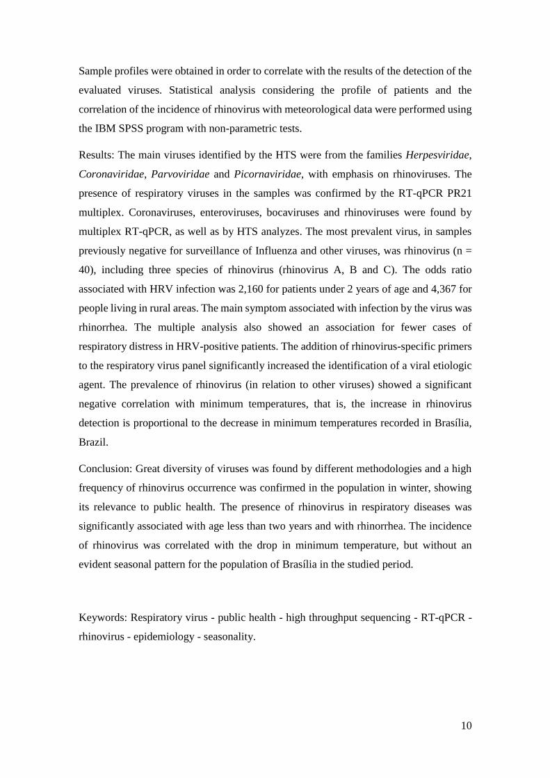
Sample profiles were obtained in order to correlate with the results of the detection of the
evaluated viruses. Statistical analysis considering the profile of patients and the
correlation of the incidence of rhinovirus with meteorological data were performed using
the IBM SPSS program with non-parametric tests.
Results: The main viruses identified by the HTS were from the families Herpesviridae,
Coronaviridae, Parvoviridae and Picornaviridae, with emphasis on rhinoviruses. The
presence of respiratory viruses in the samples was confirmed by the RT-qPCR PR21
multiplex. Coronaviruses, enteroviruses, bocaviruses and rhinoviruses were found by
multiplex RT-qPCR, as well as by HTS analyzes. The most prevalent virus, in samples
previously negative for surveillance of Influenza and other viruses, was rhinovirus (n =
40), including three species of rhinovirus (rhinovirus A, B and C). The odds ratio
associated with HRV infection was 2,160 for patients under 2 years of age and 4,367 for
people living in rural areas. The main symptom associated with infection by the virus was
rhinorrhea. The multiple analysis also showed an association for fewer cases of
respiratory distress in HRV-positive patients. The addition of rhinovirus-specific primers
to the respiratory virus panel significantly increased the identification of a viral etiologic
agent. The prevalence of rhinovirus (in relation to other viruses) showed a significant
negative correlation with minimum temperatures, that is, the increase in rhinovirus
detection is proportional to the decrease in minimum temperatures recorded in Brasília,
Brazil.
Conclusion: Great diversity of viruses was found by different methodologies and a high
frequency of rhinovirus occurrence was confirmed in the population in winter, showing
its relevance to public health. The presence of rhinovirus in respiratory diseases was
significantly associated with age less than two years and with rhinorrhea. The incidence
of rhinovirus was correlated with the drop in minimum temperature, but without an
evident seasonal pattern for the population of Brasília in the studied period.
Keywords: Respiratory virus - public health - high throughput sequencing - RT-qPCR -
rhinovirus - epidemiology - seasonality.
11
Capítulo 1
1.1 Introdução
1.1.1 Vírus respiratórios
Os vírus são os principais agentes etiológicos envolvidos em infecções do trato
respiratório, com elevado índice de morbidade e mortalidade, estando entre as cinco
principais causas de morte em todo mundo (WHO 2018). Complicações relacionadas a
essas infecções respiratórias atingem principalmente os grupos considerados de risco:
crianças menores de cinco anos, gestantes, idosos e pacientes imunossuprimidos (Zar,
2014; WHO 2015). Segundo a Organização Mundial de Saúde (OMS), as infecções
respiratórias agudas (IRAs) são responsáveis por 40 a 60% de todos os atendimentos
ambulatoriais em pediatria na América Latina. Cerca de 4 milhões de crianças menores
de cinco anos morrem por infecção aguda das vias aéreas inferiores anualmente,
principalmente em países em desenvolvimento (Salomão Júnior et al. 2011, WHO 2018).
Além disso, essas doenças geram elevados custos diretos e indiretos com assistência à
saúde (Girard et al. 2005) e representam a quarta causa de morte em crianças nos países
de média renda, como o Brasil (Ferone et al. 2013).
Em pacientes adultos, os vírus respiratórios estão associados a pneumonias
adquiridas na comunidade, além da exacerbação da doença pulmonar obstrutiva crônica
e da asma, resultando em grande número de consultas e hospitalizações (Daubin et al.
2006). Os vírus mais prevalentes são vírus sincicial respiratório, rinovírus e vírus
influenza, com positividade de 29,6% a 57,6% em pacientes com sintomatologia
respiratória (Yu et al. 2012, Falsey et al. 2014, Stover & Litwin 2014, Nam & Ison 2019).
12
A nova pneumonia associada à doença COVID-19, causada pelo vírus SARS-CoV-2, é
caracterizada por febre, fadiga, tosse seca e dispneia e acomete principalmente pacientes
adultos e imunocomprometidos. A positividade de detecção desse vírus pandêmico varia
de 29,0 a 66,6%, para síndrome gripal e síndrome respiratória aguda grave
respectivamente (Brasil 2021).
Em crianças as infecções por vírus respiratórios são a principal causa de
morbidade e de atendimentos médicos (Lambert et al. 2007; Quan et al. 2007). A
positividade para vírus respiratórios nesse público varia de 35,3% a 84% com quadros de
infecção do trato respiratório, com maior destaque para vírus sincicial respiratório e
rinovírus (Jansen et al. 2011, Martin et al. 2013, Wang et al. 2016). Estes são responsáveis
por 1 a 3% de mortalidade entre crianças menores de cinco anos de idade em países
industrializados e por 10 a 15% dos óbitos em crianças de países em desenvolvimento
(Quan et al. 2007). Os vírus respiratórios podem causar diversas complicações clínicas,
como bronquiolite, pneumonias e crupe, o que geralmente envolve hospitalizações
(Syrmis et al. 2004). Entre os vírus respiratórios, o vírus da influenza causa infecção
respiratória que afeta 5-10% dos adultos e 20-30% das crianças (WHO 2012), atingindo
290.000-650.000 mortes por ano em todo o mundo (Iuliano et al. 2018).
O perfil de ocorrência das infecções virais respiratórias é variável e está
relacionado com a região geográfica, clima e período do ano. Em locais de clima
temperado, os vírus respiratórios normalmente seguem padrões sazonais de atividade,
conforme fatores associados à temperatura (Monto 2004). Os vírus influenza,
parainfluenza, metapneumovírus humano e vírus sincicial respiratório, por exemplo,
possuem picos de incidência no inverno, enquanto enterovírus são comuns no outono e
verão. Já infecções por rinovírus possuem maior incidência em estações frias, tanto para

13
países de clima temperado como para regiões tropicais, embora circulem durante o ano
todo (Costa et al. 2006, du Prel et al. 2009).
Também em regiões de clima temperado, é comum a ocorrência de cocirculação
de vários vírus em um dado período do ano, mesmo durante o pico de ocorrência de um
determinado agente respiratório (Tamerius et al. 2013, Pica & Bouvier 2014). Um estudo
na Inglaterra mostrou que a maioria das infecções por vírus respiratórios ocorreu na idade
menor de cinco anos, com mais de 10% de infecções duplas ou múltiplas, sendo o
rinovírus, o mais prevalente nessas coinfecções (Goka et al. 2015).
O Brasil, que inclui regiões temperadas e tropicais, exibe uma importante
heterogeneidade regional da circulação desses vírus (Alonso et al. 2007, de Mello et al.
2009). As epidemias ou pandemias de gripe geralmente associam-se a climas frios e
úmidos, mas também a fatores como idade, imunidade preexistente, polimorfismos
genéticos e presença de comorbidades, influenciando na gravidade da epidemia e
responsividade à vacina (Castrucci 2017). A vacinação é a principal medida de saúde
pública utilizada para reduzir a freqüência de casos graves de influenza, a qual é realizada
na forma de campanhas anuais, em nosso país, desde 1999 (Domingues & Oliveira,
2012).
No Brasil, estudos mostram os agentes virais como a principal causa de infecção
respiratória aguda, especialmente em crianças, como observado em estudos nas cidades
de Fortaleza (de Arruda et al. 1991), Rio de Janeiro (Nascimento et al. 1991), São Paulo
(Miyao et al. 1999), Curitiba (Tsuchiya et al. 2005), Porto Alegre (de-Paris et al. 2014) e
Belo Horizonte (Monteiro et al. 2016).

14
1.1.2 Vigilância da influenza e de outros vírus respiratórios
A vigilância global epidemiológica para os vírus influenza foi iniciada em
1947, com o objetivo de monitorar os vírus circulantes e fornecer informações para
subsidiar as recomendações da Organização Mundial da Saúde (OMS). Atualmente, a
Rede Global de Vigilância de Influenza da OMS (GISN, do inglês WHO Global Influenza
Surveillance Network) é constituída de mais de 140 laboratórios em diferentes países,
denominados Centros Nacionais de Influenza (NIC – National), além de seis Centros
Colaboradores (WHO CC). No Brasil, a implantação do Sistema de Vigilância Sentinela
teve início em 2000 (Barros et al. 2004). O Sistema de Vigilância da Influenza e de
outros vírus respiratórios está presente em todos os Estados brasileiros e existem três
Centros Nacionais de Influenza (NICs). Os Lacens (Laboratórios Centrais de Saúde
Pública) são responsáveis pela base da informação utilizada para vigilância a partir da
identificação do agente etiológico, tipagem e subtipagem de vírus respiratórios
circulantes. Um quantitativo das amostras processadas pelos Lacens é sistematicamente
enviado para os Laboratórios de Referência Nacional para realização de análises
complementares, como o sequenciamento genético (Brasil 2016). O objetivo da vigilância
global da influenza é, portanto, identificar os vírus respiratórios circulantes, bem como
sua sazonalidade e as populações mais suceptíveis.
A vigilância é realizada de forma sentinela para os casos de síndrome gripal (SG),
definidos como pacientes apresentando febre e tosse ou dor de garganta além de um dos
seguintes sintomas: cefaleia, mialgia ou artralgia. Em crianças menores de 2 anos, os
casos de SG são definidos por: febre, tosse, coriza e obstrução nasal. A vigilância
sentinela consiste em uma rede de unidades de saúde designadas (públicas ou privadas)
distribuídas por todo o país, nas quais amostras aleatórias de casos respiratórios são
15
periodicamente coletadas para detecção e caracterização genética de vírus circulantes. Os
casos de síndrome respiratória aguda grave (SRAG) são definidos com pacientes
requerendo hospitalização e apresentando dispneia ou um dos seguintes sinais: saturação
de oxigênio capilar periférico < 95%, desconforto respiratório ou insuficiência
respiratória aguda. A vigilância da SRAG é realizada de forma universal, onde todos os
casos respiratórios graves internados em unidades de terapia intensiva (UTIs) e todas as
mortes relacionadas à doença respiratória são investigadas em laboratório, quanto a suas
causas (Barros et al. 2016).

16
1.1.3 Principais vírus respiratórios
Os agentes etiológicos virais mais frequentemente envolvidos com as infecções
do trato respiratório (ITR) são os vírus influenza tipo A, B e C (Influenza A virus,
Influenza B virus, Influenza C virus), vírus sincicial respiratório (VSR, Human
orthopneumovirus), metapneumovirus humano (HMPV, Human metapneumovirus),
vírus parainfluenza (PIV) tipos 1 (Human respirovirus 1), 2 (Human orthorubulavirus 2),
3 (Human respirovirus 3), 4A e 4B (Human orthorubulavirus 4), coronavirus humano
(HCoV) 229E (Human coronavirus 229E), NL63 (Human coronavirus NL63), HKU1
(Human coronavirus HKU1), OC43 (Betacoronavirus 1), SARS-CoV (Severe acute
respiratory syndrome-related coronavirus), MERS-CoV (Middle East respiratory
syndrome-related coronavirus) e SARS-CoV-2, rinovirus (HRV) tipos A (Rhinovirus A),
B (Rhinovirus B) e C (Rhinovirus C), enterovírus (EV, Enterovirus C), além dos
bocavírus (HBoV, Primate bocaparvovirus 1) e adenovírus (AdV, Human
mastadenovirus C) (van den Hoogen et al. 2001, Allander et al. 2005, Tregoning &
Schwarze 2010, ICTV 2018, Abdelrahman et al. 2020), os quais são agrupados em sete
famílias (quadro 1). Estes vírus são responsáveis por um espectro de manifestações
clínicas que incluem o comprometimento alto e baixo do trato respiratório (Quan et al.
2007). Entretanto, os agentes causadores de uma parcela das infecções respiratórias ainda
são desconhecidos.

17
Quadro 1. Características gerais dos principais vírus respiratórios.
Família Vírus Material Genético Partícula Viral
Orthomyxoviridae Influenza A virus
Influenza B virus
Influenza C virus
ssRNA (-)
segmentado
(10-14,6kb)
~100nm
Envelopado
Simetria helicoidal
Pneumoviridae Human orthopneumovirus
Human metapneumovirus
ssRNA (-)
(13,2 – 17,4kb)
150 – 600nm
Envelopado
Simetria helicoidal
Paramyxoviridae
Human respirovirus 1
Human orthorubulavirus 2
Human respirovirus 3
Human orthorubulavirus 4
ssRNA (-)
(13,2 – 17,4kb)
150 – 600nm
Envelopado
Simetria helicoidal
Coronaviridae Human coronavirus HKU1
Human coronavirus NL63
Human coronavirus 229E
Betacoronavirus 1
Middle East respiratory
syndrome-related coronavirus
(MERS)
Severe acute respiratory
syndrome-related coronavirus
(SARS)
ssRNA (+)
(27 – 32kb)
100-160nm
Envelopado
Simetria helicoidal
Picornaviridae Rhinovirus A
Rhinovirus B
Rhinovirus C
Enterovirus C
ssRNA (+)
(7,2 – 8,5kb)
~30nm
Desnudo
Simetria icosaédrica
Parvoviridae Primate bocaparvovirus 1 ssDNA
(~5kb)
~25nm
Desnudo
Simetria icosaédrica
Adenoviridae Human mastadenovirus C dsDNA
(~36kb)
70-90nm
Desnudo
Simetria icosaédrica
Fonte: Oliveira, 2016 (adaptado).
A influenza (gripe comum) é uma doença respiratória que ocorre no mundo todo
causada pelo vírus influenza, pertencente à família Orthomyxoviridae, que inclui os vírus
A, B e C em humanos. O genoma do vírus consiste em 8 segmentos de RNA (7 para
Influenza C), onde cada um codifica pelo menos uma proteínas com funções específicas
(Vincent et al. 2014, Tian et al. 2012). Até o momento, foram identificados 18 subtipos
antigênicos para a hemaglutinina (H1-18) e 11 subtipos diferentes da neuraminidase (N1-

18
11), sendo as principais cepas que circulam na população humana: H1N1, H1N2, H2N2
e H3N2 (WHO 2014).
A variabilidade genômica, que se traduz em diversidade antigênica, bem como as
recombinações, compreendem importantes mecanismos de escape imunológico do vírus,
os quais favorecem a infecção de novos hospedeiros suscetíveis, podendo diminuir
também a eficácia das vacinas (Guarnaccia et al. 2013, Tewawong et al. 2015).
Esses fatores, juntamente com a alta transmissibilidade e o potencial zoonótico e
pandêmico, tornam o desafio de prevenção e controle da influenza um problema crescente
e de reconhecida importância na saúde pública (Zambon 2014, Jerigan & Cox 2013).
O vírus sincicial respiratório humano (VSR) é a causa mais comum de infecções
do trato respiratório superior e inferior em crianças, de até dois anos de idade, seguido
pelos vírus parainfluenza humanos (PIV) (Pecchini et al. 2015). O VSR acomete crianças
principalmente nos primeiros meses de vida, podendo ainda infectar adultos e idosos, e
estando associado à complicações como broncopneumonia, desnutrição e bronquiolite,
mas também é comumente encontrado em coinfecção com outros vírus respiratórios
(Raboni et al. 2015) e foi isolado pela primeira vez no Brasil por Candeias em 1964
(Candeias 1967). Divide-se em dois subgrupos antigênicos baseados em reações contra a
glicoproteína G e posteriores análises genéticas: VSR-A, considerado mais virulento, e
VSR-B, ainda subdividido em duas variantes, B1 e B2 (Mufson et al. 1985, Melero et al.
1997).
O metapneumovírus humano (HMPV) é um vírus da família Pneumoviridae,
descoberto em 2001 na Holanda, caracterizado como agente causador de infecção
respiratória aguda em todas as faixas etárias, mas principalmente em crianças até dois
anos de idade (van den Hoogen et al. 2001, Panda et al. 2014). Esses vírus são
classificados nos genótipos A e B (com posterior classificação em sub-linhagens A1, A2a,

19
A2b, B1 e B2) (van den Hoogen et al. 2004). Os principais sintomas são febre, tosse,
congestão nasal, dispnéia, além de bronquiolite e pneumonia em casos mais graves (Kahn
2006, Broor et al. 2008, Feuillet et al. 2012). Em adultos, no geral a infecção é de leve a
moderada; contudo, idosos e indivíduos imunocomprometidos podem apresentar
complicações (Falsey et al. 2003). O primeiro genoma completo do HMPV sequenciado
no Brasil foi relatado por Di Paola e colaboradores (2018).
Em relação aos PIVs, existem quatro espécies: PIV-1, -2, -3 e -4, sendo que PIV-
4 está classificado em dois sorotipos, -4A e -4B. Estima-se que estes causem até 10% das
infecções respiratórias agudas (IRA) na infância (Boivin et al. 2002). Os vírus PIV1 e
PIV2 são a principal causa de crupe em crianças de 6 a 48 meses. O PIV3 e, em menor
extensão, o PIV1, estão mais frequentemente associados a bronquiolite e pneumonia em
crianças com menos de 1 ano. Os PIVs também causam doença grave, incluindo
pneumonia e morte em pacientes transplantados, bem como infecções nosocomiais e
surtos (Hasman et al. 2009). Estudo recente fez o primeiro relato de infecção por PIV-4
em crianças com IRA no Brasil, cuja prevalência ficou abaixo dos patógenos respiratórios
mais comuns (VSR, adenovírus, influenza e HMPV), mas foi o segundo PIV mais
prevalente, seguindo o PIV-3 (Thomazelli et al. 2018).
Os coronavírus humanos (HCoV) foram identificados pela primeira vez há mais
de 50 anos. Esses vírus são classificados em ao menos 4 gêneros (delta, gama, beta e alfa),
sendo sete destes vírus conhecidos por infectar humanos: NL63 e 229E do gênero alfa e
OC43, HKU1, SARS-CoV, MERS-CoV e SARS-CoV-2 do gênero beta (Abdelrahman
et al. 2020). Acredita-se que esses vírus tenham se originado de reservatórios animais,
sendo os SARS-CoV e MERS-CoV exemplos mais recentes que emergiram de morcegos
para civetas (gato-de-algália) ou dromedários, respectivamente, e destes para humanos
(van den Brand et al. 2015). Supõe-se que o SARS-CoV-2 emergiu de morcegos tendo

20
como possíveis hospedeiros intermediários os pangolins (Dimonaco et al. 2020). Estima-
se que as taxas de fatalidade do SARS-CoV, MERS-CoV e SARS-CoV-2 sejam de 9,5%,
34,4% e 2,3%, respectivamente (Petrosillo et al. 2020).
Em relação às manifestações clínicas, os vírus 229E (Alphacoronavirus) e OC43
(Betacoronavirus) estão relacionadas com resfriado comum, enquanto NL63
(Alphacoronavirus) está associado com casos de crupe e bronquiolite. Os HCoV-HKU1
(Betacoronavirus) foram associados a sintomas como febre, rinorreia, sibilância e tosse
(van der Hoek et al. 2005, Pyrc et al. 2007, Wu et al. 2008, Lee & Storch 2014).
Os vírus SARS-CoV (Betacoronavirus) causam pneumonia atípica, caracterizada
por tosse, febre, mialgia, dores de cabeça, mal-estar, dispneia e menos comumente
vômitos e diarreia (Hui & Chan 2010, van den Brand et al. 2014). As infecções por
MERS-CoV (Betacoronavirus) possuem sintomatologia semelhante ao SARS, contudo
consideradas mais graves e com maiores taxas de mortalidade. Ademais incluem aspectos
clínicos como pneumonia grave, às vezes com lesão pulmonar aguda fatal ou síndrome
respiratória aguda de angústia (Abdelrahman et al. 2020), além de retenção urinária e
falência renal (Al-Tawfiq 2013, Groot et al. 2013, van den Brand et al. 2015).
Existe atualmente uma pandemia mundial de um novo vírus pertencente à família
Coronaviridae, identificado pela primeira vez em dezembro de 2019 em Wuhan, China,
cuja dispersão pelo mundo se deu de forma rápida. Este agente, foi designado como
coronavírus relacionado à síndrome respiratória aguda grave 2 (SARS-CoV-2), cujo
nome da doença associada é COVID-19 (CSGICTV 2020). O SARS-CoV-2 pode ser
transmitido pela via respiratória, através de aerossóis, perdigotos e secreções
respiratórias, ou por contato com fômites. A transmissão pela via ocular tem sido cogitada
e o vírus pode também ser encontrado nas fezes de indivíduos infectados. A COVID-19
pode estar associada SRAG, pneumonia, além de quadros mais brandos ou subclinicos e
21
assintomáticos (Bchetnia et al. 2020). A infecção pelo SARS-CoV-2 já causou milhões
de mortes, com sobrecarga dos sistemas de saúde e grande impacto econômico devido à
política de isolamento social proposta para contenção da disseminação do vírus. Até o
momento, não há terapia especifica eficaz disponível, mas muitas vacinas para o vírus
estão em desenvolvimento ou já foram aprovadas para aplicação na população adulta.
Os enterovírus humanos (EV) podem causar infecções respiratórias, mas também
podem estar associados a diversas síndromes clínicas, incluindo infecções assintomáticas
e subclínicas, doenças respiratórias, gastroenterites, miocardites e meningites. Os
enterovírus pertencem à famíia Picornaviridae e estão divididos em doze espécies (EV
A-L) com base em suas propriedades biológicas e moleculares. Foram descritas até o
momento quatro espécies de EV A-D capazes de causar infecções em humanos, enquanto
as outras espécies comportam vírus que infectam primatas, suínos e bovinos (Tapparel et
al. 2013, ICTV 2018, Picornaviridae 2019). Além da transmissão por via respiratória,
também são transmitidos por via fecal-oral. As infecções respiratórias por enterovírus
podem variar desde assintomática até sintomáticas do trato respiratório superior
(resfriado, faringite) ou inferior (pneumonia, bronquiolite ou exacerbação de asma na
infância) (Chang 2008, Tapparel et al. 2013). No Brasil, Carney e colaboradores (2015)
relataram pela primeira vez, dois casos de infecção pelo enterovírus EV-D68 em crianças
com doença respiratória aguda, com sintomas como insuficiência respiratória, febre,
diarreia e taquicardia. Outro enterovírus associado à infecção respiratória recentemente
identificado no país foi o coxsackievírus B2. Ele foi identificado em uma paciente de um
ano de idade, com sintomatologia leve de infecção respiratória (Lima 2017).
O bocavírus humano (HBoV) é classificado como genótipo 1 a 4 (Guido et al.
2016) e foi descrito pela primeira vez na Suécia em 2005, sendo identificado como agente
de infecções respiratórias a partir de aspirados de nasofaringe de pacientes com infecções
22
do trato respiratório inferior (Allander et al. 2005, ICTV 2018). Estudos também
descreveram a presença do bocavírus em amostras fecais provenientes de pacientes com
gastroenterite, sugerindo tropismo do vírus pelo trato gastrointestinal para pelo menos
algumas espécies (Kapoor et al. 2009, Kapoor et al. 2010).
Os adenovírus humanos (AdV) foram isolados primeiramente por Rowe e colegas,
enquanto estudavam o cultivo de vírus em tecidos adenóides (Rowe et al. 1953). Os AdVs
são divididos em sete espécies, AdV A-G, e ainda em mais de 100 genótipos (Robinson
et al. 2011, Huang et al. 2013, Mao et al. 2019). Os adenovírus podem causar um amplo
espectro de infecções em humanos, dentre elas, patologias respiratórias, gastroentéricas,
hepáticas e oculares. A maioria dessas infecções ocorre em população pediátrica e, em
indivíduos imunocompetentes é geralmente autolimitada. No caso das infecções do trato
respiratório, os sintomas variam desde um quadro clínico de resfriado comum até casos
mais graves de pneumonias e bronquiolites (Kunz & Ottolini 2010), sendo estes vírus
responsáveis por 1 - 5% de todas as infecções respiratórias e por até 10% das pneumonias
na infância (Ampuero et al. 2012).

23
1.1.4 Rinovírus
Os vírus mais frequentemente associados a infecções respiratórias são os
Rinovírus humanos (HRV), tipicamente o resfriado comum, que inclui sintomas como
espirros, obstrução nasal, coriza, dor de garganta, dor de cabeça, tosse e mal-estar.
Entretanto, com o avanço de técnicas de biologia molecular, esse vírus também
vem sendo associado a infecções assintomáticas, disseminação sistêmica e do trato
respiratório inferior, como pneumonia e bronquiolite, particularmente entre pacientes
com asma, fibrose cística, doença pulmonar obstrutiva crônica (DPOC) e
imunossupressão relacionada ao transplante (Peltola et al. 2009, Gern 2010, Tapparel et
al. 2011).
Esses vírus são filogeneticamente classificados em três espécies, HRV-A, HRV-
B e HRV-C (Choi et al. 2015, Martin et al. 2015), os quais compreendem uma grande
diversidade genética, com mais de 160 sorotipos (Palmenberg et al. 2010). Os HRVs
usam três diferentes glicoproteínas da membrana celular expressas no epitélio respiratório
para entrar na célula hospedeira. Esses receptores virais são a molécula de adesão
intercelular 1 (usada pela maioria dos tipos de RV-A e todos os tipos de RV-B), membros
da família de receptores de lipoproteínas de baixa densidade (usados por 12 tipos de RV-
A) e membros da família 3 relacionados à caderina (CDHR3; usado por RV-C) (Basnet
et al. 2019).
Inicialmente a espécie C foi descrita como responsável por doença de maior
severidade, porém estudos posteriores mostraram que essa característica é controversa
(Pierangeli et al. 2013, Choi et al. 2015). Em muitos casos, os rinovírus são detectados
juntamente com outros vírus respiratórios, principalmente associados ao VSR A e B em
infecções de crianças e adultos (Brandão et al. 2016, Calvo et al. 2015).

24
Os rinovírus apresentam relevância mundial por circularem o ano todo em
diferentes regiões do mundo, além de infectarem todas as faixas etárias; no entanto, bebês,
crianças e idosos têm as taxas mais altas de infecção grave entre pacientes hospitalizados
(Miller et al. 2013, Fry et al., 2011). A transmissão dos HRVs ocorre principalmente
através de inalação de gotículas respiratórias, contato direto de pessoa a pessoa ou através
de superfícies contaminadas ou inoculação direta do olho ou nariz mucosa com a ponta
dos dedos (L’Huillier 2015).
O capsídeo do HRV é composto por quatro proteínas (VP1, VP2, VP3 e VP4),
que completam o genoma de RNA. As três primeiras proteínas pertencem ao capsídeo e
são responsáveis pela diversidade antigênica viral, enquanto a VP4 é internalizada,
ancorando o RNA ao capsídeo (Jacobs et al. 2013). Os mecanismos de ação dos HRVs
que envolvem o rompimento da barreira epitelial com vazamento e produção excessiva
de muco, além da secreção de muco das células caliciformes em resposta a produtos de
ativação de neutrófilos, estão provavelmente envolvidos na obstrução nasal, levando aos
sintomas das vias aéreas (Papadopoulos et al. 2000).

25
1.1.5 Diagnóstico e caracterização molecular
Algumas técnicas tradicionais de diagnóstico são usadas há décadas na detecção
de vírus respiratórios (como imunomicroscopia eletrônica, ensaio de imunoabsorção
enzimática, imunofluorescência direta e indireta, imunocromatografia) (Murdoch 2009,
Murdoch 2016a), entretanto técnicas específicas de diagnóstico molecular, têm permitido
a identificação mais rápida de muitos patógenos que antes eram difíceis de detectar
(Murdoch 2016b).
A técnica da reação em cadeia da polimerase (PCR) permite que um fragmento
específico da molécula de DNA seja amplificado, de forma exponencial, em apenas
algumas horas, sendo possível ainda a caracterização de patógenos, através da
genotipagem. As vantagens da PCR são numerosas: velocidade, baixo custo, automação,
sensibilidade e especificidade. Entretanto, o principal problema envolvendo o uso da PCR
para diagnóstico é o fato de ela ser patógeno-específica, ou seja, só identifica alvos pré-
definidos, o que supõe que o médico elaborou uma hipótese etiológica.
A utilização desta técnica está em constante atualização, como o uso cada vez
mais disseminado da PCR em tempo real, a qual permite que a a amplificação e detecção
ocorram simultaneamente, com possibilidade de gerar resultados quantitativos com
maior precisão, por meio de um sistema de monitoramento da emissão da fluorescência.
Há atualmente diversos ensaios multiplex comerciais disponíveis em uma variedade de
formatos. No contexto clínico, a detecção de um vírus respiratório em uma amostra
respiratória é geralmente considerada suficiente para atribuir uma relação causal, embora
seja importante avaliar a possibilidade de colonização ou até contaminação (Ruuskanen
et al. 2011).

26
Essas novas metodologias, como as que envolvem sequenciamento genético, têm
auxiliado na descoberta de novos vírus respiratórios, sendo que os últimos incluem o
bocavírus humano, o metapneumovírus humano e uma variedade de coronavírus (SARS-
CoV, CoV-NL63, CoV-HKU1 e MERS-CoV) (Berry et al. 2015, Lu et al. 2020).
De forma geral, as infecções do trato respiratório superior tendem a ser mono-
microbianas e predominantemente causadas por vírus, com algumas exceções notáveis
causadas por bactérias específicas (como por exemplo, faringite aguda causada por
Streptococcus pyogenes). Infecções do trato respiratório inferior são causadas por uma
ampla variedade de patógenos virais e bacterianos. Isso implica em complexidade, sendo
que o papel de cada microorganismo nessas patologias ainda não é bem elucidado (El
Kholy et al. 2016).
O sequenciamento de alto desempenho – do inglês High Throughpout Sequencing
(HTS), também chamado de sequenciamento de nova geração, é reconhecido como um
tecnologia poderosa para a detecção e identificação de microorganismos conhecidos ou
não (Mallet & Gisonni-Lex 2014). Este sequenciamento amplifica fragmentos de DNA
muitas vezes, gerando resultados com grande acurácia e aplicado ao diagnóstico
microbiano permite, em teoria, a amplificação e análise de qualquer material genético
presente na amostra clínica, mesmo em baixas concentrações e sem a necessidade de
desenhar primers específicos para pré-amplificar as sequências-alvo (Reuter et al. 2015).
Entretanto há entraves clínicos, computacionais e interpretativos, como por exemplo, a
necessidade de enriquecimento das amostras a serem sequenciadas, visto que são
provenientes de espécimes clínicos dos pacientes, extremamente contaminados com DNA
humano e de outros microrganismos, os quais podem não causar sintomas
(assintomáticos) ou estar presentes na microbiota normal e saudável (Xu et al. 2017,
Taboada et al. 2014, Zhou et al. 2016).

27
A população humana está susceptível a um crescente número de doenças
infecciosas, inclusive em razão da adaptação a humanos e emergência de vírus ainda
não caracterizados. As mudanças climáticas, a globalização, assentamentos perto de
habitats de animais silvestres e a convivência próxima de diferentes espécies, além do
aumento do número de pessoas imunocomprometidas, provavelmente, contribuem para
o surgimento e disseminação de novas infecções (Morens et al. 2004). Além disso, várias
síndromes clínicas são suspeitas de ser de etiologia viral, mas o agente pode não ser
isolado ou reconhecido por cultivo e ensaios moleculares tradicionais. Portanto, existe a
necessidade de se aprimorar os métodos para a identificação de patógenos virais ou
caracterização de novos agentes. Métodos de sequenciamento de alto desempenho tem
permitido análises genômicas mais sensíveis e rápidas em relação às metodologias
anteriores, utilizados por exemplo na descoberta do herpesvírus 8 humano (Chang et al.
1994), vírus GB humano (Simons et al. 1995), Torque teno virus (Nishizawa et al. 1997),
bocavírus (Allander et al. 2005), parvovírus humano 4 (Jones et al. 2005), WU
poliomavírus (Gaynor et al. 2007) e KI poliomavírus (Allander 2007).
Existem mais casos na literatura que mostram a utilização do sequenciamento de
alta preformance na descoberta de patógenos não identificados por métodos tradicionais,
como ocorreu com a descoberta de um arenavírus em três pacientes que morreram de uma
doença febril algumas semanas após transplante de órgãos sólidos a partir de um único
doador (Palacios et al. 2008). A plataforma Illumina GA permitiu identificar um vírus
influenza A a partir de esfregaços e montagem de novo de seu genoma (Yongfeng et al.
2011, Kuroda et al. 2010, Greninger 2010). A metodologia também permitiu a detecção
de patógenos virais em amostras de aspirado nasofarínge de pacientes com infecções
agudas do trato respiratório inferior (Yang et al. 2011), como um novo enterovírus,
denominado enterovírus 109 (EV109) detectado em uma coorte de crianças nicaraguenses

28
com doença respiratória viral (Yozwiak et al. 2010). Foi também foi possível identificar
um novo astrovírus HAstV-VA1 / HMO-C-UK1, associado a encefalite em pacientes
imunossupridos (Brown et al. 2015).
O caso mais recente de utilização de sequenciamento de alto desempenho foi a
descoberta do vírus SARS-CoV-2. O sequenciamento foi realizado a partir de amostras
de fluido de lavagem broncoalveolar e isolados de nove pacientes internados, oito dos
quais haviam visitado o mercado de frutos do mar Huanan em Wuhan, China. Sequências
genômicas completas e parciais 2019-nCoV foram obtidas desses indivíduos, com mais
de 99% de identidade de sequência. A determinação do genoma permitiu a identificação
do domínio de ligação ao receptor e, posteriormente a identificação da enzima conversora
da angiotensina 2 como receptor nas células do hospedeiro. (Lu et al. 2020).

29
1.2 Justificativa
A utilização de sequenciamento de alto desempenho (High-throughput
sequencing: HTS) tem sido aplicada com sucesso em vários campos da virologia,
incluindo descoberta de novos vírus, reconstrução de genoma completo e análise de
variantes (Capobianchi et al. 2013, Lipkin 2010, Mokili et al. 2013, Smits & Osterhaus
2013, van Boheemen et al. 2012). A vantagem mais importante fornecida por estas
plataformas é a determinação dos dados da sequência de fragmentos de DNA isolados de
uma biblioteca que são separados em chips, evitando a necessidade de clonagem em
vetores antes da aquisição de sequência (Barzon et al. 2011). Essas tecnologias são bem
conhecidas por sua enorme produção de dados de sequência genética a um custo
relativamente alto, mas decrescente. Outra vantagem é que este tipo de metodologia
elimina a necessidade de projeto e validação de várias dezenas ou centenas de primers /
sondas específicas para sequências virais, e não requer adaptação contínua desses
iniciadores a cada nova variante ou espécie identificada.
Como o diagnóstico de vírus respiratórios é realizado no Lacen-DF (Laboratório
Central de Saúde Pública do Dsitrito Federal) por meio de metodologia tradicional (RT-
PCR em tempo real), agentes etiológicos em muitas amostras podem não estar sendo
identificados, tendo em vista que essa metodologia é patógeno-específica. As amostras
encaminhadas ao Lacen são oriundas de pacientes de hospitais da rede pública e privada
de saúde com suspeita de infecção por vírus respiratórios, estando esses pacientes
internados ou não. Os principais sintomas associados são: febre, tosse, dispneia, mialgia,
dor de garganta e desconforto respiratório. Em torno de 50-60% das amostras
encaminhadas são negativas para os vírus pesquisados na rotina (Influenza A e B, Vírus
Respiratório Sincicial, Metapneumovírus humano, Adenovírus, Parainfluenza 1, 2 e 3).

30
Sendo assim, para fins de saúde pública, é fundamental conhecer quais possíveis
patógenos estão acometendo os pacientes com sintomatologia típica de gripe ou resfriado,
mas sem diagnóstico definido por metodologia tradicional.
Nesse contexto, identificar a alta prevalência de rinovírus, antes subestimado pela
ausência de alvos no painel viral, por meio da avaliação de diferentes metodologias de
diagnóstico é fundamental para fortalecer a pesquisa local e o laboratório de saúde
pública.
Além disso, a carcaterização molecular e de características clínico
epidemiológicas dos rinovírus permitem compreender características filogenéticas e
sintomatológicas dos vírus que acometem a população de Brasília. Isso se faz importante
para melhorar o diagnóstico e conhecer a epidemiologia desses vírus e assim, aprimorar
a atuação da rede de vigilância da influenza e de outros vírus respiratórios.

31
1.3 Objetivo Geral
Identificar possíveis vírus em amostras de secreções nasofaringe / traqueal de
pacientes do Distrito Federal com sintomas de vírus respiratórios em amostras clínicas
previamente negativas para os agentes etiológicos disponíveis no painel de vírus
respiratórios.
1.3.1 Objetivos específicos
1) Realizar a caracterização molecular de vírus a partir de amostras clínicas
(secreção da nasofaringe / aspirado traqueal), previamente negativas para o
painel de vírus respiratórios do LACEN- DF e coletadas entre junho e agosto
de 2016, por metagenômica, seguida de análise filogenética
2) Realizar o desenho de pares de iniciadores e sondas específicas para a
detecção de rinovírus, a partir de resultados obtidos com a análise das
amostras clínicas por Metagenômica, para serem utilizados em ensaio de
rotina diagnóstica.
3) Avaliar a sintomatologia e dados sócio demográficos de pacientes em
associação ao rinovírus, em amostras que haviam sido identificadas como
negativas por métodos de diagnóstico de rotina do Lacen-DF.
4) Avaliar a positividade viral em relação aos meses do ano em associação a
fatores climáticos da região.
32
1.4 Metodologia
Este projeto de pesquisa foi analisado e aprovado pelo comitê de ética da
Faculdade de Ciências da Saúde (Universidade de Brasília), parecer de aprovação nº
3.052.443.
Primeiramente, amostras de secreção nasofaringe e traqueal recebidas no
laboratório nos meses de junho, julho e agosto de 2016 foram testadas para o painel de
vírus respiratórios realizado no Lacen-DF pelo RT-qPCR padrão (para Influenza A,
Influenza B, Vírus sincicial respiratório humano, Metapneumovírus humano, Adenovírus
C humano, Parainfluenza 1, Parainfluenza 2 e Parainfluenza 3). As que apresentaram
resultados negativos tiveram o DNA / RNA extraídos na forma de pool (cinquenta e uma
amostras de junho, 55 de julho e 39 de agosto de 2016, perfazendo uma amostra para cada
mês). Foi realizada uma ultracentrifugação com colchão de sacarose a 20% e posterior
extração de ácidos nucleicos seguindo as instruções do High Pure Viral Nucleic Acid Kit
(Hoffmann-La Roche, Basel, Suíça), sem adição de RNA transportador. As três amostras
de DNA / RNA foram submetidas ao sequenciamento de alta performance (high-
throughput sequencing, HTS) usando Illumina HiSeq 2000 com escala de 3 G para 100
bases paired-end na Macrogen Inc. (Seul, Coréia do Sul). Em seguida, elas foram reunidas
em uma amostra de RNA única e tratadas com DNase I (Promega, Madison, EUA) para
diminuir o DNA humano ou bacteriano da amostra para enriquecimento do RNA viral e
novamente submetidas ao HTS. A biblioteca de amostras de DNA / RNA ou RNA foi
construída usando o kit TruSeq Standard total RNA.
As sequências de baixa qualidade e os adaptadores dos dados brutos foram
retirados usando a ferramenta Trimommatic v.036. As leituras metagenômicas de cada
amostra foram avaliadas no site Kaiju (http: //kaiju.binf.ku.dk) para traçar o perfil
33
taxonômico. As leituras de DNA foram filtradas com BWA v0.7.17 (17) e SAMtools v1.9
contra hg38 de referência (Homo sapiens) obtido do GenBank para remover sequências
de DNA humano do conjunto de dados HTS in silico. As leituras obtidas por HTS foram
montadas em sequências contíguas ("contigs") com os programas Velvet v.1.2.1 e
SPAdes v.3.9. Para identificação da origem viral das sequências contig foi utilizado o
tBlastx (https://blast.ncbi.nlm.nih.gov/Blast.cgi) contra o pacote de genoma viral (RefSeq
Virus, NCBI, https://www.ncbi.nlm.nih.gov/genome/viruses/) implementado no
programa Geneious R8.1 (Biomatters, Auckland, Nova Zelândia).
Parte dos achados na análise por HTS foi confirmada por meio de RT-qPCR
utilizando o kit XGen PR21 (Biometrix, Curitiba, Brasil) para 21 patógenos respiratórios
(Influenza A, Influenza B, Influenza A H1N1-swl, Coronavírus NL63, Coronavírus 229E,
Coronavírus OC43, Coronavírus HKU1, Parainfluenza 1, Parainfluenza 2, Parainfluenza
3, Parainfluenza 3, Parainfluenza 4, Parainfluenza Humana B, Vírus A e B sincicial
respiratório humano, Rinovírus, Enterovírus, Parecovírus, Adenovírus humano,
Bocavírus e Mycoplasma pneumoniae).
Os vírus identificados em maior prevalência nas amostras negativas estudadas por
HTS foram os rinovírus, com isso, primers universais HRV direcionados a regiões
conservadas em 5 'UTR (região não traduzida) foram projetados para detecção de HRV
usando o programa Geneious R8.1 (Biomatters, Auckland, Nova Zelândia). O RNA foi
extraído das 145 amostras originais usando o kit Magna Pure LC de ácido nucléico total
(Hoffmann-La Roche, Basel, Suíça), e depois amplificadas por RT-qPCR usando o
sistema GoTaq® Probe 1-Step RT-qPCR (Promega, Madison, EUA). A positividade para
rinovírus foi definida com limiar de ciclo (CT) menor ou igual a 40 (total de 45 ciclos na
reação). O gene RNase P humano foi usado como controle endógeno.

34
Também foram desenhados primers HRV_Com_For e HRV_Com_Rev visando
regiões conservadas com base em análises de viroma por HTS. Este par de iniciadores foi
usado para amplificar um fragmento de aproximadamente 540 bp para sequenciamento
de amplicon incluindo parte da 5' UTR e o gene da proteína VP4 / VP2 de rinovírus das
espécies A, B e C. O cDNA das amostras selecionadas foi sintetizado usando a
transcriptase MMLV (Thermo Fisher Scientific , Waltham, EUA) com primer hexâmero
aleatório e, a seguir, amplificado com LongAmp Taq DNA Polymerase (New England
BioLabs, Ipswich, EUA). O produto de PCR foi purificado e sequenciado pelo método
Sanger na Macrogen Inc (Seul, Coreia do Sul).
As sequências de nucleotídeos (nt) foram determinadas usando o programa
Geneious R8.1 (Biomatters, Auckland, Nova Zelândia). A árvore filogenética baseada na
5' UTR e no gene da proteína VP4 / VP2 foi construída usando o software Mega X, com
oito sequências de nucleotídeos obtidas neste estudo e outros quarenta isolados de
rinovírus que estavam disponíveis no GenBank com sequências completas do genoma
(para identificação do genótipo). A árvore filogenética foi inferida usando o método de
máxima verossimilhança com o modelo Hasegawa-Kishino-Yano conforme indicado
pela análise jModelTest.
Dados sócio demográficos e clínicos foram obtidos da ficha de notificação
compulsória da síndrome gripal e síndrome respiratória aguda grave encaminhados com
as amostras. Os dados foram analisados no software IBM SPSS (Chicago, EUA). Testes
não paramétricos foram utilizados, conforme apropriado, para variáveis qualitativas e
quantitativas.
A partir do desenho de primers universais, os rinovírus foram então inseridos no
painel de vírus respiratórios do Lacen-DF a partir de agosto de 2019. Os resultados de
35
rinovírus e outros vírus respiratórios foram avaliados de agosto de 2019 a fevereiro de
2020. Dados dos 3 anos anteriores foram usados para comparar o percentual de
identificação do agente etiológico. A partir de março de 2020, não foi possível dar
continuidade à avaliação devido à pandemia de Coronavírus (SARS-CoV-2) que causou
isolamento social e alteração da sazonalidade natural de outros vírus respiratórios, além
de alterar os critérios de recebimento de amostra para diagnóstico. Dados meteorológicos
da região de Brasília foram obtidas por meio de consultas diárias ao site oficial do Inmet
(Instituto Nacional de Meteorologia) por meio de temperaturas e umidades relativas
mínimas e máximas. A correlação não paramétrica de Spearman foi utilizada para avaliar
a relação entre os dados meteorológicos e a incidência de rinovírus no período estudado.
Todos os p-valores foram bicaudais ao nível de significância de 5%.
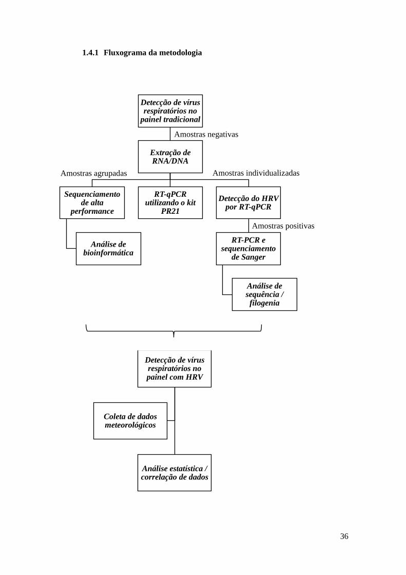
36
Detecção de vírus respiratórios no
painel tradicional
Extração de RNA/DNA
Sequenciamento de alta
performance
Análise de bioinformática
RT-qPCR utilizando o kit
PR21
Detecção do HRV por RT-qPCR
RT-PCR e sequenciamento
de Sanger
Análise de sequência / filogenia
1.4.1 Fluxograma da metodologia
Detecção de vírus respiratórios no painel com HRV
Análise estatística / correlação de dados
Coleta de dados meteorológicos
Amostras negativas
Amostras agrupadas Amostras individualizadas
Amostras positivas

37
1.5 Referências Bibliográficas
Abdelrahman Z, Li M, Wang X. Comparative Review of SARS-CoV-2, SARS-CoV, MERS-
CoV, and Influenza A Respiratory Viruses. Front Immunol. 11: 552909, 2020.
Allander, T.; Andreasson, K.; Gupta, S.; Bjerkner, A.; Bogdanovic, G.; Persson, M.A.; Dalianis,
T.; Ramqvist, T.; Andersson, B. Identification of a third human polyomavirus. J. Virol. 81:
4130–4136, 2007.
Allander, T.; Tammi, M.T.; Eriksson, M.; Bjerkner, A.; Tiveljung-Lindell, A.; Andersson, B.
Cloning of a human parvovirus by molecular screening of respiratory tract samples. Proc.
Natl. Acad. Sci. 102: 12891–12896, 2005.
Alonso WJ, Viboud C, Simonsen L, et al. Seasonality of influenza in Brazil: a traveling wave
from the Amazon to the subtropics. Am J Epidemiol. 165: 1434–42, 2007.
Al-Tawfiq JA. Middle East Respiratory Syndrome-coronavirus infection: a overview. J Infect
Public health. 6: 319-322, 2013.
Ampuero JS, Ocana V, Gómez J, Gamero ME, Garcia J, Halsey ES, Laguna-Torres VA.
Adenovirus respiratory tract infections in Peru. PLOS One. 7: 1-14, 2012.
Barros EN, Cintra O, Rossetto E, et al. Patterns of influenza B circulation in Brazil and its
relevance to seasonal vaccine composition. Braz J Infect Dis. 20: 81–90, 2016.
Barros FR. et al. O desafio da influenza: epidemiologia e organização da vigilância no Brasil.
Boletim Eletrônico Epidemiológico da Secretaria de Vigilância em Saúde [S.l.]. v. 1, p.
1-7, 2004. Disponível em: <http://portalsaude.saude.gov.br/images/pdf/
2014/julho/16/Ano04-n01-desafio-influenza-br-completo.pdf>. Acesso em: 09 de agosto de
2018.
Barzon L , Lavezzo E, Militello V, Toppo S, Palu G. Applications of next- generation
sequencing technologies to diagnostic virology. Int J Mol Sci. 12: 7861–7884, 2011.
Basnet S, Palmenberg AC, Gern JE. Rhinoviruses and Their Receptors. Chest. 155 (5): 1018-
1025, 2019.
Bchetnia M, Girard C, Duchaine C, Laprise C. The outbreak of the novel severe acute respiratory
syndrome coronavirus 2 (SARS-CoV-2): A review of the current global status. J Infect
Public Health. 13 (11): 1601-1610, 2020.
Berry M, Gamieldien J, Fielding B. Identification of new respiratory viruses in the new
millennium. Viruses. 7: 996–1019, 2015.
Brandão HV, Vieira GO, Vieira TO, Cruz AA, Guimarães AC et al. Acute viral bronchiolitis and
risk of asthma in school children: analysis of a Brasilian newborn cohort. J. Pediatr. 16,
2016.
Brasil. Ministério da Saúde. Secretaria de Vigilância em Saúde. Departamento de Vigilância das
Doenças Transmissíveis. Guia para a Rede Laboratorial de Vigilância de Influenza no
Brasil [recurso eletrônico] / Ministério da Saúde, Secretaria de Vigilância em Saúde,
Departamento de Vigilância das Doenças Transmissíveis. – Brasília: Ministério da Saúde,
2016.
Brasil. Ministério da Saúde. Secretaria de Vigilância em Saúde. Boletim epidemiológico
especial. Doença pelo Coronavírus COVID-19 [recurso eletrônico] / Ministério da Saúde,
Secretaria de Vigilância em Saúde, Departamento de Vigilância das Doenças
Transmissíveis. – Brasília: Ministério da Saúde, 2021.
Broor S, Bharaj P, Chahar HS. Human metapneumovirus: a new respiratory pathogen. J Biosc.
33: 483-493, 2008.

38
Brown JR, Morfopoulou S, Hubb J, et al. Astrovirus VA1/HMO-C: an increasingly recognised
neurotropic pathogen in immunocompromised patients. Clin Infect Dis. 60: 881-8, 2015.
Calistri, A; Palù, G. Editorial Commentary: Unbiased Next-Generation Sequencing and New
Pathogen Discovery: Undeniable Advantages and Still-Existing Drawbacks. Clinical
Infectious Diseases. 60 (6): 889–891, 2015.
Calvo C, García-García ML, Pozo F, Paula G, Molinero M, Calderón A, GonzálezEsguevillas M,
Casas I. Respiratory Syncytial Virus Coinfections With Rhinovirus and Human Bocavirus
in Hospitalized Children. Medicine. 94: 42, 2015.
Candeias, J. A. N. Isolamento de vírus respiratório sincicial em crianças com quadros respiratórios
agudos. Rev. Inst. Med. trop. S. Paulo. 9 (1): 27-30, 1967.
Capobianchi MR, Giombini E, Rozera G. Next-generation sequencing technology in clinical
virology. Clin. Microbiol. Infect. 19: 15–22, 2013.
Carney, S.; Brown, D.; Siqueira, M.M.; Dias, J.P.; da Silva, E.E. Enterovirus D68 detected in
children with severe acute respiratory illness in Brazil. Emerg. Microbes Infect. 4, 2015.
Castrucci MR. Factors affecting immune responses to the influenza vaccine. Hum Vaccin
Immunother. 1–10, 2017.
Chang L. Enterovirus 71 in Taiwan. Pediatr Neo. 49: 103-112, 2008.
Chang, Y.; Cesarman, E.; Pessin, M.S.; Lee, F.; Culpepper, J.; Knowles, D.M.; Moore, P.S.
Identification of herpesvirus-like DNA sequences in AIDS-associated Kaposi’s sarcoma.
Science. 266: 1865–1869, 1994.
Chen WJ, Arnold JC, Fairchok MP, Danaher PJ, McDonough EA et al. Epidemiologic, clinical,
and virologic characteristics of human rhinovirus infection among otherwise healthy children
and adults Rhinovirus among adults and children. Journal of Clinical Virology. 64: 74–82,
2015.
Choi SH, Hong SB, Kim T, Kim S-H, Huh JW et al. Clinical and molecular characterization of
rhinoviruses A, B and C in adult patients with pneumonia. Journal of clinical virology. 63:
70-75, 2015.
Costa LF, Yokosawa J, Mantese OC, Oliveira TFM, Silveira HL, Nepomuceno LL, Moreira LS,
Dyonisio G, Rossi LMG, Oliveira RC, Ribeiro LZG, Queiroz DAO. Respiratory viruses in
children young than five years old with acute respiratory disease from 2001 to 2004 in
Uberlândia, MG, Brazil. Mem Inst Oswaldo Cruz. 101: 301-6, 2006.
CSGICTV - Coronaviridae Study Group of the International Committee on Taxonomy of Viruses.
The species Severe acute respiratory syndrome-related coronavirus: classifying 2019-nCoV
and naming it SARS-CoV-2. Nat. Microbiol. 5: 536, 2020.
Daubin C, Parienti JJ, Vincent S, Vabret A, du Cheyron D, Ramakers M, Freymuth F,
Charbonneau P. Epidemiology and clinical outcome of virus-positive respiratory samples in
ventilated patients: a propective cohort study. Critical Care. 10: R142, 2006.
de Arruda E, Hayden FG, McAuliffe JF, de Sousa MA, Mota SB, McAuliffe MI, et al. Acute
respitarory viral infections in ambulatory children of urban northeast Brazil. J Infect Dis.
164 (2): 252-8, 1991.
de Mello WA, de Paiva TM, Ishida MA , et al. The dilemma of influenza vaccine
recommendations when applied to the tropics: the Brazilian case examined under alternative
scenarios. PLoS One. 4: e5095, 2009.
de-Paris F, Beck C, Pires MR, dos Santos RP, Kuchenbecker Rde S, Barth AL. Viral
epidemiology of respiratory infections among children at a tertiary hospital in Southern
Brazil. Rev Soc Bras Med Trop. 47(2): 223-6, 2014.

39
Di Paola N, Cunha MP, Durigon GS, Berezin EN, Durigon E, Oliveira DBL, Zanotto, PMA.
Complete Genome Sequence of a Human Metapneumovirus Isolate Collected in Brazil.
Genome Announc. 6 (6) e01597-17, 2018.
Dimonaco NJ, Salavati M, Shih BB. Computational Analysis of SARS-CoV-2 and SARS-Like
Coronavirus Diversity in Human, Bat and Pangolin Populations. Viruses. 13 (1): 49, 2020.
Domingues CMAS, Oliveira WK. Uptake of pandemic influenza (H1N1)-2009 vaccines in Brazil
2010. Vaccine. 30 (32): 4744-51, 2012.
du Prel J, Puppe W, Grondahl B, Knuf M, Weigl JAI, Schaaff F, Schmitt H. Are Meteorological
Parameters Associated with Acute Respiratory Tract Infections? Clin Infect Dis. 49: 861-
868, 2009.
El Kholy AA, Mostafa NA, Ali AA, et al. The use of multiplex PCR for the diagnosis of viral
severe acute respiratory infection in children: a high rate of co-detection during the winter
season. Eur J Clin Microbiol Infect Dis. 35: 1607–1613, 2016.
Falsey AR, Erdman D, Anderson LJ, Walsh EE. Human metapneumovirus infection in young and
elderly adults. J Infect Dis. 187: 785-790, 2003.
Falsey AR, McElhaney JE, Beran J, van Essen GA, Duval X, Esen M, Galtier F, Gervais P,
Hwang S, Kremsner P, Launay O, Leroux-Roels G, McNeil SA, Nowakowski A, Richardus
JH,Ruiz-Palacios G, Rose SS, Devaster J, Oostvogels L, Durviaux S, Taylor S. Respiratory
syncytial virus and other respiratory viral infections in older adults with moderate to severe
influenza-like illness. J Infect Dis. 209: 1873-1881, 2014.
Ferone EA, Berezin EN, Durigon GS, Finelli C, Felício MC et al. Clinical and epidemiological
aspects related to the detection of adenovirus or respiratory syncytial virus in infants
hospitalized for acute lower respiratory tract infection. J Pediatr. 90 (1): 42-49, 2013.
Feuillet F, Lina B, Rosa-Calatrava M, Boivin G. Ten years of human metapneumovirus research.
J Clin Virol. 53: 97-105, 2012.
Fry AM, Lu X, Olsen SJ, Chittaganpitch M, Sawatwong P, Chantra S, et al. Human rhinovirus
infections in rural Thailand: epidemiological evidence for rhinovirus as both pathogen and
bystander. PLoS One. 6: e17780, 2011.
G. Boivin, Y. Abed, G. Pelletier, L. Ruel, D. Moisan, S. Cote, et al. Virological features and
clinical manifestations associated with human metapneumovirus: a new paramyxovirus
responsible for acute respiratory-tract infections in all age groups. J Infect Dis. 186: 1330-
1334, 2002.
Gaynor, A.M.; Nissen, M.D.; Whiley, D.M.; Mackay, I.M.; Lambert, S.B.; Wu, G.; Brennan,
D.C.; Storch, G.A.; Sloots, T.P.; Wang, D. Identification of a novel polyomavirus
from patients with acute respiratory tract infections. PLoS Pathog. 3, 2007.
Gern, J.E. The ABCs of rhinoviruses, wheezing, and asthma. Journal of Virology. 84 (15): 7418-
7426, 2010.
Girard MP, Cherian T, Pervikov Y, Kieny MP. A review of vaccine research and development:
human acute respiratory infections. Vaccine. 23: 5708-24, 2005.
Goka, EA; Vallely, PJ; Mutton, KJ; Klapper, PE. Single, dual and multiple respiratory virus
infections and risk of hospitalization and mortality Epidemiol. Infect. 143 (1): 37-47, 2015.
Greninger, A.L.; Chen, E.C.; Sittler, T.; Scheinerman, A.; Roubinian, N.; Yu, G.; Kim, E.; Pillai,
D.R.; Guyard, C.; Mazzulli, T.; et al. A metagenomic analysis of pandemic influenza A
(2009 H1N1) infection in patients from North America. PLoS One. 5, 2010.
Groot RJ, Baker SC, Baric RS, Brown CS, Drosten C, Enjuanes L, Fouchier RAM, Galiano M,
Gorbalenya AE, Memish ZA, Perlman S, Poon LLM, Snijder EJ, Stephens GM, Woo PCY,
Zaki AM, Zambon M, Ziebuhr J. Middle East Respiratory Syndrome Coronavirus (MERS-
CoV): Announcement of the coronavirus study group. J Virol. 87: 7790-7792, 2013.

40
Guarnaccia T, Carolan LA, Maurer-Stroh S, Lee RT, Job E, Reading PC, et al. Antigenic drift of
the pandemic 2009 A(H1N1) influenza virus in A ferret model. PLoS Pathog. 9:e1003354,
2013.
Guido M, Tumolo MR, Verri T, Romano A, Serio F, De Giorgi M, et al. Human bocavirus:
Current knowledge and future challenges World J Gastroenterol. 22: 8684-8697, 2016.
Hasman H, Pachucki CT, Unal A, Nguyen D, Devlin T, Peeples ME, et al. Aetiology of Influenza-
like illness in adults includes parainfluenzavirus type 4. J Med Microb. 58: 408-413, 2009.
Huang GH, Xu WB. Recent advance in new types of human adenovirus. Bing Du Xue Bao. 29:
342–348, 2013.
Hui DSC, Chan PKS. Severe acute respiratory syndrome and coronavirus. Infect Dis Clin North
Am. 24: 619-638, 2010.
ICTV. Virus Taxonomy: 2018 Release. Disponível em: http://www.ictvonline.org/. Acessado
em: janeiro de 2019.
Iuliano, A.D., Roguski, K.M., Chang, H.H., Muscatello, D.J., Palekar, R. Tempia, S., et al.
Estimates of global seasonal influenza-associated respiratory mortality: a modelling study.
Lancet (London, England). 391: 1285–1300, 2018.
Jacobs SE, Lamson DM, St George K, Walsh TJ. Human rhinoviruses. Clin Microbiol Rev. 26:
135-62, 2013.
Jansen RR, Wieringa J, Koekkoek SM, Visser CE, Pajkrt D, Molenkamp R, Jong MD, Schinkel
J. Frequent detection of respiratory viruses without symptoms: toward defining clinically
relevant cutoff values. J Clin Microbiol. 49: 2631-2636, 2011.
Jerigan DB, Cox NJ. Human influenza: one health, one world. In: Webster RG, Monto AS,
Braciale TJ, Lamb RA, editors. Textbook of influenza. 2th edition. Oxford: Wiley
Blackwell. 3-19, 2013.
Jones, M.S.; Kapoor, A.; Lukashov, V.V.; Simmonds, P.; Hecht, F.; Delwart, E. New DNA
viruses identified in patients with acute viral infection syndrome. J. Virol. 79, 8230–8236,
2005.
Kahn JS. Epidemiology of human metapneumovirus. Clin Microbiol Rev. 19: 546-557, 2006.
Kapoor A, Simmonds P, Slikas E, Li L, Bodhidatta L, Sethabutr O, Triki H, Bahri O, Oderinde
B, Baba M, Bukbuk D, Besser J, Bartkus J, Delwart E. Human bocaviruses are highly
diverse, dispersed, recombination prone, and prevalent enteric infections. J Infect Dis. 201:
1633–1643, 2010.
Kapoor A, Slikas E, Simmonds P, Chieochansin T, Naeem A, Shaukat S, Alam MM, Sharif S,
Angez M, Zaidi S, Delwart E. A new bocavirus species in human stool. J Infect Dis. 199:
196-200, 2009.
Kunz AN, Ottolini M. The role of adenovirus in respiratory tract infections. Curr Infect Dis Rep.
12: 81-87, 2010.
Kuroda, M.; Katano, H.; Nakajima, N.; Tobiume, M.; Ainai, A.; Sekizuka, T.; Hasegawa, H.;
Tashiro, M.; Sasaki, Y.; Arakawa, Y.; et al. Characterization of quasispecies of pandemic
2009 influenza A virus (A/H1N1/2009) by de novo sequencing using a next- generation DNA
sequencer. PLoS One. 5, 2010.
Lambert SB, Allen KM, Druce JD, Birch CJ, Mackay IM, Carlin JB, et al. Community
epidemiology of human metapneumovirus, human coronavirus NL63, and other respiratory
viruses in healthy preschool-aged children using parent-collected specimens. Pediatrics.
120(4): e929-37, 2007.
Lee J, Storch MD. Characterization of Human Coronavirus-OC43 and HCoV-NL63 Infections
among Hospitalized Children <5 Years of Age. Pediatr Infect Dis J. 33: 814-820, 2014.

41
L’Huillier AG, Tapparel C, Turin L, Boquete-Suter P, Thomas Y, Kaiser L. Survival of
rhinoviruses on human fingers. Clin Microbiol Infect. 21: 381-5, 2015.
Lima, ST. Estudo da ocorrência de enterovirus (EV) humano como causa de infecção respiratória
aguda (IRA) em pacientes atendidos na cidade de Belém, Pará, Brasil. 55f. Dissertação
(Mestrado em Virologia) – Instituto Evandro Chagas, Programa de Pós-Graduação em
Virologia, Ananindeua, 2017.
Lipkin WI. Microbe hunting. Microbiol. Mol. Biol. Rev. 74: 363–377, 2010.
Lu R, Zhao X, Li J, Niu P, Yang B, Wu H, et al. Genomic characterisation and epidemiology of
2019 novel coronavirus: implications for virus origins and receptor binding. Lancet. 395:
565–74, 2020.
Mallet L, Gisonni-Lex L. 2014. Need for new technologies for detection of adventitious agents
in vaccines and other biological products. PDA J Pharm Sci Technol. 68: 556 –562, 2014.
Mao N, Zhu Z, Rivailler P, Yang J, Li Q, Han G, Yin J, Yu D, Sun L, Jiang H, Zhan Z, Xiang X,
Mei H, Wang X, Zhang B, Yu P, Li H, Lei Z, Xu W. Multiple divergent Human
mastadenovirus C co-circulating in mainland of China. Infect Genet Evol. 76:104035, 2019
Martin EK, Kuypers J, Chu HY, Lacombe K, Qin X et al. Molecular epidemiology of human
rhinovirus infections in the pediatric emergency department. Journal of clinical virology.
62: 25-31, 2015.
Martin ET, Fairchok MP, Stednick ZJ, Kuypers J, Englund JA. Epidemiology of multiple
respiratory viruses in childcare attendees. J Infect Dis. 207: 982-989, 2013.
Melero JA, García-Barreno B, Martínez I, Pringle CR, Cane PA. Antigenic structure, evolution
and immunobiology of human respiratory syncytial virus attachment (G) protein. J Gen
Virol. 78: 2411-2418, 1997.
Miller EK, Gebretsadik T, Carroll KN, et al. Viral etiologies of infant bronchiolitis, croup and
upper respiratory illness during 4 consecutive years. Pediatr Infect Dis J. 32: 950-5, 2013.
Miyao CR, Gilio AE, Vieira S, Hein N, Pahl MM, Betta SL, et al. Infecções virais em crianças
internadas por doença aguda do trato respiratório inferior. J Pediatr (Rio J). 75 (5): 334-44,
1999.
Mokili JL, Rohwer F, Dutilh BE. Metagenomics and future perspectives in virus discovery. Curr.
Opin. Virol. 2: 63–77, 2012.
Monteiro CC, Dezanet LNC, França EB. Monitoramento de vírus respiratórios na região
metropolitana de Belo Horizonte, 2011 a 2013. Epidemiol. Serv. Saúde, Brasília. 25 (2):
233-242, 2016.
Monto AS. Ocurrence of respiratory virus: time, place and person. Pediatr Infect Dis J. 23: 58-
64, 2004.
Morens, D.M.; Folkers, G.K.; Fauci, A.S. The challenge of emerging and re-emerging infectious
diseases. Nature. 430: 242–249, 2004.
Mufson MA, Orvell C, Rafnar B, Norrby E. Two distinct subtypes of human respiratory syncytial
virus. J Gen Virol. 66: 2111-2124, 1985.
Murdoch DR, O’Brien KL, Scott JAG, et al. Breathing new life into pneumonia diagnostics. J
Clin Microbiol. 47: 3405–3408, 2009.
Murdoch DR. How best to determine causative pathogens of pneumonia. Pneumonia. 8: 1–3,
2016a.
Murdoch DR. How recent advances in molecular tests could impact the diagnosis of pneumonia.
Exp Rev Molec Diagn. 16: 533–540, 2016b.
Nam HH, Ison MG. Respiratory syncytial virus infection in adults. BMJ. 366, 2019.

42
Nascimento JP, Siqueira MM, Sutmoller F, Krawczuk MM, de Farias V, Ferreira V, et al.
Longitudinal study of agude respiratory diseases in Rio de Janeiro, occurrence of respiratory
viruses during four consecutive years. Rev Inst Med Trop Sao Paulo. 33(4): 287-96, 1991.
Nishizawa, T.; Okamoto, H.; Konishi, K.; Yoshizawa, H.; Miyakawa, Y.; Mayumi, M. A novel
DNA virus (TTV) associated with elevated transaminase levels in posttransfusion hepatitis
of unknown etiology. Biochem. Biophys. Res. Commun. 241: 92–97, 1997.
Oliveira, ACR. Investigação molecular de vírus respiratórios em população pediátrica em
Goiânia, Goiás. 2016. 118 f. Dissertação (mestrado em Biologia das Interações Parasito-
Hospedeiro) - Universidade Federal de Goiás, Goiânia, 2016.
Palacios, G.; Druce, J.; Du, L.; Tran, T.; Birch, C.; Briese, T.; Conlan, S.; Quan, P. L.; Hui, J.;
Marshall, J.; et al. A new arenavirus in a cluster of fatal transplant-associated diseases. N.
Engl. J.Med. 358: 991–998, 2008.
Panda S, Mohakud NK, Pena L, Kumar S. Human Metapneumovirus: review of an important
respiratory pathogen. Int J Infect Dis. 25: 45–52, 2014.
Papadopoulos NG, Bates PJ, Bardin PG, Papi A, Leir SH, Fraenkel DJ, et al. Rhinoviruses infect
the lower airways. J Infect Dis. 181: 1875-84, 2000.
Peltola V, Jartti T, Putto-Laurila A, Mertsola J, Vainionpaa R, Waris M, Hyypia T, Ruuskanen
O. Rhinovirus infections in children: a retrospective and prospective hospital-based study. J
Med Virol. 81: 1831-1838, 2009.
Petrosillo N, Viceconte G, Ergonul O, Ippolito G, Petersen E. COVID-19, SARS andMERS: are
they closely related? Clin Microbiol Infect. 26 (6): 729–34, 2020.
Picornaviridae. Enterovirus. 2019. Disponível em:
https://www.picornaviridae.com/sg3/enterovirus/ enterovirus.htm. Acesso em dezembro de
2019.
Pierangeli A, Coccozzi M, Chiavelli S, Concato C, Giovanetti M et al. Molecular epidemiology
and genetic diversity of human rhinovirus affecting hospitalized children in Rome. Med
Microbiol Immunol. 202: 303-311, 2013.
Pyrc K, Berkhout B, van der Hoek L. The novel human coronaviruses NL63 and HKU1. J Virol.
81: 3051-3057, 2007.
Quan PL, Palacios G, Jabado OJ, Conlan S, Hirschberg DL, Pozo F, et al. Detection of respiratory
virases and subtype identification of influenza A viruses by greenchipresp oligonucleotide
microarray. J Clin Microbiol. 45 (8): 2359-64, 2007.
R. Pecchini, E.N. Berezin, M.C. Souza, A. Vaz-de-Lima L de, N. Sato, M. Salgado, et
al.Parainfluenza virus as a cause of acute respiratory infection in hospitalized children Braz
J Infect Dis. 19: 358-362, 2015.
Raboni SM, Moura FEA, Caetano BC, et al. Global Influenza Hospital-based Surveillance
Network (GIHSN): results of surveillance of influenza and other respiratory viruses in
hospitalised patients in Brazil, 2015. BMJ Open. 8 (2): e017603, 2018.
Reuter JA, Spacek DV, Snyder MP. High-throughput sequencing technologies. Mol Cell. 58:
586e97, 2015.
Robinson CM, Singh G, Henquell C, Walsh MP, Peigue-Lafeuille H, Seto D, Jones MS, Dyer
DW, Chodosh J. Computational analysis and identification of an emergent human
adenovirus pathogen implicated in a respiratory fatality. Virology. 409: 141-147, 2011.
Rowe WP, Huebner RJ, Gilmore LK, Parrot RH, Ward TG. Isolation of a cytopathogenic agent
from human adenoids undergoing spontaneous degeneration in tissue culture. Proceed Soc
Exper Biol Med. 84: 570-573, 1953.
Ruuskanen O, Lahti E, Jennings LC, et al. Viral pneumonia. Lancet. 377: 1264–1275, 2011.
43
Salomão Junior JB, Gardinassi LG, Simas PV, Bittar CO, Souza FP et al. Human respiratory
syncytial virus in children hospitalized for acute lower respiratory infection. J Pediatr. 87
(3): 219-224, 2011.
Simons, J.N.; Pilot-Matias, T.J.; Leary, T.P.; Dawson, G.J.; Desai, S.M.; Schlauder, G.G.;
Muerhoff, A.S.; Erker, J.C.; Buijk, S.L.; Chalmers, M.L.; et al. Identification of two
flaviviruslike genomes in the GB hepatitis agent. Proc. Natl. Acad. Sci. 92: 3401–405, 1995.
Smits SL, Osterhaus AD. Virus discovery: one step beyond. Curr. Opin. Virol. 3: 1–6, 2013.
Stover CS, Ltwin CM. The epidemiology of upper respiratory infections at a tertiary care Center:
prevalence, seasonality, and clinical symptoms. J Respir Med. 1-8, 2014.
Syrmis MW, Whiley DM, Thomas M, Mackay IM, Williamson J, Siebert DJ, et al. A sensitive,
specific, and cost-effective multiplex reverse transcriptase – PCR assay for the detection of
seven common respiratory viruses in respiratory samples. J Mol Diagn. 6 (2): 125-31, 2004.
Taboada B, Espinoza MA, Isa P, et al. Is there still room for novel viral pathogens in pediatric
respiratory tract infections? PLoS ONE. 9: e113570, 2014.
Tapparel C, Cordey S, Junier T, Fanirelli L, van Belle S, Soccal PM, Aubert J, Zdobnov E, Kaiser
L. Rhinovirus genome variation during chronic upper and lower respiratory tract infections.
PLOS One. 6: 1-8, 2011.
Tapparel C, Siegrist F, Petty TJ, Kaiser L. Picornavirus and enterovirus diversity with associated
human diseases. Infect Gen Evol. 14: 282-293, 2013.
Tewawong N, Prachayangprecha S, Vichiwattana P, Korkong S, Klinfueng S, Vongpunsawad S,
et al. Assessing antigenic drift of seasonal influenza A(H3N2) and A(H1N1)pdm09 viruses.
PloS ONE. 10:e0139958, 2015.
Thomazelli LM, Oliveira DB, Durigon GS, Whitaker B, Kamili S, Berezin EN, et al. Human
parainfluenza virus surveillance in pediatric patients with lower respiratory tract infections:
a special view of parainfluenza type 4. J Pediatr (Rio J). 94: 554–558, 2018.
Tian J, Zhang C, Qi W, Xu C, Huang L, Li H, et al. Genome sequence of a novel reassortant
H3N2 avian influenza virus in southern China. J Virol. 86: 9553–4, 2012.
Tregoning JS, Schwarze J. Respiratory viral infections in infants: Causes, clinical symptoms,
virology, and immunology. Clin Microbiol Rev. 23: 74-98, 2010.
Trombetta H, Faggion HZ, Leotte J, Nogueira MB, Vidal LRR & Raboni SM. Human coronavirus
and severe acute respiratory infection in Southern Brazil. Pathogens and Global Health.
110: 3, 113-118, 2016.
Tsuchiya LRRV, Costa LMD, Raboni SM, Nogueira MB, Pereira LA, Rotta I, et al. Viral
respiratory infection in Curitiba, Southern Brazil. J Infect. 51 (5): 401-7, 2005.
van Boheemen S, de Graaf M, Lauber C, Bestebroer TM, Raj VS, Zaki AM, Osterhaus AD,
Haagmans BL, Gorbalenya AE, Snijder EJ, Fouchier RA. Genomic characterization of a
newly discovered coronavirus associated with acute respiratory distress syndrome in
humans. MBio. 3: e00473-12, 2012.
van den Brand JMA, Haagmans BL, van Riel D, Osterhaus ADME, Kuiken T. The pathology and
pathogenesis of experimental severe acute respiratory syndrome and influenza in animal
models. J Comp Path. 151: 1-30, 2014.
van den Brand JMA, Smits SL, Haagmans BL. Pathogenesis of Middle East Respiratory
Syndrome Coronavirus. J Path. 235: 175-184, 2015.
van den Hoogen BG, de Jong JC, Groen J, Kuiken T, de Groot R, Fouchier RA, Osterhaus AD.
A newly discovered human pneumovirus isolated from young children with respiratory tract
disease. Nat Med. 7: 719 –724, 2001.

44
van den Hoogen BG, de Jong JC, Groen J, Kuiken T, Groot R, Fouchier RAM, Osterhaus ADME.
A newly discovered human metapneumovirus isolated from young children with respiratory
tract disease. Nature Medicine. 7: 719-724, 2001.
van den Hoogen BG, Herfst S, Sprong L, Cane PA, Forleo-Neto E, de Swart RL, et al. Antigenic
and genetic variability of human metapneumoviruses. Emerg Infect Dis. 10: 658–66, 2004.
van der Hoek L, Sure K, Ihorst G, Stang A, Pyrc K, Jebbink MF, Petersen G, Forster J, Berkhout
B, Uberla K. Croup is associated with the novel coronavirus NL63. PLOS Medicine. 2: 764-
770, 2005.
Vincent A, Awada L, Brown I, Chen H, Claes F, Dauphin G, et al. Review of influenza A virus
in swine worldwide: a call for increased surveillance and research. Zoonoses Public Health.
61: 4–17, 2014.
Wang D, Chen L, Ding Y, Zhang J, Hua J, Geng Q, Ya X, Zeng S, Wu J, Jiang Y, Zhang T, Zhao
G. Viral etiology of medically attended influenza-like illnesses in children less than 5 years
old in Suzhou, China, 2011-2014. J Med Virol. 2016.
WHO - World Health Organization. Vaccines against influenza WHO position paper – November
2012. Wkly Epidemiol. Rec. 87, 461-476, 2012.
WHO - World Health Organization. A manual for estimating disease burden associated with
seasonal influenza. Geneva: Word Health Organization. 2015.
WHO - World Health Organization. Global health estimates 2016: Deaths by cause, age, sex, by
country and by region, 2000–2016. Geneva: Word Health Organization. 2018.
WHO - World Health Organization. Influenza virus infections in humans. Geneva: Word Health
Organization. 2014.
Wu P, Chang L, Berkhout B, van der Hoek L, Lu C, Kao C, Lee P, Shao P, Lee C, Huang F,
Huang L. Clinical manifestations of human coronavirus NL63 infection in children in
Taiwan. Eur J Pediatr. 167: 75-80, 2008.
Xu L, Zhu Y, Ren L, et al. Characterization of the nasopharyngeal viral microbiome from children
with community-acquired pneumonia but negative for Luminex xTAG respiratory viral
panel assay detection. J Med Virol. 89: 2098–2107, 2017.
Yang, J.; Yang, F.; Ren, L.; Xiong, Z.; Wu, Z.; Dong, J.; Sun, L.; Zhang, T.; Hu, Y.; Du, J.; et al.
Unbiased parallel detection of viral pathogens in clinical samples using a metagenomic
approach. J. Clin. Microbiol. 49: 3463–3469, 2011.
Yongfeng, H.; Fan, Y.; Jie, D.; Jian, Y.; Ting, Z.; Lilian, S.; Jin, Q. Direct pathogen detection
from swab samples using a new high-throughput sequencing technology. Clin. Microbiol.
Infect. 17: 241–244, 2011.
Yozwiak, N.L.; Skewes-Cox, P.; Gordon, A.; Saborio, S.; Kuan, G.; Balmaseda, A.; Ganem, D.;
Harris, E.; DeRisi, J.L. Human enterovirus 109: A novel interspecies recombinant
enterovirus isolated from a case of acute pediatric respiratory illness in Nicaragua. J. Virol.
84: 9047–9058, 2010.
Yu X, Lu R, Wang Z, Zhu Na, Wang W, Julian D, Chris B, Lv J, Tan W. Etiology and clinical
characterization of respiratory virus infections in adult patients attending an emergency
department in Beijing. PLOS One 7: 1-7, 2012.
Zambon M. Influenza and other emerging respiratory viruses. Medicine. 42: 45-51, 2014.
Zar HJ, Ferkol TW. The global burden of respiratory disease—impact on child health. Pediatr
Pulmonol. 49: 430–434, 2014.
Zhou Y, Fernandez S, Yoon I-K, et al. Metagenomics study of viral pathogens in undiagnosed
respiratory specimens and identification of human enteroviruses at a Thailand hospital. Am
J Trop Med Hyg. 95: 663–669, 2016.

45
Capítulo 2
2.1 Human virome in nasopharynx and tracheal secretion samples
Larissa da Costa Souza1, 3, Rosana Blawid2, João Marcos Fagundes Silva4, Tatsuya
Nagata1, 4
1Pós-graduação em Biologia Microbiana, Campus Darcy Ribeiro, Departamento de
Biologia Celular, Universidade de Brasília, Brasília-DF, 70910-900, Brazil
2Departamento de Agronomia, Universidade Federal Rural de Pernambuco, Recife, PE,
52171-900, Brazil
3Laboratório Central de Saúde Pública do Distrito Federal, DF, 70830-010, Brazil
4Pós-graduação em Biologia Molecular, Campus Darcy Ribeiro, Departamento de
Biologia Celular, Universidade de Brasília, Brasília-DF, 70910-900, Brazil
A versão modificada deste capítulo foi publicada por Souza LC, Blawid R, Silva JMF, Nagata T
(2019) Human virome in nasopharynx and tracheal secretion samples. Mem Inst Oswaldo Cruz.
v114, doi:10.1590/0074-02760190198.
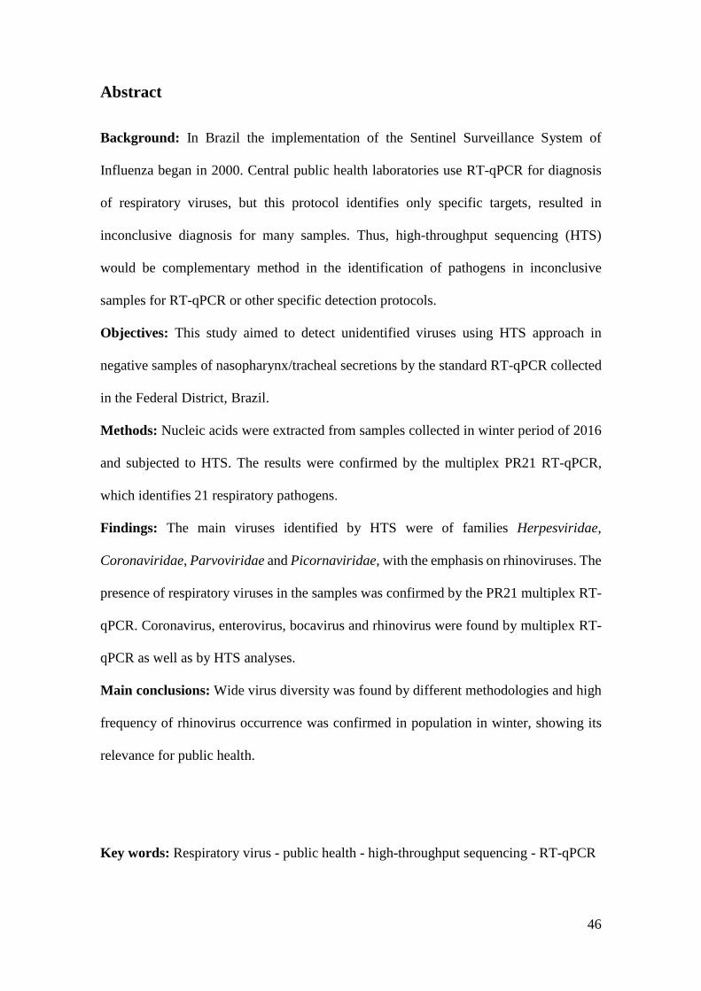
46
Abstract
Background: In Brazil the implementation of the Sentinel Surveillance System of
Influenza began in 2000. Central public health laboratories use RT-qPCR for diagnosis
of respiratory viruses, but this protocol identifies only specific targets, resulted in
inconclusive diagnosis for many samples. Thus, high-throughput sequencing (HTS)
would be complementary method in the identification of pathogens in inconclusive
samples for RT-qPCR or other specific detection protocols.
Objectives: This study aimed to detect unidentified viruses using HTS approach in
negative samples of nasopharynx/tracheal secretions by the standard RT-qPCR collected
in the Federal District, Brazil.
Methods: Nucleic acids were extracted from samples collected in winter period of 2016
and subjected to HTS. The results were confirmed by the multiplex PR21 RT-qPCR,
which identifies 21 respiratory pathogens.
Findings: The main viruses identified by HTS were of families Herpesviridae,
Coronaviridae, Parvoviridae and Picornaviridae, with the emphasis on rhinoviruses. The
presence of respiratory viruses in the samples was confirmed by the PR21 multiplex RT-
qPCR. Coronavirus, enterovirus, bocavirus and rhinovirus were found by multiplex RT-
qPCR as well as by HTS analyses.
Main conclusions: Wide virus diversity was found by different methodologies and high
frequency of rhinovirus occurrence was confirmed in population in winter, showing its
relevance for public health.
Key words: Respiratory virus - public health - high-throughput sequencing - RT-qPCR

47
Introduction
Worldwide influenza surveillance was initiated in 1947 to monitor circulating
viruses and provides information to support the vaccine-type recommendations by the
World Health Organization (Geneva, Switzerland) and the Centers for Disease Control
and Prevention (Atlanta, USA). Central laboratories (Lacens) in Brazil are responsible
for the occurrence reports about the etiologic agents, types and subtypes of circulating
respiratory viruses in patients with Influenza-like Syndrome (ILS) and Severe Acute
Respiratory Syndrome (SARS).(1)
The main technique for detecting respiratory viruses in Lacens is RT-qPCR
(reverse transcription - quantitative polymerase chain reaction), which advantages are
sensitivity, specificity, automation, relatively low cost and so on. However, this technique
as well as the conventional RT-PCR are pathogen-specific, which only find pre-defined
targets. Thus, a peculiar etiological agent or new genetic variants may not be detected,
despite the use of a wide range of sensitive diagnostic tests.(2)
In this context, high-throughput sequencing (HTS) technologies without requiring
any prior genomic information provide large volume of nucleotide sequences, resulting
in elucidation of unidentified viruses (which were not detectable by the specific detection
methods) and furthermore, allow the discovery of new viruses or new genomic variants
using metagenomic approaches.
The human population is exposed to increasing burden of infectious diseases
caused by the emergence of viruses not yet or rarely characterized. Globalization, climate
change, settlements near wild animal habitats and increasing numbers of
immunocompromised people are likely to contribute to the emergence and spread of new
infections.(3) HTS methods have allowed genomic analysis which is sensitive for

48
unknown or peculiar viruses over previous methodologies, such as the analysis of viral
genomes used in the identification or discovery of human herpesvirus 8(4), bocavirus(5),
human parvovirus 4(6), human GB virus(7), Torque Teno virus(8), WU polyomavirus(9) and
KI polyomavirus.(10)
HTS has been used, for example, in the discovery of an arenavirus in three patients
who died of a febrile illness a few weeks after transplant of solid organs from a single
donor.(11) The Illumina platform allowed identifying hepatitis A virus among cases of
acute febrile illnesses that occurred in Brazil which were suspected as yellow fever.(12)
The same platform was used to identify a new influenza virus from swabs samples and
assembly of its genome.(13) It also led to the detection of viral pathogens in
nasopharyngeal aspirate samples from patients with acute lower respiratory tract
infections, as a new enterovirus, termed enterovirus 109 (EV109) detected in a cohort of
Nicaraguan children with viral respiratory disease.(14)
Thereby, this study aimed to detect unidentified viruses by HTS approach in
negative samples of nasopharynx/tracheal secretions for diagnostics targeting several
viruses collected from June to August 2016 in the Federal District (DF), Brazil.
Materials and methods
Detection of standard respiratory viruses - The nasopharyngeal and tracheal
secretion samples received in the laboratory in June, July and August 2016 were tested
for the respiratory virus diagnostic panel performed in Lacen-DF (Public health
laboratory located in the Federal District, Brazil) by the standard RT-qPCR (for Influenza
A, Influenza B, Human respiratory syncytial virus, Human metapneumovirus, Human
adenovirus C, Parainfluenza 1, Parainfluenza 2 and Parainfluenza 3). Those that had

49
negative results were separated and stored in freezer -70º C for subsequent DNA/RNA
extraction for HTS.
RNA/DNA extraction - Fifty one samples from June, 55 from July and 39 from
August of 2016 were mixed, respectively (one for each month), and treated as three
pooled samples. Individual sample had a volume of approximately 0.5 mL (total of 20-
25 mL of each pooled sample). The volume was adjusted to 50 mL with 0.1 M sodium
phosphate buffer (pH7.2) with Triton-X 100 at the final concentration of 1%. The solution
in tubes were agitated for 1 h in a cold chamber, then, centrifuged at 4,800 × g for 40 min.
The supernatant was ultracentrifuged with 20% sucrose cushion at 140,000 × g for 1 h
using Ti45 rotor (Optima L-90K Ultracentrifuge, Beckman Coulter, Brea, USA). The
pellet was resuspended in 0.1 M sodium phosphate buffer and DNA/RNA was extracted
from the resuspended pellet following the instructions of the High Pure Viral Nucleic
Acid Kit (Hoffmann-La Roche, Basel, Switzerland), without addition of carrier RNA in
the lysis buffer. Three DNA/RNA samples were subjected to HTS. Next, part of all three
samples were treated with DNase I (Promega, Madison, USA) in order to decrease the
human or bacterial DNA in the sample for viral RNA enrichment. However, due to low
amount of RNA after DNase I treatment, all three samples were mixed as one pooled
sample for RNA HTS.
High-throughput sequencing - Approximately two μg of DNA/RNA and RNA
samples in RNAStable tube (Biomatrica, San Diego, USA) were sent for HTS to
Macrogen Inc. (Seoul, South Korea). The library of DNA/RNA or RNA samples was
constructed using TruSeq Standard total RNA kit and sequenced using Illumina HiSeq
2000 with 3 G scale for each in 100 base pared-end.

50
Bioinformatics analysis - Low quality and adapter sequences of the raw data were
trimmed using the Trimommatic tool v.036.(15) Metagenomic reads of each sample were
then uploaded to Kaiju for profiling the reads in taxonomic assessment
(http://kaiju.binf.ku.dk).(16) DNA reads were filtered with BWA v0.7.17(17) and SAMtools
v1.9(18) against reference hg38 (Homo sapiens) obtained from GenBank to remove human
DNA sequences from HTS dataset in silico. Trimmed reads were assembled into
contiguous sequences (‘contigs’) with Velvet v.1.2.1(19) and SPAdes v.3.9(20) programs.
The tBlastx search (https://blast.ncbi.nlm.nih.gov/ Blast.cgi) against the viral genome
package (RefSeq Virus, NCBI, https://www.ncbi.nlm.nih.gov/genome/viruses/) which is
implemented in the Geneious R8.1 program (Biomatters, Auckland, New Zealand) was
used to identify the contig sequences which were viral origins.
RT-qPCR using PR21 kit - A part of findings by HTS analysis was confirmed by
means of RT-qPCR. The RNA was extracted from original 145 samples using the Magna
Pure LC Total Nucleic Acid Kit (Hoffmann-La Roche, Basel, Switzerland), then a pool
of five samples were projected to the multiplex RT-qPCR using the XGen PR21 kit
(Biometrix, Curitiba, Brazil) for 21 respiratory pathogens (targeting Influenza A,
Influenza B, Influenza A H1N1-swl, Coronavirus NL63, Coronavirus 229E, Coronavirus
OC43, Coronavirus HKU1, Parainfluenza 1, Parainfluenza 2, Parainfluenza 3,
Parainfluenza 4 , Human metapneumovirus A and B, Human respiratory syncytial virus
A and B, Rhinovirus, Enterovirus, Parechovirus, Human adenovirus, Bocavirus and
Mycoplasma pneumoniae).
Ethics approval and consent to participate - This research project was reviewed
and approved by ethics committee of the Faculty of Health Sciences (University of
Brasília), approval letter No. 3.052.443 (in Appendix 1 of this thesis).
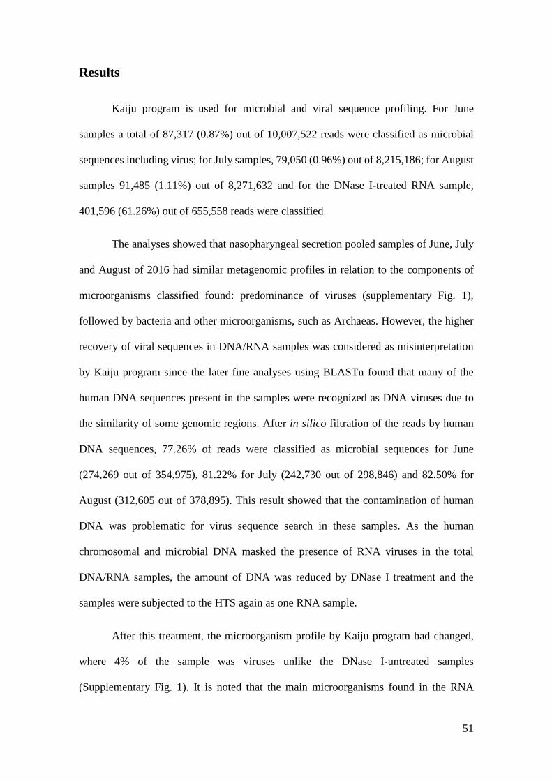
51
Results
Kaiju program is used for microbial and viral sequence profiling. For June
samples a total of 87,317 (0.87%) out of 10,007,522 reads were classified as microbial
sequences including virus; for July samples, 79,050 (0.96%) out of 8,215,186; for August
samples 91,485 (1.11%) out of 8,271,632 and for the DNase I-treated RNA sample,
401,596 (61.26%) out of 655,558 reads were classified.
The analyses showed that nasopharyngeal secretion pooled samples of June, July
and August of 2016 had similar metagenomic profiles in relation to the components of
microorganisms classified found: predominance of viruses (supplementary Fig. 1),
followed by bacteria and other microorganisms, such as Archaeas. However, the higher
recovery of viral sequences in DNA/RNA samples was considered as misinterpretation
by Kaiju program since the later fine analyses using BLASTn found that many of the
human DNA sequences present in the samples were recognized as DNA viruses due to
the similarity of some genomic regions. After in silico filtration of the reads by human
DNA sequences, 77.26% of reads were classified as microbial sequences for June
(274,269 out of 354,975), 81.22% for July (242,730 out of 298,846) and 82.50% for
August (312,605 out of 378,895). This result showed that the contamination of human
DNA was problematic for virus sequence search in these samples. As the human
chromosomal and microbial DNA masked the presence of RNA viruses in the total
DNA/RNA samples, the amount of DNA was reduced by DNase I treatment and the
samples were subjected to the HTS again as one RNA sample.
After this treatment, the microorganism profile by Kaiju program had changed,
where 4% of the sample was viruses unlike the DNase I-untreated samples
(Supplementary Fig. 1). It is noted that the main microorganisms found in the RNA
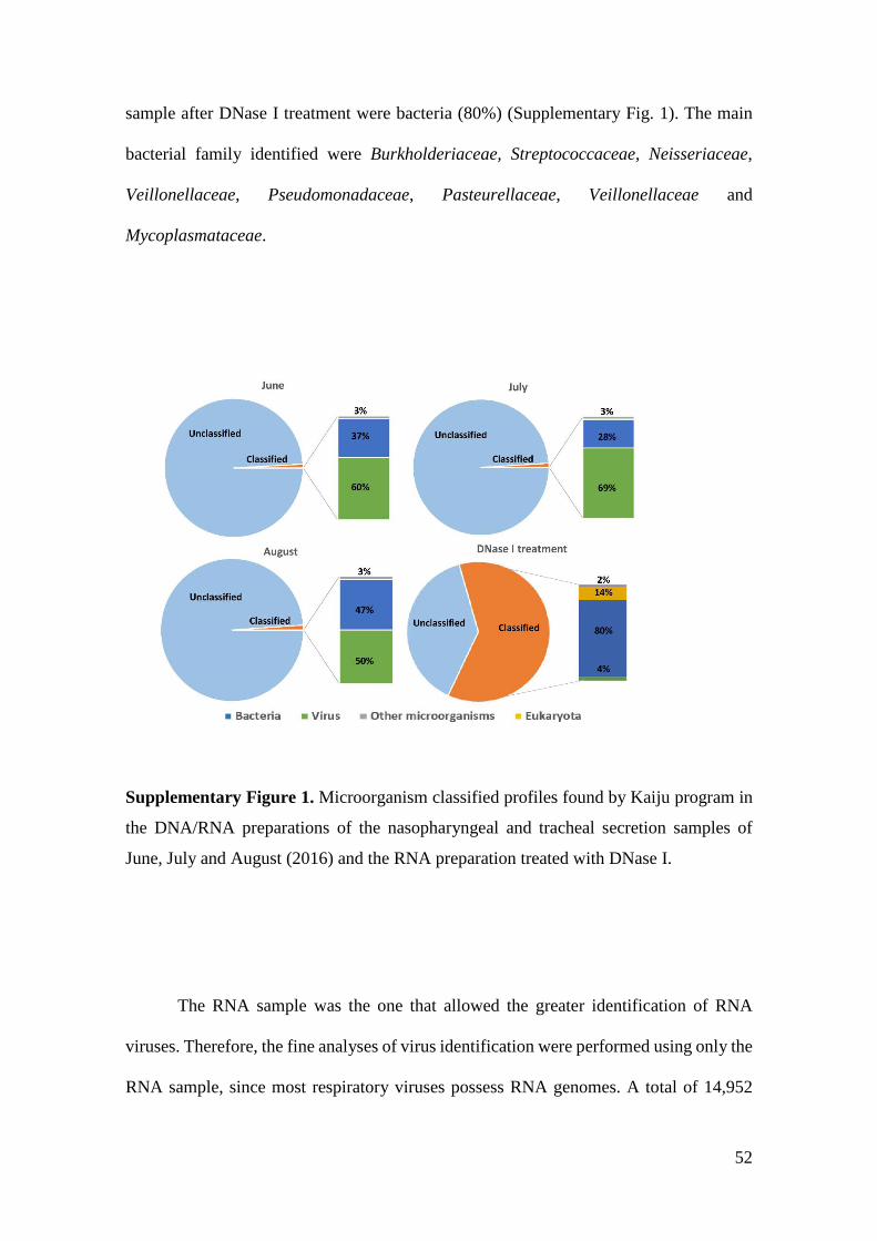
52
sample after DNase I treatment were bacteria (80%) (Supplementary Fig. 1). The main
bacterial family identified were Burkholderiaceae, Streptococcaceae, Neisseriaceae,
Veillonellaceae, Pseudomonadaceae, Pasteurellaceae, Veillonellaceae and
Mycoplasmataceae.
Supplementary Figure 1. Microorganism classified profiles found by Kaiju program in
the DNA/RNA preparations of the nasopharyngeal and tracheal secretion samples of
June, July and August (2016) and the RNA preparation treated with DNase I.
The RNA sample was the one that allowed the greater identification of RNA
viruses. Therefore, the fine analyses of virus identification were performed using only the
RNA sample, since most respiratory viruses possess RNA genomes. A total of 14,952
53
reads was classified as virus organisms. The main viruses identified using the Kaiju
program were of the families Picornaviridae, Herpesviridae, Parvoviridae and
Coronaviridae, with the emphasis on rhinoviruses (Table 1). The same viral families were
also found in the DNA/RNA sample reads after the removal of human DNA sequences
in silico, however, with less frequency. Only cytomegalovirus sequences were found
more in DNA/RNA samples than RNA sample (Table 1), probably due to the negative
effect on cytomegalovirus DNA genomes by DNaseI treatment in preparation of RNA
sample.
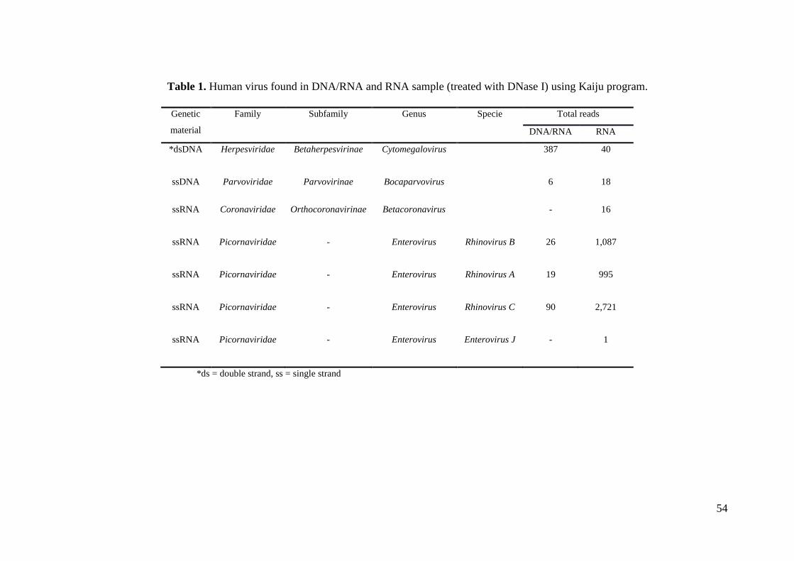
54
Table 1. Human virus found in DNA/RNA and RNA sample (treated with DNase I) using Kaiju program.
Genetic
material
Family Subfamily Genus Specie Total reads
DNA/RNA RNA
*dsDNA
Herpesviridae
Betaherpesvirinae Cytomegalovirus
387 40
ssDNA Parvoviridae Parvovirinae Bocaparvovirus 6 18
ssRNA Coronaviridae
Orthocoronavirinae
Betacoronavirus
- 16
ssRNA
Picornaviridae
- Enterovirus
Rhinovirus B 26 1,087
ssRNA
Picornaviridae
- Enterovirus
Rhinovirus A 19 995
ssRNA
Picornaviridae
- Enterovirus
Rhinovirus C 90 2,721
ssRNA Picornaviridae
- Enterovirus
Enterovirus J
- 1
*ds = double strand, ss = single strand

55
Due to low accuracy of the short-read profiling by Kaiju program, the
classification up to genus level was shown in this table, except for the Enterovirus genus,
which was the one with the most reads founded, allowing the virus species definition.
To identify the contig sequences (1,621 contigs assembled by Velvet and 334 by
SPAdes assemblers) of viral origin, tBlastx search plugged-in Geneious was used for the
known virus genome sequences recorded in the RefSeq Virus (NCBI).
The analyses identified virtually the same viral families/genera found by the Kaiju
program: Picornaviridae, Herpesviridae and Parvoviridae using contigs generated by
both SPAdes (Table 2) and Velvet assemblers (Table 3). Only the Coronaviridae family
was not identified by tBlastx in Geneious (but identified by Kaiju), probably due to
limited sensitivity of de novo contig assembly, although we chose the most sensitive ones,
Velvet and SPAdes.(21)
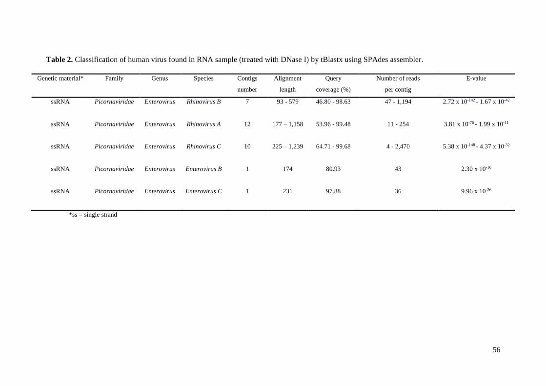
56
Table 2. Classification of human virus found in RNA sample (treated with DNase I) by tBlastx using SPAdes assembler.
Genetic material* Family Genus Species Contigs
number
Alignment
length
Query
coverage (%)
Number of reads
per contig
E-value
ssRNA
Picornaviridae
Enterovirus
Rhinovirus B 7 93 - 579 46.80 - 98.63 47 - 1,194 2.72 x 10-142 - 1.67 x 10-42
ssRNA
Picornaviridae
Enterovirus
Rhinovirus A
12 177 – 1,158 53.96 - 99.48 11 - 254 3.81 x 10-76 - 1.99 x 10⁻¹¹
ssRNA
Picornaviridae
Enterovirus
Rhinovirus C
10 225 – 1,239 64.71 - 99.68 4 - 2,470 5.38 x 10-148 - 4.37 x 10⁻³²
ssRNA Picornaviridae
Enterovirus
Enterovirus B
1 174 80.93 43 2.30 x 10⁻¹⁶
ssRNA Picornaviridae
Enterovirus
Enterovirus C
1 231 97.88 36 9.96 x 10⁻²⁶
*ss = single strand

57
Table 3. Classification of human virus found in RNA sample (treated with DNase I) by tBlastx using Velvet assembler.
Genetic
material*
Family Genus Species Contigs
number
Alignment
length
Query coverage
(%)
Number of reads
per contig
E-value
dsDNA
Herpesviridae
Cytomegalovirus
Human
betaherpesvirus 5
3 96 - 153 95.05 - 96.84 23 - 24 3.15 x 10⁻³² - 4.76 x 10⁻¹⁶
ssDNA
Parvoviridae
Bocaparvovirus
Primate
bocaparvovirus 1
4 96 - 99 95.05 - 98.02 8 - 13 2.15 x 10⁻¹⁸ - 1.34 x 10⁻¹⁶
ssRNA
Picornaviridae
Enterovirus
Rhinovirus B 29 93 - 570 63.00 - 100.00 3 - 668 2.01 x 10-117 - 3.20 x 10⁻¹¹
ssRNA
Picornaviridae
Enterovirus
Rhinovirus A
62 93 - 435 53.57 - 100.00 4 - 257 2.65 x 10-68 - 6.05 x 10⁻¹¹
ssRNA
Picornaviridae
Enterovirus
Rhinovirus C
54 87 - 885 64.15 - 100.00 11 - 962 1.23 x 10-152 - 7.86 x 10⁻¹¹
ssRNA Picornaviridae
Enterovirus
Enterovirus B
1 174 80.93 43 2.30 x 10⁻¹⁶
ssRNA Picornaviridae
Enterovirus
Enterovirus C
1 117 77.48 72 1.05 x 10⁻¹³
ssRNA Picornaviridae
Enterovirus
Enterovirus E
1 99 98.02 9 2.33 x 10⁻¹¹
*ss = single strand, ds = double strand

58
It is observed in Table 2 (contigs assembled by SPAdes) that the number of contigs
varied from 1 (Enterovirus B and C) to 12 (Rhinovirus A), with sizes of alignment
between 93 and 1,239 nucleotides. The number of reads per contig also varied greatly,
from 4 to 2,470 with the highest amount of reads per contig found in the Rhinovirus C
species. The e-values from 5.38 x 10-148 to 1.99 x 10⁻¹¹ indicates statistical significance in
the alignments found using the SPAdes as assembler.
Table 3 shows the tBlastx results of contigs assembled by Velvet. The number of
contigs varied between 1 (Enterovirus B, C and E) and 62 (Rhinovirus A) with sizes of
alignment between 87 and 885 nucleotides. The number of reads per contig ranged from
3 to 962, with the highest number of reads found also for Rhinovirus C. E-values from
1.23 x 10-152 to 2.33 x 10⁻¹¹ were also low, showing statistical significance of the
alignment. The comparison of Tables 2 and 3 reveals that, when using Velvet as
assembler, more contigs were formed, although with smaller length and fewer reads per
contig. But this is important to identify more viruses in less abundance, as Human
betaherpesvirus 5, Human bocavirus and Enterovirus E that have been identified using
Velvet, but not when using SPAdes as assembler.
The Rhinovirus sequences showed greater abundance by the presence of reads in
the sample, especially Rhinovirus C, that presented highest reads number (observed in
Kaiju analysis), and the highest reads number per contig (in tBlastx analysis). This shows
the relevance of this one virus in these negative samples by the standard RT-qPCR.
The presence of respiratory viruses in the samples was confirmed by the PR21
multiplex RT-qPCR kit, which identifies 21 respiratory pathogens. Coronavirus 63 (CT
= 30), coronavirus 43 (CT = 22-31), coronavirus HKU 1 (CT = 20-33), enterovirus (CT
= 33), bocavirus (CT = 19-35) and mainly rhinovirus (CT = 21-36) were found (Figure

59
1). The RT-qPCR assay showed the presence of coronaviruses which detection was failed
by the HTS analyses.
Figure 1. Respiratory pathogens found by PR21 multiplex RT-qPCR in nasopharyngeal
and tracheal secretion samples which were negative by the standard RT-qPCR of
influenza-like surveillance on June, July and August collected in Brasília, 2016.
Discussion
The HTS has been successfully applied in several fields of virology, including
virus discovery, complete virus genome sequence determination, and genomic variant
analyses.(22)
Lacen-DF performs diagnosis of the respiratory virus by the standard RT-qPCR
protocol with restricted targets, thus, many viruses may not be identified. The samples
referred to Lacen are from patients of public and private hospitals with respiratory virus

60
infection suspected, whether hospitalized or not. Approximately 60% of the samples
forwarded are negative for the virus detection.
In this study, Human betaherpesvirus 5, Primate bocaparvovirus 1,
Betacoronavirus, Enterovirus and three species of rhinovirus were found by at least 2
different types of analyses in samples of patients from the DF, Brazil, with flu symptoms.
Human betaherpesvirus 5 (Cytomegalovirus) was identified by analyses with Kaiju and
Geneious program, but not with PR21 RT-qPCR because specific primers for this
pathogen were not included in the kit. DNA viruses as herpesvirus and parvovirus were
also found in the sample treated with DNaseI. We assume that DNase I treatment doesn´t
exclude totally DNA in the sample, thus, herpesvirus and parvovirus reads were
recovered. Coronavirus was identified with the Kaiju program but not by the contig
analyses by tBlastx. However, its presence was confirmed by RT-qPCR of PR21 kit.
Bocavirus (or Primate bocaparvovirus 1), and Enterovirus was also identified by PR21
RT-qPCR, Kaiju and Geneious programs only using Velvet, which is more sensitive
method than SPAdes for contig assembly. Mycoplasma pneumoniae was found by PR21
RT-qPCR and many bacteria of the genus mycoplasma was also identified using the Kaiju
program. Rhinovirus was identified using all methods used is this study.
These variations in results are explained by the differences in specificity and
sensitivity in each method. For some cases, the limited sensitivity of HTS is explained by
mixing some clinical samples forming one pooled sample for the cost reduction.
Therefore, this sample preparation may mask the presence of viruses in low incidence or
concentration. The limited capacity of de novo assembly programs is still a main problem
in metagenomic analyses. Velvet and SPAdes de novo assemblers were used to build
contigs in this study since these were reported as sensitive contig assembler for viral
metagenomic study(21), although there was no best de novo assembler concluded by the
61
same authors. HTS was reported to be less sensitive than RT-qPCR for some respiratory
virus detection in such reasons.(23)
Rhinovirus was the major viruses found in patients in the DF, Brazil with ILS or
SARS samples which were negative by standard RT-qPCR of influenza-like surveillance
(targeting Influenza A and B, Respiratory Syncytial Virus, Human Metapneumovirus,
Adenovirus, Parainfluenza 1, 2 and 3). Several studies have shown the importance of
rhinoviruses in acute respiratory infection in Brazil, especially in children.(24, 25, 26) A
study involving 120 children less than 12 years old in São Paulo, Brazil, found that human
rhinoviruses (HRV) of species A, B, and C were the most frequent agents of acute
respiratory infections (ARI). Moreover, these agents are also associated with up to 70%
of virus-related wheezing exacerbations. The most recently identified Rhinovirus C has
been detected in association with bronchiolitis, wheezing, and asthma exacerbations
requiring hospitalization.(27)
Other study made with hospitalized patients at an academic care center in
Southern Brazil shows that HRV was usually detected in hospitalized children with
respiratory infections and was often present in viral co-detections, mainly with
enterovirus and respiratory syncytial virus. Comorbidities are closely associated with
HRV infections, and this virus predominates during colder seasons,(28) period of which
the samples used in this work were collected (winter). In investigations around the world,
HRV, as well as agents causing acute respiratory infections, has its peak activity
associated with the colder periods of the year.(29, 30)
In this study, the relevance of HRV in public health in Brazil was again recognized
and we suggest the inclusion of HRV for influenza-like surveillance.

62
References
1. Brazil. Ministry of Health. Secretariat of Health Surveillance. Department of Communicable
Disease Surveillance. Guide to the Influenza Surveillance Laboratory Network in Brazil
[homepage on the Internet] / Ministry of Health, Health Surveillance Secretariat,
Communicable Disease Surveillance Department. - Brasilia: Ministry of Health, 2016.
[updated 2019 Jan 22; cited 2019 May 9] Available from:
http://bvsms.saude.gov.br/bvs/publicacoes/guia_laboratorial_ influenza_vigilancia_
influenza_brasil.pdf
2. Juvén T, Mertsola J, Waris M, Leinonen M, Meurman O, Roivainen M, et al. Etiology of
community-acquired pneumonia in 254 hospitalized children. Pediatr Infect Dis. J. 2000;
19(4): 293– 8.
3. Morens DM, Folkers GK, Fauci AS. The challenge of emerging and re-emerging infectious
diseases. Nature. 2004; 430: 242–9.
4. Chang Y, Cesarman E, Pessin MS, Lee F, Culpepper J, Knowles DM, et al. Identification of
herpesvirus-like DNA sequences in AIDS-associated Kaposi’s sarcoma. Science. 1994;
266(5192): 1865–9.
5. Allander T, Tammi MT, Eriksson M, Bjerkner A, Tiveljung-Lindell A, Andersson B. Cloning
of a human parvovirus by molecular screening of respiratory tract samples. Proc Natl Acad
Sci. USA. 2005; 102(36): 12891–6.
6. Jones MS, Kapoor A, Lukashov VV, Simmonds P, Hecht F, Delwart E. New DNA viruses
identified in patients with acute viral infection syndrome. J Virol. 2005; 79(13): 8230–6.
7. Simons JN, Pilot-Matias TJ, Leary TP, Dawson GJ, Desai SM, Schlauder GG, et al.
Identification of two flaviviruslike genomes in the GB hepatitis agent. Proc Natl Acad Sci.
USA. 1995; 92(8): 3401–5.
8. Nishizawa T, Okamoto H, Konishi K, Yoshizawa H, Miyakawa Y, Mayumi M. A novel DNA
virus (TTV) associated with elevated transaminase levels in posttransfusion hepatitis of
unknown etiology. Biochem Biophys Res. Commun. 1997; 241(1): 92–7.
9. Gaynor AM, Nissen MD, Whiley DM, Mackay IM, Lambert SB, Wu G, et al. Identification of
a novel polyomavirus from patients with acute respiratory tract infections. PLoS Pathog.
2007; 3(5): e64.
10. Allander T, Andreasson K, Gupta S, Bjerkner A, Bogdanovic G, Persson MA, et al.
Identification of a third human polyomavirus. J Virol. 2007; 81(8): 4130–6.
11. Palacios G, Druce J, Du L, Tran T, Birch C, Briese T, et al. A new arenavirus in a cluster of
fatal transplant-associated diseases. N Engl J Med. 2008; 358: 991–8.
12. Conteville LC, de Filippis AMB, Nogueira RMR, de Mendonça MCL, Vicente ACP.
Metagenomic analysis reveals hepatitis a virus in suspected yellow fever cases in Brazil.
Mem. Inst. Oswaldo Cruz. 2018; 113(1): 66–7.
13. Yongfeng H, Fan Y, Jie D, Jian Y, Ting Z, Lilian S, et al. Direct pathogen detection from
swab samples using a new high-throughput sequencing technology. Clin Microbiol Infect.
2011; 17(2): 241–4.
14. Yozwiak NL, Skewes-Cox P, Gordon A, Saborio S, Kuan G, Balmaseda A, et al. Human
enterovirus 109: A novel interspecies recombinant enterovirus isolated from a case of acute
pediatric respiratory illness in Nicaragua. J Virol. 2010; 84(18): 9047–58.
15. Bolger AM, Lohse M, Usadel B. Trimmomatic: a flexible trimmer for Illumina sequence data.
Bioinformatics. 2014; 30(15): 2114–20.
16. Menzel P, Ng KL, Krogh A. Fast and sensitive taxonomic classification for metagenomics
with Kaiju. Nat. Commun. 2016; 7: 11257.
63
17. Li H, Durbin R. Fast and accurate short read alignment with Burrows-Wheeler transform.
Bioinformatics. 2009; 25(14): 1754-60.
18. Li H, Handsaker B, Wysoker A, Fennell T, Ruan J, Homer N, et al. The Sequence
Alignment/Map format and SAMtools. Bioinformatics. 2009; 25(16): 2078-9.
19. Zerbino DR, Birney E. Velvet: algorithms for de novo short read assembly using de Bruijn
graphs. Genome Res. 2008; 18: 821-9.
20. Bankevich A, Nurk S, Antipov D, Gurevich AA, Dvorkin M, Kulikov AS, et al. SPAdes: a
new genome assembly algorithm and its applications to single-cell sequencing. J Comput
Biol. 2012; 19(5): 455–77.
21. Blawid R, Silva JMF, Nagata T. Discovering and sequencing new plant viral genomes by
next-generation sequencing: description of a practical pipeline. Ann Appl Biol. 2017; 170(3):
301-14.
22. Capobianchi MR, Giombini E, Rozera G. Next-generation sequencing technology in clinical
virology. Clin Microbiol Infect. 2013; 19(1): 15–22.
23. Thorburn F, Bennett S, Modha S, Murdoch D, Gunson R, Murcia PR. The use of next
generation sequencing in the diagnosis and typing of respiratory infections. J Clin Virol.
2015; 69: 96-100.
24. Arruda E, Hayden FG, McAuliffe JF, Sota MA, Mota SB, McAuliffe MI, et al. Acute
respiratory viral infections in ambulatory children in urban northeast Brazil. J Infect Dis.
1991; 164(2): 1261–8.
25. Costa LF, Yokosawa J, Mantese OC, Oliveira TF, Silveira HL, Nepomuceno LL, et al.
Respiratory viruses in children younger than five years old with acute respiratory disease
from 2001 to 2004 in Uberlandia, MG, Brazil. Mem Inst Oswaldo Cruz. 2006; 101(3): 301–
6.
26. Fawkner-Corbett DW, Khoo SK, Duarte MC, Bezerra P, Bochkov YA, Gern JE, et al.
Rhinovirus-C detection in children presenting with acute respiratory infection to hospital in
Brazil. J Med Virol. 2015; 88(1): 58-63.
27. Moreira LP, Kamikawa J, Watanabe AS, Carraro E, Leal E, Arruda E, et al. Frequency of
human rhinovirus species in outpatient children with acute respiratory infections at primary
care level in Brazil. Pediatr Infect Dis J. 2011; 30(7): 612-4.
28. Leotte J, Trombetta H, Faggion HZ, Almeida BM, Nogueira MB, Vidal LR, et al. Impact and
seasonality of human rhinovirus infection in hospitalized patients for two consecutive years.
J Pediatr. (Rio J.) [Internet]. 2017; 93(3): 294-300.
29. Hung HM, Yang SL, Chen CJ, et al. Molecular epidemiology and clinical features of
rhinovirus infections among hospitalized patients in a medical center in Taiwan. J Microbiol
Immunol Infect. 2019; 52(2): 233-41.
30. Bellei N, Carraro E, Perosa A, Watanabe A, Arruda E, Granato C. Acute respiratory infections
and influenza-like illness viral etiologies in Brazilian adults. J Med Virol. 2008; 80(10):
1824-7.

64
Capítulo 3
3.1 Molecular and clinical characteristics related to rhinovirus
infection in Brasília, Brazil
Larissa da Costa Souza1, 2, Edson José Monteiro Bello2, Eliane Maria dos Santos2,
Tatsuya Nagata1
1Pós-graduação em Biologia Microbiana, Campus Darcy Ribeiro, Departamento de
Biologia Celular, Universidade de Brasília, Brasília-DF, 70910-900, Brazil
2Laboratório Central de Saúde Pública do Distrito Federal, DF, 70830-010, Brazil
A versão modificada deste capítulo foi publicada por Souza LC, Bello EJM, Santos EM, Nagata
T (2021) Molecular and clinical characteristics related to rhinovirus infection in Brasília, Brazil.
Braz J Microbiology, vol. 52.

65
Abstract
Human rhinovirus (HRV) is one of the most common human viral pathogens related to
infections of the upper and lower respiratory tract, which can result in bronchiolitis and
pneumonia. However, the relevance of HRV in human health was under-estimated for
long time due to the absence in targets for Influenza and Influenza-like syndrome
surveillance in Brasilia, Brazil. In this study, the clinical characteristics and outcomes of
HRV-positive patients, in comparison with patients without HRV and other common
respiratory viruses, were analyzed. For this purpose, new specific primer sets were
designed, in previous study, based on the high-throughput sequencing analysis and used
for HRV detection by RT-qPCR and Sanger sequencing of amplified cDNA of 5´
genomic region. The phylogeny analysis of representative HRV isolates was performed.
Statistical analysis considering the patient profiles was also performed using IBM SPSS
program with non-parametric tests. The most prevalent virus, in previously negative
samples for the Influenza and Influenza-like Syndrome surveillance, was rhinovirus (n =
40), including three rhinovirus species (rhinovirus A, B and C). The odds ratio associated
with HRV infection was 2.160 for patients younger than 2 years and 4.367 for people
living in rural areas. The main symptom associated with virus infection was rhinorrhea.
The multiple analysis showed association also for lower cases of HRV patients presenting
respiratory distress. In this study, it was reported the predominance of rhinoviruses in
cases of respiratory illness for negative patients for the Influenza and Influenza-like
Syndrome surveillance, being rhinorrhea, the most significant symptom associated with
the disease.
Key words: Rhinovirus – epidemiology - acute respiratory infections – symptoms
66
Introduction
Human rhinovirus (HRV) is one of the most common human viral pathogens.
They are non-enveloped single-stranded RNA virus, member of Enterovirus genus, of
Picornaviridae family [1, 2]. About half of common cold are caused by rhinoviruses,
which have more than 160 serotypes described, in three species (rhinovirus A, B and C)
[3]. In addition, cold leads to economic problems related clinical morbidity, overloading
the health system, also causing absences at work and in schools [2, 4].
Rhinoviruses cause not only upper respiratory tract infections, but also lower
respiratory tract infections, mainly in children and patients with chronic pulmonary
disease or immunocompromised [5]. The most common symptoms of upper infection are
sore throat, rhinorrhea, headache, cough, malaise and lower fever. However, in some
cases, there may be aggravation for rhinosinusitis and otitis media [6, 7]. Infections of the
lower respiratory tract are often associated with bronchiolitis and pneumonia especially
in children [8-10].
In Brazil, the implementation of the sentinel surveillance system began in 2000
[11], so referral hospitals have been conducting active surveillance to detect respiratory
viruses. Such surveillance includes notification and laboratory investigation of cases with
the diagnostic criteria of Influenza-like Syndrome (ILS) and Severe Acute Respiratory
Syndrome (SARS). This viral respiratory infection monitoring has resulted in important
information about the circulation of community-acquired respiratory viruses. However,
some viruses may not be identified by the absence of RT-qPCR target performed at
central laboratories.
The present study reports the molecular and clinical characteristics, and
outcomes of HRV infections in comparison with patients without HRV and other
common respiratory viruses.
67
Materials and Methods
Detection of HRV by RT-qPCR - The nasopharyngeal and tracheal secretion
samples received in the laboratory in June, July and August 2016 were tested for the
respiratory virus diagnostic panel performed in Lacen-DF (Central Laboratory at Federal
District, Brasilia, Brazil) by the standard RT-qPCR [12] (for influenza A virus, influenza
B virus, human orthopneumovirus, human metapneumovirus, human mastadenovirus C,
human respirovirus 1, human orthorubulavirus 2 and human respirovirus 3). The target
genes of the standard RT-qPCR are shown in the Supplementary table 1. During transport,
the samples were stored on ice and sent to the laboratory within 48 hours after collection.
Those that had negative results were separated and stored in freezer at -70 ºC for
subsequent DNA/RNA extraction. The metagenomic analyses by high throughput
sequencing (HTS) of those samples as one pooled virome was previous studied [13].
Based on this study, HRV universal primers targeting conserved regions in 5' UTR
(untranslated region) were newly designed for HRV detection (Table 1) using the
Geneious R8.1 program (Biomatters, Auckland, New Zealand). The RNA was extracted
from 145 samples using the Magna Pure LC Total Nucleic Acid Kit (Hoffmann-La
Roche, Basel, Switzerland), then all the 145 samples were projected to the RT-qPCR
using GoTaq® Probe 1-Step RT-qPCR System (Promega, Madison, USA). Positivity for
rhinovirus was defined with cycle threshold (CT) less than or equal to 40 (total of 45
cycles in the reaction). Human RNase P gene was used for endogenous control for the
same sample set in separate form.

68
Supplementary table 1 The target genes of the standard RT-qPCR performed in
Lacen-DF, Brasilia, Brazil
Name Gene target
Influenza A virus matrix gene (pos1) in segment 7
Influenza B virus segment 8 NS1/NEP
Human orthopneumovirus nucleocapsid protein (N) gene
Human metapneumovirus fusion glycoprotein (F) gene
Human mastadenovirus C hexon gene
Human respirovirus 1 hemagglutinin-neuraminidase (HN) mRNA
Human orthorubulavirus 2 hemagglutinin-neuraminidase (HN) mRNA
Human respirovirus 3 hemagglutinin-neuraminidase (HN) mRNA
Table 1 Rhinovirus primers and probe for RT-qPCR and amplicon sequencing
Name Sequence Use
HRV_Probe 5'- TCCGGCCCCTGAATGYGGCT - 3'(FAM/BHQ1)
RT-qPCR
HRV_For 5'- YCYAGCCTGCGTGGC - 3'
RT-qPCR
HRV_Rev 5'- ACACGGACACCCAAAGTAGT - 3'
RT-qPCR
HRV_Com_For 5'- ACTACTTTGGGTGTCCGTGT - 3
Amplicon sequencing
HRV_Com_Rev 5'-TCNGGHARYTTCCARCACC - 3
Amplicon sequencing
69
RT-PCR and Sanger sequencing - The HRV_Com_For and HRV_Com_Rev
primers were newly designed (Table 1) targeting conserved regions based on virome
analyses by HTS. This primer pair was used to amplify an approximately 540 bp fragment
for amplicon sequencing including part of 5' UTR and the VP4 / VP2 protein gene of
HRV A, B and C. cDNA of the selected samples were synthesized using MMLV
transcriptase (Thermo Fisher Scientific, Waltham, USA) with random hexamer primer
and, then, amplified with LongAmp Taq DNA Polymerase (New England BioLabs,
Ipswich, USA). The PCR product was purified and sequenced by the Sanger method at
Macrogen Inc (Seoul, South Korea).
Sequence analysis - The nucleotide (nt) sequences were determined using the
Geneious R8.1 program (Biomatters, Auckland, New Zealand). Complete rhinovirus
genome sequences were retrieved from Genbank as references and aligned with the
sequences obtained in this study. This alignment was again trimmed to set the sequence
size determined in this study (approximately 500 bases, the size has small variation due
to indel mutation). The phylogenetic tree based on the 5' UTR and the VP4 / VP2 protein
gene was constructed using the Mega X software [14], with eight nucleotide sequences
obtained in this study and other forty rhinovirus isolates that complete genome sequences
were available (for genotype identification). Phylogenetic tree was inferred using
maximum likelihood method with Hasegawa-Kishino-Yano model as indicated by
jModelTest analysis [15]. In order to obtain more precise nt identity in pairwise
comparison, three more related virus sequences for each obtained sequence in this study
were retrieved by BlastN search and the pairwise comparison was performed.
Statistical analysis – Medical records were obtained from compulsory
notification sheet for ILS and SARS sent with the sample to the laboratory. Data were
analyzed using the software IBM SPSS (Chicago, USA). Non-parametric tests were used
70
as appropriate, for qualitative and quantitative variables. All p-values were two-tailed and
a value of < 0.05 was considered significant.
Ethics approval and consent to participate - This research project was reviewed
and approved by ethics committee of the Faculty of Health Sciences (University of
Brasília), approval letter No. 3.052.443.
Results
Rhinoviruses were identified as the main virus found in samples that had
previously tested negative for the panel of respiratory virus survey used in Lacen-DF,
including influenza A virus, influenza B virus, human orthopneumovirus, human
metapneumovirus, human mastadenovirus C, human respirovirus 1, human
orthorubulavirus 2 and human respirovirus 3, in a previously conducted study (REF). The
RT-qPCR using newly designed primers and probe used in the present study (Table 1)
detected HRV in 40 out of 145 samples analyzed. The mean CT scan was about 28
ranging from 17 to 39. These primers targeted the 5' UTR region (Table 1), which is most
conserved among rhinoviruses.
To identify HRV at species level, amplicons obtained with primers targeting a
region of approximately 540 bp of the 5' UTR-VP4 / VP2 protein gene region were
sequenced. Five samples were concluded of HRV-A, one of HRV-B and two of HRV-C
(Fig. 1), out of eight sequenced samples of the patients in the Federal District during the
study period. The other samples were not of sufficient quality for sequencing. All
sequences were deposited in the databank: rhinovirus A BSB_8 (Accession number
LC549198), rhinovirus A BSB_16 (LC549199), rhinovirus A BSB_26 (LC549200),
rhinovirus A BSB_33 (LC549201), rhinovirus A BSB_38 (LC549202), rhinovirus B

71
BSB_30 (LC549203), rhinovirus C BSB_2 (LC549204), rhinovirus C BSB_15
(LC549205). It is interestingly that though small number of samples sequenced in this
study, the genetic variability of HRV-A was very wide, positioning in five different
clusters in the phylogenetic tree (Fig. 1). Among them, only nt identities of 72.94-83.86%
were observed (Table 2). On the other hand, two isolates of HRV-C were very similar,
possessing 98.99% of nt identity.
Figure 1 Phylogenetic tree of rhinoviruses based on 5' UTR and the VP4 / VP2 protein
gene (approximately 500 bases). The tree was construct using Hasegawa-Kishino-Yano
model of the maximum likelihood method using 48 isolates, which eight were from the
Federal District, Brazil and the other 40 were genome sequences available on databases.

72
Bootstrap values above 50 (500 repetitions), and accession number of each sequence are
indicated. Bar: number of substitutions per site. Green circle: Rhinovirus A, blue circle:
Rhinovirus B and red circle: Rhinovirus C. The identification of isolate from the Federal
District, Brazil are highlighted with colors used for species indication.
Table 2 Nucleotide identities (%) among HRV-A from Brasília, Brazil
HRV-A
BSB_38 BSB_8 BSB_26 BSB_33 BSB_16
BSB_38 78.44 77.54 75.35 72.94
BSB_8 78.44 76.08 75.80 75.64
BSB_26 77.54 76.08 74.78 75.43
BSB_33 75.35 75.80 74.78 83.86
BSB_16 72.94 75.64 75.43 83.86
By the identification of the country where the most related virus in the databases
was isolated, we can observe that there is almost no relation in the geographical
distribution (Table 3). The most related rhinovirus isolates came from several continents
such as America, Asia, Europe and Oceania. This observation suggests that the
dissemination of the rhinoviruses can happen worldwide in long distance and is different
from the arbovirus geographical dissemination.
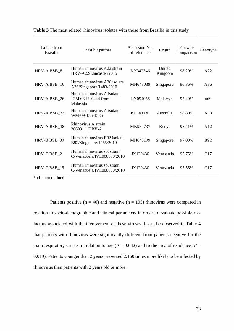
73
Table 3 The most related rhinovirus isolates with those from Brasília in this study
Isolate from
Brasília Best hit partner
Accession No.
of reference Origin
Pairwise
comparison Genotype
HRV-A BSB_8 Human rhinovirus A22 strain
HRV-A22/Lancaster/2015 KY342346
United
Kingdom 98.20% A22
HRV-A BSB_16 Human rhinovirus A36 isolate
A36/Singapore/1483/2010 MH648039 Singapore 96.36% A36
HRV-A BSB_26
Human rhinovirus A isolate
12MYKLU0444 from
Malaysia
KY094058 Malaysia 97.40% nd*
HRV-A BSB_33 Human rhinovirus A isolate
WM-09-156-1586 KF543936 Australia 98.80% A58
HRV-A BSB_38 Rhinovirus A strain
20693_1_HRV-A MK989737 Kenya 98.41% A12
HRV-B BSB_30 Human rhinovirus B92 isolate
B92/Singapore/1455/2010 MH648109 Singapore 97.00% B92
HRV-C BSB_2 Human rhinovirus sp. strain
C/Venezuela/IVE000070/2010 JX129430 Venezuela 95.75% C17
HRV-C BSB_15 Human rhinovirus sp. strain
C/Venezuela/IVE000070/2010 JX129430 Venezuela 95.55% C17
*nd = not defined.
Patients positive (n = 40) and negative (n = 105) rhinovirus were compared in
relation to socio-demographic and clinical parameters in order to evaluate possible risk
factors associated with the involvement of these viruses. It can be observed in Table 4
that patients with rhinovirus were significantly different from patients negative for the
main respiratory viruses in relation to age (P = 0.042) and to the area of residence (P =
0.019). Patients younger than 2 years presented 2.160 times more likely to be infected by
rhinovirus than patients with 2 years old or more.

74
Table 4 Socio-demographic and clinical variables of patients with acute respiratory
infection treated in the Federal District, Brazil, 2016
Rhinovirus
Yes No
n (%) n (%) P* Odds ratio (IC 95%)
Month 0.505 -
June 15 (37.5) 36 (34.3)
July 17 (42.5) 38 (36.2)
August 8 (20.0) 31 (29.5) Diagnostic criteria 0.304 1.478 (0.701 – 3.119)
ILS 17 (42.5) 35 (33.3)
SARS 23 (57.5) 70 (66.7)
Sex 0.231 0.635 (0.301 - 1.339)
Male 15 (37.5) 51 (48.6)
Female 25 (62.5) 54 (51.4)
Categorized age 0.042 2.160 (1.021 - 4.567)
< 2 years 19 (47.5) 31 (29.5)
≥ 2 years 21 (52.5) 74 (70.5)
Breed 0.075 -
White 20 (54.1) 30 (33.7)
Brown 14 (37.8) 53 (59.6)
Black 3 (8.1) 6 (6.7)
Zone 0.019 0.229 (0.061 - 0.860)
Urban 34 (85.0) 99 (96.1)
Rural 6 (15.0) 4 (3.9)
Risk factor 0.266 0.643 (0.294 - 1.404)
Yes 12 (30.0) 42 (40.0)
No 28 (70.0) 63 (60.0)
Vaccinated 0.625 1.222 (0.546 - 2.736)
Yes 12 (30.0) 27 (26.0)
No 28 (70.0) 77 (74.0)
Hospitalization 0.484 0.767 (0.365 - 1.612)
Yes 23 (57.5) 67 (63.8)
No 17 (42.5) 38 (36.2)
Medication 0.830 1.086 (0.510 - 2.313)
Oseltamivir 15 (37.5) 37 (35.6)
No 25 (62.5) 67 (64.4)
Use of ventilatory support 0.444 -
Yes, invasive 7 (17.5) 29 (27.6)
Yes, no invasive 11 (27.5) 24 (22.9)
No 22 (55.0) 52 (49.5)
* Pearson's chi-squared test

75
In this study, patients with rhinovirus were significantly younger than patients
without rhinovirus (P = 0.042) (Table 5). The median age for patients with the virus was
2.50 (interquartile range = 27.38) versus 26.00 (interquartile range = 50.13) years for
patients without this virus. Thus, greater susceptibility of children to rhinovirus infection
is reported. Figure 2 shows the difference between the groups with and without the virus
in relation to the days of symptoms and age. It is observed that the days of symptoms did
not differ significantly, but the age distribution of the patients was significantly lower in
patients who presented rhinovirus respiratory symptoms (P = 0.042).
Table 5 Quantitative variables of patients with acute respiratory infection treated in the
Federal District, Brazil, 2016
Rhinovirus
Yes No
Descriptive measures n (%) n (%) P*
Age n 40 105 0.042
Mean 18.24 29.08
Median 2.50 26.00
Standard deviation 22.55 27.23
Minimum 0.05 0.02
Maximum 76.00 94.00
Interquartile range 27.38 50.13
Days of symptoms n 39 105 0.736
Mean 5.36 5.38
Median 4.00 4.00
Standard deviation 4.56 5.25
Minimum 1.00 1.00
Maximum 19.00 41.00
Interquartile range 5.00 4.50
* Mann-Whitney test

76
Figure 2 Box-plot of days of symptoms (a) and age (b) of patients with acute respiratory
infection treated in the Federal District, Brazil, 2016. The comparison shows patients with
and without rhinovirus infection, where only the age difference was statistically
significant (P = 0.042). Extreme outliers are marked with an asterisk and moderate
outliers with a dot.
Patients with rhinovirus were located significantly more in rural areas when
compared to negative patients. The odds ratio for the rural area was 4.367 (1/0.229),
which means that people resident in rural areas were 4.367 times more affected by
rhinovirus when compared to urban dwellers.
Regarding the main symptoms of patients with acute respiratory infection, it can
be observed in Table 6 that patients with rhinovirus presented significantly more
rhinorrhea in relation to the patients without this virus detected (P = 0.001). Patients with
rhinovirus presented 3.981 times more cases of presenting rhinorrhea in relation to
negative patients. There was no statistically significant difference in relation to the other
symptoms.
b a

77
Table 6 Symptoms of patients with acute respiratory infection treated in the Federal
District, Brazil, 2016
Rhinovirus
Yes No
n (%) n (%) P* Odds ratio (IC 95%)
Fever 0.406 1.455 (0.599 - 3.532)
Yes 32 (80.0) 77 (73.3)
No 8 (20.0) 28 (26.7)
Cough 0.392 2.452 (0.524 - 11.479)
Yes 38 (95.0) 93 (88.6)
No 2 (5.0) 12 (11.4)
Sore throat 0.213 1.595 (0.763 - 3.334)
Yes 19 (47.5) 38 (36.2)
No 21 (52.5) 67 (63.8)
Dyspnea 0.157 0.585 (0.278 - 1.233)
Yes 22 (55.0) 71 (67.6)
No 18 (45.0) 34 (32.4)
Respiratory distress 0.133 0.567 (0.269 - 1.195)
Yes 15 (37.5) 54 (51.4)
No 25 (62.5) 51 (48.6)
Myalgia 1.000 1.000 (0.402 - 2.485)
Yes 8 (20.0) 21 (20.0)
No 32 (80.0) 84 (80.0)
O2 saturation < 95% 0.115 0.484 (0.194 - 1.209)
Yes 7 (17.5) 32 (30.5)
No 33 (82.5) 73 (69.5)
Rhinorrhea 0.001 3.981 (1.835 - 8.638)
Yes 27 (67.5) 36 (34.3)
No 13 (32.5) 69 (65.7)
* Pearson's chi-squared test
A multiple logistic regression with 20 explanatory variables (age, days of
symptoms, month, diagnostic criteria, sex, breed, zone, risk factor, vaccinated,
hospitalization, medication, use of ventilatory support, fever, cough, sore throat, dyspnea,
respiratory distress, myalgia, O2 saturation < 95% and rhinorrhea) was performed to
analyze the variables associated with rhinovirus infection (response variable). In the

78
model containing only the constant, there was 71% agreement with the classification
table. When entering the explanatory variables, the concordance increased to 79.8%.
The regression model was statistically significant (P = 0.005), with adequate
adjustments (P = 0.862 - Hosmer's test and Lemeshow) and R2 = 0.391. To improve the
model, a technique of variable selection was used by the backwise stepwise method. The
best fit was obtained after 15 steps of exclusion of variables, remaining six explanatory
variables. The new model was also statistically significant (P < 0.001), with optimal
adjustment (P = 0.923 - Hosmer and Lemeshow test) and R2 = 0.319. Three significant
explanatory variables were obtained: respiratory distress, rhinorrhea and medication
(Table 7).
Table 7 Logistic regression with variables selection of patients with acute respiratory
infection treated in the Federal District, Brazil, 2016
B S.E Wald D.F P Exp(B)
95% C.I. for exp(B)
Inferior Superior
Sex 0.89 0.48 3.41 1 0.065 2.446 0.946 6.326
Breed -0.67 0.41 2.67 1 0.102 0.513 0.231 1.143
Zone 1.41 0.88 2.60 1 0.107 4.114 0.738 22.931
Respiratory distress -1.09 0.56 3.86 1 0.049 0.335 0.113 0,997
Rhinorrhea 1.89 0.49 14.62 1 0.000 6.601 2.509 17.366
Medication 1.36 0.59 5.36 1 0.021 3.892 1.232 12.295
Constant -3.74 1.38 7.30 1 0.007 0.024
Abbreviations: B, [Beta coefficient]; S.E, [Standard Error]; Wald, [Wald test]; D.F, [Degree of
freedom]; P, [p-value]; Exp(B), [Beta exponential]; C.I., [Confidence interval]

79
Patients with rhinovirus presented 2.94 (1/0.34) times less chance of
experiencing respiratory distress, 6.60 and 3.89 times more chance of having rhinorrhea
and using medication (Oseltamivir), respectively. Thus, it is observed that the main
symptoms related to rhinovirus infection were the presence of rhinorrhea and absence of
respiratory discomfort. The use of antiviral medication, Oseltamivir, was also
significantly more observed in patients with rhinovirus in the multiple analysis.
To assess how respiratory symptoms were associated in patients with rhinovirus
infection, a cluster analysis was performed with the 40 positive patients, using
dendrogram with Ward method and measure of the quadratic Euclidean distance. It is
observed that two groups of symptoms were mainly formed: fever, cough and rhinorrhea
in the first group and myalgia, O2 saturation < 95%, respiratory distress, dyspnea and sore
throat in the second group) (Fig. 3). The symptoms of the first group may be associated
with cases of common cold (upper airways infection), and those of the second group, with
cases of complications of infection in the lower respiratory tract.

80
Figure 3 Cluster analysis of symptoms of rhinovirus patients treated in the Federal
District, Brazil, 2016. The analysis shows the division into two groups that suggest
infection in the upper (orange) and lower (blue) respiratory tract.
To investigate whether age was not a covariate of rhinorrhea, the Mantel-
Haenszel test was performed, which evaluated the association between rhinorrhea
symptom and age-corrected rhinovirus. It was observed that the association between
rhinorrhea and rhinovirus occurred in the group of patients older than 2 years (P = 0.001),
but not in the group younger than 2 years (P = 1,000) (Supplementary table 2), that is,
children with and without rhinovirus presented rhinorrhea in a similar way. The Mantel-
Haenszel test showed odds ratio = 3.865 (1.483 - 10.076; P = 0.005), which shows that
even if adjusted for age, rhinorrhea was statistically significant to the rhinovirus, where
rhinovirus patients had 3.865 times more chance of presenting this symptom compared
with patients without the virus.

81
Supplementary table 2 Association between the rhinorrhea symptom and the presence
of rhinovirus adjusted for age
Age Rhinovirus
Yes No Total P*
n (%) n (%) n (%)
< 2 years Rhinorrhea Yes 17 (89.5) 27 (87.1) 44 (88.0) 1.000 No 2 (10.5) 4 (12.9) 6 (12.0)
Total
19 (100.0) 31 (100.0) 50 (100.0)
> ou = 2 years Rhinorrhea Yes 10 (47.6) 9 (12.2) 19 (20.0) 0.001 No 11 (52.4) 65 (87.8) 76 (80.0)
Total
21 (100.0) 74 (100.0) 95 (100.0)
Total Rhinorrhea Yes 27 (67.5) 36 (34.3) 63 (43.4) 0.001 No 13 (32.5) 69 (65.7) 82 (56.6)
Total 40 (100.0) 105 (100.0) 145 (100.0)
* Pearson's chi-squared test
Discussion
Since the beginning of the monitoring of respiratory virus circulation in the
country, rhinovirus has been found with high frequency, either alone or co-detected with
other respiratory viruses, mainly in the south and southeast regions [16, 17]. To date,
however, the circulation of these viruses in the central region of the country has not been
well studied. Therefore, the identification of these viruses is crucial to determine the
etiologic agent of respiratory illness. In this case, the 5' UTR region is the most suitable
to identify the virus by RT-qPCR, and this region has already been used by other
laboratories with similar oligonucleotides [18, 19].
Rhinoviruses are reported to be the main cause of the common cold and spread
from person to person via direct and indirect contact, mainly by infected respiratory
secretions. The transmission by hands is important in this context [20]. The easy
82
contagion of rhinovirus can explain its rapid spread, over long distances, as shown in
Table 3.
HRV infections were associated with diseases such as asthma, chronic
pulmonary disease, bronchiolitis and pneumonia in children, the elderly and
immunocompromised. Thus, proper clinical management, timely diagnosis and
containment of outbreaks are becoming important [5, 16].
Rhinovirus infection was significantly associated with residency in rural areas.
In this study, 7% came from rural areas, and among those infected by the virus the
percentage was 15%. A study in Brazil has already reported that children from urban areas
of school age are more susceptible to respiratory inflammatory problems compared to
those in rural regions [21]. Thus, the negative cases for rhinovirus and other common
respiratory viruses may be mainly associated with allergic inflammatory processes, which
is more frequent in urban areas. With this, rural patients would present respiratory
problems related mainly to viral infections, as observed in relation to HRV. In the
multiple analysis, the relationship between rural residence and rhinovirus was not
observed.
Patients younger than 2 years were more subject to HRV infection and had a
significantly lower proportion of cases with fever, and more comorbidities such as asthma
than patients without HRV [16, 22, 23]. In this study, it was observed that children
younger than two years were more susceptible to rhinovirus infection, but there was no
association with comorbidities. The main symptom associated with HRV was rhinorrhea
rather than low frequency of fever. The HRV infections predominated during autumn and
winter with significant negative correlation between the number of HRV cases and the
average temperature [16]. So, the result obtained in this study, with rhinovirus dominance
in negative samples for the other common respiratory viruses, was expected since the

83
samples were collected in late fall and winter (June, July and August), when the virus
circulation is high.
The data here presented showed that patients with rhinovirus were significantly
younger than patients without rhinovirus, consistent with other works that showed the
detection rate of HRV infection in children was significantly higher than that in adults,
mainly the HRV-C. Clinical manifestations associated with HRV-C appear to be more
severe in children [24] and often associated with respiratory complications, like acute
wheezing illness and bronchiolitis [25, 26]. In adults HRV-A is more common and is
associated with cases of viral pneumonia [27].
Fever and nasal congestion were more frequently observed in pediatric group
than the adults which were most diagnosed with severe respiratory disease entity [28].
Figure 3 presented two groups of symptoms. This division may be associated with the
main affected area, upper or lower respiratory tract. Rhinorrhea was the clinical
manifestation significantly associated with HRV infection in this study. Other studies also
show rhinorrhea as the main symptom associated with rhinovirus infection [29, 30].
The use of oseltamivir was significantly associated with HRV infection in the
multiple analysis (Table 7). In univariate analysis, this association was not verified (Table
4). However, a high percentage (37.5%) of patients with rhinoviruses who took the
medication incorrectly were observed. Although rhinovirus and influenza symptoms can
be similar and therefore confounded [31], the use of oseltamivir in patients with HRV has
already been reported, but in a much smaller percentage (2.1 - 2.5%) [22, 31].
84
Conclusion
This study showed the predominance of rhinoviruses in cases of respiratory
illness in clinical samples from negative patients by the Influenza and Influenza-like
Syndrome surveillance. Rhinorrhea was the most significant symptom associated with
the disease. All three virus species were identified in the Federal District, Brazil. A lower
chance of presenting respiratory distress was also observed in the multiple analysis,
although the symptoms of patients with HRV were associated with both upper airways
infection and complications of infection in the lower respiratory tract (Fig. 3). In the
patients with HRV, 55.0 and 37.5% presented exacerbations such as dyspnea and
respiratory distress and 57.5% had to be hospitalized (Tables 4 and 6). As HRV is also
identified in asymptomatic patients [32, 33], more detailed studies need to be made to
better understand the prevalence, risk factors, and outcomes related to rhinovirus
positivity in the population.
85
References
1. Palmenberg AC, Spiro D, Kuzmickas R et al (2009) Sequencing and analyses of all known
human rhinovirus genomes reveal structure and evolution. Science 324: 55-59.
2. Gern JE (2010) The ABCs of rhinoviruses, wheezing, and asthma. J Virol 84:7418-7426.
3. Palmenberg AC, Rathe JA, Liggett SB (2010) Analysis of the complete genome sequences
of human rhinovirus. J Allergy Clin Immunol 125:1190-1201.
4. Fendrick AM, Monto AS, Nightengale B, Sarnes M (2003) The economic burden of non-
influenza-related viral respiratory tract infection in the United States. Arch Intern Med
163:487–494.
5. Jacobs SE, Lamson DM, George KSt, Walsh TJ (2013) Human rhinoviruses. Clin Microbiol
Rev 26:135-162.
6. Blomqvist S, Roivainen M, Puhakka T et al (2002) Virological and serological analysis of
rhinovirus infections during the first two years of life in a cohort of children. J Med Virol
66:263–268.
7. Turner BW, Cail WS, Hendley JO et al (1992) Physiologic abnormalities in the paranasal
sinuses during experimental rhinovirus colds. J Allergy Clin Immunol 90:474–478.
8. Henquell C, Mirand A, Deusebis A-L et al (2012) Prospective genotyping of human
rhinoviruses in children and adults during the winter of 2009-2010. J Clin Virol 53:280–284.
9. Lieberman D, Shimoni A, Shemer-Avni Y et al (2010) Respiratory viruses in adults with
community-acquired pneumonia. Chest 138:811-816.
10. Broberg E, Niemelä J, Lahti E et al (2011) Human rhinovirus C-associated severe
pneumonia in a neonate. J Clin Virol 51:79–82.
11. Barros FR Daufenbach LZ, Vicente MG et al (2004) O desafio da influenza: epidemiologia
e organização da vigilância no Brasil. Boletim Eletrônico Epidemiológico da Secretaria de
Vigilância em Saúde, [S.l.], v. 1, p. 1-7. Available in: <http://portalsaude.saude.gov.br/
images/pdf/2014/julho/16/Ano04-n01-desafio-influenza-br-completo.pdf>.
12. World Health Organization. CDC protocol of realtime RTPCR for influenza A (H1N1)
(2009). Available in http://www.who.int/csr/resources/publications/
swineflu/realtimeptpcr/en/index.html.
13. Souza LC, Blawid R, Silva JMF, Nagata T (2019) Human virome in nasopharynx and
tracheal secretion samples. Mem Inst Oswaldo Cruz 114, e190198.
14. Kumar S, Stecher G, Li M et al (2018) MEGA X: Molecular Evolutionary Genetics Analysis
across computing platforms. Mol Biol Evol 35:1547-1549.
15. Posada D (2008) jModelTest: Phylogenetic model averaging. Mol Biol Evol 25:1253–1256.
16. Leotte J, Trombetta H, Faggion HZ et al (2017) Impact and seasonality of human rhinovirus
infection in hospitalized patients for two consecutive years. J Pediatr 93:294–300.
17. Watanabe ASA, Carraro E, Candeias JMG et al (2011) Viral etiology among the elderly
presenting acute respiratory infection during the influenza season. Revista da Sociedade
Brasileira de Medicina Tropical 44:18-21.
18. Reijans M, Dingemans G, Klaassen CH et al (2008) RespiFinder: a new multiparameter test
to differentially identify fifteen respiratory viruses. J Clin Microbiol 46:1232–1240.
19. Jaramillo-Gutierrez G, Benschop KS, Claas EC et al (2013) September through October
2010 multi-centre study in the Netherlands examining laboratory ability to detect enterovirus
68, an emerging respiratory pathogen. J Virol Methods 190:53-62.
20. Gwaltney Jr JM, Moskalski PB, Hendley JO (1978) Hand-to-hand transmission of rhinovirus
colds. Ann Intern Med 88:463-467.

86
21. Galvão CE, Saldiva PH, Kalil Filho JE, Castro FF (2004) Inflammatory mediators in nasal
lavage among school-age children from urban and rural areas in São Paulo, Brazil. São Paulo
Med J 122:204-207.
22. Tam PI, Zhang L, Cohen Z (2018) Clinical characteristics and outcomes of human rhinovirus
positivity in hospitalized children. Ann Thorac Med 13:230-236.
23. Bizzintino J, Lee WM, Laing IA et al (2011) Association between human rhinovirus C and
severity of acute asthma in children. Eur Respir J 37:1037‑ 1042.
24. Lee WM, Lemanske RFJr, Evans MD et al (2012) Human rhinovirus species and season of
infection determine illness severity. Am J Respir Crit Care Med 186: 886–891.
25. Tapparel C, Junier T, Gerlach D et al (2009) New respiratory enterovirus and recombinant
rhinoviruses among circulating picornaviruses. Emerg Infect Dis 15:719–726.
26. Wisdom A, Kutkowska AE, McWilliam Leitch EC et al (2009) Genetics, recombination and
clinical features of human rhinovirus species C (HRV-C) infections; interactions of HRV-C
with other respiratory viruses. PLoS ONE 4, e8518.
27. Linder JE, Kraft DC, Mohamed Y et al (2013) Human rhinovirus C: age, season, and lower
respiratory illness over the past 3 decades. J Allergy Clin Immunol 131:69-77.
28. Hung HM, Yang SL, Chen CJ et al (2019) Molecular epidemiology and clinical features of
rhinovirus infections among hospitalized patients in a medical center in Taiwan. Journal of
microbiology, immunology, and infection 52:233-241.
29. Lethbridge R, Prastanti F, Robertson C et al (2018) Prospective assessment of Rhinovirus
symptoms and species recurrence in children with and without an acute wheezing
exacerbation. Viral Immunol 31:299-305.
30. Annamalay AA, Khoo SK, Jacoby P et al (2012) Prevalence of and risk factors for human
rhinovirus infection in healthy aboriginal and non-aboriginal Western Australian children.
Pediatr Infect Dis J 31:673–679.
31. Boivin G, Osterhaus AD, Gaudreau A et al (2002) Role of picornaviruses in flu-like illnesses
of adults enrolled in an oseltamivir treatment study who had no evidence of influenza virus
infection. J Clin Microbiol 40:330-334.
32. Iwane MK, Prill MM, Lu X et al (2011) Human rhinovirus species associated with
hospitalizations for acute respiratory illness in young US children. J Infect Dis
204:1702‑ 1710.
33. Byington CL, Ampofo K, Stockmann C et al (2015) Community surveillance of respiratory
viruses among families in the Utah Better Identification of Germs-Longitudinal Viral
Epidemiology (BIG-LoVE) Study. Clin Infect Dis 61:1217–1224.

87
Capítulo 4
4.1 Rhinovirus incidence in association with climate factors in Brasília,
Brazil
Larissa da Costa Souza1, 2, Eliane Maria dos Santos2, Maria Goreth2, Tatsuya
Nagata1
1Pós-graduação em Biologia Microbiana, Campus Darcy Ribeiro, Departamento de
Biologia Celular, Universidade de Brasília, Brasília-DF, 70910-900, Brazil
2Laboratório Central de Saúde Pública do Distrito Federal, DF, 70830-010, Brazil
O manuscrito deste capítulo está em preparação.

88
Abstract
Background: Human rhinovirus (HRV) is the most common pathogen of acute
respiratory infections. Evaluating the circulation of the virus throughout the months may
be useful to delineate the circulation patter of the agent and; therefore, previewing
etiological agents for the respiratory virus surveillance network (RVSN).
Objective: The present study reports rhinovirus occurrence in relation to certain periods,
as well as the definition improvement of an etiological agent for RVSN in Brasília, Brazil.
Methods: RT-qPCR was used to monitor the presence of respiratory viruses, including
rhinoviruses from samples of patients with Influenza-like Syndrome (ILS) or Severe
Acute Respiratory Syndrome (SARS). Sample profiles were obtained in order to correlate
with the results of the detection of the evaluated viruses. The correlation of the incidence
of rhinovirus with meteorological data was performed using non-parametric tests in the
IBM SPSS software.
Results: The addition of rhinovirus-specific primers in the respiratory virus panel
significantly increased the identification of a virus that is associated with respiratory
symptoms. The prevalence of rhinovirus (in relation to the other viruses) showed a
significant correlation with the minimum temperatures recorded, in other words,
increasing detection of rhinovirus is proportional to the decrease in the minimum
temperatures recorded in Brasília, Brazil.
Conclusion: The incidence of rhinovirus was correlated with the decrease in minimum
temperature, but without an evident seasonal pattern for the population of Brasília in the
studied period.
Key words: Rhinovirus – seasonality – RT-qPCR – respiratory virus surveillance

89
Introduction
The worldwide circulation of respiratory viruses is monitored through the global
influenza surveillance since 1947. This network is also responsible for controlling
possible outbreaks and epidemics, besides providing information to support the World
Health Organization (WHO) and the Centers for Disease Control and Prevention (CDC)
decide the type of vaccine recommendations. Central laboratories (Lacens) in Brazil are
responsible for reporting the occurrence of etiologic agents, types and subtypes of
circulating respiratory viruses in patients with Influenza-like Syndrome (ILS) and Severe
Acute Respiratory Syndrome (SARS) (1).
Viral detection in acute respiratory infections (ARI) depends on several factors,
such as sensitivity of the diagnostic methods, adequate patient selection influenced by the
symptoms and severity of the disease, in addition to the climate (2).
Studies in Brazil have shown a higher incidence of respiratory viruses in
moderately cold and dry months in regions with semitropical climate, in rainy seasons in
tropical regions and during the colder months in subtropical areas (3-6).
Most respiratory viral infections happen in winter, but they are also common in
other seasons. Environmental factors influence human airways physiology, immunity,
virus stability and transmission rates (7). As a result, environmental changes are closely
related to the incidence and frequency of infectious diseases.
Among the viruses that are associated with respiratory diseases, rhinoviruses
(HRV) stand out for their high frequency, being the main etiological agent of "common
colds". In most cases, it presents as a mild and self-limited disease (8), but it can also
cause bronchiolitis and pneumonia especially in children (9). Thus, knowing the
seasonality of infections by this virus could provide insights to determine the etiologic
agent of the disease.

90
The present study reports the seasonality of rhinovirus infection, as well as the
definition improvement of an etiological agent by RT-qPCR for the respiratory virus
surveillance network in Brasília, Brazil.
Materials and Methods
Detection of HRV by RT-qPCR - The nasopharyngeal and tracheal secretion
samples received in the laboratory since June 2016 were tested for the respiratory virus
diagnostic panel performed in Lacen-DF (the Federal District, Brasilia, Brazil) by the
standard RT-qPCR (for Influenza A virus, Influenza B virus, Human orthopneumovirus,
Human metapneumovirus, Human mastadenovirus C, Human respirovirus 1, Human
orthorubulavirus 2 and Human respirovirus 3). Those samples that were negative, and
were collected between June and August 2016 were separated and stored in freezer at -
70 ºC for subsequent DNA/RNA extraction. The metagenomic analyses of those samples
as one pooled virome was previous studied (10). Based on this study, rhinovirus was
confirmed as the most prevalent etiologic agent in samples with negative result; therefore,
HRV universal primers were designed for HRV detection (11).
Detection of standard respiratory viruses - The nasopharyngeal and tracheal
secretion samples received in the laboratory from August 2019
were tested for the respiratory virus diagnostic panel performed in Lacen-DF
(Public health laboratory located in the Federal District, Brazil) by the standard RT-qPCR
plus Rhinovirs, after drawing universal primers.
Meteorological data – Brasília is the federal capital of Brazil and the seat of
government of the Federal District. The capital is located in the Midwest region of the
country, along the geographical region known as Planalto Central. It is located at 15°

91
50'16 "south latitude and 47°42'48" west longitude, with altitudes between 1,000 and
1,200 meters above sea level. Brasilia's climate is tropical with a dry and rainy season,
with average monthly temperatures always above 18°C and annual rainfall of
approximately 1,480 mm (mm), concentrated between the months from October to April.
During the dry season, which starts in May and ends in September, the relative humidity
can be below 30%. Information on minimum and maximum temperatures and relative
humidities was obtained through daily consultations to the official website of Inmet
(National institute of meteorology) (12).
Statistical analysis – Data on the results of rhinovirus and other respiratory
viruses were computed from August 2019 to February 2020. Data from previous three
years were used to compare the positive definition of the etiologic agent when performing
the viral panel. From March 2020, it was not possible to continue the evaluation due to
the Coronavirus pandemic (SARS-CoV-2) that caused social isolation and alteration of
all natural seasonality of other respiratory viruses, in addition to changing the criteria for
receiving a sample for diagnosis. Data were analyzed using the software IBM SPSS
(Chicago, USA). Spearman's non-parametric correlation was used to assess the
relationship between meteorological data and the incidence of rhinovirus in the studied
period. All p-values were two-tailed and a value of < 0.05 was considered significant.
Ethics approval and consent to participate - This research project was reviewed
and approved by ethics committee of the Faculty of Health Sciences (University of
Brasília), approval letter No. 3.052.443.

92
Results
Samples from patients with Influenza-like Syndrome and Severe Acute
Respiratory Syndrome are received by the Central Public Health Laboratory to assess a
possible viral etiologic agent.
Data from August to February of 2016/2017, 2017/2018, 2018/2019 and
2019/2020 were used for comparison of diagnosis using the specific primers for the
respiratory viruses panel including rhinovirus.
All positive results (for any respiratory virus) were divided by the number of
samples from patients with suspected respiratory viral infection assessed on a monthly
basis. Table 1 shows that there was a significant increase in the percentage of positive
cases, that is, with the definition of an etiological agent from the insertion of rhinovirus
in the panel of respiratory viruses. For the statistical analysis in question, the Kruskal-
Wallis test was used, given the small number of data (n = 7 months) per group (evaluated
period). The increase in the percentage of virus identification from the inclusion of
rhinovirus can be better identified in figure 1.
Table 1 Comparison of the proportion of positive results of respiratory viruses in four
periods. In the last period, the detection of rhinovirus in the viral panel was included,
Brasília, Brazil.
Proportion of positive results
Period Samples Median Interquartile range P*
Aug/16 – Feb/17 362 0,368 0,085 0,009
Aug/17 – Feb/18 358 0,407 0,114
Aug/18 – Feb/19 686 0,380 0,244
Aug/19 – Feb/20 1297 0,587 0,185
*Kruskal-Wallis test

93
Figure 1 Box-plot of proportions of positive results of respiratory viruses in four periods
of time. In the last period, the detection of rhinovirus in the viral panel was included,
Brasília, Brazil.
Of the 1297 samples received from August 2019 to February 2020, 730 (56.28%)
were positive for, at least, one of respiratory viruses. Among the total respiratory viruses
identified, 37.26% were rhinovirus, 13.97% human respirovirus 3, 10.96% Influenza B
virus, 10.82% Influenza A virus H1N1, 10.41% human metapneumovirus, 4.79% human
mastadenovirus C, 3.97% non-subtipable Influenza A virus, 3.42% human
orthopneumovirus, 2.74% Influenza A virus H3, 1.23% human respirovirus 1 and 0.41%
were human orthorubulavirus 2. Coinfections were present in 62 samples (8.49% of
positive samples), whose rhinovirus were the main viruses in co-detection (56.45%).
Minimum and maximum temperature and relative humidity data were obtained
daily and the correlation of weekly averages and rhinovirus incidence was evaluated in
relation to the other viruses identified in the corresponding week. Climatic data were

94
obtained from the week 31 of 2019 to the week 12 of 2020, totalizing 34 epidemiological
weeks.
For most parameters, the null hypothesis of normality of data was rejected by the
Kolmogorov-Smirnov test. Then, Spearman's Rho non-parametric correlation test was
chosen. There was statistically significant correlation between the ratio of rhinovirus /
other viruses and minimum temperature (table 2). The correlation coefficient was
negative, which indicates that the increase in the identification of rhinovirus in relation to
the other respiratory viruses grows with the decrease in the minimum temperature. There
was also a moderate negative correlation with the maximum temperature, although not
statistically significant. There was no correlation between the humidity (maximum and
minimum) and the detection of rhinovirus in relation to the other investigated viruses.
Table 2 Correlation between rhinovirus positivity in relation to other respiratory viruses
and relative air temperature and humidity. August 2019 to February 2020, Brasília, Brazil.
Rhinovirus / other viruses
Spearman's Rho
Minimum
temperature (°C)
Correlation Coefficient -0,420
P 0,013
n 34
Maximum
temperature (°C)
Correlation Coefficient -0,329
P 0,057
n 34
Minimum
humidity (%)
Correlation Coefficient 0,057
P 0,751
n 34
Maximum
humidity (%)
Correlation Coefficient 0,070
P 0,696
n 34

95
Figure 2 shows more clearly the downward trend in the rhinovirus / other viruses
ratio with the higher minimum temperature in the studied period. There is also a tendency
to the decrease of detection of rhinovirus with the increase in maximum temperature,
although not significant. The detection of rhinovirus in relation to other viruses is not
influenced by humidity.
Figure 2 Dot plot with trend curve for the correlation between the rhinovirus / other
viruses ratio and the minimum (a) and maximum (b) temperatures, in addition to the
minimum (c) and maximum humidity (d). August 2019 to February 2020, Brasília, Brazil.

96
The seasonality of the circulation of rhinovirus and other respiratory viruses was
assessed using the ratio of the total from the week 31 (2019) to week 26 (2020), with total
of 48 weeks. As of week 13, it is already possible to observe the effects of social isolation
(occurred in week 11) on the circulation of common respiratory viruses (excluding SARS-
CoV-2) in Fig. 3. There was no clear pattern of positivity for respiratory viruses and
rhinovirus in the population of Brasília in the period studied prior to the social isolation
decreed in the region.
Figure 3 Weekly distribution of the ratio between the detection of rhinovirus or all respiratory
viruses in relation to the total of tests carried out from August 2019 to June 2020. Brasília,
Brazil.
0,000
0,250
0,500
0,750
1,000
31 33 35 37 39 41 43 45 47 49 51 1 3 5 7 9 11 13 15 17 19 21 23 25
Rati
o
Epidemiological week
Seasonality
Rhinovirus / total Respiratory viruses / total

97
Discussion
Since the beginning of the surveillance of respiratory viruses in the country, the
improvement in the agility and sensitivity of detection of viral etiologic agents has been
occurring in the Central Public Health Laboratories. Understanding seasonality patterns
and climatic associations can help to suppose the target detection, which generates early
diagnosis and consequently, agile surveillance action and clinical management associated
with patient risk factors.
The significant increase in the percentage of positive cases, that motivated the
inclusion of rhinovirus in the panel of respiratory viruses, will allow greater efficiency
and agility in the definition of possible outbreaks and diagnosis for adequate treatment of
patients with respiratory symptoms. More than a third of the viruses identified in the
analyzed period were rhinoviruses, which shows the relevance of these viruses,
previously unidentified, in the population of Brasília. Other studies have also shown a
higher prevalence of rhinovirus as an etiologic agent of acute respiratory infection (13,
14).
Low air temperature and drier air are associated with an increased risk of viral
infections (15-18). Some studies have shown that the decrease and dryness of the mucosa
of the upper airways can increase the host's susceptibility to infections (19, 20).
Studies with Influenza viruses have shown that the decrease in temperature, as
well as the decrease in absolute air humidity, appears to be important determinants of
infectiousness and act as a trigger for flu seasons (21, 22). In southeastern Brazil, there
was a significant trend towards an increase in viral respiratory infections as the
temperature and relative humidity decrease (23).
In a study carried out in the south of the country, rhinovirus detection was more
prevalent during the colder seasons (autumn and winter), with a moderate negative
98
correlation between the frequency of viral infection and temperature, but with no
correlation with precipitation (13). The risk of rhinovirus infection was associated with a
decrease in humidity and temperature during the three days prior to infection, showing a
relationship between short-term variation in weather factors and HRV infections (24).
Studies on seasonality of influenza in Brazil have different patterns of viral
circulation; in the Northeast region, influenza circulates in the first four months of the
year, coinciding with the period of the highest humidity (25) unlike that found in other
regions of the country. These different circulation profiles due to the climatic conditions
of each region can impact the vaccine's effectiveness.
In our study, no seasonality was observed in the incidence of rhinovirus during
the period from August 2019 to February 2020. This period coincides with the end of
winter, spring and summer. During the fall and winter of 2020, social isolation decreased
the circulation of non-SARS-CoV-2 respiratory viruses, which did not allow assessing
whether in the country's capital this same pattern of increased incidence in cold seasons
would be observed. In order to establish the seasonality of HRV infections more
accurately, the analysis should include additional years, after pandemic control.
However, in this period it was already possible to verify a moderate negative
association (ρ = -0.420, p = 0,013) with minimum temperatures and no correlation with
humidity, which may lead to consider a probable association with the coldest seasons of
the year.
The seasonality of rhinoviruses can also be associated with the species of
circulating virus. One study showed that strains of HRV-A were identified throughout the
year, the HRV-C strains appeared mainly in winter and HRV-B were found in low
quantities, but mainly in the summer (26).

99
Conclusion
We confirmed an improvement in the detection of the etiologic agent for RVSN
by including rhinovirus in the diagnosis. The incidence of rhinovirus was correlated with
the decrease in minimum temperature, but without an evident seasonal pattern for the
population of Brasília in the studied period.
Understanding the seasonal incidence of temperature-related rhinovirus infection
can help with strategies to prevent and minimize transmission and provide the health
network with more information for the clinical treatment of patients.

100
References
1. Brazil. Ministry of Health. Secretariat of Health Surveillance. Department of Communicable
Disease Surveillance. Guide to the Influenza Surveillance Laboratory Network in Brazil
[homepage on the Internet] / Ministry of Health, Health Surveillance Secretariat,
Communicable Disease Surveillance Department. - Brasilia: Ministry of Health, 2016.
[updated 2019 Jan 22; cited 2019 May 9] Available from:
http://bvsms.saude.gov.br/bvs/publicacoes/guia_laboratorial_influenza_vigilancia_influenza
_ brasil.pdf
2. Straliotto SM, Siqueira MM, Machado V, Maia TMR (2004) Respiratory viruses in the
pediatric intensive care unit: prevalence and clinical aspects. Mem Inst Oswaldo Cruz 99: 883-
7.
3. Costa LF, Yokosawa J, Mantese OC, Oliveira TFM, Silveira HL, Nepomuceno LL, Moreira
LS, Dyonisio G, Rossi LMG, Oliveira RC, Ribeiro LZG, Queiroz DAO (2006) Respiratory
viruses in children young than five years old with acute respiratory disease from 2001 to 2004
in Uberlândia, MG, Brazil. Mem Inst Oswaldo Cruz 101: 301-6.
4. Moura FEA, Borges LC, Portes SAR, Ramos EAG, Siqueira MM (2003) Respiratory syncytial
virus infections during an epidemic period in Salvador, Brazil. Viral antigenic group analysis
and description of clinical and epidemiological aspects. Mem Inst Oswaldo Cruz 98: 739-743
5. Vieira SE, Stewien KE, Queiróz DAO, Durigon EL, Torok TJ, Anderson LJ, Miyao CR, Hein
N, Botosso VF, Pahl MM, Gilio AE, Ejzenberg B, Okay Y (2001) Clinical patterns and
seasonal trends in respiratory syncytial virus hospitalizations in São Paulo, Brazil. Rev Inst
Med Trop São Paulo 43: 125-131.
6. Straliotto SM, Nestor SM, Siqueira MM (2001) Respiratory syncytial virus groups A and B in
Porto Alegre, Brazil, from 1990 to 1995 and 1998. Mem Inst Oswaldo Cruz 96: 155-158.
7. Moriyama M, Hugentobler WJ, Iwasaki A (2020) Seasonality of Respiratory Viral Infections
Annual Review of Virology 7 (1): 83-101.
8. Gern, J.E (2010) The ABCs of rhinoviruses, wheezing, and asthma. J Virol 84: 7418–7426.
9. van Piggelen RO, van Loon AM, Krediet TG, Verboon-Maciolek MA (2010) Human
rhinovirus causes severe infection in preterm infants. Pediatr. Infect. Dis. J. 29:364 –365.
10. Souza LC, Blawid R, Silva JMF, Nagata T (2019) Human virome in nasopharynx and tracheal
secretion samples. Mem Inst Oswaldo Cruz 114, e190198.
11. Souza LC, Bello EJM, Santos EM, Nagata T (2021) Molecular and clinical characteristics
related to rhinovirus infection in Brasília, Brazil. Braz J Microbiol 6: 1–10.
12. Brazil. Ministry of Agriculture, Livestock and Supply. National Institute of Meteorology.
Consulted between August 2019 and March 2020. Available from: https://portal.inmet.gov.br/
13. Leotte J, Trombetta H, Faggion HZ, Almeida BM, Nogueira MB, Vidal LR, et al (2017)
Impact and seasonality of human rhinovirus infection in hospitalized patients for two
consecutive years. J Pediatr 93: 294–300.
14. Kim JK, Jeon JS, Kim JW, Rheem IS (2013) Epidemiology of respiratory viral infection using
multiplex rt-PCR in Cheonan, Korea (2006–2010) J Microbiol Biotechnol 23: 267-273.
15. Shaman J, Kohn M (2009) Absolute humidity modulates influenza survival transmission and
seasonality. Proc Natl Acad Sci USA 106: 3243–3248.
16. Lowen AC, Steel J (2014) Roles of humidity and temperature in shaping influenza seasonality.
J Virol 88: 7692–7695.

101
17. Mäkinen TM, Juvonen R, Jokelainen J, Harju TH, Peitso A, Bloigu A, Silvennoinen-Kassinen
S, Leinonen M, Hassi J (2009) Cold temperature and low humidity are associated with
increased occurrence of respiratory tract infections. Respir Med 103: 456–462.
18. Jaakkola K, Saukkoriipi A, Jokelainen J, Juvonen R, Kauppila J, Vainio O, Ziegler T, Rönkkö
E, Jaakkola JJ, Ikäheimo TM, et al (2014) Decline in temperature and humidity increases the
occurrence of influenza in cold climate. Environ Health 13: 13–22.
19. Liener K, Leiacker R, Lindemann J, Rettinger G, Keck T (2003) Nasal mucosal temperature
after exposure to cold, dry air and hot, humid air. Acta Otolaryngol 123: 851–856.
20. Cruz AA, Naclerio RM, Proud D, Toqias A (2006) Epithelial shedding is associated with
reactions to cold, dry air. J Allergy Clin Immunol 117: 1351–1358.
21. Lowen AC, Mubareka S, Steel J, Palese P (2007) Influenza virus transmission is dependent
on relative humidity and temperature. PLoS Pathog 3: 1470–1476.
22. Shaman J, Pitzer VE, Viboud C, Grenfell BT, Lipsitch M (2010) Absolute humidity and the
seasonal onset of influenza in the continental United States. PLoS Biol 8: e1000316.
23. Gardinassi LG, Marques Simas PV, Salomão JB, Durigon EL, Trevisan DMZ, Cordeiro JA,
Lacerda MN, Rahal P, de Souza FP (2012) Seasonality of viral respiratory infections in
southeast of Brazil: The influence of temperature and air humidity. Braz J Microbiol 43: 98–
108.
24. Ikäheimo TM, Jaakkola K, Jokelainen J, Saukkoriipi A, Roivainen M, Juvonen R, Vainio O,
Jaakkola JJ (2016) A Decrease in Temperature and Humidity Precedes Human Rhinovirus
Infections in a Cold Climate. Viruses 8 (9): 244.
25. Alonso WJ, Viboud C, Simonsen L, Hirano EW, Daufenbach LZ, Miller MA (2007)
Seasonality of influenza in Brazil: a traveling wave from the Amazon to the subtropics. Am J
Epidemiol 165 (12): 1434-42.
26. Savolainen-Kopra C, Blomqvist S, Kaijalainen S, Jounio U, Juvonen R, Peitso A, Saukkoriipi
A, Vainio O, Hovi T, Roivainen M (2009) All known human rhinovirus species are present in
sputum specimens of military recruits during respiratory infection. Viruses 1: 1178–1189.

102
Conclusão geral
Amostras clínicas negativas para o painel de vírus respiratórios do sistema de vigilância,
por RT-qPCR, foram avaliadas por meio de sequenciamento de alto desempenho (HTS) a
fim de identificar possíveis vírus em secreções da nasofaringe / aspirado traqueal de
pacientes do Distrito Federal que apresentavam sintomas respiratórios.
Vírus pertencentes às famílias virais Herpesviridae, Coronaviridae, Parvoviridae
e Picornaviridae, foram identificados com maior frequência, com destaque para os
rinovírus. Houve variação nos resultados encontrados por diferentes metodologias de
identificação, explicada pela diferença de especificidade e sensibilidade em cada método.
Entretanto o sequenciamento genético foi capaz de identificar grande diversidade de vírus
e alta frequência na ocorrência dos rinovírus, mostrando a relevância desses agentes
etiológicos para a saúde pública do Distrito Federal.
Após identificação do rinovírus como o agente viral mais prevalente nas referidas
amostras, foram desenhados primers em região consenso e primers na região VP2/VP4
para identificação de espécies. Grande parte das amostras analisadas foram positivas para
rinovírus, sendo que todas as três espécies (rinovírus A, B e C) foram identificadas. A
infecção por HRV foi associada à idade menor de 2 anos e à moradia em área rural, sendo
a rinorreia o principal sintoma associado. A análise múltipla mostrou também menor
chance de pacientes com HRV apresentarem desconforto respiratório em comparação aos
pacientes com sintomas respiratórios por outros agentes.
A incorporação de primers específicos para rinovírus no painel de vírus
respiratório aumentou significativamente a identificação do agente etiológico que causa
infecção respiratória. Em relação à distribuição do vírus ao longo do ano, foi observada
103
correlação significativa com as temperaturas mínimas, onde o aumento da detecção de
rinovírus foi proporcional à diminuição das temperaturas registradas em Brasília, Brasil.
Estudos tem mostrado predominância de rinovírus no inverno, período de coleta
das amostras utilizadas nesse trabalho. Porém, durante o outono e inverno de 2020, o
isolamento social diminuiu a circulação de vírus respiratórios não SARS-CoV-2, o que
não permitiu avaliar esse padrão de aumento da incidência nas estações frias. De forma
geral, não foi observado um padrão sazonal evidente de rinovírus, mas há uma correlação
da incidência com a queda da temperatura.
Compreender os agentes virais que acometem a população, identificar sintomas
associados e a incidência sazonal desses vírus pode ajudar em estratégias para prevenir e
minimizar a transmissão, além de fornecer à rede de saúde mais informações para o
adequado manejo clínico e terapêutico das infecções respiratórias.
104
Apêndices
6.1 Apêndice 1 - Parecer do Comitê de Ética

105

106
107

108
109

110
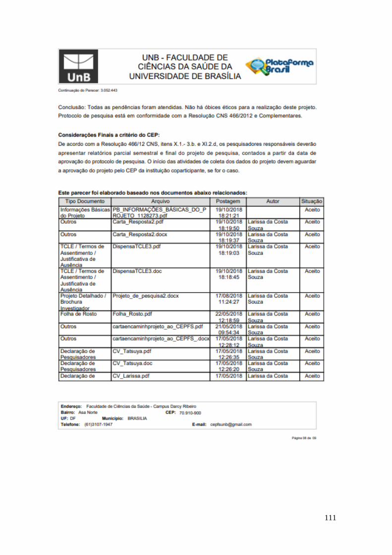
111

112

113
6.2 Apêndice 2 - Página de rosto da publicação 1
114
6.3 Apêndice 3 – Página de rosto da publicação 2
Recommended