Amélia Catarina Fernandes Soares Vieira
Synthesis of diclofenac-cyclodextrin conjugates for colon delivery
Tese de Doutoramento em Farmácia, na Especialidade de Tecnologia Farmacêutica, orientada pelo Professor Doutor Francisco José Veiga e pelo Professor Doutor Abdul W. Basit e apresentada à Faculdade de Farmácia da Universidade de Coimbra.
2014
Synthesis of diclofenac-cyclodextrin conjugates for colon
delivery
Amélia Catarina Fernandes Soares Vieira
Tese de doutoramento em Farmácia, na especialidade de Tecnologia Farmacêutica, orientada
pelo Professor Doutor Francisco J. Veiga e pelo Professor Doutor Abdul W. Basit e apresentada
à Faculdade de Farmácia da Universidade de Coimbra
2014
Faculdade de Farmácia
Universidade de Coimbra


The work presented in this thesis has been carried out under the supervision of
Professor Francisco José Baptista Veiga from the Faculty of Pharmacy,
University of Coimbra and Professor Abdul W. Basit from UCL School of
Pharmacy, University College London.

The presented work was developed in:
Faculty of Pharmacy, University of Coimbra under the supervision of
Professor Francisco José Baptista Veiga,
Faculty of Science and Technology, University of Coimbra under the supervision of Professor
António Rocha Gonsalves and Professor Arménio Serra
and
UCL School of Pharmacy, University College London under the supervision of
Abdul W. Basit.

The work presented in this thesis was supported by Fundação para a Ciência e Tecnologia,
Portugal (SFRH/BD/44925/2008) through the POPH/FSE (QREN) program.


To my parents
Alice e Saul

ACKNOWLEDGEMENTS
I would like to express my sincere gratitude to my supervisor Professor Francisco Veiga at the
Faculty of Pharmacy, University of Coimbra for the motivation to pursue a research career,
giving me the opportunity to join his research group and, finally, for teaching me how to grow
up in the science world.
To my supervisor Professor Abdul Basit, from the UCL School of Pharmacy, University
College London, I would like to thank for welcoming me as part of his research group for
more than two years. I am deeply grateful for his scientific support, enormous encouragement,
confidence and his best motivation words to carry on with this project, even when physically
distant.
From the department of Chemistry, University of Coimbra I would like to acknowledge
Professor Antonio Rocha Gonsalves and Professor Arménio Serra for welcoming me as part
of their research team for around two years. I want to thank them for the scientific support,
attention, time, advises and for the trust they set on me and on this project.
I would like to express my very great appreciation to Doctor Sudaxshina Murdan from UCL
School of Pharmacy, University College London, for her kindness, support, valuable and
constructive suggestions during the planning and development of the studies with animals.
To Professor Rui Albuquerque de Carvalho from department of Life Sciences, Faculty of
Science and Technology, University of Coimbra and Center for Neuroscience and Cell
Biology, I am greatfull for his scientific knowledge and for have his support on the resonance
magnetic nuclear analysis. To Doctor Alexandra Albuquerque Gonsalves from
Chymiotechnon and Department of Chemistry, Faculty of Science and Technology,
University of Coimbra I would like to thank for all her support with the HPLC and mass
spectra analyses.
From UCL School of Pharmacy, University College London I am thankfull to Doctor Stephen
Hilton and all his team, namely Bruno Santos, for allowing me to use his research laboratory
to follow up chemistry synthesis. Also, I am thankfull to Steve Coppard, his team and Inês
Pereira for all the support during the animal work.
I would like to acknowledge Fundação para a Ciência e Tecnologia, Portugal for the financial
support (SFRH / BD / 44925 / 2008), which allowed me to complete the work presented in
this thesis.
Now, to my colleagues and friends at University of Coimbra, I want to leave my
aknowledgements. From the Faculty of Pharmacy, I am thankfull to Rita Figueiras, Susana
Simões, Carla Vitorino, Camille, Sérgio Silva, Raquel, Ivo, Ana Santos, Dulce, Filipa,
Sandra, Isa, Ana Fortuna, Joana, Ana Serralheiro, Daniela and D. Gina for all the knowledge
and experience shared, help, encouragement and all enjoyable moments. From the department
of Chemistry, I would like to thank Pedro Cardoso, Marina Pires, Filipe Reis, Carla Barreto,
Célia Frias, Nelson Pereira, Sónia Ribeiro and Sílvia Gramacho for all their help and good
moments of friendship. I also thank my flatmates, Ana Rufino and Cláudia Rodrigues, for
their friendship and support.
At UCL School of Pharmacy, I thank everyone from the group of Abdul: Felipe Varum,
Cristina Freire, Veronika, Francisco, Hyunhong Min, Rin, Vipul, Alvaro Goyanes, Jie, Alice,
Sarit, Mustafa, Hamid and Grace for the support, motivation and friendship. Also at UCL I
am deeply greatful to Georgina, Blanka, Bahven and Cris for their help, friendship and for
carrying everyday in a good mood. Thanks also to Roberta, Rehema, and Sheiliza for sharing
with me some of the best moments in London. A special acknowledgment to Sara Laserra for
her confidence and unwavering friendship. I am deeply grateful to have met Teresa Barata
who provided me the most support in the end of this research project, thanks for her
friendship, generosity and advices. An enormous thanks to Fátima Pina for always be my
“best adviser”, I am very greatfull for her friendship. Thanks to Carla Varela for all her
friendship and support, specially for all her help with the reading of my thesis.
Thank you to all of my friends, some of them already mentioned before, that accompanied me
during this Chapter of my life. I can not mention all of them, but they know who they are. I
just want to leave a big thank you to Mila, who gave me the best impulse to proceed with this
project when I most needed. A special thanks to my godson Tomás, my best example of
patience and positive thinking.
Thanks to my Parents, Alice e Saul, brother Rui and sister Fernanda, for their unconditional
love and support. Without them this project would not have been possible. Thanks for being
always by my side.
TABLE OF CONTENTS
Publications……………………………………………………………………………………i
Abtract………………………………………………………………………………………..iii
Resumo………………………………………………………………………………….....…vii
List of Figures…………………………………………………………………………….….xi
List of Tables………………………………………………………………………..……...xvii
List of Abreviations…………………………………………………………………........…xix
1 GENERAL INTRODUCTION ....................................................................................... 3
1.1 Overview .................................................................................................................... 3
1.2 The gastrointestinal tract: main considerations on the design of a colonic prodrug ................................................................................................................................. 5
Stomach ............................................................................................................... 7 1.2.1
Small intestine ..................................................................................................... 8 1.2.2
Large intestine ..................................................................................................... 9 1.2.3
Colon as a site for drug delivery ........................................................................ 13 1.2.4
1.3 Systems to colonic target delivery of drugs to the colon ..................................... 17
Prodrugs as a strategy for site colonic delivery of drugs ................................... 20 1.3.1
Cyclodextrins ..................................................................................................... 23 1.3.2
Cyclodextrins in oral drug delivery ................................................................... 27 1.3.3
1.4 In vivo evaluation .................................................................................................... 34
1.5 Project aims ............................................................................................................. 39
2 SYNTHESIS AND CHEMICAL CHARACTERIZATION OF DICLOFENAC-CYCLODEXTRIN CONJUGATES .................................................................................... 43
2.1 Introduction ............................................................................................................. 43
Cyclodextrin chemistry ..................................................................................... 43 2.1.1
Diclofenac chemistry ......................................................................................... 45 2.1.2
Design of diclofenac-cyclodextrin ester conjugate ........................................... 46 2.1.3
2.2 Aims and Objectives ............................................................................................... 50
2.3 Materials .................................................................................................................. 50
Chemicals ........................................................................................................... 50 2.3.1
2.4 Methods .................................................................................................................... 51
Synthesis of diclofenac-β-cyclodextrin conjugate. ............................................ 51 2.4.1
Applicability of the nucleophilic microwave approach on the synthesis of 2.4.2
diclofenac-γ-cyclodextrin and diclofenac-α-cyclodextrin ................................................ 53
Characterization ................................................................................................. 55 2.4.3
Chemical stability of diclofenac-β-cyclodextrin in aqueous buffer solutions ... 56 2.4.4
2.5 Results and discussion ............................................................................................. 57
Synthesis and characterization of diclofenac-β-cyclodextrin conjugate ............ 57 2.5.1
Applicability of the nucleophilic microwave approach on the synthesis of 2.5.2
diclofenac-γ-cyclodextrin and diclofenac-α-cyclodextrin ................................................ 74
2.6 Conclusions .............................................................................................................. 82
3 STABILITY OF DICLOFENAC-ΒETA-CYCLODEXTRIN IN THE GASTROINTESTINAL TRACT. IN VITRO AND EX VIVO STUDIES USING SIMULATED FLUIDS AND GASTROINTESTINAL FLUIDS FROM ANIMALS. .... 87
3.1 Overview................................................................................................................... 87
3.2 Introduction ............................................................................................................. 88
Simulated gastrointestinal fluids ........................................................................ 89 3.2.1
Animal gastrointestinal fluids ............................................................................ 93 3.2.2
3.3 Aims and Objectives ................................................................................................ 94
3.4 Materials................................................................................................................... 95
Reagents and chemicals ..................................................................................... 95 3.4.1
Enzymes ............................................................................................................. 95 3.4.2
Animals .............................................................................................................. 96 3.4.3
Instrumentation and chromatographic conditions .............................................. 96 3.4.4
3.5 Methods .................................................................................................................... 97
Stability of the diclofenac-β-cyclodextrin conjugate in simulated fluids .......... 97 3.5.1
Stability of the diclofenac-β-cyclodextrin conjugate in animal fluids ............. 102 3.5.2
3.6 Results and discussion ........................................................................................... 104
HPLC method development ............................................................................. 104 3.6.1
Stability of diclofenac-β-cyclodextrin conjugate in simulated fluids .............. 105 3.6.2
Stability of diclofenac-β-cyclodextrin conjugate in animal fluids ................... 116 3.6.3
3.7 Conclusion .............................................................................................................. 123
4 INFLUENCE OF FOOD ON GUT FLUIDS AND METABOLISM OF DICLOFENAC-ΒETA-CYCLODEXTRIN VERSUS SULFASALAZINE IN RATS .. 127
4.1 Overview ................................................................................................................ 127
4.2 Introduction ........................................................................................................... 127
4.3 Aims and Objectives ............................................................................................. 130
4.4 Materials and methods ......................................................................................... 131
Reagents and chemicals ................................................................................... 131 4.4.1
Animals ............................................................................................................ 131 4.4.2
Feeding regimens ............................................................................................. 131 4.4.3
Determination of pH and mass of the gastrointestinal luminal contents ......... 132 4.4.4
Determination of stability of prodrugs (diclofenac-β-cyclodextrin and 4.4.5
sulfasalazine), in caecal and colonic contents ................................................................ 133
HPLC analysis ................................................................................................. 134 4.4.6
Data analysis .................................................................................................... 134 4.4.7
4.5 Results and discussion .......................................................................................... 135
Influence of feeding regimens on body weight of the rats, mass of faeces and 4.5.1
volume of urine .............................................................................................................. 135
Influence of feeding regimens on the mass of gastrointestinal contents. ........ 139 4.5.2
In situ pH of the gastrointestinal contents ....................................................... 142 4.5.3
Evaluation of the stability of diclofenac-β-cyclodextrin and sulfasalazine in 4.5.4
caecum and colon contents. ............................................................................................ 146
4.6 Conclusion ............................................................................................................. 149
5 DICLOFENAC-ΒETA-CYCLODEXTRIN VERSUS SULFASALAZINE FOR COLONIC DRUG TARGETING: IN VIVO PERFORMANCE IN RATS ................... 153
5.1 Overview ................................................................................................................ 153
5.2 Introduction ........................................................................................................... 154
5.3 Aims and Objectives ............................................................................................. 155
5.4 Materials and methods ......................................................................................... 155
Reagents and chemicals ................................................................................... 155 5.4.1
In vivo study .................................................................................................... 156 5.4.2
Simultaneous quantification of diclofenac and sulfapyridine in plasma by high-5.4.3
performance liquid chromatography with UV detection ................................................ 157
5.5 Results and discussion .......................................................................................... 160
Development of a HPLC method .................................................................... 160 5.5.1
Validation of the analytical method ................................................................. 164 5.5.2
Diclofenac and sulfapyridine plasma levels..................................................... 165 5.5.3
5.6 Conclusion .............................................................................................................. 171
6 GENERAL DISCUSSION AND FUTURE WORK .................................................. 175
6.1 General Discussion ................................................................................................ 175
6.2 Future work ........................................................................................................... 177
APPENDIX ........................................................................................................................... 183
REFERENCES ..................................................................................................................... 191
i
PUBLICATIONS
The results presented in this thesis allowed the following publications:
Amélia C. F. Vieira, Sudaxhina Murdan, Arménio C. Serra, Francisco J. Veiga, António M.
d’A Rocha Gonsalves, Abdul W. Basit, Influence of feeding regimens on rat gut fluids and
colonic metabolism of diclofenac-β-cyclodextrin, Carbohydrate Polymers, 112 (2014) 758–
764
Amélia C. F. Vieira, Arménio C. Serra, Rui A. Carvalho, Alexandra Gonsalves, Ana
Figueiras, Francisco J. Veiga, Abdul W. Basit, António M. d’A Rocha Gonsalves,
Microwaves synthesis and in vitro stability of diclofenac-β-cyclodextrin conjugate for colon
delivery, Carbohydrate Polymers, 93 (2013) 512– 517
European patent application (No. 11007013.3 – 1216) “Process to produce a diclofenac
cyclodextrin conjugate” Amélia Vieira, Arménio C. Serra, Rui A. Carvalho, Alexandra
Gonsalves, Ana Figueiras, António Rocha Gonsalves, Francisco Veiga

iii
Abstract
Inflammatory diseases, namely arthritis, are prevalent worlwide, and pain is considered to be
one of the most important determinants of life quality. Frequently, such conditions and the
pain associated with them are managed simultaneously by the use of anti-inflammatory drugs
– including both steroidal and non-steroidal – which, though readily available, have widely-
recognised adverse effects and variable efficacy between individuals. In order to minimize
these side effects and simultaneously increase the therapeutic efficacy associated with the use
of anti-inflammatory drugs, different attempts have been made, including the potential for
site-specific drug delivery to the colon. Among the useful strategies to target the colon,
prodrugs are presented as a very promising solution. Cyclodextrins can be used as a carrier to
develop colon-targeted prodrugs through the synthesis of a suitable covalent linkage with a
drug. These molecules are known as cyclodextrin conjugates.
The work presented in this thesis was based on the knowledge aforementioned with the
principal aim to successfully synthesize cyclodextrin ester prodrugs of diclofenac for colonic
delivery.
The first challenge was to find a synthetic way to produce a conjugate using the most
common natural cyclodextrin, β-cyclodextrin. Various strategies were attemped to establish
an ester linkage between the drug and β-cyclodextrin. However, only the nucleophile
substitution of mono-6-tosyl-β-cyclodextrin under microwave irradiation allowed a successful
synthesis.
The application of this strategy in the synthesis of other conjugates, namely with γ- and α-
cyclodextrin, was explored, but difficulties were encountered. This led us to focus our
investigation on the study of diclofenac-β-cyclodextrin conjugate, which was characterized by
several techniques: matrix-assisted laser desorption/ionization (MALDI) spectra, infrared (IR)
spectroscopy, proton nuclear magnetic resonance (1H NMR) spectroscopy and two-
dimensional rotating frame nuclear overhauser effect (ROESY) spectroscopy. Its purity was
confirmed by high pressure liquid chromatography (HPLC), and preliminary chemical
hydrolysis assays were performed.
In vitro and ex vivo studies were then made in order to investigate the stability of the
diclofenac-β-cyclodextrin conjugate along the gastrointestinal tract. Firstly, studies were
iv
carried out in human faecal slurries in order to assess the ability of the conjugate to release
diclofenac in the lower intestine. These stability studies were conducted by comparison with a
well-known colonic prodrug, sulfasalazine. Sugar-degrading enzymes and ester-hydrolysing
enzyme were used in order to find an enzymatic media able to mimic the colonic metabolism
of this conjugate. Secondly, stability was studied at the level of the upper GI tract using
simulated gastric and intestinal fluids as described in the Pharmacopeia. The conjugate
released diclofenac at the level of the lower intestine, and exhibited good stability in the upper
gastrointestinal tract. Consequently, ex vivo studies were conducted in fluids collected from
different animal species (pig, rabbit and rat) in order to identify a suitable animal model to
further perform in vivo studies. Rats were shown to be the animal that most closely resembled
the behaviour of the conjugate in humans and therefore were used in these additional studies
for modelling.
The influence of the regimen of food intake on the physiological conditions (mass contents
and pH) of the gastrointestinal tract and on degradation of the conjugate versus sulfasalazine
in the large intestine was then performed in rats in order to choose the best regimen to assess
the in vivo bioavailability. Results showed that the feeding regimen affects gut contents (mass
and pH), more specifically in the stomach and lower intestine, and also affects the rate of
metabolism of diclofenac-β-cyclodextrin but not that of sulfasalazine. Fasting was shown to
result in the most rapid degradation of diclofenac-β-cyclodextrin, possibly due to lack of
competition (absence of food) for microbial enzymatic activity.
As a proof-of-concept, a comparative in vivo study in fasted rats was performed by oral
administration of a suspension of diclofenac-β-cyclodextrin and sulfasalazine with parallel
administration of sodium diclofenac and sulfapyridine, as a control group of rats. A lag time
between oral intake of prodrugs and the appearance of the respective drugs in plasma was
observed, contrarily to that observed with the oral administration of free drugs whose plasma
concentrations were measurable immediately after intake. These results confirm the in vivo
ability of this conjugate to target and release diclofenac in the colon. A reduction of 50% in
bioavailability was observed for diclofenac when administered in the form of its cyclodextrin
conjugate compared with the free drug.
Overall, diclofenac-β-cyclodextrin is a promising prodrug to be explored as a suitable tactic
for the management of arthritis with simultaneous circumvention of the adverse gastric effects
associated with the free drug.

v
Keywords: anti-inflammatory drugs, colonic targeting, conjugates, cyclodextrins, diclofenac,
enzymes, fermentation, food regimen, gastrointestinal tract, in vivo studies oral delivery,
microbiota, prodrugs.

vii
RESUMO
As doenças inflamatórias, nomeadamente a artrite, são doenças que afectam a população po
todo o mundo. A ausência de dor é considerada um factor determinante para a qualidade de
vida. Os anti-inflamatórios, esteróides ou não esteróides são a classe de fármacos usada no
controlo deste tipo de doenças. No entanto, estes estão associados a diversos efeitos adversos
e a variabilidade inter-individual também afecta em termos eficácia. Diferentes estratégias
foram exploradas com o objectivo de aumentar a eficácia dos anti-inflamatórios e, ao mesmo
tempo, diminuir os seus efeitos adversos. Entre as várias estratégias, destaca-se a que envolve
a libertação específica destes fármacos ao nível do cólon. Para este efeito, o desenvolvimento
de profármacos constitui uma solução promissora. As ciclodextrinas podem ser usadas como
transportadoras para o design de profármacos, denominados conjugados, para libertação
específica no cólon, estabelecendo uma ligação covalente ao respectivo fármaco. Esta tese
resulta de um trabalho desenvolvido com base neste conceito tendo como objectivo sintetisar
um profármaco do diclofenac para libertação específica no colon, usando ciclodextrinas como
transportadores.
O primeiro desafio consistiu em explorar diferentes estratégias para a síntese do conjugado
entre o diclofenac e a β-ciclodextrina. Das diversas tentativas, apenas a substituição
nucleofílica do 6-mono-tosil-β-cyclodextrina sob radiação de microondas, permitiu a síntese
com sucesso. A aplicação da mesma estratégia para a síntese de outros conjugados,
nomeadamente com γ- e α ciclodextrina foi explorada, contudo, inúmeras dificuldades foram
encontradas. Deste modo, a nossa investigação focou-se no estudo do conjugado diclofenac-
β-cyclodextrina. Este novo conjugado foi caracterizado por diversas técnicas:
Ionização/Dessorção de Matriz Assistida por Laser (MALDI), espectroscopia de infra-
vermelho (IV), ressonância magnética nuclear de protão (1H-RMN) e espectroscopia
bidimensional do efeito nuclear de Overhauser (ROESY). A sua pureza foi avaliada por
cromatografia de alta pressão (HPLC) tendo-se posteriormente realizado ensaios de
estabilidade química.
Efectuaram-se estudos in vitro e ex vivo no sentido de investigar a estabilidade do diclofenac-
β-cyclodextrina ao longo do tracto gastrointestinal. Foram realizados ensaios num sistema de
fermentação com fezes humanas, de forma a descobrir a capacidade do conjugado libertar o
diclofenac no intestino grosso. Estes estudos de estabilidade foram executados em
comparação com um profármaco conhecido, a sulfasalazina. De forma a encontrar um sistema
viii
enzimático que mimetize o metabolismo deste conjugado ao nível do cólon, realizaram-se
estudos com enzimas capazes de degradar açúcares e enzimas com capacidade de hidrolisar
ligações ester. Posteriormente, foi estudada a estabilidade ao nível do tracto gastrointestinal
superior, utilizando simulados gástricos e intestinais descritos na Farmacopeia. O conjugado
permitiu a libertação do diclofenac ao nível do intestino grosso e exibiu uma boa estabilidade
no tracto gastrointestinal superior. Consequentemente, foram realizados estudos ex vivo em
fluídos recolhidos de diferentes espécies de animais (porco, coelho e rato), de forma a
identificar o animal adequado para a realização dos estudos in vivo. Os resultados obtidos no
rato foram os que mais se assemelharam ao comportamento do conjugado obtido no sistema
de fezes humanas. Por este motivo, o rato foi o animal escolhido para realizar os estudos
subsequentes.
Com o objectivo de escolher o regime alimentar mais adequado para concretizar os estudos de
biodisponibilidade, foi investigada a influência do regime de alimentação nas condições
fisiológicas do tracto gastrointestinal e na degradação do conjugado versus sulfasalazina no
intestino grosso. Os resultados mostraram que o regime de alimentação influencia os
conteúdos (massa e pH), mais especificamente a nível do estomago e do intestino grosso, e
também afecta a velocidade de metabolismo do diclofenac-β-cyclodextrina, mas não da
sulfasalazina. O jejum permite uma libertação mais rápida do conjugado, possivelmente
devido a falta de competição (ausência de alimento) para as enzimas microbianas.
Como prova do conceito, realizaram-se estudos in vivo em ratos em jejum, através da
administração oral de uma suspensão de conjugado com sulfasalazina a um grupo de animais.
Em paralelo, foi efectuada a administração de diclofenac de sódio e sulfapiridina a um grupo
controlo. Observou-se um atraso entre a administração oral dos profármaco e o aparecimento
do respectivo fármaco, contrariamente ao que se verificou com a administração dos fármacos
livres, cujo aparecimento no plasma foi imediato. Estes resultados confirmaram a capacidade
do conjugado em libertar o diclofenac especificamente no cólon. Uma redução de 50% na
biodisponibilidade do diclofenac ocorre aquando da administração do conjugado
comparativamente à administração do fármaco livre.
Poderemos concluir que o diclofenac-β-cyclodextrina constitui um profármaco promissor a
ser explorado no combate a doenças inflamatórias, tais como a artrite, ultrapassando
simultaneamente os efeitos adversos associados ao diclofenac.

ix
Palavras Chave: administração oral, anti-inflamatórios, ciclodextrinas, conjugados,
diclofenac, enzimas, estudos in vivo, fermentação, libertação específica no cólon, microbiota,
profármacos, regime de alimentação, tracto gastrointestinal.

xi
LIST OF FIGURES
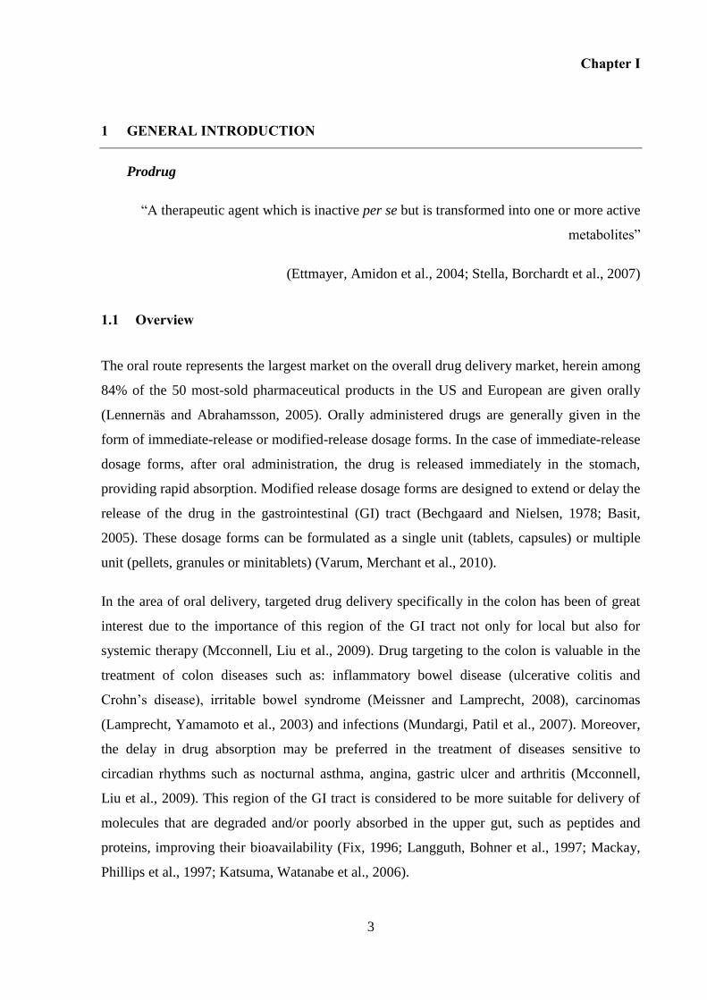
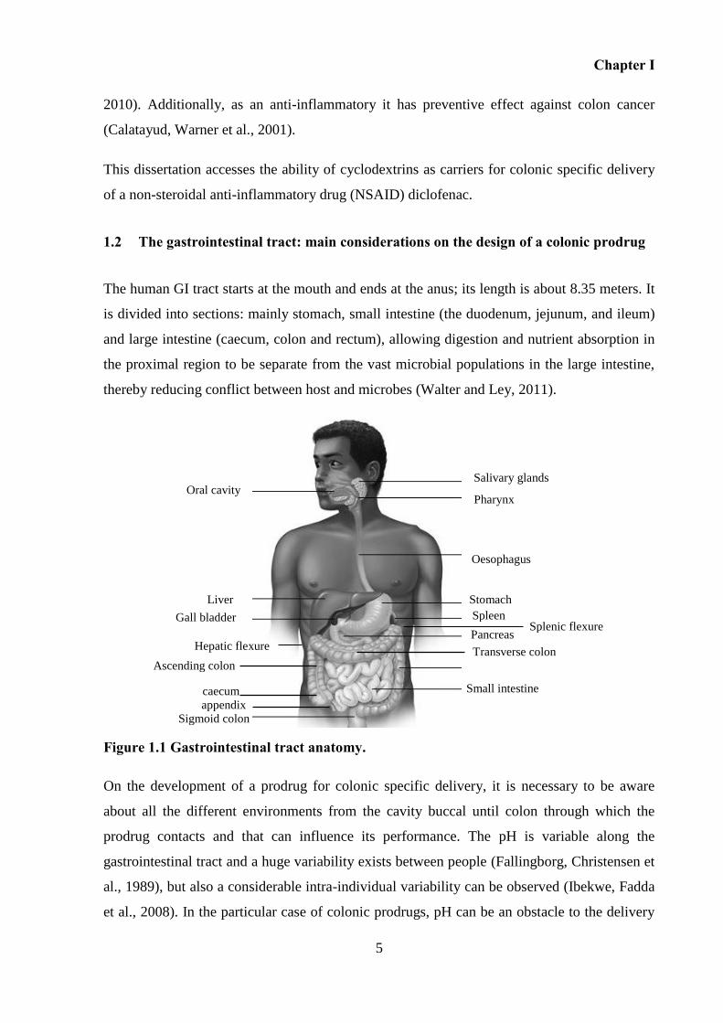
Figure 1.1 Gastrointestinal tract anatomy................................................................................... 5
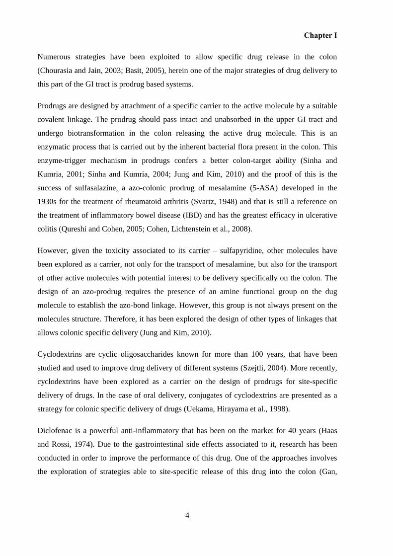
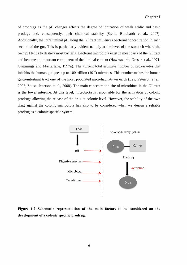
Figure 1.2 Schematic representation of the main factors to be considered on the development of a colonic specific prodrug. ............................................................................ 6
Figure 1.3 Gastrointestinal intraluminal pH, redox potential (RP) and bacterial concentration
(Sousa, Paterson et al., 2008) Mean pH values (Evans, Pye et al., 1988 ), mean Redox
potential (mV) (V Stirrup, S J Ledingham et al., 1990) and bacterial concentration (CFU/g of
contents) (Simon and Gorbach, 1984; Macfarlane and Macfarlane, 2004; Sousa, Paterson et
al., 2008). .................................................................................................................................... 9
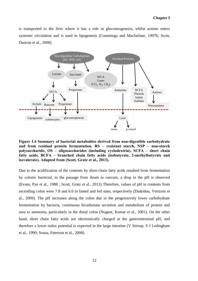
Figure 1.4 Summary of bacterial metabolites derived from non-digestible carbohydrate and
from residual protein fermentation. RS – resistant starch, NSP – non-starch polysaccharide,
OS – oligosaccharides (including cyclodextrin), SCFA – short chain fatty acids, BCFA –
branched chain fatty acids (isobutyrate, 2-methylbutyrate and isovalerate). Adapted from
(Scott, Gratz et al., 2013). ......................................................................................................... 12
Figure 1.5 The main sites of action of the anti-inflammatory drugs, adapted from the reference
(Ulrich, Bigler et al., 2006)....................................................................................................... 15
Figure 1.6 Schematic representation of the cone-shaped structure of the cyclodextrin. .......... 24
Figure 1.7 Effect of cyclodextrin complexation on the drug bioavailability after non-parental
administration. (Loftsson, Jarho et al., 2005). .......................................................................... 28
Figure 1.8 Schematic comparison of the digestion of natural α-CD, β-CD and γ-CD after oral
administration. Cyclodextrins are slowly hydrolysed by α-amylases but relatively rapidly
digested by bacteria. Adapted from (Kurkov and Loftsson, 2013). ......................................... 30
Figure 1.9 Structures of active molecules studied where cyclodextrins were used as a carrier
to target the colon. .................................................................................................................... 33
Figure 1.10 Gastrointestinal tracts of small hindgut-fermenting mammalian herbivore (rabbit)
and gastrointestinal tracts of mammalian omnivores (rat, pig). Adapted from references 1(Stevens, 1977) and
2(Argenzio and Southworth, 1975). ....................................................... 34
Figure 2.1 α-1,4 glycosidic bond and the three different types of hydroxyl groups of a
cyclodextrin. ............................................................................................................................. 44
Figure 2.2 I- 1-(2,6-dichlorophenyl) indolin-2-one structure, precursor on the synthesis of
diclofenac. II- Diclofenac: (2-(2,6-dichloranilino) phenylacetic acid) structure...................... 46
Figure 2.3 Synthetic scheme of synthesis of drug-cyclodextrin conjugate by a carboodimiide
activation of the drug. ............................................................................................................... 47
Figure 2.4 Synthetic scheme of synthesis of drug-cyclodextrin conjugate by formation of an
acid chloride of the drug and posterior reaction with cyclodextrin. ......................................... 48
Figure 2.5 Synthetic scheme of synthesis of drug-cyclodextrin conjugate by nucleophilic
substitution at the cyclodextrin structure. ................................................................................. 49
Figure 2.6 Intramolecular cyclization reaction of diclofenac (Palomo, Ballesteros et al., 1999).
.................................................................................................................................................. 57
Figure 2.7 Preferential cyclization of the diclofenac molecule suggested by the lack of success
on the attempt of synthesize an acid chloride. .......................................................................... 58
xii
Figure 2.8 Synthetic scheme of preparation of mono-6-tosyl-β-cyclodextrin. I- β-cyclodextrin;
II- Tosyl β-cyclodextrin. .......................................................................................................... 59
Figure 2.9 Synthetic scheme for the preparation of diclofenac-β-cyclodextrin conjugate. I -
tosyl β-cyclodextrin, II - sodium diclofenac, III - diclofenac-β-cyclodextrin. ........................ 61
Figure 2.10 HPLC chromatograms of crude product of conjugates synthesized under
microwave irradiation conditions for 40 minutes under different temperatures: A: 100 ºC; B:
140 ºC; C: 160 ºC; D: 200 ºC. The numbers represent the area of the corresponding peak.
Conjugate retention time: 8.0 minutes; diclofenac retention time 11.9 minutes. Elution
conditions are described in point 2.4.3. ................................................................................... 62
Figure 2.11 MALDI mass spectrum of tosylated β-cyclodextrin compound. A- β-cyclodextrin,
B- monotosylated β-cyclodextrin, C- ditosylated β-cyclodextrin ............................................ 63
Figure 2.12 1H NMR spectrum of β-cyclodextrin (A);
1H NMR spectrum of β-cyclodextrin
tosylated (B); Structure of β-cyclodextrin (I); structure of β-cyclodextrin tosylated (II). ....... 64
Figure 2.13 A- 1H NMR spectrum of β-cyclodextrin tosylated without recristalization. B-
1H
NMR spectrum of β-cyclodextrin tosylated with one recristalization. C- 1H NMR spectrum of
β-cycodextrin tosylated after two purifications. ...................................................................... 65
Figure 2.14 HPLC chromatogram of diclofenac-β-cyclodextrin conjugate (retention time of
7.9 minutes) after purification (A), and of tosylated β-cyclodextrin (retention time of 5.5
minutes) (B). Matrix-assisted laser desorption/ionization (MALDI) spectroscopy. ............... 66
Figure 2.15 MALDI mass spectrum of diclofenac-β-cyclodextrin conjugate. ........................ 66
Figure 2.16 I-Structure of the diclofenac-β-cyclodextrin; II-A-1H NMR spectrum of hydrated
β-cyclodextrin hydrated. II-B-1H NMR spectrum of sodium diclofenac. III-C-
1H NMR
spectrum of diclofenac-β-cyclodextrin conjugate. ................................................................... 68
Figure 2.17 1
H NMR ROESY spectra of the diclofenac-β-conjugate. ..................................... 70
Figure 2.18 IR spectra of diclofenac (A), β-cyclodextrin (B) and diclofenac-β-diclofenac
conjugate (C). ........................................................................................................................... 71
Figure 2.19 Stability studies of diclofenac-β-cyclodextrin in differents aqueous buffer
solutions. Each value represents mean ± S.D (n=3). ............................................................... 72
Figure 2.20 HPLC chromatograms of diclofenac-β-cyclodextrin (A), diclofenac free acid (B)
and sodium diclofenac (C) after incubation in buffer at pH 1.2 for 24 h in case of A and B and
5 h in case of C. Diclofenac-β-cyclodextrin conjugate retention time is 7.0 and retention time
of diclofenac is 16.9 minutes. .................................................................................................. 73
Figure 2.21 MALDI mass spectrum of the product from tosylation of γ-cyclodextrin with p-
toluenosulphonyl chloride. (A) γ-cyclodextrin, (B) monotosylated γ-cyclodextrin, (C)
ditosylated cyclodextrin, (D) tritosylated γ-cyclodextrin. ....................................................... 77
Figure 2.22 MALDI mass spectrum of the product from tosylation of γ-cyclodextrin with
triisopropylbenzenesulfonyl chloride. (A) mono-γ-6-(O-2,4,6- triisopropylbenzenesulfonyl)-γ-
cyclodextrin; (B) di-6-(O-2,4,6-triisopropylbenzenesulfonyl)-γ tolsylcyclodextrin. .............. 78
Figure 2.23 MALDI mass spectrum of crude product obtained after microwave reaction
between γ-cyclodextrin tosylated and sodium diclofenac.( A) γ-cyclodextrin; (B) diclofenac-
γ-cyclodextrin, (C) di-diclofenac-γ-cyclodextrin. .................................................................... 78
Figure 2.24 MALDI mass spectrum of compound diclofenac-γ-cyclodextrin conjugate after
purification using DIAION HP. ............................................................................................... 79
xiii
Figure 2.25 (A) 1H NMR spectrum of γ-cyclodextrin. (B)
1H NMR spectrum of sodium
diclofenac. (C) 1H NMR spectrum of diclofenac-γ-cyclodextrin conjugate. ........................... 80
Figure 2.26 MALDI mass spectrum of the product of tosylation of α-cyclodextrin with p-
toluenosulphonyl chloride. (A) α-cyclodextrin; (B) monotosylated α-cyclodextrin; (C)
ditosylated α-cyclodextrin. ....................................................................................................... 82
Figure 3.1 Schematic representation of enzymes involved in the hydrolysis of various bonds
(Hydrolases EC 3.), including those acting on ester bonds (esterases EC 3.1.) and those that
hydrolyse glycosyl compound (glycosylases EC 3.2.). Glycosidases hydrolyse O- and S-
glycosyl compounds (EC 3.2.1). This subgroup includes cyclomaltodextrinases (CDases; EC
3.2.1.54), maltogenic amylases (MAases; EC 3.2.1.133) and neopullulanases (NPases; EC
3.2.1.135). This classification is according to Nomenclature Committee of the International
Union of Biochemistry and Molecular Biology (NC-IUBMB). .............................................. 90
Figure 3.2 Gastrointestinal tract of pig (A), rabbit (B) and rat (C) before collection of the
respective fluids. ....................................................................................................................... 96
Figure 3.3 HPLC chromatogram of diclofenac-β-cyclodextrin (r.t. = 6.9 minutes),
sulfasalazine (r.t = 13.0 minutes) and diclofenac (r.t. = 18.6 minutes) in human faecal slurry.
................................................................................................................................................ 104
Figure 3.4 Mean levels of diclofenac-β-cyclodextrin (□) and diclofenac (▪) in human faecal
slurry (test). Mean levels of diclofenac-β-cyclodextrin (○) and diclofenac (●) in autoclaved
faecal slurry (control). Each point represents mean ± S.D. (n=3). ......................................... 105
Figure 3.5 Mean levels of diclofenac-β-cyclodextrin conjugate (A) and mean levels of
sulfasalazine (B) in three different samples of faecal slurry from male individuals from 3
different male individuals (▲, ■, ♦). Each point represents mean ± S.D. (n = 3). ................ 106
Figure 3.6 Stability of diclofenac-β-cyclodextrin in the presence of esterase from porcine liver
(39 Units/mL) in HEPES NaOH buffer (pH 7.4). Each point represents mean ± S.D. (n = 3).
................................................................................................................................................ 108
Figure 3.7 Mean levels of diclofenac-β-cyclodextrin in presence of different α-amylases (250
units/mL): α-amylase from Bacillus lechiniformes (♦), α-amylase from Aspergillus oryzae (■)
and α-amylase from porcine pancreas (●) in 0.2 M acetate buffer (pH 5.5) containing 0.01 M
CaCl2 at 37 °C. Each point represents mean ± S.D. (n = 3). .................................................. 109
Figure 3.8 First-order plots for hydrolysis of the conjugate in the presence of Aspergillus
oryzae α-amylase in acetate buffer (pH 5.5) at 37 °C at different concentrations of α-amylase
(20 (■), 40 (▲) and 250 (●) units/mL)................................................................................... 111
Figure 3.9 HPLC chromatograms of the enzymatic reaction of conjugate im presence of
different concentrations of α- amylase from Aspergillus Oryzae (20, 40 and 250 units/mL) at
0, 6, and 24 hours. .................................................................................................................. 111
Figure 3.10 Mean levels of diclofenac-β-cyclodextrin (▲) and diclofenac (∆) in the presence
of α-amylases from Aspergillus oryzae (250 units/mL) and carboxylic esterase (39 units/mL)
in acetate buffer (pH 5.5) at 37oC. Mean levels of diclofenac-β-cyclodextrin (■) and
diclofenac (∆) in the presence of α-amylases from Aspergillus oryzae (250 units/mL) in
acetate buffer (pH 5.5) at 37oC. .............................................................................................. 113
Figure 3.11 Mean levels of diclofenac-β-cyclodextrin (■) and diclofenac (□) in the presence
of α-amylases from Aspergillus oryzae (40 units/mL) and carboxylic esterase (39 units/mL) in
acetate buffer (pH 5.5) at 37 oC. The esterase (39 units/mL) was added 12 hours after the α-
amylase. A- addition of the α-amylase; B- addition of the esterase. ...................................... 114
xiv
Figure 3.12 Mean levels of diclofenac-β-cyclodextrin conjugate in simulated gastric fluid (♦)
and in simulated intestinal fluid (▲). Each point represents mean ± S.D. (n = 3). .............. 115
Figure 3.13 Stability of diclofenac-β-cyclodextrin in stomach, duodenum, jejunum and ileum
contents from pig (■), rat (▲) and rabbit (♦). Each point represents mean ± S.D. (n = 3). .. 116
Figure 3.14 Stability of diclofenac-β-cyclodextrin in caecum contents from pig (■), rabbit
(▲), rat (♦) and in pig colon A fluids (●). Each point represents mean ± S.D. (n = 3). ........ 119
Figure 3.15 Stability of sulfasalazine in caecum contents from pig (■), rabbit (▲) and rat (♦)
and in pig colon A fluids (●). Each point represents mean ± S.D. (n = 3). ........................... 119
Figure 3.16 Stability of diclofenac-β-cyclodextrin conjugate in colon contents prepared as a
faecal slurry from pig (■), rabbit (▲) and rat (♦). Each point represents mean ±S.D. (n = 3).
................................................................................................................................................ 120
Figure 3.17 Stability of sulfasalazine in colon contents prepared as a faecal slurry from pig
(■), rabbit (▲) and rat (♦). Each point represents mean ±S.D. (n = 3). ................................. 120
Figure 4.1 Schematic representation of the different feeding regimens of rat groups A, B, C
and D. A: 12-hour fast; B: 12-hour fast followed by 1-hour feeding, followed by 30-minutes
fast; C: 12-hour fast, followed by1-hour feeding, followed by 4-hour fast; D: fed ad libitum.
................................................................................................................................................ 132
Figure 4.2 Photographs of different sections of the gastrointestinal tract of rats:(I) Stomach;
(II) Duodenum; (III) Jejunum; (IV) Ileum; (V) Cecum; (VI) Colon, from Groups A, B, C and
D. ............................................................................................................................................ 138
Figure 4.3 Total mass of gastrointestinal contents in healthy male rats in different Groups. A:
12 hours fast, B: 12 hours fast then 1 hour fed then 30 minutes fast, C: 12 hours fast then 1
hour fed then 4 hours fast; D; fed. Each bar represents mean ± S.D, n = 5. .......................... 139
Figure 4.4 Mass of luminal contents in different gastrointestinal sections in rat Groups. Each
bar represents mean ± S.D. n = 5. .......................................................................................... 140
Figure 4.5 Mass of luminal contents in the different gastrointestinal sections in the rat Groups.
The data shown in Figure 4.4 has been replotted to assist comparison of the different
gastrointestinal regions. Each bar represents mean ± S.D, n = 5. .......................................... 141
Figure 4.6 In situ pH of gastrointestinal contents in the different sections of the
gastrointestinal tract in the rat groups. I and II refer to the anterior and posterior parts
respectively. Each point represents mean ± S.D, n = 5. ......................................................... 142
Figure 4.7 In situ pH of gastrointestinal contents along the gastrointestinal tract of rat Groups
A(○), B(∆), C(□) and D(◊). I and II refer to the anterior and posterior parts, respectively.
Same data as in Figure 6 has been re-plotted to ease comparisons. Each point represents mean
± S.D, n = 5. ........................................................................................................................... 143
Figure 4.8 Mean levels of diclofenac-β-cyclodextrin and sulfasalazine remaining when
prodrugs were incubated in caecal and colonic contents from rats from Groups A (○), B (∆), C
(□), and D (◊). Each point represents mean ± S.D, n = 3. ...................................................... 147
Figure 5.1 HPLC chromatograms of the (A) blank plasma, (B) spiked plasma at levels of low
limit of quantification, (C) spiked plasma at levels of the upper limit of the calibration ranges.
................................................................................................................................................ 163
Figure 5.2 Concentration-time profiles (n=7) of diclofenac and sulfapyridine in Wistar rats
after simultaneous oral administration of diclofenac-β-cyclodextrin (88.5 mg/kg) and
sulfasalazine (100 mg/kg). Each point represents mean ± S.D., n = 7. ................................. 166
xv
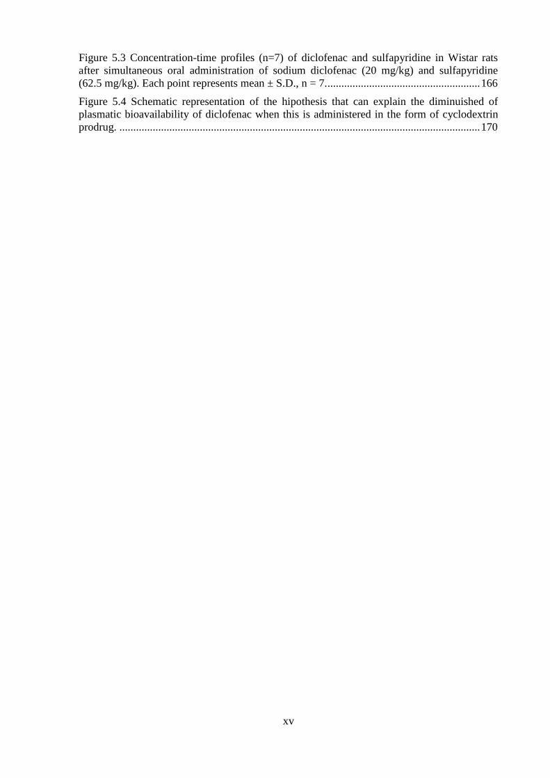
Figure 5.3 Concentration-time profiles (n=7) of diclofenac and sulfapyridine in Wistar rats
after simultaneous oral administration of sodium diclofenac (20 mg/kg) and sulfapyridine
(62.5 mg/kg). Each point represents mean ± S.D., n = 7. ....................................................... 166
Figure 5.4 Schematic representation of the hipothesis that can explain the diminuished of
plasmatic bioavailability of diclofenac when this is administered in the form of cyclodextrin
prodrug. .................................................................................................................................. 170

xvii
LIST OF TABLES
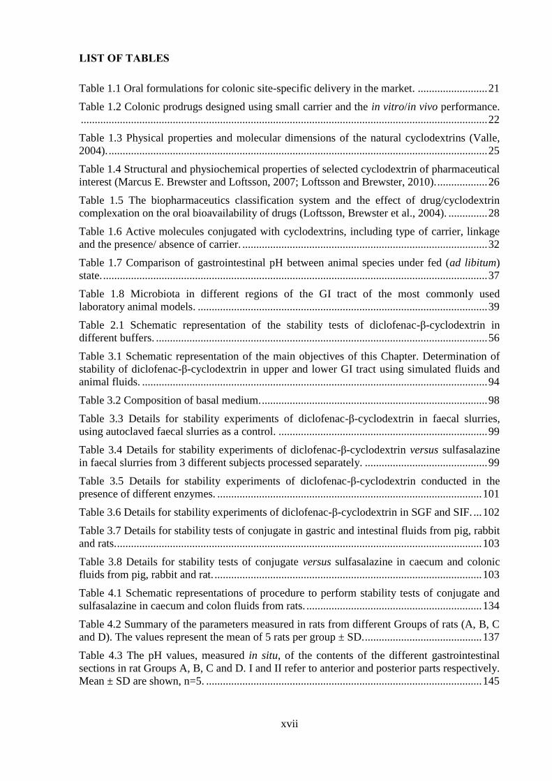
Table 1.1 Oral formulations for colonic site-specific delivery in the market. ......................... 21
Table 1.2 Colonic prodrugs designed using small carrier and the in vitro/in vivo performance.
.................................................................................................................................................. 22
Table 1.3 Physical properties and molecular dimensions of the natural cyclodextrins (Valle,
2004). ........................................................................................................................................ 25
Table 1.4 Structural and physiochemical properties of selected cyclodextrin of pharmaceutical
interest (Marcus E. Brewster and Loftsson, 2007; Loftsson and Brewster, 2010). .................. 26
Table 1.5 The biopharmaceutics classification system and the effect of drug/cyclodextrin
complexation on the oral bioavailability of drugs (Loftsson, Brewster et al., 2004). .............. 28
Table 1.6 Active molecules conjugated with cyclodextrins, including type of carrier, linkage
and the presence/ absence of carrier. ........................................................................................ 32
Table 1.7 Comparison of gastrointestinal pH between animal species under fed (ad libitum)
state. .......................................................................................................................................... 37
Table 1.8 Microbiota in different regions of the GI tract of the most commonly used
laboratory animal models. ........................................................................................................ 39
Table 2.1 Schematic representation of the stability tests of diclofenac-β-cyclodextrin in
different buffers. ....................................................................................................................... 56
Table 3.1 Schematic representation of the main objectives of this Chapter. Determination of
stability of diclofenac-β-cyclodextrin in upper and lower GI tract using simulated fluids and
animal fluids. ............................................................................................................................ 94
Table 3.2 Composition of basal medium. ................................................................................. 98
Table 3.3 Details for stability experiments of diclofenac-β-cyclodextrin in faecal slurries,
using autoclaved faecal slurries as a control. ........................................................................... 99
Table 3.4 Details for stability experiments of diclofenac-β-cyclodextrin versus sulfasalazine
in faecal slurries from 3 different subjects processed separately. ............................................ 99
Table 3.5 Details for stability experiments of diclofenac-β-cyclodextrin conducted in the
presence of different enzymes. ............................................................................................... 101
Table 3.6 Details for stability experiments of diclofenac-β-cyclodextrin in SGF and SIF. ... 102
Table 3.7 Details for stability tests of conjugate in gastric and intestinal fluids from pig, rabbit
and rats. ................................................................................................................................... 103
Table 3.8 Details for stability tests of conjugate versus sulfasalazine in caecum and colonic
fluids from pig, rabbit and rat. ................................................................................................ 103
Table 4.1 Schematic representations of procedure to perform stability tests of conjugate and
sulfasalazine in caecum and colon fluids from rats. ............................................................... 134
Table 4.2 Summary of the parameters measured in rats from different Groups of rats (A, B, C
and D). The values represent the mean of 5 rats per group ± SD. .......................................... 137
Table 4.3 The pH values, measured in situ, of the contents of the different gastrointestinal
sections in rat Groups A, B, C and D. I and II refer to anterior and posterior parts respectively.
Mean ± SD are shown, n=5. ................................................................................................... 145
xviii
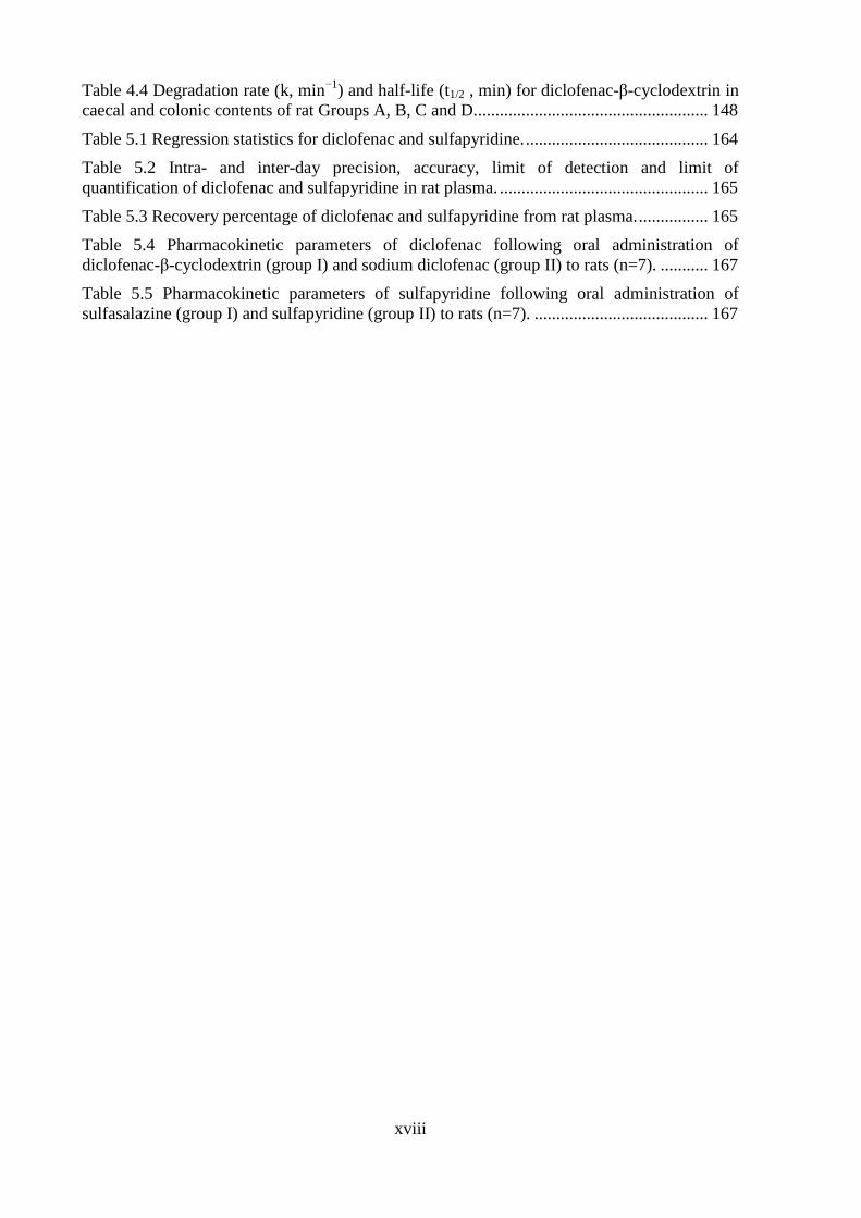
Table 4.4 Degradation rate (k, min−1
) and half-life (t1/2 , min) for diclofenac-β-cyclodextrin in
caecal and colonic contents of rat Groups A, B, C and D...................................................... 148
Table 5.1 Regression statistics for diclofenac and sulfapyridine. .......................................... 164
Table 5.2 Intra- and inter-day precision, accuracy, limit of detection and limit of
quantification of diclofenac and sulfapyridine in rat plasma. ................................................ 165
Table 5.3 Recovery percentage of diclofenac and sulfapyridine from rat plasma. ................ 165
Table 5.4 Pharmacokinetic parameters of diclofenac following oral administration of
diclofenac-β-cyclodextrin (group I) and sodium diclofenac (group II) to rats (n=7). ........... 167
Table 5.5 Pharmacokinetic parameters of sulfapyridine following oral administration of
sulfasalazine (group I) and sulfapyridine (group II) to rats (n=7). ........................................ 167
xix
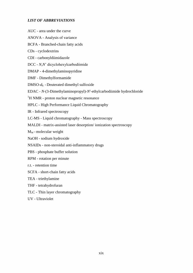
LIST OF ABBREVIATIONS
AUC - area under the curve
ANOVA - Analysis of variance
BCFA - Branched-chain fatty acids
CDs - cyclodextrins
CDI - carbonyldiimidazole
DCC - N,N’ dicyclohexylcarbodiimide
DMAP - 4-dimethylaminopyridine
DMF - Dimethylformamide
DMSO-d6 - Deuterated dimethyl sulfoxide
EDAC - N-(3-Dimethylaminopropyl)-N′-ethylcarbodiimide hydrochloride 1H NMR - proton nuclear magnetic resonance
HPLC - High Performance Liquid Chromatography
IR - Infrared spectroscopy
LC-MS - Liquid chromatography - Mass spectroscopy
MALDI - matrix-assisted laser desorption/ ionization spectroscopy
MW - molecular weight
NaOH - sodium hydroxide
NSAIDs - non-steroidal anti-inflammatory drugs
PBS - phosphate buffer solution
RPM - rotation per minute
r.t. - retention time
SCFA - short-chain fatty acids
TEA - triethylamine
THF - tetrahydrofuran
TLC - Thin layer chromatography
UV - Ultraviolet


CHAPTER I
GENERAL INTRODUCTION

Chapter I
Chapter I
3
1 GENERAL INTRODUCTION
Prodrug
“A therapeutic agent which is inactive per se but is transformed into one or more active
metabolites”
(Ettmayer, Amidon et al., 2004; Stella, Borchardt et al., 2007)
1.1 Overview
The oral route represents the largest market on the overall drug delivery market, herein among
84% of the 50 most-sold pharmaceutical products in the US and European are given orally
(Lennernäs and Abrahamsson, 2005). Orally administered drugs are generally given in the
form of immediate-release or modified-release dosage forms. In the case of immediate-release
dosage forms, after oral administration, the drug is released immediately in the stomach,
providing rapid absorption. Modified release dosage forms are designed to extend or delay the
release of the drug in the gastrointestinal (GI) tract (Bechgaard and Nielsen, 1978; Basit,
2005). These dosage forms can be formulated as a single unit (tablets, capsules) or multiple
unit (pellets, granules or minitablets) (Varum, Merchant et al., 2010).
In the area of oral delivery, targeted drug delivery specifically in the colon has been of great
interest due to the importance of this region of the GI tract not only for local but also for
systemic therapy (Mcconnell, Liu et al., 2009). Drug targeting to the colon is valuable in the
treatment of colon diseases such as: inflammatory bowel disease (ulcerative colitis and
Crohn’s disease), irritable bowel syndrome (Meissner and Lamprecht, 2008), carcinomas
(Lamprecht, Yamamoto et al., 2003) and infections (Mundargi, Patil et al., 2007). Moreover,
the delay in drug absorption may be preferred in the treatment of diseases sensitive to
circadian rhythms such as nocturnal asthma, angina, gastric ulcer and arthritis (Mcconnell,
Liu et al., 2009). This region of the GI tract is considered to be more suitable for delivery of
molecules that are degraded and/or poorly absorbed in the upper gut, such as peptides and
proteins, improving their bioavailability (Fix, 1996; Langguth, Bohner et al., 1997; Mackay,
Phillips et al., 1997; Katsuma, Watanabe et al., 2006).
Chapter I
4
Numerous strategies have been exploited to allow specific drug release in the colon
(Chourasia and Jain, 2003; Basit, 2005), herein one of the major strategies of drug delivery to
this part of the GI tract is prodrug based systems.
Prodrugs are designed by attachment of a specific carrier to the active molecule by a suitable
covalent linkage. The prodrug should pass intact and unabsorbed in the upper GI tract and
undergo biotransformation in the colon releasing the active drug molecule. This is an
enzymatic process that is carried out by the inherent bacterial flora present in the colon. This
enzyme-trigger mechanism in prodrugs confers a better colon-target ability (Sinha and
Kumria, 2001; Sinha and Kumria, 2004; Jung and Kim, 2010) and the proof of this is the
success of sulfasalazine, a azo-colonic prodrug of mesalamine (5-ASA) developed in the
1930s for the treatment of rheumatoid arthritis (Svartz, 1948) and that is still a reference on
the treatment of inflammatory bowel disease (IBD) and has the greatest efficacy in ulcerative
colitis (Qureshi and Cohen, 2005; Cohen, Lichtenstein et al., 2008).
However, given the toxicity associated to its carrier – sulfapyridine, other molecules have
been explored as a carrier, not only for the transport of mesalamine, but also for the transport
of other active molecules with potential interest to be delivery specifically on the colon. The
design of an azo-prodrug requires the presence of an amine functional group on the dug
molecule to establish the azo-bond linkage. However, this group is not always present on the
molecules structure. Therefore, it has been explored the design of other types of linkages that
allows colonic specific delivery (Jung and Kim, 2010).
Cyclodextrins are cyclic oligosaccharides known for more than 100 years, that have been
studied and used to improve drug delivery of different systems (Szejtli, 2004). More recently,
cyclodextrins have been explored as a carrier on the design of prodrugs for site-specific
delivery of drugs. In the case of oral delivery, conjugates of cyclodextrins are presented as a
strategy for colonic specific delivery of drugs (Uekama, Hirayama et al., 1998).
Diclofenac is a powerful anti-inflammatory that has been on the market for 40 years (Haas
and Rossi, 1974). Due to the gastrointestinal side effects associated to it, research has been
conducted in order to improve the performance of this drug. One of the approaches involves
the exploration of strategies able to site-specific release of this drug into the colon (Gan,
Chapter I
5
2010). Additionally, as an anti-inflammatory it has preventive effect against colon cancer
(Calatayud, Warner et al., 2001).
This dissertation accesses the ability of cyclodextrins as carriers for colonic specific delivery
of a non-steroidal anti-inflammatory drug (NSAID) diclofenac.
1.2 The gastrointestinal tract: main considerations on the design of a colonic prodrug
The human GI tract starts at the mouth and ends at the anus; its length is about 8.35 meters. It
is divided into sections: mainly stomach, small intestine (the duodenum, jejunum, and ileum)
and large intestine (caecum, colon and rectum), allowing digestion and nutrient absorption in
the proximal region to be separate from the vast microbial populations in the large intestine,
thereby reducing conflict between host and microbes (Walter and Ley, 2011).
Figure 1.1 Gastrointestinal tract anatomy.
On the development of a prodrug for colonic specific delivery, it is necessary to be aware
about all the different environments from the cavity buccal until colon through which the
prodrug contacts and that can influence its performance. The pH is variable along the
gastrointestinal tract and a huge variability exists between people (Fallingborg, Christensen et
al., 1989), but also a considerable intra-individual variability can be observed (Ibekwe, Fadda
et al., 2008). In the particular case of colonic prodrugs, pH can be an obstacle to the delivery
Ascending colon
Splenic flexure
Small intestine
Salivary glands
Pharynx Oral cavity
Oesophagus
Stomach Spleen
Pancreas
appendix caecum
Transverse colon
Liver
Gall bladder
Hepatic flexure
Sigmoid colon
Chapter I
6
of prodrugs as the pH changes affects the degree of ionization of weak acidic and basic
produgs and, consequently, their chemical stability (Stella, Borchardt et al., 2007).
Additionally, the intraluminal pH along the GI tract influences bacterial concentration in each
section of the gut. This is particularly evident namely at the level of the stomach where the
own pH tends to destroy most bacteria. Bacterial microbiota exist in most parts of the GI tract
and become an important component of the luminal content (Hawksworth, Drasar et al., 1971;
Cummings and Macfarlane, 1997a). The current total estimate number of prokaryotes that
inhabits the human gut goes up to 100 trillion (1014
) microbes. This number makes the human
gastrointestinal tract one of the most populated microhabitats on earth (Ley, Peterson et al.,
2006; Sousa, Paterson et al., 2008). The main concentration site of microbiota in the GI tract
is the lower intestine. At this level, microbiota is responsible for the activation of colonic
prodrugs allowing the release of the drug at colonic level. However, the stability of the own
drug against the colonic microbiota has also to be considered when we design a reliable
prodrug as a colonic specific system.
Figure 1.2 Schematic representation of the main factors to be considered on the
development of a colonic specific prodrug.
Prodrug
Drug Carrier
Drug
Activation
pH
Microbiota
Digestive enzymes
Transit time
Colonic delivery system
Food
Chapter I
7
Transit time through the GI tract varies depending on various factors like GI motility, quantity
and quality of food ingested, feeding regimen, food timing, and also nature of the oral
formulation (Yuen, 1986; Varum, Merchant et al., 2010). The average overall transit time
from the mouth to the anus in humans is 24–72 hours (Stella, Borchardt et al., 2007).
Considering colonic prodrugs, the time between intake of a prodrug and its arrival in the
colon will affect not only the onset of the drug in the local of action and /or at the plasmatic
level but also, consequently, its efficacy.
Following is described the main characteristics and functions of each sections of the GI tract
that can influence the success of a colonic prodrug, including aspects related to the enzymes,
pH, microbiota and gastrointestinal tract transit time
Stomach 1.2.1
The stomach is divided into cardia, fundus, corpus and pyloric region (antrum) and has
several functions: storage of the ingested food, processing the food into a fluid chimie,
mechanically and biochemically; control the rate of delivery of chimie into the duodenum and
production of acid (Washington, Washington et al., 2000).
In the stomach, due to secretion of hydrogen ions by gastric mucosa (parietal cells) the pH
ranges from 0.8 and 2.0 in the fasted state and the number of bacteria decreases comparably
with saliva (107 CFU/mL) due to the acidic conditions. After ingestion of food, due to
buffering and dilution effects caused by that, the pH ranges from 4 to 5 and bacteria
proliferate to 104 -10
8 CFU/mL. Bacteria can escape due to the substantial volumes of liquid
that are moving into the duodenum. At this level, microbiota is predominantly gram-positive
and aerobic (Evans, Pye et al., 1988 ; Fallingborg, Christensen et al., 1989) The low pH is
essential to maintain the environment sterile and regulates the production of pepsin (3.4.4.1),
an exopeptidase (Dressman, Amidon et al., 1998).
Gastric emptying presents great variability, which can range from a few seconds to a number
of hours and can depend on the feed status (Ibekwe, Liu et al., 2006). Solid, large and caloric
meals increase gastric emptying time (Varum, Hatton et al., 2013). In general, the transit time
from the mouth to the small intestine in healthy human adults is 0.5–2 hours, whereas it can
be delayed to 3–6 hours and 5–8 hours after the intake of light meals and heavy meals,
respectively (Stella, Borchardt et al., 2007). Digenis et al. verified with a multiple-unit
Chapter I
8
formulation that there is a faster gastric emptying in pre-feeding state when compared to the
fasted state, due to the increase of gastric motility and gastric emptying in presence of food
(Digenis, Sandefer et al., 1990).
Small intestine 1.2.2
Small intestine is the longest section of the digestive tube, representing 81% of the total
length of the GI tract. It is divided into 3 regions: duodenum, jejunum and ileum (Desesso and
Jacobson, 2001). In the proximal part of the duodenum occurs the neutralisation of the acidic
contents of the stomach by the alkaline pancreatic secretions, which contains bicarbonate,
lipases, amylases and proteases (Dressman, Amidon et al., 1998; Sarti, Barthelmes et al.,
2011). Consequently, it is observed a burst increase of pH to 5.5. At the level of duodenum,
food does not affect the pH (Kalantzi, Goumas et al., 2006). Additionally, bicarbonate
secretion results in a further gradual rise in the luminal pH to
6.6 ± 0.5 at jejunum level, reaching a peak at the ileum caecal junction 7.5 ± 0.5, in health
condition (Evans, Pye et al., 1988 ; Dressman, Berardi et al., 1990; Ibekwe, Fadda et al.,
2008).
In the duodenum and jejunum, the relatively rapid flow of the digesta concomitantly with bile
and pancreatic fluid secretions, does not allow the increase of microbial multiplication
(Tannock, 1999; Sousa, Paterson et al., 2008). At this stage the predominant species are
aerobic and gram positive. However, in the distal ileum gram negative bacteria outnumber the
gram positive and the bacterial composition becomes higher (Sinha and Kumria, 2003).
The intestinal area is greatly enhanced by presence of plicae circulares (or Kreking folds),
villis (finger like projections) and microvillis. Together, these folds provide a huge surface
area for absorption, 200 m2, whereas the area of the stomach is only about 1 m
2 (Kararli,
1995; Rowland, Tozer et al., 1995; Desesso and Jacobson, 2001). This big surface area
associated with the good blood supply and the gut associated lymphoid tissue (GALT), makes
the small intestine the main site of absorption (Edwards, 1993), particularly in duodenum and
proximal jejunum. The material that is not absorbed suffers movement towards the large
intestine. Separating the ileum from the caecum is a one-way valve, the ileocaecal junction
(ICJ), which regulates the movement of material between the small intestine and the colon
and prevents the reflux of colon contents, in particular, the spread of bacteria from the colon
to the ileum.
Chapter I
9
Although, the mean time to cross the small intestine is of 3-4 hours (Davis, Hardy et al.,
1986), the actual transit has demonstrated a range between 1 and 9.5 hours (Davis, Hardy et
al., 1986; Sugito, Ogata et al., 1990; Coupe, Davis et al., 1991a). Fadda et al. verified that
there is an acceleration in small intestinal transit in pre-feed state when compared with fasted
and fed state (Fadda, Mcconnell et al., 2009).
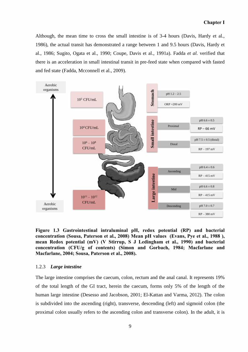
Figure 1.3 Gastrointestinal intraluminal pH, redox potential (RP) and bacterial concentration (Sousa, Paterson et al., 2008) Mean pH values (Evans, Pye et al., 1988 ), mean Redox potential (mV) (V Stirrup, S J Ledingham et al., 1990) and bacterial concentration (CFU/g of contents) (Simon and Gorbach, 1984; Macfarlane and Macfarlane, 2004; Sousa, Paterson et al., 2008).
Large intestine 1.2.3
The large intestine comprises the caecum, colon, rectum and the anal canal. It represents 19%
of the total length of the GI tract, herein the caecum, forms only 5% of the length of the
human large intestine (Desesso and Jacobson, 2001; El-Kattan and Varma, 2012). The colon
is subdivided into the ascending (right), transverse, descending (left) and sigmoid colon (the
proximal colon usually refers to the ascending colon and transverse colon). In the adult, it is
Aerobic
organismspH 1.2 – 2.5
ORP +200 mVStom
ach
pH 6.6 ± 0.5
RP – 66 mV
Smal
lint
estin
eL
arge
inte
stin
e
Ascending
Mid
Descending
Proximal
Distal
pH 7.5 ± 0.5 (distal)
RP – 197 mV
pH 6.4 ± 0.6
RP – 415 mV
pH 6.6 ± 0.8
RP – 415 mV
pH 7.0 ± 0.7
RP – 380 mV
Aerobic
organisms
103 CFU/mL
104 CFU/mL
1011 – 1012
CFU/mL
106 – 108
CFU/mL
Chapter I
10
approximately 150 cm long, significantly shorter than the small intestine. The 'large' refers to
the diameter which varies from approximately 9.0 cm in the caecum to 2.0 cm in the sigmoid
colon (Mrsny, 1992). The large gut receives material from the ileum, which has already been
digested and the contents are then mixed and retained for 6-12 hours in the caecum and right
colon. After the hepatic flexure, the chimie is processed into faeces, pass through the
transverse to the left colon for storage and eventual excretion concomitant with epithelial cell
turnover and bacteria (Cummings and Macfarlane, 1991; Hastewell, Williamson et al., 1991;
Basit, 2005). The ascending colon is the preferential region in the lower intestine regarding to
action of drugs, since the substantial residence time in this part and also because has more free
water when compared to the transverse colon (Diakidou, Vertzoni et al., 2009).
As compared with the small intestine, the large intestine shows much reduced surface area for
absorption. This is due to the presence of semilunar folds instead of circular Kerkring's valves
and due to the lack of villi and less developed microvilli (Hastewell, Williamson et al., 1991;
Kararli, 1995). Additionally, the narrow ‘tighter junctions’ between epithelial cell, restrain the
paracellular permeability of hydrophilic compounds (Hastewell, Williamson et al., 1991).
Colon participates in the maintenance of fluid and electrolyte balance through reabsorption of
water, sodium cation, chloride and secretion of potassium and bicarbonate (Dawson, 1991).
Additionally, colon contains lower levels of luminal and mucosal digestive enzymes than
stomach and small intestine (Gibson, Mcfarlan et al., 1989).
However, the human colon is the major site for growth and microbial digestion. The colonic
microbial community is very complex due to the diversity of species and, moreover, due to
the difficulty to distinguish between permanent and transient members (Yatsunenko, Rey et
al., 2012). The studies surrounding gastrointestinal microbiota have been extended for over a
century and nowadays, it is estimated that the total number of procaryotes that inhabit the
human gut goes up to 1014
, herein aerobic species outnumber (Savage, 2001; Ley, Peterson et
al., 2006). It is estimated that the gut microbiome contain 150-fold more genes than the
human genome (Qin, Li et al., 2010).
Current knowledge allows to recognize that the lower gastrointestinal tract contains over 400
distinct species of bacteria, which belongs to relatively few phyla (Yang, 2008; Jung and
Kim, 2010). The predominant microbiota belong to the phylum Firmicutes (gram positive)
Chapter I
11
and Bacteroidetes (gram negative), which account for more than 90% of the overall
phylogenetic types. Additionally, Actinobacteria (gram positive) has also been found in
human gut, normally with lower abundances of Verrucomicrobia and Proteobacteria and
Fusobacteria (Eckburg, Bik et al., 2005; Wu, Chen et al., 2011; Yatsunenko, Rey et al., 2012;
Aziz, Doré et al., 2013; Scott, Gratz et al., 2013).
In the ascending colon, microorganisms having plentiful supply of dietary nutrients, tend to
grow rapidly, while in the transverse and descending colon substrate availability is lower and
bacteria growth is slower (Sousa, Paterson et al., 2008). Nevertheless, aproximately 1/3 of the
faecal dry weight consists of bacteria (Sinha and Kumria, 2003).
The complete definition of microbiota requires further studies, which may attend the
combination of conventional and molecular microflora analysis. Moreover, the impact of
microbiota on host physiology is still unclear (Mai and Morris, 2004).
It is known that microbiota exists in relative stable conditions that rely on nutrient availability
(carbohydrates, proteins and fats), pH, temperature, redox potential, degree of anaerobiosis
and healthy state (Cummings and Macfarlane, 1991; Ojetti, Gigante et al., 2009) (Scott, Gratz
et al., 2013).
Primarily, these colonic bacteria rely on carbohydrate dietary components that are undigested
by human enzymes in the upper GI tract for energy and growth, including resistant starch
(RS), non-starch polysaccharides (NSP), and oligosaccharides (including prebiotics and
cyclodextrins). Anaerobic fermentation of these subtracts results in the production of volatile
fatty acids called short-chain fatty acids (SCFA), CO2, H2, CH4 and H2S. (Scott, Duncan et al.,
2008; Scott, Duncan et al., 2011). Gram-negative anaerobes belonging to the genus
Bacteroides are the principal polysaccharide-degrading bacteria (Cummings and Macfarlane,
1991). When the carbohydrate sources are depleted, which occurs namely at the level of the
distal colon, residual proteins are used as a source of energy. The fermentation of proteins
results in the formation of SCFA but also of branched chain fatty acids (BCFA) and other
metabolites (Scott, Gratz et al., 2013).
Short chain fatty acids are the major products of fermentation, which accounts for up to 10%
of the human energy source (Frank, St. Amand et al., 2007). Butyrate, in particular, is the
major source of energy for the colonocytes (the epithelial cells that line the colon), propionate
Chapter I
12
is transported to the liver where it has a role in gluconeogenesis, whilst acetate enters
systemic circulation and is used in lipogenesis (Cummings and Macfarlane, 1997b; Scott,
Duncan et al., 2008).
Figure 1.4 Summary of bacterial metabolites derived from non-digestible carbohydrate and from residual protein fermentation. RS – resistant starch, NSP – non-starch polysaccharide, OS – oligosaccharides (including cyclodextrin), SCFA – short chain fatty acids, BCFA – branched chain fatty acids (isobutyrate, 2-methylbutyrate and isovalerate). Adapted from (Scott, Gratz et al., 2013).
Due to the acidification of the contents by short-chain fatty acids resulted from fermentation
by colonic bacterial, in the passage from ileum to caecum, a drop in the pH is observed
(Evans, Pye et al., 1988 ; Scott, Gratz et al., 2013) Therefore, values of pH in contents from
ascending colon were 7.8 and 6.0 in fasted and fed state, respectively (Diakidou, Vertzoni et
al., 2009). The pH increases along the colon due to the progressively lower carbohydrate
fermentation by bacteria, continuous bicarbonate secretion and metabolism of protein and
urea to ammonia, particularly in the distal colon (Nugent, Kumar et al., 2001). On the other
hand, short chain fatty acids are electronically charged at the gastrointestinal pH, and
therefore a lower redox potential is expected in the large intestine (V Stirrup, S J Ledingham
et al., 1990; Sousa, Paterson et al., 2008).
Residual ProteinsNon-digestible Carbohydrate
(RS, NSP, OS)
Lactate Succinate
Acetate
Propionate
SFCA
Gases
(CO2, H2, CH4)
SFCA
Butyrate Propionate
colonocytesgluconeogenesisLipogenesis
BCFA
Phenols
Indols
Sulfides
Ammonia
Liver
ureia p-cresol
Amines
Nitrosamines
Chapter I
13
Colon as a site for drug delivery 1.2.4
The high viscosity of fluids and the lower availability of fluids, especially after the hepatic
flexure, turn the dissolution, particularly of poorly soluble drugs, difficult and limit the
diffusion of drugs from the lumen to the site of absorption. Additionally, the wider lumen
diameter reduces the exposure of drug molecules to the site of absorption (Basit, 2005).
However, a greater part of the gut transit time is spent in this part of the GI tract (almost
80%), which makes this site of the gut favorable not only for absorption of some drugs but
also favorable for activation of colonic prodrugs with release of the respective drug (Tuleu,
Basit et al., 2002; Stella, Borchardt et al., 2007; Sousa, Paterson et al., 2008). Actually, some
drugs that are considered to have good absorption at colonic level including, diclofenac, 5-
fluorouracil, glibenclamide, ibuprofen, lamirocoxib, metoprolol, nifedipine, salicylic acid,
simvastatin between others listed by Emma et al. (Mcconnell, Liu et al., 2009).
Moreover, the colon is a relatively inert site for the metabolism of drugs by cytochrome P450
(Biechea, Narjozb et al., 2007), and therefore drugs that are substrates for this enzyme have
higher absorption from the colon, as demonstrated for simvastatin (Tubic-Grozdanis,
Hilfinger et al., 2008). Additionally, the levels of efflux transporters (e.g. P-glycoprotein) are
lower in the colon, which may also contribute to improve oral drug bioavailability of drugs
that are substrates for these transporters (Englund, Rorsman et al., 2006; Berggren, Gall et al.,
2007).
The metabolic potential of colon is equal or greater than of the liver. In general, while
oxidation and conjugation are the predominant reactions metabolism in the liver, reduction
and hydrolyses are the preferred ones in the large intestinal lumen (Jung and Kim, 2010).
Colonic microbiota is capable of metabolizing a wide variety of drug molecules; over 30 drug
molecules have been identified as substrates for colonic bacteria (Sousa, Paterson et al.,
2008). The main reductive enzymes include nitroreductase, azoreductase, deaminase and urea
dehydroxylase (Scheline, 1973). On the other hand, between hydrolytic enzymes,
glycosidases are particularly important once they are involved in the activation of
polyssacarides based systems. The prevalent glycosidases are amylase, pectinase, xylanase,
α-L-arabinofuranosidase, β-D-xylosidase, β-D-galactosidase and β-D-glucosidase
(Englyst, Hay et al., 1987). The presence and capacities of these enzymes produced by the
microbiota at colonic level are the key to the design of colonic delivery systems activated by
Chapter I
14
microbiota and also colonic prodrugs (e.g. sulfasalazine) able to site-specific release of drugs
into the colon (Mooter, 2006). Further description about colonic enzymes is present in
Chapter III, namely related enzymes that are possibly involved on the degradation of
cyclodextrins.
The colon is a site of interest to delivery drugs that are medicated to colonic diseases,
inflammatory bowel diseases (Chron’s diseases and ulcerative colitis), constipation/diarrhoea,
colorectal cancer, irritable bowel disease and intestinal infections. Drug is released directly in
the affected area, not only resulting in an increase of the therapeutic potency by reduction of
the dose administered, but also decreasing the occurrence of adverse effects, which results
from the release of drugs in the upper GI tract or unnecessary systemic absorption (Chourasia
and Jain, 2003; Gupta, Bhandari et al., 2009). Glucocorticoids, mesalazine, antimicrobial
agents (nalidix acid and sulfa drugs), antispastic agent (mentol), anti-constipation (naloxone
and nalmefene), chemopreventive agents (ursodeoxycholic acid), anti-cancer agents (9-
aminocampothecin, 5-fluorouracil), immunosuppressive agents, vermicides (mebendazole),
cyclooxygenase-2 (COX-2) selective inhibitors to prevent colon rectal cancer, peptides and
proteins drugs have high potential to be delivered to the colon.
Moreover, therapeutic agents for the management of arthritis, asthma and gastric ulcer, which
commonly have a peak symptom at bedtime, are potential candidates for colonic release. The
treatment according to the circadian rhythm is called chronopharmacology, which includes
anti-inflammatory drugs, histamine receptor 2 blokers, proton pump inhibitors and
antiasthmatic agents (Jung and Kim, 2010).
1.2.4.1 Colonic delivery of non-steroidal anti-inflammatory drugs (NSAIDs)
NSAIDs are the most commonly prescribed therapeutic agents (Jones, 2001) and are good
candidates for the development of controlled release preparations, particularly through the
oral route. Their anti-inflammatory action is mainly based in the inhibition of cyclo-
oxygenase (COX), which is an enzyme that converts arachidonic acid into inflammatory
mediators such as prostaglandins, prostacyclins, and thromboxanes (Kim, Brar et al., 2009).
Two important COX enzymes have been described: COX-1, which is constitutively expressed
in all tissues is involved in the synthesis of prostaglandins that protect the stomach and kidney
from damage, and COX-2 which is almost undetectable in most tissues under normal
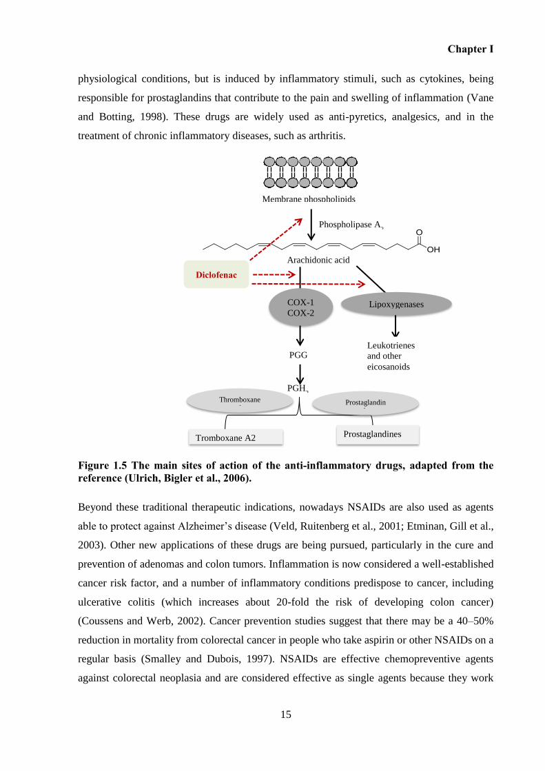
Chapter I
15
physiological conditions, but is induced by inflammatory stimuli, such as cytokines, being
responsible for prostaglandins that contribute to the pain and swelling of inflammation (Vane
and Botting, 1998). These drugs are widely used as anti-pyretics, analgesics, and in the
treatment of chronic inflammatory diseases, such as arthritis.
Figure 1.5 The main sites of action of the anti-inflammatory drugs, adapted from the reference (Ulrich, Bigler et al., 2006).
Beyond these traditional therapeutic indications, nowadays NSAIDs are also used as agents
able to protect against Alzheimer’s disease (Veld, Ruitenberg et al., 2001; Etminan, Gill et al.,
2003). Other new applications of these drugs are being pursued, particularly in the cure and
prevention of adenomas and colon tumors. Inflammation is now considered a well-established
cancer risk factor, and a number of inflammatory conditions predispose to cancer, including
ulcerative colitis (which increases about 20-fold the risk of developing colon cancer)
(Coussens and Werb, 2002). Cancer prevention studies suggest that there may be a 40–50%
reduction in mortality from colorectal cancer in people who take aspirin or other NSAIDs on a
regular basis (Smalley and Dubois, 1997). NSAIDs are effective chemopreventive agents
against colorectal neoplasia and are considered effective as single agents because they work
Lipoxygenases COX-1 COX-2
PGG
PGH2
Tromboxane A2 Prostaglandines
Membrane phospholipids
Phospholipase A2
Leukotrienes and other eicosanoids
Arachidonic acid
Thromboxane
synthase Prostaglandin
synthases
Diclofenac
Chapter I
16
early in carcinogenesis across multiple pathways (Ulrich, Bigler et al., 2006; Cuzick, Otto et
al., 2009). Second, NSAIDs reduce the number and size of colon adenomas (Hanif, Pittas et
al., 1996). However, the gastrointestinal toxicity associated with the conventional NSAIDs
may limit their long-term use for cancer prevention Long-term NSAIDs therapy has been
associated with an increased risk of gastric erosion, ulcers and bleeding (Go, 2006). NSAIDs
disrupt the normal gastric mucosal barrier of bicarbonate and hydrophobic mucus by two
mechanisms. The principal mechanism, independent of route of administration, results from
disturbance of prostaglandin synthesis by inhibition of the constitutive cyclo-oxygenase
(COX) isoenzyme, COX-1. In the stomach, prostaglandins maintain mucosal blood flow,
stimulating secretion of bicarbonate and mucus and regulating mucosal cell turnover and
repair. Loss of protection by inhibition of prostaglandins renders the stomach vulnerable to
damage by gastric acid. The second mechanism arises from the fact that NSAIDs become
lipid soluble at low pH. Taken orally, they can cross the lipid barrier into gastric mucosal
cells. At intracellular pH, they lose lipid solubility and become trapped, disrupting cell
function, perhaps by inhibiting mitochondrial oxidative phosphorylation. Damage to surface
mucosal cells compromises the normal protective mechanisms, reducing resistance to acid
damage (Hayllar and Bjarnason, 1995; Hawkins and Hanks, 2000). It has been estimated that
about 60% of all patients chronically treated with NSAIDs present dyspeptic symptoms and
around 30% have gastroduodenal ulcers (Silverstein, Graham et al., 1995; Laine, 2001).
In order to decrease gastrointestinal effects associated with NSAIDs various approaches have
been adapted to formulate systems to specifically release drugs in the colon (Luppi, Cerchiara
et al., 2003; Akhgaria, Garekania et al., 2005; Orlu, Cevher et al., 2006; Maestrellia, Zerroukb
et al., 2008; Piao, Lee et al., 2008).
1.2.4.2 Diclofenac
Diclofenac [2-(2, 6-dicloranilino) phenyl acetic acid] is a non-steroidal anti-inflammatory
drug, usually found as sodium or potassium salt. As a representative of the phenylacetic acid
derivatives group, diclofenac is widely used in the long-term treatment of inflammation and
painful conditions of rheumatic and non-rheumatic origin (Sallmann, 1986). This molecule
inhibits preferentially COX-2 (Warner, Giuliano et al., 1999; Hinz, Rau et al., 2003;
Yamazaki, Muramoto et al., 2006), and also inhibits the lipoxygenase pathway, reducing the
Chapter I
17
formation of leucotriens (Elder, Halton et al., 1997 ). In addition, diclofenac can inhibit
phospholipase A2 which may explain the high anti-inflammatory activity of this drug.
Much interest involves the specific release of diclofenac in the colon, given that such an
approach would allow for circumvention the adverse gastric effects notoriously associated
with drugs of this class. It is also widely believed that the colonic delivery of diclofenac
would provide a suitable way for the management of arthritis pain by chronotherapy (Lin and
Kawashima, 2012). Furthermore, diclofenac is known to be well-absorbed in the colon
(Gleiter, Antonin et al., 1985) and has demonstrated to have an effective chemopreventive
action against colon cancer (Saini, J. Kaur et al., 2009; Jasmeet and Nath, 2010).
Several strategies have been studied with the purpose to release diclofenac in the colon,
namely a time-based system that consisted in a polymeric matrix tablet (González-Rodríguez,
Maestrelli et al., 2003), a time- and pH-dependent colon-specific tablet (Cheng, An et al.,
2004), a combination of pH dependent polymer coating and a matrix able to release the drug
by an ionic-exchange mechanism (Ambrogi, Perioli et al., 2008), Eudragit® L 100-55
nanofibers – a pH dependent system (Shen, Yu et al., 2011), poly(methyl acrylates)-coated
chitosan–diclofenac sodium nanoparticles (Huanbutta, Sriamornsak et al., 2013). Below is
described the main systems for colonic delivery of drugs.
1.3 Systems to colonic target delivery of drugs to the colon
Because of the distal location of the colon in the GI tract, an ideal system for colonic delivery
should prevent drug release in the stomach and small intestine, and provide an abrupt onset of
the drug upon entry into the colon (Yang, 2008). Rectal administration offers the shortest
route for targeting drugs to the colon. Rectal formulations such enemas, foams and
suppositories seem to be a very simple way for local drug delivery. However, it allows only
reaching the rectum and lower parts of the distal colon. Reach the proximal part of colon via
rectal administration is difficult. Moreover, this route of administration can be uncomfortable
for patients and compliance may be less than optimal (Mooter, 2006). Therefore, the oral
delivery is presented as the most convenient and preferred way to target the colon.
Many different colon-specific drug delivery systems have been investigated based on the
exploration of selected physiological parameters that exhibit particular features only in the
Chapter I
18
colon. However, due to the similarity between distal small intestine and the proximal colon,
there are not many options that allow drug release only in the colon (Basit, 2005). Systems
have been developed considering the next aspects: variation of pH along the GI tract (pH-
based systems), transit time in the GI tract (time-based systems), intra-luminal pressure
(pressure-based systems), composition of the microbiota (microbiota-based systems) or the
combination of two of these anterior factors (Table 1.1).
pH-based systems
pH-dependent systems are designed based on the difference in pH-levels along the GIT.
These formulations use pH sensitive polymer-based coatings that are insoluble at low pH
levels, preventing dissolution and drug release in the upper gut but dissolving in the elevated
pH conditions of the distal gut (Mcconnell, Liu et al., 2009). The thickness of the coating is
the determinant factor for the success of these systems. The most commonly used pH-
dependent polymers are methacrylic acid copolymers usually known as Eudragit®. The major
drawback of these systems is associated with the fact that pH is not predictably stable and
therefore the site-specificity of formulations can be poor. pH is influenced by diet, disease
(e.g. pH is lower in inflammatory bowel disease and in ulcerative colitis) and intra and inter-
individual differences observed along the GI tract (Leopold and Eikeler, 2000; Mooter, 2006;
Mcconnell, Fadda et al., 2008). It was reported that release of drug from these systems may
start in the proximal small intestine (Singh, 2007) and, on the other hand, some of these
formulations pass intact through the gut, due to failure of the coating to dissolve (Sinha and
Kumria, 2003; Ibekwe, Fadda et al., 2006; Ibekwe, Liu et al., 2006; Mcconnell, Liu et al.,
2009).
Time-dependent systems
Time-dependent systems are designed to release the drug after a predetermined lag time of 5
or 6 hours, which is expected to be the time required for the dosage form to reach the colon
(Basit, 2005). These systems use erodible or a swelling coating polymers (Table 1.1).
These formulations are susceptible to variable gastrointestinal transit, in particular, variable
gastric emptying due to the size, shape and density of the dosage form and/or state of feeding
and health condition of the subject (accelerated transit time is observed in IBD, carcinoid
syndrome, diarrhea and ulcerative colitis) (Coupe, Davis et al., 1991a; Coupe, Davis et al.,
Chapter I
19
1991b; Vassallo, Camilleri et al., 1992; Ohe, Camilleri et al., 1993; Mcconnell, Liu et al.,
2009). Therefore, the colon arrival time of a dosage form cannot be accurately predicted and,
consequently, it is considered that this time-based systems cannot afford a reliable drug
release in the colon (Mcconnell, Fadda et al., 2008).
Pressure controlled systems
Presssure controlled systems are designed to release the drug by rupture of the dosage form in
response to the raised luminal pressure in the distal gut. This is caused by the increase of
viscosity and quantity of luminal contents (Yang, Chu et al., 2002). The performance of this
system appears to be dependent on thickness of the coating and the capsule size of the capsule
(Yang, Chu et al., 2002; Meissner and Lamprecht, 2008). The data related to the pressure
along the GI tract are limited.
Microbiota activated systems
These systems are designed taking in account the abrupt increase of bacteria population and
associated enzymatic activities in ascending colon. These systems can be divided into:
coating/matrices systems, which use polymers susceptible to fermentation in the lower
intestine and prodrugs activated by colonic enzymes. This approach seems to be more site-
specific since the biodegradable enzymes produced by the vast microbiota only exists in the
colon and, therefore, this is the most promising strategy for colonic delivery of active agents.
The coating/matrices systems can be formulated using natural polymers namely
polysaccharides (plant (e.g., pectin, guar gum, inulin, amylose), animal (e.g. chitosan,
chondroitin sulphate), algal (e.g., alginates) or microbial (e.g., dextran)), or synthetic or semi-
synthetic polymers. These natural polymers are found in abundance, are inexpensive, non-
toxic and biodegradable. However, some of them are hydrophilic leading to premature release
of drugs. Hence, these natural polymers can be chemically modified to increase their
hydrophobicity (semi-synthetic polymers). Additionally, these systems can exploit the use of
synthetic polymers (Fadda, Mcconnell et al., 2009; Shukla and Tiwari, 2012). With respect to
safety, natural polymers are more favourable. On the other hand, the derivatization of natural
polymers can result in decrease of biodegradability (Mooter, 2006).
Chapter I
20
Prodrugs approach provides site-specificity delivery and has been intensively explored. In the
market, produgs of 5-aminosalicilic acid are commercially available (Table 1.1). The
reduction of the azo bond, with the aid of azoreductase enzymes produced by the large
intestinal microbiota, has been the basis of the development of colonic prodrugs, including
those available in the market. The first example of this is sulfasalazine, which consists of
mesalazine (5-ASA) linked via an azo bond to a sulphonamide, sulfapyridine (Zoetendal,
Akkermans et al., 1998). Due to the undesirable side effects caused by sulfapyridine, an effort
has been made to replace sulfapyridine by carrier molecules thought unlikely to produce side
effects (Sousa, Paterson et al., 2008). Therefore, olsalazine, which consists of 2 molecules of
5-ASA linked by an azo-bond, was developed. However, this molecule did not demonstrate a
proportionally greater effect. Other azo prodrug of mesalazine is balsalazide which uses an
inert carrier to transport mesalazine into the colon (Basit, 2005). A drawback of azo prodrugs
is that the reduction process might be too slow, especially when diarrhoea occurs and leads to
fast elimination of the delivery system (Meissner and Lamprecht, 2008). Other carriers have
been explored for site colonic delivery of prodrugs (section 1.3.1).
Combined systems
Complementary strategies considering the combination of different mechanisms of colonic
drug release have been developed in order to overcome the limitations associated with
systems that take in account only one of the mechanisms to trigger the colon.
Prodrugs as a strategy for site colonic delivery of drugs 1.3.1
Currently, it is estimated that about 10% of worldwide marketed drugs can be classified as
prodrugs (Zawilska, Wojcieszak et al., 2013). For a drug to become a prodrug, the drug
should have an appropriate functional group able to conjugate by a covalent linkage with a
colonic specific carrier. The prodrug should pass intact through the upper gastrointestinal
tract, but once in the large intestine, the covalent link should be cleavable via a reduction or a
hydrolysis process activated by the resident microbiota, releasing the active drug molecule
from the carrier (Stella, Borchardt et al., 2007; Jung and Kim, 2010; Dhaneshwar and
Vadnerkar, 2011) To pass intact the upper gastrointestinal tract without significant absorption,
it should be hydrophilic or large, which prevents passive transport through the intestinal
epithelial cell layer (Ungell, Nylander et al., 1998; Chae, Jang et al., 2005). Therefore, the
choice of the carrier is critical.
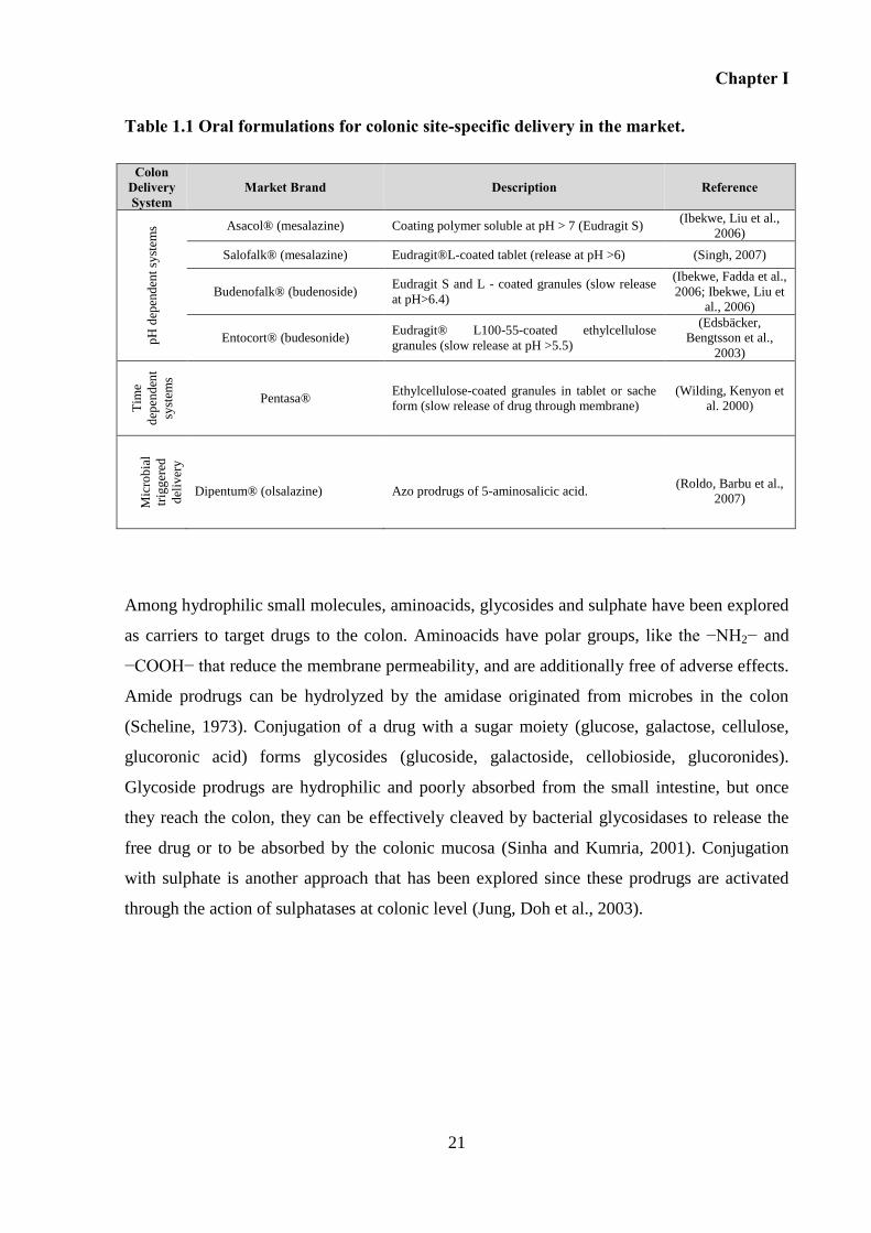
Chapter I
21
Table 1.1 Oral formulations for colonic site-specific delivery in the market.
Colon Delivery System
Market Brand Description Reference
pH
dep
end
ent
syst
ems Asacol® (mesalazine) Coating polymer soluble at pH > 7 (Eudragit S)
(Ibekwe, Liu et al.,
2006)
Salofalk® (mesalazine) Eudragit®L-coated tablet (release at pH >6) (Singh, 2007)
Budenofalk® (budenoside) Eudragit S and L - coated granules (slow release
at pH>6.4)
(Ibekwe, Fadda et al.,
2006; Ibekwe, Liu et
al., 2006)
Entocort® (budesonide) Eudragit® L100-55-coated ethylcellulose
granules (slow release at pH >5.5)
(Edsbäcker,
Bengtsson et al.,
2003)
Tim
e
dep
end
ent
syst
ems
Pentasa® Ethylcellulose-coated granules in tablet or sache
form (slow release of drug through membrane)
(Wilding, Kenyon et
al. 2000)
Mic
rob
ial
trig
ger
ed
del
iver
y
Dipentum® (olsalazine) Azo prodrugs of 5-aminosalicic acid. (Roldo, Barbu et al.,
2007)
Among hydrophilic small molecules, aminoacids, glycosides and sulphate have been explored
as carriers to target drugs to the colon. Aminoacids have polar groups, like the −NH2− and
−COOH− that reduce the membrane permeability, and are additionally free of adverse effects.
Amide prodrugs can be hydrolyzed by the amidase originated from microbes in the colon
(Scheline, 1973). Conjugation of a drug with a sugar moiety (glucose, galactose, cellulose,
glucoronic acid) forms glycosides (glucoside, galactoside, cellobioside, glucoronides).
Glycoside prodrugs are hydrophilic and poorly absorbed from the small intestine, but once
they reach the colon, they can be effectively cleaved by bacterial glycosidases to release the
free drug or to be absorbed by the colonic mucosa (Sinha and Kumria, 2001). Conjugation
with sulphate is another approach that has been explored since these prodrugs are activated
through the action of sulphatases at colonic level (Jung, Doh et al., 2003).
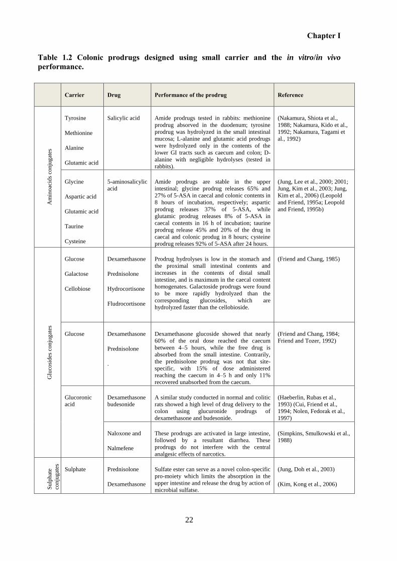
Chapter I
22
Table 1.2 Colonic prodrugs designed using small carrier and the in vitro/in vivo performance.
Carrier Drug Performance of the prodrug Reference
Am
ino
acid
s co
nju
gat
es
Tyrosine
Methionine
Alanine
Glutamic acid
Salicylic acid Amide prodrugs tested in rabbits: methionine
prodrug absorved in the duodenum; tyrosine
prodrug was hydrolyzed in the small intestinal
mucosa; L-alanine and glutamic acid prodrugs
were hydrolyzed only in the contents of the
lower GI tracts such as caecum and colon; D-
alanine with negligible hydrolyses (tested in
rabbits).
(Nakamura, Shiota et al.,
1988; Nakamura, Kido et al.,
1992; Nakamura, Tagami et
al., 1992)
Glycine
Aspartic acid
Glutamic acid
Taurine
Cysteine
5-aminosalicylic
acid
Amide prodrugs are stable in the upper
intestinal; glycine prodrug releases 65% and
27% of 5-ASA in caecal and colonic contents in
8 hours of incubation, respectively; aspartic
prodrug releases 37% of 5-ASA, while
glutamic prodrug releases 8% of 5-ASA in
caecal contents in 16 h of incubation; taurine
prodrug release 45% and 20% of the drug in
caecal and colonic produg in 8 hours; cysteine
prodrug releases 92% of 5-ASA after 24 hours.
(Jung, Lee et al., 2000; 2001;
Jung, Kim et al., 2003; Jung,
Kim et al., 2006) (Leopold
and Friend, 1995a; Leopold
and Friend, 1995b)
Glu
cosi
des
con
jug
ates
Glucose
Galactose
Cellobiose
Dexamethasone
Prednisolone
Hydrocortisone
Fludrocortisone
Prodrug hydrolyses is low in the stomach and
the proximal small intestinal contents and
increases in the contents of distal small
intestine, and is maximum in the caecal content
homogenates. Galactoside prodrugs were found
to be more rapidly hydrolyzed than the
corresponding glucosides, which are
hydrolyzed faster than the cellobioside.
(Friend and Chang, 1985)
Glucose Dexamethasone
Prednisolone
.
Dexamethasone glucoside showed that nearly
60% of the oral dose reached the caecum
between 4–5 hours, while the free drug is
absorbed from the small intestine. Contrarily,
the prednisolone prodrug was not that site-
specific, with 15% of dose administered
reaching the caecum in 4–5 h and only 11%
recovered unabsorbed from the caecum.
(Friend and Chang, 1984;
Friend and Tozer, 1992)
Glucoronic
acid
Dexamethasone
budesonide
A similar study conducted in normal and colitic
rats showed a high level of drug delivery to the
colon using glucuronide prodrugs of
dexamethasone and budesonide.
(Haeberlin, Rubas et al.,
1993) (Cui, Friend et al.,
1994; Nolen, Fedorak et al.,
1997)
Naloxone and
Nalmefene
These prodrugs are activated in large intestine,
followed by a resultant diarrhea. These
prodrugs do not interfere with the central
analgesic effects of narcotics.
(Simpkins, Smulkowski et al.,
1988)
Su
lph
ate
con
jug
ates
Sulphate Prednisolone
Dexamethasone
Sulfate ester can serve as a novel colon-specific
pro-moiety which limits the absorption in the
upper intestine and release the drug by action of
microbial sulfatse.
(Jung, Doh et al., 2003)
(Kim, Kong et al., 2006)
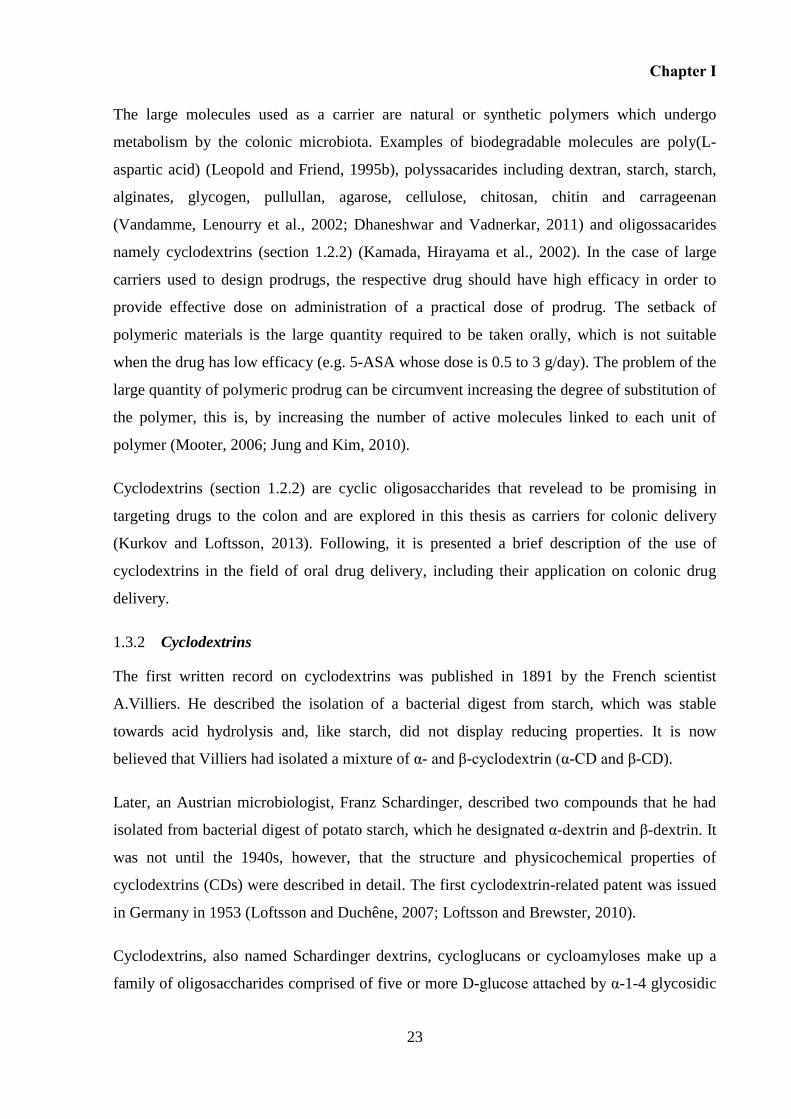
Chapter I
23
The large molecules used as a carrier are natural or synthetic polymers which undergo
metabolism by the colonic microbiota. Examples of biodegradable molecules are poly(L-
aspartic acid) (Leopold and Friend, 1995b), polyssacarides including dextran, starch, starch,
alginates, glycogen, pullullan, agarose, cellulose, chitosan, chitin and carrageenan
(Vandamme, Lenourry et al., 2002; Dhaneshwar and Vadnerkar, 2011) and oligossacarides
namely cyclodextrins (section 1.2.2) (Kamada, Hirayama et al., 2002). In the case of large
carriers used to design prodrugs, the respective drug should have high efficacy in order to
provide effective dose on administration of a practical dose of prodrug. The setback of
polymeric materials is the large quantity required to be taken orally, which is not suitable
when the drug has low efficacy (e.g. 5-ASA whose dose is 0.5 to 3 g/day). The problem of the
large quantity of polymeric prodrug can be circumvent increasing the degree of substitution of
the polymer, this is, by increasing the number of active molecules linked to each unit of
polymer (Mooter, 2006; Jung and Kim, 2010).
Cyclodextrins (section 1.2.2) are cyclic oligosaccharides that revelead to be promising in
targeting drugs to the colon and are explored in this thesis as carriers for colonic delivery
(Kurkov and Loftsson, 2013). Following, it is presented a brief description of the use of
cyclodextrins in the field of oral drug delivery, including their application on colonic drug
delivery.
Cyclodextrins 1.3.2
The first written record on cyclodextrins was published in 1891 by the French scientist
A.Villiers. He described the isolation of a bacterial digest from starch, which was stable
towards acid hydrolysis and, like starch, did not display reducing properties. It is now
believed that Villiers had isolated a mixture of α- and β-cyclodextrin (α-CD and β-CD).
Later, an Austrian microbiologist, Franz Schardinger, described two compounds that he had
isolated from bacterial digest of potato starch, which he designated α-dextrin and β-dextrin. It
was not until the 1940s, however, that the structure and physicochemical properties of
cyclodextrins (CDs) were described in detail. The first cyclodextrin-related patent was issued
in Germany in 1953 (Loftsson and Duchêne, 2007; Loftsson and Brewster, 2010).
Cyclodextrins, also named Schardinger dextrins, cycloglucans or cycloamyloses make up a
family of oligosaccharides comprised of five or more D-glucose attached by α-1-4 glycosidic
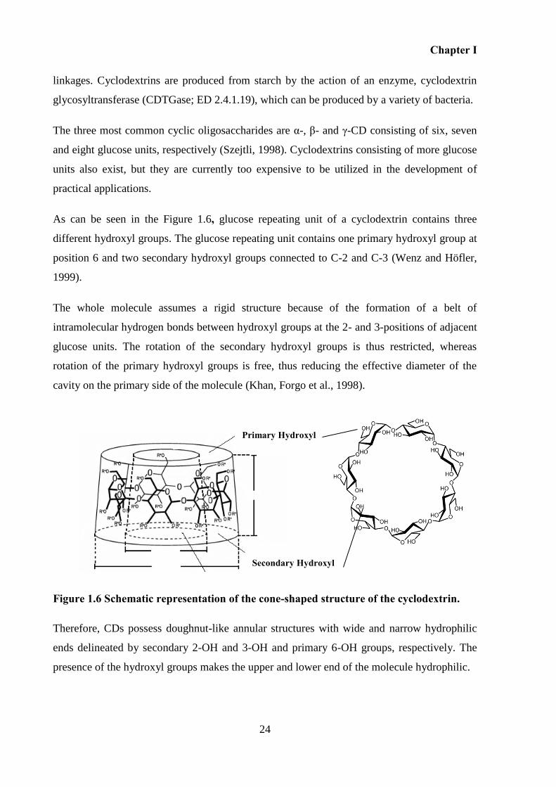
Chapter I
24
linkages. Cyclodextrins are produced from starch by the action of an enzyme, cyclodextrin
glycosyltransferase (CDTGase; ED 2.4.1.19), which can be produced by a variety of bacteria.
The three most common cyclic oligosaccharides are α-, β- and γ-CD consisting of six, seven
and eight glucose units, respectively (Szejtli, 1998). Cyclodextrins consisting of more glucose
units also exist, but they are currently too expensive to be utilized in the development of
practical applications.
As can be seen in the Figure 1.6, glucose repeating unit of a cyclodextrin contains three
different hydroxyl groups. The glucose repeating unit contains one primary hydroxyl group at
position 6 and two secondary hydroxyl groups connected to C-2 and C-3 (Wenz and Höfler,
1999).
The whole molecule assumes a rigid structure because of the formation of a belt of
intramolecular hydrogen bonds between hydroxyl groups at the 2- and 3-positions of adjacent
glucose units. The rotation of the secondary hydroxyl groups is thus restricted, whereas
rotation of the primary hydroxyl groups is free, thus reducing the effective diameter of the
cavity on the primary side of the molecule (Khan, Forgo et al., 1998).
Figure 1.6 Schematic representation of the cone-shaped structure of the cyclodextrin.
Therefore, CDs possess doughnut-like annular structures with wide and narrow hydrophilic
ends delineated by secondary 2-OH and 3-OH and primary 6-OH groups, respectively. The
presence of the hydroxyl groups makes the upper and lower end of the molecule hydrophilic.
Cavity
Primary Hydroxyl
Secondary Hydroxyl
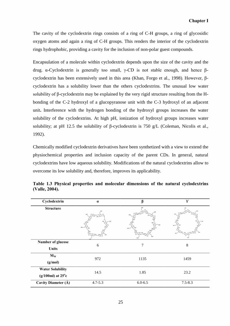
Chapter I
25
The cavity of the cyclodextrin rings consists of a ring of C-H groups, a ring of glycosidic
oxygen atoms and again a ring of C-H groups. This renders the interior of the cyclodextrin
rings hydrophobic, providing a cavity for the inclusion of non-polar guest compounds.
Encapsulation of a molecule within cyclodextrin depends upon the size of the cavity and the
drug. α-Cyclodextrin is generally too small, γ-CD is not stable enough, and hence β-
cyclodextrin has been extensively used in this area (Khan, Forgo et al., 1998). However, β-
cyclodextrin has a solubility lower than the others cyclodextrins. The unusual low water
solubility of β-cyclodextrin may be explained by the very rigid structure resulting from the H-
bonding of the C-2 hydroxyl of a glucopyranose unit with the C-3 hydroxyl of an adjacent
unit. Interference with the hydrogen bonding of the hydroxyl groups increases the water
solubility of the cyclodextrins. At high pH, ionization of hydroxyl groups increases water
solubility; at pH 12.5 the solubility of β-cyclodextrin is 750 g/L (Coleman, Nicolis et al.,
1992).
Chemically modified cyclodextrin derivatives have been synthetized with a view to extend the
physiochemical properties and inclusion capacity of the parent CDs. In general, natural
cyclodextrins have low aqueous solubility. Modifications of the natural cyclodextrins allow to
overcome its low solubility and, therefore, improves its applicability.
Table 1.3 Physical properties and molecular dimensions of the natural cyclodextrins (Valle, 2004).
Cyclodextrin α β ϒ
Structure
Number of glucose
Units 6 7 8
MW
(g/mol) 972 1135 1459
Water Solubility
(g/100ml) at 25oc 14.5 1.85 23.2
Cavity Diameter (Å) 4.7-5.3 6.0-6.5 7.5-8.3
Chapter I
26
Table 1.4 Structural and physiochemical properties of selected cyclodextrin of pharmaceutical interest (Marcus E. Brewster and Loftsson, 2007; Loftsson and Brewster, 2010).
Cyclodextrin
MS
MW (Da) Solubility in water (mg/mL at 25ºC)
2-Hydroxypropyl-α-cyclodextrin (HPαCD) 0.65 1199 >500
2-Hydroxypropyl-β-cyclodextrin (HPβCD) 0.65 1400 >600
Sulfobutylether-β-cyclodetrin sodium salt (SBEβCD) 0.9 2163 >500
Randomly methylated β-cyclodextrin (RMβCD) 1.8
0.57
1312
1191
>500
200
Maltosyl β-cyclodextrin (MβCD) 0.14 1459 >500
2-hydroxypropyl-γ-cyclodextrin 0.6 1576 >600
MS:molar degree of substitution; Mw:
Chemically modified cyclodextrins of pharmaceutical interest are classified in hydrophilic,
hydrophobic and ionizable cyclodextrin derivatives. In the hydrophilic derivatives, the
hydroxypropyl derivatives of β- and γ-cyclodextrin, the randomly methylated β-cyclodextrin,
sulfobutylether β-cyclodetrin hydroxyalkylated and branched cyclodextrin derivatives are the
most relevant ones (Loftsson, Brewster et al., 2004) The introduction of ionizable groups on
the hydroxyl groups of cyclodextrins offers a pH-dependent hydrophilic activity on such
derivatives. In this group, carboxymethyethyl cyclodextrins are the most important ionizable
derivatives.
The physicochemical properties of the CD derivatives, including their aqueous solubility and
complexation capabilities, not only depend on the structure of the appended substituent but
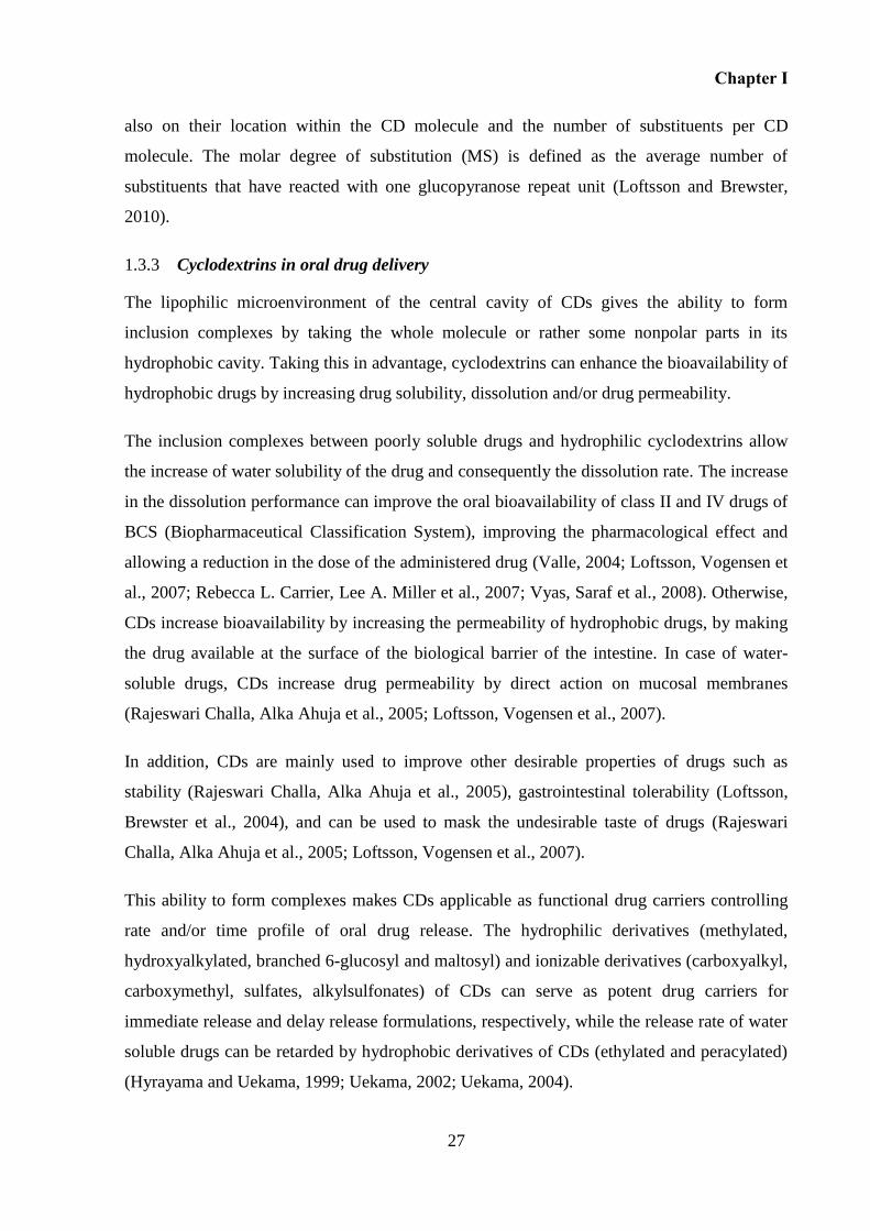
Chapter I
27
also on their location within the CD molecule and the number of substituents per CD
molecule. The molar degree of substitution (MS) is defined as the average number of
substituents that have reacted with one glucopyranose repeat unit (Loftsson and Brewster,
2010).
Cyclodextrins in oral drug delivery 1.3.3
The lipophilic microenvironment of the central cavity of CDs gives the ability to form
inclusion complexes by taking the whole molecule or rather some nonpolar parts in its
hydrophobic cavity. Taking this in advantage, cyclodextrins can enhance the bioavailability of
hydrophobic drugs by increasing drug solubility, dissolution and/or drug permeability.
The inclusion complexes between poorly soluble drugs and hydrophilic cyclodextrins allow
the increase of water solubility of the drug and consequently the dissolution rate. The increase
in the dissolution performance can improve the oral bioavailability of class II and IV drugs of
BCS (Biopharmaceutical Classification System), improving the pharmacological effect and
allowing a reduction in the dose of the administered drug (Valle, 2004; Loftsson, Vogensen et
al., 2007; Rebecca L. Carrier, Lee A. Miller et al., 2007; Vyas, Saraf et al., 2008). Otherwise,
CDs increase bioavailability by increasing the permeability of hydrophobic drugs, by making
the drug available at the surface of the biological barrier of the intestine. In case of water-
soluble drugs, CDs increase drug permeability by direct action on mucosal membranes
(Rajeswari Challa, Alka Ahuja et al., 2005; Loftsson, Vogensen et al., 2007).
In addition, CDs are mainly used to improve other desirable properties of drugs such as
stability (Rajeswari Challa, Alka Ahuja et al., 2005), gastrointestinal tolerability (Loftsson,
Brewster et al., 2004), and can be used to mask the undesirable taste of drugs (Rajeswari
Challa, Alka Ahuja et al., 2005; Loftsson, Vogensen et al., 2007).
This ability to form complexes makes CDs applicable as functional drug carriers controlling
rate and/or time profile of oral drug release. The hydrophilic derivatives (methylated,
hydroxyalkylated, branched 6-glucosyl and maltosyl) and ionizable derivatives (carboxyalkyl,
carboxymethyl, sulfates, alkylsulfonates) of CDs can serve as potent drug carriers for
immediate release and delay release formulations, respectively, while the release rate of water
soluble drugs can be retarded by hydrophobic derivatives of CDs (ethylated and peracylated)
(Hyrayama and Uekama, 1999; Uekama, 2002; Uekama, 2004).
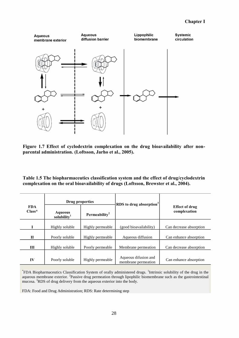
Chapter I
28
Figure 1.7 Effect of cyclodextrin complexation on the drug bioavailability after non-parental administration. (Loftsson, Jarho et al., 2005).
Table 1.5 The biopharmaceutics classification system and the effect of drug/cyclodextrin complexation on the oral bioavailability of drugs (Loftsson, Brewster et al., 2004).
FDA Class*
Drug properties RDS to drug absorption3
Effect of drug complexation Aqueous
solubility1
Permeability2
I Highly soluble Highly permeable (good bioavailability) Can decrease absorption
II Poorly soluble Highly permeable Aqueous diffusion Can enhance absorption
III Highly soluble Poorly permeable Membrane permeation Can decrease absorption
IV Poorly soluble Highly permeable Aqueous difusion and
membrane permeation Can enhance absorption
*FDA Biopharmaceutics Classification System of orally administered drugs.
1Intrinsic solubility of the drug in the
aqueous membrane exterior. 2Passive drug permeation through lipophilic biomembrane such as the gastrointestinal
mucosa. 3RDS of drug delivery from the aqueous exterior into the body.
FDA: Food and Drug Administration; RDS: Rate determining step
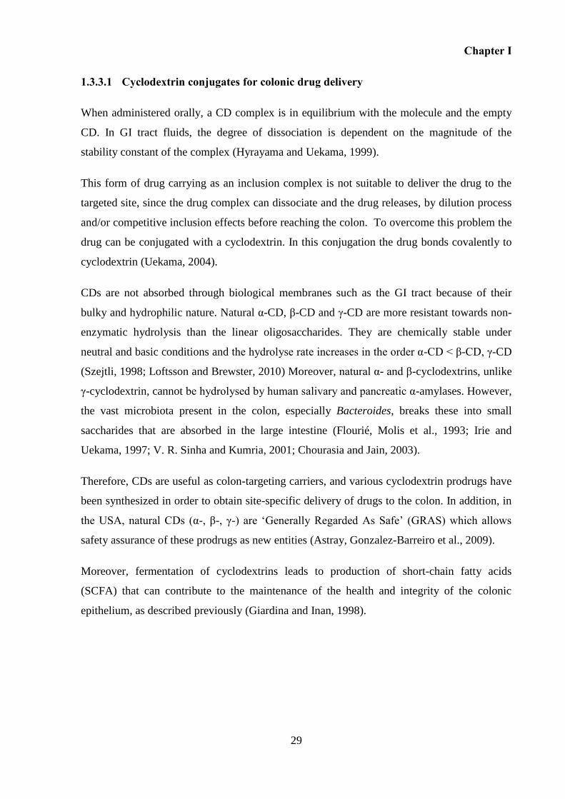
Chapter I
29
1.3.3.1 Cyclodextrin conjugates for colonic drug delivery
When administered orally, a CD complex is in equilibrium with the molecule and the empty
CD. In GI tract fluids, the degree of dissociation is dependent on the magnitude of the
stability constant of the complex (Hyrayama and Uekama, 1999).
This form of drug carrying as an inclusion complex is not suitable to deliver the drug to the
targeted site, since the drug complex can dissociate and the drug releases, by dilution process
and/or competitive inclusion effects before reaching the colon. To overcome this problem the
drug can be conjugated with a cyclodextrin. In this conjugation the drug bonds covalently to
cyclodextrin (Uekama, 2004).
CDs are not absorbed through biological membranes such as the GI tract because of their
bulky and hydrophilic nature. Natural α-CD, β-CD and γ-CD are more resistant towards non-
enzymatic hydrolysis than the linear oligosaccharides. They are chemically stable under
neutral and basic conditions and the hydrolyse rate increases in the order α-CD < β-CD, γ-CD
(Szejtli, 1998; Loftsson and Brewster, 2010) Moreover, natural α- and β-cyclodextrins, unlike
γ-cyclodextrin, cannot be hydrolysed by human salivary and pancreatic α-amylases. However,
the vast microbiota present in the colon, especially Bacteroides, breaks these into small
saccharides that are absorbed in the large intestine (Flourié, Molis et al., 1993; Irie and
Uekama, 1997; V. R. Sinha and Kumria, 2001; Chourasia and Jain, 2003).
Therefore, CDs are useful as colon-targeting carriers, and various cyclodextrin prodrugs have
been synthesized in order to obtain site-specific delivery of drugs to the colon. In addition, in
the USA, natural CDs (α-, β-, γ-) are ‘Generally Regarded As Safe’ (GRAS) which allows
safety assurance of these prodrugs as new entities (Astray, Gonzalez-Barreiro et al., 2009).
Moreover, fermentation of cyclodextrins leads to production of short-chain fatty acids
(SCFA) that can contribute to the maintenance of the health and integrity of the colonic
epithelium, as described previously (Giardina and Inan, 1998).
Chapter I
30
Figure 1.8 Schematic comparison of the digestion of natural α-CD, β-CD and γ-CD after oral administration. Cyclodextrins are slowly hydrolysed by α-amylases but relatively rapidly digested by bacteria. Adapted from (Kurkov and Loftsson, 2013).
Drug molecules were selectively conjugated using the primary or secondary hydroxyl groups
of CDs through an ester or amide linkage. Therefore, a limited number of prodrugs can be
formed due to the chemical linkage required in their formation.
Several drugs have been combined with CDs to synthesize prodrugs enabling colonic specific
release (Table 1.6). Cyclodextin conjugates of non-steroidal anti-inflammatory drugs include:
byphenylacetic acid (Uekama, Minami et al., 1997a; Minami, Hirayama et al., 1998),
ketoprofen (Kamada, Hirayama et al., 2002), etodolac (Rizzarelli, Vecchio et al., 2007),
mefenamic acid (Dev, Mhaske et al., 2007), naproxen, flurbiprofen, sulindac (El-Kamel,
A.M.Abdel-Aziz et al., 2008), ibuprofen, indomethacin (Namazi, Bahrami et al., 2005); short-
chain fatty acids such as n- butyric acid (Hirayma, Ogata et al., 2000); steroidal drugs like
prednisolone (Yano, Hirayama et al., 2001; Yano, Hirayama et al., 2002), budesonide
(Prabhu, D.Satyanarayana et al., 2009); a drug for the treatment of inflammatory bowel
disease, 5-aminosalicylic acid (Zou, Okamoto et al., 2005). There are also β-lactam antibiotics
like ampicillin, amoxicillin, cephalexin (Namazi and Kanani, 2007); and recently anticancer
drugs like 5-fluoracil (Udo, Hokonohara et al., 2010), and scuterallin, a flavonoid with
anticancer activity (Yang, Zhao et al., 2013). All these examples have been designed
Chapter I
31
anticipating new candidates for colon-specific delivery prodrugs but, asfar as we know, there
are no reports of cyclodextrin conjugate with diclofenac. In the case of prednisolone and
budesonide a linker molecule, succinic acid, was used between drug and cyclodextrin,
forming esters at both ends of the linker molecule (Yano, Hirayama et al., 2001; Prabhu,
D.Satyanarayana et al., 2009). Modification of β-cyclodextrin with water soluble polymer
poly(ethylene glycol) was used to produce conjugates of antibiotics (Namazi and Kanani,
2007) and conjugates of indomethacin and ibuprofen (Namazi, Bahrami et al., 2005).
In most of the drug-cyclodextrin conjugates, namely those with a ester linkage, the drug was
released in the contents of large intestine, whereas only minor chemical hydrolysis occured in
the upper GI tract (Uekama, 2004; Kurkov and Loftsson, 2013). These promising results
highlight cyclodextrin-based prodrugs as an area for further research.
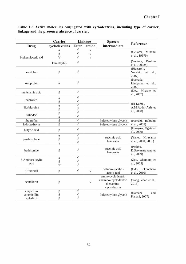
Chapter I
32
Table 1.6 Active molecules conjugated with cyclodextrins, including type of carrier, linkage and the presence/ absence of carrier.
Carrier Linkage Spacer/ intermediate Reference Drug cyclodextrins Ester amide
biphenylacetic cid
α √ √ (Uekama, Minami
et al., 1997b) β √ √
ϒ √ √
Dimethyl-β √ (Ventura, Paolino
et al., 2003a)
etodolac β √
(Rizzarelli,
Vecchio et al.,
2007)
ketoprofen α √
(Kamada,
Hirayama et al.,
2002)
mefenamic acid β √ (Dev, Mhaske et
al., 2007)
naproxen α √
(El-Kamel,
A.M.Abdel-Aziz et
al., 2008)
β √
flurbiprofen α √
β √
sulindac β √
α √
ibuprofen β √ Poly(ethylene glycol) (Namazi, Bahrami
et al., 2005) indomethacin β √ Poly(ethylene glycol)
butyric acid β √ (Hirayma, Ogata et
al., 2000)
prednisolone
α √ succinic acid
hemiester
(Yano, Hirayama
et al., 2000; 2001) β √
γ √
budesonide β √ succinic acid
hemiester
(Prabhu,
D.Satyanarayana et
al., 2009)
5-Aminosalicylic
acid
α √ (Zou, Okamoto et
al., 2005) β √
ϒ √
5-fluoracil β √ √ 5-fluorouracil-1-
acteic acid
(Udo, Hokonohara
et al., 2010)
scutellarin β √
amino-cyclodextrin
enamino- cyclodextrin
dienamino-
cyclodextrin
(Yang, Zhao et al.,
2013)
ampicillin β √
Poly(ethylene glycol) (Namazi and
Kanani, 2007) amoxicillin β √
caphalexin β √
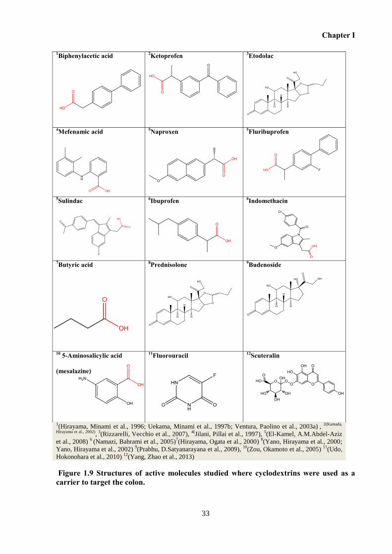
Chapter I
33
1(Hirayama, Minami et al., 1996; Uekama, Minami et al., 1997b; Ventura, Paolino et al., 2003a)
,
2(Kamada,
Hirayama et al., 2002), 3
(Rizzarelli, Vecchio et al., 2007), 4(
Jilani, Pillai et al., 1997), 5(El-Kamel, A.M.Abdel-Aziz
et al., 2008) 6
(Namazi, Bahrami et al., 2005)7(Hirayama, Ogata et al., 2000)
8(Yano, Hirayama et al., 2000;
Yano, Hirayama et al., 2002) 9(Prabhu, D.Satyanarayana et al., 2009),
10(Zou, Okamoto et al., 2005)
11(Udo,
Hokonohara et al., 2010) 12
(Yang, Zhao et al., 2013)
Figure 1.9 Structures of active molecules studied where cyclodextrins were used as a carrier to target the colon.
1Biphenylacetic acid
2Ketoprofen
3Etodolac
4Mefenamic acid
5Naproxen
5Fluribuprofen
5Sulindac 6Ibuprofen
6Indomethacin
7Butyric acid
8Prednisolone
9Budenoside
10 5-Aminosalicylic acid
(mesalazine)
11Fluorouracil 12Scuteralin
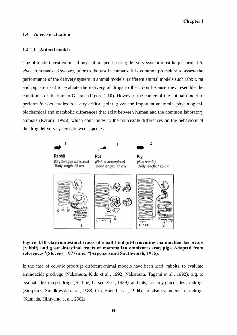
Chapter I
34
1.4 In vivo evaluation
1.4.1.1 Animal models
The ultimate investigation of any colon-specific drug delivery system must be performed in
vivo, in humans. However, prior to the test in humans, it is common procedure to assess the
performance of the delivery system in animal models. Different animal models such rabbit, rat
and pig are used to evaluate the delivery of drugs to the colon because they resemble the
conditions of the human GI tract (Figure 1.10). However, the choice of the animal model to
perform in vivo studies is a very critical point, given the important anatomic, physiological,
biochemical and metabolic differences that exist between human and the common laboratory
animals (Kararli, 1995), which contributes to the noticeable differences on the behaviour of
the drug delivery systems between species.
Figure 1.10 Gastrointestinal tracts of small hindgut-fermenting mammalian herbivore (rabbit) and gastrointestinal tracts of mammalian omnivores (rat, pig). Adapted from references 1(Stevens, 1977) and 2(Argenzio and Southworth, 1975).
In the case of colonic prodrugs different animal models have been used: rabbits, to evaluate
aminoacids prodrugs (Nakamura, Kido et al., 1992; Nakamura, Tagami et al., 1992); pig, to
evaluate dextran prodrugs (Harboe, Larsen et al., 1989); and rats, to study glucosides prodrugs
(Simpkins, Smulkowski et al., 1988; Cui, Friend et al., 1994) and also cyclodextrin prodrugs
(Kamada, Hirayama et al., 2002).
Chapter I
35
Below, it is described the main anatomic and physiological characteristics including
differences on the pH (Table 1.7) and microbiota (Table 1.8) along the GI tract of each animal
model considered thereafter in this dissertation.
1.4.1.2 Stomach
Like humans, pigs and rabbits have a glandular type stomach while the stomach of rats is
divided in two main sections: glandular and non-glandular. The non-glandular section also
called forestomach portion is an entry from esophagus and the site for storage and bacterial
digestion (Kararli, 1995).
In rabbits, stomach is the major site of digestion (Yu, Chiou et al., 2000). Its stomach is
normally never empty, herein the gastric emptying time is highly variable. After 24 hour of
fasting, it has been shown that the stomach of an adult rabbit is still 50% full (Harboe, Larsen
et al., 1989; Davies, Certzoomed et al., 2003).
The stomach of pigs is two to three times larger than the stomach of humans and the cardiac
mucosa occupies a greater portion of the stomach compared to the human stomach (Kararli,
1995).
1.4.1.3 Small intestine, pancreas and gall bladder
The small intestine is the major site of absorption, due to its enormous surface and due to
existence of villi and microvilli in all animals (Peetersg, Charlier et al., 1985; Kararli, 1995).
In rabbits, the end of the ileum is expanded into a thick walled called sacculus rotundus,
which seems to have an immunological function and is only found in Lagomorphs (Davies,
Certzoomed et al., 2003; Merchant, Mcconnell et al., 2011). Like humans, rabbits are poor
biliary excreters (Toutain, Ferran et al., 2010). Otherwise, rats are good biliary excreters, they
do not have a gall bladder, and thus the bile enters the dudodenum continuously as it is made
(Toutain, Ferran et al., 2010).
1.4.1.4 Caecum and colon
The large intestine provides a chamber for the final phase of digestion, for both herbivores
(rabbits) and omnivorous (rats and pigs). The pig, rabbit and rat have a well-defined caecum
whereas in man the caecum is very small and is continuous with the colon.
Chapter I
36
The colon of pig consists on, ascending, transverse and a portion of the descending colon
coiled tightly into a series of centripetal and centrifugal coils. Pig has an extensive sacculation
(haustra) and elongation of the large intestine (Swindle, Makin et al., 2011). The length and
the capacity of the large intestine of pigs are about 1.5 - 3 times larger than that of humans
(Christensen, Knudsen et al., 1999).
In rats, the caecum is the main site of fermentation and represents 26% of the length of the
large intestine. The colon of rats is not sacculated nor long and it is mainly important for re-
absorption of water and formation of faecal pellets (Desesso and Jacobson, 2001). Rats pratice
coprofagy, i.e. they ingest part of their faeces, which allows the digestion of faecal bacteria
and the intestinal absorption of vitamins and microbial protein (Hawksworth, Drasar et al.,
1971).
In rabbits, the thin-walled caecum is a large organ (40–60% of the total volume of the
gastrointestinal tract) that ends in an appendix which secretes bicarbonate ions into the caecal
lumen, which are thought to act as a buffer for the volatile fatty acids produced by caecal
fermentation (Davies, Certzoomed et al., 2003). The colon of rabbits is sacullated, and
consists of a proximal and distal colon separated by a short transverse colon, and it ends with
the fusus coli, which is responsible for the peristaltic waves on the colon (Davies, Certzoomed
et al., 2003). Rabbits practice caecotrophy that is considered more sophisticated than
coprophagy (Hörnicke, 1981). Rabbits produce two types of faeces: soft faeces, also named
caecotrophs that are rich in vitamins, protein and minerals and regular hard faeces. The
process of excretion of caecotrophs and immediate re-ingesting directly from the anus gives a
second opportunity to benefit from valuable nutrient substances. Such physiological recycling
usually occurs twice each day (Toutain, Ferran et al., 2010).
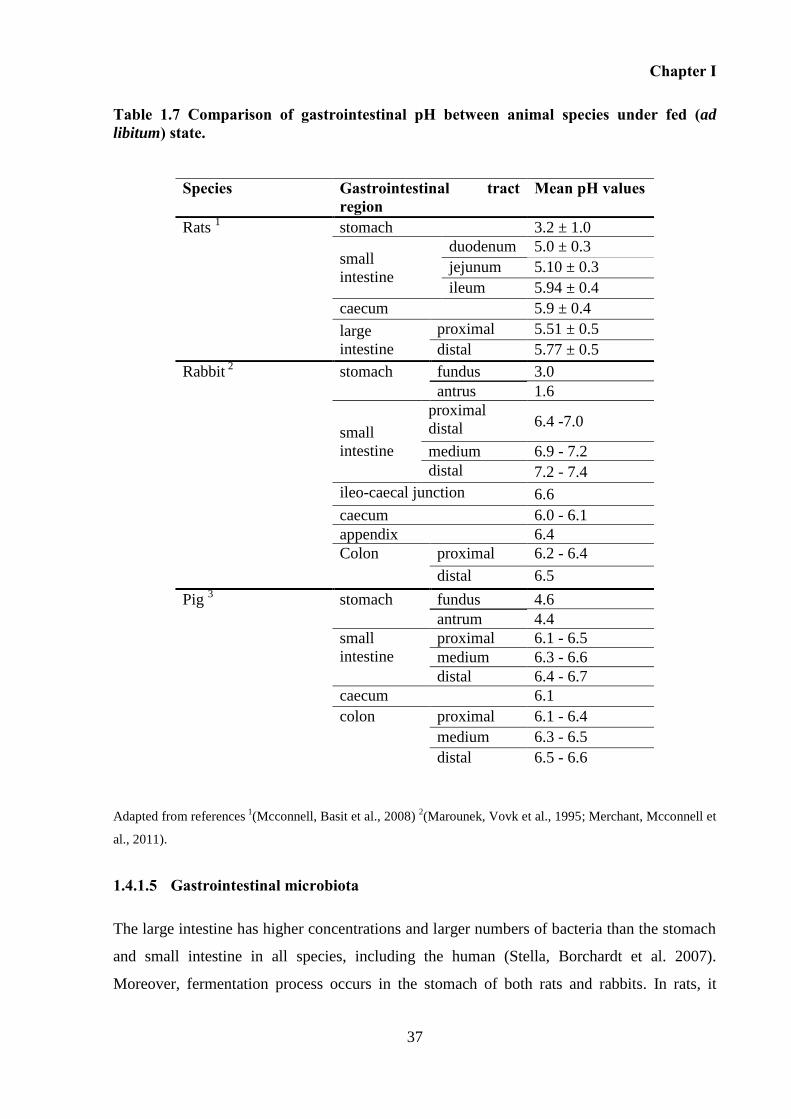
Chapter I
37
Table 1.7 Comparison of gastrointestinal pH between animal species under fed (ad
libitum) state.
Adapted from references 1
(Mcconnell, Basit et al., 2008) 2(Marounek, Vovk et al., 1995; Merchant, Mcconnell et
al., 2011).
1.4.1.5 Gastrointestinal microbiota
The large intestine has higher concentrations and larger numbers of bacteria than the stomach
and small intestine in all species, including the human (Stella, Borchardt et al. 2007).
Moreover, fermentation process occurs in the stomach of both rats and rabbits. In rats, it
Species Gastrointestinal tract region
Mean pH values
Rats 1 stomach 3.2 ± 1.0
small
intestine
duodenum 5.0 ± 0.3
jejunum 5.10 ± 0.3
ileum 5.94 ± 0.4
caecum 5.9 ± 0.4
large
intestine
proximal 5.51 ± 0.5
distal 5.77 ± 0.5
Rabbit 2 stomach fundus 3.0
antrus 1.6
small
intestine
proximal
distal 6.4 -7.0
medium 6.9 - 7.2
distal 7.2 - 7.4
ileo-caecal junction 6.6
caecum 6.0 - 6.1
appendix 6.4
Colon proximal 6.2 - 6.4
distal 6.5
Pig 3 stomach fundus 4.6
antrum 4.4
small
intestine
proximal 6.1 - 6.5
medium 6.3 - 6.6
distal 6.4 - 6.7
caecum 6.1
colon
proximal 6.1 - 6.4
medium 6.3 - 6.5
distal 6.5 - 6.6
Chapter I
38
occurs due to ingestion of coprophages and in rabbits due to ingestion of caecotrophes. In the
stomach of rats bacteria are autochthonous, i.e. permanently attached to the epithelium, and
the faeces ingested only provide the buffer necessary for fermentation process (Hörnicke,
1981). The presence of bacteria in stomach of rats is explained by the existence of a non-
glandular compartment in the stomach which permits the bacteria to proliferate in this non-
acid producing region. Consequently, rats have a large number of bacteria along the whole
length of the gastrointestinal tract. The main organisms in stomach and small intestine of rats
are Lactobacilli (Firmicutes Phylum), Bacteroides (Bacteroidetes Phylum) and Bifidobacteria
(Actinobacteria Phylum) (Hawksworth, Drasar et al., 1971; Tannock, 1999) These organisms
outnumber the enterobacteria (Proteobacteria phylum), mainly Escherichia coli by a factor of
more than 102 (Hawksworth, Drasar et al., 1971).
In rabbits, caecothrophs are responsible for the existence of bacteria at the level of the
stomach (Griffiths and Davies, 1963). The gastric pH in rabbits is generally maintained
between 1 and 2 and increase to 3 in presence of caecotrophs (Davies, Certzoomed et al.,
2003). This gastric pH avoids the occurrence of fermentation in the gastric luminal of rabbits.
Fermentation takes place inside caecothrophs, which are unaffected by the luminal pH, given
the fact that they are covered by a mucous membrane. This membrane remains
morphologically intact once caecotrophs are swallowed without mastication (Griffiths and
Davies, 1963; Davies, Certzoomed et al., 2003). The bacteria, maintained at high pH by
phosphate buffer, produces amylase which, together with the salivary and food amylases,
degrades food starch to maltose and glucose. Thus, in rabbits, the caecotrophes provide both
the buffer and the active microbes in fermentation (Griffiths and Davies, 1963). Although
caecotrophy, the flora of rabbits at the level of the upper gastrointestinal tract is very simple
and similar to that in man and the number of bacteria is much lower than in rats (Table 1.8)
(Hawksworth, Drasar et al., 1971). The prevalent organisms in the upper gastrointestinal tract
of rabbits are Bacteroids (Bacteroidetes Phylum) and Bifidobacterias (Actinobacteria
Phylum), like in human (Kararli, 1995).
Wkswortbh et al. reported the principal bacteria present in the large intestine of rats, rabbits
and human. Microbiota present in this part of the gut of these animals belong to the
Firmicutes Phylum (Enterococcus, Lactobacillus spp), Bacteroidetes Phylum (Bacteroids
spp), Proteobacteria Phylum (Enterobacteriaceae spp, mainly Escherichia coli) and
Actinobacteria Phylum (Bifidobacterium spp). The strictly anaerobic bacteria (Bacteroides
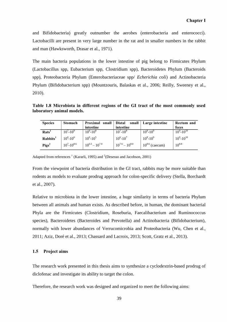
Chapter I
39
and Bifidobacteria) greatly outnumber the aerobes (enterobacteria and enterococci).
Lactobacilli are present in very large number in the rat and in smaller numbers in the rabbit
and man (Hawksworth, Drasar et al., 1971).
The main bacteria populations in the lower intestine of pig belong to Firmicutes Phylum
(Lactobacillus spp, Eubacterium spp, Clostridium spp), Bacteroidetes Phylum (Bacteroids
spp), Proteobacteria Phylum (Enterobacteriaceae spp/ Echerichia coli) and Actinobacteria
Phylum (Bifidobacterium spp) (Mountzouris, Balaskas et al., 2006; Reilly, Sweeney et al.,
2010).
Table 1.8 Microbiota in different regions of the GI tract of the most commonly used laboratory animal models.
Species Stomach Proximal small intestine
Distal small intestine
Large intestine Rectum and feces
Rats1 107-10
9 10
6-10
8 10
7-10
8 10
8-10
9 10
9-10
10
Rabbits1 100-10
6 10
0-10
5 10
6-10
7 10
8-10
9 10
9-10
10
Pigs2 107-10
8.6 10
6.5 – 10
7.0 10
7.6 – 10
8.0 10
8.6 (caecum) 10
8.8
Adapted from references 1 (Kararli, 1995) and
2(Desesso and Jacobson, 2001)
From the viewpoint of bacteria distribution in the GI tract, rabbits may be more suitable than
rodents as models to evaluate prodrug approach for colon-specific delivery (Stella, Borchardt
et al., 2007).
Relative to microbiota in the lower intestine, a huge similarity in terms of bacteria Phylum
between all animals and human exists. As described before, in human, the dominant bacterial
Phyla are the Firmicutes (Clostridium, Roseburia, Faecalibacterium and Ruminococcus
species), Bacteroidetes (Bacteroides and Prevotella) and Actinobacteria (Bifidobacterium),
normally with lower abundances of Verrucomicrobia and Proteobacteria (Wu, Chen et al.,
2011; Aziz, Doré et al., 2013; Chassard and Lacroix, 2013; Scott, Gratz et al., 2013).
1.5 Project aims
The research work presented in this thesis aims to synthesize a cyclodextrin-based prodrug of
diclofenac and investigate its ability to target the colon.
Therefore, the research work was designed and organized to meet the following aims:
Chapter I
40
- Development of a method to prepare cyclodextrin conjugates of diclofenac in a short
reaction time and physical-chemical characterization obtained molecules (Chapter II).
This would provide a reliable method to produce efficiently and conveniently new
cyclodextrin conjugates in adequate quantities to proceed for further studies.
- In vitro assessment of the stability of diclofenac-cyclodextrin conjugates in the upper
gastrointestinal tract using simulated gastrointestinal fluids and animal fluids to
predict in vivo behaviour. Investigate the ability of the conjugate to site-specific
release of diclofenac on the colon and compare the performance of this type of
prodrug with the classic prodrug sulfasalazine. This would allow accessing the inter-
species variability on the metabolism of produgs and the establishment of the best
animal model to perform the in vivo studies (Chapter III).
- Investigate the influence of different feeding regimens on the distribution and pH
contents along the gut and on the metabolism of cyclodextrin conjugates (Chapter IV).
This would provide information necessary to understand the importance of the feeding
state on the drug disposition.
- Investigate the in vivo behaviour of the cyclodextrin conjugates versus sulfasalazine
and determine the potential of cyclodextrin to be used as a carrier to colon specific
delivery (Chapter V).
CHAPTER II
SYNTHESIS AND CHEMICAL CHARACTERIZATION OF
DICLOFENAC-CYCLODEXTRIN CONJUGATES

Chapter II
43
2 SYNTHESIS AND CHEMICAL CHARACTERIZATION OF DICLOFENAC-
CYCLODEXTRIN CONJUGATES
2.1 Introduction
Natural cyclodextrins (CDs), namely α-, β- and γ- have been studied as carriers for colonic
drug delivery. Due to their size and hidrophilicity, they are not absorbed through the intestine.
Cyclodextrins, α- and β- , contrarily to γ-cyclodextrin can pass intact through the upper
gastrointestinal tract and only when reach the colon they are metabolized, as described in
Chapter I. Thus, α- and β-cyclodextrins are the most interesting cyclodextrins to work as
carriers.
To develop a stable colonic specific prodrug, it is important to produce a linkage between
CDs and a drug. The linkage should be sufficiently stable to survive the transit through the
stomach and small intestine, but also susceptible to be broken only at the colon level where
the drug is released.
However, to generate this linkage between a cyclodextrin and a drug, several challenges
related with the own CDs and the structure of the drug need to be addressed. Not all the drugs
that are candidates to colon delivery can be modified to a prodrug. For a drug to become a
colon-specific prodrug, it needs to have an appropriate functional group suitable for
conjugation (Jung and Kim, 2010). Otherwise, synthesis of CDs conjugates is associated with
a wide variety of physical-chemical processes that can succeed, owing to the presence of the
hydrophobic cavity and a large number of hydroxyl groups that provides molecule
nucleophilicity.
For this reason, what follows is a description of the main physical-chemical characteristics of
cyclodextrins as a molecule, as well as of diclofenac, with the purpose of link both in order to
get specific delivery of the anti-inflammatory drug in the colon.
Cyclodextrin chemistry 2.1.1
The α-, β-, and γ-cyclodextrin contain 18, 21 and 24 hydroxyl groups, respectively. Thus,
each cyclodextrin moiety potentially has several sites of modification. The available
modifications of cyclodextrin will occur at the hydroxyl groups, taking advantage of their
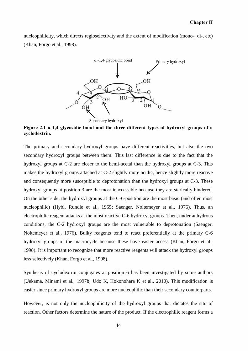
Chapter II
44
nucleophilicity, which directs regioselectivity and the extent of modification (mono-, di-, etc)
(Khan, Forgo et al., 1998).
Figure 2.1 α-1,4 glycosidic bond and the three different types of hydroxyl groups of a cyclodextrin.
The primary and secondary hydroxyl groups have different reactivities, but also the two
secondary hydroxyl groups between them. This last difference is due to the fact that the
hydroxyl groups at C-2 are closer to the hemi-acetal than the hydroxyl groups at C-3. This
makes the hydroxyl groups attached at C-2 slightly more acidic, hence slightly more reactive
and consequently more susceptible to deprotonation than the hydroxyl groups at C-3. These
hydroxyl groups at position 3 are the most inaccessible because they are sterically hindered.
On the other side, the hydroxyl groups at the C-6-position are the most basic (and often most
nucleophilic) (Hybl, Rundle et al., 1965; Saenger, Noltemeyer et al., 1976). Thus, an
electrophilic reagent attacks at the most reactive C-6 hydroxyl groups. Then, under anhydrous
conditions, the C-2 hydroxyl groups are the most vulnerable to deprotonation (Saenger,
Noltemeyer et al., 1976). Bulky reagents tend to react preferentially at the primary C-6
hydroxyl groups of the macrocycle because these have easier access (Khan, Forgo et al.,
1998). It is important to recognize that more reactive reagents will attack the hydroxyl groups
less selectively (Khan, Forgo et al., 1998).
Synthesis of cyclodextrin conjugates at position 6 has been investigated by some authors
(Uekama, Minami et al., 1997b; Udo K, Hokonohara K et al., 2010). This modification is
easier since primary hydroxyl groups are more nucleophilic than their secondary counterparts.
However, is not only the nucleophilicity of the hydroxyl groups that dictates the site of
reaction. Other factors determine the nature of the product. If the electrophilic reagent forms a
Primary hydroxyl
Secondary hydroxyl
α -1,4-glycosidic bond
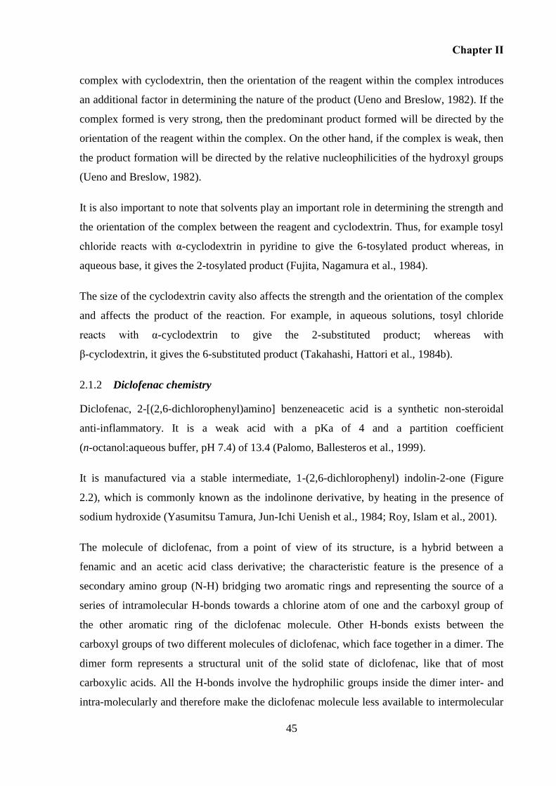
Chapter II
45
complex with cyclodextrin, then the orientation of the reagent within the complex introduces
an additional factor in determining the nature of the product (Ueno and Breslow, 1982). If the
complex formed is very strong, then the predominant product formed will be directed by the
orientation of the reagent within the complex. On the other hand, if the complex is weak, then
the product formation will be directed by the relative nucleophilicities of the hydroxyl groups
(Ueno and Breslow, 1982).
It is also important to note that solvents play an important role in determining the strength and
the orientation of the complex between the reagent and cyclodextrin. Thus, for example tosyl
chloride reacts with α-cyclodextrin in pyridine to give the 6-tosylated product whereas, in
aqueous base, it gives the 2-tosylated product (Fujita, Nagamura et al., 1984).
The size of the cyclodextrin cavity also affects the strength and the orientation of the complex
and affects the product of the reaction. For example, in aqueous solutions, tosyl chloride
reacts with α-cyclodextrin to give the 2-substituted product; whereas with
β-cyclodextrin, it gives the 6-substituted product (Takahashi, Hattori et al., 1984b).
Diclofenac chemistry 2.1.2
Diclofenac, 2-[(2,6-dichlorophenyl)amino] benzeneacetic acid is a synthetic non-steroidal
anti-inflammatory. It is a weak acid with a pKa of 4 and a partition coefficient
(n-octanol:aqueous buffer, pH 7.4) of 13.4 (Palomo, Ballesteros et al., 1999).
It is manufactured via a stable intermediate, 1-(2,6-dichlorophenyl) indolin-2-one (Figure
2.2), which is commonly known as the indolinone derivative, by heating in the presence of
sodium hydroxide (Yasumitsu Tamura, Jun-Ichi Uenish et al., 1984; Roy, Islam et al., 2001).
The molecule of diclofenac, from a point of view of its structure, is a hybrid between a
fenamic and an acetic acid class derivative; the characteristic feature is the presence of a
secondary amino group (N-H) bridging two aromatic rings and representing the source of a
series of intramolecular H-bonds towards a chlorine atom of one and the carboxyl group of
the other aromatic ring of the diclofenac molecule. Other H-bonds exists between the
carboxyl groups of two different molecules of diclofenac, which face together in a dimer. The
dimer form represents a structural unit of the solid state of diclofenac, like that of most
carboxylic acids. All the H-bonds involve the hydrophilic groups inside the dimer inter- and
intra-molecularly and therefore make the diclofenac molecule less available to intermolecular
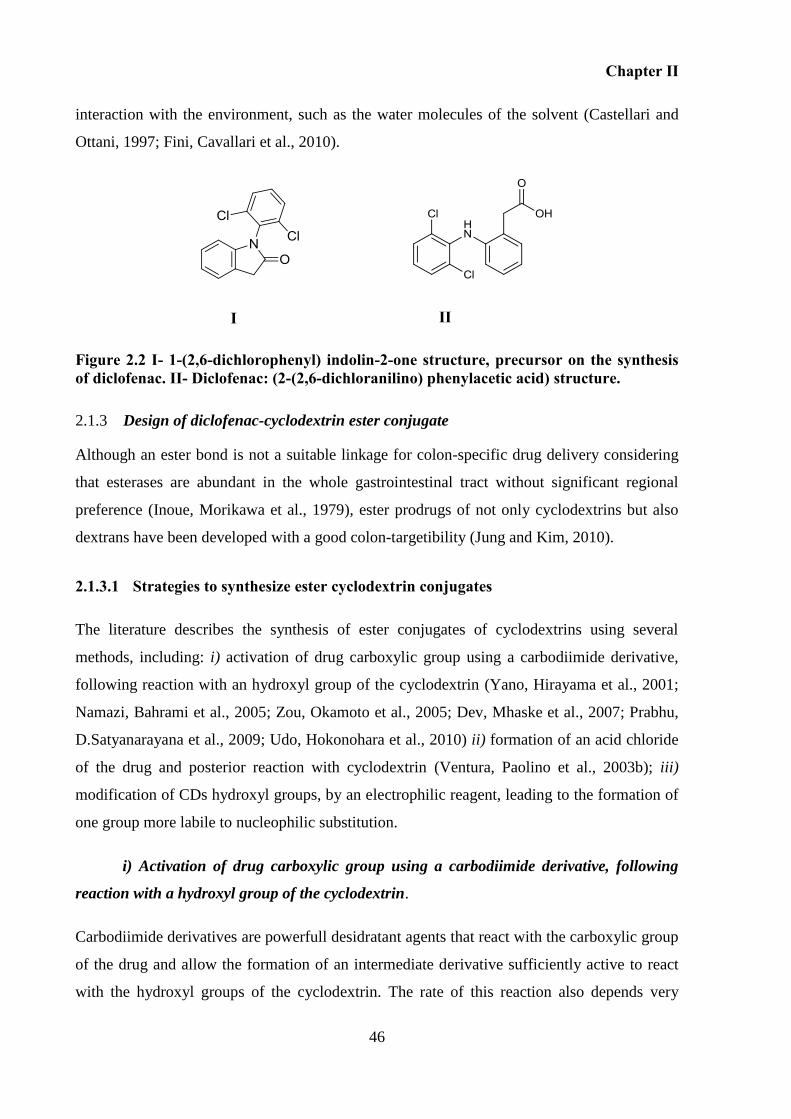
Chapter II
46
interaction with the environment, such as the water molecules of the solvent (Castellari and
Ottani, 1997; Fini, Cavallari et al., 2010).
Figure 2.2 I- 1-(2,6-dichlorophenyl) indolin-2-one structure, precursor on the synthesis of diclofenac. II- Diclofenac: (2-(2,6-dichloranilino) phenylacetic acid) structure.
Design of diclofenac-cyclodextrin ester conjugate 2.1.3
Although an ester bond is not a suitable linkage for colon-specific drug delivery considering
that esterases are abundant in the whole gastrointestinal tract without significant regional
preference (Inoue, Morikawa et al., 1979), ester prodrugs of not only cyclodextrins but also
dextrans have been developed with a good colon-targetibility (Jung and Kim, 2010).
2.1.3.1 Strategies to synthesize ester cyclodextrin conjugates
The literature describes the synthesis of ester conjugates of cyclodextrins using several
methods, including: i) activation of drug carboxylic group using a carbodiimide derivative,
following reaction with an hydroxyl group of the cyclodextrin (Yano, Hirayama et al., 2001;
Namazi, Bahrami et al., 2005; Zou, Okamoto et al., 2005; Dev, Mhaske et al., 2007; Prabhu,
D.Satyanarayana et al., 2009; Udo, Hokonohara et al., 2010) ii) formation of an acid chloride
of the drug and posterior reaction with cyclodextrin (Ventura, Paolino et al., 2003b); iii)
modification of CDs hydroxyl groups, by an electrophilic reagent, leading to the formation of
one group more labile to nucleophilic substitution.
i) Activation of drug carboxylic group using a carbodiimide derivative, following
reaction with a hydroxyl group of the cyclodextrin.
Carbodiimide derivatives are powerfull desidratant agents that react with the carboxylic group
of the drug and allow the formation of an intermediate derivative sufficiently active to react
with the hydroxyl groups of the cyclodextrin. The rate of this reaction also depends very
I II
Chapter II
47
much on the nucleophilicity of their anions. Dicyclohexylcarbodiimide (DCC) has been the
most commonly used carbodiimide and is regarded as a standard (Michael Smith, J.G. Moffatt
et al., 1958). It is a non-aqueous carbodiimide that has been used to synthesize cyclodextrin
conjugates of mefenamic acid (Dev, Mhaske et al., 2007), ibuprofen, indomethacin (Namazi,
Bahrami et al., 2005) and β-lactam antibiotics (Namazi and Kanani, 2007).
Carbonyldiimidazole (CDI) represents an alternative carbodiimide, but it is less frequently
used, most probably due to cost and its sensitivity to moisture. This carbodiimide has been
used to form cyclodextrin conjugates of 5-fluorouracil-1-acetic acid (Udo K, Hokonohara K et
al., 2010), 5-aminosalicylic acid (Zou, Okamoto et al., 2005) and prednisolone (Yano,
Hirayama et al., 2001).
Figure 2.3 Synthetic scheme of synthesis of drug-cyclodextrin conjugate by a carboodimiide activation of the drug.
A water soluble carbodiimide such as dimethylaminopropyl)-N′-ethylcarbodiimide
hydrochloride (EDAC) can be used to facilate the purification of the product (Albert and
Hamilton, 2001).
Based on the literature, reactions between acids and carbodiimides in general occurs rapidly
(Khorana, 1953). 4-dimethylaminopyridine (DMAP) (pKa=9.70) can assist on the
Drug Drug-intermediate
O-acylisourea intermediate Cyclodextrin
Carbodiimide
Drug-cyclodextrin conjugate
Step 1
Step 2
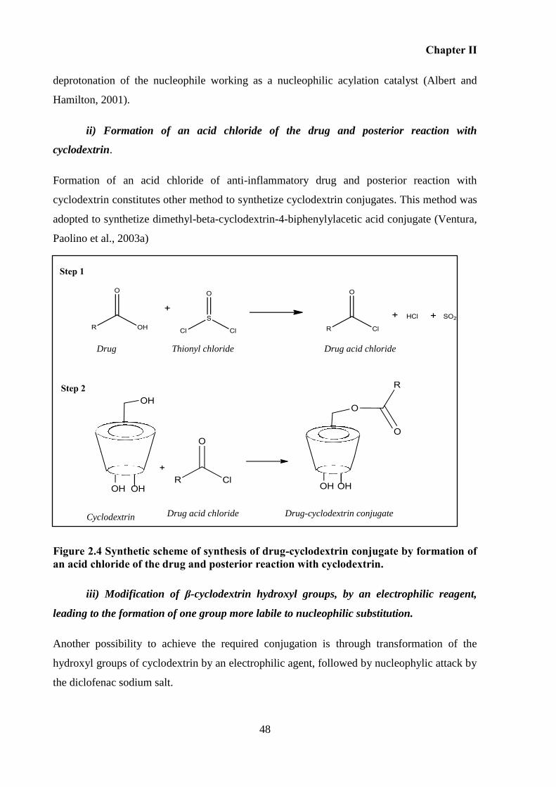
Chapter II
48
deprotonation of the nucleophile working as a nucleophilic acylation catalyst (Albert and
Hamilton, 2001).
ii) Formation of an acid chloride of the drug and posterior reaction with
cyclodextrin.
Formation of an acid chloride of anti-inflammatory drug and posterior reaction with
cyclodextrin constitutes other method to synthetize cyclodextrin conjugates. This method was
adopted to synthetize dimethyl-beta-cyclodextrin-4-biphenylylacetic acid conjugate (Ventura,
Paolino et al., 2003a)
Figure 2.4 Synthetic scheme of synthesis of drug-cyclodextrin conjugate by formation of an acid chloride of the drug and posterior reaction with cyclodextrin.
iii) Modification of β-cyclodextrin hydroxyl groups, by an electrophilic reagent,
leading to the formation of one group more labile to nucleophilic substitution.
Another possibility to achieve the required conjugation is through transformation of the
hydroxyl groups of cyclodextrin by an electrophilic agent, followed by nucleophylic attack by
the diclofenac sodium salt.
Drug Thionyl chloride Drug acid chloride
Cyclodextrin Drug-cyclodextrin conjugate Drug acid chloride
Step 1
Step 2
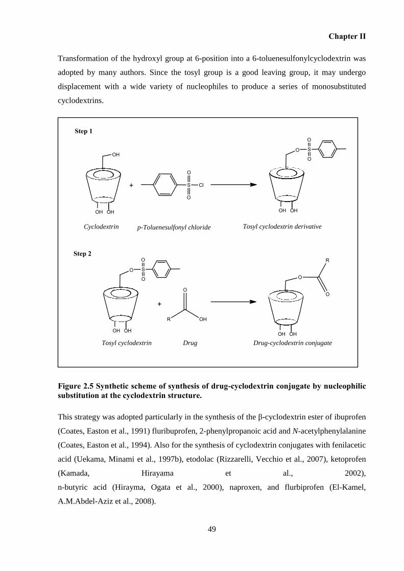
Chapter II
49
Transformation of the hydroxyl group at 6-position into a 6-toluenesulfonylcyclodextrin was
adopted by many authors. Since the tosyl group is a good leaving group, it may undergo
displacement with a wide variety of nucleophiles to produce a series of monosubstituted
cyclodextrins.
Figure 2.5 Synthetic scheme of synthesis of drug-cyclodextrin conjugate by nucleophilic substitution at the cyclodextrin structure.
This strategy was adopted particularly in the synthesis of the β-cyclodextrin ester of ibuprofen
(Coates, Easton et al., 1991) fluribuprofen, 2-phenylpropanoic acid and N-acetylphenylalanine
(Coates, Easton et al., 1994). Also for the synthesis of cyclodextrin conjugates with fenilacetic
acid (Uekama, Minami et al., 1997b), etodolac (Rizzarelli, Vecchio et al., 2007), ketoprofen
(Kamada, Hirayama et al., 2002),
n-butyric acid (Hirayma, Ogata et al., 2000), naproxen, and flurbiprofen (El-Kamel,
A.M.Abdel-Aziz et al., 2008).
Step 2
Cyclodextrin p-Toluenesulfonyl chloride Tosyl cyclodextrin derivative
Tosyl cyclodextrin
Drug Drug-cyclodextrin conjugate
Step 1
Chapter II
50
2.2 Aims and Objectives
The aim of this Chapter is to investigate a method to synthesize diclofenac-cyclodextrin
conjugates. In order to pursue this, the following objectives were defined:
- establishment of a reproducible method to synthesize diclofenac-β-cyclodextrin conjugate;
- chemical characterization of the obtained conjugate;
- evaluation of the ability of the chosen method used to synthesize diclofenac-β-cyclodextrin,
and also to synthesize the other γ- and α- conjugates.
2.3 Materials
Chemicals 2.3.1
β-cyclodextrin was purchased from Roquette. β-cyclodextrin hydrated (MW=1,134.98 g/mol),
DCC, CDI EDAC, DMAP, p-toluenesulfonyl chloride, 2,4,6-triisopropylbenzenesulfonyl
chloride, Diaion-HP-20 trifluroacetic acid and acetone were purchased from Sigma. Sodium
diclofenac (MW=318.13 g/mol) was kindly provided by Labesfal Genéricos (Portugal).
Dimethylformamide (DMF), sodium hydroxide, ethyl acetate, isopropanol, ethanol,
ammonium chloride, methanol, acetonitrile, thionyl chloride, tetrahydrofuran (THF),
triethylamine (TEA) ethanol, acetonitrile and water for HPLC, were purchased from Fisher
Scientific. Deuterated dimethyl sulfoxide (DMSO-d6) was purchased from Euriso-top
(Peypin, France). Ammonia, acetic acid, pyridine, sulfuric acid and p-anisaldehyde were
purchase from Acros Organics. DC Silicagel 60 F254 was purchased from VWR.
Pyridine was kept dry over potassium hydroxide and DMF over molecular sieves 4Å prior to
use.
Diclofenac free acid was obtained according to the method described by Fadda et al. Briefly,
a saturated aqueous solution of diclofenac sodium salt was acidified with diluted hydrochloric
acid until a white precipitate of diclofenac acid was observed. The precipitate was filtered,
washed with bidistilled water and air dried (Lai, Sinico et al., 2009).
Other chemicals and solvents were of analytical reagent grade and deionized water was used.
Chapter II
51
2.4 Methods
Synthesis of diclofenac-β-cyclodextrin conjugate. 2.4.1
Several methods were investigated to synthesize ester diclofenac-β-cyclodextrin conjugate
including:
2.4.1.1 Activation of diclofenac carboxylic group using a carbodiimide derivative,
following reaction with a hydroxyl group of the cyclodextrin.
Briefly, sodium diclofenac (1.57 mmol) was dissolved in DMF. DCC (1.57 mmol) was then
added to the solution, and the mixture stirred at 0 oC for 2 hours, adapting the method
reported previously (Dev, Mhaske et al., 2007). After the addition of the cyclodextrin in
DMF, the mixture was stirred at room temperature. Different conditions were tested following
this procedure, including the addition of DMAP as a catalyst (Albert and Hamilton, 2001).
Replacement of sodium diclofenac by diclofenac free acid was also performed (Manfredini,
Pavan et al., 2002). Other carbodiimides derivatives were tested, namely CDI (Udo,
Hokonohara et al., 2010) and EDAC (Bakar, Oelgemöller et al., 2009).
2.4.1.2 Formation of an acid chloride of the drug followed by reaction with
cyclodextrin.
For the synthesis of diclofenac chloride, diclofenac free acid (3.37 mmol) was refluxed with
thionyl chloride (10 mL) for 5 hours. After this step, β-cyclodextrin was added to diclofenac
chloride and the reaction proceeded until formation of the conjugate. This method was
performed adapting those described by Puglisi et al.(Ventura, Paolino et al., 2003a) and by
Picci et al. (Cassano, Trombino et al., 2010).
2.4.1.3 Modification of β-cyclodextrin hydroxyl groups by an electrophilic reagent,
leading to the formation of one group more labile to nucleophilic substitution.
Diclofenac-β-cyclodextrin was synthesized through a 2-steps process involving tosylation of
β-cyclodextrin (Step 1) and nucleophylic substitution of tosylated β-cyclodextrin by sodium
diclofenac (Step 2).
Chapter II
52
2.4.1.3.1 Step 1: tosylation of β-cyclodextrin
Tosylated β-cyclodextrin was synthesized adapting the procedure described by (Mcnaughton,
Engman et al., 2004). Briefly, to a solution of 5 g of β-cyclodextrin (4.4 mmol) in water (110
mL) 1.25 g of p-toluenesulfonyl chloride (6.55 mmol) was added and the resultant solution
was stirred at room temperature for 2 hours under inert atmosphere. Aqueous sodium
hydroxide (NaOH) (2.5 M, 20 mL) was added and the solution stirred for 10 minutes before
unreacted p-toluenesulfonyl chloride was filtered off. 5.8 g of ammonium chloride (108
mmol) was added to lower the pH to approximately pH 8. The solution was cooled overnight
and the resultant white precipitated collected by filtration. The white powder was washed with
acetone and water to remove the non-reacted cyclodextrin and then dried under vacuum. The
product obtained was used directly in the next step.
2.4.1.3.2 Step 2: nucleophilic reaction
This step was performed following two different approaches - with and without the use of
microwaves.
Non-microwave reaction:
In this method sodium diclofenac (0.7 mmol) was added to β-cyclodextrin tosylate (0.5
mmol) in anhydrous DMF (5 mL) and the mixture stirred at 100 ºC for 24 hours (Hirayma,
Ogata et al., 2000).
Microwave reaction:
Reactions were carried out in an appropriate 10 mL thick walled glass vials under closed
vessel conditions using CEM Discover S-class single mode microwave reactor with
temperature, pressure and microwave power monitoring.
Sodium diclofenac (0.333 mmol) was dissolved in 3 mL of anhydrous DMF containing β-
cyclodextrin tosylate (0.327 mmol) in an appropriate thick-walled glass vial. The reaction
vessel was then sealed with a teflon®
cap and the reaction mixture magnetically stirred and
heated at 140 oC for 40 minutes under focused microwave irradiation with an initial power
setting of 75 W. The product obtained was precipitated in acetone, filtered, washed several
times with acetone and ethyl ether and dried. In order to obtain a purified sample, 300 mg of
Chapter II
53
the crude product was passed through DIAION HP-20 ion-exchange chromatography column
eluting with water/methanol, and steadily increasing the methanol content. The conjugate was
eluted with 80% methanol. Methanol in the eluate was removed under reduced pressure, the
solution lyophilized in a freeze-dryer (Lyph-lock 6 apparatus, Labconco) for 72 hours and the
diclofenac-β-cyclodextrin was obtained with a yield of 20%.
1H NMR (500 MHz, DMSO-d6) δ (ppm) 7.52 (d, H-5’, H-3’ of Diclofenac), 7.22 (d, H-4’’ of
Diclofenac), 7.20 (t, H-4 of Diclofenac), 7.05 (t, H-6’’ of Diclofenac), 6.83 (t, H-5’’ of
Diclofenac), 6.25 (d, H-7’’ of Diclofenac), 5.66–5.84 (m, OH-2 and OH-3 of β-cyclodextrin),
4.83 (d, H-1 of β-cyclodextrin), 4.21–4.55 (m, OH-6, H-6 of β- cyclodextrin), 3.50–3.90 (m,
H-6, H-3 and H-5 of β-cyclodextrin), 3.25–3.43 (m, H-2, H-4 of β-cyclodextrin), MS
(MALDI) m/z for C56H79Cl2NNaO36, found 1434.308 [conjugate+Na].
Applicability of the nucleophilic microwave approach on the synthesis of diclofenac-2.4.2
γ-cyclodextrin and diclofenac-α-cyclodextrin
2.4.2.1 Diclofenac-γ-cyclodextrin
2.4.2.1.1 Step 1: tosylation of γ-cyclodextrin
Synthesis of 6-monotosyl-γ-cyclodextrin
Tosylated γ-cyclodextrin was synthesized adapting the procedure described in the literature to
produce γ-cyclodextrin ditosylated (Kahee Fujita, Hatsuo Yamamura et al., 1988). In this
case, p-toluenesulfonyl chloride (2.02 g, 10.5 mmol) was added to a solution of γ-cyclodextrin
(2.02 g, 1.55 mmol) in pyridine (20 mL) and stirred for 2 hours at room temperature under
nitrogen atmosphere followed by addition of ethanol (20 mL). After addition of acetone, a
solid precipitated was collected by filtration, washed with acetone, and dried under vacuum
and at 100 ºC to afford 1-2 g of tosylate. This material can be re-crystallized by dissolving it
in isopropanol at boiling temperature and then addition of water followed by cooling to room
temperature, and storage in a refrigerator overnight.
Synthesis of Mono-6-(O-2,4,6-triisopropylbenzenesulfonyl)-γ-cyclodextrin
The synthesis was perfomed adapting the method described by Ronald Palin et al. (Palin,
Grove et al., 2001). To a dry round-bottomed flask pyridine (10 mL) was added, followed by
γ-cyclodextrin (1.0 g, 0.77 mmol) and 2,4,6-triisopropylbenzenesulfonyl chloride (0.7 g, 2.3
Chapter II
54
mmol) at room temperature under nitrogen. After 24 hours, 100 mL of acetone was added to
the mixture.
2.4.2.1.2 Step 2: Nucleophilic reaction - microwave approach
Sodium diclofenac (79.5 mg, 0.25 mmol) was dissolved in 3 mL anhydrous DMF containing
γ-cyclodextrin tosylated (376 mg) in an appropriate thick-walled glass vial. The reaction
vessel was then sealed with a teflon® cap and the reaction mixture was magnetically stirred
and heated at temperature varying between 130-160 ºC for 20-40 minutes under focused
microwave irradiation with an initial power setting of 75 W.
After cooling to room temperature, the product was precipitated in acetone. The precipitate
was filtered and washed several times with acetone and diethyl ether, filtered off and then
dried. Then the product was purified through by DIAION HP-20 ion-exchange
chromatography eluting with water/methanol with increasing methanol content. The
conjugate appeared with a composition of 80% methanol. Methanol in the eluate was
removed under reduced pressure and the solution was lyophilized. Yield of isolated, pure
product: 10-30%.
2.4.2.2 Diclofenac-α-cyclodextrin
2.4.2.2.1 Step 1: tosylation of α-cyclodextrin
Tosylated α-cyclodextrin was synthesized adapting the procedures described in the literature
(Melton and Slessor, 1971; Weihua Tang and Ng, 2008). Briefly, 1 g (1 mmol) of γ-
cyclodextrin was added to pyridine (10 mL) under stirring at room temperature under
nitrogen. p-Toluenesulfonyl chloride was dissolved in 5 mL of pyridine and then added
slowly during 30 minutes to the mixture of α-cyclodextrin in pyridine for 3 hours. After that,
the reaction was stopped by the addition of acetone. The crude product was washed with
methanol in order to eliminate pyridine.
2.4.2.2.2 Step 2: nucleophilic reaction - microwave approach
The protocol followed was the same used to prepare diclofenac-γ-cycldoextrin conjugate.
Chapter II
55
Characterization 2.4.3
The control of reactions was perfomed using thin layer chromatography (TLC). TLC was also
used to monitorize elutes from the chromatography and the purification of the end products.
One or more of the following techniques confirmed the identification of the obtained
compounds: proton nuclear magnetic resonance (1H NMR) spectroscopy; matrix-assisted laser
desorption/ ionization (MALDI) spectroscopy; infrared (IR) spectroscopy; and high
performance liquid chromatography (HPLC).
Thin layer chromatography (TLC) was conducted on aluminum plates percoated with silica
gel F254 (Merck & Co.) and eluted with a mixture of 2-propanol/ethyl acetate/water/ammonia
(6:1:3:1) or acetonitrile/water/ammonia (6:3:1).
For spot detection the plates were immersed in a mixture of p-anisaldehyde (2 mL)/ethanol
(36 mL)/acetic acid (5–6 drops)/sulfuric acid (2 mL). The plate was heated to 150 °C for 5
minutes in order to visualize the spots.
MALDI-TOF mass spectra were obtained on an Autoflex III, Bruker using methanol and
water as solvents and α-cyano-4-hydroxy-cinnamic acid (HCCA) as matrix.
1H NMR spectra were acquired on a Varian Unity-500MHz spectrometer, using DMSO-d6 as
solvent and tetramethylsilane (TMS) as an internal reference.
IR spectra were obtained using Thermo Scientific Nicolet 6700 FT-IR spectrometer by
scanning KBr disks of the samples.
An HPLC system (model HP1100 series, Hewlett Packard, Germany) equipped with an
autosampler (Agilent 1100 series, Germany) was used. A reversed-phase X-Terra C-18
column, 5 μm, 4.6 mm × 250 mm (Waters, USA), with a pre-column, was employed. Mobile
phase consisted of acetonitrile (ACN) and 0.4% trifluoroacetic acid (TFA) in water, an
injection volume of 20 μL and detection at 254 nm at 30 °C
For analysis of the products of reactions, a gradient system of 0.4% TFA in water (A) and
acetonitrile (B) was followed: 0-15 minutes 10-90% B; 15-22 minutes 90-10% B. The sample
injection volume was 20 µl, the flow rate is 1.2 mL/min and detection wavelength was 254
nm at 30 ºC. For the analysis of the products of the chemical stability assays, the gradient
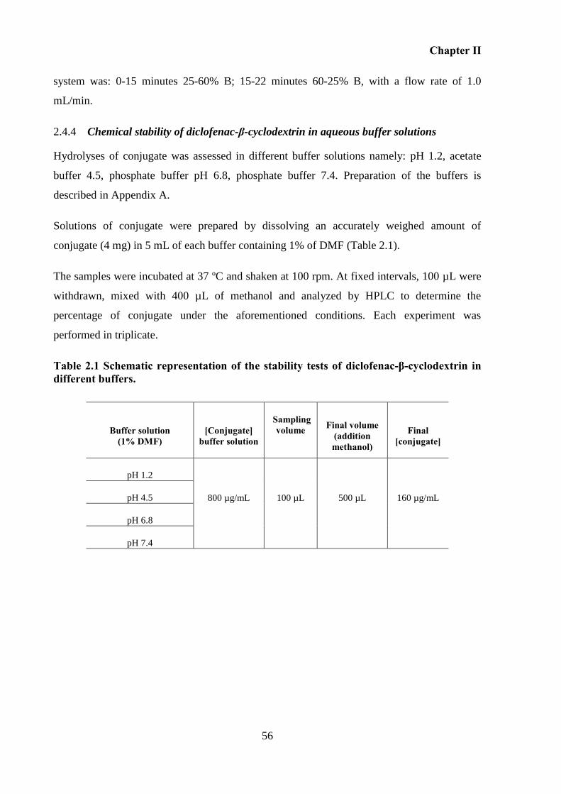
Chapter II
56
system was: 0-15 minutes 25-60% B; 15-22 minutes 60-25% B, with a flow rate of 1.0
mL/min.
Chemical stability of diclofenac-β-cyclodextrin in aqueous buffer solutions 2.4.4
Hydrolyses of conjugate was assessed in different buffer solutions namely: pH 1.2, acetate
buffer 4.5, phosphate buffer pH 6.8, phosphate buffer 7.4. Preparation of the buffers is
described in Appendix A.
Solutions of conjugate were prepared by dissolving an accurately weighed amount of
conjugate (4 mg) in 5 mL of each buffer containing 1% of DMF (Table 2.1).
The samples were incubated at 37 ºC and shaken at 100 rpm. At fixed intervals, 100 µL were
withdrawn, mixed with 400 µL of methanol and analyzed by HPLC to determine the
percentage of conjugate under the aforementioned conditions. Each experiment was
performed in triplicate.
Table 2.1 Schematic representation of the stability tests of diclofenac-β-cyclodextrin in different buffers.
Buffer solution (1% DMF)
[Conjugate] buffer solution
Sampling volume
Final volume (addition methanol)
Final [conjugate]
pH 1.2
800 µg/mL
100 µL
500 µL
160 µg/mL
pH 4.5
pH 6.8
pH 7.4
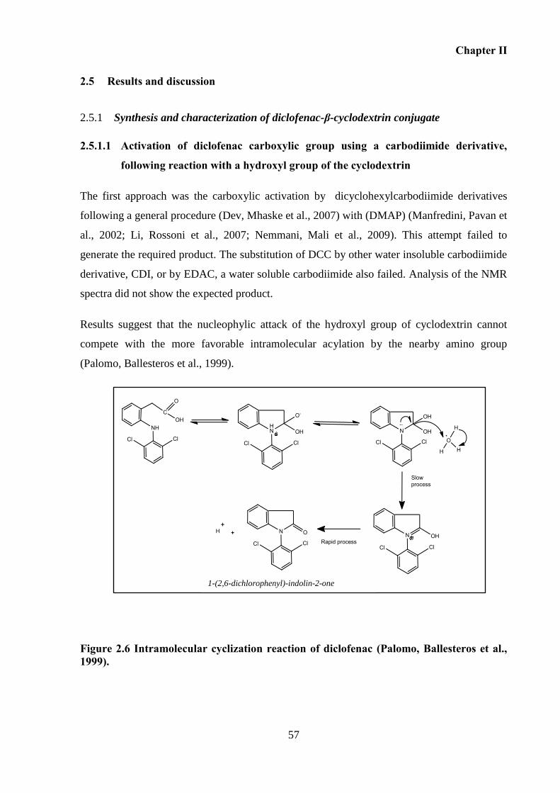
Chapter II
57
2.5 Results and discussion
Synthesis and characterization of diclofenac-β-cyclodextrin conjugate 2.5.1
2.5.1.1 Activation of diclofenac carboxylic group using a carbodiimide derivative,
following reaction with a hydroxyl group of the cyclodextrin
The first approach was the carboxylic activation by dicyclohexylcarbodiimide derivatives
following a general procedure (Dev, Mhaske et al., 2007) with (DMAP) (Manfredini, Pavan et
al., 2002; Li, Rossoni et al., 2007; Nemmani, Mali et al., 2009). This attempt failed to
generate the required product. The substitution of DCC by other water insoluble carbodiimide
derivative, CDI, or by EDAC, a water soluble carbodiimide also failed. Analysis of the NMR
spectra did not show the expected product.
Results suggest that the nucleophylic attack of the hydroxyl group of cyclodextrin cannot
compete with the more favorable intramolecular acylation by the nearby amino group
(Palomo, Ballesteros et al., 1999).
Figure 2.6 Intramolecular cyclization reaction of diclofenac (Palomo, Ballesteros et al., 1999).
1-(2,6-dichlorophenyl)-indolin-2-one
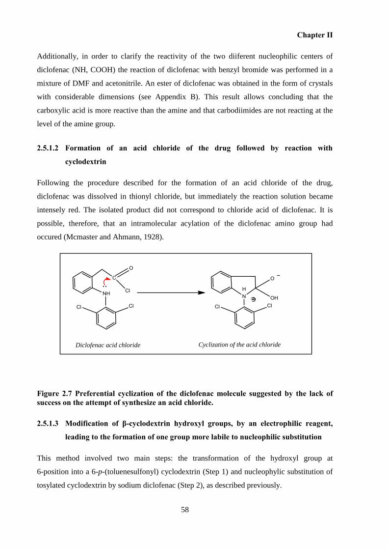
Chapter II
58
Additionally, in order to clarify the reactivity of the two diiferent nucleophilic centers of
diclofenac (NH, COOH) the reaction of diclofenac with benzyl bromide was performed in a
mixture of DMF and acetonitrile. An ester of diclofenac was obtained in the form of crystals
with considerable dimensions (see Appendix B). This result allows concluding that the
carboxylic acid is more reactive than the amine and that carbodiimides are not reacting at the
level of the amine group.
2.5.1.2 Formation of an acid chloride of the drug followed by reaction with
cyclodextrin
Following the procedure described for the formation of an acid chloride of the drug,
diclofenac was dissolved in thionyl chloride, but immediately the reaction solution became
intensely red. The isolated product did not correspond to chloride acid of diclofenac. It is
possible, therefore, that an intramolecular acylation of the diclofenac amino group had
occured (Mcmaster and Ahmann, 1928).
Figure 2.7 Preferential cyclization of the diclofenac molecule suggested by the lack of success on the attempt of synthesize an acid chloride.
2.5.1.3 Modification of β-cyclodextrin hydroxyl groups, by an electrophilic reagent,
leading to the formation of one group more labile to nucleophilic substitution
This method involved two main steps: the transformation of the hydroxyl group at
6-position into a 6-p-(toluenesulfonyl) cyclodextrin (Step 1) and nucleophylic substitution of
tosylated cyclodextrin by sodium diclofenac (Step 2), as described previously.
Diclofenac acid chloride Cyclization of the acid chloride
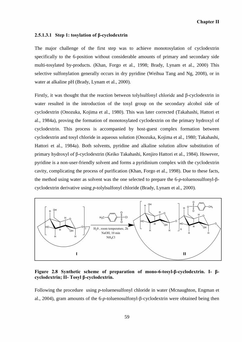
Chapter II
59
2.5.1.3.1 Step 1: tosylation of β-cyclodextrin
The major challenge of the first step was to achieve monotosylation of cyclodextrin
specifically to the 6-position without considerable amounts of primary and secondary side
multi-tosylated by-products. (Khan, Forgo et al., 1998; Brady, Lynam et al., 2000) This
selective sulfonylation generally occurs in dry pyridine (Weihua Tang and Ng, 2008), or in
water at alkaline pH (Brady, Lynam et al., 2000).
Firstly, it was thought that the reaction between tolylsulfonyl chloride and β-cyclodextrin in
water resulted in the introduction of the tosyl group on the secondary alcohol side of
cyclodextrin (Onozuka, Kojima et al., 1980). This was later corrected (Takahashi, Hattori et
al., 1984a), proving the formation of monotosylated cyclodextrin on the primary hydroxyl of
cyclodextrin. This process is accompanied by host-guest complex formation between
cyclodextrin and tosyl chloride in aqueous solution (Onozuka, Kojima et al., 1980; Takahashi,
Hattori et al., 1984a). Both solvents, pyridine and alkaline solution allow substitution of
primary hydroxyl of β-cyclodextrin (Keiko Takahashi, Kenjiro Hattori et al., 1984). However,
pyridine is a non-user-friendly solvent and forms a pyridinium complex with the cyclodextrin
cavity, complicating the process of purification (Khan, Forgo et al., 1998). Due to these facts,
the method using water as solvent was the one selected to prepare the 6-p-toluenosulfonyl-β-
cyclodextrin derivative using p-tolylsulfonyl chloride (Brady, Lynam et al., 2000).
Figure 2.8 Synthetic scheme of preparation of mono-6-tosyl-β-cyclodextrin. I- β-cyclodextrin; II- Tosyl β-cyclodextrin.
Following the procedure using p-toluenesulfonyl chloride in water (Mcnaughton, Engman et
al., 2004), gram amounts of the 6-p-toluenosulfonyl-β-cyclodextrin were obtained being then
I II
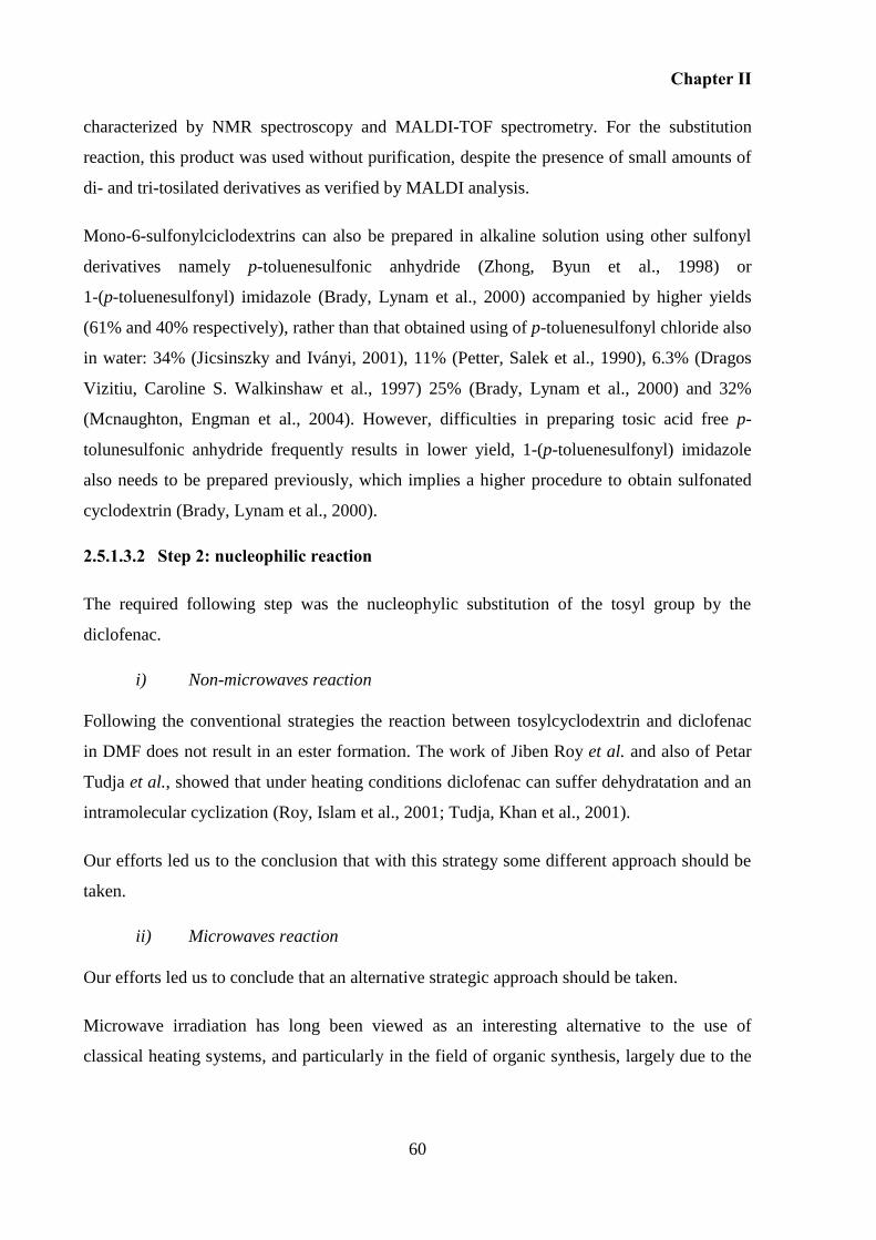
Chapter II
60
characterized by NMR spectroscopy and MALDI-TOF spectrometry. For the substitution
reaction, this product was used without purification, despite the presence of small amounts of
di- and tri-tosilated derivatives as verified by MALDI analysis.
Mono-6-sulfonylciclodextrins can also be prepared in alkaline solution using other sulfonyl
derivatives namely p-toluenesulfonic anhydride (Zhong, Byun et al., 1998) or
1-(p-toluenesulfonyl) imidazole (Brady, Lynam et al., 2000) accompanied by higher yields
(61% and 40% respectively), rather than that obtained using of p-toluenesulfonyl chloride also
in water: 34% (Jicsinszky and Iványi, 2001), 11% (Petter, Salek et al., 1990), 6.3% (Dragos
Vizitiu, Caroline S. Walkinshaw et al., 1997) 25% (Brady, Lynam et al., 2000) and 32%
(Mcnaughton, Engman et al., 2004). However, difficulties in preparing tosic acid free p-
tolunesulfonic anhydride frequently results in lower yield, 1-(p-toluenesulfonyl) imidazole
also needs to be prepared previously, which implies a higher procedure to obtain sulfonated
cyclodextrin (Brady, Lynam et al., 2000).
2.5.1.3.2 Step 2: nucleophilic reaction
The required following step was the nucleophylic substitution of the tosyl group by the
diclofenac.
i) Non-microwaves reaction
Following the conventional strategies the reaction between tosylcyclodextrin and diclofenac
in DMF does not result in an ester formation. The work of Jiben Roy et al. and also of Petar
Tudja et al., showed that under heating conditions diclofenac can suffer dehydratation and an
intramolecular cyclization (Roy, Islam et al., 2001; Tudja, Khan et al., 2001).
Our efforts led us to the conclusion that with this strategy some different approach should be
taken.
ii) Microwaves reaction
Our efforts led us to conclude that an alternative strategic approach should be taken.
Microwave irradiation has long been viewed as an interesting alternative to the use of
classical heating systems, and particularly in the field of organic synthesis, largely due to the
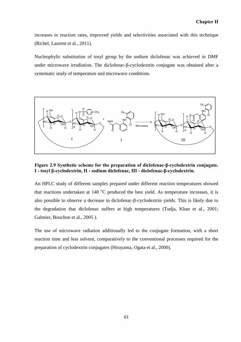
Chapter II
61
increases in reaction rates, improved yields and selectivities associated with this technique
(Richel, Laurent et al., 2011).
Nucleophylic substitution of tosyl group by the sodium diclofenac was achieved in DMF
under microwave irradiation. The diclofenac-β-cyclodextrin conjugate was obtained after a
systematic study of temperature and microwave conditions.
Figure 2.9 Synthetic scheme for the preparation of diclofenac-β-cyclodextrin conjugate. I - tosyl β-cyclodextrin, II - sodium diclofenac, III - diclofenac-β-cyclodextrin.
An HPLC study of different samples prepared under different reaction temperatures showed
that reactions undertaken at 140 oC produced the best yield. As temperature increases, it is
also possible to observe a decrease in diclofenac-β-cyclodextrin yields. This is likely due to
the degradation that diclofenac suffers at high temperatures (Tudja, Khan et al., 2001;
Galmier, Bouchon et al., 2005 ).
The use of microwave radiation additionally led to the conjugate formation, with a short
reaction time and less solvent, comparatively to the conventional processes required for the
preparation of cyclodextrin conjugates (Hirayama, Ogata et al., 2000).
+
I I
I III
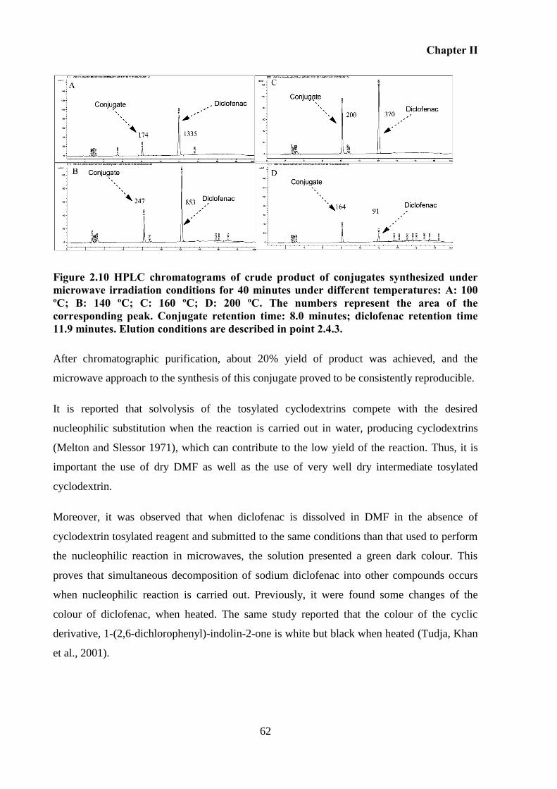
Chapter II
62
Figure 2.10 HPLC chromatograms of crude product of conjugates synthesized under microwave irradiation conditions for 40 minutes under different temperatures: A: 100 ºC; B: 140 ºC; C: 160 ºC; D: 200 ºC. The numbers represent the area of the corresponding peak. Conjugate retention time: 8.0 minutes; diclofenac retention time 11.9 minutes. Elution conditions are described in point 2.4.3.
After chromatographic purification, about 20% yield of product was achieved, and the
microwave approach to the synthesis of this conjugate proved to be consistently reproducible.
It is reported that solvolysis of the tosylated cyclodextrins compete with the desired
nucleophilic substitution when the reaction is carried out in water, producing cyclodextrins
(Melton and Slessor 1971), which can contribute to the low yield of the reaction. Thus, it is
important the use of dry DMF as well as the use of very well dry intermediate tosylated
cyclodextrin.
Moreover, it was observed that when diclofenac is dissolved in DMF in the absence of
cyclodextrin tosylated reagent and submitted to the same conditions than that used to perform
the nucleophilic reaction in microwaves, the solution presented a green dark colour. This
proves that simultaneous decomposition of sodium diclofenac into other compounds occurs
when nucleophilic reaction is carried out. Previously, it were found some changes of the
colour of diclofenac, when heated. The same study reported that the colour of the cyclic
derivative, 1-(2,6-dichlorophenyl)-indolin-2-one is white but black when heated (Tudja, Khan
et al., 2001).
Chapter II
63
2.5.1.4 Characterization of 6-p-toluenosulfonyl-β-cyclodextrin
2.5.1.4.1 Matrix-assisted laser desorption/ionization (MALDI) spectroscopy
Mass spectrometry, demonstrated that the product of the tosylation reaction is mainly the
mono-tosylated-cyclodextrin (1311.605 [M+Na] adduct) contaminated with some di-
tosylcyclodextrins (1465.667 [M+Na] adduct) (Figure 2.11).
Figure 2.11 MALDI mass spectrum of tosylated β-cyclodextrin compound. A- β-cyclodextrin, B- monotosylated β-cyclodextrin, C- ditosylated β-cyclodextrin
A
B
C
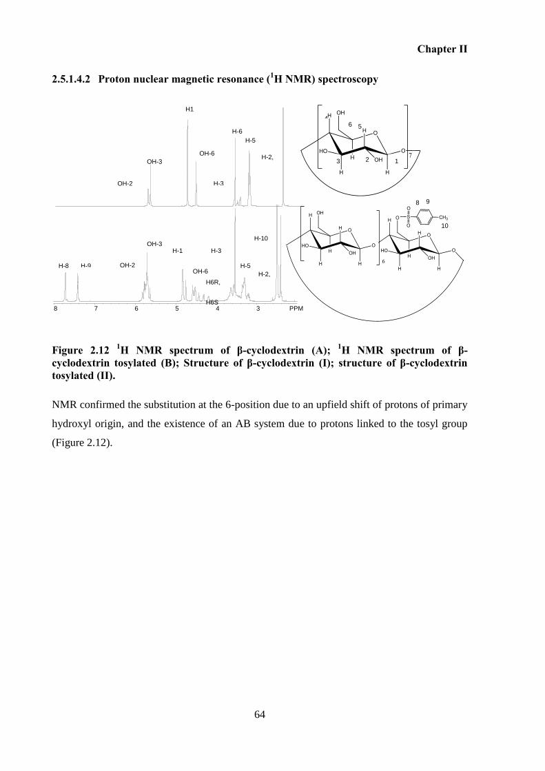
Chapter II
64
2.5.1.4.2 Proton nuclear magnetic resonance (1H NMR) spectroscopy
Figure 2.12 1H NMR spectrum of β-cyclodextrin (A); 1H NMR spectrum of β-cyclodextrin tosylated (B); Structure of β-cyclodextrin (I); structure of β-cyclodextrin tosylated (II).
NMR confirmed the substitution at the 6-position due to an upfield shift of protons of primary
hydroxyl origin, and the existence of an AB system due to protons linked to the tosyl group
(Figure 2.12).
8 9
10
H6R,
H6S
8 7 6 5 4 3 PPM
OH-2
OH-3
H1
H-3
H-6 H-5
H-2, H-4
H-8 H-9
H-10
OH-3
OH-2
H-5 H-2, H-4
H-3
H-1
OH-6
OH-6
1 2 3
5 6 4
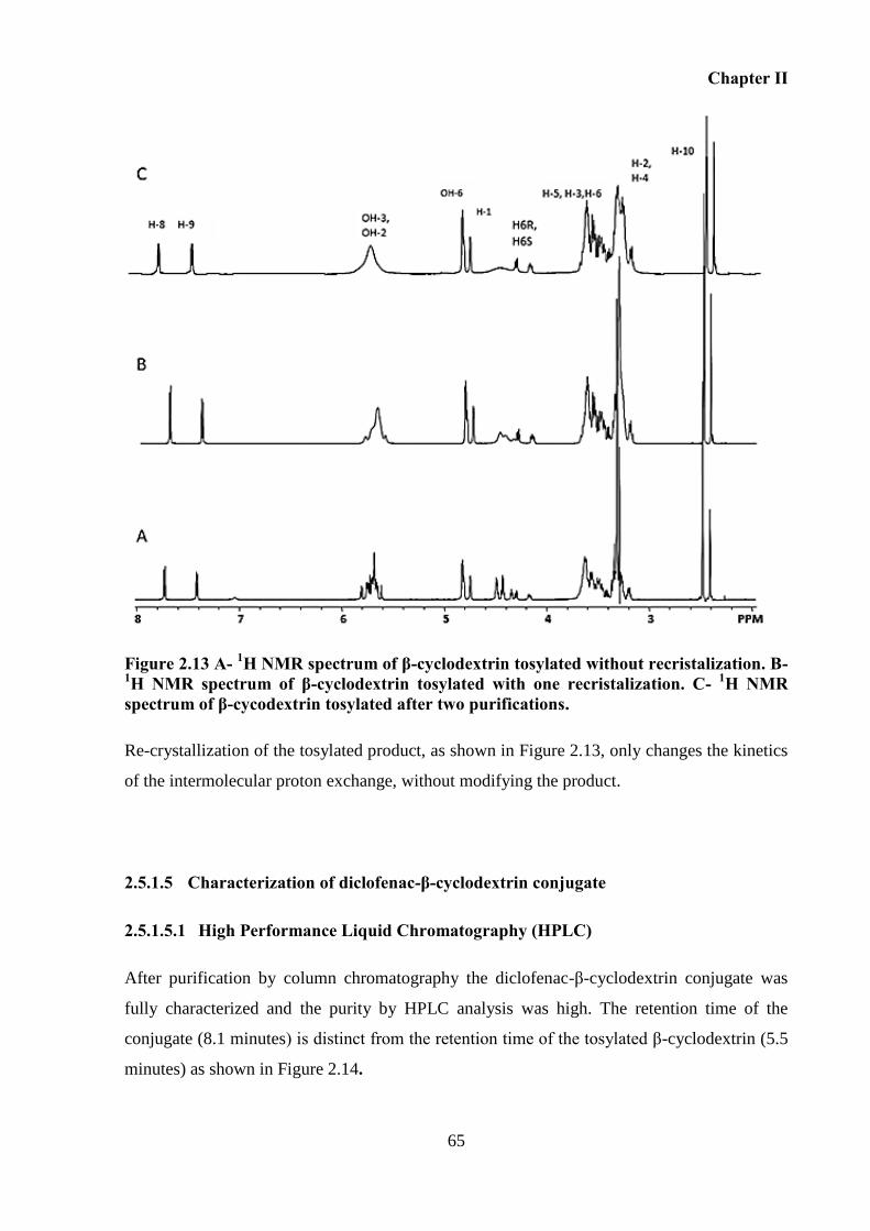
Chapter II
65
Figure 2.13 A- 1H NMR spectrum of β-cyclodextrin tosylated without recristalization. B- 1H NMR spectrum of β-cyclodextrin tosylated with one recristalization. C- 1H NMR spectrum of β-cycodextrin tosylated after two purifications.
Re-crystallization of the tosylated product, as shown in Figure 2.13, only changes the kinetics
of the intermolecular proton exchange, without modifying the product.
2.5.1.5 Characterization of diclofenac-β-cyclodextrin conjugate
2.5.1.5.1 High Performance Liquid Chromatography (HPLC)
After purification by column chromatography the diclofenac-β-cyclodextrin conjugate was
fully characterized and the purity by HPLC analysis was high. The retention time of the
conjugate (8.1 minutes) is distinct from the retention time of the tosylated β-cyclodextrin (5.5
minutes) as shown in Figure 2.14.
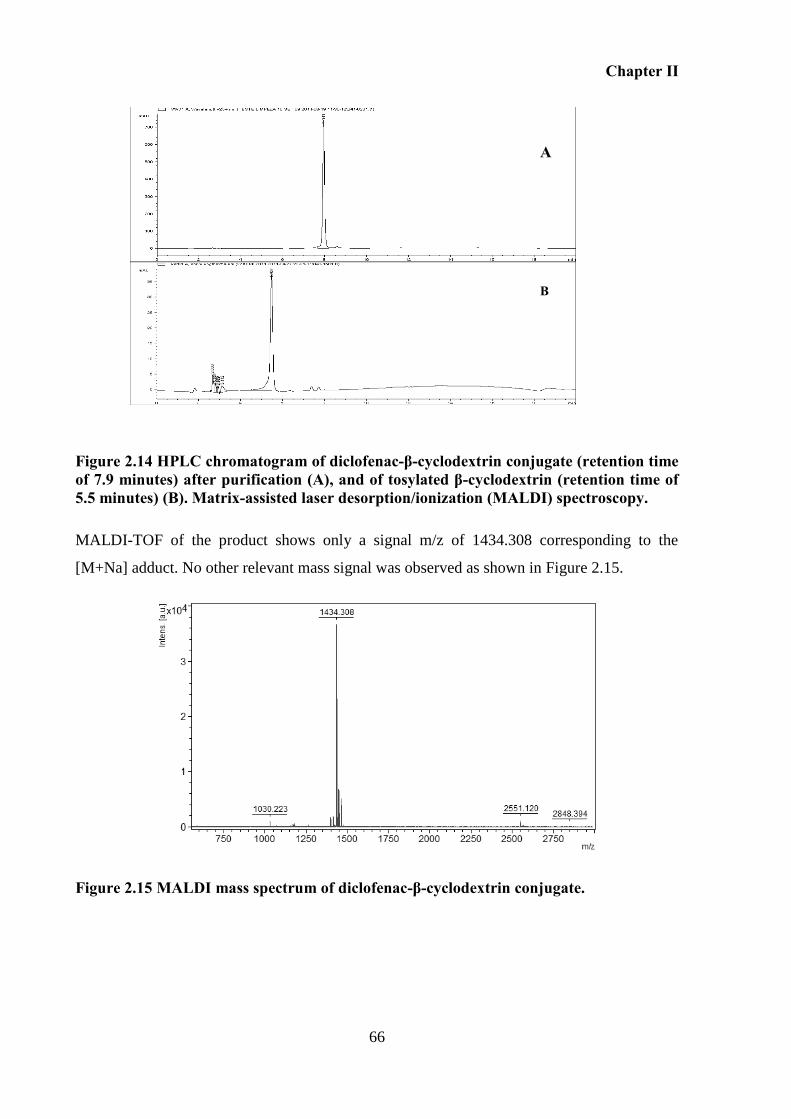
Chapter II
66
Figure 2.14 HPLC chromatogram of diclofenac-β-cyclodextrin conjugate (retention time of 7.9 minutes) after purification (A), and of tosylated β-cyclodextrin (retention time of 5.5 minutes) (B). Matrix-assisted laser desorption/ionization (MALDI) spectroscopy.
MALDI-TOF of the product shows only a signal m/z of 1434.308 corresponding to the
[M+Na] adduct. No other relevant mass signal was observed as shown in Figure 2.15.
Figure 2.15 MALDI mass spectrum of diclofenac-β-cyclodextrin conjugate.
A
B
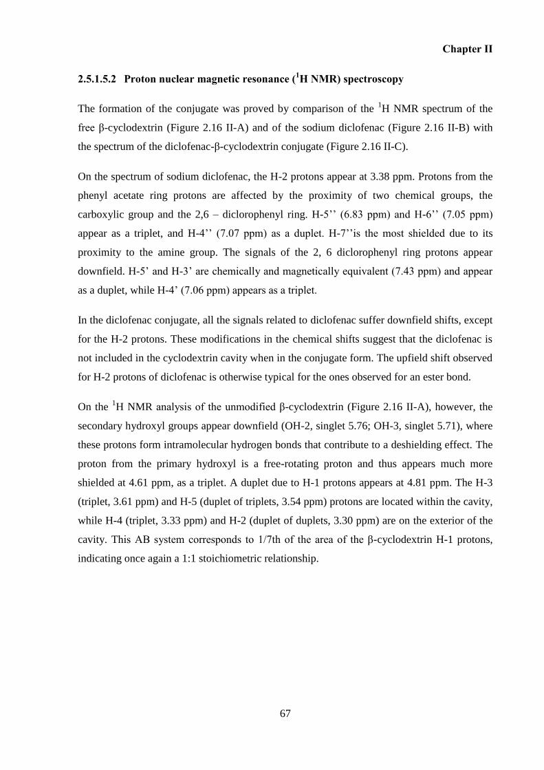
Chapter II
67
2.5.1.5.2 Proton nuclear magnetic resonance (1H NMR) spectroscopy
The formation of the conjugate was proved by comparison of the 1H NMR spectrum of the
free β-cyclodextrin (Figure 2.16 II-A) and of the sodium diclofenac (Figure 2.16 II-B) with
the spectrum of the diclofenac-β-cyclodextrin conjugate (Figure 2.16 II-C).
On the spectrum of sodium diclofenac, the H-2 protons appear at 3.38 ppm. Protons from the
phenyl acetate ring protons are affected by the proximity of two chemical groups, the
carboxylic group and the 2,6 – diclorophenyl ring. H-5’’ (6.83 ppm) and H-6’’ (7.05 ppm)
appear as a triplet, and H-4’’ (7.07 ppm) as a duplet. H-7’’is the most shielded due to its
proximity to the amine group. The signals of the 2, 6 diclorophenyl ring protons appear
downfield. H-5’ and H-3’ are chemically and magnetically equivalent (7.43 ppm) and appear
as a duplet, while H-4’ (7.06 ppm) appears as a triplet.
In the diclofenac conjugate, all the signals related to diclofenac suffer downfield shifts, except
for the H-2 protons. These modifications in the chemical shifts suggest that the diclofenac is
not included in the cyclodextrin cavity when in the conjugate form. The upfield shift observed
for H-2 protons of diclofenac is otherwise typical for the ones observed for an ester bond.
On the 1H NMR analysis of the unmodified β-cyclodextrin (Figure 2.16 II-A), however, the
secondary hydroxyl groups appear downfield (OH-2, singlet 5.76; OH-3, singlet 5.71), where
these protons form intramolecular hydrogen bonds that contribute to a deshielding effect. The
proton from the primary hydroxyl is a free-rotating proton and thus appears much more
shielded at 4.61 ppm, as a triplet. A duplet due to H-1 protons appears at 4.81 ppm. The H-3
(triplet, 3.61 ppm) and H-5 (duplet of triplets, 3.54 ppm) protons are located within the cavity,
while H-4 (triplet, 3.33 ppm) and H-2 (duplet of duplets, 3.30 ppm) are on the exterior of the
cavity. This AB system corresponds to 1/7th of the area of the β-cyclodextrin H-1 protons,
indicating once again a 1:1 stoichiometric relationship.
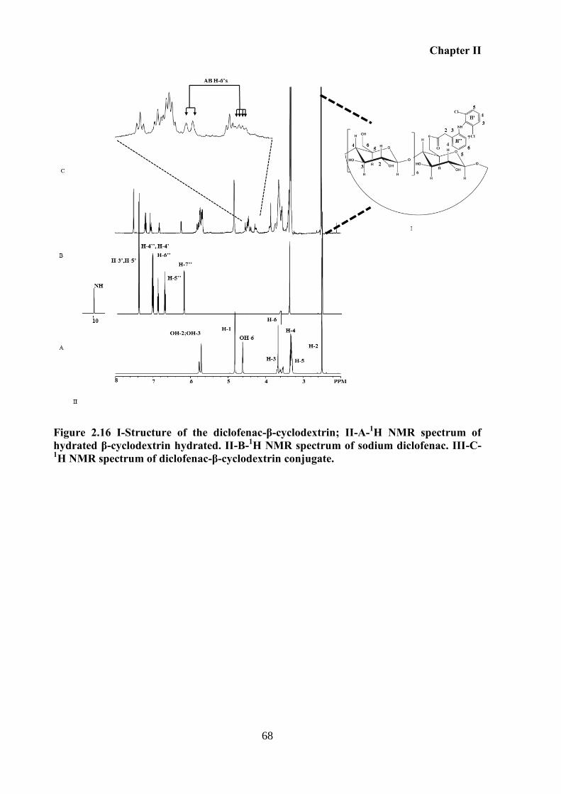
Chapter II
68
Figure 2.16 I-Structure of the diclofenac-β-cyclodextrin; II-A-1H NMR spectrum of hydrated β-cyclodextrin hydrated. II-B-1H NMR spectrum of sodium diclofenac. III-C-1H NMR spectrum of diclofenac-β-cyclodextrin conjugate.
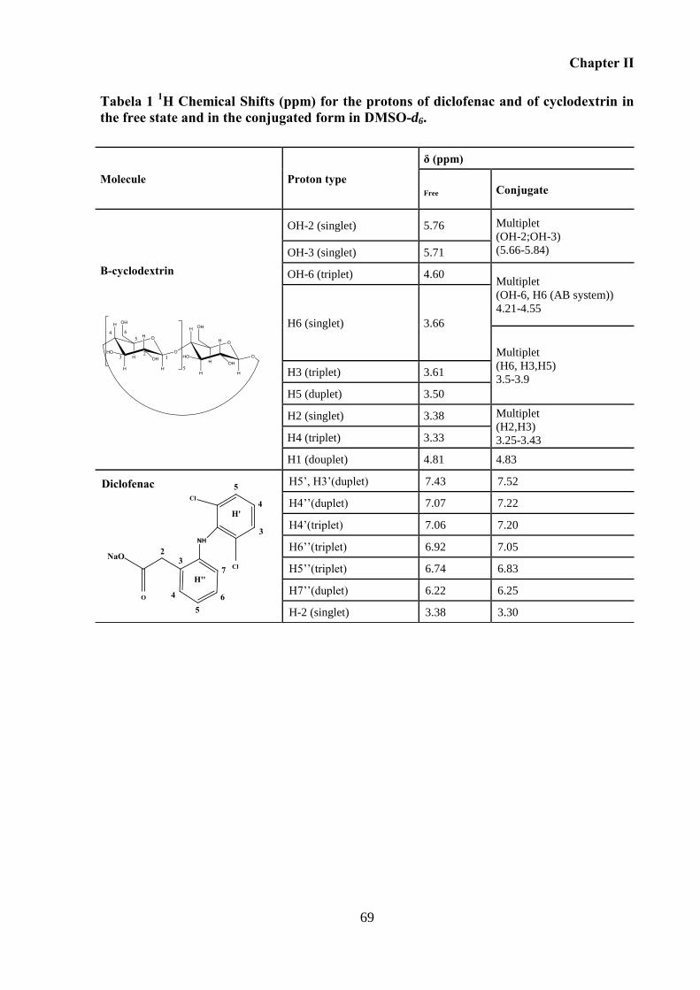
Chapter II
69
Tabela 1 1H Chemical Shifts (ppm) for the protons of diclofenac and of cyclodextrin in the free state and in the conjugated form in DMSO-d6.
Molecule Proton type
δ (ppm)
Free Conjugate
Β-cyclodextrin
OH-2 (singlet) 5.76 Multiplet
(OH-2;OH-3)
(5.66-5.84) OH-3 (singlet) 5.71
OH-6 (triplet) 4.60 Multiplet
(OH-6, H6 (AB system))
4.21-4.55
H6 (singlet) 3.66
Multiplet
(H6, H3,H5)
3.5-3.9 H3 (triplet) 3.61
H5 (duplet) 3.50
H2 (singlet) 3.38 Multiplet
(H2,H3)
3.25-3.43 H4 (triplet) 3.33
H1 (douplet) 4.81 4.83
Diclofenac H5’, H3’(duplet) 7.43 7.52
H4’’(duplet) 7.07 7.22
H4’(triplet) 7.06 7.20
H6’’(triplet) 6.92 7.05
H5’’(triplet) 6.74 6.83
H7’’(duplet) 6.22 6.25
H-2 (singlet) 3.38 3.30
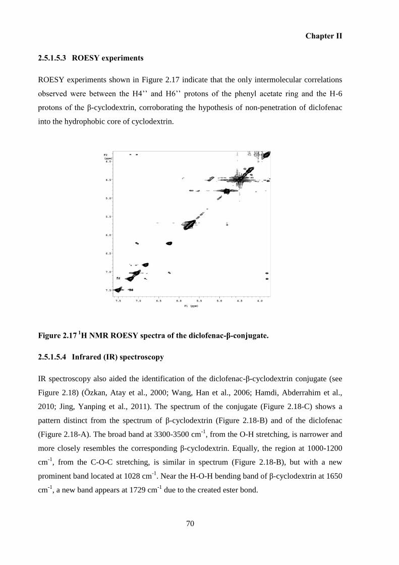
Chapter II
70
2.5.1.5.3 ROESY experiments
ROESY experiments shown in Figure 2.17 indicate that the only intermolecular correlations
observed were between the H4’’ and H6’’ protons of the phenyl acetate ring and the H-6
protons of the β-cyclodextrin, corroborating the hypothesis of non-penetration of diclofenac
into the hydrophobic core of cyclodextrin.
Figure 2.17 1H NMR ROESY spectra of the diclofenac-β-conjugate.
2.5.1.5.4 Infrared (IR) spectroscopy
IR spectroscopy also aided the identification of the diclofenac-β-cyclodextrin conjugate (see
Figure 2.18) (Özkan, Atay et al., 2000; Wang, Han et al., 2006; Hamdi, Abderrahim et al.,
2010; Jing, Yanping et al., 2011). The spectrum of the conjugate (Figure 2.18-C) shows a
pattern distinct from the spectrum of β-cyclodextrin (Figure 2.18-B) and of the diclofenac
(Figure 2.18-A). The broad band at 3300-3500 cm-1
, from the O-H stretching, is narrower and
more closely resembles the corresponding β-cyclodextrin. Equally, the region at 1000-1200
cm-1
, from the C-O-C stretching, is similar in spectrum (Figure 2.18-B), but with a new
prominent band located at 1028 cm-1
. Near the H-O-H bending band of β-cyclodextrin at 1650
cm-1
, a new band appears at 1729 cm-1
due to the created ester bond.
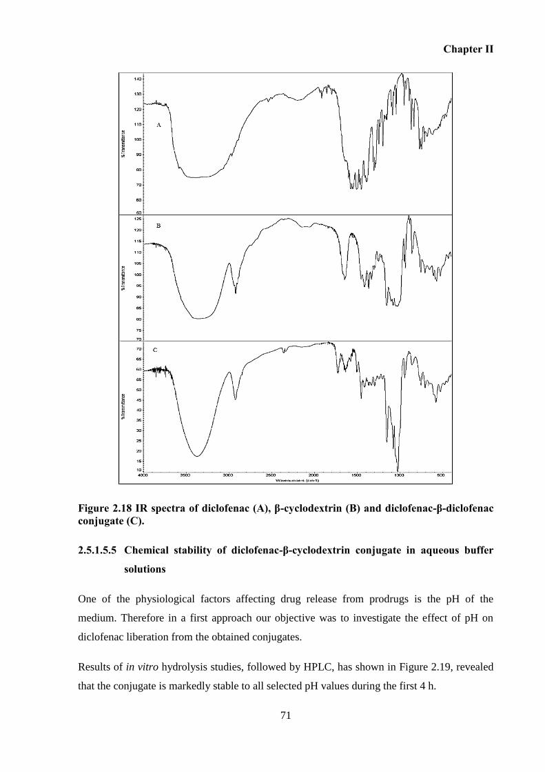
Chapter II
71
Figure 2.18 IR spectra of diclofenac (A), β-cyclodextrin (B) and diclofenac-β-diclofenac conjugate (C).
2.5.1.5.5 Chemical stability of diclofenac-β-cyclodextrin conjugate in aqueous buffer
solutions
One of the physiological factors affecting drug release from prodrugs is the pH of the
medium. Therefore in a first approach our objective was to investigate the effect of pH on
diclofenac liberation from the obtained conjugates.
Results of in vitro hydrolysis studies, followed by HPLC, has shown in Figure 2.19, revealed
that the conjugate is markedly stable to all selected pH values during the first 4 h.
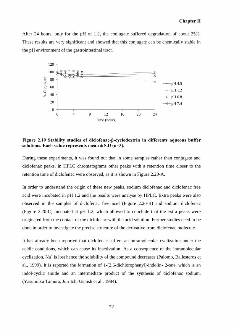
Chapter II
72
After 24 hours, only for the pH of 1.2, the conjugate suffered degradation of about 25%.
These results are very significant and showed that this conjugate can be chemically stable in
the pH environment of the gastrointestinal tract.
Figure 2.19 Stability studies of diclofenac-β-cyclodextrin in differents aqueous buffer solutions. Each value represents mean ± S.D (n=3).
During these experiments, it was found out that in some samples rather than conjugate and
diclofenac peaks, in HPLC chromatograms other peaks with a retention time closer to the
retention time of diclofenac were observed, as it is shown in Figure 2.20-A.
In order to understand the origin of these new peaks, sodium diclofenac and diclofenac free
acid were incubated in pH 1.2 and the results were analyse by HPLC. Extra peaks were also
observed in the samples of diclofenac free acid (Figure 2.20-B) and sodium diclofenac
(Figure 2.20-C) incubated at pH 1.2, which allowed to conclude that the extra peaks were
originated from the contact of the diclofenac with the acid solution. Further studies need to be
done in order to investigate the precise structure of the derivative from diclofenac molecule.
It has already been reported that diclofenac suffers an intramolecular cyclization under the
acidic conditions, which can cause its inactivation. As a consequence of the intramolecular
cyclization, Na+ is lost hence the solubility of the compound decreases (Palomo, Ballesteros et
al., 1999). It is reported the formation of 1-(2,6-dichlorophenyl)-indolin- 2-one, which is an
indol-cyclic amide and an intermediate product of the synthesis of diclofenac sodium.
(Yasumitsu Tamura, Jun-Ichi Uenish et al., 1984).
0
20
40
60
80
100
120
0 4 8 12 16 20 24
% C
onju
gat
e
Time (hours)
pH 4.5
pH 1.2
pH 6.8
pH 7.4
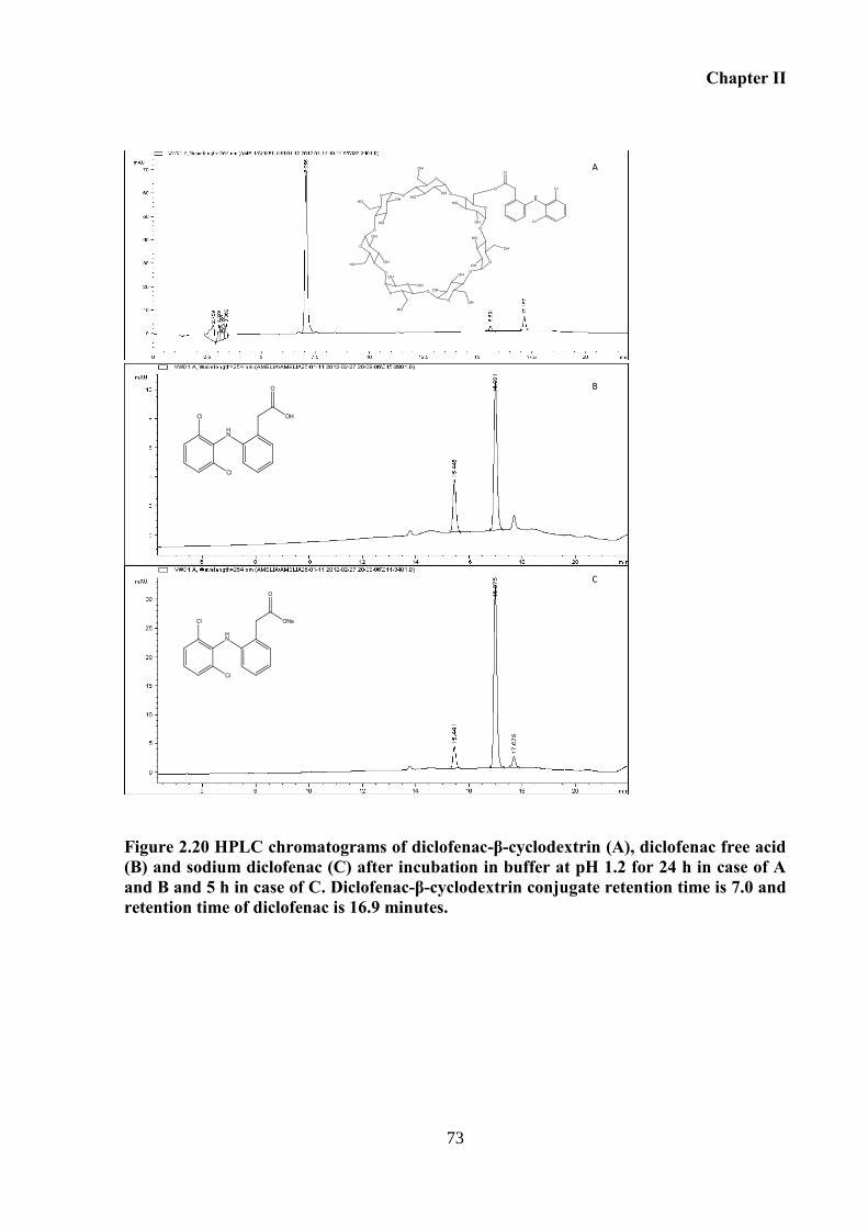
Chapter II
73
Figure 2.20 HPLC chromatograms of diclofenac-β-cyclodextrin (A), diclofenac free acid (B) and sodium diclofenac (C) after incubation in buffer at pH 1.2 for 24 h in case of A and B and 5 h in case of C. Diclofenac-β-cyclodextrin conjugate retention time is 7.0 and retention time of diclofenac is 16.9 minutes.
A
C
B
Chapter II
74
Applicability of the nucleophilic microwave approach on the synthesis of diclofenac-2.5.2
γ-cyclodextrin and diclofenac-α-cyclodextrin
The synthesis of other diclofenac conjugates derived from the natural α- and
γ- cyclodextrins was attempted by the method that successfully allowed the synthesis of
diclofenac-β-cyclodextrin.
The main goal is to assess if the reaction is possible using other cyclodextrins, once physical
differences exist between them. Both, α- and β-cyclodextrins are much more soluble in water
than β-cyclodextrin, and the molecular size is also different with consequently different
reactivities. Thus, the ability of the nucleophilic reaction under microwaves was tested.
Like observed for β-cyclodextrin, modification of the hydroxyl group by electrophilic reagent
on α- and γ-cyclodextrins had to be produced first (Step 1) followed by the nucleophilic
reaction (Step 2).
Different problems were encountered on the synthesis of α- and γ-cyclodextrin conjugates as
described below.
2.5.2.1 Synthesis and characterization of diclofenac-γ-cyclodextrin conjugate
2.5.2.1.1 Step 1: tosylation of γ-cyclodextrin
Tosylation of γ-cyclodextrin was carry out according to the method described to prepare
ditosylated cyclodextrin (Kahee Fujita, Hatsuo Yamamura et al., 1988). The synthesis of
monotosilated γ-cyclodextrin is very difficult since γ-cyclodextrin does not form an inclusion
complex with tosyl derivatives as described by Bruno Perly et al.. Consequently, its big cavity
causes polysubstitution when it reacts with p-toluenesulfonyl chloride (Djedaini-Pilard,
Azaroual-Bellanger et al., 1995). Tosylation occurs preferentially at hydroxyl groups at C-6.
However, 8 primary hydroxyl groups are available and therefore the reaction needs to be
controlled.
As it is shown in Figure 2.21 (Section 2.5.2.1.3), the product of this reaction is a mixture of
different tosylated products with multiple degrees of substitution even after recrystallization
process. In order to get the monotosylated product, some changes were made in the reaction
conditions namely the decrease of the ratio tosyl chloride/cyclodextrin, time of reaction, and
temperature; however no significant differences in product distribution were observed.
Chapter II
75
Moreover, besides its toxicity, working with pyridine led to the formation of an unstable stick
mass during isolation, which rendered stirring impossible during the course of the reactions
and during the isolation of the product.
The attempt of tosylation γ-cyclodextrin adopting the method in aqueous solution as used
described to synthesized β-cyclodextrin tosylated (Brady, Lynam et al., 2000) failed. As
described by Florence Djeda’ini-Pilard et al. the reaction of γ-cyclodextrin with 4-
toluenesulfonylchloride in aqueous solution results in polysubstitution at C-6 probably
because of the larger γ-cyclodextrin annulus (Djedaini-Pilard, Azaroual-Bellanger et al.,
1995). Moreover, once γ-cyclodextrin presents higher water solubility when compared with β-
cyclodextrin, the final product in water is very difficult to isolate, once it did not precipitate
by the addition of common organic solvents, such as acetone.
In order to get mono-6-(O-2,4,6-triisopropylbenzenesulfonyl)-cyclodextrin, the method
described by Palin et al. was adopted. This procedure resulted in less polisubstitution than that
obtained using the 4-toluenosulphonyl chloride. In our case the mono but also dissubstituted
product were obtained, as confirmed by the mass spectrum (Figure 2.22) (Palin, Grove et al.,
2001).
Beyond, 2,4,6-triisopropylbenzenesulfonyl, other larger arenesulfonyl chlorides, such as 2-
naphthalenesulfonyl chloride (Djedaini-Pilard, Azaroual-Bellanger et al., 1995), have been
explored by other authors in order to control the production of monosubstituted γ-
cyclodextrins (Djedaini-Pilard, Azaroual-Bellanger et al., 1995). For instance, the synthesis of
mono-6-(2-naphthalenesulfonyl)-γ-cyclodextrin (Ueno, Tomito et al., 1983) was adopted by
Kaneto Uekama et al. 1998 (Uekama, Minami et al., 1997b) to produce BPAA-γ-cyclodextrin
conjugate. However, Palin et al. obtained di- and tri-adducts when preparing mono-6-(2-
naphthalenesulfonyl)-γ-cyclodextrin according to literature procedures, reporting the
difficulty inherent to purification of the monosubstitued cyclodextrin. Additionally, the use of
this alternative arenesulfonyl provides low yields with a more expensive (Palin, Grove et al.,
2001).
2.5.2.1.2 Step 2: nucleophilic reaction
This step is performed using the crude product poli-substitued derivative obtained on the
former step.
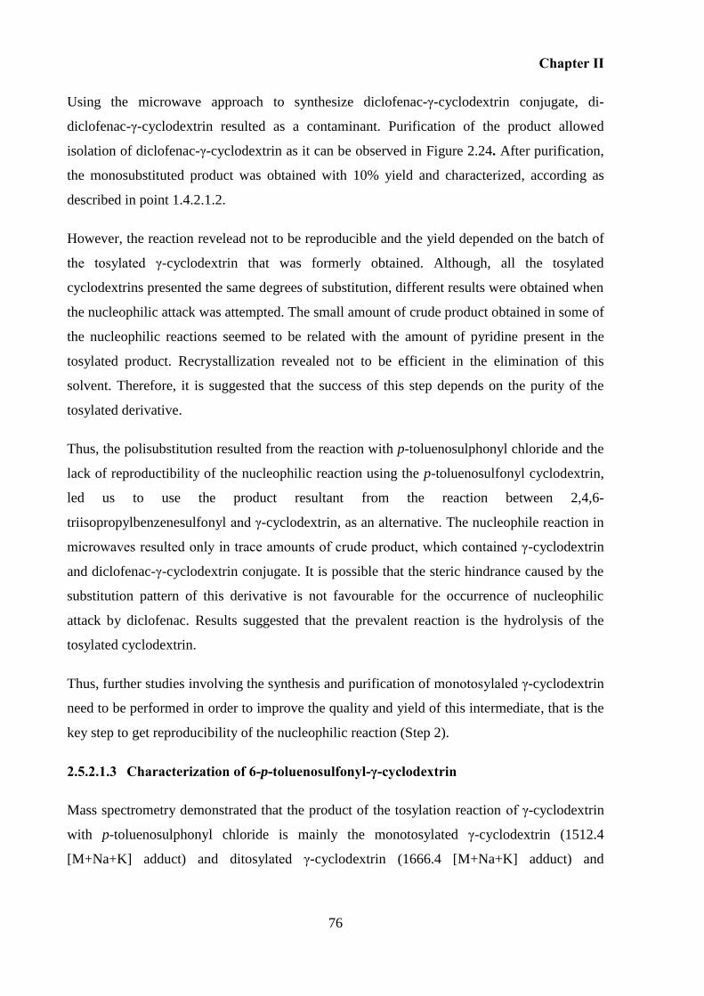
Chapter II
76
Using the microwave approach to synthesize diclofenac-γ-cyclodextrin conjugate, di-
diclofenac-γ-cyclodextrin resulted as a contaminant. Purification of the product allowed
isolation of diclofenac-γ-cyclodextrin as it can be observed in Figure 2.24. After purification,
the monosubstituted product was obtained with 10% yield and characterized, according as
described in point 1.4.2.1.2.
However, the reaction revelead not to be reproducible and the yield depended on the batch of
the tosylated γ-cyclodextrin that was formerly obtained. Although, all the tosylated
cyclodextrins presented the same degrees of substitution, different results were obtained when
the nucleophilic attack was attempted. The small amount of crude product obtained in some of
the nucleophilic reactions seemed to be related with the amount of pyridine present in the
tosylated product. Recrystallization revealed not to be efficient in the elimination of this
solvent. Therefore, it is suggested that the success of this step depends on the purity of the
tosylated derivative.
Thus, the polisubstitution resulted from the reaction with p-toluenosulphonyl chloride and the
lack of reproductibility of the nucleophilic reaction using the p-toluenosulfonyl cyclodextrin,
led us to use the product resultant from the reaction between 2,4,6-
triisopropylbenzenesulfonyl and γ-cyclodextrin, as an alternative. The nucleophile reaction in
microwaves resulted only in trace amounts of crude product, which contained γ-cyclodextrin
and diclofenac-γ-cyclodextrin conjugate. It is possible that the steric hindrance caused by the
substitution pattern of this derivative is not favourable for the occurrence of nucleophilic
attack by diclofenac. Results suggested that the prevalent reaction is the hydrolysis of the
tosylated cyclodextrin.
Thus, further studies involving the synthesis and purification of monotosylaled γ-cyclodextrin
need to be performed in order to improve the quality and yield of this intermediate, that is the
key step to get reproducibility of the nucleophilic reaction (Step 2).
2.5.2.1.3 Characterization of 6-p-toluenosulfonyl-γ-cyclodextrin
Mass spectrometry demonstrated that the product of the tosylation reaction of γ-cyclodextrin
with p-toluenosulphonyl chloride is mainly the monotosylated γ-cyclodextrin (1512.4
[M+Na+K] adduct) and ditosylated γ-cyclodextrin (1666.4 [M+Na+K] adduct) and
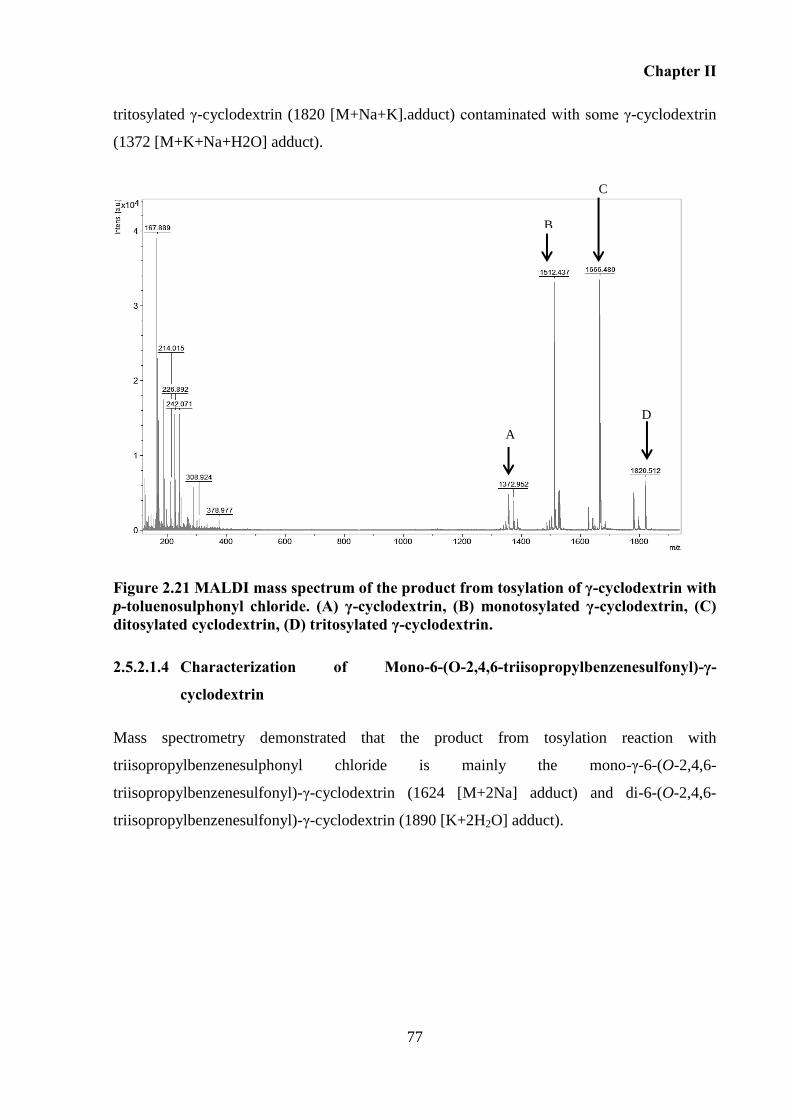
Chapter II
77
tritosylated γ-cyclodextrin (1820 [M+Na+K].adduct) contaminated with some γ-cyclodextrin
(1372 [M+K+Na+H2O] adduct).
Figure 2.21 MALDI mass spectrum of the product from tosylation of γ-cyclodextrin with p-toluenosulphonyl chloride. (A) γ-cyclodextrin, (B) monotosylated γ-cyclodextrin, (C) ditosylated cyclodextrin, (D) tritosylated γ-cyclodextrin.
2.5.2.1.4 Characterization of Mono-6-(O-2,4,6-triisopropylbenzenesulfonyl)-γ-
cyclodextrin
Mass spectrometry demonstrated that the product from tosylation reaction with
triisopropylbenzenesulphonyl chloride is mainly the mono-γ-6-(O-2,4,6-
triisopropylbenzenesulfonyl)-γ-cyclodextrin (1624 [M+2Na] adduct) and di-6-(O-2,4,6-
triisopropylbenzenesulfonyl)-γ-cyclodextrin (1890 [K+2H2O] adduct).
B
A
C
D
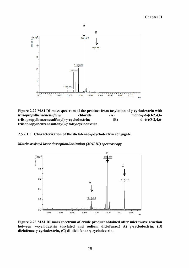
Chapter II
78
Figure 2.22 MALDI mass spectrum of the product from tosylation of γ-cyclodextrin with triisopropylbenzenesulfonyl chloride. (A) mono-γ-6-(O-2,4,6- triisopropylbenzenesulfonyl)-γ-cyclodextrin; (B) di-6-(O-2,4,6-triisopropylbenzenesulfonyl)-γ tolsylcyclodextrin.
2.5.2.1.5 Characterization of the diclofenac-γ-cyclodextrin conjugate
Matrix-assisted laser desorption/ionization (MALDI) spectroscopy
Figure 2.23 MALDI mass spectrum of crude product obtained after microwave reaction between γ-cyclodextrin tosylated and sodium diclofenac.( A) γ-cyclodextrin; (B) diclofenac-γ-cyclodextrin, (C) di-diclofenac-γ-cyclodextrin.
C
B
A
B
A
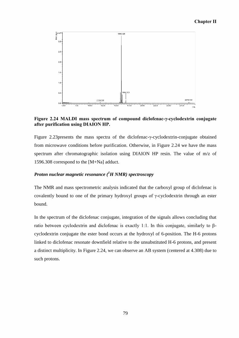
Chapter II
79
Figure 2.24 MALDI mass spectrum of compound diclofenac-γ-cyclodextrin conjugate after purification using DIAION HP.
Figure 2.23presents the mass spectra of the diclofenac-γ-cyclodextrin-conjugate obtained
from microwave conditions before purification. Otherwise, in Figure 2.24 we have the mass
spectrum after chromatographic isolation using DIAION HP resin. The value of m/z of
1596.308 correspond to the [M+Na] adduct.
Proton nuclear magnetic resonance (1H NMR) spectroscopy
The NMR and mass spectrometric analysis indicated that the carboxyl group of diclofenac is
covalently bound to one of the primary hydroxyl groups of γ-cyclodextrin through an ester
bound.
In the spectrum of the diclofenac conjugate, integration of the signals allows concluding that
ratio between cyclodextrin and diclofenac is exactly 1:1. In this conjugate, similarly to β-
cyclodextrin conjugate the ester bond occurs at the hydroxyl of 6-position. The H-6 protons
linked to diclofenac resonate downfield relative to the unsubstituted H-6 protons, and present
a distinct multiplicity. In Figure 2.24, we can observe an AB system (centered at 4.308) due to
such protons.
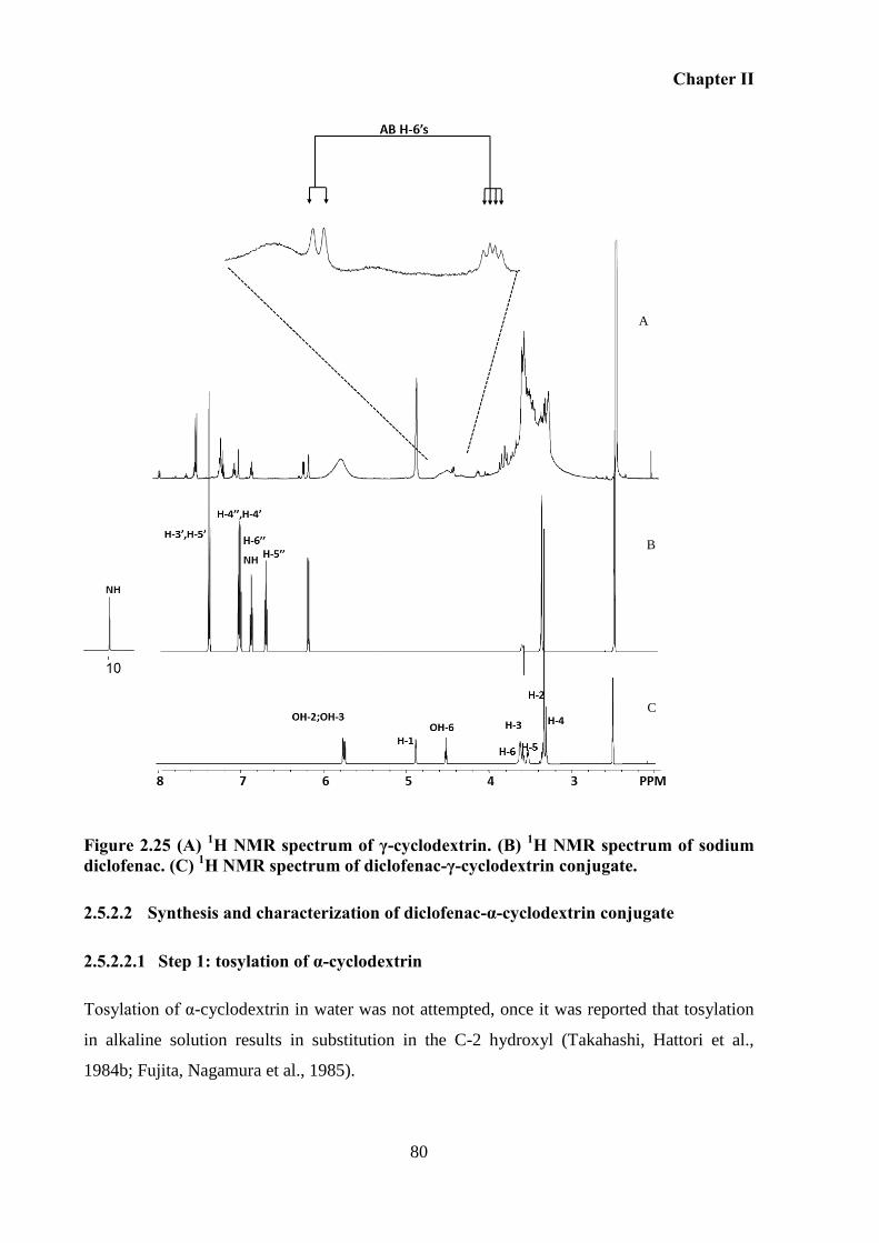
Chapter II
80
Figure 2.25 (A) 1H NMR spectrum of γ-cyclodextrin. (B) 1H NMR spectrum of sodium diclofenac. (C) 1H NMR spectrum of diclofenac-γ-cyclodextrin conjugate.
2.5.2.2 Synthesis and characterization of diclofenac-α-cyclodextrin conjugate
2.5.2.2.1 Step 1: tosylation of α-cyclodextrin
Tosylation of α-cyclodextrin in water was not attempted, once it was reported that tosylation
in alkaline solution results in substitution in the C-2 hydroxyl (Takahashi, Hattori et al.,
1984b; Fujita, Nagamura et al., 1985).
A
B
C
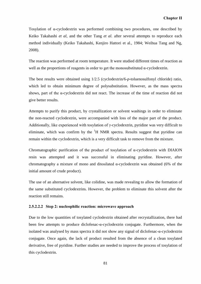
Chapter II
81
Tosylation of α-cyclodextrin was performed combining two procedures, one described by
Keiko Takahashi et al, and the other Tang et al. after several attempts to reproduce each
method individually (Keiko Takahashi, Kenjiro Hattori et al., 1984; Weihua Tang and Ng,
2008).
The reaction was performed at room temperature. It were studied different times of reaction as
well as the proportions of reagents in order to get the monosubstituted α-cyclodextrin.
The best results were obtained using 1/2.5 (cyclodextrin/6-p-toluenosulfonyl chloride) ratio,
which led to obtain minimum degree of polysubstitution. However, as the mass spectra
shows, part of the α-cyclodextrin did not react. The increase of the time of reaction did not
give better results.
Attempts to purify this product, by crystallization or solvent washings in order to eliminate
the non-reacted cyclodextrin, were accompanied with loss of the major part of the product.
Additionally, like experienced with tosylation of γ-cyclodextrin, pyridine was very difficult to
eliminate, which was confirm by the 1H NMR spectra. Results suggest that pyridine can
remain within the cyclodextrin, which is a very difficult task to remove from the mixture.
Chromatographic purification of the product of tosylation of α-cyclodextrin with DIAION
resin was attempted and it was successful in eliminating pyridine. However, after
chromatography a mixture of mono and ditosilated α-cyclodextrin was obtained (6% of the
initial amount of crude product).
The use of an alternative solvent, like colidine, was made revealing to allow the formation of
the same substituted cyclodextrins. However, the problem to eliminate this solvent after the
reaction still remains.
2.5.2.2.2 Step 2: nucleophilic reaction: microwave approach
Due to the low quantities of tosylated cyclodextrin obtained after recrystallization, there had
been few attempts to produce diclofenac-α-cyclodextrin conjugate. Furthermore, when the
isolated was analysed by mass spectra it did not show any signal of diclofenac-α-cyclodextrin
conjugate. Once again, the lack of product resulted from the absence of a clean tosylated
derivative, free of pyridine. Further studies are needed to improve the process of tosylation of
this cyclodextrin.
Chapter II
82
2.5.2.2.3 Characterization of 6-p-toluenosulfonyl-α-cyclodextrin
Mass spectrometry demonstrated that the product from the tosylation reaction of α-
cyclodextrin with p-toluenosulphonyl chloride is mainly the monotosylated α-cyclodextrin
(1188.382 [M+Na+K] adduct) and ditosylated α-cyclodextrin (1342.402 [M+Na+K] adduct)
contaminated with some α-cyclodextrin (1034 [M+K+Na] adduct).
Figure 2.26 MALDI mass spectrum of the product of tosylation of α-cyclodextrin with p-toluenosulphonyl chloride. (A) α-cyclodextrin; (B) monotosylated α-cyclodextrin; (C) ditosylated α-cyclodextrin.
2.6 Conclusions
An efficient chemical method to synthesize diclofenac-β-cyclodextrin conjugate was
developed. Additionally, the possibility to apply the same method to synthesize γ-cyclodextrin
and α-cyclodextrin derivatives was explored.
Initially, various approaches were investigated to synthesize β-cyclodextrin conjugates. These
included: nucleophylic substitution with conventional heating, activation of diclofenac using
carbodiimides, and formation of an acid chloride. All three approaches were unsuccessful in
the synthesis of the conjugate. We came to the conclusion that these approaches did not work
due to the particular structure of diclofenac since they were successfull in other cyclodextrin
conjugates.
A
B
C
Chapter II
83
Microwave radiation is being explored in organic synthesis in order to accelerate reactions.
Some authors have explored the use of this technique to promote reactions of cyclodextrin.
However, this type of non-conventional conditions was not applied in reactions that allow the
formation of cyclodextrin conjugates.
In this work, using the nucleophylic approach under microwave irradiation, diclofenac-β-
cyclodextrin conjugate was synthesized in efficient conditions and conveniently short reaction
time. The product was then purified through a polyaromatic ionic adsorbent resin. 1H
NMR
studies confirmed the covalent link of the carboxylic group of diclofenac with the hydroxyl of
β-cyclodextrin, and bidimensional 1H NMR ROESY allowed to conclude that diclofenac is
not included inside the cavity of cyclodextrin.
Preliminary chemical hydrolysis assays were performed in order to discern about the stability
of the conjugate. The drug release behavior of these conjugates was studied in different buffer
solutions (pH 1.2, 4.5, 6.8 and 7.4) at 37 ºC. Results showed that the conjugate was stable in
pH 4.5 and pH 6.8 and 7.4. Moreover, hydrolysis in pH 1.2 is negligible.
Preliminary results of β-cyclodextrin conjugate in different physiological conditions allows
predicting that this conjugate resists to different gastrointestinal tract pHs.
The synthesis of γ-cyclodextrin conjugate was made using the same approach and the end
product was characterized. However, this reaction showed not to be reproducible. On the
other hand, the production of α-cyclodextrin conjugate was not successful. Synthesis of
diclofenac α-cyclodextrin and γ-cyclodextrin conjugates was hindered by difficulties in the
synthesis and purification of the intermediary tosylated cyclodextrins. Further work needs to
be done to improve the process to prepare these tosylated intermediaries, since this seems to
be the key step to produce these conjugates.
The first step of this thesis was very challenging and demonstrated that the chemical
differences between cyclodextrins have to be taken in account when the aim is to synthesize
prodrugs. Additionally, diclofenac is a very instable molecule, which prevents its conjugation
in some chemical conditions. Overall, diclofenac-β-cyclodextrin revealed to be a molecule
with a strong potential for colon delivery of diclofenac. Therefore, the next Chapters focus on
the investigation of the potential of diclofenac-β-cyclodextrin to be used as a colonic prodrug.

Chapter II
84

CHAPTER III
STABILITY OF DICLOFENAC-β-CYCLODEXTRIN IN THE
GASTROINTESTINAL TRACT. IN VITRO AND EX VIVO STUDIES USING
SIMULATED FLUIDS, GASTROINTESTINAL FLUIDS FROM ANIMALS.

Chapter III
87
3 STABILITY OF DICLOFENAC-ΒETA-CYCLODEXTRIN IN THE
GASTROINTESTINAL TRACT. IN VITRO AND EX VIVO STUDIES USING
SIMULATED FLUIDS AND GASTROINTESTINAL FLUIDS FROM ANIMALS
3.1 Overview
An ideal prodrug for colon specific drug delivery should be stable in the stomach and small
intestine and reach the colon intact. Once the prodrug reaches the colon, the enzymes should
catalyse the drug-carrier bond breakdown, hence releasing the drug. Moreover, this process
should be rapid and complete.
In Chapter II, stability studies were carried out by incubation of diclofenac-β-cyclodextrin
conjugate in different buffer solutions in order to establish its ability to resist chemical
hydrolysis. According to those results, the conjugate revealed to be chemically stable. The
next aim is to evaluate the pre-systemic behaviour of the conjugate, namely its stability in the
GI tract by performance of in vitro and ex vivo studies using appropriate media able to
represent the physiological conditions of the GI tract.
This Chapter firstly pursues the investigation the ability of this based cyclodextrin prodrug to
release diclofenac in the lower intestine, using human faecal slurry to simulate colonic fluid.
These stability studies in the lower gastrointestinal tract are performed using a well-
established marketed available model of colonic delivery, sulfasalazine, as a positive control.
When sulfasalazine is orally administered, approximately 90 percent of it reaches the large
intestine as an intact molecule, where bacterial azoreductase cleaves the azo bond thereby
releasing mesalazine and sulfapyridine (Zoetendal, Akkermans et al., 1998).
Moreover, the stability is performed in the presence of enzymes, namely ester hydrolysing
enzymes and sugar-degrading enzymes in order to find an enzymatic media able to mimic the
metabolism of cyclodextrin-β-cyclodextrin in the colonic environment. Concomitantly, these
studies with enzymes are worth to understand the process involved on the metabolism of this
conjugate.
Chapter III
88
Further, this Chapter investigates the ability of the conjugate to be intact in the upper
gastrointestinal fluids, namely at gastric and intestinal level, using simulated gastric and
intestinal fluids, described in the Pharmacopeia.
The Chapter ends with stability studies conducted in fluids from different animals (pig, rabbit
and rat) in order evaluate the validity of each animal model to perform in vivo experiments.
3.2 Introduction
Any new molecule destined to oral delivery should be tested in vitro at an early stage of
development regarding its stability in the different environments of the GI tract, namely,
stomach, small intestine and colon. This allows an understanding of the pre-systemic behavior
of any molecule. Moreover, in the case of prodrugs, the stability tests will make it possible to
determine the site and rate of prodrug-to-drug conversion.
Different approaches are attempted in order to study and establish cyclodextrin-β-cyclodextrin
stability in the complex environment that constitutes the luminal content of the
gastrointestinal tract.
According to FDA Guidelines (Fda, 2000):
“Stability in the gastrointestinal tract may be documented using gastric and intestinal fluids
obtained from human subjects. Drug solutions in these fluids should be incubated at 37 oC for
a period that is representative of in vivo drug contact with these fluids; for example, 1 hour in
gastric fluid and 3 hours in intestinal fluid. Drug concentrations should then be determined
using a validated stability-indicating assay method. Significant degradation (>5%) of a drug in
this protocol could suggest potential instability. Obtaining gastrointestinal fluids from human
subjects requires intubation and may be difficult in some cases. Use of gastrointestinal fluids
from suitable animal models and/or simulated fluids such as Gastric and Intestinal Fluids USP
can be substituted when properly justified”.
This guidance for the determination of drugs stability in the gastrointestinal tract can be
adopted to guide the stability of prodrugs to site colonic delivery, namely of diclofenac-β-
cyclodextrin prodrug.
Chapter III
89
Simulated gastrointestinal fluids 3.2.1
3.2.1.1 Simulated colonic fluid
The primary region of the GI tract of interest to evaluate the effectiveness of a colonic
delivery system is the colon. The lack of a Pharmacopeia simulated colonic fluid for in vitro
studies warrants the use of other methodology to mimic the environment of the human colon.
Studies in fresh human fluids constitute an alternative that closely reproduces the environment
in vivo. In case of colonic release systems, the ascending colon is the primary region of
interest not only because of the substantial residence time in this region of the lower intestine
(Metcalf, Phillips et al., 1987; Kilian and Clive, 2005), but also due to the limited free water
volume in the transverse colon (Schiller, Fröhlich et al., 2005). Consequently, the potential of
colonic prodrugs should be evaluated in fluids from this region of the colon. However, human
fluids are not easily available due to the difficulty in sampling. An alternative is the use of
faeces to study metabolism at the level of the lower intestine. The advantage is that the
microbiota is essentially confined to the distal intestinal tract and epithelium associated
bacteria are apparently absent. The use of fresh human feces within an appropriate medium
has been identified as a suitable alternative, the well-known faecal slurry (Sousa, Paterson et
al., 2008). These faecal slurries from humans provide accurate information concerning not
only the intestinal microbiota, but also the enzymatic and electrolytes composition (Tannock,
1999; Basit and Lacey, 2001; Basit, Newton et al., 2002). Additionally, it is known that
microbiota is a major component and constitutes 40-45% of faecal solids in people consuming
Western-type diets (Cummings and Macfarlane, 1991).
3.2.1.2 Enzymatic colonic media containing enzymes
Given all the drawbacks associated with the use of biological fluids, namely the odour,
complexity of its composition, the lack of homogeneity, the thickness of the faeces matrix and
further the difficulties in the analysis of the drug release into this complex medium,
alternative media able to simulate the colon have been investigated.
Maria Vertzoni et al. developed two media (fasted state and fed state simulated media) able to
mimic the ascending colonic fluids useful for the determination of solubility of drugs at this
level (Vertzoni, Diakidou et al., 2010). These media do not contain enzymes and therefore are
not reliable to determine the drug release from colonic delivery systems activated by
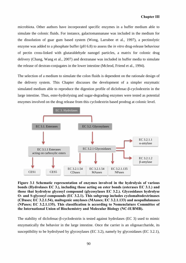
Chapter III
90
microbiota. Other authors have incorporated specific enzymes in a buffer medium able to
simulate the colonic fluids. For instance, galactomannanase was included in the medium for
the dissolution of guar gum based system (Wong, Larrabee et al., 1997), a pectinolytic
enzyme was added to a phosphate buffer (pH 6.8) to assess the in vitro drug-release behaviour
of pectin cross-linked with glutaraldehyde nanogel particles, a matrix for colonic drug
delivery (Chang, Wang et al., 2007) and dextranase was included in buffer media to simulate
the release of dextran-conjugates in the lower intestine (Mcleod, Friend et al., 1994).
The selection of a medium to simulate the colon fluids is dependent on the rationale design of
the delivery system. This Chapter discusses the development of a simpler enzymatic
simulated medium able to reproduce the digestion profile of diclofenac-β-cyclodextrin in the
large intestine. Thus, ester-hydrolysing and sugar-degrading enzymes were tested as potential
enzymes involved on the drug release from this cyclodextrin based prodrug at colonic level.
Figure 3.1 Schematic representation of enzymes involved in the hydrolysis of various bonds (Hydrolases EC 3.), including those acting on ester bonds (esterases EC 3.1.) and those that hydrolyse glycosyl compound (glycosylases EC 3.2.). Glycosidases hydrolyse O- and S-glycosyl compounds (EC 3.2.1). This subgroup includes cyclomaltodextrinases (CDases; EC 3.2.1.54), maltogenic amylases (MAases; EC 3.2.1.133) and neopullulanases (NPases; EC 3.2.1.135). This classification is according to Nomenclature Committee of the International Union of Biochemistry and Molecular Biology (NC-IUBMB).
The stability of diclofenac-β-cyclodextrin is tested against hydrolases (EC 3) used to mimic
enzymatically the behavior in the large intestine. Once the carrier is an oligosaccharide, its
susceptibility to be hydrolysed by glycosylases (EC 3.2), namely by glycosidases (EC 3.2.1),
EC 3. Hydrolases
EC 3.2. Glycosylases EC 3.1. Esterases
EC 3.2.1 Glycosidases EC 3.1.1 Esterases acting on carboxylic esters
EC 3.2.1.54 CDases
EC 3.2.1.54 MAases
EC 3.2.1.135 NPases
EC 3.2.1.2 β-amylase
EC 3.2.1.1 α-amylase
CES1 CES5
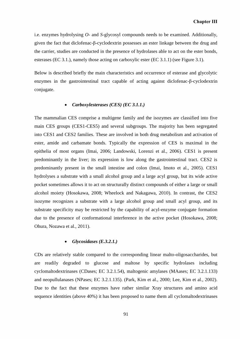
Chapter III
91
i.e. enzymes hydrolysing O- and S-glycosyl compounds needs to be examined. Additionally,
given the fact that diclofenac-β-cyclodextrin possesses an ester linkage between the drug and
the carrier, studies are conducted in the presence of hydrolases able to act on the ester bonds,
esterases (EC 3.1.), namely those acting on carboxylic ester (EC 3.1.1) (see Figure 3.1).
Below is described briefly the main characteristics and occurrence of esterase and glycolytic
enzymes in the gastrointestinal tract capable of acting against diclofenac-β-cyclodextrin
conjugate.
Carboxylesterases (CES) (EC 3.1.1.)
The mammalian CES comprise a multigene family and the isozymes are classified into five
main CES groups (CES1-CES5) and several subgroups. The majority has been segregated
into CES1 and CES2 families. These are involved in both drug metabolism and activation of
ester, amide and carbamate bonds. Typically the expression of CES is maximal in the
epithelia of most organs (Imai, 2006; Landowski, Lorenzi et al., 2006). CES1 is present
predominantly in the liver; its expression is low along the gastrointestinal tract. CES2 is
predominantly present in the small intestine and colon (Imai, Imoto et al., 2005). CES1
hydrolyses a substrate with a small alcohol group and a large acyl group, but its wide active
pocket sometimes allows it to act on structurally distinct compounds of either a large or small
alcohol moiety (Hosokawa, 2008; Wheelock and Nakagawa, 2010). In contrast, the CES2
isozyme recognizes a substrate with a large alcohol group and small acyl group, and its
substrate specificity may be restricted by the capability of acyl-enzyme conjugate formation
due to the presence of conformational interference in the active pocket (Hosokawa, 2008;
Ohura, Nozawa et al., 2011).
Glycosidases (E.3.2.1.)
CDs are relatively stable compared to the corresponding linear malto-oligosaccharides, but
are readily degraded to glucose and maltose by specific hydrolases including
cyclomaltodextrinases (CDases; EC 3.2.1.54), maltogenic amylases (MAases; EC 3.2.1.133)
and neopullulanases (NPases; EC 3.2.1.135). (Park, Kim et al., 2000; Lee, Kim et al., 2002).
Due to the fact that these enzymes have rather similar Xray structures and amino acid
sequence identities (above 40%) it has been proposed to name them all cyclomaltodextrinases
Chapter III
92
(Lee, Kim et al., 2002). They hydrolyse cyclic dextrins and also linear maltodextrins, but have
little or no activity for polysaccharides, such as starch and glycogen (Saha and Zeikus, 1992).
Generally, cyclodextrins are not hydrolyzable by exo type amylases, such as β-amylases (EC
3.2.1.2), which remove successive maltose units from the non-reducing ends of chains of
polysaccharides. Cyclodextrin works as an inhibitor of β-amylases (Suetsugu, Koyama et al.,
1974). In an other hand, α-Amylase (EC. 3.2.1.1) is an endo-type carbohydrase that
hydrolyzes α-1,4-glycosidic bonds, mainly in starch, producing α-limit dextrins and
oligosaccharides (Alkazaz, Desseaux et al., 2001; Maarel, Veen et al., 2002). Some α-
amylases, such as human salivary α-amylase, human and porcine pancreatic α-amylase have
been reported to hydrolyse CDs, although the rate is much slower than that for starch.
Moreover, some bacterial and fungal α-amylases revealed to be able to hydrolise
cyclodextrins, such as that from Aspergillus oryzae (Suetsugu, Koyama et al., 1974; Jodal,
Kandra et al., 1984). Interestingly, cyclomaltodextrinases are intracellular enzymes, whereas
most enzymes of the α-amylase family are extracellular enzymes (Depinto and Campbell,
1964; Saha and Zeikus, 1990; Hashimoto, Yamamoto et al., 2001; Lee, Kim et al., 2002).
3.2.1.3 Simulated gastric and intestinal fluids
Beyond the investigation of the ability of a colonic prodrug to release a drug in the colon, the
stability of it in the upper GI tract needs also needs to be evaluated. Pharmacopeia establishes
the composition of simulated gastric and intestinal fluids, which are well-accepted by the
worldwide scientific community. A suspension of pepsin (E.C. 3.4.23.1), a gastric
endopeptidase, at pH 1.2 is well-known to simulate gastric fluid (SIG). To replicate the
intestinal environment, Pharmacopeia describes a suspension of 1% (w/v) of pancreatin at pH
6.8, the called simulated intestinal fluid (SIF). It is worthwhile pointing out that these
simulated fluids used for in vitro testing do not faithfully reproduce the in vivo condition of
the human gastrointestinal environment. These are simple acid and phosphate buffer solutions
that contrast with the complex, dynamic and fluctuating gastrointestinal fluids (Lindahl,
Ungell et al., 1997; Moreno, Oth et al., 2006). Some elements such as electrolytes, bile acids
and a wide range of other lipids and important enzymatic co-factors are not present in the
composition of these fluids (Lindahl, Ungell et al., 1997; Mcconnell, Fadda et al., 2008; Sarti,
Barthelmes et al., 2011). Otherwise, other elements are available in excess as in the case of
Chapter III
93
pancreatic proteins, the amount of which is 5-fold higher in comparison with that in the
intestinal lumen of humans (Lindahl, Ungell et al., 1997; Moreno, Oth et al., 2006).
Animal gastrointestinal fluids 3.2.2
The simulated fluids do not represent all aspects of the physiological conditions of the oral
administration, such as pH, volume, contraction patterns, buffer capacity, bacteria, enzymes,
components of food and others already mentioned. For this reason, and although there is
variability between human and animals, namely in terms of bacterial activity, the fluids
collected from animals can be used for human predictions of drug metabolism as reviewed by
Sousa et al. (Sousa, Paterson et al., 2008). These GI fluids from animals can represent better
the in vivo metabolic fate than simulated fluids (Sarti, Barthelmes et al., 2011). The challenge
is to establish the best animal model to be further used in the in vivo experiments. The main
anatomic and physiological differences between some animal models are described on
Chapter I. In the case of colonic prodrugs, when the prodrug is converted to the active drug by
the enzymatic reaction, the conversion can be highly species-dependent (Stella, Borchardt et
al., 2007). For instance, Beagle dogs (Carnivora order) are not suitable to perform
pharmacokinetics studies of ester prodrugs, due to the absence of carboxylesterase isozyme in
the small intestine of these animals (Imai, 2006). Dogs are not suitable models to test non-
steroid anti-inflammatory drugs once they are very sensitive to ulcer-inducing properties of
these drugs (Swindle, Makin et al., 2011). As a non-rodent species, pig (Artiodactyla order) is
considered an alternative to the dog or monkey (Swindle, Makin et al., 2011). Although pig is
physiologically similar to humans, some anatomic differences are significant namely: the
existence of a well-defined caecum contrarily to that in human, and also a bigger colon
(Kararli, 1995). Pig is considered a superior model for colonic delivery when compared with
smaller laboratory animals, namely for studies of oral intake of intact dosage forms intended
for man, due to the large size (Sarfraz, Sarfraz et al., 2011). It has been used to study colonic
ester prodrugs, namely of colonic prodrugs dextran-based (Harboe, Larsen et al., 1989). As
for an non-rodent, rabbit (Lagomorpha order) is other alternative and it has been use to test
colonic formulations (Freire, Podczeck et al., 2010; Mustafin, Kabanova et al., 2011).
However, rats (Rodentia order), due namely to the small size and low cost, are used
extensively in pre-clinical tests of drugs, including prodrugs to colon delivery (Mcleod,
Friend et al., 1994; Kamada, Hirayama et al., 2002). Additionally rats have been used to
perform the in vitro stability test of colonic prodrugs, as described previously. In this Chapter,
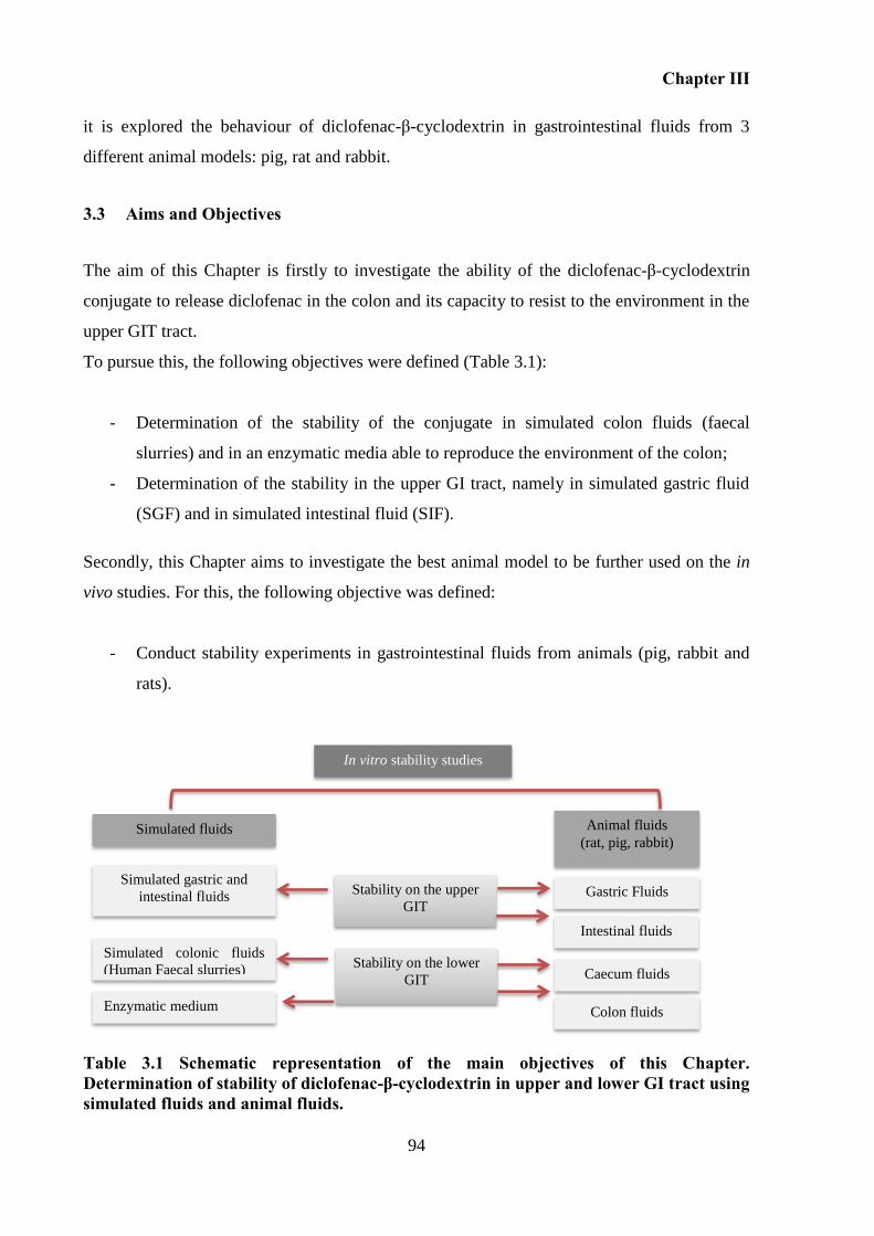
Chapter III
94
it is explored the behaviour of diclofenac-β-cyclodextrin in gastrointestinal fluids from 3
different animal models: pig, rat and rabbit.
3.3 Aims and Objectives
The aim of this Chapter is firstly to investigate the ability of the diclofenac-β-cyclodextrin
conjugate to release diclofenac in the colon and its capacity to resist to the environment in the
upper GIT tract.
To pursue this, the following objectives were defined (Table 3.1):
- Determination of the stability of the conjugate in simulated colon fluids (faecal
slurries) and in an enzymatic media able to reproduce the environment of the colon;
- Determination of the stability in the upper GI tract, namely in simulated gastric fluid
(SGF) and in simulated intestinal fluid (SIF).
Secondly, this Chapter aims to investigate the best animal model to be further used on the in
vivo studies. For this, the following objective was defined:
- Conduct stability experiments in gastrointestinal fluids from animals (pig, rabbit and
rats).
Table 3.1 Schematic representation of the main objectives of this Chapter. Determination of stability of diclofenac-β-cyclodextrin in upper and lower GI tract using simulated fluids and animal fluids.
Stability on the lower
GIT
Stability on the upper
GIT
Simulated fluids Animal fluids (rat, pig, rabbit)
Simulated gastric and
intestinal fluids Gastric Fluids
Simulated colonic fluids
(Human Faecal slurries)
Enzymatic medium
Intestinal fluids
Caecum fluids
Colon fluids
In vitro stability studies
Chapter III
95
3.4 Materials
Reagents and chemicals 3.4.1
Sodium diclofenac (MW = 318.14 g/mol) and sulfasalazine (MW = 398.394 g/mol) were
purchased from Sigma Aldrich, diclofenac-β-cyclodextrin (MW = 1411 g/mol) was
synthetized according to the method reported in Chapter II.
Sodium chloride (NaCl), potassium hydroxide (KOH), sodium hydroxide (NaOH),
dipotassium hydrogen orthophosphate (K2HPO4) potassium dihydrogen orthophosphate
(KH2PO2), hydrochloric acid (HCl), HPLC grade acetonitrile, HPLC grade methanol and
HPLC grade water were purchased from Fisher Scientifics UK Limited.
Trifluoroacetic acid (TFA) and dimethylformamide (DMF), 4-(2-Hydroxyethyl)piperazine-1-
ethanesulfonic acid, N-(2-Hydroxyethyl)piperazine-N′-(2-ethanesulfonic acid) (HEPES), L-
cysteine HCl, bile salts, vitamin K, sodium bicarbonate (NaHCO3), haemin, resazurin, and
calcium chloride dehydrate CaCl2.2H2O were acquired from Sigma Aldrich (Dorset, UK).
Calcium chloride hexahydrate (CaCl2.6H2O), magnesium sulphate heptahydrate
(MgSO4.7H2O) and sodium acetate trihydrate (NaCH3COONa.3H2O) were obtained from
VWR (Leicestershire, UK).
Peptone water and yeast extract were purchased from Oxoid Limited (Hampshire, UK).
Enzymes 3.4.2
All the enzymes were purchased from Sigma-Aldrich.
Pepsin derived from porcine gastric mucosa (Reference: P700) (EC. 3.4.23.1);
Pancreatin from porcine pancreas ( ≥3 x USP specification) (Reference: P1625) (EINECS
232-468-9);
α-Amylase from Aspergillus oryzae (Reference: 10065) (EC. 3.2.1.1);
α-Amylase from Bacillus lechiniformes (Reference: A3403) (EC. 3.2.1.1);
α-Amylase from porcine pancreas (Reference: A 3176) (EC. 3.1.1.1);
Esterase form porcine liver (PLE) (Reference: 46058) (EC. 3.1.1.1).
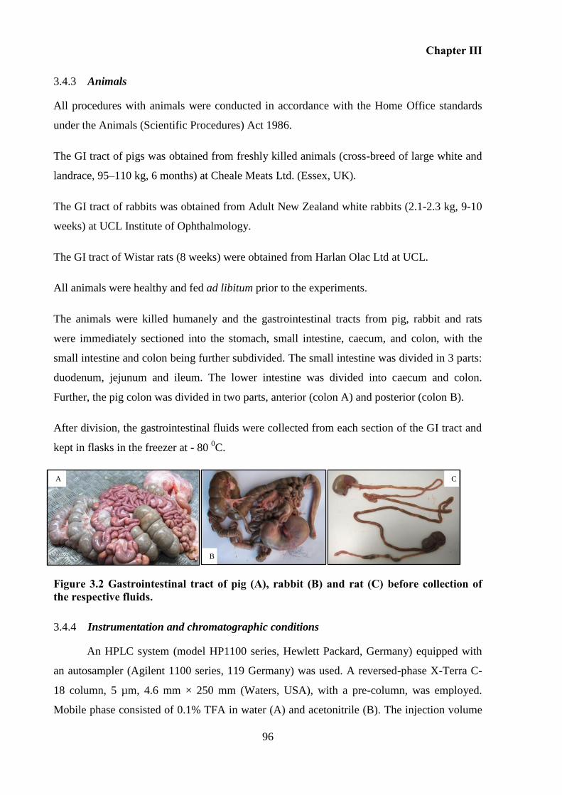
Chapter III
96
A
B
C
Animals 3.4.3
All procedures with animals were conducted in accordance with the Home Office standards
under the Animals (Scientific Procedures) Act 1986.
The GI tract of pigs was obtained from freshly killed animals (cross-breed of large white and
landrace, 95–110 kg, 6 months) at Cheale Meats Ltd. (Essex, UK).
The GI tract of rabbits was obtained from Adult New Zealand white rabbits (2.1-2.3 kg, 9-10
weeks) at UCL Institute of Ophthalmology.
The GI tract of Wistar rats (8 weeks) were obtained from Harlan Olac Ltd at UCL.
All animals were healthy and fed ad libitum prior to the experiments.
The animals were killed humanely and the gastrointestinal tracts from pig, rabbit and rats
were immediately sectioned into the stomach, small intestine, caecum, and colon, with the
small intestine and colon being further subdivided. The small intestine was divided in 3 parts:
duodenum, jejunum and ileum. The lower intestine was divided into caecum and colon.
Further, the pig colon was divided in two parts, anterior (colon A) and posterior (colon B).
After division, the gastrointestinal fluids were collected from each section of the GI tract and
kept in flasks in the freezer at - 80 0C.
Figure 3.2 Gastrointestinal tract of pig (A), rabbit (B) and rat (C) before collection of the respective fluids.
Instrumentation and chromatographic conditions 3.4.4
An HPLC system (model HP1100 series, Hewlett Packard, Germany) equipped with
an autosampler (Agilent 1100 series, 119 Germany) was used. A reversed-phase X-Terra C-
18 column, 5 µm, 4.6 mm × 250 mm (Waters, USA), with a pre-column, was employed.
Mobile phase consisted of 0.1% TFA in water (A) and acetonitrile (B). The injection volume
Chapter III
97
was 20 µL, the flow rate 1.0 mL/min and the detection wavelength was 254 nm. All the
assays were performed at 30 0C. The gradient system was: 0-15 minutes 25-60% B; 15-22
minutes 60-25% B, with a flow rate of 1.0 mL/min.
3.5 Methods
Stability of the diclofenac-β-cyclodextrin conjugate in simulated fluids 3.5.1
3.5.1.1 Stability studies in the lower GI tract
The stability was carried out in simulated colonic fluid prepared as human faecal slurry and
the use of an enzymatic media able to mimic the release in the colonic medium was
investigated.
3.5.1.2 Stability studies in human faecal slurries
Faecal slurries were utilized to simulate the conditions of the colon (Basit and Lacey, 2001;
Basit, Newton et al., 2002). Fresh faeces from healthy adults were collected in pre-weighed
sterile plastic containers. The volunteers were on no medication and had not taken antibiotics
for at least the previous six months. The plastic receptacle containing freshly voided human
faeces was transferred into an anaerobic workstation (Electroteck 500TG workstation,
Electrotek, UK) (37 oC and relative air humidity of 70%) as soon as possible after defecation
in order to preserve viability of the microbial population.
Faeces were diluted with PBS pH 6.8 (Appendix A) in order to obtain 40% (w/w) slurry. The
mixture was then homogenized and sieved through an open mesh fabric (Sefar NitexTM
Heiden, Switzerland, pore size 350 µm) to remove any unhomogenised fibrous material.
Afterwards, the sieved homogenized faecal slurry was diluted 50% (w/w) with basal medium,
which was prepared as described below.
Preparation of basal medium:
The quantity of each ingredient is described in Table 3.2. Peptone water and yeast extract
were weighed into a glass flask containing 1.3 L of distilled water and autoclaved at
125 oC for 20 minutes. In a 200 mL volumetric flask containing about 150 mL of distilled
water, NaCl, K2HPO4, MgSO4.7H2O and CaCl2.6H2O were weighed and dissolved under
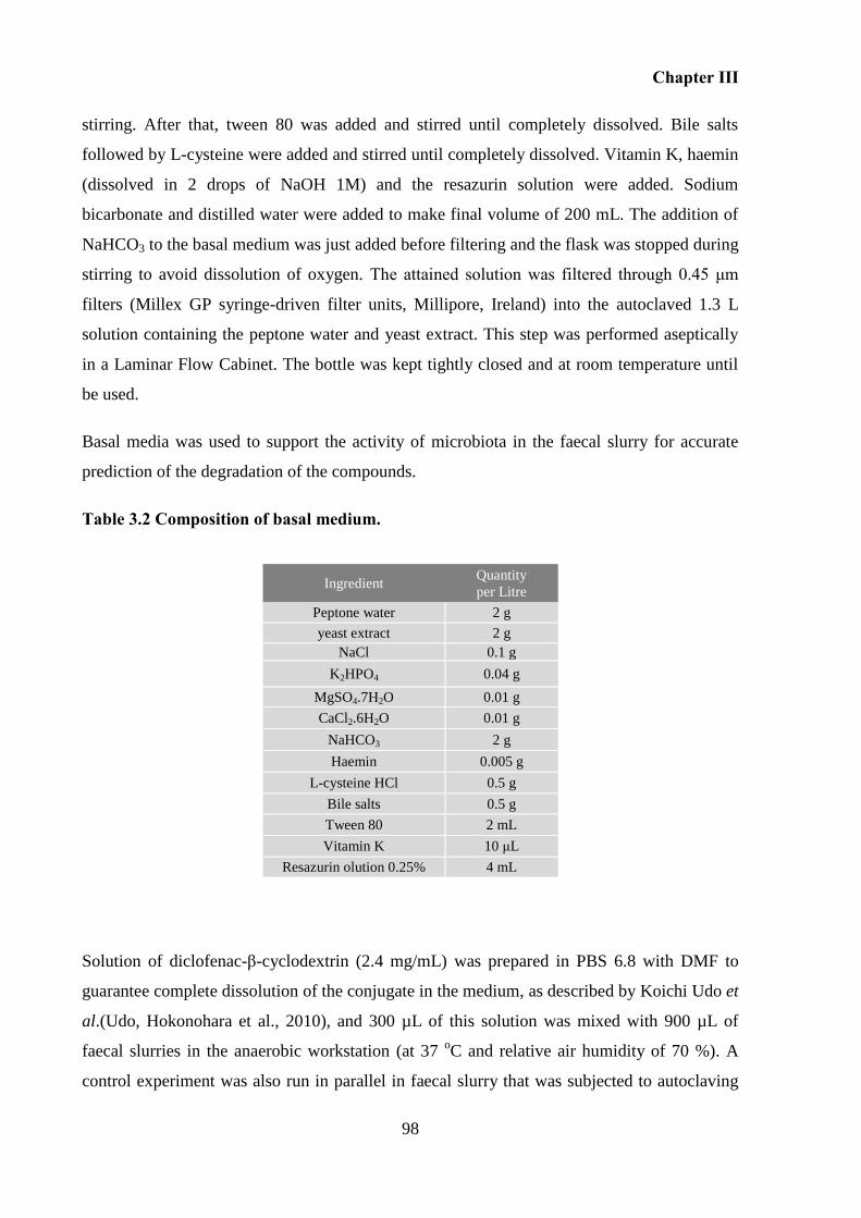
Chapter III
98
stirring. After that, tween 80 was added and stirred until completely dissolved. Bile salts
followed by L-cysteine were added and stirred until completely dissolved. Vitamin K, haemin
(dissolved in 2 drops of NaOH 1M) and the resazurin solution were added. Sodium
bicarbonate and distilled water were added to make final volume of 200 mL. The addition of
NaHCO3 to the basal medium was just added before filtering and the flask was stopped during
stirring to avoid dissolution of oxygen. The attained solution was filtered through 0.45 μm
filters (Millex GP syringe-driven filter units, Millipore, Ireland) into the autoclaved 1.3 L
solution containing the peptone water and yeast extract. This step was performed aseptically
in a Laminar Flow Cabinet. The bottle was kept tightly closed and at room temperature until
be used.
Basal media was used to support the activity of microbiota in the faecal slurry for accurate
prediction of the degradation of the compounds.
Table 3.2 Composition of basal medium.
Solution of diclofenac-β-cyclodextrin (2.4 mg/mL) was prepared in PBS 6.8 with DMF to
guarantee complete dissolution of the conjugate in the medium, as described by Koichi Udo et
al.(Udo, Hokonohara et al., 2010), and 300 µL of this solution was mixed with 900 µL of
faecal slurries in the anaerobic workstation (at 37 oC and relative air humidity of 70 %). A
control experiment was also run in parallel in faecal slurry that was subjected to autoclaving
Ingredient Quantity
per Litre
Peptone water 2 g
yeast extract 2 g
NaCl 0.1 g
K2HPO4 0.04 g
MgSO4.7H2O 0.01 g
CaCl2.6H2O 0.01 g
NaHCO3 2 g
Haemin 0.005 g
L-cysteine HCl 0.5 g
Bile salts 0.5 g
Tween 80 2 mL
Vitamin K 10 μL
Resazurin olution 0.25% 4 mL
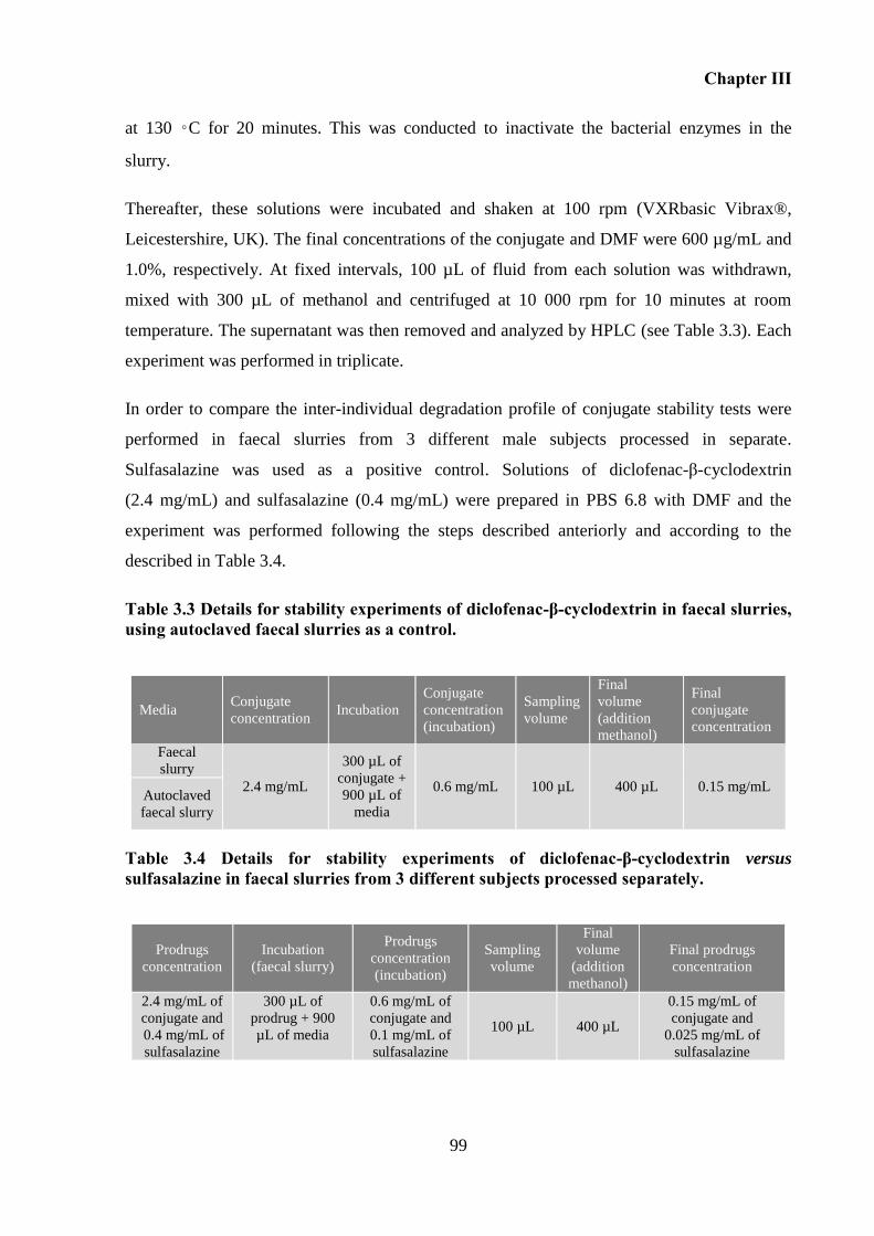
Chapter III
99
at 130 ◦C for 20 minutes. This was conducted to inactivate the bacterial enzymes in the
slurry.
Thereafter, these solutions were incubated and shaken at 100 rpm (VXRbasic Vibrax®,
Leicestershire, UK). The final concentrations of the conjugate and DMF were 600 µg/mL and
1.0%, respectively. At fixed intervals, 100 µL of fluid from each solution was withdrawn,
mixed with 300 µL of methanol and centrifuged at 10 000 rpm for 10 minutes at room
temperature. The supernatant was then removed and analyzed by HPLC (see Table 3.3). Each
experiment was performed in triplicate.
In order to compare the inter-individual degradation profile of conjugate stability tests were
performed in faecal slurries from 3 different male subjects processed in separate.
Sulfasalazine was used as a positive control. Solutions of diclofenac-β-cyclodextrin
(2.4 mg/mL) and sulfasalazine (0.4 mg/mL) were prepared in PBS 6.8 with DMF and the
experiment was performed following the steps described anteriorly and according to the
described in Table 3.4.
Table 3.3 Details for stability experiments of diclofenac-β-cyclodextrin in faecal slurries, using autoclaved faecal slurries as a control.
Table 3.4 Details for stability experiments of diclofenac-β-cyclodextrin versus sulfasalazine in faecal slurries from 3 different subjects processed separately.
Media Conjugate
concentration Incubation
Conjugate
concentration
(incubation)
Sampling
volume
Final
volume
(addition
methanol)
Final
conjugate
concentration
Faecal
slurry
2.4 mg/mL
300 µL of
conjugate +
900 µL of
media
0.6 mg/mL 100 µL 400 µL 0.15 mg/mL Autoclaved
faecal slurry
Prodrugs
concentration
Incubation
(faecal slurry)
Prodrugs
concentration
(incubation)
Sampling
volume
Final
volume
(addition
methanol)
Final prodrugs
concentration
2.4 mg/mL of
conjugate and
0.4 mg/mL of
sulfasalazine
300 µL of
prodrug + 900
µL of media
0.6 mg/mL of
conjugate and
0.1 mg/mL of
sulfasalazine
100 µL 400 µL
0.15 mg/mL of
conjugate and
0.025 mg/mL of
sulfasalazine
Chapter III
100
3.5.1.3 Stability studies in simulated colonic media containing enzymes
In presence of esterase from porcine liver solution (39 units/mL).
Solutions of conjugate (0.8 mg/mL) were prepared in HEPES/NaOH buffer (pH 7.4) (see
Appendix C) with 1% of DMF. After that, esterase from porcine liver (39 units/mL) was
added to the previous solutions. This procedure was adapted by that described by F. Hirayama
et al. (Hirayma, Ogata et al., 2000).
In presence of α-amylases solutions: Bacillus lechiniformes (250 units/mL) porcine
pancreas (250 units/mL) and Aspergillus oryzae (20-250 units/mL).
Solutions of conjugate (0.8 mg/mL) were prepared in 0.01M CaCl2/0.2 M acetate buffer (pH
5.5) (see Appendix C) with 1% of DMF. After that, the respective α-amylase was added to the
previous solutions. Stability tests with α-amylase from Aspergillus oryzae were conducted
with different concentrations of enzyme 20, 40 or 250 units/mL while with α-amylase from
Bacillus lechiniformes and with α-amylase from porcine pancreas were conducted at 250
units/mL.
In presence of α-Amylase from Aspergillus oryzae (250 units/mL) and esterase from
porcine liver (39 units/mL) simultaneously.
Solutions of conjugate (0.8 mg/mL) were prepared in 0.01 M CaCl2/0.2 M acetate buffer pH
5.5 or in HEPES/NaOH buffer (pH 7.4) with 1% of DMF. After that, α-amylase and esterase
were added to the previous solution.
In presence of α-Amylase from Aspergillus oryzae (250 units/mL) followed by the presence
of esterase (39 units/mL).
Solutions of conjugate (0.8mg/mL) were prepared in 0.01 M CaCl2/0.2 M acetate buffer (pH
5.5) with 1% of DMF. After that, α-amylase (250 units/mL) was added and the solution
incubates at 37 ºC under 100 rpm. After 12 hours of incubation, the pH of the solution was
changed to 7.4 by the addition of NaOH 0.1 N and the esterase was added to the solution.
All the previous prepared samples were incubated at 37 ºC and shaken at 100 rpm. 100 µL of
sample was withdrawn immediately after enzyme addition and at different time points
hereafter, mixed with 400 µL of methanol and centrifuged at 10 000 rpm for 10 minutes at
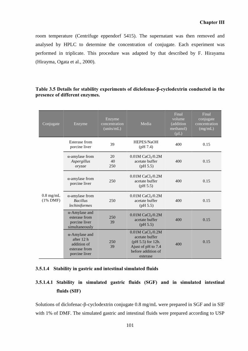
Chapter III
101
room temperature (Centrifuge eppendorf 5415). The supernatant was then removed and
analysed by HPLC to determine the concentration of conjugate. Each experiment was
performed in triplicate. This procedure was adapted by that described by F. Hirayama
(Hirayma, Ogata et al., 2000).
Table 3.5 Details for stability experiments of diclofenac-β-cyclodextrin conducted in the presence of different enzymes.
3.5.1.4 Stability in gastric and intestinal simulated fluids
3.5.1.4.1 Stability in simulated gastric fluids (SGF) and in simulated intestinal
fluids (SIF)
Solutions of diclofenac-β-cyclodextrin conjugate 0.8 mg/mL were prepared in SGF and in SIF
with 1% of DMF. The simulated gastric and intestinal fluids were prepared according to USP
Conjugate Enzyme
Enzyme
concentration
(units/mL)
Media
Final
volume
(addition
methanol)
(µL)
Final
conjugate
concentration
(mg/mL)
0.8 mg/mL
(1% DMF)
Esterase from
porcine liver 39
HEPES/NaOH
(pH 7.4) 400 0.15
α-amylase from
Aspergillus
oryzae
20
40
250
0.01M CaCl2/0.2M
acetate buffer
(pH 5.5)
400 0.15
α-amylase from
porcine liver 250
0.01M CaCl2/0.2M
acetate buffer
(pH 5.5)
400 0.15
α-amylase from
Bacillus
lechiniformes
250
0.01M CaCl2/0.2M
acetate buffer
(pH 5.5)
400 0.15
α-Amylase and
esterase from
porcine liver
simultaneously
250
39
0.01M CaCl2/0.2M
acetate buffer
(pH 5.5)
400 0.15
α-Amylase and
after 12 h
addition of
esterase from
porcine liver
250
39
0.01M CaCl2/0.2M
acetate buffer
(pH 5.5) for 12h.
Ajust of pH to 7.4
before addition of
esterase
400 0.15
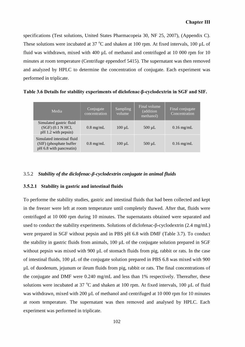
Chapter III
102
specifications (Test solutions, United States Pharmacopeia 30, NF 25, 2007), (Appendix C).
These solutions were incubated at 37 oC and shaken at 100 rpm. At fixed intervals, 100 µL of
fluid was withdrawn, mixed with 400 µL of methanol and centrifuged at 10 000 rpm for 10
minutes at room temperature (Centrifuge eppendorf 5415). The supernatant was then removed
and analyzed by HPLC to determine the concentration of conjugate. Each experiment was
performed in triplicate.
Table 3.6 Details for stability experiments of diclofenac-β-cyclodextrin in SGF and SIF.
Media Conjugate
concentration
Sampling
volume
Final volume
(addition
methanol)
Final conjugate
Concentration
Simulated gastric fluid
(SGF) (0.1 N HCl,
pH 1.2 with pepsin)
0.8 mg/mL 100 µL 500 µL 0.16 mg/mL
Simulated intestinal fluid
(SIF) (phosphate buffer
pH 6.8 with pancreatin)
0.8 mg/mL 100 µL 500 µL 0.16 mg/mL
Stability of the diclofenac-β-cyclodextrin conjugate in animal fluids 3.5.2
3.5.2.1 Stability in gastric and intestinal fluids
To performe the stability studies, gastric and intestinal fluids that had been collected and kept
in the freezer were left at room temperature until completely thawed. After that, fluids were
centrifuged at 10 000 rpm during 10 minutes. The supernatants obtained were separated and
used to conduct the stability experiments. Solutions of diclofenac-β-cyclodextrin (2.4 mg/mL)
were prepared in SGF without pepsin and in PBS pH 6.8 with DMF (Table 3.7). To conduct
the stability in gastric fluids from animals, 100 µL of the conjugate solution prepared in SGF
without pepsin was mixed with 900 µL of stomach fluids from pig, rabbit or rats. In the case
of intestinal fluids, 100 µL of the conjugate solution prepared in PBS 6.8 was mixed with 900
µL of duodenum, jejunum or ileum fluids from pig, rabbit or rats. The final concentrations of
the conjugate and DMF were 0.240 mg/mL and less than 1% respectively. Thereafter, these
solutions were incubated at 37 oC and shaken at 100 rpm. At fixed intervals, 100 µL of fluid
was withdrawn, mixed with 200 µL of methanol and centrifuged at 10 000 rpm for 10 minutes
at room temperature. The supernatant was then removed and analysed by HPLC. Each
experiment was performed in triplicate.
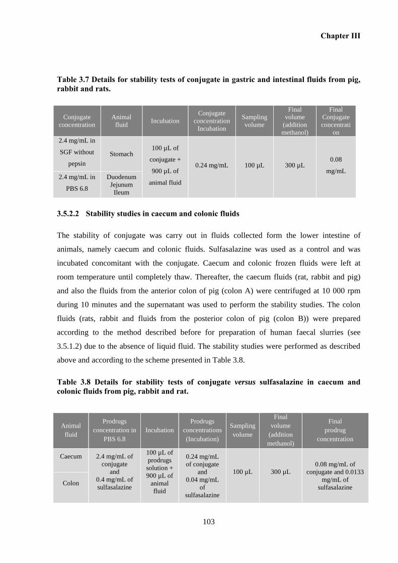
Chapter III
103
Table 3.7 Details for stability tests of conjugate in gastric and intestinal fluids from pig, rabbit and rats.
Conjugate
concentration
Animal
fluid Incubation
Conjugate
concentration
Incubation
Sampling
volume
Final
volume
(addition
methanol)
Final
Conjugate
concentrati
on
2.4 mg/mL in
SGF without
pepsin
Stomach 100 µL of
conjugate +
900 µL of
animal fluid
0.24 mg/mL 100 µL 300 µL 0.08
mg/mL 2.4 mg/mL in
PBS 6.8
Duodenum
Jejunum
Ileum
3.5.2.2 Stability studies in caecum and colonic fluids
The stability of conjugate was carry out in fluids collected form the lower intestine of
animals, namely caecum and colonic fluids. Sulfasalazine was used as a control and was
incubated concomitant with the conjugate. Caecum and colonic frozen fluids were left at
room temperature until completely thaw. Thereafter, the caecum fluids (rat, rabbit and pig)
and also the fluids from the anterior colon of pig (colon A) were centrifuged at 10 000 rpm
during 10 minutes and the supernatant was used to perform the stability studies. The colon
fluids (rats, rabbit and fluids from the posterior colon of pig (colon B)) were prepared
according to the method described before for preparation of human faecal slurries (see
3.5.1.2) due to the absence of liquid fluid. The stability studies were performed as described
above and according to the scheme presented in Table 3.8.
Table 3.8 Details for stability tests of conjugate versus sulfasalazine in caecum and colonic fluids from pig, rabbit and rat.
Animal
fluid
Prodrugs
concentration in
PBS 6.8
Incubation
Prodrugs
concentrations
(Incubation)
Sampling
volume
Final
volume
(addition
methanol)
Final
prodrug
concentration
Caecum 2.4 mg/mL of
conjugate
and
0.4 mg/mL of
sulfasalazine
100 µL of
prodrugs
solution +
900 µL of
animal
fluid
0.24 mg/mL
of conjugate
and
0.04 mg/mL
of
sulfasalazine
100 µL 300 µL 0.08 mg/mL of
conjugate and 0.0133
mg/mL of
sulfasalazine Colon
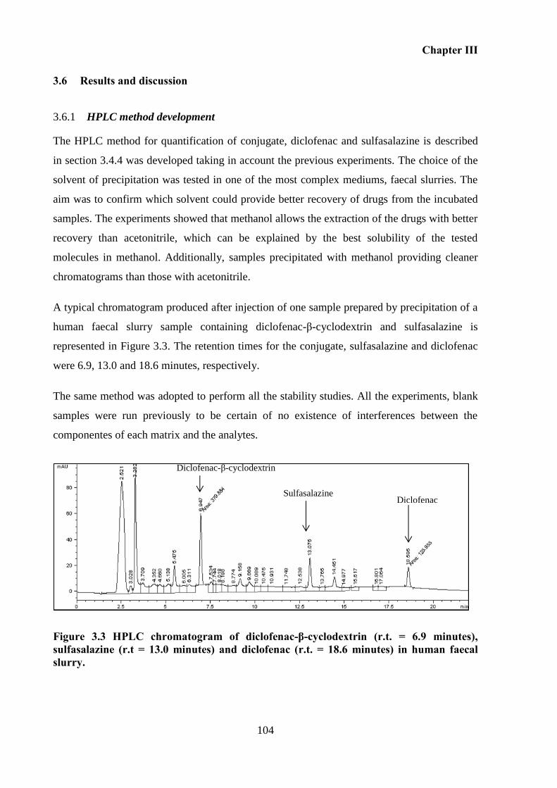
Chapter III
104
3.6 Results and discussion
HPLC method development 3.6.1
The HPLC method for quantification of conjugate, diclofenac and sulfasalazine is described
in section 3.4.4 was developed taking in account the previous experiments. The choice of the
solvent of precipitation was tested in one of the most complex mediums, faecal slurries. The
aim was to confirm which solvent could provide better recovery of drugs from the incubated
samples. The experiments showed that methanol allows the extraction of the drugs with better
recovery than acetonitrile, which can be explained by the best solubility of the tested
molecules in methanol. Additionally, samples precipitated with methanol providing cleaner
chromatograms than those with acetonitrile.
A typical chromatogram produced after injection of one sample prepared by precipitation of a
human faecal slurry sample containing diclofenac-β-cyclodextrin and sulfasalazine is
represented in Figure 3.3. The retention times for the conjugate, sulfasalazine and diclofenac
were 6.9, 13.0 and 18.6 minutes, respectively.
The same method was adopted to perform all the stability studies. All the experiments, blank
samples were run previously to be certain of no existence of interferences between the
componentes of each matrix and the analytes.
Figure 3.3 HPLC chromatogram of diclofenac-β-cyclodextrin (r.t. = 6.9 minutes), sulfasalazine (r.t = 13.0 minutes) and diclofenac (r.t. = 18.6 minutes) in human faecal slurry.
Diclofenac-β-cyclodextrin
Sulfasalazine Diclofenac
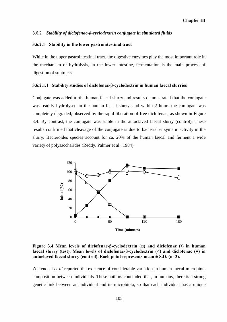
Chapter III
105
Stability of diclofenac-β-cyclodextrin conjugate in simulated fluids 3.6.2
3.6.2.1 Stability in the lower gastrointestinal tract
While in the upper gastrointestinal tract, the digestive enzymes play the most important role in
the mechanism of hydrolysis, in the lower intestine, fermentation is the main process of
digestion of subtracts.
3.6.2.1.1 Stability studies of diclofenac-β-cyclodextrin in human faecal slurries
Conjugate was added to the human faecal slurry and results demonstrated that the conjugate
was readily hydrolysed in the human faecal slurry, and within 2 hours the conjugate was
completely degraded, observed by the rapid liberation of free diclofenac, as shown in Figure
3.4. By contrast, the conjugate was stable in the autoclaved faecal slurry (control). These
results confirmed that cleavage of the conjugate is due to bacterial enzymatic activity in the
slurry. Bacteroides species account for ca. 20% of the human faecal and ferment a wide
variety of polysaccharides (Reddy, Palmer et al., 1984).
Figure 3.4 Mean levels of diclofenac-β-cyclodextrin (□) and diclofenac (▪) in human faecal slurry (test). Mean levels of diclofenac-β-cyclodextrin (○) and diclofenac (●) in autoclaved faecal slurry (control). Each point represents mean ± S.D. (n=3).
Zoetendaal et al reported the existence of considerable variation in human faecal microbiota
composition between individuals. These authors concluded that, in humans, there is a strong
genetic link between an individual and its microbiota, so that each individual has a unique
0
20
40
60
80
100
120
0 60 120 180
Initi
al (%
)
Time (minutes)
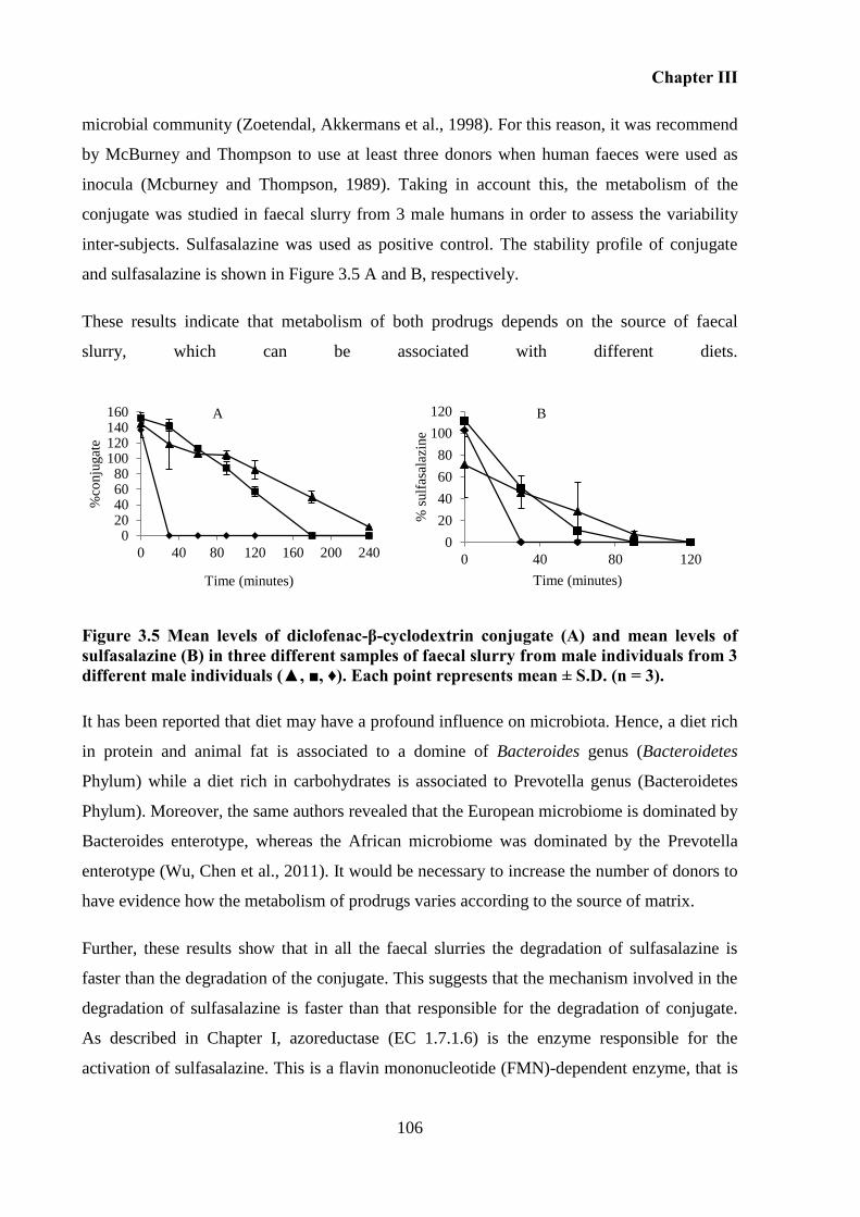
Chapter III
106
microbial community (Zoetendal, Akkermans et al., 1998). For this reason, it was recommend
by McBurney and Thompson to use at least three donors when human faeces were used as
inocula (Mcburney and Thompson, 1989). Taking in account this, the metabolism of the
conjugate was studied in faecal slurry from 3 male humans in order to assess the variability
inter-subjects. Sulfasalazine was used as positive control. The stability profile of conjugate
and sulfasalazine is shown in Figure 3.5 A and B, respectively.
These results indicate that metabolism of both prodrugs depends on the source of faecal
slurry, which can be associated with different diets.
Figure 3.5 Mean levels of diclofenac-β-cyclodextrin conjugate (A) and mean levels of sulfasalazine (B) in three different samples of faecal slurry from male individuals from 3 different male individuals (▲, ■, ♦). Each point represents mean ± S.D. (n = 3).
It has been reported that diet may have a profound influence on microbiota. Hence, a diet rich
in protein and animal fat is associated to a domine of Bacteroides genus (Bacteroidetes
Phylum) while a diet rich in carbohydrates is associated to Prevotella genus (Bacteroidetes
Phylum). Moreover, the same authors revealed that the European microbiome is dominated by
Bacteroides enterotype, whereas the African microbiome was dominated by the Prevotella
enterotype (Wu, Chen et al., 2011). It would be necessary to increase the number of donors to
have evidence how the metabolism of prodrugs varies according to the source of matrix.
Further, these results show that in all the faecal slurries the degradation of sulfasalazine is
faster than the degradation of the conjugate. This suggests that the mechanism involved in the
degradation of sulfasalazine is faster than that responsible for the degradation of conjugate.
As described in Chapter I, azoreductase (EC 1.7.1.6) is the enzyme responsible for the
activation of sulfasalazine. This is a flavin mononucleotide (FMN)-dependent enzyme, that is
0
20
40
60
80
100
120
0 40 80 120
% s
ulf
asal
azin
e
Time (minutes)
020406080
100120140160
0 40 80 120 160 200 240
%co
nju
gat
e
Time (minutes)
A B
A B
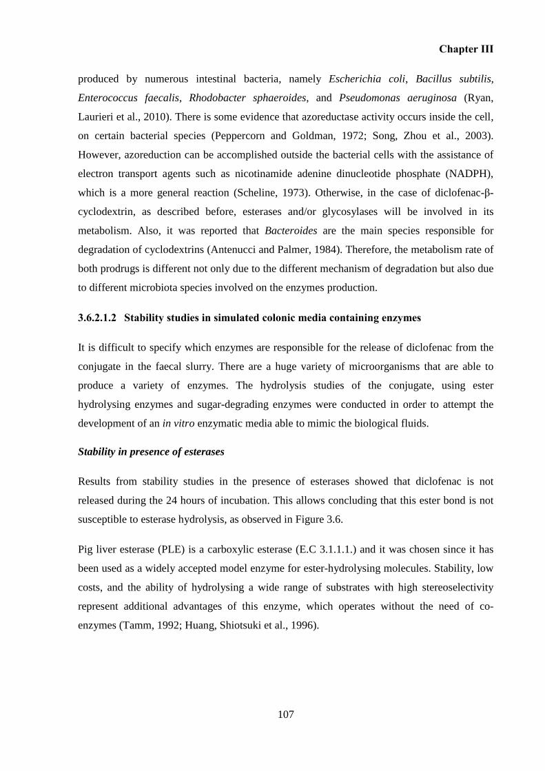
Chapter III
107
produced by numerous intestinal bacteria, namely Escherichia coli, Bacillus subtilis,
Enterococcus faecalis, Rhodobacter sphaeroides, and Pseudomonas aeruginosa (Ryan,
Laurieri et al., 2010). There is some evidence that azoreductase activity occurs inside the cell,
on certain bacterial species (Peppercorn and Goldman, 1972; Song, Zhou et al., 2003).
However, azoreduction can be accomplished outside the bacterial cells with the assistance of
electron transport agents such as nicotinamide adenine dinucleotide phosphate (NADPH),
which is a more general reaction (Scheline, 1973). Otherwise, in the case of diclofenac-β-
cyclodextrin, as described before, esterases and/or glycosylases will be involved in its
metabolism. Also, it was reported that Bacteroides are the main species responsible for
degradation of cyclodextrins (Antenucci and Palmer, 1984). Therefore, the metabolism rate of
both prodrugs is different not only due to the different mechanism of degradation but also due
to different microbiota species involved on the enzymes production.
3.6.2.1.2 Stability studies in simulated colonic media containing enzymes
It is difficult to specify which enzymes are responsible for the release of diclofenac from the
conjugate in the faecal slurry. There are a huge variety of microorganisms that are able to
produce a variety of enzymes. The hydrolysis studies of the conjugate, using ester
hydrolysing enzymes and sugar-degrading enzymes were conducted in order to attempt the
development of an in vitro enzymatic media able to mimic the biological fluids.
Stability in presence of esterases
Results from stability studies in the presence of esterases showed that diclofenac is not
released during the 24 hours of incubation. This allows concluding that this ester bond is not
susceptible to esterase hydrolysis, as observed in Figure 3.6.
Pig liver esterase (PLE) is a carboxylic esterase (E.C 3.1.1.1.) and it was chosen since it has
been used as a widely accepted model enzyme for ester-hydrolysing molecules. Stability, low
costs, and the ability of hydrolysing a wide range of substrates with high stereoselectivity
represent additional advantages of this enzyme, which operates without the need of co-
enzymes (Tamm, 1992; Huang, Shiotsuki et al., 1996).
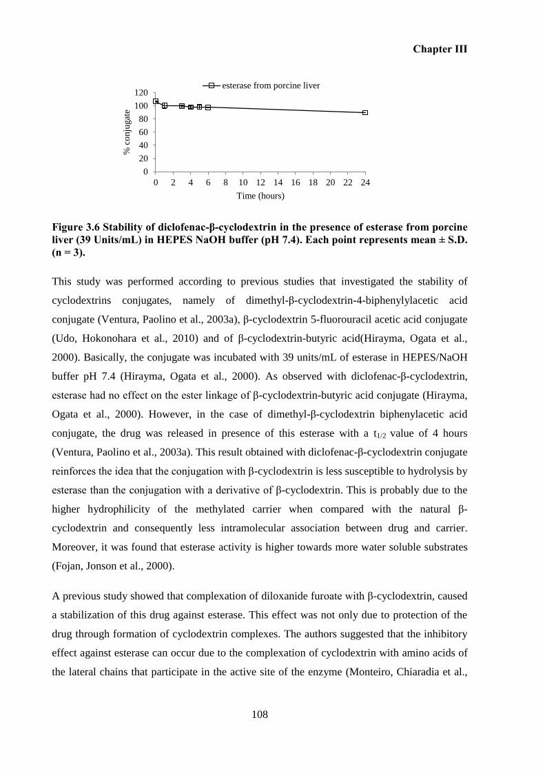
Chapter III
108
Figure 3.6 Stability of diclofenac-β-cyclodextrin in the presence of esterase from porcine liver (39 Units/mL) in HEPES NaOH buffer (pH 7.4). Each point represents mean ± S.D. (n = 3).
This study was performed according to previous studies that investigated the stability of
cyclodextrins conjugates, namely of dimethyl-β-cyclodextrin-4-biphenylylacetic acid
conjugate (Ventura, Paolino et al., 2003a), β-cyclodextrin 5-fluorouracil acetic acid conjugate
(Udo, Hokonohara et al., 2010) and of β-cyclodextrin-butyric acid(Hirayma, Ogata et al.,
2000). Basically, the conjugate was incubated with 39 units/mL of esterase in HEPES/NaOH
buffer pH 7.4 (Hirayma, Ogata et al., 2000). As observed with diclofenac-β-cyclodextrin,
esterase had no effect on the ester linkage of β-cyclodextrin-butyric acid conjugate (Hirayma,
Ogata et al., 2000). However, in the case of dimethyl-β-cyclodextrin biphenylacetic acid
conjugate, the drug was released in presence of this esterase with a t1/2 value of 4 hours
(Ventura, Paolino et al., 2003a). This result obtained with diclofenac-β-cyclodextrin conjugate
reinforces the idea that the conjugation with β-cyclodextrin is less susceptible to hydrolysis by
esterase than the conjugation with a derivative of β-cyclodextrin. This is probably due to the
higher hydrophilicity of the methylated carrier when compared with the natural β-
cyclodextrin and consequently less intramolecular association between drug and carrier.
Moreover, it was found that esterase activity is higher towards more water soluble substrates
(Fojan, Jonson et al., 2000).
A previous study showed that complexation of diloxanide furoate with β-cyclodextrin, caused
a stabilization of this drug against esterase. This effect was not only due to protection of the
drug through formation of cyclodextrin complexes. The authors suggested that the inhibitory
effect against esterase can occur due to the complexation of cyclodextrin with amino acids of
the lateral chains that participate in the active site of the enzyme (Monteiro, Chiaradia et al.,
0
20
40
60
80
100
120
0 2 4 6 8 10 12 14 16 18 20 22 24
% c
onju
gat
e
Time (hours)
esterase from porcine liver
Chapter III
109
2003). Therefore, interactions between cyclodextrins and enzymes should be taking into
account when stability studies are performed in the presence of enzymes.
Stability in presence of amylases
The assessment of efficacy of 3 different types of amylases (amylase from Bacillus
lechiniformes, amylase from porcine pancreas and amylase from Aspergillus Oryzae) to digest
the conjugate was carried out. Figure 3.7 shows the quantity of conjugate (%) after incubation
in presence of three different amylases plotted versus time.
Figure 3.7 Mean levels of diclofenac-β-cyclodextrin in presence of different α-amylases (250 units/mL): α-amylase from Bacillus lechiniformes (♦), α-amylase from Aspergillus
oryzae (■) and α-amylase from porcine pancreas (●) in 0.2 M acetate buffer (pH 5.5) containing 0.01 M CaCl2 at 37 °C. Each point represents mean ± S.D. (n = 3).
Amylase from porcine pancreas is part of pancreatin, which is commonly used to represent
the digestion within the small intestine, as described before. The intention here is to study its
activity against cyclodextrins isolated, since in the studies with pancreatin amylases are mixed
with a variety of other enzymes, including lipases and proteases. Results observed in the
presence of this amylase are in agreement with previous results with films of cyclodextrins,
which had shown that this enzyme does not degrade β-cyclodextrin (Fetznera, Bo¨Hmb et al.,
2004). This emphasizes the idea that α-amylase from porcine pancreas is not active against
this conjugate.
α-Amylase from Bacillus lechiniformes, a highly thermostable enzyme, was selected as it
resembles the properties of colonic amylases. It has been used as a model enzyme to resemble
the colonic digestion of amylase (Siew, Man et al., 2004). When the conjugate was incubated
with this enzyme, it was not observed its degradation.
0
20
40
60
80
100
120
0 2 4 6 8 10 12 14 16 18 20 22 24
Init
ial
(%)
Time (hours)
Chapter III
110
The amylase from fungus Aspergillus oryzae was selected since it was able to degrade
cyclodextrin (Jodal, Kandra et al., 1984). As observed in Figure 3.7, only this enzyme was
able to digest the conjugate.
Analysis of HPLC chromatograms of the hydrolysates obtained after incubation of the
conjugate with α-amylase from Aspergillus Oryzae after 0, 6 and 24 hours revealed that
during incubation new peaks have apperead, however non of them correspond to the
conjugate or the diclofenac. However, after 24 hours of incubation, although the conjugate
had completely disappeared, the digestion of conjugate with this enzyme only allowed to
release around 9% of diclofenac.
In order to understand the progress of the enzymatic reaction, and how the concentration of α-
amylase from Aspergillus Oryzae can affect the rate of degradation and the formation of the
intermediate compounds, the conjugate was incubated with three different concentrations of
enzyme. The results obtained are present in Figure 3.8, and reveal that the conjugate was
hydrolysed by the enzyme according to first-order kinetics, with t1/2 values of 13.3, 7.4 and
2.7 hours at enzyme concentration of 20, 40 and 250 units/mL, respectively.
In Figure 3.9 is possible to observe the evolution of the reaction between the conjugate and
different concentrations of amylase from Aspergillus oryzae. Chromatograms showed that
after 24 hours of incubation, in samples incubated with 20 and 40 units/mL of enzyme, part of
the conjugate was still intact, while in samples incubated with 250 units/mL, all the conjugate
was digested. Otherwise, after 24 hours of incubation, in the case of low concentrations of
enzyme, 20 and 40 units/mL, it was not observed any release of diclofenac from the
conjugate. In the course of the hydrolysis, it was registered the formation of 2 new peaks that
were not identified. These should be linear maltooligomers (Jodal, Kandra et al., 1984)
coupled to diclofenac, since only the drug was responsible for the ultraviolet detector
response at the wavelength used (λ = 254nm).
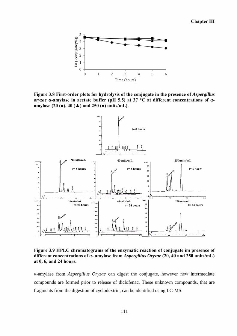
Chapter III
111
Figure 3.8 First-order plots for hydrolysis of the conjugate in the presence of Aspergillus
oryzae α-amylase in acetate buffer (pH 5.5) at 37 °C at different concentrations of α-amylase (20 (■), 40 (▲) and 250 (●) units/mL).
Figure 3.9 HPLC chromatograms of the enzymatic reaction of conjugate im presence of different concentrations of α- amylase from Aspergillus Oryzae (20, 40 and 250 units/mL) at 0, 6, and 24 hours.
α-amylase from Aspergillus Oryzae can digest the conjugate, however new intermediate
compounds are formed prior to release of diclofenac. These unknown compounds, that are
fragments from the digestion of cyclodextrin, can be identified using LC-MS.
0
1
2
3
4
5
0 1 2 3 4 5 6L
n (
co
nju
gat
e(%
))
Time (hours)
20units/mL 40units/mL 250units/mL
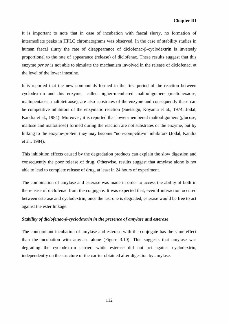
Chapter III
112
It is important to note that in case of incubation with faecal slurry, no formation of
intermediate peaks in HPLC chromatograms was observed. In the case of stability studies in
human faecal slurry the rate of disappearance of diclofenac-β-cyclodextrin is inversely
proportional to the rate of appearance (release) of diclofenac. These results suggest that this
enzyme per se is not able to simulate the mechanism involved in the release of diclofenac, at
the level of the lower intestine.
It is reported that the new compounds formed in the first period of the reaction between
cyclodextrin and this enzyme, called higher-membered maltooligomers (maltohexaose,
maltopentaose, maltotetraose), are also substrates of the enzyme and consequently these can
be competitive inhibitors of the enzymatic reaction (Suetsugu, Koyama et al., 1974; Jodal,
Kandra et al., 1984). Moreover, it is reported that lower-membered maltooligomers (glucose,
maltose and maltotriose) formed during the reaction are not substrates of the enzyme, but by
linking to the enzyme-protein they may become “non-competitive” inhibitors (Jodal, Kandra
et al., 1984).
This inhibition effects caused by the degradation products can explain the slow digestion and
consequently the poor release of drug. Otherwise, results suggest that amylase alone is not
able to lead to complete release of drug, at least in 24 hours of experiment.
The combination of amylase and esterase was made in order to access the ability of both in
the release of diclofenac from the conjugate. It was expected that, even if interaction occured
between esterase and cyclodextrin, once the last one is degraded, esterase would be free to act
against the ester linkage.
Stability of diclofenac-β-cyclodextrin in the presence of amylase and esterase
The concomitant incubation of amylase and esterase with the conjugate has the same effect
than the incubation with amylase alone (Figure 3.10). This suggests that amylase was
degrading the cyclodextrin carrier, while esterase did not act against cyclodextrin,
independently on the structure of the carrier obtained after digestion by amylase.
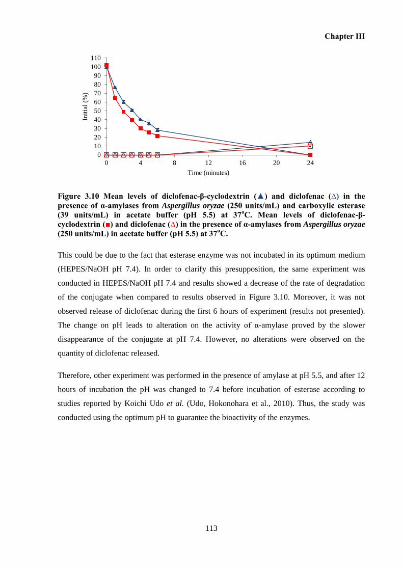
Chapter III
113
Figure 3.10 Mean levels of diclofenac-β-cyclodextrin (▲) and diclofenac (∆) in the presence of α-amylases from Aspergillus oryzae (250 units/mL) and carboxylic esterase (39 units/mL) in acetate buffer (pH 5.5) at 37oC. Mean levels of diclofenac-β-cyclodextrin (■) and diclofenac (∆) in the presence of α-amylases from Aspergillus oryzae (250 units/mL) in acetate buffer (pH 5.5) at 37oC.
This could be due to the fact that esterase enzyme was not incubated in its optimum medium
(HEPES/NaOH pH 7.4). In order to clarify this presupposition, the same experiment was
conducted in HEPES/NaOH pH 7.4 and results showed a decrease of the rate of degradation
of the conjugate when compared to results observed in Figure 3.10. Moreover, it was not
observed release of diclofenac during the first 6 hours of experiment (results not presented).
The change on pH leads to alteration on the activity of α-amylase proved by the slower
disappearance of the conjugate at pH 7.4. However, no alterations were observed on the
quantity of diclofenac released.
Therefore, other experiment was performed in the presence of amylase at pH 5.5, and after 12
hours of incubation the pH was changed to 7.4 before incubation of esterase according to
studies reported by Koichi Udo et al. (Udo, Hokonohara et al., 2010). Thus, the study was
conducted using the optimum pH to guarantee the bioactivity of the enzymes.
0
10
20
30
40
50
60
70
80
90
100
110
0 4 8 12 16 20 24
Init
ial
(%)
Time (minutes)
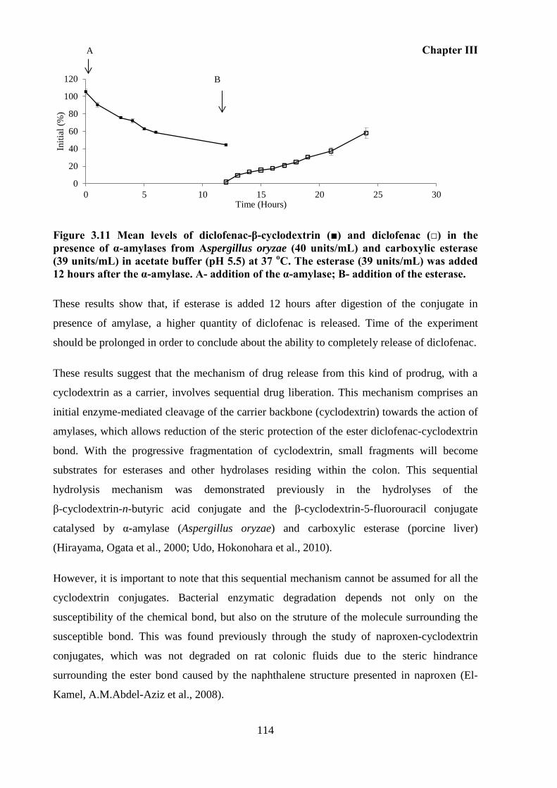
Chapter III
114
Figure 3.11 Mean levels of diclofenac-β-cyclodextrin (■) and diclofenac (□) in the presence of α-amylases from Aspergillus oryzae (40 units/mL) and carboxylic esterase (39 units/mL) in acetate buffer (pH 5.5) at 37 oC. The esterase (39 units/mL) was added 12 hours after the α-amylase. A- addition of the α-amylase; B- addition of the esterase.
These results show that, if esterase is added 12 hours after digestion of the conjugate in
presence of amylase, a higher quantity of diclofenac is released. Time of the experiment
should be prolonged in order to conclude about the ability to completely release of diclofenac.
These results suggest that the mechanism of drug release from this kind of prodrug, with a
cyclodextrin as a carrier, involves sequential drug liberation. This mechanism comprises an
initial enzyme-mediated cleavage of the carrier backbone (cyclodextrin) towards the action of
amylases, which allows reduction of the steric protection of the ester diclofenac-cyclodextrin
bond. With the progressive fragmentation of cyclodextrin, small fragments will become
substrates for esterases and other hydrolases residing within the colon. This sequential
hydrolysis mechanism was demonstrated previously in the hydrolyses of the
β-cyclodextrin-n-butyric acid conjugate and the β-cyclodextrin-5-fluorouracil conjugate
catalysed by α-amylase (Aspergillus oryzae) and carboxylic esterase (porcine liver)
(Hirayama, Ogata et al., 2000; Udo, Hokonohara et al., 2010).
However, it is important to note that this sequential mechanism cannot be assumed for all the
cyclodextrin conjugates. Bacterial enzymatic degradation depends not only on the
susceptibility of the chemical bond, but also on the struture of the molecule surrounding the
susceptible bond. This was found previously through the study of naproxen-cyclodextrin
conjugates, which was not degraded on rat colonic fluids due to the steric hindrance
surrounding the ester bond caused by the naphthalene structure presented in naproxen (El-
Kamel, A.M.Abdel-Aziz et al., 2008).
0
20
40
60
80
100
120
0 5 10 15 20 25 30
Init
ial
(%)
Time (Hours)
A
B
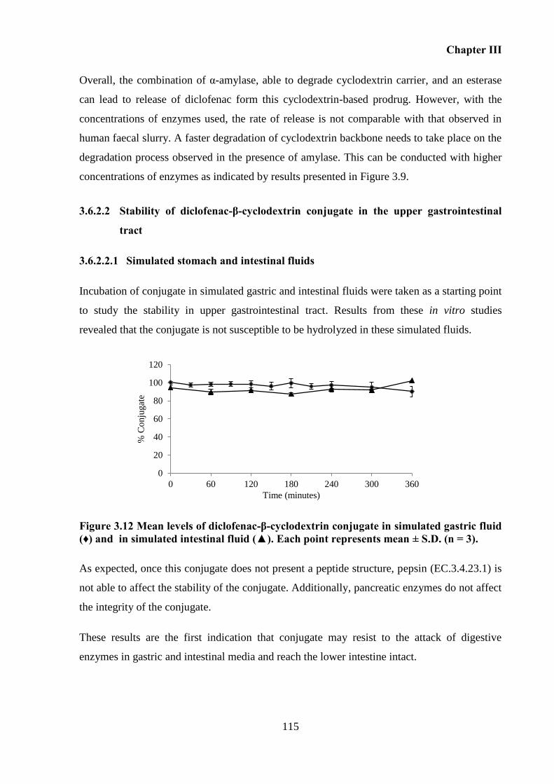
Chapter III
115
Overall, the combination of α-amylase, able to degrade cyclodextrin carrier, and an esterase
can lead to release of diclofenac form this cyclodextrin-based prodrug. However, with the
concentrations of enzymes used, the rate of release is not comparable with that observed in
human faecal slurry. A faster degradation of cyclodextrin backbone needs to take place on the
degradation process observed in the presence of amylase. This can be conducted with higher
concentrations of enzymes as indicated by results presented in Figure 3.9.
3.6.2.2 Stability of diclofenac-β-cyclodextrin conjugate in the upper gastrointestinal
tract
3.6.2.2.1 Simulated stomach and intestinal fluids
Incubation of conjugate in simulated gastric and intestinal fluids were taken as a starting point
to study the stability in upper gastrointestinal tract. Results from these in vitro studies
revealed that the conjugate is not susceptible to be hydrolyzed in these simulated fluids.
Figure 3.12 Mean levels of diclofenac-β-cyclodextrin conjugate in simulated gastric fluid (♦) and in simulated intestinal fluid (▲). Each point represents mean ± S.D. (n = 3).
As expected, once this conjugate does not present a peptide structure, pepsin (EC.3.4.23.1) is
not able to affect the stability of the conjugate. Additionally, pancreatic enzymes do not affect
the integrity of the conjugate.
These results are the first indication that conjugate may resist to the attack of digestive
enzymes in gastric and intestinal media and reach the lower intestine intact.
0
20
40
60
80
100
120
0 60 120 180 240 300 360
% C
onju
gat
e
Time (minutes)
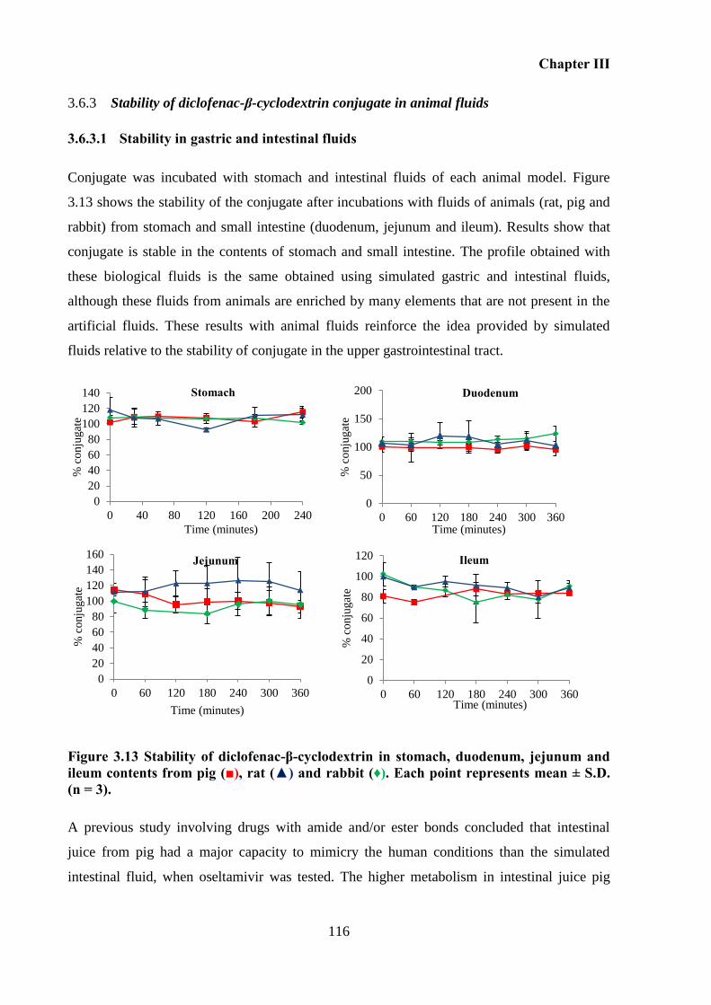
Chapter III
116
Stability of diclofenac-β-cyclodextrin conjugate in animal fluids 3.6.3
3.6.3.1 Stability in gastric and intestinal fluids
Conjugate was incubated with stomach and intestinal fluids of each animal model. Figure
3.13 shows the stability of the conjugate after incubations with fluids of animals (rat, pig and
rabbit) from stomach and small intestine (duodenum, jejunum and ileum). Results show that
conjugate is stable in the contents of stomach and small intestine. The profile obtained with
these biological fluids is the same obtained using simulated gastric and intestinal fluids,
although these fluids from animals are enriched by many elements that are not present in the
artificial fluids. These results with animal fluids reinforce the idea provided by simulated
fluids relative to the stability of conjugate in the upper gastrointestinal tract.
Figure 3.13 Stability of diclofenac-β-cyclodextrin in stomach, duodenum, jejunum and ileum contents from pig (■), rat (▲) and rabbit (♦). Each point represents mean ± S.D. (n = 3).
A previous study involving drugs with amide and/or ester bonds concluded that intestinal
juice from pig had a major capacity to mimicry the human conditions than the simulated
intestinal fluid, when oseltamivir was tested. The higher metabolism in intestinal juice pig
0
50
100
150
200
0 60 120 180 240 300 360
% c
onju
gat
e
Time (minutes)
Duodenum
0
20
40
60
80
100
120
140
160
0 60 120 180 240 300 360
% c
onju
gat
e
Time (minutes)
Jejunum
0
20
40
60
80
100
120
0 60 120 180 240 300 360
% c
onju
gat
e
Time (minutes)
Ileum
0
20
40
60
80
100
120
140
0 40 80 120 160 200 240
% c
onju
gat
e
Time (minutes)
Stomach
Chapter III
117
than in simulated intestinal fluid was also observed for atazanavir carbamate and diloxanide
furoate. The higher metabolism in collected juice was attributable to its enzymatic activity
(Sarti, Barthelmes et al., 2011).
Moreover, no differences were observed between the fluids of the upper GI tract of different
animals, which mean that the conjugate has the same behaviour independently on the specie.
Therefore, animal fluids can be replaced with simulated fluids to carry out the stability studies
on the upper gastrointestinal tract of this conjugate.
It is important to note that, despite the high levels of bacteria in rat stomach, these do not
affect conjugate stability. This might be due to the inexistence e/or inability of enzymatic
bacteria capable to operate on the conjugate. Otherwise, it is known that rabbits practice
caecoprophagy, which is associated with fermentation at the level of the stomach. However,
fermentation only exists when caecoprophes are present and it does not occur in the gastric
media. Additionally, only the supernatant obtained after centrifugation of fluids was used,
which avoided destruction and mixture of the caecoprophes with the gastric fluid. Moreover,
these studies were carried out using the supernatant of the fluids obtained after centrifugation
and some enzymes can be bound to particulate matter left in the pellet (Ikesue, Kopečkovà et
al., 1993). For this reason some authors instead of the centrifugation, proceed to dilution of
fluids before stability experiments. However, a study conducted for determination of α-
amylase activity in the supernatants of fluids used in these stability tests showed that a huge
activity of amylase is observed in stomach of rats when compared with pigs and rabbits
(Appendix D).
Enzymes in the luminal fluids can also come from dead gut wall cells, which will be abraded
into the intestine. Therefore, sometimes it is worth to include studies with microssomes to be
sure about the stability in intestinal mucous (Borde, Karlsson et al., 2012). However, previous
studies with intestine homogenates of rats without contents had shown negligible degradation
of the cyclodextrin prodrugs (Hirayama, Ogata et al., 2000; Kamada, Hirayama et al., 2002).
Results of diclofenac-β-cyclodextrin are according to those reported before which
investigated the stability of cyclodextrin conjugates in fluids from rats. For instance, butyric
acid β-cyclodextrin was stable with any release of drug in stomach and intestinal contents for
8 hours (Hirayma, Ogata et al., 2000). Okamato et al. demonstrated that in contents from
Chapter III
118
stomach and small intestine of rats, the cyclodextrin-5-ASA released 5-ASA only in small
amounts (< 12%) during 48 hours (Zou, Okamoto et al., 2005). Comparing the stability of
ethyl 4-biphenylylacetate with the stability of the equivalent cyclodextrin prodrug, the first
was rapidly hydrolysed in contents from stomach and small intestine of rat, while only 10% of
the cyclodextrin ester prodrugs were hydrolysed in the same contents (Uekama, Minami et al.,
1997a). Another study that compares the stability between prednisolone-succinate-α-
cyclodextrin and prednisolone succinate showed that in case of cyclodextrin prodrug the
quantity remained after 6 hours of incubation was 80-90%, while prednisolone succinate
alone was rapidly hydrolysed (Hideki Yano, Fumitoshi Hirayama et al., 2002). These studies
demonstrated that the type of carrier influences the stability of the ester prodrugs and
apparently the size of the carrier is determinant for the stability of the ester prodrugs in fluids
from the upper GI tract. Thus, these authors justified the resistance of the conjugate against
the ester hydrolysis in the contents from the stomach and small intestine due to a large steric
effect of the bulky cyclodextrin moiety (Uekama, Minami et al., 1997a; Hideki Yano,
Fumitoshi Hirayama et al., 2002).
3.6.3.1.1 Stability studies in caecum and colonic fluids
Results of stability of the conjugate and sulfasalazine in caecum fluids from rat, pig and rabbit
and from anterior colon of pig (colon A) are represented in Figure 3.14 and Figure 3.15.
These show that caecum and colon A pig fluids do not degrade the conjugate. Sulfasalazine is
also not metabolised. This suggests an absence of enzymatic activity or inactivity of the
microbiota at least for 6 hours of experiment in these pig fluids. Contrarily, in the case of rat
and rabbit fluids, it is observed degradation of both prodrugs.
The degradation of sulfasalazine is faster than the degradation of conjugate. In caecum rat
fluids, sulfasalazine is completely degraded in 30 minutes of incubation, while in the case of
conjugate 60% is degraded after 6 hours of incubation. With rabbits, after 5 hours of
incubation in caecum fluid, 60% of the conjugate was metabolised. For sulfasalazine, the
degradation occurs with a high rate in the first 2 hours of incubation but after the % of
prodrug remained constant, around 18%. These profiles of diclofenac-β-cyclodextrin
conjugate and sulfasalazine degradation in caecum fluids show that the metabolism of both
prodrugs is variable between these three species.
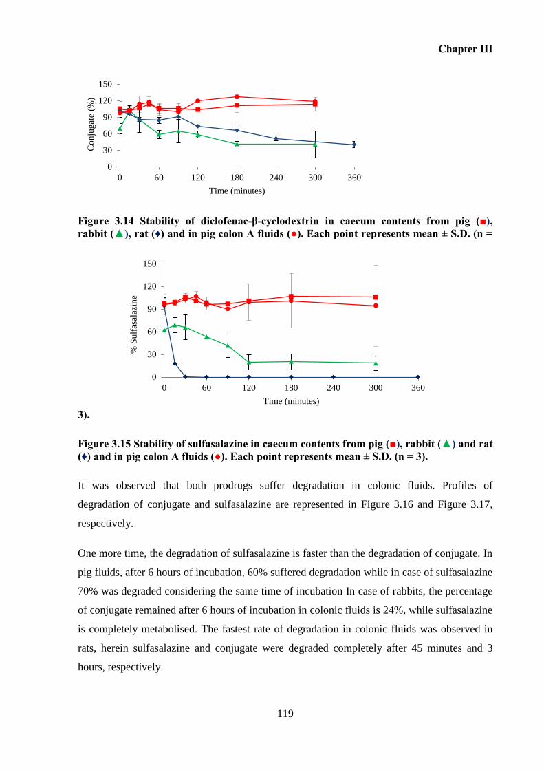
Chapter III
119
0
30
60
90
120
150
0 60 120 180 240 300 360
% S
ulf
asal
azin
e
Time (minutes)
Figure 3.14 Stability of diclofenac-β-cyclodextrin in caecum contents from pig (■), rabbit (▲), rat (♦) and in pig colon A fluids (●). Each point represents mean ± S.D. (n =
3).
Figure 3.15 Stability of sulfasalazine in caecum contents from pig (■), rabbit (▲) and rat (♦) and in pig colon A fluids (●). Each point represents mean ± S.D. (n = 3).
It was observed that both prodrugs suffer degradation in colonic fluids. Profiles of
degradation of conjugate and sulfasalazine are represented in Figure 3.16 and Figure 3.17,
respectively.
One more time, the degradation of sulfasalazine is faster than the degradation of conjugate. In
pig fluids, after 6 hours of incubation, 60% suffered degradation while in case of sulfasalazine
70% was degraded considering the same time of incubation In case of rabbits, the percentage
of conjugate remained after 6 hours of incubation in colonic fluids is 24%, while sulfasalazine
is completely metabolised. The fastest rate of degradation in colonic fluids was observed in
rats, herein sulfasalazine and conjugate were degraded completely after 45 minutes and 3
hours, respectively.
0
30
60
90
120
150
0 60 120 180 240 300 360
Co
nju
gat
e (%
)
Time (minutes)
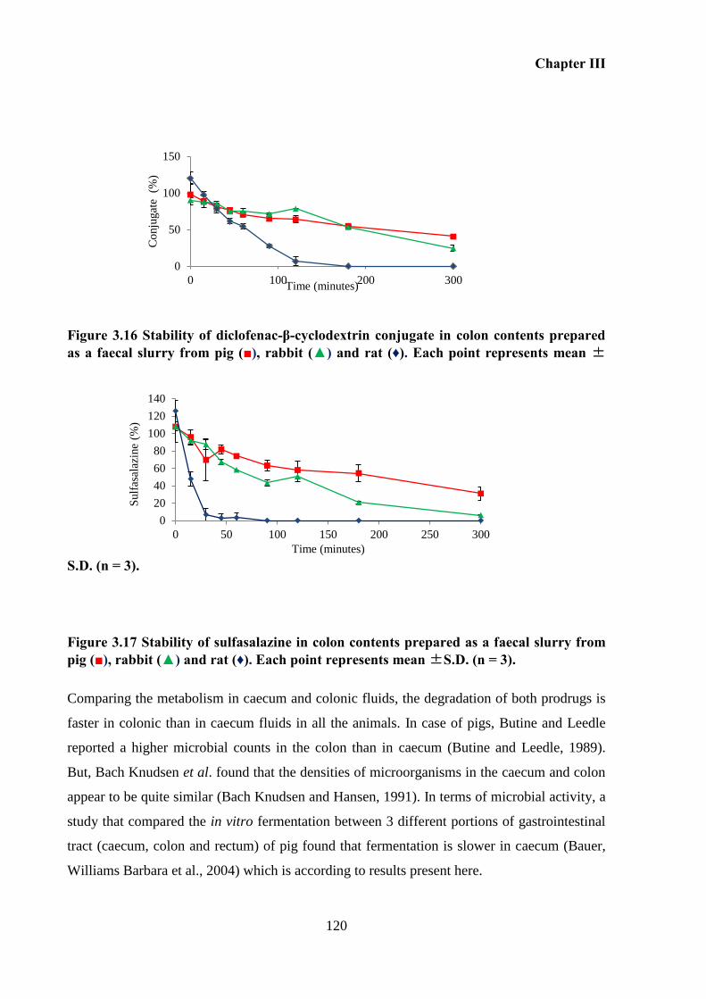
Chapter III
120
Figure 3.16 Stability of diclofenac-β-cyclodextrin conjugate in colon contents prepared as a faecal slurry from pig (■), rabbit (▲) and rat (♦). Each point represents mean ±
S.D. (n = 3).
Figure 3.17 Stability of sulfasalazine in colon contents prepared as a faecal slurry from pig (■), rabbit (▲) and rat (♦). Each point represents mean ±S.D. (n = 3).
Comparing the metabolism in caecum and colonic fluids, the degradation of both prodrugs is
faster in colonic than in caecum fluids in all the animals. In case of pigs, Butine and Leedle
reported a higher microbial counts in the colon than in caecum (Butine and Leedle, 1989).
But, Bach Knudsen et al. found that the densities of microorganisms in the caecum and colon
appear to be quite similar (Bach Knudsen and Hansen, 1991). In terms of microbial activity, a
study that compared the in vitro fermentation between 3 different portions of gastrointestinal
tract (caecum, colon and rectum) of pig found that fermentation is slower in caecum (Bauer,
Williams Barbara et al., 2004) which is according to results present here.
0
50
100
150
0 100 200 300
Co
nju
gat
e (
%)
Time (minutes)
0
20
40
60
80
100
120
140
0 50 100 150 200 250 300
Sulf
asal
azin
e (%
)
Time (minutes)
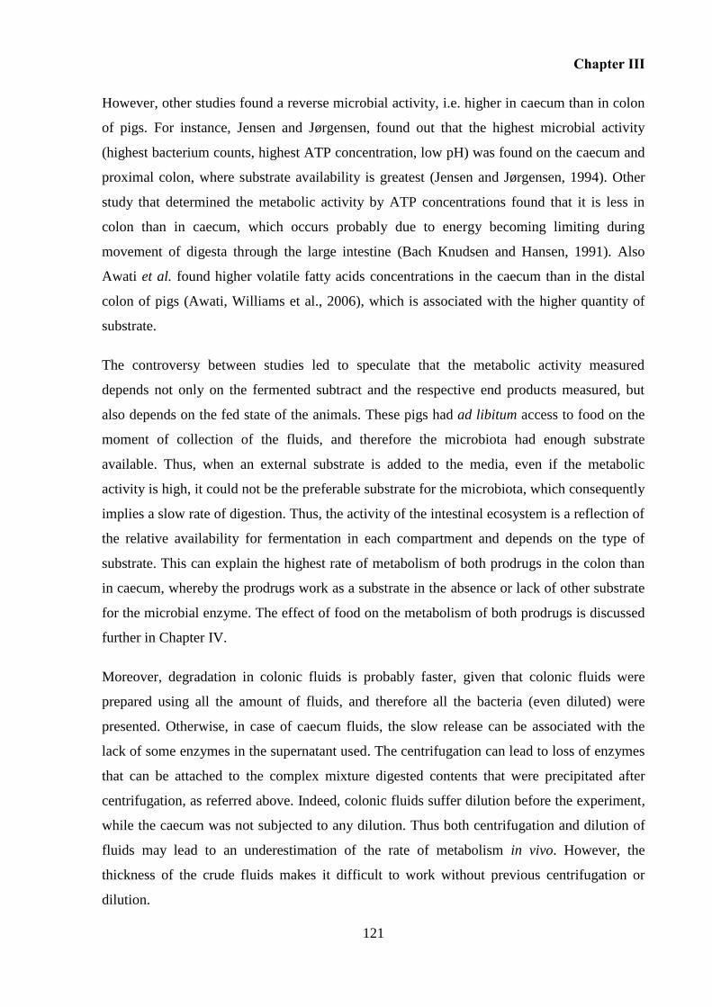
Chapter III
121
However, other studies found a reverse microbial activity, i.e. higher in caecum than in colon
of pigs. For instance, Jensen and Jørgensen, found out that the highest microbial activity
(highest bacterium counts, highest ATP concentration, low pH) was found on the caecum and
proximal colon, where substrate availability is greatest (Jensen and Jørgensen, 1994). Other
study that determined the metabolic activity by ATP concentrations found that it is less in
colon than in caecum, which occurs probably due to energy becoming limiting during
movement of digesta through the large intestine (Bach Knudsen and Hansen, 1991). Also
Awati et al. found higher volatile fatty acids concentrations in the caecum than in the distal
colon of pigs (Awati, Williams et al., 2006), which is associated with the higher quantity of
substrate.
The controversy between studies led to speculate that the metabolic activity measured
depends not only on the fermented subtract and the respective end products measured, but
also depends on the fed state of the animals. These pigs had ad libitum access to food on the
moment of collection of the fluids, and therefore the microbiota had enough substrate
available. Thus, when an external substrate is added to the media, even if the metabolic
activity is high, it could not be the preferable substrate for the microbiota, which consequently
implies a slow rate of digestion. Thus, the activity of the intestinal ecosystem is a reflection of
the relative availability for fermentation in each compartment and depends on the type of
substrate. This can explain the highest rate of metabolism of both prodrugs in the colon than
in caecum, whereby the prodrugs work as a substrate in the absence or lack of other substrate
for the microbial enzyme. The effect of food on the metabolism of both prodrugs is discussed
further in Chapter IV.
Moreover, degradation in colonic fluids is probably faster, given that colonic fluids were
prepared using all the amount of fluids, and therefore all the bacteria (even diluted) were
presented. Otherwise, in case of caecum fluids, the slow release can be associated with the
lack of some enzymes in the supernatant used. The centrifugation can lead to loss of enzymes
that can be attached to the complex mixture digested contents that were precipitated after
centrifugation, as referred above. Indeed, colonic fluids suffer dilution before the experiment,
while the caecum was not subjected to any dilution. Thus both centrifugation and dilution of
fluids may lead to an underestimation of the rate of metabolism in vivo. However, the
thickness of the crude fluids makes it difficult to work without previous centrifugation or
dilution.

Chapter III
122
Overall, hydrolysis rates of these prodrugs by luminal fluids on the lower gastrointestinal tract
were different between species. However, bacterial strains seem to commonly inhabit the gut
of laboratory animals and humans, since similar metabolic activity was noted in human faecal
slurries, and colonic fluids form pigs, rabbits and rats. Bacteroides (Bacteroidetes Phylum) are
believed to be the main species responsible for degradation of the cyclodextrin carrier.
Bacteroides genus is able to induce enzymes adapting to the available substrates. They are
able to grow on cyclodextrin as the sole carbon source. Also cyclodextrinase were isolated
from two selected Bacteroides genus (Antenucci and Palmer, 1984). Moreover, species from
Proteobacteria Phylum isolated from colon of pig (P. mirabilis and E. fergusonii)
demonstrated to increase the release of bisoprolol from a film-incorporated β-cyclodextrin
after pre-incubation in β-cyclodextrin containing nutrient medium (Fetznera, Bo¨Hmb et al.,
2004).
The stability of the conjugate was not assessed using animal faeces. However, previous
studies in rabbits had concluded that faeces could be used as an alternative source of inoculum
to caecum contents to study in vitro caecum fermentation (Bovera, D’urso et al., 2006). Other
study with rabbits confirmed that in vitro fermentation characteristics of faeces were highly
related to those of caecum content by measurement of the degraded organic matter, potential
gas production; volatile fatty acids and NH3 production (Bovera, D’urso et al., 2009). Other
authors also pointed out that using faeces from pig to perform in vitro fermentation studies
can overestimate the microbial activity of the caecum (Bauer, Williams Barbara et al., 2004).
Otherwise, in the case of rats it was reported that the faecal material does not provide accurate
information concerning the intestinal microbiota, because significant epithelial microbial
associations occur in the proximal digestive tract (Tannock, 1999). Interestingly, rat caecal
contents have been widely utilized as alternative dissolution medium of human colon (Yang,
2008). This is because of the similarity colonic microbiota between human and rodent, when
the number of Bacteroides and Bifidobacteria were counted (Hawksworth, Drasar et al.,
1971). In fact, rat was the only animal used before in order to study the in vitro stability
assays of cyclodextrin conjugates. In this work comparing the metabolism between animals,
rat is the animal that allows a faster degradation rate of both prodrugs. Actually, amylase
activity at the level of the lower GI tract is also higher in rats than in rabbits and pigs
(Appendix D). It is worth to note that the substrate used on the determination of α-amylase
was starch. Therefore, the substract was not cyclodextrin, which prevents the direct
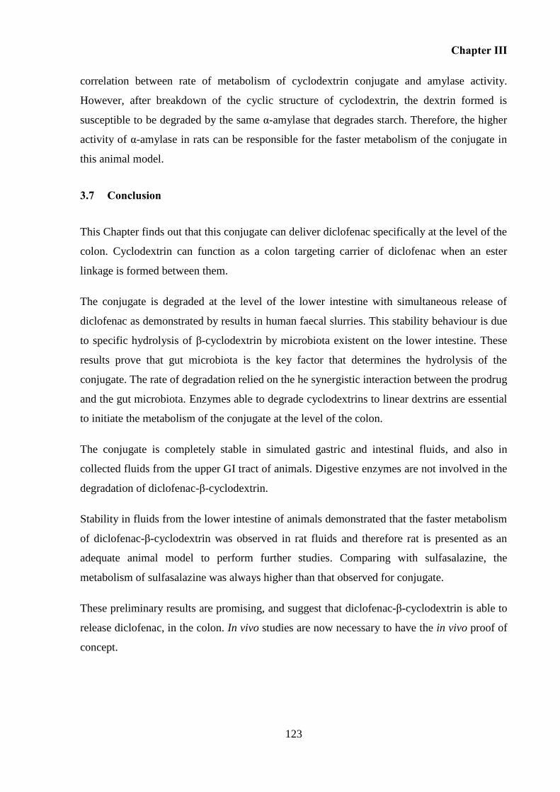
Chapter III
123
correlation between rate of metabolism of cyclodextrin conjugate and amylase activity.
However, after breakdown of the cyclic structure of cyclodextrin, the dextrin formed is
susceptible to be degraded by the same α-amylase that degrades starch. Therefore, the higher
activity of α-amylase in rats can be responsible for the faster metabolism of the conjugate in
this animal model.
3.7 Conclusion
This Chapter finds out that this conjugate can deliver diclofenac specifically at the level of the
colon. Cyclodextrin can function as a colon targeting carrier of diclofenac when an ester
linkage is formed between them.
The conjugate is degraded at the level of the lower intestine with simultaneous release of
diclofenac as demonstrated by results in human faecal slurries. This stability behaviour is due
to specific hydrolysis of β-cyclodextrin by microbiota existent on the lower intestine. These
results prove that gut microbiota is the key factor that determines the hydrolysis of the
conjugate. The rate of degradation relied on the he synergistic interaction between the prodrug
and the gut microbiota. Enzymes able to degrade cyclodextrins to linear dextrins are essential
to initiate the metabolism of the conjugate at the level of the colon.
The conjugate is completely stable in simulated gastric and intestinal fluids, and also in
collected fluids from the upper GI tract of animals. Digestive enzymes are not involved in the
degradation of diclofenac-β-cyclodextrin.
Stability in fluids from the lower intestine of animals demonstrated that the faster metabolism
of diclofenac-β-cyclodextrin was observed in rat fluids and therefore rat is presented as an
adequate animal model to perform further studies. Comparing with sulfasalazine, the
metabolism of sulfasalazine was always higher than that observed for conjugate.
These preliminary results are promising, and suggest that diclofenac-β-cyclodextrin is able to
release diclofenac, in the colon. In vivo studies are now necessary to have the in vivo proof of
concept.
Chapter III
124
CHAPTER IV
INFLUENCE OF FOOD ON GUT FLUIDS AND METABOLISM OF
DICLOFENAC-β-CYCLODEXTRIN VERSUS SULFASALAZINE IN RATS

Chapter IV
127
4 INFLUENCE OF FOOD ON GUT FLUIDS AND METABOLISM OF
DICLOFENAC-ΒETA-CYCLODEXTRIN VERSUS SULFASALAZINE IN RATS
4.1 Overview
A screening of the in vitro performance of diclofenac-β-cyclodextrin conjugate in the
different simulated gastrointestinal fluids, including different animal fluids, has been reported
in Chapter III. Results showed that this prodrug is specifically degraded by microbiota in the
large intestine, allowing diclofenac to be released into the colon.
In respect to oral administration, it is known that food can affect the bioavailability of drugs.
Generally patients are advised to take their medicines with food or on an empty stomach.
However, these are imprecise instructions that are open to individual interpretations, and
consequently are expected to contribute to a greater variability and lower therapeutic efficacy.
Research on feeding regimens, such as timing of meals in relation to drug administration is
necessary to increase the understanding of their influence on drug disposition.
The aim of this study is to understand how different feeding regimens can affect the
physiological conditions in the gastrointestinal tract and to understand how food intake
influences the degradation of the conjugate in the large intestine. Concomitantly with
diclofenac-β-cyclodextrin conjugate, the effect of feeding regimens on the metabolism of
sulfasalazine - a well-known commercially available colonic prodrug of reference - was also
studied.
In vitro studies (Chapter III) demonstrated that rats provided the faster metabolism of both
prodrugs, diclofenac-β-cyclodextrin and sulfasalazine. Therefore, this was the animal model
used in this Chapter to access the influence of different feeding states on the metabolism of
prodrugs.
4.2 Introduction
The presence of food in the gastrointestinal tract reduces gastrointestinal motility, thereby
delaying the arrival of a colonic drug carrier to its site of action (Mittelstadt, Hemenway et al.,
Chapter IV
128
2005; Varum, Merchant et al., 2010; Varum, Hatton et al., 2013). Food intake also influences
the amount of water in the gut that is available for drug dissolution prior to absorption. Food
also influences pH of gut content, which as well as controlling drug release from pH-
responsive drug carriers, also influences the ionisation of weakly acidic/basic drugs and thus
their aqueous solubility, stability and absorption (Stella, Borchardt et al., 2007; Varum,
Merchant et al., 2010). Food also influences the performance of colonic prodrugs, whose
conversion to drugs relies on gut bacterial enzymes. Bacterial activity in the colon depends on
the quantity and quality of available substrates for fermentation, and determines the intensity
and direction of gut bacterial metabolism of prodrugs and thereby drug absorption (Agorama,
Woltosza et al., 2001; Mountzourisa, Kotzampassib et al., 2009).
Diclofenac has been studied as one of the drugs whose pharmacokinetic parameters are
influenced by the presence of food. The administration of an enteric coated diclofenac sodium
formulation (50 mg) immediately after a standard breakfast showed that food does not have
significant effect on the extent of diclofenac absorption when compared with the fasted state.
However, delay in the onset of absorption (varying from 2.5 to 12 hours in fed and from 1.5
to 2.75 hours in fasted state) and a reduction in peak plasma levels of approximately 30% was
observed in the presence of food (Willis, Kendall et al., 1981). Other study conducted in
humans with diclofenac hydrogel beads (150 mg) originated comparable effects on the
presence of food. The administration immediately after breakfast did not affect the
bioavailability, however the Cmax decreases 38% and the Tmax increases three fold in the fed
state comparing with the fasted state (Thakker, Mangat et al., 1992). Billa et al. showed that
after administration of gum xanthan tablets of sodium diclofenac (100 mg) in healthy men in
fed state, AUC was not significantly affected and the Tmax increased (Tmax values 2.6±0.3
hours in fasted state and 5.5 ± 0.8 hours in fed state) (Billa, Yuen et al., 2000). This is
consistent with the findings of Willis et al. (1981), Thakker et al. (1992) and Chan et al.
(1990) (Willis, Kendall et al., 1981; Chan, Mojaverian et al., 1990; Thakker, Mangat et al.,
1992). Moreover, no differences were observed between the AUC of gum xanthan tablets and
the buffer solution (pH = 7) of diclofenac in both dietary stats, in agreement with the findings
of Riad et al. (1989) (Riad, Sawchuk et al., 1995; Billa, Yuen et al., 2000).
On the other hand, Billa et al., found out that the Cmax of diclofenac is higher in fed state
(Cmax = 347±112 ng/ml) than in fasted state (Cmax = 661.5±158.5 ng/ml), although the gastric
emptying time was longer in presence of food. The faster release and absorption of diclofenac
Chapter IV
129
was attributable to an interaction between food and the formulation. In fed state, the longer
residence of the tablets in stomach leaded to a loss of the integrity of the xanthan gum matrice
due to the low pH, allowing rapid influx of water into the matrix system with a faster rate of
drug release (Billa, Yuen et al., 2000).
Additionally, the pharmacokinetic parameters are dependent on the type of formulation, as
demonstrated by Chan et al. who found that gastric emptying of sodium diclofenac, either in
solution or as a precipitate, occurred more quickly than the emptying of Heidelberg capsule
(Chan, Mojaverian et al., 1990). Another author, Zmeili et al. (1996) reported no significant
change in Cmax in the fasted and the fed states with their enteric-coated controlled release
tablet (Zmeili, Hasan et al., 1996).
These studies proved that the presence of food affects the rate of absorption and the
concentrations of diclofenac in plasma, which depends on the the physical properties of the
dosage form, and also depends on the amount and composition of the meal which affects the
gastric emptying time (Billa, Yuen et al., 2000). However, the presence of food has no
influence on the extent of diclofenac absorption, independent of the formulation.
Given the critical influence of the fasted/fed state on the performance of colonic prodrugs as
described above, we determined the influence of different feeding regimens on gastro-
intestinal contents, pH and metabolism of the colonic prodrug, in order to establish the most
appropriate fasted/fed state that should be employed for its in vivo assessment in rats.
The study was conducted in different feeding regimens: fed ad libitum, 12-hour fast, 12-hour
fast followed by one hour feeding, which was itself followed by either 30 minutes or 4 hours
of fast, prior to the animals being killed, and measurements being taken. These four regimens
were selected for a number of reasons: firstly, as stated above, most oral drug delivery
experiments are conducted on overnight-fasted rats; secondly, a control of the 12-hour fast i.e.
animals being fed ad libitum; thirdly, a 4-hour fast after feeding, to ensure complete gastric
emptying of food ingested (Booth, Gibson et al., 1986), fourthly, a control experiment for the
latter, i.e. 30 minutes fast after feeding assures that the ingested food has not arrived in the
lower intestine (caecum and colon) (Brown, Greenburgh et al., 1994).
The rat is an appropriate model to use in early drug development; its mean intestinal transit
time is comparable to that in humans despite the different gastro-intestinal lengths, transit
Chapter IV
130
time is significantly shorter in the fasted state compared to the fed state, as in man, and its
gastrointestinal motility is under the control of the migrating myoelectric complex (MMC),
like in man (Tuleu, Andrieux et al., 1999; Mittelstadt, Hemenway et al., 2005). Rats are thus
widely used as an in vivo model and have been used to assess different colonic prodrugs,
including cyclodextrin-based ones (Minami, Hirayama et al., 1998; Kamada, Hirayama et al.,
2002).
As far as we know, the effect of fasting on the metabolism of cyclodextrins and the respective
conjugates has not been subject of investigation. Otherwise, few studies have looked at the
effect of food on the metabolism of other colonic prodrugs. One of the studies reported that
metronidazole and olsalazine were found to be practically stable in the material from the
contents of the ascending colon from human collected in the fed state. Otherwise, olsalazine
was degraded up to 20% to 5-ASA within 10 minutes in the fasted ascending colon (Vertzoni,
Carlsson et al., 2011). However, previous study in humans had shown that the systematic
availability of olsalazine was not affected by food (Ryde and Ahnfelt, 1988). These results
leaded Vertzoni et al. to conclude that the metabolism of olsalazine was limited in the
ascending colon and became higher in descending colon both in fasted and fed state
(Vertzoni, Carlsson et al., 2011). Another study reported that fasted rats had lower activities
of α-galactosidades, α-glucosidades, and β-glucosidades and higher activities of β-
galactosidase and azoreductase (Mountzourisa, Kotzampassib et al., 2009).
4.3 Aims and Objectives
This Chapter aims to investigate the influence of feeding regimen in Wistar rats on:
i) Distribution and pH contents along the gut;
ii) Metabolism of diclofenac-β-cyclodextrin within the caecal and colonic contents,
using sulfasalazine as a control.
This enables an understanding of how the regimen state affect the pharmacokinetics of
prodrugs and enable to choose the most favorable feeding state to perform the in vivo studies.
Chapter IV
131
4.4 Materials and methods
Reagents and chemicals 4.4.1
Sodium diclofenac (MW=318.14 g/mol), sulfasalazine (MW=398.394 g/mol) were purchased
from Sigma. Diclofenac-β-cyclodextrin (MW=1411 g/mol) was synthesized according to the
method reported in Chapter II.
Sodium chloride, potassium hydroxide, sodium hydroxide, potassium dihydrogen phosphate,
HPLC grades acetonitrile, methanol and water were purchased from Fisher Scientifics.
Peptone water and yeast extract were obtained from Oxoid Limited (Hampshire, UK).
Magnesium sulphate heptahydrate and calcium chloride hexahydrate were obtained from
VWR (Leicestershire, UK). Trifluoroacetic acid (TFA) and dimethylformamide (DMF),
sodium bicarbonate, haemin, l-cysteine HCl, vitamin K and resazurin were obtained from
Sigma Aldrich (Dorset, UK). All other chemicals and solvents were of HPLC reagent grade
and were used without further purification. Phosphate buffer saline (PBS) pH 6.0 and pH 6.8
were prepared according to the USPXXIV.
Animals 4.4.2
All the procedures had been approved by the School’s Ethical Review Committee and were
conducted in accordance with the Home Office standards under the Animals (Scientific
Procedures) Act, 1986. Studies were performed using healthy adult male Wistar rats (8 weeks,
240-250 g) purchased from Harlan Olac Ltd. (Oxfordshire, UK). All animals were housed in
rooms with controlled conditions: 20 oC, 40-60% humidity, 15-20 air changes per hour.
Animals underwent a period of acclimatization, with free access to standard rat chow and
water for 7 days prior the experiment. Twelve hours before the beginning of each experiment,
the animals were housed in individual metabolic cages, whose floors were perforated to
restrict the ability of animals to eat their own faeces and allowed the collection of their urine
and faeces. Water was available ad libitum through the experiment.
Feeding regimens 4.4.3
Four Groups (A, B, C and D) of 5 rats were given different food intake regimens. Rats from
Groups A, B and C were fasted for 12 hours overnight. Subsequently, Group A rats were
killed. Groups B and C rats were allowed to feed for one hour, after which they were fasted
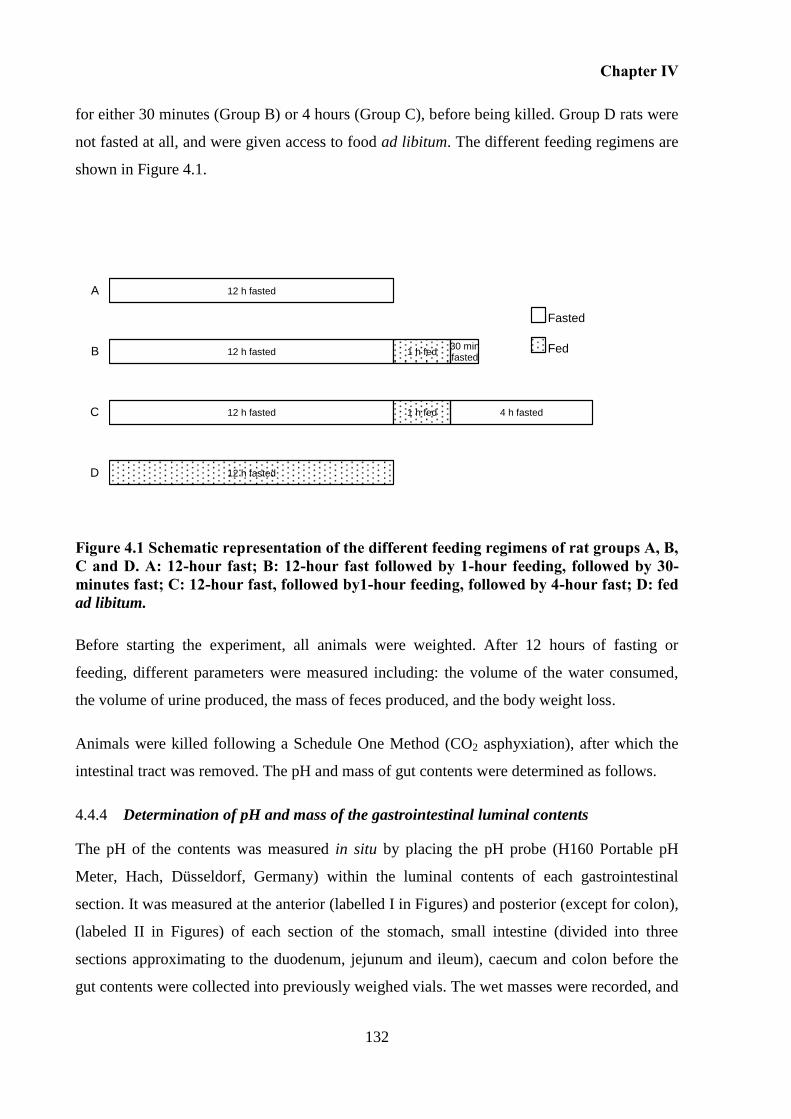
Chapter IV
132
for either 30 minutes (Group B) or 4 hours (Group C), before being killed. Group D rats were
not fasted at all, and were given access to food ad libitum. The different feeding regimens are
shown in Figure 4.1.
Figure 4.1 Schematic representation of the different feeding regimens of rat groups A, B, C and D. A: 12-hour fast; B: 12-hour fast followed by 1-hour feeding, followed by 30-minutes fast; C: 12-hour fast, followed by1-hour feeding, followed by 4-hour fast; D: fed ad libitum.
Before starting the experiment, all animals were weighted. After 12 hours of fasting or
feeding, different parameters were measured including: the volume of the water consumed,
the volume of urine produced, the mass of feces produced, and the body weight loss.
Animals were killed following a Schedule One Method (CO2 asphyxiation), after which the
intestinal tract was removed. The pH and mass of gut contents were determined as follows.
Determination of pH and mass of the gastrointestinal luminal contents 4.4.4
The pH of the contents was measured in situ by placing the pH probe (H160 Portable pH
Meter, Hach, Düsseldorf, Germany) within the luminal contents of each gastrointestinal
section. It was measured at the anterior (labelled I in Figures) and posterior (except for colon),
(labeled II in Figures) of each section of the stomach, small intestine (divided into three
sections approximating to the duodenum, jejunum and ileum), caecum and colon before the
gut contents were collected into previously weighed vials. The wet masses were recorded, and
12 h fasted
12 h fasted
12 h fasted
12 h fasted
1 h fed
1 h fed
4 h fasted
30 min fasted
D
C
B
A
Fasted
Fed

Chapter IV
133
the vials were stored at – 80 C. The pH of the distal part of colon contents could not be
reliably measured due to its solid nature.
Determination of stability of prodrugs (diclofenac-β-cyclodextrin and sulfasalazine), 4.4.5
in caecal and colonic contents
Stability tests were performed inside an anaerobic workstation (Electrotek 500TG
workstation, Electrotek, UK) at 37 C and 70% RH. The caecum and colonic contents from
each group of rats were mixed with PBS – of differing pHs as explained below - in order to
obtain a 40% w/w slurry as prepared in Chapter III. The pH of the PBS differed for the
different samples, but matched the in situ measured pH in the different gastrointestinal
sections (section above), in order to maintain the pH of the gut contents. Thus, gut contents
from Groups A and B rats were mixed with PBS pH 6.8, while those from Groups C and D
rats were mixed with PBS pH 6.0. The slurries were then homogenized using a glass rod and
sieved through an open mesh fabric (Sefar NitexTM, pore size 350 µm) to remove any
unhomogenised fibrous material. The sieved faecal slurry was then diluted 50% (w/w) with
basal medium containing peptone water, yeast extract, NaCl, K2HPO4, MgSO4·7H2O,
CaCl2·6H2O, NaHCO3, haemin, l-cysteine HCl, bile salts, tween 80, vitamin K and resazurin,
was described previously in Chapter III (Basit and Lacey, 2001; Basit, Newton et al., 2002).
Two solutions of each prodrug sulfasalazine (5 mg/mL) and diclofenac-β-cyclodextrin (2.4
mg/mL) were prepared in PBS at pH 6.8 and pH 6.0, both containing 4% (v/v) of
dimethylformamide. Subsequently, 100 µL of these solutions were mixed with 900 µL of
caecal or colonic fluids prepared above; the fluids from Groups A and B rats being mixed
with prodrug solutions at pH 6.8, while those from Groups C and D rats were mixed with
prodrug solutions at pH 6.0 in order to maintain the pH constant (Table 4.1). The final
concentrations of the conjugate, sulfasalazine and DMF were 0.5 mg/mL, 0.24 mg/mL and
0.4% (v/v), respectively.
Thereafter, these mixtures were incubated and shaken at 100 rpm (VXRbasic Vibrax®,
Leicestershire, UK), with 50 µL aliquots being withdrawn at times 0, 15, 30, 60, 90, 120, 180,
240, 360, 400, 600, 720 and 1440 minutes. The aliquots were immediately mixed with 100 µL
of methanol and centrifuged at 10 000 rpm for 10 minutes at room temperature, after which
the supernatant was removed and analyzed via HPLC for prodrugs concentrations.
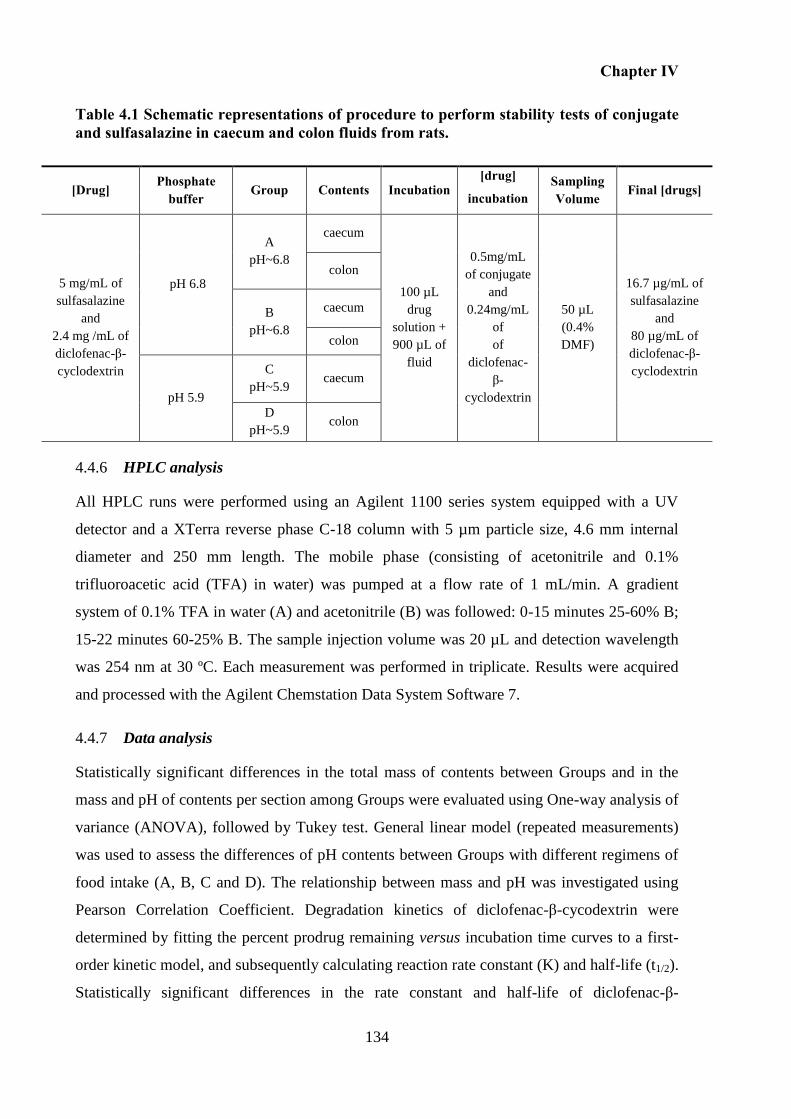
Chapter IV
134
Table 4.1 Schematic representations of procedure to perform stability tests of conjugate and sulfasalazine in caecum and colon fluids from rats.
[Drug] Phosphate
buffer Group Contents Incubation
[drug]
incubation Sampling Volume
Final [drugs]
5 mg/mL of
sulfasalazine
and
2.4 mg /mL of
diclofenac-β-
cyclodextrin
pH 6.8
A
pH~6.8
caecum
100 µL
drug
solution +
900 µL of
fluid
0.5mg/mL
of conjugate
and
0.24mg/mL
of
of
diclofenac-
β-
cyclodextrin
50 µL
(0.4%
DMF)
16.7 µg/mL of
sulfasalazine
and
80 µg/mL of
diclofenac-β-
cyclodextrin
colon
B
pH~6.8
caecum
colon
pH 5.9
C
pH~5.9 caecum
D
pH~5.9 colon
HPLC analysis 4.4.6
All HPLC runs were performed using an Agilent 1100 series system equipped with a UV
detector and a XTerra reverse phase C-18 column with 5 µm particle size, 4.6 mm internal
diameter and 250 mm length. The mobile phase (consisting of acetonitrile and 0.1%
trifluoroacetic acid (TFA) in water) was pumped at a flow rate of 1 mL/min. A gradient
system of 0.1% TFA in water (A) and acetonitrile (B) was followed: 0-15 minutes 25-60% B;
15-22 minutes 60-25% B. The sample injection volume was 20 µL and detection wavelength
was 254 nm at 30 ºC. Each measurement was performed in triplicate. Results were acquired
and processed with the Agilent Chemstation Data System Software 7.
Data analysis 4.4.7
Statistically significant differences in the total mass of contents between Groups and in the
mass and pH of contents per section among Groups were evaluated using One-way analysis of
variance (ANOVA), followed by Tukey test. General linear model (repeated measurements)
was used to assess the differences of pH contents between Groups with different regimens of
food intake (A, B, C and D). The relationship between mass and pH was investigated using
Pearson Correlation Coefficient. Degradation kinetics of diclofenac-β-cycodextrin were
determined by fitting the percent prodrug remaining versus incubation time curves to a first-
order kinetic model, and subsequently calculating reaction rate constant (K) and half-life (t1/2).
Statistically significant differences in the rate constant and half-life of diclofenac-β-
Chapter IV
135
cyclodextrin conjugate between Groups were analysed using Kruskal–Wallis test, with
Nemenyi’s post-hoc analysis. All tests, apart from Nemenyi’s test were carried out using
SPSS 21.0 for Windows®. The Nemenyi test was conducted as described (Jones, 2002).
Results were considered statistically significant when p < 0.05.
4.5 Results and discussion
Influence of feeding regimens on body weight of the rats, mass of faeces and 4.5.1
volume of urine
The changes in body weight of the rats, mass of faeces and volume of urine produced,
quantity of food and volume of water consumed per group were summarized in the Table 4.2.
In all the animals kept fasted overnight (A, B, C and D) it was verified a loss of body weight
(~5%) after 12 hours. This loss of weight was expected in these fasted overnight animals,
however animals from Group D also demonstrated weight loss. Moreover, in Group D, loss of
weight is higher (~12%), herein it was not expected since these rats were fed ad libitum. This
body weight loss can be explained due to the higher production of feces observed in this
group of rats. This was already observed before in a study with rats on an ad libitum diet
(Jeffrey, Burrows et al., 1987). The same study had observed that rats on a controlled diet
maintained their body weight.
After feeding, the animals from Group B and C did not show any production of faeces. This
can mean that the food ingested before 12 hours of fasting did not have enough time to be
digested and consequently to be excreted by the gastrointestinal tract of rats. It is important to
note that rats eat only small part of their food during the day and and consume 80% of their
total daily food intake at night, taking 5-8 meals during the night, hence during the feeding of
the animals the room was kept dark (Nebendahl, 2000).
Measurements of the solid and water contents in fed and fasted female Wistar rats had shown
that the water amount was also higher in the fed state (7.8 ± 1.5 mL) against 3.2±1.8 mL in
the fasted state, attributable to the increase of secretions and water bound with the ingested
food (Mcconnell, Basit et al., 2008). The higher quantity in fed state can be favourable to the
dissolution of drugs or formulations. However, possible interactions of food with drugs have
to be considered.
Chapter IV
136
As the photographs of each section of the gastrointestinal tract (Figure 4.2) show, the animals
from Group B (Figure 4.2-II), C (Figure 4.2-III) and D (Figure 4.2-IV) present the stomach
and the colon full comparing with the fasted animals (A) (Figure 4.2-I).
Moreover, when the stomach is full it is possible to distinguish between the glandular and the
non-glandular part. Herein, the non-glandular part is thin and translucent and the glandular is
opaque, muscular and reddish involving the fundic and pyloric regions (Ghoshal and Bal,
1989).
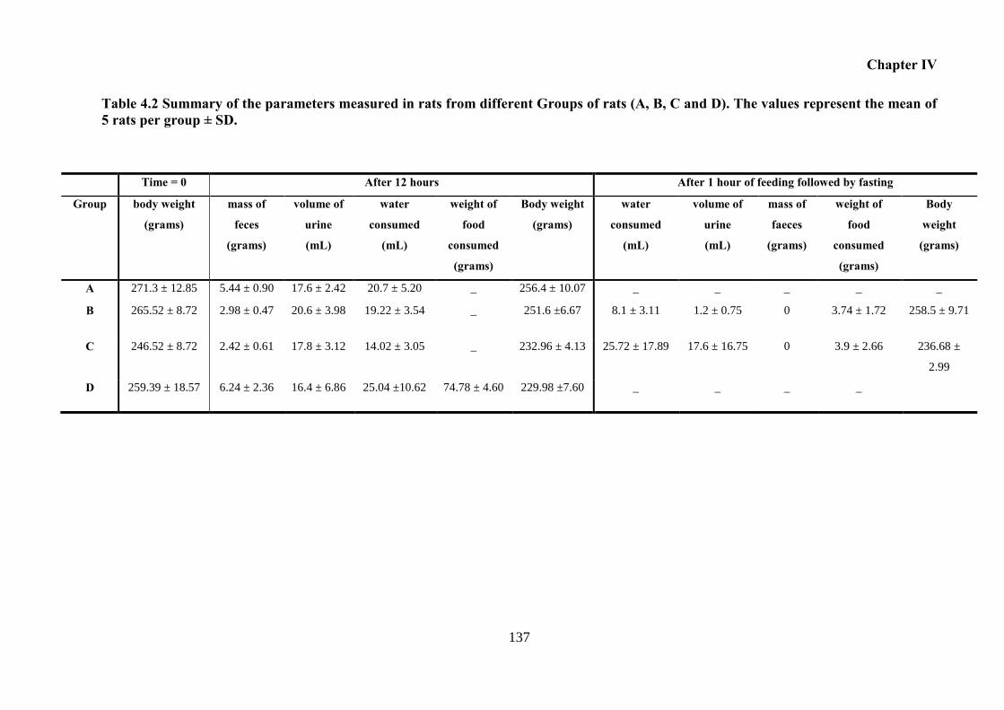
Chapter IV
137
Table 4.2 Summary of the parameters measured in rats from different Groups of rats (A, B, C and D). The values represent the mean of 5 rats per group ± SD.
Time = 0 After 12 hours After 1 hour of feeding followed by fasting
Group body weight
(grams)
mass of
feces
(grams)
volume of
urine
(mL)
water
consumed
(mL)
weight of
food
consumed
(grams)
Body weight
(grams)
water
consumed
(mL)
volume of
urine
(mL)
mass of
faeces
(grams)
weight of
food
consumed
(grams)
Body
weight
(grams)
A 271.3 ± 12.85 5.44 ± 0.90 17.6 ± 2.42 20.7 ± 5.20 _ 256.4 ± 10.07 _ _ _ _ _
B 265.52 ± 8.72 2.98 ± 0.47 20.6 ± 3.98 19.22 ± 3.54 _ 251.6 ±6.67 8.1 ± 3.11 1.2 ± 0.75 0 3.74 ± 1.72 258.5 ± 9.71
C 246.52 ± 8.72 2.42 ± 0.61 17.8 ± 3.12 14.02 ± 3.05 _ 232.96 ± 4.13 25.72 ± 17.89 17.6 ± 16.75 0 3.9 ± 2.66 236.68 ±
2.99
D 259.39 ± 18.57 6.24 ± 2.36 16.4 ± 6.86 25.04 ±10.62 74.78 ± 4.60 229.98 ±7.60 _ _ _ _
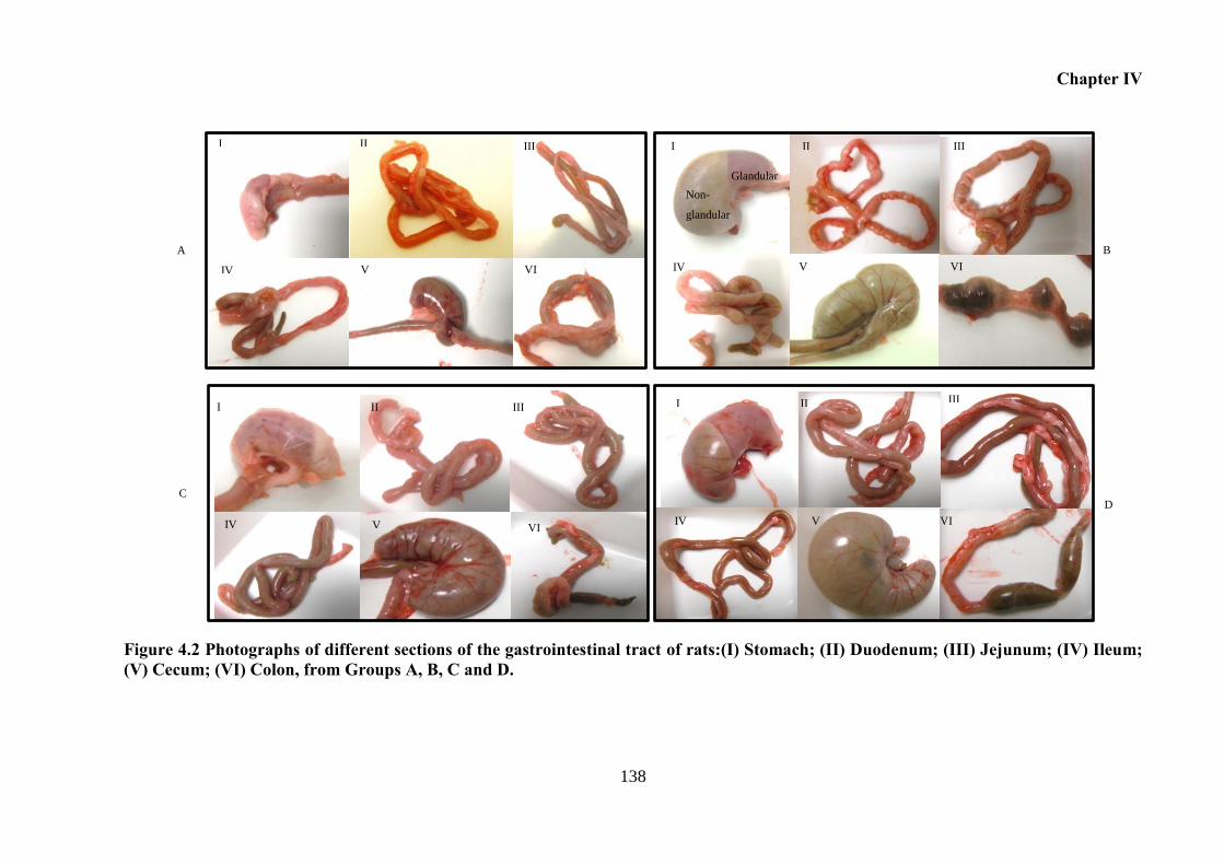
Chapter IV
138
Figure 4.2 Photographs of different sections of the gastrointestinal tract of rats:(I) Stomach; (II) Duodenum; (III) Jejunum; (IV) Ileum; (V) Cecum; (VI) Colon, from Groups A, B, C and D.
I II III
IV V VI
I
IV V VI
II III
I II III
IV
I II III
IV V VI
V VI
A B
C D
Non-
glandular
Glandular
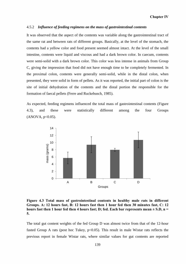
Chapter IV
139
Influence of feeding regimens on the mass of gastrointestinal contents 4.5.2
It was observed that the aspect of the contents was variable along the gastrointestinal tract of
the same rat and between rats of different groups. Basically, at the level of the stomach, the
contents had a yellow color and food present seemed almost intact. At the level of the small
intestine, contents were liquid and viscous and had a dark brown color. In caecum, contents
were semi-solid with a dark brown color. This color was less intense in animals from Group
C, giving the impression that food did not have enough time to be completely fermented. In
the proximal colon, contents were generally semi-solid, while in the distal colon, when
presented, they were solid in form of pellets. As it was reported, the initial part of colon is the
site of initial dehydration of the contents and the distal portion the responsible for the
formation of faecal pellets (Ferre and Ruckebusch, 1985).
As expected, feeding regimens influenced the total mass of gastrointestinal contents (Figure
4.3), and these were statistically different among the four Groups
(ANOVA, p<0.05).
Figure 4.3 Total mass of gastrointestinal contents in healthy male rats in different Groups. A: 12 hours fast, B: 12 hours fast then 1 hour fed then 30 minutes fast, C: 12 hours fast then 1 hour fed then 4 hours fast; D; fed. Each bar represents mean ± S.D, n = 5.
The total gut content weights of the fed Group D was almost twice from that of the 12-hour
fasted Group A rats (post hoc Tukey, p<0.05). This result in male Wistar rats reflects the
previous report in female Wistar rats, where similar values for gut contents are reported
0
2
4
6
8
10
12
14
A B C D
mass (
gra
ms)
Groups
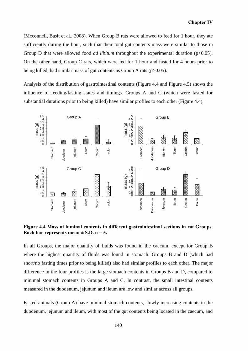
Chapter IV
140
(Mcconnell, Basit et al., 2008). When Group B rats were allowed to feed for 1 hour, they ate
sufficiently during the hour, such that their total gut contents mass were similar to those in
Group D that were allowed food ad libitum throughout the experimental duration (p>0.05).
On the other hand, Group C rats, which were fed for 1 hour and fasted for 4 hours prior to
being killed, had similar mass of gut contents as Group A rats (p>0.05).
Analysis of the distribution of gastrointestinal contents (Figure 4.4 and Figure 4.5) shows the
influence of feeding/fasting states and timings. Groups A and C (which were fasted for
substantial durations prior to being killed) have similar profiles to each other (Figure 4.4).
Figure 4.4 Mass of luminal contents in different gastrointestinal sections in rat Groups. Each bar represents mean ± S.D. n = 5.
In all Groups, the major quantity of fluids was found in the caecum, except for Group B
where the highest quantity of fluids was found in stomach. Groups B and D (which had
short/no fasting times prior to being killed) also had similar profiles to each other. The major
difference in the four profiles is the large stomach contents in Groups B and D, compared to
minimal stomach contents in Groups A and C. In contrast, the small intestinal contents
measured in the duodenum, jejunum and ileum are low and similar across all groups.
Fasted animals (Group A) have minimal stomach contents, slowly increasing contents in the
duodenum, jejunum and ileum, with most of the gut contents being located in the caecum, and
00.5
11.5
22.5
33.5
44.5
Sto
mach
duoden
um
jeju
num
ileum
Cecum
colo
n
mass (
g)
Group A
00.5
11.5
22.5
33.5
44.5
5
Sto
mach
duoden
um
jeju
num
ileum
Cecum
colo
n
mass (
g)
Group B
00.5
11.5
22.5
33.5
44.5
5
Sto
mach
Duoden
um
Jeju
num
Ileu
m
Cecum
Colo
n
mass (
g)
Group D
00.5
11.5
22.5
33.5
44.5
Sto
mach
duoden
um
jeju
num
ileum
Cecum
colo
n
mass (
g)
Group C
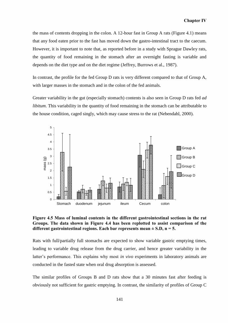
Chapter IV
141
the mass of contents dropping in the colon. A 12-hour fast in Group A rats (Figure 4.1) means
that any food eaten prior to the fast has moved down the gastro-intestinal tract to the caecum.
However, it is important to note that, as reported before in a study with Sprague Dawley rats,
the quantity of food remaining in the stomach after an overnight fasting is variable and
depends on the diet type and on the diet regime (Jeffrey, Burrows et al., 1987).
In contrast, the profile for the fed Group D rats is very different compared to that of Group A,
with larger masses in the stomach and in the colon of the fed animals.
Greater variability in the gut (especially stomach) contents is also seen in Group D rats fed ad
libitum. This variability in the quantity of food remaining in the stomach can be attributable to
the house condition, caged singly, which may cause stress to the rat (Nebendahl, 2000).
Figure 4.5 Mass of luminal contents in the different gastrointestinal sections in the rat Groups. The data shown in Figure 4.4 has been replotted to assist comparison of the different gastrointestinal regions. Each bar represents mean ± S.D, n = 5.
Rats with full/partially full stomachs are expected to show variable gastric emptying times,
leading to variable drug release from the drug carrier, and hence greater variability in the
latter’s performance. This explains why most in vivo experiments in laboratory animals are
conducted in the fasted state when oral drug absorption is assessed.
The similar profiles of Groups B and D rats show that a 30 minutes fast after feeding is
obviously not sufficient for gastric emptying. In contrast, the similarity of profiles of Group C
0
0.5
1
1.5
2
2.5
3
3.5
4
4.5
5
Stomach duodenum jejunum ileum Cecum colon
mass (
g)
Group A
Group B
Group C
Group D
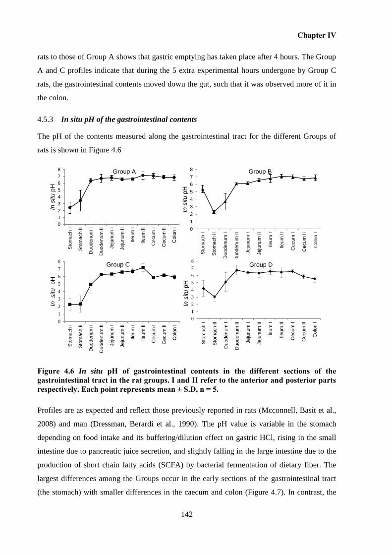
Chapter IV
142
rats to those of Group A shows that gastric emptying has taken place after 4 hours. The Group
A and C profiles indicate that during the 5 extra experimental hours undergone by Group C
rats, the gastrointestinal contents moved down the gut, such that it was observed more of it in
the colon.
In situ pH of the gastrointestinal contents 4.5.3
The pH of the contents measured along the gastrointestinal tract for the different Groups of
rats is shown in Figure 4.6
Figure 4.6 In situ pH of gastrointestinal contents in the different sections of the gastrointestinal tract in the rat groups. I and II refer to the anterior and posterior parts respectively. Each point represents mean ± S.D, n = 5.
Profiles are as expected and reflect those previously reported in rats (Mcconnell, Basit et al.,
2008) and man (Dressman, Berardi et al., 1990). The pH value is variable in the stomach
depending on food intake and its buffering/dilution effect on gastric HCl, rising in the small
intestine due to pancreatic juice secretion, and slightly falling in the large intestine due to the
production of short chain fatty acids (SCFA) by bacterial fermentation of dietary fiber. The
largest differences among the Groups occur in the early sections of the gastrointestinal tract
(the stomach) with smaller differences in the caecum and colon (Figure 4.7). In contrast, the
0
1
2
3
4
5
6
7
8
Sto
mach I
Sto
mach II
Duoden
um
I
Duoden
um
II
Jeju
num
I
Jeju
num
II
Ileu
m I
Ileu
m II
Cecum
I
Cecum
II
Colo
n I
In s
itu p
H
Group A
0
1
2
3
4
5
6
7
8
Sto
mach I
Sto
mach II
Duoden
um
I
Duoden
um
II
Jeju
num
I
Jeju
num
II
Ileu
m I
Ileu
m II
Cecum
I
Cecum
II
Colo
n I
In s
itu p
H
Group B
0
1
2
3
4
5
6
7
8
Sto
mach I
Sto
mach II
Duoden
um
I
Duoden
um
II
Jeju
num
I
Jeju
num
II
Ileu
m I
Ileu
m II
Cecum
I
Cecum
II
Colo
n I
In
situ
pH
Group C
0
1
2
3
4
5
6
7
8
Sto
mach I
Sto
mach II
Duoden
um
I
Duoden
um
II
Jeju
num
I
Jeju
num
II
Ileu
m I
Ileu
m II
Cecum
I
Cecum
II
Colo
n I
In s
itu
pH
Group D
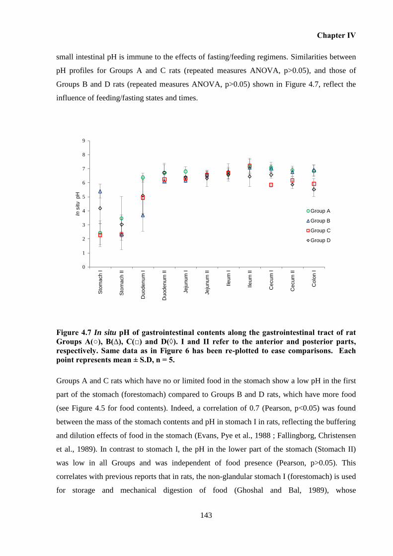
Chapter IV
143
small intestinal pH is immune to the effects of fasting/feeding regimens. Similarities between
pH profiles for Groups A and C rats (repeated measures ANOVA, p>0.05), and those of
Groups B and D rats (repeated measures ANOVA, p>0.05) shown in Figure 4.7, reflect the
influence of feeding/fasting states and times.
Figure 4.7 In situ pH of gastrointestinal contents along the gastrointestinal tract of rat Groups A(○), B(∆), C(□) and D(◊). I and II refer to the anterior and posterior parts, respectively. Same data as in Figure 6 has been re-plotted to ease comparisons. Each point represents mean ± S.D, n = 5.
Groups A and C rats which have no or limited food in the stomach show a low pH in the first
part of the stomach (forestomach) compared to Groups B and D rats, which have more food
(see Figure 4.5 for food contents). Indeed, a correlation of 0.7 (Pearson, p<0.05) was found
between the mass of the stomach contents and pH in stomach I in rats, reflecting the buffering
and dilution effects of food in the stomach (Evans, Pye et al., 1988 ; Fallingborg, Christensen
et al., 1989). In contrast to stomach I, the pH in the lower part of the stomach (Stomach II)
was low in all Groups and was independent of food presence (Pearson, p>0.05). This
correlates with previous reports that in rats, the non-glandular stomach I (forestomach) is used
for storage and mechanical digestion of food (Ghoshal and Bal, 1989), whose
0
1
2
3
4
5
6
7
8
9
Sto
ma
ch I
Sto
ma
ch II
Duoden
um
I
Duoden
um
II
Jeju
num
I
Jeju
num
II
Ileu
m I
Ileu
m II
Cecu
m I
Cecum
II
Colo
n I
In s
itu pH
Group A
Group B
Group C
Group D
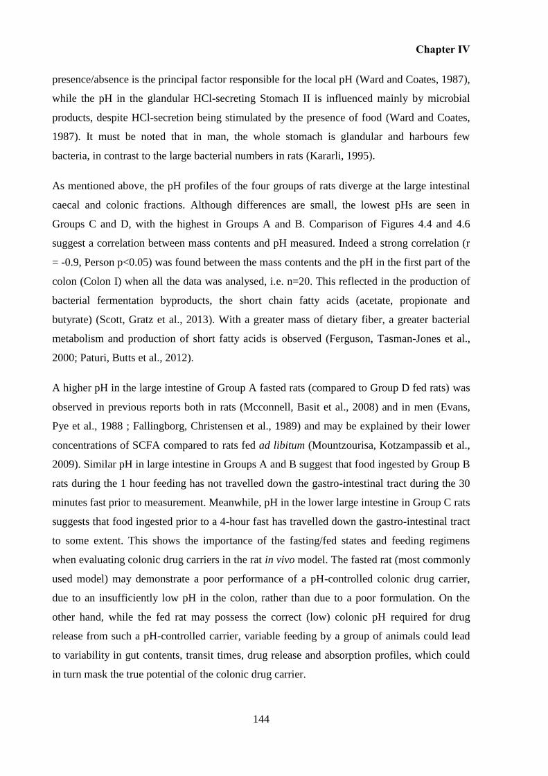
Chapter IV
144
presence/absence is the principal factor responsible for the local pH (Ward and Coates, 1987),
while the pH in the glandular HCl-secreting Stomach II is influenced mainly by microbial
products, despite HCl-secretion being stimulated by the presence of food (Ward and Coates,
1987). It must be noted that in man, the whole stomach is glandular and harbours few
bacteria, in contrast to the large bacterial numbers in rats (Kararli, 1995).
As mentioned above, the pH profiles of the four groups of rats diverge at the large intestinal
caecal and colonic fractions. Although differences are small, the lowest pHs are seen in
Groups C and D, with the highest in Groups A and B. Comparison of Figures 4.4 and 4.6
suggest a correlation between mass contents and pH measured. Indeed a strong correlation (r
= -0.9, Person p<0.05) was found between the mass contents and the pH in the first part of the
colon (Colon I) when all the data was analysed, i.e. n=20. This reflected in the production of
bacterial fermentation byproducts, the short chain fatty acids (acetate, propionate and
butyrate) (Scott, Gratz et al., 2013). With a greater mass of dietary fiber, a greater bacterial
metabolism and production of short fatty acids is observed (Ferguson, Tasman-Jones et al.,
2000; Paturi, Butts et al., 2012).
A higher pH in the large intestine of Group A fasted rats (compared to Group D fed rats) was
observed in previous reports both in rats (Mcconnell, Basit et al., 2008) and in men (Evans,
Pye et al., 1988 ; Fallingborg, Christensen et al., 1989) and may be explained by their lower
concentrations of SCFA compared to rats fed ad libitum (Mountzourisa, Kotzampassib et al.,
2009). Similar pH in large intestine in Groups A and B suggest that food ingested by Group B
rats during the 1 hour feeding has not travelled down the gastro-intestinal tract during the 30
minutes fast prior to measurement. Meanwhile, pH in the lower large intestine in Group C rats
suggests that food ingested prior to a 4-hour fast has travelled down the gastro-intestinal tract
to some extent. This shows the importance of the fasting/fed states and feeding regimens
when evaluating colonic drug carriers in the rat in vivo model. The fasted rat (most commonly
used model) may demonstrate a poor performance of a pH-controlled colonic drug carrier,
due to an insufficiently low pH in the colon, rather than due to a poor formulation. On the
other hand, while the fed rat may possess the correct (low) colonic pH required for drug
release from such a pH-controlled carrier, variable feeding by a group of animals could lead
to variability in gut contents, transit times, drug release and absorption profiles, which could
in turn mask the true potential of the colonic drug carrier.
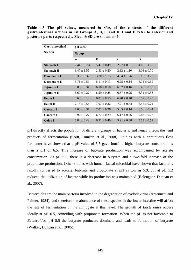
Chapter IV
145
Table 4.3 The pH values, measured in situ, of the contents of the different gastrointestinal sections in rat Groups A, B, C and D. I and II refer to anterior and posterior parts respectively. Mean ± SD are shown, n=5.
Gastrointestinal
Section pH ± SD
Group
A B C D
Stomach I 2.44 ± 0.84 5.41 ± 0.49 2.27 ± 0.81 4.19 ± 1.08
Stomach II 3.47 ± 1.55 2.33 ± 0.20 2.32 ± 1.10 3.03 ± 0.70
Duodenum I 6.38 ± 0.32 3.70 ± 1.15 4.94 ± 1.26 5.10 ± 1.29
Duodenum II 6.71 ± 0.59 6.11 ± 0.13 6.25 ± 0.14 6.72 ± 0.69
Jejunum I 6.80 ± 0.34 6.18 ± 0.18 6.32 ± 0.16 6.40 ± 0.09
Jejunum II 6.60 ± 0.22 6.59 ± 0.25 6.57 ± 0.25 6.31 ± 0.58
Ileum I 6.63 ± 0.18 6.81 ± 0.55 6.70 ± 0.40 6.57 ± 0.45
Ileum II 7.15 ± 0.54 7.07 ± 0.32 7.21 ± 0.54 6.45 ± 0.71
Caecum I 7.08 ± 0.37 7.01 ± 0.26 5.85 ± 0.14 6.56 ± 0.24
Caecum II 6.90 ± 0.27 6.77 ± 0.29 6.17 ± 0.26 5.87 ± 0.27
Colon I 6.84 ± 0.42 6.91 ± 0.40 5.91 ± 0.38 5.53 ± 0.51
pH directly affects the population of different groups of bacteria, and hence affects the end
products of fermentation (Scott, Duncan et al., 2008). Studies with a continuous flow
fermenter have shown that a pH value of 5.5 gave fourfold higher butyrate concentrations
than a pH of 6.5. This increase of butyrate production was accompanied by acetate
consumption. At pH 6.5, there is a decrease in butyrate and a two-fold increase of the
propionate production. Other studies with human faecal microbial have shown that lactate is
rapidly converted to acetate, butyrate and propionate at pH as low as 5.9, but at pH 5.2
reduced the utilization of lactate while its production was maintained (Belenguer, Duncan et
al., 2007).
Bacteroides are the main bacteria involved in the degradation of cyclodextrins (Antenucci and
Palmer, 1984), and therefore the abundance of these species in the lower intestine will affect
the rate of fermentation of the conjugate at this level. The growth of Bacteroides occurs
ideally at pH 6.5, coinciding with propionate formation. When the pH is not favorable to
Bacteroides, pH 5.5 the butyrate producers dominate and leads to formation of butyrate
(Walker, Duncan et al., 2005).
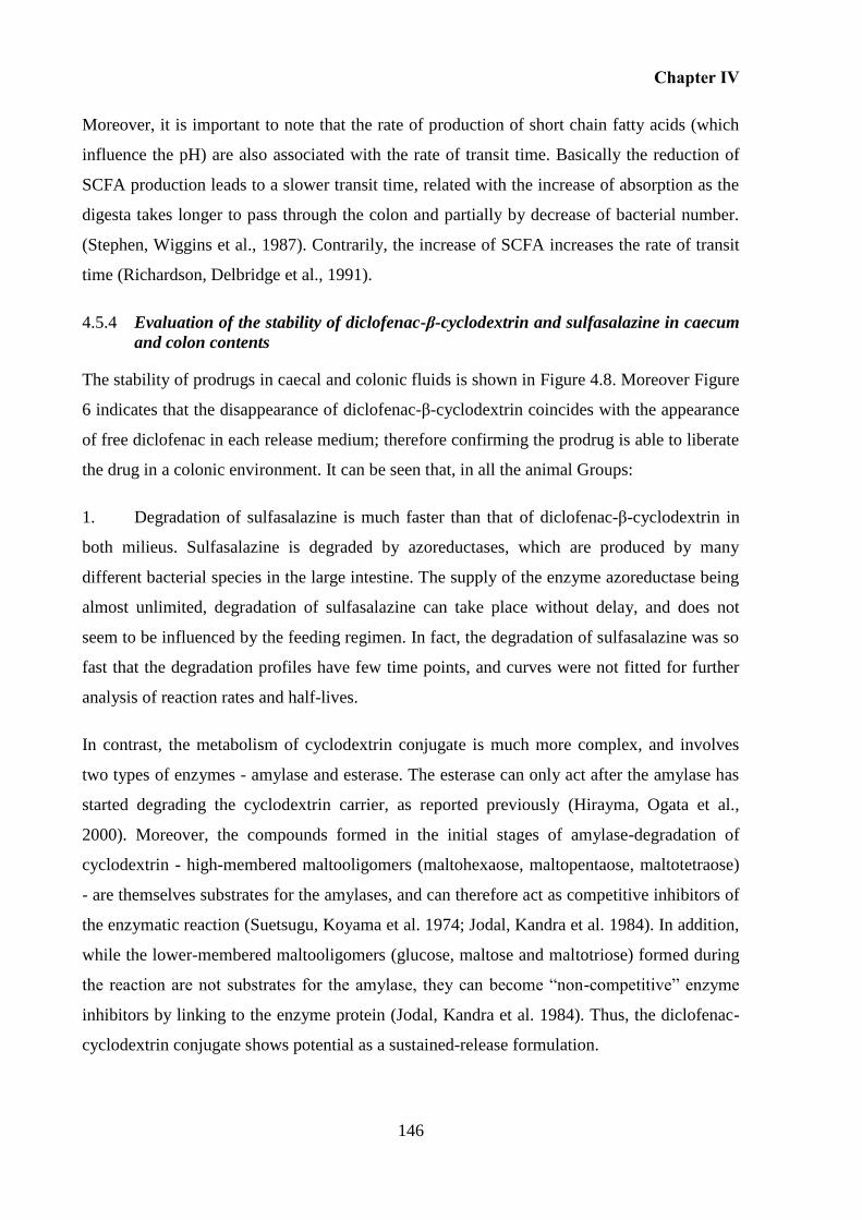
Chapter IV
146
Moreover, it is important to note that the rate of production of short chain fatty acids (which
influence the pH) are also associated with the rate of transit time. Basically the reduction of
SCFA production leads to a slower transit time, related with the increase of absorption as the
digesta takes longer to pass through the colon and partially by decrease of bacterial number.
(Stephen, Wiggins et al., 1987). Contrarily, the increase of SCFA increases the rate of transit
time (Richardson, Delbridge et al., 1991).
Evaluation of the stability of diclofenac-β-cyclodextrin and sulfasalazine in caecum 4.5.4
and colon contents
The stability of prodrugs in caecal and colonic fluids is shown in Figure 4.8. Moreover Figure
6 indicates that the disappearance of diclofenac-β-cyclodextrin coincides with the appearance
of free diclofenac in each release medium; therefore confirming the prodrug is able to liberate
the drug in a colonic environment. It can be seen that, in all the animal Groups:
1. Degradation of sulfasalazine is much faster than that of diclofenac-β-cyclodextrin in
both milieus. Sulfasalazine is degraded by azoreductases, which are produced by many
different bacterial species in the large intestine. The supply of the enzyme azoreductase being
almost unlimited, degradation of sulfasalazine can take place without delay, and does not
seem to be influenced by the feeding regimen. In fact, the degradation of sulfasalazine was so
fast that the degradation profiles have few time points, and curves were not fitted for further
analysis of reaction rates and half-lives.
In contrast, the metabolism of cyclodextrin conjugate is much more complex, and involves
two types of enzymes - amylase and esterase. The esterase can only act after the amylase has
started degrading the cyclodextrin carrier, as reported previously (Hirayma, Ogata et al.,
2000). Moreover, the compounds formed in the initial stages of amylase-degradation of
cyclodextrin - high-membered maltooligomers (maltohexaose, maltopentaose, maltotetraose)
- are themselves substrates for the amylases, and can therefore act as competitive inhibitors of
the enzymatic reaction (Suetsugu, Koyama et al. 1974; Jodal, Kandra et al. 1984). In addition,
while the lower-membered maltooligomers (glucose, maltose and maltotriose) formed during
the reaction are not substrates for the amylase, they can become “non-competitive” enzyme
inhibitors by linking to the enzyme protein (Jodal, Kandra et al. 1984). Thus, the diclofenac-
cyclodextrin conjugate shows potential as a sustained-release formulation.
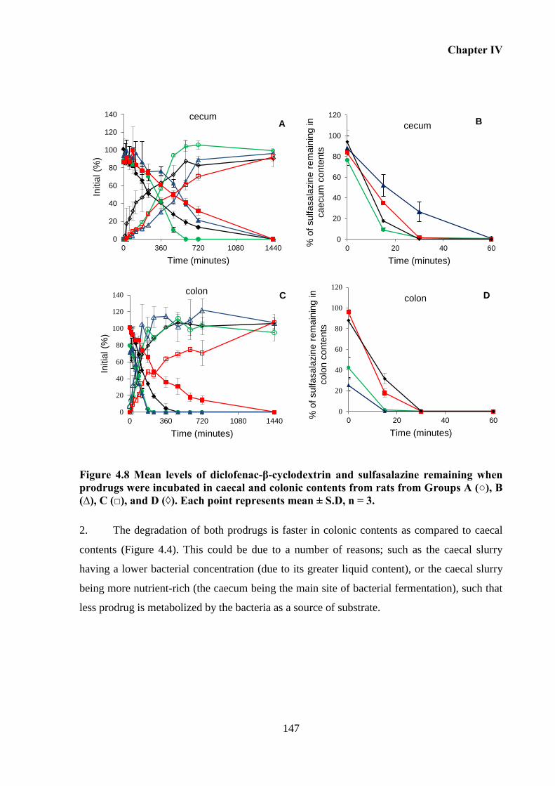
Chapter IV
147
Figure 4.8 Mean levels of diclofenac-β-cyclodextrin and sulfasalazine remaining when prodrugs were incubated in caecal and colonic contents from rats from Groups A (○), B (∆), C (□), and D (◊). Each point represents mean ± S.D, n = 3.
2. The degradation of both prodrugs is faster in colonic contents as compared to caecal
contents (Figure 4.4). This could be due to a number of reasons; such as the caecal slurry
having a lower bacterial concentration (due to its greater liquid content), or the caecal slurry
being more nutrient-rich (the caecum being the main site of bacterial fermentation), such that
less prodrug is metabolized by the bacteria as a source of substrate.
0
20
40
60
80
100
120
0 20 40 60
% o
f sulfasala
zin
e r
em
ain
ing in
caecum
conte
nts
Time (minutes)
cecum
0
20
40
60
80
100
120
0 20 40 60
% o
f sulfasala
zin
e r
em
ain
ing in
colo
n c
onte
nts
Time (minutes)
colon D
0
20
40
60
80
100
120
140
0 360 720 1080 1440
Initia
l (%
)
Time (minutes)
cecum
0
20
40
60
80
100
120
140
0 360 720 1080 1440
Initia
l (%
)
Time (minutes)
colon C
A B
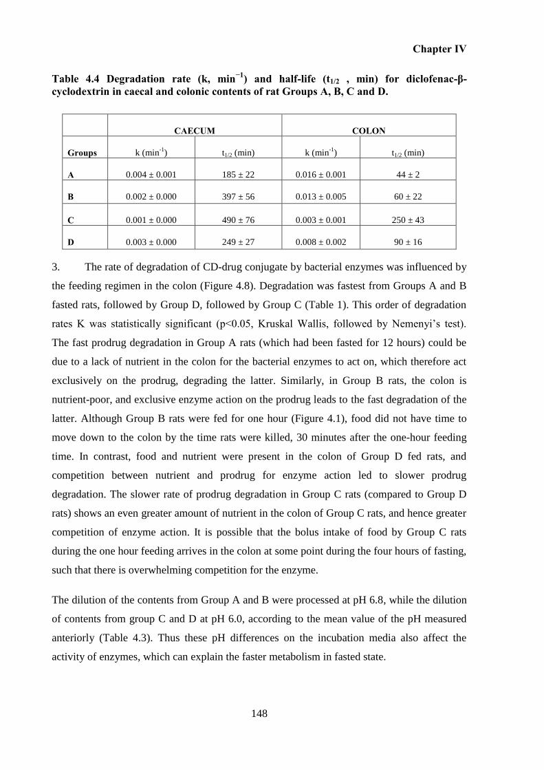
Chapter IV
148
Table 4.4 Degradation rate (k, min−1) and half-life (t1/2 , min) for diclofenac-β-cyclodextrin in caecal and colonic contents of rat Groups A, B, C and D.
CAECUM COLON
Groups k (min-1
) t1/2 (min) k (min-1
) t1/2 (min)
A 0.004 ± 0.001 185 ± 22 0.016 ± 0.001 44 ± 2
B 0.002 ± 0.000 397 ± 56 0.013 ± 0.005 60 ± 22
C 0.001 ± 0.000 490 ± 76 0.003 ± 0.001 250 ± 43
D 0.003 ± 0.000 249 ± 27 0.008 ± 0.002 90 ± 16
3. The rate of degradation of CD-drug conjugate by bacterial enzymes was influenced by
the feeding regimen in the colon (Figure 4.8). Degradation was fastest from Groups A and B
fasted rats, followed by Group D, followed by Group C (Table 1). This order of degradation
rates K was statistically significant (p<0.05, Kruskal Wallis, followed by Nemenyi’s test).
The fast prodrug degradation in Group A rats (which had been fasted for 12 hours) could be
due to a lack of nutrient in the colon for the bacterial enzymes to act on, which therefore act
exclusively on the prodrug, degrading the latter. Similarly, in Group B rats, the colon is
nutrient-poor, and exclusive enzyme action on the prodrug leads to the fast degradation of the
latter. Although Group B rats were fed for one hour (Figure 4.1), food did not have time to
move down to the colon by the time rats were killed, 30 minutes after the one-hour feeding
time. In contrast, food and nutrient were present in the colon of Group D fed rats, and
competition between nutrient and prodrug for enzyme action led to slower prodrug
degradation. The slower rate of prodrug degradation in Group C rats (compared to Group D
rats) shows an even greater amount of nutrient in the colon of Group C rats, and hence greater
competition of enzyme action. It is possible that the bolus intake of food by Group C rats
during the one hour feeding arrives in the colon at some point during the four hours of fasting,
such that there is overwhelming competition for the enzyme.
The dilution of the contents from Group A and B were processed at pH 6.8, while the dilution
of contents from group C and D at pH 6.0, according to the mean value of the pH measured
anteriorly (Table 4.3). Thus these pH differences on the incubation media also affect the
activity of enzymes, which can explain the faster metabolism in fasted state.
Chapter IV
149
In the fed state is expected a decrease on the activity of Bacteroids and consequently on the
metabolism of cyclodextrin carrier due to the diminuish of pH associated with the production
of SCFA, which can also reinforce the explanation associate with the decrease of metabolism
in fed state.
Overall, it is interesting to note that the rates of degradation of diclofenac-β-cyclodextrin in
colonic contents in Group A and B is close to the rate of degradation observed in the human
faecal slurries collected from individuals without any food intake control, as reported
previously in Chapter II.
In contrast to the obvious influence of feeding regimen on prodrug degradation in the colon,
the influence was less obvious in the caecum. The degradation curves for all rat groups have
similar profiles, especially at the beginning of the in vitro degradation reaction (Figure 4.8).
Caecum has such a high content of material in all rat groups (Figure 4.5) that feeding/fasting
did not seem to alter the nutrient content and subsequently, any competition between nutrient
and prodrug for enzyme action. One point to note though is the completion of prodrug
degradation in Group A fasted rats caecal contents at 600 minutes (Figure 4.8) in contrast to
the other groups. This correlates with the fact that Group A fasted rats had a lowest amount of
nutrient (and hence competition for enzyme action) in their caecum.
4.6 Conclusion
This study demonstrates the importance of feeding regimens, specifically the timing of meal
ingestion, on the gastrointestinal conditions in rats and how this influences the metabolism of
colonic prodrugs, namely diclofenac-β-cyclodextrin. In addition to changes in the distribution
of gut contents along the GI tract, which directly affects the gastrointestinal transit time,
different feeding regimens are accompanied by changes in the pH of gut contents, specifically
in the stomach and large intestine. Moreover, differential gut contents in the large intestine
have an impact on the microbiota activity, which affects the rate of diclofenac-β-cyclodextrin
metabolism, and hence drug release and absorption. We also showed that the different feeding
regimens did not seem to im-pact on the metabolism of sulfasalazine, which was rapidly
metabolized. Thus, we conclude that while the feeding regimen influences the performance of
the colonic prodrug, diclofenac-β-cyclodextrin, that influence has to be measured for each
prodrug individually, given the different metabolism pathways of different colonic carriers.

Chapter IV
150
CHAPTER V
DICLOFENAC-β-CYCLODEXTRIN VERSUS SULFASALAZINE FOR COLONIC DRUG
TARGETING: IN VIVO PERFORMANCE IN RATS

Chapter V
153
5 DICLOFENAC-ΒETA-CYCLODEXTRIN VERSUS SULFASALAZINE FOR
COLONIC DRUG TARGETING: IN VIVO PERFORMANCE IN RATS
5.1 Overview
The influence of feeding regimen on the performance of diclofenac-β-cyclodextrin was
studied previously in rats (Chapter IV). Results showed that its metabolism depends on the
feeding regimen, herein fasted state, leading to a faster metabolism of this conjugate.
An in vivo proof of concept is required not only to confirm the ability of the conjugate to
provide targeting of diclofenac on the large intestine, but also to study the bioavailability of
diclofenac when absorption takes place at the colonic level. Based on results from the stability
experiments of both prodrugs, and considering the greater variability on the gastrointestinal
transit time caused by food intake, oral administration was conducted in the fasted state. The
main advantage of conducting these studies under fed conditions would be the elimination of
the additional step of food removal, which could reduce potential stress on the animals caused
by fasting (Mittelstadt, Hemenway et al., 2005).
Sulfasalazine is a prodrug activated at a colonic level, as previously described, releasing 5-
aminosalicylic acid (mesalazine) and sulfapyridine. A delay between the oral intake of
sulfasalazine and the appearance of the respective metabolites in the blood circulation is
expected. When administered concomitantly with other potential colonic prodrugs, this
prodrug works as a control, providing us with an idea of the time necessary for it to reach the
colon by the appearance of its metabolites in plasma.
Hence, this Chapter presents a comparative in vivo study in fasted rats that considers oral
administration of a suspension of prodrugs, diclofenac-β-cyclodextrin and sulfasalazine to a
group of rats (Group I), and also the simultaneous quantification analysis of the respective
active drugs diclofenac and sulfapyridine in plasma. Moreover, a control group of animals
(Group II) was dosed orally with sulfapyridine and diclofenac. The comparison of
pharmacokinetic parameters between both groups, namely plasmatic concentrations and time
of absorption, allowed determining the ability of the conjugate to release diclofenac in the
large intestine. The dose of drugs administrated to Group II was equivalent to the dose of free
Chapter V
154
drugs administered to Group I; this was usefull to assess differences in the bioavailability
between each drug and the respective colonic prodrug.
5.2 Introduction
When administered orally, diclofenac absorption is rapid and complete in humans and in
animal models, namely rats (Peris-Ribera, Torres-Molina et al., 1991; Giagoudakis and
Markantonis, 1998). Diclofenac is poorly soluble in acidic media (pKa = 4), and therefore its
release, dissolution and absorption is favorable when pH is high. On the other hand, when the
pH is low, this drug will precipitate or will not be released (Chan, Mojaverian et al., 1990;
Riad, Sawchuk et al., 1995). However, diclofenac suffers first-pass metabolism, such that
~50% of the drug reaches the systemic circulation in the unchanged form. The plasma half-
life time is between 1 to 2 hours (Davies and Anderson, 1997).
When it reaches the colon, this drug is not degraded and is additionally well absorbed
(Tannergren, Bergendal et al., 2008). Nevertheless, when diclofenac reaches the colon,
namely in the form of diclofenac-β-cyclodextrin, its absorption will depend on the rate of
drug release, which is directly depends on the activity of microbiota. Moreover, in the form of
the conjugate, a longer time is expected between the time of oral intake of diclofenac-β-
cyclodextrin and the appearance of diclofenac in blood when compared with the
administration of free diclofenac.
Sulfasalazine is a well-known colonic prodrug available on the market. When administered
orally, approximately 10-30% of the sulfasalazine is absorbed unchanged, with the remaining
is cleaved in 5-aminosalicylic acid (mesalazine) and sulfapyridine when reaching the colon.
After cleavage, about 60-80% of the available sulfapyridine is absorbed and mainly excreted
in the urine, whereas the mesalazine moiety is not so well absorbed being mainly excreted in
faeces (Azad Khan and Truelove, 1980; Buggé, Gautam et al., 1990). Therefore, sulfasalasine
metabolites can be used as a marker of colonic targeting. Due to the low levels of mesalazine
in plasma and consequently the requirement of a more sensitive method for its quantification
when compared with sulfapyridine, being this last one used as an indicator of colonic delivery
(Lee, Zhang et al., 2012). As reported previously, the first appearance of sulfapyridine in
plasma can be used as a marker to determine the transit time through the small intestine
(Sjödin, Visser et al., 2011).
Chapter V
155
As in Chapter IV, the rat was chosen as an animal model to perform the in vivo studies. Rat
has been used widely as an animal model mainly due to its size and cost effectiveness.
Additionally, the mean intestinal transit time in rats seemed comparable with humans in the
order of 20 to 30 hours, even though the lengths of their small gut are different (Gruber,
Longer et al., 1987; Kararli, 1995; Tuleu, Andrieux et al., 1999) Additionally, rats have been
used to performed in vivo studies with diclofenac (Reyes-Gordillo, Muriel et al., 2007; León-
Reyes, Castañeda-Hernández et al., 2009) and sulfasalazine (Chungi, Dittert et al., 1989 ; Lee,
Zhang et al., 2012). They were particularly suited for the determination of pharmacokinetic
parameters after oral administration of powder or liquid formulations (Kararli, 1995).
Furthermore, rats have also been used to assess the performance of different colonic delivery
prodrugs, including those that use cyclodextrins as a carrier.
5.3 Aims and Objectives
This Chapter aims to perform in vivo studies in order to:
i) Assess the ability of diclofenac-β-cyclodextrin to release diclofenac into the large
intestine using sulfapyridine (sulfasalazine metabolite) as a marker of colonic
absorption;
ii) Compare the pharmacokinetic parameters between drugs and prodrugs.
5.4 Materials and methods
Reagents and chemicals 5.4.1
Diclofenac Sodium (MW=318.14 g/mol), sulfasalazine (MW=398.394 g/mol), sulfapyridine
(MW=249.29 g/mol), and trifluoroacetic acid (TFA) were purchased from Sigma. Diclofenac-
β-cyclodextrin (MW=1411 g/mol) was synthesised according to the method described in
Chapter II. Sodium chloride, Microtainer tubes containing K2EDTA, HPLC grades
acetonitrile, methanol and water were purchased from Fisher Scientifics UK Limited.
Phosphate buffer pH 7.4 was prepared according to the USPXXIV.
Chapter V
156
In vivo study 5.4.2
5.4.2.1 Animals
Adult male healthy Wistar rats (8 weeks, 240-250 g) were purchased from Harlan Olac Ltd.
(Oxfordshire, UK). All the procedures had been approved by the School of Ethical Review
Committee and were conducted in accordance with the Home Office standards under the
Animals (Scientific Procedures) Act, 1986. All animals were housed in rooms with controlled
conditions: 20 oC, 40-60% humidity, 15-20 air changes per hour. Animals underwent a period
of acclimatization with free access to standard rat chow and water for 7-days prior the
experiment. Twelve hours before the beginning of each experiment, animals were housed in
separate metabolic cages. The latter were perforated at the bottom, which restricted the ability
of animals to eat their own faeces and allowed to collect their urine and faeces, separately.
Water was available ad libitum through the experiment.
5.4.2.2 Drugs and Prodrugs suspensions
Two different suspensions were prepared freshly just before dosing in sodium chloride 0.9%.
One of the suspensions contains diclofenac-β-cyclodextrin (17.7 mg/mL) and sulfasalazine
(20 mg/mL) and the other contains sodium diclofenac (4 mg/mL) and sulfapyridine (12.5
mg/mL).
Doses and drug administration schedule of sodium diclofenac (20 mg/kg) and sulfasalazine
(100 mg/kg) were selected based on previous reports (León-Reyes, Castañeda-Hernández et
al., 2009; Zhang, Zheng et al., 2011; Lee, Zhang et al., 2012). Otherwise, the quantity of
diclofenac-β-cyclodextrin (88.5 mg/kg) is equivalent to 20 mg/kg of diclofenac and the
quantity of sulfapyridine is the correspondent to 100 mg/kg of sulfasalazine.
5.4.2.3 Experimental design
For this study, rats were assigned to two groups of 7: I (test) and II (control). Rats were fasted
overnight (12 hours) and again throughout the first 12 hours of study, with free access to
water. The first suspension containing prodrugs was administered to test rats (group I) and the
second containing the respective drugs to control ones (group II). Oral administration (1.2
Chapter V
157
mL) was performed using a polypropylene syringe (3 mL) equipped with a 18-G ×3 curved
gavage needle with 2.25-mm for p.o. in rats.
5.4.2.4 Blood sampling
Blood samples (250 - 300 µL) were collected via the tail after cutting 2 - 3 cm of the tail with
a scalpel, accompanied with massage under anesthesia. As drops of blood appeared, they were
collected into 500 µL microtainer tubes. The original wound in the tail was reopened by
removing the clot to continue blood sampling at pre-determined time points, which were 30,
90, 120, 180, 240, 360, 480, 720 and 1440 minutes, for rats in group I (test) and 10 ,30, 45,
90, 120, 240, 360, 480, 720 and 1440 minutes in group II (control).
Plasma was obtained after centrifugation of blood samples at 13 000 rpm for 10 minutes at 4
ºC (Thermo Scientific Heraeus Fresco 17 refrigerated microcentrifuge). Plasma were
transferred to labeled eppendorfs and kept at -20 ºC until analysis.
5.4.2.5 Sample preparation
Frozen plasma samples (100 μL) were allowed to thaw at room temperature. 100 µL of
acetonitrile with 2.5 % (v/v) of KOH 0.2 M was added to the plasma sample and the mixture
was mixed vigorously on a vortex mixer for 10 seconds. Samples were then centrifuged for 10
minutes at 10 000 rpm at 4 ºC. Subsequently, 100 µL of the supernatant were diluted with 20
µL of water and analyzed by HPLC.
Simultaneous quantification of diclofenac and sulfapyridine in plasma by high-5.4.3
performance liquid chromatography with UV detection
Diclofenac and sulfapyridine analysis was done by high-performance liquid chromatography
(HPLC) using an Agilent 1100 series system equipped with a UV detector. Results were
acquired and processed with the Agilent Chemstation Data System Software 7. HPLC
analysis was conducted by using a XTerra reverse phase C-18 column with 5 µm particle size,
4.6 mm internal diameter and 250 mm length. Sample injection of 50 µL, detection
wavelength 273 nm, flow rate of 1 mL/min at 40 ºC and a total run time of 24 minutes. A
gradient system of 0.1% TFA in water (A) and acetonitrile (B) was followed: 0-15 minutes,
11-80% (B), 15-22 minutes 80-22% (B), 22-24 minutes 22-11% (B).
Chapter V
158
A stock solution of sodium diclofenac (50 mg/100mL) and sulfapyridne (50 mg/100 mL) was
prepared adapting the recipe described by Doxsee et al. (Doxsee, Gout et al., 2007) following
the next steps. First, 50mg of sulfapyridine was dissolved in 5 mL of NaOH 0.1 M under
stirring. When completely dissolved, 70 mL of phosphate buffer pH 7.4 was added
continuously under stirring. After the decrease in pH, 100 µL of HCl 2 M was then added to
the solution followed by addition of 53.7 mg of sodium diclofenac equivalent to 50 mg of
diclofenac free acid. Finally, phosphate buffer pH 7.4 was added to make up the solution to
100 mL. Appropriate dilutions of the stock solution in phosphate buffer 7.4 were performed in
order to obtain standard working solutions with the next concentrations: 2.5, 5, 8, 10, 30, 50,
150 and 200 µg/mL. All these solutions were refrigerated at - 4 ºC and protected from light.
Appropriate solutions of diclofenac and sulfapyridine in plasma were prepared in the range of
0.25 μg/mL to 20 μg/mL. 90 µL of blank plasma was spiked with 10 µL of standard working
solutions, followed by the extraction of both drugs as described above (2.2.2). Plasma was
obtained by centrifugation of blood collected by cardiac puncture from rats at 13 000 rpm for
10 minutes at 4 ºC and keep in the freezer at -20ºC.
5.4.3.1 Validation of the analytical method
The optimized analytical method was validated with the assessment of selectivity, linearity,
accuracy, precision, recovery, limits of detection (LOD) and lower limit of quantification
(LLOQ).
Selectivity
To evaluate the selectivity, the capacity of the method to differentiate and quantify diclofenac
and sulfayridine in presence of the other components of the plasma, blanks samples of plasma
were run using plasma from different rats (n=6).
Limits of detection (LLOD) and quantification (LLOQ)
Separate plasma samples of low concentrations were prepared to investigate the LLOD, which
is as the lowest concentration of diclofenac and sulfapyridine that could be detected but not
necessarily quantified in the plasma. It was determined by visual evaluation of the minimum
level at which the analytes can be reliably detected. LLOQ is defined as the lowest
Chapter V
159
concentration of diclofenac and sulfapyridine quantifiable with suitable precision and
accuracy. LLOQ allows evaluating the sensitivity of the method.
Linearity
The linearity of the method was verified using diclofenac and sulfapyridine concentration
range of 0.25 μg/mL to 20 μg/mL, prepared in plasma as described before.
Accuracy and precision
Precision indicates the closeness of agreement (i.e., the degree of scatter between a series of
measurements obtained from multiple sampling of the same homogeneous sample) and was
determined by repeatability (intra-day) and intermediate precision (inter-day) for three
consecutive days. Precision was calculated by the relative standard deviation [R.S.D.(%) =
(standard deviation/average concentration)*100], which should be lower than 15%.
The accuracy of the method expresses the closeness of agreement between the nominal value
and the value observed, calculated by the coefficient of variation
[C.V.(%) = (observed concentration-nominal concentration)/nominal concentration], which
must be within 15%.
The inter-day precision and accuracy of the method was assessed by analysing four different
plasma concentration levels of 0.25, 1, 5 and 20 µg/mL on 3 different days, while the intra-
day precision was measured using 6 determinations per concentration within the same day.
Recovery
Extraction efficiencies for diclofenac and sulfapyridine in plasma were determined by
comparing replicate (n=6) peak area of extracted plasma samples to those obtained with non-
plasmatic standards with the same concentration for the LLOQ 0.25 µg, intermediate
concentration 1 µg/mL and high concentration 20 µg/mL. The extraction efficiency was
determine by calculating the percentage recoveries
[recovery (%) = (peak area of plasma standard) / (peak area of water standard)*100].
Chapter V
160
5.4.3.2 Pharmacokinetics analysis
Diclofenac and sulfapyridine concentrations in plasma (μg/mL) as a function of the time of
administration (in hours) was plotted for both groups I and II. The maximum diclofenac and
sulfapyridine concentration in the plasma (Cmax) is the arithmetic mean of 7 rats, whereas the
corresponding time (tmax) was read directly from the graph. The area under the diclofenac and
sulfapyridine plasma concentration-time curve (AUC0-24 hours) was calculated using OriginPro
9.0.
5.4.3.3 Statistical analysis
Statistical analysis was performed using SPSS 21.0 for Windows®. Data was analysed using
parametric tests. Maximum plasma concentration (Cmax), time to maximum plasma
concentration (Tmax) and area under curve (AUC0–24 hours) for diclofenac and sulfapyridine
between groups I and II were compared using the Student t-test.
5.5 Results and discussion
Development of a HPLC method 5.5.1
In order to investigate the behavior of sulfasalazine and conjugate after oral administration,
the development of a suitable and validated method able to quantify the main compounds
absorbed in the blood was necessary. Moreover, both drugs are metabolized in the liver,
resulting in the formation of the corresponding metabolites which can also be considered for
the development of the analytical method (Roškar and Lušin, 2012). Therefore, the choice of
a proper analytical method to quantify drugs and/or their metabolites plays a significant role
in the interpretation of pharmacokinetic data.
The most frequent method for quantitative determination of medicines in plasma is HPLC
analysis coupled to different detectors such as mass spectrometry or a UV detector. Although
some drawbacks are associated with the lack of sensitivity of the UV detector, namely when
low concentrations need to be measured (Chmielewska, Konieczna et al., 2006), HPLC with
UV detection has been widely used in quantification of diclofenac in plasma or serum
samples from humans (Giagoudakis and Markantonis, 1998; Idkaidek, Amidon et al., 1998;
Billa, Yuen et al., 2000; Su, Chou et al., 2003; J., N. et al., 2007; Lissy, Scallion et al., 2010;
Chapter V
161
Sarfraz, Sarfraz et al., 2011) and from rats (Yong, Oh et al., 2005; Huntjens, Strougo et al.,
2008) (Tammara, Narurkar et al., 1994; Tabata, Yamaoka et al., 1995; Reyes-Gordillo, Muriel
et al., 2007; Pathan, Karwa et al., 2010). Otherwise, HPLC techniques for the quantification
of sulfasalazine, sulfapyridine and aminosalicylic acid had been reported for analysis of
plasma samples from rats (Lanbeck and Lindström, 1978; Chungi, Rekhi et al., 1989; Yuen,
Peh et al., 1997) and also from human serum (Buggé, Gautam et al., 1990; Lee, Waller et al.,
2010).
However, until now, no technique for simultaneous analysis of these analytes have been
reported. Herein, a new method for quantification of both prodrugs and respective metabolites
in plasma was developed.
Regarding diclofenac-β-cyclodextrin, this prodrug is not absorbed into the blood, given the
fact that cyclodextrins are poorly absorbed through biological membranes including those of
the gastrointestinal tract, due to their high MW (ranging from almost 1000 to > 2000 Da)
(Loftsson, Jarho et al., 2005). Moreover, as in vitro studies showed, when conjugate reaches
the colon, the degradation of cyclodextrin releases the drug directly without formation of any
other intermediate compound resulting from cyclodextrin metabolism. Hence, diclofenac is
the main compound that will be absorbed into the plasma and therefore quantified.
Otherwise, when sulfazalasine is administered orally, 15% is absorbed intact from the small
intestine into the bloodstream without any activation. Neverthless, some of this will return to
the intestine through the bile enterohepatic circulation. The great majority of this prodrug
reaches the colon where the azo bond is cleaved, producing sulfapyridine and mesalazine
(Lee, Waller et al., 2010). Mesalazine is poorly absorbed at the level of the colon, and as
reported previously, it is not possible to quantify this in plasma after oral administration of
100mg/kg (Lee, Zhang et al., 2012). In other hand, sulfapyridine is well absorbed when
administered to rats (Lee, Zhang et al., 2012), and was considered in the development of the
method as the marker of sulfasalazine activation in the colon.
Initially, the HPLC method was developed to include the quantification of diclofenac,
sulfasalazine and sulfapyridine in plasma. Due to the fact the metabolites resulting from drug
hepatic metabolism are not available commercially, they were not quantified.
Chromatographic conditions were optimized using extracted spiked blank plasma with the
Chapter V
162
analytes, and herein the resultant chromatograms showed a good resolution of the peaks.
Afterwards, when the method was applied to extract plasma samples after oral administration
of sulfasalazine and diclofenac-β-cyclodextrin in rats, chromatograms did not show any
background interference at the retention time of sulfapyridine and diclofenac. However,
chromatograms revealed some peak interference for the retention time of sulfasalazine, which
corresponds to metabolites produced. Thus, due to the lack of time to continue the
development of other methods able to quantify sulfasalazine without any interference, the
HPLC method established was only towards the analysis of diclofenac and sulfapyridine.
Extraction conditions:
Given the small quantity of blood and consequently of plasma, the combination of the method
of extraction, volume of injection and wavelength were determinedto allow good
reproducibility and precision at low levels of concentration of analytes in plasma. Sample
preparation is commonly carried out by (a) liquid-liquid extraction, (b) solid-phase extraction
or (c) precipitation of plasma proteins (Ashri and Abdel-Rehim, 2011). Between these
techniques, protein precipitation is a very simple, fast and straightforward method widely
used to extract drugs from plasma. Acetonitrile is considered to be the most effective solvent
for disrupting protein binding (Telepchak, August et al., 2004).
Initially, the ability of two water-miscible solvents, acetonitrile and methanol, was tested for
protein precipitation. Results demonstrated that acetonitrile is the best organic plasma protein
precipitant, particularly at a 1:1 (precipitant/plasma) volume ratio to extract diclofenac.
However, in the case of sulfapyridine, acetonitrile did not provide reproducible results,
namely for low concentrations of the drug in plasma. Consequently, acetonitrile alone was not
enough to concomitantly extract both analytes, and further studies were performed.
Sulfapyridine is a basic drug (pKa = 8.4), hence the addition of base helps its extraction into
an organic solvent due to disruption of protein binding by eliminating the charge of the
molecule (Prabu and Suriyaprakash, 2012). Thus, various concentrations of bases in
acetonitrile were tested, namely NH4OH and KOH, and it was found that the addition of 2.5%
of KOH 0.2 M was a suitable choice to extract sulfapyridine from plasma. At the same time,
the extraction of diclofenac is not affected by the presence of base in the solvent of extraction.
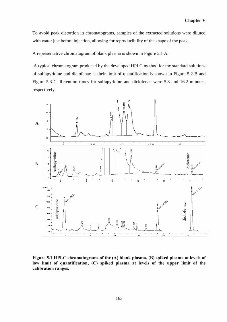
Chapter V
163
To avoid peak distortion in chromatograms, samples of the extracted solutions were diluted
with water just before injection, allowing for reproducibility of the shape of the peak.
A representative chromatogram of blank plasma is shown in Figure 5.1 A.
A typical chromatogram produced by the developed HPLC method for the standard solutions
of sulfapyridine and diclofenac at their limit of quantification is shown in Figure 5.2-B and
Figure 5.3-C. Retention times for sulfapyridine and diclofenac were 5.8 and 16.2 minutes,
respectively.
Figure 5.1 HPLC chromatograms of the (A) blank plasma, (B) spiked plasma at levels of low limit of quantification, (C) spiked plasma at levels of the upper limit of the calibration ranges.
sulf
apy
rid
ine
diclofenac
sulf
apy
rid
ine
dic
lofe
nac
B
A
C
mAU
mAU mAU
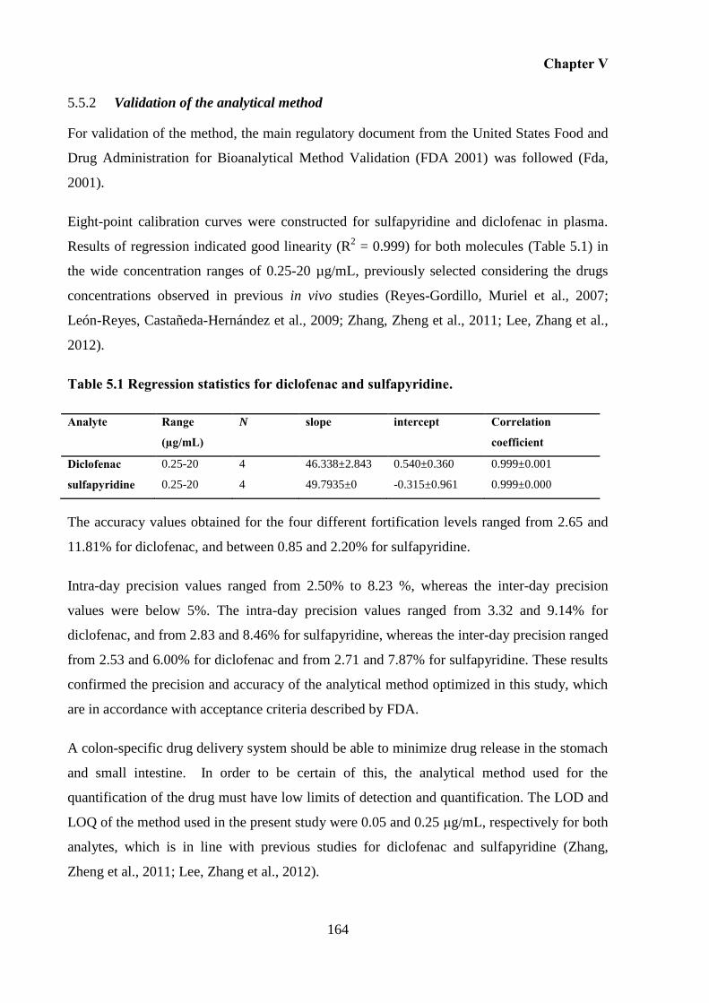
Chapter V
164
Validation of the analytical method 5.5.2
For validation of the method, the main regulatory document from the United States Food and
Drug Administration for Bioanalytical Method Validation (FDA 2001) was followed (Fda,
2001).
Eight-point calibration curves were constructed for sulfapyridine and diclofenac in plasma.
Results of regression indicated good linearity (R2 = 0.999) for both molecules (Table 5.1) in
the wide concentration ranges of 0.25-20 µg/mL, previously selected considering the drugs
concentrations observed in previous in vivo studies (Reyes-Gordillo, Muriel et al., 2007;
León-Reyes, Castañeda-Hernández et al., 2009; Zhang, Zheng et al., 2011; Lee, Zhang et al.,
2012).
Table 5.1 Regression statistics for diclofenac and sulfapyridine.
Analyte Range
(µg/mL)
N slope intercept Correlation
coefficient
Diclofenac 0.25-20 4 46.338±2.843 0.540±0.360 0.999±0.001
sulfapyridine 0.25-20 4 49.7935±0 -0.315±0.961 0.999±0.000
The accuracy values obtained for the four different fortification levels ranged from 2.65 and
11.81% for diclofenac, and between 0.85 and 2.20% for sulfapyridine.
Intra-day precision values ranged from 2.50% to 8.23 %, whereas the inter-day precision
values were below 5%. The intra-day precision values ranged from 3.32 and 9.14% for
diclofenac, and from 2.83 and 8.46% for sulfapyridine, whereas the inter-day precision ranged
from 2.53 and 6.00% for diclofenac and from 2.71 and 7.87% for sulfapyridine. These results
confirmed the precision and accuracy of the analytical method optimized in this study, which
are in accordance with acceptance criteria described by FDA.
A colon-specific drug delivery system should be able to minimize drug release in the stomach
and small intestine. In order to be certain of this, the analytical method used for the
quantification of the drug must have low limits of detection and quantification. The LOD and
LOQ of the method used in the present study were 0.05 and 0.25 μg/mL, respectively for both
analytes, which is in line with previous studies for diclofenac and sulfapyridine (Zhang,
Zheng et al., 2011; Lee, Zhang et al., 2012).
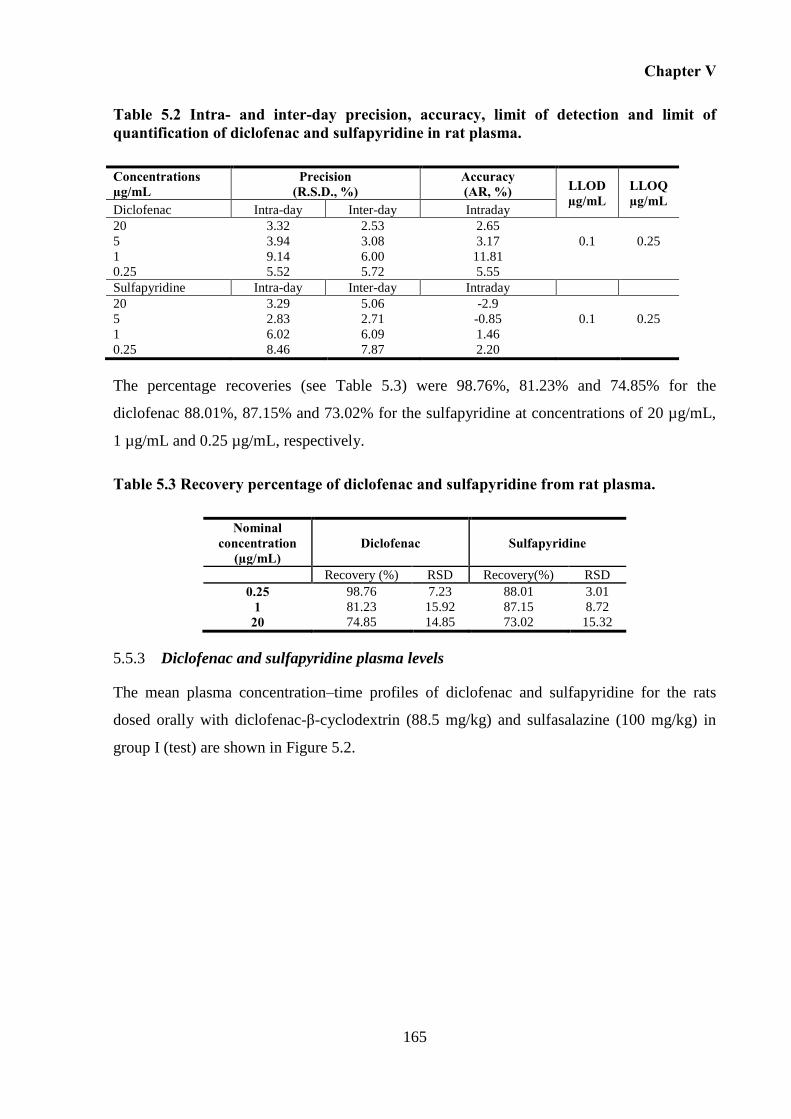
Chapter V
165
Table 5.2 Intra- and inter-day precision, accuracy, limit of detection and limit of quantification of diclofenac and sulfapyridine in rat plasma.
Concentrations µg/mL
Precision (R.S.D., %)
Accuracy (AR, %) LLOD
µg/mL LLOQ µg/mL
Diclofenac Intra-day Inter-day Intraday
20 3.32 2.53 2.65
5 3.94 3.08 3.17 0.1 0.25
1 9.14 6.00 11.81
0.25 5.52 5.72 5.55
Sulfapyridine Intra-day Inter-day Intraday
20 3.29 5.06 -2.9
5 2.83 2.71 -0.85 0.1 0.25
1 6.02 6.09 1.46
0.25 8.46 7.87 2.20
The percentage recoveries (see Table 5.3) were 98.76%, 81.23% and 74.85% for the
diclofenac 88.01%, 87.15% and 73.02% for the sulfapyridine at concentrations of 20 µg/mL,
1 µg/mL and 0.25 µg/mL, respectively.
Table 5.3 Recovery percentage of diclofenac and sulfapyridine from rat plasma.
Nominal concentration
(µg/mL) Diclofenac Sulfapyridine
Recovery (%) RSD Recovery(%) RSD
0.25 98.76 7.23 88.01 3.01
1 81.23 15.92 87.15 8.72
20 74.85 14.85 73.02 15.32
Diclofenac and sulfapyridine plasma levels 5.5.3
The mean plasma concentration–time profiles of diclofenac and sulfapyridine for the rats
dosed orally with diclofenac-β-cyclodextrin (88.5 mg/kg) and sulfasalazine (100 mg/kg) in
group I (test) are shown in Figure 5.2.
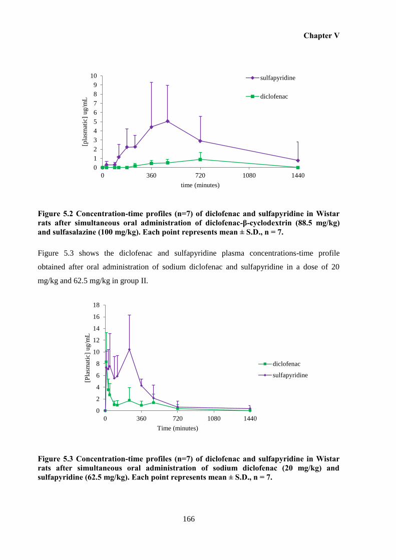
Chapter V
166
0
1
2
3
4
5
6
7
8
9
10
0 360 720 1080 1440
[pla
smat
ic]
ug/m
L
time (minutes)
sulfapyridine
diclofenac
Figure 5.2 Concentration-time profiles (n=7) of diclofenac and sulfapyridine in Wistar rats after simultaneous oral administration of diclofenac-β-cyclodextrin (88.5 mg/kg) and sulfasalazine (100 mg/kg). Each point represents mean ± S.D., n = 7.
Figure 5.3 shows the diclofenac and sulfapyridine plasma concentrations-time profile
obtained after oral administration of sodium diclofenac and sulfapyridine in a dose of 20
mg/kg and 62.5 mg/kg in group II.
0
2
4
6
8
10
12
14
16
18
0 360 720 1080 1440
[Pla
smat
ic]
ug/m
L
Time (minutes)
diclofenac
sulfapyridine
Figure 5.3 Concentration-time profiles (n=7) of diclofenac and sulfapyridine in Wistar rats after simultaneous oral administration of sodium diclofenac (20 mg/kg) and sulfapyridine (62.5 mg/kg). Each point represents mean ± S.D., n = 7.
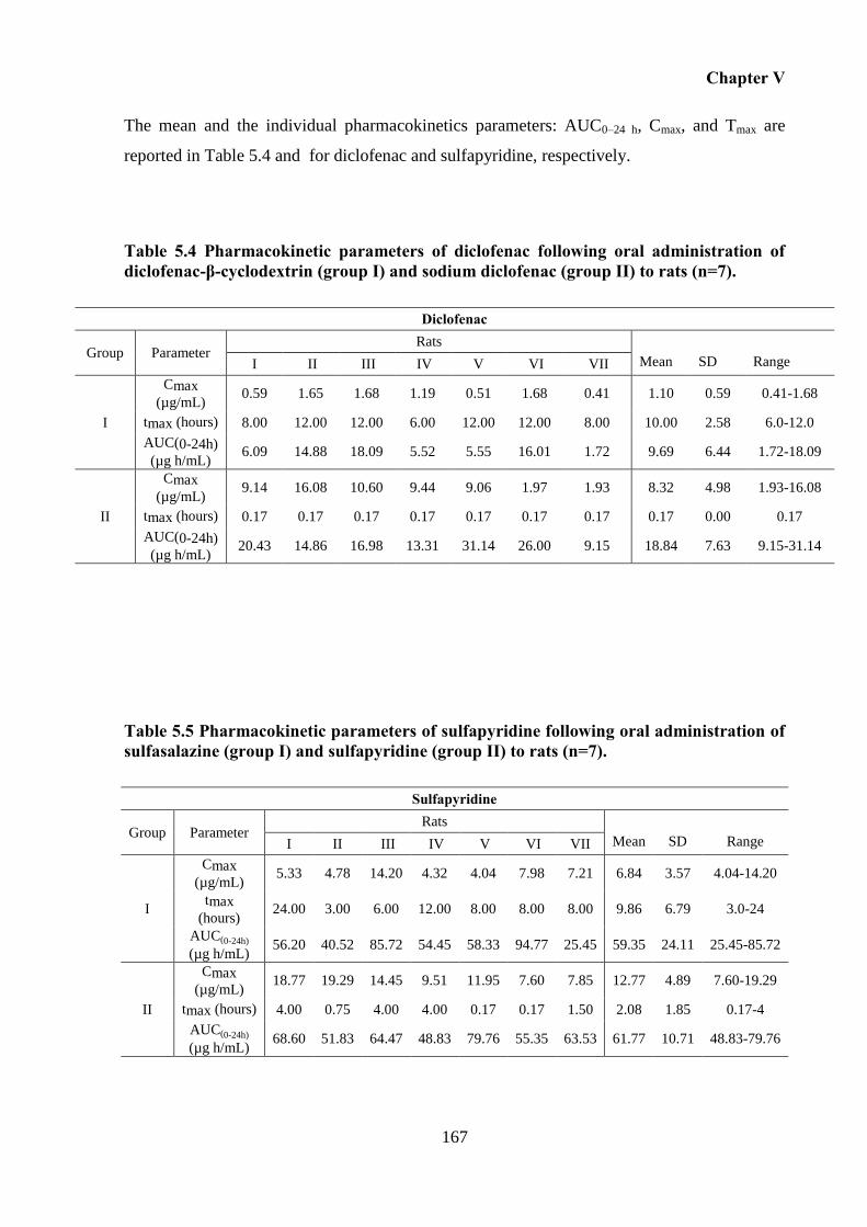
Chapter V
167
The mean and the individual pharmacokinetics parameters: AUC0–24 h, Cmax, and Tmax are
reported in Table 5.4 and for diclofenac and sulfapyridine, respectively.
Table 5.4 Pharmacokinetic parameters of diclofenac following oral administration of diclofenac-β-cyclodextrin (group I) and sodium diclofenac (group II) to rats (n=7).
Diclofenac
Group Parameter Rats
I II III IV V VI VII Mean SD Range
I
Cmax
(µg/mL) 0.59 1.65 1.68 1.19 0.51 1.68 0.41 1.10 0.59 0.41-1.68
tmax (hours) 8.00 12.00 12.00 6.00 12.00 12.00 8.00 10.00 2.58 6.0-12.0
AUC(0-24h)
(µg h/mL) 6.09 14.88 18.09 5.52 5.55 16.01 1.72 9.69 6.44 1.72-18.09
II
Cmax
(µg/mL) 9.14 16.08 10.60 9.44 9.06 1.97 1.93 8.32 4.98 1.93-16.08
tmax (hours) 0.17 0.17 0.17 0.17 0.17 0.17 0.17 0.17 0.00 0.17
AUC(0-24h)
(µg h/mL) 20.43 14.86 16.98 13.31 31.14 26.00 9.15 18.84 7.63 9.15-31.14
Table 5.5 Pharmacokinetic parameters of sulfapyridine following oral administration of sulfasalazine (group I) and sulfapyridine (group II) to rats (n=7).
Sulfapyridine
Group Parameter Rats
I II III IV V VI VII Mean SD Range
I
Cmax
(µg/mL) 5.33 4.78 14.20 4.32 4.04 7.98 7.21 6.84 3.57 4.04-14.20
tmax
(hours) 24.00 3.00 6.00 12.00 8.00 8.00 8.00 9.86 6.79 3.0-24
AUC(0-24h)
(µg h/mL) 56.20 40.52 85.72 54.45 58.33 94.77 25.45 59.35 24.11 25.45-85.72
II
Cmax
(µg/mL) 18.77 19.29 14.45 9.51 11.95 7.60 7.85 12.77 4.89 7.60-19.29
tmax (hours) 4.00 0.75 4.00 4.00 0.17 0.17 1.50 2.08 1.85 0.17-4
AUC(0-24h)
(µg h/mL) 68.60 51.83 64.47 48.83 79.76 55.35 63.53 61.77 10.71 48.83-79.76
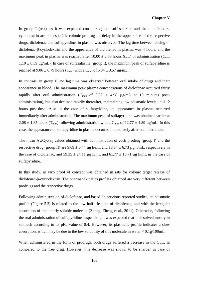
Chapter V
168
In group I (test), as it was expected considering that sulfasalazine and the diclofenac-β-
cyclodextrin are both specific colonic prodrugs, a delay in the appearance of the respective
drugs, diclofenac and sulfapyridine, in plasma was observed. The lag time between dosing of
diclofenac-β-cyclodextrin and the appearance of diclofenac in plasma was 4 hours, and the
maximum peak in plasma was reached after 10.00 ± 2.58 hours (tmax) of administration (Cmax
1.10 ± 0.59 μg/mL). In case of sulfasalazine (group I), the maximum peak of sulfapyridine is
reached at 9.86 ± 6.79 hours (tmax) with a Cmax of 6.84 ± 3.57 μg/mL.
In contrast, in group II, no lag time was observed between oral intake of drugs and their
appearance in blood. The maximum peak plasma concentrations of diclofenac occurred fairly
rapidly after oral administration (Cmax of 8.32 ± 4.98 μg/mL at 10 minutes post-
administration), but also declined rapidly thereafter, maintaining low plasmatic levels until 12
hours post-dose. Also in the case of sulfapyridine, its appearance in plasma occurred
immediately after administration. The maximum peak of sulfapyridine was obtained earlier at
2.08 ± 1.85 hours (Tmax) following administration with a Cmax of 12.77 ± 4.89 µg/mL. In this
case, the appearance of sulfapyridine in plasma occurred immediately after administration.
The mean AUC(0-24h) values obtained with administration of each prodrug (group I) and the
respective drug (group II) are 9.69 ± 6.44 µg h/mL and 18.84 ± 6.73 µg h/mL, respectively in
the case of diclofenac, and 59.35 ± 24.11 µg h/mL and 61.77 ± 10.71 µg h/mL in the case of
sulfapyridine.
In this study, in vivo proof of concept was obtained in rats for colonic target release of
diclofenac-β-cyclodextrin. The pharmacokinetics profiles obtained are very different between
prodrugs and the respective drugs.
Following administration of diclofenac, and based on previous reported studies, its plasmatic
profile (Figure 5.3) is related to the low half-life time of diclofenac, and with the irregular
absorption of this poorly soluble molecule (Zhang, Zheng et al., 2011). Otherwise, following
the oral administration of sulfapyridine suspension, it was expected that it dissolved mostly in
stomach according to its pKa value of 8.4. However, its plasmatic profile indicates a slow
absorption, which may be due to the low solubility of this molecule in water < 0.1g/100mL.
When administered in the form of prodrugs, both drugs suffered a decrease in the Cmax, as
compared to the free drug. However, this decrease was shown to be sharper in case of
Chapter V
169
diclofenac. These results showed a decrease of 87% in the maximum plasma diclofenac
concentration (t-test, p < 0.01), and a significant increase in the mean time required to reach
the maximum level (t-test, p < 0.01) following diclofenac-β-cyclodextrin administration
compared with diclofenac sodium administration. There was 53% decrease in the maximum
plasma sulfapyridine concentration (t-test, p < 0.05) and a significant increase in the mean
time required to reach the maximum level (t-test, p < 0.05) following sulfasalazine
administration relative to sulfapyridine administration.
Although the plasmatic tmax of drugs after administration of prodrugs is not significantly
different for sulfapyridine and diclofenac (t-test, p > 0.05), the lag time for the appearance of
diclofenac in blood was longer than that for sulfapyridine. This fast appearance of
sulfapyridine was in agreement with results reported previously that had shown that
appearance of sulfapyridine in plasma was rapid (~5 min) following direct instillation of
sulfasalazine in the caecum in the fasted state in humans (Kellow, Borody et al., 1986). It
could be argued that these results are also in accordance with in vitro ones that had shown the
slow rate degradation of diclofenac-β-cyclodextrin when compared with sulfasalazine
(Chapter III and IV).
Relative to the AUC0-24h, there was no significant change in bioavailability for sulfapyridine
(t-test, p > 0.05) between groups I and II, though the same was not observed for diclofenac. In
the case of diclofenac, after administration of the diclofenac-β-cyclodextrin the bioavailability
was about half of the bioavailability when the free drug was administrated (t-test p < 0.05).
Different explanations can be given for the disparity of AUC0-24h between both prodrugs
(Figure 5.4), including the slow rate of degradation of diclofenac-β-cyclodextrin as compared
with sulfasalazine at the level of the large intestine in rats, as shown in previous in vitro
studies (Chapter III and IV). This slow degradation can be associated with the small volume
of watery environment necessary to guarantee the dissolution of the diclofenac-β-cyclodextrin
and release of diclofenac to the lumen of the colon for subsequent absorption (Gleiter,
Antonin et al., 1985; Schiller, Fröhlich et al., 2005). The quantity of free water available in
gut contents is otherwise too small compared with the quantity present in the in vitro media
used to perform stability experiments.
Chapter V
170
Figure 5.4 Schematic representation of the hipothesis that can explain the diminuished of plasmatic bioavailability of diclofenac when this is administered in the form of cyclodextrin prodrug.
Otherwise, it appears that the residence time of the diclofenac-β-cyclodextrin and/ or
diclofenac in colonic contents of rats is insufficient for the diclofenac-β-cyclodextrin to be
completely metabolized and/or for the drug released to move to the lumen and to be absorbed.
Rats have a large caecum, though the colon is neither sacculated nor long, unlike in humans.
(Kararli, 1995). Indeed, humans have a poorly define caecal region, which is continuous with
the colon. Additionally, the human colon is sacculated and consists of ascending, transverse
and sigmoidal segments. Based on these anatomic differences between rats and humans, a
different bioavailability of diclofenac released from the conjugate is expected between both
species. As reported, the absorption in humans is much faster, and also to a greater extent than
in rats due to these anatomic differences (Desesso and Jacobson, 2001).
In order to clarify the pharmacokinetic parameters, a longer duration of sampling would be
necessary to correctly estimate the extrapolated area under the curve (Yáñez, Remsberg et al.,
2011). According to the literature, as long as the conjugate is completely degraded, diclofenac
will be well absorbed (Gleiter, Antonin et al., 1985; Billa, Yuen et al., 2000; Schiller, Fröhlich
et al., 2005; Tannergren, Bergendal et al., 2008). Thus, as long as the diclofenac-β-
cyclodextrin is completely degraded, the amount of diclofenac released could be absorbed in
its totality.
Colon
Blood
metabolism
absorption
elimination Faeces
Chapter V
171
5.6 Conclusion
These results confirm the in vivo ability of this diclofenac-β-cyclodextrin to target and release
diclofenac in the colon. The lag time of appearance in blood of diclofenac with a delay in the
tmax as observed for sulfapyridine demonstrated the colonic release of diclofenac from the
diclofenac-β-cyclodextrin. However, results demonstrated that the bioavailability of
diclofenac decreases by half when administered in its prodrug form. Contrarily, the
bioavailability of sulfapyridine is not altered when administered in the form of sulfasalazine,
as compared with the administration of free drug. These differences can be associated with
slow metabolism of diclofenac-β-cyclodextrin when comparing with sulfasalazine, or can be
related with limitations on the time necessary for complete absorption of diclofenac.
Overall, this study confirms the in vivo ability of this newly cyclodextrin prodrug to target and
release diclofenac specifically in the colon.

CHAPTER VI
GENERAL DISCUSSION AND FUTURE WORK

Chapter VI
175
6 GENERAL DISCUSSION AND FUTURE WORK
6.1 General Discussion
This dissertation describes the development of a new oral prodrug for colonic specific
delivery of diclofenac. A diclofenac-β-cyclodextrin conjugate was successfully synthetised
and characterised. Additionally, in vitro and in vivo experiments allowed a further
understanding of its behaviour along the GI tract and to assess its potential to target and
release diclofenac specifically into the colon.
The goal of developing a new system to target the colon exploited the idea of utilising
cyclodextrins as a carrier for colonic target delivery. These oligosaccharides, mainly α- and β-
cyclodextrins, remain intact in the upper GI tract and are enzymatically degraded exclusively
at a colonic level. Thus, using cyclodextrin as a carrier, the idea of creating a prodrug for
colon-specific delivery of diclofenac emerged.
This project started with the investigation of a method suitable for the synthesis of diclofenac-
cyclodextrin conjugates. This became a big challenge due to the high amount of unsuccessful
results faced whilst exploring conventional synthetic routes that in theory would be able to
create an ester linkage between diclofenac and cyclodextrin. Beyond all the reactive hydroxyl
groups of cyclodextrin that are available to react, diclofenac was shown to be very sensitive
and easily suffered an intramolecular reaction, which limits its intermolecular reactivity.
After, the study of different conventional approaches using different conditions, the
nucleophilic route performed under microwave irradiation allowed for the successful and
efficient production of the diclofenac-β-cyclodextrin conjugate. This process assured
reproducible synthesis of the conjugate with the advantage of drastically decreasing the
reaction time when compared to the conventional eating method. The novelty and innovation
associated with this process of preparation of a diclofenac-cyclodextrin conjugate prompted
the need for protecting the intellectual property of this work by a patent.
After overcoming the first challenge related to the synthesis process, the next step concerned
carrying out studies which demonstrated the ability of a cyclodextrin to remain intact in the
upper GI tract and carry a drug to the colon when both were covalently linked. Different
Chapter VI
176
stability studies were designed and performed, namely using simulated gastrointestinal fluids
and gastrointestinal fluids collected from different animals (rat, rabbit and pig). The prodrug
demonstrated resistance to both chemical and enzymatic degradation in the upper GI tract.
Otherwise, at the level of the lower intestine, the conjugate suffered degradation with
simultaneous release of diclofenac. Parallel studies with sulfasalazine were carried out in
order to compare its performance with diclofenac-β-cyclodextrin. Results demonstrated that
sulfasalazine metabolism was always faster than the metabolism of the conjugate. This leads
to the conclusion that the mechanism subjacent to sulfasalazine degradation is faster than the
mechanism that allows for release of a drug from a cyclodextrin.
The slowest release of the drug from the cyclodextrin prodrug can be attributed to the fact that
this conjugate requires the intervention of two different enzymes: amylases and esterases.
Although we have an ester prodrug, this is not degraded by esterases in the upper GI tract. It
seems that cyclodextrin creates a steric hinderance around the ester linkage, which prevents
esterase to attack. Firstly, cyclodextrin has to be degraded, which just occurs at colonic level,
and after that, enough space is generated allowing esterase to attack with consequent
breakdown of the ester linkage, which coincides with the release of diclofenac.
Thereafter, studies were conducted in an attempt to understand the influence of feeding
regimen on the physiological conditions and metabolism of colonic prodrugs. Studies have
previously shown that different feeding regimens affect dramatically the physiological
conditions in the gastro-intestinal tract. These observations have therefore an influence on the
metabolism of pro-drugs and subsequently expected to interfere with the drug disposition. In
fact, the metabolism of diclofenac-β-cyclodextrin was faster in fluids collected from fasted
rats. The ideal colonic prodrug would present a rate of metabolism not affected by the
changes on the microbiota activity; in other words, its metabolism would not be affected by
the presence or absence of food.
This thesis ended with in vivo studies conducted to show how the conjugate behaves after oral
administration in rats, using sulfapyridine as a marker of colonic absorption. These studies
represented a proof of concept for the colonic target delivery of diclofenac-β-cyclodextrin,
and release of diclofenac. This was evidenced by the lag time observed for diclofenac
appearance in plasma after oral delivery of conjugate, similar to that observed for
sulfapyridine after administration of sulfasalazine. Comparison of the plasmatic
Chapter VI
177
bioavailability of diclofenac after oral administration of the prodrug and the bioavailability
after oral intake of the own drug showed that the plasmatic bioavailability is reduced to half
when diclofenac was administered in the conjugate form.
Therefore, these results led to the conclusion that diclofenac-β-cyclodextrin allows for release
of diclofenac at a colonic level. Although diclofenac is well absorbed, some factors are
present which prevent its complete absorption. The decrease of plasmatic concentration can
be associated with excretion of the intact conjugate, and therefore its incomplete metabolism.
Additionally, at colonic level, the viscous contents/mucous with small volumes of fluid create
difficulty for diclofenac dissolution, and consequent difficulty in reaching the colonic mucous
to be further absorbed. These hypotheses need to be explored in order to assess the reason for
the different blood concentration between drug and prodrug.
Overall, this work provided a proof of concept of the ability of diclofenac-β-cyclodextrin to
release diclofenac at a colonic level. Given the short half life time of diclofenac in plasma,
this prodrug can be considered as an useful chronopharmacological therapeutic agent for the
management of arthritis pain. Moreover, the concomitant formulation of this prodrug with
free diclofenac and its oral administration assures sustained therapeutic blood levels of
diclofenac, avoiding the repeated daily dosing of diclofenac.
It is expected that this anti-inflammatory molecule in the form of a colonic prodrug causes
less mucosal irritation when compared with the free drug, due to its lower direct contact with
the mucous. This can be expected due to its low affinity for the lipidic membranes, not only
due to the large size of the molecule, but also due to the hydrophilic nature of the cyclodextrin
surface.
Moreover, the cyclodextrin carrier brings health benefits related to its fermentation that
produce short-chain fatty acids (SCFA) - important for the maintenance of the health and
integrity of the colonic epithelium.
6.2 Future work
Whilst carrying out this Project, a number of ideas were identified and can be further
investigated. Some ideas are the direct result of the conducted research described in this thesis
that are related to development of the synthetic process for producing diclofenac-cyclodextrin
Chapter VI
178
conjugates and investigation of its performance after oral intake as a colonic-specific prodrug.
The last ideas are associated with the potential pharmacological advantages.
Potential topics to further develop are outlined below:
Although diclofenac-β-cyclodextrin was produced using a reliable method with a high degree
of purification, more needs to be done in order to optimize its yield. The first idea that arises
is the exploration of the possibility of purification of the intermediate tosylated cyclodextrin
product. Additionally, would be interesting to explore one way to scale up the reaction and the
product purification. Preliminary experiments using the Biotage system showed to be an
efficient automatic method for purification.
Since dimethylformamide (DMF) is a protic solvent used to perform the nucleophilic
reaction, its use should ideally be avoided on account of its toxicity. Thus, the use of other
solvents less toxic than DMF on the performance of the nucleophilic reaction should be
investigated further. Preliminary experiments with an aprotic solvent, PEG-200, proved to be
a suitable solvent to produce the conjugate.
Since this new process of synthesis takes advantage of microwaves and allows the synthesis
of the conjugate in a short period of time compared with the conventional approach, this
process could be adapted for the synthesis of other cyclodextrin conjugates.
Improvement of the synthesis process of the α- and γ- cyclodextrin tosylated derivatives and
access of the right conditions to synthetize the respective diclofenac conjugates.
Diclofenac was shown to be a highly reactive molecule by an intramolecular reaction
mechanism, and which suffers modifications according to the pH change. HPLC analysis of
the diclofenac released from the conjugate and also of the diclofenac after incubation in
different buffer media revealed the appearance of extra peaks on the HPLC chromatogram.
These extra peaks need to be accurately analysed by LC-MS in order to clarify its origin.
Further investigation into the stability of the conjugate in gastrointestinal fluids from other
animal models such as monkeys, guinea pigs, or dogs using a greater number of subjects per
species, may also be used. This will also allowed assessing which animal model gives a better
prediction of the human behaviour. Also, the possible gender differences on the metabolism
Chapter VI
179
of the conjugate should be investigated, as the studies presented here only included male
species.
To better predict the behaviour of the conjugate in humans, it is also essential to assess its
stability in human gastrointestinal fluids, which should involve a reasonable number of
individuals (including male and female). Moreover, the metabolism can be accessed in fluids
collected from individuals with colonic diseases in order to assess its performance.
Investigate the causes associated with the decrease in bioavailability of diclofenac when
administered as the conjugate form in rats. Ideally, all faeces produced after oral intake of
diclofenac-β-cyclodextrin should be collected, and the existence/absence of conjugate and/or
diclofenac determined. Also, at the end of the study (after 24 hours of oral intake),
gastrointestinal fluids should be collected and evaluated for the presence of diclofenac and/or
conjugate. Also, the conjugate can be radiolabelled to easily follow its transit through the
gastrointestinal tract after oral intake.
Evaluation of the acute and chronic ulcerogenicity of diclofenac-β-cyclodextrin versus
diclofenac at the level of upper GI tract (stomach and small intestine), and also at the level of
the lower GI tract (colon) using the rat as an animal model. Study design for the in vivo
determination of anti-inflammatory activity of diclofenac-β-cyclodextrin versus diclofenac
using the carrageenan induced rat paw edema model may additionally be carried out.
A deeper understanding of the pharmacological effect of this conjugate is necessary, including
its potential to be used as a chronotherapeutic agent for rheumatic arthritis.
Assess other potential targets (colonic cancer).
Development of an appropriate formulation (capsule or tablets) of diclofenac-β-cyclodextrin.

Chapter VI
180

APPENDIX

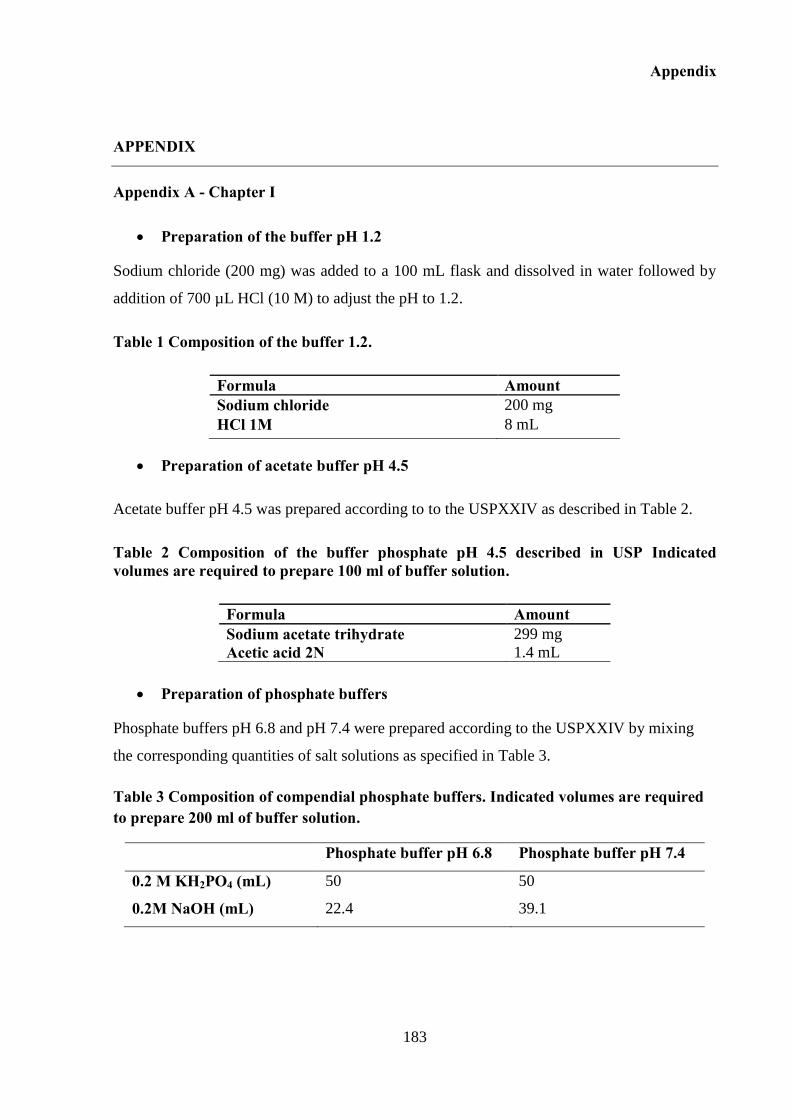
Appendix
183
APPENDIX
Appendix A - Chapter I
Preparation of the buffer pH 1.2
Sodium chloride (200 mg) was added to a 100 mL flask and dissolved in water followed by
addition of 700 µL HCl (10 M) to adjust the pH to 1.2.
Table 1 Composition of the buffer 1.2.
Formula Amount Sodium chloride 200 mg
HCl 1M 8 mL
Preparation of acetate buffer pH 4.5
Acetate buffer pH 4.5 was prepared according to to the USPXXIV as described in Table 2.
Table 2 Composition of the buffer phosphate pH 4.5 described in USP Indicated volumes are required to prepare 100 ml of buffer solution.
Formula Amount Sodium acetate trihydrate 299 mg
Acetic acid 2N 1.4 mL
Preparation of phosphate buffers
Phosphate buffers pH 6.8 and pH 7.4 were prepared according to the USPXXIV by mixing
the corresponding quantities of salt solutions as specified in Table 3.
Table 3 Composition of compendial phosphate buffers. Indicated volumes are required to prepare 200 ml of buffer solution.
Phosphate buffer pH 6.8 Phosphate buffer pH 7.4
0.2 M KH2PO4 (mL) 50 50
0.2M NaOH (mL) 22.4 39.1
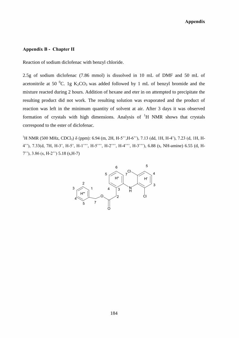
Appendix
184
Appendix B - Chapter II
Reaction of sodium diclofenac with benzyl chloride.
2.5g of sodium diclofenac (7.86 mmol) is dissolved in 10 mL of DMF and 50 mL of
acetonitrile at 50 0C. 1g K2CO3 was added followed by 1 mL of benzyl bromide and the
mixture reacted during 2 hours. Addition of hexane and eter in on attempted to precipitate the
resulting product did not work. The resulting solution was evaporated and the product of
reaction was left in the minimum quantity of solvent at air. After 3 days it was observed
formation of crystals with high dimensions. Analysis of 1H NMR shows that crystals
correspond to the ester of diclofenac.
1H NMR (500 MHz, CDCl3) δ (ppm): 6.94 (m, 2H, H-5’’,H-6’’), 7.13 (dd, 1H, H-4’), 7.23 (d, 1H, H-
4’’), 7.33(d, 7H, H-3’, H-5’, H-1’’’, H-5’’’, H-2’’’, H-4’’’, H-3’’’), 6.88 (s, NH-amine) 6.55 (d, H-
7’’), 3.86 (s, H-2’’) 5.18 (s,H-7)
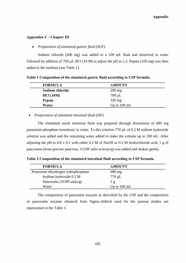
Appendix
185
Appendice C - Chapter III
Preparation of simulated gastric fluid (SGF)
Sodium chloride (200 mg) was added to a 100 mL flask and dissolved in water
followed by addition of 700 µL HCl (10 M) to adjust the pH to 1.2. Pepsin (320 mg) was then
added to the medium (see Table 1).
Table 1 Composition of the simulated gastric fluid according to USP formula.
FORMULA AMOUNT Sodium chloride 200 mg
HCl (10M) 700 µL
Pepsin 320 mg
Water Up to 100 mL
Preparation of simulated intestinal fluid (SIF)
The simulated small intestinal fluid was prepared through dissolution of 680 mg
potassium phosphate monobasic in water. To this solution 770 µL of 0.2 M sodium hydroxide
solution was added and the remaining water added to make the volume up to 100 mL. After
adjusting the pH to 6.8 ± 0.1 with either 0.2 M of NaOH or 0.2 M hydrochloride acid, 1 g of
pancreatin (from porcine pancreas, 3 USP units activity/g) was added and shaken gently.
Table 2 Composition of the simulated intestinal fluid according to USP formula.
FORMULA AMOUNT Potassium dihydrogen orthophosphate 680 mg
Sodium hydroxide 0.2 M 770 µL
Pancreatin (3USP units/g) 1 g
Water Up to 100 mL
The composition of pancreatin enzyme as described by the USP and the composition
of pancreatin enzyme obtained from Sigma-Aldrich used for the present studies are
represented in the Table 3.
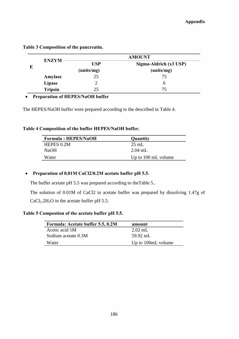
Appendix
186
Table 3 Composition of the pancreatin.
ENZYME
AMOUNT USP
(units/mg) Sigma-Aldrich (x3 USP)
(units/mg)
Amylase 25 75
Lipase 2 6
Tripsin 25 75
Preparation of HEPES/NaOH buffer
The HEPES/NaOH buffer were prepared according to the described in Table 4.
Table 4 Composition of the buffer HEPES/NaOH buffer.
Formula : HEPES/NaOH Quantity HEPES 0.2M 25 mL
NaOH 2.04 mL
Water Up to 100 mL volume
Preparation of 0.01M CaCl2/0.2M acetate buffer pH 5.5.
The buffer acetate pH 5.5 was prepared according to theTable 5..
The solution of 0.01M of CaCl2 in acetate buffer was prepared by dissolving 1.47g of
CaCl2.2H2O in the acetate buffer pH 5.5.
Table 5 Compostion of the acetate buffer pH 5.5.
Formula: Acetate buffer 5.5, 0.2M amount Acetic acid 1M 2.02 mL
Sodium acetate 0.3M 59.92 mL
Water Up to 100mL volume
Appendix
187
Appendix D - Chapter IV
Determination of amylase activity using the EnzCheck Ultra amylase Assay Kit
α-Amylase activity in gastrointestinal fluids was determined by the EnzChek Ultra Amylase
Assay Kit (E33651).
Materials
The EnzChekt Ultra Amylase Assay Kit (E33651) was purchased from Molecular Probes. The
Kit contains:
Substract: DQ starch from corn BODIPY FL conjugate;
Reaction buffer: 0.05 M MOPS (3-(N-morpholino) propanesulfonic acid)), pH 6.9;
Sodium acetate 50mM, pH 4.0;
α-amylase from Bacillus sp. (Sigma A-6380) was purchased from Sigma Aldrich;
Gastrointestinal fluids (stomach, small intestinal, caecum, colon and faecal slurry prepared
from colon fluids) obtained from pig, rat and rabbit.
Methods
A solution of 200 µg/mL subtract in acetate buffer was prepared according to the instructions
described on the kit. A stock solution of α-amylase (400 mM/mL) was prepared in MOPS.
Different standard solutions (0 - 40 mU/mL) were prepared from this stock solution in order
to create an enzyme standard curve between 0 and 20 mU/mL.
For determination of amylase activity, it was use the supernatant obtained after centrifugation
at 10 000 rpm during 10 minutes. Colon fluids were also prepared as faecal slurry (described
in Chapter III). Each fluid was diluted with MOPS buffer (4 µL of fluid to 396 µL of MOPS).
Fluorescence of the digestion products from the substrate was measured by 96 -Plate reader at
an excitation wavelength of 505 nm and an emission wavelength of 512 nm at room
temperature.
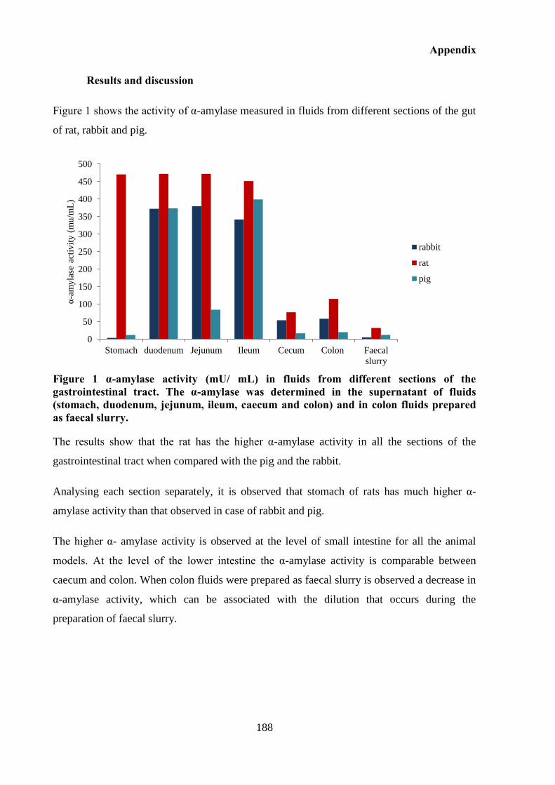
Appendix
188
Results and discussion
Figure 1 shows the activity of α-amylase measured in fluids from different sections of the gut
of rat, rabbit and pig.
Figure 1 α-amylase activity (mU/ mL) in fluids from different sections of the gastrointestinal tract. The α-amylase was determined in the supernatant of fluids (stomach, duodenum, jejunum, ileum, caecum and colon) and in colon fluids prepared as faecal slurry.
The results show that the rat has the higher α-amylase activity in all the sections of the
gastrointestinal tract when compared with the pig and the rabbit.
Analysing each section separately, it is observed that stomach of rats has much higher α-
amylase activity than that observed in case of rabbit and pig.
The higher α- amylase activity is observed at the level of small intestine for all the animal
models. At the level of the lower intestine the α-amylase activity is comparable between
caecum and colon. When colon fluids were prepared as faecal slurry is observed a decrease in
α-amylase activity, which can be associated with the dilution that occurs during the
preparation of faecal slurry.
0
50
100
150
200
250
300
350
400
450
500
Stomach duodenum Jejunum Ileum Cecum Colon Faecal
slurry
α-a
myla
se a
ctiv
ity (
mu/m
L)
rabbit
rat
pig

REFERENCES

191
REFERENCES
Agorama, B., W. S. Woltosza,M. B. Bolgera 2001. Predicting the impact of physiological and
biochemical processes on oral drug bioavailability. Advanced Drug Delivery Reviews 50, Supplement
1, S41-S67.
Akhgaria, A., H. A. Garekania, F. Sadeghia,M. Azimaieb 2005. Statistical optimization of
indomethacin pellets coated with pH-dependent methacrylic polymers for possible colonic drug
delivery. International Journal of Pharmaceutics 305,
Albert, J. S.,A. D. Hamilton 2001. 1,3-Dicyclohexylcarbodiimide–4-Dimethylaminopyridine.
Encyclopedia of Reagents for Organic Synthesis. John Wiley & Sons, Ltd.
Alkazaz, M., V. Desseaux, G. Marchis-Mouren, F. Payan, E. Forest,M. Santimone 2001. Mechanism
of porcine pancreatic α-amylase. Inhibition of amylose and maltopentaose hydrolysis by -α, -β, and λ-
cyclodextrins. European Journal of Biochemistry 268, 841-848.
Ambrogi, V., L. Perioli, M. Ricci, L. Pulcini, M. Nocchetti, S. Giovagnoli,C. Rossi 2008. Eudragit®
and hydrotalcite-like anionic clay composite system for diclofenac colonic delivery. Microporous and
Mesoporous Materials 115, 405-415.
Antenucci, R. N.,J. K. Palmer 1984. Enzymatic degradation of alpha- and beta-cyclodextrins by
Bacteroides of the human colon. Journal of Agricultural and Food Chemistry 32, 1316-1321.
Argenzio, R. A.,M. Southworth 1975. Sites of organic acid production and absorption in
gastrointestinal tract of the pig. The American journal of physiology 228, 454-460.
Ashri, N. Y.,M. Abdel-Rehim 2011. Sample treatment based on extraction techniques in biological
matrices. Bioanalysis 3, 2003-2018.
Astray, G., C. Gonzalez-Barreiro, J. C. Mejuto, R. Rial-Otero,J. Simal-Gándara 2009. A review on the
use of cyclodextrins in foods. Food Hydrocolloid. 23, 1631-1640.
Awati, A., B. A. Williams, M. W. Bosch,M. W. A. Verstegen 2006. Dietary carbohydrates with
different rates of fermentation affect fermentation end-product profiles in different sites of gastro-
intestinal tract of weaning piglet. Animal Science 82, 837-843.
Azad Khan, A. K.,S. C. Truelove 1980. Circulating levels of sulphasalazine and its metabolites and
their relation to the clinical efficacy of the drug in ulcerative colitis. Gut 21, 706-710.
Aziz, Q., J. Doré, A. Emmanuel, F. Guarner,E. M. M. Quigley 2013. Gut microbiota and
gastrointestinal health: current concepts and future directions. Neurogastroenterology & Motility 25,
4-15.
Bach Knudsen, K. E.,I. Hansen 1991. Gastrointestinal implications in pigs of wheat and oat fractions.
British Journal of Nutrition 65, 217-232.
Bakar, M. B., M. Oelgemöller,M. O. Senge 2009. Lead structures for applications in photodynamic
therapy. Part 2: Synthetic studies for photo-triggered release systems of bioconjugate porphyrin
photosensitizers. Tetrahedron 65, 7064-7078.
Basit, A. W. 2005. Advances in Colonic Drug Delivery. Drugs 65, 1991-2007.
192
Basit, A. W.,L. F. Lacey 2001. Colonic metabolism of ranitidine: implications for its delivery and
absorption. International Journal of Pharmaceutics 227, 157-165.
Basit, A. W., J. M. Newton,L. F. Lacey 2002. Susceptibility of the H2-receptor antagonists cimetidine,
famotidine and nizatidine, to metabolism by the gastrointestinal microflora. International Journal of
Pharmaceutics 237, 23-33.
Bauer, E., A. Williams Barbara, W. Bosch Marlou, C. Voigt, R. Mosenthin,W. A. Verstegen Martin
2004. Differences in microbial activity of digesta from three sections of the porcine large intestine
according to in vitro fermentation of carbohydrate-rich substrates. Journal of the Science of Food and
Agriculture 84, 2097-2104.
Bechgaard, H.,G. H. Nielsen 1978. Controlled-Release Multiple-Units and Single-Unit Doses a
Literature Review. Drug Development and Industrial Pharmacy 4, 53-67.
Belenguer, A., S. H. Duncan, G. Holtrop, S. E. Anderson, G. E. Lobley,H. J. Flint 2007. Impact of pH
on Lactate Formation and Utilization by Human Fecal Microbial Communities. Applied And
Environmental Microbiology 73, 6526-6533.
Berggren, S., C. Gall, N. Wollnitz, M. Ekelund, U. Karlbom, J. Hoogstraate, D. Schrenk,H. Lennernäs
2007. Gene and Protein Expression of P-Glycoprotein, MRP1, MRP2, and CYP3A4 in the Small and
Large Human Intestine. Molecular Pharmaceutics 4, 252-257.
Biechea, I., C. Narjozb, T. Asselahc, S. Vachera, P. Marcellinc, R. Lidereaua, P. Beauneb,I. d.
Waziers 2007. Reverse transcriptase-PCR quantification of mRNA levels from cytochrome (CYP)1,
CYP2 and CYP3 families in 22 different human tissues. Pharmacogenetics and Genomics 17, 731-
742.
Billa, N., K.-H. Yuen, M. A. A. Khader,A. Omar 2000. Gamma-scintigraphic study of the
gastrointestinal transit and in vivo dissolution of a controlled release diclofenac sodium formulation in
xanthan gum matrices. International Journal of Pharmaceutics 201, 109-120.
Booth, D. A., E. L. Gibson,B. J. Baker 1986. Gastromotor mechanism of fenfluramine anorexia.
Appetite 7, Supplement, 57-69.
Borde, A. S., E. M. Karlsson, K. Andersson, K. Björhall, H. Lennernäs,B. Abrahamsson 2012.
Assessment of enzymatic prodrug stability in human, dog and simulated intestinal fluids. European
Journal of Pharmaceutics and Biopharmaceutics 80, 630-637.
Bovera, F., S. D’Urso, C. D. Meo, G. Piccolo, S. Calabrò,A. Nizza 2006. Comparison of Rabbit
Caecal Content and Rabbit Hard Faeces as Source of Inoculum for the In vitro Gas Production
Technique. Asian Australian Journal of Animal Sciences 11, 1649-1657.
Bovera, F., S. D’Urso, C. D. Meo, R. Tudisco,A. Nizza 2009. A model to assess the use of caecal and
faecal inocula to study fermentability of nutrients in rabbit. Journal of Animal Physiology and Animal
Nutrition 93, 147-156.
Brady, B., N. Lynam, T. O'Sullivan, C. Ahern,R. Darcy 2000. 6A-O-p-toluenosulfonyl-ß-cyclodextrin.
Organic Syntheses 77, 220.
Brown, N. J., A. Greenburgh,J. Tomlin 1994. The effects of pectin and wheat bran on the distribution
of a meal in the gastrointestinal tract of the rat. British Journal of Nutrition 72, 289-297.
193
Buggé, C. J. L., S. R. Gautam, L. E. Parke, J. T. Mason,D. B. Garcia 1990. Simultaneous
Determination of Sulfasalazine and Its Metabolites Sulfapyridine and N-AcetylsuIfapyridine in
Human Serum by Ion-Pair High-Performance Liquid Chromatography Using a Polymer-Based
Column. Journal of Pharmaceutical Sciences 79, 1095-1098.
Butine, T. J.,J. A. Leedle 1989. Enumeration of selected anaerobic bacterial groups in cecal and
colonic contents of growing-finishing pigs. Applied and Environmental Microbiology 55, 1112-1116.
Calatayud, S., T. D. Warner, E. J. Breese,J. A. Mitchell 2001. Relationship between endogenous
colony stimulating factors and apoptosis in human colon cancer cells: role of cyclo-oxygenase
inhibitors. British Journal of Pharmacology 134, 1237-1244.
Cassano, R., S. Trombino, T. Ferrarelli, E. Barone, V. Arena, C. Mancuso,N. Picci 2010. Synthesis,
Characterization, and Anti-Inflammatory Activity of Diclofenac-Bound Cotton Fibers.
Biomacromolecules. 11, 1716-1720.
Castellari, C.,S. Ottani 1997. Two Monoclinic Forms of Diclofenac Acid. Acta Crystallographica
Section C 53, 794-797.
Chan, K. H., P. Mojaverian, B. Ziehmer,V. John 1990. Application of Radiotelemetric Technique in
Evaluating Diclofenac Sodium Absorption After Oral Administration of Various Dosage Forms in
Healthy Volunteers. Pharmaceutical Research 7, 1026-1032.
Chang, C., Z.-C. Wang, C.-Y. Quan, H. Cheng, S.-X. Cheng, X.-Z. Zhang,R.-X. Zhuo 2007.
Fabrication of a novel pH-sensitive glutaraldehyde cross-linked pectin nanogel for drug delivery.
Journal of Biomaterials Science, Polymer Edition 18, 1591-1599.
Chassard, C.,C. Lacroix 2013. Carbohydrates and the human gut microbiota. Current Opinion in
Clinical Nutrition & Metabolic Care 16, 453-460.
Cheng, G., F. An, M.-J. Zou, J. Sun, X.-H. Hao,Y.-X. He 2004. Time- and pH-dependent colon-
specific drug delivery for orally administered diclofenac sodium and 5-aminosalicylic acid World
Journal of Gastroenterology 10, 1769-1774.
Chmielewska, A., L. Konieczna, A. Plenis, M. Bieniecki,H. Lamparczyk 2006. Determination of
diclofenac in plasma by high-performance liquid chromatography with electrochemical detection.
Biomedical chromatography 20, 119-124.
Chourasia, M. K.,S. K. Jain 2003. Pharmaceutical approaches to colon targeted drug delivery systems.
Journal of Pharmaceutical Sciences 6, 33-66.
Christensen, D. N., K. E. B. Knudsen, J. Wolstrup,B. B. Jensen 1999. Integration of ileum cannulated
pigs and in vitro fermentation to quantify the effect of diet composition on the amount of short-chain
fatty acids available from fermentation in the large intestine. Journal of the Science of Food and
Agriculture 79, 755-762.
Chungi, V. s., L. W. Dittert,L. Shargel 1989 Pharmacokinetics of sulfasalazine metabolites in rats
following concomitant oral administration of riboflavin. Pharmaceutical Research 6, 1067-1072.
Chungi, V. S., G. Rekhi,L. Shargel 1989. A Simple and Rapid Liquid Chromatographic Method for
the Determination of Major Metabolites of Sulfasalazine in Biological Fluids. Journal of
Pharmaceutical Sciences 78, 235-238.
194
Coates, J. H., C. J. Easton, S. J. v. Eyk, B. L. May, P. Singh,S. F. Lincoln 1991. Chiral Differentiation
in the Deacylation of 6A- 0- {(2-[4-(2-MethylpropyI)phenyl]propanoyl}-β-cyclodextrin Journal of the
Chemical Society, Chemical Communications 759-760.
Coates, J. H., C. J. Easton, N. L. Fryer,S. F. Lincoln 1994. Complementary Diastereoselectivity in the
synthesis and hydrolysis of acylated cyclodextrins. Complementary Diastereoselectivity 1153-1156.
Cohen, R. D., G. R. Lichtenstein,A. V. Safdi 2008. 5-ASA Treatment for Ulcerative Colitis: What’s
on the Horizon? Gastroenterology & Hepatology 4, Supplement 24.
Coleman, A., I. Nicolis, N. Keller,J. Dalbiez 1992. Aggregation of cyclodextrins: An explanation of
the abnormal solubility of β-cyclodextrin. Journal of Inclusion Phenomena and Molecular Recognition
in Chemistry 13, 139-143.
Coupe, A., S. Davis,I. Wilding 1991a. Variation in Gastrointestinal Transit of Pharmaceutical Dosage
Forms in Healthy Subjects. Pharmaceutical Research 8, 360-364.
Coupe, A. J., S. s. Davis, D. F. Evans,I. R. Wilding 1991b. Correlation of the gastric emptying of
nondisintegrating tablets with gastrointestinal motility. Pharmaceutical Research 8, 1281-1285.
Coussens, L. M.,Z. Werb 2002. Inflammation and cancer. Nature 420, 860-867.
Cui, N., D. R. Friend,R. N. Fedorak 1994. A budesonide prodrug accelerates treatment of colitis in
rats. Gut 35, 1439-1446.
Cummings, J. H.,G. T. Macfarlane 1991. The control and consequences of bacterial fermentation in
the human colon. Journal of Applied Microbiology 70, 443-459.
Cummings, J. H.,G. T. Macfarlane 1997a. Collaborative JPEN-Clinical Nutrition Scientific
Publications Role of intestinal bacteria in nutrient metabolism. Journal of Parenteral and Enteral
Nutrition 21, 357-365.
Cummings, J. H.,G. T. Macfarlane 1997b. Role of intestinal bacteria in nutrient metabolism. Clinical
Nutrition 16, 3-11.
Cuzick, J., F. Otto, J. A. Baron, P. H. Brown, J. Burn, P. Greenwald, J. Jankowsk, C. L. Vecchia, F.
Meyskens, H. J. Senn,M. Thun 2009. Aspirin and non-steroidal anti-inflammatory drugs for cancer
prevention: an international consensus statement. Lancet Oncology 10, 501-507.
Davies, N. M.,K. E. Anderson 1997. Clinical pharmacokinetics of diclofenac. Therapeutic insights and
pitfalls. Clinical Pharmacokinetics 33, 184-213.
Davies, R. R., CertZooMed,J. A. E. R. Davies 2003. Rabbit gastrointestinal physiology. The
Veterinary Clinics Exotic Animal Practice 6, 139-153.
Davis, S. S., J. G. Hardy,J. W. Fara 1986. Transit of pharmaceutical dosage forms through the small
intestine. Gut 27, 886-892.
Dawson, D. C. 1991. Ion Channels and Colonic Salt Transport. Annual Review of Physiology 53, 321-
340.
DePinto, J. A.,L. L. Campbell 1964. Formation and Degradation of Cyclic Dextrins by Intracellular
Enzymes of Bacillus macerans. Science 146, 1064-1066.
195
DeSesso, J. M.,C. F. Jacobson 2001. Anatomical and physiological parameters affecting
gastrointestinal absorption in humans and rats. Food and Chemical Toxicology 39, 209-228.
Dev, S., D. V. Mhaske, S. S. Kadam,S. R. Dhaneshwar 2007. Synthesis and Pharmacological
evaluation of cyclodextrin conjugate prodrug of mefenamic acid. Indian Journal of Pharmaceutical
Sciences 69, 69-72.
Dhaneshwar, S. S.,G. Vadnerkar 2011. Rational Design and Development of Colon-Specific Prodrugs.
Current Topics in Medicinal Chemistry 11, 2318-2345.
Diakidou, A., M. Vertzoni, K. Goumas, E. Söderlind, B. Abrahamsson, J. Dressman,C. Reppas 2009.
Characterization of the Contents of Ascending Colon to Which Drugs are Exposed After Oral
Administration to Healthy Adults. Pharmaceutical Research 26, 2141-2151.
Digenis, G. A., E. P. Sandefer, A. F. Parr, R. Beihn, C. McClain, B. M. Scheinthal, I. Ghebre-
Sellassie, U. Iyer, R. U. Nesbitt,E. Randinitis 1990. Gastrointestinal Behavior of Orally Administered
Radiolabeled Erythromycin Pellets in Man as Determined by Gamma Scintigraphy. The Journal of
Clinical Pharmacology 30, 621-631.
Djedaini-Pilard, F., N. Azaroual-Bellanger, M. Gosnat, D. Vernet,B. Perly 1995. Potential formation
of intramolecular inclusion complexes in peptido-cyclodextrins as evidenced by NMR spectroscopy.
Journal of the Chemical Society, Perkin Transactions 2 723-730.
Doxsee, D. W., P. W. Gout, T. Kurita, M. Lo, A. R. Buckley, Y. Wang, H. Xue, C. M. Karp, J.-C.
Cutz, G. R. Cunha,Y.-Z. Wang 2007. Sulfasalazine-induced cystine starvation: Potential use for
prostate cancer therapy. The Prostate 67, 162-171.
Dragos Vizitiu, Caroline S. Walkinshaw, Borio I. Gorin,G. R. J. Thatcher 1997. Synthesis of
Monofacially Functionalized Cyclodextrins Bearing Amino Pendent Groups. Journal Organic
Chemistry 62, 8760.
Dressman, J., G. Amidon, C. Reppas,V. Shah 1998. Dissolution Testing as a Prognostic Tool for Oral
Drug Absorption: Immediate Release Dosage Forms. Pharmaceutical Research 15, 11-22.
Dressman, J. B., R. R. Berardi, L. C. Dermentzoglou, T. L. Russell, S. P. Schmaltz, J. L. Barnett,K. M.
Jarvenpaa 1990. Upper Gastrointestinal (GI) pH in Young, Healthy Men and Women. Pharmaceutical
Research 7, 756-761.
Eckburg, P. B., E. M. Bik, C. N. Bernstein, E. Purdom, L. Dethlefsen, M. Sargent, S. R. Gill, K. E.
Nelson,D. A. Relman 2005. Diversity of the Human Intestinal Microbial Flora. Science 308, 1635-
1638.
Edsbäcker, S., B. Bengtsson, P. Larsson, P. Lundin, Å. Nilsson, J. Ulmius,P. Wollmer 2003. A
pharmacoscintigraphic evaluation of oral budesonide given as controlled-release (Entocort) capsules.
Alimentary Pharmacology & Therapeutics 17, 525-536.
Edwards, C. A. 1993. Anatomical and physiological basis: Physiological factors influencing drug
absorption. Colonic Drug Absorption and Metabolism. M. Dekker. New York, P. R. Bieck. . 1-28.
El-Kamel, A. H., A. A.M.Abdel-Aziz, A. J. Fatani,H. I. El-Subbagh 2008. Oral colon targeted delivery
systems for treatment of inflammatory bowel diseases: Synthesis, in vitro and in vivo assessment.
International Journal of Pharmaceutics 358, 248-255.
196
El-Kattan, A.,M. Varma 2012. Oral Absorption, Intestinal Metabolism and Human Oral
Bioavailability. Topics on Drug Metabolism. J. Paxton. James Paxton.
Elder, D. J. E., D. E. Halton, A. Hague,C. Paraskeva 1997 Indication of apoptotic cell death in human
colorectal carcinoma cell lines by a cyclooxygenase-2 (COX-2)-selective nonsteroidal anti-
inflammatory drug: independence from COX-2 protein expression. Clinical Cancer Research 3, 1679-
1683.
Englund, G., F. Rorsman, A. Rönnblom, U. Karlbom, L. Lazorova, J. Gråsjö, A. Kindmark,P.
Artursson 2006. Regional levels of drug transporters along the human intestinal tract: Co-expression
of ABC and SLC transporters and comparison with Caco-2 cells. European Journal of Pharmaceutical
Sciences 29, 269-277.
Englyst, H. N., S. Hay,G. T. Macfarlane 1987. Polysaccharide breakdown by mixed populations of
human faecal bacteria. FEMS Microbiology Letters 45, 163-171.
Etminan, M., S. Gill,A. Samii 2003. Effect of non-steroidal anti-inflammatory drugs on risk of
Alzheimer's disease: systematic review and meta-analysis of observational studies. British Medical
Journal 327, 128.
Ettmayer, P., G. L. Amidon, B. Clement,B. Testa 2004. Lessons Learned from Marketed and
Investigational Prodrugs. Journal of Medicinal Chemistry 47, 2393-2404.
Evans, D., G. Pye, R. Bramley, A. Clark, T. Dyson,J. Hardcastle 1988 Measurement of gastrointestinal
pH profiles in normal ambulant human subjects. Gut 29, 1035-1041.
Fadda, H., E. McConnell, M. Short,A. Basit 2009. Meal-Induced Acceleration of Tablet Transit
Through the Human Small Intestine. Pharmaceutical Research 26, 356-360.
Fallingborg, J., L. Christensen, M. Ingeman-Nielsen, B. Jacobsen, K. Abildgaard,H. Rasmussen 1989.
pH-profile and regional transit times of the normal gut measured by a radiotelemetry device. Aliment
Pharmacol Ther 3, 605-613.
FDA 2000. Guidance for industry: waiver of in vivo bioavailability studies for immediate-release soild
oral dosage forms based on a biopharmaceutics classification system. C. f. D. E. a. Research.
Rockville, MD, U.S. Dept. of Health and Human Services, Food and Drug Administration, Center for
Drug Evaluation and Research.
FDA 2001. Guidance for Industry: Bioanalytical Method Validation. B. C. C. i. t. C. f. D. E. a.
Research, C. f. V. Medicine and F. a. D. Administration, U.S. Department of Health and Human
Services Food and Drug Administration.
Ferguson, L. R., C. Tasman-Jones, H. Englyst,P. J. Harris 2000. Comparative Effects of Three
Resistant Starch Preparations on Transit Time and Short-Chain Fatty Acid Production in Rats.
Nutrition and Cancer 36, 230-237.
Ferre, J. P.,Y. Ruckebusch 1985. Myoelectrical Activity And Propulsion In The Large Intestine Of
Fed And Fasted Rats. Journal of Physiology 362, 93-106.
Fetznera, A., S. Bo¨hmb, S. Schredera,R. Schubert 2004. Degradation of raw or film-incorporated b-
cyclodextrin by enzymes and colonic bacteria. European Journal of Pharmaceutics and
Biopharmaceutics 58, 91-97.
197
Fini, A., C. Cavallari,F. Ospitali 2010. Diclofenac Salts. V. Examples of Polymorphism among
Diclofenac Salts with Alkyl-hydroxy Amines Studied by DSC and HSM. Pharmaceutics 2, 136-158.
Fix, J. 1996. Oral Controlled Release Technology for Peptides: Status and Future Prospects.
Pharmaceutical Research 13, 1760-1764.
Flourié, B., C. Molis, L. Achour, H. Dupas, C. Hatat,J. C. Rambaud 1993. Fate of beta-Cyclodextrin in
the Human Intestine. The Journal of Nutrition 123, 676-680.
Fojan, P., P. H. Jonson, M. T. N. Petersen,S. B. Petersen 2000. What distinguishes an esterase from a
lipase: A novel structural approach. Biochimie 82, 1033-1041.
Frank, D. N., A. L. St. Amand, R. A. Feldman, E. C. Boedeker, N. Harpaz,N. R. Pace 2007.
Molecular-phylogenetic characterization of microbial community imbalances in human inflammatory
bowel diseases. Proceedings of the National Academy of Sciences 104, 13780-13785.
Freire, C., F. Podczeck, D. Ferreira, F. Veiga, J. Sousa,A. Pena 2010. Assessment of the in-vivo drug
release from pellets film-coated with a dispersion of high amylose starch and ethylcellulose for
potential colon delivery. Journal of Pharmacy and Pharmacology 62, 55-61.
Friend, D. R.,G. W. Chang 1984. A colon-specific drug-delivery system based on drug glycosides and
the glycosidases of colonic bacteria. Journal of Medicinal Chemistry 27, 261-266.
Friend, D. R.,G. W. Chang 1985. Drug glycosides: potential prodrugs for colon-specific drug delivery.
Journal of Medicinal Chemistry 28, 51-57.
Friend, D. R.,T. N. Tozer 1992. Drug glycosides in oral colon-specific drug delivery. Journal of
Controlled Release 19, 109-120.
Fujita, K., S. Nagamura,T. Imoto 1984. Convenient preparation and effective separation of the C-2
and C-3 tosylates of α-cyclodextrin. Tetrahedron Letters 25, 5673-5676.
Fujita, K., S. Nagamura, T. Imoto, T. Tahara,T. Koga 1985. Regiospecific sulfonation of secondary
hydroxyl groups of .alpha.-cyclodextrin. Its application to preparation of 2A2B, 2A2C-, and 2A2D-
disulfonates. Journal of the American Chemical Society 107, 3233-3235.
Galmier, M.-J., B. Bouchon, J.-C. Madelmont, F. Mercier, F. Pilotaz,C. Lartigue 2005 Identification
of degradation products of diclofenac by electrospray ion trap mass spectrometry. Journal of
Pharmaceutical and Biomedical Analysis 38, 790-796.
Gan, T. J. 2010. Diclofenac: an update on its mechanism of action and safety profile. Current Medical
Research and Opinion 26, 1715-1731.
Ghoshal, N. G.,H. S. Bal 1989. Comparative morphology of the stomach of some laboratory
mammals. Laboratory Animals 23, 21-29.
Giagoudakis, G.,S. L. Markantonis 1998. An alternative high-performance liquid-chromatographic
method for the determination of diclofenac and flurbiprofen in plasma. Journal of Pharmaceutical and
Biomedical Analysis 17, 897-901.
Giardina, C.,M. S. Inan 1998. Nonsteroidal anti-inflammatory drugs, short-chain fatty acids, and
reactive oxygen metabolism in human colorectal cancer cells. Biochimica et Biophysica Acta 1401,
277-288.
198
Gibson, S. A. W., T. C. Mcfarlan, S. Hay,G. T. Macfarlane 1989. Significance of microflora in
proteolysis in the colon. Applied And Environmental Microbiology 55, 679-683.
Gleiter, C. H., K.-H. Antonin, P. Bieck, J. Godbillon, W. Schönleber,H. Malchow 1985. Colonoscopy
in the investigation of drug absorption in healthy volunteers. Gastrointestinal Endoscopy 32, 71-73.
Go, M. F. 2006. Drug Injury in the Upper Gastrointestinal Tract: Nonsteroidal Anti-Inflammatory
Drugs. Gastrointestinal Endoscopy Clinics of North America 16, 83-97.
González-Rodríguez, M. L., F. Maestrelli, P. Mura,A. M. Rabasco 2003. In vitro release of sodium
diclofenac from a central core matrix tablet aimed for colonic drug delivery. International Journal of
Pharmaceutics 20, 125-131.
Griffiths, M.,D. Davies 1963. The Role of the Soft Pellets in the Production of Lactic Acid in the
Rabbit Stomach. The Journal of Nutrition 80, 171-180.
Gruber, P., M. A. Longer,J. R. Robinson 1987. Some biological issues in oral, controlled drug
delivery. Advanced Drug Delivery Reviews 1, 1-18.
Gupta, H., D. Bhandari,A. Sharma 2009. Recent trends in oral drug delivery: a review. Recent Patents
on Drug Delivery Formulation 3, 162-173.
Haas, G.,A. Rossi 1974. New hydroaromatic compounds. United States, Ciba-Geigy Corporation,
Ardsley, N.Y. US 4057573 A.
Haeberlin, B., W. Rubas, H. W. Nolen,D. R. Friend 1993. In Vitro Evaluation of Dexamethasone-β-D-
Glucuronide for Colon-Specific Drug Delivery. Pharmaceutical Research 10, 1553-1562.
Hamdi, H., R. Abderrahim,F. Meganem 2010. Spectroscopic studies of inclusion complex of [beta]-
cyclodextrin and benzidine diammonium dipicrate. Spectrochim. Acta Part A 75, 32-36.
Hanif, R., A. Pittas, Y. Feng, M. I. Koutsos, L. Qiao, L. Staiano-Coico, S. I. Shiff,B. Rigas 1996.
Effects of nonsteroidal anti-inflammatory drugs on proliferation and on induction of apoptosis in colon
cancer cells by a prostaglandin-independent pathway. Biochemical Pharmacology 52, 237-245.
Harboe, E., C. Larsen, M. Johansen,H. P. Olesen 1989. Macromolecular prodrugs. XIV. Absorption
characteristics of naproxen after oral administration of a dextran T-70-naproxen ester prodrug in pigs.
International Journal of Pharmaceutics 53, 157-165.
Hashimoto, Y., T. Yamamoto, S. Fujiwara, M. Takagi,T. Imanaka 2001. Extracellular Synthesis,
Specific Recognition, and Intracellular Degradation of Cyclomaltodextrins by the Hyperthermophilic
Archaeon Thermococcus sp. Strain B1001. Journal Of Bacteriology 183 5050-5057.
Hastewell, J., I. Williamson,M. Mackay 1991. Cell biology and active transport processes in the colon.
Advanced Drug Delivery Reviews 7, 119-147.
Hawkins, C.,G. W. Hanks 2000. The Gastroduodenal Toxicity of Nonsteroidal Anti-Inflammatory
Drugs. A Review of the Literature. Journal of pain and symptom management 20, 140-151.
Hawksworth, G., B. S. Drasar,M. J. Hill 1971. Intestinal bacteria and the hydrolysis of glycosidic
bonds. Journal of Medical Microbiology 4, 451-459.
Hayllar, J.,I. Bjarnason 1995. NSAIDs, Cox-2 inhibitors, and the gut. The Lancet 346, 521-522.
199
Hideki Yano, Fumitoshi Hirayama, Makoto Kamada, Hidetoshi Arima,K. Uekama 2002. Colon-
specific delivery of prednisolone-appended alpha-cyclodextrin conjugate: alleviation of systemic side
effect after oral administration. Journal of Controlled Release 79, 103-112.
Hinz, B., T. Rau, D. Auge, U. Werner, R. Ramer, S. Rietbrock,K. Brune 2003. Aceclofenac spares
cyclooxygenase 1 as a result of limited but sustained biotransformation to diclofenac. Clinical
Pharmacology & Therapeutics 74, 222-235.
Hirayama, F., K. Minami,K. Uekama 1996. In-vitro Evaluation of Biphenylyl Acetic Acid-β-
Cyclodextrin Conjugates as Colon-targeting Prodrugs: Drug Release Behaviour in Rat Biological
Media. Journal of Pharmacy and Pharmacology 48, 27-31.
Hirayama, F., T. Ogata, H. Yano, H. Arima, K. Udo, M. Takano,K. Uekama 2000. Release
characteristics of a short-chain fatty acid, n-butyric acid, from its β-cyclodextrin ester conjugate in rat
biological media. Journal of Pharmaceutical Sciences 89, 1486-1495.
Hirayma, F., T. Ogata, H. Yano, H. Arima, K. Udo, M. Takano,K. Uekama 2000. Release
Characteristics of a Short-Chain Fatty Acid,n-Butyric Acid, from Its b-Cyclodextrin Ester Conjugate
in Rat Biological Media. Journal of Pharmaceutical Sciences 89, 1486-1495.
Hörnicke, H. 1981. Utilization of caecal digesta by caecotrophy (soft faeces ingestion) in the rabbit.
Livestock Production Science 8, 361-366.
Hosokawa, M. 2008. Structure and Catalytic Properties of Carboxylesterase Isozymes Involved in
Metabolic Activation of Prodrugs. Molecules 13, 412-431.
Huanbutta, K., P. Sriamornsak, M. Luangtana-anan, S. Limmatvapirat, S. Puttipipatkhachorn, L.-Y.
Lim, K. Terada,J. Nunthanid 2013. Application of multiple stepwise spinning disk processing for the
synthesis of poly(methyl acrylates) coated chitosan–diclofenac sodium nanoparticles for colonic drug
delivery. European Journal of Pharmaceutical Sciences 50, 303-311.
Huang, T., T. Shiotsuki, T. Uematsu, B. Borhan, Q. Li,B. Hammock 1996. Structure-Activity
Relationships for Substrates and Inhibitors of Mammalian Liver Microsomal Carboxylesterases.
Pharmaceutical Research 13, 1495-1500.
Huntjens, D., A. Strougo, A. Chain, A. Metcalf, S. Summerfield, D. Spalding, M. Danhof,O. D.
Pasqua 2008. Population pharmacokinetic modelling of the enterohepatic recirculation of diclofenac
and rofecoxib in rats. British Journal of Pharmacology 153, 1072-1084.
Hybl, A., R. E. Rundle,D. E. Williams 1965. The Crystal and Molecular Structure of the
Cyclohexaamylose-Potassium Acetate Complex. Journal of the American Chemical Society 87, 2779-
2788.
Hyrayama, F.,K. Uekama 1999. Cyclodextrin-based controlled drug release system. Advanced Drug
Delivery Reviews 36, 125-141.
Ibekwe, V., H. Fadda, E. McConnell, M. Khela, D. Evans,A. Basit 2008. Interplay Between Intestinal
pH, Transit Time and Feed Status on the In Vivo Performance of pH Responsive Ileo-Colonic Release
Systems. Pharmaceutical Research 25, 1828-1835.
Ibekwe, V. C., H. M. Fadda, G. E. Parsons,A. W. Basit 2006. A comparative in vitro assessment of the
drug release performance of pH-responsive polymers for ileo-colonic delivery. Int J Pharm 308, 52-60.
200
Ibekwe, V. C., F. Liu, H. M. Fadda, M. K. Khela, D. F. Evans, G. E. Parsons,A. W. Basit 2006. An
investigation into the in vivo performance variability of pH responsive polymers for ileo-colonic drug
delivery using gamma scintigraphy in humans. Journal of Pharmaceutical Sciences 95, 2760-2766.
Idkaidek, N. M., G. L. Amidon, D. E. Smith, N. M. Najib,M. M. Hassan 1998. Determination of the
population pharmacokinetic parameters of sustained-release and enteric-coated oral formulations, and
the suppository formulation of diclofenac sodium by simultaneous data fitting using NONMEM.
Biopharmaceutics & Drug Disposition 19, 169-174.
Ikesue, K., P. Kopečkovà,J. Kopeček 1993. Degradation of proteins by guinea pig intestinal enzymes.
International Journal of Pharmaceutics 95, 171-179.
Imai, T. 2006. Human carboxylesterase isozymes: catalytic properties and rational drug design. Drug
Metabolism Pharmacokinetics 21, 173-185.
Imai, T., M. Imoto, H. Sakamoto,M. Hashimoto 2005. Identification Of Esterases Expressed In Caco-2
Cells And Effects Of Their Hydrolyzing Activity In Predicting Human Intestinal Absorption The
American Society for Pharmacology and Experimental Therapeutics 33, 1185-1190.
Inoue, M., M. Morikawa, M. Tsuboi,M. Sugiura 1979. Species difference and characterization of
intestinal esterase on the hydrolizing activity of ester-type drugs. The Japanese Journal of
Pharmacology 29, 9-16.
Irie, T.,K. Uekama 1997. Pharmaceutical applications of cyclodextrins. III. Toxicological issues and
safety evaluation. Journal of Pharmaceutical Sciences 86, 147-162.
J., E., G. N.,T. R. 2007. A rapid and sensitive modified HPLC method for determination of diclofenac
in human plasma and its application in pharmacokinetic studies. DARU Journal of Pharmaceutical
Sciences 15, 132-138.
Jasmeet, K.,S. S. Nath 2010. Induction of Apoptosis as a Potential Chemopreventive Effect of Dual
Cycloxygenase Inhibitor, Diclofenac, in Early Colon Carcinogenesis. Journal of Environmental
Pathology, Toxicology and Oncology 29, 41-53.
Jeffrey, P., M. Burrows,A. Bye 1987. Does the rat have an empty stomach after an overnight fast? .
Laboratory Animals 1, 330-334.
Jensen, B. B.,H. Jørgensen 1994. Effect of dietary fiber on microbial activity and microbial gas
production in various regions of the gastrointestinal tract of pigs. Applied and Environmental
Microbiology 60, 1897-1904.
Jicsinszky, L.,R. Iványi 2001. Catalytic tranfer hydrogenation of sugar derivatives. Carbohydrate
Polymers 45, 139-145.
Jilani, J. A., G. K. Pillai, M. S. Salem,N. M. Najib 1997. Evaluation of Hydroxyethyl Esters of
Mefenamic Acid and Diclofenac as Prodrugs. Drug Development and Industrial Pharmacy 23, 319-
323.
Jing, W., C. Yanping, S. Baoguo,W. Chengtao 2011. Physicochemical and release characterisation of
garlic oil-[beta]-cyclodextrin inclusion complexes. Food Chemistry 127, 1680-1685.
Jodal, I., L. Kandra, J. Harangi, P. Nanasi, Debrecen,J. Szejtli 1984. Hydrolysis of Cyclodextrin by
Aspergillus oryzae α-Amylase. Starch - Stärke 36, 140-143.
201
Jones, D. (2002). Pharmaceutical statistics. London, Pharmaceutical Press.
Jones, R. 2001. Nonsteroidal anti-inflammatory drug prescribing: past, present, and future. Am. J.
Med. 110
Jung, Y., H.-H. Kim, H. Kim, H. Kong, B. Choi, Y. Yang,Y. Kim 2006. Evaluation of 5-
aminosalicyltaurine as a colon-specific prodrug of 5-aminosalicylic acid for treatment of experimental
colitis. European Journal of Pharmaceutical Sciences 28, 26-33.
Jung, Y., H. Kim, H. Kong,Y. Kim 2003. Synthesis and properties of 5-aminosalicyl-taurine as a
colonspecific prodrug of 5-aminosalicylic acid. Archives of Pharmacal Research 26, 264-269.
Jung, Y.,Y. M. Kim 2010. What should be considered on design of a colon-specific prodrug? Expert
Opinion on Drug Delivery 7, 245-258.
Jung, Y. J., M. J. Doh, I. H. Kim, H. S. Kong, J. S. Lee,Y. M. Kim 2003. Prednisolone 21-sulfate
sodium: a colon-specific pro-drug of prednisolone. Journal of Pharmacy and Pharmacology 55, 1075-
1082.
Jung, Y. J., J. S. Lee,Y. M. Kim 2000. Synthesis and in vitro/in vivo evaluation of 5-aminosalicyl-
glycine as a colon-specific prodrug of 5-aminosalicylic acid. Journal of Pharmaceutical Sciences 89,
594-602.
Jung, Y. J., J. S. Lee,Y. M. Kim 2001. Colon-specific prodrugs of 5-aminosalicylic acid: Synthesis
and in vitro/in vivo properties of acidic amino acid derivatives of 5-aminosalicylic acid. Journal of
Pharmaceutical Sciences 90, 1767-1775.
Kahee Fujita, Hatsuo Yamamura, Taiji Imoto, Toshiro Fujioka,K. Mihashi 1988. Synthesis of 6A,6X-
di-O-(p-tosyl)-.gamma.-cyclodextrins and their structural determination through enzymic hydrolysis of
3A,6A;3X,6X-dianhydro-.gamma.-cyclodextrins. The Journal of Organic Chemistry 53, 1943-1947.
Kalantzi, L., K. Goumas, V. Kalioras, B. Abrahamsson, J. Dressman,C. Reppas 2006. Characterization
of the Human Upper Gastrointestinal Contents Under Conditions Simulating
Bioavailability/Bioequivalence Studies. Pharmaceutical Research 23, 165-176.
Kamada, M., F. Hirayama, K. Udo, H. Yano, H. Arima,K. Uekama 2002. Cyclodextrin conjugate-
based controlled release system: repeated- and prolonged-releases of ketoprofen after oral
administration in rats. Journal of Controlled Release 82, 407-416.
Kararli, T. T. 1995. Comparison of the gastrointestinal anatomy, physiology, and biochemistry of
humans and commonly used laboratory animals. Biopharmaceutics & Drug Disposition 16, 351-380.
Katsuma, M., S. Watanabe, H. Kawai, S. Takemura,K. Sako 2006. Effects of absorption promoters on
insulin absorption through colon-targeted delivery. International Journal of Pharmaceutics 307, 156-
162.
Keiko Takahashi, Kenjiro Hattori,F. Toda 1984. Monotosylated α- and β-cyclodextrin prepared in na
alkaline aqueous solution. Tetrahedron Letters 25, 3331-3334.
Kellow, J. E., T. J. Borody, S. F. Phillips, A. C.Haddad,M. L. Brown 1986. Sulfapyridine appearance
in plasma after salicylazosulfapyridine. Another simple measure of intestinal transit. Gastroenterology
91, 396-400.
202
Khan, A. R., P. Forgo, K. J. Stine,V. T. D’Souza 1998. Methods for Selective Modifications of
Cyclodextrins. Chemical Reviews 98, 1977-1996.
Khorana, H. G. 1953. The chemistry of carbodiimides. Chemical Reviews 53, 145-166.
Kilian, K.,G. W. Clive 2005. Gastrointestinal Transit and Drug Absorption. Pharmaceutical
Dissolution Testing. Informa Healthcare. 97-125.
Kim, I. H., H. S. Kong, B. I. Choi, Y. S. Kim, H. J. Kim, Y. W. Yang, Y. J. Jung,Y. M. Kim 2006.
Synthesis and In Vitro Properties of Dexamethasone 21-Sulfate Sodium as a Colon-Specific Prodrug
of Dexamethasone. Drug Development and Industrial Pharmacy 32, 389-397.
Kim, K., P. Brar, J. Jakubowski, S. Kaltman,E. Lopez 2009. The use of corticosteroids and
nonsteroidal antiinflammatory medication for the management of pain and inflammation after third
molar surgery: A review of the literature. Oral Surgery, Oral Medicine, Oral Pathology, Oral
Radiology, and Endodontology 107, 630-640.
Kurkov, S. V.,T. Loftsson 2013. Cyclodextrins. International Journal of Pharmaceutics 453, 167-180.
Lai, F., C. Sinico, G. Ennas, F. Marongiu, G. Marongiu,A. M. Fadda 2009. Diclofenac
nanosuspensions: influence of preparation procedure and crystal form on drug dissolution behaviour.
International Journal of Pharmaceutics 373, 124-132.
Laine, L. 2001. Approaches to nonsteroidal anti-inflammatory drug use in the high-risk patient.
Gastroenterology 120, 594-606.
Lamprecht, A., H. Yamamoto, H. Takeuchi,Y. Kawashima 2003. Microsphere design for the colonic
delivery of 5-fluorouracil. Journal of Controlled Release 90, 313-322.
Lanbeck, K.,B. Lindström 1978. Determination of salicylazosulphapyridine and sulphapuridine in
plasma using high-performance liquid chromatography. Journal of Chromatography A 154, 321-324.
Landowski, C. P., P. L. Lorenzi, X. Song,G. L. Amidon 2006. Nucleoside Ester Prodrug Substrate
Specificity of Liver Carboxylesterase. The Journal Of Pharmacology And Experimental Therapeutics
316, 572-580.
Langguth, P., V. Bohner, J. Heizmann, H. P. Merkle, S. Wolffram, G. L. Amidon,S. Yamashita 1997.
The challenge of proteolytic enzymes in intestinal peptide delivery. Journal of Controlled Release 46,
39-57.
Lee, H.-S., M.-S. Kim, H.-S. Cho, J.-I. Kim, T.-J. Kim, J.-H. Choi, C. Park, H.-S. Lee, B.-H. Oh,K.-H.
Park 2002. Cyclomaltodextrinase, Neopullulanase, and Maltogenic Amylase Are Nearly
Indistinguishable from Each Other. Journal of Biological Chemistry 277, 21891-21897.
Lee, H. J., R. D. Waller, S. Stebbings, J. Highton, D. A. Orlovich, D. Schmierer,J. P. Fawcett 2010.
The effects of an orally administered probiotic on sulfasalazine metabolism in individuals with
rheumatoid arthritis: a preliminary study. International Journal of Rheumatic Diseases 13, 48-54.
Lee, H. J., H. Zhang, D. A. Orlovich,J. P. Fawcett 2012. The influence of probiotic treatment on
sulfasalazine metabolism in rat. Xenobiotica 42, 791-797.
Lennernäs, H.,B. Abrahamsson 2005. The use of biopharmaceutic classification of drugs in drug
discovery and development: current status and future extension. Journal of Pharmacy and
Pharmacology 57, 273-285.
203
León-Reyes, M. R., G. Castañeda-Hernández,M. I. Ortiz 2009. Pharmacokinetic of Diclofenac in the
Presence and Absence of Glibenclamide in the Rat. Journal of Pharmacy & Pharmaceutical Sciences
12, 280-287.
Leopold, C.,D. Friend 1995a. In vivo pharmacokinetic study for the assessment of poly(L-aspartic
acid) as a drug carrier for colon-specific drug delivery. Journal of Pharmacokinetics and
Biopharmaceutics 23, 397-406.
Leopold, C. S.,D. Eikeler 2000. Basic Coating Polymers for the Colon-Specific Drug Delivery in
Inflammatory Bowel Disease. Drug Development and Industrial Pharmacy 26, 1239-1246.
Leopold, C. S.,D. R. Friend 1995b. In vitro study for the assessment of poly(L-aspartic acid) as a drug
carrier for colon-specific drug delivery. International Journal of Pharmaceutics 126, 139-145.
Ley, R. E., D. A. Peterson,J. I. Gordon 2006. Ecological and Evolutionary Forces Shaping Microbial
Diversity in the Human Intestine. Cell 124, 837-848.
Li, L., G. Rossoni, A. Sparatore, L. C. Lee, P. D. Soldato,P. K. Moore 2007. Anti-inflammatory and
gastrointestinal effects of a novel diclofenac derivative. Free Radical Biology and Medicine 42, 706-
719.
Lin, S.-Y.,Y. Kawashima 2012. Current status and approaches to developing press-coated
chronodelivery drug systems. Journal of Controlled Release 157, 331-353.
Lindahl, A., A.-L. Ungell, L. Knutson,H. Lennernäs 1997. Characterization of Fluids from the
Stomach and Proximal Jejunum in Men and Women. Pharmaceutical Research 14, 497-502.
Lissy, M., R. Scallion, D. D. Stiff,K. Moore 2010. Pharmacokinetic comparison of an oral diclofenac
potassium liquid-filled soft gelatin capsule with a diclofenac potassium tablet. Expert Opinion on
Pharmacotherapy 11, 701-708.
Loftsson, T.,M. E. Brewster 2010. Pharmaceutical applications of cyclodextrins: basic science and
product development. Journal of Pharmacy and Pharmacology 62, 1607-1621.
Loftsson, T., M. E. Brewster,M. a. M´asson 2004. Role of Cyclodextrins in Improving Oral Drug
Delivery. American Journal of Drug Delivery 2, 1-15.
Loftsson, T.,D. Duchêne 2007. Cyclodextrins and their pharmaceutical applications. International
Journal of Pharmaceutics 329, 1-11.
Loftsson, T., P. Jarho, M. Másson,T. Järvinen 2005. Cyclodextrins in drug delivery. Expert Opinion
on Drug Delivery 2,
Loftsson, T., S. B. Vogensen, M. E. Brewster,F. Konráðsdóttir 2007. Effects of cyclodextrins on drug
delivery through biological membranes. Journal Pharmaceutical Sciences 96, 2532-2546.
Luppi, B., T. Cerchiara, F. Bigucci, I. Orienti,V. Zecchi 2003. pH-sensitive polymeric physical-
mixture for possible site-specific delivery of ibuprofen. European Journal of Pharmaceutics and
Biopharmaceutics 55, 199-202.
Maarel, M. J. E. C. v. d., B. v. d. Veen, J. C. M. Uitdehaag, H. Leemhuis,L. Dijkhuizen 2002.
Properties and applications of starch-converting enzymes of the α-amylase family. Journal of
Biotechnology 94, 137-155.
204
Macfarlane, S.,G. T. Macfarlane 2004. Bacterial Diversity in the Human Gut. Advances in Applied
Microbiology. Academic Press. Volume 54: 261-289.
Mackay, M., J. Phillips,J. Hastewell 1997. Peptide drug delivery: Colonic and rectal absorption.
Advanced Drug Delivery Reviews 28, 253-273.
Maestrellia, F., N. Zerroukb, M. Cirria, N. Menninia,P. Muraa 2008. Microspheres for colonic
delivery of ketoprofen-hydroxypropyl-[beta]-cyclodextrin complex. European Journal of
Pharmaceutical Sciences 34, 1-11.
Mai, V.,J. G. Morris 2004. Colonic Bacterial Flora: Changing Understandings in the Molecular Age.
The Journal of Nutrition 134, 459-464.
Manfredini, S., B. Pavan, S. Vertuani, M. Scaglianti, D. Compagnone, C. Biondi, A. Scatturin, S.
Tanganelli, L. Ferraro, P. Prasad,A. Dalpiaz 2002. Design, Synthesis and Activity of Ascorbic Acid
Prodrugs of Nipecotic, Kynurenic and Diclophenamic Acids, Liable to Increase Neurotropic Activity.
J. Med. Chem. 45, 559-562.
Marcus E. Brewster,T. Loftsson 2007. Cyclodextrins as pharmaceutical solubilizers. Advanced Drug
Delivery Reviews 59, 645-666.
Marounek, M., S. J. Vovk,V. Skřivanová 1995. Distribution of activity of hydrolytic enzymes in the
digestive tract of rabbits. British Journal of Nutrition 73, 463-469.
McBurney, M. I.,L. U. Thompson 1989. Effect of Human Faecal Donor on in Vitro Fermentation
Variables. Scandinavian Journal of Gastroenterology 24, 359-367.
McConnell, E. L., A. W. Basit,S. Murdan 2008. Measurements of rat and mouse gastrointestinal pH,
fluid and lymphoid tissue, and implications for in-vivo experiments. The Journal of pharmacy and
pharmacology 60, 63-70.
McConnell, E. L., H. M. Fadda,A. W. Basit 2008. Gut instincts: Explorations in intestinal physiology
and drug delivery. International Journal of Pharmaceutics 364, 213-226.
McConnell, E. L., F. Liu,A. W. Basit 2009. Colonic treatments and targets: issues and opportunities.
Journal of Drug Targeting 17, 335-363.
McLeod, A. D., D. R. Friend,T. N. Tozer 1994. Glucocorticoid–dextran conjugates as potential
prodrugs for colon-specific delivery: Hydrolysis in rat gastrointestinal tract contents. Journal of
Pharmaceutical Sciences 83, 1284-1288.
McMaster, L.,F. F. Ahmann 1928. Action of thionyl chloride on organic acids. Journal of the
American Chemical Society 50, 145-149.
McNaughton, M., L. Engman, A. Birmingham, G. Powis,I. A. Cotgreave 2004. Cyclodextrin-Derived
Diorganyl Tellurides as Glutathione Peroxidase Mimics and Inhibitors of Thioredoxin Reductase and
Cancer Cell Growth. Journal Medicinal Chemistry 47, 233-239.
Meissner, Y.,A. Lamprecht 2008. Alternative drug delivery approaches for the therapy of
inflammatory bowel disease. J. Pharm. Sci. 97, 2878-2891.
Melton, L. D.,K. N. Slessor 1971. Synthesis of monosubstituted cyclohexaamyloses. Carbohydrate
Research 18, 29-37.
205
Merchant, H. A., E. L. McConnell, F. Liu, C. Ramaswamy, R. P. Kulkarni, A. W. Basit,S. Murdan
2011. Assessment of gastrointestinal pH, fluid and lymphoid tissue in the guinea pig, rabbit and pig,
and implications for their use in drug development. European Journal of Pharmaceutical Sciences 42,
3-10.
Metcalf, A. M., S. F. Phillips, A. R. Zinsmeister, R. L. MacCarty, R. W. Beart,B. G. Wolff 1987.
Simplified assessment of segmental colonic transit. Gastroenterology 92, 40-47.
Michael Smith, J.G. Moffatt,H. G. Khorana 1958. Carbodiimides. VIII. Observations on the Reactions
of Carbodiimides with Acids and Some New Applications in the Synthesis of Phosphoric Acid Esters.
Journal American Chemical Society 80, 6204.
Minami, K., F. Hirayama,K. Uekama 1998. Colon-Specific Drug Delivery Based on a Cyclodextrin
Prodrug: Release Behavior of Biphenylylacetic Acid from Its Cyclodextrin Conjugates in Rat
Intestinal Tracts after Oral Administration. . J. Pharm. Sci. 87, 715-720.
Mittelstadt, S. W., C. L. Hemenway,R. D. Spruell 2005. Effects of fasting on evaluation of
gastrointestinal transit with charcoal meal. Journal of Pharmacological and Toxicological Methods 52,
154-158.
Monteiro, J. B., L. D. Chiaradia, T. A. S. Brandão, J. D. Magro,R. A. Yunes 2003. Enzymatic
hydrolysis of diloxanide furoate in the presence of β-cyclodextrin and its methylated derivatives.
International Journal of Pharmaceutics 267, 93-100.
Mooter, G. V. d. 2006. Colon drug delivery. Expert Opinion on Drug Delivery 3, 111-125.
Moreno, M. P. d. l. C., M. Oth, S. Deferme, F. Lammert, J. Tack, J. Dressman,P. Augustijns 2006.
Characterization of fasted-state human intestinal fluids collected from duodenum and jejunum. Journal
of Pharmacy and Pharmacology 58, 1079-1089.
Mountzouris, K. C., C. Balaskas, F. Fava, K. M. Tuohy, G. R. Gibson,K. Fegeros 2006. Profiling of
composition and metabolic activities of the colonic microflora of growing pigs fed diets supplemented
with prebiotic oligosaccharides. Anaerobe 12, 178-185.
Mountzourisa, K. C., K. Kotzampassib, P. Tsirtsikosa, K. Kapoutzisb,K. Fegerosa 2009. Effects of
Lactobacillus acidophilus on gut microflora metabolic biomarkers in fed and fasted rats. Clinical
Nutrition 28, 318-324.
Mrsny, R. J. 1992. The colon as a site for drug delivery. Journal of Controlled Release 22, 15-34.
Mundargi, R. C., S. A. Patil, S. A. Agnuhotri,T. M. Aminabhavi 2007. Development of
Polysaccharide-Based Colon Targeted Drug Delivery Systems for the Treatment of Amoebiasis. Drug
Dev. Ind. Pharm. 33,
Mustafin, R. I., T. V. Kabanova, I. I. Semina, A. V. Bukhovets, V. R. Garipova, E. V. Shilovskaya, S.
F. Nasibullin, A. Y. Sitenkov, R. R. Kazakova,V. A. Kemenova 2011. Biopharmaceutical assessment
of a polycomplex matrix system based on carbomer 940 and Eudragit® EPO for colon-specific drug
delivery. Pharmaceutical Chemistry Journal 45, 491-494.
Nakamura, J., M. Kido, K. Nishida,H. Sasaki 1992. Hydrolysis of salicylic acid-tyrosine and salicylic
acid-methionine prodrugs in the rabbit. International Journal of Pharmaceutics 87, 59-66.
206
Nakamura, J., H. Shiota, H. Sasaki,J. Shibasaki 1988. Hydrolysis of salicyluric acid in intestinal
microorganisms and prolonged blood concentration of salicylic acid following rectal administration of
salicyluric acid in rats. Journal of pharmacobio-dynamics 11, 625-629.
Nakamura, J., C. Tagami, K. Nishida,H. Sasaki 1992. Unequal hydrolysis of salicylic acid-D-alanine
and salicylic acid-L-alanine conjugate in rabbit intestinal microorganisms. Chemical and
Pharmaceutical Bulletin 40, 547-549.
Namazi, H., S. Bahrami,A. A. Entezami 2005. Synthesis and Controlled Release of Biocompatible
Prodrugs of β-Cyclodextrin Linked with PEG Containing Ibuprofen or Indomethacin. Iranian Polymer
Journal 14 921-927.
Namazi, H.,A. Kanani 2007. Synthesis of new prodrugs based on β-CD as the natural compounds
containing b-lactam antibiotics. J. Bioact. Compat. Polym. 22, 77-88.
Nebendahl, K. 2000. Routes of Administration. Routes of Administration Procedures. A. Press.
Germany,
Nemmani, K. V. S., S. V. Mali, N. Borhade, A. R. Pathan, M. Karwa, V. Pamidiboina, S. P.
Senthilkumar, M. Gund, A. K. Jain, N. K. Mangu, N. P. Dubash, D. C. Desai, S. Sharma,A. Satyam
2009. NO-NSAIDs: Gastric-sparing nitric oxide-releasable prodrugs of non-steroidal anti-
inflammatory drugs. Bioorg. Med. Chem. Lett. 19, 5297-5301.
Nolen, H. W., R. N. Fedorak,D. R. Friend 1997. Steady-state pharmacokinetics of corticosteroid
delivery from glucuronide prodrugs in normal and colitic rats. Biopharmaceutics & Drug Disposition
18, 681-695.
Nugent, S. G., D. Kumar, D. S. Rampton,D. F. Evans 2001. Intestinal luminal pH in inflammatory
bowel disease: possible determinants and implications for therapy with aminosalicylates and other
drugs. Gut 48, 571-577.
Ohe, M. R. v. d., M. Camilleri, L. K. Kvols,G. M. Thomforde 1993. Motor Dysfunction of the Small
Bowel and Colon in Patients with the Carcinoid Syndrome and Diarrhea. New England Journal of
Medicine 329, 1073-1078.
Ohura, K., T. Nozawa, K. Murakami,T. Imai 2011. Evaluation of transport mechanism of prodrugs
and parent drugs formed by intracellular metabolism in Caco-2 cells with modified carboxylesterase
activity: Temocapril as a model case. Journal of Pharmaceutical Sciences 100, 3985-3994.
Ojetti, V., G. Gigante, M. E. Ainora, F. Fiore, F. Barbaro,A. Gasbarrini 2009. Microflora imbalance
and gastrointestinal diseases. Digestive and Liver Disease Supplements 3, 35-39.
Onozuka, S., M. Kojima, K. Hattori,F. Toda 1980. The Regiospecific Mono Tosylation of
Cyclodextrins. Bulletin of the Chemical Society of Japan 53, 3221-3224.
Orlu, M., E. Cevher,A. Araman 2006. Design and evaluation of colon specific drug delivery system
containing flurbiprofen microsponges. International Journal of Pharmaceutics 318, 103-117.
zkan, ., T. Atay, N. D kmen, A. I imer,H. . Aboul-Enein 2000. Improvement of water solubility
and in vitro dissolution rate of gliclazide by complexation with [beta]-cyclodextrin. Pharm. Acta Helv.
74, 365-370.
207
Palin, R., S. J. A. Grove, A. B. Prosser,M.-Q. Zhang 2001. Mono-6-(O-2,4,6-
triisopropylbenzenesulfonyl)-γ-cyclodextrin, a novel intermediate for the synthesis of mono-
functionalised γ-cyclodextrins. Tetrahedron Letters 42, 8897-8899.
Palomo, M. E., M. P. Ballesteros,P. Frutos 1999. Analysis of diclofenac sodium and derivatives. J.
Pharm. Biomed. Anal. 21, 83-94.
Park, K.-H., T.-J. Kim, T.-K. Cheong, J.-W. Kim, B.-H. Oh,B. Svensson 2000. Structure, specificity
and function of cyclomaltodextrinase, a multispecific enzyme of the α-amylase family. Biochimica et
Biophysica Acta (BBA) - Protein Structure and Molecular Enzymology 1478, 165-185.
Pathan, A., M. Karwa, V. Pamidiboina, J. Deshattiwar, N. Deshmukh, P. Gaikwad, S. Mali, D. Desai,
M. Dhiman, T. Thanga Mariappan, S. Sharma, A. Satyam,K. S. Nemmani 2010. Oral bioavailability,
efficacy and gastric tolerability of P2026, a novel nitric oxide-releasing diclofenac in rat.
Inflammopharmacology 18, 157-168.
Paturi, G., C. A. Butts, J. A. Monro, D. Hedderley, H. Stoklosinski, N. C. Roy,J. Ansell 2012.
Evaluation of gastrointestinal transit in rats fed dietary fibres differing in their susceptibility to large
intestine fermentation. J Funct Foods. 4, 107-115.
Peetersg, J. E., J. Charlier,R. Raeymaekers 1985. Scanning and Transmission Electron Microscopy of
Attaching Effacing Escherichia coli in Weanling Rabbits. Veterinary Pathology 22, 54-59.
Peppercorn, M. A.,P. Goldman 1972. The Role Of Intestinal Bacteria In The Metabolism Of
Salicylazosulfapyridine. Journal of Pharmacology and Experimental Therapeutics 181, 555-562.
Peris-Ribera, J.-E., F. Torres-Molina, M. C. Garcia-Carbonell, J. C. Aristorena,J. M. Pla-Delfina 1991.
Pharmacokinetics and bioavailability of diclofenac in the rat. Journal of Pharmacokinetics and
Biopharmaceutics 19, 647-665.
Petter, R. C., J. S. Salek, C. T. Sikorski, G. Kumaravel,F.-T. Lin 1990. Cooperative Binding by
Aggregated Mono-6-( alky1amino)-P-cyclodextrins. Journal of the American Chemical Society 112,
3860-3868.
Piao, Z.-Z., M.-K. Lee,B.-J. Lee 2008. Colonic release and reduced intestinal tissue damage of coated
tablets containing naproxen inclusion complex. International Journal of Pharmaceutics 350, 205-211.
Prabhu, P., D.Satyanarayana, H. N. M, M. G. Ahmed, R. Narayanacharyulu,E. V. S. Subrahmanyam
2009. Synthesis and Investigation of Colon Specific Polymeric Prodrug of Budesonide with
Cyclodextrin. Indian Journal Of Pharmaceutical Education And Research 43, 295-299.
Prabu, S. L.,T. N. K. Suriyaprakash 2012. Extraction of Drug from the Biological Matrix: A Review.
Applied Biological Engineering - Principles and Practice. D. G. R. Naik. Rijeka, Croatia, InTech.
Qin, J., R. Li, J. Raes, M. Arumugam, K. S. Burgdorf, C. Manichanh, T. Nielsen, N. Pons, F. Levenez,
T. Yamada, D. R. Mende, J. Li, J. Xu, S. Li, D. Li, J. Cao, B. Wang, H. Liang, H. Zheng, Y. Xie, J.
Tap, P. Lepage, M. Bertalan, J.-M. Batto, T. Hansen, D. L. Paslier, A. Linneberg, H. B. Nielsen, E.
Pelletier, P. Renault, T. Sicheritz-Ponten, K. Turner, H. Zhu, C. Yu, S. Li, M. Jian, Y. Zhou, Y. Li, X.
Zhang, S. Li, N. Qin, H. Yang, J. Wang, S. Brunak, J. Doré, F. Guarner, K. Kristiansen, O. Pedersen,
J. Parkhill, J. Weissenbach, M. Consortium, P. Bork, S. D. Ehrlich,J. Wang1 2010. A human gut
microbial gene catalogue established by metagenomic sequencing. Nature 464, 59-65
Qureshi, A. I.,R. D. Cohen 2005. Mesalamine delivery systems: do they really make much difference?
Advanced Drug Delivery Reviews 57, 281-302.
208
Rajeswari Challa, Alka Ahuja, Javed Ali,R. K. Khar 2005. Cyclodextrins in Drug Delivery: An
Updated Review. AAPS PharmSciTech 6
Rebecca L. Carrier, Lee A. Miller,I. Ahmed 2007. The utility of cyclodextrins for enhancing oral
bioavailability. Journal of Controlled Release 123, 78-99.
Reddy, N. R., J. K. Palmer, M. D. Pierson,R. J. Bothast 1984. Intracellular glycosidases of human
colon Bacteroides ovatus B4-11. Applied and Environmental Microbiology 48, 890-892.
Reilly, P., T. Sweeney, C. O'Shea, K. M. Pierce, S. Figat, A. G. Smith, D. A. Gahan,J. V. O’Doherty
2010. The effect of cereal-derived beta-glucans and exogenous enzyme supplementation on intestinal
microflora, nutrient digestibility, mineral metabolism and volatile fatty acid concentrations in finisher
pigs. Animal Feed Science and Technology 158, 165-176.
Reyes-Gordillo, K., P. Muriel, G. Castaneda-Hernandez,L. Favari 2007. Pharmacokinetics of
Diclofenac in Rats Intoxicated with CCL4, and in the Regenerating Liver. Biopharmaceutics & Drug
Disposition 28, 415-422.
Riad, L. E., R. J. Sawchuk, M. M. McAlary,K. K. H. Chan 1995. Effect of Food on the Multiple-Peak
Behavior After a Single Oral Dose of Diclofenac Sodium Slow-Release Tablet in Humans. American
Journal of Therapeutics 2, 237-242.
Richardson, A., A. T. Delbridge, N. J. Brown, R. D. Rumsey,N. W. Read 1991. Short chain fatty acids
in the terminal ileum accelerate stomach to caecum transit time in the rat. Gut 32, 266-269
Richel, A., P. Laurent, B. Wathelet, J.-P. Wathelet,M. Paquot 2011. Microwave-assisted conversion of
carbohydrates. State of the art and outlook. C. R.Chim. 14, 224-234.
Rizzarelli, E., G. Vecchio,A. Puglisi 2007. Cyclodextrins Functionalised With Etodolac As Specific
Site Release Agents. European Patent Application. U. D. S. D. Catania. Italy. EP 1 860 110 A1.
Roldo, M., E. Barbu, J. F. Brown, D. W. Laight, J. D. Smart,J. Tsibouklis 2007. Azo compounds in
colon-specific drug delivery. Expert Opinion on Drug Delivery 4, 547-560.
Roškar, R.,T. T. Lušin 2012. Analytical Methods for Quantification of Drug Metabolites in Biological
Samples. Chromatography – The Most Versatile Method of Chemical Analysis. L. d. A. Calderon.
Janeza Trdine 9, 51000 Rijeka, Croatia, InTech. 79-126.
Rowland, M., T. Tozer,Tozer (1995). Clinical Pharmacokinetics: Concepts and Applications.
Baltimore, Lippincott Williams & Wilkin.
Roy, J., M. Islam, A. H. Khan, S. C. Das, M. Akhteruzzaman, A. K. Deb,A. H. M. Alam 2001.
Diclofenac sodium injection sterilized by autoclave and the occurrence of cyclic reaction producing a
small amount of impurity. Journal of Pharmaceutical Sciences 90, 541-544.
Ryan, A., N. Laurieri, I. Westwood, C.-J. Wang, E. Lowe,E. Sim 2010. A Novel Mechanism for
Azoreduction. Journal of Molecular Biology 400, 24-37.
Ryde, E. M.,N. O. Ahnfelt 1988. The pharmacokinetics of olsalazine sodium in healthy volunteers
after a single i.v. dose and after oral doses with and without food. European Journal of Clinical
Pharmacology 34, 481-488.
Saenger, W., M. Noltemeyer, P. C. Manor, B. Hingerty,B. Klar 1976. “Induced-fit”-type complex
formation of the model enzyme α-cyclodextrin. Bioorganic Chemistry 5, 187-195.
209
Saha, B. C.,J. G. Zeikus 1990. Characterization of thermostable cyclodextrinase from Clostridium
thermohydrosulfuricum 39E. Applied And Environmental Microbiology 56, 2941-2943.
Saha, B. C.,J. G. Zeikus 1992. Cyclodextrin Degrading Enzymes. Starch - Stärke 44, 312-315.
Saini, M. K., P. S. J. Kaur,S. N. Sanyal 2009. Chemopreventive response of diclofenac, a non-
steroidal anti-inflammatory drug in experimental carcinogenesis. Nutrición Hospitalaria 24, 717-723.
Sallmann, A. R. 1986. The history of diclofenac. The American Journal of Medicine 80, 29-33.
Sarfraz, A., M. Sarfraz,M. Ahmad 2011. Development and Validation of a Bioanalytical Method for
Direct Extraction of Diclofenac Potassium from Spiked Plasma. Tropical Journal of Pharmaceutical
Research 10, 663-669.
Sarti, F., J. Barthelmes, J. Iqbal, F. Hintzen,A. Bernkop-Schnürch 2011. Intestinal enzymatic
metabolism of drugs. Journal of Pharmacy and Pharmacology 63, 392-399.
Savage, D. C. 2001. Microbial Biota of the Human Intestine: A Tribute to Some Pioneering Scientists.
Current Issues in Intestinal Microbiology 2, 1-15.
Scheline, R. R. 1973. Metabolism of Foreign Compounds by Gastrointestinal Microorganisms.
Pharmacological Reviews 25, 451-523.
Schiller, C., C. P. Fröhlich, T. Giessmann, W. Siegmund, H. MÖNnikes, N. Hosten,W. Weitschies
2005. Intestinal fluid volumes and transit of dosage forms as assessed by magnetic resonance imaging.
Alimentary Pharmacology & Therapeutics 22, 971-979.
Scott, K. P., S. H. Duncan,H. J. Flint 2008. Dietary fibre and the gut microbiota. Nutrition Bulletin 33,
201-211.
Scott, K. P., S. H. Duncan, P. Louis,H. J. Flint 2011. Nutritional influences on the gut microbiota and
the consequences for gastrointestinal health. Biochemical Society Transactions 39, 1073-1078.
Scott, K. P., S. W. Gratz, P. O. Sheridan, H. J. Flint,S. H. Duncan 2013. The influence of diet on the
gut microbiota. Pharmacol. Res. 69, 52-60.
Shen, X., D. Yu, L. Zhu, C. Branford-White, K. White,N. P. Chatterton 2011. Electrospun diclofenac
sodium loaded Eudragit® L 100-55 nanofibers for colon-targeted drug delivery. International Journal
of Pharmaceutics 408, 200-207.
Shukla, R. K.,A. Tiwari 2012. Carbohydrate polymers: Applications and recent advances in delivering
drugs to the colon. Carbohydrate Polymers 88, 399-416.
Siew, L. F., S. M. Man, J. M. Newton,A. W. Basit 2004. Amylose formulations for drug delivery to
the colon: a comparison of two fermentation models to assess colonic targeting performance in vitro.
Int J Pharm 273, 129-134.
Silverstein, F. E., D. Y. Graham, J. R. Senior, H. W. Davies, B. J. Struthers, R. M. Bittman,G. S. Geis
1995. Misoprostol Reduces Serious Gastrointestinal Complications in Patients with Rheumatoid
Arthritis Receiving Nonsteroidal Anti-Inflammatory DrugsA Randomized, Double-Blind, Placebo-
Controlled Trial. Annals of Internal Medicine 123, 241-249.
Simon, G.,S. Gorbach 1984. Intestinal flora in health and disease. Gastroenterology 86, 174-193.
210
Simpkins, J. W., M. Smulkowski, R. Dixon,R. Tuttle 1988. Evidence for the delivery of narcotic
antagonists to the colon as their glucuronide conjugates. Journal of Pharmacology and Experimental
Therapeutics 244, 195-205.
Singh, B. N. 2007. Modified-Release Solid Formulations for Colonic Delivery. Recent Patents on
Drug Delivery & Formulation 1, 53-63.
Sinha, V. R.,R. Kumria 2001. Colonic drug delivery: prodrug approach. Pharmaceutical Research 18,
557-564.
Sinha, V. R.,R. Kumria 2003. Microbially triggered drug delivery to the colon. European Journal of
Pharmaceutical Sciences 18, 3-18.
Sinha, V. R.,R. Kumria 2004. Polysaccharide matrices for microbially triggered drug delivery to the
colon. Drug Dev Ind Pharm 30, 143-150.
Sjödin, L., S. Visser,A. Al-Saffar 2011. Using pharmacokinetic modeling to determine the effect of
drug and food on gastrointestinal transit in dogs. Journal of Pharmacological and Toxicological
Methods 64, 42-52.
Smalley, W. E.,R. N. DuBois 1997. Colorectal Cancer and Nonsteroidal Anti-inflammatory Drugs.
Advances in Pharmacology. M. W. A. F. M. J.Thomas August and C. Joseph. Academic Press.
Volume 39: 1-20.
Song, Z.-y., J.-t. Zhou, J. Wang, B. Yan,C.-h. Du 2003. Decolorization of azo dyes by Rhodobacter
sphaeroides. Biotechnology Letters 25, 1815-1818.
Sousa, T., R. Paterson, V. Moore, A. Carlsson, B. Abrahamss,A. W. Basit 2008. The gastrointestinal
microbiota as a site for the biotransformation of drugs. International Journal of Pharmaceutics 363, 1-
25.
Stella, V. J., R. T. Borchardt, M. J. Hageman, R. Oliyai, H. Maag,J. W. Tilley 2007. Prodrugs:
Challenges and Rewards. Prodrugs. V. J. Stella, R. T. Borchardt, M. J. Hageman et al. USA, Springer.
XVII: 1464.
Stephen, A. M., H. S. Wiggins,J. H. Cummings 1987. Effect of changing transit time on colonic
microbial metabolism in man. Gut 28, 601-609.
Stevens, C. E. 1977. Comparative physiology of the digestive system. Duke’s Physiology of Domestic
Animals. M. J. Swenson. Ithaca, NY, Cornell Univ. Press. 216-232.
Su, S.-F., C.-H. Chou, C.-F. Kung,J.-d. Huang 2003. In vitro and in vivo comparison of two diclofenac
sodium sustained release oral formulations. International Journal of Pharmaceutics 260, 39-46.
Suetsugu, N., S. Koyama, K. i. Takeo,T. Kuge 1974. Kinetic Studies on the Hydrolyses of α-, β-, and
γ-Cyclodextrins by Taka-amylase A. Journal of Biochemistry 76, 57-63.
Sugito, K., H. Ogata, H. Goto, M. Noguchi, T. Kogure, M. Takano, Y. Maruyama,Y. Sasaki 1990.
Gastrointestinal transit of non-disintegrating solid formulations in humans. International Journal of
Pharmaceutics 60, 89-97.
Svartz, N. 1948. The treatment of rheumatic polyarthritis with acid azo compounds. . Rheumatism 4,
180- 185.
211
Swindle, M. M., A. Makin, A. J. Herron, F. J. Clubb,K. S. Frazier 2011. Swine as Models in
Biomedical Research and Toxicology Testing. Veterinary Pathology Online
Szejtli, J. 1998. Introduction and General Overview of Cyclodextrin Chemistry. Chem. Reviews 98,
1743-1753.
Szejtli, J. 2004. Past, present, and future of cyclodextrin research. Pure and Applied Chemistry 76,
1825-1845.
Tabata, K., K. Yamaoka, T. Fukuyama,T. Nakagawa 1995. Evaluation of Intestinal Absorption into
the Portal System in Enterohepatic Circulation by Measuring the Difference in Portal–Venous Blood
Concentrations of Diclofenac. Pharmaceutical Research 12, 880-883.
Takahashi, K., K. Hattori,F. Toda 1984a. Monotosylated α- and β-cyclodextrin prepared in na alkaline
aqueous solution. Tetrahedron Lett. 25, 3331-3334.
Takahashi, K., K. Hattori,F. Toda 1984b. Monotosylated α- and β-cyclodextrin prepared in na alkaline
aqueous solution. Tetrahedron Letters 25, 3331-3334.
Tamm, C. 1992. Pig liver esterase catalyzed hydrolysis: substrate specificity and stereoselectivity.
Pure and Applied Chemistry 64, 1187-1191.
Tammara, V. K., M. M. Narurkar, A. M. Crider,M. A. Khan 1994. Morpholinoalkyl ester prodrugs of
diclofenac: synthesis, in vitro and in vivo evaluation. Journal Of Pharmaceutical Sciences 83, 644-648.
Tannergren, C., A. Bergendal, H. Lennerna,B. Abrahamsson 2008. Toward an Increased
Understanding of the Barriers to Colonic Drug Absorption in Humans: Implications for Early
Controlled Release Candidate Assessment. Molecular Pharmaceutics 6, 60-73.
Tannock, G. W. 1999. Analysis of the intestinal microflora: a renaissance. Antonie van Leeuwenhoek
76, 265-278.
Telepchak, M. J., T. F. August,G. Chaney (2004). Forensic And Clinical Applications Of Solid Phase
Extraction. Totowa, New Jersey, Humana Press Inc.
Thakker, K. M., S. Mangat, W. Wagner, J. Castellana,G. M. Kochak 1992. Effect of food and relative
bioavailability following single doses of diclofenac 150mg hydrogel bead (HGB) capsules in healthy
humans. Biopharmaceutics & Drug Disposition 13, 327-335.
Toutain, P.-L., A. Ferran,A. Bousquet-Mélou 2010. Species Differences in Pharmacokinetics and
Pharmacodynamics. Comparative and Veterinary Pharmacology. F. Cunningham, J. Elliott and P.
Lees. Springer Berlin Heidelberg. 199: 19-48.
Tubic-Grozdanis, M., J. Hilfinger, G. Amidon, J. Kim, P. Kijek, P. Staubach,P. Langguth 2008.
Pharmacokinetics of the CYP 3A Substrate Simvastatin following Administration of Delayed Versus
Immediate Release Oral Dosage Forms. Pharmaceutical Research 25, 1591-1600.
Tudja, P., M. Z. I. Khan, E. Mestrovic, M. Horvat,P. Golja 2001. Thermal Behaviour of Diclofenac
Sodium: Decomposition and Melting Characteristics. Chemical & pharmaceutical bulletin 49, 1245-
1250.
Tuleu, C., C. Andrieux, P. Boy,J. C. Chaumeil 1999. Gastrointestinal transit of pellets in rats: effect of
size and density. International Journal of Pharmaceutics 180, 123-131.
212
Tuleu, C., A. W. Basit, W. A. Waddington, P. J. Ell,J. M. Newton 2002. Colonic delivery of 4-
aminosalicylic acid using amylose–ethylcellulose-coated hydroxypropylmethylcellulose capsules.
Alimentary Pharmacology & Therapeutics 16, 1771-1779.
Udo, K., K. Hokonohara, K. Motoyama, H. Arima, F. Hirayama,K. Uekama 2010. 5-Fluorouracil
acetic acid/beta-cyclodextrin conjugates: drug release behavior in enzymatic and rat cecal media.
International Journal of Pharmaceutics 388, 95-100.
Uekama, K. 2002. Recent aspects of Pharmaceutical Application of Cyclodextrins. J. Incl. Phenom.
Macro. Chem. 44, 3-7.
Uekama, K. 2004. Design and Evaluation of Cyclodextrin-Based Drug Formulation. Chem. Pharm.
Bull. 52, 900-915.
Uekama, K., F. Hirayama,T. Irie 1998. Cyclodextrin Drug Carrier Systems. Chemical Reviews 98,
2045-2076.
Uekama, K., K. Minami,F. Hirayama 1997a. 6A-O-[(4-Biphenylyl) acetyl]-alpha-, -beta-, and -
gamma-cyclodextrins and 6A-Deoxy-6A-[[(4- biphenylyl)acetyl]amino]-alpha-, -beta-, and -gamma-
cyclodextrins: Potential Prodrugs for Colon-Specific Delivery. Journal of Medicinal Chemistry 40,
2755-2761.
Uekama, K., K. Minami,F. Hirayama 1997b. 6A-O-[(4-Biphenylyl)acetyl]-alpha-, -beta-, and -
gamma-cyclodextrins and 6A-Deoxy-6A-[[(4-biphenylyl)acetyl]amino]-alpha-, -beta-, and -gmma-
cyclodextrins: Potential Prodrugs for Colon-Specific Delivery. Journal of Medicinal Chemistry 40,
2755-2761.
Ueno, A.,R. Breslow 1982. Selective sulfonation of a secondary hydroxyl group of β-cyclodextrin.
Tetrahedron Letters 23, 3451-3454.
Ueno, A., Y. Tomito,T. Osa 1983. Excimer Formation in the cavity of y-cyclodextrin appended by a
naphtalene moiety. Chemistry Letters 1635-1638.
Ulrich, C., J. Bigler,J. Potter 2006. Non-steroidal anti-inflammatory drugs for cancer prevention:
promise, perils and pharmacogenetics. Nature Reviews Cancer 6, 130-140.
V Stirrup, S J Ledingham, M Thomas, G Pye,D. F. Evans 1990. Redox potential measurement in the
gastrointestinal tract in man. Gut 31, A1162-A1216.
V. R. Sinha,R. Kumria 2001. Polysaccharides in colon specific drug delivery. International Journal of
Pharmaceutics 224, 19-38.
Valle, E. M. M. D. 2004. Cyclodextrins and their uses: a review. Process Biochemistry 39, 1033-1046.
Vandamme, T. F., A. Lenourry, C. Charrueau,J. C. Chaumeil 2002. The use of polysaccharides to
target drugs to the colon. Carbohydrate Polymers 48, 219-231.
Vane, J. R.,R. M. Botting 1998. Anti-inflammatory drugs and their mechanism of action.
Inflammation Research 47, S78-S87.
Varum, F. J. O., G. B. Hatton,A. W. Basit 2013. Food, physiology and drug delivery. International
Journal of Pharmaceutics 457, 446-460.
213
Varum, F. J. O., H. A. Merchant,A. W. Basit 2010. Oral modified-release formulations in motion: The
relationship between gastrointestinal transit and drug absorption. International Journal of
Pharmaceutics 395, 26-36.
Vassallo, M., M. Camilleri, S. F. Phillips, M. L. Brown, N. J. Chapman,G. M. Thomforde 1992.
Transit through the proximal colon influences stool weight in the irritable bowel syndrome. .
Gastroenterology 102, 102-108.
Veld, B. A. i. t., A. Ruitenberg, A. Hofman, L. J. Launer, C. M. v. Duijn, T. Stijnen, M. M. B.
Breteler,B. H. C. Stricker 2001. Nonsteroidal antiinflammatory drugs and the risk of Alzheimer's
disease. The New Journal of Medicine 345, 1515-1521.
Ventura, C. A., D. Paolino, S. Pedotti, V. Pistarà, A. Corsaro,G. Puglisi 2003a. Synthesis,
Characterization and In Vitro Evaluation of Dimethyl-beta-cyclodextrin-4-biphenylylacetic Acid
Conjugate. Journal of Drug Targeting 11, 233-240.
Ventura, C. A., D. Paolino, S. Pedotti, V. Pistarà, A. Corsaro,G. Puglisi 2003b. Synthesis,
Characterization and In Vitro Evaluation of Dimethyl-beta-cyclodextrin-4-biphenylylacetic Acid
Conjugate. J. Drug Target 11, 233-240.
Vertzoni, M., A. Carlsson, B. Abrahamsson, K. Goumas,C. Reppas 2011. Degradation kinetics of
metronidazole and olsalazine by bacteria in ascending colon and in feces of healthy adults.
International Journal of Pharmaceutics 413, 81-86.
Vertzoni, M., A. Diakidou, M. Chatzilias, E. Söderlind, B. Abrahamsson, J. Dressman,C. Reppas
2010. Biorelevant Media to Simulate Fluids in the Ascending Colon of Humans and Their Usefulness
in Predicting Intracolonic Drug Solubility. Pharmaceutical Research 27, 2187-2196.
Vyas, A., S. Saraf,S. Saraf 2008. Cyclodextrin based novel drug delivery systems. Journal of inclusion
phenomena and macrocyclic chemistry 62, 23-42.
Walker, A. W., S. H. Duncan, E. C. M. Leitch, M. W. Child,H. J. Flint 2005. pH and Peptide Supply
Can Radically Alter Bacterial Populations and Short-Chain Fatty Acid Ratios within Microbial
Communities from the Human Colon. Applied And Environmental Microbiology 71, 3692–3700.
Walter, J.,R. Ley 2011. The Human Gut Microbiome: Ecology and Recent Evolutionary Changes.
Annual Review of Microbiology 65, 411-429.
Wang, H. Y., J. Han, X. G. Feng,Y. L. Pang 2006. Study of inclusion complex formation between
tropaeolin OO and [beta]-cyclodextrin by spectrophotometry and Infrared spectroscopy. Spectrochim.
Acta Part A. 65, 100-105.
Ward, F. W.,M. E. Coates 1987. Gastrointestinal pH measurement in rats: influence of the microbial
flora, diet and fasting. Laboratory Animals 21, 216-222.
Warner, T. D., F. Giuliano, I. Vojnovic, A. Bukasa, J. A. Mitchell,J. R. Vane 1999. Nonsteroid drug
selectivities for cyclo-oxygenase-1 rather than cyclo-oxygenase-2 are associated with human
gastrointestinal toxicity: A full in vitro analysis Proceedings of the National Academy of Sciences 96,
7563-7568.
Washington, N., C. Washington,C. Wilson 2000. The stomach. Physiological Pharmaceutics: Barriers
to Drug Absorption. T. a. Francis. London,
214
Weihua Tang,S.-C. Ng 2008. Facile Synthesis of mono-6-amino-6-deoxy- α-, β-, γ- cyclodextrin
hydrochlorides for molecular recognition, chiral separation and drug delivery. Nature Protocols 3,
Wenz, G.,T. Höfler 1999. Synthesis of highly water-soluble cyclodextrin sulfonates by addition of
hydrogen sulfite to cyclodextrin allyl ethers. Carbohydrate Research 322, 153-165.
Wheelock, C. E.,Y. Nakagawa 2010. Carboxylesterases - from function to the field: an overview of
carboxylesterase biochemistry, structure - activity relationship, and use in environmental field
monitoring. Journal of Pesticide Science 35, 215-217.
Willis, J. V., M. J. Kendall,D. B. Jack 1981. The influence of food on the absorption of diclofenac
after single and multiple oral doses. European Journal of Clinical Pharmacology 19, 33-37.
Wong, D., S. Larrabee, K. Clifford, J. Tremblay,D. R. Friend 1997. USP Dissolution Apparatus III
(reciprocating cylinder) for screening of guar-based colonic delivery formulations. Journal of
Controlled Release 47, 173-179.
Wu, G. D., J. Chen, C. Hoffmann, K. Bittinger, Y.-Y. Chen, S. A. Keilbaugh, M. Bewtra, D. Knights,
W. A. Walters, R. Knight, R. Sinha, E. Gilroy, K. Gupta, R. Baldassano, L. Nessel, H. Li, F. D.
Bushman,J. D. Lewis 2011. Linking Long-Term Dietary Patterns with Gut Microbial Enterotypes.
Science 334,
Yamazaki, T., M. Muramoto, T. Oe, N. Morikawa, O. Okitsu, T. Nagashima, S. Nishimura, Y.
Katayama,Y. Kita 2006. Diclofenac, a non-steroidal anti-inflammatory drug, suppresses apoptosis
induced by endoplasmic reticulum stresses by inhibiting caspase signaling. Neuropharmacology 50,
558-567.
Yáñez, J. A., C. M. Remsberg, C. L. Sayre, M. L. Forrest, N. M,Davies 2011. Flip-flop
pharmacokinetics – delivering a reversal of disposition: challenges and opportunities during drug
development. Therapeutic Delivery 2, 643-672.
Yang, B., Y.-L. Zhao, X. Yang, X.-L. Liao, J. Yang, J.-H. Zhang,C.-Z. Gao 2013. Scutellarin-
cyclodextrin conjugates: Synthesis, characterization and anticancer activity. Carbohydrate Polymers
92, 1308-1314.
Yang, L. 2008. Biorelevant dissolution testing of colon-specific delivery systems activated by colonic
microflora. Journal Controled Release 125, 77-86.
Yang, L., J. S. Chu,J. A. Fix 2002. Colon-specific drug delivery: new approaches and in vitro/in vivo
evaluation. International Journal of Pharmaceutics 235, 1-15.
Yano, H., F. Hirayama, H. Arima,K. Uekama 2000. Hydrolysis Behavior of Prednisolone 21-
Hemisuccinate/.BETA.-Cyclodextrin Amide Conjugate. Involvement of Intramolecular Catalysis of
Amide Group in Drug Release. Chemical & Pharmaceutical Bulletin 48, .1125-1128.
Yano, H., F. Hirayama, H. Arima,K. Uekama 2001. Preparation of Prednisolone-Appended alpha-,
beta- and gamma-Cyclodextrins: Substitution at Secondary Hydroxyl Groups and In Vitro Hydrolysis
Behavior. Journal of Pharmaceutical Sciences 90,
Yano, H., F. Hirayama, M. Kamada, H. Arima,K. Uekama 2002. Colon-specific delivery of
prednisolone-appended alpha-cyclodextrin conjugate: alleviation of systemic side effect after oral
administration. Journal of Contolled Release 79, 103-112.
215
Yasumitsu Tamura, Jun-Ichi Uenish, Hong Dae Choi, HJun-Ichi Haruta,H. Ishibashi. 1984. Synthesis
of Diclofenac. Chemical and Pharmaceuticals Bulletin 32,
Yatsunenko, T., F. E. Rey, M. J. Manary, I. Trehan, M. G. Dominguez-Bello, M. Contreras, M.
Magris, G. Hidalgo, R. N. Baldassano, A. P. Anokhin, A. C. Heath, BarbaraWarner, J. Reeder, J.
Kuczynski, J. G. Caporaso, C. A. Lozupone, C. Lauber, J. C. Clemente,K. Dan Knights10, 12 &
Jeffrey I. Gordon1 2012. Human gut microbiome viewed across age and geography. Nature 486, 222-
228.
Yong, C. S., Y.-K. Oh, Y.-I. Kim, J. O. Kim, B.-K. Yoo, J.-D. Rhee, K. C. Lee, D.-D. Kim, Y.-J. Park,
C.-K. Kim,H.-G. Choi 2005. Physicochemical characterization and in vivo evaluation of poloxamer-
based solid suppository containing diclofenac sodium in rats. International Journal of Pharmaceutics
301, 54-61.
Yu, B., W. Chiou,C. Kuo 2000. Comparison of digestive function among rabbits, guinea-pigs, rats and
hamsters. II. Digestive enzymes and hindgut fermentation. Asian-Australasian Journal of Animal
Sciences 13, 1508-1513.
Yuen, K.-H. 1986. Alimentary tract and pancreas. Transit of pharmaceutical dosage forms through the
small intestine. Gut 27, 886-892.
Yuen, K.-H., K.-K. Peh, Y.-L. Quah,K.-L. Chan 1997. A Novel Simultaneous HPLC Assay for Serum
Paracetamol and Sulfapyridine as Markers of Gastric Emptying and Orocecal Transit. Drug
Development and Industrial Pharmacy 23, 225-228
Zhang, J., Z. Zheng, Y. Gao,Y. Zhang 2011. Spray-dried oil-in-water emulsion to improve the
intestinal absorption and oral bioavailability of ZLR-8, a nitric oxide-releasing derivative of
diclofenac. Journal of Pharmacy and Pharmacology 63, 1531-1538.
Zhong, N., H.-S. Byun,R. Bittrnan 1998. An Improved Synthesis of 6-O-Monotosyil-6-deoxy-β-
cyclodextrin. Tetrahedron Letters 39, 2919-2920.
Zmeili, S., M. Hasan, N. M. Najib, E. Sallam, S. A. Deleq,M. S. Shubair 1996. Bioavailability and
pharmacokinetic properties of 2 sustained-release formulations of diclofenac sodium, Voltaren vs
inflaban: effect of food on inflaban bioavailability. International Journal of Clinical Pharmacology and
Therapeutics 34,
Zoetendal, E. G., A. D. L. Akkermans,W. M. De Vos 1998. Temperature Gradient Gel Electrophoresis
Analysis of 16S rRNA from Human Fecal Samples Reveals Stable and Host-Specific Communities of
Active Bacteria. Applied and Environmental Microbiology 64, 3854-3859.
Zou, M., H. Okamoto, G. Cheng, X. Hao, J. Sun, F. Cui,K. Danjo 2005. Synthesis and properties of
polysaccharide prodrugs of 5-aminosalicylic acid as potential colon-specific delivery system.
European Journal Pharmaceutics Biopharmaceutics 59, 155-160.

216

217
Not everything that counts can be counted,
and not everything that can be counted count.
Albert Einestein
Recommended