Embed Size (px)
Citation preview
1
PONTIFÍCIA UNIVERSIDADE CATÓLICA DO RIO GRANDE DO SUL FACULDADE DE BIOCIÊNCIAS
PROGRAMA DE PÓS-GRADUAÇÃO EM BIOLOGIA CELULAR E MOLECULAR
MODULAÇÃO DA NEUROTRANSMISSÃO COLINÉRGICA COMO RESPOSTA AOS EFEITOS CAUSADOS PELA EXPOSIÇÃO AO
NANOCOMPOSTO DE CARBONO FULERENO C60 UTILIZANDO ZEBRAFISH (Danio rerio) COMO MODELO DE ESTUDO
Gonzalo Ogliari Dal Forno
Orientador Prof. Dr. Maurício Reis Bogo
Porto Alegre, RS 2011
2
PONTIFÍCIA UNIVERSIDADE CATÓLICA DO RIO GRANDE DO SUL FACULDADE DE BIOCIÊNCIAS
PROGRAMA DE PÓS-GRADUAÇÃO EM BIOLOGIA CELULAR E MOLECULAR
MODULAÇÃO DA NEUROTRANSMISSÃO COLINÉRGICA COMO RESPOSTA AOS EFEITOS CAUSADOS PELA EXPOSIÇÃO AO
NANOCOMPOSTO DE CARBONO FULERENO C60 UTILIZANDO ZEBRAFISH (Danio rerio) COMO MODELO DE ESTUDO
Dissertação apresentada como requisito parcial para a obtenção do grau de Mestre pelo Programa de Pós-Graduação em Biologia Celular e Molecular, da Faculdade de Biociências da Pontifícia Universidade Católica do Rio Grande do Sul.
Gonzalo Ogliari Dal Forno
Orientador Prof. Dr. Maurício Reis Bogo
Porto Alegre, RS 2011
3
Gonzalo Ogliari Dal Forno
MODULAÇÃO DA NEUROTRANSMISSÃO COLINÉRGICA COMO RESPOSTA AOS EFEITOS CAUSADOS PELA EXPOSIÇÃO AO
NANOCOMPOSTO DE CARBONO FULERENO C60 UTILIZANDO ZEBRAFISH (Danio rerio) COMO MODELO DE ESTUDO
Dissertação apresentada como requisito parcial para a obtenção do grau de Mestre pelo Programa de Pós-Graduação em Biologia Celular e Molecular, da Faculdade de Biociências da Pontifícia Universidade Católica do Rio Grande do Sul.
Aprovada em: ____de__________________de________
BANCA EXAMINADORA:
______________________________________________ Dra. Maria Martha Campos - PUCRS
______________________________________________ Dr. José Maria Monserrat - FURG
______________________________________________ Dra. Nara Regina de Souza Basso - PUCRS
Porto Alegre 2011
4
AGRADECIMENTOS
Ao Programa de Pós-Graduação em Biologia Celular e Molecular e à
PUCRS pela bolsa de estudos que possibilitou a elaboração e realização do
meu mestrado.
Ao meu orientador, Maurício Reis Bogo, pela ajuda na obtenção da
bolsa da CAPES, pela atenção, apoio, incentivo e confiança que depositou em
mim ao longo do desenvolvimento deste projeto.
Às professoras Fernanda B. Morrone e Rosane da Silva pela
participação na Banca do Projeto de Pesquisa realizada no início do mestrado,
por suas revisões e observações que enriqueceram o projeto.
Aos professores componentes da Banca de Avaliação desta dissertação,
Maria Martha Campos, José Maria Monserrat e Nara Regina de Souza Basso.
À Maria Martha agradeço ainda a relatoria deste trabalho.
Aos colegas e amigos de laboratório por todo apoio e atenção durante a
realização deste projeto.
Agradeço em especial a disponibilidade, a ajuda e a dedicação na
maratona de experimentos das amigas Mariana de Azevedo, Luiza Kist, Rachel
Fritsch e Vanessa Maynart.
Aos meus pais, Thadeu e Viviane Dal Forno, que sempre acreditaram
em mim e me apoiaram. Vocês sempre foram a base de tudo, se hoje cheguei
aqui foi através dos ensinamentos de vocês.
Aos meus avôs Linar e Marilu Ogliari, e aos avôs José e Irene Dal Forno,
que nos momentos difíceis sempre me incentivavam tornando possível concluir
o meu mestrado.
Aos amigos Miguel e Mirian Guerreiro, pela grande amizade, apoio,
conselhos e dedicação, acolhendo-me junto a sua família.

5
RESUMO
Nanocompostos derivados de átomos de carbono têm sido foco de
interesse para aplicações em vários campos industriais, tais como engenharia
eletrônica, produtos farmacêuticos, dispositivos médicos, cosméticos,
embalagens de alimentos, entre outros, desde seu descobrimento em 1985. O
fulereno C60 é um nanocomposto com 60 átomos de carbono que tem sido alvo
de inúmeros estudos por suas propriedades como a capacidade de armazenar
átomos em seu interior e aceitar diversas modificações estruturais. A
importância de entendermos todos os possíveis efeitos de uma exposição aos
fulerenos é muito grande, já que a sua produção e sua utilização estão
crescendo a cada dia aumentando a exposição do meio ambiente a este
composto. O sistema colinérgico tem como principal neurotransmissor a
acetilcolina (ACh). A acetilcolinesterase (AChE, E.C.3.1.1.7) é uma importante
enzima regulatória que controla a transmissão de impulsos nervosos através da
sinapse colinérgica pela hidrólise da ACh. Os níveis de AChE são controlados
pela interação da ACh com seus receptores, sendo que quando a interação é
acentuada, aumentam os níveis de AChE Portanto, a AChE pode ser usada
como um marcador da função colinérgica. Neste estudo nosso objetivo foi
verificar se injeções intraperitoneais de fulereno C60 nas doses de 7,5; 15 e 30
mg/kg e nos tempos de 6h, 12h e 24h de exposição causaria alguma alteração
na modulação do sistema colinérgico. Observamos que a dose de 30 mg/kg, no
tempo de exposição de 24h, apresentou um aumento de 84% na atividade
enzimática quando comparado com o grupo controle-veículo. Estes resultados
sugerem um possível efeito neurotóxico, embora estudos adicionais devam ser
realizados para estender estes achados.
Palavras chave: fulereno C60, zebrafish, Danio rerio, nanotoxicidade

6
ABSTRACT
Nanocomposites derived from carbon atoms have been the focus of
interest in various industrial fields such as electronic engineering,
pharmaceuticals, medical devices, cosmetics, food packaging and other, since
its discovery in 1985. Fullerene C60 is a nanocomposite with 60 carbon atoms,
which has been the subject of numerous studies for its ability to store atoms in
the interior and accept various structural modifications. The importance of
understanding all the possible effects of exposure to fullerenes is very great,
since its production and its use is growing every day increasing environmental
exposure to this compound. The cholinergic system has as its main
neurotransmitter acetylcholine (ACh). The acetylcholinesterase (AChE,
EC3.1.1.7) is an important regulatory enzyme that controls the transmission of
nerve impulses across cholinergic synapses by hydrolysis of the ACh. AChE
levels are controlled by the interaction of ACh with its receptors, and when the
interaction is enhanced, the levels of AChE are increased. Therefore, AChE can
be used as a marker of cholinergic function. In this study our goal was to
determine whether intraperitoneal injections of fullerene C60 at the doses of 7.5,
15 and 30 mg/kg and the time of 6h, 12h and 24h of exposure would cause a
change in the modulation of the cholinergic system. We observed that the dose
of 30 mg/kg in the exposure time of 24 hours showed a 84% increase in
enzyme activity compared with the vehicle control group. These results suggest
a possible neurotoxic effect. Further studies should be conducted to extend
these findings.
Keywords: fullerene C60, zebrafish, Danio rerio, nanotoxicity
7
LISTA DE ABREVIATURAS
Acetil CoA - acetil coenzima A
ACh - acetilcolina
AChE - acetilcolinesterase
BuChE - butirilcolinesterase
cDNA - ácido desoxirribonucléico complementar
C60 – fulereno C60
CHT - transportador de colina
DAG - diacilglicerol
mRNA - Ácido Ribonucléico Mensageiro
PCR - polymerase chain reaction (Reação em Cadeia da Polimerase)
RNA - ácido ribonucléico
SNC - Sistema Nervoso Central
SNP - Sistema Nervoso Periférico
ZFIN - Zebrafish Information Network (Rede Internacional de Dados do
Zebrafish)

8
SUMÁRIO
AGRADECIMENTOS .................................................................................................. 4
RESUMO ..................................................................................................................... 5
ABSTRACT ................................................................................................................. 6
LISTA DE ABREVIATURAS ....................................................................................... 7
CAPÍTULO 1 - INTRODUÇÃO E OBJETIVOS ........................................................... 9
1. INTRODUÇÃO ........................................................................................................ 9
1.1 Zebrafish ............................................................................................................... 9
1.2 Neurotransmissão Colinérgica ............................................................................ 11
1.2.1 Acetilcolinesterase (AChE, E.C.3.1.1.7) ........................................................... 13
1.3 Nanotecnologia ................................................................................................... 14
1.3.1 Fulerenos ......................................................................................................... 15
1.3.2 Fulereno C60 ..................................................................................................... 16
2. OBJETIVOS .......................................................................................................... 17
2.1 Objetivo Geral ..................................................................................................... 17
2.2 Objetivos Específicos .......................................................................................... 18
CAPÍTULO 2 – ARTIGO CIENTÍFICO ...................................................................... 19
3. CONSIDERAÇÕES FINAIS .................................................................................. 52
REFERÊNCIAS BIBLIOGRÁFICAS ......................................................................... 53
Anexo I (Comprovante de submissão do artigo científico) ................................. 61
9
CAPÍTULO 1 - INTRODUÇÃO E OBJETIVOS
1. INTRODUÇÃO
1.1 Zebrafish
O zebrafish ou peixe-zebra é um pequeno teleósteo (3-4 cm) de água
doce, da família Cyprinidae, que vem sendo considerado um modelo ideal para
estudos de numerosas doenças humanas (Sloman et al., 2003; Best and
Alderton, 2008). George Streisinger foi o pioneiro a estudar esta espécie que,
no final da década de 60, aplicou as técnicas de análise mutacional para avaliar
o desenvolvimento embrionário do zebrafish (Grunwald and Eisen, 2002).
Pode-se observar o interesse pelo zebrafish como modelo
experimental, através do grande número de laboratórios que o utilizam em suas
pesquisas e pelo crescimento exponencial do número de publicações
envolvendo esta espécie (Barbazuk et al., 2000; Carvan III et al., 2000;
Sprague et al., 2001; Zon & Peterson, 2005; Lieschke & Currie, 2007; Gerlai et
al., 2009). São várias as características que favorecem o uso do zebrafish
como modelo de estudo, tais como: pouco espaço para a sua manutenção,
baixo custo, fácil manipulação, desenvolvimento e ciclo biológico rápidos, fácil
análise comportamental em um ambiente controlado, boa sensibilidade para
drogas, pequeno tamanho e metabolismo rápido (Karlovich et al., 1998;
Goldsmith, 2004; Sloman et al., 2003).
O instituto Sanger iniciou o sequenciamento do genoma do zebrafish
em 2001(Stern & Zon 2003). Seu genoma mitocondrial já é conhecido e vem
servindo como base para estudos filogenéticos (Broughton et al., 2001). Os
genes desta espécie apresentam grande grau de similaridade com os genes
humanos e de camundongos (Barbazuk et al., 2000; Lieschke & Currie, 2007).
Nos últimos anos, houve um progresso considerável na genética e genômica
10
do zebrafish (Postlethwait et al., 2000; Amatruda & Patton, 2008; Milan &
MacRae, 2008).
O zebrafish se tornou o principal modelo experimental para o estudo do
desenvolvimento de vertebrados (Anderson & Ingham, 2003). As
características básicas de sua embriogênese são bem conhecidas, assim como
o destino celular durante o seu desenvolvimento (Kimmel & Warga, 1988;
Kimmel, 1989).
Foi criada uma rede de informações na web sobre o zebrafish, o ZFIN
(http://zfin.org), na qual laboratórios do mundo inteiro depositam informações
sobre esta espécie (Sprague et al., 2003). Além disso, existe um excelente
manual de manutenção e controle das condições de criação deste teleósteo em
laboratórios (Westerfield, 2000).
São muitas as áreas para as quais a utilização do zebrafish vem se
expandindo, tais como biologia do comportamento (Gerlai, 2003; Guo, 2004),
toxicologia (Hill et al., 2005), bioquímica (Taylor et al., 2004), neurociências
(Edwards & Michel, 2002) e farmacologia (Goldsmith et al., 2004).
Devido as suas peculiaridades reprodutivas e as suas características
morfológicas e fisiológicas, esta espécie desperta o interesse pela
oportunidade de acelerar o processo da descoberta de novas drogas (Stern &
Zon, 2003). Além disto, o zebrafish é capaz de absorver, de forma rápida, os
compostos que são diretamente adicionados na água e acumulá-los em
diferentes tecidos, principalmente no sistema nervoso central (SNC) (Grosell &
Wood, 2002). Foram realizados estudos envolvendo aspectos toxicológicos
após a exposição a diferentes contaminantes ambientais, tais como a 2,3,7,8-
tetraclorodibenzeno-p-dioxina (TCDD) (Dong et al., 2002; Hill et al.,2003),
pesticidas carbamatos e organofosforados (Senger et al., 2005), metanol (Rico
et al., 2006), etanol (Rico et al., 2008) e metais pesados (Senger et al., 2006a;
Rosemberg et al., 2007).
Numerosos estudos avaliando características comportamentais do
zebrafish estão sendo desenvolvidos (Gerlai et al., 2000; Guo, 2004; Emran et
al., 2008; Spence et al., 2008). Alguns estudos observaram a importância do
comportamento inato e adquirido em modelos de agressividade, sociabilidade e
sua preferência por ambientes claros ou escuros (Serra et al.,1999).
11
Atualmente, muitos projetos estão sendo realizados com esta espécie
com o objetivo de estudar as bases moleculares da neurobiologia, identificando
genes envolvidos na formação de circuitos neuronais, no comportamento e nos
mecanismos envolvidos na neuropatogênese (Guo, 2004; Eddins et al., 2009;
Gerlai et al., 2009). Diferentes sistemas de neurotransmissão já foram
identificados nesta espécie tais como: glutamatérgico (Edwards & Michel, 2002;
Tabor & Friedrich, 2008), colinérgico (Behra et al., 2002; Clemente et al., 2004;
Arenzana et al., 2005; Senger et al., 2006b; Edwards et al., 2007),
dopaminérgico (Boehmler et al., 2004; Ryu et al., 2006; Russek-Blum et al.,
2008), serotoninérgico (Rink & Guo, 2004; Lillesaar et al., 2007; Norton et al.
2008), histaminérgico (Kaslin & Panula, 2001), gabaérgico (Kim et al., 2004;
Delgado & Schmachtenberg, 2008) e purinérgico (Kucenas et al., 2003; Rico et
al., 2003; Senger et al., 2004; Low et al., 2008).
1.2 Neurotransmissão Colinérgica
A acetilcolina (ACh) é o neurotransmissor mais importante do sistema
colinérgico (Descarries et al., 1997). Sua atividade no SNC é de fundamental
importância, pois está relacionada ao comportamento, aprendizado, memória,
organização cortical do movimento e controle do fluxo sanguíneo cerebral.
(Mesulam et al., 2002; Moretto et al., 2004).
A neurotransmissão colinérgica é fundamental para o correto
funcionamento do SNC e representa o sistema neurotransmissor mais antigo
filogeneticamente (Gotti & Clementi, 2004). Os neurônios colinérgicos inervam
a musculatura voluntária do sistema somático e também são encontrados no
SNC (Soreq & Seidman, 2001). A ACh apresenta também uma função
neuromoduladora, pois seus níveis regulam a concentração de outros
neurotransmissores no cérebro (Cooper et al., 1991). A ACh é formada a partir
da Acetil Coenzima A (Acetil CoA), durante o metabolismo celular mitocondrial,
e da colina, um importante produto do metabolismo dos lipídeos. A última etapa
da síntese ocorre no citoplasma, acumulando o neurotransmissor no interior
12
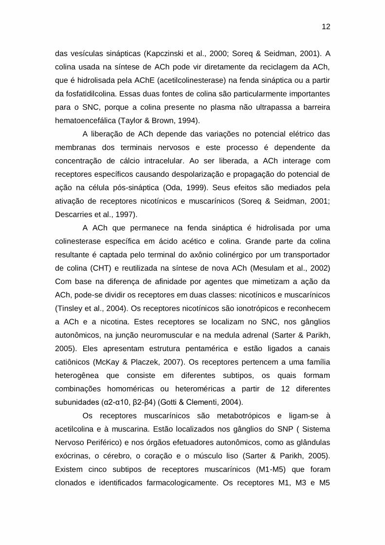
das vesículas sinápticas (Kapczinski et al., 2000; Soreq & Seidman, 2001). A
colina usada na síntese de ACh pode vir diretamente da reciclagem da ACh,
que é hidrolisada pela AChE (acetilcolinesterase) na fenda sináptica ou a partir
da fosfatidilcolina. Essas duas fontes de colina são particularmente importantes
para o SNC, porque a colina presente no plasma não ultrapassa a barreira
hematoencefálica (Taylor & Brown, 1994).
A liberação de ACh depende das variações no potencial elétrico das
membranas dos terminais nervosos e este processo é dependente da
concentração de cálcio intracelular. Ao ser liberada, a ACh interage com
receptores específicos causando despolarização e propagação do potencial de
ação na célula pós-sináptica (Oda, 1999). Seus efeitos são mediados pela
ativação de receptores nicotínicos e muscarínicos (Soreq & Seidman, 2001;
Descarries et al., 1997).
A ACh que permanece na fenda sináptica é hidrolisada por uma
colinesterase específica em ácido acético e colina. Grande parte da colina
resultante é captada pelo terminal do axônio colinérgico por um transportador
de colina (CHT) e reutilizada na síntese de nova ACh (Mesulam et al., 2002)
Com base na diferença de afinidade por agentes que mimetizam a ação da
ACh, pode-se dividir os receptores em duas classes: nicotínicos e muscarínicos
(Tinsley et al., 2004). Os receptores nicotínicos são ionotrópicos e reconhecem
a ACh e a nicotina. Estes receptores se localizam no SNC, nos gânglios
autonômicos, na junção neuromuscular e na medula adrenal (Sarter & Parikh,
2005). Eles apresentam estrutura pentamérica e estão ligados a canais
catiônicos (McKay & Placzek, 2007). Os receptores pertencem a uma família
heterogênea que consiste em diferentes subtipos, os quais formam
combinações homoméricas ou heteroméricas a partir de 12 diferentes
subunidades (α2-α10, β2-β4) (Gotti & Clementi, 2004).
Os receptores muscarínicos são metabotrópicos e ligam-se à
acetilcolina e à muscarina. Estão localizados nos gânglios do SNP ( Sistema
Nervoso Periférico) e nos órgãos efetuadores autonômicos, como as glândulas
exócrinas, o cérebro, o coração e o músculo liso (Sarter & Parikh, 2005).
Existem cinco subtipos de receptores muscarínicos (M1-M5) que foram
clonados e identificados farmacologicamente. Os receptores M1, M3 e M5

13
estão acoplados a uma proteína Gq/11 e alteram a atividade celular pela
estimulação da fosfolipase C, e pela geração do segundo mensageiro IP3, o
qual induz a liberação de cálcio intracelular e diacilglicerol (DAG). Contudo, os
receptores M2 e M4 estão acoplados a uma proteína Gi que induz sua reposta
via inibição da adenilato ciclase (Caulfield & Birdsall, 1998; Uchiyama & Chess-
Williams, 2004).
1.2.1 Acetilcolinesterase (AChE, E.C.3.1.1.7)
As colinesterases hidrolisam a ACh na fenda sináptica e desempenham
um papel muito importante na neurotransmissão colinérgica, além de outras
funções fisiológicas. São classificadas de acordo com suas propriedades
catalíticas, especificidade de inibidores e distribuição nos tecidos: A AChE
hidrolisa preferencialmente ésteres com grupamento acetil, estando presente
principalmente nas sinapses do SNC, SNP parassimpático e junção
neuromuscular por outro lado a butirilcolinesterase (E.C.3.1.1.8, BuChe)
hidrolisa outros tipos de ésteres como a butirilcolina. Ambas as colinesterases
são amplamente distribuídas no organismo (Taylor and Brown, 1999). A AChE
é uma importante enzima regulatória que controla a transmissão de impulsos
nervosos através da sinapse colinérgica pela hidrólise do neurotransmissor
excitatório ACh (Milatovic and Dettbarn, 1996). A AChE é uma serina hidrolase
que desempenha um papel essencial no mecanismo colinérgico, catalisando a
hidrólise natural do substrato acetilcolina em acetato e colina (Quinn, 1987). Os
níveis de AChE parecem ser controlados pela interação da ACh com seus
receptores, sendo que quando a interação é acentuada, aumentam os níveis
de AChE. No entanto, a AChE pode ser usada como um marcador da função
colinérgica, e mudanças na atividade da enzima podem indicar alterações na
disponibilidade de ACh e do nível de seus receptores (Fernandes and Hodges-
Savola, 1992).
Observa-se a inibição da atividade da AChE quando o zebrafish é
exposto aos agentes tóxicos paration (Roex et al., 2003) e metanol (Rico et al.,
14
2006), enquanto que o etanol promove um aumento significativo desta
atividade (Rico et al., 2007).
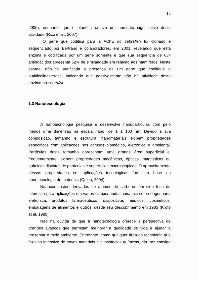
O gene que codifica para a AChE do zebrafish foi clonado e
sequenciado por Bertrand e colaboradores, em 2001, revelando que esta
enzima é codificada por um gene somente e que sua sequência de 634
aminoácidos apresenta 62% de similaridade em relação aos mamíferos. Neste
estudo, não foi verificada a presença de um gene que codifique a
butirilcolinesterase, indicando que possivelmente não há atividade desta
enzima no zebrafish.
1.3 Nanotecnologia
A nanotecnologia pesquisa e desenvolve nanopartículas com pelo
menos uma dimensão na escala nano, de 1 à 100 nm. Devido a sua
composição, tamanho e estrutura, nanomateriais exibem propriedades
específicas com aplicações nos campos biomédico, eletrônico e ambiental.
Partículas deste tamanho apresentam uma grande área superficial e,
frequentemente, exibem propriedades mecânicas, ópticas, magnéticas ou
químicas distintas de partículas e superfícies macroscópicas. O aproveitamento
dessas propriedades em aplicações tecnológicas forma a base da
nanotecnologia de materiais (Quina, 2004).
Nanocompostos derivados de átomos de carbono têm sido foco de
interesse para aplicações em vários campos industriais, tais como engenharia
eletrônica, produtos farmacêuticos, dispositivos médicos, cosméticos,
embalagens de alimentos e outros, desde seu descobrimento em 1985 (Kroto
et al. 1985).
Não há dúvida de que a nanotecnologia oferece a perspectiva de
grandes avanços que permitam melhorar a qualidade de vida e ajudar a
preservar o meio ambiente. Entretanto, como qualquer área da tecnologia que
faz uso intensivo de novos materiais e substâncias químicas, ela traz consigo

15
alguns riscos ao meio ambiente e à saúde humana que ainda precisam ser
melhor investigados (Quina, 2004).
1.3.1 Fulerenos
Fulerenos são alótropos moleculares do carbono e existem em várias
formas. Foram descobertos em 1985, por três pesquisadores que observavam
o grafite sendo aquecido com laser e vaporizando em uma atmosfera de gás
hélio. Os átomos de carbono vaporizados se misturaram ao hélio e se
combinaram formando agregados moleculares com algumas dezenas de
átomos de carbono. Dentre os agregados, duas conformações se
apresentaram com alto índice de ocorrência: os compostos com 60 e com 70
átomos de Carbono. Esta avaliação foi feita através de medida com
espectrômetro de massa (Kroto et al., 1985 ; Rocha, 1996).
A menor molécula que pode ser formada é a C20, e após ela é a C24. A
partir desta última, existem fulerenos em uma sequência com números pares
de carbono, ou seja, C26, C28, C30, e assim por diante (Amador, 2006). Até hoje,
oito fulerenos estáveis já foram isolados em quantidades significativas, sendo
os mesmos denominados [60-Ih], [70-D5h], [76-D2], [78-D3], [78-C2v(I)], [78-
C2v(II)], [84-D2(IV)] e [84-D2d(II)] em função do número de carbonos que os
formam, do grupo pontual de simetria da molécula e do número do possível
regioisômero. Um dentre estes fulerenos, a molécula de simetria Ih formada por
60 átomos de carbono, o fulereno C60 é, sem dúvida, o mais abundante e
representativo (Santos et al., 2010).
O que torna o fulereno alvo de inúmeros estudos são suas
propriedades como a capacidade de armazenar átomos em seu interior e
aceitar diversas modificações estruturais através da adição de radicais e
átomos de diferentes elementos, sendo que para cada modificação estrutural
suas propriedades se alteram. (Amador, 2006).
16
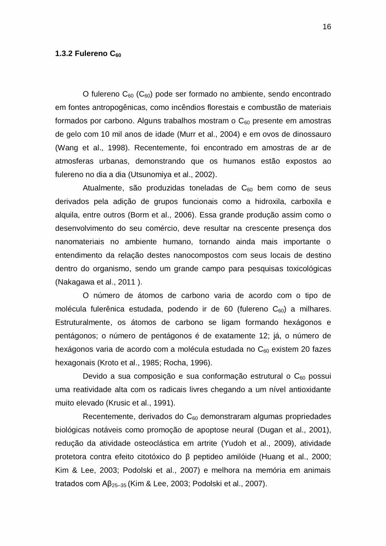
1.3.2 Fulereno C60
O fulereno C60 (C60) pode ser formado no ambiente, sendo encontrado
em fontes antropogênicas, como incêndios florestais e combustão de materiais
formados por carbono. Alguns trabalhos mostram o C60 presente em amostras
de gelo com 10 mil anos de idade (Murr et al., 2004) e em ovos de dinossauro
(Wang et al., 1998). Recentemente, foi encontrado em amostras de ar de
atmosferas urbanas, demonstrando que os humanos estão expostos ao
fulereno no dia a dia (Utsunomiya et al., 2002).
Atualmente, são produzidas toneladas de C60 bem como de seus
derivados pela adição de grupos funcionais como a hidroxila, carboxila e
alquila, entre outros (Borm et al., 2006). Essa grande produção assim como o
desenvolvimento do seu comércio, deve resultar na crescente presença dos
nanomateriais no ambiente humano, tornando ainda mais importante o
entendimento da relação destes nanocompostos com seus locais de destino
dentro do organismo, sendo um grande campo para pesquisas toxicológicas
(Nakagawa et al., 2011 ).
O número de átomos de carbono varia de acordo com o tipo de
molécula fulerênica estudada, podendo ir de 60 (fulereno C60) a milhares.
Estruturalmente, os átomos de carbono se ligam formando hexágonos e
pentágonos; o número de pentágonos é de exatamente 12; já, o número de
hexágonos varia de acordo com a molécula estudada no C60 existem 20 fazes
hexagonais (Kroto et al., 1985; Rocha, 1996).
Devido a sua composição e sua conformação estrutural o C60 possui
uma reatividade alta com os radicais livres chegando a um nível antioxidante
muito elevado (Krusic et al., 1991).
Recentemente, derivados do C60 demonstraram algumas propriedades
biológicas notáveis como promoção de apoptose neural (Dugan et al., 2001),
redução da atividade osteoclástica em artrite (Yudoh et al., 2009), atividade
protetora contra efeito citotóxico do β peptideo amilóide (Huang et al., 2000;
Kim & Lee, 2003; Podolski et al., 2007) e melhora na memória em animais
tratados com Aβ25–35 (Kim & Lee, 2003; Podolski et al., 2007).
17
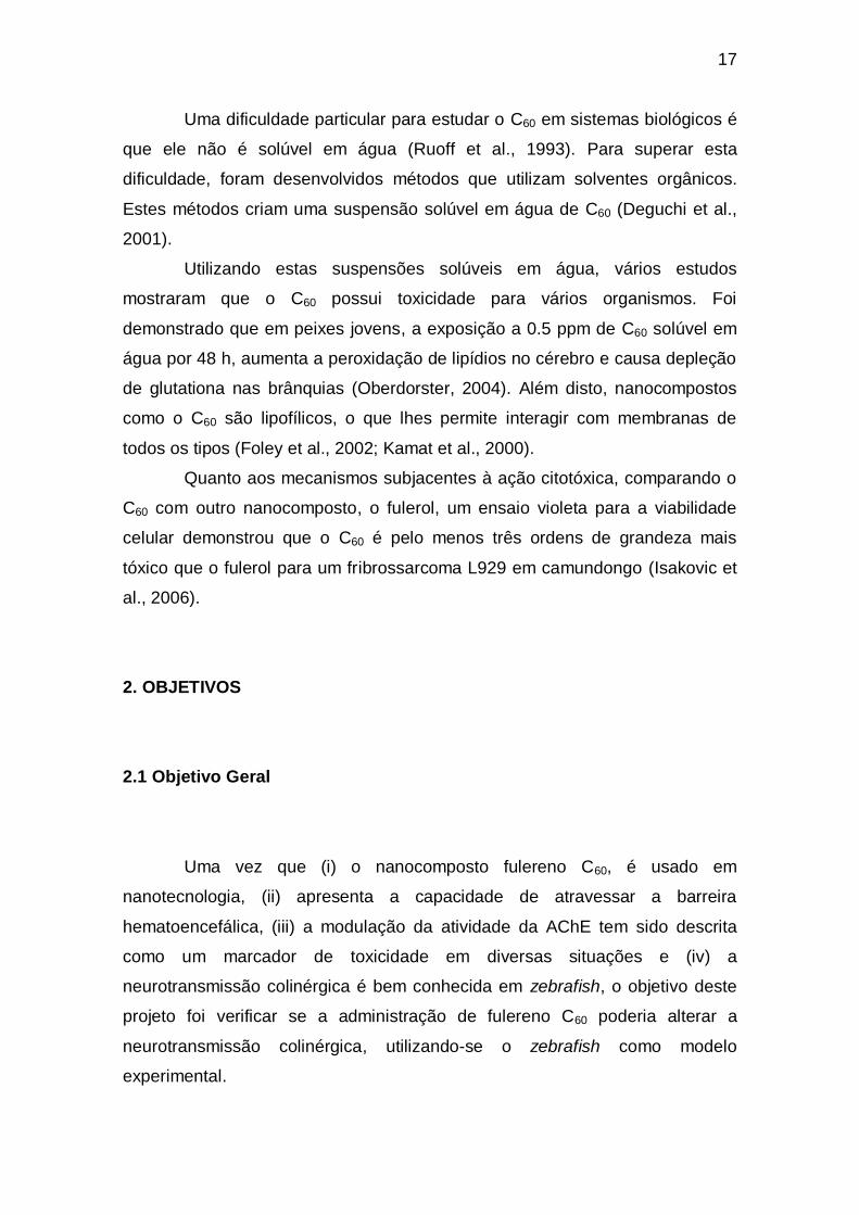
Uma dificuldade particular para estudar o C60 em sistemas biológicos é
que ele não é solúvel em água (Ruoff et al., 1993). Para superar esta
dificuldade, foram desenvolvidos métodos que utilizam solventes orgânicos.
Estes métodos criam uma suspensão solúvel em água de C60 (Deguchi et al.,
2001).
Utilizando estas suspensões solúveis em água, vários estudos
mostraram que o C60 possui toxicidade para vários organismos. Foi
demonstrado que em peixes jovens, a exposição a 0.5 ppm de C60 solúvel em
água por 48 h, aumenta a peroxidação de lipídios no cérebro e causa depleção
de glutationa nas brânquias (Oberdorster, 2004). Além disto, nanocompostos
como o C60 são lipofílicos, o que lhes permite interagir com membranas de
todos os tipos (Foley et al., 2002; Kamat et al., 2000).
Quanto aos mecanismos subjacentes à ação citotóxica, comparando o
C60 com outro nanocomposto, o fulerol, um ensaio violeta para a viabilidade
celular demonstrou que o C60 é pelo menos três ordens de grandeza mais
tóxico que o fulerol para um fribrossarcoma L929 em camundongo (Isakovic et
al., 2006).
2. OBJETIVOS
2.1 Objetivo Geral
Uma vez que (i) o nanocomposto fulereno C60, é usado em
nanotecnologia, (ii) apresenta a capacidade de atravessar a barreira
hematoencefálica, (iii) a modulação da atividade da AChE tem sido descrita
como um marcador de toxicidade em diversas situações e (iv) a
neurotransmissão colinérgica é bem conhecida em zebrafish, o objetivo deste
projeto foi verificar se a administração de fulereno C60 poderia alterar a
neurotransmissão colinérgica, utilizando-se o zebrafish como modelo
experimental.
18
2.2 Objetivos Específicos
Avaliar o efeito da exposição à diferentes doses de fulereno C60 sobre a
atividade da acetilcolinesterase em homogenato cerebral de zebrafish adultos.
Avaliar o efeito de diferentes tempos de exposição ao fulereno C60
sobre a atividade da acetilcolinesterase em homogenato cerebral de zebrafish
adultos.
19
CAPÍTULO 2 – ARTIGO CIENTÍFICO
Exposure to nano/microparticles of fullerene (C60) increases
acetylcholinesterase activity and lipid peroxidation in adult zebrafish
(Danio rerio) brain
Gonzalo Ogliari Dal Forno, Luiza Wilges Kist, Mariana Barbieri de Azevedo,
Rachel Seemann Fritsch, Talita Carneiro Brandão Pereira, Roberta Socoowski
Britto, Sílvia Stanisçuaski Guterres, Irene Clemes Külkamp-Guerreiro, Carla
Denise Bonan, José María Monserrat and Maurício Reis Bogo
(Artigo submetido à revista científica Particle and Fibre Toxicology)
20
Exposure to nano/microparticles of fullerene (C60) increases acetylcholinesterase
activity and lipid peroxidation in adult zebrafish (Danio rerio) brain
Gonzalo Ogliari Dal Forno1, Luiza Wilges Kist
1, Mariana Barbieri de Azevedo
1, Rachel
Seemann Fritsch1, Talita Carneiro Brandão Pereira
1, Roberta Socoowski Britto
2,3, Sílvia
Stanisçuaski Guterres4, Irene Clemes Külkamp-Guerreiro
4, Carla Denise Bonan
5,6, José
María Monserrat2,7,3
and Maurício Reis Bogo1,6,*
1Laboratório de Biologia Genômica e Molecular, Faculdade de Biociências, Pontifícia
Universidade Católica do Rio Grande do Sul, Avenida Ipiranga, 6681, 90619-900 Porto
Alegre, RS, Brazil.
2Universidade Federal do Rio Grande – FURG, Instituto de Ciências Biológicas (ICB),
Av. Itália Km 8 s/n, 96208-900, Rio Grande, RS, Brazil.
3Programa de Pós-Graduação em Ciências Fisiológicas – Fisiologia Animal Comparada,
FURG, Brazil.
4Faculdade de Farmácia, Universidade Federal do Rio Grande do Sul, Avenida Ipiranga,
2752, 90610-000, Porto Alegre, RS, Brazil.
5Laboratório de Neuroquímica e Psicofarmacologia, Faculdade de Biociências,
Pontifícia Universidade Católica do Rio Grande do Sul. Avenida Ipiranga, 6681, 90619-
900 Porto Alegre, RS, Brazil.
6Instituto Nacional de Ciência e Tecnologia Translacional em Medicina (INCT-TM),
90035-003, Porto Alegre, RS, Brazil.
7Instituto Nacional de Ciência e Tecnologia de Nanomateriais de Carbono
Nanomateriais de Carbono, Brazil.
Email: Gonzalo Ogliari Dal Forno - [email protected], Luiza Wilges Kist -
[email protected], Mariana Barbieri de Azevedo - [email protected],
21
Rachel Seemann Fritsch - [email protected], Talita Carneiro Brandão Pereira
- [email protected], Roberta Socoowski Britto - [email protected], Sílvia
Stanisçuaski Guterres - [email protected], Irene Clemes Külkamp-Guerreiro -
[email protected], Carla Denise Bonan - [email protected], José María Monserrat -
[email protected], Maurício Reis Bogo* - [email protected]
* Corresponding author
Maurício Reis Bogo
Faculdade de Biociências
Pontifícia Universidade Católica do Rio Grande do Sul
Avenida Ipiranga, 6681 - 12C - sala 172
Zip Code: 90619-900
Porto Alegre, RS, Brazil
Telephone: +55 51 3353-4726; Fax: +55 51 3320 3568.
E-mail address: [email protected]

22
ABSTRACT
Background: Even though technologies involving nano/microparticles have great
potential in diverse applications, it is crucial to determine possible toxicity of these
technological products before extensive use. Fullerenes C60 are nanomaterials with
unique physicochemical and biological properties that are important for the
development of many technological applications. The aim of this study was to evaluate
the consequences of C60 exposure in brain acetylcholinesterase expression and activity,
antioxidant responses and oxidative damage using adult zebrafish (Danio rerio) as an
animal model.
Results: None of the doses (i.p.) tested (7.5, 15 and 30 mg/kg) altered AChE activity
when zebrafish were exposed to C60 during 6 and 12 hours. However, the analysis for
24 hours demonstrated that animals treated with the concentration of 30 mg/kg
presented a significant increase in AChE activity (28.54 ± 3.72 µmol SCh.h-1
.mg
protein-1
; p = 0.0001) when compared to saline (12.19 ± 0.55 µmol SCh.h-1
.mg protein-
1; p = 0.0001) and to the vehicle control group (15.46 ± 0.57 µmol SCh.h
-1.mg protein
-1;
p = 0.0001). The up-regulation of brain AChE activity is not directly related with the
transcriptional control. Oxidative damage, measured by lipid peroxidation (TBARS
assays) showed a pro-oxidant condition elicited by C60 at the highest dose (30 mg/kg)
after 24 hours when compared with the vehicle control group (0.11 ± 0.02 vs 0.07 ±
0.01 nmoles/mg of wet tissue; p= 0.0194).
Conclusion: The results presented in this article provided further evidence for
neurotoxic effects after C60 exposure. In one hand, AChE activity was significantly
23
enhanced when zebrafish were exposed to C60. Besides, C60 showed a pro-oxidant
behavior when intraperitoneally injected, indicating toxicity mechanisms other than
photo-excitation. Taken together these results suggest neurotoxicity mediated by
apoptosis.
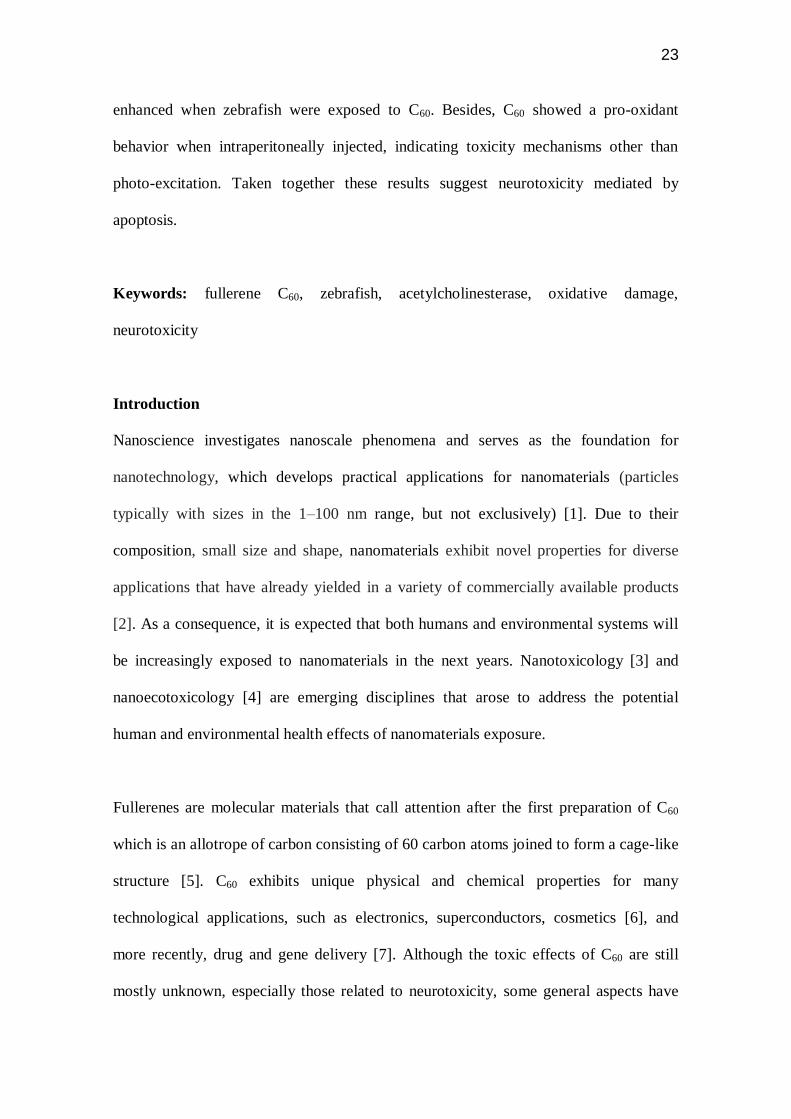
Keywords: fullerene C60, zebrafish, acetylcholinesterase, oxidative damage,
neurotoxicity
Introduction
Nanoscience investigates nanoscale phenomena and serves as the foundation for
nanotechnology, which develops practical applications for nanomaterials (particles
typically with sizes in the 1–100 nm range, but not exclusively) [1]. Due to their
composition, small size and shape, nanomaterials exhibit novel properties for diverse
applications that have already yielded in a variety of commercially available products
[2]. As a consequence, it is expected that both humans and environmental systems will
be increasingly exposed to nanomaterials in the next years. Nanotoxicology [3] and
nanoecotoxicology [4] are emerging disciplines that arose to address the potential
human and environmental health effects of nanomaterials exposure.
Fullerenes are molecular materials that call attention after the first preparation of C60
which is an allotrope of carbon consisting of 60 carbon atoms joined to form a cage-like
structure [5]. C60 exhibits unique physical and chemical properties for many
technological applications, such as electronics, superconductors, cosmetics [6], and
more recently, drug and gene delivery [7]. Although the toxic effects of C60 are still
mostly unknown, especially those related to neurotoxicity, some general aspects have
24
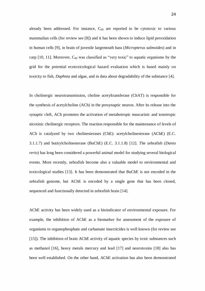
already been addressed. For instance, C60 are reported to be cytotoxic to various
mammalian cells (for review see [8]) and it has been shown to induce lipid peroxidation
in human cells [9], in brain of juvenile largemouth bass (Micropterus salmoides) and in
carp [10, 11]. Moreover, C60 was classified as “very toxic” to aquatic organisms by the
grid for the potential ecotoxicological hazard evaluation which is based mainly on
toxicity to fish, Daphnia and algae, and in data about degradability of the substance [4].
In cholinergic neurotransmission, choline acetyltransferase (ChAT) is responsible for
the synthesis of acetylcholine (ACh) in the presynaptic neuron. After its release into the
synaptic cleft, ACh promotes the activation of metabotropic muscarinic and ionotropic
nicotinic cholinergic receptors. The reaction responsible for the maintenance of levels of
ACh is catalyzed by two cholinesterases (ChE): acetylcholinesterase (AChE) (E.C.
3.1.1.7) and butirylcholinesterase (BuChE) (E.C. 3.1.1.8) [12]. The zebrafish (Danio
rerio) has long been considered a powerful animal model for studying several biological
events. More recently, zebrafish become also a valuable model to environmental and
toxicological studies [13]. It has been demonstrated that BuChE is not encoded in the
zebrafish genome, but AChE is encoded by a single gene that has been cloned,
sequenced and functionally detected in zebrafish brain [14].
AChE activity has been widely used as a bioindicator of environmental exposure. For
example, the inhibition of AChE as a biomarker for assessment of the exposure of
organisms to organophosphate and carbamate insecticides is well known (for review see
[15]). The inhibition of brain AChE activity of aquatic species by toxic substances such
as methanol [16], heavy metals mercury and lead [17] and neurotoxins [18] also has
been well established. On the other hand, AChE activation has also been demonstrated
25
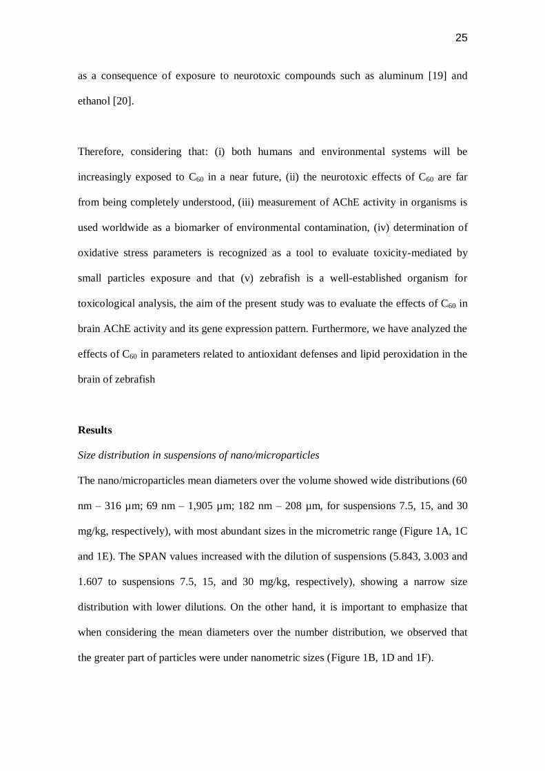
as a consequence of exposure to neurotoxic compounds such as aluminum [19] and
ethanol [20].
Therefore, considering that: (i) both humans and environmental systems will be
increasingly exposed to C60 in a near future, (ii) the neurotoxic effects of C60 are far
from being completely understood, (iii) measurement of AChE activity in organisms is
used worldwide as a biomarker of environmental contamination, (iv) determination of
oxidative stress parameters is recognized as a tool to evaluate toxicity-mediated by
small particles exposure and that (v) zebrafish is a well-established organism for
toxicological analysis, the aim of the present study was to evaluate the effects of C60 in
brain AChE activity and its gene expression pattern. Furthermore, we have analyzed the
effects of C60 in parameters related to antioxidant defenses and lipid peroxidation in the
brain of zebrafish
Results
Size distribution in suspensions of nano/microparticles
The nano/microparticles mean diameters over the volume showed wide distributions (60
nm – 316 µm; 69 nm – 1,905 µm; 182 nm – 208 µm, for suspensions 7.5, 15, and 30
mg/kg, respectively), with most abundant sizes in the micrometric range (Figure 1A, 1C
and 1E). The SPAN values increased with the dilution of suspensions (5.843, 3.003 and
1.607 to suspensions 7.5, 15, and 30 mg/kg, respectively), showing a narrow size
distribution with lower dilutions. On the other hand, it is important to emphasize that
when considering the mean diameters over the number distribution, we observed that
the greater part of particles were under nanometric sizes (Figure 1B, 1D and 1F).
26
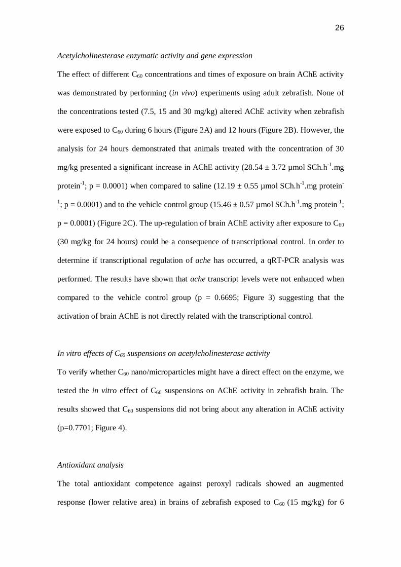
Acetylcholinesterase enzymatic activity and gene expression
The effect of different C60 concentrations and times of exposure on brain AChE activity
was demonstrated by performing (in vivo) experiments using adult zebrafish. None of
the concentrations tested (7.5, 15 and 30 mg/kg) altered AChE activity when zebrafish
were exposed to C60 during 6 hours (Figure 2A) and 12 hours (Figure 2B). However, the
analysis for 24 hours demonstrated that animals treated with the concentration of 30
mg/kg presented a significant increase in AChE activity (28.54 ± 3.72 µmol SCh.h-1
.mg
protein-1
; p = 0.0001) when compared to saline (12.19 ± 0.55 µmol SCh.h-1
.mg protein-
1; p = 0.0001) and to the vehicle control group (15.46 ± 0.57 µmol SCh.h
-1.mg protein
-1;
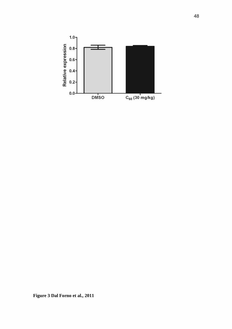
p = 0.0001) (Figure 2C). The up-regulation of brain AChE activity after exposure to C60
(30 mg/kg for 24 hours) could be a consequence of transcriptional control. In order to
determine if transcriptional regulation of ache has occurred, a qRT-PCR analysis was
performed. The results have shown that ache transcript levels were not enhanced when
compared to the vehicle control group (p = 0.6695; Figure 3) suggesting that the
activation of brain AChE is not directly related with the transcriptional control.
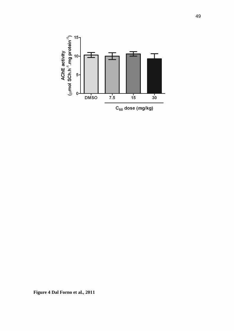
In vitro effects of C60 suspensions on acetylcholinesterase activity
To verify whether C60 nano/microparticles might have a direct effect on the enzyme, we
tested the in vitro effect of C60 suspensions on AChE activity in zebrafish brain. The
results showed that C60 suspensions did not bring about any alteration in AChE activity
(p=0.7701; Figure 4).
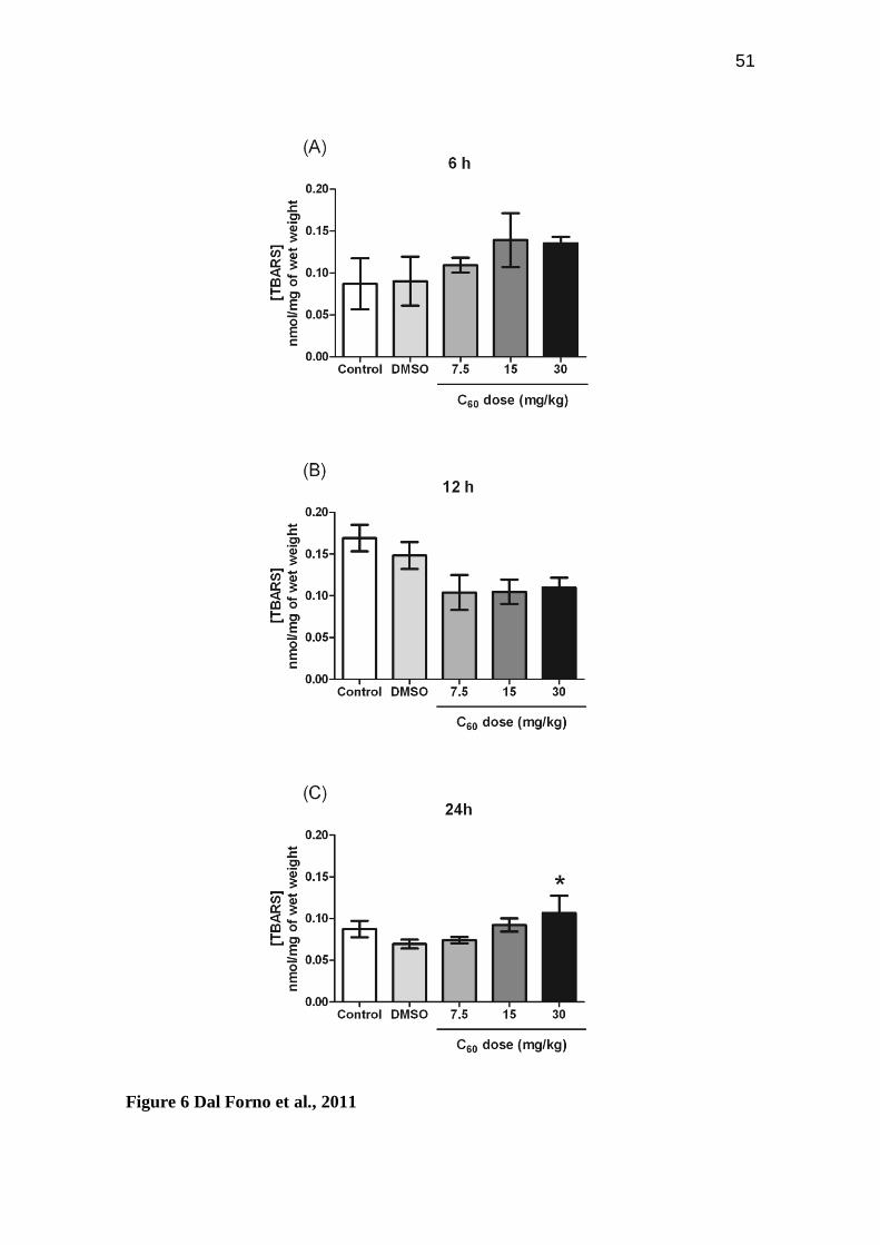
Antioxidant analysis
The total antioxidant competence against peroxyl radicals showed an augmented
response (lower relative area) in brains of zebrafish exposed to C60 (15 mg/kg) for 6
27
hours when compared to zebrafish exposed to C60 (7.5 and 30 mg/kg) for 6 hours
(p=0.0209; Figure 5A). No other differences were observed under the experimental
conditions. Oxidative damage, measured by lipid peroxidation (TBARS assays) showed
a pro-oxidant condition elicited by C60 at the highest dose (30 mg/kg) after 24 hours
(p=0.0194; Figure 6C).
Discussion
Although technologies evolving nano/microparticles have considerable potential in
diverse applications, it is crucial to determine possible toxicity of these technological
products before extensive use. Little is known about the toxic effects of fullerenes in
brain. At present, only few studies presenting contradictory findings have evaluated
possible neurotoxic effects of fullerenes exposure. For instance it was already suggested
that C60 did not cross the blood-brain barrier [21] whereas the results obtained by
Mokrushin [22] suggested that fullerenes possess marked neurotropic properties and are
neurotoxic substances irreversibly blocking the electrical activity of the nervous tissue.
Neurotoxicity of C60 in fish species has been previously reported. Generation of reactive
oxygen species (ROS) by C60 is influenced in part by the presence and type of
illumination due to the photo-excitation of C60 by UV and visible light [11, 23, 24] or
even to by-products of the organic solvents employed to prepare C60 suspensions [25].
For this reason the C60 suspensions were prepared under the protection of light and
intraperitoneal injection was adopted as administration route in the in vivo protocols, an
experimental condition that avoids the influence of light in the analyzed variables. Also,
in vitro assays were run in darkness.
28
Suspensions of C60 in DMSO were prepared as previously described [26, 27] with
modifications. Although DMSO is known to show low toxicity by itself [27, 28],
appropriate experimental controls must be employed to eliminate its influence. In this
study, the DMSO was diluted to result in 12.5% DMSO as a final concentration. Any
signal of toxicity i.e., mortality or even transient alterations in behavior was observed in
the vehicle control group (12.5% DMSO). In addition, control group (saline) and
vehicle control group were never statistically different in the conditions tested.
The characterization of the size and stability of C60 nanoparticles in suspension is very
important to evaluate their toxicity once particle size can change during the preparation
of the suspension, dilution, and exposure [11]. In this study, the nano/microparticles
mean diameters over the volume in the C60 suspensions showed wide distributions with
most abundant sizes in the micrometric range. The C60 has a tendency to form
aggregates very easily [7], and this may be a possible cause of this wide distribution. In
contrast, the nano/microparticles mean diameters over the number distribution in the C60
suspensions demonstrated that the greater part of particles were under nanometric sizes.
Totsuka et al. [29] also observed wide distributions by dynamic light scattering in
formulations manufactured with C60.
In the present study, we have evaluated the effect of different C60 doses (7.5, 15 and 30
mg/kg) and different times of exposure (6, 12 and 24 hours) on AChE activity and ache
expression in zebrafish brain. In the concentrations tested, only the animals exposed to
30 mg/kg for 24 hours have shown enhanced AChE activity. The qRT-PCR results
suggested that the activation of brain AChE is not directly related with the
transcriptional control. The in vitro results indicated that none of the C60 suspensions
29
had a direct effect on the enzyme. Moreover, we have shown the effects of C60 exposure
over the antioxidant competence and lipid peroxidation in zebrafish brain. The results
also demonstrated that the exposition to 30 mg/kg during 24 hours yielded in higher
levels of lipid peroxidation (TBARS). In accordance, Totsuka et al. [29] reported
increased micronuclei frequencies, induced DNA damage and increased mutant
frequencies after C60 nano/microparticles suspension exposure.
AChE is indispensable for terminating acetylcholine-mediated neurotransmission at
cholinergic synapses [30]. In this context, AChE is inhibited by organophosphorus and
carbamate insecticides and by neurotoxins, which are structural analogues of
acetylcholine [31]. In addition, there are evidences to suggest that AChE contributes to
diverse physiological processes through its involvement in the regulation of cell
proliferation, differentiation and survival. As a consequence, more recently AChE has
been redefined as an important regulator of apoptosis, because it can be induced by a
variety of apoptotic stimuli ([32]; for review see [33]). It is well known that apoptosis
underlies the neurotoxic effects of various compounds. Moreover, zebrafish brain AChE
activation has also been demonstrated as a consequence of exposure to known
neurotoxic compounds, including aluminum [19] and ethanol [20] and to the
cyanobacterial toxin microcystin-LR [34].
The antioxidant or pro-oxidant effects induced by C60 exposure is still a debatable issue
[9, 24, 25]. C60 is photo-excited under UV or visible light [23], a condition for example,
that elicited lipid peroxidation in brains of carp [11]. On the other hand, the absence of
light did not completely inhibited C60 toxicity to embryonic zebrafish [35]. Our results
reinforce oxidative stress generation as a pathway of C60-induced toxicity in brain of
30
fish species, even in the absence of light. Accordingly, it is well known that some
apoptotic signals induce pro-apoptotic events increasing ROS in mitochondria. The
increased ROS may lead to oxidative stress generation.
Conclusion
The results presented in this article provide further experimental evidence that C60
exposure can be neurotoxic. First, brain AChE activity was significantly enhanced when
zebrafish were exposed to C60. Besides, C60 showed a pro-oxidant behavior when
intraperitoneally injected, suggesting toxicity mechanisms that are independent of
photo-excitation. Taken together our results suggest neurotoxicity mediated by
apoptosis.
Materials and methods
Chemicals
Fullerene (C60, 99.5% purity) was purchased from Aldrich (Milwaukee, WI, USA),
DMSO was purchased from Fisher Scientific (Pittsburgh, PA, USA) and Trizma Base,
ethylenedioxy–diethylene–dinitrilo–tetraacetic acid (EDTA), ethylene glycol bis(beta
amino ethylether)-N,N,N',N'-tetraacetic acid (EGTA), sodium citrate, Coomassie Blue
G, bovine serum albumin, acetylthiocholine, 5,5‟-dithiobis-2-nitrobenzoic acid (DTNB)
HEPES, BHT (99%), 2,2′-azobis(2-methylpropionamidine) dihydrochloride (ABAP)
and 1,1,3,3-tetramethoxypropane were purchased from Sigma Chemical Co (St. Louis,
MO, USA). KCl and SDS (90%) were purchased from Labsynth (Brazil).
Tetramethoxypropane (TMP) and 2′,7′-dichlorodihydrofluorescein diacetate were
purchased from Acros Organics (Morris Plains, NJ, USA) and Molecular Probes Inc.
(Eugene, OR, USA) respectively. MgCl2 and Acetic acid 99.7% were purchased from
31
Isofar and Vetec (Brazil) respectively. TRIzol® reagent, Platinum
® Taq DNA
Polymerase and SYBR
® Green I were purchased from Invitrogen (Carlsbad, CA, USA).
ImProm-II™ Reverse Transcription System was purchased from Promega (Madison,
USA). All other reagents used were of analytical grade.
Animals
Adult wild-type zebrafish (Danio rerio, Cyprinidae) of both sexes (3-6 months-old)
were obtained from a specialized supplier (Redfish Agroloja, RS, Brazil). Animals were
kept at a density of up to five animals per liter in 50 L housing tanks with tap water that
was previously treated with Tetra‟s AquaSafe® (to neutralize chlorine, chloramines, and
heavy metals present in the water that could be harmful to fish) and continuously
aerated (7.20 mg O2/L) at 26 ± 2 ºC, under a 14/10 h light/dark controlled photoperiod.
Animals were acclimated for at least two weeks before the experiments and were fed
three times a day with TetraMin Tropical Flake fish food®. The fish were maintained
healthy and free of any signs of disease and were used according to the „„Guide for the
Care and Use of Laboratory Animals” published by the US National Institutes of
Health. All procedures in the present study were approved by the Animal Ethics
Committee of the Pontifical Catholic University of Rio Grande do Sul (PUCRS),
protocol number 10/00185-CEUA.
C60 suspension
Suspensions of C60 in DMSO were prepared as previously described [26, 27] with
modifications. Briefly, 7.6 mg of C60 was added to 0.5 mL of DMSO and sonicated for
3 h. Following the observation that the particle size was increased over time, the
suspension was additionally sonicated for one hour. The suspension of C60 was diluted
32
in water to result in 7.5, 15 and 30 mg/kg suspensions (12.5 % DMSO) that were further
sonicated for one hour prior to use. C60 suspensions were prepared and stored in a dark
condition.
Characterization of C60 suspensions
The C60 suspensions (7.5, 15 and 30 mg/kg) were characterized in terms of particle size
distribution. The mean diameter over the volume and number distribution (d4.3) was
determined by laser diffractometry (Mastersizer 2000, Malvern Instruments, UK). The
value of SPAN was utilized to determine particle size distribution according Eq. (1),
where d0.9, d0.1 and d0.5 are the particle diameters determined at 90 %, 10 % and 50 %
cumulative undersized volumes, respectively.
(Eq. 1) SPAN = (d0.9-d0.1)/d0.5
Intraperitoneal injection
Intraperitoneal injections were conducted using a 3/10-mL U-100 BD Ultra-Fine™
Short Insulin Syringe 8 mm (5/16″) × 31G Short Needle (Becton Dickinson and
Company, New Jersey, USA) according to the protocol established by Phelps et al. [36].
Briefly, the volume injected into the animal was adjusted to the fish bodyweight (mean
injection volume was 10 μL) to achieve 7.5, 15 and 30 mg/kg. The animals of the
control group received the same volume of saline solution and the animals of the
vehicle control received the same volume of 12.5% DMSO. Anesthesia of the animals
prior to the injection was obtained by its immersion in a solution of benzocaine (1 mM
in MeOH 1%) until the animal showed a lack of motor coordination and reduced
respiratory rate. The anesthetized animal was gently placed in a water-soaked gauze-

33
wrapped hemostat with the abdomen facing up and the head of the fish positioned at the
hinge of the hemostat (the pectoral fins were used as a landmark on the abdomen). The
needle was inserted parallel to the spine in the midline of the abdomen posterior to the
pectoral fins. The injection procedure was conducted in such a way as to guarantee that
the animal did not spend more than 10 s out of the water. After the injection, the
animals were placed in a separate tank with highly aerated unchlorinated tap water (25 ±
2 °C) to facilitate recovery from the anesthesia. Saline solution was used as control. All
the animals that recovered within 2-3 min following the injection continued in the
experiment while animals that did not recover during this period were discarded. Six,
twelve or twenty-four hours after the injection the animals were euthanized and AChE
activity was determined.
In vitro assays of AChE activity
In vitro assays were performed as previously described [37, 38]. Briefly, C60 suspension
was added to the reaction medium before the pre-incubation with the enzyme-
containing lysate from zebrafish brain homogenate and maintained during the enzyme
assays. C60 was tested at final concentrations of 7.5, 15 and 30 mg/kg. Control
treatments with equal volume of vehicle (DMSO 12.5%) were performed to exclude the
DMSO effect on the enzyme activities.
Determination of AChE activity
Zebrafish were euthanized and their whole brains were removed by dissection. The
brains (two whole brains for each sample) were homogenized on ice in 60 volumes
(v/w) of Tris-citrate buffer (50 mM Tris, 2mM EDTA, 2mM EGTA, pH 7.4, adjusted
with citric acid), in a glass-Teflon homogenizer. The rate of acetylthiocholine

34
hydrolysis (ACSCh, 0.88 mM) was assessed in a final volume of 300 µL with 11 mM
phosphate buffer, pH 7.5, and 0.22 mM DTNB using a method previously described
[39]. Before the addition of substrate, samples containing protein (5 µg) and the reaction
medium described above were pre-incubated for 10 min at 25 °C. The hydrolysis of
substrate was monitored by the formation of thiolate dianion of DTNB at 412 nm for 2-
3 min (intervals of 30 s) in a microplate reader. Controls without the homogenate
preparation were performed in order to determine the non-enzymatic hydrolysis of the
substrate. The linearity of absorbance against time and protein concentration was
previously determined. The AChE activity was expressed as micromoles of thiocholine
(SCh) released per hour per milligram of protein. All enzyme assays were evaluated in
triplicate and at least four independent experiments were performed.
Gene expression analysis by quantitative real time RT-PCR (qRT-PCR)
Immediately after 24 hours of intraperitoneal injection (described above), the animals
were euthanized by decapitation. For each sample, a pool of three zebrafish whole
brains was used. Total RNA was isolated with Trizol® reagent (Invitrogen, Carlsbad,
California, USA) in accordance with the manufacturer‟s instructions. The total RNA
was quantified by spectrophotometry and the cDNA was synthesized with ImProm-II™
Reverse Transcription System (Promega) from 1 μg total RNA, following the
manufacturer‟s instructions. Quantitative PCR was performed using SYBR®
Green I
(Invitrogen) to detect double-strand cDNA synthesis. Reactions were done in a volume
of 25 μL using 12.5 μL of diluted cDNA (1:100 for EF1α and Rlp13α; and 1:20 for
ache), containing a final concentration of 0.2 x SYBR®
Green I (Invitrogen), 100 μM
dNTP, 1 x PCR Buffer, 3 mM MgCl2, 0.25 U Platinum®
Taq DNA Polymerase
(Invitrogen) and 200 nM of each reverse and forward primers (Table 1). The PCR
35
cycling conditions were: an initial polymerase activation step for 5 min at 95°C, 40
cycles of 15 s at 95ºC for denaturation, 35 s at 60 °C for annealing and 15 s at 72°C for
elongation. At the end of cycling protocol, a melting-curve analysis was included and
fluorescence measured from 60 to 99 °C. Relative expression levels were determined
with 7500 Fast Real-Time System Sequence Detection Software v.2.0.5 (Applied
Biosystems). The efficiency per sample was calculated using LinRegPCR 11.0 Software
(http://LinRegPCR.nl) and the stability of the references genes, EF1α and Rlp13α (M-
value) and the optimal number of reference genes according to the pairwise variation
(V) were analyzed by GeNorm 3.5 Software (http://medgen.ugent.be/genorm/). Relative
RNA expression levels were determined using the 2-∆∆CT
method.
Antioxidant capacity against peroxyl radicals
Total antioxidant competence against peroxyl radicals was evaluated through reactive
oxygen species (ROS) determination in tissues samples treated or not with a peroxyl
radical generator [40]. Briefly, on a white 96-well microplate, 10 µL of brain
homogenates were disposed into the wells, six wells per sample. The reaction buffer
(127.5 µL) containing 30 mM HEPES (pH 7.2), 200 mM KCl and 1 mM MgCl2 were
added to the wells containing the samples. In three of the six wells of each sample, 7.5
µL of 2,2‟-azobis 2 methylpropionamidine dihydrochloride (ABAP; 4 mM) were added.
In the other three wells the same volume of ultrapure water was pipetted. After this, the
microplate was put into a fluorescence microplate reader (Victor 2, Perkin Elmer),
programmed to keep temperature at 35 ºC. At this temperature, peroxyl radicals are
produced by thermal decomposition of ABAP [41]. Immediately before microplate
reading, it was added in each well 10 µL of the fluorescent probe 2‟,7‟-
dichlorofluorescein diacetate (H2DCF-DA) in a final concentration of 40 µM, according
36
to the methodology employed by Ferreira-Cravo et al. [42]. H2DCF-DA is deacetylated
and the product H2DCF is oxidized by ROS to the fluorescent compound DCF, which is
detected at wavelengths of 488 and 525 nm, for excitation and emission, respectively.
Fluorescence readings (fluorescence units or FU) were performed every 5 min during 30
min. Total fluorescence production was calculated by integrating the fluorescence units
(FU) along the time of the measurement, after adjusting FU data to a second order
polynomial function. The results were expressed as area difference of FU × min in the
same sample with and without ABAP addition and standardized to the ROS area
without ABAP (background area). The relative difference between ROS area with and
without ABAP was considered a measure of antioxidant capacity, with high area
difference meaning low antioxidant capacity, since high fluorescence levels were
obtaining after adding ABAP, meaning low competence to neutralize peroxyl radicals
[40].
Measurement of lipid peroxidation
Lipid peroxidation was measured through determination of thiobarbituric acid reactive
substances (TBARS), following the methodology of Oakes and Van der Kraak [43].
Brain homogenates (10 μL) were added to a reaction mixture made with 150 μL of 20%
acetic acid, 150 μL of thiobarbituric acid (2.4%), 50 μL of Milli Q water and 20 μL of
sodium dodecyl sulfate (SDS, 8.1%). Samples were heated at 95 °C during 30 min and
after cooling by 10 min, 100 μL of Milli Q water and 500 μL of n-butanol was added.
After centrifugation (3,000 × g during 10 min at 15 °C), the organic phase (150 μL) was
placed in a microplate reader and the fluorescence registered after excitation at 520 nm
and emission of 580 nm. The concentration of TBARS (nmol/mg of wet tissue) was
calculated employing tetramethoxypropane (TMP) as standard.
37
Protein determination
Protein was measured by the Coomassie blue method [44] using bovine serum albumin
as standard.
Statistical analysis
AChE activity and antioxidant analyses were expressed as means S.E.M. and
analyzed by one-way analysis of variance (ANOVA). Post-hoc comparisons were made
using Tukey‟s test and orthogonal comparisons. Before ANOVA, its assumptions
(normality and variances homogeneity) were checked. Molecular data were expressed
as means S.E.M. and analyzed by Student‟s t-test. In every case the significance level
was fixed in 5 % ( = 0.05)
Abbreviations
ChAT: choline acetyltransferase; ACh: acetylcholine; AChE: acetylcholinesterase;
BuChE: butyrylcholinesterase; UV: ultraviolet light; ACAP: antioxidant capacity;
TBARS: thiobarbituric acid reactive substances; THF: tetrahydrofuran; EDTA:
ethylenedioxy–diethylene–dinitrilo–tetraacetic acid; EGTA: ethylene glycol bis(beta
amino ethylether)-N,N,N',N'-tetraacetic acid; DTNB: 5,5‟-dithiobis-2-nitrobenzoic acid;
HEPES: 4-(2-hydroxyethyl)-1-piperazineethanesulfonic acid; BHT: butylated
hydroxytoluene; ABAP: 2,2′-azobis(2-methylpropionamidine) dihydrochloride; KCl:
potassium chloride; SDS: sodium dodecyl sulfate; TMP: tetramethoxypropane; DMSO:
Dimethyl sulfoxide; MeOH: methanol; ACSCh: acetylthiocholine hydrolysis; SCh:
thiocholine; ROS: reactive oxygen species.

38
Competing interests
The authors declare that they have no competing interests.
Author’s contributions
GODalF, LWK and RSF performed the AChE experiments. MBA and RSF performed
the intraperitoneal injections. TCBP and LWK performed the qRT-PCR experiments.
RSB and JMM performed the antioxidant analysis. Analysis of size distribution and
agglomeration state of particles was done by ICKG and SSG. CDB, JMM and MRB
conceived and supervised the study.
Acknowledgements
This work was supported by Conselho Nacional de Desenvolvimento Científico e
Tecnológico (CNPq) and Fundação de Amparo à Pesquisa do Estado do Rio Grande do
Sul (FAPERGS) (Proc. FAPERGS 10/0036-5 – PRONEX). GODalF, LWK, MBA and
RSB were recipients of fellowships from Coordenação de Aperfeiçoamento de Pessoal
de Nível Superior (CAPES). R.S.F. was recipient of fellowships from CNPq. SSG,
CDB, JMM and MRB are productivity research fellows from CNPq. The logistic and
material support from the Instituto Nacional de Ciência e Tecnologia de Nanomateriais
de Carbono (CNPq) was essential for the execution of present study. The authors thank
Mariana D. Bianchin and Fabiana M. Mânica by technical support.
39
References
1. ISO / TC 229 [http://www.iso.org/iso/iso_technical_committee?commid=381983].
2. Project on Emerging Nanotechnologies
[http://www.nanotechproject.org/inventories/consumer].
3. Oberdörster G, Oberdörster E, Oberdörster J: Nanotoxicology: An Emerging
Discipline Evolving from Studies of Ultrafine Particles. Environ Health Perspect
2005, 113:823-839.
4. Kahru A, Dubourguier H-C: From ecotoxicology to nanoecotoxicology. Toxicology
2010, 269:105-19.
5. Kroto HW, Heath JR, O‟Brien SC, Curl RF, Smalley RE: C60:
Buckminsterfullerene. Nature 1985, 318:162-163.
6. Prato M: [60]Fullerene chemistry for materials science applications. J Mater
Chem 1997, 7:1097-1109.
7. Montellano A, Da Ros T, Bianco A, Prato M: Fullerene C(60) as a multifunctional
system for drug and gene delivery. Nanoscale 2011, 3:4035-41.
8. Kolosnjaj J, Szwarc H, Moussa F: Toxicity studies of fullerenes and derivatives.
Adv Exp Med Biol 2007, 620:168-80.
9. Sayes CM, Gobin AM, Ausman KD, Mendez J, West JL, Colvin VL: Nano-C60
cytotoxicity is due to lipid peroxidation. Biomaterials 2005, 26:7587-95.
10. Oberdörster E: Manufactured nanomaterials (fullerenes, C60) induce oxidative
stress in the brain of juvenile largemouth bass. Environ Health Perspect 2004,
112:1058-62.
11. Shinohara N, Matsumoto T, Gamo M, Miyauchi A, Endo S, Yonezawa Y,
Nakanishi J: Is lipid peroxidation induced by the aqueous suspension of fullerene
C60 nanoparticles in the brains of Cyprinus carpio? Environ Sci Technol 2009,
43:948-53.
12. Soreq H, Seidman S: Acetylcholinesterase--new roles for an old actor. Nat Rev
Neurosci 2001, 2:294-302.
13. Hernández PP, Allende ML: Zebrafish (Danio rerio) as a model for studying the
genetic basis of copper toxicity, deficiency, and metabolism. Am J Clin Nutr 2008,
88:835S-9S.
14. Bertrand C, Chatonnet A, Takke C, Yan YL, Postlethwait J, Toutant JP, Cousin X:
Zebrafish acetylcholinesterase is encoded by a single gene localized on linkage
group 7. Gene structure and polymorphism; molecular forms and expression
pattern during development. J Biol Chem 2001, 276:464-74.
15. Van Dyk JS, Pletschke B: Review on the use of enzymes for the detection of
organochlorine, organophosphate and carbamate pesticides in the environment. Chemosphere 2011, 82:291-307.
16. Rico EP, Rosemberg DB, Senger MR, Arizi M de B, Bernardi GF, Dias RD, Bogo
MR, Bonan CD: Methanol alters ecto-nucleotidases and acetylcholinesterase in
zebrafish brain. Neurotoxicol Teratol 2006, 28:489-96.
40
17. Richetti SK, Rosemberg DB, Ventura-Lima J, Monserrat JM, Bogo MR, Bonan CD:
Acetylcholinesterase activity and antioxidant capacity of zebrafish brain is altered
by heavy metal exposure. Neurotoxicology 2011, 32:116-22.
18. Monserrat JM, Yunes JS, Bianchini A: Effects of Anabaena spiroides
(Cyanobacteria) aqueous extracts on the acetylcholinesterase activity of aquatic
species. Environ Toxicol Chem 2001, 20:1228-35.
19. Senger MR, Seibt KJ, Ghisleni GC, Dias RD, Bogo MR, Bonan CD: Aluminum
exposure alters behavioral parameters and increases acetylcholinesterase activity
in zebrafish (Danio rerio) brain. Cell Biol Toxicol 2011, 27:199-205.
20. Rico EP, Rosemberg DB, Dias RD, Bogo MR, Bonan CD: Ethanol alters
acetylcholinesterase activity and gene expression in zebrafish brain. Toxicol Lett
2007, 174:25-30.
21. Yamada T, Jung D-Y, Sawada R, Matsuoka A, Nakaoka R, Tsuchiya T: Effects
Intracerebral Microinjection and Intraperitoneal Injection of [60]Fullerene on
Brain Functions Differ in Rats. J Nanosci Nanotechnol 2008, 8:3973-3980.
22. Mokrushin AA: Neurotoxic Effects of Fullerenes on the Electrical Activity of
Surviving Sections of the Rat Brain Olfactory Cortex. Dokl Biol Sci 2001, 377:122-
124.
23. Kamat J: Reactive oxygen species mediated membrane damage induced by
fullerene derivatives and its possible biological implications. Toxicology 2000,
155:55-61.
24. Henry TB, Petersen EJ, Compton RN: Aqueous fullerene aggregates (nC60)
generate minimal reactive oxygen species and are of low toxicity in fish: a revision
of previous reports. Curr Opin Biotechnol 2011, 22:533-7.
25. Henry TB, Menn F-M, Fleming JT, Wilgus J, Compton RN, Sayler GS: Attributing
effects of aqueous C60 nano-aggregates to tetrahydrofuran decomposition
products in larval zebrafish by assessment of gene expression. Environ Health
Perspect 2007, 115:1059-65.
26. Isaacson CW, Usenko CY, Tanguay RL, Field JA: Quantification of fullerenes by
LC/ESI-MS and its application to in vivo toxicity assays. Anal Chem 2007, 79:9091-
7.
27. Kim K-T, Jang M-H, Kim J-Y, Kim SD: Effect of preparation methods on
toxicity of fullerene water suspensions to Japanese medaka embryos. Sci Total
Environ 2010, 408:5606-12.
28. Rubin LF: Toxicologic Update of Dimethyl Sulfoxide. Ann N Y Acad Sci 1983,
411:6-10.
29. Totsuka Y, Higuchi T, Imai T, Nishikawa A, Nohmi T, Kato T, Masuda S, Kinae N,
Hiyoshi K, Ogo S, Kawanishi M, Yagi T, Ichinose T, Fukumori N, Watanabe M,
Sugimura T, Wakabayashi K: Genotoxicity of nano/microparticles in in vitro
micronuclei, in vivo comet and mutation assay systems. Part Fibre Toxicol 2009,
6:23.
30. Taylor P, Radić Z: The cholinesterases: from genes to proteins. Annu Rev
Pharmacol Toxicol 1994, 34:281-320.
41
31. Matsumura F: Toxicology of Insecticides. 2nd edition. New York: Plenum Press;
1985.
32. Zhang XJ, Yang L, Zhao Q, Caen JP, He HY, Jin QH, Guo LH, Alemany M, Zhang
LY, Shi YF: Induction of acetylcholinesterase expression during apoptosis in
various cell types. Cell Death and Differ 2002, 9:790-800.
33. Jiang H, Zhang X-J: Acetylcholinesterase and apoptosis. A novel perspective for
an old enzyme. FEBS J 2008, 275:612-7.
34. Kist LW, Rosemberg DB, Pereira TCB, de Azevedo MB, Richetti SK, de Castro
Leão J, Yunes JS, Bonan CD, Bogo MR: Microcystin-LR acute exposure increases
AChE activity via transcriptional ache activation in zebrafish (Danio rerio) brain. Comp Biochem Physiol C Toxicol Pharmacol 2011. doi:10.1016/j.cbpc.2011.09.002.
35. Usenko CY, Harper SL, Tanguay RL: Fullerene C60 exposure elicits an oxidative
stress response in embryonic zebrafish. Toxicol Applied Pharmacol 2008, 229:44-55.
36. Phelps HA, Runft DL, Neely MN: Adult zebrafish model of streptococcal
infection. Curr Protoc Microbiol 2009, Chapter 9:Unit 9D.1.
37. Seibt KJ, Oliveira R da L, Rico EP, Dias RD, Bogo MR, Bonan CD: Antipsychotic
drugs inhibit nucleotide hydrolysis in zebrafish (Danio rerio) brain membranes. Toxicol In Vitro 2009, 23:78-82.
38. Siebel AM, Rico EP, Capiotti KM, Piato AL, Cusinato CT, Franco TMA, Bogo
MR, Bonan CD: In vitro effects of antiepileptic drugs on acetylcholinesterase and
ectonucleotidase activities in zebrafish (Danio rerio) brain. Toxicol In Vitro 2010,
24:1279-84.
39. Ellman GL, Courtney KD, Andres V, Feather-Stone RM: A new and rapid
colorimetric determination of acetylcholinesterase activity. Biochem Pharmacol
1961, 7:88-95.
40. Amado LL, Garcia ML, Ramos PB, Freitas RF, Zafalon B, Ferreira JLR, Yunes JS,
Monserrat JM: A method to measure total antioxidant capacity against peroxyl
radicals in aquatic organisms: application to evaluate microcystins toxicity. Sci
Total Environ 2009, 407:2115-23.
41. Winston GW, Regoli F, Dugas AJ, Fong JH, Blanchard KA: A rapid gas
chromatographic assay for determining oxyradical scavenging capacity of
antioxidants and biological fluids. Free Radic Biol Med 1998, 24:480-93.
42. Ferreira-Cravo M, Piedras FR, Moraes TB, Ferreira JLR, de Freitas DPS, Machado
MD, Geracitano LA, Monserrat JM: Antioxidant responses and reactive oxygen
species generation in different body regions of the estuarine polychaeta Laeonereis
acuta (Nereididae). Chemosphere 2007, 66:1367-74.
43. Oakes KD, Van Der Kraak GJ: Utility of the TBARS assay in detecting oxidative
stress in white sucker (Catostomus commersoni) populations exposed to pulp mill
effluent. Aquat Toxicol 2003, 63:447-63.
44. Bradford MM: A rapid and sensitive method for the quantitation of microgram
quantities of protein utilizing the principle of protein-dye binding. Anal Biochem
1976, 72:248-54.

42
45. Tang R, Dodd A, Lai D, Mcnabb WC, Love DR: Validation of Zebrafish (Danio
rerio) Reference Genes for Quantitative Real-time RT-PCR Normalization. Acta
Biochim Biophys Sin 2007, 39:384-390.
43
Figure Legends
Figure 1: Size distribution in suspensions of nano/microparticles. Mean diameters
distribution of fullerene C60 suspensions (7.5, 15 and 30 mg/kg) were determined over
the volume (A, C and E) and over the number (B, D and F) of the nano/microparticles.
Figure 2: In vivo AChE activity. In vivo AChE activity in zebrafish brain after 06 (A),
12 (B) and 24 hours (C) of fullerene C60 exposure at distinct concentrations (7.5 - 30
mg/kg). Bars represent the mean ± S.E.M. of at least three different experiments, each
one performed in triplicate. The specific enzyme activity is reported as micromoles of
thiocholine released per hour per milligram of protein. The asterisk (*) indicates a
significant difference (p < 0.05).
Figure 3: qRT-PCR analysis. Relative ache expression profile after fullerene C60
exposure (30mg/kg for 24 hours) on zebrafish brain. Bars represent the mean ± S.E.M.
Figure 4: In vitro AChE activity. In vitro effect of different concentrations of fullerene
C60 (7.5 - 30 mg/Kg) on ACh hydrolysis in zebrafish brain. Bars represent the mean ±
S.E.M. of at least three different experiments, each one performed in triplicate.
Figure 5: Antioxidant capacity. Total antioxidant capacity against peroxyl radical in
zebrafish brain after 06 (A), 12 (B) and 24 hours (C) of fullerene C60 exposure at
distinct concentrations (7.5 - 30 mg/kg). Bars represent the mean ± S.E.M of at least
three independent experiments. The asterisk (*) indicates a significant difference when
compared 15 mg/kg to 7.5 and 30 mg/kg doses (p < 0.05).
44
Figure 6: Lipid oxidative damage. Concentration of thiobarbituric acid reactive
substances (TBARS; nmol/mg of wet weight) in zebrafish brain after 06 (A), 12 (B) and
24 hours (C) of fullerene C60 exposure at distinct concentrations (7.5 - 30 mg/kg). Bars
represent the mean ± S.E.M of at least three independent experiments. The asterisk (*)
indicates a significant difference when compared to the DMSO group (p < 0.05).
45
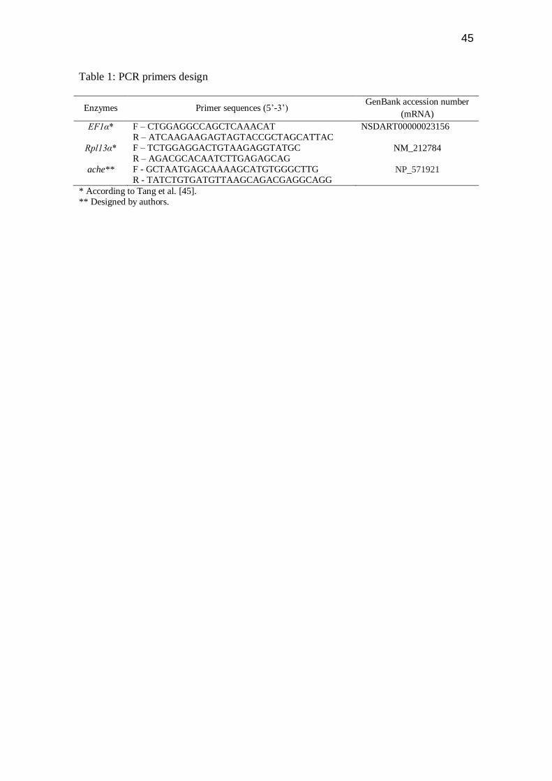
Table 1: PCR primers design
Enzymes Primer sequences (5‟-3‟) GenBank accession number
(mRNA)
EF1α* F – CTGGAGGCCAGCTCAAACAT
R – ATCAAGAAGAGTAGTACCGCTAGCATTAC
NSDART00000023156
Rpl13α* F – TCTGGAGGACTGTAAGAGGTATGC
R – AGACGCACAATCTTGAGAGCAG NM_212784
ache** F - GCTAATGAGCAAAAGCATGTGGGCTTG
R - TATCTGTGATGTTAAGCAGACGAGGCAGG NP_571921
* According to Tang et al. [45].
** Designed by authors.

46
Figure 1 Dal Forno et al., 2011

47
Figure 2 Dal Forno et al., 2011
48
Figure 3 Dal Forno et al., 2011
49
Figure 4 Dal Forno et al., 2011

50
Figure 5 Dal Forno et al., 2011
51
Figure 6 Dal Forno et al., 2011

52
3. CONSIDERAÇÕES FINAIS
O entendimento sobre efeito de cada nanocomposto nos mais diversos
tipos celulares está em pleno desenvolvimento. A importância de entendermos
todos os possíveis efeitos de uma exposição aos fulerenos é muito grande, já
que a sua produção e sua utilização estão crescendo a cada dia aumentando a
exposição do meio ambiente a este composto.
Como vem sendo mostrado em diferentes estudos, a toxicidade do
fulereno C60 varia de acordo com a linhagem celular estudada. Neste estudo,
foi mostrado que quando injetado por via intraperitoneal o efeito do
nanocomposto sobre a modulação da neurotransmissão colinérgica, foi
dependente da dose injetada, bem como do, tempo de exposição.
Por apresentarem a capacidade de atravessar todo tipo de membrana
corporal, os nanocompostos devem ser amplamente testados para ampliar
nosso conhecimento sobre as áreas a quais afetam. Devido a sua estrutura, o
fulereno C60 tem sido alvo para desenvolvimento de novos compostos,
adicionando os mais diversos radicais e elementos para observar as alterações
resultantes em suas propriedades.
Neste estudo nosso objetivo foi verificar se injeções intraperitoneais de
fulereno C60 nas doses de 7,5; 15 e 30 mg/kg e nos tempos de 6h, 12h e 24h
de exposição causariam alguma alteração na modulação do sistema
colinérgico. Observamos que a dose de 30 mg/kg, no tempo de exposição de
24h, apresentou um aumento de 84% na atividade enzimática quando
comparado com o grupo controle-veículo. Estes resultados sugerem um
possível efeito neurotóxico, embora estudos adicionais devam ser realizados
para estender estes achados.
53
REFERÊNCIAS BIBLIOGRÁFICAS
Amador CHS, Sistemas de fulereno C60 dopados com átomos covalentes e metais de transição: uma investigação computacional. Centro brasileiro de pesquisas físicas. 2006
Amatruda JF, Patton EE. Genetic models of cancer in zebrafish. Int. Rev. Cell. Mol. Biol. 2008;271:1-34. Anderson KV, Ingham PW. The transformation of the model organism: a decade of developmental genetics. Nat. Genet. 2003;33:285-293. Arenzana FJ, Clemente D, Sanchez-Gonzalez R, Porteros A, Aijon J, Arevalo R. Development of the cholinergic system in the brain and retina of the zebrafish, Brain Res. Bul. 2005;66:421-425. Barbazuk WB, Korf I, Kadavi C, Heyen J, Tate S, Wun E, et al. The syntenic relationship of the zebrafish and human genomes. Genome Res. 2000;10:1351-1358. Behra M, Cousin X, Bertrand C, Vonesch JL, Bielmann D, Chatonnet A, et al. Acetylcholinesterase is required for neuronal and muscular development in the zebrafish embryo. Nat Neurosci. 2002;5(2):111-118. Bertrand C, Chatonnet A, Takke C, Yan Y, Postlethwait J, Toutant JP, et al. Zebrafish acetylcholinesterase is encoded by a single gene localized on linkage group 7. J Biol Chem. 2001;276(1):464-474. Best JD, Alderton WK. Zebrafish: An in vivo model for the study of neurological diseases. Neuropsychiatr. Dis Treat. 2008;4(3):567-576. Boehmler, W, Obrecht-Pflumio S, Canfield V, Thisse C, Thisse B, Levenson R. Evolution and expression of D2 and D3 dopamine receptor genes in zebrafish. Dev Dyn. 2004;230:481-493. Borm PJA, Robbins D, Haubold S, Kuhlbusch T, Fissan H, Donaldson K, Schins R, Stone V, Kreyling W, Lademann J, Krutmann J, Warheit D, Oberdorster W. The potential risks of nanomaterials: a review carried out for ECETOC. Part Fibre Toxicol 3:11. 2006 Broughton RE, Milan JE, Roe BA. The complete sequence of the zebrafish (Danio rerio) mitochondrial genome and evolutionary patterns in vertebrate mitochondrial DNA. Genome Res. 2001; 11:1958-1967. Carvan III MJ, Dalton TP, Stuart GW, Nebert DW. Transgenic zebrafish as sentinels for aquatic pollution. Ann. N. Y. Acad. Sci. 2000;919:133-147.

54
Caulfield MP, Birdsall NJM. International Union of Pharmacology. XVII. Classification of muscarinic acetylcholine receptors. Pharmacol Rev. 1998;50:279-290. Clemente D, Porteros A, Weruaga E, Alonso JR, Arenzana FJ, Aijon J, et al. Cholinergic elements in the zebrafish central nervous system: histochemical and immunohistochemical analysis. J Comp Neurol. 2004;474:75-107. Cooper JR, Bloom FE, Roth RH. Acetylcholine. In: The biochemical basis of neuropharmacology. 6th ed. Oxford: Oxford University Press, 1991. p.190-213. Deguchi S, Alargova RG, Tsujii K. Stable dispersions of fullerenes, C60 and C70, in water. Preparation and characterization. Langmuir 2001;17:6013-6017. Delgado L, Schmachtenberg, O. Immunohistochemical localization of GABA, GAD65, and the receptor subunits GABAAalpha1 and GABAB1 in the zebrafish cerebellum. Cerebellum. 2008;7(3):444-450. Descarries L, Gisiger V, Steriade M. Diffuse transmission by acetylcholine in the CNS. Progress in Neurobiology. 1997;53:603-325. Dong W, Teraoka H, Yamazaki K, Tsukiyama S, Imani S, Imagawa T, et al. 2,3,7,8- tetrachlorodibenzo-p-dioxin toxicity in the zebrafish embryo: local circulation failure in the dorsal midbrain is associated with increased apoptosis. Toxicol. Sci. 2002;69(1):191-201. Dugan LL, Lovett EG, Quick KL, Lotharius J, Lin TT, O’Malley KL. Fullerene-based antioxidants and neurodegenerative disorders. Parkinsonism Relat Disord. 2001;7(3):243–246. Eddins D, Petro A, Williams P, Cerutti DT, Levin ED. Nicotine effects on learning in zebrafish: the role os dopaminergic systems. Psychopharmacology (Berl). 2009;202(1-3):103-109. Edwards JG, Michel WC. Odor-Stimulated glutamatergic neurotransmission in the zebrafish olfactory bulb. J. Comp. Neurol. 2002;454(3):294-309. Edwards JG, Greig A, Sakata Y, Elkin D, Michel WC. Cholinergic innervation of the zebrafish olfactory bulb. J. Comp. Neurol. 2007;504(6):631-645. Emran F, Rihel J, Dowling JE. A behavioral assay to measure responsiviness of zebrafish to changes in light intensities. J. Vis. Exp. (20). Pii, 923. 2008. Fernandes HL, Hodges-Savola CA. Tropic regulation of acetylcholinesterase isomerase isoenzymes in adult mammalian skeletal muscles. Neurochem. Res. 1992;17:115-124. Foley S, Crowley C, Smaihi M, Bonfils C, Erlanger B, Seta P, Larroque C. Cellular localisation of
55
a water-soluble fullerene derivative. Biochem. Biophys. Res. Commun. 2002;294:116-119. Gerlai R, Lahav M, Guo S, Rosenthal A. Drinks like a fish: zebrafish (Danio rerio) as a behavior genetic model to study alcohol effects. Pharmacol. Biochem. Behav. 2000;67:773-782. Gerlai R. Zebra fish: an uncharted behavior genetic model. Behav. Genet. 2003;33(5):461-468. Gerlai R, Chatterjee D, Pereira T, Sawashima T, Krishnannair R. Acute and chronic alcohol dose: population differences in behavior and neurochemistry of zebrafish. Genes Brain Behav. 2009. In Press. Goldsmith P. Zebrafish as a pharmacological tool: the how, why and when. Curr. Opin. Pharmacol. 2004;4(5):504-512. Gotti C, Clementi F. Neuronal nicotinic receptors: from structure to pathology. Prog Neurobiol. 2004;74:363-396. Grosell M, Wood CM. Copper uptake across rainbow trout gills: mechanisms of apical entry. J. Exp. Biol. 2002;205:1179-1188. Grunwald DJ, Eisen JS. Headwaters of the zebrafish - emergence os a model vertebrate. Nat. Rev. Genet. 2002;3:717-724. Guo S. Linking genes to brain, behavior and neurological diseases: What can we learn from zebrafish? Genes brain Behav. 2004;3:63-74. Hill A, Howard CV, Strahle U, Cossins A. Neurodevelopmental defects in zebrafish (Danio rerio) at environmentally relevant dioxin (TCDD) concentrations. Toxicol. Sci. 2003;76(2):392-399. Hill AJ, Teraoka H, Heideman W, Peterson RE. Zebrafish as a model vertebrate for investigating chemical toxicity. Toxicol Sci. 2005;86(1):6-19. Huang HM, Ou HC, Hsieh SJ, Chiang LY, Blockage of Amyloid Beta Peptide-Induced Cytosolic Free Calcium by Fullerenol-1 Carboxylate C60 in PC12 Cells, Life Sci. 2000,66(16):1525-1533. Isakovic A, Markovic Z, Markovic BT, Nikolic N, Djuric SV, Mirkovic M, Dramicanin M, Harhaji L, Raicevic N, Nikolic Z, Trajkovic V. Distinct Cytotoxic Mechanisms of Pristine versus Hydroxylated Fullerene. Oxford University Press, 2006. Kamat J, Devasagayam T, Priyadarsini K, Mohan H. Reactive oxygen species mediated membrane damage induced by fullerene derivatives and its possible biological implications. Toxicology. 2000; 155: 55–61.
56
Kapczinski F, Quevedo J, Izquierdo I (EDS). In: Bases biológicas dos transtornos psiquiátricos. Ed. Artes Médicas, Porto Alegre. 2000. Karlovich CA, John RM, Ramirez L, Stainier DY, Myers RM. Characterization of the Huntington’s disease (HD) gene homologue in the zebrafish (Danio rerio). Gene. 1998;217:117-125. Kaslin J, Panula P. Comparative anatomy of the histaminergic and other aminergic system in zebrafish (Danio rerio). J. Comp. Neurol. 2001;440:342-377. Kim JE, Lee M, Fullerene Inhibits Beta-Amyloid Peptide Aggregation, Biochem. Biophys. Res. Commun, 2003, 303(2):576–579. Kim YJ, Nam RH, Yoo YM, Lee CJ. Identification and functional evidence of GABAergic neurons in parts of the brain of adult zebrafish (Danio rerio). Neurosci. Lett. 2004;355:29-32. Kimmel C. Genetics and early development of zebrafish. Trend Genet. 1989;5:283-288. Kimmel C, Warga R. cell lineage and developmental potential of cells in the zebrafish embryos. Trends Genet. 1988;5:68-74. Kroto HW, Heath JR, O’Brien SC, Curl RF, Smalley RE. C60 Buckminsterfullerene. Nature. 1985, 318: 162-163 Krusic PJ, Wassermann E, Keizer PN, Morton JR, Preston KF. Radical reaction of C60. Science. 1991;254:1183–1185. Kucenas S, Li Z, Cox JA, Egan TM, Voigt MM. Molecular characterization of the zebrafish P2X receptor subunit gene family. Neuroscience. 2003;121:935-945. Lieschke GJ, Currie PD. Animal models of human disease: zebrafish swim into view. Nat. Rev. Genet. 2007;8(5):353-367. Lillesaar C, Tannhäuser B, Stigloher C, Kremmer E, BallycuIf L. The serotonergic phenotype is acquired by converging genetic mechanisms within the zebrafish central nervous system. Dev. Dyn. 2007;236(4):1072-1084. Low SE, Kuwada JY, Hume RI. Amino acid variations resulting in functional and nonfunctional zebrafish P2X(1) and P2X(5.1) receptors. Purinergic Signal. 2008;4(4):383-392. McKay EB, Placzek AN. Regulation of synaptic transmission and plasticity by neuronal nicotinic acetylcholine receptors. Biochemical Pharmacology. 2007;8:1120-1133. Mesulam MM, Guillozet A, Shaw P, Levey A, Duysen EG, Lockridge O. Acetylcholinesterase knouckouts establish central cholinergic pathways and can

57
use butyrylcholinesterase hydrolyse acetylcholine. Neuroscience. 2002;110:627-639. Milan DJ, MacRae CA. Zebrafish genetic models for arrhythmia. Prog. Biophys. Mol. Biol. 2008;98(2-3):301-308. Milatovic D, Dettbarn WD. Modification of acetylcholinesterase during adaption to chronic, subcute paraoxin aplication in rat. Toxicol. APP. Pharmacol. 1996;136:20-28. Moretto MB, Lermen CL, Morsch VM, Bohrer D, Ineu RP, Silva AC, et al. Effect of subchronic treatment with mercury chloride on NTPDase, 5’nucleotidase and acetylcholinesterase from cerebral cortex of rats. Journal of Trace Elements in Medicine and biology. 2004;17:256-260. Murr LE, Esquivel EV, Bang JJ, de la Rosa G, Gardea Torresdey JL. Chemistry and nanoparticulate compositions of a 10,000 year-old ice core melt water. Water Res. 2004. 38, 4282–4296. Nakagawa Y, Suzuki T, Ishii H, Nakae D, Ogata A, Cytotoxic effects of hydroxylated fullerenes on isolated rat hepatocytes via mitochondrial dysfunction 2011, 10.1007/s00204-011-0688-z Norton WH, Folchert A, Bally-Cuif L. Comparative analysis of serotonin receptor (HTR1A/HTR1B families) and transporter (slc6a4a/b) gene expression in the zebra fish brain. J Comp. Neurol. 2008;511(40):521-542. Oberdorster, E. Manufactured nanomaterials (fullerenes, C60) induce oxidative stress in the brain of juvenile largemouth bass. Environ. Health Perspect. 2004;112:1058–1062. Oda Y. Choline acetyltransferase: the structure, distribution and pathologic changesin the central nervous system. Pathology Interational. 1999;49:921-937. Podolski IYa, Podlubnaya ZA, Kosenko EA, et al., Effects of Hydrated Forms of C60 Fullerene on Amyloid 1-Peptide Fibrillization in Vitro and Performance of the Cognitive Task, J. Nanosci. Nanotechnol., 2007; 7(7):1479–1485. Postlethwait JH, Woods IG, Ngo-Hazelett P, Yan YL, Kelly PD, Chu F, et al. Zebrafish comparative genomics and the origins of vertebrate chromosomes. Genome Res. 2000;10(12):1890-1902. Quinna F. Nanotecnologia e o meio ambiente: perspectivas e riscos. Quim. Nova. 2004; 27(6): 1028-1029 Quinn DM. Acetylcholinesterase: Enzyme structure, reaction dynamics and virtual transition states. Chem. Rev. 1987;87:955-979.
58
Rico EP, Senger MR, Fauth MG, Dias RD, Bogo MR, Bonan CD. ATP and ADP hydrolysis in brain membranes of zebrafish (Danio rerio). Life Sci. 2003;73:2071-2082. Rico EP, Rosemberg DB, Senger MR, Arizi MD, Bernardi GF, Dias RD, et al. Methanol alters ectonucleotidases and acetylcholinesterase in zebrafish brain. Neurotoxicol. Teratol. 2006;28(4):489-496. Rico EP, Rosemberg DB, Dias RD, Bogo MR, Bonan CD. Ethanol alters acetylcholinesterase activity and gene expression in zebrafish brain. Toxicol Lett. 2007;174(1-3):25-30. Rico EP, Rosemberg DB, Senger MR, Arizi MB, Dias RD, Souto AA, et al. Ethanol and acetaldehyde alter NTPDase and 5'-nucleotidase from zebrafish brain membranes. Neurochem. Int. 2008;52(1-2):290-296. Rink E, Guo S. The too few mutant selectively affects subgroups of monoaminergic neurons in the zebrafish forebrain. Neurosci. 2004;27(1):147-154. Roex EWM, Keijzers R, Gestel CAM. Acetylcholinesterase inhibition and increased food consumption rate in the zebrafish, Danio rerio, after chronic exposure to parathion. Aquat. Toxicol. 2003;64(4):451-460. Rocha R.C. Os fulererenos e sua espantosa geometria molecular. Quim. Nov. Esco. 1996, 4: 7-11. Rosemberg DB, Rico EP, Senger MR, Arizi MB, Dias RD, Bogo MR, et al. Acute and subchronic copper treatments alter extracellular nucleotide hydrolysis in zebrafish brain membranes. Toxicology. 2007;236(1-2):132-139. Ruoff RS, Tse DS, Malhotra R, Lorents DC. Solubility of fullerene (C60) in a variety of solvents. J. Phys. Chem.1993; 97:3379-3383. Russek-Blum N, Gutnick A, Nabel-Rosen H, Blechman J, Staudt N, Dorsky RI, et al. Dopaminergic neuronal cluster size is determined during early forebrain patterning. Development. 2008;135(20):3401-3413. Ryu S, Holzschuh J, Mahler J, Driever W. Genetic analysis of dopaminergic system development in zebrafish. J. Neural. Transm. Suppl. 2006;70:61-66. Santos LJ, Rocha GP, Alves RB, Freitas RP. Fulereno[c60]: Química e aplicações Quim. Nova, Vol. 33, No. 3, 680-693, 2010 Sarter M, Parikh V. Choline Transporters, cholinergic transmission and cognition. Nat. Rev. Neurosci. 2005;6:49-56.

59
Senger MR, Rico EP, Dias RD, Bogo MR, Bonan CD. Ecto-5'-nucleotidase activity in brain membranes of zebrafish (Danio rerio). Comp. Biochem. Physiol. B. Biochem. Mol. Biol. 2004:139:203-207. Senger MR, Rico EP, Arizi MD, Rosemberg DB, Dias RD, Bogo MR, et al. Carbofuran and Malathion inhibit nucleotide hydrolysis in zebrafish (Danio rerio) brain membranes. Toxicology. 2005;212:107-115. Senger MR, Rico EP, Arizi MD, Frazzom AP, Dias RD, Bogo MR, et al. Exposure to Hg2+ and Pb2+ changes NTPDase and ecto- 5’-nucleotidase activities in central nervous system of zebrafish (Danio rerio). Toxicology. 2006a;226(2-3):229-237. Senger MR, Rosemberg DB, Rico EP, de Bem Arizi M, Dias RD, Bogo MR, et al. In vitro effect of zinc and cadmium on acetylcholinesterase and ectonucelotidase activities in zebrafish (Danio rerio) brain. Toxicol. In Vitro. 2006b;20(6):954-958. Serra EL, Medalha CC, Mattioli R. Natural preference of zebrafish (Danio rerio) for a dark environment. Braz. J. Med. Biol. Res. 1999;32:1551-1553. Sloman KA, Scott GR, Diao Z, Rouleau C, Wood CM, McDonald DG. Cadmium affects the social behaviour of rainbow trout, Oncorhynchs mykiss. Aquat. Toxicol. 2003;65:171-185. Soreq H, Seidman S. Acetylcholinesterase - news roles for an old actor. Nat. Rev. Neurosci. 2001;2(4):294-302. Spence R, Gerlach G, Lawrence C, Smith C. The behaviour and ecology of the zebrafish, Danio rerio. Biol. Rev. Camb. Philos. Soc. 2008;83(1):12-34. Sprague J, Doerry E, Douglas S, Westerfield M. The Zebrafish Information Network (ZFIN): a resource for genetic, genomic and developmental research. Nucleic Acids Res. 2001;29(1):87-90. Stern HM, Zon LI. Cancer genetics and drug discovery in the zebrafish. Nature Rev. Cancer. 2003;3:1-7. Tabor R, Friedrich RW. Pharmacological analysis of ionotropic glutamate receptor function in neuronal circuits of the zebrafish olfactory bulb. Plos One. 2008;3(1):14-16. Taylor MR, Hurley JB, Van Epps HA, Brockerhoff SE. A zebrafish model for pyruvate dehydrogenase deficiency: rescue of neurological dysfunction and embryonic lethality using a ketogenic diet. Proc. Natl. Acad. Sci. USA. 2004;101(13):4584-4589. Taylor P, Brown JH. In: Basic Neurochemstry: Molecular, Cellular and Medical Aspects, 5a ed., (eds. Siegel te al.). 1994. p. 231-260, Raven Press, LTDA, NY.
60
Taylor P, Brown JH, Acetylcholine. In: Siegel GJ, Agranof BW, Albers RW, Molinoff PB. (Ed.). Basic Neurochemistry: Molecular, cellular and Medical Aspects. USA, Philadelphia: Lippincott-Raven Publishers, 1999. pp. 214-242. Tinsley MR, Quinn JJ, Fanselow MS. The role of muscarinic and nicotinic cholinergic neurotransmission in aversive conditioning: comparing pavlovian fear conditioning and inhibitory avoidance. Learn Men. 2004;11:35-42. Uchiyama T, Chess-Williams R. Muscarinic receptor subtypes of the bladder and gastrointestinal tract. J. Smooth Muscle Res. 2004;40(6):237-247. Utsunomiya S, Jensen KA, Keeler GJ, Ewing RC. Uraninite and fullerene in atmospheric particulates. Environ. Sci. Technol. 2002, 36: 4943–4947. Wang ZX, Li XP, Wang WM, Xu XJ, Zi CT, Huang RB, Zheng, LS. Fullerenes in the fossil of dinosaur egg. Fuller. Sci.Technol.1998, 6: 715. Westerfield M. The zebrafish book: A guide for the laboratory use of zebrafish (Danio rerio). 4th ed. Eugene OR: University of Oregon Press. 2000. Yudoh K, Karasawa R, Masuko K, Kato T, Water-soluble fullerene (C60) inhibits the osteoclast differentiation and bone destruction in arthritis. Inter. Journ. Nanomedicine 2009, 4: 233-239 Zhao GC, Zhang P, Wei XW, Yang ZS. Determination of proteins with fullerol by a resonance light scattering technique. Anal Biochem. 2004;334:297–302. Zon LI, Peterson RT. In vivo drug discovery in the zebrafish. Nat. Rev. Drug Discov. 2005;4(1):35-44.

61
Anexo I (Comprovante de submissão do artigo científico)
MS: 8074715566403343 Exposure to nano/microparticles of fullerene (C60) increases acetylcholinesterase activity and lipid peroxidation in adult zebrafish (Danio rerio) brain Gonzalo O Dal Forno Jr, Luiza W Kist Dr, Mariana B Azevedo Jr, Rachel S Fritsch Jr, Talita CB Pereira Jr, Roberta S Britto Jr, Sílvia S Guterres Dr, Irene C Külkamp-Guerreiro Dr, Carla D Bonan Dr, José M Monserrat Dr and Maurício R Bogo Dr Particle and Fibre Toxicology Dear Dr Bogo Thank you for your recent submission to Particle and Fibre Toxicology. I would like to update you regarding your status with respect to the article processing charge that is normally due if a manuscript is accepted. You have agreed to pay an article processing charge of GBP 1,035.00/USD 1,640.00/EUR 1,205.00 if your manuscript is accepted. Since you are based in Brazil, you will be charged the price of GBP 1,035.00. Submissions from EU countries are subject to VAT at 20.0%. Payment will become due if your manuscript is accepted for publication by the editors. We allow payment by credit card or invoice. Invoice payments are subject to an administrative charge of £50.00. The article processing charge must be paid before the manuscript will be published. Kind regards, BioMed Central Accounts Team 236 Gray's Inn Road London WC1X 8HL Tel: +44 (0) 20 3192 2009 e-mail: [email protected]