Embed Size (px)
Citation preview
UNIVERSIDADE PAULISTA –UNIP
A ADMINISTRAÇÃO NO 18º DIA DA GESTAÇÃO DE
RATAS DE LIPOPOLISSACARÍDEO (LPS) MODIFICA A
INTERAÇÃO MATERNO-FILHOTE DA GERAÇÃO
PARENTAL E DAS GERAÇÕES F1 E F2
SANDRA HELOISA NUNES WHITAKER PENTEADO
SÃO PAULO
2012
Tese apresentada ao Programa de Pós-
graduação em Patologia Ambiental e
Experimental da Universidade Paulista-UNIP,
para obtenção do título de doutor em Patologia
Ambiental e Experimental.
Orientador: Prof. Dra. Maria Martha Bernardi
Penteado, Sandra Heloísa Nunes Whitaker
A administração no 18º dia da gestação de ratas de lipopolissacarideo (LPS) modifica a interação materno-filhote da geração parental e das gerações F1 e F2. / Sandra Heloisa Nunes Whitaker Penteado. – São Paulo, 2012. 89 f. : il. Tese (Doutorado) – Apresentada ao Programa de Pós-graduação em Patologia Ambiental e Experimental da Universidade Paulista, São Paulo, 2012 . Área de Concentração : Patologia Ambiental e Experimental “Orientação : Profa. Dra. Maria Martha Bernardi” 1. LPS. 2. Comportamento Maternal. 3. Agressão Materna. 4. Preferência Olfatória. 5. Epigenética. I. Universidade Paulista - UNIP. II. Título.

SANDRA HELOISA NUNES WHITAKER PENTEADO
A ADMINISTRAÇÃO NO 18º DIA DA GESTAÇÃO DE
RATAS DE LIPOPOLISSACARÍDEO (LPS) MODIFICA A
INTERAÇÃO MATERNO-FILHOTE DA GERAÇÃO
PARENTAL E DAS GERAÇÕES F1 E F2.
Aprovado em:
BANCA EXAMINADORA:
_______________________/__/___
Profa. Dra. Leoni Villano Bonamin Universidade Paulista – UNIP
_______________________/__/___ Profa. Dra. Ivana Barbosa Suffredini
Universidade Paulista – UNIP
_______________________/__/___ Profa. Dra. Elizabeth Teodorov Universidade Federal do ABC
_______________________/__/___
Prof. Dr. Luciano Freitas Felicio Universidade de São Paulo - USP
_______________________/__/___
Profa. Dra. Maria Martha Bernardi Universidade Paulista – UNIP
Tese apresentada ao Programa de Pós-
graduação em Patologia Ambiental e
Experimental da Universidade Paulista-UNIP,
para obtenção do título de doutor em Patologia
Ambiental e Experimental.

DEDICATÓRIA
Dedico essa tese de doutorado a minhas amadas filhas, Camila e Carolina,
que iluminam minha vida; aos meus pais, Luiz Antonio Nunes e Maria Ignez Barbieri
Nunes, pelo carinho e apoio; ao meu companheiro, Edmilson, por me ensinar o
significado da palavra felicidade; e a minha querida orientadora, Profa. Dra. Maria
Martha pelo apoio em todos os momentos e, principalmente, pelo carinho e pela
amizade.

“A ciência humana de maneira nenhuma nega a
existência de Deus. Quando considero quantas
e quão maravilhosas coisas o homem
compreende, pesquisa e consegue realizar,
então reconheço claramente que o espírito
humano é obra de Deus, e a mais notável.”
Galileu Galilei
AGRADECIMENTOS
Agradeço a Deus pelos momentos de felicidade, que iluminam e me dão força
para seguir a minha caminhada, e pelos momentos de dificuldade que me moldam a
cada instante para ser uma pessoa mais digna.
Agradeço a minha família, em especial minhas filhas e meus pais, pelo eterno
cuidado, dedicação e amor; pelo apoio nos momentos difíceis; por estarem ao meu
lado a cada pequena conquista e grandes realizações.
Agradeço ao meu amor e grande amigo Edmilson Cláudio Messias, pelo
companheirismo em todos os momentos, pelos sorrisos, pelo cuidado carinhoso e
por mostrar que sonhos podem ser reais.
Agradeço a Profa. Dra. Maria Martha Bernardi por ser mais que minha
orientadora, por acreditar na minha capacidade e no meu crescimento profissional e
pessoal, pelos conhecimentos repassados, pelo apoio em todos os momentos e,
principalmente, pela amizade.
A Universidade Paulista – UNIP, por me acolher e permitir o desenvolvimento
de meu trabalho.
Agradeço ao Dr. Paschoal Laércio Armonia, Diretor do Instituto de Ciências da
Saúde da Universidade Paulista – UNIP, pelo apoio e incentivo a realização deste
trabalho.
Agradeço ao Thiago Berti Kirsten, ao Thiago Marinho Reis e Silva, a Michelli
K. Acenjo, ao Raphael R. Melo e ao técnico do Laboratório de Pesquisas da UNIP
pelo auxílio no desenvolvimento do projeto.
Agradeço aos meus professores da pós-graduação e da graduação da
Universidade Paulista – UNIP, à secretaria de pós-graduação, à todos os
funcionários e a todos que contribuíram para o meu crescimento profissional e
pessoal.
RESUMO
Foram estudados os efeitos da administração de LPS no 18° dia de gestação (DG)
de ratas na interação materno-filhote das gerações parental, F1 e F2. Para tanto,
ratas prenhes receberam no DG18 100 µg/Kg da endotoxina ou seu veículo
(geração parental - P) e foram observados os comportamentos de recolher dos
filhotes, maternal e maternal agressivo. Na geração F1 foi avaliada a preferência
olfatória ao odor materno na infância e após a administração de uma dose adicional
de LPS no dia 21 da lactação, observou-se a atividade geral da prole masculina da
geração F1 e os níveis séricos de TNFα. Quando adultas, as ratas da geração F1
foram cruzadas com ratos sem qualquer tratamento e observados os seus
comportamentos ligados ao cuidado maternal. Na geração F2 testou-se a
preferência olfatória pelo odor materno e na idade adulta sua atividade geral em
campo aberto e no teste de ansiedade, o labirinto em cruz elevada.
Os resultados mostraram que, em relação ao grupo controle, a geração parental
apresentou facilitação no comportamento maternal e redução no comportamento
maternal agressivo. Na prole masculina da geração F1 verificou-se que os animais
de mães tratadas com a endotoxina tiveram menor preferência pelo odor materno, e
redução nos níveis de TNFα. Na atividade geral, os filhotes das ratas cujas mães
receberam o LPS não apresentaram alterações se comparadas àquela dos animais
do grupo controle.
Na geração F1, após cruzamento e na lactação, verificou-se aumento da latência
para assumir a posição maternal do comportamento maternal e do número e latência
para os ataques do comportamento maternal agressivo.
Na geração F2 observada na infância, a preferência olfatória pelo odor materno não
foi modificada, mas o número de filhotes indiferentes a esse odor do grupo
experimental foi maior que daquele do grupo controle. Na idade adulta, estes ratos
apresentaram menores índices de ansiedade. Concluiu-se que a exposição no 18°
dia da gestação ao LPS interfere na programação da interação mãe-filhote de duas
gerações.
Palavras chaves: LPS, comportamento maternal, agressão materna, preferência
olfatória, epigenética.
ABSTRACT
The effects of single prenatal LPS administration were investigated on maternal-pups
interaction of parental, F1 and F2 generations. Thus, pregnant rats received on DG18
100 µg / kg of LPS or its vehicle (parental generation) and the behaviors of pups
retrieval and the maternal and maternal aggressive. In the F1 generation the
olfactory preference of pups to maternal odor was assessed. In addition, at a
weaning these pups received an additional dose of LPS and the general activity
observed in an open field as well as the serum TNFα was measured. When adult
female rats of the F1 generation were mated with male rats without any treatment
and its maternal care were observed. In the F2 generation the olfactory preference of
pups to maternal odor were decreased. In adult age, the pups of F2 generation were
examined to their general activity in an open field and in the plus maze.
The results showed that in relation to the control group, the parental generation
showed facilitation of maternal behavior and reduction in maternal aggressive
behavior. In the male offspring of the F1 generation the animals from mothers treated
with endotoxin had less preference for maternal odor, and a decreased levels of
TNFα. In the open field behavior, these rats did not showed changes when
compared to that of control animals.
In the F1 generation there was increased latency to assume the maternal position
and in the number and latency to attack the intruder in maternal aggressive behavior.
In infancy, the olfactory preference of F2 generation for maternal odor was not
modified, but the number of pups indifferent to this odor in the experimental group
was higher than that of the control group. In adult age these rats showed a
decreased anxiety-like behavior in the plus maze.
It was concluded that exposure on day 18 of gestation to LPS acts as an imprinting,
which interferes with the maternal programming of the maternal-pups interaction of
two generations.
Keywords: LPS, maternal behavior, maternal aggression, olfactory preference,
epigenetics.

LISTA DE ILUSTRAÇÕES
Figura 1 - LPS da parede celular de bactérias gram-negativas............................ 12
Figura 2 - Mecanismo de ação simplificado do LPS em um macrófago,
culminando com a liberação de citocinas pró-inflamatórias................................... 14
Figura 3 - Parâmetros do comportamento maternal em ratas. Agrupamento de
filhotes (A), preparo da postura de amamentação (B), “crouching” ou cifose
fisiológica (C) e comportamento maternal total (D)................................................ 20
Figura 4 - Vias não genômicas de alterações no desenvolvimento. Fatores
como drogas, nutrição, toxina e idade podem pode levar a alterações
epigenéticas (círculo vermelho), que são então transmitidos à prole com
consequências para a variação fenotípica. Esta alteração pode conduzir a
diferencial pré-natal e / ou pós-natal no investimento materno, afentado o
desenvolvimento das crias gerado a partir deste cruzamento com
consequências para variação da prole fenotípica. Investimento materno
também pode variar como uma função das variações paternalmente mediadas
no fenótipo descendentes durante períodos tanto o pré-natal e / ou pós-natal.
Investimentos diferenciais maternos como uma função de experiências
paternas ou traços descendentes poderão servir tanto para aumentar a
transmissão de exposições paternas ou compensar défices de funcionamento
que são induzidas por estas experiências ambientais........................................... 29
LISTA DE ABREVIATURAS
125I-LPS LPS radiomarcado com iodo
ATV tegmental ventral
CD14 proteína de membrana periférica da superfície dos macrófagos
CM comportamento maternal
DG dia de gestação
DNA ácido desoxirribonucleico
GABA ácido gama-aminobutírico
HPA Eixo hipotálamo-pituitária
i.p. intraperitoneal
IL-1β interleucina 1 beta
IL-6 interleucina 6
IRAK interleukin-1 receptor associated kinase
LBP proteína ligadora de LPS, ou lipopolysaccharide binding protein
LPS Lipopolissacarídeo
MyD88 Myeloid differentiation primary response gene (88)
NF-kB fator de transcrição
P geração parental
PAG substância cinzenta periaquedutal
POA área pré-optica
quinase IkB inibidor do NF-kB (família do fator de transcrição nuclear kappa-
B)
RNA ácido ribonucleico
RNAm RNA mensageiro
siRNA small interfering RNA
SNC Sistema Nervoso Central
TAK-1 TGF-beta activated kinase 1
TLR toll-like receptor (receptor semelhante ao Toll)
TNFα Fator de necrose tumoral alfa (Tumor necrosis factor alpha)
TRAF6 TNF receptor associated factor
SUMÁRIO
1. INTRODUÇÃO ................................................................................................... 11
1.1. Sobre o Lipopolissacarídeo (LPS)............................................................... 11
1.2. Mecanismo de ação do LPS ....................................................................... 13
1.3. Citocinas ..................................................................................................... 16
1.4. Mecanismo de ação do LPS na infecção pré-natal ..................................... 17
2. SOBRE O COMPORTAMENTO MATERNAL ................................................... 19
2.1. Sobre a relação materno-filhote .................................................................. 21
3. OBJETIVO GERAL ........................................................................................... 23
3.1. Objetivos específicos .................................................................................. 23
4. CONSIDERAÇÕES GERAIS E DISCUSSÃO ................................................... 24
5. CONCLUSÃO .................................................................................................... 31
6. REFERÊNCIAS ................................................................................................. 32
ANEXOS ................................................................................................................... 37
ANEXO 1 – APROVAÇÃO PELO COMITÊ DE ÉTICA ............................................ 38
ANEXO 2 – ARTIGO ................................................................................................. 39
Prenatal lipopolysaccharide (LPS) increases maternal behavior, decreases maternal
odor preference and induces endotoxin hyporesponsiveness ................................... 39
ANEXO 3 – ARTIGO ................................................................................................. 59
Transgenerational effects of antenatal LPS exposure on maternal and pups
behaviors: studies in two generations. ...................................................................... 59

11
1. INTRODUÇÃO
1.1. Sobre o Lipopolissacarídeo (LPS)
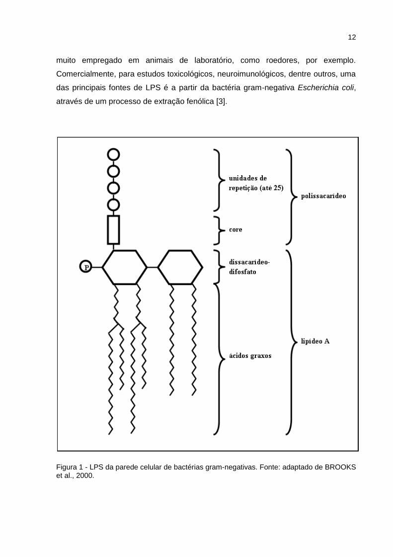
O LPS é uma endotoxina originária da parede celular de bactérias gram-
negativas. Consiste num lipídio complexo, denominado lipídio A, ao qual está ligado
um polissacarídeo constituído de um núcleo (ou core) e de uma série terminal de
unidades repetidas (Figura 1). O lipídio A consiste em unidades dissacarídicas de
glicosamina fosforilada as quais estão ligadas a vários ácidos graxos de cadeia
longa (podendo variar de acordo com a espécie bacteriana). O núcleo do
polissacarídeo é semelhante em todas as espécies gram-negativas que possuem
LPS, todavia, cada espécie contém uma unidade de repetição particular. Em geral,
as unidades de repetição consistem em trissacarídios lineares ou em tetra ou
pentassacarídios ramificados [1]. As moléculas de LPS de carga negativa são
ligadas de forma não covalente por cátions divalentes, tornando a membrana
estabilizada e proporcionando uma barreira contra moléculas hidrofóbicas. As
substâncias são termoestáveis, com peso molecular entre 3000 e vários milhões [1].
O LPS é sintetizado na membrana citoplasmática e transportado para sua
posição exterior final. É ligado à superfície celular, liberado apenas quando as
células são lisadas. Quando o LPS é clivado em lipídio A e em polissacarídeo, toda
a interação imune está associada ao lipídio A. A especificidade antigênica é
conferida pelas unidades terminais de repetição, que circundam a célula, formando
uma camada de polissacarídeos hidrofílicos [1].
A presença do LPS é necessária para a função de muitas proteínas da
membrana externa das bactérias [1]. Porém, o LPS pode ser extremamente tóxico
para animais. Administrações em doses menores que 1 nM já são capazes de ativar
o sistema imune do animal [2]. Os efeitos fisiopatológicos do LPS são semelhantes,
independente de sua origem bacteriana [1].
Dentro da área médica e veterinária, o LPS é muito utilizado nas mais
diferentes linhas de pesquisa, pelo seu efeito de estímulo do sistema imunológico. É
12
muito empregado em animais de laboratório, como roedores, por exemplo.
Comercialmente, para estudos toxicológicos, neuroimunológicos, dentre outros, uma
das principais fontes de LPS é a partir da bactéria gram-negativa Escherichia coli,
através de um processo de extração fenólica [3].
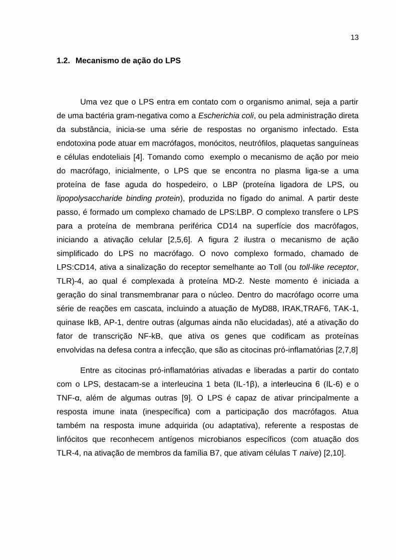
Figura 1 - LPS da parede celular de bactérias gram-negativas. Fonte: adaptado de BROOKS et al., 2000.
13
1.2. Mecanismo de ação do LPS
Uma vez que o LPS entra em contato com o organismo animal, seja a partir
de uma bactéria gram-negativa como a Escherichia coli, ou pela administração direta
da substância, inicia-se uma série de respostas no organismo infectado. Esta
endotoxina pode atuar em macrófagos, monócitos, neutrófilos, plaquetas sanguíneas
e células endoteliais [4]. Tomando como exemplo o mecanismo de ação por meio
do macrófago, inicialmente, o LPS que se encontra no plasma liga-se a uma
proteína de fase aguda do hospedeiro, o LBP (proteína ligadora de LPS, ou
lipopolysaccharide binding protein), produzida no fígado do animal. A partir deste
passo, é formado um complexo chamado de LPS:LBP. O complexo transfere o LPS
para a proteína de membrana periférica CD14 na superfície dos macrófagos,
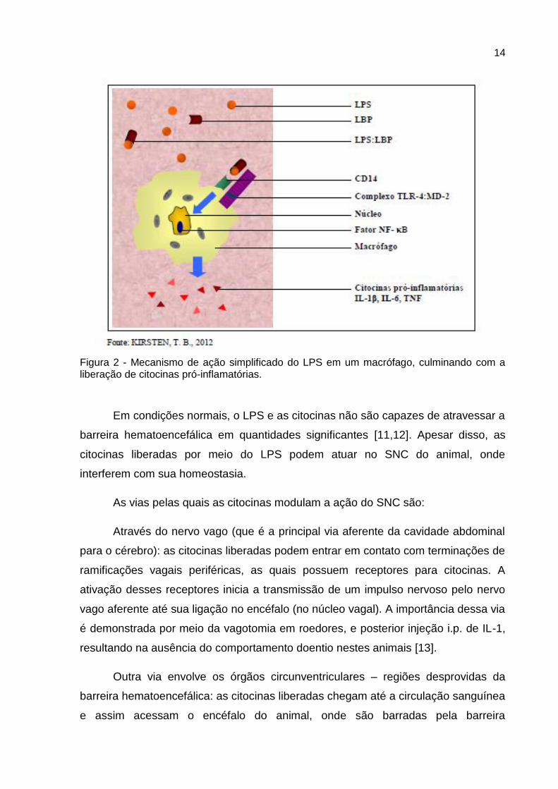
iniciando a ativação celular [2,5,6]. A figura 2 ilustra o mecanismo de ação
simplificado do LPS no macrófago. O novo complexo formado, chamado de
LPS:CD14, ativa a sinalização do receptor semelhante ao Toll (ou toll-like receptor,
TLR)-4, ao qual é complexada à proteína MD-2. Neste momento é iniciada a
geração do sinal transmembranar para o núcleo. Dentro do macrófago ocorre uma
série de reações em cascata, incluindo a atuação de MyD88, IRAK,TRAF6, TAK-1,
quinase IkB, AP-1, dentre outras (algumas ainda não elucidadas), até a ativação do
fator de transcrição NF-kB, que ativa os genes que codificam as proteínas
envolvidas na defesa contra a infecção, que são as citocinas pró-inflamatórias [2,7,8]
Entre as citocinas pró-inflamatórias ativadas e liberadas a partir do contato
com o LPS, destacam-se a interleucina 1 beta (IL-1β), a interleucina 6 (IL-6) e o
TNF-α, além de algumas outras [9]. O LPS é capaz de ativar principalmente a
resposta imune inata (inespecífica) com a participação dos macrófagos. Atua
também na resposta imune adquirida (ou adaptativa), referente a respostas de
linfócitos que reconhecem antígenos microbianos específicos (com atuação dos
TLR-4, na ativação de membros da família B7, que ativam células T naive) [2,10].
14
Figura 2 - Mecanismo de ação simplificado do LPS em um macrófago, culminando com a liberação de citocinas pró-inflamatórias.
Em condições normais, o LPS e as citocinas não são capazes de atravessar a
barreira hematoencefálica em quantidades significantes [11,12]. Apesar disso, as
citocinas liberadas por meio do LPS podem atuar no SNC do animal, onde
interferem com sua homeostasia.
As vias pelas quais as citocinas modulam a ação do SNC são:
Através do nervo vago (que é a principal via aferente da cavidade abdominal
para o cérebro): as citocinas liberadas podem entrar em contato com terminações de
ramificações vagais periféricas, as quais possuem receptores para citocinas. A
ativação desses receptores inicia a transmissão de um impulso nervoso pelo nervo
vago aferente até sua ligação no encéfalo (no núcleo vagal). A importância dessa via
é demonstrada por meio da vagotomia em roedores, e posterior injeção i.p. de IL-1,
resultando na ausência do comportamento doentio nestes animais [13].
Outra via envolve os órgãos circunventriculares – regiões desprovidas da
barreira hematoencefálica: as citocinas liberadas chegam até a circulação sanguínea
e assim acessam o encéfalo do animal, onde são barradas pela barreira
15
hematoencefálica. Para adentrar o cérebro, elas acessam os órgãos
circunventriculares. É possível confirmar a importância desta via, pois são
detectados níveis elevados de certas citocinas pró-inflamatórias após infecção nos
órgãos circunventriculares como a área postrema, eminência mediana e órgão
vasculoso da lâmina terminal, comparados a outras áreas do cérebro[12].
As citocinas atuam ainda a partir do contato com células endoteliais do
organismo: o LPS e as citocinas, que em contato com os receptores das células
endoteliais, induzem a ativação da enzima ciclooxigenase (COX), iniciando uma
resposta no organismo, que leva à produção de eicosanoides (mediadores
inflamatórios de origem lipídica), como as prostaglandinas, leucotrienos e
tromboxanos. Esses eicosanoides têm propriedades físico-químicas que os
possibilitam, via corrente sanguínea, acessar o cérebro, atravessando a barreira
hematoencefálica, podendo assim induzir processos patológicos. Para mostrar a
relevância desta via, trabalhos utilizam inibidores de eicosanoides, como por
exemplo, inibidores da enzima COX-2, resultando na supressão do comportamento
doentio [4,12,14-16].
Outras vias também podem contribuir para a atuação das citocinas no
cérebro, como quando as citocinas cruzam a barreira hematoencefálica usando
sistemas de captura específicos, porém, especialistas consideram a capacidade
desses sistemas relativamente baixa [12].
Finalmente, o LPS é ainda capaz de produzir a enzima óxido nítrico sintase,
que leva à produção do óxido nítrico, que é um importante mediador inflamatório,
com ação vasodilatadora, podendo agir também no SNC [15,17]. Provavelmente,
esses distintos mecanismos atuam simultaneamente, de forma integrada, quando da
liberação de citocinas [12].
Diversas regiões do cérebro expressam receptores para diversas citocinas
(incluindo IL-1β, TNF-α e IL-6) tanto na glia quanto nos neurônios [18]. Aventa-se
que a micróglia em contato com as citocinas liberadas pelo LPS, estimula a
produção de novas citocinas no próprio cérebro, potencializando o seu efeito. Neste
sentido, a micróglia é considerada um análogo dos macrófagos e “órgão imune” do
cérebro, com função de combater infecções e a inflamação [9,16,19].
16
1.3. Citocinas
Normalmente, as citocinas atuam no organismo a fim de combater diversos
patógenos. No sistema imune elas participam de respostas adaptativas ou reações
homeostáticas [2,18]. Dentre outras funções, as citocinas pró-inflamatórias
funcionam como sinalizadores moleculares do sistema imune para informar o
cérebro sobre inflamação periférica [20].
Muitos fatores imunes são liberados e participam no sentido de remover o
patógeno invasor, agindo localmente além de orquestrar uma complexa difusão de
alterações através de todo o organismo [18]. O problema ocorre quando existe
liberação excessiva de mediadores pró-inflamatórios, que desencadeiam respostas
exacerbadas, tornando-se prejudiciais ao funcionamento do organismo , levando-o
à inflamação sistêmica associada com o desenvolvimento de sérias complicações,
podendo até mesmo levar ao choque séptico e morte do indivíduo [20].
No SNC as citocinas podem modular neurotransmissores centrais como
dopamina, serotonina, noradrenalina, ácido gama-aminobutírico (GABA),
acetilcolina, neuropeptídeos, dentre outros. Atuam ainda na diferenciação e
crescimento neuronal, na migração dos neurônios para seus alvos e na modificação
da plasticidade sináptica. Portanto, em níveis fisiológicos, as citocinas
desempenham importantes papéis no cérebro, como, por exemplo, na neurogênese,
neuromodulação, na memória e no sono [21,22]. Porém, as citocinas podem causar
morte celular durante o desenvolvimento cerebral [12,23,24]. As citocinas podem
também ativar o eixo HPA com a liberação do fator liberador de corticotrofina do
hipotálamo, que secreta o hormônio adrenocorticotrópico da glândula pituitária,
resultando em aumento de glicocorticoides na corrente sanguínea periférica. Esses
glicocorticoides têm função básica de frear a ativação do sistema imune. Em níveis
elevados no SNC e em exposições crônicas são prejudiciais ao indivíduo,sendo
conhecidos como os hormônios do estresse, podendo estas substâncias podem
causar danos, como por exemplo, a morte de neurônios ([25].
Além disso, as citocinas podem inibir o eixo hipotálamo-pituitária-gonadal, por
reduzir a secreção de hormônios sexuais (hormônio gonadotrófico, hormônio
17
luterizante, hormônio folículo estimulante e esteroides ovarianos), interferindo na
modulação do comportamento reprodutivo [26,27]. Em resposta a infecções
periféricas, células imunes inatas produzem citocinas pró-inflamatórias que agem no
cérebro produzindo uma série de alterações comportamentais que se enquadram e
podem ser definidas como o comportamento doentio [13,28]. O comportamento
doentio é geralmente acompanhado pela diminuição da atividade exploratória, da
interação social, do interesse sexual, perda de apetite, anedonia e prejuízos
cognitivos e no aprendizado [29]. Essa série de alterações é uma estratégia
comportamental e adaptativa do organismo, visando o combate ao microrganismo
invasor e a cura rápida [30].
1.4. Mecanismo de ação do LPS na infecção pré-natal
O LPS normalmente não é capaz de chegar até o feto. Já se verificou que
após a administração de LPS radiomarcado com iodo (125I-LPS) em ratas prenhes,
ele foi detectado no sangue, fígado, rins e placenta das mães entre 1-8 h, porém
nada foi encontrado no feto. Observou-se, no entanto, a indução de citocinas em um
período de 2-8 h no plasma materno. Este fato, somado à presença de LPS na
placenta, sugere que o LPS deve agir diretamente nas células placentárias para
induzir a expressão de mediadores inflamatórios. Portanto, as alterações
encontradas na prole não são produzidas diretamente pela endotoxina, pois o LPS
parece não sofrer passagem transplacentária [11].
Ainda foram encontrados, níveis elevados de citocinas na placenta, no fluído
amniótico, no sangue e cérebro fetal (inclusive com a indução de genes de citocinas
pró-inflamatórias no cérebro fetal após administração de LPS na mãe), bem como a
ocorrência de inflamação de membranas fetais, após infecções e a inflamação
materna. Sabe-se também da existência de TLRs na placenta e em membranas
fetais [26,31-33].
As citocinas acessam o cérebro fetal por diferentes maneiras: a maioria vem
do lado materno produzidas no útero e placenta durante a infecção intrauterina,
atravessando a barreira hematoencefálica imatura do feto e acessando o SNC. Além
18
disso, as citocinas podem ser produzidas na micróglia e nos astrócitos do cérebro
fetal a partir da estimulação de citocinas oriundas da mãe [31]. Essas citocinas
podem interferir na homeostasia do ambiente fetal, alterando o desenvolvimento de
seu eixo neuroimune [16,34].
Além das citocinas, os glicocorticoides também podem atuar nos filhotes,
sendo capazes de atravessar a barreira hematoencefálica e influenciar processos de
Desenvolvimento cerebral dos fetos. Assim, já foi documentada a liberação de
hormônio corticotrópico no cérebro fetal após administração de LPS em ratas
gestantes, sugerindo a possibilidade de indução da resposta estressora no feto [35].
Portanto, citocinas e glicocorticoides podem ser os responsáveis indiretos
pelos danos encontrados nos filhotes expostos prenatalmente ao LPS e essas
alterações podem perdurar até mesmo na idade adulta do animal [9,32].
19
2. SOBRE O COMPORTAMENTO MATERNAL
Durante o desenvolvimento, o SNC é extremamente plástico para as
intervenções do ambiente. A experiência é essencial durante as primeiras semanas
pós-natal em que as atividades sensoriais se refinam e estabelecem conexões
neurológicas estáveis. Sabe-se que, os filhotes de mãe que passaram por algum
evento ou stress durante a gestação podem apresentar alterações no seu
desenvolvimento global, desta forma o comportamento maternal se apresenta como
fundamental para o desenvolvimento inicial de recém-nascidos [36].
O comportamento maternal é um comportamento complexo, instintivo, com
características específicas para cada espécie e que consiste em uma série de
cuidados que as fêmeas maduras realizam em torno dos indivíduos imaturos para
auxiliar na propagação de sua espécie, sendo um fator determinante no
desenvolvimento neurológico [37].
Os cuidados maternais se expressam desde a preparação da mãe para o
nascimento da prole e se mantêm por todo o período de lactação dos filhotes. Esse
comportamento vai se modificando de acordo com o tempo e crescimento dos
filhotes. Durante este período, o principal objetivo da fêmea é garantir a
sobrevivência dela e dos seus filhotes [37,38].
De acordo com os autores, na preparação para o parto, a mãe prepara o
ninho para acolher os filhotes na hora do parto. Nos primeiros 10 dias após o parto,
as mães permanecem mais tempo no ninho e à medida que os filhotes crescem se
tornando mais independentes em relação à mãe e à própria independência, os
cuidados maternos tendem a decrescer e a mãe se torna menos responsiva em
relação aos filhotes. Com o ganho de independência dos filhotes e a diminuição da
responsividade materna, o desmame tende a acontecer naturalmente.
Em ratas, os cuidados maternais são observados e registrados quando os
comportamentos são relacionados aos filhotes como a busca, o agrupamento, ficar
sobre os filhotes aquecendo-os e os alimentando, além de comportamentos
indiretos como agressividade e construção do ninho.
20
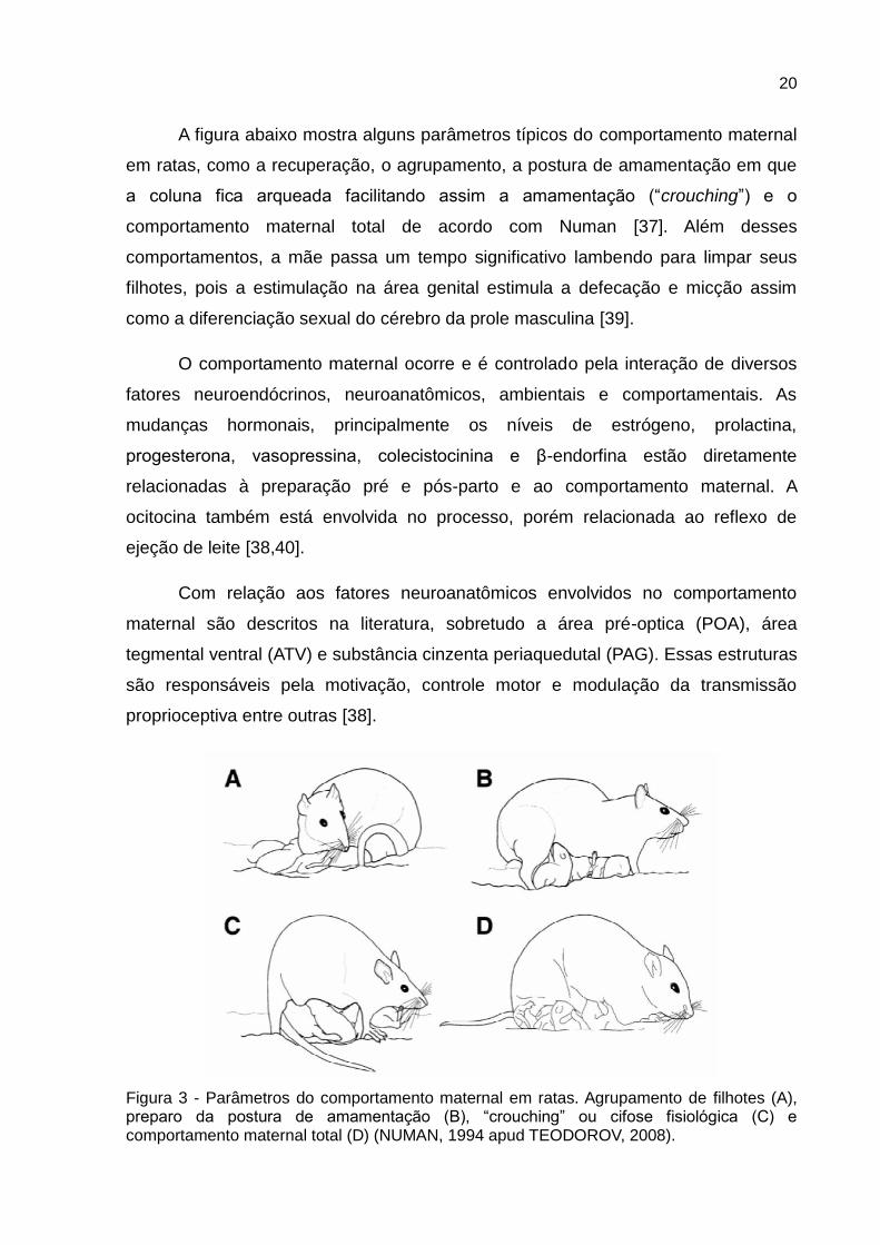
A figura abaixo mostra alguns parâmetros típicos do comportamento maternal
em ratas, como a recuperação, o agrupamento, a postura de amamentação em que
a coluna fica arqueada facilitando assim a amamentação (“crouching”) e o
comportamento maternal total de acordo com Numan [37]. Além desses
comportamentos, a mãe passa um tempo significativo lambendo para limpar seus
filhotes, pois a estimulação na área genital estimula a defecação e micção assim
como a diferenciação sexual do cérebro da prole masculina [39].
O comportamento maternal ocorre e é controlado pela interação de diversos
fatores neuroendócrinos, neuroanatômicos, ambientais e comportamentais. As
mudanças hormonais, principalmente os níveis de estrógeno, prolactina,
progesterona, vasopressina, colecistocinina e β-endorfina estão diretamente
relacionadas à preparação pré e pós-parto e ao comportamento maternal. A
ocitocina também está envolvida no processo, porém relacionada ao reflexo de
ejeção de leite [38,40].
Com relação aos fatores neuroanatômicos envolvidos no comportamento
maternal são descritos na literatura, sobretudo a área pré-optica (POA), área
tegmental ventral (ATV) e substância cinzenta periaquedutal (PAG). Essas estruturas
são responsáveis pela motivação, controle motor e modulação da transmissão
proprioceptiva entre outras [38].
Figura 3 - Parâmetros do comportamento maternal em ratas. Agrupamento de filhotes (A), preparo da postura de amamentação (B), “crouching” ou cifose fisiológica (C) e comportamento maternal total (D) (NUMAN, 1994 apud TEODOROV, 2008).
21
O controle do comportamento maternal envolve fatores neuroendócrinos e
neuroanatômicos. Os hormônios gestacionais preparam o animal para agir de forma
maternal para com o filhote, já os neurotransmissores regulam o comportamento
maternal durante a fase de manutenção e lactação [41]. A primeira fase da regulação
do comportamento maternal determina o início rápido deste no pós-parto, sendo
controlada por hormônios relacionados com a gestação e lactação (estrógeno,
progesterona, prolactina e ocitocina). A segunda fase, a de manutenção durante a
lactação, é controlada principalmente por fatores não hormonais, na qual o estímulo
proveniente do filhote se mostra o mais importante [42].
O comportamento maternal, portanto, é resultado da interação entre diversos
fatores maternos e é fundamental para o desenvolvimento e sobrevivência do filhote.
Desta forma, as alterações no período gestacional podem alterar o comportamento
maternal interferindo no desenvolvimento neurológico, comportamental e sexual do
filhote, além das possíveis alterações provenientes do período gestacional
[37,38,40]. Interferências no cuidado materno promovidas são cruciais no
desenvolvimento e na expressão comportamental da prole por alterar a
programação do seu desenvolvimento.
2.1. Sobre a relação materno-filhote
Infecções prenatais interferem com o sistema sensorial dos animais. Por
exemplo, a exposição ao LPS ou ao polyriboinosinic-polyribocytidilic acid (polyI:C,
que mimetiza infecções virais) em ratos e camundongos prejudica a aquisição de
informações cognitivas e sensoriais, em modelo de inibição da resposta do reflexo
acústico [43].
O sistema olfatório dos ratos, assim como dos mamíferos, é capaz de
detectar e discriminar milhares de diferentes moléculas no ambiente e essa
habilidade é crucial para o seu desenvolvimento e sobrevivência. Apesar de estar
presente desde o nascimento, o sistema olfatório se desenvolve durante a vida do
animal e se aprimora de acordo com as informações olfatórias adquiridas e
armazenadas. Os filhotes de ratos são menos sensíveis a odores do que ratos
22
adultos, possivelmente pela quantidade limitada de inervações sensoriais formadas
nesta etapa, porém, mesmo na infância, os filhotes já são capazes de se guiar e
utilizar as pistas olfatórias para sua sobrevivência [44].
Kirsten et al [45] mostram que a exposição no 9,5 dia da gestação não
modifica o comportamento maternal de ratas, porém reduz a preferência dos filhotes
para se encaminhar para a maravalha com odor da mãe quando comparados
àqueles filhotes do grupo controle. Além disto, verifica-se decréscimo da dopamina
do bulbo olfatório destes filhotes. Portanto a exposição a endotoxina leva ao
prejuízos no reconhecimento materno pelos filhotes e, este fato, não foi
consequência de interferências com o cuidado materno das fêmeas tratadas com
LPS.
O estudo das consequências da exposição ao LPS nas demais gerações,
torna-se importante quando infecções ocorrem no período perinatal uma vez que
podem interferir tanto no cuidado maternal como no reconhecimento da mãe pelo
filhote.
Nesta revisão foram enfocados diversos aspectos da interação entre o
sistema imune e o sistema nervoso central, em particular durante a gestação e do
processo do desenvolvimento dos seus descendentes. São várias a linhas que
investigam estas relações cujo âmbito é interdisciplinar, sendo a área denominada
de neuro-endócrino-imunomodulação. A visão ampliada dos diferentes aspectos e
efeitos de processos inflamatórios durante o período do desenvolvimento abre
espaços para o entendimento de diversas doenças, em particular àquelas ligadas às
doenças mentais.
23
3. OBJETIVO GERAL
Este trabalho tem como objetivo investigar a interação materna- filhotes em
duas gerações quando a geração parental recebeu no GD18 o LPS.
3.1. Objetivos específicos
1. Avaliar os efeitos da exposição de dose única do lipopolissacarídeo na
interação materno-filhote e nos níveis de TNF-α séricos da prole após
desafio com a mesma endotoxina.
2. Avaliar os efeitos transgeracionais da exposição antenatal no
comportamento maternal e dos filhotes em duas gerações.
24
4. CONSIDERAÇÕES GERAIS E DISCUSSÃO
Na avaliação dos efeitos da exposição de dose única do lipopolissacarídeo no
GD18 na interação materno-filhote das gerações parental e F1 observou-se
facilitação do comportamento maternal e redução do comportamento maternal
agressivo. No entanto, este tratamento não modificou o desempenho reprodutivo
das fêmeas. Em relação aos filhotes, no LD21, a administração de uma dose
desafio da mesma endotoxina aumentou o peso corporal e a duração de
imobilidade no teste do campo aberto. Além disto, esta dose desafio mostrou que a
exposição pré-natal ao LPS induziu tolerância à mesma endotoxina, expressa por
menor aumento nos níveis séricos de TNFα .
No comportamento maternal das fêmeas tratadas prenatalmente com LPS
observou-se redução na latência para a busca do primeiro filhote quando comparada
àquela do grupo controle. Em contraste, no comportamento maternal agressivo,
verificou-se nestes animais redução no número de ataques e na duração do tempo
de briga. Estes dados, aparentemente contraditórios, foram interpretados como
resultado do comportamento doentio induzido pelo LPS. De fato, esta endotoxina
promove febre e no caso, a administração do LPS no GD18 pode ter sinalizado para
a fêmea que o ambiente estaria frio aumentando a motivação materna para a busca
do filhote. Neste sentido, ratos ao nascer não controlam a temperatura corporal,
sendo provável que a menor latência para a busca do primeiro filhote tenha sido
causada pela sinalização nas fêmeas e necessidade de buscar e aquecer sua prole.
Além disto, como a fêmea cuidou mais de sua prole, ela selecionou este
comportamento em detrimento da proteção contra um macho invasor.
Com relação aos filhotes, verificou menor preferência pelo odor materno na
prole exposta ao LPS sugerindo menor reconhecimento materno. Desde que, a
atividade geral destes filhotes não foi modificada pela exposição pré-natal ao LPS,
interpretou-se este dado como devido à redução na motivação da prole promovida
pela endotoxina pré-natal. De fato, estudos anteriores mostraram que ratos expostos
prenatalmente ao LPS tinham sua interação social reduzida por apresentar menor
motivação [46].
25
Para testar a sensibilidade destes filhotes ao LPS foram avaliadas a atividade
geral em campo aberto e os níveis séricos de TNFα aos 21 dias de idade após
desafio com uma dose adicional da endotoxina.
Notou-se que ocorreu aumento na imobilidade nos animais tratados
prenatalmente com salina e aos 21 dias com LPS, tendo sido sugerido que este fato
tenha sido consequência do desenvolvimento de comportamento doentio. Por outro
lado, ratos tratados prenatalmente com LPS e desafiados com a mesma endotoxina
não apresentaram alterações na atividade geral bem como atenuação dos níveis
séricos de TNFα, fato interpretado como tolerância aos efeitos do LPS.
Na geração F1 notou-se que a exposição pré-natal ao LPS modificou o
comportamento maternal e maternal agressivo sem, no entanto, alterar sua atividade
geral. Na geração F2, a exposição antenatal reduziu o peso corporal no desmame e
a preferência pelo odor maternal. Na idade adulta, tanto a prole tratada
antenatalmente com LPS como aquela tratada com salina foram subdivididas em
dois grupos, dois dos quais foram isolados por uma semana enquanto que os
demais grupos permaneceram agrupados. Este procedimento teve como objetivo
revelar se o tratamento antenatal com LPS teria promovido alterações no sistema
nervoso central dos mesmos uma vez que o isolamento representa um estresse
para os animais. De fato, embora, a atividade geral dos ratos do grupo LPS+ LPS
(tratados antenatalmente com LPS e desafiados na idade adulta com a mesma
endotoxina) não tenha sido modificada, no teste do labirinto elevada estes animais
apresentaram menores índices de ansiedade.
No comportamento maternal notou-se redução na latência para o “crouching”,
mas ocorreu aumento no tempo de emissão deste comportamento. O
comportamento de “crouching” é uma postura quiescente e, geralmente, ocorre em
resposta à estimulação das crias. Fêmeas lactantes tendem a reduzir outras
atividades e apresentar uma postura característica com suas extremidades abertas e
costas arqueadas. A finalidade desta postura é permitir que o filhotes tenham
acesso às tetas e ao leite, para regular a sua temperatura, e para protegê-los de
agressões ambientais. Desta forma, mesmo com redução na latência para o
comportamento de “crouching”, a maior duração do mesmo pode ter compensado
este atraso no cuidado maternal. Portanto, é improvável que a redução de peso ao

26
final do desmame se deva á disponibilidade das fêmeas em amamentar sua prole.
Duas hipóteses podem explicar estes efeitos. Primeiro, o atraso na expressão do “
crouching” pode ter sido causado por uma menor estimulação da cria em relação á
sua mãe, pois notou-se que no teste de preferência olfatória estes filhotes
apresentaram menor atividade voltada à sua mãe. Em segundo lugar, não se pode
descartar que as mães da geração F1 podem ter tido redução na disponibilidade de
leite e com isto os filhotes apresentaram redução no peso corporal ao no desmame.
Note-se que logo após o nascimento estes filhotes não tinham alterações no peso
corporal indicando que intra-útero não houve prejuízos no aporte de nutrientes à
prole. Não se pode ainda descartar que o atraso na expressão do “crouching” possa
ter sido motivado pela menor estimulação promovida pelas crias e a maior duração
do mesmo pela tentativa de fornecer mais leite à prole.
Estes dados indicam que a exposição pré-natal e antenatal ao LPS promove
alterações na interação materno-filhote que se reflete na geração F2 em menor
desenvolvimento corporal e redução do estímulo da prole em relação à sua mãe.
Na idade adulta destas proles foram analisadas a atividade geral em campo
aberto e a resposta no teste do labirinto elevado. Neste caso, tanto os animais do
grupo antenatalmente tratados com LPS ou solução salina foram subdivididos em
dois grupos e obtiveram-se quatro grupos, a saber: S+AS (tratados com salina
antenatalmente que receberam solução salina na idade adulta agrupados), S + SI
(tratados com salina antenatalmente que receberam solução salina na idade adulta
isolados), LPS + LPSA (tratados com LPS antenatalmente que receberam outra
dose de LPS na idade adulta agrupados) e LPS + LPSI (tratados com LPS
antenatalmente que receberam outra dose de LPS na idade adulta isolados).
A análise dos efeitos em longo prazo da prole masculina das ratas da geração
F2 indicou menores níveis de ansiedade da prole antenatalmente tratada com o LPS
que foi isolada. Neste caso, o isolamento revelou alterações na emocionalidade dos
animais antenatalmente expostos a endotoxina as quais, por adaptação em
condições normais da vida do animal não se expressariam. No caso da atividade
geral, o isolamento aumentou a atividade geral quer seja em animais do grupo
controle quer seja no caso dos animais do grupo experimental. Portanto, este
modelo não foi capaz de revelar as alterações promovidas pela exposição antenatal
27
ao LPS.
É fato conhecido que o estresse em períodos precoces da vida leva a
alterações na reatividade ao estresse a qual persiste ao longo da vida até a idade
adulta. De fato, intervenções severas maternas como, por exemplo, a separação
materna, e mesmo a manipulação pré-natal, que representa um estresse menos
severo, sensibilizam o eixo hipotálamo-hipófise-adrenal e levam a um fenótipo
resiliente ao estresse. Este efeito pode se refletir nas demais gerações por
mecanismos denominados de epigenéticos.
O termo epigenética refere-se a todas as mudanças reversíveis e herdáveis
no genoma funcional e que não alteram a sequência de nucleotídeos do DNA [47].
De acordo com Hunter [48] o controle epigenético é a soma dos fatores genéticos e
não genéticos que agem sobre as células de forma a controlar seletivamente a
expressão dos genes, produzindo assim o aumento da complexidade fenotípica
durante o desenvolvimento. Dessa forma, seu estudo direciona-se a compreensão
dos padrões de expressão transmitidos aos descendentes, sua mudança de
expressão de genes durante a diferenciação de um tipo de célula e como os fatores
ambientais podem modificar a expressão de genes.
Os principais mecanismos de alterações epigenéticas são: (1) metilação do
DNA; (2) modificações de histonas e (3) ação de RNAs não codificadores. Os
padrões de metilação de DNA são os mais conhecidos destes três mecanismos,
embora modificações de histonas também sejam bastante discutidas [48].
A metilação do DNA está relacionada normalmente ao silenciamento de
genes. As acetilações, fosforilações e ubiquitinações são modificações de histonas
já melhor estudadas. Já a ação de RNAs não codificadores está relacionada ao
silenciamento póstranscricional de genes através do mecanismo de RNA de
interferência onde ocorre o bloqueio da tradução ou degradação do RNAm alvo.
Além, da ação bloqueadora da transcrição, os siRNA podem ser associados à
metilação de seqüências de DNA. Todos estes mecanismos parecem estar
interligados para a organização estrutural da cromatina tornando-a mais acessível
ou não aos fatores de transcrição[49].
28
As mudanças epigenéticas são fortemente influenciadas pelo ambiente, de
forma que alterações no mesmo, promovidas por ataques de patógenos, tipo de
alimentação, etc., podem acarretar mudanças epigenéticas (Figura 4). Ou seja, é um
processo pelo qual o genótipo de um organismo interage com o meio ambiente para
produzir o seu fenótipo. Sendo assim, a epigenética está intimamente relacionada
com o aumento de variabilidade fenotípica dos indivíduos resultando em uma
relevante importância para a evolução [50].
Uma vez que o ambiente tem suma importancia na forma como o organismo
irá se desenvolver, estressores que prejudiquem o desenvolvimento pré e pós-natal
podem ter efeitos profundos na vida adulta desse organismo. No caso de mamiferos,
o cuidado materno e a nutrição são fatores ligados a qualidade do ambiente no início
da vida. Em roedores, o cuidado materno é caracterizado por comportamentos
complexos e que influenciam fortemente o desenvolvimento de respostas
comportamentais, tais como o nível de resposta à ansiedade e ao stress [51].
Estressantes na vida adulta também possuem um forte impacto. Experimentos em
ratos, tal como o teste do nado forçado aumenta a fosforilação no giro denteado de
ratos e camundongos. Entretanto, essa alteração não é encontrada em outros testes
que induzem estresse tal como a exposição ao éter ou baixa temperatura. Embora
exista uma herança epigenética em vertebrados ela é considerada uma herança leve
e pode ser dividida em dois tipos: modificações que influenciam a aparência
morfológica, e modificações relacionadas à susceptibilidade à doenças, e que
podem ser alterada por fatores ambientais [51].

29
Drogas
NutriçãoDoença
Idade
Investimento Pré-
Natal
Investimento Pós-
Natal
A Prole Induz Mudança no
Comportamento Materno
Efeitos Transgeracionais Ligados a Alterações Ambientais
Figura 4 - Vias não genômicas de alterações no desenvolvimento. Fatores como drogas, nutrição, toxina e idade podem pode levar a alterações epigenéticas (círculo vermelho), que são então transmitidos à prole com consequências para a variação fenotípica. Esta alteração pode conduzir a diferencial pré-natal e / ou pós-natal no investimento materno, afentado o desenvolvimento das crias gerado a partir deste cruzamento com consequências para variação da prole fenotípica. Investimento materno também pode variar como uma função das variações paternalmente mediadas no fenótipo descendentes durante períodos tanto o pré-natal e / ou pós-natal. Investimentos diferenciais maternos como uma função de experiências paternas ou traços descendentes poderão servir tanto para aumentar a transmissão de exposições paternas ou compensar défices de funcionamento que são induzidas por estas experiências ambientais (adaptado de CURLEY, MASHOODH, CHAMPAGNE[52]).
Catalani et al. [53] observaram que ratas expostas durante a lactação á
corticoesterona apresentavam melhor desempenho em testes de memória espacial
e aprendizado de evitação condicionada do desmame até os 15 meses de vida, mas
não no período pré-desmame. Além disto, esta exposição atenuou o medo

30
condicionado do primeiro até os 15 meses de vida. Estes dados evidenciaram que
tanto a corticoesterona como o estresse pré-natal têm impacto pronunciado
epigenético tanto em seres humanos como em modelos animais e que a relação
entre a resposta ao stress e epigenética no cérebro é bidirecional [48]. Neste
trabalho, a exposição pré-natal ao LPS melhorou alguns aspectos de cuidados
maternos da geração F1 relacionados à amamentação e sobrevivência da prole,
mas não na motivação materna, provavelmente devido a estimulação do filhotes em
relação a mãe. De fato, na geração F2, a exposição antenatal ao LPS reduziu o
reconhecimento materno na infância. Além disso, observou-se efeitos
transgeracionais conduzindo a um fenótipo mais resistente a ansiedade. Se estes
fenômenos são derivadas de um mecanismo de epigenético ainda esta por ser
melhor investigado.

31
5. CONCLUSÃO
1- Na avaliação dos efeitos da exposição de dose única do lipopolissacarídeo no
GD18 na interação materno-filhote das gerações parental e F1 observou-se
facilitação do comportamento maternal e redução do comportamento maternal
agressivo. No entanto, este tratamento não modificou o desempenho reprodutivo
das fêmeas. Em relação aos filhotes, no LD21, a administração de uma dose
desafio da mesma endotoxina aumentou a o peso corporal e a duração de
imobilidade no teste do campo aberto. Além disto, esta dose desafio mostrou que a
exposição pré-natal ao LPS induziu tolerância à mesma endotoxina, expressa por
menor aumento nos níveis séricos de TNFα .
2- Na geração F1 notou-se que a exposição pré-natal ao LPS modificou o
comportamento maternal e maternal agressivo sem, no entanto, alterar sua atividade
geral. Na geração F2, a exposição antenatal reduziu o peso corporal no desmame e
a preferência pelo odor maternal. Na idade adulta, tanto a prole tratada
antenatalmente com LPS como aquela tratada com salina foram subdivididas em
dois grupos, dois dos quais foram isolados por uma semana enquanto que os
demais grupos permaneceram agrupados. Este procedimento teve como objetivo
revelar se o tratamento antenatal com LPS teria promovido alterações no sistema
nervoso central dos mesmos uma vez que o isolamento representa um estresse
para os animais. De fato, embora, a atividade geral dos ratos do grupo LPS+ LPS
(tratados antenatalmente com LPS e desafiados na idade adulta com a mesma
endotoxina) não tenha sido modificada, no teste do labirinto elevada estes animais
apresentaram menores índices de ansiedade.
Estes resultados mostram que a administração de LPS na geração parental
levou a efeitos transgeracionais na interação mãe-filhote em duas gerações. Estes
dados foram atribuídos a alterações epigenéticas induzidas pela endotoxina.
32
6. REFERÊNCIAS
1 Brooks GF, Butel JS, Morse SA: Estrutura celular; in Brooks GF, Butel JS, Morse SA (eds): Jawetz, melnick & adelberg: Microbiologia médica. Rio de Janeiro, Guanabara Koogan, 2000, pp 6-30.
2 Aderem A, Ulevitch RJ: Toll-like receptors in the induction of the innate immune response. Nature 2000;406:782-787.
3 Mims C: Microbiologia médica, ed 2. São Paulo, Manole, 1999.
4 Saluk-Juszczak J, Wachowicz B: [the proinflammatory activity of lipopolysaccharide]. Postepy biochemii 2005;51:280-287.
5 Fenton MJ, Golenbock DT: Lps-binding proteins and receptors. Journal of leukocyte biology 1998;64:25-32.
6 Miyake K: Innate recognition of lipopolysaccharide by cd14 and toll-like receptor 4-md-2: Unique roles for md-2. International immunopharmacology 2003;3:119-128.
7 Harju K, Ojaniemi M, Rounioja S, Glumoff V, Paananen R, Vuolteenaho R, Hallman M: Expression of toll-like receptor 4 and endotoxin responsiveness in mice during perinatal period. Pediatr Res 2005;57:644-648.
8 Romero R, Espinoza J, Goncalves LF, Kusanovic JP, Friel L, Hassan S: The role of inflammation and infection in preterm birth. Seminars in reproductive medicine 2007;25:21-39.
9 Hava G, Vered L, Yael M, Mordechai H, Mahoud H: Alterations in behavior in adult offspring mice following maternal inflammation during pregnancy. Dev Psychobiol 2006;48:162-168.
10 Leviton A, Dammann O, Durum SK: The adaptive immune response in neonatal cerebral white matter damage. Annals of neurology 2005;58:821-828.
11 Ashdown H, Dumont Y, Ng M, Poole S, Boksa P, Luheshi GN: The role of cytokines in mediating effects of prenatal infection on the fetus: Implications for schizophrenia. Molecular psychiatry 2006;11:47-55.

33
12 Dunn AJ: Effects of cytokines and infections on brain neurochemistry. Clinical neuroscience research 2006;6:52-68.
13 Dantzer R, Bluthe RM, Laye S, Bret-Dibat JL, Parnet P, Kelley KW: Cytokines and sickness behavior. Annals of the New York Academy of Sciences 1998;840:586-590.
14 Calder PC: N-3 fatty acids, inflammation, and immunity--relevance to postsurgical and critically ill patients. Lipids 2004;39:1147-1161.
15 Roche M, Diamond M, Kelly JP, Finn DP: In vivo modulation of lps-induced alterations in brain and peripheral cytokines and hpa axis activity by cannabinoids. J Neuroimmunol 2006;181:57-67.
16 Xu DX, Chen YH, Wang H, Zhao L, Wang JP, Wei W: Tumor necrosis factor alpha partially contributes to lipopolysaccharide-induced intra-uterine fetal growth restriction and skeletal development retardation in mice. Toxicology letters 2006;163:20-29.
17 Muratore CS, Luks FI, Zhou Y, Harty M, Reichner J, Tracy TF: Endotoxin alters early fetal lung morphogenesis. J Surg Res 2009;155:225-230.
18 Avitsur R, Yirmiya R: The immunobiology of sexual behavior: Gender differences in the suppression of sexual activity during illness. Pharmacol Biochem Behav 1999;64:787-796.
19 Lent R: As unidades do sistema nervoso; in Lent R (ed): Cem bilhões de neurônios: Conceitos fundamentais de neurociência. São Paulo, Atheneu, 2001, pp 65-95.
20 Pavlov VA, Tracey KJ: Neural regulators of innate immune responses and inflammation. Cellular and Molecular Life Sciences 2004;61:2322-2331.
21 McAfoose J, Baune BT: Evidence for a cytokine model of cognitive function. Neurosci Biobehav Rev 2009;33:355-366.
22 Lorton D, Lubahn CL, Estus C, Millar BA, Carter JL, Wood CA, Bellinger DL: Bidirectional communication between the brain and the immune system: Implications for physiological sleep and disorders with disrupted sleep. Neuroimmunomodulation 2006;13:357-374.

34
23 Dantzer R: Euroconference on cytokines in the brain: Expression and action of cytokines in the brain and pathophysiological implications. Brain, behavior, and immunity 2005;19:5.
24 Golan HM, Lev V, Hallak M, Sorokin Y, Huleihel M: Specific neurodevelopmental damage in mice offspring following maternal inflammation during pregnancy. Neuropharmacology 2005;48:903-917.
25 Sapolsky RM: Stress hormones: Good and bad. Neurobiol Dis 2000;7:540-542.
26 Avitsur R, Cohen E, Yirmiya R: Effects of interleukin-1 on sexual attractivity in a model of sickness behavior. Physiol Behav 1997;63:25-30.
27 Meyer U, Feldon J, Schedlowski M, Yee BK: Towards an immuno-precipitated neurodevelopmental animal model of schizophrenia. Neurosci Biobehav Rev 2005;29:913-947.
28 Dantzer R: Cytokine-induced sickness behaviour: A neuroimmune response to activation of innate immunity. European Journal of Pharmacology 2004;500:399-411.
29 Larson SJ, Dunn AJ: Behavioral effects of cytokines. Brain Behav Immun 2001;15:371-387.
30 Hart BL: Biological basis of the behavior of sick animals. Neurosci Biobehav Rev 1988;12:123-137.
31 Cai Z, Pan ZL, Pang Y, Evans OB, Rhodes PG: Cytokine induction in fetal rat brains and brain injury in neonatal rats after maternal lipopolysaccharide administration. Pediatr Res 2000;47:64-72.
32 Gayle DA, Beloosesky R, Desai M, Amidi F, Nunez SE, Ross MG: Maternal lps induces cytokines in the amniotic fluid and corticotropin releasing hormone in the fetal rat brain. Am J Physiol Regul Integr Comp Physiol 2004;286:R1024-1029.
33 Smith SEP, Li J, Garbett K, Mirnics K, Patterson PH: Maternal immune activation alters fetal brain development through interleukin-6. J Neurosci 2007;27:8.
34 Mutasingwa DR, Aaro LE: Injury registration in a developing country. A study based on patients' records from four hospitals in dar es salaam, tanzania. Cent Afr J Med 2001;47:203-209.

35
35 Reul JM, Stec I, Wiegers GJ, Labeur MS, Linthorst AC, Arzt E, Holsboer F: Prenatal immune challenge alters the hypothalamic-pituitary-adrenocortical axis in adult rats. J Clin Invest 1994;93:2600-2607.
36 Sale A, Cenni MC, Ciucci F, Putignano E, Chierzi S, Maffei L: Maternal enrichment during pregnancy accelerates retinal development of the fetus. PLoS One 2007;2:e1160.
37 Numan M: Maternal behavior; in KNOBIL E, NEILL JD (eds): The physiology of reproduction. New York, Raven Press, 1994, pp 221-302.
38 Teodorov E, Felício LF, Bernardi MM: Maternal behavior; in Andersen ML, Tufik S (eds): Animals models as a tool in ethical biomedical research. São Paulo, AFIP,CEPID, 2010, vol I, pp 149-165.
39 Lenz KM, Sengelaub DR: Maternal licking influences dendritic development of motoneurons in a sexually dimorphic neuromuscular system. Brain Res 2006;1092:87-99.
40 de Moura EG, Lisboa PC, Passos MC: Neonatal programming of neuroimmunomodulation--role of adipocytokines and neuropeptides. Neuroimmunomodulation 2008;15:176-188.
41 Teodorov E, Bernardi MM, Ferrari MF, Fior-Chadi DR, Felicio LF: Plasticity of opioid receptors in the female periaqueductal gray: Multiparity-induced increase in the activity of genes encoding for mu and kappa receptors and a post-translational decrease in delta receptor expression. J Mol Neurosci 2010b;43:175-181.
42 Numan M, Woodside B: Maternity: Neural mechanisms, motivational processes, and physiological adaptations. Behav Neurosci;124:715-741.
43 Borrell J, Vela JM, Arevalo-Martin A, Molina-Holgado E, Guaza C: Prenatal immune challenge disrupts sensorimotor gating in adult rats. Implications for the etiopathogenesis of schizophrenia. Neuropsychopharmacology 2002;26:204-215.
44 Raineki C, Pickenhagen A, Roth TL, Babstock DM, McLean JH, Harley CW, Lucion AB, Sullivan RM: The neurobiology of infant maternal odor learning. Brazilian journal of medical and biological research = Revista brasileira de pesquisas medicas e biologicas / Sociedade Brasileira de Biofisica [et al];43:914-919.
45 Kirsten TB, Chaves GP, Taricano M, Martins DO, Florio JC, Britto LR, Torrao AS, Palermo-Neto J, Bernardi MM: Prenatal lps exposure reduces olfactory perception in neonatal and adult rats. Physiology & behavior 2011;104:417-422.

36
46 Kirsten TB, Taricano M, Maiorka PC, Palermo-Neto J, Bernardi MM: Prenatal lipopolysaccharide reduces social behavior in male offspring. Neuroimmunomodulation 2010;17:240-251.
47 Waddington CH: The epigenotype. . Endeavour 1942;1:18-20.
48 Hunter RG: Epigenetic effects of stress and corticosteroids in the brain. Front Cell Neurosci 2012;6:18.
49 Birdi A: DNA methylation patterns and epigenetic memory. Genes & Dev 2002;16:6-21.
50 Tang WY, Ho SM: Epigenetic reprogramming and imprinting in origins of disease. Rev Endocr Metab Disord 2007;8:173-182.
51 Graff J, Mansuy IM: Epigenetic codes in cognition and behaviour. Behav Brain Res 2008;192:70-87.
52 Curley JP, Mashoodh R, Champagne FA: Epigenetics and the origins of paternal effects. Horm Behav 2011;59:306-314.
53 Catalani A, Casolini P, Cigliana G, Scaccianoce S, Consoli C, Cinque C, Zuena AR, Angelucci L: Maternal corticosterone influences behavior, stress response and corticosteroid receptors in the female rat. Pharmacol Biochem Behav 2002;73:105-114.

37
ANEXOS
38
ANEXO 1 – APROVAÇÃO PELO COMITÊ DE ÉTICA
39
ANEXO 2 – ARTIGO
Prenatal lipopolysaccharide (LPS) increases maternal behavior, decreases
maternal odor preference and induces endotoxin hyporesponsiveness
Penteado, S. H. N. W a; Massoco, C.O. SGc., Kirsten, T. B. c; Reis-Silva, T. M. c ; Melo, R. C. a;
Acenjo, M. K a; Queiroz-Hazarbassanov, N.c; Bernardi, M. M. a, b
a Health Sciences Institute, Paulista University, Rua Dr. Bacelar, 1212, 04026-002,
Sao Paulo, SP, Brazil.
b Centro de Matemática, Computação e Cognição, Universidade Federal do
ABC.Rua Santa Adélia, 166, Torre 2, Bloco A, sala 508-2, Santo André, SP,
Brasil,09210-580
c Department of Pathology, School of Veterinary Medicine, University of São Paulo,
Av. Prof. Dr. Orlando Marques de Paiva, 87, 05508–270, São Paulo, SP, Brazil
Corresponding author:
Centro de Matemática e Cognição, Universidade Federal do ABC.Rua Santa Adélia,
166, Bloco A, 5°andar, Santo André, SP, Brasil, cep: CEP 09.210-170.
Tel/Fax: +55 11 4996-7950
e-mail: [email protected]
40
Abstract.
This study investigated whether late maternal inflammation disrupts the mother/pup
interaction, resulting in long lasting effects on pups’ behavior and altering the
biological pathways, thereby programming prepubertal behavior and the pups’
inflammatory responses after an additional bacterial endotoxin treatment. Female
rats received 100 μg/kg of LPS or saline solution 0.9% on gestation day 18. At birth
the reproductive performance was observed. On lactation days (LD) 5 and LD 6,
respectively, the maternal behavior and maternal aggressive behavior were
performed. In pups, maternal odor preference (LD 7), open field behavior (LD 21),
and the serum levels of TNF-α after an LPS challenge (LD 21) were also
investigated. The results showed that prenatal LPS exposure improved maternal care
and reduced maternal aggressive behavior but did not alter the maternal reproductive
performance. The male offspring exhibited increased body weight at birth and
reduced the maternal odor preference. The LPS challenge increased immobility
duration in the open field behavior and induced a weak increased response of serum
TNF-α levels. Prenatal exposure to LPS in late pregnancy improves maternal care
but in pups, reduces the maternal olfactory preference and induces
hyporesponsiveness to a single dose of the endotoxin on serum TNF-α levels.
Key words: prenatal inflammation, LPS, maternal behavior, maternal olfactory
perception, TNFα.
Running title: Prenatal LPS and maternal-pups interaction
41
Introduction
Intrauterine infection and inflammation are known risk factors for brain injuries
in neonates. Intrauterine inflammation leads to a dysregulation of the developing
brain, irrespective of the gestational age(Burd, Balakrishnan, & Kannan, 2012),
known as fetal inflammatory response syndrome(de Moura, Lisboa, & Passos, 2008).
The maternal LPS (lipopolysaccharide) response leads to a fetal inflammatory
response mediated by cytokines that has been implicated in the development of a
spectrum of neurodevelopmental disorders such as autism and schizophrenia
(Meyer, 2011; Meyer, et al., 2006).
The endotoxin, lipopolysaccharide, an endotoxin that originates from the cell
wall of Gram-negative bacteria, mimics bacterial infections and is a potent inducer of
inflammation (Avitsur, Pollak, & Yirmiya, 1997; Saluk-Juszczak & Wachowicz, 2005).
Prenatal injections of LPS impair short and long-term behavior and central nervous
system activity of neonates (Boksa, 2010; Golan, Lev, Hallak, Sorokin, & Huleihel,
2005; Schwendener, Meyer, & Feldon, 2009). Specifically, previous investigations
from our group have shown that prenatal treatment with LPS (100 μg/kg,
intraperitoneally on gestational day [GD] 9.5) in male offspring reduces social
behavior in infancy and adulthood, decreases dopamine (DA) and metabolite levels
in the striatum, and decreases the general activity in an open field after an LPS
challenge, without signs of permanent neuroinflammation (Kirsten, et al., 2011;
Kirsten, Taricano, Florio, Palermo-Neto, & Bernardi, 2010b; Kirsten, Taricano,
Maiorka, Palermo-Neto, & Bernardi, 2010a) . Interestingly, our research has also
shown that maternal behavior is slightly improved in pregnant rats treated with LPS
on GD 9.5(Kirsten, et al., 2010a), whereas after treatment on GD 21, maternal care is
reduced (Bernardi, et al., 2010). In addition, prenatal exposure to LPS (GD 14 to GD
42
20) decreases adult neurogenesis in the dentate gyrus, persistent microglial
activation, and TGFβ1 downregulation in the hippocampus and impairs performance
in the Novel Object Recognition test (Graciarena, Depino, & Pitossi, 2010).
Our hypothesis is that late maternal inflammation may disrupt the
programming prepubertal behavior and the immune responses after additional
inflammatory stimulus. In addition, the maternal care was examined since alterations
in the maternal behavior could thus also make a strong contribution to the long-term
effects of stress on the pups’ programming behaviors.(Carlos, Lemonica, de Grava
Kempinas, & Marques Pereira, 1996; Darnaudery & Maccari, 2008). .
Thus, 100 μg/kg LPS was administered to dams on GD 18, and the following
maternal care (parental generation) was observed: (1) maternal behavior (LD 5), and
(3) maternal aggressive behavior (LD 6). Pups (F1 generation) were evaluated for (1)
maternal odor preference, (2) open field behavior, and (3) the serum levels of the
cytokine TNFα after an LPS challenge.
The maternal care was evaluated on LD 5 and LD 6 of the F1 generation
because, at these times, the degree of maternal behavior gradually decreases, and
changes in maternal care can be revealed (Teodorov, Felício, & Bernardi, 2010).
Examining the maternal odor preference evaluates a sensory system critical for
mother/pup interactions (Slotnick & Restrepo, 2005) because we previously observed
that prenatal LPS (GD 9.5) impaired maternal odor preference as well as cat odor
aversion, both of which are related to decreased dopamine levels in the olfactory
bulb (Kirsten, et al., 2011) Finally, we administered an LPS dose (50μg/kg, i.p.) on LD
21 of the F1 generation to challenge the pups’ immune system and assess whether
prenatal exposure to the endotoxin altered the behavioral response and level of a
proinflammatory cytokine, the TNF- .
43
Methods
Subjects.
Thirty-two pregnant Wistar rats (parental generation) between 12 and 13
weeks of age and weighing 230–255 g were used (GD 0 was defined as the day
when spermatozoa were detected in the vaginal smear). The dams were individually
housed in polypropylene cages (38 x 32 x 16 cm) at a controlled temperature
(22±2°C) and humidity (65–70%) with artificial lighting (12-hour light/12-hour dark
cycle, lights on at 6:00 AM). The animals had free access to Nuvilab® rodent chow
(Nuvital Co., Sao Paulo, SP, Brazil) and filtered water. Sterilized and residue-free
wood shavings were used for the animal bedding. Two experiments were performed.
In the first experiment, the maternal performance, the maternal aggressive behavior
and pups maternal odor preference were performed. In this experiment, dams were
divided into control (saline-treated) and experimental (LPS-treated) groups (n=8
dams/group). The dams were allowed to give birth and nurture their offspring
normally. The day of birth was recorded as postnatal day (PND) 1. No handling was
performed on PND 1 to avoid maternal cannibalism. On PND 2, after weighting the
entire litters and record the number of all pups, the litters were culled to eight
offspring (four males and four females), randomly selected (by anogenital
differences, greater in males). No cross-fostering procedure was used. In the second
experiment, 8 dams/group were treated as in the experiment 1. On PND 21,
littermates were separated and co-housed by sex under the same conditions as their
parents. In this day, two male from each litter (F1 generation) received either 50
µg/Kg of LPS or saline solution and were employed to open field behavior and the
serum TNF-α levels studies. The testing of the control and the LPS-treated rats was
44
intermixed. The experimental design is summarized in figure 1. The rats used in this
study were kept in accordance with the guidelines of the Committee on Care and Use
of Laboratory Animal Resources of Paulista University, Brazil (protocol No. 014/09,
CEUA-UNIP). These guidelines are similar to those of the National Institutes of
Health, Bethesda, MD. Experiments were carried out in accordance with the good
laboratory practice protocols and with quality assurance methods.
Treatment.
LPS (from Escherichia coli, Sigma®, Saint Louis, MO, USA, serotype 0127:
B8) was dissolved in sterile saline (50 µg/mL LPS in a 0.9% NaCl solution) and
administered intraperitoneally to pregnant dams at a dose of 100 µg/kg on GD 18 (n=
16 /group). This dose was chosen because it has been shown to (1) elicit sickness
behavior, (2) induce endocrine alterations in dams, (3) increase cytokines at the
placental level, and (4) impair the offspring birth rate and reduce the social behavior
of male offspring during infancy and adulthood (Kirsten, Taricano, Maiorka, Palermo-
Neto, & Bernardi, 2010a; Spencer SJ, Mouihate A, Galic MA, Ellis SL, & QJ., 2007;
Wang, Rousset, Hagberg, & Mallard, 2006) The control group consisted of pregnant
rats that received only sterile saline (0.9% NaCl) with the same treatment schedule
as the LPS animals. Each control dam was treated with 0.1 mL/100 g saline solution.
Maternal studies.
Maternal reproductive performance.
The maternal reproductive performance was observed on LD 2 of the F1
generation of dams that received LPS during gestation (n=8 dams/group). The
following parameters were assessed: number of pregnant females, total number of
45
pups, and number of male and female pups, number of pup deaths/litter and litter
weight.
Maternal behavior.
Maternal behavior was analyzed as previously described (Bernardi, et al.,
2010). Briefly, on LD 5 of the F1 generation (n=8 dams/group), between 08:00 AM
and 11:00 AM, maternal behavior was observed in female rats of the parental
generation exposed to LPS on GD 18. These dams were employed previously in the
maternal retrieval test. Pups were removed from the dam, placed in another cage
and kept warm. Immediately following the separation, the presence of a nest in the
home cage was evaluated. Sixty minutes following maternal separation, all pups
were returned to the cage of their mother, and examination of the maternal behavior
began. The retrieval of the first pup (time, s), the retrieval of all pups (s), grouping (s),
full maternal behavior (s) and latency to assume maternal behavior were recorded.
Dams were scored as displaying full maternal behavior if they transferred all pups to
the nest and displayed nursing behavior with their back arched over the pups for 3
consecutive min. If animals were not scored as displaying full maternal behavior
following 30 min of continuous observation, they were checked every 15 min for 60
min and then hourly thereafter until full maternal behavior was observed.
Maternal aggressive behavior.
The same dams observed to maternal behavior were employed in this test. On
LD 6 of the F1 generation (n=8 dams/group), between 08:00 AM and 11:00 AM, the
maternal aggressive behavior test was performed in female rats exposed to LPS on
GD 18. These rats were subjected to a 10-min maternal defense test (Wilkins, Logan,

46
& Kehoe, 1997). A male Wistar rat – the intruder – was introduced into the home
cage of the dam and offspring. Intruder rats were only used once. Behaviors during
the maternal defense test (against the intruder) was recorded via a remote digital
camcorder and later analyzed for offensive behavior by the resident: latency (s) to
first attack, attack frequency, total time (s) of attacks, and frequency and time (s) of
boxing. Furthermore, the maternal behavior in the presence of the intruder was
analyzed: frequency of carrying and hiding the pups and frequency of the intruder
sniffing pups.
Pup Studies
Maternal odor preference test.
Maternal odor preference testing was performed on PND 7 as described
elsewhere (Kirsten, et al., 2011) in male pups. Briefly, one male pup from each
experimental and control litter (n=8 for each group) was examined. The test design
was based on studies of associative olfactory learning and consisted of a two-odor
choice between areas with nest material or fresh bedding. A polypropylene cage
(38×32×16 cm) divided in half by a 2-cm wide neutral zone running the length of the
box was used. In each area, 300 mL of fresh or nest bedding was placed in adjacent
corners. The pup was placed in the 2-cm neutral zone at the end of the box facing
the opposite target beddings. During the 1-min trial, the amount of time the pup (the
head or the whole body) spent over each of the two areas was recorded. In addition,
the number of pups that reached the area with odor or without odor in each trial was
assessed. A time of 60 s was recorded when the pup did not reach one of the sides.
Animals were tested in five trials between 2:00-4:00 PM, with an inter-test interval of
2 min, during which the pup was placed in the home cage. In each trial, beddings
47
were switched between the sides of the box. Following each test period, the box was
cleaned with 5% ethanol to remove trace odors. Experiments were recorded with a
video camera for later analysis. The pups' total time in each area was obtained by
combining the number of times from the five trials.
. Pups’ general activity in the open field after an LPS challenge.
The general activity test was performed in male pups that were prenatally
exposed to LPS or saline solution, i.e., the F1 generation, on LD 21 (n=8 pups/group)
as previously described (Broadhurst, 1960). A challenge dose of LPS (50 µg/Kg, i.p.)
was administered 90 min before experiments. This dose and time were chosen
because they have been reported to cause sickness behavior and the release of
proinflammatory cytokines and glucocorticoids(Corrreia, Fernandes, & Bernardi,
2008). Thus, the following groups were formed: prenatal saline and postnatal saline
group (S+S group), prenatal saline and postnatal LPS group (S+LPS group), prenatal
LPS and postnatal saline group (LPS+S group) and prenatal LPS and postnatal LPS
group (LPS+LPS group). The open-field apparatus has been described in detail
elsewhere (Broadhurst, 1960). Briefly, it consists of a round wooden arena (40 cm in
diameter, 40 cm high walls), painted black and divided into 25 parts. The apparatus
was elevated 100 cm above the floor. For the observations, each rat was individually
placed in the center of the apparatus between 2:00-4:00 PM. The following
parameters were measured: locomotion frequency (number of floor units entered),
rearing frequency (number of times the animals stood on its hind legs) and the
immobility time (total seconds of lack of movement). The device was washed with a
5% alcohol/water solution before the animals were placed in it to negate possible
biasing effects due to odor clues left by previous rats.

48
TNF levels by ELISA.
The sera of the pups that were observed in the open field and challenged with
LPS or saline were used in this experiment. For this, a 96-well high-binding plate
(Costar, Corning, USA) was coated with mouse anti-rat TNF- antibodies (R&D
Systems, 4.0 μg/mL in PBS) overnight at room temperature (RT). Subsequently, the
plate was washed three times with PBS containing 0.05% Tween-20 (Synth, Brazil)
after each step. Nonspecific binding was blocked with PBS containing 1% BSA
(Sigma-Aldrich, Germany). Samples (100 μL of animals’ sera) or standards (0-4000
pg/ml recombinant rat TNF , R&D Systems) diluted in PBS with 1% BSA were
incubated for 2 h at RT. Immunodetection was performed with biotinylated goat anti-
rat TNF antibodies (225 ng/mL in PBS/1% BSA, R&D systems) for 2 h at RT,
followed by incubation with streptavidin-HRP for 20 minutes (1:200, R&D Systems).
The substrate solution was then applied for 15 minutes (OptEIA™, BD). The plate
was read at 450 nm, and values were calculated in pg/ml.
Statistical analysis
Mother was the experimental unit, and one or two males from each litter were
used for each experiment, thus with different animals in each experiment. The results
are expressed as mean± SEM. Homoscedasticity was verified using an F-test or
Bartlett’s test. Normality was verified by a Kolmogorov-Smirnov test. Student’s t-test
(unpaired, two-tailed) was used to compare the parametric group data (maternal
reproductive performance, pups body weight, maternal behavior, maternal
aggressive behavior). For percentage data the χ 2 test was used. A two-way analysis
of variance, followed by the Bonferroni post-hoc test were used to analyze data from

49
the time to reach one of the sides in the maternal odor preference test, in the open
field test and TNF-α levels. In all cases, values of P less than 0.05 were considered
statistically significant. Statistical analyses were carried out using GraphPad Prism
software, version 5 (GraphPad, San Diego, California, USA).
Results
No effects were observed for all reproductive parameters in females that were
or were not prenatally treated with LPS (data not show). However, compared to the
control group, the litter weight measured on LD 2, before the litters standardization,
was increased in the experimental group relative to control (control group- 49.58 ±
1.16; experimental group- 53.45 ± 1.30, P = 0.03, mean ± SEM). No differences were
observed between the number of pups born in each litter (control group – 9.18 ±0.66,
experimental group=7.83 ± 1.16, p= 0.29, mean ± SEM).
In maternal behavior, LPS-treated female rats retrieved the first pup faster
than females of the control group (p = 0.044). The remaining parameters were not
different between the control and experimental groups (Table 1).
In maternal aggressive behavior female rats prenatally treated with LPS
exhibited a decreased number of attacks (p = 0.004) and in the time of attacks (P<
0.0001) relative to control group. The remaining parameters were not different
between the control and experimental groups (Table 1).
As depicted in Fig. 2A, prenatal treatment with LPS reduced the number of
pups that reach the odor, clean and neutral zone. The ANOVA shows significant
differences between treatments [F 1/42 = 8.20, p = 0.0065] and side chosen [F 2/42 =
39.18, p< 0.0001] with interaction between factors [F 2/42 = 24.6, p< 0.0001].
Compared to control group experimental group showed a decreased number of pups
50
that reach the odor area and a largest number of pups remained in the neutral zone.
Thus, pups prenatally exposed to LPS had an impaired odor preference to the
maternal odor. The time [Fig.2B] to reach the odor side in five trials was decreased
in experimental pups relative to controls ( p< 0.029). In addition, a two way ANOVA
revealed that pups of the LPS group took an increased amount of time to reach one
of the sides only in the first session relative to the control group [treatment- F1/70 =
13.98, p = 0.0004; sessions- F4/70 = 0.77, p=0.54; interaction – F 4/70 = 1.98, p=0.11,
Bonferroni test, session 1 - p<0.05 - fig. 2C]. Thus, only in session 1 did pups of the
LPS group exhibit a decrease in locomotion function.
Fig. 3A-C shows the general activity in an open field of pups prenatally treated
(or not) with LPS and challenged (or not) on LD 21 with 50 µg/Kg of LPS.
A two-way ANOVA revealed that locomotion [postnatal treatment- F 1/28 = 0.02,
p=0,896; prenatal treatment – F1/28 = 0.12, p = 0.734; interaction – F 1/28 = 0.74, p =
0.395, fig. 3 A] and rearing [postnatal treatment- F 1/28 = 0.45, p=0,501; prenatal
treatment – F1/28 = 0.08, p = 0.773; interaction – F 1/28 = 1.29, p = 0.256 – fig. 3B]
frequencies were not modified by the prenatal or postnatal treatments. The immobility
duration (Fig. 3C) was modified by postnatal treatment [F 1/28 = 7.28, P = 0.02], but
prenatal treatment did not affect immobility [F 1/28 = 0.23, p = 0.63]; no interaction was
observed between the factors [F 1/28 = 0.14, p = 0.75]. A Bonferroni test determined
that the immobility duration was increased in the S+LPS group in relation to all other
groups.
Fig. 4 shows the TNF- levels evaluated by ELISA. A two-way ANOVA
revealed that prenatal [F 1/27 = 10.12, p = 0.0038] and postnatal [F 1/27 = 11.05, p =
0.0026] treatments affected the results; a significant interaction between factors was
detected [F1/27 = 9.98, p = 0.0039]. A Bonferroni test found significant differences
51
between the LPS+S and LPS+LPS groups [p< 0.001] but not between prenatal
treatments, i.e., between the S+S AND LPS+S groups [p> 0.05]. Thus, the S+LPS
group showed increased TNFα levels relative to the S+S group, as expected.
However, the LPS+LPS group exhibited a weak increase in cytokine levels compared
to the S+ LPS group, indicating a decreased sensitivity to LPS when pups were
exposed prenatally to the endotoxin. In addition, only pups exposed to prenatal LPS
but not exposed to postnatal LPS (the LPS+S group) did not show detectable levels
of TNF in their sera.
Discussion
The present findings show that prenatal LPS (100 μg/kg on GD 18) exposure
improved maternal care and reduced maternal aggressive behavior but did not alter
maternal reproductive performance. When considering the male offspring, prenatal
LPS increased the body weight at birth and reduced the olfactory perception of
maternal odor preference. In addition, on LD 21, a challenge dose of LPS increased
immobility duration in the open field apparatus. Also, this challenge dose induces
tolerance revealed by a weak increases on serum TNF-α levels in pup rats of
LPS+LPS group compared to LPS+ saline group.
The improvement of maternal care in females that were prenatally exposed to
LPS was observed in maternal behavior. These females presented a reduced time to
retrieve the first pup when compared to the control group. In contrast, in maternal
aggressive behavior, these females had fewer numbers of attacks with a reduced
duration in the time spent fighting than females of the control group. These
apparently contradictory data may be explained by the effects of LPS.
Several authors have reported that LPS also affects central nervous system
activity, leading to sickness behavior in many species (Aderem & Ulevitch, 2000;

52
Avitsur, et al., 1997; Saluk-Juszczak & Wachowicz, 2005). The innate immune
system is responsible for many of the acute symptoms of sickness that are related to
systemic inflammation or infection (Medzhitov & Janeway, 1999; Rivest, 2003). LPS-
induced sickness behavior is generally accompanied by a decrease in exploratory
activity, social behavior, ingestive behavior, and sexual behavior and induced
anhedonia, as well as poor learning and cognitive functions (Corrreia, et al., 2008).
Among the signals of LPS-induced sickness behavior, fever has also been reported
(Hart, 1988). Fever, an adaptive reaction to pathogens (Mackowiak, 1998), results
from a complex reaction at the hypothalamic centers, which inform the organism that
the environmental temperature is low and thereby induce an increase in body
temperature(Voltarelli & Loughran Junior, 1994). At birth, rodent neonates have not
yet developed thermoregulation mechanisms, and maternal care is critical to
maintain pup survival. Both retrieving and nest building are involved in the
thermoregulation of pups. Retrieving behavior is directly linked to the survival of the
litter and plays a major role in increasing the dam’s inclusive fitness, defined by the
number of offspring that survive and reproduce (Aubert, Goodall, Dantzer, & Gheusi,
1997)
Recent studies from our group found that 100 µg/Kg of LPS administered on
LD 3 induces fever and increases maternal behavior (Nascimento, Felício, &
Bernardi, 2011) with a peak on 48 and 72 h after the endotoxin administration and no
fever was observed 120 h after treatment. In our experiment, the maternal behavior
was observed 192 h after the endotoxin administration. Thus, it is possible that
maternal fever during pregnancy signals to mothers that the environmental
temperature is low, and this information specifically increases the motivation of the
dam to retrieve her pups.
53
The data from the maternal aggressive behavior test are apparently
contradictory when compared to the data from the maternal behavior test. However,
the reduced number of attacks and the reduced fight duration can be interpreted as
an effect of LPS on motivation, motivation being taken here as a central state that
organizes perception and action (Spencer, Martin, Mouihate, & Pittman, 2006). We
suggest that LPS induces a competing motivational state that is characterized by
reduced attention toward external events and/or an increased sensitivity threshold to
external cues, such as pup vocalizations, leading to a preference to take care of the
pups and a reduced aggressive response toward the intruder. In addition, direct
behavioral observation of the intruder shows no aggressive behavior directed toward
the pups.
In the present study, LPS that was injected on GD 18 impaired the preference
of male pups for maternal odor. We studied the olfactory maternal preference only
in male offspring because maternal behavior is much higher in male than in female
pups. Therefore, any deficit in maternal behavior could be better visualized. In
addition, transient motor effects induced by prenatal LPS were observed because
only in the first session was the time to reach one side decreased in experimental
pups compared to the control group. Thus, the impairment of olfactory preference
was likely uncorrelated with impairment in motor behavior. This hypothesis is
strengthened with the data measured at weaning, when the locomotor behavior of
prenatal LPS group (LPS+ saline group) did not differ from the control group.
Another explanation for the present finding in addition to olfaction impairment
is that rats prenatally treated with LPS had impaired motivation. Supporting evidence
for this hypothesis comes from previous findings that prenatal exposure to LPS
impaired the social interest of rats in infancy and adulthood due to motivational

54
impairments (Kirsten, Taricano, Maiorka, Palermo-Neto, & Bernardi, 2010b). For
these reasons, and because maternal olfactory stimuli are linked to rats’ survival and
are therefore difficult to ignore, we accept the reduced motivational hypothesis.
To test the sensitivity of pups to LPS-induced sickness behavior, we
administered an additional dose of LPS and observed the exploratory behavior in an
open field. Prenatally saline-treated pups challenged with LPS at weaning had
increased immobility duration when compared to other groups. No alterations were
observed in the remaining open field parameters when compared to the other
groups. This effect could be correlated to a development of sickness behavior.
However, prenatally LPS-treated pups that were also treated with LPS at weaning
(LPS+ LPS group) showed no changes in immobility, suggesting a low sensitivity to
LPS-induced sickness behavior. Thus, it is possible that an activation of the immune
system at the end of pregnancy decreases the response to LPS-induced sickness
behavior in prepubertal rats.
To verify this hypothesis, we measured the serum levels of TNF-α.
Surprisingly, pups prenatally treated with LPS did not respond to LPS-induced
immune system activation at weaning. Thus, it is possible that prenatal exposure to
LPS induces tolerance of the immune system to pathogens early in life. This result
further demonstrates that endotoxin in late pregnancy could negatively regulate LPS-
signaling during endotoxin tolerance development and anti-inflammatory cytokines
might play important roles. In vitro tolerance of human monocytes can be partially
mimicked by IL-10 and TGF-β, and the use of anti-IL-10 and anti TGF-β antibodies
during the step of tolerization can prevent the phenomenon of endotoxin tolerance
(Randow, et al., 1995).
In conclusion, prenatal exposure to LPS in late pregnancy improves maternal

55
care but reduces the maternal olfactory preference of the pups by interference with
dams and pups motivation. In addition, tolerance to a challenge dose of LPS was
observed in pups prenatally exposed to the endotoxin.
References
Aderem, A., & Ulevitch, R. J. (2000). Toll-like receptors in the induction of the innate immune response. Nature, 406(6797), 782-787.
Aubert, A., Goodall, G., Dantzer, R., & Gheusi, G. (1997). Differential effects of lipopolysaccharide on pup retrieving and nest building in lactating mice. Brain Behav Immun, 11(2), 107-118.
Avitsur, R., Pollak, Y., & Yirmiya, R. (1997). Different receptor mechanisms mediate the effects of endotoxin and interleukin-1 on female sexual behavior. Brain Res, 773(1-2), 149-161.
Bernardi, M. M., Kirsten, T. B., Matsuoka, S. M., Teodorov, E., Habr, S. F., Penteado, S. H., et al. (2010). Prenatal lipopolysaccharide exposure affects maternal behavior and male offspring sexual behavior in adulthood. Neuroimmunomodulation, 17(1), 47-55.
Boksa, P. (2010). Effects of prenatal infection on brain development and behavior: a review of findings from animal models. Brain Behavior and Immunity, 24(6), 881-897.
Broadhurst, P. L. (1960). Experiments in psychogenetics: Applications of biometrical
genetics to the inheritance of behaviour. In H. J. Eysenck (Ed.), Experiments in
personality (Vol. 1, pp. 1-256). London: Routledge & Kegan Paul. Burd, I., Balakrishnan, B., & Kannan, S. (2012). Models of fetal brain injury,
intrauterine inflammation, and preterm birth. Am J Reprod Immunol, 67(4), 287-294.
Carlos, C. P., Lemonica, I. P., de Grava Kempinas, W., & Marques Pereira, O. C. (1996). Does the male reproductive performance depend on the early lactation milk in rats? Physiol Behav, 59(1), 147-152.
Corrreia, M. S. F., Fernandes, S. C., & Bernardi, M. M. (2008). Effects of lipopolysaccharide (LPS) administration on stereotypy and catatonia of male adult mice.
. J Health Sci Inst, 26, 215-220. Darnaudery, M., & Maccari, S. (2008). Epigenetic programming of the stress
response in male and female rats by prenatal restraint stress. Brain Res Rev, 57(2), 571-585.
de Moura, E. G., Lisboa, P. C., & Passos, M. C. (2008). Neonatal programming of neuroimmunomodulation--role of adipocytokines and neuropeptides. Neuroimmunomodulation, 15(3), 176-188.
Golan, H. M., Lev, V., Hallak, M., Sorokin, Y., & Huleihel, M. (2005). Specific neurodevelopmental damage in mice offspring following maternal inflammation during pregnancy. Neuropharmacology, 48(6), 903-917.
Graciarena, M., Depino, A. M., & Pitossi, F. J. (2010). Prenatal inflammation impairs adult neurogenesis and memory related behavior through persistent hippocampal TGFbeta1 downregulation. Brain Behav Immun, 24(8), 1301-1309.
Hart, B. L. (1988). Biological basis of the behavior of sick animals. Neurosci
56
Biobehav Rev, 12(2), 123-137. Kirsten, T. B., Chaves, G. P., Taricano, M., Martins, D. O., Florio, J. C., Britto, L. R., et
al. (2011). Prenatal LPS exposure reduces olfactory perception in neonatal and adult rats. Physiol Behav, 104(3), 417-422.
Kirsten, T. B., Taricano, M., Florio, J. C., Palermo-Neto, J., & Bernardi, M. M. (2010b). Prenatal lipopolysaccharide reduces motor activity after an immune challenge in adult male offspring. Behavioural Brain Research, 211(1), 77-82.
Kirsten, T. B., Taricano, M., Maiorka, P. C., Palermo-Neto, J., & Bernardi, M. M. (2010a). Prenatal lipopolysaccharide reduces social behavior in male offspring. Neuroimmunomodulation, 17(4), 240-251.
Kirsten, T. B., Taricano, M., Maiorka, P. C., Palermo-Neto, J., & Bernardi, M. M. (2010b). Prenatal lipopolysaccharide reduces social behavior in male offspring. Neuroimmunomodulation, 17(4), 240-251.
Kirsten, T. B., Taricano, M., Maiorka, P. C., Palermo-Neto, J., & Bernardi, M. M. (2010a). Prenatal lipopolysaccharide reduces social behavior in male offspring. Neuroimmunomodulation, 17(4), 240-251.
Mackowiak, P. A. (1998). Concepts of fever. Arch Intern Med, 158(17), 1870-1881. Medzhitov, R., & Janeway, C. A., Jr. (1999). Innate immune induction of the adaptive
immune response. Cold Spring Harb Symp Quant Biol, 64, 429-435. Meyer, U. (2011). Developmental neuroinflammation and schizophrenia. Prog
Neuropsychopharmacol Biol Psychiatry. Meyer, U., Nyffeler, M., Engler, A., Urwyler, A., Schedlowski, M., Knuesel, I., et al.
(2006). The time of prenatal immune challenge determines the specificity of inflammation-mediated brain and behavioral pathology. J Neurosci, 26(18), 4752-4762.
Nascimento, A. F., Felício, L. F., & Bernardi, M. M. (2011). Role of neurochemistry in the adaptive value of behavior: Competition between maternal behavior and the sick during lactation. Paper presented at the XXVI Reunião Anual da Federação de Sociedades de Biologia Experimental (FeSBE), Rio de Janeiro.
Randow, F., Syrbe, U., Meisel, C., Krausch, D., Zuckermann, H., Platzer, C., et al. (1995). Mechanism of endotoxin desensitization: involvement of interleukin 10 and transforming growth factor beta. J Exp Med, 181(5), 1887-1892.
Rivest, S. (2003). Molecular insights on the cerebral innate immune system. Brain Behav Immun, 17(1), 13-19.
Saluk-Juszczak, J., & Wachowicz, B. (2005). The proinflammatory activity of lipopolysaccharide. Postepy Biochem, 51(3), 280-287.
Schwendener, S., Meyer, U., & Feldon, J. (2009). Deficient maternal care resulting from immunological stress during pregnancy is associated with a sex-dependent enhancement of conditioned fear in the offspring. J Neurodev Disord, 1(1), 15-32.
Slotnick, B., & Restrepo, D. (2005). Olfactometry with mice. Curr Protoc Neurosci, Chapter 8, Unit 8 20.
Spencer SJ, Mouihate A, Galic MA, Ellis SL, & QJ., P. (2007). Neonatal immune challenge
does not affect body weight regulation in rats.. Am J Physiol Regul Integr Comp
Physiol, 293(R), 581-589. Spencer, S. J., Martin, S., Mouihate, A., & Pittman, Q. J. (2006). Early-life immune
challenge: defining a critical window for effects on adult responses to immune challenge. Neuropsychopharmacology, 31(9), 1910-1918.
Teodorov, E., Felício, L. F., & Bernardi, M. M. (2010). Maternal Behavior. In FAPESP (Ed.), Animal models as ethical tools in biomedical research. (Vol. 1). São
57
Paulo: Andersen.M.L.,Tufik, S. Voltarelli, J. C., & Loughran Junior, T. P. (1994). Expansion of a subset of TCR
gamma/delta human lymphocytes from various lymphoid organs cultured with recombinant IL-2. Braz J Med Biol Res, 27(3), 709-718.
Wang, X., Rousset, C. I., Hagberg, H., & Mallard, C. (2006). Lipopolysaccharide-induced inflammation and perinatal brain injury. Semin Fetal Neonatal Med, 11(5), 343-353.
Wilkins, A. S., Logan, M., & Kehoe, P. (1997). Postnatal pup brain dopamine depletion inhibits maternal behavior. Pharmacol Biochem Behav, 58(4), 867-873.
Acknowledgments
This study was part of the doctoral thesis of Sandra Heloisa Nunes Whitaker
Penteado and was supported by CNPq ( PIBIC) , FAPESP grants 2010/01855-1 and
Thematic Awards 09/51886-3 and UNIP grant 7-02-839/2012..
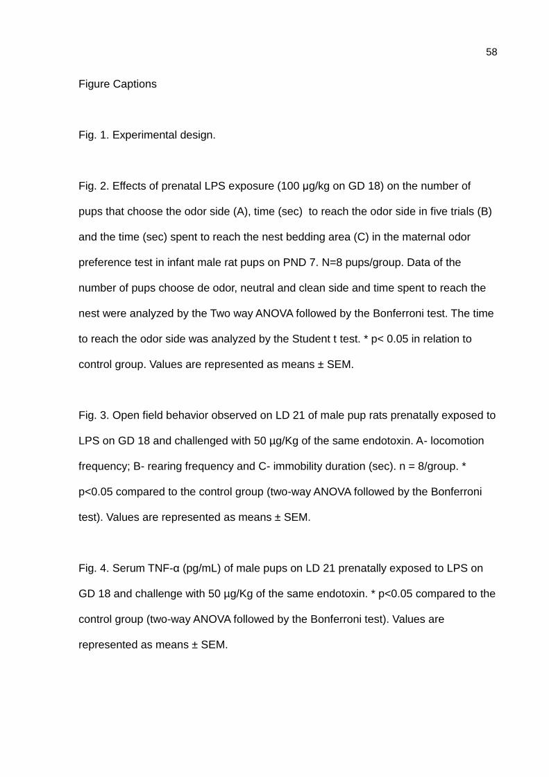
58
Figure Captions
Fig. 1. Experimental design.
Fig. 2. Effects of prenatal LPS exposure (100 μg/kg on GD 18) on the number of
pups that choose the odor side (A), time (sec) to reach the odor side in five trials (B)
and the time (sec) spent to reach the nest bedding area (C) in the maternal odor
preference test in infant male rat pups on PND 7. N=8 pups/group. Data of the
number of pups choose de odor, neutral and clean side and time spent to reach the
nest were analyzed by the Two way ANOVA followed by the Bonferroni test. The time
to reach the odor side was analyzed by the Student t test. * p< 0.05 in relation to
control group. Values are represented as means ± SEM.
Fig. 3. Open field behavior observed on LD 21 of male pup rats prenatally exposed to
LPS on GD 18 and challenged with 50 µg/Kg of the same endotoxin. A- locomotion
frequency; B- rearing frequency and C- immobility duration (sec). n = 8/group. *
p<0.05 compared to the control group (two-way ANOVA followed by the Bonferroni
test). Values are represented as means ± SEM.
Fig. 4. Serum TNF-α (pg/mL) of male pups on LD 21 prenatally exposed to LPS on
GD 18 and challenge with 50 µg/Kg of the same endotoxin. * p<0.05 compared to the
control group (two-way ANOVA followed by the Bonferroni test). Values are
represented as means ± SEM.

59
Table 1. Maternal care of dams prenatally exposed to LPS (100 µg/Kg, i.p.) Maternal
behavior and maternal aggressive behavior were observed, respectively, on postnatal (PND)5
and PND 6. N = 8/group. Data are presented as mean ± SEM or percentage.
Parameter Control group LPS group P
Maternal behavior
Retrieval 1st
pup (s) 40.38 ± 11.60 13.25 ± 3.89 0.044
Retrieval all pups(s) 202.63 ± 43.26 176.75 ± 38.43 0.662
Maternal position (min) 21.26 ± 3.93 19.55 ± 1.37 0.687
Maternal position (%) 87.5 100 1.0
Grouping pups (%) 87.5 100 1.0
Presence of the nest (%) 100 100 -
Maternal aggressive behavior
First attack (s) 147.73 ± 24.00 166.60 ± 25.00 0.595
Number of attacks 6.65 ± 0.70 3.50 ± 0.60 0.004
Total number of attacks 4.00 ± 1.26 3.00 ± 1.40 0.604
Time of fight (s) 1.80 ± 0.02 1.00 ± 0.05 <0.0001
Number of bits 0.36 ± 0.20 0.40 ± 0.24 0.074
Number of times of retrieval pups 0.27 ± 1.00 3.80 ± 1.80 0.109
Number of times hidden the pups 1.54 ± 0.66 3.80 ± 2.60 0.414
Number of times the intruder
sniffed the pups
0.82 ± 0.42 0.40 ± 0.24 0.399
Student t test.

60
Fig.1

61
Fig.2
Choose the odor side
0
1
2
3
4
5
*
Control group
LPS group
A
Groups
Nu
mb
er
of
pu
ps
Time to reach the odor side
0
20
40
60
80
B
(n=5) (n= 30)
Groups
seco
nd
s(m
ean
±S
EM
)
Time to reach one side
S1 S2 S3 S4 S50
20
40
60
80
*
C
Sessions
min
ute
s (
mean
±S
EM
)

62
Fig.3
Locomotion
0
20
40
60
80
100
A
S+S S+LPS LPS+S LPS+LPS
Groups
nu
mb
er
of
sq
uare
cro
ssed
Rearing
0
5
10
15
20
25
B
S+S S+LPS LPS+S LPS+LPS
Groups
freq
uen
cy
Immobility
0
20
40
60
*
C
S+S S+LPS LPS+S LPS+LPS
Groups
seco
nd
s
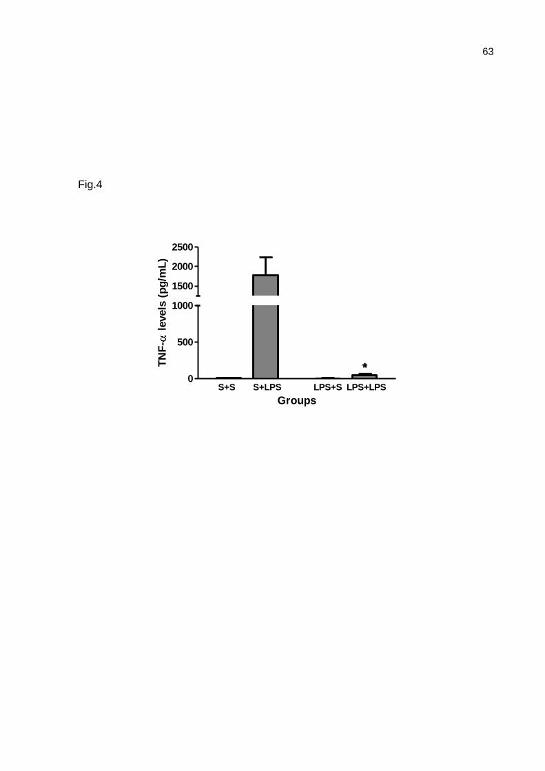
63
Fig.4
0
500
1000
1500
2000
2500
**
S+S S+LPS LPS+S LPS+LPS
Groups
TN
F-
levels
(p
g/m
L)
64
ARTIGO 2
Transgenerational effects of antenatal LPS exposure on maternal and pups
behaviors: studies in two generations.
Penteado, S. H. N. W a; Kirsten, T. B.
b; Eluf,B.
a ; Reis-Silva, T. M.
b ; Acenjo, M. K
a; Melo,
R.aR.
a; I
,Bernardi, M. M.
a,c
a Health Sciences Institute, Paulista University, Rua Dr. Bacelar, 1212, 04026-002, Sao Paulo,
SP, Brazil.
b Department of Pathology, School of Veterinary Medicine, University of São Paulo, Av. Prof.
Dr. Orlando Marques de Paiva, 87, 05508–270, São Paulo, SP, Brazil.
c Centro de Matemática e Cognição, Universidade Federal do ABC, Rua Santa Adélia, 166,
Santo André, SP, Brasil
Corresponding author:
Maria Martha Bernardi
Department of Pathology, School of Veterinary Medicine, University of Sao Paulo
Av. Prof. Dr. Orlando Marques de Paiva, 87
São Paulo, SP 05508-270, Brazil
Tel. +55 11 3091 7657, Fax +55 11 3091 7829
e-mail: [email protected]
Key words: prenatal inflammation, LPS, maternal behavior, maternal aggressive behavior.
Running title: Transgenerational LPS effects.

65
Abstract
Objective: The present experiment was designed to analyze possible transgenerational
lipopolysaccharide (LPS)-induced effects on maternal care of the F1 generation and on
behavior of the F2 generation of rats exposed antenatally to LPS (100 μg/kg LPS on GD 18).
Methods: The following parameters of the F1 generation were observed: reproductive
performance, maternal behavior, maternal aggressive behavior, and open field general activity
in adult age. In the F2 generation, body weight at birth and weaning, maternal olfactory
preference, and, in adult age, general activities in an open field and in the plus maze were
assessed. Results: In the F1 generation relative to controls, antenatal exposure to LPS showed
that 1) the latency to full maternal behavior was delayed 2) a slightly increased maternal
aggression; and 3) no interference of reproductive performance and general activity. In the F2
generation antenatally treated with the endotoxin, it was observed in relation to the control
group 1) a decreased body weight at weaning and in the olfactory recognition of maternal
odor; 2) no differences in the open field behavior; 3) a decreased percentage in time of open
arms and in time in the center while an increase in closed arms were observed. Conclusion:
These data reveal that antenatal LPS exposure modified certain aspects of maternal care of the
F1 generation related to nursing and pups’ survival, but not on maternal motivational
parameters. In the F2 generation, antenatal LPS exposure reduces maternal recognition in
infancy as well as body weight at weaning. In addition, later prenatal exposure to LPS induces
transgenerational effects in the F2 generation, resulting in a less resilient phenotype to
anxiety.
66
Introduction
Lipopolysaccharide (LPS), an endotoxin that originates in the cell wall of gram-
negative bacteria and mimics bacterial infection, activates the immune system to release
proinflammatory cytokines (Avitsur, Pollak, & Yirmiya, 1997; Saluk-Juszczak & Wachowicz,
2005). Viral and bacterial infections, including those caused by prenatal LPS exposure, induce
short- and long-term changes in behavior and central nervous system activity (Boksa, 2010;
Golan, Lev, Hallak, Sorokin, & Huleihel, 2005; Meyer, Feldon, & Fatemi, 2009b). Previous
investigations by our group have shown that prenatal LPS treatment (100 μg/kg, given
intraperitoneally on gestational day [GD] 9.5) reduces the social behavior of F1 males both in
infancy and in adulthood and decreases their striatal dopamine (DA) and DA metabolite levels
in the absence of signs indicative of neuroinflammation (Kirsten, et al., 2011b; Kirsten,
Taricano, Florio, Palermo-Neto, & Bernardi, 2010; Kirsten, Taricano, Maiorka, Palermo-Neto,
& Bernardi, 2010). Interestingly, our model also showed that maternal behavior was slightly
improved in pregnant rats treated with LPS on GD 9.5 (Kirsten, et al., 2011b), whereas
treatment on GD 21 decreased this behavior (Bernardi, et al., 2010).
It has been suggested that the effects of maternal LPS exposure on the developing fetal
brain are not directly mediated by LPS, but are instead indirectly induced via increases in
proinflammatory cytokines and glucocorticoid levels within the maternal circulation, placenta,
and fetal brain (Ashdown, et al., 2006; Cai, Pan, Pang, Evans, & Rhodes, 2000; Gayle, et al.,
2004; Urakubo, Jarskog, Lieberman, & Gilmore, 2001). Infections associated with
immunological events that occur in early/middle life (e.g., GD 8–10 in rats and mice) have a
stronger impact on fetal neurodevelopment than do late-pregnancy infections. Immune
activation during the early/middle stages of pregnancy was shown to modify fetal cell
proliferation and differentiation, cell migration, target selection, and synapse maturation
(Ghiani, et al., 2011; Meyer, et al., 2006; Meyer, Yee, & Feldon, 2007; Samuelsson,
67
Jennische, Hansson, & Holmang, 2006; Shi, Fatemi, Sidwell, & Patterson, 2003). Multiple
brain injuries and behavioral abnormalities persisting through adulthood were also reported
after early/middle stage pregnancy infections (Meyer, et al., 2007).
Environmental information received by a mother can induce a phenotypic change in
her offspring, commonly known as a maternal or transgenerational effect (Agrawal, Laforsch,
& Tollrian, 1999; Curno, Behnke, McElligott, Reader, & Barnard, 2009). Certain cues in the
maternal environment, e.g., the prevalence of predators or maternal infection, can lead to
behavioral, morphological, and immunological changes in the following generation (Agrawal,
et al., 1999; Grindstaff, et al., 2006).
The present experiment was designed to analyze possible transgenerational LPS-
induced effects on maternal care of the F1 generation and on behavior of the F2 generation of
rats exposed antenatally to LPS (100 μg/kg LPS on GD 18). The following parameters of the
F1 generation were observed: reproductive performance, maternal behavior, maternal
aggressive behavior, and open field general activity in adult age. In the F2 generation, body
weight at birth and weaning, maternal olfactory preference and, in adult age, general activities
in an open field and in the plus maze were assessed.
Material and methods
Animals
Sixty pregnant Wistar rats (parental generation) between 12 and 13 weeks of age and
weighing 216–263 g were used (GD0 was defined as the day when spermatozoa were
detected in the vaginal smear). The dams were individually housed in polypropylene cages
(38×32×16 cm) at a controlled temperature (22±2 °C) and humidity (65–70%) with artificial
lighting (12-hour light/12-hour dark cycle, lights on at 6:00 AM). The animals had free access
to Nuvilab® rodent chow (Nuvital Co., São Paulo, SP, Brazil) and filtered water. Sterilized
68
and residue-free wood shavings were used for the animal bedding. The animals were divided
into control (saline-treated) and experimental (LPS-treated) groups (n=16 dams/group). The
dams were allowed to give birth and nurture their offspring normally. The day of birth was
recorded as postnatal day (PND) 1. No handling was performed on PND1, but on PND2, 8
offspring (4 males and 4 females) were randomly selected for the following studies. No cross-
fostering procedure was used. The 8 randomly selected pups remained with each dam until
weaning (PND21). On PND21, littermates were separated and co-housed by sex and
treatments under the same conditions as their parents and named the F1 generation. In adult
age, the females of F1 generation (8/group) were mated with naïve males to produce the F2
generation. One female (F1 generation) and one male from each litter (F2 generation) were
used for the experiments; therefore, the litter was used as a unit. The testing of the control and
experimental groups was intermixed. The rats used in this study were kept in accordance with
the guidelines of the Committee on Care and Use of Laboratory Animal Resources of Paulista
University, Brazil (protocol No. 014/09, CEUA-UNIP). These guidelines are similar to those
of the National Institutes of Health, Bethesda, MD. Experiments were carried out in
accordance with good laboratory practice protocols and with quality assurance methods.
Treatments
LPS (from Escherichia coli, Sigma®, Saint Louis, MO, USA, serotype 0127: B8) was
dissolved in sterile saline (50 µg/ml LPS in a 0.9% NaCl solution) and administered
intraperitoneally to pregnant dams of the parental generation at a dose of 100 µg/kg on GD18
(n= 16 /group). This dose was chosen because it has been shown to (1) elicit sickness
behavior, (2) induce endocrine alterations in dams, (3) increase cytokines at the placental
level, and (4) impair the offspring birthrate and reduce the social behavior of male offspring
during infancy and adulthood (Kirsten, et al., 2011a; Kirsten, Taricano, Maiorka, et al., 2010;
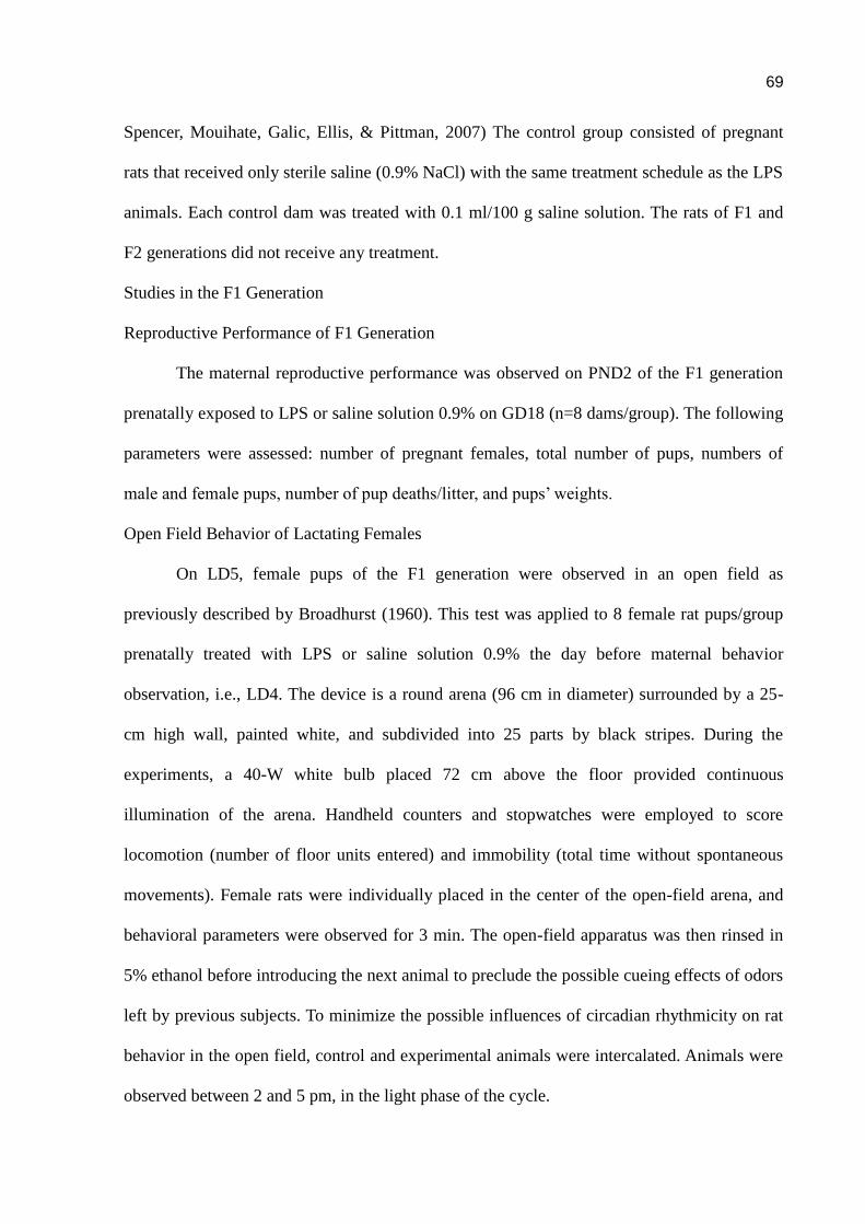
69
Spencer, Mouihate, Galic, Ellis, & Pittman, 2007) The control group consisted of pregnant
rats that received only sterile saline (0.9% NaCl) with the same treatment schedule as the LPS
animals. Each control dam was treated with 0.1 ml/100 g saline solution. The rats of F1 and
F2 generations did not receive any treatment.
Studies in the F1 Generation
Reproductive Performance of F1 Generation
The maternal reproductive performance was observed on PND2 of the F1 generation
prenatally exposed to LPS or saline solution 0.9% on GD18 (n=8 dams/group). The following
parameters were assessed: number of pregnant females, total number of pups, numbers of
male and female pups, number of pup deaths/litter, and pups’ weights.
Open Field Behavior of Lactating Females
On LD5, female pups of the F1 generation were observed in an open field as
previously described by Broadhurst (1960). This test was applied to 8 female rat pups/group
prenatally treated with LPS or saline solution 0.9% the day before maternal behavior
observation, i.e., LD4. The device is a round arena (96 cm in diameter) surrounded by a 25-
cm high wall, painted white, and subdivided into 25 parts by black stripes. During the
experiments, a 40-W white bulb placed 72 cm above the floor provided continuous
illumination of the arena. Handheld counters and stopwatches were employed to score
locomotion (number of floor units entered) and immobility (total time without spontaneous
movements). Female rats were individually placed in the center of the open-field arena, and
behavioral parameters were observed for 3 min. The open-field apparatus was then rinsed in
5% ethanol before introducing the next animal to preclude the possible cueing effects of odors
left by previous subjects. To minimize the possible influences of circadian rhythmicity on rat
behavior in the open field, control and experimental animals were intercalated. Animals were
observed between 2 and 5 pm, in the light phase of the cycle.

70
Maternal Behavior
Maternal behavior was analyzed as previously described (Teodorov, Felício, &
Bernardi, 2010). Briefly, on lactation day (LD) 5 between 08:00 AM and 11:00 AM, the
maternal behavior of the F1 generation (8 dams /group) was observed. Pups were removed
from the dam, placed in another cage, and kept warm. Immediately following the separation,
the presence of a nest in the home cage was evaluated. Sixty minutes following maternal
separation, all pups were returned to the cage of their mother, and examination of the maternal
behavior began. The latencies to retrieval of the first pup(s), to retrieval of all pups and to full
maternal behavior (s) were recorded. The percentage of dams that retrieved the 1st pup, all
pups, grouping pups and presenting maternal behavior were calculated.
Maternal Aggressive Behavior
On LD6 between 08:00 AM and 11:00 AM, the maternal aggressive behavior test was
performed in female rats of the F1 generation. These rats were subjected to a 10-min maternal
defense test (Teodorov, et al., 2010; Wilkins, Logan, & Kehoe, 1997). A male Wistar rat—the
intruder—was introduced into the home cage of the dam and offspring. Intruder rats were
only used once. Behaviors during the maternal defense test (against the intruder) were
recorded via a remote digital camcorder and later analyzed for offensive behavior by the
resident: latency(s) to first attack, attack frequency, total time(s) of attacks, frequency of bites,
and time of fight(s). Furthermore, maternal behavior in the presence of the intruder was
analyzed: frequency of carrying and hiding the pups and frequency of the intruder sniffing
pups. The female was used only once in this test.
Studies of F2 Generation
In these studies only the male pups of F2 generation were employed to avoid
hormonal interferences on behavior that occurs in female rats.
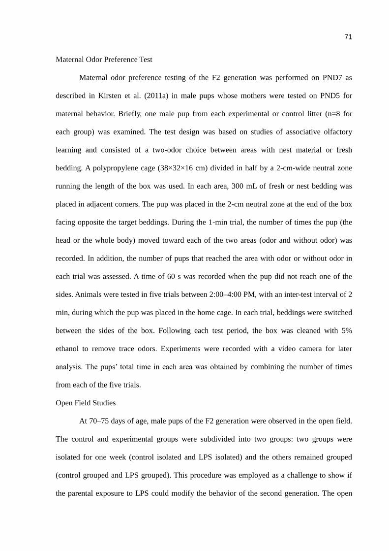
71
Maternal Odor Preference Test
Maternal odor preference testing of the F2 generation was performed on PND7 as
described in Kirsten et al. (2011a) in male pups whose mothers were tested on PND5 for
maternal behavior. Briefly, one male pup from each experimental or control litter (n=8 for
each group) was examined. The test design was based on studies of associative olfactory
learning and consisted of a two-odor choice between areas with nest material or fresh
bedding. A polypropylene cage (38×32×16 cm) divided in half by a 2-cm-wide neutral zone
running the length of the box was used. In each area, 300 mL of fresh or nest bedding was
placed in adjacent corners. The pup was placed in the 2-cm neutral zone at the end of the box
facing opposite the target beddings. During the 1-min trial, the number of times the pup (the
head or the whole body) moved toward each of the two areas (odor and without odor) was
recorded. In addition, the number of pups that reached the area with odor or without odor in
each trial was assessed. A time of 60 s was recorded when the pup did not reach one of the
sides. Animals were tested in five trials between 2:00–4:00 PM, with an inter-test interval of 2
min, during which the pup was placed in the home cage. In each trial, beddings were switched
between the sides of the box. Following each test period, the box was cleaned with 5%
ethanol to remove trace odors. Experiments were recorded with a video camera for later
analysis. The pups’ total time in each area was obtained by combining the number of times
from each of the five trials.
Open Field Studies
At 70–75 days of age, male pups of the F2 generation were observed in the open field.
The control and experimental groups were subdivided into two groups: two groups were
isolated for one week (control isolated and LPS isolated) and the others remained grouped
(control grouped and LPS grouped). This procedure was employed as a challenge to show if
the parental exposure to LPS could modify the behavior of the second generation. The open

72
field test was performed similarly to those described for the female rats.
Plus Maze Test Studies
The plus maze behaviors of offspring were measured on PND 70–75 of the F2
generation. The same rats employed in the open field test were used to perform this
experiment. The device consisted of two opposite open arms (50 cm long x 10 cm wide) and
two opposite closed arms (50 cm long x 10 cm wide x 40 cm high) arranged at 90° angles.
The floor of the maze was made of wood, painted gray (with acrylic washable covering) and
located 50 cm above the floor. The center of the maze was open and the walls of the closed
arms started 2 cm from the center of the maze. Each rat was observed using a video camera
mounted above the arena to record the behavioral data. For the observations, each animal was
individually placed in the center of the maze with the head facing one of the open arms, and
the following parameters were measured over a period of 5 min: number of entries into the
open arms, number of entries into the closed arms, time spent in the open arms, time spent in
the closed arms. The measures that reflect stress/anxiety levels here employed were the
percentage of time spent in the open arms versus closed arms (% time in the open arms =
[time in the open arms/(time in the open arms + time in the closed arms)] x 100).
To minimize the influence of possible circadian changes on plus maze behaviors,
control and experimental animals were alternated. The device was washed with a 5%
alcohol/water solution before placing the animals on it to obviate possible biasing effects of
odor clues left by previous rats. Observations were made between 2:00 and 5:00 p.m.
Statistical Analyses
The mother was the experimental unit, and one female from each litter was used for
each experiment; thus, different animals were used in each experiment. The results are
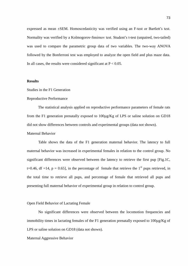
73
expressed as mean ±SEM. Homoscedasticity was verified using an F-test or Bartlett’s test.
Normality was verified by a Kolmogorov-Smirnov test. Student’s t-test (unpaired, two-tailed)
was used to compare the parametric group data of two variables. The two-way ANOVA
followed by the Bonferroni test was employed to analyze the open field and plus maze data.
In all cases, the results were considered significant at P < 0.05.
Results
Studies in the F1 Generation
Reproductive Performance
The statistical analysis applied on reproductive performance parameters of female rats
from the F1 generation prenatally exposed to 100µg/Kg of LPS or saline solution on GD18
did not show differences between controls and experimental groups (data not shown).
Maternal Behavior
Table shows the data of the F1 generation maternal behavior. The latency to full
maternal behavior was increased in experimental females in relation to the control group. No
significant differences were observed between the latency to retrieve the first pup [Fig.1C,
t=0.46, df =14, p = 0.65], in the percentage of female that retrieve the 1st pups retrieved, in
the total time to retrieve all pups, and percentage of female that retrieved all pups and
presenting full maternal behavior of experimental group in relation to control group.
Open Field Behavior of Lactating Female
No significant differences were observed between the locomotion frequencies and
immobility times in lactating females of the F1 generation prenatally exposed to 100µg/Kg of
LPS or saline solution on GD18 (data not shown).
Maternal Aggressive Behavior
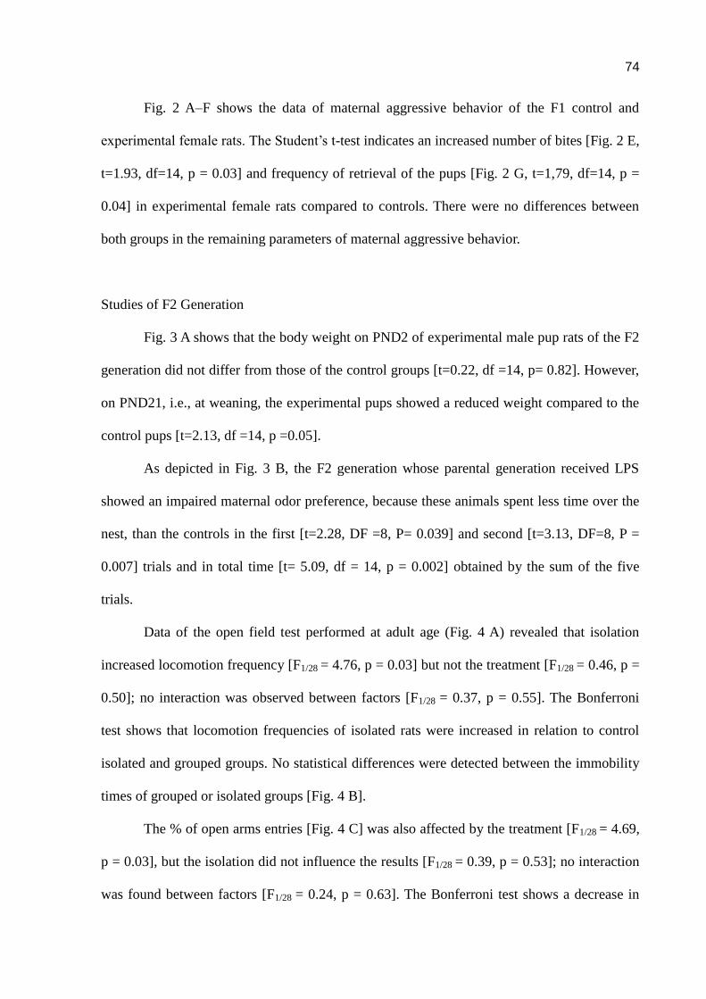
74
Fig. 2 A–F shows the data of maternal aggressive behavior of the F1 control and
experimental female rats. The Student’s t-test indicates an increased number of bites [Fig. 2 E,
t=1.93, df=14, p = 0.03] and frequency of retrieval of the pups [Fig. 2 G, t=1,79, df=14, p =
0.04] in experimental female rats compared to controls. There were no differences between
both groups in the remaining parameters of maternal aggressive behavior.
Studies of F2 Generation
Fig. 3 A shows that the body weight on PND2 of experimental male pup rats of the F2
generation did not differ from those of the control groups [t=0.22, df =14, p= 0.82]. However,
on PND21, i.e., at weaning, the experimental pups showed a reduced weight compared to the
control pups [t=2.13, df =14, p =0.05].
As depicted in Fig. 3 B, the F2 generation whose parental generation received LPS
showed an impaired maternal odor preference, because these animals spent less time over the
nest, than the controls in the first [t=2.28, DF =8, P= 0.039] and second [t=3.13, DF=8, P =
0.007] trials and in total time [t= 5.09, df = 14, p = 0.002] obtained by the sum of the five
trials.
Data of the open field test performed at adult age (Fig. 4 A) revealed that isolation
increased locomotion frequency [F1/28 = 4.76, p = 0.03] but not the treatment [F1/28 = 0.46, p =
0.50]; no interaction was observed between factors [F1/28 = 0.37, p = 0.55]. The Bonferroni
test shows that locomotion frequencies of isolated rats were increased in relation to control
isolated and grouped groups. No statistical differences were detected between the immobility
times of grouped or isolated groups [Fig. 4 B].
The % of open arms entries [Fig. 4 C] was also affected by the treatment [F1/28 = 4.69,
p = 0.03], but the isolation did not influence the results [F1/28 = 0.39, p = 0.53]; no interaction
was found between factors [F1/28 = 0.24, p = 0.63]. The Bonferroni test shows a decrease in
75
the % of time in open arms of the isolated experimental group, indicating a reduced anxiety in
these rats. As depicted in Fig. 4 D, the treatment affected the time in closed arms [F1/28 = 4.87,
p = 0.03], but not isolation [F1/28 = 0.11, p = 0.74]; an interaction was observed between
factors [F1/28 = 4.20, p = 0.04].The Bonferroni test indicates that isolated rats from the
experimental group had an increased time in the closed arms. The time in the plus maze center
(Fig. 4 E), was affected by the treatment [F1/28 = 4.28, p = 0.04]; no significant interference
was found by the isolation [F1/28 = 0.01, p = 0.94] and no interaction between factors was
detected [F1/28 = 0.50, p = 0.48. The Bonferroni test indicates a significant decrease in this
parameter in the experimental isolated group. No differences were detected between groups
on the number of entries in the open and closed arms (data not shown).
Discussion
The present study reveals the novel finding that exposure to the bacterial endotoxin in
the late prenatal period modifies the maternal behavior of the F1 generation and their
offspring’s behaviors in infancy and adulthood. Thus, the present study demonstrated that
prenatal administration of LPS was able to modify the behavior of two generations of rats. In
fact, both the maternal behavior and maternal aggressive behavior of the F1 generation were
affected by the endotoxin exposure. In addition, in the F2 generation antenatally exposed to
LPS, the body weight at weaning was decreased, and the maternal olfactory preference was
impaired. In adult age, a decreased anxiety and increased locomotor behaviors were observed
in these rats.
Prenatal exposure to LPS on GD18 did not affect the reproductive performance of
female rats. No differences were observed in the number of pups or in the body weight of the
pups at birth; likewise, perinatal deaths did not occur. A previous study of our group also
found that prenatal exposure to LPS on GD 9.5 did not alter either the pups’ body weight at
birth or their neurodevelopment. However, in the present study, at weaning the pups’ body
76
weight of the F1 generation prenatally treated with LPS decreased in relation to the control
group, suggesting that prenatal exposure in utero to LPS reduces the overall physical
development of the pups.
Immune challenge during pregnancy is associated with preterm birth and poor
perinatal development. The mechanisms of these effects are not known. LPS did not cross the
placental barrier, but induced the release of proinflammatory cytokines (Ashdown, et al.,
2006). Several pieces of evidence linked elevated cytokine levels triggered by maternal
infection to influence various neurodevelopmental processes, including cell differentiation,
maturation, and survival (Deverman & Patterson, 2009; Ghiani, et al., 2011; Paris, Brunton,
Russell, & Frye, 2011; Zhao & Schwartz, 1998). Hence, fluctuations in their maternal and
fetal levels, for instance, because of a maternal infection, signify a disturbance that can
impede the ongoing of neurodevelopmental processes, and subsequently affect proper neural
cell maturation (Jonakait, 2007; Meyer, Feldon, & Fatemi, 2009a; Meyer, Feldon, & Yee,
2009; Meyer, et al., 2007).
Also, the maternal behavior of the F1 generation prenatally exposed to LPS or saline
solution 0.9% on GD18 was influenced by the prenatal endotoxin exposure. By the way,
increases in latency to presents the full maternal behavior was observed in the F1 female rats
of the LPS group relative to controls. No significant differences between the remained
parameters in both experimental and controls female rats were found. So, although there was
a delay to perform crouching, the females of the F1 generation exposed to LPS prenatally
spent more time on their offspring. The delay to crouching the pups could not be attributed to
a motor impairment, since any effects were found in the open field test. In addition, no
motivational impairment was presently observed in F1 maternal behavior because pup
retrieval was not modified by prenatal LPS (Pedersen & Boccia, 2003; Pedersen, Vadlamudi,
Boccia, & Amico, 2006; J. M. Stern, 1990; J. M. Stern & Protomastro, 2000).
77
Crouching is considered a quiescent maternal posture and usually occurs in response
to sufficient stimulation by pups. A mother rat tends to stop other activities and develops a
characteristic posture with her extremities spread out and back arched (Teodorov, et al., 2010).
The purpose of the crouching posture is to allow the pups access to teats and milk, to regulate
their temperature, and to protect them from environmental elements. Thus, female rats of the
F1 generation prenatally exposed to LPS, provided their offspring the opportunity to feed and
to regulate their body temperature. However, the latency to present the full maternal behavior
was delayed in experimental female rats when compared to the control group. Differences in
the effects of prenatal LPS on F1 generation nursing (full maternal behavior) in comparison
to its effects on retrieval and nest behaviors may be explained by the understanding that
retrieving and nest building behaviors may be more indicative of maternal motivation,
whereas the nursing behavior may be indicative of a more reflexive maternal response
(Kristal, 2009; J.M. Stern, 1996). In other words, retrieving may represent an active voluntary
response, which reflects interest and attraction toward pup-related stimuli, and nursing may be
transiently activated as a reflex when the female wanders near pups and they crawl under her.
Despite the delay in the latency to full maternal behavior, these females guaranteed
protection of their pups from environmental threats. In fact, during maternal aggressive
behavior, these females presented more frequency of bites to intruders and retrieved their pups
more frequently than did the control female rats. In a previous experiment (not published) we
observed that prenatal exposure on GD18 to the same dose of LPS reduced the maternal
olfactory preference in pup rats. Thus, it is possible that a reduced stimulation of pups toward
their mothers delayed the onset of full maternal behavior.
It has been known for some time that stressful manipulations in early life contribute to
changes in stress reactivity that persists into adulthood. More severe interventions like
maternal separation have a sensitizing effect on the stress axis, and milder ones such as
78
neonatal handling promote a more resilient phenotype (Francis & Meaney, 1999).
LPS administration to pregnant rats upregulates mRNA expression of the stress-related
peptide, corticotrophin-releasing hormone, in the fetal brain (Iqbal, Moisiadis, Kostaki, &
Matthews, 2012), suggesting the possibility of inducing a fetal stress response. The activation
of the stress response during pregnancy was shown to have long-term consequences on the
response of adult offspring to stressful situations, as demonstrated in rodents, primates, and
humans (Matthews & Phillips, 2010). Environmental information received by a mother can
induce a phenotypic change in her offspring, commonly known as a maternal or
transgenerational effect (Agrawal, et al., 1999; Curno, et al., 2009). Certain cues in the
maternal environment, e.g., the prevalence of predators or maternal infection, can lead to
behavioral, morphological, and immunological changes in the following generation (Agrawal,
et al., 1999; Grindstaff, et al., 2006).
In the F2 generation at birth, the offspring body weight from the antenatal LPS group
did not differ from that of the control group. At weaning, however, a decrease in this
parameter was observed in the experimental pup rats, indicating a decreased overall
development in these rats, despite the improved maternal care of their respective mothers. In
addition, the males of these pup rats had an impaired preference for maternal odor. We studied
the olfactory maternal preference only in male offspring, because maternal care is much
higher in male than in female pups(Teodorov, et al., 2010). Therefore, any deficit in maternal
behavior could be better visualized. The olfactory impairment occurred in the first and second
trials, and this impairment was reflected in the total time of olfactory preference.
Maternal–pups interaction involves the maternal olfactory recognition and ultrasonic
vocalization. In pups, until the opening eyes (PND 11–16) (Fox, 1965), maternal recognition
is made through olfactory cues. Thus, olfaction impairment could be a consequence of
impairment in pups’ motivation. Supporting evidence for this hypothesis comes from previous
79
findings that prenatal exposure to LPS impaired the social interest of rats in infancy and
adulthood because of motivational impairments (Spencer, et al., 2007). Moreover, prenatal
exposure to LPS on GD 9.5 reduced the pups’ maternal odor perception in infancy; this
impairment was also observed in adult age in the aversive cat odor (Kirsten, et al., 2011a).
The most important data here observed is that a reduced pup–mother interaction occurs in the
F2 generation of rats antenatally exposed to LPS, showing a transgenerational effect of the
endotoxin.
To study if antenatal exposure to LPS had long-term effects on pups’ behavior, these
rats were observed in an open field and in the plus maze apparatus in adult age. In this case,
we subdivided the rats exposed antenatally to LPS or saline solution into two new groups: two
groups were isolated for one week, and the others remained grouped. This procedure was
employed as a challenge to show if the antenatal exposure to LPS could modify the behavior
of this generation.
In the open field test an increased locomotion frequency was observed in both the
control and experimental rats submitted to isolation, suggesting an increased exploratory
behavior, because no immobility differences between groups were observed. In the plus maze,
a clear increased anxiety in the antenatal rats isolated and treated with LPS was observed
relative to their control group. In fact, a reduced percentage in open arms was detected. This
decrease resulted from an increase in the time of closed arms and a reduced time in the center
of the apparatus. Thus, the isolation revealed the deficits produced by the antenatal exposure
to LPS. Antenatal exposure to LPS also induced long-term effects on the rats’ behavior.
Epigenetics was initially referred to as the “interactions between genes and
their products which bring the phenotype into being” (Waddington, 1942). Today, the term
refers to molecular or cellular alterations that influence gene expression and, by extension,
physiology and behavior, without causing alterations to the DNA sequence itself (Hunter,

80
2012). These alterations are generally construed to include DNA methylation, non-coding
RNAs, and covalent histone modifications or “marks,” which include acetylation,
phosphorylation, methylation, ubiquitination, and a growing host of ever more exotic moieties
(Hunter, 2012).
It has also become apparent that both corticosteroids and stress have a pronounced
epigenetic impact in both humans and animal models and that the relationship between the
stress response and epigenetics in the brain is bidirectional (Hunter, 2012). Presently,
antenatal LPS exposure improved certain aspects of maternal care of the F1 generation related
to nursing and pups’ survival, but not on maternal motivational parameters, probably because
of a reduced maternal stimulation by the pups. In fact, in the F2 generation, antenatal LPS
exposure reduces maternal recognition in infancy. In addition, later prenatal exposure to LPS
induces transgenerational effects in the F2 generation resulting in a less resilient phenotype to
anxiety. Whether these phenomena are derived from an epigenetic mechanism remains to be
investigated.
81
References
Agrawal, A. A., Laforsch, C., & Tollrian, R. (1999). Transgenerational induction of defences in animals and plants. Nature, 401, 60-63.
Ashdown, H., Dumont, Y., Ng, M., Poole, S., Boksa, P., & Luheshi, G. N. (2006). The role of cytokines in mediating effects of prenatal infection on the fetus: implications for schizophrenia. Mol Psychiatry, 11(1), 47-55.
Avitsur, R., Pollak, Y., & Yirmiya, R. (1997). Different receptor mechanisms mediate the effects of endotoxin and interleukin-1 on female sexual behavior. Brain Res, 773(1-2), 149-161.
Bernardi, M. M., Kirsten, T. B., Matsuoka, S. M., Teodorov, E., Habr, S. F., Penteado, S. H., et al. (2010). Prenatal lipopolysaccharide exposure affects maternal behavior and male offspring sexual behavior in adulthood. Neuroimmunomodulation, 17(1), 47-55.
Boksa, P. (2010). Effects of prenatal infection on brain development and behavior: a review of findings from animal models. Brain Behavior and Immunity, 24(6), 881-897.
Broadhurst, P. L. (1960). Experiments in psychogenetics: Applications of biometrical genetics to the
inheritance of behaviour. In H. J. Eysenck (Ed.), Experiments in personality (Vol. 1, pp. 1-256). London: Routledge & Kegan Paul.
Cai, Z., Pan, Z. L., Pang, Y., Evans, O. B., & Rhodes, P. G. (2000). Cytokine induction in fetal rat brains and brain injury in neonatal rats after maternal lipopolysaccharide administration. Pediatr Res, 47(1), 64-72.
Curno, O., Behnke, J. M., McElligott, A. G., Reader, T., & Barnard, C. J. (2009). Mothers produce less aggressive sons with altered immunity when there is a threat of disease during pregnancy. Proc Biol Sci, 276(1659), 1047-1054.
Deverman, B. E., & Patterson, P. H. (2009). Cytokines and CNS development. Neuron, 64(1), 61-78.
Fox, W. M. (1965). Reflex-ontogeny and behavioural development of the mouse. Anim Behav, 13(2), 234-241.
Francis, D. D., & Meaney, M. J. (1999). Maternal care and the development of stress responses. Curr Opin Neurobiol, 9(1), 128-134.
Gayle, D. A., Beloosesky, R., Desai, M., Amidi, F., Nunez, S. E., & Ross, M. G. (2004). Maternal LPS induces cytokines in the amniotic fluid and corticotropin releasing hormone in the fetal rat brain. Am J Physiol Regul Integr Comp Physiol, 286(6), R1024-1029.
Ghiani, C. A., Mattan, N. S., Nobuta, H., Malvar, J. S., Boles, J., Ross, M. G., et al. (2011). Early effects of lipopolysaccharide-induced inflammation on foetal brain development in rat. ASN Neuro, 3(4).
Golan, H. M., Lev, V., Hallak, M., Sorokin, Y., & Huleihel, M. (2005). Specific neurodevelopmental damage in mice offspring following maternal inflammation during pregnancy. Neuropharmacology, 48(6), 903-917.

82
Grindstaff, J. L., Hasselquist, D., Nilsson, J. K., Sandell, M., Smith, H. G., & Stjernman, M. (2006). Transgenerational priming of immunity: maternal exposure to a bacterial antigen enhances offspring humoral immunity. Proc Biol Sci, 273(1600), 2551-2557.
Hunter, R. G. (2012). Epigenetic effects of stress and corticosteroids in the brain. Front Cell Neurosci, 6, 18.
Iqbal, M., Moisiadis, V. G., Kostaki, A., & Matthews, S. G. (2012). Transgenerational effects of prenatal synthetic glucocorticoids on hypothalamic-pituitary-adrenal function. Endocrinology, 153(7), 3295-3307.
Jonakait, G. M. (2007). The effects of maternal inflammation on neuronal development: possible mechanisms. Int J Dev Neurosci, 25(7), 415-425.
Kirsten, T. B., Chaves, G. P., Taricano, M., Martins, D. O., Florio, J. C., Britto, L. R., et al. (2011a). Prenatal LPS exposure reduces olfactory perception in neonatal and adult rats. Physiol Behav, 104(3), 417-422.
Kirsten, T. B., Chaves, G. P., Taricano, M., Martins, D. O., Florio, J. C., Britto, L. R., et al. (2011b). Prenatal LPS exposure reduces olfactory perception in neonatal and adult rats. Physiology & Behavior, 104(3), 417-422.
Kirsten, T. B., Taricano, M., Florio, J. C., Palermo-Neto, J., & Bernardi, M. M. (2010). Prenatal lipopolysaccharide reduces motor activity after an immune challenge in adult male offspring. Behavioural Brain Research, 211(1), 77-82.
Kirsten, T. B., Taricano, M., Maiorka, P. C., Palermo-Neto, J., & Bernardi, M. M. (2010). Prenatal lipopolysaccharide reduces social behavior in male offspring. Neuroimmunomodulation, 17(4), 240-251.
Kristal, M. B. (2009). The biopsychology of maternal behavior in nonhuman mammals. ILAR J, 50(1), 51-63.
Matthews, S. G., & Phillips, D. I. (2010). Minireview: transgenerational inheritance of the stress response: a new frontier in stress research. Endocrinology, 151(1), 7-13.
Meyer, U., Feldon, J., & Fatemi, S. H. (2009a). In-vivo rodent models for the experimental investigation of prenatal immune activation effects in neurodevelopmental brain disorders. Neurosci Biobehav Rev, 33(7), 1061-1079.
Meyer, U., Feldon, J., & Fatemi, S. H. (2009b). In-vivo rodent models for the experimental investigation of prenatal immune activation effects in neurodevelopmental brain disorders. Neuroscience & Biobehavioral Reviews, 33(7), 1061-1079.
Meyer, U., Feldon, J., & Yee, B. K. (2009). A review of the fetal brain cytokine imbalance hypothesis of schizophrenia. Schizophr Bull, 35(5), 959-972.
Meyer, U., Nyffeler, M., Engler, A., Urwyler, A., Schedlowski, M., Knuesel, I., et al. (2006). The time of prenatal immune challenge determines the specificity of inflammation-mediated brain and behavioral pathology. J Neurosci, 26(18), 4752-4762.
83
Meyer, U., Yee, B. K., & Feldon, J. (2007). The neurodevelopmental impact of prenatal infections at different times of pregnancy: the earlier the worse? Neuroscientist, 13(3), 241-256.
Paris, J. J., Brunton, P. J., Russell, J. A., & Frye, C. A. (2011). Immune stress in late pregnant rats decreases length of gestation and fecundity, and alters later cognitive and affective behaviour of surviving pre-adolescent offspring. Stress, 14(6), 652-664.
Pedersen, C. A., & Boccia, M. L. (2003). Oxytocin antagonism alters rat dams' oral grooming and upright posturing over pups. Physiol Behav, 80(2-3), 233-241.
Pedersen, C. A., Vadlamudi, S. V., Boccia, M. L., & Amico, J. A. (2006). Maternal behavior deficits in nulliparous oxytocin knockout mice. Genes Brain and Behavior, 5(3), 274-281.
Saluk-Juszczak, J., & Wachowicz, B. (2005). [The proinflammatory activity of lipopolysaccharide]. Postepy Biochem, 51(3), 280-287.
Samuelsson, A. M., Jennische, E., Hansson, H. A., & Holmang, A. (2006). Prenatal exposure to interleukin-6 results in inflammatory neurodegeneration in hippocampus with NMDA/GABA(A) dysregulation and impaired spatial learning. American Journal of Physiology - Regulatory Integrative and Comparative Physiology, 290(5), R1345-1356.
Shi, L., Fatemi, S. H., Sidwell, R. W., & Patterson, P. H. (2003). Maternal influenza infection causes marked behavioral and pharmacological changes in the offspring. J Neurosci, 23(1), 297-302.
Spencer, S. J., Mouihate, A., Galic, M. A., Ellis, S. L., & Pittman, Q. J. (2007). Neonatal immune challenge does not affect body weight regulation in rats. Am J Physiol Regul Integr Comp Physiol, 293(2), R581-589.
Stern, J. M. (1990). Multisensory regulation of maternal behavior and masculine sexual behavior: a revised view. Neuroscience & Biobehavioral Reviews, 14(2), 183-200.
Stern, J. M. (1996). Somatosensation and maternal care in Norway Rats. In J. S. Rosenblatt & C. T.
Snowden (Eds.), Parental Care: Evolution, Mechanisms,and Adaptive Significance (pp. 243-294). New York: Academic Press.
Stern, J. M., & Protomastro, M. (2000). Effects of low dosages of apomorphine on maternal responsiveness in lactating rats. Pharmacology Biochemistry and Behavior, 66(2), 353-359.
Teodorov, E., Felício, L. F., & Bernardi, M. M. (2010). Maternal behavior. In M. L. Andersen & S. Tufik (Eds.), Animals Models as a Tool in Ethical Biomedical Research (Vol. I, pp. 149-165). São Paulo: AFIP,CEPID.
Urakubo, A., Jarskog, L. F., Lieberman, J. A., & Gilmore, J. H. (2001). Prenatal exposure to maternal infection alters cytokine expression in the placenta, amniotic fluid, and fetal brain. Schizophr Res, 47(1), 27-36.
Waddington, C. H. (1942). The epigenotype. . Endeavour 1, 18-20.
Wilkins, A. S., Logan, M., & Kehoe, P. (1997). Postnatal pup brain dopamine depletion inhibits maternal behavior. Pharmacol Biochem Behav, 58(4), 867-873.
84
Zhao, B., & Schwartz, J. P. (1998). Involvement of cytokines in normal CNS development and neurological diseases: recent progress and perspectives. J Neurosci Res, 52(1), 7-16.
This study was part of the doctoral thesis of Sandra Heloisa Nunes Whitaker Penteado and
was supported by CNPq ( PIBIC), FAPESP Thematic Awards 09/51886-3 and UNIP grant 7-
02-747/2010 and 7-02-839/2012.
Caption to figures
Fig.1. Effects of prenatal LPS exposure (100 μg/kg on GD 18) or saline solution (NaCl 0.9%)
on maternal aggressive behavior of F1 generation. (A) latency first attack; (B) number of
attacks; (C)total of attacks ; (D) time of fight; (E) bites; (F) hiding the pups;(F) retrieve pups ;
(G) sniffing pups . Data are presented as means ± SEM. N = 8/ group. * p< 0.05 in relation to
control group (Student t test).
Fig.2. . Effects of antenatal LPS exposure (100 μg/kg on GD 18) or saline solution (NaCl
0.9%) on body weight (A) and maternal odor preference (B) of pup rats from F2 generation.
Data are presented as means ± SEM. N = 8/ group. ANOVA two way analysis followed by
the Bonferroni test. ** p< 0.01 in relation to control group.
Fig.3. Effects of antenatal LPS exposure (100 μg/kg on GD 18) or saline solution (NaCl
0.9%) on open field behavior and on plus maze of adult rats from F2 generation. Data are
presented as means ± SEM. N = 8/ group. ANOVA two way analysis followed by the
Bonferroni test. * p< 0.05 in relation to control group.
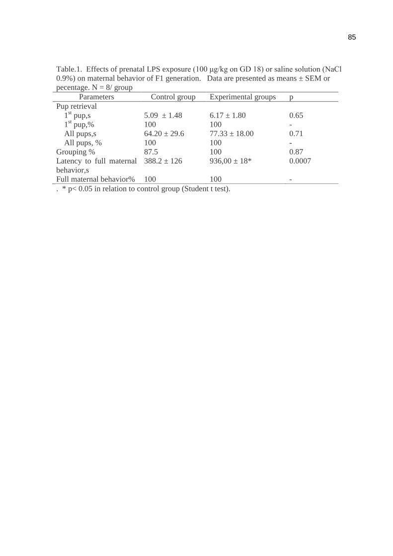
85
Table.1. Effects of prenatal LPS exposure (100 μg/kg on GD 18) or saline solution (NaCl
0.9%) on maternal behavior of F1 generation. Data are presented as means ± SEM or
pecentage. N = 8/ group
Parameters Control group Experimental groups p
Pup retrieval
1st pup,s 5.09 ± 1.48 6.17 ± 1.80 0.65
1st pup,% 100 100 -
All pups,s 64.20 ± 29.6 77.33 ± 18.00 0.71
All pups, % 100 100 -
Grouping % 87.5 100 0.87
Latency to full maternal
behavior,s
388.2 ± 126 936,00 ± 18* 0.0007
Full maternal behavior% 100 100 -
. * p< 0.05 in relation to control group (Student t test).
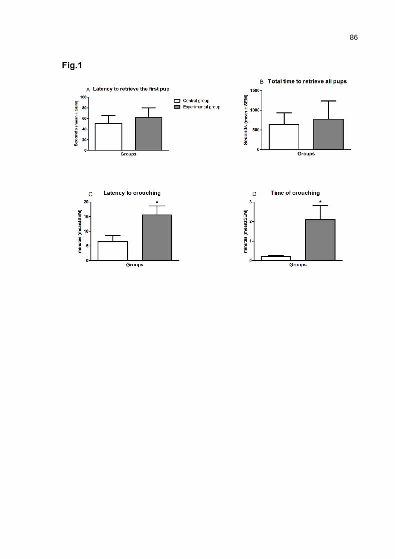
86

87

88

89
Fig.4