Embed Size (px)
Citation preview
1
2Q2
3
4
5Q1
6
7
8Q4910111213
14
15161718192021222324252627
44
45
46
47
48
49
50
51
52
53
54
Parasitology International xxx (2013) xxx–xxx
Q3
PARINT-01150; No of Pages 5
Contents lists available at ScienceDirect
Parasitology International
j ourna l homepage: www.e lsev ie r .com/ locate /par in t
F
Activation of anaerobic metabolism in Biomphalaria glabrata(Mollusca: Gastropoda) experimentally infected by Angiostrongyluscantonensis (Nematoda, Metastrongylidae) by high-performanceliquid chromatography
PRO
OVinícius Menezes Tunholi-Alves a,b,⁎, Victor Menezes Tunholi a,b, Rosane N. Castro c, Luiza D'Oliveira Sant'Ana c,Luciana Santos-Amaral d, Ana Paula Martins de Oliveira e, Juberlan Garcia f, Silvana Carvalho Thiengo e,Jairo Pinheiro a,1, Arnaldo Maldonado Jr. f,1
a Área de Biofísica, Departamento de Ciências Fisiológicas, Instituto de Biologia, UFRuralRJ, Brazilb Curso de Pós-Graduação em Ciências Veterinárias, Departamento de Parasitologia Animal, Instituto de Veterinária, UFRuralRJ, Brazilc Departamento de Química, Instituto de Ciências Exatas, Universidade Federal Rural do Rio de Janeiro, BR 465, Km 7, 23890-000 Seropédica, RJ, Brazild Departamento de Saneamento e Saúde Ambiental, Escola Nacional de Saúde Pública, Fiocruz, Brazile Laboratório de Referência Nacional em Malacologia Médica do Instituto Oswaldo Cruz, IOC, Fiocruz, RJ, Brazilf Laboratório de Biologia e Parasitologia de Mamíferos Silvestres Reservatórios, Instituto Oswaldo Cruz, Fiocruz, Av. Brasil, 4365-Manguinhos, 21040-900 Rio de Janeiro, RJ, Brazil
⁎ Corresponding author at: Curso de Pós-GraduaçDepartamento de Parasitologia Animal, Instituto de Veterindo Rio de Janeiro, Km 7, BR 465, Antiga estrada Rio-São PBrazil.
E-mail address: [email protected] (V.M. Tun1 Research fellow from CNPq.
1383-5769/$ – see front matter © 2013 Published by Elsehttp://dx.doi.org/10.1016/j.parint.2013.09.004
Please cite this article as: Tunholi-Alves VM,mentally infected by Angiostrongylus cantone
Da b s t r a c t
a r t i c l e i n f o28
29
30
31
32
33
34
35
36
Article history:Received 5 June 2013Received in revised form 27 August 2013Accepted 5 September 2013Available online xxxx
Keywords:CarbohydratesHost–parasite relationshipLarval nematodes
37
38
39
40
41
RRECTEThe activity of lactate dehydrogenase and the concentrations of glucose in the hemolymph and of glycogen in thedigestive gland and cephalopedal mass of Biomphalaria glabrata experimentally infected with Angiostrongyluscantonensis were evaluated. Additionally, high performance liquid chromatography (HPLC) was used to deter-mine the hemolymph concentrations of some carboxylic acids (oxalic, piruvic, lactic and succinic). After one,two and three weeks of infection, the snails were dissected to collect the hemolymph and separate the tissues.A significant reduction of the levels of glucose in the hemolymph was observed as of the first week of infectionin relation to the control group. The lactate dehydrogenase activity of the infected groupwas significantly higherthan the average of the control group. This increase was accompanied by a reduction of the levels of piruvic acidand an increase in the levels of lactic acid in the hemolymph of the parasited snails, confirming the acceleration ofthe anaerobic metabolism, necessary for the host to obtain energy and maintain its redox balance. In parallel,therewas a decrease in the glycogen content of the storage tissues, with that reduction being significantly greaterin the cephalopedal mass than the digestive gland, demonstrating that in this interaction system, the mobiliza-tion of glycogen was not sufficient to maintain and reestablish the normal glycemia of the infected snails.
© 2013 Published by Elsevier Ireland Ltd.
4243
55Q5
56
57
58
59
60
61
62
63Q6
64
UNCO
1. Introduction
Angiostrongylus cantonensis is a nematode that infects the pulmo-nary arteries of domestic rodents and is considered themain etiologicalagent of human eosinophilic meningoencephalitis. Epidemiologi-cal studies have confirmed the occurrence of this metazoonosis in dif-ferent regions of the world, including Brazil, calling attention to itsrapid dissemination [21].
Parasites belonging to the Metastrongylidae family are character-ized by their heteroxenous life cycle, where snails act as the main
65
66
67
68
69
70
71
ão em Ciências Veterinárias,ária, Universidade Federal Ruralaulo, 23890-000 Seropédica, RJ,
holi-Alves).
vier Ireland Ltd.
et al, Activation of anaerobicnsis..., Parasitol Int (2013), ht
intermediate hosts and the rodents Rattus rattus and Rattus norvegicusare the definitive hosts. Under natural conditions, rodents usually be-come infected with A. cantonensis by ingestion of infected snails. Al-though terrestrial snails are more important in the transmission cycleof this zoonosis, the participation of Biomphalaria glabrata as a potentialhost of A. cantonensis has been reported [3,23].
In its life cycle, the infection of the snail host occurs actively by theL1 larvae's penetration in the cephalopedal mass region, and passivelyby ingesting larvae ([20]; Caldeira et al., 2007). Various authors haveshown histopathological changes in different tissues of the interme-diate host in response to the ontogenic intramolluscan developmentof this metastrogylid, especially the digestive gland and cephalopedalmass [3], leading to the development of the subsequent larval stages(L2 and L3), which will infect the vertebrate host.
Recent studies have verified changes in the energy balance of infectedsnails,with significant reduction in the total protein content in the hemo-lymph and galactogen content in the albumen gland, accompanied by
metabolism in Biomphalaria glabrata (Mollusca: Gastropoda) experi-tp://dx.doi.org/10.1016/j.parint.2013.09.004
T
72
73
74
75
76
77
78
79
80
81
82
83
84
85
86
87
88
89
90
91
92
93
94
95
96
97
98
99
100Q7
101
102
103
104
105
106
107
108
109
110
111
112
113
114
115
116
117
118
119
120
121
122
123
124
125
126
127
128
129
130
131
132
133
134
135
136
137
138
139
140
141
142
143
144
145
146
147
148Q8
149
150
151
152
153
154
155
156
157
158
159
160
161
162
163
164
165
166
167
168
169
170
171
172
173
174
175
176
177
178
179
180
181
182
183
184
185
186
187
188
2 V.M. Tunholi-Alves et al. / Parasitology International xxx (2013) xxx–xxx
UNCO
RREC
increase in the activities of the aminotransferases (AST and ALT) inB. glabrata experimentally infected by A. cantonensis [18,20]. As a conse-quence, a marked reduction in the reproductive activity was shown,but these authors did not report histological alterations in the ovotestis.So, the reproductive changes revealed a parasitic castration phenomenoncaused by physiological responses of the host to parasitism, resultingfrom nutritional changes in the host due to competition for nutrientswith the developing larvae [13]. Despite these studies, little informationis available regarding the oxidative metabolism in infected snails andno results have been reported about changes in the carbohydratemetab-olism and oxidative metabolism in B. glabrata infected by A. cantonensis.
Oneof the few studies that relate changes in oxidativemetabolism insnailswas carried out by [6], whoobserved a significant reduction in thelevels of some organic acids (oxalic, pyruvic and succinic acids) in thegonad–digestive gland complex of B. glabrata infected by Schistosomamansoni. According to the authors, this reduction resulted from theincorporation of these molecules by the parasite larvae, necessary to as-sure their development. Surprisingly, despite this, no study has beencarried out to investigate the variation of these acids in the hemolymphof snails infected by nematode, precluding any extrapolation of theresults mentioned above, since the larval development in the snail oftrematodes differs from that of nematodes.
Thus, in order to support the hypothesis of [18], referring to the oc-currence of parasitic castration as a cause of negative energy balancegenerated by infection, in this study we evaluated the metabolism ofcarbohydrates in the B. glabrata–A. cantonensis model, since carbohy-drate depletion and changes in oxidative metabolism have been corre-lated with decreased reproductive activity in other models of infection(Pinheiro and Amato, 1995, [18,19]). Additionally, the concentrationsof organic acids (oxalic, succinic, pyruvic and lactic acids) and activitiesof D and L-lactate dehydrogenases (EC 1.1.1.27 and EC 1.1.1.28) in thehemolymph of infected and uninfected snails were analyzed, to bettercharacterize the possible changes in the glucosemetabolismof these or-ganisms and to compare them with results reported in the literature.
2. Material and methods
2.1. Maintenance of the snails and formation of groups
The snails were kept in aquariums containing 1500 ml of dechlo-rinated water, to which 0.5 g of CaCO3 was added, in the Laboratóriode Biologia e Parasitologia de Mamíferos Silvestres Reservatórios,Instituto Oswaldo Cruz, Fiocruz, IOC. The water was replaced once aweek. Six groups were formed: three control groups (uninfected) andthree infected groups (infected). Each aquarium contained 10 snails,reared in the laboratory from hatching to be certain of their age andthat the snails were free of infection by other parasites. The entireexperiment was conducted in triplicate, using a total of 180 snails, 90snails forming the control groups (uninfected) and 90 snails formingthe infected groups. The aquariumswere kept in a roomwith controlledtemperature of 25 °C throughout the experiment. The study wasperformed for three weeks, a period that corresponds to the prepatentdevelopment of A. cantonensis in B. glabrata [18].
The snails were fed with 1 g of dehydrated lettuce leaves (Lactucasativa L.) ad libitum. The lettuce leaves were changed every day to pre-vent their fermentation, at which time the number of dead snails wasalso noted.
2.2. Infection of the snails
The feces (1 g) of parasitized R. norvegicus were collected and usedto obtain the L1 larvae by the Baermann technique, employed to sepa-rate and decant them [22]. After processing the fecal samples, speci-mens of B. glabrata (8–12 mm) at 90 days of age on average wereexposed individually to approximately 1200 L1 larvae on 24-hole plates.After 48 h the snails fromeach groupwere individually examined under
Please cite this article as: Tunholi-Alves VM, et al, Activation of anaerobicmentally infected by Angiostrongylus cantonensis..., Parasitol Int (2013), ht
ED P
RO
OF
a stereomicroscope to detect larvae (L1 stage) in the plates [18]. The ab-sence of larvae in the plates ensured the infection and susceptibility ofsnails under laboratory conditions. Subsequently, the snails were re-moved from the plates and transferred to the aquariums for formationof the experimental groups. Studies have shown that under experimen-tal conditions the susceptibility of B. glabrata infection by A. cantonensisachieves a 100% infection rate [23]. The snails were grouped accordingto their infection stage (1, 2, and 3 weeks post exposure). In each periodanalyzed, 10 infected and 10 uninfected snails (control) were dissected.The dead snails were counted and removed.
After the prepatent period of A. cantonensis, B. glabrata samplingspecimens from each experimental group were randomly selected andsubjected to chemical digestion process with pepsin–HCl to recover L3.
2.3. Dissection of the snails to collect the hemolymph and tissues
Weekly the hemolymph of randomly chosen snails from each group(n = 10) was collected by cardiac puncture, after which the specimenswere dissected and the DGG and cephalopedal mass were separated,weighed and frozen at −80 °C. The hemolymph was maintained in anice bath during collection and stored at−10 °C.
2.4. Determination of glucose concentration and LDH activity
For the determination of glucose, a 10 μl of samplewas added to 1 mlof color reagent (0.05 M phosphate buffer solution, pH 7.45 ± 0.1;0.03 mM aminoantipyrine and 15 mM of sodium p-hydroxybenzoate;12 kU of glucose oxidase, and 0.8 kU peroxidase per liter). The productformed by oxidation of 4-aminoantipyrine was determined by spectro-photometry with maximum absorption at 510 nm, using a standardsolution of glucose at a concentration of 100 mg/dl [7]. The readingswere expressed in mg/dl.
For the determination of LDH activity, mixtures of 1 ml of solutioncontaining substrate (0.1 M lactate solution, 0.005 M o-phenanthrolinein 0.2 M Tris, pH 8.8), a drop of 0.012 M ferric ammonium sulfate and25 μl of sample were prepared, and the mixture was incubated at 37 °Cfor 2 min. Next a drop of solution containing 15.82 mM of nicotinamideadenine dinucleotide (NAD) and 3.73 mM of phenazine metasulfatewas added and the mixture was incubated at 37 °C for 5 min. The finalreaction was stabilized by adding 1 ml of 0.5 M hydrochloric acid. Afterhomogenization, the readings were taken in a spectrophotometer at505 nm and the results were expressed in UI [16].
2.5. Determination of the glycogen concentration
The glycogen contents of the DGG and cephalopedal mass were de-termined according to the method 3.5 DNS [8,14] and expressed asmg glucose/g tissue, wet weight.
2.6. Chemicals
Standards of oxalic, succinic, pyruvic and lactic acids were pur-chased from Sigma-Aldrich (Steinheim, Germany) in the highest puritygrade available. Acetonitrile, sodium dihydrogen phosphate and phos-phoric acidwere of analytical purity for chromatographic use. Ultrapurewaterwas obtained from aMilli-Qwater purification system (Millipore,Bedford, MA, USA). Stock standard solutions were dissolved in mobilephase and phosphate buffer was adjusted to pH 2.2 with phosphoricacid, and stored at 4 °C.
2.7. HPLC analysis
AllHPLC experimentswere carried out in a Shimadzu LC-20AT systemequipped with photodiode array detector (PDA; SPD-M20A, Shimadzu,Japan) coupled to an LCSolution ChemStation data-processingstation. Separations were carried out with reverse phase column C18
metabolism in Biomphalaria glabrata (Mollusca: Gastropoda) experi-tp://dx.doi.org/10.1016/j.parint.2013.09.004
T
189
190
191
192
193
194
195
196
197
198
199
200
201
202
203
204
205
206
207
208
209
210Q9
211
212
213
214
215
216
217Q10
218
219
220
221
222
223
224
225
226
227
228
229
230
231
232
233
234
235
236Q11
237
238
239
240
241
242
243
244
245
246
247
248
249
250
251
252
253
254
255
256
257
258
259Q12
260
261
262
263
264
265
266
267
268
269
270Q13
271
272
273
274
275
276
277
278
279
280
281
282
283
284
285
286
287
t1:1
t1:2
t1:3
t1:4
t1:5
t1:6
t1:7
t1:8
t1:9
t1:10
t1:11
3V.M. Tunholi-Alves et al. / Parasitology International xxx (2013) xxx–xxx
NCO
RREC
(150 × 4.5 mm I.D., 5 μm, Allure® Organic Acids, Restek) in isocraticconditions. The mobile phase consisted of 1% acetonitrile in 20 mol l−1
NaH2PO4 aqueous solution, adjusted to pH 2.2 with H3PO4. The temper-ature was set at 36 °C and the flow rate was 0.8 ml/min. The chromato-grams were monitored at 210 nm and the injection volume was 20 μl.The identification of organic acids present in the samples was basedon a comparison of UV spectra and retention times with those of thepure standard solutions. Quantification was performed on the basis oflinear calibration plots of peak area against concentration. Calibrationlines were constructed based on five concentration levels of standardsolutions. The calibration graphs for oxalic, succinic, pyruvic and lacticacids were linear (r = 0.99) in all cases. All experiments were per-formed in triplicate. The hemolymph was vortexed and centrifugedfor 10 min at 2520 g. The supernatant was separated and undissolvedparticles were removed by filtration using 45 μl membrane filters.Aliquots of 20 μl were used for the chromatographic analysis [16].
2.8. Statistical analyses
The results obtained were expressed as mean ± standard deviationand the Tukey test and ANOVA were used to compare the means. Apolynomial regression was calculated to analyze the relation betweenthe values obtained and the infection time (P b 0.05) (InStat, GraphPadv.4.00 and v.3.02, Prism, Inc.).
3. Results
The values of the variables of interest observed in the control groupdid not differ significantly in the three weeks of the experiment, so wegrouped them in an average value, called time zero, or week zero, ofinfection.
The results of tissue digestion showed infectivity rate above 95% forexposed groups.
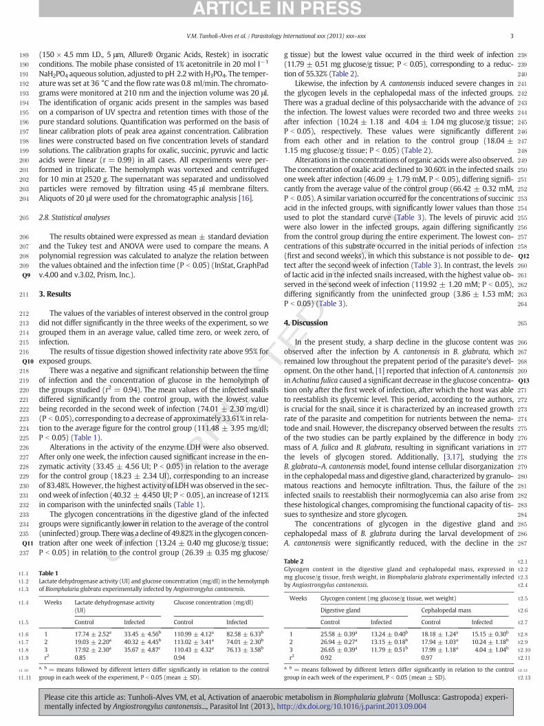
There was a negative and significant relationship between the timeof infection and the concentration of glucose in the hemolymph ofthe groups studied (r2 = 0.94). The mean values of the infected snailsdiffered significantly from the control group, with the lowest valuebeing recorded in the second week of infection (74.01 ± 2.30 mg/dl)(P b 0.05), corresponding to a decrease of approximately 33.61% in rela-tion to the average figure for the control group (111.48 ± 3.95 mg/dl;P b 0.05) (Table 1).
Alterations in the activity of the enzyme LDH were also observed.After only one week, the infection caused significant increase in the en-zymatic activity (33.45 ± 4.56 UI; P b 0.05) in relation to the averagefor the control group (18.23 ± 2.34 UI), corresponding to an increaseof 83.48%. However, the highest activity of LDHwas observed in the sec-ondweek of infection (40.32 ± 4.450 UI; P b 0.05), an increase of 121%in comparison with the uninfected snails (Table 1).
The glycogen concentrations in the digestive gland of the infectedgroups were significantly lower in relation to the average of the control(uninfected) group. Therewas adecline of 49.82% in the glycogen concen-tration after one week of infection (13.24 ± 0.40 mg glucose/g tissue;P b 0.05) in relation to the control group (26.39 ± 0.35 mg glucose/
UTable 1Lactate dehydrogenase activity (UI) and glucose concentration (mg/dl) in the hemolymphof Biomphalaria glabrata experimentally infected by Angiostrongylus cantonensis.
Weeks Lactate dehydrogenase activity(UI)
Glucose concentration (mg/dl)
Control Infected Control Infected
1 17.74 ± 2.52a 33.45 ± 4.56b 110.99 ± 4.12a 82.58 ± 6.33b
2 19.03 ± 2.20a 40.32 ± 4.45b 113.02 ± 3.41a 74.01 ± 2.30b
3 17.92 ± 2.30a 35.67 ± 4.87c 110.43 ± 4.32a 76.13 ± 3.58b
r2 0.85 0.94
a, b = means followed by different letters differ significantly in relation to the controlgroup in each week of the experiment, P b 0.05 (mean ± SD).
Please cite this article as: Tunholi-Alves VM, et al, Activation of anaerobicmentally infected by Angiostrongylus cantonensis..., Parasitol Int (2013), ht
ED P
RO
OF
g tissue) but the lowest value occurred in the third week of infection(11.79 ± 0.51 mg glucose/g tissue; P b 0.05), corresponding to a reduc-tion of 55.32% (Table 2).
Likewise, the infection by A. cantonensis induced severe changes inthe glycogen levels in the cephalopedal mass of the infected groups.There was a gradual decline of this polysaccharide with the advance ofthe infection. The lowest values were recorded two and three weeksafter infection (10.24 ± 1.18 and 4.04 ± 1.04 mg glucose/g tissue;P b 0.05), respectively. These values were significantly differentfrom each other and in relation to the control group (18.04 ±1.15 mg glucose/g tissue; P b 0.05) (Table 2).
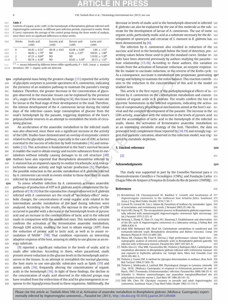
Alterations in the concentrations of organic acidswere also observed.The concentration of oxalic acid declined to 30.60% in the infected snailsone week after infection (46.09 ± 1.79 mM, P b 0.05), differing signifi-cantly from the average value of the control group (66.42 ± 0.32 mM,P b 0.05). A similar variation occurred for the concentrations of succinicacid in the infected groups, with significantly lower values than thoseused to plot the standard curve (Table 3). The levels of piruvic acidwere also lower in the infected groups, again differing significantlyfrom the control group during the entire experiment. The lowest con-centrations of this substrate occurred in the initial periods of infection(first and second weeks), in which this substance is not possible to de-tect after the second week of infection (Table 3). In contrast, the levelsof lactic acid in the infected snails increased, with the highest value ob-served in the second week of infection (119.92 ± 1.20 mM; P b 0.05),differing significantly from the uninfected group (3.86 ± 1.53 mM;P b 0.05) (Table 3).
4. Discussion
In the present study, a sharp decline in the glucose content wasobserved after the infection by A. cantonensis in B. glabrata, whichremained low throughout the prepatent period of the parasite's devel-opment. On the other hand, [1] reported that infection of A. cantonensisin Achatina fulica caused a significant decrease in the glucose concentra-tion only after the first week of infection, after which the host was ableto reestablish its glycemic level. This period, according to the authors,is crucial for the snail, since it is characterized by an increased growthrate of the parasite and competition for nutrients between the nema-tode and snail. However, the discrepancy observed between the resultsof the two studies can be partly explained by the difference in bodymass of A. fulica and B. glabrata, resulting in significant variations inthe levels of glycogen stored. Additionally, [3,17], studying theB. glabrata–A. cantonensismodel, found intense cellular disorganizationin the cephalopedalmass and digestive gland, characterized by granulo-matous reactions and hemocyte infiltration. Thus, the failure of theinfected snails to reestablish their normoglycemia can also arise fromthese histological changes, compromising the functional capacity of tis-sues to synthesize and store glycogen.
The concentrations of glycogen in the digestive gland andcephalopedal mass of B. glabrata during the larval development ofA. cantonensis were significantly reduced, with the decline in the
Table 2 t2:1
t2:2Glycogen content in the digestive gland and cephalopedal mass, expressed int2:3mg glucose/g tissue, fresh weight, in Biomphalaria glabrata experimentally infectedt2:4by Angiostrongylus cantonensis.
t2:5Weeks Glycogen content (mg glucose/g tissue, wet weight)
t2:6Digestive gland Cephalopedal mass
t2:7Control Infected Control Infected
t2:81 25.58 ± 0.39a 13.24 ± 0.40b 18.18 ± 1.24a 15.15 ± 0.30b
t2:92 26.94 ± 0.27a 13.15 ± 0.18b 17.94 ± 1.03a 10.24 ± 1.18b
t2:103 26.65 ± 0.39a 11.79 ± 0.51b 17.99 ± 1.18a 4.04 ± 1.04b
t2:11r2 0.92 0.97
t2:12a, b = means followed by different letters differ significantly in relation to the control
t2:13group in each week of the experiment, P b 0.05 (mean ± SD).
metabolism in Biomphalaria glabrata (Mollusca: Gastropoda) experi-tp://dx.doi.org/10.1016/j.parint.2013.09.004
T
288Q14
289
290
291
292
293
294
295
296
297
298
299
300
301
302
303
304
305
306
307
308Q15
309
310
311
312
313
314Q16
315
316
317
318
319
320
321
322
323
324
325
326
327
328
329
330
331
332
333
334
335
336
337
338
339
340
341
342
343
344
345
346
347
348
349
350
351
352
353Q17
354
355
356
357
358
359
360
361
362
363
364
365
366
367
368
369
370Q18
371
372Q19
373
374
375
376
377378379380381382383384385386387388389390391392393394395396397398399400401402403404405406
Table 3t3:1
t3:2 Contents of organic acids (mM) in the hemolymph of Biomphalaria glabrata infected witht3:3 Angiostrongylus cantonensis, in different post-infection periods, expressed in weeks. Weekt3:4 0 (zero) represents the average of the control group during the three weeks of analysis,t3:5 since there were no significant differences in these weeks.
t3:6 Weeks Oxalic acid(mM)
Succinic acid(mM)
Pyruvic acid(mM)
Lactic acid(mM)
t3:7 0 66.42 ± 0.32a 40.18 ± 0.63 82.08 ± 6.60a 3.86 ± 1.53a
t3:8 1 46.09 ± 1.79b ND 6.20 ± 2.25b 104.91 ± 1.00b
t3:9 2 47.23 ± 0.66b ND ND 119.92 ± 1.20b
t3:10 3 60.74 ± 0.38b ND 65.82 ± 5.58b 20.13 ± 1.52b
t3:11a, b = means followed by different letters differ significantly, P b 0.05, (mean ± standard
t3:12 deviation). ND = organic acids not detected.
4 V.M. Tunholi-Alves et al. / Parasitology International xxx (2013) xxx–xxx
UNCO
RREC
cephalopedal mass being the greatest change. [11] reported the activityof glycolytic enzymes in juvenile specimens of A. cantonensis, indicatingthe presence of an oxidative pathway to maintain the parasite's energybalance. Therefore, the greater decrease in the concentration of glyco-gen observed in the muscular tissues can be explained by the presenceof L3 stages of the nematode. According to [4], this tissue is themain sitefor larvae in the final stage of their development in the snail. Therefore,the intense development of the A. cantonensis larvae during the initialdays of the infection causes large consumption of glucose from thesnail's hemolymph by the parasite, triggering depletion of the host'spolysaccharide reserves in an attempt to reestablish the levels of circu-lating glucose.
An acceleration of the anaerobic metabolism in the infected snailswas also observed, since there was a significant increase in the activityof the LDH. Studies have demonstrated an overlap of enzymatic centersrelated to the glycolytic pathway, especially in the case of LDH, as factorsessential to the success of infection by both trematodes [16] and nema-todes [12]. This activation is fundamental to the host's survival becauseit enables the snail to obtain energy and excrete substances from itsme-tabolism without initially causing damages to its cell structure [10].Authors have also reported that Biomphalaria alexandrina infected byS.mansoni has an impaired capacity to oxidize tricarboxylic acid, low cy-tochrome oxidase activity and high lactate production [5]. Therefore,the possible reduction in the aerobic metabolism of B. glabrata infectedby A. cantonensis can result in events similar to those described in snailsinfected by trematode larvae.
To assess whether infection by A. cantonensis activates anaerobicpathways of production of ATP in B. glabrata and to complement the hy-pothesis of [18,19] that the reproductive changes observed in B. glabratainfected with A. cantonensis are the result of “secondary-effect” meta-bolic changes, the concentrations of some organic acids related to theintermediate aerobic metabolism of the host during infection wereanalyzed. According to the results, the increase in the activity of LDHoccurred in parallel with a decrease in the hemolymph levels of pyruvicacid and an increase in the concentrations of lactic acid in the infectedsnails in comparison with the uninfected ones. This metabolic scenarioconfirms the activation of the fermentative anaerobic metabolism,through LDH activity, enabling the host to obtain energy (ATP) fromthe reduction of piruvic acid to lactic acid, as well as to assure re-oxidation of NADH+ [16]. That condition represents an interestingadaptive response of thehost, assuring its ability to use glucose as an en-ergy substrate.
[9] reported a significant reduction in the levels of oxalic acid insnails after infection. According to them, when parasitized, snailspresent severe reduction in the glucose levels in the hemolymph and re-serves in the tissues. In an attempt to reestablish the normal glycemia,the host starts to use non-glycidic substrates such as lipids [19] andamino acids [16], frequently altering the concentrations of organicacids in the hemolymph [16]. In light of these findings, the decline inthe concentration of oxalic acid observed in the infected groups mayhave resulted from the redirection of this acid for gluconeogenesis in re-sponse to the hypoglycemia found in these organisms. Additionally, the
Please cite this article as: Tunholi-Alves VM, et al, Activation of anaerobicmentally infected by Angiostrongylus cantonensis..., Parasitol Int (2013), ht
ED P
RO
OF
decrease in levels of oxalic acid in the hemolymph observed in infectedB. glabrata can also be explained by the use of this molecule as the sub-strate for the development of larvae of A. cantonensis. The use of someorganic acids, particularly oxalic acid as a substrate necessary for the de-velopment of sporocysts and cercariae of S. mansoni in B. glabrata hasbeen demonstrated by [6].
The infection by A. cantonensis also resulted in reduction of thesuccinic acid level in the hemolymph below the limit of detection, pre-senting values below those used to plot the standard curve. Similar re-sults have been observed previously by authors studying the parasite–host relationship [15,16]. According to these authors, this variationresults from the activation of fumarate reductase, an enzyme responsi-ble for malate to succinate reduction, in the reverse of the Krebs cycle.As a consequence, succinate is metabolized into propionate, generatingenergy and helping tomaintain the redox balance. This reaction contrib-utes to the reduction in the concentrations of this acid in the modelstudied here.
This article is the first report of the pathophysiological effects of in-fection by A. cantonensis on the carbohydrate metabolism and concen-trations of organic acids in B. glabrata. The results confirm the loss ofglycemic homeostasis in the infected organisms, indicating the activa-tion of compensatory physiologicalmechanisms aimed at the host's sur-vival and the complete development of the parasite. The increase in theLDH activity, associated with the reduction in the levels of pyruvic acidand the accumulation of lactic acid in the hemolymph of the infectedsnails, shows the activation of fermentative anaerobic metabolismas an interesting metabolic strategy of the host. Therefore, the datapresented here complement those reported by [18,19] and strongly sug-gest that parasitic castration, observed in this infection model, was trig-gered by metabolic depletion.
5. Uncited reference
[2]
Acknowledgments
This study was supported in part by the Conselho Nacional para oDesenvolvimento Científico e Tecnológico (CNPq) and Fundação CarlosChagas Filho deAmparo à Pesquisa do Estado do Rio de Janeiro (FAPERJ).
References
[1] Brockelman CR, Chusatayanond W, Baidikul V. Growth and localization ofAngiostrongylus cantonensis in the molluscan host Achatina fulica. SoutheastAsian J Trop Med Public Health 1976;7:30–7.
[2] Grewal PS, Grewal SK, Tan L, Adams BJ. Parasitism of molluscs by nematodes: typesof associations and evolutionary trends. J Nematol 2003;35:146–56.
[3] Harris KR, Cheng TC. The encapsulation process in Biomphalaria glabrata experimen-tally infected with metastrongylid Angyonstrongylus cantonensis light microscopy.Int J Parasitol 1975;5:521–8.
[4] Hexiang L, Zhang YI, Shan LV, Ling HU, Xiaonong Z. Establishment and observationof the life cycle of Angiostrongylus cantonensis in a laboratory setting. J Pathog Biol2009;4:836–9.
[5] Ishak MM, Mohamed AM, Shraf AA. Carbohydrate metabolism in uninfected andtrematode-infected snails Biomphalaria alexandrina and Bulinus truncatus. CompBiochem Physiol B 1975;53:499–505.
[6] Massa DR, Chejlava MJ, Fried B, Sherma J. High performance column liquid chro-matographic analysis of selected carboxylic acids in Biomphalaria glabrata patentlyinfected with Schistosoma mansoni. Parasitol Res 2007;101:925–8.
[7] Mello-Silva CC, Vilar MM, Vasconcellos MC, Pinheiro J, Rodrigues MLA. Carbohydratemetabolism alterations in Biomphalaria glabrata infected with Schistosoma mansoniand exposed to Euphorbia splendens var. hislopii látex. Mem Inst Oswaldo Cruz2010;105:492–5.
[8] Pinheiro J, Gomes EM. A method for glycogen determination in molluscs. Braz ArchBiol Technol 1994;37:569–76.
[9] Pinheiro J, Maldonado A, Lanfredi RM. Physiological changes in Lymnaea columella(Say, 1818) (Mollusca, Gastropoda) in response to Echinostoma paraensei Lie andBasch, 1967 (Trematoda, Echinostomatidae) infection. Parasitol Res 2009;106:55–9.
[10] Schottler U. Weitere untersuchungen zun anaeroben energiestoffwechsel despolychaeten Arenicola marina L. Zool Beitr NF 1986;30:141–52.
[11] Shih HH, Chen SN. Glycolytic enzymes in juvenile and adult Angiostrongyluscantonensis. Southeast Asian J Trop Med Public Health 1982;13:114–9.
metabolism in Biomphalaria glabrata (Mollusca: Gastropoda) experi-tp://dx.doi.org/10.1016/j.parint.2013.09.004
407408409410411412413414415416417418419420Q20421422
423424425426427428429430431432433434435436437438
440
5V.M. Tunholi-Alves et al. / Parasitology International xxx (2013) xxx–xxx
[12] Stewart GL, Ubelaker JE, Curtis D. Pathophysiologic alterations in Biomphalariaglabrata infectedwithAngiostrongylus costaricensis. J Invertebr Pathol 1985;45:152–7.
[13] Sullivan JT, Cheng TC, Howland KH. Studies on parasitic castration: castration ofIlyanassa obsoleta (Mollusca: Gastropoda) by several marine trematodes. Trans AmMicrosc Soc 1985;104:154–71.
[14] Sumner JB. The estimation of sugar in diabetic urine using dinitrosalicylic acid. J BiolChem 1924;62:287–90.
[15] Tielens AGM. Energy generation in parasitic helminths. Parasitol Today1994;10:346–52.
[16] Tunholi VM, Tunholi-Alves VM, Lustrino D, Castro N, Sant'Ana L, Garcia J, et al.Aerobic to anaerobic transition in Biomphalaria glabrata (Say, 1818) infected withdifferent miracidial doses of Echinostoma paraensei (Lie and Basch, 1967) by high-performance liquid chromatography. Exp Parasitol 2013;133:403–10.
[17] Tunholi-Alves VM, Tunholi VM, Garcia J, Costa-Neto SF, Maldonado A, Santos MAJ,et al. Changes in the calcium metabolism of Biomphalaria glabrata experimentallyinfected with Angiostrongylus cantonensis. J Helminthol 2013 [in press].
UNCO
RRECT
439
Please cite this article as: Tunholi-Alves VM, et al, Activation of anaerobicmentally infected by Angiostrongylus cantonensis..., Parasitol Int (2013), ht
[18] Tunholi-Alves VM, Tunholi VM, Gôlo P, Lustrino D, Maldonado Jr A, BittencourtVREP, et al. Lipid levels in Biomphalaria glabrata infected with different doses ofEchinostoma paraensei miracidia. Exp Parasitol 2011;128:212–6.
[19] Tunholi-Alves VM, Tunholi VM, Lustrino D, Amaral LS, Thiengo SC, Pinheiro J.Changes in the reproductive biologyof Biomphalaria glabrata experimentally infectedwith the nematode Angiostrongylus cantonensis. J Invertebr Pathol 2011;108:220–3.
[20] Tunholi-Alves VM, Tunholi VM, Pinheiro J, Thiengo SC. Effects of infection by larvae ofAngiostrongylus cantonensis (Nematoda, Metastrongylidae) on the metabolism of theexperimental intermediate host Biomphalaria glabrata. Exp Parasitol 2012;131:143–7.
[21] Wang QP, De-Hua L, Xing-Quan Z, Xiao-Guang C, Zhao-Rong L. Human angio-strongyliasis. Lancet Infect Dis 2008;8:621–30.
[22] WillcoxHP, Coura JR. Nova concepçãopara ométododeBaermann–Moraes - Coutinhona pesquisa de larvas de nematódeos. Mem Inst Oswaldo Cruz 1989;84:539–65.
[23] Yousif F, LámmlerG. Themodeof infectionwith and thedistributionofAngionstrongyluscantonensis larvae in the intermediate host Biomphalaria glabrata. Z Parasitenkd1977;53:247–50.
ED P
RO
OF
metabolism in Biomphalaria glabrata (Mollusca: Gastropoda) experi-tp://dx.doi.org/10.1016/j.parint.2013.09.004