Embed Size (px)
Citation preview
http://jfm.sagepub.com/Journal of Feline Medicine and Surgery
http://jfm.sagepub.com/content/15/10/849The online version of this article can be found at:
DOI: 10.1177/1098612X13477415
2013 15: 849 originally published online 14 February 2013Journal of Feline Medicine and SurgeryDominique Paepe, Valérie Bavegems, Anaïs Combes, Jimmy H Saunders and Sylvie Daminet
parameters and ultrasonographyProspective evaluation of healthy Ragdoll cats for chronic kidney disease by routine laboratory
technique does not amount to an endorsement of its value or quality, or the claims made by its manufacturer.those of the authors and the inclusion in this publication of material relating to a particular product, method or of animals and interpretation of published materials lies with the veterinary practitioner. The opinions expressed arefrom actions or decisions based on information contained in this publication; ultimate responsibility for the treatment
arisingcountry. The authors, editors, owners and publishers do not accept any responsibility for any loss or damage advertising material, it is the responsibility of the reader to check that the product is authorised for use in their ownbear this in mind and be aware of the prescribing laws pertaining to their own country. Likewise, in relation to Furthermore, drugs may be mentioned that are licensed for human use, and not for veterinary use. Readers need toformulations that are not available or licensed in the individual reader's own country.The Journal of Feline Medicine and Surgery is an international journal and authors may discuss products and
Disclaimer
Published by:
International Society of Feline Medicine
American Association of Feline Practitioners
and http://www.sagepublications.com
can be found at:Journal of Feline Medicine and SurgeryAdditional services and information for
http://jfm.sagepub.com/cgi/alertsEmail Alerts:
http://jfm.sagepub.com/subscriptionsSubscriptions:
http://www.sagepub.com/journalsReprints.navReprints:
http://www.sagepub.com/journalsPermissions.navPermissions:
What is This?
- Feb 14, 2013OnlineFirst Version of Record
- Sep 20, 2013Version of Record >>
by guest on February 26, 2014jfm.sagepub.comDownloaded from by guest on February 26, 2014jfm.sagepub.comDownloaded from
Journal of Feline Medicine and Surgery15(10) 849 –857© ISFM and AAFP 2013Reprints and permissions: sagepub.co.uk/journalsPermissions.navDOI: 10.1177/1098612X13477415jfms.com
IntroductionAccording to the Cat Fanciers’ Association, the Ragdoll is one of the most popular cat breeds worldwide.1 Several Ragdoll breeder organisations, such as the Ragdoll Club Benelux and the Scandinavian Ragdoll Club, forewarn owners that renal problems may develop as a result of polycystic kidney disease (PKD), chronic interstitial nephritis (CIN), familial renal dysplasia or nephrocalci-nosis.2,3 Based on the recommendations of these breed clubs, Ragdoll cats are screened for PKD and CIN prior to breeding in several European countries, such as Belgium, the Netherlands, Sweden and Finland.2,3 Several tests are part of this screening programme, including abdominal ultrasonography to identify renal and/or hepatic cysts and evidence of CIN, measurement of serum urea and creatinine concentrations, and genetic testing for the PKD-1 mutation. The results of these screening tests can be found on the Ragdoll Health Database on the Internet.4
Recently, our group retrospectively evaluated the results of these screening tests performed on Ragdoll
Prospective evaluation of healthy Ragdoll cats for chronic kidney disease by routine laboratory parameters and ultrasonography
Dominique Paepe1, Valérie Bavegems1, Anaïs Combes2, Jimmy H Saunders2 and Sylvie Daminet1
AbstractRagdoll breeder organisations often forewarn Ragdoll cat owners that renal problems may develop as a result of polycystic kidney disease (PKD), chronic interstitial nephritis, familial renal dysplasia or nephrocalcinosis. Healthy Ragdoll and non-Ragdoll cats were prospectively evaluated by measuring serum creatinine and urea concentrations, routine urinalysis and abdominal ultrasonography. All Ragdoll cats also underwent genetic PKD testing. One hundred and thirty-three Ragdoll and 62 control cats were included. Ragdoll cats had significantly lower serum urea concentrations and higher urinary specific gravity. However, median creatinine concentration, median urinary protein-to-creatinine ratio, and the proportion of cats with serum creatinine or urea concentration exceeding the reference interval did not differ. One or more renal ultrasonographical changes were detected in 66/133 (49.6%) Ragdoll and in 25/62 (40%) control cats. Ragdoll cats showed significantly more frequent segmental cortical lesions (7.5% versus 0%), abnormal renal capsule (19.5% versus 8%) and echogenic urine (51.9% versus 25.8%). Chronic kidney disease (CKD) was ultrasonographically suspected in 7/133 (5.3%) Ragdoll and in none of the control cats, which approached significance. Laboratory parameters confirmed kidney dysfunction only in 1/7 of these Ragdoll cats. All Ragdoll cats were PKD negative. In conclusion, first, breed-specific serum creatinine reference intervals are not likely required for Ragdoll cats. Second, renal ultrasonographical abnormalities are common, both in Ragdoll and non-Ragdoll cats. Third, healthy young Ragdoll cats are uncommonly affected by PKD and CKD, but an increased susceptibility of Ragdoll cats to develop CKD cannot be excluded. Finally, Ragdoll cats are predisposed to segmental cortical lesions, which may indicate renal infarction or cortical scarring.
Accepted: 11 January 2013
1 Department of Small Animal Medicine and Clinical Biology, Faculty of Veterinary Medicine, Ghent University, Belgium
2 Department of Medical Imaging of Domestic Animals, Faculty of Veterinary Medicine, Ghent University, Salisburylaan, Merelbeke, Belgium
This paper was presented in part at the 22nd ECVIM-CA congress in Maastricht, the Netherlands, September 2012, and published as an abstract in 22nd ECVIM-CA Congress Proceedings, pp 242–243 and in Journal of Veterinary Internal Medicine 2012; 26: 1537
Corresponding author:Dominique Paepe DVM, Dipl ECVIM-CA, Department of Small Animal Medicine and Clinical Biology, Faculty of Veterinary Medicine, Ghent University, Salisburylaan 133, B-9820 Merelbeke, Belgium Email: [email protected]
477415 JFM151010.1177/1098612X13477415Journal of Feline Medicine and SurgeryPaepe et al2013
Original Article
by guest on February 26, 2014jfm.sagepub.comDownloaded from
850 Journal of Feline Medicine and Surgery 15(10)
cats at our institution. Ultrasonographical findings com-patible with chronic kidney disease (CKD) were observed in 8.6% and PKD in 2.9% of included healthy Ragdoll cats. However, this study was limited by the lack of urinalysis, incomplete screening tests in many Ragdoll cats and the lack of information about the prev-alence of renal ultrasonographical abnormalities in healthy non-Ragdoll cats.5
To further elucidate if the concerns of the Ragdoll breed organisations are justified or not, we performed a prospective study to compare serum creatinine and urea concentrations, routine urinalysis and renal ultrasono-graphical findings between Ragdoll cats and an age-matched control group.
Materials and methodsStudy populationRagdoll cats that were presented by their owner for CIN or PKD screening were considered for inclusion. Age-matched non-Ragdoll cats were recruited actively as control cats. Both pure- and mixed-breed cats were con-sidered for inclusion as control cats, but a maximum five cats of each pure cat breed were allowed. Both Ragdoll and control cats needed to be 10 months or older to be included. To avoid bias towards kidney disease in the Ragdoll population, Ragdoll cats that were presented by their owner with an already diagnosed CKD were excluded from this study. Because cats presented for screening are usually healthy, only healthy Ragdoll and control cats were included. Health was defined as clini-cally healthy for their owner and without significant abnormalities on physical examination, complete blood count (Medvet Algemeen Medisch Laboratorium Diergeneeskunde; Advia, Siemens) and serum biochem-istry profile (Medvet Algemeen Medisch Laboratorium Diergeneeskunde; Architect, Abbott), except for serum creatinine and urea concentrations. All cats were fasted for 12 h; water was offered ad libitum.
The study was completed at the Department of Small Animal Medicine and Clinical Biology, Faculty of Veterinary Medicine, Ghent University between October 2010 and March 2012. All cats were privately owned, the owners were thoroughly informed about the study aims and protocol, and the study was approved by local and national ethical committees (EC2010/104).
ProceduresThe cats underwent measurement of serum creatinine (Idexx Catalyst Dx Analyzer, Idexx Europe BV) and urea (Idexx Catalyst Dx Analyzer, Idexx Europe BV) concen-trations, complete urinalysis and abdominal ultrasonog-raphy. Only the Ragdoll cats underwent a genetic PKD test (Department of Nutrition, Genetics and Ethology, Faculty of Veterinary Medicine, Ghent University, Merelbeke, Belgium) that was completed as described
by identifying the C>A transversion in exon 29 of the PKD-1 gene.6
Urinalysis consisted of a urinary dipstick test; meas-urement of urine specific gravity (USG) with a manual refractometer, urinary pH and urinary protein-to- creatinine ratio (UPC) (Medvet Algemeen Medisch Laboratorium Diergeneeskunde; Iricell IQ, Instru -mentation Laboratory) and sediment examination. The sediment was prepared as described previously7,8 and evaluated under the microscope within 30 mins of col-lection. Crystalluria was evaluated semi-quantitatively and expressed per low-power field (LPF, 10× objective) as mild (<1/LPF), moderate (1–3/LPF) or severe (>3/LPF). Bacterial culture (Medvet Algemeen Medisch Laboratorium Diergeneeskunde; BioMerieux Media Square) of the urine was only performed if considered necessary based on routine urinalysis or on the pres-ence of sediment in the urinary bladder during ultrasonography.
Abdominal ultrasonography was performed by an ECVDI Diplomate or supervised ECVDI resident using a multifrequency (6–10 MHz) microconvex or multifre-quency (7.5–12 MHz) linear transducer (Logic 7; GE Medical Systems). The cats were restrained manually in dorsal recumbency. The hair was not, or only minimally, clipped and parted to expose the skin. To improve skin contact and conduction of ultrasound waves, the hair was soaked with alcohol and water before ultrasound coupling gel was applied. The kidneys, urinary bladder and liver were evaluated according to a standard proto-col in longitudinal and transverse scanning planes. A ventrolateral and ventral approach was used for the kid-neys and liver, and a ventral approach for the urinary bladder. Parameters assessed at the level of the kidneys were the renal capsule (normal or abnormal), renal shape (regular, irregular or other), kidney length in a sagittal plane, cortical echogenicity (diffuse hypo-, iso- or hyper-echogenic compared with the spleen or liver or focal abnormalities), medullar echogenicity (diffuse hypo-, iso- or hyperechogenic compared with the renal cortex or focal abnormalities), corticomedullary demarcation (well-delineated or reduced), renal pelvis (normal, enlarged or presence of uroliths), proximal ureter (nor-mal, enlarged or presence of uroliths), and the presence or absence of medullary rim sign, dystrophic mineralisa-tion, cavitary lesion, solid mass, nodule, segmental corti-cal lesion and retroperitoneal fluid. For the urinary bladder, bladder filling, echogenicity of the urine and presence or absence of uroliths was noted. The liver was evaluated carefully for the presence or absence of cysts, or other parenchymal changes. Additional changes in the liver, kidneys, urinary bladder and the remainder of the abdomen were also noted. Because CIN is not an ultrasound diagnosis, the radiologist was asked to judge if the observed abnormalities could be indicative of CKD
by guest on February 26, 2014jfm.sagepub.comDownloaded from
Paepe et al 851
possibly caused by CIN. In addition, the radiologist was asked if the cat could be affected by renal agenesis, hypo-plasia or dysplasia.
Statistical methodsAll statistical tests were performed with statistical soft-ware (SPSS) and at the 0.05 significance level. To com-pare the Ragdoll and the control group, student’s t-test was used for continuous variables that approached nor-mal distribution, Mann–Whitney U-test for non-nor-mally distributed continuous variables, and the Fisher’s exact test or the Pearson χ2, test for discrete variables.
ResultsStudy populationIn total, 133 Ragdoll and 62 control cats were included. Seven Ragdoll cats with known CKD and one Ragdoll cat for which it was already known that it only had one kidney were excluded. Two other Ragdoll cats were excluded — one because of dyspnoea and muffled lung sounds due to pleural effusion, and one because of ascites. Further diagnostic tests revealed feline infectious peritonitis in both cats. One control cat was excluded because of ulcerative and scaly swellings of the footpads consistent with plasma cell pododermatitis.
Age and body weight did not differ between groups (Table 1). The gender distribution differed significantly between groups (P <0.001; Ragdoll: 80 intact females,
five neutered females, 40 intact males, eight neutered males; control: 16 intact females, 20 neutered females, 0 intact males, 26 neutered males).
Ragdoll pedigrees indicated that 43 Ragdoll cats orig-inated from Belgium, 37 from the Netherlands, 15 from other European countries, 28 from the USA, seven from Australia or New Zealand, two from Canada and one from Israel.
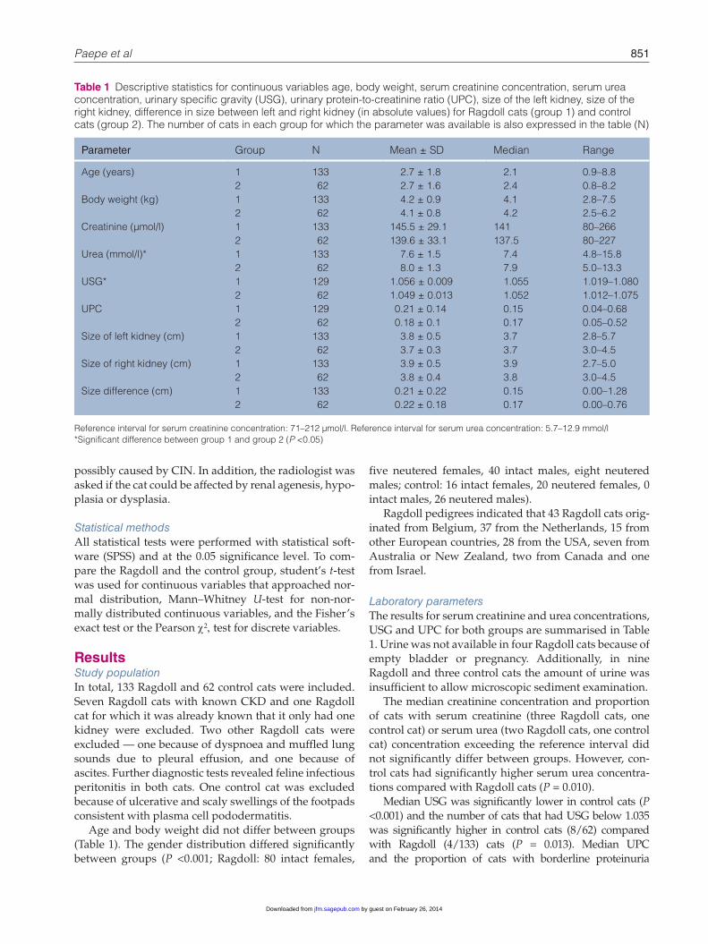
Laboratory parametersThe results for serum creatinine and urea concentrations, USG and UPC for both groups are summarised in Table 1. Urine was not available in four Ragdoll cats because of empty bladder or pregnancy. Additionally, in nine Ragdoll and three control cats the amount of urine was insufficient to allow microscopic sediment examination.
The median creatinine concentration and proportion of cats with serum creatinine (three Ragdoll cats, one control cat) or serum urea (two Ragdoll cats, one control cat) concentration exceeding the reference interval did not significantly differ between groups. However, con-trol cats had significantly higher serum urea concentra-tions compared with Ragdoll cats (P = 0.010).
Median USG was significantly lower in control cats (P <0.001) and the number of cats that had USG below 1.035 was significantly higher in control cats (8/62) compared with Ragdoll (4/133) cats (P = 0.013). Median UPC and the proportion of cats with borderline proteinuria
Table 1 Descriptive statistics for continuous variables age, body weight, serum creatinine concentration, serum urea concentration, urinary specific gravity (USG), urinary protein-to-creatinine ratio (UPC), size of the left kidney, size of the right kidney, difference in size between left and right kidney (in absolute values) for Ragdoll cats (group 1) and control cats (group 2). The number of cats in each group for which the parameter was available is also expressed in the table (N)
Parameter Group N Mean ± SD Median Range
Age (years) 1 133 2.7 ± 1.8 2.1 0.9–8.8 2 62 2.7 ± 1.6 2.4 0.8–8.2Body weight (kg) 1 133 4.2 ± 0.9 4.1 2.8–7.5 2 62 4.1 ± 0.8 4.2 2.5–6.2Creatinine (µmol/l) 1 133 145.5 ± 29.1 141 80–266 2 62 139.6 ± 33.1 137.5 80–227Urea (mmol/l)* 1 133 7.6 ± 1.5 7.4 4.8–15.8 2 62 8.0 ± 1.3 7.9 5.0–13.3USG* 1 129 1.056 ± 0.009 1.055 1.019–1.080 2 62 1.049 ± 0.013 1.052 1.012–1.075UPC 1 129 0.21 ± 0.14 0.15 0.04–0.68 2 62 0.18 ± 0.1 0.17 0.05–0.52Size of left kidney (cm) 1 133 3.8 ± 0.5 3.7 2.8–5.7 2 62 3.7 ± 0.3 3.7 3.0–4.5Size of right kidney (cm) 1 133 3.9 ± 0.5 3.9 2.7–5.0 2 62 3.8 ± 0.4 3.8 3.0–4.5Size difference (cm) 1 133 0.21 ± 0.22 0.15 0.00–1.28 2 62 0.22 ± 0.18 0.17 0.00–0.76
Reference interval for serum creatinine concentration: 71–212 µmol/l. Reference interval for serum urea concentration: 5.7–12.9 mmol/l*Significant difference between group 1 and group 2 (P <0.05)
by guest on February 26, 2014jfm.sagepub.comDownloaded from
852 Journal of Feline Medicine and Surgery 15(10)
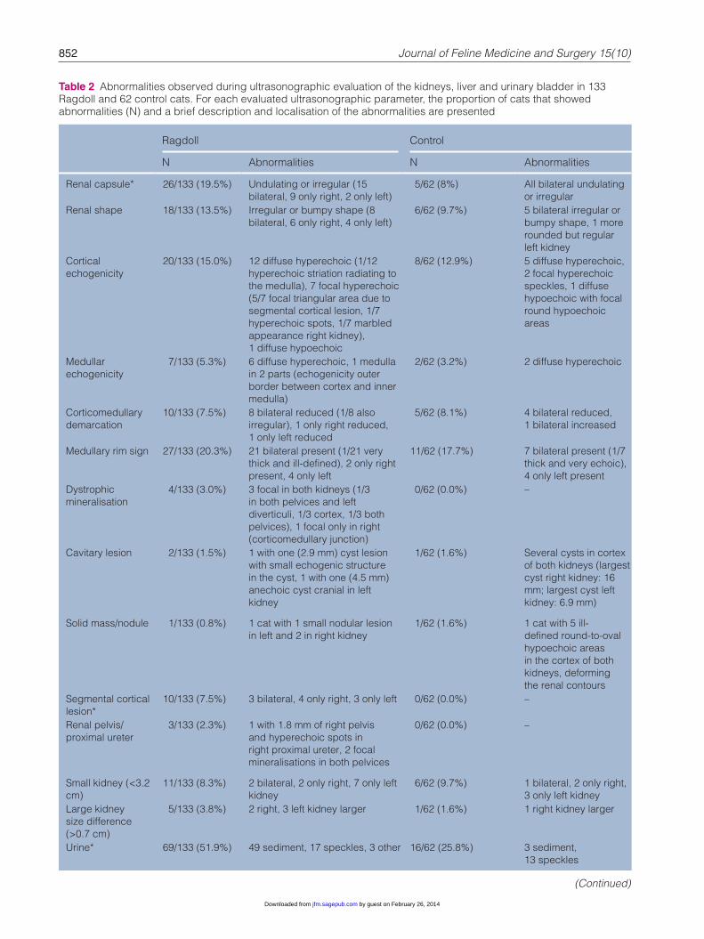
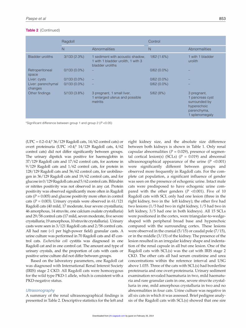
Table 2 Abnormalities observed during ultrasonographic evaluation of the kidneys, liver and urinary bladder in 133 Ragdoll and 62 control cats. For each evaluated ultrasonographic parameter, the proportion of cats that showed abnormalities (N) and a brief description and localisation of the abnormalities are presented
Ragdoll Control
N Abnormalities N Abnormalities
Renal capsule* 26/133 (19.5%) Undulating or irregular (15 bilateral, 9 only right, 2 only left)
5/62 (8%) All bilateral undulating or irregular
Renal shape 18/133 (13.5%) Irregular or bumpy shape (8 bilateral, 6 only right, 4 only left)
6/62 (9.7%) 5 bilateral irregular or bumpy shape, 1 more rounded but regular left kidney
Cortical echogenicity
20/133 (15.0%) 12 diffuse hyperechoic (1/12 hyperechoic striation radiating to the medulla), 7 focal hyperechoic (5/7 focal triangular area due to segmental cortical lesion, 1/7 hyperechoic spots, 1/7 marbled appearance right kidney), 1 diffuse hypoechoic
8/62 (12.9%) 5 diffuse hyperechoic, 2 focal hyperechoic speckles, 1 diffuse hypoechoic with focal round hypoechoic areas
Medullar echogenicity
7/133 (5.3%) 6 diffuse hyperechoic, 1 medulla in 2 parts (echogenicity outer border between cortex and inner medulla)
2/62 (3.2%) 2 diffuse hyperechoic
Corticomedullary demarcation
10/133 (7.5%) 8 bilateral reduced (1/8 also irregular), 1 only right reduced, 1 only left reduced
5/62 (8.1%) 4 bilateral reduced, 1 bilateral increased
Medullary rim sign 27/133 (20.3%) 21 bilateral present (1/21 very thick and ill-defined), 2 only right present, 4 only left
11/62 (17.7%) 7 bilateral present (1/7 thick and very echoic), 4 only left present
Dystrophic mineralisation
4/133 (3.0%) 3 focal in both kidneys (1/3 in both pelvices and left diverticuli, 1/3 cortex, 1/3 both pelvices), 1 focal only in right (corticomedullary junction)
0/62 (0.0%) –
Cavitary lesion 2/133 (1.5%) 1 with one (2.9 mm) cyst lesion with small echogenic structure in the cyst, 1 with one (4.5 mm) anechoic cyst cranial in left kidney
1/62 (1.6%) Several cysts in cortex of both kidneys (largest cyst right kidney: 16 mm; largest cyst left kidney: 6.9 mm)
Solid mass/nodule 1/133 (0.8%) 1 cat with 1 small nodular lesion in left and 2 in right kidney
1/62 (1.6%) 1 cat with 5 ill-defined round-to-oval hypoechoic areas in the cortex of both kidneys, deforming the renal contours
Segmental cortical lesion*
10/133 (7.5%) 3 bilateral, 4 only right, 3 only left 0/62 (0.0%) –
Renal pelvis/proximal ureter
3/133 (2.3%) 1 with 1.8 mm of right pelvis and hyperechoic spots in right proximal ureter, 2 focal mineralisations in both pelvices
0/62 (0.0%) –
Small kidney (<3.2 cm)
11/133 (8.3%) 2 bilateral, 2 only right, 7 only left kidney
6/62 (9.7%) 1 bilateral, 2 only right, 3 only left kidney
Large kidney size difference (>0.7 cm)
5/133 (3.8%) 2 right, 3 left kidney larger 1/62 (1.6%) 1 right kidney larger
Urine* 69/133 (51.9%) 49 sediment, 17 speckles, 3 other 16/62 (25.8%) 3 sediment, 13 speckles
(Continued)
by guest on February 26, 2014jfm.sagepub.comDownloaded from
Paepe et al 853
(UPC = 0.2–0.4;9 36/129 Ragdoll cats, 14/62 control cats) or overt proteinuria (UPC >0.4;9 14/129 Ragdoll cats, 4/62 control cats) did not differ significantly between groups. The urinary dipstick was positive for haemoglobin in 37/129 Ragdoll cats and 17/62 control cats, for acetone in 9/129 Ragdoll cats and 1/62 control cats, for protein in 128/129 Ragdoll cats and 56/62 control cats, for urobilino-gen in 36/129 Ragdoll cats and 19/62 control cats, and for glucose in 0/129 Ragdoll cats and 5/62 control cats. Bilirubin or nitrites positivity was not observed in any cat. Protein positivity was observed significantly more often in Ragdoll cats (P = 0.005) and glucose positivity more often in control cats (P = 0.003). Urinary crystals were observed in 61/121 Ragdoll cats (40 mild, 17 moderate, four severe crystalluria; 46 amorphous, 14 struvite, one calcium oxalate crystalluria) and 29/58 control cats (17 mild, seven moderate, five severe crystalluria; 19 amorphous, 10 struvite crystalluria). Urinary casts were seen in 3/121 Ragdoll cats and 2/58 control cats. All had rare (<1 per high-power field) granular casts. A urine culture was performed in 70 Ragdoll cats and 45 con-trol cats. Escherichia coli cystitis was diagnosed in one Ragdoll cat and in one control cat. The amount and type of urinary crystals, and the proportion of cats with casts or positive urine culture did not differ between groups.
Based on the laboratory parameters, one Ragdoll cat was diagnosed with International Renal Interest Society (IRIS) stage 2 CKD. All Ragdoll cats were homozygous for the wild type PKD-1 allele, which is consistent with a PKD-negative status.
UltrasonographyA summary of the renal ultrasonographical findings is presented in Table 2. Descriptive statistics for the left and
right kidney size, and the absolute size difference between both kidneys is shown in Table 1. Only renal capsular abnormalities (P = 0.029), presence of segmen-tal cortical lesion(s) (SCLs) (P = 0.019) and abnormal ultrasonographical appearance of the urine (P <0.001) were significantly different between groups and observed more frequently in Ragdoll cats. For the com-plete cat population, a significant influence of gender was seen on the presence of echogenic urine. Intact male cats were predisposed to have echogenic urine com-pared with the other genders (P <0.001). Five of 10 Ragdoll cats with SCL only had one lesion (three in the right kidney, two in the left kidney); the other five had two lesions (1/5 had two in right kidney, 1/5 had two in left kidney, 3/5 had one in both kidneys). All 15 SCLs were positioned in the cortex, were triangular-to-wedge-shaped with peripheral broad base and hyperechoic compared with the surrounding cortex. These lesions were observed in the cranial (5/15) or caudal pole (7/15), or in the middle (3/15) of the kidney. The presence of the lesion resulted in an irregular kidney shape and indenta-tion of the renal capsule in all but one lesion. One of the Ragdoll cats with SCL(s) was the cat with IRIS stage 2 CKD. The other cats all had serum creatinine and urea concentrations within the reference interval and USG above 1.035. Three of the cats with SCL(s) had borderline proteinuria and one overt proteinuria. Urinary sediment examination revealed haematuria in two, mild haematu-ria and rare granular casts in one, severe struvite crystal-luria in one, mild amorphous crystalluria in two and no abnormalities in four cats. Urine culture was negative in all six cats in which it was assessed. Brief pedigree analy-sis of the Ragdoll cats with SCL(s) showed that one sire
Ragdoll Control
N Abnormalities N Abnormalities
Bladder uroliths 3/133 (2.3%) 1 sediment with acoustic shadow, 1 with 1 bladder urolith, 1 with 3 bladder uroliths
1/62 (1.6%) 1 with 1 bladder urolith
Retroperitoneal space
0/133 (0.0%) – 0/62 (0.0%) –
Liver: cysts 0/133 (0.0%) – 0/62 (0.0%) –Liver: parenchymal changes
0/133 (0.0%) – 0/62 (0.0%) –
Other findings 5/133 (3.8%) 3 pregnant, 1 small liver, 1 enlarged uterus and possible metritis
5/62 (8%) 3 pregnant, 1 pancreas cyst surrounded by hyperechoic parenchyma, 1 splenomegaly
*Significant difference between group 1 and group 2 (P <0.05)
Table 2 (Continued)
by guest on February 26, 2014jfm.sagepub.comDownloaded from
854 Journal of Feline Medicine and Surgery 15(10)
and his daughter and one dam and her son all had SCL(s). In addition, this dam and another cat with SCL(s) had two common grandparents. Furthermore, two other cats with SCL(s) had two common grandparents.
The radiologist concluded that CKD was likely pre-sent in seven Ragdoll cats. The radiologist diagnosed PKD in one and suspected renal dysplasia in another control cat. One of these seven cats was the Ragdoll cat with IRIS stage 2 CKD. The other six cats that were sus-pected of CKD had hypersthenuric urine (USG >1.035), three had borderline proteinuria and one had a serum urea concentration exceeding the reference interval. None of the control cats showed ultrasonographical abnormalities that were indicative for CKD. Although not significant, a trend towards a significantly different proportion of CKD suspected cats between groups was observed (P = 0.065). In two Ragdoll cats, PKD could not be ruled out based on the presence of a single anechoic cyst or a single cyst with echogenic debris. The control cat suspected of renal dysplasia was a 1-year-old cat with normally sized kidneys that showed a diffusely hypoechoic cortex with multiple small-to-large cortical hypoechoic areas that completely deformed the renal shape and contours. Fine-needle aspiration of the kid-neys did not reveal significant abnormalities, so lym-phoma was ruled out as a differential for this cat.
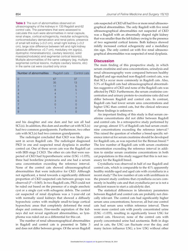
The number of renal ultrasonographical abnormalities in Ragdoll and control cats is presented in Table 3 and does not differ between groups. Of the seven Ragdoll
cats suspected of CKD all had five or more renal ultrasono-graphical abnormalities. The only Ragdoll with five renal ultrasonographical abnormalities not suspected of CKD was a Ragdoll with an abnormally shaped right kidney that was smaller than the left kidney owing to the presence of two segmental cortical lesions. Both kidneys showed mildly increased cortical echogenicity and a medullary rim sign. The only control cat with five renal ultrasono-graphical abnormalities was suspected of renal dysplasia.
DiscussionThe main finding of this prospective study, in which serum creatinine and urea concentrations, urinalysis and renal ultrasonography were compared between healthy Ragdoll and age-matched non-Ragdoll control cats, was that SCLs occur more commonly in Ragdoll cats. Also, 5.3% of Ragdoll cats had ultrasonographical abnormali-ties suggestive of CKD and none of the Ragdoll cats was affected by PKD. Furthermore, the serum creatinine con-centration and urinary protein-to-creatinine ratio did not differ between Ragdoll and control cats. In contrast, Ragdoll cats had lower serum urea concentrations and higher USG than control cats, but the clinical relevance of these findings is unknown.
An important finding of this study is that serum cre-atinine concentrations did not differ between Ragdoll and control cats. In a retrospective study performed by our group, almost 11% of Ragdoll cats had serum creati-nine concentrations exceeding the reference interval.5 This raised the question of whether a breed-specific ref-erence interval for serum creatinine should be developed for Ragdoll cats, as was reported for the Birman breed.10 The low number of Ragdoll cats with serum creatinine concentration exceeding the reference interval in addi-tion to similar serum creatinine concentrations in both cat populations in this study suggest that this is not nec-essary for the Ragdoll breed.
Crystalluria was observed in half of our Ragdoll and control cats, which is comparable to the 41% apparently healthy middle-aged and aged cats with crystalluria in a recent study.8 The low number of cats with urolithiasis in the present study confirms that crystalluria occurs com-monly in healthy cats and that crystalluria per se is not a sufficient reason to start a calculolytic diet.
The statistical differences in laboratory parameters between Ragdoll and control cats are probably not clini-cally relevant. The control cats had significantly higher serum urea concentrations; however, all but one control cats had serum urea within reference interval. There were more control cats with poorly concentrated urine (USG <1.035), resulting in significantly lower USG for control cats. However, none of the control cats with poorly concentrated urine had azotaemia. Both in dogs and in cats, the USG can fluctuate over the day, and many factors influence USG; a low USG without other
Table 3 The sum of abnormalities observed on ultrasonography of the kidneys in 133 Ragdoll and 62 control cats. The parameters that were assessed to calculate this sum were abnormalities in renal capsule, renal shape, cortical echogenicity, medullar echogenicity, corticomedullary demarcation and renal pelvis; presence of small left kidney (<3.2 cm), small right kidney (<3.2 cm), large size difference between left and right kidney (absolute difference >0.7 cm), medullary rim sign(s), dystrophic mineralisation(s), cavitary lesion(s), solid mass(es)/nodule(s) and segmental cortical lesion(s). Multiple abnormalities of the same category (eg, multiple segmental cortical lesions, multiple cavitary lesions, etc) in the same cat were counted only once
Number of abnormalities Ragdoll cats Control cats
0 67 371 35 132 11 53 8 54 4 15 3 16 3 07 0 08 1 09 1 0
by guest on February 26, 2014jfm.sagepub.comDownloaded from
Paepe et al 855
indications for kidney disease does not necessarily sug-gest decreased renal function.11,12 It must be mentioned that USG in this study was measured with a traditional optical refractometer without separate scale for feline USG. Although, it has been reported that these refrac-tometers can overestimate the actual USG in feline urine,13 a recent report has shown that this is not clini-cally relevant.14 Control cats were more likely to have glucosuria. However, none of the control cats with glu-cosuria showed or developed clinical signs of diabetes mellitus, or showed other evidence (casts, mild protein-uria) for tubular dysfunction. Higher frequency of pro-tein positivity on urinary dipstick in Ragdoll cats is probably not relevant because significant differences in UPC and in the proportion of cats with borderline and overt proteinuria were not observed between both popu-lations. In addition, urinary dipstick tests are not very reliable in identifying non-severe proteinuria, especially in cats with concentrated urine.15
Segmental cortical lesions were seen more commonly in Ragdoll versus control cats. The ultrasonographical aspect of these lesions was in line with the ultrasono-graphical description of kidney infarcts in the veterinary literature. Renal infarcts are described as linear or wedge-shaped, well-defined lesions in the renal cortex that are located perpendicular to the capsule and may cause a dimple in the adjacent serosal surface. Initially, kidney infarcts are hypoechoic, but may become hyper-echoic in the chronic state.16–18
Other pathogenic explanations besides renal infarc-tion must be considered to explain the SCLs. Hyperechoic triangular-to-wedge-shaped cortical lesions, irregular kidney shape and cortical outline, as were seen in our cats with SCLs, are also ultrasonographical features of renal scarring in humans.19 Segmental cortical scarring causing depression of the renal cortical surface has been described in humans and dogs with reflux nephropa-thy.20,21 In humans, these renal scars mostly develop as a result of chronic non-obstructing pyelonephritis second-ary to primary vesico-ureteral reflux.20 None of our cats with SCLs had a positive urine culture or a history of uri-nary tract infection. Whether sterile reflux results in renal damage remains controversial in humans.20 Renal scar-ring in humans occurs mostly in the polar segments of the kidney.20 In our study, cats were affected by single or double SCLs, and the location varied from unilateral to bilateral and from cranial to the caudal part of the kidney, and most SCLs were observed in one of the kidney poles.
Because SCLs were only observed in the Ragdoll and not in the control cat population, Ragdoll cats may have a breed-dependent increased susceptibility for SCL. Interestingly, brief pedigree analysis of the Ragdoll cats with SCLs showed two parent–offspring combinations and several other cats that were related to each other. This may indicate a hereditary explanation for the SCLs.
Major complications of renal scarring due to vesico-ureteral reflux and renal infarction in humans are hyper-tension and renal failure.20,22,23 In addition, microscopic haematuria is commonly present in humans with acute renal infarction.22,23 Indeed, some of our cats with SCLs showed mild-to-moderate urinary sediment abnormali-ties, such as microscopic haematuria. However, azotae-mic kidney disease and overt proteinuria was uncommon. Blood pressure was not evaluated in our cats, so hypertension cannot be excluded as an underly-ing cause or consequence for SCLs in Ragdoll cats. Further studies will need to be undertaken to reveal the clinical significance and underlying cause for the SCLs observed in the Ragdoll cats.
None of the control cats, but 5.3% of the Ragdoll cats, in this study showed ultrasonographical abnormalities suggestive of CKD. This percentage is slightly lower than the 8.6% prevalence of CKD in Ragdoll cats in a recent retrospective study.5 Although we only found a trend towards significance (0.5< P <1), we cannot exclude that Ragdoll cats are predisposed to show ultrasono-graphical abnormalities compatible with CKD. However, it is important to remember that ultrasonography does not correlate with renal function and is not a useful tool to predict which cats will develop azotaemic disease.16 In addition, the ultrasonographical findings do not imply that these cats were affected by CIN, as several other dis-eases, such as glomerulonephritis, glomerulosclerosis, amyloidosis and nephrocalcinosis, can result in similar ultrasonographical abnormalities.17 Follow-up of Ragdoll cats with ultrasonographical abnormalities sug-gestive of CKD is warranted to determine the clinical relevance of these findings.
In this study, PKD was only diagnosed in one Persian cat of the control group and in none of the included Ragdoll cats. In our retrospective study, PKD prevalence in Ragdoll cats of 2.9% was found.5 Although Ragdoll cats have been outcrossed with Persian cats in the past and could be at risk for PKD,24 both studies indicate that PKD is uncommon in this population of Ragdoll cats.
Ragdoll cats more frequently showed an undulating or irregular renal capsule and echogenic urine. The renal capsular abnormalities may, in part, be explained by the predisposition of Ragdoll cats for segmental cortical lesions or the presence of CKD in some cats. The predis-position for echogenic urine in Ragdoll cats may be explained by the different gender distribution of both populations. Intact male cats were predisposed for echo-genic urine in this study, possibly because the presence of sperm or mucus resulted in turbid or cloudy urine.12
A remarkable finding of this study is that ultrasono-graphical abnormalities at the level of the kidney are very common in healthy cats, especially if the young age of our cat population is taken into account. To our knowl-edge, this is the first study that has evaluated kidney
by guest on February 26, 2014jfm.sagepub.comDownloaded from
856 Journal of Feline Medicine and Surgery 15(10)
ultrasonography in a large healthy cat population. Also, several commonly cited studies reporting ultrasono-graphical features of feline kidneys date from the 1980s or early 1990s.25–29 Since then, ultrasound devices and expertise have improved, which may explain why minor changes nowadays are detected commonly. In half of the Ragdoll and 40% of the control cats at least one ultra-sonographical abnormality was observed. Abnormalities that were seen in more than 10% of cats of both groups were the presence of a medullary rim sign and changes in cortical echogenicity, particularly hyperechoic renal cortices. Although the significance of these abnormali-ties is still unknown,17,18,28,29 it is important that clinicians are aware that renal ultrasonographical abnormalities often occur in healthy cats. Further studies to evaluate which renal ultrasonographical abnormalities are clini-cally relevant are needed.
It is important to realise that disease prevalences are affected by the specific characteristics of the studied population,30 such as geography. All Ragdoll cats included in this study resided in Belgium or the Netherlands, and the majority was born in the same area. However, 40% of Ragdoll cats were imported from different areas, such as the USA, other European coun-tries, Australia or New Zealand, Canada or Israel. This means that the findings of this study are not just applica-ble for Ragdoll cats in the Benelux, but may be of interest for Ragdoll breeders all over the world. Also, a selection bias may have resulted in an underestimation of actual disease prevalence because most Ragdoll breeders that participated to this study had screened their cats over several generations.
Although precise breeding recommendations cannot be made based on this study, it seems reasonable to dis-courage intensive breeding with Ragdoll cats with SCL(s) and obvious renal ultrasonographical changes. However, if Ragdoll breeders want to screen their Ragdoll cats for the presence of kidney disease, ultra-sonography, and measurement of serum creatinine and urea concentrations must be combined with routine uri-nalysis. Concurrent urinalysis will facilitate the interpre-tation of serum urea and creatinine concentrations, and is needed to detect kidney dysfunction.31
ConclusionsBased on this population, breed-specific serum creatinine reference intervals are not required for Ragdoll cats. Furthermore, renal ultrasonographical abnormalities are common in young healthy cats, both in Ragdoll and non-Ragdoll cats. Ragdoll cats are predisposed to segmental triangular-to-wedge-shaped cortical changes. Further studies are required to elucidate whether these lesions may represent renal infarction or cortical scarring, and to determine the clinical implications of these SCLs. None of the Ragdoll cats was diagnosed with PKD, but 5.3% of
Ragdoll cats had significant renal lesions based on ultra-sonographical findings. Further studies are needed to identify if these cats are affected by CIN and if they will develop azotaemic kidney disease.
Acknowledgements We are particularly grateful to all owners of the participating cats for making this study possible.
Funding This research received no specific grant from any fund-ing agency in the public, commercial, or not-for-profit sectors.
Conflict of interest The authors do not have any potential conflicts of interest to declare.
References 1 Cat Fanciers’ Association. Top 10 most popular breeds,
http://www.cfa.org/Client/home.aspx (2012, accessed 21 August 2012).
2 Ragdoll Club Benelux. Gezondheid: CIN, http://www.Rag-dollclubbenelux.com/nl_gezondheidcin.html (2003, accessed 21 August 2012).
3 Scandinavian Ragdoll Club. Health: Scandinavian Ragdoll Clubs Health programme, http://www.weikums.se/health.htm (2004, accessed 21 August 2012).
4 Ragdoll Health database. http://www.Ragdolldatabase.nl/list_list.php (2012, accessed 21 August 2012).
5 Paepe D, Saunders JH, Bavegems V, et al. Screening of Ragdoll cats for kidney disease: a retrospective evalua-tion. J Small Anim Pract 2012; 53: 572–577.
6 Helps CR, Tasker S, Barr FJ, et al. Detection of the single nucle-otide polymorphism causing feline autosomal-dominant polycystic kidney disease in Persians from the UK using a novel real-time PCR assay. Mol Cell Probes 2007; 83: 264–268.
7 Meyer DJ. Microscopic examination of the urinary sedi-ment. In: Raskin RE and Meyer DJ (eds). Atlas of canine and feline cytology. 1st ed. Philadelphia: WB Saunders, 2001, pp 261–276.
8 Paepe D, Verjans G, Duchateau L, et al. Routine health screening of apparently healthy middle-aged and old cats. J Feline Med Surg 2013; 15: 8–19.
9 Lees GE, Brown SA, Elliott J, et al. Assessment and man-agement of proteinuria in dogs and cats: 2004 ACVIM forum consensus statement (small animal). J Vet Intern Med 2005; 19: 377–385.
10 Reynolds BS, Concordet D, Germain CA, et al. Breed dependency of reference intervals for plasma biochemi-cal values in cats. J Vet Intern Med 2012; 24: 809–818.
11 van Vonderen IK, Kooistra HS and Rijnberk A. Intra- and interindividual variation in urine osmolality and urine specific gravity in healthy pet dogs of various ages. J Vet Intern Med 1997; 11: 30–35.
12 Stockham SL and Scott MA. Urinary system. In: Stockham SL and Scott MA (eds). Fundamentals of veterinary clinical pathology. 2nd ed. Oxford: Blackwell Publishing, 2008, pp 415–494.
13 George JW. The usefulness and limitations of hand-held refractometers in veterinary laboratory medicine: an his-torical and technical review. Vet Clin Pathol 2001; 30: 201–210.
14 Bennett AD, McKnight GE, Dodkin SJ, et al. Comparison of digital and optical hand-held refractometers for the
by guest on February 26, 2014jfm.sagepub.comDownloaded from
Paepe et al 857
measurement of feline urine specific gravity. J Feline Med Surg 2011; 13: 152–154.
15 Syme H. Proteinuria in cats. Prognostic marker or media-tor? J Feline Med Surg 2009; 11: 211–218.
16 Grooters AM and Biller DS. Ultrasonographic findings in renal disease. In: Bonagura JD (ed). Kirk’s current veterinary therapy. 12th ed. Philadelphia: WB Saunders, 1995, pp 933–936.
17 Widmer WR, Biller DS and Adams LG. Ultrasonography of the urinary tract in small animals. J Am Vet Med Assoc 2004; 225: 46–54.
18 d’Anjou MA. Kidneys and ureters. In: Penninck MA and d’Anjou MA (eds). Atlas of small animal ultrasonography. 1st ed. Ames: Blackwell Publishing, 2008, pp 339–364.
19 Barry BP, Hall N, Cornford E, et al. Improved ultrasound detection of renal scarring in children following urinary tract infection. Clin Radiol 1998; 53: 747–751.
20 Cargollo PC and Diamond DA. Therapy insight: what nephrologists need to know about primary vesicoureteral reflux. Nat Clin Pract Nephrol 2007; 3: 551–563.
21 Kolbjørnsen Ø, Heggelund M and Jansen JH. End-stage kidney disease probably due to reflux nephropathy with segmental hypoplasia (Ask-Upmark kidney) in young Boxer dogs in Norway. A retrospective study. Vet Pathol 2008; 45: 467–474.
22 Racusin JS and Pollack ML. Idiopathic renal infarction in a young woman. Am J Emerg Med 2005; 23: 397–398.
23 Tsai SH, Chu SJ, Chen SJ, et al. Acute renal infarction: a 10-year experience. Int J Clin Pract 2007; 61: 62–67.
24 Beck C and Lavelle RB. Feline polycystic kidney disease in Persian and other cats: a prospective study using ultraso-nography. Aust Vet J 2001; 79, 181–184.
25 Walter PA, Feeny DA, et al. Feline renal ultrasonography: quantitative analyses of imaged anatomy. Am J Vet Res 1988; 48: 596–599.
26 Walter PA, Johnston GR, Feeney DA and O’Brien TD. Renal ultrasonogaphy in healthy cats. Am J Vet Res 1987; 48: 600–607.
27 Walter PA, Johnston GR, Feeney DA and O’Brien TD. Applications of ultrasonography in the diagnosis of parenchymal kidney disease in cats: 24 cases (1981–1986). J Am Vet Med Assoc 1988; 192: 92–98.
28 Yeager AE and Anderson WI. Study of association between histologic features and echogenicity of archi-tecturally normal cat kidneys. Am J Vet Res 1989; 50: 860–863.
29 Biller DS, Bradley GA and Partington BP. Renal medullary rim sign: ultrasonographic evidence of renal disease. Vet Radiol Ultrasound 1992; 33: 286–290.
30 Hahn KA and Overley B. Rational use of diagnostic tests. In: Ettinger SJ and Feldman EC (eds). Textbook of veteri-nary internal medicine. 7th ed. St Louis, Missouri: Elsevier Saunders, 2010, pp 32–35.
31 DiBartola SP. Clinical approach and laboratory evalua-tion of renal disease. In: Ettinger SJ and Feldman EC (eds). Textbook of veterinary internal medicine. 7th ed. St Louis, Missouri: Elsevier Saunders, 2010, pp 1955–1969.
by guest on February 26, 2014jfm.sagepub.comDownloaded from