Embed Size (px)
Citation preview
Amanda Bortoluci da Silva Oliveira
Atividade antiviral e modo de ação de um peptídeo 6 kDa isolado de Helianthus annus
L e sinergismo com aciclovir
Londrina
2009

Livros Grátis
http://www.livrosgratis.com.br
Milhares de livros grátis para download.
2
Amanda Bortoluci da Silva Oliveira
Atividade antiviral e modo de ação de um peptídeo 6 kDa isolado de Helianthus annus
L e sinergismo com aciclovir
Londrina 2009
Dissertação apresentada ao Programa de Pós-graduação em Microbiologia da Universidade Estadual de Londrina, como requisito parcial para obtenção do título de Mestre em Microbiologia.
Orientador: Prof. Dr. Benedito Prado Dias Filho
3
Amanda Bortoluci da Silva Oliveira
Atividade antiviral e modo de ação de um peptídeo 6 kDa isolado de Helianthus annus
L e sinergismo com aciclovir
BANCA EXAMINADORA
___________________________________
Prof. Dr. Benedito Prado Dias Filho Universidade Estadual de Londrina
___________________________________
Prof. Dra. Tânia Ueda-Nakamura Universidade Estadual de Maringá
___________________________________ Prof. Dra. Sueli Fumie Yamada-Ogatta
Universidade Estadual de Londrina
Londrina, 28 de maio de 2009.
Dissertação apresentada ao Programa dePós-graduação em Microbiologia daUniversidade Estadual de Londrina,como requisito parcial para obtenção dotítulo de Mestre em Microbiologia.
4
Dedico este trabalho aos meus pais, Ademir e Cristina, que sempre souberam ensinar o verdadeiro valor do conhecimento, que sempre me incentivaram a nunca desistir diante das dificuldades e a buscar meus sonhos, por mais impossível que eles parecessem, pois
se estou concluindo mais esta etapa de minha vida são graças aos seus incentivos, conselhos, doações e dedicações.
Dedico também à minha irmã Paula; e a meus eternos amores André e Leonardo pelo apoio e amor incondicionais.
5
AGRADECIMENTOS
A Deus, por ter me dado o dom da coragem e da força nos momentos em que nada dava certo, para que eu nunca desistisse; sabedoria e inteligência para que eu pudesse
compreender os resultados malucos que as vezes surgiram.
Ao meu marido André e ao meu filho Leonardo pelo carinho e por terem se privado da
minha presença sem nada cobrar.
Aos meus pais Ademir e Cristina, e a minha irmã Paula pelo amor e incentivo.
Ao Prof. Dr. Benedito Prado Dias Filho, pela orientação, pelo aprendizado e incentivo na busca da excelência. Obrigado por ter apostado em mim quando eu ainda estava
cursando o primeiro ano de faculdade. Quanto tempo !
Ao Prof. Dr. Celso Vataru Nakamura pela contribuição a este trabalho.
À Prof. Dra. Tânia Ueda-Nakamura, pela contribuição e orientação nos projetos realizados, sempre corrigindo meus resumos, painéis, etc; e pelo convívio diário.
À Prof. Maria de Lourdes pelo empréstimo da coluna C-18.
Aos professores do Programa de Pós-Graduação em Microbiologia, pelos ensinamentos
e coleguismos.
As técnicas do laboratório de microbiologia básica Adriana Barravieira, Maria Manzoti, Marinete Martinez, Rosana Monteiro, Zelita Rodrigues e não poderia me esquecer da
Dona Prisciliana, a primeira pessoa que me ensinou autoclavar materiais no laboratório! Obrigada pela paciência de vocês em me atender todos os dias nesses últimos 7,5 anos
em que convivemos juntas!
Aos colegas e amigos do laboratório; não irei mencionar nomes, pois correrei o grande risco de esquecer de alguém, pois muitas pessoas importantes passaram pela sala 123.
Obrigada pelo companheirismo, amizade, ajuda nos experimentos e pelos momentos de descontração que passamos juntos.
Aos colegas do mestrado, pela convivência, pelas conversas, pela companhia nos
almoços no RU e nos pastéis no final da tarde.
À CAPES pela concessão da bolsa e ao CNPQ pelo apoio financeiro.
E a todos aqueles que de alguma forma contribuíram para a realização desse trabalho
Muito Obrigada !
6
RESUMO
HSV-1 está geralmente associado a infecções faciais, oftálmicas e genitais. Sua grande
característica está em causar uma lesão primária, auto-limitante em pacientes
imunocompetentes, que é capaz de reativar por toda a vida do paciente. Eventualmente,
pode estar associado a lesões graves no sistema nervoso central, como encefalites
necrosantes agudas e meningites, principalmente em pacientes imunocomprometidos.
Muitos tratamentos são baseados no uso de aciclovir e em outros análogos de
nucleosídeos, mas eles são tóxicos e alguns pacientes com lesões recorrentes podem
albergar cepas resistentes. No presente estudo foi isolado um peptídeo anti-HSV-1 das
sementes de girassol (Helianthus annus) utilizando técnicas cromatográficas. A
caracterização parcial do peptídeo revelou que ele apresenta aproximadamente 6 kDa e
que possivelmente seja aniônico. A atividade antiviral contra HSV-1 foi investigada
através de vários experimentos, entre eles estão a determinação do modo de ação, pela
adição do peptídeo em várias etapas do ciclo replicativo do HSV-1; pelo ensaio de
penetração e pelo sinergismo utilizando aciclovir. O peptídeo mostrou-se altamente
eficiente ao impedir as etapas iniciais do ciclo replicativo, a adsorção e penetração, o
que pode ser comprovado pelos valores de IC50, 5,3 μg/ml quando o peptídeo foi
adicionado antes da infecção por HSV-1 e 4,5 μg/ml quando adicionado durante a
infecção por HSV-1. Atividade virucida moderada foi observada quando o peptídeo foi
incubado com o HSV-1 por 1 h a 37 ºC antes da infecção (IC50 igual a 78,6 μg/ml),
enquanto que o peptídeo se mostrou inativo depois que a infecção viral estivesse
estabelecida. 5 μg/ml do peptídeo impediu 86 % da penetração viral durante os
primeiros 20 minutos de incubação. Através do isobolograma, pode-se verificar que o
7
peptídeo apresentou sinergismo quando associado ao aciclovir, com índice de
concentração fracional inibitória (FICI) de 0,375.
Palavras-chave: HSV-1, Helianthus annus, Técnicas Cromatográficas, Peptídeo
Antiviral.
8
ABSTRACT
HSV-1 is generally associated whit primary and recurrent mucocutaneous facial,
ophthalmic or genital lesions, but under certain conditions, can produce serious
infections of the central nervous system, causing acute necrotizing encephalitis end
meningitis in patients with immunedeficiencies. Most of the treatments for HSV-1 are
based on acyclovir (ACV) and ACV-like nucleosides analogues, but they are toxic and
some immunocompromised patients with recurrent HSV lesions develop resistance to
ACV. In this study was isolated an anti-HSV-1 peptide of sunflower seeds (Helianthus
annus) by chromatografic techniques. The partial characterization revealed that it
presents 6 kDa approximately and possibly is anionic. The activity antiviral against
HSV-1 was investigated through several experiments, among them they are the
determination in the action way, for the addition of peptide in several stages of the
HSV-1 replication cycle; by penetration assay and synergism using acyclovir. The
peptide was shown highly efficient in blocking the initial stages of the replication cycle,
the adsorption and penetration, this can be proven for the values of IC50, 5.3 μg/ml when
the peptide was added before the infection by HSV-1 and 4.5 μg/ml when added during
the infection by HSV-1. Activity moderate virucidal was observed when the peptide
was incubated with HSV-1 by 1 h to 37 ºC before the infection (IC50 equal to 78.6
μg/ml), while the peptide was shown inactive after the infection viral was established. 5
μg/ml of the peptide was impeded 86 % of the penetration viral from first 20 minutes of
incubation. Through the isobologram, it can be verified that the peptide was presented
synergism when associate to acyclovir.
Keywords: HSV-1, Helianthus annus, Chromatografic Techniques, Antiviral Peptide.
9
SUMÁRIO
REVISÃO BIBLIOGRÁFICA.....................................................................................10
Família Herpesviridae.....................................................................................................10
Herpes simplex tipo 1......................................................................................................10
Infecções herpéticas ........................................................................................................12
Antivirais sintéticos.........................................................................................................17
Combinações de antivirais...............................................................................................20
Plantas como agentes antimicrobianos............................................................................21
Proteínas e peptídeos antivirais.......................................................................................24
Helianthus annus.............................................................................................................28
OBJETIVOS..................................................................................................................29
REFERÊNCIAS BIBLIOGRÁFICAS .......................................................................30 ANEXO - Anti-HSV-1 activity and mode of action of a 6 kD peptide isolated from Helianthus annus and its synergism with acyclovir……….…………..……………....38
10
REVISÃO BIBLIOGRÁFICA
Família Herpesviridae
Os membros da família Herpesviridae apresentam um cerne contendo uma fita
dupla linear de DNA, um capsídeo icosaédrico de aproximadamente 100 a 110 nm de
diâmetro formado por 162 capsômeros, sendo 150 hexaméricos e 12 pentaméricos. O
capsômero é envolvido por um material amorfo constituído de proteína, designado de
tegumento, e um envelope lipídico contendo espículas de glicoproteínas na superfície
(FIELDS, 2001; SCHLEISS, 2009).
Os vírus herpéticos são altamente disseminados na natureza. Aproximadamente
100 vírus da família Herpesviridae foram caracterizados, sendo que existem nove vírus
herpéticos humanos. Além disso, o vírus herpético B de macacos também pode infectar
o homem causando encefalite mortal (DA SILVA, 2000; ROIZMAN et al, 2001).
Os herpesvírus humano (HVH) são divididos em três subfamílias: α-
herpesvirinae que compreende Herpes simplex tipo 1 e 2 (HSV-1 e HSV-2) e Varicella-
Zoster (VZV); β-herpesvirinae que compreende citomegalovírus (CMV) e Herpes
simplex tipo 6 e 7 (HHV-6A, HHV-6B e HHV-7); e γ- herpesvirinae que compreende o
vírus Epstein-Barr e Herpes simplex tipo 8 (HHV-8) (FIELDS, 2001; GILBERTI et al.,
2002; SANTOS et al, 2009).
Herpes simplex tipo 1
Os vírus Herpes simplex foram os primeiros dos vírus humanos da família
Herpesviridae a serem descobertos e são os mais estudados. Seus atrativos estão nas
suas propriedades biológicas, particularmente na habilidade em causar uma variedade
11
de infecções e permanecerem latente por toda vida do hospedeiro com possíveis
reativações perto ou longe do local da infecção inicial (FIELDS, 2001; SAUERBREI e
WUTZLER, 2007).
O Herpes simplex tipo 1 (HSV-1) apresenta algumas propriedades biológicas
como crescimento rápido, lise das células infectadas e estabelecimento de infecções
latentes em gânglios nervosos sensoriais. Durante a latência é capaz de codificar as
principais enzimas necessárias à replicação do DNA viral, mantendo um grau de
virulência suficiente para não ser subjugado pelos mecanismos de defesa do hospedeiro
(WHITE e FENNER 1994; ROIZMAN et al, 2001).
Estruturalmente, O HSV é composto de quatro componentes distintos: o cerne
contendo DNA linear de fita dupla, com aproximadamente 152 kb. O genoma é dividido
em dois segmentos ligados covalentemente, denominados longo (L) e curto (S), ambos
contendo seqüências únicas, que são flanqueadas por regiões de seqüências repetitivas
invertidas. Seu genoma é um dos maiores genomas virais dos vírus herpéticos humanos,
codificando aproximadamente 84 proteínas (WHITE e FENNER 1994; BOEHMER e
LEHMAN, 1997; SUBAK-SHARPE e DARGAN, 1998; WHITLEY e ROIZMAN,
2001).
O capsídeo icosaédrico; o tegumento; e o envelope lipoprotéico de 120 a 200 nm
de diâmetro revestido por até 11 tipos de glicoproteínas virais e diversas proteínas não
glicosiladas, lipídios e poliaminas (WHITE e FENNER, 1994; WHITLEY e
ROIZMAN, 2001).
Os dois tipos sorológicos (tipo 1 e 2) apresentam antígenos comuns e antígenos
tipos-específicos, podendo ser distinguidos pelas manifestações clínicas e bioquímicas
(CRAIGHEAD, 2004).
12
Infecções Herpéticas
Os vírus são parasitas intracelulares obrigatórios, necessitam da atividade
metabólica e das organelas da célula hospedeira para produção de energia e síntese de
macromoléculas para a sua multiplicação (VOYLES, 1993; FIELDS, 2001; JASSIM e
NAJI, 2003).
Os vírus são capazes de reconhecer e de penetrar em células-alvo,
principalmente pela especificidade dos receptores existentes na superfície dessas células
hospedeiras e dos próprios vírus. O conjunto de eventos que vão desde a adsorção viral
até a liberação dos vírions é chamado de ciclo de multiplicação viral. A forma pela qual
o vírus realiza as etapas do seu ciclo replicativo é determinada pela estrutura do seu
genoma e pela estrutura do próprio vírion. As principais etapas de multiplicação de um
vírus podem ser resumidas em uma fase inicial, com adsorção do vírus à célula
hospedeira, penetração e desnudamento (decapsidação) da partícula, e uma fase tardia,
que vai desde a síntesedas moléculas até a automontagem e liberação dos vírions
(WHITE e FENNER, 1994; JENSSEN et al, 2004).
Para iniciar sua infecção, o HSV-1 precisa se ligar a receptores celulares, fundir
seu envelope com a membrana plasmática, e permitir que o capsídeo seja transportado
até os poros da membrana do núcleo da célula, onde o genoma viral será liberado. No
núcleo, ocorrerá a transcrição, síntese de DNA e proteínas, montagem do capsídeo,
empacotamento do DNA e envelopamento, culminando com a liberação de um novo
vírion potencialemente infeccioso (WHITLEY e ROIZMAN, 2001).
O contato inicial do HSV com a célula se dá pelo processo de adsorção, através
da ligação do vírion com cadeias de glicosaminoglicanas (GAG) de proteoglicanas da
superfície celular. O sulfato de heparana, um dos diversos tipos de GAG, é considerado
13
o principal receptor de ligação do HSV-1. Duas glicoproteínas (das onze descritas até o
momento) presentes no envelope viral, denominadas gB e gC, são capazes de se ligar ao
sulfato de heparana e mediar a adsorção viral, além de induzir a resposta imune
(SHUKLA e SPEAR, 2001; SPEAR, 2004; FUSCO et al, 2005; SU et al., 2008).
A principal via de penetração do HSV é por fusão na membrana da superfície
celular, mas também pode ocorrer por endocitose. Para que ocorra a penetração por
fusão, após a adsorção da partícula viral à superfície celular, é necessário que a
glicoproteína gD interaja com um dos receptores celulares (WHITLEY e ROIAMAN,
2001; FUSCO et al, 2005; ATANASIU, 2007). A ligação da gD com um dos receptores
resulta na fusão do envelope do envelope viral com a membrana da célula,
possibilitando a entrada do nucleocapsídeo e do tegumento no citoplasma celular. Neste
processo estão envolvidas, além da gD e seus receptores, a gB e o heterodímero gH-gL
(SHUKLA e SPEAR, 2001; SPEAR, 2004; FUSCO et al, 2005; ATANASIU, 2007; SU
et al, 2008).
Após a penetração, o nucleocapsídeo é transportado até a membrana nuclear,
atravessa os poros, e atinge o núcleo, acompanhado pelas proteínas do tegumento α-TIF
ou VP16 e vhs que desempenham importantes papéis no processo de replicação viral. A
síntese dos produtos gênicos virais é realizada em três fases seqüenciais: imediata,
precoce e tardia, sendo a última subdividida em tardia falsa e tardia verdadeira,
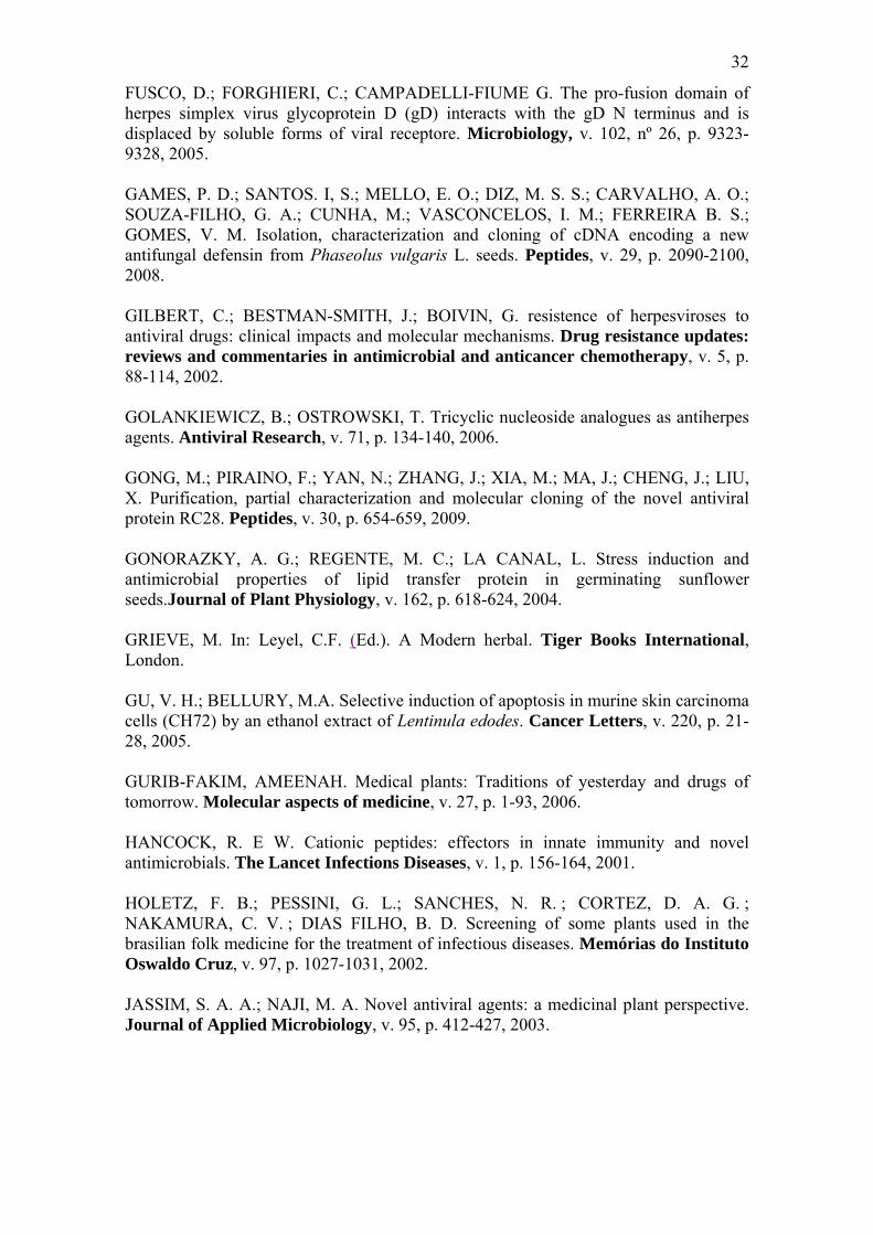
fenômeno que pode ser ilustrado pela figura 1 (MCKNIGHT et al., 1987; WHITE e
FENNER, 1994; BOEHMER e LEHMAN, 1997; ROIZMAN et al., 2001; WHITLEY
e ROIZMAN, 2001; GU E BELLURY, 2005).
A multiplicação viral alcança sua última fase com o aparecimento de transcritos
da fase tardia. Com o início da replicação, e conseqüente acúmulo de DNA viral, ocorre
a sinalização para que os genes tardios sejam transcritos, gerando mais de 30 produtos,
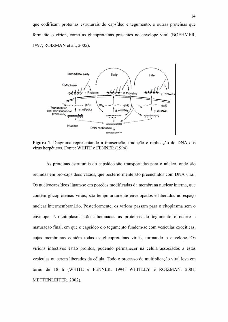
14
que codificam proteínas estruturais do capsídeo e tegumento, e outras proteínas que
formarão o vírion, como as glicoproteínas presentes no envelope viral (BOEHMER,
1997; ROIZMAN et al., 2005).
Figura 1. Diagrama representando a transcrição, tradução e replicação do DNA dos vírus herpéticos. Fonte: WHITE e FENNER (1994).
As proteínas estruturais do capsídeo são transportadas para o núcleo, onde são
reunidas em pró-capsídeos vazios, que posteriormente são preenchidos com DNA viral.
Os nucleocapsídeos ligam-se em porções modificadas da membrana nuclear interna, que
contém glicoproteínas virais; são temporariamente envelopados e liberados no espaço
nuclear intermembranário. Posteriormente, os vírions passam para o citoplasma sem o
envelope. No citoplasma são adicionadas as proteínas do tegumento e ocorre a
maturação final, em que o capsídeo e o tegumento fundem-se com vesículas exocíticas,
cujas membranas contêm todas as glicoproteínas virais, formando o envelope. Os
vírions infectivos estão prontos, podendo permanecer na célula associados a estas
vesículas ou serem liberados da célula. Todo o processo de multiplicação viral leva em
torno de 18 h (WHITE e FENNER, 1994; WHITLEY e ROIZMAN, 2001;
METTENLEITER, 2002).
15
As doenças virais acompanham o homem desde a formação das primeiras
comunidades, o primeiro caso de infecção viral, provavelmente, ocorreu por volta de
2000 a.C., na China e no leste da Ásia onde foram relatados sintomas do que hoje se
conhece por varíola. Em 2003, estimava-se que 25 a 35% da população sexualmente
ativa do ocidente estava infectada com herpes genital (Herpes simplex tipo 2) (WHO,
2003).
Nos Estados Unidos, aproximadamente 80% e 35% da população adulta está
infectada ou é soro-positiva para HSV-1 e HSV-2, respectivamente (ZHANG et al.,
2007). Segundo a Organização Mundial de Saúde (WHO, 2008), mais de 7,6% da
população mundial estão infectadas pelo vírus HSV-2 transmitido comumente pelo ato
sexual, e mais de 20 milhões de pessoas se contagiam a cada ano. Dados mais recentes
mostram que a prevalência de anticorpos para HSV-1 e HSV-2 na população é de 60% e
55 % respectivamente (SAKDARAT et al., 2009).
O Herpes simplex vírus causa uma grande variedade de infecções, que são
geralmente auto-limitadas em pacientes imunocompetentes (AMMENDOLIA et al.,
2007; CHUANASA et al., 2008), mas em pacientes imunocomprometidos e neonatos,
HSV pode causar sérias doenças sistêmicas, como infecções no sistema nervoso central,
necrotização aguda, encefalites e meningites (ZHU et al., 2006; CHUANASA et al.,
2008; SCHNITZLER et al., 2008).
Além disso, HSV tem sido apontado como um importante fator na disseminação
do HIV, pelo recrutamento de células do sistema imunológico para as mucosas afetadas,
acarretando maior disponibilidade das células-alvo do vírus HIV (SCHACKER, 2001;
CELLUM, 2004; ZHU et al., 2004).
O sorotipo HSV-1 está normalmente associado a infecções oro-faciais, oculares,
genitais e encefalites, enquanto que o sorotipo HSV-2 está geralmente relacionado a
16
infecções genitais, herpes neonatal e a oncogenia (JENSSEN et al., 2004). Contudo,
ambos os vírus podem causar infecções clinicamente indistinguíveis em vários locais e
podem permanecer latentes em gânglios sensoriais, provocando reativações
caracterizadas por infecções recorrentes sintomáticas e assintomáticas (BRADY e
BERNSTEIN, 2004; REICHLING et al., 2008).
Após a infecção primária nas células epiteliais, HSV-1 rapidamente atinge os
gânglios dorsais via axônios, onde estabelecem latência. O mecanismo de latência ainda
não está totalmente compreendido, mas este estado é caracterizado pela cessação virtual
da transcrição de todos os genes, exceto aqueles genes relacionados com a latência viral.
A reativação pode ocorrer em situações do tipo: radiação ultravioleta, estresse e
imunossupressão; nesta situação, os genes que permitem ao HSV-1 causar infecções
voltam a ser transcritos e eles saem dos gânglios através dos axônios e iniciam uma
nova infecção (SCHLEISS, 2009).
Os sintomas primários da infecção herpética incluem síndrome prodrômica,
semelhante ao resfriado comum, caracterizada por febre, dor de garganta, mal estar e
mialgias difusas seguidos por sintomas locais da infecção. As manifestações clínicas
exibem severidade diferente entre pacientes imunocompetentes e imunocomprometidos
(CHENG et al., 2004; KHAN et al., 2005).
A incidência e a severidade das infecções causadas por HSV têm aumentado nas
últimas décadas. Isto ocorre devido ao aumento do número de pacientes
imunocomprometidos pelo tratamento quimioterápico agressivo, ou devido à síndrome
da imunodeficiência adquirida (SIDA), ou indivíduos transplantados (XU et al., 1999;
ZHANG et al., 2007; SCHNITZLER et al., 2008).
Outra característica importante das infecções herpéticas é que correntemente elas
estão associadas a uma infecção bacteriana secundária, de modo que seria interessante
17
uma substância antimicrobiana que tivesse atividade antiviral e antibacteriana
(CAMARGO FILHO, 2008).
Antivirais Sintéticos
A busca por novos fármacos passou por avanços significativos nos últimos anos,
principalmente depois da introdução de modelos biológicos realizados in vitro e em
grande escala (NIELSEN, 2002; NEWMAN et al., 2004).
Em contraste com a enorme quantidade de fármacos antibacte disponíveis, o
atual arsenal terapêutico antiviral compreende aproximadamente 40 fármacos utilizados
em várias infecções virais, sendo que a maioria deles é utilizada no tratamento de
infecções causadas pelo vírus da imunodeficiência humana (DE CLERCQ, 2005).
Uma das principais razões para a falta de sucesso no desenvolvimento de
fármacos antivirais é a natureza do vírus. Como mencionado anteriormente, os vírus
apresentam uma estrutura simples e um sistema enzimático bastante restrito, sendo
dependentes dos processos metabólicos celulares para sua multiplicação (DE CLERCQ,
2004; ORHAN et al., 2009). Conseqüentemente, vários passos da sua replicação
envolvem processos metabólicos da célula hospedeira, e isto tem dificultado o ataque
direto ao vírion ou a sua replicação, sem causar efeitos adversos nas células infectadas
(JASSIM e NAJI, 2003).
Apesar disso, cada vez mais fármacos antivirais são encontrados nos diversos
estágios de desenvolvimento, para serem licenciados e utilizados por humanos e
animais. Assim o número de agentes antivirais tem aumentado (FALCO et al., 2007),
pois com a melhor compreensão dos processos de replicação viral, vários eventos
específicos do vírus se mostraram susceptíveis à quimioterapia seletiva (DE CLERCQ,
2001). Muitos vírus possuem características únicas em sua estrutura ou em seu ciclo de
18
replicação e estes constituem alvos potenciais de fármacos antivirais (JASSIM e NAJI,
2003).
Enzimas virais são as peças chaves para a patogênese viral. Se essas enzimas
forem neutralizadas, a replicação viral e o processo proteolítico essencial para a
maturação dos vírus podem ser interrompidos (JASSIM e NAJI, 2003).
O sucesso da quimioterapia antiviral contra o herpes simplex se deve ao
desenvolvimento de acicloguanosina, conhecido como aciclovir, que interage com
proteínas chaves da replicação viral (JASSIM e NAJI, 2003; JENSSEN et al., 2004).
O aciclovir, desenvolvido na década de 70, é atualmente o fármaco de escolha
para as infecções herpéticas, mostrando-se ativo contra Herpes simplex tipo 1 e tipo 2 e
vírus da Varicela Zoster (SUZUKI et al., 2006). Seu mecanismo de ação está
relacionado ao fato de ser um análogo do nucleosídeo guanosina funcionando como
terminadores da duplicação do DNA (GOLANKIEWICZ e OSTROWSKI, 2006;
SUZUKI et al., 2006; SCHNITZLER et al., 2008; SU et al., 2008).
No interior da célula infectada pelo herpes simples tipo 1 ou tipo 2, o aciclovir é
fosforilado por uma enzima viral chamada timidina quinase, o que o torna uma
substância com toxicidade seletiva. A segunda e a terceira fosforilação são feitas por
timidinas quinases celulares (MORFIN E THOUVENOT, 2003). Uma vez fosforilado,
o aciclovir interferirá na polimerização do DNA viral por competir com o nucleotídeo
trifosfatado endógeno, 2-deoxiguanosina trifosfatada (SCHLEISS, 2009). Com a
incorporação do aciclovir na cadeia de DNA recém sintetizada a síntese fica
interrompida e a cadeia não pode ser terminada (GOLANKIEWICZ e OSTROWSKI
2006; SUZUKI et al., 2006; SU et al., 2008).
O aciclovir ainda hoje é o fármaco de escolha para infecções herpéticas e é tão
grande a sua importância, que além do uso clínico, muitos cientistas usam esse
19
composto como referência em pesquisas. Em 2003, mais de 9000 artigos foram
publicados relacionando o aciclovir e seu uso (JEROME, 2005). Além do aciclovir,
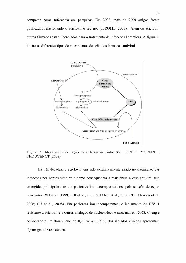
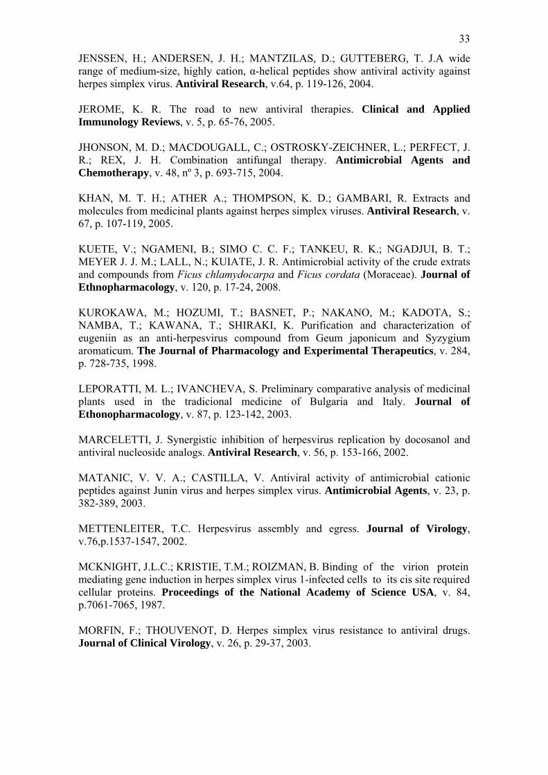
outros fármacos estão licenciados para o tratamento de infecções herpéticas. A figura 2,
ilustra os diferentes tipos de mecanismos de ação dos fármacos antivirais.
Figura 2. Mecanismo de ação dos fármacos anti-HSV. FONTE: MORFIN e THOUVENOT (2003).
Há três décadas, o aciclovir tem sido extensivamente usado no tratamento das
infecções por herpes simplex e como conseqüência a resistência a esse antiviral tem
emergido, principalmente em pacientes imunocomprometidos, pela seleção de cepas
resistentes (XU et al., 1999; THI et al., 2005; ZHANG et al., 2007; CHUANASA et al.,
2008; SU et al., 2008). Em pacientes imunocompetentes, o isolamento de HSV-1
resistente a aciclovir e a outros análogos de nucleosídeos é raro, mas em 2008, Cheng e
colaboradores relataram que de 0,28 % a 0,33 % dos isolados clínicos apresentam
algum grau de resistência.
20
Em adição a esse uso extensivo, casos de disfunção renal têm sido associados
aos análogos de nucleosídeos como ganciclovir, valaciclovir, fanciclovir e penciclovir
(ERNEST e FRANEY, 1998; ZHU et al., 2006). Outro problema encontrado com o uso
desses análogos está no alto custo de um tratamento a longo prazo, que se faz necessário
em pacientes com reativações (KHAN et al., 2005).
Embora o aciclovir seja considerado um fármaco padrão-ouro, sua eficácia é
diminuída em episódios recorrentes de infecção por HSV (COEN, 1996), como aqueles
observados nos pacientes imunocomprometidos (JENSSEN et al., 2004). A resistência
ao aciclovir e a outros análogos pode ocorrer pela mutação da timidina quinase ou de
DNA polimerase virais (MORFIN e THOUVENOT, 2003; KHAN et al., 2005). Há
relato de amostras clínicas de herpes simplex resistentes a aciclovir por não possuírem
timidina quinase ativa (KHAN et al., 2005).
Além da DNA polimerase, outras proteínas que agem durante os estágios da
replicação de herpesvírus podem ser alvos úteis para novos antivirais. Entre eles
destacam-se as proteínas necessárias à fixação e a penetração do vírus, porque os
fármacos não necessitariam entrar na célula para exercer seus efeitos. Dessa forma,
impediria a infecção e conseqüentemente inibiria a síntese de proteínas virais (COEN e
SCHARFFER, 2003).
Combinações de antivirais
A utilização da combinação de fármacos no tratamento de várias infecções virais
tem aumentado (MARCELLETTI, 2002). O efeito da administração de aciclovir com
outras substâncias antivirais no tratamento de infecções herpéticas tem sido estudado in
vitro, in vivo e durante o tratamento (MUCSI et al., 2001).
21
O uso de aciclovir associado a outras substâncias biologicamente ativas pode
apresentar um efeito sinérgico, fato que proporciona menor resistência de HSV frente
aos análogos de nucleosídeos, uma vez que a pressão de seleção se torna menor, pelo
uso de menores doses (MUCSI et al., 2001; CHUANASA et al., 2008).
Simultaneamente a isso, há diminuição dos efeitos citotóxicos (MUCSI et al., 2001).
A palavra “sinergismo” significa que no uso combinado de duas ou mais
substâncias que interagem entre si há a potencialização de um efeito (WILLIAMSON,
2001). O efeito sinérgico pode ser observado quando a segunda substância apresenta um
mecanismo de ação diferente ao do aciclovir (MUCSI et al., 2001).
Mas, desvantagens também podem ocorrer, já que casos de efeitos adversos
exagerados e falências terapêuticas são observados, fenômeno chamado de antagonismo
(JASSIM e NAJI, 2003; JOHNSON et al., 2004).
Em estudos clínicos utilizando 52 pacientes e substâncias obtidas de várias
plantas foi observado que extratos de Actium lappa, Calendula officinalis e Geranium
robertianum melhoram o quadro de queratite herpética quando associados ao aciclovir
(CORINA et al., 1999). Chuanasa e colaboradores (2008) mostraram que oxiresveratrol
combinado com aciclovir apresenta efeito sinérgico na redução da formação de placas.
Plantas como agentes antimicrobianos
Através dos anos, o homem tem retirado da natureza matérias-primas para a
produção de fragrâncias, flavorizantes, alimentos, roupas, calçados e medicamentos
(GURIB-FAKIM, 2006). A utilização de produtos naturais com propriedades
terapêuticas é tão antiga quanto à própria civilização humana. Por um longo período,
produtos de origem animal, mineral e vegetal foram as principais fontes de substâncias
biologicamente ativas (RATES, 2001; MUKHTAR et al., 2008).
22
O reino vegetal é uma das fontes mais importantes de produtos naturais
biologicamente ativos e seu estudo tem sido estimulado pela Organização Mundial da
Saúde (WHO, 1999), uma vez que as espécies vegetais têm constituído a base da
medicina tradicional (GURIB-FAKIM, 2006). No planeta, existe uma grande variedade
de plantas, cerca de 250 – 500 mil espécies, contudo a maioria ainda é desconhecida
cientificamente e pouco mais de 5% estudada fitoquimicamente, e uma porcentagem
menor avaliada sob os aspectos biológicos (GURIB-FAKIM, 2006). Os produtos
naturais e seus derivados representam mais de 50% de todas as substâncias com
atividades biológicas em uso clínico no mundo sendo que as plantas contribuem com
25% desse total (GURIB-FAKIM, 2006). Estima-se que aproximadamente 20.000
espécies de várias famílias sejam utilizadas com esse propósito (GURIB-FAKIM,
2006).
As plantas têm uma quase ilimitada habilidade de sintetizar substâncias
aromáticas, a maioria das quais, são fenóis ou seus derivados oxigênio-substituídos. A
grande parte é metabólito secundário, que contribui para o mecanismo de defesa contra
predação por microrganismos, insetos e herbívoros (COWAN, 1999). Esta diversidade
química e de atividades biológicas têm impulsionado descobertas de agentes
terapêuticos para muitas doenças (O`KEEFE, 2001).
O uso de plantas medicinais no mundo, e especialmente na América do Sul,
contribui significativamente para os primeiros cuidados com a saúde (GURIB-FAKIM,
2006).
Desde muito tempo, plantas medicinais têm sido usadas para o tratamento de
muitas doenças infecciosas, na forma de extratos e/ou infusões, em muitos casos sem a
evidência científica de sua eficiência (HOLETZ, 2002; GURIB-FAKIM, 2006). E
atualmente, grande parte da população mundial prefere o uso de produtos naturais no
23
tratamento e prevenção de doenças, além disso, cerca de 80 % dessa população depende
parcial ou totalmente de medicamentos derivados de plantas (KUETE et al., 2008). Este
panorama tem influenciado muitas indústrias farmacêuticas a produzirem novas
formulações antimicrobianas à base de plantas (JASSIM e NAJI, 2003), nesse sentido,
tem-se aumentado o estudo científico das propriedades das plantas utilizadas
popularmente, o que conseqüentemente levará ao uso racional de certas preparações
(KHAN et al., 2005).
Através de um número significativo de estudos, tem sido possível purificar
substâncias biologicamente ativas utilizando extratos de partes de plantas. Estas
substâncias têm provido as indústrias farmacêuticas, e mais de 40% dos fármacos
modernos são derivados das substâncias purificadas dessas plantas ou de sua versão
sintética (JASSIM e NAJI, 2003).
Nos Estados Unidos, entre 1959 e 1980, estima-se que de todas as prescrições
dispensadas nas farmácias comunitárias, 25% continham extratos de plantas ou
princípios ativos derivados de extratos. Pelo menos 119 substâncias químicas, derivadas
de 90 espécies de plantas, podem ser consideradas como importantes substâncias
utilizadas correntemente (GURIB-FAKIM, 2006). Durante os últimos 40 anos, pelo
menos 12 substâncias biologicamente ativas derivadas de plantas foram descobertas,
entre elas está a reserpina e a pilocarpina (GURIB-FAKIM, 2006).
Muitas plantas da medicina tradicional têm sido relatadas como potentes
antivirais, pois interferem em vários processos virais que ocorre desde a adsorção até a
liberação viral, que podem resultar em ação complementar aos medicamentos utilizados
atualmente (VLIETINCK e BERGHE, 1991; CHENG et al., 2004; SCHNITZLER, et
al., 2008).
24
Pesquisas por agentes antivirais em plantas iniciaram depois da Segunda Guerra
Mundial na Europa. Em 1952, a companhia Boots de Nottingham, Inglaterra, examinou
a atividade de 288 plantas contra o vírus influenza A, das quais 12 inibiram a replicação
viral em ovos embrionados (JASSIM e NAJI, 2003).
Nas últimas décadas, diferentes substâncias bioativas de plantas têm sido
investigadas, sendo que aproximadamente 10 % têm apresentado atividade antiviral
promissora. A avaliação da atividade dessas plantas identificou algumas substâncias
como antraquinonas, flavonóides, polissacarídeos, triterpenos, catequinas, proteínas e
peptídeos que exibem atividade inibitória na replicação do vírus herpes simples (HSV)
(ZHU et al., 2006). Em 2006, Tolo e colaboradores relataram alta atividade anti-HSV-1
in vitro e in vivo de extratos de Carissa edulis. Dois polissacarídeos, 1346TOGDG e
geraniin, isolados de Phyllanthus urinaria inibiram a replicação de HSV-1 e HSV-2,
respectivamente (YANG et al., 2007).
Proteínas e peptídeos antivirais
Proteínas com atividade antimicrobiana têm sido isoladas de uma grande
variedade de organismos vivos. Podem ser isolados de microrganismos [bactérias e
fungos (WHANG et al., 2001; ZELEZETSKY et al., 2005; GONG et al., 2009)],
animais vertebrados [peixes, anfíbios, serpentes, répteis, aves e mamíferos (WANG et
al., 2001)], invertebrados [crustáceos, insetos e esponjas (WANG et al., 2001)] e
plantas, mostrando grande espectro de atividade antimicrobiana contra diferentes
bactérias, fungos e vírus (CARVALHO et al., 2001; WHANG et al., 2001; DEEPAK et
al., 2003; MATANIC e CASTILHA, 2003; ZELEZETSKY et al., 2005; GONG et al.,
2009).
25
Nos animais, proteínas antimicrobianas constituem parte do sistema imune inato
(BOMAN, 1995; WHANG et al., 2001; ZELEZETSKY et al., 2005). A resposta inata é
a primeira linha de defesa contra agentes infecciosos. Em um primeiro momento, a
resposta imune é caracterizada pela produção de diferentes citocinas, bem como outros
mediadores imunes e fatores antivirais, como os peptídeos antimicrobianos, que
controlam a replicação viral e permitem a elaboração de uma resposta imune específica
(FALCO et al., 2007).
A atividade antiviral de defensinas humanas, um grupo de peptídeos folha β,
contra vírus envelopados como o herpes simples tipo 1 (HSV-1) e tipo 2 (HSV-2), vírus
da imunodeficiência tipo 1 (HIV–1), vírus da estomatite vesicular (VSV), vírus
influenza e citomegalovírus tem sido descrita. Esse efeito ocorre pela inativação direta
das partículas virais (MATANIC e CASTILLA, 2003) e provavelmente as defensinas
são a primeira linha de defesa contra vírus envelopados e não envelopados (FALCO et
al., 2007).
Nas plantas, provavelmente, as proteínas com atividade antimicrobianas foram
desenvolvidas para complementar o sistema de defesa por indução, que têm um
importante papel nos estágios iniciais do seu desenvolvimento (WHANG et al., 2001).
Recentemente, foi descrito que as defensinas de plantas fazem parte do armamento do
seu sistema imune inato (GAMES et al., 2008).
Peptídeos catiônicos são produzidos por todos os organismos, e têm uma vasta
atividade biológica, incluindo antibacteriana, antifúngica, antiparasitária, antitumoral e
antiviral (MATANIC e CASTILLA, 2003; JENSSEN et al., 2004). Têm uma enorme
variedade de seqüências de aminoácidos e estruturas, mas alguns aspectos são comuns.
Como por exemplo, apresentam geralmente de 12 a 50 aminoácidos, carga positiva em

26
função do excesso dos resíduos de lisina e arginina, e contêm cerca de 50% de
aminoácidos hidrofóbicos (HANCOCK, 2001).
Nas plantas, os peptídeos antimicrobianos são também chamados de defensinas,
são pequenos e apresentam de 45 a 54 resíduos de aminoácidos, tridimensionalmente
são formados por três folhas β e uma α-hélice que é estabilizada por quatro pontes
dissulfeto. As quatro pontes dissulfeto formam a estrutura estabilizada conhecida por
cisteína-α-hélice-folha-β comum desses peptídeos (GAMES et al., 2008).
Em decorrência da seqüência de aminoácidos e de estudos tridimensionais
realizados com o auxílio de ressonância magnética nuclear, esses peptídeos
antimicrobianos são classificados em cinco grupos: (a) peptídeos que formam estruturas
α-hélices; (b) ricos nos resíduos de cisteína; (c) folha β, estabilizadas por duas ou três
pontes dissulfeto; (d) ricos nos aminoácidos como histidina, arginina e prolina; e (e)
compostos por aminoácidos raros e modificados (MATANIC e CASTILLA, 2003;
REDDY et al., 2004). As estruturas α-hélice e β são as mais comuns na natureza
(HANCOCK, 2001).
Estudos mais recentes classificam os peptídeos de acordo com a homologia entre
suas cadeias de aminoácidos. Nesta abordagem, eles são classificados em seis grupos
diferentes: (a) peptídeos isolados de sementes de Mirabilis jalapa; (b) de Amaranthus
caudatus; (c) de Zea mays; (d) membros da família tionina; (e) membros da família de
proteínas transportadoras de lipídeos; e (f) defensinas de plantas (DIZ et al., 2006).
O mecanismo de ação dos peptídeos antimicrobianos ainda apresenta algumas
controvérsias, tendo um consenso que estes peptídeos seletivamente rompem a
membrana celular. O arranjo anfipático estrutural desses peptídeos representa um
importante papel neste mecanismo (REDDY et al., 2004).
27
Um dos mecanismos propostos seria a formação de canais ou poros
transmembrana pelo emparelhamento de peptídeos α-hélices anfipáticos com a
bicamada lipídica e posterior internalização. Um progressivo recrutamento de
monômeros adicionais aumenta o tamanho do poro liberando o conteúdo celular e
consequentemente morte celular (REDDY et al., 2004; ZELEZETSKY et al., 2005). Em
um outro mecanismo, em alta concentração, os peptídeos se orientam para uma posição
perpendicular em relação a membrana citoplasmática para a formação de canais com
estrutura regular ou causam a ruptura drástica da integridade celular (HANCOCK,
2001).
Os peptídeos anti-HSV obtidos de plantas são freqüentemente α-hélice e folha β,
ambos anfipáticos. Muitos peptídeos folha β inativam HSV-1 e/ou HSV-2. Além disso,
foi mostrado que peptídeos α-hélice semelhantes à cecropina, clavalina e LL-37
apresentam baixa atividade anti-HSV, enquanto indolicidina, melitina e magainina
apresentam alta atividade contra HSV-1 e HSV-2 (JENSSEN et al., 2004).
Da raiz de Panax ginseng, uma proteína de 26 kDa designada panaxagina
mostrou atividade antifúngica de amplo espectro, além de mostrar atividade anti-HIV
por inibir a transcriptase reversa (WANG et al., 2001). Eugeniin, isolada de extrato
aquoso de Geum japonicum e Syzyfgium aromaticum tem mostrado efeito profilático e
antiherpético, inibindo a síntese de DNA viral e das proteínas do capsídeo
(KUROKAWA et al., 1998). Uma proteína inibidora de ribossomo isolada de raiz de
Cucurbita foetidissima, foetidissimina, com peso molecular de 63 kDa apresenta
atividade antiviral em cultura de células infectadas por HSV (ZHANG et al., 2003). Um
peptídeo de 2 kDa isolado de Sorghum bicolor inibiu a replicação de HSV-1 depois da
incubação com 20-100 μg/ml do peptídeo, com EC50 12,5 μg/ml (CAMARGO FILHO
et al., 2008).
28
Helianthus annus
Helianthus annus, popularmente conhecida como girassol, apresenta ampla
capacidade de adaptação às diversas condições de latitude, longitude e fotoperíodo. Nos
últimos anos, vem se apresentando como opção de rotação e sucessão de culturas nas
regiões produtoras de grãos (NEWTON et al., 2002; GONORAZKY et al., 2004). .
O girassol vem sendo utilizado, principalmente, para extração de óleo e é
considerado, como um dos de melhor qualidade nutricional e organoléptica (aroma e
sabor). Além disso, a massa resultante da extração do óleo rende uma torta altamente
protéica, usada na produção de ração. O girassol ainda é utilizado na silagem para
alimentação animal e seu cultivo também pode estar associado à apicultura
(LAGAPODI e THANASSOUPOULOS, 1998; GONORAZKY et al., 2004).
Na medicina popular infusões e/ou tinturas das suas folhas são utilizadas como
dietética por estimular a digestão e diurese; e para tosse, resfriado e coqueluche
(GRIEVE, 1994; WREN, 1998), enquanto que suas sementes promovem uma leve
sedação (LEPORATTI e IVANCHEVA, 2003). O extrato alcoólico das pétalas mostrou
moderada atividade contra Mycobacterium aurum e M. smegmatis uma vez que a
concentração inibitória mínina foi maior que 500 μg/mL (NEWTON et al., 2002).
Regente e La Canal (2003) descreveram a presença de Ha-AP10, um
polipeptídeo de 10 kDa, uma defensina de planta, presente nas sementes de girassol.
Cerca de 200 μg/mL desse polipeptídeo inibiu a germinação de esporos do fungo
fitopatogênico Alternaria alternata.
29
OBJETIVOS
• Realizar o isolamento biomonitorado, a purificação e parcial caracterização de
peptídeos presentes na semente de Helianthus annus;
• Avaliar a citotoxicidade do extrato bruto, frações e do peptídeo 6 kDa em células
VERO;
• Avaliar a antividade anti-HSV-1 do extrato bruto, frações e do peptídeo 6 kDa;
• Determinar o possível mecanismo de ação do peptídeo 6 kDa contra HSV-1;
• Avaliar a atividade sinérgica entre o peptídeo 6 kDa com aciclovir contra HSV-
1.
30
REFERÊNCIAS BIBLIOGRÁFICAS AMMENDOLIA, M. G.; MARCHETTI, M.; SUPERTI, F. Bovine lactoferrin prevents the entry and intercellular spread of herpes simplex virus type 1 in Green Monkey Kidney cells. Antiviral Research, v. 76, p. 252-262, 2007. ATANASIU, D.; WHITBECK, J. C.; CAIRNS, T. M.; REILLY B.; COHEN, G. H.; EISENBERG, R. Biomolecular complementation reveals that glycoproteins gB and gH/gL of herpes simplex virus interact with each other during cell fusion. Microbiology, v.104, nº 47, p. 18718-18723, 2007. BOEHMER, P.E.; LEHMAN I.R. Herpes simplex virus DNA replication. Annual Reviews in Biochemistry, v.66, p.347-384, 1997. BOMAN, H. Peptide antibiotics and their role in innate immunity. Annual Review Immunology, v. 13, p. 61-92, 1995. BRADY, R. C.; BERNSTEIN, D. J. Treatment of herpes simplex virus infections. Antiviral Research, v.61, p. 73-81. 510, 2004. CAMARGO FILHO, I.; CORTEZ, D. A. G.; UEDA-NAKAMURA, T.; NAKAMURA, C. V.; DIAS FILHO, B. P. Antiviral activity and mode of action of a peptide isolated from Sorghum bicolor. Phytomedicine, v.15, p. 202-208, 2008. CARVALHO, A. O.; MACHADO, O. L. T.; CUNHA, M; SANTOS, I.S.; GOMES, V. M. Antimicrobial peptides and immunolocalization of a LTP in Vigna unguiculata seeds. Plant Physiology Biochemistry, v.39, p. 137-146, 2001. CELUM, C.L. The interaction between herpes simplex virus and human immunodeficiency virus. Herpes, v.11 Suppl 1, p.36A-45A, 2004. CHENG, H.Y.; LIN, T.C.; YANG, C.M.; WANG, K.C.; LIN, C.C. Mechanism of action of the suppression of herpes simplex virus type 2 replication by pterocarnin A. Microbes Infection, v.6, p.738-744, 2004. CHENG, H.Y.; HUANG, H.H.; YANG, C.M.; LIN, T.C.; LIN, C.C. The in vitro anti-herpes simplex virus type-1 and type-8 activity of long dan xie gan tan, a prescription of tradicional chinese medicine. Chemotherapy, v. 54, p. 77-83, 2008. CHUANASA, T.; PHROMJAI, J.; LIPIPUN, V.; LIKHITWITAYAWUID, K.; SUZUKI, M.; PRAMYOTHIN, P.; HATTORI, M.; SHIRAKI, K. Anti-herpes simplex virus (HSV-1) activity of oxyresveratrol derived from Thai medicinal plant: Mechanism of action and therapeutic efficacy on cutaneous HSV-1 infection in mice. Antiviral Research, v.80, p. 62-70, 2008. COEN, D. M. Antiviral drug resistance in herpes simplex. Advances in Experimental Medicine and Biology, New York, v. 394, p. 49-57, 1996.
31
COEN, D. M.; SCHARFFER, P. A. Antiherpesvirus drugs: A promising spectrum of new drugs and drugs targets. Nature reviews Drug Discovery, v. 2, p. 278-288, 2003. CORINA, P., DIMITRIS, S.; EMANUIL, T.; NORA, R. Treatment with acyclovir combined with a new Romanian products from plants. Oftalmologia, v.46, p. 55-57, 1999. COWAN, M. M. Plants products as antimicrobial agents. Clinical Microbiology Reviews, v. 12, nº 4, p. 564-582, 1999. CRAIGHEAD, J. E. Pathology and pathogenesis of human viral disease, San Diego: Academic Press, 2004. DA SILVA, A.G. Propriedades gerais dos herpesvirus. In: LUPI, O.; DA SILVA, A.G.; PEREIRA JR, A.C. (Org.). Herpes: clínica, diagnóstico e tratamento. Rio de Janeiro: Medsi, p. 1-14, 2000. DE CLERCQ, E. Molecular targets for antiviral agents. The Journal of Pharmacology and Experimental Therapeutics, v. 297, p. 1-10, 2001. DE CLERCQ, E. Antiviral drugs in current clinical use. Journal of Clinical Virology, v.30, p.115-133, 2004. DE CLERCQ, E. Antiviral drug discovery and development: where chemistry meets with biomedicine. Antiviral Research, v.67, p.56-75, 2005. DEEPAK, A.V.; THIPPESWAMY, G.; SHIVAKAMESHWARI, M.N.; SALIMATH B. P. Isolation and characterization of a 29-kDa glycoprotein with antifungal from bulbs of Urginea indica. Biochemical and Biophysical Research Comunications, v. 311, p. 735-742, 2003. DIZ, M. S. S.; CARVALHO, A. O.; RODRIGUES, R.; NEVES-FERREIRA, A. G.; CUNHA, M.; ALVES, E. W.; OKOROKOVA-FAÇANHA, A. L.; OLIVEIRA, M. A.; PERALES, J.; MACHADO, O. L. T.; GOMES, V. M. Antimicrobial peptides from chilli pepper seeds causes yeast plasma membrane permeabilization and inhibits the acidification of the medium by yeast cells. Biochimica et Biophysica Acta, v.1760, p. 1323-1332, 2006. ERNST, M. E.; FRANEY, R. J. Acyclovir- and ganciclovir- induced neurotoxicity. The annals of pharmacotherapy, v. 32, p. 111-113, 1998. FALCO, A.; MAS, V.; TAFALLA, C.; PEREZ, L.; COLL, J. M.; ESTEPA, A. Dual antiviral of human alpha-defensin-1 against viral haemorrhagic septicaemia rhabdovirus (VHSV): Inactivation of virus particles and induction of type I interferon-related response. Antiviral Research, 2007. FIELDS, H. J. Herpes simplex virus antiviral drug resistence-current trends and future prospects. Journal of Clinical Virology, v.21, p. 261-269, 2001.
32
FUSCO, D.; FORGHIERI, C.; CAMPADELLI-FIUME G. The pro-fusion domain of herpes simplex virus glycoprotein D (gD) interacts with the gD N terminus and is displaced by soluble forms of viral receptore. Microbiology, v. 102, nº 26, p. 9323-9328, 2005. GAMES, P. D.; SANTOS. I, S.; MELLO, E. O.; DIZ, M. S. S.; CARVALHO, A. O.; SOUZA-FILHO, G. A.; CUNHA, M.; VASCONCELOS, I. M.; FERREIRA B. S.; GOMES, V. M. Isolation, characterization and cloning of cDNA encoding a new antifungal defensin from Phaseolus vulgaris L. seeds. Peptides, v. 29, p. 2090-2100, 2008. GILBERT, C.; BESTMAN-SMITH, J.; BOIVIN, G. resistence of herpesviroses to antiviral drugs: clinical impacts and molecular mechanisms. Drug resistance updates: reviews and commentaries in antimicrobial and anticancer chemotherapy, v. 5, p. 88-114, 2002. GOLANKIEWICZ, B.; OSTROWSKI, T. Tricyclic nucleoside analogues as antiherpes agents. Antiviral Research, v. 71, p. 134-140, 2006. GONG, M.; PIRAINO, F.; YAN, N.; ZHANG, J.; XIA, M.; MA, J.; CHENG, J.; LIU, X. Purification, partial characterization and molecular cloning of the novel antiviral protein RC28. Peptides, v. 30, p. 654-659, 2009. GONORAZKY, A. G.; REGENTE, M. C.; LA CANAL, L. Stress induction and antimicrobial properties of lipid transfer protein in germinating sunflower seeds.Journal of Plant Physiology, v. 162, p. 618-624, 2004. GRIEVE, M. In: Leyel, C.F. (Ed.). A Modern herbal. Tiger Books International, London. GU, V. H.; BELLURY, M.A. Selective induction of apoptosis in murine skin carcinoma cells (CH72) by an ethanol extract of Lentinula edodes. Cancer Letters, v. 220, p. 21-28, 2005. GURIB-FAKIM, AMEENAH. Medical plants: Traditions of yesterday and drugs of tomorrow. Molecular aspects of medicine, v. 27, p. 1-93, 2006. HANCOCK, R. E W. Cationic peptides: effectors in innate immunity and novel antimicrobials. The Lancet Infections Diseases, v. 1, p. 156-164, 2001. HOLETZ, F. B.; PESSINI, G. L.; SANCHES, N. R. ; CORTEZ, D. A. G. ; NAKAMURA, C. V. ; DIAS FILHO, B. D. Screening of some plants used in the brasilian folk medicine for the treatment of infectious diseases. Memórias do Instituto Oswaldo Cruz, v. 97, p. 1027-1031, 2002. JASSIM, S. A. A.; NAJI, M. A. Novel antiviral agents: a medicinal plant perspective. Journal of Applied Microbiology, v. 95, p. 412-427, 2003.
33
JENSSEN, H.; ANDERSEN, J. H.; MANTZILAS, D.; GUTTEBERG, T. J.A wide range of medium-size, highly cation, α-helical peptides show antiviral activity against herpes simplex virus. Antiviral Research, v.64, p. 119-126, 2004. JEROME, K. R. The road to new antiviral therapies. Clinical and Applied Immunology Reviews, v. 5, p. 65-76, 2005. JHONSON, M. D.; MACDOUGALL, C.; OSTROSKY-ZEICHNER, L.; PERFECT, J. R.; REX, J. H. Combination antifungal therapy. Antimicrobial Agents and Chemotherapy, v. 48, nº 3, p. 693-715, 2004. KHAN, M. T. H.; ATHER A.; THOMPSON, K. D.; GAMBARI, R. Extracts and molecules from medicinal plants against herpes simplex viruses. Antiviral Research, v. 67, p. 107-119, 2005. KUETE, V.; NGAMENI, B.; SIMO C. C. F.; TANKEU, R. K.; NGADJUI, B. T.; MEYER J. J. M.; LALL, N.; KUIATE, J. R. Antimicrobial activity of the crude extrats and compounds from Ficus chlamydocarpa and Ficus cordata (Moraceae). Journal of Ethnopharmacology, v. 120, p. 17-24, 2008. KUROKAWA, M.; HOZUMI, T.; BASNET, P.; NAKANO, M.; KADOTA, S.; NAMBA, T.; KAWANA, T.; SHIRAKI, K. Purification and characterization of eugeniin as an anti-herpesvirus compound from Geum japonicum and Syzygium aromaticum. The Journal of Pharmacology and Experimental Therapeutics, v. 284, p. 728-735, 1998. LEPORATTI, M. L.; IVANCHEVA, S. Preliminary comparative analysis of medicinal plants used in the tradicional medicine of Bulgaria and Italy. Journal of Ethonopharmacology, v. 87, p. 123-142, 2003. MARCELETTI, J. Synergistic inhibition of herpesvirus replication by docosanol and antiviral nucleoside analogs. Antiviral Research, v. 56, p. 153-166, 2002. MATANIC, V. V. A.; CASTILLA, V. Antiviral activity of antimicrobial cationic peptides against Junin virus and herpes simplex virus. Antimicrobial Agents, v. 23, p. 382-389, 2003. METTENLEITER, T.C. Herpesvirus assembly and egress. Journal of Virology, v.76,p.1537-1547, 2002. MCKNIGHT, J.L.C.; KRISTIE, T.M.; ROIZMAN, B. Binding of the virion protein mediating gene induction in herpes simplex virus 1-infected cells to its cis site required cellular proteins. Proceedings of the National Academy of Science USA, v. 84, p.7061-7065, 1987. MORFIN, F.; THOUVENOT, D. Herpes simplex virus resistance to antiviral drugs. Journal of Clinical Virology, v. 26, p. 29-37, 2003.
34
MUCSI, I.; MOLNÁR, J.; MOTOHASHI, N. Combination of benz[a]phenothiazines with aciclovir against herpes simplex virus. Antimicrobial Agents, v. 18, p. 67-72, 2001. MUKHTAR, M.; ARSHAD, M.; AHMAD, M.; POMERANTZ, R. J.; WIGDAHL, B.; PARVEEN, Z. Antiviral potentials of medicinal plants. Virus Research, v. 131, p. 111-120, 2008. NEWMAN, D.J.; CRAGG, G.M.; SANDER M.K. Advanced preclinical and clinical trials of natural products and related compounds from marine sources. Current Medical Chemical, v. 11, p. 1693-713, 2004. NEWTON, S. M.; LAU, C.; GURCHA, S. S.; BESRA, G. S.; WRIGHT, C. W. The evaluation of forty-three plant species for in vitro antimycobacterial activities; isolation of active constituents from Psoralea corylifolia and Sanguinaria canadensis.Journal of Ethnopharmacology, v. 79, p. 57-67, 2002. NIELSEN, J. Combinatorial synthesis of natural products. Current Opinion in Chemical Biology, v. 6, p. 297-305, 2002. O’KEEFE, B, R. Review: Biologically active proteins from natural products extracts. Journal of Natural Products, v. 64, p. 1373-1381, 2001. ORHAN, I.; DELIORMAN-ORHAN, D.; ÖZÇELIK, B. Antiviral activity and citotoxicity of the lipophilic extracts of various edible plants and their fatty acids.Food Chemistry, v.115, p. 701-705, 2009. RATES, S. M. K. Rewiew: Plants as source of drugs. Toxicon, v. 39, p. 603-613, 2001. REDDY, K. V. R.; YEDERY, R. D.; ARANHA, C. Antimicrobial peptides: premises and promises. International Journal of Antimicrobial Agents, v. 24, p. 536-547, 2004. REGENTE, M.; DE LA CANAL, L. Purification, characterization and antifungal properties of a lipid transfer protein from sunflower (Helianthus annuus) seeds. Physiology Plant, v. 110, p. 158-163, 2003. REICHLING, J.; NOLKEMPER, S.; STINTZING, F. C.; SCHNITZLER, P. Impact of ethanolic extracts on herpesvirus infectivity in cell culture. Forschende Komplementärmedizin, v. 15, p. 313-320, 2008. ROIZMAN, B., GU, H.; MANDEL, G. 2005. The first 30 minutes in the life of a virus: unREST in the nucleus. Cell Cycle, v.4, p. 1019-1021, 2001. SAKDARAT, S.; SHUYPROM, A.; PIENTONG, C.; EKALAKASANANAN, T.; THONGCHAI, S. Bioactive constituents from the leaves of Cliancanthus nutans Lindau. Bioorganic and Medicinal Chemistry, v. 17, p. 1857-1860, 2009. SANTOS, C. R.; CAPELA, R.; PEREIRA, C. S. G. P.; VALENTE, E.; GOUVEIA, L.; PANNECOUQUE, C.; DE CLERCQ, E.; MOREIRA, R.; GOMES, P. Structure-
35
activity relationships for dipeptide prodrugs of acyclovir: Implications for produg desing. European Journal of Medicinal Chemistry, v. 44, p. 2339-2346, 2009. SAUERBREI, A.; WUTZLER, P. Novel recombinant ELISA assays for determination of type-specific IgG antibodies against HSV-1 and HSV-8. Antiviral Research, v. 144, p. 138-142, 2007. SCHACKER, T. The role of HSV in the transmission and progression of HIV. Herpes, v.8, p.46-49, 2001. SCHLEISS, M. R.; Persistent and recurring viral infections: The human herpesviruses. Current Problems in Pediatric and Adolescent Health Care, p. 7-21, 2009. SCHNITZLER, P.; SCHUHMACHER, A.; ASTANI, A.; REICHLING, J. Melissa oficinalis oil affects infectivity of enveloped herpesviruses. Phytomedicine, v. 15, p. 734-740, 2008. SHUKLA, D.; SPEAR P.G. Herpesviruses and heparan sulfate: an intimate relationship in aid of viral entry. Journal of Clinical Investigation, v.108, p.503-510, 2001. SPEAR, P.G. Herpes simplex virus: receptors and ligands for cell entry. Cell Microbiology, v.6, p.401-410, 2004. SU, C. T. ; HSU, J. T. A.; HSIEH, H. P.; LIN, P. H.; CHEN, T. C.; KAO, C. L.; LEE, C. N.; CHANG, S. Y. Anti-HSV activity of digitoxin and its possible mechanisms, Antiviral Research, v. 79, p. 62-70, 2008. SUBAK-SHARPE, J.H.; DARGAN D.J. HSV molecular biology: general aspects of herpes simplex virus molecular biology. Virus Genes, v.16, p.239-251, 1998. SUZUKI, M.; OKUDA, T.; SHIRAKI, K.; Synergistic antiviral activity of acyclovir and vidarabine against herpes simplex virus types 1 and 2 varicella-zoster virus. Antiviral Research, v. 72, p. 157-161, 2006. THI, T. N.; DEBACK, C.; MALET, I.; BONNAFOUS, P.; AIT- ARKOUB, Z.; AGUT, H. Rapid determination of antiviral drug susceptibility of herpes simplex virus types 1 and 2 by real-time PCR. Antiviral Research, v. 69, p. 152-157, 2005. TOLO, F. M.; RUKUNGA, G. M.; MULI, F. W.; NJAGI, E. N.; NJUE, W.; KUMON, K.; MUNGAI, G. M.; MUTHAURA, C. N.; MULI, J. M.; KETER, J. K.; OISHI, E.; KOFI-TSEKPO, M. W. Anti-viral activity of the extracts of a Kenyan medicinal plant Carissa edulis against herpes simplex virus. Journal of Ethnopharmacology, v. 104, p. 92-99, 2006. VLIETINCK, A. J.; BERGHE, D. A. V. Can ethnopharmacology contribute to the development of antiviral drugs? Journal of Ethnopharmacology, v. 32, p. 141-153, 1991. VOYLES, B.A. The biology of viruses, St Louis: Mosby-Year Book, p. 386, 1993,
36
WHANG, X.; BUNKERS, G. J.; WALTERS, M. R.; THOMA, R. S. Purification and characterization of three antifungal proteins from cheeseweed (Malva parviflora). Biochemical and Biophysical Research Communications, v. 282, p. 1224-1228, 2001. WHITE, D. O.;FENNER, F. J. 1994. Medical Virology, 4th ed. Academic Press, San Diego, CA. 634, 1994. WHITLEY, R.J.; ROIZMAN, B. Herpes simplex virus infections. Lancet, v. 357, p.1513-1518, 2001. WILLIAMSOM, E. M. Synergy and other interactions in phytomedicines. Phytomedicine, Sttutgart, v. 8, nº 5, p. 401-409, 2001. WORLD HEALTH ORGANIZATION (WHO) Estrategia de las OMS sobre medicina tradicional, 2003. WORLD HEALTH ORGANIZATION (WHO). Regulatory situation of herbal medicines. A worldwide reviews, 1999.
WORLD HEALTH ORGANIZATION (WHO).Centro de notícias de ONU. Más de 500 millones de personas portan virus de herpes genital, 2008.
WREN, R.C. Potters new cyclopedia of botanical London and preparations. Revised by Willianson E. M.; Evans, U. K.
XU, H.; LEE, S. H. S.; LEE. S. F.; WHITE, R.L.; BLAY, J. Isolation and characterization of an anti-HSV polysaccharide from Prunella vulgaris. Antiviral Research, v. 44, p. 43-54, 1999. YANG, C. M.; CHENG, H. Y.; LIN, T. C.; CHIANG, L. C.; LIN, C. C. The in vitro activity of geraniin and 1,3,4,6-tetra-O-galloyl-beta-D-glucose isolated from Phyllanthus urinaria against herpes simplex virus type 1 and type 2 infection. Journal Ethnopharmacology, v. 110, nº 3, p. 555-558, 2007. ZELEZETSKY, I.; PAG, U.; ANTCHEVA, N.; SAHL, H. G.; TOSSI, A. Identification and optimization of an antimicrobial peptide from the ant venom toxin pilosulin. Archives of Biochemistry and Byophysycs, v. 434, p. 358-364, 2005. ZHANG, D.; HALAWEISH, F. T. Isolation and identification of foetidissimin: a novel ribosome-inactivating protein from Curcubita foetidissima. Plant Science, v. 164, p. 387-393, 2003. ZHANG, Y.; BUT, P. P.; OOI, V. E.; XU, H.; DELANEY, G. D.; LEE, S. H.S.; SONG, F. L. Chemical properties, mode of action, and in vivo anti-herpes activities of a lignin-carbohydrate complex from Prunella vulgaris. Antiviral Research, 2007. ZHU, W.; CHIU, L. C. M.; OOI, V. E. C.; CHAN, P. K. S.; ANG, P. O. Antiviral property and mode of action of a sulphated polysaccharide from Sargassum patens against Herpes simplex virus type 2. Antimicrobial Agentes, v. 24, p. 81-85, 2004.
37
ZHU, W.; CHIU, L. C. M.; OOI, V. E. C.; CHAN, P. K. S.; ANG, P. O. Antiviral property and mechanisms of a sulphated polysaccharide from the brown alga Sargassum patens against Herpes simplex virus type 1. Phytomedicine, v. 13, p. 695-701, 2006.
38
Anti-HSV-1 activity and mode of action of a 6 kD peptide isolated from Helianthus
annus and its synergism with acyclovir
Amanda Bortoluci da Silva Oliveira a, Tânia Ueda-Nakamura b, Celso Vataru
Nakamura b, Benedito Prado Dias Filho a,*
a Programa de Pós-Graduação em Microbiologia, Departamento de Ciências Biológicas,
Universidade Estadual de Londrina, Londrina, Brazil
b Departamento de Análises Clínicas, Universidade Estadual de Maringá, Av. Colombo,
5790, 87020-900 Maringá, PR, Brazil
39
Anti-HSV-1 activity and mode of action of a 6 kD peptide isolated from Helianthus annus and its synergism with acyclovir Abstract
An antiviral peptide of about 6 kD was isolated from sunflower (Helianthus annus)
seeds by a procedure that include gel filtration, ion exchange, and high-performance
liquid chromatography (HPLC) in a reversed-phase column. The peptide designated 6
kD peptide strongly inhibited the replication of Herpes simplex type 1 (HSV-1). The
highest viral inhibition was detected when 6 kD peptide was added before or during
viral attachment with IC50 values of 5.3 μg/mL and 4.5 μg/mL, respectively. The
presence of the 6 kD peptide after HSV-1 infection showed inactive inhibition by
plaque reduction as compared to before or during infections. In addition, we
demonstrated that 5 μg/mL of 6 kD peptide exerted its anti-HSV-1 effect by inhibiting
86 % of the viral penetration into Vero cells after 20 minutes of incubation. Therefore,
it appears that 6 kD peptide can block these two early steps in the HSV infection cycle.
In the isobologram analysis, the combined effect of the 6 kD peptide with ACV in Vero
cells enhanced the anti-HSV-1 activity of ACV synergistically.
Keywords: Herpes simplex virus typo 1; Sunflower seeds; Chromatographic
techniques; Antiviral peptide; Synergism; Cytotoxicity.
40
1. Introduction
Several members of the human herpes virus (HHV) family are causative agents
for human diseases. The majority of HHV infections are associated with the Herpes
simplex virus (HSV) [1]. Herpes simplex virus type 1 (HSV-1) is a member of the α-
herpesviridae subfamily [2]. HSV-1 is a common human pathogen, and the prevalence
of its antibodies in the general population is as high as 60 % [3].
To establish an infection, HSV-1 must enter the host cell. The entry process is
initiated after attachment of the viral glycoproteins to cellular glycosaminoglycan
(GAG) chains [4]. Heparan sulfate (HS) is the main GAG chain for HSV-1 attachment
[4]. It causes a wide spectrum of mild to severe symptoms [5,6].
The most common disease is herpetic gingivostomatitis, characterized typically
by peri- and intraoral lesions involving the pharyngeal mucosa. In certain cases, it may
even lead to life-threatening conditions, especially in patients who are
immunocompromised patients [7] because of underlying diseases such as leukemia or
recently acquired immunodeficiency syndrome, or patients who undergo cancer
chemotherapy or organ transplantation [8-10]. Following the primary infection, HSV-1
will persist in the host for the latter´s entire lifetime, and HSV infection is thus
considered a lifelong infection [11].
Nucleoside analogs have been extensively investigated and have been commonly
used as anti-herpes virus agents [12]. The treatment of choice is acyclovir (ACV), an
analog of guanosine which must be phosphorylated to the active form. ACV acts by
competitive inhibition of viral DNA polymerase and is a DNA chain terminator [5].
Nucleoside-based therapeutics are effective for treatment of primary and recurrent
mucocutaneous infections. Delays in initiating treatment reduce their efficacy. In
41
addition, long-term use of nucleoside-based drugs in immunocompromised individuals
may lead to the selection of naturally occurring resistant mutants [5].
Thus there is an urgent need for novel anti-HSV-1 agents, especially those with a
different mode of action than acyclovir. Antimicrobial peptides are pervasive and
evolutionarily ancient tools of a host´s innate defense against pathogens. They are
widespread in nature and are found in bacteria, protozoa, plants, and animals [15].
These proteins are termed antimicrobial because they have an unusually broad spectrum
of activity. This may include an ability to kill or neutralize bacteria, fungi, parasites,
and even enveloped viruses such as HSV [16]. Although the antibacterial activity of
cationic peptides of plants has been extensively studied, little is known about their
ability to act as antiviral agents. Furthermore, the mechanism of action of those
peptides that exhibit antiviral activity is still poorly understood [17].
Our investigative approach is to discover novel plant-derived natural products as
new leads, which could be developed for the treatment of infectious diseases. In the
course of screening plants for antiviral proteins, we examined the inhibitory effects of a
protein extract of sunflower seeds against HSV-1. Using antiviral-guided fractionation,
we isolated and partially characterized an antiviral peptide from seeds of Helianthus
annus L.
2. Materials and methods
2.1 Antiviral-guided isolation
The 6 kD peptide was isolated from the seeds of H. annus as described previously
[15], with some modifications. Seeds were obtained from a local market in Maringá,
Brazil. The seeds (200 g) were ground in a coffee mill, and the resulting meal was
homogenized in 1 L buffer (10 mM sodium dibasic phosphate, 15 mM sodium
42
monobasic phosphate, 100 mM KCl, and 1.5 % EDTA) for 2 h at 4 ºC. The
homogenate was squeezed through cheesecloth and clarified by centrifugation (5 min at
7000 g). A protein extract was prepared by addition of a solution of 50 % ethanol/3.3 %
trifluoracetic acid (TFA), followed by stirring for 60 min at 4 ºC in order to extract the
soluble proteins. The preparation was then centrifuged at 30,000 g for 60 min at 4 ºC
and the supernatant lyophilized. The dried material was dissolved in 4-(2-
hydroxyethyl)-1-piperazineethanesulfonic acid buffer (20 mM), and neutralized with 5
M NaOH before final centrifugation at 30,000 g for 30 min at 4 ºC; the result was
applied to a Shim-pack DIOL-150 (Shimadzu Co.,Tokyo, Japan) column (7.9 mm ID x
25 cm) previously equilibrated with 0.2 M sodium sulfate in 0.01 M phosphate buffer,
pH 7.0. The column was eluted with the same buffer at a flow rate of 1 mL/min, and the
elution was monitored at 280 nm. The fractions with antiviral activity were pooled and
loaded onto Shim-pack PA-DEAE-01 (Shimadzu Co., Tokyo, Japan) anion-exchange
column (8 mm ID x 5 ml) equilibrated with 14 mM Tris-HCl, pH 8.2 (eluente A). The
column was eluted with eluent B (A + 0.5 M sodium chloride) 60-min linear gradient
from 0 % to 100 % B, at a flow rate of 1 mL/min. The elution was monitored at 280
nm. The active fraction was collected and applied in a Microsorb MV 100-5 C-18
reversed-phase column (250 mm x 4.6 mm) equilibrated with 0.1 % TFA in water. An
elution gradient (0-60 % acetonitrile in 0.1 % TFA in water, from 8 to 20 min) was
employed, and the fraction with antiviral activity was collected and rechromatographed
under the same conditions until a single antiviral peak appeared during elution. A single
peak of antiviral activity was also applied to a column of Shim-pack DIOL, and the
molecular weight was estimated using a least-squares plot constructed for a range of
proteins of known molecular weight: bovine serum albumin (66 kD), ovalbumin (45
kD), lysozyme (14 kD), trypsin inhibitor (21 kD), and vitamin B12 (1.3 kD).
43
2.2 Cells and virus
African green monkey kidney cells (Vero), used to measure the antiviral activity
against HSV-1, were originally purchased from the ATCC. Vero cells were grown in
Dulbecco's Modified Eagle medium [DMEM (Gibco, Grand Island, NY, USA)]
supplemented with 10 % fetal calf serum (FCS, Gibco) and 50 μg/mL gentamicin
(Gibco).
The HSV-1 was provided by Dr. Rosa Elisa Linhares, Microbiology Department,
State University of Londrina. The virus strain was propagated in Vero cells and stored
at – 20 ºC. The virus titer was determined by plaque assay. The virus titer used in the
plaque reduction assay was 60-80 PFU/well.
2.3 Cytotoxicity assay and selectivity index (SI)
Vero cells were plated onto 96-well microplates at 2.5 x 10⁴ cells per well. After
24 h, the cells were treated with the protein extract, fractions, and peptide at 37 °C in a
humidified 5 % CO2 atmosphere for 72 h at concentrations ranging from 0.1 to 1000
µg/mL (ratio 1:10). Cell viability was assessed by sulforhodamine B assay [18]. Each
experiment was repeated three times, and the concentration (CC50) of each compound
that inhibited cell growth by 50 % compared to untreated controls was estimated by
nonlinear regression of concentration-response curves generated from these data, and
the SI was evaluated as the ratio of CC50 to IC50.
2.4 Antiviral assays
2.4.1. Sulforhodamine B assay - Screening of in vitro anti-HSV-1 effect
Vero cells were seeded onto 96-well microplates (Nunc) at 2.5 x 10⁴ cells per
well. After 24 h, the cells were washed with PBS and infected with TCID50 /well of
44
HSV-1 at 37 °C in a humidified 5% CO2 atmosphere and treated for 72 h with non-
cytotoxic concentrations of protein extract, fractions, and peptide. Next, the culture
medium was removed, and the monolayer was fixed with 10 % trichloroacetic acid for
1 h at 4 °C, and subsequently washed 5 times with deionized water. The microplates
were left to dry at room temperature for at least 1 h, and then stained for 30 min with
0.4 % sulforhodamine B (SRB) in 1 % acetic acid. Next, the microplates were washed 4
times with 1 % acetic acid. Bound SRB was solubilized with a 150 μL 10 mM
unbuffered Tris-base solution, and the plates were left on a plate shaker for at least 15
min. Absorbance was read in a 96-well plate reader at 530 nm. The virus-induced
cytopathic effect (CPE) of the test was expressed as a percentage of the optical density
in comparison with the parallel virus control and cell control [17]. Each experiment was
repeated three times, and the concentration that inhibited 50 % of CPE compared to
untreated controls was estimated by nonlinear regression of concentration-response
curves generated from these data. Acyclovir (Sigma, St. Louis) was used as a positive
control drug.
2.4.2 Plaque reduction assay
This assay followed the procedures previously described [6] with minor
modifications. Acyclovir was used as a positive control. Vero cells were seeded onto
24-well microplates at 2.5 x 10⁵ cells per well. After 24 h, the cells were incubated with
60-80 PFU of HSV-1. After 1 h of adsorption at 37 °C, the cells were washed three
times with phosphate-buffered saline (PBS) and overlaid with 2X DMEM plus 1.0 %
carboxymethylcellulose (CMC) containing different concentrations of protein extract,
fractions, or peptide. After 72 h at 37 °C in a humidified 5 % CO2 atmosphere, the cells
were fixed with 10 % formaldehyde and the overlay medium was removed. Cell
45
monolayers were stained with 0.5 % crystal violet in 20 % ethanol, and the plaques
were counted. The inhibitory activity (%) of each test compound was determined by the
following formula: (Number of plaques control – Number of plaques experiment) X
100 / (Number of plaques control). Each experiment was repeated three times, and the
concentration that inhibited 50 % of plaque formation (IC50) compared to untreated
controls was estimated by nonlinear regression of concentration-response curves
generated from these data.
2.5 Assays for antiviral activity
2.5.1 Effect of 6 kD peptide and of heparin before virus infection
Vero cells were seeded onto 24-well microplates at 2.5 x 10⁵ cells per well. The
next day, the cells were treated with the 6 kD peptide or heparin at 4 °C in a humidified
5% CO2 atmosphere at concentrations ranging from 0.1 to 100 µg/mL (ratio 1:10).
After 1 h, the cells were washed with PBS and incubated with 60-80 PFU of HSV-1.
After 1 h of adsorption at 37 ºC the cells were washed three times with PBS and
overlaid with 2X DMEM plus 1.0 % CMC. After 72 h at 37 °C in a humidified 5 %
CO2 atmosphere, the cell monolayers were fixed and stained as described for the plaque
reduction assay.
2.5.2 Effect of 6 kD peptide during the infection
The HSV-1 stock was diluted in pre-chilled medium and mixed with an equal
volume of pre-chilled medium containing 0.1, 1, 10, and 100 μg/mL of 6 kD peptide or
with medium (control). The mixture in 0.2 ml volumes containing 60-80 PFU of HSV-1
was immediately inoculated into Vero cell cultures at 37 ºC. After 1 h adsorption, the
inocula were removed from the cultures, followed by three washes with PBS, and
46
overlaid with 2X DMEM plus 1.0 % CMC. After 72 h at 37 °C in a humidified 5 %
CO2 atmosphere, the cell monolayers were fixed and stained as described for the plaque
reduction assay.
2.5.3 Effect of 6 kD peptide on infected cells
Vero cells were seeded onto 24-well microplates at 2.5 x 10⁵ cells per well. After
24 h, the cells were incubated with 60-80 PFU of HSV-1. After 1 h of adsorption at 37
°C, the cells were washed three times with PBS and overlaid with 2X DMEM plus 1.0
% CMC containing (or not) 0.1, 1, 10, or 100 μg/mL of the 6 kD peptide. After 72 h at
37 °C in a humidified 5 % CO2 atmosphere, the cell monolayers were fixed and stained
as described for the plaque reduction assay.
2.5.4 Direct virucidal effect of 6 kD peptide on HSV-1
For studying the direct effect of the 6 kD peptide on HSV-1, the viral suspension
was pre-incubated with different concentrations of 6 kD peptide or with medium
(control). The mixture was incubated at 37 ºC. After 1 h, 0.2 mL volumes of the
mixture containing 60-80 PFU plus 0.1, 1, 10, or 100 μg/mL of 6 kD peptide or the
medium alone were inoculated into Vero cell cultures at 37 ºC. After 1 h incubation, the
cultures were washed three times with PBS and overlaid with 2X DMEM plus 1.0 %
CMC. After 72 h at 37 °C in a humidified 5 % CO2 atmosphere, the cell monolayers
were fixed and stained as described for the plaque reduction assay.
2.6 Penetration assay
The procedures for the virus penetration assay were as described elsewhere [19]
with some modifications. The Vero monolayer was grown on a 24-well culture plate
47
and pre-chilled at 4 ºC for 1 h. The cell monolayer was then infected with 60-80 PFU
HSV-1 and incubated at 4 ºC for another 2 h to allow the HSV-1 to attach to the cell
monolayer. 5.0 μg/mL of 6 kD peptide was added. The control group contained no 6 kD
peptide. The infected cell monolayer was then incubated at 37 ºC to maximize the
penetration of viruses. At 10 min intervals, the infected cell monolayer was treated with
PBS at pH 3 for 1 min to inactivate unpenetrated virus. PBS at pH 11 was then added
immediately to neutralize acidic PBS (pH 3). The neutral PBS was removed and the
cell monolayer was overlaid with 2X DMEM plus 1.0 % CMC. After 72 h at 37 °C in a
humidified 5 % CO2 atmosphere, the cell monolayers were fixed and stained as
described for the plaque reduction assay.
2.7 Combined effect of 6 kD peptide with ACV
To analyze the combined effect of the 6 kD peptide with acyclovir on plaque
formation graphically, the IC50 of these agents in their various concentrations were
plotted as an isobologram [20]. Curves falling below the line of additivity indicate
synergy, curves on the line indicate an additive reaction and curves above the line
indicate an antagonistic reaction.
The combined effect of the 6 kD peptide with ACV was examined for anti-HSV-1
activity in the plaque reduction assay. The Vero monolayer was grown on a 24-well
culture plate was infected with 60-80 PFU HSV-1 for 1 h. The cells were washed three
times with PBS and overlaid with 2X DMEM plus 1.0 % CMC containing various
concentrations of the 6 kD peptide and/or ACV, and then cultured at 37 ºC for 3 days.
After 3 days, the cell monolayers were fixed and stained as described for the plaque
reduction assay. The combined action of the 6 kD peptide with acyclovir was evaluated
by constructing an isobologram [8].
48
3. Results
3.1. Extraction and isolation of 6 kD peptide of Helianthus annus
The crude acid-soluble protein extract of H. annus seeds was subjected to
bioassay-guided fractionation by column chromatographic techniques, where the
eluates were monitored by absorbance determination at 280 nm and assayed for
antiviral activity against HSV-1. Upon fractionation by gel filtration on Shim-pack
DIOL, the mixture resolved into 3 peaks, with the antiviral activity coeluting with the
second peak (Fig. 1A). In the second step, the protein fraction was further separated by
anion-exchange chromatography at pH 8.2 on a Shim-pack PA-DEAE-01 anion-
exchange column in HPLC. Elution of the column with a linear gradient from 0 to 500
mM sodium chloride yielded 3 distinct peaks, with the antiviral activity coeluting with
the second peak (Fig. 1B). In a third step, the protein fraction was further separated by
reversed-phase chromatography on a Microsorb MV 100-5 C-18 column. Elution of the
column with a gradient of acetonitrile in 0.1 % TFA in water gave 13 distinct peaks,
with the antiviral activity coeluting with the seventh peak (Fig. 1C). In the final step,
the active fraction was purified by rechromatographing under the same conditions until
a single antiviral activity peak appeared during elution (Fig. 1D). This single peak of
antiviral activity was then applied to a column of Shim-pack DIOL. On the basis of the
chromatographic mobility of the purified antiviral peptide on a molecular exclusion
column in HPLC (Fig. 2A), a molecular weight of 6000 D was estimated using a least-
squares plot constructed for a range of proteins of known molecular weight (Fig. 2B).
3.2. Effect of protein extract, fractions and 6 kD peptide on Vero cells
49
Before the antiviral activity of the test samples was assessed, their cellular
toxicity was determined. Cytotoxicity was evaluated using the SRB assay and by
examining the cell morphology by light microscopy. The data shown in Table 1
demonstrate that the protein extract, fractions, and the 6 kD peptide showed some level
of cytotoxic effect on growing Vero cells at high concentrations. They significantly
affected 50 % cell viability at a concentration of 2,250 ± 3.7 μg/mL, 2,546 ± 2.4 μg/mL
and 3,278 ± 4.1 μg/mL, respectively. The toxic effect, however, gradually diminished
as the concentration decreased.
3.3. Assessment of anti-HSV-1 activity of protein extract, fractions and 6 kD
peptide
The antiviral activities of the protein extract and fractions against HSV-1 were
examined in susceptible cells that were infected with 60-80 PFU/well of HSV-1. After
incubating at 37 ºC for 1 h, the unadsorbed virus was removed with PBS and incubated
with increasing concentration of test compounds. Antiviral activity was determined by
plaque reduction assay. The protein extract and fractions gave a dose-dependent
response in inhibiting HSV-1 plaque formation (data not shown). The IC50 values are
reported in Table 1. We considered that the protein extract and fractions showed
moderate antiviral activity with IC50 of 10 to 50 μg/mL. The F-7 fraction showed strong
activity against HSV-1 with IC50 4.8 μg/mL. The cellular toxicity of the peptide showed
a CC50 value of 3,278 µg/mL, thus exceeding the EC50 value by 683 times.
3.4. Possible mode of action of 6 kD peptide against HSV-1
In order to investigate the possible mode of action of the antiviral 6 kD peptide,
four different experiments were performed because an antiviral compound could
50
protect cells against virus infection in several ways, by directly inactivating the virus or
by interfering with the replication cycle. The 6 kD peptide was tested for its virucidal
effect and antiviral activity before, during, and after virus infection by the plaque
reduction assay. Pre-incubation of HSV-1 with various concentrations of 6 kD peptide
showed dose-dependent at concentrations from 10 to 100 µg/mL (Fig. 3), but the
virucidal activity was weak because the IC50 value was 78.6μg/mL. To determine
whether the 6 kD peptide can be internalized into cells or bound to the cellular
membrane to exert antiviral effects, confluent Vero cells were incubated with (i)
different concentrations of peptide, and then incubated for 1 h and removed before
HSV-1 infection; (ii) different concentrations of peptide added together with the virus;
or (iii) infected with the HSV-1 for 1 h and then incubated with different concentrations
of peptide. When the 6 kD peptide was incubated before, during, and after HSV-1
infections, it showed dose-dependent plaque reduction (Fig. 3). The presence of the 6
kD peptide before and during HSV-1 infections resulted in strong inhibition of plaque
formation in treated cultures (IC50 5.3 μg/mL and IC50 4.5 μg/mL, respectively), as
compared to after infections (IC50 410 μg/mL), which was considered inactive (Fig. 3).
The results suggested that the 6 kD peptide blocked HSV-1 adsorption to cells and its
inhibitory effect was dependent on the dosage levels (Fig. 3).
3.5. Effect of temperature on HSV-1 adsorption
To investigate the direct effect of the temperature on HSV-1 adsorption, Vero cell
monolayers were incubated with 60-80 PFU/well at 4 ºC and at 37 ºC. After 1 h
adsorption, the inocula were removed from the cultures, followed by three washes with
PBS and plaques were allowed to form at 37 ºC in methylcellulose medium. A dose-
dependent inhibition by the of 6 kD peptide of virus adsorption was observed at 4 ºC,
51
and a similar dose-dependent inhibition of virus adsorption was also observed at 37 ºC.
The 6 kD peptide was equally effective in preventing plaque formation at the two
temperatures used for viral adsorption (P < 0.05).
3.6. Effect of heparin on HSV-1 adsorption
To determine the effect of heparin on HSV-1 adsorption, Vero cell monolayers
were treated with heparin at 4°C in concentrations ranging from 0.1 to 100 µg/mL
(ratio 1:10). After 1 h, cells were washed with PBS and incubated with 60-80 PFU of
HSV-1. After incubation at 37 ºC for 1 h, the unabsorbed viruses were removed with
PBS and antiviral activity was determined by the plaque reduction assay. There was a
clear difference between the effect of 6 kD peptide and heparin on the formation of
plaque. The peptide was more effective than an equal concentration of heparin.
3.7. Effect of 6 kD peptide on viral penetration
HSV-1 entry involves complex ligand-receptor interactions and has been showed
to be an ideal target for antiviral compounds [13]. To determine whether the peptide
blocks HSV-1 replication at the entry step, the effects on viral penetration were
investigated. The 6 kD peptide was shown to inhibit HSV-1 penetration into the Vero
cell (Fig. 4). A significant inhibitory effect was noted as early as 20 min after the
peptide was added (P < 0.05). The percentage of blocking of penetration by 5 μg/mL
peptide in the first 20 min was 86.2 %.
3.8. Synergism of 6 kD peptide with ACV in anti-HSV-1 activity
The combined effect of the 6 kD peptide with ACV on plaque formation of HSV-
1 in Vero cells was analyzed by an isobologram, and the percentage of plaque
52
formation of each combination was expressed as the representative data from three
independent experiments. The data are shown in Fig. 5. The curve fell below the line of
additive effect, indicating that the combination of acyclovir and the peptide.
4. Discussion
HSV-1 infections are common, because it is transmitted through contact with
saliva [12, 21] and causes recurrent facial lesions [22]. Infection with HSV-1 is
normally limited [23], but in immunocompromised patients this infection lead to
serious complications [23]. The treatment of choice is acyclovir. Some
immunocompromised patients with recurrent HSV-1 develop resistance to ACV after
repeated treatments [21], and the increasing use of ACV, especially in prophylaxis
treatments among transplant patients, has increased concern about an increasing
incidence of ACV-resistant infections [12]. The prevalence of resistance in acyclovir-
treated immunocompromised individuals is approximately 4-7 % [24]. In the
management of ACV-resistant infections, other antivirals drugs with different
mechanisms of action may be used. Consequently, plant-derived antiviral are of
increasing interest for the development of new, more effective and specific anti-herpes
virus agents.
Antimicrobial peptides are plant defensins that are recognized as part of the
armament of the plant innate immune system. Several peptide antibiotics, in particular
defensins, have also been shown to display in vitro antiviral activity [16,24]. The
antiviral activity of antimicrobial cationic peptides cecropin A, melittin, and magainin I
and II against HSV-1 and HSV-2 was evaluated by Matanic and Castilla (2004).
Cecropin A had no exhibit inhibitory effect on the production of HSV-1 and HSV-2
infectious particles, even at a concentration of 40 μM. On the other hand, 3 μM of
53
mellitin showed an inhibitory action against the HSV-1 and HSV-2. Magainin I and II
produced a dose-dependent inhibition of HSV-1 and HSV-2. The 2 kD peptide, at a
concentration of 12.5 μg/mL, inactivated 80 % of HSV-1. Similar results were observed
when the 2 kD peptide was assayed against the bovine herpes virus [15].
In this study, we investigated the presence of antimicrobial peptides in sunflower
seeds. The active anti-HSV-1 was isolated from seeds of Helianthus annus, by acid
extraction and chromatography methods. The active principle is a peptide with a
molecular weight of 6000 D. It is anionic, because was adsorbed in the Shim-pack PA-
DEAE-01 anion-exchange instead of several cationic peptides that have shown antiviral
activity against HSV [25-27].
The results from the virus pre-treatment experiment suggest that the 6 kD peptide
exerts its effect by binding to the viral particles and preventing them from binding and
infecting Vero cells. The IC50 value was 78.6 μg/mL. The 6 kD peptide and heparin are
excellent inhibitors of extracellular viral yield, presumably via binding to the virions or
by blocking receptors cells. However, the 6 kD peptide is a better inhibitor of
extracellular viral yield than is heparin. In fact, the highest viral inhibition was detected
when the 6 kD peptide was added before (IC50 value was 5.3 μg/mL) or during (IC50
value was 4.5 μg/mL) viral attachment.
In the pre-treatment of uninfected cells with the 6 kD peptide, the cells were
prevented from acquiring the virus infection. Similar results were observed during viral
attachment. This suggests that the 6 kD peptide also competes with the receptors-cell
system with HSV-1 for the heparan sulphate receptor on the cell surface, since
membrane fusion between the enveloped virion and the plasma membrane of the target
cell requires glycoproteins. A similar result was found by Ammendolia et al. (2007),
where the bovine lactoferrin prevents the entry of HSV-1 into Vero cells.
54
Attempting to investigate the post-binding stage affected by the 6 kD peptide
treatment, we performed a penetration assay. HSV-1 was allowed to attach to the cells
at 4 ºC; after the temperature was changed to 37 ºC to permit virus internalization, the
addition of the 6 kD peptide resulted in a reduction of viral infection. In this study, we
demonstrate that 5 μg/mL of 6 kD peptide exerts its anti-HSV-1 effect by inhibiting 86
% of viral penetration into Vero cells after 20 minutes of incubation. Therefore, it
appears that the 6 kD peptide can block these two early steps in the HSV infection
cycle.
Plant extracts contain many chemicals, and some of these compounds could be
acting synergistically. Sometimes, isolation of the compounds from the extract may
cause a decrease in the desired activity, underlining the importance of screening
extracts. In an isobologram analysis, the combined effect of the 6 kD peptide with ACV
in Vero cells, enhanced the anti-HSV-1 activity of the ACV synergistically. The IC50
for ACV and the 6 kD peptide, when combined with ACV was by two-to threefold
higher compared to the IC50 when the drugs were used alone. Thus, the mode of anti-
HSV-1 action of the 6 kD peptide was different from that of ACV. The combined
application of the 6 kD peptide with ACV had a synergistic activity, and should delay
the development of drug (ACV) resistance. The simultaneous application of antiviral
compounds may allow the use of smaller doses and reduce any cytotoxic effects of the
drugs.
In summary, we have isolated an anionic peptide from the seeds of Helianthus
annus that exhibits strong activity against HSV-1. To the best of our knowledge, this is
the first report of the isolation of an anionic 6 kD peptide with anti-HSV activity from
this plant. Our results suggest that this peptide may be a potential anti-HSV drug
candidate.
55
Acknowledgements
This study was supported by Conselho Nacional de Desenvolvimento Científico e
Tecnológico (CNPq), Capacitação e Aperfeiçoamento de Pessoal de Nível Superior,
(Capes), Fundação Araucária, and Programa de Pós-graduação em Microbiologia da
Universidade Estadual de Londrina.
References
[1] M. Mukhtar, M. Arshad, M. Ahmad, R. J. Pomerantz, B. Wigdahl, Z. Parveen, Antiviral potentials of medicinal plants, Virus Research. 131 (2008) 111-120.
[2] M. R. Schleiss, Persistent and Recurring Viral Infections: The Human Herpesviruses, Current Problems Pediatric Adolescent. Health Care. (2009) 7-21.
[3] S. Sakdarat, A. Shuyprom, C. Pientong, T. Ekalaksananan, S. Thongchai, Bioactive constituints from the leaves of Clinacanthus nutans Lindau, Bioorganic & Medicinal Chemistry.17 (2008) 1857-1860.
[4] R. J. Whitley, B. Roizman, Herpes simplex virus infections, The Lancet. 357 (2001) 1514-1518.
[5] T. H. Bacon, M. J. Levin, J.J. Leary, R.T. Sarisk, D. Sutton, Herpes simplex virus resistance to acyclovir and penciclovir after two decades of antiviral therapy, Clinical Microbiology Reviews. 16 (2008) 114-128.
[6] H.Y. Cheng, T.C. Lin, C.M.Yang, K.C. Wang, C.C. Lin, Mechanism of action of the suppression of herpes simplex virus type 2 replication by pterocarnin A. Microbes Infection. 6 (2004) 738-744. [7] Y. Zhang, P. P. But, V. E. Ooi, H. XU, G. D. Delaney, S. H. S. Lee, F. L. Song, Chemical properties, mode of action, and in vivo anti-herpes activities of a lignin-carbohydrate complex from Prunella vulgaris. Antiviral Research. 75 (2007) 242-249. [8] T.Chuanasa, J. Phromjai, V. Lipipun, K. Likhitwitayawuid; M. Suzuki, P. Pramyothin, M. Hattori, K. Shiraki, Anti-herpes simplex virus (HSV-1) activity of oxyresveratrol derived from Thai medicinal plant: Mechanism of action and therapeutic efficacy on cutaneous HSV-1 infection in mice. Antiviral Research. 80 (2008) 62-70. [9] D. Sharma, B. Narasimhan, P. Kumar, V. Judge, R. Narang, E. De Clerq, J. Balzarini, Synthesis, antimicrobial and antiviral evaluation of substituted imidazole derivatives. European Journal of Medicinal Chemistry. 44 (2009) 2347-2353.
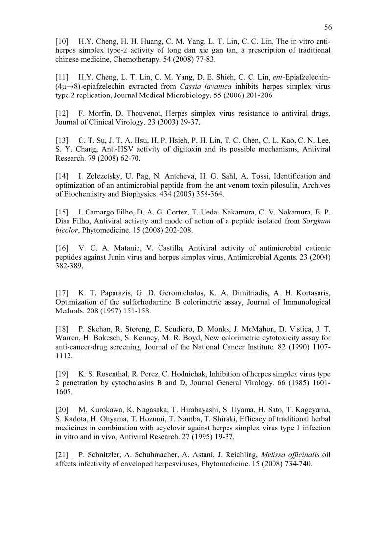
56
[10] H.Y. Cheng, H. H. Huang, C. M. Yang, L. T. Lin, C. C. Lin, The in vitro anti-herpes simplex type-2 activity of long dan xie gan tan, a prescription of traditional chinese medicine, Chemotherapy. 54 (2008) 77-83. [11] H.Y. Cheng, L. T. Lin, C. M. Yang, D. E. Shieh, C. C. Lin, ent-Epiafzelechin-(4μ→8)-epiafzelechin extracted from Cassia javanica inhibits herpes simplex virus type 2 replication, Journal Medical Microbiology. 55 (2006) 201-206. [12] F. Morfin, D. Thouvenot, Herpes simplex virus resistance to antiviral drugs, Journal of Clinical Virology. 23 (2003) 29-37. [13] C. T. Su, J. T. A. Hsu, H. P. Hsieh, P. H. Lin, T. C. Chen, C. L. Kao, C. N. Lee, S. Y. Chang, Anti-HSV activity of digitoxin and its possible mechanisms, Antiviral Research. 79 (2008) 62-70. [14] I. Zelezetsky, U. Pag, N. Antcheva, H. G. Sahl, A. Tossi, Identification and optimization of an antimicrobial peptide from the ant venom toxin pilosulin, Archives of Biochemistry and Biophysics. 434 (2005) 358-364. [15] I. Camargo Filho, D. A. G. Cortez, T. Ueda- Nakamura, C. V. Nakamura, B. P. Dias Filho, Antiviral activity and mode of action of a peptide isolated from Sorghum bicolor, Phytomedicine. 15 (2008) 202-208. [16] V. C. A. Matanic, V. Castilla, Antiviral activity of antimicrobial cationic peptides against Junin virus and herpes simplex virus, Antimicrobial Agents. 23 (2004) 382-389. [17] K. T. Paparazis, G .D. Geromichalos, K. A. Dimitriadis, A. H. Kortasaris, Optimization of the sulforhodamine B colorimetric assay, Journal of Immunological Methods. 208 (1997) 151-158. [18] P. Skehan, R. Storeng, D. Scudiero, D. Monks, J. McMahon, D. Vistica, J. T. Warren, H. Bokesch, S. Kenney, M. R. Boyd, New colorimetric cytotoxicity assay for anti-cancer-drug screening, Journal of the National Cancer Institute. 82 (1990) 1107-1112. [19] K. S. Rosenthal, R. Perez, C. Hodnichak, Inhibition of herpes simplex virus type 2 penetration by cytochalasins B and D, Journal General Virology. 66 (1985) 1601-1605. [20] M. Kurokawa, K. Nagasaka, T. Hirabayashi, S. Uyama, H. Sato, T. Kageyama, S. Kadota, H. Ohyama, T. Hozumi, T. Namba, T. Shiraki, Efficacy of traditional herbal medicines in combination with acyclovir against herpes simplex virus type 1 infection in vitro and in vivo, Antiviral Research. 27 (1995) 19-37. [21] P. Schnitzler, A. Schuhmacher, A. Astani, J. Reichling, Melissa officinalis oil affects infectivity of enveloped herpesviruses, Phytomedicine. 15 (2008) 734-740.
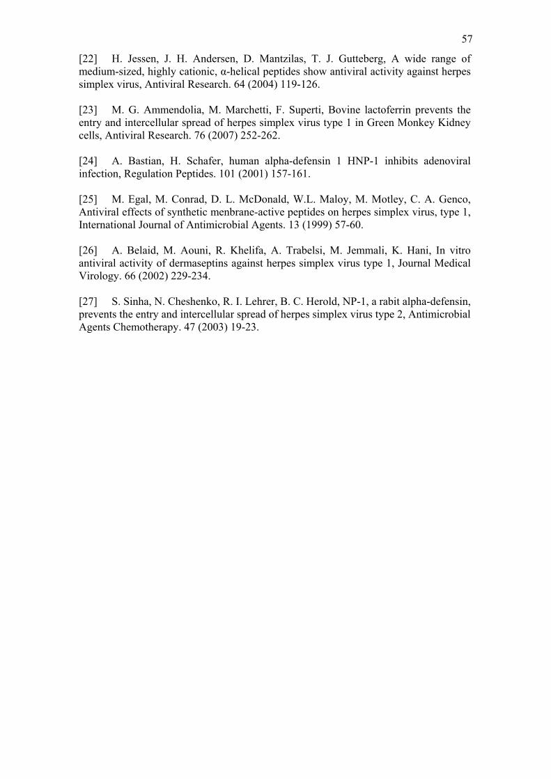
57
[22] H. Jessen, J. H. Andersen, D. Mantzilas, T. J. Gutteberg, A wide range of medium-sized, highly cationic, α-helical peptides show antiviral activity against herpes simplex virus, Antiviral Research. 64 (2004) 119-126. [23] M. G. Ammendolia, M. Marchetti, F. Superti, Bovine lactoferrin prevents the entry and intercellular spread of herpes simplex virus type 1 in Green Monkey Kidney cells, Antiviral Research. 76 (2007) 252-262. [24] A. Bastian, H. Schafer, human alpha-defensin 1 HNP-1 inhibits adenoviral infection, Regulation Peptides. 101 (2001) 157-161. [25] M. Egal, M. Conrad, D. L. McDonald, W.L. Maloy, M. Motley, C. A. Genco, Antiviral effects of synthetic menbrane-active peptides on herpes simplex virus, type 1, International Journal of Antimicrobial Agents. 13 (1999) 57-60. [26] A. Belaid, M. Aouni, R. Khelifa, A. Trabelsi, M. Jemmali, K. Hani, In vitro antiviral activity of dermaseptins against herpes simplex virus type 1, Journal Medical Virology. 66 (2002) 229-234. [27] S. Sinha, N. Cheshenko, R. I. Lehrer, B. C. Herold, NP-1, a rabit alpha-defensin, prevents the entry and intercellular spread of herpes simplex virus type 2, Antimicrobial Agents Chemotherapy. 47 (2003) 19-23.
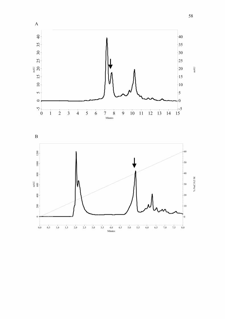
58
Minutes0 1 2 3 4 5 6 7 8 9 10 11 12 13 14 15
mA
U
-50
510
1520
2530
3540
mA
U
-5
0
5
10
15
20
25
30
35
40
Minutes0,0 0,5 1,0 1,5 2,0 2,5 3,0 3,5 4,0 4,5 5,0 5,5 6,0 6,5 7,0 7,5 8,0
mA
U0
200
400
600
800
1000
1200
% N
aCl 0
,5 M
0
10
20
30
40
50
60
A
B
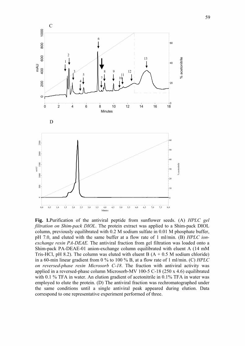
59
Minutes0 2 4 6 8 10 12 14 16 18
mA
U0
200
400
600
800
1000
% a
ceto
nitr
ile
0
20
40
60
10
1
2
3
4
5
6
7
8 911
12
13
D
Minutes0,0 0,5 1,0 1,5 2,0 2,5 3,0 3,5 4,0 4,5 5,0 5,5 6,0 6,5 7,0 7,5 8,0
mA
U0
500
1000
1500
2000
2500
% a
ceto
nitri
le
0
10
20
30
40
50
60
Fig. 1.Purification of the antiviral peptide from sunflower seeds. (A) HPLC gel filtration on Shim-pack DIOL. The protein extract was applied to a Shim-pack DIOL column, previously equilibrated with 0.2 M sodium sulfate in 0.01 M phosphate buffer, pH 7.0, and eluted with the same buffer at a flow rate of 1 ml/min. (B) HPLC ion-exchange resin PA-DEAE. The antiviral fraction from gel filtration was loaded onto a Shim-pack PA-DEAE-01 anion-exchange column equilibrated with eluent A (14 mM Tris-HCl, pH 8.2). The column was eluted with eluent B (A + 0.5 M sodium chloride) in a 60-min linear gradient from 0 % to 100 % B, at a flow rate of 1 ml/min. (C) HPLC on reversed-phase resin Microsorb C-18. The fraction with antiviral activity was applied in a reversed-phase column Microsorb-MV 100-5 C-18 (250 x 4.6) equilibrated with 0.1 % TFA in water. An elution gradient of acetonitrile in 0.1% TFA in water was employed to elute the protein. (D) The antiviral fraction was rechromatographed under the same conditions until a single antiviral peak appeared during elution. Data correspond to one representative experiment performed of three.
C
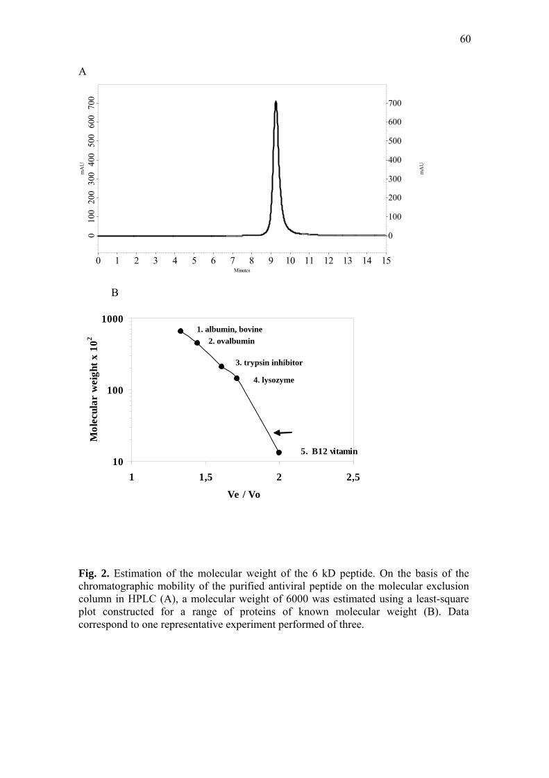
60
A
Minutes0 1 2 3 4 5 6 7 8 9 10 11 12 13 14 15
mA
U
010
020
030
040
050
060
070
0
mA
U
0
100
200
300
400
500
600
700
B Fig. 2. Estimation of the molecular weight of the 6 kD peptide. On the basis of the chromatographic mobility of the purified antiviral peptide on the molecular exclusion column in HPLC (A), a molecular weight of 6000 was estimated using a least-square plot constructed for a range of proteins of known molecular weight (B). Data correspond to one representative experiment performed of three.
10
100
1000
1 1,5 2 2,5
Ve / Vo
Mol
ecul
ar w
eigh
t x 1
02
5. B12 vitamin
1. albumin, bovine 2. ovalbumin
3. trypsin inhibitor
4. lysozyme
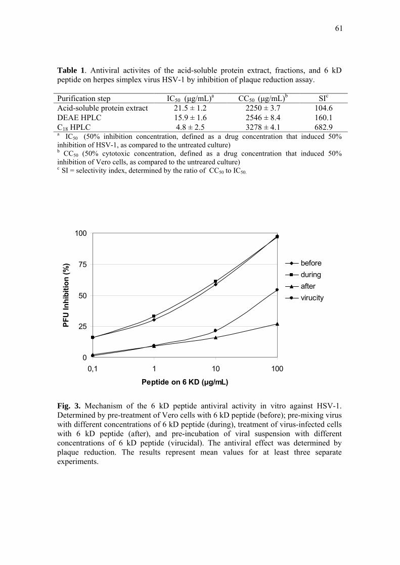
61
Table 1. Antiviral activites of the acid-soluble protein extract, fractions, and 6 kD peptide on herpes simplex virus HSV-1 by inhibition of plaque reduction assay. Purification step IC50 (μg/mL)a CC50 (μg/mL)b SIc Acid-soluble protein extract 21.5 ± 1.2 2250 ± 3.7 104.6 DEAE HPLC 15.9 ± 1.6 2546 ± 8.4 160.1 C18 HPLC 4.8 ± 2.5 3278 ± 4.1 682.9 a IC50 (50% inhibition concentration, defined as a drug concentration that induced 50% inhibition of HSV-1, as compared to the untreated culture) b CC50 (50% cytotoxic concentration, defined as a drug concentration that induced 50% inhibition of Vero cells, as compared to the untreared culture) c SI = selectivity index, determined by the ratio of CC50 to IC50.
0
25
50
75
100
0,1 1 10 100
Peptide on 6 KD (μg/mL)
PFU
Inhi
bitio
n (%
) beforeduringaftervirucity
Fig. 3. Mechanism of the 6 kD peptide antiviral activity in vitro against HSV-1. Determined by pre-treatment of Vero cells with 6 kD peptide (before); pre-mixing virus with different concentrations of 6 kD peptide (during), treatment of virus-infected cells with 6 kD peptide (after), and pre-incubation of viral suspension with different concentrations of 6 kD peptide (virucidal). The antiviral effect was determined by plaque reduction. The results represent mean values for at least three separate experiments.
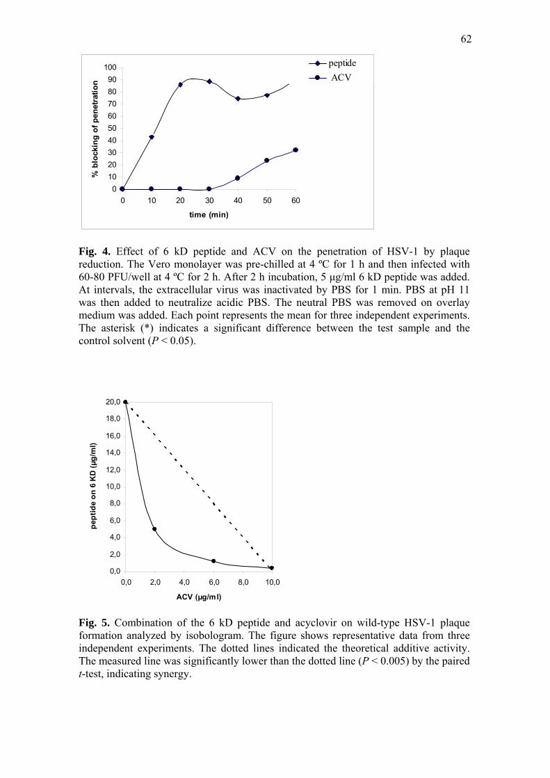
62
0102030405060708090
100
0 10 20 30 40 50 60
time (min)
% b
lock
ing
of p
enet
ratio
n
peptide ACV
Fig. 4. Effect of 6 kD peptide and ACV on the penetration of HSV-1 by plaque reduction. The Vero monolayer was pre-chilled at 4 ºC for 1 h and then infected with 60-80 PFU/well at 4 ºC for 2 h. After 2 h incubation, 5 μg/ml 6 kD peptide was added. At intervals, the extracellular virus was inactivated by PBS for 1 min. PBS at pH 11 was then added to neutralize acidic PBS. The neutral PBS was removed on overlay medium was added. Each point represents the mean for three independent experiments. The asterisk (*) indicates a significant difference between the test sample and the control solvent (P < 0.05).
0,0
2,0
4,0
6,0
8,0
10,0
12,0
14,0
16,0
18,0
20,0
0,0 2,0 4,0 6,0 8,0 10,0
ACV (µg/ml)
pept
ide
on 6
KD
(µg/
ml)
Fig. 5. Combination of the 6 kD peptide and acyclovir on wild-type HSV-1 plaque formation analyzed by isobologram. The figure shows representative data from three independent experiments. The dotted lines indicated the theoretical additive activity. The measured line was significantly lower than the dotted line (P < 0.005) by the paired t-test, indicating synergy.

63
Livros Grátis( http://www.livrosgratis.com.br )
Milhares de Livros para Download: Baixar livros de AdministraçãoBaixar livros de AgronomiaBaixar livros de ArquiteturaBaixar livros de ArtesBaixar livros de AstronomiaBaixar livros de Biologia GeralBaixar livros de Ciência da ComputaçãoBaixar livros de Ciência da InformaçãoBaixar livros de Ciência PolíticaBaixar livros de Ciências da SaúdeBaixar livros de ComunicaçãoBaixar livros do Conselho Nacional de Educação - CNEBaixar livros de Defesa civilBaixar livros de DireitoBaixar livros de Direitos humanosBaixar livros de EconomiaBaixar livros de Economia DomésticaBaixar livros de EducaçãoBaixar livros de Educação - TrânsitoBaixar livros de Educação FísicaBaixar livros de Engenharia AeroespacialBaixar livros de FarmáciaBaixar livros de FilosofiaBaixar livros de FísicaBaixar livros de GeociênciasBaixar livros de GeografiaBaixar livros de HistóriaBaixar livros de Línguas
Baixar livros de LiteraturaBaixar livros de Literatura de CordelBaixar livros de Literatura InfantilBaixar livros de MatemáticaBaixar livros de MedicinaBaixar livros de Medicina VeterináriaBaixar livros de Meio AmbienteBaixar livros de MeteorologiaBaixar Monografias e TCCBaixar livros MultidisciplinarBaixar livros de MúsicaBaixar livros de PsicologiaBaixar livros de QuímicaBaixar livros de Saúde ColetivaBaixar livros de Serviço SocialBaixar livros de SociologiaBaixar livros de TeologiaBaixar livros de TrabalhoBaixar livros de Turismo