Embed Size (px)
Citation preview

Autophagy, apoptosis and organelle features during cell exposure to cadmiumčCRISTIANE DOS SANTOS VERGILIO AND EDÉSIO JOSÉ TENÓRIO DE MELO*
Universidade Estadual do Norte Fluminense, Centro de Biociências e Biotecnologia, Laboratório de Biologia Celular e Tecidual, Campos dos Goytacazes, RJ, Brasil, 28013-602.
Key words: Cd, cell death, hepatocyte, HuH-7 cells, mitochondria
ABSTRACT: Cadmium (Cd) induces several effects in different tissues, but our knowledge of the toxic effects on organelles is insuffi cient. To observe the progression of Cd effects on organelle structure and function, HuH-7 cells (human hepatic carcinoma cell line) were exposed to CdCl2 in increasing concentrations (1 μM – 20 μM) and exposure times (2 h – 24 h). During Cd treatment, the cells exhibited a progressive decrease in viability that was both time- and dose-dependent. Cd treated cells displayed progressive morphological changes that included cytoplasm retraction and nuclear condensation preceding a total loss of cell adhesion. Treatment with 10 μM for 12 h led to irreversible damages. Before these drastic and irreparable damages, treated cells (5 μM for 12 h) presented a progressive loss of mitochondrial function and cytoplasm acidifi cation as well as dysfunction and disorganization of microfi laments and endoplasmic reticulum. These damages led to the induction of apoptotic events and an increase in autophagic bodies in the cytoplasm. These results revealed that Cd affects multiple intra-cellular targets that induce alterations in the mitochondria, cytoskeleton, endoplasmic reticulum and acidic compartments, ultimately culminating in cell death via apoptotic and autophagic pathways.
BIOCELL2013, 37(2): 45-54
ISSN 0327 - 9545PRINTED IN ARGENTINA
Introduction
Cadmium (Cd) is a highly toxic metal that exerts multiple effects on organisms (Filipič, 2012; Waisberg et al., 2003; Bertin and Averbeck, 2006). However, the complexity and diversity of events associated with cell-Cd interactions have resulted in fragmented information mainly related with organelle structure and function (Cannino et al., 2009). Biochemical studies have shown the involvement of organelles (mitochondria, lysosomes and cytoskeleton)
in Cd toxicity in several cell lines (Cannino et al., 2009; Fotakis et al., 2005; Faverney et al., 2004; L’Azou et al., 2002). However, the wide-ranging effects of this metal on organelles and their involvement in induced cell death remain to be fully understood (Fabbri et al., 2012). Therefore, the overall understanding of Cd in-duced cell damage and toxicity needs the observation of its effects on different intra-cellular targets. Cd exposure in organisms is followed by injuries in the liver, testes, lungs, kidneys and bones (Ye et al., 2007; Joseph, 2009; Nordberg, 2009; Siu et al., 2009). Cd uptake by hepatocytes makes the liver one of the major sites of Cd accumulation (Fabbri et al., 2012) and reduces its availability to other organs (Souza et al., 1997). Therefore, studies of hepatocyte organelles may help understanding the progression from the direct effects of Cd to its ultimate toxicity.
*Address correspondence to: Edésio José Tenório de MELO.E-mail: [email protected]: April 10, 2013. Revised version received: August 13, 2013. Accepted: August 18, 2013.

CRISTIANE DOS SANTOS VERGILIO and EDÉSIO JOSÉ TENÓRIO DE MELO46
With this purpose, the structure and function of mi-tochondria, acidic organelles and vesicles, endoplasmic reticulum elements and microfi laments was assessed in HuH-7 cells (a human hepatic carcinoma cell line) to observe the progression of Cd toxicity.
Materials and Methods
Cell culture and treatments
HuH-7 cells were maintained in 25 mL cell cul-ture fl asks with Dulbecco’s Modifi ed Eagle’s Medium (DMEM-1152, Sigma Aldrich®) supplemented with 10% fetal bovine serum (Gibco®) in a humidifi ed at-mosphere containing 5% CO2 at 37ºC. For experimental purposes, the cells were seeded onto 24-well plastic plates. The optimum cell concentration determined from cell line growth profi les was 105 cells/mL. Cells were allowed to attach for 24 h before Cd treatments. For Cd toxicity assays, stock solutions (0.1 M CdCl2) were prepared using ultra-pure quality water, and dilutions were made with culture medium to 1 μM, 5 μM, 10 μM, 15 μM and 20 μM fi nal concentrations. To observe the progression of Cd induced toxic effects, these concentrations were added to cell cultures for 2, 6, 12 and 24 h.
Quantifi cation and morphological analysis of Cd in-duced toxic effects
Control and Cd exposed cells were fi xed in Bouin’s solution and stained with Giemsa (10%) for light mi-
croscopy observation. All preparations were examined using a Zeiss Axioplan photomicroscope equipped with 20x and 40x objectives. HuH-7 cell survival was determined by counting the number of living cells in a given area (the cell spread on the substrate and nuclear condensation were considered for discrimination be-tween live and dead cells). For each sample, 6 randomly chosen fi elds were scored at a magnifi cation of 400x, and results were expressed as the mean ± standard devia-tion. HuH-7 control cell numbers counted at each time point were considered to be 100%. Digital images were obtained using an Axioplan microscope equipped with a Canon Power Shot camera A610/620 employing 20x and 40x objectives.
Cell viability analysis with MTT assay
Following exposure to Cd, the cells were incubated with MTT (3-(4,5-dimethylthiazol-2-yl)-2,5-diphen-yltetrazolium bromide, 6 mg/mL) in culture medium for 4 h at 37ºC (Mosmann, 1983). After the removal of MTT-containing medium, 200 μL of DMSO (dimeth-ylsulfoxide) were added, and the absorbance at 540 nm was measured after 5 min in a microplate reader (Thermoplate© TP reader). Results were expressed as mean ± standard deviation of triplicate experiments.
Scanning and transmission electron microscopy (SEM and TEM)
HuH-7 cells treated with 5 μM CdCl2 for 12 h were fi xed in 2.5% (v/v) glutaraldehyde and 4% (v/v) formaldehyde in 0.1 M cacodylate buffer (pH 7.2). For
FIGURE 1. CdCl2 toxic effects on viability of HuH-7 cells. (a) Quantifi cation of Giemsa stained HuH-7 cells after CdCl2 treat-ment. All concentrations tested were compared to a control group that was defi ned as 100%. (b) Decrease in cell viability by MTT assay in a dose/time dependent manner. *Signifi cantly different from control (p < 0.001).
100
80
60
40
20
0
2 h6 h12 h24 h
1 μM 5 μM 10 μMCdCl2 concentration
Cd Toxic Effects
15 μM 20 μM
a
% H
uH-7
cel
ls
*
*
**
*
*
*
*
**
*
*
**
*
*
* **
*
2.5
2.0
1.5
1.0
0.5
0.0
control1 μM5 μM10 μM15 μM20 μM
2 h 6 h 12 hExposure time
MTT assay
24 h
b
Abs
orba
nce
540
nm ** *
*
*
*
**
*
*
*
**
**
*
** *
47Cd INDUCED ORGANELLE DYSFUNCTIONS AND CELL DEATH
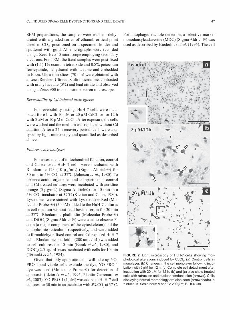
SEM preparations, the samples were washed, dehy-drated with a graded series of ethanol, critical-point dried in CO2, positioned on a specimen holder and sputtered with gold. All micrographs were recorded using a Zeiss Evo 40 microscope employing secondary electrons. For TEM, the fi xed samples were post-fi xed with (1:1) 1% osmium tetraoxide and 0.8% potassium ferricyanide, dehydrated with acetone and embedded in Epon. Ultra-thin slices (70 nm) were obtained with a Leica Reichert Ultracut S ultramicrotome, contrasted with uranyl acetate (5%) and lead citrate and observed using a Zeiss 900 transmission electron microscope.
Reversibility of Cd induced toxic effects
For reversibility testing, HuH-7 cells were incu-bated for 6 h with 10 μM or 20 μM CdCl2 or for 12 h with 5 μM or 10 μM of CdCl2. After exposure, the cells were washed and the medium was replaced without Cd addition. After a 24 h recovery period, cells were ana-lysed by light microscopy and quantifi ed as described above.
Fluorescence analyses
For assessment of mitochondrial function, control and Cd exposed HuH-7 cells were incubated with Rhodamine 123 (10 μg/mL) (Sigma Aldrich®) for 30 min in 5% CO2 at 37ºC (Johnson et al., 1980). To observe acidic organelles and compartments, control and Cd treated cultures were incubated with acridine orange (5 μg/mL) (Sigma Aldrich®) for 40 min in a 5% CO2 incubator at 37ºC (Kielian and Cohn, 1980). Lysosomes were stained with LysoTracker Red (Mo-lecular Probes®) (50 nM) added to the HuH-7 cultures in cell medium without fetal bovine serum for 30 min at 37ºC. Rhodamine phalloidin (Molecular Probes®) and DiOC6 (Sigma Aldrich®) were used to observe F-actin (a major component of the cytoskeleton) and the endoplasmic reticulum, respectively, and were added to formaldehyde-fi xed control and Cd exposed HuH-7 cells. Rhodamine phalloidin (200 units/mL) was added to cell cultures for 40 min (Barak et al., 1980), and DiOC6 (2.5 μg/mL) was incubated with cells for 10 min (Terasaki et al., 1984). Given that only apoptotic cells will take up YO-PRO-1 and viable cells exclude the dye, YO-PRO-1 dye was used (Molecular Probes®) for detection of apoptosis (Idziorek et al., 1995; Plantin-Carrenard et al., 2003). YO-PRO-1 (1 μM) was added to HuH-7 cell cultures for 30 min in an incubator with 5% CO2 at 37ºC.
For autophagic vacuole detection, a selective marker monodansylcadaverine (MDC) (Sigma Aldrich®) was used as described by Biederbick et al. (1995). The cell
FIGURE 2. Light microscopy of HuH-7 cells showing mor-phological alterations induced by CdCl2. (a) Control cells in monolayer. (b) Changes in the cell monolayer following incu-bation with 5 μM for 12 h. (c) Complete cell detachment after incubation with 20 μM for 12 h. (b) and (c) also show treated cells with retraction and nuclear condensation (arrows). Cells displaying normal morphology are also seen (arrowheads). n = nucleus. Scale bars: A and C: 200 μm; B: 100 μm.

CRISTIANE DOS SANTOS VERGILIO and EDÉSIO JOSÉ TENÓRIO DE MELO48
culture was incubated with 0.05 mM MDC in PBS at 37ºC for 10 min. All the stained cells were observed under a Zeiss Confocal Laser Scan Microscope (CLSM) using a 543 nm argon laser and a 40x objective.
Statistical analyses
All data are expressed as the means ± standard errors. Statistical analyses were made using GraphPad Prism v.4 software (GraphPad Software, Inc. CA,
FIGURE 3. Scanning (a, b, d) and transmission (c) electron microscopy showing HuH-7 cell cultures before (a) and after Cd treatment (b - d) (5 μM for 12 h). (a) Characteristic aspect of control monolayer. (b) Presence of many detaching rounded cells (arrows). (c) Ultra-structural appearance of Cd treated cells with many vacuoles in the cytoplasm (arrows) and the collapse of some mitochondrial cristae (inset, arrowheads). (d) Cells with membrane blebs (arrows) following Cd treatment. n = nucleus. Scale bar: A: 20 μm, B: 20 μm, C: 1.1 μm, D: 10 μm.

49Cd INDUCED ORGANELLE DYSFUNCTIONS AND CELL DEATH
USA). The two-way analysis of variance followed by the Bonferroni test was performed for cell viability data and reversibility test data. Differences were considered signifi cant when p < 0.05.
Results
To determine the threshold of metal damage and its relationship to metal toxicity (induction of cell death), the present study investigated the effects of Cd over the HuH-7 cell machinery after treatments with increasing concentrations and exposure times. The dose and duration of treatment were critical factors in the induction of cell death (Fig. 1a). These toxic effects were evaluated after each Cd treatment following the observation of reduced cell numbers demonstrated by the attached cell count (Fig. 1a). Cell viability was assessed through the MTT assay, verifying the decrease of cell viability indicated by the failure of mitochondrial function (Fig. 1b). The results obtained by counting the surviving cells or through assessment of mitochondrial function by MTT assay corroborate the Cd toxicity in the culture. The observation of the Cd induced toxic effects in-dicated that healthy cells at semi-confl uence, evidenced by adherence and spread cytoplasm on the substrate with prominent nuclei and nucleoli, changed during Cd treat-ment (Fig. 2a, 2b). Cells experienced different degrees of cytoplasm shrinkage and nuclear condensation (Fig. 2b). This cytoplasmic retraction was more evident at higher doses (20 μM), but occurred asynchronously within the culture (Fig. 2c, inset) and led to the gradual loss of cell viability and subsequent release from the substrate. Ultrastructural analysis of cell culture indicated that cell morphology (Fig. 3a) changed in the presence of Cd (5 μM for 12 h) as evidenced by cytoplasm retraction (Fig. 3b), severe vacuolization (Fig. 3c, arrows) and alterations in mitochondrial structure (Fig. 3c, inset). The presence of blebs on the membrane cell surface (Fig. 3d, arrows) also indicated apoptosis, and this was also confi rmed by YO-PRO-1 nuclear staining (Fig. 4a- d). No indicative probe (Fig. 4b, arrowhead) was observed in the adherent control cells (Fig. 4a). How-ever, following Cd exposure (5 μM for 12 h), staining was evident in cells with cytoplasmic retraction and nuclear disorganisation (Fig. 4c, d, arrowheads). The cells displayed different stages of cellular retraction (Fig. 4c) with distinct apoptotic staining (Fig. 4d), sug-gesting that the process occurred asynchronously within the same culture.
To assess the reversibility of Cd induced damage, cells were treated with 5 and 10 μM CdCl2 for 12 h or with 10 and 20 μM CdCl2 for 6 h, and then maintained in the absence of Cd for 24 h. After Cd removal, both treatments (10 and 20 μM) for the short period (6 h) and the lower concentration (5 μM) with long-term exposure (12 h) the culture was able to recover (Fig. 5a-h). However, treatment with 10 μM for 12 h promoted severe deleterious changes (Fig. 5i, j) that compromised cellular recovery (Fig. 5b). This fi nding is important to understand the kinetics of metal action on the cellular machinery. The treatment with 5 μM for 12 h was chosen to investigate the Cd induced changes in organelles and severe damages that compromised cell survival were observed. Initially, organelle functionality was analysed using the mitochondrial fl uorescent stain Rhodamine 123 (Fig. 6a-d). The intense and spread fi laments indica-tive of functional mitochondria (Fig. 6b, arrowheads) present in control cells changed to punctate staining
FIGURE 4. Differential interference contrast microscopy (DIC) (a and c) and confocal laser scanning microscopy of HuH-7 cells stained with YO-PRO-1 (YP-1) (1 μM) (b an d) before (a and b) and after Cd treatment with 5 μM for 12 h (c and d). (a) Control cells. (b) No fl uorescence signal in untreated cell. (c) Differential levels of cytoplasm retraction and nuclear disorganisation, both characteristics of apoptotic processes observed in Cd treated cells. (d) Cellular staining indicative of cell death via apoptotic processes following CdCl2 exposure. n= nucleus. Scale bar: 10 μm.
DIC image
Cd
trea
tmen
t
YP-1
cont
rol

CRISTIANE DOS SANTOS VERGILIO and EDÉSIO JOSÉ TENÓRIO DE MELO50
FIGURE 5. Reversibility of Cd effects on HuH-7 cells. (a) Recovery in culture after 24 h of Cd removal following the treatments with higher concentrations and short exposure time (10 μM and 20 μM for 6 h). (b) Recovery of culture following long-term exposure (12 h) with 5 μM with subsequent 24 h of Cd removal. However, the same capability to reverse Cd toxicity was not observed after 12 h incubation with 10 μM; where the toxic effects last even after Cd removal. (c - j) Morphological aspects of the culture following each Cd treatment: (c) 10 μM for 6 h, (d) 10 μM for 6 h + 24 h without Cd, (e) 20 μM for 6 h, (f) 20 μM for 6 h + 24 h without Cd, (g) 5 μM for 12 h, (h) 5 μM for 12 h + 24 h without Cd, (i) 10 μM for 12 h, (j) 10 μM for 12 h + 24 h without Cd. The concentrations tested were compared with a control group that was considered 100%. ***p<0.001.
FIGURE 6. Differential interference contrast mi-croscopy (DIC) (a, c) and confocal laser scanning microscopy of HuH-7 cells stained with rhodamine 123 (R123) (10 μg/mL) (b, d). (a) Normal aspect of untreated cell. (b) Control cell with fi lamentous fl uorescence spread in the cytoplasm, indicating the functional area of the mitochondria (arrowheads). (c) Cd treated cell (5 μM for 12 h). (d) Cd treated cell (5 μM for 12 h) with punctate fl uorescence, suggesting loss of mitochondrial functionality (arrowheads). n = nucleus. Scale bar: 10 μm.
DIC image
Cd
trea
tmen
t
R123
cont
rol
Reversibility Test - 10 and 20 μM Cd for 6h
Reversibility Test - 5 and 10 μM Cd for 12h
60
50
40
30
20
10
06h 6h + 24h without metal
Treatment
10 μM20 μM
a
***
**
% H
uH-7
cel
ls
8070605040302010
012h 12h + 24h without metal
Treatment
5 μM10 μM
b
***
***% H
uH-7
cel
ls10 μM
6h12
h6h
+ 2
4hw
ithou
t met
al12
h +
24h
with
out m
etal
5 μM
20 μM
10 μM

51Cd INDUCED ORGANELLE DYSFUNCTIONS AND CELL DEATH
(Fig. 6d, arrowheads), suggesting the loss of mitochon-drial function in Cd treated cells. These results agree with the evidence from the MTT assay (Fig. 1b) and occurred in cells that remain attached following Cd treatment (5 μM/12 h), indicating that mitochondria are an early target in Cd toxicity. While mitochondrial function was impaired after Cd exposure, the acidic compartments increased in frequency and size (Fig. 7a-d). This increase in acidic vesicles may correspond with increased abundance of lysosomes in the cytoplasm (Fig. 8a-d). Consequently, the cells changed from a punctate regular fl uorescent staining (Fig. 7b, 8b) to an intense fl uorescence pattern corresponding to acid structures in the cytoplasm (Fig. 7d, 8d arrowheads). The presence of fl uorescent acidic compartments or lysosomal vacuoles further suggests the possibility of intracellular autophagic digestion. To disclose the pres-ence of autophagosomes during Cd treatment, the cells were incubated with MDC (Fig. 9). Untreated cells (Fig.
9a) exhibited no fl uorescence indicative of autophagic vacuoles (Fig. 9b), while treated cells (Fig. 9c) showed a high frequency of fl uorescent compartments and even the formation of large vacuoles (Fig. 9d). Therefore, these results strongly suggest that the apoptotic pathway and autophagic processes are involved in Cd induced cell death. The endoplasmic reticulum, a major organelle involved in detoxifi cation (Voeltz et al., 2002), was analysed with the fl uorescent dye DiOC6 (Fig. 10). The dispersion of endoplasmic reticulum elements observed in untreated cells (Fig. 10a, b) changed after Cd treat-ment (Fig. 10c, d) even with the cell remaining adherent and spread (Fig. 10c). Cytoskeleton microfi laments were also analysed for understanding the changes in cell structure after Cd treatment (Fig. 11). The extended microfi lament net-work (Fig. 11b, arrowheads) changed after Cd treatment, given that the cells lost their microfi lament projections in the cytoplasm (Fig. 11d, arrowheads) and their ad-hesion points on the substrates (Fig. 11d, arrowheads),
FIGURE 7. Differential interference contrast microscopy (DIC) (a, c) and confocal laser scanning microscopy of HuH-7 cells stained with acridine orange (AO) (5 μg/mL) (b, d). (a) Control cell. (b) Untreated cell with punctate fl uorescence staining in the cytoplasm corresponding to acidic organelles, such as endosomes and lysosomes (arrowheads). (c) Vacuolisation in Cd treated cells (5 μM for 12 h) (arrow). (d) Intensely dis-persed fl uorescence observed in the cytoplasm (arrowheads) of treated cells (5 μM for 12 h). n = nucleus. Scale bar: 10 μm.
FIGURE 8. Differential interference contrast microscopy (DIC) (a and c) and confocal laser scanning microscopy of HuH-7 cells stained with LysoTracker Red (50 nM) (b and d). (a) Control cells. (b) Punctate fl uorescence pattern observed in untreated culture (arrowheads). (c) Cd treated cell (5 μM for 12 h). (d) Intense and dispersed fl uorescence pattern indicate increased lysosomes throughout the cytoplasm (arrowheads). n = nucleus. Scale bar: 10 μm (x400).
DIC image
Cd
trea
tmen
t
AO
cont
rol
DIC image
Cd
trea
tmen
t
LTR
cont
rol

CRISTIANE DOS SANTOS VERGILIO and EDÉSIO JOSÉ TENÓRIO DE MELO52
FIGURE 9. Differential interference contrast microscopy (DIC) (a and c) and confocal laser scanning mi-croscopy of HuH-7 cells stained with monodansylcadaverine (MDC) (0.05 mM) (b and d) after Cd treatment (5 μM for 12 h). (a) Control cells. (b) No fl uorescence signal in untreated cells. (c) Cd treated cells with vacu-oles in cytoplasm (arrowheads). (d) Autophagic vacuoles (arrowheads) staining in Cd treated cell indicat-ing Cd mediated cell death through autophagic processes. n= nucleus. Scale bar: 10 μm.
FIGURE 10. Differential interference contrast microscopy (DIC) (a, c) and confocal laser scanning microscopy of HuH-7 cells stained with DiOC6 (2.5 μg/mL) (b, d). (a) Control cells. (b) Morphological aspect of the reticular network of control cells with thinner peripheral regions (arrow) and regions with high fl uorescence close to the nucleus, mainly because of the concentration of reticulum and other membranes, such as mitochondria (arrow-heads) (c) Cd treated cells (5 μM for 12 h). (d) Weaker fl uorescence signal in treated cells (5 μM for 12 h); with evidence of the disorganisation in reticular arrangement close to nucleus (arrowheads) and in cell periphery (ar-row). n = nucleus. Scale bar: 10 μm.
DIC image
Cd
trea
tmen
t
MDC
cont
rol
DIC image
Cd
trea
tmen
t
DiOC6
cont
rol

53Cd INDUCED ORGANELLE DYSFUNCTIONS AND CELL DEATH
leading to an alteration in cell morphology (Fig. 11c, d, arrow). Interestingly, multiple Cd induced damages in organelles were observed in treated cells that remained attached, indicating that the severity of the effect in different targets is important in inducing cell death. Therefore, Cd reached several targets at the same time leading to loss of mitochondrial function, endoplasmic reticulum dysfunction, cytoplasmic acidifi cation and microfi lament disorganisation. These processes are all occurring together in treated cells, and if the exposure is not halted, might lead to cell death by apoptotic and autophagic pathways.
Discussion
The results obtained clearly show that Cd induces a decrease in cell viability and progressive damage to cell morphology in HuH-7 cells through concurrent effects in multiple intracellular targets, including mi-
tochondria, cytoskeleton, endoplasmic reticulum and acidic compartments, leading to cell death through the apoptotic and autophagic pathways. Apoptosis is considered a normal housekeeping event, but it is also necessary to arrest abnormal cell proliferation in development (Pulido and Parrish, 2003). Apoptosis can also be induced by a variety of chemi-cals, including many toxic metals (Rana, 2008), and is a known pathway of Cd mediated cell death (Wang et al., 2009; Lasfer et al., 2008; Ye et al., 2007; Mao et al., 2007; Pulido and Parrish, 2003; Faverney et al., 2004). However, the present study indicates that apoptosis is not the only process observed in Cd treated cells, given that the autophagic pathway was also observed after sustained Cd exposure. The autophagic pathway allows the digestion of dysfunctional organelles with resulting recirculation and reuse of their molecular constituents (Templeton and Liu, 2010). Furthermore, when Cd induced cell damage exceeds the repairing capacity of repair, cell death occurred. Dying cells generate increasing amounts of autophagic vacuoles and clear large proportions of their cytoplasm before dying (Bursch et al., 2008). The induction of Cd toxicity (cell death) in culture was asynchronous, suggesting preferential interference in some stages of the cell cycle. In fact, Cd can lead to cell cycle arrest, which may affect several cellular processes including cell proliferation and differentia-tion (Hartwig, 2010; Bertin and Averbeck, 2006). G2/M phase arrest was demonstrated after Cd exposure (Bork et al., 2010), preventing damaged cells from entering into mitosis, until DNA damage is repaired. Therefore, some stages of the cell cycle might be more susceptible to Cd damage as suggested by the effect on different cells in the same culture. Other authors have shown the isolated involvement of mitochondria (Caninno et al., 2009), cytoskeleton (L’Azou et al., 2002), endoplasmic reticulum (Wang et al., 2009) and lysosomes (Lekube et al., 2000) dem-onstrating the role of separate organelles and structures in Cd induced cell death. The present study shows that these intracellular targets are all being affected concur-rently and contribute to cell dysfunction leading to cell death. Moreover, the present study shows that the extent of damage induced by Cd treatment is eventually so se-vere that cells cannot reverse the toxic effects as shown by the 10 μM treatment for 12 h in the reversibility test. The present study has increased our understanding of the cellular mechanisms of Cd toxicity on HuH-7 cells, by showing the progression of Cd induced dam-age in cells, and the involvement of mitochondria,
FIGURE 11. Differential interference contrast microscopy (DIC) (a, c) and confocal laser scanning microscopy of HuH-7 cells stained with rhodamine phalloidin (RP) (b, d). (a) Untreated cells in monolayer. (b) Extended of microfi lament network and adhesion points (arrowheads) in control cells. (c) Cd treated cells (5 μM for 12 h) displaying rounded morphology (arrow). (d) Loss of microfi lament projections in the cytoplasm and of the adhesion points (arrowheads) in Cd treated cells (5 μM for 12 h). n = nucleus. Scale bar: 10 μm.
DIC image
Cd
trea
tmen
t
RP
cont
rol

CRISTIANE DOS SANTOS VERGILIO and EDÉSIO JOSÉ TENÓRIO DE MELO54
lysosomes, acidic compartments, cytoskeleton and endoplasmic reticulum as targets of Cd toxicity. Further investigations should be addressed to show the effects of this metal on each of these organelles.
Acknowledgments
This work was supported by Fundação Carlos Chagas Filho de Amparo à Pesquisa do Estado do Rio de Janeiro (E-26/171.315/2004) (E-26/ 100.470/2007) (E-26/110.921/2008).
References
Barak LS, Yocum RR, Nothnagel EA, Webb WW (1980). Fluores-cence staining of the actin cytoskeleton in living cells with 7-nitrobenz-2-oxa-1,3-diazole-phallacidin. Proceedings of the National Academy of Sciences (USA) 77: 980-984.
Bertin G, Averbeck D (2006). Cadmium: cellular effects, modi-fi cations of biomolecules, modulation of DNA repair and genotoxic consequences (a review). Biochimie 88: 1549-1559.
Biederbick A, Kern HF, Elsasser HP (1995). Monodansylcadaverine (MDC) is a specifi c in vivo marker for autophagic vacuoles. European Journal of Cell Biology 66: 3-14.
Bork U, Lee WK, Kuchler A, Dittmar T, Thévenod F (2010). Cadmium-induced DNA damage triggers G2/M arrest via chk1/2 and cdc2 in p53-defi cient kidney proximal tubule cells. American Journal of Physiology - Renal Physiology 298: 255–265.
Bursch W, Karwan A, Mayer M, Dornetshuber J, Fröhwein U, Schulte-Hermann R, Fazi B, Sano FD, Piredda L, Piacentini M, Petrovski G, Fésüs L, Gerner C (2008). Cell death and autophagy: Cytokines, drugs, and nutritional factors. Toxicol-ogy 254: 147-157.
Cannino G, Ferruggia E, Luparello C, Rinaldi AM (2009). Cadmium and mitochondria. Mitochodrion 9: 377-384.
Fabbri M, Urani C, Sacco MG, Procaccianti C, Gribaldo L (2012). Whole genome analysis and microRNAs regulation in HepG2 cells exposed to cadmium. Altex 29: 2-12.
Faverney CR, Orsini N, Sousa G, Rahmani R (2004). Cadmium-induced apoptosis through the mitochondrial pathway in rainbow trout hepatocytes: involvement of oxidative stress. Aquatic Toxicology 69: 247-258.
Filipič M (2012). Mechanisms of cadmium induced genomic insta-bility. Mutation Research 733: 69-77.
Fotakis G, Cemeli E, Anderson D, Timbrell J (2005). Cadmium chloride-induced DNA and lysosomes damage in hepatoma cell line. Toxicology in vitro 19: 481-489.
Hartwig A (2010). Mechanisms in cadmium-induced carcinogenic-ity: recent insights. Biometals 23: 951-960.
Idziorek T, Estaquier J, De Bells F, Ameisen JC (1995). YO-PRO-1 permits cytofl uorometric analysis of programmed cell death (apoptosis) without interfering with cell viability. Journal of Immunological Methods 185: 249-258.
Joseph P (2009). Mechanisms of cadmium carcinogenesis. Toxicol-ogy and Applied Pharmacology 238: 272-279.
Johnson LV, Walsh ML, Chen LB (1980). Localization of mitochon-dria in living cells with rhodamine 123. Proceedings of the National Academy of Sciences (USA) 77: 990-994.
Kielian MC, Cohn ZA (1980). Phagosome-lysosome fusion. Journal of Cell Biology 85: 54-765.
Lasfer M, Vadrot N, Aoudjehane L, Conti F, Bringuier AF, Feldmann G, Reyl-Desmars F (2008). Cadmium induces mitochondria-dependent apoptosis of normal human hepatocytes. Cell Biol-ogy and Toxicology 24: 55-62.
L’Azou B, Dubus I, Courtès CO, Labouyrie JP, Perez L, Pouvreau C, Juvet L, Cambar J (2002). Cadmium induces direct mor-phological changes in mesangial cell culture. Toxicology 179: 233-245.
Lekube X, Cajaraville PM, Marigomez I (2000). Use of polyclonal antibodies for the detection of changes by cadmium in lyso-somes of aquatic organisms. The Science of Total Environment 247: 201-212.
Mao WP, Ye JL, Guan ZB, Zhao JM, Zhang C, Zhang NN, Jiang P, Tian T (2007). Cadmium induces apoptosis in human em-bryonic kidney (HEK) 293 cells by caspase-dependent and independent pathways acting on mitochondria. Toxicology in vitro 21: 343-354.
Mosmann T (1983). Rapid colorimetric assay for cellular growth and survival: application to proliferation and cytotoxicity assays. Journal of Immunological Methods 65: 55-63.
Nordberg G (2009). Historical perspectives on cadmium toxicology. Toxicology and Applied Pharmacology 238: 192-200.
Plantin-Carrenard E, Bringuier A, Derappe C, Pichon J, Guillot R, Bernard M, Foglietti MJ, Feldmann G, Aubery M, Braut-Boucher F (2003). A fl uorescence microplate assay using yopro-1 to measure apoptosis: Application to HL60 cells subjected to oxidative stress. Cell Biology and Toxicology 19: 121-133.
Pulido MD, Parish AR (2003). Metal-induced apoptosis: Mecha-nisms. Mutation Research 533: 227-241.
Rana SVS (2008). Metals and apoptosis: Recent developments. Jour-nal of Trace Elements in Medicine and Biology 22: 262-284.
Siu ER, Mruk DD, Porto CS, Cheng CY (2009). Cadmium-induced testicular injury. Toxicology and Applied Pharmacology 238: 240-249.
Souza V, Bucio L, Ruiz MCG (1997). Cadmium uptake by a human hepatic cell line (WRL-68 cells). Toxicology 120: 215-220.
Templeton DM, Liu Y (2010). Multiple roles of cadmium in cell death and survival. Chemical Biological Interactions 188: 267-275.
Terasaki M, Song J, Wong JR, Weiss MJ, Chen LB (1984). Localiza-tion of endoplasmic reticulum in living and glutaraldehyde-fi xed cells with fl uorescent dyes. Cell 38: 101-108.
Voeltz GK, Rolls MM, Rapoport, TA (2002). Structural organization of the endoplasmic reticulum. EMBO Reports 3: 944-950.
Waisberg M, Joseph P, Hale B, Beyersmann D (2003). Molecular and cellular mechanisms of cadmium carcinogenesis. Toxicol-ogy 192: 95-117.
Wang SH, Shih YL, Lee CC, Chen WL, Lin CJ, Lin YS, Wu KH, Shih CM (2009). The role of endoplasmic reticulum in cad-mium-induced mesangial cell apoptosis. Chemical Biological Interactions 181: 45-51.
Ye JL, Mao WP, Wu AL, Zhang NN, Zhang C, Yu YJ, Zhou L, Wei CJ (2007). Cadmium-induced apoptosis in human normal liver L-02 cells by acting on mitochondria and regulating Ca2+ sig-nals. Environmental Toxicology and Pharmacology 24: 45-54.