Embed Size (px)
Citation preview

UNIVERSIDADE FEDERAL DO RIO DE JANEIRO
CENTRO DE CIÊNCIAS DA SAÚDE
FACULDADE DE ODONTOLOGIA
Liana Leite Duval Fernandes
AVALIAÇÃO DAS CONDIÇÕES ORAIS E
NEUTROPENIA FEBRIL EM RECEPTORES DE
TRANSPLANTE AUTÓLOGO DE CÉLULAS TRONCO
HEMATOPOIÉTICAS
Dissertação de Mestrado
Rio de Janeiro
2010

Livros Grátis
http://www.livrosgratis.com.br
Milhares de livros grátis para download.

i
Universidade Federal do Rio de Janeiro
Centro de Ciências da Saúde
Faculdade de Odontologia
Departamento de Clínica Odontológica
LIANA LEITE DUVAL FERNANDES
AVALIAÇÃO DE CONDIÇÕES ORAIS E NEUTROPENIA FEBRIL EM
RECEPTORES DE TRANSPLANTE AUTÓLOGO DE CÉLULAS TRONCO
HEMATOPOIÉTICAS
Dissertação de Mestrado apresentada
ao Programa de Pós-Graduação em
Odontologia (Mestrado) da Faculdade
de Odontologia da Universidade
Federal do Rio de Janeiro (UFRJ),
como parte dos requisitos necessários
à obtenção do título de Mestre em
Odontologia (Periodontia).
ORIENTADORES:
PROFA. DRA. SANDRA REGINA TORRES
PROFA. DRA. MARIA CYNÉSIA MEDEIROS DE BARROS TORRES
PROF. DR. ANGELO MAIOLINO
RIO DE JANEIRO
2010

ii
AVALIAÇÃO DE CONDIÇÕES ORAIS E NEUTROPENIA FEBRIL EM
RECEPTORES DE TRANSPLANTE AUTÓLOGO DE CÉLULAS TRONCO
HEMATOPOIÉTICAS
Liana Leite Duval Fernandes
Dissertação de Mestrado submetida ao Programa
de Pós-Graduação em Odontologia (Mestrado),
Faculdade de Odontologia da Universidade
Federal do Rio de Janeiro (UFRJ), como parte dos
requisitos necessários à obtenção do título de
Mestre em Odontologia (Periodontia), sob
orientação dos Professores.Dra. Sandra R Torres,
Dra. Maria Cynésia Medeiros de Barros Torres e
Dr. Angelo Maiolino.
Rio de Janeiro, ______de abril de 2010.
Aprovada por:
_________________________________________________________________
Carmelo Sansone – Doutor (Presidente da Banca)
Faculdade de Odontologia – Universidade Federal do Rio de Janeiro
___________________________________________________________________
Lúcio Gonçalves – Doutor
Faculdade de Odontologia – Universidade Gama Filho
_________________________________________________________________
Angelo Maiolino - Doutor
Faculdade de Medicina – Universidade Federal do Rio de Janeiro
Rio de Janeiro
2010
iii
_____________________________________________ FICHA CATALOGRÁFICA
Duval Fernandes, Liana Leite
Avaliação de Condições Orais e Neutropenia Febril em Receptores de
Transplante Autólogo de Células Tronco Hematopoiéticas – Rio de
Janeiro: UFRJ/ F.O./ Programa de Pós-Graduação em Odontologia
(Periodontia), 2010.
Referências Bibliográficas: XX – XX. 1. Doença Periodontal. 2.
Transplante autólogo de células tronco hematopoiéticas. 3. Mucosite. I.
Sandra R Torres. II. Universidade Federal do Rio de Janeiro, Centro de
Ciências da Saúde, Faculdade de Odontologia, Programa de Pós-
Graduação em Odontologia (Periodontia). III. Título.

iv
________________________________________________________DEDICATÓRIA
Aos meus pais pelo exemplo e amor Ao meu marido, André, pela compreensão e companheirismo. Pelo estímulo e amor que impulsiona meu crescimento. Aos meus queridos irmãos, Rodrigo e Angelina, minhas raízes, pela torcida mútua. À amiga Mariana Fampa Fogacci, pelo estímulo indispensável, que mudou minha trajetória profissional.

v
_________________________________________AGRADECIMENTO ESPECIAL
A minha orientadora, Sandra, pelo estímulo constante, por dividir comigo sua experiência e, sobretudo pela amizade que aqui começou. À Cynésia, pela cumplicidade, carinho e dedicação que teve comigo. Ao professor Angelo Maiolino e à Márcia Garnica, meu agradecimento pelo conhecimento compartilhado, aconselhamento e principalmente por acreditarem neste trabalho.

vi
___________________________________________________AGRADECIMENTO
À DEUS, por tudo que tenho e aprendi. Aos pacientes que por mim passaram, pela confiança, pelo sim e interesse. À minha tia Maria Helena, pelo “teto” durante o mestrado, pelo seu amor e amizade. À minha dinda, Aída, pela torcida e vida compartilhada comigo. Aos meus sogros, pela nossa convivência, por serem tão presentes, mesmo à distância. Às amigas de sempre, Laura e Ana Paula, pela motivação e histórias em comum. À Cristina, pela grande amizade que sempre teremos. À Magda, por ajudar-me no equilíbrio mental e espiritual neste período. Às amigas da Especialização da Prótese, Juliana e Sílvia, pelo estímulo e alegria. Aos meus “antecessores” Hilana, Vítor e Davi pelo entusiasmo e amizade. Às amigas, Débora e Carina, pela convivência e conhecimento compartilhado. Às queridas Mayra, Melina, Paola e Vivi pelas palavras amigas. Ao aluno de iniciação científica, Álvaro Copello por todo acompanhamento. Ao auxílio CAPES. Ao professor Ronir Raggio Luiz pela orientação na estatística deste trabalho. Ao professor Eduardo Feres e Ana Paula Colombo pela oportunidade neste mestrado. Aos professores Carmelo e Anna Thereza por todos os ensinamentos. Ao coordenador da odontologia do HEMORIO, Wellington Cavalcanti, pela confiança. Ao médico Elias Atta e à dentista Lisiane Bezerra, por todas as orientações. À todas as enfermeiras do HEMORIO, especialmente do setor de quimioterapia. Aos funcionários do arquivo de prontuários do HEMORIO, especialmente a Delaine. Aos médicos do setor de Transplante de Medula Óssea do HUCFF, especialmente Daniel Mercante e Anouchka Lavelle, pela disponibilidade. Às enfermeiras do HUCFF, do Day Clinic, 8F e 9F, pelo acolhimento e informações.
vii
___________________________________________________AGRADECIMENTO Aos funcionários da faculdade de Odontologia da UFRJ, tia Arlene, Paula, Helen, Regina e D.Lígia da prótese e Anderson da estomatologia por toda a colaboração. Aos professores da especialização de prótese da UFRJ, por todo estímulo. Aos meus professores da Universidade Federal de Pelotas por incitarem em mim o desejo de fazer pesquisa.

viii
"O verdadeiro lugar de nascimento é aquele em que lançamos
pela primeira vez um olhar inteligente sobre nós mesmos."
(Marguerite Yourcenar)

ix
_____________________________________________________________RESUMO
Introdução: As condições orais podem interferir no prognóstico de pacientes submetidos ao transplante autólogo de células tronco hematopoiéticas (TCTH). Infecções bucais pré-existentes podem levar à bacteremia, no período neutropênico pós-TCTH. O presente estudo teve como objetivo avaliar as condições orais de candidatos ao TCTH, relacionando-as com complicações pós-TCTH, além de verificar as alterações periodontais depois do TCTH autólogo. Métodos: Estudo prospectivo desenvolvido de setembro de 2008 a março de 2010, avaliando o status bucal de candidatos ao TCTH de duas instituições de referência para o tratamento de doenças onco-hematológicas. Foram coletados, dados sócio-demográficos através de questionário, dados relativos à doença de base e tratamento instituído para o câncer, através dos prontuários. O exame oral inicial incluiu análise de mucosa, estruturas dentárias, índice CPOD (dentes cariados, perdidos e obturados) e exame periodontal completo. Durante o período de hospitalização, dados relativos a complicações pós-CTH como neutropenia febril e mucosite foram relacionados ao status bucal prévio. Alterações periodontais foram re-avaliadas após um período de quatro meses pós-TCTH. Resultados: O exame oral de 56 pacientes candidatos ao TCTH foi realizado observando-se gengivite em 55,4% e periodontite em 35,7%. Baixo nível educacional foi associado com alto nível de placa visível, necessidade de extração dentária, gengivite e periodontite (p=0,05; p=0,008; p=0,04; p=0,01). Durante o período de TCTH, 48 pacientes foram avaliados. Ocorreu febre em 95,8% dos pacientes, sendo classificadas como febre de origem obscura (60,4%), bacteremia (29,2%) e clinicamente documentadas (6,2%). Espécies de Staphylococcus coagulase-negativa foram os microorganismos mais comumente encontrados. Todos os parâmetros periodontais e o CPOD tiveram prevalências maiores em pacientes com bacteremia. Candidíase ocorreu em 16,7% dos pacientes e foi associada ao uso de dentaduras (p=0,01) e ocorrência de candidíase prévia (p=0,01). Mucosite oral ocorreu em 89,6% dos pacientes e foi associada com gengivite pré-TCTH (p=0,05). No exame periodontal realizado em 39 pacientes após quatro meses do TCTH, ocorreram alterações em profundidade de bolsa à sondagem (p<0,001). Conclusão: Doenças bucais foram prevalentes em candidatos ao TCTH autólogo. As condições orais não são adequadas para a realização do TCTH. Todos os parâmetros periodontais foram piores em pacientes com bacteremia. No exame periodontal posterior ao TCTH, não houve alterações de significância clínica, quando comparado ao exame prévio.

x
__________________________________________________________ABSTRACT
Introduction: The oral conditions may interfere in the prognosis of patients submitted to autologous hematopoietic stem cell transplantation (HSCT). Pre existing oral infections may lead to bacteremia in neutropenic period of HSCT. This study aim was to evaluate oral conditions of candidates for HSCT, relating them with complications from procedure; and to identify periodontal alterations after autologous HSCT. Methods: A prospective cohort study was conducted between September 2008 and march 2010, assessing the oral status of candidates for HSCT in two reference centers for the treatment of onco-hematological diseases. Socio-demographic data was collected trough a questionnaire. Data of underlying disease and treatment for cancer was assessed trough medical records. Initial oral examination included oral mucosa and dental evaluation, DMFT index (decayed, missing and filled teeth) and a full mouth periodontal exam. Complications from HSCT as febrile neutropenia and mucositis were related to oral status prior HSCT. Periodontal changes were re-evaluated after a period of four months post-HSCT. Results: The oral examination of 56 candidates for HSCT showed 55.4% of individuals with gingivitis and 35.7% with periodontitis. Low educational level was associated with high level of visible plaque, need for teeth extractions, gingivitis and periodontitis (p = 0.05, p = 0.008, p = 0.04, p = 0.01). During hospitalization for HSCT, 48 patients were evaluated. Fever occurred in 95.8% of patients and were classified as fever of unknow origin (60.4%), bacteremia (29.2%) and clinically documented (6.2%). Species of coagulase-negative
Staphylococcus were the most commonly found microorganisms. All periodontal parameters and DMFT were higher in patients with bacteremia. Oral candidiasis during neutropenia occurred in 16.7% of patients and it was associated with the use of dentures (p=0.01) and previous candidiasis (p=0.01). Oral mucositis occurred in 89.6% of patients and was associated with gingivitis at baseline (p=0.05). The periodontal examination performed in 39 patients after four months of HSCT, showed deeper probing depth (p=0.001), without clinical significance. Conclusion: Oral diseases were prevalent in candidates for autologous HSCT. The oral conditions are not suitable for the realization of HSCT. All periodontal parameters and DMFT were worst in patients with bacteremia. When periodontal exam before and after TCTH were compared, there were no clinical differences.

xi
____________________________________________LISTA DE ABREVIATURAS
BEAM BCNU (Carmustina), etoposide, cytosine arabinoside and melphalan)
CAPES Coordenação de Aperfeiçoamento de Pessoal de Ensino Superior
CBV Cyclophosphamine, BCNU (Carmustina) and VP-16 (etoposide)
CNPq Conselho Nacional de Desenvolvimento Científico e Tecnológico
COPD Chronically obstructive pulmonary disease
CPOD Índice de dentes cariados, perdidos e obturados
DMFT Decayed, missing and filled teeth index
FAPERJ Fundação de Amparo à Pesquisa do Estado do Rio de Janeiro
G-CSF Granulocyte colony-stimulating factor
HEMORIO Instituto Estadual de Hematologia Arthur de Siqueira Cavalcanti
HSCT Hematopoietic stem cell transplantation
HUCFF Hospital Universitário Clementino Fraga Filho
POEMS Polineuropathy, organomegaly, endocrinopathy, monoclonal gammopathy
and skin changes
SD Standart deviation
TCLE Termo de consentimento livre e esclarecido
TCTH Transplante de células tronco hematopoiéticas
UFRJ Universidade Federal do Rio de Janeiro

xii
_____________________________________________________________SUMÁRIO
1. Introdução .......................................................................................................... 01
2. Revisão de Literatura .........................................................................................
03
3. Proposição ......................................................................................................... 15
3.1 Objetivo Geral ................................................................................................ 15
3.2 Objetivos Específicos ..................................................................................... 15
4. Pacientes e Métodos ......................................................................................... 16
5. Artigos .............................................................................................................. 25
5.1 Artigo 1............................................................................................................ 26
5.2 Artigo 2............................................................................................................ 45
5.3 Artigo 3............................................................................................................. 67
6. Discussão............................................................................................................ 82
7. Conclusões.......................................................................................................... 89
8. Referências Bibliográficas ................................................................................. 90
9. Anexos ............................................................................................................... 101
Anexo 1 - Termo de Consentimento Livre e Esclarecido HUCFF ....................... 102
Anexo 2 - Termo de Consentimento Livre e Esclarecido HEMORIO .................. 105
Anexo 3 - Ficha de Coleta de Dados Clínicos e Sócio-Econômicos ..................... 107
Anexo 4 - Aprovação Comitê de Ética em Pesquisa do HUCFF .......................... 109
Anexo 5 - Aprovação Comitê de Ética em Pesquisa do HEMORIO .................... 110
Anexo 6 – Ficha Exame Oral................................................................................. 111
Anexo 7 – Ficha Exame de Mucosa Pós- TCTH................................................... 112
1
1
______________________________________________________1. INTRODUÇÃO
As condições orais podem influenciar o prognóstico de pacientes submetidos ao
transplante de células tronco hematopoiéticas (TCTH) (EPSTEIN E RABER-
DURLACHER, 2009). O TCTH é acompanhado de uma fase de mielossupressão com
período de neutropenia grave (NIH, 1990; SALAZAR et al., 1999; DONNELLY,
2000; PATTNI et al., 2000), quando os pacientes são altamente suscetíveis a infecções,
(GILLESPIE e MASTERTON, 1998; COLLIN et al., 2001; AUNER et al., 2002;
WINGARD, 2002; DETTENKOFER et al., 2003; NUCCI et al., 2003), inclusive
advindas de nichos na cavidade oral.(BARRET, 1986; LAINE et al., 1992) Quando
cuidados odontológicos são instituídos, a ocorrência e a gravidade da mucosite podem
ser reduzidas (Djuric et al., 2006). A existência de doença periodontal prévia não
tratada tem o potencial de exacerbar-se no período de neutropenia que ocorre após ao
TCTH. (RABER-DURLACHER et al., 2002).
A neutropenia febril é uma ocorrência comum para estes indivíduos. (MULLEN et al.,
2000; REICH et al., 2001; AUNER et al., 2002; OLIVEIRA et al., 2007) Alguns casos
de febre caracterizam-se como infecções documentadas clinicamente,
microbiologicamente ou por meio de identificação em hemocultura da bactéria
causadora da infecção.(NIH, 1990; OLIVEIRA et al., 2007). No entanto, na maior
parte dos casos de neutropenia febril, o fator etiológico não é identificado,
caracterizando-se como febre de origem obscura (REICH et al., 2001; OLIVEIRA et
al., 2007).
A febre, em alguns casos, pode estar associada com infecções pré-existentes nos
tecidos orais, como gengivite e periodontite (OVERHOLSER et al., 1982; LAINE et
al., 1992; RABER-DURLACHER et al., 2002). Muitos dos microrganismos

2
2
encontrados na hemocultura de pacientes com febre associada ao TCTH podem ser
isolados do periodonto e do meio oral (AKINTOYE et al., 2002).
Portanto, indivíduos com infecções odontogênicas podem estar mais predispostos a
desenvolverem complicações no período pós-TCTH como mucosite e bacteremia
durante a fase de neutropenia do transplante (GILLESPIE e MASTERTON, 1998;
DONNELLY, 2000; COLLIN et al., 2001; AKINTOYE et al., 2002; AUNER et al.,
2002; WINGARD, 2002; DETTENKOFER et al., 2003; DJURIC et al., 2006).
O relevante papel dos cuidados orais prévios ao TCTH, na ocorrência e gravidade da
mucosite, tem sido apontado por vários autores (BOROWSKI et al., 1994; MELKOS
et al., 2003; RUBENSTEIN et al., 2004; DJURIC et al., 2006). Desta forma, é
necessária a eliminação de nichos infecciosos, previamente ao procedimento de TCTH
para evitar complicações. (HEIMDAHL et al., 1989; NIH, 1990; CASTRO et al.,
2001; YAMAGATA et al., 2006; KEEFE et al., 2007; EPSTEIN e RABER-
DURLACHER 2009) O presente estudo avaliou as condições orais de candidatos ao
TCTH, relacionando-as com complicações pós-TCTH, além de verificar as alterações
periodontais depois do TCTH autólogo.
3
3
________________________________________2. REVISÃO DE LITERATURA
2.1. Transplante de Células Tronco Hematopoiéticas
O transplante de células tronco hematopoiéticas (TCTH) é um procedimento
terapêutico no qual é realizada a infusão venosa de células tronco hematopoiéticas de
um doador, com a finalidade de restabelecimento da hematopoiese, para o tratamento
de certas condições hematológicas (EPSTEIN e RABER-DURLACHER, 2009).
A realização de TCTH pode ser devida a condições benignas (exemplo: anemia
aplásica), ou malignas (exemplo: leucemias e linfomas). Contudo, cerca de 80% dos
TCTH são realizados em pacientes com alguma forma de leucemia. (HOLMSTRUP e
GLICK, 2002).
O TCTH engloba uma série de procedimentos que alteram as condições de
hematopoiese e a imunidade dos indivíduos (EPSTEIN e RABER-DURLACHER,
2009). Consiste, de acordo com o exposto, na infusão venosa de células do tecido
sangüíneo do doador, ou do próprio paciente com a finalidade de restabelecimento da
hematopoiese do receptor, após a aplasia medular (CASTRO et al., 2001). Essa aplasia
medular é induzida pelo regime quimioterápico e durante este período, a contagem de
neutrófilos pode cair abaixo de 100 células por mm3, em média quatro dias após o
transplante (WEIKEL et al., 1989; GILLESPIE e MASTERTON, 1998; CASTRO et
al., 2001; AUNER et al., 2002). A meta da indução quimioterápica é alcançar a
remissão completa de células neoplásicas da medula óssea e sua especificidade está de
acordo com a doença de base do paciente. A recuperação da função medular é
influenciada por fatores como o tipo de transplante, pelo número de células infundidas
e pela ocorrência de infecções (VECHIATO et al., 2005).

4
4
Assim, de maneira geral, pacientes submetidos ao TCTH apresentam características
heterogêneas, tais como: diferentes doenças de base, progressões distintas destas
doenças, regime quimioterápico e condições clínicas individuais (AUNER et al.,
2002).
2.2 Complicações relacionadas ao Transplante Autólogo de Células Tronco
Hematopoiéticas
A chance de ocorrência de infecção é maior em indivíduos neutropênicos (FRÈRE et
al., 2004). A maioria dos pacientes transplantados apresenta neutropenia febril após o
período de condicionamento quimioterápico (MULLEN et al., 2000; REICH et al.,
2001; AUNER et al., 2002; OLIVEIRA et al., 2007). Estes pacientes são susceptíveis a
infecções graves, sendo as bacterianas as mais freqüentes (HUGUES et al., 1997;
GENCO et al., 2000; ZAGO et al., 2004). A alta taxa de mortalidade, após o TCTH, é
resultado principalmente da bacteremia (COLLIN et al., 2001; AKINTOYE et al.,
2002; VECHIATO et al., 2005).
Pacientes neutropênicos tendem a desenvolver infecções no trato respiratório,
(HUGUES et al., 1997; AKINTOYE et al., 2002), na pele e relacionadas ao uso do
cateter intra-atrial(Zago, 2004). A infecção hematogênica tem, portanto, 13,9 mais
chances de ocorrer em um indivíduo em período de neutropenia em comparação a
indivíduos com contagem normal de neutrófilos (DETTENKOFER et al., 2003).
2.2.1 Febre Associada ao Transplante Autólogo de Células Tronco
Hematopoiéticas
A febre é definida - como toda medição de temperatura - acima de 38.3 ºC por um
mínimo de 6 horas (MULLEN et al., 2000), ou acima de 39ºC em uma única

5
5
ocorrência,(GILLESPIE e MASTERTON, 1998) podendo estar relacionada à presença
de infecção. A busca do fator etiológico em que resultou o episódio de febre baseia-se
em exames laboratoriais, entre os quais: hemocultura, análise clínica da evolução dos
pacientes, incluindo exames de imagem e biópsia (MOURAD et al., 2003).
Os episódios de febre no período pós-TCTH ocorrem em 63 a 98% dos casos,
(OLIVEIRA et al., 2007), sendo classificados como bacteremia em 20 a 50% dos casos
(SALAZAR et al., 1999; COLLIN et al., 2001; REICH et al., 2001; AUNER et al.,
2002; FRÈRE et al., 2004), ou febre de origem obscura, que pode variar de 35 a 60%
na sua prevalência (REICH et al., 2001; AUNER et al., 2002). Este último tipo de
febre, responde melhor ao uso de antibióticos empíricos quando comparada à
bacteremia, pode não esta relacionada à infecção, e sim ao uso de medicamentos,
realização de transfusões, ou como um sintoma da “pega” do TCTH (WEIKEL et al.,
1989).
O Quadro 1 resume os estudos que investigaram a freqüência de febre associada ao
TCTH.

6
6
Quadro 1 - Estudos que investigaram freqüência e classificação de neutropenia febril
associada ao TCTH.
Autor e ano
Tipo de
transplante
N°
Casos
Casos
de
febre
Bacteremia
Febre
de
origem
obscura
Mullen et al. 2000 68% alogênico
32% autólogo
75 98% 29% 43%
Reich et al. 2001 autólogo 178 63% 21% 35%
Auner et al. 2002 autólogo 114 88% 29% 60%
Oliveira et al. 2007 45% autólogo
55% alogênico
411 81% 27% 49%
Infecções periodontais podem estar associadas ao aparecimento de neutropenia febril.
A contribuição da periodontite nos episódios de neutropenia febril pode, inclusive estar
subestimada (RABER-DURLACHER et al., 2002). Uma infecção sistêmica de origem
desconhecida pode ter como fator etiológico a periodontite. A presença da doença
periodontal pode não ser detectada ao exame clínico caso não seja realizado exame de
sondagem periodontal(RABER-DURLACHER et al., 2002).
Siminoski (1993) cita em seu estudo, 20 casos de febre de origem obscura que podem
ter origem em sítios dentários, destes, 54%, provavelmente, de origem periodontal. A
periodontite crônica, inclusive, pode representar um risco de septicemia em pacientes
neutropênicos. Weikel et al. (1989), avaliando 100 indivíduos mielossuprimidos, citam
doze episódios de febre de origem obscura , em que dois tiveram a periodontite como
provável fator etiológico. Por conseguinte, pacientes com periodontite crônica grave
7
7
tratados com quimioterapia intensiva podem apresentar mais episódios de febre do que
pacientes com periodonto saudável (RABER-DURLACHER et al., 2002).
2.2.2 Mucosite
A mucosite é a complicação mais freqüente do TCTH (SCHUBERT et al., 1992). As
ulcerações que ocorrem da mucosa podem facilitar a contaminação por
microorganismos, possibilitando o desenvolvimento de uma bacteremia, aumentando o
período de internação hospitalar (SONIS, 1998; ZAGO et al., 2004; CHEN et al.,
2008).
O entendimento acerca da mucosite tem mudado ao longo dos anos. Recentemente, um
novo modelo (SONIS et al., 2004) foi proposto, o qual sugere que a mucosite induzida
pela quimioterapia é o resultado de uma série de eventos complexos e dinâmicos em
nível celular e molecular que afeta o tecido epitelial e conjuntivo, injúria micro
vascular que resulta em apoptose das células endoteliais, envolvendo fator α de necrose
tumoral e interleucinas, dentre outros mediadores. Todo este processo é influenciado
por fatores genéticos que podem predispor os indivíduos à ocorrência de mucosite.
A toxicidade do condicionamento leva a alterações e à solução de continuidade da
mucosa oral. A dor associada a estas alterações pode variar de um leve desconforto até
uma interferência na função oral normal devido à exposição do tecido conjuntivo
subjacente(SANTOS e MAGALHÃES, 2006), causando inabilidade de o indivíduo
ingerir líquidos e alimentos, como também necessitando do uso de altas doses de
analgésicos opióides (SCHUBERT et al., 1992; STOKMAN et al., 2005).
O cuidado oral prévio ao TCTH pode reduzir a gravidade da mucosite (RABER-
DURLACHER et al., 2004; RUBENSTEIN et al., 2004; DJURIC et al., 2006;
SANTOS e MAGALHÃES, 2006). Estes cuidados envolvem instrução e motivação de

8
8
higiene oral, eliminação de focos de infecção através de cirurgias ou raspagens
periodontais, além de orientação do paciente sobre as alterações que poderão ocorrer
no meio oral durante e após o transplante. (RUBENSTEIN et al., 2004).
2.3. Doença Periodontal e Neoplasias Hematológicas
A doença periodontal é uma infecção, geralmente de caráter crônico, induzida por
bactérias e que afeta os tecidos de proteção e suporte do dente como gengiva, osso e
ligamento periodontal (LINDHE et al., 2005).
Se não houver remoção regular da placa bacteriana da superfície dentária, seja através
da escovação ou do uso do fio dental, a placa acumulada pode causar gengivite. O
quadro é caracterizado por inflamação dos tecidos moles de suporte do dente, com
edema e sangramento espontâneo ou à sondagem (WEIKEL et al., 1989; LINDHE et
al., 2005). Pode ocorrer a formação de cálculo, que é a mineralização da placa aderida
firmemente à coroa dentária. Em pacientes susceptíveis, este quadro evolui para
periodontite, que é caracterizada pela reabsorção da crista óssea alveolar e pelo
aumento da profundidade de sondagem (WEIKEL et al., 1989; LINDHE et al., 2005).
Denomina-se bolsa periodontal quando a profundidade de sondagem for igual ou
superior a três milímetros, denotando a presença de periodontite neste sítio isolado
(LINDHE et al., 2005).
Vários fatores podem contribuir para as complicações da doença periodontal no
paciente com neoplasias hematológicas. A quimioterapia mielossupressiva pode levar à
quebra de barreira mucosa (VECHIATO et al., 2005). As co-morbidades dos pacientes
com doenças hematológicas, além da própria doença de base, contribuem para a
mielossupressão (HOLMSTRUP e GLICK, 2002). A profundidade de bolsa à
sondagem aumentada, nos pacientes com doença periodontal, age como reservatórios e

9
9
sítios de proliferação de bactérias oportunistas em pacientes mielossuprimidos
(RABER-DURLACHER et al., 2002). O uso de medicamentos quimioterápicos e
antimicrobianos pode provocar alterações na microbiata periodontal, que, por sua vez,
pode levar ao quadro de bacteremia na fase neutropênica (BROW et al., 1989;
RABER-DURLACHER et al., 2002).
A abordagem no atendimento odontológico a pacientes portadores de neoplasias foi
tema de diversas publicações (NIH, 1990; RUBENSTEIN et al., 2004; EPSTEIN e
RABER-DURLACHER, 2009). Com o objetivo de serem estabelecidos critérios no
diagnóstico, prevenção e tratamento de pacientes com câncer, foi realizada, em 1989,
uma reunião promovida pelo National Cancer Institute, que gerou critérios para
atendimento odontológico desta população. Este consenso, publicado em 1990(NIH,
1990), tem orientado profissionais que atuam no atendimento destes pacientes. Foi
destacado na publicação, a inexistência de estudos baseados em evidências sobre a
eficácia do tratamento para as complicações orais de pacientes submetidos ao TCTH.
Ressaltou-se a necessidade de se conduzir novos estudos, delineando a problemática de
uma infecção oral, como a periodontite na doença sistêmica.
O estudo de Overholser et al. (1982) avaliou a doença periodontal em pacientes
submetidos ao tratamento quimioterápico. Os autores observaram 22 pacientes
portadores de doença periodontal assintomática que foram submetidos à quimioterapia
mielossupressiva. Estes pacientes desenvolveram 47 quadros de infecções agudas
durante o período de neutropenia, incluindo 13 de origem periodontal, sendo que
nenhuma destas evoluiu para bacteremia ou septicemia. Houve duas exacerbações
periodontais agudas nos pacientes com gengivite, cinco exacerbações nos pacientes
com periodontite moderada e seis exacerbações em pacientes com periodontite grave.

10
10
2.4. Doença Periodontal e Transplante Autólogo de Células Tronco
Hematopoiéticas
A interação entre condições periodontais tem sido investigada em algumas outras
enfermidades como o diabetes mellitus e as doenças cardiovasculares, denotando a
influência que a periodontite pode exercer no desfecho de algumas doenças. (DALY et
al., 2001; CHEN et al., 2010; DEMMER et al., 2010).
Barret (1986), acompanhou as complicações pós TCTH em quinze pacientes e
encontrou infecções orais herpéticas, candidíase e alterações em glândulas salivares,
mas nenhuma agudização de infecção odontogênica.
Pattni et al., (2000) analisaram a condição periodontal em 37 pacientes submetidos ao
TCTH alogênico, antes do transplante e aos 3 e 6 meses após o transplante. Os autores
avaliaram o nível clínico de inserção e o índice gengival, além de uma análise
radiográfica da perda óssea alveolar. Houve melhora do índice gengival e do nível
clínico de inserção nos três primeiros meses, com uma relativa estabilidade ao final do
estudo. Uma perda de 10% de osso alveolar foi estimada, sem que houvesse perda de
inserção gengival. Além disso, não foi encontrada uma correlação significativa entre a
prevalência de microbiota periodontal e as alterações periodontais clínicas. Os autores
sugeriram que esta estabilidade da doença periodontal deve-se, provavelmente, a
diversos fatores: 1. O estado de imunossupressão profundo decorrente do TCTH pode
ter suprimido o poder de reação do hospedeiro contra a placa bacteriana; 2. Os
pacientes quando internados faziam parte de um programa rígido de higiene oral; e 3.
O uso profilático de antimicrobianos no período neutropênico pode ser parcialmente
responsável pela redução na prevalência de algumas bactérias orais (PATTNI et al.,
2000).
11
11
A doença periodontal tem sido entendida por meio da análise da participação de
mediadores inflamatórios, além da presença de placa e de bactérias periodonto
patogênicas (BECK e OFFENBACHER, 2002). O TCTH altera o sistema imune do
indivíduo pela aplasia da medula, ocorrendo com a “pega” do TCTH, uma
reconstituição dessa imunidade (EPSTEIN e RABER-DURLACHER, 2009). Pattni et
al.2000, compararam o status periodontal de pacientes, antes e depois do TCTH
alogênico, observando melhora em alguns parâmetros clínicos sem alterações
significativas na microbiota (PATTNI et al., 2000).
Akyntoie et al. (2002) (AKINTOYE et al., 2002) avaliaram doença periodontal como
risco de bacteremia em pacientes submetidos ao TCTH alogênico, através da
reabsorção da crista óssea alveolar observada em radiografias panorâmicas. Os autores
não encontraram correlação entre o estado radiográfico periodontal com bacteremia e
mortalidade dentro dos primeiros 100 dias após o TCTH. Os autores sugeriram que
mais estudos prospectivos sejam realizados para avaliar o risco de desenvolvimento de
septicemia em pacientes imunossuprimidos com doença periodontal.
2.5. Microbiota de Receptores de Transplante Autólogo de Células Tronco
Hematopoiéticas
Pacientes submetidos ao TCTH têm um período prolongado de aplasia medular,
aumentando o risco de desenvolvimento de neutropenia febril (CASTRO et al., 2001).
A bacteremia no pós-transplante, na maioria das vezes, é causada por bactérias Gram-
negativas que atingem a corrente sanguínea a partir do trato gastro-intestinal lesado
pela mucosite. Nos anos 70, Collin et al. (2001) relataram que bactérias Gram-
negativas eram os microorganismos mais comumente encontrados em exames de
hemocultura, em pacientes com febre e neutropenia, e a taxa de mortalidade estava

12
12
associada em 40% a estas espécies. A proposta deste estudo consistiu na descrição das
mudanças epidemiológicas e a susceptibilidade de indivíduos submetidos ao
transplante de medula óssea em um período de sete anos. Foram recrutados 519
pacientes e, destes, 189 pacientes apresentaram hemocultura positiva para, pelo menos,
uma espécie bacteriana. A média de dias entre o transplante e a primeira cultura
positiva foi de 7,5 dias. Em 35% dos pacientes ocorreu em 5 dias; em 75% dos
pacientes em 10 dias e, em 86% dos pacientes tinham culturas positivas em até 21 dias.
Oliveira et al. (2007), abordando treze centros de referência para o TCTH, verificaram
que, nos 411 procedimentos de transplante, 333 pacientes desenvolveram febre e 91
bacteremia (118 bactérias isoladas), com 47% de bactérias Gram-positivas, 37% Gram-
negativas e 16% causadas por ambas. Pseudomonas aeruginosa (22%), Klebsiella
pneumoniae (19%) e Escherichia coli (17%) foram a maioria das bactérias Gram-
negativas isoladas. Bactérias multi-drogas resistentes estavam presentes em 37% dos
casos.
Em estudos conduzidos em modelo animal, evidenciou-se que mais bactérias
invadiram os tecidos periodontais naqueles que recebiam agentes mielossupressivos do
que nos que recebiam placebo (Raber-Durlacher et al., 2004).
2.6 Microbiota Oral de Receptores de Transplante Autólogo de Células Tronco
Hematopoiéticas
Em pacientes submetidos aos tratamentos relacionados a leucemias, o periodonto pode
agir como um sítio de reserva e proliferação de bactérias oportunistas e resistentes a
uma série de antibióticos (SOGA et al., 2008). Para prevenir um aumento anormal
destas espécies bacterianas, é aconselhável a raspagem e o alisamento radicular antes
da quimioterapia. Este procedimento pode reduzir o sangramento durante a escovação

13
13
nos períodos de neutropenia e plaquetopenia, associadas ao condicionamento
quimioterápico, podendo contribuir no controle da infecção nestes indivíduos. A
seguir, serão descritos os estudos que avaliaram a microbiotal oral de receptores de
TCTH.
Brow et al. (1989) (BROW et al., 1989) avaliaram semanalmente a microbiota oral de
quinze pacientes receptores de TCTH, por oito meses, quanto à ocorrência de bactérias
oportunistas e bacilos Gram- negativos. A língua e a mucosa jugal foram as regiões
onde as amostras foram recolhidas para a realização de cultura. As bactérias isoladas,
em 218 amostras, foram predominantemente bacilos Gram-negativos. Todos os
pacientes fizeram bochecho com clorexidina e os resultados apontaram que da semana
três para a semana quatro, houve um declínio no número de bacilos Gram-negativos.
Os microrganismos mais freqüentemente identificados em hemoculturas de indivíduos
receptores de TCTH no estudo de Akintoye et al. (2002) (Akintoye et al., 2002) foram
Staphylococcus epidermidis, Streptococcus mitis, Enterococcus faecalis, Streptococcus
sanguis, Staphylococcus aureus, e Escherichia coli. Ocorreram casos de bacteremia
por bactérias que podem ser tipicamente isoladas dos tecidos orais e do periodonto.
Neste estudo, não foi encontrada correlação entre o aspecto periodontal
radiograficamente avaliado e a septicemia nos primeiros 100 dias pós- transplante.
Pattni et al. (2000) (PATTNI et al., 2000) analisaram alterações periodontais antes e
em três e seis meses após TCTH. Coletaram placa subgengival dos seis sítios mais
profundos do periodonto de pacientes submetidos ao TCTH para verificar a
prevalência de Porphyromonas gingivalis, Aggregatibacter actinomycetemcomitans e
Prevotella intermedia. Houve uma redução na prevalência de P.gingivalis e P.
intermedia atribuída aos antimicrobianos utilizados por estes pacientes na fase de
neutropenia. Observou-se um aumento na população A. actinomycetemcomitans

14
14
denotando, de acordo com os autores, o efeito terapêutico limitado que antimicrobianos
sistêmicos apresentam em bactérias da microbiota subgengival que crescem no
biofilme. Não foi encontrada uma correlação significante entre os marcadores
periodontais e os patógenos periodontais nestes períodos de avaliação.

15
15
_______________________________________ _____________3. PROPOSIÇÃO
3.1. Objetivo Geral
Avaliar as condições orais e a ocorrência de neutropenia febril em receptores de
transplante autólogo de células tronco hematopoiéticas.
3.2. Objetivos Específicos
1- Avaliar as condições orais de candidatos ao TCTH;
2- Correlacionar neutropenia febril e mucosite com status oral e parâmetros
periodontais;
3- Comparar os parâmetros periodontais pré e pós- procedimento de TCTH.
16
16
____________________________________ _______4. PACIENTES E MÉTODOS
Desenho do estudo: Estudo prospectivo multicêntrico do tipo coorte
4.1. População do Estudo
Os indivíduos que participaram deste estudo foram de dois centros: Instituto Estadual
de Hematologia Arthur de Siqueira Cavalcanti (HEMORIO), como também do setor de
Transplante de Medula Óssea do Hospital Universitário Clementino Fraga Filho
(HUCFF) da Universidade Federal do Rio de Janeiro (UFRJ). Todos eles eram
candidatos ao transplante autólogo de células tronco hematopoiéticas e foram avaliados
de setembro de 2008 a março de 2010.
4.2. Critérios de exclusão dos participantes
Pacientes com idade inferior a 18 anos.
Pacientes com contagem de plaquetas inferior a 20.000 por mm3 de sangue no
momento do exame clínico periodontal, com o objetivo de evitar episódio hemorrágico
durante o procedimento de sondagem clínica periodontal.
Pacientes com um mínimo de 6 dentes em boca.
4.3. Cálculo do tamanho amostral
O tamanho amostral determinado por uma estimativa baseada no fluxo de pacientes
das Instituições citadas a seguir, no período de um ano, foi de 60 indivíduos.

17
17
4.4. Coleta de dados pré- transplante de células tronco hematopoiéticas
Antes do condicionamento quimioterápico para o TCTH, os pacientes foram abordados
quanto à existência do estudo, e após concordarem com suas condições, assim como
terem assinado o termo de consentimento livre e esclarecido (TCLE) (Anexos 1 e 2),
foram incluídos como participantes da pesquisa.
4.4.1. Dados sócio-econômicos e demográficos
Foram coletados, através de entrevista, dados sobre: idade, gênero, escolaridade, renda
familiar, etnia, tabagismo, e última visita ao dentista (Anexo 3).
4.4.2. Dados clínicos
Foram coletados dos prontuários dos pacientes das Instituições, os dados de doença de
base, data prevista para o TCTH, quimioterapia de alta dose utilizada, doenças
concomitantes, profilaxia antimicrobiana (Anexo 3).
4.4.3. Exame oral
As alterações de mucosa oral foram diagnosticadas através da observação das
características clínicas e encaminhadas para biópsia quando necessário.
Foi também realizado o exame dentário que incluiu o número de dentes e o índice
CPOD (dentes cariados, perdidos e obturados) - (Anexo 6).
18
18
4.4.4. Exame clínico periodontal
O exame periodontal dos pacientes envolvidos no estudo inclui os parâmetros clínicos
descritos a seguir (Anexo 6):
Placa visível
Foi definida como: presença ou ausência de placa supragengival visível. Seis
superfícies foram examinadas (mésio-vestibular, disto-vestibular, vestibular, mésio-
lingual, disto-lingual e lingual). O resultado foi apresentado em percentual de número
de sítios com placa visível. Tal índice é descrito na literatura como variante
simplificada do índice de placa de Silness & Lӧe (AINAMO e BAY, 1975).
Índice gengival
Definido como: presença ou ausência de sangramento após 15 segundos, à sondagem
da margem gengival. Seis superfícies foram examinadas (mésio-vestibular, disto-
vestibular, vestibular, mésio-lingual, disto-lingual e lingual). O resultado foi
apresentado em percentual de número de sítios com sangramento gengival marginal.
Profundidade de bolsa
A medida da profundidade de bolsa foi obtida usando a sonda periodontal milimetrada
(Carolina do Norte – Hu-Friedy®). A medida em milímetros corresponde à
profundidade que vai da base da bolsa periodontal à margem gengival. As medições
clínicas foram realizadas em 6 sítios por dente (mésio-vestibular, disto-vestibular,
vestibular, mésio-lingual, disto-lingual e lingual) em todos os dentes com exceção dos
terceiros molares.

19
19
Nível Clínico de Inserção
Corresponde à medida que vai da base da bolsa à junção cemento-esmalte (JCE). As
medições clínicas foram realizadas em 6 sítios por dente (mésio-vestibular, disto-
vestibular, vestibular, mésio-lingual, disto-lingual e lingual) em todos os dentes com
exceção dos terceiros molares.
Sangramento à sondagem
Foi definido como: presença ou ausência de sangramento à sondagem da bolsa ou
sulco periodontal. Medidas feitas em 6 sítios por dente (mésio-vestibular, disto-
vestibular, vestibular, mésio-lingual, disto-lingual e lingual) com exceção dos terceiros
molares (AINAMO e BAY, 1975). O resultado foi apresentado em percentual de
número de sítios com sangramento à sondagem.
Supuração
Foi definido como: presença ou ausência de supuração à sondagem da bolsa ou sulco
periodontal. Medidas feitas em 6 sítios por dente (mésio-vestibular, disto-vestibular,
vestibular, mésio-lingual, disto-lingual e lingual) com exceção dos terceiros molares (I,
1975). O resultado foi apresentado em percentual de número de sítios com
sangramento à supuração.
Os pacientes foram diagnosticados com periodontite quando apresentaram pelo menos
quatro sítios com nível de inserção clínica maior ou igual a 4 mm em pelo menos
quatro dentes diferentes com sangramento à sondagem. Os pacientes foram
diagnosticados com gengivite quando apresentassem sangramento gengival marginal
sem perda de inserção.

20
20
O exame clínico periodontal foi realizado pela mesma avaliadora na Unidade de
Tratamento Odontológico do HEMORIO; no HUCFF, no Setor de Transplante de
Medula Óssea.
4.5. Coleta de dados pós transplante de células tronco hematopoiéticas
4.5.1. Avaliação da mucosite
O exame para verificação de mucosite foi realizado de uma a três vezes por semana no
período de internação (Anexo 7). A mucosite foi avaliada por uma única examinadora
calibrada. A escala utilizada foi a Oral Mucosal Rating Scale preconizada por Schubert
et al. (SCHUBERT et al., 1992). O exame de acompanhamento para mucosite foi
realizado no leito do paciente, sob luz halógena frontal. A avaliação oral de mucosite
consistiu na observação clínica de diferentes regiões anatômicas da mucosa oral como:
vermelhão do lábio, mucosa labial, mucosa jugal, língua, assoalho lingual além de
palato mole e duro. Entre as possíveis alterações avaliadas estavam: eritema, edema,
hiperceratose, além de ulceração e pseudomembrana. Todas estas alterações, além de
sua gravidade, eram computadas e enfim somadas para ter-se assim, o score do
paciente (Anexo 7).
4.5.2. Registros de prontuário médico
Após a infusão de células tronco- hematopoiéticas, a pesquisadora acompanhou os
desfechos clínicos do paciente, especialmente, a ocorrência de neutropenia febril e
mucosite. Pelo prontuário, foram verificados registros de neutropenia febril e a sua
duração. A neutropenia febril foi classificada de acordo com uma adaptação das
definições da Immunocompromised Host Society, da seguinte forma: febre de origem

21
21
obscura, febre microbiologicamente documentada, febre clinicamente documentada e
febre com bacteremia (IHS, 1990; OLIVEIRA et al., 2007). A febre de origem obscura
pode ser caracterizada quando não há a identificação do microorganismo causador da
febre, nem no sangue nem em outro local do organismo. A febre microbiologicamente
documentada pode ser caracterizada quando há a identificação do microorganismo
causador da febre, em outro local do organismo que não no sangue (ex.: cateter). A
febre clinicamente documentada é assim classificada quando há a identificação através
de sinais ou sintomas clínicos da origem desta febre (ex.: diarréia) e a febre com
bacteremia é assim classificada quando há a identificação da bactéria causadora através
de exame de hemocultura. Em caso de bacteremia, foi registrado o microorganismo
isolado. A contagem de neutrófilos por mm³ de sangue foi anotada assim como a data
que o paciente obteve uma contagem inferior a 1000, 500 e 100 neutrófilos por mm³ de
sangue respectivamente. Foram também registradas as medicações implementadas
durante o período pós-transplante para infecção, como antimicrobianos e fator
estimulante de crescimento de granulócitos neutrófilos (G-CSF).
4.6 Calibração
4.6.1. Calibração do exame clínico periodontal
O exame clínico periodontal foi realizado por uma examinadora devidamente
calibrada, evitando diferenças nas mensurações. Este procedimento consistiu em um
treinamento de como executar o exame de forma a garantir a reprodutibilidade dos
achados. Para isso, a examinadora realizou o exame de sondagem periodontal
incluindo: placa visível, índice gengival, profundidade de bolsa, nível clínico de
inserção e sangramento à sondagem. Os exames foram realizados em duas ocasiões no
mesmo paciente repetindo tal procedimento em 10 pacientes. Posteriormente, foi

22
22
calculado o coeficiente de correlação intra- classe (ICC) obtendo uma concordância
superior a 75%.
4.6.2. Calibração da avaliação e mensuração da mucosite
Para uma correta reprodutibilidade e avaliação do grau de mucosite, a única
examinadora, instruiu-se previamente através da calibração da escala validada de
mucosite: Oral Mucosa Rating Scale, que se encontra disponível através de slides
(SCHUBERT et al., 1992). Após este procedimento, comparou resultados de análise de
lesões orais (eritema, hiperceratose, liquenóide, entre outros), com um examinador
experiente e igualmente analisou o índice de concordância destes resultados obtendo
um mínimo de 75%.
4.6. Aspectos Éticos
Este projeto foi submetido ao Comitê de Ética em Pesquisa dos dois centros onde foi
realizado o estudo As aprovações dos comitês do HEMORIO e do HUCFF encontram-
se anexadas (Anexos 4 e 5). Convém ratificar que esclarecimentos sobre o estudo e
posterior obtenção de assinatura do termo de consentimento livre e esclarecido (TCLE)
foram realizados no primeiro momento da identificação do paciente elegível e
mediante a concordância do paciente, entregue uma cópia do termo de consentimento
ao paciente (Anexos 1 e 2).
Ressalta-se que a pesquisa estava em consonância com a Resolução 196/96, do
Conselho Nacional de Saúde sendo, portanto, garantido o sigilo que assegura a
privacidade dos sujeitos quanto aos dados confidenciais envolvidos na pesquisa.
4.7. Análise estatística

23
23
Todos os dados coletados foram armazenados e analisados utilizando o programa SPSS
versão 16 (Statistical Package for the Social Sciences). O nível de significância foi
estabelecido em 5%. Alguns dados foram apresentados como análise descritiva.
Variáveis categóricas foram expressas em porcentagens. Variáveis contínuas foram
expressas em medianas, por se tratar de população com distribuição não homogênea.
Adicionalmente, os resultados do exame periodontal foram apresentados em médias,
para permitir a comparabilidade entre os estudos.
Os testes qui-quadrado e Fischer foram utilizados para comparar variáveis categóricas
e os testes Mann-Whitney e Kruskal-Wallis para variáveis contínuas, quando
apropriados. As correlações foram realizadas utilizando o coeficiente de correlação de
Spearman. Para a comparação entre as médias dos parâmetros periodontais realizadas
antes e depois do TCTH foram utilizados o teste Wilcoxon.
24
24
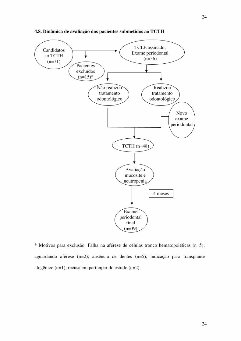
4.8. Dinâmica de avaliação dos pacientes submetidos ao TCTH
* Motivos para exclusão: Falha na aférese de células tronco hematopoiéticas (n=5);
aguardando aférese (n=2); ausência de dentes (n=5); indicação para transplante
alogênico (n=1); recusa em participar do estudo (n=2).
Candidatos ao TCTH
(n=71)
TCLE assinado; Exame periodontal
(n=56)
Não realizou tratamento
odontológico
Realizou tratamento
odontológico
Novo exame
periodontal
TCTH (n=48)
Avaliação mucosite e neutropenia
Exame periodontal
final (n=39)
4 meses
Pacientes excluídos (n=15)*
25
25
__________________________________________________________ 5. ARTIGOS
Artigo 1- “Do candidates for autologous hematopoietic stem cell transplantation
have adequate oral conditions?”, artigo submetido no periódico: Journal of American
Dental Association (JADA).
Artigo 2- “Correlation between Periodontal Status and Complications of
Autologous Hematopoietic Stem Cell Transplantation”, a ser submetido no
periódico Supportive Care in Cancer.
Artigo 3- “Alteração do status periodontal de pacientes submetidos ao transplante
autólogo”, o artigo será traduzido para o inglês e formatado segundo as normas do
periódico Journal of Periodontology.
26
26
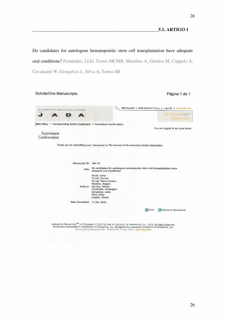
_______________________________________________________5.1. ARTIGO 1
Do candidates for autologous hematopoietic stem cell transplantation have adequate
oral conditions? Fernandes, LLD, Torres MCMB, Maiolino A, Garnica M, Coppelo A,
Cavalcanti W, Gonçalves L, Silva A, Torres SR
27
27
ABSTRACT
BACKGROUND: Oral infections may increase susceptibility to complications in
patients undergoing hematopoietic stem cell transplantation (HSCT). The aim of this
study was to evaluate oral conditions of candidates for HSCT.
METHODS: Candidates for HSCT from two cancer centers received oral exam prior to
the transplant that included: oral mucosa evaluation; number of teeth; DMFT index
(decayed, missing and filled teeth); and full mouth periodontal assessment. Socio-
demographic data, history of dental treatment, and medical data were also collected.
RESULTS: Fifty six patients (median age 52) were evaluated. Myeloma was the main
indication for HSCT (67.9%). Prevalence of gingivitis and periodontitis were 55.4%
and 35.7%; DMFT index was 12.0. Low education level was associated with higher
plaque index, indication for dental extraction, gingivitis and periodontitis, respectively,
p=0.05; p=0.008; p=0.04; p=0.01.
CONCLUSIONS: Oral diseases are highly prevalent in this population and not proper
for patients receiving HSCT. Low education level is associated with deficient oral
hygiene, dental and periodontal diseases.
CLINICAL IMPLICATIONS: Oral assessment and treatment of oral infection must be
performed by dentists prior to HSCT. Knowledge about oral complications during
HSCT may help dental team to properly address this population.
Keywords: Periodontitis, hematopoietic stem cell transplantation, epidemiologic study
characteristics, infection and neutropenia.
Introduction
Oral health conditions may influence the prognosis of patients with hematological
malignancies.1,2 Individuals with odontogenic infections may be more susceptible to
develop mucositis and septicemia, during neutropenic phase associated to

28
28
hematopoietic stem cell transplantation (HSCT).1,3-9 Therefore, candidates for HSCT
must be referred for oral care before conditioning chemotherapy.
Hematopoietic stem cell transplant is a potentially curative procedure for hematologic
malignancies, such as Hodgkin`s disease and lymphoma.10-12 The oral cavity is a
potential site for complications in patients receiving HSCT.1,2 Higher rates of
periodontal disease were observed in patients who developed fever after
HSCT.13 Patients with advanced periodontal disease may undergo bacteremia during
neutropenic phase.2,6 Bacteria found in blood culture of patients submitted to HSCT
may be typically present in the periodontal crevice of individuals with chronic
periodontitis.6
The importance of oral care prior to transplantation was emphasized by several
authors.11,2,14-17 Patients receiving oral care were less likely to develop mucositis,
when compared to patients who had not received basic oral care prior to
chemotherapy or conditioning regime for HSCT.9,18 However, many centers of
reference for cancer and stem cell transplantation offer limited oral care services. The
aim of this study was to evaluate oral status of candidates for HSCT that may
potentially complicate during neutropenia phase related to HSCT.

29
29
Methods
This is a cross sectional study that investigated the oral status of candidates for
autologous HSCT of two centers of reference in Rio de Janeiro, Brazil. Patients from
Clementino Fraga Filho University Hospital at Federal University of Rio de Janeiro
and the Institute of Hematology - Arthur de Siqueira Cavalcanti (HEMORIO) were
evaluated for oral conditions, from October 2008 to November 2009. This study was
approved by the institutional Ethical Committee and each participant signed an
informed consent form prior any study procedure. Candidates for autologous HSCT
were eligible for the study if they matched the following criteria: 18 - 70 years of age;
presenting platelet count above 20,000 platelets per mm3 of blood at the time of
clinical examination, in order to avoid bleeding episodes during periodontal probe; and
having at least six teeth.
Socio-demographic and behavioral data were collected through a structured interview
including: age, gender, economic status (family income per month) and educational
level. Medical history was collected from hospital records. Oral exams were conducted
by one previously trained and calibrated dentist before HSCT procedure.
Clinical examination was performed using a light-emitting diode head light in hospital
bed or dental equipment. Oral evaluation included: oral mucosa structures; number of
teeth; DMFT index (decayed, missing and filled teeth index); and periodontal
examination.
Oral mucosa alterations were diagnosed by clinical characteristics and biopsy, if
needed. Full mouth periodontal examination was conducted at six sites per tooth, using
a millimeter North Carolina periodontal probe (Hu-Friedy®, Chicago, IL, USA) and
included the following clinical measurements: visible plaque, gingival marginal
bleeding, probing depth, clinical attachment level, bleeding on probing and

30
30
suppuration. Gingivitis was diagnosed when gingival bleeding was present on marginal
probing. Periodontitis was defined as a minimum of four sites with a level of
attachment level ≥ 4mm and bleeding on probing in four different teeth. Patients
diagnosed with periodontitis, were excluded from the diagnosis of gingivitis.
Data were presented as descriptive analysis. Categorical and continuous variables were
expressed by percentages and means. Stratified analysis was performed to analyze
differences between socio-demographic factors and oral variables using chi-square and
Mann-Whitney tests, for categorical and non-categorical data, respectively. Levels of
significance were established at 5%.
Results
Seventy-one patients were approached to participate in this study. Fifteen individuals
were excluded mainly due to failure to collect cells for HSCT or for absence of teeth
(Figure 1).
Figure 1 - Flowchart of patients in the study.
PATIENTS ELIGIBLE FOR AUTOLOGOUS HSCT (n=71)
INCLUDED (n=56)
EXCLUDED (n=15)
FAILURE IN COLLECTING CELLS (n=5)
WAITING TO COLLECT CELLS (n=2)
ABSENCE OF TEETH (n=5)
REFERED TO ALLOGENIC TRANSPLANT (n=1)
REFUSE TO PARTICIPATE IN THE STUDY (n=2)
REASONS
FOR EXCLUSION
31
31
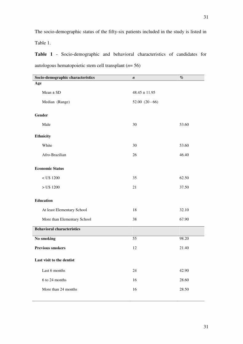
The socio-demographic status of the fifty-six patients included in the study is listed in
Table 1.
Table 1 - Socio-demographic and behavioral characteristics of candidates for
autologous hematopoietic stem cell transplant (n= 56)
Socio-demographic characteristics n % Age
Mean ± SD
Median (Range)
48.45 ± 11.95
52.00 (20 - 66)
Gender
Male
30
53.60
Ethnicity
White
Afro-Brazilian
30
26
53.60
46.40
Economic Status
< U$ 1200
> U$ 1200
35
21
62.50
37.50
Education
At least Elementary School
More than Elementary School
18
38
32.10
67.90
Behavioral characteristics
No smoking
Previous smokers
55
12
98.20
21.40
Last visit to the dentist
Last 6 months
6 to 24 months
More than 24 months
24
16
16
42.90
28.60
28.50

32
32
The underlying disease of patients in the study was mainly multiple myeloma (67.8%)
and Hodgkin's disease (12.5%). In the period of evaluation, 71.4% of patients
presented anemia, with a median count of red cells of 3.78 million/ ml of venous
blood. Clinical characteristics of HSCT candidates are listed in Table 2. Oral findings
are also listed on Table 2. The majority of the autologous HSCT candidates (91.1%)
needed dental treatment. From our sample, 48 patients were submitted to transplant.
Most of the patients had a median of 3.5 days left for dental treatment before HSCT
procedure, and this time available showed a considerable variability (Table 2).
Table 2 - Clinical characteristics of candidates for autologous hematopoietic stem cell
transplant (n= 56)
Clinical characteristics n % Hematologic disease
Myeloma
Hodgkin`s Lymphoma
Non-Hodgkin`s Lymphoma
Amyloidosis
Germ cell tumor
POEMS syndrome
38
07
06
02
02
01
67.89
12.50
10.70
3.60
3.60
1.80
Oral characteristics
Alterations in oral mucosa
Candidiasis
Plasmacytoma
Focal fibrous hyperplasia
7
5
1
1
12.50
8.90
1.80
1.80
Periodontitis 20 35.70
Gingivitis 31 55.40

33
33
Number of teeth
Mean ± SD
Median (Range)
20.69 ± 6.89
24.00 (6 – 28)
DMFT index
Mean ± SD
Median (Range)
13.87 ± 6.58
12.00 (0.00 – 27.00)
Dental treatment need
Yes
51
91.1
Recommended treatment *
Dental extraction
Scaling/ prophylaxis/ oral hygiene instructions
Restorative / Prosthetic
10
51
22
17.90
91.10
39.30
Time between oral exam and HSCT- Days (n=48)
Mean ± SD
Median (Range)
21.79 ± 40.36
3.50 (0.00 – 203.00)
* Patient may be listed more than once
POEMS - Polyneuropathy, organomegaly, endocrinopathy, monoclonal gammopathy
and skin changes; DMFT- decayed, missing and filled teeth index; HSCT –
hematopoietic stem cell transplant.
Detailed results of periodontal parameters evaluated in the study are presented in Table
3.

34
34
Table 3 - Periodontal parameters of candidates for autologous hematopoietic stem cell
transplant (n = 56)
Periodontal parameters Mean ± SD Median (Range)
Visible plaque (% sites) 49.92 ± 21.95 45.90 (4.80 – 91.20)
Gingival marginal bleeding (% sites) 8.69 ± 11.45 4.90 (0.00 – 54.20)
Bleeding on probing (% sites) 20.05 ± 14.80 17.40 (0.00 – 65.80)
Clinical attachment level (mm) 2.54 ± 1.00 2.20 (1.19 – 6.00)
Suppuration (% sites) 0.12 ± 0.93 0.00 (0.00 – 7.00)
Probing depth (mm) 1.90 ± 0.47 1.84 (1.10 – 3.50)
94.50 ± 8.20
97.10 (61.50 – 100.00)
5.13 ± 7.27 2.90 (0.00 – 28.20)
Probing depth (% sites)
1-3 mm
4-6 mm
≥ 7 mm 0.36 ± 1.64 0.00 (0.00 – 10.30)
Clinical attachment level (% sites)
1-3 mm
4-6 mm
≥ 7 mm
81.83 ± 18.80
15.06 ± 13,20
3.09 ± 8.22
88.60 (14.90 – 100.00)
11.20 (0.00 – 45.60)
0.00 (0.00 – 45.60)
All periodontal parameters evaluated showed statistically significant association with
low levels of education (Table 4). The number of missing teeth was significantly
associated with lower level of education and family income.

35
35
Table 4 - Stratified analysis between demographic characteristics and clinical
parameters (n = 56)
Education Economic Status
Clinical
parameters At least
Elementary
School
More than
Elementary
School
P - value
< U$ 1200 > U$ 1200
P - value
n % n % n % N %
Visible plaque
(Mean ; % sites)
18
58,55
38
45.83
0.05*
35
53.02
21
44.74
0.16
Gingival marginal
bleeding
18
14.30
38
6.04
0.01**
35
10.52
21
5.65
0.24
Probing depth
18
2.17
38
1.78
0.01**
35
1.94
21
1.85
0.89
Clinical
attachment level
18
3.07
38
2.29
0.05¥
35
2.62
21
2.40
0.74
Bleeding on
probing
(Mean ; % sites)
18
28.44
38
16.09
0.007**
35
22.28
21
16.35
0.19
Suppuration
18
0.38
38
0.00
0.14
35
0.20
21
0.00
0.43
Periodontitis
11
61.11
09
26.68
0.01¥¥
14
40.00
06
28.57
0.56
Gingivitis
06
33.33
25
65.78
0.04¥
18
51.42
13
61.90
0.58
DMFT
18
15.33
38
13.18
0.26
35
14.11
21
13.87
0.64
Missing teeth
(Mean; % )
18
10.55
38
5.76
0.004**
35
8.97
21
4.52
0.007**
Need for dental
extraction
07
38.88
03
7.89
0.008¥¥
07
20.00
03
14.28
0.72
* p< 0.05 - Mann-Whitney ; ** p< 0.01 - Mann-Whitney;
¥ p< 0.05 - Chi-Square; ¥¥ p< 0.01 - Chi-Square

36
36
Discussion
In this study we found that oral and periodontal conditions of overall candidates for
autologous HSCT were not appropriate, with high rates of visible plaque, gingival
inflammation and dental treatment needs. In addition , patients with low educating
degrees were those with the worst oral conditions. Other studies that evaluated oral
status of cancer patients have also identified precarious conditions with need for
extensive dental treatment.9,19-21 Oral care may prevent complications during cancer
treatment, especially during neutropenic period.2,11,22-24
Gingivitis was frequent in the studied population, as 55.4% of individuals presented
marginal gingival inflammation. Patients with hematological cancer from other studies
presented gingivitis ranging from 15% to 25%.13,25 Higher gingival index was reported
by Ellegard et al.,19 who found an mean of 72% of bleeding on probing. The reasons
for this discrepancy might be differences in sample size and the criteria for diagnosing
gingivitis.
Bacteremia has been the subject of several studies and it is linked to poor prognosis of
patients exposed to HSCT.5,26-28 The correlation between periodontal bleeding on
probing and bacteremia has been observed in some studies.29,30 In our study, the
individuals showed 20.05% of the sites with bleeding on probing.
Gingival marginal bleeding was found in 8.69% of the sites in our studied population,
which was lower than other populations.25 Patients with multiple myeloma frequently
present anemia and it has been reported that patients with anemia may show a pale
gingival mucosa.31,32 The median count of red cells of 3.78 million/ ml of venous blood
may explain why the studied population presented less marginal gingival bleeding.
Periodontitis was a common finding (35.7%) among candidates in the present study.
However, they presented low severity of periodontal disease (94.5 % of sites presented
shallow pockets). On the other hand, in a study held in Australian patients prior to

37
37
HSCT, the investigators found 61% of patients with moderate and shallow pockets.25
Geographical variations might explain these differences, since they used the same
methodology of the present study. Despite these observations, recent epidemiological
surveys performed in a Brazilian population from the same area of the country, have
shown periodontal diseases prevalence varying from 13% to 65%.33 - 35.
Prevalence of periodontitis in cancer patients varied between studies from different
countries. In a North American research of patients submitted to HSCT, periodontitis
was observed in 13.7% of patients, through radiographic exam of the alveolar crest.6
The assessment of periodontal disease through radiographic exam is limited because it
may not access all sites that may be affected by periodontitis. In a Japanese study,
conducted in 41 patients prior to HSCT, it was observed some degree of periodontitis
in 5% of the patients, although the criteria used in the study was unclear, because the
values of probing depth were not described.36
Plaque control is important in all periods of HSCT, especially during the
hospitalization.2 However, authors have pointed poor integration of dental and medical
staff and a lack of awareness about the need for brushing and flossing in these
patients.9,18 In the present study, visible plaque was observed in 45.9% of the sites.
Instruction for plaque control was conducted for leukemia patients admitted for
chemotherapy of a special program in Denmark, and a reduction in bleeding index was
observed during hospitalization period.19 These findings reinforce the need of dental
staff integrating the supportive care team for cancer patients.
DMFT in the present study showed a mean value of 13.8, similar to another Brazilian
study developed in candidates to HSCT that reported a mean of 17.21 In a Brazilian
survey of general population in the same area of the country, it was reported a DMFT

38
38
of 20.3 in an age group of 35 to 44 years of age.34 These data might reflect the DMFT
status of patients in this geographic area.
The majority of candidates (91.1%) for HSCT in this study needed dental treatment,
despite the fact that, 42.9% of these patients had visited a dentist in a period of six
months prior to HSCT. Dental treatment indications were mainly for oral hygiene
instructions and scaling (91.1%), extractions (17.9%), and restorative and prosthetics
procedures (39.3%). Other studies that evaluated cancer patients to assess oral status
have also identified precarious oral conditions with need for extensive dental
treatment.9,19,20 An evaluation conducted in Israel with patients before HSCT has
pointed out that the most commonly needed treatment was scaling (47.8%) and
restorations (39.1%).37 Another study developed in North America, which evaluated
oral conditions in 61 patients prior to allogeneic HSCT reported that 47% of the
subjects needed extractions.20 In a sample of Swiss individuals who were evaluated
prior to HSCT, there were 77% subjects with indication for dental extraction.15
Patients in the present study presented high prevalence of dental plaque and number of
missing teeth. Low economic status was predominant (62.5%) in the studied
population, but educational status was the parameter that has mainly influenced
periodontal status. This was expected since educated individuals have a better
understanding about health and prevention.
The time limitation for dental visit before HSCT is a factor that contributes to the
inability to perform dental treatment.9 The median number of days between the dental
visit and HSCT in patients of the studied population was 3.5 days. Other authors have
reported an average of 11 to 21 days left for dental treatment before HSCT.20,37 The
available time to perform dental procedures was limited and insufficient for an ideal
treatment planning and healing.9,22

39
39
The main limitation in the present study was the lack of a radiograph exam, which may
have underestimated the need for extractions and for dental fillings.
It may be concluded that the oral status of individuals eligible for HSCT in the studied
population is not appropriated. Oral care should be included in the protocol prior to
HSCT procedure. Dentists have a role to play providing good oral care for cancer
patients. To our understanding there is a need for a closer integration between health
professionals to provide an efficient care to candidates for HSCT.
Further studies should be conducted in order to assess the measures that may establish
better oral conditions for these patients.

40
40
References
1. Wingard JR. Antifungal chemoprophylaxis after blood and narrow
transplantation. Clin Infect Dis 2002;34:1386-90.
2. National Institute of Health: Consensus Development Panel. Consensus
statement: Oral complications of cancer therapies: diagnosis, prevention and treatment.
NCI Monogr 1990; 9:3-8.
3. Gillespie T, Masterton RG. Investigation of infection in the neutropenic patient
with fever. Journal of Hospital Infection 1998; 38: 77-91.
4. Donnelly JP. Infection in the neutropenica and haematopoietic stem cell
recipient. Curr Opin Infect Dis 2000; 13: 337-42.
5. Collin BA Leather HL, Wingard JR, Ramphal R. Evolution, incidence, and
Susceptibility of bacterial Bloodstream Isolates from 519 Bone Marrow Transplants
Patients. Clinical Infectious Diseases 2001; 33: 947-53.
6. Akintoye SO, Brennan MT, Graber CJ, McKinney BE, Rams TE, Barrett AJ,
Atkinson JC. A retrospective investigation of advanced periodontal disease as a risk
factor for septicemia in hematopoietic stem cell and bone marrow transplant recipients.
Oral Surg Oral Med Oral Pathol Oral Radiol Endod 2002; 94(5): 581-8.
7. Auner HW, Sill H, Mulabecirovic A, Linkesch W, Krause R. Infectious
complications after autologous hematopoietic stem cell transplantation: comparison of
patients with acute myeloid leukemia, malignant lymphoma, and multiple myeloma.
Ann Hematol 2002; 81(7): 374-7.
8. Dettenkofer M, Ebner W, Bertz H, Babikir R, Finke J, Frank U, Ruden H,
Daschner. Surveillance of nosocomial infections in adult recipients of allogeneic and
autologous bone marrow and peripheral blood stem-cell transplantation. Bone Marrow
Transplant 2003; 31(9): 795-801.
41
41
9. Djuric M, Hillier-kolarov V, Belic A, Jankovic L. Mucositis prevention by
improved dental care in acute leukemia patients. Support Care Cancer 2006; 14(2):
137-46.
10. Zago. Hematologia, fundamentos e prática. 1 ed. Rio de Janeiro: Atheneu;
2004.
11. Goldman KE. Dental management of patients with bone marrow and solid
organ Dent Clin North Am. 2006; 50(4): 659-76.
12. Epstein JB Raber-Durlacher JE. Advances in hematologic stem cell transplant:
An update for oral health care providers. Oral Surg Oral Med Oral Pathol Oral Radiol
Endod 2009; 107 (3): 301 - 12.
13. Laine PO Lindqvist JC, Pyrhönen SO, Strand-Pettinen IM, Teerenhovi LM,
Meurman JH. Oral infection as a reason for febrile episodes in lymphoma patients
receiving cytostatic drugs. european Journal of Cancer B Oral Oncol 1992; 28B(3):
103-07.
14. Overholser CD, Peterson DE, Williams LT, Schimpff SC. Periodontal infection
in patients with acute nonlymphocyte leukemia. Prevalence of acute exacerbations.
Arch Inter Med 1982; 142(3): 551-54.
15. Heimdahl A Mattson T, Dahllöf G, Lönnquist B, Ringdén O. The oral cavity as
a port of entry for early infections in patient treated with bone marrow transplantation.
Oral Surg Oral Med Oral Pathol 1989; 68(6): 711-16.
16. Donker AE Van Merkesteyn JP, Bredius RG, Van Weel-Sipman MH. Value of
panoramic radiographs in paediatric pre-bone marrow transplantation oral evaluation.
Int J Oral Maxillofac Surg 2002; 31(2): 170-72.
17. Castro CG Gregiani LJ, Brunetto AL. Bone marrow transplantation and cord
blood transplantation in children. J. Pediatric 2001; 77(5): 345-60.
42
42
18. Borowski B, Benhamou E, Pico JL, Laplanche A, Margainaud JP, Hayat M.
Prevention of oral mucositis in patients treated with high-dose chemotherapy and bone
marrow transplantation: a randomised controlled trial comparing two protocols of
dental care. European Journal of Cancer 1994: 93 -97.
19. Ellegard B, Bergman OJ, Ellegard J. Effect of plaque removal with acute
leukemia. J Oral Pathol Med 1989; 18: 54-58.
20. Graber CJ, Almeida KNF, Atkinson JC, Javaheri D, Fukuda CD, Gill VJ,
Barret AJ, Bennett JE. Dental health and viridans streptococcal bacteremia in
allogeneic hematopoietic stem cell transplant recipients. Bone Marrow Transplantation
2001; 27: 537-42.
21. Shcaira V, Boer C, Basso F, Kellermann M, Correa ME. Oral Healthy
Characteristics And DentalManagement Of Hematopoietic Stem Cell Transplantation
Patients In A Single Public Hospital. Paper presented at: Multinational Association for
Supportive Care in Cancer (MASCC ), 2009; Rome, Italy.
22. Barker GJ. Current practices in the oral management of the patient undergoing
chemotherapy or bone marrow transplant. Support Care Cancer. 1999; 7: 17 - 20.
23. Epstein JB Chow AW. Oral complications associated with immunosupression
and cancer therapies. Infectious Disease Clinics of North America 1999; 13 (4):901-23.
24. Barasch A, Coke JM. Cancer therapeutics: an update on its effects on oral
health. Periodontol 2000. 2007; 44: 44-54.
25. Pattni R Walsh LJ, Marshall RI, Cullinan MP, Seymour GJ, Bartold PM.
Changes in the periodontal status of patients undergoing bone marrow transplantation.
J Periodontol 2000; 71(3): 394-402.
26. Salazar R, Solá C, Maroto P, Tabernero JM, Brunet J, Verger G, Valentí V,
Cancelas JÁ, Ojeda B, Mendoza L, Rodríguez M, Montesinos J, López-López JJ.

43
43
Infections complications in 126 patients treated with high-dose chemotherapy and
autologous peripheral blood stem cell. Bone Marrow Transplant 1999; 23: 27-33.
27. Frère P, Hermanne JP, Debouge MH, de Mol P, Fillet G, Beguin Y. Bacteremia
after hematopoietic stem cell transplantation: incidence and predictive value of
surveillance cultures. Bone Marrow Transplant 2004; 33 (7): 745-9.
28. Oliveira AL, Souza M, Carvalho-Dias VMH, Ruiz MA, Silla L, Tanaka PY,
Simões BP, Trabasso P, Seber A, Lotfi CJ, Zanichelli MA, Araújo VR, Godoy C,
Maiolino A, Urakawa P, Cunha CA, Souza CA, Pasquini R, Nucci M. Epidemiology
of bacteremia and factors associated with multi-drug-resistent gram-negative
bacteremia in hematopoietic stem cell transplant recipients. Bone Marrow
Transplantation 2007: 1-7.
29. Daly CG, Mitchell DH, Highfield JE, Grossberg DE, Stewart D. Bacteremia
due to periodontal probing: a clinical and microbiological investigation. J Periodontol
2001; 72 (2): 210-4.
30. Demmer RT, Papapanou PN, Jacobs Jr DR, Desvarieux M. Evaluating clinical
periodontal measures as surrogates for bacterial exposure: The Oral Infections and
vascular Disease Epidemiology Study. BMC Medical Research Methodology, 2010;
10(2).
31. Kyle RA, Rajkumar SV. Treatment of multiple myeloma: a comprehensive
review. Clin Lymphoma Myeloma. 2009; 9(4): 278-88.
32. Proctor R, Kumar N, Stein A, Moles D, Porter S. Oral and dental aspects of
chronic renal failure. Journal of Dental Research 2005; 84(3): 199-208.
33. Susin C, Valle P, Oppermann RV, Haugejorden O, Albandar, JM. Ocurrence
and risk indicators of increased probing depth in an adult Brazilian population. Journal
of Clinical Periodontology 2005; 32 (2): 123 - 29.
44
44
34. Ministry of Health Project SB Brazil. Condições de Saúde bucal da população
brasileira 2002-2003 – Resultados Principais. Brasília – Ministério da saúde 2004.
35. Pion FLB Araújo MWB, Feres M, Cortelli SC. Periodontal status of a
population subgroup from Guarulhos, SP. Rev. Brasileira Epidemiologia 2006; 9 (3):
335-45.
36. Yamagata K, Onizawa K, Yanagawa T, Hasegawa Y, Kojima H, Nagasawa T,
Yoshida H. A prospective study to evaluate a new detal management protocol before
hematopoietic stem cell transplantation. Bone Marrow Transplant 2006; 38: 237 - 42.
37. Elad S, Garfunkel AA, Or R, Michaeli E, Shapira MY, Galili D. Time
limitations and the challenge of providing infectionpreventing dental care to
hematopoietic stem-cell transplantation patients. Support Care Cancer 2003;11:674-77.
45
45
________________________________________________________5.2. ARTIGO 2
Correlation between Periodontal Status and Complications of Autologous
Hematopoietic Stem Cell Transplantation
Fernandes, LLD, Torres MCMB, Maiolino A, Garnica M, Coppelo A, Cavalcanti W,
Gonçalves L, Silva A, Torres SR
Abstract
Purpose: Oral infection may be a source of bacteremia in patients undergoing
hematopoietic stem cell transplant (HSCT). The aim of this study was to test the
hypothesis that patients showing poor periodontal status present more complications
after HSCT.
Methods: Candidates for autologous HSCT were included in this cohort study.
Patients were excluded if less than 18 years; less than 6 teeth; and platelet counts were
below 20,000/mm3. Exams included: number of teeth; decayed, missing and filled
teeth index (DMFT); oral mucosa and full mouth periodontal assessment. Socio-
demographic and medical data were collected. After HSCT, febrile neutropenia was
classified as fever of unknown origin, bacteremia and clinically documented. Oral
mucosa was evaluated for the severity of mucositis.
Results: Forty-eight candidates for autologous HSCT were evaluated prior to HSCT
and during hospitalization. Seventy percent of patients presented myeloma. Median
DMFT was 13; periodontitis was diagnosed in 29.2%, and gingivitis in 60.4% of the
patients. After HSCT, fever occurred in 95.8% of patients as fever of unknown origin
(60.4%), bacteremia (29.2%) and clinically documented fever (6.2%). Coagulase-
negative Staphylococcus spp. were the most common microorganism. Although there
was no statistical significance, all the oral parameters evaluated showed worst oral

46
46
status for individuals who presented bacteremia when compared with those with no
bacteremia. Bleeding on probing over 22% was higher in patients with fever with
bacteremia (p=0.09). Candidiasis occurred in 16.7% of patients after HSCT, and it was
associated to use of dentures (p=0.01) and candidiasis at baseline (p=0.01). Oral
mucositis affected 89.6% of patients and its severity was correlated with the presence
of gingivitis at baseline (p=0.05).
Conclusions: Baseline oral status of patients submitted to autologous HSCT was worst
in patients with bacteremia than in patients that did not present bacteremia.
Key-words: hematopoietic stem cell transplant; bacteremia; fever; periodontal disease;
oral condition.
47
47
Introduction
Patients submitted to hematopoietic stem cell transplant (HSCT) go through a
period of myelosuppression with severe neutropenia [1-4] and are more
susceptible to infection. [5-10] Microbiological sites of the oral cavity may
represent a source for infection. [11-22]. Febrile neutropenia may be associated
with pre-existing infections in dental related tissues, such as gingivitis and
periodontitis. [11, 15] Periodontal disease reflects an inflammatory response to
bacteria that colonize the surface of the tooth. Periodontitis is characterized by
progressive gingival sulcus depth resulting in a pocket. Advanced periodontitis is
characterized by loss of support and may result in mobility and eventual loss of the
involved teeth. [14, 23] Periodontitis has been suggested as a source of bacteremia in
patients submitted to HSCT. [21]
Fever during myelosuppression post-HSCT may represent a sign of infection and
occurs in 63 to 98% of patients. [8, 24-26] Some cases of fever are well documented
infections, but for most of the cases there is no related etiologic factor. [25, 26]
Bacteremia is a complication that potentially leads to morbidity and mortality, and may
occur in 20% to 50% of individuals submitted to HSCT. [2, 6, 8, 25, 27] Bacteremia
usually does not have a good response to empirical antimicrobials, when compared to
the response of individuals with fever of unknown origin. [8, 27]
Risk factors for infection in HSCT recipients include medical conditions, central
venous catheter, chronic infections, lack of antimicrobial prophylaxis, and loss of
epithelium barrier. [18, 32-34] The exposure of connective tissue that occurs in
mucositis and periodontitis may represent a flaw in local defense. Advanced
periodontitis may expose up to 35cm2 of connective tissue, increasing the susceptibility
for bacteremia. [35] The occurrence of mucositis may change the prognosis of patients

48
48
and duration of hospitalization, leading to inability to eat, use of high doses of opioids
and occurrence of bacteremia. [34, 36] Oral care protocol may reduce complications in
patients who undergo chemotherapy or HSCT. [1, 17, 21, 22, 34, 37-40] The aim of
this study is to test the hypothesis that patients showing poor periodontal status
demonstrate more complications after HSCT.
Patients and Methods
Candidates for autologous hematopoietic stem cell transplant from two reference
centers of cancer treatment were included in this study. Patients from Hospital
Clementino Fraga Filho at Federal University of Rio de Janeiro Hospital (UFRJ) and
Institute of Hematology - Arthur de Siqueira Cavalcanti (HEMORIO) were evaluated
for oral conditions from September 2008 to January 2010. This study was approved by
both institutional ethics committee. Patients would be excluded if they were younger
than 18 years old; if they presented less than 6 teeth; and if platelet count was less than
20,000 per mm3 of blood.
Demographics data and clinical features such as the underlying disease, comorbidities,
antimicrobial prophylaxis, and conditioning regimen for HSCT were collected from
medical records. When the same patient underwent more than one autologous HSCT,
only one procedure was included in this assessment.
Oral exam was conducted before HSCT procedure, by a trained and calibrated dentist.
The exam was performed using a frontal light-emitting diode lamp at hospital bed or
dental office. The evaluation included the conditions of oral mucosa, dental status and
periodontal structures.
Oral mucosa manifestations were diagnosed by clinical characteristics and biopsy, if
needed. The number of present teeth, and DMFT index (decayed, missing and filled

49
49
teeth) were assessed for all patients. Indication for dental treatment, such as dental
extraction, scaling/prophylaxis or restorative/prosthetic, was determined during oral
evaluation.
Periodontal examination was conducted using a millimeter North Carolina periodontal
probe (Hu-Friedy©, Chicago, IL, USA). Clinical periodontal parameters were
evaluated at six sites per tooth. The following clinical measurements were obtained
under standard conditions: visible plaque (presence or absence), gingival marginal
bleeding (presence or absence), probing depth, clinical attachment level, bleeding on
probing (presence or absence of bleeding on probing) and suppuration (presence or
absence on probing). Probing depth was defined as the distance from the free gingival
margin to the bottom of the pocket/sulcus. The clinical attachment level was calculated
from the cemento-enamel junction to the bottom of the pocket/sulcus including
recession measurements. All measurements were considered in millimeters. Probing
depth was categorized in shallow pockets from 0 to 3 mm, moderate pockets from 4 to
6 mm, and deep pockets ≥7 mm. Clinical attachment loss was also classified in shallow
pockets 0 to 3 mm, moderate from 4 to 6 mm, and deep ≥7 mm.
Gingivitis was diagnosed when gingival bleeding was present on marginal probing.
Periodontitis was defined as a minimum of four sites with a level of attachment level ≥
4mm and bleeding on probing in four different teeth. Patients diagnosed with
periodontitis, were excluded from the diagnosis of gingivitis.
Mucositis was diagnosed by clinical aspects of oral exam and measured using “Oral
Mucosal Rating Scale”, one to three times a week in the post transplant period, while
patient remained hospitalized and considered an object in this study. [42]
Occurrence of fever and results of blood culture were collected from medical records.
Neutropenia was defined as neutrophil counts less than 500 cells x 109/l, and fever as
50
50
an axillary temperature ≥37.8ºC. [26] Episodes of febrile neutropenia were
classified according to adapted definitions of the Immunocompromised Host
Society as: fever of unknown origin; microbiologically documented fever, without
bacteremia; clinically documented fever; and fever with bacteremia. Bacteremia was
defined when any bacteria was isolated and identified in blood culture exam. [41, 26]
All statistical analyses were performed using a commercial statistical program, SPSS
16.0© (Statistical Package for the Social Sciences for Windows, SPSS Inc., Chicago,
IL, USA). The significance level established for all analysis was 5%. Differences in
categorical data were assessed by Fisher’s exact test and chi-square test. Mann-
Whitney or Kruskal-Wallis tests were used for the association of categorical and
continuous variables, when appropriated. Categorical and continuous variables were
expressed by percentages, means and medians. Studies that analyze periodontal data
always use means. For better comparison between studies, the results of periodontal
parameters are showed in means and medians, although data were analyzed through
non-parametric tests.
Results
A total of 71 patients were admitted for HSCT in both institutions and were
invited to participate in the study. Reasons for the exclusion of 23 patients were
presence of less than 6 teeth (n=5); refusal to take periodontal exam (n=2); and
not being prepared to receive HSCT procedure (n=16). Patients did not receive
the infusion due to failure to collect enough cells for autologous transplantation,
referral to allogeneic transplantation, obit and unavailable time for the transplant
procedure by data collection. Forty-eight patients submitted to autologous

51
51
HSCT met the inclusion criteria for this study. The majority of patients (83.3%)
were from Hospital Clementino Fraga Filho at Federal University of Rio de Janeiro
Hospital (UFRJ). The mean age was 48.6 ± 11.7, ranging from 20 to 66 years of
age. Fifty percent of individuals were males. Considering ethnicity, 54.2% of
the candidates were white, and 45.8% were non-whites. In 70.8% of the
autologous HSCT recipients, the underlying disease was multiple myeloma followed
by Hodgkin lymphoma (12.5%). Regarding comorbidities, 70.8% of individuals were
anemic, with a mean of 3.73 million/ml of red cells at the time of oral exam. There
were 25% of the patients with a history of hypertension, 12.5% with renal failure and
with 4.2% chronically obstructive pulmonary disease.
The chemotherapy conditioning regimen was mainly melphalan (75%) followed by the
regime cyclophosphamine, BCNU (carmustine) and VP-16 (etoposide) (CBV)
(12.5%). Regarding prophylaxis, acyclovir was the antiviral drug used in 93.8% of the
patients, ciprofloxacin was the antibacterial drug used in 85.4%, and the antifungal
drug fluconazole was given to 50% of patients.
The baseline oral characteristics are listed on Table 1 and the complications of HSCT
on Table 2. Febrile neutropenia occurred in 46 (95.8%) patients and 63.0% of
these cases were fever of unknown origin. Bacteremia occurred in 30.4% of the
individuals and the most frequent microorganisms identified were coagulase-
negative Staphylococcus spp. (8.3%), Enterobacter cloacae (6.2%), Klebsiella
pneumoniae (6.2%) and Staphylococcus aureus (4.2%). Cefepime was the
empiric antibiotic used in all cases of fever and in 21.7% of the cases
vancomycin was associated. Almost all patients (97.9%) used granulocyte
colony-stimulating factor (G-CSF).

52
52
Regarding the correlation of periodontal parameters and classification of febrile
neutropenia, it was observed that visible plaque was found in 56.4% (median 55.7) of
the sites in patients presenting bacteremia, 47.8% (median 45.1) in patients with fever
of unknown origin, 37.0% (median 38.9) in patients with clinically documented fever,
and 36.2% (median 36.2) in patients with no fever, although there was no statistically
significant difference between these groups (p=0.46). We also found that bleeding on
probing was observed in 25.5% (median 24.8) of the sites in patients with bacteremia,
17.3% (median 17.40) in patients with fever of unknown origin, 12.3% (median 13.90)
in clinically documented fever, and 11.9% (median 11.9%) in patients with no fever
(p=0.31). All the clinical periodontal parameters and DMFT were lower, except
probing depth, in patients with no febrile neutropenia, such as: marginal gingival
bleeding, (0.7% of sites), clinical attachment level (mean 2.0), number of teeth lost (2.0
teeth) and DMFT index (9.0). Gingivitis, periodontitis and probing depth also did not
show statistically significant difference between patients with different types of fever.
There were no differences between the groups of patients presenting different types of
febrile neutropenia and the number of teeth or DMFT index. The necessity of dental
treatment was not different between patients with different types of febrile neutropenia.
Regarding the correlation of periodontal parameters and febrile neutropenia, it was
observed that patients with bacteremia showed higher values for all the periodontal
parameters than individuals with no bacteremia, although not statistically significant
(Table 3). When a cut-off point of 22% of sites with bleeding on probing was
established, a statistic tendency correlation with bacteremia was found (p=0.09).
Some grade of oral mucositis (median score 8; range 2 – 25) was observed in 43
(89.6%) of the HSCT patients included in the study, and lasted for a median of 6 days
(ranging from 2 to 12 days). The score and duration of mucositis according to

53
53
febrile neutropenia classification is demonstrated on Table 2. Regarding the
correlation between periodontal status and mucositis, all patients with
periodontitis (n=14) developed mucositis. More over, the presence of gingivitis
at baseline (n=29) was significantly correlated with the occurrence of mucositis
after HSCT (p=0.05). The majority of patients, who presented mucositis
(88.4%), needed some sort of dental treatment such as restorations/prosthetics
(94.7%), scaling/prophylaxis or oral hygiene instructions (88.4%) and dental
extractions (16.0%).
Other oral complications during hospitalization period for HSCT recorded in this
population were candidiasis (16.7%), pericoronitis (4.2%) and ecchymosis (2.1%).
Candidiasis after HSCT occurred in 75% patients who presented candidiasis at baseline
(p=0.007). Forty-five percent of the eleven patients that had dentures developed
candidiasis after HSCT (p=0.01). In the 24 patients who had prophylaxis with
fluconazole, 12.5% developed candidiasis (p=0.70).

54
54
Table 1 – Oral characteristics of the patients before hematopoietic stem cell
transplant procedure: (n= 48)
Oral Characteristics n % Candidiasis 4 8.30
Plasmacytoma 1 2.10
Focal fibrous hyperplasia 1 2.10
Gingivitis 29 60.40
Periodontitis 14 29.20
Mean ± SD Median (Range)
Number of teeth 20.79 ± 7.00 24.00 (6.00 – 28.00)
DMFT index 14.37 ± 6.60 13.00 (0.00 – 27.00)
Visible plaque (% of sites) 49.19 ±
21.50
44.66 (4.80 – 90.20)
Gingival marginal bleeding (% of
sites)
8.11 ± 11.10 4.55 ( 0.00 – 54.20)
Probing depth in mm (Mean ± SD) 1.84 ± 0.40 1.80 (1.10 – 3.00)
Pocket depth
Shallow pockets (% of sites)
Moderate pockets (% of sites)
Deep pockets (% of sites)
95.71 ± 6.10
4.09 ± 5.70
0.18 ± 1.00
97.35 (72.50–
100.00)
2.65 (0.00 – 26.80)
0.00 (0.00 – 6.90)
Bleeding on probing (% of sites) 19.13 ±
13.70
17.05 (0.00 – 61.10)
Clinical attachment level (Mean ± SD) 2.43 ± 0.70 2.43 (1.10 – 4.60)
DMFT= decayed, missing and filled teeth

55
55
Table 2 – Relationship of fever classification with clinical data during
hospitalization in 46 patients who presented fever
Clinical data of
hospitalization
period Median
(Range)
Fever of
unknown
origin (n=
29)
Clinically
documented
fever (n=03)
Bacteremia
(n= 14)
p-
valu
e**
Duration of
neutropenia (days)
5.00 (1 – 10) 6.50 (5 – 8) 5.00 (1 –
11)
0.77
Duration of fever
(days)
5.00 (1 – 9) 2.00 (0 – 3) 5.00 (3 –
22)
0.03
Time of
antimicrobials use
(days)
6.00 (2 – 12) 8.00 (7 – 12) 8.00 (5 –
16)
0.02
Time to engraftment
(days)
12.00 (4 – 14) 13.00 (12 –
13)
11.50 (8 –
15)
0.31
Score of mucositis* 12.00 (4 - 25) 8.00 (4 – 8) 6.00 (2 –
23)
0.33
Duration of
mucositis* (days)
6.00 (2 – 11) 4.00 (2 – 5) 7.00 (2 –
12)
0.18
SD: standard deviation
* Evaluated in 43 patients who presented mucositis; **Kruskal- Wallis
Table 3 – Comparison of oral conditions between individuals who presented or
not bacteremia after hematopoietic stem cell transplantation
Oral Conditions Bacteremia
(n = 14)
no bacteremia
(n = 32)
p-
value*
Number of teeth (Mean ± SD) 19.14 ±
8.30
21.38 ± 6.55 0.57.
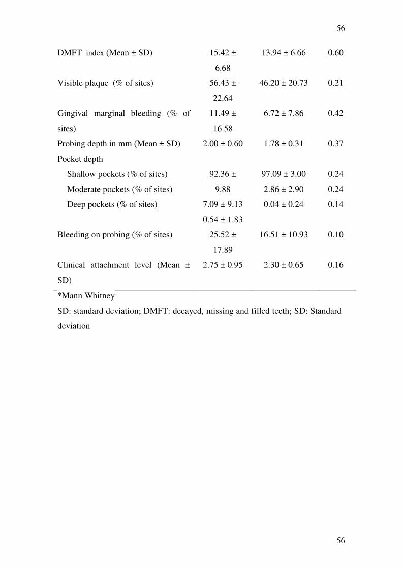
56
56
DMFT index (Mean ± SD) 15.42 ±
6.68
13.94 ± 6.66 0.60
Visible plaque (% of sites) 56.43 ±
22.64
46.20 ± 20.73 0.21
Gingival marginal bleeding (% of
sites)
11.49 ±
16.58
6.72 ± 7.86 0.42
Probing depth in mm (Mean ± SD) 2.00 ± 0.60 1.78 ± 0.31 0.37
Pocket depth
Shallow pockets (% of sites)
Moderate pockets (% of sites)
Deep pockets (% of sites)
92.36 ±
9.88
7.09 ± 9.13
0.54 ± 1.83
97.09 ± 3.00
2.86 ± 2.90
0.04 ± 0.24
0.24
0.24
0.14
Bleeding on probing (% of sites) 25.52 ±
17.89
16.51 ± 10.93 0.10
Clinical attachment level (Mean ±
SD)
2.75 ± 0.95 2.30 ± 0.65 0.16
*Mann Whitney
SD: standard deviation; DMFT: decayed, missing and filled teeth; SD: Standard
deviation

57
57
Discussion
In the present study detailed information at baseline oral status of patients undergoing
autologous HSCT was collected and correlated to complications following transplant.
Interestingly, all the oral parameters evaluated demonstrated that individuals who
presented bacteremia showed worst oral status than individuals with no bacteremia.
However, these results were not statistically significant. Bleeding on probing and
visible plaque parameters were higher in patients presenting bacteremia. The
association of bleeding on probing and bacteremia was reported in patients with
cardiovascular diseases. [28] More studies must be performed to verify this association
in populations submitted to HSCT.
It was observed that patients with bacteremia presented higher percentage of sites with
visible plaque than patients with other types of fever classification. To our knowledge,
studies verifying this association have not been published. Multicenter studies, with
larger samples of HSCT patients are necessary to confirm if higher levels of dental
plaque are related to bacteremia in this population.
One might expect that marginal gingival bleeding would be significantly higher in this
patients (n=48). Actually, the prevalence of marginal gingival bleeding was low in this
population (mean 8.11%). Maybe anemia might justify this finding, as this comorbidity
affected 70.8% of individuals in the studied population. Patients with anemia usually
present pale gingival mucosa. [43,44]
Periodontitis leads to a great exposure of the connective tissue. Despite the fact that the
studied population did not present severe periodontitis, the prevalence of periodontitis
was 35.7% in individuals with bacteremia and 28.1% in individuals with no
bacteremia. Perhaps, the reason for the non statistically significant correlation of
periodontitis and bacteremia could be the fact that there were more than 90% of the
58
58
sites presenting shallow pockets. Maybe patients with severe periodontal disease may
present higher risk for bacteremia. This hypothesis should be reinforced by studies in
populations with more severe periodontal disease. Akintoye et al. [21] did not find a
correlation between periodontal status and the occurrence of bacteremia, but they
evaluated patients through radiograph examination only, so it may have limited value
to periodontitis diagnosis.
The incidence of bacteremia (29.2%) was similar to other studies that reported a range
of 20% to 50%. [2, 6] The most common isolated bacteria in blood cultures were
coagulase-negative Staphylococcus spp., similar to the majority of studies that reported
Gram-positive isolates. [2, 9, 25-27] Despite these bacteria are not typically isolated
from periodontal or oral environment, they may be opportunistic in dental plaque. [21]
The hospital environment can probably be the source of these microorganisms.
Episodes and duration of febrile neutropenia (Table 2) were similar to other studies. [8,
24-26] Fever of unknown origin was the most frequent type of fever (60.4%), like
other authors who reported it ranging from 49% to 63%. [8, 24-26] Fever of unknown
origin may be related to infection, or as a consequence of engraftment of cells, result of
underlying disease, medications or transfusions [14]. After bacteremia, the highest
percentage of visible plaque and bleeding on probing were in patients with fever of
unknown origin. On the other hand, all the periodontal and DMFT values were
inferior, except probing depth, in patients with no febrile neutropenia, nevertheless,
just two patients have not showed fever. Maybe a significant level would be achieved
in studies with larger samples.
The duration of neutropenia may interfere with patient outcome. The predisposition for
infection increases when neutrophil count is below 500 cells per mm3 of blood. In the
population studied, patients presented neutropenia with a median duration of 6
59
59
days after HSCT. Some studies have reported longer periods of neutropenia
ranging from 8 to 11 days. [2, 8, 9, 25]. This may be explained by the fact that the
majority of patients were in the present study were under melphalan (74.5%) and
granulocyte colony stimulating factor (G-CSF) regime. It has been reported that the use
of this regime has reduced the period between the infusion and the engraftment of
cells. [45, 46]
In the present study 85.4% of patients used prophylatic ciprofloxacin. Controversial
results have been reported on the efficacy of quinolone prophylaxis for Gram-negative
pathogens in HSCT. According with Oliveira et al. 2007, the use of quinolone
prophylaxis against Gram-negative pathogens decreased in 85% the risk of developing
fever after HSCT. [26] However, other studies show no differences in the prognosis of
patients under quinolone prophylaxis. [9] Maybe, a shift in the type of microorganisms
might occur, without a clinical benefit. [6]
The chemotherapy received previous to HSCT, as well as all underlying conditions and
medications administered to cancer patients increase the predisposition to
complications and opportunistic infections [18, 46-49], such as fungal infections [7]
Oral candidiasis is a common finding in onco-hematologic patients [1, 13, 50, 51] and
was present in 8.3% of patients at present study baseline. Half of the patients who
underwent autologous HSCT procedure used fluconazole for fungal prophylaxis.
However, oral candidiasis occurred in 16.7% of patients during neutropenic period
after HSCT, despite fluconazole prophylaxis. This may be explained by the fact that
the use of fluconazole does not eliminate colonizing cepas. [54] The three patients who
developed candidiasis with antifungal prophylaxis presented oral candidiasis at
baseline and wore dentures. There was a statistical association between the onset of
candidiasis after HSCT and the use of dentures in the studied population. The relation

60
60
between candidiasis and the use of dentures is commonly reported. [52, 53] This might
emphasize the intensity of special oral care for those patients wearing dentures.
Mucositis was a common event in the studied patients submitted to HSCT (89.6%) and
occurred in 85.7% of patients with bacteremia. However, there was no correlation of
bacteremia and the occurrence and severity of mucositis. All patients with periodontitis
developed mucositis after HSCT and 88.4% of the patients with mucositis needed
some kind of dental treatment in the evaluation prior to transplant. Recent studies
reported a relationship between oral status and the occurrence of mucositis in patients
submitted to chemotherapeutic regimes or HSCT. [17, 38, 39, 56, 57] Interestingly,
patients with gingivitis showed significant correlation with mucositis after HSCT,
however, there was no such correlation with severity and duration of mucositis. In the
population of the present study there was no statistical significance between other oral
parameters prior to HSCT and occurrence, severity or duration of mucositis.
Besides oral manifestations, there are many variables that should be analyzed, in
order to evaluate the etiological factor for febrile neutropenia after HSCT. The
purpose of the present study was to verify the association between oral status
and the complications of HSCT. Patients with bacteremia presented poorer
dental and periodontal status than patients with no bacteremia; nevertheless, this
was not statistically significant. More studies must be conducted in similar
populations in order to confirm this statement.
61
61
References
1. NIH (1990) Consensus Statement: oral complications of cancer therapies:
NCI Monogr 9.
2. Salazar RSC, Maroto P, Tabernero JM, Brunet J, Verger G, Valentí V,
Cancelas JÁ, Ojeda B, Mendoza L, Rodríguez M, Montesinos J, López-López JJ
(1999) Infections complications in 126 patients treated with high-dose
chemotherapy and autologous peripheral blood stem cell. Bone Marrow
Transplant 23:27-33.
3. Pattni R, Walsh LJ, Marshall RI, Cullinan MP, Seymour GJ, Bartold PM
(2000) Changes in the periodontal status of patients undergoing bone marrow
transplantation. J Periodontol 71(3):394-402.
4. Donnelly JP (2000) Infection in the neutropenica and haematopoietic
stem cell recipient. Curr Opin Infect Dis 13:337-342.
5. Gillespie T, Masterton R (1998) Investigation of infection in the
neutropenic patient with fever. Journal of Hospital Infection 38:77-91.
6. Collin BA, Leather HL, Wingard JR, Ramphal R (2001) Evolution,
incidence, and Susceptibility of bacterial Bloodstream Isolates from 519 Bone
Marrow Transplants Patients. Clinical Infectious Diseases 33:947-953.
7. Wingard, JR (2002) Antifungal chemoprophylaxis after blood and narrow
transplantation. Clin Infect Dis 34:1386-1390.
8. Auner, HW, Sill H, Mulabecirovic A, Linkesch W, Krause R (2002)
Infectious complications after autologous hematopoietic stem cell

62
62
transplantation: comparison of patients with acute myeloid leukemia, malignant
lymphoma, and multiple myeloma. Ann Hematol 81(7):374-7.
9. Nucci M, Andrade F, Vigorito A, Trabasso P, Aranha JF, Maiolino A, De
Souza CA (2003) Infections complications in patients randomized to receive
allogeneic bone marrow or peripheral blood transplantation. Transplant
Infectious Disease 5:167-173.
10. Dettenkofer M, Ebner W, Bertz H, Babikir R, Finke J, Frank U, Ruden
H, Daschner FD (2003) Surveillance of nosocomial infections in adult recipients
of allogeneic and autologous bone marrow and peripheral blood stem-cell
transplantation. Bone Marrow Transplant 31(9):795-801.
11. Overholser CD, Peterson DE, Williams LT, Schimpff SC (1982)
Periodontal infection in patients with acute nonlymphocyte leukemia.
Prevalence of acute exacerbations. Arch Inter Med 142(3):551-554.
12. Barret AP (1986) Oral complications of bone marrow transplantation.
Aust N Z J Med 16(2):39-240.
13. Heimdahl A, Mattsson T, Dahllöf G, Lönnquist B, Ringdén O (1989) The
oral cavity as a port of entry for early infections in patient treated with bone
marrow transplantation. Oral Surg Oral Med Oral Pathol 68(6):711-716.
14. Weikel DS, Peterson DE, Rubinstein LE (1989) Incidence of fever
following invasive oral interventions in the myelosuppressed cancer patient.
Cancer Nurs 12:265-270.
15. Laine PO, Lindqvist JC, Pyrhönen SO, Strand-Pettinen IM, Teerenhovi
LM, Meurman JH (1992) Oral infection as a reason for febrile episodes in

63
63
lymphoma patients receiving cytostatic drugs. european Journal of Cancer B
Oral Oncol 28B(3):103-107.
16. Siminoski, K (1993) Persistent fever due to occult dental infection: case
report and review. Clin Infect Dis 16(4):550-554.
17. Borowski B, Benhamou E, Pico JL, Laplanche A, Margainaud JP, Hayat
M (1994) Prevention of oral mucositis in patients treated with high-dose
chemotherapy and bone marrow transplantation: a randomised controlled trial
comparing two protocols of dental care. European Journal of Cancer 30(2):93 -
97.
18. Epstein JB, Raber-Durlacher JE (2009) Advances in hematologic stem
cell transplant: An update for oral health care providers. Oral Surg Oral Med
Oral Pathol Oral Radiol Endod 107(3):301-312.
19. Raber-Durlacher JE, Epstein JB, Raber J, Van Dissel JT, Van Winkelhoff
AJ, Guiot HF, Der Velden U (2002) Periodontal infection in cancer patients
treated with high-dose chemotherapy. Support Care Cancer 10:466-473.
20. Holmstrup P, Glick M (2002) Treatment of periodontal disease in the
immunodeficient patient. Periodontology 2000 28:190-205.
21. Akintoye, SO, Brennan MT, Graber CJ, McKinney, BE. Rams TE,
Barrett, AJ, Atkinson JC (2002) A retrospective investigation of advanced
periodontal disease as a risk factor for septicemia in hematopoietic stem cell and
bone marrow transplant recipients. Oral Surg Oral Med Oral Pathol Oral Radiol
Endod 94(5):581-8.
64
64
22. Yamagata K, Onizawa K, Yanagawa T, Hasegawa Y, Kojima H,
Nagasawa T, Yoshida H (2006) A prospective study to evaluate a new detal
management protocol before hematopoietic stem cell transplantation. Bone
Marrow Transplant 38:237-242.
23. Lindhe J, Thorkild K, Lang NP (2005) Tratado de Periodontia Clínica e
Implantologia Oral 4th ed Guanabara Koogan São Paulo
24. Mullen CA, Nair J, Sandesh S, Chan KW (2000) Fever and neutropenia
in pediatric hematopoietic stem cell transplant patients. Bone Marrow
Transplant 25(1):59-65.
25. Reich G, Mapara MY, Reichardt P, Dörken B, Maschmeyer G (2001)
Infectious complications after high-dose chemotherapy and autologous stem cell
transplantation: comparison between patients with lymphoma or multiple
myeloma and patients with solid tumors. Bone Marrow Transplant 27(5):525-
529.
26. Oliveira AL, Souza M, Carvalho-Dias VMH, Ruiz MA, Silla L, Tanaka
PY, Simões BP, Trabasso P, Seber A, Lotfi CJ, Zanichelli MA, Araújo VR,
Godoy C, Maiolino A, Urakawa P, Cunha CA, Souza CA, Pasquini R, Nucci M
(2007) Epidemiology of bacteremia and factors associated with multi-drug-
resistent gram-negative bacteremia in hematopoietic stem cell transplant
recipients. Bone Marrow Transplantation: 1-7.
27. Frere P, Hermannee JP, Debouge MH, De Mol P, Fillet G, Beguin Y
(2004) Bacteremia after hematopoietic stem cell transplantation: incidence and
predictive value of surveillance cultures. Bone Marrow Transplant 33(7): 745-9.

65
65
28. Yuen K, Woo PCY, Liang RHS, Chiu EKW, Chen FFE, Wong SSY, Lau
Y, Ha S, Peiris JSM, Siau H, Chan T (1998) Clinical Significance of Alimentary
Tract Microbes in Bone Marrow Transplant Recipients. Diagn Microbiol Infect
Disease 30:75-81
29. Machatschek J, Duda J, Matthay K, Cowan M, Horn B (2003) Immune
reconstitution, infectious complications and post transplant supportive care
measures after autologous blood and marrow transplantation in children. Bone
Marrow Transplant 32(7):687-693.
30. Chen CY, Hansen KS, Hansen LK (2008) Rhizobium radiobacter as an
opportunistic pathogen in central venous catheter – associated bloodstream
infection: case report and review. Journal of Hospital Infection 68:203-207.
31. Soga Y, Yamasuji Y, Kudo C, Matsuura-Yoshimoto K, Yamabe K,
Sugiura Y, Maeda Y, Ishimaru F, Tanimoto M, Nishimura F, Takashiba S
(2009) Febrile neutropenia and periodontitis: lessons from a case periodontal
treatmnet in the intervals between chemotherapy cycles for leukemia reduced
febrile neutropenia. Support Care Cancer 17:581-587.
32. Sonis, ST (1998) Mucositis as a biological process: a new hypothesis for
the development of chemotherapy-induced stomatotoxicity. Oral Oncology
34:39-43.
33. Donker AE, Van Merkesteyn JP, Bredius RG, Van Weel-Sipman MH
(2002) Value of panoramic radiographs in paediatric pre-bone marrow
transplantation oral evaluation. Int J Oral Maxillofac Surg 31(2):170-172.

66
66
34. Djuric M, Hillier-kolarov V, Belic A, Jankovic L (2006) Mucositis
prevention by improved dental care in acute leukemia patients. Support Care
Cancer 14(2):137-146.
35. Keefe DM, Schubert M, Elying LS, Sonis ST, Epstein JB, Raber-
Durlacher JE, Migliorati CA, McGuire DB, Hutchins RD, Peterson DE (2007)
Updated Clinical Practice Guidelines for the Prevention and Treatment of
Mucositis. Cancer 109:820-831.
36. Soga Y, Nishimura F, Saito T, Ishimaru F, Mineshiba J, Mineshiba F,
Takaya H, Sato H, Kudo C, Kokeguchi S, Fujii N, Tanimoto M, Takashiba S
(2008) Appearance of multidrug-resistant opportunistic bacteria on the gingiva
during leukemia treatment. Journal Periodontology 79:181-186.
37. Schubert MM, William BE, Lloid ME, Donaldson G, Chapko MK (1992)
Assessment Scale for the Rating of Oral Mucosal Changes Associated with
Bone Marrow Transplantation. Cancer 69(10):2469-2477.
38. Immunocompromised Host Society (1990) The design, Analysis, and
Reporting of Clinical Trial on the Empirical Antibiotic Management of the
Neutropenic Patient. Journal of Infectious Diseases 16:397-401.
39. Daly CG, Mitchell DH, Highfield JE, Grossberg DE, Stewart D (2001)
Bacteremia due to periodontal probing: a clinical and microbiological
investigation. J Periodontol 72(2):210-4.
40. Brenner H, Gondos A, Pulte D (2008) Expected log-term survival of
patients diagnosed with multiple myeloma in 2006-2010. Haematologica
94(2):270- 275.
67
67
_________________________________________________________5.3 ARTIGO 3
AVALIAÇÃO DO STATUS PERIODONTAL PRÉ E PÓS TRANSPLANTE DE
CÉLULAS TRONCO HEMATOPOIÉTICAS
Resumo
INTRODUÇÃO: O transplante autólogo de células tronco hematopoiéticas (TCTH)
altera as condições de imunidade dos indivíduos. A doença periodontal pode ser
influenciada pela resposta imune do hospedeiro. O presente estudo teve como objetivo
testar a hipótese de que não há diferenças entre os parâmetros periodontais antes e
depois do TCTH.
MÉTODOS: Foram incluídos nesta avaliação pacientes de dois centros de referência de
TCTH do Rio de Janeiro, HUCFF e HEMORIO. Dados como doença de base, co-
morbidades bem como tratamento instituído foram coletados do prontuário. Uma única
examinadora calibrada realizou o exame periodontal previamente e 4 meses pós- TCTH,
incluindo os seguintes parâmetros: placa visível, índice gengival, sangramento à
sondagem, nível clínico de inserção e supuração. Os critérios de exclusão foram:
pacientes com menos de 18 anos; pacientes com menos de 6 dentes; e pacientes que
apresentassem menos de 20,000 plaquetas por mm3 de sangue no momento do exame.
RESULTADOS: Dos 39 pacientes avaliados, observou-se, que as doenças de base mais
freqüentes eram mieloma, (76,9%) e linfoma de Hodgkin (10,3%). Melfalano foi o
quimioterápico de alta dose utilizado em 82,1% dos pacientes e 28.8% eram
hipertensos. Ciprofloxaxin foi utilizado na profilaxia anti-bactereiana em 84,6% dos
casos. A profundidade de bolsa a sondagem aumentou nos 4 meses pós-TCTH e foi o
único parâmetro periodontal que teve uma alteração estatisticamente significativa

68
68
(p<0,001). Observou-se discreta melhora na porcentagem de sítios com placa visível e
índice gengival, além de uma piora em todos os outros parâmetros avaliados.
CONCLUSÃO: Não houve alterações significativas nos parâmetros periodontais
avaliados, exceto na profundidade de bolsa após 4 meses do TCTH. Mais estudos
necessitam ser conduzidos para delinear-se a participação do TCTH neste processo.

69
69
Introdução
O aumento da produção de mediadores inflamatórios na presença de bactérias
periodonto patogênicas pode aumentar a destruição dos tecidos periodontais. (Noack B,
2001). Em pacientes submetidos ao transplante autólogo de células tronco
hematopoiéticas (TCTH) há uma diminuição de células da medula óssea causada pela
quimioterapia de alta dose (ZAGO et al.,2004; VECHIATO et al., 2005). No período
pós-TCTH, a resposta inflamatória do periodonto à infecção pode estar alterada, pois
mesmo após a “pega” da medula, ocorrem alterações imunológicas. (BECK e
OFFENBACHER 2002; MACHATSCHEK et al., 2003; SCANNAPIECO et al., 2004)
O transplante autólogo de células tronco-hematopoiéticas (TCTH) é indicado no
tratamento de neoplasias hematológicas. Consiste na infusão de células tronco-
hematopoiéticas, anteriormente aferidas do próprio paciente. Antes deste procedimento,
é instituída quimioterapia de alta dose objetivando a erradicação das células neoplásicas,
o que, por sua vez, causa a aplasia medular (VECHIATO et al., 2005). No período de
aplasia, ocorre uma predisposição dos indivíduos a infecções e bacteremia. Algumas
bactérias encontradas em hemoculturas de indivíduos submetidos ao TCTH podem ser
tipicamente isoladas do periodonto (AKINTOYE et al., 2002).
Outro aspecto a ser considerado sobre o período de transplante é que muitos pacientes
fazem uso de antibióticos profiláticos e/ou empíricos, podendo alterar a microbiota, e
conseqüentemente a resposta periodontal (COLLIN et al., 2001; NUCCI et al., 2003 ;
OLIVEIRA et al., 2007). O objetivo deste estudo foi testar a hipótese de que não
ocorrem alterações no status periodontal de pacientes submetidos ao TCTH autólogo.

70
70
Pacientes e Métodos
Este é um estudo de coorte prospectivo em que as condições periodontais de indivíduos
submetidos ao TCTH autólogo foram avaliadas antes e depois do transplante. Foram
avaliados pacientes de dois centros de referência no Rio de Janeiro: Hospital
Universitário Clementino Fraga Filho (HUCFF) na Universidade Federal do Rio de
Janeiro (UFRJ) e Instituto Estadual de Hematologia Arthur de Siqueira Cavalcanti
(HEMORIO), no período de setembro de 2008 a março de 2010. Os critérios de
exclusão foram: pacientes com menos de 18 anos; pacientes com o menos de 6 dentes; e
pacientes que apresentassem menos de 20,000 plaquetas por mm3 de sangue no
momento do exame.
O protocolo do estudo foi explicado a todos os candidatos, e o paciente foi incluído no
estudo após mostrar interesse em participar e ter assinado o termo de consentimento
livre e esclarecido.
Foram coletados dados clínicos e demográficos, através do prontuário médico e exame
periodontal completo. O exame oral foi realizado utilizando-se luz halógena frontal. No
exame periodontal, avaliou-se 6 sítios por dente, utilizando-se sonda periodontal North
Carolina (Hu-Friedy®, Chicago, IL, USA), incluindo as seguintes avaliações: placa
visível, índice gengival, profundidade de bolsa, nível clínico de inserção, sangramento à
sondagem, e supuração.
A gengivite foi diagnosticada quando o sangramento marginal estava presente.
Periodontite foi diagnosticada quando se observou um mínimo de 4 sítios em 4 dentes
diferentes, com nível clínico de inserção maior ou igual a 4, e sangramento à sondagem.
Os exames foram realizados antes do TCTH e, em média 4 meses depois do
procedimento.

71
71
Os dados coletados foram armazenados e analisados através do programa SPSS 16.0
(Statistical Package for the Social Sciences for Windows®, SPSS Inc., Chicago, IL,
USA). O nível de significância foi estabelecido em 5%. Alguns dados foram
apresentados como análise descritiva. Para a comparação entre os parâmetros
periodontais, antes e após o TCTH, utilizou-se o teste de Wilcoxon.
Resultados
Nesta coorte, 48 pacientes foram submetidos ao exame oral prévio e ao TCTH autólogo.
Nove pacientes (18,8%) foram excluídos do estudo, por não terem sido reavaliados. Os
motivos para a não realização do novo exame foram: óbito de 5 (10,5%) pacientes e
impossibilidade de comparecimento de 4 (8,3%). As características clínicas como
doença de base e co-morbidades dos 39 pacientes incluídos no estudo estão listadas na
Tabela 1.

72
72
Tabela 1 – Características clínicas dos pacientes submetidos ao transplante autólogo de
células tronco hematopoiéticas (n=39)
Características Clínicas n %
Doença de Base
Mieloma múltiplo
Linfoma de Hodgkin
Linfoma não Hodgkin
Amiloidose
Síndrome POEMS
30
04
02
02
01
76,90
10,30
5,10
5,10
2,60
Regime quimioterápico de alta dose
Melfalano
CBV
BEAM
Outro
32
04
02
01
82,10
10,30
5,10
2,60
Co-morbidades
Hipertensão arterial sistêmica
Insuficiência renal crônica
DPOC
11
04
02
28,20
10,30
5,10
Profilaxias antimicrobianas
Ciprofloxaxina
Fluconazol
Aciclovir
33
18
37
84,60
46,20
94,90
POEMS: polineuropatia, organomegalia, endocrinopatia, proteína M e alterações
cutâneas; CBV: ciclofosfamida, BCNU (Carmustine) e VP-16; BEAM: BCNU
(Carmustine), citarabina, etoposide e melfalano; DPOC: doença pulmonar obstrutiva
crônica.
A média de idade dos 39 pacientes foi de 50,6 (± 10,9) anos. Observou-se 20 (51,3%)
homens e 19 (48,7%) de mulheres. A neutropenia febril ocorreu em 37 (94.9%) dos
pacientes e em todos, cefepime foi o antimicrobiano empírico de escolha, sendo que em
23,1% destes casos, a vancomicina foi associada.

73
73
Dentre os pacientes avaliados (n=39), 61,5% apresentavam gengivite e 28.2%
apresentavam periodontite antes do TCTH.
A tabela 2 mostra as médias dos parâmetros periodontais avaliados previamente e 4
meses pós-TCTH. A porcentagem de sítios com placa visível permaneceu alta entre os
indivíduos avaliados, apesar de ter ocorrido uma discreta melhora após o TCTH.
Ocorreu um discreto aumento no sangramento à sondagem e no nível clínico de
inserção, não estatisticamente significativo. No entanto, houve um aumento
estatisticamente significativo na média de profundidade de bolsa (p<0.001). Em relação
à profundidade de bolsa, 30 indivíduos aumentaram a profundidade de bolsa à
sondagem e 23 aumentaram o nível clínico de inserção na reavaliação.
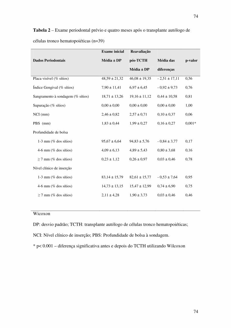
74
74
Tabela 2 – Exame periodontal prévio e quatro meses após o transplante autólogo de
células tronco hematopoiéticas (n=39)
Dados Periodontais
Exame inicial
Média ± DP
Reavaliação
pós-TCTH
Média ± DP
Média das
diferenças
p-valor
Placa visível (% sítios) 48,59 ± 21,32 46,08 ± 19,35 - 2,51 ± 17,11 0,56
Índice Gengival (% sítios) 7,90 ± 11,41 6,97 ± 6,45 - 0,92 ± 9,73 0,76
Sangramento à sondagem (% sítios) 18,71 ± 13,26 19,16 ± 11,12 0,44 ± 10,58 0,81
Supuração (% sítios) 0,00 ± 0,00 0,00 ± 0,00 0,00 ± 0,00 1,00
NCI (mm) 2,46 ± 0,82 2,57 ± 0,71 0,10 ± 0,37 0,06
PBS (mm) 1,83 ± 0,44 1,99 ± 0,27 0,16 ± 0,27 0,001*
95,67 ± 6,64
94,83 ± 5,76
- 0,84 ± 3,77
0,17
Profundidade de bolsa
1-3 mm (% dos sítios)
4-6 mm (% dos sítios)
≥ 7 mm (% dos sítios)
4,09 ± 6,13
0,23 ± 1,12
4,89 ± 5,43
0,26 ± 0,97
0,80 ± 3,68
0,03 ± 0,46
0,16
0,78
Nível clínico de inserção
1-3 mm (% dos sítios)
4-6 mm (% dos sítios)
≥ 7 mm (% dos sítios)
83,14 ± 15,79
14,73 ± 13,15
2,11 ± 4,28
82,61 ± 15,77
15,47 ± 12,99
1,90 ± 3,73
- 0,53 ± 7,64
0,74 ± 6,90
0,03 ± 0,46
0,95
0,75
0,46
Wicoxon
DP: desvio padrão; TCTH: transplante autólogo de células tronco hematopoiéticas;
NCI: Nível clínico de inserção; PBS: Profundidade de bolsa à sondagem.
* p< 0.001 – diferença significativa antes e depois do TCTH utilizando Wilcoxon

75
75
Discussão
O presente estudo avaliou candidatos ao TCTH para verificar possíveis alterações em
parâmetros periodontais após este procedimento. Não houve diferenças clínicas entre os
dois exames periodontais para a maioria dos parâmetros avaliados, entretanto ocorreu
um aumento estatisticamente significativo na profundidade de bolsa (p<0.001) e, uma
tendência estatística de perda de inserção (p=0.06), apesar de uma redução na
porcentagem de sítios com placa visível.
Houve também um aumento na porcentagem de bolsas médias. No entanto, pela
manutenção da variabilidade entre os indivíduos, não se observou significância
estatística deste parâmetro.
Existe pouca informação publicada a respeito das alterações do status periodontal
relacionadas ao TCTH (PATTNI et al., 2000). Verificamos resultados distintos quando
comparamos com o estudo de Pattni et al. (PATTNI et al., 2000). Estes autores
verificaram alteração em parâmetros periodontais que ocorreram em pacientes
submetidos ao TCTH alogênico, após 3 e 6 meses do TCTH, avaliou-se uma melhora
no nível clínico de inserção e no índice gengival. Apesar da aparente semelhança entre
os estudos, as diferenças nos resultados podem ser explicadas pelas medidas
interventivas, já que todos os pacientes no estudo de Pattni et al. receberam cuidados
orais antes, durante e após o período de TCTH.
O controle de placa já foi alvo de estudo em pacientes internados para tratamento
quimioterápico (BROW et al., 1989; ELLEGARD et al., 1989; WEIKEL et al., 1989;
BOROWSKI et al., 1994; ELAD et al., 2003), mostrando uma melhora nos níveis de
sangramento gengival após intensivo controle de placa, mesmo no período de
mielossupressão. No presente estudo, não houve um rigoroso controle de placa durante
o curto período de internação.
76
76
Setenta por cento dos pacientes do presente estudo foram avaliados com menos de uma
semana do TCTH. Este período não representa o tempo adequado para as necessidades
de tratamento apresentadas. É recomendado que os procedimentos de exodontia e
raspagem sejam realizados com pelo menos duas semanas antes do condicionamento
quimioterápico (HEIMDAHL et al., 1989; ELAD et al., 2003). Mesmo assim, todos os
pacientes que necessitavam tratamento foram orientados em relação a esta necessidade e
encaminhados para tratamento dentário, quando aptos.
Apesar da orientação que ocorreu após o exame oral, aparentemente os pacientes
avaliados não buscaram o atendimento odontológico, fato observado pela manutenção
dos parâmetros avaliados. Uma das possibilidades seria a desmotivação dos pacientes
em relação à saúde oral, pelo risco eminente de óbito relacionado ao diagnóstico de
câncer. Apesar disso, a equipe de saúde tem a responsabilidade de incluir a avaliação, e
se necessário, tratamento odontológico previamente ao TCTH, orientando em relação às
seqüelas da falta de tratamento (MOORE, 2007). A desinformação destes indivíduos
sobre a necessidade de uma avaliação e tratamento odontológico anterior ao período do
exame pode ter sido a razão pela qual estes pacientes apresentavam uma saúde oral
deficiente. Desta forma, no presente estudo, observou-se uma falta de integração entre
paciente, médico e dentista, apesar do esforço da equipe de saúde.
A população estudada não apresentava doença periodontal grave. O mesmo foi notado
no estudo de Pattni et al. (PATTNI et al., 2000). Talvez em pacientes com maior
gravidade de doença periodontal houvesse maiores alterações após o TCTH.
Vários fatores poderiam afetar os tecidos periodontais, dentre eles, o uso profilático e/ou
empírico de antimicrobianos, a duração da aplasia ou recuperação medular. Ainda não
está claro o papel que as condições relacionadas ao TCTH exercem nos parâmetros
periodontais.

77
77
As limitações do presente estudo consistiram no número reduzido de pacientes, reflexo
da demanda dos serviços de transplante, e na dificuldade em acompanhar estes
indivíduos por um período maior do que 4 meses, para que fossem verificadas
alterações periodontais devido ao TCTH.
Desta forma, pode-se concluir que, nesta população de pacientes submetidos ao TCTH
não houve alterações clinicamente significativas na maioria dos parâmetros periodontais
avaliados, exceto profundidade de bolsa, quando foram comparados os exames
periodontais pré e pós- transplante. Mais estudos necessitam ser conduzidos para
delinear-se a participação do TCTH neste processo.

78
78
Referências
AKINTOYE, S.O.; BRENNAN, M.T.; GRABER, C.J.; MC KINNEY, B.E.; RAMS,
T.E.; BARRETT, A.J. ATKINSON, J.C. A retrospective investigation of advanced
periodontal disease as a risk factor for septicemia in hematopoietic stem cell and bone
marrow transplant recipients. Oral Surg Oral Med Oral Pathol Oral Radiol Endod, v.94,
n.5, Nov, p.581-8. 2002.
BECK, J.D.; OFFENBACHER, S. Relationships Amons clinical measures of
periodontal disease and their associations with systemic markers. Ann Periodont, v.7,
p.79-89. 2002.
BOROWSKI B.; BENHAMOU E.; PICO J.L.; LAPLANCHE, A.; MARGAINAUD
J.P.; HAYAT M. Prevention of oral mucositis in patients treated with high-dose
chemotherapy and bone marrow transplantation: a randomised controlled trial
comparing two protocols of dental care. European Journal of Cancer, p.93 -97. 1994.
BROW, A.T.; SIMS RAYBOULD, T.P.; LILLICH, T.T.; HENSLEE, P.J., FERRETTI,
G.A. Oral Gram- negative Bacilli in Bone marrow Transplant Patients Given
Chlorhexidine Rinses. Journal Dental Research, v.68, n.7, july, p.1199 -1204. 1989.
COLLIN, B.A.; LEATHER, H.L.; WINGARD, J.R.; RAMPHAL, R. Evolution,
incidence, and Susceptibility of bacterial Bloodstream Isolates from 519 Bone Marrow
Transplants Patients. Clinical Infectious Diseases, v.33, p.947-953. 2001.
79
79
ELAD, S.; GARFUNKEL, A.A.; OR, R.; MICHAELI, E.; SHAPIRA, M.Y.; GALILI,
D. Time limitations and the challenge of providing infectionpreventing dental care to
hematopoietic stem-cell transplantation patients. Support Care Cancer v.11, p.674 - 677.
2003.
ELLEGARD, B.; BERGMANN, O.J.; ELLEGARD, J. Effect of plaque removal with
acute leukemia. J Oral Pathol Med, v.18, p.54 - 58. 1989.
HEIMDAHL, A.; MATTSON, T.; DAHLLÖF, G.; LÖNNQUIST, B.; RINGDÉN O.
The oral cavity as a port of entry for early infections in patient treated with bone
marrow transplantation. Oral Surg Oral Med Oral Pathol v.68, n.6, p.711 - 716. 1989.
MACHATSCHEK, J.; DUDA, J.; MATTHAY, K.; COWAN, M.; HORN, B. Immune
reconstitution, infectious complications and post transplant supportive care measures
after autologous blood and marrow transplantation in children. Bone Marrow
Transplant, v.32, n.7, p.687- 693. 2003.
MOORE, S. Facilitating oral chemotherapy treatment and compliance through
patient/family-focused education. Cancer Nurs., v.30, n.2, p.112-122. 2007.
NOACK, B.; GENCO, R.J.; TREVISAN, M.; GROSSI, S.; ZAMBON, J.J.; NARDIN,
E. Periodontal infections contribute to elevated systemic C-Reative Protein Level.
Journal Periodontology, v.72, p.1221-1227. 2001.
80
80
NUCCI, M.A.; ANDRADE, F.; VIGORITO, A.; TRABASSO, P.; ARANHA, J.F.;
MAIOLINO, A.; DE SOUZA, C.A. Infections complications in patients randomized to
receive allogeneic bone marrow or peripheral blood transplantation. Transplant
Infectious Disease v.5, p. 167-173. 2003.
OLIVEIRA, A.L.; SOUZA, M.; CARVALHO-DIAS, V.M.H.; RUIZ, M.A.; SILLA, L.;
TANAKA, P.Y.; SIMÕES, B.P.; TRABASSO, P.; SEBER, A.; LOTFI, C.J.;
ZANICHELLI, M.A.; ARAÚJO, V.R.; GODOY, C.; MAIOLINO, A.; URAKAWA, P.;
CUNHA, C.A.; SOUZA, C.A.; PASQUINI, R.; NUCCI, M. Epidemiology of
bacteremia and factors associated with multi-drug-resistent gram-negative bacteremia in
hematopoietic stem cell transplant recipients. Bone Marrow Transplantation, p.1-7.
2007.
PATTNI, R.; WALSH, L.J.; MARSHALL, R.I.; CULLINAN, M.P.; SEYMOUR, G.J.;
BARTOLD, P.M. Changes in the periodontal status of patients undergoing bone
marrow transplantation. J Periodontol v.71, n.3, march, p.394 -402. 2000.
SCANNAPIECO, F.A. Periodontal inflammation: from gingivitis to systemic disease?
Compend Contin Educ Dent, v.25, n.7, p.16 - 25. 2004.
VECHIATO, C.; SOARES, A.; JANKO, F. Quimioterápicos utilizados no tratamento
de pacientes submetidos a transplante de medula óssea, no hospital Araújo Jorge, no ano
de 2004. Revista Eletrônica de Farmácia, v.2, n.2, p.214-217. 2005.

81
81
WEIKEL, D.S.; PETERSON, D.E.; RUBINSTEIN, L.E. Incidence of fever following
invasive oral interventions in the myelosuppressed cancer patient. Cancer Nurs, v.12,
p.265-270. 1989.
ZAGO, M.A.; FALCÃO, R.P.; PASQUINI, R. Hematologia, fundamentos e prática.
Rio de Janeiro: Atheneu. 2004. 1081 p.

82
82
___________________________________ __________________ 6. DISCUSSÃO
Neste estudo foram coletados dados sobre as condições orais, doença de base e
complicações que ocorrem em pacientes submetidos ao TCTH autólogo. As condições
orais foram correlacionadas com algumas complicações que ocorrem após o TCTH. Os
resultados foram dispostos em forma de três artigos científicos em consonância com os
objetivos do estudo. Os principais resultados serão discutidos a seguir.
Inicialmente, cinqüenta e seis candidatos ao TCTH foram avaliados de acordo com
condições clínicas e sócio-demográficas, e incluídos no artigo 1 “ Os candidatos ao
transplante autólogo de células tronco hematopoiéticas estão em condições orais
adequadas?” Destes, quarenta e oito indivíduos realizaram o TCTH e foram
acompanhados em relação a algumas complicações do TCTH e incluídos no artigo 2
“A correlação entre o status periodontal e as complicações do transplante
autólogo de células tronco hematopoiéticas”. Quatro meses após o TCTH, trinta e
nove pacientes foram reavaliados em relação às condições periodontais para
comparação entre os exames pré e pós-TCTH, apresentados no artigo 3 “Alteração do
status periodontal de pacientes submetidos ao transplante autólogo de células
tronco hematopoiéticas”. Por conseguinte, a população estudada ficou separada em
três extratos, de acordo com a dinâmica do estudo. Foi observada uma variabilidade
nos parâmetros avaliados, ilustrada pelas diferentes médias entre os achados destes três
extratos populacionais: candidatos ao TCTH (n=56), indivíduos que realizaram o
TCTH (n=48) e pacientes que foram submetidos aos dois exames periodontais (n=39).
Cuidados orais prévios parecem prevenir complicações durante o período de
hospitalização para o TCTH, especialmente no período de neutropenia (NIH, 1990;
BARKER, 1999; GOLDMAN, 2006; BARASCH e COKE, 2007). Na população

83
83
estudada, os pacientes não apresentavam condições orais apropriadas para serem
submetidos ao TCTH. A maioria dos candidatos ao TCTH necessitava de tratamento
odontológico básico, como orientações de higiene e raspagem (91,1%), restaurações ou
próteses (39,3%), e extrações (17,9%). Mesmo em países desenvolvidos, foi
evidenciado que grande parte dos candidatos ao TCTH apresentava necessidade de
tratamento odontológico. (ELLEGARD et al., 1989; HEIMDAHL et al., 1989;
GRABER et al., 2001; ELAD et al., 2003)
Quando as complicações pós-TCTH foram avaliadas, os principais achados deste
estudo foram que todos os parâmetros periodontais avaliados e o CPOD, demonstraram
uma condição oral mais deficiente nos pacientes que apresentaram bacteremia quando
comparados aos que não apresentaram bacteremia, apesar destes resultados não serem
estatisticamente significativos. Cabe ressaltar, que a média de sítios com placa visível e
sangramento à sondagem foram os dois parâmetros mais elevados nos pacientes que
apresentaram neutropenia febril associada à bacteremia (Tabela 3, artigo 2). Nenhum
estudo publicado até o momento avaliou a associação entre placa visível e bacteremia
em indivíduos submetidos ao TCTH, no entanto, em pacientes com cardiopatias, esta
associação entre sangramento à sondagem e bacteremia já foi apontada (DALY et al.,
2001; DEMMER et al., 2010). Desta forma, estudos multicêntricos necessitam ser
desenvolvidos a fim de verificar estas associações nesta população.
A periodontite foi prevalente em 29.2% dos indivíduos que realizaram o TCTH.
Apesar dos valores de profundidade de bolsa à sondagem e nível clínico de inserção
terem demonstrado médias superiores nos pacientes com bacteremia, esta associação
não foi estatisticamente significativa. Talvez devido ao fato de que a população do
estudo não apresentava níveis elevados da doença periodontal. Resultados semelhantes
foram encontrados em um estudo que correlacionou reabsorção da crista óssea alveolar
84
84
e bacteremia, em pacientes submetidos ao TCTH alogênico (AKINTOYE et al., 2002).
Se pacientes com maior gravidade de doença periodontal fossem avaliados, talvez esta
associação pudesse ser encontrada.
A ocorrência da gengivite foi freqüente (55,4%) nos candidatos ao TCTH autólogo
avaliados (Tabela 2, artigo 1). Pacientes onco-hematológicos de outros estudos
apresentaram gengivite, variando de 15 a 25% (HEIMDAHL et al., 1989; PATTNI et
al., 2000). As razões para esta discrepância podem estar nas diferenças entre o
tamanho amostral ou o critério diagnóstico para gengivite que varia entre os estudos. A
gengivite foi altamente prevalente nos indivíduos que realizaram o TCTH (60,4%),
bem como nos indivíduos que foram avaliados 4 meses pós- TCTH (61.5%).
Curiosamente, o índice gengival, que avalia sangramento marginal, apresentou uma
média baixa em comparação com o sangramento à sondagem. A explicação para este
fato pode ser a freqüência de anemia encontrada nesta população, conseqüente à
doença de base (KYLE e RAJKUMAR, 2009). É sabido que pacientes com anemia
podem apresentar “palidez gengival” (PROCTOR et al., 2005). Entretanto, o
sangramento à sondagem do fundo da bolsa por ser uma mensuração mais profunda,
apresentou uma média percentual de sítios maior (Tabela 3, artigo 1; Tabela 1, artigo
2).
Em relação aos parâmetros orais avaliados, houve uma associação entre o baixo nível
educacional e os parâmetros clínicos mais deficientes. O índice de CPOD foi
semelhante a outro estudo desenvolvido em candidatos a TCTH brasileiros (SHCAIRA
et al., 2009). Não houve correlação estatística deste índice com as complicações pós-
transplante (Tabela 3, artigo 2).
A candidíase oral é um achado comum em pacientes onco-hematológicos que estão em
tratamento (HEIMDAHL et al., 1989; NIH, 1990) e estava presente em 8.3% (n=4)

85
85
dos indivíduos no primeiro exame (Tabela 1, artigo 2). Em metade dos pacientes
(n=24) que foram submetidos ao transplante autólogo foi instituída profilaxia anti-
fúngica com fluconazol. Apesar disso, desenvolveram candidíase oral 12,5% (n=3) dos
indivíduos que fizeram profilaxia, durante o período de neutropenia pós- TCTH.
Talvez esta ocorrência tenha relação com o fato do fluconazol não eliminar as cepas
deste fungo, como também a existência de uma colonização primária ser um fator que
aumente as chances de uma nova ocorrência (ALCURE et al., 2008). Os três pacientes
que desenvolveram candidíase com profilaxia anti-fúngica, tinham apresentado
candidíase no baseline e usavam próteses totais. Observou-se uma associação
estatística entre o aparecimento de candidíase no período de neutropenia pós-TCTH e o
uso de próteses totais na população estudada. A relação entre o uso de próteses totais e
o aparecimento de candidíase é comumente relatado na literatura (DAVIES et al.,
2008; PENTENERO et al., 2008).
A mucosite foi um evento comum nos pacientes submetidos ao TCTH autólogo
(89,6%), embora não apresentassem níveis altos de gravidade. Os cuidados orais antes
do TCTH parecem diminuir a ocorrência e/ou a gravidade da mucosite (BOROWSKI
et al., 1994; MELKOS et al., 2003; RUBENSTEIN et al., 2004; DJURIC et al., 2006;
KEEFE et al., 2007). No presente estudo, os indivíduos que apresentavam gengivite no
exame inicial, desenvolveram mucosite após TCTH (p=0.05). Entretanto, nenhum
outro parâmetro oral prévio ao TCTH teve associação com o diagnóstico da mucosite.
Este achado, entretanto, não esclarece a associação de envolvimento periodontal com o
surgimento, a gravidade, e a duração da mucosite.
A ocorrência, gravidade e duração da mucosite, não tiveram associação com a
ocorrência de bacteremia neste estudo.

86
86
A incidência de bacteremia (29.2%) foi semelhante a outros estudos (SALAZAR et al.,
1999; COLLIN et al., 2001; AUNER et al., 2002) que mostram uma variação de 20 a
50%. As bactérias mais comumente isoladas foram Staphylococcus spp. coagulase-
negativa, ou seja, as mesmas relatadas por outros autores (SALAZAR et al., 1999;
NUCCI et al., 2003; REICH et al., 2001; OLIVEIRA et al., 2007; FRÈRE et al.,
2004). Apesar destas espécies não serem típicas do meio oral, podem estar presentes na
placa bacteriana. Cabe ressaltar que várias bactérias periodonto-patogênicas são
anaeróbicas e de difícil crescimento em diferentes meios de cultura, portanto, futuros
estudos serão necessários para avaliar a correlação entre a presença destas bactérias e a
ocorrência de bacteremia.
A ocorrência de neutropenia febril (95.8% dos indivíduos) após o TCTH, assim como
a prevalência de febre de origem obscura (60.4%), foram semelhantes a estudos
recentes (AUNER et al., 2002; MULLEN et al, 2000; OLIVEIRA et al., 2007). Um
achado interessante foi que todos os parâmetros clínicos periodontal e CPOD no
baseline, estavam piores nos pacientes que apresentaram bacteremia, incluindo os
maiores índices de placa visível e sangramento à sondagem (Tabela 3, artigo 2). Por
outro lado, os valores de todos os parâmetros clínicos periodontais e CPOD foram
menores, exceto profundidade de bolsa, em pacientes que não apresentaram
neutropenia febril, entretanto, apenas dois pacientes não apresentaram febre.
Quanto às alterações nos parâmetros periodontais observadas no exame realizado após
quatro meses do TCTH, não houve alterações estatisticamente significativas em todos
os parâmetros clínicos periodontais avaliados, exceto profundidade de bolsa,
(p<0.001), apesar de que vários parâmetros clínicos estavam aumentados após este
período. Observamos também a permanência da alta variabilidade destes parâmetros
periodontais entre os pacientes. Em um estudo anterior (PATTNI et al., 2000),

87
87
desenvolvido em pacientes submetidos ao TCTH alogênico, foi observada uma
melhora no nível clínico de inserção, em 3 e 6 meses de acompanhamento pós-TCTH.
Entretanto, neste estudo, foi realizado um protocolo de cuidados orais intensivos o que
explica a melhora deste parâmetro.
As limitações do presente estudo consistiram no número reduzido de pacientes, reflexo
da demanda dos serviços de transplante ao longo do período proposto do estudo, e na
dificuldade em acompanhar estes indivíduos por um período maior do que 4 meses, para
que fossem verificadas alterações periodontais de longo prazo em decorrência do
TCTH.
Outra limitação existente neste estudo seria a falta de um exame radiográfico que
tornasse possível a verificação de outras ocorrências relacionadas à infecção
odontogênica nestes pacientes. O desenho deste estudo foi observacional e não
interferiu na rotina destes indivíduos.
Todos os pacientes com necessidade de tratamento foram encaminhados para o serviço
de atendimento odontológico existente nos dois centros, ou para o dentista de
preferência.
Considerando a população estudada, verificamos que a saúde oral dos pacientes
candidatos ao TCTH autólogo, não é adequada para receber os procedimentos inerentes
ao TCTH. Pacientes que desenvolveram bacteremia após o TCTH, apresentavam saúde
dental e periodontal mais comprometida do que pacientes que não apresentaram
bacteremia, apesar da ausência de significância estatística. Também foi observado que,
nesta população de pacientes submetidos ao TCTH, não houve alterações clinicamente
significativas na maioria dos parâmetros periodontais avaliados pós-TCTH, exceto
profundidade de bolsa, quando foram comparados ao exame pré-transplante.
88
88
Observamos também que existe a necessidade de uma maior integração entre os
profissionais da equipe de saúde e os pacientes candidatos ao TCTH, objetivando um
resultado mais eficaz na melhora da saúde geral do paciente.
Sugerimos que novos estudos sejam conduzidos para avaliar a influência da saúde oral
na ocorrência de neutropenia febril em um número maior de candidatos ao TCTH e em
diferentes centros.

89
89
______________________________________________________7. CONCLUSÕES
1- A condições orais de candidatos ao TCTH são inadequadas para que estes
indivíduos sejam submetidos ao transplante;
2- A ocorrência de neutropenia febril nos pacientes submetidos ao TCTH foi de 95,8%.
3- Todos os parâmetros periodontais e o índice de CPOD foram piores em pacientes
com bacteremia associada ao período de neutropenia febril;
4- A mucosite foi associada com o diagnóstico de gengivite pré-transplante.
5- Não há variação clínica considerável nos parâmetros clínicos periodontais avaliados,
exceto profundidade de bolsa, quando comparados os exames pré e quatro meses pós-
TCTH.

90
90
___________________________________8. REFERÊNCIAS BIBLIOGRÁFICAS
AKINTOYE, S.O.; BRENNAN, M.T.; GRABER, C.J.; MC KINNEY, B.E.; RAMS,
T.E.; BARRETT, A.J. ATKINSON, J.C. A retrospective investigation of advanced
periodontal disease as a risk factor for septicemia in hematopoietic stem cell and bone
marrow transplant recipients. Oral Surg Oral Med Oral Pathol Oral Radiol Endod, v.94,
n.5, Nov, p.581-8. 2002.
ALCURE M.; HIPOLITO JR O., SOUZA C, E T. P. CORRÊA M.E. TRABASSO P.
Electrophoretic Karyotyping And Antifungal Susceptibility of Sequentially Oral
Candida Spp. Isolates of Hematopoietic Stem Cell Transplant (HSCT)
Recipients. Multinational Association for Supportive Care in Cancer (MASCC ).
Houston, Texas, USA, 2008.
AUNER, H.W.; SILL, H.; MULABECIROVIC, A.; LINKESCH, W.; KRAUSE, R.
Infectious complications after autologous hematopoietic stem cell transplantation:
comparison of patients with acute myeloid leukemia, malignant lymphoma, and
multiple myeloma. Ann Hematol, v.81, n.7, Jul, p.374-7. 2002.
BARASCH, A. E J. COKE. Cancer therapeutics: an update on its effects on oral health.
Periodontol 2000, v.44, p.44-54. 2007.
BARKER, G J. Current practices in the oral management of the patient undergoing
chemotherapy or bone marrow transplant. Support Care Cancer, v.7, p.17 - 20. 1999.

91
91
BARRET, A.P. Oral complications of bone marrow transplantation. Aust N Z J Med,
v.16, n.2, abril, p.239-240. 1986.
BECK, J.D.; OFFENBACHER, S. Relationships Amons clinical measures of
periodontal disease and their associations with systemic markers. Ann Periodont, v.7,
p.79-89. 2002.
BOROWSKI B.; BENHAMOU E.; PICO J.L.; LAPLANCHE, A.; MARGAINAUD
J.P.; HAYAT M. Prevention of oral mucositis in patients treated with high-dose
chemotherapy and bone marrow transplantation: a randomised controlled trial
comparing two protocols of dental care. European Journal of Cancer, p.93 -97. 1994.
BROW, A.T.; SIMS RAYBOULD, T.P.; LILLICH, T.T.; HENSLEE, P.J., FERRETTI,
G.A. Oral Gram- negative Bacilli in Bone marrow Transplant Patients Given
Chlorhexidine Rinses. Journal Dental Research, v.68, n.7, july, p.1199 -1204. 1989.
CASTRO, C.G.; GREGIANI, L.J.; BRUNETTO, A.L. Bone marrow transplantation
and cord blood transplantation in children. J. Pediatric, v.77, n.5, p.345-360. 2001.
CHEN, C.Y.; HANSEN, K.S.; HANSEN, L.K. Rhizobium radiobacter as an
opportunistic pathogen in central venous catheter – associated bloodstream infection:
case report and review. Journal of Hospital Infection, v.68, p.203-207. 2008.

92
92
CHEN, L.; WEI, B.; LI, J.; LIU, F.; XUAN, D.; XIE, B.; ZHANG, J. Association of
periodontal parameters with metabolic level and systemic inflammatory markers in
patients with type 2 diabetes. J Periodontol., v.81, n.3, p.364-371. 2010.
COLLIN, B.A.; LEATHER, H.L.; WINGARD, J.R.; RAMPHAL, R. Evolution,
incidence, and Susceptibility of bacterial Bloodstream Isolates from 519 Bone Marrow
Transplants Patients. Clinical Infectious Diseases, v.33, p.947-953. 2001.
DALY, C.G.; MITCHELL, D.H.; HIGHFIELD, J.E.; GROSSBERG, D.E.; STEWART,
D. Bacteremia due to periodontal probing: a clinical and microbiological investigation. J
Periodontol, v.72, n.2, p.210-4. 2001.
DAVIES, A.N.; BRAILSFORD, S.R.; BEIGHTON, D.; SHORTHOSE, K.; STEVENS,
V.C. Oral candidosis in community-based patients with advanced cancer. J Pain
Symptom Manage., v.35, n.5, p.508-514. 2008.
DEMMER, R.T.; PAPAPANOU, P.N.; JACOBS JR, D.R.; DESVARIEUX, M.
Evaluating clinical periodontal measures as surrogates for bacterial exposure: the Oral
Infections and Vascular Disease Epidemiology Study (INVEST). BMC Med Res
Methodol. , v.10, n.2. 2010.
DETTENKOFER, M.; EBNER, W.; BERTZ, H.; BABIKIR, R.; FINKE, J.; FRANK,
U.; RUDEN, H.; DASCHNER, F.D. Surveillance of nosocomial infections in adult
recipients of allogeneic and autologous bone marrow and peripheral blood stem-cell
transplantation. Bone Marrow Transplant, v.31, n.9, May, p.795-801. 2003.

93
93
DJURIC, M.; HILLIER-KOLAROV, V.; BELIC, A.; JANKOVIC, L. Mucositis
prevention by improved dental care in acute leukemia patients. Support Care Cancer,
v.14, n.2, p.137 - 146. 2006.
DONNELLY, J.P. Infection in the neutropenica and haematopoietic stem cell recipient.
Curr Opin Infect Dis v.13, p.337 - 342. 2000.
ELAD, S.; GARFUNKEL, A.A.; OR, R.; MICHAELI, E.; SHAPIRA, M.Y.; GALILI,
D. Time limitations and the challenge of providing infectionpreventing dental care to
hematopoietic stem-cell transplantation patients. Support Care Cancer v.11, p.674 - 677.
2003.
ELLEGARD, B.; BERGMANN, O.J.; ELLEGARD, J. Effect of plaque removal with
acute leukemia. J Oral Pathol Med, v.18, p.54 - 58. 1989.
EPSTEIN, J.B.; RABER-DURLACHER, J. Advances in hematologic stem cell
transplant: An update for oral health care providers. Oral Surg Oral Med Oral Pathol
Oral Radiol Endod, v.107, n.3, p.301 - 312. 2009.
FRERE, P.; HERMANNE, J.P.; DEBOUGE, M.H.; DE MOL, P.; FILLET, G.;
BEGUIN, Y. Bacteremia after hematopoietic stem cell transplantation: incidence and
predictive value of surveillance cultures. Bone Marrow Transplant, v.33, n.7, Apr,
p.745-9. 2004.

94
94
ROSE, L.F.; GENCO, R. J. COHEN D.W. MEALEY B.L. Periodontal Medicine. Saint
Louis, London: BC Decker INC., 2000, 273p.
GILLESPIE, T.; MASTERTON, R.G. Investigation of infection in the neutropenic
patient with fever. Journal of Hospital Infection, v.38, p.77-91. 1998.
GOLDMAN, K.E. Dental management of patients with bone marrow and solid organ
Dent Clin North Am., v.50, n.4, p.659-676. 2006.
GRABER, C.J.; ALMEIDA, K.N.F.; ATKINSON, J.C.; JAVAHERI, D.; FUKUDA,
C.D.; GILL, V.J., BARRET AJ, BENNETT, J.E. Dental health and viridans
streptococcal bacteremia in allogeneic hematopoietic stem cell transplant recipients.
Bone Marrow Transplantation v.27, p.537 - 542. 2001.
HEIMDAHL, A.; MATTSON, T.; DAHLLÖF, G.; LÖNNQUIST, B.; RINGDÉN O.
The oral cavity as a port of entry for early infections in patient treated with bone
marrow transplantation. Oral Surg Oral Med Oral Pathol v.68, n.6, p.711 - 716. 1989.
HOLMSTRUP, P.; GLICK, M. Treatment of periodontal disease in the
immunodeficient patient. Periodontology 2000, v.28, p.190-205. 2002.
HUGUES, W.T.; ARMSTRONG, D.; BODEY, G.P. Guidelines for the use of
antimicrobial agents in neutropenic patients with unexplained fever. Infectious Diseases
Society of America, Clin Infect Dis, v.25, p.551-573. 1997.

95
95
AINAMO, J.; BAY, I. Problems and proposals for recording gingivitis and plaque. Int
Dent J. , v.25, n.4, p.229 - 235. 1975.
INTERNATIONAL HOST SOCIETY. The design, Analysis, and Reporting of Clinical
Trial on the Empirical Antibiotic Management of the Neutropenic Patient. Journal of
Infectious Diseases, v.161, p.397 - 401. 1990.
KEEFE, D.M.; SCHUBERT, M.; ELYING, L.S.; SONIS, S.T.; EPSTEIN, J.B.;
RABER-DURLACHER, J.E.; MIGLIORATI, C.A.; MC GUIRE, D.B.; HUTCHINS,
R.D.; PETERSON, D.E. Updated Clinical Practice Guidelines for the Prevention and
Treatment of Mucositis. Cancer, v.109, p.820 - 831. 2007.
KYLE, R.A.; RAJKUMAR, S.V. Treatment of multiple myeloma: a comprehensive
review. Clin Lymphoma Myeloma., v.9, n.4, p.278 - 88. 2009.
LAINE, P.O.; LINDQVIST, J.C.; PYRHÖNEN, S.O.; STRAND-PETTINEN, I.M.;
TEERENHOVI, L.M.; MEURMAN, J.H. Oral infection as a reason for febrile episodes
in lymphoma patients receiving cytostatic drugs. european Journal of Cancer B Oral
Oncol, v.28B, n.3, p.103 - 107. 1992.
LINDHE, J.; KARRING, T.; LANG, N.P. Tratado de Periodontia Clínica e
Implantologia Oral. 4a ed. São Paulo: Guanabara Koogan. 2005

96
96
MELKOS, A.B.; MASSENKEIL, G.; ARNOLD, R.; REICHART, P.A. Dental
treatment prior to stem cell transplantation and its influence on the post transplantation
outcome. Clin Oral Invest, v.7, p.113 - 115. 2003.
MOURAD, O.; PALADA. V., DETSKY, A. Comprehensive Evidence-based Approach
to Fever of Unknown Origin. Arch Intern Med, v.163, March 10, p.545-551. 2003.
MULLEN, C.A.; NAIR, J.; SANDESH, S.; CHAN, K.W. Fever and neutropenia in
pediatric hematopoietic stem cell transplant patients. Bone Marrow Transplant, v.25,
n.1, Janeiro, p.59-65. 2000.
NATIONAL INSTITUTE OF HEALTH. Consensus Development Panel. Consensus
statement: oral complications of cancer therapies: diagnosis, prevention and treatment.
NCI Monogr 9. 1990.
NUCCI, M.A.; ANDRADE, F.; VIGORITO, A.; TRABASSO, P.; ARANHA, J.F.;
MAIOLINO, A.; DE SOUZA, C.A. Infections complications in patients randomized to
receive allogeneic bone marrow or peripheral blood transplantation. Transplant
Infectious Disease v.5, p. 167-173. 2003.
OLIVEIRA, A.L.; SOUZA, M.; CARVALHO-DIAS, V.M.H.; RUIZ, M.A.; SILLA, L.;
TANAKA, P.Y.; SIMÕES, B.P.; TRABASSO, P.; SEBER, A.; LOTFI, C.J.;
ZANICHELLI, M.A.; ARAÚJO, V.R.; GODOY, C.; MAIOLINO, A.; URAKAWA, P.;
CUNHA, C.A.; SOUZA, C.A.; PASQUINI, R.; NUCCI, M. Epidemiology of
bacteremia and factors associated with multi-drug-resistent gram-negative bacteremia in

97
97
hematopoietic stem cell transplant recipients. Bone Marrow Transplantation, p.1-7.
2007.
OVERHOLSER, C.D.; PETERSON, D.E.; WILLIAMS, L.T.; SCHIMPFF, S.C.
Periodontal infection in patients with acute nonlymphocyte leukemia. Prevalence of
acute exacerbations. Arch Inter Med, v.142, n.3, march, p.551-554. 1982.
PATTNI, R.; WALSH, L.J.; MARSHALL, R.I.; CULLINAN, M.P.; SEYMOUR, G.J.;
BARTOLD, P.M. Changes in the periodontal status of patients undergoing bone
marrow transplantation. J Periodontol v.71, n.3, march, p.394 -402. 2000.
PENTENERO, M.; BROCCOLETTI, R.; CARBONE, M.; CONROTTO, D.;
GANDOLFO, S. The prevalence of oral mucosal lesions in adults from the Turin area.
Oral Dis, v.14, n.4, p.356 - 366. 2008.
PROCTOR, R.; KUMAR, N.; STEIN, A.; MOLES, D.; PORTER, S. Oral and dental
aspects of chronic renal failure. Journal of Dental Research, v.84, n.3, p.199 - 208.
2005.
RABER-DURLACHER, J.E.; BARASCH, A.; PETERSON, D.E.; LALLA, R.V.;
SCHUBERT, M.M.; FIBBE, W.E. Oral Complications and Management
Considerations in Patients Treated with High-Dose Chemotherapy. Supportive Cancer
Therapy, v.1, n.4, p.219-229. 2004.

98
98
RABER-DURLACHER, J.E.; EPSTEIN, J.B.; RABER, J.; VAN DISSEL, J.T.; VAN
WINKELHOFF, A.J.; GUIOT, H.F.; DER VELDEN, U. Periodontal infection in cancer
patients treated with high-dose chemotherapy. Support Care Cancer, v.10, p.466-473.
2002.
REICH, G.; MAPARA, M.Y.; REICHARDT, P.; DÖRKEN, B.; MASCHMEYER, G.
Infectious complications after high-dose chemotherapy and autologous stem cell
transplantation: comparison between patients with lymphoma or multiple myeloma and
patients with solid tumors. Bone Marrow Transplant, v.27, n.5, march, p.525-529. 2001.
RUBENSTEIN, E.B.; PETERSON, D.E.; SCHUBERT, M.; KEEFE, D.; MC GUIRE,
D.; EPSTEIN, J.; ELTING, L.S.; FOX, P.C.; COOKSLEY, C.; SONIS, S.T. Clinical
Practice Guidelines for the Prevention and Treatment of cancer Therapy-Induced Oral
and Gastrointestinal Mucositis. Cancer v.100, n.9, p.2026 - 2046. 2004.
SALAZAR, R.S.C.; MAROTO, P.; TABERNERO, J.M.; BRUNET. J.; VERGER, G.;
VALENTÍ, V.; CANCELAS, J.Á.; OJEDA, B.; MENDOZA, L.; RODRÍGUEZ, M.;
MONTESINOS, J.; LÓPEZ-LÓPEZ, J.J. Infections complications in 126 patients
treated with high-dose chemotherapy and autologous peripheral blood stem cell. Bone
Marrow Transplant, v.23, p.27 - 33. 1999.
SANTOS, P.S.S.; MAGALHÃES, M.H.SG. Avaliação da mucosite oral em pacientes
que receberam adequação bucal prévia ao transplante de medula óssea. Rev Pós Grad,
v.13, n.1, p.77-82. 2006.
99
99
SCHUBERT, M.M.; WILLIAM, B.E.; LLOID, M.E.; DONALDSON, G.; CHAPKO,
M.K. Assessment Scale for the Rating of Oral Mucosal Changes Associated with Bone
Marrow Transplantation. Cancer 69, v.10, p.2469-2477. 1992.
SHCAIRA, V.; BOER, C.; BASSO, F.; KELLERMANN, M.; CORREA, M.E. Oral
Healthy Characteristics And Dental Management Of Hematopoietic Stem Cell
Transplantation Patients In A Single Public Hospital. Multinational Association for
Supportive Care in Cancer (MASCC ). Rome, Italy: MASSC/ ISOO Symposium
Abstracts, 2009. 164 p.
SIMINOSKI, K. Persistent fever due to occult dental infection: case report and review.
Clin Infect Dis, v.16, n.4, april, p.550-554. 1993.
SOGA, Y.; NISHIMURA, F.; ISHIMARU, F.; MINESHIBA, F.; TAKAYA, H.;
SATO, H.; KUDO, C.; KOKEGUCHI, S.; FUJII, N.; TANIMOTO, M.; TAKASHIBA,
S. Appearance of multidrug-resistant opportunistic bacteria on the gingiva during
leukemia treatment. Journal Periodontology, v.79, p.181-186. 2008.
SONIS, S.T. Mucositis as a biological process: a new hypothesis for the development of
chemotherapy-induced stomatotoxicity. Oral Oncology, v.34, p.39-43. 1998.
SONIS, S.T. A Biological Approach to Mucositis. J Support Oncol, v.2, p.21-36. 2004.
STOKMAN, M.A.; SONIS, S.T.; DIJKSTRA, P.U.; BURGERHOF, J.G.M.;
SPIKERVET, F.K.L. Assessment of oral mucositis in clinical trials: Impact of training
100
100
on evaluators in a multi-centre trial. European Journal of Cancer, v.41, p.1735-1738.
2005.
VECHIATO, C.; SOARES, A.; JANKO, F. Quimioterápicos utilizados no tratamento
de pacientes submetidos a transplante de medula óssea, no hospital Araújo Jorge, no ano
de 2004. Revista Eletrônica de Farmácia, v.2, n.2, p.214-217. 2005.
WEIKEL, D.S.; PETERSON, D.E.; RUBINSTEIN, L.E. Incidence of fever following
invasive oral interventions in the myelosuppressed cancer patient. Cancer Nurs, v.12,
p.265-270. 1989.
WINGARD, J.R. Antifungal chemoprophylaxis after blood and narrow transplantation.
Clin Infect. Dis, v.34, p.1386-1390. 2002.
YAMAGATA, K. ONIZAWA, K.; YANAGAWA, T.; HASEGAWA, Y.; KOJIMA,
H.; NAGASAWA, T.; YOSHIDA, H. A prospective study to evaluate a new dental
management protocol before hematopoietic stem cell transplantation. Bone Marrow
Transplant, v.38, p.237 - 242. 2006.
ZAGO, M.A.; FALCÃO, R.P.; PASQUINI, R. Hematologia, fundamentos e prática.
Rio de Janeiro: Atheneu. 2004. 1081 p.

101
101
____________________________________________________________9.ANEXOS

102
102
_____________________________________________ANEXO 1. TCLE - HUCFF
UFRJ – Hospital Clementino Fraga Filho – Setor de Hematologia-
Departamento de Clínica Médica
TERMO DE CONSENTIMENTO LIVRE E
ESCLARECIDO
Título do protocolo: Correlação da Condição Periodontal de receptores de
transplante autólogo de células hematopoiéticas com episódios de febre pós-
transplante
Pesquisador responsável: Liana Leite Duval Fernandes
Você foi convidado a participar de um estudo e deve ficar à vontade para decidir se
deseja ou não participar. Leia cuidadosamente o que segue e pergunte ao profissional
responsável qualquer dúvida que você possa ter.
Problema
Muitos pacientes que foram submetidos ao transplante de medula óssea desenvolveram
febre indicando a existência de uma infecção. Esta infecção pode prejudicar a
recuperação após o transplante, aumentando a necessidade de tempo ou vindas ao
hospital, tornando necessário o uso de mais medicações para combatê-la e até interferir
no sucesso do transplante. As causas da infecção nem sempre são encontradas, pois ela
pode ter como origem locais diferentes do organismo. A boca, mais especificamente, a
gengiva, pode ser um destes locais e por isso é que este estudo está sendo proposto. A
infecção da gengiva, que algumas vezes envolve o osso que sustenta o dente causando
inflamação e até a perda deste dente, também é conhecida como periodontite. Esta pode
ser uma doença silenciosas, pois muitas vezes não causa dor nem pode ser detectada só
com exame visual.
Objetivo do Estudo
Identificar se a infecção da gengiva que alguns pacientes possam ter pode ser a causa de
alguns episódios de febre e suas características clínicas.

103
103
Procedimentos
O pesquisador de estudo e sua equipe verificarão sua história médica, examinarão sua
boca e cada um de seus dentes avaliando através de uma sonda se há inflamação na sua
gengiva e medir a quantidade de osso que pode ter sido destruída pela infecção da
gengiva. Este procedimento é realizado através de um instrumento, chamado sonda
periodontal, que tem uma marcação além de uma ponta fina e arredondada que é
introduzida entre o dente e a gengiva. Desta forma, podemos verificar se há periodontite
medindo o suporte de osso ao redor do dente e a possível presença de inflamação.
Risco
O seu tratamento será exatamente o mesmo caso você participe ou não deste estudo. O
exame da gengiva irá coincidir com uma de suas idas ao hospital. O exame da gengiva
para algumas pessoas pode causar um desconforto local transitório que costuma sumir
logo após o exame.
Benefícios
O diagnóstico da infecção da gengiva através deste exame auxiliará a equipe do serviço
de Odontologia a planejar o seu tratamento incluindo a intervenção na infecção da
gengiva.
Se durante os exames bucais forem constatadas outras alterações bucais, o serviço de
Odontologia do Hospital Clementino Fraga Filho, na sala 144, irá realizar o tratamento.
Acompanhamento
Após o seu consentimento em participar deste estudo, será feita uma ficha onde os seus
dados pessoais serão coletados. O seu acompanhamento clínico será feito através de seu
prontuário no Serviço de Transplante de Medula Óssea, e seu exame será realizado no
Hospital (sala 144). O pesquisador responsável irá lhe acompanhar e explicar sempre
quando será o próximo exame.
Caráter Confidencial dos Registros
Seu nome não será revelado ainda que as informações de seu registro médico sejam
utilizadas para propósitos educativos ou de publicação, que ocorrerão
independentemente dos resultados obtidos.
Custos
Não haverá qualquer custo ou forma de pagamento para o paciente pela sua participação
no estudo.
Sua Participação

104
104
É importante que você saiba que a sua participação neste estudo é completamente
voluntária e que você pode recusar-se a participar ou interromper sua participação a
qualquer momento sem penalidades ou perda de benefícios aos quais você tem direito.
Em caso de você decidir interromper sua participação no estudo, a equipe assistente
deve ser comunicada e a coleta de seus dados de prontuário será imediatamente
interrompida. O médico responsável por sua internação pode interromper sua
participação no estudo a qualquer momento, mesmo sem sua autorização.
Esclarecimentos
Dúvidas em relação ao projeto e aprovação ética, entre em contato com o Comitê de
Ética do Hospital Clementino Fraga Filho: 2562.2480
Para qualquer pergunta, entre em contato com a Dra. Liana Leite Duval Fernandes, na
sala 144 ou pelos telefones: (21) 92039860 ou (21) 35114419
Eu, ___________________________________________________ li e entendi este
formulário de consentimento. Todas as minhas perguntas foram respondidas. Participei
voluntariamente deste estudo.
Assinatura do Voluntário
_____________________________________Data___________
Assinatura do Investigador ____________________________________Data
__________
Representante Legal (se necessário)
___________________________________________
Assinatura do Representante Legal _____________________________
Data__________

105
105
__________________________________________ANEXO 2. TCLE - HEMORIO
UNIVERSIDADE FEDERAL DO RIO DE JANEIRO
FACULDADE DE ODONTOLOGIA – MESTRADO EM PERIODONTIA
TERMO DE CONSENTIMENTO LIVRE E ESCLARECIDO
Pesquisa: Correlação da condição periodontal de receptores de transplante autólogo de células
hematopoiéticas com episódios de febre pós-transplante
Pesquisadora responsável: Liana Leite Duval Fernandes
Você foi convidado a participar de um estudo e deve ficar à vontade para decidir se deseja ou não
participar. Leia cuidadosamente o que segue e pergunte ao profissional responsável sobre dúvidas que
você possa ter.
Problema
Muitos pacientes que foram submetidos ao transplante de medula óssea desenvolveram febre indicando a
existência de uma infecção. Esta infecção pode prejudicar a recuperação após o transplante, aumentando a
necessidade de tempo ou vindas ao hospital, tornando necessário o uso de mais medicações para combatê-
la e até interferir no sucesso do transplante. As causas da infecção nem sempre são encontradas, pois ela
pode ter como origem locais diferentes do organismo. A boca, mais especificamente, a gengiva, pode ser
um destes locais e por isso é que este estudo está sendo proposto. A infecção da gengiva, que algumas
vezes envolve o osso que sustenta o dente causando inflamação e até a perda deste dente, também é
conhecida como periodontite. Esta pode ser uma doença silenciosa, pois muitas vezes não causa dor nem
pode ser detectada só com exame visual.
Objetivo do Estudo
Identificar se a infecção da gengiva que alguns pacientes possam ter, pode ser a causa de alguns episódios
de febre e suas característica clínicas.
Procedimentos
A pesquisadora do estudo verificará sua história médica, examinará sua boca e cada um de seus dentes
avaliando através de uma sonda se há inflamação na sua gengiva e medir a quantidade de osso que pode
ter sido destruída pela infecção da gengiva. Estes exames serão realizados antes do transplante e após três
meses da realização do transplante pela pesquisadora Liana Leite Duval Fernandes.
Riscos
O seu tratamento será exatamente o mesmo caso você participe ou não deste estudo. O exame da gengiva
irá coincidir com uma de suas idas ao hospital ou ao serviço de Odontologia. O exame da gengiva para
algumas pessoas pode causar um desconforto local transitório que costuma sumir logo após o exame.
Benefícios
O diagnóstico da infecção da gengiva através deste exame auxiliará a equipe do serviço de Odontologia a
planejar o seu tratamento incluindo a intervenção na infecção da gengiva.

106
106
Se durante os exames bucais forem constatadas outras alterações bucais, o serviço de Odontologia do
HEMORIO irá realizar o seu tratamento.
Acompanhamento
Após o seu consentimento em participar deste estudo, será feita uma ficha onde os seus dados pessoais
serão coletados. O seu acompanhamento clínico será feito através de seu prontuário no setor de
transplante de medula óssea e seu exame será realizado no Serviço de Odontologia do HEMORIO. A
pesquisadora responsável irá lhe acompanhar e explicar sempre quando será o próximo exame.
Caráter Confidencial dos Registros
Seu nome não será revelado ainda que as informações de seu registro médico sejam utilizadas para
propósitos educativos ou de publicação, que ocorrerão independentemente dos resultados obtidos.
Custos
Não haverá qualquer custo ou forma de pagamento para o paciente pela sua participação no estudo.
Sua Participação
É importante que você saiba que a sua participação neste estudo é completamente voluntária e que você
pode recusar-se a participar ou interromper sua participação a qualquer momento sem penalidades ou
perda de benefícios aos quais você tem direito. Em caso de você decidir interromper sua participação no
estudo, a equipe assistente deve ser comunicada e a coleta de seus dados de prontuário será
imediatamente interrompida. O médico responsável por sua internação pode interromper sua participação
no estudo a qualquer momento, mesmo sem sua autorização.
Esclarecimentos
Para qualquer pergunta, entre em contato com a Dra. Liana Leite Duval Fernandes, pelo telefone
9203.9860, ou com Dr. Wellington no setor de Odontologia do HEMORIO.
Eu, __________________________________________________ li e entendi este formulário de
consentimento. Todas as minhas perguntas foram respondidas. Participarei volunhtariamente deste
estudo.
Assinatura do Voluntário ________________________________ Data _____________
Assinatura do Investigador ______________________________ Data ______________
Representante legal (se necessário) ____________________________________________
Assinatura do representante __________________________________ Data _____________
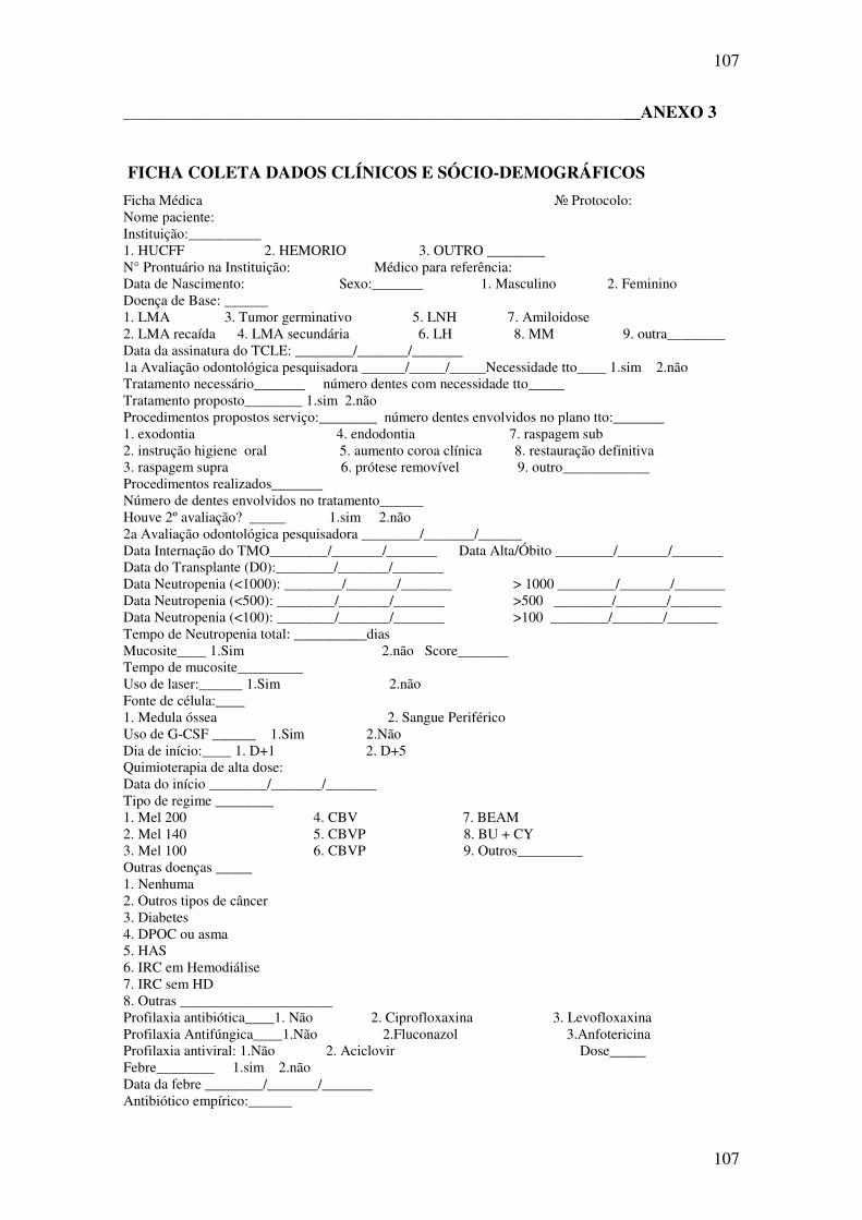
107
107
_______________________________________________________________________ANEXO 3
FICHA COLETA DADOS CLÍNICOS E SÓCIO-DEMOGRÁFICOS
Ficha Médica № Protocolo: Nome paciente: Instituição:__________ 1. HUCFF 2. HEMORIO 3. OUTRO ________ N° Prontuário na Instituição: Médico para referência: Data de Nascimento: Sexo:_______ 1. Masculino 2. Feminino Doença de Base: ______ 1. LMA 3. Tumor germinativo 5. LNH 7. Amiloidose 2. LMA recaída 4. LMA secundária 6. LH 8. MM 9. outra________ Data da assinatura do TCLE: ________/_______/_______ 1a Avaliação odontológica pesquisadora ______/_____/_____Necessidade tto____ 1.sim 2.não Tratamento necessário_______ número dentes com necessidade tto_____ Tratamento proposto________ 1.sim 2.não Procedimentos propostos serviço:________ número dentes envolvidos no plano tto:_______ 1. exodontia 4. endodontia 7. raspagem sub 2. instrução higiene oral 5. aumento coroa clínica 8. restauração definitiva 3. raspagem supra 6. prótese removível 9. outro____________ Procedimentos realizados_______ Número de dentes envolvidos no tratamento______ Houve 2º avaliação? _____ 1.sim 2.não 2a Avaliação odontológica pesquisadora ________/_______/______ Data Internação do TMO________/_______/_______ Data Alta/Óbito ________/_______/_______ Data do Transplante (D0):________/_______/_______ Data Neutropenia (<1000): ________/_______/_______ > 1000 ________/_______/_______ Data Neutropenia (<500): ________/_______/_______ >500 ________/_______/_______ Data Neutropenia (<100): ________/_______/_______ >100 ________/_______/_______ Tempo de Neutropenia total: __________dias Mucosite____ 1.Sim 2.não Score_______ Tempo de mucosite_________ Uso de laser:______ 1.Sim 2.não Fonte de célula:____ 1. Medula óssea 2. Sangue Periférico Uso de G-CSF ______ 1.Sim 2.Não Dia de início:____ 1. D+1 2. D+5 Quimioterapia de alta dose: Data do início ________/_______/_______ Tipo de regime ________ 1. Mel 200 4. CBV 7. BEAM 2. Mel 140 5. CBVP 8. BU + CY 3. Mel 100 6. CBVP 9. Outros_________ Outras doenças _____ 1. Nenhuma 2. Outros tipos de câncer 3. Diabetes 4. DPOC ou asma 5. HAS 6. IRC em Hemodiálise 7. IRC sem HD 8. Outras _____________________ Profilaxia antibiótica____1. Não 2. Ciprofloxaxina 3. Levofloxaxina Profilaxia Antifúngica____1.Não 2.Fluconazol 3.Anfotericina Profilaxia antiviral: 1.Não 2. Aciclovir Dose_____ Febre________ 1.sim 2.não Data da febre ________/_______/_______ Antibiótico empírico:______

108
108
1. Cefepime 2. pip-tazo 3. imipenem 4. não Associou Vancomicina:__________ 1. Sim 2. Não Tempo de uso de antimicrobiano:________ Dias de febre:_______ Classificação de episódio febril: (1) Sem NF (2) FOO (3) Microbiologicamente documentada s/ bacteremia (4) Clinicamente documentada (5) Bacteremia Documentação de infecção:_______ 1.Sim 2. Não Qual: _______________________ Agente: ______________________ Infecção fungica: __________ 1.Sim 2.não Local: ___________________ Qual: _____________________ Óbito:_____________ 1. Sim 2. Não Dados Sócio-econômicos Nome: Naturalidade: Nacionalidade: Ocupação: Endereço: Bairro: Cidade: UF: CEP: Telefone: Celular: Escolaridade: Estado civil:_______ 1. Solteiro 2. Casado / Companheiro 3. Divorciado 4.viúvo(a) Qual é a sua raça?______1. branca 2. parda 3. negra 4. amarela 5. indígena Fumante?_______ 1. Sim 2. Não Em caso afirmativo: Quantos cigarros por dia?_______fuma a quanto tempo____anos Se parou de fumar, quanto tempo faz?__________ Classificar posteriormente:_______ Fumante pesado: => 1 maço por dia 1. fumante leve: até menos de uma carteira por dia 2. ex-fumante pesado 3. ex-fumante leve 4.nunca fumou Carga tabagica_________ Qual a sua renda familiar (somatório da renda líquida de todos os moradores). Considere o salário mínimo de R$ 400,00)? 1. < 3 salários mínimos 2. de 3 a 6 salários mínimos 3. > 6 salários mínimos Qual foi a última vez que você foi ao dentista?_____ 1.Há menos de 6 meses 2. Menos de 1 ano 3.Menos de 2 anos 4. Menos de 5 anos 5. Mais de 5 anos 6. Não lembra Qual o tratamento realizado?______ 1. exodontia 4. endodontia 7. raspagem sub 2. Instrução higiene oral 5. aumento coroa clínica 8. restauração definitiva 3. raspagem supra 6. prótese removível 9. outro___________
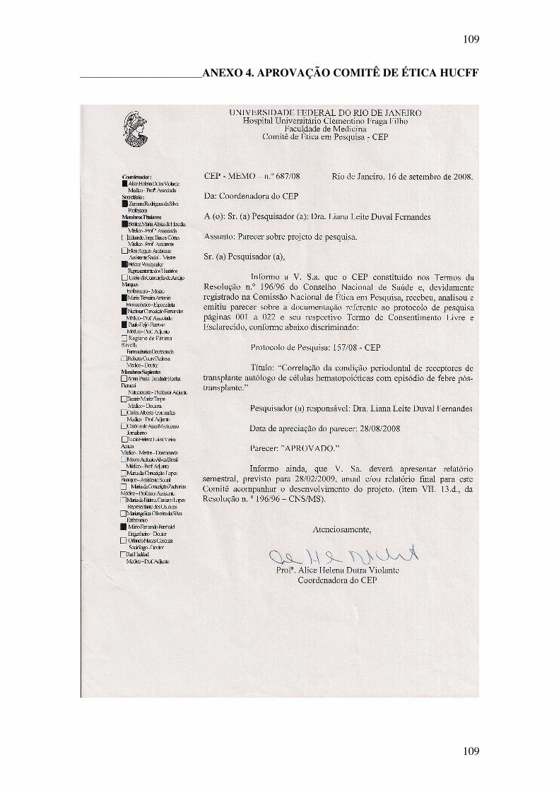
109
109
__________________________ANEXO 4. APROVAÇÃO COMITÊ DE ÉTICA HUCFF

110
110
_________________ANEXO. 5 APROVAÇÃO COMITÊ DE ÉTICA HEMORIO

111
111
_____________________________________ANEXO 6. FICHA EXAME ORAL

112
112
____________________________________________________________ANEXO 7. FICHA EXAME DE MUCOSA PÓS-TCTH

113
113
Livros Grátis( http://www.livrosgratis.com.br )
Milhares de Livros para Download: Baixar livros de AdministraçãoBaixar livros de AgronomiaBaixar livros de ArquiteturaBaixar livros de ArtesBaixar livros de AstronomiaBaixar livros de Biologia GeralBaixar livros de Ciência da ComputaçãoBaixar livros de Ciência da InformaçãoBaixar livros de Ciência PolíticaBaixar livros de Ciências da SaúdeBaixar livros de ComunicaçãoBaixar livros do Conselho Nacional de Educação - CNEBaixar livros de Defesa civilBaixar livros de DireitoBaixar livros de Direitos humanosBaixar livros de EconomiaBaixar livros de Economia DomésticaBaixar livros de EducaçãoBaixar livros de Educação - TrânsitoBaixar livros de Educação FísicaBaixar livros de Engenharia AeroespacialBaixar livros de FarmáciaBaixar livros de FilosofiaBaixar livros de FísicaBaixar livros de GeociênciasBaixar livros de GeografiaBaixar livros de HistóriaBaixar livros de Línguas
Baixar livros de LiteraturaBaixar livros de Literatura de CordelBaixar livros de Literatura InfantilBaixar livros de MatemáticaBaixar livros de MedicinaBaixar livros de Medicina VeterináriaBaixar livros de Meio AmbienteBaixar livros de MeteorologiaBaixar Monografias e TCCBaixar livros MultidisciplinarBaixar livros de MúsicaBaixar livros de PsicologiaBaixar livros de QuímicaBaixar livros de Saúde ColetivaBaixar livros de Serviço SocialBaixar livros de SociologiaBaixar livros de TeologiaBaixar livros de TrabalhoBaixar livros de Turismo