Embed Size (px)
Citation preview

UNIVERSIDADE FEDERAL DO RIO GRANDE DO SUL
INSTITUTO DE CIÊNCIAS BÁSICAS DA SAÚDE
DEPARTAMENTO DE BIOQUÍMICA
Avaliação do estado redox e de parâmetros gliais no hipocampo de
ratos submetidos a exercício físico moderado e restrição calórica
KATIANE SANTIN
Orientador
Prof. Carlos Alberto Gonçalves
Co-orientadora
Profa. Christianne Salbego
Dissertação apresentada ao curso de Pós-graduação em Ciências Biológicas
– Bioquímica da Universidade Federal do Rio Grande do Sul como requisito
parcial à obtenção do Grau de Mestre em Bioquímica
Porto Alegre, janeiro de 2010

Livros Grátis
http://www.livrosgratis.com.br
Milhares de livros grátis para download.
ii
Avaliação do estado redox e de parâmetros gliais no hipocampo de
ratos submetidos a exercício físico moderado e restrição calórica
KATIANE SANTIN
iii
“Toda a nossa ciência, comparada com a realidade,
é primitiva e infantil – e, no entanto, é a
coisa mais preciosa que temos.”
Albert Einstein
iv
Agradecimentos
Em primeiro lugar, quero agradecer a Deus por mais um objetivo alcançado em
minha vida! Quero agradecer a minha família, em especial a minha mãe Carmem, que
sempre fez de tudo para que pudéssemos estudar e ser quem nós somos. Aos meus
irmãos: Rosane, Rogério e Thiago, pela amizade e pela força de sempre. Ao meu pai
Otávio, que tenho certeza, sempre me acompanha.
Ao meu orientador CA, que sempre me atendeu e procurou, na medida do
possível, me acalmar e solucionar minhas dúvidas, sem nunca ter colocado mais
pressão sobre todas as coisas com as quais eu sempre me pressionei, e também por
ter me ensinado a usar o End Note em pequenas lições muito valiosas. À Profa.
Carmem, pela simpatia de sempre e por sempre ter tido tempo e contribuído quando
eu precisei.
Aos meus colegas queridos do lab 33:
Marina: amiga desde sempre que me deu uma baita força na seleção e no
planejamento do experimento, sou tua fã;
André: espalhando alegria e competência pelo lab sempre se mostrou solicito
para com meus problemas (grande dançarino);
Dani: pequena grande pessoa (te adoro muito), quase perdeu um dedo
removendo os músculos dos ratinhos no meu experimento;
Pati: guerreira, meiga, simpática, divertida, temos sorte em ter tua amizade;
Lets: competência e dedicação em pessoa, ainda não te vi tocar mas sou tua fã
tb;
Paulinha: cronilda, tu é uma figuraça muita culta bixo, amiga te adoro demais;
E ainda: Lari, Ana, Lê e Melissa, Caro, Rafa, Fafá, Regina, Krista, Lucas,
Douglas, Jaque, Ale e Joana, Fran e Ana Jujú, por todo o incentivo, o exemplo, o
trabalho, a preocupação e as palavras amigas. Mas principalmente, agradeço a todos
do lab 33 pelo empenho no meu projeto, que eu sei que não foi pouco. Ah, e a Ale do
apoio, por suas histórias divertidíssimas.
Aos colegas do lab 32, principalmente ao Ricardo, meu colega desde os tempos
da ESEF, que de maneira solidária e brilhante assumiu junto comigo a execução dos
v
experimentos referentes ao estresse oxidativo e colaborou com a arte de escrever
tudo isso.
Aos colegas do lab 37, Ju Hoppe, Ricardo, Ana, Dani, Rudimar, Fabri pela
amizade, e principalmente a Profa. Christianne que me orientou durante toda minha
iniciação científica e sempre foi um modelo de pesquisadora e professora.
A Fê Cechetti, grande pessoa, ótima profissional, que sempre madrugou para
me ajudar e nunca desanimou ou me deixou desanimar durante as dificuldades com
os treinamentos físicos dos ratos.
As minhas queridas e dedicadas bolsistas Thanielle e Raquel, que me ajudaram
muito com os “nossos” ratinhos e tenho certeza que aprenderam muito também
(manipular ratos não é pra qualquer um, né?!).
A minhas colegas e amigas da faculdade Paula, Tainá e Letícia, por termos
seguido nossos caminhos, mas nos encontrarmos com a amizade de sempre.
As minhas amigas Giovana, Sílvia, Marcela e Fernanda, que ouviram minhas
reclamações, deram conselhos e me ajudaram muito, como em tudo na vida, não só
no mestrado. Em especial, a Marcela e a Fernanda que me ajudaram a identificar
cerca de 1000 eppendorffs.
Aos meus colegas do Centro de Microscopia Eletrônica, Ana, Karina, Aline,
Moema, Chris, Leandro e Paulão, que compartilharam minhas alegrias e tristezas no
início do mestrado e, agora no fim do mestrado, aos meus colegas do Laboratório de
Análises Clínicas da Faculdade de Farmácia, Mari, Karlize, Jú, Majô, Carla, Michele,
Dariana, e em especial a Ana Lúcia Antunes e ao Profº Paulo Saraiva, pela
compreensão, pela confiança e pelas palavras de incentivo de sempre.
E, ao meu namorado Rodrigo, que foi, desde a prova para a seleção do
mestrado, passando pelo treinamento com os ratos, a execução dos experimentos e a
tão complicada tarefa de sentar e me concentrar em escrever a dissertação, o meu
maior incentivador, por isso os meus sinceros agradecimentos por estar ao meu lado
(mesmo quando longe) e o meu Eu Te Amo!

6
Sumário
Agradecimentos ......................................................................................................iv
Sumário................................................................................................................... 6
Parte I...................................................................................................................... 7
Resumo................................................................................................................... 8
Abstract ................................................................................................................... 9
Lista de Abreviaturas............................................................................................. 10
Introdução ............................................................................................................. 11
1. Estresse Oxidativo...................................................................................... 11
1.1 Equilíbrio Redox no Sistema Nervoso Central ......................................... 12
2. Sistema Nervoso Central............................................................................ 15
2.1 Marcadores Gliais em Astrócitos.............................................................. 17
2.1.1 S100B ................................................................................................... 18
2.1.2 Proteína Glial Fibrilar Ácida (GFAP)...................................................... 19
2.2 Transmissão Glutamatérgica ................................................................... 21
2.2.1 Captação de Glutamato e Atividade da Glutamina Sintetase................ 21
3. Exercício físico ........................................................................................... 24
4. Restrição Calórica ...................................................................................... 25
Objetivo Geral ....................................................................................................... 27
Objetivos Específicos ............................................................................................ 27
Parte II................................................................................................................... 28
Moderate exercise training and chronic caloric restriction modulate redox status in
rat hippocampus
Parte III.................................................................................................................. 70
Discussão.............................................................................................................. 71
Referências Bibliográficas..................................................................................... 79

7
Parte I
8
Resumo
O desequilíbrio entre defesas antixiodantes e espécies reativas, conhecido
como estresse oxidativo, tem sido relacionado com o desenvolvimento de doenças
neurodegenerativas. Diversos estudos indicam que o exercício físico traz
benefícios à saúde, inclusive para o sistema nervoso central, que estariam
relacionados à adaptação das defesas antioxidantes. Além disso, pesquisas
sugerem que o exercício associado a uma condição de retrição calórica pode
reduzir tanto a incidência quanto a severidade de desordens neurológicas. O
objetivo deste trabalho foi avaliar a influência de exercício físico e restrição
calórica no estado redox e em parâmetros gliais no hipocampo de ratos. Quarenta
ratos Wistar machos, com idade de aproximadamente 60 dias, foram divididos em
4 grupos: alimentados ad libitum e sedentários (AS), alimentados ad libitum e
exercitados (AE), em restrição calórica e sedentários (RS) e em restrição calórica
e exercitados (RE). Os animais foram sacrificados após 3 meses de restrição
calórica (30% da ingestão de ração) e exercício físico (de intensidade moderada
em esteira), sendo o hipocampo cirurgicamente removido para avaliação dos
parâmetros de interesse. Foram realizadas também análises bioquímicas séricas.
Os grupos AE, RS and RE apresentaram um aumento nos níveis de glutationa
reduzida (GSH) e na reatividade antioxidante total (TAR). Os níveis de
nitratos/nitritos diminuiram somente no grupo RE. Foi observado um decréscimo
no conteúdo de proteínas carboniladas nos grupos AE, RS e RE, enquanto
nenhuma modificação foi detectada no ensaio de TBARS (espécies reativas ao
ácido tiobarbitúrico). Não houve diferenças no potencial antioxidante reativo total
(TRAP), na atividade da superóxido dismutase (SOD), no conteúdo de S100B e
GFAP, entretanto, a restrição calórica foi capaz de aumentar a captação de
glutamato (grupos RS e RE) e a atividade da glutamina sintetase (GS) (grupo RS).
O exercício físico, a restrição calórica e a combinação de ambos são capazes de
atenuar o dano oxidativo no hipocampo, possivelmente através da modulação de
parâmetros gliais, podendo ser utilizados como estratégias na prevenção de
doenças neurodegenerativas.
9
Abstract
Imbalanced redox status has been related to neurodegenerative disease
development. Some studies demonstrate that physical activity produces
antioxidant adaptations that have health benefits, including in the nervous system.
Additionally, available data suggest exercise and a caloric restriction regimen may
reduce both the incidence and severity of neurological disorders. Therefore, our
aim was to compare hippocampal redox status and glial parameters among
sedentary, trained, caloric-restricted sedentary and caloric-restricted trained rats.
Main methods: Forty male adult rats were divided into 4 groups: ad libitum-fed
sedentary (AS), ad libitum-fed exercise training (AE), calorie-restricted sedentary
(RS) and calorie-restricted exercise training (RE). The caloric restriction (decrease
of 30% in food intake) and exercise training (moderate in a treadmill) were carried
out for 3 months. Thereafter hippocampus was surgically removed, and redox and
glial parameters were assessed. Key findings: Increases in reduced glutathione
(GSH) levels and total antioxidant reactivity (TAR) were observed for AE, RS and
RE groups. The nitrite/nitrate levels decreased only at RE. We found a decrease in
carbonyl content for the AE, RS and RE groups, while no modifications were
detected in thiobarbituric acid reactive substances (TBARS). Total reactive
antioxidant potential (TRAP), superoxide dismutase (SOD) activity, S100B and glial
fibrilary acid protein (GFAP) content did not change, but caloric restriction was able
to increase glutamine synthetase (GS) activity (RS group) and glutamate uptake
(RS and RE groups). Significance: Exercise training, caloric restriction and both
combined can decrease oxidative damage in the hippocampus, possibly involving
modulation of astroglial function, and could be used as a strategy for the
prevention of neurodegenerative diseases.
10
Lista de Abreviaturas
AMPA – Ácido α-amino-3-hidróxi-5-metil-4-isoxazolenopropionato (α-amino-3-
hydroxyl-5-methyl-4-isoxazole-propionate)
AST - Aspartato aminotransferase (Aspartate aminotransferase Transaminase)
DNA – Ácido Desoxirribonucleico (Deoxyribonucleic Acid)
ER – Espécies Reativas (Reactive Species)
GFAP – Proteína Glial Fibrilar Ácida (Glial Fibrilary Acid Protein)
GPx- Glutationa Peroxidase
GS – Glutamina Sintetase
GSH – Glutationa
GSSG – Glutationa Oxidada
H2O2 – Peróxido de Hidrogênio
4-HNE – 4-Hidroxinonenal
NMDA – N-Metil-D-Aspartato
NO• – Óxido Nitríco
nNOS – Óxido Nítrico Sintase Neuronal
NT – Neurotransmissor
O2•- – Ânion superóxido
ONOO•- - peroxinitrito •OH – Radical hidroxil
RC – Restrição Calórica
SNC – Sistema Nervoso Central
SOD – Superóxido Dismutase
TBARS – Espécies Reativas ao Ácido Tiobarbitúrico

11
Introdução
1. Estresse Oxidativo
Espécies reativas (ER) é um termo genérico usado para definir espécies
(átomos, íons ou moléculas) derivadas do oxigênio e nitrogênio molecular que são
reativas per se, ou que podem ser facilmente convertidas em espécies reativas;
algumas ER são radicais livres (Matsuo and Kaneko, 2000).
As ER podem ser geradas por fontes endógenas ou exógenas. Entre as
principais fontes endógenas estão a cadeia mitocondrial transportadora de
elétrons, a degradação de ácidos graxos nos peroxissomas, os mecanismos de
detoxificação mediados pelo complexo enzimático P-450 e o processo de
fagocitose; dentre as fontes exógenas estão as radiações, o cigarro e os solventes
orgânicos (Gutteridge and Halliwell, 2000).
A geração de ER in vivo é um processo fisiológico que ocorre de maneira
controlada pelos mecanismos antioxidantes celulares. Quando há um desequilíbrio
oxidante x antioxidante, ou seja, geração descontrolada de ER, diminuição das
defesas antioxidantes, ou ambos, ocorre a oxidação de biomoléculas, como ácidos
nucléicos, proteínas e lipídeos, o que pode alterar a informação genética,
desnaturar proteínas, inativar enzimas e desorganizar biomembranas, processo
este denominado estresse oxidativo (Sies, 1991).
A mudança mais significativa que ocorre durante o exercício físico é um
aumento na taxa metabólica, ligado a um aumento no consumo de oxigênio. Um
12
fluxo de oxigênio maior que a capacidade mitocondrial pode provocar “vazamento”
de elétrons na cadeia transportadora, levando a um aumento da geração de ER,
contribuindo para o estresse oxidativo (Jenkins, 1993). No entanto, alguns estudos
sugerem que com o treinamento físico ocorre uma adaptação do sistema
antioxidante celular, aumentando a atividade de enzimas antioxidantes, o que
reduziria o dano oxidativo (Powers et al., 1994, Leeuwenburgh et al., 1997, Radak
et al., 2008).
Além dos antioxidantes enzimáticos, existe uma segunda categoria de
antioxidantes, denominados não-enzimáticos, representada pelas vitaminas, que
não podem ser sintetizadas ou induzidas e devem ser absorvidas através de uma
alimentação balanceada (Berger, 2005). Então, a nutrição possui um impacto
significativo no sistema antioxidante celular. Estudos demonstraram que animais
submetidos à restrição calórica, mas não mal-nutridos, apresentaram menos dano
mediado por ER, incluindo uma diminuição da peroxidação lipídica e perda da
fluidez de membrana, de proteínas com dano oxidativo (proteínas carboniladas), e
do dano oxidativo ao DNA (Merry, 2002, Barja, 2004).
1.1 Equilíbrio Redox no Sistema Nervoso Central
A demanda de oxigênio do cérebro humano é estimada em cerca de 20%
do oxigênio consumido por todo o corpo, mesmo o cérebro representando 2% do
peso corporal (Somani and Husain, 2000). O cérebro está entre os maiores órgãos
geradores de ER e, comparado com outros órgãos, é especialmente vulnerável ao

13
estresse oxidativo por estar inadequadamente equipado com defesas
antioxidantes e por conter lipídeos com ácidos graxos insaturados em abundância,
alvo da peroxidação lipídica, o que poderia ajudar a esclarecer o dano oxidativo
observado no decorrer de doenças neurodegenerativas (Dringen et al., 2000,
Halliwell, 2006).
Aproximadamente de 2 a 4% do oxigênio consumido por mitocôndria é
desviado para formar ânion superóxido (O2•-). A enzima superóxido dismutase
(SOD) converte o superóxido em peróxido de hidrogênio (H2O2), que é
subsequentemente convertido em água e oxigênio molecular pela glutationa
peroxidase (GPx) ou pela catalase (Dringen et al., 2000). Foi demonstrado que a
atividade da catalase no cérebro é baixa e confinada aos peroxissomos (Gaunt
and de Duve, 1976).
A GPx corresponde a uma família de enzimas contendo selênio
(Brigelius-Flohe, 1999), que remove o H2O2, por reduzir esta ER numa reação
acoplada com a oxidação da glutationa (GSH), um tripeptídeo formado por
glutamato, cisteína e glicina, contendo grupamento tiol.
A glutationa oxidada (GSSG) consiste em duas GSH ligadas por uma
ponte dissulfeto, que pode ser reconvertida em GSH pelo complexo de enzimas
glutationa redutase (Halliwell, 2006). A GSH é um dos maiores antioxidantes no
sistema nervoso central (SNC) (Dringen, 2000b) (Figura 1) com uma concentração
em torno de 2-3 mM, que é bem maior que a do sangue e a do líquor (Cooper and
Kristal, 1997).

14
Figura 1: Proteção da GSH contra estresse oxidativo. Adaptado de Aoyama et
al. 2008
Quando não removido, o H2O2 pode reagir com o ferro, encontrado de
forma abundante ligado a proteínas no SNC (Zecca et al., 2004), via Reação de
Fenton e formar radicais hidroxil (•OH), que podem desencadear a peroxidação
lipídica (Youdim et al., 1989) .
O SNC também possui em abundância o óxido nítrico (NO•) produzido
pela óxido nítrico sintase neuronal (nNOS) (Zhou and Zhu, 2009), que atua como
vasodilatador e neurotransmissor não-clássico (Garthwaite, 2008). O superóxido e
o NO• isolados não são tóxicos in vivo, mas quando combinados podem reagir,
gerando um oxidante fortemente tóxico, o peroxinitrito (ONOO•-). A presença de
ONOO•- leva à oxidação de proteínas, lipídeos, e DNA, bem como à nitração de
aminoácidos, principalmente tirosina, e rapidamente inativa as enzimas
mitocondriais resultando em falha da produção de energia (Pacher et al., 2007).
A peroxidação lipídica induzida por ER leva à conversão de ácidos graxos
poliinsaturados em aldeídos altamente reativos como o 4-hidroxinonenal (4-HNE).
15
4-HNE pode danificar facilmente os transportadores de glutamato, diminuindo a
sua remoção da fenda sináptica (Mattson and Chan, 2003). 4-HNE também inibe a
atividade enzimática de GPx, aumentando por consequência os níveis de H2O2
(Bosch-Morell et al., 1999).
O esqueleto protéico e as cadeias laterais da maioria das proteínas
apresentem aminoácidos suscetíveis à oxidação, porém a adição não-enzimática
de aldeídos e cetonas a resíduos de aminoácidos específicos (carbonilação)
constitui a maior e mais comum alteração oxidativa a proteínas (Berlett and
Stadtman, 1997).
Diversas ER (incluindo ONOO•-) podem diminuir a captação de glutamato
pelas células gliais e inativar a enzima glutamina sintetase (GS), prejudicando a
conversão de glutamato em glutamina (Aksenov et al., 1997).
2. Sistema Nervoso Central
O sistema nervoso central (SNC) é composto por dois tipos celulares –
neurônios e células gliais (Jenssen, 2004). Os neurônios foram por muito tempo
considerados os elementos celulares responsáveis pelo processamento da
informação, enquanto que as células gliais eram reconhecidas apenas pelo seu
papel de suporte inerte para os neurônios (Volterra and Meldolesi, 2005).
Atualmente sabe-se que as células da glia atuam como um componente ativo em
funções cerebrais essenciais (Van Eldik and Wainwright, 2003).

16
As células da glia são divididas em dois grupos principais: a macroglia,
composta de astrócitos, oligodendrócitos e células ependimárias; e a microglia,
composta de células fagocíticas envolvidas em resposta inflamatória (Perea and
Araque, 2005).
Os astrócitos constituem aproximadamente 50% de toda massa cerebral,
sendo então, as células gliais mais abundantes do SNC (Gee and Keller, 2005)
(Figura 2). Eles são divididos em dois tipos celulares: os protoplasmáticos, na
substância cinzenta (freqüentemente ramificados e com largas expansões); e os
fibrosos, na substância branca (células cilíndricas e longas, com menos
ramificações) (Young, 1991).
Existe um novo conceito em transmissão sináptica, a sinapse de três
elementos: onde os astrócitos trocam informações com neurônios pré e pós-
sinápticos e participam da neurotransmissão como elementos regulatórios
dinâmicos (Perea and Araque, 2005). Astrócitos e neurônios possuem então, um
sofisticado sistema de comunicação recíproca que pode regular a liberação de
neurotransmissores, a excitabilidade neuronal e a transmissão sináptica
(Carmignoto, 2000).

17
Figura 2: Os principais grupos celulares do SNC e suas inter-relações.
Adaptado de www.indiana.edu
2.1 Marcadores Gliais em Astrócitos
Os astrócitos participam de vários processos fisiológicos e metabólicos
responsáveis por manter a homeostase do SNC. Estas células possuem três
proteínas específicas usadas como marcadoras: a S100B; a GFAP (proteína glial
fibrilar ácida); e a glutamina sintetase (responsável pela formação de glutamina a
partir de glutamato). Insultos físicos e metabólicos provocam alterações rápidas
nas células gliais, processo conhecido como astrogliose reativa, caracterizada por

18
um aumento na expressão dos marcadores gliais citados anteriormente (Baydas et
al., 2003).
2.1.1 S100B
A S100B é uma proteína de 21 kDa, ligante de cálcio do tipo EF-hand
(hélice-loop-hélice). Essa proteína foi isolada há mais de 40 anos (Moore, 1965) a
partir de extrato de cérebro bovino e a denominação “S100” é devida a sua
solubilidade em sulfato de amônio 100% (Van Eldik and Wainwright, 2003).
Estruturalmente, a proteína S100 forma homodímeros constituídos de
duas subunidades β unidos por pontes dissulfeto e capazes de se ligarem a
proteínas alvos (Donato, 2003).
A S100B é produzida e secretada principalmente por astrócitos e exerce
efeitos parácrinos em neurônios e microglia, e autócrinos em astrócitos (Ponath et
al., 2007). Entretanto, outras fontes extracerebrais como, células adiposas,
também podem secretar S100B. O mecanismo de secreção de S100B ainda não
foi identificado (Rothermundt et al., 2003).
A S100B possui funções intra e extracelulares. Intracelularmente, a
S100B inibe a fosforilação de diversas proteínas, tais como GFAP, p53, entre
outras; está envolvida na regulação do metabolismo energético cerebral; modula a
proliferação e a diferenciação de neurônios e células gliais. Além disso, a S100B
regula a homeostase do Ca2+, interage com muitas funções imunológicas do SNC

19
e influencia a integridade do citoesqueleto (Donato, 2003, Rothermundt et al.,
2003).
Extracelularmente, a proteína S100B pode ter efeitos tróficos ou tóxicos,
dependendo da concentração secretada. Em baixas concentrações (doses nM), a
S100B exerce efeito neurotrófico, promovendo o crescimento de neuritos,
aumentando a sobrevivência de neurônios durante o desenvolvimento e após
dano ao SNC (Tramontina et al., 2002, Gottfried et al., 2003, Tramontina et al.,
2006a) e protegendo neurônios contra a excitotoxicidade do glutamato (Ahlemeyer
et al., 2000, Tramontina et al., 2006a). Este efeito trófico também é exercido em
astrócitos.
Em altas concentrações (doses µM), a S100B exerce um efeito
neurotóxico por induzir a apoptose através de mecanismo dependente da
liberação de citocinas pró-inflamatórias como a interleucina-6, interleucina-1β e
fator de necrose tumoral-α na glia e interleucina-6 em neurônios (Van Eldik and
Wainwright, 2003) e através de estímulo de secreção de óxido nítrico por
astrócitos e microglia (Donato, 2001).
2.1.2 Proteína Glial Fibrilar Ácida (GFAP)
A GFAP é uma proteína estrutural e constitui a subunidade protéica de
filamentos intermediários do tipo III do citoesqueleto glial (Rodnight et al., 1997,
Gomes et al., 1999).
20
Inicialmente isolada de lesões cerebrais (placas) de pacientes com
esclerose múltipla (Eng et al., 2000), a GFAP é reconhecida e amplamente
utilizada como um marcador de astrócitos e de tumores de linhagem astrocítica
(Pekny and Pekna, 2004). Durante o desenvolvimento do SNC e na gliose reativa
ocorre um aumento na expressão de GFAP (Gomes et al., 1999).
Estruturalmente, a GFAP é um polímero que consiste em uma região
amino-terminal não-helicoidal altamente básica, uma região carboxi-terminal não-
helicoidal e responsável pela ligação entre os monômeros e uma região central
formada por uma α-hélice, cuja sequência de aminoácidos é conservada em
relação a outras proteínas filamentosas intermediárias. Diferenças na estrutura
dessas proteínas são normalmente evidenciadas nos aminoácidos da região
amino-terminal (Alberts et al., 2002).
A polimerização da GFAP envolve diversos passos. Inicialmente, um
dímero é formado através da pareação entre dois monômeros paralelos.
Posteriormente, dímeros anti-paralelos interagem através de resíduos na região
central formando um tetrâmero ou protofilamento e, finalmente, vários
protofilamentos se unem dando origem ao polímero (Rodnight et al., 1997).
A fosforilação de sítios específicos de proteínas de filamentos
intermediários, como a GFAP, regula o equilíbrio dinâmico entre a sua forma
polimerizada e despolimerizada, desempenhando um importante papel na mitose
(Rodnight et al., 1997).

21
2.2 Transmissão Glutamatérgica
O aminoácido L-glutamato é o principal neurotransmissor (NT) excitatório
do SNC de mamíferos e exerce um importante papel na plasticidade neural e
neurotoxicidade (Nakanishi, 1992). O glutamato apresenta diversas funções,
incluindo indução e eliminação de sinapses, migração, diferenciação e morte
celular durante o desenvolvimento do SNC e, provavelmente, está envolvido em
muitos aspectos funcionais, como cognição, memória e aprendizagem (Danbolt,
2001).
O SNC contém uma grande quantidade de glutamato, mas somente uma
pequena fração deste NT (≅ 1µM) está presente no espaço extracelular. As
maiores concentrações são encontradas no interior dos terminais nervosos
(Danbolt, 2001).
O glutamato exerce seus efeitos através da interação com receptores de
glutamato localizados na superfície das células neuronais e gliais (Figura 2). Então
o glutamato ativa receptores ionotrópicos (NMDA, AMPA e Kainato) - que são
canais iônicos permeáveis a cátions; e receptores metabotrópicos (mGluRs 1-8)
que estão acoplados à proteína G (Matute, 2006).
2.2.1 Captação de Glutamato e Atividade da Glutamina Sintetase
O glutamato, após sua síntese, é estocado pelo sistema de transporte
presente nas vesículas que se encontram no terminal pré-sináptico. Quando
22
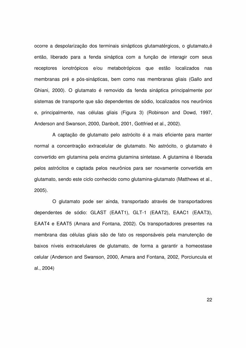
ocorre a despolarização dos terminais sinápticos glutamatérgicos, o glutamato,é
então, liberado para a fenda sináptica com a função de interagir com seus
receptores ionotrópicos e/ou metabotrópicos que estão localizados nas
membranas pré e pós-sinápticas, bem como nas membranas gliais (Gallo and
Ghiani, 2000). O glutamato é removido da fenda sináptica principalmente por
sistemas de transporte que são dependentes de sódio, localizados nos neurônios
e, principalmente, nas células gliais (Figura 3) (Robinson and Dowd, 1997,
Anderson and Swanson, 2000, Danbolt, 2001, Gottfried et al., 2002).
A captação de glutamato pelo astrócito é a mais eficiente para manter
normal a concentração extracelular de glutamato. No astrócito, o glutamato é
convertido em glutamina pela enzima glutamina sintetase. A glutamina é liberada
pelos astrócitos e captada pelos neurônios para ser novamente convertida em
glutamato, sendo este ciclo conhecido como glutamina-glutamato (Matthews et al.,
2005).
O glutamato pode ser ainda, transportado através de transportadores
dependentes de sódio: GLAST (EAAT1), GLT-1 (EAAT2), EAAC1 (EAAT3),
EAAT4 e EAAT5 (Amara and Fontana, 2002). Os transportadores presentes na
membrana das células gliais são de fato os responsáveis pela manutenção de
baixos níveis extracelulares de glutamato, de forma a garantir a homeostase
celular (Anderson and Swanson, 2000, Amara and Fontana, 2002, Porciuncula et
al., 2004)
23
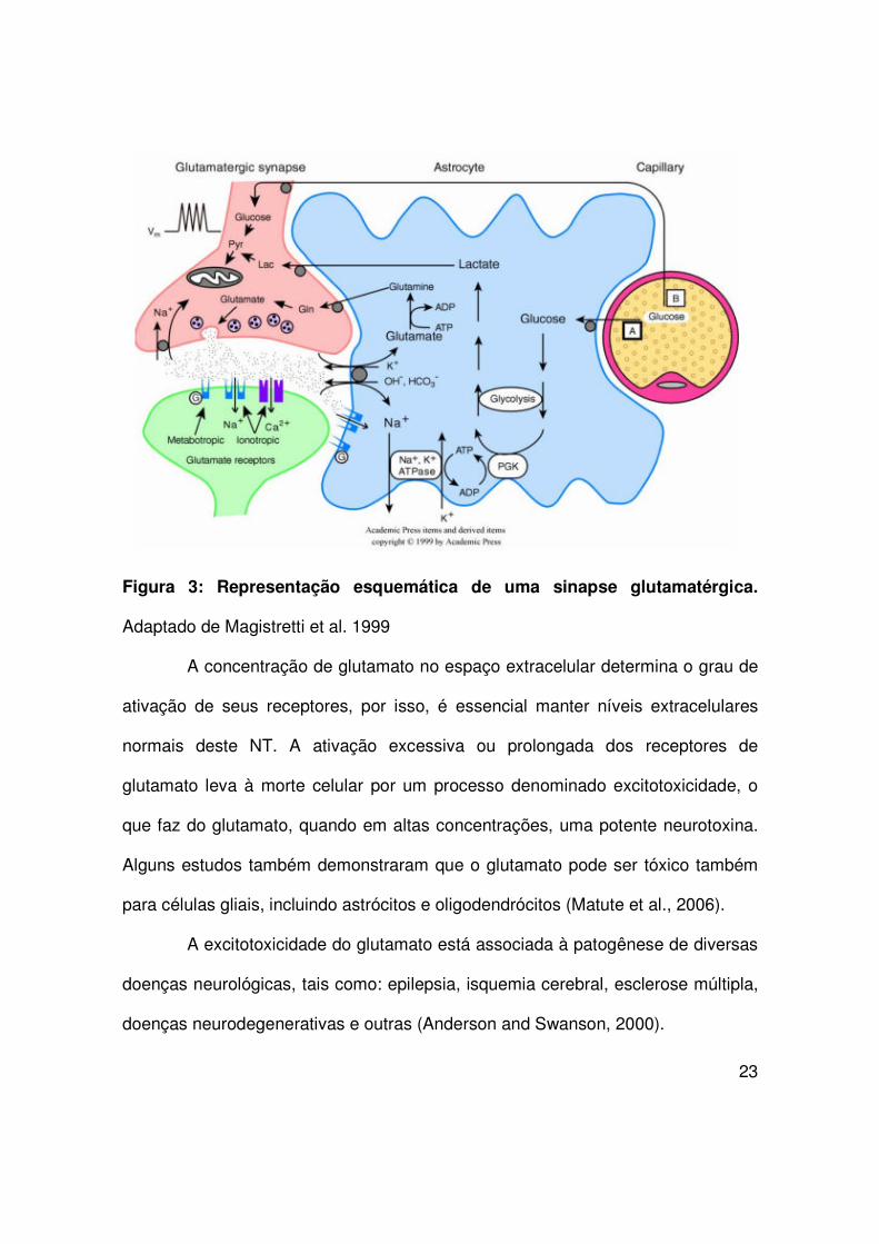
Figura 3: Representação esquemática de uma sinapse glutamatérgica.
Adaptado de Magistretti et al. 1999
A concentração de glutamato no espaço extracelular determina o grau de
ativação de seus receptores, por isso, é essencial manter níveis extracelulares
normais deste NT. A ativação excessiva ou prolongada dos receptores de
glutamato leva à morte celular por um processo denominado excitotoxicidade, o
que faz do glutamato, quando em altas concentrações, uma potente neurotoxina.
Alguns estudos também demonstraram que o glutamato pode ser tóxico também
para células gliais, incluindo astrócitos e oligodendrócitos (Matute et al., 2006).
A excitotoxicidade do glutamato está associada à patogênese de diversas
doenças neurológicas, tais como: epilepsia, isquemia cerebral, esclerose múltipla,
doenças neurodegenerativas e outras (Anderson and Swanson, 2000).
24
3. Exercício físico
Diversos estudos epidemiológicos e em animais demonstram que o
exercício físico regular atua de forma periférica melhorando a saúde
cardiovascular, a densidade mineral óssea e reduzindo fatores de risco para
doenças como câncer, diabetes, hipertensão, isquemia e infarto agudo do
miocárdio. Adicionalmente, o exercício físico influencia de forma elementar a
função cerebral por atuar em alvos no sistema nervoso central (SNC) que
melhoram a aprendizagem e memória, aliviam a depressão e previnem o declínio
cognitivo e a demência (Nichol et al., 2007), observados durante o
envelhecimento, em decorrência de doenças neurodegenerativas como doença de
Alzheimer, Parkinson, Huntington e também, em decorrência de acidente vascular
cerebral (Prolla and Mattson, 2001, Kramer and Erickson, 2007).
As mudanças estruturais e funcionais induzidas pelo exercício são
estudadas em várias regiões cerebrais, sendo a região do hipocampo uma das
mais relatadas (Cotman et al., 2007). O hipocampo é uma estrutura localizada nos
lobos temporais do cérebro, considerada a principal sede da memória e importante
componente do sistema límbico (Bear et al., 2002). O exercício físico pode
aumentar a neurogênese no hipocampo (van Praag et al., 1999b), aumentar a
produção de fatores neurotróficos derivados do SNC (Neeper et al., 1996) e
melhorar a aprendizagem e memória (van Praag et al., 1999a).

25
Os mecanismos moleculares exatos pelos quais o exercício afeta a função
cerebral não estão completamente elucidados (Sutoo and Akiyama, 2003).
Embora existam dados controversos, decorrentes de distintas metodologias
empregadas, vem sendo demonstrado que o exercício físico pode ativar vias
celulares e moleculares que contribuem para neuroproteção (Cotman and
Berchtold, 2002).
4. Restrição Calórica
O aumento da expectativa de vida em resposta a uma restrição moderada
do suprimento alimentar, é comum em uma grande variedade de organismos,
incluindo leveduras, invertebrados como nematódeos e drosófilas, e mamíferos,
como roedores e, possivelmente, humanos (Partridge and Brand, 2005, Holloszy
and Fontana, 2007, Cruzen and Colman, 2009).
O primeiro estudo científico mundialmente reconhecido por aumentar a
expectativa de vida através de uma dieta restritiva foi publicado em 1935. Neste
estudo, os pesquisadores viram que alimentando ratos com uma dieta contendo
20% de celulose não digestível, a expectativa de vida média e máxima foi
incrivelmente aumentada (McCay, 1935).
Inicialmente, os estudos sobre RC permaneceram focados na questão da
expectativa de vida aumentada, posteriormente, foi dada atenção ao estudo dos
marcadores biológicos de doenças relacionadas ao envelhecimento como câncer,
doença cardiovascular e desordens neurodegenerativas (Swindell, 2008).

26
Uma restrição calórica moderada, sem atingir estado de desnutrição, de 30
a 40% menos que uma alimentação ad libitum durante toda a vida, pode diminuir a
incidência de diabete melitus tipo 2 (Astrup et al., 2001), desordens
neurodegenerativas (Patel et al., 2005), doença cardiovascular (Fontana 2004),
perda auditiva (Someya et al., 2007), atenuar a atrofia muscular relacionada ao
envelhecimento (McKiernan et al., 2004) e aumentar a longevidade de roedores e
primatas (Masoro, 2003, 2005).
Pesquisas sugerem que um decréscimo na ingestão de calorias pode
desacelerar as mudanças moleculares no SNC relacionadas ao envelhecimento e
que levam a um aumento de proteína glial fibrilar ácida (GFAP) e dano oxidativo a
proteínas e ao DNA (Dubey et al., 1996, Morgan et al., 1997).
Uma melhor compreensão dos mecanismos celulares e moleculares pelos
quais a restrição calórica e a atividade física influenciam a função e a estrutura
cerebral poderia conduzir ao desenvolvimento de novos agentes terapêuticos que
mimetizem os efeitos benéficos observados em decorrência de um estilo de vida
saudável, no qual estas práticas são fundamentais.

27
Objetivo Geral
Avaliar o estado redox e parâmetros gliais no hipocampo de ratos
submetidos a exercício físico moderado e restrição calórica.
Objetivos Específicos
� Analisar no hipocampo os efeitos do exercício físico moderado e da
restrição calórica sobre: conteúdo de GSH, níveis de nitratos e nitritos, potencial
antioxidante não enzimático, atividade da superóxido dismutase, carbonilação de
proteínas e peroxidação lipídica.
� Avaliar o conteúdo de GFAP e S100B em fatias hipocampais de ratos
submetidos a exercício físico moderado e restrição calórica;
� Avaliar a captação de glutamato e a atividade da glutamina sintetase em
fatias hipocampais de ratos submetidos a exercício físico moderado e restrição
calórica;

28
Parte II

29
Artigo a ser submetido à revista:
Life Sciences
30
Moderate exercise training and chronic caloric restriction modulate redox
status in rat hippocampus
Katiane Santina*, Ricardo Fagundes da Rochaa, Fernanda Cechettia, Marina Concli
Leitea, André Quincozes-Santosa, Daniela Fraga de Souzaa, Patrícia Nardina,
Letícia Rodriguesa,b, José Cláudio Fonseca Moreiraa, Carmem Gottfrieda,b,
Christiane Gazzana Salbegoa, Carlos Alberto Gonçalvesa,b
aDepartament of Biochemistry, Institute of Health Basic Sciences (ICBS), Federal
University of Rio Grande do Sul (UFRGS)
bPost-graduation Program of Neurosciences, Institute of Health Basic Sciences
(ICBS), Federal University of Rio Grande do Sul (UFRGS)
*Corresponding author
Katiane Santin
Rua Ramiro Barcelos, número 2600-anexo, 90035-003, Porto Alegre, RS, Brazil
Departamento de Bioquímica, Instituto de Ciências Básicas da Saúde (ICBS),
Universidade Federal do Rio Grande do Sul (UFRGS)
Phone: +55 51 33085566/ +55 51 33085412
Fax: +55 51 33085540
E-mail address: [email protected]

31
Abstract
Imbalanced redox status has been related to neurodegenerative disease
development. Some studies demonstrate that physical activity produces
antioxidant adaptations that have health benefits, including in the nervous system.
Additionally, available data suggest exercise and a caloric restriction regimen may
reduce both the incidence and severity of neurological disorders. Therefore, our
aim was to compare hippocampal redox status and glial parameters among
sedentary, trained, caloric-restricted sedentary and caloric-restricted trained rats.
Main methods: Forty male adult rats were divided into 4 groups: ad libitum-fed
sedentary (AS), ad libitum-fed exercise training (AE), calorie-restricted sedentary
(RS) and calorie-restricted exercise training (RE). The caloric restriction (decrease
of 30% in food intake) and exercise training (moderate in a treadmill) were carried
out for 3 months. Thereafter hippocampus was surgically removed, and redox and
glial parameters were assessed. Key findings: Increases in reduced glutathione
(GSH) levels and total antioxidant reactivity (TAR) were observed for AE, RS and
RE groups. The nitrite/nitrate levels decreased only at RE. We found a decrease in
carbonyl content for the AE, RS and RE groups, while no modifications were
detected in thiobarbituric acid reactive substances (TBARS). Total reactive
antioxidant potential (TRAP), superoxide dismutase (SOD) activity, S100B and glial
fibrilary acid protein (GFAP) content did not change, but caloric restriction was able
to increase glutamine synthetase (GS) activity (RS group) and glutamate uptake
(RS and RE groups). Significance: Exercise training, caloric restriction and both
combined can decrease oxidative damage in the hippocampus, possibly involving
32
modulation of astroglial function, and could be used as a strategy for the
prevention of neurodegenerative diseases.
Key Words:
Caloric restriction
Exercise training
Glia
Oxidative stress
33
Introduction
Much evidence suggests that aging and neurological disorders are associated with
oxidative stress (Halliwell, 2001, Barja, 2004, Sinclair, 2005). The central nervous
system (CNS) is prone to oxidative damage, since it presents a higher O2 uptake
(VO2) than other organs/tissues, has lower antioxidant enzyme activity and
contains large amounts of unsaturated fatty acids, which are targets for
peroxidation (Dringen, 2000a, Halliwell, 2006). The main redox defense at the
neural level is glutathione (GSH), and its precursors are provided mainly by
astrocytes in the CNS (Dringen, 2000b).
Astrocytes are also the major glial cell responsible for glutamate removal from the
synaptic cleft (Magistretti and Pellerin, 1999, Anderson and Swanson, 2000) and
its conversion, through glutamine synthetase (GS) catalysis, into glutamine for
replacement in the neurons (Bak et al., 2006). Several reactive oxygen/nitrogen
species (RS) can decrease the uptake of glutamate by glial cells and inactivate
glutamine synthetase (Aksenov et al., 1997). Moreover, oxidative stress can
generate neural damage and promote excitatory amino acid release, creating a
“vicious cycle” (Mailly et al., 1999). High glutamate release or failure in glutamate
uptake by astrocytes, can lead to excessive and prolonged increases in
intracellular free calcium (Ca++) and sodium (Na+), yielding excitotoxicity and often
brain cell death by necrosis (Matute et al., 2006). Raised Ca++ levels can interfere
with mitochondrial function, increasing superoxide radical (•O2-) production and
activating neuronal nitric oxide synthase (nNOS) enzyme (Halliwell, 2006, Zhou
and Zhu, 2009). nNOS catalyzes nitric oxide (NO•) synthesis, which then diffuses

34
through the brain to exert its functional roles (Garthwaite, 2008). Despite the
physiological functions of NO•, in excessive amounts it can react with •O2- leading
to peroxynitrite (ONOO•-) formation, which can damage proteins by nitration
(Pacher et al., 2007).
The activity of astrocytes is commonly related to two protein markers: glial fibrilary
acid protein (GFAP) and S100B. The former is the major intermediate filament
protein in mature astrocytes (Rodnight et al., 1997) and its increased expression is
observed in astrogliosis (O'Callaghan and Sriram, 2005). The S100B protein is a
Ca++ binding protein, expressed and secreted by astrocytes, that has a trophic
activity in neuron and glial cells with implications in neuronal survival (Van Eldik
and Wainwright, 2003, Tramontina et al., 2006b). Nonetheless, S100B
overproduction by activated glia can lead to exacerbation of neuronal dysfunction
and inflammation (Donato et al., 2009).
Caloric restriction (CR) increases the maximum and the mean life spans of
laboratory rodents, suppresses a wide variety of time-related diseases, and
modulates (preventing or delaying) much of the physiological changes associated
with aging (Mattson et al., 2001, Masoro, 2005). Physical exercise also exerts a
number of beneficial effects, including an increase in median life span (Mattson,
2000). Several reports indicate that CR and regular exercise modulate cellular
antioxidant defenses and protect against free radical damage, suggesting that the
beneficial adaptations could be mediated by their effects on the redox balance
(Alessio and Goldfarb, 1988, Kim et al., 1996, Radak et al., 2007). Therefore, our
35
aim was to compare hippocampal redox status and glial parameters among
sedentary, trained, calorie-restricted sedentary and calorie-restricted trained rats.
Material and Methods
Chemicals
N-methyl-D-glucamine, 4-(2-hydroxyethyl)-1-piperazineethanesulfonic acid
(HEPES) and all other reagents were purchased from Sigma Chemical CO (St.
Louis, MO); L-[3H]glutamate (specific activity 30 Ci.mmol-1) was purchased from
Amersham International, UK.
Animals
All experiments were approved by the Local Animal Care Committee. Experiments
were carried out with 40 male 60-day-old Wistar rats obtained from our breeding
colony. The animals were kept under standard laboratory conditions (12 hours
light/dark, 22 ± 2 ºC) with water ad libitum. Animals were weight matched and
divided into four experimental groups (η = 10 to each): ad libitum-fed sedentary
(AS); ad libitum-fed exercise (AE); calorie-restricted sedentary (RS) and calorie-
restricted exercise (RE).
Training protocol
Rats were habituated with the treadmill apparatus to minimize novelty stress.
Moderate exercise training was conducted as previously described (Cechetti et al.,
2007). Briefly, running sessions consisted of 20 minutes (min), three times a week

36
for twelve weeks, on an adapted motorized rodent treadmill (INBRAMED TK01®,
Brazil) at 60 % of their maximal oxygen uptake (VO2max) (Brooks and White,
1978). The oxygen uptake peak (VO2peak) was measured in all animals, indirectly
before training. All rats ran on a treadmill at a low initial speed followed by
increases in 5 m.min-1 speed every 3 min until the exhaustion point. The time to
fatigue (in min) and workload (in m.min-1) were recorded as indexes of capacity for
exercise, which was taken as VO2max (Cechetti et al., 2007). Neither electric
shock nor physical prodding was used in this study.
Caloric Restriction Diet
All the animals received a regular laboratory chow (Nuvilab-CR1® from Nuvital,
Brazil), as presented in Table 1. The caloric restriction diet was conducted as
previously described (Ribeiro et al., 2009). Briefly, the treatment was progressive,
being initiated at 10 % restriction in the first week, changing to 20 % at the second
week and to 30 % at the third week, maintained until the end of the experiment (at
week twelve). Hence, the rats on the RS and RE were provided with an amount of
food equivalent to 70 % of that consumed by rats in the AS and AE groups,
respectively. The food intake was monitored daily, and the animals were weighed
weekly (Horska et al., 1999, Chang et al., 2007).

37
Biochemical Analysis
Forty-eight hours after the last training session, animals were overnight-starved (12
hours fasting) and anesthetized with an intramuscular injection of ketamine and
xylazine (75 and 10 mg.Kg-1, respectively). Blood samples were obtained from
intracardiac punction, and the animals were killed by decapitation. The blood
samples were kept at room temperature (25 ºC) for 30 min and then centrifuged at
1000 G for 5 min. The serum was collected and the biochemical analyses were
carried out on the same day, in a Multi-test Analyzer (Labmax 240® from Labtest,
Brazil), using specific kits supplied by Labtest: total protein, C-reactive protein
(CRP), aspartate aminotransferase transaminase (AST), alanine aminotransferase
transaminase (ALT), creatinine, urea, glucose, triacylglycerol, total cholesterol,
high density lipoprotein (HDL) and low density lipoprotein (LDL).
Hippocampal dissection
The brains were removed and placed in cold saline medium with the following
composition (in mM): NaCl 120, KCl 2, CaCl2 1, MgSO4 1, HEPES 1, KH2PO4 1
and glucose 10 adjusted to pH 7.4 and previously aerated with oxygen (O2). The
hippocampi were quickly dissected out and one hemisphere of each animal was
transformed in transverse sections (300 µm) using a Mcllwain tissue chopper, while
the other one was stored at –70 ºC for subsequent analysis.

38
Glutathione content
Reduced GSH content was determined as previously described (Browne and
Armstrong, 1998). Briefly, slices were homogenized in sodium phosphate buffer
(0.1 M, pH 8.0) containing 0.005 M EDTA and protein was precipitated with 1.7 %
meta-phosphoric acid. Supernatant was assayed with o-phthaldialdeyde (1 mg.mL-
1 of methanol) at room temperature for 15 min. Fluorescence was measured using
excitation and emission wavelengths of 350 and 420 ηm, respectively. A calibration
curve was performed with standard GSH solutions (0-500 µM). GSH
concentrations were calculated as ηmol.mg protein-1.
TRAP and TAR
The non-enzymatic antioxidant potential of the reproductive tract structures was
estimated by the total reactive antioxidant potential (TRAP) and total antioxidant
reactivity (TAR) (Lissi et al., 1995). The reaction was initiated by adding luminol (5-
Amino-2,3-dihydro-1,4-phthalazinedione, 4 mM)- an external probe for monitoring
radical production – and AAPH (2,2’-Azobis-2-methylpropianamidine-
dihydrochloride, 10 mM) – a free radical source that produces peroxyl radical at a
constant rate – in glycine buffer (0.1 M, pH 8.6) at room temperature, resulting in a
steady luminescence emission (system counts). Chemiluminescence was read
with a liquid scintillation counter (Wallace 1409®) as counts per minutes. Sample
addition decreases the luminescence proportionately to its antioxidant potential.
The luminescence emission was followed for 40 min after the addition of the
sample (100 µg of protein) in a TRAP protocol, and the area under the curve (AUC)
39
was quantified (Dresch et al., 2009). The TAR was calculated and expressed as a
ratio of the luminescense without sample for the first luminescence after sample
addition.
Superoxide Dismutase Activity
Superoxide dismutase (E.C.1.15.1.1) activity was assessed by quantifying the
inhibition of superoxide-dependent adrenaline auto-oxidation in a
spectrophotometer at 480 ηm, as previously described (Misra and Fridovich, 1972).
Results were expressed as Units SOD.mg protein-1.
Nitrite-nitrate Levels
The NO• metabolites, nitrites (NO2-) and nitrates (NO3
-) were determined as
previously described (Hevel and Marletta, 1994). Five hundred µl of supernatant
were deproteinized with 20 µl 25 % (w/v) trichloroacetic acid and centrifuged at
1800 X g. The supernatant was immediately neutralized with 35 µl 2 M potassium
bicarbonate and used for the quantification of NO2- and NO3
-. NO3- was reduced to
NO2- by nitrate reductase. The total NO2
- in the incubation was measured by a
colorimetric assay at 540 ηm based on the Griess reaction. A standard curve was
performed in the same way using sodium nitrate (0–80 µM). Results were
expressed as µM NO2-.mg protein-1.

40
Measurement of protein Carbonyls
The oxidative damage to proteins was measure by the quantification of carbonyl
groups, based on a reaction with dinitrophenylhidrazine, as previously described
(Levine et al., 1990). Proteins were precipitated by the addition of 20%
trichloroacetic acid and redissolved in dinitrophenylhidrazine and the absorbance
read in a spectrophotometer at 370 ηm. Results were expressed as ηmol
carbonyl.mg protein-1.
TBARS
As an index of lipid peroxidation, the formation of thiobarbituric acid reactive
species (TBARS) was monitored during an acid-heating reaction, which is widely
adopted for measurement of lipid redox state, as previously described (Draper and
Hadley, 1990). The samples were mixed with 0.6 ml of 10 % trichloroacetic acid
and 0.5 ml of 0.67 % thiobarbituric acid, and then heated in a boiling water bath for
25 min. Thiobarbituric acid reactive species were determined by the absorbance in
a spetrophotometer at 532 ηm. Results were expressed as thiobarbituric acid
reactive species.mg protein-1.
GFAP Measurement
Enzyme-linked immunossorbent assay was carried out for measurement of GFAP
content, as previously described (Tramontina et al., 2007). Briefly, the microtiter
plate was coated with 100 µL samples containing 500 ηg of protein for 24 hours at
4 ºC. Incubation with a polyclonal anti-GFAP from rabbit for 1 hour was followed by
41
incubation with a secondary antibody conjugated with peroxidase or 1 hour, at
room temperature. A colorimetric reaction with o-phenylenediamine was measured
at 493 ηm. The standard human GFAP (from Calbiochem) curve ranged from 0.1
to 5 ηg.mL-1.
S100B Measurement
An enzyme-linked immunossorbent assay was carried out for measurement of
intracellular S100B content, as previously described (Leite et al., 2008). Briefly, 50
µL of sample (5-10 ηg.µl-1 of total protein) plus 50 µl of Tris buffer were incubated
for 2 hours on a microtiter plate previously coated with anti-S100B monoclonal
antibody (SH-B1, from Sigma). Anti-S100 polyclonal antibody (from DAKO) was
incubated for 30 min and then peroxidase-conjugated anti-rabbit antibody was
added for a further 30 min. The color reaction with o-phenylenediamine was
measured at 492 ηm. The standard S100B curve ranged from 0.002 to 1 ηg.ml-1.
Glutamate uptake assay
Hippocampal slices were transferred immediately to 24-well culture plates, each
well containing 0.3 mL of physiological medium and only one slice. Glutamate
uptake was measured as previously described (Thomazi et al., 2004) with some
modifications. Medium was replaced by Hank’s balanced salt solution (HBSS)
containing (in mM): NaCl 137; Na2HPO4 0.63, NaHCO3 4.17, KH2PO4 0.44, KCl
5.36, CaCl2 1.26, MgSO4 0.41, MgCl2 0.41 and glucose 5.55, in pH 7.4. The
assay was started by the addition of 0.1 mM L-glutamate and 0.66 Ci.mL-1 L-[2,3-

42
3H] glutamate. Incubation was stopped after 10 min by removal of the medium and
rinsing the slices twice with ice-cold HBSS. Slices were then lysed in a solution
containing 0.5 M NaOH. Final glutamate uptake was obtained by discounting non-
specific uptake from specific uptake in assays carried out in sodium-free medium,
prepared by replacing NaCl with choline chloride in the HBSS. Radioactivity was
measured with a scintillation counter and the results were expressed as ηmol.mg
protein-1.min-1.
Glutamine synthetase activity
The enzymatic assay was performed, as previously described (dos Santos et al.,
2006). Homogenized tissue samples (0.1 mL) were added to 0.1 mL of reaction
mixture containing (in mM): MgCl2 10, L-glutamate 50; imidazole-HCl 100 buffer,
pH 7.4; 2-mercaptoethanol 10; hydroxylamine-HCl 50; ATP 10 and incubated for
15 min at 37 ºC. The reaction was stopped by the addition of 0.4 mL of a solution
containing (in mM): ferric chloride 370; HCl 670; trichloroacetic acid 200. After
centrifugation, the supernatant was measured at 530 ηm and compared to the
absorbance generated by standard quantities of glutamylhydroxamate treated with
ferric choride reagent. The results were expressed as mmol.mg protein-1.hour-1.
Mitochondrial viability assay
Mitochondrial activity was evaluated by the colorimetric 3(4,5-dimethylthiazol-2-yl)-
2,5-diphenyl tetrazolium bromide (MTT) method. Briefly, after a recovery period,
slices were incubated in a medium, containing 45 µg.ml-1 MTT, for 45min at 37ºC.
43
Active mitochondrial dehydrogenases of living cells cause cleavage and reduction
of the soluble yellow MTT dye to the insoluble purple formazan, which was
extracted in dimethyl sulfoxide (DMSO) (Mosmann, 1983). The optical density was
measured at 570 and 630 ηm, and the net A570 – A630 was taken as an index of
cell viability (Siqueira et al., 2004).
Protein Content
The total protein content was determined by the modified method of Lowry
(Peterson, 1977), using bovine serum albumin (BSA) as standard.
Statistical analysis
Data are expressed as mean ± standard error of mean (SE). The values were
considered significant when ρ ≤ 0.05. Differences in experimental groups were
determined by one-way ANOVA followed by the post hoc Newman-Keuls’ test
when indicated.
Results
Changes in body weight gain and serum biochemical parameters during physical
training and caloric restriction
The performances of the two rat groups (AE and RE) submitted to physical
exercise during 12 weeks are shown in Fig 1A. Their running capacity improved
during the first 4 weeks of training and remained stable afterwards. No significant

44
difference in exercise performance was observed in these groups, independently of
ad libitum or restricted feeding. Body weight gain at the end of the experimental
protocol is shown in Fig 1B. Rats submitted to caloric restriction, sedentary or not,
had a decrease of 27% in body weight gain during these 12 weeks. Table 2 shows
serum biochemical parameters for the four groups. No differences were observed
in glycemia, total proteinemia or levels of reactive-C protein, indicating a good
health state in all groups. LDL content decreased in rats submitted to caloric
restriction, independent of training/sedentary protocol. No change was observed in
HDL content. Total cholesterol decreased in rats submitted to caloric restriction
and exercise (RE) compared to ad libitum fed rats. No significant changes were
observed in triacylglycerol content among groups. Serum aspartate
aminotransferase transaminase (AST) activity was lower in rats submitted to
physical training (AE and RE) than the AS group. No change was detected in
serum alanine aminotransferase transaminase (ALT). Urea levels decreased in
rats submitted to caloric restriction, but creatinine levels were not different.
Physical exercise and caloric restriction affect the hippocampal redox status
The glutathione content in the hippocampus increased after caloric restriction
and/or physical exercise (Fig 2A). Moreover, using a chemiluminiscent assay with
luminol we found that total antioxidant reactivity (TAR) also increased after caloric
restriction and/or physical exercise (Fig 2B), but total reactive antioxidant potential
(TRAP) was not different among groups (data not shown). It is also important to
mention that hippocampal SOD activity was also not different (data not shown).
45
Interestingly, the NO• content was reduced in rats submitted to the combination of
caloric restriction and physical training (Fig 2C).
Two parameters were investigated to evaluate hippocampal oxidative damage, for
instance lipid peroxidation and protein carbonylation. Lipid peroxidation did not
differ among groups (Fig 3A), but caloric restriction and/or physical exercise
decreased the content of protein carbonylation (Fig 3B).
Astroglial protein markers under the influence of caloric restriction and physical
exercise
Assuming that glutathione is predominantly astroglial, we investigated alterations in
two specific markers for these cells; GFAP and S100B. No differences were
observed in the hippocampal immunocontents of GFAP or S100B among the
experimental groups (Fig 4A and 4B, respectively).
Astroglial activity measured by glutamate uptake and glutamine synthetase in
hippocampal slices
Two functional parameters of astroglial activity were investigated in hippocampal
slices; glutamate uptake (Fig 5A) and glutamine synthetase (Fig 5B). Only the
caloric restriction by itself (RS group) was able to induce an increase in GS activity,
while the hippocampal glutamate uptake was increased by caloric restriction
independently of physical activity (RS and RE groups). In order to confirm the
equivalent viability of ex-vivo hippocampal slices we measured MTT reduction
capacity and found no differences among the studied groups (Fig 5C).

46
Discussion
As life expectancy increases, the incidence of age-related neurodegenerative
conditions such as Alzheimer’s disease, Parkinson’s disease and stroke has
increased, the last being the most prevalent (Mattson, 2000). In addition to
benefiting learning and memory, extensive research demonstrates that exercise
has neuroprotective effects, reducing brain injury and delaying onset of
neurodegenerative diseases (van Praag et al., 1999a, Cotman et al., 2007) while
caloric restriction lowers the incidence of several other age-related diseases and is
highly associated with increased longevity (Prolla and Mattson, 2001). Chronic
exercise, through an adaptative response, is able to improve the antioxidant
system, inhibiting the extension of the oxidative insult induced by acute exercise
(Powers et al., 1994, Leeuwenburgh et al., 1997, Servais et al., 2003). Calorie-
restricted animals display less RS-mediated damage (Merry, 2002, Barja, 2004),
which may be related to a resistance to stress-induced apoptosis (Hiona and
Leeuwenburgh, 2004).
Our study investigated the effects of moderate exercise training and chronic caloric
restriction on hippocampus glial parameters and the CNS redox status of rats,
since reactive oxygen/nitrogen species have been related to aging and
neurodegenerative disease processes.
Differences in protocols results in different levels of oxidative stress, however the
data are still controversial. The variability in results reported probably arises from
differences in the exercise protocol (voluntary vs forced), the kind of exercise
(running vs swimming) in combination with intensity (in forced exercise models)

47
and duration of exercise exposure (acute vs chronic) (Cotman et al., 2007). Indeed,
most benefits have been associated with longer-term exercise (3-12 weeks) (van
Praag et al., 2005, Schweitzer et al., 2006, O'Callaghan et al., 2007). It has been
demonstrated that performance of moderate intensity exercise (treadmill training
protocol) with a frequency of three times a week, for 12-weeks, reduces damage in
hippocampal slices from Wistar rats that were submitted to an in vitro ischemia
protocol, suggesting exercise-induced neuroprotection (Cechetti et al., 2007).
The CR increases longevity, independently of whether protein intake is also
reduced or not, providing evidence that energy intake could play a greater role in
life extension than protein intake reduction (Masoro, 2005). A 30% reduction in
calorie intake extends the life spans of rats and mice by 30-40%, and maintenance
of this regimen for 2-4 months results in lower levels of oxidative stress in the
hippocampus, compared to mice fed ad libitum, indicating that suppression of
oxidative stress may be one mechanism underlying the neuroprotective effect of
caloric restriction (Mattson, 2000).
As an indirect measure of training status, we observed that the rats’ performance
improved during the 3 months of running training, in agreement with other studies
(Radak et al., 2005, Radak et al., 2006). Moreover, the CR-fed rats (RS and RE)
presented a lower body mass gain than ad libitum-fed rats (AS and AE), in
accordance with other reports (Horska et al., 1999, Wanagat et al., 1999). To
ensure that the animals were not undernourished or physically exhausted, we
performed biochemical serum analysis. The CR-fed rats, RS and RE, showed
benefits in health, demonstrating lower levels of total cholesterol and LDL

48
cholesterol than ad libitum-fed rats. Additionally, the exercised rats, AE and RE,
displayed lower AST activities than the sedentary, AS group, suggesting that the
exercise training program used in this experiment results in lower muscular tissue
damage (Nelson and Cox, 2005). Although creatinine levels were not different, we
found decreased urea serum levels in CR-fed rats, indicating that renal
glutaminase activity may be altered in these animals, as observed in a prior study
(Ribeiro et al., 2009). Since no differences in other parameters were observed, rats
were deemed to be healthy.
GSH is an important redox sensor for most cell types, and plays a crucial role as a
redox buffer in the central nervous system (Aoyama et al., 2008). Previous studies
have shown that GSH declines with aging (Maher, 2005) and that GSH deficiency
is involved in neurodegenerative diseases (Sian et al., 1994, Christen, 2000). The
three interventions studied were each able to increase GSH levels and, probably
as a consequence, the TAR. TRAP and the TAR are parameters of total non-
enzymatic capacity, although TAR is more related to antioxidant quality while
TRAP is more related to antioxidant amount (Lissi et al., 1995).
Our study demonstrated that non-enzymatic adaptation may be responsible for the
reduced damage, as opposed to enzymatic adaptation, since, in the present study,
no modifications were found in SOD activity. Somani and coworkers also found no
changes in hippocampal SOD activity after 7.5-weeks of exercise (Somani et al.,
1995). In contrast, Devi and Kiran found increased SOD activity in the
hippocampus after 4 months of swimming exercise, compared to controls, while
Asku and coworkers demonstrated a decreased SOD activity when regular

49
treadmill exercise was performed at different strengths (Devi and Kiran, 2004, Aksu
et al., 2009). Different results in these studies may be due to the differences in
type, duration and intensity of the exercise.
Protein carbonyl content, a measure of protein oxidation, increases with age most
rapidly in the hippocampus and striatum (Dubey et al., 1996), regions associated
with significant losses in function due to the aging process. To evaluate oxidative
damage in the hippocampus, we measured the lipid peroxidation by TBARS and
protein oxidation by carbonyl content. Although no differences were seen for
TBARS, the exercise training and caloric restriction, and both combined,
decreased carbonyl contents, indicating an attenuation of aging that is in
agreement with other caloric restriction studies (Dubey et al., 1996, Forster et al.,
2000). Other authors found similar results for TBARS following chronic exercise
(Coskun et al., 2005, Aksu et al., 2009).
NO• plays important roles in central nervous system, participating in neurogenesis,
neuron differentiation and development, memory and neuroprotection (Garthwaite,
2008). In addition, NO• can react very fast with .O2- to form peroxynitrite (ONOO-),
which can directly oxidize and nitrate proteins, lipids and DNA (Alvarez and Radi,
2003). NO2-/NO3
- levels are a good indication of NO• production, since these
molecules are the metabolism end products of NO• (Levine, 2002, Halliwell and
Whiteman, 2004). The decreased NO2-/NO3
- levels observed following the
combination of the two approaches could indicate that only regular exercise
associated with chronic caloric restriction are effective in reducing NO•.
Interestingly, Asku and coworkers did not find any change in brain (prefrontal

50
cortex, striatum and hippocampus) nitrate-nitrite levels after chronic treadmill
exercise by itself (Aksu et al., 2009).
Astrocytes are closely associated with neurons in glutamatergic transmission and,
consequently, with synaptic plasticity and neuroprotection (Chen and Swanson,
2003, Tramontina et al., 2006b). In the present study, we investigated important
astroglial functions such as glutamate uptake (which avoids excitotoxicity damage
that could lead to neuronal death) (Danbolt, 2001), and the ability to convert
glutamate into glutamine via glutamine synthetase activity. Although only caloric
restriction by itself (RS group) was able to increase GS activity, this intervention,
associated or not to exercise, showed an increase in glutamate uptake from
extracellular media due to modulation in activity and/or the amount of glutamate
transport, enhancing one of the most important functions of astrocytes. This is in
agreement with prior work (Ribeiro et al., 2009).
After injury of the CNS, either as a result of trauma, disease, genetic disorders or
chemical insult, astrocytes become reactive, termed astrogliosis, and this activation
is characterized by an increase in GFAP (Eng et al., 2000). Increasing evidence
indicates that S100B exerts functional roles by acting as an intracellular regulator
and an extracellular signal (Donato et al., 2009). S100B is secreted by an unknown
mechanism and has dual effects: at nanomolar levels, S100B stimulates neurite
growth and promotes neuronal survival, and at micromolar levels this protein
produces undesirable events such as neuronal apoptosis (Van Eldik and
Wainwright, 2003, Donato et al., 2009). High levels of brain tissue S100B have
been found in neurodegenerative disorders, including Alzheimer’s disease (Griffin
51
et al., 1998). In the present study, GFAP and S100B contents were not influenced
by caloric restriction and/or exercise, indicating absence of astrogliosis and non
predisposition to apoptosis (O'Callaghan and Sriram, 2005).
Conclusion
The present study demonstrated an improvement in antioxidant system following
exercise training, caloric restriction and both in combination, leading to a significant
modulation of astroglial functions. Moreover, caloric restriction improved glutamate
uptake and glutamine synthetase activity, which could be related to a lower risk of
excitotoxicity. These findings provide new insights into how caloric restriction, allied
with regular physical activity, could be a strategy for the prevention of
neurodegenerative diseases.
Acknowledgements
This work was supported by grants from Brazil’s public agencies, National Counsel
of Technological and Scientific Development (Conselho Nacional de
Desenvolvimento Científico e Tecnológico, CNPq), Improvement Management of
Higher Degree Personel (Coordenação de Aperfeiçoamento de Pessoal de Nível
Superior, CAPES) and Brazilian Institute of Neuroscience Net (Rede Instituto
Brasileiro de Neurociências, IBN-Net, 01.06.0842-00). We would like thank
UFRGS, a public university of Brazil, where the entire study was performed.

52
References
Aksenov, M. Y., M. V. Aksenova, J. M. Carney, and D. A. Butterfield. 1997.
Oxidative modification of glutamine synthetase by amyloid beta peptide.
Free Radic Res 27 (3):267-281.
Aksu, I., A. Topcu, U. M. Camsari, and O. Acikgoz. 2009. Effect of acute and
chronic exercise on oxidant-antioxidant equilibrium in rat hippocampus,
prefrontal cortex and striatum. Neurosci Lett 452 (3):281-285.
Alessio, H. M., and A. H. Goldfarb. 1988. Lipid peroxidation and scavenger
enzymes during exercise: adaptive response to training. J Appl Physiol 64
(4):1333-1336.
Alvarez, B., and R. Radi. 2003. Peroxynitrite reactivity with amino acids and
proteins. Amino Acids 25 (3-4):295-311.
Anderson, C. M., and R. A. Swanson. 2000. Astrocyte glutamate transport: review
of properties, regulation, and physiological functions. Glia 32 (1):1-14.
Aoyama, K., M. Watabe, and T. Nakaki. 2008. Regulation of neuronal glutathione
synthesis. J Pharmacol Sci 108 (3):227-238.
Bak, L. K., A. Schousboe, and H. S. Waagepetersen. 2006. The glutamate/GABA-
glutamine cycle: aspects of transport, neurotransmitter homeostasis and
ammonia transfer. J Neurochem 98 (3):641-653.
Barja, G. 2004. Free radicals and aging. Trends Neurosci 27 (10):595-600.
Brooks, G. A., and T. P. White. 1978. Determination of metabolic and heart rate
responses of rats to treadmill exercise. J Appl Physiol 45 (6):1009-1015.

53
Browne, R. W., and D. Armstrong. 1998. Simultaneous determination of serum
retinol, tocopherols, and carotenoids by HPLC. Methods Mol Biol 108:269-
275.
Cechetti, F., A. Rhod, F. Simao, K. Santin, C. Salbego, C. A. Netto, and I. R.
Siqueira. 2007. Effect of treadmill exercise on cell damage in rat
hippocampal slices submitted to oxygen and glucose deprivation. Brain Res
1157:121-125.
Chang, J., J. E. Cornell, H. Van Remmen, K. Hakala, W. F. Ward, and A.
Richardson. 2007. Effect of aging and caloric restriction on the mitochondrial
proteome. J Gerontol A Biol Sci Med Sci 62 (3):223-234.
Chen, Y., and R. A. Swanson. 2003. Astrocytes and brain injury. J Cereb Blood
Flow Metab 23 (2):137-149.
Christen, Y. 2000. Oxidative stress and Alzheimer disease. Am J Clin Nutr 71
(2):621S-629S.
Coskun, S., B. Gonul, N. A. Guzel, and B. Balabanli. 2005. The effects of vitamin C
supplementation on oxidative stress and antioxidant content in the brains of
chronically exercised rats. Mol Cell Biochem 280 (1-2):135-138.
Cotman, C. W., N. C. Berchtold, and L. A. Christie. 2007. Exercise builds brain
health: key roles of growth factor cascades and inflammation. Trends
Neurosci 30 (9):464-472.
Danbolt, N. C. 2001. Glutamate uptake. Prog Neurobiol 65 (1):1-105.

54
Devi, S. A., and T. R. Kiran. 2004. Regional responses in antioxidant system to
exercise training and dietary vitamin E in aging rat brain. Neurobiol Aging 25
(4):501-508.
Donato, R., G. Sorci, F. Riuzzi, C. Arcuri, R. Bianchi, F. Brozzi, C. Tubaro, and I.
Giambanco. 2009. S100B's double life: intracellular regulator and
extracellular signal. Biochim Biophys Acta 1793 (6):1008-1022.
dos Santos, A. Q., P. Nardin, C. Funchal, L. M. de Almeida, M. C. Jacques-Silva,
S. T. Wofchuk, C. A. Goncalves, and C. Gottfried. 2006. Resveratrol
increases glutamate uptake and glutamine synthetase activity in C6 glioma
cells. Arch Biochem Biophys 453 (2):161-167.
Draper, H. H., and M. Hadley. 1990. Malondialdehyde determination as index of
lipid peroxidation. Methods Enzymol 186:421-431.
Dresch, M. T., S. B. Rossato, V. D. Kappel, R. Biegelmeyer, M. L. Hoff, P.
Mayorga, J. A. Zuanazzi, A. T. Henriques, and J. C. Moreira. 2009.
Optimization and validation of an alternative method to evaluate total
reactive antioxidant potential. Anal Biochem 385 (1):107-114.
Dringen, R. 2000a. Glutathione metabolism and oxidative stress in
neurodegeneration. Eur J Biochem 267 (16):4903.
———. 2000b. Metabolism and functions of glutathione in brain. Prog Neurobiol 62
(6):649-671.
Dubey, A., M. J. Forster, H. Lal, and R. S. Sohal. 1996. Effect of age and caloric
intake on protein oxidation in different brain regions and on behavioral
functions of the mouse. Arch Biochem Biophys 333 (1):189-197.
55
Eng, L. F., R. S. Ghirnikar, and Y. L. Lee. 2000. Glial fibrillary acidic protein: GFAP-
thirty-one years (1969-2000). Neurochem Res 25 (9-10):1439-1451.
Forster, M. J., B. H. Sohal, and R. S. Sohal. 2000. Reversible effects of long-term
caloric restriction on protein oxidative damage. J Gerontol A Biol Sci Med
Sci 55 (11):B522-529.
Garthwaite, J. 2008. Concepts of neural nitric oxide-mediated transmission. Eur J
Neurosci 27 (11):2783-2802.
Griffin, W. S., J. G. Sheng, J. E. McKenzie, M. C. Royston, S. M. Gentleman, R. A.
Brumback, L. C. Cork, M. R. Del Bigio, G. W. Roberts, and R. E. Mrak.
1998. Life-long overexpression of S100beta in Down's syndrome:
implications for Alzheimer pathogenesis. Neurobiol Aging 19 (5):401-405.
Halliwell, B. 2001. Role of free radicals in the neurodegenerative diseases:
therapeutic implications for antioxidant treatment. Drugs Aging 18 (9):685-
716.
———. 2006. Oxidative stress and neurodegeneration: where are we now? J
Neurochem 97 (6):1634-1658.
Halliwell, B., and M. Whiteman. 2004. Measuring reactive species and oxidative
damage in vivo and in cell culture: how should you do it and what do the
results mean? Br J Pharmacol 142 (2):231-255.
Hevel, J. M., and M. A. Marletta. 1994. Nitric-oxide synthase assays. Methods
Enzymol 233:250-258.
Hiona, A., and C. Leeuwenburgh. 2004. Effects of age and caloric restriction on
brain neuronal cell death/survival. Ann N Y Acad Sci 1019:96-105.
56
Horska, A., L. J. Brant, D. K. Ingram, R. G. Hansford, G. S. Roth, and R. G.
Spencer. 1999. Effect of long-term caloric restriction and exercise on muscle
bioenergetics and force development in rats. Am J Physiol 276 (4 Pt
1):E766-773.
Kim, J. D., R. J. McCarter, and B. P. Yu. 1996. Influence of age, exercise, and
dietary restriction on oxidative stress in rats. Aging (Milano) 8 (2):123-129.
Leeuwenburgh, C., J. Hollander, S. Leichtweis, M. Griffiths, M. Gore, and L. L. Ji.
1997. Adaptations of glutathione antioxidant system to endurance training
are tissue and muscle fiber specific. Am J Physiol 272 (1 Pt 2):R363-369.
Leite, M. C., F. Galland, G. Brolese, M. C. Guerra, J. W. Bortolotto, R. Freitas, L.
M. Almeida, C. Gottfried, and C. A. Goncalves. 2008. A simple, sensitive
and widely applicable ELISA for S100B: Methodological features of the
measurement of this glial protein. J Neurosci Methods 169 (1):93-99.
Levine, R. L. 2002. Carbonyl modified proteins in cellular regulation, aging, and
disease. Free Radic Biol Med 32 (9):790-796.
Levine, R. L., D. Garland, C. N. Oliver, A. Amici, I. Climent, A. G. Lenz, B. W. Ahn,
S. Shaltiel, and E. R. Stadtman. 1990. Determination of carbonyl content in
oxidatively modified proteins. Methods Enzymol 186:464-478.
Lissi, E., M. Salim-Hanna, C. Pascual, and M. D. del Castillo. 1995. Evaluation of
total antioxidant potential (TRAP) and total antioxidant reactivity from
luminol-enhanced chemiluminescence measurements. Free Radic Biol Med
18 (2):153-158.

57
Magistretti, P. J., and L. Pellerin. 1999. Cellular mechanisms of brain energy
metabolism and their relevance to functional brain imaging. Philos Trans R
Soc Lond B Biol Sci 354 (1387):1155-1163.
Maher, P. 2005. The effects of stress and aging on glutathione metabolism. Ageing
Res Rev 4 (2):288-314.
Mailly, F., P. Marin, M. Israel, J. Glowinski, and J. Premont. 1999. Increase in
external glutamate and NMDA receptor activation contribute to H2O2-
induced neuronal apoptosis. J Neurochem 73 (3):1181-1188.
Masoro, E. J. 2005. Overview of caloric restriction and ageing. Mech Ageing Dev
126 (9):913-922.
Mattson, M. P. 2000. Neuroprotective signaling and the aging brain: take away my
food and let me run. Brain Res 886 (1-2):47-53.
Mattson, M. P., W. Duan, J. Lee, and Z. Guo. 2001. Suppression of brain aging
and neurodegenerative disorders by dietary restriction and environmental
enrichment: molecular mechanisms. Mech Ageing Dev 122 (7):757-778.
Matute, C., M. Domercq, and M. V. Sanchez-Gomez. 2006. Glutamate-mediated
glial injury: mechanisms and clinical importance. Glia 53 (2):212-224.
Merry, B. J. 2002. Molecular mechanisms linking calorie restriction and longevity.
Int J Biochem Cell Biol 34 (11):1340-1354.
Misra, H. P., and I. Fridovich. 1972. The role of superoxide anion in the
autoxidation of epinephrine and a simple assay for superoxide dismutase. J
Biol Chem 247 (10):3170-3175.
58
Mosmann, T. 1983. Rapid colorimetric assay for cellular growth and survival:
application to proliferation and cytotoxicity assays. J Immunol Methods 65
(1-2):55-63.
Nelson, D. L., and M. M. Cox. 2005. Lehninger Principles of Biochemistry. fourth
edition:664.
O'Callaghan, J. P., and K. Sriram. 2005. Glial fibrillary acidic protein and related
glial proteins as biomarkers of neurotoxicity. Expert Opin Drug Saf 4
(3):433-442.
O'Callaghan, R. M., R. Ohle, and A. M. Kelly. 2007. The effects of forced exercise
on hippocampal plasticity in the rat: A comparison of LTP, spatial- and non-
spatial learning. Behav Brain Res 176 (2):362-366.
Pacher, P., J. S. Beckman, and L. Liaudet. 2007. Nitric oxide and peroxynitrite in
health and disease. Physiol Rev 87 (1):315-424.
Peterson, G. L. 1977. A simplification of the protein assay method of Lowry et al.
which is more generally applicable. Anal Biochem 83 (2):346-356.
Powers, S. K., D. Criswell, J. Lawler, L. L. Ji, D. Martin, R. A. Herb, and G. Dudley.
1994. Influence of exercise and fiber type on antioxidant enzyme activity in
rat skeletal muscle. Am J Physiol 266 (2 Pt 2):R375-380.
Prolla, T. A., and M. P. Mattson. 2001. Molecular mechanisms of brain aging and
neurodegenerative disorders: lessons from dietary restriction. Trends
Neurosci 24 (11 Suppl):S21-31.
Radak, Z., H. Y. Chung, and S. Goto. 2005. Exercise and hormesis: oxidative
stress-related adaptation for successful aging. Biogerontology 6 (1):71-75.
59
Radak, Z., S. Kumagai, A. W. Taylor, H. Naito, and S. Goto. 2007. Effects of
exercise on brain function: role of free radicals. Appl Physiol Nutr Metab 32
(5):942-946.
Radak, Z., A. Toldy, Z. Szabo, S. Siamilis, C. Nyakas, G. Silye, J. Jakus, and S.
Goto. 2006. The effects of training and detraining on memory, neurotrophins
and oxidative stress markers in rat brain. Neurochem Int 49 (4):387-392.
Ribeiro, L. C., A. Quincozes-Santos, M. C. Leite, R. T. Abib, J. Kleinkauf-Rocha, R.
Biasibetti, L. N. Rotta, S. T. Wofchuk, M. L. Perry, C. A. Goncalves, and C.
Gottfried. 2009. Caloric restriction increases hippocampal glutamate uptake
and glutamine synthetase activity in Wistar rats. Neurosci Res 64 (3):330-
334.
Rodnight, R., C. A. Goncalves, S. T. Wofchuk, and R. Leal. 1997. Control of the
phosphorylation of the astrocyte marker glial fibrillary acidic protein (GFAP)
in the immature rat hippocampus by glutamate and calcium ions: possible
key factor in astrocytic plasticity. Braz J Med Biol Res 30 (3):325-338.
Schweitzer, N. B., H. M. Alessio, S. D. Berry, K. Roeske, and A. E. Hagerman.
2006. Exercise-induced changes in cardiac gene expression and its relation
to spatial maze performance. Neurochem Int 48 (1):9-16.
Servais, S., K. Couturier, H. Koubi, J. L. Rouanet, D. Desplanches, M. H. Sornay-
Mayet, B. Sempore, J. M. Lavoie, and R. Favier. 2003. Effect of voluntary
exercise on H2O2 release by subsarcolemmal and intermyofibrillar
mitochondria. Free Radic Biol Med 35 (1):24-32.
60
Sian, J., D. T. Dexter, A. J. Lees, S. Daniel, Y. Agid, F. Javoy-Agid, P. Jenner, and
C. D. Marsden. 1994. Alterations in glutathione levels in Parkinson's disease
and other neurodegenerative disorders affecting basal ganglia. Ann Neurol
36 (3):348-355.
Sinclair, D. A. 2005. Toward a unified theory of caloric restriction and longevity
regulation. Mech Ageing Dev 126 (9):987-1002.
Siqueira, I. R., H. Cimarosti, C. Fochesatto, D. S. Nunes, C. Salbego, E.
Elisabetsky, and C. A. Netto. 2004. Neuroprotective effects of
Ptychopetalum olacoides Bentham (Olacaceae) on oxygen and glucose
deprivation induced damage in rat hippocampal slices. Life Sci 75
(15):1897-1906.
Somani, S. M., R. Ravi, and L. P. Rybak. 1995. Effect of exercise training on
antioxidant system in brain regions of rat. Pharmacol Biochem Behav 50
(4):635-639.
Thomazi, A. P., G. F. Godinho, J. M. Rodrigues, F. D. Schwalm, M. E. Frizzo, E.
Moriguchi, D. O. Souza, and S. T. Wofchuk. 2004. Ontogenetic profile of
glutamate uptake in brain structures slices from rats: sensitivity to
guanosine. Mech Ageing Dev 125 (7):475-481.
Tramontina, F., M. C. Leite, K. Cereser, D. F. de Souza, A. C. Tramontina, P.
Nardin, A. C. Andreazza, C. Gottfried, F. Kapczinski, and C. A. Goncalves.
2007. Immunoassay for glial fibrillary acidic protein: antigen recognition is
affected by its phosphorylation state. J Neurosci Methods 162 (1-2):282-
286.
61
Tramontina, F., A. C. Tramontina, D. F. Souza, M. C. Leite, C. Gottfried, D. O.
Souza, S. T. Wofchuk, and C. A. Goncalves. 2006. Glutamate uptake is
stimulated by extracellular S100B in hippocampal astrocytes. Cell Mol
Neurobiol 26 (1):81-86.
Van Eldik, L. J., and M. S. Wainwright. 2003. The Janus face of glial-derived
S100B: beneficial and detrimental functions in the brain. Restor Neurol
Neurosci 21 (3-4):97-108.
van Praag, H., B. R. Christie, T. J. Sejnowski, and F. H. Gage. 1999. Running
enhances neurogenesis, learning, and long-term potentiation in mice. Proc
Natl Acad Sci U S A 96 (23):13427-13431.
van Praag, H., T. Shubert, C. Zhao, and F. H. Gage. 2005. Exercise enhances
learning and hippocampal neurogenesis in aged mice. J Neurosci 25
(38):8680-8685.
Wanagat, J., D. B. Allison, and R. Weindruch. 1999. Caloric intake and aging:
mechanisms in rodents and a study in nonhuman primates. Toxicol Sci 52
(2 Suppl):35-40.
Zhou, L., and D. Y. Zhu. 2009. Neuronal nitric oxide synthase: structure,
subcellular localization, regulation, and clinical implications. Nitric Oxide 20
(4):223-230.
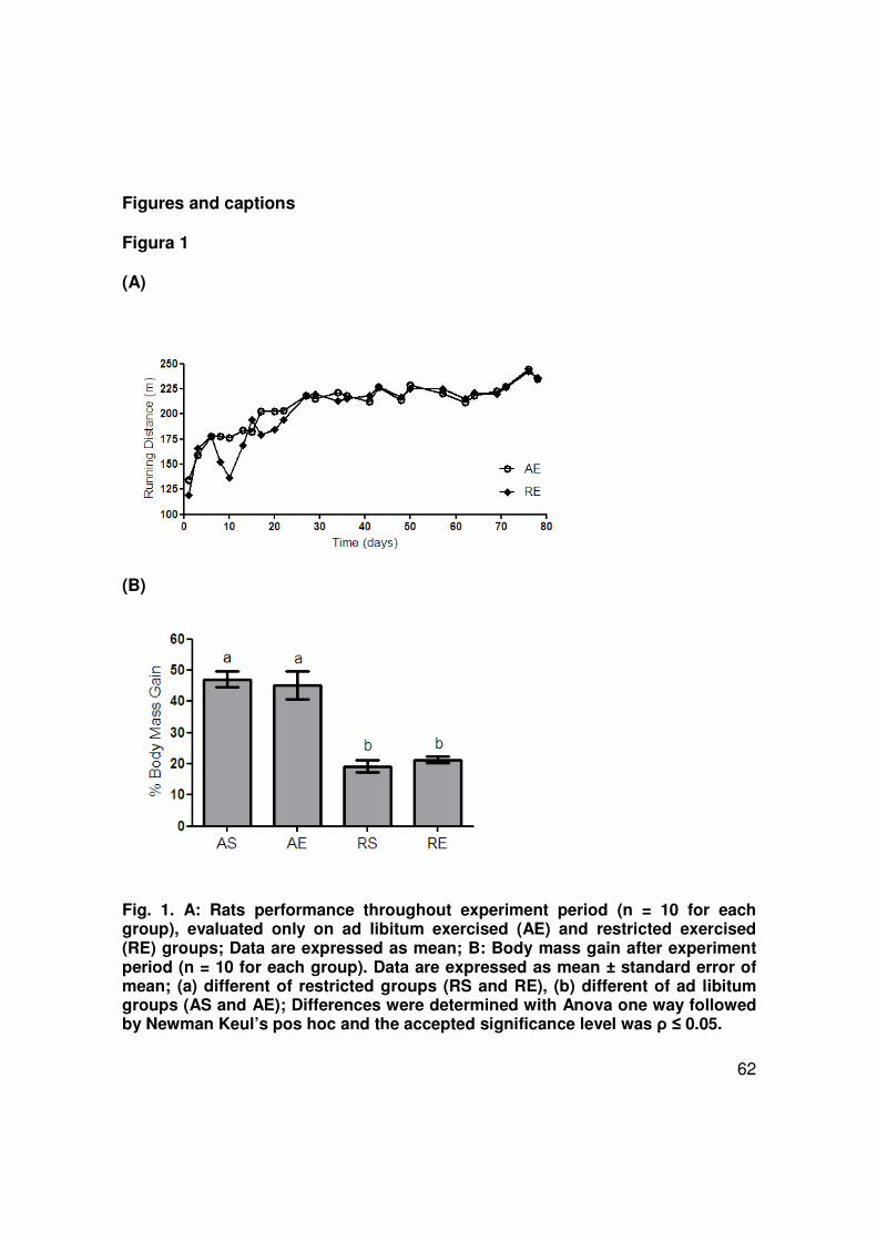
62
Figures and captions
Figura 1
(A)
(B)
Fig. 1. A: Rats performance throughout experiment period (n = 10 for each group), evaluated only on ad libitum exercised (AE) and restricted exercised (RE) groups; Data are expressed as mean; B: Body mass gain after experiment period (n = 10 for each group). Data are expressed as mean ± standard error of mean; (a) different of restricted groups (RS and RE), (b) different of ad libitum groups (AS and AE); Differences were determined with Anova one way followed by Newman Keul’s pos hoc and the accepted significance level was ρ ≤ 0.05.
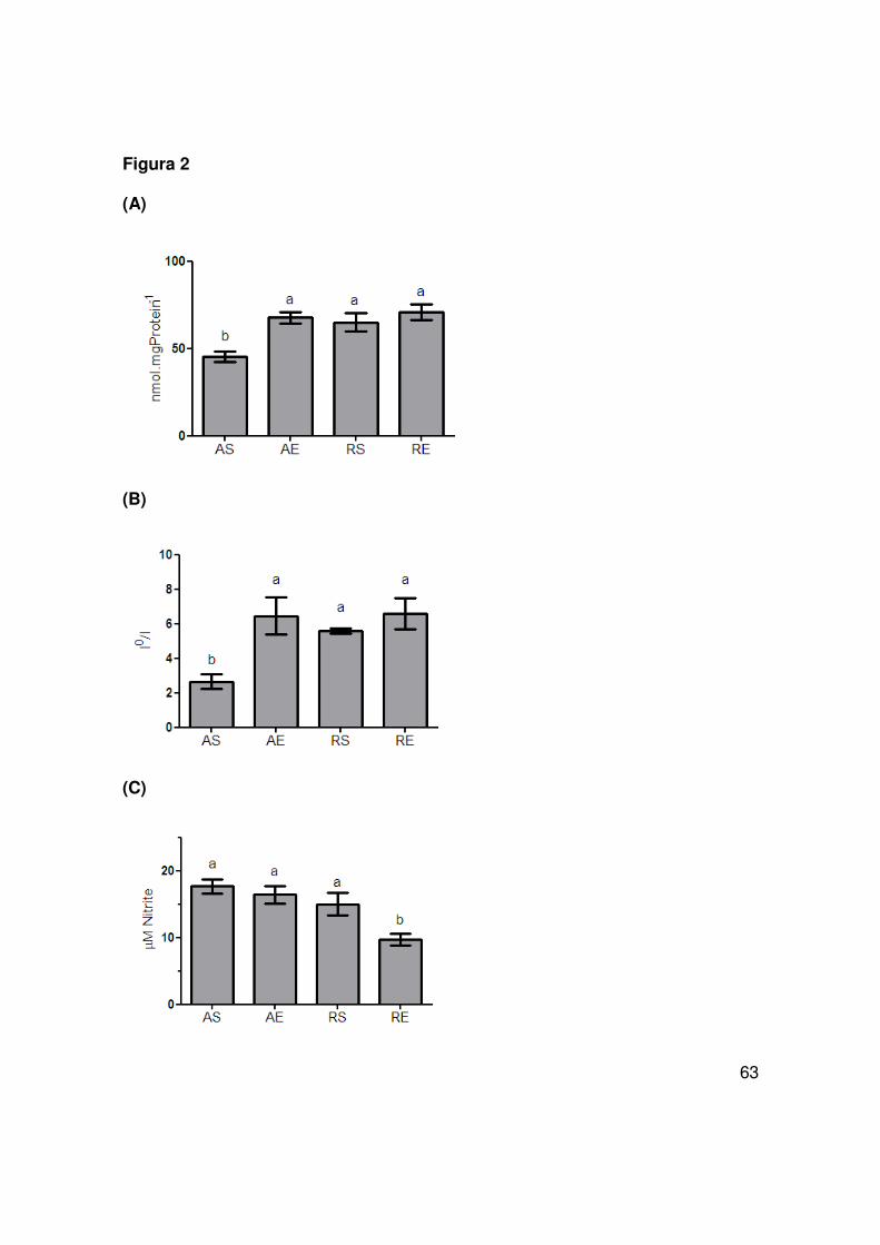
63
Figura 2
(A)
(B)
(C)
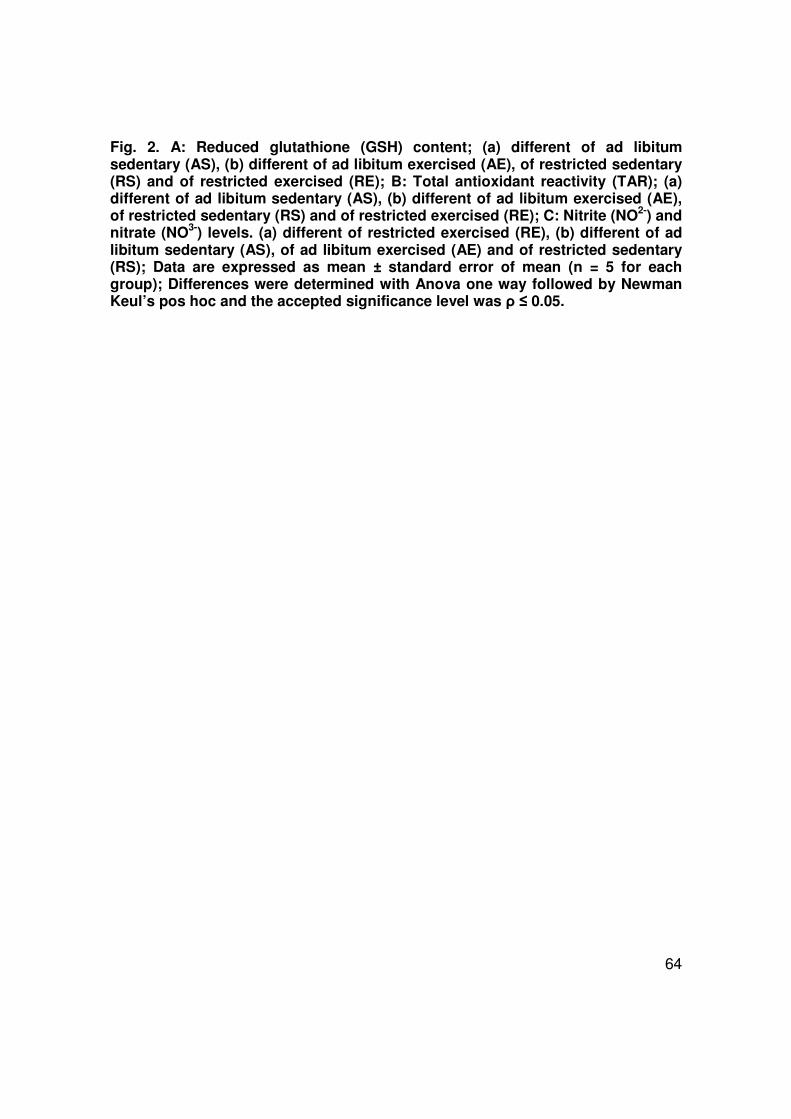
64
Fig. 2. A: Reduced glutathione (GSH) content; (a) different of ad libitum sedentary (AS), (b) different of ad libitum exercised (AE), of restricted sedentary (RS) and of restricted exercised (RE); B: Total antioxidant reactivity (TAR); (a) different of ad libitum sedentary (AS), (b) different of ad libitum exercised (AE), of restricted sedentary (RS) and of restricted exercised (RE); C: Nitrite (NO2-) and nitrate (NO3-) levels. (a) different of restricted exercised (RE), (b) different of ad libitum sedentary (AS), of ad libitum exercised (AE) and of restricted sedentary (RS); Data are expressed as mean ± standard error of mean (n = 5 for each group); Differences were determined with Anova one way followed by Newman Keul’s pos hoc and the accepted significance level was ρ ≤ 0.05.

65
Figura 3
(A)
(B)
Fig. 3. A: Thiobarbituric acid reactive species (TBARS) content; B: Protein carbonyl content; Data are expressed as mean ± standard error of mean (n = 5 for each group); (a) different of ad libitum sedentary (AS), (b) different of ad libitum exercised (AE), of restricted sedentary (RS) and of restricted exercised (RE); Differences were determined with Anova one way followed by Newman Keul’s pos hoc and the accepted significance level was ρ ≤ 0.05.
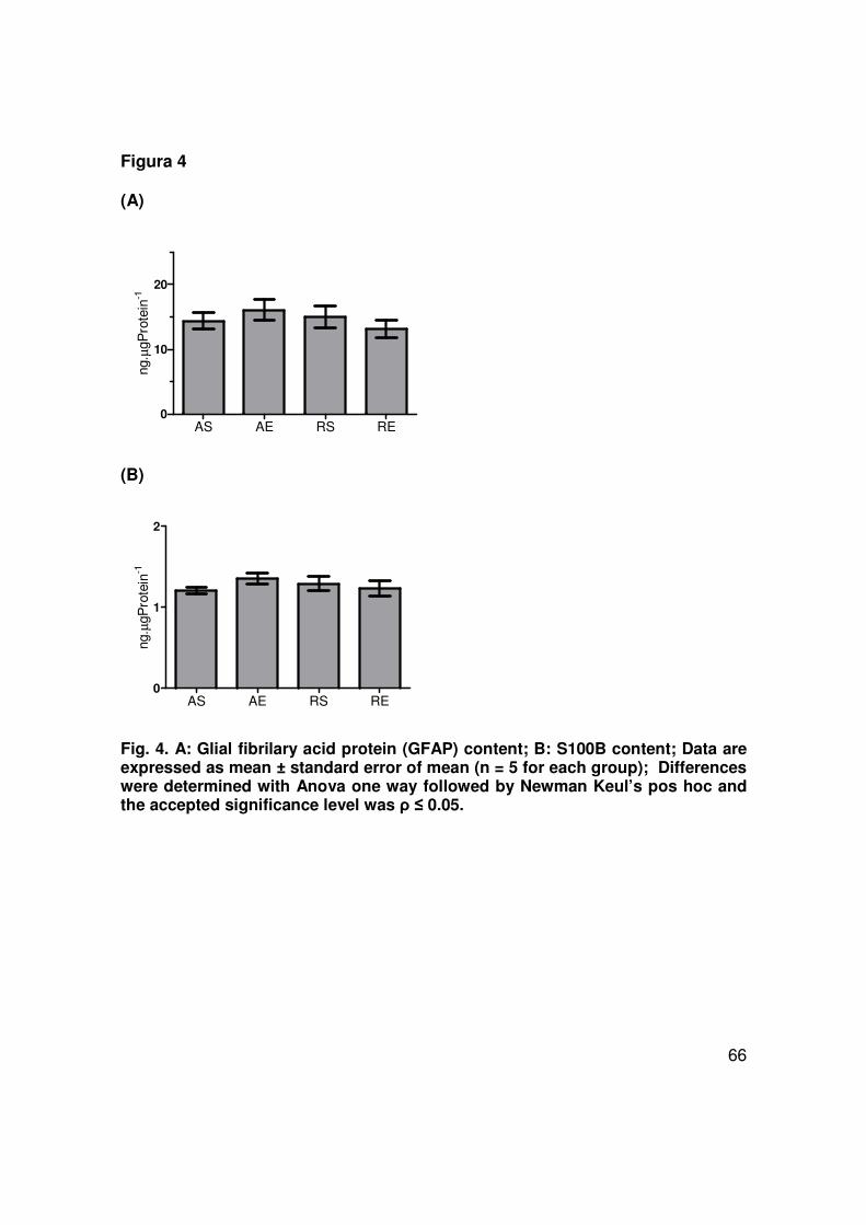
66
Figura 4
(A)
AS AE RS RE0
10
20
ng. µ
gPro
tein
-1
(B)
AS AE RS RE0
1
2
ng. µ
gPro
tein
-1
Fig. 4. A: Glial fibrilary acid protein (GFAP) content; B: S100B content; Data are expressed as mean ± standard error of mean (n = 5 for each group); Differences were determined with Anova one way followed by Newman Keul’s pos hoc and the accepted significance level was ρ ≤ 0.05.
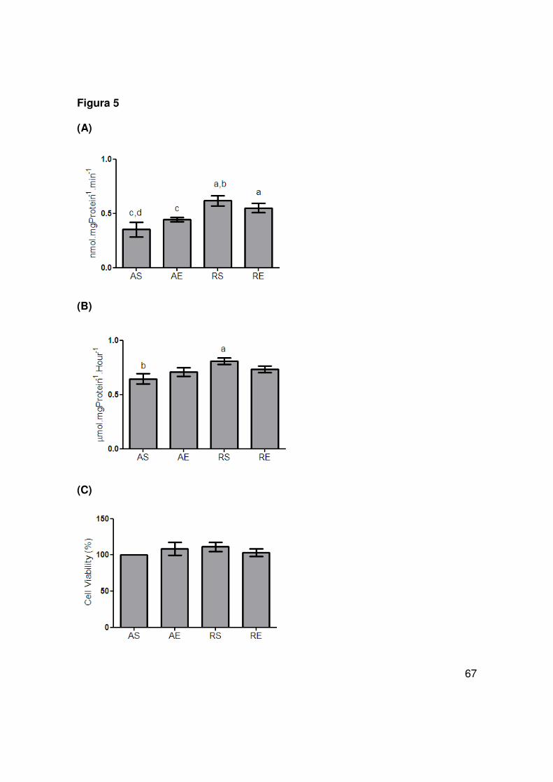
67
Figura 5
(A)
(B)
(C)

68
Fig. 5. A: Glutamate uptake; (a) different of ad libitum sedentary (AS), (b) different of ad libitum exercised (AE), (c) different of restricted sedentary (RS) and (d) different of restricted exercised (RE) B: Glutamine synthetase activity; (a) different of ad libitum sedentary (AS), (b) different of restricted sedentary (RS), C: Cell viability; Data are expressed as mean ± standard error of mean (n = 5 for each group);; Differences were determined with Anova one way followed by Newman Keul’s pos hoc and the accepted significance level was ρ ≤ 0.05.

69
Table 1. Composition of the laboratory chow Composition (g/Kg) Total fat 110 Sunflower oil 5 Proteins 220 Fibers 30 Ash 60 Vitamins 20 Carbohydrates 520 Commercial nonpurified diet, Nuvilab-CR1 (PR – Brazil) Table 2. Serum biochemistry AS AE RS RE
Glucose (mg.dL-1) 222 ± 4 186 ± 29 219 ± 3 214 ± 11
Cholesterol (mg.dL-1) 69 a ± 4 75 a ± 5 60 b ± 4 56 b ± 2
Triacylglicerol (mg.dL-1) 63 ± 13 58 ± 15 65 ± 10 61 ± 5
HDL (mg.dL-1) 27 ± 0,6 30 ± 1,3 27 ± 1,4 24 ± 0,4
LDL (mg.dL-1) 32 a ± 2 34 a ± 4 20 b ± 1 19 b ± 2
Creatinine (mg.dL-1) 0,58 ± 0,05 0,60 ± 0,04 0,56 ± 0,02 0,51 ± 0,04
Urea (mg.dL-1) 62 a ± 2,3 57 a ± 2,6 45 b ± 2,4 46 b ± 2,6
Total Proteins (g.dL-1) 6,2 ± 0,3 6,2± 0,2 6,1± 0,1 6,1 ± 0,2
C-Reactive Protein (mg.dL-1) 3,6 ± 0,1 3,3 ± 0,2 3,5 ± 0,1 3,5 ± 0,1
AST (U.L-1) 112 c ± 5 86 d ± 1 95 d ± 2 87 d ±1
ALT (U.L-1) 55 ± 2 50 ± 1 51± 4 49 ± 4
Data are expressed as mean ± standard error of mean (n=5 for each group). (a) different of restricted groups (RS and RE), (b) different of ad libitum groups (AS and AE), (c) different of ad libitum exercised (AE), of restricted sedentary (RS) and of restricted exercised (RE), (d) different of ad libitum sedentary (AS). Differences were determined with Anova one way followed by Newman Keul’s pos hoc and the accepted significance level was p≤0,05.

70
Parte III

71
Discussão
Durante os últimos anos, observou-se um aumento da expectativa de vida
média da população mundial acompanhado do aumento na prevalência de
doenças neurodegenerativas, como a doença de Alzheimer, a doença de
Parkinson, e a isquemia cerebral. Com isso, houve um aumento do interesse
público e científico nos efeitos benéficos da atividade física e da dieta visando a
manutenção cognitiva e a saúde mental na longevidade (Mattson, 2000, Kramer
and Erickson, 2007).
O exercício físico praticado regularmente, através de uma resposta
adaptativa, é capaz de melhorar as defesas antioxidantes, aumentando a atividade
de enzimas antioxidantes e o sistema GSH, dessa forma diminuindo à extensão
do dano oxidativo observado durante sessões agudas de treinamento físico
(Powers et al., 1994, Leeuwenburgh et al., 1997, Servais et al., 2003).
Estudos com ratos e camundongos mantidos em restrição calórica
demonstraram uma diminuição de dano relacionado a espécies reativas, incluindo
uma menor peroxidação lipídica, menor dano oxidativo a proteínas e ao DNA
(Merry, 2002, Barja, 2004), o que poderia estar relacionado a uma maior
resistência à apoptose, induzida por adaptação ao estresse (Hiona and
Leeuwenburgh, 2004).
O objetivo deste estudo foi investigar os efeitos de protocolos de
treinamento físico moderado e restrição calórica sobre o estado redox e
parâmetros gliais no hipocampo de ratos, uma vez que o estresse oxidativo está
72
relacionado com o desenvolvimento de desordens neurodegenerativas e com o
envelhecimento. Os principais resultados são apresentados na figura 4.
Os pesquisadores têm usado diferentes protocolos de treinamento (forçado
vs voluntário), que também variam em tempo de treinamento (semanas vs meses),
duração das sessões, intensidade e tipo de atividade (corrida vs nado) (Cotman et
al., 2007). Recentemente, foi desenvolvido um protocolo de exercício físico
moderado em esteira, com sessões três vezes por semana durante doze
semanas, capaz de reduzir o dano a fatias hipocampais submetidas a um
protocolo de isquemia in vitro, sugerindo neuroproteção induzida pelo exercício
(Cechetti et al., 2007).
Os efeitos benéficos de um regime de restrição calórica são devidos à
restrição da ingestão de calorias, ao contrário da restrição de um componente
alimentar específico (gorduras, por exemplo) ou da redução de algum componente
alimentar tóxico (aditivos, conservantes, por exemplo) (Masoro, 1998, 2000). Uma
redução de 30% na ingestão calórica demonstrou aumento na longevidade de
ratos e camundongos, e a manutenção desta condição resultou em níveis
menores de estresse oxidativo no hipocampo destes animais quando comparados
com outros alimentados ad libitum (Mattson, 2000). Estes estudos sugerem que a
supressão do estresse oxidativo pode ser um dos mecanismos pelos quais a
restrição calórica induza neuroproteção.
Neste estudo foi observada uma melhora na performance dos ratos
submetidos ao treinamento físico moderado em esteira, o que também foi
observado em outros estudos (Radak et al., 2005, Radak et al., 2006). Os animais

73
submetidos à restrição calórica de 30% apresentaram um ganho de massa
corporal aproximadamente 27% menor que os animais alimentados ad libitum,
corroborando com estudos prévios (Horska et al., 1999, Wanagat et al., 1999).
Para assegurar que os animais utilizados neste estudo não estavam
fisicamente exauridos ou desnutridos foram realizadas análises bioquímicas
séricas. Os ratos submetidos à restrição calórica apresentaram níveis menores de
colesterol total e LDL colesterol que os ratos alimentados ad libitum,
demonstrando benefícios da restrição calórica à saúde de forma geral. Os ratos
exercitados apresentaram níveis menores de atividade da transaminase aspartato
aminotransferase (AST) quando comparados ao grupo controle sedentário,
sugerindo que o programa de exercício físico utilizado resultou em menor dano
muscular (Nelson and Cox, 2005). Os níveis de creatinina entre os grupos não
foram significativamente diferentes, assegurando a manutenção da função renal,
porém foram encontrados níveis séricos diminuídos de uréia, podendo vir a ser
objeto de futura investigação da atividade da glutaminase renal. Este resultado
também está de acordo com estudo prévio (Ribeiro et al., 2009). Os demais
parâmetros estudados (glicose, triglicerídeos, HDL, proteínas totais, proteína C
reativa, alanina aminotransferase), não apresentaram alteração, indicando que os
animais estavam saudáveis.
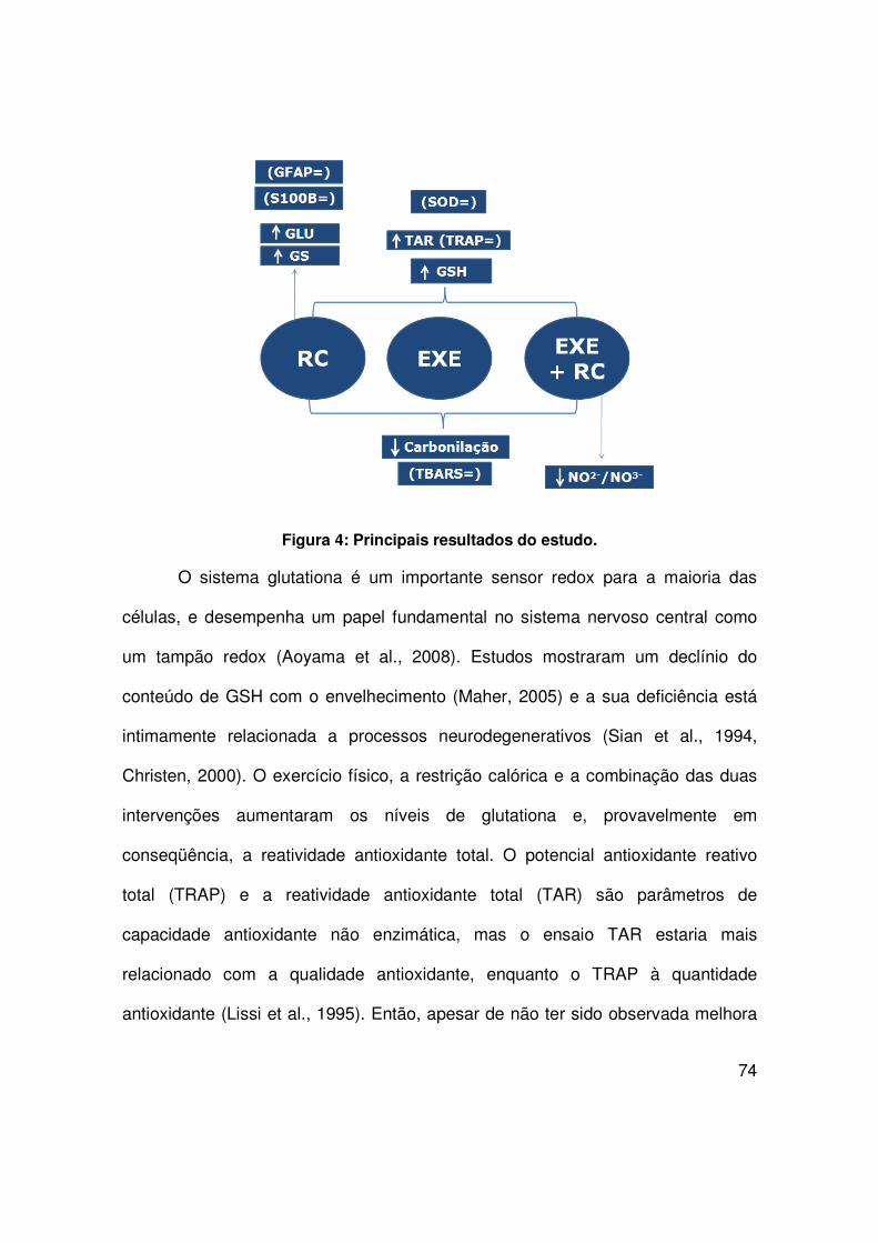
74
Figura 4: Principais resultados do estudo.
O sistema glutationa é um importante sensor redox para a maioria das
células, e desempenha um papel fundamental no sistema nervoso central como
um tampão redox (Aoyama et al., 2008). Estudos mostraram um declínio do
conteúdo de GSH com o envelhecimento (Maher, 2005) e a sua deficiência está
intimamente relacionada a processos neurodegenerativos (Sian et al., 1994,
Christen, 2000). O exercício físico, a restrição calórica e a combinação das duas
intervenções aumentaram os níveis de glutationa e, provavelmente em
conseqüência, a reatividade antioxidante total. O potencial antioxidante reativo
total (TRAP) e a reatividade antioxidante total (TAR) são parâmetros de
capacidade antioxidante não enzimática, mas o ensaio TAR estaria mais
relacionado com a qualidade antioxidante, enquanto o TRAP à quantidade
antioxidante (Lissi et al., 1995). Então, apesar de não ter sido observada melhora

75
na quantidade de defesas antioxidantes não enzimáticas (TRAP), as intervenções
(exercício, restrição e ambos) melhorarm a qualidade antioxidante não enzimática
(TAR).
Nosso estudo demonstrou que uma adaptação não enzimática pode ser
responsável por diminuir o dano oxidativo, ao contrário de uma adaptação
enzimática, já que neste estudo não foram encontradas modificações na atividade
da SOD. Somani e colaboradores também não encontraram mudanças na
atividade da SOD hipocampal após 7,5 semanas de exercício (Somani et al.,
1995). Devi e Kiran, por outro lado, reportaram um aumento da atividade desta
enzima no hipocampo após 4 meses de nado, enquanto Asku e colaboradores
evidenciaram uma diminuição da atividade da SOD com exercício regular em
esteira sob diferentes intensidades (Devi and Kiran, 2004, Aksu et al., 2009). Os
diferentes resultados destes estudos podem ser atribuídos a diferenças no tipo,
duração e intensidade dos protocolos de exercício aplicados.
O conteúdo de proteínas carboniladas, tomado como medida da oxidação
protéica, aumenta com o envelhecimento mais rapidamente no hipocampo e no
estriado, regiões associadas com perdas significativas de função durante o
processo de envelhecimento (Dubey et al., 1996). Para mensurar o dano oxidativo
no hipocampo, foi avaliada a peroxidação lipídica através de TBARS e a oxidação
protéica através do conteúdo de proteínas carboniladas. O exercício físico, a
restrição calórica e a combinação dos dois tratamentos não causaram nenhuma
alteração no ensaio TBARS, mas foram aptos a diminuir o conteúdo de proteínas
carboniladas. Estes resultados poderiam estar associados de forma positiva com a
76
atenuação do processo de envelhecimento, o que está de acordo com outros
estudos sobre restrição calórica (Dubey et al., 1996, Forster et al., 2000). Outros
pesquisadores encontraram resultados similares para o TBARS com exercício
físico regular (Coskun et al., 2005, Aksu et al., 2009).
O óxido nítrico desempenha um papel importante no SNC, incluindo
neurogênese, diferenciação e desenvolvimento neuronal, memória e
neuroproteção (Ignarro, 2002, Garthwaite, 2008). No entanto, o NO. pode reagir
rapidamente com o ânion superóxido formando peroxinitrito, o qual pode causar
oxidação e nitração em proteínas, lipídeos e DNA (Alvarez and Radi, 2003). Os
níveis de nitratos e nitritos são um bom indicativo da produção de óxido nítrico, já
que são produtos finais do seu metabolismo (Levine, 2002, Halliwell and
Whiteman, 2004). Um decréscimo nos níveis de NO2-/NO3
- observado com a
combinação das duas intervenções pode indicar que somente o exercício físico
regular em associação à restrição calórica crônica é capaz de causar esta
diminuição. Asku e colaboradores não encontraram nenhuma alteração nos níveis
de NO2-/NO3
- no cérebro (córtex pré-frontal, estriado e hipocampo) de ratos
submetidos somente a exercício regular em esteira (Aksu et al., 2009).
Os astrócitos possuem uma íntima relação com os neurônios na
transmissão glutamatérgica, por conseqüência, na plasticidade sináptica e
neuroproteção (Chen and Swanson, 2003, Tramontina et al., 2006a). No presente
estudo, foram investigadas funções astrogliais importantes como a captação de
glutamato, essencial para evitar o fenômeno de excitotoxicidade que poderia levar
a morte neuronal (Danbolt, 2001) e a habilidade em converter glutamato em
77
glutamina através da glutamina sintetase (GS). Somente a restrição calórica
demonstrou aumento na atividade da GS, e esta intervenção, associada ou não ao
exercício, também foi capaz de aumentar a captação de glutamato do meio
extracelular. Este resultado pode ser atribuído à modulação da atividade e/ou um
aumento nos transportadores de glutamato localizados nos astrócitos. Assim, a
restrição calórica estaria melhorando uma das principais funções dos astrócitos
que é a manutenção adequada do metabolismo do glutamato. Estes resultados
estão de acordo com um estudo anterior (Ribeiro et al., 2009).
Para a avaliação da captação de glutamato e da atividade da GS,
indicadores da atividade astroglial, foram utilizadas fatias hipocampais. Com o
propósito de assegurar a integridade destas fatias entre os grupos estudados foi
realizado o ensaio de viabilidade mitocondrial (MTT), onde não foram encontradas
diferenças, validando os resultados obtidos.
Uma vez que a glutationa é predominantemente astrogial e que, a
restrição calórica e o exercício físico aumentaram os níveis de GSH, foram
investigadas alterações em dois marcadores específicos destas células – GFAP e
S100B. O processo de astrogliose resultante de dano no SNC por trauma,
doença, desordem genética ou insulto químico, é evidenciado por um aumento de
GFAP (Eng et al., 2000). Da mesma forma, altos níveis de S100B no tecido
cerebral foram encontrados em casos de doenças neurodegenerativas, incluindo
Alzheimer (Griffin et al., 1998). No presente estudo, o conteúdo de GFAP e S100B
não foi influenciado pela restrição calórica nem pelo exercício físico, indicando

78
ausência de astrogliose e ausência de pré-disposição à apoptose (O'Callaghan
and Sriram, 2005).
Resumindo, o presente estudo demonstrou uma melhora no sistema
antioxidante não enzimático e uma diminuição do dano oxidativo protéico através
de restrição calórica crônica e exercício físico moderado. Somente a combinação
das duas intervenções foi capaz de diminuir os níveis de nitratos e nitritos. A
restrição calórica, possivelmente através de modulação da função astroglial, foi
capaz de promover uma melhora na captação de glutamato e na atividade da
glutamina sintetase, o que pode ser relacionado a um menor risco de
excitotoxicidade. Estes resultados nos dão novas perspectivas para compreender
os mecanismos pelos quais a restrição calórica aliada ao exercício físico pode
prevenir doenças neurodegenerativas e promover a longevidade.
79
Referências Bibliográficas
Ahlemeyer B, Beier H, Semkova I, Schaper C, Krieglstein J (S-100beta protects cultured neurons against glutamate- and staurosporine-induced damage and is involved in the antiapoptotic action of the 5 HT(1A)-receptor agonist, Bay x 3702. Brain Res 858:121-128.2000).
Aksenov MY, Aksenova MV, Carney JM, Butterfield DA (Oxidative modification of glutamine synthetase by amyloid beta peptide. Free Radic Res 27:267-281.1997).
Aksu I, Topcu A, Camsari UM, Acikgoz O (Effect of acute and chronic exercise on oxidant-antioxidant equilibrium in rat hippocampus, prefrontal cortex and striatum. Neurosci Lett 452:281-285.2009).
Alberts B, Johnson A, Lewis J, Raff M, Roberts K, Walter P (The cytoskeleton. In: Molecular Biology of the Cell, Ed Garland Publishing New York 780-820.2002).
Alessio HM, Goldfarb AH (Lipid peroxidation and scavenger enzymes during exercise: adaptive response to training. J Appl Physiol 64:1333-1336.1988).
Alvarez B, Radi R (Peroxynitrite reactivity with amino acids and proteins. Amino Acids 25:295-311.2003).
Amara SG, Fontana AC (Excitatory amino acid transporters: keeping up with glutamate. Neurochem Int 41:313-318.2002).
Anderson CM, Swanson RA (Astrocyte glutamate transport: review of properties, regulation, and physiological functions. Glia 32:1-14.2000).
Aoyama K, Watabe M, Nakaki T (Regulation of neuronal glutathione synthesis. J Pharmacol Sci 108:227-238.2008).
Astrup AV, Madsbad S, Finer N ([New discoveries about the cause of diabetes. Type 2 diabetes mellitus changed to "obesity-dependent diabetes mellitus"]. Ugeskr Laeger 163:141-143.2001).
Bak LK, Schousboe A, Waagepetersen HS (The glutamate/GABA-glutamine cycle: aspects of transport, neurotransmitter homeostasis and ammonia transfer. J Neurochem 98:641-653.2006).
Barja G (Free radicals and aging. Trends Neurosci 27:595-600.2004). Baydas G, Nedzvetskii VS, Tuzcu M, Yasar A, Kirichenko SV (Increase of glial
fibrillary acid protein and S100B in hippocampus and cortex of diabetic rats: effects of vitamin. European Journal of Pharmacology 462:67-71.2003).
Bear MF, Connors BW, Paradiso MA (Sistemas de memória. In: Neurociências - Desvendando o Sistema Nervoso, Editado por Artmed, Porto Alegre
760-764.2002). Berger MM (Can oxidative damage be treated nutritionally? Clin Nutr 24:172-
183.2005). Berlett BS, Stadtman ER (Protein oxidation in aging, disease, and oxidative stress.
J Biol Chem 272:20313-20316.1997).
80
Bosch-Morell F, Flohe L, Marin N, Romero FJ (4-Hydroxynonenal inhibits glutathione peroxidase: protection by glutathione. Free Radic Biol Med 26:1383-1387.1999).
Brigelius-Flohe R (Tissue-specific functions of individual glutathione peroxidases. Free Radic Biol Med 27:951-965.1999).
Brooks GA, White TP (Determination of metabolic and heart rate responses of rats to treadmill exercise. J Appl Physiol 45:1009-1015.1978).
Browne RW, Armstrong D (Simultaneous determination of serum retinol, tocopherols, and carotenoids by HPLC. Methods Mol Biol 108:269-275.1998).
Carmignoto G (Reciprocal communication system between astrocytes and neurons. Progress in neurobiology 62:561-581.2000).
Cechetti F, Rhod A, Simao F, Santin K, Salbego C, Netto CA, Siqueira IR (Effect of treadmill exercise on cell damage in rat hippocampal slices submitted to oxygen and glucose deprivation. Brain Res 1157:121-125.2007).
Chang J, Cornell JE, Van Remmen H, Hakala K, Ward WF, Richardson A (Effect of aging and caloric restriction on the mitochondrial proteome. J Gerontol A Biol Sci Med Sci 62:223-234.2007).
Chen Y, Swanson RA (Astrocytes and brain injury. J Cereb Blood Flow Metab 23:137-149.2003).
Christen Y (Oxidative stress and Alzheimer disease. Am J Clin Nutr 71:621S-629S.2000).
Cooper AJ, Kristal BS (Multiple roles of glutathione in the central nervous system. Biol Chem 378:793-802.1997).
Coskun S, Gonul B, Guzel NA, Balabanli B (The effects of vitamin C supplementation on oxidative stress and antioxidant content in the brains of chronically exercised rats. Mol Cell Biochem 280:135-138.2005).
Cotman CW, Berchtold NC (Exercise: a behavioral intervention to enhance brain health and plasticity. Trends Neurosci 25:295-301.2002).
Cotman CW, Berchtold NC, Christie LA (Exercise builds brain health: key roles of growth factor cascades and inflammation. Trends Neurosci 30:464-472.2007).
Cruzen C, Colman RJ (Effects of caloric restriction on cardiovascular aging in non-human primates and humans. Clin Geriatr Med 25:733-743, ix-x.2009).
Danbolt NC (Glutamate uptake. Prog Neurobiol 65:1-105.2001). Devi SA, Kiran TR (Regional responses in antioxidant system to exercise training
and dietary vitamin E in aging rat brain. Neurobiol Aging 25:501-508.2004). Donato R (S100: a multigenetic family of calcium-modulated proteins of EF-hand
type with intracellular and extracellular functional roles. The international Journal of Biochemistry & Cell Biology 33:637-668.2001).
Donato R (Intracellular and extracelluar rolesof S100 proteins. Microscopy Research Tech 60:540-551.2003).
Donato R, Sorci G, Riuzzi F, Arcuri C, Bianchi R, Brozzi F, Tubaro C, Giambanco I (S100B's double life: intracellular regulator and extracellular signal. Biochim Biophys Acta 1793:1008-1022.2009).
81
dos Santos AQ, Nardin P, Funchal C, de Almeida LM, Jacques-Silva MC, Wofchuk ST, Goncalves CA, Gottfried C (Resveratrol increases glutamate uptake and glutamine synthetase activity in C6 glioma cells. Arch Biochem Biophys 453:161-167.2006).
Draper HH, Hadley M (Malondialdehyde determination as index of lipid peroxidation. Methods Enzymol 186:421-431.1990).
Dresch MT, Rossato SB, Kappel VD, Biegelmeyer R, Hoff ML, Mayorga P, Zuanazzi JA, Henriques AT, Moreira JC (Optimization and validation of an alternative method to evaluate total reactive antioxidant potential. Anal Biochem 385:107-114.2009).
Dringen R (Glutathione metabolism and oxidative stress in neurodegeneration. Eur J Biochem 267:4903.2000a).
Dringen R (Metabolism and functions of glutathione in brain. Prog Neurobiol 62:649-671.2000b).
Dringen R, Gutterer JM, Hirrlinger J (Glutathione metabolism in brain metabolic interaction between astrocytes and neurons in the defense against reactive oxygen species. Eur J Biochem 267:4912-4916.2000).
Dubey A, Forster MJ, Lal H, Sohal RS (Effect of age and caloric intake on protein oxidation in different brain regions and on behavioral functions of the mouse. Arch Biochem Biophys 333:189-197.1996).
Eng LF, Ghirnikar RS, Lee YL (Glial fibrillary acidic protein: GFAP-thirty-one years (1969-2000). Neurochem Res 25:1439-1451.2000).
Forster MJ, Sohal BH, Sohal RS (Reversible effects of long-term caloric restriction on protein oxidative damage. J Gerontol A Biol Sci Med Sci 55:B522-529.2000).
Gallo V, Ghiani CA (Glutamate receptors in glia: new cells, new inputs and new functions. Trends Pharmacol Sci 21:252-258.2000).
Garthwaite J (Concepts of neural nitric oxide-mediated transmission. Eur J Neurosci 27:2783-2802.2008).
Gaunt GL, de Duve C (Subcellular distribution of D-amino acid oxidase and catalase in rat brain. J Neurochem 26:749-759.1976).
Gee JR, Keller JN (Astrocytes: regulation of brain homeostasis via apolipoprotein. European Journal of biochemistry 37:1145-1150.2005).
Gomes FC, Paulin D, Moura Neto V (Glial fibrillary acidic protein (GFAP): modulation by growth factors and its implication in astrocyte differentiation. Braz J Med Biol Res 32:619-631.1999).
Gottfried C, Cechin SR, Gonzalez MA, Vaccaro TS, Rodnight R (The influence of the extracellular matrix on the morphology and intracellular pH of cultured astrocytes exposed to media lacking bicarbonate. Neuroscience 121:553-562.2003).
Gottfried C, Tramontina F, Goncalves D, Goncalves CA, Moriguchi E, Dias RD, Wofchuk ST, Souza DO (Glutamate uptake in cultured astrocytes depends on age: a study about the effect of guanosine and the sensitivity to oxidative stress induced by H(2)O(2). Mech Ageing Dev 123:1333-1340.2002).
82
Griffin WS, Sheng JG, McKenzie JE, Royston MC, Gentleman SM, Brumback RA, Cork LC, Del Bigio MR, Roberts GW, Mrak RE (Life-long overexpression of S100beta in Down's syndrome: implications for Alzheimer pathogenesis. Neurobiol Aging 19:401-405.1998).
Gutteridge JM, Halliwell B (Free radicals and antioxidants in the year 2000. A historical look to the future. Ann N Y Acad Sci 899:136-147.2000).
Halliwell B (Role of free radicals in the neurodegenerative diseases: therapeutic implications for antioxidant treatment. Drugs Aging 18:685-716.2001).
Halliwell B (Oxidative stress and neurodegeneration: where are we now? J Neurochem 97:1634-1658.2006).
Halliwell B, Whiteman M (Measuring reactive species and oxidative damage in vivo and in cell culture: how should you do it and what do the results mean? Br J Pharmacol 142:231-255.2004).
Hevel JM, Marletta MA (Nitric-oxide synthase assays. Methods Enzymol 233:250-258.1994).
Hiona A, Leeuwenburgh C (Effects of age and caloric restriction on brain neuronal cell death/survival. Ann N Y Acad Sci 1019:96-105.2004).
Holloszy JO, Fontana L (Caloric restriction in humans. Exp Gerontol 42:709-712.2007).
Horska A, Brant LJ, Ingram DK, Hansford RG, Roth GS, Spencer RG (Effect of long-term caloric restriction and exercise on muscle bioenergetics and force development in rats. Am J Physiol 276:E766-773.1999).
Ignarro LJ (Nitric oxide as a unique signaling molecule in the vascular system: a historical overview. J Physiol Pharmacol 53:503-514.2002).
Jenkins RR (Exercise, oxidative stress, and antioxidants: a review. Int J Sport Nutr 3:356-375.1993).
Jenssen KR (Glial Cells. The international Journal of Biochemistry & Cell Biology 36:1861-1867.2004).
Kim JD, McCarter RJ, Yu BP (Influence of age, exercise, and dietary restriction on oxidative stress in rats. Aging (Milano) 8:123-129.1996).
Kramer AF, Erickson KI (Capitalizing on cortical plasticity: influence of physical activity on cognition and brain function. Trends Cogn Sci 11:342-348.2007).
Leeuwenburgh C, Hollander J, Leichtweis S, Griffiths M, Gore M, Ji LL (Adaptations of glutathione antioxidant system to endurance training are tissue and muscle fiber specific. Am J Physiol 272:R363-369.1997).
Leite MC, Galland F, Brolese G, Guerra MC, Bortolotto JW, Freitas R, Almeida LM, Gottfried C, Goncalves CA (A simple, sensitive and widely applicable ELISA for S100B: Methodological features of the measurement of this glial protein. J Neurosci Methods 169:93-99.2008).
Levine RL (Carbonyl modified proteins in cellular regulation, aging, and disease. Free Radic Biol Med 32:790-796.2002).
Levine RL, Garland D, Oliver CN, Amici A, Climent I, Lenz AG, Ahn BW, Shaltiel S, Stadtman ER (Determination of carbonyl content in oxidatively modified proteins. Methods Enzymol 186:464-478.1990).

83
Lissi E, Salim-Hanna M, Pascual C, del Castillo MD (Evaluation of total antioxidant potential (TRAP) and total antioxidant reactivity from luminol-enhanced chemiluminescence measurements. Free Radic Biol Med 18:153-158.1995).
Magistretti PJ, Pellerin L (Cellular mechanisms of brain energy metabolism and their relevance to functional brain imaging. Philos Trans R Soc Lond B Biol Sci 354:1155-1163.1999).
Maher P (The effects of stress and aging on glutathione metabolism. Ageing Res Rev 4:288-314.2005).
Mailly F, Marin P, Israel M, Glowinski J, Premont J (Increase in external glutamate and NMDA receptor activation contribute to H2O2-induced neuronal apoptosis. J Neurochem 73:1181-1188.1999).
Masoro EJ (Caloric restriction. Aging (Milano) 10:173-174.1998). Masoro EJ (Caloric restriction and aging: an update. Exp Gerontol 35:299-
305.2000). Masoro EJ (Subfield history: caloric restriction, slowing aging, and extending life.
Sci Aging Knowledge Environ 2003:RE2.2003). Masoro EJ (Overview of caloric restriction and ageing. Mech Ageing Dev 126:913-
922.2005). Matsuo M, Kaneko T (The chemistry of reactive oxygen species and related free
radicals. In: Free Radicals in Exercise and Aging, edited by Human Kinetics, Hungary 1-29.2000).
Matthews GD, Gould RM, Vardimon L (A single glutamine synthetase gene produces tissue-specific subcellular localization by alternative splicing. FEBS Lett 579:5527-5534.2005).
Mattson MP (Neuroprotective signaling and the aging brain: take away my food and let me run. Brain Res 886:47-53.2000).
Mattson MP, Chan SL (Neuronal and glial calcium signaling in Alzheimer's disease. Cell Calcium 34:385-397.2003).
Mattson MP, Duan W, Lee J, Guo Z (Suppression of brain aging and neurodegenerative disorders by dietary restriction and environmental enrichment: molecular mechanisms. Mech Ageing Dev 122:757-778.2001).
Matute C (Oligodendrocyte NMDA receptors: a novel therapeutic target. Trends Mol Med 12:289-292.2006).
Matute C, Domercq M, Sanchez-Gomez MV (Glutamate-mediated glial injury: mechanisms and clinical importance. Glia 53:212-224.2006).
McCay CM (Cellulose in the diet of mice and rats. Journal of Nutrition 435-437.1935).
McKiernan SH, Bua E, McGorray J, Aiken J (Early-onset calorie restriction conserves fiber number in aging rat skeletal muscle. FASEB J 18:580-581.2004).
Merry BJ (Molecular mechanisms linking calorie restriction and longevity. Int J Biochem Cell Biol 34:1340-1354.2002).
Misra HP, Fridovich I (The role of superoxide anion in the autoxidation of epinephrine and a simple assay for superoxide dismutase. J Biol Chem 247:3170-3175.1972).
84
Moore BW (A soluble protein caracteristic of the nervous system. Biochemistry & Biophysics Research Community 19:739-744.1965).
Morgan TE, Rozovsky I, Goldsmith SK, Stone DJ, Yoshida T, Finch CE (Increased transcription of the astrocyte gene GFAP during middle-age is attenuated by food restriction: implications for the role of oxidative stress. Free Radic Biol Med 23:524-528.1997).
Mosmann T (Rapid colorimetric assay for cellular growth and survival: application to proliferation and cytotoxicity assays. J Immunol Methods 65:55-63.1983).
Nakanishi S (Molecular diversity of glutamate receptors and implications for brain function. Science 258:597-603.1992).
Neeper SA, Gomez-Pinilla F, Choi J, Cotman CW (Physical activity increases mRNA for brain-derived neurotrophic factor and nerve growth factor in rat brain. Brain Res 726:49-56.1996).
Nelson DL, Cox MM (Lehninger Principles of Biochemistry. fourth edition:664.2005).
Nichol KE, Parachikova AI, Cotman CW (Three weeks of running wheel exposure improves cognitive performance in the aged Tg2576 mouse. Behav Brain Res 184:124-132.2007).
O'Callaghan JP, Sriram K (Glial fibrillary acidic protein and related glial proteins as biomarkers of neurotoxicity. Expert Opin Drug Saf 4:433-442.2005).
O'Callaghan RM, Ohle R, Kelly AM (The effects of forced exercise on hippocampal plasticity in the rat: A comparison of LTP, spatial- and non-spatial learning. Behav Brain Res 176:362-366.2007).
Pacher P, Beckman JS, Liaudet L (Nitric oxide and peroxynitrite in health and disease. Physiol Rev 87:315-424.2007).
Partridge L, Brand MD (Special issue on dietary restriction: Dietary restriction, longevity and ageing - the current state of our knowledge and ignorance. Mechanisms of Ageing and Development 126:911-912.2005).
Patel NV, Gordon MN, Connor KE, Good RA, Engelman RW, Mason J, Morgan DG, Morgan TE, Finch CE (Caloric restriction attenuates Abeta-deposition in Alzheimer transgenic models. Neurobiol Aging 26:995-1000.2005).
Pekny M, Pekna M (Astrocyte mediate filaments in CNS pathologies and regeneration. Journal of Pathology 204:428-437.2004).
Perea G, Araque A (Glial calcium signaling and neuron-glia communication. Cell Calcium 38:375-382.2005).
Peterson GL (A simplification of the protein assay method of Lowry et al. which is more generally applicable. Anal Biochem 83:346-356.1977).
Ponath G, Schettler C, Kaestner F, Voigt B, Wentker D, Arolt V, Rothermundt M (Autocrine S100B effects on astrocytes are mediated via RAGE. J Neuroimmunol 184:214-222.2007).
Porciuncula LO, Rocha JB, Ghisleni G, Tavares RG, Souza DO (The effects of ebselen on [3H]glutamate uptake by synaptic vesicles from rat brain. Brain Res 1027:192-195.2004).

85
Powers SK, Criswell D, Lawler J, Ji LL, Martin D, Herb RA, Dudley G (Influence of exercise and fiber type on antioxidant enzyme activity in rat skeletal muscle. Am J Physiol 266:R375-380.1994).
Prolla TA, Mattson MP (Molecular mechanisms of brain aging and neurodegenerative disorders: lessons from dietary restriction. Trends Neurosci 24:S21-31.2001).
Radak Z, Chung HY, Goto S (Exercise and hormesis: oxidative stress-related adaptation for successful aging. Biogerontology 6:71-75.2005).
Radak Z, Chung HY, Goto S (Systemic adaptation to oxidative challenge induced by regular exercise. Free Radic Biol Med 44:153-159.2008).
Radak Z, Kumagai S, Taylor AW, Naito H, Goto S (Effects of exercise on brain function: role of free radicals. Appl Physiol Nutr Metab 32:942-946.2007).
Radak Z, Toldy A, Szabo Z, Siamilis S, Nyakas C, Silye G, Jakus J, Goto S (The effects of training and detraining on memory, neurotrophins and oxidative stress markers in rat brain. Neurochem Int 49:387-392.2006).
Ribeiro LC, Quincozes-Santos A, Leite MC, Abib RT, Kleinkauf-Rocha J, Biasibetti R, Rotta LN, Wofchuk ST, Perry ML, Goncalves CA, Gottfried C (Caloric restriction increases hippocampal glutamate uptake and glutamine synthetase activity in Wistar rats. Neurosci Res 64:330-334.2009).
Robinson MB, Dowd LA (Heterogeneity and functional properties of subtypes of sodium-dependent glutamate transporters in the mammalian central nervous system. Adv Pharmacol 37:69-115.1997).
Rodnight R, Goncalves CA, Wofchuk ST, Leal R (Control of the phosphorylation of the astrocyte marker glial fibrillary acidic protein (GFAP) in the immature rat hippocampus by glutamate and calcium ions: possible key factor in astrocytic plasticity. Braz J Med Biol Res 30:325-338.1997).
Rothermundt M, Peters M, Prehn JHM, Arolt V (S100 in brain damage and neurodegeneration. Microscopy Research Tech 60:614-632.2003).
Schweitzer NB, Alessio HM, Berry SD, Roeske K, Hagerman AE (Exercise-induced changes in cardiac gene expression and its relation to spatial maze performance. Neurochem Int 48:9-16.2006).
Servais S, Couturier K, Koubi H, Rouanet JL, Desplanches D, Sornay-Mayet MH, Sempore B, Lavoie JM, Favier R (Effect of voluntary exercise on H2O2 release by subsarcolemmal and intermyofibrillar mitochondria. Free Radic Biol Med 35:24-32.2003).
Sian J, Dexter DT, Lees AJ, Daniel S, Agid Y, Javoy-Agid F, Jenner P, Marsden CD (Alterations in glutathione levels in Parkinson's disease and other neurodegenerative disorders affecting basal ganglia. Ann Neurol 36:348-355.1994).
Sies H (Role of reactive oxygen species in biological processes. Klin Wochenschr 69:965-968.1991).
Sinclair DA (Toward a unified theory of caloric restriction and longevity regulation. Mech Ageing Dev 126:987-1002.2005).
Siqueira IR, Cimarosti H, Fochesatto C, Nunes DS, Salbego C, Elisabetsky E, Netto CA (Neuroprotective effects of Ptychopetalum olacoides Bentham
86
(Olacaceae) on oxygen and glucose deprivation induced damage in rat hippocampal slices. Life Sci 75:1897-1906.2004).
Somani SM, Husain K (Influence of exercise-induced oxidative stress on the central nervous system. In: Handbook of Oxidants and Antioxidants in Exercise Chapter 26:716-751.2000).
Somani SM, Ravi R, Rybak LP (Effect of exercise training on antioxidant system in brain regions of rat. Pharmacol Biochem Behav 50:635-639.1995).
Someya S, Yamasoba T, Weindruch R, Prolla TA, Tanokura M (Caloric restriction suppresses apoptotic cell death in the mammalian cochlea and leads to prevention of presbycusis. Neurobiol Aging 28:1613-1622.2007).
Sutoo D, Akiyama K (Regulation of brain function by exercise. Neurobiol Dis 13:1-14.2003).
Swindell WR (Comparative analysis of microarray data identifies common responses to caloric restriction among mouse tissues. Mech Ageing Dev 129:138-153.2008).
Thomazi AP, Godinho GF, Rodrigues JM, Schwalm FD, Frizzo ME, Moriguchi E, Souza DO, Wofchuk ST (Ontogenetic profile of glutamate uptake in brain structures slices from rats: sensitivity to guanosine. Mech Ageing Dev 125:475-481.2004).
Tramontina F, Conte S, Goncalves D, Gottfried C, Portela LV, Vinade L, Salbego C, Goncalves CA (Developmental changes in S100B content in brain tissue, cerebrospinal fluid, and astrocyte cultures of rats. Cell Mol Neurobiol 22:373-378.2002).
Tramontina F, Leite MC, Cereser K, de Souza DF, Tramontina AC, Nardin P, Andreazza AC, Gottfried C, Kapczinski F, Goncalves CA (Immunoassay for glial fibrillary acidic protein: antigen recognition is affected by its phosphorylation state. J Neurosci Methods 162:282-286.2007).
Tramontina F, Leite MC, Goncalves D, Tramontina AC, Souza DF, Frizzo JK, Nardin P, Gottfried C, Wofchuk ST, Goncalves CA (High glutamate decreases S100B secretion by a mechanism dependent on the glutamate transporter. Neurochem Res 31:815-820.2006a).
Tramontina F, Tramontina AC, Souza DF, Leite MC, Gottfried C, Souza DO, Wofchuk ST, Goncalves CA (Glutamate uptake is stimulated by extracellular S100B in hippocampal astrocytes. Cell Mol Neurobiol 26:81-86.2006b).
Van Eldik LJ, Wainwright MS (The Janus face of glial-derived S100B: beneficial and detrimental functions in the brain. Restor Neurol Neurosci 21:97-108.2003).
van Praag H, Christie BR, Sejnowski TJ, Gage FH (Running enhances neurogenesis, learning, and long-term potentiation in mice. Proc Natl Acad Sci U S A 96:13427-13431.1999a).
van Praag H, Kempermann G, Gage FH (Running increases cell proliferation and neurogenesis in the adult mouse dentate gyrus. Nat Neurosci 2:266-270.1999b).
van Praag H, Shubert T, Zhao C, Gage FH (Exercise enhances learning and hippocampal neurogenesis in aged mice. J Neurosci 25:8680-8685.2005).

87
Volterra A, Meldolesi J (Astrocytes, from brain glue to communication elements: the revolution continues. Nature 6:624-640.2005).
Wanagat J, Allison DB, Weindruch R (Caloric intake and aging: mechanisms in rodents and a study in nonhuman primates. Toxicol Sci 52:35-40.1999).
Youdim MB, Ben-Shachar D, Riederer P (Is Parkinson's disease a progressive siderosis of substantia nigra resulting in iron and melanin induced neurodegeneration? Acta Neurol Scand Suppl 126:47-54.1989).
Young JZ (The concept of neuroglia. Annals of New York Academy of Sciences 633:1-18.1991).
Zecca L, Youdim MB, Riederer P, Connor JR, Crichton RR (Iron, brain ageing and neurodegenerative disorders. Nat Rev Neurosci 5:863-873.2004).
Zhou L, Zhu DY (Neuronal nitric oxide synthase: structure, subcellular localization, regulation, and clinical implications. Nitric Oxide 20:223-230.2009).
Livros Grátis( http://www.livrosgratis.com.br )
Milhares de Livros para Download: Baixar livros de AdministraçãoBaixar livros de AgronomiaBaixar livros de ArquiteturaBaixar livros de ArtesBaixar livros de AstronomiaBaixar livros de Biologia GeralBaixar livros de Ciência da ComputaçãoBaixar livros de Ciência da InformaçãoBaixar livros de Ciência PolíticaBaixar livros de Ciências da SaúdeBaixar livros de ComunicaçãoBaixar livros do Conselho Nacional de Educação - CNEBaixar livros de Defesa civilBaixar livros de DireitoBaixar livros de Direitos humanosBaixar livros de EconomiaBaixar livros de Economia DomésticaBaixar livros de EducaçãoBaixar livros de Educação - TrânsitoBaixar livros de Educação FísicaBaixar livros de Engenharia AeroespacialBaixar livros de FarmáciaBaixar livros de FilosofiaBaixar livros de FísicaBaixar livros de GeociênciasBaixar livros de GeografiaBaixar livros de HistóriaBaixar livros de Línguas

Baixar livros de LiteraturaBaixar livros de Literatura de CordelBaixar livros de Literatura InfantilBaixar livros de MatemáticaBaixar livros de MedicinaBaixar livros de Medicina VeterináriaBaixar livros de Meio AmbienteBaixar livros de MeteorologiaBaixar Monografias e TCCBaixar livros MultidisciplinarBaixar livros de MúsicaBaixar livros de PsicologiaBaixar livros de QuímicaBaixar livros de Saúde ColetivaBaixar livros de Serviço SocialBaixar livros de SociologiaBaixar livros de TeologiaBaixar livros de TrabalhoBaixar livros de Turismo

![aula 3 - Redox [Modo de Compatibilidade]Title: Microsoft PowerPoint - aula 3 - Redox [Modo de Compatibilidade] Author: Denise Created Date: 10/28/2010 6:24:11 AM](https://img.document.onl/doc/110x75/60020faeeffd2314d260b7b6/aula-3-redox-modo-de-compatibilidade-title-microsoft-powerpoint-aula-3-.jpg)


![Supramolecular chemistry based on redox- active ...353246/FULLTEXT01.pdfSamir Andersson, 2010: Supramolecular chemistry based on redox-active components and cucurbit[n]urils, KTH Chemical](https://img.document.onl/doc/110x75/5f7022eb84817e6ef319ad1d/supramolecular-chemistry-based-on-redox-active-353246fulltext01pdf-samir.jpg)