Embed Size (px)
Citation preview
UNIVERSIDADE ESTADUAL PAULISTA
“JÚLIO DE MESQUITA FILHO”
FACULDADE DE ODONTOLOGIA DE ARARAQUARA
Camila Andrade Zamperini
EEEffeeiittooss ddee DDiiffeerreenntteess TTrraattaammeennttooss
aa PPllaassmmaa ee ddee VVaarriiaaççõõeess nnaa CCoolleettaa,, PPrreeppaarroo ee PPrréé--ccoonnddiicciioonnaammeennttoo
ccoomm SSaalliivvaa nnaa AAddeessããoo ddee CCaannddiiddaa aa uummaa RReessiinnaa ppaarraa BBaassee ddee PPrróótteessee
Tese apresentada ao Programa de Pós-
graduação em Reabilitação Oral, da Faculdade
de Odontologia de Araraquara, da Universidade
Estadual Paulista “Júlio de Mesquita Filho”,
para a obtenção do título de Doutor em
Reabilitação Oral.
Orientadora: Profa. Dra. Ana Lúcia Machado
Araraquara
2011
ii
Camila Andrade Zamperini
Efeitos de Diferentes Tratamentos a Plasma e de Variações na
Coleta, Preparo e Pré-condicionamento com Saliva na Adesão de
Candida a uma Resina para Base de Prótese
COMISSÃO JULGADORA
DISSERTAÇÃO PARA OBTENÇÃO DO GRAU DE DOUTOR
Presidente e Orientador: Profa. Dra. Ana Lúcia Machado
2º Examinador: Prof. Dr. Wander José da Silva
3º Examinador: Profa. Dra. Helena de Freitas Oliveira Paranhos
4º Examinador: Prof. Dr. Francisco de Assis Mollo Junior
5º Examinador: Prof. Dr. Gelson Luis Adabo

iii
DDados Curriculares
Camila Andrade Zamperini
NASCIMENTO 16 de junho de 1982 – Altinópolis/SP.
FILIAÇÃO Basílio Augusto Zamperini
Maria Helena F. de Andrade Zamperini
2000 a 2006 Graduação em Odontologia – Faculdade de
Odontologia de Araraquara – Universidade Estadual
Paulista (UNESP).
2007 a 2011 Pós-graduação em Reabilitação Oral – Curso de
Doutorado – Faculdade de Odontologia de
Araraquara – Universidade Estadual Paulista
(UNESP).

iv
DDedicatória
A Deus... que me deu a vida e a encheu de bênçãos! A Deus... que
cuida de mim dia-a-dia, que me protege, me dá saúde e paz! A Deus... por todas
as oportunidades! A Deus... que me deu a alegria de chegar até aqui! A
Deus... que me dá forças, me capacita e me faz vencer! A Deus... meu amigo
fiel, meu refúgio e fortaleza! A Deus... que me faz acreditar que tudo dará certo!
“Confia no SENHOR e faze o bem; habita na terra e alimenta-te da verdade.
agrada-te do SENHOR, e Ele satisfará os desejos do teu coração. Entrega o teu
caminho ao SENHOR, confia Nele, e o mais Ele fará”
(Salmos 37:3-5)
Aos meus amados pais, Maria Helena e Basílio... Vocês são tudo
para mim! As palavras são pequenas e insuficientes para expressarem o amor que
eu sinto por vocês. Eu nunca esquecerei, em momento algum, o que vocês fizeram
para que eu chegasse até aqui... As dificuldades pelas quais passaram e os sonhos
que postergaram para que eu tivesse a chance de sonhar. Eu guardo no coração o
apoio incondicional que me deram e o amor exageradamente grande (e que as
palavras não definem) que vocês têm por mim. Essas certezas me acompanham
todos os dias da minha vida e me dão forças para ir sempre além. Saibam que tudo
o que eu faço e tudo o que eu sou vem de vocês e são para vocês! Vocês são meus
v
exemplos, de quem eu me orgulho muito, a razão da minha vida e meu maior
tesouro! Amo vocês!!! E a vocês, meus amados pais, dedico este trabalho!
“Com efeito, grandes cousas fez o SENHOR por nós, por isso estamos alegres”
(Salmos 126:3)
“Ainda que eu tenha o dom de profetizar e conheça todos os mistérios e toda a
ciência; ainda que eu tenha tamanha fé, a ponto de transportar montes, se não
tiver amor nada serei”
(I Coríntios 13:2)
Aos meus amados e lindos irmãos, RRenata e Augusto... Vocês são a
melhor parte da minha vida, o melhor da minha infância... Algumas vezes eu “fiz
por vocês”, mas muitas e muitas outras vocês “fizeram por mim”... E assim, eu
cresci e aprendi ao lado de vocês... Hoje, eu trago comigo muito de vocês e eu me
orgulho disso. Irmão é a melhor coisa do mundo! Meus irmãos, amigos para toda
a vida, companhias sempre presente, refúgio e estímulo. Amo vocês!!! E a vocês,
meus amados irmãos, dedico este trabalho!
“Os que confiam no SENHOR são como o monte Sião, que não se abala, firme
para sempre”
(Salmos 125:1)
“E ainda que eu distribua todos os meus bens entre os pobres... se não tiver
amor, nada disso me aproveitará”
(I Coríntios 13:3)
vi
Ao meu amor FFabrício... A você que é tão especial e presente... A você
que me ajuda incondicionalmente e vibra com as minhas vitórias... A você que se
alegra com as minhas alegrias e torna meus problemas menores... A você, meu
companheiro e amigo... A você, meu amor, dedico este trabalho!
“O amor é paciente, é benigno; o amor não arde em ciúmes... Não se alegra com
a injustiça, mas regozija-se com a verdade; O amor jamais acaba!”
(I Coríntios 13:4,6,8)
vii
AAgradecimentos Especiais
Muito obrigada, querida orientadora e amiga Ana Lúcia Machado...
Esses anos com você já teriam valido muito pela simples convivência ao seu lado.
Você é, inquestionavelmente, exemplo de competência, dedicação, inteligência,
elegância, beleza e gentileza. E eu não me refiro apenas à beleza e à elegância que
são visíveis aos olhos, mas àquelas da alma e que apenas o coração é capaz de ver.
E as qualidades não param por aí... Eu acredito que na vida nada é por acaso e
agradeço a Deus pela oportunidade de tê-la como minha orientadora e amiga. Eu
ficarei imensamente feliz se, um dia, eu for para um aluno um “pouquinho” do
que você é e significa para mim. Obrigada pelos ensinamentos, pela paciência,
pela amizade, pela confiança e pelas incontáveis horas de convívio que eu levarei
guardadas no fundo do meu coração por onde eu for...
Muito obrigada, querido Professor Carlos Eduardo Vergani... Obrigada pela especial participação que você teve na minha vida e na minha
formação ao longo desses anos. Obrigada pela oportunidade de trabalhar ao seu
lado, poder aprender com você, e ainda, de admirar sua inteligência e seu
dinamismo.
Muito obrigada, querida amiga e Professora Andréa Gonçalves... Obrigada por me dar as primeiras orientações nos caminhos da Iniciação
Científica. Você também faz parte dessa história! O seu exemplo e a sua amizade
estão comigo sempre!
viii
Muito obrigada, amados pais, MMaria Helena e Basílio... Obrigada
por me proporcionarem tudo o que realmente precisei... amor e educação!
Obrigada também pela dedicação e paciência diárias! Obrigada pelos exemplos,
pelo estímulo e por nunca questionarem as minhas escolhas! Meu eterno, muito
obrigada!
Muito obrigada, amados irmãos, Renata e Augusto... Obrigada pelo
amor, amizade e pela torcida sincera! Obrigada por serem tão presentes e
importantes na minha vida!
Queridos sogros D. Dalva e Sr. Adilson... Obrigada pelo carinho
e por me acolherem como “filha”!
Paula e Davi... Obrigada pelo carinho e por fazerem parte da nossa
família! Saibam que eu os tenho como irmãos! Contem comigo sempre!
Cláudia... Obrigada pelo exemplo, apoio e também por me receber com
tanto carinho!
Querida amiga Ana Lúcia Franco... Obrigada por sua agradável
companhia ao longo desses anos! Obrigada pela amizade, pela paciência, pelas
longas conversas e, principalmente, obrigada por dividir comigo as muitas
alegrias e as poucas decepções experimentadas ao longo desse caminho.
Muito obrigada, querida amiga e “irmã” Amanda Fucci Wady...
Saiba que é esse carinho que tenho por você... carinho que se tem por um “irmão
ix
mais novo”! Se eu pudesse, tornaria seus caminhos mais fáceis e seus problemas
menores... Obrigada por sua contagiante animação e por fazer meus dias mais
alegres! A sua amizade e seu lindo sorriso estarão comigo para sempre!
Obrigada, querida amiga AAndréa Azevedo Lazarin... eu nunca me
esquecerei a forma como você me recebeu e me ajudou no início dessa etapa.
Saiba que eu a admiro muito e guardarei na lembrança os momentos que
passamos juntas nos laboratórios... suas palavras carinhosas e seus conselhos
sinceros! Você mora no meu coração!
Querida amiga Delise Pellizzaro... Muito obrigada por tudo! Eu tenho
certeza de que sua companhia e amizade foram presentes que Deus me deu...
Obrigada pela amizade confiável, pelo sincero respeito e por todo carinho que eu
sei que temos uma pela outra! Existe um “pedacinho” especial do meu coração
reservado para você! E, mesmo que os rumos da vida nos distanciem fisicamente,
quero tê-la sempre perto da mente e do coração!
Querida amiga Fernanda Izumida... Obrigada pela oportunidade de
te conhecer mais a cada dia... A tua amizade foi outro agradável presente que o
tempo me trouxe! Saiba que eu admiro muito a filha, a irmã, a aluna e a amiga que
você é! Conte comigo sempre, amiga! Obrigada por tudo!
Querida amiga Carolina de Andrade Lima Chaves... Obrigada
pelos bons momentos vividos, pelas angústias e medos divididos, pelo apoio e
pela amizade durante essa etapa!
x
AAgradecimentos
À Faculdade de Odontologia de Araraquara e ao Prof. Dr. José
Claudio Martins Segalla, diretor desta Instituição.
Aos queridos professores da Disciplina de Prótese Parcial Removível,
Profa. Dra. Ana Cláudia Pavarina, Prof. Dr. Carlos Eduardo
Vergani, e Profa. Dra. Eunice Teresinha Giampaolo, pelos preciosos
ensinamentos e agradável convívio ao longo desses anos.
Aos professores do Laboratório de Plasmas Tecnológicos - Unesp
(LaPTec), Prof. Dr. Nilson Cristino da Cruz e Profa. Dra. Elidiane
Cipriano Rangel pela disponibilidade, profissionalismo e oportunidades
concedidas sem as quais a concretização deste trabalho não seria possível.
Ao Prof. Dr. Peter Hammer pela realização das análises de XPS.
Ao Prof. Dr. Romeu Magnani, pela disponibilidade e realização das
análises estatísticas deste estudo.
xi
Aos meus amigos de turma do curso de Doutorado, AAna Lúcia,
André, Ana Paula, Antônio, Carlos Eduardo, Carolina,
Cristiane, Fernanda, Flávia, Juliano, Patrícia e Rodrigo, por todos
os bons momentos de convívio e aprendizado.
Aos demais amigos, Juliano de Pierri, Juliê, Lívia e Paula,
pela amizade e convívio.
Aos alunos do Laboratório de Plasmas Tecnológicos, em especial
Guilherme, Péricles e Rita, pelo apoio e aprendizado.
Aos alunos de Mestrado, especialmente Amanda, Delise, Eduardo,
Karen, Juliana e Mariana, pelo companheirismo e pelos bons momentos
vividos.
Aos amigos que já concluíram a pós-graduação e, mesmo fisicamente
distantes, continuam presentes e carinhosamente lembrados, especialmente
Andréa, Daniela, Éwerton, Janaína, Luciano e Mariana.
Às minhas grandes e sempre amigas, Etiene e Flávia, pelo carinho e
pelos valiosos momentos vividos.
xii
Aos meus inesquecíveis amigos de graduação, AAdriana, Camilla
Campos, Débora, Gisele, Igor, Juliano de Pierri, Patrícia Kalil,
Plínio, Sandra e Victor. Obrigada por sempre torcerem por mim!
Aos queridos alunos de Iniciação Científica, Camila, Darcy,
Haline e Patrícia, pelos ensinamentos, colaboração e convívio.
Aos Professores e Funcionários do Departamento de Materiais
Odontológicos e Prótese desta Instituição.
Aos Funcionários da Seção de Pós-graduação, da Biblioteca e da
Portaria, pelo carinho, pela amizade e disponibilidade em ajudar.
À Fundação de Amparo à Pesquisa do Estado de São Paulo (Fapesp), pelas concessões de bolsa de Mestrado (Processo nº 2007/02210-1),
bolsas de Iniciação Científica (Processos nº 2008/05338-1 e nº 2008/05339-8) e
Auxílio à Pesquisa (Processo nº 2007/04917-5), imprescindíveis para o
desenvolvimento deste estudo.
Ao Conselho Nacional de Desenvolvimento Científico e
Tecnológico (CNPq) , pelas concessões de bolsas de Iniciação Científica
(Processo nº 508143/2010-1) e Auxílio à Pesquisa (Processo nº 479252/2007-6)
imprescindíveis para o desenvolvimento deste estudo.

xiii
À CCapes (Coordenação de Aperfeiçoamento de Pessoal de
Nível Superior) , pela concessão de bolsa de Doutorado.
Ao Programa de Apoio à Pós-graduação e Pesquisa
(PROAP) pelo auxílio financeiro para realização deste estudo e
crescimento profissional.
A todos que, direta ou indiretamente, contribuíram para a realização deste
estudo.
Meus sinceros agradecimentos e
minha eterna gratidão.

xiv
((......)) HHoojjee mmee ssiinnttoo mmaaiiss ffoorrttee,,
MMaaiiss ffeelliizz,, qquueemm ssaabbee
EEuu ssóó lleevvoo aa cceerrtteezzaa
DDee qquuee mmuuiittoo ppoouuccoo sseeii,,
OOuu nnaaddaa sseeii
((......)) ÉÉ pprreecciissoo aammoorr
PPrraa ppooddeerr ppuullssaarr
ÉÉ pprreecciissoo ppaazz pprraa ppooddeerr ssoorrrriirr
ÉÉ pprreecciissoo aa cchhuuvvaa ppaarraa fflloorriirr
((...... )) CCaaddaa uumm ddee nnóóss ccoommppõõee aa ssuuaa hhiissttóórriiaa
CCaaddaa sseerr eemm ssii
CCaarrrreeggaa oo ddoomm ddee sseerr ccaappaazz
DDee sseerr ffeelliizz......
(Tocando em Frente
Almir Sater e Renato Teixeira)
Sumário
Resumo...................................................................................................................15 Abstract..................................................................................................................17 1 INTRODUÇÃO..................................................................................................20 2 PROPOSIÇÃO....................................................................................................27 3 CAPÍTULOS.......................................................................................................29 3.1 Capítulo 1.............................................................................................29 3.2 Capítulo 2.............................................................................................59 3.3 Capítulo 3.............................................................................................88 3.4 Capítulo 4...........................................................................................113 3.5 Capítulo 5...........................................................................................133 4 DISCUSSÃO.....................................................................................................157 5 CONCLUSÃO....................................................................................................69 6 REFERÊNCIAS................................................................................................171 7 ANEXOS...........................................................................................................183 8 APÊNDICES.....................................................................................................192
Zamperini CA. Efeitos de diferentes tratamentos a plasma e variações na coleta,
preparo e pré-condicionamento com saliva na adesão de Candida a uma resina
para base de prótese [Tese de Doutorado]. Araraquara: Faculdade de Odontologia
da UNESP; 2011.
Resumo
A adesão de Candida às superfícies protéticas é o passo inicial para
ocorrência da estomatite protética. Entre os diversos fatores envolvidos na adesão
de Candida spp. às superfícies poliméricas estão as interações hidrofóbicas e
eletrostáticas, a rugosidade superficial e a película salivar. Assim, os objetivos
deste estudo foram: investigar o potencial de diferentes tratamentos a plasma
(Ar/50W; ArO2/70W; AAt/130W; ArSF6/70W) de modificar uma resina acrílica
para base de prótese (VIPIWAVE) para reduzir a aderência de Candida albicans
(ATCC 90028), avaliada pelo ensaio de XTT e cristal violeta. O efeito da
rugosidade de superfície e do pré-condicionamento com saliva também foram
avaliados; investigar se modificações de superfícies por meio de dois tratamentos
a plasma (Ar/50W; AAt/130W) reduziriam a aderência de Candida glabrata
(ATCC 2001), avaliada pela coloração cristal violeta, sobre superfícies lisas de
resina acrílica. Além disso, o efeito do pré-condicionamento com saliva também
foi avaliado; e ainda, avaliar se variações nos períodos de pré-condicionamento
com saliva (0 min; 30 min; 60 min; 180 min; 720 min), nos parâmetros de
centrifugação (velocidade e tempo) e número de doadores de saliva influenciariam
os resultados de adesão de Candida albicans a uma resina acrílica para base de
prótese, avaliada por meio do ensaio de XTT e coloração cristal violeta. Além
disso, a correlação entre os dois métodos utilizados para avaliação da adesão de
Candida albicans também foi avaliada. Os resultados obtidos demonstraram que
os tratamentos a plasma são efetivos para modificação da hidrofobicidade de
superfície ou incorporação de átomos de flúor na superfície da resina acrílica.
Entretanto, após os tratamentos a plasma e imersão das amostras em água, houve

ii
alterações significantes nos valores médios de ângulo de contato obtidos. Os
grupos ArO2/70W e ArSF6/70 W apresentaram menores valores de absorbância
para a adesão de Candida albicans comparados aos outros grupos. Nenhuma
diferença significante foi observada entre os grupos tratados a plasma e o grupo
controle, quando a adesão de Candida albicans foi avaliada por meio da coloração
cristal violeta, independente da rugosidade superficial e presença ou ausência de
saliva. O número de Candida glabrata aderida, avaliado pela coloração cristal
violeta, foi significantemente menor no grupo tratado com Ar/50W comparado ao
grupo controle, na ausência de saliva. Entretanto, na presença de pré-
condicionamento com saliva, nenhuma diferença significante foi observada entre
os grupos experimentais e controle para adesão de Candida glabrata. Os
diferentes períodos de pré-condicionamento com saliva não influenciaram
significantemente a adesão de Candida albicans, entretanto, os parâmetros de
centrifugação (velocidade e tempo) e o número de doadores de saliva
influenciaram significantemente os resultados de adesão de Candida albicans à
resina acrílica avaliada. Nenhuma correlação significante foi encontrada entre os
métodos utilizados para avaliação da adesão de Candida albicans, coloração
cristal violeta e ensaio de XTT. Portanto, os tratamentos a plasma com ArO2/70W
e ArSF6/70W demonstraram-se promissores para redução da adesão de Candida
albicans, enquanto o tratamento a plasma com Ar/50W apresentou resultado
promissor para redução da adesão de Candida glabrata à resina acrílica avaliada.
Além disso, a película de saliva, dependendo das condições experimentais, pode
aumentar a adesão de Candida albicans, mas não altera significantemente a
adesão de Candida glabrata. As variações metodológicas relacionadas ao pré-
condicionamento com saliva influenciaram os resultados de adesão de Candida
albicans. Palavras-chave: Aderência celular; biofilmes; Candida albicans; Candida
glabrata; resinas acrílicas; saliva.

Zamperini CA. Effects of different plasma treatments and variations in the
collection, preparation and preconditioning with saliva on Candida adhesion to a
denture base acrylic resin [Tese de Doutorado]. Araraquara: Faculdade de
Odontologia da UNESP; 2011.
Abstract
The adhesion of Candida to denture surfaces is the initial step for
occurrence of denture stomatitis. Among the various factors involved on Candida
adhesion to polymeric surfaces are the hydrophobic and eletrostatic interactions,
surface roughness and pellicle salivary. Hence, the aims of this study were: to
investigate the potential of different plasma treatments (Ar/50W; ArO2/70W;
AAt/130W; ArSF6/70W) to modify a denture base acrylic resin (VIPIWAVE) to
reduce the Candida albicans adhesion (ATCC 90028), evaluated by XTT
reduction assay and crystal violet staining. The effect of surface roughness and
saliva coating was also evaluated; to investigate the potential of two plasma
treatments (Ar/50W; AAt/130W) to modify a denture base acrylic resin to reduce
the Candida glabrata adhesion (ATCC 2001), evaluated by crystal violet staining.
Moreover, the effect of saliva coating was also evaluated; and to assess the effect
of different periods of preconditioning with saliva (0 min; 30 min; 60 min; 180
min; 720 min), variations in the centrifugation parameters (speed and time) and
number of donors of saliva on Candida albicans adhesion to a denture base resin
using crystal violet staining and XTT reduction assay. Additionally, the
correlation between the two methods used for assessing Candida albicans
adhesion was also evaluated. The results obtained demonstrated that the plasma
treatments were effective in modifying hydrophobicity or incorporation of
fluorine into acrylic resin. However, there were significant alterations in the
contact angle measured after immersion in water. Groups ArO2/70W and
ArSF6/70W showed significantly lower absorbance readings to Candida albicans
adhesion than the other groups. No statistically significant difference in the
ii
adherence of Candida albicans, evaluated by crystal violet staining, was observed
between the plasma treated and control groups, irrespective of the presence or
absence of saliva, and surface roughness. The number of adhered Candida
glabrata, evaluated by counting after crystal violet staining, was significantly
lower in Ar/50W group than the control group, in the absence of saliva. However,
after preconditioning with saliva, Candida glabrata adherence in experimental
and control groups did not differ significantly. The different periods of
preconditioning with saliva had no significant influence in the Candida albicans
adhesion, but the centrifugation parameters (speed and time) and number of
donors of saliva influenced the results of Candida albicans adhesion to the
denture base acrylic resin. No significant correlation was found between the two
methods used for assessing Candida albicans adhesion, crystal violet staining and
XTT reduction method. Thus, the results demonstrated that ArO2/70W and
ArSF6/70W plasma treatments showed promising potential for reducing Candida
albicans adhesion, while the Ar/50W plasma treatment showed promising
potential for reducing Candida glabrata adhesion to denture base resins.
Moreover, the saliva pellicle, depending of experimental conditions, may increase
the Candida albicans adhesion, but it not significantly influences the Candida
glabrata adhesion. The diverse methodological procedures regarding to
preconditioning with saliva alter the results of Candida albicans adhesion.
Keywords: Cell adherence; biofilms; Candida albicans; Candida glabrata; acrylic
resins; saliva.


11 Introdução
A estomatite protética é um tipo de candidíase bucal que comumente afeta
os usuários de prótese (Dagistan et al.11, 2009). Essa condição patológica
caracteriza-se pela presença de inflamação na mucosa, particularmente naquela
que mantém contato com a superfície interna das próteses removíveis, totais ou
parciais (Wilson80, 1998; Barbeau et al.2, 2003; Ramage et al.60, 2004). Apesar da
etiologia multifatorial (Wilson80, 1998; Dagistan et al.11, 2009), tem sido
observado que Candida albicans é o microrganismo mais freqüentemente
associado à estomatite protética (Dagistan et al.11, 2009; Abaci et al.1, 2010).
Entretanto, recentemente, espécies não-albicans têm sido isoladas das superfícies
protéticas e da mucosa oral (Dagistan et al.11, 2009; Abaci et al.1, 2010). Entre
essas espécies, Candida glabrata foi a espécie mais comumente isolada em
pacientes com estomatite protética, seguida pela Candida pseudotropicalis,
Candida Krusei, Candida tropicalis, Candida parapsilosis, e outras (Dagistan et
al.11, 2009). Segundo Coco et al.10 (2008), biofilmes mistos de Candida albicans e
Candida glabrata foram associados com a ocorrência da estomatite protética,
indicando que a Candida glabrata pode desempenhar um papel importante nessa
patogênese. Além disso, nos últimos anos, a prevalência de infecções com
Candida glabrata tem aumentado, principalmente em pacientes
imunocomprometidos, o que merece atenção desde que essas infecções são,
frequentemente, mais difíceis de tratar e apresentam maior taxa de mortalidade
comparada às infecções com outras espécies não-albicans (Li et al.29, 2007).
Os tratamentos mais comumente recomendados para a estomatite protética
têm sido a utilização de medicamentos antifúngicos tópicos ou sistêmicos e a
associação da escovação da prótese com a imersão em soluções desinfetantes
(Budtz-Jorgensen5, 1990; Chau et al.9, 1995; Pavarina et al.48, 2003). Outro
método proposto para a desinfecção das próteses é a irradiação com energia de
micro-ondas (Ribeiro et al.62, 2009). Embora esses tratamentos sejam eficientes na
redução dos sinais e sintomas da doença, eles apresentam alguns inconvenientes,
21
como: não eliminação do microrganismo (Lombardi et al.31, 1993; Lamfon et al.27,
2005); a indução de efeitos hepatotóxicos e nefrotóxicos (Lombardi et al.31, 1993);
a resistência dos microrganismos a esses medicamentos (Lamfon et al.27, 2005);
possíveis efeitos citotóxicos (Sagripanti et al.64, 2000); e alterações nas
propriedades físicas e mecânicas das resinas acrílicas utilizadas na confecção das
próteses (Polyzois et al.54, 1995; Ma et al.33, 1997). Além disso, todos esses
métodos visam à inativação dos microrganismos após sua adesão sobre a
superfície das próteses. Essas limitações e desvantagens das terapias atuais
enfatizam a importância de métodos de tratamento direcionados para a redução da
adesão inicial dos microrganismos, desde que o pré-requisito para colonização e,
consequentemente, ocorrência da estomatite protética é a adesão de Candida spp.
às superfícies orais, incluindo mucosa e superfícies protéticas (Nikawa et al.44,
1997; Verran, Maryan77, 1997; Yildirim et al.81, 2005).
Embora os mecanismos exatos por meio dos quais a adesão de Candida às
superfícies acrílicas ocorre sejam desconhecidos, muitos fatores que podem afetar
a adesão têm sido descritos, entre eles, a rugosidade superficial, a película de
saliva e as interações hidrofóbicas e eletrostáticas.
Idealmente, um material deveria possuir uma superfície lisa e polida, a fim
de que o acúmulo de biofilme fosse evitado ou minimizado (Zissis et al.84, 2000).
Entretanto, Zissis et al.84 (2000), ao estudar diversas resinas para base de prótese e
resinas reembasadoras, encontraram que a rugosidade de superfície dos materiais
protéticos estudados variaram de 0,7 a 7,6 micrômetros. Em função dos valores de
rugosidade obtidos e da grande variação entre os materiais, os autores concluíram
que há possibilidade de acúmulo de biofilme em todos os materiais avaliados.
Particularmente em relação à estomatite protética, a rugosidade está diretamente
associada à retenção e aderência de Candida e desenvolvimento do biofilme
dessas espécies (Pereira-Cenci et al.51, 2008). Nesse contexto, a rugosidade
superficial pode favorecer a fixação dos microrganismos, devido à maior área de
superfície disponível para adesão, e ainda, por protegê-los contra as forças de
remoção (Radford et al.59, 1998; Taylor et al.74, 1998; Radford et al.58, 1999;
Lamfon et al.28, 2003).

22
Quando a prótese é inserida na cavidade oral, sua superfície é rapidamente
recoberta por um fino filme de saliva denominado película salivar (Yildirim et
al.82, 2006). Tendo em vista que os microrganismos usualmente não se fixam
diretamente nas superfícies das próteses, a presença e importância da saliva no
processo de adesão e colonização fúngica são indiscutíveis, mas, o papel que ela
desempenha ainda não é claro (Radford et al.58, 1999; Nikawa et al.40, 2001). A
saliva é uma secreção exócrina produzida por diferentes glândulas salivares,
consistindo de água, eletrólitos e proteínas (de Almeida et al.12, 2008; Bräuer et
al.4, 2009). Várias funções têm sido atribuídas à saliva, entre elas as propriedades
antimicrobianas, devido à presença de proteínas imunológicas e não imunológicas
(de Almeida et al.12, 2008). Entretanto, a saliva também possui proteínas que
poderiam atuar como receptores para promover a adesão microbiana inicial
(Edgerton et al.14, 1993; Holmes et al.23, 2006; Bürgers et al.6, 2010), e/ou
atuarem como fonte de água e nutrientes para o crescimento e reprodução dos
microrganismos (De Jong, Van Der Hoeven13, 1987). Assim, a influência da
película salivar pode ser regulada por interações específicas entre a célula de
Candida spp. e receptores presentes na saliva. Além disso, a película de saliva
também pode influenciar a adesão por meio de alterações das características de
superfície dos substratos envolvidas no processo de adesão, tais como a
rugosidade superficial e a hidrofobicidade do material (Sipahi et al.71, 2001;
Yildirim et al.81, 2005; Burgers et al.7, 2009). Embora muitos estudos têm
avaliado o papel da película salivar na adesão de Candida spp, os resultados
obtidos até o presente momento são controversos. Tem sido sugerido que essa
divergência entre os estudos pode estar relacionada às variações metodológicas
(Pereira-Cenci et al.51, 2008), tais como variações no número de doadores, tipo de
saliva utilizada (estimulada ou não estimulada), parâmetros de centrifugação
(tempo e velocidade), tempo de condicionamento com saliva, entre outros.
A correlação entre aderência fúngica e hidrofobicidade de superfície dos
materiais também tem sido avaliada (Klotz et al.26, 1985; Minagi et al.36, 1985).
Klotz et al.26 (1985) encontraram uma relação linear entre o número de células
aderidas por unidade de área e o ângulo de contato do substrato, ou seja, quanto

23
mais hidrofóbica a superfície, maior a aderência celular por unidade de área. Por
outro lado, Minagi et al.36 (1985) observaram que o aumento do ângulo de contato
dos materiais estudados resultou em um aumento no número de células aderidas
para Candida tropicalis, mas uma diminuição foi observada para Candida
albicans. Apesar da contradição com relação à exata interação entre forças
hidrofóbicas e a aderência de Candida albicans, esses autores concordaram com
relação à importância da interação hidrofóbica na adesão inicial dos fungos aos
substratos inertes, especialmente, às superfícies protéticas. Ainda nesse contexto,
é importante considerar a hidrofobicidade de superfície da célula fúngica.
Diversos autores afirmam que a maior hidrofobicidade de superfície celular
fúngica associa-se à maior capacidade de aderência às superfícies acrílicas ou às
células do hospedeiro (Hazen et al.20, 1991; Samaranayake et al.66, 1994;
Samaranayake et al.67, 1995; Panagoda et al.46, 2001; Luo, Samaranayake32, 2002;
Blanco et al.3, 2006) e que Candida albicans, comparada às outras espécies,
apresenta uma das menores hidrofobicidades de superfície celular, ou seja,
menores medidas de ângulos de contato (Minagi et al.35, 1986; Samaranayake et
al.67, 1995; Luo, Samaranayake32, 2002). Os resultados encontrados por Luo,
Samaranayake32 (2002) demonstraram que, tanto Candida glabrata como
Candida albicans apresentaram boa aderência às superfícies acrílicas; entretanto,
Candida glabrata apresentou maior aderência a essas superfícies quando
comparada a Candida albicans, resultado que foi correlacionado à maior
hidrofobicidade relativa de superfície celular dos isolados de Candida glabrata.
Minagi et al.35 (1986) estudaram a hidrofobicidade de superfície celular de seis
espécies de Candida. Esses autores encontraram a seguinte seqüência, do maior
para o menor ângulo de contato da célula fúngica: Candida tropicalis, Candida
krusei, Candida glabrata, Candida parapsilosis, Candida albicans e Candida
stellatoidea. Assim, todos esses resultados sugerem que superfícies hidrofílicas
poderiam inibir a adesão de Candida às superfícies acrílicas, particularmente de
células relativamente hidrofóbicas (Yoshijima et al.83, 2010).
Interações eletrostáticas também têm sido mencionadas como um fator que
pode influenciar a aderência de Candida às superfícies poliméricas (Park et al.47,
24
2003; Puri et al.55, 2008). A interação entre polímeros e fungos sugere a presença
de forças eletrostáticas, desde que as superfícies plásticas possuem um grau
variado de carga de superfície negativa e, similarmente, todas as células vivas,
incluindo os fungos, possuem carga de superfície negativa (Klotz et al.26, 1985).
Klotz et al.26 (1985) avaliaram a influência das interações eletrostáticas negativas
ao carregarem os fungos positivamente. Essa carga positiva nos fungos ocasionou
alteração do comportamento de aderência, tornando-os, consideravelmente, mais
aderentes. Diante disso, esses autores concluíram que as interações eletrostáticas
repulsivas realmente existem, porque na ausência delas, a aderência é aumentada.
Eles ainda puderam supor que essas interações eletrostáticas, embora presentes e
capazes de influenciar a cinética de aderência, são menores quando comparadas às
forças hidrofóbicas, considerando que mesmo na presença delas (forças
repulsivas) a adesão ocorre (Klotz et al.26, 1985) . Isso indica que tratamentos que
resultem em superfícies negativamente carregadas poderiam reduzir a adesão de
Candida spp.
Desde que as características dos substratos são importantes para a adesão
de Candida, a modificação de superfícies visando inibir ou diminuir a adesão de
microrganismos seria uma alternativa para prevenção da estomatite protética.
Nesse contexto, o tratamento a plasma tem sido considerado um método de
modificação de superfícies de materiais com aplicação em várias áreas (Yildirim
et al.81, 2005). Nessa técnica, um gás parcialmente ionizado é criado por uma
descarga elétrica, e assim, um ambiente altamente reativo é gerado com presença
de elétrons, íons e radicais livres (Hauser et al. 19, 2009). Além de ser um processo
eficiente, outra vantagem dessa técnica é que ela permite a alteração de superfície
sem indução de modificações profundas (Rangel et al.61, 2004; Hodak et al.22,
2008), preservando as propriedades físicas e mecânicas do material. Alguns
autores têm demonstrado que o tratamento a plasma é um método efetivo para
melhorar a hidrofilicidade (Rangel et al.61, 2004; Yildirim et al.81, 2005),
modificar a composição química das superfícies (Hodak et al.22, 2008; Suanpoot
et al.72, 2008) e diminuir a adesão bacteriana (Rad et al.57, 1998). O tratamento a
plasma também permite a incorporação de flúor no material (Guruvenket et al.17,
25
2008), resultando em uma superfície carregada negativamente (Robinson et al.63,
1997). Esses resultados sugerem que a superfície dos materiais utilizados na
confecção de próteses removíveis totais ou parciais poderia ser modificada por
meio do tratamento a plasma, prevenindo que tais superfícies atuem como um
reservatório de infecção.

22 Proposição
Os objetivos deste estudo in vitro foram:
1. Investigar o potencial de diferentes tratamentos a plasma de modificar a
superfície de uma resina acrílica para base de prótese para reduzir a adesão
de Candida albicans avaliada por meio do ensaio de XTT. Os efeitos da
rugosidade superficial do substrato e pré-condicionamento com saliva
também foram avaliados.
2. Investigar o potencial de diferentes tratamentos a plasma de modificar a
superfície de uma resina acrílica para base de prótese para reduzir a adesão
de Candida albicans avaliada por meio da contagem celular após
coloração com cristal violeta. Os efeitos da rugosidade superficial do
substrato e pré-condicionamento com saliva também foram avaliados.
3. Investigar o potencial de diferentes tratamentos a plasma de modificar a
superfície de uma resina acrílica para base de prótese para reduzir a adesão
de Candida glabrata avaliada por meio da contagem celular após
coloração com cristal violeta. O efeito do pré-condicionamento com saliva
também foi avaliado.
4. Avaliar o efeito de diferentes períodos de pré-condicionamento com saliva
na adesão de Candida albicans a uma resina acrílica para base de prótese.
Adicionalmente, a correlação entre os dois métodos utilizados para
avaliação da adesão de Candida albicans, ensaio de XTT e contagem
celular após coloração cristal violeta, foi investigada.
5. Avaliar o efeito de variações nos parâmetros de centrifugação e número de
doadores de saliva na adesão de Candida albicans a uma resina acrílica
para base de prótese, por meio do ensaio de XTT e contagem celular após
coloração cristal violeta.

3 Capítulos 3.1 Capítulo 1
Adherence in vitro of Candida albicans to plasma treated acrylic resin.
Effect of plasma parameters, surface roughness and salivary pellicle
Adherence of Candida to modified acrylic
Camila Andrade Zamperini 1, Ana Lucia Machado 1*, Carlos Eduardo Vergani 1,
Ana Claudia Pavarina 1, Eunice Terezinha Giampaolo 1, Nilson Cristino da Cruz
2
1 Araraquara Dental School, UNESP - Univ Estadual Paulista, Department
of Dental Materials and Prosthodontics, Araraquara, São Paulo, Brazil.
2 Laboratory of Technological Plasmas, UNESP - Univ Estadual Paulista,
Sorocaba, São Paulo, Brazil.
*Corresponding author:
Profa. Dra. Ana Lucia Machado
Araraquara Dental School, UNESP – Univ Estadual Paulista, Department of
Dental Materials and Prosthodontics, Araraquara, São Paulo, Rua Humaitá nº
1680, CEP 14.801-903, Brazil. Tel: 55-16-33016410 Fax: 55-16-33016406
Email: [email protected]

30
Abstract
The adhesion of Candida albicans to surfaces is the prerequisite for occurrence of
denture stomatitis. Objective: Hence, this study investigated if surface
modifications with plasma treatments could reduce the adherence of Candida
albicans to a denture base resin. Methods: Specimens (n=180) with roughened
and smooth surfaces were made and divided into five groups: control – specimens
were left untreated; experimental groups – specimens were submitted to plasma
treatments to obtain surfaces with different hydrophobicity (Ar/50 W; ArO2/70 W;
AAt/130 W) or incorporation of fluorine (Ar/SF670 W). Contact angle
measurements were performed immediately after the treatments and after
immersion in water for 48 hours. For each group, half of the specimens were
incubated with saliva prior to the adhesion assay. The number of adherent yeasts
was evaluated by XTT reduction method. Results: For the experimental groups,
there was significant change in the mean contact angle after 48 hours of
immersion in water. Groups ArO2/70 W and ArSF6/70 W showed significantly
lower absorbance readings than the other groups, regardless the presence or
absence of saliva and surface roughness. Conclusions: Results demonstrated that
ArO2/70 W and ArSF6/70 W plasma treatments showed promising potential for
reducing the adherence of Candida albicans to denture base resins.
Keywords: Candida albicans; denture acrylic; saliva; roughness; fungal
adherence.
31
Introduction
The inability of current antifungal therapy to cure denture stomatitis
emphasizes the importance of treatment methods directed towards reducing initial
fungal attachment, since the prerequisite for colonization and, consequently,
occurrence of denture stomatitis is the adhesion of Candida albicans to oral
surfaces, including mucosa and denture surfaces 1-3. Although the exact
mechanisms by which the adhesion of Candida to acrylic surfaces occurs are
unknown, many factors that affect Candida adherence have been described,
among them surface roughness, salivary pellicle, and hydrophobic and
electrostatic interactions. Surface roughness seems to favor microbial attachment
and difficult detachment, probably because it provides a larger surface area and/or
protection against shear forces 4. The influence of salivary pellicle may be
regulated by specific interactions between the C. albicans cellular adhesins and
receptors in the pellicle 5. Saliva may also alter the surface characteristics of the
substrates involved in the adhesion process, such as roughness and hydrophobicity
3,5,6. With regard to hydrophobic interactions, a nearly linear relationship between
the number of Candida albicans adhering per unit area and the hydrophobicity of
polymers (determined by the contact angle) has been observed 7. In addition, it
has been reported that the closer the surface free energy of the substrate surface
and the yeast, the higher was the probability of adherence 8. Electrostatic
interaction has also been mentioned as a factor that can influence the adherence of
Candida to polymers 9,10. Yeasts whose surfaces had been electrically altered

32
(positive charge) were more adherent due to repulsive forces between negatively
charged yeast cell and polymer surfaces 7.
Since surface characteristics of substratum are important to Candida
adherence 3,6, chemical modification of the surface charge of denture base acrylic
resins by copolymerization of methacrylic acid to methyl methacrylate 9;11 or
incorporation of phosphate groups in the monomer 10,12 have been proposed to
prevent denture stomatitis. Another approach is the application of coatings with or
without incorporation of antifungal medications to change the hydrophobicity or
discourage microbial attachment 11,13,14. Although these methods have been
effective in reducing the adhesion of Candida albicans to the acrylic surfaces,
there are concerns regarding the biocompatibility and the physical properties of
these modified polymers 9,10-12 as well as long-term durability 11,13,14. Glow
discharge plasma-based treatments have also been considered a potential method
for surface modification of polymeric materials in many fields 3. In this technique,
a partially ionized gas is generated by an electrical discharge, and thus, a highly
reactive environment is created with species like electrons, ions and free radicals.
Besides time efficient process, another advantage of this technique is that it allows
surface alteration without inducing bulk modifications 15,16, preserving the
mechanical and physical-chemistry properties of the original materials. Some
authors have demonstrated that the plasma treatment is an effective method to
improve the hydrophilicity 3,15,17, modify the chemical composition of the surfaces
16,18, and decrease bacterial attachment 19. Plasma treatment also allows the
incorporation of fluorine-containing species to the material 20,21, resulting in

33
negatively charged surfaces 22. These results suggest that the surface of materials
used in removable complete and partial denture could be modified by plasma
treatment, preventing such surfaces to act as infection reservoirs. However,
information on the adhesion of Candida albicans to glow-discharge modified
acrylic denture base polymers are scarce and only oxygen plasma treatment was
evaluated 3. Moreover, surface modification of denture base resins by fluorine
plasma treatment still remains to be investigated.
The main purpose of the present in vitro study was to investigate the
potential of different plasma treatments to modify a denture base acrylic resin to
reduce the Candida albicans adhesion. The effect of substrate surface roughness
and saliva coating was also evaluated.
Materials and Methods
Preparation of Acrylic Resin Specimens
The specimens (n=180) were fabricated from an acrylic resin denture base
material (Vipi Wave - VIPI Indústria e Comércio Exportação e Importação de
Produtos Odontológicos Ltda Pirassununga, SP, Brazil) using a conventional
flasking and pressure-pack technique. Initially, a metal mold was used to make
disk-shaped silicone patterns Zetaplus/Indurent - Zhermack, Badia Polesine,
Rovigo, Italy) measuring 13.8 X 2 mm. Half of the silicone patterns were invested
in the flaks directly in dental stone, while the other half of the patterns were
sandwiched between two glass slides before investing. These two types of
investing techniques were used to obtain rough and smooth specimens, thus
mimicking the tissue-fitting surface and the outer surface of dentures,

34
respectively. The flasks were separated, the silicone patterns were removed, and
the stone surfaces were painted with a separating medium (Vipi Film - VIPI
Indústria e Comércio Exportação e Importação de Produtos Odontológicos Ltda
Pirassununga, SP, Brazil). For each specimen, 1 g of powder and 0.47 mL of
monomer liquid were mixed and processed according to the manufacturer’s
instructions. The mixture was packed into the molds, a trial pack was completed,
and excess material was removed. A final pack was performed and held for 15
minutes. The denture base acrylic resin was processed in a 500 W domestic
microwave oven (Brastemp – Brastemp da Amazonia SA, Manaus, AM, Brazil)
for 20 minutes at 20% power, followed by 5 minutes at 90% power. The flasks
were allowed to bench cool at room temperature, the specimens were deflasked,
and excess flash was aseptically removed with a sterile bur (Maxi-Cut; Lesfils de
August Malleifer SA, Ballaigues, Switzerland).
Surface Roughness Measurements
The surface roughness of all specimens was measured with a profilometer
(Mitutoyo SJ 400 – Mitutoyo Corporation - Japan). Three measurements were
made for each specimen and the average reading was designated as the Ra (μm)
value of that specimen. Resolution was 0.01 μm, interval (cutoff length) was 0.8
mm, transverse length was 2.4 mm, the stylus speed was 0.5 mm/s, and the
diamond stylus tip radius was 5 μm. All measurements were recorded by one
operator.

35
Plasma Treatments
After roughness measurements, the specimens were cleaned in an
ultrasonic cleaner using water and detergent bath for 15 minutes, then sonicated in
distilled water for 15 minutes and dried in air. The specimens were then divided
into five groups, each one including 18 specimens processed against stone and 18
polymerized in contact with glass. In the control group, the specimens were left
untreated. For the four experimental groups, both specimen surfaces were exposed
to plasmas generated under the following conditions: argon atmosphere at 50 W
(group Ar/50 W); argon/oxygen atmosphere at 70 W (group ArO2/70 W);
atmospheric air at 130 W (AAt/130 W); argon atmosphere, followed by plasma
treatment in a sulfur hexafluoride atmosphere, both performed at 70 W (group
Ar/SF670 W). The plasma exposure time (5 minutes) and the position of the
specimens within the plasma chamber were kept unchanged. To determine the
plasma parameters used in the experimental groups, pilot experiments were
performed in which various conditions of exposure time, atmosphere composition
and pressure, and radiofrequency power were tested. For groups Ar/50 W,
Ar/O270 W and AAt/130 W, the plasma parameters were chosen based on the
degree of surface hydrophobicity. Parameters that produced surfaces with low
hydrophobicity (contact angle close to zero) were used for group AAt/130 W. For
groups Ar/50 W and Ar/O270 W, parameters that provided hydrophobicity values
between those of the untreated specimens (higher hydrophobic) and those of the
group AAt/130 W specimens were chosen. In the case of group Ar/SF670 W, the
pilot experiments established the appropriate conditions for the incorporation of
36
fluorine into the surfaces. Fluorine incorporation was confirmed by photoelectron
spectroscopy analysis (XPS), carried out in a UNI-SPECS UHV spectrometer
using Mg K line (E = 1253.6 eV) and with the analyzer pass energy set to 10 eV.
The inelastic background of the C 1s, F 1s, O 1s, and N 1s electron core-level
spectra was subtracted using Shirley’s method. The binding energies of the
spectra were corrected using the hydrocarbon component of the polymer fixed at
285.0 eV. The composition of the surface layer was determined from the ratio of
the relative peak areas corrected by sensitivity factors of the corresponding
elements. The spectra were fitted without placing constraints using multiple Voigt
profiles. The width at half maximum (FWHM) varied between 1.6 and 2.0 eV and
the accuracy of the peak positions was ±0.1 eV. One specimen of untreated
denture base acrylic resin and one of ArSF6-treated specimen were analyzed.
Plasma treatments were performed by the application of radiofrequency
power (13.56 MHz) to two parallel plate electrodes fitted inside a homemade
stainless steel vacuum chamber. In this technique, gas temperature remains at
room temperature, preserving the integrity of the material 23,24. In addition, during
plasma treatment, specific active agents such as, ultraviolet photons and radicals
are generated, resulting in sterilization of the samples 25.
Contact Angle Measurements
The water contact angle has been measured to characterize the surface
wettability 3,15. This angle is defined as the angle at the intercept of a plane
tangent to the drop and the plane containing the substrate-liquid interface. The
37
measurements were performed in an automated goniometer (Ramé-Hart, 100-00)
using deionized water as test liquid. The goniometer comprises a CCD camera to
record the image of a droplet placed onto the surface using a microsyringe and a
dedicated image processing software to determine the contact angle.
Measurements in two different positions were made for each specimen and the
average was calculated. Specimens were then stored at room temperature in sterile
distilled water for 48 h to release any residual monomer 26. Afterwards, the
contact angles of each specimen were again measured.
Saliva Collection
Unstimulated whole human saliva was collected from fifteen healthy adult
volunteers. The saliva was expectorated into sterile 50 mL Falcon tubes on ice,
pooled and clarified by centrifugation at 10000 g for 5 min at 4 ºC 26. The saliva
was prepared at 50% (vol/vol) in sterile PBS 27. The resulting saliva was
immediately stored at -70 ºC until use. The study was approved by the Ethics
Committee of Araraquara Dental School (027/2007), and all subjects volunteered
to participate and signed an informed consent form.
Adherence Assay
Candida albicans strain ATCC 90028 was used. Stock cultures were
maintained at -70 ºC. After recovery this was maintained on YEPD medium (1%
yeast extract, 2% peptone, 2% dextrose, 2% agar) stored at 4 – 6 ºC during the
experimental period. To prepare the yeast inoculum, a loopful of the stock culture
was streaked onto YEPD medium and incubated at 37 ºC for 48 h. Two loopfuls

38
of this young culture were transferred to 20 mL of yeast nitrogen base (YNB)
medium with 50 mM glucose and incubated at 37 ºC for 24 h. Cells of the
resultant culture were harvested, washed twice with phosphate-buffered saline
(PBS, pH 7.2) at 5000 g for 5 min and resuspended in YNB with 100mM glucose.
Candida suspensions were spectrophotometrically standardized to a concentration
of 1 x 10⁷ cells/mL. Three mL of the standardized C. albicans cell suspension was
added to each well containing the specimen. The cells were left to adhere for 90
min at 37 ºC 28. The non-adherent cells were removed from the specimen by
gently washing twice with 3 ml PBS. For all experimental conditions, the negative
controls were acrylic specimens to which no cells were added. All experiments
were performed in triplicate on three independent occasions.
Preconditioning with Saliva
To investigate the effect of the saliva on candidal adhesion to the denture
acrylic resin, half of the specimens from each group (9 rough and 9 smooth) were
incubated into the 12-well microtiter plates and coated with 3 mL of prepared
saliva for 30 min at room temperature prior to the adhesion assay.
Measurement of Adherent C. albicans
To estimate the number of adherent yeasts, 9 specimens from each
experimental condition were evaluated by XTT reduction assay, which evaluates
cell viability of the adherent cells. XTT (Sigma, MO, USA) was prepared in
ultrapure water at a final concentration of 1mg/mL. The solution was filter
sterilized and stored at -70 ºC until use. Menadione (Sigma, MO, USA) solution
39
was prepared in acetone at 0.4 mM immediately before each assay. After washing,
the specimens were transferred to new wells with 158 μl PBS with 200mM
glucose, 40 μl XTT and 2 μl menadione were inoculated to each well. The plates
were incubated for 3 h in the dark at 37 ºC 29. The whole content of each well was
transferred to a tube, and centrifuged at 5000 g for 2 minutes. The colorimetric
change of the supernatant was measured using a microtiter plate reader (Thermo
Plate – TP Reader) at 492 nm.
Differences in the metabolic activity (XTT assay) among the experimental
conditions (smooth and rough surface, presence and absence of saliva), within
each group, was evaluated by Kruskal-Wallis test. Because no significant
differences were found, data from each group were then pooled together, and a
Kruskal-Wallis non-parametric analysis was performed to detect differences
among the groups. For each group, two-way repeated measure analysis of
variance, followed by Tukey’s test, was used to evaluate the effect of investing
technique and time of measurement on the contact angle. Data from roughness
measurements were analyzed by Kruskall-Wallis non-parametric test. A
significance level of 0.05 was used for all statistical tests.
Results
Candida albicans adherence as determined by XTT assay is shown in
Figure 1. Groups ArO2/70 W and ArSF6/70 W were not different from each other
and both showed significantly lower absorbance readings than the other groups
(p<.05), regardless the presence or absence of saliva and surface roughness
40
(smooth or roughened). All negative controls exhibited no metabolic activity (data
not shown).
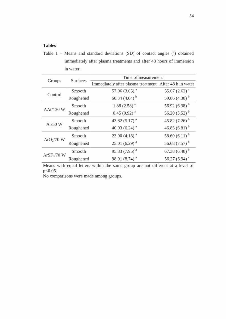
Table 1 shows the means and standard deviations of contact angle and the
results of Tukey post hoc tests. It can be seen that there was significant change in
the mean contact angle after 48 hours of immersion in water for all groups
evaluated, with the exception of the control group, in which no significant
difference was found. In control group, there was significant difference between
the mean contact angle of smooth and roughened surfaces, regardless the time of
measurement. Similar result was observed in group ArSF6/70 W only in the 48
hours period.
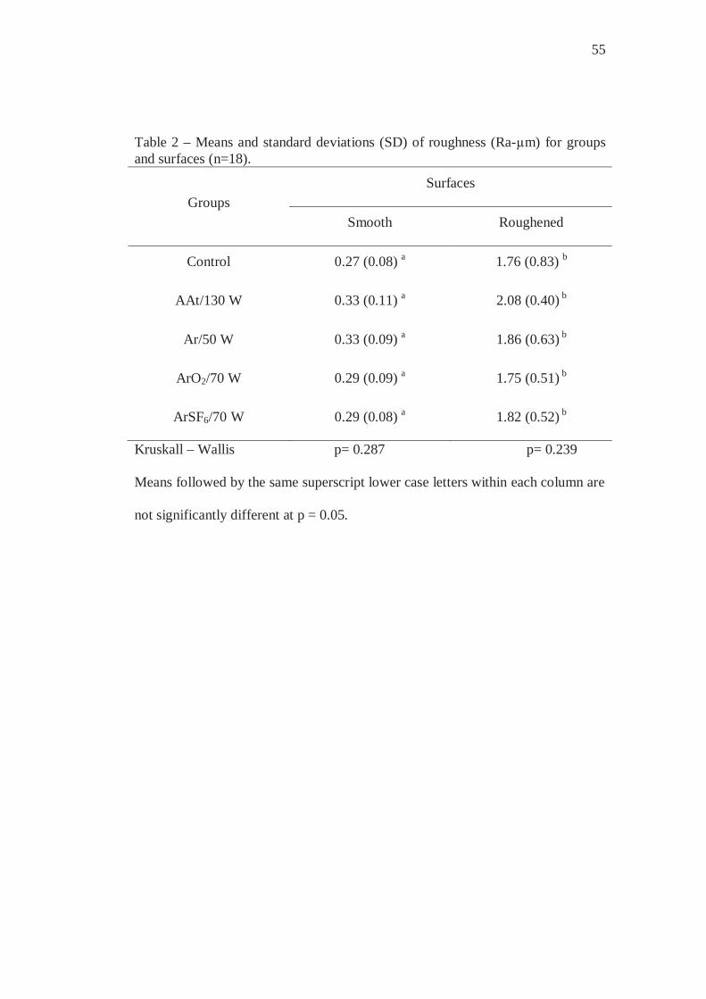
Roughness values of all groups evaluated are presented in Table 2. There
were no significant differences among the groups in each investing technique. For
all groups, the mean roughness values of the specimens processed against stone
were higher than those of the specimens processed against glass.
XPS analysis demonstrated the incorporation of fluorine into the surface of
the specimens of ArSF6/70 group. Figure 2 shows the signals assigned to CF2 and
CF3 moieties and the envelope curve, which represents the total F(1s) area.
Discussion
The initial attachment of Candida albicans on the mucosal surface of the
denture is essential in the colonization and development of denture stomatitis.
Since many factors may influence the initial adherence of yeasts to acrylic
surfaces, such as attractive hydrophobic interactions and repulsive electrostatic

41
forces, the development of the methods that reduce the adherence of Candida to
these surfaces, could be a significant step toward treatment and prevention of
denture stomatitis. Glow-discharge plasma, a type of cold plasma, has been often
used as a method of surface modification; however, in dentistry it has received
little attention. In this technique, gas temperature can remain as low as room
temperature 24, preserving the integrity of polymer-based materials. This is of
particular importance for denture base acrylic resins, in which the heating may
cause dimensional changes and affect the fit of the denture bases to the supporting
tissues 23.
In this study, the aim was to investigate whether surface modifications
with different plasma treatments could decrease the adherence of Candida
albicans to a denture base resin. One of these modifications was intended to
decrease the surface hydrophobicity. The results revealed that plasma treatments
with Ar/50 W, ArO2/70 W, AAt/130 W decreased the hydrophobicity of all
surfaces (rough and smooth) immediately after the plasma treatment, when
compared to the untreated specimens. This occurred most likely because the
plasma treatments generated free radicals in the material by inelastic collisions,
mainly involving energetic electrons in the discharge and species on the polymer
surfaces 15. Chemical reactions that occur between these free radicals and species,
such as atomic hydrogen or oxygen from the polymer or atmospheric
contaminants, incorporate hydrophilic groups to the polymer surfaces 15, and the
contact angle is reduced.

42
Another surface modification used in the present study involved the
incorporation of fluorine in the resin surface. To the author’s knowledge, to date
this is the first study that has addressed this issue. The results have shown that the
contact angle, immediately after plasma treatment with ArSF6/70 W, increased
considerably compared to control group, which is in agreement with the studies of
Guruvenket et al. 21 and Rangel et al. 20. This was probably due to the replacement
of hydrophilic species by fluorine atoms 16. As a result, the hydrogen bonds
between water molecules and surface groups decrease, reducing the hydrophilicity
16. X-ray photoelectron spectroscopy (XPS) was used to confirm the chemical
changes on surfaces treated with ArSF6/70 W. The incorporation of fluorine
occurred, as demonstrated by the presence of F(1s) peak in XPS spectra. There
was a decrease in the atomic concentrations of carbon from 75.3 at.% to 55.4
at.%, oxygen from 23.0 at.% to 14.1 at.%, and fluorine incorporation of 29.6 at.%.
In this study, the adhesion of Candida albicans was quantified using the
2,3-bis(2-methoxy-4-nitro-5-sulfo-phenyl)-2H-tetrazolium-5-carboxanilide (XTT)
reduction assay. This colorimetric method is based on metabolic activity 30 and
has been widely used for the quantification of yeasts 27,30,31. The results revealed
that ArO2/70W plasma treatment significantly reduced the yeast adhesion. These
results do not agree with the data reported by Yildirim et al. 3, who have found
higher counts of Candida albicans in plasma treated surfaces than in the
unmodified control group. One possible reason for this disagreement could be
that, in the study of Yildirim et al. 3, different plasma parameters were used
(oxygen atmosphere at 50 or 100 W, during 15 minutes). The decrease of
43
adherence observed in the present study could not be related to hydrophobic
interactions. Although ArO2/70W plasma treatment resulted in a more hydrophilic
surface (Table 1), after immersion of the specimens in water for 48 hours, the
contact angles were similar to those of the control specimens. A possible
explanation for this recovery could be that the decrease in the water contact angle
obtained with ArO2/70 W enhanced the surface energy. Under such a situation, it
has been observed the polymer surfaces submitted to plasmas tended to return to
their original hydrophobicity 15. This was attributed to movement of polar groups
from the surface to the polymer bulk.
The results also demonstrated that the adherence of C. albicans to
ArSF6/70W plasma treated specimens was significantly reduced compared to
control. To the author’s knowledge to date, the potential of fluorine plasma
treatment to reduce adhesion of Candida albicans to denture base material has yet
not been addressed. Similarly to the ArO2/70W treatment, it was not possible to
correlate the reduction in C. albicans adhesion promoted by ArSF6/70W treatment
with surface hydrophobicity. After the ArSF6/70W plasma treatment, the sample
surfaces became hydrophobic exhibiting the highest contact angle values.
However, after water immersion for 48 hours, a decrease in the contact angle
values was observed and the values were close to those obtained in the other
groups, including control. Despite these changes, the fluorine was still present in
the surface, as demonstrated by XPS analysis. Hence, the reduction of the
adherence of C. albicans with ArSF6/70W plasma treatment could be attributed to
repulsive electrostatic forces between the fungal cells and specimens in which

44
fluorine was incorporated. Robinson et al. 22 found that increasing the degree of
fluorination the surfaces became more negative due to the presence of the
electronegative fluorine atoms. It has been reported that surface-charged resins
may alter the ionic interaction between the denture base and Candida spp 9-11.
Negatively charged resin surfaces showed significantly lower levels of Candida
than the untreated ones 9,11.
Roughness has been considered an important factor that affects the
adhesion and some studies have found that an increase in surface roughness
facilitated the yeast retention 1,32-34. Thus, in this study, in all groups half of the
specimens were processed against stone and half were polymerized in contact
with glass for obtaining rough and smooth surfaces, respectively. However, no
significant differences were observed in the Candida albicans adhesion (Figure
1). These results are in accordance with those reported in recent studies where no
significant influence of roughness on adherence of Candida albicans was verified.
5,26,35-37. Nevertheless, other studies should be conducted using specimens with
prepared surfaces that cover a wide range of roughness values.
Since all intra-oral surfaces are coated by saliva, it is important to consider
its effects on adhesion. Hence, in the present investigation, half of the specimens
of each group were preconditioned with saliva prior to inoculation. Adhesion of
Candida albicans to untreated and treated specimens was not influenced by saliva.
A comparison among in vitro studies reveals contradictory results. While some
authors observed that salivary pellicle promoted fungal colonization on the
materials 2,28,38-41, others have found that the pretreatment with saliva had no

45
effect 27,30,42-44 or decreased the Candida adherence 26,44,45-48. These divergent
results could be attributed to different methodologies used, including the number
of donors and possible individual variations, the use of stimulated or unstimulated
saliva, filtered or whole saliva, undiluted or diluted saliva, different speed and
time of saliva centrifugation and incubation periods and temperatures. These
factors could result in different compositions and viscosities, affecting the role of
saliva in adherence. The different results could also be related to the materials
evaluated in each study, such as resilient denture lining materials 2,39,40,42,47,48,
maxillofacial polymeric materials 41, acrylic surfaces 26,28,38,41,43-48, and
polystyrene 27,30. It has been observed that surfaces with small differences in their
chemical composition differ in respect to adsorption of salivary proteins 6. These
variations in methodologies make comparison among studies difficult and point to
the need of standardization. Nevertheless, the results of this study are similar to
those reported by Ramage et al. 27, Jin et al. 30 and Thein et al. 43 who observed
that the presence of saliva did not interfere with Candida albicans adherence.
This study has limitations since only one strain of Candida albicans and
one heat-polymerized denture base were used. In addition, other plasma
parameters and/or atmospheres should be evaluated. Despite these limitations, the
results demonstrated that ArSF6/70W e ArO2/70 plasma treatments showed
promise, justifying further investigation.
Within the limitations of this in vitro study, the following conclusions can
be drawn:
46
1) The adherence of Candida albicans was significantly reduced by ArO2/70
W and ArSF6/70 W plasma treatments when compared to the control
group, regardless the presence or absence of saliva and surface roughness
(smooth or roughened).
2) The hydrophobicity (high water contact angle) of the acrylic resin
evaluated was altered by the plasma treatments. However, after 48 hours
of immersion in water, the mean contact angles of the treated specimens
were similar to those of control specimens.
3) No significant effect of surface roughness and saliva on the adherence of
Candida albicans was detected for all groups evaluated.
Acknowledgements
This research was supported by FAPESP (Grant - 2007/02210-1 and
2007/04917-5) and CNPq (Grant 479252/2007-6). We thank Prof. Peter Hammer
for his assistance with the XPS analysis.
References
1 Verran J, Maryan CJ. Retention of Candida albicans on acrylic resin and
silicone of different surface topography. J Prosthet Dent, 1997; 77:535-39.
2 Nikawa H, Yamamoto T, Hamada T, Rahardjo MB, Murata H. Antifungal
effect of zeolite-incorporated tissue conditioner against Candida albicans growth
and/or acid production. J Oral Rehabil, 1997; 24:350-57.
47
3 Yildirim MS, Hasanreisoglu U, Hasirci N, Sultan N. Adherence of
Candida albicans to glow-discharge modified acrylic denture base polymers. J
Oral Rehabil, 2005; 32:518-25.
4 Radford DR, Challacombe SJ, Walter JD. Denture plaque and adherence
of Candida albicans to denture-base materials in vivo and in vitro. Crit Rev Oral
Biol Med, 1999; 10(1):99-116.
5 Burgers R, Schneider-Brachert W, Rosentritt M, Handel G, Hahnel S.
Candida albicans adhesion to composite resin materials. Clin Oral Investig, 2009;
13(3):292-9.
6 Sipahi C, Anil N, Bayramli E. The effect of acquired salivary pellicle on
the surface free energy and wettability of different denture base materials. J Dent,
2001; 29:197-204.
7 Klotz SA, Drutz DJ, Zajic JE. Factors governing adherence of Candida
species to plastic surfaces. Infect Immun, 1985; 50(1):97-101.
8 Minagi S, Miyake Y, Inagaki K, Tsuru H, Suginaka H. Hydrophobic
interaction in Candida albicans and Candida tropicalis adherence to various
denture base resin materials. Infect Immun, 1985; 47:11-4.
9 Park SE, Periathamby AR, Loza JC. Effect of surface-charged
poly(methylmethacrylate) on the adhesion of Candida albicans. J Prosthodont,
2003; 12:249-54.
10 Puri G, Berzins DW, Dhuru VB, Raj PA, Rambhia SK, Dhir G, Dentino
AR. Effect of phosphate group addition on the properties of denture base resins. J
Prosthet Dent, 2008; 100:302-8.
48
11 Park SE, Blissett R, Susarla SM, Weber H-P. Candida albicans
adherence to surface-modified denture resin surfaces. J Prosthodont, 2008;
17(5):365-69.
12 Dhir G, Berzins DW, Dhuru VB, Periathamby AR, Dentino A. Physical
properties of denture base resins potentially resistant to Candida adhesion. J
Prosthodont, 2007; 16(6):465-72.
13 Redding S, Bhatt B, Rawls HR, Siegel G, Scott K, Lopez-Ribot J.
Inhibition of Candida albicans biofilm formation on denture material. Oral Surg
Oral Med Oral Pathol Oral Radiol Endod, 2009; 107(5):669-72.
14 Yoshijima Y, Murakami K, Kayama S, Liu D, Hirota K, Ichikawa T,
Miyake Y. Effect of substrate surface hydrophobicity on the adherence of yeast
and hyphal Candida. Mycoses. In press.
15 Rangel EC, Gadioli GZ, Cruz NC. Investigations on the stability of
plasma modified silicone surfaces. Plasmas and Polymers 2004;9:35-48.
16 Hodak SK, Supasai T, Paosawatyanyong B, Kamlangkla K, Pavarajarn
V. Enhancement of the hydrophobicity of silk fabrics by SF6 plasma. Appl Surf
Sci, 2008; 254:4744-49.
17 Lai J, Sunderland B, Xue J, Yan S, Zhao W, Folkard M, Michael BD,
Wang Y. Study of hydrophilicity of polymer surfaces improved by plasma
treatment. Appl Surf Sci, 2006; 252:3375-79.
18 Suanpoot P, Kueseng K, Ortmann S, Kaufmann R, Umongno C,
Nimmanpipug P, Boonyawan D, Vilaithong T. Surface analysis of hydrophobicity
49
of Thai silk treated by SF6 plasma. Surface & Coatings Technology, 2008;
202:5543-49.
19 Rad AY, Ayhan H, Piskin E. Adhesion of different bacterial strains to
low-temperature plasma-treated sutures. J Biomed Mater Res A, 1998; 41:349-58.
20 Rangel EC, Bento WCA, Kayama M, Schreiner WH, Cruz NC.
Enhancement of polymer hydrophobicity by SF6 plasma treatment and argon
plasma immersion ion implantation. Surf Interface Anal, 2003; 35:179-183.
21 Guruvenket S, Iyer GRS, Shestakova L, Morgen P, Larsen NB, Rao GM.
Fluorination of polymethylmethacrylate with tetrafluoroethane using DC glow
discharge plasma. Appl Surf Sci, 2008; 254:5722-26.
22 Robinson GN, Kebabian PL, Feedman A, DePalma V. Temperature-
dependent surface potentials of fluorinated alkanethiolate self-assembled
monolayers. Thin Solid Films, 1997; 310:24-8.
23 Polukoshko KM, Brudvik JS, Nicholls JI, Smith DE. Evaluation of heat-
cured resin bases following the addition of denture teeth using a second heat cure.
J Prosthet Dent, 1992; 67(4):556-62.
24 Liu Y, Kuai P, Huo P, Liu C. Fabrication of CuO nanofibers via the
plasma decomposition of Cu(OH)2. Mater Lett, 2009; 63:188-90.
25 Moisan M, Barbeau J, Crevier MC, Pelletier J, Philip N, Saoudi B.
Plasma sterilization. Methods and mechanisms. Pure Appl. Chem, 2002;
74(3):349-58.
50
26 Moura JS, Silva WJ, Pereira T, Cury ADB, Garcia RCMR. Influence of
acrylic resin polymerization methods and saliva on the adherence of four Candida
species. J Prosthet Dent, 2006; 96:205-11.
27 Ramage G, Tomsett K, Wickes BL, López-Ribot JL, Redding SW.
Denture stomatitis: A role for Candida biofilms. Oral Surg Oral Med Oral Pathol
Oral Radiol Endod, 2004; 98:53-9.
28 Chandra J, Mukherjee PK, Leidich SD, Faddoul FF, Hoyer LL, Douglas
LJ, Ghannoum MA. Antifungal resistance of candidal biofilms formed on denture
acrylic in vitro. J Dent Res, 2001; 80:903-8.
29 Silva WJ, Seneviratne J, Parahitiyawa N, Rosa EAR, Samaranayake LP,
Del Bel Cury AA. Improvement of XTT assay performance of studies involving
Candida albicans biofilms. Braz Dent J, 2008; 19:364-69.
30 Jin Y, Samaranayake LP, Samaranayake Y, Yip HK. Biofilm formation
of Candida albicans is variably affected by saliva and dietary sugars. Arch Oral
Biol, 2004; 49:789-98.
31 Kuhn DM, Balkis M, Chandra J, Mukherjee PK, Ghannoum MA. Uses
and limitations of the XTT assay in studies of Candida growth and metabolism. J
Clin Microbiol, 2003; 41:506-8.
32 Radford DR, Sweet SP, Challacombe SH, Walter JD. Adherence of
Candida albicans to denture-base materials with different surface finishes. J Dent,
1998; 26:577-83.

51
33 Taylor R, Maryan C, Verran J. Retention of oral microorganisms on
cobalt-chromium alloy and dental acrylic resin with different surface finishes. J
Prosthet Dent, 1998; 80:592-97.
34 Lamfon H, Porter SR, McCullough M, Pratten J. Formation of Candida
albicans biofilms on non-shedding oral surfaces. Eur J Oral Sci, 2003; 111:465-
71.
35 Nikawa H, Jin C, Makihira S, Egusa H, Hamada T, Kumagai H. Biofilm
formation of Candida albicans on the surfaces of deteriorated soft denture lining
materials caused by denture cleansers in vitro. J Oral Rehabil, 2003; 30:243-50.
36 Nevzatoglu EU, Özcan M, Kulak-Ozkan Y, Kadir T. Adherence of
Candida albicans to denture base acrylics and silicone-based resilient liner
materials with different surface finishes. Clin Oral Investig, 2007; 11(3):231-36.
37 Ferreira MAF, Pereira-Cenci T, Rodrigues de Vasconcelos LM,
Rodrigues-Garcia RCM, Del Bel Cury AA. Efficacy of denture cleansers on
denture liners contaminated with Candida species. Clin Oral Investig, 2009;
13:237-42.
38 Henriques M, Azeredo J, Oliveira R. Adhesion of Candida albicans and
Candida dubliniensis to acrylic and hydroxyapatite. Colloids Surf B
Biointerfaces, 2004; 33:235-41.
39 Nikawa H, Hayashi S, Nikawa Y, Hamada T, Samaranayake LP.
Interactions between denture lining material, protein pellicles and Candida
albicans. Arch Oral Biol, 1993; 38(7):631-34.
52
40 Nikawa H, Jin C, Hamada T, Murata H. Interactions between thermal
cycled resilient denture lining materials, salivary and serum pellicles and Candida
albicans in vitro. Part I. Effects on fungal growth. J Oral Rehabil, 2000; 27:41-5.
41 Nikawa H, Chen J, Hamada T, Nishimura M, Polyzois G. Candida
albicans colonization on thermal cycled maxillofacial polymeric materials in
vitro. J Oral Rehabil, 2001; 28:526-33.
42 Tari BF, Nalbant D, Al DF, Kustimur S. Surface roughness and
adherence of Candida albicans on soft lining materials as influenced by
accelerated aging. J Contemp Dent Pract, 2007; 8(5):1-11.
43 Thein ZM, Samaranayake YH, Samaranayake LP. Characteristics of dual
species Candida biofilms on denture acrylic surfaces. Arch Oral Biol, 2007;
52:1200-08.
44 Karaagaclioglu L, Can G, Yilmaz B, Ayhan N, Semiz O, Levent H. The
adherence of Candida albicans to acrylic resin reinforced with different fibers. J
Mater Sci Mater Med, 2008; 19(2):959-63.
45 Samaranayake, L. P.; McCourtie, J.; MacFarlane, T. W. Factors affecting
the in-vitro adherence of Candida albicans to acrylic surfaces. Arch Oral Biol,
1980; 25:611-15.
46 McCourtie J, MacFarlane TW, Samaranayake LP. Effect of saliva and
serum on the adherence of Candida species to chlorhexidine-treated denture
acrylic. J Med Microbiol, 1986; 21:209-13.

53
47 Waters MGJ, Williams DW, Jagger RG, Lewis MAO. Adherence of
Candida albicans to experimental denture soft lining materials. J Prosthet Dent,
1997; 77:306-12.
48 Pereira-Cenci T, Cury AADB, Cenci MS, Rodrigues-Garcia RCM. In
vitro Candida colonization on acrylic resins and denture liners: influence of
surface free energy, roughness, saliva, and adhering bacteria. Int J Prosthodont,
2007; 20:308-10.
54
Tables
Table 1 – Means and standard deviations (SD) of contact angles (º) obtained
immediately after plasma treatments and after 48 hours of immersion
in water.
Groups Surfaces Time of measurement
Immediately after plasma treatment After 48 h in water
Control Smooth 57.06 (3.05) a 55.67 (2.62) a
Roughened 60.34 (4.04) b 59.86 (4.38) b
AAt/130 W Smooth 1.88 (2.58) a 56.92 (6.38) b
Roughened 0.45 (0.92) a 56.20 (5.52) b
Ar/50 W Smooth 43.82 (5.17) a 45.82 (7.26) b
Roughened 40.03 (6.24) a 46.85 (6.81) b
ArO2/70 W Smooth 23.00 (4.18) a 58.60 (6.11) b
Roughened 25.01 (6.29) a 56.68 (7.57) b
ArSF6/70 W Smooth 95.83 (7.95) a 67.38 (6.48) b
Roughened 98.91 (8.74) a 56.27 (6.94) c Means with equal letters within the same group are not different at a level of p<0.05. No comparisons were made among groups.
55
Table 2 – Means and standard deviations (SD) of roughness (Ra-μm) for groups and surfaces (n=18).
Groups Surfaces
Smooth Roughened
Control 0.27 (0.08) a 1.76 (0.83) b
AAt/130 W 0.33 (0.11) a 2.08 (0.40) b
Ar/50 W 0.33 (0.09) a 1.86 (0.63) b
ArO2/70 W 0.29 (0.09) a 1.75 (0.51) b
ArSF6/70 W 0.29 (0.08) a 1.82 (0.52) b
Kruskall – Wallis p= 0.287 p= 0.239
Means followed by the same superscript lower case letters within each column are
not significantly different at p = 0.05.
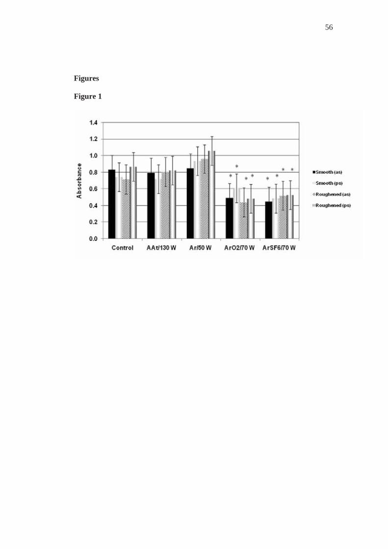
56
Figures Figure 1
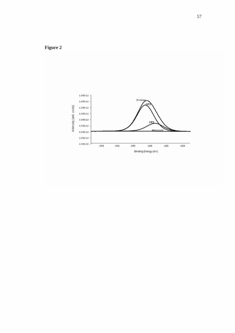
57
Figure 2
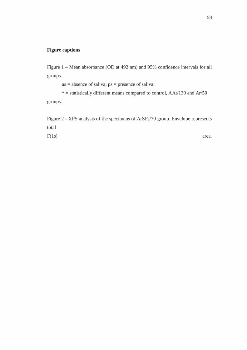
58
Figure captions
Figure 1 – Mean absorbance (OD at 492 nm) and 95% confidence intervals for all
groups.
as = absence of saliva; ps = presence of saliva.
* = statistically different means compared to control, AAt/130 and Ar/50
groups.
Figure 2 - XPS analysis of the specimens of ArSF6/70 group. Envelope represents
total
F(1s) area.
3.2 Capítulo 2
Evaluation of fungal adherence to plasma-modified
polymethylmethacrylate
Fungal adherence to polymethylmethacrylate
Zamperini CAa, Machado ALa,*, Vergani CEa, Pavarina ACa, Rangel ECb, Cruz
NCb
a Araraquara Dental School, UNESP – Univ Estadual Paulista, Department
of Dental Materials and Prosthodontics, Araraquara, São Paulo, Rua Humaitá nº
1680, CEP 14.801-903, Brazil.
b UNESP – Univ Estadual Paulista, Laboratory of Technological Plasmas,
Sorocaba, São Paulo, Avenida 3 de Março nº 511, CEP 18.085-180, Brazil.
*Corresponding author:
Profa. Dra. Ana Lucia Machado
Araraquara Dental School , UNESP –Univ. Estadual Paulista, Department of
Dental Materials and Prosthodontics, Araraquara, São Paulo, Rua Humaitá nº
1680, CEP 14.801-903, Brazil.
Tel: 55-16-33016410 Fax: 55-16-33016406
Email: [email protected]
Summary
There is a propensity for fungal adherence to the polymethylmethacrylate
used for making denture bases. Therefore, this study investigated whether surface
modifications with plasma treatments would reduce the adherence of C. albicans
to a denture base resin. Samples (n=180) with smooth and rough surfaces were
60
made and divided into five groups: control – non treated; experimental groups –
submitted to plasma treatments to obtain surfaces with different hydrophobicities
(Ar/50 W; ArO2/70 W; AAt/130 W) or with incorporated fluoride (Ar/SF670 W).
Contact angles were measured immediately after treatments and after samples
were immersed in water for 48 hours. For each group, half the samples were
incubated with saliva before the adherence test. The number of adhered C.
albicans was evaluated by counting after violet crystal staining. The plasma
treatments were effective in modifying the polymethylmethacrylate surface.
However, there was a significant alteration in the contact angle measured after
immersion in water. No statistically significant difference in the adherence of C.
albicans was observed between the experimental and control groups, irrespective
of the presence or absence of saliva, and surface roughness.
Keywords: Candida spp; Candida albicans; fungal adherence; denture stomatitis;
cell-surface-hydrophobicity.
Introduction
Polymers are widely used materials in different areas [1, 2]. In dentistry,
polymethylmethacrylate is the polymer of choice for making removable denture
bases for the purpose of rehabilitating partially or completely edentulous patients.
Nevertheless, in spite of the good mechanical and esthetic properties of this
material, microorganisms, particularly Candida albicans, have a propensity for

61
adhering to denture surfaces [1]. This adhesion capacity of Candida albicans to
denture surfaces is the first step, considered essential, for the development of
denture stomatitis [3-5], a common type of oral candidiasis among denture
wearers [6-8]. Therefore, the development of methods that could modify these
surfaces in order to prevent the adhesion of Candida albicans, would be a
significant advancement in the treatment of this pathology.
Although the exact mechanisms involved in the adherence of
microorganisms to dentures are not completely known, various factors may
influence this process, among them, surface roughness and the presence of saliva.
The increase in surface roughness has been correlated with the greater ease of
fungal retention [3, 9]. On the other hand, the effect of saliva on this process is not
clear and the results are controversial [10-12]. Some authors [13-15] also point
out the influence of hydrophobic interactions. Minagi et al. [14] observed that the
closer the surface energy of the fungal cell and the substrate are, the greater the
probability of adherence occurring. Klotz et al. [13] observed a linear relationship
between the number of Candida albicans adhered per unit of area and the
hydrophobicity of polymers. Electrostatic interactions have also been mentioned
as another factor that could influence the adherence of microorganisms to
polymers [11, 13, 16]. Fungal cells, whose surfaces were electrically altered with
a positive charge, were shown to be more adherent, suggesting the action of
repulsive forces present between the fungi and polymers [13].
Thus, considering that the characteristics of the substrate are important for
Candida adherence [4, 17], the surface modification of the

62
polymethylmethacrylate used for making denture bases could reduce the adhesion
of Candida albicans. In this context, the modification of biomaterial surfaces by
means of plasma treatment has been proposed [1, 4, 15]. Among the various
results achieved by this technique, is the increase in the hydrophilicity of
biomaterials [2, 4, 18], modification of the chemical composition of surfaces [19,
20] and the reduction in bacterial adhesion [21]. Plasma treatment also allows
fluoride to be incorporated into the materials [22, 23], which could result in a
negatively charged surface [24] and reduce the adhesion of Candida [11, 13].
In view of this, the object of the present study was to verify whether
surface modifications with different plasma treatments would diminish the
adherence of Candida albicans to a polymethylmethacrylate used for denture
bases.
Materials and Methods
Preparation of Acrylic Resin Specimens
The specimens (n=180) were fabricated from an acrylic resin denture base
material (Vipi Wave - VIPI Indústria e Comércio Exportação e Importação de
Produtos Odontológicos Ltda Pirassununga, SP, Brazil) using a conventional
flasking and pressure-pack technique. Initially, a metal mold was used to make
disk-shaped silicone patterns Zetaplus/Indurent - Zhermack, Badia Polesine,
Rovigo, Italy) measuring 13.8 X 2 mm. Half of the silicone patterns were invested
in the flasks directly in dental stone, while the other half of the patterns were
sandwiched between two glass slides before investing. These two types of
investing techniques were used to obtain rough and smooth specimens, thus
63
mimicking the tissue-fitting surface and the outer surface of dentures,
respectively. The flasks were separated, silicone patterns removed, and the stone
surfaces were painted with a separating medium (Vipi Film - VIPI Indústria e
Comércio Exportação e Importação de Produtos Odontológicos Ltda
Pirassununga, SP, Brazil). For each specimen, 1 g of powder and 0.47 ml of
monomer liquid were mixed and processed according to the manufacturer’s
instructions. The mixture was packed into the molds, a trial pack was completed,
and excess material was removed. A final pack was performed and held for 15
minutes. The denture base acrylic resin was processed in a 500 W domestic
microwave oven (Brastemp – Brastemp da Amazônia SA, Manaus, AM, Brazil)
for 20 minutes at 20% power, followed by 5 minutes at 90% power. The flasks
were allowed to bench cool at room temperature, the specimens were deflasked,
and excess flash was aseptically removed with a sterile bur (Maxi-Cut; Lesfils de
August Malleifer SA, Ballaigues, Switzerland).
Surface Roughness Measurements
The surface roughness of all specimens was measured with a profilometer
(Mitutoyo SJ 400 – Mitutoyo Corporation - Japan). Three measurements were
made for each specimen and the average reading was designated as the Ra (μm)
value of that specimen. The resolution was 0.01 μm, the interval (cutoff length) of
0.8 mm, the transverse length of 2.4 mm; the stylus speed 0.5 mm/s, and the
diamond stylus tip radius was 5 μm. All measurements were recorded by one
operator.
Plasma Treatments

64
After roughness measurements, the specimens were cleaned in an
ultrasonic cleaner using water and detergent bath for 15 minutes, then sonicated in
distilled water for 15 minutes and air dried. The specimens were then divided into
five groups, each including 18 specimens processed against stone and 18
polymerized in contact with glass. In the control group, the specimens were left
untreated. For the four experimental groups, both specimen surfaces were exposed
to plasmas generated under the following conditions: argon atmosphere at 50 W
(group Ar/50 W); argon/oxygen atmosphere at 70 W (group ArO2/70 W);
atmospheric air at 130 W (AAt/130 W); argon atmosphere, followed by plasma
treatment in a sulfur hexafluoride atmosphere, both performed at 70 W (group
Ar/SF670 W). The plasma exposure time (5 minutes) and the position of the
specimens within the plasma chamber were kept unchanged. To determine the
plasma parameters used in the experimental groups, pilot experiments were
performed, in which various conditions of exposure time, atmosphere composition
and pressure, and radiofrequency power were tested. For groups Ar/50 W,
Ar/O270 W and AAt/130 W, the plasma parameters were chosen based on the
degree of surface hydrophobicity. Parameters that produced surfaces with low
hydrophobicity (contact angle close to zero) were used for group AAt/130 W. For
groups Ar/50 W and Ar/O270 W, parameters that provided hydrophobicity values
between those of the untreated specimens (higher hydrophobic) and those of the
group AAt/130 W specimens were chosen. In the case of group Ar/SF670 W, the
pilot experiments established the appropriate conditions for the incorporation of
fluorine into the surfaces. Fluorine incorporation was confirmed by photoelectron

65
spectroscopy analysis (XPS), carried out in a UNI-SPECS UHV spectrometer
using Mg K line (E = 1253.6 eV) and with the analyzer pass energy set to 10 eV.
The inelastic background of the C 1s, F 1s, O 1s, and N 1s electron core-level
spectra was subtracted using Shirley’s method. The binding energies of the
spectra were corrected using the hydrocarbon component of the polymer fixed at
285.0 eV. The composition of the surface layer was determined from the ratio of
the relative peak areas corrected by sensitivity factors of the corresponding
elements. The spectra were fitted without placing constraints using multiple Voigt
profiles. The width at half maximum (FWHM) varied between 1.6 and 2.0 eV and
the accuracy of the peak positions was ±0.1 eV. One specimen of untreated
denture base acrylic resin and one of ArSF6-treated specimen were analyzed.
Plasma treatments were performed by the application of radiofrequency
power (13.56 MHz) to two parallel plate electrodes fitted inside a homemade
stainless steel vacuum chamber.
Contact Angle Measurements
The water contact angle has been measured to characterize the surface
wettability [4, 18]. This angle is defined as the angle at the intercept of a plane
tangent to the drop and the plane containing the substrate-liquid interface. The
measurements were performed in an automated goniometer (Ramé-Hart, 100-00)
using deionized water as test liquid. The goniometer comprises a CCD camera to
record the image of a droplet placed onto the surface using a microsyringe and a
dedicated image processing software to determine the contact angle.
Measurements in two different positions were made for each specimen and the

66
average was calculated. Specimens were then stored at room temperature in sterile
distilled water for 48 h to release any residual monomer [10]. Afterwards, the
contact angles of each specimen were again measured.
Saliva Collection
Unstimulated whole human saliva was collected from fifteen healthy adult
volunteers. The saliva was expectorated into sterile 50 ml Falcon tubes on ice,
pooled and clarified by centrifugation at 10,000 g for 5 min at 4 ºC [10]. The
saliva was prepared at 50% (vol/vol) in sterile PBS [25]. The resulting saliva was
immediately stored at -70 ºC until use. The study was approved by the Ethics
Committee of Araraquara Dental School (027/2007), and all subjects volunteered
to participate and signed an informed consent form.
Adherence Assay
Candida albicans strain ATCC 90028 was used. Stock cultures were
maintained at -70 ºC. After recovery this was maintained on YEPD medium (1%
yeast extract, 2% peptone, 2% dextrose, 2% agar) stored at 4 – 6 ºC during the
experimental period. To prepare the yeast inoculum, a loopful of the stock culture
was streaked onto YEPD medium and incubated at 37 ºC for 48 h. Two loopfuls
of this young culture were transferred to 20 ml of yeast nitrogen base (YNB)
medium with 50 mM glucose and incubated at 37 ºC for 24 h. Cells of the
resultant culture were harvested, washed twice with phosphate-buffered saline
(PBS, pH 7.2) at 5000 g for 5 min and resuspended in YNB with 100 mM
glucose. Candida suspensions were spectrophotometrically standardized to a
concentration of 1 x 10⁷ cells/ml. Three ml of the standardized C. albicans cell

67
suspension was added to each well containing the specimen. The cells were left to
adhere for 90 min at 37 ºC [26]. The non-adherent cells were removed from the
specimen by gently washing twice with 3 ml PBS. The negative controls were
acrylic specimens to which no cells were added. All experimental conditions were
performed in triplicate on three independent occasions.
Preconditioning with Saliva
To investigate the effect of the saliva on candidal adhesion to the denture
acrylic resin, half of the specimens from each group (9 rough and 9 smooth) were
incubated in 12-well microtiter plates and coated with 3 ml of prepared saliva for
30 min at room temperature prior to the adhesion assay.
Measurement of Adherent C. albicans
Nine specimens from each experimental condition (rough and smooth
surfaces, plasma treatment and control, presence and absence of saliva) were
evaluated by crystal violet staining assay. After the non-adherent cells were
removed by washing, the specimens were fixed in 80% ethanol, stained with
crystal violet for 1 minute and washed with PBS [27]. Adherent yeast cells were
counted in 10 different fields for each specimen, using a light microscope
(Olympus BX51, Japan) at 400 x magnification and the mean values were
calculated. The results were expressed as cells/mm2.
Data from roughness measurements were analyzed by Kruskall-Wallis
non-parametric test. For each group, two-way repeated measure analysis of
variance, followed by Tukey’s test were used to evaluate the effect of investing
technique and time of measurement on the contact angle. Differences in the

68
adherent yeast cells (crystal violet staining assay) among the experimental
conditions were evaluated by two-way measure analysis of variance. Data of yeast
counts (cells per mm2) were transformed by log. A significance level of 0.01 was
used for all statistical tests.
Results
Roughness values of all groups evaluated are presented in Table 1. For all
groups, the mean roughness values of the specimens processed against glass were
lower than those of the specimens processed against stone. There were no
significant differences among the groups in each investing technique.
Table 2 shows the means and standard deviations of contact angle and the
results of Tukey post hoc tests. It can be observed that there was significant
change in the mean contact angle after 48 hours of immersion in water for all
groups, with the exception of the control group, in which no significant difference
was found. In ArO2/70 W and ArSF6/70 W groups, there was significant
difference between the mean contact angle of smooth and roughened surfaces, in
the 48-hour period. XPS analysis demonstrated the incorporation of fluorine into
the surface of the specimens of the ArSF6/70 group (Fig. 1).
Candida albicans adherence, as determined by crystal violet staining assay
is shown in Fig. 2. Experimental and control groups did not differ from each other
(p<.01), irrespective of the presence or absence of saliva and surface roughness
(smooth or roughened).
Discussion
69
Initial adherence of Candida albicans on the surface of the
polymethylmethacrylate used for making denture bases, is essential in the
pathogenesis of denture stomatitis. In view of this, the development of methods
that reduce this adherence would be an important step in the treatment and
prevention of denture stomatitis. Plasma treatment has been widely used as a
method for modifying surfaces. In this technique, a partially ionized gas is created
by an electrical discharge, and consequently, a highly reactive environment is
formed. Thus, the purpose of this study was to investigate whether surface
modifications by means of different plasma treatments would diminish the
adherence of Candida albicans on a denture base acrylic resin.
Bearing in mind the possible influence of surface roughness on the initial
adherence of microorganisms [3, 28, 29], two test specimen fabrication methods
were used in this study to obtain rough and smooth surfaces, simulating,
respectively, the internal and external surfaces of dental prostheses. According to
Quirynen et al. [28], roughness could increase the area available for adhesion and
provide niches for microorganisms, protecting them from the action of the forces
of removal. In this study, the mean Ra values of rough surfaces were higher than
those of the smooth surfaces for all the groups. Nevertheless, no statistically
significant effect of roughness on the adherence of Candida albicans was
detected. It has been demonstrated that the roughness of denture base materials
can vary considerably, and values between 3.4 and 7.6 μm have been observed
[30], which are higher than those obtained in the rough samples used in this study.
Moreover, Taylor et al. [31] observed that although a small increase in roughness
70
had resulted in greater bacterial adhesion, higher increases in roughness
diminished adherence. Results such as these indicate the need for further studies
that evaluate the effect of surface roughness on the adhesion of Candida albicans,
using resin samples with different pre-established roughness values that cover a
wide range of variation. In any event, the results observed in the present study are
in agreement with those of recent researches [10, 27, 32-34] in which no
significant influence of roughness on the adherence of Candida albicans was
observed.
The surface modifications made in the samples of the present study
include the reduction of surface hydrophobicity, obtained by means of the plasma
treatments with AAt/130 W, ArO2/70 W, Ar/50 W, as well as the incorporation of
fluoride into the resin surface, achieved in the group treated with ArSF6/70 W.
The groups AAt/130 W, ArO2/70 W and Ar/50 W presented a decrease in the
angles of contact values, indicating that immediately after the treatments, the
surfaces became more hydrophilic. These results could be attributed to the fact
that during plasma treatments, inelastic collisions involving energy electrons from
the discharge and species from the polymer surface create free radicals, which in
turn react chemically with species from the polymer or atmospheric air,
incorporating hydrophilic groups into the polymeric surfaces [18]. This
incorporation of hydrophilic groups increases the surface energy, consequently
diminishing the angles of contact values [18].
Similar to the observations made by other authors [22, 23], plasma
treatment with ArSF6/70 W increased the angle of contact of the samples. This

71
increase probably occurred by means of substitutions of hydrophilic species by
fluoride atoms [19]. As a result, the hydrogen bonds between the water molecules
and the superficial groups of the polymer diminished, reducing the hydrophilicity
of the samples [19]. X-ray photoelectron spectroscopy (XPS) was performed to
confirm the chemical changes in specimens treated with ArSF6/70 W. The
incorporation of fluorine occurred, as demonstrated by the presence of F(1s) peak
in XPS spectra. There was a decrease in the atomic concentrations of carbon from
75.3 at.% to 55.4 at.%, oxygen from 23.0 at.% to 14.1 at.%, and fluorine
incorporation of 29.6 at.%.
When the adherence of Candida albicans was considered, no significant
difference was found between the control and experimental groups. This result
differs from those reported by Yildirim et al. [4], who found greater adhesion of
Candida albicans on surfaces submitted to plasma treatments, when compared
with the control group. A possible explanation for this divergence could be
attributed to the different plasma treatment parameters used in the two studies. In
the research by Yildirim et al. [4], the samples were submitted to treatments in
oxygen atmospheres at 50 or 100 W, for 15 minutes. The absence of difference
between the groups observed in the present study could be explained by the fact
that after the samples were immersed in water for 48 hours, there was an alteration
in the angles of contact values for the experimental groups; that is, the
hydrophobicity of the samples submitted to plasma treatments came close to those
presented by the samples in the control group.

72
In the groups AAt/130 W, ArO2/70 W and Ar/50 W, this alteration in
hydrophobicity occurred due to the increase in surface energy. Under this
condition, it has been observed that polymeric surfaces submitted to plasmas tend
to return to their original hydrophobicity [18]. This could be attributed to the
movement of polar groups from the surface to the internal part of the polymer.
Although the plasma treatment with ArSF6/70W created hydrophobic surfaces, or
surfaces with low surface energy, after the samples had been immersed in water
the angles of contact values of this group also came close to the values of the
others, including those of the control group. In spite of this alteration in
hydrophobicity, the fluoride remained present on the surface, as demonstrated by
the XPS analysis. Robinson et al. [24] observed that the increase in the degree of
fluorination of the surfaces became more negative due to the presence of
electronegative atoms of fluoride. Moreover, it has been suggested that resins with
charged surfaces can alter the ionic interaction between the denture base and the
microorganisms [11, 16, 35]. However, no reduction in the adherence of Candida
albicans by means of plasma treatment with ArSF6/70W was detected in this
study.
When the denture is inserted into the oral cavity, its surface is rapidly
covered by a fine film of saliva known as the acquired pellicle [36]. This film is
capable of altering the properties of the surfaces exposed to it [4], in the same way
as the chemical characteristic of the surface of biomaterials is also capable of
influencing the formation and composition of the acquired film [4, 10, 36]. Thus,
surfaces with small differences in their chemical compositions differ with regard
73
to the adsorption of salivary proteins [4, 17]. In spite of the importance of saliva
in the process of adhesion and colonization of microorganisms, the role it plays is
still not clear [37]. In this study, half the samples of each group were pre-
conditioned in saliva before the fungal adherence test. The results demonstrated
that the adhesion of Candida albicans to the surfaces was not influenced by the
saliva, under all the experimental conditions evaluated. A comparison between in
vitro studies revealed contradictory results [4]. While some authors observed that
the film of saliva promoted fungal colonization on the materials [5, 7, 26, 37-39],
others found that pre-treatment with saliva did not significantly affect [25, 40-43]
or diminish the adherence of Candida [8-10, 43-45). These divergent results could
be attributed to the different methodologies used in each study, including different
numbers of donors, the use of stimulated or unstimulated saliva, filtered or total
saliva, diluted or undiluted saliva, different centrifugation times and speeds, as
well as different incubation periods and temperatures. In the present investigation,
the saliva used was diluted with PBS, according to the methodology of Ramage et
al. [25], which could have contributed to the absence of the effect of pre-
conditioning on fungal adherence observed in the two studies.
In addition to the factors related to the saliva itself, the different results
found in the literature could also be related to the different materials evaluated in
the studies of Candida albicans adherence, such as resilient denture relining
materials [5, 9, 38, 39, 41, 45], maxillofacial polymeric materials [37], acrylic
surfaces [7-10,26, 37, 42-45], hydroxyapatite [7] and polystyrene [25, 40]. These
methodological variations make comparison between the studies difficult and
74
point out the need for standardization. Nevertheless, the results of the present
study are similar to those related by Ramage et al. [25], Jin et al. [40] and Thein et
al. [42], in which the presence of saliva did not interfere significantly in the
adherence of Candida albicans.
This study has limitations, considering that only one strain of Candida
albicans and one heat polymerizable denture base material were used. In spite of
the adherence of Candida albicans not having been altered by the plasma
treatments, other parameters and treatment atmospheres may provide different
results. Moreover, an increase in the incidence of other species, among them
Candida glabrata, which present greater hydrophobicity than Candida albicans,
has been observed particularly in immunosuppressed patients. These aspects must
be considered in future studies.
Within the limitations of this in vitro study, the following conclusions were
drawn:
1) The hydrophobicity (contact angle) of the acrylic resin evaluated was
altered by the plasma treatments used. However, the mean contact angles
of the treated specimens were similar to those of control specimens, after
48 hours of immersion in water.
2) The adherence of Candida albicans was not significantly reduced by
plasma treatments when compared with the control, irrespective of the
presence or absence of saliva and surface roughness (smooth or
roughened).
75
3) For all groups evaluated, no significant effect was detected with regard to
the influence of surface roughness and saliva on the adherence of Candida
albicans.
Acknowledgements
This research was supported by FAPESP (Grant - 2007/02210-1 and
2007/04917-5) and CNPq (Grant 479252/2007-6). We thank Prof. Peter Hammer
for his assistance with the XPS analysis.
References
1. Shmack G, Dutschk V, Pisanova E. Modification of
polyamide fibres to improve their biocompatibility. Fibre Chemistry 2000;
32: 48-55.
2. Lai J, Sunderland B, Xue J, Yan S, Zhao W, Folkard M,
Michael BD, Wang Y. Study of hydrophilicity of polymer surfaces
improved by plasma treatment. Appl Surf Sci 2006; 252: 3375-3379.
3. Verran J, Maryan CJ. Retention of Candida albicans on
acrylic resin and silicone of different surface topography. J Prosthet Dent
1997; 77: 535-539.
4. Yildirim MS, Hasanreisoglu U, Hasirci N, Sultan N.
Adherence of Candida albicans to glow-discharge modified acrylic denture
base polymers. J Oral Rehabil 2005; 32: 518-525.
5. Nikawa H, Yamamoto T, Hamada T, Rahardjo MB, Murata
H. Antifungal effect of zeolite-incorporated tissue conditioner against

76
Candida albicans growth and/or acid production. J Oral Rehabil 1997; 24:
350-357.
6. Dagistan S, Aktas AE, Caglayan F, Ayyildiz A, Bilge M.
Differential diagnosis of denture-induced stomatitis, Candida, and their
variations in patients using complete denture: a clinical and mycological
study. Mycoses 2009; 52: 266-271.
7. Henriques M, Azeredo J, Oliveira R. Adhesion of Candida
albicans and Candida dubliniensis to acrylic and hydroxyapatite. Colloids
Surf B Biointerfaces 2004; 33: 235-241.
8. Samaranayake LP, McCourtie J, MacFarlane TW. Factors
affecting the in-vitro adherence of Candida albicans to acrylic surfaces.
Arch Oral Biol 1980; 25: 611-615.
9. Pereira-Cenci T, Cury AADB, Cenci MS, Rodrigues-
Garcia RCM. In vitro Candida colonization on acrylic resins and denture
liners: influence of surface free energy, roughness, saliva, and adhering
bacteria. Int J Prosthodont 2007; 20: 308-310.
10. Moura JS, Silva WJ, Pereira T, Cury AADB, Garcia
RCMR. Influence of acrylic resin polymerization methods and saliva on the
adherence of four Candida species. J Prosthet Dent 2006; 96: 205-211.
11. Park SE, Periathamby AR, Loza JC. Effect of surface-
charged poly(methylmethacrylate) on the adhesion of Candida albicans. J
Prosthodont 2003; 12: 249-254.
77
12. Pereira-Cenci T, Cury AADB, Crielaard W, Ten Cate JM.
Development of Candida-associated denture stomatitis: new insights. J Appl
Oral Sci 2008; 16: 86-94.
13. Klotz SA, Drutz DJ, Zajic JE. Factors governing adherence
of Candida species to plastic surfaces. Infect Immun 1985; 97-101.
14. Minagi S, Miyake Y, Inagaki K, Tsuru H, Suginaka H.
Hydrophobic interaction in Candida albicans and Candida tropicalis
adherence to various denture base resin materials. Infect Immun 1985; 47:
11-14.
15. Everaert EPJM, Van De Belt-Gritter B, Van Der Mei HC,
Busscher HJ, Verkerke GJ, Dijk F. In vitro and in vivo microbial adhesion
and growth on argon plasma-treated silicone rubber voice prostheses. J
Mater Sci Mater Med 1998; 9: 147-157.
16. Puri G, Berzins DW, Dhuru VB, Raj PA, Rambhia SK,
Dhir G, Dentino AR. Effect of phosphate group addition on the properties of
denture base resins. J Prosthet Dent 2008; 100: 302-308.
17. Sipahi C, Anil N, Bayramli E. The effect of acquired
salivary pellicle on the surface free energy and wettability of different
denture base materials. J Dent 2001; 197-204.
18. Rangel EC, Gadioli GZ, Cruz NC. Investigations on the
stability of plasma modified silicone surfaces. Plasmas Polymers 2004; 9:
35-48.

78
19. Hodak SK, Supasai T, Paosawatyanyong B, Kamlangkla K,
Pavarajarn V. Enhancement of the hydrophobicity of silk fabrics by SF6
plasma. Appl Surf Sci 2008; 254: 4744-4749.
20. Suanpoot P, Kueseng K, Ortmann S, Kaufmann R,
Umongno C, Nimmanpipug P, Boonyawan D, Vilaithong T. Surface
analysis of hydrophobicity of Thai silk treated by SF6 plasma. Surface &
Coatings Technology 2008; 202: 5543-5549.
21. Rad AY, Ayhan H, Piskin E. Adhesion of different
bacterial strains to low-temperature plasma-treated sutures. J Biomed Mater
Res 1998; 41: 349-358.
22. Rangel EC, Bento WCA, Kayama M, Schreiner WH, Cruz
NC. Enhancement of polymer hydrophobicity by SF6 plasma treatment and
argon plasma immersion ion implantation. Surf Interface Anal 2003; 35:
179-183.
23. Guruvenket S, Iyer GRS, Shestakova L, Morgen P, Larsen
NB, Rao GM. Fluorination of polymethylmethacrylate with
tetrafluoroethane using DC glow discharge plasma. Appl Surf Sci 2008;
254: 5722-5726.
24. Robinson GN, Kebabian PL, Feedman A, DePalma V.
Temperature-dependent surface potentials of fluorinated alkanethiolate self-
assembled monolayers. Thin Solid Films 1997; 310: 24-28.

79
25. Ramage G, Tomsett K, Wickes BL, López-Ribot JL,
Redding SW. Denture stomatitis: A role for Candida biofilms. Oral Surg
Oral Med Oral Pathol Oral Radiol Endod 2004; 98: 53-59.
26. Chandra J, Mukherjee PK, Leidich SD, Faddoul FF, Hoyer
LL, Douglas LJ, Ghannoum MA.Antifungal resistance of candidal biofilms
formed on denture acrylic in vitro. J. Dent. Res 2001; 80: 903-908.
27. Ferreira MAF, Pereira-Cenci T, Rodrigues de Vasconcelos
LM, Rodrigues-Garcia RCM, Cury AADB. Efficacy of denture cleansers on
denture liners contaminated with Candida species. Clin Oral Invest 2009;
13: 237-242.
28. Quirynen M, Marechal M, Busscher HJ, Weerkamp AH,
Darius PL, Steenberghe van. The influence of surface free energy and
surface roughness on early plaque formation. An in vivo study in man. J
Clin Periodontol 1990; 17: 138-144.
29. Radford DR, Sweet SP, Challacombe SH, Walter JD.
Adherence of Candida albicans to denture-base materials with different
surface finishes. J Dent 1998; 26: 577-583.
30. Zissis AJ, Polyzois GL, Yannikakis SA, Harrison A.
Roughness of denture materials: a comparative study. Int J Prosthodont
2000; 13: 136-140.
31. Taylor RL, Verran J, Lees GC, Ward AJP. The influence of
substratum topography on bacterial adhesion to polymethyl methacrylate. J
Mater Sci Mater Med 1998; 9: 7-22.
80
32. Nikawa H, Jin C, Makihira S, Egusa H, Hamada T,
Kumagai H. Biofilm formation of Candida albicans on the surfaces of
deteriorated soft denture lining materials caused by denture cleansers in
vitro. J Oral Rehabil 2003; 30: 243-250.
33. Nevzatoglu EU, Özcan M, Kulak-Ozkan Y, Kadir T.
Adherence of Candida albicans to denture base acrylics and silicone-based
resilient liner materials with different surface finishes. Clin Oral Invest
2007; 11: 231-236.
34. Burgers R, Schneider-Brachert W, Rosentritt M, Handel G,
Hahnel S. Candida albicans adhesion to composite resin materials. Clin
Oral Invest 2009; 13: 293-299.
35. Park SE, Blissett R, Susarla SM, Weber H-P. Candida
albicans adherence to surface-modified denture resin surfaces. J
Prosthodont 2008; 17: 365-369.
36. Yildirim MS, Kesimer M, Hasirci N, Kiliç N,
Hasanreisoglu U. Adsorption of human salivary mucin MG1 onto glow-
discharge plasma treated acrylic resin surfaces. J Oral Rehabil 2006; 33:
775-783.
37. Nikawa H, Chen J, Hamada T, Nishimura M, Polyzois G.
Candida albicans colonization on thermal cycled maxillofacial polymeric
materials in vitro. J Oral Rehabil 2001; 28: 526-533.

81
38. Nikawa H, Hayashi S, Nikawa Y, Hamada T,
Samaranayake LP. Interactions between denture lining material, protein
pellicles and Candida albicans. Arch Oral Biol 1993; 38: 631-634.
39. Nikawa H, Jin C, Hamada T, Murata H. Interactions
between thermal cycled resilient denture lining materials, salivary and
serum pellicles and Candida albicans in vitro. Part I. Effects on fungal
growth. J Oral Rehabil 2000; 27: 41-45.
40. Jin Y, Samaranayake LP, Samaranayake Y, Yip HK.
Biofilm formation of Candida albicans is variably affected by saliva and
dietary sugars. Arch Oral Biol 2004; 49: 789-798.
41. Tari BF, Nalbant D, Al DF, Kustimur S. Surface roughness
and adherence of Candida albicans on soft lining materials as influenced by
accelerated aging. J Contemp Dent Pract 2007; 8: 1-11.
42. Thein ZM, Samaranayake YH, Samaranayake LP.
Characteristics of dual species Candida biofilms on denture acrylic surfaces.
Arch Oral Biol 2007; 52: 1200-1208.
43. Karaagaclioglu L, Can G, Yilmaz B, Ayhan N, Semiz O,
Levent H. The adherence of Candida albicans to acrylic resin reinforced
with different fibers. J Mater Sci Mater Med 2008; 19: 959-963.
44. McCourtie J, MacFarlane TW, Samaranayake LP. Effect of
saliva and serum on the adherence of Candida species to chlorhexidine-
treated denture acrylic. J Med Microbiol 1986; 21: 209-213.
82
45. Waters MGJ, Williams DW, Jagger RG, Lewis MAO.
Adherence of Candida albicans to experimental denture soft lining
materials. J Prosthet Dent 1997; 77: 306-312.
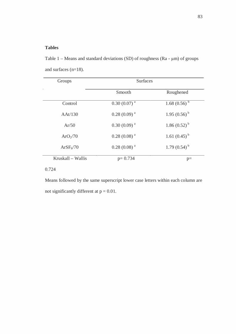
83
Tables
Table 1 – Means and standard deviations (SD) of roughness (Ra - μm) of groups
and surfaces (n=18).
Groups Surfaces
Smooth Roughened
Control 0.30 (0.07) a 1.68 (0.56) b
AAt/130 0.28 (0.09) a 1.95 (0.56) b
Ar/50 0.30 (0.09) a 1.86 (0.52) b
ArO2/70 0.28 (0.08) a 1.61 (0.45) b
ArSF6/70 0.28 (0.08) a 1.79 (0.54) b
Kruskall – Wallis p= 0.734 p=
0.724
Means followed by the same superscript lower case letters within each column are
not significantly different at p = 0.01.
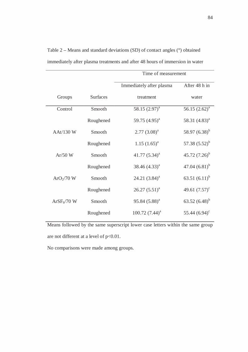
84
Table 2 – Means and standard deviations (SD) of contact angles (°) obtained
immediately after plasma treatments and after 48 hours of immersion in water
Groups Surfaces
Time of measurement
Immediately after plasma
treatment
After 48 h in
water
Control Smooth 58.15 (2.97)a 56.15 (2.62)a
Roughened 59.75 (4.95)a 58.31 (4.83)a
AAt/130 W Smooth 2.77 (3.08)a 58.97 (6.38)b
Roughened 1.15 (1.65)a 57.38 (5.52)b
Ar/50 W Smooth 41.77 (5.34)a 45.72 (7.26)b
Roughened 38.46 (4.33)a 47.04 (6.81)b
ArO2/70 W Smooth 24.21 (3.84)a 63.51 (6.11)b
Roughened 26.27 (5.51)a 49.61 (7.57)c
ArSF6/70 W Smooth 95.84 (5.88)a 63.52 (6.48)b
Roughened 100.72 (7.44)a 55.44 (6.94)c
Means followed by the same superscript lower case letters within the same group
are not different at a level of p<0.01.
No comparisons were made among groups.
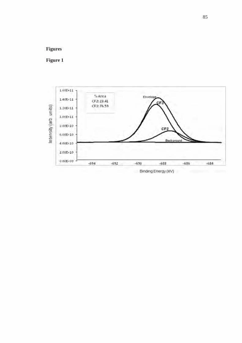
85
Figures Figure 1

86
Figure 2

87
Figure Legends
Figure 1 – XPS analysis of the specimens of ArSF6/70 group. Envelope represents
total F(1s) area.
Figure 2 – Mean log numbers (cells/mm2) and 95% confidence intervals for all
groups.
as = absence of saliva; ps = presence of saliva.
3.3 Capítulo 3
In vitro adhesion of Candida glabrata to denture base acrylic resin
modified by glow-discharge plasma treatment
Running title: C. glabrata adhesion to a modified resin
Zamperini CAa, Carneiro HLa, Rangel ECb, Cruz NCb, Vergani CEa, Machado
ALa,*
a Araraquara Dental School, UNESP – Univ Estadual Paulista, Department
of Dental Materials and Prosthodontics, Araraquara, São Paulo, Rua Humaitá nº
1680, CEP 14.801-903, Brazil.
b UNESP – Univ Estadual Paulista, Laboratory of Technological Plasmas,
Sorocaba, São Paulo, Avenida 3 de Março nº 511, CEP 18.085-180, Brazil.
*Corresponding author:
Profa. Dra. Ana Lucia Machado
Araraquara Dental School , UNESP –Univ. Estadual Paulista, Department of
Dental Materials and Prosthodontics, Araraquara, São Paulo, Rua Humaitá nº
1680, CEP 14.801-903, Brazil.
Tel: 55-16-33016410 Fax: 55-16-33016406
Email: [email protected]
89
Abstract
Candida adhesion to polymeric surfaces has been related to hydrophobic
interactions. Objective: Therefore, this study evaluated if surface modifications
with plasma treatments would reduce Candida glabrata adhesion to a denture
base resin. Methods: Specimens (n=54) with smooth surfaces were made and
divided into three groups (n=18): control – non-treated; experimental groups –
specimens submitted to glow-discharge plasma treatment to obtain hydrophilic
surfaces (Ar/50 W; AAt/130 W). Contact angles measurements were performed
immediately after the treatments and after immersion in water for 48 h. For each
group, half (n=9) of the specimens were precondicionated with saliva before the
adhesion assay. The number of adhered C. glabrata was evaluated by counting
after crystal violet staining. Results: Ar/50W group showed significantly lower C.
glabrata adherence than the control group, in the absence of saliva. After
preconditioning with saliva, C. glabrata adherence in experimental and control
groups did not differ significantly. The plasma treatments were effective in
modifying the acrylic surface. However, there were significant changes in the
contact angles after 48 h of immersion in water. Conclusions: The results
demonstrated that Ar/50 W plasma treatment showed promising potential for
reducing C. glabrata adhesion to denture base resins.
Keywords: Candida; Candida glabrata; saliva; acrylic resins.

90
Introduction
The ability of Candida to grow attached to oral surfaces in communities
known as biofilms is an important factor in the development of denture stomatitis.
Although Candida albicans still is the microorganism most often associated with
this infection, non-albicans species have been isolated from denture surfaces and
oral mucosa [1]. Candida glabrata was the second most commonly isolated
pathogen in patients with denture-induced stomatitis, followed by C.
pseudotropicalis, C. Krusei, C. tropicalis, C. parapsilosis and others [1]. In
addition, mixed Candida albicans and Candida glabrata biofilms have been
associated with denture stomatitis [2], indicating that Candida glabrata may also
play an integral role in this pathogenesis [2]. Moreover, in recent years, the
prevalence of C. glabrata infections has increased, mainly in compromised
patients [3]. This fact must receive attention because the mortality rate of C.
glabrata infections is higher compared with infection with other non-albicans
Candida and are more often difficult to treat [3].
Since the adhesion of Candida spp. to surfaces is a prerequisite for the
formation of biofilm and development of denture stomatitis, the inhibition of this
process could be effective to treat or prevent this pathology [4]. Many factors that
affect Candida adherence have been described, among them the hydrophobic
interactions. It has been demonstrated that these interactions are involved in the
adherence of Candida to acrylic [4-8]. The closer the surface free energy of the
substrate and the yeast, the higher was the probability of adherence [6]. A
significant positive correlation between cell surface hydrophobicity and adhesion
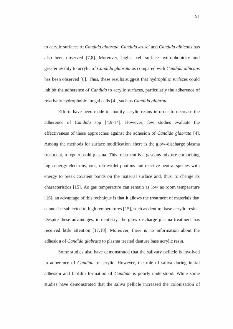
91
to acrylic surfaces of Candida glabrata, Candida krusei and Candida albicans has
also been observed [7,8]. Moreover, higher cell surface hydrophobicity and
greater avidity to acrylic of Candida glabrata as compared with Candida albicans
has been observed [8]. Thus, these results suggest that hydrophilic surfaces could
inhibit the adherence of Candida to acrylic surfaces, particularly the adherence of
relatively hydrophobic fungal cells [4], such as Candida glabrata.
Efforts have been made to modify acrylic resins in order to decrease the
adherence of Candida spp [4,9-14]. However, few studies evaluate the
effectiveness of these approaches against the adhesion of Candida glabrata [4].
Among the methods for surface modification, there is the glow-discharge plasma
treatment, a type of cold plasma. This treatment is a gaseous mixture comprising
high energy electrons, ions, ultraviolet photons and reactive neutral species with
energy to break covalent bonds on the material surface and, thus, to change its
characteristics [15]. As gas temperature can remain as low as room temperature
[16], an advantage of this technique is that it allows the treatment of materials that
cannot be subjected to high temperatures [15], such as denture base acrylic resins.
Despite these advantages, in dentistry, the glow-discharge plasma treatment has
received little attention [17,18]. Moreover, there is no information about the
adhesion of Candida glabrata to plasma treated denture base acrylic resin.
Some studies also have demonstrated that the salivary pellicle is involved
in adherence of Candida to acrylic. However, the role of saliva during initial
adhesion and biofilm formation of Candida is poorly understood. While some
studies have demonstrated that the saliva pellicle increased the colonization of

92
Candida [19-23], others have showed that preconditioning the materials with
saliva either did not affect [24-27] or reduced Candida adhesion [28-32].
Furthermore, few studies evaluated the effect of saliva on Candida glabrata
adhesion [29,31,32].
Therefore, the main purpose of this in vitro study was to investigate the
potential of two plasma treatments to modify a denture base acrylic resin to
reduce the Candida glabrata adhesion. Moreover, the effect of saliva coating was
also evaluated.
Materials and Methods
Preparation of acrylic resin specimens
The specimens (n=54) were fabricated from a microwave denture base
acrylic resin (Vipi Wave - VIPI Indústria e Comércio Exportação e Importação de
Produtos Odontológicos Ltda Pirassununga, SP, Brazil) using a conventional
flasking and pressure-pack technique. Initially, with the use of a metal mold, disk-
shaped silicone patterns (Zetaplus/Indurent - Zhermack, Badia Polesine, Rovigo,
Italy) were made with dimensions 13.8 2 mm. For surface standardization, the
silicone patterns were invested between two glass slides in dental stone in
microwave flaks. After the stone had set, the flasks were separated and the
silicone patterns were removed. For each specimen, 1 g of powder and 0.47 ml of
monomer liquid were mixed and processed according to the manufacturer’s
instructions. The mixture was packed into the molds, a trial pack was completed,
and excess material was removed. A final pack was performed and held for 15
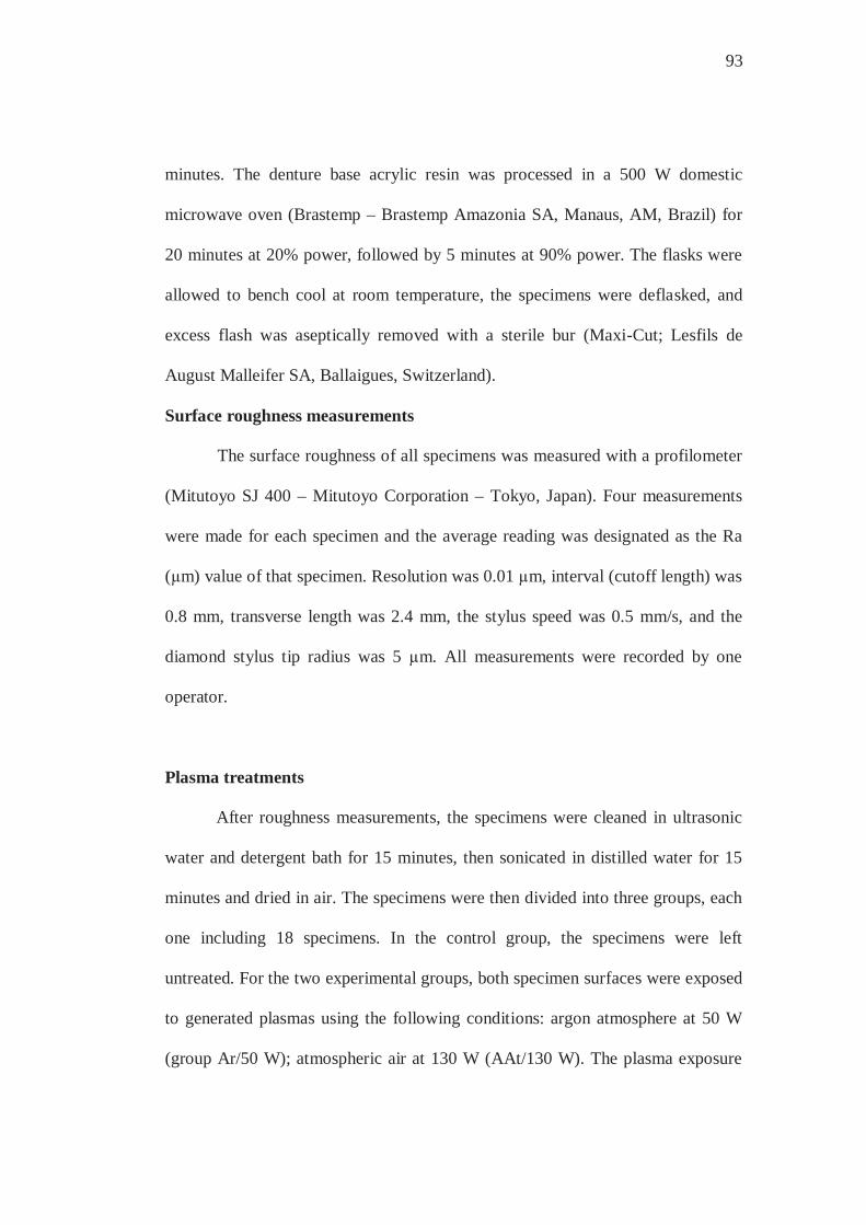
93
minutes. The denture base acrylic resin was processed in a 500 W domestic
microwave oven (Brastemp – Brastemp Amazonia SA, Manaus, AM, Brazil) for
20 minutes at 20% power, followed by 5 minutes at 90% power. The flasks were
allowed to bench cool at room temperature, the specimens were deflasked, and
excess flash was aseptically removed with a sterile bur (Maxi-Cut; Lesfils de
August Malleifer SA, Ballaigues, Switzerland).
Surface roughness measurements
The surface roughness of all specimens was measured with a profilometer
(Mitutoyo SJ 400 – Mitutoyo Corporation – Tokyo, Japan). Four measurements
were made for each specimen and the average reading was designated as the Ra
(μm) value of that specimen. Resolution was 0.01 μm, interval (cutoff length) was
0.8 mm, transverse length was 2.4 mm, the stylus speed was 0.5 mm/s, and the
diamond stylus tip radius was 5 μm. All measurements were recorded by one
operator.
Plasma treatments
After roughness measurements, the specimens were cleaned in ultrasonic
water and detergent bath for 15 minutes, then sonicated in distilled water for 15
minutes and dried in air. The specimens were then divided into three groups, each
one including 18 specimens. In the control group, the specimens were left
untreated. For the two experimental groups, both specimen surfaces were exposed
to generated plasmas using the following conditions: argon atmosphere at 50 W
(group Ar/50 W); atmospheric air at 130 W (AAt/130 W). The plasma exposure
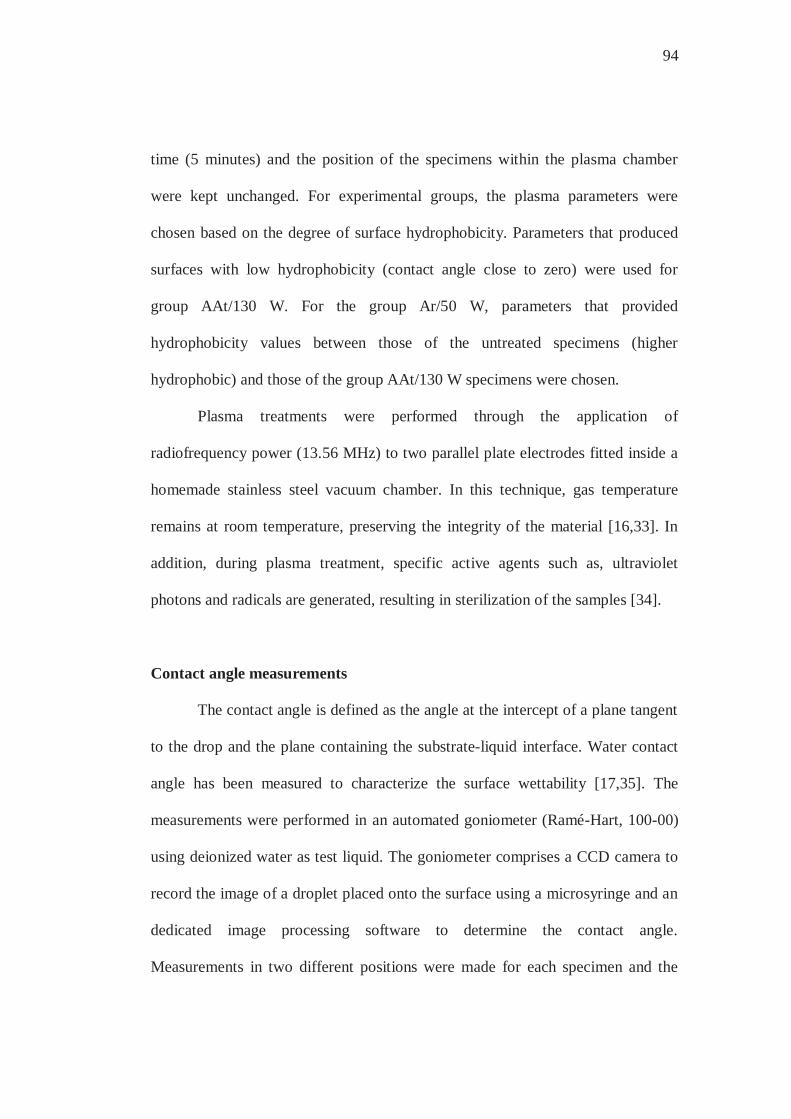
94
time (5 minutes) and the position of the specimens within the plasma chamber
were kept unchanged. For experimental groups, the plasma parameters were
chosen based on the degree of surface hydrophobicity. Parameters that produced
surfaces with low hydrophobicity (contact angle close to zero) were used for
group AAt/130 W. For the group Ar/50 W, parameters that provided
hydrophobicity values between those of the untreated specimens (higher
hydrophobic) and those of the group AAt/130 W specimens were chosen.
Plasma treatments were performed through the application of
radiofrequency power (13.56 MHz) to two parallel plate electrodes fitted inside a
homemade stainless steel vacuum chamber. In this technique, gas temperature
remains at room temperature, preserving the integrity of the material [16,33]. In
addition, during plasma treatment, specific active agents such as, ultraviolet
photons and radicals are generated, resulting in sterilization of the samples [34].
Contact angle measurements
The contact angle is defined as the angle at the intercept of a plane tangent
to the drop and the plane containing the substrate-liquid interface. Water contact
angle has been measured to characterize the surface wettability [17,35]. The
measurements were performed in an automated goniometer (Ramé-Hart, 100-00)
using deionized water as test liquid. The goniometer comprises a CCD camera to
record the image of a droplet placed onto the surface using a microsyringe and an
dedicated image processing software to determine the contact angle.
Measurements in two different positions were made for each specimen and the

95
average was calculated. Specimens were then stored at room temperature in sterile
distilled water 48 h to release any residual monomer [31]. Afterwards, contact
angles of each specimen were again measured.
Saliva Collection
Whole human unstimulated saliva was collected from 15 healthy adult
volunteers. The saliva was expectorated into sterile 50 ml Falcon tubes on ice,
pooled and clarified by centrifugation at 10000 g for 5 min at 4 ºC [31] and then
filtered through membrane filter of 0.22 μm pore size [23,26,36]. The resulting
saliva was immediately stored at -70 ºC until use. The study was approved by the
Ethics Committee of Araraquara Dental School (21/2008), and all subjects
volunteered to participate and signed an informed consent form.
Adherence assay
Candida glabrata strain ATCC 2001 was used. Stock cultures were
maintained at -70 ºC. After recovery this was maintained on YEPD medium (1%
yeast extract, 2% peptone, 2% dextrose, 2% agar) stored at 4 – 6 ºC during the
experimental period. For preparation of the yeast inoculum, two loopfuls of the
stock culture were streaked onto YEPD medium and incubated at 37 ºC for 48 h.
Two loopfuls of this young culture were transferred to 20 ml of yeast nitrogen
base (YNB) medium with 50 mM glucose and incubated at 37 ºC for 24 h. Cells
of the resultant culture were harvested, washed twice with phosphate-buffered
saline (PBS, pH 7.2) at 5000 g for 5 min and resuspended in YNB with 100 mM
glucose. Candida suspensions were standardized to a concentration of 1 107

96
cells/ml, spectrophotometrically. Three ml of the standardized C. glabrata cell
suspension was added to each well containing the specimen. The cells were left to
adhere for 90 min at 37 ºC [23]. The non-adherent cells were removed from the
specimen by gently washing with 3 ml PBS twice. For all experimental
conditions, the negative controls were acrylic specimens to which no cells were
added. All experiments were performed in triplicate on three independent
occasions.
Preconditioning with saliva
To investigate the effect of the saliva on Candida glabrata adhesion to the
denture acrylic resin, half of specimens from each group (n=9) were incubated
into the 12-well microtiter plates and coated with 3 ml of prepared saliva for 30
min at room temperature prior to the adhesion assay.
Measurement of adherent C. glabrata
Crystal violet staining
After the non-adherent cells were removed by washing, all specimens were
fixed in 80% ethanol, stained with crystal violet for 1 minute and washed with
PBS [37]. Adherent yeast cells were counted in 10 different fields for each
specimen, using a light microscope (Olympus BX51, Tokyo, Japan) at 400 x
magnification and the mean values were calculated. Adherent yeast cells were
counted in a “blind” manner to avoid subjective bias. The results were expressed
as cells/mm2.
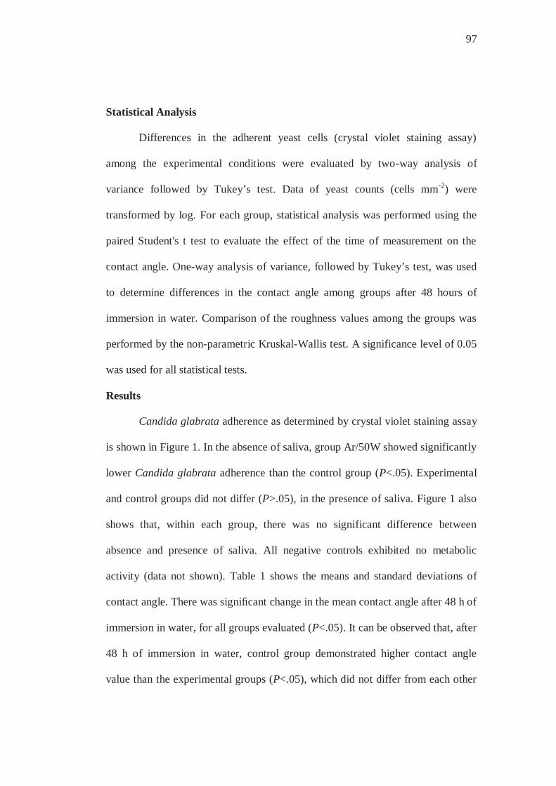
97
Statistical Analysis
Differences in the adherent yeast cells (crystal violet staining assay)
among the experimental conditions were evaluated by two-way analysis of
variance followed by Tukey’s test. Data of yeast counts (cells mm-2) were
transformed by log. For each group, statistical analysis was performed using the
paired Student's t test to evaluate the effect of the time of measurement on the
contact angle. One-way analysis of variance, followed by Tukey’s test, was used
to determine differences in the contact angle among groups after 48 hours of
immersion in water. Comparison of the roughness values among the groups was
performed by the non-parametric Kruskal-Wallis test. A significance level of 0.05
was used for all statistical tests.
Results
Candida glabrata adherence as determined by crystal violet staining assay
is shown in Figure 1. In the absence of saliva, group Ar/50W showed significantly
lower Candida glabrata adherence than the control group (P<.05). Experimental
and control groups did not differ (P>.05), in the presence of saliva. Figure 1 also
shows that, within each group, there was no significant difference between
absence and presence of saliva. All negative controls exhibited no metabolic
activity (data not shown). Table 1 shows the means and standard deviations of
contact angle. There was significant change in the mean contact angle after 48 h of
immersion in water, for all groups evaluated (P<.05). It can be observed that, after
48 h of immersion in water, control group demonstrated higher contact angle
value than the experimental groups (P<.05), which did not differ from each other
98
(P>.05). Roughness maximum, median and minimum values are presented in
Table 2. There were no significant differences in the median roughness values
among all groups evaluated.
Discussion
Among the virulence attributes of Candida, the ability of adherence to
acrylic is a prerequisite for colonization and development of biofilms on denture
surfaces. Although this attribute has been extensively studied in C. albicans, few
studies evaluated C. glabrata adhesion, mainly to modified surfaces [4]. This is
particularly important because, in the last years, the prevalence of C. glabrata has
increased in human infections as a consequence of the emergence of the acquired
immunodeficiency syndrome and the wide use of immunosuppressive medications
[3]. Moreover, a high mortality rate has been observed when C. glabrata is
associated with systemic infections [3]. Therefore, in the present study we
evaluate the potential of two plasma treatments to modify a denture base acrylic
resin in order to reduce the C. glabrata adhesion. To the author’s knowledge, this
has yet not been investigated.
Various advantages have been attributed to plasma treatment. This
technique is dry, cold, fast and allows altering the surface properties of a wide
variety of materials [15]. Another positive aspect of this method is that the bulk
properties and function of the material are usually kept, as the depth of plasma
treatment is limited to a few nanometers of the surface [15,17,35]. In this study,
the plasma treatments intended to decrease the surface hydrophobicity to reduce
99
the C. glabrata adhesion. The measurements made immediately after the
treatments indicated that, since the decrease in contact angle values is related with
the plasma atmosphere and the power applied, it was possible to regulate surface
hydrophilicity as planned [14,18].
In the absence of saliva, the results demonstrated that the plasma treatment
with Ar/50 W significantly reduced C. glabrata adhesion. One possible
explanation for this reduction could be the fact that Ar/50 W group specimens
presented the lowest contact angle, after immersion in water. Since hydrophobic
interactions are involved in the adhesion process [4-8], the hydrophilic surface
observed in Ar/50W group could have inhibited the adherence of C. glabrata.
Although the characteristic of cell surface hydrophobicity (CSH) is not species
specific [4,38], C. glabrata has been considered a relatively hydrophobic Candida
[8,39]. When compared to C. albicans, C. glabrata presented higher CSH and
higher tendency to adhere to acrylic surface [8]. Additionally, the closer the
surface free energy of the surface and the microorganism, is higher the probability
of Candida adherence [6]. Thus, the results obtained suggest that hydrophilic
acrylic surfaces could inhibit the adherence of Candida glabrata.
The decrease in the hydrophobicity of the surfaces, observed immediately
after the plasma treatments, can be attributed to energetic electrons generated
during the procedure that collide on the acrylic surface. These collisions can result
in chemical bonds breakage creating free radicals in the surface [15,35]. The
reactions between the free radicals and species from material or atmosphere can
increase the surface free energy that is reflected in a decrease of the contact angle
100
values [15,35]. Similar results were also reported in other investigations in which
plasma treatments were made to modify polymeric surfaces [14,17,18,35,40].
The results also showed that, after immersion in water for 48 hours, an
alteration of the contact angles of all groups evaluated was detected. This
demonstrates that the hydrophobicity of the specimens submitted to plasma
treatments came close to that observed for the control group specimens. This
alteration occurs because the modification caused by plasma treatment increases
the surface energy [15,35]. Under this condition, it has been observed that the
polymers have a tendency to recover their hydrophobicity, which has been
attributed to the rotation of polar groups around the polymeric backbone into the
material bulk [35]. Despite the increase in the contact angle values observed in
experimental groups, they remained significantly more hydrophilic compared to
control after 48 hours of immersion in water.
In this study, when the acrylic surfaces were preconditioned with saliva,
there were no significant differences in Candida glabrata adhesion among all
groups evaluated. It has been demonstrated that the salivary coating is an
important factor in determining the wettability properties of denture materials and,
after conditioning with saliva, wettability characteristics of biomaterials can be
altered [18,41]. The surface free energy of various materials, including a
microwave-cured acrylic resin, was decreased in approximately 10% when the
specimens were coated with saliva [41]. This may help explain the absence of any
plasma effect in Candida glabrata adhesion after preconditioning with saliva.
101
One other important observation is that, within each group, there was no
significant difference between absence and presence of saliva. This result is in
accordance with those obtained in previous investigations [29,31] which evaluated
the effect of saliva on C. glabrata adhesion. These findings suggest that the
salivary pellicle did not increase the Candida glabrata adhesion to polymer
surfaces, as has been found with Candida albicans [19,23]. Moreover, there are
only a few studies dealing with interactions between salivary pellicle and Candida
glabrata [29,31,32,42]. Hence, further studies are needed on this subject.
The surface roughness is an important factor that can affect the adhesion of
Candida to the material surfaces [32,43,44]. Thus, in this study, the specimens
were made between glass slides in order to obtain smooth and standardized
surfaces. As can be observed in Table 2, the results demonstrated that there were
no significant differences in the median roughness values among all groups
evaluated.
In conclusion, this in vitro study demonstrates that hydrophilic surfaces, as
those obtained with Ar/50W plasma treatment, have potential for reducing the
adhesion of Candida glabrata, which may be involved in the pathogenesis of
denture stomatitis and related infections. Additionally, the saliva pellicle did not
significantly increase Candida glabrata adhesion. However, as this study is
limited only to one strain and one heat-polymerized denture base, more
comprehensive investigations using other strains as well as different denture base
materials are required.
102
Acknowledgements
This research was supported by FAPESP (Grant - 08/05338-1) and CNPq
(Grant 479895/2009-0).
Conflict of interest
The authors declare that they have no conflict of interest.
References
1. Dagistan S, Aktas AE, Caglayan F, Ayyildiz A, Bilge M. Differential
diagnosis of denture-induced stomatitis, Candida, and their variations in
patients using complete denture: a clinical and mycological study.
Mycoses 2009;52(3):266-71.
2. Coco BJ, Bagg J, Cross LJ, Jose A, Cross J, Ramage G. Mixed Candida
albicans and Candida glabrata populations associated with the
pathogenesis of denture stomatitis. Oral Microbiol Immunol
2008;23(5):377-83.
3. Li L, Redding S, Dongari-Bagtzoglou A. Candida glabrata: an
emerging oral opportunistic pathogen. J Dent Res 2007;86(3):204-15.
4. Yoshijima Y, Murakami K, Kayama S, Liu D, Hirota K, Ichikawa T,
Miyake Y. Effect of substrate surface hydrophobicity on the adherence
of yeast and hyphal Candida. Mycoses. 2010; 53(3):221-6.
5. Klotz SA, Drutz DJ, Zajic JE. Factors governing adherence of Candida
species to plastic surfaces. Infect Immun 1985;50(1):97-101.

103
6. Minagi S, Miyake Y, Inagaki K, Tsuru H, Suginaka H. Hydrophobic
interaction in Candida albicans and Candida tropicalis adherence to
various denture base resin materials. Infect Immun 1985;47(1):11-4.
7. Samaranayake YH, Wu PC, Samaranayake LP, So M, Yuen KY.
Adhesion and colonisation of Candida krusei on host surfaces. J Med
Microbiol 1994;41(4):250-8.
8. Luo G, Samaranayake LP. Candida glabrata, an emerging fungal
pathogen, exhibits superior relative cell surface hydrophobicity and
adhesion to denture acrylic surfaces compared with Candida albicans.
APMIS 2002;110(9):601-10.
9. Park SE, Periathamby AR, Loza JC. Effect of surface-charged
poly(methylmethacrylate) on the adhesion of Candida albicans. J
Prosthodont 2003;12(4):249-54.
10. Dhir G, Berzins DW, DhuruVB, Periathamby AR, Dentino A. Physical
properties of denture base resins potentially resistant to Candida
adhesion. J Prosthodont 2007;16(6):465–72.
11. Park SE, Blissett R, Susarla SM, Weber H-P. Candida albicans
adherence to surface-modified denture resin surfaces. J Prosthodont
2008;17(5):365-69.
12. Puri G, Berzins DW, Dhuru VB, Raj PA, Rambhia SK, Dhir G, Dentino
AR. Effect of phosphate group addition on the properties of denture base
resins. J Prosthet Dent 2008;100(4):302-8.
104
13. Redding S, Bhatt B, Rawls HR, Siegel G, Scott K, Lopez-Ribot J.
Inhibition of Candida albicans biofilm formation on denture material.
Oral Surg Oral Med Oral Pathol Oral Radiol Endod 2009;107(5):669–
72.
14. Zamperini CA, Machado AL, Vergani CE, Pavarina AC, Giampaolo ET,
da Cruz NC. Adherence in vitro of Candida albicans to plasma treated
acrylic resin. Effect of plasma parameters, surface roughness and
salivary pellicle. Arch Oral Biol 2010;55:763-70.
15. Hauser J, Zietlow J, Koller M, Esenwein SA, Halfmann H, Awakowicz
P, Steinau HU. Enhanced cell adhesion to silicone implant material
through plasma surface modification. J Mater Sci Mater Med
2009;20:2541–48.
16. Li Y, Kuai P, Huo P, Liu C. Fabrication of CuO nanofibers via the
plasma decomposition of Cu(OH)2. Mater Lett 2009; 63:188–90.
17. Yildirim MS, Hasanreisoglu U, Hasirci N, Sultan N. Adherence of
Candida albicans to glow-discharge modified acrylic denture base
polymers. J Oral Rehabil 2005;32:518-25.
18. Yildirim MS, Kesimer M, Hasirci N, Kiliç N, Hasanreisoglu U.
Adsorption of human salivary mucin MG1 onto glow-discharge plasma
treated acrylic resin surfaces. J Oral Rehabil 2006;33:775-83.

105
19. Nikawa H, Hayashi S, Nikawa Y, Hamada T, Samaranayake LP.
Interactions between denture lining material, protein pellicles and
Candida albicans. Arch Oral Biol 1993;38(7): 631-4.
20. Nikawa H, Yamamoto T, Hamada T, Rahardjo MB, Murata H.
Antifungal effect of zeolite-incorporated tissue conditioner against
Candida albicans growth and/or acid production. J Oral Rehabil
1997;24:350-7.
21. Nikawa H, Jin C, Hamada T, Murata H. Interactions between thermal
cycled resilient denture lining materials, salivary and serum pellicles and
Candida albicans in vitro. Part I. Effects on fungal growth. J Oral
Rehabil 2000;27:41-51.
22. Nikawa H, Chen J, Hamada T, Nishimura M, Polyzois G. Candida
albicans colonization on thermal cycled maxillofacial polymeric
materials in vitro. J Oral Rehabil 2001;28:526-33.
23. Chandra J, Mukherjee PK, Leidich SD, Faddoul FF, Hoyer LL, Douglas
LJ, Ghannoum MA. Antifungal resistance of candidal biofilms formed
on denture acrylic in vitro. J Dent Res 2001; 80(3): 903-8.
24. Jin Y, Samaranayake LP, Samaranayake Y, Yip HK. Biofilm formation
of Candida albicans is variably affected by saliva and dietary sugars.
Arch Oral Biol 2004;49:789-98.
25. Tari BF, Nalbant D, Al FD, Kustimur S. Surface roughness and
adherence of Candida albicans on soft lining materials as influenced by
accelerated aging. J Contemp Dent Pract 2007;8(5): 1-11.
106
26. Thein ZM, Samaranayake YH, Samaranayake LP. Characteristics of
dual species Candida biofilms on denture acrylic surfaces. Arch Oral
Biol 2007;52:1200-8.
27. Karaagaclioglu L, Can G, Yilmaz B, Ayhan N, Semiz O, Levent H. The
adherence of Candida albicans to acrylic resin reinforced with different
fibers. J Mater Sci Mater Med 2008;19(2):959-63.
28. Samaranayake LP, McCourtie J, MacFarlane TW. Factors affecting the
in-vitro adherence of Candida albicans to acrylic surfaces. Arch Oral
Biol 1980;25:611-15.
29. McCourtie J, MacFarlane TW, Samaranayake LP. Effect of saliva and
serum on the adherence of Candida species to chlorhexidine-treated
denture acrylic. J Med Microbiol 1986;21:209-13.
30. Waters MGJ, Williams DW, Jagger RG, Lewis MAO. Adherence of
Candida albicans to experimental denture soft lining materials. J
Prosthet Dent 1997;77(3): 306-12.
31. Moura JS, Silva WJ, Pereira T, Cury AADB, Garcia RCMR. Influence
of acrylic resin polymerization methods and saliva on the adherence of
four Candida species. J Prosthet Dent 2006; 96:205-11.
32. Pereira T, Cury AADB, Cenci MS, Rodrigues-Garcia RCM. In vitro
Candida colonization on acrylic resins and denture liners: influence of
surface free energy, roughness, saliva, and adhering bacteria. Int J
Prosthodont 2007;20:308-10.
107
33. Polukoshko KM, Brudvik JS, Nicholls JI,Smith DE. Evaluation of heat-
cured resin bases following the addition of denture teeth using a second
heat cure. J Prosthet Dent 1992;67(4):556–62.
34. Moisan M, Barbeau J, Crevier MC, Pelletier J, Philip N, Saoudi B.
Plasma sterilization. Methods and mechanisms. Pure Appl Chem
2002;74(3):349–58.
35. Rangel EC, Gadioli GZ, Cruz NC. Investigations on the stability of
plasma modified silicone surfaces. Plasmas and Polymers 2004;9(1):35-
48.
36. Peros WJ, Gibbons RJ. Influence of growth medium on adsorption of
Streptococcus mutans, Actinomyces viscosus, and Actinomyces
naeslundii to saliva-treated hydroxyapatite surfaces. Infect Immun
1981;32(1):111-7.
37. Ferreira MAF, Pereira-Cenci T, Rodrigues de Vasconcelos LM,
Rodrigues-Garcia RCM, Cury AADB. Efficacy of denture cleansers on
denture liners contaminated with Candida species. Clin Oral Investig
2009;13(2):237-42.
38. Panagoda GJ, Ellepola ANB, Samaranayake LP. Adhesion of Candida
parapsilosis to epithelial and acrylic surfaces correlates with cell surface
hydrophobicity. Mycoses 2001;44(1-2):29-35.
39. Minagi S, Miyake Y, Fujioka Y, Tsuru H, Suginaka H. Cell-surface
hydrophobicity of Candida species as determined by the contact-angle
108
and hydrocarbon-adherence methods. J Gen Microbiol
1986;132(4):1111-5.
40. Ozden N, Akaltan F, Suzer S, Akovali G. Time-related wettability
characteristic of acrylic resin surfaces treated by glow discharge. J
Prosthet Dent 1999;82:680-684.
41. Sipahi C, Anil N, Bayramli E. The effect of acquired salivary pellicle on
the surface free energy and wettability of different denture base
materials. J Dent 2001;29:197-204.
42. Pereira-Cenci T, Deng DM, Kraneveld EA, Manders EMM, Cury
AADB, Ten Cate JM, Crielaard W. The effect of Streptococcus mutans
and Candida glabrata on Candida albicans biofilms formed on different
surfaces. Arch Oral Biol 2008;53:755-64.
43. Verran J, Maryan CJ. Retention of Candida albicans on acrylic resin
and silicone of different surface topography. J Prosthet Dent
1997;77:535-9.
44. Radford DR, Sweet SP, Challacombe SJ, Walter JD. Adherence of
Candida albicans to denture-base materials with different surface
finishes. J Dent 1998;26:577-83.
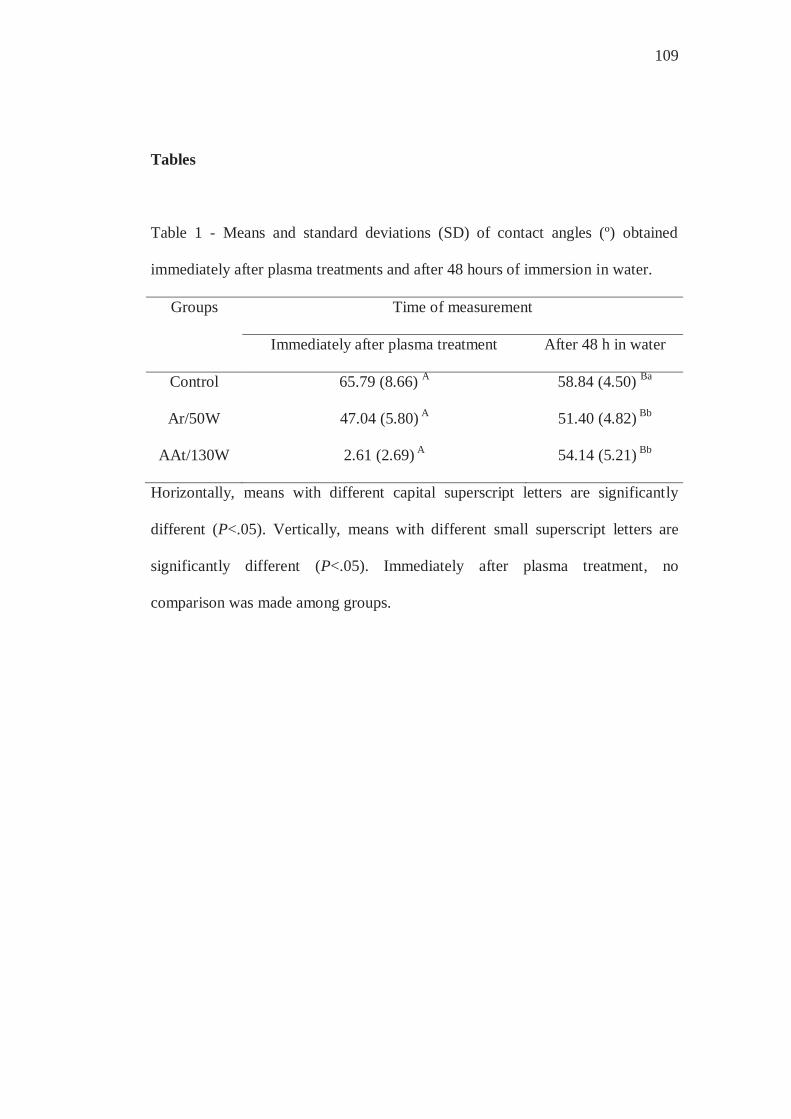
109
Tables
Table 1 - Means and standard deviations (SD) of contact angles (º) obtained
immediately after plasma treatments and after 48 hours of immersion in water.
Groups Time of measurement
Immediately after plasma treatment After 48 h in water
Control 65.79 (8.66) A 58.84 (4.50) Ba
Ar/50W 47.04 (5.80) A 51.40 (4.82) Bb
AAt/130W 2.61 (2.69) A 54.14 (5.21) Bb
Horizontally, means with different capital superscript letters are significantly
different (P<.05). Vertically, means with different small superscript letters are
significantly different (P<.05). Immediately after plasma treatment, no
comparison was made among groups.
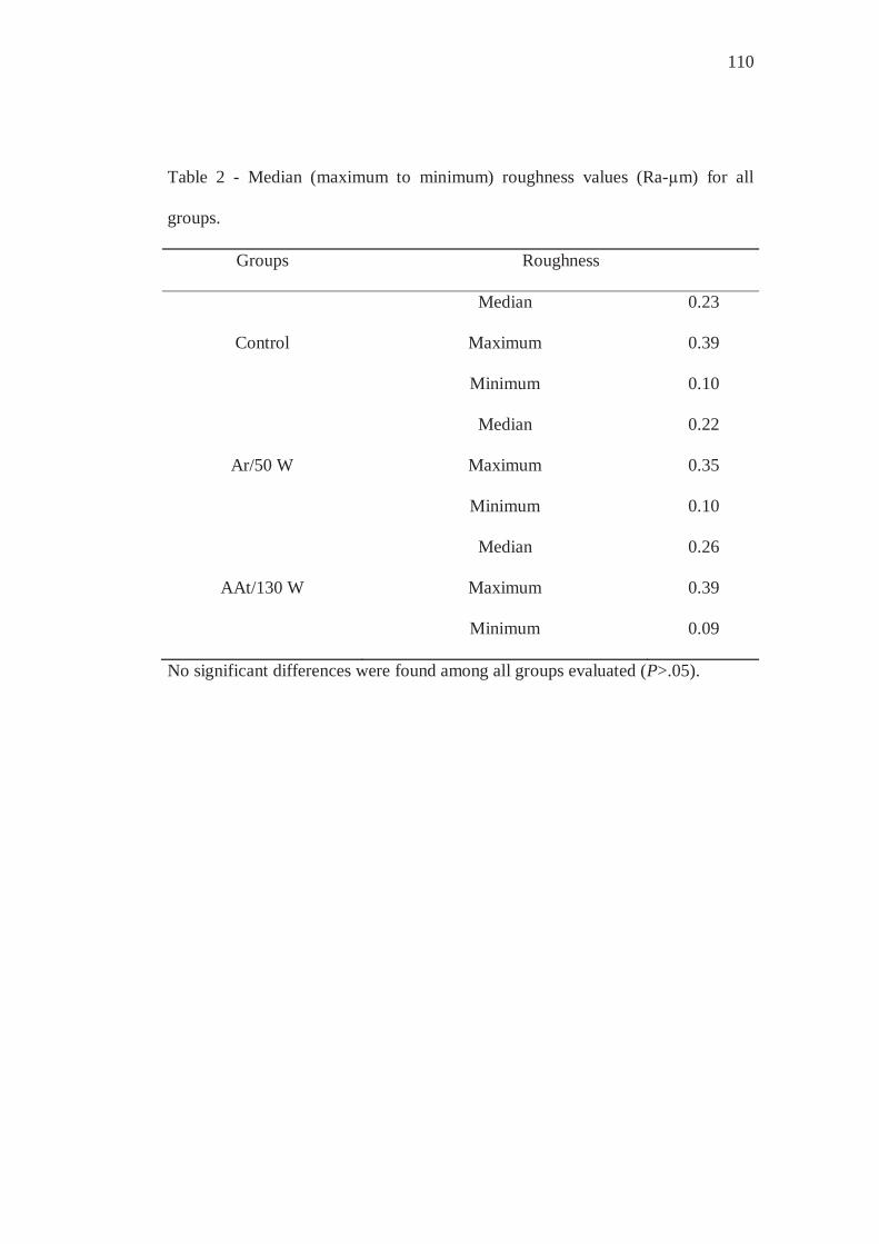
110
Table 2 - Median (maximum to minimum) roughness values (Ra-μm) for all
groups.
Groups Roughness
Median 0.23
Control Maximum 0.39
Minimum 0.10
Median 0.22
Ar/50 W Maximum 0.35
Minimum 0.10
Median 0.26
AAt/130 W Maximum 0.39
Minimum 0.09
No significant differences were found among all groups evaluated (P>.05).

111
0.0
1.0
2.0
3.0
4.0
5.0
Control Ar/50 W AAt/130 W
Groups
Log
(cel
/mm
2)as ps
Figure 1
*

112
Figure Caption
Figure 1. Mean log numbers (cells/mm2) and 95% confidence intervals for all
groups. as: absence of saliva; ps: presence of saliva. (*) Statistically different
mean compared to control.
3.4 Capítulo 4
Effect of different periods of preconditioning with saliva on adhesion
of C. albicans to a denture base resin by crystal violet staining and XTT assay
Short title: Effect of saliva on adhesion of Candida
Camila Andrade Zamperini 1, Patrícia Cristiane dos Santos Schiavinato 1, Ana
Lucia Machado 1*, Eunice Teresinha Giampaolo 1, Ana Claudia Pavarina 1,
Carlos Eduardo Vergani 1
1 Araraquara Dental School, UNESP - Univ Estadual Paulista, Department
of Dental Materials and Prosthodontics, Araraquara, São Paulo, Brazil.
*Corresponding author:
Profa. Dra. Ana Lucia Machado
Araraquara Dental School, UNESP – Univ Estadual Paulista, Department of
Dental Materials and Prosthodontics, Araraquara, São Paulo, Rua Humaitá nº
1680, CEP 14.801-903, Brazil.
Tel: 55-16-33016410 Fax: 55-16-33016406. Email:
114
Abstract
The role of saliva on Candida adhesion to biomaterials is not clearly defined.
Aim: this study investigated whether different periods of preconditioning with
saliva would influence the adhesion of Candida albicans to a denture base resin.
Methods: samples (n=90) with smooth surfaces were made and then divided into
five groups: 1 control - without saliva; 4 experimental groups – conditioned in
saliva for periods of: 30, 60, 180 or 720 minutes. Candida adhesion was evaluated
by crystal violet staining and XTT assay. Results: the one-way analyses of
variance revealed that there were no significant differences among the mean
numbers of adherent cells or among the mean absorbances for all groups. No
significant correlation was found between the two methods used for assessing C.
albicans adhesion. Conclusions: the different periods of preconditioning with
saliva had no significant influence on the adhesion of Candida albicans to the
denture base acrylic resin.
Key words: Acrylic Resins; Biofilms; Adhesion; Candida albicans; Saliva.

115
Introduction
The presence of Candida albicans biofilms on removable denture surfaces
plays an important role in the etiology of denture stomatitis. The capacity of this
fungus to adhere to surfaces is the first stage in the biofilm formation process,
which is followed by colony formation and cell organization, secretion of
extracellular matrix, maturation and dissemination of the biofilm 1.
It is known that the adhesion of Candida cells to denture surfaces is
mediated by a salivary conditioning film or pellicle 2,3,4 that provides receptors for
microbial adhesion 5. However, the role of saliva on Candida albicans adhesion to
biomaterials is, as yet, not fully established, as conflicting results have been
reported. Some authors have observed that the saliva pellicle promoted Candida
albicans colonization on the materials 6,7,8,9,10. Conversely, others have found that
preconditioning the materials with saliva either did not affect 11,12,13,14 or reduced
Candida albicans adhesion 15,16,17,18,19. The different periods of preconditioning
with saliva could interfere in the adhesive capacity of Candida 20 and contribute to
the inconsistent findings from these studies. In a number of researches, the
materials were incubated with saliva for short periods such as 30 minutes 16,17,18,19,
1 hour 7,8,9, 2 to 3 hours 4,10 and 4 hours 11,13. Longer incubation periods, including
overnight 21, 18 hours 15 and 24 hours 22 have also been used to create a salivary
conditioning film. However, to date, no information is available concerning how
different periods of preconditioning with saliva affect the adhesion of Candida
cells to denture materials.
116
One method used for evaluating adhesion of Candida is the cell staining
assay 15,18,19,23,24. In this method, the adhered cells on the material surface are
fixed, stained and counted in a microscope. The dye commonly used in this
technique is the crystal violet, which is basic and binds to negatively charged
extracellular molecules, including cell surface molecules and polysaccharides in
the extracellular matrices 25. Another method that has been widely used is the
XTT assay 1,10,11,16, which evaluates the metabolic activity of viable cells 26. This
assay is based on the reduction of a water-soluble tetrazolium salt [2,3-bis(2-
methoxy-4-nitro-5-sulfophenyl)-5-[(phenylamino) carbonyl]-2H-tetrazolium-
hydroxide] to a formazan product by the mitochondrial dehydrogenases of the live
cells.
Therefore, the aim of this study was to assess the effect of different periods
of preconditioning with saliva on the adhesion of C. albicans to a denture base
resin using crystal violet staining and XTT assay. Additionally, the correlation
between the two methods used for assessing C. albicans adhesion was evaluated.
Materials and Methods
Preparation of acrylic resin specimens
The specimens (n=90) were fabricated from a microwave denture-base
acrylic resin (Vipi Wave - VIPI Indústria e Comércio Exportação e Importação de
Produtos Odontológicos Ltda Pirassununga, SP, Brazil) using a conventional
flasking and pressure-pack technique. Initially, with the use of a metal mold, disk-
shaped silicone patterns Zetaplus/Indurent - Zhermack, Badia Polesine, Rovigo,
117
Italy) measuring 13.8 X 2 mm were made. To obtain smooth and standardized
surfaces, the silicone patterns were invested in dental stone in microwave flasks
and positioned between two glass slides. After the stone had set, the flasks were
separated and the silicone patterns were removed. For each specimen, 1 g of
powder and 0.47 ml of monomer liquid were mixed, packed into the molds, and
processed in a 500 W domestic microwave oven (Brastemp – Brastemp da
Amazonia SA, Manaus, AM, Brazil) according to the manufacturer’s instructions
(20 minutes at 20% power, followed by 5 minutes at 90% power). The flasks were
allowed to bench cool at room temperature, the specimens were deflasked, and
excess flash was aseptically removed with a sterile bur (Maxi-Cut; Lesfils de
August Malleifer SA, Ballaigues, Switzerland).
Surface roughness measurements
The surface roughness of all specimens was measured with a profilometer
(Mitutoyo SJ 400 – Mitutoyo Corporation - Japan). Four measurements were
made for each specimen and the mean reading was designated as the Ra (μm)
value of that specimen. The measurements were recorded by one operator.
Resolution was 0.01 μm, interval (cutoff length) was 0.8 mm, transverse length
was 2.4 mm, the stylus speed was 0.5 mm/s, and the diamond stylus tip radius was
5 μm.
Saliva Collection
Whole human unstimulated saliva was collected from 15 healthy adult
volunteers. The saliva was expectorated into sterile 50 ml Falcon tubes on ice,
118
pooled and clarified by centrifugation at 10000 g for 5 min at 4 ºC 18 and then
sterilized using membrane filter with a 0.22 μm pore size 10,13,27,28. The resulting
saliva was immediately stored at -70 ºC until use. The study was approved by the
Ethics Committee of Araraquara Dental School, and all subjects volunteered to
participate and signed an informed consent form.
Test specimen sterilization
Before the microbiological tests, the test specimens were kept in distilled
water at ambient temperature for 48 hours, in order to eliminate residual
monomers 18. After this, they received an ultrasound bath for 20 minutes and were
exposed to ultraviolet light in the laminar flow chamber for 20 minutes on each
side 29.
Preconditioning with saliva
The 90 test specimens were divided into five groups (n=18), as follows: 1
control (without preconditioning in saliva) and 4 experimental groups that were
conditioned with saliva for periods of: 30 minutes (30 min); 1 hour (60 min); 3
hours (180 min); or 12 hours (720 min). The specimens of the four experimental
groups were incubated in 12-well microtiter plates with 3 ml of the saliva
preparation at room temperature 17 prior to the adherence assay.
Adherence assay
The stock culture of Candida albicans strain ATCC 90028 was maintained
in YEPD medium (1% yeast extract, 2% peptone, 2% dextrose, 2% agar) stored at
119
4 – 6 ºC during the experimental period. For preparation of the yeast inoculum,
two loopfuls of the stock culture were streaked onto YEPD medium and incubated
at 37 ºC for 48 h. Two loopfuls of this young culture were transferred to 20 ml of
yeast nitrogen base (YNB) medium with 50 mM glucose and incubated at 37 ºC
for 21 h. Cells of the resultant culture were harvested, washed twice with
phosphate-buffered saline (PBS, pH 7.2) at 5000 g for 5 min and resuspended in
YNB with 100 mM glucose. Candida suspensions were standardized to a
concentration of 1 x 10⁷ cells/ml, spectrophotometrically. Three ml of the
standardized C. albicans cell suspension was added to each well containing the
specimen. The cells were left to adhere for 90 min at 37 ºC 10. The non-adherent
cells were removed from the specimen by gently washing with 3 ml PBS twice.
The negative controls were acrylic specimens to which no cells were added. All
experiments were performed in triplicate on three independent occasions.
Crystal violet staining
Nine specimens from each group were evaluated by crystal violet staining
assay. After the non-adherent cells were removed by washing, the specimens were
fixed in 80% ethanol, stained with crystal violet for 1 minute and washed with
PBS 24. Adherent yeast cells were counted in 10 different fields for each
specimen, using a light microscope (Olympus BX51, Japan) at 400 x
magnification and the mean values were calculated. Adherent yeast cells were
counted in a "blind" manner to avoid subjective bias. The results were expressed
as cells/mm2.

120
XTT assay
Nine specimens from each group were evaluated by XTT reduction assay,
as described elsewhere 1. Briefly, XTT (Sigma, MO, USA) was prepared in
ultrapure water (1mg/ml), filter sterilized and stored at -70 ºC until used.
Menadione (Sigma, MO, USA) solution was prepared in acetone at 0.4 mM
immediately before each assay. After washing, the specimens were transferred to
new wells with 158 μl PBS with 200mM glucose, 40 μl XTT and 2 μl menadione
were inoculated to each well. The plates were incubated for 3 h in the dark at 37
ºC. The whole content of each well was centrifuged at 5000 g for 2 minutes and
the colorimetric change of the supernatant was measured using a microtiter plate
reader (Thermo Plate – TP Reader) at 492 nm.
Statistical Analysis
Comparison of the roughness values among the groups was performed by
the non-parametric Kruskal-Wallis test. For each adhesion assay used, one-way
analysis of variance with Welch's correction was performed to evaluate the effect
of time of preconditioning with saliva on Candida albicans adhesion. For the
crystal violet technique, data of yeast counts (cells per mm2) were transformed by
log. A significance level of 0.05 was used for all statistical tests. To evaluate the
correlation between the two methods used for assessing C. albicans adherence,
Pearson’s coefficient of correlation was used.
Results and Discussion
121
Candida adhesion to dentures, an essential step in biofilm formation and
development of denture stomatitis, may be influenced by saliva. This influence
may be regulated by specific interactions between the cellular adhesins and
receptors in the salivary pellicle 2,4,5, as well as, by the action of salivary proteins
as a source of nutrients for microbiological growth 30. On the other hand, these
proteins may also act by blocking the locations of adhesion originally present on
substrates 4,11. It has also been observed that saliva may alter the surface
characteristics of the substrates involved in the adhesion process, such as
roughness and hydrophobicity 4,28,31,32. Conversely, some authors have reported
that the original substratum surface properties may be transferred even through
the protein layer on the substratum surface and still influence microbial adhesion
4,28. In spite of several studies that have investigated the influence of the salivary
pellicle in the adhesion process 6,7,8,9,10,11,12,13,14,15,16,17,18,19,21, the role it plays is not
yet clear. The divergences found in the literature could be related to the different
methodologies, including the different periods of preconditioning with saliva 20.
Thus, the aim of this study was to evaluate whether different periods of
preconditioning with saliva would have an influence on the adhesion of Candida
albicans to a denture base acrylic resin using crystal violet staining and XTT
assay. To the author’s knowledge, to date this is the first study that has addressed
this issue.
Considering that surface roughness can influence the adhesion of Candida
albicans to the substrate 19,33,34,35, the samples were made between glass slides in
order to obtain smooth, standardized surfaces. The results demonstrated that there
122
were no significant differences in the mean roughness values among all groups
evaluated, for both crystal violet staining and XTT assay (Table 1).
The results from crystal violet staining and XTT assay revealed that the
differences among all experimental groups were not significant, indicating that the
periods of preconditioning with saliva evaluated in the present investigation did
not influence the adhesion of Candida albicans (Table 2). These findings suggest
that in studies of Candida albicans adhesion to acrylic resins, shorter periods of
preconditioning with saliva, such as 30 or 60 minutes could be used, making the
adhesion assay procedures easier and quicker to perform. Nevertheless, it is
important to point out that the saliva used in this study was filter sterilized 27 to
obtain more reproducible results 17. Given that salivary pellicle formation on
biomaterials is a selective process 31, the filtration process may have depleted
some of the protein complexes, such as certain mucin complexes involved in
Candida adhesion, that are present in human saliva 17. Therefore, further studies
should be conducted to evaluate the influence of different periods of
preconditioning the substrates on Candida adhesion, when using whole saliva.
The coefficient of correlation between crystal violet staining and XTT
assay was low (r=0.223, P = 0.141), showing no significant correlation between
the two methods used for assessing C. albicans adherence. The results obtained by
staining demonstrated that there were no significant differences between the
experimental groups and the control, indicating that preconditioning with saliva
did not influence the fungal adhesion (Table 2). For XTT assay, although the
statistical analysis failed to find significant differences among the groups, the
123
absorbance values obtained after preconditioning with saliva were higher than
those of the control group. The two methods used in the present study to
determine the adhesion of C. albicans to the denture base resin are based on
different principles. In the crystal violet staining method, the stained cells are
quantified by counting in selected fields of the substrate 15,18,19,23,24, whereas in the
XTT assay, the metabolic activity of all viable cells is measured 1,11,21,26. It is
worth noting that most of the studies that have found an increase in Candida
albicans colonization on denture materials after pre-incubation with saliva have
used tests of monitoring the pH of the growth medium, adenosine triphosphate
(ATP) analysis and XTT assay 6,7,8,9,10. Conversely, when the adherent cells were
quantified by microscopy or by colony forming unit (UFC) counts, the majority of
the studies on Candida albicans colonization on denture materials observed an
absence of significant effect of saliva 12,13,14 or reduced adhesion 14,15,16,17,18,19.
Hence, it is suggested that the controversial results regarding the role of saliva on
the adhesion of Candida to denture materials may at least partially be accounted
for by different adhesion assays used. This highlight that the effect of saliva on
the adhesion and biofilm development of Candida should not be studied by a
single quantification method.
The divergent results among studies may also be attributable to the various
materials that have been used as substrates, among them acrylic surfaces
9,10,13,14,15,16,17,18,19,22, denture reline materials 6,7,8,12,17,19, maxillofacial polymeric
materials 5,9 and polystyrene 11,21. It has been reported that the exposed chemical
groups of the solid surface probably play the most important role in determining

124
the selectivity of the adsorption process 4,31. Yildirim et al. (2006) 36 observed that
acrylic resin surfaces modified by plasma treatments adsorbed different amounts
of high molecular weight mucin. Therefore, the composition of the salivary
pellicle may vary among the materials used. As there are only few data in the
literature about the resulting protein concentrations formed on different prosthetic
materials 37, further studies are necessary to evaluate these differences.
In conclusion, this study demonstrated that different periods of
preconditioning with saliva did not influence the adhesion of Candida albicans to
the denture-base acrylic resin as evaluated by crystal violet staining and XTT
assay. Further, the coefficient of correlation between the results obtained by
crystal violet staining and XTT assay was not significant. Finally, as the current
study was confined only to one denture-base acrylic resin and one strain of
Candida albicans future studies with other Candida species and denture base
materials are warranted. Besides, in addition to the period of preconditioning with
saliva, other experimental conditions, such as number of donors, speed and time
of centrifugation have varied considerably among studies and the influence of
these differences on Candida adhesion to denture materials needs further
investigations.
Acknowledgements
This research was supported by FAPESP (Grant - 2008/05339-8) and
CNPq (Grant - 479895/2009-0).
References
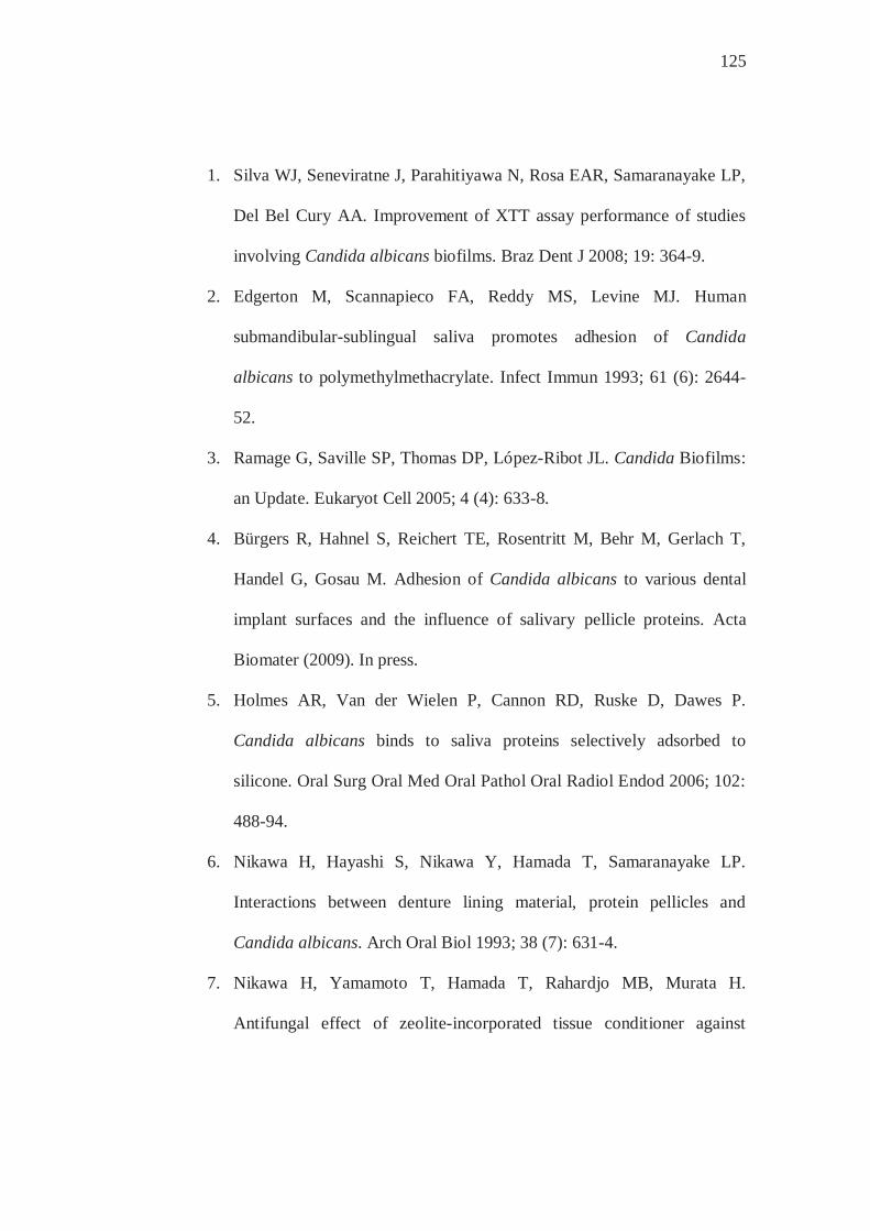
125
1. Silva WJ, Seneviratne J, Parahitiyawa N, Rosa EAR, Samaranayake LP,
Del Bel Cury AA. Improvement of XTT assay performance of studies
involving Candida albicans biofilms. Braz Dent J 2008; 19: 364-9.
2. Edgerton M, Scannapieco FA, Reddy MS, Levine MJ. Human
submandibular-sublingual saliva promotes adhesion of Candida
albicans to polymethylmethacrylate. Infect Immun 1993; 61 (6): 2644-
52.
3. Ramage G, Saville SP, Thomas DP, López-Ribot JL. Candida Biofilms:
an Update. Eukaryot Cell 2005; 4 (4): 633-8.
4. Bürgers R, Hahnel S, Reichert TE, Rosentritt M, Behr M, Gerlach T,
Handel G, Gosau M. Adhesion of Candida albicans to various dental
implant surfaces and the influence of salivary pellicle proteins. Acta
Biomater (2009). In press.
5. Holmes AR, Van der Wielen P, Cannon RD, Ruske D, Dawes P.
Candida albicans binds to saliva proteins selectively adsorbed to
silicone. Oral Surg Oral Med Oral Pathol Oral Radiol Endod 2006; 102:
488-94.
6. Nikawa H, Hayashi S, Nikawa Y, Hamada T, Samaranayake LP.
Interactions between denture lining material, protein pellicles and
Candida albicans. Arch Oral Biol 1993; 38 (7): 631-4.
7. Nikawa H, Yamamoto T, Hamada T, Rahardjo MB, Murata H.
Antifungal effect of zeolite-incorporated tissue conditioner against

126
Candida albicans growth and/or acid production. J Oral Rehabil 1997;
24: 350-7.
8. Nikawa H, Jin C, Hamada T, Murata H. Interactions between thermal
cycled resilient denture lining materials, salivary and serum pellicles and
Candida albicans in vitro. Part I. Effects on fungal growth. J Oral
Rehabil 2000; 27: 41-5.
9. Nikawa H, Chen J, Hamada T, Nishimura M, Polyzois G. Candida
albicans colonization on thermal cycled maxillofacial polymeric
materials in vitro. J Oral Rehabil 2001; 28: 526-33.
10. Chandra J, Mukherjee PK, Leidich SD, Faddoul FF, Hoyer LL, Douglas
LJ, Ghannoum MA. Antifungal resistance of candidal biofilms formed
on denture acrylic in vitro. J Dent Res 2001; 80: 903-8.
11. Jin Y, Samaranayake LP, Samaranayake Y, Yip HK. Biofilm formation
of Candida albicans is variably affected by saliva and dietary sugars.
Arch Oral Biol 2004; 49: 789-98.
12. Tari BF, Nalbant D, Al DF, Kustimur S. Surface roughness and
adherence of Candida albicans on soft lining materials as influenced by
accelerated aging. J Contemp Dent Pract 2007; 8 (5): 1-11.
13. Thein ZM, Samaranayake YH, Samaranayake LP. Characteristics of
dual species Candida biofilms on denture acrylic surfaces. Arch Oral
Biol 2007; 52: 1200-8.

127
14. Karaagaclioglu L, Can G, Yilmaz B, Ayhan N, Semiz O, Levent H. The
adherence of Candida albicans to acrylic resin reinforced with different
fibers. J Mater Sci Mater Med 2008; 19 (2): 959-63.
15. Samaranayake LP, McCourtie J, MacFarlane TW. Factors affecting the
in-vitro adherence of Candida albicans to acrylic surfaces. Arch Oral
Biol 1980; 25: 611-15.
16. McCourtie J, MacFarlane TW, Samaranayake LP. Effect of saliva and
serum on the adherence of Candida species to chlorhexidine-treated
denture acrylic. J Med Microbiol 1986; 21: 209-13.
17. Waters MGJ, Williams DW, Jagger RG, Lewis MAO. Adherence of
Candida albicans to experimental denture soft lining materials. J
Prosthet Dent 1997; 77: 306-12.
18. Moura JS, Silva WJ, Pereira T, Cury ADB, Garcia RCMR. Influence of
acrylic resin polymerization methods and saliva on the adherence of four
Candida species. J Prosthet Dent 2006; 96: 205-11.
19. Pereira-Cenci T, Cury AADB, Cenci MS, Rodrigues-Garcia RCM. In
vitro Candida colonization on acrylic resins and denture liners:
influence of surface free energy, roughness, saliva, and adhering
bacteria. Int J Prosthodont 2007; 20: 308-10.
20. Pereira-Cenci T, Del Bel Cury AA, Crielaard W, Ten Cate JM.
Development of Candida-associated denture stomatitis: new insights. J
Appl Oral Sci 2008; 16 (2): 86-94.
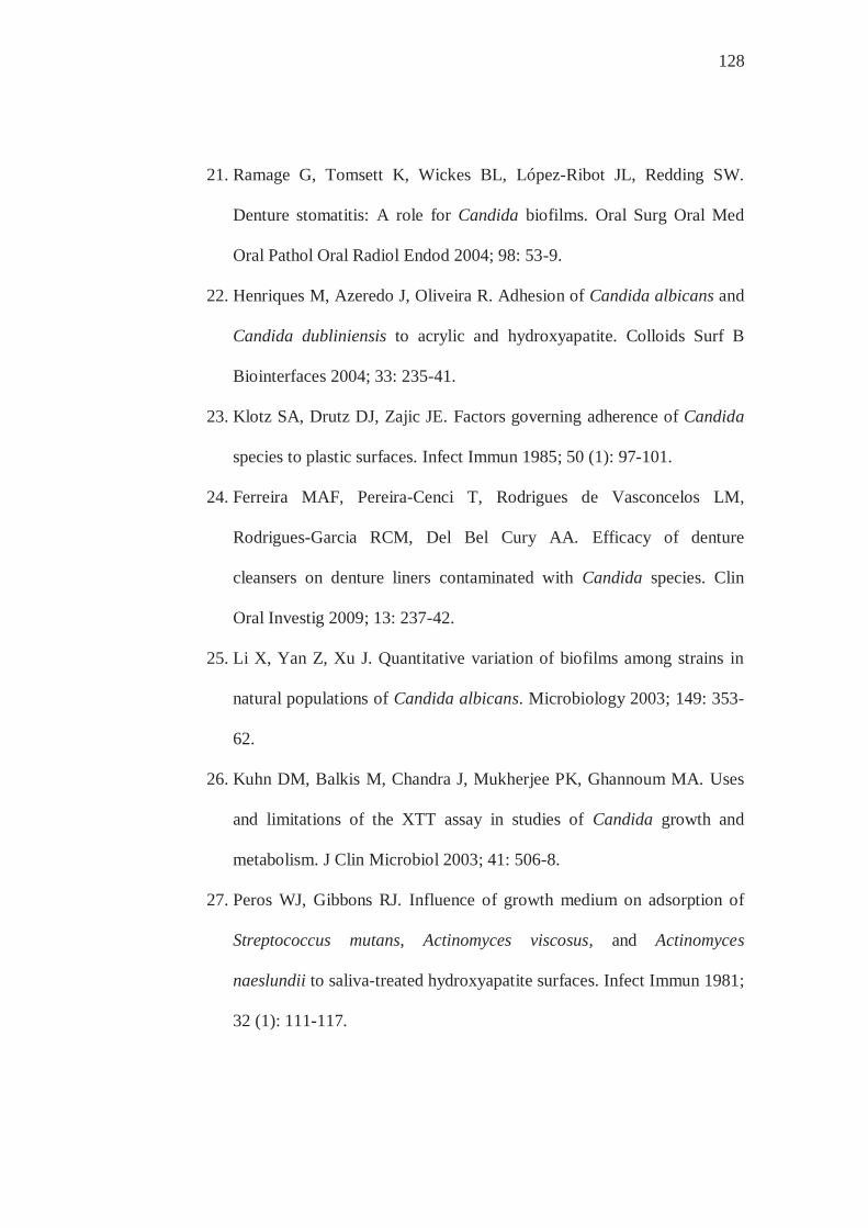
128
21. Ramage G, Tomsett K, Wickes BL, López-Ribot JL, Redding SW.
Denture stomatitis: A role for Candida biofilms. Oral Surg Oral Med
Oral Pathol Oral Radiol Endod 2004; 98: 53-9.
22. Henriques M, Azeredo J, Oliveira R. Adhesion of Candida albicans and
Candida dubliniensis to acrylic and hydroxyapatite. Colloids Surf B
Biointerfaces 2004; 33: 235-41.
23. Klotz SA, Drutz DJ, Zajic JE. Factors governing adherence of Candida
species to plastic surfaces. Infect Immun 1985; 50 (1): 97-101.
24. Ferreira MAF, Pereira-Cenci T, Rodrigues de Vasconcelos LM,
Rodrigues-Garcia RCM, Del Bel Cury AA. Efficacy of denture
cleansers on denture liners contaminated with Candida species. Clin
Oral Investig 2009; 13: 237-42.
25. Li X, Yan Z, Xu J. Quantitative variation of biofilms among strains in
natural populations of Candida albicans. Microbiology 2003; 149: 353-
62.
26. Kuhn DM, Balkis M, Chandra J, Mukherjee PK, Ghannoum MA. Uses
and limitations of the XTT assay in studies of Candida growth and
metabolism. J Clin Microbiol 2003; 41: 506-8.
27. Peros WJ, Gibbons RJ. Influence of growth medium on adsorption of
Streptococcus mutans, Actinomyces viscosus, and Actinomyces
naeslundii to saliva-treated hydroxyapatite surfaces. Infect Immun 1981;
32 (1): 111-117.

129
28. Hahnel S, Rosentritt M, Handel G, Bürgers R. In vitro evaluation of
artificial ageing on surface properties and early Candida albicans
adhesion to prosthetic resins. J Mater Sci: Mater Med 2009; 20: 249-55.
29. Sheridan PJ, Koda S, Ewoldsen NO, Lefebvre CA, Lavin MT.
Cytotoxicity of denture base resins. Int J Prosthodont 1997; 10 (1): 73-7.
30. De Jong MH, Van Der Hoeven JS. The growth of oral bacteria on saliva.
J Dent Res 1987; 66 (2): 498-505.
31. Sipahi C, Anil N, Bayramli E. The effect of acquired salivary pellicle on
the surface free energy and wettability of different denture base
materials. J Dent 2001; 29: 197-204.
32. Yildirim MS, Hasanreisoglu U, Hasirci N, Sultan N. Adherence of
Candida albicans to glow-discharge modified acrylic denture base
polymers. J Oral Rehabil 2005; 32: 518-25.
33. Verran J, Maryan CJ. Retention of Candida albicans on acrylic resin
and silicone of different surface topography. J Prosthet Dent 1997; 77:
535-9.
34. Radford DR, Sweet SP, Challacombe SH, Walter JD. Adherence of
Candida albicans to denture-base materials with different surface
finishes. J Dent 1998; 26: 577-83.
35. Nevzatoglu EU, Özcan M, Kulak-Ozkan Y, Kadir T. Adherence of
Candida albicans to denture base acrylics and silicone-based resilient
liner materials with different surface finishes. Clin Oral Investig 2007;
11 (3): 231-6.

130
36. Yildirim MS, Kesimer M, Hasirci N, Kiliç N, Hasanreisoglu U.
Adsorption of human salivary mucin MG1 onto glow-discharge plasma
treated acrylic resin surfaces. J Oral Rehabil 2006; 33: 775-83.
37. Göcke R, Gerath F, von Schwanewede H. Quantitative determination of
salivary components in the pellicle on PMMA denture base material.
Clin Oral Invest 2002; 6: 227-35.
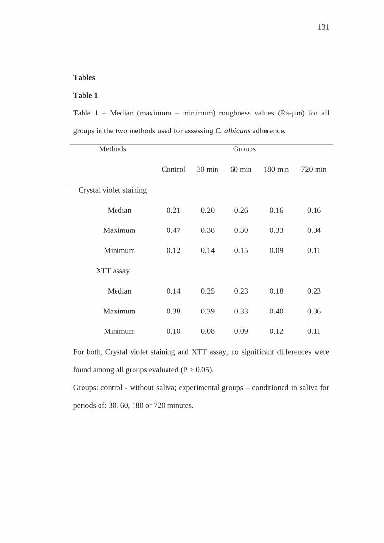
131
Tables
Table 1
Table 1 – Median (maximum – minimum) roughness values (Ra-μm) for all
groups in the two methods used for assessing C. albicans adherence.
Methods Groups
Control 30 min 60 min 180 min 720 min
Crystal violet staining
Median 0.21 0.20 0.26 0.16 0.16
Maximum 0.47 0.38 0.30 0.33 0.34
Minimum 0.12 0.14 0.15 0.09 0.11
XTT assay
Median 0.14 0.25 0.23 0.18 0.23
Maximum 0.38 0.39 0.33 0.40 0.36
Minimum 0.10 0.08 0.09 0.12 0.11
For both, Crystal violet staining and XTT assay, no significant differences were
found among all groups evaluated (P > 0.05).
Groups: control - without saliva; experimental groups – conditioned in saliva for
periods of: 30, 60, 180 or 720 minutes.
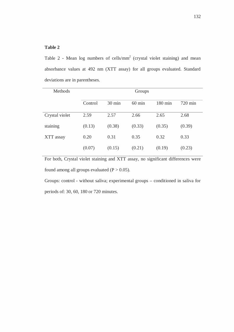
132
Table 2
Table 2 - Mean log numbers of cells/mm2 (crystal violet staining) and mean
absorbance values at 492 nm (XTT assay) for all groups evaluated. Standard
deviations are in parentheses.
Methods Groups
Control 30 min 60 min 180 min 720 min
Crystal violet
staining
2.59
(0.13)
2.57
(0.38)
2.66
(0.33)
2.65
(0.35)
2.68
(0.39)
XTT assay 0.20
(0.07)
0.31
(0.15)
0.35
(0.21)
0.32
(0.19)
0.33
(0.23)
For both, Crystal violet staining and XTT assay, no significant differences were
found among all groups evaluated (P > 0.05).
Groups: control - without saliva; experimental groups – conditioned in saliva for
periods of: 30, 60, 180 or 720 minutes.

3.5 Capítulo 5
The effect of human whole saliva on the in vitro adhesion of Candida albicans
to a denture base acrylic resin: a focus on collection and preparation of saliva
samples
Short title: Effect of human saliva on adhesion of Candida
Zamperini CAa, Schiavinato PCSa, Pavarina ACa, Giampaolo ETa, Vergani CEa,
Machado ALa,*
a Araraquara Dental School, UNESP – Univ Estadual Paulista, Department
of Dental Materials and Prosthodontics, Araraquara, São Paulo, Rua Humaitá nº
1680, CEP 14.801-903, Brazil.
*Corresponding author:
Profa. Dra. Ana Lucia Machado
Araraquara Dental School, UNESP – Univ. Estadual Paulista, Department of
Dental Materials and Prosthodontics, Araraquara, São Paulo, Rua Humaitá nº
1680, CEP 14.801-903, Brazil.
Tel: 55-16-33016543; Fax: 55-16-33016406
Email: [email protected]

134
Summary The effect of saliva on Candida albicans adhesion still remains controversial. The
diverse protocols used for collection and preparation of saliva samples may
contribute to the conflicting results. Thus, this study investigated whether
variations in the centrifugation parameters and number of donors of saliva would
influence the adhesion of C. albicans (ATCC 90028) to a denture base resin.
Samples (n=72) with smooth surfaces were made and then divided into four
groups: 1 control (C) - without saliva; 3 experimental groups – G1: saliva from 15
volunteers centrifuged at 10000 rpm for 5 min; G2: saliva from 15 volunteers
centrifuged at 12000 rpm for 30 min; G3: saliva from 1 volunteer centrifuged at
10000 rpm for 5 min. Candida adhesion was evaluated by XTT reduction assay
and crystal violet staining. Data were analyzed by one-way analyses of variance
(P=0.05). For XTT assay, groups G2, G3 and control were not significantly
different, while group G1 showed significantly higher absorbance value than
control. For crystal violet staining, there were no significant differences among
the mean log numbers of adherent cells for all groups. The results indicated that
variations in the centrifugation parameters and number of donors may influence
the effect of saliva on Candida albicans adhesion to denture base resins.
Key words: Acrylic Resins; Biofilms; Candida; Candida albicans; Saliva.

135
Introduction
An important step in the pathogenesis of denture stomatitis is the
attachment of Candida albicans to denture surfaces, followed by biofilm
formation [1-2]. The physicochemical characteristics of the denture materials,
such as roughness [3-5], electrostatic charge [1, 6] and surface free energy [1, 7-
8], may considerably influence C. albicans adhesion. In the oral environment,
however, the denture surfaces are coated by a thin film of saliva known as
salivary pellicle. Saliva is an exocrine secretion produced by different salivary
glands [9], consisting of water, electrolytes, and proteins [9-10]. Various functions
have been attributed to saliva, among them antimicrobial properties due to the
presence of immunologic and non-immunologic proteins [10]. However, saliva
also possesses proteins that could act as receptors, to promote the initial microbial
adhesion [11-13], and as a source of water and nutrients for growth and
reproduction of microorganisms [14]. Thus, besides non-specific surface
properties, yeast adhesion can also be influenced by specific receptors in the
acquired salivary pellicle [11-13].
In this context, to develop new strategies for preventing denture stomatitis,
it is essential to evaluate the influence of salivary pellicle on Candida adhesion to
denture surfaces. However, the interactions between biomaterials, salivary pellicle
and C. albicans are complex [15], and the effect of saliva on C. albicans adhesion
still remains controversial. While some researchers have found that the salivary
pellicle increases C. albicans colonization on the materials [11-13, 16-21], others

136
have observed that the preconditioning with saliva either does not significantly
affect [22-26] or decreases C. albicans colonization [4, 27-31].
In these studies, diverse protocols have been used for collection and
preparation of saliva samples, with differences in the nature (e.g. the quality of
saliva from different individuals) and in the centrifugation parameters. It is
therefore conceivable that these factors may have affected the outcomes and that
this variability may have accounted partly for the conflicting data reported by
different authors [32]. All these show the importance of quality control of saliva
in studies of this nature and point out the need for standardization [22]. However,
to date, no information is available concerning how different methods of
collection and preparation of saliva samples affect the adhesion of Candida spp.
cells to denture materials.
Different methods have been used to investigate the adhesion of Candida
to biomaterials, among them XTT and cell staining assays. For XTT assay, the
water-soluble tetrazolium salt [2,3-bis(2-methoxy-4-nitro-5-sulfophenyl)-5-
[(phenylamino) carbonyl]-2H-tetrazolium-hydroxide] is taken up by living cells
and is reduced by mitochondrial dehydrogenase to colored tetrazolium formazan
products that are determined spectrophotometrically [20, 22, 26, 33-35]. In the
staining assay, the fungal cells that adhered to the material surface are fixed,
stained with a basic dye, crystal violet, and then counted under a microscope [1,
26-27, 30-31, 36-37].
Therefore, the aim of the present study was to evaluate the effect of
variations in the centrifugation parameters and number of donors of saliva on C.
137
albicans adhesion to a denture base resin using crystal violet staining and XTT
reduction assay.
Materials and Methods
Preparation of acrylic resin specimens
The specimens (n=72) were fabricated from a microwave denture base
acrylic resin (Vipi Wave - VIPI Indústria e Comércio Exportação e Importação de
Produtos Odontológicos Ltda Pirassununga, SP, Brazil) using a conventional
flasking and pressure-pack technique. Initially, with the use of a metal mold, disk-
shaped silicone patterns (Zetaplus/Indurent - Zhermack, Badia Polesine, Rovigo,
Italy) were made with dimensions 13.8 X 2 mm. For surface standardization, the
silicone patterns were invested between two glass slides in dental stone in
microwave flaks [15, 38]. After the stone had set, the flasks were separated and
the silicone patterns were removed. For each specimen, 1 g of powder and 0.47 ml
of monomer liquid were mixed and processed according to the manufacturer’s
instructions. The mixture was packed into the molds, a trial pack was completed,
and excess material was removed. A final pack was performed and held for 15
minutes. The denture base acrylic resin was processed in a 500 W domestic
microwave oven (Brastemp – Brastemp Amazonia SA, Manaus, AM, Brazil) for
20 minutes at 20% power, followed by 5 minutes at 90% power. The flasks were
allowed to bench cool at room temperature, the specimens were deflasked, and
excess flash was aseptically removed with a sterile bur (Maxi-Cut; Lesfils de
August Malleifer SA, Ballaigues, Switzerland).
Surface roughness measurements
138
The surface roughness of all specimens was measured with a profilometer
(Mitutoyo SJ 400 – Mitutoyo Corporation – Tokyo, Japan). Four measurements
were made for each specimen and the average reading was designated as the Ra
(μm) value of that specimen. Resolution was 0.01 μm, interval (cutoff length) was
0.8 mm, transverse length was 2.4 mm, the stylus speed was 0.5 mm/s, and the
diamond stylus tip radius was 5 μm. All measurements were recorded by one
operator. Only samples with an average surface roughness ≤ 0.2 μm were selected
for this study.
Specimen sterilization and group assignment
Before the microbiological tests, the specimens were kept in distilled water
at ambient temperature for 48 hours, in order to eliminate residual monomers [30].
After this, they received an ultrasound bath for 20 minutes and were exposed to
ultraviolet light in the laminar flow chamber for 20 minutes on each side [39].
Hence, the 72 specimens were divided into four groups (n=18), as follows:
1 control (C), without preconditioning in saliva, and 3 experimental groups (G1,
G2 and G3) that were conditioned with saliva prepared as described below.
Saliva collection and preparation
For the experimental groups G1 and G2, whole human unstimulated saliva
from 15 healthy adult volunteers was expectorated into sterile 50 ml Falcon tubes
on ice, pooled and clarified by centrifugation. Thereafter, for group G1, the saliva
was centrifuged at 10000 rpm for 5 min at 4 ºC [30], while for group G2, saliva
was centrifuged at 12000 rpm for 30 min at 4 ºC [18-19, 22, 25, 27-28].
139
For group G3, whole human unstimulated saliva was collected from one
healthy adult volunteer. The saliva was expectorated into sterile 50 ml Falcon
tubes on ice, pooled and clarified by centrifugation at 10000 rpm for 5 min at 4 ºC
[16-17, 25, 30-31].
For all experimental groups, the supernatant of saliva was sterilized using
membrane filter with a 0.22 μm pore size [20, 24]. The resulting saliva was
immediately stored at -70 ºC until use. The study was approved by the Ethics
Committee of Araraquara Dental School, and all subjects volunteered to
participate and signed an informed consent form.
In vitro salivary pellicle formation
The specimens of the three experimental groups were incubated in 12-well
microtiter plates with 3 ml of the saliva preparation at room temperature for 30
minutes prior to the adherence assay [29].
Preparation of Candida suspension and adherence assay
The stock culture of Candida albicans strain ATCC 90028 was maintained
in YEPD medium (1% yeast extract, 2% peptone, 2% dextrose, 2% agar) stored at
4 – 6 ºC during the experimental period. For preparation of the yeast inoculum,
two loopfuls of the stock culture were streaked onto YEPD medium and incubated
at 37 ºC for 48 h. Two loopfuls of this young culture were transferred to 20 ml of
yeast nitrogen base (YNB) medium with 50 mM glucose and incubated at 37 ºC
for 21 h. Cells of the resultant culture were harvested, washed twice with
phosphate-buffered saline (PBS, pH 7.2), centrifuged at 4000 g for 5 min and
resuspended in YNB with 100 mM glucose. Candida suspensions were
140
standardized to a concentration of 1 x 10⁷ cells/ml, spectrophotometrically. Three
ml of the standardized C. albicans cell suspension was added to each well
containing the specimen. The cells were left to adhere for 90 min at 37 ºC in a
shaker at 75 rpm [20]. The non-adherent cells were removed from the specimen
by gently washing with 3 ml PBS twice. The negative controls were acrylic
specimens to which no cells were added. All experiments were performed in
triplicate on three independent occasions.
XTT reduction assay
Nine specimens from each group were evaluated by XTT reduction assay,
as described elsewhere [22]. Briefly, XTT (Sigma, MO, USA) was prepared in
ultrapure water (1mg/ml), filter sterilized and stored at -70 ºC until used.
Menadione (Sigma, MO, USA) solution was prepared in acetone at 0.4 mM
immediately before each assay. After washing, the specimens were transferred to
new wells with XTT solution in the following proportion: 158 μl PBS with 200
mM glucose, 40 μl XTT and 2 μl menadione. The plates were incubated for 3 h in
the dark at 37 ºC. The whole content of each well was centrifuged at 5000 g for 2
minutes and the colorimetric change of the supernatant was measured using a
microtiter plate reader (Thermo Plate – TP Reader) at 492 nm.
Crystal violet staining
Nine specimens from each group were evaluated by cell counting after
crystal violet staining. After the non-adherent cells were removed by washing, the
specimens were fixed in 80% ethanol, stained with crystal violet for 1 minute and
washed with PBS [36]. Adherent yeast cells were counted in 10 different fields for
141
each specimen, using a light microscope (Olympus BX51, Japan) at 400 x
magnification and the mean values were calculated. Adherent yeast cells were
counted in a "blind" manner to avoid subjective bias. The results were expressed
as cells/mm2.
Statistical Analysis
For each adhesion assay used, one-way analysis of variance, followed by
Tukey’s test, was performed to evaluate the effect of variations in the
centrifugation parameters and number of donors of saliva on Candida albicans
adhesion. For the crystal violet technique, data of yeast counts (cells/mm2) were
transformed by log. A significance level of 0.05 was used for all statistical tests.
Results
Candida albicans adhesion determined by XTT reduction assay is shown
in Fig. 1. One-way analysis of variance demonstrated that groups G2, G3 and
control were not significantly different, while group G1 showed significantly
higher absorbance readings than the control group (p<.034).
Candida albicans adhesion, as determined by crystal violet staining assay,
is shown in Fig. 2. One-way analysis of variance revealed that there were no
significant differences among the mean log numbers of adherent cells for all
groups evaluated.
All negative controls exhibited no metabolic activity (data not shown).
Discussion
Although several studies have evaluated the influence of the salivary
pellicle in the C. albicans adhesion process to biomaterials [4, 11-13, 16-31], the

142
results are conflicting and the role it plays is not yet clear. Among other factors,
these divergences could be related to the diverse methodological procedures used
in these studies. Recently, the effect of different periods of preconditioning with
saliva on Candida albicans adhesion to one denture base acrylic resin was
investigated, and no significant effect was found [26]. However, to the best of our
knowledge to date there are no studies that have addressed the effect of variations
in the methods used for collection and preparation of saliva samples. Thus, the
aim of this study was evaluate whether variations in the centrifugation speed and
time and number of donors of saliva would influence on the Candida albicans
adhesion to a denture base acrylic resin using crystal violet staining and XTT
assay.
Since the attachment of microorganisms on surfaces is related to surface
roughness [3-5, 31], the specimens of this study were made between glass slides
and their roughness were measured to ensure that smooth and standardized
surfaces were obtained [15, 38].
XTT reduction assay and crystal violet staining assays were selected for
this study because they are simple and versatile methods that have been frequently
used by investigators to investigate the adhesion of C. albicans to biomaterials [1,
20, 22, 26-27, 30-31, 33-37].
The results obtained by XTT reduction assay demonstrated that the
preconditioning with saliva collected from various donors and centrifuged at
10000 rpm for 5 minutes increased significantly the metabolic activity of Candida
albicans in comparison to control (Fig. 1). However, in the groups in which saliva
143
was collected from one donor or centrifuged longer and at a higher speed, no
significant differences were detected when they were compared to control. The
yeast-surface recognition systems involve different ligand-receptor mechanisms
based on protein or carbohydrate moieties existing in this interaction [11].
Moreover, it was suggested salivary mucins also bind to C. albicans, and they
promote yeast adhesion to polymethylmethacrylate [11]. Higher centrifugal forces
might separate significant amounts of the high molecular weight mucins [4, 32].
Thus, the centrifugation must be made without causing considerable effects on the
biochemical and biophysical properties of saliva. It has been observed that
centrifugation at 10000 g for 5 minutes at 4 °C had minimal impact normal saliva
protein profiling [40]. These may help explain, at least in part, the significantly
higher absorbance value of group G1 when compared to control. Another aspect
to be considered is that saliva composition varies greatly inter-individually [32].
Hence, the use of saliva collected from several donors and pooled may have
minimized this variation and, consequently, its influence on the adhesion results.
Differently from the results obtained by the XTT assay, the crystal violet
staining revealed that the adhered cell number was higher in group G1 compared
to control and the other experimental groups, but the differences did not reach
statistical significance. It is important to emphasize that the two methods used in
this investigation for assessing C. albicans adherence to the denture base resin are
based on different principles. The XTT reduction assay is a method in which the
metabolic activity of viable cells is measured [22, 26, 33-35]. Mitochondrial
dehydrogenases of viable cells cleave the tetrazolium ring of XTT, yielding

144
colored formazan crystals, which may be measured spectrophotometrically [41]. It
has been reported that the XTT method correlates well with other quantitative
technique, such as adenosine tri-phosphate (ATP) and colony forming units
(CFU) methods [22, 42]. For the other assay, the adhered cells are fixed, stained
with the basic dye crystal violet, and quantified by counting in selected fields of
the substrate surface [1, 26-27, 30-31, 36-37]. Therefore, because cells (both
living and dead) are stained by crystal violet, this assay does not allow
differentiating between living and dead cells [43]. These differences between XTT
and crystal violet assays may help explain the results obtained in this study and
may have accounted, at least in part, for the controversial results reported by
others. Therefore, when possible, it is recommended to use more than one method
to evaluate the effect of saliva on Candida adhesion to surfaces, particularly if
they are based on different principles.
It has been reported that exposed chemical groups of the material surface
have important role in determining the selectivity of the adsorption process [44].
Hence, considering that solid surfaces differ in respect to adsorption of salivary
proteins, the pellicle composition may vary among diverse materials [44]. An
analysis of the literature reveals that a variety of materials has been used as
substrate in vitro studies that evaluate the effect of saliva on Candida adhesion [4,
12-13, 16-20, 23-31]. This variety of materials makes the comparison of results
difficult and can partially contribute to the conflicting results regarding the effect
of preconditioning with saliva on Candida adhesion.
145
In conclusion, this study focused mainly on the effect of variations in the
collection and preparation of saliva on the Candida albicans adhesion. The results
demonstrated that variations in the centrifugation parameters and number of
donors can influence the effect of saliva on Candida albicans adhesion, thus
emphasizing that the protocols must be standardized. As the present study
evaluated only one denture base acrylic resin, further investigations with other
materials are necessary. Moreover, other Candida species must also be evaluated
in future studies. Besides, in addition to the speed and time of centrifugation and
number of donors, the use of filtered or unfiltered saliva has varied among studies
and the influence of this difference on Candida adhesion to denture materials
needs further investigations.
Acknowledgements
This research was supported by FAPESP (Grant - 2008/05339-8) and
CNPq (Grant - 479895/2009-0).
References
1. Klotz SA, Drutz DJ, Zajic JE. Factors governing adherence of Candida
species to plastic surfaces. Infect Immun. 1985; 50 (1): 97-101.
2. Radford DR, Challacombe SJ, Walter JD. Denture plaque and adherence
of Candida albicans to denture-base materials in vivo and in vitro. Crit
Rev Oral Biol Med. 1999; 10 (1): 99-116.
3. Verran J, Maryan CJ. Retention of Candida albicans on acrylic resin
and silicone of different surface topography. J Prosthet Dent. 1997; 77:
535-9.

146
4. Radford DR, Sweet SP, Challacombe SH, Walter JD. Adherence of
Candida albicans to denture-base materials with different surface
finishes. J Dent. 1998; 26: 577-583.
5. Lamfon H, Porter SR, McCullough M, Pratten J. Formation of Candida
albicans biofilms on non-shedding oral surfaces. Eur J Oral Sci. 2003;
111: 465-471.
6. Park SE, Periathamby AR, Loza JC. Effect of surface-charged
poly(methylmethacrylate) on the adhesion of Candida albicans. J
Prosthodont. 2003; 12: 249-254.
7. Luo G, Samaranayake LP. Candida glabrata, an emerging fungal
pathogen, exhibits superior relative cell surface hydrophobicity and
adhesion to denture acrylic surfaces compared with Candida albicans.
APMIS 2002; 110 (9): 601-610.
8. Yoshijima Y, Murakami K, Kayama S, Liu D, Hirota K, Ichikawa T,
Miyake Y. Effect of substrate surface hydrophobicity on the adherence
of yeast and hyphal Candida. Mycoses. 2010; 53 (3): 221-226.
9. Bräuer L, Möschter S, Beileke S, Jäger K, Garreis F, Paulsen FP.
Human parotid and submandibular glands express and secrete surfactant
proteins A, B, C and D. Histochem Cell Biol. 2009; 132 (3): 331-338.
10. de Almeida P del V, Grégio AM, Machado MA, de Lima AA, Azevedo
LR. Saliva composition and functions: a comprehensive review. J
Contemp Dent Pract. 2008; 9 (3): 72-80.

147
11. Edgerton M, Scannapieco FA, Reddy MS, Levine MJ. Human
submandibular-sublingual saliva promotes adhesion of Candida
albicans to polymethylmethacrylate. Infect Immun. 1993; 61 (6): 2644-
2652.
12. Holmes AR, Van der Wielen P, Cannon RD, Ruske D, Dawes P.
Candida albicans binds to saliva proteins selectively adsorbed to
silicone. Oral Surg Oral Med Oral Pathol Oral Radiol Endod. 2006; 102:
488-494.
13. Bürgers R, Hahnel S, Reichert TE, Rosentritt M, Behr M, Gerlach T,
Handel G, Gosau M. Adhesion of Candida albicans to various dental
implant surfaces and the influence of salivary pellicle proteins. Acta
Biomater. 2010; 6 (6): 2307-2313.
14. De Jong MH, Van Der Hoeven JS. The growth of oral bacteria on saliva.
J Dent Res. 1987; 66 (2): 498-505.
15. Vural C, Ozdemir G, Kurtulmus H, Kumbuloglu O, Ozcan M.
Comparative effects of two different artificial body fluids on Candida
albicans adhesion to soft lining materials. Dent Mater J. 2010; 29 (2):
206-212.
16. Nikawa H, Hayashi S, Nikawa Y, Hamada T, Samaranayake LP.
Interactions between denture lining material, protein pellicles and
Candida albicans. Arch Oral Biol. 1993; 38 (7): 631-634.
17. Nikawa H, Yamamoto T, Hamada T, Rahardjo MB, Murata H.
Antifungal effect of zeolite-incorporated tissue conditioner against
148
Candida albicans growth and/or acid production. J Oral Rehabil. 1997;
24: 350-357.
18. Nikawa H, Jin C, Hamada T, Murata H. Interactions between thermal
cycled resilient denture lining materials, salivary and serum pellicles and
Candida albicans in vitro. Part I. Effects on fungal growth. J Oral
Rehabil. 2000; 27: 41-45.
19. Nikawa H, Chen J, Hamada T, Nishimura M, Polyzois G. Candida
albicans colonization on thermal cycled maxillofacial polymeric
materials in vitro. J Oral Rehabil. 2001; 28: 526-533.
20. Chandra J, Mukherjee PK, Leidich SD, Faddoul FF, Hoyer LL, Douglas
LJ, Ghannoum MA. Antifungal resistance of candidal biofilms formed
on denture acrylic in vitro. J Dent Res. 2001; 80: 903-908.
21. Pusateri CR, Monaco EA, Edgerton M. Sensitivity of Candida albicans
biofilm cells grown on denture acrylic to antifungal proteins and
chlorhexidine. Arch Oral Biol. 2009; 54 (6): 588-594.
22. Jin Y, Samaranayake LP, Samaranayake Y, Yip HK. Biofilm formation
of Candida albicans is variably affected by saliva and dietary sugars.
Arch Oral Biol. 2004; 49: 789-798.
23. Tari BF, Nalbant D, Al DF, Kustimur S. Surface roughness and
adherence of Candida albicans on soft lining materials as influenced by
accelerated aging. J Contemp Dent Pract. 2007; 8 (5): 1-11.

149
24. Thein ZM, Samaranayake YH, Samaranayake LP. Characteristics of
dual species Candida biofilms on denture acrylic surfaces. Arch Oral
Biol. 2007; 52: 1200-1208.
25. Karaagaclioglu L, Can G, Yilmaz B, Ayhan N, Semiz O, Levent H. The
adherence of Candida albicans to acrylic resin reinforced with different
fibers. J Mater Sci Mater Med. 2008; 19 (2): 959-963.
26. Zamperini CA, Schiavinato PCS, Machado AL, Giampaolo ET,
Pavarina AC, Vergani CE. Effect of different periods of preconditioning
with saliva on Candida albicans adhesion to a denture base resin by
crystal violet staining and XTT assay. Journal of Investigative and
Clinical Dentistry 2010; 1(2): 114-119.
27. Samaranayake LP, McCourtie J, MacFarlane TW. Factors affecting the
in-vitro adherence of Candida albicans to acrylic surfaces. Arch Oral
Biol. 1980; 25: 611-615.
28. McCourtie J, MacFarlane TW, Samaranayake LP. Effect of saliva and
serum on the adherence of Candida species to chlorhexidine-treated
denture acrylic. J Med Microbiol. 1986; 21: 209-213.
29. Waters MGJ, Williams DW, Jagger RG, Lewis MAO. Adherence of
Candida albicans to experimental denture soft lining materials. J
Prosthet Dent. 1997; 77: 306-312.
30. Moura JS, Silva WJ, Pereira T, Cury ADB, Garcia RCMR. Influence of
acrylic resin polymerization methods and saliva on the adherence of four
Candida species. J Prosthet Dent. 2006; 96: 205-211.

150
31. Pereira-Cenci T, Cury AADB, Cenci MS, Rodrigues-Garcia RCM. In
vitro Candida colonization on acrylic resins and denture liners:
influence of surface free energy, roughness, saliva, and adhering
bacteria. Int J Prosthodont. 2007; 20: 308-310.
32. Schipper RG, Silletti E, Vingerhoeds MH. Saliva as research material:
biochemical, physicochemical and practical aspects. Arch Oral Biol.
2007; 52:1114-1135.
33. Ramage G, Tomsett K, Wickes BL, López-Ribot JL, Redding SW.
Denture stomatitis: A role for Candida biofilms. Oral Surg Oral Med
Oral Pathol Oral Radiol Endod. 2004; 98: 53-59.
34. Silva WJ, Seneviratne J, Parahitiyawa N, Rosa EAR, Samaranayake LP,
Del Bel Cury AA. Improvement of XTT assay performance of studies
involving Candida albicans biofilms. Braz Dent J. 2008; 19: 364-369.
35. Zamperini CA, Machado AL, Vergani CE, Pavarina AC, Giampaolo ET,
da Cruz NC. Adherence in vitro of Candida albicans to plasma treated
acrylic resin. Effect of plasma parameters, surface roughness and
salivary pellicle. Arch Oral Biol. 2010; 55: 763-770.
36. Ferreira MAF, Pereira-Cenci T, Rodrigues de Vasconcelos LM,
Rodrigues-Garcia RCM, Del Bel Cury AA. Efficacy of denture
cleansers on denture liners contaminated with Candida species. Clin
Oral Investig. 2009; 13: 237-42.
151
37. Zamperini CA, Machado AL, Vergani CE, Pavarina AC, Rangel EC,
Cruz NC. Evaluation of fungal adherence to plasma-modified
polymethylmethacrylate. Mycoses. 2010 In press.
38. Mutluay MM, Oğuz S, Orstavik D, Fløystrand F, Doğan A, Söderling E,
Närhi T, Olsen I. Experiments on in vivo biofilm formation and in vitro
adhesion of Candida species on polysiloxane liners. Gerodontology.
2010; 27 (4): 283-291.
39. Sheridan PJ, Koda S, Ewoldsen NO, Lefebvre CA, Lavin MT.
Cytotoxicity of denture base resins. Int J Prosthodont. 1997; 10 (1): 73-
77.
40. Schipper R, Loof A, de Groot J, Harthoorn L, Dransfield E, van Heerde
W. SELDI-TOF-MS of saliva: Methodology and pre-treatment effects. J
Chromatogr B Analyt Technol Biomed Life Sci. 2007; 847 (1): 45-53.
41. Bumgardner JD, Gerard PD, Geurtsen W, Leyhausen G. Cytotoxicity of
precious and nonprecious alloys-experimental comparison of in vitro
data from two laboratories. J Biomed Mater Res. 2002; 63 (2): 214-219.
42. Thein ZM, Samaranayake YH, Samaranayake LP. In vitro biofilm
formation of Candida albicans and non-albicans Candida species under
dynamic and anaerobic conditions. Arch Oral Biol. 2007; 52 (8): 761-
767.
43. Peeters E, Nelis HJ, Coenye T. Comparison of multiple methods for
quantification of microbial biofilms grown in microtiter plates. J
Microbiol Methods. 2008; 72 (2): 157-165.
152
44. Sipahi C, Anil N, Bayramli E. The effect of acquired salivary pellicle on
the surface free energy and wettability of different denture base
materials. J Dent. 2001; 29: 197-204.

153
Figures
Figure 1
*

154
Figure 2
155
Figure Legends
Figure 1. Mean absorbance values at 492 nm and 95% confidence intervals
for all groups. Groups: C - control - without saliva; experimental groups –
conditioned in saliva prepared as follows: G1 – 10000 g/5 min – 15 donors; G2 –
12000 g/30 min – 15 donors; G3 – 10000 g/5 min – 1 donor. Statistically different
mean compared to control group.
Figure 2. Mean log numbers (cells mm-2) and 95% confidence intervals for
all groups. For abbreviations, see legend of Figure 1.


4 Discussão
Para discussão dos resultados obtidos no presente estudo, os fatores
avaliados foram divididos em tópicos e serão abordados a seguir.
Adesão de Candida spp.
A aderência inicial de Candida nas superfícies das próteses removíveis é
essencial na colonização dessas superfícies e, consequentemente, no
desenvolvimento da estomatite protética. Desde que muitos fatores podem
influenciar o fenômeno de adesão inicial dos fungos às superfícies acrílicas, tais
como interações hidrofóbicas atrativas e forças eletrostáticas repulsivas, o
desenvolvimento de métodos que alterem as características superficiais e reduzam
a aderência de Candida a essas superfícies, seria um passo importante no
tratamento e prevenção da estomatite protética.
O tratamento a plasma tem sido utilizado como um método de modificação
de superfícies22,81. Esse tipo de tratamento caracteriza-se por ser seco, frio e
rápido, o que permite a alteração das propriedades de superfície de uma ampla
variedade de materiais19. Outro aspecto positivo desse método é que as
propriedades e a função dos materiais são preservadas, considerando-se que a
profundidade do tratamento a plasma é limitada a poucos nanômetros da
superfície19,61,81. Além disso, nessa técnica, a temperatura permanece tão baixa
quanto a temperatura ambiente30. Esse aspecto é particularmente importante para
as resinas acrílicas para base de prótese, nas quais o aquecimento pode causar
alterações dimensionais e afetar a adaptação das bases aos tecidos de suporte53.
Nos estudos apresentados, os tratamentos a plasma objetivaram diminuir a
hidrofobicidade, bem como, incorporar flúor na resina acrílica visando à
diminuição da adesão de Candida albicans e Candida glabrata.
Os resultados obtidos com relação à adesão de Candida albicans
demonstraram que os grupos ArSF6/70W e ArO2/70W não apresentaram
diferenças entre si e os níveis de atividade metabólica neles obtidos foram

158
significantemente menores quando comparados aos outros grupos tratados e ao
controle, conforme indicado pelo ensaio de XTT. Essa observação difere do
resultado obtido por Yildirim et al.81 (2005) que observaram aumento da
aderência de Candida albicans a uma resina para base de prótese após tratamentos
a plasma. Uma possível explicação para essa diferença seriam os parâmetros
determinados no estudo de Yildirim et al.81 (2005), no qual foi utilizado o gás
oxigênio a 50 e 100 W durante 15 minutos. A modificação de superfície
observada no grupo ArSF6/70W envolveu a incorporação de flúor na superfície da
resina acrílica. Os resultados demonstraram que os valores de ângulos de contato,
imediatamente após o tratamento a plasma com ArSF6/70W, aumentaram
consideravelmente comparados aos valores do grupo controle, estando de acordo
com as observações de Guruvenket et al.17 (2008). Esse fato provavelmente
ocorreu devido à substituição de espécies hidrofílicas por átomos de flúor22.
Consequentemente, as ligações de hidrogênio entre as moléculas de água e os
grupos presentes na superfície diminuíram, reduzindo a hidrofilicidade das
amostras tratadas com ArSF6/70W22. Assim, a redução da adesão de Candida
albicans no grupo tratado com ArSF6/70W pode ser atribuída à presença de flúor
na resina acrílica como demonstrado pela análise de XPS. Essa redução da adesão
de Candida albicans poderia ser atribuída ao aumento das forças eletrostáticas
repulsivas entre as células fúngicas e as amostras contendo átomos de flúor.
Robinson et al.63 (1997) relataram que a incorporação de flúor torna as superfícies
mais negativas, devido à presença de átomos eletronegativos de flúor. Tem sido
relatado que superfícies de resina acrílica carregadas negativamente apresentaram
níveis significantemente menores de Candida aderidas comparadas às superfícies
não tratadas, ou seja, superfícies carregadas negativamente podem alterar a
interação iônica entre a base de prótese e a célula de Candida spp.47,55.
Os resultados obtidos no estudo apresentado no capítulo 3 demonstraram
também que o tratamento a plasma com Ar/50W reduziu significantemente a
adesão de Candida glabrata, na ausência de saliva. Uma possível explicação para
essa redução poderia ser o fato de as amostras do grupo Ar/50W apresentarem o
menor valor de ângulo de contato, após 48 horas de imersão em água.

159
Considerando-se que as interações hidrofóbicas estão envolvidas no processo de
adesão26,32,36,66,83, as superfícies hidrofílicas observadas no grupo Ar/50W podem
ter inibido a adesão de Candida glabrata. Embora a característica de
hidrofobicidade de superfície celular (HSC) não seja específica para cada
espécie46,83, Candida glabrata tem sido considerada uma espécie relativamente
hidrofóbica32,35. Quando comparada com Candida albicans, Candida glabrata
apresentou maior HSC e maior tendência de se aderir às superfícies acrílicas32.
Adicionalmente, quanto mais próxima a energia livre de superfície do substrato e
do microrganismo, maior é a probabilidade de adesão de Candida36. Assim, os
resultados obtidos sugerem que superfícies acrílicas hidrofílicas poderiam inibir a
adesão de Candida glabrata. Entretanto, na presença de saliva, não foi possível
observar o mesmo efeito após o tratamento a plasma com Ar/50W. Tem sido
observado que a cobertura com saliva é um importante fator na determinação das
propriedades de molhabilidade dos materiais protéticos, ou seja, após o
condicionamento com saliva, as características de molhabilidade dos biomateriais
podem ser alteradas71,83. A energia livre de superfície de vários materiais,
incluindo uma resina acrílica polimerizada por meio de micro-ondas, diminuiu em
aproximadamente 10% quando as amostras foram recobertas com saliva71. Isso
poderia ajudar a explicar a ausência de efeito do tratamento a plasma na adesão de
Candida glabrata depois do pré-condicionamento das amostras de resina acrílica
com saliva.
As mensurações dos ângulos de contato dos grupos imediatamente após os
tratamentos a plasma demonstraram que os diferentes graus de hidrofilicidade
propostos no presente estudo foram obtidos. A diminuição na hidrofobicidade das
superfícies, observadas imediatamente após os tratamentos a plasma Ar/50W,
ArO2/70W, AAt/130W, pode ser atribuída aos elétrons energéticos criados
durante o tratamento que colidem na superfície acrílica. Essas colisões podem
resultar em quebra de ligações químicas criando radicais livres na superfície19,61.
As reações entre os radicais livres e espécies do material ou da atmosfera, tais
como hidrogênio ou oxigênio, podem incorporar grupos hidrofílicos na superfície
do polímero, aumentando sua energia livre de superfície e, consequentemente,

160
diminuindo os valores de ângulo de contato19,61. Resultados similares também
foram relatados em outras investigações em que tratamentos a plasma foram
utilizados para modificar superfícies poliméricas45,61,81-82.
Entretanto, foi observado que, quando as amostras tratadas a plasma foram
imersas em água por 48 horas, houve uma tendência de recuperação, ou seja, os
valores de ângulos de contato aproximaram-se dos valores obtidos nas amostras
do grupo controle. Esse aspecto, provavelmente, pode ter contribuído para a
igualdade entre as médias de adesão de Candida albicans e Candida glabrata
observada entre o grupo controle e os demais grupos tratados a plasma. Para
Candida albicans, foi observada igualdade entre todos os tratamentos e o
controle, quando a adesão foi avaliada pela coloração cristal violeta, e entre os
grupos AAt/130W, Ar/50W e o controle, quando avaliados pelo ensaio de XTT.
Para adesão de Candida glabrata, os resultados não demonstraram diferença entre
as amostras tratadas com plasma AAt/130W e aquelas não tratadas, nas duas
condições avaliadas (com e sem pré-condicionamento com saliva). Devido à
alteração nos ângulos de contato das amostras tratadas, após sua imersão em água,
também não foi possível relacionar a redução da adesão de Candida albicans,
observada pelo ensaio de XTT, para os grupos ArSF6/70W e ArO2/70W, com a
hidrofobicidade das superfícies. A estabilidade das superfícies hidrofílicas obtidas
por meio de tratamentos a plasma foi estudada por Rangel et al.61, em 2004. Esses
autores observaram que embora superfícies de silicone submetidas a diferentes
tratamentos a plasma apresentaram uma diminuição dos valores de ângulos de
contato, elas retornaram à hidrofobicidade original, após a exposição ao ar
atmosférico. Uma possível explicação para essa alteração é que a diminuição do
ângulo de contato obtida por meio do tratamento a plasma aumenta a energia de
superfície da resina acrílica19,61. Nessa condição, tem sido observado que as
superfícies poliméricas tendem a retornar à hidrofobicidade original, devido à
rotação dos grupos polares ao redor da cadeia polimérica, da superfície em direção
ao interior do material61.
Nos estudos apresentados nos capítulos 1, 2 e 3, os valores de ângulo de
contato, após 48 horas, demonstraram que os resultados obtidos com os
161
tratamentos a plasma sobre superfícies de resina acrílica também não foram
estáveis e alteraram-se após imersão em água, embora o tratamento a plasma com
Ar/50W tenha apresentado valores menores de ângulos de contato comparados
aos demais grupos.
Rugosidade Superficial
Zissis et al.84 (2000), ao estudar diversas resinas para base de prótese e
resinas utilizadas para reembasamento, observaram que a rugosidade de superfície
dos materiais protéticos pode variar consideravelmente, tendo sido obtidos valores
de 0,7 a 7,6 micrômetros. Assim, tem sido sugerido que a rugosidade superficial
pode favorecer a fixação dos microrganismos, devido à maior área de superfície
disponível para adesão, e ainda, por proteger os microrganismos contra as forças
de remoção28,58-59,74,77. Assim, visando avaliar o efeito da rugosidade superficial
sobre a adesão de Candida albicans, dois métodos de confecção das amostras
foram utilizados nos estudos apresentados nos capítulos 1 e 2, os quais permitiram
a obtenção de superfícies com diferentes valores de rugosidades superficiais.
Dessa forma, nesses estudos, todos os grupos eram compostos de metade das
amostras processada contra o gesso e a outra metade polimerizada em contato
com o vidro, a fim de se obterem, respectivamente, superfícies rugosas e lisas.
Para todos os grupos avaliados, os valores médios de rugosidade obtidos nas
amostras processadas contra o gesso foram sempre superiores aqueles das
amostras processadas contra o vidro. Entretanto, a avaliação do efeito da
rugosidade superficial sobre a aderência de Candida albicans não revelou
diferenças estatisticamente significantes. Esses resultados discordam dos obtidos
em diversos estudos28,59,74,77, em que números significantemente maiores de
Candida albicans foram encontrados em superfícies rugosas comparadas às
superfícies lisas. Por outro lado, recentemente, outros autores7,15,37,39,43 também
não observaram influência significante da rugosidade sobre a aderência de
Candida albicans, concordando com os resultados observados no presente estudo.
Essa divergência aponta para necessidade de mais estudos que avaliem o efeito de
diferentes valores de rugosidade na adesão de Candida spp.

162
Nos estudos apresentados nos capítulos 3, 4 e 5, todas as amostras foram
confeccionadas entre duas lâminas de vidro para obtenção de superfícies lisas e
padronizadas38,78. Os resultados obtidos demonstraram que não houve diferenças
significativas nos valores de rugosidade entre todos os grupos avaliados.
Película Salivar
Desde que todas as superfícies orais são recobertas pela película salivar,
foi considerado importante avaliar o efeito da saliva no processo de adesão
fúngica. Estudos in vitro que avaliam esse efeito têm apresentado resultados
contraditórios. Alguns autores6,8,14,21,23,40-42,44,56 observaram aumento da
colonização fúngica ao estudarem o efeito da película de saliva na adesão de
Candida albicans. Outros estudos25,34,37,50,65,79, por outro lado, encontraram que a
saliva resultou em diminuição nos valores de adesão fúngica sobre as superfícies
acrílicas, silicone e materiais reembasadores. Ao considerar o efeito da saliva,
Ramage et al.60 (2004) observaram que esse foi dependente do período de
avaliação, ou seja, eles observaram um aumento da adesão de Candida albicans
na fase de aderência inicial, mas um efeito mínimo ou de diminuição após 24
horas. Segundo Thein et al.75 (2007), a saliva humana pode modular o processo de
adesão e colonização dependendo da natureza e número de espécies envolvidas.
Os resultados obtidos nos estudos apresentados nos capítulos 1 e 2 indicaram que
a presença da película salivar não alterou significantemente o processo de adesão
de Candida albicans à resina acrílica avaliada, o que concorda com os estudos
realizados por Tari et al.73 (2007) e Jin et al.24 (2004). Entretanto, é importante
ressaltar que a saliva utilizada nesses dois estudos foi diluída em PBS60, o que
pode ter influenciado os resultados obtidos.
Outra observação importante é que a saliva não aumentou a adesão de
Candida glabrata como demonstrado no estudo apresentado no capítulo 3. Foi
possível observar que não houve nenhuma diferença significantemente entre
presença e ausência da película salivar na adesão de Candida glabrata. Esses
resultados estão de acordo com aqueles obtidos em investigações
anteriores34,37,50,52 que avaliaram o efeito da saliva na adesão de Candida glabrata.

163
Dessa forma, os resultados sugerem que a película salivar não aumenta a adesão
de Candida glabrata às superfícies poliméricas, ao contrário do que tem sido
observado por alguns autores para Candida albicans8,40-42,44. Além disso,
considerando que poucos estudos avaliaram a interação entre a película salivar e
Candida glabrata34,37,50,52, mais investigações ainda são necessárias para avaliar
essa questão.
As divergências encontradas na literatura com relação ao desempenho da
película salivar no processo de adesão de Candida albicans têm sido, em parte,
atribuídas às variações metodológicas entre os estudos, incluindo os diferentes
períodos de pré-condicionamento com saliva e as variações na coleta e preparo
das amostras de saliva51. Assim, os objetivos dos estudos apresentados nos
capítulos 4 e 5 foram avaliar se diferentes períodos de pré-condicionamento com
saliva e variações na coleta e preparo das amostras de saliva influenciariam os
resultados de adesão de Candida albicans a uma resina para base de prótese
utilizando-se dois métodos de análise, o ensaio de XTT e a coloração cristal
violeta.
A influência da película salivar na adesão de Candida e no
desenvolvimento do biofilme protético pode ocorrer por meio de interações
específicas entre as adesinas celulares e receptores específicos presentes na
saliva7,14,23, e ainda, pela atuação das proteínas salivares como fontes de nutrientes
para o crescimento microbiológico13. Por outro lado, essas proteínas também
podem atuar bloqueando os locais de adesão originalmente presentes nos
substratos7,24. Tem sido observado ainda que a saliva pode alterar as
características superficiais dos substratos envolvidas no processo de adesão, como
rugosidade e hidrofobicidade superficiais7,18,71,81, embora existam também autores
que relatem que as propriedades superficiais dos materiais são transferidas através
da película proteica, mantendo sua influência sobre a adesão microbiana18,23.
Os resultados obtidos após os diferentes períodos de pré-condicionamento
em saliva revelaram que as diferenças entre todos os grupos não foram
significativas, ou seja, os diferentes períodos de pré-condicionamento em saliva
não influenciaram a adesão de Candida albicans, tanto pelo ensaio de XTT como
164
pela coloração cristal violeta. Esses resultados sugerem que, em estudos de adesão
de Candida albicans em resinas acrílicas, períodos mais curtos de pré-
condicionamento em saliva, como 30 ou 60 minutos, poderiam ser utilizados,
tendo em vista a maior facilidade de execução quando comparados a períodos
mais longos.
Ainda com relação a esses resultados, o coeficiente de correlação entre os
dois métodos de análise utilizados, coloração cristal violeta e ensaio de XTT, foi
baixo. Os resultados obtidos por meio da coloração demonstraram que não houve
diferenças significativas entre os grupos experimentais e o controle, indicando que
o pré-condicionamento em saliva não alterou a adesão fúngica. Por outro lado, no
ensaio de XTT, embora a análise estatística não tenha encontrado diferenças
significantes entre os grupos, os valores de absorbância obtidos após o
condicionamento em saliva foram numericamente maiores comparados aos
valores do grupo controle. Um aspecto importante a ser considerado é que os dois
métodos de análise baseiam-se em princípios diferentes. Na coloração cristal
violeta, as células aderidas às superfícies da resina acrílica são fixadas, coradas
com o corante básico cristal violeta e quantificadas por meio da contagem celular
em campos selecionados na superfície do substrato15,26,37,50,65. Desde que células
(viáveis e não viáveis) são coradas pelo corante cristal violeta, esse método não
permite diferenciação entre células vivas e mortas49. Por outro lado, o ensaio de
XTT é um método em que a atividade metabólica das células viáveis é
medida24,60,70. Tem sido relatado que esse método correlaciona bem com outras
técnicas quantitativas, tais como, adenosina trifosfato (ATP) e contagem das
unidades formadoras de colônias viáveis (UFC/mL)24,76. Uma análise da literatura
revela que a maioria dos estudos que encontraram aumento da colonização de
Candida albicans sobre materiais protéticos após pré-condicionamento com saliva
utilizou testes que avaliam o metabolismo fúngico8,40-42,44. Por outro lado, quando
métodos de quantificação celular por meio de microscopias ou contagem de
unidades formadoras de colônias (UFC/mL) foram utilizados, a maioria dos
estudos sobre colonização de Candida albicans sobre materiais protéticos
observou ausência de efeito significativo da saliva25,73,75 ou diminuição da

165
adesão25,34,37,50,79. Diante disso, é possível que as divergências encontradas nos
estudos que avaliam o efeito da saliva na adesão de Candida albicans possam
também estar relacionadas, pelo menos parcialmente, com os diferentes métodos
de avaliação de adesão utilizados. Os resultados obtidos no presente estudo
apontam que os efeitos da saliva na adesão e no desenvolvimento de biofilmes de
Candida deveriam ser avaliados utilizando-se mais de um método de
quantificação, particularmente se eles são baseados em princípios diferentes.
Com relação à coleta e preparo das amostras de saliva, os resultados
obtidos pelo ensaio de XTT demonstraram que o pré-condicionamento com saliva
coletada de vários doadores e centrifugada a 10.000 g por 5 minutos aumentou
significantemente a atividade metabólica de Candida albicans quando comparada
ao grupo controle (sem saliva). Entretanto, nos grupos em que a saliva foi
coletada de um único doador, ou centrifugada por tempo e velocidade maiores,
nenhuma diferença significante foi detectada quando comparados ao grupo
controle. Os sistemas de reconhecimento fungo-superfície envolvem diferentes
mecanismos baseados em carboidratos e proteínas existentes nesta interação14.
Tem sido sugerido também que mucinas salivares aderem-se às células de
Candida albicans, promovendo a adesão fúngica ao polimetilmetacrilato14.
Considerando-se que maiores forças de centrifugação podem separar quantidades
significantes de mucinas de alto peso molecular58,69, a centrifugação deveria ser
feita sem causar efeitos consideráveis nas propriedades bioquímicas e biofísicas
da saliva. Tem sido observado que a centrifugação a 10.000 g por 5 minutos a 4
°C teve impacto mínimo no perfil protéico da saliva68. Esses achados ajudam a
explicar, pelo menos parcialmente, o valor de absorbância significantemente
maior do grupo G1 (saliva de vários doadores centrifugada a 10.000 g por 5
minutos a 4 °C) quando comparado ao grupo controle. Outro aspecto a ser
considerado é que a composição da saliva varia consideravelmente entre os
indivíduos69. Assim, o uso de amostras de saliva coletada de vários doadores pode
minimizar essa variação e, consequentemente, sua influência nos resultados
relacionados à adesão de Candida albicans.

166
Diferentemente dos resultados obtidos por meio do ensaio de XTT, a
coloração cristal violeta revelou que o número de células aderidas foi maior para o
grupo G1 comparado ao grupo controle e aos demais grupos experimentais, mas
essas diferenças não alcançaram significância estatística. É importante enfatizar
novamente que os dois métodos utilizados para avaliar a adesão de Candida
albicans à resina acrílica são baseados em princípios diferentes. As diferenças
apontadas entre o ensaio de XTT e a coloração cristal violeta podem ajudar a
explicar os resultados obtidos neste estudo e podem explicar, pelo menos
parcialmente, os resultados controversos encontrados na literatura. Assim, quando
possível, é recomendado utilizar mais de um método para avaliar o efeito da saliva
na adesão de Candida às superfícies.
Finalmente, as divergências de resultados entre os estudos podem também
ser atribuídas aos diferentes materiais que têm sido utilizados como substratos,
entre eles, superfícies acrílicas8,21,25,34,37,40,50,65,75,79, materiais protéticos
reembasadores41-42,44,50,73,79, materiais poliméricos maxilofaciais23,40 e
poliestireno24,60. Tem sido relatado que os grupos químicos expostos nas
superfícies sólidas desempenham papel importante na seletividade do processo de
adsorção6,71. Yildirim et al.82 (2006) observaram que superfícies de resina acrílica
modificadas por meio de tratamentos a plasma adsorveram quantidades diferentes
de mucina de alto peso molecular. Portanto, é possível afirmar que a composição
da película salivar pode variar entre os diversos materiais avaliados. Tendo em
vista que as concentrações proteicas resultantes nos diferentes materiais protéticos
foram pouco avaliadas16, estudos adicionais são necessários para analisar essas
diferenças.
Os estudos apresentados apresentam limitações desde que uma resina
acrílica para base de prótese e uma cepa de Candida albicans e Candida glabrata
foram utilizadas. Além disso, tratamentos a plasma utilizando outros parâmetros
também devem ser avaliados. Apesar dessas limitações, os resultados obtidos
sugerem que os tratamentos utilizados nos grupos ArSF6/70W e ArO2/70W tem
potencial para redução da aderência de Candida albicans sobre as superfícies das
bases de próteses e devem ser melhor analisados em estudos futuros. Além disso,
167
superfícies hidrofílicas são efetivas para redução da aderência de Candida
glabrata sobre superfícies acrílicas. Os diferentes períodos de pré-
condicionamento em saliva não influenciaram os resultados de adesão de Candida
albicans a uma resina acrílica para base de próteses. Entretanto, variações na
coleta e preparo das amostras de saliva são fatores que podem influenciar nos
resultados de adesão de Candida albicans.

55 Conclusão Dentro das limitações deste estudo, as seguintes conclusões podem ser feitas:
Os tratamentos a plasma utilizados neste estudo foram eficientes para
alterar o grau de hidrofobicidade ou incorporar flúor na superfície da
resina acrílica;
A diminuição da atividade metabólica de Candida albicans sobre a
resina acrílica estudada foi observada nos tratamentos ArSF6/70W e
ArO2/70W, quando analisada pelo método XTT;
A aderência de Candida glabrata foi diminuída sobre superfícies
hidrofílicas obtidas por meio do tratamento a plasma com Ar/50W,
quando avaliada pela coloração cristal violeta;
A saliva não alterou significantemente a aderência de Candida
glabrata.
Os diferentes períodos de pré-condicionamento em saliva não
influenciaram significativamente a aderência de Candida albicans
sobre a resina acrílica avaliada;
O coeficiente de correlação entre os resultados obtidos com a
coloração cristal violeta e o ensaio de XTT foi baixo;
As variações na coleta e preparo das amostras de saliva utilizadas são
fatores que podem influenciar os resultados de aderência de Candida
albicans.
A rugosidade superficial não alterou significantemente a aderência de
Candida albicans.

66 Referências*
1. Abaci O, Haliki-Uztan A, Ozturk B, Toksavul S, Ulusoy M,
Boyacioglu H. Determining Candida spp. incidence in denture
wearers. Mycopathologia. 2010; 169: 365-72.
2. Barbeau J, Séguin J, Goulet JP, Koninck L, Avon SL, Lalonde B et al.
Reassessing the presence of Candida albicans in denture-related
stomatitis. Oral Surg Oral Med Oral Pathol Oral Radiol Endod. 2003;
95: 51-9.
3. Blanco MT, Morales JJ, Lucio L, Pérez-Giraldo C, Hurtado C, Gómez-
Garcia AC. Modification of adherence to plastic and to human buccal
cells of Candida albicans and Candida dubliniensis by a subinhibitory
concentration of itraconazole. Oral Microbiol Immunol. 2006; 21: 69-
72.
4. Bräuer L, Möschter S, Beileke S, Jäger K, Garreis F, Paulsen FP.
Human parotid and submandibular glands express and secrete
surfactant proteins A, B, C and D. Histochem Cell Biol. 2009; 132:
331-8.
5. Budtz-Jörgensen. E. Etiology, pathogenesis, therapy, and prophylaxis
of oral yeast infections. Acta Odontol Scand. 1990; 48: 61-9.
*De acordo com o estilo Vancouver. Disponível no site: http://www.nlm.nih.gov/bsd/uniform_requirements.html.

172
6. Bürgers R, Hahnel S, Reichert TE, Rosentritt M, Behr M, Gerlach T et
al. Adhesion of Candida albicans to various dental implant surfaces
and the influence of salivary pellicle proteins. Acta Biomater. 2010; 6:
2307-13.
7. Burgers R, Schneider-Brachert W, Rosentritt M, Handel G, Hahnel S.
Candida albicans adhesion to composite resin materials. Clin Oral
Investig. 2009; 13: 292-9.
8. Chandra J, Mukherjee PK, Leidich SD, Faddoul FF, Hoyer LL,
Douglas LJ et al. Antifungal resistance of candidal biofilms formed on
denture acrylic in vitro. J Dent Res. 2001; 80: 903-8.
9. Chau VB, Saunders TR, Pimsler M, Elfring DR. In depth disinfection
of acrylic resins. J Prosthet Dent. 1995; 74: 309-13.
10. Coco BJ, Bagg J, Cross LJ, Jose A, Cross J, Ramage G. Mixed
Candida albicans and Candida glabrata populations associated with
the pathogenesis of denture stomatitis. Oral Microbiol Immunol. 2008;
23: 377-83.
11. Dagistan S, Aktas AE, Caglayan F, Ayyildiz A, Bilge M. Differential
diagnosis of denture-induced stomatitis, Candida, and their variations
in patients using complete denture: a clinical and mycological study.
Mycoses 2009; 52: 266-71.
12. de Almeida P del V, Grégio AM, Machado MA, de Lima AA,
Azevedo LR. Saliva composition and functions: a comprehensive
review. J Contemp Dent Pract. 2008; 9: 72-80.
13. De Jong MH, Van Der Hoeven JS. The growth of oral bacteria on
saliva. J Dent Res. 1987; 66: 498-505.
173
14. Edgerton M, Scannapieco FA, Reddy MS, Levine MJ. Human
submandibular-sublingual saliva promotes adhesion of Candida
albicans to polymethylmethacrylate. Infect Immun. 1993; 61: 2644-52.
15. Ferreira MAF, Pereira-Cenci T, Rodrigues de Vasconcelos LM,
Rodrigues-Garcia RCM, Cury AADB. Efficacy of denture cleansers on
denture liners contaminated with Candida species. Clin Oral Investig.
2009; 13: 237-42.
16. Göcke R, Gerath F, von Schwanewede H. Quantitative determination
of salivary components in the pellicle on PMMA denture base
material. Clin Oral Invest. 2002; 6: 227-35.
17. Guruvenket S, Iyer GRS, Shestakova L, Morgen P, Larsen NB, Rao
GM. Fluorination of polymethylmethacrylate with tetrafluoroethane
using DC glow discharge plasma. Appl Surf Sci. 2008; 254: 5722-6.
18. Hahnel S, Rosentritt M, Handel G, Bürgers R. In vitro evaluation of
artificial ageing on surface properties and early Candida albicans
adhesion to prosthetic resins. J Mater Sci: Mater Med. 2009; 20: 249-
55.
19. Hauser J, Zietlow J, Koller M, Esenwein SA, Halfmann H, Awakowicz
P et al. Enhanced cell adhesion to silicone implant material through
plasma surface modification. J Mater Sci Mater Med. 2009; 20: 2541–
8.
20. Hazen KC, Brawner DL, Riesselman MH, Jutila MA, Cutler JE.
Differential adherence of hydrophobic and hydrophilic Candida
albicans yeast cells to mouse tissues. Infect Immun. 1991; 59: 907-12.
21. Henriques M, Azeredo J, Oliveira R. Adhesion of Candida albicans
and Candida dubliniensis to acrylic and hydroxyapatite. Colloids Surf
B Biointerfaces. 2004; 33: 235-41.

174
22. Hodak SK, Supasai T, Paosawatyanyong B, Kamlangkla K, Pavarajarn
V. Enhancement of the hydrophobicity of silk fabrics by SF6 plasma.
Appl Surf Sci. 2008; 254: 4744-9.
23. Holmes AR, Van der Wielen P, Cannon RD, Ruske D, Dawes P.
Candida albicans binds to saliva proteins selectively adsorbed to
silicone. Oral Surg Oral Med Oral Pathol Oral Radiol Endod. 2006;
102: 488-94.
24. Jin Y, Samaranayake LP, Samaranayake Y, Yip HK. Biofilm
formation of Candida albicans is variably affected by saliva and
dietary sugars. Arch Oral Biol. 2004; 49: 789-98.
25. Karaagaclioglu L, Can G, Yilmaz B, Ayhan N, Semiz O, Levent H.
The adherence of Candida albicans to acrylic resin reinforced with
different fibers. J Mater Sci Mater Med. 2008; 19: 959-63.
26. Klotz SA, Drutz DJ, Zajic JE. Factors governing adherence of Candida
species to plastic surfaces. Infect Immun. 1985; 50: 97-101.
27. Lamfon H, Al-Karaawi Z, McCullough M, Porter SR, Pratten J.
Composition of in vitro denture plaque biofilms and susceptibility to
antifungals. FEMS Microbiol Lett. 2005; 242: 345-51.
28. Lamfon H, Porter SR, McCullough M, Pratten J. Formation of
Candida albicans biofilms on non-shedding oral surfaces. Eur J Oral
Sci. 2003; 111: 465-71.
29. Li L, Redding S, Dongari-Bagtzoglou A. Candida glabrata: an
emerging oral opportunistic pathogen. J Dent Res. 2007; 86: 204-15.
30. Liu Y, Kuai P, Huo P, Liu C. Fabrication of CuO nanofibers via the
plasma decomposition of Cu(OH)2. Mater Lett. 2009; 63: 188-90.
31. Lombardi T, Budtz-Jörgensen E. Treatment of denture-induced
stomatitis: a review. Eur J Prosthodont Restor Dent. 1993; 2: 17-22.

175
32. Luo G, Samaranayake LP. Candida glabrata, an emerging fungal
pathogen, exhibits superior relative cell surface hydrophobicity and
adhesion to denture acrylic surfaces compared with Candida albicans.
APMIS 2002; 110: 601-10.
33. Ma T, Johnson GH, Gordon GE. Effects of chemical disinfectants on
the surface characteristics and color of denture resins. J Prosthet Dent.
1997; 77: 197-204.
34. McCourtie J, MacFarlane TW, Samaranayake LP. Effect of saliva and
serum on the adherence of Candida species to chlorhexidine-treated
denture acrylic. J Med Microbiol. 1986; 21: 209-13.
35. Minagi S, Miyake Y, Fujioka Y, Tsuru H, Suginaka H. Cell-surface
hydrophobicity of Candida species as determined by the contact-angle
and hydrocarbon-adherence methods. J Gen Microbiol. 1986; 132:
1111-15.
36. Minagi S, Miyake Y, Inagaki K, Tsuru H, Suginaka H. Hydrophobic
interaction in Candida albicans and Candida tropicalis adherence to
various denture base resin materials. Infect Immun. 1985; 47: 11-4.
37. Moura JS, Silva WJ, Pereira T, Cury AADB, Garcia RCMR. Influence
of acrylic resin polymerization methods and saliva on the adherence of
four Candida species. J Prosthet Dent. 2006; 96: 205-11.
38. Mutluay MM, Oğuz S, Orstavik D, Fløystrand F, Doğan A, Söderling
E et al. Experiments on in vivo biofilm formation and in vitro adhesion
of Candida species on polysiloxane liners. Gerodontology. 2010; 27:
283-91.
39. Nevzatoglu EU, Özcan M, Kulak-Ozkan Y, Kadir T. Adherence of
Candida albicans to denture base acrylics and silicone-based resilient
liner materials with different surface finishes. Clin Oral Investig. 2007;
11: 231-6.
176
40. Nikawa H, Chen J, Hamada T, Nishimura M, Polyzois G. Candida
albicans colonization on thermal cycled maxillofacial polymeric
materials in vitro. J Oral Rehabil. 2001; 28: 526-33.
41. Nikawa H, Hayashi S, Nikawa Y, Hamada T, Samaranayake LP.
Interactions between denture lining material, protein pellicles and
Candida albicans. Arch Oral Biol. 1993; 38: 631-4.
42. Nikawa H, Jin C, Hamada T, Murata H. Interactions between thermal
cycled resilient denture lining materials, salivary and serum pellicles
and Candida albicans in vitro. Part I. Effects on fungal growth. J Oral
Rehabil. 2000; 27: 41-51.
43. Nikawa H, Jin C, Makihira S, Egusa H, Hamada T, Kumagai H.
Biofilm formation of Candida albicans on the surfaces of deteriorated
soft denture lining materials caused by denture cleansers in vitro. J
Oral Rehabil. 2003; 30: 243-50.
44. Nikawa H, Yamamoto T, Hamada T, Rahardjo MB, Murata H.
Antifungal effect of zeolite-incorporated tissue conditioner against
Candida albicans growth and/or acid production. J Oral Rehabil. 1997;
24: 350-7.
45. Ozden N, Akaltan F, Suzer S, Akovali G. Time-related wettability
characteristic of acrylic resin surfaces treated by glow discharge. J
Prosthet Dent. 1999; 82: 680-4.
46. Panagoda GJ, Ellepola ANB, Samaranayake LP. Adhesion of Candida
parapsilosis to epithelial and acrylic surfaces correlates with cell
surface hydrophobicity. Mycoses 2001; 44: 29-35.
47. Park SE, Periathamby AR, Loza JC. Effect of surface-charged
poly(methylmethacrylate) on the adhesion of Candida albicans. J
Prosthodont. 2003; 12: 249-54.

177
48. Pavarina AC, Pizzollitto AC, Machado AL, Vergani CE, Giampaolo
ET. An infection control protocol: effectiveness of immersion
solutions to reduce the microbial growth on dental prostheses. J Oral
Rehabil. 2003; 30: 532-6.
49. Peeters E, Nelis HJ, Coenye T. Comparison of multiple methods for
quantification of microbial biofilms grown in microtiter plates. J
Microbiol Methods. 2008; 72: 157-65.
50. Pereira-Cenci T, Cury AADB, Cenci MS, Rodrigues-Garcia RCM. In
vitro Candida colonization on acrylic resins and denture liners:
influence of surface free energy, roughness, saliva, and adhering
bacteria. Int J Prosthodont. 2007; 20: 308-10.
51. Pereira-Cenci T, Del Bel Cury AA, Crielaard W, Ten Cate JM.
Development of Candida-associated denture stomatitis: new insights. J
Appl Oral Sci. 2008; 16: 86-94.
52. Pereira-Cenci T, Deng DM, Kraneveld EA, Manders EMM, Cury
AADB, Ten Cate JM et al. The effect of Streptococcus mutans and
Candida glabrata on Candida albicans biofilms formed on different
surfaces. Arch Oral Biol. 2008; 53: 755-64.
53. Polukoshko KM, Brudvik JS, Nicholls JI, Smith DE. Evaluation of
heat-cured resin bases following the addition of denture teeth using a
second heat cure. J Prosthet Dent. 1992; 67: 556-62.
54. Polyzois GL, Zissis AJ, Yannikakis SA. The effect of glutaraldehyde
and microwave disinfection on some properties of acrylic denture
resin. Int J Prosthodont. 1995; 8: 150-4.
55. Puri G, Berzins DW, Dhuru VB, Raj PA, Rambhia SK, Dhir G et al.
Effect of phosphate group addition on the properties of denture base
resins. J Prosthet Dent. 2008; 100: 302-8.
178
56. Pusateri CR, Monaco EA, Edgerton M. Sensitivity of Candida
albicans biofilm cells grown on denture acrylic to antifungal proteins
and chlorhexidine. Arch Oral Biol. 2009; 54: 588-94.
57. Rad AY, Ayhan H, Piskin E. Adhesion of different bacterial strains to
low-temperature plasma-treated sutures. J Biomed Mater Res A. 1998;
41: 349-58.
58. Radford DR, Challacombe SJ, Walter JD. Denture plaque and
adherence of Candida albicans to denture-base materials in vivo and in
vitro. Crit Rev Oral Biol Med. 1999; 10: 99-116.
59. Radford DR, Sweet SP, Challacombe SJ, Walter JD. Adherence of
Candida albicans to denture-base materials with different surface
finishes. J Dent. 1998; 26: 577-83.
60. Ramage G, Tomsett K, Wickes BL, López-Ribot JL, Redding SW.
Denture stomatitis: A role for Candida biofilms. Oral Surg Oral Med
Oral Pathol Oral Radiol Endod. 2004; 98: 53-9.
61. Rangel EC, Gadioli GZ, Cruz NC. Investigations on the stability of
plasma modified silicone surfaces. Plasmas and Polymers. 2004; 9: 35-
48.
62. Ribeiro DG, Pavarina AC, Dovigo LN, Spolidorio DMP, Giampaolo
ET, Vergani CE. Denture disinfection by microwave irradiation: A
randomized clinical study. J Dent. 2009; 37: 666-72.
63. Robinson GN, Kebabian PL, Feedman A, DePalma V. Temperature-
dependent surface potentials of fluorinated alkanethiolate self-
assembled monolayers. Thin Solid Films. 1997; 310: 24-8.
64. Sagripanti JL, Bonifacino A. Cytotoxicity of liquid disinfectants. Surg.
Infect. 2000; 1: 3-14.
179
65. Samaranayake LP, McCourtie J, MacFarlane TW. Factors affecting the
in-vitro adherence of Candida albicans to acrylic surfaces. Arch Oral
Biol. 1980; 25: 611-5.
66. Samaranayake YH, Wu PC, Samaranayake LP, So M, Yuen KY.
Adhesion and colonisation of Candida krusei on host surfaces. J Med
Microbiol. 1994; 41: 250-8.
67. Samaranayake YH, Wu PC, Samaranayake LP, So M. Relationship
between the cell surface hydrophobicity and adherence of Candida
krusei and Candida albicans to epithelial and denture acrylic surfaces.
APMIS. 1995; 103: 707-13.
68. Schipper R, Loof A, de Groot J, Harthoorn L, Dransfield E, van
Heerde W. SELDI-TOF-MS of saliva: methodology and pre-treatment
effects. J Chromatogr B Analyt Technol Biomed Life Sci. 2007; 847:
45-53.
69. Schipper RG, Silletti E, Vingerhoeds MH. Saliva as research material:
biochemical, physicochemical and practical aspects. Arch Oral Biol.
2007; 52: 1114-35.
70. Silva WJ, Seneviratne J, Parahitiyawa N, Rosa EAR, Samaranayake
LP, Del Bel Cury AA. Improvement of XTT assay performance of
studies involving Candida albicans biofilms. Braz Dent J. 2008; 19:
364-9.
71. Sipahi C, Anil N, Bayramli E. The effect of acquired salivary pellicle
on the surface free energy and wettability of different denture base
materials. J Dent. 2001; 29: 197-204.
72. Suanpoot P, Kueseng K, Ortmann S, Kaufmann R, Umongno C,
Nimmanpipug P et al. Surface analysis of hydrophobicity of Thai silk
treated by SF6 plasma. Surface & Coatings Technology. 2008; 202:
5543-49.
180
73. Tari BF, Nalbant D, Al DF, Kustimur S. Surface roughness and
adherence of Candida albicans on soft lining materials as influenced
by accelerated aging. J Contemp Dent Pract. 2007; 8: 1-11.
74. Taylor R, Maryan C, Verran J. Retention of oral microorganisms on
cobalt-chromium alloy and dental acrylic resin with different surface
finishes. J Prosthet Dent. 1998; 80: 592-7.
75. Thein ZM, Samaranayake YH, Samaranayake LP. Characteristics of
dual species Candida biofilms on denture acrylic surfaces. Arch Oral
Biol. 2007; 52: 1200-8.
76. Thein ZM, Samaranayake YH, Samaranayake LP. In vitro biofilm
formation of Candida albicans and non-albicans Candida species
under dynamic and anaerobic conditions. Arch Oral Biol. 2007; 52:
761-7.
77. Verran J, Maryan CJ. Retention of Candida albicans on acrylic resin
and silicone of different surface topography. J Prosthet Dent. 1997; 77:
535-9.
78. Vural C, Ozdemir G, Kurtulmus H, Kumbuloglu O, Ozcan M.
Comparative effects of two different artificial body fluids on Candida
albicans adhesion to soft lining materials. Dent Mater J. 2010; 29: 206-
12.
79. Waters MGJ, Williams DW, Jagger RG, Lewis MAO. Adherence of
Candida albicans to experimental denture soft lining materials. J
Prosthet Dent. 1997; 77: 306-12.
80. Wilson J. The aetiology, diagnosis and management of denture
stomatitis. Br Dent J. 1998; 185: 380-4.
181
81. Yildirim MS, Hasanreisoglu U, Hasirci N, Sultan N. Adherence of
Candida albicans to glow-discharge modified acrylic denture base
polymers. J Oral Rehabil. 2005; 32: 518-25.
82. Yildirim MS, Kesimer M, Hasirci N, Kiliç N, Hasanreisoglu U.
Adsorption of human salivary mucin MG1 onto glow-discharge plasma
treated acrylic resin surfaces. J Oral Rehabil. 2006; 33: 775-83.
83. Yoshijima Y, Murakami K, Kayama S, Liu D, Hirota K, Ichikawa T et
al. Effect of substrate surface hydrophobicity on the adherence of yeast
and hyphal Candida. Mycoses. 2010; 53: 221-6.
84. Zissis AJ, Polyzois GL, Yannikakis SA, Harrison A. Roughness of
denture materials: a comparative study. Int J Prosthodont. 2000; 13:
136-40.

7 Anexos Anexo 1

Anexo 2

Anexo 3

Anexo 4
Anexo 5
AAnexo 6
AAnexo 7

AAnexo 8


8 Apêndice
Apêndice 1 Os resultados obtidos durante as leituras de rugosidade superficial das
amostras processadas entre vidros, ângulo de contato (imediatamente após os
tratamentos e 48 horas após a imersão em água destilada estéril), bem como, os
valores de absorbância relativos à adesão de Candida albicans nos diferentes
grupos estão apresentados nas Tabelas 1A a 5A.
Tabela 1A - Grupo controle (não submetido ao tratamento a plasma)
Grupo Saliva Rugosidade (Ra-μm) Ângulo de contato Ângulo de contato XTT (Abs)
após tratamento (º) 48 horas (º)
Controle Ausente 0,28 53,80 51,91 0,63
Controle Ausente 0,32 60,31 59,87 1,25
Controle Ausente 0,29 55,30 56,90 0,83
Controle Ausente 0,31 55,30 53,22 1,00
Controle Ausente 0,35 53,80 51,91 0,67
Controle Ausente 0,20 57,68 56,18 0,79
Controle Ausente 0,35 56,62 59,83 0,97
Controle Ausente 0,34 57,78 55,20 0,37
Controle Ausente 0,31 55,35 52,87 1,00
Controle Presente 0,18 57,68 56,18 0,78
Controle Presente 0,23 56,62 59,83 1,01
Controle Presente 0,16 57,78 55,20 0,96
Controle Presente 0,15 55,35 52,87 0,54
Controle Presente 0,39 51,80 53,68 0,57
Controle Presente 0,25 65,20 56,85 0,37
Controle Presente 0,36 61,17 58,23 0,74
Controle Presente 0,20 57,78 55,20 0,96
Controle Presente 0,19 57,68 56,18 0,78
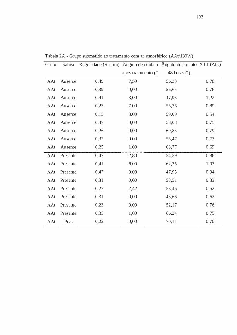
193
Tabela 2A - Grupo submetido ao tratamento com ar atmosférico (AAt/130W)
Grupo Saliva Rugosidade (Ra-μm) Ângulo de contato Ângulo de contato XTT (Abs)
após tratamento (º) 48 horas (º)
AAt Ausente 0,49 7,59 56,33 0,78
AAt Ausente 0,39 0,00 56,65 0,76
AAt Ausente 0,41 3,00 47,95 1,22
AAt Ausente 0,23 7,00 55,36 0,89
AAt Ausente 0,15 3,00 59,09 0,54
AAt Ausente 0,47 0,00 58,08 0,75
AAt Ausente 0,26 0,00 60,85 0,79
AAt Ausente 0,32 0,00 55,47 0,73
AAt Ausente 0,25 1,00 63,77 0,69
AAt Presente 0,47 2,80 54,59 0,86
AAt Presente 0,41 6,00 62,25 1,03
AAt Presente 0,47 0,00 47,95 0,94
AAt Presente 0,31 0,00 58,51 0,33
AAt Presente 0,22 2,42 53,46 0,52
AAt Presente 0,31 0,00 45,66 0,62
AAt Presente 0,23 0,00 52,17 0,76
AAt Presente 0,35 1,00 66,24 0,75
AAt Pres 0,22 0,00 70,11 0,70

194
Tabela 3A - Grupo submetido ao tratamento com uma mistura de gás argônio e
oxigênio (ArO2/70W)
Grupo Saliva Rugosidade (Ra-μm) Ângulo de contato Ângulo de contato XTT (Abs)
após tratamento (º) 48 horas (º)
ArO2 Ausente 0,38 23,02 56,34 0,84
ArO2 Ausente 0,34 21,28 59,21 0,33
ArO2 Ausente 0,20 27,46 56,49 0,35
ArO2 Ausente 0,25 19,46 68,80 0,34
ArO2 Ausente 0,21 22,31 52,87 0,31
ArO2 Ausente 0,33 29,54 58,43 0,32
ArO2 Ausente 0,30 22,02 59,38 0,76
ArO2 Ausente 0,18 26,99 57,32 0,61
ArO2 Ausente 0,17 25,31 43,32 0,57
ArO2 Presente 0,22 16,82 57,79 0,86
ArO2 Presente 0,28 20,28 59,46 0,10
ArO2 Presente 0,47 17,85 60,20 0,35
ArO2 Presente 0,45 32,22 50,86 0,53
ArO2 Presente 0,37 22,69 56,52 0,31
ArO2 Presente 0,31 20,24 68,59 0,31
ArO2 Presente 0,27 26,61 63,01 1,12
ArO2 Presente 0,29 19,87 59,66 0,94
ArO2 Presente 0,24 20,01 66,51 0,96

195
Tabela 4A - Grupo submetido ao tratamento com gás argônio (Ar/50W)
Grupo Saliva Rugosidade (Ra-μm) Ângulo de contato Ângulo de contato XTT (Abs)
após tratamento (º) 48 horas (º)
Ar Ausente 0,25 39,08 35,26 0,62
Ar Ausente 0,22 41,85 52,10 0,87
Ar Ausente 0,33 39,69 58,62 1,19
Ar Ausente 0,49 49,84 37,26 1,08
Ar Ausente 0,31 41,63 46,71 1,14
Ar Ausente 0,32 44,05 44,27 0,93
Ar Ausente 0,23 32,69 56,08 1,06
Ar Ausente 0,15 48,13 48,51 0,38
Ar Ausente 0,39 48,02 34,62 0,37
Ar Presente 0,40 49,19 36,49 0,91
Ar Presente 0,31 38,52 41,07 0,91
Ar Presente 0,42 44,79 43,29 0,71
Ar Presente 0,27 53,15 49,92 0,97
Ar Presente 0,45 43,47 41,73 1,15
Ar Presente 0,38 48,17 50,54 0,99
Ar Presente 0,27 44,05 51,76 1,20
Ar Presente 0,45 37,25 53,19 1,04
Ar Presente 0,25 45,21 43,32 0,55

196
Tabela 5A - Grupo submetido ao tratamento com gás argônio seguido pelo
tratamento com o gás hexafluoreto de enxofre (ArSF6/70W)
Grupo Saliva Rugosidade (Ra-μm) Ângulo de contato Ângulo de contato XTT (Abs)
após tratamento (º) 48 horas (º)
ARSF6 Ausente 0,28 98,13 70,09 0,24
ARSF6 Ausente 0,21 96,86 67,58 0,37
ARSF6 Ausente 0,24 98,71 74,55 0,18
ARSF6 Ausente 0,30 86,79 60,18 0,34
ARSF6 Ausente 0,33 97,15 69,92 0,33
ARSF6 Ausente 0,22 105,67 53,03 0,33
ARSF6 Ausente 0,26 103,27 69,39 0,70
ARSF6 Ausente 0,43 94,53 61,31 0,92
ARSF6 Ausente 0,30 98,98 64,41 0,60
ARSF6 Presente 0,27 105,05 66,79 0,43
ARSF6 Presente 0,36 91,82 71,90 0,32
ARSF6 Presente 0,21 84,97 73,48 0,10
ARSF6 Presente 0,13 93,26 57,44 0,98
ARSF6 Presente 0,26 103,55 67,43 0,32
ARSF6 Presente 0,30 108,52 71,21 0,33
ARSF6 Presente 0,36 79,21 62,54 0,74
ARSF6 Presente 0,40 90,85 77,64 0,47
ARSF6 Presente 0,41 87,68 73,86 0,69
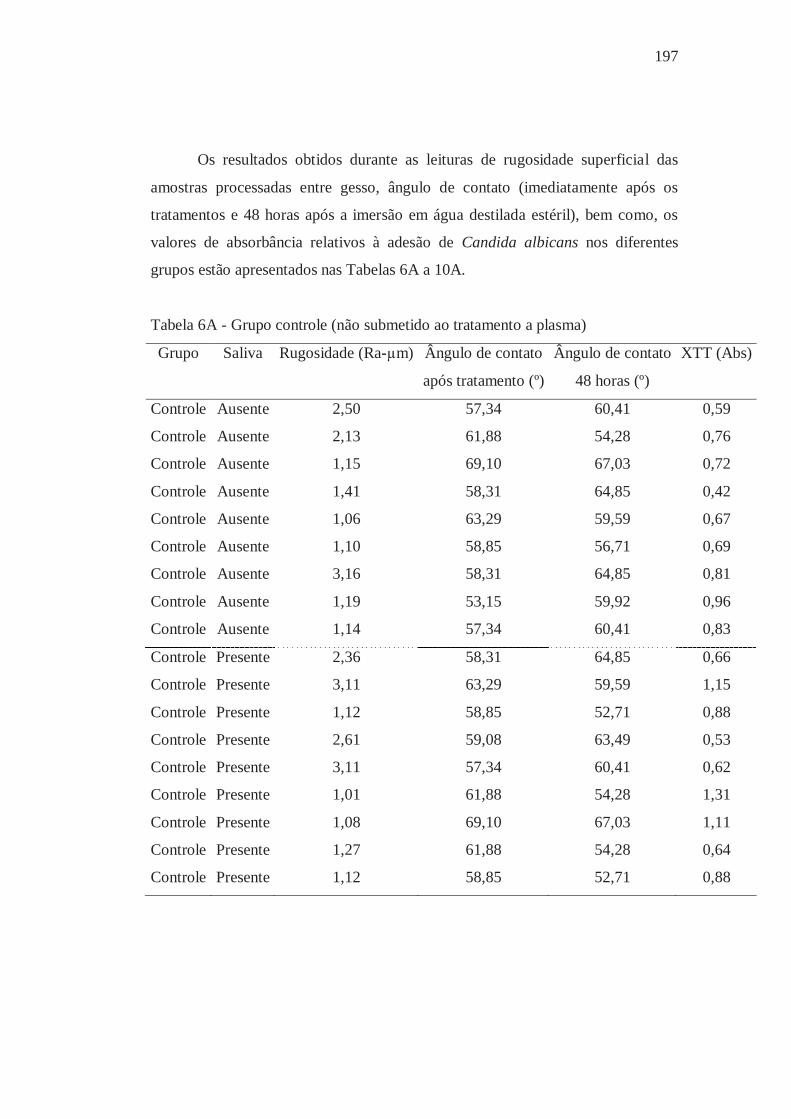
197
Os resultados obtidos durante as leituras de rugosidade superficial das
amostras processadas entre gesso, ângulo de contato (imediatamente após os
tratamentos e 48 horas após a imersão em água destilada estéril), bem como, os
valores de absorbância relativos à adesão de Candida albicans nos diferentes
grupos estão apresentados nas Tabelas 6A a 10A.
Tabela 6A - Grupo controle (não submetido ao tratamento a plasma)
Grupo Saliva Rugosidade (Ra-μm) Ângulo de contato Ângulo de contato XTT (Abs)
após tratamento (º) 48 horas (º)
Controle Ausente 2,50 57,34 60,41 0,59
Controle Ausente 2,13 61,88 54,28 0,76
Controle Ausente 1,15 69,10 67,03 0,72
Controle Ausente 1,41 58,31 64,85 0,42
Controle Ausente 1,06 63,29 59,59 0,67
Controle Ausente 1,10 58,85 56,71 0,69
Controle Ausente 3,16 58,31 64,85 0,81
Controle Ausente 1,19 53,15 59,92 0,96
Controle Ausente 1,14 57,34 60,41 0,83
Controle Presente 2,36 58,31 64,85 0,66
Controle Presente 3,11 63,29 59,59 1,15
Controle Presente 1,12 58,85 52,71 0,88
Controle Presente 2,61 59,08 63,49 0,53
Controle Presente 3,11 57,34 60,41 0,62
Controle Presente 1,01 61,88 54,28 1,31
Controle Presente 1,08 69,10 67,03 1,11
Controle Presente 1,27 61,88 54,28 0,64
Controle Presente 1,12 58,85 52,71 0,88
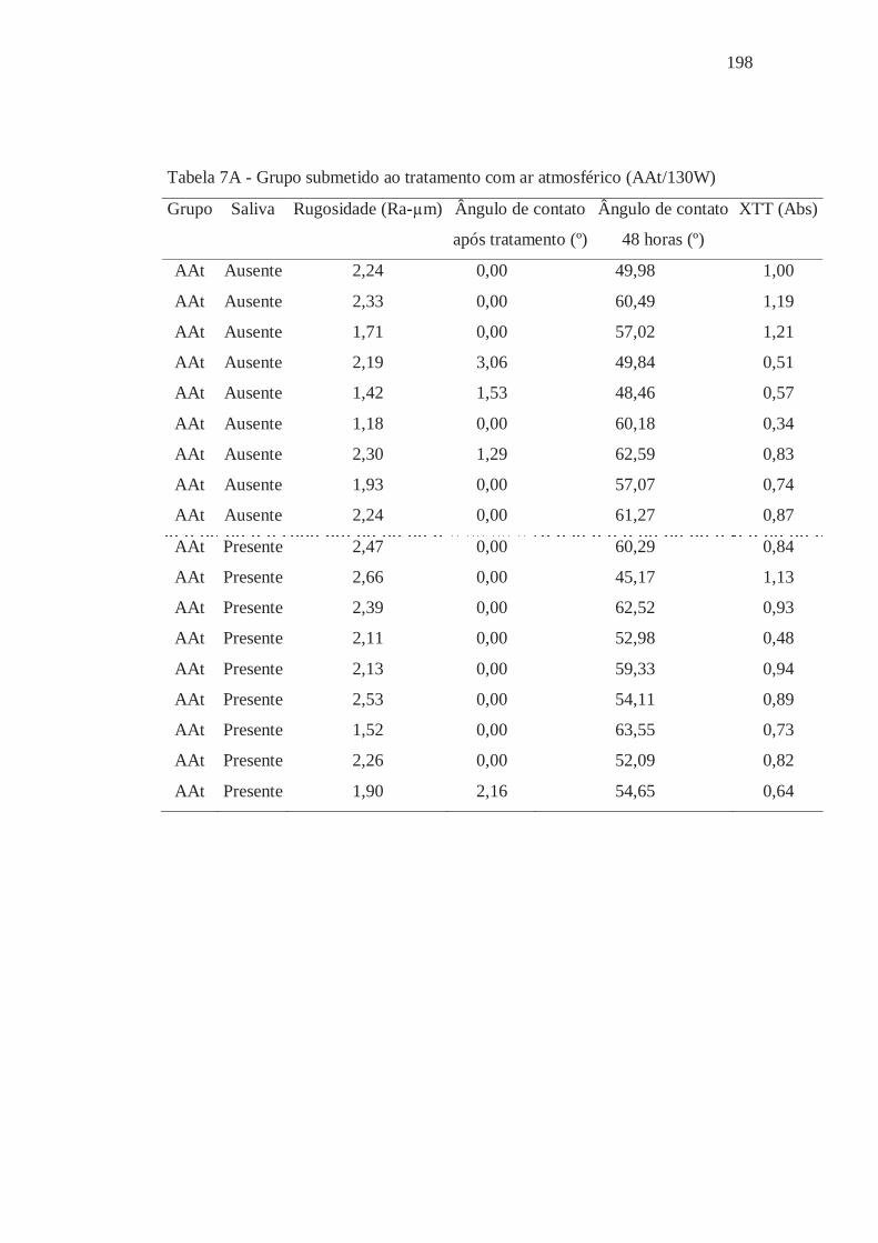
198
Tabela 7A - Grupo submetido ao tratamento com ar atmosférico (AAt/130W)
Grupo Saliva Rugosidade (Ra-μm) Ângulo de contato Ângulo de contato XTT (Abs)
após tratamento (º) 48 horas (º)
AAt Ausente 2,24 0,00 49,98 1,00
AAt Ausente 2,33 0,00 60,49 1,19
AAt Ausente 1,71 0,00 57,02 1,21
AAt Ausente 2,19 3,06 49,84 0,51
AAt Ausente 1,42 1,53 48,46 0,57
AAt Ausente 1,18 0,00 60,18 0,34
AAt Ausente 2,30 1,29 62,59 0,83
AAt Ausente 1,93 0,00 57,07 0,74
AAt Ausente 2,24 0,00 61,27 0,87
AAt Presente 2,47 0,00 60,29 0,84
AAt Presente 2,66 0,00 45,17 1,13
AAt Presente 2,39 0,00 62,52 0,93
AAt Presente 2,11 0,00 52,98 0,48
AAt Presente 2,13 0,00 59,33 0,94
AAt Presente 2,53 0,00 54,11 0,89
AAt Presente 1,52 0,00 63,55 0,73
AAt Presente 2,26 0,00 52,09 0,82
AAt Presente 1,90 2,16 54,65 0,64

199
Tabela 8A - Grupo submetido ao tratamento com uma mistura de gás argônio e
oxigênio (ArO2/70W)
Grupo Saliva Rugosidade (Ra-μm) Ângulo de contato Ângulo de contato XTT (Abs)
após tratamento (º) 48 horas (º)
ArO2 Ausente 1,28 26,40 70,50 0,14
ArO2 Ausente 1,32 16,31 52,54 0,14
ArO2 Ausente 1,01 19,01 59,30 0,12
ArO2 Ausente 2,32 28,40 69,15 0,52
ArO2 Ausente 2,06 26,25 62,54 0,39
ArO2 Ausente 2,46 32,36 51,02 0,31
ArO2 Ausente 2,66 34,59 50,76 1,18
ArO2 Ausente 2,52 20,58 47,08 0,74
ArO2 Ausente 1,78 28,65 52,40 0,42
ArO2 Presente 1,70 17,79 59,49 0,16
ArO2 Presente 2,18 17,95 70,18 0,21
ArO2 Presente 1,43 21,23 58,25 0,14
ArO2 Presente 1,02 17,85 60,20 0,79
ArO2 Presente 1,30 32,22 50,86 0,68
ArO2 Presente 1,49 22,69 56,52 0,31
ArO2 Presente 1,65 34,81 52,56 0,68
ArO2 Presente 1,75 31,15 47,83 0,57
ArO2 Presente 1,57 21,91 49,03 0,80
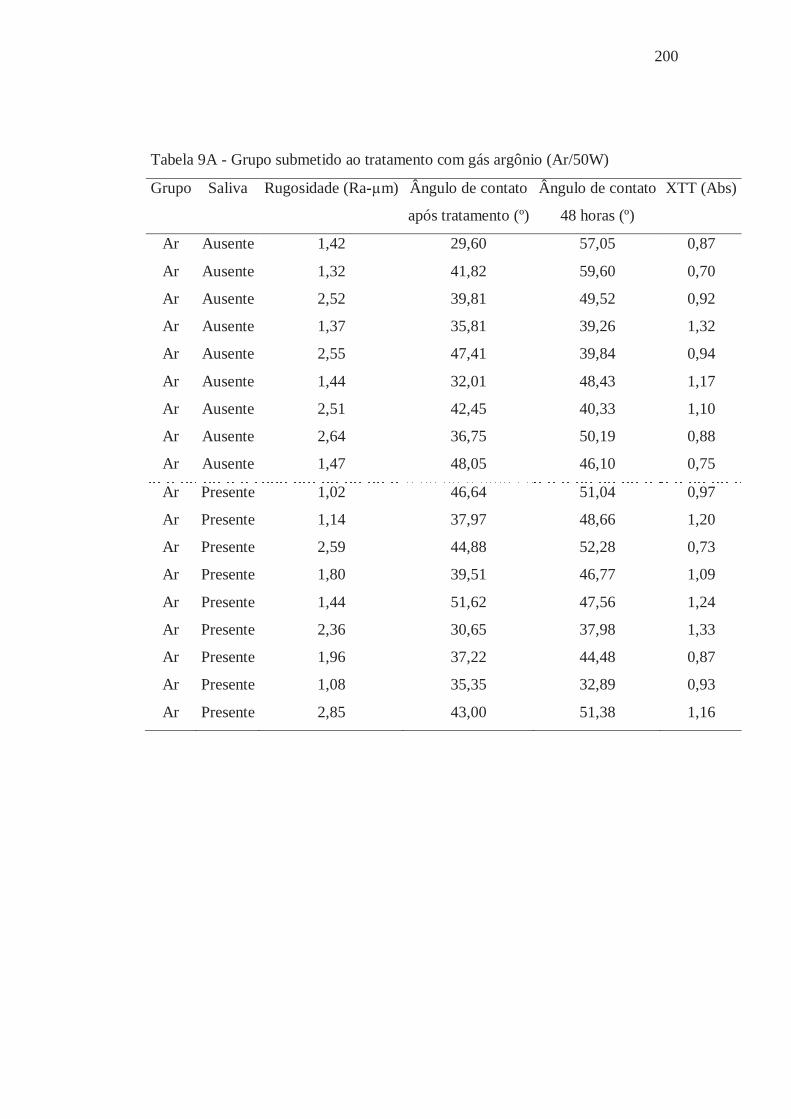
200
Tabela 9A - Grupo submetido ao tratamento com gás argônio (Ar/50W)
Grupo Saliva Rugosidade (Ra-μm) Ângulo de contato Ângulo de contato XTT (Abs)
após tratamento (º) 48 horas (º)
Ar Ausente 1,42 29,60 57,05 0,87
Ar Ausente 1,32 41,82 59,60 0,70
Ar Ausente 2,52 39,81 49,52 0,92
Ar Ausente 1,37 35,81 39,26 1,32
Ar Ausente 2,55 47,41 39,84 0,94
Ar Ausente 1,44 32,01 48,43 1,17
Ar Ausente 2,51 42,45 40,33 1,10
Ar Ausente 2,64 36,75 50,19 0,88
Ar Ausente 1,47 48,05 46,10 0,75
Ar Presente 1,02 46,64 51,04 0,97
Ar Presente 1,14 37,97 48,66 1,20
Ar Presente 2,59 44,88 52,28 0,73
Ar Presente 1,80 39,51 46,77 1,09
Ar Presente 1,44 51,62 47,56 1,24
Ar Presente 2,36 30,65 37,98 1,33
Ar Presente 1,96 37,22 44,48 0,87
Ar Presente 1,08 35,35 32,89 0,93
Ar Presente 2,85 43,00 51,38 1,16

201
Tabela 10A - Grupo submetido ao tratamento com gás argônio seguido pelo
tratamento com gás hexafluoreto de enxofre (ArSF6/70W)
Grupo Saliva Rugosidade (Ra-μm) Ângulo de contato Ângulo de contato XTT (Abs)
após tratamento (º) 48 horas (º)
ARSF6 Ausente 1,67 92,47 49,62 0,17
ARSF6 Ausente 1,71 92,91 54,96 0,36
ARSF6 Ausente 1,91 84,56 50,61 0,16
ARSF6 Ausente 1,83 95,32 71,40 0,56
ARSF6 Ausente 2,33 90,56 58,44 0,36
ARSF6 Ausente 3,13 108,35 61,52 0,53
ARSF6 Ausente 1,71 94,92 56,74 0,57
ARSF6 Ausente 1,07 110,34 68,60 0,90
ARSF6 Ausente 1,76 106,36 57,43 1,08
ARSF6 Presente 1,02 93,73 49,23 0,21
ARSF6 Presente 1,43 90,22 52,17 0,15
ARSF6 Presente 2,04 87,25 54,24 0,25
ARSF6 Presente 2,06 112,59 56,44 0,37
ARSF6 Presente 1,40 109,19 53,17 0,31
ARSF6 Presente 1,82 103,90 60,43 1,04
ARSF6 Presente 1,34 104,84 55,63 1,19
ARSF6 Presente 1,82 105,76 41,73 0,74
ARSF6 Presente 2,62 97,13 60,49 0,49

Apêndice 2 Os resultados obtidos durante as leituras de rugosidade superficial das
amostras processadas entre vidros, ângulo de contato (imediatamente após os
tratamentos e 48 horas após a imersão em água destilada estéril), bem como, os
valores de células/mm2 relativos à adesão de Candida albicans nos diferentes
grupos estão apresentados nas Tabelas 11A a 15A.
Tabela 11A - Grupo controle (não submetido ao tratamento a plasma)
Grupo Saliva Rugosidade Ângulo de contato Ângulo de contato cel/mm2 log (cel)
(Ra-μm) após tratamento (º) 48 horas (º)
Controle Ausente 0,24 55,30 53,22 278,21 2,45
Controle Ausente 0,24 51,80 53,68 2289,74 3,36
Controle Ausente 0,31 60,31 51,87 2603,85 3,42
Controle Ausente 0,30 58,10 55,90 306,41 2,49
Controle Ausente 0,38 54,73 60,23 520,51 2,72
Controle Ausente 0,32 57,80 55,44 1514,10 3,18
Controle Ausente 0,46 58,60 57,11 3205,13 3,51
Controle Ausente 0,41 55,57 51,84 398,72 2,60
Controle Ausente 0,33 58,10 55,90 315,38 2,50
Controle Presente 0,36 58,31 64,85 703,85 2,85
Controle Presente 0,24 63,29 59,59 319,23 2,51
Controle Presente 0,23 58,85 52,71 1097,44 3,04
Controle Presente 0,25 64,25 60,90 665,38 2,82
Controle Presente 0,23 59,11 57,47 3130,77 3,50
Controle Presente 0,23 60,31 51,87 2730,77 3,44
Controle Presente 0,26 55,30 56,90 333,33 2,52
Controle Presente 0,36 58,83 55,23 703,85 2,85
Controle Presente 0,21 58,10 55,90 283,33 2,45

203
Tabela 12A - Grupo submetido ao tratamento com ar atmosférico (AAt/130W)
Grupo Saliva Rugosidade Ângulo de contato Ângulo de contato cel/mm2 log (cel)
(Ra-μm) após tratamento (º) 48 horas (º)
AAt Ausente 0,23 3,20 62,92 1230,77 3,09
AAt Ausente 0,42 3,40 55,76 456,41 2,66
AAt Ausente 0,42 2,00 58,14 371,79 2,57
AAt Ausente 0,29 0,00 62,35 207,69 2,32
AAt Ausente 0,35 0,00 65,52 1454,65 3,16
AAt Ausente 0,27 0,00 62,86 550,00 2,74
AAt Ausente 0,18 6,00 64,61 3342,31 3,52
AAt Ausente 0,16 9,00 58,21 3116,67 3,49
AAt Ausente 0,24 0,00 68,97 2361,54 3,37
AAt Presente 0,23 3,55 54,81 419,23 2,62
AAt Presente 0,27 4,76 55,76 624,36 2,80
AAt Presente 0,40 5,00 59,73 78,21 1,90
AAt Presente 0,45 0,00 51,47 219,23 2,34
AAt Presente 0,19 0,00 55,73 1219,23 3,09
AAt Presente 0,20 0,00 38,99 534,62 2,73
AAt Presente 0,28 4,00 68,62 3052,56 3,48
AAt Presente 0,23 9,00 51,15 3471,79 3,54
AAt Presente 0,17 0,00 65,82 4143,59 3,62
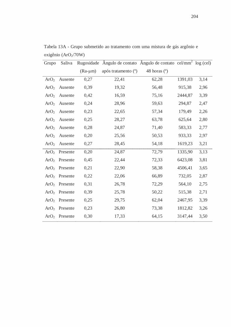
204
Tabela 13A - Grupo submetido ao tratamento com uma mistura de gás argônio e
oxigênio (ArO2/70W)
Grupo Saliva Rugosidade Ângulo de contato Ângulo de contato cel/mm2 log (cel)
(Ra-μm) após tratamento (º) 48 horas (º)
ArO2 Ausente 0,27 22,41 62,28 1391,03 3,14
ArO2 Ausente 0,39 19,32 56,48 915,38 2,96
ArO2 Ausente 0,42 16,59 75,16 2444,87 3,39
ArO2 Ausente 0,24 28,96 59,63 294,87 2,47
ArO2 Ausente 0,23 22,65 57,34 179,49 2,26
ArO2 Ausente 0,25 28,27 63,78 625,64 2,80
ArO2 Ausente 0,28 24,87 71,40 583,33 2,77
ArO2 Ausente 0,20 25,56 50,53 933,33 2,97
ArO2 Ausente 0,27 28,45 54,18 1619,23 3,21
ArO2 Presente 0,20 24,87 72,79 1335,90 3,13
ArO2 Presente 0,45 22,44 72,33 6423,08 3,81
ArO2 Presente 0,21 22,90 58,38 4506,41 3,65
ArO2 Presente 0,22 22,06 66,89 732,05 2,87
ArO2 Presente 0,31 26,78 72,29 564,10 2,75
ArO2 Presente 0,39 25,78 50,22 515,38 2,71
ArO2 Presente 0,25 29,75 62,04 2467,95 3,39
ArO2 Presente 0,23 26,80 73,38 1812,82 3,26
ArO2 Presente 0,30 17,33 64,15 3147,44 3,50
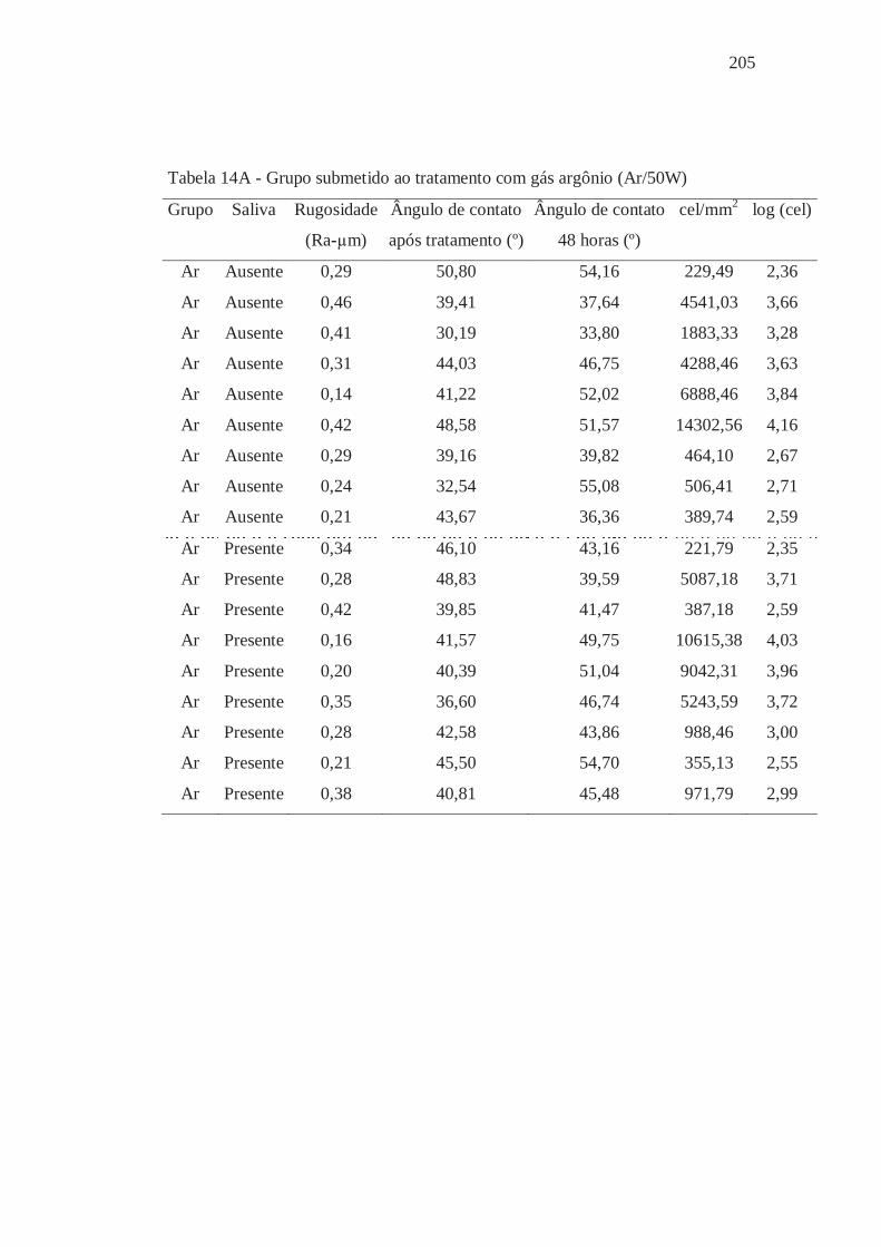
205
Tabela 14A - Grupo submetido ao tratamento com gás argônio (Ar/50W)
Grupo Saliva Rugosidade Ângulo de contato Ângulo de contato cel/mm2 log (cel)
(Ra-μm) após tratamento (º) 48 horas (º)
Ar Ausente 0,29 50,80 54,16 229,49 2,36
Ar Ausente 0,46 39,41 37,64 4541,03 3,66
Ar Ausente 0,41 30,19 33,80 1883,33 3,28
Ar Ausente 0,31 44,03 46,75 4288,46 3,63
Ar Ausente 0,14 41,22 52,02 6888,46 3,84
Ar Ausente 0,42 48,58 51,57 14302,56 4,16
Ar Ausente 0,29 39,16 39,82 464,10 2,67
Ar Ausente 0,24 32,54 55,08 506,41 2,71
Ar Ausente 0,21 43,67 36,36 389,74 2,59
Ar Presente 0,34 46,10 43,16 221,79 2,35
Ar Presente 0,28 48,83 39,59 5087,18 3,71
Ar Presente 0,42 39,85 41,47 387,18 2,59
Ar Presente 0,16 41,57 49,75 10615,38 4,03
Ar Presente 0,20 40,39 51,04 9042,31 3,96
Ar Presente 0,35 36,60 46,74 5243,59 3,72
Ar Presente 0,28 42,58 43,86 988,46 3,00
Ar Presente 0,21 45,50 54,70 355,13 2,55
Ar Presente 0,38 40,81 45,48 971,79 2,99

206
Tabela 15A - Grupo submetido ao tratamento com gás argônio seguido pelo
tratamento com gás hexafluoreto de enxofre (ArSF6/70W)
Grupo Saliva Rugosidade Ângulo de contato Ângulo de contato cel/mm2 log (cel)
(Ra-μm) após tratamento (º) 48 horas (º)
ARSF6 Ausente 0,31 94,30 60,80 1861,54 3,27
ARSF6 Ausente 0,21 96,05 61,06 1966,67 3,29
ARSF6 Ausente 0,23 90,08 67,60 2235,90 3,35
ARSF6 Ausente 0,23 101,28 66,38 805,13 2,91
ARSF6 Ausente 0,22 110,79 70,10 1346,15 3,13
ARSF6 Ausente 0,22 92,47 71,41 2921,79 3,47
ARSF6 Ausente 0,33 92,29 65,12 1448,72 3,16
ARSF6 Ausente 0,49 97,19 70,37 2955,13 3,47
ARSF6 Ausente 0,35 98,14 64,68 5019,23 3,70
ARSF6 Presente 0,25 97,12 64,87 7378,21 3,87
ARSF6 Presente 0,23 92,96 59,27 5830,77 3,77
ARSF6 Presente 0,29 91,69 58,27 5484,62 3,74
ARSF6 Presente 0,24 91,02 55,63 2694,87 3,43
ARSF6 Presente 0,22 97,55 72,03 7082,05 3,85
ARSF6 Presente 0,24 102,36 64,85 3565,38 3,55
ARSF6 Presente 0,32 100,18 51,43 1548,72 3,19
ARSF6 Presente 0,41 83,49 61,67 3424,36 3,53
ARSF6 Presente 0,20 96,09 57,74 779,49 2,89

207
Os resultados obtidos durante as leituras de rugosidade superficial das
amostras processadas entre gesso, ângulo de contato (imediatamente após os
tratamentos e 48 horas após a imersão em água destilada estéril), bem como, os
valores de células/mm2 relativos à adesão de Candida albicans nos diferentes
grupos estão apresentados nas Tabelas 16A a 20A.
Tabela 16A - Grupo controle (não submetido ao tratamento a plasma)
Grupo Saliva Rugosidade Ângulo de contato Ângulo de contato cel/mm2 log (cel)
(Ra-μm) após tratamento (º) 48 horas (º)
Controle Ausente 1,90 53,15 49,92 948,72 2,98
Controle Ausente 1,51 49,19 56,49 764,10 2,88
Controle Ausente 2,84 57,34 60,41 573,08 2,76
Controle Ausente 1,28 66,70 50,45 1985,90 3,30
Controle Ausente 1,30 62,01 65,27 5202,56 3,72
Controle Ausente 1,07 49,19 56,49 719,23 2,86
Controle Ausente 1,33 62,01 65,27 438,46 2,64
Controle Ausente 1,54 65,04 63,31 1296,15 3,11
Controle Ausente 1,86 59,10 64,82 3930,77 3,59
Controle Presente 1,05 61,88 54,28 156,41 2,20
Controle Presente 1,86 58,85 52,71 889,74 2,95
Controle Presente 1,46 64,25 60,90 5752,56 3,76
Controle Presente 1,53 61,88 54,28 1244,87 3,10
Controle Presente 3,10 58,85 52,71 1184,62 3,07
Controle Presente 1,86 59,08 63,49 106,41 2,03
Controle Presente 2,07 64,25 60,90 156,41 2,20
Controle Presente 1,39 62,07 62,59 3365,38 3,53
Controle Presente 1,20 60,68 55,24 2430,77 3,39

208
Tabela 17A - Grupo submetido ao tratamento com ar atmosférico (AAt/130W)
Grupo Saliva Rugosidade Ângulo de contato Ângulo de contato cel/mm2 log (cel)
(Ra-μm) após tratamento (º) 48 horas (º)
AAt Ausente 2,47 2,73 57,26 316,67 2,50
AAt Ausente 1,87 0,97 49,67 4965,38 3,70
AAt Ausente 2,07 0,00 59,32 2325,64 3,37
AAt Ausente 1,53 0,00 72,71 517,95 2,72
AAt Ausente 1,69 0,00 61,52 617,95 2,79
AAt Ausente 1,98 0,00 63,53 505,13 2,70
AAt Ausente 2,64 5,10 61,88 3375,64 3,53
AAt Ausente 1,11 3,92 66,35 3367,95 3,53
AAt Ausente 2,48 0,00 56,79 3360,26 3,53
AAt Presente 1,81 3,36 56,93 839,74 2,92
AAt Presente 2,32 0,00 45,11 388,46 2,59
AAt Presente 1,04 0,00 57,47 1024,359 3,01
AAt Presente 1,05 0,00 53,85 397,4359 2,60
AAt Presente 1,71 2,35 60,53 415,38 2,62
AAt Presente 2,22 0,44 55,78 526,92 2,72
AAt Presente 2,02 0,00 51,50 9457,69 3,98
AAt Presente 3,09 0,00 49,70 2876,92 3,46
AAt Presente 1,96 1,87 52,92 2037,18 3,31

209
Tabela 18A - Grupo submetido ao tratamento com uma mistura de gás argônio e
oxigênio (ArO2/70W)
Grupo Saliva Rugosidade Ângulo de contato Ângulo de contato cel/mm2 log (cel)
(Ra-μm) após tratamento (º) 48 horas (º)
ArO2 Ausente 1,41 26,26 58,81 1520,51 3,18
ArO2 Ausente 1,29 31,48 36,75 1841,03 3,27
ArO2 Ausente 1,76 18,30 52,47 2292,31 3,36
ArO2 Ausente 2,32 27,86 52,69 735,90 2,87
ArO2 Ausente 1,51 22,77 47,99 2485,90 3,40
ArO2 Ausente 1,42 22,61 42,99 433,33 2,64
ArO2 Ausente 1,04 21,47 36,59 775,64 2,89
ArO2 Ausente 1,56 29,02 51,15 810,26 2,91
ArO2 Ausente 2,05 31,34 49,69 580,77 2,76
ArO2 Presente 2,47 20,29 43,63 1905,13 3,28
ArO2 Presente 1,01 20,31 49,57 3485,90 3,54
ArO2 Presente 2,10 21,65 55,63 1947,44 3,29
ArO2 Presente 1,97 23,82 43,36 278,21 2,45
ArO2 Presente 1,79 32,10 51,12 1461,54 3,17
ArO2 Presente 1,05 28,43 55,99 657,69 2,82
ArO2 Presente 1,67 23,55 61,59 280,77 2,45
ArO2 Presente 1,47 38,49 47,93 903,85 2,96
ArO2 Presente 1,02 33,11 55,09 2832,05 3,45

210
Tabela 19A - Grupo submetido ao tratamento com gás argônio (Ar/50W)
Grupo Saliva Rugosidade Ângulo de contato Ângulo de contato cel/mm2 log (cel)
(Ra-μm) após tratamento (º) 48 horas (º)
Ar Ausente 1,74 29,61 53,29 1980,77 3,30
Ar Ausente 1,55 47,90 40,09 7442,31 3,87
Ar Ausente 1,85 36,83 48,70 6889,74 3,84
Ar Ausente 1,45 39,89 47,96 8961,54 3,95
Ar Ausente 2,73 41,82 47,46 16185,90 4,21
Ar Ausente 1,46 40,00 31,07 11607,69 4,06
Ar Ausente 1,49 37,64 51,71 485,90 2,69
Ar Ausente 1,81 39,61 55,28 3017,95 3,48
Ar Ausente 2,16 37,21 34,08 333,33 2,52
Ar Presente 1,18 31,55 63,31 1334,62 3,13
Ar Presente 1,49 36,92 45,89 8550,00 3,93
Ar Presente 1,51 40,60 48,18 21423,08 4,33
Ar Presente 2,42 35,34 51,94 4655,13 3,67
Ar Presente 3,08 37,12 53,34 4411,54 3,64
Ar Presente 1,96 36,90 43,35 7676,92 3,89
Ar Presente 1,18 36,89 42,04 1529,49 3,18
Ar Presente 2,10 45,71 38,11 2538,46 3,40
Ar Presente 2,30 40,70 50,92 484,62 2,69
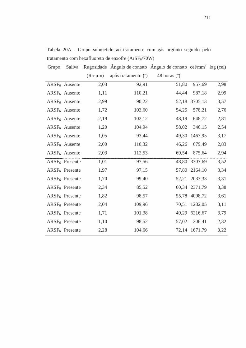
211
Tabela 20A - Grupo submetido ao tratamento com gás argônio seguido pelo
tratamento com hexafluoreto de enxofre (ArSF6/70W)
Grupo Saliva Rugosidade Ângulo de contato Ângulo de contato cel/mm2 log (cel)
(Ra-μm) após tratamento (º) 48 horas (º)
ARSF6 Ausente 2,03 92,91 51,80 957,69 2,98
ARSF6 Ausente 1,11 110,21 44,44 987,18 2,99
ARSF6 Ausente 2,99 90,22 52,18 3705,13 3,57
ARSF6 Ausente 1,72 103,60 54,25 578,21 2,76
ARSF6 Ausente 2,19 102,12 48,19 648,72 2,81
ARSF6 Ausente 1,20 104,94 58,02 346,15 2,54
ARSF6 Ausente 1,05 93,44 49,30 1467,95 3,17
ARSF6 Ausente 2,00 110,32 46,26 679,49 2,83
ARSF6 Ausente 2,03 112,53 69,54 875,64 2,94
ARSF6 Presente 1,01 97,56 48,80 3307,69 3,52
ARSF6 Presente 1,97 97,15 57,80 2164,10 3,34
ARSF6 Presente 1,70 99,40 52,21 2033,33 3,31
ARSF6 Presente 2,34 85,52 60,34 2371,79 3,38
ARSF6 Presente 1,82 98,57 55,78 4098,72 3,61
ARSF6 Presente 2,04 109,96 70,51 1282,05 3,11
ARSF6 Presente 1,71 101,38 49,29 6216,67 3,79
ARSF6 Presente 1,10 98,52 57,02 206,41 2,32
ARSF6 Presente 2,28 104,66 72,14 1671,79 3,22
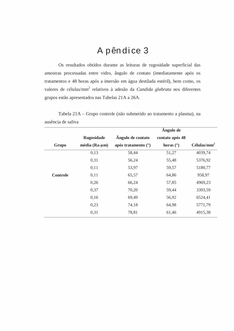
Apêndice 3 Os resultados obtidos durante as leituras de rugosidade superficial das
amostras processadas entre vidro, ângulo de contato (imediatamente após os
tratamentos e 48 horas após a imersão em água destilada estéril), bem como, os
valores de células/mm2 relativos à adesão da Candida glabrata nos diferentes
grupos estão apresentados nas Tabelas 21A a 26A.
Tabela 21A – Grupo controle (não submetido ao tratamento a plasma), na
ausência de saliva
Grupo
Rugosidade
média (Ra-μm)
Ângulo de contato
após tratamento (°)
Ângulo de
contato após 48
horas (°) Células/mm2
0,13 58,44 51,27 4039,74
0,31 56,24 55,48 5376,92
0,11 53,97 59,57 5180,77
Controle 0,11 65,57 64,86 958,97
0,26 66,24 57,85 4969,23
0,37 70,20 59,44 3393,59
0,16 69,49 56,92 6524,41
0,23 74,18 64,98 5771,79
0,31 78,81 61,46 4915,38
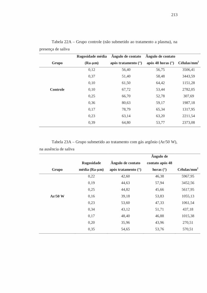
213
Tabela 22A – Grupo controle (não submetido ao tratamento a plasma), na
presença de saliva
Grupo
Rugosidade média
(Ra-μm)
Ângulo de contato
após tratamento (°)
Ângulo de contato
após 48 horas (°) Células/mm2
0,12 56,40 56,75 3506,41
0,37 51,40 58,48 3443,59
0,10 61,50 64,42 1151,28
Controle 0,10 67,72 53,44 2782,05
0,25 66,70 52,78 307,69
0,36 80,63 59,17 1987,18
0,17 78,79 65,34 1317,95
0,23 63,14 63,20 2211,54
0,39 64,80 53,77 2373,08
Tabela 23A – Grupo submetido ao tratamento com gás argônio (Ar/50 W),
na ausência de saliva
Grupo
Rugosidade
média (Ra-μm)
Ângulo de contato
após tratamento (°)
Ângulo de
contato após 48
horas (°) Células/mm2
0,22 42,60 46,38 5967,95
0,19 44,63 57,94 3452,56
0,25 44,82 45,66 5617,95
Ar/50 W 0,16 39,18 53,83 1055,13
0,23 53,60 47,33 1061,54
0,34 43,12 51,71 437,18
0,17 48,40 46,88 1015,38
0,20 35,96 43,96 270,51
0,35 54,65 53,76 570,51
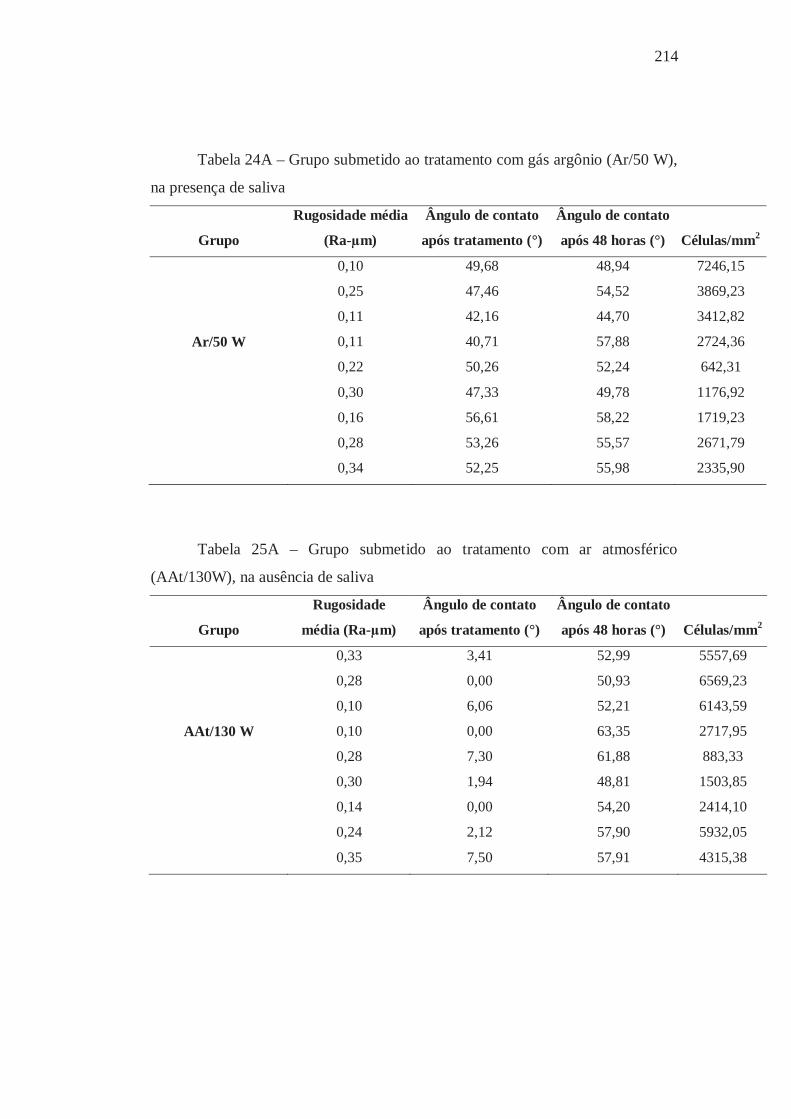
214
Tabela 24A – Grupo submetido ao tratamento com gás argônio (Ar/50 W),
na presença de saliva
Grupo
Rugosidade média
(Ra-μm)
Ângulo de contato
após tratamento (°)
Ângulo de contato
após 48 horas (°) Células/mm2
0,10 49,68 48,94 7246,15
0,25 47,46 54,52 3869,23
0,11 42,16 44,70 3412,82
Ar/50 W 0,11 40,71 57,88 2724,36
0,22 50,26 52,24 642,31
0,30 47,33 49,78 1176,92
0,16 56,61 58,22 1719,23
0,28 53,26 55,57 2671,79
0,34 52,25 55,98 2335,90
Tabela 25A – Grupo submetido ao tratamento com ar atmosférico
(AAt/130W), na ausência de saliva
Grupo
Rugosidade
média (Ra-μm)
Ângulo de contato
após tratamento (°)
Ângulo de contato
após 48 horas (°) Células/mm2
0,33 3,41 52,99 5557,69
0,28 0,00 50,93 6569,23
0,10 6,06 52,21 6143,59
AAt/130 W 0,10 0,00 63,35 2717,95
0,28 7,30 61,88 883,33
0,30 1,94 48,81 1503,85
0,14 0,00 54,20 2414,10
0,24 2,12 57,90 5932,05
0,35 7,50 57,91 4315,38
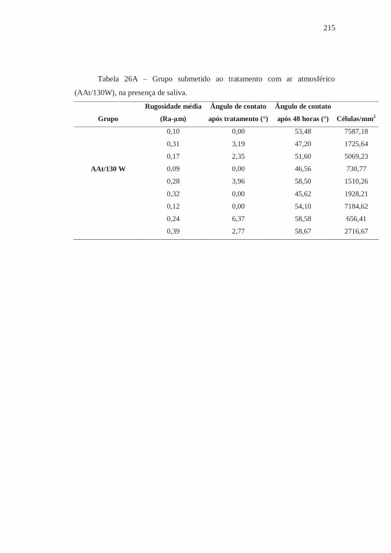
215
Tabela 26A – Grupo submetido ao tratamento com ar atmosférico
(AAt/130W), na presença de saliva.
Grupo
Rugosidade média
(Ra-μm)
Ângulo de contato
após tratamento (°)
Ângulo de contato
após 48 horas (°) Células/mm2
0,10 0,00 53,48 7587,18
0,31 3,19 47,20 1725,64
0,17 2,35 51,60 5069,23
AAt/130 W 0,09 0,00 46,56 730,77
0,28 3,96 58,50 1510,26
0,32 0,00 45,62 1928,21
0,12 0,00 54,10 7184,62
0,24 6,37 58,58 656,41
0,39 2,77 58,67 2716,67
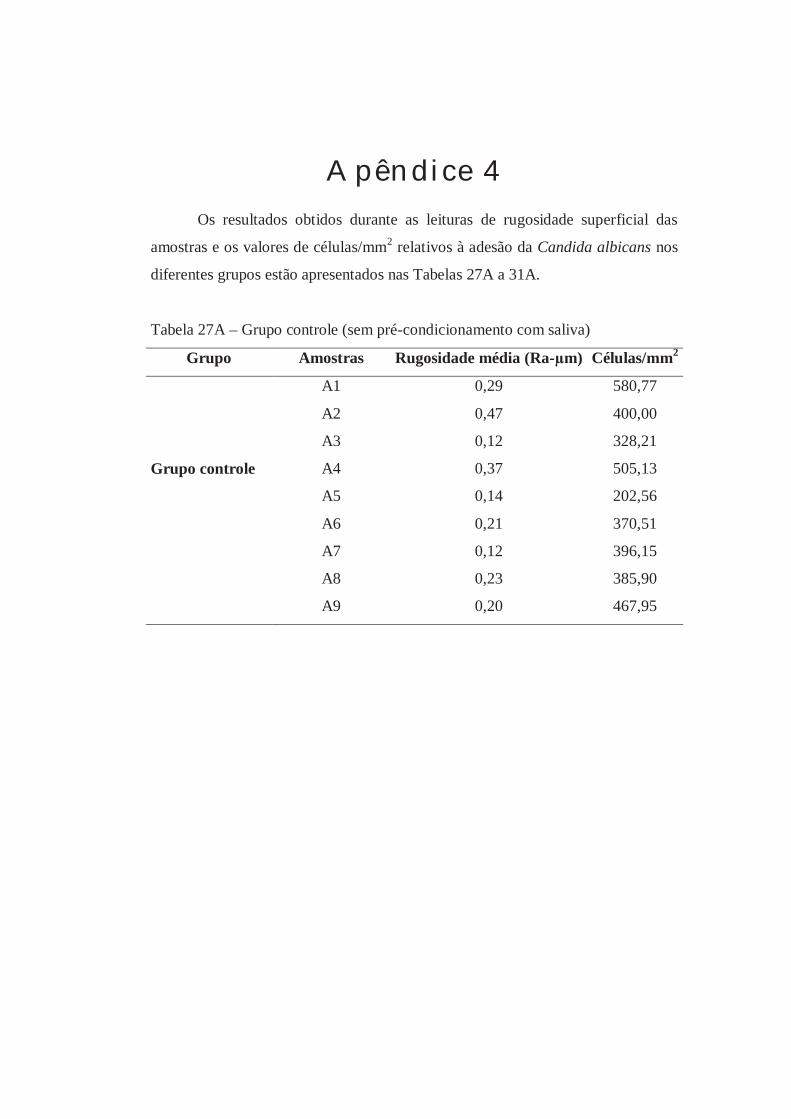
Apêndice 4 Os resultados obtidos durante as leituras de rugosidade superficial das
amostras e os valores de células/mm2 relativos à adesão da Candida albicans nos
diferentes grupos estão apresentados nas Tabelas 27A a 31A.
Tabela 27A – Grupo controle (sem pré-condicionamento com saliva)
Grupo Amostras Rugosidade média (Ra-μm) Células/mm2
A1 0,29 580,77
A2 0,47 400,00
A3 0,12 328,21
Grupo controle A4 0,37 505,13
A5 0,14 202,56
A6 0,21 370,51
A7 0,12 396,15
A8 0,23 385,90
A9 0,20 467,95
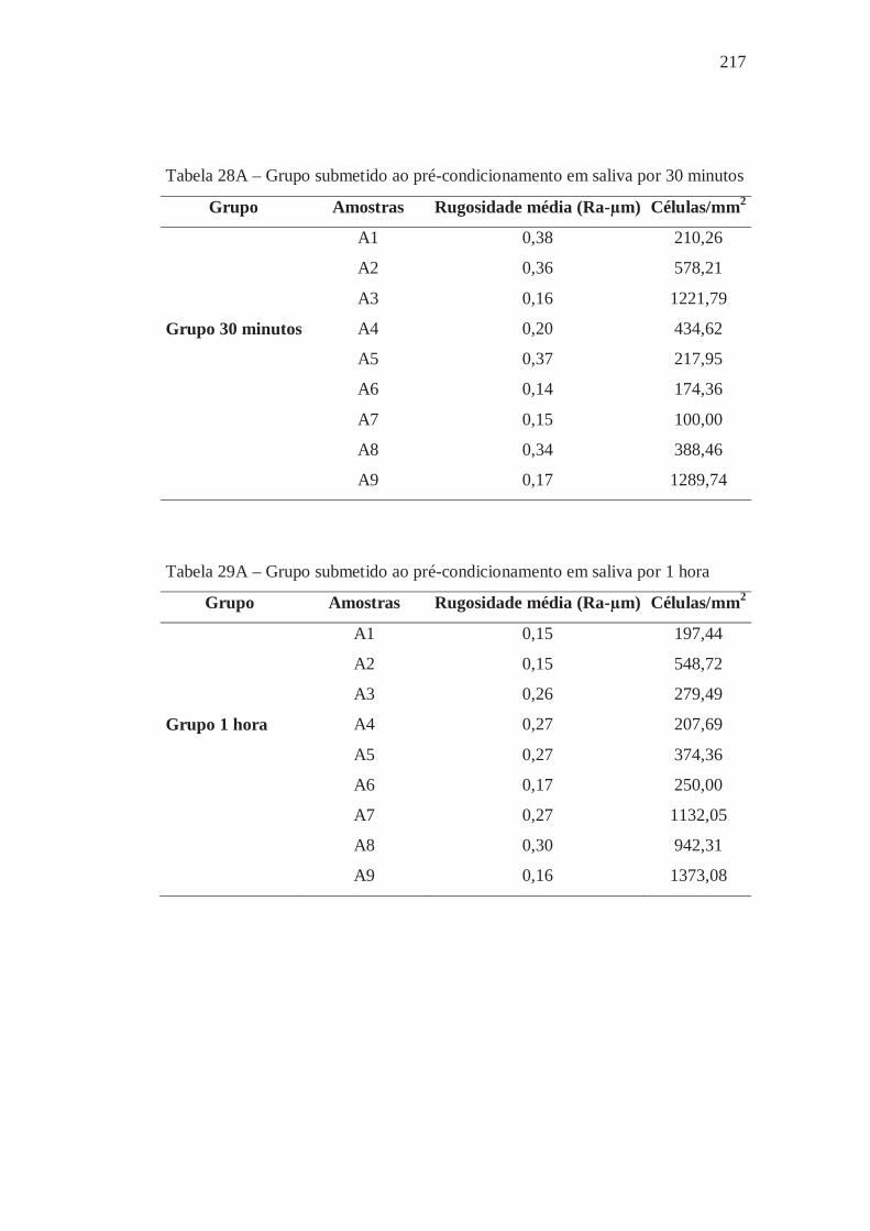
217
Tabela 28A – Grupo submetido ao pré-condicionamento em saliva por 30 minutos
Grupo Amostras Rugosidade média (Ra-μm) Células/mm2
A1 0,38 210,26
A2 0,36 578,21
A3 0,16 1221,79
Grupo 30 minutos A4 0,20 434,62
A5 0,37 217,95
A6 0,14 174,36
A7 0,15 100,00
A8 0,34 388,46
A9 0,17 1289,74
Tabela 29A – Grupo submetido ao pré-condicionamento em saliva por 1 hora
Grupo Amostras Rugosidade média (Ra-μm) Células/mm2
A1 0,15 197,44
A2 0,15 548,72
A3 0,26 279,49
Grupo 1 hora A4 0,27 207,69
A5 0,27 374,36
A6 0,17 250,00
A7 0,27 1132,05
A8 0,30 942,31
A9 0,16 1373,08

218
Tabela 30A – Grupo submetido ao pré-condicionamento em saliva por 3 horas
Grupo Amostras Rugosidade média (Ra-μm) Células/mm2
A1 0,29 301,28
A2 0,09 708,97
A3 0,28 335,90
Grupo 3 horas A4 0,14 394,87
A5 0,13 1273,08
A6 0,20 1548,72
A7 0,15 426,92
A8 0,33 219,23
A9 0,16 134,62
Tabela 31A – Grupo submetido ao pré-condicionamento em saliva por 12 horas
Grupo Amostras Rugosidade média (Ra-μm) Células/mm2
A1 0,15 289,74
A2 0,12 185,90
A3 0,33 255,13
Grupo 12 horas A4 0,14 1675,64
A5 0,25 621,79
A6 0,34 2000,00
A7 0,31 248,72
A8 0,11 769,23
A9 0,16 228,21

219
Os resultados obtidos durante as leituras de rugosidade superficial das
amostras e os valores de absorbância relativos à adesão da Candida albicans nos
diferentes grupos estão apresentados nas Tabelas 32A a 36A.
Tabela 32A – Grupo controle (sem pré-condicionamento com saliva)
Grupo Amostras Rugosidade média (Ra-μm) Absorbância
A1 0,11 0,239
A2 0,12 0,202
A3 0,14 0,213
Grupo controle A4 0,28 0,269
A5 0,12 0,223
A6 0,37 0,063
A7 0,10 0,115
A8 0,38 0,243
A9 0,20 0,244
Tabela 33A– Grupo submetido ao pré-condicionamento em saliva por 30 minutos
Grupo Amostras Rugosidade média (Ra-μm) Absorbância
A1 0,08 0,349
A2 0,22 0,588
A3 0,25 0,276
Grupo 30 minutos A4 0,21 0,408
A5 0,39 0,236
A6 0,11 0,163
A7 0,26 0,141
A8 0,37 0,454
A9 0,27 0,181

220
Tabela 34A – Grupo submetido ao pré-condicionamento em saliva por 1 hora
Grupo Amostras Rugosidade média (Ra-μm) Absorbância
A1 0,09 0,757
A2 0,23 0,410
A3 0,18 0,407
Grupo 1 hora A4 0,29 0,219
A5 0,33 0,137
A6 0,15 0,211
A7 0,26 0,258
A8 0,27 0,167
A9 0,13 0,603
Tabela 35A – Grupo submetido ao pré-condicionamento em saliva por 3 horas
Grupo Amostras Rugosidade média (Ra-μm) Absorbância
A1 0,14 0,276
A2 0,18 0,281
A3 0,14 0,189
Grupo 3 horas A4 0,27 0,794
A5 0,26 0,339
A6 0,33 0,377
A7 0,40 0,196
A8 0,14 0,205
A9 0,12 0,180
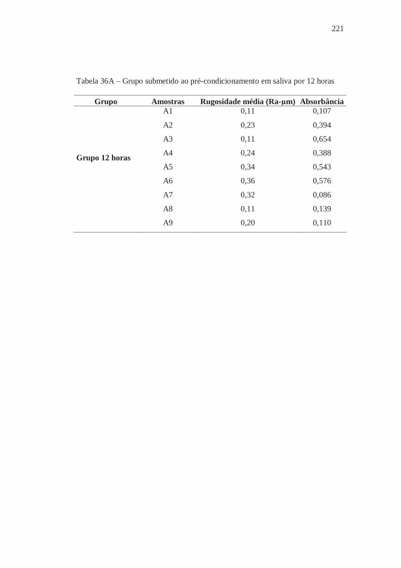
221
Tabela 36A – Grupo submetido ao pré-condicionamento em saliva por 12 horas
Grupo Amostras Rugosidade média (Ra-μm) Absorbância
A1 0,11 0,107
A2 0,23 0,394
A3 0,11 0,654
Grupo 12 horas A4 0,24 0,388
A5 0,34 0,543
A6 0,36 0,576
A7 0,32 0,086
A8 0,11 0,139
A9 0,20 0,110

Apêndice 5 Os resultados obtidos durante as leituras de rugosidade superficial das
amostras e os valores de células/mm2 relativos à adesão da Candida albicans nos
diferentes grupos estão apresentados nas Tabelas 37A a 40A.
Tabela 37A – Grupo controle (sem pré-condicionamento com saliva)
Grupo Amostras Rugosidade média (Ra-μm) Células/mm2
A1 0,25 142,50
A2 0,35 74,20
A3 0,17 84,50
Grupo 1 A4 0,27 31,50
A5 0,31 31,70
A6 0,08 22,50
A7 0,26 49,50
A8 0,16 45,60
A9 0,25 14,70
Tabela 38A – Grupo 2 - Vários doadores (10.000/5min)
Grupo Amostras Rugosidade média (Ra-μm) Células/mm2
A1 0,18 261,20
A2 0,38 963,20
A3 0,11 17,60
Grupo 2 A4 0,16 12,70
A5 0,33 126,30
A6 0,23 39,60
A7 0,05 54,40
A8 0,37 170,20
A9 0,26 520,60

223
Tabela 39A – Grupo 3 - Vários doadores (12.000/30min)
Grupo Amostras Rugosidade média (Ra-μm) Células/mm2
A1 0,24 9,40
A2 0,24 21,00
A3 0,29 403,00
Grupo 3 A4 0,35 14,80
A5 0,33 34,00
A6 0,08 15,00
A7 0,20 70,10
A8 0,21 155,00
A9 0,20 37,60
Tabela 40A – Grupo 4 – Um doador (10.000/5min)
Grupo Amostras Rugosidade média (Ra-μm) Células/mm2
A1 0,38 34,60
A2 0,08 9,10
A3 0,28 79,60
Grupo 4 A4 0,19 42,30
A5 0,34 30,80
A6 0,15 9,40
A7 0,21 114,50
A8 0,07 24,60
A9 0,35 15,70
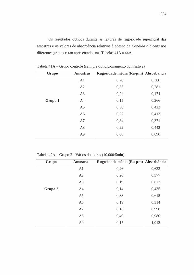
224
Os resultados obtidos durante as leituras de rugosidade superficial das
amostras e os valores de absorbância relativos à adesão da Candida albicans nos
diferentes grupos estão apresentados nas Tabelas 41A a 44A.
Tabela 41A – Grupo controle (sem pré-condicionamento com saliva)
Grupo Amostras Rugosidade média (Ra-μm) Absorbância
A1 0,28 0,360
A2 0,35 0,281
A3 0,24 0,474
Grupo 1 A4 0,15 0,266
A5 0,38 0,422
A6 0,27 0,413
A7 0,34 0,371
A8 0,22 0,442
A9 0,08 0,690
Tabela 42A – Grupo 2 - Vários doadores (10.000/5min)
Grupo Amostras Rugosidade média (Ra-μm) Absorbância
A1 0,26 0,633
A2 0,20 0,577
A3 0,19 0,673
Grupo 2 A4 0,14 0,435
A5 0,33 0,615
A6 0,19 0,514
A7 0,16 0,998
A8 0,40 0,980
A9 0,17 1,012
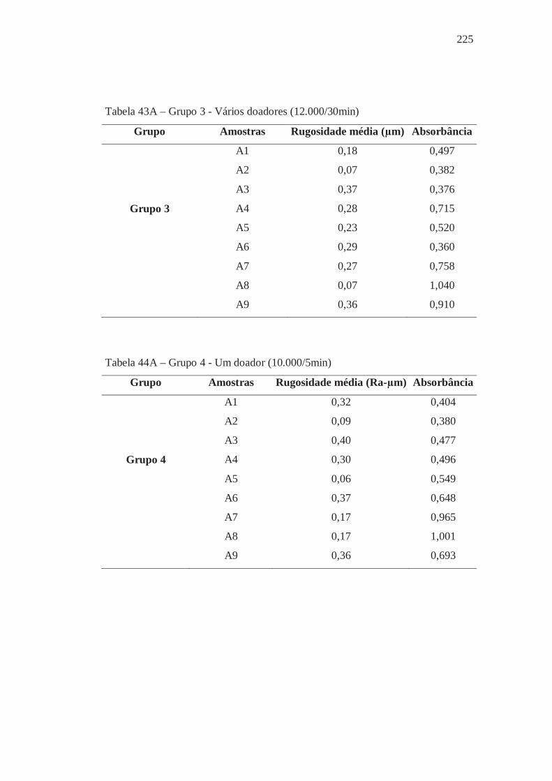
225
Tabela 43A – Grupo 3 - Vários doadores (12.000/30min)
Grupo Amostras Rugosidade média (μm) Absorbância
A1 0,18 0,497
A2 0,07 0,382
A3 0,37 0,376
Grupo 3 A4 0,28 0,715
A5 0,23 0,520
A6 0,29 0,360
A7 0,27 0,758
A8 0,07 1,040
A9 0,36 0,910
Tabela 44A – Grupo 4 - Um doador (10.000/5min)
Grupo Amostras Rugosidade média (Ra-μm) Absorbância
A1 0,32 0,404
A2 0,09 0,380
A3 0,40 0,477
Grupo 4 A4 0,30 0,496
A5 0,06 0,549
A6 0,37 0,648
A7 0,17 0,965
A8 0,17 1,001
A9 0,36 0,693

Autorizo a reprodução deste trabalho.
(Direitos de publicação reservado ao autor)
Araraquara, 30 de março de 2011.
CAMILA ANDRADE ZAMPERINI